JP4264919B2 - Multiplex displacement amplification - Google Patents
Multiplex displacement amplificationDownload PDFInfo
- Publication number
- JP4264919B2 JP4264919B2JP2000515033AJP2000515033AJP4264919B2JP 4264919 B2JP4264919 B2JP 4264919B2JP 2000515033 AJP2000515033 AJP 2000515033AJP 2000515033 AJP2000515033 AJP 2000515033AJP 4264919 B2JP4264919 B2JP 4264919B2
- Authority
- JP
- Japan
- Prior art keywords
- primer
- primers
- dna
- nucleic acid
- strand
- Prior art date
- Legal status (The legal status is an assumption and is not a legal conclusion. Google has not performed a legal analysis and makes no representation as to the accuracy of the status listed.)
- Expired - Lifetime
Links
- 238000006073displacement reactionMethods0.000titleclaimsabstractdescription104
- 230000003321amplificationEffects0.000titleabstractdescription132
- 238000003199nucleic acid amplification methodMethods0.000titleabstractdescription132
- 150000007523nucleic acidsChemical group0.000claimsabstractdescription138
- 230000000295complement effectEffects0.000claimsabstractdescription115
- 238000000034methodMethods0.000claimsabstractdescription102
- 230000010076replicationEffects0.000claimsabstractdescription65
- 239000000203mixtureSubstances0.000claimsabstractdescription42
- 108091028043Nucleic acid sequenceProteins0.000claimsabstractdescription28
- 108020004414DNAProteins0.000claimsdescription141
- 238000009396hybridizationMethods0.000claimsdescription47
- 108010014303DNA-directed DNA polymeraseProteins0.000claimsdescription44
- 102000016928DNA-directed DNA polymeraseHuman genes0.000claimsdescription44
- 239000012634fragmentSubstances0.000claimsdescription32
- 239000002299complementary DNASubstances0.000claimsdescription20
- 108020004999messenger RNAProteins0.000claimsdescription9
- 238000002156mixingMethods0.000claimsdescription8
- 238000005382thermal cyclingMethods0.000claimsdescription5
- 108020004707nucleic acidsProteins0.000abstractdescription101
- 102000039446nucleic acidsHuman genes0.000abstractdescription101
- 230000002269spontaneous effectEffects0.000abstractdescription2
- 239000000523sampleSubstances0.000description127
- 238000001514detection methodMethods0.000description104
- 125000003729nucleotide groupChemical group0.000description68
- 239000002773nucleotideSubstances0.000description63
- 239000007787solidSubstances0.000description33
- 238000003752polymerase chain reactionMethods0.000description30
- 239000002585baseSubstances0.000description27
- 238000006243chemical reactionMethods0.000description25
- 239000000758substrateSubstances0.000description18
- 108091034117OligonucleotideProteins0.000description17
- YBJHBAHKTGYVGT-ZKWXMUAHSA-N(+)-BiotinChemical compoundN1C(=O)N[C@@H]2[C@H](CCCCC(=O)O)SC[C@@H]21YBJHBAHKTGYVGT-ZKWXMUAHSA-N0.000description16
- 102000004190EnzymesHuman genes0.000description14
- 108090000790EnzymesProteins0.000description14
- 238000003556assayMethods0.000description14
- 108091032973(ribonucleotides)n+mProteins0.000description12
- 238000013412genome amplificationMethods0.000description11
- JLCPHMBAVCMARE-UHFFFAOYSA-N[3-[[3-[[3-[[3-[[3-[[3-[[3-[[3-[[3-[[3-[[3-[[5-(2-amino-6-oxo-1H-purin-9-yl)-3-[[3-[[3-[[3-[[3-[[3-[[5-(2-amino-6-oxo-1H-purin-9-yl)-3-[[5-(2-amino-6-oxo-1H-purin-9-yl)-3-hydroxyoxolan-2-yl]methoxy-hydroxyphosphoryl]oxyoxolan-2-yl]methoxy-hydroxyphosphoryl]oxy-5-(5-methyl-2,4-dioxopyrimidin-1-yl)oxolan-2-yl]methoxy-hydroxyphosphoryl]oxy-5-(6-aminopurin-9-yl)oxolan-2-yl]methoxy-hydroxyphosphoryl]oxy-5-(6-aminopurin-9-yl)oxolan-2-yl]methoxy-hydroxyphosphoryl]oxy-5-(6-aminopurin-9-yl)oxolan-2-yl]methoxy-hydroxyphosphoryl]oxy-5-(6-aminopurin-9-yl)oxolan-2-yl]methoxy-hydroxyphosphoryl]oxyoxolan-2-yl]methoxy-hydroxyphosphoryl]oxy-5-(5-methyl-2,4-dioxopyrimidin-1-yl)oxolan-2-yl]methoxy-hydroxyphosphoryl]oxy-5-(4-amino-2-oxopyrimidin-1-yl)oxolan-2-yl]methoxy-hydroxyphosphoryl]oxy-5-(5-methyl-2,4-dioxopyrimidin-1-yl)oxolan-2-yl]methoxy-hydroxyphosphoryl]oxy-5-(5-methyl-2,4-dioxopyrimidin-1-yl)oxolan-2-yl]methoxy-hydroxyphosphoryl]oxy-5-(6-aminopurin-9-yl)oxolan-2-yl]methoxy-hydroxyphosphoryl]oxy-5-(6-aminopurin-9-yl)oxolan-2-yl]methoxy-hydroxyphosphoryl]oxy-5-(4-amino-2-oxopyrimidin-1-yl)oxolan-2-yl]methoxy-hydroxyphosphoryl]oxy-5-(4-amino-2-oxopyrimidin-1-yl)oxolan-2-yl]methoxy-hydroxyphosphoryl]oxy-5-(4-amino-2-oxopyrimidin-1-yl)oxolan-2-yl]methoxy-hydroxyphosphoryl]oxy-5-(6-aminopurin-9-yl)oxolan-2-yl]methoxy-hydroxyphosphoryl]oxy-5-(4-amino-2-oxopyrimidin-1-yl)oxolan-2-yl]methyl [5-(6-aminopurin-9-yl)-2-(hydroxymethyl)oxolan-3-yl] hydrogen phosphatePolymersCc1cn(C2CC(OP(O)(=O)OCC3OC(CC3OP(O)(=O)OCC3OC(CC3O)n3cnc4c3nc(N)[nH]c4=O)n3cnc4c3nc(N)[nH]c4=O)C(COP(O)(=O)OC3CC(OC3COP(O)(=O)OC3CC(OC3COP(O)(=O)OC3CC(OC3COP(O)(=O)OC3CC(OC3COP(O)(=O)OC3CC(OC3COP(O)(=O)OC3CC(OC3COP(O)(=O)OC3CC(OC3COP(O)(=O)OC3CC(OC3COP(O)(=O)OC3CC(OC3COP(O)(=O)OC3CC(OC3COP(O)(=O)OC3CC(OC3COP(O)(=O)OC3CC(OC3COP(O)(=O)OC3CC(OC3COP(O)(=O)OC3CC(OC3COP(O)(=O)OC3CC(OC3COP(O)(=O)OC3CC(OC3COP(O)(=O)OC3CC(OC3CO)n3cnc4c(N)ncnc34)n3ccc(N)nc3=O)n3cnc4c(N)ncnc34)n3ccc(N)nc3=O)n3ccc(N)nc3=O)n3ccc(N)nc3=O)n3cnc4c(N)ncnc34)n3cnc4c(N)ncnc34)n3cc(C)c(=O)[nH]c3=O)n3cc(C)c(=O)[nH]c3=O)n3ccc(N)nc3=O)n3cc(C)c(=O)[nH]c3=O)n3cnc4c3nc(N)[nH]c4=O)n3cnc4c(N)ncnc34)n3cnc4c(N)ncnc34)n3cnc4c(N)ncnc34)n3cnc4c(N)ncnc34)O2)c(=O)[nH]c1=OJLCPHMBAVCMARE-UHFFFAOYSA-N0.000description10
- 239000000463materialSubstances0.000description9
- 230000037452primingEffects0.000description9
- 108090000623proteins and genesProteins0.000description9
- -1Cy5Chemical compound0.000description8
- 230000015572biosynthetic processEffects0.000description8
- 229960002685biotinDrugs0.000description8
- 235000020958biotinNutrition0.000description8
- 239000011616biotinSubstances0.000description8
- 210000004027cellAnatomy0.000description8
- 239000011521glassSubstances0.000description8
- 238000006116polymerization reactionMethods0.000description8
- 230000000750progressive effectEffects0.000description8
- 230000004543DNA replicationEffects0.000description7
- 238000004458analytical methodMethods0.000description7
- WOVKYSAHUYNSMH-RRKCRQDMSA-N5-bromodeoxyuridineChemical compoundC1[C@H](O)[C@@H](CO)O[C@H]1N1C(=O)NC(=O)C(Br)=C1WOVKYSAHUYNSMH-RRKCRQDMSA-N0.000description6
- FAPWRFPIFSIZLT-UHFFFAOYSA-MSodium chlorideChemical compound[Na+].[Cl-]FAPWRFPIFSIZLT-UHFFFAOYSA-M0.000description6
- 239000000872bufferSubstances0.000description6
- 238000003786synthesis reactionMethods0.000description6
- XLYOFNOQVPJJNP-UHFFFAOYSA-NwaterChemical compoundOXLYOFNOQVPJJNP-UHFFFAOYSA-N0.000description6
- 102000053602DNAHuman genes0.000description5
- IAZDPXIOMUYVGZ-UHFFFAOYSA-NDimethylsulphoxideChemical compoundCS(C)=OIAZDPXIOMUYVGZ-UHFFFAOYSA-N0.000description5
- 108091036060Linker DNAProteins0.000description5
- 108060004795MethyltransferaseProteins0.000description4
- 101710126859Single-stranded DNA-binding proteinProteins0.000description4
- 238000010586diagramMethods0.000description4
- 230000000694effectsEffects0.000description4
- 230000000977initiatory effectEffects0.000description4
- 238000002372labellingMethods0.000description4
- 235000018102proteinsNutrition0.000description4
- 102000004169proteins and genesHuman genes0.000description4
- 230000002285radioactive effectEffects0.000description4
- 108091008146restriction endonucleasesProteins0.000description4
- 238000013518transcriptionMethods0.000description4
- 230000035897transcriptionEffects0.000description4
- QGKMIGUHVLGJBR-UHFFFAOYSA-M(4z)-1-(3-methylbutyl)-4-[[1-(3-methylbutyl)quinolin-1-ium-4-yl]methylidene]quinoline;iodideChemical compound[I-].C12=CC=CC=C2N(CCC(C)C)C=CC1=CC1=CC=[N+](CCC(C)C)C2=CC=CC=C12QGKMIGUHVLGJBR-UHFFFAOYSA-M0.000description3
- QKNYBSVHEMOAJP-UHFFFAOYSA-N2-amino-2-(hydroxymethyl)propane-1,3-diol;hydron;chlorideChemical compoundCl.OCC(N)(CO)COQKNYBSVHEMOAJP-UHFFFAOYSA-N0.000description3
- SHIBSTMRCDJXLN-UHFFFAOYSA-NDigoxigeninNatural productsC1CC(C2C(C3(C)CCC(O)CC3CC2)CC2O)(O)C2(C)C1C1=CC(=O)OC1SHIBSTMRCDJXLN-UHFFFAOYSA-N0.000description3
- KCXVZYZYPLLWCC-UHFFFAOYSA-NEDTAChemical compoundOC(=O)CN(CC(O)=O)CCN(CC(O)=O)CC(O)=OKCXVZYZYPLLWCC-UHFFFAOYSA-N0.000description3
- 238000012408PCR amplificationMethods0.000description3
- 239000002253acidSubstances0.000description3
- 150000007513acidsChemical class0.000description3
- 108010058966bacteriophage T7 induced DNA polymeraseProteins0.000description3
- 230000027455bindingEffects0.000description3
- 238000004925denaturationMethods0.000description3
- 230000036425denaturationEffects0.000description3
- QONQRTHLHBTMGP-UHFFFAOYSA-NdigitoxigeninNatural productsCC12CCC(C3(CCC(O)CC3CC3)C)C3C11OC1CC2C1=CC(=O)OC1QONQRTHLHBTMGP-UHFFFAOYSA-N0.000description3
- SHIBSTMRCDJXLN-KCZCNTNESA-NdigoxigeninChemical compoundC1([C@@H]2[C@@]3([C@@](CC2)(O)[C@H]2[C@@H]([C@@]4(C)CC[C@H](O)C[C@H]4CC2)C[C@H]3O)C)=CC(=O)OC1SHIBSTMRCDJXLN-KCZCNTNESA-N0.000description3
- 239000012153distilled waterSubstances0.000description3
- 239000000975dyeSubstances0.000description3
- 239000000835fiberSubstances0.000description3
- GNBHRKFJIUUOQI-UHFFFAOYSA-NfluoresceinChemical compoundO1C(=O)C2=CC=CC=C2C21C1=CC=C(O)C=C1OC1=CC(O)=CC=C21GNBHRKFJIUUOQI-UHFFFAOYSA-N0.000description3
- MHMNJMPURVTYEJ-UHFFFAOYSA-Nfluorescein-5-isothiocyanateChemical compoundO1C(=O)C2=CC(N=C=S)=CC=C2C21C1=CC=C(O)C=C1OC1=CC(O)=CC=C21MHMNJMPURVTYEJ-UHFFFAOYSA-N0.000description3
- 238000010348incorporationMethods0.000description3
- 230000001404mediated effectEffects0.000description3
- 238000002493microarrayMethods0.000description3
- 239000003068molecular probeSubstances0.000description3
- 238000011160researchMethods0.000description3
- 239000011780sodium chlorideSubstances0.000description3
- 241000894007speciesSpecies0.000description3
- 238000010561standard procedureMethods0.000description3
- 238000006467substitution reactionMethods0.000description3
- 238000005406washingMethods0.000description3
- HRPVXLWXLXDGHG-UHFFFAOYSA-NAcrylamideChemical compoundNC(=O)C=CHRPVXLWXLXDGHG-UHFFFAOYSA-N0.000description2
- 241000701844Bacillus virus phi29Species0.000description2
- HEDRZPFGACZZDS-UHFFFAOYSA-NChloroformChemical compoundClC(Cl)ClHEDRZPFGACZZDS-UHFFFAOYSA-N0.000description2
- 102000008186CollagenHuman genes0.000description2
- 108010035532CollagenProteins0.000description2
- 102000012410DNA LigasesHuman genes0.000description2
- 108010061982DNA LigasesProteins0.000description2
- 108010017826DNA Polymerase IProteins0.000description2
- 102000004594DNA Polymerase IHuman genes0.000description2
- 108090000626DNA-directed RNA polymerasesProteins0.000description2
- 102000004163DNA-directed RNA polymerasesHuman genes0.000description2
- VZCYOOQTPOCHFL-OWOJBTEDSA-NFumaric acidChemical compoundOC(=O)\C=C\C(O)=OVZCYOOQTPOCHFL-OWOJBTEDSA-N0.000description2
- 229920002683GlycosaminoglycanPolymers0.000description2
- 239000000020NitrocelluloseSubstances0.000description2
- 239000004677NylonSubstances0.000description2
- 229920003171Poly (ethylene oxide)Polymers0.000description2
- 229920002732PolyanhydridePolymers0.000description2
- 239000004698PolyethyleneSubstances0.000description2
- 229920001710PolyorthoesterPolymers0.000description2
- 239000004743PolypropyleneSubstances0.000description2
- 239000004793PolystyreneSubstances0.000description2
- 239000004809TeflonSubstances0.000description2
- 229920006362Teflon®Polymers0.000description2
- 239000007983Tris bufferSubstances0.000description2
- QWXOJIDBSHLIFI-UHFFFAOYSA-N[3-(1-chloro-3'-methoxyspiro[adamantane-4,4'-dioxetane]-3'-yl)phenyl] dihydrogen phosphateChemical compoundO1OC2(C3CC4CC2CC(Cl)(C4)C3)C1(OC)C1=CC=CC(OP(O)(O)=O)=C1QWXOJIDBSHLIFI-UHFFFAOYSA-N0.000description2
- OIRDTQYFTABQOQ-KQYNXXCUSA-Nadenosine groupChemical group[C@@H]1([C@H](O)[C@H](O)[C@@H](CO)O1)N1C=NC=2C(N)=NC=NC12OIRDTQYFTABQOQ-KQYNXXCUSA-N0.000description2
- 239000011324beadSubstances0.000description2
- 108091008324binding proteinsProteins0.000description2
- 102000023732binding proteinsHuman genes0.000description2
- 239000012472biological sampleSubstances0.000description2
- 238000010804cDNA synthesisMethods0.000description2
- 229920002678cellulosePolymers0.000description2
- 239000001913celluloseSubstances0.000description2
- 239000003153chemical reaction reagentSubstances0.000description2
- 238000003776cleavage reactionMethods0.000description2
- 229920001436collagenPolymers0.000description2
- SUYVUBYJARFZHO-RRKCRQDMSA-NdATPChemical compoundC1=NC=2C(N)=NC=NC=2N1[C@H]1C[C@H](O)[C@@H](COP(O)(=O)OP(O)(=O)OP(O)(O)=O)O1SUYVUBYJARFZHO-RRKCRQDMSA-N0.000description2
- SUYVUBYJARFZHO-UHFFFAOYSA-NdATPNatural productsC1=NC=2C(N)=NC=NC=2N1C1CC(O)C(COP(O)(=O)OP(O)(=O)OP(O)(O)=O)O1SUYVUBYJARFZHO-UHFFFAOYSA-N0.000description2
- RGWHQCVHVJXOKC-SHYZEUOFSA-JdCTP(4-)Chemical compoundO=C1N=C(N)C=CN1[C@@H]1O[C@H](COP([O-])(=O)OP([O-])(=O)OP([O-])([O-])=O)[C@@H](O)C1RGWHQCVHVJXOKC-SHYZEUOFSA-J0.000description2
- HAAZLUGHYHWQIW-KVQBGUIXSA-NdGTPChemical compoundC1=NC=2C(=O)NC(N)=NC=2N1[C@H]1C[C@H](O)[C@@H](COP(O)(=O)OP(O)(=O)OP(O)(O)=O)O1HAAZLUGHYHWQIW-KVQBGUIXSA-N0.000description2
- NHVNXKFIZYSCEB-XLPZGREQSA-NdTTPChemical compoundO=C1NC(=O)C(C)=CN1[C@@H]1O[C@H](COP(O)(=O)OP(O)(=O)OP(O)(O)=O)[C@@H](O)C1NHVNXKFIZYSCEB-XLPZGREQSA-N0.000description2
- 230000001419dependent effectEffects0.000description2
- 230000029087digestionEffects0.000description2
- NBVXSUQYWXRMNV-UHFFFAOYSA-NfluoromethaneChemical compoundFCNBVXSUQYWXRMNV-UHFFFAOYSA-N0.000description2
- 230000008571general functionEffects0.000description2
- 102000054766genetic haplotypesHuman genes0.000description2
- 230000003100immobilizing effectEffects0.000description2
- 238000007834ligase chain reactionMethods0.000description2
- 239000012528membraneSubstances0.000description2
- 239000011859microparticleSubstances0.000description2
- 238000012986modificationMethods0.000description2
- 230000004048modificationEffects0.000description2
- 229920001220nitrocellulosPolymers0.000description2
- 229920001778nylonPolymers0.000description2
- 238000002515oligonucleotide synthesisMethods0.000description2
- 239000002245particleSubstances0.000description2
- RXNXLAHQOVLMIE-UHFFFAOYSA-Nphenyl 10-methylacridin-10-ium-9-carboxylateChemical compoundC12=CC=CC=C2[N+](C)=C2C=CC=CC2=C1C(=O)OC1=CC=CC=C1RXNXLAHQOVLMIE-UHFFFAOYSA-N0.000description2
- 229920001308poly(aminoacid)Polymers0.000description2
- 229920000747poly(lactic acid)Polymers0.000description2
- 239000004417polycarbonateSubstances0.000description2
- 229920000515polycarbonatePolymers0.000description2
- 229920000573polyethylenePolymers0.000description2
- 229920000151polyglycolPolymers0.000description2
- 239000010695polyglycolSubstances0.000description2
- 239000004626polylactic acidSubstances0.000description2
- 229920000642polymerPolymers0.000description2
- 229920000193polymethacrylatePolymers0.000description2
- 229920001155polypropylenePolymers0.000description2
- 229920002223polystyrenePolymers0.000description2
- 239000011535reaction bufferSubstances0.000description2
- PYWVYCXTNDRMGF-UHFFFAOYSA-Nrhodamine BChemical compound[Cl-].C=12C=CC(=[N+](CC)CC)C=C2OC2=CC(N(CC)CC)=CC=C2C=1C1=CC=CC=C1C(O)=OPYWVYCXTNDRMGF-UHFFFAOYSA-N0.000description2
- 230000007017scissionEffects0.000description2
- 238000000926separation methodMethods0.000description2
- 229920002379silicone rubberPolymers0.000description2
- 239000004945silicone rubberSubstances0.000description2
- 239000011343solid materialSubstances0.000description2
- 108010068698spleen exonucleaseProteins0.000description2
- 239000000126substanceSubstances0.000description2
- 238000001308synthesis methodMethods0.000description2
- 239000010409thin filmSubstances0.000description2
- VZCYOOQTPOCHFL-UHFFFAOYSA-Ntrans-butenedioic acidNatural productsOC(=O)C=CC(O)=OVZCYOOQTPOCHFL-UHFFFAOYSA-N0.000description2
- LENZDBCJOHFCAS-UHFFFAOYSA-NtrisChemical compoundOCC(N)(CO)COLENZDBCJOHFCAS-UHFFFAOYSA-N0.000description2
- 238000012800visualizationMethods0.000description2
- FALRKNHUBBKYCC-UHFFFAOYSA-N2-(chloromethyl)pyridine-3-carbonitrileChemical compoundClCC1=NC=CC=C1C#NFALRKNHUBBKYCC-UHFFFAOYSA-N0.000description1
- KMEMIMRPZGDOMG-UHFFFAOYSA-N2-cyanoethoxyphosphonamidous acidChemical compoundNP(O)OCCC#NKMEMIMRPZGDOMG-UHFFFAOYSA-N0.000description1
- GOLORTLGFDVFDW-UHFFFAOYSA-N3-(1h-benzimidazol-2-yl)-7-(diethylamino)chromen-2-oneChemical compoundC1=CC=C2NC(C3=CC4=CC=C(C=C4OC3=O)N(CC)CC)=NC2=C1GOLORTLGFDVFDW-UHFFFAOYSA-N0.000description1
- FWBHETKCLVMNFS-UHFFFAOYSA-N4',6-Diamino-2-phenylindolChemical compoundC1=CC(C(=N)N)=CC=C1C1=CC2=CC=C(C(N)=N)C=C2N1FWBHETKCLVMNFS-UHFFFAOYSA-N0.000description1
- FWMNVWWHGCHHJJ-SKKKGAJSSA-N4-amino-1-[(2r)-6-amino-2-[[(2r)-2-[[(2r)-2-[[(2r)-2-amino-3-phenylpropanoyl]amino]-3-phenylpropanoyl]amino]-4-methylpentanoyl]amino]hexanoyl]piperidine-4-carboxylic acidChemical compoundC([C@H](C(=O)N[C@H](CC(C)C)C(=O)N[C@H](CCCCN)C(=O)N1CCC(N)(CC1)C(O)=O)NC(=O)[C@H](N)CC=1C=CC=CC=1)C1=CC=CC=C1FWMNVWWHGCHHJJ-SKKKGAJSSA-N0.000description1
- 108700015125Adenovirus DBPProteins0.000description1
- 108090001008AvidinProteins0.000description1
- 101150062763BMRF1 geneProteins0.000description1
- WOVKYSAHUYNSMH-UHFFFAOYSA-NBROMODEOXYURIDINENatural productsC1C(O)C(CO)OC1N1C(=O)NC(=O)C(Br)=C1WOVKYSAHUYNSMH-UHFFFAOYSA-N0.000description1
- 241000322342Bacillus phage M2Species0.000description1
- 240000001082Bambusa multiplexSpecies0.000description1
- 241000283690Bos taurusSpecies0.000description1
- 108020004635Complementary DNAProteins0.000description1
- 101150026402DBP geneProteins0.000description1
- 108020003215DNA ProbesProteins0.000description1
- 230000004544DNA amplificationEffects0.000description1
- 239000003298DNA probeSubstances0.000description1
- 230000006820DNA synthesisEffects0.000description1
- 102000052510DNA-Binding ProteinsHuman genes0.000description1
- 101710116602DNA-Binding protein G5PProteins0.000description1
- XPDXVDYUQZHFPV-UHFFFAOYSA-NDansyl ChlorideChemical compoundC1=CC=C2C(N(C)C)=CC=CC2=C1S(Cl)(=O)=OXPDXVDYUQZHFPV-UHFFFAOYSA-N0.000description1
- AHCYMLUZIRLXAA-SHYZEUOFSA-NDeoxyuridine 5'-triphosphateChemical compoundO1[C@H](COP(O)(=O)OP(O)(=O)OP(O)(O)=O)[C@@H](O)C[C@@H]1N1C(=O)NC(=O)C=C1AHCYMLUZIRLXAA-SHYZEUOFSA-N0.000description1
- 108010067770Endopeptidase KProteins0.000description1
- 241000588724Escherichia coliSpecies0.000description1
- 241000701533Escherichia virus T4Species0.000description1
- 101710085030Gene 32 proteinProteins0.000description1
- 108010025076HoloenzymesProteins0.000description1
- 102000003960LigasesHuman genes0.000description1
- 108090000364LigasesProteins0.000description1
- 206010028980NeoplasmDiseases0.000description1
- 108020004711Nucleic Acid ProbesProteins0.000description1
- 229910019142PO4Inorganic materials0.000description1
- 101150054516PRD1 geneProteins0.000description1
- ISWSIDIOOBJBQZ-UHFFFAOYSA-NPhenolChemical compoundOC1=CC=CC=C1ISWSIDIOOBJBQZ-UHFFFAOYSA-N0.000description1
- 102000004160Phosphoric Monoester HydrolasesHuman genes0.000description1
- 108090000608Phosphoric Monoester HydrolasesProteins0.000description1
- ZYFVNVRFVHJEIU-UHFFFAOYSA-NPicoGreenChemical compoundCN(C)CCCN(CCCN(C)C)C1=CC(=CC2=[N+](C3=CC=CC=C3S2)C)C2=CC=CC=C2N1C1=CC=CC=C1ZYFVNVRFVHJEIU-UHFFFAOYSA-N0.000description1
- 108010066717Q beta ReplicaseProteins0.000description1
- 239000013614RNA sampleSubstances0.000description1
- 108091081062Repeated sequence (DNA)Proteins0.000description1
- 101710162453Replication factor AProteins0.000description1
- 101710176758Replication protein A 70 kDa DNA-binding subunitProteins0.000description1
- 101710176276SSB proteinProteins0.000description1
- 101100459905Saccharomyces cerevisiae (strain ATCC 204508 / S288c) NCP1 geneProteins0.000description1
- 241000700584SimplexvirusSpecies0.000description1
- 101150104425T4 geneProteins0.000description1
- 108010006785Taq PolymeraseProteins0.000description1
- IQFYYKKMVGJFEH-XLPZGREQSA-NThymidineChemical groupO=C1NC(=O)C(C)=CN1[C@@H]1O[C@H](CO)[C@@H](O)C1IQFYYKKMVGJFEH-XLPZGREQSA-N0.000description1
- 229920004890Triton X-100Polymers0.000description1
- 239000013504Triton X-100Substances0.000description1
- PGAVKCOVUIYSFO-XVFCMESISA-NUTPChemical classO[C@@H]1[C@H](O)[C@@H](COP(O)(=O)OP(O)(=O)OP(O)(O)=O)O[C@H]1N1C(=O)NC(=O)C=C1PGAVKCOVUIYSFO-XVFCMESISA-N0.000description1
- LBZXELUWKIEZEQ-MYINAIGISA-N[[(2r,3s,5s)-5-bromo-5-(2,4-dioxopyrimidin-1-yl)-3-hydroxyoxolan-2-yl]methoxy-hydroxyphosphoryl] phosphono hydrogen phosphateChemical compoundO1[C@H](COP(O)(=O)OP(O)(=O)OP(O)(O)=O)[C@@H](O)C[C@]1(Br)N1C(=O)NC(=O)C=C1LBZXELUWKIEZEQ-MYINAIGISA-N0.000description1
- 238000010521absorption reactionMethods0.000description1
- 239000003513alkaliSubstances0.000description1
- 150000001412aminesChemical class0.000description1
- 230000000903blocking effectEffects0.000description1
- KGBXLFKZBHKPEV-UHFFFAOYSA-Nboric acidChemical compoundOB(O)OKGBXLFKZBHKPEV-UHFFFAOYSA-N0.000description1
- 239000004327boric acidSubstances0.000description1
- 229950004398broxuridineDrugs0.000description1
- 201000011510cancerDiseases0.000description1
- 239000005018caseinSubstances0.000description1
- BECPQYXYKAMYBN-UHFFFAOYSA-Ncasein, tech.Chemical compoundNCCCCC(C(O)=O)N=C(O)C(CC(O)=O)N=C(O)C(CCC(O)=N)N=C(O)C(CC(C)C)N=C(O)C(CCC(O)=O)N=C(O)C(CC(O)=O)N=C(O)C(CCC(O)=O)N=C(O)C(C(C)O)N=C(O)C(CCC(O)=N)N=C(O)C(CCC(O)=N)N=C(O)C(CCC(O)=N)N=C(O)C(CCC(O)=O)N=C(O)C(CCC(O)=O)N=C(O)C(COP(O)(O)=O)N=C(O)C(CCC(O)=N)N=C(O)C(N)CC1=CC=CC=C1BECPQYXYKAMYBN-UHFFFAOYSA-N0.000description1
- 235000021240caseinsNutrition0.000description1
- 210000000349chromosomeAnatomy0.000description1
- 230000001351cycling effectEffects0.000description1
- DIOQZVSQGTUSAI-UHFFFAOYSA-NdecaneChemical compoundCCCCCCCCCCDIOQZVSQGTUSAI-UHFFFAOYSA-N0.000description1
- TYJOJLOWRIQYQM-UHFFFAOYSA-Ldisodium;phenyl phosphateChemical compound[Na+].[Na+].[O-]P([O-])(=O)OC1=CC=CC=C1TYJOJLOWRIQYQM-UHFFFAOYSA-L0.000description1
- 238000011549displacement methodMethods0.000description1
- 230000006862enzymatic digestionEffects0.000description1
- 238000001976enzyme digestionMethods0.000description1
- 230000005284excitationEffects0.000description1
- 238000010195expression analysisMethods0.000description1
- 238000000605extractionMethods0.000description1
- 239000001046green dyeSubstances0.000description1
- 238000011065in-situ storageMethods0.000description1
- 238000011534incubationMethods0.000description1
- 238000002955isolationMethods0.000description1
- 239000003446ligandSubstances0.000description1
- 230000004807localizationEffects0.000description1
- 229940127554medical productDrugs0.000description1
- 238000002715modification methodMethods0.000description1
- 238000010369molecular cloningMethods0.000description1
- 230000035772mutationEffects0.000description1
- 239000004745nonwoven fabricSubstances0.000description1
- 239000002853nucleic acid probeSubstances0.000description1
- 210000005105peripheral blood lymphocyteAnatomy0.000description1
- NBIIXXVUZAFLBC-UHFFFAOYSA-KphosphateChemical compound[O-]P([O-])([O-])=ONBIIXXVUZAFLBC-UHFFFAOYSA-K0.000description1
- 239000010452phosphateSubstances0.000description1
- 125000002467phosphate groupChemical group[H]OP(=O)(O[H])O[*]0.000description1
- 150000008300phosphoramiditesChemical class0.000description1
- 229920000729poly(L-lysine) polymerPolymers0.000description1
- 239000002243precursorSubstances0.000description1
- 238000002360preparation methodMethods0.000description1
- 238000012545processingMethods0.000description1
- HNJBEVLQSNELDL-UHFFFAOYSA-Npyrrolidin-2-oneChemical compoundO=C1CCCN1HNJBEVLQSNELDL-UHFFFAOYSA-N0.000description1
- 238000011002quantificationMethods0.000description1
- 230000003252repetitive effectEffects0.000description1
- 230000003362replicative effectEffects0.000description1
- 238000003345scintillation countingMethods0.000description1
- 239000004065semiconductorSubstances0.000description1
- 239000000243solutionSubstances0.000description1
- 125000006850spacer groupChemical group0.000description1
- 229940014800succinic anhydrideDrugs0.000description1
- 230000002194synthesizing effectEffects0.000description1
- 238000010189synthetic methodMethods0.000description1
- MPLHNVLQVRSVEE-UHFFFAOYSA-Ntexas redChemical compound[O-]S(=O)(=O)C1=CC(S(Cl)(=O)=O)=CC=C1C(C1=CC=2CCCN3CCCC(C=23)=C1O1)=C2C1=C(CCC1)C3=[N+]1CCCC3=C2MPLHNVLQVRSVEE-UHFFFAOYSA-N0.000description1
- 210000001541thymus glandAnatomy0.000description1
- 239000001226triphosphateSubstances0.000description1
- 235000011178triphosphateNutrition0.000description1
- UNXRWKVEANCORM-UHFFFAOYSA-Ntriphosphoric acidChemical compoundOP(O)(=O)OP(O)(=O)OP(O)(O)=OUNXRWKVEANCORM-UHFFFAOYSA-N0.000description1
- 241001515965unidentified phageSpecies0.000description1
- 238000011144upstream manufacturingMethods0.000description1
- 230000003612virological effectEffects0.000description1
Images


Classifications
- C—CHEMISTRY; METALLURGY
- C12—BIOCHEMISTRY; BEER; SPIRITS; WINE; VINEGAR; MICROBIOLOGY; ENZYMOLOGY; MUTATION OR GENETIC ENGINEERING
- C12Q—MEASURING OR TESTING PROCESSES INVOLVING ENZYMES, NUCLEIC ACIDS OR MICROORGANISMS; COMPOSITIONS OR TEST PAPERS THEREFOR; PROCESSES OF PREPARING SUCH COMPOSITIONS; CONDITION-RESPONSIVE CONTROL IN MICROBIOLOGICAL OR ENZYMOLOGICAL PROCESSES
- C12Q1/00—Measuring or testing processes involving enzymes, nucleic acids or microorganisms; Compositions therefor; Processes of preparing such compositions
- C12Q1/68—Measuring or testing processes involving enzymes, nucleic acids or microorganisms; Compositions therefor; Processes of preparing such compositions involving nucleic acids
- C12Q1/6844—Nucleic acid amplification reactions
Landscapes
- Chemical & Material Sciences (AREA)
- Life Sciences & Earth Sciences (AREA)
- Organic Chemistry (AREA)
- Engineering & Computer Science (AREA)
- Zoology (AREA)
- Wood Science & Technology (AREA)
- Proteomics, Peptides & Aminoacids (AREA)
- Health & Medical Sciences (AREA)
- Biophysics (AREA)
- Chemical Kinetics & Catalysis (AREA)
- Immunology (AREA)
- Microbiology (AREA)
- Molecular Biology (AREA)
- Analytical Chemistry (AREA)
- Physics & Mathematics (AREA)
- Biotechnology (AREA)
- Biochemistry (AREA)
- Bioinformatics & Cheminformatics (AREA)
- General Engineering & Computer Science (AREA)
- General Health & Medical Sciences (AREA)
- Genetics & Genomics (AREA)
- Measuring Or Testing Involving Enzymes Or Micro-Organisms (AREA)
- Fire Alarms (AREA)
Abstract
Description
Translated fromJapanese【0001】
(発明の背景)
開示される発明は、一般に核酸の増幅の分野にある。
【0002】
指数関数的な増幅のための多くの方法が開発されている。これらには、ポリメラーゼ連鎖反応(PCR)、リガーゼ連鎖反応(LCR)、自己維持(self−sustained)配列複製(3SR)、核酸配列に基づく増幅(NASBA)、鎖置換増幅(SDA)、およびQβレプリカーゼでの増幅(BirkenmeyerおよびMushahwar, J. Virological Methods, 35:117−126 (1991); Landegren, Trends Genetics, 9:199−202 (1993))が含まれる。
【0003】
現在のPCR増幅方法は、目的の核酸配列に隣接する領域にハイブリダイズし、その結果そのプライマーで開始されるDNA複製が目的の核酸配列を複製するような2つのプライマーの使用を含む。変性工程を使用してテンプレート鎖から複製された鎖を分離することによって、同じプライマーを用いた別の回の複製は、目的の核酸配列の幾何級数的な増幅をもたらし得る。PCR増幅は、増幅反応が連続的に進行し得ず、そして一連の反応条件において核酸サンプルを多重サイクルに供することによって実施されねばならないという欠点を有する。
【0004】
ゲノム全体(whole−genome)PCRと称されるPCR増幅の改変は、同じPCR反応においてある生物のゲノム全体を増幅するランダムまたは部分的にランダムなプライマーの使用を包含する。この技術は、プライマー対が中程度の間隔でゲノムDNAの全体にわたってハイブリダイズするような、ランダムまたは部分的にランダムな配列の充分数のプライマーを有することに依存する。次いで、そのプライマーで開始した複製は、別のプライマーがハイブリダイズし得る複製鎖重複部位を生じ得る。このゲノムサンプルを多重重複サイクルに供することによって、そのゲノム配列が増幅される。ゲノム全体PCRは、PCRの他の形態と同じ欠点を有する。
【0005】
増幅が関連する別の分野は、RNA発現プロフィール作成(profiling)である。ここで、目的は、生物学的サンプルにおける多くの異なる分子種の相対濃度を決定することである。目的のRNAのいくつかは、比較的低濃度で存在し、そして分析前にそれらを増幅することが所望される。ポリメラーゼ連鎖反応を使用してそれらを増幅することは不可能である。なぜなら、このmRNA混合物は、代表的に、5,000〜20,000の異なる分子種からなる複合物であるからである。ポリメラーゼ連鎖反応は、異なる分子種が異なる速度で増幅し、mRNAの相対濃度を歪めるという欠点を有する。
【0006】
サンプルにおいてすべてのRNAの中程度の増幅を同時に可能にするいくつかの手順が記載されている。例えば、Lockhartら、Nature Biotechnology 14:1675−1680(1996)において、二本鎖cDNAが強力なRNAポリメラーゼプロモーターが各cDNAの末端に取り込まれるような様式において合成された。次いで、このプロモーター配列を使用してcDNAを転写し、各cDNA分子について約100〜150RNAコピーが生成した。この弱い増幅系は、少なくとも100,000細胞を含む生物学的サンプルのRNAプロフィール作成を可能にした。しかし、非常に少数の細胞を含むサンプルのプロフィール分析を可能にするより強力な増幅方法についての必要性が存在する。
【0007】
多重プライマーを用い、そして鎖置換を含む核酸の増幅は、欧州特許出願0 466 520 A1、およびPCT出願WO 95/25180、およびWO95/03430に記載されている。
【0008】
従って、あまり複雑でなく、より信頼性が高く、そしてより短い時間でより多くの増幅がもたらされる増幅方法に対する必要性が存在する。
【0009】
従って、開示される本発明の目的は、連続的等温反応において標的核酸配列を増幅する方法を提供することである。
【0010】
開示される本発明の別の目的は、連続的等温反応においてゲノム全体または他の高度に複雑な核酸サンプルを増幅する方法を提供することである。
【0011】
開示される本発明の別の目的は、標的核酸配列を増幅する方法を提供することであり、ここで多重コピーの標的核酸配列が単回増幅サイクルにおいて生じる。
【0012】
開示される本発明の別の目的は、連続的等温反応において連結されたDNAを増幅する方法を提供することである。
【0013】
開示される本発明の別の目的は、連続的等温反応において標的核酸配列を増幅するためのキットを提供することである。
【0014】
開示される本発明の別の目的は、連続的等温反応においてゲノム全体または他の高度に複雑な核酸サンプルを増幅するためのキットを提供することである。
【0015】
(発明の要旨)
目的の核酸配列の増幅のための組成物および方法が開示される。この方法は、多重プライマーによる核酸配列の鎖置換増幅に基づく。多重鎖置換増幅(MSDA)と称される、この方法の1つの好ましい形態において、2セットのプライマー、右のセットおよび左のセットが使用される。右のセットのプライマーにおけるプライマーは各々が標的ヌクレオチド配列の一つの側に隣接するヌクレオチド配列に相補的な部分を有し、そして左のセットのプライマーにおけるプライマーは各々がその標的ヌクレオチド配列の他方の側に隣接するヌクレオチド配列に相補的な部分を有する。右のセットにおけるプライマーは、その標的ヌクレオチド配列を含む核酸分子の一つの側に相補的であり、そして左のセットにおけるプライマーは反対の鎖に相補的である。両方のセットにおけるプライマーの5’末端は、そのプライマーが核酸分子における隣接配列にハイブリダイズする場合、目的の核酸配列から遠位にある。好ましくは、各セットの各メンバーは、その標的ヌクレオチド配列に隣接する別個でかつ重複していないヌクレオチド配列に対して相補的な部分を有する。各プライマーにて開始する複製およびその標的核酸配列にわたって継続することによって増幅が進行する。この方法の主要な特徴は、複製の間の介在プライマーの置換である。一旦右のセットのプライマーから伸長する核酸鎖が、左のセットのプライマーがハイブリダイズする核酸分子の領域に達し、そして逆もそうなると、別の回のプライミングおよび複製が起こる。これは、その標的核酸配列のネスティドセットの複数コピーが短時間の期間で合成されることを可能にする。右のセットおよび左のセットにおける充分な数のプライマーを用いることによって、数回のみの複製が、目的の核酸配列の数十万ものコピーを産生するために必要とされる。この開示された方法は、ポリメラーゼ連鎖反応に対して利点を有する。なぜなら、それは、等温条件下で実施され得るからである。サーマルサイクルは必要とされない。なぜなら、伸長鎖(または、適合可能な鎖置換タンパク質)の頭部でのポリメラーゼがそれの先の鎖を置換し、それによってハイブリダイゼーションが利用可能になるからである。多重鎖置換増幅の他の利点は、非常に長い核酸セグメント(50キロ塩基の程度で)を増幅する能力、およびより短いセグメント(10キロ塩基以下)を迅速に増幅する能力を包含する。多重鎖置換増幅において、意図されない部位での単一のプライミング事象は、これらの部位での人為的増幅をもたらさない(なぜなら、意図された部位での増幅が意図されない部位での単一の鎖複製を迅速に凌駕するからである。)。
【0016】
ゲノム全体鎖置換増幅(WGSDA)と称する、この方法の別の好ましい形態において、プライマーのランダムなセットを使用して、ゲノム核酸のサンプル(または高度に複雑な核酸の別のサンプル)をランダムにプライミングする。ランダムまたは部分的にランダムな配列の充分に大きなセットのプライマーを選択することによって、そのセットにおけるプライマーは、そのサンプルにおける核酸にわたって分布する核酸配列に対して、総合的に、そしてランダムに相補的となる。各プライマーにて高度に処理能力のあるポリメラーゼ開始を伴う複製および自然に終結するまで継続することによって増幅が進行する。この方法の主要な特徴は、ポリメラーゼによる複製の間の介在プライマーの置換である。この方法で、ゲノム全体の多重重複コピーが短時間で合成され得る。この方法は、ポリメラーゼ連鎖反応よりも利点を有する。なぜなら、これは、等温条件下で行われ得るからである。ゲノム全体鎖置換増幅の他の利点は、ゲノム全体PCRより高レベルの(5倍まで高い)増幅を含み、増幅はPCRよりもあまり配列依存的ではなく、そしてPCRに伴って生じ得るような再アニール人為産物も遺伝子シャフリング人為産物も存在しない(なぜなら、変性および再アニールのサイクルが存在しないからである)。
【0017】
連結DNAの多重鎖置換増幅(MSDA−CD)と称する、この方法の別の好ましい形態において、DNAのフラグメントが、好ましくはリンカーとまず一緒に連結される。次いで、連結されたDNAは、適切なプライマーを用いる鎖置換合成によって増幅される。ランダムなセットのプライマーを使用して、ゲノム全体増幅と類似した様式においてDNAコンカテマーの合成をランダムにプライミングし得る。ゲノム全体増幅におけるように、ランダムまたは部分的にランダムな配列の充分大きなセットのプライマーを選択することによって、そのセットにおけるプライマーは、連結DNAにわたって分布する核酸配列に対して総合的にかつランダムに相補的となる。リンカーを使用してDNAフラグメントを連結する場合、リンカー配列に対して相補的なプライマーを使用して、そのコンカテマーを増幅し得る。そのリンカーから、連結されたDNAの一区画を通じて、次のリンカーへと合成が進行し、そしてそれを超えて継続する。リンカー領域が複製されるにつれて、DNA合成のための新しいプライミング部位が作製される。このようにして、連結DNAサンプル全体の多重重複コピーが短時間に合成され得る。
【0018】
増幅後、増幅された配列は、任意の目的、例えば、PCR増幅配列について公知でありかつ確立されている用途のためのものであり得る。例えば、増幅された配列は、蛍光標識の検出、酵素連結検出系、抗体媒介標識検出、および放射標識の検出のような核酸についての従来の検出系のいずれかを用いて検出され得る。開示される方法の主要な特徴は、増幅がサイクルではなく連続的等温複製において生じることである。これによって、増幅があまり複雑でなく、そして出力においてはるかにより一貫することとなる。鎖置換によって、単回連続的等温反応において、核酸配列またはサンプルの多重コピーの迅速な生成が可能になる。次いで、開示された本発明を用いて生成されたDNAは、任意の目的または他の任意の所望の方法において使用され得る。例えば、PCRを使用して、ゲノム全体鎖置換方法によって予め増幅した任意の特定のDNA配列をさらに増幅し得る。
【0019】
(発明の詳細な説明)
開示される方法は、標的核酸配列およびゲノム全体または他の高度に複雑な核酸サンプルの増幅を可能にする特定の材料および手順を使用する。
【0020】
(I.材料)
(A.標的配列)
標的配列(これは、増幅の目的物である)は、任意の核酸であり得る。標的配列は、複数の核酸分子、例えばゲノム全体増幅の場合、核酸分子における複数の部位、または核酸分子の単一の領域を含み得る。多重鎖置換増幅について、一般に、標的配列は、核酸分子または核酸サンプル中の単一の領域である。ゲノム全体増幅について、標的配列は、ゲノム全体または核酸サンプルである。標的配列は、目的の任意の核酸サンプルであり得る。多くのこのような核酸サンプルの供給源、同定、および調製は公知である。増幅または検出方法における使用について公知または同定されている核酸サンプルが、本明細書中に記載される方法のために使用されることが好ましい。核酸サンプルは、単一細胞由来の核酸サンプルであり得る。多重鎖置換増幅について、好ましい標的配列は、例えば長さまたは組成に起因して、PCRを使用する増幅が困難である配列である。ゲノム全体増幅について、好ましい標的配列は、単一細胞由来の核酸サンプルである。連結されたDNAの多重鎖置換増幅について、標的は、連結されたDNAである。開示される方法における使用のための標的配列は、好ましくは、複雑であり、そして非反復性である(リンカー連結されたDNAにおけるリンカー、およびゲノムDNAにおける反復性DNAの切片を除く)核酸分子またはサンプルの部分である。
【0021】
多重鎖置換増幅のための標的配列:核酸サンプル中の複数の部位は、同じMSDA反応において同時に増幅され得るが、簡便にするために、以下の議論では、増幅されるべき目的の単一の核酸配列の特徴に言及する。この配列は、以下で標的配列という。MSDAのための標的配列は、2つの型の標的領域(増幅標的およびハイブリダイゼーション標的)を含むことが好ましい。ハイブリダイゼーション標的は、プライマーセット中のプライマーに相補的な標的配列中の配列を含む。増幅標的は、増幅されるべき標的配列の部分である。この目的のために、増幅標的は、好ましくはハイブリダイゼーション標的の下流、またはそれに隣接する。標的配列を選択するための特定の配列または構造上の要件は存在しない。標的配列内のハイブリダイゼーション標的および増幅標的は、プライマーセット中のプライマーに対する標的配列の関係によって規定される。プライマーは、選択される標的配列にマッチするように設計される。図1の上方のパネルは、プライマーセット中のプライマーが、どのように標的配列中の領域を規定し得るかの例を示す。プライマーがハイブリダイズする部位中およびその部位の周辺の配列が増幅されるので、増幅されるべき配列とそのプライマーのハイブリダイゼーションの部位とは離れていることが好ましいが、そのことは要求されない。この例を、図3に示す。
【0022】
リンカー連結されたDNAの多重鎖置換増幅において、リンカーによって結合されるDNAフラグメントが増幅標的であり、そしてリンカーがハイブリダイゼーション標的である。ハイブリダイゼーション標的(すなわち、リンカー)は、増幅のために使用されるプライマーに相補的である配列を含む。増幅のために好ましい形態の連結されたDNAは、連結されたcDNAである。
【0023】
(B.プライマー)
開示される増幅方法における使用のためのプライマーは、標的配列に相補的な配列を有するオリゴヌクレオチドである。この配列を、プライマーの相補的部分という。プライマーの相補的部分は、プライマーと標的配列との間の特異的かつ安定なハイブリダイゼーションを支持する任意の長さであり得る。一般に、これは、10〜35ヌクレオチド長であるが、好ましくは16〜24ヌクレオチド長である。ゲノム全体増幅には、プライマーは12〜60ヌクレオチド長であることが好ましい。
【0024】
プライマーはまた、プライマーの5’末端に、標的配列に相補的ではないさらなる配列を含むことが好ましい。この配列を、プライマーの非相補的部分という。プライマーの非相補的部分は、存在する場合、DNA複製間に安定な置換を促進するように作用する。プライマーの非相補的部分はまた、RNAポリメラーゼについてのプロモーターのような機能的配列を含み得る。プライマーの非相補的部分は、任意の長さであり得るが、一般に1〜100ヌクレオチド長であり、そして好ましくは4〜8ヌクレオチド長である。ランダムまたは部分的にランダムなプライマーがゲノム全体増幅について使用される場合、非相補的部分の使用は、好ましくない。
【0025】
多重鎖置換増幅のためのプライマー:多重鎖置換増幅の場合において、各プライマーの相補的部分は、標的配列におけるハイブリダイゼーション標的に相補的であるように設計される。プライマーセットにおいて、各プライマーの相補的部分が、標的配列の異なる部分に相補的であることが好ましい。このセット中のプライマーが、標的配列中の隣接する部位に相補的であることがより好ましい。標的配列中のこのような隣接する部位がまた、標的配列中の増幅標的に隣接することもまた好ましい。
【0026】
標的配列にハイブリダイズされる場合、プライマーセット中のプライマーが互いに分離されることが好ましい。ハイブリダイズされる場合、プライマーセット中のプライマーが、互いに、少なくとも5塩基離れていることが好ましい。ハイブリダイズされる場合、プライマーセット中のプライマーが、互いに、少なくとも10塩基離れていることが好ましい。ハイブリダイズされる場合、プライマーセット中のプライマーが、互いに、少なくとも20塩基離れていることがなおさらに好ましい。ハイブリダイズされる場合、プライマーセット中のプライマーが、互いに、少なくとも30塩基離れていることがなおさらに好ましい。ハイブリダイズされる場合、プライマーセット中のプライマーが、互いに、少なくとも40塩基離れていることがなおさらに好ましい。ハイブリダイズされる場合、プライマーセット中のプライマーが、互いに、少なくとも50塩基離れていることがなおさらに好ましい。
【0027】
ハイブリダイズされる場合、プライマーセット中のプライマーが、互いに、約500塩基以下で離れていることが好ましい。ハイブリダイズされる場合、プライマーセット中のプライマーが、互いに、約400塩基以下で離れていることがより好ましい。ハイブリダイズされる場合、プライマーセット中のプライマーが、互いに、約300塩基以下で離れていることがなおさらに好ましい。ハイブリダイズされる場合、プライマーセット中のプライマーが、互いに、約200塩基以下で離れていることがなおさらに好ましい。上記の分離の好ましい上限および下限の組み合わせが、特に意図され、すべての中間の範囲を含む。ハイブリダイズされる場合、プライマーセット中のプライマーは、互いに、同じ塩基数離れている必要はない。ハイブリダイズされる場合、プライマーセット中のプライマーは、互いに、ほぼ同じ塩基数離れていることが好ましい。
【0028】
プライマー間の至適な分離距離は、すべてのDNAポリメラーゼについて同じではない。なぜなら、このパラメーターは、正味の重合速度に依存するからである。進行性DNAポリメラーゼは、1秒あたり5〜300ヌクレオチドの範囲に及び得る特徴的な重合速度を有し、そしてアクセサリーssDNA結合タンパク質およびヘリカーゼの存在または非存在によって影響され得る。非進行性ポリメラーゼの場合において、正味の重合速度は、酵素の濃度に依存する。なぜなら、より高い濃度では、より多くの再開始事象が存在し、そして正味の重合速度が増大されるからである。進行性ポリメラーゼの例は、φ29DNAポリメラーゼであり、これは、1秒あたり50ヌクレオチドで進行する。非進行性ポリメラーゼの例は、Ventエキソ(−)DNAポリメラーゼであり、これは、低い濃度で1秒あたり4ヌクレオチド、またはより高い濃度で1秒あたり16ヌクレオチドの有効な重合速度を与える。
【0029】
MSDA反応における至適な収量を得るために、プライマー間隔は、好ましくは使用されるポリメラーゼに合わせて調整される。迅速な重合速度を有するポリメラーゼを使用する場合には、長いプライマー間隔が好ましい。より遅い重合速度を有するポリメラーゼを使用する場合には、より短いプライマー間隔が好ましい。以下のアッセイは、任意のポリメラーゼでの至適な間隔を決定するために使用され得る。このアッセイは、プライマーセットを用い、各プライマーは、5つの左側プライマーおよび5つの右側プライマーから作製される。プライマーセットは、異なるプライマー間隔を有するプライマーの異なるセットの各々と、同じ標的配列に隣接してハイブリダイズするように設計される。間隔は、プライマーセット間で、25ヌクレオチド〜400ヌクレオチドの範囲内の25ヌクレオチドの増加量で組織的に変化される(各セット内のプライマーの間隔は同じである)。一連の反応が行われ、ここで同じ標的配列は、プライマーの異なるセットを使用して増幅される。DNAの最も高い実験収量を生じる間隔は、特定のDNAポリメラーゼ、または使用されるDNAポリメラーゼおよびアクセサリータンパク質の組み合わせについての至適なプライマー間隔である。
【0030】
プライマーがハイブリダイズする標的配列中の部位で開始されるDNA複製は、隣接する部位にハイブリダイズされるプライマーから複製される鎖まで伸長し、そしてそれを置換する。隣接する鎖の置換は、隣接する鎖を、別のプライマーに対するハイブリダイゼーション、および引き続いての別の回の複製の開始について利用可能にする。プライマーがハイブリダイズする標的配列の領域をを、標的配列のハイブリダイゼーション標的という。図1の上方のパネルは、標的配列、および標的配列の増幅標的に対するプライマーセットの好ましい関係のうちの1つを示す。
【0031】
プライマーセットは、任意の所望の数の、異なるヌクレオチド配列のプライマーを含み得る。MSDAについて、プライマーセットが、複数のプライマーを含むことが好ましい。プライマーセットが3以上のプライマーを含むことがより好ましい。プライマーセットが、4以上、5以上、6以上、または7以上のプライマーを含むことがなおさらに好ましい。一般により多くのプライマーが使用されるほど、得られる増幅のレベルはより高い。プライマーセットが有し得るプライマー数に対する必須の上限は存在しない。しかし、所定の標的配列について、プライマーセット中のプライマー数は、一般に、標的配列において利用可能なハイブリダイゼーション部位の数に限定される。例えば、標的配列が10,000ヌクレオチドのDNA分子であり、そして20ヌクレオチドのプライマーが使用される場合、標的配列中に500の非重複20ヌクレオチド部位が存在する。重複部位が所望されるか、または受容可能である場合には、これよりもより多いプライマーでさえ、使用され得る。プライマーセットが約300以上のプライマーを含むことが好ましい。プライマーセットが約200以上のプライマーを含むことが好ましい。プライマーセットが約100以上のプライマーを含むことがなおより好ましい。プライマーセットが約50以上のプライマーを含むことがより好ましい。プライマーセットが7〜約50のプライマーを含むことが最も好ましい。上記のプライマーセットにおけるプライマーの数についての好ましい上限および下限の任意の組み合わせが特に意図され、すべての中間の範囲を含む。
【0032】
MSDAにおける使用のための好ましい形態のプライマーセットは、右側のプライマーセットおよび左側のプライマーセットというプライマーの2つのセットを含む。右側のプライマーセットおよび左側のプライマーセットは、標的配列の反対側の鎖に相補的であるように設計される。右側のプライマーセットの相補的な部分が、右側のハイブリダイゼーション標的に各々相補的であること、および各々が、右側のハイブリダイゼーション標的の異なる部分に相補的であることが好ましい。左側のプライマーセットの相補的な部分が各々左側のハイブリダイゼーション標的に相補的であること、および各々が左側のハイブリダイゼーション標的の異なる部分に相補的であることが好ましい。右側のハイブリダイゼーション標的および左側のハイブリダイゼーション標的は、標的配列中の増幅標的の反対の末端に隣接する。これらの関係の好ましい形態を、図1の上方のパネルに示す。右側のプライマーセットおよび左側のプライマーセットの各々が、プライマーセットについて上記の好ましい数のプライマーを含むことは好ましい。特に、右側のプライマーセットまたは左側のプライマーセットが、プライマーセットについて複数のプライマーを含むことは好ましい。右側のプライマーセットまたは左側のプライマーセットが、3以上のプライマーを含むことはより好ましい。右側のプライマーセットまたは左側のプライマーセットが、4以上、5以上、6以上、または7以上のプライマーを含むことはなおより好ましい。右側のプライマーセットまたは左側のプライマーセットが、プライマーセットについて約200以下のプライマーを含むことは好ましい。右側のプライマーセットまたは左側のプライマーセットが、プライマーセットについて約100以下のプライマーを含むことはより好ましい。右側のプライマーセットまたは左側のプライマーセットの各々が、プライマーセットについて7〜約100のプライマーを含むことが最も好ましい。上記のプライマーセットにおけるプライマーの数についての好ましい上限および下限の任意の組み合わせが特に意図され、すべての中間の範囲が含まれる。所定の標的配列について、右側のプライマーセットおよび左側のプライマーセットが同じプライマー数を含むこともまた好ましい。所定の標的配列について、右側のプライマーセットおよび左側のプライマーセットが、類似の長さおよび/またはハイブリダイゼーション特徴のプライマーから構成されることもまた好ましい。
【0033】
ゲノム全体鎖置換増幅のためのプライマー:ゲノム全体鎖置換増幅の場合において、ランダムまたは部分的にランダムなヌクレオチド配列を有するプライマーセットが使用されることが好ましい。顕著に複雑な核酸サンプル(これは、WGSDAについての好ましい標的配列である)において、サンプル中に存在する特定の核酸配列は公知である必要はなく、そしてプライマーは、任意の特定の配列に相補的であるように設計される必要はない。むしろ、核酸サンプルの複雑性は、ランダムまたは部分的にランダムな配列の種々のプライマーに相補的であサンプル中の、大多数の異なるハイブリダイゼーション標的配列を生じる。WGSDAにおける使用のためのプライマーの相補的部分は、十分にランダム化され得るか、ランダム化されている部分のみを有し得るか、または他の様式で選択的にランダム化され得る。
【0034】
プライマーの相補的部分におけるランダムな塩基位置の数は、好ましくは、プライマーの相補的な部分中のヌクレオチドの総数の20%〜100%である。より好ましくは、ランダムな塩基位置の数は、プライマーの相補的な部分中のヌクレオチドの総数の30%〜100%である。最も好ましくは、ランダムな塩基位置の数は、プライマーの相補的な部分中のヌクレオチドの総数の50%〜100%である。ランダムまたは部分的にランダムな配列を有するプライマーセットは、各位置の任意のヌクレオチドの添加がランダム化されるのを可能にすることによって、標準的な技術を使用して合成され得る。プライマーセットは、類似の長さおよび/またはハイブリダイゼーション特徴のプライマーから構成されることもまた好ましい。
【0035】
連結されたDNAでの多重鎖置換増幅のためのプライマー:連結されたDNAの多重鎖置換増幅について、ランダムまたは部分的にランダムなヌクレオチド配列を有するプライマーセットが使用され得る。顕著に複雑な核酸サンプル(例えば、多くの配列の混合物から連結されたDNA)において、サンプル中に存在する特定の核酸配列は公知である必要はなく、そしてプライマーは、任意の特定の配列に相補的であるように設計される必要はない。むしろ、核酸サンプルの複雑性は、ランダムまたは部分的にランダムな配列の種々のプライマーに相補的であるサンプル中の、大多数の異なるハイブリダイゼーション標的配列を生じる。MSDA−CDにおける使用のためのプライマーの相補的部分は、十分にランダム化され得るか、ランダム化されている部分のみを有し得るか、または他の様式で選択的にランダム化され得る。
【0036】
プライマーの相補的部分におけるランダムな塩基位置の数は、好ましくは、プライマーの相補的な部分中のヌクレオチドの総数の20%〜100%である。より好ましくは、ランダムな塩基位置の数は、プライマーの相補的な部分中のヌクレオチドの総数の30%〜100%である。最も好ましくは、ランダムな塩基位置の数は、プライマーの相補的な部分中のヌクレオチドの総数の50%〜100%である。ランダムまたは部分的にランダムな配列を有するプライマーセットは、ランダム化される各位置での任意のヌクレオチドの付加を可能にすることによって、標準的な技術を使用して合成され得る。プライマーセットは、類似の長さおよび/またはハイブリダイゼーション特徴のプライマーから構成されることもまた好ましい。
【0037】
DNAがリンカーと連結されている場合、リンカー中の配列の相補的なプライマーを使用して増幅が行われ得る。これは、MSDA−CDの好ましい形態である。各プライマーの相補的部分がリンカー中の配列に相補的であるように設計されることが好ましい。リンカー連結されたDNAとの使用のためのプライマーは、リンカー配列の両方の鎖に相補的なプライマーを含むことが好ましい。これを図4に示す。プライマーが互いに相補的ではないこともまた好ましい。このことは、プライマーが互いにハイブリダイズするのを防ぐ。DNAを連結するために使用されるリンカーが十分に長い場合、リンカー配列の異なる部分に相補的なプライマーセットが使用され得る。これは、MSDAにおける状況に等価であり、そしてプライマーセットは、MSDAプライマーセットについて議論されるのと同じ様式で設計および使用され得る。ランダムなプライマーは、リンカーがDNAを連結するために使用されていても、使用されていなくても、連結されたDNAを増幅するために使用され得る。
【0038】
MSDA、WGSDA、およびMSDA−CDにおける使用のための標的配列が、配列の複数の縦列反復から構成される核酸分子でないか、またはその部分ではないことが好ましい。標的配列が、単一配列の複数の縦列反復から構成される核酸分子ではないか、またはその部分ではないことがより好ましい。循環複製(circle replication)を回転させることによって合成された単一の配列の複数の縦列反復から構成される核酸分子でないか、またはその部分ではないことが最も好ましい。循環複製を回転させることによって作製されるそのような縦列反復DNAの例は、WO 97/19193において記載される縦列配列DNAである。同一またはほぼ同一のDNAフラグメントから連結されるDNAは、循環複製を回転させることによって作製されず、そのため循環複製を回転させることによって合成された単一の配列の複数の縦列反復から構成される核酸分子ではない。従って、標的配列は、単一の配列の複数の縦列反復から構成される核酸分子ではないが、いくつかのそのような標的配列(例えば、同一またはほぼ同一のDNAフラグメントから連結されるDNA)は、循環複製を回転させることによって合成された単一の配列の複数の縦列反復(例えば、WO97/19193において記載される縦列配列DNA)から構成される核酸分子よりも好ましい。開示される方法についての標的配列がWO 97/19193において記載される方法によって生成されないことが好ましい。
【0039】
検出タグ:プライマーの非相補的部分は、増幅された配列をさらに操作または分析するために使用されるべき配列を含み得る。そのような配列の例は検出タグであり、検出タグは、プライマーの非相補的部分中に存在する特定のヌクレオチド配列である。検出タグは、検出プローブに相補的な配列を有する。検出タグは、それらのコグネイト検出プローブを使用して検出され得る。検出タグは、増幅された鎖の末端に取り込まれるようになる。結果物は、検出プローブの相補的な部分に相補的である検出タグ配列を有する増幅されたDNAである。存在する場合、プライマー上に、1、2、3、または3より多くの検出タグが存在し得る。プライマーが1、2、3、または4の検出タグを有することが好ましい。最も好ましくは、プライマーは1つの検出タグを有する。一般に、プライマーは10以下の検出タグを有することが好ましい。プライマーのサイズを除いて、プライマー上に存在し得る検出タグの数に対する必須の限定は存在しない。複数の検出タグが存在する場合、それらは同じ配列を有し得、またはそれらは異なる配列を有し得、各異なる配列は、異なる検出プローブに相補的である。プライマーが検出タグを含み、これらが単一の検出プローブに対してすべて相補的であるように同じ配列を有することが好ましい。いくつかの複数の検出方法について、プライマーが6までの検出タグを含むこと、および検出タグ部分が、検出タグ部分の各々が異なる検出プローブに相補的であるように異なる配列を有することが好ましい。類似の効果は、各々が単一の異なる検出タグを有するプライマーセットを使用することによって達成され得る。検出タグは、それぞれ、検出タグと検出プローブとの間の特異的かつ安定なハイブリダイゼーションを支持する任意の長さであり得る。この目的のために、10〜35ヌクレオチド長が好ましく、検出タグ部分が15〜20ヌクレオチド長であることが最も好ましい。
【0040】
アドレスタグ(Address Tag):プライマーの非相補的部分中に含まれ得る配列の別の例は、アドレスタグである。アドレスタグは、アドレスプローブに相補的な配列を有する。アドレスタグは、増幅される鎖の末端に取り込まれるようになる。結果物は、アドレスプローブの相補的な部分に相補的であるアドレスタグ配列を有する増幅されたDNAである。存在する場合、プライマー上に、1、または1より多くのアドレスタグが存在し得る。プライマーが1または2のアドレスタグを有することが好ましい。最も好ましくは、プライマーは1つのアドレスタグを有する。一般に、プライマーは10以下のアドレスタグを有することが好ましい。プライマーのサイズを除いて、プライマー上に存在し得るアドレスタグの数に対する必須の限定は存在しない。複数のアドレスタグが存在する場合、それらは同じ配列を有し得、またはそれらは異なる配列を有し得、各異なる配列は、異なるアドレスプローブに相補的である。プライマーがアドレスタグを含み、これらが単一のアドレスプローブに対してすべて相補的であるように同じ配列を有することが好ましい。アドレスタグ部分は、それぞれ、アドレスタグとアドレスプローブとの間の特異的かつ安定なハイブリダイゼーションを支持する任意の長さであり得る。この目的のために、10〜35ヌクレオチド長が好ましく、アドレスタグ部分が15〜20ヌクレオチド長であることが最も好ましい。
【0041】
(C.リンカー)
連結されたDNAについて本明細書中で使用される場合、リンカーは、小さな二本鎖DNA分子である。MSDA−CDについて、リンカーは2つの主な目的を果たす:DNAフラグメントの連結を促進すること、および増幅を促進すること。第1の目的のために、リンカーは、一般に、連結されるべきDNAフラグメントの末端と適合性である末端を有するように設計される。例えば、DNAフラグメントが平滑末端を有する(または、末端が平滑にされる)場合、平滑末端化リンカーが使用される。1つ以上のヌクレオチドで末端をつながれたDNAフラグメントについて、リンカーは、相補的テイルを有するべきである。そのようなテイル化(tailing)の例は、cDNAの3’末端への単一のアデノシン残基の付加である。増幅を促進するために、リンカーは、MSDA−CDにおいて使用されるべきプライマーに相補的な1つ以上の配列を有するべきである。そのような配列を、リンカーのプライマー相補的部分という。リンカーのプライマー相補的部分は、プライマーの相補的部分に相補的である。プライマー相補的部分は、それが意図されたプライマーの部分に相補的である限り、任意の配列を有し得る。リンカーの反対の鎖上にプライマー相補的部分が存在する場合、それらは重複するべきではない。プライマーはまた、1つ以上の制限酵素切断部位を有し得る。そのような制限部位は、増幅されたDNAがフラグメント、および好ましくは連結された元々のDNAフラグメントを示すフラグメントに切断されることを可能にする。この目的のために、まれな制限部位(例えば、8塩基の認識部位)が使用されることが好ましい。この型のリンカーの構造の例を以下に示す:
【0042】
【化1】
【0043】
【化2】
【0044】
(D.検出標識)
開示した方法を用いて増幅された核酸の検出および定量を補助するために、検出標識は、増幅された核酸に直接取り込まれ得るか、または検出分子に結合し得る。本明細書で使用する場合、検出標識は、増幅された核酸と直接的または間接的に会合し得、そして測定可能で検出可能なシグナルを、直接的または間接的のいずれかで生じる任意の分子である。核酸に取り込まれる、または核酸プローブに結合するための多くのこのような標識は、当業者に公知である。開示した方法における使用に適切な検出標識の例は、放射性同位体、蛍光分子、燐光分子、酵素、抗体、およびリガンドである。
【0045】
適切な蛍光標識の例は、フルオレセイン(FITC)、5,6−カルボキシメチルフルオレセイン、テキサスレッド、ニトロベンズ−2−オキサ−1,3−ジアゾール−4−イル(NBD)、クマリン、ダンシルクロリド、ローダミン、4’−6−ジアミジノ−2−フェニルインドール(DAPI)、およびシアニン染料Cy3、Cy3.5、Cy5、Cy5.5およびCy7を含む。好ましい蛍光標識は、フルオレセイン(5−カルボキシフルオレセイン−N−ヒドロキシスクシンイミドエステル)およびローダミン(5,6−テトラメチルローダミン)である。好ましい蛍光標識は、FITCおよびシアニン染料Cy3、Cy3.5、Cy5、Cy5.5、およびCy7である。これらの蛍光体の吸収極大および発光極大は、それぞれ:FITC(490 nm;520 nm)、Cy3(554 nm;568 nm)、Cy3.5(581 nm;588 nm)、Cy5(652 nm;672 nm)、Cy5.5(682 nm;703 nm)、およびCy7(755 nm;778 nm)であり、従って、それらの同時検出を可能にする。蛍光標識は、種々の市販の供給源(Molecular Probes, Eugene, OR and Research Organics, Cleveland, Ohioを含む)から入手可能である。
【0046】
標識されたヌクレオチドは、検出標識の好ましい形態である。なぜなら、それらは合成中に増幅産物に直接取り込まれ得るからである。増幅されたDNAまたはRNAに取り込まれ得る検出標識の例は、BrdUrd(HoyおよびSchimke, Mutation Research 290:217−230(1993))、BrUTP(Wansickら、J. Cell Biology 122:283−293(1993))、およびビオチン(Langerら、Proc. Natl. Acad. Sci. USA 78:6633(1981))またはジゴキシゲニン(Kerkhof, Anal. Biochem. 205:359−364(1992))のような適切なハプテンで改変されたヌクレオチドのようなヌクレオチドアナログを含む。適切な蛍光で標識されたヌクレオチドは、フルオレセイン−イソチオシアネート−dUTP、シアニン−3−dUTP、およびシアニン−5−dUTP(Yuら、Nucleic Acids Res., 22:3226−3232(1994)である。DNAの好ましいヌクレオチドアナログ検出標識は、BrdUrd(BUDR三リン酸、Sigma)であり、そしてRNAの好ましいヌクレオチドアナログ検出標識は、ビオチン−16−ウリジン−5’−三リン酸(ビオチン−16−dUTP、Boehringher Mannheim)である。フルオレセイン、Cy3、およびCy5は、直接標識のためにdUTPに結合し得る。Cy3.5およびCy7は、ビオチンで標識したプローブまたはジゴキシゲニンで標識したプローブの二次検出のためのアビジンまたは抗ジゴキシゲニン結合体として入手可能である。
【0047】
増幅された核酸に取り込まれる検出標識(例えば、ビオチン)は、続いて、当該分野で周知の鋭敏な方法を用いて検出され得る。例えば、ビオチンは、ストレプトアビジン−アルカリホスファターゼ結合体(Tropix,Inc.)(これは、ビオチンに結合し、続いて適切な基質(例えば、化学発光基質CSPD:3−(4−メトキシスピロ−[1,2,−ジオキセエタン−3−2’−(5’−クロロ)トリシクロ[3.3.1.13.7]デカン]−4−イル)フェニルホスフェート二ナトリウム;Tropix,Inc.)の化学発光によって次に検出される)を用いて検出され得る。
【0048】
増幅されたRNAの検出における使用のために好ましい検出標識は、アクリジニウム−エステル標識したDNAプローブ(GenProbe, Inc., Arnoldら、Clinical Chemistry 35:1588−1594(1989)に記載される)である。アクリジニウム−エステル標識した検出プローブは、洗浄を伴わないで、増幅されたRNAの検出を可能にする。なぜなら、ハイブリダイズしていないプローブはアルカリで破壊され得るからである(Arnoldら、(1989))。
【0049】
これらの検出標識の2つ以上を組み合わせた分子はまた、検出標識と考えられる。任意の公知の検出標識は、開示されたプローブ、タグ、および方法とともに使用され得、開示された方法を用いて増幅された核酸を標識および検出する。検出標識によって生じるシグナルを検出および測定するための方法はまた、当業者に公知である。例えば、放射性同位体は、シンチレーションカウンティングまたは直接的可視化によって検出され得る;蛍光分子は、蛍光分光光度計で検出され得る;燐光分子は、分光光度計によって検出され得るかまたはカメラで直接可視化され得る;酵素は、酵素によって触媒される反応の産物の検出または可視化によって検出され得る;抗体は、抗体に結合した二次検出標識を検出することによって検出され得る。本明細書中で用いられる場合、検出分子は、増幅された核酸と相互作用し、そして一つ以上の検出標識が結合された分子である。
【0050】
(E.検出プローブ)
検出プローブは、増幅した核酸上の検出タグに相補的な配列を有する標識されたオリゴヌクレオチドである。検出プローブの相補的な部分は、検出プローブと検出タグとの間の特異的かつ安定なハイブリダイゼーションを支持する、任意の長さであり得る。この目的のために、10〜35ヌクレオチドの長さが好ましく、16〜20ヌクレオチド長の検出プローブの相補的な部分が最も好ましい。検出プローブは、上記の任意の検出ラベルを含み得る。好ましい標識は、ビオチンおよび蛍光分子である。特に好ましい検出プローブは、分子ビーコンである。分子ビーコンは、検出プローブがハイブリダイズされた場合にのみ蛍光部分が蛍光を発する蛍光部分で標識された検出プローブである(TyagiおよびKramer, Nature Biotechnol. 14:303−309(1995))。このようなプローブの使用は、標識検出の前にハイブリダイズしていないプローブを除去する必要性を排除する。なぜなら、ハイブリダイズしていない検出プローブは、シグナルを生成しないからである。これは、複合アッセイに特に有用である。
【0051】
(F.アドレスプローブ)
アドレスプローブは、プライマー上のアドレスタグに相補的な配列を有するオリゴヌクレオチドである。アドレスプローブの相補的な部分は、アドレスプローブとアドレスタグとの間の特異的かつ安定なハイブリダイゼーションを支持する、任意の長さであり得る。この目的のために、10〜35ヌクレオチドの長さが好ましく、12〜18ヌクレオチド長のアドレスプローブの相補的な部分が最も好ましい。アドレスプローブは、単一の相補的な部分または複数の相補的な部分を含み得る。好ましくは、アドレスプローブは、直接的にまたはスペーサー分子を介してのいずれかで、固体の支持体に結合される。アドレスプローブと固体の支持体とのこのような組み合わせは、固体の検出器の好ましい形態である。
【0052】
(G. オリゴヌクレオチド合成)
プライマー、検出プローブ、アドレスプローブ、および任意の他のオリゴヌクレオチドが、確立されたオリゴヌクレオチド合成法を使用して合成され得る。オリゴヌクレオチドを生成または合成する方法は、当該分野において周知である。そのような方法は、標準的な酵素消化後のヌクレオチドフラグメント単離(例えば、Sambrookら, Molecular Cloning: A Laboratory Manual, 第2版(Cold Spring Harbor Laboratory Press, Cold Spring Harbor, N.Y., 1989)第5、6章)を参照のこと)から、純粋な合成法、例えば、MilligenまたはBeckman System 1 Plus DNA合成機(例えば、Millingen−Biosearch, Burlington, MAのModel 8700自動合成機、またはABI Model 380B)を用いるシアノエチルホスホロアミダイト法による合成法まで範囲であり得る。オリゴヌクレオチドを作製するのに有用な合成法はまた、Ikutaら, Ann. Rev. Biochem. 53:323−356(1984)(ホスホトリエステルおよびホスファイト−トリエステル法)およびNarangら, Methods Enzymol., 65:610−620(1980)(ホスホトリエステル法)に記載される。タンパク質核酸分子は、Nielsenら, Bioconjug. Chem. 5:3−7(1994)に記載される方法のような、公知の方法を使用して作製され得る。
【0053】
本明細書中に記載される多くのオリゴヌクレオチドは、他のオリゴヌクレオチドまたは核酸の特定の部分に相補的であるように設計され、その結果、安定なハイブリッドがそれらの間に形成され得る。これらのハイブリッドの安定性は、LesnickおよびFreier, Biochemistry 34:10807−10815(1995)、McGrawら, Biotechniques 8:674−678(1990)、およびRychlikら, Nucleic Acids Res. 18:6409−6412(1990)に記載される方法のような、公知の方法を用いて計算され得る。
【0054】
(H. 固体検出器)
固体検出器は、それらにアドレスプローブまたは検出分子を結合した固体基材または支持体である。固体検出器の好ましい形態は、アレイ検出器である。アレイ検出器は、そこに複数の異なるアドレスプローブまたは検出分子がアレイ、格子、または他の組織的な様式で結合した固体検出器である。
【0055】
固体検出器における使用のための固体基材は、そこにオリゴヌクレオチドを結合し得る任意の固体物質を含み得る。これはアクリルアミド、セルロース、ニトロセルロース、ガラス、ポリスチレン、ポリエチレンビニルアセテート、ポリプロピレン、ポリメタクリレート、ポリエチレン、ポリエチレンオキシド、ガラス、ポリケイ酸塩、ポリカーボネート、テフロン、フルオロカーボン、ナイロン、シリコンゴム、ポリ無水物、ポリグリコール酸、ポリ乳酸、ポリオルトエステル、ポリフマル酸プロピル(polypropylfumerate)、コラーゲン、グリコサミノグリカン、およびポリアミノ酸のような物質を含む。固体基材は、薄いフィルムまたは膜、ビーズ、ボトル、ディッシュ、繊維、不織布、成形された高分子、粒子、および微粒子を含む、任意の有用な形態を有し得る。固体基材のための好ましい形態は、マイクロタイターディッシュである。マイクロタイターディッシュの最も好ましい形態は、標準96ウェルタイプである。
【0056】
固体基材上で固定されたアドレスプローブは、固体検出器上の開示した増幅方法の産物の捕捉を可能にする。そのような捕捉は、続く検出工程を妨げ得る反応成分を洗い去る、好都合な手段を提供する。異なるアドレスプローブを固体検出器の異なる領域に付着させることにより、異なる増幅産物が、固体検出器上の異なる位置、従って診断的な位置で捕捉され得る。例えば、マイクロタイタープレート複合アッセイにおいて、96までの異なる増幅核酸(各々が異なるプライマーのセットを介して増幅された異なる標的配列を表す)に特異的なアドレスプローブは、各々が異なるウェル中で、マイクロタイタープレート上に固定され得る。捕捉および検出は、増幅核酸に対応する標的配列がサンプル中に存在した、増幅核酸に対応するウェルの中でのみ起こる。
【0057】
固体基材へのオリゴヌクレオチドの固定のための方法は、良く確立されている。アドレスプローブおよび検出プローブを含むオリゴヌクレオチドは、確立された結合法を用いて基材に結合され得る。例えば、適切な付着法は、Peaseら, Proc. Natl. Acad. Sci. USA 91(11):5022−5026(1994)、およびKhrapkoら, Mol Biol(Mosk)(USSR)25:718−730(1991)により記載される。カゼイン被覆スライド上の3’アミンオリゴヌクレオチドの固定方法は、Stimpsonら, Proc. Natl. Acad. Sci. USA 92:6379−6383(1995)により記載される。固体基材にオリゴヌクレオチドを付着させる好ましい方法は、Guoら, Nucleic Acids Res. 22:5456−5465(1994)により記載される。
【0058】
(I.固体サンプル)
固体サンプルは、そこに標的配列が結合または接着している、固体基材または支持体である。標的配列は、好ましくは、標的サンプルまたはアッセイサンプル中に送達される。固体サンプルの好ましい形態は、アレイサンプルである。アレイサンプルは、そこに複数の異なる標的サンプルをアレイ、格子、または他の組織的な様式で結合または接着している、固体サンプルである。
【0059】
固体サンプルにおける使用のための固体基材は、そこに標的配列を結合または接着し得る、任意の固体物質を含み得る。これはアクリルアミド、セルロース、ニトロセルロース、ガラス、ポリスチレン、ポリエチレンビニルアセテート、ポリプロピレン、ポリメタクリレート、ポリエチレン、ポリエチレンオキシド、ガラス、ポリケイ酸塩、ポリカーボネート、テフロン、フルオロカーボン、ナイロン、シリコンゴム、ポリ無水物、ポリグリコール酸、ポリ乳酸、ポリオルトエステル、ポリフマル酸プロピル(polypropylfumerate)、コラーゲン、グリコサミノグリカン、およびポリアミノ酸のような物質を含む。固体基材は、薄いフィルムまたは膜、ビーズ、ボトル、ディッシュ、スライド、繊維、織物繊維、成形された高分子、粒子、および微粒子を含む、任意の有用な形態を有し得る。固体基材のための好ましい形態は、マイクロタイターディッシュおよびガラススライドである。マイクロタイターディッシュの最も好ましい形態は、標準96ウェルタイプである。
【0060】
固体基材上で固定された標的配列は、固体基材上に局在される標的特異的増幅核酸の形成を可能にする。そのような局在化は、続く検出工程を妨げ得る反応成分を洗い去る好都合な手段を提供し、そして複数の異なるサンプルを同時にアッセイする好都合な方法を提供する。増幅した核酸は、異なるサンプルが接着する各部位に独立して形成され得る。固体サンプルを形成するための標的配列または他のオリゴヌクレオチド分子の固定のために、上記の方法が使用され得る。
【0061】
固体基材の好ましい形態は、そこに256までの別々の標的サンプルが小さな点のアレイとして接着しているガラススライドである。各点は、好ましくは直径0.1〜2.5mmであり、最も好ましくは直径およそ2.5mmである。そのような微少アレイは、例えば、Schenaら, Science 270:487−470(1995)により記載される方法を使用して製造され得る。簡潔には、微少アレイは、プリントチップを備えたアレイマシンによって、ポリ−L−リジン被覆顕微鏡スライド(Sigma)上に製造され得る。チップは、例えば、96−ウェルのマイクロタイタープレートからの1μlのDNAサンプル(0.5mg/ml)で充填され、そして所望の間隔で複数のスライド上に1スライドあたり約0.005μlで配置される。次いで、プリントされたスライドは、湿潤チャンバー内で2時間再水和され、100℃で1分間急速乾燥(snap−dry)され、0.1%SDS中ですすがれ、そして50% 1−メチル−2−ピロリジノンおよび50%ホウ酸から成る緩衝液中に調製された0.05%無水コハク酸で処理され得る。次いでスライド上のDNAは、例えば、使用の直前に90℃で2分間、蒸留水中で変性され得る。微少アレイ固体サンプルは、例えば、コンピュータ制御XYステージおよび顕微鏡対物レンズを有するレーザー蛍光スキャナによって、スキャンされ得る。混合ガス、多線レーザーは、複数の蛍光物質の連続的な励起を可能にする。
【0062】
(J.DNAポリメラーゼ)
多重置換増幅において有用なDNAポリメラーゼは、単独でまたは適合する鎖置換因子と組み合せて、複製中に遭遇したハイブリダイズ鎖を置換し得るにちがいない。このようなポリメラーゼを、本明細書中で鎖置換DNAポリメラーゼと呼ぶ。鎖置換DNAポリメラーゼは5’→3’エキソヌクレアーゼ活性を欠くことが好ましい。鎖置換は、標的配列の複数コピーの合成を生じるために必要である。5’→3’エキソヌクレアーゼ活性は、存在すれば、合成鎖の破壊をもたらし得る。開示された方法における使用のためのDNAポリメラーゼは、高度に進行性であることもまた好ましい。開示された方法における使用のためのDNAポリメラーゼの適合性は、鎖置換複製を行うその能力を評価することにより容易に決定され得る。好ましい鎖置換DNAポリメラーゼとしては、BstラージフラグメントDNAポリメラーゼ(エキソ(−)Bst;Aliottaら,Genet.Anal.(Netherlands)12:185−195(1996))およびエキソ(−)Bca DNAポリメラーゼ(Walker and Linn,Climical Chemistry 42:1604−1608(1996))である。他の有用なポリメラーゼには、バクテリオファージφ29 DNAポリメラーゼ(Blancoらの、米国特許第5,198,543号および同第5,001,050号)、ファージM2 DNAポリメラーゼ(Matsumotoら、Gene 84: 247 (1989))、ファージφPRD1 DNAポリメラーゼ(Jungら、Proc. Natl. Acad. Sci. USA 84: 8287 (1987))、エキソ(−)VENT(登録商標)DNAポリメラーゼ(Kongら、J.Biol.Chem. 268: 1965−1975 (1993))、DNAポリメラーゼIのKlenowフラグメント(Jacobsenら、Eur.J.Biochem. 45: 623−627 (1974))、T5 DNAポリメラーゼ(Chatterjeeら、Gene 97: 13−19 (1991))、Sequenase(U.S.Biochemicals)PRD1 DNAポリメラーゼ(ZhuおよびIto, Biochim.Biophys.Acta. 1219: 267−276 (1994))、およびT4 DNAポリメラーゼホロ酵素(KaboordおよびBenkovic, Curr.Biol. 5: 149−157 (1995))が挙げられる。エキソ(−)Bst DNAポリメラーゼが最も好ましい。
【0063】
鎖置換は、鎖置換因子(例えば、ヘリカーゼ)の使用により促進され得る。鎖置換因子の存在下で鎖置換複製を行い得る任意のDNAポリメラーゼは、たとえこのDNAポリメラーゼが、このような因子の非存在下で鎖置換複製を行わないとしても、開示される方法における使用のために適切であると考えられる。鎖置換複製において有用な鎖置換因子として、BMRF1ポリメラーゼアクセサリーサブユニット(Tsurumiら、J.Virology 67(12): 7648−7653 (1993))、アデノウイルスDNA結合タンパク質(Zijderveldおよびvan der Vliet, J.Virology 68(2): 1158−1164 (1994))、単純ヘルペスウイルスタンパク質ICP8(BoehmerおよびLehman, J.Virology 67(2): 711−715 (1993);SkaliterおよびLehman, Proc. Natl. Acad. Sci. USA 91(22): 10665−10669 (1994))、一本鎖DNA結合タンパク質(SSB; RiglerおよびRomano, J.Biol.Chem. 270: 8910−8919 (1995));T4ファージ遺伝子32タンパク質(Villemain and Giedroc,Biochemistry 35:14395−14404(1996))およびウシ胸腺ヘリカーゼ(Siegelら、J.Biol.Chem. 267: 13629−13635 (1992))が挙げられる。
【0064】
鎖置換複製を行うポリメラーゼの能力は、実施例1および2に記載されるアッセイのような、鎖置換複製アッセイにおいてポリメラーゼを使用することにより決定され得る。この実施例のアッセイは適切に改変され得る。例えば、ヘリカーゼはSSBの代わりに使用され得る。このようなアッセイは、使用される酵素について最適な活性に適した温度で行われるべきである。例えばφ29DNAポリメラーゼについては32℃、エキソ(−)Bst DNAポリメラーゼについては46℃〜64℃、または高度好熱性生物由来の酵素については約60℃〜70℃である。60℃〜70℃のアッセイについてのプライマー長を、ランダムプライマーについては20塩基まで、または特定のプライマーについては22塩基まで増加し得る。ポリメラーゼを選択する別の有用なアッセイは、Kongら,J.Biol.Chem.268:1965−1975(1993)に記載されるプライマーブロックアッセイである。このアッセイは、伸長するプライマーの上流にハイブリダイズしその進行をブロックするオリゴヌクレオチドの存在下または非存在下におけるM13 ssDNA鋳型を使用するプライマー伸長アッセイからなる。このアッセイにおいてこのブロッキングプラーマーを置換し得る酵素は、この公開した方法について有用である。
【0065】
上述の物質は、この開示した方法を行うのに有用なキットとして任意の適切な組み合せによりともにパッケージされ得る。
【0066】
(II.方法)
この開示した方法は、複数プライマーによる核酸配列の鎖置換複製に基づく。本方法を使用し、1つ以上の特異的な配列(多重鎖置換増幅)、高度に複雑なゲノム全体または他のDNA(ゲノム全体鎖置換増幅)、あるいは連結したDNA(連結したDNAの多重鎖置換増幅)を増幅し得る。一般的にこの開示した方法は、プライマーの標的核酸配列とのハイブリダイゼーション、およびこのハイブリダイゼーションプライマーによりプライミングされる標的配列の複製を含み、その結果、標的配列の複製は、この標的配列と相補的な複製した鎖を生じる。複製の間、成長している複製した鎖は、鎖置換複製を介して、他の複製した鎖を、この標的配列(または別の置換した鎖)から置換する。複製した鎖のこのような置換の例を、図中に説明する。本明細書中に使用される複製した鎖は、標的配列または別の複製した鎖にハイブリダイズしたプライマーの伸長から生じる核酸鎖である。鎖置換複製は、複製した鎖の成長末端が、この鋳型鎖(または別の複製鎖)由来の別の鎖に遭遇し、置換するDNA複製を言う。他の複製鎖による複製鎖の置換は、標的配列の複数コピーが、単一の等温反応中に作製されるようにするこの開示した方法の特徴である。
【0067】
(A.多重鎖置換増幅)
多重鎖置換増幅(MSDA)と呼ばれる本方法の1つの好ましい形態において、2セットのプライマー、右セットおよび左セットが使用される。右セットプライマーにおけるプライマーは各々、標的ヌクレオチド配列の片側に隣接するヌクレオチド配列に相補的な部分を有し、ならびに左セットのプライマー中のプライマーの各々はこの標的ヌクレオチド配列の別側に隣接するヌクレオチドに相補的な部分を有する。右セットのプライマーはこの標的ヌクレオチド配列を含む核酸分子1つの鎖に相補的であり、左セットのプライマーはこの反対側の鎖に相補的である。両セットのプライマーの5’末端は、このプライマーがこの核酸分子中の隣接する配列にハイブリダイズする場合、目的の核酸配列に遠位である。好ましくは、各セットの各メンバーは、この標的ヌクレオチド配列に隣接する別々のおよび非重複のヌクレオチド配列に相補的な部分を有する。増幅は、各プライマーで開始した複製により進行し、そしてこの標的核酸配列を介して継続する。本方法の重要な特徴は、複製中の在介プライマーの置換である。一旦、右セットのプライマーから伸長した核酸鎖が、この左セットのプライマーがハイブリダイズする核酸分子の領域に到達する場合、およびその逆の場合、そして逆に、別の回のプライミングおよび複製が起こる。これは、この標的核酸配列の入れ子のセットの複数コピーが短期間に合成されるようにする。
【0068】
多重鎖置換増幅は、以下により行われ得る:(a)プライマーのセットを標的サンプルと混合し、プライマー標的サンプル混合物を産生し、そしてこのプライマー標的サンプル混合物を、このプライマー標的サンプル混合物中のプライマーおよび標的配列間のハイブリダイゼーションを促進する条件下でインキュベートする工程、および(b)DNAポリメラーゼをこのプライマー標的サンプル混合物と混合し、ポリメラーゼ標的サンプル混合物を産生し、この標的配列の複製を促進する条件下で、このポリメラーゼ標的サンプル混合物をインキュベートする工程。鎖置換複製は好ましくは、鎖置換DNAポリメラーゼまたは適合鎖置換因子と組み合せたDNAポリメラーゼを使用することにより達成される。MSDAの好ましい例を、図1で説明する。MSDAの別の例を、図3で説明する。
【0069】
右および左セットにおける充分な数のプライマーを使用することにより、2、3回の複製のみが数十万コピーの目的の核酸配列を産生するために必要である。例えば、26プライマーの右および左プライマーセットを各々使用して、200,000コピーの5000ヌクレオチド増幅標的が10分間(4回のプライミングおよび複製を表す)に生成され得ることが推定され得る。26プライマーの右および左プライマーセットを各々使用して、200,000コピーの47,000ヌクレオチド増幅標的が60分間(再度4回のプライミングおよび複製を表す)に生成され得ることもまた推定され得る。これらの計算は、1秒あたり50ヌクレオチドのポリメラーゼ伸長速度に基づく。反応は継続され、かつ等温(サイクルは要求されない)であることが重視される。
【0070】
この開示した方法は、ポリメラーゼ連鎖反応に対して利点を有する。なぜなら等温条件下で実施されるからである。温度サイクルは必要ではない、なぜなら伸長している鎖の頭部でこのポリメラーゼ(または適合鎖置換因子)が、その前方の鎖を置換し、これによりこの鎖のハイブリダイゼーションに利用可能にする。多重鎖置換増幅の別の利点は、非常に長い核酸セグメント(50キロベースのオーダーで)を増幅する能力、およびより短いセグメント(10キロベース以下)の迅速な増幅を含む。長い核酸セグメントは、この開示した方法で増幅され得る。なぜなら連続的な合成を妨げ得るか、または複製した鎖の再ハイブリダイゼーションに起因するアーチファクトの形成を可能にするサイクルがないからである。多重鎖置換増幅において、意図しない部位での単一のプライミング事象は、これらの部位でのアーチファクトの増幅を導かない(なぜなら意図した部位での増幅は、意図しない部位での一本鎖複製を迅速に追い越すからである)。
【0071】
(B.ゲノム全体鎖置換増幅)
ゲノム全体鎖置換増幅(WGSDA)と呼ばれる本方法の別の好ましい形態において、ランダムなまたは部分的にランダムなセットのプライマーが使用され、ゲノム核酸のサンプル(または高度に複雑な核酸の別のサンプル)にランダムにプライムする。ランダムまたはほとんどランダムな配列の十分に大きなセットのプライマーを選択することにより、このセットのプライマーは、集合的に、ランダムに、試料中の核酸中に分布する核酸配列に相補的となる。増幅は、各プライマーで開始した前進的なポリメラーゼを用いた複製により進行し、そして自然に終結するまで継続する。この方法の重要な特徴は、ポリメラーゼによる置換中の介在プライマーの置換である。この方法において、ゲノム全体の複数の重複するコピーは、短期間で合成され得る。ゲノムサンプル上のWGSDAにおいて、インキュベーションの180分後、各プライマーは平均で55,000塩基まで伸長されていることが推定され得る。十分に高い濃度のプライマーを使用することにより、複製した鎖上のさらなるプライミング事象はさらなる回数のコピーをもたらす。180分後、400コピーのゲノム全体が生成されることが推定され得る。
【0072】
ゲノム全体鎖置換増幅は以下により行われ得る:(a)ゲノムサンプル(または高度に複雑な他の核酸サンプル)を用いてランダムまたは部分的にランダムなプライマーのセットを混合して、プライマー標的サンプル混合物を生成し、そしてこのプライマー標的サンプル混合物中のプライマーおよびゲノムDNA間のハイブリダイゼーションを促進する条件下でこのプライマー標的サンプル混合物をインキュベートする工程、および(b)DNAポリメラーゼとこのプライマー標的サンプル混合物とを混合し、ポリメラーゼ標的サンプル混合物を産生し、そしてゲノムDNA複製を促進する条件下でこのポリメラーゼ標的サンプル混合物をインキュベートする工程。鎖置換複製は好ましくは、鎖置換DNAポリメラーゼまたは適合鎖置換因子と組み合せたDNAポリメラーゼを使用することにより達成される。WGSDAは図2で説明する。
【0073】
本方法は、ポリメラーゼ連鎖反応に対する利点を有する。なぜなら等温条件下で行われ得るからである。ゲノム全体鎖置換増幅の他の利点には、ゲノム全体PCRよりも高レベルの増幅(5倍高いレベルまで)が挙げられ、増幅はPCRよりも配列依存性ではなく、かつPCRを用いて生じ得るような再アニールアーチファクトまたは遺伝子シャッフリングアーチファクトはない(なぜなら変性または再アニールのサイクルがないからである)。
【0074】
増幅後、増幅された配列は、PCR増幅配列について公知および確立された使用のような任意の目的であり得る。例えば、増幅配列は、蛍光標識の検出、酵素連結検出系、抗体媒介標識検出,および放射活性標識の検出のような核酸に対する従来の検出系のいずれをも用いて検出され得る。開示された方法の鍵となる特徴は、増幅がサイクルでは起こらず、連続的な等温複製で起こることである。これは、増幅を、より簡単し、そして出力においてより大きな一致をもたらす。鎖置換は、単一の連続等温反応で、核酸配列または試料の複数コピーの迅速生成を可能にする。
【0075】
WGSDAについて用いられるプライマーのセットは、プライマーが核酸試料内で所望の間隔でハイブリダイズすることを可能にする濃度で用いられることが好ましい。例えば、プライマーのセットを、それらが、平均して4000〜8000塩基ごとにハイブリダイズすることを可能にする濃度で用いることにより、これらの部位で開始したDNA複製が伸び、そして隣接部位から複製される鎖を置換する。プライマーは、規則的な間隔で標的配列にハイブリダイズすることは予期されないことに注意すべきである。むしろ、平均の間隔は、プライマー濃度の一般的機能である。
【0076】
多重鎖置換増幅におけるように、隣接鎖の置換は、それを、別のプライマーへのハイブリダイゼーション、および引き続く別のラウンドの複製の開始に利用可能にする。プライマーセット中のプライマーが標的配列にハイブリダイズする間隔は、増幅のレベルを決定する。例えば、平均間隔が短い場合、隣接鎖は迅速および頻繁に置換される。平均間隔が長い場合、隣接鎖は、長い行程の複製後のみに置換される。
【0077】
開示された方法では、DNAポリメラーゼが、プライマー伸長、および所望される限り進行する前進的な鎖置換重合反応における鎖置換を触媒し、60,000ヌクレオチドまで、またはそれより大きい分子を生成する。好適な鎖置換DNAポリメラーゼは、大フラグメントBst DNAポリメラーゼ(Exo(−)Bst)、exo(−)Bca DNAポリメラーゼ、バクテリオファージφ29のDNAポリメラーゼおよびSequenaseである。鎖置換複製の間に、反応中に生成されるDNAを標識するために、放射活性、またはブロモデオキシウリジン三リン酸のような改変ヌクレオチドをさらに含み得る。あるいは、ビオチン化ヌクレオチドのような(Langerら(1981))、結合成分を提供する適切な前駆体を含み得る。
【0078】
PCRを用いるゲノム増幅、および増幅DNAの使用は、Zhangら、Proc.Natl.Acad.Sci.USA 89:5487−5851(1992)、Teleniusら、Genomics 13:718−725(1992)、Cheungら、Proc.Natl.Acad.Sci.USA 93:14676−14679(1996)、およびKukasjaarviら、Genes、Chromosomes and Cancer 18:94−101(1997)に記載されている。これらの刊行物に記載された増幅DNAの使用はまた、一般に、本明細書で開示された方法を用いて増幅されたDNAに適用可能である。PCRを基礎にしたゲノム全体増幅とは異なり、ゲノム全体鎖置換増幅は、ハプロ型分析に適切である。なぜなら、WGSDAは、PCRを基礎にしたゲノム全体増幅より長いフラグメントを生じるからである。PCRを基礎にしたゲノム全体増幅はまた、ハプロ型分析により適切ではない。なぜなら、PCRにおける各サイクルが、同一フラグメント中に一緒に置かれる、(ゲノム中の)遠い配列の会合を生じるプライミング事象に機会を引き起こすからである。
【0079】
(C.連結されたDNAの多重鎖置換増幅)
連結されたDNAの多重鎖置換増幅(MSDA−CD)として言及される方法の別の好適な形態では、連結されたDNAが増幅される。連結されたDNAの好適な形態は、連結されたcDNAである。連結されたDNAは、WGSDAにおけるように、ランダムまたは部分的にランダムなセットのプライマーを用いてか、または連結されたDNA中の特異的ハイブリダイゼーション標的に相補的な特異的プライマーを用いて増幅され得る。MSDA−CDは、複合体混合物または比較的短い核酸試料(すなわち、ほぼ100〜6,000ヌクレオチドの範囲にあるフラグメント)の増幅に好適である。メッセンジャーRNAは、このような複合体混合物の最も重要な例である。MSDA−CDは、細胞中のすべてのcDNAを増幅する手段を提供し、これは等価な様式である。連結されたcDNAは、5,000倍まで増幅され得るので、MSDA−CDは、ほんの2〜3の細胞に基づくRNAプロファイル分析を可能にする。MSDA−CDを実施するために、DNAは、まず第一に連結工程に供しなければならない。(mRNAのような)RNA試料が増幅される場合、RNAは、最初に、標準的な方法を用いて二本鎖cDNAに変換される。このcDNA、または増幅されるべき他のDNAフラグメントの任意のセットは、次いで、好ましくはリンカーを取り込んでDNA連結物に変換される。
【0080】
DNAフラグメントは、標準的な条件を用いる連結(ligation)により連結(concatenate)され得る。DNAを連結(concatenate)する場合、DNAフラグメントの末端の状態(平滑、スタッガーまたはラガッド)が考慮されるべきである。例えば、制限酵素もを用いた消化により産生されるようなスタッガー末端は、オーバーハング配列が適合する場合、連結化を媒介するために用いられ得る。ラガッドまたはスタッガー末端をもつDNAは、連結の前に平滑末端にされ得る。これら操作のすべては、周知でありかつ一般の使用である。リンカーが用いられる場合、リンカーは、(平滑末端リンカーを用いて)平滑末端DNA、または適合するオーバーハング末端を有するDNA(この場合リンカーはアダプターの形態であり得る)のいずれかに連結され得る。
【0081】
以下は、MSDA−CDをどのように用いてmRNA配列を増幅し得るかの例を示す。第一に、cDNAは、目的のmRNAから作成される。この例では、cDNAは、それがリン酸化5’末端を含むような方法で作成される。次いで、cDNAを、Taq DNAポリメラーゼを用いて両方の3’末端に1つのアデノシン残基をテールとして付ける(例えば、Brownsteinら、Biotechniques 20:1004−1010(1996)により、そしてResearch Genetics,Inc.のカタログにおいて記載されるように)。次いで、このAテールcDNAは、標準的な連結緩衝液(例えば、HoltonおよびGraham、Nucl.Acids Res.19:1156(1991)、およびPromegaカタログ(Promega Biotec、Madison、WI、1997)206頁中のpGEM−Tベクターの使用のための指示書を参照のこと)中、ATPおよびT4 DNAリガーゼの存在下で、Tテールリンカーと混合され、そして反応物を、一晩16#Cでインキュベートし、長い連結されたDNA分子を生成する。この連結された分子は、交互する順序にある、構造−リンカー−DNA−リンカー−DNA−リンカー−DNA−の、タンデムに連結されたcDNAおよびリンカーからなる。このAテールおよびTテール法は、リンカーおよびDNAフラグメントの、タンデムな、連結された(concatenated)連結(ligation)を得るための多くの可能な方法の1つの例に過ぎない。リンカーなしにDNAフラグメントを連結し、構造−DNA−DNA−DNA−DNA−の連結された分子を得ることもまた可能である。リンカーを備えた連結されたDNAフラグメントを、本明細書では、リンカー−連結されたDNAまたはリンカー−DNA連結物と呼ぶ。リンカーなしの連結されたDNAフラグメントを、本明細書では、非リンカー−連結されたDNAまたは非リンカー−DNA連結物と呼ぶ。用語連結されたDNAおよびDNA連結物は、リンカー−連結されたDNAおよび非リンカー−連結されたDNAの両方をいう。リンカー−DNA連結物の増幅は、非リンカー−DNA連結物の増幅より特異的かつ効率的である。なぜなら、特異的プライマーをリンカー配列に指向させ得るからである。従って、このリンカー−DNA連結法は、MSDA−CDを実施する好適な形態である。
【0082】
連結された産物は可能な限り長いことが好ましい。これは、任意の期間内にMSDA−CDを用いて得られ得るDNA増幅の程度は、連結されたDNAの長さにより影響されるからである。連結されたDNAが長い程、そしてそれがより多くのリンカーを含む程、増幅プロセスはより効率的である。一般に、連結は、高濃度でフラグメントを連結することが好まれる。
【0083】
リンカー−連結されたDNAに対して実施されるMSDA−CDの例を図4に示す。リンカーの異なる鎖上の異なる配列上でプライムする、2つの異なるリンカー特異的プライマーを用いた。この2つのプライマーは、互いに相補的であるべきではない。図4の一番上は、取り込まれたリンカーを有する二本鎖DNA連結物である。DNAを変性してそれを一本鎖にし、そして2つのリンカー特異的プイラマーを利用して、複数鎖置換により増幅する。MSDA−CDは、DNA試料を5,000倍増幅すると推定され得る。mRNAプロファイリング(Lockhartら)の場合には、転写増幅と組み合わせて用い、検出の限界を改善し得、わずか20細胞を含む試料中のプロファイリング分析を可能する。
【0084】
リンカー−連結されたDNAを用いる場合には、連結されたDNAの多重鎖置換増幅は、(a)プライマーを連結されたDNA試料と混合し、プライマー−標的試料混合物を生成すること、そしてこのプライマー−標的試料混合物を、このプライマー−標的試料混合物中で上記プライマーと上記連結されたDNAとの間のハイブリダイゼーションを促進する条件下でインキュベートすること、および(b)DNAポリメラーゼを、上記プライマー−標的試料混合物と混合し、ポリメラーゼ−標的試料混合物を生成すること、およびこのポリメラーゼ−標的試料混合物を、連結されたDNAの複製を促進する条件下でインキュベートすることによって実施され得る。好ましくは、鎖置換複製は、鎖置換DNAポリメラーゼ、または適合する鎖置換因子と組み合わせたDNAポリメラーゼを用いることにより達成される。
【0085】
増幅の後、増幅された配列は、PCR増幅配列について公知および確立された使用のような任意の目的であり得る。例えば、増幅配列は、蛍光標識の検出、酵素連結検出系、抗体媒介標識検出,および放射活性標識の検出のような核酸に対する従来の検出系のいずれをも用いて検出され得る。開示された方法の鍵となる特徴は、増幅がサイクルでは起こらず、連続的な等温複製で起こることである。これは、増幅を、より簡単し、そして出力においてより大きな一致をもたらす。鎖置換は、単一の連続等温反応で、核酸配列または試料の複数コピーの迅速生成を可能にする。リンカーまたはプライマーがプロモーター配列を含む、連結されたDNAに対して実施された、MSDA−CDによって増幅されたDNA中の配列は、プロモーターを用いる転写増幅によりさらに増幅され得る。
【0086】
連結のために用いられたリンカーが制限酵素部位を含む場合、この増幅DNAは、制限酵素消化によりフラグメント化され得る。増幅DNAの切断は、さらなるプロセッシングおよび増幅DNAの分析を可能または簡略化し得る。用いられる部位がまれであるような場合(例えば、8塩基認識部位)、得られるフラグメントは、連結された当初のDNAフラグメントを示す。
【0087】
ランダムまたは部分的にランダムなセットのプライマーが用いられる場合、それは連結されたDNAをランダムにプライムする。ランダムまたは大部分がランダムな配列のプライマーの十分に大きなセットを選択することにより、セット中のプライマーは、連結されたDNA中に分布される核酸配列に対して、集合的、およびランダム、相補的である。増幅は、各プライマーで開始された前進的ポリメラーゼを用いた複製により進行し、そして自然に停止するまで継続する。この方法の鍵となる特徴は、ポリメラーゼによる複製の間に介在するプライマーの置換である。このようにして、全連結されたDNA試料の複数の重複するコピーが短時間に合成され得る。
【0088】
ランダムまたは部分的にランダムなプライマーを用いる場合、連結されたDNAの多重鎖置換増幅は、(a)ランダムまたは部分的にランダムなプライマーのセットを連結されたDNA試料と混合し、プライマー−標的試料混合物を生成すること、そしてこのプライマー−標的試料混合物を、このプライマー−標的試料混合物中で上記プライマーと上記連結されたDNAとの間のハイブリダイゼーションを促進する条件下でインキュベートすること、および(b)DNAポリメラーゼを、上記プライマー−標的試料混合物と混合し、ポリメラーゼ−標的試料混合物を生成すること、およびこのポリメラーゼ−標的試料混合物を、連結されたDNAの複製を促進する条件下でインキュベートすることによって実施され得る。ランダムまたは部分的にランダムなプライマーを用いるMSDA−CDはWGSDAと類似であり、そしてほぼ図2に示されるように進行する。
【0089】
MSDA−CDに用いられるランダムまたは部分的にランダムなプライマーのセットは、プライマーを核酸試料内で所望の間隔でハイブリダイズすることを可能にする濃度で用いられることが好ましい。例えば、プライマーのセットを、それらが、平均して4000〜8000塩基ごとにハイブリダイズすることを可能にする濃度で用いることにより、これらの部位で開始したDNA複製が伸び、そして隣接部位から複製される鎖を置換する。プライマーは、規則的な間隔で標的配列にハイブリダイズすることは予期されないことに注意すべきである。むしろ、平均の間隔は、プライマー濃度の一般的機能である。
【0090】
多重鎖置換増幅におけるように、隣接鎖の置換は、それを、別のプライマーへのハイブリダイゼーション、および引き続く別のラウンドの複製の開始に利用可能にする。プライマーセット中のプライマーが標的配列にハイブリダイズする間隔は、増幅のレベルを決定する。例えば、平均間隔が短い場合、隣接鎖は迅速および頻繁に置換される。平均間隔が長い場合、隣接鎖は、長い行程の複製後のみ置換される。
【0091】
(D.改変およびさらなる操作)
1.増幅産物の検出。
【0092】
増幅産物は、例えば、以下に記載のように、1次標識化または2次標識化により直接的に検出され得る。
【0093】
(a)1次標識
1次標識は、鎖置換複製の間に、標識部分(例えば、蛍光ヌクレオチド、ビオチン化ヌクレオチド、ジゴキシゲニン含有ヌクレオチド、またはブロモデオキシウリジン)を取り込むことからなる。例えば、100ヌクレオチドごとに4アナログの頻度で、シアニン色素UTPアナログ(Yuら、(1994))を取り込み得る。インサイチュで増幅された核酸を検出するための好ましい方法は、BrdUrdをともなう増幅の間にDNAを標識し、続いて取り込まれたBUDRにビオチン化抗BUDR抗体(Zymed Labs,San Francisco,CA)を結合させ、続いてビオチン部分にストレプトアビジン-ペルオキシダーゼ(Life Sciences,Inc.)を結合させ、最終的に、フルオレセイン-トリアミド(DuPont de Nemours&Co.,Medical Products Dept.)による蛍光を発光させることである。
【0094】
(b)検出プローブでの2次標識
2次標識は、適切な分子プローブ(検出プローブと呼ばれる)を用いて増幅DNAまたはRNAを検出することからなる。例えば、プライマーは、その非相補的部分に、公知の任意の配列(検出タグと呼ばれる)を含むように設計され得る。2次ハイブリダイゼーション工程は、これらの検出タグに検出プローブを結合させるために使用され得る。検出プローブは、例えば、酵素、蛍光部分、または放射性同位元素を用いて、上記のように標識され得る。プライマーあたり3つの検出タグ、および各検出プローブあたり4つの蛍光部分を用いることにより、複製鎖ごとに合計12の蛍光シグナルを入手し得る。
【0095】
(c)複合化(multiplexing)およびハイブリダイゼーションアレイ検出
増幅核酸の検出は、異なるプライマー(各セットは、異なる標的配列を増幅するために設計されている)のセットを用いることにより複合化され得る。それらの標的を見出し得るプライマーのみが、増幅産物を生じる。所定の増幅核酸を、半導体検出器中の固定位置に捕獲するために2つの代替法がある。1つは、プライマーの非相補的部分に、各独特のセットのプライマーについて独特のアドレスタグ配列を含めることである。次いで、所定のセットのプライマーを使用して増幅された核酸は、特異的なアドレスタグ配列に対応する配列を含む。第2番目のそして好適な代替法は、標的配列中に存在する配列をアドレスタグとして用いることである。
(d)酵素連結検出
標識ヌクレオチドの取り込みによって標識された増幅核酸は、確立した酵素連結検出システムで検出され得る。例えば、ビオチン−16−UTP(Boehringher Mannheim)の取り込みによって標識された増幅核酸は、以下のように検出され得る。核酸を、増幅核酸中に存在する標的配列(または、その相補物)に相補的な相補的DNAオリゴヌクレオチド(アドレスプローブ)とのハイブリダイゼーションによって、固体ガラス表面上に固定する。ハイブリダイゼーション後、ガラススライドを洗浄し、そしてアルカリフォスファターゼ-ストレプトアビジンコンジュゲート(Tropix,Inc.,Bedford,MA)と接触させる。この酵素−ストレプトアビジンコンジュゲートは、増幅核酸上のビオチン部分へ結合する。過剰な酵素コンジュゲートを取り除くためスライドを再び洗浄し、そして化学蛍光基質CSPD(Tropix,Inc.)を添加し、そしてカバーガラススリップで覆う。次いで、スライドをBiorad Fluorimagerで画像化し得る。
【0096】
2.直鎖置換増幅
標的配列の直線状増幅を生じる多重鎖置換増幅の改変形態が実施され得る。この改変方法は、直鎖置換増幅(LSDA)と呼ばれ、そしてすべてのプライマーが標的配列の同じ鎖に相補的であるプライマーセットを用いることにより達成される。LSDAでは、MSDAにおけるように、プライマーのセットが標的配列にハイブリダイズし、そして鎖置換増幅が起こる。しかし、標的配列の1つの鎖のみが増幅される。LSDAは、新たなセットのプライマーを標的配列にハイブリダイズさせるために、複製の各ラウンドの間に熱サイクリングを必要とする。このような熱サイクリングは、PCRで用いられるそれと同様である。しかし、直線状、または単一プライマーPCRとは異なり、LSDAにおける複製の各ラウンドは、標的配列の複数コピーを生じる。用いた各プライマーに対して1コピーが作成される。従って、LSDAで20のプライマーが用いられる場合、20コピーの標的配列が、複製の各サイクルで作成される。
【0097】
MSDAおよびWGSDAを用いて増幅されたDNAは、転写によってさらに増幅され得る。この目的のために、プロモーター配列を、鎖置換増幅に用いたプライマーの非相補的部分中、またはMSDA−CDについてDNAを連結するために用いるリンカー配列に含め得る。
【0098】
(実施例)
(実施例1:λDNAの多重鎖置換増幅)
この実施例は、合計14のプライマー(右プライマーセットおよび左プライマーセットの各々が7)を用いる多重置換増幅を例示する。各セット中のプライマーは、増幅されるべき領域のそれぞれの側にある対向する鎖にハイブリダイズするように設計されている。
【0099】
1.最初の工程は、ニックを閉じるための連結であり、長鎖がコピーのために利用可能であることを保証する。合計10μgのバクテリオファージλDNAを、100μlのT4リガーゼ緩衝液(10mM Tris、pH7.5、0.20M NaCl、10mM MgCl2、2mM ATP)中に溶解した。T4 DNAリガーゼを最終濃度8単位/μlに添加し、そしてDNA中の任意のニックを閉鎖するために、この材料を、1.5時間37#Cでインキュベートし、それを完全に二本鎖にした。次いで、このDNA溶液を、蒸留水で5倍に希釈し、20ng/μlの最終DNA濃度とした。
【0100】
2.連結したλDNAの1.5μlのアリコート(30ngのDNAを含む)を、18.2μlの蒸留水、および適切な複数プライマー混合物(標準的なホスホルアミダイト化学により作成されたプライマー)と混合した。本実施例で用いたプライマーを以下に示す。命名は、左プライマーに対して「PL」そして右プライマーに対して「PR」である。7つの左プライマーおよび7つの右プライマーを用いた。7プライマーの各セットについて、配列は、互いの間が300〜400ヌクレオチドの間隔を置いている。プライマーにより標的されるλDNAは、マップ位置39500〜22000により境界を画される領域内に位置し、そして合計約17500塩基を含む。この領域は、2322bpおよび9416bpのλHindIIフラグメントを包含する。
【0101】
左プライマー(5’〜3’)
1 GTTGATACATCAACTGCAC PL7(配列番号1)
2 CAATTACCTGAAGTCTTTC PL6(配列番号2)
3 TTGTCATATTGTATCATGC PL5(配列番号3)
4 AAGATGAAATAAGAGTAGC PL4(配列番号4)
5 TGCATGCTAGATGCTGATA PL3(配列番号5)
6 TATGACTGTACGCCACTGT PL2(配列番号6)
7 AGAGTTTCTTTGAGTAATC PL1(配列番号7)
右プライマー(5’〜3’)
1r TTACAACCACTAAACCCAC PR1(配列番号8)
2r AATCGCCAGAGAAATCTAC PR2(配列番号9)
3r AGGGTTATGCGTTGTTCCA PR3(配列番号10)
4r TGTTAAGCAACGCACTCTC PR4(配列番号11)
5r AGTCTGGCGTAACCATCAT PR5(配列番号12)
6r AATAGTGTCTTTTGTGTCC PR6(配列番号13)
7r GCTTGTTACGGTTGATTTC PR7(配列番号14)
プライマーを、次の工程(工程3、以下)で各プライマーの最終濃度が約1マイクロモル濃度であるように添加した。λDNAおよびプライマー混合物を、ラムダDNAを変性するために95#Cで2.5分間加熱し、そしてチューブを直ちに氷中に置いた。
【0102】
3.多重鎖置換増幅反応を、0#Cで、30μlの容量に、工程2のチューブに以下の試薬を添加することによりセットアップし、以下に示す最終濃度を与えた:
(a)3μlの10×反応緩衝液、40mM Tris−HCl(pH7.5)、25mM NaCl、8mM MgCl2、6.7mM DTT、5%v/v DMSO(ジメチルスルホキシド)、および400μM mM dATP、dGTP、dCTP、dTTPの最終濃度を生じるように設計されている。いくつかのMSDA反応は、1%〜7%の範囲にある異なる濃度のDMSOでより良好に進み得る。
【0103】
(b)1.4μMの最終濃度までのE.coli一本鎖結合タンパク質(SSB)。
【0104】
(c)0.475単位/μl(約400nM)の濃度までのSequenase2.0(Amersham Life Sciences)。
【0105】
4.反応物を37#Cで45分間インキュベートした。DNAは約45倍増幅された。
【0106】
所望であれば、増幅DNAは、残存する一本鎖材料の大部分を再生させるために、30mM Tris−HCl(pH8.2)、150mM NaCl、1mM EDTAを含む緩衝液中、55#Cで2〜24時間の任意の時間インキュベートされ得る。増幅収率は、増幅されるべきDNA領域の各側上により多くのプライマーを用いることにより増大され得る。このために適切な数のプライマーは、所望のDNAドメインの各側上に10〜30プライマーの範囲であり得る。各側上に24を超えるプライマー数は、非特異的増幅の頻度を増大し得る。
【0107】
(実施例2:ヒトDNAのゲノム全体増幅)
この実施例は、ランダムプライマーを用いるヒトゲノムの増幅のために実施されるようなゲノム全体増幅についてである。
【0108】
1.DNAを、末梢血リンパ球から、標準的なプロテイナーゼK消化、次いでフェノール/クロロホルムを用いる抽出を用いて抽出した。このDNAは、Pico−Green色素法(Molecular Probes,Inc.Eugene,Oregon;Kit P−7589)を用いて定量し、次いでこの材料を、TE−0.2緩衝液(10mM Tris pH8.3、0.2mM EDTA)中に希釈し、1ng/μlの最終DNA濃度を得た。
【0109】
2.4μl(4ng)のヒトDNAおよび20μlのTE−0.2緩衝液を、500μlの微小遠心分離チューブ中で混合し、そして97#Cで5分間変性した。次いでチューブを直ちに氷中に置いた。
【0110】
3.増幅反応を、氷浴中で、30μlの容量に、工程2のチューブに以下の試薬を添加することによりセットアップし、以下に示す最終濃度を与えた:
(a)3μlの10×反応緩衝液、25mM Tris−HCl(pH8.8)、10mM KCl、10mM (NH4)2SO4、2mM MgSO4、0.1% Triton X−100、5%v/vDMSO(ジメチルスルホキシド)、および400μM mM dATP、dGTP、dCTP、dTTPの最終濃度を生じるように設計されている。
【0111】
(b)4.0μMの最終濃度までの長さ20塩基のランダムDNAオリゴヌクレオチドプライマー。
【0112】
(c)30ng/μlの最終濃度まで添加された、ファージT4遺伝子32タンパク質。
【0113】
(d)0.35単位/μlの最終濃度で最後に添加される、Bst DNAポリメラーゼ大フラグメント(New England Biolabs)。
【0114】
4.反応を48#Cで4時間インキュベートし、そしてEDTA(最終濃度4mM)の添加により停止した。
【図面の簡単な説明】
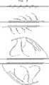
【図1】 図1は、多重鎖置換増幅(MSDA)の一例の図である。上部に示すのは、目的の核酸を含む二本鎖核酸分子である(斜線領域)。右のセットのプライマーおよび左のセットのプライマーがこの核酸分子にハイブリダイズする。中部に示すのは、各プライマーから伸長されつつある、複製される核酸の多重鎖である。各伸長鎖の末端におけるポリメラーゼは、その先のプライマーの伸長鎖を置換する。下部に示すのは、さらに伸長された、複製される核酸の多重鎖である。新たに複製された鎖におけるそれらの相補的な部位にハイブリダイズする次のセットのプライマーもまた示される。新たに複製された鎖によって、続きの鎖のポリメラーゼ伸長による置換を介してプライマーへのハイブリダイゼーションについて利用可能になる。
【図2】 図2は、ゲノム全体鎖置換増幅(WGSDA)の一例の図である。上部には、ゲノムDNAの図的表現がある。ランダムまたは部分的にランダムなプライマーのセットからのプライマーがその核酸分子に対してハイブリダイズする(プライマー長は、測定されることを意図しない)。単純さのために、ゲノムDNAの1分子の一部のみを示す。中部に示すのは、各プライマーから伸長されつつある、複製される核酸の多重鎖である。各伸長鎖の末端でのポリメラーゼが、それが遭遇する任意のプライマーの伸長鎖を置換する。ランダムまたは部分的にランダムなプライマーのセットからのさらなるプライマーもまた示し、これは、新たに複製された鎖における相補的部位にハイブリダイズする。新たに複製された鎖は、次の鎖を伸長するポリメラーゼによる置換を通してプライマーへのハイブリダイゼーションについて利用可能にされる。下部に示すのは、さらに伸長した、複製された核酸の多重鎖である。単純さのために、もとの合成鎖のうち4つのみ(上側の標的配列鎖上の2つおよび下側の標的配列鎖上の2つ)を、下部パネルに示す。
【図3】 図3は、多重鎖置換増幅(MSDA)の一例の図である。上部に示すのは、目的の核酸を含む二本鎖核酸分子である(斜線領域)。右のセットのプライマー(上部パネル中の上側鎖)および左のセットのプライマー(上部パネル中の下側の鎖)がこの核酸分子にハイブリダイズする。中部に示すのは、各プライマーから伸長されつつある、複製される核酸の多重鎖である。新たに複製された鎖のそれらの相補部位に対してハイブリダイズするプライマーの次のセットもまた示される。この新たに複製された鎖は、次の鎖を伸長するポリメラーゼによる置換を通してそのプライマーへのハイブリダイゼーションについて利用可能にされる。各伸長鎖の末端におけるポリメラーゼは、その先のプライマーの伸長鎖を置換する。下部に示すのは、さらに伸長した、複製された核酸の多重鎖である。単純さのために、もとの合成鎖のうち4つのみ(上側の標的配列鎖上の2つおよび下側の標的配列鎖上の2つ)を、下部パネルに示す。
【図4】 図4は、連結DNAの多重鎖置換増幅(MSDA−CD)の一例の図である。上部には、リンカーと連結したDNAの図的表現がある。中部には、リンカー配列と相補的なプライマーが、連結DNAの変性鎖にハイブリダイズされる(リンカーおよびプライマーの長さは測定されることを意図しない)。単純さのために、連結DNAの1分子の一部のみを示す。下部に示すのは、各プライマーから伸長されつつある、複製される核酸の多重鎖である。各伸長鎖の末端のポリメラーゼは、それが遭遇する任意のプライマーの伸長鎖を置換する。新たに複製された鎖上の複製されたリンカー配列における相補部位に対してハイブリダイズするさらなるプライマーもまた示される。新たに複製された鎖は、次の鎖を伸長するポリメラーゼによる置換を通じてそのプライマーとのハイブリダイゼーションについて利用可能にされる。
【配列表】
(Background of the Invention)
The disclosed invention is generally in the field of nucleic acid amplification.
[0002]
Many methods for exponential amplification have been developed. These include polymerase chain reaction (PCR), ligase chain reaction (LCR), self-sustained sequence replication (3SR), nucleic acid sequence-based amplification (NASBA), strand displacement amplification (SDA), and Qβ replicase (Birkenmeyer and Mushawar, J. Virological Methods, 35: 117-126 (1991); Landegren, Trends Genetics, 9: 199-202 (1993)).
[0003]
Current PCR amplification methods involve the use of two primers that hybridize to a region adjacent to the nucleic acid sequence of interest so that DNA replication initiated with that primer replicates the nucleic acid sequence of interest. By separating the replicated strand from the template strand using a denaturation step, another round of replication with the same primer can result in a geometric amplification of the nucleic acid sequence of interest. PCR amplification has the disadvantage that the amplification reaction cannot proceed continuously and must be performed by subjecting the nucleic acid sample to multiple cycles in a series of reaction conditions.
[0004]
Modification of PCR amplification, referred to as whole-genome PCR, involves the use of random or partially random primers that amplify the entire genome of an organism in the same PCR reaction. This technique relies on having a sufficient number of primers of random or partially random sequence such that primer pairs hybridize throughout the genomic DNA at moderate intervals. Replication initiated with that primer can then produce a duplicated strand overlap site to which another primer can hybridize. By subjecting this genomic sample to multiple overlapping cycles, its genomic sequence is amplified. Whole genome PCR has the same drawbacks as other forms of PCR.
[0005]
Another area where amplification is relevant is RNA expression profiling. Here, the aim is to determine the relative concentrations of many different molecular species in a biological sample. Some of the RNA of interest is present at relatively low concentrations and it is desirable to amplify them prior to analysis. It is impossible to amplify them using the polymerase chain reaction. This is because this mRNA mixture is typically a complex composed of 5,000 to 20,000 different molecular species. The polymerase chain reaction has the disadvantage that different molecular species amplify at different rates and distort the relative concentration of mRNA.
[0006]
Several procedures have been described that allow moderate amplification of all RNA simultaneously in a sample. For example, in Lockhart et al., Nature Biotechnology 14: 1675-1680 (1996), double-stranded cDNA was synthesized in such a way that a strong RNA polymerase promoter was incorporated at the end of each cDNA. This promoter sequence was then used to transcribe cDNA, producing approximately 100-150 RNA copies for each cDNA molecule. This weak amplification system allowed the creation of RNA profiles for biological samples containing at least 100,000 cells. However, there is a need for more powerful amplification methods that allow profile analysis of samples containing very few cells.
[0007]
Amplification of nucleic acids using multiplex primers and including strand displacement is described in European Patent Application 0 466 520 A1, and PCT Applications WO 95/25180 and WO 95/03430.
[0008]
Thus, there is a need for an amplification method that is less complex, more reliable, and provides more amplification in a shorter time.
[0009]
Accordingly, it is an object of the disclosed invention to provide a method for amplifying a target nucleic acid sequence in a continuous isothermal reaction.
[0010]
Another object of the disclosed invention is to provide a method for amplifying whole genomes or other highly complex nucleic acid samples in a continuous isothermal reaction.
[0011]
Another object of the disclosed invention is to provide a method for amplifying a target nucleic acid sequence, wherein multiple copies of the target nucleic acid sequence occur in a single amplification cycle.
[0012]
Another object of the disclosed invention is to provide a method for amplifying linked DNA in a continuous isothermal reaction.
[0013]
Another object of the disclosed invention is to provide a kit for amplifying a target nucleic acid sequence in a continuous isothermal reaction.
[0014]
Another object of the disclosed invention is to provide a kit for amplifying whole genomes or other highly complex nucleic acid samples in a continuous isothermal reaction.
[0015]
(Summary of the Invention)
Disclosed are compositions and methods for amplification of nucleic acid sequences of interest. This method is based on strand displacement amplification of nucleic acid sequences with multiple primers. In one preferred form of the method, referred to as multiple strand displacement amplification (MSDA), two sets of primers are used, the right set and the left set. The primers in the right set of primers each have a portion complementary to the nucleotide sequence adjacent to one side of the target nucleotide sequence, and the primers in the left set of primers each have the other side of the target nucleotide sequence Has a portion complementary to the nucleotide sequence adjacent to. The primers in the right set are complementary to one side of the nucleic acid molecule containing its target nucleotide sequence, and the primers in the left set are complementary to the opposite strand. The 5 'end of the primer in both sets is distal from the nucleic acid sequence of interest when the primer hybridizes to an adjacent sequence in the nucleic acid molecule. Preferably, each member of each set has a portion complementary to a distinct and non-overlapping nucleotide sequence adjacent to its target nucleotide sequence. Amplification proceeds by replication starting with each primer and continuing over its target nucleic acid sequence. The main feature of this method is the replacement of intervening primers during replication. Once the nucleic acid strand extending from the right set of primers reaches the region of the nucleic acid molecule to which the left set of primers hybridizes, and vice versa, another round of priming and replication occurs. This allows multiple copies of the nested set of the target nucleic acid sequence to be synthesized in a short period of time. By using a sufficient number of primers in the right and left sets, only a few replications are required to produce hundreds of thousands of copies of the nucleic acid sequence of interest. This disclosed method has advantages over the polymerase chain reaction. This is because it can be carried out under isothermal conditions. Thermal cycling is not required. This is because the polymerase at the head of the extended strand (or compatible strand displacement protein) displaces its previous strand, thereby making hybridization available. Other advantages of multiple strand displacement amplification include the ability to amplify very long nucleic acid segments (on the order of 50 kilobases) and the ability to rapidly amplify shorter segments (less than 10 kilobases). In multiple strand displacement amplification, a single priming event at unintended sites does not result in artificial amplification at these sites (because single strand replication at sites where amplification at the intended site is not intended) Because it surpasses quickly.)
[0016]
In another preferred form of this method, referred to as whole genome strand displacement amplification (WGSDA), a random set of primers is used to randomly prime a sample of genomic nucleic acid (or another sample of highly complex nucleic acid). To do. By selecting a sufficiently large set of primers of random or partially random sequence, the primers in the set are comprehensively and randomly complementary to the nucleic acid sequences distributed across the nucleic acids in the sample. Become. Amplification proceeds by replication with each primer being highly processive with polymerase initiation and continuing until natural termination. A key feature of this method is the replacement of intervening primers during replication by polymerase. In this way, multiple duplicate copies of the entire genome can be synthesized in a short time. This method has advantages over the polymerase chain reaction. This is because this can be done under isothermal conditions. Other advantages of whole-genome strand displacement amplification include higher levels of amplification (up to 5 times higher) than whole-genome PCR, where amplification is less sequence-dependent than PCR and can be performed with PCR. There are no annealed artifacts or gene shuffling artifacts (since there is no denaturation and reannealing cycle).
[0017]
In another preferred form of this method, referred to as ligated DNA multiple strand displacement amplification (MSDA-CD), the fragments of DNA are preferably first ligated together with a linker. The ligated DNA is then amplified by strand displacement synthesis using appropriate primers. A random set of primers can be used to randomly prime the synthesis of DNA concatamers in a manner similar to whole genome amplification. By selecting a sufficiently large set of primers of random or partially random sequences, such as in whole genome amplification, the primers in that set will be totally and randomly complementary to the nucleic acid sequences distributed over the ligated DNA It becomes the target. When using a linker to link DNA fragments, primers complementary to the linker sequence can be used to amplify the concatamer. From that linker, synthesis proceeds through a section of linked DNA to the next linker and continues beyond. As the linker region is replicated, a new priming site for DNA synthesis is created. In this way, multiple duplicate copies of the entire ligated DNA sample can be synthesized in a short time.
[0018]
After amplification, the amplified sequence can be for any purpose, for example for known and established applications for PCR amplified sequences. For example, the amplified sequence can be detected using any of the conventional detection systems for nucleic acids such as detection of fluorescent labels, enzyme-linked detection systems, antibody-mediated label detection, and detection of radiolabels. A key feature of the disclosed method is that amplification occurs in continuous isothermal replication rather than cycling. This makes the amplification less complex and much more consistent in output. Strand displacement allows for the rapid generation of multiple copies of a nucleic acid sequence or sample in a single continuous isothermal reaction. The DNA produced using the disclosed invention can then be used for any purpose or any other desired method. For example, PCR can be used to further amplify any particular DNA sequence previously amplified by the genome-wide strand displacement method.
[0019]
(Detailed description of the invention)
The disclosed methods use specific materials and procedures that allow amplification of target nucleic acid sequences and whole genomes or other highly complex nucleic acid samples.
[0020]
(I. Material)
(A. Target sequence)
The target sequence, which is the object of amplification, can be any nucleic acid. A target sequence may comprise multiple nucleic acid molecules, eg, multiple sites in a nucleic acid molecule, or a single region of a nucleic acid molecule in the case of whole genome amplification. For multiple strand displacement amplification, generally the target sequence is a single region in a nucleic acid molecule or nucleic acid sample. For whole genome amplification, the target sequence is the whole genome or a nucleic acid sample. The target sequence can be any nucleic acid sample of interest. The source, identification, and preparation of many such nucleic acid samples are known. Preferably, nucleic acid samples that are known or identified for use in amplification or detection methods are used for the methods described herein. The nucleic acid sample can be a nucleic acid sample derived from a single cell. For multiple strand displacement amplification, preferred target sequences are sequences that are difficult to amplify using PCR, for example due to length or composition. For whole genome amplification, the preferred target sequence is a nucleic acid sample from a single cell. For multiple strand displacement amplification of ligated DNA, the target is ligated DNA. Target sequences for use in the disclosed methods are preferably complex and non-repetitive (except for linkers in linker-linked DNA and sections of repetitive DNA in genomic DNA) or Part of the sample.
[0021]
Target sequence for multiple strand displacement amplification: Multiple sites in a nucleic acid sample can be amplified simultaneously in the same MSDA reaction, but for convenience, in the following discussion, a single nucleic acid of interest to be amplified is Reference to sequence features. This sequence is referred to below as the target sequence. The target sequence for MSDA preferably includes two types of target regions (amplification target and hybridization target). The hybridization target includes a sequence in the target sequence that is complementary to a primer in the primer set. An amplification target is a portion of the target sequence that is to be amplified. For this purpose, the amplification target is preferably downstream of or adjacent to the hybridization target. There is no specific sequence or structural requirement to select a target sequence. Hybridization targets and amplification targets within the target sequence are defined by the relationship of the target sequence to the primers in the primer set. Primers are designed to match the selected target sequence. The upper panel of FIG. 1 shows an example of how the primers in the primer set can define regions in the target sequence. Since the sequence in and around the site where the primer hybridizes is amplified, it is preferable that the sequence to be amplified is separated from the hybridization site of the primer, but this is not required. An example of this is shown in FIG.
[0022]
In multiple strand displacement amplification of linker-linked DNA, the DNA fragment bound by the linker is the amplification target and the linker is the hybridization target. Hybridization targets (ie, linkers) contain sequences that are complementary to the primers used for amplification. A preferred form of ligated DNA for amplification is ligated cDNA.
[0023]
(B. Primer)
Primers for use in the disclosed amplification method are oligonucleotides having sequences complementary to the target sequence. This sequence is referred to as the complementary portion of the primer. The complementary portion of the primer can be any length that supports specific and stable hybridization between the primer and the target sequence. Generally, this is 10-35 nucleotides long, but preferably 16-24 nucleotides long. For whole genome amplification, the primers are preferably 12-60 nucleotides in length.
[0024]
The primer also preferably includes additional sequences at the 5 ′ end of the primer that are not complementary to the target sequence. This sequence is referred to as the non-complementary part of the primer. The non-complementary portion of the primer, when present, acts to promote stable substitution during DNA replication. The non-complementary portion of the primer can also include a functional sequence such as a promoter for RNA polymerase. The non-complementary portion of the primer can be of any length, but is generally 1-100 nucleotides long and preferably 4-8 nucleotides long. If random or partially random primers are used for whole genome amplification, the use of non-complementary portions is not preferred.
[0025]
Primers for multiple strand displacement amplification: In the case of multiple strand displacement amplification, the complementary portion of each primer is designed to be complementary to the hybridization target in the target sequence. In the primer set, the complementary part of each primer is preferably complementary to a different part of the target sequence. More preferably, the primers in this set are complementary to adjacent sites in the target sequence. It is also preferred that such adjacent sites in the target sequence are also adjacent to the amplified target in the target sequence.
[0026]
When hybridizing to a target sequence, the primers in the primer set are preferably separated from one another. When hybridized, the primers in the primer set are preferably separated from each other by at least 5 bases. When hybridized, the primers in the primer set are preferably separated from each other by at least 10 bases. Even more preferably, when hybridized, the primers in the primer set are at least 20 bases away from each other. Even more preferably, when hybridized, the primers in the primer set are at least 30 bases away from each other. Even more preferably, when hybridized, the primers in the primer set are at least 40 bases away from each other. Even more preferably, when hybridized, the primers in the primer set are at least 50 bases away from each other.
[0027]
When hybridized, the primers in the primer set are preferably separated from each other by about 500 bases or less. When hybridized, the primers in the primer set are more preferably separated from each other by about 400 bases or less. Even more preferably, when hybridized, the primers in the primer set are separated from each other by about 300 bases or less. Even more preferably, when hybridized, the primers in the primer set are separated from each other by about 200 bases or less. Preferred upper and lower combinations of the above separations are specifically contemplated and include all intermediate ranges. When hybridized, the primers in the primer set need not be separated from each other by the same number of bases. When hybridized, the primers in the primer set are preferably separated from each other by substantially the same number of bases.
[0028]
The optimal separation distance between primers is not the same for all DNA polymerases. This is because this parameter depends on the net polymerization rate. Progressive DNA polymerase has a characteristic polymerization rate that can range from 5 to 300 nucleotides per second and can be affected by the presence or absence of accessory ssDNA binding proteins and helicases. In the case of non-progressive polymerases, the net polymerization rate depends on the enzyme concentration. This is because at higher concentrations there are more restart events and the net polymerization rate is increased. An example of a progressive polymerase is φ29 DNA polymerase, which proceeds at 50 nucleotides per second. An example of a non-progressive polymerase is Vent exo (-) DNA polymerase, which provides an effective polymerization rate of 4 nucleotides per second at low concentrations, or 16 nucleotides per second at higher concentrations.
[0029]
In order to obtain an optimal yield in the MSDA reaction, the primer spacing is preferably adjusted for the polymerase used. Long primer spacing is preferred when using a polymerase with a rapid polymerization rate. Shorter primer spacing is preferred when using polymerases with slower polymerization rates. The following assay can be used to determine the optimal interval with any polymerase. This assay uses a primer set, where each primer is made up of 5 left primers and 5 right primers. The primer set is designed to hybridize adjacent to the same target sequence with each of the different sets of primers having different primer spacings. The spacing is systematically varied between primer sets with increments of 25 nucleotides in the range of 25 nucleotides to 400 nucleotides (primer spacing within each set is the same). A series of reactions are performed, where the same target sequence is amplified using different sets of primers. The interval that produces the highest experimental yield of DNA is the optimal primer interval for the particular DNA polymerase, or combination of DNA polymerase and accessory protein used.
[0030]
DNA replication initiated at a site in the target sequence to which the primer hybridizes extends from the primer hybridized to the adjacent site to the replicated strand and displaces it. Flanking strand replacement makes the adjacent strand available for hybridization to another primer and subsequent initiation of another round of replication. The region of the target sequence to which the primer hybridizes is called the hybridization target of the target sequence. The upper panel of FIG. 1 shows one of the preferred relationships of the target sequence and the primer set to the amplified target of the target sequence.
[0031]
A primer set can include any desired number of primers of different nucleotide sequences. For MSDA, the primer set preferably comprises a plurality of primers. More preferably, the primer set comprises 3 or more primers. Even more preferably, the primer set comprises 4 or more, 5 or more, 6 or more, or 7 or more primers. In general, the more primers used, the higher the level of amplification obtained. There is no essential upper limit on the number of primers that a primer set can have. However, for a given target sequence, the number of primers in the primer set is generally limited to the number of hybridization sites available in the target sequence. For example, if the target sequence is a 10,000 nucleotide DNA molecule and a 20 nucleotide primer is used, there are 500 non-overlapping 20 nucleotide sites in the target sequence. Even more primers can be used if overlapping sites are desired or acceptable. It is preferred that the primer set comprises about 300 or more primers. It is preferred that the primer set comprises about 200 or more primers. Even more preferably, the primer set comprises about 100 or more primers. More preferably, the primer set comprises about 50 or more primers. Most preferably, the primer set comprises 7 to about 50 primers. Any combination of preferred upper and lower limits for the number of primers in the above primer set is specifically contemplated and includes all intermediate ranges.
[0032]
A preferred form of primer set for use in MSDA includes two sets of primers: a right primer set and a left primer set. The right and left primer sets are designed to be complementary to the opposite strand of the target sequence. Preferably, the complementary portions of the right primer set are each complementary to the right hybridization target, and each is complementary to a different portion of the right hybridization target. Preferably, the complementary portions of the left primer set are each complementary to the left hybridization target, and each is complementary to a different portion of the left hybridization target. The right hybridization target and the left hybridization target are adjacent to opposite ends of the amplification target in the target sequence. A preferred form of these relationships is shown in the upper panel of FIG. It is preferred that each of the right primer set and the left primer set comprises the preferred number of primers described above for the primer set. In particular, the right primer set or the left primer set preferably includes a plurality of primers for the primer set. More preferably, the right primer set or the left primer set comprises three or more primers. Even more preferably, the right primer set or the left primer set comprises 4 or more, 5 or more, 6 or more, or 7 or more primers. It is preferred that the right primer set or the left primer set comprises about 200 or less primers for the primer set. More preferably, the right primer set or the left primer set comprises no more than about 100 primers for the primer set. Most preferably, each of the right primer set or the left primer set comprises 7 to about 100 primers for the primer set. Any combination of preferred upper and lower limits for the number of primers in the above primer set is specifically contemplated, including all intermediate ranges. It is also preferred that for a given target sequence, the right primer set and the left primer set contain the same number of primers. It is also preferred that for a given target sequence, the right primer set and the left primer set are composed of primers of similar length and / or hybridization characteristics.
[0033]
Primers for genome-wide strand displacement amplification: In the case of genome-wide strand displacement amplification, it is preferred to use a primer set having a random or partially random nucleotide sequence. In a remarkably complex nucleic acid sample (which is the preferred target sequence for WGSDA), the specific nucleic acid sequence present in the sample need not be known and the primer is complementary to any specific sequence Need not be designed to be. Rather, the complexity of the nucleic acid sample results in a large number of different hybridization target sequences in the sample that are complementary to various primers of random or partially random sequence. The complementary portion of a primer for use in WGSDA can be fully randomized, have only the portion that is randomized, or can be selectively randomized in other ways.
[0034]
The number of random base positions in the complementary part of the primer is preferably 20% to 100% of the total number of nucleotides in the complementary part of the primer. More preferably, the number of random base positions is 30% to 100% of the total number of nucleotides in the complementary portion of the primer. Most preferably, the number of random base positions is 50% to 100% of the total number of nucleotides in the complementary portion of the primer. Primer sets with random or partially random sequences can be synthesized using standard techniques by allowing the addition of any nucleotide at each position to be randomized. It is also preferred that the primer set is composed of primers of similar length and / or hybridization characteristics.
[0035]
Primers for multiple strand displacement amplification with ligated DNA: For multiple strand displacement amplification of ligated DNA, primer sets with random or partially random nucleotide sequences can be used. In a remarkably complex nucleic acid sample (eg, DNA linked from a mixture of many sequences), the specific nucleic acid sequence present in the sample need not be known and the primer is complementary to any particular sequence It need not be designed to be objective. Rather, the complexity of the nucleic acid sample results in the majority of different hybridization target sequences in the sample being complementary to various primers of random or partially random sequence. The complementary portion of a primer for use in MSDA-CD can be fully randomized, have only the portion that is randomized, or can be selectively randomized in other ways.
[0036]
The number of random base positions in the complementary part of the primer is preferably 20% to 100% of the total number of nucleotides in the complementary part of the primer. More preferably, the number of random base positions is 30% to 100% of the total number of nucleotides in the complementary portion of the primer. Most preferably, the number of random base positions is 50% to 100% of the total number of nucleotides in the complementary portion of the primer. Primer sets with random or partially random sequences can be synthesized using standard techniques by allowing the addition of any nucleotide at each randomized position. It is also preferred that the primer set is composed of primers of similar length and / or hybridization characteristics.
[0037]
If the DNA is linked to a linker, amplification can be performed using primers that are complementary to the sequences in the linker. This is the preferred form of MSDA-CD. It is preferred that the complementary portion of each primer is designed to be complementary to the sequence in the linker. Primers for use with linker-linked DNA preferably include primers that are complementary to both strands of the linker sequence. This is shown in FIG. It is also preferred that the primers are not complementary to each other. This prevents the primers from hybridizing to each other. If the linker used to join the DNA is long enough, primer sets complementary to different parts of the linker sequence can be used. This is equivalent to the situation in MSDA, and primer sets can be designed and used in the same manner as discussed for MSDA primer sets. Random primers can be used to amplify the ligated DNA whether or not a linker is used to ligate the DNA.
[0038]
It is preferred that the target sequence for use in MSDA, WGSDA, and MSDA-CD is not, or not part of, a nucleic acid molecule composed of multiple tandem repeats of the sequence. More preferably, the target sequence is not a nucleic acid molecule composed of multiple tandem repeats of a single sequence or is not part thereof. Most preferably, it is not, or not part of, a nucleic acid molecule composed of multiple tandem repeats of a single sequence synthesized by rotating circular replication. An example of such a tandem repeat DNA made by rotating a circular replica is the tandem sequence DNA described in WO 97/19193. DNA ligated from identical or nearly identical DNA fragments is not made by rotating circular replication, so nucleic acids composed of multiple tandem repeats of a single sequence synthesized by rotating circular replication Not a molecule. Thus, a target sequence is not a nucleic acid molecule composed of multiple tandem repeats of a single sequence, but several such target sequences (eg, DNA linked from identical or nearly identical DNA fragments) are Preferred over nucleic acid molecules composed of multiple tandem repeats of a single sequence synthesized by rotating circular replication (eg, tandem sequence DNA described in WO 97/19193). It is preferred that the target sequence for the disclosed method is not generated by the method described in WO 97/19193.
[0039]
Detection tag: The non-complementary portion of the primer may comprise a sequence to be used to further manipulate or analyze the amplified sequence. An example of such a sequence is a detection tag, which is a specific nucleotide sequence present in the non-complementary portion of the primer. The detection tag has a sequence complementary to the detection probe. Detection tags can be detected using their cognate detection probes. The detection tag becomes incorporated at the end of the amplified strand. The result is amplified DNA having a detection tag sequence that is complementary to the complementary portion of the detection probe. If present, there may be more than 1, 2, 3, or 3 detection tags on the primer. It is preferred that the primer has 1, 2, 3, or 4 detection tags. Most preferably, the primer has one detection tag. In general, the primer preferably has 10 or fewer detection tags. Except for the size of the primer, there is no essential limitation on the number of detection tags that can be present on the primer. When multiple detection tags are present, they can have the same sequence, or they can have different sequences, each different sequence being complementary to a different detection probe. It is preferred that the primers contain detection tags and have the same sequence so that they are all complementary to a single detection probe. For some multiple detection methods, it is preferred that the primer comprises up to 6 detection tags and that the detection tag portions have different sequences such that each of the detection tag portions is complementary to a different detection probe. Similar effects can be achieved by using primer sets each having a single different detection tag. Each detection tag can be of any length that supports specific and stable hybridization between the detection tag and the detection probe. For this purpose, a length of 10 to 35 nucleotides is preferred and the detection tag portion is most preferably 15 to 20 nucleotides long.
[0040]
Address Tag: Another example of a sequence that can be included in the non-complementary portion of a primer is an address tag. The address tag has a sequence complementary to the address probe. The address tag becomes incorporated at the end of the amplified strand. The result is amplified DNA having an address tag sequence that is complementary to the complementary portion of the address probe. If present, there can be one or more than one address tag on the primer. It is preferred that the primer has 1 or 2 address tags. Most preferably, the primer has one address tag. In general, it is preferred that the primer has 10 or fewer address tags. Except for the size of the primer, there is no essential limitation on the number of address tags that can be present on the primer. When multiple address tags are present, they can have the same sequence, or they can have different sequences, each different sequence being complementary to a different address probe. It is preferred that the primers contain address tags and have the same sequence so that they are all complementary to a single address probe. Each address tag portion can be of any length that supports specific and stable hybridization between the address tag and the address probe. For this purpose, a length of 10 to 35 nucleotides is preferred and the address tag portion is most preferably 15 to 20 nucleotides long.
[0041]
(C. Linker)
As used herein for ligated DNA, a linker is a small double stranded DNA molecule. For MSDA-CD, the linker serves two main purposes: to facilitate ligation of DNA fragments and to facilitate amplification. For the first purpose, linkers are generally designed to have ends that are compatible with the ends of the DNA fragments to be ligated. For example, if the DNA fragment has blunt ends (or the ends are blunted), blunt ended linkers are used. For DNA fragments that are terminated with one or more nucleotides, the linker should have a complementary tail. An example of such tailing is the addition of a single adenosine residue to the 3 ′ end of the cDNA. In order to facilitate amplification, the linker should have one or more sequences complementary to the primers to be used in MSDA-CD. Such a sequence is referred to as the primer complementary portion of the linker. The primer complementary portion of the linker is complementary to the complementary portion of the primer. A primer-complementary portion can have any sequence as long as it is complementary to the intended primer portion. If primer complementary portions are present on the opposite strand of the linker, they should not overlap. A primer can also have one or more restriction enzyme cleavage sites. Such restriction sites allow the amplified DNA to be cleaved into fragments, and preferably fragments that represent the original DNA fragment ligated. For this purpose, it is preferred that rare restriction sites (for example, 8-base recognition sites) are used. An example of the structure of this type of linker is shown below:
[0042]
[Chemical 1]
[0043]
[Chemical formula 2]
[0044]
(D. Detection label)
To assist in the detection and quantification of nucleic acid amplified using the disclosed methods, the detection label can be incorporated directly into the amplified nucleic acid or can be bound to a detection molecule. As used herein, a detection label is any molecule that can associate directly or indirectly with an amplified nucleic acid and produce a measurable and detectable signal, either directly or indirectly. It is. Many such labels for incorporation into nucleic acids or for binding to nucleic acid probes are known to those skilled in the art. Examples of suitable detection labels for use in the disclosed methods are radioisotopes, fluorescent molecules, phosphorescent molecules, enzymes, antibodies, and ligands.
[0045]
Examples of suitable fluorescent labels are fluorescein (FITC), 5,6-carboxymethylfluorescein, Texas Red, nitrobenz-2-oxa-1,3-diazol-4-yl (NBD), coumarin, dansyl chloride, rhodamine, 4'-6-diamidino-2-phenylindole (DAPI) and the cyanine dyes Cy3, Cy3.5, Cy5, Cy5.5 and Cy7. Preferred fluorescent labels are fluorescein (5-carboxyfluorescein-N-hydroxysuccinimide ester) and rhodamine (5,6-tetramethylrhodamine). Preferred fluorescent labels are FITC and the cyanine dyes Cy3, Cy3.5, Cy5, Cy5.5, and Cy7. The absorption maximum and emission maximum of these phosphors are respectively: FITC (490 nm; 520 nm), Cy3 (554 nm; 568 nm), Cy3.5 (581 nm; 588 nm), Cy5 (652 nm; 672 nm). ), Cy5.5 (682 nm; 703 nm), and Cy7 (755 nm; 778 nm), thus allowing their simultaneous detection. Fluorescent labels are available from a variety of commercial sources, including Molecular Probes, Eugene, OR and Research Organics, Cleveland, Ohio.
[0046]
Labeled nucleotides are a preferred form of detection label. This is because they can be directly incorporated into amplification products during synthesis. Examples of detection labels that can be incorporated into amplified DNA or RNA include BrdUrd (Hoy and Schimke, Mutation Research 290: 217-230 (1993)), BrUTP (Wansick et al., J. Cell Biology 122: 283-293 (1993). )), And biotin (Langer et al., Proc. Natl. Acad. Sci. USA 78: 6633 (1981)) or digoxigenin (Kerkhof, Anal. Biochem. 205: 359-364 (1992)). Includes nucleotide analogs such as modified nucleotides. Suitable fluorescently labeled nucleotides are fluorescein-isothiocyanate-dUTP, cyanine-3-dUTP, and cyanine-5-dUTP (Yu et al., Nucleic Acids Res., 22: 3226-3232 (1994). The preferred nucleotide analog detection label of is BrdUrd (BUDR triphosphate, Sigma) and the preferred nucleotide analog detection label of RNA is biotin-16-uridine-5′-triphosphate (biotin-16-dUTP, Boehringher). Fluorescein, Cy3, and Cy5 can bind to dUTP for direct labeling, Cy3.5 and Cy7 are biotin-labeled probes or digoxigenin-labeled probes. Available as avidin or anti-digoxigenin conjugate for subsequent detection.
[0047]
Detection labels (eg, biotin) that are incorporated into the amplified nucleic acid can subsequently be detected using sensitive methods well known in the art. For example, biotin is a streptavidin-alkaline phosphatase conjugate (Tropix, Inc.) (which binds to biotin followed by a suitable substrate (eg, chemiluminescent substrate CSPD: 3- (4-methoxyspiro- [1 , 2, -Dioxeethane-3-2 '-(5'-chloro) tricyclo [3.3.1.13.7 ] Decan] -4-yl) phenyl phosphate disodium; Tropix, Inc. ), Which is then detected by chemiluminescence).
[0048]
A preferred detection label for use in the detection of amplified RNA is an acridinium-ester labeled DNA probe (described in GenProbe, Inc., Arnold et al., Clinical Chemistry 35: 1588-1594 (1989)). Acridinium-ester labeled detection probes allow detection of amplified RNA without washing. This is because unhybridized probes can be destroyed with alkali (Arnold et al., (1989)).
[0049]
Molecules that combine two or more of these detection labels are also considered detection labels. Any known detection label can be used with the disclosed probes, tags, and methods to label and detect nucleic acids amplified using the disclosed methods. Methods for detecting and measuring the signal produced by the detection label are also known to those skilled in the art. For example, radioactive isotopes can be detected by scintillation counting or direct visualization; fluorescent molecules can be detected with a fluorescence spectrophotometer; phosphorescent molecules can be detected with a spectrophotometer or directly visualized with a camera The enzyme can be detected by detection or visualization of the product of the reaction catalyzed by the enzyme; the antibody can be detected by detecting a secondary detection label bound to the antibody. As used herein, a detection molecule is a molecule that interacts with amplified nucleic acid and has one or more detection labels attached thereto.
[0050]
(E. Detection probe)
A detection probe is a labeled oligonucleotide having a sequence complementary to a detection tag on the amplified nucleic acid. The complementary portion of the detection probe can be of any length that supports specific and stable hybridization between the detection probe and the detection tag. For this purpose, a length of 10-35 nucleotides is preferred, and the complementary portion of a detection probe 16-20 nucleotides long is most preferred. The detection probe can include any of the detection labels described above. Preferred labels are biotin and fluorescent molecules. A particularly preferred detection probe is a molecular beacon. Molecular beacons are detection probes that are labeled with a fluorescent moiety that fluoresces only when the detection probe is hybridized (Tyagi and Kramer, Nature Biotechnol. 14: 303-309 (1995)). Use of such probes eliminates the need to remove unhybridized probes prior to label detection. This is because a detection probe that is not hybridized does not generate a signal. This is particularly useful for complex assays.
[0051]
(F. Address probe)
An address probe is an oligonucleotide having a sequence complementary to an address tag on a primer. The complementary portion of the address probe can be of any length that supports specific and stable hybridization between the address probe and the address tag. For this purpose, a length of 10 to 35 nucleotides is preferred, with the complementary portion of the address probe being 12 to 18 nucleotides long being most preferred. The address probe can include a single complementary portion or multiple complementary portions. Preferably, the address probe is attached to the solid support either directly or via a spacer molecule. Such a combination of address probe and solid support is a preferred form of solid state detector.
[0052]
(G. Oligonucleotide synthesis)
Primers, detection probes, address probes, and any other oligonucleotides can be synthesized using established oligonucleotide synthesis methods. Methods for producing or synthesizing oligonucleotides are well known in the art. Such methods include nucleotide fragment isolation following standard enzymatic digestion (see, eg, Sambrook et al., Molecular Cloning: A Laboratory Manual, 2nd Edition (Cold Spring Harbor Laboratory Press, Cold Spring Harbor, N. Y. 9). ) See chapters 5 and 6)) from pure synthesis methods, such as Milligen or Beckman System 1 Plus DNA synthesizers (eg Millingen-Biosearch, Burlington, MA Model 8700 automatic synthesizer, or ABI Model). 380B) to the synthesis method by the cyanoethyl phosphoramidite method. Synthetic methods useful for making oligonucleotides are also described by Ikuta et al., Ann. Rev. Biochem. 53: 323-356 (1984) (phosphotriester and phosphite-triester method) and Narang et al., Methods Enzymol. 65: 610-620 (1980) (phosphotriester method). Protein nucleic acid molecules are described in Nielsen et al., Bioconjug. Chem. 5: 3-7 (1994) and can be made using known methods.
[0053]
Many of the oligonucleotides described herein are designed to be complementary to specific portions of other oligonucleotides or nucleic acids so that stable hybrids can be formed between them. The stability of these hybrids is described by Lesnick and Freier, Biochemistry 34: 10807-10815 (1995), McGraw et al., Biotechniques 8: 674-678 (1990), and Rychlik et al., Nucleic Acids Res. 18: 6409-6612 (1990) and can be calculated using known methods.
[0054]
(H. Solid state detector)
Solid state detectors are solid substrates or supports that have address probes or detection molecules attached to them. A preferred form of solid state detector is an array detector. An array detector is a solid state detector to which a plurality of different address probes or detection molecules are bound in an array, lattice, or other organized manner.
[0055]
A solid substrate for use in a solid state detector can include any solid material to which an oligonucleotide can be bound. This is acrylamide, cellulose, nitrocellulose, glass, polystyrene, polyethylene vinyl acetate, polypropylene, polymethacrylate, polyethylene, polyethylene oxide, glass, polysilicate, polycarbonate, Teflon, fluorocarbon, nylon, silicone rubber, polyanhydride, polyglycol Includes materials such as acids, polylactic acid, polyorthoesters, polypropyl fumarate, collagen, glycosaminoglycans, and polyamino acids. The solid substrate can have any useful form, including thin films or membranes, beads, bottles, dishes, fibers, nonwovens, shaped polymers, particles, and microparticles. A preferred form for the solid substrate is a microtiter dish. The most preferred form of microtiter dish is the standard 96 well type.
[0056]
An address probe immobilized on a solid substrate allows for the capture of the products of the disclosed amplification method on a solid state detector. Such capture provides a convenient means of washing away reactive components that can interfere with subsequent detection steps. By attaching different address probes to different regions of the solid state detector, different amplification products can be captured at different locations on the solid state detector, and thus diagnostic locations. For example, in a microtiter plate complex assay, address probes specific for up to 96 different amplified nucleic acids, each representing a different target sequence amplified through a different set of primers, are It can be fixed on a titer plate. Capture and detection occurs only in the well corresponding to the amplified nucleic acid where the target sequence corresponding to the amplified nucleic acid was present in the sample.
[0057]
Methods for immobilizing oligonucleotides to a solid substrate are well established. Oligonucleotides, including address probes and detection probes, can be bound to the substrate using established binding methods. For example, suitable attachment methods are described by Pease et al., Proc. Natl. Acad. Sci. USA 91 (11): 5022-5026 (1994), and Krapko et al., Mol Biol (Mosk) (USSR) 25: 718-730 (1991). Methods for immobilizing 3 ′ amine oligonucleotides on casein-coated slides are described in Stimpson et al., Proc. Natl. Acad. Sci. USA 92: 6379-6383 (1995). A preferred method of attaching oligonucleotides to a solid substrate is described by Guo et al., Nucleic Acids Res. 22: 5456-5465 (1994).
[0058]
(I. Solid sample)
A solid sample is a solid substrate or support to which the target sequence is bound or adhered. The target sequence is preferably delivered into the target sample or assay sample. A preferred form of the solid sample is an array sample. An array sample is a solid sample to which a plurality of different target samples are bound or adhered in an array, lattice, or other organized manner.
[0059]
A solid substrate for use in a solid sample can include any solid material to which the target sequence can be bound or adhered. This is acrylamide, cellulose, nitrocellulose, glass, polystyrene, polyethylene vinyl acetate, polypropylene, polymethacrylate, polyethylene, polyethylene oxide, glass, polysilicate, polycarbonate, Teflon, fluorocarbon, nylon, silicone rubber, polyanhydride, polyglycol Includes materials such as acids, polylactic acid, polyorthoesters, polypropyl fumarate, collagen, glycosaminoglycans, and polyamino acids. The solid substrate can have any useful form, including thin films or membranes, beads, bottles, dishes, slides, fibers, woven fibers, shaped polymers, particles, and microparticles. Preferred forms for solid substrates are microtiter dishes and glass slides. The most preferred form of microtiter dish is the standard 96 well type.
[0060]
The target sequence immobilized on the solid substrate allows the formation of target specific amplified nucleic acids that are localized on the solid substrate. Such localization provides a convenient means of washing away reaction components that may interfere with subsequent detection steps and provides a convenient method of assaying multiple different samples simultaneously. Amplified nucleic acids can be formed independently at each site where different samples adhere. The methods described above can be used for immobilization of target sequences or other oligonucleotide molecules to form a solid sample.
[0061]
The preferred form of the solid substrate is a glass slide on which up to 256 separate target samples are adhered as an array of small dots. Each point is preferably 0.1 to 2.5 mm in diameter, and most preferably approximately 2.5 mm in diameter. Such microarrays can be manufactured, for example, using the method described by Schena et al., Science 270: 487-470 (1995). Briefly, microarrays can be produced on poly-L-lysine coated microscope slides (Sigma) by an array machine equipped with printed chips. The chip is filled with, for example, 1 μl of DNA sample (0.5 mg / ml) from a 96-well microtiter plate and placed at approximately 0.005 μl per slide on multiple slides at desired intervals . The printed slides were then rehydrated in a humid chamber for 2 hours, snap-dried at 100 ° C. for 1 minute, rinsed in 0.1% SDS, and 50% 1-methyl-2 Can be treated with 0.05% succinic anhydride prepared in a buffer consisting of pyrrolidinone and 50% boric acid. The DNA on the slide can then be denatured in distilled water, for example, at 90 ° C. for 2 minutes just prior to use. The microarray solid sample can be scanned, for example, by a laser fluorescence scanner with a computer controlled XY stage and a microscope objective. Mixed gas, multi-line lasers allow continuous excitation of multiple fluorescent materials.
[0062]
(J. DNA polymerase)
DNA polymerases useful in multiplex displacement amplification must be able to displace hybridized strands encountered during replication, either alone or in combination with a compatible strand displacement factor. Such a polymerase is referred to herein as a strand displacement DNA polymerase. It is preferred that the strand displacement DNA polymerase lacks 5 ′ → 3 ′ exonuclease activity. Strand displacement is necessary to result in the synthesis of multiple copies of the target sequence. The 5 ′ → 3 ′ exonuclease activity, if present, can result in synthetic strand breaks. It is also preferred that the DNA polymerase for use in the disclosed methods is highly progressive. The suitability of a DNA polymerase for use in the disclosed methods can be readily determined by assessing its ability to perform strand displacement replication. Preferred strand displacement DNA polymerases include Bst large fragment DNA polymerases (Exo (-) Bst; Aliotta et al., Genet. Anal. (Netherlands) 12: 185-195 (1996)) and exo (-) Bca DNA polymerase (Walker and Linn, Clinical Chemistry 42: 1604-1608 (1996)). Other useful polymerases include bacteriophage φ29 DNA polymerase (Blanco et al., US Pat. Nos. 5,198,543 and 5,001,050), phage M2 DNA polymerase (Matsumoto et al., Gene 84: 247). (1989)), phage φPRD1 DNA polymerase (Jung et al., Proc. Natl. Acad. Sci. USA 84: 8287 (1987)), exo (−) VENT® DNA polymerase (Kong et al., J. Biol. Chem. 268: 1965-1975 (1993)), Klenow fragment of DNA polymerase I (Jacobsen et al., Eur. J. Biochem. 45: 623-627 (1974)), T5 DNA polymerase ( hatterjee et al., Gene 97: 13-19 (1991)), Sequenase (US Biochemicals) PRD1 DNA polymerase (Zhu and Ito, Biochim. Biophys. Acta. 1219: 267-276 (1994)), and T4 DNA polymerase. Holoenzymes (Kaboord and Benkovic, Curr. Biol. 5: 149-157 (1995)). Most preferred is exo (-) Bst DNA polymerase.
[0063]
Strand displacement can be facilitated by the use of a strand displacement factor (eg, helicase). Any DNA polymerase capable of performing strand displacement replication in the presence of a strand displacement factor may be used in the disclosed methods even if the DNA polymerase does not perform strand displacement replication in the absence of such a factor. Therefore, it is considered appropriate. BMRF1 polymerase accessory subunits (Tsurumi et al., J. Virology 67 (12): 7648-7653 (1993)), adenovirus DNA binding proteins (Zijderveld and van der Vliet, J. et al., 2000). Virology 68 (2): 1158-1164 (1994)), herpes simplex virus protein ICP8 (Boehmer and Lehman, J. Virology 67 (2): 711-715 (1993); Skaliter and Lehman, Proc. Natl. USA 91 (22): 10665-10669 (1994)), single stranded DNA binding protein (SSB; Rigler and Ro). mano, J. Biol. Chem. 270: 8910-8919 (1995)); T4 phage gene 32 protein (Villemain and Giedroc, Biochemistry 35: 14395-14404 (1996)) and bovine thymus helicase (Siegel et al., J. Biol. Chem. 267: 13629-13635 (1992)).
[0064]
The ability of a polymerase to perform strand displacement replication can be determined by using the polymerase in a strand displacement replication assay, such as the assays described in Examples 1 and 2. The assay of this example can be modified as appropriate. For example, helicase can be used instead of SSB. Such an assay should be performed at a temperature suitable for optimal activity for the enzyme used. For example, it is 32 ° C. for φ29 DNA polymerase, 46 ° C. to 64 ° C. for exo (−) Bst DNA polymerase, or about 60 ° C. to 70 ° C. for enzymes from highly thermophilic organisms. The primer length for assays between 60 ° C. and 70 ° C. can be increased to 20 bases for random primers or 22 bases for specific primers. Another useful assay for selecting a polymerase is described by Kong et al. Biol. Chem. 268: 1965-1975 (1993). This assay consists of a primer extension assay using an M13 ssDNA template in the presence or absence of an oligonucleotide that hybridizes upstream of the extending primer and blocks its progression. Enzymes that can displace this blocking primer in this assay are useful for this published method.
[0065]
The materials described above can be packaged together in any suitable combination as a kit useful for performing the disclosed methods.
[0066]
(II. Method)
This disclosed method is based on strand displacement replication of nucleic acid sequences with multiple primers. Using this method, one or more specific sequences (multiple strand displacement amplification), highly complex whole genome or other DNA (genome whole strand displacement amplification), or ligated DNA (multiple strands of ligated DNA) Displacement amplification) can be amplified. In general, the disclosed methods include hybridization of a primer with a target nucleic acid sequence and replication of the target sequence primed by the hybridization primer so that replication of the target sequence is complementary to the target sequence. A new replicated strand. During replication, a growing replicated strand displaces other replicated strands from this target sequence (or another substituted strand) via strand displacement replication. An example of such substitution of replicated strands is illustrated in the figure. As used herein, a replicated strand is a nucleic acid strand that results from the extension of a primer that hybridizes to a target sequence or another replicated strand. Strand displacement replication refers to DNA replication in which the growing end of the replicated strand encounters and replaces another strand from this template strand (or another replication strand). Replacement of the replicated strands with other replicating strands is a feature of this disclosed method that allows multiple copies of the target sequence to be made during a single isothermal reaction.
[0067]
(A. Multiplex displacement amplification)
In one preferred form of the method, called Multiplex Displacement Amplification (MSDA), two sets of primers are used, a right set and a left set. Each of the primers in the right set primer has a portion complementary to the nucleotide sequence adjacent to one side of the target nucleotide sequence, and each of the primers in the left set primer is to a nucleotide adjacent to the other side of the target nucleotide sequence. It has a complementary part. The right set of primers is complementary to one strand of the nucleic acid molecule containing the target nucleotide sequence, and the left set of primers is complementary to the opposite strand. The 5 'end of both sets of primers is distal to the nucleic acid sequence of interest when the primer hybridizes to an adjacent sequence in the nucleic acid molecule. Preferably, each member of each set has a portion complementary to a separate and non-overlapping nucleotide sequence adjacent to the target nucleotide sequence. Amplification proceeds by replication initiated with each primer and continues through this target nucleic acid sequence. An important feature of this method is the replacement of the intervening primer during replication. Once the nucleic acid strand extended from the right set of primers reaches the region of the nucleic acid molecule to which this left set of primer hybridizes, and vice versa, and conversely, another round of priming and replication occurs . This allows multiple copies of this nested set of target nucleic acid sequences to be synthesized in a short period of time.
[0068]
Multiplex displacement amplification can be performed by: (a) mixing a set of primers with a target sample to produce a primer target sample mixture, and combining the primer target sample mixture with the primers in the primer target sample mixture and Incubating under conditions that promote hybridization between target sequences; and (b) conditions under which DNA polymerase is mixed with the primer target sample mixture to produce a polymerase target sample mixture and promote replication of the target sequence. Incubating the polymerase target sample mixture. Strand displacement replication is preferably accomplished by using a DNA polymerase in combination with a strand displacement DNA polymerase or a compatible strand displacement factor. A preferred example of MSDA is illustrated in FIG. Another example of MSDA is illustrated in FIG.
[0069]
By using a sufficient number of primers in the right and left sets, only a few replicates are required to produce hundreds of thousands of copies of the nucleic acid sequence of interest. For example, it can be deduced that 200,000 copies of a 5000 nucleotide amplified target can be generated in 10 minutes (representing 4 primings and replications) using 26 primer right and left primer sets, respectively. It can also be deduced that 200,000 copies of a 47,000 nucleotide amplified target can be generated in 60 minutes (again representing 4 primings and replications) using the right and left primer sets of 26 primers each. These calculations are based on a polymerase extension rate of 50 nucleotides per second. It is emphasized that the reaction is continued and isotherm (no cycle required).
[0070]
This disclosed method has advantages over the polymerase chain reaction. This is because it is carried out under isothermal conditions. A temperature cycle is not necessary because the polymerase (or compatible strand displacement factor) replaces the front strand at the head of the extending strand, thereby making it available for hybridization of the strand. Another advantage of multiple strand displacement amplification includes the ability to amplify very long nucleic acid segments (on the order of 50 kilobases) and rapid amplification of shorter segments (less than 10 kilobases). Long nucleic acid segments can be amplified with this disclosed method. This is because there are no cycles that can prevent continuous synthesis or allow the formation of artifacts due to rehybridization of replicated strands. In multistrand displacement amplification, a single priming event at unintended sites does not lead to amplification of artifacts at these sites (because amplification at intended sites quickly single-stranded replication at unintended sites). Because it overtakes.)
[0071]
(B. Whole genome strand displacement amplification)
In another preferred form of the method, referred to as whole genome strand displacement amplification (WGSDA), a random or partially random set of primers is used and a sample of genomic nucleic acid (or another sample of highly complex nucleic acid). Prime at random. By selecting a sufficiently large set of primers of random or almost random sequence, this set of primers is complementary, collectively, randomly, to the nucleic acid sequences distributed among the nucleic acids in the sample. Amplification proceeds by replication with a progressive polymerase initiated with each primer and continues until spontaneous termination. An important feature of this method is the replacement of the intervening primer during displacement by polymerase. In this way, multiple overlapping copies of the entire genome can be synthesized in a short period of time. In WGSDA on a genomic sample, it can be estimated that after 180 minutes of incubation, each primer is extended to an average of 55,000 bases. By using a sufficiently high concentration of primer, further priming events on the replicated strands result in an additional number of copies. It can be estimated that after 180 minutes, an entire genome of 400 copies is produced.
[0072]
Genome-wide strand displacement amplification can be performed by: (a) mixing a set of random or partially random primers with a genomic sample (or other highly complex nucleic acid sample) to obtain a primer target sample mixture And incubating the primer target sample mixture under conditions that promote hybridization between the primer and genomic DNA in the primer target sample mixture, and (b) a DNA polymerase and the primer target sample mixture. Mixing, producing a polymerase target sample mixture, and incubating the polymerase target sample mixture under conditions that promote genomic DNA replication. Strand displacement replication is preferably accomplished by using a DNA polymerase in combination with a strand displacement DNA polymerase or a compatible strand displacement factor. WGSDA is illustrated in FIG.
[0073]
This method has advantages over the polymerase chain reaction. This is because it can be performed under isothermal conditions. Other advantages of whole genome strand displacement amplification include higher levels of amplification (up to 5 times higher) than whole genome PCR, amplification is less sequence dependent than PCR and can occur using PCR There are no such reannealing artifacts or gene shuffling artifacts (since there is no denaturation or reannealing cycle).
[0074]
After amplification, the amplified sequence can be of any purpose, such as known and established uses for PCR amplified sequences. For example, the amplified sequence can be detected using any of the conventional detection systems for nucleic acids, such as detection of fluorescent labels, enzyme-linked detection systems, antibody-mediated label detection, and detection of radioactive labels. A key feature of the disclosed method is that amplification does not occur in cycles but occurs in continuous isothermal replication. This makes amplification easier and results in a greater match in output. Strand displacement allows rapid generation of multiple copies of a nucleic acid sequence or sample in a single continuous isothermal reaction.
[0075]
The set of primers used for WGSDA is preferably used at a concentration that allows the primers to hybridize at desired intervals within the nucleic acid sample. For example, by using a set of primers at a concentration that allows them to hybridize every 4000-8000 bases on average, DNA replication initiated at these sites is extended and replicated from adjacent sites. To replace the chain. It should be noted that the primer is not expected to hybridize to the target sequence at regular intervals. Rather, the average interval is a general function of primer concentration.
[0076]
As in multiple strand displacement amplification, adjacent strand displacement makes it available for hybridization to another primer and subsequent initiation of another round of replication. The interval at which the primers in the primer set hybridize to the target sequence determines the level of amplification. For example, when the average spacing is short, adjacent strands are quickly and frequently replaced. If the average spacing is long, adjacent strands are replaced only after a long stroke replication.
[0077]
In the disclosed method, a DNA polymerase catalyzes primer extension and strand displacement in a progressive strand displacement polymerization reaction that proceeds as long as desired, producing molecules up to 60,000 nucleotides or larger. Suitable strand displacement DNA polymerases are large fragment Bst DNA polymerase (Exo (−) Bst), exo (−) Bca DNA polymerase, bacteriophage φ29 DNA polymerase and Sequenase. In order to label the DNA produced during the reaction during strand displacement replication, it may further include radioactive, or modified nucleotides such as bromodeoxyuridine triphosphate. Alternatively, suitable precursors can be included that provide binding components, such as biotinylated nucleotides (Langer et al. (1981)).
[0078]
Genomic amplification using PCR and the use of amplified DNA are described in Zhang et al., Proc. Natl. Acad. Sci. USA 89: 5487-5851 (1992), Telenus et al., Genomics 13: 718-725 (1992), Cheung et al., Proc. Natl. Acad. Sci. USA 93: 14676-14679 (1996), and Kukasjaarvi et al., Genes, Chromosomes and Cancer 18: 94-101 (1997). The use of amplified DNA described in these publications is also generally applicable to DNA amplified using the methods disclosed herein. Unlike whole genome amplification based on PCR, whole genome strand displacement amplification is suitable for haplotype analysis. This is because WGSDA yields longer fragments than PCR-based whole genome amplification. PCR-based genome-wide amplification is also not appropriate by haplotype analysis. This is because each cycle in PCR creates an opportunity for a priming event that results in the assembly of distant sequences (in the genome) that are put together in the same fragment.
[0079]
(C. Multiplexed displacement amplification of linked DNA)
In another preferred form of the method referred to as ligated DNA multiple strand displacement amplification (MSDA-CD), the ligated DNA is amplified. A preferred form of ligated DNA is ligated cDNA. The ligated DNA is amplified using a random or partially random set of primers, as in WGSDA, or using specific primers complementary to a specific hybridization target in the ligated DNA. obtain. MSDA-CD is suitable for the amplification of complex mixtures or relatively short nucleic acid samples (ie fragments in the range of approximately 100-6,000 nucleotides). Messenger RNA is the most important example of such a complex mixture. MSDA-CD provides a means to amplify all cDNA in cells, which is an equivalent manner. Since ligated cDNA can be amplified up to 5,000-fold, MSDA-CD allows only a few cell-based RNA profile analysis. In order to perform MSDA-CD, the DNA must first be subjected to a ligation step. When an RNA sample (such as mRNA) is amplified, the RNA is first converted to double stranded cDNA using standard methods. This cDNA, or any set of other DNA fragments to be amplified, is then converted into a DNA conjugate, preferably incorporating a linker.
[0080]
DNA fragments can be ligated by ligation using standard conditions. When ligating DNA, the state of the ends of the DNA fragment (smooth, staggered or rugged) should be considered. For example, a staggered end such as produced by digestion with a restriction enzyme can also be used to mediate ligation if the overhang sequence is compatible. DNA with ragged or staggered ends can be blunted prior to ligation. All of these operations are well known and in general use. If a linker is used, the linker can be linked to either blunt end DNA (using a blunt end linker) or DNA with compatible overhang ends, in which case the linker can be in the form of an adapter.
[0081]
The following shows an example of how MSDA-CD can be used to amplify mRNA sequences. First, cDNA is created from the mRNA of interest. In this example, the cDNA is made in such a way that it contains a phosphorylated 5 ′ end. The cDNA is then tailed with one adenosine residue at both 3 ′ ends using Taq DNA polymerase (see, eg, Brownstein et al., Biotechniques 20: 1004-1010 (1996) and in Research Genetics, Inc.). As described in the catalog). This A-tail cDNA is then obtained in standard ligation buffers (eg, Holton and Graham, Nucl. Acids Res. 19: 1156 (1991), and Promega catalog (Promega Biotec, Madison, WI, 1997) page 206). in the presence of ATP and T4 DNA ligase, and the reaction is incubated overnight at 16 # C for a long time (see instructions for use of pGEM-T vector) Generate linked DNA molecules. This linked molecule consists of a structure-linker-DNA-linker-DNA-linker-DNA-tandem-linked cDNA and linker in alternating order. This A-tail and T-tail method is just one example of many possible ways to obtain a tandem, ligated ligation of linkers and DNA fragments. It is also possible to link DNA fragments without a linker to obtain a linked molecule of structure-DNA-DNA-DNA-DNA-. A linked DNA fragment with a linker is referred to herein as a linker-linked DNA or linker-DNA conjugate. Linker-less linked DNA fragments are referred to herein as non-linker-linked DNA or non-linker-DNA conjugates. The terms ligated DNA and DNA ligation refer to both linker-linked DNA and non-linker-linked DNA. Amplification of linker-DNA conjugates is more specific and efficient than amplification of non-linker-DNA conjugates. This is because a specific primer can be directed to the linker sequence. Therefore, this linker-DNA linking method is a preferred form for performing MSDA-CD.
[0082]
The linked product is preferably as long as possible. This is because the degree of DNA amplification that can be obtained using MSDA-CD within an arbitrary period is affected by the length of the ligated DNA. The longer the ligated DNA and the more linker it contains, the more efficient the amplification process. In general, ligation is preferred to ligate fragments at high concentrations.
[0083]
An example of MSDA-CD performed on linker-linked DNA is shown in FIG. Two different linker-specific primers were used that prime on different sequences on different strands of the linker. The two primers should not be complementary to each other. The top of FIG. 4 is a double stranded DNA ligation with an incorporated linker. The DNA is denatured to make it single stranded and amplified by multiple strand displacement utilizing two linker specific primers. MSDA-CD can be estimated to amplify a DNA sample by a factor of 5,000. In the case of mRNA profiling (Lockhart et al.), it can be used in combination with transcription amplification to improve the limit of detection and allow profiling analysis in samples containing as few as 20 cells.
[0084]
When using linker-ligated DNA, multiple strand displacement amplification of the ligated DNA includes: (a) mixing the primer with the ligated DNA sample to produce a primer-target sample mixture, and the primer Incubating the target sample mixture under conditions that promote hybridization between the primer and the ligated DNA in the primer-target sample mixture; and (b) DNA polymerase is the primer-target It can be performed by mixing with a sample mixture to produce a polymerase-target sample mixture and incubating the polymerase-target sample mixture under conditions that promote replication of the linked DNA. Preferably, strand displacement replication is achieved by using strand displacement DNA polymerase or a DNA polymerase in combination with a compatible strand displacement factor.
[0085]
Following amplification, the amplified sequence can be of any purpose, such as known and established uses for PCR amplified sequences. For example, the amplified sequence can be detected using any of the conventional detection systems for nucleic acids, such as detection of fluorescent labels, enzyme-linked detection systems, antibody-mediated label detection, and detection of radioactive labels. A key feature of the disclosed method is that amplification does not occur in cycles but occurs in continuous isothermal replication. This makes amplification easier and results in a greater match in output. Strand displacement allows rapid generation of multiple copies of a nucleic acid sequence or sample in a single continuous isothermal reaction. Sequences in DNA amplified by MSDA-CD performed on ligated DNA, where the linker or primer contains a promoter sequence, can be further amplified by transcription amplification using a promoter.
[0086]
If the linker used for ligation contains a restriction enzyme site, this amplified DNA can be fragmented by restriction enzyme digestion. Cleavage of the amplified DNA can allow or simplify further processing and analysis of the amplified DNA. If the site used is rare (eg, an 8 base recognition site), the resulting fragment represents the original DNA fragment that was ligated.
[0087]
If a random or partially random set of primers is used, it will prime the ligated DNA randomly. By selecting a sufficiently large set of primers of random or mostly random sequence, the primers in the set are collective and random, complementary to the nucleic acid sequences distributed in the linked DNA It is. Amplification proceeds by replication with a progressive polymerase initiated with each primer and continues until spontaneously stopped. A key feature of this method is the displacement of the primer that mediates during replication by the polymerase. In this way, multiple overlapping copies of all linked DNA samples can be synthesized in a short time.
[0088]
When using random or partially random primers, multiple strand displacement amplification of the ligated DNA includes (a) mixing a set of random or partially random primers with the ligated DNA sample, and primer-target sample Generating a mixture and incubating the primer-target sample mixture under conditions that promote hybridization between the primer and the ligated DNA in the primer-target sample mixture; and (b ) By mixing the DNA polymerase with the primer-target sample mixture to produce a polymerase-target sample mixture and incubating the polymerase-target sample mixture under conditions that promote replication of the ligated DNA; Can be implemented. MSDA-CD using random or partially random primers is similar to WGSDA and proceeds approximately as shown in FIG.
[0089]
The random or partially random set of primers used for MSDA-CD is preferably used at a concentration that allows the primers to hybridize at desired intervals within the nucleic acid sample. For example, by using a set of primers at a concentration that allows them to hybridize every 4000-8000 bases on average, DNA replication initiated at these sites is extended and replicated from adjacent sites. To replace the chain. It should be noted that the primer is not expected to hybridize to the target sequence at regular intervals. Rather, the average interval is a general function of primer concentration.
[0090]
As in multiple strand displacement amplification, adjacent strand displacement makes it available for hybridization to another primer and subsequent initiation of another round of replication. The interval at which the primers in the primer set hybridize to the target sequence determines the level of amplification. For example, when the average spacing is short, adjacent strands are quickly and frequently replaced. If the average spacing is long, adjacent strands are replaced only after a long stroke replication.
[0091]
(D. Modification and further manipulation)
1. Detection of amplification products.
[0092]
Amplification products can be detected directly by primary or secondary labeling, for example, as described below.
[0093]
(A) Primary sign
Primary label consists of incorporating a label moiety (eg, fluorescent nucleotide, biotinylated nucleotide, digoxigenin-containing nucleotide, or bromodeoxyuridine) during strand displacement replication. For example, the cyanine dye UTP analog (Yu et al. (1994)) can be incorporated at a frequency of 4 analogs every 100 nucleotides. A preferred method for detecting nucleic acids amplified in situ is to label the DNA during amplification with BrdUrd and subsequently bind biotinylated anti-BUDR antibody (Zymed Labs, San Francisco, Calif.) To the incorporated BUDR. Subsequently, streptavidin-peroxidase (Life Sciences, Inc.) is bound to the biotin moiety, and finally fluorescence is emitted by fluorescein-triamide (DuPont de Nemours & Co., Medical Products Dept.).
[0094]
(B) Secondary labeling with detection probe
Secondary labeling consists of detecting amplified DNA or RNA using an appropriate molecular probe (called a detection probe). For example, a primer can be designed to include any known sequence (called a detection tag) in its non-complementary portion. A secondary hybridization step can be used to attach detection probes to these detection tags. The detection probe can be labeled as described above using, for example, an enzyme, a fluorescent moiety, or a radioisotope. By using 3 detection tags per primer and 4 fluorescent moieties for each detection probe, a total of 12 fluorescent signals can be obtained for each replicated strand.
[0095]
(C) Multiplexing and hybridization array detection
Detection of amplified nucleic acid can be complexed by using different sets of primers, each set designed to amplify different target sequences. Only primers that can find their target yield amplification products. There are two alternative methods for capturing a given amplified nucleic acid at a fixed location in a semiconductor detector. One is to include in the non-complementary part of the primer a unique address tag sequence for each unique set of primers. The nucleic acid amplified using the predetermined set of primers then contains a sequence corresponding to a specific address tag sequence. A second and preferred alternative is to use the sequence present in the target sequence as an address tag.
(D) Enzyme linked detection
Amplified nucleic acids labeled by incorporation of labeled nucleotides can be detected with established enzyme-linked detection systems. For example, an amplified nucleic acid labeled by incorporation of biotin-16-UTP (Boehringer Mannheim) can be detected as follows. The nucleic acid is immobilized on a solid glass surface by hybridization with a complementary DNA oligonucleotide (address probe) complementary to the target sequence (or its complement) present in the amplified nucleic acid. After hybridization, the glass slide is washed and contacted with alkaline phosphatase-streptavidin conjugate (Tropix, Inc., Bedford, Mass.). This enzyme-streptavidin conjugate binds to the biotin moiety on the amplified nucleic acid. The slide is washed again to remove excess enzyme conjugate and the chemifluorescent substrate CSPD (Tropix, Inc.) is added and covered with a coverslip slip. The slide can then be imaged with a Biorad Fluorimager.
[0096]
2. Linear displacement amplification
A modified form of multiple strand displacement amplification that results in linear amplification of the target sequence can be implemented. This modification method is called linear displacement amplification (LSDA) and is accomplished by using a primer set in which all primers are complementary to the same strand of the target sequence. In LSDA, as in MSDA, a set of primers hybridizes to the target sequence and strand displacement amplification occurs. However, only one strand of the target sequence is amplified. LSDA requires thermal cycling between each round of replication in order to hybridize a new set of primers to the target sequence. Such thermal cycling is similar to that used in PCR. However, unlike linear or single primer PCR, each round of replication in LSDA results in multiple copies of the target sequence. One copy is made for each primer used. Thus, when 20 primers are used in LSDA, 20 copies of the target sequence are created in each cycle of replication.
[0097]
DNA amplified using MSDA and WGSDA can be further amplified by transcription. For this purpose, a promoter sequence can be included in the non-complementary portion of the primer used for strand displacement amplification or in the linker sequence used to link the DNA for MSDA-CD.
[0098]
(Example)
(Example 1: Multiplex substitution amplification of λDNA)
This example illustrates multiple displacement amplification using a total of 14 primers (7 for each of the right and left primer sets). The primers in each set are designed to hybridize to opposing strands on each side of the region to be amplified.
[0099]
1. The first step is a ligation to close the nick, ensuring that the long chain is available for copying. A total of 10 μg of bacteriophage λDNA was added to 100 μl of T4 ligase buffer (10 mM Tris, pH 7.5, 0.20 M NaCl, 10
[0100]
2. A 1.5 μl aliquot of ligated λDNA (containing 30 ng of DNA) was mixed with 18.2 μl of distilled water and the appropriate multiple primer mix (primers made by standard phosphoramidite chemistry). The primers used in this example are shown below. The nomenclature is “PL” for the left primer and “PR” for the right primer. Seven left primers and seven right primers were used. For each set of 7 primers, the sequences are spaced 300-400 nucleotides apart from each other. The lambda DNA targeted by the primer is located within the region bounded by map positions 39500-22000 and contains a total of about 17500 bases. This region encompasses the 2322 bp and 9416 bp λHindII fragments.
[0101]
Left primer (5'-3 ')
1 GTTGATATACATCAACTGCAC PL7 (SEQ ID NO: 1)
2 CAATTACCTGAAGTCTTTC PL6 (SEQ ID NO: 2)
3 TTGTCATATTGTATCATGC PL5 (SEQ ID NO: 3)
4 AAGATGAAATAAGAGTAGC PL4 (SEQ ID NO: 4)
5 TGCATGCTAGATAGCTGATA PL3 (SEQ ID NO: 5)
6 TATGACTGTACGCCCACTGT PL2 (SEQ ID NO: 6)
7 AGAGTTTCTTTGAGTAATC PL1 (SEQ ID NO: 7)
Right primer (5'-3 ')
1r TTACAACCACTAAACCAC PR1 (SEQ ID NO: 8)
2r AATCGCCAGAGAAAATCTAC PR2 (SEQ ID NO: 9)
3r AGGGTTATGCGTGTTCCA PR3 (SEQ ID NO: 10)
4r TGTTAAGCAACGCACTCTC PR4 (SEQ ID NO: 11)
5r AGTCTGGGCGTAACCATCAT PR5 (SEQ ID NO: 12)
6r AATAGTGTCTTTTGGTCCPR6 (SEQ ID NO: 13)
7r GCTTGTTACGGTTGATTTC PR7 (SEQ ID NO: 14)
Primers were added in the next step (Step 3, below) so that the final concentration of each primer was about 1 micromolar. The lambda DNA and primer mixture was heated at 95 # C for 2.5 minutes to denature the lambda DNA and the tube was immediately placed in ice.
[0102]
3. The multiplex displacement amplification reaction was set up at 0 # C in a volume of 30 μl by adding the following reagents to the tube of
(A) 3 μl of 10 × reaction buffer, 40 mM Tris-HCl (pH 7.5), 25 mM NaCl, 8 mM MgCl2 , 6.7 mM DTT, 5% v / v DMSO (dimethyl sulfoxide), and 400 μM mM dATP, dGTP, dCTP, dTTP. Some MSDA reactions may proceed better with different concentrations of DMSO ranging from 1% to 7%.
[0103]
(B) E. to a final concentration of 1.4 μM. E. coli single chain binding protein (SSB).
[0104]
(C) Sequenase 2.0 (Amersham Life Sciences) to a concentration of 0.475 units / μl (about 400 nM).
[0105]
4). The reaction was incubated at 37 # C for 45 minutes. DNA was amplified about 45 times.
[0106]
If desired, the amplified DNA can be reconstituted at 55 # C in a buffer containing 30 mM Tris-HCl (pH 8.2), 150 mM NaCl, 1 mM EDTA to regenerate most of the remaining single-stranded material. It can be incubated for any time up to 24 hours. Amplification yield can be increased by using more primers on each side of the DNA region to be amplified. An appropriate number of primers for this can range from 10-30 primers on each side of the desired DNA domain. A primer number greater than 24 on each side can increase the frequency of non-specific amplification.
[0107]
(Example 2: Whole genome amplification of human DNA)
This example is for whole genome amplification as performed for amplification of the human genome using random primers.
[0108]
1. DNA was extracted from peripheral blood lymphocytes using standard proteinase K digestion followed by extraction with phenol / chloroform. The DNA is quantified using the Pico-Green dye method (Molecular Probes, Inc. Eugene, Oregon; Kit P-7589) and the material is then quantified with TE-0.2 buffer (10 mM Tris pH 8.3, 0 .2 mM EDTA) to give a final DNA concentration of 1 ng / μl.
[0109]
2.4 μl (4 ng) human DNA and 20 μl TE-0.2 buffer were mixed in a 500 μl microcentrifuge tube and denatured at 97 # C for 5 minutes. The tube was then immediately placed in ice.
[0110]
3. The amplification reaction was set up in an ice bath in a volume of 30 μl by adding the following reagents to the tube of
(A) 3 μl of 10 × reaction buffer, 25 mM Tris-HCl (pH 8.8), 10 mM KCl, 10 mM (NHFour )2 SOFour 2 mM MgSOFour , 0.1% Triton X-100, 5% v / v DMSO (dimethyl sulfoxide), and 400 μM mM dATP, dGTP, dCTP, dTTP.
[0111]
(B) Random DNA oligonucleotide primer 20 bases long to a final concentration of 4.0 μM.
[0112]
(C) Phage T4 gene 32 protein added to a final concentration of 30 ng / μl.
[0113]
(D) Bst DNA polymerase large fragment (New England Biolabs) added last at a final concentration of 0.35 units / μl.
[0114]
4). The reaction was incubated at 48 # C for 4 hours and stopped by the addition of EDTA (final concentration 4 mM).
[Brief description of the drawings]
FIG. 1 is a diagram of an example of multiple strand displacement amplification (MSDA). Shown at the top is a double-stranded nucleic acid molecule containing the nucleic acid of interest (shaded area). The right set of primers and the left set of primers hybridize to the nucleic acid molecule. Shown in the middle is a multiplex of replicated nucleic acids being extended from each primer. The polymerase at the end of each extension strand displaces the extension strand of the previous primer. Shown at the bottom is a further extended multiple strand of replicated nucleic acid. Also shown is the next set of primers that hybridize to their complementary sites in the newly replicated strand. The newly replicated strand becomes available for hybridization to the primer via displacement of subsequent strands by polymerase extension.
FIG. 2 is a diagram of an example of whole genome strand displacement amplification (WGSDA). At the top is a graphical representation of genomic DNA. Primers from a set of random or partially random primers will hybridize to the nucleic acid molecule (primer length is not intended to be measured). For simplicity, only a portion of one molecule of genomic DNA is shown. Shown in the middle is a multiplex of replicated nucleic acids being extended from each primer. A polymerase at the end of each extension strand displaces the extension strand of any primer it encounters. Additional primers from a set of random or partially random primers are also shown, which hybridize to complementary sites in the newly replicated strand. The newly replicated strand is made available for hybridization to the primer through displacement with a polymerase that extends the next strand. Shown at the bottom is a multiple strand of replicated nucleic acid that is further extended. For simplicity, only four of the original synthetic strands (two on the upper target sequence strand and two on the lower target sequence strand) are shown in the lower panel.
FIG. 3 is a diagram of an example of multiple strand displacement amplification (MSDA). Shown at the top is a double-stranded nucleic acid molecule containing the nucleic acid of interest (shaded area). The right set of primers (upper strand in the upper panel) and the left set of primers (lower strand in the upper panel) hybridize to the nucleic acid molecule. Shown in the middle is a multiplex of replicated nucleic acids being extended from each primer. Also shown is the next set of primers that hybridize to their complementary sites in the newly replicated strand. This newly replicated strand is made available for hybridization to its primer through displacement with a polymerase that extends the next strand. The polymerase at the end of each extension strand displaces the extension strand of the previous primer. Shown at the bottom is a multiple strand of replicated nucleic acid that is further extended. For simplicity, only four of the original synthetic strands (two on the upper target sequence strand and two on the lower target sequence strand) are shown in the lower panel.
FIG. 4 is a diagram of an example of ligated DNA multiple strand displacement amplification (MSDA-CD). At the top is a graphical representation of DNA linked to a linker. In the middle, a primer complementary to the linker sequence is hybridized to the denatured strand of the ligated DNA (linker and primer length are not intended to be measured). For simplicity, only a portion of one molecule of linked DNA is shown. Shown below is a multiplex of replicated nucleic acids being extended from each primer. The polymerase at the end of each extension strand displaces the extension strand of any primer it encounters. Additional primers that hybridize to complementary sites in the replicated linker sequence on the newly replicated strand are also shown. The newly replicated strand is made available for hybridization with its primer through displacement with a polymerase that extends the next strand.
[Sequence Listing]
Claims (10)
Translated fromJapanese(a)2種のプライマーセットと標的サンプルとを混合して、プライマー−標的サンプル混合物を生成する工程、ここで、前記プライマーセットはそれぞれ少なくとも1種のプライマーを含み、かつ前記標的サンプルは標的配列を含み、および
該プライマー−標的サンプル混合物を、該プライマー−標的サンプル混合物における該プライマーと該標的配列との間のハイブリダイゼーションを促進する条件下でインキュベートする工程であって、
ここで、前記2種のプライマーセットは標的配列の向かい合う鎖にハイブリダイズし、および、
該標的配列が複数のリンカーと連結されたDNAであり、
各リンカーがプライマー相補的部分を含み、
各プライマーが相補的部分を含み、
各プライマーの相補的部分が1以上の前記複数のリンカーの相補的部分に相補的である、工程;
(b)DNAポリメラーゼと該プライマー−標的サンプル混合物とを混合して、ポリメラーゼ−標的サンプル混合物を生成する工程、および該ポリメラーゼ−標的サンプル混合物をインキュベートして、そして該標的配列が複製される工程であって、ここで、 該標的配列の複製が複製された鎖を生じ、そして複製の間に、該複製された鎖の少なくとも1つが、別の複製された鎖の鎖置換複製によって該標的配列から置換される、工程、を包含する、方法。A method for amplifying a target nucleic acid sequence comprising the following steps:
(A) a mixture of twokinds of the primersets and target sample, primers - generating a target sample mixture,wherein the primer set comprises at least one primer, respectively, and the target sample target sequence And incubating the primer-target sample mixture under conditions that promote hybridization between the primer and the target sequence in the primer-target sample mixture, comprising:
Wherein the two primer sets hybridize to opposite strands of the target sequence, and
A DNA which target sequence is linkedto a plurality of linkers,
Each linker comprises a primer complementportions,
Each primer comprises acomplementary portion,
Phase complementaryportionofeach primer is complementary to thephase complementaryportionsofone or more of the plurality of linkers, process;
(B) mixing a DNA polymerase with the primer-target sample mixture to produce a polymerase-target sample mixture, and incubating the polymerase-target sample mixture and the target sequence is replicated; Wherein replication of the target sequence results in a replicated strand, and during replication, at least one of the replicated strands is removed from the target sequence by strand displacement replication of another replicated strand. A method comprising the steps of:
Applications Claiming Priority (3)
| Application Number | Priority Date | Filing Date | Title |
|---|---|---|---|
| US08/946,732US6124120A (en) | 1997-10-08 | 1997-10-08 | Multiple displacement amplification |
| US08/946,732 | 1997-10-08 | ||
| PCT/US1998/021177WO1999018241A1 (en) | 1997-10-08 | 1998-10-08 | Multiple displacement amplification |
Publications (2)
| Publication Number | Publication Date |
|---|---|
| JP2001519172A JP2001519172A (en) | 2001-10-23 |
| JP4264919B2true JP4264919B2 (en) | 2009-05-20 |
Family
ID=25484902
Family Applications (1)
| Application Number | Title | Priority Date | Filing Date |
|---|---|---|---|
| JP2000515033AExpired - LifetimeJP4264919B2 (en) | 1997-10-08 | 1998-10-08 | Multiplex displacement amplification |
Country Status (8)
| Country | Link |
|---|---|
| US (4) | US6124120A (en) |
| EP (1) | EP1021562B1 (en) |
| JP (1) | JP4264919B2 (en) |
| AT (1) | ATE382710T1 (en) |
| AU (1) | AU749560B2 (en) |
| CA (1) | CA2308004A1 (en) |
| DE (1) | DE69838950T2 (en) |
| WO (1) | WO1999018241A1 (en) |
Families Citing this family (252)
| Publication number | Priority date | Publication date | Assignee | Title |
|---|---|---|---|---|
| WO1994003624A1 (en)* | 1992-08-04 | 1994-02-17 | Auerbach Jeffrey I | Methods for the isothermal amplification of nucleic acid molecules |
| US6261808B1 (en)* | 1992-08-04 | 2001-07-17 | Replicon, Inc. | Amplification of nucleic acid molecules via circular replicons |
| US6593086B2 (en) | 1996-05-20 | 2003-07-15 | Mount Sinai School Of Medicine Of New York University | Nucleic acid amplification methods |
| US5854033A (en)* | 1995-11-21 | 1998-12-29 | Yale University | Rolling circle replication reporter systems |
| US6143495A (en) | 1995-11-21 | 2000-11-07 | Yale University | Unimolecular segment amplification and sequencing |
| ATE545710T1 (en)* | 1997-04-01 | 2012-03-15 | Illumina Cambridge Ltd | METHOD FOR THE DUPLICATION OF NUCLEIC ACIDS |
| US6124120A (en)* | 1997-10-08 | 2000-09-26 | Yale University | Multiple displacement amplification |
| US8445664B2 (en)* | 1998-06-24 | 2013-05-21 | Enzo Diagnostics, Inc. | Kits for amplifying and detecting nucleic acid sequences |
| US6743605B1 (en)* | 1998-06-24 | 2004-06-01 | Enzo Life Sciences, Inc. | Linear amplification of specific nucleic acid sequences |
| WO2000015849A1 (en)* | 1998-09-15 | 2000-03-23 | Yale University | Artificial long terminal repeat vectors |
| AR021833A1 (en) | 1998-09-30 | 2002-08-07 | Applied Research Systems | METHODS OF AMPLIFICATION AND SEQUENCING OF NUCLEIC ACID |
| US6326173B1 (en)* | 1999-04-12 | 2001-12-04 | Nanogen/Becton Dickinson Partnership | Electronically mediated nucleic acid amplification in NASBA |
| JP2002541475A (en)* | 1999-04-12 | 2002-12-03 | ナノゲン/ベクトン・ディッキンソン・パートナーシップ | Amplification and separation of nucleic acid sequences using strand displacement amplification and bioelectronic microchip technology |
| US6238868B1 (en)* | 1999-04-12 | 2001-05-29 | Nanogen/Becton Dickinson Partnership | Multiplex amplification and separation of nucleic acid sequences using ligation-dependant strand displacement amplification and bioelectronic chip technology |
| US6531302B1 (en)* | 1999-04-12 | 2003-03-11 | Nanogen/Becton Dickinson Partnership | Anchored strand displacement amplification on an electronically addressable microchip |
| US20060275782A1 (en) | 1999-04-20 | 2006-12-07 | Illumina, Inc. | Detection of nucleic acid reactions on bead arrays |
| US20030207295A1 (en)* | 1999-04-20 | 2003-11-06 | Kevin Gunderson | Detection of nucleic acid reactions on bead arrays |
| US6921638B2 (en)* | 1999-06-25 | 2005-07-26 | Amersham Biosciences Ab | Hydrogel-based microarray signal amplification methods and devices therefor |
| US6692918B2 (en) | 1999-09-13 | 2004-02-17 | Nugen Technologies, Inc. | Methods and compositions for linear isothermal amplification of polynucleotide sequences |
| PT1218542E (en) | 1999-09-13 | 2004-08-31 | Nugen Technologies Inc | METHODS AND COMPOSITIONS FOR LINEAR ISOTHERMAL AMPLIFICATION OF POLYNUCLEOTIDIC SEQUENCES |
| US6221600B1 (en)* | 1999-10-08 | 2001-04-24 | Board Of Regents, The University Of Texas System | Combinatorial oligonucleotide PCR: a method for rapid, global expression analysis |
| DK1259643T3 (en)* | 2000-02-07 | 2009-02-23 | Illumina Inc | Method for Detecting Nucleic Acid Using Universal Priming |
| US7955794B2 (en) | 2000-09-21 | 2011-06-07 | Illumina, Inc. | Multiplex nucleic acid reactions |
| US7582420B2 (en) | 2001-07-12 | 2009-09-01 | Illumina, Inc. | Multiplex nucleic acid reactions |
| US8076063B2 (en) | 2000-02-07 | 2011-12-13 | Illumina, Inc. | Multiplexed methylation detection methods |
| US7611869B2 (en) | 2000-02-07 | 2009-11-03 | Illumina, Inc. | Multiplexed methylation detection methods |
| US7205129B1 (en) | 2000-02-28 | 2007-04-17 | Qiagen Gmbh | Method for reducing artifacts in nucleic acid amplification |
| US7455966B1 (en)* | 2000-05-01 | 2008-11-25 | Science Applications International Corporation | System and method for detecting a biological entity in a water sample |
| US20020120409A1 (en)* | 2000-05-19 | 2002-08-29 | Affymetrix, Inc. | Methods for gene expression analysis |
| AU2001274869A1 (en)* | 2000-05-20 | 2001-12-03 | The Regents Of The University Of Michigan | Method of producing a dna library using positional amplification |
| US7435541B2 (en)* | 2000-05-23 | 2008-10-14 | Sequenom, Inc. | Restriction enzyme genotyping |
| EP1356094B1 (en) | 2000-06-26 | 2010-01-13 | Nugen Technologies, Inc. | Methods and compositions for transcription-based nucleic acid amplification |
| US7846733B2 (en) | 2000-06-26 | 2010-12-07 | Nugen Technologies, Inc. | Methods and compositions for transcription-based nucleic acid amplification |
| US6323009B1 (en)* | 2000-06-28 | 2001-11-27 | Molecular Staging, Inc. | Multiply-primed amplification of nucleic acid sequences |
| US6562572B1 (en)* | 2000-09-27 | 2003-05-13 | Mayo Foundation For Medical Education And Research | Methods for amplifying nucleic acid sequences |
| AR031640A1 (en) | 2000-12-08 | 2003-09-24 | Applied Research Systems | ISOTHERMAL AMPLIFICATION OF NUCLEIC ACIDS IN A SOLID SUPPORT |
| EP1427847B1 (en) | 2000-12-13 | 2010-07-28 | Nugen Technologies, Inc. | Methods and compositions for generation of multiple copies of nucleic acid sequences and methods of detection thereof |
| US6635425B2 (en) | 2001-01-05 | 2003-10-21 | Molecular Staging, Inc. | 5′-thio phosphate directed ligation of oligonucleotides and use in detection of single nucleotide polymorphisms |
| BR0205268A (en) | 2001-03-09 | 2004-11-30 | Nugen Technologies Inc | Processes and compositions for mRNA sequence mplification |
| WO2002072773A2 (en) | 2001-03-09 | 2002-09-19 | Nugen Technologies, Inc. | Methods and compositions for amplification of rna sequences |
| US6777187B2 (en)* | 2001-05-02 | 2004-08-17 | Rubicon Genomics, Inc. | Genome walking by selective amplification of nick-translate DNA library and amplification from complex mixtures of templates |
| US8137911B2 (en)* | 2001-05-22 | 2012-03-20 | Cellscript, Inc. | Preparation and use of single-stranded transcription substrates for synthesis of transcription products corresponding to target sequences |
| AU2002312588A1 (en)* | 2001-06-29 | 2003-03-03 | Rubicon Genomics Inc. | Methods of using nick translate libraries for snp analysis |
| US7112423B2 (en)* | 2001-07-15 | 2006-09-26 | Keck Graduate Institute | Nucleic acid amplification using nicking agents |
| GB2378245A (en) | 2001-08-03 | 2003-02-05 | Mats Nilsson | Nucleic acid amplification method |
| US20030124549A1 (en)* | 2001-10-11 | 2003-07-03 | Xerox Corporation | Devices and methods for detecting genetic sequences |
| US6977148B2 (en)* | 2001-10-15 | 2005-12-20 | Qiagen Gmbh | Multiple displacement amplification |
| US7297485B2 (en)* | 2001-10-15 | 2007-11-20 | Qiagen Gmbh | Method for nucleic acid amplification that results in low amplification bias |
| US6617137B2 (en)* | 2001-10-15 | 2003-09-09 | Molecular Staging Inc. | Method of amplifying whole genomes without subjecting the genome to denaturing conditions |
| US6942972B2 (en)* | 2001-10-24 | 2005-09-13 | Beckman Coulter, Inc. | Efficient synthesis of protein-oligonucleotide conjugates |
| AU2002359436A1 (en)* | 2001-11-13 | 2003-06-23 | Rubicon Genomics Inc. | Dna amplification and sequencing using dna molecules generated by random fragmentation |
| US7553619B2 (en) | 2002-02-08 | 2009-06-30 | Qiagen Gmbh | Detection method using dissociated rolling circle amplification |
| US7629152B2 (en)* | 2002-03-01 | 2009-12-08 | Integrated Dna Technologies, Inc. | Methods for amplifying polymeric nucleic acids |
| CA2476564C (en)* | 2002-03-01 | 2012-05-15 | Integrated Dna Technologies, Inc. | Polynomial amplification of nucleic acids |
| US6977162B2 (en)* | 2002-03-01 | 2005-12-20 | Ravgen, Inc. | Rapid analysis of variations in a genome |
| US7208274B2 (en) | 2002-03-01 | 2007-04-24 | Ravgen, Inc. | Rapid analysis of variations in a genome |
| US7442506B2 (en)* | 2002-05-08 | 2008-10-28 | Ravgen, Inc. | Methods for detection of genetic disorders |
| US7727720B2 (en)* | 2002-05-08 | 2010-06-01 | Ravgen, Inc. | Methods for detection of genetic disorders |
| US20070178478A1 (en)* | 2002-05-08 | 2007-08-02 | Dhallan Ravinder S | Methods for detection of genetic disorders |
| US7176002B2 (en)* | 2002-05-16 | 2007-02-13 | Applera Corporation | Universal-tagged oligonucleotide primers and methods of use |
| US7115370B2 (en) | 2002-06-05 | 2006-10-03 | Capital Genomix, Inc. | Combinatorial oligonucleotide PCR |
| AU2003247268A1 (en)* | 2002-08-01 | 2004-02-23 | Novozymes A/S | Methods for rolling circle amplification and signal trapping of libraries |
| US20050118616A1 (en)* | 2002-08-16 | 2005-06-02 | Kawashima Tadashi R. | Amplification of target nucleotide sequence without polymerase chain reaction |
| US20040081962A1 (en)* | 2002-10-23 | 2004-04-29 | Caifu Chen | Methods for synthesizing complementary DNA |
| US20040086892A1 (en)* | 2002-11-06 | 2004-05-06 | Crothers Donald M. | Universal tag assay |
| AU2003277589A1 (en)* | 2002-11-08 | 2004-06-15 | Kaneka Corporation | Method of separating ergosterol |
| AU2003297557B2 (en) | 2002-11-21 | 2009-02-26 | Cellscript, Inc. | Methods for using primers that encode one strand of a double-stranded promoter |
| US20040115644A1 (en)* | 2002-12-12 | 2004-06-17 | Affymetrix, Inc. | Methods of direct amplification and complexity reduction for genomic DNA |
| US20040115643A1 (en)* | 2002-12-12 | 2004-06-17 | Lizardi Paul M. | Thermodynamic equilibrium extension of primers |
| US20040121338A1 (en)* | 2002-12-19 | 2004-06-24 | Alsmadi Osama A. | Real-time detection of rolling circle amplification products |
| US9487823B2 (en)* | 2002-12-20 | 2016-11-08 | Qiagen Gmbh | Nucleic acid amplification |
| EP2261371B1 (en) | 2002-12-20 | 2015-07-22 | QIAGEN GmbH | Nucleic acid amplification |
| US7955795B2 (en)* | 2003-06-06 | 2011-06-07 | Qiagen Gmbh | Method of whole genome amplification with reduced artifact production |
| US6977153B2 (en) | 2002-12-31 | 2005-12-20 | Qiagen Gmbh | Rolling circle amplification of RNA |
| WO2004070053A2 (en)* | 2003-02-03 | 2004-08-19 | Amersham Biosciences Corporation | cDNA AMPLIFICATION FOR EXPRESSION PROFILING |
| CA2930400A1 (en) | 2003-02-26 | 2004-09-10 | Callida Genomics, Inc. | Apparatus with pixel alignment system for analyzing nucleic acid |
| JP4773338B2 (en)* | 2003-03-07 | 2011-09-14 | ルビコン ゲノミクス, インコーポレイテッド | Amplification and analysis of whole genome and whole transcriptome libraries generated by the DNA polymerization process |
| US8206913B1 (en) | 2003-03-07 | 2012-06-26 | Rubicon Genomics, Inc. | Amplification and analysis of whole genome and whole transcriptome libraries generated by a DNA polymerization process |
| US8043834B2 (en) | 2003-03-31 | 2011-10-25 | Qiagen Gmbh | Universal reagents for rolling circle amplification and methods of use |
| CA2521084A1 (en)* | 2003-04-14 | 2004-10-28 | Nugen Technologies, Inc. | Global amplification using a randomly primed composite primer |
| WO2004110246A2 (en)* | 2003-05-15 | 2004-12-23 | Illumina, Inc. | Methods and compositions for diagnosing conditions associated with specific dna methylation patterns |
| US8150626B2 (en)* | 2003-05-15 | 2012-04-03 | Illumina, Inc. | Methods and compositions for diagnosing lung cancer with specific DNA methylation patterns |
| US8150627B2 (en) | 2003-05-15 | 2012-04-03 | Illumina, Inc. | Methods and compositions for diagnosing lung cancer with specific DNA methylation patterns |
| US20040248103A1 (en)* | 2003-06-04 | 2004-12-09 | Feaver William John | Proximity-mediated rolling circle amplification |
| US20050181394A1 (en)* | 2003-06-20 | 2005-08-18 | Illumina, Inc. | Methods and compositions for whole genome amplification and genotyping |
| US20040259100A1 (en)* | 2003-06-20 | 2004-12-23 | Illumina, Inc. | Methods and compositions for whole genome amplification and genotyping |
| AU2004253882B2 (en) | 2003-06-20 | 2010-06-10 | Illumina, Inc. | Methods and compositions for whole genome amplification and genotyping |
| WO2005010159A2 (en)* | 2003-07-17 | 2005-02-03 | Children's Hospital Medical Center | Rolling circle amplification of micro-rna samples |
| WO2005012550A2 (en)* | 2003-07-31 | 2005-02-10 | Diversa Corporation | Screening methods and libraries of trace amounts of dna from uncultivated microorganisms |
| US20060141487A1 (en)* | 2004-01-26 | 2006-06-29 | Applera Corporation | Methods, compositions, and kits for amplifying and sequencing polynucleotides |
| US8440404B2 (en)* | 2004-03-08 | 2013-05-14 | Rubicon Genomics | Methods and compositions for generating and amplifying DNA libraries for sensitive detection and analysis of DNA methylation |
| US7776530B2 (en)* | 2004-06-29 | 2010-08-17 | Wallac Oy | Integrated nucleic acid analysis |
| US20090269737A1 (en)* | 2004-06-29 | 2009-10-29 | Wallac Oy | Integrated non-homogeneous nucleic acid amplification and detection |
| US7700281B2 (en)* | 2004-06-30 | 2010-04-20 | Usb Corporation | Hot start nucleic acid amplification |
| US20060216724A1 (en)* | 2004-07-30 | 2006-09-28 | Affymetrix, Inc. | Methods for normalized amplification of nucleic acids |
| WO2006020617A1 (en)* | 2004-08-09 | 2006-02-23 | Generation Biotech, Llc | Method for nucleic acid isolation and amplification |
| US7585626B1 (en)* | 2004-08-19 | 2009-09-08 | Quest Diagnostics Investments Incorporated | Methods for nucleic acid amplification |
| US7170050B2 (en) | 2004-09-17 | 2007-01-30 | Pacific Biosciences Of California, Inc. | Apparatus and methods for optical analysis of molecules |
| US7302146B2 (en)* | 2004-09-17 | 2007-11-27 | Pacific Biosciences Of California, Inc. | Apparatus and method for analysis of molecules |
| WO2006063355A2 (en)* | 2004-12-11 | 2006-06-15 | Cytogenix , Inc. | Cell free biosynthesis of high-quality nucleic acid and uses thereof |
| US20060134650A1 (en)* | 2004-12-21 | 2006-06-22 | Illumina, Inc. | Methylation-sensitive restriction enzyme endonuclease method of whole genome methylation analysis |
| CA2491067A1 (en) | 2004-12-24 | 2006-06-24 | Stichting Katholieke Universiteit | Mrna rations in urinary sediments and/or urine as a prognostic marker for prostate cancer |
| WO2010096202A2 (en)* | 2009-02-23 | 2010-08-26 | Georgetown University | Sequence-specific detection of nucleotide sequences |
| US7932027B2 (en)* | 2005-02-16 | 2011-04-26 | Epigenomics Ag | Method for determining the methylation pattern of a polynucleic acid |
| EP1693468A1 (en) | 2005-02-16 | 2006-08-23 | Epigenomics AG | Method for determining the methylation pattern of a polynucleic acid |
| US8309303B2 (en) | 2005-04-01 | 2012-11-13 | Qiagen Gmbh | Reverse transcription and amplification of RNA with simultaneous degradation of DNA |
| US7449297B2 (en) | 2005-04-14 | 2008-11-11 | Euclid Diagnostics Llc | Methods of copying the methylation pattern of DNA during isothermal amplification and microarrays |
| DK1871912T3 (en) | 2005-04-15 | 2012-05-14 | Epigenomics Ag | Method for determining DNA methylation in blood or urine samples |
| WO2006111784A1 (en)* | 2005-04-20 | 2006-10-26 | Parco Tecnologico Padano Srl | Method for enlarging the range of applicability of multiple displacement amplification of linear dna |
| US20090264299A1 (en)* | 2006-02-24 | 2009-10-22 | Complete Genomics, Inc. | High throughput genome sequencing on DNA arrays |
| EP2620510B2 (en) | 2005-06-15 | 2020-02-19 | Complete Genomics Inc. | Single molecule arrays for genetic and chemical analysis |
| US20070020667A1 (en)* | 2005-06-30 | 2007-01-25 | Ruff David W | Methods and compositions for amplifying nucleic acids |
| DE502005008746D1 (en) | 2005-07-21 | 2010-02-04 | Epigenomics Ag | Method for quantifying methylated DNA |
| US11111544B2 (en) | 2005-07-29 | 2021-09-07 | Natera, Inc. | System and method for cleaning noisy genetic data and determining chromosome copy number |
| US11111543B2 (en) | 2005-07-29 | 2021-09-07 | Natera, Inc. | System and method for cleaning noisy genetic data and determining chromosome copy number |
| US9424392B2 (en) | 2005-11-26 | 2016-08-23 | Natera, Inc. | System and method for cleaning noisy genetic data from target individuals using genetic data from genetically related individuals |
| WO2007018602A1 (en) | 2005-08-02 | 2007-02-15 | Rubicon Genomics, Inc. | Isolation of cpg islands by thermal segregation and enzymatic selection-amplification method |
| ATE510930T1 (en)* | 2005-08-02 | 2011-06-15 | Rubicon Genomics Inc | COMPOSITIONS AND METHODS FOR EDITING AND AMPLIFICATION OF DNA USING MULTIPLE ENZYMES IN A SINGLE REACTION |
| US20090137415A1 (en)* | 2005-08-05 | 2009-05-28 | Euclid Diagnostics Llc | SUBTRACTIVE SEPARATION AND AMPLIFICATION OF NON-RIBOSOMAL TRANSCRIBED RNA (nrRNA) |
| US7939258B2 (en) | 2005-09-07 | 2011-05-10 | Nugen Technologies, Inc. | Nucleic acid amplification procedure using RNA and DNA composite primers |
| EP1762627A1 (en) | 2005-09-09 | 2007-03-14 | Qiagen GmbH | Method for the activation of a nucleic acid for performing a polymerase reaction |
| US8093030B2 (en)* | 2005-10-06 | 2012-01-10 | Lucigen Corporation | Thermostable viral polymerases and methods of use |
| US7960104B2 (en) | 2005-10-07 | 2011-06-14 | Callida Genomics, Inc. | Self-assembled single molecule arrays and uses thereof |
| WO2007133831A2 (en)* | 2006-02-24 | 2007-11-22 | Callida Genomics, Inc. | High throughput genome sequencing on dna arrays |
| US20070092899A1 (en)* | 2005-10-21 | 2007-04-26 | The Regents Of The University Of California | Multiple displacement amplification with blocker DNA |
| EP1951911A2 (en) | 2005-11-08 | 2008-08-06 | Euclid Diagnostics LLC | Materials and methods for assaying for methylation of cpg islands associated with genes in the evaluation of cancer |
| WO2007083766A1 (en) | 2006-01-20 | 2007-07-26 | Olympus Corporation | Method for detecting nucleic acid sequence using intramolecular probe |
| US20080057499A1 (en)* | 2006-02-06 | 2008-03-06 | Affymetrix, Inc. | Methods for high specificity whole genome amplification and hybridization |
| EP2016199A4 (en)* | 2006-05-11 | 2009-12-30 | Geneohm Sciences Inc | 100% sequence identity detection methods for variable genomes |
| US7833716B2 (en) | 2006-06-06 | 2010-11-16 | Gen-Probe Incorporated | Tagged oligonucleotides and their use in nucleic acid amplification methods |
| US20080050739A1 (en) | 2006-06-14 | 2008-02-28 | Roland Stoughton | Diagnosis of fetal abnormalities using polymorphisms including short tandem repeats |
| EP2589668A1 (en) | 2006-06-14 | 2013-05-08 | Verinata Health, Inc | Rare cell analysis using sample splitting and DNA tags |
| WO2007147074A2 (en) | 2006-06-14 | 2007-12-21 | Living Microsystems, Inc. | Use of highly parallel snp genotyping for fetal diagnosis |
| US7501254B2 (en)* | 2006-07-20 | 2009-03-10 | Ghc Technologies, Inc. | Methods and compositions for amplification and capture of nucleic acid sequences |
| EP2520935A3 (en) | 2006-08-09 | 2013-02-13 | Homestead Clinical Corporation | Organ-specific proteins and methods of their use |
| AU2007289053B2 (en) | 2006-09-01 | 2013-05-30 | Ventana Medical Systems, Inc. | Method for producing nucleic acid probes |
| EP2069540B1 (en)* | 2006-10-09 | 2014-03-19 | Oxitec Limited | Methods for amplifying and detecting nucleic acid sequences |
| US20100184152A1 (en)* | 2006-10-23 | 2010-07-22 | Vladislav Sandler | Target-oriented whole genome amplification of nucleic acids |
| US20080096258A1 (en)* | 2006-10-24 | 2008-04-24 | Christian Korfhage | Rolling circle amplification of circular genomes |
| JP5576120B2 (en) | 2006-11-01 | 2014-08-20 | ベンタナ・メデイカル・システムズ・インコーポレーテツド | Haptens, hapten conjugates, compositions thereof and methods for their production and use |
| US20090105961A1 (en)* | 2006-11-09 | 2009-04-23 | Complete Genomics, Inc. | Methods of nucleic acid identification in large-scale sequencing |
| WO2008093098A2 (en) | 2007-02-02 | 2008-08-07 | Illumina Cambridge Limited | Methods for indexing samples and sequencing multiple nucleotide templates |
| US7682789B2 (en)* | 2007-05-04 | 2010-03-23 | Ventana Medical Systems, Inc. | Method for quantifying biomolecules conjugated to a nanoparticle |
| EP3561513A1 (en) | 2007-05-23 | 2019-10-30 | Ventana Medical Systems, Inc. | Polymeric carriers for immunohistochemistry and in situ hybridization |
| US9598724B2 (en) | 2007-06-01 | 2017-03-21 | Ibis Biosciences, Inc. | Methods and compositions for multiple displacement amplification of nucleic acids |
| RU2414510C2 (en)* | 2007-06-14 | 2011-03-20 | Институт биохимии и генетики Уфимского научного центра РАН | Method of amplifying specific nucleic acid fragments through recurrent chain reaction |
| WO2009031054A2 (en)* | 2007-06-29 | 2009-03-12 | Population Genetics Technologies Ltd. | Methods and compositions for isolating nucleic acid sequence variants |
| US8460874B2 (en) | 2007-07-03 | 2013-06-11 | Genaphora Ltd. | Use of RNA/DNA chimeric primers for improved nucleic acid amplification reactions |
| US8143006B2 (en)* | 2007-08-03 | 2012-03-27 | Igor Kutyavin | Accelerated cascade amplification (ACA) of nucleic acids comprising strand and sequence specific DNA nicking |
| US20090099040A1 (en)* | 2007-10-15 | 2009-04-16 | Sigma Aldrich Company | Degenerate oligonucleotides and their uses |
| US8617811B2 (en) | 2008-01-28 | 2013-12-31 | Complete Genomics, Inc. | Methods and compositions for efficient base calling in sequencing reactions |
| US8415099B2 (en)* | 2007-11-05 | 2013-04-09 | Complete Genomics, Inc. | Efficient base determination in sequencing reactions |
| US7897344B2 (en)* | 2007-11-06 | 2011-03-01 | Complete Genomics, Inc. | Methods and oligonucleotide designs for insertion of multiple adaptors into library constructs |
| US20090263872A1 (en)* | 2008-01-23 | 2009-10-22 | Complete Genomics Inc. | Methods and compositions for preventing bias in amplification and sequencing reactions |
| US7901890B2 (en)* | 2007-11-05 | 2011-03-08 | Complete Genomics, Inc. | Methods and oligonucleotide designs for insertion of multiple adaptors employing selective methylation |
| US8518640B2 (en)* | 2007-10-29 | 2013-08-27 | Complete Genomics, Inc. | Nucleic acid sequencing and process |
| WO2009073629A2 (en) | 2007-11-29 | 2009-06-11 | Complete Genomics, Inc. | Efficient shotgun sequencing methods |
| US8592150B2 (en) | 2007-12-05 | 2013-11-26 | Complete Genomics, Inc. | Methods and compositions for long fragment read sequencing |
| US20090186778A1 (en)* | 2008-01-18 | 2009-07-23 | Ramunas Stepanauskas | Method for analysis of multiple regions of DNA in single cells of uncultured microorganisms |
| US8034568B2 (en) | 2008-02-12 | 2011-10-11 | Nugen Technologies, Inc. | Isothermal nucleic acid amplification methods and compositions |
| US7846666B2 (en) | 2008-03-21 | 2010-12-07 | Nugen Technologies, Inc. | Methods of RNA amplification in the presence of DNA |
| CA2719747C (en) | 2008-03-28 | 2018-02-20 | Pacific Biosciences Of California, Inc. | Compositions and methods for nucleic acid sequencing |
| WO2009132028A1 (en)* | 2008-04-21 | 2009-10-29 | Complete Genomics, Inc. | Array structures for nucleic acid detection |
| CA2720728C (en) | 2008-06-05 | 2018-04-03 | Ventana Medical Systems, Inc. | Compositions comprising nanomaterials and method for using such compositions for histochemical processes |
| KR20100019220A (en) | 2008-08-08 | 2010-02-18 | 삼성전자주식회사 | Method for amplifying a target nucleic acid sequence by a multiple displacement amplification comprising thermal cycling |
| WO2010038042A1 (en)* | 2008-10-02 | 2010-04-08 | Illumina Cambridge Ltd. | Nucleic acid sample enrichment for sequencing applications |
| ES2550237T3 (en) | 2008-10-24 | 2015-11-05 | Epicentre Technologies Corporation | Transposon end compositions and methods for modifying nucleic acids |
| US9080211B2 (en)* | 2008-10-24 | 2015-07-14 | Epicentre Technologies Corporation | Transposon end compositions and methods for modifying nucleic acids |
| EP2350314B1 (en)* | 2008-10-30 | 2015-06-03 | QIAGEN Gaithersburg, Inc. | Individually synthesized g-deficient primers to be used in whole genome amplification |
| US20110294167A1 (en)* | 2008-12-02 | 2011-12-01 | Kapabiosystems | Nucleic acid amplification |
| GB0901593D0 (en) | 2009-01-30 | 2009-03-11 | Touchlight Genetics Ltd | Production of closed linear DNA |
| US9524369B2 (en) | 2009-06-15 | 2016-12-20 | Complete Genomics, Inc. | Processing and analysis of complex nucleic acid sequence data |
| US20120157324A1 (en) | 2009-08-17 | 2012-06-21 | Yale University | Methylation biomarkers and methods of use |
| US8182994B2 (en) | 2009-09-15 | 2012-05-22 | Illumina Cambridge Limited | Centroid markers for image analysis of high denisty clusters in complex polynucleotide sequencing |
| US20120185176A1 (en) | 2009-09-30 | 2012-07-19 | Natera, Inc. | Methods for Non-Invasive Prenatal Ploidy Calling |
| EP3225695A1 (en)* | 2009-10-15 | 2017-10-04 | Ibis Biosciences, Inc. | Multiple displacement amplification |
| US20110312503A1 (en) | 2010-01-23 | 2011-12-22 | Artemis Health, Inc. | Methods of fetal abnormality detection |
| USPP22463P3 (en)* | 2010-02-16 | 2012-01-17 | Menachem Bornstein | Gypsophila plant named ‘Pearl Blossom’ |
| US8774488B2 (en) | 2010-03-11 | 2014-07-08 | Cellscape Corporation | Method and device for identification of nucleated red blood cells from a maternal blood sample |
| US11339429B2 (en) | 2010-05-18 | 2022-05-24 | Natera, Inc. | Methods for non-invasive prenatal ploidy calling |
| US12152275B2 (en) | 2010-05-18 | 2024-11-26 | Natera, Inc. | Methods for non-invasive prenatal ploidy calling |
| US11332785B2 (en) | 2010-05-18 | 2022-05-17 | Natera, Inc. | Methods for non-invasive prenatal ploidy calling |
| US11322224B2 (en) | 2010-05-18 | 2022-05-03 | Natera, Inc. | Methods for non-invasive prenatal ploidy calling |
| US20190010543A1 (en) | 2010-05-18 | 2019-01-10 | Natera, Inc. | Methods for simultaneous amplification of target loci |
| EP2854057B1 (en) | 2010-05-18 | 2018-03-07 | Natera, Inc. | Methods for non-invasive pre-natal ploidy calling |
| US11326208B2 (en) | 2010-05-18 | 2022-05-10 | Natera, Inc. | Methods for nested PCR amplification of cell-free DNA |
| US11332793B2 (en) | 2010-05-18 | 2022-05-17 | Natera, Inc. | Methods for simultaneous amplification of target loci |
| US10316362B2 (en) | 2010-05-18 | 2019-06-11 | Natera, Inc. | Methods for simultaneous amplification of target loci |
| US11408031B2 (en) | 2010-05-18 | 2022-08-09 | Natera, Inc. | Methods for non-invasive prenatal paternity testing |
| US12221653B2 (en) | 2010-05-18 | 2025-02-11 | Natera, Inc. | Methods for simultaneous amplification of target loci |
| US10017812B2 (en) | 2010-05-18 | 2018-07-10 | Natera, Inc. | Methods for non-invasive prenatal ploidy calling |
| US9677118B2 (en) | 2014-04-21 | 2017-06-13 | Natera, Inc. | Methods for simultaneous amplification of target loci |
| US11939634B2 (en) | 2010-05-18 | 2024-03-26 | Natera, Inc. | Methods for simultaneous amplification of target loci |
| AU2011348100B2 (en) | 2010-12-22 | 2016-08-25 | Natera, Inc. | Methods for non-invasive prenatal paternity testing |
| WO2012092433A1 (en)* | 2010-12-29 | 2012-07-05 | Ibis Biosciences, Inc. | Rapid whole genome amplification |
| JP5688702B2 (en)* | 2011-03-14 | 2015-03-25 | 国立大学法人北海道大学 | Nucleic acid amplification method and use thereof |
| WO2012129363A2 (en) | 2011-03-24 | 2012-09-27 | President And Fellows Of Harvard College | Single cell nucleic acid detection and analysis |
| EP3395957B1 (en) | 2011-04-25 | 2020-08-12 | Bio-Rad Laboratories, Inc. | Methods and compositions for nucleic acid analysis |
| EP2718465B1 (en) | 2011-06-09 | 2022-04-13 | Illumina, Inc. | Method of making an analyte array |
| WO2013024173A1 (en) | 2011-08-17 | 2013-02-21 | Technische Universität München | Computer implemented method for identifying regulatory regions or regulatory variations |
| CA2856163C (en) | 2011-10-28 | 2019-05-07 | Illumina, Inc. | Microarray fabrication system and method |
| EP3211100A1 (en)* | 2011-12-22 | 2017-08-30 | Ibis Biosciences, Inc. | Amplification primers and methods |
| US20140100126A1 (en) | 2012-08-17 | 2014-04-10 | Natera, Inc. | Method for Non-Invasive Prenatal Testing Using Parental Mosaicism Data |
| KR101922124B1 (en) | 2012-08-27 | 2018-11-26 | 삼성전자주식회사 | Method for amplifying DNA from RNA in a sample |
| WO2014140165A1 (en) | 2013-03-14 | 2014-09-18 | Dsm Ip Assets B.V. | Cell wall deconstruction enzymes of paecilomyces byssochlamydoides and uses thereof |
| WO2014140167A1 (en) | 2013-03-14 | 2014-09-18 | Dsm Ip Assets B.V. | Cell wall deconstruction enzymes of malbranchea cinnamomea and uses thereof |
| EP3633048B1 (en) | 2013-03-27 | 2022-10-12 | Alan Handyside | Assessment of risk of aneuploidy |
| EP2840148B1 (en)* | 2013-08-23 | 2019-04-03 | F. Hoffmann-La Roche AG | Methods for nucleic acid amplification |
| EP3039133A1 (en) | 2013-08-30 | 2016-07-06 | University of Connecticut | Modified epstein-barr virus dna polymerase and methods for isothermal dna amplification |
| US10577655B2 (en) | 2013-09-27 | 2020-03-03 | Natera, Inc. | Cell free DNA diagnostic testing standards |
| JP6659575B2 (en) | 2014-04-21 | 2020-03-04 | ナテラ, インコーポレイテッド | Mutation detection and chromosomal segment ploidy |
| EP3140425B1 (en)* | 2014-05-06 | 2020-02-12 | Baylor College of Medicine | Methods of linearly amplifying whole genome of a single cell |
| CN106536734B (en) | 2014-05-16 | 2020-12-22 | Illumina公司 | Nucleic acid synthesis technology |
| US20180173845A1 (en) | 2014-06-05 | 2018-06-21 | Natera, Inc. | Systems and Methods for Detection of Aneuploidy |
| US10017759B2 (en) | 2014-06-26 | 2018-07-10 | Illumina, Inc. | Library preparation of tagged nucleic acid |
| WO2016053883A1 (en) | 2014-10-03 | 2016-04-07 | Life Technologies Corporation | Detection, identification, validation and enrichment of target nucleic acids |
| ES2706531T3 (en) | 2014-11-11 | 2019-03-29 | Illumina Inc | Amplification of polynucleotides using CRISPR-Cas systems |
| KR102657306B1 (en) | 2014-12-08 | 2024-04-12 | 버그 엘엘씨 | Use of markers including filamin a in the diagnosis and treatment of prostate cancer |
| US10208339B2 (en)* | 2015-02-19 | 2019-02-19 | Takara Bio Usa, Inc. | Systems and methods for whole genome amplification |
| DK3294906T3 (en) | 2015-05-11 | 2024-08-05 | Natera Inc | Methods for determining ploidy |
| EP3443119B8 (en) | 2016-04-15 | 2022-04-06 | Natera, Inc. | Methods for lung cancer detection |
| EP3656873A3 (en) | 2016-05-11 | 2020-07-29 | Illumina, Inc. | Polynucleotide enrichment and amplification using argonaute systems |
| US10894990B2 (en) | 2016-05-17 | 2021-01-19 | Shoreline Biome, Llc | High throughput method for identification and sequencing of unknown microbial and eukaryotic genomes from complex mixtures |
| WO2017205957A1 (en) | 2016-06-01 | 2017-12-07 | 9087-4405 Quebec Inc. | Remote access system and method for plant pathogen management |
| WO2018067517A1 (en) | 2016-10-04 | 2018-04-12 | Natera, Inc. | Methods for characterizing copy number variation using proximity-litigation sequencing |
| US10011870B2 (en) | 2016-12-07 | 2018-07-03 | Natera, Inc. | Compositions and methods for identifying nucleic acid molecules |
| US12110545B2 (en) | 2017-01-06 | 2024-10-08 | Editas Medicine, Inc. | Methods of assessing nuclease cleavage |
| US10914729B2 (en) | 2017-05-22 | 2021-02-09 | The Trustees Of Princeton University | Methods for detecting protein binding sequences and tagging nucleic acids |
| CA3085933A1 (en) | 2017-12-14 | 2019-06-20 | Tai Diagnostics, Inc. | Assessing graft suitability for transplantation |
| WO2019200228A1 (en) | 2018-04-14 | 2019-10-17 | Natera, Inc. | Methods for cancer detection and monitoring by means of personalized detection of circulating tumor dna |
| US12234509B2 (en) | 2018-07-03 | 2025-02-25 | Natera, Inc. | Methods for detection of donor-derived cell-free DNA |
| KR20220015394A (en) | 2019-04-30 | 2022-02-08 | 라리마 테라퓨틱스, 인코포레이티드 | Frataxin sensitivity markers for determining the efficacy of prataxin replacement therapy |
| US12305235B2 (en) | 2019-06-06 | 2025-05-20 | Natera, Inc. | Methods for detecting immune cell DNA and monitoring immune system |
| EP4114966B1 (en) | 2020-03-03 | 2025-02-26 | Pacific Biosciences Of California, Inc. | Methods and compositions for sequencing double stranded nucleic acids |
| WO2022032194A1 (en) | 2020-08-06 | 2022-02-10 | Singular Genomics Systems, Inc. | Methods for in situ transcriptomics and proteomics |
| WO2022047359A1 (en) | 2020-08-31 | 2022-03-03 | Berg Llc | Protein biomarkers for pancreatic cancer |
| US11280028B1 (en) | 2021-02-24 | 2022-03-22 | Agency For Science, Technology And Research (A*Star) | Unbiased and simultaneous amplification method for preparing a double-stranded DNA library from a sample of more than one type of nucleic acid |
| CA3209074A1 (en) | 2021-03-09 | 2022-09-15 | Andrew Kennedy | Genomic library preparation and targeted epigenetic assays using cas-grna ribonucleoproteins |
| WO2022192191A1 (en) | 2021-03-09 | 2022-09-15 | Illumina, Inc. | Analyzing expression of protein-coding variants in cells |
| CA3214833A1 (en) | 2021-04-06 | 2022-10-13 | Bpgbio, Inc. | Protein markers for the prognosis of breast cancer progression |
| CN118661103A (en) | 2021-04-06 | 2024-09-17 | 布普格生物制药公司 | Protein markers for estrogen receptor (ER)-positive and estrogen receptor (ER)-negative breast cancer |
| EP4320440A1 (en) | 2021-04-06 | 2024-02-14 | BPGbio, Inc. | Protein markers for estrogen receptor (er)-positive luminal a(la)-like and luminal b1 (lb1)-like breast cancer |
| EP4334706A4 (en) | 2021-05-05 | 2025-08-06 | Singular Genomics Systems Inc | Multiomic analysis device and methods of use thereof |
| EP4614146A2 (en) | 2021-08-11 | 2025-09-10 | Illumina, Inc. | Detection of analytes using targeted epigenetic assays, proximity-induced tagmentation, strand invasion, restriction, or ligation |
| IL316114A (en) | 2022-04-06 | 2024-12-01 | Larimar Therapeutics Inc | Frataxin-sensitive markers for monitoring frataxin replacement therapy |
| WO2023240201A1 (en) | 2022-06-08 | 2023-12-14 | Larimar Therapeutics, Inc. | Frataxin-sensitive markers for monitoring progression and treatment of leigh syndrome |
| WO2024204517A1 (en)* | 2023-03-30 | 2024-10-03 | Sony Group Corporation | Analyzing method, circular nucleic acid manufacturing method, and nucleic acid |
| WO2024202267A1 (en)* | 2023-03-30 | 2024-10-03 | Sony Group Corporation | Analyzing method by circular nucleic acid formation |
| WO2024249961A1 (en) | 2023-06-01 | 2024-12-05 | Singular Genomics Systems, Inc. | Methods and probes for detecting polynucleotide sequences in cells and tissues |
| WO2025010160A1 (en) | 2023-07-06 | 2025-01-09 | Pacific Biosciences Of California, Inc. | Methods and compositions for stabilizing concatemers |
| WO2025137268A1 (en) | 2023-12-20 | 2025-06-26 | Pacific Biosciences Of California, Inc. | Methods and compositions for reducing gc bias |
| WO2025155991A2 (en) | 2024-01-19 | 2025-07-24 | Bpgbio, Inc. | Predictive biomarkers for response to coenzyme q10 treatment in pancreatic cancer |
Family Cites Families (20)
| Publication number | Priority date | Publication date | Assignee | Title |
|---|---|---|---|---|
| US4711955A (en)* | 1981-04-17 | 1987-12-08 | Yale University | Modified nucleotides and methods of preparing and using same |
| US4994373A (en)* | 1983-01-27 | 1991-02-19 | Enzo Biochem, Inc. | Method and structures employing chemically-labelled polynucleotide probes |
| CA1228811A (en)* | 1983-05-05 | 1987-11-03 | Robert G. Pergolizzi | Assay method utilizing polynucleotide sequences |
| US5001050A (en)* | 1989-03-24 | 1991-03-19 | Consejo Superior Investigaciones Cientificas | PHφ29 DNA polymerase |
| US5198543A (en)* | 1989-03-24 | 1993-03-30 | Consejo Superior Investigaciones Cientificas | PHI29 DNA polymerase |
| US5106727A (en)* | 1989-04-27 | 1992-04-21 | Life Technologies, Inc. | Amplification of nucleic acid sequences using oligonucleotides of random sequences as primers |
| US5043272A (en)* | 1989-04-27 | 1991-08-27 | Life Technologies, Incorporated | Amplification of nucleic acid sequences using oligonucleotides of random sequence as primers |
| US5545522A (en)* | 1989-09-22 | 1996-08-13 | Van Gelder; Russell N. | Process for amplifying a target polynucleotide sequence using a single primer-promoter complex |
| DE69231646T2 (en)* | 1991-10-23 | 2001-06-13 | Baylor College Of Medicine, Houston | FINGERPRINT-LIKE IDENTIFICATION OF BACTERIA STRAINS BY AMPLIFICATING REPETITIVE DNA SEQUENCES |
| US5547843A (en)* | 1992-07-17 | 1996-08-20 | Associated Universities, Inc. | Method for promoting specific alignment of short oligonucleotides on nucleic acids |
| US5470723A (en)* | 1993-05-05 | 1995-11-28 | Becton, Dickinson And Company | Detection of mycobacteria by multiplex nucleic acid amplification |
| JP3026843B2 (en)* | 1993-07-23 | 2000-03-27 | ジェン−プローブ・インコーポレーテッド | How to promote nucleic acid amplification |
| CA2140081C (en)* | 1994-01-13 | 2008-04-01 | Dean L. Engelhardt | Process, construct and conjugate for producing multiple nucleic acid copies |
| CA2185239C (en)* | 1994-03-16 | 2002-12-17 | Nanibhushan Dattagupta | Isothermal strand displacement nucleic acid amplification |
| US5629179A (en)* | 1995-03-17 | 1997-05-13 | Novagen, Inc. | Method and kit for making CDNA library |
| US5854033A (en) | 1995-11-21 | 1998-12-29 | Yale University | Rolling circle replication reporter systems |
| US6143495A (en) | 1995-11-21 | 2000-11-07 | Yale University | Unimolecular segment amplification and sequencing |
| US5614390A (en)* | 1996-06-14 | 1997-03-25 | Becton, Dickinson And Company | Species-specific detection of Mycobacterium kansasii |
| US6124120A (en)* | 1997-10-08 | 2000-09-26 | Yale University | Multiple displacement amplification |
| WO2000015849A1 (en) | 1998-09-15 | 2000-03-23 | Yale University | Artificial long terminal repeat vectors |
- 1997
- 1997-10-08USUS08/946,732patent/US6124120A/ennot_activeExpired - Lifetime
- 1998
- 1998-10-08CACA002308004Apatent/CA2308004A1/ennot_activeAbandoned
- 1998-10-08ATAT98952147Tpatent/ATE382710T1/ennot_activeIP Right Cessation
- 1998-10-08WOPCT/US1998/021177patent/WO1999018241A1/enactiveIP Right Grant
- 1998-10-08JPJP2000515033Apatent/JP4264919B2/ennot_activeExpired - Lifetime
- 1998-10-08AUAU97915/98Apatent/AU749560B2/ennot_activeCeased
- 1998-10-08DEDE69838950Tpatent/DE69838950T2/ennot_activeExpired - Lifetime
- 1998-10-08EPEP98952147Apatent/EP1021562B1/ennot_activeExpired - Lifetime
- 1999
- 1999-09-17USUS09/397,915patent/US6280949B1/ennot_activeExpired - Lifetime
- 2001
- 2001-07-23USUS09/911,226patent/US6642034B2/ennot_activeExpired - Lifetime
- 2003
- 2003-11-03USUS10/700,018patent/US20040063144A1/ennot_activeAbandoned
Also Published As
| Publication number | Publication date |
|---|---|
| WO1999018241A1 (en) | 1999-04-15 |
| US6280949B1 (en) | 2001-08-28 |
| EP1021562A1 (en) | 2000-07-26 |
| JP2001519172A (en) | 2001-10-23 |
| US20040063144A1 (en) | 2004-04-01 |
| DE69838950D1 (en) | 2008-02-14 |
| US6124120A (en) | 2000-09-26 |
| US20030143536A1 (en) | 2003-07-31 |
| DE69838950T2 (en) | 2009-01-08 |
| AU749560B2 (en) | 2002-06-27 |
| AU9791598A (en) | 1999-04-27 |
| ATE382710T1 (en) | 2008-01-15 |
| CA2308004A1 (en) | 1999-04-15 |
| US6642034B2 (en) | 2003-11-04 |
| EP1021562B1 (en) | 2008-01-02 |
Similar Documents
| Publication | Publication Date | Title |
|---|---|---|
| JP4264919B2 (en) | Multiplex displacement amplification | |
| US6573051B2 (en) | Open circle probes with intramolecular stem structures | |
| US5854033A (en) | Rolling circle replication reporter systems | |
| US7309571B2 (en) | Amplification of self-ligated, circularized cDNA for expression profiling | |
| JP3974941B2 (en) | Amplification and detection of single molecule segments | |
| AU763890B2 (en) | Artificial long terminal repeat vectors | |
| WO2000004193A9 (en) | Method for detecting nucleic acids using target-mediated ligation of bipartite primers |
Legal Events
| Date | Code | Title | Description |
|---|---|---|---|
| A131 | Notification of reasons for refusal | Free format text:JAPANESE INTERMEDIATE CODE: A131 Effective date:20040511 | |
| A601 | Written request for extension of time | Free format text:JAPANESE INTERMEDIATE CODE: A601 Effective date:20040805 | |
| A602 | Written permission of extension of time | Free format text:JAPANESE INTERMEDIATE CODE: A602 Effective date:20040827 | |
| A02 | Decision of refusal | Free format text:JAPANESE INTERMEDIATE CODE: A02 Effective date:20050906 | |
| A521 | Request for written amendment filed | Free format text:JAPANESE INTERMEDIATE CODE: A523 Effective date:20051228 | |
| A521 | Request for written amendment filed | Free format text:JAPANESE INTERMEDIATE CODE: A523 Effective date:20060210 | |
| RD02 | Notification of acceptance of power of attorney | Free format text:JAPANESE INTERMEDIATE CODE: A7422 Effective date:20060210 | |
| A521 | Request for written amendment filed | Free format text:JAPANESE INTERMEDIATE CODE: A523 Effective date:20060210 | |
| A911 | Transfer to examiner for re-examination before appeal (zenchi) | Free format text:JAPANESE INTERMEDIATE CODE: A911 Effective date:20060314 | |
| A912 | Re-examination (zenchi) completed and case transferred to appeal board | Free format text:JAPANESE INTERMEDIATE CODE: A912 Effective date:20060414 | |
| A601 | Written request for extension of time | Free format text:JAPANESE INTERMEDIATE CODE: A601 Effective date:20071219 | |
| A602 | Written permission of extension of time | Free format text:JAPANESE INTERMEDIATE CODE: A602 Effective date:20071225 | |
| A601 | Written request for extension of time | Free format text:JAPANESE INTERMEDIATE CODE: A601 Effective date:20080201 | |
| A602 | Written permission of extension of time | Free format text:JAPANESE INTERMEDIATE CODE: A602 Effective date:20080206 | |
| A521 | Request for written amendment filed | Free format text:JAPANESE INTERMEDIATE CODE: A523 Effective date:20081222 | |
| A01 | Written decision to grant a patent or to grant a registration (utility model) | Free format text:JAPANESE INTERMEDIATE CODE: A01 | |
| A61 | First payment of annual fees (during grant procedure) | Free format text:JAPANESE INTERMEDIATE CODE: A61 Effective date:20090206 | |
| R150 | Certificate of patent or registration of utility model | Free format text:JAPANESE INTERMEDIATE CODE: R150 | |
| FPAY | Renewal fee payment (event date is renewal date of database) | Free format text:PAYMENT UNTIL: 20120227 Year of fee payment:3 | |
| FPAY | Renewal fee payment (event date is renewal date of database) | Free format text:PAYMENT UNTIL: 20130227 Year of fee payment:4 | |
| FPAY | Renewal fee payment (event date is renewal date of database) | Free format text:PAYMENT UNTIL: 20140227 Year of fee payment:5 | |
| R250 | Receipt of annual fees | Free format text:JAPANESE INTERMEDIATE CODE: R250 | |
| R250 | Receipt of annual fees | Free format text:JAPANESE INTERMEDIATE CODE: R250 | |
| R250 | Receipt of annual fees | Free format text:JAPANESE INTERMEDIATE CODE: R250 | |
| R250 | Receipt of annual fees | Free format text:JAPANESE INTERMEDIATE CODE: R250 | |
| R250 | Receipt of annual fees | Free format text:JAPANESE INTERMEDIATE CODE: R250 | |
| EXPY | Cancellation because of completion of term |