JP4238381B2 - Pyrene-modified RNA and RNA analysis method - Google Patents
Pyrene-modified RNA and RNA analysis methodDownload PDFInfo
- Publication number
- JP4238381B2 JP4238381B2JP20258998AJP20258998AJP4238381B2JP 4238381 B2JP4238381 B2JP 4238381B2JP 20258998 AJP20258998 AJP 20258998AJP 20258998 AJP20258998 AJP 20258998AJP 4238381 B2JP4238381 B2JP 4238381B2
- Authority
- JP
- Japan
- Prior art keywords
- rna
- pyrene
- present
- dna
- base sequence
- Prior art date
- Legal status (The legal status is an assumption and is not a legal conclusion. Google has not performed a legal analysis and makes no representation as to the accuracy of the status listed.)
- Expired - Fee Related
Links
- 0*CC1OC(N(C=CC(N2)=*)C2=O)=CCC1*Chemical compound*CC1OC(N(C=CC(N2)=*)C2=O)=CCC1*0.000description5
- UAOMVDZJSHZZME-UHFFFAOYSA-NCC(C)NC(C)CChemical compoundCC(C)NC(C)CUAOMVDZJSHZZME-UHFFFAOYSA-N0.000description1
Images


Landscapes
- Saccharide Compounds (AREA)
- Measuring Or Testing Involving Enzymes Or Micro-Organisms (AREA)
Description
Translated fromJapanese【0001】
【発明の属する技術分野】
本発明は、修飾RNAとこのRNAによるRNAの検出方法に関するものである。更に具体的には、蛍光標識したRNAとその応用に関するものである。
【0002】
【従来の技術】
ハイブリダイズに基づくDNAやRNAの分析技術には、多くのバリエーションが知られている。しかし、通常はプローブとしてDNAが利用されることが多い。これはDNAの合成が容易であること、DNAにおいて様々な標識技術が知られていること、あるいはRNAに比べてDNAが安定であることといった理由のためである。DNAの標識には、放射性同位元素、蛍光物質、発光物質、リガンド、酵素等が用いられる。いずれの標識を利用するにしろ、通常はハイブリダイズしなかったプローブを標識の検出に先立って除去する必要がある。ハイブリダイズしなかったプローブの除去は、何らかの手段によりハイブリダイズした標的配列とプローブを固相に保持させ、これを洗浄する方法が一般的である。固相と液相という異なる相で反応系が構成されることから、この種の分析方法は不均一系分析と呼ばれる。不均一系分析は、洗浄のために作業行程が増えること、ハイブリダイズ生成物が洗浄によって失われ感度低下につながる恐れが有ること、逆に不十分な洗浄によるバックグランドの上昇が結果的に感度の低下を招くこと等の問題点を有していた。未反応プローブの洗浄が分析結果を左右する例として、インサイチュハイブリダイゼーションアッセイがある。通常のインサイチュでのハイブリダイゼーションアッセイでは、未反応のプローブの洗浄程度が結果に大きな影響を与える。残留するプローブがバックグランドノイズにつながるため、正確な観察がむずかしくなるのである。より完全な洗浄効果を得るために、未反応のプローブを酵素で分解することもある。また無関係なRNAや不活性タンパク質の添加による非特異的な吸着の防止対策も一般に行われている。
【0003】
不均一系の問題点を解消するために、均一系による分析技術が試みられた。均一系分析では、ハイブリダイズしたときとしなかったときで、標識に起因するシグナルに違いを生じるような系を組み合わせることで、不均一系で必須とされている洗浄・分離操作を省略している。均一系分析の代表的なものが、ハイブリダイズによる分子の接近をシグナルの変化に関連付ける方法である。たとえば接近により蛍光シグナルの強度が変化する2種類の分子で2つのオリゴヌクレオチドプローブを標識する。オリゴヌクレオチドの塩基配列が標的配列上で隣接するように設計すれば、ハイブリダイズによって2つの分子が接近し、ハイブリダイズしなかったときとは異なった蛍光シグナルを発することになる。このような標識物質の組み合わせには、蛍光ドナーとその蛍光を吸収する蛍光アクセプターが公知である。フルオレセイン(蛍光ドナー)の蛍光強度は、ローダミン(蛍光アクセプター)との接近により弱まり、代わってローダミンの蛍光シグナルが増強される(Pro.Natl.Acad.Sci.USA,Vol.85,8790-8794,1988)。同じような標識を組み合わせて競合阻害による反応原理を構成した公知例もある(Anal. Biochem.183,231-244,1989)。蛍光ランタニドキレートがサリチル酸残基との接近によって蛍光強度を増大する現象も均一系ハイブリダイゼーションアッセイに応用されている(Angew, Chem.Int.Ed.Engl.29/10,1167-1169,1990)。更にアクリジン誘導体をエチジウム誘導体と組み合わせ、3重鎖形成に伴う両者の接近を蛍光シグナルの変化として捕らえる方法が公知である(Biochemistry,33,15321-15328,1994)。異なる分子の接近をシグナルの変化に関連付ける原理に基づく方法では、2種類以上の標識プローブを用意しなければならないのでコストアップにつながりやすい。また試薬構成が複雑なために、実験室レベルの小規模な分析に応用しにくい。
【0004】
複数種の標識プローブを用いるこれらの公知技術に対して、1種類の標識プローブによって均一系の分析を実施した報告も存在する。すなわちヌクレオチドプローブの5'末端に導入されたルテニウムキレートは、プローブが1本鎖の状態では蛍光活性を示さないが、相補的な塩基配列にプローブがハイブリダイズして2本鎖を形成すると蛍光シグナルを生成する(J.Am.Chem.Soc.Vil.114,No.22,8736-8738,1992)。しかし標識プローブは1種類ですむものの、その合成操作は複雑である。また、蛍光強度の増強効果が塩基配列によって変動するため、定量的な分析は困難である。またヘキスト33258のようなDNAマイナーグローブ検出試薬を結合したDNAプローブが、2本鎖を形成したときに観察される蛍光強度の増強を均一系のハイブリダイゼーションアッセイに応用した方法が公知である(J.Am.Chem.Soc.118,7055-7062,1996)。この方法においても標識オリゴヌクレオチドの合成は煩雑であるし、A-T塩基対特異性を持つという特性上、シグナルの強度は配列に依存する。
【0005】
このような均一系分析の試みの一つとして、蛍光物質であるピレンを導入したデオキシウリジンを取り込ませたオリゴヌクレオチドをプローブに使用する方法が報告された(Nucleic Acids Symposium Series, No.35,117-118,1996; Nucleosides & Nucleotides,11,383-390,1992)。これらの報告は、ウリジンに共有結合したピレンは、そのウリジンがオリゴヌクレオチドへ取り込まれることにより蛍光シグナルを失うが、当該オリゴヌクレオチドの相補的なRNAとのハイブリダイズによって蛍光シグナルを復活するという原理に基づいている。この方法は、1種類の標識しか利用しないので操作性やコストの面で有利である。しかしこのときに観察される蛍光強度の増加は、もとのピレン分子の蛍光強度に対してわずかに数%前後ときわめて低く、実際の分析に応用するには感度の点で改善の余地を残していた。また、配列を構成する塩基によって蛍光量子収率が上下することがあり、定量的な分析には応用しにくい面が有った。ピレンの他ではDNS(5-dimethylaminonaphthalene-1-sulfonate)誘導体を蛍光標識化合物に用い、同様の均一系分析を試みた報告も有る(Tetrahedron, Vol.55,No.12,4265-4270,1997)。
【0006】
【発明が解決しようとする課題】
本発明の課題は、1種類の標識化合物に基づく単純な反応系でありながら、感度や特異性を高い水準で維持することができる均一系による新規なRNAの検出方法の提供にある。加えて本発明は、この新しい検出方法を実現するための、新規なピレン修飾RNAの提供をも課題とするものである。
【0007】
【課題を解決するための手段】
本発明者らは、ピレンを導入した塩基をその構成塩基配列の中に含むRNAと、このRNAをプローブとしてRNAにハイブリダイズさせて蛍光強度の変化を観察し標的塩基配列を含むRNAの検出を行うことで、前記課題を解決した。
すなわち本発明は、
[1]ピレンを導入した塩基をその構成塩基配列の中に含むRNAであって、前記ピレンを導入した塩基が、ウリジン残基の2'位の-OH基に前記ピレンを共有結合させたものであるRNA、
[2]標的塩基配列を含む被検RNAの検出方法であって、前記標的塩基配列に相補的な塩基配列を持つ[1]のRNAを被検RNAにハイブリダイズさせ、ハイブリダイズによるピレンの蛍光シグナルに基づいて被検RNAを検出する方法、
[3]標的塩基配列を含む被検RNAの特定の部位における変異検出方法であって、前記標的塩基配列に相補的な塩基配列を持ち、検出すべき変異が生じる部位近傍に対する塩基にピレンを導入した[1]のRNAを被検RNAにハイブリダイズさせ、ハイブリダイズによるピレンの蛍光シグナルに基づいて変異を検出することを特徴とする変異検出方法、および
[4]被検RNAが、mRNA、リボソーマルRNA、RNAウイルスのゲノム、およびDNAを鋳型として転写されたRNAで構成される群から選択される[3]の変異検出方法、に関する。
【0008】
本発明者らは、前記課題解決のために、ピレンを導入したウリジンをその構成塩基配列の中に含むRNAを新規に合成した。ピレンをウリジンに導入する方法は既に知られているので、得られるピレン導入ウリジン誘導体をもとにオリゴリボヌクレオチドの合成を行うことによって本発明のRNAを得ることができる。
【0009】
ピレンは単独では青い蛍光を示すが、オリゴリボヌクレオチドに取り込まれるとその蛍光活性を失ってしまう。本発明者らは、このオリゴリボヌクレオチドが相補的な塩基配列を持つRNAにハイブリダイズすると、失われていたピレンの蛍光活性を取り戻すことを見出した。このときに得られる蛍光強度は、ピレン単独の場合の80%以上にも及んだ。更に、ハイブリダイズによるピレンの蛍光強度の回復のレベルは、ウリジンに導入した状態にあるピレンの蛍光強度をしのぐことを確認した。ピレンの蛍光強度は、ウリジンへの導入、ピレン導入ウリジン誘導体のオリゴリボヌクレオチドへの取り込みと、合成ステップを進めるのにしたがって、しだいに小さくなる。つまりピレン単独のときよりもウリジンに導入したときの方が蛍光強度は小さいのである。にもかかわらず、本発明者らは、そのピレン導入ウリジン誘導体で構成されるオリゴリボヌクレオチドが相補鎖RNAにハイブリダイズした場合には、ピレン導入ウリジン誘導体における蛍光強度よりも大きな蛍光シグナルを発することを明らかにしたのである。
【0010】
先行技術において引用したように、同じピレン導入ウリジン誘導体を用いても、DNAプローブとして合成した場合には、ハイブリダイズによる蛍光強度の回復程度はわずかに数%にすぎない。したがって、均一系の分析に応用するには、感度の点で解決すべき課題を残していた。本発明においては、ピレン導入ウリジン誘導体をRNAプローブとして利用することにより、高感度な均一系分析を可能とする新たな分析技術を完成した。
【0011】
また本発明者らは、ハイブリダイズによる蛍光強度の復活は、ピレンを導入した塩基がハイブリダイズしているかどうかに大きな影響を受けることを見出した。すなわち、本発明によるピレン修飾RNAがミスマッチを伴って相補鎖とハイブリダイズするとき、ピレンを導入した部位にミスマッチがあると、全体としてはハイブリダイズしていても蛍光強度の増強は観察されないのである。
【0012】
以上のような特徴に基づいて、本発明によるピレン導入RNAは、相補的な塩基配列を持つRNAの検出に応用することができる。本発明によるピレン修飾RNAは、更に変異の検出に応用することもできる。以下に本発明の実施の形態について詳細に述べる。
【0013】
【発明の実施の形態】
本発明におけるピレン修飾したRNAは、ピレンを導入したウリジン誘導体を基質としてRNAを合成することにより簡単に得ることができる。ウリジンへのピレンの導入は公知である(Tetrahedron Lett.Vol.32,No.44,6347-6350,1991)。たとえば図1に示す合成スキームにしたがってウリジンの2'位水酸基にピレンを導入することができる。すなわち、適当な保護基でウリジンの3'位と5'位を保護し、これにピレンを導入する。ついでDNA合成装置による化学合成が可能となるように、更に3'位の-OHをリン酸化して最終的に4,4'-ジメトキシトリチル−ウリジン(pyr)アミダイトを得ることができる。本化合物は、DNA合成装置においてウリジントリフォスフェートと同じようにウリジン;Uの付加に利用することができる。現在広く普及しているDNA合成装置は、ホスホルアミダイト法に基づく合成ステップを自動化している。この種の自動分析装置においては、dNTPに換えて適当な誘導体を用いればオリゴヌクレオチドの誘導体を得ることができる。RNAの合成においても同じように、NTPに換えてそのピレン誘導体を用いれば、生成物であるオリゴリボヌクレオチドに目的とするピレン修飾を施すことができるのである。
【0014】
またRNAを酵素的に合成するには、目的とする塩基配列に相補的な塩基配列を持つDNAやRNAを鋳型として、RNAポリメラーゼの作用により相補鎖合成を行う方法を採用することができる。RNAポリメラーゼには、DNAを鋳型とするものと、RNA依存性のものが知られている。前者のDNAを鋳型として相補鎖合成を行うものとしては、SP6RNAポリメラーゼ、T7RNAポリメラーゼ、あるいはT3RNAポリメラーゼ等が市販されている。RNAを鋳型とするものでは、Qβレプリカーゼがある。いずれのRNAポリメラーゼも、相補鎖合成のためにはプロモーターを必要とする。したがって、本発明によるピレン修飾RNAを酵素的に合成するには、鋳型となる配列の上流にRNAポリメラーゼによって認識されるプロモーター領域を配置した構造が必要となる。SP6、T3、またはT7等のプロモーターを含むベクターが市販されているので、検出対象とする遺伝子をこのベクターに組み込み、直鎖化した後にプロモーターに応じたRNAポリメラーゼによって転写を行わせれば良い。反応系に混在する鋳型DNAは、RNAの合成後にDNAseで酵素的に分解除去することができる。RNAの酵素的な合成に当たっては、「RiboMAX」(プロメガ社製、商品名)のような、前記転写反応に必要な基質やベクターなどを組み合わせた市販のキットを必要に応じて利用すると便利である。
【0015】
いずれの合成方法においても、ピレンを導入したウリジン誘導体は、もとのウリジンとほぼ同じように挙動する。したがって、通常の合成条件のままピレン修飾ウリジン誘導体を用いれば、合成されるRNAはUの部分でピレン修飾されることになる。また、もしもUの構成比率の大きな塩基配列をプローブとするときには、1本のRNAが多くのピレンによって修飾されることになる。RNA1本あたりのピレンの分子数が多くなれば、それだけハイブリダイズしたときの蛍光強度が強まることから、高感度な分析系とすることができる。ただし、ピレンの修飾によって若干のTm値の低下が起きるため、無制限なピレンの導入は場合により反応性の低下をもたらす恐れがある。一般的な検出条件において、本発明によるピレン修飾RNAにおける1分子当たりのピレンの分子数は、1-10分子としておけば十分な感度を期待することができる。
【0016】
本発明によるRNAを、ハイブリダイゼーションアッセイ用のプローブとして利用できるように、予想されるストリンジェンシーの基で標的配列に対し、プローブ全体として、十分な特異性と親和性を維持できるように設計することは、当業者には自明である。通常の条件では10-500bp、望ましくは15-30bp程度の鎖長を持ち、標的塩基配列に特異的に存在する塩基配列に対して相補的な塩基配列とするのが適当である。なお反応液のストリンジェンシーは、主に配列を構成する塩基の構成、並びに緩衝液の組成と温度によって決まる。これらの関係は一定の関係を持つことが知られているので、算出されるTmよりも5-15℃低い温度でハイブリダイズを行うのが一般的である。
【0017】
本発明のピレン修飾RNAの塩基配列は、標的塩基配列に対して必ずしも完全な相補性を保証する必要はない。すなわち、全体として特異性を維持しつつハイブリダイズすること、ピレンで修飾した部位に相当する塩基の少なくとも1つは相補とすること、という2つの条件を満足するものであれば良い。多少のミスマッチを含みながらもハイブリダイズしうる配列を利用することにより、たとえば一定の類似性を持ったものを1種類のプローブでまとめて検出する系を構成することもできる。
【0018】
本発明によるピレン修飾RNAは、予想される標的塩基配列の最大濃度に対して、過剰となるように加えると、感度向上の点では有利である。本発明によるRNAプローブと被検RNAとのハイブリダイゼーション反応は、一般的なRNA-RNAハイブリッド形成のための反応条件の下で行うことができる。通常、緩衝液には塩濃度100mM以上、pH6-9のものを利用し、RNAの安定化を目的としてEDTAを1mM程度添加しておくこともできる。たとえば、100mMのNaCl、10mMリン酸緩衝液(7.0)に1mMのEDTAを加えた緩衝液は、本発明によるRNAプローブのハイブリダイゼーション反応に好適な緩衝液である。
【0019】
標的塩基配列とハイブリダイズした本発明のピレン修飾RNAの蛍光強度の増強は、338nmの励起光を用い、380nm前後における蛍光強度の変化によって測定することができる。ハイブリダイズしなかったものでは蛍光をほとんど発しないため、これを分離すること無く反応液を直接蛍光計数用の試料とすることが可能である。
【0020】
ピレンの蛍光を観察するときに、蛍光測定サンプルの中にピレンの蛍光活性に対して蛍光消光剤として作用する物質が混入しないように注意する。たとえばアクリルアミドは蛍光消光作用を持つので、蛍光測定系へ多量に混入しないようにするのが望ましい。もっとも、一般的な試料では多量の蛍光消光性物質が混入することは希である。
【0021】
本発明のピレン修飾RNAは、様々な検出系に応用することができる。もっとも基本的な態様の一つは、液相中に存在するRNAの検出である。たとえば、血液、喀痰、各種組織から採取された粘液、あるいは生検試料といった生体試料について、その中に病原体や疾病遺伝子に由来するRNAが存在するのかどうかを試験することができる。これらの試料には、mRNA、リボソーマルRNA、あるいはRNAウイルスのゲノムといったRNAが存在する可能性がある。検出に当たっては、試料を適当な緩衝液で希釈したり、あるいは適宜抽出操作を行って、本発明のピレン修飾RNAと接触させれば良い。
【0022】
本発明のピレン修飾RNAは、液相中のRNAのみならず細胞や組織の中に存在するRNAをも検出の対象とすることができる。すなわち、インサイチュにおけるハイブリダイゼーションアッセイである。細胞の中にあるmRNAは、遺伝子の発現状態を知るうえで重要な情報源である。またリボソーマルRNAは、1細胞中に多量に含まれることから、その塩基配列を検出対象とするときには高い感度を期待できる。本発明ではインサイチュハイブリダイゼーションにおいても洗浄を必要としないことから、感度や特異性を犠牲にすること無く操作を大幅に簡略化できることになる。
【0023】
インサイチュハイブリダイゼーションでは、分析すべき細胞や組織を適当な方法で固定し、これに標識プローブをハイブリダイズさせる。ハイブリダイズは通常のハイブリダイゼーションバッファー中で行う。たとえば次のような組成が一般に用いられている。
5M NaCl
0.5M EDTA
20xDenhardt
50% Dextrun sulfate
20mg/ml E.coli tRNA
1.0MのTris-HCl pH8.0
【0024】
インサイチュハイブリダイゼーションのためのプローブの長さは、一般に1kbp以下、通常10-500bpとされている。従来のインサイチュハイブリダイゼーションアッセイでは、ハイブリダイズの後に洗浄が必要となるが、本発明ではこのまま蛍光顕微鏡で観察することができる。
【0025】
液相系の場合にしろ、インサイチュでの反応にしろ、生体試料との反応においては、試料中に存在するリボヌクレアーゼが本発明のピレン修飾RNAを分解する恐れがある。ピレン修飾RNAの分解は、ピレンを導入したリボヌクレオチドを遊離し、非特異的な蛍光の原因となってしまう。この問題の対策として、予めリボヌクレアーゼの阻害剤をプローブと共存させておくこともできる。リボヌクレアーゼの阻害剤には、市販のものを利用することができる。あるいは、0.01%のDEPCとインキュベーションすれば、共存するリボヌクレアーゼ活性を除去することができる。
【0026】
本発明によって検出可能なRNAは、もともとRNAとして存在しているもののみならず、DNAやRNAを鋳型として転写されたRNAであっても良い。各種RNAポリメラーゼは、適切な反応条件下で、少量の鋳型配列をもとに数百コピー、あるいはそれをはるかに越えるコピー数の相補RNAを転写生成物として与える。したがってRNAポリメラーゼの転写生成物を本発明によって分析すれば、きわめて高い感度を達成できる。T7RNAポリメラーゼやQβリプリカーゼを利用したRNAの転写反応に基づく遺伝子やシグナルの増幅方法が公知である。ここで、遺伝子の増幅とは標的となる塩基配列が鋳型となって増幅が行われることを意味する。一方、シグナルの増幅とは、標的配列の存在によって特定の増幅反応系がトリガーされ、一定の配列を持ったRNAが多量に転写されることを意味する。後者においては、標的配列そのものは転写されることが無く、他の配列の転写反応をトリガーするだけである。いずれの反応においても、なんらかの形で一定の配列を持ったRNAが多量に生成することから、この反応生成物を本発明のピレン修飾RNAで検出することが可能である。特にシグナル増幅反応系と組み合わせたときには、反応の進行をリアルタイムに観察可能なことから、ユニークな反応系を構成できる。すなわち、標的塩基配列によってトリガーされるRNA転写反応系と、本発明によるピレン修飾RNAとを同時に試料と混合する。標的塩基配列が存在すればRNA転写系によって特定の塩基配列を持った転写生成物が多量に生じる。ピレン修飾RNAの塩基配列を、この転写生成物に相補的な配列としておけば、転写反応の進行を蛍光強度の上昇としてリアルタイムに観察することが可能である。ピレン修飾RNA自身は、RNA転写反応に対して何も関与しないので、このような反応系が構成できるのである。
【0027】
一方、標的塩基配列自身が転写される反応系においては、本発明のピレン修飾RNAは標的配列と同じ塩基配列を持つことになるので、転写反応と同時に存在することはできない。したがって転写反応終了後に添加し、転写生成物とのハイブリダイズを行わせることになる。
【0028】
本発明に基づくRNAの検出方法は、特定の標的配列の存在の検出のみならず、変異の検出に応用することができる。本発明のピレン修飾RNAが、ミスマッチ部位で修飾されている場合には蛍光の回復が観察されないことは既に述べた。本発明によれば、部位特異的にピレン修飾を行うことができるので、変異の検出が必要な部位をピレン修飾しておけば変異の検出が可能である。部位特異的にピレン修飾するには、RNAプローブを化学合成するときに、特定の塩基の伸長反応のときのみ、ピレン導入ヌクレオチドを取り込ませるのである。酵素的な合成方法では部位特異的な修飾が難しいので、ミスマッチ検出を目的とするRNAプローブは化学合成するのが望ましい。
【0029】
このようなミスマッチの検出が意味を持つ分析用途として、ras遺伝子の点突然変異を挙げることができる。ヒトのがん組織ではras遺伝子が点突然変異や遺伝子再編により活性化されていることが見出されていることから、rasの点突然変異の検出はがんの診断において重要な意味を持つ。たとえば、膵臓がん、胆道がんにおいてはK-rasの変異が高頻度で観察される。またリンパ球系の腫瘍においてはN-rasの変異が多く見られる。ヒトK-rasはM54968として、またヒトN-rasはL00040として塩基配列がGenBankにも登録されているので、これを基に必要なプローブを設計することができる(実験医学別冊BioScience用語ライブラリー癌遺伝子・癌抑制遺伝子;田矢洋一・山本雅 編、1997.10/10羊土社発行、p12-21)。
【0030】
一般的な標的配列の検出方法にしろ、変異の検出方法にしろ、本発明による各種検出方法に必要な試薬をまとめてキットとすることができる。本発明によるキットは、ピレン修飾したRNAプローブ、試料の希釈や抽出に必要な各種緩衝液、陽性や陰性を確認するための対照(コントロール)等で構成される。更にRNAの転写系との組み合わせにおいては、RNAの転写を行うためのプローブ、RNAポリメラーゼ、そしてリボヌクレオチドなどで構成される。
【0031】
【実施例】
1.ピレン修飾オリゴヌクレオチド誘導体の合成
【0032】
ピレン修飾オリゴヌクレオチド誘導体は、まずピレンを導入したウリジンを合成し、これをオリゴヌクレオチドの合成に用いることで得た。
【0033】
1−1.4,4'-ジメトキシトリチル−ウリジン(pyr)アミダイトの合成
【0034】
ピレン修飾オリゴヌクレオチド誘導体の合成に先立ち、まずピレン修飾ウリジンを合成した。合成スキームを図1に示した。
【0035】
水酸基の3'位と5'位をトリチル基(以下Tr基と省略する)で保護したウリジン(1)と1−クロロメチルピレン(2)をベンゼン−ジオキサン中で水酸化カリウム存在下で反応し、続いて0.5N―HClで処理することによりTr基の脱保護を行い、2'-O-(1-ピレニルメチル)ウリジン(3)を得た(図1のi)。(3)と4,4'-ジメトキシトリチル(以下DMTrと省略する)Clをピリジン中で反応させ、(3)の糖部5'位の水酸基をDMTr基で保護した(4)を得た後(図1のii)、次に(4)と2-シアノエチルN,N-ジイソプロピルホスホロジアミダイトをCH2C12中テトラゾール、ジイソプロピルアミンの存在下で反応させ(4)の糖部3'位の水酸基をリン酸化しDNA自動合成機に適用できる化合物(5)−DMTr-U(Pyr)アミダイト−にまで誘導した(図1のiii)。(1)〜(4)の化合物の確認はlH-NMRで行い、(6)の化合物の確認は31P-NMRで行った。
【0036】
1−2.ピレン修飾ウリジンによるオリゴヌクレオチド合成
【0037】
1−1の合成によって得られたホスホロジアミダイト試薬(5)を用いてPharmacia DNA自動合成機により、U(pyr)をDNA中に導入したオリゴデオキシリボヌクレオチド誘導体を3種類(DNAオリゴマー1〜3)、RNA中に導入したオリゴリボヌクレオチド誘導体(本発明によるRNAオリゴマー)を3種類(RNAオリゴマー4〜6)を合成した。以下に合成した各種オリゴヌクレオチド誘導体、あるいは対照として用意したオリゴヌクレオチドの塩基配列を示す。U(pyr)導入部位については、Uの3'側に(Pyr)を付けた。本発明によるRNAオリゴマー4〜6(配列番号:1−3)は塩基配列としては同じで、U(Pyr)導入位置のみが相違している。一方比較のために合成したDNAオリゴマーについては、U(Pyr)の導入位置でTがUに置換されることになるため、塩基配列としても3種類の異なったオリゴヌクレオチド(配列番号:4−6)となる。
【0038】
DNA Control 5'-dACATGCAGTGTTGAT-3'
相補鎖DNA 5'-dATCAACACTGCATGT-3'
DNAオリゴマー1 5'-dACAU(Pyr)GCAGTGTTGAT-3'
DNAオリゴマー2 5'-dACATGCAGU(Pyr)GTTGAT-3'
DNAオリゴマー3 5'-dACATGCAGTGU(Pyr)TGAT-3'
RNA Control 5'-rACAUGCAGUGUUGAU-3'
相補鎖RNA 5'-rAUCAACACUGCAUGU-3'
RNAオリゴマー4 5'-rACAU(Pyr)GCAGUGUUGAU-3'
RNAオリゴマー5 5'-rACAUGCAGU(Pyr)GUUGAU-3'
RNAオリゴマー6 5'-rACAUGCAGUGU(Pyr)UGAU-3'
【0039】
ピレンを有するオリゴヌクレオチド誘導体の合成に関しては、0.2μmolの合成スケールで行い、U(pyr)アミダイトユニット導入時以外は、通常の合成サイクル{縮合時間:2分(DNA)、5.4分(RNA)、投入アミダイト量:50μl(DNA)、100μl(RNA)}により合成し、U(pyr)アミダイトユニット導入時は、縮合時間を10分に延長し投入アミダイト量を120μlにする事により合成した。U(pyr)アミダイトユニット導入時の縮合効率は98%以上に達し、通常の未修飾のアミダイト試薬の縮合効率とほとんど変わらない結果が得られた。得られたオリゴヌクレオチド誘導体及びオリゴデオキシリボヌクレオチド誘導体は、20%変性ポリアクリルアミドゲル電気泳動により精製し、WATERS SEP―PACK C18カートリッジにより脱塩処理した。脱塩処理後RNAオリゴマーに関しては、0.01MHCl水溶液を用いて糖部2’位の水酸基のFpmp保護基を脱保護し、再度WATERS SEP―PACK C18カートリッジにより脱塩処理した。
【0040】
1−3.ピレンを有するオリゴリボヌクレオチド誘導体の確認
【0041】
合成したRNAオリゴマーは、マススペクトルにより確認した。マススペクトルは、イオンスプレー法の負イオンモードで測定した。負イオンはプロトンの引き抜きによって生成するので、計算式のプロトン質量数の符号を逆にし、各ピークから分子量を決定した。その結果を基に計算した値(実測値)と各RNAオリゴマーとの分子量(理論値)を表1に示した。マススペクトルから得られた値と各RNAオリゴマーとの分子量が一致したために目的のオリゴリボヌクレオチド誘導体であることを確認した。
一方、合成したDNA oligomaer1-3については、酵素分解によってヌクレオシドにまで分解し、その組成を液体クロマトグラフィーで分析することにより確認した。いずれのDNA oligomaerについても、塩基組成比は目的とする配列と一致し、目的のオリゴデオキシリボヌクレオチド誘導体であることを確認した。
【0042】
【表1】
2.本発明によるRNAオリゴマーとその相補鎖DNAあるいはRNAとの相互作用1で合成した各種ピレン修飾オリゴヌクレオチドと、その相補的な塩基配列を持つDNA、あるいはRNAとの相互作用を観察した。
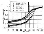
2−1.UV融解曲線 本発明によるRNAオリゴマーと、その相補鎖との結合状態を観察するため、2本鎖の温度変化に対する260nmにおける吸光度変化を測定した。0.1M NaCl含有0.01Mリン酸緩衝液(pH7.0)中で、本発明のRNAオリゴマーと相補鎖とを1:1、また核酸の濃度を全体で4.27×10⁻⁵Mとし、これを80℃に加温後に室温まで徐冷してアニールさせたものをサンプルとした。窒素気流下0℃で20分間ホールドし、昇温速度0.5℃/min.で80℃まで4分間隔で繰り返しUVスペクトルを測定した。このスペクトルの260nmにおける吸収を読み取り、80℃での吸光度を1.0として相対吸光度をプロットした。得られたUV融解曲線は、図2(相補鎖DNA)、および図3(相補鎖RNA)に示した。DNAオリゴマーのときと同様にRNAオリゴマー4〜6のUV融解曲線は、いずれも典型的シグモイドカーブを示した。これらの結果からRNAオリゴマーは、天然型の未修飾のオリゴヌクレオチドと同様に低温側では2本鎖を形成し温度の上昇と共に一本鎖に解離していることが示された。UV融解曲線から得られたTm値を表2に示した。またピレン修飾したものと天然のものとのTm値の差をΔTm℃として表中に記載した。
【0044】
【表2】
【0045】
以上のことからピレンを有するオリゴリボヌクレオチド誘導体は、中性水溶液中で天然型の未修飾のオリゴヌクレオチドと同様に相補鎖DNAあるいはRNAと2本鎖を形成することが確認できた。ピレン修飾は、RNAオリゴマーと相補鎖DNA(または相補鎖RNA)との結合に対して特に影響を与えないものと考えられた。
【0046】
2−2.温度に対するUV吸収スペクトル変化
オリゴヌクレオチドへ導入されたピレンの蛍光スペクトルが、相補鎖との結合によってどのような影響を受けるのかを調べるために、温度変化に対する300-360nmにおけるUV吸収スペクトル変化(バンド幅5nm)を観察した。0.1M NaCl含有0.01Mリン酸緩衝液(pH7.0)中で、本発明のRNAオリゴマーと相補鎖とを1:1、また核酸の濃度を全体で4.27×10⁻⁵Mとして測定した。得られた結果をプロットしたのが図4(相補鎖DNA)、および図5(相補鎖RNA)である。
【0047】
図4(相補鎖DNA)、および図5(相補鎖RNA)のいずれも、温度の上昇にともなって吸収極大の短波長シフトとアブソーバンスの増減が観測された。この低温域での吸収極大と高温域での吸収極大の短波長シフトから、ピレン周辺の環境が1本鎖と2本鎖の状態では異なっていることが推測された。また、RNAオリゴマーと相補鎖RNAとの組み合わせ(図5)での2本鎖形成温度(40-50℃)付近のUVスペクトルの形状が、同様にして求めたDNAオリゴマーのUVスペクトル(データは示さず)とはまったく異なっていた。このことから、RNAオリゴマー/相補鎖RNA2本鎖中におけるピレンの核酸塩基との相互作用は、他の組み合わせ(たとえばDNAオリゴマー/相補鎖DNA2本鎖)のものとは大きく異なっているものと推測された。
【0048】
2−3.本発明によるRNAオリゴマーと相補鎖DNA(または相補鎖RNA)の2本鎖形成と蛍光強度変化
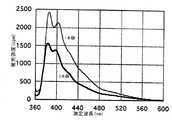
本発明によるピレンを有するオリゴリボヌクレオチド誘導体に対して相補鎖DNA(または相補鎖RNA)を室温下で加え、2本鎖形成前後での蛍光スペクトル、並びに蛍光強度の変化を観察した。0.1M NaCl含有0.01Mリン酸緩衝液(pH7.0)中で、本発明のRNAオリゴマーと相補鎖とを1:1、また核酸の濃度を全体で4.27×10⁻⁵Mとし、励起波長338nm、測定波長360-600nm(バンド幅5nm)で測定した。蛍光スペクトル変化の測定結果は図6(相補鎖DNA)および図7(相補鎖RNA)に示した。
【0049】
【表3】
各RNAオリゴマーが相補鎖RNAと2本鎖を形成したときは、表3に示したように約15〜20倍の著しい蛍光強度の増大を示し、しかもDNAオリゴマーとは異なり蛍光強度の増大が塩基配列に依存しない結果が得られた。RNAオリゴマーが相補鎖DNAと2本鎖を形成したときは、一本鎖の約半分ぐらいに消光することが、各RNAオリゴマーで観測された。
【0051】
2−4.相補鎖RNAの濃度を変化させたときの蛍光強度の変化
本発明によるRNAオリゴマー6について、相補鎖RNAとのハイブリダイズと蛍光強度の増大との関連を調べるために、RNAオリゴマー6に相補鎖RNAを少しずつ加え蛍光強度の変化を測定した。結果は図8に示す。380nmでの蛍光強度を[RNAオリゴマー 6]/[complementaryRNA]比に対してプロットしていくと、濃度比が1に達するまでは蛍光強度が増大していき、濃度比が1以上になると蛍光強度の増大が観測されなくなった。このことによりRNAオリゴマーと相補鎖RNAは1:1で結合して蛍光強度の増大を示すことが明らかである。
【0052】
2−5.二重らせんの温度に対する蛍光スペクトルの変化
本発明によるRNAオリゴマー6と相補鎖RNAとによる蛍光強度の増大が、2本鎖形成によるものであることを確認するために、RNAオリゴマー6と相補鎖RNAとをリン酸バッファー中、等モルで混合したサンプルの各温度での蛍光強度(励起波長:338nm、測定波長:380nm)を測定した。結果は図9に示した。2本鎖を形成している17℃での蛍光強度は大きいが、半解離温度以上の60℃に達すると著しく消光している。消光したときの蛍光強度は、図7で示したRNAオリゴマー6だけの蛍光強度とほぼ一致していた。更に温度上昇によって低下した蛍光強度は、温度を17℃に戻すと昇温する前と同様の蛍光スペクトルに回復した。この実験結果と2−4の結果から、RNAオリゴマー6と相補鎖RNAが濃度比1:1で結合して2本鎖を形成したときに、その蛍光強度を著しく増大することが確認された。
【0053】
2−6.励起スペクトル測定
【0054】
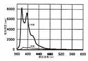
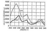
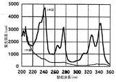
本発明によるRNAオリゴマーの、2本鎖形成に伴う蛍光強度(380nm)増大のメカニズムを明らかにするために励起スペクトルを測定した。0.1M NaCl含有0.01Mリン酸緩衝液(pH7.0)中で、本発明のRNAオリゴマーと相補鎖とを1:1、また核酸の濃度を全体で4.27×10⁻⁵Mとし、励起波長を200-360nm(バンド幅5nm)、蛍光強度は380nmで測定した。結果は図10(相補DNA)および図11(相補RNA)に示す。
【0055】
図10(相補DNA)および図11(相補RNA)、どちらのグラフにおいてもピレン特有のスペクトルパターンが観測された。RNAオリゴマーと相補鎖RNAの組み合わせ(図11)では、2本鎖を形成したときにスペクトルパターンの著しい変化が観測されたが、RNAオリゴマーと相補鎖DNAの組み合わせ(図10)では、相補鎖RNAのときのようなスペクトルパターンの変化が観測されなかった。これらの結果から、本発明によるRNAオリゴマーと相補鎖RNAとの2本鎖形成に伴う380nmにおける蛍光強度の増大は、ピレンに由来するものであると結論した。また2本鎖の状態にあるときのピレン周辺の環境が、相補鎖DNAと相補鎖RNAの間では大きく異なっていることが示された。
【0056】
2−7.塩基配列特異性
【0057】
ピレン修飾した本発明によるRNAオリゴマーとRNAとの塩基配列特異性を検討するため、相補鎖RNAに対して1塩基または2塩基ミスマッチ部位を設定したRNAのUV融解曲線と蛍光スペクトルを測定した。0.1M NaCl含有0.01Mリン酸緩衝液(pH7.0)中で、本発明のRNAオリゴマーと相補鎖とを1:1、また核酸の濃度を全体で4.27×10⁻⁵Mとし、励起波長338nm、測定波長380nmで蛍光測定した。ミスマッチを設けたRNAの塩基配列は下記のとおりである。小文字で示したのがミスマッチ部位である。結果は図12(UV融解曲線)、および図13(蛍光スペクトル)に示した。
【0058】
RNAの塩基配列
RNAオリゴマー4:5'-rACAUGCAGUGUUGAU-3'
相補鎖 RNA :5'-rAUCAACACUGCAUGU-3'
ミスマッチ1 :5'-rAUCAACACUGCgUGU-3'
ミスマッチ2 :5'-rAUCAACACUGuAUGU-3'
ミスマッチ3 :5'-rAUCAACACUGugUGU-3'
【0059】
図12のミスマッチ1〜3とRNAオリゴマー4の組み合わせにおいて、UV融解曲線は未修飾のものと同様にシグモイドカーブを示している。したがって、ミスマッチ部位が存在していても、本発明によるRNAオリゴマーは未修飾のものとほぼ同様に相補鎖と2本鎖を形成していることが確認された。ただし、ミスマッチのもののTm値は、相補鎖RNAのTm値より3〜7℃低下していた。
【0060】
また図13から、RNAオリゴマー4がミスマッチ1またはミスマッチ3と2本鎖を形成したときの蛍光強度はRNAオリゴマー4の一本鎖の蛍光強度とほとんど変化しなかった。一方、RNAオリゴマー4がミスマッチ2と2本鎖を形成したときの蛍光強度は、RNAオリゴマー4の蛍光強度の約4.6倍の蛍光強度の増大が観測された。ただこの増大は、相補鎖RNAとのハイブリダイズに伴う増大(約20倍)には満たない。これらの結果から、本発明によるRNAオリゴマー4の2本鎖形成に伴う蛍光強度の増大は塩基配列特異性を有しており、1塩基のミスマッチ部位を識別して蛍光強度が変化することが示された。
【0061】
2−8.RNAオリゴマーと相補鎖DNA(または相補鎖RNA)との相互作用の検討
【0062】
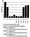
以上示したように、本発明によるRNAオリゴマーは相補鎖RNAと1:1の濃度比で2本鎖を形成することによって蛍光強度を著しく増大する。ピレンと、ピレンを導入した各種誘導体(本発明によるRNAオリゴマーも含む)、そしてそれらがハイブリダイズしたときの蛍光強度を比較したものが図14(本発明によるRNAオリゴマー)、および図15(DNAオリゴマー)である。RNAオリゴマーと相補鎖RNAとの組み合わせでは、蛍光スペクトルの結果からわかるように2本鎖の蛍光量子収率が1本鎖の約5倍から10倍にも達し、U(Pyr)の蛍光量子収率とほぼ同様、あるいはそれ以上の結果を示した。そして蛍光強度の増大の程度は、配列に依存しないものであった。これに対して過去に知られているピレン修飾DNAは、全体の配列によって2本鎖形成時の蛍光強度の増大程度に違いを示した。更に、本発明によるRNAオリゴマー4の2本鎖形成に伴う蛍光強度の増大は塩基配列特異性を有しており、1塩基のミスマッチ部位を識別して蛍光強度が変化することが示された。
【0063】
本発明によるRNAオリゴマーでは、公知のDNAオリゴマーと同様に一本鎖の状態で核酸塩基とピレンが相互作用して消光している。二重らせんの形成に伴って、蛍光に有利と思われるA型類似の二重らせんを形成することが判明した。この、蛍光により好都合なコンフォメーションは、A型二重らせん中のピレンが二重らせん内の結合に対して外側の面のマイナーグローブ上に位置していているために、核酸塩基との相互作用を受けにくい自由な状態であるためであると考えられる。
【0064】
【発明の効果】
本発明によって、簡単な反応原理に基づいて均一系の反応によりRNAのハイブリダイズをきわめて高感度に検出することができる。本発明のピレン導入塩基を取り込んだRNAプローブは、どのような配列であっても通常のRNAの合成方法にしたがって簡単に合成することができる。本発明は、プローブの修飾によって反応性や特異性にも大きな影響が無く、またどのような塩基配列に対しても適用することができる応用範囲の広い検出技術を提供する。
【0065】
本発明は、ピレン修飾したRNAをプローブに用いることにより、遺伝子変異の新規な検出方法を提供する。本発明の遺伝子変異の検出方法によれば、任意の位置における変異を同じ反応原理に基づいて簡単に実施することが可能である。
【0066】
更に本発明は、洗浄操作が不要な新規なインサイチュのハイブリダイゼーションアッセイを提供する。洗浄操作を省くことにより、操作が大幅に簡略化される。また各種RNA転写増幅系との組み合わせにより、高感度な検出を均一系の反応により実現できる。
【配列表】
【図面の簡単な説明】
【図1】ピレン導入ウリジン誘導体の合成スキームを示す図。
【図2】本発明によるピレン修飾RNAオリゴマーと相補鎖DNAによる2本鎖の、260nmにおけるUV融解曲線。縦軸は80℃の測定値を1としたときの260nmにおける吸光度の比を、横軸は温度を示す。
【図3】本発明によるピレン修飾RNAオリゴマーと相補鎖RNAによる2本鎖の、260nmにおけるUV融解曲線。縦軸は80℃の測定値を1としたときの260nmにおける吸光度の比を、横軸は温度を示す。
【図4】0-80℃における、本発明のピレン修飾RNAオリゴマーと相補鎖DNAとのUVスペクトル。縦軸は吸光度を、横軸は測定波長(nm)を示す。
【図5】0-80℃における、本発明のピレン修飾RNAオリゴマーと相補鎖RNAとのUVスペクトル。縦軸は吸光度を、横軸は測定波長(nm)を示す。
【図6】本発明によるRNAオリゴマーと相補鎖DNAの2本鎖形成の前後における、蛍光スペクトル変化。縦軸は蛍光強度(cpm)を、横軸は測定波長(nm)を示す。
【図7】本発明によるRNAオリゴマーと相補鎖RNAの2本鎖形成の前後における、蛍光スペクトル変化を示すグラフ。縦軸は蛍光強度(cpm)を、横軸は測定波長(nm)を示す。
【図8】本発明のRNAオリゴマー6に相補鎖RNAを加えたときの蛍光強度変化を示すグラフ。縦軸は蛍光強度(nm)を、横軸は[oligomer6]/[相補鎖RNA]比を示す。
【図9】温度変化に伴う、本発明のRNAオリゴマー6に相補鎖RNAを加えたときの蛍光スペクトル変化を示すグラフ。縦軸は蛍光強度(cpm)、横軸は測定波長(nm)である。
【図10】本発明のRNAオリゴマー6と相補鎖DNAによる2本鎖の励起光スペクトル。縦軸は380nmにおける蛍光強度(cpm)を、横軸は励起波長(nm)を示す。
【図11】本発明のRNAオリゴマー6と相補鎖RNAによる2本鎖の励起光スペクトル。縦軸は380nmにおける蛍光強度(cpm)を、横軸は励起波長(nm)を示す。
【図12】本発明のRNAオリゴマーとミスマッチRNA(または相補鎖RNA)による2本鎖のUV融解曲線。縦軸は80℃の測定値を1としたときの260nmにおける吸光度の比を、横軸は温度を示す。
【図13】本発明のRNAオリゴマーとミスマッチRNA(または相補鎖RNA)による2本鎖の蛍光スペクトル。縦軸は蛍光強度(cpm)を、横軸は測定波長(nm)を示す。
【図14】ピレン、またはピレンを導入した種々のRNA関連誘導体、ならびにそれが2本鎖形成したときの蛍光量子収率を比較したグラフ。縦軸は蛍光量子収率(Φλ)を、横軸は化合物の種類を示す。
【図15】ピレン、またはピレンを導入した種々のDNA関連誘導体、ならびにそれが2本鎖形成したときの蛍光量子収率を比較したグラフ。縦軸は蛍光量子収率(Φλ)を、横軸は化合物の種類を示す。横軸において+RNA、または+DNAの表示は、いずれも相補鎖との2本鎖の形成を意味する。[0001]
BACKGROUND OF THE INVENTION
The present invention relates to a modified RNA and a method for detecting RNA using this RNA. More specifically, it relates to fluorescently labeled RNA and its application.
[0002]
[Prior art]
Many variations of DNA and RNA analysis techniques based on hybridization are known. However, usually DNA is often used as a probe. This is because DNA is easy to synthesize, various labeling techniques are known for DNA, or DNA is more stable than RNA. For labeling DNA, radioisotopes, fluorescent substances, luminescent substances, ligands, enzymes and the like are used. Whichever label is used, it is usually necessary to remove the probe that did not hybridize prior to detection of the label. The removal of the probe that has not hybridized is generally performed by a method in which the hybridized target sequence and probe are held on a solid phase by some means and washed. This type of analysis method is called heterogeneous analysis because the reaction system is composed of different phases, a solid phase and a liquid phase. Heterogeneous analysis results in increased work steps due to washing, hybridization products may be lost by washing, leading to reduced sensitivity, and conversely increased background due to insufficient washing results in sensitivity. There was a problem such as inviting a decrease in. As an example in which washing of unreacted probes affects the analysis results, there is an in situ hybridization assay. In a typical in situ hybridization assay, the degree of washing of unreacted probe has a significant effect on the results. The remaining probe leads to background noise, making accurate observation difficult. In order to obtain a more complete cleaning effect, an unreacted probe may be decomposed with an enzyme. In addition, measures to prevent non-specific adsorption by adding irrelevant RNA or inactive protein are generally performed.
[0003]
In order to solve the problems of heterogeneous systems, analysis techniques using homogeneous systems have been tried. In homogeneous analysis, washing / separation operations that are essential for heterogeneous systems are omitted by combining systems that produce a difference in signal due to labeling when hybridized or not. . A typical homogenous analysis is a method of relating the approach of a molecule by hybridization to a change in signal. For example, two oligonucleotide probes are labeled with two types of molecules whose fluorescence signal intensity changes with proximity. If the base sequence of the oligonucleotide is designed to be adjacent on the target sequence, the two molecules will come close to each other by hybridization, and will emit a fluorescent signal different from that when they did not hybridize. As such a combination of labeling substances, a fluorescent donor and a fluorescent acceptor that absorbs the fluorescence are known. The fluorescence intensity of fluorescein (fluorescent donor) is weakened by the proximity to rhodamine (fluorescent acceptor), and the fluorescent signal of rhodamine is enhanced instead (Pro.Natl.Acad.Sci.USA, Vol.85,8790-8794, 1988). There is also a known example in which the reaction principle by competitive inhibition is constituted by combining similar labels (Anal. Biochem. 183, 231-244, 1989). The phenomenon in which fluorescent lanthanide chelates increase fluorescence intensity by approaching salicylic acid residues has also been applied to homogeneous hybridization assays (Angew, Chem. Int. Ed. Engl. 29/10, 1167-1169, 1990). Furthermore, a method is known in which an acridine derivative is combined with an ethidium derivative, and the approach of the triple chain formation is captured as a change in fluorescence signal (Biochemistry, 33, 15321-15328, 1994). In the method based on the principle of relating the approach of different molecules to the change in signal, since two or more kinds of labeled probes must be prepared, the cost tends to increase. In addition, since the reagent configuration is complicated, it is difficult to apply to small-scale analysis at the laboratory level.
[0004]
In contrast to these known techniques using a plurality of types of labeled probes, there are also reports of conducting homogeneous analysis with one type of labeled probe. That is, the ruthenium chelate introduced at the 5 ′ end of the nucleotide probe does not exhibit fluorescence activity when the probe is in a single strand state, but when the probe hybridizes to a complementary base sequence to form a double strand, a fluorescence signal is obtained. (J. Am. Chem. Soc. Vil. 114, No. 22, 836-8738, 1992). However, although only one type of labeled probe is required, the synthesis operation is complicated. In addition, since the enhancement effect of fluorescence intensity varies depending on the base sequence, quantitative analysis is difficult. Further, a method is known in which the enhancement of fluorescence intensity observed when a DNA probe bound with a DNA minor globe detection reagent such as Hoechst 33258 forms a double strand is applied to a homogeneous hybridization assay (J Am. Chem. Soc. 118,7055-7062, 1996). Also in this method, the synthesis of the labeled oligonucleotide is complicated, and the intensity of the signal depends on the sequence due to the characteristic of having the AT base pair specificity.
[0005]
As one of such homogeneous analysis attempts, a method using an oligonucleotide incorporating deoxyuridine into which a fluorescent substance pyrene has been introduced as a probe has been reported (Nucleic Acids Symposium Series, No. 35, 117-118). , 1996; Nucleosides & Nucleotides, 11, 383-390, 1992). These reports are based on the principle that pyrene covalently bound to uridine loses the fluorescence signal when the uridine is incorporated into the oligonucleotide, but the fluorescence signal is restored by hybridization with the complementary RNA of the oligonucleotide. Is based. This method is advantageous in terms of operability and cost since only one type of label is used. However, the increase in the fluorescence intensity observed at this time is extremely low, just around a few percent of the fluorescence intensity of the original pyrene molecule, leaving room for improvement in terms of sensitivity for practical analysis. It was. In addition, the fluorescence quantum yield sometimes fluctuates depending on the base constituting the sequence, which makes it difficult to apply to quantitative analysis. In addition to pyrene, there is also a report that attempted a homogeneous analysis using a DNS (5-dimethylaminonaphthalene-1-sulfonate) derivative as a fluorescent labeling compound (Tetrahedron, Vol. 55, No. 12, 4265-4270, 1997). .
[0006]
[Problems to be solved by the invention]
The subject of this invention is providing the novel detection method of RNA by the homogeneous system which can maintain a sensitivity and specificity at a high level, although it is a simple reaction system based on one type of labeling compound. In addition, an object of the present invention is to provide a novel pyrene-modified RNA for realizing this new detection method.
[0007]
[Means for Solving the Problems]
The present inventors have detected RNA containing a target base sequence by observing a change in fluorescence intensity by hybridizing RNA containing the base into which pyrene is introduced in its constituent base sequence, and using this RNA as a probe. By performing, the said subject was solved.
That is, the present invention
[1] Containing the base in which pyrene is introduced in the base sequenceRNAWherein the base into which the pyrene is introduced is a uridine residue.2 'Place-OHThe above-mentioned pyrene is covalently bonded to a groupRNA,
[2] A method for detecting a test RNA containing a target base sequence, wherein the RNA of [1] having a base sequence complementary to the target base sequence is hybridized to the test RNA, and pyrene fluorescence by hybridization A method of detecting a test RNA based on a signal,
[3] A method for detecting a mutation in a specific site of a test RNA containing a target base sequence, which has a base sequence complementary to the target base sequence and introduces pyrene into the base near the site where the mutation to be detected occurs. A method for detecting a mutation, comprising: hybridizing the RNA of [1] to a test RNA, and detecting a mutation based on a fluorescence signal of pyrene by the hybridization; and
[4] The mutation detection method according to [3], wherein the test RNA is selected from the group consisting of mRNA, ribosomal RNA, RNA virus genome, and RNA transcribed using DNA as a template.
[0008]
In order to solve the above problems, the present inventors newly synthesized RNA containing pyridine-introduced uridine in the base sequence. Since the method of introducing pyrene into uridine is already known, the RNA of the present invention can be obtained by synthesizing oligoribonucleotides based on the resulting pyrene-introduced uridine derivative..
[0009]
Pyrene alone exhibits blue fluorescence, but loses its fluorescence activity when incorporated into oligoribonucleotides. The present inventors have found that when this oligoribonucleotide hybridizes to RNA having a complementary base sequence, the fluorescence activity of pyrene that has been lost is recovered. The fluorescence intensity obtained at this time reached 80% or more of pyrene alone. Furthermore, it was confirmed that the level of recovery of pyrene fluorescence intensity by hybridization surpassed that of pyrene in a state introduced into uridine. The fluorescence intensity of pyrene gradually decreases with the introduction of uridine, the incorporation of pyrene-introduced uridine derivatives into oligoribonucleotides and the synthesis steps. That is, the fluorescence intensity is smaller when introduced into uridine than when pyrene alone is used. Nevertheless, when the oligoribonucleotide composed of the pyrene-introduced uridine derivative is hybridized to the complementary RNA, the present inventors emit a fluorescence signal larger than the fluorescence intensity in the pyrene-introduced uridine derivative. It was revealed.
[0010]
As cited in the prior art, even when the same pyrene-introduced uridine derivative is used, when it is synthesized as a DNA probe, the degree of recovery of fluorescence intensity by hybridization is only a few percent. Therefore, the problem to be solved in terms of sensitivity remains to be applied to the analysis of homogeneous systems. In the present invention, a new analysis technique that enables highly sensitive homogeneous analysis was completed by using a pyrene-introduced uridine derivative as an RNA probe.
[0011]
Further, the present inventors have found that the restoration of fluorescence intensity by hybridization is greatly influenced by whether or not the base into which pyrene is introduced is hybridized. That is, when the pyrene-modified RNA according to the present invention hybridizes with a complementary strand with a mismatch, if there is a mismatch at the site where pyrene is introduced, no increase in fluorescence intensity is observed even if it is hybridized as a whole. .
[0012]
Based on the above characteristics, the pyrene-introduced RNA according to the present invention can be applied to the detection of RNA having a complementary base sequence. The pyrene-modified RNA according to the present invention can also be applied to mutation detection. Hereinafter, embodiments of the present invention will be described in detail.
[0013]
DETAILED DESCRIPTION OF THE INVENTION
The pyrene-modified RNA in the present invention can be easily obtained by synthesizing RNA using a uridine derivative into which pyrene is introduced as a substrate. The introduction of pyrene into uridine is known (Tetrahedron Lett. Vol. 32, No. 44, 6347-6350, 1991). For example, pyrene can be introduced into the 2′-position hydroxyl group of uridine according to the synthesis scheme shown in FIG. That is, 3′-position and 5′-position of uridine are protected with an appropriate protecting group, and pyrene is introduced into this. Subsequently, the 4′-dimethoxytrityl-uridine (pyr) amidite can be finally obtained by phosphorylating the 3′-position —OH so that chemical synthesis by a DNA synthesizer is possible. This compound can be used for addition of uridine; U in the same manner as uridine triphosphate in a DNA synthesizer. Currently widely used DNA synthesizers automate synthesis steps based on the phosphoramidite method. In this type of automatic analyzer, an oligonucleotide derivative can be obtained by using an appropriate derivative instead of dNTP. Similarly, in the synthesis of RNA, if the pyrene derivative is used instead of NTP, the target oligoribonucleotide can be modified with the desired pyrene.
[0014]
In order to synthesize RNA enzymatically, a method of synthesizing complementary strands by the action of RNA polymerase using DNA or RNA having a base sequence complementary to the target base sequence as a template can be employed. Two types of RNA polymerases are known, one using DNA as a template and the other depending on RNA. SP6 RNA polymerase, T7 RNA polymerase, T3 RNA polymerase, and the like are commercially available for performing complementary strand synthesis using the former DNA as a template. Among those using RNA as a template, there is Qβ replicase. All RNA polymerases require a promoter for complementary strand synthesis. Therefore, in order to synthesize pyrene-modified RNA according to the present invention enzymatically, a structure in which a promoter region recognized by RNA polymerase is arranged upstream of the template sequence is required. Vectors containing a promoter such as SP6, T3, or T7 are commercially available. The gene to be detected may be incorporated into this vector, linearized, and then transcribed with RNA polymerase according to the promoter. Template DNA mixed in the reaction system can be enzymatically decomposed and removed by DNAse after RNA synthesis. When enzymatically synthesizing RNA, it is convenient to use commercially available kits that combine substrates and vectors necessary for the transcription reaction, such as “RiboMAX” (trade name, manufactured by Promega) as necessary. .
[0015]
In any synthesis method, a uridine derivative into which pyrene is introduced behaves in substantially the same manner as the original uridine. Therefore, if a pyrene-modified uridine derivative is used under normal synthesis conditions, the synthesized RNA is pyrene-modified at the U portion. In addition, if a base sequence having a large U composition ratio is used as a probe, one RNA is modified with many pyrenes. If the number of molecules of pyrene per RNA increases, the fluorescence intensity when hybridized by that amount increases, so that a highly sensitive analysis system can be obtained. However, the modification of pyrene causes a slight decrease in the Tm value, so unlimited introduction of pyrene may cause a decrease in reactivity in some cases. Under general detection conditions, if the number of pyrene molecules per molecule in the pyrene-modified RNA according to the present invention is 1-10 molecules, sufficient sensitivity can be expected.
[0016]
The RNA according to the present invention is designed so that sufficient specificity and affinity can be maintained as a whole with respect to the target sequence with the expected stringency group so that the RNA according to the present invention can be used as a probe for a hybridization assay. Is obvious to those skilled in the art. Under normal conditions, it is appropriate that the base sequence has a chain length of about 10-500 bp, preferably about 15-30 bp and is complementary to a base sequence that specifically exists in the target base sequence. The stringency of the reaction solution is mainly determined by the structure of the base constituting the sequence, and the composition and temperature of the buffer solution. Since these relationships are known to have a certain relationship, hybridization is generally performed at a temperature 5-15 ° C. lower than the calculated Tm.
[0017]
The base sequence of the pyrene-modified RNA of the present invention does not necessarily have to guarantee complete complementarity with the target base sequence. That is, it is only necessary to satisfy two conditions, that is, hybridization while maintaining specificity as a whole and that at least one base corresponding to the site modified with pyrene is complementary. By using a sequence that can be hybridized with some mismatch, for example, it is possible to construct a system in which, for example, one having a certain similarity is collectively detected with one type of probe.
[0018]
The pyrene-modified RNA according to the present invention is advantageous in terms of improving sensitivity when it is added in excess to the expected maximum concentration of the target base sequence. The hybridization reaction between the RNA probe and the test RNA according to the present invention can be performed under reaction conditions for general RNA-RNA hybridization. Usually, a buffer solution having a salt concentration of 100 mM or more and pH 6-9 can be used, and about 1 mM of EDTA can be added for the purpose of RNA stabilization. For example, a buffer solution obtained by adding 1 mM EDTA to 100 mM NaCl, 10 mM phosphate buffer (7.0) is a buffer solution suitable for the RNA probe hybridization reaction according to the present invention.
[0019]
The enhancement of the fluorescence intensity of the pyrene-modified RNA of the present invention hybridized with the target base sequence can be measured by a change in the fluorescence intensity around 380 nm using excitation light of 338 nm. Those not hybridized hardly emit fluorescence, and therefore the reaction solution can be directly used as a sample for fluorescence counting without separation.
[0020]
When observing the fluorescence of pyrene, care should be taken not to mix a substance that acts as a fluorescence quencher with respect to the fluorescence activity of pyrene in the fluorescence measurement sample. For example, since acrylamide has a fluorescence quenching action, it is desirable not to mix a large amount into the fluorescence measurement system. However, it is rare for a general sample to contain a large amount of a fluorescence quenching substance.
[0021]
The pyrene-modified RNA of the present invention can be applied to various detection systems. One of the most basic aspects is the detection of RNA present in the liquid phase. For example, biological samples such as blood, sputum, mucus collected from various tissues, or biopsy samples can be tested for the presence of RNA derived from pathogens and disease genes. These samples may contain RNA, such as mRNA, ribosomal RNA, or the genome of an RNA virus. For detection, the sample may be diluted with an appropriate buffer, or appropriately extracted, and contacted with the pyrene-modified RNA of the present invention.
[0022]
The pyrene-modified RNA of the present invention can detect not only RNA in the liquid phase but also RNA present in cells and tissues. That is, an in situ hybridization assay. MRNA in cells is an important information source for knowing the expression status of genes. Further, since ribosomal RNA is contained in a large amount in one cell, high sensitivity can be expected when the base sequence is to be detected. In the present invention, since washing is not required even in in situ hybridization, the operation can be greatly simplified without sacrificing sensitivity or specificity.
[0023]
In in situ hybridization, a cell or tissue to be analyzed is fixed by an appropriate method, and a labeled probe is hybridized thereto. Hybridization is performed in a normal hybridization buffer. For example, the following composition is generally used.
5M NaCl
0.5M EDTA
20xDenhardt
50% Dextrun sulfate
20mg / ml E.coli tRNA
1.0M Tris-HCl pH8.0
[0024]
The length of the probe for in situ hybridization is generally 1 kbp or less, usually 10-500 bp. In the conventional in situ hybridization assay, washing is required after hybridization, but in the present invention, it can be observed with a fluorescence microscope as it is.
[0025]
Whether it is a liquid phase system or an in situ reaction, ribonuclease present in the sample may degrade the pyrene-modified RNA of the present invention in the reaction with the biological sample. The degradation of pyrene-modified RNA liberates ribonucleotides into which pyrene has been introduced, causing nonspecific fluorescence. As a countermeasure against this problem, a ribonuclease inhibitor may be allowed to coexist with the probe in advance. Commercially available products can be used as the ribonuclease inhibitor. Alternatively, coexisting ribonuclease activity can be removed by incubation with 0.01% DEPC.
[0026]
The RNA detectable by the present invention is not limited to RNA originally present as RNA, but may be RNA transcribed using DNA or RNA as a template. Various RNA polymerases give hundreds of copies or much more copies of complementary RNA as transcription products based on a small amount of template sequence under appropriate reaction conditions. Therefore, if RNA polymerase transcripts are analyzed according to the present invention, extremely high sensitivity can be achieved. Methods for amplifying genes and signals based on RNA transcription reactions using T7 RNA polymerase or Qβ replicase are known. Here, gene amplification means that amplification is performed using a target base sequence as a template. On the other hand, signal amplification means that a specific amplification reaction system is triggered by the presence of a target sequence, and a large amount of RNA having a certain sequence is transcribed. In the latter, the target sequence itself is not transcribed and only triggers the transcription reaction of other sequences. In any reaction, a large amount of RNA having a certain sequence in some form is produced, so that this reaction product can be detected with the pyrene-modified RNA of the present invention. In particular, when combined with a signal amplification reaction system, the progress of the reaction can be observed in real time, so that a unique reaction system can be constructed. That is, the RNA transcription reaction system triggered by the target base sequence and the pyrene-modified RNA according to the present invention are simultaneously mixed with the sample. If the target base sequence is present, a large amount of transcription product having a specific base sequence is generated by the RNA transcription system. If the base sequence of pyrene-modified RNA is a sequence complementary to this transcription product, the progress of the transcription reaction can be observed in real time as the fluorescence intensity increases. Since pyrene-modified RNA itself has nothing to do with RNA transcription reaction, such a reaction system can be constructed.
[0027]
On the other hand, in a reaction system in which the target base sequence itself is transcribed, the pyrene-modified RNA of the present invention has the same base sequence as the target sequence, and therefore cannot exist simultaneously with the transcription reaction. Therefore, it is added after the completion of the transcription reaction to allow hybridization with the transcription product.
[0028]
The RNA detection method based on the present invention can be applied not only to the detection of the presence of a specific target sequence, but also to the detection of a mutation. As described above, when the pyrene-modified RNA of the present invention is modified at a mismatch site, no recovery of fluorescence is observed. According to the present invention, pyrene modification can be performed in a site-specific manner. Therefore, mutation can be detected by modifying pyrene with a site that requires mutation detection. For site-specific pyrene modification, pyrene-introduced nucleotides are incorporated only during an extension reaction of a specific base when chemically synthesizing an RNA probe. Since site-specific modification is difficult with enzymatic synthesis methods, it is desirable to chemically synthesize RNA probes for mismatch detection purposes.
[0029]
An analytical use where such mismatch detection is meaningful is the point mutation of the ras gene. Since it has been found that the ras gene is activated by point mutations and gene rearrangements in human cancer tissues, detection of ras point mutations is important in cancer diagnosis. For example, K-ras mutations are frequently observed in pancreatic cancer and biliary tract cancer. N-ras mutations are common in lymphoid tumors. Since the base sequence is registered in GenBank as human K-ras as M54968 and human N-ras as L00040, the necessary probes can be designed based on this (Experimental Medicine separate volume BioScience term library cancer Genes / tumor suppressor genes: Yoichi Taya and Masaru Yamamoto, 1997.10 / 10 Yodosha, p12-21).
[0030]
Regardless of a general method for detecting a target sequence or a method for detecting a mutation, reagents necessary for various detection methods according to the present invention can be combined into a kit. The kit according to the present invention includes a pyrene-modified RNA probe, various buffers necessary for dilution and extraction of a sample, a control (control) for confirming positive or negative. Furthermore, in combination with an RNA transcription system, it comprises a probe for RNA transcription, RNA polymerase, ribonucleotides, and the like.
[0031]
【Example】
1. Synthesis of pyrene-modified oligonucleotide derivatives
[0032]
The pyrene-modified oligonucleotide derivative was obtained by first synthesizing uridine into which pyrene was introduced and using it for the synthesis of the oligonucleotide.
[0033]
1-1. Synthesis of 4,4'-dimethoxytrityl-uridine (pyr) amidite
[0034]
Prior to the synthesis of pyrene-modified oligonucleotide derivatives, pyrene-modified uridine was first synthesized. The synthesis scheme is shown in FIG.
[0035]
Reaction of uridine (1) and 1-chloromethylpyrene (2) in which 3 'and 5' positions of the hydroxyl group are protected with a trityl group (hereinafter abbreviated as Tr group) in benzene-dioxane in the presence of potassium hydroxide. Subsequently, the Tr group was deprotected by treatment with 0.5N-HCl to obtain 2′-O- (1-pyrenylmethyl) uridine (3) (i in FIG. 1). After reacting (3) with 4,4′-dimethoxytrityl (hereinafter abbreviated as DMTr) in pyridine to protect the hydroxyl group at the 5′-position of the sugar moiety of (3) with a DMTr group (4) (Ii in FIG. 1) Next, (4) and 2-cyanoethyl N, N-diisopropyl phosphorodiamidite are mixed with CH.2C12In the presence of tetrazole and diisopropylamine in the middle, the hydroxyl group at the 3'-position of the sugar part of (4) was phosphorylated to induce compound (5) -DMTr-U (Pyr) amidite- applicable to an automatic DNA synthesizer. (Iii in FIG. 1). Confirmation of the compounds (1) to (4) was performed by lH-NMR, and confirmation of the compound (6) was performed by 31P-NMR.
[0036]
1-2. Oligonucleotide synthesis with pyrene-modified uridine
[0037]
3 types of oligodeoxyribonucleotide derivatives in which U (pyr) is introduced into DNA using a phosphoramidite reagent (5) obtained by the synthesis of 1-1 using a Pharmacia DNA automatic synthesizer (
[0038]
DNA Control 5'-dACATGCAGTGTTGAT-3 '
Complementary DNA 5'-dATCAACACTGCATGT-3 '
RNA Control 5'-rACAUGCAGUGUUGAU-3 '
Complementary RNA 5'-rAUCAACACUGCAUGU-3 '
[0039]
The synthesis of the oligonucleotide derivative having pyrene is carried out at a synthesis scale of 0.2 μmol. Except when the U (pyr) amidite unit is introduced, a normal synthesis cycle {condensation time: 2 minutes (DNA), 5.4 minutes ( RNA), amount of input amidite: 50 μl (DNA), 100 μl (RNA)}, and when introducing U (pyr) amidite unit, it was synthesized by extending the condensation time to 10 minutes and making the amount of input amidite 120 μl. . The condensation efficiency when introducing the U (pyr) amidite unit reached 98% or more, and the result was almost the same as the condensation efficiency of the usual unmodified amidite reagent. The obtained oligonucleotide derivative and oligodeoxyribonucleotide derivative were purified by 20% denaturing polyacrylamide gel electrophoresis and desalted with a WATERS SEP-PACK C18 cartridge. After the desalting treatment, the RNA oligomer was deprotected with a WATERS SEP-PACK C18 cartridge after deprotecting the Fpmp protecting group of the hydroxyl group at the 2'-position of the sugar using 0.01 M HCl aqueous solution.
[0040]
1-3. Identification of oligoribonucleotide derivatives with pyrene
[0041]
The synthesized RNA oligomer was confirmed by mass spectrum. The mass spectrum was measured in the negative ion mode of the ion spray method. Since negative ions are generated by extracting protons, the sign of proton mass number in the calculation formula was reversed, and the molecular weight was determined from each peak. Table 1 shows the values calculated based on the results (actual measurement values) and the molecular weights (theoretical values) of each RNA oligomer. Since the value obtained from the mass spectrum and the molecular weight of each RNA oligomer coincided with each other, it was confirmed to be the objective oligoribonucleotide derivative.
On the other hand, the synthesized DNA oligomaer1-3 was decomposed into nucleosides by enzymatic decomposition, and the composition was confirmed by liquid chromatography analysis. For any DNA oligomaer, the base composition ratio coincided with the target sequence, and it was confirmed that it was the target oligodeoxyribonucleotide derivative.
[0042]
[Table 1]
2. Interaction between RNA Oligomers According to the Present Invention and Their Complementary Strand DNA or RNA Various pyrene-modified oligonucleotides synthesized in 1 and DNA or RNA having a complementary base sequence were observed.
2-1. UV melting curve In order to observe the binding state of the RNA oligomer according to the present invention and its complementary strand, the change in absorbance at 260 nm with respect to the temperature change of the double strand was measured. In a 0.1 M NaCl-containing 0.01 M phosphate buffer (pH 7.0), the RNA oligomer of the present invention and the complementary strand were 1: 1, and the total concentration of nucleic acids was 4.27 × 10 6.-FiveThe sample was M, which was heated to 80 ° C. and then slowly cooled to room temperature and annealed. The sample was held at 0 ° C. for 20 minutes under a nitrogen stream, and UV spectra were repeatedly measured at intervals of 4 minutes up to 80 ° C. at a rate of temperature increase of 0.5 ° C./min. The absorption at 260 nm of this spectrum was read, and the relative absorbance was plotted with the absorbance at 80 ° C. being 1.0. The obtained UV melting curves are shown in FIG. 2 (complementary strand DNA) and FIG. 3 (complementary strand RNA). As with the DNA oligomer, the UV melting curves of RNA oligomers 4-6 all showed a typical sigmoid curve. From these results, it was shown that the RNA oligomer formed a double strand on the low temperature side and dissociated into a single strand as the temperature increased, like the natural unmodified oligonucleotide. Table 2 shows Tm values obtained from the UV melting curves. Further, the difference in Tm value between pyrene-modified and natural ones is shown in the table as ΔTm ° C.
[0044]
[Table 2]
[0045]
From the above, it was confirmed that the oligoribonucleotide derivative having pyrene forms a double strand with complementary strand DNA or RNA in a neutral aqueous solution like the natural unmodified oligonucleotide. It was considered that pyrene modification does not particularly affect the binding between RNA oligomer and complementary strand DNA (or complementary strand RNA).
[0046]
2-2. Change in UV absorption spectrum with temperature
In order to investigate how the fluorescence spectrum of pyrene introduced into the oligonucleotide is affected by the binding with the complementary strand, a change in UV absorption spectrum at 300 to 360 nm (
[0047]
In both FIG. 4 (complementary strand DNA) and FIG. 5 (complementary strand RNA), a short wavelength shift of absorption maximum and an increase / decrease in absorption were observed as the temperature increased. From the short wavelength shift of the absorption maximum in the low temperature region and the absorption maximum in the high temperature region, it was speculated that the environment around pyrene is different in the single-stranded and double-stranded states. In addition, the shape of the UV spectrum near the duplex formation temperature (40-50 ° C) in the combination of RNA oligomer and complementary RNA (Fig. 5) is the same as the UV spectrum of the DNA oligomer (data is shown Z)) was completely different. From this, it is speculated that the interaction of pyrene with the nucleobase in RNA oligomer / complementary RNA duplex is significantly different from that of other combinations (for example, DNA oligomer / complementary DNA duplex). It was.
[0048]
2-3. Double strand formation and change in fluorescence intensity of RNA oligomer and complementary strand DNA (or complementary strand RNA) according to the present invention
To the oligoribonucleotide derivative having pyrene according to the present invention, complementary strand DNA (or complementary strand RNA) was added at room temperature, and changes in fluorescence spectrum and fluorescence intensity before and after the formation of double strand were observed. In a 0.1 M NaCl-containing 0.01 M phosphate buffer (pH 7.0), the RNA oligomer of the present invention and the complementary strand were 1: 1, and the total concentration of nucleic acids was 4.27 × 10 6.-FiveM was measured at an excitation wavelength of 338 nm and a measurement wavelength of 360-600 nm (
[0049]
[Table 3]
When each RNA oligomer forms a double strand with complementary RNA, as shown in Table 3, it shows a remarkable increase in fluorescence intensity of about 15 to 20 times, and unlike the DNA oligomer, the increase in fluorescence intensity is a base. Sequence independent results were obtained. When the RNA oligomer formed a double strand with the complementary DNA, it was observed in each RNA oligomer that it was quenched to about half of the single strand.
[0051]
2-4. Changes in fluorescence intensity when the concentration of complementary RNA is changed
For the
[0052]
2-5. Change of fluorescence spectrum with temperature of double helix
In order to confirm that the increase in fluorescence intensity due to
[0053]
2-6. Excitation spectrum measurement
[0054]
In order to elucidate the mechanism of the increase in fluorescence intensity (380 nm) associated with double strand formation of the RNA oligomer according to the present invention, the excitation spectrum was measured. In a 0.1 M NaCl-containing 0.01 M phosphate buffer (pH 7.0), the RNA oligomer of the present invention and the complementary strand were 1: 1, and the total concentration of nucleic acids was 4.27 × 10 6.-FiveM was measured, the excitation wavelength was 200-360 nm (
[0055]
A spectral pattern peculiar to pyrene was observed in both graphs of FIG. 10 (complementary DNA) and FIG. 11 (complementary RNA). In the combination of RNA oligomer and complementary strand RNA (FIG. 11), a significant change in the spectral pattern was observed when the double strand was formed. In the combination of RNA oligomer and complementary strand DNA (FIG. 10), the complementary strand RNA No change in the spectral pattern was observed as in. From these results, it was concluded that the increase in fluorescence intensity at 380 nm accompanying the duplex formation of the RNA oligomer and complementary strand RNA according to the present invention originates from pyrene. Moreover, it was shown that the environment around pyrene when in a double-stranded state is greatly different between complementary strand DNA and complementary strand RNA.
[0056]
2-7. Base sequence specificity
[0057]
In order to examine the base sequence specificity between the RNA oligomer of the present invention modified with pyrene and RNA, the UV melting curve and fluorescence spectrum of RNA in which a 1-base or 2-base mismatch site was set for the complementary strand RNA were measured. In a 0.1 M NaCl-containing 0.01 M phosphate buffer (pH 7.0), the RNA oligomer of the present invention and the complementary strand were 1: 1, and the total concentration of nucleic acids was 4.27 × 10 6.-FiveFluorescence was measured with an excitation wavelength of 338 nm and a measurement wavelength of 380 nm. The base sequence of RNA with mismatches is as follows. The mismatch site is shown in lower case. The results are shown in FIG. 12 (UV melting curve) and FIG. 13 (fluorescence spectrum).
[0058]
RNA base sequence
RNA oligomer 4: 5'-rACAUGCAGUGUUGAU-3 '
Complementary RNA: 5'-rAUCAACACUGCAUGU-3 '
Mismatch 1: 5'-rAUCAACACUGCgUGU-3 '
Mismatch 2: 5'-rAUCAACACUGuAUGU-3 '
Mismatch 3: 5'-rAUCAACACUGugUGU-3 '
[0059]
In the combination of
[0060]
From FIG. 13, the fluorescence intensity when
[0061]
2-8. Examination of interaction between RNA oligomer and complementary strand DNA (or complementary strand RNA)
[0062]
As described above, the RNA oligomer according to the present invention remarkably increases the fluorescence intensity by forming a double strand with a complementary strand RNA at a concentration ratio of 1: 1. FIG. 14 (RNA oligomer according to the present invention) and FIG. 15 (DNA oligomer) are a comparison of pyrene, various derivatives into which pyrene has been introduced (including RNA oligomers according to the present invention), and their fluorescence intensities when they are hybridized. ). In the combination of RNA oligomer and complementary strand RNA, the fluorescence quantum yield of the double strand reaches about 5 to 10 times that of the single strand, as can be seen from the result of the fluorescence spectrum, and the fluorescence quantum yield of U (Pyr) The result was almost the same as or better than the rate. The degree of increase in fluorescence intensity was independent of the sequence. On the other hand, pyrene-modified DNA known in the past showed a difference in the degree of increase in fluorescence intensity during double strand formation depending on the entire sequence. Furthermore, the increase in fluorescence intensity associated with the formation of double strands of
[0063]
In the RNA oligomer according to the present invention, the nucleobase and pyrene interact with each other in a single-stranded state and are quenched in the same manner as known DNA oligomers. It was found that with the formation of a double helix, a double helix similar to type A, which seems to favor fluorescence, was formed. This fluorescent, more favorable conformation is due to the interaction of the nucleobase with the pyrene in the A-type double helix located on the minor glove on the outer face for binding within the double helix. It is thought that it is because it is in a free state that is difficult to receive.
[0064]
【The invention's effect】
According to the present invention, RNA hybridization can be detected with extremely high sensitivity by a homogeneous reaction based on a simple reaction principle. The RNA probe incorporating the pyrene-introduced base of the present invention can be easily synthesized according to the usual RNA synthesis method, regardless of the sequence. The present invention provides a detection technique with a wide range of application that can be applied to any base sequence without significant influence on reactivity and specificity by modification of the probe.
[0065]
The present invention provides a novel method for detecting a gene mutation by using pyrene-modified RNA as a probe. According to the gene mutation detection method of the present invention, mutation at an arbitrary position can be easily performed based on the same reaction principle.
[0066]
Furthermore, the present invention provides a novel in situ hybridization assay that does not require a washing operation. By eliminating the cleaning operation, the operation is greatly simplified. In combination with various RNA transcription amplification systems, highly sensitive detection can be realized by a homogeneous reaction.
[Sequence Listing]
[Brief description of the drawings]
FIG. 1 shows a synthesis scheme of pyrene-introduced uridine derivatives.
FIG. 2 is a UV melting curve at 260 nm of a double strand of pyrene-modified RNA oligomer according to the present invention and a complementary strand DNA. The vertical axis represents the ratio of absorbance at 260 nm when the measured value at 80 ° C. is 1, and the horizontal axis represents temperature.
FIG. 3 is a UV melting curve at 260 nm of a double strand of a pyrene-modified RNA oligomer according to the present invention and a complementary strand RNA. The vertical axis represents the ratio of absorbance at 260 nm when the measured value at 80 ° C. is 1, and the horizontal axis represents temperature.
FIG. 4 shows UV spectra of the pyrene-modified RNA oligomer of the present invention and complementary strand DNA at 0-80 ° C. The vertical axis represents the absorbance, and the horizontal axis represents the measurement wavelength (nm).
FIG. 5 shows UV spectra of the pyrene-modified RNA oligomer of the present invention and complementary strand RNA at 0-80 ° C. The vertical axis represents the absorbance, and the horizontal axis represents the measurement wavelength (nm).
FIG. 6 shows changes in fluorescence spectrum before and after double strand formation between RNA oligomer and complementary strand DNA according to the present invention. The vertical axis represents fluorescence intensity (cpm), and the horizontal axis represents measurement wavelength (nm).
FIG. 7 is a graph showing changes in fluorescence spectrum before and after double strand formation of RNA oligomer and complementary strand RNA according to the present invention. The vertical axis represents fluorescence intensity (cpm), and the horizontal axis represents measurement wavelength (nm).
FIG. 8 is a graph showing changes in fluorescence intensity when complementary strand RNA is added to
FIG. 9 is a graph showing changes in the fluorescence spectrum when complementary strand RNA is added to the
FIG. 10 shows a double-stranded excitation light spectrum of
FIG. 11 shows a double-stranded excitation light spectrum of
FIG. 12 shows a double-stranded UV melting curve of the RNA oligomer of the present invention and a mismatch RNA (or complementary RNA). The vertical axis represents the ratio of absorbance at 260 nm when the measured value at 80 ° C. is 1, and the horizontal axis represents temperature.
FIG. 13 shows a double-stranded fluorescence spectrum of the RNA oligomer of the present invention and mismatch RNA (or complementary strand RNA). The vertical axis represents fluorescence intensity (cpm), and the horizontal axis represents measurement wavelength (nm).
FIG. 14 is a graph comparing pyrene, various RNA-related derivatives into which pyrene is introduced, and fluorescence quantum yields when they are double-stranded. The vertical axis represents the fluorescence quantum yield (Φλ), and the horizontal axis represents the type of compound.
FIG. 15 is a graph comparing pyrene, various DNA-related derivatives into which pyrene is introduced, and fluorescence quantum yields when it forms a double strand. The vertical axis represents the fluorescence quantum yield (Φλ), and the horizontal axis represents the type of compound. The indication of + RNA or + DNA on the horizontal axis means that a double strand is formed with a complementary strand.
Claims (4)
Translated fromJapanesePriority Applications (1)
| Application Number | Priority Date | Filing Date | Title |
|---|---|---|---|
| JP20258998AJP4238381B2 (en) | 1998-07-17 | 1998-07-17 | Pyrene-modified RNA and RNA analysis method |
Applications Claiming Priority (1)
| Application Number | Priority Date | Filing Date | Title |
|---|---|---|---|
| JP20258998AJP4238381B2 (en) | 1998-07-17 | 1998-07-17 | Pyrene-modified RNA and RNA analysis method |
Publications (2)
| Publication Number | Publication Date |
|---|---|
| JP2000032999A JP2000032999A (en) | 2000-02-02 |
| JP4238381B2true JP4238381B2 (en) | 2009-03-18 |
Family
ID=16459996
Family Applications (1)
| Application Number | Title | Priority Date | Filing Date |
|---|---|---|---|
| JP20258998AExpired - Fee RelatedJP4238381B2 (en) | 1998-07-17 | 1998-07-17 | Pyrene-modified RNA and RNA analysis method |
Country Status (1)
| Country | Link |
|---|---|
| JP (1) | JP4238381B2 (en) |
Cited By (1)
| Publication number | Priority date | Publication date | Assignee | Title |
|---|---|---|---|---|
| US12005074B2 (en) | 2018-05-07 | 2024-06-11 | Alnylam Pharmaceuticals, Inc. | Extrahepatic delivery |
Families Citing this family (5)
| Publication number | Priority date | Publication date | Assignee | Title |
|---|---|---|---|---|
| JP2007215477A (en)* | 2006-02-16 | 2007-08-30 | Kyoto Institute Of Technology | Fluorescently labeled oligonucleotide probe |
| BRPI0820738A2 (en) | 2007-12-14 | 2015-06-16 | Minitube America Inc | Gender-specific separation of sperm and embryo cells |
| WO2011032034A2 (en) | 2009-09-10 | 2011-03-17 | University Of Idaho | Nucleobase-functionalized conformationally restricted nucleotides and oligonucleotides for targeting nucleic acids |
| WO2013013068A2 (en) | 2011-07-19 | 2013-01-24 | University Of Idaho | Embodiments of a probe and method for targeting nucleic acids |
| CN103234948B (en)* | 2013-05-09 | 2015-07-22 | 中南大学 | Application of pyrene and derivative as photosensitive perssad deprotection sensibilization reagent in biological chip production |
- 1998
- 1998-07-17JPJP20258998Apatent/JP4238381B2/ennot_activeExpired - Fee Related
Cited By (2)
| Publication number | Priority date | Publication date | Assignee | Title |
|---|---|---|---|---|
| US12005074B2 (en) | 2018-05-07 | 2024-06-11 | Alnylam Pharmaceuticals, Inc. | Extrahepatic delivery |
| US12397013B2 (en) | 2018-05-07 | 2025-08-26 | Alnylam Pharmaceuticals, Inc. | Extrahepatic delivery |
Also Published As
| Publication number | Publication date |
|---|---|
| JP2000032999A (en) | 2000-02-02 |
Similar Documents
| Publication | Publication Date | Title |
|---|---|---|
| US12077811B2 (en) | Compositions of toehold primer duplexes and methods of use | |
| EP4168578B1 (en) | Compositions and methods for in situ single cell analysis using enzymatic nucleic acid extension | |
| US5093245A (en) | Labeling by simultaneous ligation and restriction | |
| US5681943A (en) | Method for covalently linking adjacent oligonucleotides | |
| EP2496715B1 (en) | Quantitative nuclease protection sequencing (qnps) | |
| KR100557329B1 (en) | Hybridization Portion Control Oligonucleotide and Its Uses | |
| CA2836577C (en) | Methods and compositions for detecting target nucleic acids | |
| EP1161554B1 (en) | Detection of mutations in genes by specific lna primers | |
| US10519184B2 (en) | 5-formylcytosine specific chemical labeling method and related applications | |
| US20060160125A1 (en) | Compositions and methods for nonenzymatic ligation of oligonucleotides and detection of genetic polymorphisms | |
| WO2005085269A1 (en) | Nucleotide derivatives and dna microarray | |
| JP4743787B2 (en) | Methods for methylation and detection of cytosine in DNA | |
| JP2010521142A (en) | Gene expression assay | |
| WO2005047468A2 (en) | Improved methods for detecting and measuring specific nucleic acid sequences | |
| JP4238381B2 (en) | Pyrene-modified RNA and RNA analysis method | |
| BRPI0618721A2 (en) | cot-1 DNA distortion relief in nucleic acid hybridization | |
| US20070049745A1 (en) | Oligonucleotide synthesis using periodate salts | |
| JP3970816B2 (en) | Fluorescent hybridization probe that lowers background | |
| JP2008534011A (en) | Unique sequence hybridization probe (USP) | |
| WO2004058793A1 (en) | Nucleotide derivatives and dna microarray | |
| Okamoto | DNA–Osmium Complexes: Recent Developments in the Operative Chemical Analysis of DNA Epigenetic Modifications | |
| CN1187457C (en) | Prepn of bar code-type gene chip | |
| JP2982304B2 (en) | Method for identifying nucleic acid and test set for identifying nucleic acid | |
| JP7669344B2 (en) | Methods and compositions relating to targeted analysis | |
| JP5299964B2 (en) | Enzyme reagent for DNA 3 'terminal modification removal |
Legal Events
| Date | Code | Title | Description |
|---|---|---|---|
| A711 | Notification of change in applicant | Free format text:JAPANESE INTERMEDIATE CODE: A711 Effective date:20050523 | |
| A621 | Written request for application examination | Free format text:JAPANESE INTERMEDIATE CODE: A621 Effective date:20050527 | |
| A711 | Notification of change in applicant | Free format text:JAPANESE INTERMEDIATE CODE: A711 Effective date:20050613 | |
| A521 | Written amendment | Free format text:JAPANESE INTERMEDIATE CODE: A821 Effective date:20050523 | |
| A521 | Written amendment | Free format text:JAPANESE INTERMEDIATE CODE: A821 Effective date:20050613 | |
| A131 | Notification of reasons for refusal | Free format text:JAPANESE INTERMEDIATE CODE: A131 Effective date:20080603 | |
| A521 | Written amendment | Free format text:JAPANESE INTERMEDIATE CODE: A523 Effective date:20080804 | |
| A131 | Notification of reasons for refusal | Free format text:JAPANESE INTERMEDIATE CODE: A131 Effective date:20080902 | |
| TRDD | Decision of grant or rejection written | ||
| A01 | Written decision to grant a patent or to grant a registration (utility model) | Free format text:JAPANESE INTERMEDIATE CODE: A01 Effective date:20081125 | |
| A01 | Written decision to grant a patent or to grant a registration (utility model) | Free format text:JAPANESE INTERMEDIATE CODE: A01 | |
| A61 | First payment of annual fees (during grant procedure) | Free format text:JAPANESE INTERMEDIATE CODE: A61 Effective date:20081208 | |
| FPAY | Renewal fee payment (prs date is renewal date of database) | Free format text:PAYMENT UNTIL: 20120109 Year of fee payment:3 | |
| R150 | Certificate of patent (=grant) or registration of utility model | Free format text:JAPANESE INTERMEDIATE CODE: R150 | |
| FPAY | Renewal fee payment (prs date is renewal date of database) | Free format text:PAYMENT UNTIL: 20130109 Year of fee payment:4 | |
| LAPS | Cancellation because of no payment of annual fees |