JP4159285B2 - Intraoperative evaluation method and apparatus for cardiovascular risk - Google Patents
Intraoperative evaluation method and apparatus for cardiovascular riskDownload PDFInfo
- Publication number
- JP4159285B2 JP4159285B2JP2001399812AJP2001399812AJP4159285B2JP 4159285 B2JP4159285 B2JP 4159285B2JP 2001399812 AJP2001399812 AJP 2001399812AJP 2001399812 AJP2001399812 AJP 2001399812AJP 4159285 B2JP4159285 B2JP 4159285B2
- Authority
- JP
- Japan
- Prior art keywords
- cardiovascular risk
- statement
- ecg
- electrocardiogram
- interpretation
- Prior art date
- Legal status (The legal status is an assumption and is not a legal conclusion. Google has not performed a legal analysis and makes no representation as to the accuracy of the status listed.)
- Expired - Lifetime
Links
- 230000002526effect on cardiovascular systemEffects0.000titleclaimsdescription70
- 238000011156evaluationMethods0.000title1
- 206010003119arrhythmiaDiseases0.000claimsdescription14
- 230000006793arrhythmiaEffects0.000claimsdescription14
- 208000010125myocardial infarctionDiseases0.000claimsdescription11
- 230000002792vascularEffects0.000claimsdescription8
- 230000033764rhythmic processEffects0.000claimsdescription7
- 208000007177Left Ventricular HypertrophyDiseases0.000claimsdescription5
- 206010007559Cardiac failure congestiveDiseases0.000claimsdescription4
- 206010019280Heart failuresDiseases0.000claimsdescription4
- 230000005856abnormalityEffects0.000claimsdescription4
- 201000010099diseaseDiseases0.000claimsdescription4
- 208000037265diseases, disorders, signs and symptomsDiseases0.000claimsdescription4
- 230000002861ventricularEffects0.000claimsdescription4
- 206010002388Angina unstableDiseases0.000claimsdescription2
- 206010006580Bundle branch block leftDiseases0.000claimsdescription2
- 208000007718Stable AnginaDiseases0.000claimsdescription2
- 208000007814Unstable AnginaDiseases0.000claimsdescription2
- 230000002159abnormal effectEffects0.000claimsdescription2
- 210000004375bundle of hisAnatomy0.000claimsdescription2
- 201000004332intermediate coronary syndromeDiseases0.000claimsdescription2
- 230000001746atrial effectEffects0.000claims1
- 230000000747cardiac effectEffects0.000claims1
- 239000003814drugSubstances0.000claims1
- 229940079593drugDrugs0.000claims1
- 238000002565electrocardiographyMethods0.000description97
- 238000004891communicationMethods0.000description17
- 238000013480data collectionMethods0.000description15
- 238000000034methodMethods0.000description11
- 238000007675cardiac surgeryMethods0.000description10
- 238000005259measurementMethods0.000description10
- 208000019622heart diseaseDiseases0.000description6
- 230000001154acute effectEffects0.000description5
- 238000010586diagramMethods0.000description5
- 206010061216InfarctionDiseases0.000description4
- 230000007574infarctionEffects0.000description4
- 208000028867ischemiaDiseases0.000description4
- 239000000463materialSubstances0.000description4
- 230000004044responseEffects0.000description4
- 208000004476Acute Coronary SyndromeDiseases0.000description3
- 206010003662Atrial flutterDiseases0.000description3
- 206010000891acute myocardial infarctionDiseases0.000description3
- 230000002596correlated effectEffects0.000description3
- 230000006378damageEffects0.000description3
- 206010042600Supraventricular arrhythmiasDiseases0.000description2
- 208000027418Wounds and injuryDiseases0.000description2
- 208000014674injuryDiseases0.000description2
- 230000033001locomotionEffects0.000description2
- 238000012216screeningMethods0.000description2
- 230000000153supplemental effectEffects0.000description2
- 238000001356surgical procedureMethods0.000description2
- PPDBOQMNKNNODG-NTEUORMPSA-N(5E)-5-(4-chlorobenzylidene)-2,2-dimethyl-1-(1,2,4-triazol-1-ylmethyl)cyclopentanolChemical compoundC1=NC=NN1CC1(O)C(C)(C)CC\C1=C/C1=CC=C(Cl)C=C1PPDBOQMNKNNODG-NTEUORMPSA-N0.000description1
- 206010002383Angina PectorisDiseases0.000description1
- 208000020446Cardiac diseaseDiseases0.000description1
- 208000002193PainDiseases0.000description1
- 208000031481Pathologic ConstrictionDiseases0.000description1
- 208000037892acute myocardial injuryDiseases0.000description1
- 230000005540biological transmissionEffects0.000description1
- 239000008280bloodSubstances0.000description1
- 210000004369bloodAnatomy0.000description1
- 230000036772blood pressureEffects0.000description1
- 230000007177brain activityEffects0.000description1
- 230000007211cardiovascular eventEffects0.000description1
- 230000001447compensatory effectEffects0.000description1
- 238000010276constructionMethods0.000description1
- 238000013500data storageMethods0.000description1
- 238000003745diagnosisMethods0.000description1
- 238000005516engineering processMethods0.000description1
- 239000012530fluidSubstances0.000description1
- 230000006870functionEffects0.000description1
- 208000031225myocardial ischemiaDiseases0.000description1
- 230000003647oxidationEffects0.000description1
- 238000007254oxidation reactionMethods0.000description1
- 238000000718qrs complexMethods0.000description1
- 238000010992refluxMethods0.000description1
- 230000036262stenosisEffects0.000description1
- 208000037804stenosisDiseases0.000description1
- 208000024891symptomDiseases0.000description1
- 238000012549trainingMethods0.000description1
- 238000002604ultrasonographyMethods0.000description1
Images


Classifications
- A—HUMAN NECESSITIES
- A61—MEDICAL OR VETERINARY SCIENCE; HYGIENE
- A61B—DIAGNOSIS; SURGERY; IDENTIFICATION
- A61B5/00—Measuring for diagnostic purposes; Identification of persons
- A61B5/24—Detecting, measuring or recording bioelectric or biomagnetic signals of the body or parts thereof
- A61B5/316—Modalities, i.e. specific diagnostic methods
- A61B5/318—Heart-related electrical modalities, e.g. electrocardiography [ECG]
- A61B5/346—Analysis of electrocardiograms
- A61B5/349—Detecting specific parameters of the electrocardiograph cycle
Landscapes
- Health & Medical Sciences (AREA)
- Life Sciences & Earth Sciences (AREA)
- Cardiology (AREA)
- Heart & Thoracic Surgery (AREA)
- Molecular Biology (AREA)
- Pathology (AREA)
- Engineering & Computer Science (AREA)
- Biomedical Technology (AREA)
- Physics & Mathematics (AREA)
- Medical Informatics (AREA)
- Biophysics (AREA)
- Surgery (AREA)
- Animal Behavior & Ethology (AREA)
- General Health & Medical Sciences (AREA)
- Public Health (AREA)
- Veterinary Medicine (AREA)
- Measurement And Recording Of Electrical Phenomena And Electrical Characteristics Of The Living Body (AREA)
- Medical Treatment And Welfare Office Work (AREA)
Description
Translated fromJapanese【0001】
【従来の技術】
数多くの疾病及び医学的状態を診断し、治療する際には生理的運動を監視する。例えば、一般に、心臓の運動は心電図(「ECG」)データを収集することにより監視される。ECGデータは、通常、ECG機器により発生される波形を読み取る特殊な訓練を受けた医師である心臓専門医により解釈される。
【0002】
多くの状況においてECGデータやその他の整理データを利用することはできるが、そのデータを読み取るための適切な訓練を受けた専門医が常に存在しているとは限らない。この問題に対応するため、専門医ではない医師がそのようなデータを解釈し、利用するのを補助するソフトウェア解釈ツールが開発されている。しかし、それらのツール、特にECGツールは満足できるものではない。既存のECGツールは解釈を生成するように設計されている。コンピュータがその結論に達するまでに使用した基準を記述した1つ以上のステートメントにより、コンピュータ生成解釈が支援されている場合もある。しかし、それらのステートメントは、通常、波形の性質を記述する内容に限られており、ECGの読み取りに慣れていない者にとってはほとんど助けにならないのが普通である。既存のツールのもう1つの欠点は、ECG装置が正確にECGを測定したという前提に基づいて結論を出していることである。言い換えれば、既存のコンピュータツールはECG測定機器に故障又はその他の誤りが決して起こらないと想定しているのである。
【0003】
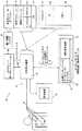
既存のECG解釈システムの出力を図1に示す。出力は画面画像10を含む。画像10は患者識別情報12と、患者の訴え又は症状などの初期診断情報14と、測定値16と、波形の形態をとる生理データ18と、診断又は解釈22と、一連の理由説明ステートメント24とを含む。図示されている例では、解釈22は患者が急性心臓虚血である確率は42%であることを指示している。この解釈を支える理由が理由説明ステートメント24に記載されている。例えば、解釈は患者が男性であり、心臓の痛みを訴えており、且つ「重大なQ波又は1次STセグメントの異常」が検出されなかったことに基づいている。また、理由説明ステートメント24は「前部T波」が平坦であることも指示している。
【0004】
専門医でない多くの者は図1に示すような理由説明ステートメントが専門的すぎ、従って、解釈を理解する役に立たないと感じる。更に、専門医でない場合、確率の高くない解釈に依存することに不快感を覚えるのが一般的である。図示されている例では、生成された解釈はわずか42%の確率しか示していない。すなわち、データに対して別の解釈が当てはまる確率が58%あるということになる。従って、現在のシステムがあいまいな確率を生成したような場合、その解釈はほとんど助けにはならないのである。
【0005】
更に、患者が心臓以外の手術を受けようとしている場合には、手術を実施することが患者に及ぼす術中心血管リスクを正確に評価できることが必要である。この場合にも、心臓専門医の到着を待ってそのような評価を下してもらうことは実用的ではなく、不可能でさえある。
【0006】
【発明の概要】
従って、本発明は、非心臓手術の実施と関連する術中心血管リスクを評価するために患者のECGを解釈する際のリアルタイム支援を提供する。収集装置は患者のECGを収集する。収集装置に結合するプロセッサは、収集されたECGが心血管リスクの診断ステートメントを有しているか否かを判定する。この診断ステートメントから、プロセッサは非心臓手術と関連する心血管リスクの確率を判定する。次に、心血管リスクの確率は重度、中程度又は軽度のいずれかの心血管リスクの標識の形態で臨床医に対して表示される。
【0007】
本発明は、非心臓手術の実施と関連する術中心血管リスクを評価するために患者のECGを解釈する新たな方法を更に提供する。方法は患者のECGを収集することと、ECGが心血管リスクの診断ステートメントを示しているか否かを判定することと、その診断ステートメントに基づいて心血管リスクの確率を判定することとを含む。方法は、判定された心血管リスクの確率を重度、中程度又は軽度のいずれかの心血管リスクの標識の形態で臨床医に対して表示することを更に含む。
【0008】
【発明の実施の形態】
本発明の一実施例を詳細に説明するが、本発明が以下の説明中に記載される、又は添付の図面に図示されている構成要素の構成及び配置の詳細に限定されないことを理解すべきである。本発明では他の実施例も可能であり、様々な方法で本発明を実施又は実行することが可能である。また、ここで使用する語句及び用語は説明の便宜上用いられるものであり、本発明を限定するとみなされてはならないということも理解すべである。「含む」、「具備する」及びそこから派生した言葉を使用するときには、それはそれ以降に列挙される項目及びそれと等価の項目、並びに追加の項目を包含することを意味している。
【0009】
生理データシステム30を図2に示す。システム30を説明する前に、ECGデータに関して本発明を説明することを理解すべきである。しかし、X線画像、核画像、超音波画像及び磁気共鳴画像を含む画像データ、血圧、酸化、脳の活動などの他のデータの解釈を補助するようにこのシステムを構成することも可能であろう。従って、本発明はここで説明し且つ図示する例に限定されるべきではない。
【0010】
システムは患者34に装着されるいくつかのセンサ又はそれに類する装置32を含む。センサ32により感知された生理データはリンク36を介して主ユニット40と、表示装置42とを有するデータ収集表示装置38へ伝送される。主ユニット40はプロセッサ44、入出力インタフェース46及びデータ記憶装置、すなわち、メモリ48などの典型的なハードウェアを含む。主ユニットはオペレーティングシステム50及びディスプレイモジュール52の形態をとる他のソフトウェアと、オプションであるウェブブラウザなどのブラウザ54と、相関モジュール56(オプションとしてデータ保全性検査モジュール(図示せず)を含んでいても良い)と、解釈モジュール58とを更に含む。
【0011】
データ収集表示装置38はいくつかの機能を実行する。第一に、センサ32を使用して患者34から信号又は生データを収集する。この生データはその後に測定される。例えば、図5に示すように、ECGデータを収集する場合には、波形高さ、ピーク間隔などの多数の特性が測定される。測定が実行された後、波形の様々な特徴が取り出される。以下に更に詳細に説明するが、それらの特徴を先に解釈済みの生理データの特徴と比較し、それを利用して解釈モジュール58により実行された解釈を検査することができる。
【0012】
解釈モジュール58は測定された特徴を使用して、生理データの解釈を生成する。本発明では既存の多様な生理データ解釈モジュールを使用できるであろう。ECGデータを解釈する構成である場合、GE Medical Systems Information Technologies, Incより販売されている12SL(登録商標)ソフトウェアを本発明で使用しても良い。
【0013】
データ収集表示装置38は、エキスパートロケーション(後述する)及び遠隔データライブラリ(同様に後述する)との通信を調整し且つ制御するためにオプションとして通信モジュール60を含んでいても良い。通信モジュール60に結合する、又は通信モジュール60の一部として形成された情報フィルタ62によって、データ収集表示装置38と遠隔装置との通信の質を向上させても良い。情報フィルタ62は、患者の個人情報などの所定の情報が収集表示装置38に結合する装置及び場所へ送信されないように保証するために、そのような所定の情報の送信を阻止するように構成されても良い。
【0014】
前述のように、解釈モジュール58は患者34から収集されたデータ又はデータセットを解析し且つ解釈して、そのデータセットに関する解釈を生成する。その後、相関モジュール56はその解釈を同じ又は類似する特徴及び解釈を有する生理データレコードにリンクする。例えば、R波の進行が乏しいことから引き出される前心筋梗塞(MI)は、前MIを伴う可能性のある特徴を示す全てのECGにリンクされるのではなく、同じ特徴を有するECGにリンクされることになるであろう。
【0015】
相関モジュール56は解釈を生理データレコードのライブラリ70にリンクする。すなわち、相関モジュール56は現在生理データから取り出された特徴をライブラリ70に格納されている生理データの事前解釈済みレコードと整合又は相関させる。次に、相関モジュール56は相関するレコードに対してハイパーリンクなどのリンクを作成する。生理データレコードのライブラリ70は通信リンク72を介してデータ収集表示装置38に結合している。通信リンク72は、レコードのライブラリ70が局所記憶装置に格納されている場合はローカルバスであっても良いが、レコードのライブラリ70をウェブサーバなどの遠隔サーバ74に格納できるように、インターネットリンクを含む他の多様なリンクであっても良い。サーバ74は、データ及び解釈の関連する特徴及び特性の説明を提供するために表示されている生理データ及び相関レコードにリンクできる追加又は補助教育資料又は情報レコードのライブラリ76を更に含んでいても良い。レコード70及び76をサーバに配置すると、更新が容易な情報の中央デポジトリを維持できるなどのいくつかの利点が得られる。しかし、レコード70及び76は別個のサーバで維持されても良いし、データ収集表示装置38に局所的にこれを維持することも可能であろう。
【0016】
データ収集表示装置38はリンク82を介してエキスパートロケーション80にリンクされても良い。エキスパートロケーション80はカスタム化ウェブサイト又はポータルであっても良く、また、リンク82はインターネットリンクであっても良いが、多様なロケーション及び通信リンクを使用できるであろう。例えば、本発明ではダイアルアップリンクを有する遠隔サーバを使用できるであろう。エキスパートロケーション80とデータ収集表示装置38との通信はテキスト又は音声に基づく電子メール、インスタントメッセージング又はチャットサービスを使用して行われば良い。このようなサービスは、ディスプレイモジュール52が生理データ画像をブラウザ54により生成されるウィンドウに表示させ且つエキスパートロケーション80がインターネット接続を介して収集表示装置38に結合されるようにデータ収集表示装置38が構成されている場合に特に適している。
【0017】
エキスパートロケーション80はコンピュータ又はそれに類する機器を有するサイト又はそれに類する場所である。典型的な例は、解釈モジュール58により解釈されるべき種類の生理データに関連する医療分野の専門医がいるオフィス又は施設に設置されたコンピュータである。データ収集表示装置38からのメッセージがエキスパートロケーション80で受信されると、専門医はそれに応答して、データ収集表示装置38を使用している医師又は他の担当者が生理データを解釈するのを補助する。
【0018】
図3は、ECT100の形態をとる生理データを示す。ECG100は患者ECG識別情報107と、患者識別情報108と、測定値110と、解釈112とを更に含む。解釈112は解釈モジュール58により生成される。解釈112が生成されると、相関モジュール56はレコードのライブラリ70を検索し、整合する生理データレコードを判定し、それらの整合レコードをECGレコード100にリンクする。ブラウザ54でアイコン又はボタン116を選択することにより整合レコードが表示されるように、システム30を構成することができる。
【0019】
図4は、整合するレコード120、122及び124を有するECG100を表示する画面118を示す。画面118は波形の関連特性を記述した説明ステートメント128を更に含む。
【0020】
整合する生理データレコード及び説明ステートメントの形の解釈と補助情報を提供するのに加えて、本発明は保全性検査を実行しても良い。保全性検査は、整合する生理データレコード及び説明ステートメントが表示された後に臨床医により実行される。臨床医は収集されたECGと、生理データレコードのライブラリ70から収集されたECGと相関されていた生理データレコードとの偏差を検出する。あるいは、データ収集表示装置38により得られた測定値が装置38の故障又はその他の問題によって偏向されていない又は誤っていないことを確実に保証する保全性検査モジュール(図示せず)がデータ収集表示装置38に含まれていても良い。
【0021】
先に述べた通り、システムのユーザが生理データの解釈に関する追加情報を望む場合には、生理データレコードのライブラリ70又はエキスパートロケーション80にいる専門医と通信することにより専門的なアドバイスを得ても良い。図6は、データ収集表示装置38により表示される専門医の応答画面の一例150を示す。この画面はECG100と共に、前述の通信ツールの1つを使用して生成される専門医のコメント160を含む。専門医応答画面150はそのコメントを述べた専門医の氏名及び写真などの専門医識別情報162を更に含んでいても良い。
【0022】
図7、図8及び図9は、患者に対して非心臓手術を実施することにより患者に及ぼされる術中心血管リスクを判定するのにも有用である本発明の特定の一実施例を示す。同じ部分は同じ図中符号で指示されている。以下の説明は同時係属米国特許出願第09/684,064号の主題を参考として取り入れている。
【0023】
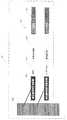
図2及び図7を参照して説明すると、生理データレコードのライブラリ70はステートメントコードライブラリ172を含む。ステートメントコードライブラリ172はECG波形を特徴づけるために使用される複数のステートメントコード170を含む。
【0024】
図7はステートメントコード170の2つの例を示す。ステートメントコード170はステートメント番号174と、ステートメント頭文字176と、診断ステートメント178と、術中心血管リスクの確率180とを含む。単なる一例であるが、正常な洞律動を示すECGを特徴づけるために使用されるステートメントコードはステートメント番号である5と、頭文字のNSRと、診断ステートメントである正常洞律動と、心血管リスクの確率である軽度リスクとを含む。
【0025】
最も好ましい実施例においては、ステートメントコード170は術中心血管リスクの評価以外の臨床的設定に合わせて構成自在である。すなわち、様々に異なる臨床的設定の特定の要求を反映するように心血管リスクの確率180を構成することができる。単なる一例であるが、家庭医療用の設定又は救急処置室での設定における心血管スクリーニングに対応するようにステートメントコード170を構成することが可能であろう。
【0026】
収集装置40によりECGが収集されると、収集装置40はECGを解析のために解釈モジュール58に入力する。解釈モジュール58にこの他に入力されるのは、実施すべき非心臓手術の種類、交換すべき患者体液の量、又は非心臓手術により影響を受ける循環器系の特定の構成部分などであろう。解釈モジュール58はECGを他の入力と共に解析し、ECGにステートメント番号174のみを割り当てる。
【0027】
ECGにステートメント番号174を割り当てるために、解釈モジュール58はECGを次に説明するように解析する。ECG解析の好ましい実施例を説明する前に、様々に異なるレベルの心血管リスクと関連する診断ステートメントを判定するための収集ECGの解析が本発明の範囲内に入っていることを理解すべきである。また、以下で論じる心血管リスクの診断ステートメントは単なる例として使用されているにすぎず、本発明の範囲を限定するものではないことも理解すべきである。更に、本発明の方法で実現される診断ステートメントは以下に例として挙げられる診断ステートメントと比べて特定の心血管事象又は心血管状態に関してより特殊性をもつものであっても良い。
【0028】
解釈モジュール58は、まず、重大な心血管リスクと関連する急性冠状動脈症候群に関してECGを解析する。解釈モジュール58が急性冠状動脈症候群を検出したならば、解釈モジュール58は心血管リスク178の診断ステートメント及び重大な心血管リスクの確率180を指示するステートメント番号174をECGに割り当てる。
【0029】
急性冠状動脈症候群という用語は、急性心筋梗塞、損傷及び急性虚血を含む多数の異なる診断ステートメントを包含する広い意味を持つ用語である。重大な心血管リスクの診断ステートメント及びそれらを検出するために解釈モジュール58が使用する方法については、以下に説明する。
【0030】
急性心筋梗塞(MI)は3つのECG特性、すなわち、虚血、損傷及びQ波梗塞を含む場合が多いが、これら3つの特性のいずれかが単独で現れることもある。
【0031】
虚血はT波の反転により特徴づけられる。すなわち、解釈モジュール58が反転T波を検出した場合、解釈モジュール58はECGに虚血を指定するステートメント番号174を割り当てる。
【0032】
損傷はSTセグメントの上昇により特徴づけられ、STセグメントの上昇はMIが急性であることを指示する。従って、ECGがSTセグメントの上昇を示す場合、解釈モジュール58は急性MIを指定するステートメント番号174をECGに割り当てる。解釈モジュール58は心血管リスクのより特定的な診断ステートメントによってECGを認識することができるのが好ましい。例えば、解釈もジュール58は全体的なSTセグメントの上昇だけではなく、ある特性を持つSTセグメントの上昇によってECGを認識することができる。
【0033】
急性梗塞と関連する第3のECG特性はQ波梗塞と呼ばれるQ波の存在である。特に急速な変化の形跡を伴うQ波梗塞は重大な心血管リスクの臨床的予兆であると考えられる。従って、解釈モジュール58がQ波の存在と同時に現れるST上昇を検出したならば、解釈モジュール58は急性心筋梗塞を指定するステートメント番号174をECGに割り当てる。
【0034】
非心臓手術中の重大な心血管リスクと関連する第2のカテゴリの診断ステートメントは不整脈、特に血液力学的に重大な不整脈である。血液力学的に重大な不整脈とは、心臓が効果的に血液を押し出すことを不可能にするような不整脈である。血液力学的に重大な不整脈の例としては、高度房室間ブロック、背後に心臓疾患が存在する場合の不整脈の徴候及び心室流量の制御不能を伴う心室上不整脈などがある。
【0035】
高度房室間ブロックは、房室間(AV)結節が心室に衝動を送ることができない場合に起こる。解釈モジュール58が高度房室間ブロックを検出すると、解釈モジュール58は高度房室間ブロックを指定するステートメント番号174をECGに割り当てる。
【0036】
背後に心臓疾患が存在する場合の不整脈の徴候は重大な心血管リスクの臨床的予兆であると考えられる。解釈モジュール58が背後に心臓疾患が存在する場合の不整脈を検出すると、解釈モジュール58は特定の不整脈及び心臓疾患の不整脈を指定するステートメント番号174をECGに割り当てる。
【0037】
心室上不整脈、特に心室流量の制御不能を伴う心房粗動は重大な心血管リスクの診断ステートメントである。単なる一例であるが、解釈モジュール58が心房粗動に対する急速な応答を検出した場合、解釈モジュール58は心室流量の制御不能を伴う心房粗動を指定するステートメント番号174をECGに割り当てる。
【0038】
解釈モジュール58が重大な心血管リスクの診断ステートメント178を求めてECGを解析し終わると、解釈モジュール58は中程度の心血管リスクの診断ステートメント178を求めてECGを解析する。中程度の心血管リスクと関連する1つの診断ステートメント178は以前に患った心筋梗塞である。解釈モジュール58は先にQ波心筋梗塞について説明したのとほぼ同じようにして以前の心筋梗塞を検出するが、この場合、ECGではST上昇は明白には現れない。
【0039】
解釈モジュール58が中程度の心血管リスクの診断ステートメント178を求めてECGを解析し終わると、解析モジュール58は軽度の心血管リスクの診断ステートメント178を求めてECGを解析する。軽度の心血管リスクと関連する診断ステートメント178は左心室肥大(LVH)、左房室束脚ブロック(LBBB)、機能的容量には影響しないST/Tセグメントの異常、及び洞律動以外の律動などのECGの異常である。
【0040】
解釈モジュール58が軽度の心血管リスクの診断ステートメント178を求めてECGを解析し終わったならば、解釈モジュール58は患者の現在ECGと、患者の以前のECG(利用可能である場合)との逐次比較を実行する。解釈モジュール58は、軽度の心血管リスクと関連する診断ステートメント178が新たなものであるか否か、すなわち、軽度の心血管リスクと関連する診断ステートメント178が以前のECGでは検出されておらず、現在ECGで検出されたのか否かを判定するために、患者の現在ECGと以前のECGとの逐次比較を実行する。検出された診断ステートメント178が新たなものであれば、心血管リスクの確率180は実際には中程度又は重大になる可能性もある。診断ステートメント178が新たなものではない場合には、心血管リスクの確率180は軽度である。
【0041】
現在ECGと以前のECGとの逐次比較は当該技術分野では一般的な方法である。逐次比較は以下に説明するようにして実行されるのが好ましい。以前に判定されたステートメントコード、測定値及び波形を含む患者の以前のECGは生理データレコードのデータベース70に格納されている。解釈モジュール58は現在ECGと以前のECGとの逐次比較を実行する。解釈モジュール58により検出されるべき心臓の状態に応じて、解釈モジュール58はステートメントコード、測定値及び波形のうち少なくとも1つを使用して、以前のECGを現在ECGと比較する。例えば、解釈モジュール58が異常律動を検出している場合、解釈モジュール58は以前のECGのステートメントコード170を現在ECGのステートメントコード170と比較する。解釈モジュール58がQRSコンプレックスの変化を検出している場合には、解釈モジュール58は以前のECGのステートメントコード170、測定値及び波形を現在ECGのステートメントコード170、測定値及び波形と比較する。解釈モジュール58がST/Tセグメント異常を検出している場合には、解釈モジュール58は以前のECGの波形を現在ECGの波形と比較する。このように、解釈モジュール58は心血管リスクの何らかの新たな診断ステートメント178を検出し、ステートメントコード170を、その新たな心血管リスクの診断ステートメント178を指定する現在ECGに割り当てる。
【0042】
心血管リスクの診断ステートメント178を重度、中程度及び軽度の心血管リスクの確率180に分類することは、様々に異なる臨床的設定に合わせて構成できるステートメントコード170の設定であることを理解すべきである。例えば、術中設定においてある不整脈が重大な心血管リスクの確率180と関連していることがあるが、同じ不整脈が家庭医療用の設定では一般的な心血管スクリーニングの間に中程度の心血管リスクの確率178と関連しているだけの場合もありうる。
【0043】
図2及び図7を参照して説明する。解釈モジュール58が以上説明した方法により現在ECGに1つ以上のステートメント番号174を割り当てたならば、そのステートメント番号174は解釈モジュール58からプロセッサ44へエクスポートされる。プロセッサ44は、ステートメント番号174をステートメントコード170と相関するために、生理データレコードのライブラリ170にあるステートメントライブラリ172をアクセスする。ステートメント番号174とステートメントコード170との相関が成立したならば、プロセッサ44は診断ステートメント178を記述するテキストステートメントと、関連する心血管リスクの確率180とをアクセスする。次に、プロセッサ44は心血管リスクの診断ステートメント178と、心血管リスクの確率180とを含むテキストレポートを生成する。生成されるレポートは、ECGの解釈に関して心臓専門医の助言を求めることを臨床医に示唆するステートメントを含んでいても良い。生成されたレポートは表示装置42により臨床医に対して表示される。
【0044】
図8及び図9は、本発明の方法を示すフローチャートである。図2、図7及び図8を参照して説明すると、まず、臨床医は患者の病歴を知る(190)。臨床医は患者の話を聞き取り、心血管の状態に関連する一連の問診を行うことにより患者の病歴を作成する。詳細には、臨床医は、患者が以前に狭心症にかかったか、最近又は過去にMIを起こしたか、鬱血性心臓障害はあるか、又は不整脈の徴候はあるかなどの重大な心臓病をわずらったか否かを尋ねる。患者の病歴によって、患者が重大な心臓病を抱えていることが判明した場合(192)、臨床医は非心臓手術を実施する前に心臓専門医の助言を求めるのが好ましい(194)。
【0045】
患者の病歴が重大な心臓病の状態を明示しない場合には(192)、臨床医は物理的検査を実施する(196)。物理的検査の間、臨床医は患者が安定した又は不安定な狭心症、代償又は代償不全鬱血性心臓障害等の重大な心臓の病状を有しているか否かを判定する。加えて、臨床医は、患者が狭窄症状又は逆流性弁疾患などの重大な弁の疾患を患っているか否かを判定する。患者の検査によって、患者が重大な心臓病を抱えていることが明示された場合(197)、臨床医は非心臓手術を実施する前に心臓専門医の助言を求めるのが好ましい(198)。
【0046】
患者の病歴で重大な心臓の病状が明示されず(192)且つ物理的検査でも患者の心臓の機能容量が良好であることが明示された(197)場合には、患者は初めて心血管リスクが低いと指定される(200)。
【0047】
この場合にも同様に、患者が重大な心臓の病状を抱えている又は患者の機能的容量が乏しいことを臨床医が知ったならば、臨床医は非心臓手術を進める前に心臓専門医に相談するのが好ましい。しかし、初期段階で患者の心血管リスクが低いことが示された(200)場合には、臨床医は収集装置40によって患者のECGを収集する(202)。収集装置40はECGを解析のために解釈モジュール58へ送信する(204)。解釈モジュール58はECGを解析し、ステートメント番号174をECGに割り当てる。次に、解釈モジュール58は現在ECGを患者の以前のECGと比較することにより逐次比較を実行する(205)。解釈モジュール58は、この逐次比較の結果を指定する追加ステートメント番号174を現在ECGに割り当てる(205)。ステートメント番号174は解釈モジュール58から収集装置40及びプロセッサ44にエクスポートされる(206)。
【0048】
図2、図7及び図9を参照して説明する。生理データレコードのライブラリ70の中にステートメントコードライブラリ172を形成する(208)。ライブラリ70はデータ収集表示装置38のプロセッサ44に結合している。プロセッサ44はステートメントコードライブラリ172をアクセスし、解釈モジュール58により生成されたステートメント番号174をステートメントコードのリスト170と比較して、心血管リスクの診断ステートメント178及びステートメント番号174と関連するリスクの確率180を判定する(210)。
【0049】
ステートメント番号174がステートメントコード170と相関されたならば、プロセッサ44は、まず、ステートメントコード170が重大であると考えられる心血管リスクの確率180を含むか否かを判定する(212)。心血管リスクの確率180が重大であれば、プロセッサ44は重大な心血管リスクの標識と、心臓専門医の助言を求めるようにとの示唆とを含むテキストレポートを生成する(214)。
【0050】
臨床医は、心臓専門医の助言を求めるようにとの示唆を含むテキストレポートを読んだならば、直ちにエキスパートロケーション80にいる心臓専門医の意見を聞く道を選択することができる。臨床医が心臓専門医に相談することを選択した場合には、収集装置40の通信モジュール60は現在ECGを含むEメールメッセージを生成し、エキスパートロケーション80の心臓専門医へ送信する(234)。通信モジュール60は所定の時限でタイマーを設定し(236)、心臓専門医はその時間内にECGの解釈を完了して、応答を送信しなければならない。心臓専門医はECGを解釈し、そのECG解釈のテキストレポートを生成し、生成されたテキストレポートをEメールを介して収集装置40へ送信する。ECG解釈のテキストレポートを含むEメールメッセージは収集装置40により受信され(238)、処理される。ECG解釈のテキストレポートは表示装置42に表示される(240)。心臓専門医と収集装置40との通信をEメールメッセージとして説明したが、この通信は電話呼び出し、イントラネット呼び出し又は何らかの種類のインターネットメッセージを含む任意のアナログ形態又はデジタル形態の通信であれば良い。
【0051】
臨床医は心臓専門医に相談する方法を選ばずに、生理データレコードのライブラリ70又は補助教育資料76をECGを解釈する上での助けとして使用する方法をとっても良い。
【0052】
心血管リスクの確率180が重大なものではない場合、プロセッサ44は、次に、ステートメントコード170が中程度であると考えられる心血管リスクの確率180を含むか否かを判定する(218)。心血管リスクの確率180が中程度であれば、プロセッサ44は中程度の心血管リスクの標識と、心臓専門医の助言を求めるようにとの示唆とを含むテキストレポートを生成する(220)。テキストレポート222は表示装置42により臨床医に対して表示される(222)。臨床医は心臓専門医の助言を求めることを示唆するテキストレポートを検討した後、エキスパートロケーション80にいる心臓専門医の助言を求める道を選ぶこともできるし、あるいは生理データレコードのライブラリ70又は補助教育資料76をECGを解釈する上での助けとして使用する方法をとることも可能である。
【0053】
心血管リスクの確率180が重大又は中程度ではない場合には、プロセッサ44は最終的にステートメントコード170が軽度であると考えられる心血管リスクの確率180を含むか否かを判定する(224)。心血管リスクの確率180が軽度であれば、プロセッサ44は軽度の心血管リスクの標識を含むテキストレポートを生成する(230)。テキストレポートは表示装置42により臨床医に対して表示される(232)。ステートメントコードが軽度の心血管リスクの確率さえ指示していない場合には、プロセッサはECGが正常であることを指示するテキストレポートを生成する(226)。テキストレポートは表示装置42により臨床医に対して表示される(228)。
【0054】
本発明の様々な特徴及び利点は特許請求の範囲に記載されている。
【図面の簡単な説明】
【図1】 既存の生理データ解釈システムの出力を示す図。
【図2】 本発明の生理データ解釈システムを示す図。
【図3】 図2のシステムの出力を示す図。
【図4】 図2のシステムの出力を示す図。
【図5】 特徴を定義するために使用される生理的波形の測定値を示す図。
【図6】 専門医との通信の内容を示す図。
【図7】 本発明の方法で使用するためのステートメントコードを示す図。
【図8】 本発明の方法を示すフローチャート。
【図9】 本発明の方法を更に示すための図8に続くフローチャート。
【符号の説明】
30…生理データシステム、32…センサ、34…患者、36…リンク、38…データ収集表示装置、40…主ユニット、42…表示装置、44…プロセッサ、46…入出力インタフェース、48…メモリ、50…オペレーティングシステム、52…ディスプレイモジュール、54…ブラウザ、56…相関モジュール、58…解釈モジュール、60…通信モジュール、62…個人情報フィルタ、70…生理データレコードのライブラリ、72…通信リンク、74…遠隔サーバ、76…補助教育資料、80…エキスパートロケーション、170…ステートメントコード、172…ステートメントコードライブラリ、174…ステートメント番号、178…診断ステートメント、180…術中心血管リスクの確率[0001]
[Prior art]
Physiological movement is monitored when diagnosing and treating a number of diseases and medical conditions. For example, in general, heart motion is monitored by collecting electrocardiogram (“ECG”) data. ECG data is typically interpreted by a cardiologist, a specially trained physician who reads the waveforms generated by the ECG device.
[0002]
Although ECG data and other organizing data can be used in many situations, there are not always specialists with appropriate training to read the data. To address this problem, software interpretation tools have been developed to help non-specialists interpret and use such data. However, those tools, especially ECG tools, are not satisfactory. Existing ECG tools are designed to generate interpretations. The computer-generated interpretation may be aided by one or more statements that describe the criteria used by the computer to reach its conclusion. However, these statements are usually limited to content that describes the nature of the waveform and are usually of little help to those who are not familiar with reading ECGs. Another drawback of existing tools is that they draw conclusions based on the assumption that the ECG device has accurately measured the ECG. In other words, existing computer tools assume that no failure or other error will occur in the ECG measurement equipment.
[0003]
The output of an existing ECG interpretation system is shown in FIG. The output includes a
[0004]
Many non-specialists feel that a reasoning statement such as that shown in FIG. 1 is too technical and therefore not helpful to understand the interpretation. Furthermore, if you are not a specialist, you generally feel uncomfortable with relying on non-probable interpretations. In the example shown, the generated interpretation shows only 42% probability. That is, there is a 58% probability that another interpretation applies to the data. Thus, if the current system produces an ambiguous probability, its interpretation is of little help.
[0005]
Furthermore, when a patient is about to undergo surgery other than the heart, it is necessary to be able to accurately assess the central vascular risk that the surgery will have on the patient. Again, it is impractical and even impossible to have such an assessment after the arrival of a cardiologist.
[0006]
SUMMARY OF THE INVENTION
Thus, the present invention provides real-time assistance in interpreting a patient's ECG to assess central vascular risk associated with performing non-cardiac surgery. The collection device collects the patient's ECG. A processor coupled to the collection device determines whether the collected ECG has a cardiovascular risk diagnostic statement. From this diagnostic statement, the processor determines the probability of cardiovascular risk associated with non-cardiac surgery. The cardiovascular risk probability is then displayed to the clinician in the form of either a severe, moderate or mild cardiovascular risk label.
[0007]
The present invention further provides a new method of interpreting a patient's ECG to assess central vascular risk associated with performing non-cardiac surgery. The method includes collecting a patient's ECG, determining whether the ECG indicates a cardiovascular risk diagnostic statement, and determining a cardiovascular risk probability based on the diagnostic statement. The method further includes displaying the determined cardiovascular risk probability to the clinician in the form of either a severe, moderate or mild cardiovascular risk label.
[0008]
DETAILED DESCRIPTION OF THE INVENTION
Reference will now be made in detail to an embodiment of the present invention, but it should be understood that the invention is not limited to the details of construction and arrangement of components set forth in the following description or illustrated in the accompanying drawings. It is. The invention is capable of other embodiments and of being practiced or carried out in various ways. It should also be understood that the terms and terms used herein are used for convenience of description and should not be considered as limiting the present invention. When the words “including”, “comprising” and words derived therefrom are used, it is meant to include items listed thereafter and equivalents thereof, as well as additional items.
[0009]
A
[0010]
The system includes a number of sensors or similar devices 32 attached to the patient 34. The physiological data sensed by the sensor 32 is transmitted via a link 36 to a data collection /
[0011]
The data collection and
[0012]
[0013]
The data collection and
[0014]
As described above, the
[0015]
The
[0016]
[0017]
The
[0018]
FIG. 3 shows physiological data in the form of ECT100.
[0019]
FIG. 4 shows a screen 118 that displays
[0020]
In addition to providing matching physiological data records and explanation statement form interpretations and auxiliary information, the present invention may perform integrity checks. The integrity check is performed by the clinician after the matching physiological data record and explanatory statements are displayed. The clinician detects the deviation between the collected ECG and the physiological data record that was correlated with the ECG collected from the
[0021]
As noted above, if a user of the system desires additional information regarding the interpretation of physiological data, expert advice may be obtained by communicating with a specialist in the physiological
[0022]
FIGS. 7, 8 and 9 illustrate one particular embodiment of the invention that is also useful for determining the operative central vascular risk posed to a patient by performing non-cardiac surgery on the patient. The same parts are indicated by the same reference numerals in the figure. The following description incorporates by reference the subject matter of co-pending US patent application Ser. No. 09 / 684,064.
[0023]
Referring to FIGS. 2 and 7, the physiological
[0024]
FIG. 7 shows two examples of
[0025]
In the most preferred embodiment, the
[0026]
Once the ECG is collected by the
[0027]
In order to assign a
[0028]
[0029]
The term acute coronary syndrome is a broad term that encompasses a number of different diagnostic statements including acute myocardial infarction, injury and acute ischemia. The diagnostic statements for significant cardiovascular risks and the methods used by the
[0030]
Acute myocardial infarction (MI) often includes three ECG characteristics: ischemia, injury, and Q-wave infarction, but any of these three characteristics may appear alone.
[0031]
Ischemia is characterized by T-wave reversal. That is, if the
[0032]
Injury is characterized by elevated ST segment, which indicates that MI is acute. Thus, if the ECG indicates an ST segment rise,
[0033]
A third ECG characteristic associated with acute infarction is the presence of a Q wave called Q wave infarction. Q-wave infarction, particularly with evidence of rapid changes, is considered a clinical sign of significant cardiovascular risk. Thus, if
[0034]
A second category of diagnostic statements associated with significant cardiovascular risk during non-cardiac surgery is arrhythmias, particularly hemodynamically significant arrhythmias. A hemodynamically significant arrhythmia is an arrhythmia that makes it impossible for the heart to effectively pump blood. Examples of hemodynamically significant arrhythmias include advanced interventricular block, signs of arrhythmia in the presence of heart disease behind and supraventricular arrhythmia with inability to control ventricular flow.
[0035]
Advanced interatrioventricular block occurs when the interatrioventricular (AV) node cannot send impulses to the ventricles. When
[0036]
Signs of arrhythmia in the presence of heart disease behind are considered clinical signs of significant cardiovascular risk. When
[0037]
Supraventricular arrhythmias, particularly atrial flutter with inability to control ventricular flow, are a diagnostic statement of significant cardiovascular risk. By way of example only, if
[0038]
Once the
[0039]
When
[0040]
Once the
[0041]
The sequential comparison between the current ECG and the previous ECG is a common method in the art. The successive approximation is preferably performed as described below. The patient's previous ECG, including previously determined statement codes, measurements and waveforms, is stored in the
[0042]
It should be understood that classifying cardiovascular risk
[0043]
This will be described with reference to FIGS. If
[0044]
8 and 9 are flowcharts illustrating the method of the present invention. Referring to FIGS. 2, 7 and 8, first, the clinician knows the patient's medical history (190). The clinician listens to the patient and creates a medical history of the patient by conducting a series of questions related to cardiovascular conditions. Specifically, the clinician may have a serious heart condition, such as whether the patient has previously had angina, recently or in the past MI, has congestive heart failure, or has signs of arrhythmia. Ask if you've been bothered. If the patient's medical history indicates that the patient has significant heart disease (192), the clinician preferably seeks cardiologist advice before performing non-cardiac surgery (194).
[0045]
If the patient's medical history does not reveal a significant heart condition (192), the clinician performs a physical examination (196). During the physical examination, the clinician determines whether the patient has a serious heart condition such as stable or unstable angina, compensatory or decompensated congestive heart failure. In addition, the clinician determines whether the patient is suffering from a significant valve disease, such as a stenosis condition or reflux valve disease. If patient examination reveals that the patient has significant heart disease (197), the clinician preferably seeks cardiologist advice before performing non-cardiac surgery (198).
[0046]
If the patient's medical history does not reveal a significant heart condition (192) and physical examination clearly indicates that the patient's heart functional capacity is good (197), the patient is at first cardiovascular risk. Designated as low (200).
[0047]
Again, if the clinician learns that the patient has a serious heart condition or that the patient's functional capacity is poor, the clinician should consult a cardiologist before proceeding with non-cardiac surgery. It is preferable to do this. However, if the patient is shown to have a low cardiovascular risk at an early stage (200), the clinician collects the patient's ECG through the collection device 40 (202). The
[0048]
This will be described with reference to FIGS. 2, 7 and 9. A
[0049]
If the
[0050]
Once the clinician has read a text report containing suggestions to seek cardiologist's advice, he can immediately choose to hear the cardiologist's opinion at the
[0051]
The clinician may choose to use the physiological
[0052]
If the
[0053]
If the
[0054]
Various features and advantages of the invention are set forth in the following claims.
[Brief description of the drawings]
FIG. 1 is a diagram showing an output of an existing physiological data interpretation system.
FIG. 2 is a diagram showing a physiological data interpretation system of the present invention.
FIG. 3 is a diagram showing the output of the system of FIG. 2;
4 is a diagram showing the output of the system of FIG.
FIG. 5 shows physiological waveform measurements used to define features.
FIG. 6 is a diagram showing the contents of communication with a specialist.
FIG. 7 shows a statement code for use in the method of the present invention.
FIG. 8 is a flowchart illustrating the method of the present invention.
FIG. 9 is a flowchart following FIG. 8 to further illustrate the method of the present invention.
[Explanation of symbols]
DESCRIPTION OF
Claims (9)
Translated fromJapanese心電図(100)を収集する収集装置(38)と、
前記収集装置(38)に結合したプロセッサ(44)であって、
収集された心電図(100)が左心室肥大(LVH)、左房室束脚ブロック(LBBB)及び、機能的容量には影響しないST/Tセグメントの異常のいずれかである命の危機をもたらさない心状態を示す場合に、前記収集された心電図(100)と以前に収集された心電図との逐次比較を実行し、
前記収集された心電図(100)の前記命の危機をもたらさない心状態が前記以前の心電図に示されているか否かを決定し、
該心状態が前記以前の心電図に示されていなかった場合に、前記収集された心電図(100)に前記逐次比較の結果を指定する追加ステートメントを割り振る、
前記プロセッサ(44)と、
心血管リスクの確率(180)を臨床医に対して指示する表示装置(42)とを具備する装置。In a device for assessing a patient's central vascular risk,
A collection device (38) for collecting an electrocardiogram (100);
A processor (44) coupled to the collector (38), comprising:
The collected electrocardiogram (100) doesnot pose a life-threatening thatis either left ventricular hypertrophy (LVH), left atrial ventricular bundle leg block (LBBB), or an abnormal ST / T segment that does not affect functional capacity to indicate heartcondition, perform the sequential comparison of the collected electrocardiogram and previously the collected electrocardiogram (100),
Determining whether a heart condition that does not result in the life crisis of the collected electrocardiogram (100) is indicated in the previous electrocardiogram;
Allocatingan additional statementspecifying the result of the successive approximation tothe collected electrocardiogram (100) if the cardiac condition was not indicated in the previous electrocardiogram;
The processor (44);
A display device (42) for instructing the clinician on the probability of cardiovascular risk (180).
Applications Claiming Priority (2)
| Application Number | Priority Date | Filing Date | Title |
|---|---|---|---|
| US09/752081 | 2000-12-29 | ||
| US09/752,081US6665559B2 (en) | 2000-10-06 | 2000-12-29 | Method and apparatus for perioperative assessment of cardiovascular risk |
Publications (3)
| Publication Number | Publication Date |
|---|---|
| JP2002330937A JP2002330937A (en) | 2002-11-19 |
| JP2002330937A5 JP2002330937A5 (en) | 2005-08-04 |
| JP4159285B2true JP4159285B2 (en) | 2008-10-01 |
Family
ID=25024780
Family Applications (1)
| Application Number | Title | Priority Date | Filing Date |
|---|---|---|---|
| JP2001399812AExpired - LifetimeJP4159285B2 (en) | 2000-12-29 | 2001-12-28 | Intraoperative evaluation method and apparatus for cardiovascular risk |
Country Status (3)
| Country | Link |
|---|---|
| US (2) | US6665559B2 (en) |
| JP (1) | JP4159285B2 (en) |
| DE (1) | DE10164322A1 (en) |
Families Citing this family (104)
| Publication number | Priority date | Publication date | Assignee | Title |
|---|---|---|---|---|
| US6527711B1 (en) | 1999-10-18 | 2003-03-04 | Bodymedia, Inc. | Wearable human physiological data sensors and reporting system therefor |
| WO2005029242A2 (en)* | 2000-06-16 | 2005-03-31 | Bodymedia, Inc. | System for monitoring and managing body weight and other physiological conditions including iterative and personalized planning, intervention and reporting capability |
| US7285090B2 (en)* | 2000-06-16 | 2007-10-23 | Bodymedia, Inc. | Apparatus for detecting, receiving, deriving and displaying human physiological and contextual information |
| US20060122474A1 (en) | 2000-06-16 | 2006-06-08 | Bodymedia, Inc. | Apparatus for monitoring health, wellness and fitness |
| US7689437B1 (en) | 2000-06-16 | 2010-03-30 | Bodymedia, Inc. | System for monitoring health, wellness and fitness |
| US7261690B2 (en)* | 2000-06-16 | 2007-08-28 | Bodymedia, Inc. | Apparatus for monitoring health, wellness and fitness |
| PT1292218E (en) | 2000-06-23 | 2006-09-29 | Bodymedia Inc | SYSTEM FOR HEALTH, WELL-BEING AND APPROPRIATE SURVEILLANCE |
| US7224281B2 (en)* | 2001-08-31 | 2007-05-29 | Draeger Medical Systems, Inc. | Patient monitoring and alarm processing system and user interface |
| JP4330866B2 (en)* | 2002-01-10 | 2009-09-16 | 株式会社東芝 | Medical communication system |
| US20030149423A1 (en)* | 2002-02-04 | 2003-08-07 | Fischell Robert E. | Methods for the detection and treatment of cardiac events |
| US20030208106A1 (en)* | 2002-05-03 | 2003-11-06 | Cortex Biophysik Gmbh | Method of cardiac risk assessment |
| EP1529487A4 (en)* | 2002-07-04 | 2009-05-27 | Dainippon Sumitomo Pharma Co | ELECTROCARDIOGRAM ANALYSIS DEVICE AND ASSOCIATED METHOD |
| US7020508B2 (en) | 2002-08-22 | 2006-03-28 | Bodymedia, Inc. | Apparatus for detecting human physiological and contextual information |
| US7194298B2 (en)* | 2002-10-02 | 2007-03-20 | Medicale Intelligence Inc. | Method and apparatus for trend detection in an electrocardiogram monitoring signal |
| US7182738B2 (en) | 2003-04-23 | 2007-02-27 | Marctec, Llc | Patient monitoring apparatus and method for orthosis and other devices |
| US7330750B2 (en)* | 2003-04-25 | 2008-02-12 | Instrumentarium Corp. | Estimation of cardiac death risk |
| US7131947B2 (en)* | 2003-05-08 | 2006-11-07 | Koninklijke Philips Electronics N.V. | Volumetric ultrasonic image segment acquisition with ECG display |
| JP2005000409A (en)* | 2003-06-12 | 2005-01-06 | Omron Healthcare Co Ltd | ECG and ECG waveform display method |
| KR101084554B1 (en)* | 2003-09-12 | 2011-11-17 | 보디미디어 인코퍼레이티드 | Method and apparatus for measuring heart related parameters |
| US9171129B2 (en)* | 2004-02-10 | 2015-10-27 | Todd J. Cohen | System and method for storing, accessing, and displaying specialized patient information and other medical information |
| WO2005092177A1 (en) | 2004-03-22 | 2005-10-06 | Bodymedia, Inc. | Non-invasive temperature monitoring device |
| DE102005036501A1 (en)* | 2005-07-30 | 2007-02-01 | Bonaventura, Klaus, Dr. | Portable electrocardiagram device for monitoring person heart activity, has one unit for comparing derived electrocardiagram with reference electrocardiagram, where individual electrocardiagram of patients is stored as reference cardiagram |
| US7542955B2 (en)* | 2006-03-10 | 2009-06-02 | The General Electric Company | Exercise test interpretation |
| US7702382B2 (en) | 2006-04-17 | 2010-04-20 | General Electric Company | Multi-tier system for cardiology and patient monitoring data analysis |
| US20080320029A1 (en) | 2007-02-16 | 2008-12-25 | Stivoric John M | Lifeotype interfaces |
| JP5592654B2 (en)* | 2007-03-07 | 2014-09-17 | コーニンクレッカ フィリップス エヌ ヴェ | ECG management system diagnostic code and customization |
| EP2194858B1 (en) | 2007-09-14 | 2017-11-22 | Corventis, Inc. | Medical device automatic start-up upon contact to patient tissue |
| EP3922171A1 (en) | 2007-09-14 | 2021-12-15 | Medtronic Monitoring, Inc. | Adherent cardiac monitor with advanced sensing capabilities |
| WO2009036327A1 (en) | 2007-09-14 | 2009-03-19 | Corventis, Inc. | Adherent device for respiratory monitoring and sleep disordered breathing |
| WO2009036369A1 (en) | 2007-09-14 | 2009-03-19 | Corventis, Inc. | System and methods for wireless body fluid monitoring |
| US9186089B2 (en) | 2007-09-14 | 2015-11-17 | Medtronic Monitoring, Inc. | Injectable physiological monitoring system |
| US8116841B2 (en) | 2007-09-14 | 2012-02-14 | Corventis, Inc. | Adherent device with multiple physiological sensors |
| WO2009036316A1 (en) | 2007-09-14 | 2009-03-19 | Corventis, Inc. | Energy management, tracking and security for adherent patient monitor |
| WO2009053888A2 (en)* | 2007-10-24 | 2009-04-30 | Koninklijke Philips Electronics, N.V. | System and method for combining serial ecg analysis and ecg ordering |
| US8185623B2 (en) | 2008-02-29 | 2012-05-22 | Physio-Control, Inc. | Selectively routing patient data between field devices and treatment center destinations |
| EP2257216B1 (en)* | 2008-03-12 | 2021-04-28 | Medtronic Monitoring, Inc. | Heart failure decompensation prediction based on cardiac rhythm |
| US8808185B2 (en)* | 2008-03-28 | 2014-08-19 | General Electric Company | System and method for generating a patient diagnosis |
| WO2009146214A1 (en) | 2008-04-18 | 2009-12-03 | Corventis, Inc. | Method and apparatus to measure bioelectric impedance of patient tissue |
| JP5356961B2 (en)* | 2009-09-17 | 2013-12-04 | フクダ電子株式会社 | Medical information service provision device |
| WO2011050283A2 (en) | 2009-10-22 | 2011-04-28 | Corventis, Inc. | Remote detection and monitoring of functional chronotropic incompetence |
| US8374686B2 (en)* | 2009-12-04 | 2013-02-12 | Medtronic, Inc. | Continuous monitoring of risk burden for sudden cardiac death risk stratification |
| US9451897B2 (en) | 2009-12-14 | 2016-09-27 | Medtronic Monitoring, Inc. | Body adherent patch with electronics for physiologic monitoring |
| US20110166888A1 (en)* | 2010-01-04 | 2011-07-07 | Physio-Control, Inc. | Simplified launching of electronic messages in center for treating patient |
| US20110224565A1 (en) | 2010-03-15 | 2011-09-15 | Singapore Health Services Pte Ltd. | Method of predicting acute cardiopulmonary events and survivability of a patient |
| US8965498B2 (en) | 2010-04-05 | 2015-02-24 | Corventis, Inc. | Method and apparatus for personalized physiologic parameters |
| US8082027B2 (en) | 2010-05-07 | 2011-12-20 | General Electric Company | Portable USB electrocardiograph system and method |
| WO2011153428A1 (en)* | 2010-06-03 | 2011-12-08 | Medtronic, Inc. | System and method for assessing a likelihood of a patient to experience a future cardiac arrhythmia using dynamic changes in a biological parameter |
| US8239012B2 (en) | 2010-10-08 | 2012-08-07 | Cardiac Science Corporation | Microcontrolled electrocardiographic monitoring circuit with differential voltage encoding |
| US20120089000A1 (en) | 2010-10-08 | 2012-04-12 | Jon Mikalson Bishay | Ambulatory Electrocardiographic Monitor For Providing Ease Of Use In Women And Method Of Use |
| US8613708B2 (en) | 2010-10-08 | 2013-12-24 | Cardiac Science Corporation | Ambulatory electrocardiographic monitor with jumpered sensing electrode |
| US9037477B2 (en) | 2010-10-08 | 2015-05-19 | Cardiac Science Corporation | Computer-implemented system and method for evaluating ambulatory electrocardiographic monitoring of cardiac rhythm disorders |
| RU2598049C2 (en)* | 2010-12-01 | 2016-09-20 | Конинклейке Филипс Электроникс Н.В. | Automated identification of location of occlusion in infarct-related coronary artery |
| US20120165617A1 (en)* | 2010-12-28 | 2012-06-28 | General Electric Company | Patient enabled methods, apparatus, and systems for early health and preventive care using wearable sensors |
| CN104755024B (en)* | 2012-10-26 | 2017-12-19 | 皇家飞利浦有限公司 | To the diagnostic expression and deciphering of ECG Lead on digital display |
| US10736529B2 (en) | 2013-09-25 | 2020-08-11 | Bardy Diagnostics, Inc. | Subcutaneous insertable electrocardiography monitor |
| US9433380B1 (en) | 2013-09-25 | 2016-09-06 | Bardy Diagnostics, Inc. | Extended wear electrocardiography patch |
| US9619660B1 (en) | 2013-09-25 | 2017-04-11 | Bardy Diagnostics, Inc. | Computer-implemented system for secure physiological data collection and processing |
| US10820801B2 (en) | 2013-09-25 | 2020-11-03 | Bardy Diagnostics, Inc. | Electrocardiography monitor configured for self-optimizing ECG data compression |
| US10736531B2 (en) | 2013-09-25 | 2020-08-11 | Bardy Diagnostics, Inc. | Subcutaneous insertable cardiac monitor optimized for long term, low amplitude electrocardiographic data collection |
| US9408551B2 (en) | 2013-11-14 | 2016-08-09 | Bardy Diagnostics, Inc. | System and method for facilitating diagnosis of cardiac rhythm disorders with the aid of a digital computer |
| US10433748B2 (en) | 2013-09-25 | 2019-10-08 | Bardy Diagnostics, Inc. | Extended wear electrocardiography and physiological sensor monitor |
| US10433751B2 (en) | 2013-09-25 | 2019-10-08 | Bardy Diagnostics, Inc. | System and method for facilitating a cardiac rhythm disorder diagnosis based on subcutaneous cardiac monitoring data |
| US10624551B2 (en) | 2013-09-25 | 2020-04-21 | Bardy Diagnostics, Inc. | Insertable cardiac monitor for use in performing long term electrocardiographic monitoring |
| US10463269B2 (en) | 2013-09-25 | 2019-11-05 | Bardy Diagnostics, Inc. | System and method for machine-learning-based atrial fibrillation detection |
| US9737224B2 (en) | 2013-09-25 | 2017-08-22 | Bardy Diagnostics, Inc. | Event alerting through actigraphy embedded within electrocardiographic data |
| US9364155B2 (en) | 2013-09-25 | 2016-06-14 | Bardy Diagnostics, Inc. | Self-contained personal air flow sensing monitor |
| US10806360B2 (en) | 2013-09-25 | 2020-10-20 | Bardy Diagnostics, Inc. | Extended wear ambulatory electrocardiography and physiological sensor monitor |
| US20190167139A1 (en) | 2017-12-05 | 2019-06-06 | Gust H. Bardy | Subcutaneous P-Wave Centric Insertable Cardiac Monitor For Long Term Electrocardiographic Monitoring |
| US10165946B2 (en) | 2013-09-25 | 2019-01-01 | Bardy Diagnostics, Inc. | Computer-implemented system and method for providing a personal mobile device-triggered medical intervention |
| US11723575B2 (en) | 2013-09-25 | 2023-08-15 | Bardy Diagnostics, Inc. | Electrocardiography patch |
| US9504423B1 (en) | 2015-10-05 | 2016-11-29 | Bardy Diagnostics, Inc. | Method for addressing medical conditions through a wearable health monitor with the aid of a digital computer |
| US9700227B2 (en) | 2013-09-25 | 2017-07-11 | Bardy Diagnostics, Inc. | Ambulatory electrocardiography monitoring patch optimized for capturing low amplitude cardiac action potential propagation |
| US9655538B2 (en) | 2013-09-25 | 2017-05-23 | Bardy Diagnostics, Inc. | Self-authenticating electrocardiography monitoring circuit |
| US9345414B1 (en) | 2013-09-25 | 2016-05-24 | Bardy Diagnostics, Inc. | Method for providing dynamic gain over electrocardiographic data with the aid of a digital computer |
| US9717433B2 (en) | 2013-09-25 | 2017-08-01 | Bardy Diagnostics, Inc. | Ambulatory electrocardiography monitoring patch optimized for capturing low amplitude cardiac action potential propagation |
| US9717432B2 (en) | 2013-09-25 | 2017-08-01 | Bardy Diagnostics, Inc. | Extended wear electrocardiography patch using interlaced wire electrodes |
| US10888239B2 (en) | 2013-09-25 | 2021-01-12 | Bardy Diagnostics, Inc. | Remote interfacing electrocardiography patch |
| US9408545B2 (en) | 2013-09-25 | 2016-08-09 | Bardy Diagnostics, Inc. | Method for efficiently encoding and compressing ECG data optimized for use in an ambulatory ECG monitor |
| US10799137B2 (en) | 2013-09-25 | 2020-10-13 | Bardy Diagnostics, Inc. | System and method for facilitating a cardiac rhythm disorder diagnosis with the aid of a digital computer |
| US9615763B2 (en) | 2013-09-25 | 2017-04-11 | Bardy Diagnostics, Inc. | Ambulatory electrocardiography monitor recorder optimized for capturing low amplitude cardiac action potential propagation |
| US10667711B1 (en) | 2013-09-25 | 2020-06-02 | Bardy Diagnostics, Inc. | Contact-activated extended wear electrocardiography and physiological sensor monitor recorder |
| US9433367B2 (en) | 2013-09-25 | 2016-09-06 | Bardy Diagnostics, Inc. | Remote interfacing of extended wear electrocardiography and physiological sensor monitor |
| US9545204B2 (en) | 2013-09-25 | 2017-01-17 | Bardy Diagnostics, Inc. | Extended wear electrocardiography patch |
| US9655537B2 (en) | 2013-09-25 | 2017-05-23 | Bardy Diagnostics, Inc. | Wearable electrocardiography and physiology monitoring ensemble |
| WO2015048194A1 (en) | 2013-09-25 | 2015-04-02 | Bardy Diagnostics, Inc. | Self-contained personal air flow sensing monitor |
| US11213237B2 (en) | 2013-09-25 | 2022-01-04 | Bardy Diagnostics, Inc. | System and method for secure cloud-based physiological data processing and delivery |
| US9775536B2 (en) | 2013-09-25 | 2017-10-03 | Bardy Diagnostics, Inc. | Method for constructing a stress-pliant physiological electrode assembly |
| US10251576B2 (en) | 2013-09-25 | 2019-04-09 | Bardy Diagnostics, Inc. | System and method for ECG data classification for use in facilitating diagnosis of cardiac rhythm disorders with the aid of a digital computer |
| USD793566S1 (en) | 2015-09-10 | 2017-08-01 | Bardy Diagnostics, Inc. | Extended wear electrode patch |
| USD831833S1 (en) | 2013-11-07 | 2018-10-23 | Bardy Diagnostics, Inc. | Extended wear electrode patch |
| USD717955S1 (en) | 2013-11-07 | 2014-11-18 | Bardy Diagnostics, Inc. | Electrocardiography monitor |
| USD892340S1 (en) | 2013-11-07 | 2020-08-04 | Bardy Diagnostics, Inc. | Extended wear electrode patch |
| USD801528S1 (en) | 2013-11-07 | 2017-10-31 | Bardy Diagnostics, Inc. | Electrocardiography monitor |
| USD744659S1 (en) | 2013-11-07 | 2015-12-01 | Bardy Diagnostics, Inc. | Extended wear electrode patch |
| USD766447S1 (en) | 2015-09-10 | 2016-09-13 | Bardy Diagnostics, Inc. | Extended wear electrode patch |
| JP7045084B2 (en)* | 2016-03-28 | 2022-03-31 | ジェンドゥ・イノベイションズ・プライベイト・リミテッド | Systems and methods for monitoring vascular health |
| EP3638691A4 (en)* | 2017-06-13 | 2020-07-15 | Wade T. Rogers | Compositions and methods for predicting post-surgical cardiovascular events |
| US10930392B2 (en)* | 2018-02-19 | 2021-02-23 | General Electric Company | System and method for processing ECG recordings from multiple patients for clinician overreading |
| US11696681B2 (en) | 2019-07-03 | 2023-07-11 | Bardy Diagnostics Inc. | Configurable hardware platform for physiological monitoring of a living body |
| US11116451B2 (en) | 2019-07-03 | 2021-09-14 | Bardy Diagnostics, Inc. | Subcutaneous P-wave centric insertable cardiac monitor with energy harvesting capabilities |
| US11096579B2 (en) | 2019-07-03 | 2021-08-24 | Bardy Diagnostics, Inc. | System and method for remote ECG data streaming in real-time |
| KR102510680B1 (en)* | 2021-07-09 | 2023-03-16 | 서울대학교병원 | Apparatus and method for determining a prognosis of patient following or during a surgical procedure |
| WO2023282600A1 (en)* | 2021-07-09 | 2023-01-12 | 서울대학교병원 | Apparatus and method for determining prognosis during or after surgery |
| CN115299889A (en)* | 2022-07-25 | 2022-11-08 | 深圳市邦健科技有限公司 | A cardiovascular event risk assessment system for preoperative surgical load |
Family Cites Families (12)
| Publication number | Priority date | Publication date | Assignee | Title |
|---|---|---|---|---|
| JPS63240830A (en) | 1987-03-30 | 1988-10-06 | 東邦電子株式会社 | Electrocardiograph state monitor apparatus using acoustic element |
| US4957115A (en) | 1988-03-25 | 1990-09-18 | New England Medical Center Hosp. | Device for determining the probability of death of cardiac patients |
| US4998535A (en) | 1989-09-05 | 1991-03-12 | Univ. of Washington New England Medical Center Hospitals, Inc. | Thrombolysis predictive instrument |
| EP0520015A1 (en)* | 1990-03-16 | 1992-12-30 | Seismed Instruments, Inc. | Myocardial ischemia detection system |
| US5276612A (en) | 1990-09-21 | 1994-01-04 | New England Medical Center Hospitals, Inc. | Risk management system for use with cardiac patients |
| US5277188A (en) | 1991-06-26 | 1994-01-11 | New England Medical Center Hospitals, Inc. | Clinical information reporting system |
| US5501229A (en)* | 1994-08-01 | 1996-03-26 | New England Medical Center Hospital | Continuous monitoring using a predictive instrument |
| US5724983A (en) | 1994-08-01 | 1998-03-10 | New England Center Hospitals, Inc. | Continuous monitoring using a predictive instrument |
| US6230048B1 (en) | 1998-09-17 | 2001-05-08 | Inovise Medical, Inc. | Pictorial-display electrocardiographic interpretation system and method |
| US6067466A (en) | 1998-11-18 | 2000-05-23 | New England Medical Center Hospitals, Inc. | Diagnostic tool using a predictive instrument |
| US6049730A (en) | 1998-12-28 | 2000-04-11 | Flaga Hf | Method and apparatus for improving the accuracy of interpretation of ECG-signals |
| US6339720B1 (en)* | 1999-09-20 | 2002-01-15 | Fernando Anzellini | Early warning apparatus for acute Myocardial Infarction in the first six hours of pain |
- 2000
- 2000-12-29USUS09/752,081patent/US6665559B2/ennot_activeExpired - Lifetime
- 2001
- 2001-12-28JPJP2001399812Apatent/JP4159285B2/ennot_activeExpired - Lifetime
- 2001-12-28DEDE2001164322patent/DE10164322A1/ennot_activeCeased
- 2003
- 2003-10-15USUS10/685,788patent/US7328061B2/ennot_activeExpired - Lifetime
Also Published As
| Publication number | Publication date |
|---|---|
| US20040073126A1 (en) | 2004-04-15 |
| US6665559B2 (en) | 2003-12-16 |
| DE10164322A1 (en) | 2002-08-08 |
| US20020042579A1 (en) | 2002-04-11 |
| JP2002330937A (en) | 2002-11-19 |
| US7328061B2 (en) | 2008-02-05 |
Similar Documents
| Publication | Publication Date | Title |
|---|---|---|
| JP4159285B2 (en) | Intraoperative evaluation method and apparatus for cardiovascular risk | |
| JP5468724B2 (en) | Multi-layer system for cardiac medical and patient monitoring data analysis | |
| Warner et al. | HELP? A program for medical decision-making | |
| US7272435B2 (en) | System and method for sudden cardiac death prediction | |
| US5501229A (en) | Continuous monitoring using a predictive instrument | |
| JP4386235B2 (en) | Method and apparatus for sequential comparison of electrocardiograms | |
| US20070142722A1 (en) | Egg monitoring methods and apparatus | |
| EP1219235A1 (en) | Automated scheduling of emergency procedure based on identification of high-risk patient | |
| EP2015211A1 (en) | Automated collection and analysis patient care system and method for ordering and prioritizing multiple health disorders to identify an index disorder | |
| US20060161065A1 (en) | Similarity scores for electrocardiography | |
| US20060161066A1 (en) | Feature-based editing for electrocardiography | |
| WO2004004561A9 (en) | Electrocardiogram analysis device and method thereof | |
| CN105307566B (en) | ECG features for type-ahead editing and automatic updates for report interpretation | |
| US20060161067A1 (en) | Complexity scores for electrocardiography reading sessions | |
| JP2010512883A (en) | Automatic prioritization of medical cases | |
| JP7282161B2 (en) | Advanced cardiac waveform analysis | |
| Woolley et al. | Comparison of electrocardiogram interpretations by family physicians, a computer, and a cardiology service | |
| WO2010000009A1 (en) | Improved detection of cardiac dysfunction | |
| US20110295134A1 (en) | Method For ECG Screening | |
| Gamlyn et al. | The development of a neural network-based ambulatory ECG monitor | |
| Grauer et al. | Computerized electrocardiogram interpretations: are they useful for the family physician | |
| JP2007236930A (en) | Exercise test interpretation | |
| WO2011123589A2 (en) | Reviewing tests on client devices | |
| JP2002248086A (en) | Physiological data interpretation system | |
| KR20250061449A (en) | ECG display program capable of analyzing electrocardiograms stored on recording media and electrocardiogram display method using the same |
Legal Events
| Date | Code | Title | Description |
|---|---|---|---|
| A521 | Request for written amendment filed | Free format text:JAPANESE INTERMEDIATE CODE: A523 Effective date:20041228 | |
| A621 | Written request for application examination | Free format text:JAPANESE INTERMEDIATE CODE: A621 Effective date:20041228 | |
| A131 | Notification of reasons for refusal | Free format text:JAPANESE INTERMEDIATE CODE: A131 Effective date:20070605 | |
| A601 | Written request for extension of time | Free format text:JAPANESE INTERMEDIATE CODE: A601 Effective date:20070904 | |
| A602 | Written permission of extension of time | Free format text:JAPANESE INTERMEDIATE CODE: A602 Effective date:20070907 | |
| A521 | Request for written amendment filed | Free format text:JAPANESE INTERMEDIATE CODE: A523 Effective date:20071130 | |
| A131 | Notification of reasons for refusal | Free format text:JAPANESE INTERMEDIATE CODE: A131 Effective date:20080401 | |
| A521 | Request for written amendment filed | Free format text:JAPANESE INTERMEDIATE CODE: A523 Effective date:20080423 | |
| TRDD | Decision of grant or rejection written | ||
| A01 | Written decision to grant a patent or to grant a registration (utility model) | Free format text:JAPANESE INTERMEDIATE CODE: A01 Effective date:20080617 | |
| A01 | Written decision to grant a patent or to grant a registration (utility model) | Free format text:JAPANESE INTERMEDIATE CODE: A01 | |
| A61 | First payment of annual fees (during grant procedure) | Free format text:JAPANESE INTERMEDIATE CODE: A61 Effective date:20080715 | |
| R150 | Certificate of patent or registration of utility model | Ref document number:4159285 Country of ref document:JP Free format text:JAPANESE INTERMEDIATE CODE: R150 Free format text:JAPANESE INTERMEDIATE CODE: R150 | |
| FPAY | Renewal fee payment (event date is renewal date of database) | Free format text:PAYMENT UNTIL: 20110725 Year of fee payment:3 | |
| FPAY | Renewal fee payment (event date is renewal date of database) | Free format text:PAYMENT UNTIL: 20110725 Year of fee payment:3 | |
| FPAY | Renewal fee payment (event date is renewal date of database) | Free format text:PAYMENT UNTIL: 20120725 Year of fee payment:4 | |
| R250 | Receipt of annual fees | Free format text:JAPANESE INTERMEDIATE CODE: R250 | |
| FPAY | Renewal fee payment (event date is renewal date of database) | Free format text:PAYMENT UNTIL: 20120725 Year of fee payment:4 | |
| FPAY | Renewal fee payment (event date is renewal date of database) | Free format text:PAYMENT UNTIL: 20130725 Year of fee payment:5 | |
| R250 | Receipt of annual fees | Free format text:JAPANESE INTERMEDIATE CODE: R250 | |
| R250 | Receipt of annual fees | Free format text:JAPANESE INTERMEDIATE CODE: R250 | |
| R250 | Receipt of annual fees | Free format text:JAPANESE INTERMEDIATE CODE: R250 | |
| R250 | Receipt of annual fees | Free format text:JAPANESE INTERMEDIATE CODE: R250 | |
| R250 | Receipt of annual fees | Free format text:JAPANESE INTERMEDIATE CODE: R250 | |
| R250 | Receipt of annual fees | Free format text:JAPANESE INTERMEDIATE CODE: R250 | |
| R250 | Receipt of annual fees | Free format text:JAPANESE INTERMEDIATE CODE: R250 | |
| R250 | Receipt of annual fees | Free format text:JAPANESE INTERMEDIATE CODE: R250 | |
| R250 | Receipt of annual fees | Free format text:JAPANESE INTERMEDIATE CODE: R250 | |
| R250 | Receipt of annual fees | Free format text:JAPANESE INTERMEDIATE CODE: R250 | |
| EXPY | Cancellation because of completion of term |