JP3923290B2 - Methods and compositions based on the suppression of hyperfibrosis or scar formation by anionic polymers - Google Patents
Methods and compositions based on the suppression of hyperfibrosis or scar formation by anionic polymersDownload PDFInfo
- Publication number
- JP3923290B2 JP3923290B2JP2001278340AJP2001278340AJP3923290B2JP 3923290 B2JP3923290 B2JP 3923290B2JP 2001278340 AJP2001278340 AJP 2001278340AJP 2001278340 AJP2001278340 AJP 2001278340AJP 3923290 B2JP3923290 B2JP 3923290B2
- Authority
- JP
- Japan
- Prior art keywords
- anionic polymer
- dextran sulfate
- cell
- growth
- inhibitory
- Prior art date
- Legal status (The legal status is an assumption and is not a legal conclusion. Google has not performed a legal analysis and makes no representation as to the accuracy of the status listed.)
- Expired - Fee Related
Links
Images


Classifications
- A—HUMAN NECESSITIES
- A61—MEDICAL OR VETERINARY SCIENCE; HYGIENE
- A61K—PREPARATIONS FOR MEDICAL, DENTAL OR TOILETRY PURPOSES
- A61K31/00—Medicinal preparations containing organic active ingredients
- A61K31/70—Carbohydrates; Sugars; Derivatives thereof
- A61K31/715—Polysaccharides, i.e. having more than five saccharide radicals attached to each other by glycosidic linkages; Derivatives thereof, e.g. ethers, esters
- A61K31/734—Alginic acid
- A—HUMAN NECESSITIES
- A61—MEDICAL OR VETERINARY SCIENCE; HYGIENE
- A61K—PREPARATIONS FOR MEDICAL, DENTAL OR TOILETRY PURPOSES
- A61K31/00—Medicinal preparations containing organic active ingredients
- A61K31/70—Carbohydrates; Sugars; Derivatives thereof
- A61K31/715—Polysaccharides, i.e. having more than five saccharide radicals attached to each other by glycosidic linkages; Derivatives thereof, e.g. ethers, esters
- A61K31/726—Glycosaminoglycans, i.e. mucopolysaccharides
- A61K31/727—Heparin; Heparan
- A—HUMAN NECESSITIES
- A61—MEDICAL OR VETERINARY SCIENCE; HYGIENE
- A61K—PREPARATIONS FOR MEDICAL, DENTAL OR TOILETRY PURPOSES
- A61K31/00—Medicinal preparations containing organic active ingredients
- A61K31/70—Carbohydrates; Sugars; Derivatives thereof
- A61K31/715—Polysaccharides, i.e. having more than five saccharide radicals attached to each other by glycosidic linkages; Derivatives thereof, e.g. ethers, esters
- A61K31/726—Glycosaminoglycans, i.e. mucopolysaccharides
- A61K31/728—Hyaluronic acid
- A—HUMAN NECESSITIES
- A61—MEDICAL OR VETERINARY SCIENCE; HYGIENE
- A61K—PREPARATIONS FOR MEDICAL, DENTAL OR TOILETRY PURPOSES
- A61K31/00—Medicinal preparations containing organic active ingredients
- A61K31/70—Carbohydrates; Sugars; Derivatives thereof
- A61K31/715—Polysaccharides, i.e. having more than five saccharide radicals attached to each other by glycosidic linkages; Derivatives thereof, e.g. ethers, esters
- A61K31/737—Sulfated polysaccharides, e.g. chondroitin sulfate, dermatan sulfate
- A—HUMAN NECESSITIES
- A61—MEDICAL OR VETERINARY SCIENCE; HYGIENE
- A61K—PREPARATIONS FOR MEDICAL, DENTAL OR TOILETRY PURPOSES
- A61K31/00—Medicinal preparations containing organic active ingredients
- A61K31/70—Carbohydrates; Sugars; Derivatives thereof
- A61K31/715—Polysaccharides, i.e. having more than five saccharide radicals attached to each other by glycosidic linkages; Derivatives thereof, e.g. ethers, esters
- A61K31/738—Cross-linked polysaccharides
- A—HUMAN NECESSITIES
- A61—MEDICAL OR VETERINARY SCIENCE; HYGIENE
- A61K—PREPARATIONS FOR MEDICAL, DENTAL OR TOILETRY PURPOSES
- A61K38/00—Medicinal preparations containing peptides
- A61K38/16—Peptides having more than 20 amino acids; Gastrins; Somatostatins; Melanotropins; Derivatives thereof
- A61K38/17—Peptides having more than 20 amino acids; Gastrins; Somatostatins; Melanotropins; Derivatives thereof from animals; from humans
- A61K38/1703—Peptides having more than 20 amino acids; Gastrins; Somatostatins; Melanotropins; Derivatives thereof from animals; from humans from vertebrates
- A—HUMAN NECESSITIES
- A61—MEDICAL OR VETERINARY SCIENCE; HYGIENE
- A61K—PREPARATIONS FOR MEDICAL, DENTAL OR TOILETRY PURPOSES
- A61K38/00—Medicinal preparations containing peptides
- A61K38/16—Peptides having more than 20 amino acids; Gastrins; Somatostatins; Melanotropins; Derivatives thereof
- A61K38/17—Peptides having more than 20 amino acids; Gastrins; Somatostatins; Melanotropins; Derivatives thereof from animals; from humans
- A61K38/39—Connective tissue peptides, e.g. collagen, elastin, laminin, fibronectin, vitronectin, cold insoluble globulin [CIG]
- A—HUMAN NECESSITIES
- A61—MEDICAL OR VETERINARY SCIENCE; HYGIENE
- A61L—METHODS OR APPARATUS FOR STERILISING MATERIALS OR OBJECTS IN GENERAL; DISINFECTION, STERILISATION OR DEODORISATION OF AIR; CHEMICAL ASPECTS OF BANDAGES, DRESSINGS, ABSORBENT PADS OR SURGICAL ARTICLES; MATERIALS FOR BANDAGES, DRESSINGS, ABSORBENT PADS OR SURGICAL ARTICLES
- A61L27/00—Materials for grafts or prostheses or for coating grafts or prostheses
- A61L27/14—Macromolecular materials
- A61L27/20—Polysaccharides
- A—HUMAN NECESSITIES
- A61—MEDICAL OR VETERINARY SCIENCE; HYGIENE
- A61L—METHODS OR APPARATUS FOR STERILISING MATERIALS OR OBJECTS IN GENERAL; DISINFECTION, STERILISATION OR DEODORISATION OF AIR; CHEMICAL ASPECTS OF BANDAGES, DRESSINGS, ABSORBENT PADS OR SURGICAL ARTICLES; MATERIALS FOR BANDAGES, DRESSINGS, ABSORBENT PADS OR SURGICAL ARTICLES
- A61L27/00—Materials for grafts or prostheses or for coating grafts or prostheses
- A61L27/28—Materials for coating prostheses
- A61L27/34—Macromolecular materials
- A—HUMAN NECESSITIES
- A61—MEDICAL OR VETERINARY SCIENCE; HYGIENE
- A61L—METHODS OR APPARATUS FOR STERILISING MATERIALS OR OBJECTS IN GENERAL; DISINFECTION, STERILISATION OR DEODORISATION OF AIR; CHEMICAL ASPECTS OF BANDAGES, DRESSINGS, ABSORBENT PADS OR SURGICAL ARTICLES; MATERIALS FOR BANDAGES, DRESSINGS, ABSORBENT PADS OR SURGICAL ARTICLES
- A61L31/00—Materials for other surgical articles, e.g. stents, stent-grafts, shunts, surgical drapes, guide wires, materials for adhesion prevention, occluding devices, surgical gloves, tissue fixation devices
- A61L31/04—Macromolecular materials
- A61L31/042—Polysaccharides
- A—HUMAN NECESSITIES
- A61—MEDICAL OR VETERINARY SCIENCE; HYGIENE
- A61P—SPECIFIC THERAPEUTIC ACTIVITY OF CHEMICAL COMPOUNDS OR MEDICINAL PREPARATIONS
- A61P17/00—Drugs for dermatological disorders
- A—HUMAN NECESSITIES
- A61—MEDICAL OR VETERINARY SCIENCE; HYGIENE
- A61P—SPECIFIC THERAPEUTIC ACTIVITY OF CHEMICAL COMPOUNDS OR MEDICINAL PREPARATIONS
- A61P17/00—Drugs for dermatological disorders
- A61P17/02—Drugs for dermatological disorders for treating wounds, ulcers, burns, scars, keloids, or the like
- A—HUMAN NECESSITIES
- A61—MEDICAL OR VETERINARY SCIENCE; HYGIENE
- A61P—SPECIFIC THERAPEUTIC ACTIVITY OF CHEMICAL COMPOUNDS OR MEDICINAL PREPARATIONS
- A61P19/00—Drugs for skeletal disorders
- A—HUMAN NECESSITIES
- A61—MEDICAL OR VETERINARY SCIENCE; HYGIENE
- A61P—SPECIFIC THERAPEUTIC ACTIVITY OF CHEMICAL COMPOUNDS OR MEDICINAL PREPARATIONS
- A61P25/00—Drugs for disorders of the nervous system
- A—HUMAN NECESSITIES
- A61—MEDICAL OR VETERINARY SCIENCE; HYGIENE
- A61P—SPECIFIC THERAPEUTIC ACTIVITY OF CHEMICAL COMPOUNDS OR MEDICINAL PREPARATIONS
- A61P3/00—Drugs for disorders of the metabolism
- A—HUMAN NECESSITIES
- A61—MEDICAL OR VETERINARY SCIENCE; HYGIENE
- A61P—SPECIFIC THERAPEUTIC ACTIVITY OF CHEMICAL COMPOUNDS OR MEDICINAL PREPARATIONS
- A61P31/00—Antiinfectives, i.e. antibiotics, antiseptics, chemotherapeutics
- A61P31/04—Antibacterial agents
- A—HUMAN NECESSITIES
- A61—MEDICAL OR VETERINARY SCIENCE; HYGIENE
- A61P—SPECIFIC THERAPEUTIC ACTIVITY OF CHEMICAL COMPOUNDS OR MEDICINAL PREPARATIONS
- A61P31/00—Antiinfectives, i.e. antibiotics, antiseptics, chemotherapeutics
- A61P31/12—Antivirals
- A—HUMAN NECESSITIES
- A61—MEDICAL OR VETERINARY SCIENCE; HYGIENE
- A61P—SPECIFIC THERAPEUTIC ACTIVITY OF CHEMICAL COMPOUNDS OR MEDICINAL PREPARATIONS
- A61P35/00—Antineoplastic agents
- A—HUMAN NECESSITIES
- A61—MEDICAL OR VETERINARY SCIENCE; HYGIENE
- A61P—SPECIFIC THERAPEUTIC ACTIVITY OF CHEMICAL COMPOUNDS OR MEDICINAL PREPARATIONS
- A61P39/00—General protective or antinoxious agents
- A—HUMAN NECESSITIES
- A61—MEDICAL OR VETERINARY SCIENCE; HYGIENE
- A61P—SPECIFIC THERAPEUTIC ACTIVITY OF CHEMICAL COMPOUNDS OR MEDICINAL PREPARATIONS
- A61P41/00—Drugs used in surgical methods, e.g. surgery adjuvants for preventing adhesion or for vitreum substitution
- A—HUMAN NECESSITIES
- A61—MEDICAL OR VETERINARY SCIENCE; HYGIENE
- A61P—SPECIFIC THERAPEUTIC ACTIVITY OF CHEMICAL COMPOUNDS OR MEDICINAL PREPARATIONS
- A61P43/00—Drugs for specific purposes, not provided for in groups A61P1/00-A61P41/00
- Y—GENERAL TAGGING OF NEW TECHNOLOGICAL DEVELOPMENTS; GENERAL TAGGING OF CROSS-SECTIONAL TECHNOLOGIES SPANNING OVER SEVERAL SECTIONS OF THE IPC; TECHNICAL SUBJECTS COVERED BY FORMER USPC CROSS-REFERENCE ART COLLECTIONS [XRACs] AND DIGESTS
- Y10—TECHNICAL SUBJECTS COVERED BY FORMER USPC
- Y10S—TECHNICAL SUBJECTS COVERED BY FORMER USPC CROSS-REFERENCE ART COLLECTIONS [XRACs] AND DIGESTS
- Y10S530/00—Chemistry: natural resins or derivatives; peptides or proteins; lignins or reaction products thereof
- Y10S530/855—Proteins from animals other than mammals or birds
- Y10S530/857—Fish; fish eggs; shell fish; crustacea
- Y—GENERAL TAGGING OF NEW TECHNOLOGICAL DEVELOPMENTS; GENERAL TAGGING OF CROSS-SECTIONAL TECHNOLOGIES SPANNING OVER SEVERAL SECTIONS OF THE IPC; TECHNICAL SUBJECTS COVERED BY FORMER USPC CROSS-REFERENCE ART COLLECTIONS [XRACs] AND DIGESTS
- Y10—TECHNICAL SUBJECTS COVERED BY FORMER USPC
- Y10S—TECHNICAL SUBJECTS COVERED BY FORMER USPC CROSS-REFERENCE ART COLLECTIONS [XRACs] AND DIGESTS
- Y10S623/00—Prosthesis, i.e. artificial body members, parts thereof, or aids and accessories therefor
- Y10S623/915—Method or apparatus for preparing biological material
- Y—GENERAL TAGGING OF NEW TECHNOLOGICAL DEVELOPMENTS; GENERAL TAGGING OF CROSS-SECTIONAL TECHNOLOGIES SPANNING OVER SEVERAL SECTIONS OF THE IPC; TECHNICAL SUBJECTS COVERED BY FORMER USPC CROSS-REFERENCE ART COLLECTIONS [XRACs] AND DIGESTS
- Y10—TECHNICAL SUBJECTS COVERED BY FORMER USPC
- Y10S—TECHNICAL SUBJECTS COVERED BY FORMER USPC CROSS-REFERENCE ART COLLECTIONS [XRACs] AND DIGESTS
- Y10S623/00—Prosthesis, i.e. artificial body members, parts thereof, or aids and accessories therefor
- Y10S623/924—Material characteristic
- Y—GENERAL TAGGING OF NEW TECHNOLOGICAL DEVELOPMENTS; GENERAL TAGGING OF CROSS-SECTIONAL TECHNOLOGIES SPANNING OVER SEVERAL SECTIONS OF THE IPC; TECHNICAL SUBJECTS COVERED BY FORMER USPC CROSS-REFERENCE ART COLLECTIONS [XRACs] AND DIGESTS
- Y10—TECHNICAL SUBJECTS COVERED BY FORMER USPC
- Y10S—TECHNICAL SUBJECTS COVERED BY FORMER USPC CROSS-REFERENCE ART COLLECTIONS [XRACs] AND DIGESTS
- Y10S977/00—Nanotechnology
- Y10S977/70—Nanostructure
- Y10S977/788—Of specified organic or carbon-based composition
- Y10S977/789—Of specified organic or carbon-based composition in array format
- Y10S977/79—Of specified organic or carbon-based composition in array format with heterogeneous nanostructures
- Y10S977/791—Molecular array
- Y10S977/793—Protein array
- Y—GENERAL TAGGING OF NEW TECHNOLOGICAL DEVELOPMENTS; GENERAL TAGGING OF CROSS-SECTIONAL TECHNOLOGIES SPANNING OVER SEVERAL SECTIONS OF THE IPC; TECHNICAL SUBJECTS COVERED BY FORMER USPC CROSS-REFERENCE ART COLLECTIONS [XRACs] AND DIGESTS
- Y10—TECHNICAL SUBJECTS COVERED BY FORMER USPC
- Y10S—TECHNICAL SUBJECTS COVERED BY FORMER USPC CROSS-REFERENCE ART COLLECTIONS [XRACs] AND DIGESTS
- Y10S977/00—Nanotechnology
- Y10S977/70—Nanostructure
- Y10S977/788—Of specified organic or carbon-based composition
- Y10S977/795—Composed of biological material
- Y—GENERAL TAGGING OF NEW TECHNOLOGICAL DEVELOPMENTS; GENERAL TAGGING OF CROSS-SECTIONAL TECHNOLOGIES SPANNING OVER SEVERAL SECTIONS OF THE IPC; TECHNICAL SUBJECTS COVERED BY FORMER USPC CROSS-REFERENCE ART COLLECTIONS [XRACs] AND DIGESTS
- Y10—TECHNICAL SUBJECTS COVERED BY FORMER USPC
- Y10S—TECHNICAL SUBJECTS COVERED BY FORMER USPC CROSS-REFERENCE ART COLLECTIONS [XRACs] AND DIGESTS
- Y10S977/00—Nanotechnology
- Y10S977/902—Specified use of nanostructure
- Y10S977/904—Specified use of nanostructure for medical, immunological, body treatment, or diagnosis
- Y10S977/908—Mechanical repair performed/surgical
- Y10S977/91—Strengthening cell or tissue
Landscapes
- Health & Medical Sciences (AREA)
- Life Sciences & Earth Sciences (AREA)
- Veterinary Medicine (AREA)
- Public Health (AREA)
- General Health & Medical Sciences (AREA)
- Animal Behavior & Ethology (AREA)
- Chemical & Material Sciences (AREA)
- Medicinal Chemistry (AREA)
- Pharmacology & Pharmacy (AREA)
- Epidemiology (AREA)
- Engineering & Computer Science (AREA)
- Dermatology (AREA)
- Organic Chemistry (AREA)
- Nuclear Medicine, Radiotherapy & Molecular Imaging (AREA)
- General Chemical & Material Sciences (AREA)
- Chemical Kinetics & Catalysis (AREA)
- Bioinformatics & Cheminformatics (AREA)
- Molecular Biology (AREA)
- Gastroenterology & Hepatology (AREA)
- Proteomics, Peptides & Aminoacids (AREA)
- Immunology (AREA)
- Zoology (AREA)
- Transplantation (AREA)
- Oral & Maxillofacial Surgery (AREA)
- Biomedical Technology (AREA)
- Surgery (AREA)
- Vascular Medicine (AREA)
- Communicable Diseases (AREA)
- Oncology (AREA)
- Marine Sciences & Fisheries (AREA)
- Heart & Thoracic Surgery (AREA)
- Physical Education & Sports Medicine (AREA)
- Diabetes (AREA)
- Hematology (AREA)
- Obesity (AREA)
- Neurosurgery (AREA)
- Toxicology (AREA)
- Neurology (AREA)
- Virology (AREA)
- Pharmaceuticals Containing Other Organic And Inorganic Compounds (AREA)
Abstract
Description
Translated fromJapanese【0001】
【産業上の利用分野】
本発明は、生体適合性アニオン性ポリマーを含む組成物及び線維過多、及び瘢痕形成及び外科的癒着の如き合併症を抑制するためにかかる組成物を使用する方法に向けられている。また、グリア細胞侵入、神経突起成長及び骨成長を抑制するための組成物及び方法も提供される。
【0002】
【従来の技術】
外科的癒着−瘢痕組織を介しての器官又は組織相互の付着−は臨床上の問題をもたらし得る。瘢痕組織の形成は、外科手術又は他の組織傷害の正常な帰結であり、適切な傷の治癒には必要である。しかしながら、ある場合には、瘢痕組織が予定した領域を越えて成長し外科的癒着を生む。これら瘢痕組織外科的癒着は、冒された身体部分の正常な運動及び機能を制限する。末梢神経が関与する場合は、線維性接着が普通の動作の最中に激しい痛みを引き起こす。更には、瘢痕及びケロイド組織(盛り上がった瘢痕組織)は見苦しいことが多く、心理的及び情緒的問題を生む。
【0003】
1硬膜外線維過多
好ましくない瘢痕形成の臨床的に重要な例は硬膜外線維過多で起こる。この症状は、腰の椎弓切除及び椎間板切除後に再発 性の背中低部の痛みを招く (コウコイックス (Cauchoix) ら, 1978, Spine 3:256-259 ; ジャクソン (Jackson), 1971, J. Bone Joint Surg. 538:409-616 ; フィーサント (Pheasant), 1985, Orthop. Clin. North Am. 6:319-329 ;ヨン・ヒン(Yong-Hing) ら,1980, Spine 5:59-64)。組織瘢痕形成は神経根運動を制限し、以前にヘルニアになった椎間板と同じ分布でしばしば再発する根性痛と相互に関連付けられてきた(ベノイスト (Benoist,M.) ら, 1980, Spine 5:432-436)。
【0004】
2好ましくない瘢痕化の阻止
多くの研究者が好ましくない瘢痕を阻止する種々の処理の有効性を研究してきた。脂肪移植片が使用され、瘢痕形成を阻止又は改善する多少の成功を収めている(ラロッカ(Larocca) 及びマクナブ (Macnab), 1974, J. Bone Joint Surg. 56B:545-550;ランゲンスコルド(Langenskold) 及びキビルボト(Kivilvoto), 1976, Clin. Orthrop. 115:82-85; ギル (Gill) ら, 1985, Spine 10:662-667; ギルら, 1979, Spine 4:176-185; ヨン・ヒンら, 1980, Spine 5:59-64)。ゲルフォーム(変性コラーゲンゲル)及びサイラスティック膜が、接着を阻止する多少の有効性を示した(ラロッカ (La Rocca) 及びマクナブ (Macnab) ,前記文献)。しかしながら、その後の研究は、ゲルフォームは有効ではないか又は瘢痕形成を助長することを示した(ギルら, 1985, 前記文献; ギルら, 1979, 前記文献; ヨン・ヒンら, 前記文献)。ソンガー (Songer) らは、ゲルフォームでも他の脂肪移植片でもないヒアルロン酸ナトリウムが、犬をモデルにした場合に瘢痕を抑制して線維芽細胞侵入を軽減したことを報告している(1990, Spine 15:550-554)。
【0005】
3細胞侵入及び付着
スノー (Snow) らによる先の研究 (1990, Exp. Neurol. 309: 111-130)は、ケラタン (keratan)硫酸/コンドロイチン硫酸−プロテオグリカン(KS/GS−PG)が胚生(E−9)ヒヨコ背根神経節(DRG)からの神経突起成長に対し抑制性であることを明らかにした。神経突起は、急に止まるか又は方向転換をしてKS/GS−PGストライプの縁に沿って進んだ。この現象は、プロテオグリカンの濃度に依存し、中間濃度では交差のパターンが断続した。
【0006】
多くの研究において、細胞付着におけるプロテオグリカンの役割が考慮された。未分別の軟骨プロテオグリカン、及び精製の程度が低い軟骨成分であるコンドロイチン硫酸がコラーゲン及びフィブロネクチンへの線維芽細胞結合をin vitroで抑制することが見出された (リッチ (Rich) ら, 1981, Nature293:224-226)。デルマタン硫酸プロテオグリカン(DS−PG)は、血漿フィブロネクチン被覆培養基質上で3T3線維芽細胞の付着及び拡散を抑制することが認められた(レワンドフスカ(Lewandowska) ら, 1987, J. Cell Biol.105:1443-1454; ローゼンべルグ (Rosenberg,L.C.) ら, 1986, CIBA Foundation Symposium 124:47-68)。デキストラン硫酸及び高分子量ヘパリンは、コラーゲンへのチャイニーズハムスターの卵巣及びG−8マウスの筋芽細胞の付着の初期速度を低下させた(クレーベ (Klebe,R.J.) 及びモック (P.J.Mock), 1982, J. Cell. Physiol. 112:5-9)。軟骨から単離した、糖タンパク質及びヒアルロン酸を含まないプロテオグリカンは、組織培養プラスチック及びコラーゲンへのヒヨコ胚生線維芽細胞を含む種々の細胞型の付着を抑制する(クノックス (Knox, P.) 及びウェルズ (P. Wells), 1979, J. Cell Sci. 40:77-88)。
【0007】
しかしながら、グリコサミノグリカンケラタン硫酸、コンドロイチン硫酸及びヒアルロン酸は、細胞付着の抑制を示さなかった(クノックス及びウェルズ,前記文献)。
【0008】
主としてヘパリン硫酸及びデルマタン硫酸も、フィブロネクチンへの線維芽細胞(マウス3T3細胞)付着の媒介物質として同定された(ラテラ(Laterra) ら, 1980, Proc. Natl. Acad. Sci. U.S.A. 77:6662-6666)。3次元I型コラーゲンスポンジにおけるフィブロネクチン又はヒアルロン酸、又はそれら両方の存在がin vivo で傷の治癒を促進し、そして生成するコラーゲン堆積物で線維芽細胞侵入をin vitroで支援することが見出された(ドイロン(Doillon,C.J.) ら, 1987, Biomaterials 8:195-200)。
【0009】
2つのグリア、2つの内皮及び1つの線維芽細胞の細胞株は、コラーゲンに匹敵するか又は低下した、コラーゲン−グリコサミノグリカンへの結合を示した(レイカード−ブラウン (Reichard-Brown) 及びアケソン (Akeson),前記文献)。ヒアルロン酸は3T3線維芽細胞の凝集を抑制し(アンダーヒル(Underhill,C)及びドーフマン (Dorfman, A.), 1978, Exp. Cell. Res. 117:155-164)、そしてコンドロイチン硫酸は内皮への白血球の接着を阻止するようである(フィビ (Fibbi,G.) ら, 1983, Biochem. Biophys. Acta 762:512-518) 。
【0010】
線維芽細胞の基質接着部位の組成の研究は、細胞表面プロテオグリカン、主としてヘパリン硫酸プロテオグリカンが閉鎖性で限局性の接触接着に重要な役割を果たしていることを示している(カルプ (Culp, L.A.) ら, 1986, CIBA Foundation Symp osium 124:158-83 ;イザード (Izzard, C.S.) ら, 1986, Exp.Cell. Res. 165:320-336; ラーク (Lark, M.W.) ら, 1985, Fed. Proc. 44:394-403;ロリンス (Rollins,B.J.) 及びカルプ, 1979, Biochem. 18:141-148; カルプら, 1979, Supramol. Struct. 11:401-427; カルプら, 1978, J. Cell Biol. 79:788-801 ;カルプ及びベンスザン (H.Bensusan), 1978, Nature 273:680-682; コルニック (Cornic, M.) ら, 1980, Eur. J. Cell Biol. 22:262)。フィブロネクチン及びコラーゲンよりはむしろ、基質付着物質内で分泌されたグリコサミノグリカンが、骨格マウス筋芽細胞株の接着プロセスに速度制限的役割を果たしているようである(シュベルト (Schubert, D.) 及びラ・コルビエレ (La Corbiere), 1980, J. Biol. Chem. 255:11564-569) 。ラット卵黄嚢腫瘍細胞により分泌されたプロテオグリカンは、フィブロネクチン及び、IV型コラーゲンではなくI型コラーゲンへの腫瘍細胞結合を抑制した。このIV型コラーゲンはI型コラーゲンより12倍少なくプロテオグリカンに結合した(ブレンナン (Brennan,M.J.) ら, 1983, Cancer Res. 43:4302-4307)。
【0011】
4接着性タンパク質
ムラサキイガイ、カキ及びフジツボの生体接着性タンパク質は、水面下で種々の表面に高い結合強度で接着する。他のDOPA(3,4−ジヒドロキシフェニルアラニン)含有タンパク質も接着特性を示す。ブラウンは、イガイ科の貝の足糸及び肝臓吸虫類である肝てつの卵鞘を含む無脊椎動物の構成タンパク質におけるキノンなめし(tanning) の証拠を記した(1950, Nature 165:275)。ジェンセン (Jensen)及びモース(Morse) は、海洋虫(marine worm)であるフラグマトポマ・カリフォルニカ (Phragmatopoma californica) がその保護管を作るために使用 する接着性タンパク質の特質を明らかにした(1968, J. Comp. Physiol. B 158:317-324)。イガイ科の貝の接着性タンパク質については、DOPAがフラグマトポマ接着タンパク質の主要な構成成分である;リシン、セリン、及び水酸基含有アミノ酸も存在した(ジェンセン及びモース,1988, 前記文献)。
【0012】
更に、フィブリン接着剤は普及しており、多く応用されている(ストラウスベルグ (Strausberg, R.L.) 及びリンク (R.P. Link), 1990, Trends Biotech 8:53-57)。
【0013】
【課題を解決するための手段】
本発明は、一定の生体適合性アニオン性ポリマーが瘢痕形成、特に外科的癒着を効果的に抑制でき、及びこれらアニオン性ポリマーが線維過多を概ね抑制するという発見に関連している。本発明は、アニオン性ポリマーが好ましくない治癒プロセス、即ち、線維過多、瘢痕化に関連する細胞の侵入を効果的に抑制するという発見に基づくものである。特に、抑制性アニオン性ポリマーと呼ぶ本発明のアニオン性ポリマーは、線維芽細胞侵入を抑制し、かくして、治癒プロセスを調節して線維過多を阻止するのに有用である。本発明のアニオン性ポリマーは、グリア細胞侵入、骨成長及び神経突起成長も抑制する。本発明は、更に、細胞侵入、例えば、線維芽細胞侵入の抑制におけるアニオン性ポリマーの有効性が、該ポリマーのアニオン電荷基の数と部分的に相関関係を有しているという発見に関連している。かくして、本発明は瘢痕形成及び線維過多、特に、外科的癒着の抑制に使用する多くの物質、及びその有効アニオン電荷含量に部分的に基づく、本発明に使用するための所定の物質の適合性を確認する方法を提供する。
【0014】
本発明で使用するアニオン性ポリマーには、デキストラン硫酸(DX)及びペントサンポリ硫酸(PS)が含まれる。従って、デルマタン硫酸(DS)、コンドロイチン硫酸(CS)、ケラタン硫酸(KS)、ヘパリン硫酸(HS)、及びヘパリン(HN)を含む、中性のプロテオグリカン、又はプロテオグリカンのグリコサミノグリカン部分が使用できる。アルギネート(AL)も使用できる。適した濃度では、前述の分子は、ラミニンの如き適当な移動促進基質の存在下でさえも、線維芽細胞侵入又は移動を抑制できる。特定の側面においては、本発明は、線維芽細胞侵入及び線維過多を抑制、阻止又は調節するために、及び前述のものが望ましい場合は治療に、DXを使用する方法、及びDXを含む分子及び組成物に向けられている。本発明は、更に、線維芽細胞侵入及び線維過多又は単核細胞/マクロファージ侵入を抑制するために、1又は2以上のアニオン性ポリマーを使用する方法、及び1又は2以上のアニオン性ポリマーを含む組成物及びその治療的用途に向けられている。KS、CS、DS、HS、又はHNを含むかかる分子には、ジサッカライド、グリコサミノグリカン、及びプロテオグリカン構造体が含まれるがこれらに限定されない。好ましい態様においては、本発明の線維芽細胞抑制性組成物及び方法に、DXを使用してもよい。
【0015】
本発明は、更に、瘢痕形成の阻止に使用するのに適した追加の抑制性アニオン性ポリマーを提供する。1つの態様においては、硫黄含量が約5重量%より大きい、酸性硫酸を含むアニオン性ポリマーを使用してもよい。より好ましい態様においては、硫黄含量は約10重量%より大きい。
【0016】
他の態様においては、本発明は、更に、線維芽細胞侵入及び線維過多を抑制するために、並びにグリア細胞侵入、神経突起成長、及び骨成長も抑制するために、接着性タンパク質と組み合わせて抑制性アニオン性ポリマー、好ましくはデキストラン硫酸又はペントサンポリ硫酸を使用する方法に向けられている。接着性タンパク質は、抑制性分子と適当な標的を架橋することができる。従って、本発明は、抑制又は調節活性が望まれる部位に抑制性アニオン性ポリマーを固定する方法を提供する。
【0017】
好ましい態様においては、接着性タンパク質を、ジヒドロキシフェニルアラニン(DOPA)残基の化学的又は酵素的酸化により活性化してキノンを形成し、KS、CS、DS、HS、HN、DX、又はヒアルロン酸(HA)を含む分子と共に目的の位置に適用し、そして治癒させる。接着性タンパク質には、ムラサキイガイ、カキ、フジツボ、フラグマトポマ・カリフォルニカ、又は肝てつ、又はフィブリン、又は組み換え体で、若しくは化学合成により、若しくは天然接着性タンパク質の分解及び再重合により生成するあらゆる接着性タンパク質からの接着性タンパク質が含まれるが、これらに限定されない。
【0018】
更なる態様においては、抑制性アニオン性ポリマーのうちの1を含む分子を、1又は2以上の他の抑制性アニオン性ポリマー及び接着性タンパク質と一緒に使用してもよい。
【0019】
本発明は、更に、抑制性アニオン性ポリマー及び適当な製剤上の担体を含む組成物、及び一般に瘢痕形成及び線維過多を抑制するために、及び望ましくない骨成長、グリア細胞の侵入及び/又は神経突起成長を抑制するために、該組成物を投与する方法を提供する。
【0020】
本発明は、また、有効量の抑制性アニオン性ポリマー、好ましくはデキストラン硫酸又はペントサン硫酸、有効量の接着性タンパク質、及び薬学的に許容できる製剤上の担体を含む組成物を提供する。
【0021】
1定義
AL アルギネート
CS コンドロイチン硫酸
DS デルマタン硫酸
DX デキストラン硫酸
GAG グリコサミノグリカン
HA ヒアルロン酸
HN ヘパリン
HS ヘパリン硫酸
KS ケラタン硫酸
LN ラミニン
PG プロテオグリカン
PS ペントサンポリ硫酸
細胞接着 細胞表面と基質の間の初期の相互作用であって付着及びそれに続く細胞拡散に帰着する
細胞移動 同一基質内での細胞運動
細胞侵入 1つの型の基質から他の型の基質への細胞運動
2.発明の詳細な説明
本発明は、一定の生物適合性を有するアニオンポリマーが瘢痕形成、殊に外科手術の癒着を効果的に抑制しうること、及び “抑制性アニオンポリマー" と称されるこれらのアニオン性のポリマーが線維症を一般的に抑制するとの知見に関する。本発明は、アニオンポリマーが好ましくない治癒過程に関連する細胞の侵入、例えば線維症及び瘢痕形成を効果的に抑制するとの知見に基づくものである。特に、本発明のアニオンポリマーは線維芽細胞の侵入を抑制するために有用であり、その結果、治癒工程を調節し、線維症を予防する。本発明のアニオンポリマーは、また、グリア細胞の侵入、骨成長、神経突起の成長及び単核白血球(monocyte)/マクロファージの侵入を抑制することができる。本発明は、さらに、細胞、例えば線維芽細胞の侵入を抑制する際のアニオンポリマーの有効性は、一部、ポリマー上のアニオン性の荷電基の有効なものの数に相関するとの知見に関係する。このため、本発明は、瘢痕形成及び線維症を抑制し、外科手術の癒着を抑制するために使用される多数の物質を教示する。本発明で使用するための他のアニオンポリマーは、有効なアニオン性の荷電含量又は密度に基づいて確認しうる。
【0022】
本発明で使用するための抑制性アニオンポリマーには、デキストラン硫酸(DX)及びペントサンポリスルフェート(PS)が含まれる。加えて、中性のプロテオグリカン、又はデルマタン硫酸(DS)、コンドロイチン硫酸(CS)、ケラタン硫酸(KS)、ヘパリン硫酸(HS)及びヘパリン(HN)を含むプロテオグリカンのグリコサミノグリカン成分が使用しうる。アニオン性のカルボハイドレートアルギネート(AL)も使用できる。適当な濃度において、上述の分子はラミニン(laminin)のような適当な移動促進物質の存在下においてさえも、線維芽細胞の侵入又は移動を抑制することができる。本発明で使用するための他のアニオンポリマーには、セルロース誘導体が含まれる。好適な態様において、本発明は、前述のものが望ましい場合には線維芽細胞の侵入及び線維症を抑制し、予防し又は調節するために、DX及びDXを含む分子及び組成物を使用する方法に関連する。本発明は、さらに、線維芽細胞の侵入及び線維症を抑制するために1又は2以上のアニオンポリマーを使用する方法及び1又は2以上のアニオンポリマーを含む組成物及びそれらの治療上の使用に向けられる。KS、CS、DS、HS又はHNを含むこれらの分子は、限定されるものではないが、ジサッカライド、グリコサミノグリカン、及びプロテオグリカン構造体を含む。
【0023】
本発明は、さらに抑制性アニオンポリマー又は抑制性アニオンポリマーと粘着性(adhesive) タンパク質、及び医薬的に許容しうる担体を含む医薬組成物、並びに一般に瘢痕形成、及び線維症を抑制し、さらにグリア細胞の望ましくない侵入、骨成長、及び神経突起の成長を抑制するためにこの組成物を投与する方法を提供する。
【0024】
本発明の組成物及び方法は、動物、好適には哺乳類、及びより好適にはヒトを治療するために適当である。治療上の有効量の本発明のアニオンポリマーを含む組成物は、後述するいずれかの方法で動物の病変部(lesion)に投与され得る。
【0025】
2.1.本発明で使用するための他のアニオンポリマー
デキストラン硫酸、ペントサンポリスルフェート、グリコサミノグリカン及び本発明で使用するための他のアニオンポリマー(上述)に加えて、本発明は、一部、ポリマーの効果的なアニオン性の特性がその抑制的可能性を決定するのを助けるという知見(下記第7節参照)に基づく本発明において使用するための他の付加的なアニオンポリマーを提供する。一の態様において、使用上適当なかかるアニオンポリマーは、酸性硫酸塩を含むアニオンポリマーであり、このものにおいて硫黄含量は約5重量%よりも大きい。より好ましい態様において、硫黄含量は約10重量%よりも大きい。
【0026】
本発明で使用するためのアニオンポリマーは、天然に、例えばプロテオグリカン中に豊富に見い出し得る。代わりに、ポリマーはアニオンポリマーを製造するために化学的に変性させることができる。例えば、ポリグルコースポリマーデキストランは、硫酸中で沸騰させ、クロロスルホン酸でエステル化することにより処理して、デキストラン硫酸を製造しうる(例えば、メルクインデックス、第10版、1983、No.2915、第427 頁を参照)。化学的に製造されるアニオンポリマーの他の例は、セルロース誘導体を含む。ポリマーの骨格は、炭水化物ポリマーに限定されない。生物適合性アニオンポリマーは商業的に入手されうる(例えば、下記第5.1.6節を参照)。本発明で使用するアニオンポリマーは、代わりに、天然源から精製でき、又は合成的に製造できる。
【0027】
2.2.線維症及び細胞の侵入を予防するための組成物
本発明は、線維芽細胞の侵入、グリア細胞の侵入、神経突起の成長、骨成長、及び単核白血球/マクロファージ侵入を抑制するために使用する組成物を提供する。特に、この組成物は、線維症及び瘢痕形成、例えば外科手術の癒着を予防する際に有用である。この組成物は、本発明の抑制性アニオンポリマーを含む。この組成物が治療方法又は治療において使用するために意図される場合には、これは治療上有効量のアニオンポリマー及び医薬的に許容しうる賦形剤又は担体、例えば、インビボでの使用に適するものを含む医薬組成物として調製され得る。アニオンポリマーは、好適には約1mg/ml以上の濃度で存在する。本発明の組成物は、以後、“抑制性組成物" と称される、というのはそれらは細胞の侵入に対するアニオンポリマー抑制剤を含むからである。
【0028】
例えば、本発明の抑制性組成物において、アニオンポリマーはデキストラン硫酸(DX)、ケラタン硫酸(KS)、デルマタン硫酸(DS)、コンドロイチン硫酸(CS)、ヘパリン(HN)、ヘパリン硫酸(HS)、アルギネート(AL)又はペントサンポリスルフェート(PS)を含みうる。好適な態様において、アニオンポリマーはデキストラン硫酸又はペントサンポリスルフェートである。より好ましい態様において、アニオンポリマーは硫黄含量が約10重量%以上のデキストラン硫酸である。より一層好ましい態様において、デキストラン硫酸の平均分子量は約40,000から500,000 ダルトンである。好ましくは、デキストラン硫酸の平均分子量は約40,000ダルトンである。デキストラン硫酸は、好適には約2〜20mg/mlの溶液濃度で存在すべきである。
【0029】
上述のアニオンポリマーとともに使用する担体又は賦形剤は、水、食塩水、生理的緩衝塩液、デキストロース溶液のような水溶液、又は当該分野で既知のいずれかの担体を含む。アニオンポリマーと担体の混合物は、粘性の液体又はゲルを生ずるかもしれない。液体又はゲルの粘性を増大するためには、架橋したアニオンポリマーが実質的にそのアニオン性を保持すること、即ち、ポリマーの負の電荷密度が実質的に影響されないことを条件に、当該分野の専門家に既知のいずれかの架橋剤を使用してアニオンポリマーを架橋させることができる。
【0030】
他の態様において、この発明の生物適合性アニオンポリマーは、ペースト、ゲル、フォーム又はシートのような固体又は半固体の賦形剤又は担体と組み合わせてもよい。本発明のアニオンポリマーは、コロイド状の懸濁液又は混合物にて上記の担体又は賦形剤と混合することができる;代わりに、担体又は賦形剤にアニオンポリマーを含浸させて、組成物を形成させてもよい。理解されるように、アニオンポリマーと担体又は賦形剤との結合は、共有的又は非共有的とすることができる。
【0031】
本発明は、DX、DS、KS、CS、HN、HS、ヒアルロン酸(HA)、PS又はAL、又は例えば上記第2.1節に参照される適当なアニオンポリマーのいずれかを上述の担体と組み合わせて、使用のために提供する。
【0032】
好適な態様において、アニオンポリマーはデキストラン硫酸又はペントサンポリスルフェートであり得る。好適な態様において、担体又は賦形剤は、医薬的に許容しうる担体又は賦形剤、即ち、インビボにおける使用に適するものでありえる。好ましい半固体の担体には、HYSKON-70(ファルマシア)、INTERCEED(登録商標)(ジョンソン&ジョンソン)のようなデキストランゲル、天然のコラーゲンゲル、及び GELFOAM(登録商標)(アプジョン)のような変性コラーゲンゲルが含まれる。
【0033】
明らかのように、好ましくは、アニオンポリマー及び医薬的に許容しうる担体又は移植片を含む組成物は、一定時間、好ましくは1か月内に生物−吸収可能である。好適なアニオンポリマーはデキストラン硫酸及びペントサンポリスルフェートであり、デスキトラン硫酸がより好ましい。かかる生物−吸収可能な担体は、限定されるものではないが、上述のようにデキストラン、天然コラーゲン及び変成コラーゲンを含む。もし、コラーゲンゲルのような担体が使用されるならば、コラーゲンは好ましくは架橋されるべきではなく、その結果インビボで一層容易に吸収される。
【0034】
ある態様において、GELFOAM(登録商標)は、カルシウム/マグネシウム不含のリン酸緩衝食塩溶液中の2〜20mg/mlのデキストラン硫酸溶液を浸漬させてもよい(下記、第3節を参照 )。
【0035】
明らかのように、かかる組成物において医薬的に許容しうる外科用ポリマー移植片、例えばデキストラン、天然のコラーゲン又は変成されたコラーゲンは、約5%(w/v) の組成物、好適には少なくとも約10%、及び、他の態様において約20%以上の組成物を構成する。かかる組成物は、アニオンポリマー、例えばデキストラン硫酸を含有し、下記の実施例に示されるように水性媒質も含むことができる。
【0036】
2.2.1.接着性タンパク質と抑制性分子の組成物:抑制性−接着剤
特定の態様において、抑制性アニオンポリマーは、適当な濃度又は量の接着性タンパク質と組みあわせて使用でき、この組合せは下記の第2.3節に開示される治療機能に影響する “抑制性−接着剤"を与える。接着性タンパク質と結合した抑制性分子を使用すると、接着性タンパク質は抑制性分子を適所につなぎ留めるのに役立つので、抑制性分子が分散するのを防止する。適当濃度又は量の接着性タンパク質は接着特性を示すのに必要な濃度又は量である。
【0037】
ここで使用されるように、“接着性タンパク質" という語は、一又は二以上の分子又は表面に非特異的に接着しうるタンパク質又はペプチドに関する。実質的な量のジヒドロキシフェニルアラニン(DOPA)及びヒドロキシル−含有アミノ酸残基を含有するいずれの天然、非天然又は組み換えタンパク質は、タンパク質を活性化し、硬化(cure)させるために(下記参照)、本発明で使用しうる。出願人は、機能的ペプチド並びに全タンパク質が本発明で使用しうることを想定している。機能的ペプチドは、タンパク質の接着特性を生ずるアミノ酸の最小の数を含有するものである。例えば、限定されるものではないが、接着性タンパク質はフィブリン又はTUSSUCOL(登録商標)、TISSEEL(登録商標)又はヒトIMMUNO(登録商標)のような製品に基づく商業的に入手可能なフィブリンであってもよい。代わりに、接着性タンパク質は、イガイ(mussel) のポリフェノール性接着性タンパク質、フジツボ(barnacle) のポリフェノール性接着性タンパク質又はカキのポリフェノール性接着性タンパク質であってもよい。生物接着性のタンパク質は、商業的に入手できるもの、例えば、ADHERACELL(登録商標)(Genex Corp, Gaithesburg, MD) 及びMAP (“イガイ接着性タンパク質" 、Bio-Polymers社、Farmington, CT) である。接着性タンパク質は、Phragmatopoma californica又は Fasciola hepatica (肝のフレーク)から得られるDOPA−タンパク質を含むことができる。イガイ、フジツボ又はカキから得られるポリフェノール性粘着タンパク質は、動物のフェノール性腺から抽出し、精製でき、又は組み換え的に製造される(国際特許公開公報 WO88/03953, 1988,及び WO 88/07076, 1988; ストラスベルグら、1989,“Adhesives from Renewable Resources" 、ACS シンポジウムシリーズ 385、ヘミングウェイ及びコーナー、Eds.、第453-464頁) 。
【0038】
抽出された天然分子を化学的に又はタンパク分解的に開裂して製造された又は組み換え的に製造された、イガイ、フジツボ又はカキからのフェノール性接着性タンパク質の断片が本発明の医薬組成物において使用しうるものと考えられる。他の態様において、接着性タンパク質は接着タンパク質から化学的に重合したペプチド、例えばMytilus edulisタンパク質の断片をタンパク分解的又は化学的に消化し、次いでグルタルアルデヒド重合させたものであってもよい(米国特許第4,585,585号明細書) 。
【0039】
他の態様において、接着性タンパク質は組み換え的に製造された接着タンパク質の類似体であってもよい。組み換え的に製造された類似体の接着タンパク質はイガイ、フジツボ又はカキのポリフェノール性接着性タンパク質、又はいずれかの他の接着性タンパク質に由来するものであってもよい。保存性のアミノ酸代用物を組み込んだ接着タンパク質のフラグメント及び類似体は、本発明の実施において接着性タンパク質として使用できる。組み換え的に製造された類似体の接着タンパク質は、イガイ、フジツボ又はカキ、又はフラグマトポマカリフォルニカ(Phragmatopoma californica)、ファッシオラヘパチカ(Fasciola hepatica)又は他の匹敵する接着タンパク質からの1以上の接着タンパク質の部分を含んでいてもよい。もし必要ならば、接着性タンパク質は化学的に又は酵素的にヒドロキシル化してもよい。例えば、きのこのチロシナーゼ又はストレプトマイセスアンチビオチクス(streptomyces antibioticus)で処理すると、接着性タンパク質前駆体を修飾することができる。こうして、この発明の医薬組成物は天然に生ずる接着タンパク質の本質を組み込んだ、純粋に操作された接着タンパク質を含んでいてもよい。
【0040】
ある態様において、抑制性分子及び接着性タンパク質は、接着性タンパク質の活性化及び硬化の前に非共有結合的に結合させてもよい。他の態様において、抑制性の分子は活性化及び硬化の前に接着性タンパク質へ共有結合させてもよい。
【0041】
本発明の接着性タンパク質は所望の表面に接着可能とするように硬化されなければならない。硬化は抑制性接着性組成物、特に接着性タンパク質を活性化し、引き続いての、活性化された接着性タンパク質を所望の分子と反応させる工程を含む。酵素的又は化学的工程が硬化を開始させる。ポリフェノール性の接着タンパク質の場合には、硬化はDOPAをキノンに酸化することにより開始されうる。酸化は、空気にさらすこと、酵素、例えばカテコールオキシダーゼにより処理すること、又は市販のオキシダーゼ架橋剤、例えばCOX(バイオポリマー社)により行うことができる。上記の方法は例として提供されるものであって、決して限定されるものではない;本分野の当業者ならば、他の酸化剤又は方法を知るであろう。フィブリン硬化は、フィブリノーゲンの例えばトロンビンによる開裂により開始される。フィブリンモノマーは自発的に重合する;トランスアミナーゼによるさらなる作用は共有的な架橋を触媒する。硬化工程は、活性化されたタンパク質が周囲環境の分子又は表面に共有的に又は非共有的に結合するとき完了する。
【0042】
こうして、抑制性−接着性組成物は病変部に適用され、硬化される。或る部位に接着性タンパク質により抑制性分子を固定することは局部の有効濃度の時間を増大させることにより抑制分子の効率を増大するであろう。そのことにより一層効果的に線維芽細胞の侵入及び線維症、グリア細胞侵入、神経突起の成長及び骨成長の発生を抑制する。抑制性分子及び接着性タンパク質が外科的切開、損傷及び一般的な外傷部位で適用されかつ硬化されるときは、有益な結果が期待されるであろう。
【0043】
2.3.治療方法
本発明の組成物は、線維芽細胞の侵入及び線維症、肉芽化、及び特に瘢痕形成を抑制する動物の治療方法が望まれる広範の状態を治療するために使用できる。病変部の線維症、肉芽化又は瘢痕形成を抑制する方法であって、抑制性組成物又は抑制性−接着性組成物を病変部に投与することから成る方法が提供される。治療の処置のために医薬組成物を投与する方法も提供される。
【0044】
2.3.1.一般的な適応症
抑制性アニオンポリマー又は抑制性−接着性組成物を含む抑制性組成物は、外傷、外科手術、感染(ウイルス又は細菌)、代謝性疾病、悪性疾患、毒性物質への暴露、及び他の過形成状況により引き起こされる細胞の移動又は侵入に対するバリヤーとして使用できる。抑制組成物はコーティング方法により前述の状態から器官又は組織を保護するために好ましい方法を提供する。例えば、後根神経節、視神経、又は視神経交差は制御できない細胞の侵入及び接着に対して保護するために抑制性組成物でコーティングされ得る。グリア細胞侵入、神経突起の成長、及び骨成長並びに線維芽細胞の侵入は本発明の方法により抑制することができる。抑制性組成物で器官又は組織をコーティングすることは防止のため又は予防的であってもよい。抑制性−接着性組成物は好ましいコーティング組成物を提供する。
【0045】
2.3.2.外科手術
一の態様において、硬膜外の(又は硬膜上の(peridural))線維症は、外科的病変に本発明の組成物を適用することにより抑制できる。一の態様において、本発明のアニオンポリマーを含む溶液に浸漬したコラーゲンゲルの一部を病変部に適用し得る。他の態様において、アニオンポリマーはペースト、ゲル又はシートのような担体又は賦形剤中に添加しうる。さらに他の態様において、アニオンポリマーは生理食塩緩衝液のような担体中に溶解又は懸濁させることができ、灌注法により又はエアゾールとして適用しうる。好ましい態様において、医薬担体又は賦形剤を使用してもよい。
【0046】
本発明は、椎弓切除の後の線維症、特に腰部椎弓切除の後の硬膜外の(硬膜上の) 線維症を抑制するための物質及び方法を提供する(第3節参照)。限定されるものではないが、一例を挙げると半固体の医薬的に許容しうる担体の一部をデキストラン硫酸、又は他の本発明のアニオンポリマーに浸漬する。浸漬した担体は予め又は外科手術の際に調製されるうる。担体は、カルシウム/マグネシウム−不含リン酸緩衝食塩水(CMF−PBS)又はインビボの使用に適した溶液中にデキストラン硫酸又は他の本発明で使用するためのアニオンポリマーの溶液中に浸漬しうる。担体をアニオンポリマーで含浸させた後に、その部分を椎弓切除部位に挿入する。前述の手法を使用すると、再出術の際に、椎弓切除した部位は最小の瘢痕組織形成及び骨成長を示すであろう。そして担体は容易に除去でき、硬膜は平滑な透明の膜としてみることが可能である。好ましくない線維症、即ち神経根をつなぎとめる瘢痕及び外科手術の癒着に関連する解剖特徴は存在しないであろう。
【0047】
本発明のアニオンポリマー、及びこれを含有する組成物は、又、骨成長の抑制が望まれる部位に治療上効果的な量のアニオンポリマーを投与することにより、望ましくない骨の成長を抑制する際に使用される(下記第3節参照)。さらに、アニオンポリマーは骨の成長の抑制が望ましい他の適応症を治療するために使用しうる。これは、クラニオステモストシス(craniostemostosis)のような、特に、子供における或る種の操作の後の骨成長の望ましくない場合が相当する。
【0048】
他の態様において、本発明はファロピウス組織(fallopian tisue) の線維症及び瘢痕形成を抑制するための組成物及び方法を提供する。特に、外科手術の後のファロピウス管部及びその周辺の病変部の線維症及び瘢痕形成を抑制できる。感染及び他の原因から生ずるファロピウス管の線維症は25〜30%の場合において不妊の原因となる。腰帯(pelvic) の側壁の癒着も不妊に関係する。外科手術後に瘢痕組織が形成されるので、癒着を外科手術的に除去するだけでは十分な治療ではない。このように本発明は不妊の処置において重要な適用を有する。
【0049】
以下の実例は、外科手術後のファロピウス組織の治療における本発明の多くの用途の1つを示唆する。アニオンポリマーを含む組成物について、外傷的損傷を標定した後、ラットの子宮角(uterine horn) の線維症を抑制する能力を試験することができる。モデル動物、例えば雌ルイスラットの腹部の皮膚及び筋肉を、腹腔を開口するために切開しうる。切開は子宮角の壁においてなしうる。子宮内膜に標定された外傷的損傷をつくり、次いで適当なアニオンポリマー、例えばデキストラン硫酸に 浸漬した半固体の医薬的に許容しうる担体を病変部位に挿入しうる。
【0050】
外科手術後のファロピウス組織の治療のための好適な方法において、アニオンポリマーを含むゲル又はペーストが挿入され、抑制性組成物は病変の治ゆに従って自然に体内に吸収されるか、又は卵管通水法(hydrotubation) により洗い流される。抑制性組成物が挿入された後で、腹部の筋肉及び筋膜が閉じられ、縫合される。皮膚切開部も閉じられる。
【0051】
当業者ならば、以上の記載から、本発明はいずれの外科手術にも関連する好適な治療方法を提供することが理解できよう。以上の方法は、外科手術後の線維症、瘢痕形成及びケロイド形成を抑制するために使用しうる。本発明の方法は、瘢痕形成を最小にすることが望まれる場合、即ち、再生又は美容外科のような、特に形成のための外科的処置において有用であろう。他の外科的適応症は、限定されるものではないが、腹部手術、関節手術、腱手術、骨盤側壁癒着を分離する手術、腹膜手術、胸部手術、血管手術及び心臓手術、特にバイパス手術、弁置換手術、心臓血管系手術又は他の開心手術である。
【0052】
本発明は、さらに、高張性及びケロイドの瘢痕治療を提供する。美観の損失及び運動性を限定する瘢痕が、しばしば外科的除去の後に生ずる。抑制性又は抑制性−接着性組成物の使用は、瘢痕を除去するための治療の後の高張性の瘢痕又はケロイドの形成を限定し又は予防する。
【0053】
他の態様において、治療上効果的な量の本発明のアニオンポリマー含有する組成物を移植片に適用することを含む、移植部の周辺における線維症を抑制する方法において使用しうる。瘢痕形成及び線維症の抑制が望ましい移植片の例は、限定されるものではないが、腎フィステル形成用のチューブ、腹膜ドレナージ用のチューブ、人工股関節、人工心臓弁、末梢神経修復プロテーゼ及び他のプロテーゼ及び静脈カテーテルである。移植片は本発明により提供される組成物でコーティング又は含浸させることにより処理しうる。本発明はさらに改善された移植片を提供し、この場合、改善は移植片をコーティングすることから成り、このコーティングは適当量の抑制性−接着性組成物から成るものである。移植片はポリマー移植片であり得る。上記ポリマー移植片は種々の組成物、孔径及びジオメトリーを有することができ、かつ、これに限定されるものではないが、ニトロセルロース、ポリアンヒドライド及びアクリルポリマー製の生物適合性ポリマーを含むことができる。移植片を形成するポリマーはそれ自体アニオンポリマーであり得る。
【0054】
2.3.3.関節における線維症病変部の治療
本発明の抑制性組成物は、線維症関節病変部の治療において使用しうる。外傷性の傷害は、しばしば陸上競技に関連する身体的活動及び接触から起こる。転倒又は衝突のような外傷性の傷害から生ずる関節の線維症は、傷ついた関節を固め、運動に痛みが伴うようにする。外傷を負った領域における瘢痕組織は、しばしば腱の損傷の後に生ずる。側頭下顎骨の関節の機能不全において、あごの動きは限定され、かつ痛みを伴うものとなるかもしれない。
【0055】
関節の病変部を治療する一つの方法は、関節を外科手術で切開するか、又は関節鏡検的に関節にアクセスし、癒着を除去するものである。これらの方法は、治癒過程においてさらに線維症を引き起こすという不利益を有する。本発明の抑制性のアニオンポリマーを含む組成物を投与すると、関節におけるその後の線維症及び癒着形成が抑制されるため、治療を成功させる機 会が増大するであろう。
【0056】
本発明は、陸上競技又は事故から生ずる関節の損傷の治療における整形外科的手術のために使用されるであろう。口腔外科では、幾つかの型の側頭下顎骨の関節の機能不全を治療する際に本発明は有用であることが見い出されるであろう。治療方法において使用するためのアニオンポリマーは上記第2.及び2.1.節に記載されており、組成物は上記第2.2.及び2.2.1.節に記載されている。好ましい態様において、硫黄含有量が10重量%以上のデキストラン硫酸が使用され得る。さらに好ましい態様において、デキストラン硫酸の平均分子量は約40,000から500,000ダルトンである。
【0057】
2.3.4.グリア細胞の侵入の抑制
デキストラン硫酸、ペントサンポリ硫酸、アルギネート、及び高度にアニオン性の特性を有するアニオンポリマー、例えば、約10%以上の硫黄含有量を有し、硫黄が酸性スルフェートとして見い出される硫酸化されたアニオンポリマーのような、本発明のアニオンポリマーは、グリア細胞の侵入の抑制が望まれる部位に上記アニオンポリマーを投与することを含む、グリア細胞の侵入を抑制する方法において有用である。種々の態様において、グリア細胞の侵入は、外傷、手術、ウイルス感染、細菌感染、代謝病、悪性疾患、毒性物質への暴露、又は過形成状況により引き起こされる。グリア細胞は星状細胞であってもよい。
【0058】
他の態様において、抑制性−接着性組成物は、グリア細胞の移動又は侵入に対するバリアーとして使用することができる。抑制性−接着性組成物は、コーティング方法により前述の状態から器官又は組織を保護するための好適な方法を提供する。例えば、後根神経節、視神経又は視神経交叉は、制御できない細 胞侵入及び癒着から保護するために抑制性−接着性組成物でコーティングされてもよい。線維芽細胞並びにグリア細胞の侵入は、この方法により抑制されるであろう。抑制性−接着剤で器官又は組織をコーティングすることは防止又は予防効果があり、又はすでに症状の表われた患者における治療となり得る。
【0059】
2.3.5.神経細胞の侵入の抑制
高度にアニオン性の特性を有するアニオンポリマー、例えば約10%以上の硫黄含有量の硫酸化された(sulfated) アニオンポリマー及びこれを含有する医薬組成物を投与することにより細胞の侵入を抑制する方法は、神経突起の成長を抑制するために使用することができる。特性の態様において、抑制性−接着性組成物を使用しうる。アニオンポリマーは神経突起の成長又はグリア細胞の侵入を抑制することが望まれる領域に投与しうる。或る態様において、抑制性組成物は神経膠腫、又は神経組織の腫瘍、即ち、神経芽腫のような悪性腫瘍を有する患者の治療に使用しうる。
【0060】
他の態様において、抑制性組成物は神経腫(軸索が適当な目標又は神経発達のための基質経路のいずれかを見失った場合の状態に関連する方向性のない軸索の成長(undirected axon growth)を治療するために使用することができる。例えば、抑制性組成物は切断、病変又は先天的な奇形等に関連する神経腫の治療のためにも使用しうる。現在、骨の近辺にドリルで穴をあけ、その穴に軸索が置かれる。しばしば、この方法は、穴から軸索が離れて、神経腫を形成するため失敗に終わる。神経端部及び骨をコーティングするために本発明の抑制性組成物を使用すると、方向性の定まらない軸索の成長に対し環境的な抑制を与えることにより上記の現行の欠点が克服されるであろう。代わりに、この抑制性組成物は、ポリマー “キャップ(cap)" 、即ち、一端が閉じたポリマーのシリンダーとして使用しうるかもしれない。本発明のアニオンポリマーは、キャップの内側をコーティングするためにも使用可能であり、そのことにより神経突起の成長及び神経腫の形成に対して環境的に抑制作用を与える。他の態様において、ポリマーキャップはアニオンポリマーを含むことができる。
【0061】
好適な態様において、抑制性−接着性組成物は、神経腫の治療に使用しうる。軸索及び骨をコーティングするために本発明の抑制性−接着性組成物を使用すると、
(1)軸索を骨の内部の穴に固定し、そして
(2)抑制的環境を提供するであろう。
【0062】
代わりに、抑制性−接着性組成物は、ポリマー“キャップ" 内に神経の端部を固定するのに使用しうるかもしれない。
【0063】
限定されるものではないが、神経成長促進因子、線毛の神経栄養因子(ciliary neurotrophic factor)、脳由来の成長因子、ラミニア(laminia)、NCAM、L2及びSSEA−1を含む神経成長−促進因子の過剰生産から生ずる疾患は、抑制性−接着性組成物又は抑制性組成物の投与により治療することができる。抑制性又は抑制性−接着性組成物は、中枢及び/又は末梢神経系の疾患を治療するために使用することができる。
【0064】
2.3.6.投与形態
本発明の抑制性アニオンポリマー又は抑制性−接着性組成物の導入方法は、当業者に既知の方法を含む。本発明の抑制性組成物又は抑制性−接着性組成物はいずれかの適切な経路により関連部位に導入することが望ましい。これらは、限定されるものではないが、例えば手術時の局所注入又は適用により、注射により、エアゾールにより、カテーテル手段により、又は移植片手段により、(但し、この移植片は多孔の、非多孔の、又はゼラチン様物質であって、サイラスティック膜又は繊維のような膜を含むものである)により達成しうるかもしれない。好適な態様において、移植片は本発明の抑制性−接着性組成物によりコーティング又は浸漬される。抑制性分子で処理された、又は、より好適には抑制性−接着剤でコーティングされたポリマー移植片は、治療が望まれる部位に適用され又は挿入されうる。かかるポリマーは、種々の組成、孔径、及びジオメトリーを有することができる。限定されないが、使用しうるポリマーには、ニトロセルロース、ポリアンヒドライド及びアクリルポリマーから製造されたものが含まれる。
【0065】
本発明は、外科的方法による抑制性組成物又は抑制性−接着性組成物の適用を提供する。抑制性アニオンポリマー又は抑制性−接着剤は外科的な傷に適用され得る。アニオンポリマー又は抑制性−接着剤は、組織の損傷部位へ、又は全器官をコーティングするため又は外科的切開を閉じるために直接に使用しうる。適当な場合、抑制性のアニオンポリマー又は抑制性−接着性組成物の投与は、オルソロスコピックな方法(orthroscopic procedures)でなし得るかもしれない。
【0066】
抑制性−接着性組成物が導入された場合には、接着性タンパク質は所望の部位で硬化する機会を有し(上記第2.2.1.を参照)、抑制性の分子を最も効果的に固定するであろう。硬化は、抑制性−接着性組成物が外科的切開、外傷性損傷、又は古典的な切開手術の間に器官又は組織に適用される場合のような、空気環境下で生じうる。代わりに、硬化は弱い酸化的分子にさらすか、又は組成物の粘度が組成物を硬化が生ずる間所望の部位の近辺に保持しうるようなものである場合には、自然に、その場で(in situ)生じるかもしれない。
【0067】
3. 実施例:硬膜外の線維症動物モデル
硬膜外の線維症は、椎弓切除処置の後に生ずる瘢痕組織に関係する。瘢痕組織は椎弓切除部位内で生じ、脊柱起立筋(erector spinae muscles)の下面を硬膜の後部(posterior)及び下部(lateral)表面及び硬膜を通して存在する神経根に結合させる。神経根への瘢痕組織の付着は、椎弓切除処置の後で長く続く、再発性の痛みの原因であると信じられている。
【0068】
硬膜外の線維症は、ラット、ウサギ及び犬の腰部の椎弓切除の後で試験した。これらのモデルにおいて、椎弓切除を実施し、次いで試験物質を椎弓切除部位に適用した。続いて、椎弓切除部位は、大きな切開により又は組織学的な分析により、線維症を試験した。
【0069】
これらの実施例において、硬膜外の線維症を抑制するために試験物質として使用するための組成物は、一般に40kDa の分子量のデキストラン硫酸、ゲルフォーム粉末及びリン酸緩衝食塩水を使用して製造された。これらの三成分は、静脈内の、経口の、及び非経口用のヒトの臨床的な使用にける安全性の長い歴史を有している。以下の節で議論される試験の幾つかは、500kDaの分子量を有するデキストラン硫酸を使用しており、媒体(vehicle) としてゲルフォーム粉末に代えてゲルフォームスポンジを使用した。これらの変更は、ラット、ウサギ及び犬のモデルにおける臨床的結果に対して影響を与えないことが示された。
【0070】
下記に記載される動物モデルは、40kDa と500kDaのデキストラン硫酸物質の間では、抗線維症作用において差異を示さなかった。文献に与えられているデータは、しかしながら、より低い分子量の物質は、より高い分子量の物質に比較して低毒性でありうることを示していた(下記に記載される濃度では、毒性は観察されなかった)。この測定が一度なされたら、その後の試験は40kDa のデキストラン硫酸を使用して実施した。
【0071】
デキストラン硫酸と使用するのに適した媒体を決定する際に、幾つかの物質−ゲルフォームスポンジ、プルロニックゲル(Pluronic gel) 、ゼラチン、コラーゲン及びゲルフォーム粉末を試験した。試験結果及び外科医の好みに基づいて、デキストラン硫酸を含有するゲルとして配合されるゲルフォーム粉末が選ばれた。以下の節で議論される多くの試験は、この粉末の代わりにゲルフォームスポンジを使用した。このスポンジ及び粉末の製造者である、アップジョン社により提供された情報によると、この粉末は実際には粉末状に加工されたスポンジ物質である。我々は、ゲルフォームスポンジ及びゲルフォーム粉末により達成された結果の間では何ら臨床的な差異を見出さなかった。
【0072】
3.1.ラットモデル
3.1.1.実験計画
各動物内で自動コントロールを提供するために、(以下に記載するとおり)椎弓切除を腰椎のL3及びL5で実施した。実験用又はコントロール用の溶液に浸漬したゲルフォーム(GELFOAM)(登録商標、アップジョン社)の一部を椎弓切除部位に挿入した。浸漬したゲルフォーム片を調製し、24時間前にコードした。ゲルフォーム片は、カルシウム/マグネシウム不含リン酸緩衝食塩水(CMF−PBS)中のデキストラン硫酸、CMF−PBS中のデキストラン又はCMF−PBSのみの溶液に浸漬させた。幾つかの椎弓切除部位では、ゲルフォーム片は偽装操作として使用するために省略された。続いて、大きく切開して、椎弓切除部位にいずれの溶液が配置されたかについて何らの知識もないようにして評価を実施した。このように、実験は二重盲検であった。
【0073】
3.1.2.椎弓切除
ルイスの同系交配したラットを4%の抱水クロラールで麻酔した(34mg/100g体重)。背部皮膚を切開し、腰椎L2からL6の棘状突起からパラスピナル(paraspinal) 筋を分離した。L3とL5の棘状突起は骨鉗子で取り除いた;次に、椎骨板(vertebral lamina) をマイクロ骨鉗子で取出し、4×2mmn の径の長方形の椎弓切除の欠損部を作った。止血はエピネフリンを浸漬した綿のペレット及び緩やかな圧力を使用して達成した。出血が停止した後、ランダムなプロトコールに従ってゲルフォーム片を椎弓切除部位に配置した。上に位置するパラスピナル筋は、再吸収可能な6−0 PDS II (エチコン)で表面上の筋膜とともに縫合することによりその部位を閉じた。皮膚の切口は傷用クリップで閉じた。
【0074】
3.1.3.分析
動物は2週間保持してから、全体の評価を行った。経時的実験において、動物は4, 8, 16, 26及び36週維持した。その時点で、ラットは4%の抱水クロラールで麻酔し、傷部は皮膚を切開し、パラスピナル筋を分離することにより再び開口させた。椎弓切除部位上の瘢痕組織の量及び質、ゲルフォーム(GELFOAM 、登録商標)の外観及び新しい骨の成長の程度は、硬膜まで鋭く切開する間、視覚的かつ触覚の観察により評価した。全体観察が完了した後で、ラットは麻酔の過剰投与により安楽死させた。
【0075】
3.1.4.結果
治療コードを教える前に、すべての全体評価を行ったが、その観察は治療グループごとに一覧表にして示した。硬膜外の線維症の程度の評価及び定量のための尺度は、表1に示されている。
【0076】
【表1】
【0077】
デキストラン硫酸部位では、表面の瘢痕組織は薄い層であることが見出され、それは容易に下に位置するゲルフォーム(GELFOAM、登録商標)から剥離された。ゲルフォームは椎弓切除部位から1又は2個の切片で容易にはがすことができた。ゲルフールムを除去した後で、硬膜は平滑なかつ透明な膜のように見えた。椎弓切除部位は、それ自体、大きさにおいて明らかな変化がなかった;その部位の境界は平らのようにみえた。
【0078】
負のコントロール部位(デキストラン又はCMF−PBS)は劇的に異なっていた。第1の差異は瘢痕組織の表面層に観察された:この層はゲルフォームに癒着していた。瘢痕組織が除去されたとき、ゲルフォーム内及びその周辺で出血が始まった(出血はデキストラン硫酸に浸漬したゲルフォームを除去したときは、生じなかった)。もう一つの差異はゲルフォームのテキスチャーであった:それは非常にもろく、それを除去するときくずれた。ゲルフォームを除去した後、椎弓切除部位の大きさは劇的に減少し、巾の狭いスリットになっていた。椎弓切除 境界のテキスチャー及び固さは、椎弓切除部位が新しい骨成長のために狭くなったことを示していた。
【0079】
【表2】
【0080】
硬膜外の線維症におけるデキストラン硫酸の異なる濃度の作用は、ラットの椎弓切除モデルで試験され、上記表1に記載されたとおりの点数をあげた。結果は表3に示されている。
【0081】
【表3】
【0082】
硬膜外の線維症の抑制に対するデキストラン硫酸、デキストラン及びリン酸緩衝液の作用は、移植後の時間の関数として試験され、表1に記載されたとおりの点数をあげた。
【0083】
【表4】
【0084】
2 PBS のみを浸漬したスポンジまたは PBSに混入したデキストランを浸漬したスポンジあるいは未浸漬スポンジで処理したラットにより対照群を構成した。使用したどの対照群から得られた結果にも、統計的な相違が見られなかったところから、ここではスコアを総合してある。
【0085】
表4 で明らかなように、デキストラン硫酸による瘢痕形成の抑制は、移植後4 週間目でも、移植後2 週間目と変わらず強かった(有意差が見られなかった)。処置後 4, 8, 16, 26, 36 週間後にも同様な結果が観察された(表5)。デキストランや緩衝液は、いずれの時点においても、瘢痕形成を抑制しなかった。結果は、デキストラン硫酸による瘢痕の抑制が有効であり、また持続的であることを示している。
【0086】
椎弓切除用ラットのモデルを用い、アニオンポリマーを含む何種類かの異なった組成物を使用して、硬膜外線維症が抑制されたかどうかをテストした。ヘパリンとヒアルロン酸とはポリ硫酸化グリコサミノグリカンであり、ペントサンはポリ硫酸化キシランであり、アルギネートはカルボキシル化した炭水化物ポリマーである。それぞれの化合物について、表1 に記載したごとく、瘢痕形成に及ぼすその効果を採点した(表5)。
【0087】
【表5】
【0088】
3.2ウサギの椎弓切除モデル
ニュージーランド産の白ウサギでは、椎弓切除後の硬膜外線維過多の形成が、術中にデキストラン硫酸(MW =500 KDa; 400μg)を用いたことにより有意に低減していた。硬膜外線維過多の程度は、総体的な解剖により上記ラットのモデルに使用した採点方法を用いて評価した。この評価の結果は、表 6に掲げてある。
【0089】
【表6】
【0090】
3.3イヌの椎弓切除モデル
イヌのモデルによって椎弓切除後の硬膜外線維症を評価した。硬膜は、第6、第4 ならびに第2 腰椎部位で、1 x 2 cmの椎弓切除により露出した。一番目の部位には、ゲルフォーム(Gelfoam)スポンジ中のデキストラン硫酸を、二番目の部位には、粉末状のゲルフォームで調製したゲル懸濁液中のデキストラン硫酸を使用したが、三番目の部位には、なにも使用しなかった。3 種類の腰椎部位には、これら3 種類の異なった方法による処理を無作為に行なった。硬膜外の瘢痕形成は、術後 4, 8, 16 週目に行なった(一回にイヌ4 匹の)総体的解剖評価により分析した。
【0091】
腰弓切除部位に存在した結合組織、硬膜癒着ならびに骨の成長は、表7 に掲げた評価基準により採点したが、皮膚、腰背筋膜または錯棘状筋組織の治癒には、なんら差異が見られなかった。
【0092】
【表7】
【0093】
【表8】
硬膜外線維症のモデルとして使用したラット、ウサギならびにイヌでは、デキストラン硫酸の存在が、明らかに、線維症や瘢痕形成を抑制したことによる実質的な治療上の恩恵を受ていた。デキストラン硫酸で処理したゲルフォ−ムRも同様に骨の成長を抑制していた。前述の実施例では、外科的病変部を、デキストラン硫酸−ゲルフォ−ムRで治療すると、瘢痕組織の薄層のほとんどを抑制し、硬膜はなめらかで透明のままだった。デキストラン硫酸を含浸させたゲルフォ−ムRは一片として容易に除去でき、このことは線維症を予防したことを示す。対照的に、陰性の対照部位には、表層の瘢痕形成や厚い執着性の根深い瘢痕に加え密度の高い骨の成長が見られた。
【0094】
ラットやウサギではいずれも、椎弓切除のモデルが、ヒトの背部外科手術の様相に類似している。瘢痕形成は、創傷部位から外科領域への線維芽細胞、上皮細胞ならびに免疫系細胞の増殖や遊走に基づいている。その後、線維結合組織や血管が外科領域に形成される。これら硬膜外線維症の特色は、ヒトやラットそれにウサギに共通して見られるものである。
【0095】
さまざまな相違点は、大きさの違いや外科手術の細部に関連している。比較的その体が小さいラットやウサギは、高度な外科手術を余儀なくされている。ラットやウサギでは、全椎弓切除術が行なわれるが、ヒトの場合の背部手術は、通常、部分的な椎弓切除を必要とする。また、ヒトの背部外科手術の最も共通した理由の1つが椎間板切除である。小動物のモデルでは、こうした手術が不可能なところから、脊椎板を損傷する後遺症をラットやウサギのモデルでは研究し得ない。しかし、実際のヒトの外科手術に極めて類似したイヌのモデルではこれを行なうことが可能である。
【0096】
この実施例で用いたデキストラン硫酸は高度に有効な陰イオン電荷密度を有するアニオンポリマーであり、硫黄の含有量は15.3 wt%である。デキストラン硫酸は、デキストランを硫酸と共に煮沸し、クロルスルフォン酸でエステル化して調製することができる。デキストランとデキストラン硫酸はいずれもポリグルコ−ス炭水化物の主鎖を有している。デキストラン硫酸またはデキストランを含浸したゲルフォ−ムRの結果を比較したところ(高度に陰イオン性の)デキストラン硫酸が、(陰イオン性でない)デキストランよりも遥かに硬膜外線維症を抑制する効果があったところから、本発明では、ポリマーの陰イオン性状がその抑制効果と有用性とに影響を与えていることが明白である。
【0097】
しかしながら、表5 に示した他の陰イオンポリマーにより得られた結果は、電荷密度のみが生体内での潜在的な抑制能力を表しているわけではないことを示している。試験管内では強力に抑制的であった天然のプロテオグリカンは(第4 節、下記参照)、生体内で最小限に活性であるか(アルギネート、ヘパリン)、不活性(ヒアルロン酸)であった。ペントサンは硬膜外線維症の予防にまずますの効果を示したが、試験管内では、細胞の侵入を抑制する上で非常に効果があった。
【0098】
4.実施例: 試験管内における細胞侵入の抑制
アニオンポリマーは、細胞の侵入を抑制することがわかった。(以下に記述される)ストリップ検定法は、試験管内で細 胞侵入の有無を知る上で有用なモデルになっている。抑制活性は、ラミニンのような基質分子を被覆したストリップから、基質分子と抑制分子(例えばアニオンポリマー)を被覆したストリップへの細胞の遊走度によって測定することができる。また、アニオンポリマー、特に DS や KS/CS の細胞接着を抑制する能力をも測定した。
【0099】
4.1.物質および方法
4.1.1.細胞培養物
細胞系:ネズミの3T3線維芽細胞(NIH)、ラットのC6膠腫(Paganetti等、1988,J.Cell Biol. 107:2281)ならびに MCG-28の若い不死化したマウスの星状膠細胞(SV-40 で不死化したネズミの新生児星状膠細胞系)を、ダルベッコの改良イ−グル培地(DMEM)、10%ウシ胎児血清(Gibco Laboratories)およびペニシリンG/ストレプトマイシン(それぞれl00 units/ml、100 μg/ml、Gibco Laboratories)からなる培地で増殖した。ストリップ検定では、集密プレ−トを 0.05%トリプシン、0.53 mM EDTA (Gibco Laboratories)で分離し、200,000 細胞/ml (3 ml/プレート)でまいた。細胞の接着検定では、0.03% EDTAで集密培養物を分離し、ウエル当たり 20,000 個から100,000 個の細胞を96−ウェルのマイクロタイタープレ−トにまいた。好クロ−ム性細胞腫の細胞系であるPC12細胞(Green L.A. & Tischler A.S.,1976,Proc.Natl.Acad.Sci.U.S.A.73:2424) を、ロスウェル・パーク・メモリアル・インスチチュート(RPMI)の培地 1640 (Gibco Laboratories)、ウマの10% ウマ血清、10% ウシ胎児血清およびペニシリン G/ ストレプトマイシン(それぞれl00 units/ml、100 μg/ml、Gibco Laboratories)を含有した培地で増殖した。細胞は、0.03% EDTAで分離し、96−ウェルのマイクロタイタープレ−トに、ウエル当たり細胞 3,000個の割合でまいた。
【0100】
一次細胞培養物:ラットの髄膜線維芽細胞(RMF) の一次培養物を、P-3 ラットの子どもより得た。組織を動物から取り出し、L-グルタミン培養液を有する Leibovitz's L-15 に移した。その後、解剖用のメスを用い、組織を細断し、Ca2+/Mg2+ を含有しないハンクスの平衡塩類溶液(CMF-HBSS, Gibco Laboratories) 中の0.25% トリプシンで37℃、20分間酵素的に解離した。細胞は、10% FCS を加えたDMEM で洗浄し、遠心分離により濃縮したのち、75cm2 のフラスコ当たり細胞数 5.0 x 10⁶ でプレートするために、10% FCS を加えたDMEM により希釈した。成熟したラットの座骨神経線維芽細胞 (RSF)は、成熟ラット座骨神経の 1mm切片を外植することによって得た。
【0101】
ラット星状細胞 (RAST) の培養物は、P-1-3 ラットの脳皮質より調製した。脳皮質を、結合組織や血管から剥離し、細断し、0.5%コラゲナ−ゼ(Gibco Laboratories) により37℃で 20分間解離したのち、37℃で 0.25%トリプシンにより 15 分間、0.04 mg/ml DNAse (Sigma)および 0.52 mg/ml 大豆トリプシン 抑制剤により 5分間解離した。細胞はその後、機械的に、パストゥ−ルのピペットを用いて摩砕によって解離し、ポリ-L-リシン (PLL、Sigma)を被覆した 75 cm2 のフラスコ内で、細胞数 1.5 x 10⁷でまいた。プレ−ティング後、フラスコを4 時間振盪し、接着性の乏しい非星状細胞の乏突起神経膠細胞およびニューロンを取り除いた。
【0102】
一次外植片培養物:背根神経節 (DRG)は、E8-E9 ニワトリの胚より得た。解離は CMF-HBSS で行なった。脊髄を露出し、DRG を取り出したのち、周囲の結合組織を除去した。DRG は、神経成長因子 (NGF) 50 ng/ml を含有するDMEM/F12、10%ウシ胎児血清中にまいた。また、DRG はラミニン(LN,Gibco Laboratories)
もしくはポリ−L−リシン(PLL,Sigma)で被覆した皿にまいた。
【0103】
4.1.2.ストリップ検定
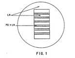
ストリップ検定は、本質的に、Snow 等が記述した通りに行なった (1990, Exp.Neural 309:111-136)。組織培養用のペトリ皿 (Falcon,60 mm) を予めメタノール6ml に溶解した5 cm2 切片のニトロセルロース(Schleicher & Schuell, Type BA85) の混合液 0.5mlで被覆したのち、層流フ−ドで風乾した。セルロースの濾紙(Whatman #1) を McILwain の組織用チョッパー(The Mickle Laboratory Engineering Co.,Ltd.)で 12 x 0.35 mm のストリップに切断した。濾紙のストリップは、所定の濃度の DS-PG または S/CS-PG および LN 溶液20μl に浸漬し、ニトロセルロースで被覆した皿に 30 秒間ブロットしてのち除去した。試験溶液を風乾させた。このプロセスを5 、6 回繰り返して、明確に規定された、間隔が等しい、幅の均一な平行バンドをプレート上に調製した。その後、0.1 もしくは 1 mg/ml LN の薄層を、湾曲したガラス製のパストゥ−ルピペットでバンドを越え均一に伸張し、培地で被覆した(図 1) 。
【0104】
プレ−トに、2.0 x l05 細胞/mlの細胞懸濁液もしくは4-10 DRGの外植片をまいた。培養物は、Leitz Fluovertの顕微鏡を用い、最高7 日間にわたって、DS-PG または KS-CS-PG バンドの接着、遊走および/ もしくは侵入の度合いによって評価した 。
【0105】
細胞の接着、遊走および/ もしくは侵入が完全に抑制されていたバンドは、その評価結果を (-)とし、細胞の接着、遊走および/ または侵入が制限されていたものを (+/-)とし、細胞の接着、遊走および/ または侵入が許容されていたものは (+)とした。細胞の接着は、その付着や拡散の原因となる細胞の表面と下層との初期の相互作用として定義され、細胞の遊走は、操作上、同じ下層内における細胞の運動として定義され、そして、細胞の侵入は、操作上、あるタイプの下層から他のタイプの下層への、細胞運動として定義された。
【0106】
4.1.3.外植片の成長検定
DRGの外植片を、LN または PLL で被覆した皿上で培養したところ、ニューロンならびに非ニューロン(線維芽細胞および Schwann細胞)成長の輪が、Leitz Fluovertの顕微鏡で 1、2、3日間にわたって観察された。処理しなかった対照群の成長に比較してその相対的な成長は、1:対照群の 0-19%; 2:対照群の 20-39%; 3: 対照群の 40-59%; 4: 対照群の 60-79%; 5: 対照群の 80-100%として評価した。
【0107】
4.1.4.PC12神経突起の成長検定
PC12 細胞は、20 ng/ml NGF により8日間にわたって活性化し、PBS中の 0.03% EDTA によって収集し、ウエル当たり3,000個の細胞で予備被覆した96−ウェルのマイクロタイタ−プレ−トにまいた。マイクロタイタ−プレ−トは、予めポリ−L-リシンで被覆し、細胞の添加に先立って20分間、接着培地(PRMI 中の1% BSA)でブロックした。細胞は 20ng/ml NGFを補足した接着培地で生育させた。細胞はまた、試験化合物の添加に先立って、2時間付着させた(Akson & Warren1986, Exp. Cell Res. 162:347-362)。
【0108】
4.1.5.比色定量細胞接着検定
マイクロタイタ−プレ−トは予め、ポリ−L-リシンで被覆し、細胞の添加に先立って、DMEM中の1%BSAによって20分間ブロックした。細胞を予め被覆した96−ウエルのプレート上で培養し、試験化合物を培地に添加した。加湿インキュベ−タ−内で5% CO2、36℃で 4 - 24 時間培養したのち、テトラゾリウム塩 MTT (3-(4,5- ジメチルタゾール-2- イル)-2,5-ジフェニルテトラゾリウムブロミド)を追加した。培養を2時間行なったところ、生細胞内に見られたミトコンドリアのコハク酸デヒドロゲナ−ゼが、MTTを、ホルマザン青色反応生成物に加水分解した。培地を吸引し、ウエルを洗って、分離した細胞をすべて取り除いた。付着した細胞内のホルマザン青色反応生成物を、イソプロパノ−ル中の、0.08 NHC1 によって可溶化し、プレ−トを 630 nm の標準フィルタ−を用い、570 nmでマイクロタイタープレートリーダー(Dynatech MR 4000)により読み取った。結果的に得られた光学密度の読み取り値は、ウエル内の生細胞数に直接関連している(Mosmann, T., 1983, J. Immunol. Methods 65:55-63)。サンプルはポイント当たり、3-6 の反応実験を行い、SDは 5% 以下であった。
【0109】
4.1.6.試薬
ヘパリン、H-7005 Sigma, ブタから得たロット: 19F0268 グレード II 、ナトリウム塩;デルマタン硫酸 90%(コンドロイチン硫酸 B) C-4259 シグマ、ロット 59F0848、ナトリウム塩、ブタの皮膚;コンドロイチン硫酸 A、C-0914 シグマ、ナトリウム塩、ブタ; デキストラン硫酸、D-6001 シグマ、ロット 50HO458、MW 500 kD; デキストラン、シグマ、D-5251 シグマ、ロット 40HO4211 、MW
485 kD;ペントサンポリ硫酸、シグマ、P-8275、ロット 114F0194 。
【0110】
4.2結果
4.2.1.細胞の接着、遊走ならびに侵入に及ぼすDS-PGの影響
細胞の付着(つまり接着)、遊走ならびに侵入に及ぼすDS-PG の影響を一次培養物、線維芽細胞およびグリア細胞系により試験した。細胞浮遊液は上述したストリップ検定用に調製した組織培養プレ−ト上で培養した。培養プレ−トは、交互にDS-PG/LN (それぞれ 0.1-0.8 mg/mlおよび 20μg/ml) とLN (20μg/ml)ストリップで被覆し、細胞の接着、遊走ならびに侵入については、プレ−ティングの2日後に評価した。
【0111】
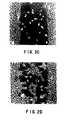
0.8mg/ml DS-PG ならびに20μg/ml LN の混合物を含む下層のストリップは、3T3細胞の接着に対して抑制的であった。細胞 は、DS-PG が欠如しているLNの下層領域に、選択的に付着していた。細胞の交互のバンドはDS-PG/LNおよびLNを含むストリップ間の界面に、非常に明確な境界線を形成していた( 図 2A & 2B;(- )として評価)。0.2 mg/ml では、僅かな侵入が見られ(図 2C; (+/-)として評価) 、さらに 0.1 mg/ml では、細胞がバンド上に侵入し、遊走することが可能であった(図 2 D; (+)として評価)。細胞の接着、遊走ならびに侵入に及ぼす DS-PG の抑制効果は、試験したすべての種類の細胞に観察され た(表9) 。一次細胞と細胞系とは、0.8 mg/ml DS-PG で、そして一次星状細胞を除いて0.4 mg/mlで、細胞の欠如したバンドを形成した。この効果は、約 0.2 mg/ml DS-PGで抑制が低減し、0.1 mg/ml では抑制効果が無くなるという用量依存性であった。
【0112】
【表9】
【0113】
【表10】
【0114】
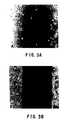
線維芽細胞と星状細胞の一次細胞培養物に加え、DRGSの外植片培養物から遊走する線維芽細胞ならびに Schwann 細胞に及ぼす DS-PGの効果を試験した。胚生ニワトリ (E-8)のDRGを上述した交互の DS-PG/LN と LN ストリップ上で培養した。0.8 mg/ml DS-PG/20μg/ml LN では、LNの下層にわずかな DRGが付着しており、神経突起の成長や非神経細胞の遊走が制限されて いることを実証していた(図 4A)。0.4 mg/ml のDS-PGでは、LNの下層にDRGが付着しており、神経突起の成長と非神経細胞の遊走のいずれをも明らかに示していた(図4B)。LN とDS-PG/LN ストリップ間の界面では、伸張した神経突起と遊走する非神経細胞が急激に停止したり、向きを変えたり、LNとDS-PG/LNストリップの境界に沿って移動していた。DS-PG による抑制は用量依存性であり、低い濃度(0.2 mg/ml) は制限された細胞の侵入をもたらした。一方、0.1 mg/ml のDS-PGでは、神経突起や非神経細胞の侵入が抑制されていなかった(表 6)。
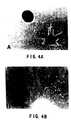
【0115】
4.2.2.細胞の接着、遊走ならびに侵入に及ぼすKS/CS-PGの効果
線維芽細胞やグリア細胞の一次培養物および細胞系に及ぼす KS/CS-PGの効果を試験した。試験した細胞の種類には、ラットの髄膜線維芽細胞(RMF)、成熟ラットの座骨神経線維芽細胞 (RSF)、ラットの星状細胞(RAST) 、3T3-マウス線維芽細胞系、C6-ラットのグリア細胞系および MCG-28の若い不死化した マウスの星状細胞が含まれていた。細胞は、第4.1.2. 節で上述した通り調製された培養プレ−トにまき、細胞の接着、遊走ならびに侵入についての評価を行なった。
【0116】
2.7 mg/mlKS/CS-PGと20μg/ml LN の混合物を含む下層は、試験の対象としたすべての細胞型の付着と遊走を抑制した。ラット髄膜線維芽細胞、3T3 細胞、ラット星状細胞ならびにニワトリ DRG神経突起は、KS/CS-PGによる抑制を最も受けやすかった(表12)。用量と反応の関係を評価したところ、DRGニューロンが KS/CS-PG による抑制を最も受けやすく、グリア細胞系 C6 と MCG-28 が抑制を最も受けにくいことが明らかになった。C6細胞は2.7 mg/ml KS/CS-PGで付着がまず抑制され(図 5A)、その後遊走と侵入が部分的に抑制された(図 4B)が、長続きしなかった。プレーティングの48時間後までには、C6細胞はKS/CS-PGの抑制作用に打ち勝ち(図 5C)、試験を行なった細胞密度では72時間後までにバンドの形成が認められなかった。
【0117】
【表11】
データによれば、DS-PG が KS/CS-PG よりも細胞の付着、遊走ならびに侵入を抑制する作用が強いようである(表 9対表 11)。この強さの違いは、乾燥重量/容積(mg/ml)による濃度をほぼ同じにして二つのプロテオグリカンを比較すれば明らかになる。しかし、DS-PGの推定分子量は KS/CS-PG のそれの約1/10なので、これをモル濃度で補正すると、DS-PGとKS/CS-PGの強さの違いは大幅に低減する。例えば、1.0mg/mlでのKS/CS-PG の概算モル濃度は1.25μMである。同じモル濃度の DS-PG(1.25μM 1.0 mg/ml)にはもはや抑制作用はなく、KS/CS-PGで見られた結果を反映している(表 9と表 11を比較されたい)。
【0118】
4.2.4ヘパリンとデキストラン硫酸が細胞の遊走に及ぼす影響
ニワトリ DRGの外植片培養物からの細胞遊走にヘパリンとデキストラン硫酸が及ぼす影響を試験した。PLL または LN のいずれかの下層を予め塗布した培養皿にDRGをプレートし、濃度の異なるヘパリン、デキストラン硫酸もしくはデキストラン溶液を含む培地で培養した。試験化合物を用いない場合には、24 - 48時間以内に神経突起の成長の輪と非神経細胞、すなわち 線維芽細胞とシュヴァン細胞の遊走が神経節の周囲に認められた(図 6A)。DRG外植片から遊走する細胞の輪の大きさは、ヘパリン 0.4 mg/mlの存在下でその外植片を培養した場合に低減した(図 6B)。デキストラン硫酸0.2 mg/ml の存在下で外植片を培養した場合には、DRG外植片周囲の細胞の輪は大きさが劇的に低減した(図 6C)。対照的に、デキストラン 0.4 mg/mlの場合は何ら影響が見られなかった(図6D)。試験化合物の存在下で培養した DRGから発している細胞の輪の大きさを、処理を施していない対照と比較して評価し、第 4.1.3節に記載したように等級を定めた(表 12 )。
【0119】
【表12】
【0120】
4.2.5GAGとその他の硫酸化炭水化物による線維芽細胞付着の抑制
GAGとその他の硫酸化炭水化物は容易に組織培養プラスチックやその他の基板に吸着されないため、我々は定量法を用いて溶液中のそれらの活性を調べた。培地に試験組成物が存在する場合のPLLを塗布したマイクロタイタープレートへの線維芽細胞の付着を、第 4.1.5節に記載した MTT検定を用いて評価し、付着した細胞の数を対照に対する百分率で表した。ヘパリン、ペントサンポリ硫酸、デキストラン硫酸ならびにデキストランの存在下において4時間の培養がラット髄膜線維芽細胞(RMF)と3T3細胞のPLLへの接着に及ぼす影響を図 7に示した。
【0121】
ヘパリン、ペントサンポリ硫酸ならびにデキストラン硫酸は、処理を施さなかった対照と比較して、付着細胞数を低減させたが、デキストランで処理した場合は付着細胞数が僅かに増加していた。一次ラット髄膜線維芽細胞もしくは線維芽細胞系3T3のどちらで試験を行なっても、抑制度の順位は類似していた。一次線維芽細胞の付着はヘパリン、ペントサンポリ硫酸ならびにデキストラン硫酸で3T3細胞系よりも強く抑制された。処理 24 時間後にデキストランとデキストラン硫酸が付着細胞数に及ぼす影響を図8に示した。
【0122】
4.2.6GAGとその他のアニオンポリマーが細胞遊走に及ぼす影響
GAG とその他のポリアニオン分子が細胞の付着と遊走に及ぼす影響を、PC12細胞の神経突起の成長の簡便かつ迅速なモデル系を使って調べた。NGF の存在下で培養した PC12細胞は神経突起を伸張した。神経突起の先端の成長円錐は付着、分離、遊走のサイクルを繰り返すことにより成長を媒介する。この過程の正味の結果は神経突起の伸長である。
【0123】
NGFで刺激した PC-12細胞を96−ウェルのプレートにまいた。試験液をウェルに加え、2 日後に、少なくとも細胞体の直径の二つ分の長さがある神経突起が大多数の細胞に見られる場合にはそれらの細胞を(+)として評価し、短い突起しか見られないかまたは全然見られない場合には(-)と評価した。各試験化合物に対して完全な用量反応曲線を作成し、その結果を IC100(g/ml)、すなわち、その化合物が神経突起の成長を100% 抑制した時の最低濃度として表した。試験したそれぞれの化合物に毒性がないことは、顕微鏡的には細胞死と細胞分離の証拠が認められないこと、細胞がトリパンブルーで陽性に染色されなかったこと、そして抑制効果のある化合物を培地から取り除くと神経突起が成長したことから確認した。試験の対象とした化合物は、GAG(ヘパリン、デルマタン硫酸、コンドロイチン硫酸 A、ケラタン硫酸、ヒアルロン酸)、硫酸化炭水化物ポリマー(デキストラン硫酸、ペントサンポリ硫酸)とその他のポリアニオンポリマー(アルギン酸など)であった。
【0124】
【表13】
【0125】
4.2.7デキストラン硫酸分子の大きさが線維芽細胞の付着に及ぼす影響
硫黄の含有量はほぼ同じであるが(15 - 16重量%)分子の大きさが異なっている硫酸化デキストランが線維芽細胞の付着に及ぼす影響を調べた。第4.1.5 節に記載した比色細胞付着定量法を用いて、5 kD、8 kD、500 kD、2,000 kDのデキストラン硫酸の存在下で 24 時間培養した 3T3細胞の用量反応曲線を作成した(図 9)。5 kD、8 kD、500 kD、2,000 kDのデキストラン硫酸で、3T3 細胞の接着に対する抑制作用が観察されたが、その強さは有意に異なっていた。デキストラン硫酸による 3T3細胞付着の抑制に対する EC50値は、5 kDでは 6 mg/ml、8 kDでは4 mg/ml 、500 kDでは 0.006 mg/ml、2,000 kDでは 20 mg/ml であった。この検定で作用が最も強かった分子は 500 kD のデキストラン硫酸である。
【0126】
4.3結論
瘢痕形成と線維過多は、線維芽細胞が傷害部もしくは病変部に制御されずに侵入することから生じる。その他の有害な状態も、神経突起の成長、グリア細胞の侵入、単球/ マクロファージの侵入といった制御されない細胞の侵入によって生じる。線維芽細胞の侵入を抑制することで、瘢痕化や関連した有害作用、例えば、硬膜外線維症などの外科手術による癒着や、美容整形や再建手術につながる美容上不適当な瘢痕を防ぐこともできる。前述の結果は、グリコサミノグリカンとその他のアニオンポリマーが、線維芽細胞とグリア細胞などのその他の非神経細胞の侵入ならびに神経突起の成長を抑制して、関連する有害作用を防止することを示している。
【0127】
前述の結果は、抑制の程度が陰イオンの電荷密度と相関関係があり、この関係が本発明の実施に使用するためアニオンポリマーを同定、確認する上で有用となり得ることをも示している。しかしながら、in vivo での結果は、電荷密度が部分的にしか抑制効果を決定しないことを示している。
【0128】
本発明は、ここに記載した特定の具体例によってその範囲を限定されるものではない。実際には、先の記載した内容と添付の図面から、ここに記載したものの他に本発明の様々な改変が、当業者には明らかになるはずである。そのような改変は、本特許請求の範囲内に含まれるものである。
【0129】
各種の出版物を引用したが、その明細は参照によってここに完全に組み込まれるものとする。
【図面の簡単な説明】
【図1】ラミニン(LN)とプロテオグリカン(PG)+LNの交互するバンドと、プレート全体にわたるLNの上張りを示すストライプ分析の概略図。
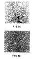
【図2】3T3細胞接着、移動及び侵入についてのDS−PGの投与量依存効果。4.1.2.節に記載したようにして被覆された60mm組織培養皿の中で増殖した生きた3T3細胞の顕微鏡写真で、種々のDS−PG濃度における3日間のLNとDS−PG/LNの交互するバンドを示す。(A)0.8mg/mlDS−PG、(B)0.4mg/mlDS−PG、(C)0.2mg/ml DS−PG、及び(D)0.1mg/mlDS−PG。(100 ×)
【図3】C6細胞接着、移動及び侵入についてのDS−PGの時間依存効果。4.1.2.節に記載したようにして被覆された60mm組織培養皿の中で増殖した生きたC6細胞の顕微鏡写真で、0.8mg/mlDS−PGにおける(A)2時間、(B)1日間、(C)2日間、及び(D)6日間のLNとDS−PG/LNの交互するバンドを示す。6日間までの抑制剤の安定性と6日目における制限された細胞侵入に注目のこと。(100×)
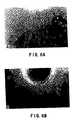
【図4】DRG外植片の接着及び細胞移動についてのDS−PGの効果。顕微鏡写真は、4.1.2.節に記載したようにして被覆された60mm組織培養皿の中で増殖した生きたDRG外植片を示し、LNとDS−PG/LNの交互するバンドを示す。(A)0.8mg/mlDS−PG、及び(B)0.4mg/mlDS−PG。(A)の外植片はLNだけを含有するスプライトに付着しており;DS−PGを含有するスプライトに接着した外植片はない。溶解したDS−PGによるらしい細胞移動の劇的な抑制に注目のこと。0.4mg/mlDS−PGの存在下では、外植片はLNスプライトに接着し細胞移動がその上で起こっているが、DS−PG/LNバンド上では細胞接着も移動も侵入もみられない。(100×)
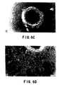
【図5】C6細胞接着、移動及び侵入についてのKS/CS−PGの時間依存効果。4.1.2.節に記載したようにして被覆された60mm組織培養皿の中で増殖した生きたC6細胞の顕微鏡写真で、27mg/mlにおける(A)2時間、(B)1日間、(C)2日間、及び(D)3日間のLNとKS/CS−PG/LNの交互するバンドを示す。プレーティング後最初の2時間のKS/CS−PG被覆バンドへの接着の欠如及びプレーティング後24時間に形成されたバンドの安定性に注目のこと。KS/CS−PGバンドのC6侵入はプレーティング後2日目で起こり、優先的な接着の証拠がないことは3日目に明らかになっている。(100×)
【図6】DRG外植片からの細胞移動についてのヘパリン、デキストラン硫酸及びデキストランの効果。50ng/mlNGFを補充したDMEM/F12中に10%FCSを含有する培養培地内で24時間増殖後の生きたDRG外植片の顕微鏡写真。試験溶液を該培養培地にプレーティング時に添加した。そして、神経細胞及び非神経細胞の細胞移動についての効果が示される。(A)ビヒクルコントロール、(B)400μg/mlヘパリン、(C)200μg/mlデキストラン硫酸、(D)400μg/mlデキストラン。ヘパリンによる細胞移動の抑制及びデキストラン硫酸による劇的な抑制に注目のこと。(100×)
【図7】線維芽細胞及び3T3細胞接着についての硫酸化された炭水化物の効果。ラット髄膜線維芽細胞(RMF)及び3T3細胞の一次培養物を、PLL被覆96ウェルマイクロタイタープレート上にプレートし、試験化合物の存在下で4時間インキュベートし、4.1.5.節に記載したようにして比色細胞接着分析用に処理した。該データは、サンプル当たり6反復試験区の平均から得られた結果を表す(標準偏差は±5%を越えなかった)。該実験を2回繰り返して本質的に同じ結果を得た。
【図8】3T3細胞接着についてのデキストラン硫酸効果の投与量−応答曲線。3T3細胞をPLL被覆96ウェルマイクロタイタープレート上にプレートし、種々の濃度のデキストラン硫酸又はデキストランの存在下で24時間インキュベートし、4.1.5.節に記載したようにして比色細胞接着分析用に処理した。各点は3反復試験区の平均を表す。
【図9】異なる分子量のデキストラン硫酸についての投与量−応答曲線:3T3細胞接着についての効果。3T3細胞をポリ−L−リシン被覆96ウェルマイクロタイタープレート上にプレートし、種々の濃度のデキストラン硫酸の存在下で24時間インキュベートし、4.1.5.節に記載したようにして比色細胞接着分析用に処理した。各点は3反復試験区の平均を表す。[0001]
[Industrial application fields]
The present invention is directed to compositions comprising biocompatible anionic polymers and methods of using such compositions to inhibit hyperfibrosis and complications such as scar formation and surgical adhesions. Also provided are compositions and methods for inhibiting glial cell invasion, neurite outgrowth and bone growth.
[0002]
[Prior art]
Surgical adhesions-attachment of organs or tissues to each other through scar tissue-can lead to clinical problems. Scar tissue formation is a normal consequence of surgery or other tissue injury and is necessary for proper wound healing. However, in some cases, scar tissue grows beyond the planned area, creating surgical adhesions. These scar tissue surgical adhesions limit the normal movement and function of affected body parts. When peripheral nerves are involved, fibrous adhesions cause severe pain during normal movements. Furthermore, scars and keloid tissue (raised scar tissue) are often unsightly and create psychological and emotional problems.
[0003]
1Epidural hyperplasia
A clinically important example of undesired scar formation occurs with excessive epidural fibrosis. This symptom leads to recurrent low back pain after lumbar laminectomy and discectomy (Cauchoix et al., 1978, Spine 3: 256-259; Jackson, 1971, J. Bone Joint Surg. 538: 409-616; Pheasant, 1985, Orthop. Clin. North Am. 6: 319-329; Yong-Hing et al., 1980, Spine 5: 59-64). Tissue scar formation limits nerve root movement and has been associated with frequent recurrent root pain with the same distribution as a previously herniated disc (Benoist, M. et al., 1980, Spine 5: 432 -436).
[0004]
2Preventing unwanted scarring
Many researchers have studied the effectiveness of various treatments to prevent undesirable scarring. Fat grafts have been used with some success in preventing or improving scar formation (Larocca and Macnab, 1974, J. Bone Joint Surg. 56B: 545-550; Langenskold) and Kivilvoto, 1976, Clin. Orthrop. 115: 82-85; Gill et al., 1985, Spine 10: 662-667; Gill et al., 1979, Spine 4: 176-185; Yong Hin 1980, Spine 5: 59-64). Gel foams (denatured collagen gels) and silastic membranes have shown some effectiveness in preventing adhesion (La Rocca and Macnab, supra). However, subsequent studies have shown that gel foam is not effective or promotes scar formation (Gill et al., 1985, supra; Gil et al., 1979, supra; Yong Hin et al., Supra). Songer et al. Reported that sodium hyaluronate, which is neither a gel foam nor other fat grafts, suppressed scarring and reduced fibroblast invasion when modeled on dogs (1990, Spine 15: 550-554).
[0005]
3Cell entry and attachment
A previous study by Snow et al. (1990, Exp. Neurol. 309: 111-130) showed that keratan sulfate / chondroitin sulfate-proteoglycan (KS / GS-PG) is embryonic (E-9) chick It was shown to be inhibitory to neurite outgrowth from dorsal root ganglia (DRG). The neurites either stopped suddenly or turned around and advanced along the edges of the KS / GS-PG stripe. This phenomenon was dependent on the concentration of proteoglycan, and the pattern of crossing was intermittent at intermediate concentrations.
[0006]
Many studies have considered the role of proteoglycans in cell attachment. Unsorted cartilage proteoglycans and chondroitin sulfate, a less purified cartilage component, were found to inhibit fibroblast binding to collagen and fibronectin in vitro (Rich et al., 1981, Nature293 : 224-226). Dermatan sulfate proteoglycan (DS-PG) was found to inhibit 3T3 fibroblast attachment and spreading on plasma fibronectin-coated culture substrates (Lewandowska et al., 1987, J. Cell Biol. 105: 1443 -1454; Rosenberg, LC et al., 1986, CIBA Foundation Symposium 124: 47-68). Dextran sulfate and high molecular weight heparin reduced the initial rate of Chinese hamster ovary and G-8 mouse myoblast attachment to collagen (Klebe, RJ and MJ, 1982, J Cell. Physiol. 112: 5-9). Proteoglycans free from glycoprotein and hyaluronic acid, isolated from cartilage, inhibit the attachment of various cell types, including chick embryonic fibroblasts, to tissue culture plastic and collagen (Knox, P.) and Wells (P. Wells), 1979, J. Cell Sci. 40: 77-88).
[0007]
However, glycosaminoglycan keratan sulfate, chondroitin sulfate and hyaluronic acid did not show inhibition of cell adhesion (Knox and Wells, supra).
[0008]
Heparin sulfate and dermatan sulfate have also been identified as mediators of fibroblast (mouse 3T3 cell) adhesion to fibronectin (Laterra et al., 1980, Proc. Natl. Acad. Sci. USA 77: 6662-6666 ). The presence of fibronectin and / or hyaluronic acid in a three-dimensional type I collagen sponge has been found to promote wound healing in vivo and to support fibroblast in vitro with the resulting collagen deposits. (Doillon, CJ et al., 1987, Biomaterials 8: 195-200).
[0009]
Two glia, two endothelium and one fibroblast cell line showed collagen-glycosaminoglycan binding comparable or reduced to collagen (Reichard-Brown and Akeson, supra). Hyaluronic acid inhibits aggregation of 3T3 fibroblasts (Underhill, C. and Dorfman, A., 1978, Exp. Cell. Res. 117: 155-164), and chondroitin sulfate goes to the endothelium. Appears to block the adhesion of leukocytes (Fibbi, G. et al., 1983, Biochem. Biophys. Acta 762: 512-518).
[0010]
Studies of the composition of the fibroblast substrate adhesion site show that cell surface proteoglycans, primarily heparin sulfate proteoglycans, play an important role in closed and localized contact adhesion (Culp, LA et al. , 1986, CIBA Foundation Symp osium 124: 158-83; Izzard, CS et al., 1986, Exp. Cell. Res. 165: 320-336; Lark, MW et al., 1985, Fed. Proc. 44 : 394-403; Rollins, BJ and Calp, 1979, Biochem. 18: 141-148; Calp et al., 1979, Supramol. Struct. 11: 401-427; Calp et al., 1978, J. Cell Biol. 79 : 788-801; Calp and Bensusan, 1978, Nature 273: 680-682; Cornic, M. et al., 1980, Eur. J. Cell Biol. 22: 262). Rather than fibronectin and collagen, glycosaminoglycans secreted within the substrate adherent appear to play a rate-limiting role in the adhesion process of skeletal mouse myoblast cell lines (Schubert, D.) and La Corbiere, 1980, J. Biol. Chem. 255: 11564-569). Proteoglycans secreted by rat yolk sac tumor cells suppressed fibronectin and tumor cell binding to type I collagen but not type IV collagen. This type IV collagen bound to proteoglycan 12 times less than type I collagen (Brennan, M.J. et al., 1983, Cancer Res. 43: 4302-4307).
[0011]
4Adhesive protein
The mussel, oyster and barnacle bioadhesive proteins adhere to various surfaces with high bond strength under water. Other DOPA (3,4-dihydroxyphenylalanine) containing proteins also exhibit adhesive properties. Brown wrote evidence of quinone tanning in invertebrate constitutive proteins, including mussel shells and hepatic fluke, an egg sheath of the liver worm (1950, Nature 165: 275). Jensen and Morse have identified the nature of the adhesive protein used by the marine worm Phragmatopoma californica to create its protective tube (1968, J Comp. Physiol. B 158: 317-324). For mussel shell adhesion proteins, DOPA is a major component of the flagmatopoma adhesion protein; lysine, serine, and hydroxyl-containing amino acids were also present (Jensen and Morse, 1988, supra).
[0012]
In addition, fibrin adhesives are widespread and have many applications (Strausberg, R.L. and R.P. Link, 1990, Trends Biotech 8: 53-57).
[0013]
[Means for Solving the Problems]
The present invention is related to the discovery that certain biocompatible anionic polymers can effectively inhibit scar formation, particularly surgical adhesions, and that these anionic polymers generally inhibit fibrosis. The present invention is based on the discovery that anionic polymers effectively inhibit unfavorable healing processes, i.e., hyperfibrosis, cell invasion associated with scarring. In particular, the anionic polymers of the present invention, referred to as inhibitory anionic polymers, are useful for inhibiting fibroblast invasion, thus regulating the healing process and preventing fibrosis. The anionic polymer of the present invention also inhibits glial cell invasion, bone growth and neurite growth. The present invention further relates to the discovery that the effectiveness of anionic polymers in inhibiting cell invasion, eg, fibroblast invasion, is partially correlated with the number of anionic charge groups on the polymer. ing. Thus, the present invention is compatible with certain substances for use in the present invention, based in part on many substances used to inhibit scar formation and hyperfibrosis, particularly surgical adhesions, and their effective anionic charge content. Provide a way to check.
[0014]
Anionic polymers used in the present invention include dextran sulfate (DX) and pentosan polysulfate (PS). Thus, neutral proteoglycans or glycosaminoglycan moieties of proteoglycans, including dermatan sulfate (DS), chondroitin sulfate (CS), keratan sulfate (KS), heparin sulfate (HS), and heparin (HN) can be used. . Alginate (AL) can also be used. At suitable concentrations, the aforementioned molecules can inhibit fibroblast invasion or migration even in the presence of a suitable migration promoting substrate such as laminin. In certain aspects, the invention relates to methods of using DX, and molecules comprising DX, to inhibit, block or modulate fibroblast invasion and hyperplasia and, if the foregoing is desirable, Directed to the composition. The present invention further includes a method of using one or more anionic polymers to inhibit fibroblast invasion and hyperfibrosis or mononuclear cell / macrophage invasion, and one or more anionic polymers. It is directed to the composition and its therapeutic use. Such molecules including KS, CS, DS, HS, or HN include, but are not limited to, disaccharide, glycosaminoglycan, and proteoglycan structures. In a preferred embodiment, DX may be used in the fibroblast inhibitory compositions and methods of the present invention.
[0015]
The present invention further provides additional inhibitory anionic polymers suitable for use in inhibiting scar formation. In one embodiment, an anionic polymer comprising acidic sulfuric acid having a sulfur content greater than about 5% by weight may be used. In a more preferred embodiment, the sulfur content is greater than about 10% by weight.
[0016]
In another aspect, the present invention further inhibits in combination with an adhesive protein to inhibit fibroblast invasion and hyperplasia and to also inhibit glial cell invasion, neurite outgrowth, and bone growth. Anionic anionic polymers, preferably directed to processes using dextran sulfate or pentosan polysulfate. Adhesive proteins can crosslink inhibitory molecules with appropriate targets. Accordingly, the present invention provides a method of immobilizing an inhibitory anionic polymer at a site where inhibitory or regulatory activity is desired.
[0017]
In a preferred embodiment, the adhesive protein is activated by chemical or enzymatic oxidation of dihydroxyphenylalanine (DOPA) residues to form quinones, and KS, CS, DS, HS, HN, DX, or hyaluronic acid (HA) ) And applied to the target location with the molecule containing and cured. Adhesive proteins include mussels, oysters, barnacles, flagmatopoma californica, or hepatic or fibrin, or recombinant, any chemical produced or by degradation and repolymerization of natural adhesive proteins Include, but are not limited to, adhesive proteins from sex proteins.
[0018]
In further embodiments, molecules comprising one of the inhibitory anionic polymers may be used in conjunction with one or more other inhibitory anionic polymers and adhesive proteins.
[0019]
The present invention further comprises a composition comprising an inhibitory anionic polymer and a suitable pharmaceutical carrier, and generally for inhibiting scar formation and hyperplasia and for unwanted bone growth, glial cell invasion and / or nerves. A method of administering the composition to inhibit process growth is provided.
[0020]
The present invention also provides a composition comprising an effective amount of an inhibitory anionic polymer, preferably dextran sulfate or pentosan sulfate, an effective amount of an adhesive protein, and a pharmaceutically acceptable pharmaceutical carrier.
[0021]
1Definition
AL alginate
CS chondroitin sulfate
DS dermatan sulfate
DX Dextran sulfate
GAG Glycosaminoglycan
HA Hyaluronic acid
HN Heparin
HS heparin sulfate
KS Keratan sulfate
LN laminin
PG proteoglycan
PS Pentosan polysulfate
Cell adhesion The initial interaction between the cell surface and the substrate resulting in attachment and subsequent cell spreading
Cell migration Cell movement in the same substrate
Cell entry Cell movement from one type of substrate to another
2.Detailed Description of the Invention
The present invention demonstrates that certain biocompatible anionic polymers can effectively inhibit scar formation, particularly surgical adhesions, and these anionic polymers, referred to as “suppressing anionic polymers” It relates to the finding that fibrosis is generally suppressed. The present invention is based on the finding that anionic polymers effectively inhibit cell invasion associated with unfavorable healing processes, such as fibrosis and scar formation. In particular, the anionic polymers of the present invention are useful for inhibiting fibroblast invasion, thereby regulating the healing process and preventing fibrosis. The anionic polymers of the present invention can also inhibit glial cell invasion, bone growth, neurite growth and monocyte / macrophage invasion. The present invention further relates to the finding that the effectiveness of anionic polymers in suppressing the invasion of cells, eg fibroblasts, is in part correlated with the number of effective anionic charged groups on the polymer. . Thus, the present invention teaches a number of substances that are used to suppress scar formation and fibrosis and suppress surgical adhesions. Other anionic polymers for use in the present invention may be identified based on the effective anionic charge content or density.
[0022]
Inhibitory anionic polymers for use in the present invention include dextran sulfate (DX) and pentosan polysulfate (PS). In addition, neutral proteoglycans or glycosaminoglycan components of proteoglycans including dermatan sulfate (DS), chondroitin sulfate (CS), keratan sulfate (KS), heparin sulfate (HS) and heparin (HN) may be used. . Anionic carbohydrate alginate (AL) can also be used. At appropriate concentrations, the molecules described above can inhibit fibroblast invasion or migration, even in the presence of a suitable migration promoter such as laminin. Other anionic polymers for use in the present invention include cellulose derivatives. In a preferred embodiment, the present invention provides a method for using molecules and compositions comprising DX and DX to inhibit, prevent or regulate fibroblast invasion and fibrosis where the foregoing is desirable. is connected with. The present invention further relates to methods of using one or more anionic polymers to inhibit fibroblast invasion and fibrosis and compositions comprising one or more anionic polymers and their therapeutic use. Directed. These molecules including KS, CS, DS, HS or HN include, but are not limited to, disaccharide, glycosaminoglycan, and proteoglycan structures.
[0023]
The present invention further provides a pharmaceutical composition comprising an inhibitory anionic polymer or an inhibitory anion polymer and an adhesive protein, and a pharmaceutically acceptable carrier, and generally inhibits scar formation and fibrosis, and further Methods are provided for administering this composition to inhibit unwanted cell invasion, bone growth, and neurite growth.
[0024]
The compositions and methods of the present invention are suitable for treating animals, preferably mammals, and more preferably humans. A composition comprising a therapeutically effective amount of an anionic polymer of the invention can be administered to an animal lesion in any of the ways described below.
[0025]
2.1.Other anionic polymers for use in the present invention
In addition to dextran sulfate, pentosan polysulfate, glycosaminoglycans and other anionic polymers for use in the present invention (as described above), the present invention, in part, inhibits the effective anionic properties of the polymer. Other additional anionic polymers are provided for use in the present invention based on the finding (see Section 7 below) to help determine potential. In one embodiment, such anionic polymers suitable for use are anionic polymers comprising acidic sulfates, in which the sulfur content is greater than about 5% by weight. In a more preferred embodiment, the sulfur content is greater than about 10% by weight.
[0026]
Anionic polymers for use in the present invention can be found abundantly in nature, for example in proteoglycans. Alternatively, the polymer can be chemically modified to produce an anionic polymer. For example, polyglucose polymer dextran can be processed by boiling in sulfuric acid and esterifying with chlorosulfonic acid to produce dextran sulfate (eg, Merck Index, 10th Edition, 1983, No. 2915, No. See page 427). Other examples of chemically produced anionic polymers include cellulose derivatives. The polymer backbone is not limited to carbohydrate polymers. Biocompatible anionic polymers can be obtained commercially (see, eg, section 5.1.6 below). The anionic polymers used in the present invention can alternatively be purified from natural sources or synthetically produced.
[0027]
2.2.Composition for preventing fibrosis and cellular invasion
The present invention provides compositions for use in inhibiting fibroblast invasion, glial cell invasion, neurite outgrowth, bone growth, and mononuclear leukocyte / macrophage invasion. In particular, the composition is useful in preventing fibrosis and scar formation, such as surgical adhesions. This composition comprises the inhibitory anionic polymer of the present invention. Where the composition is intended for use in a therapeutic method or therapy, it is suitable for use in therapeutically effective amounts of anionic polymers and pharmaceutically acceptable excipients or carriers, eg, in vivo. Can be prepared as a pharmaceutical composition comprising The anionic polymer is preferably present at a concentration of about 1 mg / ml or higher. The compositions of the present invention are hereinafter referred to as “inhibitory compositions” because they contain anionic polymer inhibitors against cellular invasion.
[0028]
For example, in the inhibitory composition of the present invention, the anionic polymer is dextran sulfate (DX), keratan sulfate (KS), dermatan sulfate (DS), chondroitin sulfate (CS), heparin (HN), heparin sulfate (HS), alginate. (AL) or pentosan polysulfate (PS). In a preferred embodiment, the anionic polymer is dextran sulfate or pentosan polysulfate. In a more preferred embodiment, the anionic polymer is dextran sulfate having a sulfur content of about 10% by weight or more. In an even more preferred embodiment, the average molecular weight of dextran sulfate is about 40,000 to 500,000 daltons. Preferably, the average molecular weight of dextran sulfate is about 40,000 daltons. Dextran sulfate should preferably be present at a solution concentration of about 2-20 mg / ml.
[0029]
Carriers or excipients for use with the anionic polymers described above include water, saline, physiological buffered saline, aqueous solutions such as dextrose solution, or any carrier known in the art. The mixture of anionic polymer and carrier may yield a viscous liquid or gel. To increase the viscosity of a liquid or gel, the cross-linked anionic polymer remains substantially anionic, i.e., the negative charge density of the polymer is substantially unaffected. Any crosslinker known to the expert can be used to crosslink the anionic polymer.
[0030]
In other embodiments, the biocompatible anionic polymers of this invention may be combined with solid or semi-solid excipients or carriers such as pastes, gels, foams or sheets. The anionic polymer of the present invention can be mixed with the above carrier or excipient in a colloidal suspension or mixture; alternatively, the carrier or excipient is impregnated with the anionic polymer to form the composition. It may be formed. As will be appreciated, the binding between the anionic polymer and the carrier or excipient can be covalent or non-covalent.
[0031]
The present invention relates to any one of DX, DS, KS, CS, HN, HS, hyaluronic acid (HA), PS or AL, or a suitable anionic polymer, eg as referred to in Section 2.1 above, Combined and provided for use.
[0032]
In a preferred embodiment, the anionic polymer can be dextran sulfate or pentosan polysulfate. In a preferred embodiment, the carrier or excipient may be a pharmaceutically acceptable carrier or excipient, i.e. suitable for in vivo use. Preferred semi-solid carriers include HYSKON-70 (Pharmacia), dextran gels such as INTERCEED® (Johnson & Johnson), natural collagen gels, and modified collagens such as GELFOAM® (Apjon) Gel is included.
[0033]
As will be apparent, preferably the composition comprising an anionic polymer and a pharmaceutically acceptable carrier or implant is bio-absorbable within a period of time, preferably within a month. Suitable anionic polymers are dextran sulfate and pentosan polysulfate, with dextran sulfate being more preferred. Such bio-absorbable carriers include, but are not limited to, dextran, natural collagen and modified collagen as described above. If a carrier such as a collagen gel is used, the collagen should preferably not be cross-linked so that it is more readily absorbed in vivo.
[0034]
In some embodiments, GELFOAM® may be dipped in a 2-20 mg / ml dextran sulfate solution in a phosphate buffered saline solution without calcium / magnesium (see Section 3 below).
[0035]
As will be apparent, a pharmaceutically acceptable surgical polymer graft, such as dextran, natural collagen or modified collagen, in such a composition is about 5% (w / v) composition, preferably at least About 10% and in other embodiments about 20% or more of the composition. Such compositions contain an anionic polymer, such as dextran sulfate, and may also contain an aqueous medium as shown in the examples below.
[0036]
2.2.1.Composition of adhesive protein and inhibitory molecule: inhibitory-adhesive
In certain embodiments, the inhibitory anionic polymer can be used in combination with an appropriate concentration or amount of adhesive protein, and this combination affects the therapeutic function disclosed in Section 2.3 below. Give "glue". Using an inhibitory molecule associated with an adhesive protein prevents the inhibitory molecule from dispersing because the adhesive protein helps anchor the inhibitory molecule in place. An appropriate concentration or amount of adhesive protein is the concentration or amount necessary to exhibit adhesive properties.
[0037]
As used herein, the term “adhesive protein” refers to a protein or peptide that can non-specifically adhere to one or more molecules or surfaces. Any natural, non-natural or recombinant protein containing substantial amounts of dihydroxyphenylalanine (DOPA) and hydroxyl-containing amino acid residues can be used to activate and cure the protein (see below). Can be used. Applicants envision that functional peptides as well as total proteins can be used in the present invention. Functional peptides are those that contain the minimum number of amino acids that produce the adhesive properties of the protein. For example, without limitation, the adhesive protein is fibrin or a commercially available fibrin based on a product such as TUSSUCOL®, TISSEEL® or human IMMUNO®. Also good. Alternatively, the adhesive protein may be a mussel polyphenolic adhesive protein, a barnacle polyphenolic adhesive protein or an oyster polyphenolic adhesive protein. Bioadhesive proteins are commercially available, for example, ADHERACELL® (Genex Corp, Gaithesburg, MD) and MAP (“mussel adhesive protein”, Bio-Polymers, Farmington, CT). . Adhesive protein isPhragmatopoma californicaOrFasciola hepatica DOPA-proteins obtained from (liver flakes) can be included. Polyphenolic adhesive proteins obtained from mussels, barnacles or oysters can be extracted from animal phenolic glands and purified or produced recombinantly (International Patent Publications WO 88/03953, 1988, and WO 88/07076, 1988). Strasberg et al., 1989, “Adhesives from Renewable Resources”, ACS Symposium Series 385, Hemingway and Corner, Eds., Pp. 453-464).
[0038]
Fragments of phenolic adhesive proteins from mussels, barnacles or oysters, produced by chemically or proteolytically cleaving extracted natural molecules or in oysters, barnacles or oysters, It can be used. In other embodiments, the adhesion protein may be a peptide polymerized chemically from the adhesion protein, eg, a fragment of Mytilus edulis protein, proteolytically or chemically digested and then polymerized with glutaraldehyde (US (Patent No. 4,585,585 specification).
[0039]
In other embodiments, the adhesion protein may be an analog of a recombinantly produced adhesion protein. The recombinantly produced analog adhesion protein may be derived from a mussel, barnacle or oyster polyphenolic adhesion protein, or any other adhesion protein. Fragments and analogs of adhesion proteins that incorporate conservative amino acid surrogates can be used as adhesion proteins in the practice of the present invention. Recombinantly produced analog adhesion proteins include mussels, barnacles or oysters, or Fragmatopoma californica (Phragmatopoma californica), Facio La Hepatica (Fasciola hepatica)Alternatively, it may contain portions of one or more adhesion proteins from other comparable adhesion proteins. If necessary, the adhesive protein may be chemically or enzymatically hydroxylated. For example, mushroom tyrosinase or Streptomyces antibiotics (streptomyces antibioticus)Treatment with can modify the adhesive protein precursor. Thus, the pharmaceutical composition of the present invention may comprise a purely engineered adhesion protein that incorporates the essence of a naturally occurring adhesion protein.
[0040]
In certain embodiments, the inhibitory molecule and adhesive protein may be non-covalently bound prior to activation and curing of the adhesive protein. In other embodiments, the inhibitory molecule may be covalently attached to the adhesive protein prior to activation and curing.
[0041]
The adhesive protein of the present invention must be cured so that it can adhere to the desired surface. Curing involves activating the inhibitory adhesive composition, particularly the adhesive protein, followed by reacting the activated adhesive protein with the desired molecule. An enzymatic or chemical process initiates curing. In the case of polyphenolic adhesion proteins, curing can be initiated by oxidizing DOPA to quinones. Oxidation can be performed by exposure to air, treatment with an enzyme such as catechol oxidase, or a commercially available oxidase cross-linking agent such as COX (Biopolymer). The above method is provided by way of example and is in no way limiting; one of ordinary skill in the art will be aware of other oxidizing agents or methods. Fibrin sclerosis is initiated by cleavage of fibrinogen, for example with thrombin. Fibrin monomers polymerize spontaneously; further action by transaminases catalyzes covalent crosslinking. The curing process is complete when the activated protein binds covalently or non-covalently to molecules or surfaces in the surrounding environment.
[0042]
Thus, the inhibitory-adhesive composition is applied to the lesion and cured. Immobilizing an inhibitory molecule with an adhesive protein at a site will increase the efficiency of the inhibitory molecule by increasing the time of local effective concentration. This more effectively suppresses fibroblast invasion and fibrosis, glial cell invasion, neurite growth and bone growth. Beneficial results will be expected when inhibitory molecules and adhesive proteins are applied and cured at surgical incisions, injuries and general trauma sites.
[0043]
2.3.Method of treatment
The compositions of the present invention can be used to treat a wide range of conditions where a method of treatment of animals that inhibits fibroblast invasion and fibrosis, granulation, and in particular scar formation is desired. A method is provided for inhibiting fibrosis, granulation or scar formation of a lesion, comprising administering an inhibitory composition or an inhibitory-adhesive composition to the lesion. Also provided is a method of administering a pharmaceutical composition for therapeutic treatment.
[0044]
2.3.1.General indications
Inhibitory compositions, including inhibitory anionic polymers or inhibitory-adhesive compositions, can be used for trauma, surgery, infection (virus or bacteria), metabolic disease, malignancy, exposure to toxic substances, and other hyperplasias. It can be used as a barrier against cell migration or invasion caused by the situation. Inhibitory compositions provide a preferred method for protecting organs or tissues from the aforementioned conditions by coating methods. For example, dorsal root ganglia, optic nerves, or optic nerve intersections can be coated with an inhibitory composition to protect against uncontrolled cell invasion and adhesion. Glial cell invasion, neurite outgrowth, and bone growth and fibroblast invasion can be suppressed by the method of the present invention. Coating an organ or tissue with an inhibitory composition may be preventive or prophylactic. The inhibitory-adhesive composition provides a preferred coating composition.
[0045]
2.3.2.surgery
In one embodiment, epidural (or peridural) fibrosis can be suppressed by applying the compositions of the invention to surgical lesions. In one embodiment, a portion of a collagen gel immersed in a solution containing the anionic polymer of the present invention can be applied to the lesion. In other embodiments, the anionic polymer may be added in a carrier or excipient such as a paste, gel or sheet. In yet other embodiments, the anionic polymer can be dissolved or suspended in a carrier such as a saline buffer and applied by irrigation or as an aerosol. In a preferred embodiment, a pharmaceutical carrier or excipient may be used.
[0046]
The present invention provides materials and methods for inhibiting fibrosis after laminectomy, particularly epidural (on the dura mater) fibrosis after lumbar laminectomy (see Section 3). . By way of example, but not limitation, a portion of a semi-solid pharmaceutically acceptable carrier is immersed in dextran sulfate or other anionic polymers of the present invention. The soaked carrier can be prepared in advance or at the time of surgery. The carrier may be immersed in a solution of dextran sulfate or other anionic polymer for use in the present invention in calcium / magnesium-free phosphate buffered saline (CMF-PBS) or a solution suitable for in vivo use. . After the carrier is impregnated with the anionic polymer, the part is inserted into the laminectomy site. Using the technique described above, the laminectomy site will exhibit minimal scar tissue formation and bone growth during re-extraction. The carrier can be easily removed, and the dura can be viewed as a smooth transparent film. There may be no anatomical features associated with undesired fibrosis, i.e. scars that hold nerve roots and surgical adhesions.
[0047]
The anionic polymer of the present invention and the composition containing the same can also be used to suppress undesirable bone growth by administering a therapeutically effective amount of the anionic polymer to a site where bone growth suppression is desired. (See Section 3 below). In addition, anionic polymers can be used to treat other indications where inhibition of bone growth is desirable. This corresponds to the undesirable case of bone growth, especially after certain manipulations in children, such as craniostemostosis.
[0048]
In another aspect, the present invention provides compositions and methods for inhibiting fibrosis and scar formation of fallopian tisue. In particular, it is possible to suppress fibrosis and scar formation at the fallopian tube and its surrounding lesions after surgery. Fallopian tube fibrosis resulting from infection and other causes causes infertility in 25-30% of cases. Adhesion of the side wall of the pelvic is also related to infertility. Scar tissue is formed after surgery, so surgical removal of adhesions is not sufficient treatment. Thus, the present invention has important applications in the treatment of infertility.
[0049]
The following example suggests one of the many uses of the present invention in the treatment of Fallopian tissue after surgery. Compositions containing anionic polymers can be tested for their ability to inhibit fibrosis in the rat uterine horn after normalizing traumatic injury. The abdominal skin and muscles of model animals, such as female Lewis rats, can be dissected to open the abdominal cavity. An incision can be made in the wall of the uterine horn. A traumatic injury directed to the endometrium can be created and then a semi-solid pharmaceutically acceptable carrier soaked in a suitable anionic polymer such as dextran sulfate can be inserted into the lesion site.
[0050]
In a preferred method for the treatment of Fallopian tissue after surgery, a gel or paste containing an anionic polymer is inserted and the inhibitory composition is naturally absorbed into the body according to the healing of the lesion, or through the fallopian tube. Washed off by hydrotubation. After the inhibitory composition is inserted, the abdominal muscles and fascia are closed and sutured. The skin incision is also closed.
[0051]
Those skilled in the art will appreciate from the foregoing description that the present invention provides suitable treatment methods associated with any surgical procedure. The above methods can be used to suppress fibrosis, scar formation and keloid formation after surgery. The method of the present invention may be useful when it is desired to minimize scar formation, i.e., especially in surgical procedures for formation, such as regeneration or cosmetic surgery. Other surgical indications include, but are not limited to, abdominal surgery, joint surgery, tendon surgery, surgery to isolate pelvic side wall adhesions, peritoneal surgery, thoracic surgery, vascular surgery and heart surgery, especially bypass surgery, valves Replacement surgery, cardiovascular surgery or other open heart surgery.
[0052]
The present invention further provides hypertonic and keloid scar treatment. Scars limiting aesthetic loss and motility often occur after surgical removal. The use of an inhibitory or inhibitory-adhesive composition limits or prevents the formation of hypertonic scars or keloids after treatment to remove the scar.
[0053]
In other embodiments, it may be used in a method of inhibiting fibrosis around a graft that includes applying a therapeutically effective amount of an anionic polymer-containing composition of the invention to the graft. Examples of grafts where suppression of scar formation and fibrosis are desirable include, but are not limited to, tubes for renal fistula formation, tubes for peritoneal drainage, hip prostheses, prosthetic heart valves, peripheral nerve repair prostheses and other Prosthesis and venous catheter. The implant may be treated by coating or impregnating with the composition provided by the present invention. The present invention further provides an improved implant, where the improvement consists of coating the implant, which coating comprises an appropriate amount of the inhibitor-adhesive composition. The graft can be a polymer graft. The polymer implant can have a variety of compositions, pore sizes and geometries, and includes, but is not limited to, biocompatible polymers made of nitrocellulose, polyanhydrides, and acrylic polymers. it can. The polymer that forms the implant may itself be an anionic polymer.
[0054]
2.3.3.Treatment of fibrotic lesions in the joint
The inhibitory composition of the present invention may be used in the treatment of fibrotic joint lesions. Traumatic injuries often result from physical activity and contact associated with athletics. Joint fibrosis resulting from traumatic injuries such as falls or collisions will solidify the damaged joint and make the movement painful. Scar tissue in the injured area often occurs after tendon injury. In dysfunction of the temporal mandibular joint, jaw movement is limited and may be painful.
[0055]
One method of treating joint lesions is to open the joint surgically or to access the joint arthroscopically to remove the adhesion. These methods have the disadvantage of causing more fibrosis during the healing process. Administration of a composition comprising an inhibitory anionic polymer of the present invention will increase the chances of successful treatment because subsequent fibrosis and adhesion formation in the joint is inhibited.
[0056]
The invention will be used for orthopedic surgery in the treatment of joint damage resulting from athletics or accidents. In oral surgery, it will be found that the present invention is useful in treating joint dysfunction in several types of temporal mandible. Anionic polymers for use in therapeutic methods are described in
[0057]
2.3.4.Inhibition of glial cell invasion
Like dextran sulfate, pentosan polysulfate, alginate, and anionic polymers with highly anionic properties, such as sulfated anionic polymers with a sulfur content of about 10% or more, where sulfur is found as acidic sulfate The anionic polymer of the present invention is useful in a method for inhibiting glial cell invasion, which comprises administering the anionic polymer to a site where inhibition of glial cell invasion is desired. In various embodiments, glial cell invasion is caused by trauma, surgery, viral infection, bacterial infection, metabolic disease, malignancy, exposure to toxic substances, or hyperplastic situations. The glial cell may be an astrocyte.
[0058]
In other embodiments, the inhibitory-adhesive composition can be used as a barrier to glial migration or invasion. The inhibitory-adhesive composition provides a suitable method for protecting organs or tissues from the aforementioned conditions by coating methods. For example, dorsal root ganglia, optic nerves or optic nerve crossings may be coated with an inhibitory-adhesive composition to protect against uncontrolled cell invasion and adhesion. Invasion of fibroblasts as well as glial cells would be suppressed by this method. Inhibitory—Coating an organ or tissue with an adhesive has a preventive or prophylactic effect, or can be a treatment in patients already symptomatic.
[0059]
2.3.5.Suppression of nerve cell invasion
Method for inhibiting cell invasion by administering an anionic polymer having a highly anionic property, for example, a sulfated anionic polymer having a sulfur content of about 10% or more and a pharmaceutical composition containing the anionic polymer Can be used to inhibit neurite outgrowth. In an aspect of the properties, an inhibitory-adhesive composition can be used. Anionic polymers can be administered to areas where it is desired to inhibit neurite outgrowth or glial cell invasion. In certain embodiments, the inhibitory composition may be used to treat patients with gliomas or tumors of neural tissue, ie, malignant tumors such as neuroblastoma.
[0060]
In other embodiments, the inhibitory composition is a neuroma (undirected axon growth associated with a condition when the axon loses either an appropriate target or a substrate pathway for neural development. For example, the inhibitory composition can also be used for the treatment of neuromas associated with amputations, lesions or congenital malformations, etc. A drill is drilled and an axon is placed in the hole.Often, this method fails to remove the axon from the hole and form a neuroma. The use of the inhibitory composition of the invention will overcome the above-mentioned current drawbacks by providing an environmental restraint to axon growth with undirected orientation. Is the polymer “cap”, ie one It may also be used as a closed-end polymer cylinder, the anionic polymer of the present invention can also be used to coat the inside of the cap, thereby preventing neurite growth and neuroma formation. In other embodiments, the polymer cap can include an anionic polymer.
[0061]
In a preferred embodiment, the inhibitory-adhesive composition may be used for the treatment of neuromas. Using the inhibitory-adhesive composition of the present invention to coat axons and bones,
(1) fix the axon in the hole inside the bone, and
(2) It will provide a restraining environment.
[0062]
Alternatively, the inhibitory-adhesive composition may be used to anchor nerve ends within a polymer “cap”.
[0063]
Nerve growth-stimulating factors including, but not limited to, nerve growth promoting factor, ciliary neurotrophic factor, brain-derived growth factor, laminia, NCAM, L2 and SSEA-1 Diseases resulting from overproduction of can be treated by administration of an inhibitory-adhesive composition or an inhibitory composition. Inhibitory or inhibitory-adhesive compositions can be used to treat diseases of the central and / or peripheral nervous system.
[0064]
2.3.6.Dosage form
Methods for introducing the inhibitory anionic polymer or inhibitory-adhesive composition of the present invention include methods known to those skilled in the art. Desirably, the inhibitory composition or inhibitory-adhesive composition of the present invention is introduced into the relevant site by any suitable route. These include, but are not limited to, for example, local injection or application during surgery, by injection, by aerosol, by catheter means, or by graft means, provided that the graft is porous, non-porous Or a gelatin-like material, including a film such as a silastic film or fiber). In a preferred embodiment, the implant is coated or dipped with the inhibitor-adhesive composition of the present invention. A polymer implant treated with an inhibitory molecule or more preferably coated with an inhibitory-adhesive can be applied or inserted at the site where treatment is desired. Such polymers can have various compositions, pore sizes, and geometries. Without limitation, polymers that can be used include those made from nitrocellulose, polyanhydrides, and acrylic polymers.
[0065]
The present invention provides the application of inhibitory compositions or inhibitory-adhesive compositions by surgical methods. The inhibitory anionic polymer or inhibitory-adhesive can be applied to a surgical wound. Anionic polymers or inhibitory-adhesives can be used directly at the site of tissue injury or to coat the entire organ or close the surgical incision. Where appropriate, administration of the inhibitory anionic polymer or inhibitory-adhesive composition may be accomplished by orthoscopic procedures.
[0066]
When an inhibitory-adhesive composition is introduced, the adhesive protein has the opportunity to cure at the desired site (see Section 2.2.1 above) and most effectively fix the inhibitory molecule. Will do. Curing can occur in an air environment, such as when the inhibitory-adhesive composition is applied to an organ or tissue during a surgical incision, traumatic injury, or classic open surgery. Alternatively, if the cure is exposed to weak oxidative molecules, or if the viscosity of the composition is such that the composition can be held in the vicinity of the desired site while curing occurs, it will naturally occur in situ. (In situ) may occur.
[0067]
3. Example:Epidural fibrosis animal model
Epidural fibrosis is associated with scar tissue that occurs after laminectomy. Scar tissue occurs within the laminectomy site and connects the lower surface of the spine spine muscles to the existing nerve roots through the posterior and lateral surfaces of the dura mater and the dura mater. The attachment of scar tissue to the nerve root is believed to be responsible for the recurrent pain that lasts long after the laminectomy procedure.
[0068]
Epidural fibrosis was tested after laminectomy of the lumbar region of rats, rabbits and dogs. In these models, a laminectomy was performed and then the test substance was applied to the laminectomy site. Subsequently, the laminectomy site was examined for fibrosis by large incision or by histological analysis.
[0069]
In these examples, compositions for use as test substances to inhibit epidural fibrosis are generally prepared using dextran sulfate having a molecular weight of 40 kDa, gelfoam powder and phosphate buffered saline. It was done. These three components have a long history of safety in intravenous, oral and parenteral human clinical use. Some of the tests discussed in the following section used dextran sulfate with a molecular weight of 500 kDa, and used gel foam sponge instead of gel foam powder as the vehicle. These changes have been shown to have no effect on clinical outcome in rat, rabbit and dog models.
[0070]
The animal model described below showed no difference in antifibrotic activity between the 40 kDa and 500 kDa dextran sulfate materials. The data given in the literature, however, showed that lower molecular weight substances can be less toxic compared to higher molecular weight substances (at the concentrations described below, toxicity is observed). Not) Once this measurement was made, subsequent tests were performed using 40 kDa dextran sulfate.
[0071]
In determining suitable media for use with dextran sulfate, several substances-gel foam sponge, Pluronic gel, gelatin, collagen and gel foam powder were tested. Based on test results and surgeon preference, gel foam powder was selected that was formulated as a gel containing dextran sulfate. Many tests discussed in the following sections used gel foam sponges instead of this powder. According to the information provided by Upjohn, the manufacturer of this sponge and powder, this powder is actually a sponge material that has been processed into a powder form. We found no clinical differences between the results achieved with the gel foam sponge and gel foam powder.
[0072]
3.1.Rat model
3.1.1.Experimental design
Laminectomy (as described below) was performed at L3 and L5 of the lumbar vertebrae to provide automatic control within each animal. A portion of gel foam (GELFOAM) (registered trademark, Upjohn) immersed in an experimental or control solution was inserted into the laminectomy site. Soaked gel foam pieces were prepared and coded 24 hours ago. Gel foam pieces were immersed in a solution of dextran sulfate in calcium / magnesium free phosphate buffered saline (CMF-PBS), dextran in CMF-PBS or CMF-PBS only. At some laminectomy sites, the gel foam pieces were omitted for use as a camouflage operation. Subsequently, a large incision was made and the evaluation was performed without any knowledge about which solution was placed at the laminectomy site. Thus, the experiment was double blind.
[0073]
3.1.2.Laminectomy
Lewis inbred rats were anesthetized with 4% chloral hydrate (34 mg / 100 g body weight). The dorsal skin was incised and the paraspinal muscles were separated from the spinous processes of lumbar vertebra L2 to L6. The spinous processes at L3 and L5 were removed with bone forceps; the vertebral lamina was then removed with microbone forceps to create a rectangular laminectomy defect with a 4 × 2 mm diameter. Hemostasis was achieved using cotton pellets soaked with epinephrine and gentle pressure. After bleeding stopped, a gel foam piece was placed at the laminectomy site according to a random protocol. The overlying paraspinal muscle was closed with a resorbable 6-0 PDS II (Ethicon) by suturing with the fascia on the surface. The skin cut was closed with a wound clip.
[0074]
3.1.3.MinAnalysis
The animals were kept for 2 weeks before the overall evaluation. In time course experiments, animals were maintained at 4, 8, 16, 26 and 36 weeks. At that time, the rats were anesthetized with 4% chloral hydrate and the wound was reopened by incising the skin and separating the paraspinal muscles. The amount and quality of scar tissue on the laminectomy site, the appearance of the gel foam (GELFOAM®), and the extent of new bone growth were assessed by visual and tactile observation during the sharp incision to the dura mater. After complete observation, the rats were euthanized by an overdose of anesthesia.
[0075]
3.1.4.ResultFruit
Prior to teaching treatment codes, all global assessments were performed, and the observations were listed by treatment group. A scale for the assessment and quantification of the degree of epidural fibrosis is shown in Table 1.
[0076]
[Table 1]
[0077]
At the dextran sulfate site, the superficial scar tissue was found to be a thin layer that was easily detached from the underlying gel foam (GELFOAM®). The gel foam could be easily peeled in 1 or 2 sections from the laminectomy site. After removing the gel film, the dura appeared as a smooth and transparent film. The laminectomy site itself had no obvious change in size; the boundary of the site appeared to be flat.
[0078]
Negative control sites (dextran or CMF-PBS) were dramatically different. The first difference was observed in the surface layer of scar tissue: this layer adhered to the gel foam. When the scar tissue was removed, bleeding began in and around the gel foam (no bleeding occurred when the gel foam soaked in dextran sulfate was removed). Another difference was the gel foam texture: it was very brittle and broke down when it was removed. After removing the gel foam, the size of the laminectomy site decreased dramatically, resulting in a narrow slit. The laminectomy boundary texture and stiffness indicated that the laminectomy site was narrowed due to new bone growth.
[0079]
[Table 2]
[0080]
The effect of different concentrations of dextran sulfate on epidural fibrosis was tested in a rat laminectomy model and scored as described in Table 1 above. The results are shown in Table 3.
[0081]
[Table 3]
[0082]
The effects of dextran sulfate, dextran and phosphate buffer on the suppression of epidural fibrosis were tested as a function of time after transplantation and scored as described in Table 1.
[0083]
[Table 4]
[0084]
2 The control group was composed of rats treated with a sponge soaked with PBS alone, a sponge soaked with dextran mixed with PBS, or an unsoaked sponge. Since no statistical differences were found in the results obtained from any of the control groups used, the scores are summarized here.
[0085]
As shown in Table 4, the suppression of scar formation by dextran sulfate was as strong as that at 2 weeks after transplantation, as was the case at 2 weeks after transplantation (no significant difference was observed). Similar results were observed after 4, 8, 16, 26, and 36 weeks after treatment (Table 5). Dextran and buffer did not inhibit scar formation at any time. The results indicate that scar suppression with dextran sulfate is both effective and persistent.
[0086]
A model of a laminectomy rat was used to test whether epidural fibrosis was suppressed using several different compositions containing anionic polymers. Heparin and hyaluronic acid are polysulfated glycosaminoglycans, pentosan is a polysulfated xylan, and alginate is a carboxylated carbohydrate polymer. Each compound was scored for its effect on scar formation as described in Table 1 (Table 5).
[0087]
[Table 5]
[0088]
3.2Rabbit laminectomy model
In New Zealand white rabbits, the formation of epidural hyperplasia after laminectomy was significantly reduced by the use of dextran sulfate (MW = 500 KDa; 400 μg) during surgery. The degree of epidural hyperplasia was assessed using the scoring method used for the rat model by gross dissection. The results of this evaluation are listed in Table 6.
[0089]
[Table 6]
[0090]
3.3Canine laminectomy model
Epidural fibrosis after laminectomy was evaluated by a canine model. The dura mater was exposed by 1 x 2 cm laminectomy at the 6th, 4th and 2nd lumbar sites. The first part used dextran sulfate in a gelfoam (Gelfoam) sponge and the second part used dextran sulfate in a gel suspension prepared with a powdered gel foam. No part of the site was used. Three types of lumbar spine sites were randomly treated using these three different methods. Epidural scar formation was analyzed by gross anatomical assessment (4 dogs at a time) at 4, 8, and 16 weeks after surgery.
[0091]
The connective tissue, dural adhesion, and bone growth that were present at the lumbar resection site were scored according to the criteria listed in Table 7. I couldn't see it.
[0092]
[Table 7]
[0093]
[Table 8]
In rats, rabbits and dogs used as models of epidural fibrosis, the presence of dextran sulfate clearly benefited substantially from the suppression of fibrosis and scar formation. Similarly, Gelform R treated with dextran sulfate also suppressed bone growth. In the previous example, treatment of the surgical lesion with dextran sulfate-gelform R suppressed most of the thin layer of scar tissue and the dura mater remained smooth and transparent. Gelform R impregnated with dextran sulfate can be easily removed as a piece, indicating that fibrosis has been prevented. In contrast, negative control sites showed superficial scar formation and thick, sticky, deep scars as well as dense bone growth.
[0094]
In both rats and rabbits, the laminectomy model is similar to that of human back surgery. Scar formation is based on the proliferation and migration of fibroblasts, epithelial cells and immune system cells from the wound site to the surgical area. Thereafter, fiber connective tissue and blood vessels are formed in the surgical area. These features of epidural fibrosis are commonly seen in humans, rats and rabbits.
[0095]
Various differences are related to size differences and surgical details. Rats and rabbits, which are relatively small, are forced to undergo advanced surgery. In rats and rabbits, total laminectomy is performed, but back surgery in humans usually requires partial laminectomy. Also, one of the most common reasons for human back surgery is discectomy. In small animal models, these operations are not possible, so the sequelae of vertebral disc damage cannot be studied in rat or rabbit models. However, this can be done in a dog model that is very similar to a real human surgery.
[0096]
The dextran sulfate used in this example is an anionic polymer with a highly effective anionic charge density and a sulfur content of 15.3 wt%. Dextran sulfate can be prepared by boiling dextran with sulfuric acid and esterifying with chlorosulfonic acid. Both dextran and dextran sulfate have a polyglucose carbohydrate backbone. Comparison of dextran sulfate or dextran impregnated Gelform R results in that dextran sulfate (highly anionic) is much more effective at suppressing epidural fibrosis than dextran (not anionic) From the fact, in the present invention, it is clear that the anionic property of the polymer influences its inhibitory effect and usefulness.
[0097]
However, the results obtained with the other anionic polymers shown in Table 5 indicate that charge density alone does not represent a potential inhibitory capacity in vivo. Natural proteoglycans that were strongly inhibitory in vitro (
[0098]
Four.Example: Inhibition of cell invasion in a test tube
Anionic polymers have been found to inhibit cell invasion. The strip assay (described below) is a useful model for determining the presence or absence of cell invasion in a test tube. Inhibitory activity can be measured by the degree of cell migration from a strip coated with a substrate molecule such as laminin to a strip coated with a substrate molecule and an inhibitor molecule (eg, an anionic polymer). We also measured the ability of anionic polymers, especially DS and KS / CS, to inhibit cell adhesion.
[0099]
4.1.Substances and methods
4.1.1.Cell culture
Cell lines: murine 3T3 fibroblasts (NIH), rat C6 glioma (Paganetti et al., 1988, J. Cell Biol. 107: 2281) and MCG-28 young immortalized mouse astrocytes (SV) -40 murine neonatal astrocytes), Dulbecco's modified Eagle medium (DMEM), 10% fetal bovine serum (Gibco Laboratories) and penicillin G / streptomycin (100 units / ml, 100 It was grown on a medium consisting of μg / ml, Gibco Laboratories). For the strip assay, confluent plates were separated with 0.05% trypsin, 0.53 mM EDTA (Gibco Laboratories) and spread at 200,000 cells / ml (3 ml / plate). In the cell adhesion assay, confluent cultures were separated with 0.03% EDTA, and 20,000 to 100,000 cells per well were seeded in a 96-well microtiter plate. PC12 cells (Green LA & Tischler AS, 1976, Proc. Natl. Acad. Sci. USA 73: 2424), a cell line of pheochromocytoma, were purchased from Roswell Park Memorial Institute (RPMI). Medium 1640 (Gibco Laboratories),
[0100]
Primary cell culture: Primary cultures of rat meningeal fibroblasts (RMF) were obtained from children of P-3 rats. Tissues were removed from the animals and transferred to Leibovitz's L-15 with L-glutamine medium. Then, using a scalpel, the tissue was shredded and enzymatically dissociated with 0.25% trypsin in Hanks' balanced salt solution (CMF-HBSS, Gibco Laboratories) without Ca2 + / Mg2 + for 20 minutes at 37 ° C. . Cells were washed with DMEM supplemented with 10% FCS, concentrated by centrifugation, and then diluted with DMEM supplemented with 10% FCS to plate at 5.0 x 10⁶ cells per 75
[0101]
Rat astrocytes (RAST) cultures were prepared from P-1-3 rat brain cortex. Brain cortex is detached from connective tissue and blood vessels, chopped, dissociated with 0.5% collagenase (Gibco Laboratories) for 20 minutes at 37 ° C, and then with 0.25% trypsin at 37 ° C for 15 minutes, 0.04 mg / ml DNAse (Sigma) and 0.52 mg / ml soybean trypsin inhibitor for 5 minutes. The cells were then mechanically dissociated by trituration with a Pasteur pipette and seeded at 1.5 x 10⁷ cells in a 75 cm2 flask coated with poly-L-lysine (PLL, Sigma). . After plating, the flasks were shaken for 4 hours to remove non-astrocytic oligodendrocytes and neurons with poor adhesion.
[0102]
Primary explant cultures: dorsal root ganglia (DRG) were obtained from E8-E9 chicken embryos. Dissociation was performed with CMF-HBSS. After exposing the spinal cord and removing the DRG, the surrounding connective tissue was removed. DRG was dispersed in DMEM / F12, 10% fetal bovine serum containing 50 ng / ml nerve growth factor (NGF). DRG is also a laminin (LN, Gibco Laboratories)
Alternatively, they were spread on a dish coated with poly-L-lysine (PLL, Sigma).
[0103]
4.1.2.Strip test
The strip test was performed essentially as described by Snow et al. (1990, Exp. Neural 309: 111-136). Cover a tissue culture Petri dish (Falcon, 60 mm) with 0.5 ml of a 5 cm2 section of nitrocellulose (Schleicher & Schuell, Type BA85) previously dissolved in 6 ml of methanol, and then air-dry in a laminar flow hood. did. Cellulose filter paper (Whatman # 1) was cut into 12 × 0.35 mm strips with McILwain tissue chopper (The Mickle Laboratory Engineering Co., Ltd.). The strips of filter paper were removed by soaking in 20 μl of a predetermined concentration of DS-PG or S / CS-PG and LN solution, blotting for 30 seconds in a nitrocellulose-coated dish. The test solution was allowed to air dry. This process was repeated 5-6 times to produce well-defined, equally spaced, uniform width parallel bands on the plate. A thin layer of 0.1 or 1 mg / ml LN was then stretched uniformly across the band with a curved glass Pasteur pipette and covered with medium (FIG. 1).
[0104]
Plates were seeded with 2.0 x 10⁵ cells / ml cell suspension or 4-10 DRG explants. Cultures were evaluated by DS-PG or KS-CS-PG band adhesion, migration and / or invasion for up to 7 days using a Leitz Fluovert microscope.
[0105]
Bands in which cell adhesion, migration, and / or invasion were completely suppressed were evaluated as (-), and those with restricted cell adhesion, migration, and / or invasion were defined as (+/-). Those that allowed cell attachment, migration and / or invasion were marked (+). Cell adhesion is defined as the initial interaction between the cell surface and the underlying layer that causes its attachment and diffusion, and cell migration is defined as the movement of cells within the same underlying layer, in operation, and Invasion was defined as cell movement from one type of lower layer to another type of operation.
[0106]
4.1.3.Explant growth test
When DRG explants are cultured on dishes coated with LN or PLL, the growth loop of neurons and non-neurons (fibroblasts and Schwann cells) can be observed with a Leitz Fluovert microscope for 1, 2, or 3 days It was done. Compared to the untreated control group, its relative growth is as follows: 1: 0-19% in the control group; 2: 20-39% in the control group; 3: 40-59% in the control group; 4 : 60-79% of control group; 5: Evaluated as 80-100% of control group.
[0107]
4.1.4.PC12Neurite growth assay
PC12 cells were activated with 20 ng / ml NGF for 8 days, harvested with 0.03% EDTA in PBS, and seeded in 96-well microtiter plates precoated with 3,000 cells per well. Microtiter plates were pre-coated with poly-L-lysine and blocked with adhesion medium (1% BSA in PRMI) for 20 minutes prior to addition of cells. Cells were grown in adherent medium supplemented with 20 ng / ml NGF. Cells were also allowed to attach for 2 hours prior to addition of test compound (Akson & Warren 1986, Exp. Cell Res. 162: 347-362).
[0108]
4.1.5.Colorimetric cell adhesion assay
Microtiter plates were pre-coated with poly-L-lysine and blocked for 20 minutes with 1% BSA in DMEM prior to addition of cells. Cells were cultured on pre-coated 96-well plates and test compounds were added to the medium. Tetrazolium salt MTT (3- (4,5-dimethyltazol-2-yl) -2,5-diphenyltetrazolium bromide) after culturing in humidified incubator at 5% CO2, 36 ℃ for 4-24 hours Added. When cultured for 2 hours, mitochondrial succinate dehydrogenase found in living cells hydrolyzed MTT into a formazan blue reaction product. The medium was aspirated and the wells were washed to remove all separated cells. The attached intracellular formazan blue reaction product was solubilized with 0.08 NHC1 in isopropanol, and the plate was 570 nm using a 630 nm standard filter at 570 nm with a microtiter plate reader (Dynatech MR 4000). Read by. The resulting optical density reading is directly related to the number of viable cells in the well (Mosmann, T., 1983, J. Immunol. Methods 65: 55-63). The sample was subjected to 3-6 reaction experiments per point, and the SD was less than 5%.
[0109]
4.1.6.reagent
Heparin, H-7005 Sigma, lot from pig: 19F0268 Grade II, sodium salt; dermatan sulfate 90% (chondroitin sulfate B) C-4259 Sigma, lot 59F0848, sodium salt, pig skin; chondroitin sulfate A, C- 0914 Sigma, Sodium Salt, Pig; Dextran Sulfate, D-6001 Sigma, Lot 50HO458, MW 500 kD; Dextran, Sigma, D-5251 Sigma, Lot 40HO4211, MW
485 kD; Pentosan polysulfate, Sigma, P-8275, lot 114F0194.
[0110]
4.2result
4.2.1.Effects on cell adhesion, migration and invasionDS-PGImpact of
The effects of DS-PG on cell attachment (ie adhesion), migration and invasion were examined in primary cultures, fibroblasts and glial cell lines. The cell suspension was cultured on a tissue culture plate prepared for the strip assay described above. Culture plates are alternately coated with DS-PG / LN (0.1-0.8 mg / ml and 20 μg / ml, respectively) and LN (20 μg / ml) strips for cell adhesion, migration and invasion. Assessed 2 days after ting.
[0111]
The lower strip containing a mixture of 0.8 mg / ml DS-PG and 20 μg / ml LN was inhibitory to 3T3 cell adhesion. Cells were selectively attached to the lower layer of LN lacking DS-PG. The alternating bands of cells formed a very clear boundary at the interface between the strips containing DS-PG / LN and LN (evaluated as Figure 2A &2B; (-)). At 0.2 mg / ml, slight invasion was observed (Fig. 2C; evaluated as (+/-)), and at 0.1 mg / ml, cells were able to invade and migrate on the band (Fig. 2 D; evaluated as (+)). The inhibitory effect of DS-PG on cell adhesion, migration and invasion was observed in all cell types tested (Table 9). Primary cells and cell lines formed cell-depleted bands at 0.8 mg / ml DS-PG and at 0.4 mg / ml excluding primary astrocytes. This effect was dose-dependent, with suppression being reduced at about 0.2 mg / ml DS-PG and no effect at 0.1 mg / ml.
[0112]
[Table 9]
[0113]
[Table 10]
[0114]
In addition to primary cell cultures of fibroblasts and astrocytes, the effect of DS-PG on fibroblasts and Schwann cells migrating from DRGS explant cultures was tested. Embryonic chicken (E-8) DRGs were cultured on the alternating DS-PG / LN and LN strips described above. In 0.8 mg / ml DS-PG / 20 μg / ml LN, a small amount of DRG was attached to the lower layer of LN, demonstrating that neurite growth and non-neuronal cell migration were restricted (Fig. 4A). In 0.4 mg / ml DS-PG, DRG was attached to the lower layer of LN, clearly showing both neurite outgrowth and non-neuronal cell migration (FIG. 4B). At the interface between the LN and the DS-PG / LN strip, the stretched neurites and migrating non-neuronal cells suddenly stop, turn, or move along the boundary between the LN and DS-PG / LN strips. It was. Inhibition by DS-PG was dose dependent, with low concentrations (0.2 mg / ml) resulting in limited cell invasion. On the other hand, invasion of neurites and non-neuronal cells was not suppressed with 0.1 mg / ml DS-PG (Table 6).
[0115]
4.2.2.Effects on cell adhesion, migration and invasionKS / CS-PGEffect of
The effects of KS / CS-PG on primary cultures and cell lines of fibroblasts and glial cells were tested. Cell types tested include rat meningeal fibroblasts (RMF), adult rat sciatic neurofibroblasts (RSF), rat astrocytes (RAST), 3T3-mouse fibroblast cell line, C6- The rat glial cell line and MCG-28 young immortalized mouse astrocytes were included. Cells were seeded into culture plates prepared as described above in Section 4.1.2 and evaluated for cell adhesion, migration and invasion.
[0116]
The lower layer containing a mixture of 2.7 mg / ml KS / CS-PG and 20 μg / ml LN inhibited adhesion and migration of all cell types tested. Rat meningeal fibroblasts, 3T3 cells, rat astrocytes and chicken DRG neurites were most susceptible to inhibition by KS / CS-PG (Table 12). Evaluation of dose-response relationships revealed that DRG neurons were most susceptible to inhibition by KS / CS-PG, and glial cell lines C6 and MCG-28 were least susceptible to inhibition. C6 cells were first inhibited from attachment with 2.7 mg / ml KS / CS-PG (FIG. 5A) and then partially inhibited from migration and invasion (FIG. 4B), but did not last long. By 48 hours after plating, C6 cells overcome the inhibitory effect of KS / CS-PG (FIG. 5C), and no band formation was observed by 72 hours after the tested cell density.
[0117]
[Table 11]
According to the data, DS-PG seems to have a stronger inhibitory effect on cell attachment, migration and invasion than KS / CS-PG (Table 9 vs. Table 11). This difference in strength becomes apparent when two proteoglycans are compared at approximately the same concentration by dry weight / volume (mg / ml). However, the estimated molecular weight of DS-PG is about 1/10 that of KS / CS-PG, so when this is corrected by molar concentration, the difference in strength between DS-PG and KS / CS-PG is greatly reduced. . For example, the approximate molar concentration of KS / CS-PG at 1.0 mg / ml is 1.25 μM. DS-PG at the same molar concentration (1.25 μM 1.0 mg / ml) no longer has an inhibitory effect and reflects the results seen with KS / CS-PG (compare Table 9 and Table 11).
[0118]
4.2.4Effects of heparin and dextran sulfate on cell migration
The effects of heparin and dextran sulfate on cell migration from chick DRG explant cultures were tested. DRG was plated on a culture dish pre-coated with either PLL or LN, and cultured in a medium containing heparin, dextran sulfate or dextran solutions having different concentrations. Without the test compound, neurite growth rings and non-neuronal cells, namely fibroblasts and Schwann cells, migrated around the ganglia within 24-48 hours (Figure 6A). The ring size of cells migrating from DRG explants was reduced when the explants were cultured in the presence of 0.4 mg / ml heparin (FIG. 6B). When explants were cultured in the presence of 0.2 mg / ml dextran sulfate, the ring of cells around the DRG explants was dramatically reduced in size (Figure 6C). In contrast, dextran 0.4 mg / ml had no effect (Figure 6D). The ring size of cells emanating from DRG cultured in the presence of the test compound was evaluated relative to untreated controls and graded as described in Section 4.1.3 (Table 12).
[0119]
[Table 12]
[0120]
4.2.5GAGAnd other sulfated carbohydrates inhibit fibroblast adhesion
Since GAG and other sulfated carbohydrates are not readily adsorbed to tissue culture plastics and other substrates, we investigated their activity in solution using quantitative methods. The adhesion of fibroblasts to PLL-coated microtiter plates in the presence of the test composition in the culture medium was evaluated using the MTT assay described in Section 4.1.5, and the number of attached cells compared to the control. Expressed as a percentage. Figure 7 shows the effect of 4 hours of culture in the presence of heparin, pentosan polysulfate, dextran sulfate, and dextran on adhesion of rat meningeal fibroblasts (RMF) and 3T3 cells to PLL.
[0121]
Heparin, pentosan polysulfate and dextran sulfate reduced the number of adherent cells compared to the untreated control, but there was a slight increase in the number of adherent cells when treated with dextran. The degree of inhibition was similar whether tested on primary rat meningeal fibroblasts or fibroblast line 3T3. Primary fibroblast adhesion was more strongly inhibited by heparin, pentosan polysulfate and dextran sulfate than the 3T3 cell line. The effect of dextran and dextran sulfate on the number of adherent cells 24 hours after treatment is shown in FIG.
[0122]
4.2.6GAGOf cell and other anionic polymers on cell migration
The effect of GAG and other polyanion molecules on cell attachment and migration was investigated using a simple and rapid model system for neurite growth in PC12 cells. PC12 cells cultured in the presence of NGF extended neurites. The growth cone at the tip of the neurite mediates growth by repeating the cycle of attachment, separation, and migration. The net result of this process is neurite outgrowth.
[0123]
PC-12 cells stimulated with NGF were seeded in 96-well plates. Test solution is added to the wells, and after 2 days, if the majority of cells have neurites that are at least twice the diameter of the cell body, evaluate those cells as (+) and shorten When only a protrusion was seen or not at all, it was evaluated as (-). Complete dose response curves were generated for each test compound and the results were100(G / ml), ie, the lowest concentration at which the compound inhibited neurite outgrowth by 100%. The lack of toxicity for each compound tested is that there is no evidence of cell death and cell separation microscopically, the cells did not stain positively with trypan blue, and the inhibitory compound was It was confirmed from the fact that the neurite grew when it was removed from. The compounds tested were GAG (heparin, dermatan sulfate, chondroitin sulfate A, keratan sulfate, hyaluronic acid), sulfated carbohydrate polymers (dextran sulfate, pentosan polysulfate) and other polyanion polymers (such as alginic acid). .
[0124]
[Table 13]
[0125]
4.2.7Effect of dextran sulfate molecular size on fibroblast adhesion
We investigated the effect of sulfated dextran with almost the same sulfur content (15-16 wt%) but different molecular size on fibroblast adhesion. Using the colorimetric cell attachment assay described in Section 4.1.5, a dose response curve was generated for 3T3 cells cultured in the presence of 5 kD, 8 kD, 500 kD, and 2,000 kD dextran sulfate (24 hours). (Figure 9) Inhibition of 3T3 cell adhesion was observed with dextran sulfate at 5 kD, 8 kD, 500 kD, and 2,000 kD, but the strengths were significantly different. EC for inhibition of 3T3 cell adhesion by dextran sulfate50The values were 6 mg / ml at 5 kD, 4 mg / ml at 8 kD, 0.006 mg / ml at 500 kD, and 20 mg / ml at 2,000 kD. The most potent molecule in this assay is 500 kD dextran sulfate.
[0126]
4.3Conclusion
Scar formation and hyperfibrosis result from the uncontrolled entry of fibroblasts into the lesion or lesion. Other deleterious conditions are also caused by uncontrolled cellular invasion such as neurite outgrowth, glial cell invasion, monocyte / macrophage invasion. Suppressing fibroblast invasion to prevent scarring and related adverse effects, such as surgical adhesions such as epidural fibrosis, and cosmetically inappropriate scars leading to cosmetic surgery and reconstruction surgery You can also. The above results show that glycosaminoglycans and other anionic polymers inhibit the invasion of other non-neuronal cells such as fibroblasts and glial cells and the growth of neurites to prevent related adverse effects. Show.
[0127]
The foregoing results also show that the degree of inhibition correlates with the charge density of the anion, and this relationship can be useful in identifying and confirming anionic polymers for use in the practice of the present invention. However, in vivo results indicate that the charge density determines the inhibitory effect only partially.
[0128]
The scope of the present invention is not limited by the specific embodiments described herein. Indeed, various modifications of the invention in addition to those described herein will become apparent to those skilled in the art from the foregoing description and accompanying drawings. Such modifications are intended to fall within the scope of the claims.
[0129]
References have been made to various publications, the specifications of which are hereby fully incorporated by reference.
[Brief description of the drawings]
FIG. 1 is a schematic of a stripe analysis showing alternating bands of laminin (LN) and proteoglycan (PG) + LN and LN overlay across the plate.
FIG. 2: Dose-dependent effect of DS-PG on 3T3 cell adhesion, migration and invasion. Photomicrographs of live 3T3 cells grown in 60 mm tissue culture dishes coated as described in section 4.1.2. 3 days of LN and DS-PG / LN at various DS-PG concentrations. Alternating bands are shown. (A) 0.8 mg / ml DS-PG, (B) 0.4 mg / ml DS-PG, (C) 0.2 mg / ml DS-PG, and (D) 0.1 mg / ml DS-PG. (100x)
FIG. 3. Time-dependent effects of DS-PG on C6 cell adhesion, migration and invasion. 4A. Photomicrographs of live C6 cells grown in a 60 mm tissue culture dish coated as described in section 4.1.2, (A) 2 hours at 0.8 mg / ml DS-PG, (B) 1 The alternating bands of LN and DS-PG / LN are shown for days, (C) 2 days, and (D) 6 days. Note the stability of the inhibitor up to 6 days and the limited cell invasion on day 6. (100x)
FIG. 4. Effect of DS-PG on DRG explant adhesion and cell migration. The photomicrograph shows live DRG explants grown in 60 mm tissue culture dishes coated as described in section 4.1.2., Showing alternating bands of LN and DS-PG / LN. (A) 0.8 mg / ml DS-PG and (B) 0.4 mg / ml DS-PG. The explant of (A) is attached to a sprite containing only LN; there is no explant adhered to the sprite containing DS-PG. Note the dramatic suppression of cell migration likely by lysed DS-PG. In the presence of 0.4 mg / ml DS-PG, the explant adheres to the LN sprite and cell migration occurs on it, but no cell adhesion, migration or invasion is seen on the DS-PG / LN band. (100x)
FIG. 5. Time-dependent effects of KS / CS-PG on C6 cell adhesion, migration and invasion. Photomicrographs of live C6 cells grown in a 60 mm tissue culture dish coated as described in Section 4.1.2. (A) 2 hours at 27 mg / ml, (B) 1 day (C ) Shows alternating bands of LN and KS / CS-PG / LN for 2 days and (D) 3 days. Note the lack of adhesion to the KS / CS-PG coated band for the first 2 hours after plating and the stability of the band formed 24 hours after plating. It is clear on day 3 that C6 invasion of the KS / CS-PG band occurs on
FIG. 6: Effect of heparin, dextran sulfate and dextran on cell migration from DRG explants. Photomicrograph of live DRG explants after 24 hours growth in culture medium containing 10% FCS in DMEM / F12 supplemented with 50 ng / ml NGF. Test solution was added to the culture medium during plating. And the effect about the cell migration of a nerve cell and a non-neuronal cell is shown. (A) Vehicle control, (B) 400 μg / ml heparin, (C) 200 μg / ml dextran sulfate, (D) 400 μg / ml dextran. Note the inhibition of cell migration by heparin and the dramatic inhibition by dextran sulfate. (100x)
FIG. 7: Effect of sulfated carbohydrates on fibroblast and 3T3 cell adhesion. Primary cultures of rat meningeal fibroblasts (RMF) and 3T3 cells are plated onto PLL-coated 96-well microtiter plates and incubated for 4 hours in the presence of test compounds, as described in section 4.1.5. And processed for colorimetric cell adhesion analysis. The data represents the results obtained from the average of 6 replicates per sample (standard deviation did not exceed ± 5%). The experiment was repeated twice with essentially the same results.
FIG. 8. Dose-response curve of dextran sulfate effect on 3T3 cell adhesion. 3T3 cells are plated onto PLL-coated 96-well microtiter plates and incubated for 24 hours in the presence of various concentrations of dextran sulfate or dextran for colorimetric cell adhesion analysis as described in section 4.1.5. Processed. Each point represents the average of 3 replicates.
FIG. 9: Dose-response curve for different molecular weight dextran sulfate: effect on 3T3 cell adhesion. 3T3 cells were plated on poly-L-lysine coated 96 well microtiter plates, incubated for 24 hours in the presence of various concentrations of dextran sulfate and colorimetric cell adhesion assay as described in section 4.1.5. Processed for. Each point represents the average of 3 replicates.
Claims (19)
Translated fromJapaneseApplications Claiming Priority (2)
| Application Number | Priority Date | Filing Date | Title |
|---|---|---|---|
| US708,660 | 1991-05-31 | ||
| US07/708,660US5605938A (en) | 1991-05-31 | 1991-05-31 | Methods and compositions for inhibition of cell invasion and fibrosis using dextran sulfate |
Related Parent Applications (1)
| Application Number | Title | Priority Date | Filing Date |
|---|---|---|---|
| JP50055293ADivisionJP3379757B2 (en) | 1991-05-31 | 1992-05-29 | Methods and compositions based on the inhibition of cell invasion and hyperfibrosis by anionic polymers |
Publications (2)
| Publication Number | Publication Date |
|---|---|
| JP2002138039A JP2002138039A (en) | 2002-05-14 |
| JP3923290B2true JP3923290B2 (en) | 2007-05-30 |
Family
ID=24846689
Family Applications (6)
| Application Number | Title | Priority Date | Filing Date |
|---|---|---|---|
| JP50055293AExpired - Fee RelatedJP3379757B2 (en) | 1991-05-31 | 1992-05-29 | Methods and compositions based on the inhibition of cell invasion and hyperfibrosis by anionic polymers |
| JP2000124553APendingJP2000312715A (en) | 1991-05-31 | 2000-04-25 | Methods and compositions based on the suppression of cell invasion and hyperfibrosis by alginate |
| JP2001278399APendingJP2002138040A (en) | 1991-05-31 | 2001-09-13 | Methods and compositions based on the suppression of fibrosis by pentosan polysulfate |
| JP2001278310AWithdrawnJP2002154972A (en) | 1991-05-31 | 2001-09-13 | Methods and compositions based on the inhibition of cell invasion by anionic polymers |
| JP2001278271APendingJP2002154973A (en) | 1991-05-31 | 2001-09-13 | Methods and compositions based on the inhibition of bone growth by anionic polymers |
| JP2001278340AExpired - Fee RelatedJP3923290B2 (en) | 1991-05-31 | 2001-09-13 | Methods and compositions based on the suppression of hyperfibrosis or scar formation by anionic polymers |
Family Applications Before (5)
| Application Number | Title | Priority Date | Filing Date |
|---|---|---|---|
| JP50055293AExpired - Fee RelatedJP3379757B2 (en) | 1991-05-31 | 1992-05-29 | Methods and compositions based on the inhibition of cell invasion and hyperfibrosis by anionic polymers |
| JP2000124553APendingJP2000312715A (en) | 1991-05-31 | 2000-04-25 | Methods and compositions based on the suppression of cell invasion and hyperfibrosis by alginate |
| JP2001278399APendingJP2002138040A (en) | 1991-05-31 | 2001-09-13 | Methods and compositions based on the suppression of fibrosis by pentosan polysulfate |
| JP2001278310AWithdrawnJP2002154972A (en) | 1991-05-31 | 2001-09-13 | Methods and compositions based on the inhibition of cell invasion by anionic polymers |
| JP2001278271APendingJP2002154973A (en) | 1991-05-31 | 2001-09-13 | Methods and compositions based on the inhibition of bone growth by anionic polymers |
Country Status (19)
| Country | Link |
|---|---|
| US (8) | US5605938A (en) |
| EP (3) | EP0586535B1 (en) |
| JP (6) | JP3379757B2 (en) |
| KR (1) | KR100237731B1 (en) |
| AT (3) | ATE418993T1 (en) |
| AU (1) | AU671256B2 (en) |
| BG (1) | BG98333A (en) |
| BR (1) | BR9206077A (en) |
| CA (1) | CA2110291C (en) |
| CZ (1) | CZ259493A3 (en) |
| DE (5) | DE69231574T2 (en) |
| DK (1) | DK0586535T3 (en) |
| ES (3) | ES2156840T1 (en) |
| FI (1) | FI935345A7 (en) |
| GR (3) | GR3034760T3 (en) |
| HU (1) | HUT66427A (en) |
| NO (1) | NO305974B1 (en) |
| SK (1) | SK134393A3 (en) |
| WO (1) | WO1992021354A1 (en) |
Families Citing this family (250)
| Publication number | Priority date | Publication date | Assignee | Title |
|---|---|---|---|---|
| EP0431046B1 (en)* | 1988-08-24 | 1995-05-03 | SLEPIAN, Marvin J. | Biodegradable polymeric endoluminal sealing |
| US5605938A (en)* | 1991-05-31 | 1997-02-25 | Gliatech, Inc. | Methods and compositions for inhibition of cell invasion and fibrosis using dextran sulfate |
| US6231881B1 (en) | 1992-02-24 | 2001-05-15 | Anton-Lewis Usala | Medium and matrix for long-term proliferation of cells |
| US6352707B1 (en) | 1992-02-24 | 2002-03-05 | Anton-Lewis Usala | Transplant encapsulation in a hydrogel matrix to obscure immune recognition |
| IT1259090B (en)* | 1992-04-17 | 1996-03-11 | Fidia Spa | BIOMATERIALS FOR BONE PROSTHESIS |
| JP3579052B2 (en)* | 1993-03-23 | 2004-10-20 | フォーカル,インコーポレイテッド | Apparatus for local application of polymeric material to living tissue |
| US5665063A (en)* | 1994-06-24 | 1997-09-09 | Focal, Inc. | Methods for application of intraluminal photopolymerized gels |
| US5583114A (en) | 1994-07-27 | 1996-12-10 | Minnesota Mining And Manufacturing Company | Adhesive sealant composition |
| USRE38827E1 (en) | 1994-07-27 | 2005-10-11 | 3M Innovative Properties Company | Adhesive sealant composition |
| US7396505B2 (en)* | 1994-08-12 | 2008-07-08 | Diamicron, Inc. | Use of CoCrMo to augment biocompatibility in polycrystalline diamond compacts |
| US7396501B2 (en) | 1994-08-12 | 2008-07-08 | Diamicron, Inc. | Use of gradient layers and stress modifiers to fabricate composite constructs |
| US6425922B1 (en) | 2000-01-30 | 2002-07-30 | Diamicron, Inc. | Prosthetic hip joint having at least one sintered polycrystalline diamond compact articulation surface |
| US6517583B1 (en) | 2000-01-30 | 2003-02-11 | Diamicron, Inc. | Prosthetic hip joint having a polycrystalline diamond compact articulation surface and a counter bearing surface |
| US6596225B1 (en) | 2000-01-31 | 2003-07-22 | Diamicron, Inc. | Methods for manufacturing a diamond prosthetic joint component |
| US6494918B1 (en) | 2000-01-30 | 2002-12-17 | Diamicron, Inc. | Component for a prosthetic joint having a diamond load bearing and articulation surface |
| US6793681B1 (en) | 1994-08-12 | 2004-09-21 | Diamicron, Inc. | Prosthetic hip joint having a polycrystalline diamond articulation surface and a plurality of substrate layers |
| US7077867B1 (en) | 1994-08-12 | 2006-07-18 | Diamicron, Inc. | Prosthetic knee joint having at least one diamond articulation surface |
| US6514289B1 (en) | 2000-01-30 | 2003-02-04 | Diamicron, Inc. | Diamond articulation surface for use in a prosthetic joint |
| US5931165A (en)* | 1994-09-06 | 1999-08-03 | Fusion Medical Technologies, Inc. | Films having improved characteristics and methods for their preparation and use |
| US6492332B1 (en)* | 1995-12-12 | 2002-12-10 | Omeros Corporation | Irrigation solution and methods for inhibition of tumor cell adhesion, pain and inflammation |
| US7112320B1 (en) | 1995-06-07 | 2006-09-26 | Andre Beaulieu | Solid wound healing formulations containing fibronectin |
| US6528483B2 (en) | 1995-06-07 | 2003-03-04 | André Beaulieu | Method of producing concentrated non-buffered solutions of fibronectin |
| US5643892A (en)* | 1995-06-07 | 1997-07-01 | Baker Norton Pharmaceuticals, Inc. | Method of treating chronic progressive vascular diseases |
| US5779673A (en)* | 1995-06-26 | 1998-07-14 | Focal, Inc. | Devices and methods for application of intraluminal photopolymerized gels |
| WO1997041901A1 (en)* | 1996-05-03 | 1997-11-13 | Uros Corporation | Pentosanpolysulfate coating for medical devices |
| US5791352A (en)* | 1996-06-19 | 1998-08-11 | Fusion Medical Technologies, Inc. | Methods and compositions for inhibiting tissue adhesion |
| US6063061A (en)* | 1996-08-27 | 2000-05-16 | Fusion Medical Technologies, Inc. | Fragmented polymeric compositions and methods for their use |
| US7871637B2 (en) | 1996-08-27 | 2011-01-18 | Baxter International Inc. | Dry hemostatic compositions and methods for their preparation |
| US6066325A (en)* | 1996-08-27 | 2000-05-23 | Fusion Medical Technologies, Inc. | Fragmented polymeric compositions and methods for their use |
| DE69739085D1 (en)* | 1996-09-19 | 2008-12-18 | Univ Michigan | POLYMERS CONTAIN POLYSACCHARIDES SUCH AS ALGINATES OR MODIFIED ALGINATES |
| US20090324721A1 (en) | 1996-09-23 | 2009-12-31 | Jack Kennedy | Hydrogels Suitable For Use In Polyp Removal |
| US7009034B2 (en) | 1996-09-23 | 2006-03-07 | Incept, Llc | Biocompatible crosslinked polymers |
| US8003705B2 (en)* | 1996-09-23 | 2011-08-23 | Incept Llc | Biocompatible hydrogels made with small molecule precursors |
| US6206917B1 (en) | 1997-05-02 | 2001-03-27 | St. Jude Medical, Inc. | Differential treatment of prosthetic devices |
| US6018035A (en)* | 1997-07-21 | 2000-01-25 | Baxter International Inc. | Reagents for isotropic size enhancement of a peptide, protein, nucleotide or other substrate |
| US5981710A (en)* | 1997-07-21 | 1999-11-09 | Baxter International, Inc. | Therapeutic hemoglobin composition having isotropically increased size |
| US6204254B1 (en) | 1997-09-12 | 2001-03-20 | Baxter International, Inc. | Biocompatible surfaces and a method for their preparation |
| UA65587C2 (en)* | 1997-10-09 | 2004-04-15 | Baker Norton Pharma | Method for preventing nephrotoxicity induced by cyclosporins or tacrolimus |
| AU1214999A (en)* | 1998-01-22 | 1999-08-12 | Seikagaku Corporation | Antifibrotic agent |
| US6872384B1 (en) | 1998-02-23 | 2005-03-29 | Life Medical Sciences, Inc. | Treatment of trauma |
| GB9810127D0 (en) | 1998-05-13 | 1998-07-08 | Ml Lab Plc | Prevention of surgical adhesions |
| US6818018B1 (en) | 1998-08-14 | 2004-11-16 | Incept Llc | In situ polymerizable hydrogels |
| US6514534B1 (en)* | 1998-08-14 | 2003-02-04 | Incept Llc | Methods for forming regional tissue adherent barriers and drug delivery systems |
| AU2707500A (en)* | 1998-12-04 | 2000-06-26 | Incept Llc | Biocompatible crosslinked polymers |
| CA2363048A1 (en) | 1999-02-23 | 2000-08-31 | Phairson Medical, Inc. | Treatment of trauma |
| EP1173790A2 (en) | 1999-03-01 | 2002-01-23 | Boston Innovative Optics, Inc. | System and method for increasing the depth of focus of the human eye |
| US7083777B1 (en)* | 1999-04-02 | 2006-08-01 | The Brigham And Women's Hospital, Inc. | Immunomodulating polymers |
| EP1459757A1 (en)* | 1999-04-02 | 2004-09-22 | The Brigham And Women's Hospital, Inc. | Immunomodulating polymers |
| US6942695B1 (en) | 1999-04-05 | 2005-09-13 | Wessley-Jessen Corporation | Biomedical devices with polyimide coating |
| US6083933A (en)* | 1999-04-19 | 2000-07-04 | Stellar International Inc. | Treatment of cystitis-like symptoms with chondroitin sulfate following administration of a challenge solution |
| US6743774B1 (en) | 1999-04-23 | 2004-06-01 | Rhode Island Hospital | Tribonectins |
| WO2001045577A2 (en) | 1999-12-06 | 2001-06-28 | Sdgi Holdings, Inc. | Intervertebral disc treatment devices and methods |
| AR035026A1 (en)* | 2000-01-19 | 2004-04-14 | Baker Norton Pharma | USE OF PENTOSAN POLISULFATE IN THE TREATMENT OF CERTAIN CONDITIONS OF THE PROSTATE |
| AU2001226926A1 (en)* | 2000-01-21 | 2001-07-31 | M.L. Laboratories Plc | Dextrin containing compositions for prevention of adhesions |
| US6410877B1 (en) | 2000-01-30 | 2002-06-25 | Diamicron, Inc. | Methods for shaping and finishing prosthetic joint components including polycrystalline diamond compacts |
| US6488715B1 (en) | 2000-01-30 | 2002-12-03 | Diamicron, Inc. | Diamond-surfaced cup for use in a prosthetic joint |
| US6709463B1 (en) | 2000-01-30 | 2004-03-23 | Diamicron, Inc. | Prosthetic joint component having at least one solid polycrystalline diamond component |
| US6500205B1 (en)* | 2000-04-19 | 2002-12-31 | Gary K. Michelson | Expandable threaded arcuate interbody spinal fusion implant with cylindrical configuration during insertion |
| US6814756B1 (en)* | 2000-02-04 | 2004-11-09 | Gary K. Michelson | Expandable threaded arcuate interbody spinal fusion implant with lordotic configuration during insertion |
| EP1645248B8 (en)* | 2000-02-04 | 2010-06-16 | Warsaw Orthopedic, Inc. | Expandable interbody spinal fusion implant having pivotally attached blocker |
| US6627785B1 (en)* | 2000-02-29 | 2003-09-30 | Virginia Commwealth University | Wound dressings with protease-lowering activity |
| KR100338491B1 (en)* | 2000-03-30 | 2002-05-30 | 채수경 | Polyphosphate as promoter for recovery of wound and restraint of scar |
| CA2410968C (en)* | 2000-05-31 | 2011-03-29 | Encelle, Inc. | Method of treating chronic ulcers |
| US20020177680A1 (en)* | 2000-08-23 | 2002-11-28 | Hubbell Jeffrey A. | Novel polymer compounds |
| US20030007951A1 (en)* | 2000-08-23 | 2003-01-09 | Richard Franklin | Treatment of trauma |
| SV2003000753A (en) | 2000-12-05 | 2003-06-16 | Brigham & Womens Hospital | USE OF ZWITTERIONIC POLYSACARIDS FOR THE SPECIFIC MODULATION OF IMMUNE PROGRESS |
| DE60224850T2 (en)* | 2001-02-04 | 2009-01-22 | Warsaw Orthopedic, Inc., Warsaw | Instrumentation for introducing and positioning an expandable intervertebral fusion implant |
| JP4283665B2 (en)* | 2001-06-04 | 2009-06-24 | ウォーソー・オーソペディック・インコーポレーテッド | Dynamic plate for anterior cervical spine with movable segments |
| US7097645B2 (en)* | 2001-06-04 | 2006-08-29 | Sdgi Holdings, Inc. | Dynamic single-lock anterior cervical plate system having non-detachably fastened and moveable segments |
| US7186256B2 (en)* | 2001-06-04 | 2007-03-06 | Warsaw Orthopedic, Inc. | Dynamic, modular, single-lock anterior cervical plate system having assembleable and movable segments |
| CA2443429C (en) | 2001-06-04 | 2010-08-10 | Gary Karlin Michelson | Anterior cervical plate system having vertebral body engaging anchors, connecting plate, and method for installation thereof |
| US7044952B2 (en)* | 2001-06-06 | 2006-05-16 | Sdgi Holdings, Inc. | Dynamic multilock anterior cervical plate system having non-detachably fastened and moveable segments |
| US7041105B2 (en)* | 2001-06-06 | 2006-05-09 | Sdgi Holdings, Inc. | Dynamic, modular, multilock anterior cervical plate system having detachably fastened assembleable and moveable segments |
| US8101196B2 (en) | 2001-06-26 | 2012-01-24 | Biointeractions, Ltd. | Polysaccharide biomaterials and methods of use thereof |
| US8815793B2 (en)* | 2001-07-20 | 2014-08-26 | Northwestern University | Polymeric compositions and related methods of use |
| US7618937B2 (en)* | 2001-07-20 | 2009-11-17 | Northwestern University | Peptidomimetic polymers for antifouling surfaces |
| US7858679B2 (en)* | 2001-07-20 | 2010-12-28 | Northwestern University | Polymeric compositions and related methods of use |
| US20050203578A1 (en)* | 2001-08-15 | 2005-09-15 | Weiner Michael L. | Process and apparatus for treating biological organisms |
| US6812220B2 (en)* | 2001-08-29 | 2004-11-02 | University Of British Columbia | Pharmaceutical compositions and methods relating to fucans |
| US20070238697A1 (en)* | 2001-08-29 | 2007-10-11 | University Of British Columbia | Pharmaceutical compositions and methods relating to fucans |
| CA2467836A1 (en)* | 2001-11-20 | 2003-09-04 | Duke University | Interfacial biomaterials |
| EP1476202B1 (en) | 2002-02-21 | 2009-01-14 | Encelle, Inc. | Cross-linked bioactive hydrogel matrices |
| US20080086792A1 (en) | 2006-10-13 | 2008-04-17 | Thomas Charles Kuracina | Method and apparatus for diverting sweat, liquid, moisture or the like from an eye |
| CA2419831A1 (en)* | 2002-02-28 | 2003-08-28 | Macropore Biosurgery, Inc. | Methods for governing bone growth |
| US7410797B2 (en)* | 2002-06-11 | 2008-08-12 | Ogle Roy C | Meningeal-derived stem cells |
| US8911831B2 (en)* | 2002-07-19 | 2014-12-16 | Northwestern University | Surface independent, surface-modifying, multifunctional coatings and applications thereof |
| AU2003239060A1 (en)* | 2002-07-29 | 2004-02-16 | Potencia Medical Ag | Durable implant |
| WO2004018009A1 (en)* | 2002-08-20 | 2004-03-04 | John N Semertzides | Compositions comprising epithelial cells for the treatment and prevention of tissue adhesions |
| US20040054414A1 (en)* | 2002-09-18 | 2004-03-18 | Trieu Hai H. | Collagen-based materials and methods for augmenting intervertebral discs |
| US7744651B2 (en) | 2002-09-18 | 2010-06-29 | Warsaw Orthopedic, Inc | Compositions and methods for treating intervertebral discs with collagen-based materials |
| US7217254B2 (en) | 2002-09-20 | 2007-05-15 | Genzyme Corporation | Multi-pressure biocompatible agent delivery device and method |
| JP2006515765A (en) | 2002-11-15 | 2006-06-08 | エスディージーアイ・ホールディングス・インコーポレーテッド | Collagen-based materials and methods for treating synovial joints |
| EP1570270A4 (en)* | 2002-11-15 | 2010-09-22 | Univ Arizona | THERAPEUTIC BIOCONJUGATES |
| US20040161476A1 (en)* | 2003-02-19 | 2004-08-19 | Hahn Sungtack Samuel | Cystitis treatment with high dose chondroitin sulfate |
| US7238677B2 (en)* | 2003-03-28 | 2007-07-03 | Kimberly-Clark Worldwide, Inc. | Prevention of urogenital infections |
| JP2006522135A (en) | 2003-03-31 | 2006-09-28 | ザ ブライアム アンド ウィミンズ ホスピタル インコーポレーテッド | Zwitterionic immunomodulators for the treatment of asthma and allergies |
| ITMI20031023A1 (en)* | 2003-05-21 | 2004-11-22 | Umberto Cornelli | GLYCOSAMINOGLICANS WITH AVERAGE MOLECULAR WEIGHT 2400 D AT THE TREATMENT OF EMOTIONAL DYSFUNCTIONS. |
| US7628810B2 (en) | 2003-05-28 | 2009-12-08 | Acufocus, Inc. | Mask configured to maintain nutrient transport without producing visible diffraction patterns |
| CA2527429A1 (en)* | 2003-05-30 | 2004-12-09 | Arc Pharmaceuticals, Inc. | Pharmaceutical compositions and methods relating to inhibiting fibrous adhesions using various agents |
| US20060240037A1 (en)* | 2003-06-04 | 2006-10-26 | Edward Fey | Methods and compositions for the treatment and prevention of degenerative joint disorders |
| US8834864B2 (en) | 2003-06-05 | 2014-09-16 | Baxter International Inc. | Methods for repairing and regenerating human dura mater |
| US20050046794A1 (en)* | 2003-06-17 | 2005-03-03 | Silvestrini Thomas A. | Method and apparatus for aligning a mask with the visual axis of an eye |
| WO2005007110A2 (en)* | 2003-07-11 | 2005-01-27 | Pro-Pharmaceuticals, Inc. | Compositions and methods for hydrophobic drug delivery |
| US7927626B2 (en) | 2003-08-07 | 2011-04-19 | Ethicon, Inc. | Process of making flowable hemostatic compositions and devices containing such compositions |
| AU2004264970B2 (en)* | 2003-08-14 | 2010-02-11 | Wyeth | Recombinant lubricin molecules and uses thereof |
| US20050053664A1 (en)* | 2003-09-08 | 2005-03-10 | Eliezer Zomer | Co-administration of a polysaccharide with a chemotherapeutic agent for the treatment of cancer |
| US20050095219A1 (en)* | 2003-10-29 | 2005-05-05 | Shu-Ping Yang | Compositions for promoting vaginal cell proliferation and maturation |
| US9066912B2 (en)* | 2003-11-17 | 2015-06-30 | Ethicon, Inc. | Drug-enhanced adhesion prevention |
| US20050208095A1 (en)* | 2003-11-20 | 2005-09-22 | Angiotech International Ag | Polymer compositions and methods for their use |
| WO2005054446A2 (en)* | 2003-12-02 | 2005-06-16 | Universiteit Gent | Use of polysulphated alginate in cellular matrices |
| WO2005079314A2 (en)* | 2004-02-13 | 2005-09-01 | Pro-Pharmaceuticals, Inc. | Compositions and methods used to treat acne and candida |
| US8277831B2 (en)* | 2004-02-17 | 2012-10-02 | Advanced Technologies And Regenerative Medicine, Llc. | Drug-enhanced adhesion prevention |
| US7840263B2 (en)* | 2004-02-27 | 2010-11-23 | Cardiac Pacemakers, Inc. | Method and apparatus for device controlled gene expression |
| EP1744796B1 (en) | 2004-04-20 | 2015-03-18 | Genzyme Corporation | Surgical mesh-like implant |
| WO2005102363A2 (en)* | 2004-04-20 | 2005-11-03 | Mucosal Therapeutics Llc | Methods of promoting cartilage healing or cartilage integration |
| SE0401069D0 (en)* | 2004-04-26 | 2004-04-26 | Anamar Medical Ab | Use of Compounds for the Treatment of Diseases and Conditions |
| US7764995B2 (en)* | 2004-06-07 | 2010-07-27 | Cardiac Pacemakers, Inc. | Method and apparatus to modulate cellular regeneration post myocardial infarct |
| US7794490B2 (en)* | 2004-06-22 | 2010-09-14 | Boston Scientific Scimed, Inc. | Implantable medical devices with antimicrobial and biodegradable matrices |
| DE102004031258A1 (en)* | 2004-06-29 | 2006-02-09 | Jennissen, Herbert P., Prof. Dr. | Protein hybrids with polyhydroxyaromatic amino acid epitopes |
| US20060009839A1 (en)* | 2004-07-12 | 2006-01-12 | Scimed Life Systems, Inc. | Composite vascular graft including bioactive agent coating and biodegradable sheath |
| US7729761B2 (en)* | 2004-07-14 | 2010-06-01 | Cardiac Pacemakers, Inc. | Method and apparatus for controlled gene or protein delivery |
| CA2575086A1 (en)* | 2004-07-23 | 2006-02-02 | Mucosal Therapeutics Llc | Compositions and methods for viscosupplementation |
| EP1773128A2 (en)* | 2004-08-02 | 2007-04-18 | Pro-Pharmaceuticals, Inc. | Compositions and methods for the enhancement of chemotherapy with microbial cytotoxins |
| US20060040894A1 (en)* | 2004-08-13 | 2006-02-23 | Angiotech International Ag | Compositions and methods using hyaluronic acid |
| US7828711B2 (en)* | 2004-08-16 | 2010-11-09 | Cardiac Pacemakers, Inc. | Method and apparatus for modulating cellular growth and regeneration using ventricular assist device |
| WO2006083328A2 (en)* | 2004-09-15 | 2006-08-10 | Massachusetts Institute Of Technology | Biologically active surfaces and methods of their use |
| CA2623471C (en)* | 2004-09-23 | 2018-02-13 | Arc Pharmaceuticals, Inc. | Pharmaceutical compositions and methods relating to inhibiting fibrous adhesions or inflammatory disease using low sulphate fucans |
| EP1827478A4 (en)* | 2004-12-03 | 2009-08-05 | Mucosal Therapeutics Llc | Methods of treatment for injured or diseased joints |
| US7981065B2 (en) | 2004-12-20 | 2011-07-19 | Cardiac Pacemakers, Inc. | Lead electrode incorporating extracellular matrix |
| US8060219B2 (en) | 2004-12-20 | 2011-11-15 | Cardiac Pacemakers, Inc. | Epicardial patch including isolated extracellular matrix with pacing electrodes |
| US8257438B2 (en)* | 2005-04-12 | 2012-09-04 | Warsaw Orthopedic, Inc. | Methods and devices for preserving motion in an articulating prosthetic disc |
| US7976577B2 (en) | 2005-04-14 | 2011-07-12 | Acufocus, Inc. | Corneal optic formed of degradation resistant polymer |
| WO2006111348A1 (en)* | 2005-04-18 | 2006-10-26 | Abbott Gmbh & Co. Kg | Use of heparin and heparin derivatives for modulating the neurite growth-controlling nogo receptor |
| US20070111327A1 (en)* | 2005-05-05 | 2007-05-17 | Jay Gregory D | Methods of detecting lubricin |
| US20070031498A1 (en)* | 2005-08-02 | 2007-02-08 | Wright Medical Technology, Inc. | Gel composition for cellular adhesion inhibition |
| CA2620663A1 (en)* | 2005-09-02 | 2007-03-08 | Colbar Lifescience Ltd. | Cross-linked polysaccharide and protein matrices and methods for their preparation |
| US7774057B2 (en) | 2005-09-06 | 2010-08-10 | Cardiac Pacemakers, Inc. | Method and apparatus for device controlled gene expression for cardiac protection |
| JP4732881B2 (en)* | 2005-12-07 | 2011-07-27 | Tti・エルビュー株式会社 | Iontophoresis device |
| US8206726B2 (en) | 2006-02-06 | 2012-06-26 | The Brigham And Women's Hospital, Inc. | Zwitterionic polysaccharides for promotion of immune system maturation and health |
| US7732539B2 (en)* | 2006-02-16 | 2010-06-08 | National Science Foundation | Modified acrylic block copolymers for hydrogels and pressure sensitive wet adhesives |
| US20070232660A1 (en)* | 2006-04-04 | 2007-10-04 | Allergan, Inc. | Therapeutic and delivery methods of prostaglandin ep4 agonists |
| WO2008011216A2 (en) | 2006-05-16 | 2008-01-24 | Pro-Pharmaceuticals, Inc. | Galactose-pronged polysaccharides in a formulation for antifibrotic therapies |
| US8399619B2 (en) | 2006-06-30 | 2013-03-19 | Warsaw Orthopedic, Inc. | Injectable collagen material |
| US8118779B2 (en) | 2006-06-30 | 2012-02-21 | Warsaw Orthopedic, Inc. | Collagen delivery device |
| TWI436793B (en) | 2006-08-02 | 2014-05-11 | Baxter Int | Rapidly acting dry sealant and methods for use and manufacture |
| EP2064251B1 (en) | 2006-08-04 | 2017-10-25 | Kensey Nash Corporation | Biomimetic compounds and synthetic methods therefor |
| WO2008091386A2 (en)* | 2006-08-04 | 2008-07-31 | Northwestern University | Biomimetic modular adhesive complex: material, methods and applications therefore |
| US20080069855A1 (en)* | 2006-08-21 | 2008-03-20 | Bonutti Peter M | Method of inhibiting the formation of adhesions and scar tissue and reducing blood loss |
| WO2008089032A1 (en)* | 2007-01-11 | 2008-07-24 | Northwestern University | Fouling resistant coatings and methods of making same |
| US8383092B2 (en)* | 2007-02-16 | 2013-02-26 | Knc Ner Acquisition Sub, Inc. | Bioadhesive constructs |
| US8673286B2 (en) | 2007-04-09 | 2014-03-18 | Northwestern University | DOPA-functionalized, branched, poly(aklylene oxide) adhesives |
| US20090068247A1 (en)* | 2007-09-12 | 2009-03-12 | Mucosal Therapeutics | Biocompatible devices coated with a tribonectin and methods for their production |
| US9308068B2 (en) | 2007-12-03 | 2016-04-12 | Sofradim Production | Implant for parastomal hernia |
| WO2009108760A2 (en) | 2008-02-26 | 2009-09-03 | Board Of Regents, The University Of Texas System | Dendritic macroporous hydrogels prepared by crystal templating |
| EP2095832A1 (en) | 2008-02-28 | 2009-09-02 | Bayer MaterialScience AG | Post-operative adhesion barriers |
| JP5569398B2 (en) | 2008-02-29 | 2014-08-13 | フェッローサン メディカル ディバイス エー/エス | Device for promoting hemostasis and / or wound healing |
| WO2009156866A2 (en) | 2008-06-27 | 2009-12-30 | Sofradim Production | Biosynthetic implant for soft tissue repair |
| JP5890182B2 (en) | 2009-02-12 | 2016-03-22 | インセプト エルエルシー | Drug delivery with hydrogel plugs |
| WO2010096466A2 (en)* | 2009-02-17 | 2010-08-26 | Novelmed Therapeutics, Inc. | Methods and compositions for inhibiting cellular proliferation and surgical adhesion |
| US9566297B2 (en)* | 2009-02-17 | 2017-02-14 | Novelmed Therapeutics, Inc. | Methods and compositions for inhibiting cellular proliferation and surgical adhesion |
| US8545459B2 (en)* | 2009-02-25 | 2013-10-01 | Teleflex Medical Incorporated | Stabilized enzyme compositions |
| US9333280B2 (en) | 2009-02-25 | 2016-05-10 | Teleflex Medical Incorporated | Stabilized enzyme compositions |
| US9039783B2 (en) | 2009-05-18 | 2015-05-26 | Baxter International, Inc. | Method for the improvement of mesh implant biocompatibility |
| AU2010262058B2 (en) | 2009-06-16 | 2013-08-29 | Baxter Healthcare S.A. | Hemostatic sponge |
| JO3676B1 (en)* | 2009-07-27 | 2020-08-27 | Arc Medical Devices Inc | Pharmaceutical compounds comprising modified fukanes and methods related thereto |
| CA2770735C (en)* | 2009-08-13 | 2017-07-18 | Acufocus, Inc. | Masked intraocular implants and lenses |
| FR2949688B1 (en) | 2009-09-04 | 2012-08-24 | Sofradim Production | FABRIC WITH PICOTS COATED WITH A BIORESORBABLE MICROPOROUS LAYER |
| USD656526S1 (en) | 2009-11-10 | 2012-03-27 | Acufocus, Inc. | Ocular mask |
| US20110130465A1 (en)* | 2009-12-01 | 2011-06-02 | Nerites Corporation | Coatings for prevention of biofilms |
| GB0921460D0 (en) | 2009-12-08 | 2010-01-20 | Fujifilm Mfg Europe Bv | Anti-fibrotic hydrogel compositions |
| MX2012007056A (en) | 2009-12-16 | 2012-09-28 | Baxter Healthcare Sa | Hemostatic sponge. |
| GB201001203D0 (en)* | 2010-01-25 | 2010-03-10 | Anamar Medical Ab | Use of pharmaceutically active compounds |
| SA111320355B1 (en) | 2010-04-07 | 2015-01-08 | Baxter Heathcare S A | Hemostatic sponge |
| KR101003331B1 (en) | 2010-05-11 | 2010-12-23 | 조강선 | Skin filler composition |
| KR102002755B1 (en) | 2010-06-01 | 2019-07-23 | 백스터 인터내셔널 인코포레이티드 | Process for making dry and stable hemostatic compositions |
| US9084728B2 (en) | 2010-06-01 | 2015-07-21 | Baxter International Inc. | Process for making dry and stable hemostatic compositions |
| CA2801120C (en) | 2010-06-01 | 2019-08-20 | Baxter International Inc. | Process for making dry and stable hemostatic compositions |
| WO2012008908A1 (en)* | 2010-07-16 | 2012-01-19 | Tx Medic Ab | Cell therapy |
| WO2012048283A1 (en) | 2010-10-08 | 2012-04-12 | Board Of Regents, The University Of Texas System | One-step processing of hydrogels for mechanically robust and chemically desired features |
| JP6042815B2 (en) | 2010-10-08 | 2016-12-14 | ザ ボード オブ リージェンツ オブ ザ ユニバーシティ オブ テキサス システム | Anti-adhesion barrier membranes using alginate and hyaluronic acid for biomedical applications |
| US9320826B2 (en) | 2010-11-09 | 2016-04-26 | Kensey Nash Corporation | Adhesive compounds and methods use for hernia repair |
| FR2972626B1 (en) | 2011-03-16 | 2014-04-11 | Sofradim Production | PROSTHETIC COMPRISING A THREE-DIMENSIONAL KNIT AND ADJUSTED |
| WO2012138368A1 (en)* | 2011-04-07 | 2012-10-11 | Allergan, Inc. | Devices, compositions and methods utilizing ep4 and ep2 receptor agonists for preventing, reducing or treating capsular contracture |
| US9567357B2 (en) | 2011-06-24 | 2017-02-14 | Biointeractions Ltd. | Biocompatible, biomimetic ampholyte materials |
| US9539281B2 (en) | 2011-07-12 | 2017-01-10 | The Brigham And Women's Hospital, Inc. | Lipid-containing PSA compositions, methods of isolation and methods of use thereof |
| FR2977789B1 (en) | 2011-07-13 | 2013-07-19 | Sofradim Production | PROSTHETIC FOR UMBILIC HERNIA |
| FR2977790B1 (en) | 2011-07-13 | 2013-07-19 | Sofradim Production | PROSTHETIC FOR UMBILIC HERNIA |
| US9526603B2 (en) | 2011-09-30 | 2016-12-27 | Covidien Lp | Reversible stiffening of light weight mesh |
| MX356185B (en) | 2011-10-11 | 2018-05-17 | Baxter Int | Hemostatic compositions. |
| US20130090291A1 (en) | 2011-10-11 | 2013-04-11 | Baxter Healthcare S.A. | Hemostatic compositions |
| CA2853356C (en) | 2011-10-27 | 2021-03-23 | Baxter International Inc. | Hemostatic compositions |
| WO2013067195A1 (en)* | 2011-11-02 | 2013-05-10 | Halscion, Inc. | Methods and compositions for wound treatment |
| JP6046160B2 (en) | 2011-12-02 | 2016-12-14 | アキュフォーカス・インコーポレーテッド | Ophthalmic mask with selective spectral transmission |
| ES2699386T3 (en) | 2011-12-28 | 2019-02-11 | Galectin Therapeutics Inc | Composition of a new carbohydrate drug for the treatment of human diseases |
| FR2985170B1 (en) | 2011-12-29 | 2014-01-24 | Sofradim Production | PROSTHESIS FOR INGUINAL HERNIA |
| FR2985271B1 (en) | 2011-12-29 | 2014-01-24 | Sofradim Production | KNITTED PICOTS |
| EP2612846A1 (en) | 2012-01-09 | 2013-07-10 | Bayer MaterialScience AG | Beta amino acid esters and use of same |
| WO2013131520A2 (en) | 2012-03-06 | 2013-09-12 | Ferrosan Medical Devices A/S | Pressurized container containing haemostatic paste |
| JP6517141B2 (en) | 2012-06-06 | 2019-05-22 | ガレクティン・セラピューティクス・インコーポレイテッドGalectin Therapeutics, Inc. | Galacto-rhamnogalacturonate compositions for treating diseases associated with highly inducible nitric oxide synthase |
| CN104349797B (en) | 2012-06-12 | 2017-10-27 | 弗罗桑医疗设备公司 | Dry hemostatic composition |
| FR2994185B1 (en) | 2012-08-02 | 2015-07-31 | Sofradim Production | PROCESS FOR THE PREPARATION OF A POROUS CHITOSAN LAYER |
| ES2773921T3 (en) | 2012-09-17 | 2020-07-15 | Galectin Therapeutics Inc | Method to improve specific immunotherapies in cancer treatment |
| FR2995779B1 (en) | 2012-09-25 | 2015-09-25 | Sofradim Production | PROSTHETIC COMPRISING A TREILLIS AND A MEANS OF CONSOLIDATION |
| FR2995788B1 (en) | 2012-09-25 | 2014-09-26 | Sofradim Production | HEMOSTATIC PATCH AND PREPARATION METHOD |
| FR2995778B1 (en) | 2012-09-25 | 2015-06-26 | Sofradim Production | ABDOMINAL WALL REINFORCING PROSTHESIS AND METHOD FOR MANUFACTURING THE SAME |
| AU2013322268B2 (en) | 2012-09-28 | 2017-08-31 | Sofradim Production | Packaging for a hernia repair device |
| BR112015007985A8 (en) | 2012-10-10 | 2019-08-27 | Galectin Therapeutics Inc | use of carbohydrate dentate galactose for the preparation of compositions for treating diabetic nephropathy and associated disorders, and their compositions |
| US9421221B2 (en)* | 2012-11-12 | 2016-08-23 | Warsaw Orthopedic, Inc. | Compositions and methods for inhibiting adhesion formation |
| US9707323B2 (en) | 2012-11-12 | 2017-07-18 | Warsaw Orthopedic, Inc. | Devices and methods for inhibiting adhesion formation |
| CN102973593A (en)* | 2012-11-26 | 2013-03-20 | 合肥博太医药生物技术发展有限公司 | Application of dextran sulfate in preparing medicament for treating hepatic fibrosis |
| US11565027B2 (en) | 2012-12-11 | 2023-01-31 | Board Of Regents, The University Of Texas System | Hydrogel membrane for adhesion prevention |
| JO3529B1 (en)* | 2013-02-08 | 2020-07-05 | Amgen Res Munich Gmbh | Anti-leukocyte adhesion for the mitigation of potential adverse events caused by CD3-specific binding domains |
| WO2014130648A1 (en) | 2013-02-20 | 2014-08-28 | Galectin Therapeutics, Inc. | Method for treatment of pulmonary fibrosis |
| US9204962B2 (en) | 2013-03-13 | 2015-12-08 | Acufocus, Inc. | In situ adjustable optical mask |
| US9427922B2 (en) | 2013-03-14 | 2016-08-30 | Acufocus, Inc. | Process for manufacturing an intraocular lens with an embedded mask |
| USRE49528E1 (en) | 2013-04-26 | 2023-05-16 | Biointeractions Ltd. | Bioactive coatings |
| CA3105565C (en) | 2013-04-26 | 2023-03-07 | Biointeractions Limited | Bioactive coatings |
| FR3006581B1 (en) | 2013-06-07 | 2016-07-22 | Sofradim Production | PROSTHESIS BASED ON TEXTILE FOR LAPAROSCOPIC PATHWAY |
| FR3006578B1 (en) | 2013-06-07 | 2015-05-29 | Sofradim Production | PROSTHESIS BASED ON TEXTILE FOR LAPAROSCOPIC PATHWAY |
| CA2912357C (en) | 2013-06-21 | 2019-12-31 | Ferrosan Medical Devices A/S | Vacuum expanded dry composition and syringe for retaining same |
| RU2678592C1 (en) | 2013-12-11 | 2019-01-30 | Ферросан Медикал Дивайсиз А/С | Dry composition comprising extrusion enhancer |
| EP3000433B1 (en) | 2014-09-29 | 2022-09-21 | Sofradim Production | Device for introducing a prosthesis for hernia treatment into an incision and flexible textile based prosthesis |
| EP3000432B1 (en) | 2014-09-29 | 2022-05-04 | Sofradim Production | Textile-based prosthesis for treatment of inguinal hernia |
| JP6726852B2 (en) | 2014-10-13 | 2020-07-22 | フェッローサン メディカル ディバイス エー/エス | Dry composition for use in hemostasis and wound healing |
| EP3029189B1 (en) | 2014-12-05 | 2021-08-11 | Sofradim Production | Prosthetic porous knit, method of making same and hernia prosthesis |
| CA2970710A1 (en) | 2014-12-24 | 2016-06-30 | Ferrosan Medical Devices A/S | Syringe for retaining and mixing first and second substances |
| EP3059255B1 (en) | 2015-02-17 | 2020-05-13 | Sofradim Production | Method for preparing a chitosan-based matrix comprising a fiber reinforcement member |
| EP3085337B1 (en) | 2015-04-24 | 2022-09-14 | Sofradim Production | Prosthesis for supporting a breast structure |
| ES2676072T3 (en) | 2015-06-19 | 2018-07-16 | Sofradim Production | Synthetic prosthesis comprising a knitted fabric and a non-porous film and method of forming it |
| EP3316930B1 (en) | 2015-07-03 | 2019-07-31 | Ferrosan Medical Devices A/S | Syringe for mixing two components and for retaining a vacuum in a storage condition |
| WO2017011986A1 (en) | 2015-07-20 | 2017-01-26 | 赵兵 | Air filter |
| JP6778219B2 (en) | 2015-07-20 | 2020-10-28 | ジャンイン ベンクト アイ サミュエルソン インスティテュート オブ ライフ サイエンス カンパニー リミテッド | Igai adhesive protein products that suppress skin inflammation and their applications |
| MX2018000335A (en) | 2015-07-20 | 2018-05-22 | Bengt I Samuelsson Institute Of Life Science Res | Applications of mussel adhesive protein product in treatment and prevention of diseases related to melanin. |
| SE539575C2 (en)* | 2015-07-30 | 2017-10-17 | Tx Medic Ab | Dextran sulfate for use in treating, inhibiting or preventing cardiac fibrosis |
| WO2017028025A1 (en) | 2015-08-14 | 2017-02-23 | 江阴市本特塞缪森生命科学研究院有限公司 | Mussel adhesive protein product and use thereof for inhibiting mucosal inflammation |
| CA2997211A1 (en) | 2015-08-19 | 2017-02-23 | President And Fellows Of Harvard College | Lipidated psa compositions and methods |
| EP3195830B1 (en) | 2016-01-25 | 2020-11-18 | Sofradim Production | Prosthesis for hernia repair |
| EP3484441A4 (en) | 2016-07-15 | 2020-03-18 | President and Fellows of Harvard College | Glycolipid compositions and methods of use |
| EP3312325B1 (en) | 2016-10-21 | 2021-09-22 | Sofradim Production | Method for forming a mesh having a barbed suture attached thereto and the mesh thus obtained |
| US11980700B2 (en) | 2017-03-08 | 2024-05-14 | Alafair Biosciences, Inc. | Hydrogel medium for the storage and preservation of tissue |
| EP3398554B1 (en) | 2017-05-02 | 2025-06-25 | Sofradim Production | Prosthesis for inguinal hernia repair |
| EP3532072B1 (en) | 2017-05-17 | 2020-01-01 | TX Medic AB | Treatment of glaucoma |
| CA3074987A1 (en)* | 2017-09-08 | 2019-03-14 | Tx Medic Ab | New use of dextran sulfate |
| TW201940178A (en)* | 2018-01-31 | 2019-10-16 | 日商瑪路弘股份有限公司 | Topical dermatological composition |
| WO2019215274A1 (en) | 2018-05-09 | 2019-11-14 | Ferrosan Medical Devices A/S | Method for preparing a haemostatic composition |
| EP3653171B1 (en) | 2018-11-16 | 2024-08-21 | Sofradim Production | Implants suitable for soft tissue repair |
| EP4008368A4 (en)* | 2019-08-01 | 2023-06-07 | KM Biologics Co., Ltd. | TISSUE FIBROSIS INHIBITOR USING BIOCOMPATIBLE POLYMER |
| US12064330B2 (en) | 2020-04-28 | 2024-08-20 | Covidien Lp | Implantable prothesis for minimally invasive hernia repair |
| CN114870085A (en)* | 2022-04-21 | 2022-08-09 | 河南亚都实业有限公司 | Fish skin collagen/oyster bone induction composite material and preparation method thereof |
| FR3144752A1 (en) | 2023-01-10 | 2024-07-12 | Song Huang | USE OF PENTOSAN POLYSULFATE FOR THE TREATMENT OF DRY COUGH |
Family Cites Families (132)
| Publication number | Priority date | Publication date | Assignee | Title |
|---|---|---|---|---|
| DE252839C (en) | ||||
| DE252838C (en) | ||||
| US5A (en)* | 1836-08-10 | Thomas Blanchard | Machine for mortising solid wooden shells of ships' tackle-blocks | |
| US2715091A (en)* | 1949-11-28 | 1955-08-09 | Nat Res Dev | Dextran sulfate as anticoagulant, process of preparing same, and sterile solution thereof |
| US3487150A (en)* | 1965-10-23 | 1969-12-30 | Cornell Res Foundation Inc | Dextran sulphate treatment of peptic ulcers |
| US3632754A (en)* | 1968-02-12 | 1972-01-04 | Lescarden Ltd | Use of chitin for promoting wound healing |
| US3627872A (en)* | 1968-04-08 | 1971-12-14 | Upjohn Co | Oral treatment of hyper-cholesteremia in mammals and birds with ether-type anion exchangers of polysaccharides |
| US3914413A (en)* | 1971-02-10 | 1975-10-21 | Leslie L Balassa | Process for facilitating wound healing with N-acetylated partially depolymerized chitin materials |
| US3847155A (en)* | 1972-01-26 | 1974-11-12 | O Bernaola | Methods for the elimination of scars using copolymer films in place of surgical sutures |
| US4103003A (en)* | 1972-09-28 | 1978-07-25 | Ashmead H H | Composition for improving biologic development |
| SE452109B (en)* | 1973-01-29 | 1987-11-16 | Pharmacia Ab | SCIENTIFIC CLEANER EXTENDED SARYTOR |
| US4537767A (en)* | 1973-01-29 | 1985-08-27 | Pharmacia Aktiebolag | Method for cleansing fluid discharging skin surfaces, wounds and mucous membranes and means for carrying out the method |
| US3886947A (en)* | 1973-04-13 | 1975-06-03 | Meadox Medicals Inc | Non-thrombogenic catheter |
| JPS5616765B2 (en)* | 1975-02-05 | 1981-04-18 | ||
| DE2522178A1 (en)* | 1975-05-17 | 1976-11-25 | Bayer Ag | GRANULES AND POWDER FOR THE MANUFACTURE OF Ointments |
| US4280954A (en)* | 1975-07-15 | 1981-07-28 | Massachusetts Institute Of Technology | Crosslinked collagen-mucopolysaccharide composite materials |
| GB1506217A (en)* | 1975-08-26 | 1978-04-05 | Taiho Pharmaceutical Co Ltd | Oral antilipemic composition |
| US4141973A (en)* | 1975-10-17 | 1979-02-27 | Biotrics, Inc. | Ultrapure hyaluronic acid and the use thereof |
| US4021544A (en)* | 1976-07-12 | 1977-05-03 | American Cyanamid Company | Complement inhibitors |
| US4066829A (en)* | 1976-07-12 | 1978-01-03 | American Cyanamid Company | Malto-dextrin poly(H-)sulfates |
| LU77562A1 (en)* | 1977-06-17 | 1979-03-26 | Ciba Geigy Ag | METHOD FOR PRODUCING NEW PHARMACEUTICAL PREPARATIONS |
| US4271084A (en)* | 1978-03-01 | 1981-06-02 | Ryuichi Sato | Germanium-containing organic polymer and the process for the production of the same |
| US4321273A (en)* | 1978-03-01 | 1982-03-23 | Ryuichi Sato | Germanium-containing organic polymer and its use in the treatment of pulmonary fibrosis |
| US4692435A (en)* | 1978-11-06 | 1987-09-08 | Choay, S.A. | Mucopolysaccharide composition having a regulatory action on coagulation, medicament containing same and process of preparation |
| FR2440376A1 (en)* | 1978-11-06 | 1980-05-30 | Choay Sa | MUCOPOLYSACCHARIDIC COMPOSITION HAVING REGULATORY COAGULATION ACTIVITY, MEDICAMENT CONTAINING SAME AND METHOD FOR OBTAINING SAME |
| US4232077A (en)* | 1979-02-22 | 1980-11-04 | Fritz Meisel | Hot-stamping film, and process and apparatus for its manufacture |
| DE2925009A1 (en)* | 1979-06-21 | 1981-01-08 | Basf Ag | PREPARATION FOR SUBSTANCES, METHOD FOR THE PRODUCTION AND USE THEREOF |
| FR2461724A1 (en)* | 1979-07-20 | 1981-02-06 | Christine Fougnot | GROUP-SUBSTITUTED POLYMERS CONFERRING THEM WITH ANTI-COAGULANT PROPERTIES AND METHOD FOR THE PREPARATION THEREOF, OBJECTS CONSISTING OF AND / OR COMPRISING THE SAID POLYMERS AND METHODS OF MAKING THEM, APPLICATION OF SAID OBJECTS IN SURGERY AND MEDICINE, AND PHARMACEUTICAL COMPOSITIONS CONTAINING SAID SUBSTITUTED POLYMERS |
| US4618490A (en)* | 1980-06-06 | 1986-10-21 | Marco Peter T De | Method of treatment of animal and human tissues damaged by burns and frank visible gangrene |
| JPS5885813A (en)* | 1981-11-17 | 1983-05-23 | Toyo Jozo Co Ltd | Drug preparation having high absorbability |
| US4793336A (en)* | 1981-03-25 | 1988-12-27 | Wang Paul Y | Wound coverings and processes for their preparation |
| FR2535324A1 (en)* | 1982-10-27 | 1984-05-04 | Choay Sa | PERFECTED STATION FOR THE PURIFICATION OF WASTEWATER |
| US4350629A (en)* | 1981-07-29 | 1982-09-21 | Massachusetts Institute Of Technology | Procedures for preparing composite materials from collagen and glycosaminoglycan |
| US4428939A (en)* | 1981-10-16 | 1984-01-31 | Prockop Darwin J | Collagen inhibiting compositions and processes for manufacturing and using same |
| US4672031A (en)* | 1981-10-16 | 1987-06-09 | University Of Medicine And Dentistry Of New Jersey | Method of treating the effectiveness of collagen-inhibiting compositions |
| US4625016A (en)* | 1981-10-16 | 1986-11-25 | University Of Medicine And Dentistry Of New Jersey | Collagen inhibiting compositions and processes for manufacturing and using same |
| DE3305047A1 (en)* | 1982-02-19 | 1983-09-22 | Terumo Kabushiki Kaisha trading as Terumo Corp., Tokyo | POLYSACCHARIDES, THEIR PRODUCTION AND THE THERAPEUTIC COMPOSITIONS CONTAINING THEM |
| US4485088A (en)* | 1982-03-26 | 1984-11-27 | Bio-Products, Inc. | Method of treatment of fibrotic lesions by topical administration of lathyrogenic drugs |
| US4538603A (en)* | 1982-04-22 | 1985-09-03 | E. R. Squibb & Sons, Inc. | Dressings, granules, and their use in treating wounds |
| US4728642A (en)* | 1982-04-22 | 1988-03-01 | E. R. Squibb & Sons, Inc. | Method of treating wounds with granules and dressing |
| EP0227955A3 (en)* | 1982-07-21 | 1987-11-25 | University of Strathclyde | Composite wound dressing |
| US4517295A (en)* | 1983-02-18 | 1985-05-14 | Diagnostic, Inc. | Hyaluronic acid from bacterial culture |
| US4640912A (en)* | 1983-06-09 | 1987-02-03 | Hausman Marvin S | Administration of "active" chondroitin sulfate A and "active" chondroitin sulfate C or mixtures thereof to mammals including humans |
| JPS6028936A (en)* | 1983-07-27 | 1985-02-14 | Koken:Kk | Atherocollagen aqueous solution and its preparation |
| SE8304836D0 (en)* | 1983-09-09 | 1983-09-09 | Pharmacia Ab | SETTLE TO DETERMINE CHANGES IN LEDS |
| US4448718A (en)* | 1983-09-13 | 1984-05-15 | Massachusetts Institute Of Technology | Method for the preparation of collagen-glycosaminoglycan composite materials |
| GB2150833B (en)* | 1983-12-08 | 1987-04-15 | Ceskoslovenska Akademie Ved | Proteolytic wounds dressing |
| US4808570A (en)* | 1984-02-24 | 1989-02-28 | University Of California | Compositions and method for improving wound healing |
| US4837024A (en)* | 1984-02-24 | 1989-06-06 | The Regents Of The University Of California | Compositions, articles and mehtod for improving wound healing |
| US4745098A (en)* | 1984-02-24 | 1988-05-17 | The Regents Of The University Of California | Compositions and method for improving wound healing |
| US4496397A (en)* | 1984-03-07 | 1985-01-29 | University Of Connecticut | Process for purifying and stabilizing catechol-containing proteins and materials obtained thereby |
| US4585585A (en)* | 1984-03-07 | 1986-04-29 | University Of Connecticut Research & Development Corporation | Decapeptides produced from bioadhesive polyphenolic proteins |
| US4687740A (en)* | 1984-03-07 | 1987-08-18 | University Of Connecticut Research & Development Corp. | Decapeptides produced from bioadhesive polyphenolic proteins |
| US4925924A (en)* | 1984-03-27 | 1990-05-15 | University Of Medicine And Dentistry Of New Jersey | Biocompatible synthetic and collagen compositions having a dual-type porosity for treatment of wounds and pressure ulcers and therapeutic methods thereof |
| MX163953B (en)* | 1984-03-27 | 1992-07-03 | Univ New Jersey Med | PROCEDURE FOR PREPARING A BIODEGRADABLE COLLAGEN MATRIX |
| US4716224A (en)* | 1984-05-04 | 1987-12-29 | Seikagaku Kogyo Co. Ltd. | Crosslinked hyaluronic acid and its use |
| US4863907A (en)* | 1984-06-29 | 1989-09-05 | Seikagaku Kogyo Co., Ltd. | Crosslinked glycosaminoglycans and their use |
| SE456346B (en)* | 1984-07-23 | 1988-09-26 | Pharmacia Ab | GEL TO PREVENT ADHESION BETWEEN BODY TISSUE AND SET FOR ITS PREPARATION |
| US4710493A (en)* | 1984-08-14 | 1987-12-01 | Albert Landsberger | Therapeutic agent for the use in cancer treatment |
| US4820303A (en)* | 1984-08-30 | 1989-04-11 | Daniel Brauman | Implantable prosthetic devices |
| GB8430265D0 (en)* | 1984-11-30 | 1985-01-09 | Vascutek Ltd | Vascular graft |
| US4773408A (en)* | 1985-01-04 | 1988-09-27 | E. R. Squibb & Sons, Inc. | Wound dressing |
| US4773409A (en)* | 1985-09-20 | 1988-09-27 | E. R. Squibb & Sons, Inc. | Wound dressing |
| IT1208509B (en)* | 1985-03-13 | 1989-07-10 | Mediolanum Farmaceutici Srl | PROCESS FOR THE PRODUCTION OF NATURALLY PURE NATURAL EPARAN SULPHATE AND DERMATAN SULPHATE AND THEIR PHARMACEUTICAL USE. |
| SE8501723L (en)* | 1985-04-09 | 1986-10-10 | Pharmacia Ab | PREPARATION TO BE USED IN TREATMENT OF LED INFLAMMATION |
| US4659572A (en)* | 1985-04-15 | 1987-04-21 | Her Majesty The Queen In Right Of Canada, As Represented By The Minister Of National Defence | Burn wound dressing material |
| FR2584728B1 (en)* | 1985-07-12 | 1987-11-20 | Choay Sa | PROCESS FOR THE SULFATION OF GLYCOSAMINOGLYCANS AND THEIR FRAGMENTS |
| US4829000A (en)* | 1985-08-30 | 1989-05-09 | The United States Of America As Represented By The Secretary Of The Department Of Health And Human Services | Reconstituted basement membrane complex with biological activity |
| US4713446A (en)* | 1985-09-06 | 1987-12-15 | Minnesota Mining And Manufacturing Company | Viscoelastic collagen solution for ophthalmic use and method of preparation |
| US4908350A (en)* | 1985-10-31 | 1990-03-13 | The Regents Of The University Of California | Hyperosmotic/hyperoncotic solutions for resuscitation of hypodynamic shock |
| JPH0663187B2 (en)* | 1985-12-23 | 1994-08-17 | 東京製鋼株式会社 | Steel cord for reinforcing plastics |
| US4912093A (en)* | 1986-10-01 | 1990-03-27 | Marion Laboratories, Inc. | Use of synthetic sulfated saccharides to enhance wound healing |
| DE3601136A1 (en)* | 1986-01-16 | 1987-07-23 | Max Planck Gesellschaft | INHIBITORS OF REVERSE TRANSCRIPTASE FOR PROPHYLAXIS AND THERAPY OF RETROVIRUS INFECTIONS IN MAMMALS |
| US5262403A (en)* | 1986-03-10 | 1993-11-16 | Board Of Regents, The University Of Texas System | Glycosaminoglycan derivatives and their use as inhibitors of tumor invasiveness of metastatic profusion-II |
| DE3782652D1 (en)* | 1986-04-04 | 1992-12-17 | Angiogenics Ltd | PREPARATION TO STOP ANGIOGENESIS AND A CAPILLARY, CELL OR MEMBRANE LEAK. |
| EP0240098A3 (en)* | 1986-04-04 | 1989-05-10 | Kabushiki Kaisha Ueno Seiyaku Oyo Kenkyujo | Oligo and polysaccharides for the treatment of diseases caused by retroviruses |
| US4760131A (en)* | 1986-04-23 | 1988-07-26 | Collagen Corporation | Wound-healing composition |
| JPS6323670A (en)* | 1986-04-25 | 1988-01-30 | バイオ−ポリマ−ズ インコ−ポレ−テツド | Adhesive coating composition and its production |
| US5112608A (en)* | 1987-03-13 | 1992-05-12 | Incyte Pharmaceuticals | Use of protease nexin-I to mediate wound healing |
| US5196196A (en)* | 1986-06-03 | 1993-03-23 | Incyte Pharmaceuticals, Inc. | Use of protease nexin-I in wound dressings |
| US4803075A (en)* | 1986-06-25 | 1989-02-07 | Collagen Corporation | Injectable implant composition having improved intrudability |
| JPS6357522A (en)* | 1986-08-28 | 1988-03-12 | Mect Corp | Inhibitor of blood platelet aggregation consisting of pyrimido(2,1-d)benzothiazole derivative |
| DD252839A1 (en)* | 1986-09-25 | 1987-12-30 | Thueringen Gummiwerke Veb | METHOD FOR PRODUCING LATEX ARTICLES WITH ANTICOAGULATIVE DEPOT EFFECT |
| DD252838A1 (en)* | 1986-09-25 | 1987-12-30 | Thueringen Gummiwerke Veb | METHOD FOR PRODUCING LATEX ARTICLES WITH ANTICOAGULATIVE DEPOT EFFECT |
| DE3634392A1 (en)* | 1986-10-09 | 1988-04-14 | Knoll Ag | USE OF POLYSULFATED LOW MOLECULAR DEXTRANSULFATES |
| NZ235556A (en)* | 1986-11-05 | 1991-06-25 | Ethicon Inc | Breast milk substitute containing recombinant human egf |
| US4925678A (en)* | 1987-04-01 | 1990-05-15 | Ranney David F | Endothelial envelopment drug carriers |
| WO1988003953A1 (en)* | 1986-11-24 | 1988-06-02 | Genex Corporation | Bioadhesives |
| JPH021287A (en) | 1986-12-25 | 1990-01-05 | Terumo Corp | Bio-compatible material, medical material and microcapsule using the same and preparation thereof |
| DE3705669A1 (en)* | 1987-02-21 | 1988-09-01 | Thomae Gmbh Dr K | USE OF DEXTRANSULFATE FOR THE PRODUCTION OF A MEDICINAL PRODUCT FOR THE TREATMENT AND PROPHYLAXIS OF VESSEL NARROWS AND VESSEL CLOSURES |
| JPH02500082A (en)* | 1987-03-12 | 1990-01-18 | ジェネックス・コーポレーション | Production of bioadhesive precursor protein analogs by genetically engineered organisms |
| AU606410B2 (en)* | 1987-03-13 | 1991-02-07 | Brown University Research Foundation | Piezoelectric nerve guidance channels |
| DE3709101A1 (en)* | 1987-03-20 | 1988-09-29 | Mueller Lierheim Kg Biolog Lab | IMMOBILIZATION SUBSTANCE |
| US5108759A (en)* | 1987-04-01 | 1992-04-28 | Ranney David F | Endothelial envelopment drug carriers |
| US4927806A (en)* | 1987-04-23 | 1990-05-22 | The Regents Of The University Of California | Saturated salt/concentrated dextran formulation to treat hemorrhage |
| US4795741A (en)* | 1987-05-06 | 1989-01-03 | Biomatrix, Inc. | Compositions for therapeutic percutaneous embolization and the use thereof |
| FR2616318A1 (en) | 1987-06-15 | 1988-12-16 | Centre Nat Rech Scient | ARTIFICIAL SKIN AND PROCESS FOR PREPARING THE SAME |
| US4882148A (en)* | 1987-06-18 | 1989-11-21 | Corvita Corporation | Crack prevention and improved thrombogenicity of implanted prostheses by sulfonation |
| US4880429A (en)* | 1987-07-20 | 1989-11-14 | Stone Kevin R | Prosthetic meniscus |
| US5055298A (en)* | 1987-12-01 | 1991-10-08 | Chemisches Laboratorium Dr. Kurt Richter Gmbh | Cosmetic composition comprising an extracellular connective tissue matrix |
| IT1217458B (en)* | 1988-05-02 | 1990-03-22 | Crinos Ind Farmacoriologica S | SULFOAMINO DERIVATIVES OF CONDROITIN SULPHATES, DERMATAN SULPHATE AND HYALURONIC ACID AND THEIR PHARMACOLOGICAL PROPERTIES |
| US4945086A (en)* | 1988-05-03 | 1990-07-31 | The Board Of Trustees Of The Leland Stanford Junior University | Smooth muscle cell growth inhibitor |
| US4865846A (en)* | 1988-06-03 | 1989-09-12 | Kaufman Herbert E | Drug delivery system |
| AU614137B2 (en)* | 1988-06-06 | 1991-08-22 | Takeda Chemical Industries Ltd. | Stabilized fgf composition and production thereof |
| US5089479A (en)* | 1988-11-28 | 1992-02-18 | Krivan Howard C | Adhesion of mycoplasma pneumoniae and mycoplasma hominus to sulfatide |
| FR2635327B1 (en)* | 1988-08-10 | 1990-11-16 | Guerbet Sa | IODINE POLYMERS WITH DEXTRAN SKELETON, METHODS OF PREPARING THEM AND APPLICATIONS THEREOF AS CONTRAST PRODUCTS |
| JPH0296588A (en)* | 1988-09-29 | 1990-04-09 | Rikagaku Kenkyusho | Sulfated oligosaccharides and related substances |
| AU629259B2 (en)* | 1988-10-27 | 1992-10-01 | University Of Kentucky Research Foundation, The | Human leukocyte elastase inhibitors and methods of producing and using same |
| US5135920A (en)* | 1988-11-16 | 1992-08-04 | Takeda Chemical Industries, Ltd. | Angiostatic agents |
| EP0449973B1 (en)* | 1988-12-20 | 1996-03-20 | La Jolla Cancer Research Foundation | Polypeptide-polymer conjugates active in wound healing |
| DE3902021A1 (en)* | 1989-01-20 | 1990-07-26 | Entec | USE OF ANTIPEPTIDERGENS, IF NECESSARY IN COMBINATION WITH ANTI-ANDROGEN AGENTS, FOR THE TREATMENT OF HUMAN PROSTATE CARCINOMA |
| SE8900586L (en)* | 1989-02-21 | 1990-08-22 | Pharmacia Ab | COMPOSITION AND PROCEDURES TO PREVENT ADHESION BETWEEN BODY TISSUE |
| NL8900442A (en)* | 1989-02-22 | 1990-09-17 | Stichting Rega V Z W | SULFATED VINYL POLYMERS IN MEDICINES FOR TREATING RETROVIRUS INFECTIONS. |
| US5196185A (en)* | 1989-09-11 | 1993-03-23 | Micro-Collagen Pharmaceutics, Ltd. | Collagen-based wound dressing and method for applying same |
| US5049403A (en)* | 1989-10-12 | 1991-09-17 | Horsk Hydro A.S. | Process for the preparation of surface modified solid substrates |
| US5213898A (en)* | 1989-10-12 | 1993-05-25 | Norsk Hydro A.S. | Process for the preparation of surface modified solid substrates |
| EP0493533A4 (en)* | 1989-10-27 | 1992-10-28 | Case Western Reserve University | Inhibition of cell growth by keratan sulfate, chondroitin sulfate, dermatan sulfate and other glycans |
| US5459054A (en)* | 1989-12-05 | 1995-10-17 | Neocrin Company | Cells encapsulated in alginate containing a high content of a- l- guluronic acid |
| WO1991011205A1 (en)* | 1990-01-23 | 1991-08-08 | Trancel Corporation | Mannuronic acid containing alginate wound healing composition and method |
| US5227372A (en)* | 1990-03-07 | 1993-07-13 | Children's Medical Center Corporation | Method for retaining ophthalmological agents in ocular tissues |
| CA2081340A1 (en)* | 1990-04-26 | 1991-10-27 | R. Martin Emanuele | Composition and method for topical treatment of damaged or diseased tissue |
| US5120322A (en)* | 1990-06-13 | 1992-06-09 | Lathrotec, Inc. | Method and apparatus for treatment of fibrotic lesions |
| US5292362A (en)* | 1990-07-27 | 1994-03-08 | The Trustees Of Columbia University In The City Of New York | Tissue bonding and sealing composition and method of using the same |
| US5209776A (en)* | 1990-07-27 | 1993-05-11 | The Trustees Of Columbia University In The City Of New York | Tissue bonding and sealing composition and method of using the same |
| DE4039867A1 (en) | 1990-12-13 | 1992-06-17 | Diagen Inst Molekularbio | Agent for prophylaxis and therapy of viral infections - comprises antiviral substances and/or polyanions in physiologically acceptable carrier particles |
| CA2102207A1 (en) | 1991-05-02 | 1992-11-03 | Irun R. Cohen | Compositions for the prevention and/or treatment of patholigical processes |
| US5705178A (en)* | 1991-05-31 | 1998-01-06 | Gliatech, Inc. | Methods and compositions based on inhibition of cell invasion and fibrosis by anionic polymers |
| US5605938A (en)* | 1991-05-31 | 1997-02-25 | Gliatech, Inc. | Methods and compositions for inhibition of cell invasion and fibrosis using dextran sulfate |
| FI943810A7 (en) | 1994-08-19 | 1996-02-20 | Bretec Oy | Hydraulic impact hammer |
| US5981509A (en) | 1997-05-20 | 1999-11-09 | Shiseido Company, Ltd. | Preparation for prophylaxis or treatment of renal diseases containing sulfated polysaccharide |
| WO1999000417A1 (en) | 1997-06-25 | 1999-01-07 | Novo Nordisk A/S | Production of heparin-binding protein in mammalian cells |
| AU2437899A (en) | 1998-02-11 | 1999-08-30 | Neurochem, Inc. | Method for modulating macrophage activation |
- 1991
- 1991-05-31USUS07/708,660patent/US5605938A/ennot_activeExpired - Lifetime
- 1992
- 1992-05-29ATAT00201292Tpatent/ATE418993T1/ennot_activeIP Right Cessation
- 1992-05-29WOPCT/US1992/004474patent/WO1992021354A1/enactiveIP Right Grant
- 1992-05-29JPJP50055293Apatent/JP3379757B2/ennot_activeExpired - Fee Related
- 1992-05-29EPEP92912450Apatent/EP0586535B1/ennot_activeExpired - Lifetime
- 1992-05-29DEDE69231574Tpatent/DE69231574T2/ennot_activeExpired - Lifetime
- 1992-05-29DEDE69233753Tpatent/DE69233753D1/ennot_activeExpired - Fee Related
- 1992-05-29EPEP00201293Apatent/EP1038528B1/ennot_activeExpired - Lifetime
- 1992-05-29ATAT92912450Tpatent/ATE197673T1/enactive
- 1992-05-29USUS08/150,185patent/US5705177A/ennot_activeExpired - Lifetime
- 1992-05-29BRBR9206077Apatent/BR9206077A/ennot_activeApplication Discontinuation
- 1992-05-29DEDE1038528Tpatent/DE1038528T1/enactivePending
- 1992-05-29SKSK1343-93Apatent/SK134393A3/enunknown
- 1992-05-29KRKR1019930703652Apatent/KR100237731B1/ennot_activeExpired - Lifetime
- 1992-05-29EPEP00201292Apatent/EP1027893B1/ennot_activeExpired - Lifetime
- 1992-05-29ESES00201293Tpatent/ES2156840T1/enactivePending
- 1992-05-29ATAT00201293Tpatent/ATE297212T1/ennot_activeIP Right Cessation
- 1992-05-29HUHU9303389Apatent/HUT66427A/enunknown
- 1992-05-29DEDE69233527Tpatent/DE69233527D1/ennot_activeExpired - Lifetime
- 1992-05-29ESES00201292Tpatent/ES2156839T1/enactivePending
- 1992-05-29CZCS932594Apatent/CZ259493A3/enunknown
- 1992-05-29CACA002110291Apatent/CA2110291C/ennot_activeExpired - Lifetime
- 1992-05-29DEDE1027893Tpatent/DE1027893T1/enactivePending
- 1992-05-29AUAU21469/92Apatent/AU671256B2/ennot_activeExpired
- 1992-05-29DKDK92912450Tpatent/DK0586535T3/enactive
- 1992-05-29ESES92912450Tpatent/ES2151887T3/ennot_activeExpired - Lifetime
- 1993
- 1993-11-29NONO934319Apatent/NO305974B1/enunknown
- 1993-11-30FIFI935345Apatent/FI935345A7/ennot_activeApplication Discontinuation
- 1993-12-24BGBG98333Apatent/BG98333A/enunknown
- 1995
- 1995-06-06USUS08/470,092patent/US5994325A/ennot_activeExpired - Lifetime
- 1995-06-06USUS08/469,560patent/US6020326A/ennot_activeExpired - Lifetime
- 1995-06-06USUS08/471,990patent/US6083930A/ennot_activeExpired - Lifetime
- 1999
- 1999-09-01USUS09/388,825patent/US6127348A/ennot_activeExpired - Lifetime
- 1999-12-30USUS09/476,158patent/US6417173B1/ennot_activeExpired - Fee Related
- 2000
- 2000-04-25JPJP2000124553Apatent/JP2000312715A/enactivePending
- 2000-11-07GRGR20000402449Tpatent/GR3034760T3/ennot_activeIP Right Cessation
- 2001
- 2001-06-29GRGR20010300032Tpatent/GR20010300032T1/enunknown
- 2001-06-29GRGR20010300031Tpatent/GR20010300031T1/enunknown
- 2001-09-13JPJP2001278399Apatent/JP2002138040A/enactivePending
- 2001-09-13JPJP2001278310Apatent/JP2002154972A/ennot_activeWithdrawn
- 2001-09-13JPJP2001278271Apatent/JP2002154973A/enactivePending
- 2001-09-13JPJP2001278340Apatent/JP3923290B2/ennot_activeExpired - Fee Related
- 2002
- 2002-05-06USUS10/138,705patent/US6756362B2/ennot_activeExpired - Fee Related
Also Published As
Similar Documents
| Publication | Publication Date | Title |
|---|---|---|
| JP3923290B2 (en) | Methods and compositions based on the suppression of hyperfibrosis or scar formation by anionic polymers | |
| US5705178A (en) | Methods and compositions based on inhibition of cell invasion and fibrosis by anionic polymers | |
| US9623146B2 (en) | Bone implant materials comprising cross-linked bioactive hydrogel matrices | |
| EP0772466B1 (en) | Compositions and methods for a bioartificial extracellular matrix | |
| US6241981B1 (en) | Composition and method for repairing neurological tissue | |
| Reddi | Collagen and cell differentiation | |
| US10383981B2 (en) | Structural lattice and method of making same | |
| JP4033485B2 (en) | Anti-adhesive agent | |
| CN111671979A (en) | a nerve repair material |
Legal Events
| Date | Code | Title | Description |
|---|---|---|---|
| A131 | Notification of reasons for refusal | Free format text:JAPANESE INTERMEDIATE CODE: A131 Effective date:20050104 | |
| A601 | Written request for extension of time | Free format text:JAPANESE INTERMEDIATE CODE: A601 Effective date:20050329 | |
| A602 | Written permission of extension of time | Free format text:JAPANESE INTERMEDIATE CODE: A602 Effective date:20050405 | |
| A521 | Request for written amendment filed | Free format text:JAPANESE INTERMEDIATE CODE: A523 Effective date:20050704 | |
| A131 | Notification of reasons for refusal | Free format text:JAPANESE INTERMEDIATE CODE: A132 Effective date:20060627 | |
| A601 | Written request for extension of time | Free format text:JAPANESE INTERMEDIATE CODE: A601 Effective date:20060921 | |
| A602 | Written permission of extension of time | Free format text:JAPANESE INTERMEDIATE CODE: A602 Effective date:20060926 | |
| TRDD | Decision of grant or rejection written | ||
| A01 | Written decision to grant a patent or to grant a registration (utility model) | Free format text:JAPANESE INTERMEDIATE CODE: A01 Effective date:20070130 | |
| A61 | First payment of annual fees (during grant procedure) | Free format text:JAPANESE INTERMEDIATE CODE: A61 Effective date:20070221 | |
| R150 | Certificate of patent or registration of utility model | Free format text:JAPANESE INTERMEDIATE CODE: R150 | |
| R154 | Certificate of patent or utility model (reissue) | Free format text:JAPANESE INTERMEDIATE CODE: R154 | |
| FPAY | Renewal fee payment (event date is renewal date of database) | Free format text:PAYMENT UNTIL: 20110302 Year of fee payment:4 | |
| LAPS | Cancellation because of no payment of annual fees |