JP3887015B2 - Novel compositions for introduction of polyanionic materials into cells - Google Patents
Novel compositions for introduction of polyanionic materials into cellsDownload PDFInfo
- Publication number
- JP3887015B2 JP3887015B2JP51122696AJP51122696AJP3887015B2JP 3887015 B2JP3887015 B2JP 3887015B2JP 51122696 AJP51122696 AJP 51122696AJP 51122696 AJP51122696 AJP 51122696AJP 3887015 B2JP3887015 B2JP 3887015B2
- Authority
- JP
- Japan
- Prior art keywords
- cells
- chch
- dodac
- transfection
- cationic
- Prior art date
- Legal status (The legal status is an assumption and is not a legal conclusion. Google has not performed a legal analysis and makes no representation as to the accuracy of the status listed.)
- Expired - Fee Related
Links
- 239000000203mixtureSubstances0.000titleclaimsdescription66
- 239000000463materialSubstances0.000titledescription36
- 239000002502liposomeSubstances0.000claimsdescription74
- 150000002632lipidsChemical class0.000claimsdescription59
- 230000007935neutral effectEffects0.000claimsdescription25
- 150000001767cationic compoundsChemical class0.000claimsdescription21
- GVNVAWHJIKLAGL-UHFFFAOYSA-N2-(cyclohexen-1-yl)cyclohexan-1-oneChemical compoundO=C1CCCCC1C1=CCCCC1GVNVAWHJIKLAGL-UHFFFAOYSA-N0.000claimsdescription13
- 101150065749Churc1 geneProteins0.000claimsdescription13
- 102100038239Protein ChurchillHuman genes0.000claimsdescription13
- 150000001450anionsChemical class0.000claimsdescription9
- 125000002496methyl groupChemical group[H]C([H])([H])*0.000claimsdescription7
- 229940106189ceramideDrugs0.000claimsdescription6
- YDNKGFDKKRUKPY-JHOUSYSJSA-NC16 ceramideNatural productsCCCCCCCCCCCCCCCC(=O)N[C@@H](CO)[C@H](O)C=CCCCCCCCCCCCCCYDNKGFDKKRUKPY-JHOUSYSJSA-N0.000claimsdescription4
- CRJGESKKUOMBCT-VQTJNVASSA-NN-acetylsphinganineChemical compoundCCCCCCCCCCCCCCC[C@@H](O)[C@H](CO)NC(C)=OCRJGESKKUOMBCT-VQTJNVASSA-N0.000claimsdescription4
- 229910019142PO4Inorganic materials0.000claimsdescription4
- ZVEQCJWYRWKARO-UHFFFAOYSA-NceramideNatural productsCCCCCCCCCCCCCCC(O)C(=O)NC(CO)C(O)C=CCCC=C(C)CCCCCCCCCZVEQCJWYRWKARO-UHFFFAOYSA-N0.000claimsdescription4
- VVGIYYKRAMHVLU-UHFFFAOYSA-NnewbouldiamideNatural productsCCCCCCCCCCCCCCCCCCCC(O)C(O)C(O)C(CO)NC(=O)CCCCCCCCCCCCCCCCCVVGIYYKRAMHVLU-UHFFFAOYSA-N0.000claimsdescription4
- 229920000447polyanionic polymerPolymers0.000claimsdescription4
- VEXZGXHMUGYJMC-UHFFFAOYSA-MChloride anionChemical compound[Cl-]VEXZGXHMUGYJMC-UHFFFAOYSA-M0.000claimsdescription3
- WHUUTDBJXJRKMK-VKHMYHEASA-NL-glutamic acidChemical compoundOC(=O)[C@@H](N)CCC(O)=OWHUUTDBJXJRKMK-VKHMYHEASA-N0.000claimsdescription3
- NBIIXXVUZAFLBC-UHFFFAOYSA-KphosphateChemical compound[O-]P([O-])([O-])=ONBIIXXVUZAFLBC-UHFFFAOYSA-K0.000claimsdescription3
- 239000010452phosphateSubstances0.000claimsdescription3
- QTBSBXVTEAMEQO-UHFFFAOYSA-MAcetateChemical compoundCC([O-])=OQTBSBXVTEAMEQO-UHFFFAOYSA-M0.000claimsdescription2
- CPELXLSAUQHCOX-UHFFFAOYSA-MBromideChemical compound[Br-]CPELXLSAUQHCOX-UHFFFAOYSA-M0.000claimsdescription2
- KRKNYBCHXYNGOX-UHFFFAOYSA-KCitrateChemical compound[O-]C(=O)CC(O)(CC([O-])=O)C([O-])=OKRKNYBCHXYNGOX-UHFFFAOYSA-K0.000claimsdescription2
- JVTAAEKCZFNVCJ-UHFFFAOYSA-MLactateChemical compoundCC(O)C([O-])=OJVTAAEKCZFNVCJ-UHFFFAOYSA-M0.000claimsdescription2
- QAOWNCQODCNURD-UHFFFAOYSA-LSulfateChemical compound[O-]S([O-])(=O)=OQAOWNCQODCNURD-UHFFFAOYSA-L0.000claimsdescription2
- WPYMKLBDIGXBTP-UHFFFAOYSA-Nbenzoic acidChemical compoundOC(=O)C1=CC=CC=C1WPYMKLBDIGXBTP-UHFFFAOYSA-N0.000claimsdescription2
- 229930195712glutamateNatural products0.000claimsdescription2
- KRHYYFGTRYWZRS-UHFFFAOYSA-MFluoride anionChemical compound[F-]KRHYYFGTRYWZRS-UHFFFAOYSA-M0.000claims1
- 229910002651NO3Inorganic materials0.000claims1
- NHNBFGGVMKEFGY-UHFFFAOYSA-NNitrateChemical compound[O-][N+]([O-])=ONHNBFGGVMKEFGY-UHFFFAOYSA-N0.000claims1
- 239000003937drug carrierSubstances0.000claims1
- XMBWDFGMSWQBCA-UHFFFAOYSA-Nhydrogen iodideChemical compoundIXMBWDFGMSWQBCA-UHFFFAOYSA-N0.000claims1
- 239000008196pharmacological compositionSubstances0.000claims1
- 210000004027cellAnatomy0.000description80
- YMWUJEATGCHHMB-UHFFFAOYSA-NDichloromethaneChemical compoundClCClYMWUJEATGCHHMB-UHFFFAOYSA-N0.000description54
- OKKJLVBELUTLKV-UHFFFAOYSA-NMethanolChemical compoundOCOKKJLVBELUTLKV-UHFFFAOYSA-N0.000description51
- 238000000034methodMethods0.000description49
- 238000001890transfectionMethods0.000description48
- 239000000243solutionSubstances0.000description47
- 108020004414DNAProteins0.000description33
- -1cationic lipidChemical class0.000description23
- 125000002091cationic groupChemical group0.000description22
- XLYOFNOQVPJJNP-UHFFFAOYSA-NwaterSubstancesOXLYOFNOQVPJJNP-UHFFFAOYSA-N0.000description22
- UHOVQNZJYSORNB-UHFFFAOYSA-NBenzeneChemical compoundC1=CC=CC=C1UHOVQNZJYSORNB-UHFFFAOYSA-N0.000description21
- 230000015572biosynthetic processEffects0.000description20
- 108090000623proteins and genesProteins0.000description20
- 108010035563Chloramphenicol O-acetyltransferaseProteins0.000description19
- LFQSCWFLJHTTHZ-UHFFFAOYSA-NEthanolChemical compoundCCOLFQSCWFLJHTTHZ-UHFFFAOYSA-N0.000description19
- 108010005774beta-GalactosidaseProteins0.000description18
- 239000002904solventSubstances0.000description18
- 230000000694effectsEffects0.000description16
- 229910052757nitrogenInorganic materials0.000description16
- HEMHJVSKTPXQMS-UHFFFAOYSA-MSodium hydroxideChemical compound[OH-].[Na+]HEMHJVSKTPXQMS-UHFFFAOYSA-M0.000description15
- 230000004927fusionEffects0.000description15
- IJGRMHOSHXDMSA-UHFFFAOYSA-NAtomic nitrogenChemical compoundN#NIJGRMHOSHXDMSA-UHFFFAOYSA-N0.000description14
- 239000000872bufferSubstances0.000description14
- WQZGKKKJIJFFOK-FPRJBGLDSA-Nbeta-D-galactoseChemical compoundOC[C@H]1O[C@@H](O)[C@H](O)[C@@H](O)[C@H]1OWQZGKKKJIJFFOK-FPRJBGLDSA-N0.000description13
- 239000012528membraneSubstances0.000description13
- HEDRZPFGACZZDS-UHFFFAOYSA-NChloroformChemical compoundClC(Cl)ClHEDRZPFGACZZDS-UHFFFAOYSA-N0.000description12
- VEXZGXHMUGYJMC-UHFFFAOYSA-NHydrochloric acidChemical compoundClVEXZGXHMUGYJMC-UHFFFAOYSA-N0.000description12
- 210000003098myoblastAnatomy0.000description12
- CTSLXHKWHWQRSH-UHFFFAOYSA-Noxalyl chlorideChemical compoundClC(=O)C(Cl)=OCTSLXHKWHWQRSH-UHFFFAOYSA-N0.000description12
- FAPWRFPIFSIZLT-UHFFFAOYSA-MSodium chlorideChemical compound[Na+].[Cl-]FAPWRFPIFSIZLT-UHFFFAOYSA-M0.000description11
- PSLWZOIUBRXAQW-UHFFFAOYSA-Mdimethyl(dioctadecyl)azanium;bromideChemical compound[Br-].CCCCCCCCCCCCCCCCCC[N+](C)(C)CCCCCCCCCCCCCCCCCCPSLWZOIUBRXAQW-UHFFFAOYSA-M0.000description11
- 239000000725suspensionSubstances0.000description11
- 210000001519tissueAnatomy0.000description11
- 239000000284extractSubstances0.000description10
- 239000013612plasmidSubstances0.000description10
- 238000003786synthesis reactionMethods0.000description10
- JKMHFZQWWAIEOD-UHFFFAOYSA-N2-[4-(2-hydroxyethyl)piperazin-1-yl]ethanesulfonic acidChemical compoundOCC[NH+]1CCN(CCS([O-])(=O)=O)CC1JKMHFZQWWAIEOD-UHFFFAOYSA-N0.000description9
- 108091003079Bovine Serum AlbuminProteins0.000description9
- 239000004698PolyethyleneSubstances0.000description9
- 239000012091fetal bovine serumSubstances0.000description9
- 238000009472formulationMethods0.000description9
- 150000007523nucleic acidsChemical group0.000description9
- 102000004169proteins and genesHuman genes0.000description9
- QGLWBTPVKHMVHM-KTKRTIGZSA-N(z)-octadec-9-en-1-amineChemical compoundCCCCCCCC\C=C/CCCCCCCCNQGLWBTPVKHMVHM-KTKRTIGZSA-N0.000description8
- QGZKDVFQNNGYKY-UHFFFAOYSA-NAmmoniaChemical compoundNQGZKDVFQNNGYKY-UHFFFAOYSA-N0.000description8
- ROSDSFDQCJNGOL-UHFFFAOYSA-NDimethylamineChemical compoundCNCROSDSFDQCJNGOL-UHFFFAOYSA-N0.000description8
- TWRXJAOTZQYOKJ-UHFFFAOYSA-LMagnesium chlorideChemical compound[Mg+2].[Cl-].[Cl-]TWRXJAOTZQYOKJ-UHFFFAOYSA-L0.000description8
- 229920003171Poly (ethylene oxide)Polymers0.000description8
- VYPSYNLAJGMNEJ-UHFFFAOYSA-NSilicium dioxideChemical compoundO=[Si]=OVYPSYNLAJGMNEJ-UHFFFAOYSA-N0.000description8
- 125000000217alkyl groupChemical group0.000description8
- UAKOZKUVZRMOFN-JDVCJPALSA-Mdimethyl-bis[(z)-octadec-9-enyl]azanium;chlorideChemical compound[Cl-].CCCCCCCC\C=C/CCCCCCCC[N+](C)(C)CCCCCCCC\C=C/CCCCCCCCUAKOZKUVZRMOFN-JDVCJPALSA-M0.000description8
- 210000003743erythrocyteAnatomy0.000description8
- 239000002609mediumSubstances0.000description8
- 108020004707nucleic acidsProteins0.000description8
- 102000039446nucleic acidsHuman genes0.000description8
- 239000003960organic solventSubstances0.000description8
- YBYRMVIVWMBXKQ-UHFFFAOYSA-Nphenylmethanesulfonyl fluorideChemical compoundFS(=O)(=O)CC1=CC=CC=C1YBYRMVIVWMBXKQ-UHFFFAOYSA-N0.000description8
- 239000002953phosphate buffered salineSubstances0.000description8
- 238000002360preparation methodMethods0.000description8
- 239000000741silica gelSubstances0.000description8
- 229910002027silica gelInorganic materials0.000description8
- 239000000126substanceSubstances0.000description8
- 239000006144Dulbecco’s modified Eagle's mediumSubstances0.000description7
- WSFSSNUMVMOOMR-UHFFFAOYSA-NFormaldehydeChemical compoundO=CWSFSSNUMVMOOMR-UHFFFAOYSA-N0.000description7
- 239000007995HEPES bufferSubstances0.000description7
- 125000002252acyl groupChemical group0.000description7
- 238000003556assayMethods0.000description7
- 239000013604expression vectorSubstances0.000description7
- 238000000338in vitroMethods0.000description7
- 238000001727in vivoMethods0.000description7
- 229910001629magnesium chlorideInorganic materials0.000description7
- 239000011541reaction mixtureSubstances0.000description7
- 210000002966serumAnatomy0.000description7
- CSNNHWWHGAXBCP-UHFFFAOYSA-LMagnesium sulfateChemical compound[Mg+2].[O-][S+2]([O-])([O-])[O-]CSNNHWWHGAXBCP-UHFFFAOYSA-L0.000description6
- WYURNTSHIVDZCO-UHFFFAOYSA-NTetrahydrofuranChemical compoundC1CCOC1WYURNTSHIVDZCO-UHFFFAOYSA-N0.000description6
- 238000002474experimental methodMethods0.000description6
- 230000014509gene expressionEffects0.000description6
- 239000000047productSubstances0.000description6
- 239000000523sampleSubstances0.000description6
- 210000000952spleenAnatomy0.000description6
- 230000032258transportEffects0.000description6
- OPIFSICVWOWJMJ-AEOCFKNESA-N5-bromo-4-chloro-3-indolyl beta-D-galactosideChemical compoundO[C@@H]1[C@@H](O)[C@@H](O)[C@@H](CO)O[C@H]1OC1=CNC2=CC=C(Br)C(Cl)=C12OPIFSICVWOWJMJ-AEOCFKNESA-N0.000description5
- 241000699670Mus sp.Species0.000description5
- 102000005936beta-GalactosidaseHuman genes0.000description5
- 229960005091chloramphenicolDrugs0.000description5
- 239000012153distilled waterSubstances0.000description5
- 238000009826distributionMethods0.000description5
- 229940079593drugDrugs0.000description5
- 239000003814drugSubstances0.000description5
- 239000003480eluentSubstances0.000description5
- DEFVIWRASFVYLL-UHFFFAOYSA-Nethylene glycol bis(2-aminoethyl)tetraacetic acidChemical compoundOC(=O)CN(CC(O)=O)CCOCCOCCN(CC(O)=O)CC(O)=ODEFVIWRASFVYLL-UHFFFAOYSA-N0.000description5
- 239000010408filmSubstances0.000description5
- 239000011539homogenization bufferSubstances0.000description5
- INQOMBQAUSQDDS-UHFFFAOYSA-NiodomethaneChemical compoundICINQOMBQAUSQDDS-UHFFFAOYSA-N0.000description5
- 239000007788liquidSubstances0.000description5
- 210000004072lungAnatomy0.000description5
- 239000000693micelleSubstances0.000description5
- UMSVPCYSAUKCAZ-UHFFFAOYSA-Npropane;hydrochlorideChemical compoundCl.CCCUMSVPCYSAUKCAZ-UHFFFAOYSA-N0.000description5
- 238000003756stirringMethods0.000description5
- WRIDQFICGBMAFQ-UHFFFAOYSA-N(E)-8-Octadecenoic acidNatural productsCCCCCCCCCC=CCCCCCCC(O)=OWRIDQFICGBMAFQ-UHFFFAOYSA-N0.000description4
- LQJBNNIYVWPHFW-UHFFFAOYSA-N20:1omega9c fatty acidNatural productsCCCCCCCCCCC=CCCCCCCCC(O)=OLQJBNNIYVWPHFW-UHFFFAOYSA-N0.000description4
- QSBYPNXLFMSGKH-UHFFFAOYSA-N9-HeptadecensaeureNatural productsCCCCCCCC=CCCCCCCCC(O)=OQSBYPNXLFMSGKH-UHFFFAOYSA-N0.000description4
- CURLTUGMZLYLDI-UHFFFAOYSA-NCarbon dioxideChemical compoundO=C=OCURLTUGMZLYLDI-UHFFFAOYSA-N0.000description4
- KFZMGEQAYNKOFK-UHFFFAOYSA-NIsopropanolChemical compoundCC(C)OKFZMGEQAYNKOFK-UHFFFAOYSA-N0.000description4
- 241001465754MetazoaSpecies0.000description4
- 239000005642Oleic acidSubstances0.000description4
- ZQPPMHVWECSIRJ-UHFFFAOYSA-NOleic acidNatural productsCCCCCCCCC=CCCCCCCCC(O)=OZQPPMHVWECSIRJ-UHFFFAOYSA-N0.000description4
- 238000007792additionMethods0.000description4
- 230000001464adherent effectEffects0.000description4
- 150000001408amidesChemical class0.000description4
- 229910021529ammoniaInorganic materials0.000description4
- HVYWMOMLDIMFJA-DPAQBDIFSA-NcholesterolChemical compoundC1C=C2C[C@@H](O)CC[C@]2(C)[C@@H]2[C@@H]1[C@@H]1CC[C@H]([C@H](C)CCCC(C)C)[C@@]1(C)CC2HVYWMOMLDIMFJA-DPAQBDIFSA-N0.000description4
- 150000001875compoundsChemical class0.000description4
- KXGVEGMKQFWNSR-LLQZFEROSA-Ndeoxycholic acidChemical compoundC([C@H]1CC2)[C@H](O)CC[C@]1(C)[C@@H]1[C@@H]2[C@@H]2CC[C@H]([C@@H](CCC(O)=O)C)[C@@]2(C)[C@@H](O)C1KXGVEGMKQFWNSR-LLQZFEROSA-N0.000description4
- 239000003599detergentSubstances0.000description4
- 238000010790dilutionMethods0.000description4
- 239000012895dilutionSubstances0.000description4
- 238000011534incubationMethods0.000description4
- 238000002347injectionMethods0.000description4
- 239000007924injectionSubstances0.000description4
- QXJSBBXBKPUZAA-UHFFFAOYSA-Nisooleic acidNatural productsCCCCCCCC=CCCCCCCCCC(O)=OQXJSBBXBKPUZAA-UHFFFAOYSA-N0.000description4
- 239000012280lithium aluminium hydrideSubstances0.000description4
- 210000004185liverAnatomy0.000description4
- 210000002540macrophageAnatomy0.000description4
- ZQPPMHVWECSIRJ-KTKRTIGZSA-Noleic acidChemical compoundCCCCCCCC\C=C/CCCCCCCC(O)=OZQPPMHVWECSIRJ-KTKRTIGZSA-N0.000description4
- 239000012074organic phaseSubstances0.000description4
- WYMSBXTXOHUIGT-UHFFFAOYSA-NparaoxonChemical compoundCCOP(=O)(OCC)OC1=CC=C([N+]([O-])=O)C=C1WYMSBXTXOHUIGT-UHFFFAOYSA-N0.000description4
- 229960004623paraoxonDrugs0.000description4
- 239000011148porous materialSubstances0.000description4
- 239000000843powderSubstances0.000description4
- 229920006395saturated elastomerPolymers0.000description4
- 239000012679serum free mediumSubstances0.000description4
- 239000011780sodium chlorideSubstances0.000description4
- 239000007787solidSubstances0.000description4
- 238000010186stainingMethods0.000description4
- 239000011550stock solutionSubstances0.000description4
- 239000013598vectorSubstances0.000description4
- 238000005406washingMethods0.000description4
- RRQWVJIFKFIUJU-KTKRTIGZSA-N(z)-1-bromooctadec-9-eneChemical compoundCCCCCCCC\C=C/CCCCCCCCBrRRQWVJIFKFIUJU-KTKRTIGZSA-N0.000description3
- JTQQDDNCCLCMER-CLFAGFIQSA-N(z)-n-[(z)-octadec-9-enyl]octadec-9-en-1-amineChemical compoundCCCCCCCC\C=C/CCCCCCCCNCCCCCCCC\C=C/CCCCCCCCJTQQDDNCCLCMER-CLFAGFIQSA-N0.000description3
- WSULSMOGMLRGKU-UHFFFAOYSA-N1-bromooctadecaneChemical compoundCCCCCCCCCCCCCCCCCCBrWSULSMOGMLRGKU-UHFFFAOYSA-N0.000description3
- QTBSBXVTEAMEQO-UHFFFAOYSA-NAcetic acidChemical compoundCC(O)=OQTBSBXVTEAMEQO-UHFFFAOYSA-N0.000description3
- WEVYAHXRMPXWCK-UHFFFAOYSA-NAcetonitrileChemical compoundCC#NWEVYAHXRMPXWCK-UHFFFAOYSA-N0.000description3
- OKTJSMMVPCPJKN-UHFFFAOYSA-NCarbonChemical group[C]OKTJSMMVPCPJKN-UHFFFAOYSA-N0.000description3
- 241000701022CytomegalovirusSpecies0.000description3
- RTZKZFJDLAIYFH-UHFFFAOYSA-NDiethyl etherChemical compoundCCOCCRTZKZFJDLAIYFH-UHFFFAOYSA-N0.000description3
- 241000588724Escherichia coliSpecies0.000description3
- SXRSQZLOMIGNAQ-UHFFFAOYSA-NGlutaraldehydeChemical compoundO=CCCCC=OSXRSQZLOMIGNAQ-UHFFFAOYSA-N0.000description3
- PEDCQBHIVMGVHV-UHFFFAOYSA-NGlycerineChemical compoundOCC(O)COPEDCQBHIVMGVHV-UHFFFAOYSA-N0.000description3
- JZNWSCPGTDBMEW-UHFFFAOYSA-NGlycerophosphorylethanolaminNatural productsNCCOP(O)(=O)OCC(O)COJZNWSCPGTDBMEW-UHFFFAOYSA-N0.000description3
- 108091061960Naked DNAProteins0.000description3
- CTQNGGLPUBDAKN-UHFFFAOYSA-NO-XyleneChemical compoundCC1=CC=CC=C1CCTQNGGLPUBDAKN-UHFFFAOYSA-N0.000description3
- ZMANZCXQSJIPKH-UHFFFAOYSA-NTriethylamineChemical compoundCCN(CC)CCZMANZCXQSJIPKH-UHFFFAOYSA-N0.000description3
- HMNZFMSWFCAGGW-XPWSMXQVSA-N[3-[hydroxy(2-hydroxyethoxy)phosphoryl]oxy-2-[(e)-octadec-9-enoyl]oxypropyl] (e)-octadec-9-enoateChemical compoundCCCCCCCC\C=C\CCCCCCCC(=O)OCC(COP(O)(=O)OCCO)OC(=O)CCCCCCC\C=C\CCCCCCCCHMNZFMSWFCAGGW-XPWSMXQVSA-N0.000description3
- 150000001412aminesChemical class0.000description3
- 125000000129anionic groupChemical group0.000description3
- 238000013459approachMethods0.000description3
- 229910002092carbon dioxideInorganic materials0.000description3
- 229960003964deoxycholic acidDrugs0.000description3
- MWRBNPKJOOWZPW-CLFAGFIQSA-Ndioleoyl phosphatidylethanolamineChemical compoundCCCCCCCC\C=C/CCCCCCCC(=O)OCC(COP(O)(=O)OCCN)OC(=O)CCCCCCC\C=C/CCCCCCCCMWRBNPKJOOWZPW-CLFAGFIQSA-N0.000description3
- LOKCTEFSRHRXRJ-UHFFFAOYSA-Idipotassium trisodium dihydrogen phosphate hydrogen phosphate dichlorideChemical compoundP(=O)(O)(O)[O-].[K+].P(=O)(O)([O-])[O-].[Na+].[Na+].[Cl-].[K+].[Cl-].[Na+]LOKCTEFSRHRXRJ-UHFFFAOYSA-I0.000description3
- 239000000839emulsionSubstances0.000description3
- 238000001125extrusionMethods0.000description3
- 150000004665fatty acidsChemical class0.000description3
- 239000000834fixativeSubstances0.000description3
- 238000004108freeze dryingMethods0.000description3
- 238000001415gene therapyMethods0.000description3
- 230000001744histochemical effectEffects0.000description3
- 238000000265homogenisationMethods0.000description3
- 229910052943magnesium sulfateInorganic materials0.000description3
- 235000019341magnesium sulphateNutrition0.000description3
- 210000000107myocyteAnatomy0.000description3
- 239000002773nucleotideSubstances0.000description3
- 125000003729nucleotide groupChemical group0.000description3
- 229940113162oleylamideDrugs0.000description3
- 239000002245particleSubstances0.000description3
- 239000012071phaseSubstances0.000description3
- 235000021317phosphateNutrition0.000description3
- 238000006722reduction reactionMethods0.000description3
- RYMZZMVNJRMUDD-HGQWONQESA-NsimvastatinChemical compoundC([C@H]1[C@@H](C)C=CC2=C[C@H](C)C[C@@H]([C@H]12)OC(=O)C(C)(C)CC)C[C@@H]1C[C@@H](O)CC(=O)O1RYMZZMVNJRMUDD-HGQWONQESA-N0.000description3
- BEOOHQFXGBMRKU-UHFFFAOYSA-Nsodium cyanoborohydrideChemical compound[Na+].[B-]C#NBEOOHQFXGBMRKU-UHFFFAOYSA-N0.000description3
- 238000010532solid phase synthesis reactionMethods0.000description3
- 210000004989spleen cellAnatomy0.000description3
- 239000012192staining solutionSubstances0.000description3
- 238000010561standard procedureMethods0.000description3
- 239000008096xyleneSubstances0.000description3
- VBCJJAZGEJSVTL-UHFFFAOYSA-N(Z)-18-methylnonadec-9-en-1-amineChemical compoundCC(CCCCCCCC=C/CCCCCCCCN)CVBCJJAZGEJSVTL-UHFFFAOYSA-N0.000description2
- 108091032973(ribonucleotides)n+mProteins0.000description2
- DCNHQNGFLVPROM-QXMHVHEDSA-N(z)-n,n-dimethyloctadec-9-en-1-amineChemical compoundCCCCCCCC\C=C/CCCCCCCCN(C)CDCNHQNGFLVPROM-QXMHVHEDSA-N0.000description2
- QKNYBSVHEMOAJP-UHFFFAOYSA-N2-amino-2-(hydroxymethyl)propane-1,3-diol;hydron;chlorideChemical compoundCl.OCC(N)(CO)COQKNYBSVHEMOAJP-UHFFFAOYSA-N0.000description2
- CFWRDBDJAOHXSH-SECBINFHSA-N2-azaniumylethyl [(2r)-2,3-diacetyloxypropyl] phosphateChemical compoundCC(=O)OC[C@@H](OC(C)=O)COP(O)(=O)OCCNCFWRDBDJAOHXSH-SECBINFHSA-N0.000description2
- KCXVZYZYPLLWCC-UHFFFAOYSA-NEDTAChemical compoundOC(=O)CN(CC(O)=O)CCN(CC(O)=O)CC(O)=OKCXVZYZYPLLWCC-UHFFFAOYSA-N0.000description2
- CEAZRRDELHUEMR-URQXQFDESA-NGentamicinChemical compoundO1[C@H](C(C)NC)CC[C@@H](N)[C@H]1O[C@H]1[C@H](O)[C@@H](O[C@@H]2[C@@H]([C@@H](NC)[C@@](C)(O)CO2)O)[C@H](N)C[C@@H]1NCEAZRRDELHUEMR-URQXQFDESA-N0.000description2
- 229930182566GentamicinNatural products0.000description2
- UFHFLCQGNIYNRP-UHFFFAOYSA-NHydrogenChemical compound[H][H]UFHFLCQGNIYNRP-UHFFFAOYSA-N0.000description2
- 208000026350Inborn Genetic diseaseDiseases0.000description2
- GDBQQVLCIARPGH-UHFFFAOYSA-NLeupeptinNatural productsCC(C)CC(NC(C)=O)C(=O)NC(CC(C)C)C(=O)NC(C=O)CCCN=C(N)NGDBQQVLCIARPGH-UHFFFAOYSA-N0.000description2
- 241000699666Mus <mouse, genus>Species0.000description2
- 108700008625Reporter GenesProteins0.000description2
- 210000001744T-lymphocyteAnatomy0.000description2
- DKGAVHZHDRPRBM-UHFFFAOYSA-NTert-ButanolChemical compoundCC(C)(C)ODKGAVHZHDRPRBM-UHFFFAOYSA-N0.000description2
- 241000700605VirusesSpecies0.000description2
- ZSLZBFCDCINBPY-ZSJPKINUSA-Nacetyl-CoAChemical compoundO[C@@H]1[C@H](OP(O)(O)=O)[C@@H](COP(O)(=O)OP(O)(=O)OCC(C)(C)[C@@H](O)C(=O)NCCC(=O)NCCSC(=O)C)O[C@H]1N1C2=NC=NC(N)=C2N=C1ZSLZBFCDCINBPY-ZSJPKINUSA-N0.000description2
- 239000002253acidSubstances0.000description2
- 150000007513acidsChemical class0.000description2
- 150000001263acyl chloridesChemical class0.000description2
- 150000003973alkyl aminesChemical class0.000description2
- 150000001350alkyl halidesChemical class0.000description2
- 238000005349anion exchangeMethods0.000description2
- 239000007864aqueous solutionSubstances0.000description2
- 210000001185bone marrowAnatomy0.000description2
- 125000004432carbon atomChemical groupC*0.000description2
- 239000001569carbon dioxideSubstances0.000description2
- 150000001732carboxylic acid derivativesChemical group0.000description2
- 210000000170cell membraneAnatomy0.000description2
- 239000006285cell suspensionSubstances0.000description2
- 239000003153chemical reaction reagentSubstances0.000description2
- WIIZWVCIJKGZOK-RKDXNWHRSA-NchloramphenicolChemical compoundClC(Cl)C(=O)N[C@H](CO)[C@H](O)C1=CC=C([N+]([O-])=O)C=C1WIIZWVCIJKGZOK-RKDXNWHRSA-N0.000description2
- NEHMKBQYUWJMIP-UHFFFAOYSA-NchloromethaneChemical compoundClCNEHMKBQYUWJMIP-UHFFFAOYSA-N0.000description2
- 235000012000cholesterolNutrition0.000description2
- 230000007423decreaseEffects0.000description2
- 235000014113dietary fatty acidsNutrition0.000description2
- BSNAXPHXHLLRKI-WQGAEACMSA-Mdimethyl-[(z)-octadec-9-enyl]-octadecylazanium;chlorideChemical compound[Cl-].CCCCCCCCCCCCCCCCCC[N+](C)(C)CCCCCCCC\C=C/CCCCCCCCBSNAXPHXHLLRKI-WQGAEACMSA-M0.000description2
- 229910001873dinitrogenInorganic materials0.000description2
- ZGSPNIOCEDOHGS-UHFFFAOYSA-Ldisodium [3-[2,3-di(octadeca-9,12-dienoyloxy)propoxy-oxidophosphoryl]oxy-2-hydroxypropyl] 2,3-di(octadeca-9,12-dienoyloxy)propyl phosphateChemical compound[Na+].[Na+].CCCCCC=CCC=CCCCCCCCC(=O)OCC(OC(=O)CCCCCCCC=CCC=CCCCCC)COP([O-])(=O)OCC(O)COP([O-])(=O)OCC(OC(=O)CCCCCCCC=CCC=CCCCCC)COC(=O)CCCCCCCC=CCC=CCCCCCZGSPNIOCEDOHGS-UHFFFAOYSA-L0.000description2
- 238000005538encapsulationMethods0.000description2
- 239000003623enhancerSubstances0.000description2
- 125000001495ethyl groupChemical group[H]C([H])([H])C([H])([H])*0.000description2
- 229930195729fatty acidNatural products0.000description2
- 239000000194fatty acidSubstances0.000description2
- 210000002950fibroblastAnatomy0.000description2
- 238000001914filtrationMethods0.000description2
- 239000007789gasSubstances0.000description2
- 208000016361genetic diseaseDiseases0.000description2
- 229960002518gentamicinDrugs0.000description2
- 239000011521glassSubstances0.000description2
- 239000001963growth mediumSubstances0.000description2
- 239000001257hydrogenSubstances0.000description2
- 229910052739hydrogenInorganic materials0.000description2
- 238000001990intravenous administrationMethods0.000description2
- 229910052740iodineInorganic materials0.000description2
- GDBQQVLCIARPGH-ULQDDVLXSA-NleupeptinChemical compoundCC(C)C[C@H](NC(C)=O)C(=O)N[C@@H](CC(C)C)C(=O)N[C@H](C=O)CCCN=C(N)NGDBQQVLCIARPGH-ULQDDVLXSA-N0.000description2
- 108010052968leupeptinProteins0.000description2
- 210000005228liver tissueAnatomy0.000description2
- 210000004698lymphocyteAnatomy0.000description2
- 210000004962mammalian cellAnatomy0.000description2
- 108020004999messenger RNAProteins0.000description2
- 238000002156mixingMethods0.000description2
- 125000001419myristoyl groupChemical groupO=C([*])C([H])([H])C([H])([H])C([H])([H])C([H])([H])C([H])([H])C([H])([H])C([H])([H])C([H])([H])C([H])([H])C([H])([H])C([H])([H])C([H])([H])C([H])([H])[H]0.000description2
- 239000003921oilSubstances0.000description2
- 125000002811oleoyl groupChemical groupO=C([*])C([H])([H])C([H])([H])C([H])([H])C([H])([H])C([H])([H])C([H])([H])C([H])([H])/C([H])=C([H])\C([H])([H])C([H])([H])C([H])([H])C([H])([H])C([H])([H])C([H])([H])C([H])([H])C([H])([H])[H]0.000description2
- 210000000056organAnatomy0.000description2
- 150000007524organic acidsChemical class0.000description2
- 125000001312palmitoyl groupChemical groupO=C([*])C([H])([H])C([H])([H])C([H])([H])C([H])([H])C([H])([H])C([H])([H])C([H])([H])C([H])([H])C([H])([H])C([H])([H])C([H])([H])C([H])([H])C([H])([H])C([H])([H])C([H])([H])[H]0.000description2
- 239000008194pharmaceutical compositionSubstances0.000description2
- 230000000144pharmacologic effectEffects0.000description2
- 150000008104phosphatidylethanolaminesChemical class0.000description2
- 239000002244precipitateSubstances0.000description2
- 230000008569processEffects0.000description2
- 108090000765processed proteins & peptidesProteins0.000description2
- 102000004196processed proteins & peptidesHuman genes0.000description2
- 238000002731protein assayMethods0.000description2
- 150000003856quaternary ammonium compoundsChemical class0.000description2
- 230000009467reductionEffects0.000description2
- 230000010076replicationEffects0.000description2
- 150000003839saltsChemical class0.000description2
- 150000004671saturated fatty acidsChemical class0.000description2
- 150000003335secondary aminesChemical class0.000description2
- 239000002002slurrySubstances0.000description2
- HPALAKNZSZLMCH-UHFFFAOYSA-Msodium;chloride;hydrateChemical classO.[Na+].[Cl-]HPALAKNZSZLMCH-UHFFFAOYSA-M0.000description2
- 238000000527sonicationMethods0.000description2
- 125000003696stearoyl groupChemical groupO=C([*])C([H])([H])C([H])([H])C([H])([H])C([H])([H])C([H])([H])C([H])([H])C([H])([H])C([H])([H])C([H])([H])C([H])([H])C([H])([H])C([H])([H])C([H])([H])C([H])([H])C([H])([H])C([H])([H])C([H])([H])[H]0.000description2
- 230000001954sterilising effectEffects0.000description2
- 230000000638stimulationEffects0.000description2
- 125000000472sulfonyl groupChemical group*S(*)(=O)=O0.000description2
- 239000006228supernatantSubstances0.000description2
- 238000012360testing methodMethods0.000description2
- FYSNRJHAOHDILO-UHFFFAOYSA-Nthionyl chlorideChemical compoundClS(Cl)=OFYSNRJHAOHDILO-UHFFFAOYSA-N0.000description2
- 239000002691unilamellar liposomeSubstances0.000description2
- 239000013603viral vectorSubstances0.000description2
- JSPNNZKWADNWHI-PNANGNLXSA-N(2r)-2-hydroxy-n-[(2s,3r,4e,8e)-3-hydroxy-9-methyl-1-[(2r,3r,4s,5s,6r)-3,4,5-trihydroxy-6-(hydroxymethyl)oxan-2-yl]oxyoctadeca-4,8-dien-2-yl]heptadecanamideChemical compoundCCCCCCCCCCCCCCC[C@@H](O)C(=O)N[C@H]([C@H](O)\C=C\CC\C=C(/C)CCCCCCCCC)CO[C@@H]1O[C@H](CO)[C@@H](O)[C@H](O)[C@H]1OJSPNNZKWADNWHI-PNANGNLXSA-N0.000description1
- ALSTYHKOOCGGFT-KTKRTIGZSA-N(9Z)-octadecen-1-olChemical compoundCCCCCCCC\C=C/CCCCCCCCOALSTYHKOOCGGFT-KTKRTIGZSA-N0.000description1
- PORPENFLTBBHSG-MGBGTMOVSA-N1,2-dihexadecanoyl-sn-glycerol-3-phosphateChemical compoundCCCCCCCCCCCCCCCC(=O)OC[C@H](COP(O)(O)=O)OC(=O)CCCCCCCCCCCCCCCPORPENFLTBBHSG-MGBGTMOVSA-N0.000description1
- RYCNUMLMNKHWPZ-SNVBAGLBSA-N1-acetyl-sn-glycero-3-phosphocholineChemical compoundCC(=O)OC[C@@H](O)COP([O-])(=O)OCC[N+](C)(C)CRYCNUMLMNKHWPZ-SNVBAGLBSA-N0.000description1
- ZPDQFUYPBVXUKS-YADHBBJMSA-N1-stearoyl-sn-glycero-3-phosphoserineChemical compoundCCCCCCCCCCCCCCCCCC(=O)OC[C@@H](O)COP(O)(=O)OC[C@H](N)C(O)=OZPDQFUYPBVXUKS-YADHBBJMSA-N0.000description1
- IHPYMWDTONKSCO-UHFFFAOYSA-N2,2'-piperazine-1,4-diylbisethanesulfonic acidChemical compoundOS(=O)(=O)CCN1CCN(CCS(O)(=O)=O)CC1IHPYMWDTONKSCO-UHFFFAOYSA-N0.000description1
- NGNBDVOYPDDBFK-UHFFFAOYSA-N2-[2,4-di(pentan-2-yl)phenoxy]acetyl chlorideChemical compoundCCCC(C)C1=CC=C(OCC(Cl)=O)C(C(C)CCC)=C1NGNBDVOYPDDBFK-UHFFFAOYSA-N0.000description1
- 1250000009542-hydroxyethyl groupChemical group[H]C([*])([H])C([H])([H])O[H]0.000description1
- 208000030507AIDSDiseases0.000description1
- 108010013043AcetylesteraseProteins0.000description1
- 108020000948Antisense OligonucleotidesProteins0.000description1
- 108010039627AprotininProteins0.000description1
- 238000000035BCA protein assayMethods0.000description1
- 238000009010Bradford assayMethods0.000description1
- 108010062580Concanavalin AProteins0.000description1
- 241000699800CricetinaeSpecies0.000description1
- XULFJDKZVHTRLG-JDVCJPALSA-NDOSPA trifluoroacetateChemical compound[O-]C(=O)C(F)(F)F.CCCCCCCC\C=C/CCCCCCCCOCC(C[N+](C)(C)CCNC(=O)C(CCCNCCCN)NCCCN)OCCCCCCCC\C=C/CCCCCCCCXULFJDKZVHTRLG-JDVCJPALSA-N0.000description1
- 229920002307DextranPolymers0.000description1
- 206010013801Duchenne Muscular DystrophyDiseases0.000description1
- 241000196324EmbryophytaSpecies0.000description1
- WQZGKKKJIJFFOK-GASJEMHNSA-NGlucoseNatural productsOC[C@H]1OC(O)[C@H](O)[C@@H](O)[C@@H]1OWQZGKKKJIJFFOK-GASJEMHNSA-N0.000description1
- 208000031220HemophiliaDiseases0.000description1
- 208000009292Hemophilia ADiseases0.000description1
- 241000713772Human immunodeficiency virus 1Species0.000description1
- 108010076504Protein Sorting SignalsProteins0.000description1
- MTCFGRXMJLQNBG-UHFFFAOYSA-NSerineNatural productsOCC(N)C(O)=OMTCFGRXMJLQNBG-UHFFFAOYSA-N0.000description1
- 101710172711Structural proteinProteins0.000description1
- GLNADSQYFUSGOU-GPTZEZBUSA-JTrypan blueChemical compound[Na+].[Na+].[Na+].[Na+].C1=C(S([O-])(=O)=O)C=C2C=C(S([O-])(=O)=O)C(/N=N/C3=CC=C(C=C3C)C=3C=C(C(=CC=3)\N=N\C=3C(=CC4=CC(=CC(N)=C4C=3O)S([O-])(=O)=O)S([O-])(=O)=O)C)=C(O)C2=C1NGLNADSQYFUSGOU-GPTZEZBUSA-J0.000description1
- 241000251539Vertebrata <Metazoa>Species0.000description1
- 208000036142Viral infectionDiseases0.000description1
- CWRILEGKIAOYKP-SSDOTTSWSA-M[(2r)-3-acetyloxy-2-hydroxypropyl] 2-aminoethyl phosphateChemical compoundCC(=O)OC[C@@H](O)COP([O-])(=O)OCCNCWRILEGKIAOYKP-SSDOTTSWSA-M0.000description1
- 238000002835absorbanceMethods0.000description1
- 150000001242acetic acid derivativesChemical class0.000description1
- 125000000218acetic acid groupChemical groupC(C)(=O)*0.000description1
- 108020002494acetyltransferaseProteins0.000description1
- 102000005421acetyltransferaseHuman genes0.000description1
- 239000000654additiveSubstances0.000description1
- 230000000996additive effectEffects0.000description1
- 125000003342alkenyl groupChemical group0.000description1
- 230000009435amidationEffects0.000description1
- 238000007112amidation reactionMethods0.000description1
- 150000003863ammonium saltsChemical class0.000description1
- 210000004102animal cellAnatomy0.000description1
- 230000036436anti-hivEffects0.000description1
- 239000000074antisense oligonucleotideSubstances0.000description1
- 238000012230antisense oligonucleotidesMethods0.000description1
- 229960004405aprotininDrugs0.000description1
- 239000012062aqueous bufferSubstances0.000description1
- 239000012298atmosphereSubstances0.000description1
- 230000008901benefitEffects0.000description1
- 150000001558benzoic acid derivativesChemical class0.000description1
- 210000004369bloodAnatomy0.000description1
- 239000008280bloodSubstances0.000description1
- 238000007664blowingMethods0.000description1
- 210000002798bone marrow cellAnatomy0.000description1
- 210000004556brainAnatomy0.000description1
- 239000012267brineSubstances0.000description1
- 239000007853buffer solutionSubstances0.000description1
- 244000309464bullSpecies0.000description1
- 239000001506calcium phosphateSubstances0.000description1
- 229910000389calcium phosphateInorganic materials0.000description1
- 235000011010calcium phosphatesNutrition0.000description1
- 239000000969carrierSubstances0.000description1
- 238000004113cell cultureMethods0.000description1
- 239000008004cell lysis bufferSubstances0.000description1
- 230000001413cellular effectEffects0.000description1
- 239000000919ceramicSubstances0.000description1
- 150000001783ceramidesChemical class0.000description1
- 229930183167cerebrosideNatural products0.000description1
- RIZIAUKTHDLMQX-UHFFFAOYSA-Ncerebroside DNatural productsCCCCCCCCCCCCCCCCC(O)C(=O)NC(C(O)C=CCCC=C(C)CCCCCCCCC)COC1OC(CO)C(O)C(O)C1ORIZIAUKTHDLMQX-UHFFFAOYSA-N0.000description1
- 238000012512characterization methodMethods0.000description1
- 238000006243chemical reactionMethods0.000description1
- 239000007795chemical reaction productSubstances0.000description1
- 239000003795chemical substances by applicationSubstances0.000description1
- VVOIFRARHIZCJD-GHMZBOCLSA-Nchloramphenicol 3-acetateChemical compoundCC(=O)OC[C@@H](NC(=O)C(Cl)Cl)[C@H](O)C1=CC=C([N+]([O-])=O)C=C1VVOIFRARHIZCJD-GHMZBOCLSA-N0.000description1
- 150000001860citric acid derivativesChemical class0.000description1
- 238000010367cloningMethods0.000description1
- 239000013599cloning vectorSubstances0.000description1
- 239000012230colorless oilSubstances0.000description1
- 230000009918complex formationEffects0.000description1
- 239000002131composite materialSubstances0.000description1
- 238000009833condensationMethods0.000description1
- 230000005494condensationEffects0.000description1
- 230000001268conjugating effectEffects0.000description1
- 238000010276constructionMethods0.000description1
- 210000000805cytoplasmAnatomy0.000description1
- 239000008367deionised waterSubstances0.000description1
- 229910021641deionized waterInorganic materials0.000description1
- 229940009976deoxycholateDrugs0.000description1
- 230000004069differentiationEffects0.000description1
- 238000007865dilutingMethods0.000description1
- 238000004821distillationMethods0.000description1
- 238000004520electroporationMethods0.000description1
- 238000010828elutionMethods0.000description1
- 239000002158endotoxinSubstances0.000description1
- 238000001952enzyme assayMethods0.000description1
- 210000000981epitheliumAnatomy0.000description1
- 230000007717exclusionEffects0.000description1
- 230000001747exhibiting effectEffects0.000description1
- 239000013613expression plasmidSubstances0.000description1
- 238000000605extractionMethods0.000description1
- 210000002458fetal heartAnatomy0.000description1
- 239000000706filtrateSubstances0.000description1
- 239000007850fluorescent dyeSubstances0.000description1
- 239000011888foilSubstances0.000description1
- 235000013305foodNutrition0.000description1
- 239000008098formaldehyde solutionSubstances0.000description1
- 239000012634fragmentSubstances0.000description1
- 238000007710freezingMethods0.000description1
- 230000008014freezingEffects0.000description1
- 239000012737fresh mediumSubstances0.000description1
- 230000006870functionEffects0.000description1
- 230000002185fusiogenic effectEffects0.000description1
- 230000005021gaitEffects0.000description1
- 239000000499gelSubstances0.000description1
- 238000010353genetic engineeringMethods0.000description1
- 210000004907glandAnatomy0.000description1
- 239000008103glucoseSubstances0.000description1
- 150000002306glutamic acid derivativesChemical class0.000description1
- 229940088597hormoneDrugs0.000description1
- 239000005556hormoneSubstances0.000description1
- 210000005260human cellAnatomy0.000description1
- 230000000887hydrating effectEffects0.000description1
- 230000036571hydrationEffects0.000description1
- 238000006703hydration reactionMethods0.000description1
- 150000002430hydrocarbonsChemical group0.000description1
- 125000002887hydroxy groupChemical group[H]O*0.000description1
- 230000001900immune effectEffects0.000description1
- 230000028993immune responseEffects0.000description1
- 239000012535impuritySubstances0.000description1
- 238000000099in vitro assayMethods0.000description1
- 238000010348incorporationMethods0.000description1
- 239000011261inert gasSubstances0.000description1
- ZPNFWUPYTFPOJU-LPYSRVMUSA-NiniprolChemical compoundC([C@H]1C(=O)NCC(=O)NCC(=O)N[C@H]2CSSC[C@H]3C(=O)N[C@@H](CCCCN)C(=O)N[C@@H](C)C(=O)N[C@@H](CCCNC(N)=N)C(=O)N[C@H](C(N[C@H](C(=O)N[C@@H](CCCNC(N)=N)C(=O)N[C@@H](CC=4C=CC(O)=CC=4)C(=O)N[C@@H](CC=4C=CC=CC=4)C(=O)N[C@@H](CC=4C=CC(O)=CC=4)C(=O)N[C@@H](CC(N)=O)C(=O)N[C@@H](C)C(=O)N[C@@H](CCCCN)C(=O)N[C@@H](C)C(=O)NCC(=O)N[C@@H](CC(C)C)C(=O)N[C@@H](CSSC[C@H](NC(=O)[C@H](CC(O)=O)NC(=O)[C@H](CCC(O)=O)NC(=O)[C@H](C)NC(=O)[C@H](CO)NC(=O)[C@H](CCCCN)NC(=O)[C@H](CC=4C=CC=CC=4)NC(=O)[C@H](CC(N)=O)NC(=O)[C@H](CC(N)=O)NC(=O)[C@H](CCCNC(N)=N)NC(=O)[C@H](CCCCN)NC(=O)[C@H](C)NC(=O)[C@H](CCCNC(N)=N)NC2=O)C(=O)N[C@@H](CCSC)C(=O)N[C@@H](CCCNC(N)=N)C(=O)N[C@@H]([C@@H](C)O)C(=O)N[C@@H](CSSC[C@H](NC(=O)[C@H](CC=2C=CC=CC=2)NC(=O)[C@H](CC(O)=O)NC(=O)[C@H]2N(CCC2)C(=O)[C@@H](N)CCCNC(N)=N)C(=O)N[C@@H](CC(C)C)C(=O)N[C@@H](CCC(O)=O)C(=O)N2[C@@H](CCC2)C(=O)N2[C@@H](CCC2)C(=O)N[C@@H](CC=2C=CC(O)=CC=2)C(=O)N[C@@H]([C@@H](C)O)C(=O)NCC(=O)N2[C@@H](CCC2)C(=O)N3)C(=O)NCC(=O)NCC(=O)N[C@@H](C)C(O)=O)C(=O)N[C@@H](CCC(N)=O)C(=O)N[C@H](C(=O)N[C@@H](CC=2C=CC=CC=2)C(=O)N[C@H](C(=O)N1)C(C)C)[C@@H](C)O)[C@@H](C)CC)=O)[C@@H](C)CC)C1=CC=C(O)C=C1ZPNFWUPYTFPOJU-LPYSRVMUSA-N0.000description1
- 238000003780insertionMethods0.000description1
- 230000037431insertionEffects0.000description1
- 230000003834intracellular effectEffects0.000description1
- 210000002977intracellular fluidAnatomy0.000description1
- 230000010189intracellular transportEffects0.000description1
- 239000011630iodineSubstances0.000description1
- 125000001449isopropyl groupChemical group[H]C([H])([H])C([H])(*)C([H])([H])[H]0.000description1
- 210000003734kidneyAnatomy0.000description1
- 150000003893lactate saltsChemical class0.000description1
- 125000000400lauroyl groupChemical groupO=C([*])C([H])([H])C([H])([H])C([H])([H])C([H])([H])C([H])([H])C([H])([H])C([H])([H])C([H])([H])C([H])([H])C([H])([H])C([H])([H])[H]0.000description1
- 125000003473lipid groupChemical group0.000description1
- 238000001638lipofectionMethods0.000description1
- 229920006008lipopolysaccharidePolymers0.000description1
- 238000011068loading methodMethods0.000description1
- 230000007774longtermEffects0.000description1
- 210000002751lymphAnatomy0.000description1
- 239000008176lyophilized powderSubstances0.000description1
- 239000006166lysateSubstances0.000description1
- 238000005259measurementMethods0.000description1
- 230000034217membrane fusionEffects0.000description1
- 229940050176methyl chlorideDrugs0.000description1
- 230000003278mimic effectEffects0.000description1
- 150000007522mineralic acidsChemical class0.000description1
- 239000003226mitogenSubstances0.000description1
- 230000000394mitotic effectEffects0.000description1
- 238000010369molecular cloningMethods0.000description1
- 210000000663muscle cellAnatomy0.000description1
- 210000001087myotubuleAnatomy0.000description1
- 150000002823nitratesChemical class0.000description1
- 231100000252nontoxicToxicity0.000description1
- 230000003000nontoxic effectEffects0.000description1
- 229940055577oleyl alcoholDrugs0.000description1
- XMLQWXUVTXCDDL-UHFFFAOYSA-Noleyl alcoholNatural productsCCCCCCC=CCCCCCCCCCCOXMLQWXUVTXCDDL-UHFFFAOYSA-N0.000description1
- 238000002515oligonucleotide synthesisMethods0.000description1
- 230000021962pH elevationEffects0.000description1
- 238000005192partitionMethods0.000description1
- 230000037361pathwayEffects0.000description1
- 239000008188pelletSubstances0.000description1
- 150000004713phosphodiestersChemical group0.000description1
- 150000003904phospholipidsChemical class0.000description1
- 150000003013phosphoric acid derivativesChemical class0.000description1
- 230000000704physical effectEffects0.000description1
- 239000004417polycarbonateSubstances0.000description1
- 229920000515polycarbonatePolymers0.000description1
- 229920000573polyethylenePolymers0.000description1
- 229920001184polypeptidePolymers0.000description1
- 238000001556precipitationMethods0.000description1
- 239000002243precursorSubstances0.000description1
- 230000000644propagated effectEffects0.000description1
- 125000001436propyl groupChemical group[H]C([*])([H])C([H])([H])C([H])([H])[H]0.000description1
- 238000000425proton nuclear magnetic resonance spectrumMethods0.000description1
- 238000000746purificationMethods0.000description1
- 230000002285radioactive effectEffects0.000description1
- 238000005932reductive alkylation reactionMethods0.000description1
- 230000002829reductive effectEffects0.000description1
- 238000010992refluxMethods0.000description1
- 230000001105regulatory effectEffects0.000description1
- 230000008439repair processEffects0.000description1
- 230000003362replicative effectEffects0.000description1
- 239000013557residual solventSubstances0.000description1
- 238000002165resonance energy transferMethods0.000description1
- 235000003441saturated fatty acidsNutrition0.000description1
- 229930195734saturated hydrocarbonNatural products0.000description1
- 238000010187selection methodMethods0.000description1
- 125000003607serino groupChemical group[H]N([H])[C@]([H])(C(=O)[*])C(O[H])([H])[H]0.000description1
- 239000002453shampooSubstances0.000description1
- 238000010898silica gel chromatographyMethods0.000description1
- 238000005549size reductionMethods0.000description1
- 238000004513sizingMethods0.000description1
- 230000003381solubilizing effectEffects0.000description1
- 238000004659sterilization and disinfectionMethods0.000description1
- 238000003860storageMethods0.000description1
- 125000001424substituent groupChemical group0.000description1
- KDYFGRWQOYBRFD-UHFFFAOYSA-Lsuccinate(2-)Chemical compound[O-]C(=O)CCC([O-])=OKDYFGRWQOYBRFD-UHFFFAOYSA-L0.000description1
- 150000003467sulfuric acid derivativesChemical class0.000description1
- 238000010189synthetic methodMethods0.000description1
- FBWNMEQMRUMQSO-UHFFFAOYSA-Ntergitol NP-9Chemical compoundCCCCCCCCCC1=CC=C(OCCOCCOCCOCCOCCOCCOCCOCCOCCO)C=C1FBWNMEQMRUMQSO-UHFFFAOYSA-N0.000description1
- YLQBMQCUIZJEEH-UHFFFAOYSA-NtetrahydrofuranNatural productsC=1C=COC=1YLQBMQCUIZJEEH-UHFFFAOYSA-N0.000description1
- 238000010257thawingMethods0.000description1
- 238000002560therapeutic procedureMethods0.000description1
- 239000010409thin filmSubstances0.000description1
- 210000001541thymus glandAnatomy0.000description1
- 230000002103transcriptional effectEffects0.000description1
- 238000012546transferMethods0.000description1
- 230000009466transformationEffects0.000description1
- QORWJWZARLRLPR-UHFFFAOYSA-Htricalcium bis(phosphate)Chemical compound[Ca+2].[Ca+2].[Ca+2].[O-]P([O-])([O-])=O.[O-]P([O-])([O-])=OQORWJWZARLRLPR-UHFFFAOYSA-H0.000description1
- 150000004670unsaturated fatty acidsChemical group0.000description1
- 229960005486vaccineDrugs0.000description1
- 125000003774valeryl groupChemical groupO=C([*])C([H])([H])C([H])([H])C([H])([H])C([H])([H])[H]0.000description1
- 230000007332vesicle formationEffects0.000description1
- 230000009385viral infectionEffects0.000description1
- 239000000277virosomeSubstances0.000description1
- KMIOJWCYOHBUJS-HAKPAVFJSA-NvorolanibChemical compoundC1N(C(=O)N(C)C)CC[C@@H]1NC(=O)C1=C(C)NC(\C=C/2C3=CC(F)=CC=C3NC\2=O)=C1CKMIOJWCYOHBUJS-HAKPAVFJSA-N0.000description1
- 239000002023woodSubstances0.000description1
Images


Classifications
- A—HUMAN NECESSITIES
- A61—MEDICAL OR VETERINARY SCIENCE; HYGIENE
- A61K—PREPARATIONS FOR MEDICAL, DENTAL OR TOILETRY PURPOSES
- A61K9/00—Medicinal preparations characterised by special physical form
- A61K9/10—Dispersions; Emulsions
- A61K9/127—Synthetic bilayered vehicles, e.g. liposomes or liposomes with cholesterol as the only non-phosphatidyl surfactant
- A61K9/1271—Non-conventional liposomes, e.g. PEGylated liposomes or liposomes coated or grafted with polymers
- A61K9/1272—Non-conventional liposomes, e.g. PEGylated liposomes or liposomes coated or grafted with polymers comprising non-phosphatidyl surfactants as bilayer-forming substances, e.g. cationic lipids or non-phosphatidyl liposomes coated or grafted with polymers
- C—CHEMISTRY; METALLURGY
- C12—BIOCHEMISTRY; BEER; SPIRITS; WINE; VINEGAR; MICROBIOLOGY; ENZYMOLOGY; MUTATION OR GENETIC ENGINEERING
- C12N—MICROORGANISMS OR ENZYMES; COMPOSITIONS THEREOF; PROPAGATING, PRESERVING, OR MAINTAINING MICROORGANISMS; MUTATION OR GENETIC ENGINEERING; CULTURE MEDIA
- C12N15/00—Mutation or genetic engineering; DNA or RNA concerning genetic engineering, vectors, e.g. plasmids, or their isolation, preparation or purification; Use of hosts therefor
- C12N15/09—Recombinant DNA-technology
- C12N15/87—Introduction of foreign genetic material using processes not otherwise provided for, e.g. co-transformation
- C12N15/88—Introduction of foreign genetic material using processes not otherwise provided for, e.g. co-transformation using microencapsulation, e.g. using amphiphile liposome vesicle
Landscapes
- Health & Medical Sciences (AREA)
- Life Sciences & Earth Sciences (AREA)
- Genetics & Genomics (AREA)
- Chemical & Material Sciences (AREA)
- Engineering & Computer Science (AREA)
- Zoology (AREA)
- Organic Chemistry (AREA)
- Wood Science & Technology (AREA)
- Bioinformatics & Cheminformatics (AREA)
- General Engineering & Computer Science (AREA)
- General Health & Medical Sciences (AREA)
- Biotechnology (AREA)
- Biomedical Technology (AREA)
- Molecular Biology (AREA)
- Biophysics (AREA)
- Physics & Mathematics (AREA)
- Veterinary Medicine (AREA)
- Public Health (AREA)
- Animal Behavior & Ethology (AREA)
- Plant Pathology (AREA)
- Epidemiology (AREA)
- Microbiology (AREA)
- Pharmacology & Pharmacy (AREA)
- Medicinal Chemistry (AREA)
- Biochemistry (AREA)
- Dispersion Chemistry (AREA)
- Medicinal Preparation (AREA)
- Pharmaceuticals Containing Other Organic And Inorganic Compounds (AREA)
Description
Translated fromJapanese発明の背景
遺伝子治療は欠失タンパク質の発現を助長するように細胞に遺伝子材料を導入せしめることを包括する現在関心のもたれている分野である。これを成し遂げるのに現在5つの主要な方法がある。即ち、(i)リン酸カルシウム沈殿、(ii)DEAE−デキストラン複合体、(iii)エレクトロポレーション、(iv)カチオン脂質複合体及び(v)再構成化ウィルス又はビロソーム、である(Changら、Focus 10:88(1988)を参照のこと)。カチオン脂質複合体はトランスフェクションを行うのに一般的に利用されている現在最も有効な手段である。
カチオン脂質を組込む多種多様な製剤が市販されている。即ち、(i)LIPOFECTIN(商標)(これは1,2−ジオレイルオキシ−3−(N,N,N−トリメチルアミノ)プロパンクロリド又はDOTMAを使用する;Eppsteinら、米国特許第4,897,355号参照のこと);LIPOFECTAMINE(商標)(これはDOPSAを使用する;Hawley-Nelsonら、Focus 15(3):73(1993)参照のこと);及びLIPOFECTACE(商標)(これはN,N−ジステアリル−N,N−ジメチルアンモニウムブロミド又はDDABを使用する;Rose米国特許第5,279,833号を参照のこと)。その他の研究者は、本質的に同様に作用するが、異なる効能をもって作用する別のカチオン脂質、例えば1,2−ジオレイルオキシ−3−(N,N,N−トリメチルアミノ)プロパンクロリド又はDOTAP(Stomatatosら、Biochemistry 27:3917-3925(1988)参照のこと);グリセロールベース脂質(Leventisら、Biochem.Biophys. Acta 1023:124(1990)を参照のこと);リポポリアミン(Behrら、米国特許第5,171,678号参照のこと)及びコレステロールベース脂質(Epandら、WO 93/05162号参照のこと)を報告する。
その他の者は、DOTMA及び近縁の化合物が、その飽和型類似体よりもトランスフェクションアッセイにおいて有意に活性が強いことを示している(Felgnerら、WO 91/16024号参照のこと)。しかしながら、DOTMA及びDOSPAベース製剤は共に、トランスフェクションを行ううえで最も有効なカチオン脂質であるにもかかわらず、法外な価格である。一方、DDABは安価であり、そして化学メーカーから容易に入手できるが、しかしほとんどの細胞系においてDOTMAよりも効果が低い。
当業界に何が必要とされるかというと、トランスフェクションがより有効であり、且つより経済的な新規の組成物及び方法である。驚くべきことに、本発明はかかる組成物及び方法を提供する。
発明の概要
本発明は細胞へのポリアニオン性材料の導入にとって有用な組成物及び方法を提供する。この組成物はカチオン化合物と中性脂質との混合物であり、それらは一般にリポソームとして配合される。このカチオン化合物は四級アンモニウム化合物であり、その窒素は少なくとも2個の付加された長鎖アルキル基を有し、その少なくとも1個は不飽和である。細胞をトランスフェクションする方法は(a)このポリアニオン性材料を上記の組成物のリポソーム製剤と接触させて複合体を作り、そして(b)この複合体とトランスフェクションすべき細胞とを接触させる、ことを包括する。
【図面の簡単な説明】
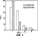
図1はプラスミドDNAにより誘導したDOTMA:DOPE及びDODAC:DOPEの融合を示す。
図2は脂質/DNA複合体とRBCゴーストとの融合を示す。
図3は50モル%のDOPEと配合せしめたカチオン脂質小胞のトランスフェクション能力の事前検定を示す。
図4はBHK細胞におけるDOTMA:DOPE及びDODAC:DOPEトランスフェクション効能の負荷比力価を示す。
図5は血清の非存在下(Figure 5A)及び存在下(Figure 5B)での集密度50%のH9C2筋芽細胞についてのDODAC:DOPE複合体のトランスフェクション効能を示す。
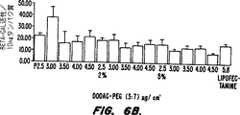
図6は血清の非存在下(Figure 6A)及び存在下(Figure 6B)での集密度100%のH9C2筋芽細胞についてのDODAC:DOPE複合体のトランスフェクション効能を示す。
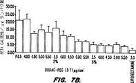
図7は血清の非存在下(Figure 7A)及び存在下(Figure 7B)でのH9C2筋管についてのDODAC:DOPE複合体のトランスフェクション効能を示す。
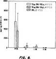
図8はICRマウスについてのDODAC:DOPE複合体のトランスフェクション効能を示す。
発明の詳細な説明
略語及び定義
本明細書において以下の略語を用いた:BHK:ベビーハムスターの腎臓;RBC:赤血球細胞;DDAB:N,N−ジステアリル−N,N−ジメチルアンモニウムブロミド;DODAC:N,N−ジオレイル−N,N−ジメチルアンモニウムクロリド;DOPE:1,2−sn−ジオレオイルホスファチジルエタノールアミン;DOSPA:2,3−ジオレイルオキシ−N−(2(スペルミンカルボキシアミド)エチル)−N,N−ジメチル−1−プロパンアミニウムトリフルオロアセテート;DOTAP:1,2−ジオレオイルオキシ−3−(N,N,N−トリメチルアミノ)プロパンクロリド;DOTMA:1,2−ジオレイルオキシ−3−(N,N,N−トリメチルアミノ)プロパンクロリド;OSDAC;N−オレイル−N−ステアリル−N,N−ジメチルアンモニウムクロリド;RT:室温;HEPES:4−(2−ヒドロキシエチル)−1−ピペラジンエタンスルホン酸;FBS:胎児牛血清;DMEM:ダルベッコ改良イーグル培地;PEG−Cer−C14:1−O−(2′−(ω−メトキシポリエチレングリコール)スクシノイル)−2−N−ミリストイル−スフィンゴシン;PEG−Cer−C20:1−O−(2′−(ω−メトキシポリエチレングリコール)スクシノイル)−2−N−アラキドイル−スフィンゴシン;PBS:リン酸緩衝食塩水;THF:テトラヒドロフラン;EGTA:エチレンビス(オキシエチレンニトリリロ)−四酢酸;SF−DMEM:無血清DMEM;及びNP40:ノニルフェノキシポリエトキシエタノール。
本明細書において用いる語「アルキル」とは、飽和炭化水素基を意味し、それは直鎖でも枝分れしていてもよい(例えば、メチル、エチル、プロピル、イソプロピル)。一部の置換基にとって好適なアルキル基は1〜3個の炭素原子を含む低級アルキル基である。その他のアルキル基置換基については、16〜20個の炭素原子を含む長鎖アルキル基が好ましい。本明細書及び請求の範囲における数値範囲は全て上限値及び下限値を含むことを意図する。
「アシル」なる語はヒドロキシル基の除去により有機酸から出来る基を意味する。アシル基の例にはアセチル、ペンタノイル、パルミトイル、ステアロイル、ミリストイル、カプロイル及びオレオイルが含まれる。
本明細書において用いる語「薬理学的に許容されるアニオン」とは、薬理製剤において無毒な塩を供する有機酸又は無機酸のアニオンを意味する。かかるアニオンの例にはクロリド、ブロミド、スルフェート、ホスフェート、アセテート、ベンゾエート、シトレート、グルタメート及びラクテートが含まれる。薬理学に許容される塩の調製は、引用することで本明細書に組入れるBergeらJ.Pharm.Sci. 66:1-19(1977)に記載されている。
本明細書において用いる語「ポリアニオン」とは、複数のアニオン基を有する物質を意味する。例えば、ポリアニオンはDNA及びRNAの双方の核酸を意味するように用いており、それらは核酸骨格伝いに複数個のアニオン性ホスホジエステル基を有するポリアニオン形質において存在する。「ポリアニオン」なる語は中性pHにおいて複数個のアニオン基を有する薬剤も意味する。かかる薬剤には多重のカルボン酸官能基(即ち、Glu,Asp)をもつペプチドが含まれる。
「中性脂質」なる語は生理学的pHにおいて無荷電又は中性双イオン形態で存在する種々の脂質物質を意味する。かかる脂質には、例えばジアシルホスファチジルコリン、ジアシルホスファチジルエタノールアミン、セラミド、スフィンゴミエリン、セファリン、カルジオリピン、及びセレブロミドが含まれる。
「トランスフェクション」及び「形質転換」は本明細書において同義語として用いており、そしてポリアニオン性材料、特に核酸の細胞への導入を意味する。「リポフェクチン」なる語はリポソーム複合体を用いてかかる材料を導入することを意味する。ポリアニオン性材料は、細胞への侵入後の遺伝子発現を助長するように発現ベクターに連結されたDNA又はRNAの形態であってよい。即ち、本発明において用いられるポリアニオン性材料は、構造タンパク質、レセプター及びホルモンについてのコード配列を有するDNA、並びに転写及び翻訳調節セグメント(即ち、プロモーター、エンハンサー、ターミネーター、及びシグナル配列)、並びにベクターを含むことを意味する。発現ベクターへの特定の核酸の組込みの方法は当業界に公知であるが、しかし例えば共に引用することで本明細書に組入れるSambrookらMolecular Cloning:A Laboratory Manual(第2版)Vol 1〜3, Cold Spring Harbor Laboratory,(1989)又はCurrent Protocds in Molecular Biology, F.Ausubelら編、Greene Publishing and Wiley-Interscience, New York(1987)に詳しく記載されている。
「発現ベクター」、「クローニングベクター」又は「ベクター」は往々にして選定の宿主細胞の中で複製できるプラスミド又はその他の核酸分子である。発現ベクターは自己複製できうるか、又は当業界公知の方法により宿主細胞のゲノムの中に挿入されることにより複製できうる。自己複製するベクターは複製起点を有するか又は選定の宿主細胞の中で機能的な自己複製配列(ARS)を有するであろう。往々にして、ベクターは、複数の宿主細胞において、例えばクローニング及び構築のためにE.コリ(E.coli)において、且つ発現のために哺乳細胞において有用であることが所望される。
詳細な説明
本発明は細胞へのポリアニオン性材料の導入のための組成物及び方法を提供する。この組成物は式Iのカチオン化合物及び少なくとも一種の中性脂質を含んで成る。
好適な態様の一群において、このカチオン化合物は、R1及びR2がメチルであり、そしてY及びZがそれぞれ独立して−CH=CHCH2CH2CH2−,−CH2CH=CHCH2CH2−,−CH2CH2CH=CHCH2−又は−CH2CH2CH2CH=CH−である式Iの化合物である。本発明の特に好適な態様においては、R1及びR2はメチルであり;Y及びZはそれぞれ−CH=CHCH2CH2CH2−であり;n及びqは共に7であり、そしてm及びqは共に5である。最も好適な態様において、カチオン化合物はDODAC(N,N−ジオレイル−N,N−ジメチルアンモニウムクロリド)である。DODACは洗剤及びシャンプーの添加剤としてかなり利用されている公知の化合物である。リポソーム組成物におけるその他の洗剤との共存脂質(colipid)としてのその用途も報告されている(Takahashiら、GB 2147243号を参照のこと)。
本組成物の一部である天然脂質は洗剤の中に又はミセルもしくはリポソームの形成のために一般に利用されている任意の様々な中性脂質であってよい。本組成物において有用な中性脂質の例はジアシルホスファチジルコリン、ジアシルホスファチジルエタノールアミン、セラミド、スフィンゴミエリン、セファリン、カルジオリピン、及びセレブロシドである。好適な態様において、本組成物は1又は複数種の中性脂質を含んでよく、それはジアシルホスファチジルコリン、ジアシルホスファチジルエタノールアミン、セラミド又はスフィンゴミエリンである。これらの中性脂質におけるアシル基は好ましくはC10−C24炭素鎖を有する脂肪酸に由来するアシル基である。より好ましくは、このアシル基はラウロイル、ミリストイル、パルミトイル、ステアロイル又はオレオイルである。特に好適な態様において、この中性脂質は、1,2−sn−ジオレオイルホスファチジルエタノールアミンである。
アニオンX-も同様に任意の様々な薬理学的に許容されるアニオンでありうる。これらのアニオンは有機系又は無機系、例えばBr-,Cl-,F-,I-、スルフェート、ホスフェート、アセテート、ニトレート、ベンゾエート、シトレート、グルタメート及びラクテートでありうる。好適な態様において、X-はCl-及びAcO-である。
上記の成分に加えて、本発明の組成物は薬理学的に許容される担体も含むであろう。薬理学的に許容される担体は当業者に公知であ担体の選択は投与する特定の組成物及びこの組成物を投与する特定の方法によりある程度決定されるであろう。好ましくは、この薬理担体は水又は食塩水である。
本発明の組成物において、カチオン化合物、対、中性脂質の比は約25:75(カチオン化合物:中性脂質)〜75:25(カチオン化合物:中性脂質)、好ましくは約50:50の範囲に属するであろう。
上記の組成物において用いられるカチオン化合物は標準の合成反応を利用して当業者に公知の方法により調製されうる(March, Advanced Organic Chemistry第4版、Wiley-Interscience, NY NY(1992)を参照のこと;引用することで本明細書に組入れる)。例えば、OSDACの合成はまずオレイルアミンをホルムアルデヒド及びナトリウムシアノボロヒドリドでアミンの還元的アルキル化が生ずる条件下で処理することにより実施できる。これはジメチルオレイルアミンを供し、これは次にステアリルブロミドでアルキル化されて対応のアンモニウム塩となりうる。アニオン交換はOSDACの形成をもたらす。ジメチルオレイルアミンはオレイルブロミドの大過剰量のジメチルアミンによる処理によっても合成でき、そして上記の通りに更に誘導化する。双方の脂肪酸鎖が不飽和となっているカチオン化合物(即ちDODAC)に関しては、以下の一般手順が利用できる。不飽和酸(即ち、オレイン酸)はオキサリルクロリド、チオニルクロリド、PCl3又はPCl5の如き試薬によりその対応の酸クロリドに変換されうる。アシルクロリドは対応のアミドを供するように不飽和アミン(即ち、オレイルアミン)により処理してよい。例えばリチウムアルミニウムヒドリドによるアミドの還元は双方のアルキル基が不飽和長鎖アルキル基である第二級アミンを供する。次いでこの第二級アミンをアルキルハライド、例えばヨウ化メチルで処理して四級アンモニウム化合物にすることができる。次いでアニオン交換を行って所望の薬理学的に許容されるアニオンを有するカチオン化合物を供することができる。アルキルアミン前駆体は似たようにして合成できる。例えば、アルキルハライドの大過剰量のアンモニアメタノール性溶液による処理は、精製後に必要なアミンを供するであろう。他方、適当なカルボン酸のオキサリルクロリドによる処理によって生成されるアシルクロリドはアンモニアと反応してアミドを供しうる。アミドのLiAlH4による還元は必須のアルキルアミンを供するであろう。
一の態様の群において、本発明の薬理組成物はミセル又はリポソームとして配合されるであろう。
本発明のカチオン化合物及び中性脂質を含むミセルは当業者に公知の方法により調製できる。本組成物のミセル製剤に加えて、本発明はその他の物質、例えばリソホスファチジルコリン、リソホスファチジルエタノールアミン、リソホスファチジルセリン、リソホスファチジルグリセロール、ホスファチジルエタノールアミン−ポリオキシエチレン・コンジュゲート、セラミド−ポリオキシエチレン・コンジュゲート又はホスファチジル酸−ポリオキシエチレン・コンジュゲートを含むミセル製剤も提供する。本発明の組成物において使用するポリオキシエチレンコンジュゲートは接合基(即ち、ホスファチジン酸又はホスファチジルエタノールアミン)を適当な官能化ポリオキシエチレン誘導体と組合せることにより調製できる。例えば、ホスファチジルエタノールアミンをω−メトキシポリエチレングリコールスクシネートと組合せてホスファチジルエタノールアミン−ポリオキシエチレン・コンジュゲートにすることができる。引用することで本明細書に組入れるParrら、Biochim.Biophys. Acta 1195:21-30(1994)を参照のこと。
本薬理組成物のカチオン化合物及び中性脂質からリポソームも形成できる。中性脂質の選択は一般に、例えばリポソームのサイズ及び血流中のリポソームの安定性により導かれる。
前述の通り、リポソーム中の中性脂質成分は2個のアシル基を有する脂質である(即ち、ジアシルホスファチジルコリン及びジアシルホスファチジルエタノールアミン)。様々な鎖長及び飽和度の様々なアシル鎖基を有する脂質が入手可能であるか、又は公知の技術により単離もしくは合成できうる。一般に、濾過除菌のために特にリポソームが約0.3ミクロンより小さくなくてはならないとき、飽和度の低い脂質の方がより簡単にサイズ選別できる。一の態様の群において、C14〜C22の範囲の炭素鎖長を有する飽和脂肪酸を含む脂質が好ましい。別の態様の群において、C14〜C22の範囲の炭素鎖長を有するモノ又はジ不飽和脂肪酸をもつ脂質が使用される。更に、飽和と不飽和の脂肪酸鎖の混合をもつ脂質が利用できる。本発明において有用なリポソームはスフィンゴミエリン、又はセリン及びイソシトールの如きその他の頭部基をもつリン脂質より成っていてもよい。本発明において有用なその他のリポソームにはコレステロール、ジグリセリド、セラミド、ホスファチジルエタノールアミン−ポリオキシエチレン・コンジュゲート、ホスファチジン酸−ポリオキシエチレンコンジュゲート又はポリエチレングリコール−セラミド・コンジュゲート(例えばPEG−Cer−C14又はPEG−Cer−C20)が含まれであろう。リポソームをサイズ選別及び濾過除菌する方法を以下に述べる。
様々な方法がリポソームの調製のために有用であり、例えばSzokaらAnn.Rev.Biophys.Bioeng. 9:467(1980),米国特許第4,235,871、4,501,728、4,837,028号、論文Liposomes, Marc J.Ostro,編Marcel Dekker, Inc., New York, 1983,第I章及びHopeらChem.Phys.Lip.40:89(1986)に記載されている。これらは全て引用することで本明細書に組入れる。一の方法は不均質なサイズの多重層小胞を供する。この方法においては、小胞形成性脂質を適当な有機溶媒又は溶媒系の中に溶かし、そして真空又は不活性ガス下で乾燥させて脂質薄膜を形成する。所望するなら、この膜を適当な溶媒、例えば第三ブタノールの中に溶かし、次いで凍結乾燥して一層水和し易い粉末様形態にする一層均質な脂質混合物にする。この膜を水性緩衝溶液で覆い、そして一般に撹拌しながら15〜60分かけて水和させる。得られる多重層小胞のサイズ分布は一層強力な撹拌条件下での脂質の水和により、又はデオキシコレートの如き可溶化性洗浄剤の添加により一層小さいサイズにシフトしうる。
リポソーム調製の後、リポソームをサイズ選別にかけて所望のサイズ範囲及び比較的狭い分布のリポソームサイズを得るようにしてよい。約0.2〜0.4ミクロンのサイズ範囲はリポソーム懸濁物を慣用のフィルター、一般には0.22ミクロンのフィルターで濾過除菌することを可能にする。
いくつかの技術がリポソームを所望のサイズにサイズ選別するために有用である。一のサイズ選別法は引用することで本明細書に組入れる米国特許第4,737,323号に記載されている。浴槽又はプローブ音波処理のいづれかによるリポソーム懸濁物の音波処理は約0.05ミクロン未満のサイズの小型の単層小胞に至る連続的なサイズ減少を供する。ホモジネーションがその他の方法であり、それは大型リポソームを小型のものに断片化する剪断エネルギーを頼りとする。一般的なホモジネーション手順において、多重層小胞を選定のリポソームのサイズにまで標準のエマルションホモジナイザーに再循環させる。一般に約0.1〜0.5ミクロンが観察される。両方の方法において、粒子サイズ分布は慣用のレーザービーム粒子サイズ標識によりモニターできる。
小孔ポリカーボネート膜又は非対称セラミック膜を通じるリポソームの押出もリポソームのサイズを比較的よく規定されたサイズ分布に減少させるための有効な方法である。一般に、懸濁物を膜に1又は数個、所望のリポソームサイズ分布が達成されるまで循環させる。これらのリポソームは、リポソームのサイズの暫進的な減少を達成するために小孔膜に連続して押出してよい。本発明における用途のため、約0.05ミクロン〜約0.15ミクロンのサイズを有するリポソームが好ましい。
本発明の組成物は細胞へのポリアニオン性材料の導入のために有用である。従って、本発明は細胞へのポリアニオン性材料の導入のための方法も提供する。この方法は、in vitroでポリアニオン性材料を少なくとも一種の中性脂質及び式Iのカチオン化合物の組成物と接触させることにより実施する。このポリアニオン性材料をリポソーム製剤と接触させて複合体を形成した後、この複合体を細胞とトランスフェクションが起こるのに十分な時間接触させる。
上記の通り、このポリアニオン性材料はまず中性脂質及びカチオン性化合物を含んで成る組成物を接触させてポリアニオン性材料−リポソーム複合体を供する。この接触は(中性脂質及びカチオン化合物からの)リポソーム形成の前に、又は一次リポソーム形成の後に行ってよい。好適な態様においては、中性脂質及びカチオン化合物のリポソームをまず形成し、次いでポリアニオン性材料と接触させる。このポリアニオン性材料は一般に負に帯電したポリアニオン性材料と正に帯電したリポソームの表層との間でのイオン性吸引力の結果、リポソームの表層に結合するであろう。一般に、複合体の形成をもたらしめるポリアニオン性材料とリポソームとの接触は約15℃〜約45℃の温度、好ましくは約25℃で実施されるであろう。複合体の形成を完了するのにかかる時間の長さは温度、並びにポリアニオン性材料及びリポソーム自体の種類に依存するであろう。約25℃の接触温度を利用した場合、リポソームと核酸との複合体を形成するための時間の長さは約15分〜約2時間であろうが、一定の状況ではより長い時間が必要とされうる。他方、このポリアニオン性材料は当業者に公知の慣用の薬剤を負荷するために利用されている方法によりリポソームの内部に組込むことができる。リポソームに薬剤を負荷するための一の方法は封入を含み、そして様々な技術により実施できる。
一の封入技術において、この薬剤及びリポソーム成分は、全ての物質が混和性であり、且つドライフィルムへと濃縮されるようにする有機溶媒に溶かす。次いでこのドライフィルムにバッファーを加え、そしてポリアニオン性材料が小胞壁の中に組込まれたリポソームが形成される。他方、このポリアニオン性材料をバッファーの中に入れ、そして脂質成分のみのドライフィルムに添加してよい。このようにして、ポリアニオン性材料はリポソームの水性内部に封入されるようになるであろう。リポソームの形成において用いるバッファーは例えば等張性食塩水、リン酸緩衝食塩水又はその他の低イオン強度バッファーの任意の生物適合性バッファー溶液でありうる。一般に、ポリアニオン性材料は約0.01ng/ml〜約50mg/mlの量で存在するであろう。水性内部又は膜の中にポリアニオン性材料の組込まれた得られるリポソームを次に上記の通りにして任意的にサイズ選別にかける。
一の好適な態様の群において、このポリアニオン性材料:リポソーム複合体は約0.5〜約4.0の帯電率(+/−)を有するであろう。このような帯電率を達成せしめるため、この複合体はカチオン脂質及び少なくとも一種の中性脂質の水性リポソーム製剤であってその中のカチオン脂質が全脂質濃度の約25%〜約75%で存在しているものを調製することにより形成する。上記の通りにしてリポソームをサイズ選別した後、ポリアニオン性材料の水性溶液をリポソーム懸濁物で処理する。得られる調製品を次に生物適合性バッファーで好ましくは約5倍希釈し、約0.5〜約4.0の帯電率を有する0.05〜1.0μg/mlの最終濃度のポリアニオン性材料:リポソーム複合体を供する。
ポリアニオン性材料:リポソーム複合体の形成後、この複合体をトランスフェクションすべき細胞と接触させる。リポソームはほとんど全ての細胞タイプに吸着する。一旦吸着したら、リポソーム(先に記載の複合体を含む)は細胞の一部により食作用を受け、細胞膜と脂質交換するか、又は細胞と融合しうる。複合体のポリアニオン性部分の輸送又は組込みはこれらの任意のいづれかの経路を介して行われうる。特に、融合が起こるとき、リポソーム膜は細胞膜に組込まれ、そしてリポソームの内容物は細胞内流体と組合わさる。細胞とポリアニオン性材料−リポソーム複合体との接触は、in vitroで行われるとき、生物適合性媒体の中で行われるであろう。脂質の濃度は特定の用途に依存して様々であってよいが、しかしながら一般には約1μmol〜約10mmolである。ポリアニオン性材料:リポソーム複合体による細胞の処理は一般に生理学的温度(約37℃)で約1〜6時間、好ましくは約2〜4時間にわたって実施されるであて。in vitro用途において、ポリアニオン性材料の輸送は培養物の中で増殖させている任意の細胞(植物又は動物起源、脊椎動物又は無脊椎動物)及び任意の組織又はタイプに対して行ってよい。好適な態様において、これらの細胞動物細胞、より好ましくは哺乳動物細胞、そして最も好ましくはヒト細胞であろう。
一の好適な態様の群において、このポリアニオン性材料:リポソーム複合体を約103〜105細胞/ml、より好ましくは約2×104細胞/mlの細胞密度を有する60〜80%の集密度のプレート細胞に加える。複合体の濃度は好ましくは約0.01〜0.2μg/ml、より好ましくは約0.1μg/mlであろう。
一般の適用には、治療的に有用なポリペプチドをコードするDNA又はmRNA配列の細胞内輸送を供する公知のトランスフェクション手順の利用を含む。しかしながら、この組成物は発現された遺伝子生成物又はタンパク質自体の輸送のためにも利用できる。このようにして、欠失又は非存在遺伝子生成物を供給することによる遺伝子疾患のための(即ち、DuchenneジストロフィーについてはKunkelら、Brit.Med.Bull. 45(3):630-643(1989)を、そして嚢胞性線維芽症についてはGoodfellow, Nature 341:102-103(1989)を参照のこと)治療法が提供される。本発明の組成物についてのその他の用途には細胞へのアンチセンスオリゴヌクレオチドの導入が含まれる(Bennetteら、Mol.Pharm. 41:1023-1033(1992)参照のこと)。
他方、本発明の組成物は当業者公知の方法を利用し、細胞のin vivoトランスフェクションのためにも利用できる。特に、引用することで本明細書に組入れるZhuらScience 261:209-211(1993)はDOTMA-DOPE複合体を利用するサイトメガロウイルス(CMV)−クロラムフェニコールアセチルトランスフェラーゼ(CAT)発現プラスミドの静脈内輸送を述べる。引用することで本明細書に組入れるHydeらNature 362:250-256(1993)は、リポソームを利用したマウスの気路の上皮に至る及び肺胞に至る嚢胞線維芽症トランスメンブラン伝搬調節因子(CFTR)遺伝子の輸送を述べている。引用することで本明細書に組入れるBrighamら、Am.J.Med.Sci. 298:278-281(1989)は細胞内酵素クロラムフェニコールアセチルトランスフェラーゼ(CAT)をコードする機能性の原核系遺伝子によるマウスの肺のin vivoトランスフェクションを述べている。
本方法において利用するポリアニオン性材料が核酸であるとき、それらは天然起源から単離されたもの、ATCCもしくはGen Bankライブラリーの如きソースから入手したもの、又は合成法により調製したものでありうる。合成核酸は様々な溶液又は固相方法により調製できる。一般に、固相合成が好ましい。ホスフィット−トリエステル、ホスホトリエステル及びH−ホスホネート化学による核酸の固相合成のための手順の詳細な説明は幅広く入手できる。例えば、Itakura米国特許第4,401,796号;Caruthersら米国特許第4,458,066及び4,500,707号;BeaucageらTetrahedron Lett., 22:1859-1862(1981);MatteucciらJ.Am.Chem.Soc., 103:3185-3191(1981);CaruthersらGenetic Engineering, 4:1-17(1982);Jones第2章、Atkinsonら第3章及びSproatら第4章Oligonucleotide Synthesis:A Practical Approach, Gait(編),IRL Press, Washington D.C.(1984);FroehlerらTetrahedron Lett., 27:469-472(1986);FroehlerらNucleic Acids Res., 14:5399-5407(1986);SinhaらTetrahedron Lett., 24:5843-5846(1983);及びSinhaらNucl.Acids Res., 12:4539-4557(1994)を参照のこと。これらは引用することで本明細書に組入れる。
本発明はまた細胞にその他のポリアニオン性材料、特にタンパク質を導入するのにも有用である。細胞への外生又は内生タンパク質の導入はmRNAの翻訳を行うことのできない細胞を有する個体のための適当な治療法を担いうる。
以下の実施例は単に例示の目的のために提供し、本発明を限定又は規定するものではない。
実施例
材料
オレイルアミンはFluka Chemical Company, St.Louis, Missouri, USAより入手し、そして下記の方法によって合成もした。40%のホルムアルデヒド溶液はFischer Scientific, Ottawa, Canadaから入手した。ナトリウムシアノボロヒドリド、ステアリルブロミド、オレイン酸、オキサリルクロリド、リチウムアルミニウムヒドリド、ヨウ化メチル及びN−(2−ヒドロキシエチル)ピペラジン−N′−2−エタンスルホン酸(HEPES)はSigma Chemical Company, St.Louis, Missouri, USAより入手した。N−(7−ニトロベンズ−2−オキサ−1,3−ジアゾール−4−イル)ジオレオイルホスファチジル−エタノールアミン(NBD−PE)、N−(リッサミンローダミン8スルホニル)ジオレオイルホスファチジル−エタノールアミン(Rho−PE)及び1,2−sn−ジオレオイルホスファチジルエタノールアミン(DOPE)はAvanti Polar Lipids, Alabster, Alabama, USAより入手した。Lipex押出機はLipex Biomembranes, Vancouver, Canadaより入手した。pCMVβ発現ベクター(β−gal)はClonetech Laboratories, Inc., Palo Alto, Califomia, USAより入手した。シリカゲルはBDH, Canadaより入手した。PEG−Cer−C14,PEG−Cer−C20及びその他のPEG修飾脂質はParrら、Biochim.Biophys. Acta 1195:21-30(1994)及び同時係属出願第08/486,214号に供されている方法により調製できる。
実施例1
本実施例はオレイルアミンの調製を例示する。この合成は、商業用のオレイルアミンが往々にしてエライジルアミンにより汚染されているため、開発した。
ベンゼン中のオレイン酸(5g)の溶液を室温で1時間撹拌しながらオキサリルクロリド(1ml)で処理した。溶媒を蒸留により除去した。その残渣を塩化メチレンに溶かし、そして濃アンモニアを加えた。得られるエマルションを室温で2h撹拌した。真空でアンモニアを除去後、その溶液を塩酸で酸性にし、そして塩化メチレンで抽出した。得られる残渣をジエチルエーテルに溶かし、そして過剰のリチウムアルミニウムヒドリドを粉末として強く撹拌しながらゆっくり加えた。ゼラチン状の懸濁物が形成し、そしてそれを更に4h撹拌した。次にメタノールを水素の発生が終るまで加えた。過剰の希塩酸を全ての固体が溶解し、且つ更なる水素発生が観察されなくなるまで加えた。この混合物を塩化メチレンで抽出した。有機相を飽和ブラインで洗い、そして溶媒をロータリーエバポレーターで除去した。この残差をシリカゲルカラム(Merck, Art. 9385, 20gのゲル/反応生成物g)に、塩化メチレン中の2%のメタノール(容量)を用い、不純物(オレイルアルコール及び未反応のオレオイルアミド)が全て溶出するまで通した。次いでメタノール濃度を溶出液250ml当り2%づつ、24%の濃度が達成されるまで上昇させた。このカラムをメタノールでフラッシングした。生成物含有画分からの溶媒の除去は1H NMRスペクトルにおいてエライジルアミンの徴候を示さず3gのオレイルアミンを供した。
実施例2
本実施例はN−ステアリル−N−オレイル−N,N−ジメチルアンモニウムクロリド(OSDAC)の合成を示す。
アセトニトリル(100ml)及びエタノール(50m)中のオレイルアミン(6.7g)の溶液を40%の水性ホルムアルデヒド(10ml)及びナトリウムシアノボロヒドリド(2.7g)により室温で2時間処理した。酢酸(5ml)をゆっくり加え、そしてその溶液を更に1時間撹拌した。この反応混合物を水で希釈し、水性水酸化ナトリウムで塩基性にし、そして塩化メチレンで抽出し、有機画分を硫酸マグネシウムで乾かし、濾過し、そして溶媒を真空で除去した。その残渣をシリカゲルカラム(150g)に、溶出液として塩化メチレン中の15%のメタノールを用いて通し、淡黄色油(5g)を得た。
2.2.OSDACの合成
N,N−ジメチルオレイルアミン(1g)及びステアリルブロミド(5.4g)の溶液を塩化メチレン(50ml)に溶かし、そして水性水酸化ナトリウム溶液(5Mの溶液5ml)で室温にて一夜撹拌しながら処理した。その反応混合物を水で洗い、次いで希塩酸で洗った。有機相を飽和塩化ナトリウム溶液(15×)で洗い、そして溶媒を除去した。その残渣をメタノール性塩酸に溶かし、そして水及び塩化メチレンから抽出した。これを更に3回繰り返した。有機溶媒を真空下で除去し、そしてその残渣をシリカゲルカラム(150g)に溶出液として塩化メチレン中の5%のメタノールを用いて通し、ベンゼン中の10%のメタノールから凍結乾燥後に0.6gのOSDACが白色粉末として得られた。
実施例3
本例は還元アミド化を利用するN,N−ジオレイル−N,N−ジメチルアンモニウムクロリド(DODAC)の合成を例示する。
ベンゼン(50ml)中のオレイン酸(5g)の溶液をオキサリルクロリド(2ml)で室温において1時間処理した。この溶媒及び過剰量のオキサリルクロリドを真空下で除去し、そしてその残渣をベンゼン(20ml)に溶かした。ベンゼン(10ml)中のオレイルアミン(7g)の溶液をゆっくり加え、次いでトリエチルアミン(3ml)を加えた。この反応混合物を室温で1時間撹拌し、次いで過剰の希塩酸で中和した。この混合物を塩化メチレンで抽出し、次いで合わせた有機抽出物を硫酸マグネシウムで乾かし、濾過し、そして溶媒を除去した。その残渣をシリカゲルカラム(150g)に塩化メチレン中の5%のメタノールを溶出液として用いて通し、白色固体としてのN−オレイルオレイルアミドを得た。
3.2.ジオレイルアミンの合成
THF(100ml)中のN−オレイルオレイルアミド(上記で調製)の溶液を40℃にまで温めた。リチウムアルミニウムヒドリドを気体の過激な発生が止むまでゆっくりと加えた。この反応混合物を1時間還流加熱し、次いで室温にまで冷却した。メタノール(100ml)をゆっくりと加え、次いで水(200ml)を加えた。塩化メチレンを加え、そして得られる懸濁物を15分撹拌した。このスラリーを濾過し、そしてその沈殿物をエタノール/塩化メチレン(50:50、50ml、2×)で洗った。合わせた濾液を塩化メチレンで抽出した。有機画分を硫酸マグネシウムで乾かし、濾過し、そして溶媒を真空で除去した。その残渣をシリカゲルカラム(150g)に塩化メチレン中の5%のメタノールを溶出液として用いて通し、淡黄色油としてのジオレイルアミンが得られた。
3.3.N,N−ジオレイル−N,N−ジメチルアンモニウムクロリドの合成
クロロホルム(50ml)中の上記で調製したジオレイルアミンの溶液をヨウ化メチル(100ml)により室温で1時間処理した。次いで水性水酸化ナトリウム(5Mの溶液1ml)及びヨウ化メチル(5ml)を加え、そしてその混合物を更に1時間撹拌した。この有機溶媒を真空下で除去し、そして得られるスラリーを希塩酸で中和した。水を加え、そしてその反応混合物を塩化メチレンで抽出した。この有機相を飽和塩化ナトリウム溶液で洗い(15×)、その後溶媒を真空下で有機相から除去した。その残渣をウェットエタノールにおいて濃塩酸溶液に溶かし、水で希釈し、そして塩化メチレンで抽出した。この方法を更に3回繰り返した。この生成物を、溶媒の除去後、シリカゲルカラム(150g)に塩化メチレン中のメタノールの溶出液として用い(メタノールの量を5容量%から32容量%にまで増やしながら)通した。純粋な画分を合わせ、そしてその溶媒を除去し、白色のワックス状固体又は放置すると固化する無色の油のいづれかとしてのN,N−ジオレイル−N,N−ジメチルアンモニウムクロリド(5g)を得た。DODACはベンゼン中の10%のメタノールからの凍結乾燥により白色粘質粉末として充密となりうる。
実施例4
本例はアルカリ化経由によるN,N−ジオレイル−N,N−ジメチルアンモニウムクロリド(DODAC)の合成を例示する。
ジメチルアミンの溶液をドライメタノール(200ml)に無水ジメチルアミンガスを30分吹き込むことにより調製した。オレイルブロミド(5g)を加え、そしてこの溶液を暗所の中で室温で16時間撹拌した。この溶媒をロータリーエバポレーターで除去し、そして残留ジメチルアミンを無水エタノール(50ml)の2回の添加、そして続くロータリーエバポレーターでの溶媒の除去により除去した。オレイルブロミド(10g)をこの残渣に加え、そしてこの混合物をクロロホルム(40ml)に溶かした。水酸化ナトリウム溶液(1N 10ml)を加え、そして得られる溶液を暗所の中で静かに16時間還流した。
有機溶媒をロトバップで除去し、そして残渣をエタノール(50ml)に溶かした。濃HCl(33%、10ml)を加え、次いで水(100ml)を加えた。得られるエマルションをクロロホルム(3×50ml、次いで2×25ml)で抽出した。その抽出物を合わせ、そして洗浄/抽出手順に更に5サイクル再びかけた。次いで有機溶媒をロータリーエバポレーターで除去し、そしてその残渣を飽和塩化ナトリウム溶液に懸濁した。この溶液をクロロホルム(3×50ml、2×25ml)で抽出した。この工程を繰り返した。この有機溶媒をロータリーエバポレーターで除去し、そしてその残渣をドライエタノールの2回の添加、それに続くロータリーエバポレーターでの溶媒の除去により乾燥させた。この残渣をシリカゲルクロマトグラフィー(200gのシリカゲル)及びクロロホルム中のメタノール勾配溶出(2%から12%のメタノール)を利用して精製した。純粋DODACを含む画分を合わせ、そしてその溶媒を除去して7gの無色のゴムを得た。ベンゼン中の10%のメタノールからの凍結乾燥は無色の粘質粉末のDODACを供した。
実施例5
本例はカチオン小胞/DNA複合体と生体膜との紡錘原性(fusogenicity)を例示する。
5.1.小胞−小胞融合
これらの実験のために用いた小胞は従来記述の押出手順により作った(Hopeら、Biochim.Biophys. Acta 812:55(1985);引用することで本明細書に組入れる)。簡単には、等モル比のカチオン脂質とDOPEとより成るカチオン脂質混合物をクロロホルムから窒素ガスの気流下で乾燥させた。残留溶媒を真空で2時間除去した。乾燥脂質フィルムを蒸留水で水和し、そして得られる多重相小胞(MLV)懸濁物を液体窒素及び温水サイクルを利用して5回凍結融解した。大量の単層小胞がこのMLV懸濁物をLipex押出機を用いて2枚重ねの孔径100nmのフィルターに押し通すことにより成形された。
小胞融合は本明細書に引用することで組入れるStruckらBiochemistry 20:4093(1981)に記載の通りにして共鳴エネルギー伝達を利用してモニターした。簡単には、未ラベルの小胞を20mMのHEPES pH 7.4のバッファー中の0.5mole%づつのRho−PE(N−(リッサミンローダミンBスルホニル)ジオレオイル−ホスファチジルエタノールアミン)及びNBD−PE(N−(7−ニトロベンズ−2−オキサ−1,3−ジアゾル−4−イル)−ジオレオイルホスファチジルエタノールアミン)を含む類似の小胞と10:1で混合した。後者の膜蛍光プローブはエネルギー供与体を担い、そして前者はエネルギー受容体を担う。ラベル化小胞と未ラベル小胞との融合はプローブの希釈をもたらす。即ち、Rho−PE小胞の減少に基づくNBD−PE蛍光の上昇は膜融合の指標である。カチオン小胞の融合を誘導するのに7kBのβ−galプラスミドを用いた。。図1はDOTAP:DOPE及びDODAC:DOPEとプラスミドDNAとの融合実験より得た結果を示す。帯電率は325の平均ヌクレオチド分子量に基づいて計算した。図1に示す通り、DOTMA:DOPE及びDODAC:DOPE小胞は共に全ての帯電率において同程度に融合し、そして1の帯電率において最適な紡錘原性を示し、これは双方の物質のトランスフェクションにとっての最適帯電率に相当する(図4参照)。
5.2.赤血球(RBC)ゴーストとの脂質−DNA複合体の融合
0.5%づつのRho−PE及びNBD−PEでラベルされたカチオン小胞を20mMのHEPES pH 7.4バッファー中の7kBのβ−galプラスミドと混合した。この脂質DNA複合体をRBCゴースト溶液に加えた。ゴースト膜との融合はプローブ希釈及びNBD蛍光の上昇をもたらす。RBCゴーストは文献に記載の通りにして調製した(Wood, Methods in Enzymology, 149:271-280 Academic Press(1987)参照)。図2は3通りの帯電率及び4種のカチオン脂質(DDAB, DOTMA, OSDAC及びDODAC)を利用する脂質/DNA複合体−RBCゴースト融合実験の結果を示す。図2に示す通り、不飽和誘導体DOTMA, DODAC及びOSDACは飽和誘導体DDABよりも生物膜に対して優れた融合特性を有する。更に、DODAC及びOSDACは、最も一般的に利用されている商業的トランスフェクション脂質であるDOTMAよりRBCゴーストとの優れた紡錘原特性を示した。
実施例6
本例はβ−gal(プラスミドを用いてBHK細胞とトランスフェクションする一の手順を提供し、そして更にはDODAC−,DDAB−,DOTMA−及びOSDAC−含有小胞の相対トランスフェクション効能の対比を供す。
6.1.カチオンリポソームの調製
カチオンリポソームは実施例5記載の方法によりDOPEとDODAC, DDAB, DOTMA又はOSDACとから調製した。カチオン脂質をそれぞれDOPEと混合して0.25, 0.5, 3及び4(4組の混合物それぞれに関して)の帯電率を有する小胞を形成した。
6.2.BHK細胞のトランスフェクション
一般的なリポフェクションプロトコールの下記の通りに実施した:
1日目、BHK細胞を0.5mlの培地(DMEM中5%のFBS)の中で104細胞/24穴プレートウェルでプレート培養した。2日目、ウェルの中にまずH2Oを分注し、次いで脂質を加えることにより脂質:DNA複合体を24穴プレートの中に調製した。このDNAをH2Oの中で調製し、次いでウェルに加え、混合するように撹拌し、そして室温で30分インキュベートした。インキュベーション期間中、培地を細胞から除去し、そして細胞をPBSの中で洗った。洗浄後、750μlのSF−DMEMを加えた。
脂質:DNA複合体(200μl)をBHK細胞を含む適当なウェルに加え、そしてこのプレートをゆらして混合し、次いで37℃で4時間インキュベートした。トランスフェクション培地をDMEM中の5%のFBS 0.5mlと交換した。3日目、培地を除去し、そして細胞をβ−ガラクトシダーゼについての組織化学染色のための標準手順に従って染色した。4日目、染色液を除去し、そして細胞をPBSで洗い、70%のエタノールをかぶせ、そしてカウントした。
6.3.β−ガラクトシダーゼについての組織化学染色
組織化学染色のために必要な溶液はストックバッファー、固定剤、洗浄液及び染色液を含む。これらの溶液は下記の通りに調製及び保存した。
1.ストックバッファーを蒸留、脱イオン水を用いて水性溶液として調製した。保存温度は表示の通りとした。この溶液は、47%のグルタルアルデヒド、4℃;1MのMgCl2、RT;100mMのEGTA、pH 7.2、RT;10%のナトリウムデオキシコレート、RT;10%のNP40、RT;1MのHEPES、4℃;50mMのK3Fe(CN)6、4℃:暗所で3ヶ月まで保存;50mMのK4Fe(CN)6、4℃:暗所で3ヶ月まで保存;5MのNaCl、RT;及びX−gal、固体、−20℃で保存。
2.固定溶液は0.2%のグルタルアルデヒド、2mMのMgCl2及び5mMのEGTAの最終濃度を有し、そして220μlの47%のグルタルアルデヒド、100μlのMgCl2溶液及び2.5mlの100mMのEGTA、pH 7.2を合わせ、そして全容量をPBSで50mlに合わせることにより調製した。
3.洗浄溶液は100μlのMgCl2溶液、500mlの10%のナトリウムデオキシコレート及び100μlの10%のNP40を合わせ、次いで全容量を50μlに合わせることにより調製した。これは2mMのMgCl2、0.1%のナトリウムデオキシコレート及び0.02%のNP40の最終濃度をもたらした。
4.染色液は2.2mlの1MのHEPES、3.0mlの50mMのK3Fe(CN)6、3.0mlの50mMのK4Fe(CN)6、150μlの5MのNaCl、65μlの1MのMgCl2及び全容量を50mlにするH2Oを合わせることによって調製した。この溶液を42℃にまで温め、そして100μlのDMF中の12.5mgのX−gal(0.4%の最終容量)を加え、そして溶解させた。他方、X−galをDMFの中で125μg/μlにし、そしてホイルの中で−20℃で保存してよい。溶液中の物質の最終濃度は44mMのHEPES、3mMのK3Fe(CN)6、3mMのK4Fe(CN)6、15mMのNaCl、1.3mMのMgCl2及び0.5mg/mlのX−galであった。
細胞は以下の通りに染色した:
細胞をPBSで1回洗った。固定剤(5ml)を各プレートに加え、そして細胞をRTで5分インキュベートした。固定剤を除去し、そして細胞を浸透化溶液で2回(3分づつ)洗った。X−gal染色液(500μl/ウェル)を細胞に加え、これを二酸化炭素の雰囲気下で37℃で一夜インキュベートした。全ての溶液のpHは内生β−ガラクトシダーゼに由来するバックグランド妨害を回避するために7.5〜8.0に維持した。
6.4.結果
図3は4種の脂質DDAB, DOTMA, OSDAC及びDODACの4通りの帯電率(0.25, 0.5, 3及び4)での相対トランスフェクション効能の概要を示す。相対トランスフェクション効能は細胞プレート上の25の任意領域に関するトランスフェクションされた細胞の平均数である。DODACは1を超える帯電率において有意に優れた相対トランスフェクション効能を示し、一方DDAB, DOTMA及びOSDACは全て類似であった。全ての調製品が1未満の帯電率で低いトランスフェクション効能を有していた。DDABとDODACとの比較は極めて重要であり、なぜならそれはトランスフェクション効能に対して不飽和化の作用を示すからである。
様々な帯電率でのDODAC及びDOTMAの全細胞数に対するトランスフェクションされた細胞数で表示するトランスフェクション効能を図4に示す。双方の系についての最適トランスフェクションは1〜2の帯電率で認められ、そしてより高い帯電率で低下しはじめた。1未満の帯電率でのトランスフェクション効能は著しく低く、図3に示す結果と一致した。
実施例7
本例は培養筋芽細胞のトランスフェクションを供するための様々なカチオンリポソーム小胞の利用を例示する。
遺伝病、例えばDuchenne筋ジストロフィー又はA型もしくはB型血友病の処置のための遺伝子改変筋芽細胞療法の利用における著しい関心がもたれている。この生体外手法は、骨格組織に注射したときの筋管と融合する筋芽細胞の能力の利点を有する。このようにして、遺伝子改変筋芽細胞は筋繊維に修復遺伝子を供するために用いられうる。ウィルスベクター又は裸DNAが筋細胞をトランスフェクションするのに最も一般的に利用されている。ウィルスベクターの利用に由来するこの強力な免疫反応はその用途を制約する。裸DNA注入は筋細胞をトランスフェクションするのにおいて非常に非効率的である。
7.1.細胞及び培養条件
ラットの胎児の心臓に由来する筋細胞系H9C2(2−1)をATCCから入手した。この細胞を10%の胎児牛血清(FBS)及び10mg/mlのゲンタマイシン(正常増殖培地)の添加したα−最少必須培地(α−MEM)の中で増殖させた。培養物を5%のCO2の雰囲気下で37℃で維持した。トランスフェクションは24穴プレート(表面積2cm2)の中で行った。細胞を3通りの段階でトランスフェクションした:集密度50%の筋芽細胞、集密度100%の筋芽細胞及び分化筋管。筋芽細胞をトランスフェクションの一日前に半集密及び集密密度のそれぞれについて約3×104及び7×104細胞/cm2でプレート培養した。筋管形成は細胞が集密なときに正常増殖培地を融合培地(2%のFBS及び10mg/mlのゲンタマイシンを含むα−MEM)で交換することにより誘導した。融合培地の中で4日後、筋管をトランスフェクションにかけた。
7.2.プラスミド
pCMVβ(Clonetech由来)は、CMVプロモーター及びSV40エンハンサーにより駆動するβ−ガラクトシダーゼを含むプラスミドである。pCMVβを以下の標準技術を利用して増殖させ、そしてQiagen Maxiカラム(Qiagen, Chatworth, Califonia, USA)により精製した。
7.3.カチオンリポソーム
リポソーム製剤は2又は5モルはのPEG−Cer−C14を含むDODAC:DOPE(3:7モル/モル)又はDODAC:DOPE(3:7モル/モル)より成る。商業的に入手できるLIPOFECTAMINE(商標)(Gibco由来;DOSPA(2,3−ジオレイルオキシ−N−(2−(スペルミンカルボキシアミド)エチル)−N,N−ジメチル−1−プロパンアミニウムトリフルオロアセテート)及びDOPEを3:1のモル比で含むポリカチオンリポソーム)を比較のために用いた。
7.4.トランスフェクション
15μlの20mMのHepes、pH 7.4バッファー中のプラスミドDNA(290ng)の10μlの無血清培地(α−MEM)に加えた。得られた混合物を所望量のカチオンリポソームを含む20μlの無血清培地に加えた。この混合物を室温で45分インキュベートし、次いで無血清培地、又は筋芽細胞もしくは筋管のそれぞれのための12.5%もしくは2.5%のFBSを含むα−MEMにより全容量200μlにまで希釈した。24穴組織培養プレートの中で成育する細胞を無血清培地で2回すすぎ、そしてトランスフェクション混合物の存在下で5hインキュベートした。5hのインキュベーションの後、正常量の2倍量のFBSを含むα−MEM 200μlを加えた。24h後、この培地を500μlの新鮮培地と交換した。更に24h後、細胞をリン酸緩衝食塩水で2回すすぎ、次いで100μlの細胞溶解バッファー(Promega, Madison, Wisconsin, USA)で溶解した。β−ガラクトシダーゼ活性は上記の通りにして測定した。Bradfordタンパク質アッセイ(Pierce, Rockford, Illinois, USA由来)をこのリゼートのタンパク質濃度の評価のために用いた。β−gal活性は各サンプルのタンパク質濃度に対して標準化した。
7.5.結果
図5〜7に示す通り、DODAC含有リポソームはH9C2筋芽細胞及び筋管をトランスフェクションすることが可能である。トランスフェクションは細胞に加えるカチオンリポソームの量並びに集密度及び分化の状態に依存する。更に、トランスフェクションは血清の存在下で行えうる。更に、トランスフェクションを血清の存在下で行ったとき、DODAC含有リポソームは最適量の商業的に入手できるLIPOFECTAMINE(商標)よりも筋芽細胞及び筋管のトランスフェクションにおいてより効率的である。現在まで、LIPOFECTAMINE(商標)が血清の存在下で細胞をトランスフェクションする能力を有する唯一の認識されたカチオンリポソーム製剤であった。このような発見、特に血清の存在下で筋管をトランスフェクションする能力の発見は、DODAC含有系がin vivoで直接遺伝子を筋細胞に導入できうることを示唆する。
実施例8
本例はin vitroでリンパ球及びマクロファージ細胞タイプをトランスフェクションする様々なカチオンリポソーム小胞の利用を例示する。
ウィルス感染症、例えばHIV−1(AIDS)の処置のための手法はin vivoでの長期効能を示す薬剤又はワクチンの欠如に基づく遺伝子療法手段として注目を浴びている。HIV−I複製を阻害するように機能するポリTAR/抗−Tat抗−HIV−I遺伝子系が開発されている(Lisziewiczら、J.Virol. 69:206-212(1995)を参照のこと)。にもかかわらず、標的部位に対する遺伝子の導入は問題が残っている。主要細胞標的はCD4+Tリンパ球及びマクロファージであり、その理由は、これらの細胞のウィルスを担持するからである本例は、培養物におけるこれらの細胞タイプに対して遺伝子を導入するカチオンリポソームの能力を例示する。
8.1.動物
同系C57 BL/6雌マウス(Jackson Laboratories, Bar Harbor, Maine, USA)をこの研究に用いた。マウスを生後5週目において購入し、そしてコロニー室の中でケージ当り5匹で飼育し(ストレス条件なし)、人工照明を介して昼−夜(12hr)のサイクルを維持した。この実験の間動物は食餌及び水を自由に取れるようにし、そして実験操作前に少なくとも1週間順化させた。
8.2.トランスフェクション複合体の形成
水中のDODAC:DOPE(1:1モル/モル)リポソーム(1mMの濃度)を上記の押出手順により調製した。pCMVβ(InVitrogen, San Diego, California, USA)をQiagen法(Qiagen, Chatworth, Califonia, USA)を利用してE.コリリゼートから精製した。
トランスフェクション複合体の形成のため、500μlの水の容量の中の20μgのpCMVβ(62nmolのリン酸塩)を500μlの水中の186nmolのDODAC:DOPEに加えた。この脂質:DNAの比は1.5(±)の帯電率をもたらした。この懸濁物を室温で20〜30分インキュベートした。
8.3.マイトジェン刺激アッセイ
生後7週目のマウスを断頭により犠牲にした。脾臓、脳腺及び骨髄を取り出し、そして単一の細胞懸濁物を調製した。これらのリンパ管の細胞質をトリパンブルー排除検査により評価した。細胞懸濁物を所望の濃度に調整し(脾臓については5×10⁶/ml;胸腺については1×107/ml;そして骨髄については2×106/ml)、そして48穴プレートの中に0.5ml/ウェルでプレート培養した。細胞を直ちに50μlのDODAC:DOPE/pCMVβによりトランスフェクションするか、又は48時間後にコンカナバリンA(1, 2.5及び5μg/ml;Sigma)及びリポ多糖(2.5及び5μg/ml;Sigma)で有糸分裂刺激した。細胞をトランスフェクション因子の存在下で48hインキュベートし、その後それらを上記の通りにしてβ−gal発現について染色した。適切なコントロールのために独立のウェルを用意した:処理なし、裸pCMVβ、又はプラスミドの非存在下でのDODAC:DOPE。
8.4.結果
カチオンリポソームを利用する一次脾臓細胞、胸腺細胞及び骨髄細胞のトランスフェクションは低レベルのβ−gal陽性細胞(<5%)をもたらした。達成されるレベルはわずかなβ−gal陽性細胞(<0.01%)を有し、そして未処理細胞と同等な裸DNAコントロールにより得られたものよりも有意に高かった。ある実験においては、非接着脾臓細胞を固定及びβ−gal活性についての染色前に接着細胞から分離させた。少なくとも2通りの実験において、>80%の接着脾臓細胞(形態的にマクロファージを擬態)がβ−gal活性について陽性染色された。非接着細胞画分においてはわずかな細胞しかβ−gal活性について染色されなかった。小さくて丸いこれらの細胞はリンパ球を擬態していた。非刺激及び刺激細胞は共に類似の低レベルのトランスフェクションを示した。一次細胞中のpCMVβ発現ベクターからの発現レベルはかなり低いと推定された(細胞が青色になるのにかかる時間に基づき)。β−galアッセイは比較的に鈍感であり、青色細胞が検出されるには細胞当り数百のコピーを必要とした。従って、少なくとも一次細胞に関し、より強力な哺乳発現ベクターがβ−galリポーター遺伝子発現の測定を助長するために必要とされた。細胞の免疫学的特性決定がトランスフェクションされた細胞の同定のために必要である。完全器官に由来する一次複合細胞培養物を利用するこのin vitroアッセイは、抗HIV遺伝子治療プログラムにとっての標的細胞であるマクロファージ及びCD4+Tリンパ球への遺伝子導入のためにこれら紡錘原性カチオンリポソーム系を最適化するのが非常に有用である。
実施例9
本例はDODAC:DOPE脂質混合物及びDOTMA:DOPE脂質混合物のトランスフェクション特性の対比を供する。
遺伝子構築体のin vivo細胞間輸送を媒介するのにカチオン脂質を使用するとき、この標準手順は予備成形した小胞をDNA溶液に加え、次いで小胞/DNA複合体を動物に注射することを含む。以下はDOTMA:DOPE(LIPOFECTIN(商標)脂質混合物に対するDODAC:DOPE脂質混合物の対比である。
9.1.カチオン脂質
モノカチオン脂質、ジオレイルジメチルアンモニウムクロリド(DODAC、mw=584.6g/mol)及び1,2−ジオレイルオキシ−3−(N,N,N−トリメチルアミノ)プロパンクロリド(DOTMA、mw=670g/mol)を本明細書記載の方法により、及び米国特許第4,946,787号における方法の適用により合成した。簡単には、米国特許第4,946,787号記載の方法において、塩化メチルを過剰の水性水酸化ナトリウムの存在下でヨウ化メチルと交換した。ヨウ素イオンをこの生成物から除去し、そしてその生成物をHCl、次いでブラインで洗うことにより塩素イオンと交換した。DOPEはAvanti Polar Lipids及びNorthern Lipidsより購入した。
9.2.小胞形成
脂質薄膜はまず脂質をCHCl3の中で混合し、次いで溶媒を窒素ガス流下で除去することにより形成した。この膜を真空下に2hr置いて残留有機溶媒を除去した。多重層小胞懸濁物をこの脂質膜を蒸留水で水和することにより形成した。MLV懸濁物を液体窒素及び60℃の湯のサイクルで5回凍結融解後、その小胞を孔径100nmの2枚重ねNucleporeフィルターに押し通した。
9.3.カチオン脂質/DNA複合体の形成
カチオン小胞及びDNAを5%のグルコース(最終濃度)に等容量(600μl)となるまで希釈した。この小胞及びDNAをすばやく混合し、そして注射前少なくとも30分放置したが、しかしながら混合の3hr以内に使用した。ガラス製試験管の利用はDNAがガラスに付着するので避けた。1.2mlより多くの複合体が必要なとき、複合体を1.2mlに小分けし、そしてプールした。その理由は、小胞とDNAを大量に一緒に添加することは不均一な場合に基づく粒状溶液をもたらすからである。この複合体は一般に50μg/DNA/400μlにした。
9.4.帯電率の計算
複合体の物理特性(融合及びDNA縮合)並びにin vitroトランスフェクションデーターを(+/−)帯電率で表示する。例えば、DODAC:DOPE(1:1、モル比)の分子量は664g/molであり、そしてDNAヌクレオチドの予約分子量は325g/molである。1個のDNAヌクレオチドは1個の負電荷を有するため、50μgのDNA及び0.92μmol又は0.61mgのDODAC:DOPEより成る複合体は3の帯電率を示す。
9.5.CATアッセイ
pCMV4CAT(Dr.Hugh O'Brodovitch The Sick Children's Hospital of Toronto, Toronto, Ontario, Canadaからの贈呈品)をこれらの研究におけるリポーター遺伝子として用いた。放射能相分配CAT酵素アッセイ(以下記載)をトランスフェクション度を決定するために用いた。
材料:
250mMのトリスHCl/5mMのEDTAバッファーpH 7.8;ロイペプチン及びアプロチニン(Boeringer)ストック溶液(0.1%)を0.0025gづつ秤量し、そして両者を2.5mlの蒸留水に加えた:混合、分注及び6ヶ月まで−20℃で保存又は4℃で1週間保存;フェニルメチルスルホニルフルオリドストック溶液(PMSF)10mg/ml:0.05gのPMSF及び5mlのイソプロパノールより調製(暗所で室温にて1年まで保存);完全ホモジネーションバッファー(トリスHCl/EDTAバッファー(100ml)、PMSFストック(350μl、35μg/mlの最終濃度)及びロイペプチン/アプロチニンストック(50μl、0.5μl/mLの最終濃度)より調製;BSAバッファー、完全ホモジネーションバッファー+2mg/mlのBSA画分Vより調製;n−ブチリル補酵素A(Sigma B1508凍結乾燥粉末、5mg/mlに希釈、そしてよけいな凍結融解を避けるために小分けして−20℃で保存);CAT酵素(Sigma C8413クロラムフェニコールアセチルトランスフェラーゼ);14C−クロラムフェニコール(NEN NEC-408Aクロラムフェニコール、D−スレオ−〔ジクロロアセチル−1,2−14C〕−CATアッセイグレート(TLCベース)、4℃で保存、そして使用直前にBSAバッファーで1:10に希釈);混合キシレン(Aldrich);パラオキソン(ジエチル−n−ニトロフェニルホスフェート、Sigma由来;90%の純度の液体)ストック溶液は原液をイソプロパノールで1:80に希釈することにより調製(備考:130μlの反応混合物中で5μMの最終パラオキソン濃度を達成するため、ホモジナイゼーションバッファーでの更なる1:20の希釈を行い、そしてこの0.8mMのパラオキソン溶液3.3μlをこの反応混合物に加えた);BCAタンパク質試薬。
方法:
A.組織ホモジナイゼーション手順:
マウスを二酸化炭素で充満した容器の中に入れることにより安楽死させた。適当な組織を集め、そして氷上で保存するか又は液体窒素の中で凍結した。ホモジナイゼーションバッファーを組織に加え、そしてこの混合物をホモジナイズし、脾臓及び肝臓組織を有する25%(0.25g/ml)の溶液又は肺組織を有する40%の溶液を作った。これらの混合物を直ちにマイクロ遠沈管に移し、そして液体窒素の中で凍結した。肝臓サンプルは、50%の溶液をホモジナイズするのが簡単あり、25%の溶液を得るためにそのアリコートを2倍希釈した。細胞抽出物を液体窒素及び37℃の水のサイクルを利用する3回の凍結融解により組織ホモジネート品から形成した。得られる抽出物を10,000rpm(マイクロ遠心機で高/全速)で10分遠心して細胞塊をペレット化した。その上清液を新しいマイクロ遠沈管に移し、そして65℃で15分加熱不活性化した。その上清液を上記の通りに遠心し(10,000rpmで10分)、そして新しいマイクロ遠沈管に移し、そしてCAT活性についてのアッセイが整うまで−70℃で保存した。
B.細胞抽出物のタンパク質アッセイ
タンパク質アッセイはCAT活性の値を標準化するのに必要である。BCAタンパク質アッセイ及び標準品としてBSAを利用して、組織抽出物の一般のタンパク質濃度は肝臓については5〜10mg/ml、そし肺については10mg/mlであった。従って、肺抽出物は20倍希釈し、そして肝臓及び脾臓抽出物は蒸留水で15倍希釈した。BSA標準品は0.2, 0.3, 0.4, 0.5, 0.6, 0.7及び0.8mg/mlの濃度で調製した。10μlの標準品又はサンプルを48穴プレートに加え(二重又は三重で)、そして190μlのBCA試薬をウェルに加えた。20分後、540nmでの吸収をプレートリーダーを用いて読み取った。
C.CAT標準曲線
Sigmaより供給されるCAT酵素は10μl当り110単位の活性を有する(一単位のCATはpH 7.8、25℃で1分当り1nmのクロラムフェニコール及びアセチル−CoAをクロラムフェニコール3−アセテート及びCoAに変換するであろう)。
9.6.結果
静脈内投与の後、カチオン脂質/DNA複合体のin vivoトランスフェクション活性を評価した。DODAC:DOPE(1:1のモル比)、2%のPEG−Cer−C20を伴うDODAC:DOPE(1:1のモル比)、DODAC:DOPE(3:7)及びDOTMA:DOPEは全て2.5の帯電率においてトランスフェクションを媒介することが見い出せた。CAT活性は注射後2〜3日目に決定した。更に、2.5及び3.0の帯電率にした複合体は全て活性であったが、0.5の帯電率で調製したものは活性でなかった。
必要なDNAの量に関し、25μgのDNAが測定可能なCATを供すことが見い出せた(脾臓においてバックグランドより500〜2000dpm高い)。組織mg当りのCAT活性に関し、トランスフェクション活性を示す器官には脾臓>>肺及び心臓>肝臓が含まれた。2日後には血液の中で活性は検出されなかった。その他の競合アセチル化酵素のレベルの高さに基づき、CAT活性の測定が可能となるのにパラオキサン(1μM)を肝臓組織抽出物の中に含ませなくてはならなかった。
一般に、複合体は3の帯電率で作り、そして350〜400μlの容量において50μgのDNAを含む。この濃度で、この複合体は直径が200〜400nmである。複合体をより高い濃度で作ると、粒状(>1μmの直径)溶液が形成される。しかしながら、血漿の添加はこのような大凝集物を崩壊し、そしてin vitroでトランスフェクション的に活性な粒子を形成する。粒状溶液(50μgのDNA/100μl)又はより希薄な溶液(50μgのDNA/400μl)のいづれかの注射はin vivoで同等なCAT活性を供すことが見い出され(図8参照)、大沈殿物は循環系の中で解離するようであることが示唆される。
本明細書に挙げる文献は全て引用することで本明細書に組入れる。
本発明を幾分詳細に説明してきたが、本発明はその範囲を逸脱することなく改良されうることが当業者に理解されるであろう。Background of the Invention
Gene therapy is an area of current interest that involves introducing genetic material into cells to facilitate the expression of deleted proteins. There are currently five main ways to accomplish this. (I) calcium phosphate precipitation, (ii) DEAE-dextran complex, (iii) electroporation, (iv) cationic lipid complex, and (v) reconstituted virus or virosome (Chang et al., Focus 10 : 88 (1988)). Cationic lipid complexes are currently the most effective means commonly used for transfection.
A wide variety of formulations are commercially available that incorporate cationic lipids. (I) LIPOFECTIN ™ (which uses 1,2-dioleyloxy-3- (N, N, N-trimethylamino) propane chloride or DOTMA; see Eppstein et al., US Pat. No. 4,897,355. LIPOFECTAMINE ™ (which uses DOPSA; see Hawley-Nelson et al., Focus 15 (3): 73 (1993)); and LIPOFECTACE ™ (which is N, N-distearyl- N, N-dimethylammonium bromide or DDAB is used; see Rose US Pat. No. 5,279,833). Other investigators have found that other cationic lipids that act essentially the same but with different efficacy, such as 1,2-dioleyloxy-3- (N, N, N-trimethylamino) propane chloride or DOTAP (See Stomatatos et al., Biochemistry 27: 3917-3925 (1988)); Glycerol-based lipids (see Leventis et al., Biochem. Biophys. Acta 1023: 124 (1990)); Lipopolyamines (Behr et al., US Patent) No. 5,171,678) and cholesterol-based lipids (see Epand et al., WO 93/05162).
Others have shown that DOTMA and related compounds are significantly more active in transfection assays than their saturated analogs (see Felgner et al., WO 91/16024). However, both DOTMA and DOSPA-based formulations are prohibitively priced despite being the most effective cationic lipids for transfection. On the other hand, DDAB is inexpensive and readily available from chemical manufacturers, but is less effective than DOTMA in most cell lines.
What is needed in the art is transfection is a more effective and more economical new composition and method. Surprisingly, the present invention provides such compositions and methods.
Summary of the Invention
The present invention provides compositions and methods useful for introducing polyanionic materials into cells. This composition is a mixture of cationic compounds and neutral lipids, which are generally formulated as liposomes. The cationic compound is a quaternary ammonium compound, the nitrogen of which has at least two added long chain alkyl groups, at least one of which is unsaturated. The method of transfecting cells comprises (a) contacting the polyanionic material with a liposomal formulation of the above composition to form a complex, and (b) contacting the complex with the cell to be transfected. Is included.
[Brief description of the drawings]
FIG. 1 shows DOTMA: DOPE and DODAC: DOPE fusions induced by plasmid DNA.
FIG. 2 shows the fusion of lipid / DNA complex and RBC ghost.
FIG. 3 shows a pre-assay of the transfection ability of cationic lipid vesicles formulated with 50 mol% DOPE.
FIG. 4 shows the load specific titer of the DOTMA: DOPE and DODAC: DOPE transfection efficacy in BHK cells.
FIG. 5 shows the transfection efficacy of the DODAC: DOPE complex on 50% confluent H9C2 myoblasts in the absence (Figure 5A) and presence (Figure 5B) of serum.
FIG. 6 shows the transfection efficacy of the DODAC: DOPE complex on 100% confluent H9C2 myoblasts in the absence (Figure 6A) and presence (Figure 6B) of serum.
FIG. 7 shows the transfection efficacy of DODAC: DOPE complex for H9C2 myotubes in the absence (Figure 7A) and presence (Figure 7B) of serum.
FIG. 8 shows the transfection efficacy of DODAC: DOPE complex for ICR mice.
Detailed Description of the Invention
Abbreviations and definitions
The following abbreviations were used herein: BHK: baby hamster kidney; RBC: red blood cells; DDAB: N, N-distearyl-N, N-dimethylammonium bromide; DODAC: N, N-dioleyl-N, N-dimethylammonium chloride; DOPE: 1,2-sn-dioleoylphosphatidylethanolamine; DOSPA: 2,3-dioleoyloxy-N- (2 (sperminecarboxamido) ethyl) -N, N-dimethyl-1 -Propanaminium trifluoroacetate; DOTAP: 1,2-dioleoyloxy-3- (N, N, N-trimethylamino) propane chloride; DOTMA: 1,2-dioleoyloxy-3- (N, N , N-trimethylamino) propane chloride; OSDAC; N-oleyl-N-stearyl-N, N-dimethylammonium chloride; RT: room temperature; HEPES: 4- (2-hydroxyethyl) -1-piperazine ethanesulfonic acid; FBS: fetal bovine serum; DMEM: Dulbecco's modified Eagle medium; PEG-Cer-C14: 1-O- (2 '-(ω-methoxypolyethyleneglycol) succinoyl) -2-N-myristoyl-sphingosine; PEG-Cer-C20: 1-O- (2 '-(ω-methoxypolyethyleneglycol) succinoyl) -2-N-arachidoyl-sphingosine; PBS: phosphate buffered saline; THF: tetrahydrofuran; EGTA: ethylenebis (oxyethylenenitrilo)- Tetraacetic acid; SF-DMEM: serum-free DMEM; and NP40: nonylphenoxypolyethoxyethanol.
As used herein, the term “alkyl” means a saturated hydrocarbon group, which may be straight chained or branched (eg, methyl, ethyl, propyl, isopropyl). Suitable alkyl groups for some substituents are lower alkyl groups containing 1 to 3 carbon atoms. For other alkyl group substituents, long chain alkyl groups containing from 16 to 20 carbon atoms are preferred. All numerical ranges in this specification and claims are intended to include upper and lower limits.
The term “acyl” means a group formed from an organic acid by removal of the hydroxyl group. Examples of acyl groups include acetyl, pentanoyl, palmitoyl, stearoyl, myristoyl, caproyl and oleoyl.
As used herein, the term “pharmacologically acceptable anion” means an anion of an organic or inorganic acid that provides a non-toxic salt in a pharmacological preparation. Examples of such anions include chloride, bromide, sulfate, phosphate, acetate, benzoate, citrate, glutamate and lactate. The preparation of pharmacologically acceptable salts is described in Berge et al. J. Pharm. Sci. 66: 1-19 (1977), which is incorporated herein by reference.
As used herein, the term “polyanion” means a substance having a plurality of anionic groups. For example, polyanions are used to refer to both DNA and RNA nucleic acids, which are present in polyanionic traits having multiple anionic phosphodiester groups along the nucleic acid backbone. The term “polyanion” also refers to a drug having a plurality of anionic groups at neutral pH. Such agents include peptides with multiple carboxylic acid functional groups (ie, Glu, Asp).
The term “neutral lipid” refers to various lipid substances that exist in uncharged or neutral zwitterionic form at physiological pH. Such lipids include, for example, diacylphosphatidylcholine, diacylphosphatidylethanolamine, ceramide, sphingomyelin, cephalin, cardiolipin, and cerebromide.
“Transfection” and “transformation” are used synonymously herein and refer to the introduction of polyanionic materials, particularly nucleic acids, into cells. The term “lipofectin” means introducing such material using a liposome complex. The polyanionic material may be in the form of DNA or RNA linked to an expression vector to facilitate gene expression after entry into the cell. That is, polyanionic materials used in the present invention include DNA having coding sequences for structural proteins, receptors and hormones, and transcriptional and translational regulatory segments (ie, promoters, enhancers, terminators, and signal sequences), and vectors. Means that. Methods of integrating specific nucleic acids into expression vectors are known in the art, but are for example Sambrook et al. Molecular Cloning: A Laboratory Manual (2nd edition) Vol 1-3, incorporated herein by reference. Cold Spring Harbor Laboratory, (1989) or Current Protocds in Molecular Biology, edited by F. Ausubel et al., Greene Publishing and Wiley-Interscience, New York (1987).
An “expression vector”, “cloning vector” or “vector” is often a plasmid or other nucleic acid molecule capable of replicating in a selected host cell. An expression vector can be self-replicating or can be replicated by insertion into the genome of a host cell by methods known in the art. A self-replicating vector will have an origin of replication or a functional self-replicating sequence (ARS) in the selected host cell. Often, the vector is E. coli in multiple host cells, eg, for cloning and construction. It is desirable to be useful in E. coli and in mammalian cells for expression.
Detailed description
The present invention provides compositions and methods for the introduction of polyanionic materials into cells. The composition comprises a cationic compound of formula I and at least one neutral lipid.
In one group of preferred embodiments, the cationic compound is R1And R2Are methyl and Y and Z are each independently -CH = CHCH2CH2CH2−, −CH2CH = CHCH2CH2−, −CH2CH2CH = CHCH2-Or-CH2CH2CH2A compound of formula I wherein CH = CH-. In a particularly preferred embodiment of the present invention, R1And R2Is methyl; Y and Z are each —CH═CHCH2CH2CH2N and q are both 7 and m and q are both 5. In the most preferred embodiment, the cationic compound is DODAC (N, N-dioleyl-N, N-dimethylammonium chloride). DODAC is a known compound that has been used extensively as an additive in detergents and shampoos. Its use as a colipid with other detergents in liposome compositions has also been reported (see Takahashi et al., GB 2147243).
The natural lipid that is part of the composition may be any of a variety of neutral lipids commonly utilized in detergents or for the formation of micelles or liposomes. Examples of neutral lipids useful in the present compositions are diacylphosphatidylcholine, diacylphosphatidylethanolamine, ceramide, sphingomyelin, cephalin, cardiolipin, and cerebroside. In a preferred embodiment, the composition may comprise one or more neutral lipids, which are diacylphosphatidylcholine, diacylphosphatidylethanolamine, ceramide or sphingomyelin. The acyl group in these neutral lipids is preferably CTen-Ctwenty fourAn acyl group derived from a fatty acid having a carbon chain. More preferably, the acyl group is lauroyl, myristoyl, palmitoyl, stearoyl or oleoyl. In a particularly preferred embodiment, the neutral lipid is 1,2-sn-dioleoylphosphatidylethanolamine.
Anion X-Can also be any of a variety of pharmacologically acceptable anions. These anions can be organic or inorganic, such as Br-, Cl-, F-, I-, Sulfates, phosphates, acetates, nitrates, benzoates, citrates, glutamates and lactates. In a preferred embodiment, X-Is Cl-And AcO-It is.
In addition to the above components, the compositions of the present invention will also include a pharmacologically acceptable carrier. Pharmacologically acceptable carriers are known to those skilled in the art, and the choice of carrier will be determined in part by the particular composition being administered and the particular method of administering the composition. Preferably, the pharmacological carrier is water or saline.
In the composition of the present invention, the ratio of cationic compound to neutral lipid is about 25:75 (cationic compound: neutral lipid) to 75:25 (cationic compound: neutral lipid), preferably about 50:50. Will belong to the range.
The cationic compounds used in the above composition can be prepared by methods known to those skilled in the art using standard synthetic reactions (see March, Advanced Organic Chemistry 4th Edition, Wiley-Interscience, NY NY (1992). It is incorporated herein by reference). For example, the synthesis of OSDAC can be performed by first treating oleylamine with formaldehyde and sodium cyanoborohydride under conditions that result in reductive alkylation of the amine. This provides dimethyloleylamine, which can then be alkylated with stearyl bromide to the corresponding ammonium salt. Anion exchange results in the formation of OSDAC. Dimethyloleylamine can also be synthesized by treatment of oleyl bromide with a large excess of dimethylamine and is further derivatized as described above. For cationic compounds in which both fatty acid chains are unsaturated (ie DODAC), the following general procedure can be used. Unsaturated acids (ie oleic acid) are oxalyl chloride, thionyl chloride, PClThreeOr PClFiveCan be converted to the corresponding acid chloride. The acyl chloride may be treated with an unsaturated amine (ie, oleylamine) to provide the corresponding amide. For example, reduction of amides with lithium aluminum hydride provides secondary amines in which both alkyl groups are unsaturated long chain alkyl groups. This secondary amine can then be treated with an alkyl halide, such as methyl iodide, to give a quaternary ammonium compound. Anion exchange can then be performed to provide a cationic compound having the desired pharmacologically acceptable anion. Alkylamine precursors can be synthesized in a similar manner. For example, treatment with a large excess of ammonia methanolic solution of alkyl halide will provide the required amine after purification. On the other hand, acyl chloride produced by treatment of a suitable carboxylic acid with oxalyl chloride can react with ammonia to provide an amide. Amide LiAlHFourReduction with will provide the essential alkylamine.
In one group of embodiments, the pharmaceutical compositions of the invention will be formulated as micelles or liposomes.
A micelle containing the cationic compound of the present invention and a neutral lipid can be prepared by methods known to those skilled in the art. In addition to the micelle formulation of the composition, the present invention includes other substances such as lysophosphatidylcholine, lysophosphatidylethanolamine, lysophosphatidylserine, lysophosphatidylglycerol, phosphatidylethanolamine-polyoxyethylene conjugates, ceramide-polyoxyethylene. Also provided is a micelle formulation comprising a conjugate or phosphatidylic acid-polyoxyethylene conjugate. Polyoxyethylene conjugates for use in the compositions of the present invention can be prepared by combining a conjugating group (ie, phosphatidic acid or phosphatidylethanolamine) with a suitable functionalized polyoxyethylene derivative. For example, phosphatidylethanolamine can be combined with ω-methoxypolyethyleneglycol succinate into a phosphatidylethanolamine-polyoxyethylene conjugate. See Parr et al., Biochim. Biophys. Acta 1195: 21-30 (1994), which is incorporated herein by reference.
Liposomes can also be formed from the cationic compounds and neutral lipids of the present pharmaceutical composition. The choice of neutral lipid is generally guided by, for example, the size of the liposomes and the stability of the liposomes in the bloodstream.
As described above, the neutral lipid component in the liposome is a lipid having two acyl groups (ie, diacylphosphatidylcholine and diacylphosphatidylethanolamine). Lipids with various acyl chain groups of various chain lengths and saturations are available or can be isolated or synthesized by known techniques. In general, less saturated lipids can be more easily sized for liposome sterilization, especially when the liposomes must be smaller than about 0.3 microns. In a group of embodiments, C14~ Ctwenty twoLipids containing saturated fatty acids having a carbon chain length in the range of are preferred. In another group of embodiments, C14~ Ctwenty twoLipids having mono or diunsaturated fatty acids with carbon chain lengths in the range of In addition, lipids with a mixture of saturated and unsaturated fatty acid chains can be utilized. Liposomes useful in the present invention may consist of sphingomyelin or phospholipids with other head groups such as serine and isositol. Other liposomes useful in the present invention include cholesterol, diglycerides, ceramides, phosphatidylethanolamine-polyoxyethylene conjugates, phosphatidic acid-polyoxyethylene conjugates or polyethylene glycol-ceramide conjugates (eg PEG-Cer-C14Or PEG-Cer-C20) Would be included. A method for size-selecting and filtering and sterilizing the liposome will be described below.
Various methods are useful for the preparation of liposomes, such as Szoka et al. Ann. Rev. Biophys. Bioeng. 9: 467 (1980), U.S. Pat. Ed. Marcel Dekker, Inc., New York, 1983, Chapter I and Hope et al. Chem. Phys. Lip. 40:89 (1986). All of which are incorporated herein by reference. One method provides heterogeneous sized multilamellar vesicles. In this method, vesicle-forming lipids are dissolved in a suitable organic solvent or solvent system and dried under vacuum or inert gas to form a lipid film. If desired, the membrane can be dissolved in a suitable solvent, such as tert-butanol, and then lyophilized to a more homogenous lipid mixture that forms a more hydrated powder-like form. The membrane is covered with an aqueous buffer solution and hydrated over 15-60 minutes, generally with stirring. The size distribution of the resulting multilamellar vesicles can be shifted to a smaller size by hydration of the lipids under stronger stirring conditions or by the addition of a solubilizing detergent such as deoxycholate.
After liposome preparation, the liposomes may be sized to obtain the desired size range and relatively narrow distribution of liposome sizes. A size range of about 0.2 to 0.4 microns allows the liposome suspension to be sterilized by filtration through conventional filters, generally 0.22 microns.
Several techniques are useful for sizing the liposomes to the desired size. One size selection method is described in US Pat. No. 4,737,323, which is incorporated herein by reference. Sonication of the liposome suspension by either bath or probe sonication provides a continuous size reduction leading to small unilamellar vesicles of a size of less than about 0.05 microns. Homogenization is another method that relies on shear energy to fragment large liposomes into smaller ones. In a typical homogenization procedure, multilamellar vesicles are recycled to a standard emulsion homogenizer to the selected liposome size. Generally about 0.1 to 0.5 microns are observed. In both methods, the particle size distribution can be monitored by conventional laser beam particle size labels.
Extrusion of liposomes through small pore polycarbonate membranes or asymmetric ceramic membranes is also an effective method for reducing the size of liposomes to a relatively well-defined size distribution. Generally, one or several suspensions are circulated through the membrane until the desired liposome size distribution is achieved. These liposomes may be continuously extruded into a small pore membrane to achieve a gradual reduction in liposome size. For use in the present invention, liposomes having a size of about 0.05 microns to about 0.15 microns are preferred.
The compositions of the present invention are useful for introducing polyanionic materials into cells. Thus, the present invention also provides a method for the introduction of polyanionic material into cells. This method is carried out by contacting the polyanionic material in vitro with a composition of at least one neutral lipid and a cationic compound of formula I. After contacting the polyanionic material with a liposomal formulation to form a complex, the complex is contacted with a cell for a time sufficient for transfection to occur.
As described above, the polyanionic material is first contacted with a composition comprising a neutral lipid and a cationic compound to provide a polyanionic material-liposome complex. This contact may occur before liposome formation (from neutral lipids and cationic compounds) or after primary liposome formation. In a preferred embodiment, neutral lipid and cationic compound liposomes are first formed and then contacted with a polyanionic material. This polyanionic material will generally bind to the surface of the liposome as a result of ionic attraction between the negatively charged polyanionic material and the surface of the positively charged liposome. In general, contacting the polyanionic material with liposomes that results in the formation of a complex will be performed at a temperature of about 15 ° C to about 45 ° C, preferably about 25 ° C. The length of time it takes to complete the complex formation will depend on the temperature and the type of polyanionic material and the liposome itself. When using a contact temperature of about 25 ° C., the length of time to form the complex between the liposome and the nucleic acid will be from about 15 minutes to about 2 hours, but in certain situations, longer times may be required. Can be done. On the other hand, this polyanionic material can be incorporated into the interior of the liposomes by methods utilized to load conventional drugs known to those skilled in the art. One method for loading drugs into liposomes involves encapsulation and can be performed by various techniques.
In one encapsulation technique, the drug and liposome components are dissolved in an organic solvent that allows all materials to be miscible and concentrated to a dry film. The dry film is then buffered and liposomes are formed in which the polyanionic material is incorporated into the vesicle wall. Alternatively, the polyanionic material may be placed in a buffer and added to a dry film of lipid components only. In this way, the polyanionic material will become encapsulated within the aqueous interior of the liposome. The buffer used in forming the liposomes can be any biocompatible buffer solution, eg, isotonic saline, phosphate buffered saline, or other low ionic strength buffers. Generally, the polyanionic material will be present in an amount from about 0.01 ng / ml to about 50 mg / ml. The resulting liposomes incorporating the polyanionic material in the aqueous interior or membrane are then optionally size selected as described above.
In one preferred group of embodiments, the polyanionic material: liposome complex will have a charge rate (+/−) of about 0.5 to about 4.0. To achieve such a charge rate, the complex is an aqueous liposomal formulation of cationic lipids and at least one neutral lipid, in which the cationic lipid is present at about 25% to about 75% of the total lipid concentration. It is formed by preparing what is. After size sorting the liposomes as described above, an aqueous solution of polyanionic material is treated with the liposome suspension. The resulting preparation is then preferably diluted about 5-fold with a biocompatible buffer to provide a final concentration of polyanionic material: liposome complex of 0.05 to 1.0 μg / ml having a charge rate of about 0.5 to about 4.0.
After formation of the polyanionic material: liposome complex, the complex is contacted with the cells to be transfected. Liposomes adsorb to almost all cell types. Once adsorbed, the liposomes (including the complexes described above) can be phagocytosed by some of the cells, lipid exchange with the cell membrane, or fused with the cells. Transport or incorporation of the polyanionic portion of the complex can occur via any of these pathways. In particular, when fusion occurs, the liposome membrane is incorporated into the cell membrane and the contents of the liposome combine with the intracellular fluid. Contact of the cells with the polyanionic material-liposome complex will occur in a biocompatible medium when performed in vitro. The concentration of lipid may vary depending on the particular application, but is generally from about 1 μmol to about 10 mmol. Treatment of cells with a polyanionic material: liposome complex is generally carried out at physiological temperature (about 37 ° C.) for about 1-6 hours, preferably about 2-4 hours. For in vitro applications, transport of the polyanionic material may be to any cell (plant or animal origin, vertebrate or invertebrate) and any tissue or type that is being grown in culture. In preferred embodiments, these cell animal cells will be more preferably mammalian cells, and most preferably human cells.
In one group of preferred embodiments, the polyanionic material: liposome complex is about 10Three~TenFiveCells / ml, more preferably about 2 × 10FourAdd to plate cells at 60-80% confluency with cell density of cells / ml. The concentration of the complex will preferably be about 0.01-0.2 μg / ml, more preferably about 0.1 μg / ml.
Common applications include the use of known transfection procedures that provide for intracellular transport of DNA or mRNA sequences encoding therapeutically useful polypeptides. However, the composition can also be used for transport of the expressed gene product or the protein itself. Thus, for genetic diseases by supplying a deleted or absent gene product (ie Kunkel et al., Brit. Med. Bull. 45 (3): 630-643 (1989) for Duchenne dystrophy). And, for cystic fibroblasts, see Goodfellow, Nature 341: 102-103 (1989)). Other uses for the compositions of the invention include the introduction of antisense oligonucleotides into cells (see Bennette et al., Mol. Pharm. 41: 1023-1033 (1992)).
On the other hand, the composition of the present invention can be used for in vivo transfection of cells using methods known to those skilled in the art. In particular, Zhu et al., Science 261: 209-211 (1993), which is incorporated herein by reference, describes a cytomegalovirus (CMV) -chloramphenicol acetyltransferase (CAT) expression plasmid that utilizes the DOTMA-DOPE complex. Describe intravenous transport. Hyde et al. Nature 362: 250-256 (1993), which is incorporated herein by reference, is a cystic fibroblast transmembrane propagation regulator (CFTR) that uses liposomes to reach the epithelium and alveoli of mouse airways. ) Describes gene transport. Brigham et al., Am. J. Med. Sci. 298: 278-281 (1989), which is incorporated herein by reference, is a functional prokaryotic gene encoding the intracellular enzyme chloramphenicol acetyltransferase (CAT). Describes in vivo transfection of mouse lungs.
When the polyanionic materials utilized in the method are nucleic acids, they can be isolated from natural sources, obtained from sources such as ATCC or Gen Bank libraries, or prepared by synthetic methods. Synthetic nucleic acids can be prepared by various solution or solid phase methods. In general, solid phase synthesis is preferred. Detailed descriptions of procedures for solid phase synthesis of nucleic acids by phosphite-triester, phosphotriester and H-phosphonate chemistry are widely available. See, for example, Itakura US Pat. No. 4,401,796; Caruthers et al. US Pat. Nos. 4,458,066 and 4,500,707; Beaucage et al. Tetrahedron Lett., 22: 1859-1862 (1981); Matteucci et al. J. Am. Chem. Soc. (1981); Caruthers et al. Genetic Engineering, 4: 1-17 (1982);
The present invention is also useful for introducing other polyanionic materials, particularly proteins, into cells. Introduction of exogenous or endogenous proteins into cells can be an appropriate treatment for individuals with cells that are unable to translate mRNA.
The following examples are provided for illustrative purposes only and are not intended to limit or define the invention.
Example
material
Oleylamine was obtained from Fluka Chemical Company, St. Louis, Missouri, USA and was also synthesized by the following method. A 40% formaldehyde solution was obtained from Fischer Scientific, Ottawa, Canada. Sodium cyanoborohydride, stearyl bromide, oleic acid, oxalyl chloride, lithium aluminum hydride, methyl iodide and N- (2-hydroxyethyl) piperazine-N'-2-ethanesulfonic acid (HEPES) are available from Sigma Chemical Company, St. Obtained from Louis, Missouri, USA. N- (7-nitrobenz-2-oxa-1,3-diazol-4-yl) dioleoylphosphatidyl-ethanolamine (NBD-PE), N- (lysaminerhodamine 8 sulfonyl) dioleoylphosphatidyl-ethanolamine (Rho-PE) and 1,2-sn-dioleoylphosphatidylethanolamine (DOPE) were obtained from Avanti Polar Lipids, Alabster, Alabama, USA. Lipex extruder was obtained from Lipex Biomembranes, Vancouver, Canada. The pCMVβ expression vector (β-gal) was obtained from Clonetech Laboratories, Inc., Palo Alto, Califomia, USA. Silica gel was obtained from BDH, Canada. PEG-Cer-C14, PEG-Cer-C20And other PEG-modified lipids can be prepared by the methods provided in Parr et al., Biochim. Biophys. Acta 1195: 21-30 (1994) and co-pending application Ser. No. 08 / 486,214.
Example 1
This example illustrates the preparation of oleylamine. This synthesis was developed because commercial oleylamine is often contaminated by elaidylamine.
A solution of oleic acid (5 g) in benzene was treated with oxalyl chloride (1 ml) with stirring for 1 hour at room temperature. The solvent was removed by distillation. The residue was dissolved in methylene chloride and concentrated ammonia was added. The resulting emulsion was stirred at room temperature for 2 h. After removing the ammonia in vacuo, the solution was acidified with hydrochloric acid and extracted with methylene chloride. The resulting residue was dissolved in diethyl ether and excess lithium aluminum hydride was slowly added as a powder with vigorous stirring. A gelatinous suspension formed and was stirred for an additional 4 h. Methanol was then added until hydrogen evolution ceased. Excess dilute hydrochloric acid was added until all solids dissolved and no further hydrogen evolution was observed. This mixture was extracted with methylene chloride. The organic phase was washed with saturated brine and the solvent was removed on a rotary evaporator. This residue was applied to a silica gel column (Merck, Art. 9385, 20 g gel / reaction product g) using 2% methanol (volume) in methylene chloride to produce impurities (oleyl alcohol and unreacted oleoylamide). Was passed through until all was eluted. The methanol concentration was then increased by 2% per 250 ml of eluate until a concentration of 24% was achieved. The column was flushed with methanol. Solvent removal from product-containing fractions1There was provided 3 g of oleylamine without any indication of elaidylamine in the 1 H NMR spectrum.
Example 2
This example shows the synthesis of N-stearyl-N-oleyl-N, N-dimethylammonium chloride (OSDAC).
A solution of oleylamine (6.7 g) in acetonitrile (100 ml) and ethanol (50 m) was treated with 40% aqueous formaldehyde (10 ml) and sodium cyanoborohydride (2.7 g) at room temperature for 2 hours. Acetic acid (5 ml) was added slowly and the solution was stirred for an additional hour. The reaction mixture was diluted with water, basified with aqueous sodium hydroxide and extracted with methylene chloride, the organic fraction was dried over magnesium sulfate, filtered and the solvent removed in vacuo. The residue was passed through a silica gel column (150 g) using 15% methanol in methylene chloride as eluent to give a pale yellow oil (5 g).
2.2. OSDAC synthesis
A solution of N, N-dimethyloleylamine (1 g) and stearyl bromide (5.4 g) was dissolved in methylene chloride (50 ml) and treated with aqueous sodium hydroxide solution (5 ml of a 5M solution) at room temperature overnight with stirring. The reaction mixture was washed with water and then with dilute hydrochloric acid. The organic phase was washed with saturated sodium chloride solution (15 ×) and the solvent was removed. The residue was dissolved in methanolic hydrochloric acid and extracted from water and methylene chloride. This was repeated three more times. The organic solvent was removed under vacuum and the residue was passed through a silica gel column (150 g) using 5% methanol in methylene chloride as eluent and 0.6 g OSDAC after lyophilization from 10% methanol in benzene. Was obtained as a white powder.
Example 3
This example illustrates the synthesis of N, N-dioleyl-N, N-dimethylammonium chloride (DODAC) utilizing reductive amidation.
A solution of oleic acid (5 g) in benzene (50 ml) was treated with oxalyl chloride (2 ml) at room temperature for 1 hour. The solvent and excess oxalyl chloride were removed under vacuum and the residue was dissolved in benzene (20 ml). A solution of oleylamine (7 g) in benzene (10 ml) was added slowly, followed by triethylamine (3 ml). The reaction mixture was stirred at room temperature for 1 hour and then neutralized with excess dilute hydrochloric acid. The mixture was extracted with methylene chloride, then the combined organic extracts were dried over magnesium sulfate, filtered and the solvent removed. The residue was passed through a silica gel column (150 g) using 5% methanol in methylene chloride as eluent to give N-oleyl oleylamide as a white solid.
3.2. Synthesis of dioleylamine
A solution of N-oleyl oleylamide (prepared above) in THF (100 ml) was warmed to 40 ° C. Lithium aluminum hydride was added slowly until gas evolution ceased. The reaction mixture was heated at reflux for 1 hour and then cooled to room temperature. Methanol (100 ml) was added slowly, followed by water (200 ml). Methylene chloride was added and the resulting suspension was stirred for 15 minutes. The slurry was filtered and the precipitate was washed with ethanol / methylene chloride (50:50, 50 ml, 2 ×). The combined filtrate was extracted with methylene chloride. The organic fraction was dried over magnesium sulfate, filtered and the solvent removed in vacuo. The residue was passed through a silica gel column (150 g) using 5% methanol in methylene chloride as eluent to give dioleylamine as a pale yellow oil.
3.3. Synthesis of N, N-dioleyl-N, N-dimethylammonium chloride
A solution of the dioleylamine prepared above in chloroform (50 ml) was treated with methyl iodide (100 ml) at room temperature for 1 hour. Aqueous sodium hydroxide (1 ml of a 5M solution) and methyl iodide (5 ml) were then added and the mixture was stirred for an additional hour. The organic solvent was removed under vacuum and the resulting slurry was neutralized with dilute hydrochloric acid. Water was added and the reaction mixture was extracted with methylene chloride. The organic phase was washed with saturated sodium chloride solution (15 ×), after which the solvent was removed from the organic phase under vacuum. The residue was dissolved in concentrated hydrochloric acid solution in wet ethanol, diluted with water and extracted with methylene chloride. This process was repeated three more times. The product was passed through a silica gel column (150 g) after removal of the solvent as an eluent of methanol in methylene chloride (increasing the amount of methanol from 5% to 32% by volume). Pure fractions were combined and the solvent removed to give N, N-dioleyl-N, N-dimethylammonium chloride (5 g) as either a white waxy solid or a colorless oil that solidified on standing. . DODAC can become dense as a white sticky powder by lyophilization from 10% methanol in benzene.
Example 4
This example illustrates the synthesis of N, N-dioleyl-N, N-dimethylammonium chloride (DODAC) via alkalinization.
A solution of dimethylamine was prepared by blowing anhydrous dimethylamine gas into dry methanol (200 ml) for 30 minutes. Oleyl bromide (5 g) was added and the solution was stirred in the dark at room temperature for 16 hours. The solvent was removed on a rotary evaporator and residual dimethylamine was removed by two additions of absolute ethanol (50 ml) followed by removal of the solvent on a rotary evaporator. Oleyl bromide (10 g) was added to the residue and the mixture was dissolved in chloroform (40 ml). Sodium hydroxide solution (
The organic solvent was removed on a rotovap and the residue was dissolved in ethanol (50 ml). Concentrated HCl (33%, 10 ml) was added followed by water (100 ml). The resulting emulsion was extracted with chloroform (3 × 50 ml, then 2 × 25 ml). The extracts were combined and subjected to the wash / extraction procedure for another 5 cycles. The organic solvent was then removed on a rotary evaporator and the residue was suspended in saturated sodium chloride solution. This solution was extracted with chloroform (3 × 50 ml, 2 × 25 ml). This process was repeated. The organic solvent was removed on a rotary evaporator and the residue was dried by two additions of dry ethanol followed by removal of the solvent on a rotary evaporator. The residue was purified using silica gel chromatography (200 g silica gel) and gradient elution of methanol in chloroform (2% to 12% methanol). Fractions containing pure DODAC were combined and the solvent removed to give 7 g of colorless gum. Lyophilization from 10% methanol in benzene provided colorless viscous powder DODAC.
Example 5
This example illustrates the fusogenicity of cationic vesicle / DNA complexes and biological membranes.
5.1. Vesicle-vesicle fusion
The vesicles used for these experiments were made by the previously described extrusion procedure (Hope et al., Biochim. Biophys. Acta 812: 55 (1985); incorporated herein by reference). Briefly, a cationic lipid mixture consisting of an equimolar ratio of cationic lipid and DOPE was dried from chloroform under a stream of nitrogen gas. Residual solvent was removed in vacuo for 2 hours. The dried lipid film was hydrated with distilled water and the resulting multiphase vesicle (MLV) suspension was freeze thawed 5 times using liquid nitrogen and warm water cycles. Large numbers of unilamellar vesicles were formed by pushing this MLV suspension through a two-layer filter with a pore size of 100 nm using a Lipex extruder.
Vesicle fusion was monitored using resonance energy transfer as described by Struck et al. Biochemistry 20: 4093 (1981), incorporated herein by reference. Briefly, unlabeled vesicles were isolated from 0.5 mole% Rho-PE (N- (Lissaminrhodamine B sulfonyl) dioleoyl-phosphatidylethanolamine) and NBD-PE (N--) in 20 mM HEPES pH 7.4 buffer. 10: 1 with similar vesicles containing (7-nitrobenz-2-oxa-1,3-diazol-4-yl) -dioleoylphosphatidylethanolamine). The latter membrane fluorescent probe is responsible for the energy donor and the former is the energy acceptor. Fusion of labeled and unlabeled vesicles results in dilution of the probe. That is, an increase in NBD-PE fluorescence based on a decrease in Rho-PE vesicles is an indicator of membrane fusion. A 7 kB β-gal plasmid was used to induce fusion of the cationic vesicles. . FIG. 1 shows the results obtained from a fusion experiment of DOTAP: DOPE and DODAC: DOPE with plasmid DNA. The charge rate was calculated based on an average nucleotide molecular weight of 325. As shown in FIG. 1, both DOTMA: DOPE and DODAC: DOPE vesicles fuse to the same extent at all charge rates and show optimal spindle properties at 1 charge rate, which is a transfection of both substances. This corresponds to the optimum charging rate for the battery (see FIG. 4).
5.2. Fusion of lipid-DNA complex with red blood cell (RBC) ghost
0.5% of Rho-PE and NBD-PE labeled cationic vesicles were mixed with 7 kB β-gal plasmid in 20 mM HEPES pH 7.4 buffer. This lipid DNA complex was added to the RBC ghost solution. Fusion with the ghost membrane results in probe dilution and increased NBD fluorescence. RBC ghosts were prepared as described in the literature (see Wood, Methods in Enzymology, 149: 271-280 Academic Press (1987)). FIG. 2 shows the results of a lipid / DNA complex-RBC ghost fusion experiment using three charge rates and four cationic lipids (DDAB, DOTMA, OSDAC, and DODAC). As shown in FIG. 2, the unsaturated derivatives DOTMA, DODAC and OSDAC have better fusion properties for biofilms than the saturated derivative DDAB. In addition, DODAC and OSDAC showed superior spindle properties with RBC ghosts than DOTMA, the most commonly used commercial transfection lipid.
Example 6
This example provides a procedure for transfecting BHK cells with β-gal (plasmids, and also provides a contrast of the relative transfection efficacy of DODAC-, DDAB-, DOTMA- and OSDAC-containing vesicles .
6.1. Preparation of cationic liposomes
Cationic liposomes were prepared from DOPE and DODAC, DDAB, DOTMA or OSDAC by the method described in Example 5. Cationic lipids were each mixed with DOPE to form vesicles with charge rates of 0.25, 0.5, 3, and 4 (for each of the four sets of mixtures).
6.2. Transfection of BHK cells
The general lipofection protocol was performed as follows:
On day 1, BHK cells are placed in 0.5 ml medium (5% FBS in DMEM) 10FourCells were plated in cells / 24-well plate wells. On the second day, first H in the well2Lipid: DNA complexes were prepared in 24-well plates by dispensing O and then adding lipid. This DNA is H2Prepared in O, then added to wells, stirred to mix and incubated at room temperature for 30 minutes. During the incubation period, media was removed from the cells and the cells were washed in PBS. After washing, 750 μl of SF-DMEM was added.
Lipid: DNA complex (200 μl) was added to the appropriate wells containing BHK cells and the plate was shaken and mixed then incubated at 37 ° C. for 4 hours. The transfection medium was replaced with 0.5 ml of 5% FBS in DMEM. On
6.3. Histochemical staining for β-galactosidase
Solutions required for histochemical staining include stock buffer, fixative, washing solution and staining solution. These solutions were prepared and stored as follows.
1. Stock buffer was prepared as an aqueous solution using distilled and deionized water. The storage temperature was as indicated. This solution is 47% glutaraldehyde, 4 ° C; 1M MgCl2, RT; 100 mM EGTA, pH 7.2, RT; 10% sodium deoxycholate, RT; 10% NP40, RT; 1 M HEPES, 4 ° C .; 50 mM KThreeFe (CN)64 ° C: stored in the dark for up to 3 months; 50 mM KFourFe (CN)64 ° C: stored in the dark for up to 3 months; 5M NaCl, RT; and X-gal, solid, stored at -20 ° C.
2. Fixing solution is 0.2% glutaraldehyde, 2 mM MgCl2And a final concentration of 5 mM EGTA, and 220 μl 47% glutaraldehyde, 100 μl MgCl2The solution and 2.5 ml of 100 mM EGTA, pH 7.2 were combined and prepared by bringing the total volume to 50 ml with PBS.
3. The washing solution is 100 μl MgCl2Prepared by combining solution, 500 ml of 10% sodium deoxycholate and 100 μl of 10% NP40, then total volume to 50 μl. This is 2mM MgCl2Resulted in a final concentration of 0.1% sodium deoxycholate and 0.02% NP40.
4). The staining solution is 2.2 ml of 1M HEPES, 3.0 ml of 50 mM K.ThreeFe (CN)6, 3.0ml 50mM KFourFe (CN)6150 μl of 5M NaCl, 65 μl of 1M MgCl2And H to make the total volume 50ml2Prepared by combining O. The solution was warmed to 42 ° C. and 12.5 mg X-gal (0.4% final volume) in 100 μl DMF was added and allowed to dissolve. On the other hand, X-gal may be 125 μg / μl in DMF and stored at −20 ° C. in foil. The final concentration of the substance in the solution is 44 mM HEPES, 3 mM KThreeFe (CN)63 mM KFourFe (CN)6, 15 mM NaCl, 1.3 mM MgCl2And 0.5 mg / ml X-gal.
Cells were stained as follows:
Cells were washed once with PBS. Fixative (5 ml) was added to each plate and the cells were incubated for 5 minutes at RT. The fixative was removed and the cells were washed twice (3 minutes each) with the permeabilizing solution. X-gal staining solution (500 μl / well) was added to the cells and this was incubated overnight at 37 ° C. in an atmosphere of carbon dioxide. The pH of all solutions was maintained between 7.5 and 8.0 to avoid background interference from endogenous β-galactosidase.
6.4. result
FIG. 3 outlines the relative transfection efficacy of the four lipids DDAB, DOTMA, OSDAC and DODAC at four different charge rates (0.25, 0.5, 3 and 4). Relative transfection efficacy is the average number of transfected cells for 25 arbitrary regions on the cell plate. DODAC showed significantly better relative transfection efficacy at charge rates greater than 1, while DDAB, DOTMA and OSDAC were all similar. All preparations had low transfection efficacy with a charge rate of less than 1. The comparison between DDAB and DODAC is extremely important because it has a desaturation effect on transfection efficacy.
FIG. 4 shows the transfection efficacy expressed as the number of transfected cells relative to the total number of DODAC and DOTMA cells at various charge rates. Optimal transfection for both systems was observed with a charge rate of 1-2 and began to decline at higher charge rates. The transfection efficacy at a charge rate of less than 1 was significantly lower, consistent with the results shown in FIG.
Example 7
This example illustrates the use of various cationic liposome vesicles to provide for transfection of cultured myoblasts.
There is significant interest in the use of genetically modified myoblast therapy for the treatment of genetic diseases such as Duchenne muscular dystrophy or type A or B hemophilia. This in vitro approach has the advantage of the ability of myoblasts to fuse with the myotube when injected into skeletal tissue. In this way, genetically modified myoblasts can be used to provide repair genes for muscle fibers. Viral vectors or naked DNA are most commonly used to transfect myocytes. This strong immune response resulting from the use of viral vectors limits its use. Naked DNA injection is very inefficient in transfecting myocytes.
7.1. Cells and culture conditions
Myocyte line H9C2 (2-1) derived from rat fetal heart was obtained from ATCC. The cells were grown in α-minimal essential medium (α-MEM) supplemented with 10% fetal bovine serum (FBS) and 10 mg / ml gentamicin (normal growth medium). The culture is 5% CO2And maintained at 37 ° C. Transfection is a 24-well plate (surface area 2cm2) Went in. Cells were transfected in three stages: 50% confluent myoblasts, 100% confluent myoblasts and differentiated myotubes. Myoblasts are approximately 3 x 10 for each of the semi-confluent and confluent densities one day prior to transfection.FourAnd 7 × 10FourCells / cm2And plate culture. Myotube formation was induced by replacing normal growth medium with fusion medium (α-MEM containing 2% FBS and 10 mg / ml gentamicin) when cells were confluent. After 4 days in fusion medium, myotubes were transfected.
7.2. Plasmid
pCMVβ (derived from Clonetech) is a plasmid containing β-galactosidase driven by a CMV promoter and SV40 enhancer. pCMVβ was propagated using the following standard techniques and purified on a Qiagen Maxi column (Qiagen, Chatworth, Califonia, USA).
7.3. Cationic liposome
The liposomal formulation is 2 or 5 moles of PEG-Cer-C.14Comprising DODAC: DOPE (3: 7 mol / mol) or DODAC: DOPE (3: 7 mol / mol). Commercially available LIPOFECTAMINE ™ (Gibco derived; DOSPA (2,3-dioleyloxy-N- (2- (sperminecarboxamido) ethyl) -N, N-dimethyl-1-propanaminium trifluoroacetate) And polycation liposomes containing DOPE in a 3: 1 molar ratio) were used for comparison.
7.4. Transfection
15 μl of 20 mM Hepes, plasmid DNA (290 ng) in pH 7.4 buffer was added to 10 μl of serum-free medium (α-MEM). The resulting mixture was added to 20 μl of serum free medium containing the desired amount of cationic liposomes. This mixture was incubated at room temperature for 45 minutes and then diluted to a total volume of 200 μl with serum-free medium or α-MEM containing 12.5% or 2.5% FBS for myoblasts or myotubes, respectively. Cells growing in 24-well tissue culture plates were rinsed twice with serum-free medium and incubated for 5 h in the presence of the transfection mixture. After incubation for 5 h, 200 μl of α-MEM containing twice the normal amount of FBS was added. After 24 h, the medium was replaced with 500 μl fresh medium. After an additional 24 h, cells were rinsed twice with phosphate buffered saline and then lysed with 100 μl of cell lysis buffer (Promega, Madison, Wisconsin, USA). β-galactosidase activity was measured as described above. Bradford protein assay (from Pierce, Rockford, Illinois, USA) was used to assess the protein concentration of this lysate. β-gal activity was normalized to the protein concentration of each sample.
7.5. result
As shown in FIGS. 5-7, DODAC-containing liposomes can transfect H9C2 myoblasts and myotubes. Transfection depends on the amount of cationic liposomes added to the cells and the state of confluence and differentiation. In addition, transfection can be performed in the presence of serum. Furthermore, when transfection is performed in the presence of serum, DODAC-containing liposomes are more efficient in transfection of myoblasts and myotubes than the optimal amount of commercially available LIPOFECTAMINE ™. To date, LIPOFECTAMINE ™ has been the only recognized cationic liposome formulation with the ability to transfect cells in the presence of serum. Such discoveries, particularly the discovery of the ability to transfect myotubes in the presence of serum, suggest that DODAC-containing systems can introduce genes directly into muscle cells in vivo.
Example8
This example illustrates the use of various cationic liposome vesicles to transfect lymphocyte and macrophage cell types in vitro.
Approaches for the treatment of viral infections such as HIV-1 (AIDS) are attracting attention as gene therapy tools based on the lack of drugs or vaccines that show long-term efficacy in vivo. A poly TAR / anti-Tat anti-HIV-I gene system has been developed that functions to inhibit HIV-I replication (see Lisziewicz et al., J. Virol. 69: 206-212 (1995)). . Nevertheless, the introduction of genes into target sites remains problematic. Major cell target is CD4+This example illustrates the ability of cationic liposomes to introduce genes for these cell types in culture because they carry T lymphocytes and macrophages because they carry the virus of these cells.
8.1. animal
Syngeneic C57 BL / 6 female mice (Jackson Laboratories, Bar Harbor, Maine, USA) were used for this study. Mice were purchased at 5 weeks of age and were housed at 5 per cage (no stress conditions) in a colony room and maintained a day-night (12 hr) cycle via artificial lighting. During this experiment, animals were allowed free access to food and water and were allowed to acclimatize for at least one week prior to experimental manipulation.
8.2. Formation of transfection complexes
DODAC: DOPE (1: 1 mol / mol) liposomes (1 mM concentration) in water were prepared by the extrusion procedure described above. pCMVβ (InVitrogen, San Diego, California, USA) was transformed into E. coli using the Qiagen method (Qiagen, Chatworth, Califonia, USA). Purified from corilyzeate.
For formation of the transfection complex, 20 μg pCMVβ (62 nmol phosphate) in a volume of 500 μl water was added to 186 nmol DODAC: DOPE in 500 μl water. This lipid: DNA ratio resulted in a charge rate of 1.5 (±). This suspension was incubated at room temperature for 20-30 minutes.
8.3. Mitogen stimulation assay
Seven weeks old mice were sacrificed by decapitation. The spleen, brain gland and bone marrow were removed and a single cell suspension was prepared. The cytoplasm of these lymph vessels was evaluated by trypan blue exclusion test. Adjust the cell suspension to the desired concentration (5x10 for spleen)6/ Ml; 1 x 10 for thymus7/ Ml; and 2 × 10 for bone marrow6/ Ml) and 0.5 ml / well in 48-well plates. Cells are immediately transfected with 50 μl DODAC: DOPE / pCMVβ, or mitotic stimulation 48 hours later with concanavalin A (1, 2.5 and 5 μg / ml; Sigma) and lipopolysaccharide (2.5 and 5 μg / ml; Sigma) did. Cells were incubated for 48 h in the presence of transfection factor, after which they were stained for β-gal expression as described above. Independent wells were prepared for appropriate controls: no treatment, naked pCMVβ, or DODAC in the absence of plasmid: DOPE.
8.4. result
Transfection of primary spleen cells, thymocytes and bone marrow cells utilizing cationic liposomes resulted in low levels of β-gal positive cells (<5%). The levels achieved were few β-gal positive cells (<0.01%) and were significantly higher than those obtained with the naked DNA control equivalent to untreated cells. In some experiments, non-adherent spleen cells were separated from adherent cells prior to fixation and staining for β-gal activity. In at least two experiments,> 80% of adherent spleen cells (morphologically mimicking macrophages) stained positive for β-gal activity. Only a few cells stained for β-gal activity in the non-adherent cell fraction. These small and round cells mimic lymphocytes. Both unstimulated and stimulated cells showed similar low levels of transfection. The expression level from the pCMVβ expression vector in primary cells was estimated to be quite low (based on the time it took for the cells to turn blue). The β-gal assay was relatively insensitive and required several hundred copies per cell for blue cells to be detected. Therefore, at least for primary cells, a stronger mammalian expression vector was needed to facilitate measurement of β-gal reporter gene expression. Cellular immunological characterization is necessary for the identification of transfected cells. This in vitro assay, which utilizes primary complex cell cultures derived from whole organs, is a target cell for anti-HIV gene therapy programs, macrophages and CD4+It is very useful to optimize these fusiogenic cationic liposome systems for gene transfer into T lymphocytes.
Example 9
This example provides a contrast of the transfection properties of DODAC: DOPE lipid mixtures and DOTMA: DOPE lipid mixtures.
When using cationic lipids to mediate in vivo cell-cell transport of gene constructs, this standard procedure involves adding preformed vesicles to a DNA solution and then injecting the vesicle / DNA complex into an animal. Including. The following is a comparison of DODAC: DOPE lipid mixture to DOTMA: DOPE (LIPOFECTIN ™ lipid mixture).
9.1. Cationic lipid
Monocation lipids, dioleyldimethylammonium chloride (DODAC, mw = 584.6 g / mol) and 1,2-dioleyloxy-3- (N, N, N-trimethylamino) propane chloride (DOTMA, mw = 670 g / mol) ) Was synthesized by the method described herein and by application of the method in US Pat. No. 4,946,787. Briefly, methyl chloride was exchanged for methyl iodide in the presence of excess aqueous sodium hydroxide in the method described in US Pat. No. 4,946,787. Iodine ions were removed from the product and the product was exchanged for chloride ions by washing with HCl followed by brine. DOPE was purchased from Avanti Polar Lipids and Northern Lipids.
9.2. Vesicle formation
Lipid thin film first has CHCl as lipidThreeAnd then the solvent was removed under a stream of nitrogen gas. The membrane was placed under vacuum for 2 hours to remove residual organic solvent. Multilamellar vesicle suspensions were formed by hydrating the lipid membrane with distilled water. The MLV suspension was freeze-thawed 5 times with a cycle of liquid nitrogen and 60 ° C. hot water, and the vesicles were pushed through a two-layer Nuclepore filter with a pore size of 100 nm.
9.3. Formation of cationic lipid / DNA complex
Cationic vesicles and DNA were diluted to 5% glucose (final concentration) to an equal volume (600 μl). The vesicles and DNA were mixed rapidly and left for at least 30 minutes prior to injection, but were used within 3 hours of mixing. Use of glass test tubes was avoided because DNA adheres to the glass. When more than 1.2 ml of complex was required, the complex was subdivided into 1.2 ml and pooled. The reason is that adding large amounts of vesicles and DNA together results in a granular solution based on a heterogeneous case. This complex was generally 50 μg / DNA / 400 μl.
9.4. Charge rate calculation
The physical properties of the complex (fusion and DNA condensation) and in vitro transfection data are displayed as (+/−) charge rate. For example, the molecular weight of DODAC: DOPE (1: 1, molar ratio) is 664 g / mol, and the reserved molecular weight of DNA nucleotides is 325 g / mol. Since one DNA nucleotide has one negative charge, a complex consisting of 50 μg DNA and 0.92 μmol or 0.61 mg DODAC: DOPE shows a charge of 3.
9.5. CAT assay
pCMVFourCAT (a gift from Dr. Hugh O'Brodovitch The Sick Children's Hospital of Toronto, Toronto, Ontario, Canada) was used as the reporter gene in these studies. A radioactive phase partition CAT enzyme assay (described below) was used to determine the degree of transfection.
material:
250 mM Tris HCl / 5 mM EDTA buffer pH 7.8; leupeptin and Boeringer stock solution (0.1%) were weighed in 0.0025 g portions and both were added to 2.5 ml distilled water: mixing, dispensing and 6 months Store at -20 ° C or 1 week at 4 ° C; phenylmethylsulfonyl fluoride stock solution (PMSF) 10 mg / ml: prepared from 0.05 g PMSF and 5 ml isopropanol (stored in the dark at room temperature for up to 1 year) Prepared from complete homogenization buffer (Tris HCl / EDTA buffer (100 ml), PMSF stock (350 μl, final concentration of 35 μg / ml) and leupeptin / aprotinin stock (50 μl, final concentration of 0.5 μl / mL); BSA buffer, complete Prepared from homogenization buffer + 2 mg / ml BSA fraction V; n-butyryl coenzyme A (Sigma B1508 lyophilized powder, diluted to 5 mg / ml Do stored at aliquots at -20 ° C. in order to avoid freezing and thawing); CAT enzyme (Sigma C8413 chloramphenicol acetyl transferase);14C-chloramphenicol (NEN NEC-408A chloramphenicol, D-threo- [dichloroacetyl-1,2-14C] -CAT assay great (TLC base), stored at 4 ° C. and diluted 1:10 with BSA buffer immediately before use); mixed xylene (Aldrich); paraoxon (diethyl-n-nitrophenyl phosphate, from Sigma; 90 Stock solution is prepared by diluting the stock solution 1:80 with isopropanol (Note: To achieve a final paraoxon concentration of 5 μM in a 130 μl reaction mixture, additional 1 in homogenization buffer) : 20 dilutions were made and 3.3 μl of this 0.8 mM paraoxon solution was added to the reaction mixture); BCA protein reagent.
Method:
A. Tissue homogenization procedure:
Mice were euthanized by placing them in a container filled with carbon dioxide. Appropriate tissues were collected and stored on ice or frozen in liquid nitrogen. Homogenization buffer was added to the tissue and the mixture was homogenized to make a 25% (0.25 g / ml) solution with spleen and liver tissue or a 40% solution with lung tissue. These mixtures were immediately transferred to microcentrifuge tubes and frozen in liquid nitrogen. The liver sample was easy to homogenize a 50% solution and the aliquot was diluted 2-fold to obtain a 25% solution. Cell extracts were formed from tissue homogenates by three freeze-thaw cycles using liquid nitrogen and 37 ° C water cycles. The resulting extract was centrifuged at 10,000 rpm (high / full speed in a microcentrifuge) for 10 minutes to pellet the cell mass. The supernatant was transferred to a new microcentrifuge tube and heat inactivated at 65 ° C. for 15 minutes. The supernatant was centrifuged as above (10 minutes at 10,000 rpm) and transferred to a new microcentrifuge tube and stored at -70 ° C. until assayed for CAT activity.
B. Cell extract protein assay
Protein assays are necessary to normalize CAT activity values. Using the BCA protein assay and BSA as a standard, typical protein concentrations in tissue extracts were 5-10 mg / ml for the liver and 10 mg / ml for the lung. Therefore, the lung extract was diluted 20-fold and the liver and spleen extract was diluted 15-fold with distilled water. BSA standards were prepared at concentrations of 0.2, 0.3, 0.4, 0.5, 0.6, 0.7 and 0.8 mg / ml. 10 μl of standard or sample was added to the 48-well plate (in double or triple) and 190 μl of BCA reagent was added to the wells. After 20 minutes, the absorbance at 540 nm was read using a plate reader.
C. CAT standard curve
CAT enzyme supplied by Sigma has an activity of 110 units per 10 μl (one unit of CAT is pH 7.8, 1 nm chloramphenicol and acetyl-CoA per minute at 25 ° C. and chloramphenicol 3-acetate and Will convert to CoA).
9.6. result
After intravenous administration, the in vivo transfection activity of the cationic lipid / DNA complex was evaluated. DODAC: DOPE (1: 1 molar ratio), 2% PEG-Cer-C20DODAC: DOPE (1: 1 molar ratio) with DODAC: DOPE (3: 7) and DOTMA: DOPE were all found to mediate transfection at a charge rate of 2.5. CAT activity was determined 2-3 days after injection. Furthermore, the complexes with 2.5 and 3.0 charge rates were all active, while those prepared with a charge rate of 0.5 were not active.
With regard to the amount of DNA required, it was found that 25 μg of DNA provided measurable CAT (500-2000 dpm above background in spleen). Regarding CAT activity per mg tissue, organs exhibiting transfection activity included spleen >> lung and heart> liver. No activity was detected in the blood after 2 days. Based on the high level of other competing acetylases, paraoxane (1 μM) had to be included in the liver tissue extract in order to be able to measure CAT activity.
In general, the complex is made with a charge of 3 and contains 50 μg of DNA in a volume of 350-400 μl. At this concentration, the complex is 200-400 nm in diameter. When the composite is made at a higher concentration, a granular (> 1 μm diameter) solution is formed. However, the addition of plasma disrupts such large aggregates and forms transfectively active particles in vitro. Injection of either a granular solution (50 μg DNA / 100 μl) or a more dilute solution (50 μg DNA / 400 μl) was found to provide equivalent CAT activity in vivo (see FIG. 8) and the large precipitate circulated. It is suggested that it seems to dissociate in the system.
All references cited in this specification are incorporated herein by reference.
Although the present invention has been described in some detail, those skilled in the art will recognize that the invention can be improved without departing from the scope thereof.
Claims (7)
Translated fromJapanese次式のカチオン化合物
R1及びR2は独立してC1〜C3アルキルであり;
Y及びZは独立して−CH2CH2CH2CH2CH2−,−CH=CHCH2CH2CH2−,−CH2CH=CHCH2CH2−,−CH2CH2CH=CHCH2−,−CH2CH2CH2CH=CH−,−CH=CHCH=CHCH2−,−CH=CHCH2CH=CH−及び−CH2CH=CHCH=CH−より成る群から選ばれる構成員であり、但しY及びZは共に−CH2CH2CH2CH2CH2−でないことを条件とし;
n及びqは独立して3〜7の整数であり;そして
m及びpは独立して4〜9の整数であり;但しn+m及びq+pの合計は10〜14てあることを条件とし;そして
X-はクロリド、ブロミド、フルオリド、ヨージド、ニトレート、スルフェート、ホスフェート、アセテート、ベンゾエート、シトレート、グルタメート及びラクテートより成る群から選ばれる薬理学的に許容されるアニオンである);
ジアシルホスファチジルコリン、ジアシルホスファチジルエタノールアミン、セラミド及びスフィンゴミエリンより成る群から選ばれる少なくとも一種の中性脂質;並びに
薬理学的に許容される担体;
を含んで成る組成物。A pharmacological composition for introducing a polyanion into a cell, comprising:
Cationic compounds of the formula
R1 and R2 are independently C1 -C3 alkyl;
Y and Z are independently-CH 2 CH 2 CH 2 CH 2 CH 2 -, - CH = CHCH 2 CH 2 CH 2 -, - CH 2 CH = CHCH 2 CH 2 -, - CH 2 CH 2 CH = CHCH2 —, —CH2 CH2 CH2 CH═CH—, —CH═CHCH = CHCH2 —, —CH═CHCH2 CH═CH— and —CH2 CH═CHCH = CH— Provided that both Y and Z are not —CH2 CH2 CH2 CH2 CH2 —;
n and q are each independently an integer from 3 to 7; and m and p are independently an integer from 4 to 9; provided that the sum of n + m and q + p is from 10 to 14; and X- is chloride, bromide, fluoride, iodide, nitrate, sulfate, phosphate, acetate, benzoate, citrate, pharmacologically acceptable anion selected from the group consisting of glutamate and lactate);
At least one neutral lipid selected from the group consisting of diacylphosphatidylcholine, diacylphosphatidylethanolamine, ceramide and sphingomyelin; and a pharmaceutically acceptable carrier;
A composition comprising:
Applications Claiming Priority (6)
| Application Number | Priority Date | Filing Date | Title |
|---|---|---|---|
| US31639994A | 1994-09-30 | 1994-09-30 | |
| US08/316,399 | 1994-09-30 | ||
| US08/442,267US5753613A (en) | 1994-09-30 | 1995-05-16 | Compositions for the introduction of polyanionic materials into cells |
| US08/442,267 | 1995-05-16 | ||
| US08/536,584US5785992A (en) | 1994-09-30 | 1995-09-29 | Compositions for the introduction of polyanionic materials into cells |
| PCT/CA1995/000553WO1996010390A1 (en) | 1994-09-30 | 1995-09-29 | Novel compositions for the introduction of polyanionic materials into cells |
Publications (2)
| Publication Number | Publication Date |
|---|---|
| JPH10509418A JPH10509418A (en) | 1998-09-14 |
| JP3887015B2true JP3887015B2 (en) | 2007-02-28 |
Family
ID=27405865
Family Applications (1)
| Application Number | Title | Priority Date | Filing Date |
|---|---|---|---|
| JP51122696AExpired - Fee RelatedJP3887015B2 (en) | 1994-09-30 | 1995-09-29 | Novel compositions for introduction of polyanionic materials into cells |
Country Status (5)
| Country | Link |
|---|---|
| US (1) | US5753613A (en) |
| EP (1) | EP0783296B1 (en) |
| JP (1) | JP3887015B2 (en) |
| AU (1) | AU3559595A (en) |
| WO (1) | WO1996010390A1 (en) |
Families Citing this family (275)
| Publication number | Priority date | Publication date | Assignee | Title |
|---|---|---|---|---|
| US5674908A (en) | 1993-12-20 | 1997-10-07 | Life Technologies, Inc. | Highly packed polycationic ammonium, sulfonium and phosphonium lipids |
| US6989434B1 (en)* | 1994-02-11 | 2006-01-24 | Invitrogen Corporation | Reagents for intracellular delivery of macromolecules |
| US6312719B1 (en)* | 1994-03-04 | 2001-11-06 | The University Of British Columbia | Liposome compositions and methods for the treatment of atherosclerosis |
| US6773719B2 (en)* | 1994-03-04 | 2004-08-10 | Esperion Luv Development, Inc. | Liposomal compositions, and methods of using liposomal compositions to treat dislipidemias |
| US20030026782A1 (en)* | 1995-02-07 | 2003-02-06 | Arthur M. Krieg | Immunomodulatory oligonucleotides |
| US6207646B1 (en)* | 1994-07-15 | 2001-03-27 | University Of Iowa Research Foundation | Immunostimulatory nucleic acid molecules |
| US5705385A (en)* | 1995-06-07 | 1998-01-06 | Inex Pharmaceuticals Corporation | Lipid-nucleic acid particles prepared via a hydrophobic lipid-nucleic acid complex intermediate and use for gene transfer |
| US7422902B1 (en) | 1995-06-07 | 2008-09-09 | The University Of British Columbia | Lipid-nucleic acid particles prepared via a hydrophobic lipid-nucleic acid complex intermediate and use for gene transfer |
| US5981501A (en) | 1995-06-07 | 1999-11-09 | Inex Pharmaceuticals Corp. | Methods for encapsulating plasmids in lipid bilayers |
| US5976567A (en)* | 1995-06-07 | 1999-11-02 | Inex Pharmaceuticals Corp. | Lipid-nucleic acid particles prepared via a hydrophobic lipid-nucleic acid complex intermediate and use for gene transfer |
| US6346398B1 (en) | 1995-10-26 | 2002-02-12 | Ribozyme Pharmaceuticals, Inc. | Method and reagent for the treatment of diseases or conditions related to levels of vascular endothelial growth factor receptor |
| US7034009B2 (en) | 1995-10-26 | 2006-04-25 | Sirna Therapeutics, Inc. | Enzymatic nucleic acid-mediated treatment of ocular diseases or conditions related to levels of vascular endothelial growth factor receptor (VEGF-R) |
| DE19605175A1 (en)* | 1996-02-13 | 1997-08-14 | Sourovoi Andrej Dr | Lipid compounds and their use |
| US5994317A (en)* | 1996-04-09 | 1999-11-30 | Vical Incorporated | Quaternary cytofectins |
| EP0906421A1 (en)* | 1996-05-30 | 1999-04-07 | University Of British Columbia | Enhanced efficacy of liposomal antisense delivery |
| US6093816A (en) | 1996-06-27 | 2000-07-25 | Isis Pharmaceuticals, Inc. | Cationic lipids |
| EP0855184A1 (en)* | 1997-01-23 | 1998-07-29 | Grayson B. Dr. Lipford | Pharmaceutical composition comprising a polynucleotide and an antigen especially for vaccination |
| US6406705B1 (en)* | 1997-03-10 | 2002-06-18 | University Of Iowa Research Foundation | Use of nucleic acids containing unmethylated CpG dinucleotide as an adjuvant |
| US5965542A (en)* | 1997-03-18 | 1999-10-12 | Inex Pharmaceuticals Corp. | Use of temperature to control the size of cationic liposome/plasmid DNA complexes |
| US6835395B1 (en) | 1997-05-14 | 2004-12-28 | The University Of British Columbia | Composition containing small multilamellar oligodeoxynucleotide-containing lipid vesicles |
| EP1027033B1 (en) | 1997-05-14 | 2009-07-22 | The University Of British Columbia | High efficiency encapsulation of nucleic acids in lipid vesicles |
| EP2113247A3 (en)* | 1997-05-14 | 2010-05-05 | The University Of British Columbia | High efficiency encapsulation of nucleic acids in lipid vesicles |
| US20030035829A1 (en)* | 1997-07-24 | 2003-02-20 | Townsend And Townsend And Crew | Liposomal compositions for the delivery of nucleic acid catalysts |
| US6316612B1 (en) | 1997-08-22 | 2001-11-13 | Ribozyme Pharmaceuticals, Inc. | Xylofuranosly-containing nucleoside phosphoramidites and polynucleotides |
| WO1999016871A2 (en)* | 1997-09-22 | 1999-04-08 | Max-Planck-Gesellschaft Zur Forderung Der Wissensc | Nucleic acid catalysts with endonuclease activity |
| US6656731B1 (en) | 1997-09-22 | 2003-12-02 | Max Planck Gesellschaft Zur Forderung Der Wissenschaften E.V. | Nucleic acid catalysts with endonuclease activity |
| US6617438B1 (en) | 1997-11-05 | 2003-09-09 | Sirna Therapeutics, Inc. | Oligoribonucleotides with enzymatic activity |
| US6127535A (en)* | 1997-11-05 | 2000-10-03 | Ribozyme Pharmaceuticals, Inc. | Nucleoside triphosphates and their incorporation into oligonucleotides |
| US6482932B1 (en) | 1997-11-05 | 2002-11-19 | Ribozyme Pharmaceuticals, Incorporated | Nucleoside triphosphates and their incorporation into oligonucleotides |
| US6410328B1 (en)* | 1998-02-03 | 2002-06-25 | Protiva Biotherapeutics Inc. | Sensitizing cells to compounds using lipid-mediated gene and compound delivery |
| US6268534B1 (en) | 1998-02-23 | 2001-07-31 | Ribozyme Pharmaceuticals, Inc. | Process for the synthesis of N,N-Dioleyl-N,N-Dimethylammonium Chloride |
| CA2326823A1 (en) | 1998-04-20 | 1999-10-28 | Ribozyme Pharmaceuticals, Inc. | Nucleic acid molecules with novel chemical compositions capable of modulating gene expression |
| US20030022854A1 (en) | 1998-06-25 | 2003-01-30 | Dow Steven W. | Vaccines using nucleic acid-lipid complexes |
| US6693086B1 (en)* | 1998-06-25 | 2004-02-17 | National Jewish Medical And Research Center | Systemic immune activation method using nucleic acid-lipid complexes |
| EP2298728A1 (en)* | 1998-11-12 | 2011-03-23 | Life Technologies Corporation | Transfection reagents |
| DE19909115A1 (en)* | 1999-03-03 | 2000-09-07 | Florian Lang | Use of ceramides to treat cystic fibrosis |
| US6949520B1 (en)* | 1999-09-27 | 2005-09-27 | Coley Pharmaceutical Group, Inc. | Methods related to immunostimulatory nucleic acid-induced interferon |
| WO2001042424A2 (en)* | 1999-11-29 | 2001-06-14 | The Collaborative Group, Ltd. | Sphingolipid-containing cationic liposomes for topical delivery of bioactive material |
| AU3108001A (en)* | 2000-01-20 | 2001-12-24 | Coley Pharmaceutical Group, Inc. | Immunostimulatory nucleic acids for inducing a th2 immune response |
| US6831171B2 (en) | 2000-02-08 | 2004-12-14 | Yale University | Nucleic acid catalysts with endonuclease activity |
| US7833992B2 (en) | 2001-05-18 | 2010-11-16 | Merck Sharpe & Dohme | Conjugates and compositions for cellular delivery |
| US7491805B2 (en) | 2001-05-18 | 2009-02-17 | Sirna Therapeutics, Inc. | Conjugates and compositions for cellular delivery |
| WO2002081628A2 (en) | 2001-04-05 | 2002-10-17 | Ribozyme Pharmaceuticals, Incorporated | Modulation of gene expression associated with inflammation proliferation and neurite outgrowth, using nucleic acid based technologies |
| US8202979B2 (en) | 2002-02-20 | 2012-06-19 | Sirna Therapeutics, Inc. | RNA interference mediated inhibition of gene expression using chemically modified short interfering nucleic acid |
| US8273866B2 (en) | 2002-02-20 | 2012-09-25 | Merck Sharp & Dohme Corp. | RNA interference mediated inhibition of gene expression using chemically modified short interfering nucleic acid (SINA) |
| WO2002034879A2 (en)* | 2000-10-27 | 2002-05-02 | Invitrogen Corporation | Method for introducing antisense oligonucleotides into eucaryotic cells |
| US20030096414A1 (en)* | 2001-03-27 | 2003-05-22 | Invitrogen Corporation | Culture medium for cell growth and transfection |
| US7109165B2 (en) | 2001-05-18 | 2006-09-19 | Sirna Therapeutics, Inc. | Conjugates and compositions for cellular delivery |
| US7517864B2 (en) | 2001-05-18 | 2009-04-14 | Sirna Therapeutics, Inc. | RNA interference mediated inhibition of vascular endothelial growth factor and vascular endothelial growth factor receptor gene expression using short interfering nucleic acid (siNA) |
| US20050148530A1 (en) | 2002-02-20 | 2005-07-07 | Sirna Therapeutics, Inc. | RNA interference mediated inhibition of vascular endothelial growth factor and vascular endothelial growth factor receptor gene expression using short interfering nucleic acid (siNA) |
| EP1572067A4 (en) | 2001-05-18 | 2009-05-13 | Sirna Therapeutics Inc | CONJUGATES AND COMPOSITIONS FOR CELL DELIVERY |
| US9994853B2 (en) | 2001-05-18 | 2018-06-12 | Sirna Therapeutics, Inc. | Chemically modified multifunctional short interfering nucleic acid molecules that mediate RNA interference |
| US20050260256A1 (en)* | 2001-09-28 | 2005-11-24 | Hill Knut R | Methods and apparatus for extrusion of vesicles at high pressure |
| US7071311B2 (en) | 2002-02-13 | 2006-07-04 | Sirna Therapeutics, Inc. | Antibodies having specificity for 2′-C-allyl nucleic acids |
| US9181551B2 (en) | 2002-02-20 | 2015-11-10 | Sirna Therapeutics, Inc. | RNA interference mediated inhibition of gene expression using chemically modified short interfering nucleic acid (siNA) |
| EP1432724A4 (en) | 2002-02-20 | 2006-02-01 | Sirna Therapeutics Inc | RNA inhibition mediated inhibition of MAP KINASE GENES |
| US9657294B2 (en) | 2002-02-20 | 2017-05-23 | Sirna Therapeutics, Inc. | RNA interference mediated inhibition of gene expression using chemically modified short interfering nucleic acid (siNA) |
| CA2477979A1 (en)* | 2002-03-05 | 2003-09-18 | Transave, Inc. | An inhalation system for treatment of intracellular infections |
| US20040009216A1 (en)* | 2002-04-05 | 2004-01-15 | Rodrigueza Wendi V. | Compositions and methods for dosing liposomes of certain sizes to treat or prevent disease |
| US7273620B1 (en)* | 2002-05-20 | 2007-09-25 | University Of British Columbia | Triggered release of liposomal drugs following mixing of cationic and anionic liposomes |
| WO2004002453A1 (en) | 2002-06-28 | 2004-01-08 | Protiva Biotherapeutics Ltd. | Method and apparatus for producing liposomes |
| US7923547B2 (en) | 2002-09-05 | 2011-04-12 | Sirna Therapeutics, Inc. | RNA interference mediated inhibition of gene expression using chemically modified short interfering nucleic acid (siNA) |
| US7718189B2 (en)* | 2002-10-29 | 2010-05-18 | Transave, Inc. | Sustained release of antiinfectives |
| BR0315868A (en)* | 2002-10-29 | 2005-09-27 | Transave Inc | Method for treating or ameliorating pulmonary infection in a patient with cystic fibrosis; and method for treating or ameliorating pulmonary infection in an animal |
| US7879351B2 (en)* | 2002-10-29 | 2011-02-01 | Transave, Inc. | High delivery rates for lipid based drug formulations, and methods of treatment thereof |
| US7956043B2 (en) | 2002-12-11 | 2011-06-07 | Coley Pharmaceutical Group, Inc. | 5′ CpG nucleic acids and methods of use |
| US20040176282A1 (en)* | 2003-01-09 | 2004-09-09 | Brian Dalby | Cellular delivery and activation of polypeptide-nucleic acid complexes |
| AU2004226605A1 (en)* | 2003-04-02 | 2004-10-14 | Coley Pharmaceutical Group, Ltd. | Immunostimulatory nucleic acid oil-in-water formulations for topical application |
| EP3222294A1 (en) | 2003-04-30 | 2017-09-27 | Sirna Therapeutics, Inc. | Conjugates and compositions for cellular delivery |
| US7531693B2 (en)* | 2003-05-22 | 2009-05-12 | Molecular Transfer, Inc. | Lipids for transfection of nucleic acids |
| EP2567693B1 (en) | 2003-07-16 | 2015-10-21 | Protiva Biotherapeutics Inc. | Lipid encapsulated interfering RNA |
| CA2551022C (en) | 2003-09-15 | 2013-06-04 | Protiva Biotherapeutics, Inc. | Polyethyleneglycol-modified lipid compounds and uses thereof |
| EP2636739B1 (en) | 2004-03-12 | 2014-12-10 | Alnylam Pharmaceuticals Inc. | iRNA agents targeting VEGF |
| TWI235440B (en)* | 2004-03-31 | 2005-07-01 | Advanced Semiconductor Eng | Method for making leadless semiconductor package |
| US10508277B2 (en) | 2004-05-24 | 2019-12-17 | Sirna Therapeutics, Inc. | Chemically modified multifunctional short interfering nucleic acid molecules that mediate RNA interference |
| ATE536418T1 (en) | 2004-06-07 | 2011-12-15 | Protiva Biotherapeutics Inc | LIPID ENCAPSULATED INTERFERENCE RNA |
| US7745651B2 (en) | 2004-06-07 | 2010-06-29 | Protiva Biotherapeutics, Inc. | Cationic lipids and methods of use |
| EP1773857A4 (en) | 2004-07-02 | 2009-05-13 | Protiva Biotherapeutics Inc | Immunostimulatory sirna molecules and uses therefor |
| US8003619B2 (en) | 2004-12-09 | 2011-08-23 | Alnylam Pharmaceuticals, Inc. | Method of stimulating an immune response and inhibiting expression of a gene using an oligonucleotide |
| EP1904111A4 (en) | 2005-06-03 | 2009-08-19 | Univ Johns Hopkins | COMPOSITIONS AND METHODS FOR DECREASING MICROARN EXPRESSION FOR THE TREATMENT OF NEOPLASIA |
| AU2006261732B2 (en) | 2005-06-27 | 2011-09-15 | Alnylam Pharmaceuticals, Inc. | RNAi modulation of HIF-1 and theraputic uses thereof |
| US20070213292A1 (en) | 2005-08-10 | 2007-09-13 | The Rockefeller University | Chemically modified oligonucleotides for use in modulating micro RNA and uses thereof |
| US20070123482A1 (en) | 2005-08-10 | 2007-05-31 | Markus Stoffel | Chemically modified oligonucleotides for use in modulating micro RNA and uses thereof |
| US7943134B2 (en) | 2005-08-31 | 2011-05-17 | Academia Sinica | Compositions and methods for identifying response targets and treating flavivirus infection responses |
| JP5336853B2 (en) | 2005-11-02 | 2013-11-06 | プロチバ バイオセラピューティクス インコーポレイティッド | Modified siRNA molecules and methods of use thereof |
| EP1962805B1 (en) | 2005-12-08 | 2016-07-06 | Insmed Incorporated | Lipid-based compositions of antiinfectives for treating pulmonary infections |
| EP3936621A1 (en) | 2006-01-20 | 2022-01-12 | Cell Signaling Technology, Inc. | Translocation and mutant ros kinase in human non-small cell lung carcinoma |
| US20120208824A1 (en) | 2006-01-20 | 2012-08-16 | Cell Signaling Technology, Inc. | ROS Kinase in Lung Cancer |
| WO2007109097A2 (en) | 2006-03-16 | 2007-09-27 | Alnylam Pharmaceuticals, Inc. | RNAi MODULATION OF TGF-BETA AND THERAPEUTIC USES THEREOF |
| KR101362681B1 (en) | 2006-03-31 | 2014-02-13 | 알닐람 파마슈티칼스 인코포레이티드 | Compositions and methods for inhibiting expression of Eg5 gene |
| US7741469B2 (en) | 2006-04-05 | 2010-06-22 | University Of Iowa Research Foundation | Compositions for treating hearing loss and methods of use thereof |
| DK2450437T3 (en) | 2006-04-14 | 2017-09-11 | Cell Signaling Technology Inc | Gene defects and mutant ALK kinase in human solid tumors |
| US8598333B2 (en) | 2006-05-26 | 2013-12-03 | Alnylam Pharmaceuticals, Inc. | SiRNA silencing of genes expressed in cancer |
| US7915399B2 (en) | 2006-06-09 | 2011-03-29 | Protiva Biotherapeutics, Inc. | Modified siRNA molecules and uses thereof |
| JP2009544281A (en) | 2006-07-21 | 2009-12-17 | サイレンス・セラピューティクス・アーゲー | Means for inhibiting the expression of protein kinase 3 |
| EP1900749A1 (en) | 2006-09-12 | 2008-03-19 | Institut National De La Sante Et De La Recherche Medicale (Inserm) | Nucleic acids for expressing a polynucleotide of interest in mammalian cancer cells |
| JP2010525826A (en) | 2007-05-03 | 2010-07-29 | ロゼッタ インファーマティックス エルエルシー | Composition comprising a MIR34 therapeutic for treating cancer |
| US20100196455A1 (en) | 2007-05-04 | 2010-08-05 | Transave, Inc. | Compositions of Multicationic Drugs for Reducing Interactions with Polyanionic Biomolecules and Methods of Use Thereof |
| DK2494993T3 (en) | 2007-05-04 | 2018-11-12 | Marina Biotech Inc | Amino acid lipids and uses thereof |
| US9114081B2 (en) | 2007-05-07 | 2015-08-25 | Insmed Incorporated | Methods of treating pulmonary disorders with liposomal amikacin formulations |
| US9333214B2 (en) | 2007-05-07 | 2016-05-10 | Insmed Incorporated | Method for treating pulmonary disorders with liposomal amikacin formulations |
| US9119783B2 (en) | 2007-05-07 | 2015-09-01 | Insmed Incorporated | Method of treating pulmonary disorders with liposomal amikacin formulations |
| EP2162552A4 (en) | 2007-05-11 | 2010-06-30 | Univ Johns Hopkins | BIOMARKERS FOR MELANOMES |
| DK2203558T3 (en) | 2007-10-18 | 2016-06-27 | Cell Signaling Technology Inc | TRANSLOCATION AND mutant ROS kinase IN HUMAN NON-small cell lung carcinoma |
| JP5697988B2 (en) | 2007-12-27 | 2015-04-08 | プロチバ バイオセラピューティクス インコーポレイティッド | Method for silencing polo-like kinase expression using interfering RNA |
| JP5749494B2 (en)* | 2008-01-02 | 2015-07-15 | テクミラ ファーマシューティカルズ コーポレイション | Improved compositions and methods for delivery of nucleic acids |
| WO2009129319A2 (en) | 2008-04-15 | 2009-10-22 | Protiva Biotherapeutics, Inc. | Silencing of csn5 gene expression using interfering rna |
| NZ588583A (en) | 2008-04-15 | 2012-08-31 | Protiva Biotherapeutics Inc | Novel lipid formulations for nucleic acid delivery |
| CA2720363A1 (en) | 2008-04-15 | 2009-10-22 | Bira Arya | Compositions and methods for delivering inhibitory oligonucleotides |
| WO2009137095A2 (en) | 2008-05-08 | 2009-11-12 | The Johns Hopkins University | Compositions and methods for modulating an immune response |
| ATE550024T1 (en) | 2008-05-30 | 2012-04-15 | Univ Yale | TARGETED OLIGONUCLEOTIDE COMPOSITIONS FOR MODIFYING GENE EXPRESSION |
| CA2726052A1 (en) | 2008-06-04 | 2009-12-10 | The Board Of Regents Of The University Of Texas System | Modulation of gene expression through endogenous small rna targeting of gene promoters |
| PL2350043T3 (en) | 2008-10-09 | 2014-09-30 | Tekmira Pharmaceuticals Corp | Improved amino lipids and methods for the delivery of nucleic acids |
| CN104382853A (en) | 2008-10-16 | 2015-03-04 | 玛瑞纳生物技术有限公司 | Processes and Compositions for Liposomal and Efficient Delivery of Gene Silencing Therapeutics |
| CN105779453A (en) | 2008-10-24 | 2016-07-20 | 萨雷普塔治疗公司 | Multiple exon skipping compositions for DMD |
| EP2881402B1 (en) | 2009-02-12 | 2017-05-10 | Cell Signaling Technology, Inc. | Mutant ROS expression in human liver cancer |
| WO2010091878A2 (en) | 2009-02-13 | 2010-08-19 | Silence Therapeutics Ag | Means for inhibiting the expression of opa1 |
| EP2398903A1 (en) | 2009-02-18 | 2011-12-28 | Silence Therapeutics Aktiengesellschaft | Means for inhibiting the expression of ang2 |
| WO2010107952A2 (en) | 2009-03-19 | 2010-09-23 | Merck Sharp & Dohme Corp. | RNA INTERFERENCE MEDIATED INHIBITION OF CONNECTIVE TISSUE GROWTH FACTOR (CTGF) GENE EXPRESSION USING SHORT INTERFERING NUCLEIC ACID (siNA) |
| WO2010107955A2 (en) | 2009-03-19 | 2010-09-23 | Merck Sharp & Dohme Corp. | RNA INTERFERENCE MEDIATED INHIBITION OF BTB AND CNC HOMOLOGY 1, BASIC LEUCINE ZIPPER TRANSCRIPTION FACTOR 1 (BACH 1) GENE EXPRESSION USING SHORT INTERFERING NUCLEIC ACID (siNA) SEQUENCE LISTING |
| US20120035247A1 (en) | 2009-03-19 | 2012-02-09 | Merck Sharp & Dohme Corp. | RNA Interference Mediated Inhibition of Signal Transducer and Activator of Transcription 6 (STAT6) Gene Expression Using Short Interfering Nucleic Acid (siNA) |
| JP2012520685A (en) | 2009-03-19 | 2012-09-10 | メルク・シャープ・エンド・ドーム・コーポレイション | RNA interference-mediated inhibition of GATA binding protein 3 (GATA3) gene expression using small interfering nucleic acids (siNA) |
| US20120022142A1 (en) | 2009-03-27 | 2012-01-26 | Merck Sharp & Dohme Corp. | RNA Interference Mediated Inhibition of Signal Transducer and Activator of Transcription 1 (STAT1) Gene Expression Using Short Interfering Nucleic Acid (siNA) |
| EP2411516A1 (en) | 2009-03-27 | 2012-02-01 | Merck Sharp&Dohme Corp. | RNA INTERFERENCE MEDIATED INHIBITION OF APOPTOSIS SIGNAL-REGULATING KINASE 1 (ASK1) GENE EXPRESSION USING SHORT INTERFERING NUCLEIC ACID (siNA) |
| AU2010229847A1 (en) | 2009-03-27 | 2011-10-13 | Merck Sharp & Dohme Corp. | RNA interference mediated inhibition of the intercellular adhesion molecule 1 (ICAM-1)gene expression using short interfering nucleic acid (siNA) |
| WO2010111490A2 (en) | 2009-03-27 | 2010-09-30 | Merck Sharp & Dohme Corp. | RNA INTERFERENCE MEDIATED INHIBITION OF THE THYMIC STROMAL LYMPHOPOIETIN (TSLP) GENE EXPRESSION USING SHORT INTERFERING NUCLEIC ACID (siNA) |
| JP2012521762A (en) | 2009-03-27 | 2012-09-20 | メルク・シャープ・エンド・ドーム・コーポレイション | RNA interference-mediated inhibition of nerve growth factor β chain (NGFβ) gene expression using small interfering nucleic acids (siNA) |
| PT2417257E (en) | 2009-04-10 | 2016-06-03 | Universität Bern | Tricyclo-dna antisense oligonucleotides, compositions, and methods for the treatment of disease |
| US8815586B2 (en) | 2009-04-24 | 2014-08-26 | The Board Of Regents Of The University Of Texas System | Modulation of gene expression using oligomers that target gene regions downstream of 3′ untranslated regions |
| WO2010135207A1 (en) | 2009-05-16 | 2010-11-25 | Agave Pharma, Incorporated | Compositions comprising cationic amphiphiles and colipids for delivering therapeutics molecules |
| US8569256B2 (en) | 2009-07-01 | 2013-10-29 | Protiva Biotherapeutics, Inc. | Cationic lipids and methods for the delivery of therapeutic agents |
| US9018187B2 (en) | 2009-07-01 | 2015-04-28 | Protiva Biotherapeutics, Inc. | Cationic lipids and methods for the delivery of therapeutic agents |
| CA2767127A1 (en) | 2009-07-01 | 2011-01-06 | Protiva Biotherapeutics, Inc. | Novel lipid formulations for delivery of therapeutic agents to solid tumors |
| WO2011011447A1 (en) | 2009-07-20 | 2011-01-27 | Protiva Biotherapeutics, Inc. | Compositions and methods for silencing ebola virus gene expression |
| WO2011038160A2 (en) | 2009-09-23 | 2011-03-31 | Protiva Biotherapeutics, Inc. | Compositions and methods for silencing genes expressed in cancer |
| WO2011056682A1 (en) | 2009-10-27 | 2011-05-12 | The University Of British Columbia | Reverse head group lipids, lipid particle compositions comprising reverse headgroup lipids, and methods for the delivery of nucleic acids |
| US20110110860A1 (en) | 2009-11-02 | 2011-05-12 | The Board Of Regents Of The University Of Texas System | Modulation of ldl receptor gene expression with double-stranded rnas targeting the ldl receptor gene promoter |
| KR101944119B1 (en) | 2009-11-13 | 2019-01-30 | 사렙타 쎄러퓨틱스 인코퍼레이티드 | Antisense antiviral compound and method for treating influenza viral infection |
| WO2011113015A2 (en) | 2010-03-12 | 2011-09-15 | Avi Biopharma, Inc. | Antisense modulation of nuclear hormone receptors |
| WO2011120023A1 (en) | 2010-03-26 | 2011-09-29 | Marina Biotech, Inc. | Nucleic acid compounds for inhibiting survivin gene expression uses thereof |
| US9145556B2 (en) | 2010-04-13 | 2015-09-29 | Life Technologies Corporation | Compositions and methods for inhibition of nucleic acids function |
| WO2011133584A2 (en) | 2010-04-19 | 2011-10-27 | Marina Biotech, Inc. | Nucleic acid compounds for inhibiting hras gene expression and uses thereof |
| WO2011139842A2 (en) | 2010-04-28 | 2011-11-10 | Marina Biotech, Inc. | Nucleic acid compounds for inhibiting fgfr3 gene expression and uses thereof |
| WO2011141705A1 (en) | 2010-05-12 | 2011-11-17 | Protiva Biotherapeutics, Inc. | Novel cationic lipids and methods of use thereof |
| US10077232B2 (en) | 2010-05-12 | 2018-09-18 | Arbutus Biopharma Corporation | Cyclic cationic lipids and methods of use |
| WO2011143608A1 (en) | 2010-05-13 | 2011-11-17 | Avi Biopharma, Inc. | Antisense modulation of interleukins 17 and 23 signaling |
| CA2799403C (en) | 2010-05-14 | 2020-01-21 | Dana-Farber Cancer Institute, Inc. | Compositions and methods for treating leukemia |
| JP5913292B2 (en) | 2010-05-14 | 2016-04-27 | ダナ−ファーバー キャンサー インスティテュート, インコーポレイテッド | Compositions and methods for modulating metabolism |
| AU2011268146A1 (en) | 2010-06-17 | 2013-01-10 | Actogenix Nv | Compositions and methods for treating inflammatory conditions |
| US9006417B2 (en) | 2010-06-30 | 2015-04-14 | Protiva Biotherapeutics, Inc. | Non-liposomal systems for nucleic acid delivery |
| EP2601293B1 (en) | 2010-08-02 | 2017-12-06 | Sirna Therapeutics, Inc. | RNA INTERFERENCE MEDIATED INHIBITION OF CATENIN (CADHERIN-ASSOCIATED PROTEIN), BETA 1 (CTNNB1) GENE EXPRESSION USING SHORT INTERFERING NUCLEIC ACID (siNA) |
| AU2011287430A1 (en) | 2010-08-04 | 2013-03-21 | Cizzle Biotechnology Limited | Methods and compounds for the diagnosis and treatment of cancer |
| US20120101108A1 (en) | 2010-08-06 | 2012-04-26 | Cell Signaling Technology, Inc. | Anaplastic Lymphoma Kinase In Kidney Cancer |
| CA2807307C (en) | 2010-08-17 | 2021-02-09 | Merck Sharp & Dohme Corp. | Rna interference mediated inhibition of hepatitis b virus (hbv) gene expression using short interfering nucleic acid (sina) |
| US9243246B2 (en) | 2010-08-24 | 2016-01-26 | Sirna Therapeutics, Inc. | Single-stranded RNAi agents containing an internal, non-nucleic acid spacer |
| EP2609106A4 (en) | 2010-08-26 | 2014-03-19 | Merck Sharp & Dohme | RNA INTERFERENCE-MEDIATED INHIBITION OF EXPRESSION OF PHD2 GENE (PROLYL HYDROXYLASE DOMAIN 2) USING SMALL INTERFERING NUCLEIC ACID (PANI) |
| EP3766975A1 (en) | 2010-10-29 | 2021-01-20 | Sirna Therapeutics, Inc. | Rna interference mediated inhibition of gene expression using short interfering nucleic acid (sina) |
| US8785502B2 (en) | 2010-12-06 | 2014-07-22 | The Penn State Research Foundation | Compositions and methods relating to proliferative diseases |
| DK2670843T3 (en) | 2011-02-03 | 2015-01-05 | Government Of The U S A As Represented By The Secretary Dept Of Health And Human Services | MULTIVALENT VACCINES FOR RABIES VIRUS AND FILOVIRUS |
| WO2013012752A2 (en) | 2011-07-15 | 2013-01-24 | Sarepta Therapeutics, Inc. | Methods and compositions for manipulating translation of protein isoforms from alternative initiation start sites |
| PL2581448T3 (en) | 2011-10-13 | 2015-08-31 | Association Inst De Myologie | Tricyclo-phosphorothioate DNA |
| WO2013075132A1 (en) | 2011-11-17 | 2013-05-23 | The United States Of America, As Represented By The Secretary, Department Of Health & Human Services | Therapeutic rna switches compositions and methods of use |
| JP6093775B2 (en) | 2011-11-17 | 2017-03-08 | ザ ユナイテッド ステイツ オブ アメリカ, アズ リプレゼンテッド バイ ザ セクレタリー, デパートメント オブ ヘルス アンド ヒューマン サービシーズ | R / DNA chimera nanoparticles (NP) for automatic recognition therapy |
| PL2788487T3 (en) | 2011-12-08 | 2018-10-31 | Sarepta Therapeutics, Inc. | Oligonucleotide analogues targeting human lmna |
| US20140045913A1 (en)* | 2011-12-12 | 2014-02-13 | Kyowa Hakko Kirin Co., Ltd. | Lipid nano particles comprising combination of cationic lipid |
| JP6182457B2 (en)* | 2011-12-12 | 2017-08-16 | 協和発酵キリン株式会社 | Lipid nanoparticles for drug delivery systems containing cationic lipids |
| US9035039B2 (en) | 2011-12-22 | 2015-05-19 | Protiva Biotherapeutics, Inc. | Compositions and methods for silencing SMAD4 |
| CA2860731C (en) | 2012-01-10 | 2023-02-28 | M. Mahmood Hussain | Method of treating hyperlipidemia and atherosclerosis with mir-30c |
| WO2013123996A1 (en) | 2012-02-24 | 2013-08-29 | Astrazeneca Uk Limited | Novel sirna inhibitors of human icam-1 |
| US9352042B2 (en) | 2012-02-24 | 2016-05-31 | Protiva Biotherapeutics, Inc. | Trialkyl cationic lipids and methods of use thereof |
| WO2013158859A1 (en) | 2012-04-18 | 2013-10-24 | Cell Signaling Technology, Inc. | Egfr and ros1 in cancer |
| MX369828B (en) | 2012-05-21 | 2019-11-22 | Insmed Inc | SYSTEMS TO TREAT PULMONARY INFECTIONS. |
| US10502737B2 (en) | 2012-05-25 | 2019-12-10 | The University Of Vermont And State Agriculture College | Anti-thrombotic compositions and methods for assaying platelet reactivity and treatment selection |
| TW201726599A (en)* | 2012-07-06 | 2017-08-01 | 協和醱酵麒麟有限公司 | Cationic lipid |
| JP6495408B2 (en)* | 2012-07-06 | 2019-04-03 | 協和発酵キリン株式会社 | Cationic lipid |
| EP3581186A1 (en) | 2012-11-29 | 2019-12-18 | Insmed Incorporated | Stabilized vancomycin formulations |
| KR20200143739A (en) | 2012-12-20 | 2020-12-24 | 사렙타 쎄러퓨틱스 인코퍼레이티드 | Improved exon skipping compositions for treating muscular dystrophy |
| WO2014153240A2 (en) | 2013-03-14 | 2014-09-25 | Sarepta Therapeutics, Inc. | Exon skipping compositions for treating muscular dystrophy |
| KR102390965B1 (en) | 2013-03-14 | 2022-04-26 | 사렙타 쎄러퓨틱스 인코퍼레이티드 | Exon skipping compositions for treating muscular dystrophy |
| CA2905696A1 (en) | 2013-03-15 | 2014-09-18 | The Penn State Research Foundation | Compositions and methods including leelamine and arachidonyl trifluoromethyl ketone relating to treatment of cancer |
| CN113633787A (en) | 2013-03-15 | 2021-11-12 | 萨勒普塔医疗公司 | Improved composition for treating muscular dystrophy |
| EP3003295A1 (en) | 2013-03-15 | 2016-04-13 | The Penn State Research Foundation | Compositions and methods including celecoxib and plumbagin relating to treatment of cancer |
| TW201534578A (en) | 2013-07-08 | 2015-09-16 | Daiichi Sankyo Co Ltd | Novel lipid |
| CA2919226C (en) | 2013-07-23 | 2024-05-14 | Protiva Biotherapeutics, Inc. | Compositions and methods for delivering messenger rna |
| EP2853595A1 (en) | 2013-09-30 | 2015-04-01 | Soluventis GmbH | NOTCH 1 specific siRNA molecules |
| ME03536B (en) | 2014-05-15 | 2020-04-20 | Insmed Inc | Methods for treating pulmonary non-tuberculous mycobacterial infections |
| KR102524543B1 (en) | 2014-06-10 | 2023-04-20 | 에라스무스 유니버시티 메디컬 센터 로테르담 | Antisense oligonucleotides useful in treatment of Pompe Disease |
| US10195280B2 (en) | 2014-07-15 | 2019-02-05 | Life Technologies Corporation | Compositions and methods for efficient delivery of molecules to cells |
| AU2015328012A1 (en) | 2014-10-02 | 2017-05-11 | Arbutus Biopharma Corporation | Compositions and methods for silencing Hepatitis B virus gene expression |
| WO2016083624A1 (en) | 2014-11-28 | 2016-06-02 | Silence Therapeutics Gmbh | Means for inhibiting the expression of edn1 |
| JP7191690B2 (en) | 2015-05-06 | 2022-12-19 | ベニテック バイオファーマ リミテッド | Reagents and uses thereof for treating hepatitis B virus (HBV) infection |
| US20180245074A1 (en) | 2015-06-04 | 2018-08-30 | Protiva Biotherapeutics, Inc. | Treating hepatitis b virus infection using crispr |
| EP3329003A2 (en) | 2015-07-29 | 2018-06-06 | Arbutus Biopharma Corporation | Compositions and methods for silencing hepatitis b virus gene expression |
| MX2018002090A (en) | 2015-08-24 | 2018-09-12 | Halo Bio Rnai Therapeutics Inc | Polynucleotide nanoparticles for the modulation of gene expression and uses thereof. |
| US20190054113A1 (en) | 2015-09-30 | 2019-02-21 | Sarepta Therapeutics, Inc. | Methods for treating muscular dystrophy |
| HK1258902A1 (en) | 2015-11-10 | 2019-11-22 | B. G. Negev Technologies And Applications Ltd., At Ben-Gurion University | Means and methods for reducing tumorigenicity of cancer stem cells |
| AU2015416656B2 (en) | 2015-12-07 | 2023-02-23 | Erasmus University Medical Center Rotterdam | Enzymatic replacement therapy and antisense therapy for Pompe disease |
| EP3386594B1 (en) | 2015-12-11 | 2023-05-03 | The General Hospital Corporation | Combination of a platelet-derived growth factor receptor alpha inhibitor and a histone lysine demethylase inhibitor for treating drug-tolerant glioblastoma |
| WO2017176596A1 (en) | 2016-04-04 | 2017-10-12 | The United States Of America, As Represented By The Secretary, Department Of Health & Human Services | Multivalent vaccines for rabies virus and coronaviruses |
| EP3228326A1 (en) | 2016-04-05 | 2017-10-11 | Silence Therapeutics GmbH | Nucleic acid linked to a trivalent glycoconjugate |
| KR102353847B1 (en) | 2016-04-14 | 2022-01-21 | 베니텍 바이오파마 리미티드 | Reagents for the treatment of oropharyngeal muscular dystrophy (OPMD) and uses thereof |
| US12285464B2 (en) | 2016-04-18 | 2025-04-29 | The Trustees Of Columbia University In The City Of New York | Therapeutic targets involved in the progression of nonalcoholic steatohepatitis (NASH) |
| NZ747685A (en) | 2016-04-29 | 2023-05-26 | Sarepta Therapeutics Inc | Oligonucleotide analogues targeting human lmna |
| SG11201810143PA (en) | 2016-06-30 | 2019-01-30 | Sarepta Therapeutics Inc | Exon skipping oligomers for muscular dystrophy |
| JP7086870B2 (en) | 2016-06-30 | 2022-06-20 | アルブータス・バイオファーマー・コーポレイション | Compositions and Methods for Delivering Messenger RNA |
| NL2017295B1 (en) | 2016-08-05 | 2018-02-14 | Univ Erasmus Med Ct Rotterdam | Antisense oligomeric compound for Pompe disease |
| NL2017294B1 (en) | 2016-08-05 | 2018-02-14 | Univ Erasmus Med Ct Rotterdam | Natural cryptic exon removal by pairs of antisense oligonucleotides. |
| RS63705B1 (en) | 2016-12-19 | 2022-11-30 | Sarepta Therapeutics Inc | OLIGOMER CONJUGATES FOR EXON SKIPPING FOR MUSCULAR DYSTROPHY |
| MX2019006879A (en) | 2016-12-19 | 2019-08-01 | Sarepta Therapeutics Inc | EXON OMISSION OLIGOMER CONJUGATES FOR MUSCULAR DYSTROPHY. |
| KR102552428B1 (en) | 2016-12-19 | 2023-07-06 | 사렙타 쎄러퓨틱스 인코퍼레이티드 | Exon skipping oligomeric conjugates for dyskinesia |
| EP3385272A1 (en) | 2017-04-05 | 2018-10-10 | Silence Therapeutics GmbH | Further novel oligonucleotide-ligand conjugates |
| US11013905B2 (en) | 2017-05-11 | 2021-05-25 | Tci Llc | Thermal interconnect for implantable blood pump |
| EA201991450A1 (en) | 2017-09-22 | 2019-12-30 | Сарепта Терапьютикс, Инк. | OLIGOMER CONJUGATES FOR EXONISM SKIP IN MUSCULAR DYSTROPHY |
| US20200248178A1 (en) | 2017-09-28 | 2020-08-06 | Sarepta Therapeutics, Inc. | Combination therapies for treating muscular dystrophy |
| US20200254002A1 (en) | 2017-09-28 | 2020-08-13 | Sarepta Therapeutics, Inc. | Combination therapies for treating muscular dystrophy |
| EP3687577A1 (en) | 2017-09-28 | 2020-08-05 | Sarepta Therapeutics, Inc. | Combination therapies for treating muscular dystrophy |
| EP3773505A4 (en) | 2018-03-30 | 2021-12-22 | Insmed Incorporated | PROCESS FOR THE CONTINUOUS MANUFACTURING OF LIPOSOMAL MEDICINAL PRODUCTS |
| CA3096274A1 (en) | 2018-04-06 | 2019-10-10 | Children's Medical Center Corporation | Compositions and methods for somatic cell reprogramming and modulating imprinting |
| WO2019210394A1 (en) | 2018-04-29 | 2019-11-07 | Precision Nanosystems Inc. | Compositions for transfecting resistant cell types |
| US10758629B2 (en) | 2018-05-29 | 2020-09-01 | Sarepta Therapeutics, Inc. | Exon skipping oligomer conjugates for muscular dystrophy |
| EP3806868A4 (en) | 2018-06-13 | 2022-06-22 | Sarepta Therapeutics, Inc. | EXON-SKIPPING OLIGOMERS FOR MUSCULAR DYSTROPHY |
| TW202449155A (en) | 2018-07-27 | 2024-12-16 | 美商薩羅塔治療公司 | Exon skipping oligomers for muscular dystrophy |
| US20210317461A1 (en) | 2018-08-09 | 2021-10-14 | Verseau Therapeutics, Inc. | Oligonucleotide compositions for targeting ccr2 and csf1r and uses thereof |
| EP3894558A1 (en) | 2018-12-13 | 2021-10-20 | Sarepta Therapeutics, Inc. | Exon skipping oligomer conjugates for muscular dystrophy |
| JP2022526763A (en) | 2019-03-28 | 2022-05-26 | サレプタ セラピューティクス, インコーポレイテッド | How to treat muscular dystrophy with Casimersen |
| EP3955966A1 (en) | 2019-04-18 | 2022-02-23 | Sarepta Therapeutics, Inc. | Compositions for treating muscular dystrophy |
| US20220175723A1 (en) | 2019-04-22 | 2022-06-09 | The Penn State Research Foundation | Methods and compositions relating to inhibition of aldehyde dehydrogenases for treatment of cancer |
| PE20220256A1 (en) | 2019-06-07 | 2022-02-21 | Scribe Therapeutics Inc | ARTIFICIAL CASX SYSTEMS |
| US20220296633A1 (en) | 2019-06-19 | 2022-09-22 | Sarepta Therapeutics, Inc. | Methods for treating muscular dystrophy |
| EP4121018A4 (en) | 2020-03-17 | 2024-07-03 | Genevant Sciences Gmbh | CATIONIC LIPIDS FOR THE DELIVERY OF LIPID NANOPARTICLES OF THERAPEUTIC AGENTS TO HEPATIC STELLAR CELLS |
| JP2023530234A (en) | 2020-06-05 | 2023-07-14 | ザ・ブロード・インスティテュート・インコーポレイテッド | Compositions and methods for treating neoplasms |
| CA3200234A1 (en) | 2020-11-25 | 2022-06-02 | Daryl C. Drummond | Lipid nanoparticles for delivery of nucleic acids, and related methods of use |
| CA3201258A1 (en) | 2020-12-03 | 2022-06-09 | Sean Higgins | Engineered class 2 type v crispr systems |
| CN117479963A (en) | 2020-12-18 | 2024-01-30 | 盖纳万科学有限公司 | PEG lipids and lipid nanoparticles |
| IL307937A (en) | 2021-04-30 | 2023-12-01 | Sarepta Therapeutics Inc | Treatment methods for muscular dystrophy |
| US20230042860A1 (en) | 2021-07-15 | 2023-02-09 | Turn Biotechnologies, Inc. | Polycistronic expression vectors |
| US20230046606A1 (en) | 2021-07-15 | 2023-02-16 | Turn Biotechnologies, Inc. | Synthetic, persistent rna constructs with on/off mechanism for controlled expression and methods of use |
| US20230044997A1 (en) | 2021-07-15 | 2023-02-09 | Turn Biotechnologies, Inc. | Synthetic, persistent rna constructs and methods of use for cell rejuvenation and for treatment |
| WO2023069987A1 (en) | 2021-10-20 | 2023-04-27 | University Of Rochester | Rejuvenation treatment of age-related white matter loss cross reference to related application |
| JP2024539512A (en) | 2021-10-22 | 2024-10-28 | セイル バイオメディシンズ インコーポレイテッド | MRNA Vaccine Compositions |
| CA3238764A1 (en) | 2021-11-23 | 2023-06-01 | Siddharth Patel | A bacteria-derived lipid composition and use thereof |
| JP2025500373A (en) | 2021-12-20 | 2025-01-09 | セイル バイオメディシンズ インコーポレイテッド | Composition for MRNA treatment |
| WO2023144798A1 (en) | 2022-01-31 | 2023-08-03 | Genevant Sciences Gmbh | Ionizable cationic lipids for lipid nanoparticles |
| KR20240155352A (en) | 2022-03-17 | 2024-10-28 | 사렙타 쎄러퓨틱스 인코퍼레이티드 | Phosphorodiamidate morpholino oligomer conjugates |
| EP4522726A1 (en) | 2022-05-09 | 2025-03-19 | Regeneron Pharmaceuticals, Inc. | Vectors and methods for in vivo antibody production |
| IL317109A (en) | 2022-05-25 | 2025-01-01 | Akagera Medicines Inc | Lipid nanoparticles for delivery of nucleic acids and methods of use thereof |
| EP4534108A1 (en) | 2022-05-30 | 2025-04-09 | Kyushu University, National University Corporation | Composition containing anionic long-chain lipid to be used for attenuating skin barrier function, and dispersion containing complex comprising cationic long-chain lipid, anionic long-chain lipid and drug dispersed therein |
| US20230392188A1 (en) | 2022-06-02 | 2023-12-07 | Perkinelmer Health Sciences, Inc. | Electrophoresis-mediated characterization of dna content of adeno-associated virus capsids |
| JP2025520127A (en) | 2022-06-02 | 2025-07-01 | スクライブ・セラピューティクス・インコーポレイテッド | Engineered Class 2 Type V CRISPR System |
| WO2023240074A1 (en) | 2022-06-07 | 2023-12-14 | Scribe Therapeutics Inc. | Compositions and methods for the targeting of pcsk9 |
| AU2023283464A1 (en) | 2022-06-07 | 2024-12-05 | Scribe Therapeutics Inc. | Compositions and methods for the targeting of pcsk9 |
| WO2024006863A1 (en) | 2022-06-30 | 2024-01-04 | Precision NanoSystems ULC | Lipid nanoparticle formulations for vaccines |
| EP4561636A1 (en) | 2022-07-29 | 2025-06-04 | Regeneron Pharmaceuticals, Inc. | Compositions and methods for transferrin receptor (tfr)-mediated delivery to the brain and muscle |
| US20240218365A1 (en) | 2022-11-02 | 2024-07-04 | Sarepta Therapeutics, Inc. | Formulation of an antisense oligomer conjugate |
| CN120693347A (en) | 2022-11-04 | 2025-09-23 | 瑞泽恩制药公司 | Calcium voltage-gated channel auxiliary subunit gamma 1 (CACNG1) binding protein and CACNG1-mediated delivery to skeletal muscle |
| WO2024102434A1 (en) | 2022-11-10 | 2024-05-16 | Senda Biosciences, Inc. | Rna compositions comprising lipid nanoparticles or lipid reconstructed natural messenger packs |
| KR20250116795A (en) | 2022-11-14 | 2025-08-01 | 리제너론 파마슈티칼스 인코포레이티드 | Compositions and methods for fibroblast growth factor receptor 3-mediated delivery to astrocytes |
| KR20250120406A (en) | 2022-12-12 | 2025-08-08 | 글로벌 라이프 사이언시즈 솔루션즈 캐나다 유엘씨 | Method and composition for freeze-drying lipid nanoparticles |
| WO2024159172A1 (en) | 2023-01-27 | 2024-08-02 | Senda Biosciences, Inc. | A modified lipid composition and uses thereof |
| WO2024163747A2 (en) | 2023-02-02 | 2024-08-08 | University Of Rochester | Competitive replacement of glial cells |
| WO2024206676A1 (en) | 2023-03-29 | 2024-10-03 | Scribe Therapeutics Inc. | Compositions and methods for the targeting of lpa |
| AU2024246046A1 (en) | 2023-03-29 | 2025-10-02 | Scribe Therapeutics Inc. | Repressor fusion protein systems |
| AU2024248139A1 (en) | 2023-03-29 | 2025-09-25 | Scribe Therapeutics Inc. | Compositions and methods for the targeting of pcsk9 |
| WO2024220712A2 (en) | 2023-04-19 | 2024-10-24 | Sail Biomedicines, Inc. | Vaccine compositions |
| WO2024220752A2 (en) | 2023-04-19 | 2024-10-24 | Sail Biomedicines, Inc. | Rna therapeutic compositions |
| WO2024220625A1 (en) | 2023-04-19 | 2024-10-24 | Sail Biomedicines, Inc. | Delivery of polynucleotides from lipid nanoparticles comprising rna and ionizable lipids |
| WO2024226499A1 (en) | 2023-04-24 | 2024-10-31 | The Broad Institute, Inc. | Compositions and methods for modifying fertility |
| WO2025005991A1 (en) | 2023-06-28 | 2025-01-02 | Global Life Sciences Solutions Canada Ulc | Lipid nanoparticle formulations for cell therapy and related methods |
| GB202311334D0 (en) | 2023-07-24 | 2023-09-06 | Astrazeneca Ab | Multivalent cargo-carrying complexes and uses thereof |
| WO2025021831A1 (en) | 2023-07-24 | 2025-01-30 | Astrazeneca Ab | Multivalent cargo-carrying complexes and uses thereof |
| GB202311324D0 (en) | 2023-07-24 | 2023-09-06 | Astrazeneca Ab | Multivalent cargo-carrying complexes and uses thereof |
| WO2025049579A1 (en) | 2023-08-31 | 2025-03-06 | Global Life Sciences Solutions Canada Ulc | Intranasal formulation for pulmonary delivery of nanoparticles, compositions, uses, and manufacturing methods thereof |
| WO2025052278A1 (en) | 2023-09-05 | 2025-03-13 | Genevant Sciences Gmbh | Pyrrolidine based cationic lipids for lipid nanoparticle delivery of therapeutics to hepatic stellate cells |
| WO2025090427A1 (en) | 2023-10-23 | 2025-05-01 | University Of Rochester | Glial-targeted relief of hyperexcitability in neurodegenerative diseases |
| WO2025096975A1 (en) | 2023-11-02 | 2025-05-08 | The Regents Of The University Of Colorado, A Body Corporate | Compositions and methods of enhancing immune cell therapies by runx2 modulation |
Family Cites Families (9)
| Publication number | Priority date | Publication date | Assignee | Title |
|---|---|---|---|---|
| US4394372A (en)* | 1980-12-22 | 1983-07-19 | The Procter & Gamble Company | Process for making lipid membrane structures |
| JPS6072831A (en)* | 1983-09-29 | 1985-04-24 | Kao Corp | Composition for vesicles |
| US4897355A (en)* | 1985-01-07 | 1990-01-30 | Syntex (U.S.A.) Inc. | N[ω,(ω-1)-dialkyloxy]- and N-[ω,(ω-1)-dialkenyloxy]-alk-1-yl-N,N,N-tetrasubstituted ammonium lipids and uses therefor |
| US5234767A (en)* | 1987-03-13 | 1993-08-10 | Micro-Pak, Inc. | Hybrid paucilamellar lipid vesicles |
| FR2645866B1 (en)* | 1989-04-17 | 1991-07-05 | Centre Nat Rech Scient | NEW LIPOPOLYAMINES, THEIR PREPARATION AND THEIR USE |
| US5286634A (en)* | 1989-09-28 | 1994-02-15 | Stadler Joan K | Synergistic method for host cell transformation |
| US5279833A (en)* | 1990-04-04 | 1994-01-18 | Yale University | Liposomal transfection of nucleic acids into animal cells |
| US5264618A (en)* | 1990-04-19 | 1993-11-23 | Vical, Inc. | Cationic lipids for intracellular delivery of biologically active molecules |
| US5283185A (en)* | 1991-08-28 | 1994-02-01 | University Of Tennessee Research Corporation | Method for delivering nucleic acids into cells |
- 1995
- 1995-05-16USUS08/442,267patent/US5753613A/ennot_activeExpired - Lifetime
- 1995-09-29WOPCT/CA1995/000553patent/WO1996010390A1/enactiveIP Right Grant
- 1995-09-29EPEP95932591Apatent/EP0783296B1/ennot_activeExpired - Lifetime
- 1995-09-29JPJP51122696Apatent/JP3887015B2/ennot_activeExpired - Fee Related
- 1995-09-29AUAU35595/95Apatent/AU3559595A/ennot_activeAbandoned
Also Published As
| Publication number | Publication date |
|---|---|
| WO1996010390A1 (en) | 1996-04-11 |
| EP0783296A1 (en) | 1997-07-16 |
| JPH10509418A (en) | 1998-09-14 |
| US5753613A (en) | 1998-05-19 |
| EP0783296B1 (en) | 2002-06-26 |
| AU3559595A (en) | 1996-04-26 |
Similar Documents
| Publication | Publication Date | Title |
|---|---|---|
| JP3887015B2 (en) | Novel compositions for introduction of polyanionic materials into cells | |
| US5785992A (en) | Compositions for the introduction of polyanionic materials into cells | |
| Hope et al. | Cationic lipids, phosphatidylethanolamine and the intracellular delivery of polymeric, nucleic acid-based drugs | |
| US5705385A (en) | Lipid-nucleic acid particles prepared via a hydrophobic lipid-nucleic acid complex intermediate and use for gene transfer | |
| US6110745A (en) | Preparation of lipid-nucleic acid particles using a solvent extraction and direct hydration method | |
| US6008202A (en) | Stable lipid-comprising drug delivery complexes and methods for their production | |
| US8771728B2 (en) | Stable lipid-comprising drug delivery complexes and methods for their production | |
| US7422902B1 (en) | Lipid-nucleic acid particles prepared via a hydrophobic lipid-nucleic acid complex intermediate and use for gene transfer | |
| Maurer et al. | Lipid-based systems for the intracellular delivery of genetic drugs | |
| WO1991017424A1 (en) | Intracellular delivery of biologically active substances by means of self-assembling lipid complexes | |
| WO1998041192A1 (en) | The use of temperature to control the size of cationic liposome/plasmid dna complexes | |
| WO1996040964A2 (en) | Lipid-nucleic acid particles prepared via a hydrophobic lipid-nucleic acid complex intermediate and use for gene transfer | |
| WO1991016024A1 (en) | Cationic lipids for intracellular delivery of biologically active molecules | |
| JP2000509404A (en) | Cationic virosome as a system for introducing genetic material | |
| Felgner et al. | Advances in the design and application of cytofectin formulations | |
| Katsel et al. | Eukaryotic gene transfer with liposomes: effect of differences in lipid structure | |
| US6156338A (en) | Method for preparing compositions for transferring nucleic acids | |
| KR100373845B1 (en) | Cationic lipids and method for preparing the same | |
| KR100381512B1 (en) | Cationic lipids and use thereof | |
| AU771241B2 (en) | Lipid-nucleic acid particles prepared via a hydrophobic lipid-nucleic acid complex intermediate and use for gene transfer | |
| NICOLAUª et al. | ªCBR Laboratories and Harvard Medical School, Boston, MA 02135, USA; Department of Cellular and Molecular Pharmacology, University of California, San Francisco, CA and CPMCRI, San Francisco, CA 94115, USA |
Legal Events
| Date | Code | Title | Description |
|---|---|---|---|
| A131 | Notification of reasons for refusal | Free format text:JAPANESE INTERMEDIATE CODE: A131 Effective date:20060606 | |
| A521 | Request for written amendment filed | Free format text:JAPANESE INTERMEDIATE CODE: A523 Effective date:20060906 | |
| TRDD | Decision of grant or rejection written | ||
| A01 | Written decision to grant a patent or to grant a registration (utility model) | Free format text:JAPANESE INTERMEDIATE CODE: A01 Effective date:20061024 | |
| A61 | First payment of annual fees (during grant procedure) | Free format text:JAPANESE INTERMEDIATE CODE: A61 Effective date:20061124 | |
| R150 | Certificate of patent or registration of utility model | Free format text:JAPANESE INTERMEDIATE CODE: R150 | |
| FPAY | Renewal fee payment (event date is renewal date of database) | Free format text:PAYMENT UNTIL: 20091201 Year of fee payment:3 | |
| FPAY | Renewal fee payment (event date is renewal date of database) | Free format text:PAYMENT UNTIL: 20101201 Year of fee payment:4 | |
| FPAY | Renewal fee payment (event date is renewal date of database) | Free format text:PAYMENT UNTIL: 20111201 Year of fee payment:5 | |
| LAPS | Cancellation because of no payment of annual fees |