JP3822903B2 - Novel mutant protein of IFN-β - Google Patents
Novel mutant protein of IFN-βDownload PDFInfo
- Publication number
- JP3822903B2 JP3822903B2JP52414595AJP52414595AJP3822903B2JP 3822903 B2JP3822903 B2JP 3822903B2JP 52414595 AJP52414595 AJP 52414595AJP 52414595 AJP52414595 AJP 52414595AJP 3822903 B2JP3822903 B2JP 3822903B2
- Authority
- JP
- Japan
- Prior art keywords
- ifn
- wild
- mutein
- dna
- phe
- Prior art date
- Legal status (The legal status is an assumption and is not a legal conclusion. Google has not performed a legal analysis and makes no representation as to the accuracy of the status listed.)
- Expired - Lifetime
Links
- 108090000467Interferon-betaProteins0.000titleclaimsdescription209
- 102100026720Interferon betaHuman genes0.000titleclaimsdescription207
- 102000008300Mutant ProteinsHuman genes0.000titleclaimsdescription27
- 108010021466Mutant ProteinsProteins0.000titleclaimsdescription27
- 230000014509gene expressionEffects0.000claimsabstractdescription33
- 238000000034methodMethods0.000claimsabstractdescription22
- 239000000203mixtureSubstances0.000claimsabstractdescription21
- 108020004511Recombinant DNAProteins0.000claimsabstractdescription8
- 208000036142Viral infectionDiseases0.000claimsabstractdescription8
- 230000009385viral infectionEffects0.000claimsabstractdescription8
- 108090000623proteins and genesProteins0.000claimsdescription35
- 108020004414DNAProteins0.000claimsdescription32
- 230000000840anti-viral effectEffects0.000claimsdescription18
- 125000003275alpha amino acid groupChemical group0.000claimsdescription14
- FWMNVWWHGCHHJJ-SKKKGAJSSA-N4-amino-1-[(2r)-6-amino-2-[[(2r)-2-[[(2r)-2-[[(2r)-2-amino-3-phenylpropanoyl]amino]-3-phenylpropanoyl]amino]-4-methylpentanoyl]amino]hexanoyl]piperidine-4-carboxylic acidChemical compoundC([C@H](C(=O)N[C@H](CC(C)C)C(=O)N[C@H](CCCCN)C(=O)N1CCC(N)(CC1)C(O)=O)NC(=O)[C@H](N)CC=1C=CC=CC=1)C1=CC=CC=C1FWMNVWWHGCHHJJ-SKKKGAJSSA-N0.000claimsdescription9
- 210000004102animal cellAnatomy0.000claimsdescription8
- 108020004705CodonProteins0.000claimsdescription7
- 239000003814drugSubstances0.000claimsdescription6
- 102000053602DNAHuman genes0.000claimsdescription4
- 239000003937drug carrierSubstances0.000claimsdescription4
- 230000001225therapeutic effectEffects0.000claimsdescription4
- 229940079593drugDrugs0.000claimsdescription3
- 208000006454hepatitisDiseases0.000claimsdescription3
- 231100000283hepatitisToxicity0.000claimsdescription3
- 239000002773nucleotideSubstances0.000claimsdescription3
- 125000003729nucleotide groupChemical group0.000claimsdescription3
- 238000012258culturingMethods0.000claimsdescription2
- 238000004519manufacturing processMethods0.000claimsdescription2
- 108091028043Nucleic acid sequenceProteins0.000abstractdescription28
- 206010028980NeoplasmDiseases0.000abstractdescription9
- 239000008194pharmaceutical compositionSubstances0.000abstractdescription8
- 230000002519immonomodulatory effectEffects0.000abstractdescription5
- 201000011510cancerDiseases0.000abstract1
- 230000001939inductive effectEffects0.000abstract1
- 210000004027cellAnatomy0.000description70
- 239000013598vectorSubstances0.000description37
- 238000001415gene therapyMethods0.000description19
- 239000013612plasmidSubstances0.000description16
- 108010076504Protein Sorting SignalsProteins0.000description14
- 150000001413amino acidsChemical class0.000description14
- 108090000765processed proteins & peptidesProteins0.000description14
- 239000000523sampleSubstances0.000description14
- 238000003556assayMethods0.000description12
- 108091003079Bovine Serum AlbuminProteins0.000description11
- FAPWRFPIFSIZLT-UHFFFAOYSA-MSodium chlorideChemical compound[Na+].[Cl-]FAPWRFPIFSIZLT-UHFFFAOYSA-M0.000description11
- 108010022394Threonine synthaseProteins0.000description11
- 102000004419dihydrofolate reductaseHuman genes0.000description11
- 230000000694effectsEffects0.000description11
- 239000012091fetal bovine serumSubstances0.000description10
- 210000001519tissueAnatomy0.000description10
- 238000011282treatmentMethods0.000description10
- FBOZXECLQNJBKD-ZDUSSCGKSA-NL-methotrexateChemical compoundC=1N=C2N=C(N)N=C(N)C2=NC=1CN(C)C1=CC=C(C(=O)N[C@@H](CCC(O)=O)C(O)=O)C=C1FBOZXECLQNJBKD-ZDUSSCGKSA-N0.000description9
- 238000010790dilutionMethods0.000description9
- 239000012895dilutionSubstances0.000description9
- 239000013604expression vectorSubstances0.000description9
- 239000002609mediumSubstances0.000description9
- 229960000485methotrexateDrugs0.000description9
- 102000004169proteins and genesHuman genes0.000description9
- 239000006144Dulbecco’s modified Eagle's mediumSubstances0.000description8
- 241000283973Oryctolagus cuniculusSpecies0.000description8
- 241000701161unidentified adenovirusSpecies0.000description8
- 102000014150InterferonsHuman genes0.000description7
- 108010050904InterferonsProteins0.000description7
- 238000004458analytical methodMethods0.000description7
- 239000000243solutionSubstances0.000description7
- 238000006467substitution reactionMethods0.000description7
- 241000700605VirusesSpecies0.000description6
- ZDXPYRJPNDTMRX-UHFFFAOYSA-NglutamineNatural productsOC(=O)C(N)CCC(N)=OZDXPYRJPNDTMRX-UHFFFAOYSA-N0.000description6
- 239000002953phosphate buffered salineSubstances0.000description6
- 210000002966serumAnatomy0.000description6
- 239000011780sodium chlorideSubstances0.000description6
- RXGJTUSBYWCRBK-UHFFFAOYSA-M5-methylphenazinium methyl sulfateChemical compoundCOS([O-])(=O)=O.C1=CC=C2[N+](C)=C(C=CC=C3)C3=NC2=C1RXGJTUSBYWCRBK-UHFFFAOYSA-M0.000description5
- 241000710188Encephalomyocarditis virusSpecies0.000description5
- 241000829100Macaca mulatta polyomavirus 1Species0.000description5
- 241001465754MetazoaSpecies0.000description5
- 238000002835absorbanceMethods0.000description5
- 210000001367arteryAnatomy0.000description5
- 201000010099diseaseDiseases0.000description5
- 208000037265diseases, disorders, signs and symptomsDiseases0.000description5
- 229920001184polypeptidePolymers0.000description5
- 102000004196processed proteins & peptidesHuman genes0.000description5
- 238000000746purificationMethods0.000description5
- 239000011347resinSubstances0.000description5
- 229920005989resinPolymers0.000description5
- 238000003786synthesis reactionMethods0.000description5
- WSFSSNUMVMOOMR-UHFFFAOYSA-NFormaldehydeChemical compoundO=CWSFSSNUMVMOOMR-UHFFFAOYSA-N0.000description4
- 108091034117OligonucleotideProteins0.000description4
- 230000003321amplificationEffects0.000description4
- 230000000692anti-sense effectEffects0.000description4
- 229940079322interferonDrugs0.000description4
- 230000001404mediated effectEffects0.000description4
- 238000012986modificationMethods0.000description4
- 230000004048modificationEffects0.000description4
- 238000006386neutralization reactionMethods0.000description4
- 238000003199nucleic acid amplification methodMethods0.000description4
- 150000007523nucleic acidsChemical class0.000description4
- 238000012510peptide mapping methodMethods0.000description4
- 238000002360preparation methodMethods0.000description4
- 230000035755proliferationEffects0.000description4
- 208000037803restenosisDiseases0.000description4
- 238000002741site-directed mutagenesisMethods0.000description4
- 238000002560therapeutic procedureMethods0.000description4
- 238000012546transferMethods0.000description4
- WEVYAHXRMPXWCK-UHFFFAOYSA-NAcetonitrileChemical compoundCC#NWEVYAHXRMPXWCK-UHFFFAOYSA-N0.000description3
- 206010003162Arterial injuryDiseases0.000description3
- 241000702421DependoparvovirusSpecies0.000description3
- 241000196324EmbryophytaSpecies0.000description3
- 241000588724Escherichia coliSpecies0.000description3
- 241000238631HexapodaSpecies0.000description3
- 101001054334Homo sapiens Interferon betaProteins0.000description3
- 102000001617Interferon ReceptorsHuman genes0.000description3
- 108010054267Interferon ReceptorsProteins0.000description3
- 240000004808Saccharomyces cerevisiaeSpecies0.000description3
- 101100221606Saccharomyces cerevisiae (strain ATCC 204508 / S288c) COS7 geneProteins0.000description3
- JLCPHMBAVCMARE-UHFFFAOYSA-N[3-[[3-[[3-[[3-[[3-[[3-[[3-[[3-[[3-[[3-[[3-[[5-(2-amino-6-oxo-1H-purin-9-yl)-3-[[3-[[3-[[3-[[3-[[3-[[5-(2-amino-6-oxo-1H-purin-9-yl)-3-[[5-(2-amino-6-oxo-1H-purin-9-yl)-3-hydroxyoxolan-2-yl]methoxy-hydroxyphosphoryl]oxyoxolan-2-yl]methoxy-hydroxyphosphoryl]oxy-5-(5-methyl-2,4-dioxopyrimidin-1-yl)oxolan-2-yl]methoxy-hydroxyphosphoryl]oxy-5-(6-aminopurin-9-yl)oxolan-2-yl]methoxy-hydroxyphosphoryl]oxy-5-(6-aminopurin-9-yl)oxolan-2-yl]methoxy-hydroxyphosphoryl]oxy-5-(6-aminopurin-9-yl)oxolan-2-yl]methoxy-hydroxyphosphoryl]oxy-5-(6-aminopurin-9-yl)oxolan-2-yl]methoxy-hydroxyphosphoryl]oxyoxolan-2-yl]methoxy-hydroxyphosphoryl]oxy-5-(5-methyl-2,4-dioxopyrimidin-1-yl)oxolan-2-yl]methoxy-hydroxyphosphoryl]oxy-5-(4-amino-2-oxopyrimidin-1-yl)oxolan-2-yl]methoxy-hydroxyphosphoryl]oxy-5-(5-methyl-2,4-dioxopyrimidin-1-yl)oxolan-2-yl]methoxy-hydroxyphosphoryl]oxy-5-(5-methyl-2,4-dioxopyrimidin-1-yl)oxolan-2-yl]methoxy-hydroxyphosphoryl]oxy-5-(6-aminopurin-9-yl)oxolan-2-yl]methoxy-hydroxyphosphoryl]oxy-5-(6-aminopurin-9-yl)oxolan-2-yl]methoxy-hydroxyphosphoryl]oxy-5-(4-amino-2-oxopyrimidin-1-yl)oxolan-2-yl]methoxy-hydroxyphosphoryl]oxy-5-(4-amino-2-oxopyrimidin-1-yl)oxolan-2-yl]methoxy-hydroxyphosphoryl]oxy-5-(4-amino-2-oxopyrimidin-1-yl)oxolan-2-yl]methoxy-hydroxyphosphoryl]oxy-5-(6-aminopurin-9-yl)oxolan-2-yl]methoxy-hydroxyphosphoryl]oxy-5-(4-amino-2-oxopyrimidin-1-yl)oxolan-2-yl]methyl [5-(6-aminopurin-9-yl)-2-(hydroxymethyl)oxolan-3-yl] hydrogen phosphatePolymersCc1cn(C2CC(OP(O)(=O)OCC3OC(CC3OP(O)(=O)OCC3OC(CC3O)n3cnc4c3nc(N)[nH]c4=O)n3cnc4c3nc(N)[nH]c4=O)C(COP(O)(=O)OC3CC(OC3COP(O)(=O)OC3CC(OC3COP(O)(=O)OC3CC(OC3COP(O)(=O)OC3CC(OC3COP(O)(=O)OC3CC(OC3COP(O)(=O)OC3CC(OC3COP(O)(=O)OC3CC(OC3COP(O)(=O)OC3CC(OC3COP(O)(=O)OC3CC(OC3COP(O)(=O)OC3CC(OC3COP(O)(=O)OC3CC(OC3COP(O)(=O)OC3CC(OC3COP(O)(=O)OC3CC(OC3COP(O)(=O)OC3CC(OC3COP(O)(=O)OC3CC(OC3COP(O)(=O)OC3CC(OC3COP(O)(=O)OC3CC(OC3CO)n3cnc4c(N)ncnc34)n3ccc(N)nc3=O)n3cnc4c(N)ncnc34)n3ccc(N)nc3=O)n3ccc(N)nc3=O)n3ccc(N)nc3=O)n3cnc4c(N)ncnc34)n3cnc4c(N)ncnc34)n3cc(C)c(=O)[nH]c3=O)n3cc(C)c(=O)[nH]c3=O)n3ccc(N)nc3=O)n3cc(C)c(=O)[nH]c3=O)n3cnc4c3nc(N)[nH]c4=O)n3cnc4c(N)ncnc34)n3cnc4c(N)ncnc34)n3cnc4c(N)ncnc34)n3cnc4c(N)ncnc34)O2)c(=O)[nH]c1=OJLCPHMBAVCMARE-UHFFFAOYSA-N0.000description3
- 238000002399angioplastyMethods0.000description3
- 230000001028anti-proliverative effectEffects0.000description3
- 230000000259anti-tumor effectEffects0.000description3
- 230000008901benefitEffects0.000description3
- 230000004071biological effectEffects0.000description3
- 230000004663cell proliferationEffects0.000description3
- 238000004440column chromatographyMethods0.000description3
- 239000002299complementary DNASubstances0.000description3
- 230000000120cytopathologic effectEffects0.000description3
- 239000005549deoxyribonucleosideSubstances0.000description3
- 210000003527eukaryotic cellAnatomy0.000description3
- 210000002950fibroblastAnatomy0.000description3
- 230000006870functionEffects0.000description3
- 238000003364immunohistochemistryMethods0.000description3
- 238000001727in vivoMethods0.000description3
- 230000006698inductionEffects0.000description3
- 238000003780insertionMethods0.000description3
- 230000037431insertionEffects0.000description3
- 229940047124interferonsDrugs0.000description3
- 238000002955isolationMethods0.000description3
- 239000002502liposomeSubstances0.000description3
- 108020004707nucleic acidsProteins0.000description3
- 102000039446nucleic acidsHuman genes0.000description3
- 230000004044responseEffects0.000description3
- 239000002342ribonucleosideSubstances0.000description3
- 229940124597therapeutic agentDrugs0.000description3
- 210000004509vascular smooth muscle cellAnatomy0.000description3
- HZAXFHJVJLSVMW-UHFFFAOYSA-N2-Aminoethan-1-olChemical compoundNCCOHZAXFHJVJLSVMW-UHFFFAOYSA-N0.000description2
- 206010059313Anogenital wartsDiseases0.000description2
- IJGRMHOSHXDMSA-UHFFFAOYSA-NAtomic nitrogenChemical compoundN#NIJGRMHOSHXDMSA-UHFFFAOYSA-N0.000description2
- 241000894006BacteriaSpecies0.000description2
- 241000283690Bos taurusSpecies0.000description2
- 241000699802Cricetulus griseusSpecies0.000description2
- 101150074155DHFR geneProteins0.000description2
- 241000450599DNA virusesSpecies0.000description2
- 241000701959Escherichia virus LambdaSpecies0.000description2
- 241000233866FungiSpecies0.000description2
- NYHBQMYGNKIUIF-UUOKFMHZSA-NGuanosineChemical compoundC1=NC=2C(=O)NC(N)=NC=2N1[C@@H]1O[C@H](CO)[C@@H](O)[C@H]1ONYHBQMYGNKIUIF-UUOKFMHZSA-N0.000description2
- WZUVPPKBWHMQCE-UHFFFAOYSA-NHaematoxylinChemical compoundC12=CC(O)=C(O)C=C2CC2(O)C1C1=CC=C(O)C(O)=C1OC2WZUVPPKBWHMQCE-UHFFFAOYSA-N0.000description2
- 208000007514Herpes zosterDiseases0.000description2
- 241000282412HomoSpecies0.000description2
- 241000124008MammaliaSpecies0.000description2
- 125000001429N-terminal alpha-amino-acid groupChemical group0.000description2
- GQPLMRYTRLFLPF-UHFFFAOYSA-NNitrous OxideChemical compound[O-][N+]#NGQPLMRYTRLFLPF-UHFFFAOYSA-N0.000description2
- 108010030544Peptidyl-Lys metalloendopeptidaseProteins0.000description2
- 241000125945ProtoparvovirusSpecies0.000description2
- 229920002684SepharosePolymers0.000description2
- 239000007983Tris bufferSubstances0.000description2
- 239000002253acidSubstances0.000description2
- 230000001093anti-cancerEffects0.000description2
- 230000001580bacterial effectEffects0.000description2
- 230000015572biosynthetic processEffects0.000description2
- 239000007844bleaching agentSubstances0.000description2
- 230000017531blood circulationEffects0.000description2
- 239000000872bufferSubstances0.000description2
- 239000003795chemical substances by applicationSubstances0.000description2
- 210000004978chinese hamster ovary cellAnatomy0.000description2
- 238000010276constructionMethods0.000description2
- 239000013078crystalSubstances0.000description2
- ATDGTVJJHBUTRL-UHFFFAOYSA-Ncyanogen bromideChemical compoundBrC#NATDGTVJJHBUTRL-UHFFFAOYSA-N0.000description2
- 230000006378damageEffects0.000description2
- 230000002950deficientEffects0.000description2
- 238000012217deletionMethods0.000description2
- 230000037430deletionEffects0.000description2
- 238000011161developmentMethods0.000description2
- 239000010432diamondSubstances0.000description2
- 238000007865dilutingMethods0.000description2
- LOKCTEFSRHRXRJ-UHFFFAOYSA-Idipotassium trisodium dihydrogen phosphate hydrogen phosphate dichlorideChemical compoundP(=O)(O)(O)[O-].[K+].P(=O)(O)([O-])[O-].[Na+].[Na+].[Cl-].[K+].[Cl-].[Na+]LOKCTEFSRHRXRJ-UHFFFAOYSA-I0.000description2
- 210000001105femoral arteryAnatomy0.000description2
- 238000000855fermentationMethods0.000description2
- 230000004151fermentationEffects0.000description2
- 239000003547immunosorbentSubstances0.000description2
- 238000009169immunotherapyMethods0.000description2
- 239000003112inhibitorSubstances0.000description2
- 239000007788liquidSubstances0.000description2
- 238000013507mappingMethods0.000description2
- 201000006417multiple sclerosisDiseases0.000description2
- 230000008488polyadenylationEffects0.000description2
- 210000001236prokaryotic cellAnatomy0.000description2
- 230000002062proliferating effectEffects0.000description2
- 230000009696proliferative responseEffects0.000description2
- 108020003175receptorsProteins0.000description2
- 102000005962receptorsHuman genes0.000description2
- 210000000329smooth muscle myocyteAnatomy0.000description2
- 238000010561standard procedureMethods0.000description2
- 241001430294unidentified retrovirusSpecies0.000description2
- 230000003612virological effectEffects0.000description2
- XLYOFNOQVPJJNP-UHFFFAOYSA-NwaterChemical compoundOXLYOFNOQVPJJNP-UHFFFAOYSA-N0.000description2
- MSTNYGQPCMXVAQ-RYUDHWBXSA-N(6S)-5,6,7,8-tetrahydrofolic acidChemical compoundC([C@H]1CNC=2N=C(NC(=O)C=2N1)N)NC1=CC=C(C(=O)N[C@@H](CCC(O)=O)C(O)=O)C=C1MSTNYGQPCMXVAQ-RYUDHWBXSA-N0.000description1
- GZCWLCBFPRFLKL-UHFFFAOYSA-N1-prop-2-ynoxypropan-2-olChemical compoundCC(O)COCC#CGZCWLCBFPRFLKL-UHFFFAOYSA-N0.000description1
- JKMHFZQWWAIEOD-UHFFFAOYSA-N2-[4-(2-hydroxyethyl)piperazin-1-yl]ethanesulfonic acidChemical compoundOCC[NH+]1CCN(CCS([O-])(=O)=O)CC1JKMHFZQWWAIEOD-UHFFFAOYSA-N0.000description1
- QKNYBSVHEMOAJP-UHFFFAOYSA-N2-amino-2-(hydroxymethyl)propane-1,3-diol;hydron;chlorideChemical compoundCl.OCC(N)(CO)COQKNYBSVHEMOAJP-UHFFFAOYSA-N0.000description1
- OSJPPGNTCRNQQC-UWTATZPHSA-N3-phospho-D-glyceric acidChemical compoundOC(=O)[C@H](O)COP(O)(O)=OOSJPPGNTCRNQQC-UWTATZPHSA-N0.000description1
- ZCYVEMRRCGMTRW-UHFFFAOYSA-N7553-56-2Chemical compound[I]ZCYVEMRRCGMTRW-UHFFFAOYSA-N0.000description1
- 239000005541ACE inhibitorSubstances0.000description1
- 102000013563Acid PhosphataseHuman genes0.000description1
- 108010051457Acid PhosphataseProteins0.000description1
- 102000007469ActinsHuman genes0.000description1
- 108010085238ActinsProteins0.000description1
- 206010059193Acute hepatitis BDiseases0.000description1
- 208000031261Acute myeloid leukaemiaDiseases0.000description1
- -1AminoChemical group0.000description1
- 201000001320AtherosclerosisDiseases0.000description1
- 241000193830Bacillus <bacterium>Species0.000description1
- 206010004146Basal cell carcinomaDiseases0.000description1
- 241000701822Bovine papillomavirusSpecies0.000description1
- 206010006187Breast cancerDiseases0.000description1
- 208000026310Breast neoplasmDiseases0.000description1
- 101150029409CFTR geneProteins0.000description1
- 101100348617Candida albicans (strain SC5314 / ATCC MYA-2876) NIK1 geneProteins0.000description1
- 241000282472Canis lupus familiarisSpecies0.000description1
- 101710132601Capsid proteinProteins0.000description1
- 206010007269CarcinogenicityDiseases0.000description1
- 206010008263Cervical dysplasiaDiseases0.000description1
- 241000282552Chlorocebus aethiopsSpecies0.000description1
- 208000000419Chronic Hepatitis BDiseases0.000description1
- 101710094648Coat proteinProteins0.000description1
- 108091026890Coding regionProteins0.000description1
- 208000035473Communicable diseaseDiseases0.000description1
- 208000000907Condylomata AcuminataDiseases0.000description1
- 201000003883Cystic fibrosisDiseases0.000description1
- 241000701022CytomegalovirusSpecies0.000description1
- 238000007399DNA isolationMethods0.000description1
- 239000003155DNA primerSubstances0.000description1
- KCXVZYZYPLLWCC-UHFFFAOYSA-NEDTAChemical compoundOC(=O)CN(CC(O)=O)CCN(CC(O)=O)CC(O)=OKCXVZYZYPLLWCC-UHFFFAOYSA-N0.000description1
- 206010014612Encephalitis viralDiseases0.000description1
- 241000709661EnterovirusSpecies0.000description1
- 102000004190EnzymesHuman genes0.000description1
- 108090000790EnzymesProteins0.000description1
- PLUBXMRUUVWRLT-UHFFFAOYSA-NEthyl methanesulfonateChemical compoundCCOS(C)(=O)=OPLUBXMRUUVWRLT-UHFFFAOYSA-N0.000description1
- 102000010834Extracellular Matrix ProteinsHuman genes0.000description1
- 108010037362Extracellular Matrix ProteinsProteins0.000description1
- 206010016077Factor IX deficiencyDiseases0.000description1
- 241000190598Flexal mammarenavirusSpecies0.000description1
- 108700039691Genetic Promoter RegionsProteins0.000description1
- 206010072210Genital herpes zosterDiseases0.000description1
- 208000032612Glial tumorDiseases0.000description1
- 206010018338GliomaDiseases0.000description1
- 102000003886GlycoproteinsHuman genes0.000description1
- 108090000288GlycoproteinsProteins0.000description1
- 102100021181Golgi phosphoprotein 3Human genes0.000description1
- 239000007995HEPES bufferSubstances0.000description1
- 208000031220HemophiliaDiseases0.000description1
- 208000009292Hemophilia ADiseases0.000description1
- 208000005176Hepatitis CDiseases0.000description1
- 206010019786Hepatitis non-A non-BDiseases0.000description1
- 208000017604Hodgkin diseaseDiseases0.000description1
- 208000021519Hodgkin lymphomaDiseases0.000description1
- 208000010747Hodgkins lymphomaDiseases0.000description1
- 108091006905Human Serum AlbuminProteins0.000description1
- 102000008100Human Serum AlbuminHuman genes0.000description1
- 241000701109Human adenovirus 2Species0.000description1
- 108010067060Immunoglobulin Variable RegionProteins0.000description1
- 102100034343IntegraseHuman genes0.000description1
- 102000003996Interferon-betaHuman genes0.000description1
- 102000003960LigasesHuman genes0.000description1
- 108090000364LigasesProteins0.000description1
- 101710125418Major capsid proteinProteins0.000description1
- 208000034578Multiple myelomasDiseases0.000description1
- 101000966481Mus musculus Dihydrofolate reductaseProteins0.000description1
- 208000033776Myeloid Acute LeukemiaDiseases0.000description1
- 101710141454NucleoproteinProteins0.000description1
- 108020005187Oligonucleotide ProbesProteins0.000description1
- 101150012394PHO5 geneProteins0.000description1
- 241001631646PapillomaviridaeSpecies0.000description1
- 102000004861Phosphoric Diester HydrolasesHuman genes0.000description1
- 108090001050Phosphoric Diester HydrolasesProteins0.000description1
- 108091000080PhosphotransferaseProteins0.000description1
- 206010035226Plasma cell myelomaDiseases0.000description1
- 101710083689Probable capsid proteinProteins0.000description1
- 102000001253Protein KinaseHuman genes0.000description1
- 241000589516PseudomonasSpecies0.000description1
- 108020005067RNA Splice SitesProteins0.000description1
- 108010092799RNA-directed DNA polymeraseProteins0.000description1
- 108700025701Retinoblastoma GenesProteins0.000description1
- 101100007329Saccharomyces cerevisiae (strain ATCC 204508 / S288c) COS1 geneProteins0.000description1
- 238000012300Sequence AnalysisMethods0.000description1
- 241000700584SimplexvirusSpecies0.000description1
- 108020004682Single-Stranded DNAProteins0.000description1
- 241000256251Spodoptera frugiperdaSpecies0.000description1
- 241000282887SuidaeSpecies0.000description1
- 239000012505Superdex™Substances0.000description1
- 239000004098TetracyclineSubstances0.000description1
- 208000027418Wounds and injuryDiseases0.000description1
- SIIZPVYVXNXXQG-KGXOGWRBSA-N[(2r,3r,4r,5r)-5-(6-aminopurin-9-yl)-4-[[(3s,4r)-5-(6-aminopurin-9-yl)-3,4-dihydroxyoxolan-2-yl]methoxy-hydroxyphosphoryl]oxy-3-hydroxyoxolan-2-yl]methyl [(2r,4r,5r)-2-(6-aminopurin-9-yl)-4-hydroxy-5-(phosphonooxymethyl)oxolan-3-yl] hydrogen phosphatePolymersC1=NC2=C(N)N=CN=C2N1[C@@H]1O[C@H](COP(O)(=O)OC2[C@@H](O[C@H](COP(O)(O)=O)[C@H]2O)N2C3=NC=NC(N)=C3N=C2)[C@@H](O)[C@H]1OP(O)(=O)OCC([C@@H](O)[C@H]1O)OC1N1C(N=CN=C2N)=C2N=C1SIIZPVYVXNXXQG-KGXOGWRBSA-N0.000description1
- 230000002159abnormal effectEffects0.000description1
- 239000008351acetate bufferSubstances0.000description1
- 230000001154acute effectEffects0.000description1
- 208000037628acute hepatitis B virus infectionDiseases0.000description1
- 125000002490anilino groupChemical group[H]N(*)C1=C([H])C([H])=C([H])C([H])=C1[H]0.000description1
- 208000025009anogenital human papillomavirus infectionDiseases0.000description1
- 201000004201anogenital venereal wartDiseases0.000description1
- 229940124650anti-cancer therapiesDrugs0.000description1
- 238000002832anti-viral assayMethods0.000description1
- 238000011319anticancer therapyMethods0.000description1
- 229940127218antiplatelet drugDrugs0.000description1
- 238000013459approachMethods0.000description1
- QVGXLLKOCUKJST-UHFFFAOYSA-Natomic oxygenChemical compound[O]QVGXLLKOCUKJST-UHFFFAOYSA-N0.000description1
- 150000001540azidesChemical class0.000description1
- 239000012148binding bufferSubstances0.000description1
- 230000003115biocidal effectEffects0.000description1
- 230000008033biological extinctionEffects0.000description1
- 230000033228biological regulationEffects0.000description1
- 210000004204blood vesselAnatomy0.000description1
- 238000006664bond formation reactionMethods0.000description1
- 229940098773bovine serum albuminDrugs0.000description1
- 239000007975buffered salineSubstances0.000description1
- 125000002915carbonyl groupChemical group[*:2]C([*:1])=O0.000description1
- 231100000260carcinogenicityToxicity0.000description1
- 230000007670carcinogenicityEffects0.000description1
- 125000002091cationic groupChemical group0.000description1
- 238000004113cell cultureMethods0.000description1
- 230000003915cell functionEffects0.000description1
- 239000006285cell suspensionSubstances0.000description1
- 230000001413cellular effectEffects0.000description1
- 238000005119centrifugationMethods0.000description1
- 238000012512characterization methodMethods0.000description1
- 238000006243chemical reactionMethods0.000description1
- 239000003153chemical reaction reagentSubstances0.000description1
- VDQQXEISLMTGAB-UHFFFAOYSA-Nchloramine TChemical compound[Na+].CC1=CC=C(S(=O)(=O)[N-]Cl)C=C1VDQQXEISLMTGAB-UHFFFAOYSA-N0.000description1
- 238000004587chromatography analysisMethods0.000description1
- 238000004737colorimetric analysisMethods0.000description1
- 230000000295complement effectEffects0.000description1
- 150000001875compoundsChemical class0.000description1
- 210000004351coronary vesselAnatomy0.000description1
- 238000012937correctionMethods0.000description1
- 230000002596correlated effectEffects0.000description1
- 230000000875corresponding effectEffects0.000description1
- 238000004132cross linkingMethods0.000description1
- 208000020023cytomegalovirus pneumoniaDiseases0.000description1
- 230000001672cytoproliferative effectEffects0.000description1
- 231100000433cytotoxicToxicity0.000description1
- 230000001472cytotoxic effectEffects0.000description1
- 230000029087digestionEffects0.000description1
- 239000012153distilled waterSubstances0.000description1
- 238000005516engineering processMethods0.000description1
- YQGOJNYOYNNSMM-UHFFFAOYSA-NeosinChemical compound[Na+].OC(=O)C1=CC=CC=C1C1=C2C=C(Br)C(=O)C(Br)=C2OC2=C(Br)C(O)=C(Br)C=C21YQGOJNYOYNNSMM-UHFFFAOYSA-N0.000description1
- 239000006167equilibration bufferSubstances0.000description1
- 238000002474experimental methodMethods0.000description1
- 210000002744extracellular matrixAnatomy0.000description1
- 230000002349favourable effectEffects0.000description1
- 229940014144folateDrugs0.000description1
- OVBPIULPVIDEAO-LBPRGKRZSA-Nfolic acidChemical compoundC=1N=C2NC(N)=NC(=O)C2=NC=1CNC1=CC=C(C(=O)N[C@@H](CCC(O)=O)C(O)=O)C=C1OVBPIULPVIDEAO-LBPRGKRZSA-N0.000description1
- 235000019152folic acidNutrition0.000description1
- 239000011724folic acidSubstances0.000description1
- 150000002224folic acidsChemical class0.000description1
- 238000009472formulationMethods0.000description1
- 239000012634fragmentSubstances0.000description1
- 230000002068genetic effectEffects0.000description1
- 239000011521glassSubstances0.000description1
- 102000005396glutamine synthetaseHuman genes0.000description1
- 108020002326glutamine synthetaseProteins0.000description1
- 230000002414glycolytic effectEffects0.000description1
- 230000009036growth inhibitionEffects0.000description1
- 239000001963growth mediumSubstances0.000description1
- 230000036541healthEffects0.000description1
- 208000009429hemophilia BDiseases0.000description1
- 208000002672hepatitis BDiseases0.000description1
- 210000003494hepatocyteAnatomy0.000description1
- 210000005260human cellAnatomy0.000description1
- 210000003917human chromosomeAnatomy0.000description1
- 238000009396hybridizationMethods0.000description1
- 238000003384imaging methodMethods0.000description1
- 230000001900immune effectEffects0.000description1
- 238000000338in vitroMethods0.000description1
- 238000011534incubationMethods0.000description1
- 239000000411inducerSubstances0.000description1
- 239000012678infectious agentSubstances0.000description1
- 208000015181infectious diseaseDiseases0.000description1
- 238000001802infusionMethods0.000description1
- 230000005764inhibitory processEffects0.000description1
- 238000013101initial testMethods0.000description1
- 208000014674injuryDiseases0.000description1
- 239000002054inoculumSubstances0.000description1
- 238000002743insertional mutagenesisMethods0.000description1
- 230000010354integrationEffects0.000description1
- 238000001990intravenous administrationMethods0.000description1
- 229910052740iodineInorganic materials0.000description1
- 239000011630iodineSubstances0.000description1
- 210000000265leukocyteAnatomy0.000description1
- 239000003446ligandSubstances0.000description1
- 230000036210malignancyEffects0.000description1
- 210000004962mammalian cellAnatomy0.000description1
- 239000003550markerSubstances0.000description1
- 238000005259measurementMethods0.000description1
- 201000001441melanomaDiseases0.000description1
- 238000013508migrationMethods0.000description1
- 230000005012migrationEffects0.000description1
- 239000003226mitogenSubstances0.000description1
- 210000003205muscleAnatomy0.000description1
- 238000002703mutagenesisMethods0.000description1
- 231100000350mutagenesisToxicity0.000description1
- 210000002850nasal mucosaAnatomy0.000description1
- 210000002569neuronAnatomy0.000description1
- 229910052757nitrogenInorganic materials0.000description1
- 239000001272nitrous oxideSubstances0.000description1
- 231100001223noncarcinogenicToxicity0.000description1
- 210000004940nucleusAnatomy0.000description1
- 239000002751oligonucleotide probeSubstances0.000description1
- 201000008968osteosarcomaDiseases0.000description1
- 210000001672ovaryAnatomy0.000description1
- 239000001301oxygenSubstances0.000description1
- 229910052760oxygenInorganic materials0.000description1
- 239000012188paraffin waxSubstances0.000description1
- 230000008506pathogenesisEffects0.000description1
- 230000002085persistent effectEffects0.000description1
- 102000020233phosphotransferaseHuman genes0.000description1
- 239000000106platelet aggregation inhibitorSubstances0.000description1
- 108091033319polynucleotideProteins0.000description1
- 102000040430polynucleotideHuman genes0.000description1
- 239000002157polynucleotideSubstances0.000description1
- 231100000683possible toxicityToxicity0.000description1
- 238000011321prophylaxisMethods0.000description1
- 108060006633protein kinaseProteins0.000description1
- 230000005855radiationEffects0.000description1
- 238000010188recombinant methodMethods0.000description1
- 230000022532regulation of transcription, DNA-dependentEffects0.000description1
- 230000001105regulatory effectEffects0.000description1
- 230000010076replicationEffects0.000description1
- 238000011160researchMethods0.000description1
- 230000001177retroviral effectEffects0.000description1
- 238000004007reversed phase HPLCMethods0.000description1
- 229940069575rompunDrugs0.000description1
- 230000028327secretionEffects0.000description1
- 238000012163sequencing techniqueMethods0.000description1
- 238000013207serial dilutionMethods0.000description1
- 238000001542size-exclusion chromatographyMethods0.000description1
- 210000002460smooth muscleAnatomy0.000description1
- NLJMYIDDQXHKNR-UHFFFAOYSA-Ksodium citrateChemical compoundO.O.[Na+].[Na+].[Na+].[O-]C(=O)CC(O)(CC([O-])=O)C([O-])=ONLJMYIDDQXHKNR-UHFFFAOYSA-K0.000description1
- 239000001509sodium citrateSubstances0.000description1
- 238000007920subcutaneous administrationMethods0.000description1
- 239000000126substanceSubstances0.000description1
- 238000001356surgical procedureMethods0.000description1
- 230000002194synthesizing effectEffects0.000description1
- 229960002180tetracyclineDrugs0.000description1
- 229930101283tetracyclineNatural products0.000description1
- 235000019364tetracyclineNutrition0.000description1
- 150000003522tetracyclinesChemical class0.000description1
- 239000005460tetrahydrofolateSubstances0.000description1
- 125000003831tetrazolyl groupChemical group0.000description1
- 238000004448titrationMethods0.000description1
- 231100000419toxicityToxicity0.000description1
- 230000001988toxicityEffects0.000description1
- 230000002103transcriptional effectEffects0.000description1
- 238000001890transfectionMethods0.000description1
- 230000009466transformationEffects0.000description1
- 230000009261transgenic effectEffects0.000description1
- 238000011269treatment regimenMethods0.000description1
- LENZDBCJOHFCAS-UHFFFAOYSA-NtrisChemical compoundOCC(N)(CO)COLENZDBCJOHFCAS-UHFFFAOYSA-N0.000description1
- 241001529453unidentified herpesvirusSpecies0.000description1
- 230000002792vascularEffects0.000description1
- 201000002498viral encephalitisDiseases0.000description1
- QYEFBJRXKKSABU-UHFFFAOYSA-Nxylazine hydrochlorideChemical compoundCl.CC1=CC=CC(C)=C1NC1=NCCCS1QYEFBJRXKKSABU-UHFFFAOYSA-N0.000description1
- 210000005253yeast cellAnatomy0.000description1
Images


Classifications
- C—CHEMISTRY; METALLURGY
- C07—ORGANIC CHEMISTRY
- C07K—PEPTIDES
- C07K14/00—Peptides having more than 20 amino acids; Gastrins; Somatostatins; Melanotropins; Derivatives thereof
- C07K14/435—Peptides having more than 20 amino acids; Gastrins; Somatostatins; Melanotropins; Derivatives thereof from animals; from humans
- C07K14/52—Cytokines; Lymphokines; Interferons
- C07K14/555—Interferons [IFN]
- C07K14/565—IFN-beta
- A—HUMAN NECESSITIES
- A61—MEDICAL OR VETERINARY SCIENCE; HYGIENE
- A61P—SPECIFIC THERAPEUTIC ACTIVITY OF CHEMICAL COMPOUNDS OR MEDICINAL PREPARATIONS
- A61P31/00—Antiinfectives, i.e. antibiotics, antiseptics, chemotherapeutics
- A61P31/12—Antivirals
- A—HUMAN NECESSITIES
- A61—MEDICAL OR VETERINARY SCIENCE; HYGIENE
- A61P—SPECIFIC THERAPEUTIC ACTIVITY OF CHEMICAL COMPOUNDS OR MEDICINAL PREPARATIONS
- A61P35/00—Antineoplastic agents
- A—HUMAN NECESSITIES
- A61—MEDICAL OR VETERINARY SCIENCE; HYGIENE
- A61P—SPECIFIC THERAPEUTIC ACTIVITY OF CHEMICAL COMPOUNDS OR MEDICINAL PREPARATIONS
- A61P37/00—Drugs for immunological or allergic disorders
- A—HUMAN NECESSITIES
- A61—MEDICAL OR VETERINARY SCIENCE; HYGIENE
- A61P—SPECIFIC THERAPEUTIC ACTIVITY OF CHEMICAL COMPOUNDS OR MEDICINAL PREPARATIONS
- A61P43/00—Drugs for specific purposes, not provided for in groups A61P1/00-A61P41/00
- A—HUMAN NECESSITIES
- A01—AGRICULTURE; FORESTRY; ANIMAL HUSBANDRY; HUNTING; TRAPPING; FISHING
- A01K—ANIMAL HUSBANDRY; AVICULTURE; APICULTURE; PISCICULTURE; FISHING; REARING OR BREEDING ANIMALS, NOT OTHERWISE PROVIDED FOR; NEW BREEDS OF ANIMALS
- A01K2217/00—Genetically modified animals
- A01K2217/05—Animals comprising random inserted nucleic acids (transgenic)
- A—HUMAN NECESSITIES
- A61—MEDICAL OR VETERINARY SCIENCE; HYGIENE
- A61K—PREPARATIONS FOR MEDICAL, DENTAL OR TOILETRY PURPOSES
- A61K38/00—Medicinal preparations containing peptides
Landscapes
- Health & Medical Sciences (AREA)
- Chemical & Material Sciences (AREA)
- Life Sciences & Earth Sciences (AREA)
- Organic Chemistry (AREA)
- General Health & Medical Sciences (AREA)
- Medicinal Chemistry (AREA)
- Chemical Kinetics & Catalysis (AREA)
- General Chemical & Material Sciences (AREA)
- Public Health (AREA)
- Nuclear Medicine, Radiotherapy & Molecular Imaging (AREA)
- Veterinary Medicine (AREA)
- Pharmacology & Pharmacy (AREA)
- Animal Behavior & Ethology (AREA)
- Gastroenterology & Hepatology (AREA)
- Engineering & Computer Science (AREA)
- Bioinformatics & Cheminformatics (AREA)
- Toxicology (AREA)
- Zoology (AREA)
- Proteomics, Peptides & Aminoacids (AREA)
- Biochemistry (AREA)
- Biophysics (AREA)
- Genetics & Genomics (AREA)
- Molecular Biology (AREA)
- Virology (AREA)
- Oncology (AREA)
- Communicable Diseases (AREA)
- Immunology (AREA)
- Medicines That Contain Protein Lipid Enzymes And Other Medicines (AREA)
- Micro-Organisms Or Cultivation Processes Thereof (AREA)
- Peptides Or Proteins (AREA)
- Amplifiers (AREA)
- Preparation Of Compounds By Using Micro-Organisms (AREA)
- Saccharide Compounds (AREA)
- Organic Low-Molecular-Weight Compounds And Preparation Thereof (AREA)
- Inorganic Compounds Of Heavy Metals (AREA)
- Silicates, Zeolites, And Molecular Sieves (AREA)
- Transition And Organic Metals Composition Catalysts For Addition Polymerization (AREA)
Abstract
Description
Translated fromJapanese発明の技術分野
本発明は、野生型IFN−βに従って番号付けした場合に、101位のval(V)がphe(F)、trp(W)、tyr(Y)、またはhis(H)で置換されているインターフェロンβ(IFN−β)の変異タンパク質に関する。
発明の背景
インターフェロンは、ウイルス、マイトジェン、およびポリヌクレオチドを含む種々の誘導因子に反応してほとんどの動物細胞により分泌される1本鎖ポリペプチドである。インターフェロンは、細胞の機能の調節に関与し、そして抗ウイルス特性、抗増殖特性、および免疫調節特性を有する。天然のヒトインターフェロンは3つの主なタイプ:α-IFN(白血球)、IFN-β(線維芽細胞)およびγ-IFN(免疫性)に分類される。天然のIFN-βは、主として2倍体線維芽細胞により生成され、そしてリンパ芽球細胞により生成される量はより少ない。
IFN-βは、糖タンパク質である。この核酸配列およびアミノ酸配列は決定されている(Houghtonら、「逆転写酵素の合成オリゴデオキシリボヌクレオチドプライマーを使用して推定されるヒト線維芽細胞インターフェロンの完全なアミノ酸配列」,Nucleic Acids Research, 8, 2885-94頁(1980); T.Taniguchiら、「ヒト線維芽細胞DNAの核酸配列」,Gene, 10, 11-15頁(1980))。組換えIFN-βは、生成されそして特徴付けられている。
IFN-βは、種々の生物学的活性および免疫学的活性を示す。IFN-βの生物学的活性の1つは、抗ウイルス活性である。この抗ウイルス活性は、IFN-βに対する抗体により中和され得る。欧州特許第EP-B1-41313号を参照のこと。IFN-βに対する抗体の調製および精製は、欧州特許第EP-B1-41313号および本明細書中の引用文献に記載されている。IFN-βはまた、インターフェロンレセプターを発現する細胞(例えば、Daudi細胞またはA549細胞)に結合し得る。
これらの活性の結果、IFN-βは、免疫療法、抗腫瘍および抗ガン療法、ならびに抗ウイルス療法への潜在的な応用性を有する。
多数の研究および臨床試験が野生型および組換えIFN-βの両方の抗腫瘍および抗ガンの特性に対して行われ、現在も続けられている。これらは、いくつかの悪性疾患(例えば、骨肉腫、基底細胞ガン、子宮頚部形成異常、神経膠腫、急性骨髄性白血病、多発性骨髄腫、およびHodgkin疾患)の処置を包含する。さらに、IFN-βは、黒色腫および乳ガンを罹う患者において皮下腫瘍節に注入した場合、局所的な腫瘍後退を生じることが示されている。
IFN-β(野生型および組換え体)は、パピローマウイルスを含む種々のウイルス感染(例えば、性器疣および子宮頚部のコンジローム;急性/慢性のB型肝炎および非A非B型肝炎(C型肝炎)を含むウイルス肝炎;陰部疱疹;帯状疱疹;疱疹状角膜炎;単純疱疹;ウイルス脳炎;サイトメガロウイルス肺炎(pheumonia));およびライノウイルスの予防において臨床的に試験されてきた。多発性硬化症の処置における組換えIFN-βを使用する臨床試験もまた、行われており、そしてIFN-βは、米国では多発性硬化症の処置のために販売が認可されている。
発明の要旨
本発明は、野生型IFN-βに従って番号付けした101位のval(V)がphe(F)、tyr(Y)、trp(W)、またはhis(H)で置換されているIFN-βの変異タンパク質を提供する。本発明はまた、これらのIFN-β変異タンパク質をコードするDNA配列、発現調節配列に作動可能に連結されたそれらの配列を含み、そして適切な宿主においてIFN-β変異タンパク質の発現を誘導し得る組換えDNA分子、これらの組換えDNA分子で形質転換された宿主、およびIFN-βを含む薬学的組成物を提供する。これらの組成物は、免疫療法ならびに抗ガン、抗腫瘍、および抗ウイルス療法において有用である。
【図面の簡単な説明】
図1は、本発明の好ましい変異タンパク質IFN-β(phe101)のアミノ酸配列(配列番号1)を示す。
図2は、IFN-β(phe101)をコードする好ましい縮重DNA配列および天然IFN-β(配列番号2)のシグナル配列を示す。
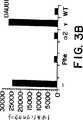
図3は、インターフェロンレセプター保有細胞に対するIFN-β(phe101)結合の分析を示す。パネルAおよびBは、それぞれDaudi細胞に対する125I-IFN-β(phe101)および野生型125I-IFN-βのレセプター結合データを示す。パネルCおよびDは、それぞれA549細胞に対する125I-IFN-β(phe101)および野生型125I-IFN-βのレセプター結合データを示す。
図4は、エンドプロテイナーゼLyse-CによるペプチドマッピングでのIFN-β(phe101)および野生型IFN-βの分析を示す。
図5は、野生型IFN-βに対する抗体がIFN-β(phe101)および野生型IFN-βの活性を中和することを示す。
発明の詳細な説明
本明細書中で用いたように、「野生型IFN-β」は、例えば、欧州特許第EP-B1-41313号、図4で示される天然のヒトIFN-βの通常存在するアミノ酸配列を有する天然または組換えのIFN-βを意味する。
本明細書中で用いたように、「IFN-β変異タンパク質」は、野生型IFN-βに従って番号付けした101位のval(V)がphe(F)、tyr(Y)、trp(W)、またはhis(H)、好ましくはphe(F)で置換されているポリペプチドを意味する。本発明者らの最も好ましいIFN-β変異タンパク質は、他の残基において野生型IFN-βと同一のアミノ酸配列を有する。しかし、本発明のIFN-β変異タンパク質はまた、天然のIFN-βポリペプチド鎖の他の残基の1またはそれ以上の部位におけるアミノ酸の挿入、欠失、置換、および改変により特徴付けられ得る。本発明に従って、任意のこのような挿入、欠失、置換、および改変は、野生型IFN-βに対する抗体により少なくとも部分的に中和され得る抗ウイルス活性を保持するIFN-β変異タンパク質を生じる。
本発明者らは、保存的改変および置換(すなわち、変異タンパク質の2次構造および3次構造に最小限度の影響を有するもの)を好む。このような保存的置換は、DayhoffによるAtlas of Protein Sequence and Structure5(1978),およびArgosによるEMBO J.,8, 779-785(1989)に記載されている置換を包含する。例えば、以下の群のいずれかに属するアミノ酸は保存的変化を示す:
「野生型IFN-βに従って番号付けした」により、本発明者らは、そのアミノ酸が野生型IFN-βにおいて天然に存在する位置に関して、選択アミノ酸を同定することを意味する。挿入または置換がIFN-β変異タンパク質になされる場合、当業者は野生型IFN-βに従って番号付けした場合、101位に通常存在するval(V)が変異タンパク質において位置がシフトし得ることを認める。しかし、シフトしたval(V)の位置は、隣接アミノ酸の検査および野生型IFN-βにおけるval101と隣接するアミノ酸との相関により容易に決定され得る。
本発明のIFN-β変異タンパク質は、当該分野で公知の適切な方法により生成される。このような方法は、本発明のIFN-β変異タンパク質をコードするDNA配列を構築する工程、および適切な形質転換宿主においてそれらの配列を発現させる工程を包含する。この方法は、本発明の組換え変異タンパク質を生成し得る。しかし、本発明の変異タンパク質はまた、あまり好ましくないにもかかわらず、化学合成、または化学合成と組換えDNA技術とを組み合わせることによっても生成され得る。
本発明の変異タンパク質を生成するための組換え方法の1実施態様において、DNA配列は野生型IFN-βをコードするDNA配列を単離または合成することにより構築され、次いで、部位特異的変異誘発によりval101のコドンをphe(F)、trp(W)、tyr(Y)、またはhis(H)、好ましくはphe(F)のコドンに変える。この技術は周知である。例えば、Markら、「ヒト線維芽細胞インターフェロン遺伝子の部位特異的変異誘発」、Proc.Natl.Acad.Sci.USA, 81, 5662-66頁(1984);米国特許第4,588,585号(本明細書で参考として引用されている)を参照のこと。
本発明のIFN-β変異タンパク質をコードするDNA配列を構築する別の方法は、化学合成である。例えば、所望のIFN-β変異タンパク質をコードする遺伝子がオリゴヌクレオチド合成機を使用する化学手段により合成され得る。このようなオリゴヌクレオチドは、所望のIFN-β変異タンパク質のアミノ酸配列に基づいて設計され、そして好ましくは組換え変異タンパク質が生成される宿主細胞において好まれるコドンが選択される。これに関しては、遺伝コードが縮重(アミノ酸が1以上のコドンによりコードされ得ること)することが十分に認識されている。例えば、phe(F)は、2つのコドンTTCまたはTTTによりコードされ、tyr(Y)はTACまたはTATによりコードされ、そしてhis(H)はCACまたはCATによりコードされる。trp(W)は単一のコドンTGGによりコードされる。従って、特定のIFN-β変異タンパク質をコードする所定のDNA配列については、そのIFN-β変異タンパク質をコードする多くのDNA縮重配列が存在し得ることが認められる。例えば、図2に示す好ましいDNA配列に加えて、図1に示すIFN-β変異タンパク質をコードする多くの縮重DNA配列が存在する。これらの縮重DNA配列は本発明の範囲内であると考えられる。
部位特異的変異誘発、合成、または他の方法により調製される本発明のIFN-β変異タンパク質をコードするDNA配列はまた、シグナル配列をコードするDNA配列を含み得るか、または含み得ない。このようなシグナル配列は、存在する場合、IFN-β変異タンパク質の発現のために選択された細胞により認識されるシグナル配列であるべきである。これは、原核性シグナル配列、真核性シグナル配列、またはこの2つの組み合わせであり得る。これはまた、天然のIFN-βのシグナル配列であり得る。シグナル配列の含有は、IFN-β変異タンパク質が作られる組換え細胞からのIFN-β変異タンパク質の分泌が望まれるか否かに依存する。選択細胞が原核細胞である場合、DNA配列がシグナル配列をコードしないことが一般に好ましい。選択細胞が真核細胞である場合、シグナル配列がコードされることが一般に好ましく、そして最も好ましくは野生型IFN-βシグナル配列が使用される。
標準的な方法が、本発明に従って、IFN-β変異タンパク質をコードする遺伝子を合成するために適用され得る。例えば、逆翻訳遺伝子を構築するために全アミノ酸配列が使用され得る。IFN-β変異タンパク質をコードするヌクレオチド配列を含むDNAオリゴマーが合成され得る。例えば、所望のポリペプチドの一部をコードするいくつかの小さなオリゴヌクレオチドが合成され得、次いで連結され得る。個々のオリゴヌクレオチドは、代表的には、相補的なアセンブリーのための5’または3’突出部(overhangs)を含有する。
一旦アセンブリーされると(合成、部位特異的変異誘発、または別の方法により)、本発明のIFN-β変異タンパク質をコードするDNA配列は、発現ベクターに挿入され、そして所望の形質転換宿主においてIFN-β変異タンパク質の発現に適切な発現調節配列に作動可能に連結される。適当なアセンブリーが、ヌクレオチド配列決定、制限酸素マッピング、および適切な宿主における生物学的に活性なポリペプチドの発現により確認され得る。当該分野で周知であるように、宿主においてトランスフェクトされた遺伝子の高レベルの発現を得るためには、この遺伝子は選択した発現宿主において機能する転写および翻訳発現調節配列に作動可能に連結されなければならない。
発現調節配列および発現ベクターの選択は、宿主の選択に依存する。広範囲の発現宿主/ベクター組み合わせが用いられ得る。真核宿主に有用な発現ベクターは、例えば、SV40、ウシパピローマウイルス、アデノウイルス、およびサイトメガロウイルス由来の発現調節配列を含むベクターを含有する。細菌宿主に有用な発現ベクターは、公知の細菌プラスミド(例えば、E.coliに由来するプラスミド、col E1、pCR1、pBR322、pMB9、およびそれらの誘導体を包含する)、より広い宿主範囲のプラスミド(例えば、RP4)、ファージDNA(例えば、λファージの数多くの誘導体(例えば、NM989))、および他のDNAファージ(例えば、M13および糸状単鎖DNAファージ)を包含する。酵母細胞に有用な発現ベクターは、2μプラスミドおよびその誘導体を包含する。昆虫細胞に有用なベクターは、pVL941を包含する。本発明者らはpBG311を好む。Gateら「ミューラー管阻害物質に関するウシおよびヒト遺伝子の単離および動物細胞におけるヒト遺伝子の発現」,Cell, 45, 685-98頁(1986)。
さらに、任意の多種多様な発現調節配列がこれらのベクターにおいて使用され得る。これらの有用な発現調節配列は、上記の発現ベクターの構造遺伝子と関連した発現調節配列を包含する。有用な発現調節配列の例は、例えば、以下を包含する:SV40またはアデノウイルスの初期および後期プロモーター、lacシステム、trpシステム、TACシステムまたはTRCシステム、λファージの主要なオペレーターおよびプロモーター領域(例えば、PL)、fdコートタンパク質の調節領域、3-ホスホグリセリン酸キナーゼまたは他の解糖酵素のプロモーター、酸性ホスファターゼのプロモーター(例えば、Pho5)、酵母α接合系のプロモーター、および原核または真核細胞またはそれらのウイルスの遺伝子の発現を調節することが公知の他の配列、ならびにそれらの種々の組み合わせ。
任意の適切な宿主が、本発明のIFN-β変異タンパク質の産生に使用され得る。これは、細菌、真菌(酵母を包含する)、植物、昆虫、哺乳動物、または他の適切な動物細胞または細胞株、ならびにトランスジェニック動物またはトランスジェニック植物を包含する。さらに詳細には、これらの宿主は、周知の真核宿主および原核宿主(例えば、E.coli、Pseudomonas、Bacillus、Streptomycesの系統)、真菌、酵母、昆虫細胞(例えば、SpodopteraFrugiperda(SF9))、動物細胞(例えば、チャイニーズハムスター卵巣(CHO)細胞およびNS/0のようなマウス細胞、COS1、COS7、BSC1、BSC40、およびBMT10のようなアフリカミドリザル細胞、およびヒト細胞、ならびに組織培養における植物細胞を包含する。動物細胞の発現について、本発明者らは培養中のCHO細胞およびCOS7細胞を好み、そして特にCHO-DDUKY-β1細胞系統(前出、18〜19頁)を好む。
無論のこと、全てのベクターおよび発現調節配列が本明細書に記載のDNA配列を発現させるために同等に良好に機能するわけではないことが理解されるべきである。全ての宿主が同じ発現系で同等に良好に機能するわけでもない。しかし、当業者は、これらのベクター、発現調節配列、および宿主の間で過度の実験なしに選択を行い得る。例えば、ベクターの選択において、宿主細胞は考慮されなければならない。なぜなら、ベクターはその宿主内で複製しなければならないからである。ベクターのコピー数、このコピー数を調節する能力、およびベクターによりコードされる任意の他のタンパク質(例えば、抗生物質マーカー)の発現もまた、考慮されるべきである。例えば、本発明で使用される好ましいベクターは、IFN-β変異タンパク質をコードするDNAをコピー数増幅させ得るベクターを包含する。このような増幅可能ベクターは、当該分野で周知である。これらは、例えば、DHFR増幅により増幅され得るベクター(例えば、Kaufman、米国特許第4,470,461号、KaufmanおよびSharp、「モジュラージヒドロ葉酸レダクターゼcDNA遺伝子の構築:効率的な発現に利用されるシグナルの解析」,Mol.Cell.Biol.,2, 1304-19頁,(1982)を参照のこと)またはグルタミンシンテターゼ(「GS」)増幅により増幅され得るベクター(例えば、米国特許第5,122,464号および欧州特許公開出願では第338,841号を参照のこと)を包含する。
発現調節配列の選択において、種々の因子がまた考慮されるべきである。これらは、例えば、配列の相対的強度(relative strength)、その制御性、および本発明のIFN-β変異タンパク質をコードする実際のDNA配列とのその適合性、特に潜在的な2次構造に関しての適合性を包含する。宿主は、それらの選択したベクターとの適合性、本発明のDNA配列によりコードされる産物の毒性、それらの分泌特性、ポリペプチドを正確に折り畳む能力、それらの発酵または培養の必要条件、およびDNA配列によりコードされた産物の精製の容易さを考慮して選択されるべきである。
これらのパラメーター内で、当業者は、発酵または大量動物細胞培養(例えば、CHO細胞またはCOS7細胞を使用する)において所望のDNA配列を発現する種々のベクター/発現調節配列/宿主の組み合わせを選択し得る。
本発明に従って得られたIFN-β変異タンパク質は、変異タンパク質を産生するために使用される宿主生物に依存して、グリコシル化され得るかまたはグリコシル化され得ない。細菌が宿主細胞として選ばれる場合、産生されるIFN-β変異タンパク質はグリコシル化されない。一方、真核細胞は、IFN-β変異タンパク質をグリコシル化するが、おそらくこれは天然のIFN-βがグリコシル化されるのと同じ方法にはよらない。
形質転換宿主により産生されたIFN-β変異タンパク質は、任意の適切な方法に従って精製され得る。INF-βの精製について種々の方法が公知である。例えば、米国特許第4,289,689号、同第4,359,389号、同第4,172,071号、同第4,551,271号、同第5,244,655号、同第4,485,017号、同第4,257,938号、および同第4,541,952号を参照のこと。本発明者らは、免疫アフィニティー精製を好む。例えば、Okamuraら、「ヒト線維芽細胞様(fibroblastoid)インターフェロン:免疫吸着カラムクロマトグラフィーおよびN末端アミノ酸配列」,Biochem., 19, 3831-35頁(1980)を参照のこと。
本発明のIFN-β変異タンパク質の生物学的活性は、当該分野で公知の任意の適切な方法によりアッセイされ得る。このようなアッセイは、欧州特許第EP-B1-41313号に記載のような抗ウイルス活性の抗体中和、プロテインキナーゼ活性の誘導、オリゴアデニル酸2,5-Aシンテターゼ活性の誘導、またはホスホジエステラーゼ活性の誘導を包含する。このようなアッセイはまた免疫調節アッセイ(例えば、米国特許第4,753,795号を参照のこと)、増殖阻害アッセイ、およびインターフェロンレセプターを発現する細胞への結合の測定を包含する。
本発明のIFN-β変異タンパク質は、野生型の天然IFN-βまたは組換えIFN-βが治療に用いられるのとほぼ匹敵する用量で投与される。IFN-β変異タンパク質の有効量が、好ましくは投与される。「有効量」は、処置される状態または適応症の重篤度または展開を防止または軽減し得る量を意味する。IFN-β変異タンパク質の有効量は、とりわけ、疾患、用量、IFN-β変異タンパク質の投与計画、IFN-βが単独で投与されるかまたは他の治療剤と併用されるか、組成物の血清中半減期、および患者の全般的な健康状態に依存することが、当業者には明らかである。
IFN-β変異タンパク質は、好ましくは薬学的に受容可能なキャリアを含む組成物中で投与される。「薬学的に受容可能なキャリア」は、それが投与される患者に適当でない効果を全く生じないキャリアを意味する。このような薬学的に受容可能なキャリアは、当該分野で周知である。本発明者らは、ヒト血清アルブミンを好む。
本発明のIFN-β変異タンパク質は周知の方法で薬学的組成物中に処方され得る。例えば、E.W.MartinによるRemington's Pharmaceutical Sciences(これは、本明細書中に参考として援用されている)は、適切な処方を記載しているので、参照のこと。IFN-β変異タンパク質の薬学的組成物は、液体、ゲル、凍結乾燥、または任意の他の適切な形態を包含する種々の形態で処方され得る。好ましい形態は、特定の処方される適応症に依存し、そして当業者には明らかである。
IFN-β変異タンパク質の薬学的組成物は、経口、静脈内、筋肉内、腹腔内、皮内、または皮下、または任意の他の受容可能な様式で投与され得る。投与の好ましい様式は、特定の処置される適応症に依存し、そして当業者には明らかである。
IFN-β変異タンパク質の薬学的組成物は、他の治療剤と併用して投与され得る。これらの薬剤は、同じ薬学的組成物の一部として混合され得るか、またはIFN-β変異タンパク質とは別々に、同時にまたは任意の他の受容可能な処置計画に従うかのいずれかで投与され得る。さらに、IFN-β変異タンパク質の薬学的組成物は、他の治療の補助として使用され得る。
従って、本発明は、ウイルス感染、ガンまたは腫瘍、異常な細胞増殖の処置のため、または任意の適切な動物、好ましくは哺乳動物、最も好ましくはヒトにおける免疫調節のための組成物および方法を提供する。
遺伝子治療適用における本発明のIFN-β変異タンパク質をコードするDNA配列の使用もまた意図されている。
意図される遺伝子治療適用は、IFN-βがその抗ウイルス活性により効果的な治療を提供することが期待される疾患(例えば、肝炎および特にHBVを包含するウイルス性疾患、またはIFN-βまたはIFN-βに感受性の感染性因子に応答する他の感染症)の処置を包含する。同様に、本発明は、免疫調節のための遺伝子治療適用、ならびにその抗増殖活性によりINF-βが効果的な治療を提供することが期待される疾患(例えば、腫瘍およびガン、または所望でない細胞増殖により特徴付けられる他の状態(例えば、再狭窄))の処置における適用を包含する。
遺伝子治療を使用するIFN-βの局所的な送達は、非特異的投与に伴う潜在毒性の問題を回避し、標的領域へ治療剤を提供し得る。
インビトロおよびインビボの遺伝子治療法の両方が意図される。
限定された細胞集団に潜在的な治療遺伝子を導入するためのいくつかの方法が公知である。例えば、Mulligan、「遺伝子治療の基本的科学」,Science, 260, 926-31頁(1993)を参照のこと。これらの方法は、以下を包含する:
1)直接的遺伝子導入。例えば、Wolffら、「インビボにおけるマウス筋肉への直接的遺伝子導入」,Science, 247, 1465-68頁(1990)を参照のこと;
2)リポソーム媒介DNA導入。例えば、Caplenら、「嚢胞性繊維症を患う患者の鼻上皮へのリポソーム媒介CFTR遺伝子導入」,Nature Med., 3, 39-46頁(1995);Crystal、「薬物としての遺伝子」,Nature Med., 1, 15-17頁(1995);GaoおよびHuang、「哺乳動物細胞の効率的なトランスフェクションのための新規のカチオン性リポソーム試薬」,Biochem. Biophys. Res. Comm., 179, 280-85頁(1991)を参照のこと;
3)レトロウイルス媒介DNA導入。例えば、Kayら、「B型血友病のインビボ遺伝子治療:第IX因子欠損のイヌにおける持続的な部分的矯正」,Science, 262, 117-19頁(1993);Anderson、「ヒトの遺伝子治療」,Science, 256, 808-13頁(1992)を参照のこと。
4)DNAウイルス媒介DNA導入。このようなDNAウイルスは、アデノウイルス(好ましくはAd-2またはAd-5に基づくベクター)、ヘルペスウイルス(好ましくは単純疱疹ウイルスに基づくベクター)、およびパルボウイルス(好ましくは「欠損」または非自律性パルボウイルスに基づくベクター、より好ましくはアデノ随伴ウイルスに基づくベクター、最も好ましくはAAV-2に基づくベクター)を包含する。例えば、Aliら、「遺伝子治療のためのベクターとしてのDNAウイルスの使用」,Gene Therapy, 1, 367-84頁(1994);米国特許第4,797,368号(これは、本明細書中に参考として援用されている)、および米国特許第5,139,941号(これは、本明細書中に参考として援用されている)を参照のこと。
目的の遺伝子を導入するための特定のベクター系の選択は、種々の因子に依存する。重要な因子の1つは、標的の細胞集団の性質である。レトロウイルスベクターが幅広く研究され、そして数多くの遺伝子治療の適用に使用されているが、これらのベクターは、一般に非分裂細胞の感染に適していない。さらに、レトロウイルスは、発ガン性の可能性を有する。
アデノウイルスは広範な宿主域を有するという利点があるが、静止期の細胞または最終分化細胞(例えば、ニューロンまたは肝細胞)に感染し得、そして本質的に非発ガン性であるようである。例えば、Aliら、前出、367頁を参照のこと。アデノウイルスは宿主ゲノムに組み込まれないようである。なぜなら、それらは染色体外に存在するため、挿入変異誘発の危険性は著しく減少している。Aliら、前出、373頁。
アデノ随伴ウイルスは、アデノウイルスに基づくベクターと類似の利点を示す。しかし、AAVは、ヒト第19染色体への部位特異的組込みを示す。Aliら、前出、377頁。
好ましい実施態様においては、本発明のIFN-β変異タンパク質コードDNAは、動脈損傷後の血管平滑筋細胞増殖の遺伝子治療に使用される。動脈壁の損傷により、平滑筋細胞の動脈壁内層への移動が生じ、ここで、平滑筋細胞は増殖し、そして細胞外基質成分を合成する。例えば、Changら、「構成的に活性な形態の網膜芽腫遺伝子産物を用いる血管増殖性疾患のための細胞増殖抑制性の遺伝子治療」,Science, 267, 518頁(1995)を参照のこと。この増殖反応は、アテローム性動脈硬化の病因に関与している。
動脈損傷の臨床上重要な硬化(setting)の1つは、冠状動脈の経皮バルーン血管形成のために起こる。動脈の機械的拡張後、多くの場合、細胞性増殖反応が生じ、細胞の再増殖を局所的に導き、これは内腔を侵し、そして血流を危険にさらす。この反応は、再狭窄として知られ、抗血小板剤、アンジオテンシン転換酵素アンタゴニスト、またはヒトにおける細胞毒性薬を包含する従来の処置には反応しなかった。例えば、Ohnoら、「動脈損傷後の血管平滑筋細胞の増殖のための遺伝子治療」,Science, 265, 781頁(1994)を参照のこと。
本実施態様によれば、本発明のIFN-β変異タンパク質をコードするDNAでの遺伝子治療は、冠状動脈バルーン血管形成と同時または直後において、それ自身を必要とする患者に提供される。この取り組みは、本発明のIFN-β変異タンパク質の抗増殖活性により所望でないSMC増殖を防止するという利点を有する。当業者は、INF-β変異タンパク質DNAを含有する任意の適切な遺伝子治療ベクターが本実施態様に従って使用され得ることを認める。このようなベクターを構築する技術は公知である。例えば、Ohnoら、前出、784頁;Changら、前出、522頁を参照のこと。冠状動脈バルーン血管形成の手順は周知である。IFN-β変異タンパク質DNA含有ベクターの標的動脈部位への導入は、例えば、Ohnoら、前出、784頁に記載のような公知の技術により達成され得る。
本発明がより理解されるように、以下の実施例を示す。これらの実施例は例示を目的とするだけであり、本発明の範囲が何らかの方法によって限定されるとは解釈されない。
実施例
ヒトIFN-β(phe101)を含有する発現ベクター
本発明者らは、発現ベクターとしてプラスミドpBG311を用いた。pBG311の完全な記載は、Cateら、「ミューラー管阻害物質に関するウシおよびヒト遺伝子の単離ならびに動物細胞におけるヒト遺伝子の発現」,Cell, 45, 685-98頁(1986)に示される。このベクターは、SV40初期プロモーター、スプライスシグナル、およびポリアデニル化シグナルを使用し、そして骨格としてpAT153を用いて構築された。
標準的なプロトコルに従い、図2に示されるDNA配列(配列番号2)を含有するDNAフラグメントをpBG311にクローン化し、そして天然のIFN-βのシグナル配列をコードするDNA配列を通してSV40初期プロモーターに作動可能に連結した。得られた発現ベクターをpBeta-pheと命名した。IFN-β変異タンパク質DNA配列(配列番号2)は、野生型IFN-βに従って番号付けした101位のval(V)が、phe(F)で置換されていることを除くと、野生型IFN-βと同一のアミノ酸配列を有するIFN-β変異タンパク質をコードする。この配列によりコードされる変異タンパク質は、IFN-β(phe101)と命名されている。
コンピテントなEscherichia coli(SURETM, Stratagene)を、標準的な手順に従いpBeta-pheプラスミドで形質転換した。pBeta-pheプラスミドを含有する(すなわちIFN-β(phe101)をコードするDNA配列を含有する)コロニーを、標準的なプロトコル(GrunsteinおよびHogness, 1975)を用いてIFN-β(phe101)に特異的なオリゴヌクレオチドプローブへのハイブリダイゼーションにより同定した。
増殖ベクター
本発明者らは、プラスミドpAdD26SV(A)-3を用いて、本発明者らの最終的な形質転換体においてIFN-β(phe101)遺伝子を増幅した。このプラスミドは、KaufmanおよびSharp、「モジュラージヒドロ葉酸レダクターゼcDNA遺伝子の構築:効率的な発現に利用されるシグナルの解析」、Mol. Cell. Biol., 2, 1304-19頁(1982)および米国特許第4,740,461号に記載されている。このプラスミドは、アデノウイルス2(Ad2)主要後期プロモーター(MLP)の転写調節下で、マウスのジヒドロ葉酸レダクターゼ(DHFR)を発現する。5’スプライス部位(免疫グロブリン可変領域遺伝子由来)は、Ad2 MLPとDHFRコード配列との間に位置する。SV40ポリアデニル化部位は、DHFR遺伝子の下流に存在する。プラスミドは、原核生物の複製の起点(ori)およびpBR322由来のテトラサイクリン耐性遺伝子を含有する。
細胞株の形質転換
CHO-DUKX-B1 DHFR-細胞株を、pBeta-pheプラスミドおよびプラスミドpAdD26SV(a)-3で共形質転換した。この細胞株は、エチルメタンスルホネートおよびUV照射誘導変異誘発による野生型CHO-K1細胞株由来であった。ChasinおよびUrlaub「ジヒドロ葉酸レダクターゼ活性欠損チャイニーズハムスター細胞変異株の単離」、Proc.Natl.Acad.Sci.USA、77, 4216-20頁(1980)を参照のこと。ジヒドロ葉酸レダクターゼは、葉酸のテトラヒドロ葉酸への転換を触媒する。機能的なDHFRを有しない細胞は、増殖のために外因性のリボヌクレオシドおよびデオキシリボヌクレオシドを必要とする。増殖の阻害は、DHFRに結合し、そしてDHFRを阻害する、メトトレキセート、葉酸アナログにより誘導され得る。メトトレキセートの滴定は、DHFR遺伝子の増幅によるメトトレキセート耐性を導き得る。(KaufmanおよびSharp, 1982,前出)。DHFRが増幅される場合、DHFRの近隣の遺伝子の増幅および発現の増加がしばしば生じる。それ故、高レベルのメトトレキセートに耐性の細胞は、しばしば近隣遺伝子の増加した特異的な生産性を示す。
pBeta-pheプラスミド(Xmn1で制限した)およびプラスミドpAdD26SV(a)-3(Stulで制限した)を、それぞれ10:1の比で混合した。DNAを、エレクトロポレーションによりCHO-DUKX-B1 DHFR-細胞に形質転換した。細胞を、非選択性α+培地(αMEM塩基+リボヌクレオシドおよびデオキシリボヌクレオシド、10%ウシ胎児血清(FBS)、4mMグルタミン)にプレートし、そして2日間増殖させた。次いで培地をα-培地(リボヌクレオシドおよびデオキシリボヌクレオシドを含有しないαMEM塩基、10%FBS、4mMグルタミン)+50nMメトトレキセート(MTX)に交換した。細胞をトリプシン処理により取り出し、そして約8×10⁵細胞/10cm組織培養プレートに播いた。14日後、クローンを選び取り、そして96ウェル組織培養プレートで増殖させた。1つのクローンを、12ウェル組織培養プレートに拡大し、次いで7日後に250nM MTXの存在下で6ウェル組織培養プレートに置いた。このクローンをT75フラスコに拡大し(α-培地+250nM MTXで成育させた)、次いで750nM MTXで増幅した。サブクローンを、96ウェル組織培養プレートに選び取り、48ウェル組織培養プレートに拡大し、次いで6ウェル組織培養プレートに拡大し、次いでT75組織培養フラスコに拡大した。
IFN-β(phe101)の精製
上記のサブクローン(またはそれに類似した他のもの)を培養することにより産生され、次いで培養培地に分泌されたIFN-β(phe101)を、実質的にOkamuraら、「ヒト線維芽細胞様インターフェロン:免疫吸着カラムクロマトグラフィーおよびN末端のアミノ酸配列」、Biochem., 19, 3831-35頁(1980)に記載されるような免疫アフィニティークロマトグラフィーにより精製し得る。
CNBr-Sepharose 4B樹脂(2g、7ml)を、1mM HClに懸濁することにより調製する。ゲルを、15分間焼結(scintered)ガラスフィルター上で1mM HClで洗浄する。抗IFN-β mabs(例えば、B02、Yamasa、Japan)を、2時間室温にて撹拌装置上で結合緩衝液(100mM NaHCO3、pH8.3、500mM NaCl)中にインキュベートすることにより、CNBr-Sepharose 4B樹脂に結合させる。代表的には、樹脂の1mlあたり1〜2mg IFN-β mabが結合されるが、この量は、変動し得る。未反応のCNBrを4℃、一晩、100mM Tris-HCl、pH8、500mM NaClでブロックする。あるいは、未反応のCNBrを、実質的に同一の条件下で100mMエタノールアミンでブロックする。
結合樹脂を、異なるpHの3つのサイクルで洗浄する。それぞれのサイクルは、500mM NaClを含有するアセテート緩衝液(100mM、pH4)での洗浄、それに続く500mM NaClを含有するTris緩衝液(100mM、pH8)での洗浄からなる。
1cm×3cmカラム(2.3ml総容積)を、結合樹脂で調製する。カラムをPBS(5カラム容量よりも大きい)で平衡化する。IFN-β(phe101)含有サンプルを、平衡化緩衝液、pH6.8中に1:3で希釈し、そしてロードする。このロードをPBSで追跡し、そして20mM K2HPO4、1M NaCl、pH6.8で洗浄し、そして200mMクエン酸ナトリウム、pH2で溶出する。500mM Mes、pH6でこのサンプルを希釈することにより、溶出液のpHを6に調整した。
ペプチドマッピングによる特徴付け
上記と異なり、かつより好ましくない様式で生産および精製されたIFN-β(phe101)、変異タンパク質をペプチドマッピングにより特徴付けた。IFN-β(phe101)または野生型IFN-βサンプルの30μgアリコートを凍結乾燥し、200μlのエンドプロテイナーゼLys-C消化緩衝液(100mM TRIS、pH9、0.5mM EDTA)に懸濁し、1.5μgのエンドプロテイナーゼLys-Cとともに12時間22℃にてインキュベートし、そしてC8逆相HPLCカラム(0.45×25cm)上でマッピング分析を行った。カラムを、30分間、1.4mls/分の0.1%TFA中のアセトニトリルの0〜70%グラジエントで展開した。カラム流出液を214nmでモニターした。図4、パネルAは、IFN-β(phe101)に関するペプチドマップの一部を示す。矢尻は、ペプチドTFLEEK(配列番号3)を示す。このピークは、野生型IFN-βに関するペプチドマップ中には、存在しない。図4、パネルBは、野生型IFN-βに関するペプチドマップの相当する領域を示す。矢尻は、ペプチドTVLEEK(配列番号4)を示す。TFLEEKおよびTVLEEKの一致性を、タンパク質配列分析により確認した。本発明者らは、β-Phe101および野生型β-IFNが、98%の純度よりも高いことを見積もる。タンパク質濃度を、1mg溶液に対して1.5の吸光係数を用いて、280nmでの吸光度から見積もった。生物学的研究のためにタンパク質を安定化するために、それらを5%FBSおよび5mM HEPES、を含有するPBS pH7.5中に4μg/mlにまで希釈した。
CPEアッセイにおけるIFN-β(phe101)の抗ウイルス活性
ペプチドマッピングにより特徴付けたIFN-β(phe101)の調製物を、抗ウイルス活性について細胞変性効果(Cytopathic Effect)(CPE)アッセイにおいて分析した。野生型組換えIFN-βスタンダードを、10,000ユニット/mLの濃度でDulbeccoの改変Eagle培地(DMEM)、10% FBS、4mMグルタミン中に調製し、-70℃にて一定分量で保存した。1日目、スタンダード、コントロール、およびIFN-βPheサンプルを、3つの希釈系列でDMEM、10% FBS、4mMグルタミンに希釈した:
i)64単位/mLで開始し、続いて2倍に希釈する、ii)12単位/mLで開始し、続いて1.5倍に希釈する、およびiii)6単位/mLで開始し、続いて1.2倍に希釈する。次いで希釈物の50μlを、96ウェルマイクロタイタープレートのウェルに対するカラム中に添加した。A549細胞を、105細胞/mlで、ウェルあたり50μLの、DMEM、10%FBS、4mMグルタミンのそれぞれのウェルに添加した。そして細胞を、5%CO2下で15〜20時間37℃にてインキュベートする。
プレート内容物を漂白バケット(bleach bucket)内で振盪し、そして培地中の適切な希釈度の100μL脳心筋炎ウイルス(EMCウイルス)をそれぞれのウェルに添加した。ウイルスおよび細胞を5%CO2下で30時間37℃にてインキュベートした。次いでプレート内容物を漂白バケット内で振盪し、そして0.75%クリスタルバイオレット色素をプレートに添加した。5〜10分後、プレートを蒸留水で洗浄し、そして乾燥させ、視覚的に示した。
サンプルおよびスタンダードを、それぞれのアッセイプレート上で2点平行で試験した。これによりアッセイの日あたり希釈系列あたり2つのデータ点が得られる。
IFN-β(phe101)を、2点平行で14アッセイで試験した。野生型組換えIFN-βをスタンダードとして用いた。これらの実験に基づき、IFN-β(phe101)は、3.5〜6.7×10⁸の95%信頼区間で4.8×10⁸単位/mgの比活性を有した。野生型IFN-βは、1.6〜2.5×108の信頼区間で約2.0×108単位/mgの比活性を有した。図5のデータは、同様の結果を示す。
本発明者らの抗ウイルスアッセイにおいて計測されたように、組換えIFN-β(phe101)の比活性は、組換え野生型IFN-βの比活性よりも平均して約2.5倍高い。
レセプター結合に関するIFN-β(phe101)の分析
上記のCPEアッセイで用いたIFN-β(phe101)をまた、インターフェロンレセプターを発現する細胞に結合する能力についても分析した。これらの研究のために、本発明者らは、Daudi細胞またはA549細胞への野生型125I-IFN-βまたは125I-IFN-β(phe101)のいずれかの結合を調査した(図3)。キャリア非含有IFN-βを、実質的にクロラミンT方法に従ってヨウ素化した。未反応のヨウ素を、1mg/mlウシ血清アルブミンを含有するPBSで平衡化したSuperdex 75カラムでサイズ排除クロマトグラフィーにより除去した。ヨウ素化IFN-βの濃度を、CPEアッセイにより測定し、これは2×10⁸単位/mgの比活性と見なした。通常5ng(1μL、300,000cpm)のヨウ素化IFN-β(単独または50倍過剰量の非ヨウ素化インターフェロンの存在下のいずれか)を10μLよりも少ない総容積で1.7mLエッペンドルフチューブに添加した。標識したリガンドを、単独(−)で結合させるか、または非標識IFN-β(phe101)、α2-IFN(α2)、γ-IFN(γ)または野生型組換えIFN-β(WT)と競合させた。
Daudi細胞およびA549細胞(American Type Culture Collection)の両方を用いた。細胞を2×10⁶細胞/mLでDMEM/5%FBS中に懸濁した。IFN-βのサンプルに、0.5mLの細胞懸濁物を添加した。チューブを反転により混合し、45分間外界温度でインキュベートした。次いで細胞を、2分間1000×gでペレット化し、0.5mL DMEM/10%FBSで2回洗浄した。それぞれの洗浄に続いて2分間1000×gの遠心分離の工程を行った。細胞を0.1mLに再懸濁し、計測のためにチューブに移し、そして結合をBeckmanγ407カウンターで定量した。
このデータは、IFN-β(phe101)の結合が、両方の細胞型における野生型IFN-βの結合と非常に類似していることを示唆する。野生型125I-IFN-βおよび125I-IFN-β(phe101)の匹敵する量が結合され、そして非ヨウ素化α-IFN、野生型IFN-β、およびI-IFN-β(phe101)により同様に競合された。この結合は、組換えヒトγ-IFNの添加により影響されなかった。
IFN-β(phe101)の抗ウイルス活性は、野生型IFN-βに対する抗体により実質的に中和される。
上記のようにCHO細胞中に産生された多数の組換え変異タンパク質IFN-β(phe101)の調製物を、カラムクロマトグラフィーを用いて約90%の純度に精製した。これらのサンプルを25pg/mlに希釈した。野生型組換えIFN-β(本明細書では、IFN-β(val101)と称される)を、実質的に同一の方法で生産および精製した。
スタンダード抗ウイルスアッセイおよび抗体中和アッセイを用いて、野生型IFN-β(val101)に対する抗体は、IFN-β(phe101)を少なくとも部分的に中和することを実証した。本発明者らが用いた特定の抗ウイルスアッセイおよび中和アッセイ(以下に詳細に示す)は、欧州特許第EP B1 41313号に記載されている抗ウイルスアッセイおよび抗体中和アッセイと実質的に同一である。26頁、1-21行;29頁、48行〜32頁、3行を参照のこと。
A.細胞含有プレートの調製
A549細胞(ATCC CCL185)を、3×10⁴細胞/100μl培地/ウェルで96ウェルプレートに播種した。用いた培地は、Dulbeccoの改変Eagle培地(DMEM)、10%FBS、4mMグルタミンであった。次いで細胞含有プレートを5%CO2下で約24時間37℃でインキュベートした。
B.マスタープレートの調製
次いで系列希釈したウェル(2点平行)を用いてサンプルまたはスタンダードを含有するマスタープレートを作製した。サンプルウェルは、ウサギ抗IFN-βポリクローナル血清の存在下または非存在下で、精製組換え変異タンパク質IFN-β(phe101)または野生型組換えIFN-β(val101)のいずれかを含有した。コントロールウェルは、抗LFA3抗体の存在下または非存在下で緩衝液単独または組換え野生型IFN-β(スタンダードとして)のいずれかを含有した(データは示さず)。
系列希釈を以下のようにして実施した。200μlのコントロール、あるいは野生型または変異タンパク質IFN-βサンプル(約25pg/mlの濃度)を、それぞれのプレートのA列のそれぞれのウェルに添加した。A列のサンプルの最終濃度は、8.5pg/mlであった。次いで1:1.5の希釈を、それぞれのプレートに実施した。
C.野生型IFN-βに対する対抗
野生型IFN-β(val101)に対する抗体を、組換え野生型IFN-β(val101)で免疫したウサギ中に産生させた。ウサギ抗IFN-βポリクローナル血清を、適切な間隔で免疫ウサギから回収し、プールし、そして使用するまで保存した(ウサギIFN-β血清プール6/25/93;5ml/バイアル、0.02%アジド;ref. 0.01742.062)。
D.サンプル/Abインキュベーション
ウサギ抗IFN-βポリクローナル血清を、マスタープレートの適切なウェルに添加した。抗体の最終希釈度は、1:1000であった。抗体/IFN-β混合物を45分間室温でインキュベートした。
E.コントロール、サンプル、またはサンプル/Abとの細胞のインキュベーション
次いで培地を調製された細胞含有プレートから吸引し、そして調製されたマスタープレート由来の、適切な、コントロール、IFN-βサンプル、またはIFN-β/Abサンプルのアリコート(100μl/ウェル)に取り替えた。細胞含有プレートを、5%CO2下で16〜24時間37℃にてインキュベートした。
F.ウイルスのチャレンジ
次の工程は、ウイルスのチャレンジであった。次いで細胞含有プレート内容物を吸引し、そして適切な希釈度の脳心筋炎ウイルス(EMCV)の溶液の100μlをそれぞれの細胞に添加した。ウイルスおよび細胞を5%CO2下で41〜45時間37℃にてインキュベートした。
細胞含有プレートをXTT/PMS比色定量法を用いて展開した。1mg/ml XTT(3,3-[1-(フェニルアミノ)カルボニル]-3,4-テトラアゾリウム]-ビス-(4-メトキシ-6-ニトロ)-ベンゼンスルホン酸;Sigma)溶液を、リン酸緩衝生理食塩水中に調製した。1mg/ml PMS(フェナジンメトサルフェート)溶液を水中に調製した。PMS/XTT溶液を1:50で調製した。展開溶液を、リン酸緩衝生理食塩水中にPMS/XTTを1:3で希釈することにより調製した。テトラゾリウム化合物XTTは、生細胞により還元され、オレンジ色のホルマゾンを形成する。発色は、生細胞数に直接相関する。
細胞含有プレートを吸引し、そして150μl/のウェルのリン酸緩衝生理食塩水で洗浄した。次いでそれぞれのウェルに150μlの展開溶液を与える。プレートを5%CO2下で30〜60分間37℃にてインキュベートした。
450nmの吸光度をSoftmaxソフトウェアを有するMolecular Devices Thermomaxマイクロプレートリーダーで測定した。結果を、図5にグラフで示す。吸光度をIFN-β濃度に対してプロットする。
図5は、ウサギ抗IFN-βポリクローナル血清の非存在下における、変異タンパク質IFN-β(phe101)サンプル(黒四角;■)および野生型IFN-β(val101)のサンプル(黒菱形;◆)が、EMCVからA549細胞を保護したことを示す。これは、変異タンパク質IFN-βまたは野生型IFN-β濃度を増加につれて吸光度が増加すること(より多い生細胞を示す)により示される。
図5はまた、ウサギ抗IFN-βポリクローナル血清の存在下における、変異タンパク質IFN-β(phe101)のサンプル(白四角;□)および野生型IFN-β(val101)のサンプル(白菱形;◇)が、EMCVからA549細胞を保護しなかったことを示す。これは、変異タンパク質IFN-βまたは野生型IFN-β濃度のいずれかに関する基線の吸光度の値により示され、ほとんど全てのA549細胞が死滅したことを示す。
まとめると、図5は、変異タンパク質IFN-β(phe101)の抗ウイルス活性が、野生型IFN-βに対する抗体(すなわち、ウサギ抗IFN-β抗体)により中和されたことを示す。
IFN-β変異タンパク質遺伝子治療での再狭窄の処置
再狭窄のための遺伝子治療の初期の試験は、以下のプロトコルに従って、組換え野生型ブタIFN-βを用いるブタモデルにおいて行われる。
細胞増殖は、免疫組織化学により測定する。全ての動物に、屠殺の1時間前に、25mg/kg総用量のBrdC(Sigma, St. Louis, MO)の静脈注入を与える。BrdCに対するモノクローナル抗体(1:1000希釈、Amersham Life Sciences, Arlington Heights, IL)を用いる免疫組織化学を、Goncharoffら、J.Immunol.Methods、93、97頁(1988)に記載のように実施し、増殖細胞中の核を標識する。血管平滑筋細胞の同定を、Islkら、Am.J.Pathol., 141, 1139頁(1992)に記載のように、平滑筋α-アクチンに対する抗体(1:500希釈、Boehringer Mannheim, Germany)を用いる免疫組織化学により実施する。
家畜のヨークシャーブタ(12〜15kg)を、1%亜酸化窒素を用いて(2.2mg/kg筋肉内)ロンプンと組み合わせてゾラゼパミン-チレタミン(6.0mg/kg)で麻酔する。腸骨大腿骨動脈を、無菌的外科手順により曝露し、そして2重のバルーンカテーテルを、Nabelら、Science, 249, 1285頁(1990)に記載のように腸骨大腿骨動脈内に挿入する。基部のバルーンを、5分間、オンライン変圧器(pressure transducer)により測定されるように、300mm Hgにまで膨張させる。バルーンを収縮させ、そして基部のバルーンと遠部のバルーンとの間の中央空間が、以前のバルーン損傷の領域を占めるように、カテーテルを進行させる。両方のバルーンを膨張させ、そしてセグメントをヘパリン処理生理食塩水で洗浄する。アデノウイルスの種菌を、カテーテルの中央空間内に20分間滴注する。カテーテルを除去し、そして回復するように血流を順行させる。
全てのブタの損傷した動脈に、ブタIFN-βをコードする挿入物を含有するADV-ΔE1ベクター、または挿入物を欠失するADV-ΔE1ベクターの1mlあたり1010プラーク形成単位(PFU)を感染させる。それぞれの動物において、両方の腸骨大腿骨動脈を、1×10¹⁰PFU/mlの力価の同一のベクターでトランスフェクトし、0.7mlをそれぞれの動物に用いた(7×109PFUの最終容量)。
これらのブタの血管セグメントを、21または42日後に摘出する。それぞれの動脈を同一の様式で処理する。2つの2重バルーン間の滴注の領域を、同一サイズの5つの横断面にカットする。セクション1および4を、メチルカルノアで固定し、そしてセクション3および5をホルマリンで固定し、そして全てのセクションをパラフィン包埋し、そしてヘマトキシリンエオシンで染色する。さらなる抗体研究を、メチルカルノア固定した動脈またはホルマリン固定した動脈において実施する。セクション2由来の組織を液体窒素内で瞬間凍結し、そしてDNA単離のために-80℃にて保存する。内膜および内側の領域の大きさを、2名の異なる読みとり者により盲検的にそれぞれの動脈由来の4つのセクションにおいて測定し、それぞれの血管についての大きさを平均化する。動脈の検体のスライドを、Nabelら、Proc.Natl.Acad.Sci U.S.A, 90, 10759頁(1993)に記載のように顕微鏡に基づくビデオ画像診断分析システム(Image-1システム、Universal Imaging, Weschester, PA)で研究する。
上記のアデノウイルスに基づくAd-ΔE1ベクターに代わるものとして、直接的な遺伝子導入もまた用いられ得る。1つの適切な構築物は、IFN-βDNA配列に対して3’側にSV-40ポリA’シグナルを有する、RSV骨格(IFN-βの発現を導くRSV-Lプロモーターを有する)由来のプラスミドである。例えば、Gormanら、Science, 221, 551-53頁(1983)を参照のこと。
次いでベクターが、ヒトにおける遺伝子治療のために本発明のヒトIFN-β変異タンパク質をコードするDNA挿入物を含有するように、上記のプロトコルを改変する。
配列
以下は、配列リストに示される配列の要旨である:
配列番号1−−IFN-β(phe101)のアミノ酸配列
配列番号2−−天然のIFN-βのシグナル配列をコードする配列を含有する、IFN-β(phe101)をコードするDNA配列
配列番号3−−ペプチドTFLEEKのアミノ酸配列
配列番号4−−ペプチドTVLEEKのアミノ酸配列
寄託
プラスミドpBeta-phe(これは、IFN-β(phe101)および天然のIFN-βのシグナル配列をコードするDNA配列を含有する)を含有するE.coli K-12は、寄託されている。寄託は、ブダペスト条約に従ってなされ、そして1994年3月11日にAmerican Type Culture Collection, Rockville, Maryland, U.S.Aに寄託された。寄託物は、受託番号69584を受けた。
上記の記載は、説明および記述の目的のためのみに示されている。この記載は、開示された厳密な形態に本発明を限定することを意図しない。本発明の範囲は、本明細書に添付された請求の範囲によって定義されることを意図する。
配列表
(1)一般的情報:
(i)出願人:バイオジェン,インコーポレイテッドゴールズ,スーザン イーケイト,リチャード エルペピンスキー,ブレイク アールチャウ,ピンチャン イー
(ii)発明の名称:IFN-βの新規変異タンパク質
(iii)配列数:5
(iv)連絡住所:
(A)名称:ジェイムス エフ.ハーレー,ジュニア
(B)番地:フィッシュ アンド ニーブ,アベニュー オブ ザ アメリカズ1251
(C)市:ニューヨーク
(D)州:ニューヨーク
(E)国:アメリカ合衆国
(F)郵便番号:10020-1104
(v)コンピューター読み出し形態:
(A)媒体型:フロッピーディスク
(B)コンピューター:IBM PC互換用
(C)OS:PC-DOS/MS-DOS
(D)ソフトウェア:パテントイン リリース #1.0,バージョン #1.25
(vi)現在の出願データ:
(A)出願番号:
(B)出願日:
(C)分類:
(viii)代理人/事務所情報:
(A)氏名:ハーレー,ジュニア,ジェイムス エフ
(B)登録番号:27,794
(C)照会/記録番号:B179
(ix)電話回線情報:
(A)電話:(212)596-9000
(B)テレファックス:(212)596-9090
(2)配列番号1の情報:
(i)配列の特色:
(A)長さ:166アミノ酸
(B)型:アミノ酸
(C)鎖の数:一本鎖
(D)トポロジー:直鎖状
(ii)配列の種類:タンパク質
(iii)ハイポセティカル:NO
(iv)アンチセンス:NO
(xi)配列:配列番号1:
(i)配列の特色:
(A)長さ:561塩基対
(B)型:核酸
(C)鎖の数:一本鎖
(D)トポロジー:直鎖状
(ii)配列の種類:cDNA
(iii)ハイポセティカル:NO
(iv)アンチセンス:NO
(ix)配列の特徴:
(A)特徴を表す記号:sig_peptide
(B)存在位置:1..63
(ix)配列の特徴:
(A)特徴を表す記号:mat_peptide
(B)存在位置:64..561
(ix)配列の特徴:
(A)特徴を表す記号:CDS
(B)存在位置:1..561
(xi)配列:配列番号2:
(i)配列の特色:
(A)長さ:187アミノ酸
(B)型:アミノ酸
(D)トポロジー:直鎖状
(ii)配列の種類:タンパク質
(xi)配列:配列番号3:
(i)配列の特色:
(A)長さ:6アミノ酸
(B)型:アミノ酸
(C)鎖の数:一本鎖
(D)トポロジー:直鎖状
(ii)配列の種類:ペプチド
(iii)ハイポセティカル:NO
(iv)アンチセンス:NO
(xi)配列:配列番号4:
(i)配列の特色:
(A)長さ:6アミノ酸
(B)型:アミノ酸
(C)鎖の数:一本鎖
(D)トポロジー:直鎖状
(ii)配列の種類:ペプチド
(iii)ハイポセティカル:NO
(iv)アンチセンス:NO
(xi)配列:配列番号5:
The present invention relates to an interferon wherein val (V) at
Background of the Invention
Interferons are single chain polypeptides that are secreted by most animal cells in response to various inducers, including viruses, mitogens, and polynucleotides. Interferons are involved in the regulation of cellular function and have antiviral, antiproliferative, and immunomodulatory properties. Natural human interferons are classified into three main types: α-IFN (leukocytes), IFN-β (fibroblasts) and γ-IFN (immune). Natural IFN-β is mainly produced by diploid fibroblasts and less is produced by lymphoblasts.
IFN-β is a glycoprotein. The nucleic acid and amino acid sequences have been determined (Houghton et al., “Complete amino acid sequence of human fibroblast interferon deduced using synthetic oligodeoxyribonucleotide primers for reverse transcriptase”,Nucleic Acids Research, 8, 2885-94 (1980); T. Taniguchi et al., "Nucleic acid sequence of human fibroblast DNA",Gene, 10, 11-15 (1980)). Recombinant IFN-β has been produced and characterized.
IFN-β exhibits a variety of biological and immunological activities. One of the biological activities of IFN-β is antiviral activity. This antiviral activity can be neutralized by antibodies against IFN-β. See European Patent EP-B1-41313. Preparation and purification of antibodies against IFN-β are described in EP-B1-41313 and references cited therein. IFN-β can also bind to cells that express the interferon receptor (eg, Daudi cells or A549 cells).
As a result of these activities, IFN-β has potential applications in immunotherapy, antitumor and anticancer therapies, and antiviral therapies.
Numerous studies and clinical trials have been conducted against the anti-tumor and anti-cancer properties of both wild-type and recombinant IFN-β and are ongoing. These include the treatment of several malignancies such as osteosarcoma, basal cell carcinoma, cervical dysplasia, glioma, acute myeloid leukemia, multiple myeloma, and Hodgkin disease. Furthermore, IFN-β has been shown to cause local tumor regression when injected into subcutaneous tumor nodes in patients with melanoma and breast cancer.
IFN-β (wild-type and recombinant) is associated with various viral infections including papillomavirus (eg, genital warts and cervical condyloma; acute / chronic hepatitis B and non-A non-B hepatitis (hepatitis C) ), And genital herpes zoster; herpes zoster; herpes zoster; viral encephalitis; cytomegalovirus pneumonia); and rhinovirus prophylaxis. Clinical trials using recombinant IFN-β in the treatment of multiple sclerosis have also been conducted and IFN-β is approved for sale in the United States for the treatment of multiple sclerosis.
Summary of the Invention
The present invention relates to IFN-β wherein the val (V) at
[Brief description of the drawings]
FIG. 1 shows the preferred mutant protein IFN-β (phe101) Amino acid sequence (SEQ ID NO: 1).
Figure 2 shows IFN-β (phe101The preferred degenerate DNA sequence encoding) and the signal sequence of native IFN-β (SEQ ID NO: 2).
FIG. 3 shows IFN-β (phe101) Shows analysis of binding. Panels A and B are for Daudi cells, respectively.125I-IFN-β (phe101) And wild type125The receptor binding data of I-IFN-β are shown. Panels C and D are for A549 cells, respectively.125I-IFN-β (phe101) And wild type125The receptor binding data of I-IFN-β are shown.
FIG. 4 shows IFN-β (phe) in peptide mapping with endoproteinase Lyse-C.101) And wild type IFN-β analysis.
FIG. 5 shows that an antibody against wild-type IFN-β is IFN-β (phe101) And neutralize the activity of wild-type IFN-β.
Detailed Description of the Invention
As used herein, “wild-type IFN-β” has the normally present amino acid sequence of natural human IFN-β as shown, for example, in European Patent No. EP-B1-41313, FIG. Means natural or recombinant IFN-β.
As used herein, an `` IFN-β mutein '' is a val (V) at
We prefer conservative modifications and substitutions (ie, those that have a minimal effect on the secondary and tertiary structure of the mutant protein). Such conservative substitutions are due to Dayhoff.Atlas of Protein Sequence and StructureFive(1978), and by ArgosEMBO J.,8, 779-785 (1989). For example, amino acids belonging to any of the following groups exhibit conservative changes:
By “numbered according to wild-type IFN-β” we mean identifying the selected amino acid with respect to the position where the amino acid is naturally present in wild-type IFN-β. When insertions or substitutions are made in the IFN-β mutant protein, one skilled in the art will recognize that the val (V) normally present at
The IFN-β muteins of the present invention are produced by appropriate methods known in the art. Such methods include the steps of constructing DNA sequences encoding the IFN-β muteins of the present invention and expressing those sequences in a suitable transformed host. This method can produce the recombinant muteins of the present invention. However, the mutant proteins of the present invention can also be produced by chemical synthesis, or a combination of chemical synthesis and recombinant DNA technology, although less preferred.
In one embodiment of the recombinant method for producing the mutant protein of the present invention, the DNA sequence is constructed by isolating or synthesizing a DNA sequence encoding wild type IFN-β, and then site-directed mutagenesis. By val101Is changed to a codon of phe (F), trp (W), tyr (Y), or his (H), preferably phe (F). This technique is well known. For example, Mark et al., “Site-directed mutagenesis of the human fibroblast interferon gene”,Proc.Natl.Acad.Sci.USA81, 5662-66 (1984); U.S. Pat. No. 4,588,585 (incorporated herein by reference).
Another method of constructing a DNA sequence encoding an IFN-β mutein of the present invention is chemical synthesis. For example, the gene encoding the desired IFN-β mutein can be synthesized by chemical means using an oligonucleotide synthesizer. Such oligonucleotides are designed based on the amino acid sequence of the desired IFN-β mutein and are preferably selected for codons preferred in the host cell in which the recombinant mutein is produced. In this regard, it is well recognized that the genetic code is degenerate (amino acids can be encoded by one or more codons). For example, phe (F) is encoded by two codons TTC or TTT, tyr (Y) is encoded by TAC or TAT, and his (H) is encoded by CAC or CAT. trp (W) is encoded by a single codon TGG. Thus, for a given DNA sequence encoding a particular IFN-β mutein, it will be appreciated that there can be many DNA degenerate sequences encoding that IFN-β mutein. For example, in addition to the preferred DNA sequence shown in FIG. 2, there are many degenerate DNA sequences that encode the IFN-β muteins shown in FIG. These degenerate DNA sequences are considered within the scope of the present invention.
A DNA sequence encoding an IFN-β mutein of the invention prepared by site-directed mutagenesis, synthesis, or other methods may also include or not include a DNA sequence encoding a signal sequence. Such a signal sequence, if present, should be a signal sequence that is recognized by the cells selected for expression of the IFN-β mutein. This can be a prokaryotic signal sequence, a eukaryotic signal sequence, or a combination of the two. This can also be the signal sequence of native IFN-β. The inclusion of the signal sequence depends on whether it is desired to secrete the IFN-β mutein from a recombinant cell from which the IFN-β mutein is made. If the selected cell is a prokaryotic cell, it is generally preferred that the DNA sequence does not encode a signal sequence. If the selected cell is a eukaryotic cell, it is generally preferred that a signal sequence is encoded, and most preferably a wild type IFN-β signal sequence is used.
Standard methods can be applied to synthesize a gene encoding an IFN-β mutein according to the present invention. For example, the entire amino acid sequence can be used to construct a back-translated gene. A DNA oligomer comprising a nucleotide sequence encoding an IFN-β mutein can be synthesized. For example, several small oligonucleotides that encode portions of the desired polypeptide can be synthesized and then ligated. Individual oligonucleotides typically contain 5 'or 3' overhangs for complementary assembly.
Once assembled (by synthesis, site-directed mutagenesis, or otherwise), the DNA sequence encoding the IFN-β mutein of the invention is inserted into an expression vector and IFN in the desired transformed host. -operably linked to expression control sequences suitable for expression of the β-mutated protein. Appropriate assembly can be confirmed by nucleotide sequencing, restriction oxygen mapping, and expression of the biologically active polypeptide in a suitable host. As is well known in the art, in order to obtain high level expression of a transfected gene in a host, this gene must be operably linked to transcriptional and translational expression control sequences that function in the chosen expression host. I must.
The choice of expression control sequence and expression vector will depend on the choice of host. A wide range of expression host / vector combinations can be used. Useful expression vectors for eukaryotic hosts include, for example, vectors containing expression control sequences from SV40, bovine papilloma virus, adenovirus, and cytomegalovirus. Useful expression vectors for bacterial hosts include known bacterial plasmids (eg,E.coliIncluding col E1, pCR1, pBR322, pMB9, and derivatives thereof, broader host range plasmids (eg, RP4), phage DNA (eg, numerous derivatives of lambda phage (eg, NM989) )), And other DNA phage (eg, M13 and filamentous single-stranded DNA phage). Useful expression vectors for yeast cells include the 2μ plasmid and its derivatives. Vectors useful for insect cells include pVL941. We prefer pBG311. Gate et al. "Isolation of bovine and human genes for Muellerian tube inhibitors and expression of human genes in animal cells",Cell45, 685-98 (1986).
In addition, any of a wide variety of expression control sequences can be used in these vectors. These useful expression control sequences include expression control sequences associated with the structural genes of the expression vectors described above. Examples of useful expression control sequences include, for example: SV40 or adenovirus early and late promoters,lacsystem,trpsystem,TACSystem orTRCSystem, major operator and promoter region of lambda phage (eg PL), regulatory region of fd coat protein, promoter of 3-phosphoglycerate kinase or other glycolytic enzymes, promoter of acid phosphatase (eg Pho5), yeast α-zygous promoters and other sequences known to regulate gene expression in prokaryotic or eukaryotic cells or their viruses, and various combinations thereof.
Any suitable host can be used for production of the IFN-β muteins of the present invention. This includes bacteria, fungi (including yeast), plants, insects, mammals, or other suitable animal cells or cell lines, as well as transgenic animals or plants. More particularly, these hosts include well-known eukaryotic and prokaryotic hosts (eg,E.coli,Pseudomonas,Bacillus,StreptomycesStrains), fungi, yeast, insect cells (eg,SpodopteraFrugiperda(SF9)), animal cells (eg, mouse cells such as Chinese hamster ovary (CHO) cells and NS / 0, African green monkey cells such as COS1, COS7, BSC1, BSC40, and BMT10, and human cells, and tissues) Including plant cells in culture For the expression of animal cells we prefer CHO cells and COS7 cells in culture, and especially the CHO-DDUKY-β1 cell line (supra, pages 18-19) .
Of course, it should be understood that not all vectors and expression control sequences function equally well to express the DNA sequences described herein. Not all hosts function equally well with the same expression system. However, one of ordinary skill in the art can make a selection without undue experimentation between these vectors, expression control sequences, and the host. For example, in selecting a vector, the host cell must be considered. This is because the vector must replicate in its host. The copy number of the vector, the ability to regulate this copy number, and the expression of any other protein (eg, antibiotic marker) encoded by the vector should also be considered. For example, preferred vectors used in the present invention include vectors that can amplify the copy number of DNA encoding an IFN-β mutant protein. Such amplifiable vectors are well known in the art. These include, for example, vectors that can be amplified by DHFR amplification (eg, Kaufman, US Pat. No. 4,470,461, Kaufman and Sharp, “Construction of Modular Dihydrofolate Reductase cDNA Gene: Analysis of Signals Used for Efficient Expression”,Mol.Cell.Biol., 2, 1304-19, (1982)) or a vector that can be amplified by glutamine synthetase (“GS”) amplification (see, eg, US Pat. No. 5,122,464 and European Patent Publication No. 338,841). Included).
Various factors should also be considered in the selection of expression control sequences. These include, for example, the relative strength of the sequence, its controllability, and its compatibility with the actual DNA sequence encoding the IFN-β muteins of the present invention, particularly with regard to potential secondary structure. Includes compatibility. Hosts are compatible with their chosen vector, the toxicity of the products encoded by the DNA sequences of the invention, their secretion properties, their ability to correctly fold polypeptides, their fermentation or culture requirements, and DNA The selection should be made in view of the ease of purification of the product encoded by the sequence.
Within these parameters, those skilled in the art will select various vector / expression control sequence / host combinations that will express the desired DNA sequence in fermentation or mass animal cell culture (eg, using CHO cells or COS7 cells). obtain.
The IFN-β mutein obtained according to the present invention may or may not be glycosylated depending on the host organism used to produce the mutein. When bacteria are chosen as host cells, the IFN-β muteins produced are not glycosylated. On the other hand, eukaryotic cells glycosylate IFN-β muteins, but this is probably not the same way native IFN-β is glycosylated.
The IFN-β mutein produced by the transformed host can be purified according to any suitable method. Various methods are known for the purification of INF-β. See, for example, U.S. Pat. Nos. 4,289,689, 4,359,389, 4,172,071, 4,551,271, 5,244,655, 4,485,017, 4,257,938, and 4,541,952. We prefer immunoaffinity purification. For example, Okamura et al., “Human fibroblastoid interferon: immunosorbent column chromatography and N-terminal amino acid sequence”,Biochem., 19, 3831-35 (1980).
The biological activity of the IFN-β muteins of the present invention can be assayed by any suitable method known in the art. Such assays may include antibody neutralization of antiviral activity, induction of protein kinase activity, induction of oligoadenylate 2,5-A synthetase activity, or phosphodiesterase activity as described in EP-B1-41313 Including induction. Such assays also include immunomodulatory assays (see, eg, US Pat. No. 4,753,795), growth inhibition assays, and measurement of binding to cells expressing the interferon receptor.
The IFN-β muteins of the present invention are administered at a dose that is approximately comparable to wild-type native IFN-β or recombinant IFN-β used for therapy. An effective amount of an IFN-β mutein is preferably administered. “Effective amount” means an amount that can prevent or reduce the severity or development of the condition or indication being treated. The effective amount of IFN-β mutein is, inter alia, the disease, dose, regimen of IFN-β mutein, whether IFN-β is administered alone or in combination with other therapeutic agents, serum of the composition It will be apparent to those skilled in the art that it depends on the medium half-life and the general health of the patient.
The IFN-β mutein is preferably administered in a composition comprising a pharmaceutically acceptable carrier. "Pharmaceutically acceptable carrier" means a carrier that does not produce any untoward effects for the patient to whom it is administered. Such pharmaceutically acceptable carriers are well known in the art. We prefer human serum albumin.
The IFN-β muteins of the present invention can be formulated into pharmaceutical compositions by well-known methods. See, for example, Remington's Pharmaceutical Sciences by E.W.Martin, which is incorporated herein by reference, as it describes suitable formulations. The pharmaceutical composition of IFN-β mutein can be formulated in a variety of forms including liquid, gel, lyophilized, or any other suitable form. The preferred form depends on the particular prescribed indication and will be apparent to those skilled in the art.
The pharmaceutical composition of IFN-β mutein can be administered orally, intravenously, intramuscularly, intraperitoneally, intradermally, or subcutaneously, or in any other acceptable manner. The preferred mode of administration will depend on the particular indication being treated and will be apparent to those skilled in the art.
The pharmaceutical composition of IFN-β mutein can be administered in combination with other therapeutic agents. These agents can be mixed as part of the same pharmaceutical composition, or can be administered either separately from the IFN-β mutein, simultaneously, or according to any other acceptable treatment regimen . Furthermore, pharmaceutical compositions of IFN-β muteins can be used as an adjunct to other therapies.
Thus, the present invention provides compositions and methods for the treatment of viral infections, cancers or tumors, abnormal cell proliferation or for immunomodulation in any suitable animal, preferably a mammal, most preferably a human. To do.
The use of DNA sequences encoding the IFN-β muteins of the present invention in gene therapy applications is also contemplated.
The intended gene therapy application is for diseases in which IFN-β is expected to provide effective treatment due to its antiviral activity (eg viral diseases including hepatitis and especially HBV, or IFN-β or IFN) -treatment of other infectious diseases in response to infectious agents sensitive to β. Similarly, the present invention applies to gene therapy applications for immunomodulation, as well as diseases (eg, tumors and cancers, or undesired cells) for which INF-β is expected to provide effective treatment due to its anti-proliferative activity. Includes application in the treatment of other conditions characterized by proliferation (eg, restenosis).
Local delivery of IFN-β using gene therapy can avoid potential toxicity problems associated with non-specific administration and provide a therapeutic agent to the target area.
Both in vitro and in vivo gene therapy methods are contemplated.
Several methods are known for introducing potential therapeutic genes into a limited cell population. For example, Mulligan, “Basic Science of Gene Therapy”,Science, See pages 260, 926-31 (1993). These methods include the following:
1) Direct gene transfer. For example, Wolff et al., “Direct gene transfer into mouse muscle in vivo”,Science247, 1465-68 (1990);
2) Liposome-mediated DNA introduction. For example, Caplen et al., “Liposome-mediated CFTR gene transfer into the nasal epithelium of patients with cystic fibrosis”,Nature Med.3, 39-46 (1995); Crystal, “Gene as a drug”,Nature Med.1, 15-17 (1995); Gao and Huang, “New Cationic Liposome Reagents for Efficient Transfection of Mammalian Cells”,Biochem. Biophys. Res. Comm.179, 280-85 (1991);
3) Retrovirus-mediated DNA introduction. For example, Kay et al., “In vivo gene therapy for type B hemophilia: persistent partial correction in dogs with factor IX deficiency”,Science, 262, 117-19 (1993); Anderson, “Human gene therapy”,Science256, 808-13 (1992).
4) DNA virus-mediated DNA introduction. Such DNA viruses include adenoviruses (preferably vectors based on Ad-2 or Ad-5), herpes viruses (preferably vectors based on herpes simplex virus), and parvoviruses (preferably “deficient” or non-autonomous. A vector based on parvovirus, more preferably a vector based on adeno-associated virus, most preferably a vector based on AAV-2). For example, Ali et al., “Use of DNA viruses as vectors for gene therapy”,Gene Therapy, 1, 367-84 (1994); U.S. Pat. No. 4,797,368, which is hereby incorporated by reference, and U.S. Pat. No. 5,139,941, which is incorporated herein by reference. See incorporated).
The choice of a particular vector system for introducing the gene of interest depends on various factors. One important factor is the nature of the target cell population. Although retroviral vectors have been extensively studied and used in numerous gene therapy applications, these vectors are generally not suitable for infection of non-dividing cells. In addition, retroviruses have the potential for carcinogenicity.
Adenoviruses have the advantage of having a broad host range, but can infect quiescent or terminally differentiated cells (eg, neurons or hepatocytes) and appear to be essentially non-carcinogenic. See, for example, Ali et al., Supra, page 367. Adenovirus does not appear to integrate into the host genome. Because they are extrachromosomal, the risk of insertional mutagenesis is significantly reduced. Ali et al., Supra, page 373.
Adeno-associated virus exhibits similar advantages as adenovirus-based vectors. However, AAV shows site-specific integration into human chromosome 19. Ali et al., Supra, page 377.
In a preferred embodiment, the IFN-β mutein-encoding DNA of the present invention is used for gene therapy of vascular smooth muscle cell proliferation after arterial injury. Damage to the arterial wall causes migration of smooth muscle cells into the inner lining of the arterial wall, where the smooth muscle cells proliferate and synthesize extracellular matrix components. For example, Chang et al., “Cytoproliferative gene therapy for vascular proliferative diseases using a constitutively active form of the retinoblastoma gene product”,Science267, 518 (1995). This proliferative response has been implicated in the pathogenesis of atherosclerosis.
One clinically important setting for arterial injury occurs due to percutaneous balloon angioplasty of the coronary arteries. After mechanical dilatation of the arteries, a cellular proliferative response often occurs, leading to local cell regrowth that affects the lumen and jeopardizes blood flow. This response was known as restenosis and did not respond to conventional treatments including antiplatelet agents, angiotensin converting enzyme antagonists, or cytotoxic drugs in humans. For example, Ohno et al., “Gene therapy for the proliferation of vascular smooth muscle cells after arterial injury”,Science265, 781 (1994).
According to this embodiment, gene therapy with DNA encoding an IFN-β mutein of the present invention is provided to a patient in need thereof at the same time or immediately after coronary balloon angioplasty. This approach has the advantage of preventing unwanted SMC proliferation due to the antiproliferative activity of the IFN-β muteins of the present invention. One skilled in the art will recognize that any suitable gene therapy vector containing INF-β mutein DNA can be used in accordance with this embodiment. Techniques for constructing such vectors are known. See, for example, Ohno et al., Supra, page 784; Chang et al., Supra, page 522. The procedure for coronary balloon angioplasty is well known. Introduction of the IFN-β mutein DNA-containing vector into the target arterial site can be achieved by known techniques such as those described in Ohno et al., Supra, page 784.
In order that this invention may be better understood, the following examples are set forth. These examples are for illustrative purposes only and are not to be construed as limiting the scope of the invention in any way.
Example
Human IFN-β (phe101) Containing expression vector
We used plasmid pBG311 as an expression vector. A complete description of pBG311 can be found in Cate et al., “Isolation of bovine and human genes for Muellerian tube inhibitors and expression of human genes in animal cells”,Cell45, 685-98 (1986). This vector was constructed using the SV40 early promoter, splice signal, and polyadenylation signal, and pAT153 as the backbone.
According to standard protocols, a DNA fragment containing the DNA sequence shown in FIG. 2 (SEQ ID NO: 2) is cloned into pBG311 and is operable on the SV40 early promoter through the DNA sequence encoding the native IFN-β signal sequence. Connected. The obtained expression vector was named pBeta-phe. The IFN-β mutein DNA sequence (SEQ ID NO: 2) is wild-type IFN-, except that val (V) at
Competent Escherichia coli (SURETMStratagene) was transformed with the pBeta-phe plasmid according to standard procedures. contains pBeta-phe plasmid (ie IFN-β (phe101) Colonies containing DNA sequences encoding) using standard protocols (Grunstein and Hogness, 1975).101) Was identified by hybridization to a specific oligonucleotide probe.
Propagation vector
We use the plasmid pAdD26SV (A) -3 in our final transformant, IFN-β (phe101) The gene was amplified. This plasmid was developed by Kaufman and Sharp, “Construction of Modular Dihydrofolate Reductase cDNA Gene: Analysis of Signals Used for Efficient Expression”,Mol. Cell. Biol.2, 1304-19 (1982) and U.S. Pat. No. 4,740,461. This plasmid expresses mouse dihydrofolate reductase (DHFR) under the transcriptional control of the adenovirus 2 (Ad2) major late promoter (MLP). A 5 'splice site (derived from the immunoglobulin variable region gene) is located between the Ad2 MLP and the DHFR coding sequence. The SV40 polyadenylation site is downstream of the DHFR gene. The plasmid contains a prokaryotic origin of replication (ori) and a tetracycline resistance gene from pBR322.
Cell line transformation
CHO-DUKX-B1 DHFR-The cell line was co-transformed with pBeta-phe plasmid and plasmid pAdD26SV (a) -3. This cell line was derived from the wild-type CHO-K1 cell line by ethyl methanesulfonate and UV radiation induced mutagenesis. Chasin and Urlaub “Isolation of a Chinese hamster cell mutant deficient in dihydrofolate reductase activity”,Proc.Natl.Acad.Sci.USA77, 4216-20 (1980). Dihydrofolate reductase catalyzes the conversion of folate to tetrahydrofolate. Cells that do not have functional DHFR require exogenous ribonucleosides and deoxyribonucleosides for growth. Inhibition of proliferation can be induced by methotrexate, a folate analog, that binds to and inhibits DHFR. Titration of methotrexate can lead to methotrexate resistance by amplification of the DHFR gene. (Kaufman and Sharp, 1982, supra). When DHFR is amplified, amplification and increased expression of genes in the vicinity of DHFR often occur. Therefore, cells resistant to high levels of methotrexate often show increased specific productivity of neighboring genes.
The pBeta-phe plasmid (restricted with Xmn1) and the plasmid pAdD26SV (a) -3 (restricted with Stul) were each mixed at a ratio of 10: 1. DNA is electroporated by CHO-DUKX-B1 DHFR-Cells were transformed. Cells were plated on non-selective α + medium (αMEM base + ribonucleoside and deoxyribonucleoside, 10% fetal bovine serum (FBS), 4 mM glutamine) and allowed to grow for 2 days. The medium is then α-The medium was replaced with αMEM base (10% FBS, 4 mM glutamine not containing ribonucleoside and deoxyribonucleoside) +50 nM methotrexate (MTX). Cells are removed by trypsinization and approximately 8 × 10FiveCells were seeded on 10 cm tissue culture plates. After 14 days, clones were picked and grown in 96 well tissue culture plates. One clone was expanded into a 12-well tissue culture plate and then placed in a 6-well tissue culture plate in the presence of 250 nM MTX after 7 days. Expand this clone into a T75 flask (α-Medium + 250 nM MTX) and then amplified with 750 nM MTX. Subclones were picked into 96-well tissue culture plates, expanded into 48-well tissue culture plates, then expanded into 6-well tissue culture plates, and then expanded into T75 tissue culture flasks.
IFN-β (phe101) Purification
IFN-β produced by culturing the above subclone (or others similar thereto) and then secreted into the culture medium (phe101), Substantially Okamura et al., “Human fibroblast-like interferon: immunosorbent column chromatography and N-terminal amino acid sequence”,Biochem.19, 3831-35 (1980), and can be purified by immunoaffinity chromatography.
CNBr-Sepharose 4B resin (2 g, 7 ml) is prepared by suspending in 1 mM HCl. The gel is washed with 1 mM HCl on a scintered glass filter for 15 minutes. Anti-IFN-β mabs (eg, B02, Yamasa, Japan) are bound with binding buffer (100 mM NaHCO) on a stirrer at room temperature for 2 hours.Three, PH 8.3, 500 mM NaCl) to bind to CNBr-Sepharose 4B resin. Typically 1-2 mg IFN-β mab is bound per ml of resin, although this amount can vary. Unreacted CNBr is blocked with 100 mM Tris-HCl, pH 8, 500 mM NaCl overnight at 4 ° C. Alternatively, unreacted CNBr is blocked with 100 mM ethanolamine under substantially identical conditions.
The bound resin is washed in three cycles with different pH. Each cycle consists of a wash with acetate buffer (100 mM, pH 4) containing 500 mM NaCl followed by a wash with Tris buffer (100 mM, pH 8) containing 500 mM NaCl.
A 1 cm × 3 cm column (2.3 ml total volume) is prepared with binding resin. Equilibrate the column with PBS (greater than 5 column volumes). IFN-β (phe101) Dilute the loaded sample 1: 3 in equilibration buffer, pH 6.8 and load. Track this load with PBS and 20 mM K2HPOFourWash with 1M NaCl, pH 6.8, and elute with 200 mM sodium citrate, pH 2. The pH of the eluate was adjusted to 6 by diluting this sample with 500 mM Mes,
Characterization by peptide mapping
IFN-β (phe, produced and purified in a different and less favorable manner101), Mutated proteins were characterized by peptide mapping. IFN-β (phe101) Or 30 μg aliquots of wild-type IFN-β samples, lyophilized and suspended in 200 μl of endoproteinase Lys-C digestion buffer (100 mM TRIS, pH 9, 0.5 mM EDTA) and 12 with 1.5 μg of endoproteinase Lys-C. Incubate at 22 ° C for hours and C8Mapping analysis was performed on a reverse phase HPLC column (0.45 × 25 cm). The column was developed for 30 minutes with a 0-70% gradient of acetonitrile in 0.1% TFA at 1.4 mls / min. The column effluent was monitored at 214 nm. Figure 4, Panel A shows IFN-β (phe101) Shows a part of the peptide map. Arrowhead indicates the peptide TFLEEK (SEQ ID NO: 3). This peak is not present in the peptide map for wild type IFN-β. FIG. 4, panel B shows the corresponding region of the peptide map for wild type IFN-β. Arrowhead indicates the peptide TVLEEK (SEQ ID NO: 4). The consistency of TFLEEK and TVLEEK was confirmed by protein sequence analysis. We have β-Phe101And wild-type β-IFN is estimated to be higher than 98% pure. The protein concentration was estimated from the absorbance at 280 nm using an extinction coefficient of 1.5 for a 1 mg solution. To stabilize the proteins for biological studies, they were diluted to 4 μg / ml in PBS pH 7.5 containing 5% FBS and 5 mM HEPES.
IFN-β (phe101) Antiviral activity
IFN-β characterized by peptide mapping (phe101) Preparations were analyzed for antiviral activity in a Cytopathic Effect (CPE) assay. Wild-type recombinant IFN-β standards were prepared in Dulbecco's modified Eagle's medium (DMEM), 10% FBS, 4 mM glutamine at a concentration of 10,000 units / mL and stored in aliquots at -70 °
i) start at 64 units / mL, then dilute 2-fold, ii) start at 12 units / mL, then dilute 1.5-fold, and iii) start at 6 units / mL, followed by 1.2 Dilute twice. 50 μl of the dilution was then added into the column to the wells of a 96 well microtiter plate. A549 cells, 10FiveCells / ml were added to each well of DMEM, 10% FBS, 4 mM glutamine, 50 μL per well. And the cells are 5% CO2Incubate at 37 ° C. for 15-20 hours under.
The plate contents were shaken in a bleach bucket and 100 μL of encephalomyocarditis virus (EMC virus) at the appropriate dilution in medium was added to each well. Virus and
Samples and standards were tested in parallel at two points on each assay plate. This gives two data points per dilution series per day of assay.
IFN-β (phe101) Were tested in 14 assays in parallel at two points. Wild type recombinant IFN-β was used as a standard. Based on these experiments, IFN-β (phe101) 3.5-6.7 × 1084.8 × 10 with 95% confidence interval8It had a specific activity of units / mg. Wild type IFN-β is 1.6-2.5 × 10⁸The confidence interval of about 2.0 × 10⁸It had a specific activity of units / mg. The data in FIG. 5 shows similar results.
As measured in our antiviral assay, recombinant IFN-β (phe101) On average is about 2.5 times higher than that of recombinant wild type IFN-β.
IFN-β (phe101Analysis
IFN-β (phe101Were also analyzed for the ability to bind to cells expressing the interferon receptor. For these studies, we have used wild type to Daudi or A549 cells.125I-IFN-β or125I-IFN-β (phe101) Was investigated (FIG. 3). Carrier-free IFN-β was iodinated substantially according to the chloramine T method. Unreacted iodine was removed by size exclusion chromatography on a Superdex 75 column equilibrated with PBS containing 1 mg / ml bovine serum albumin. The concentration of iodinated IFN-β was measured by CPE assay, which was 2 × 10⁸A specific activity of units / mg was considered. Typically 5 ng (1 μL, 300,000 cpm) of iodinated IFN-β (either alone or in the presence of a 50-fold excess of non-iodinated interferon) was added to a 1.7 mL Eppendorf tube in a total volume of less than 10 μL. Labeled ligand can be bound alone (-) or unlabeled IFN-β (phe101), Α2-IFN (α2), γ-IFN (γ) or wild type recombinant IFN-β (WT).
Both Daudi cells and A549 cells (American Type Culture Collection) were used. 2 × 10 cells6Suspended in DMEM / 5% FBS at cells / mL. To the sample of IFN-β, 0.5 mL of cell suspension was added. Tubes were mixed by inversion and incubated at ambient temperature for 45 minutes. Cells were then pelleted at 1000 xg for 2 minutes and washed twice with 0.5 mL DMEM / 10% FBS. Each wash was followed by a 1000 xg centrifugation step for 2 minutes. Cells were resuspended in 0.1 mL, transferred to tubes for counting, and binding was quantified with a Beckman γ407 counter.
This data is from IFN-β (phe101) Binding is very similar to wild-type IFN-β binding in both cell types. Wild type125I-IFN-β and125I-IFN-β (phe101) And a non-iodinated α-IFN, wild-type IFN-β, and I-IFN-β (phe101). This binding was not affected by the addition of recombinant human γ-IFN.
IFN-β (phe101) Is substantially neutralized by an antibody against wild-type IFN-β.
A number of recombinant mutant proteins IFN-β (phe101) Was purified to about 90% purity using column chromatography. These samples were diluted to 25 pg / ml. Wild-type recombinant IFN-β (here, IFN-β (val101Was produced and purified in substantially the same way.
Using standard antiviral and antibody neutralization assays, wild-type IFN-β (val101) Antibody against IFN-β (phe101) Has been demonstrated to at least partially neutralize. The specific antiviral and neutralization assays used by the inventors (shown in detail below) are substantially identical to the antiviral and antibody neutralization assays described in European Patent No. EP B1 41313 It is. See page 26, lines 1-21; page 29, lines 48-32,
A.Preparation of cell-containing plates
A549 cells (ATCC CCL185), 3 x 10FourCells / 100 μl medium / well were seeded in 96 well plates. The medium used was Dulbecco's modified Eagle medium (DMEM), 10% FBS, 4 mM glutamine. The cell-containing plate is then transferred to 5% CO2Incubate for about 24 hours at 37 ° C.
B.Master plate preparation
Master plates containing samples or standards were then made using serially diluted wells (2 point parallel). Sample wells were purified in the presence or absence of rabbit anti-IFN-β polyclonal serum and purified recombinant mutant protein IFN-β (phe101) Or wild type recombinant IFN-β (val101). Control wells contained either buffer alone or recombinant wild type IFN-β (as a standard) in the presence or absence of anti-LFA3 antibody (data not shown).
Serial dilutions were performed as follows. 200 μl of control, or wild type or mutant protein IFN-β sample (concentration of about 25 pg / ml) was added to each well in row A of each plate. The final concentration of the samples in row A was 8.5 pg / ml. A 1: 1.5 dilution was then performed on each plate.
C.Countering against wild-type IFN-β
Wild type IFN-β (val101) Antibody against recombinant wild type IFN-β (val101) In immunized rabbits. Rabbit anti-IFN-β polyclonal serum was collected from immunized rabbits at appropriate intervals, pooled and stored until use (rabbit IFN-
D.Sample / Ab incubation
Rabbit anti-IFN-β polyclonal serum was added to the appropriate wells of the master plate. The final dilution of antibody was 1: 1000. The antibody / IFN-β mixture was incubated for 45 minutes at room temperature.
E.Incubate cells with control, sample, or sample / Ab
The medium was then aspirated from the prepared cell-containing plate and replaced with an appropriate aliquot (100 μl / well) of the control, IFN-β sample, or IFN-β / Ab sample from the prepared master plate. Cell-containing plates with 5% CO2Incubated for 16-24 hours at 37 ° C.
F.Virus challenge
The next step was a viral challenge. The cell-containing plate contents were then aspirated and 100 μl of an appropriate dilution of encephalomyocarditis virus (EMCV) solution was added to each cell. Virus and
Cell-containing plates were developed using the XTT / PMS colorimetric method. 1 mg / ml XTT (3,3- [1- (phenylamino) carbonyl] -3,4-tetraazolium] -bis- (4-methoxy-6-nitro) -benzenesulfonic acid; Sigma) solution Prepared in acid buffered saline. A 1 mg / ml PMS (phenazine methosulfate) solution was prepared in water. A PMS / XTT solution was prepared at 1:50. A developing solution was prepared by diluting PMS / XTT 1: 3 in phosphate buffered saline. The tetrazolium compound XTT is reduced by living cells to form orange formazone. Color development is directly correlated to the number of viable cells.
The cell-containing plate was aspirated and washed with 150 μl / well phosphate buffered saline. Each well is then given 150 μl of developing solution.
Molecular Devices Thermo with Softmax software for absorbance at 450nmmaxMeasured with a microplate reader. The results are shown graphically in FIG. Absorbance is plotted against IFN-β concentration.
FIG. 5 shows mutant protein IFN-β (pheu in the absence of rabbit anti-IFN-β polyclonal serum.101) Sample (black square; ■) and wild type IFN-β (val101) Sample (black diamond; ♦) shows that A549 cells were protected from EMCV. This is indicated by an increase in absorbance (indicating more live cells) with increasing concentrations of mutant protein IFN-β or wild type IFN-β.
FIG. 5 also shows the mutant protein IFN-β (phe101) Sample (white square; □) and wild type IFN-β (val101) Sample (white diamond; ◇) did not protect A549 cells from EMCV. This is indicated by the baseline absorbance value for either the mutant protein IFN-β or the wild type IFN-β concentration, indicating that almost all A549 cells were killed.
In summary, FIG. 5 shows that the mutant protein IFN-β (phe101) Was neutralized by an antibody against wild-type IFN-β (ie, a rabbit anti-IFN-β antibody).
Treatment of restenosis with IFN-β mutein gene therapy
Initial testing of gene therapy for restenosis is performed in a porcine model using recombinant wild type porcine IFN-β according to the following protocol.
Cell proliferation is measured by immunohistochemistry. All animals receive an intravenous infusion of 25 mg / kg total dose of BrdC (Sigma, St. Louis, MO) 1 hour prior to sacrifice. Immunohistochemistry using a monoclonal antibody against BrdC (1: 1000 dilution, Amersham Life Sciences, Arlington Heights, IL), Goncharoff et al.,J.Immunol.Methods93, page 97 (1988) and label nuclei in proliferating cells. Identification of vascular smooth muscle cells, Islk et al.Am.J.Pathol., 141, 1139 (1992), by immunohistochemistry using an antibody against smooth muscle α-actin (1: 500 dilution, Boehringer Mannheim, Germany).
Domestic Yorkshire pigs (12-15 kg) are anesthetized with zolazepamine-tilletamine (6.0 mg / kg) in combination with rompun using 1% nitrous oxide (2.2 mg / kg intramuscularly). The iliac femoral artery is exposed by an aseptic surgical procedure, and a double balloon catheter is used, Nabel et al.Science, 249, p. 1285 (1990). The base balloon is inflated to 300 mm Hg for 5 minutes as measured by an online pressure transducer. The balloon is deflated and the catheter is advanced so that the central space between the base balloon and the far balloon occupies the area of the previous balloon injury. Both balloons are inflated and the segments are washed with heparinized saline. Adenovirus inoculum is instilled into the central space of the catheter for 20 minutes. The catheter is removed and the blood flow is forced to recover.
All porcine damaged arteries contain 10 per ml of ADV-ΔE1 vector containing an insert encoding porcine IFN-β or an ADV-ΔE1 vector lacking an insert.TenInfect plaque-forming units (PFU). In each animal, both iliac femoral arteries wereTenTransfected with the same vector with PFU / ml titer, 0.7 ml was used for each animal (7 × 10⁹PFU final capacity).
These porcine blood vessel segments are removed after 21 or 42 days. Each artery is treated in the same manner. The area of instillation between two double balloons is cut into 5 cross sections of the same size.
As an alternative to the adenovirus-based Ad-ΔE1 vector described above, direct gene transfer can also be used. One suitable construct is a plasmid derived from the RSV backbone (with the RSV-L promoter directing the expression of IFN-β) with the SV-40 poly A ′
The above protocol is then modified so that the vector contains a DNA insert encoding the human IFN-β mutein of the invention for gene therapy in humans.
Array
The following is a summary of the sequences shown in the sequence list:
SEQ ID NO: 1--IFN-β (phe101Amino acid sequence
SEQ ID NO: 2--IFN-β (phe, which contains the sequence encoding the signal sequence of native IFN-β101DNA sequence encoding
SEQ ID NO: 3--amino acid sequence of peptide TFLEEK
SEQ ID NO: 4--amino acid sequence of peptide TVLEEK
Deposit
Plasmid pBeta-phe (this is IFN-β (phe101And a DNA sequence encoding the signal sequence of natural IFN-β)E.coli K-12 has been deposited. The deposit was made in accordance with the Budapest Treaty and was deposited on March 11, 1994 with the American Type Culture Collection, Rockville, Maryland, U.S.A. The deposit received deposit number 69584.
The above description is presented for illustrative and descriptive purposes only. This description is not intended to limit the invention to the precise form disclosed. It is intended that the scope of the invention be defined by the claims appended hereto.
Sequence listing
(1) General information:
(I) Applicants: Biogen, Incorporated Golds, Susan Ekate, Richard Elpepinski, Blake Ahlachau, Pinchan Yi
(Ii) Title of invention: Novel mutant protein of IFN-β
(Iii) Number of sequences: 5
(Iv) Contact address:
(A) Name: James F. Harley, Junior
(B) Address: Fish and Nieve, Avenue of the Americas 1251
(C) City: New York
(D) State: New York
(E) Country: United States
(F) Zip code: 10020-1104
(V) Computer readout form:
(A) Media type: floppy disk
(B) Computer: IBM PC compatible
(C) OS: PC-DOS / MS-DOS
(D) Software: Patent in release # 1.0, version # 1.25
(Vi) Current application data:
(A) Application number:
(B) Application date:
(C) Classification:
(Viii) Agent / office information:
(A) Name: Harley, Junior, James F
(B) Registration number: 27,794
(C) Inquiry / Record number: B179
(Ix) Telephone line information:
(A) Phone: (212) 596-9000
(B) Telefax: (212) 596-9090
(2) Information of SEQ ID NO: 1:
(I) Array features:
(A) Length: 166 amino acids
(B) Type: Amino acid
(C) Number of chains: single chain
(D) Topology: Linear
(Ii) Sequence type: protein
(Iii) Hypothetical: NO
(Iv) Antisense: NO
(Xi) Sequence: SEQ ID NO: 1:
(I) Array features:
(A) Length: 561 base pairs
(B) Type: Nucleic acid
(C) Number of chains: single chain
(D) Topology: Linear
(Ii) Sequence type: cDNA
(Iii) Hypothetical: NO
(Iv) Antisense: NO
(Ix) Sequence features:
(A) Symbol representing feature: sig_peptide
(B) Location: 1..63
(Ix) Sequence features:
(A) Symbol representing feature: mat_peptide
(B) Location: 64..561
(Ix) Sequence features:
(A) Symbols representing features: CDS
(B) Location: 1..561
(Xi) Sequence: SEQ ID NO: 2:
(I) Array features:
(A) Length: 187 amino acids
(B) Type: Amino acid
(D) Topology: Linear
(Ii) Sequence type: protein
(Xi) Sequence: SEQ ID NO: 3:
(I) Array features:
(A) Length: 6 amino acids
(B) Type: Amino acid
(C) Number of chains: single chain
(D) Topology: Linear
(Ii) Sequence type: peptide
(Iii) Hypothetical: NO
(Iv) Antisense: NO
(Xi) Sequence: SEQ ID NO: 4:
(I) Array features:
(A) Length: 6 amino acids
(B) Type: Amino acid
(C) Number of chains: single chain
(D) Topology: Linear
(Ii) Sequence type: peptide
(Iii) Hypothetical: NO
(Iv) Antisense: NO
(Xi) Sequence: SEQ ID NO: 5
Claims (20)
Translated fromJapanese野生型IFN−βに従って番号付けした野生型IFN−βの101位のval(V)がphe(F)で置換されており、
該変異タンパク質が抗ウイルス活性を示し、これは野生型IFN−βに対する抗体で少なくとも部分的に中和される、IFN−β変異タンパク質。An IFN-β mutein,
Val (V) at position 101 of wild type IFN-β numbered according to wild type IFN-β is replaced with phe (F),
An IFN-β mutein, wherein the mutein exhibits antiviral activity, which is at least partially neutralized with an antibody against wild type IFN-β.
野生型IFN−βに従って番号付けした野生型IFN−βの101位のval(V)がphe(F)で置換されており、
該変異タンパク質が抗ウイルス活性を示し、これは野生型IFN−βに対する抗体で少なくとも部分的に中和される、DNA。DNA encoding an IFN-β mutant protein,
Val (V) at position 101 of wild type IFN-β numbered according to wild type IFN-β is replaced with phe (F),
DNA wherein the mutein exhibits antiviral activity, which is at least partially neutralized with an antibody against wild type IFN-β.
ここで、野生型IFN−βに従って番号付けした野生型IFN−βの101位のval(V>がPhe(F)で置換されており、
該変異タンパク質が抗ウイルス活性を示し、これは野生型IFN−βに対する抗体で少なくとも部分的に中和され、
該方法が、請求項10に記載の宿主を培養する工程およびIFN−β変異タンパク質を回収する工程を包含する、方法。A method for producing an IFN-β mutein, comprising:
Here, val (V> at position 101 of wild type IFN-β numbered according to wild type IFN-β is substituted with Phe (F),
The mutein exhibits antiviral activity, which is at least partially neutralized with an antibody against wild-type IFN-β;
A method comprising culturing a host according to claim 10 and recovering an IFN-β mutein.
該組成物は、IFN−β変異タンパク質をコードするDNAを含み、
野生型IFN−βに従って番号付けした野生型IFN−βの101位のval(V)がphe(F)で置換されており、
該変異タンパク質が抗ウイルス活性を示し、これは野生型IFN−βに対する抗体で少なくとも部分的に中和される、組成物。A gene therapeutic compositionheritage for treating a viral infectionin a patient,
The composition comprises DNA encoding an IFN-β mutein,
Val (V) at position 101 of wild type IFN-β numbered according to wild type IFN-β is replaced with phe (F),
A composition wherein the mutein exhibits antiviral activity, which is at least partially neutralized with an antibody against wild type IFN-β.
該組成物は、IFN−β変異タンパク質をコードするDNAを含み、
該IFN−β変異タンパク質が、野生型IFN−βに従って番号付けした野生型IFN−βの101位のval(V)がphe(F)で置換されていることを除いて野生型IFN−βと同一であるアミノ酸配列を有する、組成物。A gene therapeutic compositionheritage for treating a viral infectionin a patient,
The composition comprises DNA encoding an IFN-β mutein,
The IFN-β mutant protein is wild-type IFN-β except that val (V) at position 101 of wild-type IFN-β numbered according to wild-type IFN-β is replaced with phe (F). A composition having an amino acid sequence that is identical.
Applications Claiming Priority (3)
| Application Number | Priority Date | Filing Date | Title |
|---|---|---|---|
| US08/213,448US5545723A (en) | 1994-03-15 | 1994-03-15 | Muteins of IFN-β |
| US08/213,448 | 1994-03-15 | ||
| PCT/US1995/003206WO1995025170A1 (en) | 1994-03-15 | 1995-03-13 | NOVEL MUTEINS OF IFN-$g(b) |
Related Child Applications (1)
| Application Number | Title | Priority Date | Filing Date |
|---|---|---|---|
| JP2006113870ADivisionJP2006199711A (en) | 1994-03-15 | 2006-04-17 | New mutein of ifn-beta |
Publications (2)
| Publication Number | Publication Date |
|---|---|
| JPH10500563A JPH10500563A (en) | 1998-01-20 |
| JP3822903B2true JP3822903B2 (en) | 2006-09-20 |
Family
ID=22795174
Family Applications (2)
| Application Number | Title | Priority Date | Filing Date |
|---|---|---|---|
| JP52414595AExpired - LifetimeJP3822903B2 (en) | 1994-03-15 | 1995-03-13 | Novel mutant protein of IFN-β |
| JP2006113870AWithdrawnJP2006199711A (en) | 1994-03-15 | 2006-04-17 | New mutein of ifn-beta |
Family Applications After (1)
| Application Number | Title | Priority Date | Filing Date |
|---|---|---|---|
| JP2006113870AWithdrawnJP2006199711A (en) | 1994-03-15 | 2006-04-17 | New mutein of ifn-beta |
Country Status (12)
| Country | Link |
|---|---|
| US (2) | US5545723A (en) |
| EP (1) | EP0750668B1 (en) |
| JP (2) | JP3822903B2 (en) |
| AT (1) | ATE414152T1 (en) |
| AU (1) | AU695208B2 (en) |
| CA (1) | CA2185352C (en) |
| DE (1) | DE69535883D1 (en) |
| DK (1) | DK0750668T3 (en) |
| FI (1) | FI120356B (en) |
| NO (1) | NO318989B1 (en) |
| NZ (2) | NZ329970A (en) |
| WO (1) | WO1995025170A1 (en) |
Families Citing this family (33)
| Publication number | Priority date | Publication date | Assignee | Title |
|---|---|---|---|---|
| KR101042660B1 (en)* | 1996-12-24 | 2011-06-20 | 바이오겐 아이덱 엠에이 인코포레이티드 | Stable Liquid Interferon Formulations |
| WO2000023472A2 (en)* | 1998-10-16 | 2000-04-27 | Biogen, Inc. | Interferon-beta fusion proteins and uses |
| LT2599503T (en) | 1998-10-16 | 2017-06-26 | Biogen Ma Inc. | Polymer conjugates of interferon beta-1A and uses thereof |
| US6514729B1 (en) | 1999-05-12 | 2003-02-04 | Xencor, Inc. | Recombinant interferon-beta muteins |
| US7144574B2 (en)* | 1999-08-27 | 2006-12-05 | Maxygen Aps | Interferon β variants and conjugates |
| US6531122B1 (en)* | 1999-08-27 | 2003-03-11 | Maxygen Aps | Interferon-β variants and conjugates |
| US7431921B2 (en)* | 1999-08-27 | 2008-10-07 | Maxygen Aps | Interferon beta-like molecules |
| DE60021760T2 (en)* | 1999-12-09 | 2006-06-08 | Chiron Corp., Emeryville | METHOD FOR THE ADMINISTRATION OF ACTIVE SUBSTANCES IN THE CENTRAL NERVOUS SYSTEM OR LYMPHIC SYSTEM |
| US20020169290A1 (en)* | 2000-11-02 | 2002-11-14 | Claus Bornaes | New multimeric interferon beta polypeptides |
| YU48703A (en) | 2001-02-27 | 2006-05-25 | Maxygen Aps | New interferon beta-like molecules |
| WO2002072019A2 (en)* | 2001-03-13 | 2002-09-19 | Vical Incorporated | Interferon-beta polynucleotide therapy for autoimmune and inflammatory diseases |
| US6887462B2 (en) | 2001-04-09 | 2005-05-03 | Chiron Corporation | HSA-free formulations of interferon-beta |
| CA2463935A1 (en)* | 2001-09-18 | 2003-03-27 | Chiron Corporation | Methods for treating multiple sclerosis |
| EP2292271A3 (en) | 2001-10-10 | 2011-09-14 | BioGeneriX AG | Remodelling and glycoconjugation of an antibody |
| WO2004022593A2 (en)* | 2002-09-09 | 2004-03-18 | Nautilus Biotech | Rational evolution of cytokines for higher stability, the cytokines and encoding nucleic acid molecules |
| RS20050255A (en)* | 2002-09-27 | 2007-08-03 | Biogen Idec Ma Inc., | Therapies for chronic inflammatory demyelinating polyneuropathy using interferon-betha |
| AU2003277088A1 (en)* | 2002-10-01 | 2004-04-23 | Xencor, Inc | Interferon variants with improved properties |
| US20040175359A1 (en)* | 2002-11-12 | 2004-09-09 | Desjarlais John Rudolph | Novel proteins with antiviral, antineoplastic, and/or immunomodulatory activity |
| EP2338333B1 (en) | 2003-04-09 | 2017-09-06 | ratiopharm GmbH | Glycopegylation methods and proteins/peptides produced by the methods |
| AU2005211385B2 (en) | 2004-02-02 | 2008-12-11 | Ambrx, Inc. | Modified human growth hormone polypeptides and their uses |
| US7597884B2 (en) | 2004-08-09 | 2009-10-06 | Alios Biopharma, Inc. | Hyperglycosylated polypeptide variants and methods of use |
| JP2008545639A (en)* | 2005-05-19 | 2008-12-18 | バイエル・シェーリング・ファルマ・アクチェンゲゼルシャフト | Treatment of diseases using an improved regulated expression system |
| WO2006122972A1 (en)* | 2005-05-19 | 2006-11-23 | Bayer Schering Pharma Aktiengesellschaft | Interferon-beta gene therapy using an improved, regulated expression system |
| US20080076729A1 (en)* | 2005-05-19 | 2008-03-27 | Schering Aktiengesellachaft | Interferon-beta gene therapy using an improved, regulated expression system |
| US20070179113A1 (en)* | 2005-05-19 | 2007-08-02 | Schering Aktiengesellachaft | GM-CSF gene therapy for Crohn's disease using an improved regulated expression system |
| SI1917276T1 (en)* | 2005-08-26 | 2018-05-31 | Ares Trading S.A. | Process for the preparation of glycosylated interferon beta |
| US20080038224A1 (en)* | 2006-03-28 | 2008-02-14 | Thierry Guyon | Modified interferon-beta (IFN-beta) polypeptides |
| WO2008125222A2 (en)* | 2007-04-11 | 2008-10-23 | Bayer Schering Pharma Aktiengesellschaft | New modulation molecules for an improved regulated expression system |
| WO2008134406A1 (en)* | 2007-04-25 | 2008-11-06 | The Board Of Trustees Of The Leland Stanford Junior University | Ischemia-induced neovascularization is enhanced by hcns-sc transplantation |
| CA2685596A1 (en)* | 2007-05-02 | 2008-11-13 | Ambrx, Inc. | Modified interferon beta polypeptides and their uses |
| WO2009045553A1 (en)* | 2007-10-05 | 2009-04-09 | Barofold, Inc. | High pressure treatment of aggregated interferons |
| EP3187581A1 (en)* | 2008-12-23 | 2017-07-05 | BOCO Silicon Valley, Inc. | Target populations of oligodendrocyte precursor cells and methods of making and using same |
| WO2019073315A1 (en) | 2017-10-09 | 2019-04-18 | Mansour Poorebrahim | Interferon-beta analog peptide |
Family Cites Families (18)
| Publication number | Priority date | Publication date | Assignee | Title |
|---|---|---|---|---|
| IE57069B1 (en)* | 1980-04-03 | 1992-04-22 | Biogen Inc | Dna sequences,recombinant dna molecules and processes for producing human fibroblast interferon |
| US4992271A (en)* | 1982-09-23 | 1991-02-12 | Cetus Corporation | Formulation for lipophilic IL-2 proteins |
| US4853332A (en)* | 1982-10-19 | 1989-08-01 | Cetus Corporation | Structural genes, plasmids and transformed cells for producing cysteine depleted muteins of biologically active proteins |
| FI82266C (en)* | 1982-10-19 | 1991-02-11 | Cetus Corp | FOERFARANDE FOER FRAMSTAELLNING AV IL-2 MUTEIN. |
| US4588585A (en)* | 1982-10-19 | 1986-05-13 | Cetus Corporation | Human recombinant cysteine depleted interferon-β muteins |
| US4737462A (en)* | 1982-10-19 | 1988-04-12 | Cetus Corporation | Structural genes, plasmids and transformed cells for producing cysteine depleted muteins of interferon-β |
| US4518584A (en)* | 1983-04-15 | 1985-05-21 | Cetus Corporation | Human recombinant interleukin-2 muteins |
| GB8317880D0 (en)* | 1983-07-01 | 1983-08-03 | Searle & Co | Structure and synthesis of interferons |
| GB8334102D0 (en)* | 1983-12-21 | 1984-02-01 | Searle & Co | Interferons with cysteine pattern |
| US4530787A (en)* | 1984-03-28 | 1985-07-23 | Cetus Corporation | Controlled oxidation of microbially produced cysteine-containing proteins |
| GB8412564D0 (en)* | 1984-05-17 | 1984-06-20 | Searle & Co | Structure and properties |
| US4959314A (en)* | 1984-11-09 | 1990-09-25 | Cetus Corporation | Cysteine-depleted muteins of biologically active proteins |
| US4572798A (en)* | 1984-12-06 | 1986-02-25 | Cetus Corporation | Method for promoting disulfide bond formation in recombinant proteins |
| US4816440A (en)* | 1985-09-26 | 1989-03-28 | Cetus Corporation | Stable formulation of biologically active proteins for parenteral injection |
| ATE72582T1 (en)* | 1986-09-05 | 1992-02-15 | Cetus Corp | OXIDATION RESISTANT MUTEINS OF BETA-INTERFERON, THEIR PRODUCTION AND PREPARATIONS CONTAINING THESE MUTEINS. |
| US5183746A (en)* | 1986-10-27 | 1993-02-02 | Schering Aktiengesellschaft | Formulation processes for pharmaceutical compositions of recombinant β- |
| IL92124A (en)* | 1988-10-28 | 1996-10-31 | Sidney Pestka | Recombinant proteins modified to contain non-naturally occurring phosphorylation sites |
| WO1993015609A1 (en)* | 1992-02-05 | 1993-08-19 | Thomas Jefferson University | Interferon gene therapy for the treatment of vascular disorders |
- 1994
- 1994-03-15USUS08/213,448patent/US5545723A/ennot_activeExpired - Lifetime
- 1995
- 1995-03-13WOPCT/US1995/003206patent/WO1995025170A1/enactiveApplication Filing
- 1995-03-13JPJP52414595Apatent/JP3822903B2/ennot_activeExpired - Lifetime
- 1995-03-13NZNZ329970Apatent/NZ329970A/ennot_activeIP Right Cessation
- 1995-03-13DKDK95914045Tpatent/DK0750668T3/enactive
- 1995-03-13AUAU21202/95Apatent/AU695208B2/ennot_activeExpired
- 1995-03-13NZNZ283217Apatent/NZ283217A/ennot_activeIP Right Cessation
- 1995-03-13CACA002185352Apatent/CA2185352C/ennot_activeExpired - Lifetime
- 1995-03-13DEDE69535883Tpatent/DE69535883D1/ennot_activeExpired - Lifetime
- 1995-03-13EPEP95914045Apatent/EP0750668B1/ennot_activeExpired - Lifetime
- 1995-03-13ATAT95914045Tpatent/ATE414152T1/ennot_activeIP Right Cessation
- 1996
- 1996-09-13FIFI963630Apatent/FI120356B/ennot_activeIP Right Cessation
- 1996-09-13NONO19963837Apatent/NO318989B1/ennot_activeIP Right Cessation
- 1997
- 1997-08-18USUS08/912,768patent/US6127332A/ennot_activeExpired - Lifetime
- 2006
- 2006-04-17JPJP2006113870Apatent/JP2006199711A/ennot_activeWithdrawn
Also Published As
| Publication number | Publication date |
|---|---|
| FI963630A0 (en) | 1996-09-13 |
| AU2120295A (en) | 1995-10-03 |
| EP0750668B1 (en) | 2008-11-12 |
| NO963837D0 (en) | 1996-09-13 |
| FI963630A7 (en) | 1996-09-13 |
| CA2185352A1 (en) | 1995-09-21 |
| US5545723A (en) | 1996-08-13 |
| EP0750668A1 (en) | 1997-01-02 |
| AU695208B2 (en) | 1998-08-06 |
| MX9604073A (en) | 1997-12-31 |
| JP2006199711A (en) | 2006-08-03 |
| DK0750668T3 (en) | 2009-03-02 |
| ATE414152T1 (en) | 2008-11-15 |
| NZ283217A (en) | 1998-05-27 |
| CA2185352C (en) | 2005-02-22 |
| DE69535883D1 (en) | 2008-12-24 |
| FI120356B (en) | 2009-09-30 |
| NO963837L (en) | 1996-11-14 |
| US6127332A (en) | 2000-10-03 |
| NZ329970A (en) | 2000-01-28 |
| NO318989B1 (en) | 2005-05-30 |
| JPH10500563A (en) | 1998-01-20 |
| WO1995025170A1 (en) | 1995-09-21 |
Similar Documents
| Publication | Publication Date | Title |
|---|---|---|
| JP3822903B2 (en) | Novel mutant protein of IFN-β | |
| CN100480266C (en) | Interferon-beta fusion protein and use | |
| US6200780B1 (en) | Human interferon-ε(IFN-ε), a type I interferon | |
| AU727480B2 (en) | Human CTLA-8 and uses of CTLA-8-related proteins | |
| US5004689A (en) | DNA sequences, recombinant DNA molecules and processes for producing human gamma interferon-like polypeptides in high yields | |
| EP0871705A1 (en) | Human ccn-like growth factor | |
| WO1996035789A1 (en) | Human interferon tau compositions and methods of use | |
| US7666995B2 (en) | Interferons, uses and compositions related thereto | |
| Vogel et al. | Macrophages from endotoxin-hyporesponsive (Lpsd) C3H/HeJ mice are permissive for vesicular stomatitis virus because of reduced levels of endogenous interferon: possible mechanism for natural resistance to virus infection | |
| US6319691B1 (en) | Fusion proteins comprising IFN-alpha2b and TM-alpha1 | |
| AU2002228886A1 (en) | Interferons, uses and compositions related thereto | |
| KR20000067914A (en) | High-Affinity Interleukin-4 Muteins | |
| US5684129A (en) | Interferon receptor binding peptides | |
| WO2006099451A2 (en) | Novel human, feline, chicken and other animal interferons and uses thereof | |
| KR19990022330A (en) | Monocyte Chemotactic Protein-4 | |
| MXPA96004073A (en) | Novel muteins of ifn-'beta' | |
| Stebbing | Interferon hybrids: prospects for therapy | |
| Stebbing | AMGen, Inc., 1900 Oak Terrace Lane, Thousand Oaks | |
| KR19990022317A (en) | Human Vascular Endothelial Growth Factor 3 | |
| AU2800201A (en) | Human CTLA-8 and uses of CTLA-8-related proteins | |
| MXPA99000688A (en) | Muleines de interleuquina-4 de gran afini |
Legal Events
| Date | Code | Title | Description |
|---|---|---|---|
| A131 | Notification of reasons for refusal | Free format text:JAPANESE INTERMEDIATE CODE: A131 Effective date:20050524 | |
| A601 | Written request for extension of time | Free format text:JAPANESE INTERMEDIATE CODE: A601 Effective date:20050819 | |
| A602 | Written permission of extension of time | Free format text:JAPANESE INTERMEDIATE CODE: A602 Effective date:20051003 | |
| A521 | Request for written amendment filed | Free format text:JAPANESE INTERMEDIATE CODE: A523 Effective date:20051124 | |
| A131 | Notification of reasons for refusal | Free format text:JAPANESE INTERMEDIATE CODE: A131 Effective date:20060117 | |
| A521 | Request for written amendment filed | Free format text:JAPANESE INTERMEDIATE CODE: A523 Effective date:20060417 | |
| TRDD | Decision of grant or rejection written | ||
| A01 | Written decision to grant a patent or to grant a registration (utility model) | Free format text:JAPANESE INTERMEDIATE CODE: A01 Effective date:20060613 | |
| A61 | First payment of annual fees (during grant procedure) | Free format text:JAPANESE INTERMEDIATE CODE: A61 Effective date:20060626 | |
| R150 | Certificate of patent or registration of utility model | Free format text:JAPANESE INTERMEDIATE CODE: R150 | |
| FPAY | Renewal fee payment (event date is renewal date of database) | Free format text:PAYMENT UNTIL: 20090630 Year of fee payment:3 | |
| FPAY | Renewal fee payment (event date is renewal date of database) | Free format text:PAYMENT UNTIL: 20100630 Year of fee payment:4 | |
| FPAY | Renewal fee payment (event date is renewal date of database) | Free format text:PAYMENT UNTIL: 20100630 Year of fee payment:4 | |
| FPAY | Renewal fee payment (event date is renewal date of database) | Free format text:PAYMENT UNTIL: 20110630 Year of fee payment:5 | |
| FPAY | Renewal fee payment (event date is renewal date of database) | Free format text:PAYMENT UNTIL: 20120630 Year of fee payment:6 | |
| FPAY | Renewal fee payment (event date is renewal date of database) | Free format text:PAYMENT UNTIL: 20130630 Year of fee payment:7 | |
| R250 | Receipt of annual fees | Free format text:JAPANESE INTERMEDIATE CODE: R250 | |
| R250 | Receipt of annual fees | Free format text:JAPANESE INTERMEDIATE CODE: R250 | |
| EXPY | Cancellation because of completion of term |