JP3593343B2 - Treatment of insulin-dependent diabetes - Google Patents
Treatment of insulin-dependent diabetesDownload PDFInfo
- Publication number
- JP3593343B2 JP3593343B2JP51833394AJP51833394AJP3593343B2JP 3593343 B2JP3593343 B2JP 3593343B2JP 51833394 AJP51833394 AJP 51833394AJP 51833394 AJP51833394 AJP 51833394AJP 3593343 B2JP3593343 B2JP 3593343B2
- Authority
- JP
- Japan
- Prior art keywords
- vla4
- diabetes
- antibody
- cells
- antibodies
- Prior art date
- Legal status (The legal status is an assumption and is not a legal conclusion. Google has not performed a legal analysis and makes no representation as to the accuracy of the status listed.)
- Expired - Fee Related
Links
- 238000011282treatmentMethods0.000titleclaimsdescription47
- 206010067584Type 1 diabetes mellitusDiseases0.000titledescription14
- 206010012601diabetes mellitusDiseases0.000claimsabstractdescription172
- 238000000034methodMethods0.000claimsabstractdescription46
- NOESYZHRGYRDHS-UHFFFAOYSA-NinsulinChemical compoundN1C(=O)C(NC(=O)C(CCC(N)=O)NC(=O)C(CCC(O)=O)NC(=O)C(C(C)C)NC(=O)C(NC(=O)CN)C(C)CC)CSSCC(C(NC(CO)C(=O)NC(CC(C)C)C(=O)NC(CC=2C=CC(O)=CC=2)C(=O)NC(CCC(N)=O)C(=O)NC(CC(C)C)C(=O)NC(CCC(O)=O)C(=O)NC(CC(N)=O)C(=O)NC(CC=2C=CC(O)=CC=2)C(=O)NC(CSSCC(NC(=O)C(C(C)C)NC(=O)C(CC(C)C)NC(=O)C(CC=2C=CC(O)=CC=2)NC(=O)C(CC(C)C)NC(=O)C(C)NC(=O)C(CCC(O)=O)NC(=O)C(C(C)C)NC(=O)C(CC(C)C)NC(=O)C(CC=2NC=NC=2)NC(=O)C(CO)NC(=O)CNC2=O)C(=O)NCC(=O)NC(CCC(O)=O)C(=O)NC(CCCNC(N)=N)C(=O)NCC(=O)NC(CC=3C=CC=CC=3)C(=O)NC(CC=3C=CC=CC=3)C(=O)NC(CC=3C=CC(O)=CC=3)C(=O)NC(C(C)O)C(=O)N3C(CCC3)C(=O)NC(CCCCN)C(=O)NC(C)C(O)=O)C(=O)NC(CC(N)=O)C(O)=O)=O)NC(=O)C(C(C)CC)NC(=O)C(CO)NC(=O)C(C(C)O)NC(=O)C1CSSCC2NC(=O)C(CC(C)C)NC(=O)C(NC(=O)C(CCC(N)=O)NC(=O)C(CC(N)=O)NC(=O)C(NC(=O)C(N)CC=1C=CC=CC=1)C(C)C)CC1=CN=CN1NOESYZHRGYRDHS-UHFFFAOYSA-N0.000claimsabstractdescription24
- 102000004877InsulinHuman genes0.000claimsabstractdescription12
- 108090001061InsulinProteins0.000claimsabstractdescription12
- 229940125396insulinDrugs0.000claimsabstractdescription12
- 108090000765processed proteins & peptidesProteins0.000claimsabstractdescription12
- 230000002265preventionEffects0.000claimsabstractdescription8
- 230000001419dependent effectEffects0.000claimsabstractdescription7
- 102000004196processed proteins & peptidesHuman genes0.000claimsabstractdescription7
- 229920001184polypeptidePolymers0.000claimsabstractdescription5
- 108010008212Integrin alpha4beta1Proteins0.000claimsabstract2
- 239000012634fragmentSubstances0.000claimsdescription25
- 238000011161developmentMethods0.000claimsdescription24
- 239000003814drugSubstances0.000claimsdescription22
- 230000027455bindingEffects0.000claimsdescription19
- WQZGKKKJIJFFOK-GASJEMHNSA-NGlucoseNatural productsOC[C@H]1OC(O)[C@H](O)[C@@H](O)[C@@H]1OWQZGKKKJIJFFOK-GASJEMHNSA-N0.000claimsdescription18
- 239000008103glucoseSubstances0.000claimsdescription17
- 108010000134Vascular Cell Adhesion Molecule-1Proteins0.000claimsdescription16
- 102100023543Vascular cell adhesion protein 1Human genes0.000claimsdescription16
- 206010018429Glucose tolerance impairedDiseases0.000claimsdescription14
- 108010067306FibronectinsProteins0.000claimsdescription13
- 102000016359FibronectinsHuman genes0.000claimsdescription13
- 229940079593drugDrugs0.000claimsdescription12
- 241000124008MammaliaSpecies0.000claimsdescription9
- 210000005259peripheral bloodAnatomy0.000claimsdescription7
- 239000011886peripheral bloodSubstances0.000claimsdescription7
- 238000004519manufacturing processMethods0.000claimsdescription5
- 210000002966serumAnatomy0.000claimsdescription5
- 108010021625Immunoglobulin FragmentsProteins0.000claimsdescription4
- 102000008394Immunoglobulin FragmentsHuman genes0.000claimsdescription4
- 239000003937drug carrierSubstances0.000claimsdescription2
- 239000008194pharmaceutical compositionSubstances0.000claimsdescription2
- 210000004027cellAnatomy0.000description70
- 241000699670Mus sp.Species0.000description64
- 201000010099diseaseDiseases0.000description50
- 208000037265diseases, disorders, signs and symptomsDiseases0.000description50
- 210000004989spleen cellAnatomy0.000description45
- 241000700159RattusSpecies0.000description44
- 238000002054transplantationMethods0.000description32
- 230000018109developmental processEffects0.000description23
- 239000002953phosphate buffered salineSubstances0.000description23
- 239000011230binding agentSubstances0.000description21
- 239000013612plasmidSubstances0.000description19
- 239000000427antigenSubstances0.000description18
- 102000036639antigensHuman genes0.000description18
- 108091007433antigensProteins0.000description18
- 238000012546transferMethods0.000description18
- 210000000227basophil cell of anterior lobe of hypophysisAnatomy0.000description16
- 230000000694effectsEffects0.000description16
- 108020001507fusion proteinsProteins0.000description15
- 102000037865fusion proteinsHuman genes0.000description15
- 241000699666Mus <mouse, genus>Species0.000description13
- 210000001744T-lymphocyteAnatomy0.000description13
- 150000001413amino acidsChemical group0.000description13
- 210000004408hybridomaAnatomy0.000description13
- 238000002347injectionMethods0.000description13
- 239000007924injectionSubstances0.000description13
- 230000008595infiltrationEffects0.000description11
- 238000001764infiltrationMethods0.000description11
- 102100025390Integrin beta-2Human genes0.000description10
- 230000005540biological transmissionEffects0.000description10
- 210000002540macrophageAnatomy0.000description10
- 230000002269spontaneous effectEffects0.000description10
- 230000001681protective effectEffects0.000description9
- 108020004414DNAProteins0.000description8
- 210000004369bloodAnatomy0.000description8
- 239000008280bloodSubstances0.000description8
- 239000000872bufferSubstances0.000description8
- 239000002299complementary DNASubstances0.000description8
- 230000006378damageEffects0.000description8
- 210000000496pancreasAnatomy0.000description8
- 108090000623proteins and genesProteins0.000description8
- 102000004169proteins and genesHuman genes0.000description8
- 230000002829reductive effectEffects0.000description8
- 229930105110Cyclosporin ANatural products0.000description7
- PMATZTZNYRCHOR-CGLBZJNRSA-NCyclosporin AChemical compoundCC[C@@H]1NC(=O)[C@H]([C@H](O)[C@H](C)C\C=C\C)N(C)C(=O)[C@H](C(C)C)N(C)C(=O)[C@H](CC(C)C)N(C)C(=O)[C@H](CC(C)C)N(C)C(=O)[C@@H](C)NC(=O)[C@H](C)NC(=O)[C@H](CC(C)C)N(C)C(=O)[C@H](C(C)C)NC(=O)[C@H](CC(C)C)N(C)C(=O)CN(C)C1=OPMATZTZNYRCHOR-CGLBZJNRSA-N0.000description7
- 108010036949CyclosporineProteins0.000description7
- 108091028043Nucleic acid sequenceProteins0.000description7
- 239000003446ligandSubstances0.000description7
- 238000000246agarose gel electrophoresisMethods0.000description6
- 230000000903blocking effectEffects0.000description6
- 229960001265ciclosporinDrugs0.000description6
- 238000002474experimental methodMethods0.000description6
- 238000002513implantationMethods0.000description6
- 210000004698lymphocyteAnatomy0.000description6
- 239000000203mixtureSubstances0.000description6
- 230000008569processEffects0.000description6
- 241000588724Escherichia coliSpecies0.000description5
- 241000282412HomoSpecies0.000description5
- 101001046686Homo sapiens Integrin alpha-MProteins0.000description5
- 101000935040Homo sapiens Integrin beta-2Proteins0.000description5
- 108010064548Lymphocyte Function-Associated Antigen-1Proteins0.000description5
- 101000962498Macropis fulvipes MacropinProteins0.000description5
- 108091034117OligonucleotideProteins0.000description5
- 206010035226Plasma cell myelomaDiseases0.000description5
- 230000001154acute effectEffects0.000description5
- 238000004458analytical methodMethods0.000description5
- 238000010276constructionMethods0.000description5
- 239000010432diamondSubstances0.000description5
- 239000007850fluorescent dyeSubstances0.000description5
- 230000005764inhibitory processEffects0.000description5
- 239000002609mediumSubstances0.000description5
- 201000000050myeloid neoplasmDiseases0.000description5
- 230000037361pathwayEffects0.000description5
- 239000000243solutionSubstances0.000description5
- 206010061218InflammationDiseases0.000description4
- 208000001280Prediabetic StateDiseases0.000description4
- FAPWRFPIFSIZLT-UHFFFAOYSA-MSodium chlorideChemical compound[Na+].[Cl-]FAPWRFPIFSIZLT-UHFFFAOYSA-M0.000description4
- 239000006285cell suspensionSubstances0.000description4
- 239000003795chemical substances by applicationSubstances0.000description4
- 210000004978chinese hamster ovary cellAnatomy0.000description4
- 238000003776cleavage reactionMethods0.000description4
- 239000011248coating agentSubstances0.000description4
- 238000000576coating methodMethods0.000description4
- 239000012228culture supernatantSubstances0.000description4
- 230000003111delayed effectEffects0.000description4
- 239000013604expression vectorSubstances0.000description4
- 201000001421hyperglycemiaDiseases0.000description4
- 238000000338in vitroMethods0.000description4
- 230000004054inflammatory processEffects0.000description4
- 210000004153islets of langerhanAnatomy0.000description4
- 230000007774longtermEffects0.000description4
- 210000005210lymphoid organAnatomy0.000description4
- 238000002844meltingMethods0.000description4
- 230000008018meltingEffects0.000description4
- 239000013642negative controlSubstances0.000description4
- 230000001019normoglycemic effectEffects0.000description4
- 201000009104prediabetes syndromeDiseases0.000description4
- 230000003449preventive effectEffects0.000description4
- 230000007017scissionEffects0.000description4
- 238000002560therapeutic procedureMethods0.000description4
- 102000004127CytokinesHuman genes0.000description3
- 108090000695CytokinesProteins0.000description3
- IAZDPXIOMUYVGZ-UHFFFAOYSA-NDimethylsulphoxideChemical compoundCS(C)=OIAZDPXIOMUYVGZ-UHFFFAOYSA-N0.000description3
- LFQSCWFLJHTTHZ-UHFFFAOYSA-NEthanolChemical compoundCCOLFQSCWFLJHTTHZ-UHFFFAOYSA-N0.000description3
- PEDCQBHIVMGVHV-UHFFFAOYSA-NGlycerineChemical compoundOCC(O)COPEDCQBHIVMGVHV-UHFFFAOYSA-N0.000description3
- 229920001213Polysorbate 20Polymers0.000description3
- 230000006044T cell activationEffects0.000description3
- 210000000612antigen-presenting cellAnatomy0.000description3
- 230000012292cell migrationEffects0.000description3
- 238000006243chemical reactionMethods0.000description3
- KRKNYBCHXYNGOX-UHFFFAOYSA-Ncitric acidChemical compoundOC(=O)CC(O)(C(O)=O)CC(O)=OKRKNYBCHXYNGOX-UHFFFAOYSA-N0.000description3
- 238000009535clinical urine testMethods0.000description3
- 238000010367cloningMethods0.000description3
- 230000029087digestionEffects0.000description3
- 238000010790dilutionMethods0.000description3
- 239000012895dilutionSubstances0.000description3
- 230000002068genetic effectEffects0.000description3
- 230000001900immune effectEffects0.000description3
- 239000002955immunomodulating agentSubstances0.000description3
- 238000001727in vivoMethods0.000description3
- 230000003914insulin secretionEffects0.000description3
- 238000002955isolationMethods0.000description3
- 210000004923pancreatic tissueAnatomy0.000description3
- 210000003200peritoneal cavityAnatomy0.000description3
- 239000000256polyoxyethylene sorbitan monolaurateSubstances0.000description3
- 235000010486polyoxyethylene sorbitan monolaurateNutrition0.000description3
- 239000013641positive controlSubstances0.000description3
- 230000000750progressive effectEffects0.000description3
- 238000011160researchMethods0.000description3
- 238000009738saturatingMethods0.000description3
- 238000007423screening assayMethods0.000description3
- 150000003384small moleculesChemical class0.000description3
- 239000011780sodium chlorideSubstances0.000description3
- 239000000758substrateSubstances0.000description3
- 239000013598vectorSubstances0.000description3
- XLYOFNOQVPJJNP-UHFFFAOYSA-NwaterSubstancesOXLYOFNOQVPJJNP-UHFFFAOYSA-N0.000description3
- 102000002260Alkaline PhosphataseHuman genes0.000description2
- 108020004774Alkaline PhosphataseProteins0.000description2
- 206010003445AscitesDiseases0.000description2
- 208000023275Autoimmune diseaseDiseases0.000description2
- 108010075254C-PeptideProteins0.000description2
- 101710104316Cell surface-binding proteinProteins0.000description2
- 101150074155DHFR geneProteins0.000description2
- 238000002965ELISAMethods0.000description2
- 102000010834Extracellular Matrix ProteinsHuman genes0.000description2
- 108010037362Extracellular Matrix ProteinsProteins0.000description2
- 238000012413Fluorescence activated cell sorting analysisMethods0.000description2
- WSFSSNUMVMOOMR-UHFFFAOYSA-NFormaldehydeChemical compoundO=CWSFSSNUMVMOOMR-UHFFFAOYSA-N0.000description2
- 208000034826Genetic Predisposition to DiseaseDiseases0.000description2
- WZUVPPKBWHMQCE-UHFFFAOYSA-NHaematoxylinChemical compoundC12=CC(O)=C(O)C=C2CC2(O)C1C1=CC=C(O)C(O)=C1OC2WZUVPPKBWHMQCE-UHFFFAOYSA-N0.000description2
- MHAJPDPJQMAIIY-UHFFFAOYSA-NHydrogen peroxideChemical compoundOOMHAJPDPJQMAIIY-UHFFFAOYSA-N0.000description2
- 241001465754MetazoaSpecies0.000description2
- 108010076504Protein Sorting SignalsProteins0.000description2
- 239000012564Q sepharose fast flow resinSubstances0.000description2
- 101000702488Rattus norvegicus High affinity cationic amino acid transporter 1Proteins0.000description2
- 229920002684SepharosePolymers0.000description2
- 238000012300Sequence AnalysisMethods0.000description2
- VYPSYNLAJGMNEJ-UHFFFAOYSA-NSilicium dioxideChemical compoundO=[Si]=OVYPSYNLAJGMNEJ-UHFFFAOYSA-N0.000description2
- CDBYLPFSWZWCQE-UHFFFAOYSA-LSodium CarbonateChemical compound[Na+].[Na+].[O-]C([O-])=OCDBYLPFSWZWCQE-UHFFFAOYSA-L0.000description2
- QAOWNCQODCNURD-UHFFFAOYSA-NSulfuric acidChemical compoundOS(O)(=O)=OQAOWNCQODCNURD-UHFFFAOYSA-N0.000description2
- IQFYYKKMVGJFEH-XLPZGREQSA-NThymidineChemical compoundO=C1NC(=O)C(C)=CN1[C@@H]1O[C@H](CO)[C@@H](O)C1IQFYYKKMVGJFEH-XLPZGREQSA-N0.000description2
- 230000001363autoimmuneEffects0.000description2
- 239000011324beadSubstances0.000description2
- 239000012148binding bufferSubstances0.000description2
- 102000023732binding proteinsHuman genes0.000description2
- 108091008324binding proteinsProteins0.000description2
- 230000037396body weightEffects0.000description2
- 210000001185bone marrowAnatomy0.000description2
- 244000309466calfSpecies0.000description2
- 230000001413cellular effectEffects0.000description2
- 108010047295complement receptorsProteins0.000description2
- 102000006834complement receptorsHuman genes0.000description2
- 239000003636conditioned culture mediumSubstances0.000description2
- 210000004443dendritic cellAnatomy0.000description2
- BFMYDTVEBKDAKJ-UHFFFAOYSA-Ldisodium;(2',7'-dibromo-3',6'-dioxido-3-oxospiro[2-benzofuran-1,9'-xanthene]-4'-yl)mercury;hydrateChemical compoundO.[Na+].[Na+].O1C(=O)C2=CC=CC=C2C21C1=CC(Br)=C([O-])C([Hg])=C1OC1=C2C=C(Br)C([O-])=C1BFMYDTVEBKDAKJ-UHFFFAOYSA-L0.000description2
- 239000012636effectorSubstances0.000description2
- 238000004520electroporationMethods0.000description2
- 238000010828elutionMethods0.000description2
- 238000005516engineering processMethods0.000description2
- 239000003623enhancerSubstances0.000description2
- 238000011156evaluationMethods0.000description2
- 210000002744extracellular matrixAnatomy0.000description2
- 239000012530fluidSubstances0.000description2
- 230000014509gene expressionEffects0.000description2
- 239000001963growth mediumSubstances0.000description2
- FDGQSTZJBFJUBT-UHFFFAOYSA-NhypoxanthineChemical compoundO=C1NC=NC2=C1NC=N2FDGQSTZJBFJUBT-UHFFFAOYSA-N0.000description2
- 230000003053immunizationEffects0.000description2
- 238000002649immunizationMethods0.000description2
- 229940121354immunomodulatorDrugs0.000description2
- 230000001506immunosuppresive effectEffects0.000description2
- 239000003018immunosuppressive agentSubstances0.000description2
- 230000002757inflammatory effectEffects0.000description2
- 238000001990intravenous administrationMethods0.000description2
- 210000000265leukocyteAnatomy0.000description2
- 230000000527lymphocytic effectEffects0.000description2
- 210000005087mononuclear cellAnatomy0.000description2
- 238000010172mouse modelMethods0.000description2
- 210000000056organAnatomy0.000description2
- 210000004976peripheral blood cellAnatomy0.000description2
- 230000036470plasma concentrationEffects0.000description2
- 239000000047productSubstances0.000description2
- 230000002035prolonged effectEffects0.000description2
- 238000000746purificationMethods0.000description2
- 230000001105regulatory effectEffects0.000description2
- 238000012216screeningMethods0.000description2
- 239000007787solidSubstances0.000description2
- 241000894007speciesSpecies0.000description2
- 210000000952spleenAnatomy0.000description2
- 238000012360testing methodMethods0.000description2
- ZFXYFBGIUFBOJW-UHFFFAOYSA-NtheophyllineChemical compoundO=C1N(C)C(=O)N(C)C2=C1NC=N2ZFXYFBGIUFBOJW-UHFFFAOYSA-N0.000description2
- 241000701161unidentified adenovirusSpecies0.000description2
- QKNYBSVHEMOAJP-UHFFFAOYSA-N2-amino-2-(hydroxymethyl)propane-1,3-diol;hydron;chlorideChemical compoundCl.OCC(N)(CO)COQKNYBSVHEMOAJP-UHFFFAOYSA-N0.000description1
- UAIUNKRWKOVEES-UHFFFAOYSA-N3,3',5,5'-tetramethylbenzidineChemical compoundCC1=C(N)C(C)=CC(C=2C=C(C)C(N)=C(C)C=2)=C1UAIUNKRWKOVEES-UHFFFAOYSA-N0.000description1
- FWMNVWWHGCHHJJ-SKKKGAJSSA-N4-amino-1-[(2r)-6-amino-2-[[(2r)-2-[[(2r)-2-[[(2r)-2-amino-3-phenylpropanoyl]amino]-3-phenylpropanoyl]amino]-4-methylpentanoyl]amino]hexanoyl]piperidine-4-carboxylic acidChemical compoundC([C@H](C(=O)N[C@H](CC(C)C)C(=O)N[C@H](CCCCN)C(=O)N1CCC(N)(CC1)C(O)=O)NC(=O)[C@H](N)CC=1C=CC=CC=1)C1=CC=CC=C1FWMNVWWHGCHHJJ-SKKKGAJSSA-N0.000description1
- TVZGACDUOSZQKY-LBPRGKRZSA-N4-aminofolic acidChemical compoundC1=NC2=NC(N)=NC(N)=C2N=C1CNC1=CC=C(C(=O)N[C@@H](CCC(O)=O)C(O)=O)C=C1TVZGACDUOSZQKY-LBPRGKRZSA-N0.000description1
- 208000031873Animal Disease ModelsDiseases0.000description1
- 241000972773AulopiformesSpecies0.000description1
- 208000032116Autoimmune Experimental EncephalomyelitisDiseases0.000description1
- 210000002237B-cell of pancreatic isletAnatomy0.000description1
- DWRXFEITVBNRMK-UHFFFAOYSA-NBeta-D-1-ArabinofuranosylthymineNatural productsO=C1NC(=O)C(C)=CN1C1C(O)C(O)C(CO)O1DWRXFEITVBNRMK-UHFFFAOYSA-N0.000description1
- 108091003079Bovine Serum AlbuminProteins0.000description1
- 108010084313CD58 AntigensProteins0.000description1
- 101100289995Caenorhabditis elegans mac-1 geneProteins0.000description1
- 108091026890Coding regionProteins0.000description1
- 102000008186CollagenHuman genes0.000description1
- 108010035532CollagenProteins0.000description1
- 102000012410DNA LigasesHuman genes0.000description1
- 108010061982DNA LigasesProteins0.000description1
- 102100024746Dihydrofolate reductaseHuman genes0.000description1
- 206010061818Disease progressionDiseases0.000description1
- 102100031780EndonucleaseHuman genes0.000description1
- 208000009386Experimental ArthritisDiseases0.000description1
- 102000006395GlobulinsHuman genes0.000description1
- 108010044091GlobulinsProteins0.000description1
- 101000914514Homo sapiens T-cell-specific surface glycoprotein CD28Proteins0.000description1
- 241000701109Human adenovirus 2Species0.000description1
- 241000701024Human betaherpesvirus 5Species0.000description1
- UGQMRVRMYYASKQ-UHFFFAOYSA-NHypoxanthine nucleosideNatural productsOC1C(O)C(CO)OC1N1C(NC=NC2=O)=C2N=C1UGQMRVRMYYASKQ-UHFFFAOYSA-N0.000description1
- 108060003951ImmunoglobulinProteins0.000description1
- 102000014150InterferonsHuman genes0.000description1
- 108010050904InterferonsProteins0.000description1
- 206010022678Intestinal infectionsDiseases0.000description1
- FBOZXECLQNJBKD-ZDUSSCGKSA-NL-methotrexateChemical compoundC=1N=C2N=C(N)N=C(N)C2=NC=1CN(C)C1=CC=C(C(=O)N[C@@H](CCC(O)=O)C(O)=O)C=C1FBOZXECLQNJBKD-ZDUSSCGKSA-N0.000description1
- HLFSDGLLUJUHTE-SNVBAGLBSA-NLevamisoleChemical compoundC1([C@H]2CN3CCSC3=N2)=CC=CC=C1HLFSDGLLUJUHTE-SNVBAGLBSA-N0.000description1
- 206010062049Lymphocytic infiltrationDiseases0.000description1
- KDXKERNSBIXSRK-UHFFFAOYSA-NLysineNatural productsNCCCCC(N)C(O)=OKDXKERNSBIXSRK-UHFFFAOYSA-N0.000description1
- 239000004472LysineSubstances0.000description1
- 102000043129MHC class I familyHuman genes0.000description1
- 108091054437MHC class I familyProteins0.000description1
- 102000043131MHC class II familyHuman genes0.000description1
- 108091054438MHC class II familyProteins0.000description1
- 108700018351Major Histocompatibility ComplexProteins0.000description1
- 108010086093Mung Bean NucleaseProteins0.000description1
- 241000699660Mus musculusSpecies0.000description1
- 241000283973Oryctolagus cuniculusSpecies0.000description1
- 238000012408PCR amplificationMethods0.000description1
- 206010049082Pancreatic massDiseases0.000description1
- 102000005877Peptide Initiation FactorsHuman genes0.000description1
- 108010044843Peptide Initiation FactorsProteins0.000description1
- 206010035664PneumoniaDiseases0.000description1
- 229920002535Polyethylene Glycol 1500Polymers0.000description1
- 239000002202Polyethylene glycolSubstances0.000description1
- 108010076181ProinsulinProteins0.000description1
- 239000012614Q-SepharoseSubstances0.000description1
- 108010092799RNA-directed DNA polymeraseProteins0.000description1
- 208000013007Rodent diseaseDiseases0.000description1
- 241000283984RodentiaSpecies0.000description1
- 101100221606Saccharomyces cerevisiae (strain ATCC 204508 / S288c) COS7 geneProteins0.000description1
- VMHLLURERBWHNL-UHFFFAOYSA-MSodium acetateChemical compound[Na+].CC([O-])=OVMHLLURERBWHNL-UHFFFAOYSA-M0.000description1
- 102100027213T-cell-specific surface glycoprotein CD28Human genes0.000description1
- 108010022394Threonine synthaseProteins0.000description1
- 206010053613Type IV hypersensitivity reactionDiseases0.000description1
- DFPAKSUCGFBDDF-ZQBYOMGUSA-N[14c]-nicotinamideChemical compoundN[14C](=O)C1=CC=CN=C1DFPAKSUCGFBDDF-ZQBYOMGUSA-N0.000description1
- JLCPHMBAVCMARE-UHFFFAOYSA-N[3-[[3-[[3-[[3-[[3-[[3-[[3-[[3-[[3-[[3-[[3-[[5-(2-amino-6-oxo-1H-purin-9-yl)-3-[[3-[[3-[[3-[[3-[[3-[[5-(2-amino-6-oxo-1H-purin-9-yl)-3-[[5-(2-amino-6-oxo-1H-purin-9-yl)-3-hydroxyoxolan-2-yl]methoxy-hydroxyphosphoryl]oxyoxolan-2-yl]methoxy-hydroxyphosphoryl]oxy-5-(5-methyl-2,4-dioxopyrimidin-1-yl)oxolan-2-yl]methoxy-hydroxyphosphoryl]oxy-5-(6-aminopurin-9-yl)oxolan-2-yl]methoxy-hydroxyphosphoryl]oxy-5-(6-aminopurin-9-yl)oxolan-2-yl]methoxy-hydroxyphosphoryl]oxy-5-(6-aminopurin-9-yl)oxolan-2-yl]methoxy-hydroxyphosphoryl]oxy-5-(6-aminopurin-9-yl)oxolan-2-yl]methoxy-hydroxyphosphoryl]oxyoxolan-2-yl]methoxy-hydroxyphosphoryl]oxy-5-(5-methyl-2,4-dioxopyrimidin-1-yl)oxolan-2-yl]methoxy-hydroxyphosphoryl]oxy-5-(4-amino-2-oxopyrimidin-1-yl)oxolan-2-yl]methoxy-hydroxyphosphoryl]oxy-5-(5-methyl-2,4-dioxopyrimidin-1-yl)oxolan-2-yl]methoxy-hydroxyphosphoryl]oxy-5-(5-methyl-2,4-dioxopyrimidin-1-yl)oxolan-2-yl]methoxy-hydroxyphosphoryl]oxy-5-(6-aminopurin-9-yl)oxolan-2-yl]methoxy-hydroxyphosphoryl]oxy-5-(6-aminopurin-9-yl)oxolan-2-yl]methoxy-hydroxyphosphoryl]oxy-5-(4-amino-2-oxopyrimidin-1-yl)oxolan-2-yl]methoxy-hydroxyphosphoryl]oxy-5-(4-amino-2-oxopyrimidin-1-yl)oxolan-2-yl]methoxy-hydroxyphosphoryl]oxy-5-(4-amino-2-oxopyrimidin-1-yl)oxolan-2-yl]methoxy-hydroxyphosphoryl]oxy-5-(6-aminopurin-9-yl)oxolan-2-yl]methoxy-hydroxyphosphoryl]oxy-5-(4-amino-2-oxopyrimidin-1-yl)oxolan-2-yl]methyl [5-(6-aminopurin-9-yl)-2-(hydroxymethyl)oxolan-3-yl] hydrogen phosphatePolymersCc1cn(C2CC(OP(O)(=O)OCC3OC(CC3OP(O)(=O)OCC3OC(CC3O)n3cnc4c3nc(N)[nH]c4=O)n3cnc4c3nc(N)[nH]c4=O)C(COP(O)(=O)OC3CC(OC3COP(O)(=O)OC3CC(OC3COP(O)(=O)OC3CC(OC3COP(O)(=O)OC3CC(OC3COP(O)(=O)OC3CC(OC3COP(O)(=O)OC3CC(OC3COP(O)(=O)OC3CC(OC3COP(O)(=O)OC3CC(OC3COP(O)(=O)OC3CC(OC3COP(O)(=O)OC3CC(OC3COP(O)(=O)OC3CC(OC3COP(O)(=O)OC3CC(OC3COP(O)(=O)OC3CC(OC3COP(O)(=O)OC3CC(OC3COP(O)(=O)OC3CC(OC3COP(O)(=O)OC3CC(OC3COP(O)(=O)OC3CC(OC3CO)n3cnc4c(N)ncnc34)n3ccc(N)nc3=O)n3cnc4c(N)ncnc34)n3ccc(N)nc3=O)n3ccc(N)nc3=O)n3ccc(N)nc3=O)n3cnc4c(N)ncnc34)n3cnc4c(N)ncnc34)n3cc(C)c(=O)[nH]c3=O)n3cc(C)c(=O)[nH]c3=O)n3ccc(N)nc3=O)n3cc(C)c(=O)[nH]c3=O)n3cnc4c3nc(N)[nH]c4=O)n3cnc4c(N)ncnc34)n3cnc4c(N)ncnc34)n3cnc4c(N)ncnc34)n3cnc4c(N)ncnc34)O2)c(=O)[nH]c1=OJLCPHMBAVCMARE-UHFFFAOYSA-N0.000description1
- 230000005856abnormalityEffects0.000description1
- 238000002835absorbanceMethods0.000description1
- 230000002378acidificating effectEffects0.000description1
- 230000004913activationEffects0.000description1
- 230000001464adherent effectEffects0.000description1
- 229960003896aminopterinDrugs0.000description1
- 238000011558animal model by diseaseMethods0.000description1
- 230000001494anti-thymocyte effectEffects0.000description1
- 238000003556assayMethods0.000description1
- 208000006673asthmaDiseases0.000description1
- 230000005784autoimmunityEffects0.000description1
- LMEKQMALGUDUQG-UHFFFAOYSA-NazathioprineChemical compoundCN1C=NC([N+]([O-])=O)=C1SC1=NC=NC2=C1NC=N2LMEKQMALGUDUQG-UHFFFAOYSA-N0.000description1
- 229960002170azathioprineDrugs0.000description1
- 210000003719b-lymphocyteAnatomy0.000description1
- 230000009286beneficial effectEffects0.000description1
- WQZGKKKJIJFFOK-VFUOTHLCSA-Nbeta-D-glucoseChemical compoundOC[C@H]1O[C@@H](O)[C@H](O)[C@@H](O)[C@@H]1OWQZGKKKJIJFFOK-VFUOTHLCSA-N0.000description1
- IQFYYKKMVGJFEH-UHFFFAOYSA-Nbeta-L-thymidineNatural productsO=C1NC(=O)C(C)=CN1C1OC(CO)C(O)C1IQFYYKKMVGJFEH-UHFFFAOYSA-N0.000description1
- 210000000601blood cellAnatomy0.000description1
- 239000007853buffer solutionSubstances0.000description1
- 239000007975buffered salineSubstances0.000description1
- 229910052792caesiumInorganic materials0.000description1
- TVFDJXOCXUVLDH-UHFFFAOYSA-Ncaesium atomChemical compound[Cs]TVFDJXOCXUVLDH-UHFFFAOYSA-N0.000description1
- 150000001720carbohydratesChemical class0.000description1
- 235000014633carbohydratesNutrition0.000description1
- 239000000969carrierSubstances0.000description1
- 230000021164cell adhesionEffects0.000description1
- 230000011712cell developmentEffects0.000description1
- 239000013592cell lysateSubstances0.000description1
- 230000006041cell recruitmentEffects0.000description1
- 230000008614cellular interactionEffects0.000description1
- 230000008859changeEffects0.000description1
- 239000003153chemical reaction reagentSubstances0.000description1
- 230000001684chronic effectEffects0.000description1
- 229920001436collagenPolymers0.000description1
- 230000000052comparative effectEffects0.000description1
- 239000012141concentrateSubstances0.000description1
- 230000001276controlling effectEffects0.000description1
- 230000001086cytosolic effectEffects0.000description1
- 230000002950deficientEffects0.000description1
- 238000001514detection methodMethods0.000description1
- 239000008121dextroseSubstances0.000description1
- 230000001904diabetogenic effectEffects0.000description1
- 102000004419dihydrofolate reductaseHuman genes0.000description1
- 108020001096dihydrofolate reductaseProteins0.000description1
- 239000000539dimerSubstances0.000description1
- LOKCTEFSRHRXRJ-UHFFFAOYSA-Idipotassium trisodium dihydrogen phosphate hydrogen phosphate dichlorideChemical compoundP(=O)(O)(O)[O-].[K+].P(=O)(O)([O-])[O-].[Na+].[Na+].[Cl-].[K+].[Cl-].[Na+]LOKCTEFSRHRXRJ-UHFFFAOYSA-I0.000description1
- 230000006806disease preventionEffects0.000description1
- 230000005750disease progressionEffects0.000description1
- 238000006073displacement reactionMethods0.000description1
- 238000009826distributionMethods0.000description1
- 235000013399edible fruitsNutrition0.000description1
- 201000002491encephalomyelitisDiseases0.000description1
- 210000002889endothelial cellAnatomy0.000description1
- 210000003989endothelium vascularAnatomy0.000description1
- YQGOJNYOYNNSMM-UHFFFAOYSA-NeosinChemical compound[Na+].OC(=O)C1=CC=CC=C1C1=C2C=C(Br)C(=O)C(Br)=C2OC2=C(Br)C(O)=C(Br)C=C21YQGOJNYOYNNSMM-UHFFFAOYSA-N0.000description1
- 210000003743erythrocyteAnatomy0.000description1
- 239000013613expression plasmidSubstances0.000description1
- 239000012091fetal bovine serumSubstances0.000description1
- 238000000684flow cytometryMethods0.000description1
- 238000001215fluorescent labellingMethods0.000description1
- 230000004927fusionEffects0.000description1
- 108010074605gamma-GlobulinsProteins0.000description1
- 239000011521glassSubstances0.000description1
- 238000007446glucose tolerance testMethods0.000description1
- 230000005484gravityEffects0.000description1
- 230000002949hemolytic effectEffects0.000description1
- 230000009390immune abnormalityEffects0.000description1
- 230000036039immunityEffects0.000description1
- 102000018358immunoglobulinHuman genes0.000description1
- 238000001114immunoprecipitationMethods0.000description1
- 229960003444immunosuppressant agentDrugs0.000description1
- 229940125721immunosuppressive agentDrugs0.000description1
- 238000011534incubationMethods0.000description1
- 210000004969inflammatory cellAnatomy0.000description1
- 230000028709inflammatory responseEffects0.000description1
- 238000001802infusionMethods0.000description1
- 230000002401inhibitory effectEffects0.000description1
- 102000006495integrinsHuman genes0.000description1
- 108010044426integrinsProteins0.000description1
- 230000003993interactionEffects0.000description1
- 229940079322interferonDrugs0.000description1
- 238000010253intravenous injectionMethods0.000description1
- 230000009545invasionEffects0.000description1
- 230000002427irreversible effectEffects0.000description1
- 210000003734kidneyAnatomy0.000description1
- 230000003902lesionEffects0.000description1
- 229960001614levamisoleDrugs0.000description1
- 210000003563lymphoid tissueAnatomy0.000description1
- 125000003588lysine groupChemical group[H]N([H])C([H])([H])C([H])([H])C([H])([H])C([H])([H])C([H])(N([H])[H])C(*)=O0.000description1
- 239000003550markerSubstances0.000description1
- 230000007246mechanismEffects0.000description1
- 230000001404mediated effectEffects0.000description1
- 230000002503metabolic effectEffects0.000description1
- 229960000485methotrexateDrugs0.000description1
- 230000003278mimic effectEffects0.000description1
- 238000010369molecular cloningMethods0.000description1
- 230000003990molecular pathwayEffects0.000description1
- 238000012544monitoring processMethods0.000description1
- 210000001616monocyteAnatomy0.000description1
- 239000000178monomerSubstances0.000description1
- 239000002773nucleotideSubstances0.000description1
- 125000003729nucleotide groupChemical group0.000description1
- 238000011580nude mouse modelMethods0.000description1
- 235000015097nutrientsNutrition0.000description1
- 238000005457optimizationMethods0.000description1
- 239000012188paraffin waxSubstances0.000description1
- 230000036961partial effectEffects0.000description1
- 244000052769pathogenSpecies0.000description1
- 210000003819peripheral blood mononuclear cellAnatomy0.000description1
- 230000002093peripheral effectEffects0.000description1
- PHEDXBVPIONUQT-RGYGYFBISA-Nphorbol 13-acetate 12-myristateChemical compoundC([C@]1(O)C(=O)C(C)=C[C@H]1[C@@]1(O)[C@H](C)[C@H]2OC(=O)CCCCCCCCCCCCC)C(CO)=C[C@H]1[C@H]1[C@]2(OC(C)=O)C1(C)CPHEDXBVPIONUQT-RGYGYFBISA-N0.000description1
- 239000002644phorbol esterSubstances0.000description1
- 238000002264polyacrylamide gel electrophoresisMethods0.000description1
- 229920001223polyethylene glycolPolymers0.000description1
- 238000003752polymerase chain reactionMethods0.000description1
- 230000003389potentiating effectEffects0.000description1
- 238000002360preparation methodMethods0.000description1
- 125000002924primary amino groupChemical group[H]N([H])*0.000description1
- 230000002250progressing effectEffects0.000description1
- 230000000069prophylactic effectEffects0.000description1
- 238000011321prophylaxisMethods0.000description1
- 230000005855radiationEffects0.000description1
- 238000011552rat modelMethods0.000description1
- 102000005962receptorsHuman genes0.000description1
- 108020003175receptorsProteins0.000description1
- 238000003259recombinant expressionMethods0.000description1
- 239000011347resinSubstances0.000description1
- 229920005989resinPolymers0.000description1
- 108091008146restriction endonucleasesProteins0.000description1
- 238000010839reverse transcriptionMethods0.000description1
- 230000002441reversible effectEffects0.000description1
- 235000019515salmonNutrition0.000description1
- 239000000377silicon dioxideSubstances0.000description1
- 239000011734sodiumSubstances0.000description1
- 239000001632sodium acetateSubstances0.000description1
- 235000017281sodium acetateNutrition0.000description1
- 229910000029sodium carbonateInorganic materials0.000description1
- 238000002415sodium dodecyl sulfate polyacrylamide gel electrophoresisMethods0.000description1
- 238000010186stainingMethods0.000description1
- 238000010561standard procedureMethods0.000description1
- 239000007858starting materialSubstances0.000description1
- 230000000638stimulationEffects0.000description1
- 238000007801sublethal irradiationMethods0.000description1
- 230000020382suppression by virus of host antigen processing and presentation of peptide antigen via MHC class IEffects0.000description1
- 208000024891symptomDiseases0.000description1
- 229960000278theophyllineDrugs0.000description1
- 230000001225therapeutic effectEffects0.000description1
- 230000002992thymic effectEffects0.000description1
- 229940104230thymidineDrugs0.000description1
- 239000000724thymus hormoneSubstances0.000description1
- 210000001519tissueAnatomy0.000description1
- 231100000331toxicToxicity0.000description1
- 230000002588toxic effectEffects0.000description1
- 238000013518transcriptionMethods0.000description1
- 230000035897transcriptionEffects0.000description1
- 230000009466transformationEffects0.000description1
- 230000005951type IV hypersensitivityEffects0.000description1
- 208000027930type IV hypersensitivity diseaseDiseases0.000description1
- 238000000108ultra-filtrationMethods0.000description1
- 230000002485urinary effectEffects0.000description1
- 230000003442weekly effectEffects0.000description1
Images


Classifications
- C—CHEMISTRY; METALLURGY
- C07—ORGANIC CHEMISTRY
- C07K—PEPTIDES
- C07K14/00—Peptides having more than 20 amino acids; Gastrins; Somatostatins; Melanotropins; Derivatives thereof
- C07K14/435—Peptides having more than 20 amino acids; Gastrins; Somatostatins; Melanotropins; Derivatives thereof from animals; from humans
- C07K14/705—Receptors; Cell surface antigens; Cell surface determinants
- C07K14/70503—Immunoglobulin superfamily
- C07K14/70542—CD106
- A—HUMAN NECESSITIES
- A61—MEDICAL OR VETERINARY SCIENCE; HYGIENE
- A61P—SPECIFIC THERAPEUTIC ACTIVITY OF CHEMICAL COMPOUNDS OR MEDICINAL PREPARATIONS
- A61P3/00—Drugs for disorders of the metabolism
- A61P3/08—Drugs for disorders of the metabolism for glucose homeostasis
- A—HUMAN NECESSITIES
- A61—MEDICAL OR VETERINARY SCIENCE; HYGIENE
- A61P—SPECIFIC THERAPEUTIC ACTIVITY OF CHEMICAL COMPOUNDS OR MEDICINAL PREPARATIONS
- A61P3/00—Drugs for disorders of the metabolism
- A61P3/08—Drugs for disorders of the metabolism for glucose homeostasis
- A61P3/10—Drugs for disorders of the metabolism for glucose homeostasis for hyperglycaemia, e.g. antidiabetics
- C—CHEMISTRY; METALLURGY
- C07—ORGANIC CHEMISTRY
- C07K—PEPTIDES
- C07K16/00—Immunoglobulins [IGs], e.g. monoclonal or polyclonal antibodies
- C07K16/18—Immunoglobulins [IGs], e.g. monoclonal or polyclonal antibodies against material from animals or humans
- C07K16/28—Immunoglobulins [IGs], e.g. monoclonal or polyclonal antibodies against material from animals or humans against receptors, cell surface antigens or cell surface determinants
- C07K16/2839—Immunoglobulins [IGs], e.g. monoclonal or polyclonal antibodies against material from animals or humans against receptors, cell surface antigens or cell surface determinants against the integrin superfamily
- C07K16/2842—Immunoglobulins [IGs], e.g. monoclonal or polyclonal antibodies against material from animals or humans against receptors, cell surface antigens or cell surface determinants against the integrin superfamily against integrin beta1-subunit-containing molecules, e.g. CD29, CD49
- A—HUMAN NECESSITIES
- A61—MEDICAL OR VETERINARY SCIENCE; HYGIENE
- A61K—PREPARATIONS FOR MEDICAL, DENTAL OR TOILETRY PURPOSES
- A61K39/00—Medicinal preparations containing antigens or antibodies
- A61K2039/505—Medicinal preparations containing antigens or antibodies comprising antibodies
- A—HUMAN NECESSITIES
- A61—MEDICAL OR VETERINARY SCIENCE; HYGIENE
- A61K—PREPARATIONS FOR MEDICAL, DENTAL OR TOILETRY PURPOSES
- A61K38/00—Medicinal preparations containing peptides
- C—CHEMISTRY; METALLURGY
- C07—ORGANIC CHEMISTRY
- C07K—PEPTIDES
- C07K2319/00—Fusion polypeptide
- C—CHEMISTRY; METALLURGY
- C07—ORGANIC CHEMISTRY
- C07K—PEPTIDES
- C07K2319/00—Fusion polypeptide
- C07K2319/01—Fusion polypeptide containing a localisation/targetting motif
- C07K2319/02—Fusion polypeptide containing a localisation/targetting motif containing a signal sequence
Landscapes
- Health & Medical Sciences (AREA)
- Chemical & Material Sciences (AREA)
- Life Sciences & Earth Sciences (AREA)
- Organic Chemistry (AREA)
- Immunology (AREA)
- Medicinal Chemistry (AREA)
- General Health & Medical Sciences (AREA)
- Diabetes (AREA)
- Molecular Biology (AREA)
- Biochemistry (AREA)
- Biophysics (AREA)
- Genetics & Genomics (AREA)
- Proteomics, Peptides & Aminoacids (AREA)
- Animal Behavior & Ethology (AREA)
- Gastroenterology & Hepatology (AREA)
- Nuclear Medicine, Radiotherapy & Molecular Imaging (AREA)
- General Chemical & Material Sciences (AREA)
- Chemical Kinetics & Catalysis (AREA)
- Public Health (AREA)
- Veterinary Medicine (AREA)
- Obesity (AREA)
- Cell Biology (AREA)
- Toxicology (AREA)
- Zoology (AREA)
- Pharmacology & Pharmacy (AREA)
- Hematology (AREA)
- Bioinformatics & Cheminformatics (AREA)
- Engineering & Computer Science (AREA)
- Endocrinology (AREA)
- Emergency Medicine (AREA)
- Medicines That Contain Protein Lipid Enzymes And Other Medicines (AREA)
- Medicines Containing Antibodies Or Antigens For Use As Internal Diagnostic Agents (AREA)
- Peptides Or Proteins (AREA)
- Preparation Of Compounds By Using Micro-Organisms (AREA)
- Medicines Containing Material From Animals Or Micro-Organisms (AREA)
Abstract
Description
Translated fromJapanese技術の分野
本発明は、インシュリン依存型(I型)糖尿病の治療に関するものである。特に、この発明は、糖尿病の予防における、インテグリンVLA4(verylateantigen 4)を認識する抗体の利用に関するものである。
発明の背景
インシュリン依存型糖尿病(I型糖尿病とも呼ばれ、以前は若年性糖尿病と呼ばれた)は、過去20年の間に慢性自己免疫疾患として分類された。この病気においては、膵島内でインシュリンを産生する細胞(β細胞)が、膵臓の細胞性浸潤物により、選択的に標的とされ破壊される。この島に影響を与える炎症性浸潤は、インシュリン炎と呼ばれる。インシュリンを産生する細胞は、島細胞の大部分を含むが、全膵臓塊の2%未満である(Castano及びEisenbarth,1990,[1];Fujita等、1982[2];Foulis等、1986[3])。I型糖尿病の発達は、概念的に、6段階に分けることが出来、遺伝的感受性にて始まり、完全なβ細胞破壊で終る(Eisenbarth,1986[4])。第I段階は、遺伝的感受性であり、これは、この病気の発達に必要な条件であるが十分ではない。仮説の引き金事象(第II段階)が、β細胞に対する活性な自己免疫へと導く(第III段階)。第III段階において、β細胞塊は減退するという仮説が立てられており、免疫異常例えばインシュリン及び島細胞質抗原に対する自己抗体等が見出される。刺激されたインシュリン分泌は、この段階で未だ保存されている。しかしながら、数年間にわたって、β細胞の進行性の喪失が、その個体が未だ血糖正常であるにもかかわらず、静脈内グルコース負荷試験(IVGTT)で、減少したインシュリン分泌へと導く(第IV段階)。明白な糖尿病(即ち、糖尿病発症又は高血糖による病気の臨床的明示)は第V段階であり、数年後膵臓のβ細胞の90%が破壊されたときに生じ得る。第V段階で明白な糖尿病が最初に認められたときに、幾らかの残存インシュリン産生は残っている(プロインシュリンの結合ペプチドであるCペプチドの血清中での存在により示される)が、その個体は、通常、生きるために外来のインシュリンを必要とする。最終的に、第VI段階においては、残存β細胞さへ破壊され、もはや循環中にCペプチドは検出し得ない。
開始因子及び糖尿病へ導く特異的連鎖(種々の細胞型及びサイトカインの重要性を含む)は、未だ、広く議論されているが、鍵となる役割は、一般に、自己抗原反応性T細胞について認められている(Miller等、1988[5];Harada及びMakino,1986[6];Koike等、1987[7];Makino等、1986[8])。Tリンパ球に加えて、インシュリン炎は、マクロファージ、樹状細胞(Voorbij等、1989[9])及びB細胞により特徴付けられ、これらは、専門の抗原提示細胞(APC)として働き得る。マクロファージは又、サイトカイン又はフリーラジカルの放出によって島β細胞自身をも破壊し得る(Nomikos等、1986[10])。従って、自己免疫糖尿病は、細胞移動及び新たに存在する細胞の免疫刺激の両方に依存する。
炎症部位への細胞交通は、リンパ球(LFA−1、VLA4)及びマクロファージ(Mac−1、VLA4)の表面上の付属分子LFA−1、MAC−1及びVLA4(Larson及びSpringer,1990[11];Hemler等、1990[12])及びそれらと対となるリガンドICAM(LFA−1及びMAC−1に対する)及びVCAM(VLA4に対する)により調節されるが、これらは、血管内皮においてサイトカインによって調節されない(Larson及びSpringer,1990[11];Lobb,1992[13];Osborn,1990,[14])。更に、VLA4は、細胞外マトリックス成分、フィブロネクチン(FN)のCS−1ドメインに結合する(Wayner等、1989[15])。これらの経路例えばリンパ球におけるLFA−1及びVLA4又は単核球におけるMAC−1及びVLA4の細胞移動の制御における相対的重要性は、未だ研究の主題である。イン・ビトロのデータは、これらの経路の異なる利用が白血球及び内皮細胞の両方の活性化状態に依存するらしいことを示唆している(Shimizu等、1991[16])。それらのイン・ビボでの炎症部位への細胞移動を制御する能力は、種々の動物疾患モデルを阻止するICAM、MAC−1又はVLA4に対するモノクローナル抗体(mAB)を用いて直接的に示された(Barton等、1989[17],ホルボールエステル誘導したウサギ肺炎症;Issekutz及びIssekutz,1991[18],遅延型過敏症;Issekutz,1991[19],アジュバント誘導した関節炎;Yednock等、1992[20],実験的アレルギー性脳脊髄炎(EAE)の伝達;Lobb,1992[21],喘息)。
ICAM及びVCAMは又、リンパ組織におけるマクロファージ及び樹状細胞の表面にも見出される(Dustin等、1986[22];Rice等、1990[23];Rice等、1991[24])。これらの専門のAPCにおけるそれらの分布は、T細胞活性化におけるLFA−1及びVLA4の役割を示す機能データ(Shimuzu等、1990[25];Burkly等、1991[26])と一致する。しかしながら、CD4/MHCクラスII及びCD8/MHCクラスI(Rudd等、1989[27])、CD2/LFA−3(Moingeon等、1989[287])、CD28/B7(Harding等、1992[29])を含む多くの他のレセプター−リガンド対も又、T/APC又はT/標的細胞相互作用の間、接着を支持し且つT細胞を同時刺激し得る。糖尿病の発達におけるこれらの多くの経路の特異的寄与は、未解決である。細胞接着及びT細胞活性化のための多くの分子経路があるので、これらの経路の1つ以上における干渉が糖尿病の発症又は激烈さに影響するかどうか、特にこの病気過程に重要であるか又は関連するこれらの経路の干渉がその発症又は激烈さに影響するかどうかを予想することは不可能である。
T細胞に対する抗体は、自発性糖尿病及び養子免疫伝達糖尿病のマウス又はラットモデルにおいてT細胞を枯渇させ、そうして病気を阻止するために用いられている(例えば、Harada及びMakino,1986[6],抗Thyl.2;Koike等、1987[7],Miller等、1988[5]及びShizuru等、1988[30],抗CD4;Barlow及びLike,1992[31],抗CD2;Like等、1986[32],抗CD5及び抗CD8を参照されたい)。更に、マクロファージ上の3型補体レセプター(CR3)分子又はMAC−1に対する抗体は、病気のマウス養子免疫伝達モデルの膵臓組織のマクロファージ及びT細胞浸潤を阻止するために用いられている(Hutchings等、1990[33])。VLA4がインシュリン炎に又は膵臓内に位置させた後に島特異的細胞の活性に関連するのかどうかは知られていない。
I型糖尿病に対して提案されている現在の治療プロトコールは、Federlin及びBecker[34]により要約されたある種の免疫調節剤を含み、本明細書中に参考として援用する。免疫異常及び進行性β細胞破壊を伴う長期の前糖尿病期は、免疫干渉によってβ細胞喪失を停止させることが可能であることを示唆する(Castano及びEisenbarth)。
示唆された薬剤/プロトコールは、ある種の免疫調節剤及び免疫抑制剤:レバミゾール、テオフィリン、胸腺ホルモン、シアメクソン、抗胸腺細胞グロブリン、インターフェロン、ニコチンアミド、ガンマーグロブリン輸液、血球返還採血法又は白血球輸血を含んでいる。T細胞活性化及びT細胞発生をそれぞれ害するシクロスポリンA、アザチオプリン等の薬剤が臨床的試みにおいて用いられてきた(Zielasek等、1989[35])。最も有望な結果は、シクロスポリンAを用いて達成された(Castano及びEisenbarth,1990[35])。しかしながら、Federlin及びBecker,1990[34]は、シクロスポリンAは、少なくとも高投与量で与えた場合には毒性の副作用のために、一般的又は長期の使用には勧められないということを示唆している。シクロスポリンAの高投与量又は他の免疫抑制剤との組合せ或はその両方は、リンパ種の発生及び不可逆的な腎臓障害と関係している(Eisenbarth,1986[4];Eisenbarth,1987[36])。他の示唆された薬剤についての更なる研究が、安全性及び効果を評価するために必要である。シクロスポリンAの研究でさへ、その糖尿病の緩解を維持する効力が、1年間で新たな糖尿病の発症の約30〜60%であることを示している。しかしながら、緩解は、3年以内に、殆ど常に失われる(Castano及びEisenbarth,1990[1])。病気の発症後の治療プロトコールは、例えば、糖尿病が診断された時点で、インシュリン炎は、典型的には、既に進行していてβ細胞の80%より多くが失われているので、特に問題である。従って、シクロスポリンAはβ細胞の更なる破壊を阻止することは出来るが、糖尿病の発症の時点で僅かのβ細胞しか存在しえないのでそれらは長期にわたって非糖尿病状態を維持することが出来ないということはあり得る(Castano及びEisenbarth,1990[1])。インシュリン炎の抑制及び/又は病気の予防は、治療をもっと初期の段階即ち病気の発症の前に開始することが出来れば一層上首尾に行なうことが出来る。
如何なる糖尿病の予防処置を開発するためにも2つの主要な前提条件がある:(1)前糖尿病の個体を正確に同定する能力及び(2)安全で特異的且つ効果的な予防処置の開発。有意の進歩が前糖尿病の個体を同定することにおいて為されたが、Eisenbarthと共同研究者(例えば、Ziegler及びEisenbarth,1990[37];Zienler等、1990[38];Zienler等、1990[39]を参照されたい)により討論され総説されているように安全で特異的且つ効果的な予防処置については多くの仕事が残っている。I型糖尿病についてある種の危険因子及び危険状態にあるグループを同定し、それにより臨床的疾病へと進むことが最もありそうな個体を予想しこれらの個体における病気の発症の凡その割合を見積もることは可能となっている。遺伝学的(HLA分類)、免疫学的(島及びインシュリン自己抗体)及び代謝的(高血糖の発達に先行する静脈内グルコースへの第1段階インシュリン分泌)マーカーの組合せによって、糖尿病に対する感受性を有する個体を同定し又は前臨床段階でI型糖尿病を予想する能力は、β細胞破壊が部分的であるならば、自己免疫病過程の発展中における予防免疫治療剤の同定及び利用を可能にする。しかしながら、今日まで、ヒトの糖尿病の治療は殆ど成功していない。一般に、ヒトの治療は、糖尿病の発症後にのみ為されるので、治療は、何人かの患者についてのみ一時的に完全な又は部分的な緩解を生じるだけであった。免疫抑制機構がインシュリン炎及び/又は糖尿病を阻止し得るので、前糖尿病段階における利用のための免疫抑制成分の要求がある。特に、一層安全で且つ特異的に作用する成分例えば、エフェクター細胞の膵臓への侵入を阻止し又は既にランゲルハンス島に侵入したこれらの細胞の機能を阻止するモノクローナル抗体の要求がある。
この度、驚くべきことに、ゲッ歯類の糖尿病モデルにおいて、抗VLA4抗体の投与が糖尿病の発病率を有意に減じることが発見された。NODマウス糖尿病モデルは、ヒトのI型糖尿病に正に匹敵する十分確立されたモデルである。養子免疫伝達した病気の実験用プロトコールを用いて、照射した糖尿病でないNODマウスに、病気の急性伝達のために、自発的糖尿病のNODマウスから脾臓細胞を投与した。これらの脾臓細胞を投与前に抗VLA4抗体で処理し且つレシピエントも伝達後に抗VLA4抗体で様々な期間にわたって処理した。
発明の要約
従って、本発明は、前糖尿病における、インシュリン依存型(I型)糖尿病の治療のための新規な方法を提供する。特に、本発明は、抗VLA4抗体例えば抗体HP1/2又はHP1/2から誘導したヒト化抗VLA4抗体を前糖尿病の個体に投与するステップを含むインシュリン依存型糖尿病の予防のための方法を提供する。糖尿病の治療における抗VLA4抗体の作用を真似る類似の抗体、抗体断片、可溶性蛋白質及び低分子の利用も又企図する。更に、本発明は、ヒトを含む糖尿病に感受性の哺乳動物に、VLA4のα4サブユニットに結合し得る抗体を糖尿病の発症を阻止するのに十分な量で投与することによる糖尿病の治療法法を提供する。更に、α4/VLA4に結合することの出来る組換え及びキメラ抗体、かかる抗体の断片、ポリペプチド又は低分子の利用も又企図される。VLA4に対する天然の可溶型結合蛋白質も又企図される(可溶性VCAM−1、VCAM−1ペプチド又はVCAM−1融合蛋白質並びにフィブロネクチン、別のスプライシングを受けた非III型結合セグメントを有するフィブロネクチン及びアミノ酸配列EILDV又は類似の保存的置換されたアミノ酸配列を含むフィブロネクチンペプチドを含む)。これらの薬剤は、VLA4に対する細胞表面結合蛋白質と競争することによって作用する。
【図面の簡単な説明】
図1は、膵臓細胞の養子免疫伝達後の糖尿病の阻止についての抗VLA4抗体(R1−2)及び対照の効果を描いたグラフであり、糖尿病になったレシピエントの頻度及び病気発症までの日を、処理してない(塗りつぶした丸)、非特異的ラットIgG2b処理した(塗りつぶした三角形)及びR1−2抗VLA4処理した(塗りつぶした菱形)糖尿病(D)のNODドナーからの2×10⁷の脾臓細胞の移植について並びに糖尿病でない(Y)NODドナー(塗りつぶしてない正方形)からの脾臓細胞の移植について示してある。これらの脾臓細胞を、R1−2若しくはラットIgG2bと共に又はmAbを伴わずに移植し、次いで、R1−2若しくはラットIgG2bを移植後12日間にわたって1日おきに注射した(全グループについてn=8〜10)。
図2は、脾臓細胞の養子免疫伝達後の糖尿病の阻止についての抗VLA4抗体(R1−2)及び対照の効果を描いたグラフであり、糖尿病になったレシピエントの頻度及び病気発症までの日を、処理してない(塗りつぶした丸)、非特異的ラットIgG2b処理した(塗りつぶした三角形)及びR1−2抗VLA4処理した(塗りつぶした菱形)糖尿病(D)のNODドナーからの3×10⁷の脾臓細胞の移植について並びに糖尿病でない(Y)NODドナー(塗りつぶしてない正方形)からの脾臓細胞の移植について示してある。これらの脾臓細胞を、R1−2若しくはラットIgG2bと共に又はmAbを伴わずに移植し、次いで、R1−2若しくはラットIgG2bを移植後25日間にわたって3.5日毎に注射した(全グループについてn=4〜5)。
図3は、脾臓細胞の養子免疫伝達後の糖尿病の阻止についての抗VLA4抗体(R1−2)及び対照の効果を描いたグラフであり、糖尿病になったレシピエントの頻度及び病気発症までの日を、処理してない(塗りつぶした丸)、非特異的ラットIgG2b処理した(塗りつぶした三角形)及びR1−2抗VLA4処理した(塗りつぶした菱形)糖尿病(D)のNODドナーからの2〜3×10⁷の脾臓細胞の移植について並びに糖尿病でない(Y)NODドナー(塗りつぶしてない正方形)からの脾臓細胞の移植について又はPBS単独(塗りつぶしてない丸)について示してある。これらの脾臓細胞を、R1−2若しくはラットIgG2bと共に又はmAbを伴わずに移植し、次いで、R1−2若しくはラットIgG2bを移植後25日間にわたって3.5日毎に注射した(全グループについてn=5)。
図4は、脾臓細胞の養子免疫伝達後のインシュリン炎の程度についての抗VLA4抗体(R1−2)及び対照の効果を描いた棒グラフであり、R1−2、ラットIgG2bで処理した又はmAbを伴わない細胞を移植し、次いでR1−2若しくはラットIgG2bを25日間にわたって3.5日毎にマウスに注射し、マウスが糖尿病になったとき又は移植後26日目に殺して、浸潤されなかった島(0〜I級浸潤、点を付けた棒)及び浸潤された島(II〜IV級インシュリン炎、塗りつぶした棒)の頻度を定量して示した。n=4〜5のマウスからの膵臓の切片を、各実験グループ、即ち、糖尿病でないレシピエント(Y)へのY→Y(糖尿病でないドナーの細胞の移植)又はD→Y(糖尿病ドナーの細胞の移植)(mAb処理をしないで、ラットIgG2bで処理して又はR1−2で処理して)について評点した。
図5は、脾臓細胞の養子免疫伝達後のインシュリン炎の程度についての抗VLA4抗体(R1−2)及び対照の効果を描いた棒グラフであり、R1−2、ラットIgG2bで処理した又はmAbを伴わない細胞を移植し、次いでR1−2若しくはラットIgG2bを移植後12日間にわたって1日おきに注射し、その後は更なるmAb注射をせずに維持し、マウスが糖尿病になったとき又は移植後29日目に殺して、浸潤されなかった島(0〜I級浸潤、点を付けた棒)及び浸潤された島(II〜IV級インシュリン炎、塗りつぶした棒)の頻度を定量して示した。n=4〜5のマウスからの膵臓の切片を、各実験グループ、即ち、糖尿病でないレシピエント(Y)へのY→Y(糖尿病でないドナーの細胞の移植)又はD→Y(糖尿病ドナーの細胞の移植)(mAb処理をしないで、ラットIgG2bで処理して又はR1−2で処理して)について評点した。
図6は、自発的糖尿病モデルにおける病気の阻止についての抗VLA4抗体(R1−2)及び対照の効果を描いたグラフであり、糖尿病になったレシピエントの頻度及び病気の発症までの日を、処理してない(塗りつぶした正方形)、非特異的ラットIgG2b処理した(塗りつぶした丸)及びR1−2抗VLA4処理した(塗りつぶした三角形)NODマウスについて示してある。R1−2又はラットIgG2bを8週間にわたってNODマウスに注射し、4〜12週齢では週2回注射した。
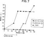
図7は、脾臓細胞の養子免疫伝達後の糖尿病の阻止についてのVCAM2C−IgG融合蛋白質及び対照の効果を描いたグラフであり、糖尿病になったレシピエントの頻度及び病気の発症までの日を、無関係のラットLFA−3Ig融合蛋白質処理した(塗りつぶした正方形)及びVCAM2D−IgG処理した(塗りつぶしてない丸)糖尿病(D)のNODドナーからの2×10⁷の脾臓細胞の移植について、或は細胞を移植せずPBSだけを受けたレシピエント(塗りつぶした三角形)の発症頻度を示してある。これらの脾臓細胞は、VCAM2D−IgG又はラットLFA−3Igと共に移植し、その後VCAM2D−IgG又はラットLFA−3Igを移植後17日間にわたって1日おきに注射した(すべてのグループについてn=5)。
図8は、実施例5に記載したVCAM2DIgG融合蛋白質の構造を描いた図式表示である。VCAM2D−IgGは、VLA4に対するリガンド(VCAM1)の可溶性形態であり、ヒトIgG1重鎖定常領域配列(ヒンジ、CH2及びCH3)に融合した2つのVCAM1のN末端ドメインからなる。
発明の詳細な説明
この発明は、インシュリン依存型(I型)糖尿病の予防を含む治療に関するものである。特に、この発明は、前糖尿病の個体における糖尿病の治療におけるVLA4に対する抗体の利用に関係する。用語「前糖尿病」は、明白な糖尿病又は糖尿病の発症前の病気過程の如何なる段階であっても糖尿病の発達の危険にある(例えば、遺伝的に病気にかかり易い)個体を意味することを意図している。用語「糖尿病」は、明白な高血糖(即ち、空腹時血糖レベル≧250mg/dL)を有する個体を意味することを意図している。用語「明白な糖尿病」又は「糖尿病の発症」は、膵臓島細胞が破壊され明白な高血糖(即ち、空腹時血糖レベル≧250mg/dL)により臨床的に明白である病気状態を意味することを意図する。
第1の面において、この発明は、リンパ球及びマクロファージを含むVLA4陽性細胞の表面上のVLA4抗原に結合する(ブロックし又は被覆することを含む)ことの出来る組成物を投与するステップを含む糖尿病の治療方法を提供する。この発明の目的に関して、用語「VLA4抗原に結合する」は、細胞上のVLA4抗原と反応性であり、それにより、VLA4抗原と他の細胞表面上のVCAM−1又はフィブロネクチンの何れかとの相互作用を妨害し、又はそれによりVLA4陽性細胞の機能変化を誘導することを意味することを意図している。ここに示すように、VLA4抗原のかかる結合(ブロック又は被覆することを含む)は、糖尿病の発生に対する予防又は防護を生じる。この例示は、VLA4に対するモノクローナル抗体をVLA4抗原を効果的にブロックし又は被覆する結合剤として利用した。当業者は、この例示を与えられれば、VLA4抗原に結合し得る薬剤(ブロック又は被覆し得るものも含む)が、この発明の方法において上首尾に利用し得ることを認識するであろう。従って、この発明の目的に関して、VLA4を有する細胞の表面上のVLA4抗原に結合し得る任意の薬剤及びVLA4抗原を効果的にブロックし又は被覆することの出来る任意の薬剤は、本明細書の実施例で用いられているモノクローナル抗体の同等物であると考えられる。例えば、この発明は、少なくとも、VLA4を有する細胞の表面上のVLA4抗原に結合することの出来るペプチド、ペプチド類似物、炭水化物及び低分子を、結合性同等物として企図する。
好適具体例において、細胞表面VLA4抗原に結合する(ブロック及び被覆を含む)この発明の方法において利用する薬剤は、モノクローナル抗体又は抗体誘導体である。治療時にヒトの治療のための好適な抗体誘導体は、ヒト化組換え抗体、キメラ組換え抗体、Fab、Fab'、F(ab')2及びF(v)抗体断片、並びに抗体重鎖若しくは軽鎖のモノマー若しくはダイマー又はこれらの混合物を含む。従って、VLA4に対するモノクローナル抗体は、この発明による方法における好適結合剤である。
モノクローナル抗体を生成するための技術は、周知である。簡単に言えば、不滅化細胞系統(典型的には、ミエローマ細胞)を、所定の抗原例えばVLA4を発現している全細胞で免疫した哺乳動物からのリンパ球(典型的には、脾臓細胞)と融合させ、その結果生成したハイブリドーマ細胞の培養上清をその抗原に対する抗体についてスクリーニングする(一般的には、Kohler等、1975[40]を参照されたい)。
免疫化は、標準的手順を用いて達成することが出来る。単位投与量及び免疫化管理は、免疫化する哺乳動物種、その免疫状態、その哺乳動物の体重等に依存する。典型的には、免疫した哺乳動物から採血し、各血液試料からの血清を適当なスクリーニング用アッセイを用いて特定の抗体についてアッセイする。例えば、抗VLA4抗体は、VLA4発現細胞からの125I標識した細胞溶解物の免疫沈降によって同定することが出来る(Sanchez−Madrid等、1986[41]及びHemler等、1987[42]を参照されたい)。抗VLA4抗体は又、フローサイトメトリーにより例えばVLA4を認識すると考えられる抗体と共にインキュベートしたRamos細胞の蛍光染色を測定することにより同定することも出来る(Elices等、(1990)[43]を参照されたい)。ハイブリドーマ細胞の生成に典型的に利用されるリンパ球は、血清が抗VLA4抗体の存在について陽性であることをかかるスクリーニングアッセイを用いて既に試験してある免疫化哺乳動物から単離される。
典型的には、不滅化細胞系統(例えば、ミエローマ細胞系統)を、リンパ球と同じ哺乳動物種から誘導する。好適不滅化細胞系統は、ヒポキサンチン、アミノプテリン及びチミジンを含む培地(「HAT培地」)に感受性のマウスミエローマ細胞系統である。
典型的には、HAT感受性マウスミエローマ細胞を、分子量1500のポリエチレングリコール(「PEG1500」)を用いて、マウス脾臓細胞と融合させる。この融合から生成したハイブリドーマ細胞を、次いで、未融合の又は非産生的に融合したミエローマ細胞を殺すHAT培地を用いて選択する(未融合の脾臓細胞は、トランスフォームされていないので数日後に死ぬ)。所望の抗体を産生するハイブリドーマを、ハイブリドーマ培養上清をスクリーニングすることによって検出する。例えば、抗VLA4抗体を産生するために調製したハイブリドーマを、組換えα4サブユニット発現細胞系統例えばトランスフェクトしたH−562細胞に結合する能力を有する分泌された抗体についてハイブリドーマ培養上清を試験することによってスクリーニングすることが出来る(Elices等、[43]を参照されたい)。
抗VLA4抗体を生成するために、かかるスクリーニングアッセイで試験して陽性のハイブリドーマ細胞を、栄養培地中で、ハイブリドーマ細胞がモノクローナル抗体を培養培地中に分泌するのに十分な条件及び時間で培養した。これらのハイブリドーマ細胞に適した培養技術及び培養培地は、周知である。調整したハイブリドーマ培養上清を集め、抗VLA4抗体を適宜更に周知の方法によって精製することが出来る。
或は、所望の抗体を、ハイブリドーマ細胞を非免疫化マウスの腹膜腔に注入することによって生成することが出来る。これらのハイブリドーマ細胞は、腹膜腔内で増殖して抗体を分泌し、それは腹水液として蓄積する。注射器で腹膜腔から腹水液を回収することにより抗体を採取することが出来る。
幾つかのマウス抗VLA4モノクローナル抗体が以前に記載された(例えば、Sanchez−Madrid等、1986[41];Hemler等、1987[42];Pulido等、1991[44]を参照されたい)。これらのVLA4のα鎖を認識することの出来る抗VLA4モノクローナル抗体例えばHP1/2及び他の抗VLA4抗体(例えば、mAbHP2/1、HP2/4、L25、P4C2、P4G9)は、本発明による治療方法において有用である。VCAM−1及びフィブロネクチンリガンドへの結合に関与するVLA−α4鎖エピトープを認識する抗VLA4抗体(即ち、リガンド認識に関与する部位でVLA4に結合してVCAM−1及びフィブロネクチン結合をブロックすることの出来る抗体)は、好適である。かかる抗体は、Bエピトープ特異的抗体(B1又はB2)として規定されており(Pulido等、(1991)[36]を参照されたい)、本発明による好適抗VLA4抗体である。ここに記載のように利用したR1−2抗体は、Bエピトープ型抗体である。
VLA4に対するヒトモノクローナル抗体は、この発明の方法においてVLA4抗原をブロック又は被覆することの出来る他の好適結合剤である。これらは、Boerner等、1991[45]により記載されたように、イン・ビトロでプライムしたヒト脾臓細胞を用いて調製することが出来る。或は、それらを、Persson等、1991[46]又はHuang及びStollar,1991[47]により記載されたようにレパートリークローニングによって調製することが出来る。この発明の方法においてVLA4抗原をブロック若しくは被覆し得る他の好適結合剤は、抗VLA4特異性とヒト抗体定常領域とを有するキメラ組換え抗体である。この発明の方法においてVLA4抗原をブロック若しくは被覆することの出来る更に他の好適結合剤は、抗VLA4特異性を有するヒト化組換え抗体である。ヒト化抗体は、Jones等、1989[48];Riechmann,1988[49];Queen等、1989[50];及びOrlandi等、1989[51]に例示されたようにして調製することが出来る。Bエピトープ特異性を有するキメラ組換え抗体及びヒト化組換え抗体を含む好適結合剤は、同時継続中の同時譲渡された米国特許出願第08/004,798号(1993年1月12日出願)[52]にて調製され且つ説明されている。キメラ(マウスV−ヒトC)及びヒト化抗VLA4抗体の調製のための出発物質は、以前に記載されたようなマウスモノクローナル抗VLA4抗体、市販のモノクローナル抗VLA4抗体(例えば、HP2/1、メイン、Westbrook,Amac,International,Inc.)又は本明細書の教示に従って調製したモノクローナル抗VLA4抗体であってよい。例えば、抗VLA4抗体HP1/2の重鎖及び軽鎖の可変領域が、クローン化され、配列決定され且つヒト免疫グロブリン重鎖及び軽鎖の定常領域と組合せて発現された。かかるキメラHP1/2抗体は、マウスHP1/2抗体に対して特異性及び効力において類似しており、本発明による治療方法において有用であり得る。HP1/2VHDNA配列及び翻訳されたアミノ酸配列を、それぞれ、SEQ ID NO:1及びSEQ ID NO:2に示す。HP1/2VKDNA配列及びその翻訳されたアミノ酸配列を、それぞれ、SEQ ID NO:3及びSEQ ID NO:4に示す。同様に、ヒト化組換え抗VLA4抗体は、これらの方法において有用であり得る。好適なヒト化組換え抗VLA4抗体は、AS/SVMDY抗体であり、例えば、1992年11月3日にATCCに寄託して受託番号CRL11175を受けた細胞系統により産生されるAS/SVMDY抗体である。AS/SVMDYヒト化抗体は、少なくとも、マウスHP1/2抗体と同じ効力を有するか又は恐らくは一層強力である。AS VHDNA配列及びその翻訳されたアミノ酸配列を、それぞれ、SEQ ID NO:5及びSEQ ID NO:6。SVMDY VKDNA配列及びその翻訳されたアミノ酸配列を、それぞれ、SEQ ID NO:7及びSEQ ID NO:8に示す。
当業者は、任意の上記の抗体又は抗体誘導体結合剤がこの発明の方法においてVLA4のレセプターに結合することによっても作用することが出来、細胞表面VLA4抗原をブロック若しくは被覆することが出来ることを認めるであろう。従って、この発明による抗体及び抗体誘導体結合剤は、VCAM−1若しくはフィブロネクチンに対する結合特性を有する具体例を含み得る(何故なら、これらの分子は、粘着細胞若しくは細胞外マトリックスにおいて重要であるか又は細胞の組織及び血液中の交通を妨害するかの何れかであるらしいから)。
或は、この発明による方法において用いられるこれらの結合剤は、抗体若しくは抗体誘導体ではなくて、VLA4に対する天然の結合蛋白質の可溶性形態であってよい。これらの結合剤は、可溶性VCAM−1、VCAM−1ペプチド又はVCAM−1融合蛋白質並びにフィブロネクチン、別のスプライシングを受けた非III型結合セグメントを有するフィブロネクチン及びアミノ酸配列EILDV若しくは類似の保存的に置換されたアミノ酸配列を含むフィブロネクチンペプチドを含む。これらの結合剤は、VLA4に対する細胞表面結合蛋白質と競争することによって作用する。
この発明の第1の面によるこの方法において、VLA4結合剤は、好ましくは、非経口的に投与する。これらのVLA4結合剤は、好ましくは、製薬上許容し得るキャリアーを含む無菌の製薬組成物として投与する(該キャリア−は、多くの周知のキャリアー例えば水、塩類溶液、リン酸緩衝塩溶液、デキストロース、グリセロール、エタノール等、又はこれらの組合せの何れであってもよい)。好ましくは、VLA4結合剤は、抗体若しくは抗体誘導体であるならば、約0.1〜20mg/kg体重/日の範囲、好ましくは、約0.1〜10mg/kg体重/日の範囲の投与量及び各1〜14日の間隔で投与する。抗体若しくは抗体誘導体でない結合剤については、投与量範囲は、好ましくは、これらの抗体量に対するモル当量の範囲である。好ましくは、抗体組成物を少なくとも1μg/mlの血漿レベルの抗体を与えるのに有効な量で投与する。投与量の最適化は、結合剤の投与及び、その後の、所定投与量をイン・ビボ投与した後の長期間にわたる薬剤によるVLA4陽性細胞の被覆の評価によって決定することが出来る。個体の末梢血液試料に含まれる末梢血液単核細胞は、この薬剤の存在について、イン・ビトロ(又はex vivo)で、投与した薬剤を検出するための第2の試薬を用いてプローブ検出されるはずである。例えば、これは、投与した薬剤に特異的な蛍光色素標識した抗体であってよく、次いで、それを標準的FACS(蛍光活性化セルソーター)分析により測定する。或は、投与した薬剤の存在は、個体の細胞の標識(例えば、蛍光色素標識)した同じ薬剤に対する結合不能又は結合能力の減少によって、イン・ビトロ(又はex vivo)で検出することが出来る。好適な投与量は、大多数のVLA4陽性細胞の検出可能な被覆を被覆を生成するはずである。好ましくは、被覆は、モノクローナル抗体若しくはモノクローナル抗体誘導体の場合には、1〜14日間にわたって維持される。
この発明の実施において、VLA4結合剤での治療は、好ましくは、前糖尿病患者が上記のような多くの公知のマーカーにより反映されるような安定な血糖正常の血漿レベル及び安定な前糖尿病状態を維持する限り継続する。後述の実施例において、抗VLA4mAb例えばR1−2mAbの投与は、治療中の糖尿病の発症を阻止し且つ治療の残留の有益な結果がR1−2治療を停止した後2か月間に及ぶということが見出された。しかしながら、糖尿病発症に対するVLA4結合剤の完全な防御効果を維持するためには、これらの結合剤での連続的治療が好ましい。
本発明の方法は、前糖尿病の個体に抗VLA4抗体を含む組成物を投与することを含む。後述の実施例は、ゲッ歯動物病気モデルにおいて観察された結果を示す。これらの結果は、この病気の急性伝達モデルにおける病気の発症における抗VLA4抗体の防御効果を示している。非肥満性糖尿病(NOD)マウスは、Makino等、1980[7]により導入されて以来、I型即ちインシュリン依存型糖尿病の重要なモデルとなっており、ヒトの糖尿病と特に関連するモデルとして記載された(例えば、Castano及びEisenbarth[1],Miller等、1988[5],Hutchings等、1990[33]及び本明細書中で引用している文献を参照されたい)。NODマウス及びヒトにおいて示された糖尿病の症状が類似していることが、幾つかの系統の証拠によって示された。例えば、NODマウス及びヒトの両者において[1]、糖尿病と主要組織適合性複合体の遺伝子座とは強い遺伝的関係がある。更に、例えば、両種において、自己免疫病因論は、(i)β細胞の選択的破壊を媒介する膵臓の島におけるリンパ球性炎症(即ち、インシュリン炎)の存在、(ii)抗島細胞抗体の存在、及び(iii)シクロスポリンAの調節効果によって明示されている。糖尿病の自己免疫病因論についてのNODマウスにおける更なる証拠は、(i)糖尿病ドナーからの脾臓細胞(精製脾臓T細胞を含む)を用いて糖尿病を伝達する能力、(ii)T細胞に対して特異的な抗体を用いるイン・ビボ治療による糖尿病の阻止、及び(iii)NOD遺伝的バックグラウンドを有する胸腺ヌードマウスでは糖尿病を発達させることが出来ないことである(例えば、Miller等、1988[5],Hutchings等、1990[33]及び本明細書で引用されている文献を参照されたい)。
糖尿病を生じる正確な事象は不明なままであるが、NODマウスにおいては、膵臓の進行性炎症応答が、脈管周囲の単核細胞浸潤として3〜4週齢で始まる最初の組織的病変であるらしい。約4〜6週齢において、インシュリン炎が観察され得、約12週齢での明白な糖尿病の開始(即ち、糖尿のアッセイ用のTestape(インジアナ、Indianapolis在、Eli Lilly)を用いて首尾一貫した1+以上の値、又は、血漿グルコースをモニターした際に250mg/dLを超える値)が起きる。動物の免疫状態の変動を避けるために、NODマウスを病原体を有しない特別の集団から得、それらは、安定な、高発病率の糖尿病を、雌の約80%及び雄の約20%で示す(典型的には、約20週齢で糖尿病になる)。ここに記載した実験で用いるNODマウスの好ましい供給源は、Taconic Farms(ニューヨーク、Germantown在)である。特にBBラット及びNODマウスの研究からの多くのデータは、I型糖尿病がT細胞媒介の病気であることを示した。今日までの証拠は、両主要T細胞サブポピュレーション(CD4/L3T4及びCD8/Ly2)の、ヒト及びNODマウスにおける糖尿病の発達における重要な役割を示唆している。糖尿病におけるT細胞の本質的役割を支持するデータは、Tリンパ球が他の細胞(例えば、マクロファージ)をβ細胞破壊に対する最終的エフェクターとして補充するという可能性を排除しない。マクロファージは、浸潤された島に存在していること及び長期のシリカ治療の病気を阻止する能力に基づいて病気過程に関係付けられている(例えば、Hutchings等、1990[33]及び本明細書中で引用されている文献を参照されたい)。
NOD系統のマウスを用いて、研究者は、自発的な病気モデルに匹敵する病気の急性伝達モデルを開発したが、該モデルにおいては、糖尿病誘発性NODマウスから誘導した伝達された細胞が病気過程を媒介し、それは、インシュリン炎及び島β細胞特異的な破壊を媒介する免疫反応性細胞により特徴付けられる。更に、このモデルにおいて、T細胞に対するある種のモノクローナル抗体(例えば、Miller等、1988[5]参照)及びマクロファージ(例えば、Hutchings等、1990[33]参照)は、病気の発症を排除することが示された。かかるモノクローナル抗体は、自発的病気及び養子免疫伝達された病気の治療に利用された。例えば、抗CD4抗体は、両モデルにおいて病気を排除することが示された(Miller等、1988[5]及びShizuru等、1988[30])。養子免疫伝達モデル又は自発的病気モデルにおける治療結果は、ヒトの病気過程を調節する薬剤の能力を暗示する。
実施例1
病気の養子免疫伝達に対する抗VLA4抗体処理の効果
糖尿病実験の養子免疫伝達のために、NODマウスをTaconic Farms(ニューヨーク、Germantown)又はJoslin Diabetes Center(マサチューセッツ、Boston)から入手した。最近発症した自発性糖尿病(D)の雌(13〜20週齢)を脾臓細胞ドナーとして用い、8週齢の糖尿病でない(Y)雌をレシピエントとして供した。病気を伝達し得ない4週齢の糖尿病でない(Y)雌ドナーからの脾臓細胞を負の対照として用いた。
高線量照射(Gamma Cell 1000 Cesium137線源、カナダ国、Ontario在、Nordion International,Inc.)後の腸内感染の発病率を最小にするために、レシピエントマウスを1週間酸性水(濃HClを水で1:8400に稀釈したもの)中に置いてから準致死的照射(775ラド)を分割線量(300ラド、300ラド及び175ラド)にて3日間(−2日、−1日及び伝達の日)で行なった。脾臓を糖尿病のドナー及び糖尿病でない対照から採取し、細胞懸濁液を作成し、Hemolytic Geys溶液を用いて赤血球を溶解させた。脾臓細胞を、75μgのR1−2モノクローナル抗体(mAb)、75μgのラットIgG2bで予備処理し又はしないで静脈注射した(0.2mlPBS中2〜3×10⁷)。抗体処理のために、細胞を単に375μg/mlのmAbと懸濁して1〜1.5×108細胞/mlとし、注射まで氷上に置いた。注射のタイミングは、最後の照射の後3時間以内であった。何匹かのレシピエントは、PBSだけを受けた。抗VLA4mAbR1−2及びイソタイプ適合のラットIgG2bをPharmingen(カリフォルニア、La Jolla在)から購入した。R1−2(ラット抗マウス)抗VLA4mAbは、最初、Holzmann等、1989[53]により記載された。R1−2抗VLA4mAbは、VLA4のそのリガンドへの結合をブロックし(Hession等、1992[54])、それ故、定義によりBグループに属する(Pulido等、1991[44])即ち、Bグループの抗ヒトVLA4mAb(例えば、HP1/2又はHP2/1)と同等である。
R1−2mAb又はラットIgG2bを、2〜3日毎に、75μg/0.2mlの投与量で腹腔内投与した。投与用管理(regimen)は、末梢血液、リンパ様器官及び骨髄においてVLA4陽性細胞の最大被覆(末梢血液細胞及びこれらの器官から調製した単一細胞懸濁のR1−2mAbに特異的な蛍光色素標識したmAbでの染色及び蛍光色素陽性細胞を測定するFACS分析により検出)を維持するように決めた。注射を、伝達の後、12日又は24日間続けた。マウスを、TesTape(インディアナ、Indianapolis在、Eli Lilly)を用いて糖尿について試験することにより及び血漿グルコースレベル(Glucometer,3 Blood Glucose Meter,インディアナ、Elkhart在、Miles,Inc.)により糖尿病についてモニターし、2回連続尿試験陽性[Testape値[+1]以上]又は血漿グルコースレベル>250mg/dLの後で糖尿病と見なした。
NOD糖尿病ドナーから単離した脾臓細胞を飽和量の抗VLA4mAbR1−2で処理し、その後、上記のように、糖尿病でない照射した宿主に移植し、次いで、末梢血液及びリンパ様器官中のすべてのVLA4陽性細胞の最大被覆を2週間維持するために、R1−2mAbを12日間1日おきに投与した場合に、抗VLA4mAbの糖尿病発症に対する予防効果が示された。図1は、糖尿病になったレシピエントの頻度及びNODドナー(D→Y)からの2×10⁷の脾臓細胞を、(i)処理なし(塗りつぶした丸)で;(ii)ラットIgG2b処理して(塗りつぶした三角形)及び(iii)R1−2抗VLA4処理(塗りつぶした菱形)して移植した場合、並びに糖尿病でないNODドナー(Y→Y)からの脾臓細胞(塗りつぶしてない正方形)の移植後の病気発症までの日数を示している。PBSだけの注射は、0%の発病率を与えた。これらの条件下で、8匹のR1−2mAb処理した各レシピエントの内、1匹だけが移植後29日に糖尿病を発症した。対照してみると、mAbで処理しなかった又は非特異的なラットIgG2bで処理した糖尿病ドナーからの脾臓細胞を受けた個体は、それぞれ、6/10及び5/9が糖尿病になった。図1に示したように、無関係のラットIgG2bの移植は発症を幾らか遅らせはしたが、移植後14日という早さで糖尿病の発症が起きた。
これらのデータは、R1−2mAbのVLA4に対する特異性に依存した防護効果を示している。糖尿病でないマウスからの脾臓細胞又はPBSのみのレシピエントは糖尿病にならなかった。従って、抗VLA4抗体での処理は、移植後30日間における糖尿病の頻度を減少させた。
図1に示した結果は臨床的糖尿病が抗VLA4処理した動物8匹中1匹にのみ生じたことを示しているが、抗VLA4抗体が単に糖尿病の発症を少し遅らせたに過ぎないということはあり得た。如何なる血糖レベルの増加をも定量しそれにより臨床的糖尿病への進行を検出するために、血漿グルコースレベルを尿中グルコースと並行して監視した。図1に示した抗VLA4抗体処理したグループの内、すべてのマウスは、29日目において未だ平均血漿グルコース値100±7mg/dL(n=7)を有して血糖正常であった(但し、1個体は、尿試験及び500mg/dLを超える血漿グルコースにより臨床的糖尿病として評点された)。従って、病気の進行は、移植後29日、最後の抗VLA4抗体注射の丸2週間後にて図1に示した他の抗VLA4抗体処理したレシピエントの何れにおいても明らかではなかった。これらのマウスからの血清の分析は、抗VLA4mAbが移植後18〜21日までに低レベル又は検出不能なレベルにまで低下することを確実にした。
抗VLA4mAbが糖尿病の移植に対して防護となることを確認するために更なる細胞移植を行なった。これらの実験において、抗VLA4抗体処理を、移植後25日まで延長したが、3.5日毎に投与しそれによりR1−2mAb又はラットIgG2bの飽和レベルを膵臓組織を取るためにマウスを殺す26日目まで維持した。これらの条件下で、糖尿病の発症に対する抗VLA4mAbの阻止効果は又、脾臓細胞移植及びR1−2処理に対しても示された。図2は、糖尿病になったレシピエント(各グループについてn=4〜5)の頻度及び糖尿病NODドナー(D→Y)からの3×10⁷の脾臓細胞を(i)処理しないで(塗りつぶした丸)で、(ii)IgG2b処理(塗りつぶした三角形)して及びR1−2抗VLA4処理(塗りつぶした菱形)して移植した場合、並びに糖尿病でないNODドナー(塗りつぶしてない正方形)からの脾臓細胞の移植後の病気の発症までの日数を示している。PBSだけの注射は、0%の発病率を与えた。図2は、5匹のR1−2mAb処理したマウスの内の1匹だけが移植後22日までに糖尿病になったが、R1−2mAb処理しなかったレシピエントの4/4及びラットIgG2bで処理したレシピエントの5/5において糖尿病が伝達された。病気の発症は、移植後13日の早さで起きた。これらの実験は、個別に及び集合的に、抗VLA4mAbが再現性をもって急性伝達病気モデルにおける糖尿病の発達に対する防護を与えることを示している。
更なる実験を行なって、抗VLA4mAbが単に処理期間中病気の発症を遅らせただけなのか或は長期の防護効果を達成し得るのかを決定した。図3は、R1−2注射(3.5日毎に25日まで)後の長期にわたるマウスの糖尿病の発症を示しているが、移植後35日及び38日(最後のR1−2注射後10〜13日)に2/5のマウスが糖尿病になっただけである。対照してみると、未処理及びIgG2b処理したグループにおいては、移植後11日の早さで、18〜21日目までに100%の発症率で糖尿病が起きた。驚くべきことに、R1−2処理したグループにおける発病率は、最後のR1−2注射の後2か月の長期間においてさえ増加しなかった。この期間中並行して監視した血漿グルコース値は、これらの3匹の個体が一貫して血糖正常であったことを示している。この時点(即ち、移植後約3か月)の後は、PBSのみ又は糖尿病でない細胞を受けた負の対照グループでさえ自発的な病気の発達が始まる。要約すると、VLA4特異的mAbは、糖尿病伝達の発病率を減少させる。更に、病気に対するその防護効果は、更なるmAb処理がなくても維持される。
実施例2
膵臓インシュリン炎に対する抗VLA4mAbの効果
組織学的分析のために、この実施例では、上記のように移植後2〜4週にてマウスを殺して、10%ホルマリン緩衝塩溶液中で膵臓を採取してパラフィン包埋切片用とし、該切片を、組織学用にヘマトキシリン及びエオシン(H&E)で染色した。インシュリン炎の程度を次のように評点した:等級0:インシュリン炎なし[全く炎症のない島];等級I:周囲インシュリン炎[島の周辺に位置する炎症性単核細胞];等級II:25%未満の浸潤[25%の島内部がリンパ球性炎症性細胞を含む];等級III:25〜50%の浸潤[リンパ球性の浸潤];等級IV:50%を超える浸潤。次いで、各等級の島の試験した島の総数に対する相対的パーセントを計算した。組織学的切片を試験して、抗VLA4mAb処理を伴う又は伴わないNOD脾臓細胞の養子免疫伝達後のインシュリン炎の程度を評点して結果を表にした。特に、浸潤されてない島(等級0〜Iの浸潤)及び等級II〜IVのインシュリン炎(上記の通り)を有する島を計量した。各実験グループについて、n=4〜5のマウスからの膵臓切片を評点した。
様々な期間にわたって抗VLA4mAbで処理したレシピエントから、島特異的な細胞性浸潤の確立に対するその効果の問題を扱うために、膵臓組織を回収した。マウスを非特異的なラットIgG2b又はR1−2mAbで3.5日毎に、殺した14日目まで処理した。同様に、マウスを25日目まで処理して、糖尿病と診断されるか又は移植の26日後に殺した。移植後14日間にわたってR1−2mAbで連続的に処理したマウスは、高頻度(76%)の未浸潤の島を維持し、等級II〜IVのインシュリン炎に進行したのは24%だけであった。対照してみると、非特異的なラットIgG2bで処理したものは、74%の重症のインシュリン炎を有して、逆のパターンを示している。同様に、25日目までR1−2で処理したマウス(20%糖尿病、図2で示したマウスから単離した膵臓)において、図4に示したように、若いNOD脾臓細胞の糖尿病でないレシピエントにおける頻度(55%)と類似して、高頻度(58%)の未浸潤の島が保存された。対照してみると、未処理の又はIgG2b処理したマウスの両者は、28%の未浸潤の島を有するだけであり、逆に、増加したインシュリン炎(72%)を有した。従って、抗VLA4mAb処理は、糖尿病誘発性脾臓細胞の養子免疫伝達によるインシュリン炎の発達を特異的に阻止し、或は遅延させるらしい。
これらの選択肢を区別するために、移植の4週間後のインシュリン炎パターンを、マウスをラットIgG2b又はR1−2mAbで12日目まで処理し、その後は更なる処理をしないで維持した場合について測定した。糖尿病が診断されるか又は移植後29日目にマウスを殺した。これらのマウスからの血清の分析は、循環抗VLA4mAbが移植後18〜21日までに検出不能なレベルまで低下することを確実にした。このプロトコールを用いて、R1−2処理したグループのインシュリン炎の程度(69%インシュリン炎、25%糖尿病)は、図5に示したように、ラットIgG2b処理したマウスのそれ(96%インシュリン炎、75%糖尿病)よりは低いが、未処理のレシピエントのそれ(73%インシュリン炎、60%糖尿病)と類似していた。有意に、インシュリン炎の激烈さは、R1−2処理、未処理及びラットIgG2b処理したグループ間で類似しており、それぞれ、平均57%、47%、64%の等級III/IVの浸潤を有した。糖尿病でないR1−2処理した個体だけを考えても、やはりそれらは、52%の等級III/IV浸潤を伴う59%のインシュリン炎を示した。糖尿病誘発性でないNOD脾臓細胞のレシピエントは、7%の等級III/IV浸潤を有しただけであった。逆に、図5は、塩溶液又は非糖尿病誘発性脾臓細胞のレシピエントと比較して、浸潤されてない島の頻度がR1−2処理したマウスにおいて減少したことを示している。従って、インシュリン炎の程度は、R1−2処理を維持したマウス(図4)に比較して、これらのR1−2処理したマウスにおいて進行して(図5)、未処理又はラットIgG2b処理した対照グループのそれに近づいた。ひとまとめにして考えると、これらのデータは、抗VLA4mAb投与が病気の急性伝達モデルにおいてインシュリン炎の進行を遅延させることが出来ることを示している。
実施例3
糖尿病の養子免疫伝達に対する種々の抗VLA4抗体処理の比較
この実施例は、実施例1に記載の養子免疫伝達モデル及び手順を用いて、PS/2、抗VLA4抗体とR1−2の比較用の効果の結果を提供する。NODマウスを(a)無関係の対照用抗体(D/ラットIgG2b、n=19マウス);(b)R1−2抗体(D/R1−2mAb、n=24マウス);(c)PS/2mAb(D/PS/2mAb、n=5マウス)で処理し、又は(d)処理しなかった(NONE、n=26マウス)。脾臓細胞を静脈注射し(0.2mlのPBS中の2〜3×107)、75μgのR1−2mAb、75μgのPS/2mAb、75μgのラットIgG2bの何れかで予備処理し又は処理しなかった。PS/2抗VLA4mAbの単離精製は、最初に、Miyake等、1991[55]により記載された。
R1−2mAb、PS/2mAb又はラットIgG2bを、75μg/0.2mlの投与量で2〜3日毎に腹腔内投与した。投与用管理は、末梢血液、リンパ様器官及び骨髄においてVLA4陽性細胞の最大被覆(末梢血液細胞及びこれらの器官から調製した単一細胞懸濁のR1−2及びPS/2mAbに特異的な蛍光色素標識したmAbでの染色及び蛍光色素陽性細胞を測定するFACS分析により検出)を維持するように決めた。注射を、伝達の後、22〜25日間続けた。マウスを、TesTape(インディアナ、Indianapolis在、Eli Lilly)を用いて糖尿について試験することにより及び血漿グルコースレベル(Glucometer,3 Blood Glucose Meter,インディアナ、Elkhart在、Miles,Inc.)により糖尿病についてモニターし、2回連続尿試験陽性[Testape値[+1]以上]又は血漿グルコースレベル>250mg/dLの後で糖尿病と見なした。
NOD糖尿病ドナーから単離した脾臓細胞を飽和量の抗VLA4mAbR1−2又はPS/2で処理し、その後、上記のように、糖尿病でない照射した宿主に伝達し、次いで、末梢血液及びリンパ様器官中のすべてのVLA4陽性細胞の最大被覆を約2週間維持するために、R1−2mAb又はPS/2を22〜25日間1日おきに投与した場合に、抗VLA4mAbの糖尿病発症に対する予防効果が示された。表1は、糖尿病になったレシピエントの頻度及びNODドナーからの脾臓細胞を、(i)処理なし(D)で;(ii)ラットIgG2b処理して(D/非特異的なラットIgG2b);(iii)R1−2抗VLA4処理(D/R1−2mAb)して;(iv)PS/2処理(D/PS/2mAb)して移植した場合、並びに糖尿病でないNODドナーからの脾臓細胞(non−D)の移植後の病気発症までの日数を示している。PBSを受けて脾臓細胞を受けてない糖尿病でないマウスを対照として含めた。PBSだけの注射は、4%の発病率を与えた。これらの条件下で、24匹のR1−2mAb処理した各レシピエントの内、1匹だけが移植後22日に糖尿病を発症し、他方、5匹のPS/2mAb処理したレシピエントは何れも糖尿病にならなかった。対照してみると、mAbで処理しなかった又は非特異的なラットIgG2bで処理した糖尿病ドナーからの脾臓細胞を受けた個体は、16/19が糖尿病になった。表1に示したように、無関係のラットIgG2bの移植は発症を1日だけ遅らせはしたが、移植後14日という早さで糖尿病の発症が起きた。
これらのデータは、R1−2mAb及びPS/2のVLA4に対する特異性に依存した防護効果を示している。糖尿病でないマウスからの脾臓細胞又はPBSのみのレシピエントは糖尿病にならなかった。従って、抗VLA4抗体での処理は、移植後30日間における糖尿病の頻度を減少させた。これらのマウスからの血清の分析は、R1−2及びPS/2抗VLA4mAbのレベルが移植後26〜34日で検出不能になることを確実にした。
自発的糖尿病モデルに対する抗VLA4抗体処理の効果
この実施例には、NODマウスを用いた自発的糖尿病モデルにおけるR1−2mAbを用いた効果の結果を記載した。NODマウスを8週間にわたって(a)無関係の対照用抗体(NOD/ラットIgG2b、n=10マウス);(b)R1−2抗体(NOD/R1−2、n=20マウス)で処理し;又は(c)は処理しなかった(NOD、n=10マウス)(4週齢で始めて12週齢まで行なった)。mAbを0.2mlPBS中の75μgの投与量にて、週2回静脈投与した。マウスを、前記のように、糖尿用のTesTapeによって、糖尿病事象について監視した。
図6は、2つの対照用グループと比較して、R1−2投与後に糖尿病発症の顕著な遅延(12〜16週間の遅延)を示している。無関係なIgG2bmAbを受けたか又は処理を受けてないNODマウスは、13週の早さで糖尿病が発達した。これらの自発的病気モデルの結果は、図1に説明したR1−2を伴う養子免疫伝達の結果と同様であり、抗VLA4抗体が糖尿病の発症に対する防護となることを直接的に示している。
実施例5
糖尿病の養子免疫伝達に対するVCAM−Ig融合蛋白質の効果
実施例1に記載した養子免疫伝達実験を、抗VLA4mAbの代わりにVCAM−Ig融合蛋白質(VCAM2D−IgG)を用いて繰り返した。VCAM2D−IgGは、VLA4に対するリガンド(VCAM1)の可溶性形態であって、ヒトIgG1重鎖定常領域配列(ヒンジ、CH2及びCH3)に融合したVCAM1の2つのN末端ドメインからなる。VCAM2D−IgGDNA配列及びその翻訳アミノ酸配列をSEQ ID NO:9に示す。図8は、融合蛋白質構造を説明するものである。この融合蛋白質を、下記の組換え技術によって構築した。
ヒトIgG重鎖領域のcDNAの単離とプラスミッドpSAB144の構築
ヒトIgG1重鎖領域のcDNAコピーを単離するために、プラスミッドVCAM1−IgG1(pSAB133としても知られる)によって一時的にトランスフェクトしたCOS7細胞からRNAを調製した。プラスミッドVCAM1−IgG1の構築は、PCT特許出願WO90/13300に記載されている。このRNAを、逆転写酵素及びランダムヘキサマー(プライマーとして)を用いて逆転写してcDNAを生成した。42℃で30分経過後、逆転写反応を、反応物を95℃で5分間のインキュベーションにより停止させた。次いで、このcDNAを、次のキナーゼ処理したプライマーを用いてPCR(ポリメラーゼ連鎖反応、例えば、Sambrook等、Molecular Cloning,Vol.3,pp.14.1−14.35(Cold Spring Harbor;1989)を参照)により増幅した:
PCR増幅したcDNAを、プラスミッドpNN03でのクローン化のためにアガロースゲル電気泳動及びガラスビーズ溶出により精製した。プラスミッドpNN03を、市販のプラスミッドpUC8(ニュージャージー、Piscataway在、Pharmacia)から、制限エンドヌクレアーゼ消化によって合成ポリリンカー配列を除去し、下記の新規な配列(SEQ ID NO:12)を有する合成ポリリンカー配列により置換することによって構築した:
プラスミッドpSAB142の構築
プラスミッドpSAB142を次のようにして構築した。pSAB133でトランスフェクトしたCOS細胞から(前節に記載のようにして)調製したcDNAを、オリゴヌクレオチド370−01及び370−29を用いるPCR増幅にかけた。オリゴヌクレオチド370−01はNot I部位及びVCAM−1シグナル配列のアミノ酸1〜7に対応するヌクレオチド(SEQ ID NO:13)を含む:
pSAB132の構築
pJOD−S(Barsoum,J.,DNA and Cell Biol.,9,pp.293−300(1990))を、アデノウイルス主要後期プロモーターからのユニークなNot I部位下流を挿入するように改変し、Not I断片が発現ベクター中に挿入され得るようにした。pJOD−Sを、プラスミッドDNAのNot I開裂によって線状化した。突き出ている5'末端を、ヤエナリ(Mung Bean)ヌクレアーゼを用いて鈍端化し、この線状化DNA断片を低融点アガロースゲル電気泳動により精製した。DNA断片をT4DNAリガーゼを用いて再結合した。このライゲートした分子を、次いで、大腸菌JA221中にトランスフォームした。コロニーをNot I部位の不在についてスクリーニングした。その結果のベクターをpJOD−SデルタNot Iと呼んだ。pJOD−8デルタNot Iを、Sal Iを用いて線状化し、5'末端を仔ウシアルカリホスファターゼを用いて脱リン酸化した。線状化DNA断片を低融点アガロースゲル電気泳動により精製し、リン酸化オリゴヌクレオチドACE175の存在にてライゲートした。該オリゴヌクレオチドは、次の配列(SEQ ID NO:15)を有する:
DHFRcDNA、pMDR901及びpJODΔe−tPA(Barsoum,DNAand Cell Biol.,9,pp.239−300(1990))の転写を制御するSV40プロモーター中の2つのSV40エンハンサー反復を削除するために、両者をAst II及びDra IIIで開裂した。pMDR901からの2578bpのAst II−Dra III断片及びpJODΔe−tPAからの5424bpのAst II−Dra III断片を、低融点アガロースゲル電気泳動により単離し、1つにライゲートした。大腸菌JA221へのトランスフォーメーションの後に、生成したプラスミッドpMDR902を単離した。次いで、pSAB132を、アデノウイルス主要後期プロモーターを含むpMDR902のEcoR I−Not I断片を除去し、それを、ヒトサイトメガロウイルス最初期プロモーター及びエンハンサーを含むプラスミッドpCMV−B(カリフォルニア、Palo Alto在、Clontech)からの83bpのEcoR I−Not I断片で置換することによって形成した。
pSAB146の構築
pSAB144をSal I及びNot Iで開裂し、693bp断片を単離した。pSAB142をNot I及びSal Iで開裂させて664bp断片を単離した。これらの2つの断片を、Not Iで開裂し且つ仔牛アルカリホスファターゼにより脱リン酸化したpSAB132にライゲートした。その結果生成したプラスミッドpSAB146は、VCAM−1シグナル配列、成熟VCAM−1のアミノ末端の219アミノ酸、IgG1のヒンジ領域の10アミノ酸並びにIgG1のCH2及びCH3定常ドメインをコードするDNA配列を含んだ。
安定にトランスフォームしたCHO細胞系統からのVCAM2D−IgGの産生
組換えVCAM2D−IgG発現ベクターを下記のようにして構築し、CHO細胞中にトランスフェクトしてVCAM2D−IgGを連続的に分泌する細胞系統を生成した。
pSAB146のVCAM2D−IgGコード配列を含む1.357kbのNot I断片を、アガロースゲル電気泳動によって精製した。この断片を、発現ベクターpMDR901のNot Iクローニング部位にライゲートした(該ベクターは、異種遺伝子発現のためのアデノウイルス2主要後期プロモーター及び選択可能で増幅可能なジヒドロ葉酸レダクターゼ(dhfr)マーカーを利用する)。このライゲートしたDNAを用いて大腸菌DH5をトランスフォームした。所望の正しい向きの挿入物を有するプラスミッドを含むコロニーを、Hind IIIでの消化における5853及び3734bp断片の存在、及びBgl IIでの消化における4301、2555、2293及び438bp断片の存在により同定した。その結果生成した組換えVCAM2D−IgG発現ベクターをpEAG100と呼んだ。この正しい挿入物の同一性をDNA配列分析により確認した。
組換え発現プラスミッドpEAG100をJ.Barsoumの公表されたプロトコール(DNA Cell Biol9:293−300,1990)に従って、dhfr−欠乏CHO細胞中に電気穿孔法で挿入した(但し、200μgのPvu I線状化pEAG100プラスミッド及び200μgの超音波処理したサケ精子DNAをこの電気穿孔法プロトコールにおいて用いた)。更に、細胞を、200nMメトトレキセートを補ったアルファ完全培地にて選択した。
分泌されたVCAM2D−IgGの発現レベルを測定するために、クローンを平底の96ウェルミクロ滴定プレートに移して、集密になるまで生育させ、下記のようにELISAによりアッセイした。
Immulon 2プレート(バージニア、Chantilly在、Dynatech)の各ウェルを、100μlの抗VCAM4B9MAb(0.05M炭酸ナトリウム/重炭酸塩緩衝液(pH9.6)中で10μg/mlに稀釈したもの)を用いて抗VCAMMAb4B9(Carlos等、1990[56]により記載されたようにしてプロテインAセファロース上で単離精製したもの)で被覆し、パラフィルムでカバーして4℃で一晩インキュベートした。翌日、プレートの中身を落として、2μのフィルターを通して濾過した200μl/ウェルのブロック用緩衝液(1×PBS中の5%ウシ胎児血清)でブロックした。室温で1時間のインキュベーションの後に緩衝液を除去し、プレートを1×PBS中の0.05%Tween−20溶液で2回洗った。調整培地を種々の稀釈にて加えた。陽性対照として、抗マウスIgも又含めた。ブロック用緩衝液及びLFA−3TIPは、陰性対照を構成した。試料及び対照を室温で2時間インキュベートした。
次いで、これらのプレートを1×PBS中の0.05%Tween−20溶液で2回洗った。次いで、陽性対照のウェルを除いて、各ウェルを50μlのブロック用緩衝液中の1:2000稀釈のHRP−ロバ抗ヒトIgG(H+L)(ペンシルベニア、West Grove在、Jackson Immune Research Laboratories)で満たした。陽性対照のウェルは、50μlのブロック用緩衝液中の1:2000稀釈のHRP−ヤギ抗マウスIgG(H+L)(ペンシルベニア、West Grove在、Jackson Immune Research Laboratories)を満たした。次いで、これらのプレートを1時間室温でインキュベートした。
HRP結合Ab溶液を除去して、これらのウェルを1×PBS中の0.05%Tween−20溶液で2回洗った。次いで、100μlのHRP基質緩衝液を各ウェルに室温で加えた。HRP基質緩衝液を次のように調整した:0.5mlのDMSO(Aldrich)中の42mM 3,3',5,5'−テトラメチルベンジジン(TMB)(サウスカロライナ、Lisle在、ICN Immunobiologicals,カタログ番号980501)を50mlの基質緩衝液(0.1M酢酸ナトリウム/クエン酸、pH4.9)にゆっくりと加え、その後、7.5μlの30%過酸化水素(Sigma,カタログ番号H−1009)を加えた。
各ウェル内の青色の発色を、ミクロ滴定プレートリーダーで650nmにてモニターした。7〜10分後に、100μlの2N硫酸の添加によって発色を停止させた。その結果生じた黄色をミクロ滴定プレートリーダーで450nmにて読んだ。陰性対照用ウェルをこの機械用のブランクとして用いた。
VCAM2D−IgGの精製
VCAM2D−IgGを発現しているCHO細胞を、ローラーボトル中でコラーゲンビーズ上で生育させた。調整培地(5リットル)を、Amicon S1Y10らせん限外濾過カートリッジ(マサチューセッツ、Danvers在、Amicon)を用いて500mlに濃縮した。濃縮物を、1リットルのPierceプロテインA結合用緩衝液(イリノイ、Rockford在、Pierce)で稀釈し、重力により10mlのプロテインAカラム(Sepharose 4 Fast Flow,ニュージャージー、Piscataway在、Pharmacia)に載せた。このカラムを、10mlのプロテインA結合用緩衝液で9回洗い、次いで、10mlのPBSで7回洗った。25mM H3PO4(pH2.8)、100mM NaClを含む12の5mlステップでVCAM2D−IgGを溶出した。溶出した試料を、0.5M Na2HPO4(pH8.6)を25mMまで加えることによって中和した。画分を、280nmの吸光度及びSDS−PAGEにより分析した。最高純度の3つのピーク画分をプールして濾過し、アリコートを取って−70℃に保持した。SDA−PAGEによれば、生成物は95%より高い純度であった。幾つかの例においては、プロテインA溶出生成物を更にQ−Sepharose FF(Pharmacia)にて生成することが必要であった。プロテインA溶出物を、3倍容の25mMトリスHCl(pH8.0)で稀釈し、樹脂1ml当り10mgのVCAM2D−IgGにてQ−Sepharose FFカラムに載せた。次いで、VCAM2D−IgGをPBSでQ−Sepharoseから溶出した。
VCAM2D−IgGの評価
脾臓細胞懸濁液を、上記のように、糖尿病のドナー又は糖尿病でない対照から調製した。脾臓細胞を静脈注射(0.2mlPBS中の2〜3×10⁷)し且つ100μgのVCAM2D−IgG又は100μgの無関係のLFA−3Ig融合蛋白質対照で予備処理した。他のグループは、PBSのみを受け、細胞の移植はしなかった。融合蛋白質LFA−3Ig(LFA−3TIP)を、PCT US92/02050及びMiller等、1993[57]に記載のようにして単離精製した。VCAM2D−IgG融合蛋白質又は無関係のLFA−3Ig蛋白質を100μg/0.2mlの投与量で17日目まで週2回腹腔内投与した。この濃度は、VLA4陽性細胞を飽和させるのに十分な融合蛋白質の血清レベルを与えるのに十分であった(血清レベルは、上記のようにELISAによって測定した)。糖尿病の発症を、上記のようにして監視した。
評価の結果を図7に示す。この図に示したように、VCAM2D−IgG融合蛋白質(D/VCAM−Ig、塗りつぶしてない丸)は、糖尿病ドナー(データは示さない)からの細胞とLFA−3Ig無関係対照Ig融合蛋白質(D/LFA−3Ig)とを受けたマウスが移植後15日目で既に60%の発病率に達したのに比べて、糖尿病ドナーマウスからの細胞のレシピエントにおける糖尿病の発症を有意に阻止し、移植後30日目までで60%の発病率である。細胞を受けなかったマウス(PBSのみ)は、病気を起こさなかった。実験グループ当りのマウス数は、n=5であった。
要約すると、ヒトの糖尿病に対するマウスモデルを用いて、VLA4結合剤例えば抗VLA4抗体は、糖尿病の発症に対して防御力があり(実施例1、3及び4)、インシュリン炎の進行を遅らせるのに有効であった(実施例2)。他のVLA4結合剤例えば可溶性VCAM誘導体(VCAM2D−IgG)も又、糖尿病発症に対する防御において有用であった(実施例5)。上記の実施例は、本発明の方法を説明することを意図しており、後記の請求の範囲に記載の発明の制限として提供するものではない。上記の開示から、この発明の多くの変法及び更なる具体例が当業者には明らかとなるであろう。例えば、実際に用いる投与量、使用する抗体若しくは抗体断片の型、投与形式、正確な組成、治療用の投与の時間及び方法、並びに他の多くのすべての特徴は、上記の説明から離れずに変更することが出来る。かかるすべての変法及び更なる具体例は、この発明の企図する内にあり、添付の請求の範囲の内にある。
The present invention relates to the treatment of insulin-dependent (type I) diabetes. In particular, this invention relates to integrin VLA4 (verylateaIt relates to the use of antibodies that recognize ntigen 4).
Background of the Invention
Insulin-dependent diabetes (also called type I diabetes, formerly called juvenile diabetes) has been classified as a chronic autoimmune disease during the past 20 years. In this disease, the insulin-producing cells in the islets (β cells) are selectively targeted and destroyed by cellular infiltrates of the pancreas. The inflammatory infiltrate affecting this island is called insulinitis. Insulin-producing cells, including the majority of islet cells, account for less than 2% of the total pancreatic mass (Castano and Eisenbarth, 1990, [1]; Fujita et al., 1982 [2]; Foulis et al., 1986 [3] ]). The development of type I diabetes can be conceptually divided into six stages, beginning with genetic susceptibility and ending with complete beta cell destruction (Eisenbarth, 1986 [4]). Stage I is genetic susceptibility, which is a necessary but not sufficient condition for the development of this disease. A hypothetical trigger event (Stage II) leads to active autoimmunity against β cells (Stage III). In stage III, it has been hypothesized that the β-cell mass is reduced, and immunological abnormalities such as autoantibodies to insulin and islet cytoplasmic antigens are found. Stimulated insulin secretion is still preserved at this stage. However, over the years, the progressive loss of beta cells leads to reduced insulin secretion in the intravenous glucose tolerance test (IVGTT), despite the individual still being normoglycemic (Phase IV) . Obvious diabetes (ie, clinical manifestation of diabetes or disease due to hyperglycemia) is stage V and can occur several years later when 90% of the pancreatic β-cells are destroyed. When overt diabetes is first seen in stage V, some residual insulin production remains (indicated by the presence in serum of the C peptide, the binding peptide of proinsulin), but the individual Usually require exogenous insulin to live. Eventually, in stage VI, the remaining beta cells are destroyed and C peptides can no longer be detected in the circulation.
Initiation factors and specific linkages leading to diabetes, including the importance of various cell types and cytokines, are still widely discussed, but a key role is generally recognized for autoantigen-reactive T cells. (Miller et al., 1988 [5]; Harada and Makino, 1986 [6]; Koike et al., 1987 [7]; Makino et al., 1986 [8]). In addition to T lymphocytes, insulinitis is characterized by macrophages, dendritic cells (Voorbij et al., 1989 [9]) and B cells, which can serve as specialized antigen presenting cells (APCs). Macrophages can also destroy islet β cells themselves by release of cytokines or free radicals (Nomikos et al., 1986 [10]). Thus, autoimmune diabetes relies on both cell migration and immune stimulation of newly existing cells.
Cell traffic to the site of inflammation is induced by the accessory molecules LFA-1, MAC-1 and VLA4 on the surface of lymphocytes (LFA-1, VLA4) and macrophages (Mac-1, VLA4) (Larson and Springer, 1990 [11]. Hemler et al., 1990 [12]) and their companion ligands ICAM (for LFA-1 and MAC-1) and VCAM (for VLA4), but they are not regulated by cytokines in vascular endothelium ( Larson and Springer, 1990 [11]; Lobb, 1992 [13]; Osborn, 1990, [14]). In addition, VLA4 binds to the CS-1 domain of the extracellular matrix component, fibronectin (FN) (Wayner et al., 1989 [15]). The relative importance of these pathways, such as LFA-1 and VLA4 in lymphocytes or MAC-1 and VLA4 in monocytes, in controlling cell migration is still the subject of research. In vitro data suggests that different uses of these pathways may be dependent on the activation status of both leukocytes and endothelial cells (Shimizu et al., 1991 [16]). Their ability to control cell migration to sites of inflammation in vivo was demonstrated directly using monoclonal antibodies (mAB) to ICAM, MAC-1 or VLA4, which block various animal disease models ( Barton et al., 1989 [17], phorbol ester-induced rabbit lung inflammation; Issekutz and Issekutz, 1991 [18], delayed-type hypersensitivity; Issekutz, 1991 [19], adjuvant-induced arthritis; Yednock et al., 1992 [20]. , Transmission of experimental allergic encephalomyelitis (EAE); Lobb, 1992 [21], asthma).
ICAM and VCAM are also found on the surface of macrophages and dendritic cells in lymphoid tissues (Dustin et al., 1986 [22]; Rice et al., 1990 [23]; Rice et al., 1991 [24]). Their distribution in these specialized APCs is consistent with functional data indicating the role of LFA-1 and VLA4 in T cell activation (Shimuzu et al., 1990 [25]; Burkly et al., 1991 [26]). However, CD4 / MHC class II and CD8 / MHC class I (Rudd et al., 1989 [27]), CD2 / LFA-3 (Moingeon et al., 1989 [287]), CD28 / B7 (Harding et al., 1992 [29]) Many other receptor-ligand pairs can also support adhesion and co-stimulate T cells during T / APC or T / target cell interactions. The specific contribution of many of these pathways in the development of diabetes remains unresolved. Given that there are many molecular pathways for cell adhesion and T cell activation, whether interference in one or more of these pathways affects the onset or severity of diabetes, is particularly important in this disease process, or It is not possible to predict whether interference of these relevant pathways will affect their onset or intensity.
Antibodies to T cells have been used to deplete T cells in a mouse or rat model of spontaneous diabetes and adoptive transfer diabetes, thus preventing the disease (eg, Harada and Makino, 1986 [6]). 1987 [7], Miller et al., 1988 [5] and Shizuru et al., 1988 [30], anti-CD4; Barlow and Like, 1992 [31], anti-CD2; Like et al., 1986 [J. 32], anti-CD5 and anti-CD8). In addition, antibodies to type 3 complement receptor (CR3) molecules on macrophages or MAC-1 have been used to block macrophage and T cell infiltration of pancreatic tissue in a diseased mouse adoptive transfer model (Hutchings et al.). , 1990 [33]). It is not known whether VLA4 is involved in the activity of islet-specific cells after being located in insulinitis or in the pancreas.
Current treatment protocols proposed for type I diabetes include certain immunomodulators, summarized by Federlin and Becker [34], which are incorporated herein by reference. The prolonged prediabetic phase with immune abnormalities and progressive β-cell destruction suggests that immune interference can stop β-cell loss (Castano and Eisenbarth).
Suggested drugs / protocols include certain immunomodulators and immunosuppressants: levamisole, theophylline, thymic hormone, siamexone, antithymocyte globulin, interferon, nicotinamide, gamma globulin infusion, blood cell recruitment or leukocyte transfusion. Contains. Drugs such as cyclosporin A and azathioprine, which impair T cell activation and T cell development, respectively, have been used in clinical trials (Zielasek et al., 1989 [35]). The most promising results have been achieved with cyclosporin A (Castano and Eisenbarth, 1990 [35]). However, Federlin and Becker, 1990 [34] suggest that cyclosporin A is not recommended for general or long-term use, at least when given at high doses, due to toxic side effects. I have. High doses of cyclosporin A and / or combinations with other immunosuppressive agents have been associated with lymphoid development and irreversible kidney damage (Eisenbarth, 1986 [4]; Eisenbarth, 1987 [36]). ). Further studies on other suggested drugs are needed to evaluate safety and efficacy. Studies with cyclosporine A have further shown that its efficacy in maintaining remission of diabetes is about 30-60% of the incidence of new diabetes in one year. However, remissions are almost always lost within three years (Castano and Eisenbarth, 1990 [1]). Treatment protocols after the onset of the disease are particularly problematic, for example, when diabetes is diagnosed, insulinitis is typically already in progress and more than 80% of beta cells are lost. is there. Thus, although cyclosporin A can prevent further destruction of beta cells, they cannot maintain long-term non-diabetic status because only a few beta cells can be present at the onset of diabetes. It is possible (Castano and Eisenbarth, 1990 [1]). Inhibition of insulinitis and / or prevention of the disease can be more successful if the treatment can be started at an earlier stage, ie before the onset of the disease.
There are two major prerequisites for developing a preventive treatment for any diabetes: (1) the ability to accurately identify individuals with pre-diabetes and (2) the development of safe, specific and effective preventive treatments. Significant progress has been made in identifying individuals with pre-diabetes, although Eisenbarth and co-workers (eg, Ziegler and Eisenbarth, 1990 [37]; Zienler et al., 1990 [38]; Zienler et al., 1990 [39] Much work remains on safe, specific and effective prophylactic treatment, as discussed and reviewed by J.C. Identify certain risk factors and groups at risk for type I diabetes, thereby predicting the individuals most likely to progress to clinical disease and estimating the approximate rate of developing disease in these individuals It is possible. Susceptibility to diabetes through a combination of genetic (HLA classification), immunological (islet and insulin autoantibodies) and metabolic (first-stage insulin secretion into intravenous glucose preceding development of hyperglycemia) markers The ability to identify individuals or predict type I diabetes at the pre-clinical stage allows the identification and use of prophylactic immunotherapeutic agents during the development of the autoimmune disease process if beta cell destruction is partial. However, to date, treatment of diabetes in humans has been largely unsuccessful. In general, treatment in humans is done only after the onset of diabetes, so treatment has only resulted in a temporary or complete remission of only some patients. There is a need for immunosuppressive components for use in the pre-diabetic stage, as the immunosuppressive mechanism may prevent insulinitis and / or diabetes. In particular, there is a need for components that are safer and specifically acting, for example, monoclonal antibodies that prevent the entry of effector cells into the pancreas or the function of those cells that have already entered the islets of Langerhans.
It has now surprisingly been found that administration of anti-VLA4 antibodies significantly reduces the incidence of diabetes in rodent diabetes models. The NOD mouse diabetes model is a well-established model that is exactly comparable to human type I diabetes. Irradiated non-diabetic NOD mice were administered spleen cells from spontaneously diabetic NOD mice for acute transmission of the disease using an experimental protocol for adoptively transmitted disease. These spleen cells were treated with anti-VLA4 antibody before administration and recipients were also treated with anti-VLA4 antibody for various periods of time after delivery.
Summary of the Invention
Thus, the present invention provides a novel method for the treatment of insulin dependent (type I) diabetes in pre-diabetes. In particular, the present invention provides a method for the prevention of insulin-dependent diabetes comprising the step of administering to a pre-diabetic individual an anti-VLA4 antibody, such as the antibody HP1 / 2 or a humanized anti-VLA4 antibody derived from HP1 / 2. . The use of similar antibodies, antibody fragments, soluble proteins and small molecules that mimic the effects of anti-VLA4 antibodies in the treatment of diabetes is also contemplated. Further, the present invention provides a method of treating diabetes by administering to a mammal susceptible to diabetes, including a human, an antibody capable of binding to the α4 subunit of VLA4 in an amount sufficient to prevent the onset of diabetes. provide. In addition, the use of recombinant and chimeric antibodies, fragments, polypeptides or small molecules of such antibodies capable of binding α4 / VLA4 is also contemplated. Naturally soluble binding proteins to VLA4 are also contemplated (soluble VCAM-1, VCAM-1 peptide or VCAM-1 fusion protein and fibronectin, fibronectin with another spliced non-type III binding segment and amino acid sequence). (Including fibronectin peptides containing EILDV or similar conservatively substituted amino acid sequences). These drugs act by competing with cell surface binding proteins for VLA4.
[Brief description of the drawings]
FIG. 1 is a graph depicting the effects of anti-VLA4 antibody (R1-2) and controls on inhibition of diabetes after adoptive transfer of pancreatic cells, the frequency of recipients with diabetes and the day to disease onset. Were 2 × 10 5 from untreated (filled circles), non-specific rat IgG2b-treated (filled triangles) and R1-2 anti-VLA4-treated (filled diamonds) diabetic (D) NOD donors.7Of spleen cells from a non-diabetic (Y) NOD donor (unfilled squares). These spleen cells were implanted with or without R1-2 or rat IgG2b, and then injected every other day for 12 days post-implantation with R1-2 or rat IgG2b (n = 8 for all groups). Ten).
FIG. 2 is a graph depicting the effects of anti-VLA4 antibody (R1-2) and controls on the prevention of diabetes after adoptive transfer of spleen cells, the frequency of recipients with diabetes and the day to disease onset. Were 3 × 10 5 from untreated (filled circles), non-specific rat IgG2b-treated (filled triangles) and R1-2 anti-VLA4 treated (filled diamonds) diabetic (D) NOD donors.7Of spleen cells from a non-diabetic (Y) NOD donor (unfilled squares). These spleen cells were implanted with or without R1-2 or rat IgG2b and then injected with R1-2 or rat IgG2b every 3.5 days for 25 days after implantation (n = 4-5 for all groups). ).
FIG. 3 is a graph depicting the effect of anti-VLA4 antibody (R1-2) and controls on the inhibition of diabetes after adoptive transfer of spleen cells, the frequency of recipients with diabetes and the day to disease onset. Were 2-3x from untreated (filled circles), non-specific rat IgG2b-treated (filled triangles) and R1-2 anti-VLA4 treated (filled diamonds) diabetic (D) NOD donors. Ten7For spleen cells transplantation and for spleen cells from non-diabetic (Y) NOD donors (solid squares) or for PBS alone (solid circles). These spleen cells were implanted with or without R1-2 or rat IgG2b and then injected with R1-2 or rat IgG2b every 3.5 days for 25 days after implantation (n = 5 for all groups).
FIG. 4 is a bar graph depicting the effect of anti-VLA4 antibody (R1-2) and control on the degree of insulinitis following adoptive transfer of spleen cells, with R1-2, treated with rat IgG2b or with mAb. Non-infiltrated islets (0 to 0) were implanted with non-infected cells and then injected into mice with R1-2 or rat IgG2b every 3.5 days for 25 days, when the mice became diabetic or killed 26 days after transplantation. The frequency of class I infiltration, dotted bars) and infiltrated islets (class II-IV insulinitis, filled bars) were quantified and shown. Sections of pancreas from mice with n = 4-5 were taken from each experimental group, ie, Y → Y (transplantation of non-diabetic donor cells) or D → Y (cells of diabetic donor) into non-diabetic recipients (Y). Transplantation) (without mAb treatment, treated with rat IgG2b or treated with R1-2).
FIG. 5 is a bar graph depicting the effect of anti-VLA4 antibody (R1-2) and control on the extent of insulinitis following adoptive transfer of spleen cells, with R1-2, treated with rat IgG2b or with mAb. Implanted cells were then implanted with R1-2 or rat IgG2b injected every other day for 12 days post-implantation, and then maintained without further mAb injection, when the mice became diabetic or after transplantation. The frequency of non-infiltrated islets (0-I infiltration, dotted bars) and infiltrated islets (II-IV insulitis, filled bars) killed on day was shown. Sections of pancreas from mice with n = 4-5 were taken from each experimental group, ie, Y → Y (transplantation of non-diabetic donor cells) or D → Y (cells of diabetic donor) into non-diabetic recipients (Y). Transplantation) (without mAb treatment, treated with rat IgG2b or treated with R1-2).
FIG. 6 is a graph depicting the effect of anti-VLA4 antibody (R1-2) and controls on disease prevention in a spontaneous diabetes model, showing the frequency of recipients with diabetes and the days to disease onset; Shown for untreated (filled squares), non-specific rat IgG2b-treated (filled circles) and R1-2 anti-VLA4 treated (filled triangles) NOD mice. R1-2 or rat IgG2b was injected into NOD mice for 8 weeks and twice a week at 4-12 weeks of age.
FIG. 7 is a graph depicting the effect of VCAM2C-IgG fusion protein and controls on inhibition of diabetes after adoptive transfer of spleen cells, showing the frequency of recipients with diabetes and the days to disease onset. 2 × 10 4 from NOD donors of irrelevant rat LFA-3Ig fusion protein treated (filled squares) and VCAM2D-IgG treated (unfilled circles) diabetic (D)72 shows the incidence of spleen cell transplantation in recipients (filled triangles) that received only PBS without cell transplantation. These spleen cells were implanted with VCAM2D-IgG or rat LFA-3Ig followed by injection of VCAM2D-IgG or rat LFA-3Ig every other day for 17 days after implantation (n = 5 for all groups).
FIG. 8 is a schematic representation depicting the structure of the VCAM2DIgG fusion protein described in Example 5. VCAM2D-IgG is a soluble form of the ligand for VLA4 (VCAM1), and the human IgG1 heavy chain constant region sequence (hinge,
DETAILED DESCRIPTION OF THE INVENTION
The present invention relates to treatment, including prevention of insulin dependent (type I) diabetes. In particular, the invention relates to the use of antibodies to VLA4 in the treatment of diabetes in pre-diabetic individuals. The term “pre-diabetes” is intended to mean an individual who is at risk of developing diabetes (eg, genetically susceptible to disease) at any stage of overt diabetes or any stage of the disease process prior to the onset of diabetes. are doing. The term “diabetes” is intended to mean an individual with overt hyperglycemia (ie, fasting blood glucose levels ≧ 250 mg / dL). The term "overt diabetes" or "onset of diabetes" is intended to mean a disease state in which pancreatic islet cells are destroyed and are clinically apparent due to overt hyperglycemia (ie, fasting blood glucose levels ≥250 mg / dL). Intend.
In a first aspect, the invention provides a method for treating diabetes comprising administering a composition capable of binding (including blocking or coating) to a VLA4 antigen on the surface of VLA4-positive cells, including lymphocytes and macrophages. And a method of treating the same. For the purposes of this invention, the term "binds to the VLA4 antigen" is reactive with the VLA4 antigen on the cell, thereby allowing the interaction of the VLA4 antigen with any other VCAM-1 or fibronectin on the cell surface. Or to thereby induce a change in the function of VLA4-positive cells. As shown herein, such binding (including blocking or coating) of the VLA4 antigen results in prevention or protection against the development of diabetes. This example utilized a monoclonal antibody against VLA4 as a binder to effectively block or coat the VLA4 antigen. One of skill in the art will recognize that given this illustration, agents that can bind to the VLA4 antigen, including those that can block or coat, can be successfully utilized in the methods of the invention. Thus, for the purposes of this invention, any agent that can bind to the VLA4 antigen on the surface of VLA4-bearing cells and any agent that can effectively block or coat the VLA4 antigen is described herein. It is believed to be equivalent to the monoclonal antibody used in the examples. For example, the present invention contemplates, as binding equivalents, at least peptides, peptide analogs, carbohydrates, and small molecules capable of binding to the VLA4 antigen on the surface of VLA4-bearing cells.
In a preferred embodiment, the agent utilized in the methods of the invention that binds (including blocks and coats) to the cell surface VLA4 antigen is a monoclonal antibody or antibody derivative. Preferred antibody derivatives for human therapy during therapy include humanized recombinant antibodies, chimeric recombinant antibodies, Fab, Fab ', F (ab')2And F (v) antibody fragments, as well as monomers or dimers of antibody heavy or light chains or mixtures thereof. Thus, a monoclonal antibody against VLA4 is a preferred binder in the method according to the invention.
Techniques for producing monoclonal antibodies are well known. Briefly, lymphocytes (typically spleen cells) from a mammal immunized with an immortalized cell line (typically myeloma cells) with all cells expressing a predetermined antigen, eg, VLA4 And the resulting culture supernatants of hybridoma cells are screened for antibodies to the antigen (see generally Kohler et al., 1975 [40]).
Immunization can be accomplished using standard procedures. Unit dosages and immunization regimes will depend on the mammal species to be immunized, its immunity status, the weight of the mammal, and the like. Typically, blood is drawn from the immunized mammal and serum from each blood sample is assayed for a particular antibody using an appropriate screening assay. For example, an anti-VLA4 antibody is obtained from VLA4-expressing cells.125It can be identified by immunoprecipitation of I-labeled cell lysates (see Sanchez-Madrid et al., 1986 [41] and Hemler et al., 1987 [42]). Anti-VLA4 antibodies can also be identified by flow cytometry, for example, by measuring fluorescent staining of Ramos cells incubated with an antibody that is believed to recognize VLA4 (see Elices et al., (1990) [43]). ). Lymphocytes typically utilized for generation of hybridoma cells are isolated from immunized mammals whose sera have already been tested for positive for the presence of anti-VLA4 antibody using such a screening assay.
Typically, an immortalized cell line (eg, a myeloma cell line) is derived from the same mammalian species as the lymphocytes. Preferred immortalized cell lines are mouse myeloma cell lines that are sensitive to a medium containing hypoxanthine, aminopterin and thymidine ("HAT medium").
Typically, HAT-sensitive mouse myeloma cells are fused with mouse spleen cells using 1500 molecular weight polyethylene glycol ("PEG1500"). The hybridoma cells generated from this fusion are then selected using HAT medium that kills unfused or non-productively fused myeloma cells (unfused spleen cells die after a few days because they have not been transformed). ). Hybridomas producing the desired antibody are detected by screening the hybridoma culture supernatant. For example, a hybridoma prepared to produce an anti-VLA4 antibody is4Screening of hybridoma culture supernatants for secreted antibodies capable of binding subunit expressing cell lines, such as transfected H-562 cells, can be screened (see Elices et al., [43]). .
To generate anti-VLA4 antibodies, hybridoma cells tested positive in such a screening assay were cultured in nutrient medium under conditions and for a time sufficient for the hybridoma cells to secrete monoclonal antibodies into the culture medium. Culture techniques and culture media suitable for these hybridoma cells are well known. The prepared hybridoma culture supernatant is collected, and the anti-VLA4 antibody can be further purified by a well-known method as appropriate.
Alternatively, the desired antibody can be produced by injecting the hybridoma cells into the peritoneal cavity of a non-immunized mouse. These hybridoma cells proliferate in the peritoneal cavity and secrete antibodies, which accumulate as ascites fluid. The antibody can be collected by collecting the ascites fluid from the peritoneal cavity with a syringe.
Several mouse anti-VLA4 monoclonal antibodies have been described previously (see, for example, Sanchez-Madrid et al., 1986 [41]; Hemler et al., 1987 [42]; Pulido et al., 1991 [44]). Anti-VLA4 monoclonal antibodies capable of recognizing the α-chain of VLA4, such as HP1 / 2 and other anti-VLA4 antibodies (eg, mAb HP2 / 1, HP2 / 4, L25, P4C2, P4G9) are used in the therapeutic method according to the present invention. It is useful in VLA-α involved in binding to VCAM-1 and fibronectin ligand4Anti-VLA4 antibodies that recognize chain epitopes (ie, antibodies that can bind to VLA4 at sites involved in ligand recognition and block VCAM-1 and fibronectin binding) are preferred. Such antibodies have been defined as B epitope specific antibodies (B1 or B2) (see Pulido et al., (1991) [36]) and are suitable anti-VLA4 antibodies according to the invention. The R1-2 antibody utilized as described herein is a B epitope type antibody.
Human monoclonal antibodies to VLA4 are other suitable binding agents that can block or coat the VLA4 antigen in the method of the invention. These can be prepared using in vitro primed human spleen cells, as described by Boerner et al., 1991 [45]. Alternatively, they can be prepared by repertoire cloning as described by Persson et al., 1991 [46] or Huang and Stollar, 1991 [47]. Other suitable binding agents that can block or coat the VLA4 antigen in the methods of the invention are chimeric recombinant antibodies having anti-VLA4 specificity and human antibody constant regions. Still other suitable binding agents that can block or coat the VLA4 antigen in the method of the invention are humanized recombinant antibodies with anti-VLA4 specificity. Humanized antibodies can be prepared as exemplified in Jones et al., 1989 [48]; Riechmann, 1988 [49]; Queen et al., 1989 [50]; and Orlandi et al., 1989 [51]. Suitable binding agents, including chimeric and humanized recombinant antibodies with B epitope specificity, are described in co-pending co-assigned US patent application Ser. No. 08 / 004,798 (filed Jan. 12, 1993) [52 ] Has been prepared and described. Starting materials for the preparation of chimeric (mouse V-human C) and humanized anti-VLA4 antibodies are mouse monoclonal anti-VLA4 antibodies as previously described, commercially available monoclonal anti-VLA4 antibodies (eg, HP2 / 1, main Westbrook, Amac, International, Inc.) or a monoclonal anti-VLA4 antibody prepared according to the teachings herein. For example, the heavy and light chain variable regions of the anti-VLA4 antibody HP1 / 2 have been cloned, sequenced, and expressed in combination with human immunoglobulin heavy and light chain constant regions. Such chimeric HP1 / 2 antibodies are similar in specificity and potency to the mouse HP1 / 2 antibodies and may be useful in therapeutic methods according to the present invention. HP1 / 2VHThe DNA sequence and translated amino acid sequence are shown in SEQ ID NO: 1 and SEQ ID NO: 2, respectively. HP1 / 2VKThe DNA sequence and its translated amino acid sequence are shown in SEQ ID NO: 3 and SEQ ID NO: 4, respectively. Similarly, humanized recombinant anti-VLA4 antibodies may be useful in these methods. A suitable humanized recombinant anti-VLA4 antibody is an AS / SVMDY antibody, for example, an AS / SVMDY antibody produced by a cell line deposited with the ATCC on November 3, 1992 and having accession number CRL11175. . The AS / SVMDY humanized antibody has at least the same potency as the mouse HP1 / 2 antibody or is probably more potent. AS VHThe DNA sequence and its translated amino acid sequence are shown in SEQ ID NO: 5 and SEQ ID NO: 6, respectively. SVMDY VKThe DNA sequence and its translated amino acid sequence are shown in SEQ ID NO: 7 and SEQ ID NO: 8, respectively.
One of skill in the art will recognize that any of the above-described antibodies or antibody derivative binders can also act by binding to the receptor for VLA4 in the methods of the invention and block or coat the cell surface VLA4 antigen. Will. Thus, the antibodies and antibody derivative binding agents according to the invention may include embodiments having binding properties to VCAM-1 or fibronectin (because these molecules are important in adherent cells or extracellular matrix or Because it is likely to block traffic in the tissues and blood of the human body).
Alternatively, these binding agents used in the method according to the invention may be a soluble form of the natural binding protein for VLA4, rather than an antibody or antibody derivative. These binders include soluble VCAM-1, VCAM-1 peptide or VCAM-1 fusion protein as well as fibronectin, fibronectin with another spliced non-type III binding segment and the amino acid sequence EILDV or similar conservatively substituted. A fibronectin peptide comprising the amino acid sequence of These binders act by competing with cell surface binding proteins for VLA4.
In this method according to the first aspect of the invention, the VLA4 binding agent is preferably administered parenterally. These VLA4 binding agents are preferably administered as a sterile pharmaceutical composition containing a pharmaceutically acceptable carrier, which can be any of a number of well-known carriers such as water, saline, phosphate buffered saline, dextrose. Glycerol, ethanol, etc., or a combination thereof). Preferably, if the VLA4 binding agent is an antibody or an antibody derivative, a dose in the range of about 0.1-20 mg / kg body weight / day, preferably in the range of about 0.1-10 mg / kg body weight / day and each Administer at 14-day intervals. For binding agents that are not antibodies or antibody derivatives, the dosage range is preferably in the range of molar equivalents to the amount of these antibodies. Preferably, the antibody composition is administered in an amount effective to provide a plasma level of the antibody of at least 1 μg / ml. Dosage optimization can be determined by administration of the binding agent, followed by assessment of VLA4-positive cell coverage by the drug over an extended period of time following a given dose in vivo. Peripheral blood mononuclear cells contained in a peripheral blood sample of the individual are probed for the presence of this drug in vitro (or ex vivo) using a second reagent to detect the administered drug. Should be. For example, this may be a fluorochrome-labeled antibody specific for the administered drug, which is then measured by standard FACS (fluorescence activated cell sorter) analysis. Alternatively, the presence of the administered drug can be detected in vitro (or ex vivo) by the inability or reduced binding capacity of the individual's cells to the same (eg, fluorescent dye-labeled) drug. Suitable dosages should produce a detectable coating of the majority of VLA4-positive cells. Preferably, the coating is maintained for 1 to 14 days in the case of a monoclonal antibody or monoclonal antibody derivative.
In the practice of this invention, treatment with a VLA4 binding agent preferably results in pre-diabetic patients having stable normoglycemic plasma levels and a stable pre-diabetic state as reflected by a number of known markers as described above. Continue as long as you maintain. In the examples below, administration of an anti-VLA4 mAb, e.g., R1-2 mAb, prevents the onset of diabetes during treatment and that the beneficial consequences of remaining treatment are two months after cessation of R1-2 treatment. Was found. However, in order to maintain the full protective effect of VLA4 binding agents against diabetes development, continuous treatment with these binding agents is preferred.
The method of the invention comprises administering to a pre-diabetic individual a composition comprising an anti-VLA4 antibody. The examples below show the results observed in rodent disease models. These results demonstrate the protective effect of anti-VLA4 antibodies on disease development in an acute transmission model of this disease. Non-obese diabetic (NOD) mice have been an important model of type I, or insulin-dependent diabetes, since being introduced by Makino et al., 1980 [7] and have been described as a model particularly associated with human diabetes. (See, for example, Castano and Eisenbarth [1], Miller et al., 1988 [5], Hutchings et al., 1990 [33] and the references cited herein). Several lines of evidence have shown that the symptoms of diabetes shown in NOD mice and humans are similar. For example, in both NOD mice and humans [1], there is a strong genetic association between diabetes and the locus of the major histocompatibility complex. Further, for example, in both species, autoimmune etiology may be based on (i) the presence of lymphocytic inflammation (ie, insulinitis) in pancreatic islets that mediates selective destruction of β-cells, (ii) anti-islet cell antibodies And (iii) the regulatory effect of cyclosporin A. Further evidence in NOD mice about the autoimmune etiology of diabetes is: (i) the ability to transmit diabetes using spleen cells (including purified spleen T cells) from diabetic donors; Inhibition of diabetes by in vivo treatment with specific antibodies, and (iii) inability to develop diabetes in thymic nude mice with NOD genetic background (see, eg, Miller et al., 1988 [5]. ], Hutchings et al., 1990 [33] and references cited herein).
Although the precise events that lead to diabetes remain unknown, in NOD mice, the progressive inflammatory response of the pancreas is the first histological lesion beginning at 3-4 weeks of age as perivascular mononuclear cell infiltration It seems. At about 4-6 weeks of age, insulinitis can be observed, with a pronounced onset of diabetes at about 12 weeks of age (ie, consistent with Testape for diabetes assay (Eli Lilly, Indianapolis, Indianapolis)). 1+ or more than 250 mg / dL when monitoring plasma glucose). To avoid fluctuations in the immune status of the animals, NOD mice were obtained from a special population free of pathogens, which showed stable, high incidence of diabetes in about 80% of females and about 20% of males (Typically, it becomes diabetic at about 20 weeks of age). A preferred source of NOD mice for use in the experiments described herein is Taconic Farms (Germantown, NY). Numerous data, especially from studies of BB rats and NOD mice, indicated that type I diabetes was a T cell mediated disease. Evidence to date suggests an important role for both major T cell subpopulations (CD4 / L3T4 and CD8 / Ly2) in the development of diabetes in humans and NOD mice. Data supporting the essential role of T cells in diabetes do not exclude the possibility that T lymphocytes recruit other cells (eg, macrophages) as the ultimate effector on β-cell destruction. Macrophages have been implicated in disease processes based on their presence in infiltrated islets and the ability to prevent long-term silica treatment of the disease (eg, Hutchings et al., 1990 [33] and herein). See the references cited in).
Using NOD strain mice, researchers have developed an acute transmission model of the disease that is comparable to the spontaneous disease model, in which transmitted cells derived from the diabetogenic NOD mouse are responsible for the disease process. , Which are characterized by immunoreactive cells that mediate insulinitis and islet β cell-specific destruction. Furthermore, in this model, certain monoclonal antibodies against T cells (see, eg, Miller et al., 1988 [5]) and macrophages (see, eg, Hutchings et al., 1990 [33]) may rule out the onset of the disease. Indicated. Such monoclonal antibodies have been used in the treatment of spontaneous and adoptive transmitted diseases. For example, anti-CD4 antibodies have been shown to eliminate disease in both models (Miller et al., 1988 [5] and Shizuru et al., 1988 [30]). Therapeutic results in an adoptive transfer model or a spontaneous disease model are indicative of the ability of the drug to modulate the human disease process.
Example 1
Effect of anti-VLA4 antibody treatment on adoptive transfer of disease
NOD mice were obtained from Taconic Farms (Germantown, NY) or Joslin Diabetes Center (Boston, MA) for adoptive transfer of diabetes experiments. Recently developed spontaneous diabetic (D) females (13-20 weeks old) were used as spleen cell donors and 8-week old non-diabetic (Y) females served as recipients. Spleen cells from a 4-week-old non-diabetic (Y) female donor who were unable to transmit the disease were used as negative controls.
High dose irradiation (Gamma Cell 1000 Cesium137To minimize the incidence of enteric infections after radiation (Source, Nordion International, Inc., Ontario, Canada), recipient mice were diluted 1: 8400 in acidic water (concentrated HCl 1: 8 with water) for one week ), And sublethal irradiation (775 rads) was performed in divided doses (300 rads, 300 rads and 175 rads) for 3 days (-2 days, -1 day and day of transmission). Spleens were collected from diabetic donors and non-diabetic controls, cell suspensions were made, and erythrocytes were lysed using Hemolytic Geys solution. Spleen cells were injected intravenously with or without pretreatment with 75 μg R1-2 monoclonal antibody (mAb), 75 μg rat IgG2b (2-3 × 10 5 in 0.2 ml PBS).7). For antibody treatment, cells were simply suspended with 375 μg / ml mAb and 1-1.5 × 10⁸Cells / ml and kept on ice until injection. The timing of the injection was within 3 hours after the last irradiation. Some recipients received PBS only. Anti-VLA4mAbR1-2 and isotype-matched rat IgG2b were purchased from Pharmingen (La Jolla, Calif.). The R1-2 (rat anti-mouse) anti-VLA4 mAb was originally described by Holzmann et al., 1989 [53]. The R1-2 anti-VLA4 mAb blocks the binding of VLA4 to its ligand (Hession et al., 1992 [54]) and therefore belongs to group B by definition (Pulido et al., 1991 [44]), ie, Equivalent to anti-human VLA4 mAb (eg, HP1 / 2 or HP2 / 1).
R1-2 mAb or rat IgG2b was administered intraperitoneally every 2-3 days at a dose of 75 μg / 0.2 ml. Regimen for administration include maximum coverage of VLA4-positive cells in peripheral blood, lymphoid organs and bone marrow (fluorescent labeling specific to R1-2mAb in peripheral blood cells and single cell suspensions prepared from these organs) (Detected by FACS analysis to measure fluorescent dye positive cells). Injections continued for 12 or 24 days after transmission. Mice were monitored for diabetes by testing for diabetes using TesTape (Eli Lilly, Indianapolis, Indiana) and by plasma glucose levels (Glucometer, 3 Blood Glucose Meter, Indiana, Elkhart, Miles, Inc.); Diabetes was considered after two consecutive urine tests positive [Testape value [+1] or more] or plasma glucose levels> 250 mg / dL.
Spleen cells isolated from NOD diabetic donors were treated with saturating amounts of anti-VLA4mAbR1-2, then transplanted into non-diabetic irradiated hosts, as described above, and then all VLA4 in peripheral blood and lymphoid organs. When R1-2 mAb was administered every other day for 12 days in order to maintain the maximum coverage of positive cells for 2 weeks, the preventive effect of anti-VLA4 mAb on the onset of diabetes was shown. FIG. 1 shows the frequency of recipients with diabetes and 2 × 10 5 from NOD donors (D → Y).7Transplanted with (i) no treatment (filled circles); (ii) rat IgG2b treatment (filled triangles) and (iii) R1-2 anti-VLA4 treatment (filled diamonds) , And the number of days to onset of disease after transplantation of spleen cells (unfilled squares) from a non-diabetic NOD donor (Y → Y). Injection with PBS alone gave 0% disease incidence. Under these conditions, only one out of eight R1-2mAb-treated recipients developed diabetes 29 days after transplantation. By contrast, individuals receiving spleen cells from diabetic donors not treated with mAb or treated with non-specific rat IgG2b became 6/10 and 5/9 diabetic, respectively. As shown in FIG. 1, transplantation of irrelevant rat IgG2b delayed the onset somewhat, but onset of diabetes occurred as early as 14 days after transplantation.
These data indicate a protective effect dependent on the specificity of R1-2mAb for VLA4. Spleen cells or PBS only recipients from non-diabetic mice did not become diabetic. Thus, treatment with anti-VLA4 antibody reduced the frequency of
Although the results shown in FIG. 1 show that clinical diabetes only occurred in one of eight anti-VLA4-treated animals, it was not clear that the anti-VLA4 antibody only slightly delayed the onset of diabetes. It was possible. Plasma glucose levels were monitored in parallel with urinary glucose to quantify any increase in blood glucose levels and thereby detect progression to clinical diabetes. Of the anti-VLA4 antibody-treated groups shown in FIG. 1, all mice were still normoglycemic on day 29 with an average plasma glucose value of 100 ± 7 mg / dL (n = 7) (provided that One individual was scored as clinically diabetic with a urine test and plasma glucose greater than 500 mg / dL). Thus, disease progression was not evident in any of the other anti-VLA4 antibody treated recipients shown in FIG. 1 at 29 days post-transplant, two weeks after the last anti-VLA4 antibody injection. Analysis of sera from these mice ensured that the anti-VLA4mAb was reduced to low or undetectable levels by 18-21 days post-implantation.
Additional cell transplants were performed to confirm that the anti-VLA4mAb was protective against diabetes transplants. In these experiments, anti-VLA4 antibody treatment was extended up to 25 days post-transplantation, but administered every 3.5 days, until day 26 when mice were killed to remove saturating levels of R1-2 mAb or rat IgG2b to remove pancreatic tissue. Maintained. Under these conditions, the inhibitory effect of anti-VLA4mAb on the development of diabetes was also shown against spleen cell transplantation and R1-2 treatment. FIG. 2 shows the frequency of diabetic recipients (n = 4-5 for each group) and 3 × 10 5 from diabetic NOD donors (D → Y).7Transplanted spleen cells from (i) untreated (filled circles), (ii) treated with IgG2b (filled triangles) and R1-2 anti-VLA4 (filled diamonds), and not diabetic Shows the number of days to onset of disease after transplantation of spleen cells from a NOD donor (unfilled squares). Injection with PBS alone gave 0% disease incidence. FIG. 2 shows that only one of the five R1-2 mAb-treated mice became diabetic by day 22 post-transplantation, but was treated with 4/4 of recipients without R1-2 mAb and rat IgG2b. Diabetes was transmitted in 5/5 of the recipients. The onset of the disease occurred as early as 13 days after the transplant. These experiments, individually and collectively, show that anti-VLA4mAb reproducibly provides protection against diabetes development in an acute transmission disease model.
Further experiments were performed to determine whether the anti-VLA4mAb simply delayed the onset of the disease during the treatment period or could achieve a long-term protective effect. FIG. 3 shows the prolonged development of diabetes in mice after R1-2 injection (every 3.5 days up to 25 days), but 35 and 38 days after transplantation (10-13 days after last R1-2 injection). Only) 2/5 mice became diabetic. By contrast, in the untreated and IgG2b-treated groups, diabetes occurred as early as 11 days post-transplant with a 100% incidence by day 18-21. Surprisingly, the incidence in the R1-2 treated group did not increase even over a long period of two months after the last R1-2 injection. Plasma glucose levels monitored in parallel during this period indicate that these three individuals were consistently normoglycemic. After this point (ie, about three months after transplantation), spontaneous disease development begins even in the negative control group, which received PBS alone or non-diabetic cells. In summary, VLA4-specific mAbs reduce the incidence of diabetes transmission. Furthermore, its protective effect against disease is maintained without further mAb treatment.
Example 2
Effect of anti-VLA4mAb on pancreatic insulinitis
For histological analysis, in this example, mice were sacrificed 2-4 weeks after transplantation as described above, and the pancreas was harvested in 10% formalin buffered saline for paraffin embedded sections; The sections were stained with hematoxylin and eosin (H & E) for histology. The degree of insulinitis was scored as follows: Grade 0: no insulinitis [islands without any inflammation]; grade I: peripheral insulinitis [inflammatory mononuclear cells located around the islets]; grade II: 25 % Infiltration [25% of the islets contain lymphocytic inflammatory cells]; Grade III: 25-50% infiltration [lymphocytic infiltration]; Grade IV: greater than 50% infiltration. The relative percentage of each grade of islands to the total number of islands tested was then calculated. Histological sections were examined and the results tabulated for the degree of insulinitis following adoptive transfer of NOD spleen cells with or without anti-VLA4 mAb treatment. In particular, non-infiltrated islands (grade 0-I infiltration) and islands with grade II-IV insulin flaming (as described above) were weighed. Pancreatic sections from n = 4-5 mice were scored for each experimental group.
Pancreatic tissue was collected from recipients treated with anti-VLA4 mAb for various periods to address the issue of its effect on establishing islet-specific cellular invasion. Mice were treated with non-specific rat IgG2b or R1-2mAb every 3.5 days until day 14 of sacrifice. Similarly, mice were treated up to
To distinguish these options, the insulinitis pattern 4 weeks after transplantation was measured when mice were treated with rat IgG2b or R1-2 mAb until day 12 and then maintained without further treatment. . Mice were killed 29 days after transplantation or diabetes was diagnosed. Analysis of sera from these mice ensured that circulating anti-VLA4 mAb was reduced to undetectable levels by day 18-21 after transplantation. Using this protocol, the extent of insulinitis in the R1-2 treated group (69% insulinitis, 25% diabetes) was, as shown in FIG. 5, that of rat IgG2b treated mice (96% insulinitis, (75% diabetes) but similar to that of untreated recipients (73% insulinitis, 60% diabetes). Significantly, the intensity of insulinitis was similar between the R1-2-treated, untreated and rat IgG2b-treated groups, with an average of 57%, 47%, and 64% grade III / IV infiltration, respectively. did. Even considering only non-diabetic R1-2 treated individuals, again they showed 59% insulinitis with 52% grade III / IV infiltration. Non-diabetogenic NOD spleen cell recipients had only a 7% grade III / IV infiltration. Conversely, FIG. 5 shows that the frequency of uninfiltrated islets was reduced in R1-2 treated mice compared to saline or non-diabetic spleen cell recipients. Thus, the extent of insulinitis progressed in these R1-2 treated mice (FIG. 5) as compared to untreated or rat IgG2b treated controls as compared to R1-2 treated mice (FIG. 4). Approached that of the group. Taken together, these data indicate that anti-VLA4mAb administration can slow the progression of insulinitis in an acute transmission model of the disease.
Example 3
Treatment of various anti-VLA4 antibodies for adoptive transfer of diabetesComparison
This example provides results for comparative effects of PS / 2, anti-VLA4 antibody and R1-2 using the adoptive transfer model and procedure described in Example 1. NOD mice were treated with (a) an irrelevant control antibody (D / rat IgG2b, n = 19 mice); (b) R1-2 antibody (D / R1-2 mAb, n = 24 mice); (c) PS / 2 mAb ( D / PS / 2 mAb, n = 5 mice) or (d) untreated (NONE, n = 26 mice). Spleen cells were injected intravenously (2-3 × 10 5 in 0.2 ml PBS).7), 75 μg R1-2 mAb, 75 μg PS / 2 mAb, 75 μg rat IgG2b or no pretreatment. The isolation and purification of the PS / 2 anti-VLA4 mAb was first described by Miyake et al., 1991 [55].
R1-2 mAb, PS / 2 mAb or rat IgG2b was administered intraperitoneally every 2-3 days at a dose of 75 μg / 0.2 ml. Dosing regimens include maximum coverage of VLA4-positive cells in peripheral blood, lymphoid organs and bone marrow (fluorescent dyes specific to R1-2 and PS / 2mAb in peripheral blood cells and single cell suspensions prepared from these organs) Staining with labeled mAbs and detection by FACS analysis measuring fluorochrome-positive cells). Injections continued for 22-25 days after transmission. Mice were monitored for diabetes by testing for diabetes using TesTape (Eli Lilly, Indianapolis, Indiana) and by plasma glucose levels (Glucometer, 3 Blood Glucose Meter, Indiana, Elkhart, Miles, Inc.); Diabetes was considered after two consecutive urine tests positive [Testape value [+1] or more] or plasma glucose levels> 250 mg / dL.
Spleen cells isolated from NOD diabetic donors are treated with saturating amounts of anti-VLA4mAbR1-2 or PS / 2, and then transmitted to non-diabetic irradiated hosts, as described above, and then in peripheral blood and lymphoid organs. When R1-2mAb or PS / 2 was administered every other day for 22 to 25 days to maintain the maximum coverage of all VLA4 positive cells for about 2 weeks, the preventive effect of anti-VLA4 mAb on the development of diabetes was shown. Was. Table 1 shows the frequency of recipients with diabetes and spleen cells from NOD donors (i) without treatment (D); (ii) rat IgG2b treatment (D / non-specific rat IgG2b); (Iii) R1-2 anti-VLA4 treatment (D / R1-2 mAb); (iv) when transplanted with PS / 2 treatment (D / PS / 2 mAb), and spleen cells (non-diabetic NOD donor) -D) shows the number of days until the onset of the disease after transplantation. Non-diabetic mice receiving PBS and not receiving spleen cells were included as controls. Injection with PBS alone gave a 4% disease incidence. Under these conditions, of the 24 R1-2mAb-treated recipients, only one developed diabetes 22 days after transplantation, while all five PS / 2mAb-treated recipients had diabetes. Did not become. By contrast, individuals receiving spleen cells from diabetic donors not treated with mAb or treated with non-specific rat IgG2b became 16/19 diabetic. As shown in Table 1, transplantation of irrelevant rat IgG2b delayed onset by one day, but onset of diabetes occurred as early as 14 days after transplantation.
These data indicate a protective effect depending on the specificity of R1-2 mAb and PS / 2 for VLA4. Spleen cells or PBS only recipients from non-diabetic mice did not become diabetic. Thus, treatment with anti-VLA4 antibody reduced the frequency of
Effect of anti-VLA4 antibody treatment on spontaneous diabetes model
This example describes the results of the effect of using R1-2mAb in a spontaneous diabetes model using NOD mice. Treating NOD mice for 8 weeks with (a) an irrelevant control antibody (NOD / rat IgG2b, n = 10 mice); (b) an R1-2 antibody (NOD / R1-2, n = 20 mice); or (C) was untreated (NOD, n = 10 mice) (started at 4 weeks of age and performed until 12 weeks of age). The mAb was administered intravenously twice weekly at a dose of 75 μg in 0.2 ml PBS. Mice were monitored for diabetic events by TesTape for diabetes as described above.
FIG. 6 shows a significant delay (12-16 weeks delay) in the onset of diabetes after R1-2 administration compared to the two control groups. NOD mice that received or received no irrelevant IgG2b mAb developed diabetes as early as 13 weeks. The results of these spontaneous disease models are similar to the results of adoptive transfer with R1-2 described in FIG. 1 and directly show that anti-VLA4 antibodies are protective against the development of diabetes.
Example 5
Effect of VCAM-Ig fusion protein on adoptive transfer in diabetesFruit
The adoptive transfer experiment described in Example 1 was repeated using the VCAM-Ig fusion protein (VCAM2D-IgG) instead of the anti-VLA4 mAb. VCAM2D-IgG is a soluble form of the ligand for VLA4 (VCAM1), comprising a human IgG1 heavy chain constant region sequence (hinge,
Isolation of cDNA for human IgG heavy chain region and plasmid pSAB144Construction
To isolate a cDNA copy of the human IgG1 heavy chain region, RNA was prepared from COS7 cells transiently transfected with plasmid VCAM1-IgG1 (also known as pSAB133). The construction of plasmid VCAM1-IgG1 is described in PCT patent application WO 90/13300. This RNA was reverse transcribed using reverse transcriptase and random hexamers (as primers) to generate cDNA. After 30 minutes at 42 ° C., the reverse transcription reaction was stopped by incubating the reaction at 95 ° C. for 5 minutes. The cDNA is then amplified by PCR using the following kinased primers (polymerase chain reaction, see, eg, Sambrook et al., Molecular Cloning, Vol. 3, pp. 14.1-14.35 (Cold Spring Harbor; 1989)). did:
PCR amplified cDNA was purified by agarose gel electrophoresis and glass bead elution for cloning in plasmid pNN03. Plasmid pNN03 was synthesized from a commercially available plasmid pUC8 (Pharmacia, Piscataway, NJ) by removing the synthetic polylinker sequence by restriction endonuclease digestion, resulting in a synthetic polylinker having the following novel sequence (SEQ ID NO: 12): Constructed by replacing by sequence:
Construction of plasmid pSAB142
Plasmid pSAB142 was constructed as follows. cDNA prepared from COS cells transfected with pSAB133 (as described in the previous section) was subjected to PCR amplification using oligonucleotides 370-01 and 370-29. Oligonucleotide 370-01 isNot Including the I site and nucleotides corresponding to
Construction of pSAB132
pJOD-S (Barsoum, J.,DNA and Cell Biol.,9, pp. 293-300 (1990)), which is unique from the adenovirus major late promoter.Not Modified to insert I site downstream,Not The I fragment could be inserted into the expression vector. pJOD-S was converted to plasmid DNANot Linearized by I cleavage. The protruding 5 'end was blunted using Mung Bean nuclease, and the linearized DNA fragment was purified by low melting point agarose gel electrophoresis. The DNA fragments were religated using T4 DNA ligase. This ligated molecule was then transformed into E. coli JA221. ColonyNot Screened for absence of I site. The resulting vector is called pJOD-SdeltaNot Called I. pJOD-8 DeltaNot I,Sal It was linearized with I and the 5 'end was dephosphorylated with calf alkaline phosphatase. The linearized DNA fragment was purified by low melting point agarose gel electrophoresis and ligated in the presence of phosphorylated oligonucleotide ACE175. The oligonucleotide has the following sequence (SEQ ID NO: 15):
DHFR cDNA, pMDR901 and pJODΔe-tPA (Barsoum,DNAand Cell Biol.,9, pp. 239-300 (1990)) to eliminate the two SV40 enhancer repeats in the SV40 promoter that control transcription.Ast II andDra Cleavage at III. 2578 bp from pMDR901Ast II−Dra III fragment and 5424 bp from pJODΔe-tPAAst II−Dra The III fragment was isolated by low melting agarose gel electrophoresis and ligated together. After transformation into E. coli JA221, the resulting plasmid pMDR902 was isolated. PSAB132 was then replaced with pMDR902, which contains the adenovirus major late promoter.EcoR I−Not The I fragment was removed and it was replaced with an 83 bp plasmid from plasmid pCMV-B (Clontech, Palo Alto, Calif.) Containing the human cytomegalovirus immediate early promoter and enhancer.EcoR I−Not It was formed by displacement with the I fragment.
Construction of pSAB146
pSAB144Sal I andNot Cleavage with I and isolation of a 693 bp fragment. pSAB142Not I andSal The 664 bp fragment was isolated by cleavage with I. These two fragmentsNot Ligated into pSAB132 cleaved with I and dephosphorylated with calf alkaline phosphatase. The resulting plasmid pSAB146 contains the VCAM-1 signal sequence, the amino terminal 219 amino acids of mature VCAM-1, the 10 amino acids of the hinge region of IgG1, and the C1 of IgG1.H2 and CHDNA sequences encoding three constant domains were included.
VCAM2D from a stably transformed CHO cell line-IgG production
A recombinant VCAM2D-IgG expression vector was constructed as described below and transfected into CHO cells to generate a cell line that continuously secretes VCAM2D-IgG.
1.357 kb containing the VCAM2D-IgG coding sequence of pSAB146Not The I fragment was purified by agarose gel electrophoresis. This fragment was constructed using the expression vector pMDR901.Not Ligation to the I cloning site (the vector utilizes the
The recombinant expression plasmid pEAG100 was prepared using the published protocol of J. Barsoum (DNA Cell Biol.9: 293-300, 1990) and inserted into dhfr-deficient CHO cells by electroporation (with the exception of 200 μg).Pvu I-linearized pEAG100 plasmid and 200 μg of sonicated salmon sperm DNA were used in this electroporation protocol). In addition, cells were selected in alpha complete medium supplemented with 200 nM methotrexate.
To determine the expression level of secreted VCAM2D-IgG, clones were transferred to flat-bottom 96-well microtiter plates, grown to confluence, and assayed by ELISA as described below.
Each well of an
The plates were then washed twice with a 0.05% Tween-20 solution in 1 × PBS. Each well was then filled with 1: 2000 dilution of HRP-donkey anti-human IgG (H + L) (Jackson Immune Research Laboratories, West Grove, Pa.) In 50 μl of blocking buffer, except for the positive control wells. . Positive control wells were filled with 1: 2000 dilution of HRP-goat anti-mouse IgG (H + L) (Jackson Immune Research Laboratories, West Grove, Pa.) In 50 μl of blocking buffer. The plates were then incubated for 1 hour at room temperature.
The HRP-bound Ab solution was removed and the wells were washed twice with a 0.05% Tween-20 solution in 1 × PBS. Then, 100 μl of HRP substrate buffer was added to each well at room temperature. The HRP substrate buffer was adjusted as follows: 42
The blue color development in each well was monitored at 650 nm on a microtiter plate reader. After 7 to 10 minutes, the color development was stopped by adding 100 μl of 2N sulfuric acid. The resulting yellow color was read at 450 nm on a microtiter plate reader. Negative control wells were used as blanks for this machine.
Purification of VCAM2D-IgG
CHO cells expressing VCAM2D-IgG were grown on collagen beads in roller bottles. The conditioned medium (5 liters) was concentrated to 500 ml using an Amicon S1Y10 spiral ultrafiltration cartridge (Amicon, Danvers, Mass.). The concentrate was diluted with one liter of Pierce Protein A binding buffer (Pierce, Illinois, Rockford) and loaded by gravity onto a 10 ml Protein A column (Sepharose 4 Fast Flow, Piscataway, NJ, Pharmacia). The column was washed 9 times with 10 ml of Protein A binding buffer and then 7 times with 10 ml of PBS. 25mM HThreePOFourVCAM2D-IgG was eluted in 12 5 ml steps (pH 2.8) containing 100 mM NaCl. Elute the sample with 0.5M NaTwoHPOFour(PH 8.6) by adding up to 25 mM. Fractions were analyzed by absorbance at 280 nm and SDS-PAGE. The three peak fractions with the highest purity were pooled, filtered and aliquots were kept at -70 ° C. The product was more than 95% pure by SDA-PAGE. In some cases, it was necessary to further generate the Protein A elution product on Q-Sepharose FF (Pharmacia). The protein A eluate was diluted with 3 volumes of 25 mM Tris HCl (pH 8.0) and loaded on a Q-Sepharose FF column with 10 mg of VCAM2D-IgG per ml of resin. Then, VCAM2D-IgG was eluted from Q-Sepharose with PBS.
Evaluation of VCAM2D-IgG
Spleen cell suspensions were prepared from diabetic donors or non-diabetic controls as described above. Intravenous injection of spleen cells (2-3 × 10 5 in 0.2 ml PBS)7) And pretreated with 100 μg of VCAM2D-IgG or 100 μg of an irrelevant LFA-3Ig fusion protein control. Other groups received PBS only and did not transplant cells. The fusion protein LFA-3Ig (LFA-3TIP) was isolated and purified as described in PCT US92 / 02050 and Miller et al., 1993 [57]. VCAM2D-IgG fusion protein or irrelevant LFA-3Ig protein was intraperitoneally administered twice a week until the 17th day at a dose of 100 μg / 0.2 ml. This concentration was sufficient to provide serum levels of the fusion protein sufficient to saturate VLA4 positive cells (serum levels were measured by ELISA as described above). Diabetes development was monitored as described above.
FIG. 7 shows the results of the evaluation. As shown in this figure, the VCAM2D-IgG fusion protein (D / VCAM-Ig, open circles) was obtained from cells from a diabetic donor (data not shown) and LFA-3Ig irrelevant control Ig fusion protein (D / V). LFA-3Ig) significantly prevented the onset of diabetes in recipients of cells from diabetic donor mice, compared to mice that had already reached 60
In summary, using a mouse model for human diabetes, a VLA4 binding agent, such as an anti-VLA4 antibody, is protective against the development of diabetes (Examples 1, 3 and 4) and slows the progression of insulinitis. It was effective (Example 2). Other VLA4 binding agents, such as soluble VCAM derivatives (VCAM2D-IgG), were also useful in protecting against diabetes development (Example 5). The above examples are intended to illustrate the method of the present invention and are not provided as limitations on the invention described in the claims that follow. From the above disclosure, many variations and further embodiments of the invention will be apparent to those skilled in the art. For example, the dosage used, the type of antibody or antibody fragment used, the mode of administration, the exact composition, the time and method of administration for treatment, and many other features are all departed from the above description. Can be changed. All such variations and further embodiments are within the contemplation of this invention and are within the scope of the appended claims.
Claims (19)
Translated fromJapaneseApplications Claiming Priority (3)
| Application Number | Priority Date | Filing Date | Title |
|---|---|---|---|
| US2933093A | 1993-02-09 | 1993-02-09 | |
| US08/029,330 | 1993-02-09 | ||
| PCT/US1994/001456WO1994017828A2 (en) | 1993-02-09 | 1994-02-09 | Treatment for insulin dependent diabetes |
Publications (2)
| Publication Number | Publication Date |
|---|---|
| JPH08508719A JPH08508719A (en) | 1996-09-17 |
| JP3593343B2true JP3593343B2 (en) | 2004-11-24 |
Family
ID=21848497
Family Applications (1)
| Application Number | Title | Priority Date | Filing Date |
|---|---|---|---|
| JP51833394AExpired - Fee RelatedJP3593343B2 (en) | 1993-02-09 | 1994-02-09 | Treatment of insulin-dependent diabetes |
Country Status (12)
| Country | Link |
|---|---|
| US (2) | US5888507A (en) |
| EP (1) | EP0682529B2 (en) |
| JP (1) | JP3593343B2 (en) |
| AT (1) | ATE161730T1 (en) |
| AU (1) | AU687790B2 (en) |
| CA (1) | CA2155303C (en) |
| DE (1) | DE69407758T3 (en) |
| DK (1) | DK0682529T4 (en) |
| ES (1) | ES2114183T5 (en) |
| GR (1) | GR3026531T3 (en) |
| NZ (1) | NZ262615A (en) |
| WO (1) | WO1994017828A2 (en) |
Families Citing this family (71)
| Publication number | Priority date | Publication date | Assignee | Title |
|---|---|---|---|---|
| DE69309487T2 (en) | 1992-11-13 | 1997-10-23 | Univ Washington | PERIPHERALIZATION OF HEMATOPOIETIC STEM CELLS |
| EP0678122B1 (en) | 1993-01-12 | 1999-07-28 | Biogen, Inc. | Recombinant anti-vla4 antibody molecules |
| NZ262615A (en)* | 1993-02-09 | 1996-02-27 | Biogen Inc | Treating insulin dependent diabetes (type i) with anti vla4 (very late antigen) antibodies |
| US5677291A (en)* | 1993-12-10 | 1997-10-14 | Hoechst Marion Roussel, Inc. | Method of lowering serum cholesterol levels with 2,6-di-alkyl-4-silyl-phenols |
| US5750336A (en)* | 1994-02-10 | 1998-05-12 | The Salk Institute For Biological Studies | Assays for the identification of compounds which inhibit activation of cAMP and mitogen responsive genes |
| US7803904B2 (en) | 1995-09-01 | 2010-09-28 | Millennium Pharmaceuticals, Inc. | Mucosal vascular addressing and uses thereof |
| US6551593B1 (en) | 1995-02-10 | 2003-04-22 | Millennium Pharmaceuticals, Inc. | Treatment of Inflammatory bowel disease by inhibiting binding and/or signalling through α 4 β 7 and its ligands and madcam |
| US5608095A (en)* | 1996-04-30 | 1997-03-04 | Hoechst Marion Roussel, Inc. | Alkyl-4-silyl-phenols and esters thereof as antiatherosclerotic agents |
| US5795876A (en)* | 1996-04-30 | 1998-08-18 | Hoechst Marion Rousssel, Inc. | Method of inhibiting vascular cell adhesion molecule-1 and treating chronic inflammatory diseases with 2, 6-di-alkyl-4-silyl-phenols |
| US7147851B1 (en)* | 1996-08-15 | 2006-12-12 | Millennium Pharmaceuticals, Inc. | Humanized immunoglobulin reactive with α4β7 integrin |
| US6114572A (en)* | 1996-11-20 | 2000-09-05 | Hoechst Marion Roussel, Inc. | Substituted phenols and thiophenols useful as antioxidant agents |
| CN1257545A (en)* | 1997-04-18 | 2000-06-21 | 拜奥根有限公司 | Type II TGF-beta receptor/immunoglobulin constant region fusion proteins |
| US6121463A (en)* | 1997-06-24 | 2000-09-19 | Hoechst Marion Roussel, Inc. | Alkyl-4-silylheterocyclic phenols and thiophenols useful as antioxidant agents |
| US6133467A (en)* | 1997-06-25 | 2000-10-17 | Hoechst Marion Roussel, Inc. | 2,6-di-t-butyl-4-[(dimethyl-4-methoxyphenylsilyl)-methyl-oxy]phenol and 2,6-di-t-butyl-4-[(dimethyl-2-methoxy-phenylsilyl)methyloxy]phenol |
| DE19741235A1 (en) | 1997-09-18 | 1999-03-25 | Hoechst Marion Roussel De Gmbh | Novel imidazolidine derivatives, their preparation, their use and pharmaceutical compositions containing them |
| DE19741873A1 (en)* | 1997-09-23 | 1999-03-25 | Hoechst Marion Roussel De Gmbh | New 5-ring heterocycles, their preparation, their use and pharmaceutical preparations containing them |
| DE19751251A1 (en) | 1997-11-19 | 1999-05-20 | Hoechst Marion Roussel De Gmbh | Substituted imidazolidine derivatives, their manufacture, their use and pharmaceutical preparations containing them |
| DE19821483A1 (en) | 1998-05-14 | 1999-11-18 | Hoechst Marion Roussel De Gmbh | New imidazolidine derivatives useful as leukocyte adhesion and migration inhibitors and/or VLA-4 receptor antagonists for treating E.G. inflammatory and allergic disorders |
| EP1113810B1 (en) | 1998-09-14 | 2008-12-31 | Board of Regents, The University of Texas System | Methods of treating multiple myeloma and myeloma-induced bone resorption using antagonists of integrin / receptor binding |
| US7618630B2 (en) | 1998-09-14 | 2009-11-17 | Board Of Regents, The University Of Texas System | Methods of treating multiple myeloma and myeloma-induced bone resorption using integrin antagonists |
| US6110109A (en)* | 1999-03-26 | 2000-08-29 | Biosignia, Inc. | System and method for predicting disease onset |
| AU783054B2 (en)* | 1999-04-22 | 2005-09-22 | Biogen Ma Inc. | Method for the treatment of fibrosis using an antagonist of the integrin alpha-4 subunit |
| DE19922462A1 (en) | 1999-05-17 | 2000-11-23 | Aventis Pharma Gmbh | New phenylureidobenzyl-substituted spiro-imidazolidinedione derivatives, are inhibitors of leukocyte adhesion or migration or VLA-4 receptors, useful e.g. for treating inflammatory or allergic disease |
| SK17232001A3 (en) | 1999-06-01 | 2002-09-10 | Biogen, Inc. | A blocking monoclonal antibody to vla-1 and its use for the treatment of inflammatory disorders |
| WO2001019396A1 (en)* | 1999-09-14 | 2001-03-22 | Biogen, Inc. | Therapies for chronic renal failure using one or more integrin antagonists |
| CN100475953C (en) | 1999-12-06 | 2009-04-08 | 通用医疗公司 | pancreatic stem cells |
| ATE447969T1 (en) | 1999-12-16 | 2009-11-15 | Biogen Idec Inc | METHOD FOR TREATING CENTRAL NERVOUS SYSTEM DAMAGE CAUSED BY ISCHEMIA OR BY HEMORRHAGIA WITH ANTAGONISTS OF ALPHA4 INTEGRIN |
| US20060115473A1 (en)* | 2000-12-14 | 2006-06-01 | Biogen Idec Ma Inc., A Massachusetts Corporation | Methods of treating central nervous system ischemic or hemorrhagic injury using anti alpha4 integrin antagonists |
| JP3459609B2 (en)* | 2000-03-17 | 2003-10-20 | 三洋電機株式会社 | Write cache circuit, recording device provided with write cache circuit, and write cache method |
| US20010046496A1 (en)* | 2000-04-14 | 2001-11-29 | Brettman Lee R. | Method of administering an antibody |
| DE10111877A1 (en) | 2001-03-10 | 2002-09-12 | Aventis Pharma Gmbh | Novel imidazolidine derivatives, their preparation, their use and pharmaceutical compositions containing them |
| CN1561345B (en) | 2001-04-13 | 2011-09-14 | 比奥根艾迪克Ma公司 | Antibodies against VLA-1 |
| DE10137595A1 (en) | 2001-08-01 | 2003-02-13 | Aventis Pharma Gmbh | New 3-alkylaminoalkyl-imdazolidin-4-one derivatives, are VLA-4 receptor and leukocyte adhesion and/or migration inhibitors, useful e.g. for treating inflammatory, allergic, autoimmune or cardiovascular disease |
| FR2849598B1 (en)* | 2003-01-07 | 2006-09-22 | Merck Sante Sas | USE OF KYNURENINE-3-HYDROXYLASE INHIBITORS FOR THE TREATMENT OF DIABETES BY INCREASING THE NUMBER OF CELLS OF THE LANGERHANS ISLANDS |
| KR101260497B1 (en) | 2003-01-24 | 2013-09-12 | 엘란 파마슈티칼스, 인크. | Composition for and treatment of demyelinating diseases and paralysis by administration of remyelinating agents |
| US7419666B1 (en) | 2004-02-23 | 2008-09-02 | Massachusetts Eye And Ear Infirmary | Treatment of ocular disorders |
| CA2478458A1 (en)* | 2004-08-20 | 2006-02-20 | Michael Panzara | Treatment of pediatric multiple sclerosis |
| WO2006055871A2 (en) | 2004-11-19 | 2006-05-26 | Biogen Idec Ma Inc. | Treatment for multiple sclerosis |
| CA2589379A1 (en)* | 2004-12-03 | 2006-06-08 | Biogen Idec Ma Inc. | Delaying or preventing onset of multiple sclerosis |
| CN101351710B (en) | 2005-04-04 | 2013-06-05 | 生物基因Idecma公司 | Methods and products for evaluating an immune response to a therapeutic agent |
| AU2006316629A1 (en)* | 2005-11-17 | 2007-05-31 | Millennium Pharmaceuticals, Inc. | Humanized immunoglobulin reactive with alpha 4 beta 7 integrin |
| JP5496658B2 (en) | 2006-05-25 | 2014-05-21 | バイオジェン・アイデック・エムエイ・インコーポレイテッド | How to treat a stroke |
| EA020456B1 (en) | 2007-06-14 | 2014-11-28 | Байоджен Айдек Ма Инк. | Pharmaceutical composition comprising vla-4 binding antibody for subcutaneous or intramuscular administration |
| US9233107B2 (en) | 2007-09-14 | 2016-01-12 | Biogen Ma Inc. | Compositions and methods for the treatment of progressive multifocal leukoencephalopathy (PML) |
| US8058406B2 (en) | 2008-07-09 | 2011-11-15 | Biogen Idec Ma Inc. | Composition comprising antibodies to LINGO or fragments thereof |
| CN102573907A (en)* | 2009-04-17 | 2012-07-11 | 比奥根艾迪克Ma公司 | Compositions and methods to treat acute myelogenous leukemia |
| US20120258093A1 (en) | 2009-08-20 | 2012-10-11 | Institut National De La Sante Et De La Recherche Medicale (Inserm) | Vla-4 as a biomarker for prognosis and target for therapy in duchenne muscular dystrophy |
| AU2010303156B2 (en) | 2009-10-11 | 2016-02-04 | Biogen Ma Inc. | Anti-VLA-4 related assays |
| EP4032912B1 (en) | 2010-04-16 | 2025-05-28 | Biogen MA Inc. | Anti-vla-4 antibodies |
| PT3722808T (en) | 2010-10-25 | 2024-12-09 | Biogen Ma Inc | Methods for determining differences in alpha-4 integrin activity by correlating differences in svcam and/or smadcam levels |
| UA116189C2 (en) | 2011-05-02 | 2018-02-26 | Мілленніум Фармасьютікалз, Інк. | COMPOSITION OF ANTI-α4β7 ANTIBODY |
| KR102493433B1 (en) | 2011-05-02 | 2023-01-27 | 밀레니엄 파머슈티컬스 인코퍼레이티드 | FORMULATION FOR ANTI-α4β7 ANTIBODY |
| WO2013123114A2 (en) | 2012-02-16 | 2013-08-22 | Santarus, Inc. | Antibody formulations |
| DK2828284T3 (en) | 2012-03-20 | 2019-06-11 | Biogen Ma Inc | JCV NEUTRALIZING ANTIBODIES |
| WO2013158969A1 (en) | 2012-04-20 | 2013-10-24 | Biogen Idec Ma Inc. | Cognitive composite parameters and uses thereof for evaluating multiple sclerosis |
| WO2013173364A2 (en) | 2012-05-14 | 2013-11-21 | Biogen Idec Ma Inc. | Lingo-2 antagonists for treatment of conditions involving motor neurons |
| US8735359B2 (en)* | 2012-05-24 | 2014-05-27 | Orban Biotech Llc | Combinations of modalities for the treatment of diabetes |
| EP2883177A1 (en) | 2012-08-13 | 2015-06-17 | Biogen Idec MA Inc. | Disease progression parameters and uses thereof for evaluating multiple sclerosis |
| CA2887682A1 (en) | 2012-10-09 | 2014-04-17 | Biogen Idec Ma Inc. | Combination therapies and uses for treatment of demyelinating disorders |
| WO2015063604A2 (en) | 2013-11-01 | 2015-05-07 | Imberti Luisa | BIOMARKERS PREDICTIVE OF THERAPEUTIC RESPONSIVENESS TO IFNβ AND USES THEREOF |
| AU2016205197B2 (en) | 2015-01-08 | 2021-10-21 | Biogen Ma Inc. | LINGO-1 antagonists and uses for treatment of demyelinating disorders |
| HK1252425A1 (en) | 2015-06-05 | 2019-05-24 | Vertex Pharmaceuticals Incorporated | Triazoles for the treatment of demyelinating diseases |
| MX2019000327A (en) | 2016-07-13 | 2019-04-11 | Biogen Ma Inc | Dosage regimens of lingo-1 antagonists and uses for treatment of demyelinating disorders. |
| WO2018045162A1 (en) | 2016-09-01 | 2018-03-08 | Biogen Ma Inc. | Biomarkers predictive of primary progressive multiple sclerosis and uses thereof |
| WO2018106646A1 (en) | 2016-12-06 | 2018-06-14 | Vertex Pharmaceuticals Incorporated | Aminotriazoles for the treatment of demyelinating diseases |
| WO2018106641A1 (en) | 2016-12-06 | 2018-06-14 | Vertex Pharmaceuticals Incorporated | Pyrazoles for the treatment of demyelinating diseases |
| WO2018106643A1 (en) | 2016-12-06 | 2018-06-14 | Vertex Pharmaceuticals Incorporated | Heterocyclic azoles for the treatment of demyelinating diseases |
| WO2018140510A1 (en) | 2017-01-25 | 2018-08-02 | Biogen Ma Inc. | Compositions and methods for treatment of stroke and other cns disorders |
| CN112867394B9 (en) | 2018-06-04 | 2024-12-06 | 马萨诸塞州渤健公司 | Anti-VLA-4 antibodies with reduced effector function |
| CN113661179A (en) | 2019-03-11 | 2021-11-16 | 比奥根Ma公司 | Pharmaceutical composition containing anti-LINGO-1 antibody |
| WO2022162164A1 (en) | 2021-01-29 | 2022-08-04 | INSERM (Institut National de la Santé et de la Recherche Médicale) | Methods of assessing the risk of developing progressive multifocal leukoencephalopathy in patients treated with vla-4 antagonists |
Family Cites Families (20)
| Publication number | Priority date | Publication date | Assignee | Title |
|---|---|---|---|---|
| NZ215865A (en)* | 1985-04-22 | 1988-10-28 | Commw Serum Lab Commission | Method of determining the active site of a receptor-binding analogue |
| US5053393A (en)* | 1988-07-20 | 1991-10-01 | Monsanto Company | Novel platelet-aggregation inhibitor |
| US4879313A (en)* | 1988-07-20 | 1989-11-07 | Mosanto Company | Novel platelet-aggregation inhibitors |
| ATE142505T1 (en)* | 1988-11-14 | 1996-09-15 | Brigham & Womens Hospital | ANTIBODIES SPECIFIC TO ELAM-1 AND THEIR USE |
| US5225538A (en)* | 1989-02-23 | 1993-07-06 | Genentech, Inc. | Lymphocyte homing receptor/immunoglobulin fusion proteins |
| US5217870A (en)* | 1989-04-28 | 1993-06-08 | Biogen, Inc. | Monoclonal antibodies against CDX |
| US5272263A (en)* | 1989-04-28 | 1993-12-21 | Biogen, Inc. | DNA sequences encoding vascular cell adhesion molecules (VCAMS) |
| US5061693A (en)* | 1989-07-28 | 1991-10-29 | Merck & Co., Inc. | Fibrinogen receptor antagonists |
| IL95501A (en)† | 1989-09-01 | 1997-04-15 | Hutchinson Fred Cancer Res | Inhibition by antibodies of lymphocyte adherence to vascular endothelium utilizing an extracellular matrix receptor-ligand interaction, and pharmaceutical compositions containing said antibodies |
| US4952562A (en)* | 1989-09-29 | 1990-08-28 | Rorer Pharmaceutical Corporation | Anti-thrombotic peptides and pseudopeptides |
| US5053392A (en)* | 1989-12-01 | 1991-10-01 | Rhone-Poulenc Rorer Pharmaceuticals Inc. | Novel arginine, glycine, aspartic acid derivatives as platelet-aggregation inhibitors |
| US5260277A (en)* | 1990-09-10 | 1993-11-09 | Tanabe Seiyaku Co., Ltd. | Guanidinyl and related cell adhesion modulation compounds |
| WO1993008823A1 (en)* | 1991-11-06 | 1993-05-13 | Tanabe Seiyaku Co., Ltd. | Guanidinyl and related cell adhesion modulation compounds |
| DK0626861T4 (en)* | 1992-01-13 | 2004-08-16 | Biogen Inc | Treatment of asthma. |
| JPH07506566A (en)* | 1992-02-12 | 1995-07-20 | バイオゲン インコーポレイテッド | Treatment of inflammatory gastrointestinal diseases |
| DE69309487T2 (en)* | 1992-11-13 | 1997-10-23 | Univ Washington | PERIPHERALIZATION OF HEMATOPOIETIC STEM CELLS |
| US5591719A (en)* | 1992-12-10 | 1997-01-07 | Regents Of The University Of Minnesota | Method for treating acute and chronic inflammatory disorders using polypeptides with fibronectin activity |
| EP0678122B1 (en)* | 1993-01-12 | 1999-07-28 | Biogen, Inc. | Recombinant anti-vla4 antibody molecules |
| NZ262615A (en)* | 1993-02-09 | 1996-02-27 | Biogen Inc | Treating insulin dependent diabetes (type i) with anti vla4 (very late antigen) antibodies |
| DE122006000043I1 (en)* | 1994-01-25 | 2007-02-15 | Elan Pharm Inc | Humanized antibodies to the leukocyte adhesion molecule vla-4 |
- 1994
- 1994-02-09NZNZ262615Apatent/NZ262615A/ennot_activeIP Right Cessation
- 1994-02-09AUAU62379/94Apatent/AU687790B2/ennot_activeCeased
- 1994-02-09DKDK94909584Tpatent/DK0682529T4/enactive
- 1994-02-09EPEP94909584Apatent/EP0682529B2/ennot_activeExpired - Lifetime
- 1994-02-09JPJP51833394Apatent/JP3593343B2/ennot_activeExpired - Fee Related
- 1994-02-09ATAT94909584Tpatent/ATE161730T1/enactive
- 1994-02-09CACA2155303Apatent/CA2155303C/ennot_activeExpired - Fee Related
- 1994-02-09ESES94909584Tpatent/ES2114183T5/ennot_activeExpired - Lifetime
- 1994-02-09DEDE69407758Tpatent/DE69407758T3/ennot_activeExpired - Lifetime
- 1994-02-09WOPCT/US1994/001456patent/WO1994017828A2/enactiveIP Right Grant
- 1995
- 1995-05-22USUS08/447,118patent/US5888507A/ennot_activeExpired - Lifetime
- 1998
- 1998-04-03GRGR980400715Tpatent/GR3026531T3/enunknown
- 2005
- 2005-04-11USUS11/103,362patent/US20050208053A1/ennot_activeAbandoned
Also Published As
| Publication number | Publication date |
|---|---|
| WO1994017828A3 (en) | 1994-10-13 |
| US5888507A (en) | 1999-03-30 |
| DE69407758D1 (en) | 1998-02-12 |
| EP0682529B1 (en) | 1998-01-07 |
| ES2114183T3 (en) | 1998-05-16 |
| CA2155303C (en) | 2010-04-20 |
| EP0682529B2 (en) | 2005-12-28 |
| CA2155303A1 (en) | 1994-08-18 |
| US20050208053A1 (en) | 2005-09-22 |
| EP0682529A1 (en) | 1995-11-22 |
| JPH08508719A (en) | 1996-09-17 |
| DE69407758T3 (en) | 2007-05-24 |
| ATE161730T1 (en) | 1998-01-15 |
| HK1008731A1 (en) | 1999-05-14 |
| DK0682529T3 (en) | 1998-09-07 |
| ES2114183T5 (en) | 2006-06-16 |
| AU687790B2 (en) | 1998-03-05 |
| GR3026531T3 (en) | 1998-07-31 |
| WO1994017828A2 (en) | 1994-08-18 |
| DK0682529T4 (en) | 2006-05-15 |
| AU6237994A (en) | 1994-08-29 |
| DE69407758T2 (en) | 1998-08-27 |
| NZ262615A (en) | 1996-02-27 |
Similar Documents
| Publication | Publication Date | Title |
|---|---|---|
| JP3593343B2 (en) | Treatment of insulin-dependent diabetes | |
| JP4391558B2 (en) | Ligands for induction of antigen-specific apoptosis in T cells | |
| Abrams et al. | CTLA4Ig-mediated blockade of T-cell costimulation in patients with psoriasis vulgaris | |
| US7105166B1 (en) | Soluble CTLA4 mutant molecules and uses thereof | |
| CA2146895C (en) | Ctla4/cd28ig hybrid fusion proteins | |
| CA2113744C (en) | Methods for regulating the immune response using ctla4-binding molecules and il4-binding molecules | |
| Chahine et al. | Immunomodulation of pancreatic islet allografts in mice with CTLA4Ig secreting muscle cells | |
| US20080095774A1 (en) | Agents and Methods for Specifically Blocking CD28-Mediated Signaling | |
| Przepiorka et al. | A phase II study of BTI-322, a monoclonal anti-CD2 antibody, for treatment of steroid-resistant acute graft-versus-host disease | |
| US20120269806A1 (en) | Methods of inducing tolerance | |
| JP2002540764A (en) | Humanized immunoglobulin reacting with B7 molecule and therapeutic method using the same | |
| WO1996014865A1 (en) | Methods for inhibiting graft versus host disease in bone marrow transplantation | |
| EP2321353B1 (en) | Antibody modulating the differentiation and function of dendritic cells via binding intercellular adhesion molecule-1 and use thereof | |
| WO2001068134A9 (en) | Therapies that improve graft survival, using antibodies against a b7 antigen | |
| AU727187B2 (en) | Treatment for insulin dependent diabetes | |
| HK1008731B (en) | Antibody for the treatment of insulin dependent diabetes | |
| CA2361637A1 (en) | Porcine b7-1 and antibodies thereto | |
| Antin et al. | A Phase II Study of BTI-322, a Monoclonal Anti-CD2 Antibody, for | |
| HK1014720B (en) | Ligands for induction of antigen specific apoptosis in t cells |
Legal Events
| Date | Code | Title | Description |
|---|---|---|---|
| A131 | Notification of reasons for refusal | Free format text:JAPANESE INTERMEDIATE CODE: A131 Effective date:20040302 | |
| A521 | Request for written amendment filed | Free format text:JAPANESE INTERMEDIATE CODE: A523 Effective date:20040602 | |
| TRDD | Decision of grant or rejection written | ||
| A01 | Written decision to grant a patent or to grant a registration (utility model) | Free format text:JAPANESE INTERMEDIATE CODE: A01 Effective date:20040803 | |
| A61 | First payment of annual fees (during grant procedure) | Free format text:JAPANESE INTERMEDIATE CODE: A61 Effective date:20040830 | |
| R150 | Certificate of patent or registration of utility model | Free format text:JAPANESE INTERMEDIATE CODE: R150 | |
| FPAY | Renewal fee payment (event date is renewal date of database) | Free format text:PAYMENT UNTIL: 20080903 Year of fee payment:4 | |
| FPAY | Renewal fee payment (event date is renewal date of database) | Free format text:PAYMENT UNTIL: 20090903 Year of fee payment:5 | |
| FPAY | Renewal fee payment (event date is renewal date of database) | Free format text:PAYMENT UNTIL: 20100903 Year of fee payment:6 | |
| FPAY | Renewal fee payment (event date is renewal date of database) | Free format text:PAYMENT UNTIL: 20110903 Year of fee payment:7 | |
| FPAY | Renewal fee payment (event date is renewal date of database) | Free format text:PAYMENT UNTIL: 20110903 Year of fee payment:7 | |
| FPAY | Renewal fee payment (event date is renewal date of database) | Free format text:PAYMENT UNTIL: 20120903 Year of fee payment:8 | |
| LAPS | Cancellation because of no payment of annual fees |