JP2025503309A - Method and device for correlating sets of cardiovascular data - Patents.com - Google Patents
Method and device for correlating sets of cardiovascular data - Patents.comDownload PDFInfo
- Publication number
- JP2025503309A JP2025503309AJP2024545769AJP2024545769AJP2025503309AJP 2025503309 AJP2025503309 AJP 2025503309AJP 2024545769 AJP2024545769 AJP 2024545769AJP 2024545769 AJP2024545769 AJP 2024545769AJP 2025503309 AJP2025503309 AJP 2025503309A
- Authority
- JP
- Japan
- Prior art keywords
- data
- location
- blood vessel
- vessel
- interest
- Prior art date
- Legal status (The legal status is an assumption and is not a legal conclusion. Google has not performed a legal analysis and makes no representation as to the accuracy of the status listed.)
- Pending
Links
Images


Classifications
- G—PHYSICS
- G06—COMPUTING OR CALCULATING; COUNTING
- G06T—IMAGE DATA PROCESSING OR GENERATION, IN GENERAL
- G06T7/00—Image analysis
- G06T7/30—Determination of transform parameters for the alignment of images, i.e. image registration
- G06T7/33—Determination of transform parameters for the alignment of images, i.e. image registration using feature-based methods
- G—PHYSICS
- G06—COMPUTING OR CALCULATING; COUNTING
- G06T—IMAGE DATA PROCESSING OR GENERATION, IN GENERAL
- G06T7/00—Image analysis
- G06T7/0002—Inspection of images, e.g. flaw detection
- G06T7/0012—Biomedical image inspection
- G06T7/0014—Biomedical image inspection using an image reference approach
- A—HUMAN NECESSITIES
- A61—MEDICAL OR VETERINARY SCIENCE; HYGIENE
- A61B—DIAGNOSIS; SURGERY; IDENTIFICATION
- A61B5/00—Measuring for diagnostic purposes; Identification of persons
- A61B5/0059—Measuring for diagnostic purposes; Identification of persons using light, e.g. diagnosis by transillumination, diascopy, fluorescence
- A61B5/0062—Arrangements for scanning
- A61B5/0066—Optical coherence imaging
- A—HUMAN NECESSITIES
- A61—MEDICAL OR VETERINARY SCIENCE; HYGIENE
- A61B—DIAGNOSIS; SURGERY; IDENTIFICATION
- A61B8/00—Diagnosis using ultrasonic, sonic or infrasonic waves
- A61B8/08—Clinical applications
- A61B8/0891—Clinical applications for diagnosis of blood vessels
- G—PHYSICS
- G06—COMPUTING OR CALCULATING; COUNTING
- G06T—IMAGE DATA PROCESSING OR GENERATION, IN GENERAL
- G06T2207/00—Indexing scheme for image analysis or image enhancement
- G06T2207/10—Image acquisition modality
- G06T2207/10072—Tomographic images
- G06T2207/10081—Computed x-ray tomography [CT]
- G—PHYSICS
- G06—COMPUTING OR CALCULATING; COUNTING
- G06T—IMAGE DATA PROCESSING OR GENERATION, IN GENERAL
- G06T2207/00—Indexing scheme for image analysis or image enhancement
- G06T2207/10—Image acquisition modality
- G06T2207/10072—Tomographic images
- G06T2207/10101—Optical tomography; Optical coherence tomography [OCT]
- G—PHYSICS
- G06—COMPUTING OR CALCULATING; COUNTING
- G06T—IMAGE DATA PROCESSING OR GENERATION, IN GENERAL
- G06T2207/00—Indexing scheme for image analysis or image enhancement
- G06T2207/10—Image acquisition modality
- G06T2207/10132—Ultrasound image
- G—PHYSICS
- G06—COMPUTING OR CALCULATING; COUNTING
- G06T—IMAGE DATA PROCESSING OR GENERATION, IN GENERAL
- G06T2207/00—Indexing scheme for image analysis or image enhancement
- G06T2207/30—Subject of image; Context of image processing
- G06T2207/30004—Biomedical image processing
- G06T2207/30101—Blood vessel; Artery; Vein; Vascular
- G—PHYSICS
- G06—COMPUTING OR CALCULATING; COUNTING
- G06T—IMAGE DATA PROCESSING OR GENERATION, IN GENERAL
- G06T2207/00—Indexing scheme for image analysis or image enhancement
- G06T2207/30—Subject of image; Context of image processing
- G06T2207/30172—Centreline of tubular or elongated structure
Landscapes
- Engineering & Computer Science (AREA)
- Computer Vision & Pattern Recognition (AREA)
- Theoretical Computer Science (AREA)
- General Physics & Mathematics (AREA)
- Physics & Mathematics (AREA)
- Medical Informatics (AREA)
- Quality & Reliability (AREA)
- Radiology & Medical Imaging (AREA)
- Nuclear Medicine, Radiotherapy & Molecular Imaging (AREA)
- Health & Medical Sciences (AREA)
- General Health & Medical Sciences (AREA)
- Apparatus For Radiation Diagnosis (AREA)
- Ultra Sonic Daignosis Equipment (AREA)
- Endoscopes (AREA)
Abstract
Translated fromJapanese
Description
Translated fromJapanese様々な態様およびそれらの変形形態は、血管構造の幾何学的表現に関する、電子フォーマットの外部から収集されたデータを、血管壁内を含めた、血管の外側および近傍の物質に関するデータを含む、血管構造の血管内収集データと関連付けることに関する。Various aspects and variations thereof relate to correlating externally collected data in electronic format regarding a geometric representation of the vasculature with intravascularly collected data of the vasculature, including data regarding material outside and adjacent to the vessel, including within the vessel wall.
画像化の様々な方法が、心臓血管構造または脳血管構造のような血管解剖学的構造を識別する様々な方法を可能にする。血管内腔は、たとえば、X線および血管に注入された造影剤を使用すること、コンピュータ断層撮影(CT)スキャンを使用すること、血管内超音波法(IVUS: intravascular ultrasound)を使用すること、光干渉断層撮影法(OCT)および他の方法を使用することで、画像化されることが可能である。これらの方法の一部は、血管構造の3次元モデルを再構築することを可能にするデータを収集するのに適しており、他のものは、血管壁内または血管壁のすぐ外側のいずれかで、血管の内腔を取り囲む特定の物質の識別を可能にするデータを収集することに適している。特定のデータ収集方法を用いると、石灰化物質、脂質物質、壊死物質、または他の種類の物質のような、物質の種類も識別され得る。Different methods of imaging allow different ways of identifying vascular anatomical structures, such as cardiovascular or cerebral vasculature. The vascular lumen can be imaged, for example, using X-rays and contrast agents injected into the vessel, using computed tomography (CT) scans, using intravascular ultrasound (IVUS), using optical coherence tomography (OCT), and other methods. Some of these methods are suitable for collecting data that allows a three-dimensional model of the vascular structure to be reconstructed, while others are suitable for collecting data that allows the identification of specific materials that surround the lumen of the vessel, either within the vessel wall or just outside the vessel wall. With certain data collection methods, types of materials can also be identified, such as calcified material, lipid material, necrotic material, or other types of material.
異なるデータ収集方法を使用して、血管構造の異なる特性に関する詳細なデータは取得され得るが、すべてのデータ収集技術に適合する1つの技術がないという問題が特定される。したがって、1つのモデルで、または1つの視覚化で、すべての収集可能なデータを集めることができるように、様々なデータ収集方法を使用して取得されたデータが組み合わされることになる。これは、詳細には一部のデータが、CTまたはX線のように、体外から集められ、他のデータが、IVUSまたはOCTのように、血管内で集められる場合、困難となり得る。IVUSまたはOCTに関する問題は、血管の屈曲のような幾何学的データが見えないことであり、これは、X線データと組み合わせた、識別された血管壁内の物質の視覚化を困難にする。Using different data collection methods, detailed data on different characteristics of the vascular structure can be obtained, but a problem is identified that there is no one-size-fits-all data collection technique. Therefore, data acquired using various data collection methods will be combined so that all collectable data can be collected in one model or in one visualization. This can be difficult, particularly when some data is collected from outside the body, such as CT or X-ray, and other data is collected inside the vessel, such as IVUS or OCT. A problem with IVUS or OCT is that they do not show geometric data such as the curvature of the vessel, which makes the visualization of identified material in the vessel wall difficult in combination with X-ray data.
それに対して、第1の態様は、電子コンピューティングデバイスにおいて、哺乳動物および詳細には人間の体内の血管の幾何学的表現を含むデータの第1のセットと、血管の構造に関するデータの第2のセットであって、血管内で収集されたデータの第2のセットとを関連付ける方法を提供する。この方法は、データの第1のセットに基づいて、血管の長さに沿った第1の位置で、第1の位置における血管の第1の関心点を識別するステップと、データの第2のセットに基づいて、血管の長さに沿った第2の位置で、第2の位置における血管の第2の関心点を識別するステップとを含む。この方法はさらに、一方では第1のロケーションおよび第2のロケーションに、他方では第1の関心点および第2の関心点に基づいて、データの第1のセットをデータの第2のセットにマッチングさせるステップと、データの第2のセットに基づいて内腔の周囲の物質を識別し、識別された物質の少なくとも1つの第1の物質ロケーションを取得するステップと、マッチングに基づいて、識別された物質の第1の物質ロケーションを、血管の長さに関する、データの第2のセットにおける第2の物質ロケーションと関連付けるステップとを含む。その後、データの第3のセットが提供され、データの第3のセットは、血管の幾何学的表現と、血管の幾何学的表現に関する識別された物質のロケーションとの組み合わせを含む。In response, a first aspect provides a method for associating, in an electronic computing device, a first set of data including a geometric representation of a blood vessel in a mammalian and particularly human body with a second set of data related to the structure of the blood vessel, the second set of data being collected within the blood vessel. The method includes identifying a first point of interest of the blood vessel at a first location along a length of the blood vessel based on the first set of data, and identifying a second point of interest of the blood vessel at a second location along a length of the blood vessel based on the second set of data. The method further includes matching the first set of data to the second set of data based on the first and second locations on the one hand and the first and second points of interest on the other hand, identifying a material surrounding the lumen based on the second set of data and obtaining at least one first material location of the identified material, and associating the first material location of the identified material with a second material location in the second set of data, relative to the length of the blood vessel, based on the matching. A third set of data is then provided, the third set of data including a combination of a geometric representation of the blood vessel and a location of the identified material relative to the geometric representation of the blood vessel.
データの第1のセットは、CTスキャン、X線スキャン、その他、またはそれらの組み合わせを使用して収集され得る。X線を使用する場合、2つのX線画像が、たとえば造影剤を使用して、互いに対してある角度の下で収集されてもよく、これらの2つの画像から、精査中の血管の3次元モデルが構築され得る。別の例では、ただ1つのX線画像が、2次元画像として提供される。データの第2のセットは、静脈内超音波法(IVUS:intravenous ultrasound)、光干渉断層撮影法(OCT)、他の同様の技術、またはそれらの組み合わせを使用して収集され得る。そのようなデータは、データが基本的に、血管を移動するプローブがたどる、血管の中心線に関連して収集されるという意味では、血管の形状に関するデータを含まない。これは、たとえば、血管の屈曲または湾曲が収集データから検出できないことを意味する。The first set of data may be collected using a CT scan, an X-ray scan, etc., or a combination thereof. When using X-ray, two X-ray images may be collected under an angle relative to each other, for example using a contrast agent, and from these two images a three-dimensional model of the blood vessel under investigation may be constructed. In another example, only one X-ray image is provided as a two-dimensional image. The second set of data may be collected using intravenous ultrasound (IVUS), optical coherence tomography (OCT), other similar techniques, or a combination thereof. Such data does not include data regarding the shape of the blood vessel in the sense that the data is essentially collected relative to the centerline of the blood vessel, which is followed by a probe moving through the blood vessel. This means, for example, that bends or curvatures of the blood vessel cannot be detected from the collected data.
関心点は、使用されるデータ収集のための方法に基づいて異なる場合がある。サイズ、形、面積、その他、またはそれらの組み合わせに関する内腔における偏差(deviation)は、血管の特定の位置を特徴づける関心点または血管ランドマークとして使用され得る。追加または代替として、血管の分枝のロケーション、血管の壁内または血管のすぐ外側で検出される物質もまた、関心点を識別するために使用され得る。データの2つのセットをマッチングさせるために使用される実際の関心点は、使用されるデータ収集方法に左右される可能性がある。関心点は、両方のデータ収集方法を使用して検出可能でなければならない。たとえば、IVUSは、CTスキャンとまったく同じように、血管壁内の石灰化物質を検出するために使用される場合があるが、これは、X線では可能ではない。X線では、OCTおよびIVUSと同様に、側枝がよく見える。またX線では、内腔の解像度が、CTを用いるよりも良く、このためX線によるデータは、内腔形状の偏差に基づくOCTまたはIVUSとのマッチングに適している。またCTデータが、X線データと組み合わされる場合もある。別の実装形態では、データの3つ以上のセットもまた、本態様を使用して組み合わされる場合がある。The points of interest may vary based on the method for data collection used. Deviations in the lumen with respect to size, shape, area, etc., or combinations thereof, may be used as points of interest or vascular landmarks that characterize a particular location of the vessel. Additionally or alternatively, the location of the vessel branches, material detected within the vessel wall or just outside the vessel may also be used to identify points of interest. The actual points of interest used to match the two sets of data may depend on the data collection method used. The points of interest must be detectable using both data collection methods. For example, IVUS may be used to detect calcified material within the vessel wall, just like a CT scan, which is not possible with X-ray. X-ray shows side branches well, as do OCT and IVUS. X-ray also provides better resolution of the lumen than with CT, making X-ray data suitable for matching with OCT or IVUS based on deviations in lumen shape. CT data may also be combined with X-ray data. In another implementation, three or more sets of data may also be combined using this embodiment.
データの2つのセットは、セットの各々において、同じ物理的特徴または血管の特徴、たとえば側枝または血管壁の石灰化物質に関係する1つまたは複数の関心点を識別することによってマッチングされる。これにより、データの第1のセットにおける1つまたは複数の第1のロケーションが、データの第2のセットにおける第2のロケーションにマッチングされることが可能になる。次に、関心点間のロケーションが、両方のデータセットに登録され得る。The two sets of data are matched by identifying one or more points of interest in each of the sets that relate to the same physical or vascular feature, such as a side branch or calcified material in the vessel wall. This allows one or more first locations in the first set of data to be matched to second locations in the second set of data. Locations between the points of interest may then be registered in both data sets.
最後に、データの第3のセットのこのように組み合わされたデータは、血管自体とともに、ならびに血管壁に、または血管周囲物質として、内腔の外側の何らかの物質とともに、データの第1のセットが2次元データを提供するか、3次元データを提供するかに応じて、2次元または3次元モデルで提示されてもよい。Finally, this combined data of the third set of data may be presented in a two- or three-dimensional model, depending on whether the first set of data provided two- or three-dimensional data, along with the blood vessel itself, as well as along with any material outside the lumen, either in the vessel wall or as perivascular material.
一例では、第1の関心点は、第1の位置における血管の第1の内腔の第1の幾何学的特徴と関連付けられ、第2の関心点は、第2の位置における血管の第2の内腔の第2の幾何学的特徴と関連付けられる。そのような幾何学的特徴は、形状、面積、サイズ、対称性など、ならびに1つまたは複数の基準値からのそれらの偏差、側枝、屈曲もしくは湾曲、その他、またはそれらの組み合わせであってもよい。使用されるデータ収集方法に応じて、異なるタイプの幾何学的特徴が識別され、採用され得る。In one example, a first point of interest is associated with a first geometric feature of a first lumen of a blood vessel at a first location, and a second point of interest is associated with a second geometric feature of a second lumen of the blood vessel at a second location. Such geometric features may be shape, area, size, symmetry, etc., as well as their deviation from one or more reference values, side branches, bends or curvatures, etc., or combinations thereof. Depending on the data collection method used, different types of geometric features may be identified and employed.
別の例では、第1の内腔の第1の関心点を識別するステップは、データの第1のセットに基づいて、血管の第1の長さに沿って、内腔が所定の形状から所定の第1のしきい値を超えて偏差する第1の偏差の第1の偏差ロケーションを識別するステップを含み、第2の内腔の第2の関心点を識別するステップは、データの第2のセットに基づいて、血管の第2の長さに沿って、内腔が所定の形状から所定の第2のしきい値を超えて偏差する第2の偏差の第2の偏差ロケーションを識別するステップを含み、マッチングは、一方では第1の偏差ロケーションおよび第2の偏差ロケーションに基づき、他方では第1の偏差および第2の偏差に基づいて、データの第1のセットをデータの第2のセットにマッチングさせるステップをさらに含む。使用されるデータ収集方法に基づいて、幾何学的パラメータの特定の偏差が、都合よく計算され、マッチングで使用するために処理されてもよい。In another example, identifying the first point of interest of the first lumen includes identifying, based on the first set of data, a first deviation location of a first deviation along the first length of the vessel where the lumen deviates from the predetermined shape by more than a first predetermined threshold, and identifying the second point of interest of the second lumen includes identifying, based on the second set of data, a second deviation location of a second deviation along the second length of the vessel where the lumen deviates from the predetermined shape by more than a second predetermined threshold, and matching further includes matching the first set of data to the second set of data based on the first deviation location and the second deviation location on the one hand and the first deviation and the second deviation on the other hand. Based on the data collection method used, the specific deviations of the geometric parameters may be conveniently calculated and processed for use in matching.
さらなる例は、内腔に対する、第1のロケーションにおける第1の関心点の第1の角度位置、および内腔に対する、第2のロケーションにおける第2の関心点の第2の角度位置に関するデータを取得するステップをさらに含み、この例では、マッチングは、第1の角度位置および第2の角度位置にさらに基づいている。内腔に対する角度位置は、内腔の中心線または内腔内の別の基準点から決定されてもよく、実際の角度は、基準に対する、中心線に垂直な平面における角度である。そのような基準は、後で使用するために設定され、記憶される限り、あらかじめ定められても、任意であってもよい。A further example further includes obtaining data regarding a first angular position of the first point of interest at a first location relative to the lumen, and a second angular position of the second point of interest at a second location relative to the lumen, in this example, the matching is further based on the first angular position and the second angular position. The angular position relative to the lumen may be determined from a centerline of the lumen or another reference point within the lumen, and the actual angle is the angle in a plane perpendicular to the centerline, relative to the reference. Such references may be predetermined or arbitrary, so long as they are set and stored for later use.
さらに別の例では、第1の偏差は、第1の距離基準と、第1のロケーションにおける血管の中心線と血管の内壁との間の第1の距離との第1の差に基づいて決定され、第2の偏差は、第2の距離基準と、第2のロケーションにおける血管の中心線と血管の内壁との間の第2の距離との第2の差に基づいて決定される。したがって、偏差は、中心線と血管壁距離との間の実際の距離と、基準距離とに基づいている。実際の距離は、たとえば狭窄の場合は、より小さい可能性があり、または血管の側枝のロケーションでは、より大きい可能性がある。In yet another example, the first deviation is determined based on a first difference between a first distance criterion and a first distance between the centerline of the vessel and the inner wall of the vessel at the first location, and the second deviation is determined based on a second difference between a second distance criterion and a second distance between the centerline of the vessel and the inner wall of the vessel at the second location. Thus, the deviation is based on the actual distance between the centerline and the vessel wall distance and the reference distance. The actual distance may be smaller, for example, in the case of a stenosis, or may be larger at the location of a side branch of the vessel.
またさらなる例では、第1の関心点は、中心線に対する第1の角度位置の第1のロケーションにおいて決定され、第2の関心点は、中心線に対する第2の角度位置の第2のロケーションにおいて決定される。関心点の角度位置がデータの両方のセットでわかっているので、データは、詳細には3次元ビューの、改善されたマッチングを実現するためにマッチングされ得る。詳細には、これにより、屈曲、湾曲、および側枝のような血管に関係する特定の角度位置とともに、他のデータに関係する識別された物質の適切な角度位置を提供することが可能になる。In yet a further example, a first point of interest is determined at a first location at a first angular position relative to the centerline, and a second point of interest is determined at a second location at a second angular position relative to the centerline. Because the angular positions of the points of interest are known in both sets of data, the data can be matched to achieve improved matching, particularly of a three-dimensional view. In particular, this can provide specific angular positions related to vessels such as bends, curves, and side branches, as well as appropriate angular positions of identified material related to other data.
同じく別の例では、第1の関心点は、データの第1のセットのデータに含まれる第1のパラメータの第1の値が、第1の所定の値とは第1のしきい値を超えて異なる第1のロケーションにおける偏差を決定することによって決定され、第2の関心点は、データの第2のセットのデータに含まれる第2のパラメータの第2の値が、第2の所定の値とは第2のしきい値を超えて異なる第2のロケーションにおける偏差を決定することによって決定される。これにより、偏差に特定のマージンを設定することが可能になる。所定とは、方法が設計される時点で決定されるものと理解され得る。代替または追加として、所定とは、差値を決定する前に決定されるものと理解され得る。したがって、所定のしきい値は、方法の実行中に、しかし差が計算される前に、決定される場合もある。In yet another example, the first point of interest is determined by determining a deviation at a first location where a first value of a first parameter in the first set of data differs from a first predefined value by more than a first threshold, and the second point of interest is determined by determining a deviation at a second location where a second value of a second parameter in the second set of data differs from a second predefined value by more than a second threshold. This allows a certain margin to be set for the deviation. Predefined may be understood as determined at the time the method is designed. Alternatively or additionally, predefined may be understood as determined before determining the difference value. Thus, the predefined threshold may be determined during the execution of the method, but before the difference is calculated.
別の例では、第1のしきい値は、第1の値、第1の所定の値、および第1のしきい値のうちの少なくとも1つに応じた固定値および相対値の少なくとも一方であり、第2のしきい値は、第2の値、第2の所定の値、および第2のしきい値のうちの少なくとも1つに応じた固定値および相対値のうちの少なくとも一方である。これにより、対象ごとのデータの専用の評価が可能になる。In another example, the first threshold is at least one of a fixed value and a relative value depending on at least one of the first value, the first predetermined value, and the first threshold, and the second threshold is at least one of a fixed value and a relative value depending on at least one of the second value, the second predetermined value, and the second threshold. This allows for a dedicated evaluation of the data for each subject.
さらなる例では、第1の関心点を決定するステップは、血管の第1の分枝を識別するステップを含み、第2の関心点を決定するステップは、血管の第1の分枝を識別するステップを含む。血管の分枝は、使用されるデータ収集方法にかかわらず、比較的簡単に検出され得る。したがって、分枝は、血管の関心点またはランドマークとして使用される適切な例である。代替または追加として、ステントが、ランドマークとして使用されてもよい。In a further example, determining the first point of interest includes identifying a first branch of a blood vessel and determining the second point of interest includes identifying the first branch of a blood vessel. Blood vessel branches may be relatively easy to detect regardless of the data collection method used. Thus, branches are suitable examples of points of interest or landmarks to be used in the blood vessel. Alternatively or additionally, a stent may be used as a landmark.
別の例では、物質を識別するステップは、血管の壁内の物質を識別するステップを含む。IVUSおよびCTでは、石灰化物質が、血管壁内で検出され得る。IVUSでは、石灰化物質は、それがもたらす影により見ることができ、一般的なCT視覚化では、血管壁内の石灰化物質は、白で示される。In another example, identifying the material includes identifying material within the wall of the blood vessel. With IVUS and CT, calcified material can be detected within the blood vessel wall. With IVUS, the calcified material can be seen by the shadow it casts, and with typical CT visualization, calcified material within the blood vessel wall appears white.
また別の例では、第2のセットのデータは、血管内超音波法および光干渉断層撮影法の少なくとも一方を使用して取得される。そのようなデータ収集方法は知られており、組み合わされたプローブを使用して一緒に使用される場合さえある。両方のデータ収集方法のデータは、その後さらに幾何学的データと組み合わされる場合がある。In yet another example, the second set of data is acquired using at least one of intravascular ultrasound and optical coherence tomography. Such data acquisition methods are known and may even be used together using a combined probe. Data from both data acquisition methods may then be further combined with the geometric data.
またさらなる例は、血管および血管に関係する識別された物質の幾何学的、好ましくは3次元表現の少なくとも一部を表示することによってデータの第3のセットの少なくとも一部を表示するステップをさらに含むが、これは2次元であってもよい。そのような視覚化は、医師が血管の状態および医学的問題を生じる可能性がある何らかの異常を評価することを可能にする。したがって、そのような表現は、診断を設定する医師へのさらなる支援を提供し得る。A still further example further includes displaying at least a portion of the third set of data by displaying at least a portion of a geometric, preferably three-dimensional, representation of the blood vessels and the identified material related to the blood vessels, although this may be two-dimensional. Such visualization allows the physician to assess the condition of the blood vessels and any abnormalities that may give rise to medical problems. Thus, such a representation may provide further assistance to the physician in setting a diagnosis.
同じく別の例は、第1のロケーションにおいて、中心線に対する多数の角度ロケーションでの多数の第1の距離を取得するステップと、第2のロケーションにおいて、中心線に対する多数の角度ロケーションでの多数の第2の距離を取得するステップと、多数の第1の距離の値の統計的パラメータが所定の第1の統計的条件にマッチする場合、第1のロケーションにおける第1の偏差を決定するステップと、多数の第2の距離の値の統計的パラメータが所定の第2の統計的条件に一致する場合、第2のロケーションにおける第2の偏差を決定するステップとを含む。この例は、内腔構造の偏差を決定する実践的実装形態を提供する。Yet another example includes obtaining a number of first distances at a first location at a number of angular locations relative to the centerline, obtaining a number of second distances at a second location at a number of angular locations relative to the centerline, determining a first deviation at the first location if a statistical parameter of the number of first distance values matches a predetermined first statistical condition, and determining a second deviation at the second location if a statistical parameter of the number of second distance values matches a predetermined second statistical condition. This example provides a practical implementation for determining deviation of a lumen structure.
また別の例では、第1の統計的パラメータは、多数の第1の距離の平均、中央値、標準偏差、最大値、および最小値のうちの少なくとも1つに基づいており、第2の統計的パラメータは、多数の第2の距離の平均、中央値、標準偏差、最大値、および最小値のうちの少なくとも1つに基づいている。この例は、内腔構造の偏差を決定する実践的実装形態を提供する。In yet another example, the first statistical parameter is based on at least one of the mean, median, standard deviation, maximum, and minimum of the multiple first distances, and the second statistical parameter is based on at least one of the mean, median, standard deviation, maximum, and minimum of the multiple second distances. This example provides a practical implementation for determining deviations in the lumen structure.
さらなる例では、第1の統計的基準は、第1の距離の所定量が、第1の統計的パラメータとは第1の統計的しきい値を超えて異なる場合に満たされ、第2の統計的基準は、第2の距離の所定量が、第2の統計的パラメータとは第2の統計的しきい値を超えて異なる場合に満たされる。この例は、内腔構造の偏差を決定する実践的実装形態を提供する。In a further example, the first statistical criterion is met if a predetermined amount of the first distance differs from the first statistical parameter by more than a first statistical threshold, and the second statistical criterion is met if a predetermined amount of the second distance differs from the second statistical parameter by more than a second statistical threshold. This example provides a practical implementation for determining deviations in lumen structure.
同じく別の例では、第1の統計的基準は、第1の距離の少なくとも1つの値が、第1の統計的パラメータとは第3の統計的パラメータしきい値を超えて異なる場合に満たされ、第2の統計的基準は、第2の距離の少なくとも1つの値が、第2の統計的パラメータとは第4の統計的パラメータしきい値を超えて異なる場合に満たされる。この例は、内腔構造の偏差を決定する実践的実装形態を提供する。
[説明および利点とともに、請求項1の繰返しが以下に続く] In yet another example, the first statistical criterion is met if at least one value of the first distance differs from the first statistical parameter by more than a third statistical parameter threshold, and the second statistical criterion is met if at least one value of the second distance differs from the second statistical parameter by more than a fourth statistical parameter threshold. This example provides a practical implementation for determining deviations in lumen structure.
[A repeat of claim 1 follows below, along with explanations and advantages]
次に様々な態様およびそれらの変形形態について、図面と併せてさらに説明する。Various embodiments and variations thereof will now be further described in conjunction with the drawings.
図1は、第2の態様の一例として、電子医療データ収集および処理システム100を示す。システム100またはそれの一部は、診療所または病院の心臓カテーテル検査室に見られる場合がある。システム100は、第1のX線源126および第2のX線源128と、第1のX線源126からX線データを受け取るように構成された第1のX線検出器122と、第2のX線源128からX線データを受け取るように構成された第2のX線センサー124とを含むX線画像収集モジュールを含む。第1のX線源126、第2のX線源128、第1のX線検出器122、および第2のX線センサー124は、互いに対してある角度の下で心臓血管構造180の画像を取得するように配置される。角度は、好ましくは25°~45°、より好ましくは30°~40°である。Figure 1 shows an electronic medical data acquisition and
第1のX線検出器122および第2のX線検出器124は、電子コンピューティングデバイス110のデータ収集モジュール116に接続される。電子コンピューティングデバイスは、処理ユニット112と、ストレージモジュール114と、周辺機器I/Oコントローラ118とをさらに備える。処理ユニット112は、マイクロプロセッサ、マイクロコントローラ、または他の電子データ処理デバイスとして実装される場合があるが、電子コンピューティングデバイス110およびシステム100の様々な部分を制御するように構成され、第1の態様およびそれの実装形態による方法を実行するように構成される。The
ストレージモジュール114は、たとえば、コンピューティングデバイス110によってシステム100の様々な他の部分から、直接または処理ユニット112による処理の後に収集されたデータを、それに記憶するように構成される。ストレージユニット114は、少なくとも部分的に非一時的記憶媒体として実装され、処理ユニット112が第1の態様およびそれの実装形態による方法を実行することを可能にするコンピュータ実行可能コードを記憶するようにさらに構成される。The
システム100は、管状血管または一般に血管の一例として心臓血管構造180の冠状動脈182の壁184に関するデータを取得するためにデータ収集プローブ148を含む。データ収集プローブ148は、人間のような哺乳動物の体内に挿入されるカテーテル146を介して、冠状動脈182に挿入され得る。さらに、冠状動脈カテーテル146の先端は、検査中の冠状動脈182の口に置かれる。データ収集プローブ148は、この変形形態では超音波プローブであり、冠状動脈182の内部構造ならびに壁184に関するデータを取得するように構成される。The
図1は、血管内超音波(IVUS)画像化法を用いて検出された、冠状動脈182の複数のロケーションに脂質物質186を含んだ壁184を示している。別の事例では、または同じ事例の別のロケーションでは、脂質物質186は、冠状動脈182の外側に、血管周囲脂肪組織(PVAT:perivascular adipose tissue)として血管壁184の外側に隣接して存在している。したがって、脂質物質186は、冠状動脈の内腔の周囲の、動脈の壁184内または壁184のすぐ外側のいずれかに存在する。代替または追加として、データ収集プローブ148は、光干渉断層撮影法を使用してデータを取得するように構成されてもよい。さらに、カテーテル146は、造影剤150、または生理的食塩水などの別の染料を、冠状動脈182または体の別の血管に挿入するために使用されてもよく、これにより冠状動脈182は、X線検出器を使用して見えるようになる。FIG. 1 shows a
周辺機器I/Oコントローラ118は、コンピューティングデバイス110、およびそれの様々な構成要素を、ユーザ入力のようなデータを受け取るための、キーボード142またはタッチスクリーンのような入力デバイスに接続するように構成される。周辺機器I/Oコントローラ118は、コンピューティングデバイス110およびそれの様々な構成要素を、コンピューティングデバイス110によって受け取られた処理されたまたは処理されていないデータに関するデータをユーザに提供するように構成された、電子ディスプレイ144のような出力デバイスおよび他の出力デバイスに接続するように構成される。The peripheral I/
図1に示すように、カテーテル146およびデータ収集プローブ148は、冠状動脈182に挿入される。冠状動脈182には、狭窄190が存在している可能性がある。狭窄190は、プラークによって引き起こされる場合があり、プラークは、カルシウム、脂質物質、または他のプラーク構成要素186などの様々な物質で構成される場合がある。この狭窄領域が、冠状動脈182の狭窄をもたらし、これが狭窄領域で圧力低下を招く。狭窄190は、冠状動脈182の非対称の、またはいずれの場合も円形もしくは楕円形でない断面をもたらす可能性がある。図1に示す心臓血管構造180は、仮定的構造である可能性があり、必ずしも実際の解剖学的構造を表すものであるとは限らない。As shown in FIG. 1, the
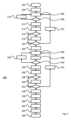
上記で説明したシステム100およびそれの部分のさらなる機能について、図2に示すフローチャート200と併せてさらに説明する。フローチャート200に示した手順は、システム100および詳細には、処理ユニット112によって制御される電子コンピューティングデバイス110によって実行される。この機能を提供するために、処理ユニット112は、コンピュータ実行可能コードを含むコンピュータプログラム製品を用いてプログラムされてもよい。コンピュータプログラム製品は、電子メモリとしてストレージユニット114に記憶されてもよく、電子メモリは非一時的メモリであってもよい。フローチャート200の様々な部分について、以下に簡単にまとめる。様々なステップは、別段に明示的に規定されていない限り、実行の順に入れ替えられても、並行して実行されてもよい。
202 手順の開始
204 3次元血管構造データを取得する
206 中心線に沿ったあるロケーションにおける角度位置として内腔半径を決定する
208 すべての位置が行われたか?
210 次の角度位置に進む
212 内腔面積を決定する
214 半径および面積データを処理する
216 処理されたデータの偏差は?
218 偏差データおよびロケーションを登録する
220 すべてのロケーションが行われたか?
222 次のロケーションに進む
224 血管内取得データを取得する
226 中心線に沿ったあるロケーションにおける角度位置として内腔半径を決定する
228 角度位置の血管壁内の物質を決定し、データを記憶する
230 すべての位置が行われたか?
232 次の角度位置に進む
234 内腔面積を決定する
236 半径および面積データを処理する
238 処理されたデータの偏差は?
240 偏差データおよびロケーションを登録する
242 すべてのロケーションが行われたか?
244 次のロケーションに進む
246 データセットの偏差データをマッチングさせる
248 ロケーション3次元血管構造データおよびIVデータを関連付ける
250 決定された物質データを3次元血管構造データに関連付ける
252 物質データと3次元血管構造データを組み合わせる
254 組み合わされたデータを表示する
256 手順を終了する Further functionality of the
202 Start of procedure
204 Acquire 3D vascular structure data
206 Determine the lumen radius as an angular position at a location along the centerline
208 Have all positions been performed?
210 Go to next angle position
212 Determine Lumen Area
214 Processing Radius and Area Data
216 What is the deviation of the processed data?
218 Registering deviation data and location
220 Have all locations been done?
222 Go to next location
224 Acquire intravascular data
226 Determine the lumen radius as an angular position at a location along the centerline
Determine the material in the vessel wall at 228 angular positions and store the data
230 Have all positions been performed?
232 Go to next angle position
234 Determine Lumen Area
236 Processing Radius and Area Data
238 What is the deviation of the processed data?
240 Register deviation data and location
242 Have all locations been done?
244 Go to next location
Matching deviation data for 246 data sets
248 Locations Correlate 3D Vascular Structure Data and IV Data
250 Linking the determined material data to 3D vascular structure data
252 Combining material data and 3D vascular structure data
254 Displaying Combined Data
256 End procedure
手順は、ターミネータ202から始まり、ステップ204に進み、心臓血管構造180の3次元血管構造に関するデータが取得される。このデータは、システム100(図1)を使用して、または別の方法で、たとえばコンピュータ断層撮影(CT)スキャンを使用して、取得され得る。図3Aは、冠状動脈182が血管壁184および分枝血管188を有する心臓血管構造180の3次元表現を示す。ステップ206において、冠状動脈182内の第1のロケーションにおいて、第1のロケーションの内腔の半径が決定される。この決定の前に、取得されたデータセット内の3次元モデルによって記述される冠状動脈182について中心線が決定され得る。このようにして決定された中心線は、中心線から始まり、冠状動脈182の内壁184に至る半径を決定するために使用され得る。The procedure starts at
図4Aは、図3Aおよび図3Bにおいて「4A」と示されるロケーションでの冠状動脈182の断面を示し、図4Bは、図3Aおよび図3Bにおいて「4B」と示されるロケーションでの冠状動脈184の断面を示す。したがって、図4Aは、図4Bとは異なるロケーションでの冠状動脈184の断面を示す。図4Aと図4Bの両方でわかるように、冠状動脈182の半径は、異なる角度位置で異なる可能性がある。図4Aは、冠状動脈182の狭窄ロケーションでの断面を示す。図4Bは、冠状動脈182から分枝血管188が分岐するロケーションでの断面を示す。FIG. 4A shows a cross-section of
一般に、冠状動脈の断面は、実質的に円形の断面を有する。そのような場合、冠状動脈は、大体回転対称の形状を有する。断面の半径は、中心線における異なる角度位置で異なる可能性があるが、血管内壁184または内腔の湾曲は、中心線から見ると、一般的に凹状である。さらに、異なる角度位置の半径は変化するが、徐々に、特定の境界内で変化する。In general, the cross-section of a coronary artery has a substantially circular cross-section. In such cases, the coronary artery has a roughly rotationally symmetric shape. The radius of the cross-section may be different at different angular positions about the centerline, but the curvature of the vessel
一方、いくつかの場合には、内腔の湾曲は、中心線から凸状の場合があり得る。これは詳細には、血管の壁に大きい異常が存在している場合である。そのような場合、他の場合でもあるが、内腔の半径は、異なる角度位置で大きく異なる可能性があり、必ずしも徐々に変化せず、むしろ急激に変化する可能性がある。On the other hand, in some cases, the curvature of the lumen may be convex from the centerline. This is particularly the case when large abnormalities are present in the vessel wall. In such cases, but also in other cases, the lumen radius may be significantly different at different angular positions and may not necessarily change gradually but rather abruptly.
図4Aおよび図4Bでわかるように、狭窄および分枝のような異常の場合、断面は、回転対称ではない異なる形状を有する可能性がある。さらに、中心線を起点として、異なる角度位置では半径の著しい変化がある可能性がある。As can be seen in Figures 4A and 4B, for abnormalities such as stenosis and bifurcations, the cross sections may have different shapes that are not rotationally symmetric. Furthermore, there may be significant changes in radius at different angular positions starting from the centerline.
上記で説明したように、ステップ206において、冠状動脈182の内腔の半径が、冠状動脈182の長さに沿った、詳細には中心線に沿った特定のロケーションにおいて決定される。それが行われると、データは記憶され、手順はステップ208へと続き、その特定の角度位置での半径を決定するためにすべての角度位置がチェックされたかどうかがチェックされる。角度位置は、等間隔に配分されてもよく、たとえば、10°の間隔で36の測定が行われてもよく、または36°の間隔で10の測定が行われてもよく、または任意の他の数の測定が行われてもよい。すべての位置が処理されたわけではない場合、手順は分岐してステップ210を経由してステップ206に戻り、次の角度位置が選択される。As explained above, in
1つのロケーションについての各位置での半径の値に関して、すべての角度位置が処理された場合、ステップ212において冠状動脈182のアクティブロケーションについて内腔面積が決定される。次に、ステップ214において、1つのロケーションでの決定された半径および面積に関するデータが処理される。処理は、平均、中央値、標準偏差、最小値、最大値、その他、もしくはそれらの任意の組み合わせのような統計的パラメータを計算すること、外れ値を決定し、外れ値を削除すること、ノイズもしくはオフセットなどの測定誤差の補正、他の処理、またはそれらの任意の組み合わせを含んでもよい。When all angular positions have been processed with respect to the radius values at each position for a location, the lumen area is determined for the active locations of the
ステップ216において、断面に偏差があるかどうかがチェックされる。より詳細には、ステップ216において、半径の値が、すべて特定の境界内であるかどうかがチェックされてもよく、特定の境界は、半径の値に基づいているか、ステップ214の処理から得られる値に基づいている所定の固定値、その他の値か、またはそれらの任意の組み合わせを有する。たとえば、半径の1つの値が、中央値または平均値から、中央値または中央値の10%を超えて、またはほぼ1である多数の標準偏差を超えて変わる場合、断面は偏差している断面であると決定され得る。In
断面の変化および断面の偏差の根本的原因は、非常に多いことがある。断面寸法の偏差の明白な原因は、側枝である。他の原因は、血管壁に石灰化物質を有する、石灰化領域であることがある。脂質物質のような、血管壁の他の物質もまた、断面の変化を生じることがある。したがって、側枝が、内腔の偏差と考えられることもある。The underlying causes of cross-sectional changes and cross-sectional deviations can be numerous. An obvious cause of cross-sectional dimension deviations are side branches. Other causes can be calcified areas with calcified material in the vessel wall. Other materials in the vessel wall, such as lipid material, can also cause cross-sectional changes. Thus, side branches can also be considered lumen deviations.
側枝の存在および/またはそれのロケーションは、上記で説明した処理を使用して決定される必要はなく、追加または代替として、たとえば画像認識を使用して幾何学的内腔特徴として検出される場合もある。側枝の存在およびロケーションとは別に、冠状血管または検査中の他の血管の他のランドマーク特徴の存在およびロケーションもまた検出されるか、場合によっては幾何学的内腔特徴として識別され、したがって以下で説明するようにデータの共同登録に使用される可能性がある。The presence and/or location of side branches need not be determined using the processes described above, but may additionally or alternatively be detected as geometric luminal features, for example using image recognition. Apart from the presence and location of side branches, the presence and location of other landmark features of the coronary vessel or other vessel under examination may also be detected or possibly identified as geometric luminal features, and thus used to co-register the data, as described below.
断面が偏差していると決定される場合、断面のロケーションは、ステップ218において登録される。登録では、内腔の面積、冠状動脈184の中心線に沿った位置、様々な角度位置での半径の値、その他、またはそれらの組み合わせのようなさらなるデータが登録される場合がある。その後、手順はステップ220へと続く。断面が偏差していると決定されない場合、手順はステップ216からステップ220の方向を継続する。If the cross section is determined to be deviated, the location of the cross section is registered in
ステップ220において、冠状動脈184の長さに沿ったすべてのロケーションが、半径および面積の決定に関して、ならびに偏差の決定に関して処理されたかどうかがチェックされる。ロケーションの数は、必要とされる精度に基づいて決定されてもよく、ミリメートルまたはセンチメートルまたは長さの他の尺度当たりのロケーションの量として決定されてもよい。追加または代替として、ロケーションの数は、設定されてもよく、ロケーション間の距離は、ロケーションの数および冠状動脈182の長さに基づいて設定されてもよい。すべてのロケーションが処理されたわけではない場合、手順は分岐してステップ222を経由してステップ206に戻り、次のロケーションが選択される。すべてのロケーションが処理された場合、処理はステップ224へ進む。In
ステップ224において、上記で説明したように、血管内で収集された、冠状動脈182に関するデータが取得される。血管内で取得されたデータを含むデータセットは、図3Aによって示す3次元血管構造、すなわち血管がその長さに沿ってどのように湾曲しているかを含んでいない。むしろ、データを収集するプロセスの性質によって、血管で収集されるデータは、データ収集プローブ148が冠状動脈182を通って引っ張られた、または押し込まれた線に沿って提供される。これは、図3Bに示されている。図3Bは、冠状動脈182の中心を示す中心線190を示している。In
中心線190は、様々な方法で決定されてもよく、たとえば、中心は、冠状動脈182の複数の後続の断面図において決定され、中心線は、これらの中心をたどる線として決定され得る。代替的に、中心の移動平均が、2、3、5、10、20、または別の多数の断面図に基づいて決定される。これは、図3Aによって示すデータにも当てはまる。血管の中心線を決定するための様々な方法が、当技術分野で知られており、これらはすべて採用され得る。The
ステップ226において、冠状動脈182の内腔の半径が、特定の角度位置で血管内収集データに基づいて決定される。これは、ステップ206において上記で説明したように行われ得る。その後、ステップ228において、特定の物質が冠状動脈182の壁に存在しているかどうかを決定するために、血管内で収集されたデータが処理される。そのような物質は、脂質物質、石灰化物質、壊死組織、その他、またはそれらの組み合わせである可能性がある。In
そのような物質について、中心線190までの最小距離に対して中心線からの距離が決定され、ならびに中心線190までの最大距離が決定される。そのようなデータは、冠状動脈182の長さに沿った、および詳細には中心線190に沿った複数のロケーションにおいて、複数の角度位置について入手可能であるので、脂質物質186のサイズおよび形状が、中心線に対して、内壁184に対して、冠状動脈182の別の特徴に対して、またはそれらの組み合わせで再構築され得る。For such material, the distance from the centerline is determined relative to the minimum distance to the
ステップ230において、中心線に沿ったすべての角度位置が、半径、および血管内腔の外側の、血管の壁内またはそれの外の何らかの物質を決定するためにカバーされたかどうかが決定される。すべての位置がカバーされたわけではない場合、プロセスは分岐してステップ232を経由してステップ226に戻り、次の角度位置が選択される。半径を決定するためにすべての角度位置がカバーされた場合、手順はステップ234へと続く。In
ステップ234において、ステップ214の動作と同様に、血管内取得データに基づいて、内腔面積が決定される。その後、ステップ236において、半径データ、面積データ、および検出物質データが処理される。処理は、ステップ214に関連して説明した通りであってもよい。その後、ステップ238において、生の半径および面積データに基づいて、処理の結果に基づいて、所定のしきい値のような他のデータに基づいて、適用可能な断面が偏差している断面であるかどうかが決定される。In
半径の大きい変化があると、たとえば、図4Aによって示される断面は、偏差していると特徴づけられてもよい。この例では、内腔の偏差した形状は、冠状血管182の壁内の物質によって引き起こされる可能性がある。図4Aによって示される断面は、回転対称ではないことに留意されたい。またそのような特徴は、断面がこれらの理由のうちの1つまたは複数のために偏差しているかどうかを決定するために用いられてもよい。また分枝血管118のロケーションの図4Bによって示された断面は、偏差していると考えられ得る。分岐に起因する、体積の急激な増加、または半径の値の大きい変化などの、異なる基準が採用されてもよい。あるロケーションの断面が偏差していると考えられる場合、データは、ステップ218の動作と同様に、ステップ240において記憶される。プロセスは、ステップ242へと続く。If there is a large change in radius, for example, the cross section shown by FIG. 4A may be characterized as deviating. In this example, the deviated shape of the lumen may be caused by material in the wall of the
決定される偏差がない場合、手順は直接ステップ242へと続く。ステップ242において、ステップ220と同様に、中心線190に沿ったすべてのロケーションがカバーされたかどうかがチェックされる。すべてのロケーションが評価されたわけではない場合、手順は分岐してステップ226に戻り、ステップ222と同様に、中心線に沿った次のロケーションを選択する。すべてのロケーションが処理された場合、手順はステップ246へと続く。If no deviations are determined, the procedure continues directly to step 242. In
ステップ246において、3次元形状データのために、および血管内収集データのために、上記で説明したように収集された偏差データは、マッチングされる。マッチングは、この変形形態では、詳細には、偏差しているとわかった断面の幾何学的特徴を評価することによって行われる。一変形形態では、第1のデータセット、すなわち幾何学的3次元表現の偏差しているロケーションにおいて取得された偏差している断面の特定のロケーションの複数の位置での半径は、血管内取得データを有する、第2のデータセットの偏差しているロケーションの複数の位置での半径と比較される。In
2つのセットにおけるロケーションでの半径のパターンが、特定のしきい値に満たない違いであることがわかる場合、両方のセットのロケーションは、一致していると考えられてもよい。このステップは、すべてのロケーションでのデータに同様に実行され得るが、偏差しているロケーションのみをマッチングすることによって、処理労力が軽減されることが明らかであろう。第2に、特定のロケーションについての実質的に回転対称で楕円形または円形の断面のマッチングは、一般に、異常がある断面の場合よりも複雑である。If the patterns of radii at the locations in the two sets are found to differ by less than a certain threshold, then both sets of locations may be considered to match. This step may be performed on the data at all locations as well, but it will be apparent that by matching only the locations that deviate, the processing effort is reduced. Second, matching a substantially rotationally symmetric elliptical or circular cross-section for a particular location is generally more complicated than for cross-sections with anomalies.
しかしながら、十分な処理能力および適切なパターン認識およびデータ比較アルゴリズムが利用可能であれば、心臓血管構造180の3次元表現に関するデータを有するセットのデータを、血管内で取得されたデータを有するセットのデータとマッチングさせることが可能である。人工知能の最近の発達に伴い、両方のセットの全部または少なくとも大部分のデータを使用してセットをマッチングさせる技術が利用可能である。However, if sufficient processing power and appropriate pattern recognition and data comparison algorithms are available, it is possible to match a set of data having data relating to a three-dimensional representation of
両方のデータセットにおいて一致している断面が識別されると、ステップ248で第1のセットのデータおよび第2のセットのデータが関連付けられてもよい。詳細には、ロケーションごとの角度データが両方のセットに利用可能であるので、両方のセットのロケーションが関連付けられるだけでなく、1つのセットの特定のデータのロケーションが、別のセットのデータと関連付けられてもよい。たとえば、上記で説明したように、中心線190に対する脂質物質186の角度位置が、血管内収集データに基づいて決定されてもよい。Once matching slices are identified in both data sets, the first and second sets of data may be associated in
中心線に対する角度位置に関連して、断面の形状または半径を関連付けることによって、両方のセットのデータもまた、角度位置に対して関連付けられ得る。このようにして、心臓血管構造180の3次元の幾何学的表現において、詳細には冠状動脈182および詳細にはそれの中心線の長さに関連して、脂質物質186のロケーションを決定することが可能であるが、冠状動脈182に関連して脂質物質186の角度位置を再構築することもまた可能である。By relating the shape or radius of the cross-section in relation to the angular position relative to the centerline, both sets of data can also be related to the angular position. In this way, it is possible to determine the location of the
再構築は、ステップ250において実施され得る。ステップ252では、関連付けに基づいて、両方のデータセットのデータが、ステップ254において組み合わされてもよい。詳細には、第2のデータセットにおいて入手可能な冠状血管182の壁の脂質物質186に関するデータは、このようにして、心臓血管構造180の3次元表現を提供する第1のデータセットのデータと組み合わされてもよい。The reconstruction may be performed in
結果を図5に示し、冠状動脈182に対する実際の長手方向および半径方向位置に脂質物質186がある、心臓血管構造180の3次元表現を示している。この構造は、ステップ254において様々な方法で表示され得る。図5は、マージされたデータの3次元表示を示す。代替または追加として、2次元図が、たとえば、脂質物質186または石灰化物質のような他の物質が、冠状動脈182に対して実際の長手方向および/または半径方向位置にある心臓血管構造180の2次元の幾何学的図を提供され得る。代替または追加として、血管の断面図が、何らかの物質がそこに存在する血管壁全体、および選択されて、存在する場合は、血管周囲物質とともに提供され得る。表示後、手順は、ターミネータ256で終了する。The result is shown in FIG. 5, which shows a three-dimensional representation of the
要約すれば、身体血管、血管壁の異常、および血管周囲の(per-vascular)物質についてのすべてのデータ収集技術に適合する1つの技術はない。したがって、1つのモデルで、または1つの視覚化で、すべての収集可能なデータを集めることができるように、様々なデータ収集方法を使用して取得されたデータが組み合わされることになる。これは、詳細には一部のデータが、CTまたはX線のように、体外から集められ、他のデータが、IVUSまたはOCTのように、血管内で集められる場合、困難である。IVUSおよびOCTでは、血管内の屈曲のような幾何学的データは見えない。側枝のような、血管の特定の特徴に関係する関心点を識別することによって、異なる収集方法を使用して取得されたデータが、データセットにわたって同じ特徴を識別する2つ以上のデータセットの関心点を関連付けること、および画像データに血管およびその周囲の物質を提供するためにデータをマージするための基礎としてそれを使用することにより整合され得る。In summary, there is no one-size-fits-all data collection technique for body vessels, vessel wall anomalies, and per-vascular material. Thus, data acquired using various data collection methods will be combined so that all collectable data can be collected in one model or in one visualization. This is particularly difficult when some data is collected from outside the body, such as CT or X-ray, and other data is collected inside the vessel, such as IVUS or OCT. In IVUS and OCT, geometric data such as bends inside the vessel are not visible. By identifying points of interest that relate to specific features of the vessel, such as side branches, data acquired using different collection methods can be aligned by correlating points of interest in two or more datasets that identify the same feature across datasets, and using that as a basis for merging the data to provide image data of the vessel and its surrounding material.
100 システム
110 電子コンピューティングデバイス
112 処理ユニット
114 ストレージモジュール
116 データ収集モジュール
118 周辺機器I/Oコントローラ
122 第1のX線検出器
124 第2のX線センサー
126 第1のX線源
128 第2のX線源
142 キーボード
144 電子ディスプレイ
146 カテーテル
148 データ収集プローブ
150 造影剤
180 心臓血管構造
182 冠状動脈
184 血管壁
186 脂質物質
188 分枝血管
190 狭窄 100 Systems
110 Electronic Computing Devices
112 Processing Unit
114 Storage Module
116 Data Collection Module
118 Peripheral I/O Controller
122 First X-ray detector
124 Second X-ray sensor
126 The first X-ray source
128 Second X-ray source
142 Keyboard
144 Electronic Display
146 Catheter
148 Data Collection Probes
150 Contrast Agent
180 Cardiovascular Structure
182 Coronary Arteries
184 Blood Vessel Wall
186 Lipid Substances
188 Branching Vessels
190 Stenosis
Claims (19)
Translated fromJapaneseデータの前記第1のセットに基づいて、前記血管の長さに沿った第1のロケーションにおける前記血管の第1の関心点を識別するステップと、
データの前記第2のセットに基づいて、前記血管の長さに沿った第2のロケーションにおける前記血管の第2の関心点を識別するステップと、
一方では前記第1のロケーションおよび前記第2のロケーションに、他方では前記第1の関心点および前記第2の関心点に基づいて、データの前記第1のセットをデータの前記第2のセットにマッチングさせるステップと、
データの前記第2のセットに基づいて前記血管の内腔の周囲の物質を識別し、データの前記第1のセットにおける前記識別された物質の少なくとも1つの第1の物質ロケーションを取得するステップと、
前記マッチングに基づいて、データの前記第1のセットにおける前記識別された物質の前記第1の物質ロケーションを、前記血管の前記長さに関連して、データの前記第2のセットにおける第2の物質ロケーションと関連付けるステップと、
前記血管の前記幾何学的表現と、前記血管の前記幾何学的表現に関係する前記識別された物質のロケーションとの組み合わせを含む、データの第3のセットを提供するステップと
を含む、方法。 1. A method in an electronic computing device for execution by or performed by said electronic computing device of associating a first set of data comprising a geometric representation of a blood vessel in a mammalian, and in particular a human, body with a second set of data relating to a structure of said blood vessel, said second set of data acquired within said blood vessel, comprising:
identifying a first point of interest in the vessel at a first location along a length of the vessel based on the first set of data;
identifying a second point of interest in the vessel at a second location along a length of the vessel based on the second set of data;
matching said first set of data to said second set of data based on said first location and said second location on the one hand and said first interest point and said second interest point on the other hand;
identifying a material surrounding the lumen of the blood vessel based on the second set of data and obtaining at least a first material location of the identified material in the first set of data;
associating the first material location of the identified material in the first set of data with a second material location in the second set of data relative to the length of the blood vessel based on the matching;
providing a third set of data comprising a combination of the geometric representation of the blood vessel and a location of the identified material relative to the geometric representation of the blood vessel.
前記第2の内腔の第2の関心点を識別するステップが、データの前記第2のセットに基づいて、前記血管の第2の長さに沿って、前記内腔が所定の第2のしきい値を超えて所定の形状から偏差する第2の偏差の第2の偏差ロケーションを識別するステップを含み、
前記マッチングが、一方では前記第1の偏差ロケーションおよび前記第2の偏差ロケーションに、他方では前記第1の偏差および前記第2の偏差に基づいて、データの前記第1のセットをデータの前記第2のセットにマッチングさせるステップをさらに含む、
請求項1または2に記載の方法。 identifying a first point of interest in the first lumen includes identifying a first deviation location along a first length of the blood vessel based on the first set of data, where the lumen deviates from a predetermined shape by more than a first predetermined threshold;
identifying a second point of interest in the second lumen includes identifying, based on the second set of data, a second deviation location along a second length of the blood vessel where the lumen deviates from a predetermined shape by more than a second predetermined threshold;
said matching further comprising matching said first set of data to said second set of data based on said first deviation location and said second deviation location on the one hand and said first deviation and said second deviation on the other hand;
The method according to claim 1 or 2.
をさらに含み、
前記マッチングが、前記第1の角度位置および前記第2の角度位置にさらに基づく、請求項1から3のいずれか一項に記載の方法。 obtaining data relating to a first angular position of the first point of interest at the first location relative to the lumen and a second angular position of the second point of interest at the second location relative to the lumen;
The method of claim 1 , wherein the matching is further based on the first angular position and the second angular position.
前記第2の偏差が、前記第2のロケーションにおける、前記血管の中心線と前記血管の内壁との間の第2の距離間の第2の差に基づいて決定される、
請求項3または請求項3に従属する範囲の請求項4に記載の方法。 the first deviation is determined based on a first difference between a first distance between a centerline of the vessel and an inner wall of the vessel at the first location;
the second deviation is determined based on a second difference between a second distance between a centerline of the vessel and an inner wall of the vessel at the second location.
A method according to claim 3 or claim 4 depending thereon.
前記第2の関心点が、前記中心線に対して第2の角度位置の前記第2のロケーションにおいて決定される、
請求項1から5のいずれか一項に記載の方法。 the first point of interest is determined at the first location at a first angular position relative to the centerline;
the second point of interest is determined at the second location at a second angular position relative to the centerline;
6. The method according to any one of claims 1 to 5.
前記第2の関心点が、データの前記第2のセットのデータに含まれる第2のパラメータの第2の値が第2の所定の値とは第2のしきい値を超えて異なる前記第2のロケーションにおける偏差を決定することによって決定される、
請求項1から6のいずれか一項に記載の方法。 the first point of interest is determined by determining a deviation at the first location where a first value of a first parameter included in the first set of data differs from a first predetermined value by more than a first threshold;
the second point of interest is determined by determining a deviation at the second location where a second value of a second parameter included in the second set of data differs from a second predetermined value by more than a second threshold value.
7. The method according to any one of claims 1 to 6.
前記第2のしきい値が、前記第2の値、前記第2の所定の値、および前記第2のしきい値のうちの少なくとも1つに応じた固定値および相対値のうちの少なくとも一方である、
請求項7に記載の方法。 the first threshold value is at least one of a fixed value and a relative value depending on at least one of the first value, the first predetermined value, and the first threshold value;
the second threshold value is at least one of a fixed value and a relative value depending on at least one of the second value, the second predetermined value, and the second threshold value;
The method of claim 7.
前記第2の関心点を決定するステップが、前記血管の前記第1の分枝を識別するステップを含む、
請求項1から8のいずれか一項に記載の方法。 determining the first point of interest includes identifying a first branch of the blood vessel;
determining the second point of interest includes identifying the first branch of the blood vessel;
9. The method according to any one of claims 1 to 8.
前記第2のロケーションにおいて、前記中心線に対する多数の角度ロケーションでの多数の第2の距離を取得するステップと、
前記多数の第1の距離の値の統計的パラメータが、所定の第1の統計的条件に一致する場合、前記第1のロケーションにおける前記第1の偏差を決定するステップと、
前記多数の第2の距離の値の統計的パラメータが、所定の第2の統計的条件に一致する場合、前記第2のロケーションにおける前記第2の偏差を決定するステップと
をさらに含む、請求項5に従属する範囲の請求項6から11のいずれか一項に記載の方法。 obtaining, at the first location, a number of first distances at a number of angular locations relative to the centerline;
obtaining, at the second location, a number of second distances at a number of angular locations relative to the centerline;
determining the first deviation at the first location if a statistical parameter of the multiple first distance values matches a first predetermined statistical condition;
and determining the second deviation at the second location if a statistical parameter of the multiple second distance values matches a predetermined second statistical condition.
前記第2の統計的パラメータが、前記多数の第2の距離の平均、中央値、標準偏差、最大値、および最小値のうちの少なくとも1つに基づいている、
請求項13に記載の方法。 the first statistical parameter is based on at least one of a mean, a median, a standard deviation, a maximum, and a minimum of the plurality of first distances;
the second statistical parameter is based on at least one of a mean, a median, a standard deviation, a maximum, and a minimum of the multiple second distances;
The method of claim 13.
第2の距離の所定量が、前記第2の統計的パラメータとは第2の統計的しきい値を超えて異なる場合、第2の統計的基準が満たされる、
請求項14に記載の方法。 a first statistical criterion is met if a predetermined amount of the first distance differs from the first statistical parameter by more than a first statistical threshold;
a second statistical criterion is met if a predetermined amount of the second distance differs from the second statistical parameter by more than a second statistical threshold;
The method of claim 14.
前記第2の距離の少なくとも1つの値が、第4の統計的パラメータしきい値を超えて前記第2の統計的パラメータとは異なる場合、前記第2の統計的基準が満たされる、
請求項15に記載の方法。 the first statistical criterion is met if at least one value of the first distance differs from the first statistical parameter by more than a third statistical parameter threshold;
the second statistical criterion is met if at least one value of the second distance differs from the second statistical parameter by more than a fourth statistical parameter threshold.
The method of claim 15.
データの前記第1のセットに基づいて、前記血管の長さに沿った第1の位置で、第1のロケーションにおける前記血管の第1の関心点を識別することと、
データの前記第2のセットに基づいて、前記血管の長さに沿った第2の位置で、第2のロケーションにおける前記血管の第2の関心点を識別することと、
一方では前記第1のロケーションおよび前記第2のロケーションに、他方では前記第1の関心点および前記第2の関心点に基づいて、データの前記第1のセットをデータの前記第2のセットにマッチングさせることと、
データの前記第2のセットに基づいて前記内腔の周囲の物質を識別し、前記識別された物質の少なくとも1つの第1の物質ロケーションを取得することと、
前記マッチングに基づいて、前記識別された物質の前記第1の物質ロケーションを、前記血管の前記長さに関連して、データの前記第2のセットにおける第2の物質ロケーションと関連付けることと、
前記血管の前記幾何学的表現、たとえば3次元の表現と、前記血管の前記幾何学的表現に関係する前記識別された物質のロケーションとの組み合わせを含む、データの第3のセットを提供することと
を行うように構成された処理ユニットを備える、デバイス。 1. A device configured to associate a first set of data comprising a geometric representation of a blood vessel in a mammalian, and particularly a human, body with a second set of data relating to a structure of said blood vessel, said second set of data being collected within said blood vessel, said device comprising:
identifying a first point of interest in the vessel at a first location at a first position along a length of the vessel based on the first set of data;
identifying a second point of interest in the vessel at a second location at a second position along a length of the vessel based on the second set of data;
matching the first set of data to the second set of data based on the first location and the second location on the one hand and the first point of interest and the second point of interest on the other hand;
identifying a material surrounding the lumen based on the second set of data and obtaining at least a first material location of the identified material;
associating the first material location of the identified material with a second material location in the second set of data relative to the length of the blood vessel based on the matching;
and providing a third set of data including a combination of the geometric representation, e.g., a three-dimensional representation, of the blood vessel and a location of the identified material relative to the geometric representation of the blood vessel.
データの前記第1のセットに基づいて、前記血管の長さに沿った第1の位置で、第1のロケーションにおける前記血管の第1の関心点を識別するステップと、
データの前記第2のセットに基づいて、前記血管の長さに沿った第2の位置で、第2のロケーションにおける前記血管の第2の関心点を識別するステップと、
一方では前記第1のロケーションおよび前記第2のロケーションに、他方では前記第1の関心点および前記第2の関心点に基づいて、データの前記第1のセットをデータの前記第2のセットにマッチングさせるステップと、
データの前記第2のセットに基づいて前記内腔の周囲の物質を識別し、前記識別された物質の少なくとも1つの第1の物質ロケーションを取得するステップと、
前記マッチングに基づいて、前記識別された物質の前記第1の物質ロケーションを、前記血管の前記長さに関連して、データの前記第2のセットにおける第2の物質ロケーションと関連付けるステップと、
前記血管の前記幾何学的表現と、前記血管の前記幾何学的表現に関係する前記識別された物質のロケーションとの組み合わせを含む、データの第3のセットを提供するステップと
を含む、コンピュータプログラム製品。 1. A computer program product comprising computer executable code configured to cause a processing unit of a processing device, when programmed according to said computer executable code, to execute a method of associating a first set of data comprising a geometric representation of a blood vessel in a mammalian, and in particular a human, body with a second set of data relating to a structure of the blood vessel, the second set of data being collected within the blood vessel, the method comprising:
identifying a first point of interest in the vessel at a first location at a first position along a length of the vessel based on the first set of data;
identifying a second point of interest in the vessel at a second location at a second position along a length of the vessel based on the second set of data;
matching said first set of data to said second set of data based on said first location and said second location on the one hand and said first interest point and said second interest point on the other hand;
identifying a material surrounding the lumen based on the second set of data and obtaining at least a first material location of the identified material;
associating the first material location of the identified material with a second material location in the second set of data relative to the length of the blood vessel based on the matching;
and providing a third set of data comprising a combination of the geometric representation of the blood vessel and a location of the identified material relative to the geometric representation of the blood vessel.
データの前記第1のセットに基づいて、前記血管の長さに沿った第1の位置で、第1のロケーションにおける前記血管の第1の関心点を識別するステップと、
データの前記第2のセットに基づいて、前記血管の長さに沿った第2の位置で、第2のロケーションにおける前記血管の第2の関心点を識別するステップと、
一方では前記第1のロケーションおよび前記第2のロケーションに、他方では前記第1の関心点および前記第2の関心点に基づいて、データの前記第1のセットをデータの前記第2のセットにマッチングさせるステップと、
データの前記第2のセットに基づいて前記内腔の周囲の物質を識別し、前記識別された物質の少なくとも1つの第1の物質ロケーションを取得するステップと、
前記マッチングに基づいて、前記識別された物質の前記第1の物質ロケーションを、前記血管の前記長さに関連して、データの前記第2のセットにおける第2の物質ロケーションと関連付けるステップと、
前記血管の前記幾何学的表現と、前記血管の前記幾何学的表現に関係する前記識別された物質のロケーションとの組み合わせを含む、データの第3のセットを提供するステップと
を含む、非一時的媒体。 1. A non-transitory medium carrying a computer program product comprising computer executable code configured to cause a processing unit of a processing device, when programmed according to said computer executable code, to execute a method of associating a first set of data comprising a geometric representation of a blood vessel in a mammalian, and in particular a human, body with a second set of data relating to a structure of the blood vessel, the second set of data being collected within the blood vessel, the method comprising:
identifying a first point of interest in the vessel at a first location at a first position along a length of the vessel based on the first set of data;
identifying a second point of interest in the vessel at a second location at a second position along a length of the vessel based on the second set of data;
matching said first set of data to said second set of data based on said first location and said second location on the one hand and said first interest point and said second interest point on the other hand;
identifying a material surrounding the lumen based on the second set of data and obtaining at least a first material location of the identified material;
associating the first material location of the identified material with a second material location in the second set of data relative to the length of the blood vessel based on the matching;
and providing a third set of data comprising a combination of the geometric representation of the blood vessel and a location of the identified material relative to the geometric representation of the blood vessel.
Applications Claiming Priority (3)
| Application Number | Priority Date | Filing Date | Title |
|---|---|---|---|
| NL2030789 | 2022-01-31 | ||
| NL2030789ANL2030789B1 (en) | 2022-01-31 | 2022-01-31 | method and device for associating sets of cardiovascular data |
| PCT/NL2023/050037WO2023146401A1 (en) | 2022-01-31 | 2023-01-30 | Method and device for associating sets of cardiovascular data |
Publications (1)
| Publication Number | Publication Date |
|---|---|
| JP2025503309Atrue JP2025503309A (en) | 2025-01-30 |
Family
ID=80625464
Family Applications (1)
| Application Number | Title | Priority Date | Filing Date |
|---|---|---|---|
| JP2024545769APendingJP2025503309A (en) | 2022-01-31 | 2023-01-30 | Method and device for correlating sets of cardiovascular data - Patents.com |
Country Status (5)
| Country | Link |
|---|---|
| US (1) | US20250131567A1 (en) |
| EP (1) | EP4473482A1 (en) |
| JP (1) | JP2025503309A (en) |
| NL (1) | NL2030789B1 (en) |
| WO (1) | WO2023146401A1 (en) |
Families Citing this family (4)
| Publication number | Priority date | Publication date | Assignee | Title |
|---|---|---|---|---|
| US10210956B2 (en) | 2012-10-24 | 2019-02-19 | Cathworks Ltd. | Diagnostically useful results in real time |
| IL263065B2 (en) | 2016-05-16 | 2024-08-01 | Cathworks Ltd | System for vascular assessment |
| US12315076B1 (en) | 2021-09-22 | 2025-05-27 | Cathworks Ltd. | Four-dimensional motion analysis of a patient's coronary arteries and myocardial wall |
| JP2025506500A (en) | 2022-02-10 | 2025-03-11 | キャスワークス リミテッド | Systems and methods for machine learning based sensor analysis and vascular tree segmentation |
Family Cites Families (1)
| Publication number | Priority date | Publication date | Assignee | Title |
|---|---|---|---|---|
| JP2012075702A (en)* | 2010-10-01 | 2012-04-19 | Fujifilm Corp | Apparatus, method, and program for reconstructing intra-tubular-structure image |
- 2022
- 2022-01-31NLNL2030789Apatent/NL2030789B1/enactive
- 2023
- 2023-01-30JPJP2024545769Apatent/JP2025503309A/enactivePending
- 2023-01-30USUS18/833,975patent/US20250131567A1/enactivePending
- 2023-01-30EPEP23703365.9Apatent/EP4473482A1/enactivePending
- 2023-01-30WOPCT/NL2023/050037patent/WO2023146401A1/ennot_activeCeased
Also Published As
| Publication number | Publication date |
|---|---|
| US20250131567A1 (en) | 2025-04-24 |
| EP4473482A1 (en) | 2024-12-11 |
| NL2030789B1 (en) | 2023-08-08 |
| WO2023146401A1 (en) | 2023-08-03 |
Similar Documents
| Publication | Publication Date | Title |
|---|---|---|
| US12217872B2 (en) | Diagnostically useful results in real time | |
| US11406337B2 (en) | Calculating a fractional flow reserve | |
| CN111134651B (en) | Method, device and system for calculating fractional flow reserve based on intracavity images and computer storage medium | |
| US9977869B2 (en) | Vascular flow assessment | |
| CN115515504B (en) | Automatic control of intraluminal data acquisition and related devices, systems, and methods | |
| JP2025503309A (en) | Method and device for correlating sets of cardiovascular data - Patents.com | |
| CN106572824B (en) | Stenosis assessment | |
| US8538508B2 (en) | Method and apparatus for ECG-synchronized optically-based image acquisition and transformation | |
| US20200129147A1 (en) | Intraluminal ultrasound vessel border selection and associated devices, systems, and methods | |
| US20190282211A1 (en) | Scoring intravascular lesions and stent deployment in medical intraluminal ultrasound imaging | |
| CN113614845A (en) | Arterial imaging and assessment system and method and related user interface based workflow | |
| JP7152955B2 (en) | System and method for motion-based image segmentation | |
| US12394080B2 (en) | Intraluminal image-based vessel diameter determination and associated devices, systems, and methods | |
| US20220395333A1 (en) | Co-registration of intravascular data and multi-segment vasculature, and associated devices, systems, and methods | |
| US20160066795A1 (en) | Stenosis therapy planning | |
| US20230118551A1 (en) | Self expanding stent system with imaging | |
| Bourantas et al. | Comparison of quantitative coronary angiography with intracoronary ultrasound. Can quantitative coronary angiography accurately estimate the severity of a luminal stenosis? | |
| CN114463267B (en) | Method, device and computer storage medium for predicting blood flow reserve fraction based on optical coherence tomography | |
| CN118121230B (en) | Lower limb venous blood flow pressure analysis method and device, storage medium and electronic equipment |
Legal Events
| Date | Code | Title | Description |
|---|---|---|---|
| A521 | Request for written amendment filed | Free format text:JAPANESE INTERMEDIATE CODE: A523 Effective date:20241111 |