JP2024542644A - Method and processor for analyzing the vasculature of a subject - Patents.com - Google Patents
Method and processor for analyzing the vasculature of a subject - Patents.comDownload PDFInfo
- Publication number
- JP2024542644A JP2024542644AJP2024532224AJP2024532224AJP2024542644AJP 2024542644 AJP2024542644 AJP 2024542644AJP 2024532224 AJP2024532224 AJP 2024532224AJP 2024532224 AJP2024532224 AJP 2024532224AJP 2024542644 AJP2024542644 AJP 2024542644A
- Authority
- JP
- Japan
- Prior art keywords
- vessels
- collateral
- image
- tree
- interest
- Prior art date
- Legal status (The legal status is an assumption and is not a legal conclusion. Google has not performed a legal analysis and makes no representation as to the accuracy of the status listed.)
- Pending
Links
Images


Classifications
- G—PHYSICS
- G06—COMPUTING OR CALCULATING; COUNTING
- G06T—IMAGE DATA PROCESSING OR GENERATION, IN GENERAL
- G06T7/00—Image analysis
- G06T7/0002—Inspection of images, e.g. flaw detection
- G06T7/0012—Biomedical image inspection
- G—PHYSICS
- G06—COMPUTING OR CALCULATING; COUNTING
- G06T—IMAGE DATA PROCESSING OR GENERATION, IN GENERAL
- G06T7/00—Image analysis
- G06T7/0002—Inspection of images, e.g. flaw detection
- G06T7/0012—Biomedical image inspection
- G06T7/0014—Biomedical image inspection using an image reference approach
- G—PHYSICS
- G06—COMPUTING OR CALCULATING; COUNTING
- G06T—IMAGE DATA PROCESSING OR GENERATION, IN GENERAL
- G06T7/00—Image analysis
- G06T7/10—Segmentation; Edge detection
- G06T7/11—Region-based segmentation
- G—PHYSICS
- G06—COMPUTING OR CALCULATING; COUNTING
- G06T—IMAGE DATA PROCESSING OR GENERATION, IN GENERAL
- G06T7/00—Image analysis
- G06T7/60—Analysis of geometric attributes
- G06T7/62—Analysis of geometric attributes of area, perimeter, diameter or volume
- G—PHYSICS
- G06—COMPUTING OR CALCULATING; COUNTING
- G06T—IMAGE DATA PROCESSING OR GENERATION, IN GENERAL
- G06T2207/00—Indexing scheme for image analysis or image enhancement
- G06T2207/10—Image acquisition modality
- G06T2207/10028—Range image; Depth image; 3D point clouds
- G—PHYSICS
- G06—COMPUTING OR CALCULATING; COUNTING
- G06T—IMAGE DATA PROCESSING OR GENERATION, IN GENERAL
- G06T2207/00—Indexing scheme for image analysis or image enhancement
- G06T2207/10—Image acquisition modality
- G06T2207/10072—Tomographic images
- G—PHYSICS
- G06—COMPUTING OR CALCULATING; COUNTING
- G06T—IMAGE DATA PROCESSING OR GENERATION, IN GENERAL
- G06T2207/00—Indexing scheme for image analysis or image enhancement
- G06T2207/10—Image acquisition modality
- G06T2207/10072—Tomographic images
- G06T2207/10081—Computed x-ray tomography [CT]
- G—PHYSICS
- G06—COMPUTING OR CALCULATING; COUNTING
- G06T—IMAGE DATA PROCESSING OR GENERATION, IN GENERAL
- G06T2207/00—Indexing scheme for image analysis or image enhancement
- G06T2207/10—Image acquisition modality
- G06T2207/10116—X-ray image
- G—PHYSICS
- G06—COMPUTING OR CALCULATING; COUNTING
- G06T—IMAGE DATA PROCESSING OR GENERATION, IN GENERAL
- G06T2207/00—Indexing scheme for image analysis or image enhancement
- G06T2207/20—Special algorithmic details
- G06T2207/20112—Image segmentation details
- G06T2207/20128—Atlas-based segmentation
- G—PHYSICS
- G06—COMPUTING OR CALCULATING; COUNTING
- G06T—IMAGE DATA PROCESSING OR GENERATION, IN GENERAL
- G06T2207/00—Indexing scheme for image analysis or image enhancement
- G06T2207/30—Subject of image; Context of image processing
- G06T2207/30004—Biomedical image processing
- G06T2207/30048—Heart; Cardiac
- G—PHYSICS
- G06—COMPUTING OR CALCULATING; COUNTING
- G06T—IMAGE DATA PROCESSING OR GENERATION, IN GENERAL
- G06T2207/00—Indexing scheme for image analysis or image enhancement
- G06T2207/30—Subject of image; Context of image processing
- G06T2207/30004—Biomedical image processing
- G06T2207/30101—Blood vessel; Artery; Vein; Vascular
- G—PHYSICS
- G06—COMPUTING OR CALCULATING; COUNTING
- G06T—IMAGE DATA PROCESSING OR GENERATION, IN GENERAL
- G06T2207/00—Indexing scheme for image analysis or image enhancement
- G06T2207/30—Subject of image; Context of image processing
- G06T2207/30172—Centreline of tubular or elongated structure
- G—PHYSICS
- G06—COMPUTING OR CALCULATING; COUNTING
- G06T—IMAGE DATA PROCESSING OR GENERATION, IN GENERAL
- G06T2207/00—Indexing scheme for image analysis or image enhancement
- G06T2207/30—Subject of image; Context of image processing
- G06T2207/30242—Counting objects in image
Landscapes
- Engineering & Computer Science (AREA)
- Physics & Mathematics (AREA)
- Computer Vision & Pattern Recognition (AREA)
- General Physics & Mathematics (AREA)
- Theoretical Computer Science (AREA)
- Health & Medical Sciences (AREA)
- General Health & Medical Sciences (AREA)
- Medical Informatics (AREA)
- Nuclear Medicine, Radiotherapy & Molecular Imaging (AREA)
- Radiology & Medical Imaging (AREA)
- Quality & Reliability (AREA)
- Geometry (AREA)
- Apparatus For Radiation Diagnosis (AREA)
- Image Analysis (AREA)
Abstract
Translated fromJapanese
Description
Translated fromJapanese本発明は、被検体の血管を表示している画像の解析に関する。The present invention relates to the analysis of images showing the blood vessels of a subject.
冠動脈系又は末梢系の血管疾患は、狭窄性病変や体内組織の血液供給における完全閉塞を伴うことが多い。Vascular disease of the coronary or peripheral system often involves stenotic lesions or complete blockages in the blood supply to body tissues.
側副血管形成の存在及び範囲は、2つの異なる状況の重要なマーカーとして認識されている。まず、側副血管は、人体の代償機構の作用の指標として見出されている。つまり、狭窄性病変は、側副循環の発達によって補償され、さもなければ強い虚血性で壊死しやすい部位へのかん流を維持する。したがって、側副の形成は、患者の転帰との負の相関関係を明らかにしている。つまり、側副の形成が不良ではなく適正である患者の予後は良好であることが判明している。次に、これらの側副は血液供給問題を十分に解決しないことも知られている。特にストレス下では、障害が依然として示されるためである。The presence and extent of collateral vessel formation is recognized as an important marker in two different situations. First, collateral vessels are found as an indicator of the action of the body's compensatory mechanisms: stenotic lesions are compensated by the development of collateral circulation, maintaining perfusion to areas that are otherwise highly ischemic and prone to necrosis. Thus, collateral formation reveals a negative correlation with patient outcome: patients with adequate, rather than poor, collateral formation have been found to have a good prognosis. Second, it is also known that these collaterals do not fully solve the blood supply problem, since failures are still demonstrated, especially under stress.
これら2つの作用のいずれも、側副循環の関連性と血管状態の定量化の必要性とを強調する。関心のパラメータは、特に、側副の絶対数、それらの直径(実際の血管の代わりにこれらの側副血行路を通過している血液量の指標であるため)、側副血行路の長さ、及び血管木のどの部分が迂回されているかである。Both of these effects emphasize the relevance of collateral circulation and the need to quantify the vascular status. Parameters of interest are, in particular, the absolute number of collaterals, their diameter (as it is an indicator of the amount of blood passing through these collaterals instead of the actual blood vessels), the length of the collaterals, and which parts of the vascular tree are bypassed.
血管系を解析するためのアルゴリズムが知られている。しかし、ほとんどのアルゴリズムソリューションは現在、主血管の検出に焦点を当てている。主血管はほとんどの患者で確実に検出され、通常は介入のターゲットであるためである。Algorithms are known for analyzing the vasculature. However, most algorithmic solutions currently focus on detecting main vessels, as these are reliably detected in most patients and are usually the target of intervention.
しかしながら、大きな解剖学的変異の小さな血管がいくつかある。血管木のこの残りの部分の特性は、全身性疾患の状態を示すマーカーを提供するが、現在のところ、最先端の解析応用では定量化されていない。しかし、血管系のこの無視された部分には、閉塞部位又は狭窄性部位の自然の側副血行路として機能する血管木の異なる部分を相互接続する多くの側副血管が含まれている場合がある。However, there are some small vessels of great anatomical variation. The properties of this remaining part of the vascular tree provide markers of systemic disease status, but are currently not quantified by state-of-the-art analytical applications. However, this neglected part of the vascular system may contain many collateral vessels interconnecting different parts of the vascular tree that act as natural collaterals for occluded or stenosed areas.
そのため、特に主要な血管木に血管疾患を有する患者において、無視されることの多い側副形成の血管状態を定量化する必要がある。Therefore, there is a need to quantify the often neglected vascular status of collateral formation, especially in patients with vascular disease in the major vascular trees.
Van Horssen Pepijn他の「Innate collateral segments are predominantly present in the subendocardium without preferential connectivity within the left ventricular wall:Distribution and morphology of innate collateral connections」、Journal of Physiology、第592巻、第5号、2014年1月23日、1047~1060頁(XP055920694)は、慢性動脈疾患が発症すると側副動脈が成長し、閉塞した冠動脈のかん流部位への酸素豊富な血液の経路を提供するという理解に基づいた動脈の解析を開示している。健康な心臓における先天性の側副ネットワークの形態及び分布が定量化されている。Van Horssen Pepijn et al., “Innate collaborative segments are predominantly present in the subendocardium without preferential connectivity within the left ventricular wall: Distribution and morphology of innate collaborative connections”, Journal of Physiology, Vol. 592, No. 5, January 23, 2014, pp. 1047-1060 (XP055920694) reported that collateral arteries grow during the development of chronic arterial disease, providing oxygen-rich blood to the perfusion site of the occluded coronary artery. The paper presents an analysis of arteries based on the understanding that they provide an optimal pathway for blood flow. The morphology and distribution of the innate collateral network in healthy hearts is quantified.
本発明は、特許請求の範囲によって規定される。The invention is defined by the claims.
本発明の態様による実施例によれば、被検体の血管系を解析する方法が提供される。この方法は、
被検体の関心領域の画像を受信するステップと、
関心領域に存在する血管木を同定するステップと、
同定された血管木内の主要血管を同定するステップであって、主要血管は、主要血管の標準木の一部を形成する、同定するステップと、
同定された血管木の残りの血管を同定し、したがって、標準木の主要血管を除外し、これにより、関心領域内の側副血管を同定するステップと、
側副血管の解析を行うステップとを含む。 According to an embodiment of the present invention, there is provided a method for analysing the vasculature of a subject, the method comprising:
receiving an image of a region of interest of a subject;
Identifying a vascular tree present in a region of interest;
- identifying major vessels within the identified vascular tree, the major vessels forming part of a standard tree of major vessels;
- identifying remaining vessels of the identified vascular tree, thus excluding the main vessels of the standard tree, thereby identifying collateral vessels within the region of interest;
and performing a collateral vessel analysis.
この解析では、特に側副血管に関連する血管状態の定量化に使用するために臨床医に報告される測定値を提供できる。これは主要血管木に血管疾患の存在を示す。This analysis can provide measurements that can be reported to clinicians for use in quantifying vascular status, particularly in relation to collateral vessels, which indicate the presence of vascular disease in the primary vascular tree.
この方法では、まず、検出された血管木全体(つまり、正味)の一部として標準血管木を検出する。標準木は、例えば健康な個人にも存在する主要な大血管幹に基づいた、階層的なルールベースの分類によって抽出される。血管木の統計的アトラスを使用してもよい。統計的アトラスでは、木の構造パラメータ及び位置パラメータに確率が割り当てられる。The method first detects a standard vascular tree as a part of the entire (i.e. net) detected vascular tree. The standard tree is extracted by a hierarchical rule-based classification, e.g. based on the major large vascular trunks that are also present in healthy individuals. A statistical atlas of vascular trees may be used, in which probabilities are assigned to the structural and positional parameters of the tree.
残りの血管系は側副血管を含み、これらの血管は、特に側副血管形成の解剖学的特性を定量化するために解析される。残りの血管は、同定された血管木から主要血管を減算することによって見つけられる。例えば、定量化された解剖学的特性には、側副血管の数、側副血管の内腔面積若しくは血液量、血管長、又は正味の血管密度などのパラメータが含まれる。The remaining vasculature includes collateral vessels, which are analyzed to specifically quantify anatomical characteristics of collateral vessel formation. The remaining vessels are found by subtracting the main vessels from the identified vascular tree. For example, the anatomical characteristics quantified include parameters such as the number of collateral vessels, the luminal area or blood volume of the collateral vessels, vessel length, or net vascular density.
関心領域に存在する血管木を同定するステップは、例えば画像セグメンテーション及び血管中心線抽出を含む。2D画像データ及び3D画像データの両方から血管形状を抽出するためのアルゴリズムは知られている。画像データは、冠動脈コンピューダ断層撮影血管造影(CCTA)画像などの低解像度画像に適用できる。Identifying the vascular tree present in the region of interest includes, for example, image segmentation and vascular centerline extraction. Algorithms are known for extracting vascular geometry from both 2D and 3D image data. The image data can be applied to low-resolution images such as coronary computed tomography angiography (CCTA) images.
主要血管を同定するステップは、例えば階層的なルールベースの分類を使用するステップを含む。ここでも、このためのアルゴリズムは知られている。Identifying major vessels may involve, for example, using hierarchical rule-based classification. Again, algorithms for this are known.
側副血管の解析には、側副血管の形成を示す1つ以上のパラメータの決定が含まれる。Collateral analysis involves determining one or more parameters indicative of collateral formation.
第1の実施例では、パラメータは側副血管の数を含む。In a first embodiment, the parameters include the number of collateral vessels.
第2の実施例では、パラメータは側副血管の累積長を含む。In a second embodiment, the parameters include the cumulative length of collateral vessels.
第3の実施例では、パラメータは側副内腔ボリュームを含む。In a third embodiment, the parameters include collateral lumen volume.
第4の実施例では、パラメータは血管間面積に関連している。このパラメータは、例えば基準表面への投影で計算された正味の血管密度を含む。この投影により、関心領域の3Dボリュームを表す2D画像の解析によって、正味密度を取得できる。例えば基準表面は、セグメント化された心臓画像と並べられた心外膜壁を含む。In a fourth embodiment, the parameter is related to the intervascular area. This parameter comprises, for example, the net vessel density calculated by projection onto a reference surface. This projection allows the net density to be obtained by analysis of a 2D image representing the 3D volume of the region of interest. For example, the reference surface comprises the epicardial wall aligned with the segmented cardiac image.
側副血管の解析を行うステップは、例えば標準木のパラメータの値に対してパラメータを正規化するステップを含む。The step of analyzing collateral vessels includes, for example, a step of normalizing parameters with respect to the parameter values of a standard tree.
或いは、側副血管の解析を行うステップは、健康な被検体に対応するパラメータの基準値に対して、又は特定の患者グループの基準値に対してパラメータを正規化するステップを含んでいてもよい。Alternatively, performing the collateral analysis may include normalizing the parameters to baseline values of the parameters corresponding to healthy subjects or to baseline values for a particular patient group.
どちらの場合も、これは標準血管木又は健康な集団を参照してすぐに理解可能な尺度を提供する。In either case, this provides a measure that can be readily understood with reference to a standard vascular tree or a healthy population.
受信画像は、3Dコンピュータ断層撮影(CT)血管造影画像や3D磁気共鳴血管造影(MRA)画像などの3D画像であってもよい。The received images may be 3D images, such as 3D computed tomography (CT) angiography images or 3D magnetic resonance angiography (MRA) images.
ただし、受信画像は、2D X線血管造影画像などの2D画像であってもよい。However, the received image may also be a 2D image, such as a 2D X-ray angiogram image.
したがって、本発明は、診断3Dイメージングと介入2D血管造影の両方に適用可能である。The invention is therefore applicable to both diagnostic 3D imaging and interventional 2D angiography.
本発明はまた、コンピュータプログラムコード手段を含むコンピュータプログラムを提供する。コンピュータプログラムコード手段は、該コンピュータプログラムがコンピュータ上で実行されると、前述の方法を実施する。The present invention also provides a computer program comprising computer program code means for implementing the above-mentioned method when the computer program is run on a computer.
本発明はまた、上記のコンピュータプログラムでプログラムされたプロセッサも提供する。The present invention also provides a processor programmed with the above computer program.
本発明はまた、イメージングシステムも提供する。このシステムは、
被検体の関心領域の画像を取得するためのイメージャと、
画像を解析して、関心領域内の側副血管の解析を行うための上記のプロセッサとを含む。 The present invention also provides an imaging system comprising:
an imager for acquiring an image of a region of interest of the subject;
and a processor as described above for analyzing the image to perform an analysis of collateral vessels within the region of interest.
本発明のこれらの及び他の態様は、以下に説明する実施形態から明らかになり、また、当該実施形態を参照して説明される。These and other aspects of the invention will become apparent from and be elucidated with reference to the embodiments described hereinafter.
本発明をより深く理解し、それがどのように実行されるかをより明確に示すために、ほんの一例として添付の図面を参照する。For a better understanding of the invention and to show more clearly how it may be carried into effect, reference is made, by way of example only, to the accompanying drawings, in which:
本発明を、図を参照して説明する。The present invention will be described with reference to the drawings.
詳細な説明及び具体的な実施例は、装置、システム、及び方法の模範的な実施形態を示しているが、説明のみを目的としたものであり、発明の範囲を限定することを意図したものではないことが理解されるべきである。本発明の装置、システム、及び方法のこれらの及び他の特徴、態様並びに利点は、次の説明、添付の特許請求の範囲、及び添付の図面からよりよく理解されるようになるであろう。図は概略図に過ぎず、縮尺どおりに描かれていないことが理解されるべきである。また、図全体で同じ参照番号を使用して、同じ部分又は類似の部品を示すことが理解されるべきである。It should be understood that the detailed description and specific examples, while illustrating exemplary embodiments of the devices, systems, and methods, are for purposes of illustration only and are not intended to limit the scope of the invention. These and other features, aspects, and advantages of the devices, systems, and methods of the present invention will become better understood from the following description, the appended claims, and the accompanying drawings. It should be understood that the figures are schematic representations only and are not drawn to scale. It should also be understood that the same reference numerals are used throughout the figures to indicate the same or similar parts.
本発明は、被検体の血管系を解析するための方法を提供する。関心領域の画像内で、血管木が同定される。主要血管の標準木の一部を形成する主要血管が同定され、次に、同定された血管木の残りの血管が同定され、標準木の主要血管は除外される。これにより、関心領域内の側副血管が分離され、側副血管の解析を行うことができる。The present invention provides a method for analyzing the vasculature of a subject. Within an image of a region of interest, a vascular tree is identified. Major vessels that form part of a standard tree of major vessels are identified, and then the remaining vessels of the identified vascular tree are identified, excluding the major vessels of the standard tree. This allows collateral vessels within the region of interest to be isolated and analyzed.
図1では、上図に心臓の3D画像を示し、主要血管10が示されている。主要血管10はほとんどの患者に見られるが、より細かいスケールでは血管はより強く異なっている可能性がある。これらのうち、血流の側副血行路を確立できる多数の側副血管がある可能性がある。これらの側副の数及び範囲は、冠動脈疾患を示す説明的なマーカーであることがわかっている。In FIG. 1, a 3D image of the heart is shown at the top, depicting the
図1は、下図に、血管のネットワーク全体を示し、小さい側副血管を示している。冠動脈血管木により、心筋かん流が確保される。解剖学的変異は、木がより細かくなるほど大きくなる。Figure 1 shows the entire vascular network at the bottom, showing small collateral vessels. The coronary vascular tree ensures myocardial perfusion. The anatomical variations are greater the finer the tree.
図2は、足の末梢血管系を示している。上図では、患者は足部には疾患がなく(又は、図3よりも疾患の程度は少ない)、主にふくらはぎから来て足に供給する3つの主要血管を示している。下図は、下ふくらはぎと足に重度の閉塞がある患者のものである。代償機構として、いくつかの側副が発達している。Figure 2 shows the peripheral vasculature of the foot. In the top image, the patient has no disease in the foot (or less disease than in Figure 3), and shows three major vessels that come primarily from the calf and supply the foot. The bottom image is from a patient with severe obstruction in the lower calf and foot. As a compensatory mechanism, several collaterals have developed.
本発明は、解析のために側副血管を分離することに基づいている。The invention is based on isolating collateral vessels for analysis.
図4は、被検体の血管系を解析する方法を示す。Figure 4 shows a method for analyzing the vascular system of a subject.
ステップ40では、被検体の関心領域の画像が取得される。これは、心臓付近の血管木の解析のための胸部領域であり得るが、どの関心領域であってもよい。In
ステップ42では、関心領域に存在する血管木が同定される。In
ステップ44では、主要血管、つまり、主要血管の標準木の一部を形成する主要血管が同定される。In
次に、ステップ46では、同定された血管木内の残りの血管、つまり、関心領域に存在する、標準木の主要血管は除いた全ての同定された血管が同定される。したがって、これらは関心領域の側副血管である。Next, in
ステップ48では、側副血管の解析が行われる。この解析では、画像データに適用された1つ以上の尺度に基づいて、1つ以上の解剖学的パラメータが記述される。In
これらのパラメータは、特に主要血管木に血管疾患がある場合に、血管の状態を定量化するために使用される。血液供給された側副の数及び特徴は、身体がその標準的な経路に依存できなくなる程度、したがって、側副が全体の血液供給を維持する上でどの程度重要な役割を果たすかを示す。These parameters are used to quantify vascular status, especially in the presence of vascular disease in the primary vascular tree. The number and characteristics of blood-supplied collaterals indicate the extent to which the body can no longer rely on its standard pathways and, therefore, how important a role the collaterals play in maintaining the overall blood supply.
図5~図7は、Graham他による「Robust 3-D Airway Tree Segmentation for Image-Guided Peripheral Broncoscopy」(TMI、29:4、2010)に説明されているプロセスを概略図で示している。Figures 5-7 show a schematic diagram of the process described in "Robust 3-D Airway Tree Segmentation for Image-Guided Peripheral Broncoscopy" by Graham et al. (TMI, 29:4, 2010).
図5は、3D CTデータなどの3D画像からどのように血管系を同定するかを概略的に示す。Figure 5 shows a schematic of how vasculature is identified from a 3D image, such as 3D CT data.
一連の2D楕円A、B、Cが取得される。これは、一組の単純な閾値化操作の後に、連結成分解析及び局所楕円フィッティングを行うことによって、3つの軸(x、y、z)全てに沿って行われる。A set of 2D ellipses A, B, C is obtained along all three axes (x, y, z) by a set of simple thresholding operations followed by connected component analysis and local ellipse fitting.
楕円は、3つの連続した楕円を接続する血管中心線60に直交する平面に投影され、軸と整列しなくなる最終的な楕円Dがもたらされる。The ellipses are projected onto a plane perpendicular to the
図6は、楕円A~C、血管の決定された向き、及び血管軸に垂直な断面における血管形状Dを示す。この図から、血管の直径及び面積を求めることができる。Figure 6 shows ellipses A-C, the determined orientation of the vessel, and the vessel shape D in a cross section perpendicular to the vessel axis. From this figure, the diameter and area of the vessel can be determined.
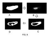
図7は、どのように血管系がセグメント化されるかを示す。パネルAに示すように、一連の楕円が取得される。これらはパネルBに示すように、血管セグメントのセットに集められる。血管セグメント間の間隙は、パネルCに示すように、補間ステップによって埋められ、血管枝が得られる。血管枝は、パネルDに示すように、ボクセル化され、全ての枝の結合によりバイナリ血管マスクが得られる。Figure 7 shows how the vasculature is segmented. A set of ellipses is obtained, as shown in panel A. These are assembled into a set of vessel segments, as shown in panel B. The gaps between the vessel segments are filled by an interpolation step, as shown in panel C, to obtain the vessel branches. The vessel branches are voxelized, as shown in panel D, and the union of all the branches results in a binary vessel mask.
この手順の詳細は、
Graham、Robust Methods for Human Airway-Tree Segmentation and Anatomical-Tree Matching、博士論文、2008、https://etda.libraries.psu.edu/files/final_submissions/4430に記載されている。 For more information about this procedure, see
Graham, Robust Methods for Human Airway-Tree Segmentation and Anatomical-Tree Matching, PhD thesis, 2008, https://etda.libraries.psu.edu/files/final_submissions/4430.
要約すると、セグメンテーションステップでは、軸整列した楕円を使用して局所情報を収集し、バイナリボクセルマスクを生成する。任意選択で、セグメンテーションモジュールは、外部の確率的アトラスを参照として使用してもよい。In summary, the segmentation step uses axis-aligned ellipses to collect local information and generate a binary voxel mask. Optionally, the segmentation module may use an external probabilistic atlas as a reference.
図8は、3D CTデータなどの3D画像データから中心線を抽出する手順を示している。パネルAに示すバイナリマスクから開始して、パネルBに示すように距離変換を使用する。そして、パネルCに示すように、中心線トラッカの入力として、ピークまでの距離(distance-to-peak)マスクが適用される。また、カルシウムペナルティ80を、平均大動脈強度+3標準偏差に基づく閾値と共に使用して、中心線が石灰化組織を通過しないようにすることができる。Figure 8 shows the procedure for extracting centerlines from 3D image data, such as 3D CT data. Starting with a binary mask, shown in panel A, a distance transform is used, as shown in panel B. Then, a distance-to-peak mask is applied as input to the centerline tracker, as shown in panel C. Also, a calcium penalty of 80 can be used along with a threshold based on the mean aortic intensity plus 3 standard deviations to ensure that the centerline does not pass through calcified tissue.
したがって、中心線抽出は、血管によって占められている画像ボリュームを含むバイナリマスクを、枝を次々にトレースすることによってツリー構造に変換するプロセスである。Centerline extraction is therefore the process of converting a binary mask containing the image volume occupied by the vessels into a tree structure by tracing its branches one after the other.
その後、後処理を使用して静脈枝を取り除き、二重検出を除去できる。平滑化を適用することもできる。最大長、分岐深さ、血管内径、冠動脈血管アトラスとの一致、血管曲率、分岐角度、及び位置などのヒューリスティックのセットが、例えば、後処理中に使用される。Post-processing can then be used to remove venous branches and eliminate double detections. Smoothing can also be applied. A set of heuristics such as maximum length, branching depth, vessel internal diameter, matching with coronary vessel atlas, vessel curvature, branching angle, and location are used during post-processing, for example.
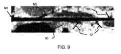
親血管(例えば左回旋枝、LCX)を所与とすると、子血管(例えば辺縁部や対角枝)の検出は、血管の腸管展開図(stretched lumen view)を計算することによって支援される。これは、親血管の中心線に垂直な等サイズの断面画像のシーケンスである。次に、ニューラルエンコーダモデルを適用して(単一又は複数の断面を入力として)側分岐を検出する。Given a parent vessel (e.g., the left circumflex artery, LCX), detection of child vessels (e.g., peripheral and diagonal branches) is aided by computing a stretched lumen view of the vessel, which is a sequence of equal-sized cross-sectional images perpendicular to the centerline of the parent vessel. A neural encoder model is then applied (with single or multiple cross-sections as input) to detect side branches.
図9は、LCX血管の拡大された長手方向図を示しており、マーカー90の左側に第1の辺縁動脈枝M1及び第2の辺縁動脈枝M2がある。右側の矢印92は、側副血流の候補となるより小さな枝を更に示している。Figure 9 shows an enlarged longitudinal view of the LCX vessel with the first marginal artery branch M1 and the second marginal artery branch M2 to the left of the
標準木と側副木との分離のために、標準木は、ルールベースの分類を適用することによって候補中心線の集合から分離される。ルールの分類及び適用は、例えば、動脈口のおける左主(LM)冠動脈又は右冠動脈(RCA)の子孫から始まり、親から子へと血管木を下っていくなど、階層的に行われる。For separation of standard trees and collateral trees, the standard trees are separated from the set of candidate centerlines by applying a rule-based classification. The classification and application of rules is performed hierarchically, for example starting from the left main (LM) or right coronary artery (RCA) descendants at the ostium and proceeding down the vessel tree from parent to child.
多数のルールには、例えば、次のものが含まれる:
最小の内腔面積/血管径(つまり、血管内径の閾値)を必要とすること、
主血管からの先験的に既知の場所からのみ分岐すること。冠動脈の場合、左前下行枝(LAD)からの典型的なLCX分岐点の確率分布は、以前の患者データのセットからモデル化され、LM動脈からの親の子孫の関数としてLAD中心線にエンコードされる。
原点(冠動脈口など)又は親の子孫から一定の最大長で切り捨てる。 A number of rules include, for example:
Requiring a minimum lumen area/vessel diameter (i.e., a threshold vessel diameter);
Branch only from a priori known locations from the main vessel. For the coronary arteries, the probability distribution of typical LCX branching points from the left anterior descending artery (LAD) is modeled from a set of prior patient data and encoded onto the LAD centerline as a function of parent descent from the LM artery.
Truncate to a certain maximum length from the origin (such as the coronary ostium) or from the descendants of the parents.
セグメンテーション及び分離のプロセスからの出力は、ラベル付きの標準血管木と、小さな血管及び側副循環を含む残りの木とによって最終的に与えられる。The output from the segmentation and separation process is finally given by a labeled standard vascular tree and a residual tree that contains small vessels and collateral circulation.
次に、側副血管の解析に使用可能な解剖学的パラメータが説明される。Next, anatomical parameters that can be used to analyze collateral vessels are described.
セグメンテーション及び分離関数からの出力は、Aが全ての検出された/セグメント化された血管を含む添字集合を示し、Sが標準血管木に属する全ての検出された血管をランオーバーする添字集合を示すような形式に入れられる。The output from the segmentation and separation functions is put into a form such that A denotes the index set containing all detected/segmented vessels and S denotes the index set running over all detected vessels that belong to the standard vascular tree.
上記の指定された基準によると、(S⊆A)である。この表記法を使用すると、側副血管の定量化の具体的な実現には、次のことが必要になる:
検出された側副の数は、NC=|A|-|S|として定量化でき、ここで、|A|は、所与の添字集合の濃度を示す。 According to the above specified criteria, (S ⊆ A). Using this notation, a concrete realization of collateral vessel quantification requires:
The number of detected collaterals can be quantified as NC =|A|-|S|, where |A| denotes the cardinality of a given index set.
累積側副血管長LCは、次のように定量化できる:


累積側副内腔ボリュームVCは、次のように定量化できる:
断面積(楕円など)をフィッティングする代わりに、血管セグメンテーションは、バイナリ(ボクセル化された)ビットマスクに基づいて行うことができ、そして、側副内腔ボリュームは、単純に添字集合Aから全てのボクセルを合計し、添字集合Sから全てのボクセルを減算し、所与の収集の空間ボクセル分解能を使用して、ボクセル数を物理ボリューム尺度に変換することによって計算される。Instead of fitting a cross-sectional area (e.g., an ellipse), vessel segmentation can be based on a binary (voxelized) bit mask, and the collateral lumen volume is calculated by simply summing all voxels from index set A and subtracting all voxels from index set S, and converting the voxel counts to a physical volume measure using the spatial voxel resolution of the given acquisition.
血管間面積(つまり、血管間の空間)を定量化できる。この尺度は、血管密度又はカバレッジを表す。計算を容易にするために、既知の表面積Asurf(O)の2D表面Oへの投影(例えば心筋への冠状血管のために)を行うことができる。 The intervascular area (i.e., the space between blood vessels) can be quantified. This measure represents the blood vessel density or coverage. To facilitate the calculation, a projection of a known surface areaAsurf (O) onto a 2D surface O (e.g., for coronary vessels into the myocardium) can be performed.
血管木投影は、基準表面の表面法線を使用することによって、血管木内腔ビットマスクの各ボクセルについて計算され、次の面積が得られる:
Avessel≦Asurf The vascular tree projection is calculated for each voxel of the vascular tree lumen bitmask by using the surface normal of the reference surface, resulting in an area:
Avessel ≦Asurf
この埋め込み2D空間において、血管間面積Avoid=Asurf-Avesselや、血管密度D=Avessel/Asurfなどのメトリックを計算できる。 In this embedded 2D space, metrics such as the intervessel area Avoid =Asurf -Avessel and the vessel density D =Avessel /Asurf can be calculated.
投影は、例えば左室心筋心外膜面をキャプチャする指向性のセグメンテーションメッシュを考慮することによって取得される。この場合、その法線ベクトルがそのマッピングのための(局所)投影方向として機能する。The projection is obtained by considering an oriented segmentation mesh capturing, for example, the left ventricular myocardial epicardial surface, whose normal vector in this case serves as the (local) projection direction for the mapping.
このように、種々可能なパラメータが上記で設定される。これらのパラメータのうちの1つ以上が決定され得る。このように、パラメータは、側副血管の数、側副血管の累積長、(累積)側副内腔ボリューム、又は正味の血管密度などの血管間面積に関連するパラメータを含み得る。Thus, various possible parameters are set above. One or more of these parameters may be determined. Thus, the parameters may include parameters related to the intervascular area, such as the number of collateral vessels, the cumulative length of the collateral vessels, the (cumulative) collateral lumen volume, or the net vessel density.
上記の解析から出力されたパラメータ(NC、LC、VCなど)は、標準血管木又は血管木全体からの対応する値を基準値として使用することによって、正規化された尺度に変換できる。そして、例えば、

更に、基準パラメータ値(例えば、NC、LC、VCの)を、健康な患者コホートから記録し、これらの基準値を正規化に使用することができる。この場合、正規化された値は、選択されたコホートによって設定される健康な患者標準からの偏差を示す。 Additionally, baseline parameter values (e.g., for NC , LC , VC ) can be recorded from a cohort of healthy patients and these baseline values can be used for normalization, where the normalized values indicate deviation from the healthy patient standard established by the selected cohort.
一般的な1人の健康な患者に対してのみパラメータを正規化するのではなく、得られたパラメータ値は、特定の患者に関する同定の母集団統計値のコンテキスト内で解釈されてもよい。これらの統計値は、例えば、転帰、疾患の重症度などの他の臨床パラメータと関連している場合がある。Rather than normalizing parameters only to a typical healthy patient, the resulting parameter values may be interpreted within the context of specific population statistics for a particular patient. These statistics may be related to other clinical parameters, e.g., outcome, disease severity, etc.
特定の患者統計値に対して現在のパラメータ値のセットを解釈するために、1つのアプローチでは、患者プロパティの分布全体内での患者の位置を明示し、現在のものに近い患者のパラメータを使用する。暗黙的なアプローチでは、統計値のデータ駆動型モデリング又は学習を使用し、患者がいくつかのグループのいずれかに分類される。To interpret the current set of parameter values for a particular patient statistic, one approach is to explicitly state the patient's position within the overall distribution of patient properties and use the patient's parameters that are close to the current ones. An implicit approach uses data-driven modeling or learning of statistics to classify patients into one of several groups.
上記の解析は、3D血管造影CTスキャン画像又は3D磁気共鳴血管造影(MRA)画像などの3D画像データに基づいている。しかし、本発明は、2D X線血管造影画像にも適用できる。The above analysis is based on 3D image data, such as 3D angiographic CT scan images or 3D magnetic resonance angiography (MRA) images. However, the invention is also applicable to 2D X-ray angiographic images.
図10は、足の血管系の完全及び標準木がセグメンテーションモジュールによって同定された後の介入2D血管造影図の例を示す。Figure 10 shows an example of an interventional 2D angiogram after the complete and standard trees of the leg vasculature have been identified by the segmentation module.
上の図は完全木Aを示し、真ん中の図は標準木Sを示し、下の図は残りの木R=A\Sを示す。図10は、側副形成がほとんど又はまったくない患者のものである。The top diagram shows the complete tree A, the middle diagram shows the standard tree S, and the bottom diagram shows the remaining tree R=A\S. Figure 10 is for a patient with little or no collateral formation.
図11は、側副の形成が中程度である患者の足の血管系の完全及び標準木がセグメンテーションモジュールによって同定された後の介入2D血管造影図の例を示す。Figure 11 shows an example of an interventional 2D angiogram after the segmentation module has identified the complete and standard trees of the leg vasculature of a patient with moderate collateral formation.
ここでも、上の図は完全木Aを示し、真ん中の図は標準木Sを示し、下の図は残りの木R=A\Sを示す。Again, the top diagram shows the complete tree A, the middle diagram shows the standard tree S, and the bottom diagram shows the remaining tree R = A \ S.
この場合、フォアショートニング作用を引き起こすイメージングモダリティの投影性に起因して、側副定量化パラメータは近似値にしかならない。正味の密度計算のための表面は、検出器平面によって直接与えられる。In this case, collateral quantification parameters can only be approximate due to the projective nature of the imaging modality which causes a foreshortening effect. The surface for the net density calculation is given directly by the detector plane.
セグメンテーション及び側副定量化のステップは、スペクトル若しくはダイナミックCT又はX線スキャンを使用して向上させることができる。The segmentation and collateral quantification steps can be enhanced using spectral or dynamic CT or X-ray scans.
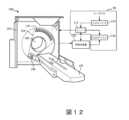
図12は、本発明の方法を用いて解析するための画像を提供するために使用されるイメージングシステムの例を示す。Figure 12 shows an example of an imaging system that can be used to provide images for analysis using the methods of the present invention.
この説明でのイメージング装置100は、X線コンピュータ断層撮影(CT)スキャナである。The
イメージング装置100は一般的に、固定ガントリ102と、回転ガントリ104とを含む。回転ガントリ104は、固定ガントリ102によって回転可能に支持され、長手方向軸、軸方向、又はz軸の周りで検査領域の周りを回転する。The
カウチなどの患者支持体120は、検査領域内で物体や、人間の患者などの被検体を支持する。支持体120は、物体又は被検体のロード、スキャン、及び/又はアンロードのために物体又は被検体を移動する。支持体120は、軸方向に沿って移動可能である。つまり、z軸又は長手方向軸の方向に沿って移動可能である。支持体を移動させると、支持体に対する(したがって、支持体によって支えられている被検体に対する)回転ガントリの軸方向位置が変化する。A
X線管などの放射線源108が、回転ガントリ104によって回転可能に支持されている。放射線源108は回転ガントリ104と共に回転し、検査領域106を横断する放射線を放出する。A
放射線に反応する検出器アレイ110が、検査領域106を挟んで放射線源108の反対側の角度に対する弧の範囲を定める。検出器アレイ110には、z軸方向に沿って延在する1列以上の検出器が含まれ、検査領域106を横断する放射線を検出し、それを示す投影データを生成する。A radiation-
ガントリ104の回転によって、被検体に対するスキャナの角度位置又は回転位置が変化し、z軸に沿った支持体の移動によって、被検体に対するスキャナの軸方向位置が変化する。Rotation of the
典型的なスキャンは、スキャンプロトコルを用いて事前に設定される。スキャンプロトコルは、複数のスキャンパラメータを含む。スキャンパラメータは、とりわけ、スキャナの軸方向軸及び回転軸に対するスキャンの空間範囲を画定する。例えば、スキャンパラメータには、イメージング装置の1つ以上の軸(例えば、回転軸及び軸方向軸の片方又は両方)に沿ったスキャン範囲の境界(つまり、開始点と終了点)が含まれる。スキャン範囲は、スキャン中にイメージングデータが収集される視野(FOV)を画定する。スキャンパラメータには、通常、管電流、管電圧、スキャン空間分解能、スキャン時間分解能、及び/又はファン角度などの多数の他のパラメータも含まれる。分解能パラメータは、ガントリ104の回転速度と、ガントリを通る支持体120の軸方向移動の速度とによって定義できる。A typical scan is pre-defined using a scan protocol. The scan protocol includes multiple scan parameters. The scan parameters define, among other things, the spatial extent of the scan relative to the axial and rotational axes of the scanner. For example, the scan parameters include the boundaries (i.e., start and end points) of the scan extent along one or more axes (e.g., one or both of the rotational and axial axes) of the imaging device. The scan extent defines the field of view (FOV) within which imaging data is collected during the scan. The scan parameters also typically include a number of other parameters, such as tube current, tube voltage, scan spatial resolution, scan temporal resolution, and/or fan angle. The resolution parameter can be defined by the rotational speed of the
汎用コンピューティングシステム又はコンピュータが、オペレータコンソール112として機能し、マウス、キーボードなどの入力デバイス114と、ディスプレイモニタなどの出力デバイス116とを含む。コンソール、入力デバイス、及び出力デバイスが、ユーザインターフェース30を形成する。コンソール112は、オペレータがシステム100の動作を制御することを可能にする。A general-purpose computing system or computer serves as the
再構成装置118が、投影データを処理し、ボリュメトリック画像データを再構成する。このデータは、出力デバイス116の1つ以上のディスプレイモニタを介して表示できる。A reconstructor 118 processes the projection data and reconstructs volumetric image data, which can be displayed via one or more display monitors of the
再構成装置118は、フィルタ補正逆投影(FBP)再構成、(画像ドメイン及び/又は投影ドメインの)低ノイズ再構成アルゴリズム(反復再構成など)、及び/又は他のアルゴリズムを採用できる。再構成装置118は、物理メモリや他の非一時的な媒体などのコンピュータ可読記憶媒体にエンコード又は埋め込みされるコンピュータ可読命令を実行するマイクロプロセッサを介して実施できることが理解されるものとする。更に又は或いは、マイクロプロセッサはキャリア波、信号、及び他の一時的(又は非、非一時的)媒体によって運ばれるコンピュータ可読命令を実行することもできる。The
再構成装置118には、生成された3D CTスキャン画像を解析して、関心領域内の側副血管の解析を行うための上記の方法を実施するためにコンピュータプログラムでプログラムされたプロセッサが組み込まれていてもよい。The
血管の同定及び側副血管の解析には、トレーニングされたニューラルネットワークの使用や、手作りのアルゴリズムの使用が含まれる場合がある。Vessel identification and collateral analysis may involve the use of trained neural networks or hand-crafted algorithms.
前述のように、システムはプロセッサを使用してデータ処理を行う。プロセッサは、ソフトウェアやハードウェアを使用して、様々なやり方で実装して、必要な様々な機能を行うことができる。通常、プロセッサは、ソフトウェア(例えばマイクロコード)を使用してプログラムされて、必要な機能を行う1つ以上のマイクロプロセッサを用いる。プロセッサは、一部の機能を行うための専用ハードウェアと、他の機能を行うための1つ以上のプログラム済みマイクロプロセッサ及び関連回路との組み合わせとして実装できる。As mentioned above, the system uses a processor to process data. The processor can be implemented in a variety of ways using software and/or hardware to perform the various functions required. Typically, the processor uses one or more microprocessors that are programmed using software (e.g., microcode) to perform the required functions. The processor can be implemented as a combination of dedicated hardware to perform some functions and one or more programmed microprocessors and associated circuitry to perform other functions.
本開示の様々な実施形態に用いられ得る回路の例としては、従来のマイクロプロセッサ、特定用途向け集積回路(ASIC)、及びフィールドプログラマブルゲートアレイ(FPGA)が挙げられるが、これらに限定されない。Examples of circuitry that may be used in various embodiments of the present disclosure include, but are not limited to, conventional microprocessors, application specific integrated circuits (ASICs), and field programmable gate arrays (FPGAs).
様々な実装形態では、プロセッサは、RAM、PROM、EPROM、及びEEPROM(登録商標)などの揮発性及び不揮発性コンピュータメモリなどの1つ以上の記憶媒体と関連付けられ得る。記憶媒体は、1以上のプロセッサ及び/又はコントローラ上で実行されると、必要な機能を実行する1つ以上のプログラムでエンコードされ得る。様々な記憶媒体は、プロセッサ又はコントローラ内で固定されていても、そこに保存されている1つ以上のプログラムをプロセッサにロードできるように輸送可能であってもよい。In various implementations, the processor may be associated with one or more storage media, such as volatile and non-volatile computer memory, such as RAM, PROM, EPROM, and EEPROM. The storage media may be encoded with one or more programs that, when executed on one or more processors and/or controllers, perform the necessary functions. The various storage media may be fixed within the processor or controller, or may be transportable so that one or more programs stored thereon may be loaded into the processor.
開示された実施形態の変形例は、図面、開示及び添付の特許請求の範囲の検討から、請求項に係る発明を実施する際に当業者によって理解され、実行され得る。特許請求の範囲において、「含む」という用語は、他の要素又はステップを排除するものではなく、単数形の要素は複数を排除するものではない。Variations of the disclosed embodiments can be understood and effected by those skilled in the art in practicing the claimed invention, from a study of the drawings, the disclosure, and the appended claims. In the claims, the term "comprising" does not exclude other elements or steps, and singular elements do not exclude a plurality.
特定の手段が相互に異なる従属請求項に記載されているという単なる事実は、これらの手段の組み合わせを有利に使用することができないことを意味するものではない。The mere fact that certain measures are recited in mutually different dependent claims does not indicate that a combination of these measures cannot be used to advantage.
コンピュータプログラムは、他のハードウェアと一緒に又はその一部として供給される、光記憶媒体又は固体媒体などの任意の適切な媒体に保存/配布することができるが、インターネット又は他の有線若しくはワイヤレス通信システムを介してなど他の形式で配布することもできる。The computer program may be stored/distributed on any suitable medium, such as optical storage media or solid state media, supplied together with or as part of other hardware, but may also be distributed in other forms, such as via the Internet or other wired or wireless communication systems.
「~するように適応されている」という用語が、特許請求の範囲又は説明で使用されている場合、「~するように適応されている」という用語は「~するように構成されている」という用語と同等であることを意図していることに留意されたい。Please note that when the term "adapted to" is used in the claims or description, it is intended to be equivalent to the term "configured to."
特許請求の範囲における任意の参照符号は、範囲を限定するものと解釈されるべきではない。Any reference signs in the claims shall not be construed as limiting the scope.
Claims (14)
Translated fromJapanese前記被検体の関心領域の画像を受信するステップと、
前記関心領域に存在する血管木を同定するステップと、
同定された前記血管木内の主要血管を同定するステップであって、前記主要血管は、前記主要血管の標準木の一部を形成する、同定するステップと、
同定された前記血管木から、同定された前記主要血管を減算し、したがって、前記標準木の前記主要血管を除外することによって、同定された前記血管木の残りの血管を同定し、これにより、前記関心領域内の側副血管を同定するステップと、
前記側副血管の解析を行うステップと、
を含む、方法。 1. A method for analyzing the vasculature of a subject, comprising:
receiving an image of a region of interest of the subject;
identifying a vascular tree present in the region of interest;
- identifying major vessels within the identified vascular tree, the major vessels forming part of a standard tree of major vessels;
identifying remaining vessels of the identified vascular tree by subtracting the identified major vessel from the identified vascular tree, thus excluding the major vessel of the standard tree, thereby identifying collateral vessels within the region of interest;
performing an analysis of said collateral vessels;
A method comprising:
前記画像を解析して、前記関心領域内の前記側副血管の解析を行うための、請求項13に記載のプロセッサと、
を含む、イメージングシステム。 an imager for acquiring an image of a region of interest of the subject;
14. The processor of claim 13 for analyzing the image to perform an analysis of the collateral vessels within the region of interest;
1. An imaging system comprising:
Applications Claiming Priority (3)
| Application Number | Priority Date | Filing Date | Title |
|---|---|---|---|
| EP21211556.2AEP4191520A1 (en) | 2021-12-01 | 2021-12-01 | A method and processor for analyzing the vasculature of a subject |
| EP21211556.2 | 2021-12-01 | ||
| PCT/EP2022/081434WO2023099144A1 (en) | 2021-12-01 | 2022-11-10 | A method and processor for analyzing the vasculature of a subject |
Publications (2)
| Publication Number | Publication Date |
|---|---|
| JP2024542644Atrue JP2024542644A (en) | 2024-11-15 |
| JPWO2023099144A5 JPWO2023099144A5 (en) | 2025-10-01 |
Family
ID=78821427
Family Applications (1)
| Application Number | Title | Priority Date | Filing Date |
|---|---|---|---|
| JP2024532224APendingJP2024542644A (en) | 2021-12-01 | 2022-11-10 | Method and processor for analyzing the vasculature of a subject - Patents.com |
Country Status (5)
| Country | Link |
|---|---|
| US (1) | US20250022133A1 (en) |
| EP (2) | EP4191520A1 (en) |
| JP (1) | JP2024542644A (en) |
| CN (1) | CN118339581A (en) |
| WO (1) | WO2023099144A1 (en) |
Families Citing this family (5)
| Publication number | Priority date | Publication date | Assignee | Title |
|---|---|---|---|---|
| US10210956B2 (en) | 2012-10-24 | 2019-02-19 | Cathworks Ltd. | Diagnostically useful results in real time |
| IL263065B2 (en) | 2016-05-16 | 2024-08-01 | Cathworks Ltd | System for vascular assessment |
| US12315076B1 (en) | 2021-09-22 | 2025-05-27 | Cathworks Ltd. | Four-dimensional motion analysis of a patient's coronary arteries and myocardial wall |
| JP2025506500A (en) | 2022-02-10 | 2025-03-11 | キャスワークス リミテッド | Systems and methods for machine learning based sensor analysis and vascular tree segmentation |
| CN118154590B (en)* | 2024-05-09 | 2024-07-16 | 杭州脉流科技有限公司 | Method, device, equipment and storage medium for detecting intracranial large vessel occlusion region |
- 2021
- 2021-12-01EPEP21211556.2Apatent/EP4191520A1/ennot_activeWithdrawn
- 2022
- 2022-11-10USUS18/712,882patent/US20250022133A1/enactivePending
- 2022-11-10EPEP22814409.3Apatent/EP4441700A1/enactivePending
- 2022-11-10WOPCT/EP2022/081434patent/WO2023099144A1/ennot_activeCeased
- 2022-11-10CNCN202280079790.XApatent/CN118339581A/enactivePending
- 2022-11-10JPJP2024532224Apatent/JP2024542644A/enactivePending
Also Published As
| Publication number | Publication date |
|---|---|
| CN118339581A (en) | 2024-07-12 |
| EP4441700A1 (en) | 2024-10-09 |
| WO2023099144A1 (en) | 2023-06-08 |
| EP4191520A1 (en) | 2023-06-07 |
| US20250022133A1 (en) | 2025-01-16 |
Similar Documents
| Publication | Publication Date | Title |
|---|---|---|
| CN110546646B (en) | Methods and systems for assessing vascular occlusion based on machine learning | |
| US12089977B2 (en) | Method and system for assessing vessel obstruction based on machine learning | |
| JP2024542644A (en) | Method and processor for analyzing the vasculature of a subject - Patents.com | |
| NL2009379C2 (en) | System and method for blood vessel stenosis visualization and navigation. | |
| CN110944586B (en) | Inflammation Estimation from X-ray Image Data | |
| CN112967220B (en) | Computer-implemented method of evaluating CT data sets relating to perivascular tissue | |
| Özkan et al. | A novel method for pulmonary embolism detection in CTA images | |
| JP2020081866A (en) | Deep learning for arterial analysis and assessment | |
| US10970836B2 (en) | Spectral image data processing | |
| JP7325331B2 (en) | Machine learning spectrum FFR-CT | |
| JP6484760B2 (en) | Modeling collateral blood flow for non-invasive blood flow reserve ratio (FFR) | |
| Van den Oever et al. | Application of artificial intelligence in cardiac CT: From basics to clinical practice | |
| JP2024028194A (en) | Information processing method, medical image diagnostic device, and information processing system including deep learning for optimal cardiac phase selection | |
| Zhou et al. | Computerized analysis of coronary artery disease: performance evaluation of segmentation and tracking of coronary arteries in CT angiograms | |
| US12315076B1 (en) | Four-dimensional motion analysis of a patient's coronary arteries and myocardial wall | |
| EP4449993A1 (en) | Determining a status of vascular disease | |
| US20250268553A1 (en) | Systems and methods for contrast flow modeling with deep learning | |
| US20250104228A1 (en) | Coronary Artery Lumen Contour Adjustment Based on Segmentation Uncertainty | |
| WO2024175415A1 (en) | Determining a status of vascular disease | |
| de Gouw | Automatic Coronary Calcium Scoring using Computed Tomography | |
| Wang | Computer-Assisted Coronary CT Angiography Analysis From Software Development to Clinical Application | |
| Toledano et al. | Learning to Detect Coronary Artery Stenosis from Multi-Detector CT imaging | |
| de Sousa Silva | Left Ventricle Functional Analysis from Coronary CT Angiography |
Legal Events
| Date | Code | Title | Description |
|---|---|---|---|
| RD03 | Notification of appointment of power of attorney | Free format text:JAPANESE INTERMEDIATE CODE: A7423 Effective date:20250403 | |
| RD04 | Notification of resignation of power of attorney | Free format text:JAPANESE INTERMEDIATE CODE: A7424 Effective date:20250408 | |
| A521 | Request for written amendment filed | Free format text:JAPANESE INTERMEDIATE CODE: A523 Effective date:20250918 | |
| A621 | Written request for application examination | Free format text:JAPANESE INTERMEDIATE CODE: A621 Effective date:20250918 |