JP2024540185A - A method for analyzing bone texture from digital images. - Google Patents
A method for analyzing bone texture from digital images.Download PDFInfo
- Publication number
- JP2024540185A JP2024540185AJP2024525677AJP2024525677AJP2024540185AJP 2024540185 AJP2024540185 AJP 2024540185AJP 2024525677 AJP2024525677 AJP 2024525677AJP 2024525677 AJP2024525677 AJP 2024525677AJP 2024540185 AJP2024540185 AJP 2024540185A
- Authority
- JP
- Japan
- Prior art keywords
- training
- bone
- type
- image
- score
- Prior art date
- Legal status (The legal status is an assumption and is not a legal conclusion. Google has not performed a legal analysis and makes no representation as to the accuracy of the status listed.)
- Pending
Links
Images


Classifications
- G—PHYSICS
- G16—INFORMATION AND COMMUNICATION TECHNOLOGY [ICT] SPECIALLY ADAPTED FOR SPECIFIC APPLICATION FIELDS
- G16H—HEALTHCARE INFORMATICS, i.e. INFORMATION AND COMMUNICATION TECHNOLOGY [ICT] SPECIALLY ADAPTED FOR THE HANDLING OR PROCESSING OF MEDICAL OR HEALTHCARE DATA
- G16H50/00—ICT specially adapted for medical diagnosis, medical simulation or medical data mining; ICT specially adapted for detecting, monitoring or modelling epidemics or pandemics
- G16H50/20—ICT specially adapted for medical diagnosis, medical simulation or medical data mining; ICT specially adapted for detecting, monitoring or modelling epidemics or pandemics for computer-aided diagnosis, e.g. based on medical expert systems
- G—PHYSICS
- G16—INFORMATION AND COMMUNICATION TECHNOLOGY [ICT] SPECIALLY ADAPTED FOR SPECIFIC APPLICATION FIELDS
- G16H—HEALTHCARE INFORMATICS, i.e. INFORMATION AND COMMUNICATION TECHNOLOGY [ICT] SPECIALLY ADAPTED FOR THE HANDLING OR PROCESSING OF MEDICAL OR HEALTHCARE DATA
- G16H30/00—ICT specially adapted for the handling or processing of medical images
- G16H30/40—ICT specially adapted for the handling or processing of medical images for processing medical images, e.g. editing
Landscapes
- Health & Medical Sciences (AREA)
- Engineering & Computer Science (AREA)
- Medical Informatics (AREA)
- Public Health (AREA)
- General Health & Medical Sciences (AREA)
- Biomedical Technology (AREA)
- Epidemiology (AREA)
- Primary Health Care (AREA)
- Pathology (AREA)
- Data Mining & Analysis (AREA)
- Nuclear Medicine, Radiotherapy & Molecular Imaging (AREA)
- Radiology & Medical Imaging (AREA)
- Databases & Information Systems (AREA)
- Apparatus For Radiation Diagnosis (AREA)
- Life Sciences & Earth Sciences (AREA)
- Physics & Mathematics (AREA)
- Biophysics (AREA)
- Molecular Biology (AREA)
- Theoretical Computer Science (AREA)
- Veterinary Medicine (AREA)
- Animal Behavior & Ethology (AREA)
- Surgery (AREA)
- Heart & Thoracic Surgery (AREA)
- Optics & Photonics (AREA)
- High Energy & Nuclear Physics (AREA)
- Computational Linguistics (AREA)
- Software Systems (AREA)
- Orthopedic Medicine & Surgery (AREA)
- Dentistry (AREA)
- Oral & Maxillofacial Surgery (AREA)
- Mathematical Physics (AREA)
- General Physics & Mathematics (AREA)
- General Engineering & Computer Science (AREA)
- Computing Systems (AREA)
- Evolutionary Computation (AREA)
- Artificial Intelligence (AREA)
Abstract
Translated fromJapanese
Description
Translated fromJapanese本発明は、デジタル画像から骨のテクスチャを解析する方法に関する。The present invention relates to a method for analyzing bone texture from digital images.
本発明はまた、デジタル画像から骨のテクスチャを解析する装置に関する。The present invention also relates to an apparatus for analyzing bone texture from digital images.
本発明の技術分野は、典型的にはディープラーニングの技術分野であり、特にデジタルX線ベースの画像の適切なスクリーニングから、DXA BMDおよびTBS、あるいは他の同等の方法で評価される劣化した骨量と骨微細構造を有する骨粗鬆症と診断される可能性の高い個人を同定する方法であるが、これに限定されない。The technical field of the present invention is typically that of deep learning, particularly but not limited to, methods for identifying individuals likely to be diagnosed with osteoporosis from appropriate screening of digital x-ray based images, who have impaired bone mass and bone microarchitecture as assessed by DXA BMD and TBS, or other equivalent methods.
1990年代初頭、世界保健機関(WHO)は、骨粗鬆症を、骨量の低下(量の減少)と骨組織の微細構造の劣化(質の低下)を特徴とし、その結果、骨の脆弱性と骨折のリスクが高まる全身性の骨格疾患と概念的に定義している(非特許文献1)。さらに、2000年代初頭に、米国国立衛生研究所(NIH)は、骨粗鬆症を骨強度が低下し骨折のリスクが高まる骨格疾患と定義した(非特許文献2)。本質的に、骨粗鬆症では、骨強度の低下により脆弱性骨折という外傷性の転帰をもたらす。In the early 1990s, the World Health Organization (WHO) conceptualized osteoporosis as a systemic skeletal disease characterized by reduced bone mass (reduced quantity) and deterioration of bone tissue microarchitecture (reduced quality), resulting in increased bone fragility and risk of fracture (Non-Patent Document 1). Furthermore, in the early 2000s, the National Institutes of Health (NIH) defined osteoporosis as a skeletal disease with reduced bone strength and increased risk of fracture (Non-Patent Document 2). Essentially, in osteoporosis, reduced bone strength leads to a traumatic outcome in the form of fragility fractures.
骨強度は、骨量(すなわち骨密度)と骨質という2つの主要な特徴の統合を反映している。骨密度は、単位面積または体積あたりのミネラルのグラム数として表され、個人では最大骨量と骨喪失量によって決定される。骨質とは、骨構造、骨の弾力性、代謝回転、損傷の蓄積(例えば、微小骨折)、および石灰化を指す。骨構造は、多くの異なる実体に使用される一般的な用語であり、さらに細分化できる。マクロレベルでは、骨のマクロ構造(骨マクロ構造とも呼ばれる)として知られる骨構造は、骨の全体的な形状と幾何学的形態、ならびに海綿骨(骨梁とも呼ばれる)と皮質骨への分化を表す(非特許文献3)。Bone strength reflects the integration of two major features: bone mass (i.e., bone density) and bone quality. Bone density is expressed as grams of mineral per unit area or volume and is determined in an individual by the maximum bone mass and the amount of bone loss. Bone quality refers to bone structure, bone elasticity, turnover, accumulation of damage (e.g., microfractures), and mineralization. Bone structure is a general term used for many different entities and can be further subdivided. At the macro level, bone structure, known as bone macroarchitecture (also called bone macroarchitecture), describes the overall shape and geometry of the bone as well as its differentiation into cancellous bone (also called trabecular bone) and cortical bone (Non-Patent Document 3).
いずれにせよ、骨粗鬆症の特徴は脆弱性骨折である。脆弱性骨折とは、通常は骨折を引き起こすことのない機械的力に反応して、立位の高さから転倒することによって発生する骨折と定義される(非特許文献4、非特許文献5)。大腿骨近位部、脊椎、上腕骨、前腕は、脆弱性骨折が最も多く発生する骨格部位である。これらの骨折は、主要な骨粗鬆症性骨折と呼ばれる。In any case, osteoporosis is characterized by fragility fractures. A fragility fracture is defined as a fracture that occurs due to a fall from standing height in response to a mechanical force that would not normally cause a fracture (Non-Patent
骨折リスクが特定されると、ある種の「階層的」レベルで予防措置が講じられる。一般的なライフスタイルのアドバイス、健康的なライフスタイルに加えて適切なカルシウムおよび/またはビタミンDの補給、および/または薬物療法である。作用機序に基づいて、骨吸収を抑制する抗吸収剤(ビスホスホネート、エストロゲンアゴニスト/アンタゴニスト、エストロゲン、カルシトニン、デノスマブなど)と、骨形成を刺激する骨形成促進剤(テリパラチドなど)の2種類の骨粗鬆症薬物療法がある。最近では、ロモソズマブがその骨形成作用により承認されている(非特許文献6)。Once fracture risk has been identified, preventive measures are implemented at a sort of "hierarchical" level: general lifestyle advice, healthy lifestyle plus appropriate calcium and/or vitamin D supplementation, and/or pharmacotherapy. Based on the mechanism of action, there are two types of osteoporosis pharmacotherapy: antiresorptive agents (e.g. bisphosphonates, estrogen agonists/antagonists, estrogen, calcitonin, denosumab) that inhibit bone resorption, and osteogenic agents (e.g. teriparatide) that stimulate bone formation. Recently, romosozumab has been approved for its osteogenic activity (Non-Patent Document 6).
世界では毎年900万件超の脆弱性骨折が発生しており、人口の高齢化に伴いその数は増加すると予想される(非特許文献7、非特許文献8)。50歳を過ぎてから、女性の2人に1人、男性の4人に1人が、余生において主要な骨粗鬆症性骨折を起こすと推定されている(非特許文献9)。45歳超の女性では、骨粗鬆症は糖尿病、心筋梗塞、乳がんなど、他の多くの疾患よりも入院日数が多い。さらに、過去の骨折は、その後の骨折のリスクを86%増加させる。骨折は高い罹患率と死亡率に関連しており、高齢者では障害、自立の喪失、早期死亡の前兆となることが多い(非特許文献10)。例えば、全体として、2010年のEUだけで薬物介入を含む骨粗鬆症の費用は370億ユーロと推定され、そのうち66%が事故による骨折の治療費、5%が薬物予防、29%が長期の骨折ケアを占めた(非特許文献11)。また、国際骨粗鬆症財団とヨーロッパ製薬工業連盟とが共同で準備した報告書が報告された(非特許文献12)。Over 9 million fragility fractures occur annually worldwide, and the number is expected to increase with the aging of the population (Non-Patent
残念ながら、骨粗鬆症のリスクがあると考えられる人の約70%が特定されることなく、DXA(ゴールドスタンダード)による適切な診断のために骨専門医を紹介されたことすらないと推定されている。さらに、多くの研究で、大腿骨近位部骨折を経験した後でさえ、骨粗鬆症の登録治療を受けている患者の割合は年々大幅に減少していることが示されている。実際、すでに少なくとも1回の骨粗鬆症性骨折を経験したハイリスク者の大多数(おそらく80%)が、特定されておらず、治療も受けていない(非特許文献13)。また、国際骨粗鬆症財団とヨーロッパ製薬工業連盟(EFPIA)とが共同で準備した報告書が報告された(非特許文献14~非特許文献17)。Unfortunately, it is estimated that about 70% of people considered to be at risk for osteoporosis are never identified and are never even referred to an osteologist for proper diagnosis by DXA (the gold standard). Furthermore, many studies have shown that the percentage of patients who are registered and treated for osteoporosis, even after experiencing a hip fracture, is decreasing significantly year by year. In fact, the majority (perhaps 80%) of high-risk individuals who have already experienced at least one osteoporotic fracture are not identified and are not treated (Non-Patent Document 13). In addition, a report prepared jointly by the International Osteoporosis Foundation and the European Federation of Pharmaceutical Industries and Associations (EFPIA) was reported (Non-Patent
骨粗鬆症のリスクのある個人の特定不足や骨粗鬆症の診断・治療の減少には、多くの理由がある。おそらく最大の問題は、一般市民と医療従事者の両方で骨疾患に対する認識が不足していることであり、その多くは問題の大きさを理解しておらず、ましてや骨疾患の予防と治療の方法を理解していない。患者が脆弱性骨折を経験した場合でも、無症候性の椎体圧迫骨折を含むすべての骨粗鬆症性骨折の重大性が十分に認識されておらず、骨粗鬆症性骨折で入院した患者が2度目の骨折を防ぐための骨粗鬆症管理計画に確実には組み込まれていない。実際、この後者の問題に対処し、その後の2度目の脆弱性骨折を回避するために、多職種によるフラクチャーリエゾンサービス(FLS)を開発する国際的な動きがある。FLSは、骨粗鬆症性骨折で病院、緊急治療室、救急診療所に入院した患者を特定し、それらの患者をよく練られた骨粗鬆症管理・治療計画に導くためのメカニズムと経路の開発に依存している(非特許文献18)。There are many reasons for the underidentification of individuals at risk for osteoporosis and the underdiagnosis and treatment of osteoporosis. Perhaps the biggest problem is the lack of awareness of bone disease among both the general public and health care professionals, many of whom do not understand the magnitude of the problem, much less how to prevent and treat bone disease. Even when patients experience fragility fractures, the significance of all osteoporotic fractures, including asymptomatic vertebral compression fractures, is not fully recognized and patients admitted with osteoporotic fractures are not reliably integrated into osteoporosis management plans to prevent a second fracture. Indeed, there is an international movement to develop multidisciplinary fracture liaison services (FLS) to address this latter issue and avoid subsequent second fragility fractures. FLS relies on the development of mechanisms and pathways to identify patients admitted to hospitals, emergency rooms, and urgent care clinics with osteoporotic fractures and to direct those patients to a well-designed osteoporosis management and treatment plan (Non-Patent Document 18).
脆弱性骨折が発生する前の様々な年齢で、個人の骨の健康に潜在的な問題を示唆する多くの「危険信号」がある。骨密度(BMD)検査は依然として骨粗鬆症と骨折リスクを特定するための「ゴールドスタンダード」の診断検査として機能しているが、母集団全体のBMD検査は、骨疾患のリスクを評価するための費用対効果の高い実用的な方法ではない。BMD検査は一部の集団(65歳超の女性など)で推奨されているが、他の個人、つまり骨疾患を有さず骨疾患のリスクもない大多数の人々に対しては、BMD検査は日常的に使用されていない。広範なBMD検査は、経済的にも医学的にもほとんど意味がない。むしろ、エビデンスは、検査から最も恩恵を受ける可能性の高いハイリスク者のサブセット(例えば、複数のリスク因子を有する若年女性、脆弱性骨折の既往がある男女、骨折リスクを大幅に高める可能性のある疾患を有する男女など)を特定するために、まず他のリスク因子の評価を支持している。これらのリスク因子の中には、BMDレベルに直接的または間接的に影響を与えるものもあるが、骨密度とは無関係のもの(例えば、転倒のリスク因子)もある。そのため、リスク評価モデルに基づく多くのスクリーニング戦略が開発されている。残念ながら、いくつかの問題と制限により、リスク因子評価ツールの開発と広範な適用が遅れている。重要な問題の1つは、骨の健康に関するリスク因子についての現在の医学的知識の限界に関連しているが、さらに重要なのは、一般市民と医療従事者の両方の認識に関連する積極的なスクリーニング行動が依然として必要とされることである(非特許文献19~22)。At various ages before a fragility fracture occurs, there are many "red flags" that suggest potential problems with an individual's bone health. Although bone mineral density (BMD) testing still serves as the "gold standard" diagnostic test for identifying osteoporosis and fracture risk, population-wide BMD testing is not a cost-effective or practical method for assessing risk of bone disease. Although BMD testing is recommended for some populations (e.g., women over 65 years of age), for other individuals, i.e., the majority of people who do not have or are not at risk for bone disease, BMD testing is not used routinely. Extensive BMD testing makes little economic or medical sense. Rather, evidence supports evaluation of other risk factors first to identify the subset of high-risk individuals most likely to benefit from testing (e.g., young women with multiple risk factors, men and women with a history of fragility fractures, men and women with diseases that may substantially increase fracture risk, etc.). Some of these risk factors directly or indirectly affect BMD levels, while others (e.g., risk factors for falls) are independent of BMD. Therefore, many screening strategies based on risk assessment models have been developed. Unfortunately, several problems and limitations have delayed the development and widespread application of risk factor assessment tools. One of the key issues is related to the limitations of current medical knowledge about risk factors for bone health, but more importantly, active screening behavior related to awareness of both the general public and health care professionals is still needed (Non-Patent Documents 19-22).
この積極性の必要性を克服し、例えばDXAに基づく全集団の体系的なスクリーニングのコストの超過を避けるために、適切な場合に自動的な適切なアプローチを想像することができる。この後者のアプローチは、例えば、骨粗鬆症以外の理由で毎年何百万人もの個人に対して実施されるX線ベースの画像で実行することができる。このようなアプローチは、偽陽性の観点で最適化されなければならず、「ゴールドスタンダード」の診断確認(骨量と骨質の両方の評価)のためにDXA施設に紹介される骨粗鬆症の真のリスクのある個人の数を増やすことを目的とすべきである。To overcome this need for aggressiveness and to avoid the cost overruns of systematic screening of the entire population, for example based on DXA, one can envision an appropriate approach, automatic in appropriate cases. This latter approach could be implemented, for example, on X-ray-based images performed annually on millions of individuals for reasons other than osteoporosis. Such an approach must be optimized in terms of false positives and should aim to increase the number of individuals at true risk of osteoporosis referred to a DXA center for the "gold standard" diagnostic confirmation (assessment of both bone mass and bone quality).
本発明の目的は、通常は骨密度(BMD)または海綿骨スコア(TBS)を得ることができないような画像、すなわち典型的には二重エネルギーX線吸収測定(DXA)画像以外の画像にも適用できる、骨のテクスチャおよび/または健康状態を迅速かつ簡単に解析する方法または装置を提示することである。The object of the present invention is to provide a method or apparatus for rapid and simple analysis of bone texture and/or health that can be applied to images where bone mineral density (BMD) or trabecular bone score (TBS) cannot normally be obtained, i.e. images other than typically dual energy x-ray absorptiometry (DXA) images.
本発明の一態様は、骨(好ましくはデジタル画像から画像化により得られ、骨構造を含む領域で選択される)のテクスチャを解析する方法(好ましくはコンピュータで実施される)に関し、以下の工程を含む。
- 入力骨を示すデジタル化された入力X線画像を受信する工程、
- 技術的手段により実装された骨スコア人工知能による、受信した入力X線画像の骨スコア解析工程であって、骨スコア人工知能が、この骨スコア解析の結果として以下を与える工程:
-- 受信した入力X線画像に示された入力骨の海綿骨部分のテクスチャ(またはグレーレベルの実験的バリオグラムからグレーレベルの局所的な変動を定量化するもの)に依存する海綿骨スコア(TBS)に少なくとも依存するグローバルスコア、および/または
-- 受信した入力X線画像に示された入力骨の海綿骨部分のテクスチャ(またはグレーレベルの実験的バリオグラムからグレーレベルの局所的な変動を定量化するもの)に依存する海綿骨スコア(TBS)、および/または受信した入力X線画像に示された入力骨の骨密度に依存する密度スコア。 One aspect of the invention relates to a method, preferably computer implemented, for analysing the texture of a bone, preferably obtained by imaging from a digital image and selected in areas containing bone structure, comprising the steps of:
receiving an input digitized X-ray image representative of an input bone;
- a step of bone score analysis of the received input X-ray image by a bone score artificial intelligence implemented by technical means, which gives as a result of this bone score analysis:
-- a global score that depends at least on a trabecular bone score (TBS) that depends on the texture (or one that quantifies the local variation in gray levels from an empirical variogram of gray levels) of the trabecular bone portion of the input bone shown in the received input X-ray image, and/or -- a trabecular bone score (TBS) that depends on the texture (or one that quantifies the local variation in gray levels from an empirical variogram of gray levels) of the trabecular bone portion of the input bone shown in the received input X-ray image, and/or a density score that depends on the bone density of the input bone shown in the received input X-ray image.
骨スコア人工知能はニューラルネットワークであり得る。The bone score artificial intelligence can be a neural network.
本発明による方法は、以下の工程を含み得る。
- 以下のステップを複数回実施することにより、第1の学習セットを構築する工程:
-- 学習用骨の海綿骨部分を示す第1のタイプの学習画像を取得するステップ
-- 同じ学習用骨を示す、X線ベースの画像である関連する第2のタイプの学習画像を取得するステップ
-- 技術的手段により、第1のタイプの学習画像から以下を決定するステップ:
--- 第1のタイプの学習画像に示された学習用骨の骨密度に依存する密度スコア、および
--- 第1のタイプの学習画像に示された学習用骨の海綿骨部分のテクスチャ(またはグレーレベルの実験的バリオグラムからグレーレベルの局所的な変動を定量化するもの)に依存する海綿骨スコア(TBS)
-- 技術的手段により、以下からこれらの密度スコアと海綿骨スコアに依存するグローバルスコアを決定するステップ:
--- 第1のタイプの学習画像に示された学習用骨の骨密度に依存する密度スコア、および
--- 第1のタイプの学習画像に示された学習用骨の海綿骨部分のテクスチャ(またはグレーレベルの実験的バリオグラムからグレーレベルの局所的な変動を定量化するもの)に依存する海綿骨スコア(TBS)、
- 第2のタイプの学習画像とともに、この第2のタイプの学習画像に関連付けられた第1のタイプの学習画像について決定されたグローバルスコアを含むその関連するグランド・トゥルース(ground truth)を骨スコア人工知能に提供することにより、骨スコア人工知能を学習させる工程。 The method according to the invention may comprise the following steps:
- constructing a first training set by performing the following steps multiple times:
-- acquiring a first type of training image showing the cancellous part of a training bone; -- acquiring a related second type of training image showing the same training bone, the second type being an X-ray based image; -- determining by technical means from the first type of training image:
--- a density score depending on the bone density of the training bones shown in the training images of the first type, and --- a trabecular bone score (TBS) depending on the texture (or a quantification of the local variation of gray levels from an empirical variogram of gray levels) of the trabecular bone portion of the training bones shown in the training images of the first type.
-- determining by technical means a global score which depends on these density scores and the cancellous bone score from:
--- a density score depending on the bone density of the training bones shown in the training images of the first type; and --- a trabecular bone score (TBS) depending on the texture (or a quantification of the local variation of gray levels from an empirical variogram of gray levels) of the trabecular bone parts of the training bones shown in the training images of the first type.
training a Bone Score Artificial Intelligence by providing the Bone Score Artificial Intelligence with training images of the second type together with their associated ground truth including global scores determined for training images of the first type associated with the training images of the second type.
本発明による方法は、以下の工程を含み得る。
- 以下のステップを複数回実施することにより、第2の学習セットを構築する工程:
-- 学習用骨の海綿骨部分を示す第1のタイプの学習画像を取得するステップ
-- 同じ学習用骨を示す、X線ベースの画像である関連する第2のタイプの学習画像を取得するステップ
-- 技術的手段により、第1のタイプの学習画像から以下を決定するステップ:
--- 第1のタイプの学習画像に示された学習用骨の骨密度に依存する密度スコア、および/または
--- 第1のタイプの学習画像に示された学習用骨の海綿骨部分のテクスチャ(またはグレーレベルの実験的バリオグラムからグレーレベルの局所的な変動を定量化するもの)に依存する海綿骨スコア(TBS)
- 第2のタイプの学習画像とともに、以下を含むその関連するグランド・トゥルースを骨スコア人工知能に提供することにより、骨スコア人工知能を学習させる工程:
-- この第2のタイプの学習画像に関連付けられた第1のタイプの学習画像について決定された密度スコア、および/または
-- この第2のタイプの学習画像に関連付けられた第1のタイプの学習画像について決定された海綿骨スコア。 The method according to the invention may comprise the following steps:
- constructing a second training set by performing the following steps multiple times:
-- acquiring a first type of training image showing the cancellous part of a training bone; -- acquiring a related second type of training image showing the same training bone, the second type being an X-ray based image; -- determining by technical means from the first type of training image:
--- a density score depending on the bone density of the training bones shown in the training images of the first type, and/or --- a trabecular bone score (TBS) depending on the texture (or a quantification of the local variation of gray levels from an empirical variogram of gray levels) of the trabecular bone parts of the training bones shown in the training images of the first type.
training a Bone Score Artificial Intelligence by providing the Bone Score Artificial Intelligence with training images of the second type together with their associated ground truth, including:
--a density score determined for a training image of a first type associated with this training image of a second type, and/or --a trabecular bone score determined for a training image of a first type associated with this training image of a second type.
本発明による方法は、以下の工程を含み得る。
- 入力骨を示す入力X線画像を受信する工程、
- 技術的手段により実装された第1の人工知能による、受信した入力X線画像の第1の解析工程であって、第1の人工知能が第1の解析の結果として以下の両方に依存するグローバルスコアを与える工程:
-- 受信した入力X線画像に示された入力骨の骨密度に依存する密度スコア
-- 受信した入力X線画像に示された入力骨の海綿骨部分のテクスチャ(またはグレーレベルの実験的バリオグラムからグレーレベルの局所的な変動を定量化するもの)に依存する海綿骨スコア(TBS)、
- 技術的手段により実装された第2の人工知能による、受信した入力X線画像の第2の解析工程であって、第2の人工知能が第2の解析の結果として以下を与える工程:
-- 受信した入力X線画像に示された入力骨の骨密度に依存する密度スコア、および/または
-- 受信した入力X線画像に示された入力骨の海綿骨部分のテクスチャ(またはグレーレベルの実験的バリオグラムからグレーレベルの局所的な変動を定量化するもの)に依存する海綿骨スコア(TBS)、
- 技術的手段により実装された第3の人工知能による第3の解析工程であって、第3の人工知能が第1および第2の解析の結果を入力として有し、第1の解析の結果と第2の解析の結果の整合性に依存する結果を出力として有する工程。 The method according to the invention may comprise the following steps:
receiving an input X-ray image representative of an input bone;
a first analysis step of the received input X-ray image by a first artificial intelligence implemented by technical means, the first artificial intelligence giving as a result of the first analysis a global score that depends on both:
-- a density score depending on the bone density of the input bone shown in the received input X-ray image; -- a trabecular bone score (TBS) depending on the texture (or a quantification of the local variation of grey levels from an empirical variogram of grey levels) of the trabecular bone portion of the input bone shown in the received input X-ray image;
a second analysis step of the received input X-ray image by a second artificial intelligence implemented by technical means, the second artificial intelligence providing as a result of the second analysis:
a density score depending on the bone density of the input bone shown in the received input X-ray image, and/or a trabecular bone score (TBS) depending on the texture (or a quantification of the local variations in grey levels from an empirical variogram of grey levels) of the trabecular bone portion of the input bone shown in the received input X-ray image,
a third analysis step by a third artificial intelligence implemented by technical means, the third artificial intelligence having as input the results of the first and second analyses and having as output a result that depends on the consistency between the result of the first analysis and the result of the second analysis.
第3の人工知能は、受信した入力X線画像が取得された患者の年齢、受信した入力X線画像が取得された患者の性別、受信した入力X線画像が取得された患者の体型、受信した入力X線画像が取得された機器の種類、および/または受信した入力X線画像が取得された機器の取得パラメータのうちの少なくとも1つのパラメータをさらなる入力として使用し得る。The third artificial intelligence may use as further input at least one of the following parameters: the age of the patient from whom the received input X-ray image was obtained, the gender of the patient from whom the received input X-ray image was obtained, the body type of the patient from whom the received input X-ray image was obtained, the type of equipment from which the received input X-ray image was obtained, and/or the acquisition parameters of the equipment from which the received input X-ray image was obtained.
第1の人工知能および/または第2の人工知能は、ニューラルネットワークであり得る。The first artificial intelligence and/or the second artificial intelligence may be a neural network.
第1の人工知能、第2の人工知能、および第3の人工知能は、3つの異なる人工知能であり得る。The first artificial intelligence, the second artificial intelligence, and the third artificial intelligence may be three different artificial intelligences.
第1の人工知能、第2の人工知能、および第3の人工知能を実装するための技術的手段は、同じ技術的手段であり得る。The technical means for implementing the first artificial intelligence, the second artificial intelligence and the third artificial intelligence may be the same technical means.
本発明による方法は、以下の工程を含み得る。
- 以下のステップを複数回実施することにより、第1の学習セットを構築する工程:
-- 学習用骨の海綿骨部分を示す第1のタイプの学習画像を取得するステップ
-- 同じ学習用骨を示す、X線ベースの画像である関連する第2のタイプの学習画像を取得するステップ
-- 技術的手段により、第1のタイプの学習画像から以下を決定するステップ:
--- 第1のタイプの学習画像に示された学習用骨の骨密度に依存する密度スコア、および
--- 第1のタイプの学習画像に示された学習用骨の海綿骨部分のテクスチャ(またはグレーレベルの実験的バリオグラムからグレーレベルの局所的な変動を定量化するもの)に依存する海綿骨スコア(TBS)
-- 技術的手段により、以下からこれらの密度スコアと海綿骨スコアに依存するグローバルスコアを決定するステップ:
--- 第1のタイプの学習画像に示された学習用骨の骨密度に依存する密度スコア、および
--- 第1のタイプの学習画像に示された学習用骨の海綿骨部分のテクスチャ(またはグレーレベルの実験的バリオグラムからグレーレベルの局所的な変動を定量化するもの)に依存する海綿骨スコア(TBS)、
- 第2のタイプの学習画像とともに、この第2のタイプの学習画像に関連付けられた第1のタイプの学習画像について決定されたグローバルスコアを含むその関連するグランド・トゥルースを第1の人工知能に提供することにより、第1の人工知能を学習させる工程。 The method according to the invention may comprise the following steps:
- constructing a first training set by performing the following steps multiple times:
-- acquiring a first type of training image showing the cancellous part of a training bone; -- acquiring a related second type of training image showing the same training bone, the second type being an X-ray based image; -- determining by technical means from the first type of training image:
--- a density score depending on the bone density of the training bones shown in the training images of the first type, and --- a trabecular bone score (TBS) depending on the texture (or a quantification of the local variation of gray levels from an empirical variogram of gray levels) of the trabecular bone portion of the training bones shown in the training images of the first type.
-- determining by technical means a global score which depends on these density scores and the cancellous bone score from:
--- a density score depending on the bone density of the training bones shown in the training images of the first type; and --- a trabecular bone score (TBS) depending on the texture (or a quantification of the local variation of gray levels from an empirical variogram of gray levels) of the trabecular bone parts of the training bones shown in the training images of the first type.
- training the first artificial intelligence by providing it with training images of the second type together with their associated ground truth including global scores determined for training images of the first type associated with the training images of the second type.
本発明による方法は、以下の工程を含み得る。
- 以下のステップを複数回実施することにより、第2の学習セットを構築する工程:
-- 学習用骨の海綿骨部分を示す第1のタイプの学習画像を取得するステップ
-- 同じ学習用骨を示す、X線ベースの画像である関連する第2のタイプの学習画像を取得するステップ
-- 技術的手段により、第1のタイプの学習画像から以下を決定するステップ:
--- 第1のタイプの学習画像に示された学習用骨の骨密度に依存する密度スコア、および/または
--- 第1のタイプの学習画像に示された学習用骨の海綿骨部分のテクスチャ(またはグレーレベルの実験的バリオグラムからグレーレベルの局所的な変動を定量化するもの)に依存する海綿骨スコア(TBS)
- 第2のタイプの学習画像とともに、以下を含むその関連するグランド・トゥルースを第2の人工知能に提供することにより、第2の人工知能を学習させる工程:
-- この第2のタイプの学習画像に関連付けられた第1のタイプの学習画像について決定された密度スコア、および/または
-- この第2のタイプの学習画像に関連付けられた第1のタイプの学習画像について決定された海綿骨スコア。 The method according to the invention may comprise the following steps:
- constructing a second training set by performing the following steps multiple times:
-- acquiring a first type of training image showing the cancellous part of a training bone; -- acquiring a related second type of training image showing the same training bone, the second type being an X-ray based image; -- determining by technical means from the first type of training image:
--- a density score depending on the bone density of the training bones shown in the training images of the first type, and/or --- a trabecular bone score (TBS) depending on the texture (or a quantification of the local variation of gray levels from an empirical variogram of gray levels) of the trabecular bone portion of the training bones shown in the training images of the first type.
training a second artificial intelligence by providing it with training images of the second type together with their associated ground truth, including:
--a density score determined for a training image of a first type associated with this training image of a second type, and/or --a trabecular bone score determined for a training image of a first type associated with this training image of a second type.
第1の人工知能と第2の人工知能は、第1のタイプの学習画像と第2のタイプの学習画像の同じデータベースを使用して学習させ得る。The first artificial intelligence and the second artificial intelligence may be trained using the same database of training images of the first type and training images of the second type.
本発明による方法は、以下の工程を含み得る。
-以下のステップを複数回実施することにより、第3の学習セットを構築する工程:
-- 学習用骨の海綿骨部分を示す第1のタイプの学習画像を取得するステップ
-- 同じ学習用骨を示す、X線ベースの画像である第2のタイプの学習画像を取得するステップ
-- 技術的手段により、第1のタイプの学習画像から以下を決定するステップ:
--- 第1のタイプの学習画像に示された学習用骨の骨密度に依存する密度スコア、および
--- 第1のタイプの学習画像に示された学習用骨の海綿骨部分のテクスチャ(またはグレーレベルの実験的バリオグラムからグレーレベルの局所的な変動を定量化するもの)に依存する海綿骨スコア(TBS)
-- 技術的手段により、以下からこの密度スコアと海綿骨スコアに依存するグローバルスコアを決定するステップ:
--- 第1のタイプの学習画像に示された学習用骨の骨密度に依存する密度スコア、および
--- 第1のタイプの学習画像に示された学習用骨の海綿骨部分のテクスチャ(またはグレーレベルの実験的バリオグラムからグレーレベルの局所的な変動を定量化するもの)に依存する海綿骨スコア(TBS)、
-- 第2のタイプの学習画像に対して第1および第2の人工知能による第1および第2の解析を実施するステップ、および
- 第1のタイプの学習画像から得られたスコアと同じ学習用骨の第2のタイプの学習画像から得られたスコアとの差から学習することにより、第3の人工知能を学習させる工程。 The method according to the invention may comprise the following steps:
- constructing a third training set by performing the following steps multiple times:
-- acquiring a first type of training image showing the cancellous part of a training bone; -- acquiring a second type of training image showing the same training bone, the second type being an X-ray based image; -- determining by technical means from the first type of training image:
--- a density score depending on the bone density of the training bones shown in the training images of the first type, and --- a trabecular bone score (TBS) depending on the texture (or a quantification of the local variation of gray levels from an empirical variogram of gray levels) of the trabecular bone portion of the training bones shown in the training images of the first type.
determining, by technical means, a global score which depends on this density score and the cancellous bone score from:
--- a density score depending on the bone density of the training bones shown in the training images of the first type; and --- a trabecular bone score (TBS) depending on the texture (or a quantification of the local variation of gray levels from an empirical variogram of gray levels) of the trabecular bone parts of the training bones shown in the training images of the first type.
-- carrying out first and second analyses by first and second artificial intelligences on training images of the second type; and -- training a third artificial intelligence by learning from the difference between the scores obtained from training images of the first type and the scores obtained from training images of the second type of the same training bone.
第1の人工知能、第2の人工知能、および第3の人工知能は、別々に学習させ得る。The first artificial intelligence, the second artificial intelligence, and the third artificial intelligence can be trained separately.
第1のタイプの学習画像と第2のタイプの学習画像は、同じ学習用骨で取得することができ、6か月未満の間隔で取得される。The first type of training image and the second type of training image may be acquired of the same training bone and are acquired less than six months apart.
第1のタイプの学習画像は、二重エネルギーX線吸収測定(DXA)画像、末梢定量的コンピュータ断層撮影((p)QCT)画像および/または高分解能末梢定量的コンピュータ断層撮影(HR-pQCT)画像、コンピュータ断層撮影(CT)画像、または定量的超音波(QUS)画像であり得る。The first type of training image may be a dual-energy X-ray absorptiometry (DXA) image, a peripheral quantitative computed tomography ((p)QCT) image and/or a high-resolution peripheral quantitative computed tomography (HR-pQCT) image, a computed tomography (CT) image, or a quantitative ultrasound (QUS) image.
第2のタイプの学習画像は、好ましくは、二重エネルギーX線吸収測定(DXA)画像、末梢定量的コンピュータ断層撮影((p)QCT)画像および/または高分解能末梢定量的コンピュータ断層撮影(HR-pQCT)画像、コンピュータ断層撮影(CT)画像、または定量的超音波(QUS)画像ではない。The second type of training image is preferably not a dual-energy X-ray absorptiometry (DXA) image, a peripheral quantitative computed tomography ((p)QCT) image and/or a high-resolution peripheral quantitative computed tomography (HR-pQCT) image, a computed tomography (CT) image, or a quantitative ultrasound (QUS) image.
受信した入力X線画像は、好ましくは、二重エネルギーX線吸収測定(DXA)画像、末梢定量的コンピュータ断層撮影((p)QCT)画像および/または高分解能末梢定量的コンピュータ断層撮影(HR-pQCT)画像、コンピュータ断層撮影(CT)画像、または定量的超音波(QUS)画像ではない。The received input X-ray image is preferably not a dual-energy X-ray absorptiometry (DXA) image, a peripheral quantitative computed tomography ((p)QCT) image and/or a high-resolution peripheral quantitative computed tomography (HR-pQCT) image, a computed tomography (CT) image, or a quantitative ultrasound (QUS) image.
受信した入力X線画像は、1ピクセルあたり1mm未満の空間分解能を有するデジタルX線画像であり得る。The received input X-ray image may be a digital X-ray image with a spatial resolution of less than 1 mm per pixel.
本発明の他の態様は、コンピュータにより実行されたときに、本発明による方法のステップを実施する命令を含むコンピュータプログラムに関する。Another aspect of the invention relates to a computer program comprising instructions which, when executed by a computer, perform the steps of the method according to the invention.
本発明の他の態様は、プログラムがコンピュータにより実行されたときに、コンピュータに本発明による方法のステップを実行させる命令を含むコンピュータプログラム製品に関する。Another aspect of the invention relates to a computer program product comprising instructions which, when executed by a computer, cause the computer to perform the steps of the method according to the invention.
本発明の他の態様は、コンピュータにより実行されたときに、コンピュータに本発明による方法のステップを実行させる命令を含むコンピュータ可読記憶媒体に関する。Another aspect of the invention relates to a computer-readable storage medium comprising instructions that, when executed by a computer, cause the computer to perform steps of a method according to the invention.
本発明の他の態様は、(好ましくはデジタル画像から、画像化により得られ、骨構造を含む領域で選択される)骨のテクスチャを解析する装置であって、以下を含む装置に関する。
- 入力骨を示すデジタル化された入力X線画像を受信するように配置および/またはプログラムされた、および/または構成された手段、
- 受信した入力X線画像の骨スコア解析を実装するように配置および/またはプログラムされた、および/または構成された骨スコア人工知能を実装するように配置および/またはプログラムされた、および/または構成された技術的手段であって、骨スコア人工知能が、この骨スコア解析の結果として以下を与えるように配置および/またはプログラムされた、および/または構成された技術的手段:
-- 受信した入力X線画像に示された入力骨の海綿骨部分のテクスチャ(またはグレーレベルの実験的バリオグラムからグレーレベルの局所的な変動を定量化するもの)に依存する海綿骨スコア(TBS)に少なくとも依存するグローバルスコア、および/または
-- 受信した入力X線画像に示された入力骨の海綿骨部分のテクスチャ(またはグレーレベルの実験的バリオグラムからグレーレベルの局所的な変動を定量化するもの)に依存する海綿骨スコア(TBS)、および/または受信した入力X線画像に示された入力骨の骨密度に依存する密度スコア。 Another aspect of the invention relates to a device for analysing bone texture (preferably obtained by imaging from a digital image and selected in areas containing bone structures), comprising:
means arranged and/or programmed and/or configured to receive an input digitized X-ray image representative of an input bone;
- technical means arranged and/or programmed and/or configured to implement a bone score artificial intelligence arranged and/or programmed and/or configured to implement a bone score analysis of a received input X-ray image, said bone score artificial intelligence arranged and/or programmed and/or configured to provide as a result of said bone score analysis:
-- a global score that depends at least on a trabecular bone score (TBS) that depends on the texture (or one that quantifies the local variation in gray levels from an empirical variogram of gray levels) of the trabecular bone portion of the input bone shown in the received input X-ray image, and/or -- a trabecular bone score (TBS) that depends on the texture (or one that quantifies the local variation in gray levels from an empirical variogram of gray levels) of the trabecular bone portion of the input bone shown in the received input X-ray image, and/or a density score that depends on the bone density of the input bone shown in the received input X-ray image.
骨スコア人工知能はニューラルネットワークであり得る。The bone score artificial intelligence can be a neural network.
本発明による装置は、以下を含み得る。
- 以下のステップを複数回実施することにより、第1の学習セットを構築するように配置および/またはプログラムされた、および/または構成された手段:
-- 学習用骨の海綿骨部分を示す第1のタイプの学習画像を(取得のための技術的手段により)取得するステップ
-- 同じ学習用骨を示す、X線ベースの画像である関連する第2のタイプの学習画像を(取得のための技術的手段により)取得するステップ
-- 決定のための技術的手段により、第1のタイプの学習画像から以下を決定するステップ:
--- 第1のタイプの学習画像に示された学習用骨の骨密度に依存する密度スコア、および
--- 第1のタイプの学習画像に示された学習用骨の海綿骨部分のテクスチャ(またはグレーレベルの実験的バリオグラムからグレーレベルの局所的な変動を定量化するもの)に依存する海綿骨スコア(TBS)
-- 決定のための技術的手段により、以下からこれらの密度スコアと海綿骨スコアに依存するグローバルスコアを決定するステップ:
--- 第1のタイプの学習画像に示された学習用骨の骨密度に依存する密度スコア、および
--- 第1のタイプの学習画像に示された学習用骨の海綿骨部分のテクスチャ(またはグレーレベルの実験的バリオグラムからグレーレベルの局所的な変動を定量化するもの)に依存する海綿骨スコア(TBS)
- 第2のタイプの学習画像とともに、この第2のタイプの学習画像に関連付けられた第1のタイプの学習画像について決定されたグローバルスコアを含むその関連するグランド・トゥルースを骨スコア人工知能に提供することにより、骨スコア人工知能を学習させるように配置および/またはプログラムされた、および/または構成された手段。 An apparatus according to the invention may include:
- means arranged and/or programmed and/or configured to construct a first training set by performing the following steps a number of times:
--acquiring (by technical means for acquiring) a first type of training image showing the cancellous bone part of a training bone; --acquiring (by technical means for acquiring) a related second type of training image showing the same training bone, which is an X-ray based image; --determining (by technical means for determining) from the first type of training image:
--- a density score depending on the bone density of the training bones shown in the training images of the first type, and --- a trabecular bone score (TBS) depending on the texture (or a quantification of the local variation of gray levels from an empirical variogram of gray levels) of the trabecular bone portion of the training bones shown in the training images of the first type.
-- determining, by technical means for determining, a global score which depends on these density scores and the cancellous bone score from:
--- a density score depending on the bone density of the training bones shown in the training images of the first type, and --- a trabecular bone score (TBS) depending on the texture (or a quantification of the local variation of gray levels from an empirical variogram of gray levels) of the trabecular bone portion of the training bones shown in the training images of the first type.
- means arranged and/or programmed and/or configured to train a bone score artificial intelligence by providing said bone score artificial intelligence with training images of a second type together with their associated ground truth comprising a global score determined for training images of a first type associated with said training images of the second type.
本発明による装置は、以下を含み得る。
- 以下のステップを複数回実施することにより、第2の学習セットを構築するように配置および/またはプログラムされた、および/または構成された手段:
-- 学習用骨の海綿骨部分を示す第1のタイプの学習画像を(取得のための技術的手段により)取得するステップ
-- 同じ学習用骨を示す、X線ベースの画像である関連する第2のタイプの学習画像を(取得のための技術的手段により)取得するステップ
-- 決定のための技術的手段により、第1のタイプの学習画像から以下を決定するステップ:
--- 第1のタイプの学習画像に示された学習用骨の骨密度に依存する密度スコア、および/または
--- 第1のタイプの学習画像に示された学習用骨の海綿骨部分のテクスチャ(またはグレーレベルの実験的バリオグラムからグレーレベルの局所的な変動を定量化するもの)に依存する海綿骨スコア(TBS)
- 第2のタイプの学習画像とともに、以下を含むその関連するグランド・トゥルースを人工知能に提供することにより、骨スコア人工知能を学習させるように配置および/またはプログラムされた、および/または構成された手段:
-- この第2のタイプの学習画像に関連付けられた第1のタイプの学習画像について決定された密度スコア、および/または
-- この第2のタイプの学習画像に関連付けられた第1のタイプの学習画像について決定された海綿骨スコア。 An apparatus according to the invention may include:
- means arranged and/or programmed and/or configured to construct a second training set by performing the following steps a number of times:
--acquiring (by technical means for acquiring) a first type of training image showing the cancellous bone part of a training bone; --acquiring (by technical means for acquiring) a related second type of training image showing the same training bone, which is an X-ray based image; --determining (by technical means for determining) from the first type of training image:
--- a density score depending on the bone density of the training bones shown in the training images of the first type, and/or --- a trabecular bone score (TBS) depending on the texture (or a quantification of the local variation of gray levels from an empirical variogram of gray levels) of the trabecular bone portion of the training bones shown in the training images of the first type.
- means arranged and/or programmed and/or configured to train a bone score artificial intelligence by providing said artificial intelligence with said training images of the second type together with its associated ground truth comprising:
--a density score determined for a training image of a first type associated with this training image of a second type, and/or --a trabecular bone score determined for a training image of a first type associated with this training image of a second type.
本発明による装置は、以下を含み得る。
- 入力骨を示す入力X線画像を受信するように配置および/またはプログラムされた、および/または構成された手段、
- 受信した入力X線画像の第1の解析を実装するように配置および/またはプログラムされた、および/または構成された第1の人工知能を実装するように配置および/またはプログラムされた、および/または構成された技術的手段であって、第1の人工知能が第1の解析の結果として以下の両方に依存するグローバルスコアを与えるように配置および/またはプログラムされた、および/または構成された技術的手段:
-- 受信した入力X線画像に示された入力骨の骨密度に依存する密度スコア
-- 受信した入力X線画像に示された入力骨の海綿骨部分のテクスチャ(またはグレーレベルの実験的バリオグラムからグレーレベルの局所的な変動を定量化するもの)に依存する海綿骨スコア(TBS)
- 受信した入力X線画像の第2の解析を実装するように配置および/またはプログラムされた、および/または構成された第2の人工知能を実装するように配置および/またはプログラムされた、および/または構成された技術的手段であって、第2の人工知能が第2の解析の結果として以下を与えるように配置および/またはプログラムされた、および/または構成された技術的手段:
-- 受信した入力X線画像に示された入力骨の骨密度に依存する密度スコア、および/または
-- 受信した入力X線画像に示された入力骨の海綿骨部分のテクスチャ(またはグレーレベルの実験的バリオグラムからグレーレベルの局所的な変動を定量化するもの)に依存する海綿骨スコア(TBS)
- 第3の解析を実装するように配置および/またはプログラムされた、および/または構成された第3の人工知能を実装するように配置および/またはプログラムされた、および/または構成された技術的手段であって、第3の人工知能が第1および第2の解析の結果を入力として有し、第1の解析の結果と第2の解析の結果の整合性に依存する結果を出力として有するように配置および/またはプログラムされた、および/または構成された技術的手段。 An apparatus according to the invention may include:
means arranged and/or programmed and/or configured to receive an input X-ray image representative of an input bone;
technical means arranged and/or programmed and/or configured to implement a first artificial intelligence arranged and/or programmed and/or configured to implement a first analysis of a received input X-ray image, said first artificial intelligence arranged and/or programmed and/or configured to give as result of the first analysis a global score which is dependent on both:
-- a density score depending on the bone density of the input bone shown in the received input X-ray image; -- a trabecular bone score (TBS) depending on the texture (or a quantification of the local variation of gray levels from an empirical variogram of gray levels) of the trabecular bone portion of the input bone shown in the received input X-ray image.
technical means arranged and/or programmed and/or configured to implement a second artificial intelligence arranged and/or programmed and/or configured to implement a second analysis of the received input X-ray image, said second artificial intelligence arranged and/or programmed and/or configured to provide as a result of the second analysis:
a density score depending on the bone density of the input bone shown in the received input X-ray image, and/or a trabecular bone score (TBS) depending on the texture (or a quantification of the local variation of grey levels from an empirical variogram of grey levels) of the trabecular bone portion of the input bone shown in the received input X-ray image.
- technical means arranged and/or programmed and/or configured to implement a third artificial intelligence arranged and/or programmed and/or configured to implement a third analysis, wherein the third artificial intelligence has as input the results of the first and second analyses and has as output a result which depends on the consistency between the result of the first analysis and the result of the second analysis.
第3の人工知能は、受信した入力X線画像が取得された患者の年齢、受信した入力X線画像が取得された患者の性別、受信した入力X線画像が取得された患者の体型、受信した入力X線画像が取得された機器の種類、および/または受信した入力X線画像が取得された機器の取得パラメータのうちの少なくとも1つのパラメータをさらなる入力として使用するように配置および/またはプログラムされ得る、および/または構成され得る。The third artificial intelligence may be arranged and/or programmed and/or configured to use as further input at least one of the following parameters: the age of the patient from whom the received input X-ray image was obtained, the sex of the patient from whom the received input X-ray image was obtained, the body habitus of the patient from whom the received input X-ray image was obtained, the type of equipment from which the received input X-ray image was obtained, and/or the acquisition parameters of the equipment from which the received input X-ray image was obtained.
第1の人工知能および/または第2の人工知能は、ニューラルネットワークであり得る。The first artificial intelligence and/or the second artificial intelligence may be a neural network.
第1の人工知能、第2の人工知能、および第3の人工知能は、3つの異なる人工知能であり得る。The first artificial intelligence, the second artificial intelligence, and the third artificial intelligence may be three different artificial intelligences.
第1の人工知能、第2の人工知能、および第3の人工知能を実装するための技術的手段は、同じ技術的手段であり得る。The technical means for implementing the first artificial intelligence, the second artificial intelligence and the third artificial intelligence may be the same technical means.
本発明による装置は、以下を含み得る。
- 以下のステップを複数回実施することにより、第1の学習セットを構築するように配置および/またはプログラムされた、および/または構成された手段:
-- 学習用骨の海綿骨部分を示す第1のタイプの学習画像を(取得のための技術的手段により)取得するステップ
-- 同じ学習用骨を示す、X線ベースの画像である関連する第2のタイプの学習画像を(取得のための技術的手段により)取得するステップ
-- 決定のための技術的手段により、第1のタイプの学習画像から以下を決定するステップ:
--- 第1のタイプの学習画像に示された学習用骨の骨密度に依存する密度スコア、および
--- 第1のタイプの学習画像に示された学習用骨の海綿骨部分のテクスチャ(またはグレーレベルの実験的バリオグラムからグレーレベルの局所的な変動を定量化するもの)に依存する海綿骨スコア(TBS)
-- 決定のための技術的手段により、以下からこれらの密度スコアと海綿骨スコアに依存するグローバルスコアを決定するステップ:
--- 第1のタイプの学習画像に示された学習用骨の骨密度に依存する密度スコア、および
--- 第1のタイプの学習画像に示された学習用骨の海綿骨部分のテクスチャ(またはグレーレベルの実験的バリオグラムからグレーレベルの局所的な変動を定量化するもの)に依存する海綿骨スコア(TBS)
- 第2のタイプの学習画像とともに、この第2のタイプの学習画像に関連付けられた第1のタイプの学習画像について決定されたグローバルスコアを含むその関連するグランド・トゥルースを第1の人工知能に提供することにより、第1の人工知能を学習させるように配置および/またはプログラムされた、および/または構成された手段。 An apparatus according to the invention may include:
- means arranged and/or programmed and/or configured to construct a first training set by performing the following steps a number of times:
--acquiring (by technical means for acquiring) a first type of training image showing the cancellous bone part of a training bone; --acquiring (by technical means for acquiring) a related second type of training image showing the same training bone, which is an X-ray based image; --determining (by technical means for determining) from the first type of training image:
--- a density score depending on the bone density of the training bones shown in the training images of the first type, and --- a trabecular bone score (TBS) depending on the texture (or a quantification of the local variation of gray levels from an empirical variogram of gray levels) of the trabecular bone portion of the training bones shown in the training images of the first type.
-- determining, by technical means for determining, a global score which depends on these density scores and the cancellous bone score from:
--- a density score depending on the bone density of the training bones shown in the training images of the first type, and --- a trabecular bone score (TBS) depending on the texture (or a quantification of the local variation of gray levels from an empirical variogram of gray levels) of the trabecular bone portion of the training bones shown in the training images of the first type.
- Means arranged and/or programmed and/or configured to train a first artificial intelligence by providing said first artificial intelligence with training images of a second type together with their associated ground truth comprising a global score determined for training images of a first type associated with said training images of the second type.
本発明による装置は、以下を含み得る。
- 以下のステップを複数回実施することにより、第2の学習セットを構築するように配置および/またはプログラムされた、および/または構成された手段:
-- 学習用骨の海綿骨部分を示す第1のタイプの学習画像を(取得のための技術的手段により)取得するステップ
-- 同じ学習用骨を示す、X線ベースの画像である関連する第2のタイプの学習画像を(取得のための技術的手段により)取得するステップ
-- 決定のための技術的手段により、第1のタイプの学習画像から以下を決定するステップ:
--- 第1のタイプの学習画像に示された学習用骨の骨密度に依存する密度スコア、および/または
--- 第1のタイプの学習画像に示された学習用骨の海綿骨部分のテクスチャ(またはグレーレベルの実験的バリオグラムからグレーレベルの局所的な変動を定量化するもの)に依存する海綿骨スコア(TBS)
- 第2のタイプの学習画像とともに、以下を含むその関連するグランド・トゥルースを第2の人工知能に提供することにより、第2の人工知能を学習させるように配置および/またはプログラムされた、および/または構成された手段:
-- この第2のタイプの学習画像に関連付けられた第1のタイプの学習画像について決定された密度スコア、および/または
-- この第2のタイプの学習画像に関連付けられた第1のタイプの学習画像について決定された海綿骨スコア。 An apparatus according to the invention may include:
- means arranged and/or programmed and/or configured to construct a second training set by performing the following steps a number of times:
--acquiring (by technical means for acquiring) a first type of training image showing the cancellous bone part of a training bone; --acquiring (by technical means for acquiring) a related second type of training image showing the same training bone, which is an X-ray based image; --determining (by technical means for determining) from the first type of training image:
--- a density score depending on the bone density of the training bones shown in the training images of the first type, and/or --- a trabecular bone score (TBS) depending on the texture (or a quantification of the local variation of gray levels from an empirical variogram of gray levels) of the trabecular bone parts of the training bones shown in the training images of the first type.
means arranged and/or programmed and/or configured to train a second artificial intelligence by providing said second artificial intelligence with said training images of a second type together with their associated ground truth comprising:
--a density score determined for a training image of a first type associated with this training image of a second type, and/or --a trabecular bone score determined for a training image of a first type associated with this training image of a second type.
第1の人工知能と第2の人工知能は、第1のタイプの学習画像と第2のタイプの学習画像の同じデータベースを使用して学習させるように配置および/またはプログラムされ得る、および/または構成され得る。The first artificial intelligence and the second artificial intelligence may be arranged and/or programmed and/or configured to train using the same database of training images of the first type and training images of the second type.
本発明による装置は、以下を含み得る。
- 以下のステップを複数回実施することにより、第3の学習セットを構築するように配置および/またはプログラムされた、および/または構成された手段:
-- 学習用骨の海綿骨部分を示す第1のタイプの学習画像を(取得のための技術的手段により)取得するステップ
-- 同じ学習用骨を示す、X線ベースの画像である第2のタイプの学習画像を(取得のための技術的手段により)取得するステップ
-- 決定のための技術的手段により、第1のタイプの学習画像から以下を決定するステップ:
--- 第1のタイプの学習画像に示された学習用骨の骨密度に依存する密度スコア、および
--- 第1のタイプの学習画像に示された学習用骨の海綿骨部分のテクスチャ(またはグレーレベルの実験的バリオグラムからグレーレベルの局所的な変動を定量化するもの)に依存する海綿骨スコア(TBS)
-- 決定のための技術的手段により、以下からこの密度スコアと海綿骨スコアに依存するグローバルスコアを決定するステップ:
--- 第1のタイプの学習画像に示された学習用骨の骨密度に依存する密度スコア、および
--- 第1のタイプの学習画像に示された学習用骨の海綿骨部分のテクスチャ(またはグレーレベルの実験的バリオグラムからグレーレベルの局所的な変動を定量化するもの)に依存する海綿骨スコア(TBS)、
- 第2のタイプの学習画像に対して第1および第2の人工知能による第1および第2の解析を実施するステップ、および
- 第1のタイプの学習画像から得られたスコアと同じ学習用骨の第2のタイプの学習画像から得られたスコアとの差から学習することにより、第3の人工知能を学習させるように配置および/またはプログラムされた、および/または構成された手段。 An apparatus according to the invention may include:
- means arranged and/or programmed and/or configured to construct a third training set by performing the following steps a number of times:
--acquiring (by technical means for acquiring) a first type of training image showing the cancellous bone part of a training bone; --acquiring (by technical means for acquiring) a second type of training image showing the same training bone, the second type being an X-ray based image; --determining (by technical means for determining) from the first type of training image:
--- a density score depending on the bone density of the training bones shown in the training images of the first type, and --- a trabecular bone score (TBS) depending on the texture (or a quantification of the local variation of gray levels from an empirical variogram of gray levels) of the trabecular bone portion of the training bones shown in the training images of the first type.
determining, by technical means for determining, a global score which depends on said density score and the cancellous bone score from:
--- a density score depending on the bone density of the training bones shown in the training images of the first type; and --- a trabecular bone score (TBS) depending on the texture (or a quantification of the local variation of gray levels from an empirical variogram of gray levels) of the trabecular bone parts of the training bones shown in the training images of the first type.
- carrying out first and second analyses by the first and second artificial intelligences on training images of the second type; and - means arranged and/or programmed and/or configured to train a third artificial intelligence by learning from the difference between the scores obtained from training images of the first type and the scores obtained from training images of the second type of the same training bone.
第1の人工知能、第2の人工知能、および第3の人工知能は、別々に学習させるように配置および/またはプログラムされ得る、および/または構成され得る。The first artificial intelligence, the second artificial intelligence, and the third artificial intelligence may be arranged and/or programmed and/or configured to learn separately.
第1の人工知能を学習させるように配置および/またはプログラムされた、および/または構成された手段と、第2の人工知能を学習させるように配置および/またはプログラムされた、および/または構成された手段は、第1のタイプの学習画像と第2のタイプの学習画像が同じ学習用骨で取得され、6か月未満の間隔で取得されたことをチェックするように一緒に配置および/またはプログラムされ得る、および/または一緒に構成され得る。The means arranged and/or programmed and/or configured to train the first artificial intelligence and the means arranged and/or programmed and/or configured to train the second artificial intelligence may be arranged and/or programmed and/or configured together to check that the first type of training image and the second type of training image are acquired on the same training bone and acquired less than six months apart.
第1のタイプの学習画像は、二重エネルギーX線吸収測定(DXA)画像、末梢定量的コンピュータ断層撮影((p)QCT)画像および/または高分解能末梢定量的コンピュータ断層撮影(HR-pQCT)画像、コンピュータ断層撮影(CT)画像、または定量的超音波(QUS)画像であり得る。The first type of training image may be a dual-energy X-ray absorptiometry (DXA) image, a peripheral quantitative computed tomography ((p)QCT) image and/or a high-resolution peripheral quantitative computed tomography (HR-pQCT) image, a computed tomography (CT) image, or a quantitative ultrasound (QUS) image.
第2のタイプの学習画像は、好ましくは、二重エネルギーX線吸収測定(DXA)画像、末梢定量的コンピュータ断層撮影((p)QCT)画像および/または高分解能末梢定量的コンピュータ断層撮影(HR-pQCT)画像、コンピュータ断層撮影(CT)画像、または定量的超音波(QUS)画像ではない。The second type of training image is preferably not a dual-energy X-ray absorptiometry (DXA) image, a peripheral quantitative computed tomography ((p)QCT) image and/or a high-resolution peripheral quantitative computed tomography (HR-pQCT) image, a computed tomography (CT) image, or a quantitative ultrasound (QUS) image.
受信した入力X線画像は、好ましくは、二重エネルギーX線吸収測定(DXA)画像、末梢定量的コンピュータ断層撮影((p)QCT)画像および/または高分解能末梢定量的コンピュータ断層撮影(HR-pQCT)画像、コンピュータ断層撮影(CT)画像、または定量的超音波(QUS)画像ではない。The received input X-ray image is preferably not a dual-energy X-ray absorptiometry (DXA) image, a peripheral quantitative computed tomography ((p)QCT) image and/or a high-resolution peripheral quantitative computed tomography (HR-pQCT) image, a computed tomography (CT) image, or a quantitative ultrasound (QUS) image.
受信した入力X線画像は、1ピクセルあたり1mm未満の空間分解能を有するデジタルX線画像であり得る。The received input X-ray image may be a digital X-ray image with a spatial resolution of less than 1 mm per pixel.
本発明の他の利点および特徴は、限定的ではない実施形態の詳細な説明および添付図面の検討により明らかになるであろう。Other advantages and features of the present invention will become apparent upon consideration of the detailed description of the non-limiting embodiments and the accompanying drawings.
これらの実施形態は決して限定的ではなく、以下に説明または図示する特徴または工程の選択のみを含む本発明の変形例を考慮することができ、この特徴または工程の選択が、技術的利点を与えるのに十分であるか、または本発明を先行技術に対して際立たせるのに十分である場合、他の説明または図示された特徴または工程から分離されていてもよい(たとえこの選択が、これらの他の特徴または工程を含む文から取られたものであっても)。その部分が技術的利点を与えるのに十分であるか、または本発明を先行技術に対して際立たせるのに十分である場合、この選択は、構造的な詳細を伴わない、または構造的な詳細の一部のみを伴う少なくとも1つの特徴、好ましくは機能的な特徴を含むことができる。These embodiments are in no way limiting, and variations of the invention may be considered that include only a selection of the features or steps described or illustrated below, and that the selection of features or steps may be separated from other described or illustrated features or steps (even if the selection is taken from a sentence that includes these other features or steps) if that portion is sufficient to provide a technical advantage or to distinguish the invention over the prior art. The selection may include at least one feature, preferably a functional feature, without structural details or with only a portion of structural details, if that portion is sufficient to provide a technical advantage or to distinguish the invention over the prior art.
ここで、図1から8を参照して、本発明による、コンピュータで実装された方法100の第1の実施形態について説明する。Now, with reference to Figures 1 to 8, a first embodiment of a computer-implemented
図1から7は、この方法100の異なる部分を示している。Figures 1 to 7 show different parts of this
これらの図は互いにリンクしている。These diagrams are linked to each other.
例えば、
- 図3のリンク1は、図5のリンク1に対応する。
- 図3のリンク2は、図5のリンク2に対応する。
- 図4のリンク3は、図5のリンク3に対応する。
- 図4のリンク4は、図5のリンク4に対応する。
- 図4のリンク5は、図6のリンク5に対応する。
- 図4のリンク7は、図5のリンク7に対応する。
- 図4のリンク8は、図5のリンク8に対応する。
- 図3のリンク10は、図5および6のリンク10に対応する。
- 図5のリンク33は、図6のリンク33に対応する。
- 図5のリンク44は、図6のリンク44に対応する。 for example,
The
方法100は、骨密度(BMD)と骨微細構造の両方が低下していると定義される骨粗鬆症の潜在的な高リスクの個人を放射線科医が特定するのを支援することを目的としている。これらの個人は、診断の確認および/または適切な疾患管理のために、骨疾患の専門センターまたはDXAセンターを紹介される可能性がある。The
人体の骨格には、皮質骨と海綿骨の2種類の骨組織がある。2種類の骨は、肉眼的および微視的に異なるが、化学組成は同一である。網状骨または海綿状骨とも呼ばれる海綿骨は、多数の大きな空間を囲む高度に多孔質な骨(典型的には75~95%の多孔度)であり、大きなリモデリング領域と高い代謝回転率を有するハニカム状またはスポンジ状の海綿骨ネットワークを形成する。より正確には、骨基質は、骨梁と呼ばれる互いに連結した棒状および板状の3次元格子状構造に編成され、骨髄、脂肪および血管で満たされた孔を取り囲んでいる。この海綿骨は通常、機械的応力の方向に沿って配列される。海綿骨は通常、強度と剛性をより高める緻密骨(皮質骨とも呼ばれる)の殻に囲まれている。海綿骨は、ヒトの骨格の骨量の約20%を占めるが、骨リモデリング活性の約80%を占め、構造的支持と柔軟性を提供し、この骨を骨代謝に影響を与える治療の最も適切な標的とするだけでなく、原発性および続発性骨粗鬆症などの代謝障害による影響を最初に受ける骨とする。There are two types of bone tissue in the human skeleton: cortical bone and cancellous bone. The two types of bone are macroscopically and microscopically different, but have the same chemical composition. Trabecular bone, also called cancellous or spongy bone, is a highly porous bone (typically 75-95% porosity) that encloses many large spaces, forming a honeycomb-like or sponge-like cancellous bone network with large areas of remodeling and a high turnover rate. More precisely, the bone matrix is organized into a three-dimensional lattice-like structure of interconnected rods and plates called trabeculae, which surround pores filled with bone marrow, fat, and blood vessels. This cancellous bone is usually aligned along the direction of mechanical stress. Cancellous bone is usually surrounded by a shell of compact bone (also called cortical bone), which provides greater strength and stiffness. Trabecular bone accounts for approximately 20% of the bone mass in the human skeleton, but accounts for approximately 80% of the bone remodeling activity, providing structural support and flexibility, making this bone the most suitable target for therapies affecting bone metabolism, as well as the first bone to be affected by metabolic disorders such as primary and secondary osteoporosis.
したがって、海綿骨(骨梁とも呼ぶ)は、当業者によく知られている骨の一部であり、椎骨などの短骨に多く存在する。それは椎骨体、手根骨、長骨の中心部の主要な構成要素である。それは脆弱で、非同心円状に配列された骨小板または骨梁からなり、赤色骨髄で満たされた空洞または小腔を取り囲んでいる。したがって、それは、
- スポンジ状の外観を与える骨小板または骨梁からなる;
- 皮質骨よりも強度は低いが、受ける応力に対して3次元的な抵抗を提供する;
- 主に骨の中心部に存在する;
- 血管が豊富で、衝撃の頻度と強度に骨構造を適応させるために絶え間なくリモデリングされている;
- 激しい圧力の下で圧迫骨折を起こしやすく、骨粗鬆症の影響を受けやすい。 Thus, cancellous bone (also called trabecular bone) is a part of bone that is well known to those skilled in the art and is abundant in short bones such as vertebrae. It is the main component of the vertebral bodies, carpal bones and the central part of long bones. It is weak and consists of non-concentrically arranged platelets or trabeculae, surrounding a cavity or cavity filled with red bone marrow. It is therefore
- composed of bone platelets or trabeculae giving it a spongy appearance;
- It is less strong than cortical bone, but provides three-dimensional resistance to stresses;
- Predominantly located in the central part of the bone;
- rich in blood vessels and undergoes constant remodeling to adapt the bone structure to the frequency and intensity of impacts;
- They are prone to compression fractures under intense pressure and are susceptible to osteoporosis.
方法100は、「医用画像保管・通信システム」(PACS)またはクラウドベースのシステムから日常的臨床実践中に取得されたデジタルX線画像を適切に分析する。これらのX線画像は、主に骨粗鬆症以外の理由で取得される。この日常的な実践中にスキャンされる個人は、通常、骨粗鬆症と診断されることはない。The
方法100は、複数の人工ニューラルネットワーク(ANN)ANN1とANN2の組み合わせを使用して、デジタルX線画像の分析を実行するグローバルアンサンブルモデルである。 The
これらのANNは、二重エネルギーX線吸収測定法(DXA)によって評価される骨密度(BMD)と海綿骨スコア(TBS、骨微細構造と相関するテクスチャパラメータ、特許文献1参照)をグランド・トゥルースとして使用して学習させる。BMDとTBSの組み合わせは、現在、骨粗鬆症診断のゴールドスタンダードを構成している。These ANNs are trained using bone mineral density (BMD) assessed by dual-energy X-ray absorptiometry (DXA) and trabecular bone score (TBS, a textural parameter that correlates with bone microarchitecture; see U.S. Patent No. 5,339,933) as ground truth. The combination of BMD and TBS currently constitutes the gold standard for osteoporosis diagnosis.
複数のANNの出力を組み合わせることにより、最終的なリスク評価が提供される。The outputs of multiple ANNs are combined to provide a final risk assessment.
この方法を使用して、方法100は、PACSシステムを介した体系的なバックグラウンドタスクとして、またはクラウドベースのプラットフォームを介したアクティブプッシュとして、適切なスクリーニングを実行し、骨粗鬆症のリスクが高い個人を特定する。Using this method,
したがって、この方法100のグローバルアプローチは、PACSからの骨関連のX線画像を使用して、バックグラウンドタスク方式で(またはクラウドベースのプラットフォームを介したアクティブプッシュとして)患者を適切にスクリーニングし、DXAによる骨密度(BMD)と海綿骨スコア(TBS)から定義されるように、骨粗鬆症のリスクが高いかまたは非常に低い可能性が最も高い個人を特定することである。The global approach of this
高リスクと特定された人については、オプションで包括的な自動レポートを生成し、診断確認(BMD+TBS)のために骨専門センターまたはDXAセンターへの紹介を提案することができる。For those identified as high risk, a comprehensive automated report can optionally be generated, suggesting referral to a specialized bone center or DXA center for diagnostic confirmation (BMD+TBS).
方法100のアプローチは、教師あり深層学習モデルの組み合わせに基づいている。方法100の人工知能(AI)モデルANN1とANN2は、デジタルX線ベースの画像を処理するために、グランド・トゥルースDXA(ただし限定されない)データ(BMD+TBS)で学習させる。 The approach of
この方法100は、臨床転帰(偽陽性率が低いなど)、短い処理時間のために最適化され、放射線学的ワークフローにシームレスに統合される。The
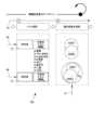
方法100は、以下の順序で、以下の段階を含む。
- リポジトリ抽出段階(図3(左部)および図4に示す)、
- グランド・トゥルース処理段階(図3(右部)に示す)、
- 前処理段階(図5(左部)に示す)、
- 深層学習または学習段階(図5(右部)に示す)、
- テスト段階(図6(左部)に示す)、
- 臨床最適化段階(図6(右部)に示す)、
- 検証段階(図7(左部)に示す)、
- 工業化段階(図7(右部)に示す)。 The
- a repository extraction stage (illustrated in Figure 3 (left) and Figure 4);
- a ground truth processing stage (illustrated in Fig. 3 (right part)),
- a pre-treatment stage (shown in FIG. 5 (left)),
- a deep learning or training phase (illustrated in Figure 5 (right part)),
- a test phase (shown in FIG. 6 (left part)),
- a clinical optimization phase (shown in FIG. 6 (right)),
- a verification phase (illustrated in FIG. 7 (left part)),
- The industrialization stage (shown in Figure 7 (right)).
図1に示すように、方法100は、(図1の最終的な工業化段階および図7の右部で)、入力骨を示すデジタル化された入力X線画像6を取得および受信する。As shown in FIG. 1, the method 100 (at the final industrialization stage in FIG. 1 and on the right in FIG. 7) acquires and receives an input digitized
受信した入力X線画像6は、好ましくは、二重エネルギーX線吸収測定(DXA)画像、末梢定量的コンピュータ断層撮影((p)QCT)画像および/または高分解能末梢定量的コンピュータ断層撮影(HR-pQCT)画像、コンピュータ断層撮影(CT)画像、または定量的超音波(QUS)画像ではない。The received
この実施形態では、各画像6、9、または19は、デジタル化された画像である。In this embodiment, each
受信した入力X線画像6は、好ましくは、1ピクセルあたり1mm未満の空間分解能を有するデジタルX線画像である。The received
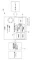
次に、方法100は、技術的手段(典型的には、少なくとも1つのコンピュータ、1つの中央処理装置または演算装置、1つのアナログ電子回路(好ましくは専用)、1つのデジタル電子回路(好ましくは専用)、および/または1つのマイクロプロセッサ(好ましくは専用)、および/またはソフトウェア手段を含むか、またはそれらで構成される)によって実装された第1の人工知能ANN1による、受信した入力X線画像6の第1の解析11を含む。第1の人工知能は、第1の解析の結果として、図2に示すグローバルスコア16を与え、以下の両方に依存する。
- 受信した入力X線画像に示された入力骨の骨密度に依存する(またはそれで構成される)密度スコア
- 受信した入力X線画像に示された入力骨の海綿骨部分のテクスチャに依存する海綿骨スコア(TBS) The
a density score that depends on (or consists of) the bone density of the input bone shown in the received input X-ray image; a trabecular bone score (TBS) that depends on the texture of the trabecular bone part of the input bone shown in the received input X-ray image.
グローバルスコア16は、好ましくは、この密度スコア(好ましくは骨密度で構成される)と海綿骨スコア(TBS)のみに依存し、したがって好ましくは、この密度スコア(好ましくは骨密度で構成される)と海綿骨スコア(TBS)の組み合わせである。The
それにもかかわらず、ANN1は、画像6からの密度スコアまたはTBSを決定または計算せず、直接グローバルスコア16を決定する。 Nevertheless, the ANN1 does not determine or calculate density scores or TBS from the
第1の人工知能はニューラルネットワークである。The first artificial intelligence is a neural network.
グローバルスコア16はマルチクラス分類器である。GlobalScore16 is a multi-class classifier.
グローバルスコア16は有限個の可能な値を有する。それは図2に示すように9つの可能な値しか有さない。これらの値のそれぞれは正の整数である。これらの値のそれぞれは、好ましくは1、2、3、4、5、5、7、8、および9の中の正の整数である。The
図1から8の説明のすべてにおいて、密度スコアは以下のようになり得る。
- 骨密度に等しい
- BMDの値に応じて決定される。例えば、
-- BMD T値≦-2.5の場合、密度スコア=「骨粗鬆症」または「骨粗鬆症の」
-- BMD T値が]-2.5,-1[の場合、密度スコア=「骨減少症」または「骨減少症の」
-- BMD T値≧-1の場合、密度スコア=「正常」
; 骨密度は、従来の先行技術の技術に従って画像9から決定される。骨密度(BMD)は、典型的には、骨による各ビームの吸収から決定される。二重エネルギーX線吸収測定法は、最も広く使用され、最も徹底的に研究されている骨密度測定技術である。 In all of the illustrations of Figures 1 to 8, the density scores can be as follows:
- Equivalent to bone mineral density - Determined according to the value of BMD. For example,
-- If BMD T value ≦-2.5, density score = "osteoporotic" or "osteoporotic"
-- If BMD T value is ]-2.5,-1[, density score = "osteopenic" or "osteopenic"
-- If BMD T value >=-1, density score = "normal"
bone mineral density is determined from the
通常、骨密度は二重エネルギーX線吸収測定(DXA)画像から直接取得され、典型的には二重エネルギーX線吸収測定(DXA)画像自体のメタデータであり得る。Typically, bone density is obtained directly from dual-energy x-ray absorptiometry (DXA) images and typically may be metadata of the dual-energy x-ray absorptiometry (DXA) image itself.
海綿骨スコア(TBS)は、グレーレベルの局所的な変動を定量化するテクスチャパラメータであり、デジタル化された画像のグレーレベルの実験的バリオグラムの評価から導出される。このデジタル化画像は、典型的には二重エネルギーX線吸収測定(DXA)画像であるが、他のデジタルX線画像または他の多くのX線画像モダリティであってもよい。The Trabecular Bone Score (TBS) is a texture parameter that quantifies the local variation in gray levels and is derived from the evaluation of an empirical variogram of gray levels in a digitized image. This digitized image is typically a dual-energy X-ray absorptiometry (DXA) image, but may also be other digital x-ray images or many other x-ray imaging modalities.
TBSのより詳細な説明または例は、非特許文献23に見出すことができる。A more detailed description or examples of TBS can be found in non-patent literature 23.
それにもかかわらず、この特定のケースでは、グローバルスコア16は、TBSの計算と受信した入力X線画像6のグレーレベルの実験的バリオグラムの計算または決定なしで、第1の人工知能によって決定される。Nevertheless, in this particular case, the
次に、方法100は、技術的手段(典型的には、少なくとも1つのコンピュータ、1つの中央処理装置または演算装置、1つのアナログ電子回路(好ましくは専用)、1つのデジタル電子回路(好ましくは専用)、および/または1つのマイクロプロセッサ(好ましくは専用)、および/またはソフトウェア手段を含むか、またはそれらで構成される)によって実装された第2の人工知能ANN2による、受信した入力X線画像6の第2の解析12を含む。第2の人工知能は、第2の解析の結果として以下を与える。
- 受信した入力X線画像6に示された入力骨の骨密度に依存する(またはそれで構成される)密度スコア、および/または
- 受信した入力X線画像6に示された入力骨の海綿骨部分のテクスチャに依存する海綿骨スコア(TBS) The
a density score that depends on (or consists of) the bone density of the input bone shown in the received
第2の人工知能はニューラルネットワークである。The second type of artificial intelligence is neural networks.
第2の人工知能は、連続値を与えるまたは推論する回帰モデルである。The second type of artificial intelligence is a regression model that gives or infers continuous values.
TBSは、受信した入力X線画像6のグレーレベルの実験的バリオグラムの計算または決定なしで、第2の人工知能によって決定される。The TBS is determined by the second artificial intelligence without calculation or determination of an empirical variogram of the grey levels of the received
次に、方法100は、技術的手段(典型的には、少なくとも1つのコンピュータ、1つの中央処理装置または演算装置、1つのアナログ電子回路(好ましくは専用)、1つのデジタル電子回路(好ましくは専用)、および/または1つのマイクロプロセッサ(好ましくは専用)、および/またはソフトウェア手段を含むか、またはそれらで構成される)によって実装された第3の人工知能AI3による第3の解析13を含む。第3の人工知能は、第1および第2の解析の結果を入力として有し、第1の解析の結果と第2の解析の結果の整合性に依存する結果を出力として有する。典型的には、AI3の出力は、患者関連のメタデータ18(軟部組織の厚さ、年齢、BMIなど)からの重み付け情報を使用したANN1の第1の解析の結果とANN2の第2の解析の結果の整合性に依存する。
- ケース#1:
・ ANN1 = 6,7,8または9(グローバルスコア)
・ ANN2 = 骨密度ステータスが骨減少症または骨粗鬆症、テクスチャステータスが部分的に劣化または劣化
AI3の結果 = フラグ
- ケース#2:
・ ANN1 = 6,7,8または9(グローバルスコア)
・ ANN2 = 骨密度ステータスが正常、テクスチャステータスが部分的に劣化または劣化
AI3の結果 = メタデータ18に応じて、フラグを立てるかどうかを評価するための重み付け応答
- ケース#3:
・ ANN1 = 6,7,8または9(グローバルスコア)
・ ANN2 = 骨密度ステータスが骨減少症または骨粗鬆症、テクスチャステータスが正常
AI3の結果 = メタデータ18に応じて、フラグを立てるかどうかを評価するための重み付け応答
- ケース#4:
・ ANN1 = 1,2,3,4または5(グローバルスコア)
・ ANN2 = 骨密度ステータスが正常、テクスチャステータスが正常
AI3の結果 = フラグなし
- ケース#5
・ ANN1 = 1,2,3,4または5(グローバルスコア)
・ ANN2 = 骨密度ステータスが骨減少症または骨粗鬆症、テクスチャステータスが正常
AI3の結果 = メタデータ18に応じて、フラグを立てるかどうかを評価するための重み付け応答
- ケース#6
・ ANN1 = 1,2,3,4または5(グローバルスコア)
・ ANN2 = 骨密度ステータス、テクスチャステータスが部分的に劣化または劣化
AI3の結果 = メタデータ18に応じて、フラグを立てるかどうかを評価するための重み付け応答 The
- Case #1:
ANN1 = 6, 7, 8 or 9 (global score)
ANN2 = Bone density status is osteopenic or osteoporotic, texture status is partially degraded or degraded AI3 result = Flag - Case #2:
ANN1 = 6, 7, 8 or 9 (global score)
ANN2 = Bone density status normal, texture status partially degraded or degraded AI3 result = Weighted response to evaluate whether to flag or not depending on metadata 18 - Case #3:
ANN1 = 6, 7, 8 or 9 (global score)
ANN2 = Bone density status is osteopenia or osteoporosis, texture status is normal AI3 result = Weighted response to evaluate whether to flag or not depending on metadata 18 - Case #4:
ANN1 = 1, 2, 3, 4 or 5 (global score)
ANN2 = Bone density status normal, texture status normal AI3 result = No flags -
ANN1 = 1, 2, 3, 4 or 5 (global score)
ANN2 = Bone density status is osteopenic or osteoporotic, texture status is normal AI3 result = Weighted response to evaluate whether to flag depending on metadata 18 -
ANN1 = 1, 2, 3, 4 or 5 (global score)
ANN2 = Bone density status, texture status partially degraded or degraded AI3 result = Weighted response to evaluate whether to flag or not depending on
第3の人工知能は、さらなる入力としてメタデータ18を使用し、メタデータ18は、受信した入力X線画像が取得された患者の年齢、受信した入力X線画像が取得された患者の性別、受信した入力X線画像が取得された患者の体型、受信した入力X線画像が取得された機器の種類、および/または受信した入力X線画像が取得された機器の取得パラメータのうちの少なくとも1つを含む。The third artificial intelligence uses as
第3の人工知能はニューラルネットワークであってもよいが、好ましくは、決定木、ランダムフォレスト、またはサポートベクターマシンなどの二値分類器である。The third artificial intelligence may be a neural network, but is preferably a binary classifier such as a decision tree, random forest, or support vector machine.
したがって、最後のステップとして、前述の2つのアプローチANN1とANN2の出力は、追加の選択されたメタデータ18(例えば、年齢、性別、体型、機器の種類、取得パラメータ)とともに、典型的には決定木のような学習モデルであるAI3の入力変数として入力される。分類木は、最終的な分類の最適化(DXA BMDとTBSによって評価される、劣化した骨微細構造を伴う骨粗鬆症として選択される可能性)のための最良の組み合わせを出力として提供する。バギング決定木などの特定の技術(アンサンブル法とも呼ばれる)が使用され、所与の時点および所与の個人に対する複数画像セットの可能性を考慮するが、それらに限定されない。 Therefore, as a final step, the outputs of the two aforementioned approaches ANN1 and ANN2 , together with additional selected metadata 18 (e.g. age, sex, body type, type of equipment, acquisition parameters) are input as input variables to AI3 , a learning model, typically like a decision tree. The classification tree provides as output the best combination for the final classification optimization (likelihood of being selected as osteoporosis with deteriorated bone microarchitecture, as assessed by DXA BMD and TBS). Certain techniques, such as bagging decision trees (also called ensemble methods), are used to consider the likelihood of multiple image sets for a given time point and a given individual, but are not limited to them.
第1の人工知能、第2の人工知能、および第3の人工知能は、3つの異なる人工知能または3つの異なる人工知能アーキテクチャである。The first artificial intelligence, the second artificial intelligence, and the third artificial intelligence are three different artificial intelligences or three different artificial intelligence architectures.
第1の人工知能、第2の人工知能、および第3の人工知能を実装するための技術的手段は、好ましくは同じ技術的手段、すなわちそれらに限定されないが、好ましくは、同じ少なくとも1つのコンピュータ、1つの中央処理装置または演算装置、1つのアナログ電子回路(好ましくは専用)、1つのデジタル電子回路(好ましくは専用)、および/または1つのマイクロプロセッサ(好ましくは専用)内で、および/またはソフトウェア手段によって組み込みモジュールに統合される。The technical means for implementing the first artificial intelligence, the second artificial intelligence and the third artificial intelligence are preferably integrated in the same technical means, i.e., but not limited to, preferably within the same at least one computer, one central processing unit or computing unit, one analog electronic circuit (preferably dedicated), one digital electronic circuit (preferably dedicated) and/or one microprocessor (preferably dedicated) and/or in an embedded module by software means.
これらの人工知能ANN1、ANN2、AI3は、事前に学習させる。 These artificial intelligences ANN1 , ANN2 , and AI3 are trained in advance.
したがって、方法100は、工業化段階の解析ステップ11、12、および13の前に、以下のステップを含む。
- 以下のステップを複数回実施することにより、第1の学習セットを構築するステップ:
-- 学習用骨の海綿骨部分を示す第1のタイプの学習画像9(第1の学習セットの)を取得する(リポジトリ抽出段階中)ステップ
-- 同じ学習用骨を示すが、必ずしもその海綿骨部分を示さない、X線ベースの画像である関連する第2のタイプ19の学習画像(第1の学習セットの)を取得する(リポジトリ抽出段階中)ステップ
-- 第1の学習セットの第1のタイプの学習画像9から、技術的手段(典型的には、少なくとも1つのコンピュータ、1つの中央処理装置または演算装置、1つのアナログ電子回路(好ましくは専用)、1つのデジタル電子回路(好ましくは専用)、および/または1つのマイクロプロセッサ(好ましくは専用)を含むか、またはそれらで構成される、および/またはソフトウェア手段による)によって(ただし、画像9に人工知能を実装せずに)、以下を決定する(グランド・トゥルース処理段階中)ステップ:
--- 第1の学習セットの第1のタイプの学習画像に示された学習用骨の骨密度に依存するまたは等しい密度スコア14。典型的には、骨密度は、第1のタイプの学習画像のDXAのメタデータとして第1のタイプの学習画像から直接決定される。および
--- 第1の学習セットの第1のタイプの学習画像に示された学習用骨の海綿骨部分のテクスチャに依存する海綿骨スコア(TBS)15。典型的には、このTBS 15は、第1のタイプの学習画像のグレーレベルの実験的バリオグラムを計算または決定することによって決定される(特許文献2)。
-- 以下から、技術的手段(典型的には、少なくとも1つのコンピュータ、1つの中央処理装置または演算装置、1つのアナログ電子回路(好ましくは専用)、1つのデジタル電子回路(好ましくは専用)、および/または1つのマイクロプロセッサ(好ましくは専用)を含むか、またはそれらで構成される、および/またはソフトウェア手段による)によって、(ただし、画像9、および/または事前に決定された密度スコア14、および/または事前に決定されたTBS 15に人工知能を実装せずに)、これらの密度スコアと海綿骨スコアに依存する(好ましくはそれらのみに依存する)グローバルスコア16を決定する(グランド・トゥルース処理段階中)ステップ:
--- 第1の学習セットの第1のタイプの学習画像9に示された学習用骨の骨密度に依存するまたは等しい密度スコア14、および
--- 第1の学習セットの第1のタイプの学習画像9に示された学習用骨の海綿骨部分のテクスチャに依存する海綿骨スコア(TBS)15;
例えば、
- 骨密度(BMD)の値に応じて、密度スコアまたはステータスを決定することができる。例えば、
-- BMD T値≦-2.5の場合、密度スコア=「骨粗鬆症」または「骨粗鬆症の」
-- BMD T値が]-2.5,-1[の場合、密度スコア=「骨減少症」または「骨減少症の」
-- BMD T値≧-1の場合、密度スコア=「正常」
- 設定アルゴリズムは技術的および臨床的情報に適応されるため、以下に示す値は例であるが、TBSの値に応じて骨テクスチャステータスを決定することができる。
-- TBS>1.31の場合、骨テクスチャステータス=「正常」
-- TBSが[1.23,1.31]の場合、骨テクスチャステータス=「部分的に劣化」
-- TBS<1.23の場合、骨テクスチャステータス=「劣化」
- グローバルスコア16は、(図2に示すように)以下の規則に従って決定することができる。
-- 密度スコア=「正常」かつ骨テクスチャステータス=「正常」の場合、グローバルスコア= 1
-- 密度スコア=「正常」かつ骨テクスチャステータス=「部分的に劣化」の場合、グローバルスコア= 2
-- 密度スコア=「正常」かつ骨テクスチャステータス=「劣化」の場合、グローバルスコア= 3
-- 密度スコア=「骨減少症」または「骨減少症の」かつ骨テクスチャステータス=「正常」の場合、グローバルスコア= 4
-- 密度スコア=「骨減少症」または「骨減少症の」かつ骨テクスチャステータス=「部分的に劣化」の場合、グローバルスコア= 5
-- 密度スコア=「骨減少症」または「骨減少症の」かつ骨テクスチャステータス=「劣化」の場合、グローバルスコア= 6
-- 密度スコア=「骨粗鬆症」または「骨粗鬆症の」かつ骨テクスチャステータス=「正常」の場合、グローバルスコア= 7
-- 密度スコア=「骨粗鬆症」または「骨粗鬆症の」かつ骨テクスチャステータス=「部分的に劣化」の場合、グローバルスコア= 8
-- 密度スコア=「骨粗鬆症」または「骨粗鬆症の」かつ骨テクスチャステータス=「劣化」の場合、グローバルスコア= 9
- 第1の学習セットの第1のタイプの学習画像9について決定されたグローバルスコア16を含むまたはそれで構成されるその関連するグランド・トゥルース1とともに、第1の学習セットの第2のタイプの学習画像19(ステップ7)を第1の人工知能に提供することにより、第1の人工知能ANN1を学習させる(前処理段階および深層学習または学習段階中)ステップ。方法100の好ましい実施形態では、
-- ANN1は、(ただし限定されないが)複数の層(例えば、50層以上)を有するMLP、CNNである。好ましい実施形態では、ANN1はマルチクラス分類器の畳み込みニューラルネットワークである。
-- この学習ステップは、精度(TP/(TP+FP))を最適化することによって勾配逆伝播によって行われる(TP=真陽性、FP=偽陽性)。
-- 過学習を避けつつ最小のテストロス(カテゴリカルクロスエントロピー損失関数)に達するまで学習が行われる。 Thus, the
- constructing a first training set by performing the following steps multiple times:
-- obtaining (during the repository extraction phase)
--- a
- determining (during the ground truth processing stage) a
--- a
for example,
Depending on the value of bone mineral density (BMD) a density score or status can be determined, e.g.
-- If BMD T value ≦-2.5, density score = "osteoporotic" or "osteoporotic"
-- If BMD T value is ]-2.5,-1[, density score = "osteopenic" or "osteopenic"
-- If BMD T value >=-1, density score = "normal"
The setting algorithm is adapted to technical and clinical information, so that the bone texture status can be determined depending on the value of TBS, although the values shown below are examples:
-- If TBS>1.31, then bone texture status = "normal"
-- if TBS is [1.23, 1.31], bone texture status = "partially degraded"
-- if TBS<1.23, bone texture status = "degraded"
The
-- If density score = "normal" and bone texture status = "normal", then global score = 1
-- If density score = "normal" and bone texture status = "partially deteriorated", then global score = 2
-- If density score = "normal" and bone texture status = "deteriorated", then global score = 3
-- If density score = "osteopenic" or "osteopenic" and bone texture status = "normal", then global score = 4
-- If density score = "osteopenic" or "osteopenic" and bone texture status = "partially deteriorated", then global score = 5.
-- If density score = "osteopenic" or "osteopenic" and bone texture status = "poor", then global score = 6
-- If density score = "osteoporotic" or "osteoporotic" and bone texture status = "normal", then global score = 7
-- If density score = "osteoporotic" or "osteoporotic" and bone texture status = "partially deteriorated", then global score = 8
-- If density score = "osteoporotic" or "osteoporotic" and bone texture status = "deteriorated", then global score = 9
training (during the pre-processing and deep learning or training phases) of a first artificial intelligence ANN1 by providing it with
--ANN1 is (but is not limited to) a MLP, CNN with multiple layers (e.g., 50 layers or more). In a preferred embodiment,
--This learning step is done by gradient backpropagation by optimizing the precision (TP/(TP+FP)) (TP=true positives, FP=false positives).
-- Training continues until the minimum test loss (categorical cross-entropy loss function) is reached while avoiding overfitting.
第1の学習セットについて、第1のタイプの学習画像9と第2のタイプの学習画像19は、同じ学習用骨で取得され、6か月未満の間隔で取得される。For the first training set, the first
第1の学習セットについて、第1のタイプの学習画像9は、二重エネルギーX線吸収測定(DXA)画像、末梢定量的コンピュータ断層撮影((p)QCT)画像および/または高分解能末梢定量的コンピュータ断層撮影(HR-pQCT)画像、コンピュータ断層撮影(CT)画像、または定量的超音波(QUS)画像である。For the first training set, the first type of
第1の学習セットについて、第2のタイプの学習画像19は、二重エネルギーX線吸収測定(DXA)画像、末梢定量的コンピュータ断層撮影((p)QCT)画像および/または高分解能末梢定量的コンピュータ断層撮影(HR-pQCT)画像、コンピュータ断層撮影(CT)画像、または定量的超音波(QUS)画像ではなく、例えばDICOM(「医用デジタル画像と通信」の略)画像である。For the first training set, the second type of
第1の学習セットについて、第2のタイプの学習画像19は、好ましくは、1ピクセルあたり1mm未満の空間分解能を有するデジタルX線画像である。For the first training set, the second type of
方法100はまた、解析ステップ11、12、および13の前に、以下のステップを含む。
- 以下のステップを複数回実施することにより、第2の学習セットを構築するステップ:
-- 学習用骨の海綿骨部分を示す第1のタイプ9の学習画像(第2の学習セットの)を取得する(リポジトリ抽出段階中)ステップ。
-- 同じ学習用骨を示すが、必ずしもその海綿骨部分を示さない、X線ベースの画像である関連する第2のタイプ19の学習画像(第2の学習セットの)を取得する(リポジトリ抽出段階中)ステップ。
-- 第2の学習セットの第1のタイプの学習画像9から、技術的手段(典型的には、少なくとも1つのコンピュータ、1つの中央処理装置または演算装置、1つのアナログ電子回路(好ましくは専用)、1つのデジタル電子回路(好ましくは専用)、および/または1つのマイクロプロセッサ(好ましくは専用)を含むか、またはそれらで構成される、および/またはソフトウェア手段による)によって(ただし、画像9に人工知能を実装せずに)、以下を決定する(グランド・トゥルース処理段階中)ステップ:
--- 第2の学習セットの第1のタイプの学習画像9に示された学習用骨の骨密度に依存するまたは等しい密度スコア14。典型的には、骨密度は、第1のタイプの学習画像のDXAのメタデータとして第1のタイプの学習画像から直接決定される。および/または
--- 第2の学習セットの第1のタイプの学習画像9に示された学習用骨の海綿骨部分のテクスチャに依存する海綿骨スコア(TBS)15。典型的には、このTBS 15は、第1のタイプの学習画像のグレーレベルの実験的バリオグラムを計算または決定することによって決定される(特許文献2)。
- 以下を含むまたはそれらで構成されるその関連するグランド・トゥルース2とともに、第2の学習セットの第2のタイプの学習画像19を第2の人工知能に提供することにより(ステップ8)、第2の人工知能ANN2を学習させる(前処理段階および深層学習または学習段階中)ステップ:
-- この第2の学習セットの第2のタイプの学習画像19に関連付けられた第2の学習セットの第1のタイプの学習画像9について決定された密度スコア14、および/または
-- この第2の学習セットの第2のタイプの学習画像19に関連付けられた第2の学習セットの第1のタイプの学習画像9について決定された海綿骨スコア15。 The
- constructing a second training set by performing the following steps multiple times:
-- Obtaining (during the repository extraction phase)
-- Obtaining (during the repository extraction phase) related training images of a second type 19 (of a second training set) that are X-ray based images showing the same training bone but not necessarily showing its cancellous portion.
- determining (during the ground truth processing phase) from
--- a
training (during a pre-processing phase and a deep learning or training phase) a second artificial intelligence ANN2 by providing it (step 8) with
--a
方法100の好ましい実施形態では、
- ANN2は、(ただし限定されないが)複数の層(例えば、50層以上)を有するMLP、CNNである。好ましい実施形態では、ANN1は回帰畳み込みニューラルネットワークである。
- この学習ステップは、過学習を避けつつ最小のテストロスに達するまで、二次損失関数を最小化することによって勾配逆伝播によって行われる。
- この学習は、過学習を避けつつ最小のテストロス(二次損失関数)に達するまで行われる。 In a preferred embodiment of
- ANN2 is (but is not limited to) a MLP, CNN with multiple layers (e.g., 50 layers or more). In a preferred embodiment,
This learning step is done by gradient backpropagation by minimizing a quadratic loss function until the minimum test loss is reached while avoiding overfitting.
This training continues until the minimum test loss (quadratic loss function) is reached while avoiding overfitting.
第2の学習セットについて、第1のタイプの学習画像と第2のタイプの学習画像は、同じ学習用骨で取得され、6か月未満の間隔で取得される。For the second training set, the first type of training images and the second type of training images are acquired of the same training bone and are acquired less than six months apart.
第2の学習セットについて、第1のタイプの学習画像は、二重エネルギーX線吸収測定(DXA)画像、末梢定量的コンピュータ断層撮影((p)QCT)画像および/または高分解能末梢定量的コンピュータ断層撮影(HR-pQCT)画像、コンピュータ断層撮影(CT)画像、または定量的超音波(QUS)画像である。For the second training set, the first type of training images are dual-energy X-ray absorptiometry (DXA) images, peripheral quantitative computed tomography ((p)QCT) images and/or high-resolution peripheral quantitative computed tomography (HR-pQCT) images, computed tomography (CT) images, or quantitative ultrasound (QUS) images.
第2の学習セットについて、第2のタイプの学習画像は、二重エネルギーX線吸収測定(DXA)画像、末梢定量的コンピュータ断層撮影((p)QCT)画像および/または高分解能末梢定量的コンピュータ断層撮影(HR-pQCT)画像、コンピュータ断層撮影(CT)画像、または定量的超音波(QUS)画像ではなく、例えばDICOM画像である。For the second training set, the second type of training images are, for example, DICOM images, rather than dual-energy X-ray absorptiometry (DXA) images, peripheral quantitative computed tomography ((p)QCT) images and/or high-resolution peripheral quantitative computed tomography (HR-pQCT) images, computed tomography (CT) images, or quantitative ultrasound (QUS) images.
第2の学習セットについて、第2のタイプの学習画像19は、好ましくは、1ピクセルあたり1mm未満の空間分解能を有するデジタルX線画像である。For the second training set, the second type of
決定された海綿骨スコア15は、グレーレベルの局所的な変動を定量化するテクスチャパラメータであり、第1のタイプのデジタル化された画像9のグレーレベルの実験的バリオグラムの評価から導出される。このデジタル化画像9は、典型的には二重エネルギーX線吸収測定(DXA)画像である。The determined
第1の人工知能ANN1を学習させるためのこれらのステップおよび第2の人工知能ANN2を学習させるためのこれらのステップについて、海綿骨スコア(TBS)15は、画像9に人工知能を実装せずに、典型的には特許文献2に記載の方法に従って、または学習方法の以下のステップa)からi)に従って、第1のタイプの各学習画像9から決定される。 For these steps for training the first artificial intelligence ANN1 and for these steps for training the second artificial intelligence ANN2 , a cancellous bone score (TBS) 15 is determined from each
a) 画像9の最適化されたピクセルサンプリングSを決定する。各ピクセルPi=(xi,yi)∈Sは、そのグレーレベル値h(Pi)を有する(またはこれにより定義される)。
画像9から、(ステップd)で)バリオグラムViが評価される位置を選択するために、ピクセルサンプリングSを決定する。
これらの位置は、最終的なTBSが所与の臨床的文脈および解剖学的部位に適応され、効率的になるように最適化される。最も基本的な意味では、サンプリング領域は、画像中に骨が存在する領域に対応する。
骨部位に応じて、画像中に骨が存在するこの領域のサブサンプルを使用して、性能を向上させることができる。 a) Determine an optimized pixel sampling S of an
From the
These locations are optimized so that the final TBS is adapted and efficient for a given clinical context and anatomical site. In the most basic sense, the sampling region corresponds to the area in the image where bone is present.
Depending on the bone site, a subsample of this region where bone is present in the image can be used to improve performance.
b) ピクセルサンプリングSの少なくとも1つの関心領域(ROI)について、好ましくは複数のROIについて、
- 所与の関心領域(ROI)に応じて、計算距離R0を決定する。範囲R0は、骨格部位および画像9の画像解像度に応じて決定される。DXAシステムでは、R0の値は1cmから2cmの間である。
- 所与の関心領域(ROI)に応じて、所定の方向のセットIを選択する。この所定の方向のセットIを選択するステップは、N個の方向単位ベクトル b) for at least one region of interest (ROI) of pixel sampling S, preferably for a plurality of ROIs,
-Depending on the given region of interest (ROI), the calculation distanceR0 is determined. The rangeR0 is determined depending on the skeletal site and the image resolution of the
- Select a set of predefined directions I according to a given region of interest (ROI). This step of selecting a set of predefined directions I is carried out by using N directional unit vectors

のセットを決定することによって行われる(Nは正の整数であり、θk∈[-π,π],∀k∈1,…,N,θkは、考慮されるピクセルの周りで、ベクトル(where N is a positive integer, θk ∈[−π,π],∀k∈1,...,N,θk is a vector

を運ぶ角度である)。
典型的にはN>2である。
Nの最大値は、ROIの考慮される撮像骨の骨構造の複雑さおよびその画像解像度に依存する。典型的には、椎骨または腰椎のような非複雑な骨構造を有する骨についてはN<9であるが、大腿骨近位部のような複雑な構造については、Nが大幅に増加する場合がある。
例えば、N=3、4、6、または8である。
N個の方向ベクトルは、好ましくは、考慮されるピクセルの周りで角度2π/Nで均一に分布する。
所定の方向のセットI(ベクトル(The angle at which the
Typically, N>2.
The maximum value of N depends on the complexity of the bone structure of the imaged bone considered in the ROI and its image resolution. Typically, N<9 for bones with non-complex bone structure such as the vertebrae or lumbar spine, but for complex structures such as the proximal femur, N may be significantly increased.
For example, N=3, 4, 6, or 8.
The N direction vectors are preferably uniformly distributed with an angle 2π/N around the pixel under consideration.
Given a set of directions I (vectors

)は、以下に依存する。
- 画像上および/またはROI上の骨の骨格部位(ヒトまたは動物の組織は骨である)、および/または
- 考慮される関心領域(ROI)、および/または
- 画像の解像度、および/または
- 画像の信号対雑音比。
この方向のセットIの決定は、
- 骨折を識別または予測する能力を向上させ、および/または
- より良好な測定の再現性(または精度)を可能にし、および/または
- 骨の微細構造とのより良好な相関を可能にする。
したがって、後のステップd)でバリオグラムVPiを計算するために、値h(Pi)を有するピクセルを、特定の方向の直線に沿って配置されたピクセルと比較する。
これらの方向は、骨の種類(すなわち、骨格部位)と、テクスチャ測定を最適化するためにこの骨で測定のために選択された関心領域の両方に依存する。
実際、選択された方向は、特に海綿骨の骨梁の方向など、骨の形態に関連している。例えば、脊椎では、骨梁の好ましい方向は垂直方向であるため、選択される方向は垂直方向および水平方向[-Π/2,0,Π/2,Π](骨梁の向きに平行および垂直)となる。) depends on:
- the bone skeletal site on the image and/or on the ROI (human or animal tissue is bone), and/or - the region of interest (ROI) considered, and/or - the image resolution, and/or - the signal-to-noise ratio of the image.
The determination of this set of directions I is
- improves the ability to identify or predict fractures, and/or - allows for better reproducibility (or precision) of measurements, and/or - allows for better correlation with bone microarchitecture.
Thus, to calculate the variogram VPi in the following step d), the pixel with value h(Pi ) is compared with the pixels located along a straight line in a particular direction.
These directions depend on both the type of bone (ie, skeletal site) and the region of interest selected for measurement on this bone to optimize the texture measurement.
In fact, the chosen directions are related to the bone morphology, especially the direction of the trabeculae of cancellous bone: for example, in the spine, the preferred direction of the trabeculae is vertical, so the chosen directions are vertical and horizontal [-Π/2,0,Π/2,Π] (parallel and perpendicular to the orientation of the trabeculae).
c) 各ピクセルPi=(xi,yi)∈Sおよび各方向 c) For each pixel Pi = (xi , yi ) ∈ S and for each direction

について、About

に沿って距離r∈[1,R0](ピクセル単位)まで移動する。このようなピクセルのグレー値を, by a distance r ∈ [1, R0 ] (in pixels).

と表す。This is expressed as:
d) 各ピクセルについて、これらの方向Iに沿った距離rの関数としてサンプリングSのグレーレベルの少なくとも1つのバリオグラムを計算する(すなわち、少なくとも1つの距離r∈[1,R0]をこのピクセルから移動することによって。前述のように、距離rを移動することは、各ピクセルPi=(xi,yi)∈Sおよび各方向 d) for each pixel, calculate at least one variogram of the grey levels of the sampling S as a function of the distance r along these directions I (i.e. by moving at least one distance r ∈ [1, R0 ] from this pixel. As mentioned before, moving a distance r means calculating for each pixel Pi = (xi , yi ) ∈ S and for each direction I,

について、About

に沿って距離r∈[1,R0]ピクセルを移動することによって行われ、This is done by moving a distance r ∈ [1, R0 ] pixels along

はそのようなピクセルのグレー値である)。バリオグラムは以下のように計算される。
- 各所定の方向について1つずつ(すなわち、Sの各ピクセルおよびIの各方向について1つのバリオグラム)。各所定の方向についてバリオグラムが計算される場合、h(O)は移動前の初期の所与のピクセルのグレーレベルであり、h(r)は初期の所与のピクセルから所定の方向の1つに沿って距離rを移動した後の所与の新しいピクセルのグレーレベルであり、バリオグラムは式Vi(r)=[h(r)-h(O)]2で計算され、i∈Iである。または、
- 同時にすべての所定の方向について(すなわち、Sの各ピクセルについて1つのバリオグラム)。すべての所定の方向について同時にバリオグラムが計算される場合、グレーレベルのバリオグラムを距離rの関数として計算するステップは、各々が距離rにある複数のピクセルのペアについてhの二乗差を平均化することによって行われ、式は以下の通りである。is the grey value of such a pixel.) The variogram is calculated as follows:
one for each given direction (i.e. one variogram for each pixel in S and each direction in I). If a variogram is calculated for each given direction, h(O) is the grey level of the initial given pixel before movement, h(r) is the grey level of a given new pixel after moving a distance r along one of the given directions from the initial given pixel, and the variogram is calculated by the formula Vi (r) = [h(r) - h(O)]2 , i ∈ I, or
- for all given directions simultaneously (i.e. one variogram for each pixel of S). When variograms are calculated simultaneously for all given directions, the step of calculating the variogram of the grey level as a function of distance r is done by averaging the squared differences of h for several pairs of pixels, each at distance r, and the formula is:

VPi(r)は、すべてのピクセルPi∈Sについて計算される。
VPiの式は、r∈[1,R0]の値の所与の範囲について、各Pi∈Sに適用される。rのこの特定の値の範囲は、バリオグラムVPiのすべての必要なパラメータの評価のために VPi (r) is calculated for all pixels Pi εS.
The formula for VPi is applied to each Pi ∈ S for a given range of values of r ∈ [1, R0 ]. This particular range of values of r is used for the evaluation of all the necessary parameters of the variogram VPi.

が収束することを可能にするように選択される。
範囲R0は、骨格部位および画像の解像度に応じて決定される。DXAシステムでは、R0の値は1cmから2cmの間である。
計算の範囲R0は、バリオグラムモデルの範囲パラメータcと混同されるべきではない。is chosen to allow for convergence.
The rangeR0 is determined depending on the skeletal location and the image resolution. In DXA systems, the value ofR0 is between 1 cm and 2 cm.
The range of the calculationR0 should not be confused with the range parameter c of the variogram model.
e) すべてのピクセルPi∈SについてVPi(r)を計算し、対数-対数スケールで関連する曲線をトレースまたは計算または決定する。
対数-対数スケールでのバリオグラム曲線VPiの表現は、各軸に沿った値がもはや単位を有さないことを意味する。 e) Calculate VPi (r) for every pixel Pi εS and trace or calculate or determine the associated curve on a log-log scale.
The representation of the variogram curves VPi on a log-log scale means that the values along each axis no longer have units.
f) VPi表現の最小二乗回帰モデルとして完全なモデルVを評価する。 f) Evaluate the complete model V as a least squares regression model of the VPi representation.
g) 初期の傾きa、シルb、範囲c、ナゲットd、および/または曲線下の面積eを含むが、これらに限定されないこのモデルのパラメータを評価する。
各ピクセルのバリオグラムVPiおよび/または各ピクセルのバリオグラムを組み合わせたサンプリングSのグローバルバリオグラムVについて、対数-対数スケールで以下のパラメータの少なくとも1つを評価する。
- バリオグラムの初期の傾きa(図8の54参照)、
- バリオグラムの漸近値を表すシルb(図8の55参照)、
- バリオグラム曲線が準線形の進行から漸近的挙動に移行する距離を表す範囲c(図8の56参照)、
- バリオグラムの初期値を表すナゲットd(図8の57参照)、および
- バリオグラムのバリオグラム曲線下の面積e(図8の58参照)。
VPiまたはVの対数-対数プロットでは、数学モデルにフィッティングする。このモデルのこれらのパラメータ(すなわち、係数)a、b、c、d、eを組み合わせて、骨テクスチャスコアBを作成する。
各パラメータa、b、c、d、および/またはeは、考慮されるバリオグラムの最小二乗回帰モデルから評価される。
画像9の内容に応じて、モデルの選択された係数は、明確に定義されない可能性があるため、変化し得る(例えば、バリオグラム曲線が漸近線に収束しない可能性があり、したがって「範囲」が定義されない可能性がある)。
好ましくは、学習方法は、各ピクセルの各バリオグラムおよび/または各ピクセルのバリオグラムを組み合わせたサンプリングSのグローバルバリオグラムについて、対数-対数スケールでパラメータシルb、範囲c、ナゲットd、面積eの少なくとも1つを評価することを含む。
好ましくは、学習方法は、各ピクセルの各バリオグラムおよび/または各ピクセルのバリオグラムを組み合わせたサンプリングSのグローバルバリオグラムについて、対数-対数スケールでパラメータ傾きa、シルb、範囲c、ナゲットd、面積eの少なくとも2つを評価することを含む。
好ましくは、学習方法は、各ピクセルの各バリオグラムおよび/または各ピクセルのバリオグラムを組み合わせたサンプリングSのグローバルバリオグラムについて、初期の傾きaおよび対数-対数スケールで以下のパラメータシルb、範囲c、ナゲットd、面積eの少なくとも1つを評価することを含む。
好ましくは、学習方法は、各ピクセルの各バリオグラムおよび/または各ピクセルのバリオグラムを組み合わせたサンプリングSのグローバルバリオグラムについて、対数-対数スケールですべてのパラメータ傾きa、シルb、範囲c、ナゲットd、面積eを評価することを含む。 g) Evaluate the parameters of the model including, but not limited to, the initial slope a, sill b, range c, nugget d, and/or area under the curve e.
For each pixel's variogram VPi and/or for the global variogram V of the sampling S combining the variograms of each pixel, at least one of the following parameters is evaluated in a log-log scale:
the initial slope a of the variogram (see 54 in FIG. 8),
- sil b (see 55 in FIG. 8) which represents the asymptotic value of the variogram,
- the range c (see 56 in Fig. 8) which represents the distance at which the variogram curve passes from a quasi-linear progression to an asymptotic behavior,
the nugget d (see 57 in FIG. 8) which represents the initial value of the variogram, and the area under the variogram curve e (see 58 in FIG. 8) of the variogram.
A log-log plot of VPi or V is fitted to a mathematical model. These parameters (i.e., coefficients) a, b, c, d, and e of the model are combined to create a bone texture score, B.
Each parameter a, b, c, d, and/or e is estimated from a least-squares regression model of the variogram considered.
Depending on the content of
Preferably, the training method includes evaluating at least one of the parameters sill b, extent c, nugget d, area e on a log-log scale for each variogram of each pixel and/or a global variogram of a sampling S combining the variograms of each pixel.
Preferably, the training method includes evaluating at least two of the parameters slope a, sill b, range c, nugget d, and area e on a log-log scale for each variogram at each pixel and/or a global variogram of a sampling S combining the variograms at each pixel.
Preferably, the training method comprises evaluating, for each variogram of each pixel and/or a global variogram of a sampling S combining the variograms of each pixel, an initial slope a and at least one of the following parameters sill b, extent c, nugget d, area e on a log-log scale:
Preferably, the training method involves evaluating all parameters slope a, sill b, range c, nugget d, area e on a log-log scale for each variogram at each pixel and/or a global variogram of a sampling S combining the variograms at each pixel.
h) 少なくとも1つのパラメータa、b、c、dおよび/またはeを、例えば臨床的文脈に応じて線形または非線形の方程式を使用して、TBS(無次元)に組み合わせる。
例えば、学習方法は、以下のステップを含むことができる。
- ピクセルまたは各ピクセルのバリオグラムについて得られた少なくとも1つの評価されたパラメータa、b、c、dおよび/またはeを、ピクセルまたは各ピクセルのTBSに組み合わせる、および/または
- サンプリングSのグローバルバリオグラムについて得られた少なくとも1つの評価されたパラメータa、b、c、dおよび/またはeを、サンプリングSのTBSに組み合わせる、および/または
- 各ピクセルのバリオグラムについて得られた少なくとも1つの評価されたパラメータa、b、c、dおよび/またはeを、サンプリングSのTBSに組み合わせる。
少なくとも2つのパラメータa、b、c、d、eの中から、臨床的文脈に応じて線形または非線形の方程式を使用してTBSに組み合わせることが好ましい。実際、バリオグラムモデルのパラメータは、組み合わせ方程式を使用してTBSに一緒に組み合わされる。例として、このような組み合わせ方程式は、所与の臨床的文脈および解剖学的部位に対する重回帰モデルを含むことができるが、それに限定されない。そのようなケースシナリオでは、TBSは以下のように定義される。
TBS=αa+βb+γc+δd+εe,
ここで、係数α,β,γ,δ,εは、それぞれ傾き、シル、ナゲット、および曲線下の面積に関連付けられる。これらの係数は、臨床性能最適化段階の前に慎重に定義される。
これらの係数は、典型的には異なる実験分析から得られる。
最良の係数の選択は、例えば、大規模な臨床研究および画像のセットで、さらにグリッドサーチ最適化段階で得られる。
より一般的には、α、β、γ、δおよび/またはεは、以下を含む異なる方法で得ることができる。
- 実験測定に基づく補正係数表、および/または
- 理論に基づく補正の数学モデルおよび/またはシミュレーション、および/または
-機械学習または人工知能(AI)アルゴリズム(この機械学習または人工知能(AI)は、学習画像9に適用されない)、および/または
- 例えば、大規模な臨床研究および画像のセットで、さらに臨床性能の結果を伴うグリッドサーチ最適化段階で得られる最良の係数の選択、および/または
- 上記の方法の組み合わせ、
- など。
例えば、
TBS=a
すなわち、α=1 及び β=γ=δ=ε=0
好ましくは、TBSは、同時にすべての所定の方向について計算されたサンプリングSのグローバルバリオグラムから計算される。 h) Combine at least one parameter a, b, c, d and/or e into a TBS (dimensionless), e.g. using a linear or non-linear equation depending on the clinical context.
For example, the learning method may include the following steps.
- combining at least one evaluated parameter a, b, c, d and/or e obtained for the variogram of the pixel or of each pixel into the TBS of the pixel or of each pixel, and/or - combining at least one evaluated parameter a, b, c, d and/or e obtained for the global variogram of the sampling S into the TBS of the sampling S, and/or - combining at least one evaluated parameter a, b, c, d and/or e obtained for the variogram of each pixel into the TBS of the sampling S.
Preferably, at least two parameters a, b, c, d, e are combined into the TBS using a linear or non-linear equation depending on the clinical context. In practice, the parameters of the variogram model are combined together into the TBS using a combination equation. By way of example, such a combination equation can include, but is not limited to, a multiple regression model for a given clinical context and anatomical site. In such a case scenario, the TBS is defined as follows:
TBS=αa+βb+γc+δd+εe,
where the coefficients α, β, γ, δ, and ε are related to the slope, sill, nugget, and area under the curve, respectively. These coefficients are carefully defined prior to the clinical performance optimization stage.
These coefficients are typically obtained from different experimental analyses.
The best coefficient selection is obtained, for example, on a large set of clinical studies and images, followed by a grid search optimization step.
More generally, α, β, γ, δ and/or ε can be obtained in different ways, including:
- a correction coefficient table based on experimental measurements, and/or - a mathematical model and/or simulation of the correction based on theory, and/or - a machine learning or artificial intelligence (AI) algorithm (this machine learning or artificial intelligence (AI) is not applied to the training images 9), and/or - a selection of the best coefficients obtained for example in a grid search optimization stage on a large set of clinical studies and images, further with clinical performance results, and/or - a combination of the above methods,
-- etc.
for example,
TBS=a
That is, α=1 and β=γ=δ=ε=0.
Preferably, the TBS is calculated from a global variogram of the sampling S calculated simultaneously for all given directions.
i) 任意選択で、少なくとも1つの患者因子および/または少なくとも1つの技術的因子に関連するロバスト性改善ステップをTBSに適用し、好ましくは患者および技術的因子の両方に関連するロバスト性改善ステップを適用する。
ロバスト性改善ステップは、以下のように実装され得る。
- 前のステップh)の前または間に(スコアBの決定または計算の前または間に、患者および/または技術的因子の関数としてR0、α,β,γ,δ,ε、a、b、c、d、および/またはeを決定および/または補正することによって)、および/または
- 前のステップh)の後に(患者および/または技術的因子の関数としてスコアBを補正することによって)。
ロバスト性改善ステップは、以下を含む異なる方法で(R0、α,β,γ,δ,ε、a、b、c、d、e、および/またはbを決定および/または補正することによって)実装され得る。
- 患者および/または技術的因子ならびにそれらのバリオグラムおよび/またはTBSに対する観測された影響を考慮に入れた実験測定に基づく補正係数表、および/または
- 患者および/または技術的因子ならびにそれらのバリオグラムおよび/またはTBSに対する予測される影響を考慮に入れた理論に基づく補正の数学モデルおよび/またはシミュレーション、および/または
- 患者および/または技術的因子のバリオグラムおよび/またはテクスチャスコアBに対する影響を最小限に抑えるように学習する機械学習または人工知能(AI)アルゴリズム(この機械学習または人工知能(AI)は、学習画像9に適用されない)、および/または
- 例えば、大規模な臨床研究および画像のセットで、さらに患者および/または技術的因子がバリオグラムおよび/またはTBSに対して最小限の影響を与える点まで、臨床性能の結果を伴うグリッドサーチ最適化段階で得られる最良の係数の選択、および/または
- 上記の方法の組み合わせ。
例えば、R0は、X線取得画像の画像解像度の関数として決定および/または補正される。
少なくとも1つの患者因子は、以下を含む。
- 以下の少なくとも1つを含む患者の形態の影響:
-- 患者の軟部組織、および/または組織の厚さ、および/またはその分布および/またはその組成の影響、および/または間接的な代用物、および/または
-- 患者の体重および/またはボディマス指数(BMI)および/または腹囲、および/または
-- 患者のサイズ、および/または
- 患者の少なくとも1つの病理または状態(関節症、腹水、大動脈石灰化、ガスなど)の影響、および/または
- 画像の取得中の患者の位置決めの影響。
少なくとも1つの技術的因子は、以下を含む。
- 画像を取得するための潜在的な欠陥のある検出器およびセンサ、および/または
- 画像を取得するためのスキャンモードおよび設定の影響、および/または
- 画像を取得するために使用される画像化装置の技術的特性
- 画像を取得するための画像化システム間の変動性の影響、および/または
- 画像の信号対雑音比(SNR)、および/または
- 画像の解像度。
ロバスト性改善ステップは、以下を可能にする。
- 患者の生理学的特性、例えば骨を取り巻く軟部組織の体積および/または性質による影響を受けにくくする、および/または
- 画像取得の技術的パラメータの選択による影響を受けにくくする。
典型的には、
- 係数αは、
-- 再現性に影響を与える
-- 骨折予測を最適化する
- 係数βは、
-- (骨)組織の全体的な石灰化の寄与に影響を与える
- 係数γは、
-- (骨)組織の全体的な幾何学的特性の寄与に影響を与える
- 係数δは、
-- 画像の信号対雑音比に関連する
- 係数εは、
-- 再現性に影響を与える。 i) Optionally, applying robustness improvement steps to the TBS related to at least one patient factor and/or at least one technical factor, preferably applying robustness improvement steps related to both patient and technical factors.
The robustness improvement step can be implemented as follows.
- before or during the previous step h) (by determining and/or correcting R0, α, β, γ, δ, ε, a, b, c, d and/or e as a function of patient and/or technical factors before or during the determination or calculation of score B), and/or - after the previous step h) (by correcting score B as a function of patient and/or technical factors).
The robustness improvement step can be implemented in different ways (by determining and/or correcting R0, α, β, γ, δ, ε, a, b, c, d, e, and/or b), including the following:
- a correction coefficient table based on experimental measurements taking into account patient and/or technical factors and their observed influence on the variogram and/or TBS, and/or - a mathematical model and/or simulation of the correction based on theory taking into account patient and/or technical factors and their predicted influence on the variogram and/or TBS, and/or - a machine learning or artificial intelligence (AI) algorithm that learns to minimize the influence of patient and/or technical factors on the variogram and/or texture score B (this machine learning or artificial intelligence (AI) is not applied to the training images 9), and/or - selection of the best coefficients obtained in a grid search optimization phase with the results of clinical performance, for example on a large clinical study and set of images, further up to the point where patient and/or technical factors have a minimal influence on the variogram and/or TBS, and/or - a combination of the above methods.
For example, R0 may be determined and/or corrected as a function of the image resolution of the x-ray acquired image.
The at least one patient factor includes:
- Effects of the patient's morphology, including at least one of the following:
-- the influence of the patient's soft tissue, and/or tissue thickness, and/or its distribution and/or its composition, and/or indirect surrogates, and/or -- the patient's weight and/or body mass index (BMI) and/or waist circumference, and/or -- the patient's size, and/or -- the influence of at least one pathology or condition of the patient (arthrosis, ascites, aortic calcification, gas, etc.), and/or -- the influence of the patient's positioning during image acquisition.
The at least one technical factor includes:
- potentially defective detectors and sensors for acquiring the images, and/or - the influence of scan modes and settings for acquiring the images, and/or - the technical characteristics of the imaging device used to acquire the images, - the influence of variability between imaging systems for acquiring the images, and/or - the signal-to-noise ratio (SNR) of the images, and/or - the resolution of the images.
The robustness improvement step allows:
- being less sensitive to the physiological characteristics of the patient, for example the volume and/or nature of the soft tissue surrounding the bone, and/or - being less sensitive to the choice of technical parameters of the image acquisition.
Typically,
- The coefficient α is
-- Affects reproducibility -- Optimizes fracture prediction -- The coefficient β is
-- Influencing the overall mineralization contribution of the (bone) tissue -- the coefficient γ is
--affects the contribution of the overall geometric properties of the (bone) tissue --coefficient δ is
-- related to the signal-to-noise ratio of the image -- the coefficient ε is
--Affects reproducibility.
第1の人工知能と第2の人工知能は、第1のタイプの学習画像9と第2のタイプの学習画像19の同じデータベースを使用して学習させる。この適格なデータセットは、ANN1とANN2の深層学習モデルを学習するために必要である。これらは教師あり学習に依存するためである。 The first and second artificial intelligences are trained using the same database of training images of the
このようなデータセットは、人工ニューラルネットワーク(ANN1とANN2)の精緻化、学習、および検証に使用される。 Such datasets are used to refine, train, and validate artificial neural networks (ANN1 and ANN2 ).
学習データセットの各要素は、特定のグランド・トゥルースに関連付けられたX線デジタルラジオグラフで構成される。Each element of the training dataset consists of an X-ray digital radiograph associated with a particular ground truth.
グランド・トゥルース1、2は、DXAスキャンから抽出された骨密度および骨テクスチャパラメータから得られる。これは、(p)QCT、CT、QUS画像など(しかし限定されない)の他の技術でも可能である。これらのスキャンでは、BMD T値と骨テクスチャ(例えばTBS)値が取得される。所与の患者について、複数の解剖学的部位(脊椎、大腿骨、前腕)からのDXAスキャンが使用される。最小のBMD T値が、骨折リスクプロファイルに最も関連性が高いものとして選択される。BMD T値14と骨テクスチャ(例えばTBS)15の値は、それぞれの分類閾値と比較される(図2に示すように、これらの閾値またはカテゴリは、BMDについては「正常」、「骨減少症」、「骨粗鬆症」、TBSについては「正常」、「部分的に劣化」、「劣化」である)。各スコアの結果として得られる層別化は組み合わされ(図2参照)、骨折リスクカテゴリ(グローバルスコア16とも呼ばれる)が生成される。この骨折リスクカテゴリは、1から9までの数字でラベル付けされる(他のタイプのカテゴリが定義されている場合は、それより少ない)。The
学習させるANNに応じて、グランド・トゥルース1または2のデータは、骨折リスクカテゴリラベル(ANN1-マルチクラス分類タスク)、またはBMD T値と骨テクスチャ(例えばTBS)(ANN2-回帰タスク)の連続値のいずれかで構成される。 Depending on the ANN being trained, the
グランド・トゥルースデータとX線デジタルラジオグラフとのマッチングは、匿名化された患者識別子(PID)を使用して確保される。DXAスキャン9とX線デジタルラジオグラフ19は、必ずしも同じ日に取得されるわけではない。ただし、X線スキャン19とDXAスキャン9との間の経過日数が十分に短い(すなわち6か月未満)ことを確保し、両方のモダリティ間の骨の状態の変化が最小限になるようにする。Matching of the ground truth data with the X-ray digital radiograph is ensured using an anonymous patient identifier (PID). The
DXAスキャン9からのグランド・トゥルース1とそれに関連するX線デジタルラジオグラフ19は、必ずしも同じ解剖学的部位に由来するわけではない。簡略化のために、以下では、DXAとTBSという用語を使用するが、他の画像化装置のタイプと骨テクスチャまたは構造パラメータである可能性がある。The
したがって、方法100は、異なるアプローチに基づいている。いくつかの人工ニューラルネットワーク(ANN)モデルANN1とANN2が別々に定義および学習され、その後、最終的な骨リスクカテゴリ(方法100のグランド・トゥルースで定義されているように、高リスクと低リスク)を評価するために1つのアンサンブルモデルに組み合わされる。 Therefore,
1つのANN(ANN1)は、マルチクラス分類器として設計および学習させる。これは、リスクカテゴリクラス16(TBSおよびBMD DXA測定値14、15を反映するクラス1から9)を予測するためにデジタルX線画像6を入力として受け取る。他のANN(ANN2)は、同じデジタルX線画像6を入力として受け取り、BMD T値14と生のTBS 15の連続値のセットを予測するように設計される。 One ANN (ANN1 ) is designed and trained as a multi-class classifier that takes as input a
両方のモデルを使用すると、ANNの予測(カテゴリ値と連続値)およびメタデータ値18に基づいて、専用のアンサンブルモデルを使用して第2段階で評価される最終的な骨リスク評価が可能になる。The use of both models allows for a final bone risk assessment that is evaluated in a second stage using a dedicated ensemble model based on the ANN predictions (categorical and continuous values) and metadata values18.
これらのモデルの最適化は、専用の学習段階で偽陽性率を最小限に抑え、過学習を避けるという最終目標で確保される。実際、モデル(ANN1とANN2)は、データ拡張を使用して学習され、交差検証されて、学習時に誘発されるバイアスのない最大の予測性能を得ている。 The optimization of these models is ensured during a dedicated training phase with the final goal of minimizing the false positive rate and avoiding overfitting. In fact, the models (ANN1 andANN2 ) are trained using data augmentation and cross-validated to obtain maximum predictive performance without bias induced during training.
ANN1のこの最初のモデルのアプローチでは、1つの深層人工ニューラルネットワークANN1(例えば、畳み込みニューラルネットワーク(CNN)または再帰ニューラルネットワーク(RNN)を含むが、これらに限定されない)が実装および学習されて、「骨折リスクプロファイル」クラスを予測する。学習段階では、ANN1は、前処理されたデジタル骨X線ベースの画像19とそれに対応するラベルを入力として受け取る。X線入力画像の専用前処理は、学習と評価の両方で同じ方法で実行される。前処理により、できるだけ多くの解像度を維持しつつ、骨のROI選択の自動化と、方法100のモデルの入力サイズとの一致が保証される。この深層ニューラルネットワークANN1の原理は、強力なバックボーンアーキテクチャと高解像度X線画像から恩恵を受けて、正しいグランド・トゥルースリスクプロファイル分類を可能にする最も重要な特徴マップ情報を抽出することである。この分類器の出力は、サイズ9のベクトルで構成され、その最大値のインデックスが予測されたクラスとして使用される。 In this first model approach of ANN1 , one deep artificial neural network ANN1 (e.g., including but not limited to, convolutional neural network (CNN) or recurrent neural network (RNN)) is implemented and trained to predict the “fracture risk profile” class. In the training phase, ANN1 receives preprocessed digital bone X-ray based
ANN2の第2のモデルのアプローチは、最小BMD T値と生のTBSの連続値を推論する深層ANNである。これは、回帰出力層から得られる2つの連続値のセットを出力する深層回帰モデルとして提示できる。バックボーンアーキテクチャにより、入力X線画像が高いDXAライクな解像度に縮小され、その上で特徴抽出により、最小BMD T値と生のTBSの回帰と計算タスクが可能になる。 The second model approach of ANN2 is a deep ANN that infers the continuous values of the minimum BMD T value and raw TBS. It can be presented as a deep regression model that outputs a set of two continuous values obtained from the regression output layer. The backbone architecture reduces the input X-ray images to a high DXA-like resolution, on which feature extraction enables the regression and computation tasks of the minimum BMD T value and raw TBS.
前処理段階17(前処理段階、テスト段階、または検証段階の)は、X線デジタル画像を修正またはラベル付けして、人工ニューラルネットワークに推論のために供給できるようにする(学習段階と評価段階の両方で)。前処理には、以下のステップが含まれるが、これらに限定されない。
・潜在的な人工物(例えば、金属、インプラント)のラベル付け
・X線ラジオグラフの外観の均一化(例えば、解像度、CR/BR、サイズ)
・ROIのフォーカス/最適化
・データ拡張(学習方法のみ)
・解剖学的部位の特定
・セグメンテーション
・グレースケールピクセル値
・正規化、すなわちピクセル値を[0,1]に
・ラジオグラフ撮像の品質評価 A pre-processing stage 17 (of the pre-processing, testing, or validation stage) modifies or labels the X-ray digital images so that they can be fed to the artificial neural network for inference (in both the training and evaluation stages). Pre-processing includes, but is not limited to, the following steps:
Labeling of potential artifacts (e.g. metals, implants) Uniform appearance of X-ray radiographs (e.g. resolution, CR/BR, size)
・ROI focus/optimization ・Data augmentation (learning methods only)
Anatomical location identification Segmentation Grayscale pixel values Normalization, i.e. pixel values to [0,1] Quality assessment of radiographic images
方法100はまた、解析ステップ11、12、および13の前に、以下のステップを含む(図6のテスト段階および臨床最適化段階中)。
- 以下のステップを複数回実施することにより、第3の学習セットを構築するステップ:
-- 学習用骨の海綿骨部分を示す第1のタイプの学習画像9(第3の学習セットの)を取得するステップ
-- 同じ学習用骨を示すが、必ずしもその海綿骨部分を示さない、X線ベースの画像である第2のタイプの学習画像19(第3の学習セットの)を取得するステップ
-- メタデータ18を取得するステップ
-- 第3の学習セットの第1のタイプの学習画像9から、技術的手段によって(ただし、画像9に人工知能を実装せずに)、以下を決定するステップ:
--- 第3の学習セットの第1のタイプの学習画像に示された学習用骨の骨密度に依存するまたは等しい密度スコア14。典型的には、骨密度は、第1のタイプの学習画像のDXAのメタデータとして第1のタイプの学習画像から直接決定される。および
--- 第3の学習セットの第1のタイプの学習画像9に示された学習用骨の海綿骨部分のテクスチャに依存する海綿骨スコア(TBS)15。典型的には、このTBS 15は、第1のタイプの学習画像のグレーレベルの実験的バリオグラムを計算または決定することによって決定される(特許文献2)(前述のステップa)からi)も参照)。
-- 以下から、技術的手段によって(ただし、画像9に人工知能を実装せずに)、この密度スコアと海綿骨スコアに依存するグローバルスコア16を決定するステップ:
--- 第3の学習セットの第1のタイプの学習画像9に示された学習用骨の骨密度に依存する密度スコア14、および
--- 第3の学習セットの第1のタイプの学習画像9に示された学習用骨の海綿骨部分のテクスチャに依存する海綿骨スコア(TBS)15
- 第3の学習セットの第2のタイプの学習画像に対して第1および第2の人工知能による第1および第2の解析11、12を実装するステップ、および
- 以下から学習することによって、第3の人工知能を学習させるステップ:
-- ANN1とANN2なしで第3の学習セットの第1のタイプの学習画像9から得られたスコア(密度スコア14、TBS 15および/またはグローバルスコア16)と、ANN1とANN2を使用して第3の学習セットの同じ学習用骨の第2のタイプの学習画像19から得られたスコア(密度スコア14、TBS 15および/またはグローバルスコア16)との差、および
-- メタデータ18。
- constructing a third training set by performing the following steps multiple times:
-- obtaining training images of a first type 9 (of a third training set) showing the cancellous part of a training bone; -- obtaining training images of a second type 19 (of a third training set) being X-ray based images showing the same training bone but not necessarily showing its cancellous part; -- obtaining
--- a
determining, by technical means (but without implementing artificial intelligence on the images 9), a
a
- implementing the first and
--the difference between the scores (
方法100の好ましい実施形態では、
- AI3は、関連する信頼スコアでフラグを立てるかどうかを評価するための分類および回帰ツリー(CART)である。
- この学習ステップは、DXAのグランド・トゥルース(フラグまたはフラグなし)から教師あり学習によって学習することによって行われる。
- この学習は、最小のテストロスに達するまで、精度スコアを最適化するためにツリーアーキテクチャ(垂直方向の深さ、終端ノードの数、ノードの分割を考慮する最大の特徴量などを最適化する)についてグリッドサーチを使用して行われ、過学習は避けられる。 In a preferred embodiment of
- AI3 is a Classification and Regression Tree (CART) to evaluate whether to flag with an associated confidence score.
- This learning step is done by learning from the DXA ground truth (flagged or unflagged) by supervised learning.
- This training is done using a grid search over the tree architecture (optimizing vertical depth, number of terminal nodes, maximum features considering node splits, etc.) to optimize the accuracy score until the minimum test loss is reached, while avoiding overfitting.
第1の学習セットと第2の学習セットは、同じ学習セットであってもよい。The first learning set and the second learning set may be the same learning set.
第3の学習セットは、第1の学習セットおよび/または第2の学習セットと同じ学習セットではない。第3の学習セット(臨床最適化段階で使用される)は、第1の学習セットおよび/または第2の学習セットに基づいてANN1とANN2の学習後に方法100を最適化するために使用されるためである。 The third training set is not the same training set as the first training set and/or the second training set because the third training set (used in the clinical optimization phase) is used to optimize
第1の人工知能、第2の人工知能および第3の人工知能は、別々に学習させる。The first artificial intelligence, the second artificial intelligence, and the third artificial intelligence are trained separately.
検証段階(図7の左部)は、上記で説明した段階で完全に最適化され、臨床的態様に関連するすべてのANNi(i=1または2)モデルを準備および検証するために重要である。この段階では、ANN1とANN2のモデルは、外部コホートでテストされ、それらのロバスト性と最終的な臨床転帰を予測する能力が確認される。 The validation stage (left part of FIG. 7) is important to prepare and validate all ANNi (i=1 or 2) models that are fully optimized in the above-described stages and are relevant for clinical aspects. In this stage, the ANN1 and ANN2 models are tested on external cohorts to confirm their robustness and their ability to predict the final clinical outcome.
この検証段階では、すべてのモデル、それらの構成、それらのパラメータが、封止され、タイムスタンプが付けられ、追跡され、パッケージ化される。パラメータの追跡可能性と追跡を確保するために使用されるメカニズムは、ブロックチェーンのようなものである。すべてのパラメータ、モデル、メタデータ、コードバージョン、メトリクス、タグ、ラベル、出力ファイル、および結果が、タイムスタンプ付きのレコードにログ記録される。検証段階に含まれるステップは、ANNiモデルのすべての構成の永続性をレコードとして確保することである。In this validation phase, all models, their configurations, their parameters are sealed, time-stamped, tracked and packaged. The mechanism used to ensure traceability and tracking of parameters is like a blockchain. All parameters, models, metadata, code versions, metrics, tags, labels, output files and results are logged in a time-stamped record. A step included in the validation phase is to ensure persistence of all configurations of ANNi models as records.
この段階のこれらすべての操作ステップは、AIとANNテクノロジーを組み込んだ医療機器としてのソフトウェアの分野における規制要件とそれらの遵守に適合している。また、この分野のベストテクノロジープラクティスに従っている。All these operational steps at this stage are in compliance with and comply with regulatory requirements in the field of software as a medical device incorporating AI and ANN technologies and follow best technology practices in this field.
この段階の終了時に、すべてのANNiモデルが正常に検証され、工業化段階での最終製品への統合の準備が整う。At the end of this phase, all ANNi models will be successfully validated and ready for integration into the final product at the industrialization stage.
すべてのANNiモデルが検証段階で検証されると、最終的なグローバルモジュールが工業化段階の最終製品内に統合される(図1および図7の右部)。Once all ANNi models are verified in the validation phase, the final global module is integrated into the final product in the industrialization phase (right part of Figures 1 and 7).
すべてのANNi学習実験の追跡可能性は、グランド・トゥルースからデプロイメントまでの前の段階で確保される。この追跡可能性の原則は、最終製品への臨床検証のための最適化されたパラメータを備えたモデルの適合性を保証している。また、オフラインの継続的な改善フローでより良い最適化が実行されるときに、この適切なモジュールを更新またはアップグレードする可能性とともに、適切なデプロイされたモジュールバージョンをフィールドに許可している。Traceability of all ANNi learning experiments is ensured at the previous stage from ground truth to deployment. This traceability principle guarantees the suitability of the model with optimized parameters for clinical validation to the final product. It also allows the right deployed module version in the field, together with the possibility to update or upgrade this right module when better optimization is performed in the offline continuous improvement flow.
このモジュールは、この発明の説明に従って、新しい機能をサービスとして提供する製品に動的にロードされる。This module is dynamically loaded into the product to provide new functionality as a service, as described in this invention.
本発明による装置は、前述のすべてのステップ(特に、それぞれ算出、決定、取得、選択、計算、評価、組み合わせ、改善ステップの適用、画像の受信、人工知能の実装、解析の実装、結果の提示、学習セットの構築、人工知能の学習のステップ)を実装するように配置および/またはプログラムされた、および/または構成された技術的手段(特に、それぞれ算出、決定、取得、選択、計算、評価、組み合わせ、改善ステップの適用、画像の受信、人工知能の実装、解析の実装、結果の提示、学習セットの構築、人工知能の学習のために配置および/またはプログラムされた、および/または構成された手段)を備える。The device according to the invention comprises technical means (particularly means arranged and/or programmed and/or configured for, respectively, calculating, determining, obtaining, selecting, calculating, evaluating, combining, applying an improvement step, receiving an image, implementing an artificial intelligence, implementing an analysis, presenting the results, constructing a learning set, training an artificial intelligence) arranged and/or programmed and/or configured to implement all the aforementioned steps (particularly, respectively, the steps of calculating, determining, obtaining, selecting, calculating, evaluating, combining, applying an improvement step, receiving an image, implementing an artificial intelligence, implementing an analysis, presenting the results, constructing a learning set, training an artificial intelligence).
典型的には、前述の本発明による装置の手段の少なくとも1つ、好ましくは本発明による装置の各手段(特に、算出、決定、取得、選択、計算、評価、組み合わせ、改善ステップの適用、画像の受信、人工知能の実装、解析の実装、結果の提示、学習セットの構築、人工知能の学習のために配置および/またはプログラムされた、および/または構成された手段)は、技術的手段である。Typically, at least one of the aforementioned means of the device according to the invention, preferably each means of the device according to the invention (in particular means arranged and/or programmed and/or configured for calculating, determining, obtaining, selecting, calculating, evaluating, combining, applying an improvement step, receiving images, implementing artificial intelligence, implementing an analysis, presenting results, building a learning set, training artificial intelligence) is a technical means.
典型的には、前述のステップを実装する本発明による装置の各手段(特に、算出、決定、取得、選択、計算、評価、組み合わせ、改善ステップの適用、画像の受信、人工知能の実装、解析の実装、結果の提示、学習セットの構築、人工知能の学習のために配置および/またはプログラムされた、および/または構成された手段)は、少なくとも1つのコンピュータ、1つの中央処理装置または演算装置、1つのアナログ電子回路(好ましくは専用)、1つのデジタル電子回路(好ましくは専用)、および/または1つのマイクロプロセッサ(好ましくは専用)、および/またはソフトウェア手段を備える。Typically, each of the means of the device according to the invention implementing the aforementioned steps (in particular the means arranged and/or programmed and/or configured for calculating, determining, obtaining, selecting, calculating, evaluating, combining, applying improvement steps, receiving images, implementing artificial intelligence, implementing analysis, presenting results, building a learning set, training artificial intelligence) comprises at least one computer, one central processing unit or computing unit, one analog electronic circuit (preferably dedicated), one digital electronic circuit (preferably dedicated), and/or one microprocessor (preferably dedicated), and/or software means.
本発明による装置は、以下を備える。
- 画像6および/または9および/または19を取得するステップを実装するための手段。これらのデジタル化された画像を取得するための手段は、典型的には以下を備える。
-- 従来のX線撮像システム、および/または
-- デジタルX線撮像システム、および/または
-- 二重エネルギーX線吸収測定(DXA)撮像システム、および/または
-- 投影コンピュータ断層撮影(CT)撮像システム、および/または
-- 定量的コンピュータ断層撮影(QCT)撮像システム、および/または
-- 投影定量的コンピュータ断層撮影撮像システム、および/または
-- 末梢定量的コンピュータ断層撮影(pQCT)撮像システム、および/または
-- 高分解能末梢定量的コンピュータ断層撮影(HR-pQCT)撮像システム、および/または
-- それらの組み合わせ、および
- 前述のステップ(特に、それぞれ算出、決定、取得、選択、計算、評価、組み合わせ、改善ステップの適用、画像の受信、人工知能の実装、解析の実装、結果の提示、学習セットの構築、人工知能の学習のステップ)を実装するための手段。これらの手段は、典型的には単一のコンピュータにまとめられる。
- ANN1の出力16および/またはANN2の出力14および/または15および/またはAI3の結論または出力を表示するように配置された画面。 The device according to the invention comprises:
- means for implementing the steps of acquiring
-- a conventional X-ray imaging system, and/or -- a digital X-ray imaging system, and/or -- a dual-energy X-ray absorptiometry (DXA) imaging system, and/or -- a projection computed tomography (CT) imaging system, and/or -- a quantitative computed tomography (QCT) imaging system, and/or -- a projection quantitative computed tomography imaging system, and/or -- a peripheral quantitative computed tomography (pQCT) imaging system, and/or -- a high-resolution peripheral quantitative computed tomography (HR-pQCT) imaging system, and/or -- a combination thereof, and -- means for implementing the aforementioned steps (in particular the steps of calculating, determining, obtaining, selecting, calculating, evaluating, combining, applying an improvement step, receiving images, implementing artificial intelligence, implementing analysis, presenting results, building a training set, training an artificial intelligence, respectively), which means are typically combined in a single computer.
A screen arranged to display the
この実施形態はまた、以下を備える。
- コンピュータによって実行されたときに、方法100のステップを実装する命令を含むコンピュータプログラム、および/または
- プログラムがコンピュータによって実行されたときに、コンピュータに方法100のステップを実行させる命令を含むコンピュータプログラム製品、および/または
- コンピュータによって実行されたときに、コンピュータに方法100のステップを実行させる命令を含むコンピュータ可読記憶媒体。 This embodiment also includes the following:
- a computer program comprising instructions which, when executed by a computer, implement the steps of the
もちろん、本発明は、ここで説明した例に限定されるものではなく、これらの例に対して本発明の範囲を超えることなく多数の修正を加えることができる。Of course, the invention is not limited to the examples described here, and many modifications can be made to these examples without going beyond the scope of the invention.
例えば、方法100の変形例(およびそれに対応する装置)は、ANN1のみ(ANN2とAI3なし)またはANN2のみ(ANN1とAI3なし)を含むことができる。この変形例では、方法100(図1から8の前述の説明と比較してその差異についてのみ説明される)は、骨構造を含む領域で選択された撮像により得られるデジタル画像6から、骨のテクスチャを解析する方法であり、以下を含む。
- 入力骨を示す入力X線画像6を受信するステップ、
- 技術的手段によって実装された骨スコア人工知能(それぞれANN1またはANN2)による、受信した入力X線画像6の骨スコア解析(それぞれ前述の11または12)ステップであって、骨スコア人工知能が、この骨スコア解析の結果として以下を与えるステップ、
-- 受信した入力X線画像に示された入力骨の海綿骨部分のテクスチャに依存する海綿骨スコア(TBS)に少なくとも依存するグローバルスコア16(すなわち、骨スコア人工知能がANN1の場合)、および/または
-- 受信した入力X線画像に示された入力骨の骨密度に依存する(またはそれで構成される)密度スコア14、および/または受信した入力X線画像に示された入力骨の海綿骨部分のテクスチャに依存する海綿骨スコア(TBS)15(すなわち、骨スコア人工知能がANN2の場合)。 For example, a variation of method 100 (and the corresponding apparatus) could include ANN1 only (without ANN2 and AI3 ) or ANN2 only (without ANN1 and AI3 ). In this variation, method 100 (described only for its differences compared to the above description of Figures 1 to 8) is a method for analyzing bone texture from a
receiving an
a step of bone score analysis (respectively 11 or 12 above) of the received
-- a
骨スコア人工知能ANN1またはANN2はニューラルネットワークである。 The bone score artificial intelligence ANN1 or ANN2 is a neural network.
さらにこの変形例では、骨スコア人工知能がANN1の場合、方法100は、解析ステップ11の前に、ANN1について前述の以下の学習を含む。
- 以下のステップを複数回実施することにより、第1の学習セットを構築する。
-- 学習用骨の海綿骨部分を示す第1のタイプの学習画像9を取得するステップ
-- 同じ学習用骨を示すが、必ずしもその海綿骨部分を示さない、X線ベースの画像である関連する第2のタイプの学習画像12を取得するステップ
-- 技術的手段によって(ただし、画像9に人工知能を実装せずに)、第1のタイプの学習画像9から以下を決定するステップ:
--- 第1のタイプの学習画像に示された学習用骨の骨密度に依存する密度スコア14、および
--- 第1のタイプの学習画像に示された学習用骨の海綿骨部分のテクスチャに依存する海綿骨スコア(TBS)15
-- 技術的手段によって(ただし、画像9および/または事前に決定された密度スコア14および/または事前に決定されたTBS 15に人工知能を実装せずに)、以下からこれらの密度スコアと海綿骨スコアに依存するグローバルスコア16を決定するステップ:
--- 第1のタイプの学習画像に示された学習用骨の骨密度に依存する密度スコア14、および
--- 第1のタイプの学習画像に示された学習用骨の海綿骨部分のテクスチャに依存する海綿骨スコア(TBS)15
-この第2のタイプの学習画像19に関連付けられた第1のタイプの学習画像9について決定されたグローバルスコア16を含むまたはそれで構成されるその関連するグランド・トゥルースとともに第2のタイプの学習画像19を骨スコア人工知能に提供することにより、骨スコア人工知能ANN1を学習させる。 Further in this variation, if the bone scoring artificial intelligence is ANN1 , the
- Construct a first training set by performing the following steps multiple times:
-- acquiring training images of a
--- a
- determining by technical means (but without implementing artificial intelligence on the
--- a
- training the bone score
さらにこの変形例では、骨スコア人工知能がANN2の場合、方法100は、解析ステップ12の前に、ANN2について前述の以下の学習を含む。
- 以下のステップを複数回実施することにより、第2の学習セットを構築する。
-- 学習用骨の海綿骨部分を示す第1のタイプの学習画像9を取得するステップ
-- 同じ学習用骨を示すが、必ずしもその海綿骨部分を示さない、X線ベースの画像である関連する第2のタイプの学習画像19を取得するステップ
-- 技術的手段によって(ただし、画像9に人工知能を実装せずに)、第1のタイプの学習画像から以下を決定するステップ
--- 第1のタイプの学習画像に示された学習用骨の骨密度に依存する密度スコア14、および/または
--- 第1のタイプの学習画像に示された学習用骨の海綿骨部分のテクスチャに依存する海綿骨スコア(TBS)15
- この第2のタイプの学習画像19に関連付けられた第1のタイプの学習画像9について決定された密度スコアおよび/またはこの第2のタイプの学習画像19に関連付けられた第1のタイプの学習画像9について決定された海綿骨スコアを含むまたはそれらで構成されるグランド・トゥルースとともに第2のタイプの学習画像を骨スコア人工知能に提供することにより、骨スコア人工知能を学習させる。 Further in this variation, if the bone score artificial intelligence is ANN2 , the
- Construct a second training set by performing the following steps multiple times:
-- acquiring training images of a
- Training the bone score artificial intelligence by providing the second type of
別の変形例では、方法100(およびそれに対応する装置)は、AI3なしでANN1とANN2を含むことができる。 In another variation, the method 100 (and corresponding apparatus) may include ANN1 and ANN2 without AI3 .
もちろん、本発明の異なる特徴、形態、変形例および実施形態は、それらが両立しないまたは相互に排他的でない限り、様々な組み合わせで互いに組み合わせることができる。Of course, different features, aspects, modifications and embodiments of the invention can be combined with each other in various combinations, unless they are incompatible or mutually exclusive.
Claims (40)
Translated fromJapanese入力骨を示すデジタル化された入力X線画像(6)を受信する工程と、
技術的手段によって実装された骨スコア人工知能による、前記受信した入力X線画像の骨スコア解析(11、12)工程であって、前記骨スコア人工知能が、この骨スコア解析の結果として、前記受信した入力X線画像(6)の前記グレーレベルの実験的バリオグラムの計算または決定なしで、以下を与える工程とを含み、
-- 前記受信した入力X線画像に示された前記入力骨の前記海綿骨部分の前記グレーレベルの前記実験的バリオグラムからグレーレベルの前記局所的な変動を定量化する海綿骨スコア(TBS)に少なくとも依存するグローバルスコア(16)、および/または
-- 前記受信した入力X線画像に示された前記入力骨の前記海綿骨部分の前記グレーレベルの前記実験的バリオグラムからグレーレベルの前記局所的な変動を定量化する海綿骨スコア(TBS)(15)、
を含む、骨のテクスチャを解析する方法。 1. A method for analyzing bone texture, comprising the steps of:
receiving an input digitized X-ray image (6) showing an input bone;
a step of bone score analysis (11, 12) of the received input X-ray image by a bone score artificial intelligence implemented by technical means, the bone score artificial intelligence giving as a result of this bone score analysis, without calculation or determination of an empirical variogram of the grey levels of the received input X-ray image (6),
a global score (16) depending at least on a trabecular bone score (TBS) quantifying the local variations in grey levels from the empirical variogram of grey levels of the trabecular bone portion of the input bone shown in the received input X-ray image, and/or a trabecular bone score (TBS) (15) quantifying the local variations in grey levels from the empirical variogram of grey levels of the trabecular bone portion of the input bone shown in the received input X-ray image,
A method for analyzing bone texture, including:
-- 学習用骨の海綿骨部分を示す第1のタイプの学習画像(9)を取得するステップと、
-- 同じ学習用骨を示す、X線ベースの画像である関連する第2のタイプの学習画像(19)を取得するステップと、
-- 技術的手段により、前記第1のタイプの学習画像から以下を決定するステップであって、
--- 前記第1のタイプの学習画像に示された前記学習用骨の骨密度に依存する密度スコア(14)、および
--- 前記第1のタイプの学習画像に示された前記学習用骨の海綿骨部分のテクスチャに依存する海綿骨スコア(TBS)(15)を含むステップと、
-- 技術的手段により、
--- 前記第1のタイプの学習画像に示された前記学習用骨の骨密度に依存する前記密度スコア、および
--- 前記第1のタイプの学習画像に示された前記学習用骨の前記海綿骨部分のテクスチャに依存する前記海綿骨スコア(TBS)から、これらの密度スコアと海綿骨スコアに依存するグローバルスコア(16)を決定するステップと、
この前記第2のタイプの学習画像(19)に関連付けられた前記第1のタイプの学習画像(9)について決定された前記グローバルスコアを含むその関連するグランド・トゥルースとともに前記第2のタイプの学習画像を骨スコア人工知能(ANN1)に提供することにより、前記骨スコア人工知能(ANN1)を学習させる工程、
を含む、請求項1または請求項2に記載の方法。 Constructing a training set by performing the following steps multiple times:
-- obtaining a training image of a first type (9) showing the cancellous part of a training bone;
-- acquiring a related second type of training image (19), which is an X-ray based image, showing the same training bone;
determining, by technical means, from said training images of the first type:
a density score (14) depending on the bone density of the training bones shown in the training images of the first type; and a trabecular bone score (TBS) (15) depending on the texture of the trabecular bone portion of the training bones shown in the training images of the first type;
-- by technical means,
determining a global score (16) that depends on the density score and the cancellous bone score from said density score, which depends on the bone density of said training bones shown in said training images of said first type, and said cancellous bone score (TBS), which depends on the texture of said cancellous bone parts of said training bones shown in said training images of said first type;
training a bone score artificial intelligence (ANN 1) by providing said second type of training image (19 ) together with its associated ground truth comprising said global score determined for said first type of training image (9 ) associated with said second type of training image (19);
The method of claim 1 or claim 2, comprising:
-- 学習用骨の海綿骨部分を示す第1のタイプ(9)の学習画像を取得するステップと、
-- 同じ学習用骨を示す、X線ベースの画像である関連する第2のタイプの学習画像(19)を取得するステップと、
-- 技術的手段により、
--- 前記第1のタイプの学習画像に示された前記学習用骨の骨密度に依存する密度スコア(14)、および/または
--- 前記第1のタイプの学習画像に示された前記学習用骨の前記海綿骨部分のテクスチャに依存する海綿骨スコア(TBS)(15)から、前記第1のタイプの学習画像から以下を決定するステップと、
-- この前記第2のタイプの学習画像(19)に関連付けられた前記第1のタイプの前記学習画像(9)について決定された前記密度スコア、および/または
-- この前記第2のタイプの学習画像(19)に関連付けられた前記第1のタイプの前記学習画像(9)について決定された前記海綿骨スコアを含む工程であって、その関連するグランド・トゥルースとともに第2のタイプの学習画像を前記骨スコア人工知能(ANN2)に提供することにより、前記骨スコア人工知能を学習させる工程と、
を含む、請求項1から請求項3のいずれか一項に記載の方法。 Constructing a training set by performing the following steps multiple times:
-- obtaining training images of a first type (9) showing the cancellous portion of a training bone;
-- acquiring a related second type of training image (19), which is an X-ray based image, showing the same training bone;
-- by technical means,
from said first type of training image, a density score (14) depending on the bone density of said training bones shown in said first type of training image, and/or a trabecular bone score (TBS) (15) depending on the texture of the trabecular bone part of said training bones shown in said first type of training image,
training said bone score artificial intelligence (ANN 2) by providing said training images of the second type together with their associated ground truth, said density score determined for said training images (9) of said first type associated with said training images (19) of said second type, and/or said trabecular bone score determined for said training images (9 ) of said first type associated with said training images (19) of said second type;
The method according to any one of claims 1 to 3, comprising:
技術的手段によって実装された第1の人工知能(ANN1)による、前記受信した入力X線画像の第1の解析(11)工程であって、前記第1の人工知能が前記第1の解析の結果として
-- 前記受信した入力X線画像に示された前記入力骨の骨密度に依存する密度スコア
-- 前記受信した入力X線画像に示された前記入力骨の前記海綿骨部分のテクスチャに依存する海綿骨スコア(TBS)
の両方に依存するグローバルスコア(16)を与える工程と、
技術的手段によって実装された第2の人工知能(ANN2)による、前記受信した入力X線画像の第2の解析(12)工程であって、前記第2の人工知能が前記第2の解析の結果として
-- 前記受信した入力X線画像に示された前記入力骨の骨密度に依存する密度スコア(14)、および/または
-- 前記受信した入力X線画像に示された前記入力骨の前記海綿骨部分のテクスチャに依存する前記海綿骨スコア(TBS)(15)を与える工程と、
技術的手段によって実装された第3の人工知能(AI3)による第3の解析工程であって、前記第3の人工知能が前記第1および第2の解析の前記結果を入力として有し、前記第1の解析の前記結果と前記第2の解析の前記結果の前記整合性に依存する結果を出力として有する工程と、
を含む、請求項1から請求項4のいずれか一項に記載の方法。 receiving said input X-ray image (6) showing an input bone;
A first analysis (11) step of the received input X-ray image by a first artificial intelligence (ANN1 ) implemented by technical means, the first artificial intelligence determining as a result of the first analysis: a density score depending on the bone density of the input bone shown in the received input X-ray image; a cancellous bone score (TBS) depending on the texture of the cancellous bone part of the input bone shown in the received input X-ray image.
giving a global score (16) which depends on both
a second analysis (12) of the received input X-ray image by a second artificial intelligence (ANN2 ) implemented by technical means, the second artificial intelligence providing as a result of the second analysis: a density score (14) depending on the bone density of the input bone shown in the received input X-ray image, and/or a trabecular bone score (TBS) (15) depending on the texture of the trabecular bone portion of the input bone shown in the received input X-ray image;
a third analysis step by a third artificial intelligence (AI3 ) implemented by technical means, said third artificial intelligence having as input the results of said first and second analyses and having as output a result that depends on the consistency of said result of said first analysis with said result of said second analysis;
The method of any one of claims 1 to 4, comprising:
-- 学習用骨の海綿骨部分を示す第1のタイプの学習画像(9)を取得するステップと、
-- 同じ学習用骨を示す、X線ベースの画像である関連する第2のタイプの学習画像(19)を取得するステップと、
-- 技術的手段により、前記第1のタイプの学習画像から
--- 前記第1のタイプの学習画像に示された前記学習用骨の骨密度に依存する密度スコア、および
--- 前記第1のタイプの学習画像に示された前記学習用骨の前記海綿骨部分のテクスチャに依存する海綿骨スコア(TBS)を決定するステップと、
-- 技術的手段により、
--- 前記第1のタイプの学習画像に示された前記学習用骨の骨密度に依存する前記密度スコア、および
--- 前記第1のタイプの学習画像に示された前記学習用骨の前記海綿骨部分のテクスチャに依存する前記海綿骨スコア(TBS)から、これらの密度スコアと海綿骨スコアに依存するグローバルスコアを決定するステップと、を複数回実施することにより、第1の学習セットを構築する工程と、
この前記第2のタイプの学習画像(19)に関連付けられた前記第1のタイプの前記学習画像(9)について決定された前記グローバルスコア(16)を含むその関連するグランド・トゥルースとともに前記第2のタイプの学習画像(19)を前記第1の人工知能(ANN1)に提供することにより、前記第1の人工知能を学習させる工程と、
を含む、請求項5から請求項9のいずれかに記載の方法。 The first training set is subjected to the following steps:
-- obtaining training images of a first type (9) showing the cancellous part of a training bone;
-- acquiring a related second type of training image (19), which is an X-ray based image, showing the same training bone;
determining, by technical means, from said first type of training image, a density score depending on the bone density of said training bones shown in said first type of training image, and a cancellous bone score (TBS) depending on the texture of the cancellous bone part of said training bones shown in said first type of training image;
-- by technical means,
a step of constructing a first training set by performing a step of: determining a global score depending on the density score and the cancellous bone score from the density score depending on the bone density of the training bones shown in the training images of the first type, and the cancellous bone score (TBS) depending on the texture of the cancellous bone portion of the training bones shown in the training images of the first type, multiple times;
training said first artificial intelligence (ANN 1) by providing said training images (19) of the second type together with their associated ground truth comprising said global score (16) determined for said training images (9) of the first type associated with said training images (19 ) of the second type;
The method of any one of claims 5 to 9, comprising:
-- 学習用骨の海綿骨部分を示す第1のタイプの学習画像(9)を取得するステップと、
-- 同じ学習用骨を示す、X線ベースの画像である関連する第2のタイプの学習画像(19)を取得するステップと、
-- 技術的手段により、前記第1のタイプの学習画像から
--- 前記第1のタイプの学習画像に示された前記学習用骨の骨密度に依存する密度スコア(14)、および/または
--- 前記第1のタイプの学習画像に示された前記学習用骨の前記海綿骨部分のテクスチャに依存する海綿骨スコア(TBS)(15)を決定するステップと、
を複数回実施することにより、第2の学習セットを構築する工程と、
-- この前記第2のタイプの学習画像(19)に関連付けられた前記第1のタイプの前記学習画像(9)について決定された前記密度スコア(14)、および/または
-- この前記第2のタイプの学習画像(19)に関連付けられた前記第1のタイプの学習画像(9)について決定された前記海綿骨スコア(15)、を含むその関連するグランド・トゥルースとともに前記第2のタイプの学習画像(19)を前記第2の人工知能(ANN2)に提供することにより、前記第2の人工知能を学習させる工程と、
を含む、請求項5から請求項10のいずれか一項に記載の方法。 The steps of:
-- obtaining training images of a first type (9) showing the cancellous part of a training bone;
-- acquiring a related second type of training image (19), which is an X-ray based image, showing the same training bone;
determining, by technical means, from said first type of training image, a density score (14) depending on the bone density of said training bones shown in said first type of training image, and/or a trabecular bone score (TBS) (15) depending on the texture of the trabecular bone part of said training bones shown in said first type of training image;
multiple times to construct a second training set;
training said second artificial intelligence (ANN 2) by providing said second type of training images (19) together with their associated ground truth including the density score (14) determined for said first type of training images (9) associated with said second type of training images (19), and/or the trabecular bone score (15) determined for said first type of training images (9) associated with said second type of training images (19 );
The method of any one of claims 5 to 10, comprising:
-- 学習用骨の海綿骨部分を示す第1のタイプの学習画像(9)を取得するステップと、
-- 同じ学習用骨を示す、X線ベースの画像である第2のタイプの学習画像(19)を取得するステップと、
-- 技術的手段により、第1のタイプの学習画像(9)から
--- 前記第1のタイプの学習画像に示された前記学習用骨の骨密度に依存する密度スコア(14)、および
--- 前記第1のタイプの学習画像に示された前記学習用骨の前記海綿骨部分のテクスチャに依存する海綿骨スコア(TBS)(15)を決定するステップと、
-- 技術的手段により、
--- 前記第1のタイプの学習画像(9)に示された前記学習用骨の骨密度に依存する前記密度スコア、および
--- 前記第1のタイプの学習画像(9)に示された前記学習用骨の前記海綿骨部分のテクスチャに依存する前記海綿骨スコア(TBS)から、この密度スコアと海綿骨スコアに依存するグローバルスコア(16)を決定するステップと、
-- 前記第2のタイプの学習画像(19)に対して前記第1および第2の人工知能による前記第1および第2の解析を実装するステップと、
を複数回実施することにより、第3の学習セットを構築する工程と、および、
前記第1のタイプの学習画像(9)から得られた前記スコア(14、15、16)と同じ学習用骨の前記第2のタイプの学習画像(19)から得られた前記スコア(14、15、16)との差から学習することによって、前記第3の人工知能(AI3)を学習させる工程と、
を含む、請求項5から請求項12のいずれか一項に記載の方法。 The steps of:
-- obtaining training images of a first type (9) showing the cancellous part of a training bone;
-- acquiring a second type of training image (19), which is an X-ray based image, showing the same training bone;
determining, by technical means, from a training image of a first type (9) a density score (14) depending on the bone density of the training bone shown in said training image of the first type, and a trabecular bone score (TBS) (15) depending on the texture of the trabecular bone part of the training bone shown in said training image of the first type;
-- by technical means,
determining from said density score, which depends on the bone density of said training bones shown in said training images of said first type (9), and said trabecular bone score (TBS), which depends on the texture of said trabecular bone part of said training bones shown in said training images of said first type (9), a global score (16) which depends on said density score and on said trabecular bone score,
-- implementing said first and second analyses by said first and second artificial intelligences on said second type of training images (19);
constructing a third training set by performing the above multiple times; and
training said third artificial intelligence (AI 3 ) by learning from the difference between said scores (14, 15,16 ) obtained from said first type of training images (9) and said scores (14, 15, 16) obtained from said second type of training images (19) of the same training bones;
13. The method of any one of claims 5 to 12, comprising:
入力骨を示すデジタル化された入力X線画像(6)を受信するように配置および/またはプログラムされた、および/または構成された手段と、
前記受信した入力X線画像の骨スコア解析(11、12)を実装するように配置および/またはプログラムされた、および/または構成された骨スコア人工知能(ANN1、ANN2)であって、この骨スコア解析の結果として、前記受信した入力X線画像(6)の前記グレーレベルの実験的バリオグラムの計算または決定なしで、
-- 前記受信した入力X線画像に示された前記入力骨の前記海綿骨部分の前記グレーレベルの前記実験的バリオグラムからグレーレベルの前記局所的な変動を定量化する海綿骨スコア(TBS)に少なくとも依存するグローバルスコア(16)、および/または
-- 前記受信した入力X線画像に示された前記入力骨の前記海綿骨部分の前記グレーレベルの前記実験的バリオグラムからグレーレベルの前記局所的な変動を定量化する海綿骨スコア(TBS)(15)を与えるように配置および/またはプログラムされた、および/または構成された前記骨スコア人工知能と、
を備える、骨のテクスチャを解析する装置。 1. An apparatus for analyzing bone texture, comprising:
- means arranged and/or programmed and/or configured to receive an input digitized X-ray image (6) representative of an input bone;
a bone score artificial intelligence (ANN1 , ANN2 ) arranged and/or programmed and/or configured to implement a bone score analysis (11, 12) of said received input X-ray image, as a result of which bone score analysis, without calculation or determination of an empirical variogram of said grey levels of said received input X-ray image (6),
said bone score artificial intelligence arranged and/or programmed and/or configured to provide a global score (16) dependent at least on a trabecular bone score (TBS) quantifying the local variation in grey levels from the empirical variogram of grey levels of the trabecular bone portion of the input bone shown in the received input X-ray image; and/or said bone score artificial intelligence arranged and/or programmed and/or configured to provide a trabecular bone score (TBS) (15) quantifying the local variation in grey levels from the empirical variogram of grey levels of the trabecular bone portion of the input bone shown in the received input X-ray image.
1. An apparatus for analyzing bone texture comprising:
-- 学習用骨の海綿骨部分を示す第1のタイプの学習画像(9)を取得するステップと、
-- 同じ学習用骨を示す、X線ベースの画像である関連する第2のタイプの学習画像(19)を取得するステップと、
-- 技術的手段により、前記第1のタイプの学習画像から
--- 前記第1のタイプの学習画像に示された前記学習用骨の骨密度に依存する密度スコア(14)、および
--- 前記第1のタイプの学習画像に示された前記学習用骨の前記海綿骨部分のテクスチャに依存する海綿骨スコア(TBS)(15)を決定するステップと、
-- 技術的手段により、
--- 前記第1のタイプの学習画像に示された前記学習用骨の骨密度に依存する前記密度スコア、および
--- 前記第1のタイプの学習画像に示された前記学習用骨の前記海綿骨部分のテクスチャに依存する前記海綿骨スコア(TBS)、から、これらの密度スコアと海綿骨スコアに依存するグローバルスコア(16)を決定するステップと、を複数回実施することにより、学習セットを構築するように配置および/またはプログラムされた、および/または構成された手段と、
この前記第2のタイプの学習画像(19)に関連付けられた前記第1のタイプの前記学習画像(9)について決定された前記グローバルスコアを含むその関連するグランド・トゥルースとともに前記第2のタイプの学習画像を骨スコア人工知能(ANN1)に提供することにより、前記骨スコア人工知能(ANN1)を学習させるように配置および/またはプログラムされた、および/または構成された手段と、
を含む、請求項20または請求項21に記載の装置。 The steps of:
-- obtaining training images of a first type (9) showing the cancellous part of a training bone;
-- acquiring a related second type of training image (19), which is an X-ray based image, showing the same training bone;
determining, by technical means, from said first type of training image, a density score (14) depending on the bone density of said training bones shown in said first type of training image, and a trabecular bone score (TBS) (15) depending on the texture of the trabecular bone part of said training bones shown in said first type of training image;
-- by technical means,
a step of determining a global score (16) that depends on the density scores and the cancellous bone scores from the density scores, the density scores being dependent on the bone density of the training bones shown in the training images of the first type, and the cancellous bone scores (TBS), the cancellous bone scores being dependent on the texture of the cancellous bone parts of the training bones shown in the training images of the first type, to construct a training set;
means arranged and/or programmed and/or configured to train a bone score artificial intelligence (ANN1 ) by providing said training images of the second type to said ANN 1 together with their associated ground truth comprising said global score determined for said training images (9 ) of said first type associated with said training images of said second type (19);
22. The apparatus of claim 20 or claim 21, comprising:
-- 学習用骨の海綿骨部分を示す第1のタイプの学習画像(9)を取得するステップと、
-- 同じ学習用骨を示す、X線ベースの画像である関連する第2のタイプの学習画像(19)を取得するステップと、
-- 技術的手段により、前記第1のタイプの学習画像から
--- 前記第1のタイプの学習画像に示された前記学習用骨の骨密度に依存する密度スコア(14)、および/または
--- 前記第1のタイプの学習画像に示された前記学習用骨の前記海綿骨部分のテクスチャに依存する海綿骨スコア(TBS)(15)を決定するステップと、
を複数回実施することにより、学習セットを構築するように配置および/またはプログラムされた、および/または構成された手段と、
-- この前記第2のタイプの学習画像(19)に関連付けられた前記第1のタイプの学習画像(9)について決定された前記密度スコア、および/または
-- この前記第2のタイプの学習画像(19)に関連付けられた前記第1のタイプの前記学習画像(9)について決定された前記海綿骨スコア、を含むその関連するグランド・トゥルースとともに前記第2のタイプの学習画像を骨スコア人工知能(ANN2)に提供することにより、前記骨スコア人工知能を学習させるように配置および/またはプログラムされた、および/または構成された手段と、
を含む、請求項20から請求項22のいずれか一項に記載の装置。 The steps of:
-- obtaining training images of a first type (9) showing the cancellous part of a training bone;
-- acquiring a related second type of training image (19), which is an X-ray based image, showing the same training bone;
determining, by technical means, from said first type of training image, a density score (14) depending on the bone density of said training bones shown in said first type of training image, and/or a trabecular bone score (TBS) (15) depending on the texture of the trabecular bone part of said training bones shown in said first type of training image;
a plurality of times to construct a training set; and
means arranged and/or programmed and/or configured to train a bone score artificial intelligence (ANN 2) by providing said second type of training images together with their associated ground truth including the density score determined for said first type of training images (9) associated with said second type of training images (19), and/or the trabecular bone score determined for said first type of training images (9 ) associated with said second type of training images (19);
23. An apparatus according to any one of claims 20 to 22, comprising:
前記受信した入力X線画像の第1の解析(11)を実装するように配置および/またはプログラムされた、および/または構成された第1の人工知能(ANN1)であって、前記第1の解析の結果として、
-- 前記受信した入力X線画像に示された前記入力骨の骨密度に依存する密度スコアおよび
-- 前記受信した入力X線画像に示された前記入力骨の海綿骨部分のテクスチャに依存する海綿骨スコア(TBS)の両方に依存するグローバルスコア(16)を与えるように配置および/またはプログラムされた、および/または構成された前記第1の人工知能と、
前記受信した入力X線画像の第2の解析(12)を実装するように配置および/またはプログラムされた、および/または構成された第2の人工知能(ANN2)であって、前記第2の解析の結果として
-- 前記受信した入力X線画像に示された前記入力骨の骨密度に依存する前記密度スコア(14)、および/または
-- 前記受信した入力X線画像に示された前記入力骨の海綿骨部分のテクスチャに依存する前記海綿骨スコア(TBS)(15)を与えるように配置および/またはプログラムされた、および/または構成された第2の人工知能と、
第3の解析を実装するように配置および/またはプログラムされた、および/または構成された第3の人工知能(AI3)であって、前記第1および第2の解析の前記結果を入力として有し、前記第1の解析の前記結果と前記第2の解析の前記結果の前記整合性に依存する結果を出力として有するように配置および/またはプログラムされた、および/または構成された前記第3の人工知能と、
を備える、請求項20から23のいずれか一項に記載の装置。 - means arranged and/or programmed and/or configured to receive said input X-ray image (6) showing an input bone;
a first artificial intelligence (ANN1 ) arranged and/or programmed and/or configured to implement a first analysis (11) of said received input X-ray image, said first analysis resulting in:
said first artificial intelligence arranged and/or programmed and/or configured to provide a global score (16) that depends on both a density score that depends on the bone density of said input bone shown in said received input X-ray image, and a trabecular bone score (TBS) that depends on the texture of the trabecular bone portion of said input bone shown in said received input X-ray image;
a second artificial intelligence (ANN2 ) arranged and/or programmed and/or configured to implement a second analysis (12) of the received input X-ray image, the second artificial intelligence being arranged and/or programmed and/or configured to provide, as a result of the second analysis: the density score (14) depending on the bone density of the input bone shown in the received input X-ray image, and/or the trabecular bone score (TBS) (15) depending on the texture of the trabecular bone portion of the input bone shown in the received input X-ray image;
a third artificial intelligence (AI3 ) arranged and/or programmed and/or configured to implement a third analysis, said third artificial intelligence arranged and/or programmed and/or configured to have as input the results of said first and second analyses and to have as output a result that depends on the consistency of said result of said first analysis and said result of said second analysis;
24. Apparatus according to any one of claims 20 to 23, comprising:
-- 学習用骨の海綿骨部分を示す第1のタイプの学習画像を取得するステップと、
-- 同じ学習用骨を示す、X線ベースの画像である関連する第2のタイプの学習画像を取得するステップと、
-- 技術的手段により、前記第1のタイプの学習画像から
--- 前記第1のタイプの学習画像に示された前記学習用骨の骨密度に依存する密度スコア、および
--- 前記第1のタイプの学習画像に示された前記学習用骨の前記海綿骨部分のテクスチャに依存する海綿骨スコア(TBS)を決定するステップと、
-- 技術的手段により、
--- 前記第1のタイプの学習画像に示された前記学習用骨の骨密度に依存する前記密度スコア、および
--- 前記第1のタイプの学習画像に示された前記学習用骨の前記海綿骨部分のテクスチャに依存する前記海綿骨スコア(TBS)から、これらの密度スコアと海綿骨スコアに依存するグローバルスコアを決定するステップと、
を複数回実施することにより、第1の学習セットを構築するように配置および/またはプログラムされた、および/または構成された手段と、
この前記第2のタイプの学習画像(19)に関連付けられた前記第1のタイプの学習画像(9)について決定された前記グローバルスコアを含むその関連するグランド・トゥルースとともに前記第2のタイプの学習画像を前記第1の人工知能に提供することにより、前記第1の人工知能を学習させるように配置および/またはプログラムされた、および/または構成された手段と、
を含む、請求項24から請求項28のいずれか一項に記載の装置: The steps of:
--obtaining a first type of training image showing a cancellous portion of a training bone;
--acquiring a related second type of training image, which is an X-ray based image, showing the same training bone;
determining, by technical means, from said first type of training image, a density score depending on the bone density of said training bones shown in said first type of training image, and a cancellous bone score (TBS) depending on the texture of the cancellous bone part of said training bones shown in said first type of training image;
-- by technical means,
determining a global score depending on the density score and the cancellous bone score from the density score depending on the bone density of the training bone shown in the training image of the first type, and the cancellous bone score (TBS) depending on the texture of the cancellous bone part of the training bone shown in the training image of the first type;
a plurality of times to construct a first training set; and
means arranged and/or programmed and/or configured to train the first artificial intelligence by providing said second type of training image (9) together with its associated ground truth including said global score determined for said first type of training image (9) associated with said second type of training image (19);
29. The apparatus of any one of claims 24 to 28, comprising:
-- 学習用骨の海綿骨部分を示す第1のタイプの学習画像を取得するステップと、
-- 同じ学習用骨を示す、X線ベースの画像である関連する第2のタイプの学習画像を取得するステップと、
-- 技術的手段により、前記第1のタイプの学習画像から
--- 前記第1のタイプの学習画像に示された前記学習用骨の骨密度に依存する密度スコア、および/または
--- 前記第1のタイプの学習画像に示された前記学習用骨の前記海綿骨部分のテクスチャに依存する海綿骨スコア(TBS)を決定するステップと、
を複数回実施することにより、第2の学習セットを構築するように配置および/またはプログラムされた、および/または構成された手段と、
-- この前記第2のタイプの学習画像(19)に関連付けられた前記第1のタイプの前記学習画像(9)について決定された前記密度スコア、および/または
-- この前記第2のタイプの学習画像(19)に関連付けられた前記第1のタイプの前記学習画像(9)について決定された前記海綿骨スコアを含むその関連するグランド・トゥルースとともに前記第2のタイプの学習画像を前記第2の人工知能に提供することにより、前記第2の人工知能を学習させるように配置および/またはプログラムされた、および/または構成された手段と、
を含む、請求項24から請求項29のいずれか一項に記載の装置。 The steps of:
--obtaining a first type of training image showing a cancellous portion of a training bone;
--acquiring a related second type of training image, which is an X-ray based image, showing the same training bone;
determining, by technical means, from said first type of training image, a density score depending on the bone density of said training bones shown in said first type of training image, and/or a trabecular bone score (TBS) depending on the texture of the trabecular bone part of said training bones shown in said first type of training image;
a plurality of times to construct a second training set; and
means arranged and/or programmed and/or configured to train the second artificial intelligence by providing said second type of training image together with its associated ground truth including the density score determined for said training image (9) of said first type associated with said second type of training image (19), and/or the cancellous bone score determined for said training image (9) of said first type associated with said second type of training image (19);
30. An apparatus according to any one of claims 24 to 29, comprising:
-- 学習用骨の海綿骨部分を示す第1のタイプの学習画像を取得するステップと、
-- 同じ学習用骨を示す、X線ベースの画像である第2のタイプの学習画像を取得するステップと、
-- 技術的手段により、前記第1のタイプの学習画像から
--- 前記第1のタイプの学習画像に示された前記学習用骨の骨密度に依存する密度スコア、および
--- 前記第1のタイプの学習画像に示された前記学習用骨の前記海綿骨部分のテクスチャに依存する海綿骨スコア(TBS)を決定するステップと、
-- 技術的手段により、
--- 前記第1のタイプの学習画像に示された前記学習用骨の骨密度に依存する前記密度スコア、および
--- 前記第1のタイプの学習画像に示された前記学習用骨の前記海綿骨部分のテクスチャに依存する前記海綿骨スコア(TBS)から、この密度スコアと海綿骨スコアに依存するグローバルスコアを決定するステップと、
前記第2のタイプの学習画像に対して第1および第2の人工知能による第1および第2の解析を実装するステップと、
を複数回実施することにより、第3の学習セットを構築するように配置および/またはプログラムされた、および/または構成された手段と、
前記第1のタイプの学習画像から得られた前記スコアと同じ学習用骨の前記第2のタイプの学習画像から得られた前記スコアとの差から学習することによって、前記第3の人工知能を学習させるように配置および/またはプログラムされた、および/または構成された手段と、
を含む、請求項19から請求項31のいずれか一項に記載の装置。 The steps of:
--obtaining a first type of training image showing a cancellous portion of a training bone;
--acquiring a second type of training image, which is an X-ray based image, showing the same training bone;
determining, by technical means, from said first type of training image, a density score depending on the bone density of said training bones shown in said first type of training image, and a cancellous bone score (TBS) depending on the texture of the cancellous bone part of said training bones shown in said first type of training image;
-- by technical means,
determining a global score depending on the density score and the trabecular bone score (TBS) from said density score depending on the bone density of said training bones shown in said training images of said first type, and from said trabecular bone score depending on the texture of said trabecular bone part of said training bones shown in said training images of said first type;
implementing first and second analyses by first and second artificial intelligences on the second type of training images;
a plurality of times to construct a third training set; and
means arranged and/or programmed and/or configured to train the third artificial intelligence by learning from the difference between the scores obtained from the first type of training images and the scores obtained from the second type of training images of the same training bones;
32. An apparatus according to any one of claims 19 to 31, comprising:
Applications Claiming Priority (3)
| Application Number | Priority Date | Filing Date | Title |
|---|---|---|---|
| EPPCT/EP2021/080527 | 2021-11-03 | ||
| PCT/EP2021/080527WO2023078545A1 (en) | 2021-11-03 | 2021-11-03 | Method for analyzing a texture of a bone from a digitized image |
| PCT/EP2022/080505WO2023078897A1 (en) | 2021-11-03 | 2022-11-02 | Method for analyzing a texture of a bone from a digitized image |
Publications (2)
| Publication Number | Publication Date |
|---|---|
| JP2024540185Atrue JP2024540185A (en) | 2024-10-31 |
| JPWO2023078897A5 JPWO2023078897A5 (en) | 2025-01-30 |
Family
ID=78649261
Family Applications (1)
| Application Number | Title | Priority Date | Filing Date |
|---|---|---|---|
| JP2024525677APendingJP2024540185A (en) | 2021-11-03 | 2022-11-02 | A method for analyzing bone texture from digital images. |
Country Status (6)
| Country | Link |
|---|---|
| EP (1) | EP4427236A1 (en) |
| JP (1) | JP2024540185A (en) |
| KR (1) | KR20240095322A (en) |
| AU (1) | AU2022383430A1 (en) |
| MX (1) | MX2024005407A (en) |
| WO (2) | WO2023078545A1 (en) |
Family Cites Families (8)
| Publication number | Priority date | Publication date | Assignee | Title |
|---|---|---|---|---|
| US7660453B2 (en)* | 2000-10-11 | 2010-02-09 | Imaging Therapeutics, Inc. | Methods and devices for analysis of x-ray images |
| FR2848694B1 (en) | 2002-12-17 | 2005-06-10 | Laurent Pothuaud | METHOD FOR DETERMINING A 3D STRUCTURE FROM A 2D IMAGE, IN PARTICULAR THE STRUCTURE OF A BONE |
| JP6429858B2 (en)* | 2014-03-19 | 2018-11-28 | メディマップス グループ エスアー | Method for determining the index of bone implant stability |
| US10588589B2 (en)* | 2014-07-21 | 2020-03-17 | Zebra Medical Vision Ltd. | Systems and methods for prediction of osteoporotic fracture risk |
| US10039513B2 (en)* | 2014-07-21 | 2018-08-07 | Zebra Medical Vision Ltd. | Systems and methods for emulating DEXA scores based on CT images |
| US11830195B2 (en)* | 2018-08-06 | 2023-11-28 | Shimadzu Corporation | Training label image correction method, trained model creation method, and image analysis device |
| EP3893787B1 (en)* | 2018-12-12 | 2025-09-10 | Howmedica Osteonics Corp. | Bone density modeling and orthopedic surgical planning system |
| US11263497B2 (en)* | 2019-06-21 | 2022-03-01 | StraxCorp Pty. Ltd. | Method and system for machine learning classification based on structure or material segmentation in an image |
- 2021
- 2021-11-03WOPCT/EP2021/080527patent/WO2023078545A1/ennot_activeCeased
- 2022
- 2022-11-02AUAU2022383430Apatent/AU2022383430A1/enactivePending
- 2022-11-02MXMX2024005407Apatent/MX2024005407A/enunknown
- 2022-11-02JPJP2024525677Apatent/JP2024540185A/enactivePending
- 2022-11-02EPEP22813483.9Apatent/EP4427236A1/enactivePending
- 2022-11-02WOPCT/EP2022/080505patent/WO2023078897A1/ennot_activeCeased
- 2022-11-02KRKR1020247017959Apatent/KR20240095322A/enactivePending
Also Published As
| Publication number | Publication date |
|---|---|
| AU2022383430A1 (en) | 2024-05-16 |
| KR20240095322A (en) | 2024-06-25 |
| MX2024005407A (en) | 2024-05-24 |
| WO2023078897A1 (en) | 2023-05-11 |
| WO2023078545A1 (en) | 2023-05-11 |
| EP4427236A1 (en) | 2024-09-11 |
Similar Documents
| Publication | Publication Date | Title |
|---|---|---|
| Langsetmo et al. | Volumetric bone mineral density and failure load of distal limbs predict incident clinical fracture independent of FRAX and clinical risk factors among older men | |
| Krieg et al. | Effects of anti-resorptive agents on trabecular bone score (TBS) in older women | |
| Kopperdahl et al. | Assessment of incident spine and hip fractures in women and men using finite element analysis of CT scans | |
| Poelert et al. | Patient-specific finite element modeling of bones | |
| Cauley et al. | Long-term risk of incident vertebral fractures | |
| Krishnaraj et al. | Simulating dual-energy X-ray absorptiometry in CT using deep-learning segmentation cascade | |
| Gong et al. | Relationships between femoral strength evaluated by nonlinear finite element analysis and BMD, material distribution and geometric morphology | |
| Nguyen et al. | A novel approach for evaluating bone mineral density of hips based on Sobel gradient-based map of radiographs utilizing convolutional neural network | |
| Fleps et al. | A review of CT-based fracture risk assessment with finite element modeling and machine learning | |
| EP3629902B1 (en) | Systems and methods for patient specific modeling of the mechanical properties of bone | |
| Jazinizadeh et al. | Advanced 2D image processing technique to predict hip fracture risk in an older population based on single DXA scans | |
| Jang et al. | Leg-length discrepancy variability on standard anteroposterior pelvis radiographs: an analysis using deep learning measurements | |
| Yamaoka et al. | Artificial intelligence in coronary artery calcium measurement: Barriers and solutions for implementation into daily practice | |
| WO2024157202A1 (en) | System and method for analysis of medical images | |
| Zellagui et al. | Prediction of proximal femur fracture risk from DXA images based on novel fracture indexes | |
| JP2024540185A (en) | A method for analyzing bone texture from digital images. | |
| AU2021102772A4 (en) | Biomedical Image Analysis for Osteoporosis using fuzz logic Detection, Diagnosis and Prediction Model. | |
| Mu et al. | How Accurately Does Bone Mineral Density Predict Bone Strength? A Clinical Observational Study of Osteoporosis Vertebral Compression Fractures in Postmenopausal Women | |
| KR20240088842A (en) | Processes and devices for analyzing tissue texture | |
| Shah et al. | Prevalence of non-communicable chronic diseases in rural India amongst peri-and post-menopausal women: Can artificial intelligence help in early identification? | |
| Bonel et al. | Multislice computed tomography of the distal radius metaphysis: relationship of cortical bone structure with gender, age, osteoporotic status, and mechanical competence | |
| JP7675998B2 (en) | Prediction system, prediction device, prediction method, control program, and recording medium | |
| Moreno | Estimations of mechanical properties of intact and damaged bone from high resolution CT-images | |
| Nishiyama | In Vivo Assessment of Bone Microarchitecture and Estimated Bone Strength | |
| Arias-Moreno | Estimations of mechanical properties of intact and damaged bone from high resolution CT-images |
Legal Events
| Date | Code | Title | Description |
|---|---|---|---|
| A521 | Request for written amendment filed | Free format text:JAPANESE INTERMEDIATE CODE: A523 Effective date:20250122 | |
| A621 | Written request for application examination | Free format text:JAPANESE INTERMEDIATE CODE: A621 Effective date:20250827 |