JP2024003730A - X-ray patch - Google Patents
X-ray patchDownload PDFInfo
- Publication number
- JP2024003730A JP2024003730AJP2022112050AJP2022112050AJP2024003730AJP 2024003730 AJP2024003730 AJP 2024003730AJP 2022112050 AJP2022112050 AJP 2022112050AJP 2022112050 AJP2022112050 AJP 2022112050AJP 2024003730 AJP2024003730 AJP 2024003730A
- Authority
- JP
- Japan
- Prior art keywords
- ray
- patch
- body surface
- surgery
- radiofrequency ablation
- Prior art date
- Legal status (The legal status is an assumption and is not a legal conclusion. Google has not performed a legal analysis and makes no representation as to the accuracy of the status listed.)
- Pending
Links
Images


Landscapes
- Materials For Medical Uses (AREA)
Abstract
Translated fromJapanese
Description
Translated fromJapanese医療用X線装置による撮影で、本発明のX線用貼付剤により、患部の体表面上の位置を確定する外科手術。A surgical procedure in which the location of the affected area on the body surface is determined using the X-ray patch of the present invention through imaging with a medical X-ray device.
医療用X線装置による診断で肝臓に癌が見つかった場合、外科的には開腹手術、腹腔鏡手術、そしてラジオ波焼灼手術等が施行される。
ラジオ波焼灼手術ではX線、電磁波、超音波等による各種診断後、ラジオ波焼灼プローブ先端の針を体表面から穿刺し、患部を焼灼する。When cancer is found in the liver through diagnosis using a medical X-ray device, surgical procedures such as open surgery, laparoscopic surgery, and radiofrequency ablation surgery are performed.
In radiofrequency ablation surgery, after various diagnoses using X-rays, electromagnetic waves, ultrasound, etc., a needle at the tip of a radiofrequency ablation probe is inserted through the body surface to cauterize the affected area.
食品、医薬品工場、レストラン、外食産業における作業員の絆創膏混入事故をX線異物検知機並びに金属検知機を用いて未然に防止することを可能にした金属を含有した絆創膏がある。There is a metal-containing bandage that has made it possible to prevent accidents involving bandages by workers in the food and pharmaceutical factories, restaurants, and food service industries by using X-ray foreign object detectors and metal detectors.
患者の患部がX線影像には写っても、体表面上から見て患部の位置を正確に指し示すのは医師にとって難しい。
肝臓癌患者手術前のX線装置による診断で、ラジオ波焼灼プローブ先端を穿刺する体表面の位置は、術者にとって手術直前まで不確定である。Even if a patient's affected area appears on an X-ray image, it is difficult for a doctor to accurately pinpoint the location of the affected area when viewed from the body surface.
When diagnosing a liver cancer patient using an X-ray device before surgery, the position on the body surface where the tip of the radiofrequency ablation probe will be punctured is uncertain for the operator until just before the surgery.
布、膜、箔、フィルム、或いは板状材料に、図形、文字、記号の何れかの形状をしたX線不透過性物質を貼付、塗付、包埋、内包、或いは染み込ませたX線用貼付剤をX線撮影時に使用する。X線影像から患者体表面の手術位置を特定する事により、術者の手術手順を容易にする。本発明のX線用貼付剤は患者の皮膚に優しい絆創膏の様な、ウレタン不織布材料使用が好ましい。For X-rays, an X-ray opaque substance in the shape of a figure, character, or symbol is pasted, painted, embedded, included, or impregnated on a cloth, membrane, foil, film, or plate-like material. The patch is used during X-ray photography. By specifying the surgical position on the patient's body surface from the X-ray image, the surgical procedure for the operator is facilitated. The X-ray patch of the present invention preferably uses a urethane nonwoven material, such as a bandage, which is gentle on the patient's skin.
本発明のX線用貼付剤の患者皮膚への粘着剤として、アクリル系粘着剤、ゴム系粘着剤、シリコーン系等の医療用粘着剤等がある。X線不透過性物質としてはアルミニウム、銅、等がある。Examples of the adhesive for applying the X-ray patch of the present invention to the patient's skin include medical adhesives such as acrylic adhesives, rubber adhesives, and silicone adhesives. Examples of the radiopaque material include aluminum, copper, and the like.
CT(Computer Tomography)X線断層診断装置による撮影時に本発明のX線用貼付剤を1ヶ所以上、肝臓周辺と考えられる体表面に、貼付する。X線影像には患部と共に目印となるX線用貼付剤の図形、文字、又は記号が写っており、癌患部との位置関係が容易に計測出来る。At the time of imaging using a CT (Computer Tomography) X-ray tomography diagnostic device, the X-ray patch of the present invention is applied to one or more body surfaces considered to be around the liver. The X-ray image shows the affected area as well as the marks, letters, or symbols of the X-ray patch, making it easy to measure the positional relationship with the cancer-affected area.
つまり、本発明は、布、膜、箔、フィルム、シート、テープ、或いは板状の材料に、図形、文字、記号の何れかの形状をした電磁波感応物質を貼付、塗付、包埋、内包、或いは染み込ませた事を特徴とする電磁波感応貼付剤である。In other words, the present invention involves attaching, painting, embedding, or including an electromagnetic wave-sensitive material in the shape of a figure, character, or symbol to a cloth, membrane, foil, film, sheet, tape, or plate-like material. , or an electromagnetic wave-sensitive patch characterized by being impregnated with it.
本発明では、電磁波は元より、磁力線に対しても同様の形態や使用方法が考えられ、磁力波感応貼付剤も提示、提供出来る。In the present invention, similar forms and usage methods can be considered not only for electromagnetic waves but also for magnetic lines of force, and magnetic force wave sensitive patches can also be presented and provided.
本発明の電磁波感応貼付剤や磁力波感応貼付剤を人や動物の体表に貼付し、その電磁波、或いは磁力線による体内診断を基に、体内の病変や患部或いは異物の体表上での位置を確定する方法を提供する。The electromagnetic wave-sensitive patch or magnetic force wave-sensitive patch of the present invention is applied to the body surface of a person or animal, and based on in-vivo diagnosis using the electromagnetic waves or magnetic field lines, the position of lesions, affected areas, or foreign objects in the body can be determined on the body surface. Provide a method to determine the
CT装置による撮影時に本発明のX線用貼付剤を使用する事により、患部位置を確定し、術者がラジオ波焼灼プローブ先端進入がし易くなる。
更に、CT影像で、患部と体表間の距離が出ているので手術がより簡単に施行可能となる。By using the X-ray patch of the present invention during imaging with a CT device, the position of the affected area can be determined and the operator can easily enter the tip of the radiofrequency ablation probe.
Furthermore, CT images show the distance between the affected area and the body surface, making surgery easier.
医療従事者にとって、図形、文字、記号等の形状をしたX線不透過性物質はX線影像の中で判別し易く、診断に間違いを起こさない。
本発明のX線用貼付剤はX線影像に伴う診断に役立ち、外科手術の時間短縮、創口縮小、創口位置決定、等を可能にする。For medical personnel, X-ray opaque substances in the shape of figures, letters, symbols, etc. are easy to distinguish in X-ray images and do not cause errors in diagnosis.
The X-ray patch of the present invention is useful for diagnosis based on X-ray images, and enables shortening of surgical operation time, wound reduction, wound position determination, and the like.
CT影像で患部Tの深さが出ているので、ラジオ波焼灼手術時にプローブ先端を皮膚直下、垂直に進められる。Since the depth of the affected area T is visible in the CT image, the tip of the probe can be advanced vertically just below the skin during radiofrequency ablation surgery.
本発明は医療用貼付剤であり、X線用貼付剤にX線不透過性物質及び切り取り部を備えている。本発明のX線用貼付剤は丸、四角、長方形等のみに拘らない。本発明の医療用X線用貼付剤裏面には、患者体表に装着可能な様に、粘着剤が付いている。The present invention is a medical patch, which includes an X-ray patch including an X-ray opaque material and a cutout. The X-ray patch of the present invention is not limited to a round, square, or rectangular shape. An adhesive is attached to the back side of the medical X-ray patch of the present invention so that it can be attached to the patient's body surface.
図面3及び図面4のX線不透過性物質DのみのX線用貼付剤も可能ではあるが、医療関係者にとっては、図1の様な絆創膏状のX線用貼付剤の方が取扱い易いと考えられる。Although it is possible to use an X-ray patch containing only the radiopaque material D shown in Figures 3 and 4, it is easier for medical personnel to handle a bandage-like X-ray patch as shown in Figure 1. it is conceivable that.
医療用絆創膏(ニチバン株式会社 商品名ケアリーヴ)丸型で直径22mmを使用した。絆創膏の止血ガーゼを外した。図1の様にX線不透過性物質Bとしてアルミ箔で大きさ2x18mmを1枚、絆創膏中心から上下放射状に貼付した。中心に切り取り部C、直径3mm、を設け、図1の様なX線用貼付剤を作成した。切り取り部Cは照準用であり、体表に皮膚マーカーや皮膚ペン等で目印を体表に記す事が出来る。。A medical bandage (trade name: Careleve, manufactured by Nichiban Co., Ltd.) was used in a round shape with a diameter of 22 mm. I removed the hemostatic gauze from the bandage. As shown in FIG. 1, one piece of aluminum foil with a size of 2 x 18 mm was pasted radially upward and downward from the center of the bandage as X-ray opaque material B. A cutout C with a diameter of 3 mm was provided at the center, and an X-ray patch as shown in FIG. 1 was prepared. The cutout C is for aiming, and marks can be marked on the body surface with a skin marker, skin pen, etc. .
実施例1と同様に丸型で直径22mm医療用絆創膏の止血ガーゼを外し作成した。図2の様にX線不透過性物質Bとしてアルミニウム箔大きさ2x18mmを2枚、十字に貼付し、X線用貼付剤を作成した。切り取り部は無くてもCT影像上で位置関係は測定できる。As in Example 1, a round medical bandage with a diameter of 22 mm was prepared by removing the hemostatic gauze. As shown in FIG. 2, two pieces of aluminum foil with a size of 2 x 18 mm were pasted as X-ray opaque material B in a cross pattern to create an X-ray patch. Even without a cutout, the positional relationship can be measured on the CT image.
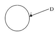
アルミニゥム箔にアクリル系接着剤を塗布したテープ(株式会社寺岡製作所)から丸く直径10mm切り抜き、図3の様な円形のアルミニウム貼付剤Dを作成した。皮膚への装着には実施例1や実施例2で用いた止血ガーゼを外した絆創膏上に接着してX線用貼付剤とした。A circular aluminum patch D as shown in FIG. 3 was created by cutting out a circle with a diameter of 10 mm from a tape made of aluminum foil coated with an acrylic adhesive (manufactured by Teraoka Seisakusho Co., Ltd.). For attachment to the skin, the hemostatic gauze used in Examples 1 and 2 was removed and adhered to the bandage to form an X-ray patch.
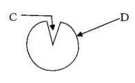
アルミニゥム箔テープ(株式会社寺岡製作所)から丸く直径10mm切り抜き、更に中心部を直径3mm切り抜き、図4の様なドーナツ状のX線用貼付剤Dを作成した。中心の切り取り部Cは体表に目印を皮膚マーカーや皮膚ペンで記す為の切り取り部でもある。皮膚への装着には実施例1や実施例2で用いた絆創膏の止血ガーゼを外し、更に直径3mm切り抜き、図4の切り取り部Cと絆創膏の切り取り部に沿うように接着してX線用粘着剤とした。A donut-shaped X-ray patch D as shown in FIG. 4 was prepared by cutting out a circle with a diameter of 10 mm from an aluminum foil tape (Teraoka Seisakusho Co., Ltd.), and then cutting out a center part with a diameter of 3 mm. The central cutout C is also a cutout for marking marks on the body surface with a skin marker or skin pen. To attach it to the skin, remove the hemostatic gauze from the bandage used in Examples 1 and 2, cut out a 3 mm diameter cutout, and glue it along the cutout C in Figure 4 and the cutout of the bandage to make an X-ray adhesive. It was used as a drug.
アルミニゥム箔テープ(株式会社寺岡製作所)から丸く直径10mm切り抜き、更に中心部を直径3mm切り抜き、図4の様なドーナツ状の貼付剤Dを作成した。中心の切り取り部Cは体表に目印を皮膚マーカーや皮膚ペンで記す為の切り取り部でもある。A donut-shaped adhesive patch D as shown in FIG. 4 was prepared by cutting out a circle with a diameter of 10 mm from an aluminum foil tape (Teraoka Seisakusho Co., Ltd.), and then cutting out the center with a diameter of 3 mm. The central cutout C is also a cutout for marking marks on the body surface with a skin marker or skin pen.
本発明のX線用貼付剤は医療用であり、CT撮影時に使用する事により、患者患部の手術位置を体表に皮膚マーカーや皮膚ペンで記す事を可能にする。The X-ray patch of the present invention is for medical use, and when used during CT imaging, it makes it possible to mark the surgical position of the patient's affected area on the body surface with a skin marker or skin pen.
A...布、膜、箔、フィルム、或いは板状のX線照準貼付剤
B...X線不透過性物質
C...切り取り部
D...X線照準貼付剤
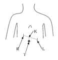
L...CT撮影時の人体左の図1のX線用貼付剤
R...CT撮影時の人体右の図1のX線用貼付剤
T...肝癌の位置
K...左右のX線用貼付剤間の肝癌の縦軸上の地点A. .. .. Cloth, membrane, foil, film, or plate-shaped X-ray aiming patch B. .. .. X-ray opaque substanceC. .. .. Cutout part D. .. .. X-ray aiming patch L. .. .. The X-ray patch R in Figure 1 on the left side of the human body during CT imaging. .. .. The X-ray patch T shown in Figure 1 on the right side of the human body during CT imaging. .. .. Location of liver cancer K. .. .. Point on the vertical axis of liver cancer between the left and right X-ray patches
Claims (9)
Translated fromJapanesePriority Applications (1)
| Application Number | Priority Date | Filing Date | Title |
|---|---|---|---|
| JP2022112050AJP2024003730A (en) | 2022-06-27 | 2022-06-27 | X-ray patch |
Applications Claiming Priority (1)
| Application Number | Priority Date | Filing Date | Title |
|---|---|---|---|
| JP2022112050AJP2024003730A (en) | 2022-06-27 | 2022-06-27 | X-ray patch |
Publications (1)
| Publication Number | Publication Date |
|---|---|
| JP2024003730Atrue JP2024003730A (en) | 2024-01-15 |
Family
ID=89533866
Family Applications (1)
| Application Number | Title | Priority Date | Filing Date |
|---|---|---|---|
| JP2022112050APendingJP2024003730A (en) | 2022-06-27 | 2022-06-27 | X-ray patch |
Country Status (1)
| Country | Link |
|---|---|
| JP (1) | JP2024003730A (en) |
Cited By (1)
| Publication number | Priority date | Publication date | Assignee | Title |
|---|---|---|---|---|
| WO2025158889A1 (en)* | 2024-01-26 | 2025-07-31 | セントラル硝子株式会社 | Resin film containing x-ray contrast material |
Citations (8)
| Publication number | Priority date | Publication date | Assignee | Title |
|---|---|---|---|---|
| US4860331A (en)* | 1988-09-12 | 1989-08-22 | Williams John F | Image marker device |
| US5368030A (en)* | 1992-09-09 | 1994-11-29 | Izi Corporation | Non-invasive multi-modality radiographic surface markers |
| US6356621B1 (en)* | 1999-07-14 | 2002-03-12 | Nitto Denko Corporation | Pressure-sensitive adhesive sheet for radiography |
| WO2008146529A1 (en)* | 2007-05-30 | 2008-12-04 | Piac Co., Ltd. | Fabric containing x-ray-detectable material, adhesive plaster for x-ray detection, and process for producing adhesive plaster for x-ray detection |
| US20090022272A1 (en)* | 2007-07-20 | 2009-01-22 | Karen Joseph | Multi-density skin marker |
| DE102008045988A1 (en)* | 2008-09-05 | 2010-03-11 | Fraunhofer-Gesellschaft zur Förderung der angewandten Forschung e.V. | Identification feature for marking tissue area in e.g. computed tomography, is assigned to tissue area, and attached to surface of tissue area, and comprising selectable code e.g. bar code |
| WO2021025861A1 (en)* | 2019-08-06 | 2021-02-11 | Wright Medical Technology, Inc. | Surgical guide and method of use |
| CN113490515A (en)* | 2019-02-28 | 2021-10-08 | 矿物快速护理有限公司 | Paste for marking textile fabrics and/or other products that cannot be X-rayed |
- 2022
- 2022-06-27JPJP2022112050Apatent/JP2024003730A/enactivePending
Patent Citations (8)
| Publication number | Priority date | Publication date | Assignee | Title |
|---|---|---|---|---|
| US4860331A (en)* | 1988-09-12 | 1989-08-22 | Williams John F | Image marker device |
| US5368030A (en)* | 1992-09-09 | 1994-11-29 | Izi Corporation | Non-invasive multi-modality radiographic surface markers |
| US6356621B1 (en)* | 1999-07-14 | 2002-03-12 | Nitto Denko Corporation | Pressure-sensitive adhesive sheet for radiography |
| WO2008146529A1 (en)* | 2007-05-30 | 2008-12-04 | Piac Co., Ltd. | Fabric containing x-ray-detectable material, adhesive plaster for x-ray detection, and process for producing adhesive plaster for x-ray detection |
| US20090022272A1 (en)* | 2007-07-20 | 2009-01-22 | Karen Joseph | Multi-density skin marker |
| DE102008045988A1 (en)* | 2008-09-05 | 2010-03-11 | Fraunhofer-Gesellschaft zur Förderung der angewandten Forschung e.V. | Identification feature for marking tissue area in e.g. computed tomography, is assigned to tissue area, and attached to surface of tissue area, and comprising selectable code e.g. bar code |
| CN113490515A (en)* | 2019-02-28 | 2021-10-08 | 矿物快速护理有限公司 | Paste for marking textile fabrics and/or other products that cannot be X-rayed |
| WO2021025861A1 (en)* | 2019-08-06 | 2021-02-11 | Wright Medical Technology, Inc. | Surgical guide and method of use |
Cited By (1)
| Publication number | Priority date | Publication date | Assignee | Title |
|---|---|---|---|---|
| WO2025158889A1 (en)* | 2024-01-26 | 2025-07-31 | セントラル硝子株式会社 | Resin film containing x-ray contrast material |
Similar Documents
| Publication | Publication Date | Title |
|---|---|---|
| US5848125A (en) | Radiopaque landmark skin markers and method | |
| AU2001253671B2 (en) | Surgical targeting system | |
| US20160331344A1 (en) | Ultrasound probe cover and method of use | |
| US5368030A (en) | Non-invasive multi-modality radiographic surface markers | |
| US5260985A (en) | Conforming localization/biopsy grid and control apparatus | |
| US20150223906A1 (en) | Medical Procedure Localizing Aid | |
| US20160100911A1 (en) | Medical Procedure Localizing Aid | |
| US9408671B2 (en) | Biopsy grid | |
| CN103908348B (en) | A kind of human organ body surface sticky marker | |
| AU2001253671A1 (en) | Surgical targeting system | |
| CN102125462B (en) | Tool for positioning surgical incision in combination with fluoroscopy | |
| WO2012092830A1 (en) | Clinical medical sticking film with development coordinates | |
| WO2016110086A1 (en) | Medical radiation positioning film and method for photographing lesion site, positioning of which is convenient and fast | |
| US20240033488A1 (en) | Echogenic balloon dilation catheter and balloon thereof | |
| JP2024003730A (en) | X-ray patch | |
| RU11679U1 (en) | TOOL SET FOR ASPIRATION BIOPSY | |
| US9452269B2 (en) | Method, retention device and medical treatment device for stable support of a needle to be inserted into a patient | |
| CN209611254U (en) | A CT-guided percutaneous lung biopsy positioning device | |
| CN213406231U (en) | Puncture positioning sheet for minimally invasive surgery | |
| CN203953813U (en) | A kind of human organ body surface sticky marker | |
| CN110664494A (en) | Ablation puncture auxiliary patch | |
| CN211962224U (en) | A device that facilitates the positioning of breast nodules | |
| CN215899914U (en) | Chest CT positioning sticker with good fitting performance | |
| CN212089686U (en) | a medical puncture needle | |
| CN205073038U (en) | A patch for lesion localization |
Legal Events
| Date | Code | Title | Description |
|---|---|---|---|
| A521 | Request for written amendment filed | Free format text:JAPANESE INTERMEDIATE CODE: A523 Effective date:20220905 | |
| A131 | Notification of reasons for refusal | Free format text:JAPANESE INTERMEDIATE CODE: A131 Effective date:20230711 | |
| A521 | Request for written amendment filed | Free format text:JAPANESE INTERMEDIATE CODE: A523 Effective date:20230822 | |
| A131 | Notification of reasons for refusal | Free format text:JAPANESE INTERMEDIATE CODE: A131 Effective date:20231121 | |
| A521 | Request for written amendment filed | Free format text:JAPANESE INTERMEDIATE CODE: A523 Effective date:20231208 | |
| A02 | Decision of refusal | Free format text:JAPANESE INTERMEDIATE CODE: A02 Effective date:20240319 | |
| A521 | Request for written amendment filed | Free format text:JAPANESE INTERMEDIATE CODE: A523 Effective date:20240417 | |
| A911 | Transfer to examiner for re-examination before appeal (zenchi) | Free format text:JAPANESE INTERMEDIATE CODE: A911 Effective date:20240529 | |
| A912 | Re-examination (zenchi) completed and case transferred to appeal board | Free format text:JAPANESE INTERMEDIATE CODE: A912 Effective date:20240809 |