JP2023182154A - Inspection report creation device and method - Google Patents
Inspection report creation device and methodDownload PDFInfo
- Publication number
- JP2023182154A JP2023182154AJP2022095596AJP2022095596AJP2023182154AJP 2023182154 AJP2023182154 AJP 2023182154AJP 2022095596 AJP2022095596 AJP 2022095596AJP 2022095596 AJP2022095596 AJP 2022095596AJP 2023182154 AJP2023182154 AJP 2023182154A
- Authority
- JP
- Japan
- Prior art keywords
- fixed phrase
- specific
- list
- inspection report
- area
- Prior art date
- Legal status (The legal status is an assumption and is not a legal conclusion. Google has not performed a legal analysis and makes no representation as to the accuracy of the status listed.)
- Pending
Links
Images


Landscapes
- Ultra Sonic Daignosis Equipment (AREA)
Abstract
Description
Translated fromJapanese本開示は、検査レポート作成装置及び方法に関し、特に、検査レポートの作成を支援する技術に関する。 The present disclosure relates to an inspection report creation device and method, and particularly relates to a technique for supporting the creation of an inspection report.
被検者の超音波検査においては、超音波診断装置が用いられる。超音波検査後に検査レポートが作成される。検査レポートには、一般に、超音波検査で取得された画像及び計測値が含まれ、また様々な所見(Findings)が含まれる。所見は、レポート作成者(医師、検査技師、等)の見解又はコメントに相当する。所見として定型文が用いられることも多い。定型文を修正することにより所見が作成されることもある。 An ultrasonic diagnostic apparatus is used in an ultrasonic examination of a subject. An inspection report will be created after the ultrasound examination. The examination report generally includes images and measurement values obtained by ultrasonic examination, and also includes various findings. Observations correspond to the opinions or comments of the report creator (physician, laboratory technician, etc.). Fixed phrases are often used as findings. Findings may also be created by modifying a fixed phrase.
超音波診断装置は、通常、検査レポートを作成する機能を有している(特許文献1-4を参照)。その観点から見て超音波診断装置は検査レポート作成装置を包含する装置である。超音波診断装置とは別の情報処理装置において検査レポートが作成されることもある。その場合、その情報処理装置が検査レポート作成装置に相当する。 Ultrasonic diagnostic apparatuses usually have a function of creating an examination report (see Patent Documents 1 to 4). From this point of view, the ultrasonic diagnostic device is a device that includes an examination report creation device. An examination report may be created in an information processing device other than the ultrasound diagnostic device. In that case, the information processing device corresponds to an inspection report creation device.
検査レポート作成装置においては、レポート作成者による検査レポートの作成を支援するために、多くの定型文リストが用意されている。それらの定型文リストの中から実際に使用する定型文リストを正確に且つ素早く選択できる仕組みを実現することが望まれる。 In the inspection report creation device, many fixed phrase lists are prepared to assist the report creator in creating an inspection report. It is desired to realize a mechanism that can accurately and quickly select a list of fixed phrases to be actually used from among these lists of fixed phrases.
本開示の目的は、複数の定型文リストの中から特定の定型文リストを正確に且つ素早く選択できる検査レポート作成装置及び方法を提供することにある。 An object of the present disclosure is to provide an inspection report creation device and method that can accurately and quickly select a specific list of fixed phrases from a plurality of lists of fixed phrases.
本開示に係る検査レポート作成装置は、表示される模式図上に設定された複数のエリアを管理すると共に前記複数のエリアに対応付けられた複数の定型文リストを管理する管理部と、前記複数のエリアの中の特定のエリアがユーザーにより指定された場合に、前記複数の定型文リストの中から前記特定のエリアに対応付けられた特定の定型文リストを抽出するリスト抽出部と、前記特定の定型文リストの中の特定の定型文がユーザーにより指定された場合に、前記特定の定型文リストの中から前記特定の定型文を抽出する定型文抽出部と、前記特定の定型文又はその修正により生成された文を所見として検査レポートに挿入するレポート編集部と、を含むことを特徴とする。 An inspection report creation device according to the present disclosure includes a management unit that manages a plurality of areas set on a schematic diagram to be displayed and a plurality of fixed phrase lists associated with the plurality of areas; a list extraction unit that extracts a specific list of fixed phrases associated with the specific area from among the plurality of fixed phrase lists when a specific area among the areas is specified by the user; a fixed phrase extraction unit that extracts the specific fixed phrase from the specific fixed phrase list when a specific fixed phrase in the fixed fixed phrase list is specified by the user; The present invention is characterized in that it includes a report editing section that inserts sentences generated by correction into an inspection report as findings.
本開示に係る検査レポート作成方法は、表示される模式図上に設定された複数のエリアを管理すると共に前記複数のエリアに対応付けられた複数の定型文リストを管理する工程と、前記複数のエリアの中の特定のエリアがユーザーにより指定された場合に、前記複数の定型文リストの中から前記特定のエリアに対応付けられた特定の定型文リストを抽出する工程と、前記特定の定型文リストの中の特定の定型文がユーザーにより指定された場合
に、前記特定の定型文リストの中から前記特定の定型文を抽出する工程と、前記特定の定型文又はその修正により生成された文を所見として検査レポートに挿入する工程と、を含むことを特徴とする。An inspection report creation method according to the present disclosure includes a step of managing a plurality of areas set on a schematic diagram to be displayed and a plurality of fixed phrase lists associated with the plurality of areas; a step of extracting a specific fixed phrase list associated with the specific area from the plurality of fixed phrase lists when a specific area among the areas is specified by the user; a step of extracting the specific fixed phrase from the specific fixed phrase list when a specific fixed phrase in the list is specified by the user; and a sentence generated by the specific fixed phrase or modification thereof. The method is characterized by including a step of inserting the information into the inspection report as a finding.
本開示に係るレポート作成装置及び方法によれば、複数の定型文リストの中から特定の定型文リストを正確に且つ素早く選択できる。 According to the report creation device and method according to the present disclosure, it is possible to accurately and quickly select a specific list of fixed phrases from among a plurality of lists of fixed phrases.
以下、実施形態を図面に基づいて説明する。 Hereinafter, embodiments will be described based on the drawings.
(1)実施形態の概要
実施形態に係る検査レポート作成装置は、管理部、リスト抽出部、定型文抽出部、及び、レポート編集部を有する。管理部は、表示される模式図上に設定された複数のエリアを管理すると共に、複数のエリアに対応付けられた複数の定型文リストを管理する。リスト抽出部は、複数のエリアの中の特定のエリアがユーザーにより指定された場合に、複数の定型文リストの中から特定のエリアに対応付けられた特定の定型文リストを抽出する。定型文抽出部は、特定の定型文リストの中の特定の定型文がユーザーにより指定された場合に、特定の定型文リストの中から特定の定型文を抽出する。レポート編集部は、特定の定型文又はその修正により生成された文を所見として検査レポートに挿入する。(1) Overview of Embodiment The inspection report creation device according to the embodiment includes a management section, a list extraction section, a fixed phrase extraction section, and a report editing section. The management unit manages a plurality of areas set on the displayed schematic diagram, and also manages a plurality of fixed phrase lists associated with the plurality of areas. The list extraction unit extracts a specific list of fixed phrases associated with the specific area from among the plurality of fixed phrase lists when a specific area among the plurality of areas is specified by the user. The fixed phrase extracting unit extracts a specific fixed phrase from the specific fixed phrase list when a specific fixed phrase in the specific fixed phrase list is designated by a user. The report editing section inserts a specific fixed phrase or a sentence generated by modifying the fixed phrase into the inspection report as a finding.
上記構成によれば、ユーザーが特定のエリアを指定すると、それに対応付けられた特定の定型文リストが自動的に選択される。よって、適切な定型文リストを迅速に利用することが可能となる。特定の定型文リストの選択後、特定の定型文リストの中から特定の定型文がユーザーにより指定される。 According to the above configuration, when the user specifies a specific area, a specific list of fixed phrases associated with the area is automatically selected. Therefore, it becomes possible to quickly utilize an appropriate list of fixed phrases. After selecting the specific fixed phrase list, the user specifies a specific fixed phrase from the specific fixed phrase list.
実施形態においては、エリア指定を受け付けるために模式図が表示され、定型文指定を受け付けるために定型文リストが表示される。模式図には、複数のエリアを視覚的に識別するための補助図形が含まれる。補助図形には複数の表示要素が含まれる。 In the embodiment, a schematic diagram is displayed in order to accept area designation, and a list of fixed phrases is displayed in order to receive fixed phrase designation. The schematic diagram includes auxiliary figures for visually identifying multiple areas. The auxiliary figure includes multiple display elements.
例えば、模式図は心臓の模式図である。その場合、複数のエリアには、心腔エリア、心筋エリア及び弁エリアの内の少なくとも1つが含まれる。ある心筋における複数の部分に対して複数のエリアが設定されている場合、それら複数のエリアを区分する複数の区分線が複数の表示要素として表示されてもよい。典型的には、超音波検査の対象となった組織
を表す模式図が表示される。生体から取得された心電図等の生体信号を模擬した模式図が表示されてもよい。模式図は、所見対象となる生体組織や生体信号を模式的に表現した参考図である。For example, the schematic diagram is a schematic diagram of the heart. In that case, the plurality of areas include at least one of a heart chamber area, a myocardial area, and a valve area. When a plurality of areas are set for a plurality of parts of a certain myocardium, a plurality of dividing lines dividing the plurality of areas may be displayed as a plurality of display elements. Typically, a schematic diagram representing the tissue targeted by the ultrasound examination is displayed. A schematic diagram simulating a biological signal such as an electrocardiogram obtained from a living body may be displayed. The schematic diagram is a reference diagram that schematically represents a biological tissue or a biological signal to be observed.
実施形態に係る検査レポート作成装置は、超音波検査中に所見入力支援画像を表示する表示制御部を含む。所見入力支援画像には、模式図、及び、特定の定型文リストが表示されるリスト表示欄、が含まれる。所見入力支援画像には、更に、特定の定型文が表示される欄であって特定の定型文の修正作業が行われる所見欄が含まれる。超音波検査中に、所定のユーザー操作により、所見欄の内容が所見として保存される。レポート編集部は、超音波検査中に又は検査レポート作成開始時に、保存された所見を検査レポートに自動的に挿入する。 The examination report creation device according to the embodiment includes a display control unit that displays a finding input support image during an ultrasound examination. The finding input support image includes a schematic diagram and a list display column in which a list of specific fixed phrases is displayed. The finding input support image further includes a finding column in which a specific fixed phrase is displayed and in which correction work for the specific fixed phrase is performed. During the ultrasound examination, the contents of the findings column are saved as findings by a predetermined user operation. The report editing section automatically inserts the saved findings into the examination report during the ultrasound examination or at the start of examination report creation.
超音波検査は、超音波の送信及び受信を行いながら複数の超音波画像を取得及び格納する一連の過程を意味する。その過程には、フリーズ状態での計測が含まれる。超音波検査後に、検査レポート作成過程が実施される。実施形態においては、超音波検査中において検査レポート作成準備を行える。すなわち、超音波検査と並行して、検査レポートに記入する複数の情報を事前に用意できる。 Ultrasound examination refers to a series of processes of acquiring and storing a plurality of ultrasound images while transmitting and receiving ultrasound waves. The process includes measurements in a frozen state. After the ultrasound examination, an examination report creation process is performed. In the embodiment, preparation for creating an examination report can be performed during an ultrasound examination. That is, a plurality of pieces of information can be prepared in advance to be filled in the examination report in parallel with the ultrasound examination.
実施形態において、表示制御部は、複数のエリアの中でユーザー操作されるポインタが属するエリアを特定のエリアとして判定すると共に当該特定のエリアを強調表示する。模式図上におけるポインタの移動に伴って、強調表示されるエリアが切り替わり且つリスト表示欄の内容が切り替わる。この構成によれば、ポインタが属しているエリアを明確に特定でき、正しい定型文リストを表示させることが可能となる。あるいは、定型文リストの内容を見ながらポインタを動かす(つまりエリアを指定する)ことが可能となる。 In the embodiment, the display control unit determines the area to which a pointer operated by the user belongs among the plurality of areas as a specific area, and highlights the specific area. As the pointer moves on the schematic diagram, the highlighted area changes and the contents of the list display column change. According to this configuration, it is possible to clearly specify the area to which the pointer belongs, and it is possible to display a correct list of fixed phrases. Alternatively, it is possible to move the pointer (that is, specify an area) while looking at the contents of the fixed phrase list.
主表示器及び副表示器が設けられている場合において、主表示器に所見入力支援画像が表示されてもよいし、副表示器に所見入力支援画像が表示されてもよい。主表示器に所見入力支援画像の一部が表示され、副表示器に所見入力支援画像の他の一部が表示されてもよい。 In the case where a main display and a sub-display are provided, the finding input support image may be displayed on the main display, or the finding input support image may be displayed on the sub-display. A part of the finding input support image may be displayed on the main display, and another part of the finding input support image may be displayed on the sub display.
実施形態に係る検査レポート作成装置は、超音波検査中にいずれかのエリアがマーキング対象として指定された場合に当該エリアに対してマークを付し、これによりマーク付き模式図を生成する生成部を含む。検査レポートの作成時にマーク付き模式図が表示される。この構成によれば、検査レポートの作成時に、表示されたマーク付き模式図を参照することが可能となる。これにより、所見の記入の必要性及び記入対象となる部位(エリア)を特定できる。複数種類のマークの中から選択されたマークが模式図に付されてもよい。定型文リストの表示が行われない検査レポート作成装置に、マーキングのための上記構成が組み込まれてもよい。上記の生成部は、後述する支援画像生成部に相当する。 The inspection report creation device according to the embodiment includes a generation unit that, when any area is designated as a marking target during an ultrasonic inspection, marks the area and thereby generates a marked schematic diagram. include. A marked schematic diagram is displayed when creating an inspection report. According to this configuration, it is possible to refer to the displayed schematic diagram with marks when creating an inspection report. This makes it possible to specify the necessity of entering the findings and the site (area) to be entered. A mark selected from a plurality of types of marks may be attached to the schematic diagram. The above configuration for marking may be incorporated into an inspection report creation device that does not display a list of fixed phrases. The generation unit described above corresponds to a support image generation unit that will be described later.
ユーザーは、定型文リストの選択及び表示のための第1エリア指定、及び、マーキング対象の選択のための第2エリア指定、を行い得る。第1エリア指定は、超音波検査中において所見を作成するためのものである。第2エリア指定は、超音波検査後のレポート作成段階において所見を作成するためのものである。単一のエリア指定を第1エリア指定及び第2エリア指定として取り扱うようにしてもよい。 The user can specify a first area for selecting and displaying a list of fixed phrases and a second area for selecting a marking target. The first area designation is for creating findings during an ultrasound examination. The second area designation is for creating findings in the report creation stage after the ultrasound examination. A single area designation may be treated as a first area designation and a second area designation.
実施形態に係る検査レポート作成方法は、管理工程、リスト抽出工程、定型文抽出工程、及び、レポート編集工程を有する。管理工程では、表示される模式図上に設定された複数のエリアが管理され、また、複数のエリアに対応付けられた複数の定型文リストが管理される。リスト抽出工程では、複数のエリアの中の特定のエリアがユーザーにより指定された場合に、複数の定型文リストの中から特定のエリアに対応付けられた特定の定型文リ
ストが抽出される。定型文抽出工程では、特定の定型文リストの中の特定の定型文がユーザーにより指定された場合に、特定の定型文リストの中から特定の定型文が抽出される。レポート編集工程では、特定の定型文又はその修正により生成された文が所見として検査レポートに挿入される。The inspection report creation method according to the embodiment includes a management process, a list extraction process, a fixed phrase extraction process, and a report editing process. In the management step, a plurality of areas set on the displayed schematic diagram are managed, and a plurality of fixed phrase lists associated with the plurality of areas are managed. In the list extraction step, when a specific area among the plurality of areas is specified by the user, a specific fixed phrase list associated with the specific area is extracted from among the plural fixed phrase lists. In the fixed phrase extraction step, when a specific fixed phrase in the specific fixed phrase list is designated by the user, the specific fixed phrase is extracted from the specific fixed phrase list. In the report editing process, a specific fixed phrase or a sentence generated by modifying the fixed phrase is inserted into the inspection report as a finding.
上記検査レポート作成方法は、ハードウエアの機能として又はソフトウエアの機能として実現され得る。後者の場合には、上記検査レポート作成方法を実行するプログラムがネットワーク又は可搬型記憶媒体を介して情報処理装置へインストールされる。情報処理装置には、上記のプログラムを格納する非一時的記憶媒体が含まれる。 The above inspection report creation method can be realized as a hardware function or a software function. In the latter case, a program for executing the above inspection report creation method is installed in the information processing device via a network or a portable storage medium. The information processing device includes a non-temporary storage medium that stores the above program.
(2)実施形態の詳細
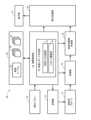
図1には、実施形態に係る超音波診断装置10が示されている。超音波診断装置10は、被検者の超音波検査を行うための医用装置である。超音波診断装置10において、後述する検査レポート作成部30及びそれに関連する構成が、検査レポート作成装置に相当する。(2) Details of Embodiment FIG. 1 shows an ultrasonic
超音波プローブ12は、生体内への超音波の送信及び生体内からの反射波の受信を行う可搬型デバイスである。超音波プローブ12は、直線状又は円弧状に配列された複数の振動素子からなる振動素子アレイを有している。振動素子アレイによって超音波ビームが形成される。その超音波ビームが電子走査される。電子走査方式として、電子セクタ走査方式、電子リニア走査方式、等が知られている。超音波ビームの電子走査により生体内にビーム走査面が形成される。超音波プローブ12内に2D振動素子アレイを設けてもよい。 The
送信部14は、送信ビームフォーマーとして機能する電子回路である。具体的には、送信部14は、送信時において、振動素子アレイに対して複数の送信信号を並列的に供給する。これにより、超音波が生体内へ放射され、つまり送信ビームが形成される。 The
受信部16は、受信ビームフォーマーとして機能する電子回路である。具体的には、受信部16は、受信時において、振動素子アレイから並列的に出力された複数の受信信号に対して整相加算を適用し、これにより受信ビームデータを生成する。超音波ビームの電子走査の繰り返しにより、ビーム走査面が繰り返し形成される。これにより、受信部16から、時系列順で並ぶ複数の受信フレームでータが出力される。各受信フレームデータは、電子走査方向に並ぶ複数の受信ビームデータにより構成される。各受信ビームデータは、深さ方向に並ぶ複数のエコーデータにより構成される。受信部16の後段に設けられたビームデータ処理部の図示が省略されている。 The receiving
超音波画像形成部18は、複数の受信フレームデータに基づいて複数の表示フレームデータを生成するものである。超音波画像形成部18は、具体的には、デジタルスキャンコンバータ(DSC)により構成される。各表示フレームデータは実施形態において断層画像データである。複数の表示フレームデータは、表示処理部20を介して、表示部22へ送られている。表示部22に、リアルタイム動画像としてのBモード断層画像が表示される。 The ultrasound
演算制御部24は、プログラムを実行するプロセッサ(例えばCPU)により構成される。演算制御部24により、図1に示されている各構成要素の動作が制御される。演算制御部24は、超音波検査において必要となる情報処理を実行する。その情報処理には、検査レポート作成のための情報処理が含まれる。図1においては、その情報処理が検査レポート作成部30として表現されている。検査レポート作成部30は、所見入力支援機能及びレポート編集機能を有する。それらの機能が図1において所見入力支援部32及びレポート編集部34として表現されている。 The arithmetic control unit 24 is composed of a processor (for example, a CPU) that executes a program. The operation of each component shown in FIG. 1 is controlled by the calculation control section 24. The arithmetic control unit 24 executes information processing necessary for ultrasonic examination. The information processing includes information processing for creating an inspection report. In FIG. 1, the information processing is expressed as an inspection report creation section 30. The inspection report creation unit 30 has a finding input support function and a report editing function. These functions are expressed as a finding
演算制御部24には、操作パネル26及び記憶部28が接続されている。操作パネル26には、複数のスイッチ、複数のつまみ、キーボード、トラックボール等が含まれる。トラックボールはポインティングデバイスとして機能する。記憶部28は、半導体メモリ、ハードディスク等により構成される。表示部22は、例えば、主表示器及び副表示器により構成される。主表示器は、例えば、大型のLCDや有機EL表示器により構成される。副表示器は、例えば、タッチパネルとLCDパネルとからなる複合パネルにより構成される。副表示器は、通常、操作パネル26に組み込まれる。 An
記憶部28には、各種のデータ及び各種のファイルが記憶される。実施形態においては、記憶部28に、管理テーブル36、模式図群(模式図データ群)38、及び、定型文リスト群(定型文リストデータ群)40、が記憶されている。更に、記憶部28には、超音波検査中においてストアされた一連の画像(一連の画像データ)が格納され、また、超音波検査後に作成された検査レポート(検査レポートデータ)が格納される。なお、コンピュータ等の情報処理装置に検査レポート作成部30及び記憶部28が設けられてもよい。その場合、その情報処理装置は検査レポート作成装置とみなせる。 The
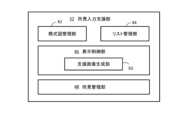
図2には、所見入力支援部32の構成例が示されている。所見入力支援部32は、模式図管理部42、リスト管理部44、表示制御部46、及び、所見管理部48を有する。模式図管理部42は、記憶部内の管理テーブルを用いて、複数種類の超音波検査に対応付けられた複数の模式図(シェーマ)を管理するものである。リスト管理部44は、記憶部内の管理テーブルを用いて、複数の模式図に対応付けられた複数の定型文リストセットを管理するものである。各定型文リストセットは、模式図に対して設定された複数のエリアに対応付けられた複数の定型文リストにより構成される。 FIG. 2 shows an example of the configuration of the finding
表示制御部46は、所見入力支援過程において表示制御を実行するものである。表示制御部46は、支援画像生成部50を有する。支援画像生成部50は、所見入力支援画像を生成するものである。所見入力支援画像には、後述するように、模式図、リスト表示欄、及び、所見欄が含まれる。 The display control unit 46 executes display control in the finding input support process. The display control section 46 includes a support
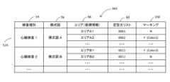
図3には、管理テーブル36の具体例が示されている。管理テーブル36は、複数のレコード52により構成される。各レコード52には、超音波検査種別を示す情報54、模式図を特定する情報56、複数のエリアを特定する情報(座標情報を含む)58、及び、定型文リストを特定する情報60、が含まれる。管理テーブル36上において、超音波検査種別ごとにそれに対応する模式図が管理されており、また、その模式図に含まれる複数のエリアが管理されており、更に、それらの複数のエリアに対応付けられた複数の定型文リストが管理されている。超音波検査種別は、超音波検査の対象部位及び超音波検査の方法の組み合わせに相当する。 FIG. 3 shows a specific example of the management table 36. The management table 36 is composed of a plurality of
図4には、実施形態に係る所見入力支援方法が概念図として示されている。記憶部28は、模式図群38及び定型文リスト群40が格納されている。模式図群38は、複数の模式図38Aにより構成される。各模式図38Aは、超音波検査の対象臓器等を模擬したグラフィック画像である。具体的には、各模式図38Aには、対象臓器の断面を模擬した断面図形64、及び、その断面図形上に設定された複数のエリアを視覚的に識別するために付加された補助図形66、が含まれる。補助図形は、複数の表示要素からなる。各表示要素は、直線、曲線、囲み図形、等である。断面図形64において、ある部位を囲む輪郭図形は、当該部位に対応するエリアを示す表示要素として機能する。ユーザー(医師、検査技師、等)は、断面図形64及び補助図形66の参照により、複数のエリアを認識し得る。 FIG. 4 shows a conceptual diagram of the finding input support method according to the embodiment. The
定型文リスト群40は、複数の模式図38Aに対応付けられた複数の定型文リストセット40Aからなる。個々の定型文リストセット40Aは、対応する模式図内の複数のエリアに対応付けられた複数の定型文リストにより構成される。 The fixed
例えば、検査情報68に基づいて、超音波検査種別が識別される。模式図群38の中から、その超音波検査種別に対応する特定の模式図72が選択される(符号70を参照)。超音波検査種別は、例えば、超音波診断装置の動作条件を定めるために事前に用意され且つ超音波検査開始時に選択されたプリセットデータに基づいて自動的に特定され得る。主表示器又は副表示器に表示された複数の模式図の中からユーザーに模式図を選択させてもよい。 For example, based on the examination information 68, the ultrasonic examination type is identified. A particular schematic diagram 72 corresponding to the ultrasonic examination type is selected from the schematic diagram group 38 (see reference numeral 70). The ultrasonic examination type may be automatically specified, for example, based on preset data that is prepared in advance to define the operating conditions of the ultrasonic diagnostic apparatus and selected at the time of starting the ultrasonic examination. The user may be allowed to select a schematic diagram from among a plurality of schematic diagrams displayed on the main display or the sub-display.
表示部22に模式図72が表示され、そこに表示された複数のエリアの中から、ユーザーにより特定のエリアが指定される。そのエリアは、所見入力を行いたい部位に相当し又はそれを包含する範囲に相当する。エリアの指定により、定型文リスト群40の中から、指定されたエリアに対応付けられた特定の定型文リスト76が抽出され(符号74を参照)、それが表示部22に表示される。 A schematic diagram 72 is displayed on the
表示された定型文リスト76の中から、ユーザーにより特定の定型文78が指定されると、定型文リスト76の中から、その定型文78が抽出される(符号80を参照)。表示部22には、所見欄82が表示されており、その中に、抽出された定型文84が表示される。必要に応じて、表示された定型文84をユーザーにより修正し得る(符号86を参照)。所見欄82は、修正作業領域に相当する。修正後、ユーザーの所定の操作により、所見欄82の内容が所見として記憶部上に保存される。このような過程が繰り返し実施されると、記憶部上に複数の所見が蓄積される。この一連の過程は、検査レポート作成準備過程とも言い得る。なお、ユーザーにより指定された特定の定型文78に対応する定型文(例えばより詳しい内容を有する定型文)が所見欄82に表示され、それが必要に応じて修正されてもよい。定型文リスト76を構成する各定形文が、リスト表示欄に表示される第1定型文(例えば簡略定型文)、及び、所見欄に表示される第2定型文(例えば詳細定型文)により構成されてもよい。 When the user specifies a specific fixed phrase 78 from the displayed fixed phrase list 76, that fixed phrase 78 is extracted from the fixed phrase list 76 (see reference numeral 80). A finding column 82 is displayed on the
超音波検査後の検査レポートの作成時に、複数の所見、複数の画像、複数の計測値等が挿入された検査レポートがユーザーに提示される。その後、検査レポートの編集を行い得る。その編集には、各所見の確認、修正等が含まれる。そのような過程を経て検査レポートの作成が完了する。 When creating an examination report after an ultrasound examination, the examination report in which multiple findings, multiple images, multiple measured values, etc. are inserted is presented to the user. Editing of the inspection report may then occur. Editing includes checking and correcting each finding. After such a process, the creation of the inspection report is completed.
図5には、所見入力支援画像の第1例が示されている。超音波検査中におけるユーザーの所定操作により、所見入力支援画像92が表示される。所見入力支援画像92は、模式図94、リスト表示欄104、及び、所見欄110を有する。 FIG. 5 shows a first example of the finding input support image. A finding input support image 92 is displayed by a user's predetermined operation during the ultrasound examination. The finding input support image 92 has a schematic diagram 94, a list display field 104, and a finding field 110.
模式図94は、断面図形96及び補助図形98により構成される。断面図形96は、図示の例において、心臓の断面を示すものである。断面図形96には、複数の輪郭線が含まれる。補助図形98には、複数の表示要素としての複数の区分線が含まれる。例えば、複数の心筋(心壁)図形のそれぞれを複数の区分が横切っている。各区分線は、隣接する2つのエリアを区分する境界線である。このように、断面図形96及び補助図形98によって複数のエリアが表現されている。図示の例において、複数のエリアには、心腔エリア、心筋エリア、弁エリア、等が含まれる。 The schematic diagram 94 is composed of a cross-sectional figure 96 and an auxiliary figure 98. In the illustrated example, the cross-sectional figure 96 shows a cross section of the heart. The cross-sectional figure 96 includes a plurality of contour lines. The auxiliary graphic 98 includes a plurality of dividing lines as a plurality of display elements. For example, a plurality of segments cross each of a plurality of myocardial (heart wall) figures. Each dividing line is a boundary line that divides two adjacent areas. In this way, a plurality of areas are expressed by the cross-sectional figure 96 and the auxiliary figure 98. In the illustrated example, the plurality of areas includes a heart chamber area, a myocardial area, a valve area, and the like.
例えば、ポインティングデバイスの操作により、ポインタ(カーソル)100の位置を変更し得る。ポインタ100の属するエリアが判定され、そのエリアに対応付けられた定型文リスト105がリスト表示欄104に表示される(符号106を参照)。実施形態に
おいては、ポインタ100の属するエリアがハイライト表示される。具体的には、そのエリア全体が所定のカラーでペイントされる(符号102を参照)。模式図94上においてポインタ100を移動させると、ポインタの属するエリアが順次切り替わる。これに伴って、ハイライト対象が順次切り替わり、リスト表示欄104に表示される定型文リストが順次切り替わる。なお、ポインタの属するエリアの判定後、ユーザーの所定操作が行われた場合に、定型文リストが表示(更新)されてもよい。For example, the position of the pointer (cursor) 100 can be changed by operating a pointing device. The area to which the pointer 100 belongs is determined, and a list of fixed phrases 105 associated with the area is displayed in the list display field 104 (see reference numeral 106). In the embodiment, the area to which the pointer 100 belongs is highlighted. Specifically, the entire area is painted in a predetermined color (see reference numeral 102). When the pointer 100 is moved on the schematic diagram 94, the area to which the pointer belongs changes sequentially. Along with this, the highlighted target is sequentially switched, and the list of fixed phrases displayed in the list display field 104 is sequentially switched. Note that the fixed phrase list may be displayed (updated) when the user performs a predetermined operation after determining the area to which the pointer belongs.
ユーザーにより、定型文リスト105の中から特定の定型文108が指定されると、その定型文108が抽出される(符号112を参照)。抽出された定型文108(又はそれに対応する定型文)が所見欄110に表示される。符号114は、所見欄110に表示された定型文を示している。図示の例では、所見欄110の中には、1つの定型文が既に表示されており、それに加えて、定型文114が表示されている。任意数の定型文を所見欄110に表示させることが可能である。必要な修正過程を経て、ユーザーの所定操作が行われると、所見欄110の内容が所見として保存される。 When a user specifies a specific fixed phrase 108 from the fixed phrase list 105, that fixed phrase 108 is extracted (see reference numeral 112). The extracted fixed phrase 108 (or the fixed phrase corresponding thereto) is displayed in the findings column 110. Reference numeral 114 indicates a fixed phrase displayed in the findings column 110. In the illustrated example, one fixed phrase is already displayed in the findings column 110, and in addition to that, a fixed phrase 114 is displayed. Any number of fixed phrases can be displayed in the findings column 110. When the user performs a predetermined operation after a necessary correction process, the contents of the findings column 110 are saved as findings.
図6には、所見入力支援画像の第2例が示されている。表示画像116には、断層画像118、所見入力支援画像119及びサムネイル画像群125が含まれる。断層画像118は、リアルタイム動作状態で表示される断層画像(動画像)又はフリーズ状態で表示される断層画像(静止画像)である。所見入力支援画像119は、模式図120、リスト表示欄122及び所見欄124が含まれる。このように、断層画像118と共に所見入力支援画像119が表示されてもよい。サムネイル画像群125は、超音波検査中にストアされた複数の断層画像を表すものである。 FIG. 6 shows a second example of the finding input support image. The display image 116 includes a tomographic image 118, a finding input support image 119, and a thumbnail image group 125. The tomographic image 118 is a tomographic image (moving image) displayed in a real-time operating state or a tomographic image (still image) displayed in a frozen state. The finding input support image 119 includes a schematic diagram 120, a list display field 122, and a finding field 124. In this way, the finding input support image 119 may be displayed together with the tomographic image 118. The thumbnail image group 125 represents a plurality of tomographic images stored during an ultrasound examination.
なお、主表示器に表示される画像に断層画像を含め、且つ、副表示器に表示される画像に所見入力支援画像を含めてもよい。主表示器に表示される画像に断層画像及び所見入力支援画像の一部分を含め、且つ、副表示器に表示される画像に所見入力支援画像の残りの部分を含めてもよい。 Note that the image displayed on the main display may include a tomographic image, and the image displayed on the sub display may include a finding input support image. The image displayed on the main display may include a portion of the tomographic image and the finding input support image, and the image displayed on the sub display may include the remaining portion of the finding input support image.
図7には、超音波検査後に作成される検査レポートの一例が示されている。検査レポートは電子ファイルに相当する。検査レポート126には、所見記入欄が含まれる。所見記入欄には、保存された一連の所見128が自動的に挿入されており、また、所見記入欄には、保存された模式図130が自動的に挿入されている。図示の例では、模式図130において、複数の所見に対応する複数のエリア132がペイント処理されている。検査レポート126の中に、そのような模式図130を含めることにより、各所見に対応する部位を容易に認識できる。 FIG. 7 shows an example of an examination report created after an ultrasound examination. Inspection reports correspond to electronic files. The inspection report 126 includes a column for entering findings. A series of saved findings 128 are automatically inserted into the findings entry field, and a saved schematic diagram 130 is automatically inserted into the findings entry field. In the illustrated example, in the schematic diagram 130, a plurality of areas 132 corresponding to a plurality of findings are painted. By including such a schematic diagram 130 in the examination report 126, the region corresponding to each finding can be easily recognized.
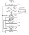
図8には、所見入力支援過程がフローチャートとして示されている。図8は、図1に示した所見入力支援部の動作又は図2に示した表示制御部の動作を示すものである。所見入力支援機能がオンされると、S10において、超音波検査種別に基づいて模式図が選択され、選択された模式図が表示される。S12では、本処理を続行させるか否かが判断される。 FIG. 8 shows the finding input support process as a flowchart. FIG. 8 shows the operation of the finding input support section shown in FIG. 1 or the operation of the display control section shown in FIG. 2. When the finding input support function is turned on, in S10, a schematic diagram is selected based on the ultrasound examination type, and the selected schematic diagram is displayed. In S12, it is determined whether or not to continue this process.
S14では、ユーザーによるエリアの指定の有無が判断される。エリアが指定されたことが判断されると、S16において、当該エリアに対応付けられた定型文リストが抽出され、それが表示される。S18では、表示された定型文リストの中のいずれかの定型文がユーザーにより指定された場合、その定型文が抽出され、その定型文が所見欄の中に表示される。 In S14, it is determined whether or not the user has designated an area. When it is determined that an area has been designated, a list of fixed phrases associated with the area is extracted and displayed in S16. In S18, if the user specifies any fixed phrase in the displayed fixed phrase list, that fixed phrase is extracted and displayed in the findings column.
S20では、所見欄の中に表示された定型文に対する修正が受け付けられる。その後、S22において、入力完了の有無、具体的にはユーザーの所定操作の有無が判定される。
入力継続の場合にはS18以降の工程が再び実行される。入力完了の場合、S23において、所見欄の内容が所見として保存される。その後、S12以降の工程が再び実行される。In S20, modifications to the fixed phrase displayed in the findings column are accepted. Thereafter, in S22, it is determined whether the input has been completed, specifically whether the user has performed a predetermined operation.
In the case of continuing input, the steps from S18 onwards are executed again. If the input is complete, the contents of the findings column are saved as findings in S23. After that, the steps after S12 are executed again.
図9には、検査レポート作成過程がフローチャートとして示されている。図9は、図1に示した検査レポート作成部の動作又はレポート編集部の動作を示すものである。S30では、検査レポートが表示される。その表示に先立って、保存された所見、画像、計測値等が検査レポートに組み込まれる。情報組み込み後の検査レポートが表示される。S32では、表示された検査レポートがユーザーにより編集される。例えば、検査レポート内の特定の所見が修正され、あるいは、検査レポートに対して新たな所見が追加される。S34では、完成した検査レポートが保存され、また必要に応じて出力(例えば印刷)される。 FIG. 9 shows the inspection report creation process as a flowchart. FIG. 9 shows the operation of the inspection report creation section or the report editing section shown in FIG. 1. In S30, an inspection report is displayed. Prior to its display, the saved findings, images, measured values, etc. are incorporated into the inspection report. The inspection report with the information incorporated will be displayed. In S32, the displayed inspection report is edited by the user. For example, certain findings within the inspection report may be modified, or new findings may be added to the inspection report. In S34, the completed inspection report is saved and output (for example, printed) as necessary.
次に、図10~12を用いて他の実施形態に係る所見入力支援について説明する。図10には、表示例が示されている。主表示器には第1画像134が表示され、副表示器には第2画像136が表示されている。図示の例において、第1画像134には、断層画像138、リスト表示欄140、及び、所見欄142が含まれる。一方、第2画像136には、模式図144が含まれ、また、ボタン列146が含まれる。 Next, finding input support according to another embodiment will be described using FIGS. 10 to 12. A display example is shown in FIG. A first image 134 is displayed on the main display, and a second image 136 is displayed on the sub display. In the illustrated example, the first image 134 includes a tomographic image 138, a list display field 140, and a finding field 142. On the other hand, the second image 136 includes a schematic diagram 144 and a button row 146.
模式図144、リスト表示欄140及び所見欄142が入力支援画像に相当する。すなわち、図10に示す他の実施形態においても、エリア指定に従って表示された定型文リストを利用して所見を作成し得る。 The schematic diagram 144, the list display field 140, and the findings field 142 correspond to input support images. That is, in the other embodiment shown in FIG. 10 as well, findings can be created using a list of fixed phrases displayed according to area designation.
ボタン列146は、複数のカラーに対応付けられた複数のボタンにより構成される。複数のカラーは、それぞれエリア識別子として機能する。超音波検査中に、所見を事後的に記入すべき部位が認識された場合、特定のボタンが選択され、その上で、特定のエリアが指定される。すると、そのエリアが、特定のボタンに対応する特定のカラーでペイントされる。すなわち、マーキング対象エリアに対してマークが付与される(符号148を参照)。模式図144に対して任意数のマークを付与し得る。 The button row 146 is composed of a plurality of buttons associated with a plurality of colors. Each of the plurality of colors functions as an area identifier. During an ultrasound examination, if a site is recognized where findings should be recorded afterwards, a specific button is selected and a specific area is then designated. The area will then be painted with a specific color that corresponds to the specific button. That is, a mark is applied to the marking target area (see reference numeral 148). An arbitrary number of marks can be added to the schematic diagram 144.
検査レポート作成時においては、マーク付き模式図が補助的に参考的に表示される。ユーザーは、マーク付き模式図の観察を通じて、所見の記入の必要性及び所見を記入すべき部位を識別し得る。よって、所見の記入漏れを防止できる。超音波検査中において所見入力とマーキングの両方が実行されてもよい。 When creating an inspection report, a schematic diagram with marks is displayed as an auxiliary reference. Through observation of the marked schematic diagram, the user can identify the necessity of entering a finding and the site where the finding should be entered. Therefore, it is possible to prevent omissions in the entry of findings. Both finding entry and marking may be performed during the ultrasound examination.
図11には、他の実施形態に係る管理テーブルの構成例が示されている。なお、図11において、図3に示した要素と同様の要素には同一の符号を付し、その説明を省略する。 FIG. 11 shows a configuration example of a management table according to another embodiment. In FIG. 11, the same elements as those shown in FIG. 3 are denoted by the same reference numerals, and the explanation thereof will be omitted.
レコード52Aには、エリアごとに管理された情報として、マーキングの有無を示す情報150が含まれる。その情報150の参照により、マーキングの有無及びマーキングカラーを特定し得る。 The
図12には、他の実施形態に係る所見入力支援過程がフローチャートとして示されている。なお、図12において、図8に示した工程と同様の工程には同一の符号を付し、その説明を省略する。 FIG. 12 shows a finding input support process according to another embodiment as a flowchart. Note that in FIG. 12, steps similar to those shown in FIG. 8 are given the same reference numerals, and their explanations will be omitted.
S100では、マーキングの要否、具体的にはマーキングのためのエリア指定の有無が判断される。所見入力のためのエリア指定を第1エリア指定と表現した場合、マーキングのためのエリア指定を第2エリア指定と表現し得る。S100において、第2エリア指定の発生が判断された場合、S102において、指定されたエリアがペイント処理され、ま
た、指定されたエリアを特定する情報及びマーキング情報が管理テーブル上に保存される。In S100, it is determined whether or not marking is necessary, specifically whether or not an area for marking is specified. If the area designation for inputting findings is expressed as a first area designation, the area designation for marking can be expressed as a second area designation. If it is determined in S100 that the second area has been designated, the designated area is painted, and information specifying the designated area and marking information are stored on the management table in S102.
図13には、他の実施形態に係る検査レポート作成過程がフローチャートとして示されている。なお、図13において、図9に示した工程と同様の工程には同一の符号を付し、その説明を省略する。 FIG. 13 shows a flowchart of an inspection report creation process according to another embodiment. Note that in FIG. 13, steps similar to those shown in FIG. 9 are given the same reference numerals, and their explanations will be omitted.
S30及びS32の実行過程でS104が実行される。S104では、マーキングされた模式図が表示される。その参照により、ユーザーは、所見記入の必要性を認識でき、また、所見記入対象となるエリアを認識できる。 S104 is executed during the execution process of S30 and S32. In S104, the marked schematic diagram is displayed. By referring to it, the user can recognize the necessity of entering the findings, and can also recognize the area where the findings are to be entered.
図14には、変形例に係る表示例が示されている。主表示器に表示された画像92Aには、模式図94、リスト表示欄104、及び、所見欄110が含まれる。また、画像92Aには、模式図94に代えて使用し得る複数の模式図152が含まれる。ユーザーにおいて、任意の模式図を選択して使用し得る。各模式図152に対して複数のエリアが設定されており、また、複数のエリアに対しては複数の定型文リストが対応付けられている。 FIG. 14 shows a display example according to a modification. The image 92A displayed on the main display includes a schematic diagram 94, a list display field 104, and a finding field 110. Furthermore, the image 92A includes a plurality of schematic diagrams 152 that can be used in place of the schematic diagram 94. The user can select and use any schematic diagram. A plurality of areas are set for each schematic diagram 152, and a plurality of fixed phrase lists are associated with the plurality of areas.
実施形態に係るレポート作成装置及び方法によれば、複数の定型文リストの中から特定の定型文リストを正確に且つ素早く選択できる。よって、検査レポートの作成時におけるレポート作成者の負担を軽減できる。 According to the report creation device and method according to the embodiment, a specific list of fixed phrases can be selected accurately and quickly from among a plurality of lists of fixed phrases. Therefore, the burden on the report creator when creating an inspection report can be reduced.
10 超音波診断装置、30 検査レポート作成部、32 所見入力支援部、34 レポート編集部、42 模式図管理部、44 リスト管理部、46 表示制御部、48 所見管理部、94 模式図、96 断面図形、98 補助図形、104 リスト表示欄、110 所見欄。
Claims (9)
Translated fromJapanese前記複数のエリアの中の特定のエリアがユーザーにより指定された場合に、前記複数の定型文リストの中から前記特定のエリアに対応付けられた特定の定型文リストを抽出するリスト抽出部と、
前記特定の定型文リストの中の特定の定型文がユーザーにより指定された場合に、前記特定の定型文リストの中から前記特定の定型文を抽出する定型文抽出部と、
前記特定の定型文又はその修正により生成された文を所見として検査レポートに挿入するレポート編集部と、
を含むことを特徴とする検査レポート作成装置。a management unit that manages a plurality of areas set on the displayed schematic diagram and a plurality of fixed phrase lists associated with the plurality of areas;
a list extraction unit that extracts a specific list of fixed phrases associated with the specific area from among the plurality of fixed phrase lists when a specific area among the plurality of areas is specified by a user;
a fixed phrase extraction unit that extracts the specific fixed phrase from the specific fixed phrase list when the specific fixed phrase in the specific fixed phrase list is specified by the user;
a report editing department that inserts the specific fixed phrase or a sentence generated by modifying the fixed phrase into the inspection report as a finding;
An inspection report creation device comprising:
前記模式図には、前記複数のエリアを視覚的に識別するための補助図形が含まれる、
ことを特徴とする検査レポート作成装置。In the inspection report creation device according to claim 1,
The schematic diagram includes auxiliary figures for visually identifying the plurality of areas.
An inspection report creation device characterized by:
前記模式図は心臓の模式図であり、
前記複数のエリアには、心腔エリア、心筋エリア及び弁エリアの内の少なくとも1つが含まれる、
ことを特徴とする検査レポート作成装置。In the inspection report creation device according to claim 1,
The schematic diagram is a schematic diagram of a heart,
The plurality of areas include at least one of a heart chamber area, a myocardial area, and a valve area.
An inspection report creation device characterized by:
超音波検査中に所見入力支援画像を表示する表示制御部を含み、
前記所見入力支援画像には、前記模式図、及び、前記特定の定型文リストが表示されるリスト表示欄が含まれる、
ことを特徴とする検査レポート作成装置。In the inspection report creation device according to claim 1,
Includes a display control unit that displays a finding input support image during an ultrasound examination,
The finding input support image includes the schematic diagram and a list display field in which the specific list of fixed phrases is displayed.
An inspection report creation device characterized by:
前記所見入力支援画像には、更に、前記特定の定型文が表示される欄であって前記特定の定型文の修正作業が行われる所見欄が含まれ、
前記超音波検査中に、所定のユーザー操作により、前記所見欄の内容が前記所見として保存され、
前記レポート編集部は、前記保存された所見を前記検査レポートに挿入する、
ことを特徴とする検査レポート作成装置。In the inspection report creation device according to claim 4,
The finding input support image further includes a finding column in which the specific fixed phrase is displayed and in which correction work for the specific fixed phrase is performed,
During the ultrasound examination, the contents of the findings column are saved as the findings by a predetermined user operation,
the report editing unit inserts the saved findings into the inspection report;
An inspection report creation device characterized by:
前記表示制御部は、前記複数のエリアの中でユーザー操作されるポインタが属するエリアを前記特定のエリアとして判定すると共に前記特定のエリアを強調表示し、
前記模式図上における前記ポインタの移動に伴って、前記強調表示されるエリアが切り替わり且つ前記リスト表示欄の内容が切り替わる、
ことを特徴とする検査レポート作成装置。In the inspection report creation device according to claim 4,
The display control unit determines an area to which a pointer operated by the user belongs among the plurality of areas as the specific area, and highlights the specific area;
As the pointer moves on the schematic diagram, the highlighted area changes and the contents of the list display column change;
An inspection report creation device characterized by:
超音波検査中にいずれかのエリアがマーキング対象として指定された場合に当該エリアに対してマークを付し、これによりマーク付き模式図を生成する生成部を含み、
前記検査レポートの作成時に前記マーク付き模式図が表示される、
ことを特徴とする検査レポート作成装置。In the inspection report creation device according to claim 1,
When any area is designated as a marking target during an ultrasonic examination, the method includes a generation unit that marks the area and thereby generates a marked schematic diagram;
the marked schematic diagram is displayed when creating the inspection report;
An inspection report creation device characterized by:
前記複数のエリアの中の特定のエリアがユーザーにより指定された場合に、前記複数の定型文リストの中から前記特定のエリアに対応付けられた特定の定型文リストを抽出する工程と、
前記特定の定型文リストの中の特定の定型文がユーザーにより指定された場合に、前記特定の定型文リストの中から前記特定の定型文を抽出する工程と、
前記特定の定型文又はその修正により生成された文を所見として検査レポートに挿入する工程と、
を含むことを特徴とする検査レポート作成方法。a step of managing a plurality of areas set on the displayed schematic diagram and a plurality of fixed phrase lists associated with the plurality of areas;
When a specific area among the plurality of areas is specified by the user, extracting a specific list of fixed phrases associated with the specific area from among the plurality of fixed phrase lists;
extracting the specific fixed phrase from the specific fixed phrase list when the specific fixed phrase in the specific fixed phrase list is specified by the user;
inserting the specific fixed phrase or a sentence generated by modifying it into the inspection report as a finding;
An inspection report creation method characterized by comprising:
表示された模式図上に設定された複数のエリアを管理すると共に前記複数のエリアに対応付けられた複数の定型文リストを管理する機能と、
前記複数のエリアの中の特定のエリアがユーザーにより指定された場合に、前記複数の定型文リストの中から前記特定のエリアに対応付けられた特定の定型文リストを抽出する機能と、
前記特定の定型文リストの中の特定の定型文がユーザーにより指定された場合に、前記特定の定型文リストの中から前記特定の定型文を抽出する機能と、
前記特定の定型文又はその修正により生成された文を所見として検査レポートに挿入する機能と、
を含むことを特徴とするプログラム。
A program executed in an information processing device,
a function of managing a plurality of areas set on the displayed schematic diagram and a plurality of fixed phrase lists associated with the plurality of areas;
a function of extracting a specific list of fixed phrases associated with the specific area from among the plurality of fixed phrase lists when a specific area among the plurality of areas is specified by the user;
a function of extracting the specific fixed phrase from the specific fixed phrase list when the specific fixed phrase in the specific fixed phrase list is specified by the user;
a function of inserting the specific fixed phrase or a sentence generated by modifying it into the inspection report as a finding;
A program characterized by including.
Priority Applications (1)
| Application Number | Priority Date | Filing Date | Title |
|---|---|---|---|
| JP2022095596AJP2023182154A (en) | 2022-06-14 | 2022-06-14 | Inspection report creation device and method |
Applications Claiming Priority (1)
| Application Number | Priority Date | Filing Date | Title |
|---|---|---|---|
| JP2022095596AJP2023182154A (en) | 2022-06-14 | 2022-06-14 | Inspection report creation device and method |
Publications (1)
| Publication Number | Publication Date |
|---|---|
| JP2023182154Atrue JP2023182154A (en) | 2023-12-26 |
Family
ID=89310256
Family Applications (1)
| Application Number | Title | Priority Date | Filing Date |
|---|---|---|---|
| JP2022095596APendingJP2023182154A (en) | 2022-06-14 | 2022-06-14 | Inspection report creation device and method |
Country Status (1)
| Country | Link |
|---|---|
| JP (1) | JP2023182154A (en) |
Citations (11)
| Publication number | Priority date | Publication date | Assignee | Title |
|---|---|---|---|---|
| JP2004157815A (en)* | 2002-11-07 | 2004-06-03 | Hitachi Ltd | Medical image report input system |
| JP2004355412A (en)* | 2003-05-29 | 2004-12-16 | Hitachi Medical Corp | Diagnosis support system |
| JP2006141903A (en)* | 2004-11-25 | 2006-06-08 | Hitachi Medical Corp | Ultrasonic diagnostic apparatus |
| JP2007252763A (en)* | 2006-03-24 | 2007-10-04 | Gifu Univ | Image diagnosis system using medical moving images |
| JP2008117239A (en)* | 2006-11-06 | 2008-05-22 | Techmatrix Corp | Medical information processing system, finding data editing apparatus, finding data editing method and program |
| JP2015102944A (en)* | 2013-11-22 | 2015-06-04 | コニカミノルタ株式会社 | Medical information processing device |
| JP2015112123A (en)* | 2013-12-09 | 2015-06-22 | 株式会社東芝 | Medical image diagnostic apparatus |
| JP2016021216A (en)* | 2014-06-19 | 2016-02-04 | レイシスソフトウェアーサービス株式会社 | Remark input support system, device, method and program |
| JP2016040688A (en)* | 2014-08-12 | 2016-03-24 | 株式会社東芝 | Interpretation report creation support device |
| JP2020205070A (en)* | 2020-08-24 | 2020-12-24 | キヤノン株式会社 | Medical report creation device and its control method, medical report creation system, program |
| WO2022080184A1 (en)* | 2020-10-16 | 2022-04-21 | 富士フイルム株式会社 | Ultrasonic diagnostic device and display method for ultrasonic diagnostic device |
- 2022
- 2022-06-14JPJP2022095596Apatent/JP2023182154A/enactivePending
Patent Citations (11)
| Publication number | Priority date | Publication date | Assignee | Title |
|---|---|---|---|---|
| JP2004157815A (en)* | 2002-11-07 | 2004-06-03 | Hitachi Ltd | Medical image report input system |
| JP2004355412A (en)* | 2003-05-29 | 2004-12-16 | Hitachi Medical Corp | Diagnosis support system |
| JP2006141903A (en)* | 2004-11-25 | 2006-06-08 | Hitachi Medical Corp | Ultrasonic diagnostic apparatus |
| JP2007252763A (en)* | 2006-03-24 | 2007-10-04 | Gifu Univ | Image diagnosis system using medical moving images |
| JP2008117239A (en)* | 2006-11-06 | 2008-05-22 | Techmatrix Corp | Medical information processing system, finding data editing apparatus, finding data editing method and program |
| JP2015102944A (en)* | 2013-11-22 | 2015-06-04 | コニカミノルタ株式会社 | Medical information processing device |
| JP2015112123A (en)* | 2013-12-09 | 2015-06-22 | 株式会社東芝 | Medical image diagnostic apparatus |
| JP2016021216A (en)* | 2014-06-19 | 2016-02-04 | レイシスソフトウェアーサービス株式会社 | Remark input support system, device, method and program |
| JP2016040688A (en)* | 2014-08-12 | 2016-03-24 | 株式会社東芝 | Interpretation report creation support device |
| JP2020205070A (en)* | 2020-08-24 | 2020-12-24 | キヤノン株式会社 | Medical report creation device and its control method, medical report creation system, program |
| WO2022080184A1 (en)* | 2020-10-16 | 2022-04-21 | 富士フイルム株式会社 | Ultrasonic diagnostic device and display method for ultrasonic diagnostic device |
Similar Documents
| Publication | Publication Date | Title |
|---|---|---|
| US11094138B2 (en) | Systems for linking features in medical images to anatomical models and methods of operation thereof | |
| JP6736864B2 (en) | Ultrasound diagnostic imaging device | |
| CN100591280C (en) | Ultrasound imaging system with body marker annotation | |
| JP6719928B2 (en) | Ultrasonic diagnostic device, ultrasonic diagnostic device control program, medical image processing device, and medical image processing program | |
| JP4971333B2 (en) | User interface system and method for generating and managing measurement-based calculations in an ultrasound imaging system | |
| US6213945B1 (en) | Ultrasound system and method for generating a graphical vascular report | |
| CN111671461B (en) | Ultrasonic diagnostic apparatus and display method | |
| CN101273279A (en) | User interface system and method for creating, organizing and setting-up ultrasound imaging protocols | |
| JP7441189B2 (en) | Ultrasonic diagnostic equipment and ultrasound examination support method | |
| JP2011250941A (en) | Ultrasound diagnosis apparatus and image-information management apparatus | |
| EP1911402B1 (en) | Imaging diagnosis device, measurement point setting method, and program | |
| US20060116577A1 (en) | Direct image measurement editing mode for ultrasound reports | |
| JP2008284263A (en) | Ultrasonic diagnostic equipment | |
| JP2023182154A (en) | Inspection report creation device and method | |
| JP6510762B2 (en) | Medical imaging apparatus and control program therefor | |
| JP2018138087A (en) | Ultrasonic image processing device | |
| JP4564130B2 (en) | Ultrasound diagnostic imaging equipment | |
| JP5190220B2 (en) | Ultrasonic diagnostic equipment | |
| JP7710389B2 (en) | Ultrasound diagnostic equipment | |
| JP6163223B1 (en) | Ultrasonic diagnostic equipment | |
| JP4418199B2 (en) | Ultrasonic imaging device | |
| JP7641886B2 (en) | Ultrasound diagnostic device and ultrasound image transfer method | |
| JP2010012028A (en) | Ultrasonic diagnostic apparatus, image display device, and image display program | |
| JP2009223364A (en) | Medical report system and schema display method | |
| JP2007020730A (en) | Ultrasonic diagnostic equipment |
Legal Events
| Date | Code | Title | Description |
|---|---|---|---|
| A711 | Notification of change in applicant | Free format text:JAPANESE INTERMEDIATE CODE: A712 Effective date:20240821 | |
| A621 | Written request for application examination | Free format text:JAPANESE INTERMEDIATE CODE: A621 Effective date:20250306 | |
| A131 | Notification of reasons for refusal | Free format text:JAPANESE INTERMEDIATE CODE: A131 Effective date:20250909 |