JP2023052614A - Anti-adrenomedullin (adm) antibody or anti-adm antibody fragment or anti-adm non-ig scaffold for use in intervention and therapy of congestion in patient in need thereof - Google Patents
Anti-adrenomedullin (adm) antibody or anti-adm antibody fragment or anti-adm non-ig scaffold for use in intervention and therapy of congestion in patient in need thereofDownload PDFInfo
- Publication number
- JP2023052614A JP2023052614AJP2023009286AJP2023009286AJP2023052614AJP 2023052614 AJP2023052614 AJP 2023052614AJP 2023009286 AJP2023009286 AJP 2023009286AJP 2023009286 AJP2023009286 AJP 2023009286AJP 2023052614 AJP2023052614 AJP 2023052614A
- Authority
- JP
- Japan
- Prior art keywords
- adm
- antibody
- adrenomedullin
- congestion
- patient
- Prior art date
- Legal status (The legal status is an assumption and is not a legal conclusion. Google has not performed a legal analysis and makes no representation as to the accuracy of the status listed.)
- Pending
Links
Images


Classifications
- A—HUMAN NECESSITIES
- A61—MEDICAL OR VETERINARY SCIENCE; HYGIENE
- A61K—PREPARATIONS FOR MEDICAL, DENTAL OR TOILETRY PURPOSES
- A61K39/00—Medicinal preparations containing antigens or antibodies
- A61K39/395—Antibodies; Immunoglobulins; Immune serum, e.g. antilymphocytic serum
- A61K39/39533—Antibodies; Immunoglobulins; Immune serum, e.g. antilymphocytic serum against materials from animals
- A61K39/3955—Antibodies; Immunoglobulins; Immune serum, e.g. antilymphocytic serum against materials from animals against proteinaceous materials, e.g. enzymes, hormones, lymphokines
- C—CHEMISTRY; METALLURGY
- C07—ORGANIC CHEMISTRY
- C07K—PEPTIDES
- C07K16/00—Immunoglobulins [IGs], e.g. monoclonal or polyclonal antibodies
- C07K16/18—Immunoglobulins [IGs], e.g. monoclonal or polyclonal antibodies against material from animals or humans
- C07K16/22—Immunoglobulins [IGs], e.g. monoclonal or polyclonal antibodies against material from animals or humans against growth factors ; against growth regulators
- A—HUMAN NECESSITIES
- A61—MEDICAL OR VETERINARY SCIENCE; HYGIENE
- A61P—SPECIFIC THERAPEUTIC ACTIVITY OF CHEMICAL COMPOUNDS OR MEDICINAL PREPARATIONS
- A61P1/00—Drugs for disorders of the alimentary tract or the digestive system
- A61P1/16—Drugs for disorders of the alimentary tract or the digestive system for liver or gallbladder disorders, e.g. hepatoprotective agents, cholagogues, litholytics
- A—HUMAN NECESSITIES
- A61—MEDICAL OR VETERINARY SCIENCE; HYGIENE
- A61P—SPECIFIC THERAPEUTIC ACTIVITY OF CHEMICAL COMPOUNDS OR MEDICINAL PREPARATIONS
- A61P13/00—Drugs for disorders of the urinary system
- A61P13/12—Drugs for disorders of the urinary system of the kidneys
- A—HUMAN NECESSITIES
- A61—MEDICAL OR VETERINARY SCIENCE; HYGIENE
- A61P—SPECIFIC THERAPEUTIC ACTIVITY OF CHEMICAL COMPOUNDS OR MEDICINAL PREPARATIONS
- A61P43/00—Drugs for specific purposes, not provided for in groups A61P1/00-A61P41/00
- A—HUMAN NECESSITIES
- A61—MEDICAL OR VETERINARY SCIENCE; HYGIENE
- A61P—SPECIFIC THERAPEUTIC ACTIVITY OF CHEMICAL COMPOUNDS OR MEDICINAL PREPARATIONS
- A61P7/00—Drugs for disorders of the blood or the extracellular fluid
- A61P7/10—Antioedematous agents; Diuretics
- A—HUMAN NECESSITIES
- A61—MEDICAL OR VETERINARY SCIENCE; HYGIENE
- A61P—SPECIFIC THERAPEUTIC ACTIVITY OF CHEMICAL COMPOUNDS OR MEDICINAL PREPARATIONS
- A61P9/00—Drugs for disorders of the cardiovascular system
- A—HUMAN NECESSITIES
- A61—MEDICAL OR VETERINARY SCIENCE; HYGIENE
- A61P—SPECIFIC THERAPEUTIC ACTIVITY OF CHEMICAL COMPOUNDS OR MEDICINAL PREPARATIONS
- A61P9/00—Drugs for disorders of the cardiovascular system
- A61P9/04—Inotropic agents, i.e. stimulants of cardiac contraction; Drugs for heart failure
- A—HUMAN NECESSITIES
- A61—MEDICAL OR VETERINARY SCIENCE; HYGIENE
- A61P—SPECIFIC THERAPEUTIC ACTIVITY OF CHEMICAL COMPOUNDS OR MEDICINAL PREPARATIONS
- A61P9/00—Drugs for disorders of the cardiovascular system
- A61P9/12—Antihypertensives
- A—HUMAN NECESSITIES
- A61—MEDICAL OR VETERINARY SCIENCE; HYGIENE
- A61P—SPECIFIC THERAPEUTIC ACTIVITY OF CHEMICAL COMPOUNDS OR MEDICINAL PREPARATIONS
- A61P9/00—Drugs for disorders of the cardiovascular system
- A61P9/14—Vasoprotectives; Antihaemorrhoidals; Drugs for varicose therapy; Capillary stabilisers
- A—HUMAN NECESSITIES
- A61—MEDICAL OR VETERINARY SCIENCE; HYGIENE
- A61K—PREPARATIONS FOR MEDICAL, DENTAL OR TOILETRY PURPOSES
- A61K39/00—Medicinal preparations containing antigens or antibodies
- A61K2039/505—Medicinal preparations containing antigens or antibodies comprising antibodies
- C—CHEMISTRY; METALLURGY
- C07—ORGANIC CHEMISTRY
- C07K—PEPTIDES
- C07K2317/00—Immunoglobulins specific features
- C07K2317/20—Immunoglobulins specific features characterized by taxonomic origin
- C07K2317/21—Immunoglobulins specific features characterized by taxonomic origin from primates, e.g. man
- C—CHEMISTRY; METALLURGY
- C07—ORGANIC CHEMISTRY
- C07K—PEPTIDES
- C07K2317/00—Immunoglobulins specific features
- C07K2317/20—Immunoglobulins specific features characterized by taxonomic origin
- C07K2317/24—Immunoglobulins specific features characterized by taxonomic origin containing regions, domains or residues from different species, e.g. chimeric, humanized or veneered
- C—CHEMISTRY; METALLURGY
- C07—ORGANIC CHEMISTRY
- C07K—PEPTIDES
- C07K2317/00—Immunoglobulins specific features
- C07K2317/30—Immunoglobulins specific features characterized by aspects of specificity or valency
- C07K2317/34—Identification of a linear epitope shorter than 20 amino acid residues or of a conformational epitope defined by amino acid residues
- C—CHEMISTRY; METALLURGY
- C07—ORGANIC CHEMISTRY
- C07K—PEPTIDES
- C07K2317/00—Immunoglobulins specific features
- C07K2317/50—Immunoglobulins specific features characterized by immunoglobulin fragments
- C07K2317/54—F(ab')2
- C—CHEMISTRY; METALLURGY
- C07—ORGANIC CHEMISTRY
- C07K—PEPTIDES
- C07K2317/00—Immunoglobulins specific features
- C07K2317/50—Immunoglobulins specific features characterized by immunoglobulin fragments
- C07K2317/55—Fab or Fab'
- C—CHEMISTRY; METALLURGY
- C07—ORGANIC CHEMISTRY
- C07K—PEPTIDES
- C07K2317/00—Immunoglobulins specific features
- C07K2317/70—Immunoglobulins specific features characterized by effect upon binding to a cell or to an antigen
- C07K2317/76—Antagonist effect on antigen, e.g. neutralization or inhibition of binding
- C—CHEMISTRY; METALLURGY
- C07—ORGANIC CHEMISTRY
- C07K—PEPTIDES
- C07K2317/00—Immunoglobulins specific features
- C07K2317/90—Immunoglobulins specific features characterized by (pharmaco)kinetic aspects or by stability of the immunoglobulin
- C07K2317/92—Affinity (KD), association rate (Ka), dissociation rate (Kd) or EC50 value
- C—CHEMISTRY; METALLURGY
- C07—ORGANIC CHEMISTRY
- C07K—PEPTIDES
- C07K2317/00—Immunoglobulins specific features
- C07K2317/90—Immunoglobulins specific features characterized by (pharmaco)kinetic aspects or by stability of the immunoglobulin
- C07K2317/94—Stability, e.g. half-life, pH, temperature or enzyme-resistance
Landscapes
- Health & Medical Sciences (AREA)
- Chemical & Material Sciences (AREA)
- Organic Chemistry (AREA)
- Life Sciences & Earth Sciences (AREA)
- Medicinal Chemistry (AREA)
- General Health & Medical Sciences (AREA)
- Engineering & Computer Science (AREA)
- Bioinformatics & Cheminformatics (AREA)
- Animal Behavior & Ethology (AREA)
- Pharmacology & Pharmacy (AREA)
- Public Health (AREA)
- Veterinary Medicine (AREA)
- Nuclear Medicine, Radiotherapy & Molecular Imaging (AREA)
- General Chemical & Material Sciences (AREA)
- Chemical Kinetics & Catalysis (AREA)
- Cardiology (AREA)
- Heart & Thoracic Surgery (AREA)
- Immunology (AREA)
- Proteomics, Peptides & Aminoacids (AREA)
- Biochemistry (AREA)
- Biophysics (AREA)
- Genetics & Genomics (AREA)
- Molecular Biology (AREA)
- Vascular Medicine (AREA)
- Diabetes (AREA)
- Hematology (AREA)
- Urology & Nephrology (AREA)
- Hospice & Palliative Care (AREA)
- Microbiology (AREA)
- Endocrinology (AREA)
- Gastroenterology & Hepatology (AREA)
- Mycology (AREA)
- Epidemiology (AREA)
- Peptides Or Proteins (AREA)
- Medicines Containing Antibodies Or Antigens For Use As Internal Diagnostic Agents (AREA)
- Medicines That Contain Protein Lipid Enzymes And Other Medicines (AREA)
- Preparation Of Compounds By Using Micro-Organisms (AREA)
Abstract
Description
Translated fromJapanese発明の分野
本発明の主題は、うっ血の処置と治療を必要とする患者でその処置と治療に使用するための抗アドレノメデュリン(ADM)抗体、または抗ADM抗体フラグメント、または抗ADM非Ig足場である。FIELD OF THE INVENTION The subject of the present invention is an anti-adrenomedullin (ADM) antibody, or anti-ADM antibody fragment, or anti-ADM non-Ig scaffold for use in treating and treating patients in need thereof. .
背景
アドレノメデュリン(ADM)というペプチドは、52個のアミノ酸を含む新規な血圧低下ペプチドとして1993年に初めて報告された(Kitamura他 1993年、Biochem Biophys Res Comm 第192巻(2):553~560ページ)が、それ以前にヒト褐色細胞腫細胞系から単離されていた(配列番号20)。同年、185個のアミノ酸を含む前駆ペプチドをコードするcDNAと、この前駆ペプチドの完全なアミノ酸配列も報告された。この前駆ペプチドは、特にN末端に21個のアミノ酸からなるシグナル配列を含んでおり、「プレ-プロアドレノメデュリン」(プレ-プロADM)と呼ばれる。本明細書では、特定するすべてのアミノ酸位置が、通常は、185個のアミノ酸を含むプレ-プロADMに関係している。アドレノメデュリン(ADM)というペプチドは、52個のアミノ酸を含むペプチド(配列番号20)であり、プレ-プロADMのアミノ酸95~146を含んでいて、そこからタンパク質分解による切断でこのペプチドが形成される。現在、プレ-プロADMの切断によって形成されるペプチド断片のうちの実質的にいくつかの断片だけがより正確に調べられている。それは特に、生理学的に活性なペプチドADMと、プレ-プロADMの中のシグナルペプチドの21個のアミノ酸の後に続く20個のアミノ酸(22~41)を含む「PAMP」である。1993年にADMが発見されて特徴が明らかにされたことによって精力的な研究活動が開始され、その結果がさまざまな概説論文にまとめられている。本明細書の文脈では、特に「Peptides」のADM特集号に掲載された論文を参照する(Takahashi 2001年、Peptides第22巻:1691ページ;Eto 2001年、Peptides 第22巻:1693~1711ページ)。さらに別の概説論文は、Hinson他、2000年(Hinson他 2000年、Endocrine Reviews 第21巻(2):138~167ページ)である。現在までの科学的研究において、ADMを多機能の調節ペプチドと見なせることが特にわかっている。ADMは、グリシンによって延長された不活性な形態で循環の中に放出される(Kitamura他 1998年、Biochem Biophys Res Comm 第244巻(2):551~555ページ)。ADMに対して特異的な結合タンパク質も存在しており(Pio他 2001年、The Journal of Biological Chemistry第276巻(15):12292~12300ページ)、おそらく同様にADMの効果を調節している。現在までの研究において最も重要なADMとPAMPのこれら生理学的効果は、血圧に及ぼす効果であった。Background Adrenomedullin (ADM), a peptide, was first reported in 1993 as a novel blood pressure-lowering peptide containing 52 amino acids (Kitamura et al. 1993, Biochem Biophys Res Comm 192(2):553-560). was previously isolated from a human pheochromocytoma cell line (SEQ ID NO:20). In the same year, a cDNA encoding a precursor peptide containing 185 amino acids and the complete amino acid sequence of this precursor peptide were also reported. This precursor peptide contains a signal sequence of 21 amino acids, especially at the N-terminus, and is called "pre-pro-adrenomedullin" (pre-pro-ADM). As used herein, all amino acid positions specified generally relate to the pre-pro-ADM comprising 185 amino acids. The peptide Adrenomedullin (ADM) is a 52 amino acid peptide (SEQ ID NO: 20) that includes amino acids 95-146 of pre-proADM from which proteolytic cleavage forms this peptide. . At present, virtually only some of the peptide fragments formed by cleavage of pre-proADM have been investigated with greater precision. It is in particular a "PAMP" comprising the physiologically active peptide ADM and the 20 amino acids (22-41) following the 21 amino acids of the signal peptide in the pre-proADM. The discovery and characterization of ADM in 1993 initiated an intensive research effort, the results of which have been summarized in various review papers. In the context of this specification, reference is made in particular to papers published in the ADM Special Issue of "Peptides" (Takahashi 2001, Peptides 22:1691; Eto 2001, Peptides 22:1693-1711). . Yet another review article is Hinson et al., 2000 (Hinson et al. 2000, Endocrine Reviews 21(2):138-167). Scientific studies to date have particularly shown that ADM can be viewed as a multifunctional regulatory peptide. ADM is released into the circulation in an inactive form that is extended by glycine (Kitamura et al. 1998, Biochem Biophys Res Comm 244(2):551-555). Specific binding proteins for ADM also exist (Pio et al. 2001, The Journal of Biological Chemistry 276(15):12292-12300) and probably modulate the effects of ADM as well. The most important of these physiological effects of ADM and PAMP in studies to date were those on blood pressure.
したがってADMは効果的な血管拡張剤であるため、血圧低下効果をADMのC末端部分の中にある特定のペプチド区画と関連づけることができる。さらに、プレ-プロADMから形成される上記の生理学的に活性なペプチドPAMPは、ADMとは異なる作用機構を持つように見えるとはいえ、同様に血圧低下効果を示すことが見いだされている(上記の概説論文Eto他 2001年とHinson他 2000年に加えてKuwasaki他 1997年、FEBS Lett第414巻(1):105~110ページ;Kuwasaki他 1999年、Ann. Clin. Biochem. 第36巻:622~628ページ;Tsuruda他 2001年、Life Sci. 第69巻(2):239~245ページ;欧州特許出願公開第EP-A2 0 622 458号も参照されたい)。さらに、循環の中や他の体液の中で測定することのできるADMの濃度は、多くの病的状態では、健康な対照対象で見られる濃度よりも有意に大きいことが見いだされている。したがって、うっ血性心不全、心筋梗塞、腎臓疾患、高血圧異常、糖尿病を有する患者や、ショックの急性段階、敗血症、敗血症ショックにおけるADMのレベルは、程度が異なるとはいえ有意に上昇している。PAMPの濃度は上記の病的状態のいくつかにおいても上昇しているが、血漿レベルはADMよりも低い(Eto 2001年、Peptides 第22巻:1693~1711ページ)。異常に高濃度のADMが敗血症で観察され、敗血性ショックにおいて最高濃度であることが報告された(Eto 2001年、Peptides第22巻:1693~1711ページ;Hirata他 Journal of Clinical Endocrinology and Metabolism第81巻(4):1449~1453ページ;Ehlenz他 1997年、Exp Clin Endocrinol Diabetes第105巻:156~162ページ;Tomoda他 2001. Peptides第22巻:1783~1794ページ;Ueda他 1999年、Am. J. Respir. Crit. Care Med. 第160巻:132~136ページ;Wang他 2001年、Peptides第22巻:1835~1840ページ)。 Therefore, since ADM is an effective vasodilator, the hypotensive effect can be associated with a specific peptide compartment within the C-terminal portion of ADM. Furthermore, it has been found that the above-mentioned physiologically active peptide PAMP, formed from pre-pro-ADM, exhibits similar blood pressure-lowering effects, although it appears to have a different mechanism of action than ADM ( In addition to the above review papers Eto et al. 2001 and Hinson et al. 2000, Kuwasaki et al. 1997, FEBS Lett 414(1): 105-110; Kuwasaki et al. 1999, Ann. Clin. Biochem. 622-628; Tsuruda et al. 2001, Life Sci. 69(2):239-245; see also European Patent Application Publication No. EP-
ADMの血漿濃度は心不全を有する患者で上昇しており、疾患の重症度と相関している(Hirayama他 1999年、J Endocrinol第160巻:297~303ページ;Yu他 2001年、Heart第86巻:155~160ページ)。血漿ADMが高いことは、これら対象における独立な陰性の予後インジケータである(Poyner他 2002年、Pharmacol Rev第54巻:233~246ページ)。 Plasma concentrations of ADM are elevated in patients with heart failure and correlate with disease severity (Hirayama et al. 1999, J Endocrinol 160:297-303; Yu et al. 2001 Heart 86). : pages 155-160). Elevated plasma ADM is an independent negative prognostic indicator in these subjects (Poyner et al. 2002 Pharmacol Rev 54:233-246).
心不全におけるMR-プロADM(配列番号33)の役割がいくつかの研究で調べられたBACH試験(Maisel他 2010年、J. Am. Coll. Cardiol. 第55巻:2062~2076ページ)では、MR-プロADMは90日での死の強力な予後因子であり、利尿ペプチドを超える予後値を追加する。その後PRIDE試験からのデータ(Shah他 2012年、Eur. Heart J. 第33巻:2197~2205ページ)により、予後に関するMR-プロADMの潜在的な役割が確固になった。さまざまな患者で、MR-プロADMが、1年での死亡に関して最良の曲線下面積(AUC)を持っていた。同様に、慢性心不全(CHF)を有する患者におけるMR-プロADMのレベルは疾患の重症度と強く相関しており、このペプチドのレベル上昇は、12ヶ月間追跡した時点での死のリスク上昇と強く関係していた(van Haehling他 2010年、European Journal of Heart Failure第12巻:484~491ページ;Adlbrecht他 2009年、European Journal of Heart Failure第11巻:361~366ページ)。 In the BACH trial (Maisel et al. 2010, J. Am. Coll. Cardiol. 55:2062-2076), several studies investigated the role of MR-proADM (SEQ ID NO:33) in heart failure. - Pro-ADM is a strong prognostic factor for death at 90 days, adding prognostic value over diuretic peptides. Subsequently, data from the PRIDE trial (Shah et al. 2012, Eur. Heart J. 33:2197-2205) confirmed the potential role of MR-proADM on prognosis. In various patients, MR-proADM had the best area under the curve (AUC) for mortality at 1 year. Similarly, levels of MR-proADM in patients with chronic heart failure (CHF) were strongly correlated with disease severity, and elevated levels of this peptide were associated with increased risk of death at 12 months follow-up. (van Haehling et al. 2010 European Journal of Heart Failure 12:484-491; Adlbrecht et al. 2009 European Journal of Heart Failure 11:361-366).
急性非代償性心不全を有する患者の治療中にMR-プロADMが調べられた(Boyer他 2012年、Congest Heart Fail第18巻(2):91~97ページ)。急性の治療中にMR-プロADMのレベルが上昇する傾向があった患者には、持続的なうっ血と関係する知見が見られた。MR-プロADMが上昇している患者は、治療後の12~24時間の期間に末梢浮腫が増加していた。Kaiserらは、単心室の患者でMR-プロADMを測定した(Kaiser他 2014年、Europ J Heart Failure第16巻:1082~1088ページ)。フォンタン循環がうまくいかない(腹水症と末梢浮腫を示す)患者でのレベルは、フォンタン循環の破綻がない患者と比べて有意に高かった。さらに、Eisenhutは、アドレノメデュリンのレベルを低下させる治療が、肺炎と敗血症における肺胞浮腫の重症度と程度を低下させることができるかどうかを推測した(Eisenhut 2006年、Crit Care第10巻:418ページ)。 MR-proADM was investigated during the treatment of patients with acute decompensated heart failure (Boyer et al. 2012, Congest Heart Fail 18(2):91-97). Patients whose levels of MR-proADM tended to rise during acute therapy were found to be associated with persistent congestion. Patients with elevated MR-proADM had increased peripheral edema during the 12- to 24-hour period after treatment. Kaiser et al. measured MR-proADM in single-ventricular patients (Kaiser et al. 2014, Europe J Heart Failure 16:1082-1088). Levels in patients with compromised Fontan circulation (indicating ascites and peripheral edema) were significantly higher than those without disruption of Fontan circulation. In addition, Eisenhut speculated whether treatments that lower levels of adrenomedullin could reduce the severity and extent of alveolar edema in pneumonia and sepsis (Eisenhut 2006, Crit Care 10:418). ).
本分野ではさらに、診断、その中でも特に敗血症の診断、心臓の診断、がんの診断を目的として、体液中のアドレノメデュリンの免疫反応性を明らかにする方法が知られている。本発明によれば、プロアドレノメデュリンの中央領域にあってプレアドレノメデュリン全体のうちのアミノ酸(45~92)を含有する部分ペプチド(配列番号33)を、特にその中央-プロADMの配列を特異的に認識する少なくとも1つの標識した抗体を用いるイムノアッセイで測定する(WO 2004/090546)。 Further known in the art are methods for determining the immunoreactivity of adrenomedullin in body fluids for diagnostic purposes, in particular sepsis diagnostics, cardiac diagnostics, and cancer diagnostics. According to the present invention, a partial peptide (SEQ ID NO: 33) in the central region of pro-adrenomedullin and containing amino acids (45-92) of the entire pre-adrenomedullin, in particular the sequence of the central-pro-ADM It is measured with an immunoassay using at least one labeled antibody that recognizes (WO 2004/090546).
WO 2004/097423には、アドレノメデュリンに対する抗体を利用した心血管障害の診断、予後、治療が記載されている。ADM受容体を阻止することによる疾患の治療も本分野で報告されており(例えばWO 2006/027147、PCT/EP2005/012844)、その疾患として、敗血症、敗血症性ショック、心血管疾患、感染症、皮膚疾患、内分泌疾患、代謝疾患、胃腸疾患、がん、炎症、血液疾患、呼吸疾患、筋骨格疾患、神経疾患、泌尿器疾患が可能である。 WO 2004/097423 describes the diagnosis, prognosis and treatment of cardiovascular disorders using antibodies to adrenomedullin. Treatment of diseases by blocking ADM receptors has also been reported in the art (eg WO 2006/027147, PCT/EP2005/012844), such as sepsis, septic shock, cardiovascular disease, infectious diseases, Skin diseases, endocrine diseases, metabolic diseases, gastrointestinal diseases, cancer, inflammation, blood diseases, respiratory diseases, musculoskeletal diseases, neurological diseases, urological diseases are possible.
敗血症の初期段階には、ADMが、心臓機能と、肝臓、脾臓、腎臓、小腸の中での血液供給を改善することが報告されている。抗ADM中和抗体は、敗血症の初期段階の間に上記の効果を中和する(Wang他 2001年、Peptides第22巻:1835~1840ページ)。 During the early stages of sepsis, ADM has been reported to improve heart function and blood supply in the liver, spleen, kidneys and small intestine. Anti-ADM neutralizing antibodies neutralize the above effects during the early stages of sepsis (Wang et al. 2001 Peptides 22:1835-1840).
他の疾患ではADMを阻止することがある程度有効である可能性がある。しかしADMが全面的に中和されると有害である可能性もある。というのも、いくつかの生理学的機能のためにはある程度の量のADMが必要とされるからである。多くの報告において、ADMの投与がいくつかの疾患に有効である可能性のあることが強調された。それとは逆に別の報告では、ADMは、ある条件で投与されるときには生命を脅かすことが報告された。 Blocking ADM may be of some benefit in other diseases. However, total neutralization of ADM can be harmful. This is because certain amounts of ADM are required for several physiological functions. A number of reports have emphasized that administration of ADM may be effective in some diseases. Conversely, another report reported that ADM was life-threatening when administered under certain conditions.
WO 2013/072510には、患者の重度の慢性疾患または急性疾患または急性状態の治療に非中和抗ADM抗体を用いてその患者が死亡するリスクを低下させることが記載されている。 WO 2013/072510 describes the use of non-neutralizing anti-ADM antibodies to treat a severe chronic or acute disease or condition in a patient to reduce the patient's risk of dying.
WO 2013/072511には、臓器機能障害または臓器不全の予防または減少を目的として、患者の重度の慢性疾患または急性疾患または急性状態の治療に非中和抗ADM抗体を用いることが記載されている。 WO 2013/072511 describes the use of non-neutralizing anti-ADM antibodies in the treatment of severe chronic or acute diseases or conditions in patients for the prevention or reduction of organ dysfunction or failure. .
WO 2013/072512には、血清、血液、血漿の中のアドレノメデュリンの半減期(t1/2;保持時間の1/2の値)を長くするADM安定化抗体である非中和抗ADM抗体が記載されている。このADM安定化抗体は、ADMの生物活性を阻止して80%未満にする。WO 2013/072512 describes a non-neutralizing anti-ADM antibody that is an ADM-stabilizing antibody that prolongs the half-life (t1/2 ;
WO 2013/072513には、患者の急性の疾患または状態の治療に用いて循環を安定化させるための非中和抗ADM抗体が記載されている。 WO 2013/072513 describes non-neutralizing anti-ADM antibodies for use in treating acute diseases or conditions in patients to stabilize circulation.
WO 2013/072514には、慢性疾患または急性疾患または急性状態を有する患者で体液のバランスを調節するための非中和抗ADM抗体が記載されている。 WO 2013/072514 describes non-neutralizing anti-ADM antibodies for regulating fluid balance in patients with chronic or acute diseases or conditions.
発明の説明
本発明によれば、うっ血の処置と治療を必要とする患者の処置と治療を目的として、ADMに結合する抗ADM抗体または抗ADM抗体フラグメントを投与すること、またはADMに結合する抗ADM非Ig足場を投与することができる。DESCRIPTION OF THE INVENTION According to the present invention, anti-ADM antibodies or anti-ADM antibody fragments that bind ADM, or anti-ADM antibodies that bind ADM, are administered for the treatment and therapy of patients in need of treatment and therapy for blood stasis. ADM non-Ig scaffolds can be administered.
本明細書全体を通じ、本発明による「抗体」または「抗体フラグメント」または「非Ig足場」は、ADMに結合することができてADMに向かうため、「抗ADM抗体」、「抗ADM抗体フラグメント」、「抗ADM非Ig足場」と呼ぶことができる。 Throughout the specification, "anti-ADM antibody", "anti-ADM antibody fragment", "anti-ADM antibody", "anti-ADM antibody fragment", because "antibodies" or "antibody fragments" or "non-Ig scaffolds" according to the invention are capable of binding to and directed to ADM , can be referred to as the "anti-ADM non-Ig scaffold".
ADMに結合する抗ADM抗体または抗ADM抗体フラグメントを投与すること、またはADMに結合する抗ADM非Ig足場を投与することの利点は、例えば利尿剤の投与とは異なり、腎臓保護効果である。ADMに結合する抗ADM抗体または抗ADM抗体フラグメント、またはADMに結合する抗ADM非Ig足場は、腎臓に害を及ぼさないため、この点に関して副作用は予想されない。 An advantage of administering an anti-ADM antibody or anti-ADM antibody fragment that binds ADM, or administering an anti-ADM non-Ig scaffold that binds ADM, is renoprotective effects, as opposed to, for example, administration of diuretics. Anti-ADM antibodies or anti-ADM antibody fragments that bind ADM, or anti-ADM non-Ig scaffolds that bind ADM do not harm the kidneys, so no side effects are expected in this regard.
本発明によれば、ADMに結合する抗ADM抗体または抗ADM抗体フラグメントの投与、またはADMに結合する抗ADM非Ig足場の投与は、全身投与であることが好ましい。 According to the present invention, administration of anti-ADM antibodies or anti-ADM antibody fragments that bind ADM, or administration of anti-ADM non-Ig scaffolds that bind ADM, is preferably systemic administration.
特別な一実施態様では、ADMに結合する抗ADM抗体または抗ADM抗体フラグメント、またはADMに結合する抗ADM非Ig足場は、うっ血へとつながる可能性がある血管関門機能障害または内皮機能障害を有する患者に投与することができる。 In a particular embodiment, the anti-ADM antibody or anti-ADM antibody fragment that binds ADM, or the anti-ADM non-Ig scaffold that binds ADM, has vascular barrier or endothelial dysfunction that can lead to blood stasis. can be administered to a patient.
血管関門機能障害または内皮機能障害は、内皮(血管の内側ライニング)の全身性病的状態であり、内皮が産生する(または内皮に作用する)血管拡張物質と血管収縮物質の間の不均衡として広く定義することができる(Deanfield他 2005年、J Hypertens第23巻(1):7~17ページ)。内皮細胞の正常な機能に含まれるのは、凝固の媒介、血小板の接着、免疫機能、血管内空間と血管外空間の体積と電解質含量の制御である。内皮は、心血管系全体を裏打ちしている単層細胞であり、多くのプロセス(血管緊張、血栓、血管新生、炎症が含まれる)を調節している。内皮細胞は表現型が変化することがわかっており、さまざまな局所刺激と全身刺激に反応して不活動状態と活性化状態の間を移り変わる(Colombo他 2015年、Curr Heart Fail Rep. 第12巻(3):215~222ページ)。近年、新たな研究によって内皮機能障害が心血管疾患(高血圧、アテローム性動脈硬化、うっ血性心不全が含まれる)の主要な因子であることが示された(Gutierrez他 2013年、European Heart Journal第34巻:3175~3181ページ)。内皮は循環系から周辺組織への体液交換を厳格に制御していて、この関門に機能障害があると体液の滲出が制御されず、うっ血および/または浮腫へとつながる可能性がある。浮腫(例えば肺浮腫)の一般的な1つの特徴は、水への小分子量の溶解質の浸透性増大である(Rocker他 1987年、Thorax第42巻:620~623ページ)。 Vascular barrier dysfunction or endothelial dysfunction is a systemic pathological condition of the endothelium (the inner lining of blood vessels), broadly defined as an imbalance between vasodilators and vasoconstrictors produced by (or acting on) the endothelium. (Deanfield et al. 2005, J Hypertens 23(1):7-17). Normal functions of endothelial cells include mediating coagulation, platelet adhesion, immune function, and regulation of intra- and extravascular space volume and electrolyte content. The endothelium is a monolayer of cells that lines the entire cardiovascular system and regulates many processes, including vascular tone, thrombosis, angiogenesis, and inflammation. Endothelial cells are known to be phenotypically variable, transitioning between quiescent and activated states in response to a variety of local and systemic stimuli (Colombo et al. 2015, Curr Heart Fail Rep. vol. 12). (3): pp. 215-222). Recently, new research has shown that endothelial dysfunction is a major factor in cardiovascular disease, including hypertension, atherosclerosis, and congestive heart failure (Gutierrez et al. 2013, European Heart Journal vol. 34). Volume: 3175-3181 pages). The endothelium tightly controls fluid exchange from the circulatory system to surrounding tissues, and dysfunction of this barrier can lead to uncontrolled fluid exudation, leading to congestion and/or edema. One common feature of edema (eg, pulmonary edema) is increased permeability of small molecular weight solutes into water (Rocker et al. 1987, Thorax 42:620-623).
内皮機能障害は、高血圧、高コレステロール血症、糖尿病、敗血症性ショックなどで起こるように、いくつかの疾患プロセスから生じる可能性、および/またはいくつかの疾患プロセスに寄与する可能性がある。内皮機能障害は、冠動脈疾患やそれ以外のアテローム性動脈硬化疾患へとつながる主要な病理生理学的機構である。 Endothelial dysfunction can result from and/or contribute to several disease processes, as occurs in hypertension, hypercholesterolemia, diabetes, septic shock, and the like. Endothelial dysfunction is a major pathophysiological mechanism leading to coronary artery disease and other atherosclerotic diseases.
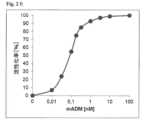
敗血症/敗血症性ショックのモデルにおける前臨床実験から、抗ADM抗体の投与によって血漿バイオADM濃度の増加が誘導されること(実施例8、図9)と、それが生存率の上昇と同時に起こることが知られている(Struck他 2013年、Intensive Care Med Exp第1巻(1):22ページ)。この効果の裏にある機構は以下のようであると考えられている。すなわち、この抗体は、静脈内に投与されると、そのサイズが理由で内皮関門を通過して間質の中に入ることができず、血液循環の中に留まる。したがってこの抗体は、内在ADMを大きく超えるモル量で投与されると、血漿内の実質的にすべてのADMに結合し、結合平衡に到達する。その単純な帰結として、ADMが間質から血液循環へと移動する。間質に位置するADMは血管平滑筋に結合できるため弛緩を誘導し、その結果として血管拡張が起こる。これは、この抗体の投与によって少なくなる。その一方で、血漿中のADMは内皮細胞に結合し、そのことによって血管を安定化させる、それどころか血管を無傷の状態に回復させる。したがって非中和抗体であるこの抗体を投与した帰結として血漿ADMレベルが上昇すると、この機能が強化される。最後に、この抗体がADMに結合すると、ADMのタンパク質分解が減少する。 Preclinical studies in a model of sepsis/septic shock show that administration of anti-ADM antibodies induces an increase in plasma bio-ADM concentrations (Example 8, Figure 9), which coincides with an increase in survival. are known (Struck et al. 2013, Intensive Care Med Exp 1(1):22). The mechanism behind this effect is believed to be as follows. That is, when this antibody is administered intravenously, because of its size, it is unable to cross the endothelial barrier into the interstitium and remains in the blood circulation. Thus, the antibody, when administered in a molar amount that greatly exceeds endogenous ADM, binds virtually all ADM in the plasma and reaches binding equilibrium. A simple consequence is that ADM moves from the stroma into the blood circulation. ADM located in the stroma can bind to vascular smooth muscle and induce relaxation, resulting in vasodilation. This is reduced by administration of this antibody. On the other hand, ADM in plasma binds to endothelial cells, thereby stabilizing blood vessels and even restoring them to an intact state. Thus, increased plasma ADM levels as a result of administration of this antibody, which is a non-neutralizing antibody, enhances this function. Finally, binding of this antibody to ADM reduces proteolysis of ADM.
驚くべきことに、われわれはPROTECT試験(実施例6)とBIOSTAT試験(実施例7)において、心不全を有する患者では、利尿剤を用いて治療しているにもかかわらず、うっ血の存在と重症度に応じてバイオADMの濃度が上昇することを観察した。したがってこれら患者におけるバイオADMの上昇は、組織のうっ血に対抗する患者の側の調節である。しかしこの自然な上昇は、対抗してなされるこの調節を効果的に実現するには不十分である。組織のうっ血は敗血症でも起こる。実施例5、9、10は、敗血症動物モデルで抗ADM抗体を投与すると損傷した血管が無傷の状態に回復することを示している。敗血症と心不全の両方で組織のうっ血機構が共通しているため、本分野の専門家にとって、抗ADM抗体を投与することが、敗血症/敗血症性ショックにおけるのと同様、心不全におけるうっ血の治療にも有益であるはずであることは信頼できる。 Surprisingly, in the PROTECT trial (Example 6) and the BIOSTAT trial (Example 7), we found that patients with heart failure, despite treatment with diuretics, were less likely to have congestion and severity We observed that the concentration of bio-ADM increased depending on the Elevation of bioADM in these patients is therefore a regulation on the part of the patient to counter tissue congestion. However, this natural rise is insufficient to effectively realize this counter-adjustment. Tissue congestion also occurs in sepsis. Examples 5, 9, and 10 demonstrate that administration of anti-ADM antibodies restores damaged blood vessels to an intact state in an animal model of sepsis. Due to the common mechanisms of tissue congestion in both sepsis and heart failure, administration of anti-ADM antibodies is of interest to experts in the field for the treatment of congestion in heart failure as well as in sepsis/septic shock. It is reliable that it should be beneficial.
特別な一実施態様では、うっ血の処置と治療に使用するため、ADMに結合する抗ADM抗体または抗ADM抗体フラグメント、またはADMに結合する抗ADM非Ig足場を、合わせて実施される診断法の助けを借りて患者に投与することができる。合わせて実施される診断法は下に記載する。 In a particular embodiment, a diagnostic method performed in conjunction with an anti-ADM antibody or anti-ADM antibody fragment that binds ADM, or an anti-ADM non-Ig scaffold that binds ADM, for use in the treatment and therapy of blood stasis. It can be administered to the patient with help. Concomitant diagnostic methods are described below.
プロ-アドレノメデュリンまたは少なくとも5個のアミノ酸からなるその断片は、うっ血の初期代替マーカーとして使用できるため、うっ血の治療または処置のガイドとなる。そのガイドに含まれるのは、
・対象から得られた体液中のプロ-アドレノメデュリンのレベル、または少なくとも5個のアミノ酸からなるその断片のレベルを求め;
a)プロ-アドレノメデュリンまたはその断片に関するそのレベルを対象のうっ血の程度と相関させるか、うっ血を診断すること(上昇したレベルが所定の閾値を超えているというのは、うっ血であること、またはうっ血の程度を示す)、または
b)プロ-アドレノメデュリンまたはその断片に関するそのレベルを、その対象におけるうっ血の治療の必要性または成功と、または処置と治療の必要性または成功と相関させること(レベルが所定の閾値よりも低いというのは、うっ血の治療の成功、または処置と治療の成功を予測しており、レベルが所定の閾値よりも上であるというのは、うっ血の治療の必要性、または処置と治療の必要性を示している)、または
c)プロ-アドレノメデュリンまたはその断片に関するそのレベルを、うっ血を治療した後に、または処置と治療をした後にうっ血が除去されるかうっ血が残留するかの予測と相関させること(上昇したレベルが所定の閾値を超えているというのは、うっ血を治療した後に、または処置と治療をした後にうっ血が残留することを予測しているのに対し、レベルが所定の閾値よりも低いというのは、うっ血を治療した後、または処置と治療をした後にうっ血が除去されることを予測している)、または
d)プロ-アドレノメデュリンまたはその断片に関するそのレベルを、うっ血を治療した後に、または処置と治療をした後にうっ血が除去されるかうっ血が残留するかと相関させること(上昇したレベルが所定の閾値を超えているというのは、うっ血を治療した後に、または処置と治療をした後にうっ血が残留することを示しているのに対し、レベルが所定の閾値よりも低いというのは、うっ血を治療した後、または処置と治療をした後にうっ血が除去されることを示している)、または
e)プロ-アドレノメデュリンまたはその断片に関するそのレベルを、退院に関する判断の評価と相関させること(上昇したレベルが所定の閾値を超えているというのは、対象が退院できないことを意味し、レベルが所定の閾値よりも低いというのは、対象を退院させてもよいことを意味する)であり、
プロ-アドレノメデュリンまたはその断片の選択は、配列番号31のプロ-アドレノメデュリン、配列番号32のPAMP、配列番号33のMR-プロADM、配列番号20のADM-NH2、配列番号34のADM-Gly、配列番号35のCT-プロADMを含むグループからなされる。Pro-adrenomedullin or a fragment thereof consisting of at least 5 amino acids can be used as an early surrogate marker for congestion and thus guide therapy or treatment of congestion. Included in that guide is
- determining the level of pro-adrenomedullin, or a fragment thereof consisting of at least 5 amino acids, in body fluids obtained from the subject;
a) Correlating the level of pro-adrenomedullin or fragments thereof with the degree of congestion in a subject or diagnosing congestion (elevated levels above a predetermined threshold indicate congestion or ), or
b) correlating the level of pro-adrenomedullin or a fragment thereof with the need for or success in treating congestion in the subject, or with the need or success of treatment and therapy (i.e. the level is below a predetermined threshold); predicts successful treatment of congestion, or success of treatment and therapy, and levels above a predetermined threshold indicate a need for treatment of congestion, or a need for treatment and therapy. ), or
c) correlating its levels with respect to pro-adrenomedullin or fragments thereof with the prediction of whether congestion will clear or remain after treatment of congestion or after treatment and therapy (elevated levels are Above the threshold predicts residual congestion after treating the congestion or after treatment and treatment, whereas levels below a given threshold predict congestion. anticipate decongestion after treatment or after treatment and treatment), or
d) correlating its levels for pro-adrenomedullin or fragments thereof with elimination of congestion or residual congestion after treatment of congestion or after treatment and therapy (elevated levels exceed a predetermined threshold); is indicative of residual congestion after treating the congestion or after treatment and treatment, whereas a level below a predetermined threshold indicates that after treating the congestion, or showing decongestion after treatment and therapy), or
e) correlating its levels for pro-adrenomedullin or fragments thereof with the evaluation of decisions regarding discharge (elevated levels above a predetermined threshold mean that the subject cannot be discharged; means that the subject may be discharged) and
A selection of pro-adrenomedullin or fragments thereof include pro-adrenomedullin of SEQ ID NO:31, PAMP of SEQ ID NO:32, MR-proADM of SEQ ID NO:33, ADM-NH2 of SEQ ID NO:20 , ADM-Gly of SEQ ID NO:34, Made from the group containing CT-proADM of SEQ ID NO:35.
このような方法は、欧州特許出願第EP16199092号と第EP16178725号に詳細に記載されており、その内容は参照によって本明細書に組み込まれている。上記の診断法で言及した治療と処置は、ADMに結合する抗ADM抗体または抗ADM抗体フラグメントの投与、またはADMに結合する抗ADM非Ig足場の投与である。 Such methods are described in detail in European Patent Applications EP16199092 and EP16178725, the contents of which are incorporated herein by reference. The therapies and treatments referred to in the diagnostic methods above are administration of anti-ADM antibodies or anti-ADM antibody fragments that bind ADM, or administration of anti-ADM non-Ig scaffolds that bind ADM.
うっ血の重症度、うっ血の程度、うっ血の度合い、うっ血のグレードなどは、本出願全体を通じて同義語として使用する。 Severity of congestion, degree of congestion, degree of congestion, grade of congestion, etc. are used synonymously throughout this application.
成熟ABM、バイオADM、ADM-NH2は、本出願全体を通じて同義語であり、配列番号20の分子を表わす。Mature ABM, bio-ADM, ADM-NH2 are synonymous throughout this application and refer to the molecule of SEQ ID NO:20.
プロ-アドレノメデュリンまたはその断片は、急性心不全と心不全の状況におけるうっ血、特に急性心不全を有する対象、および/または悪化している徴候を示す心不全を有する対象、および/または心不全または急性心不全の症状を有する対象におけるうっ血の定量的かつ正確な初期代替マーカーである。急性心不全または心不全の状況におけるうっ血の正確な初期代替マーカーとは、その濃度および/または免疫反応性のレベルがうっ血の程度を反映していることを意味する。 Pro-Adrenomedullin or a fragment thereof is used for acute heart failure and congestion in the setting of heart failure, especially in subjects with acute heart failure and/or with heart failure showing signs of worsening and/or with symptoms of heart failure or acute heart failure. It is a quantitative and accurate early surrogate marker of congestion in a subject. An accurate early surrogate marker of congestion in the setting of acute heart failure or heart failure means that its concentration and/or level of immunoreactivity reflect the degree of congestion.
プロ-アドレノメデュリンまたはその断片のレベルが所定の閾値レベルを超えた場合には、うっ血の治療または処置として、ADMに結合する抗ADM抗体または抗ADM抗体フラグメント、またはADMに結合する抗ADM非Ig足場を投与する。 An anti-ADM antibody or anti-ADM antibody fragment that binds ADM, or an anti-ADM non-Ig scaffold that binds ADM, as therapy or treatment of congestion when the level of pro-adrenomedullin or a fragment thereof exceeds a predetermined threshold level to administer.
本発明の特別な一実施態様の主題では、これは、患者から採取した体液のサンプルで、プロADMおよび/または少なくとも5個のアミノ酸を有するその断片が所定の閾値を超える上昇したレベルを示す場合に、ADMに結合する抗ADM抗体または抗ADM抗体フラグメント、またはADMに結合する抗ADM非Ig足場が、その患者のうっ血の処置と治療に使用されることを意味する。したがってこのプロADMおよび/またはその断片を用いた診断法は、合わせて実施される診断法として役立つ。 In the subject of one particular embodiment of the present invention, this is the case when a sample of body fluid taken from a patient shows elevated levels of pro-ADM and/or fragments thereof having at least 5 amino acids above a predetermined threshold. In other words, the anti-ADM antibody or anti-ADM antibody fragment that binds ADM, or the anti-ADM non-Ig scaffold that binds ADM, is meant to be used in the treatment and treatment of congestion in that patient. Diagnostic methods using this pro-ADM and/or fragments thereof therefore serve as diagnostic methods performed in conjunction.
診断法の特別な一実施態様では、プロADMおよび/または少なくとも5個のアミノ酸を有するその断片の選択は、
配列番号31(プロADM):164個のアミノ酸(プレプロADMのアミノ酸22~185)
ARLDVASEF RKKWNKWALS RGKRELRMSS SYPTGLADVK AGPAQTLIRP QDMKGASRSP EDSSPDAARI RVKRYRQSMN NFQGLRSFGC RFGTCTVQKL AHQIYQFTDK DKDNVAPRSK ISPQGYGRRR RRSLPEAGPG RTLVSSKPQA HGAPAPPSGS APHFL、
配列番号32(プロアドレノメデュリンN-20末端ペプチド、PAMP):プレプロADMのアミノ酸22~41
ARLDVASEF RKKWNKWALS R、
配列番号33(中央領域プロアドレノメデュリン、MR-プロADM):プレプロADMのアミノ酸45~92
ELRMSS SYPTGLADVK AGPAQTLIRP QDMKGASRSP EDSSPDAARI RV、
配列番号20(成熟アドレノメデュリン(成熟ADM);アミド化されたADM;バイオADM):アミノ酸95~146-CONH2
YRQSMN NFQGLRSFGC RFGTCTVQKL AHQIYQFTDK DKDNVAPRSK ISPQGY-CONH2、
配列番号34(アドレノメデュリン1-52-Gly(ADM 1-52-Gly)):プレプロADMのアミノ酸95~147
YRQSMN NFQGLRSFGC RFGTCTVQKL AHQIYQFTDK DKDNVAPRSK ISPQGYG、
配列番号35(C末端プロアドレノメデュリン、CT-プロADM):プレプロADMのアミノ酸148~185
RRR RRSLPEAGPG RTLVSSKPQA HGAPAPPSGS APHFL
を含むグループからなされる。In one particular embodiment of the diagnostic method, the selection of proADM and/or fragments thereof having at least 5 amino acids comprises:
SEQ ID NO:31 (pro-ADM): 164 amino acids (amino acids 22-185 of pre-pro-ADM)
ARLDVASEF RKKWNKWALS RGKRELRMSS SYPTGLADVK AGPAQTLIRP QDMKGASRSP EDSSPDAARI RVKRYRQSMN NFQGLRSFGC RFGTCTVQKL AHQIYQFTDK DKDNVAPRSK ISPQGYGRRR RRSLPEAGPG RTLVSSKPQA HGAPAPPSGS APHFL,
SEQ ID NO: 32 (pro-adrenomedullin N-20 terminal peptide, PAMP): amino acids 22-41 of pre-pro-ADM
ARLDVASEF RKKWNKWALS R,
SEQ ID NO:33 (middle region pro-adrenomedullin, MR-proADM): amino acids 45-92 of preproADM
ELRMSS SYPTGLADVK AGPAQTLIRP QDMKGASRSP EDSSPDAARI RV,
SEQ ID NO:20 (mature adrenomedullin (mature ADM); amidated ADM; bio-ADM): amino acids 95-146-CONH2
YRQSMN NFQGLRSFGC RFGTCTVQKL AHQIYQFTDK DKDNVAPRSK ISPQGY-CONH2 ,
SEQ ID NO: 34 (Adrenomedullin 1-52-Gly (ADM 1-52-Gly)): amino acids 95-147 of prepro-ADM
YRQSMN NFQGLRSFGC RFGTCTVQKL AHQIYQFTDK DKDNVAPRSK ISPQGYG,
SEQ ID NO: 35 (C-terminal pro-adrenomedullin, CT-proADM): amino acids 148-185 of pre-pro-ADM
RRR RRSLPEAGPG RTLVSSKPQA HGAAPPSGS APHFL
is made from a group containing
診断法の特別な一実施態様では、プロADMおよび/または少なくとも5個のアミノ酸を有するその断片の選択は、成熟ADM-NH2(配列番号20)、ADM 1-52-Gly(配列番号34)、MR-プロADM(配列番号33)、CT-プロADM(配列番号35)を含むグループからなされる。In one particular embodiment of the diagnostic method, the selection of proADM and/or fragments thereof having at least 5 amino acids is mature ADM-NH2 (SEQ ID NO:20), ADM1-52-Gly (SEQ ID NO:34) , MR-proADM (SEQ ID NO: 33), CT-proADM (SEQ ID NO: 35).
診断法の特別な一実施態様では、成熟ADM-NH2(配列番号20)免疫活性のレベル、および/またはADM 1-52-Gly(配列番号34)免疫活性のレベル、またはMR-プロADM(配列番号33)免疫活性のレベル、またはCT-プロADM(配列番号35)免疫活性のレベルを求め、患者の治療または処置の必要性と相関させる。ただしその患者にそのような必要性があると同定されるのは、体液中の成熟ADM-NH2(配列番号20)免疫活性のレベル、および/またはADM 1-52-Gly(配列番号34)免疫活性のレベル、またはMR-プロADM(配列番号33)免疫活性のレベル、またはCT-プロADM(配列番号35)免疫活性のレベルが閾値を超えている場合である。In one particular embodiment of the diagnostic method, the level of mature ADM-NH2 (SEQ ID NO:20) immunoreactivity and/or the level of ADM1-52-Gly (SEQ ID NO:34) immunoreactivity, or MR-proADM ( SEQ ID NO: 33) levels of immune activity, or CT-proADM (SEQ ID NO: 35) immune activity levels are determined and correlated with patient therapy or need for treatment. However, it is the level of mature ADM-NH2 (SEQ ID NO: 20) immune activity in the body fluids and/or ADM 1-52-Gly (SEQ ID NO: 34) that identifies such a need in the patient. when the level of immune activity, or the level of MR-proADM (SEQ ID NO:33) immune activity, or the level of CT-proADM (SEQ ID NO:35) immune activity exceeds a threshold.
診断法の特別な一実施態様では、プロADMおよび/またはその断片のレベルは、成熟ADM-NH2(配列番号20)および/またはADM 1-52-Gly(配列番号34)の配列に含まれるある領域に結合する結合剤と、成熟ADM-NH2(配列番号20)および/またはADM 1-52-Gly(配列番号34)の配列に含まれるある領域に結合する第2の結合剤からなるグループから選択された少なくとも1つの結合剤を用いて求められる。In one particular embodiment of the diagnostic method, the levels of pro-ADM and/or fragments thereof are contained in the sequences of mature ADM-NH2 (SEQ ID NO: 20) and/or ADM 1-52-Gly (SEQ ID NO: 34). A binding agent that binds to a region and a second binding agent that binds to a region contained in the sequences of mature ADM-NH2 (SEQ ID NO:20) and/or ADM1-52-Gly (SEQ ID NO:34) determined using at least one binding agent selected from the group.
診断法の特別な一実施態様では、プロADMおよび/またはその断片のレベルは、MR-プロADM(配列番号33)の配列に含まれるある領域に結合する結合剤と、MR-プロADM(配列番号33)の配列に含まれるある領域に結合する第2の結合剤からなるグループから選択された少なくとも1つの結合剤を用いて求められる。 In one particular embodiment of the diagnostic method, levels of proADM and/or fragments thereof are obtained by combining a binding agent that binds to a region contained within the sequence of MR-proADM (SEQ ID NO: 33) and MR-proADM (SEQ ID NO: 33). number 33) using at least one binding agent selected from the group consisting of a second binding agent that binds to a region contained in the sequence of number 33).
診断法の特別な一実施態様では、プロADMおよび/またはその断片のレベルは、CT-プロADM(配列番号35)の配列に含まれるある領域に結合する結合剤と、CT-プロADM(配列番号35)の配列に含まれるある領域に結合する第2の結合剤からなるグループから選択された少なくとも1つの結合剤を用いて求められる。 In one particular embodiment of the diagnostic method, levels of proADM and/or fragments thereof are obtained by combining a binding agent that binds to a region comprised within the sequence of CT-proADM (SEQ ID NO: 35) and CT-proADM (SEQ ID NO: 35). number 35) using at least one binding agent selected from the group consisting of a second binding agent that binds to a region contained in the sequence of number 35).
本発明の診断法の特別な一実施態様における主題は、断片を、配列番号33のMR-プロADM、または配列番号20の成熟ADM-NH2から選択することのできる本発明の方法である。A subject in one particular embodiment of the diagnostic method of the invention is the method according to the invention, wherein the fragment can be selected from MR-proADM of SEQ ID NO:33 or mature ADM-NH2 of SEQ ID NO:20 .
本発明の診断法の主題は、プロ-アドレノメデュリンおよび/または少なくとも5個のアミノ酸を有するその断片のレベルを、プロ-アドレノメデュリンおよび/または少なくとも5個のアミノ酸を有するその断片に対する結合剤を用いて求める、本発明の診断法に従う方法である。 The diagnostic method subject of the present invention determines the level of pro-adrenomedullin and/or fragments thereof having at least 5 amino acids using binding agents to pro-adrenomedullin and/or fragments thereof having at least 5 amino acids. , a method according to the diagnostic method of the present invention.
本診断法の主題は、結合剤が、プロADMおよび/または少なくとも5個のアミノ酸を有するその断片に結合する抗体、抗体フラグメント、非Ig足場を含むグループから選択される、本発明の診断法に従う方法である。 The subject of this diagnostic method is according to the diagnostic method of the invention, wherein the binding agent is selected from the group comprising antibodies, antibody fragments, non-Ig scaffolds that bind to proADM and/or fragments thereof having at least 5 amino acids. The method.
本発明の体液は、特別な一実施態様では血液サンプルである。血液サンプルは、全血、血清、血漿を含むグループから選択することができる。診断法の特別な一実施態様では、サンプルは、ヒトクエン酸血漿、ヘパリン血漿、EDTA血漿を含むグループから選択される。 A bodily fluid according to the invention is a blood sample in one particular embodiment. A blood sample can be selected from the group comprising whole blood, serum, plasma. In one particular embodiment of the diagnostic method, the sample is selected from the group comprising human citrated plasma, heparinized plasma, EDTA plasma.
本発明の特別な一実施態様では、ADMに結合する抗ADM抗体または抗ADM抗体フラグメント、またはADMに結合する抗ADM非Ig足場は、本発明の任意の実施態様に従い、利尿剤抵抗性であるか利尿剤療法に反応しない患者で、うっ血の処置と治療に使用される。 In a particular embodiment of the invention, the anti-ADM antibody or anti-ADM antibody fragment that binds ADM, or the anti-ADM non-Ig scaffold that binds ADM, according to any embodiment of the invention, is diuretic resistant. It is used to treat and treat congestion in patients who do not respond to diuretic therapy.
本発明の別の特別な一実施態様は、うっ血の処置と治療を必要とする患者でその処置と治療に用いるための抗アドレノメデュリン抗体、または抗アドレノメデュリン抗体フラグメント、または抗ADM非Ig足場に関するものである(ただしこの抗ADM抗体、または抗ADM抗体フラグメント、または抗ADM非Ig足場は、アドレノメデュリンのN末端部分(アミノ酸1~21):
YRQSMNNFQGLRSFGCRFGTC(配列番号22)
に結合し、
患者は、利尿剤抵抗性であるか利尿剤療法に反応しない)。Another particular embodiment of the present invention relates to anti-adrenomedullin antibodies, or anti-adrenomedullin antibody fragments, or anti-ADM non-Ig scaffolds for use in treating and treating patients in need thereof. (However, this anti-ADM antibody, or anti-ADM antibody fragment, or anti-ADM non-Ig scaffold contains the N-terminal portion of adrenomedullin (amino acids 1-21):
YRQSMNNFQGLRSFGCRFGTC (SEQ ID NO: 22)
to
Patients are diuretic-resistant or do not respond to diuretic therapy).
「利尿剤抵抗性」という用語は、一般に、利尿剤を十分に使用しているにもかかわらず細胞外体液の体積を減らせないことと定義される(Ravnan他 2002年、CHF 第8巻:80~85ページ)。Epsteinらは、利尿剤抵抗性を、160 mgの用量のフロセミドを1日に2回経口投与して72時間以内に少なくとも90ミリモルのナトリウムを排泄できないことと定義した(Epstein他 1977年、Curr Ther Res. 第21巻:656~667ページ)。 The term “diuretic resistance” is generally defined as the inability to reduce extracellular fluid volume despite adequate diuretic use (Ravnan et al. 2002, CHF 8:80 ~ page 85). Epstein et al. defined diuretic resistance as the inability to excrete at least 90 millimoles of sodium within 72 hours of oral administration of furosemide at a dose of 160 mg twice daily (Epstein et al. 1977, Curr Ther. Res. 21:656-667).
利尿剤に対する適応と利尿剤抵抗性は、似た機構によって生じる可能性がある。利尿剤に対する適応は、利尿剤が作用している間に起こる適応、短期間のうちにナトリウム貯留を引き起こす(「利尿剤後NaCl貯留」を引き起こす)適応、慢性的にナトリウム貯留を増加させる適応(「ブレーキ現象」)に分類することができる。腎臓が長期の利尿剤治療に適応する様式は以下の通りである。最初に、利尿剤を投与している間は供給されるNaCl負荷が増加するため、利尿剤作用部位の下流にあるネフロンセグメントがNaClの再吸収を増加させる。第2に、腎細管内の利尿剤の濃度が低下すると、腎細管は、次に利尿剤が投与されるまでNaを貯留させるように機能する。第3に、腎臓のNaCl排泄を増加させる利尿剤の能力が時間経過とともに低下する。これは、細胞外体液の体積の欠乏と、腎細管そのものの構造および機能の変化の両方に起因する効果である。これらの適応はすべてNaCl再吸収率を増大させ、利尿剤療法の効果を弱める。概説に関しては、Ellison 1999年、Semin Nephrol. 第19巻(6):581~597ページと、De Bruyne 2003年、Postgrad Med J第79巻:268~271ページを参照されたい。 Diuretic adaptation and diuretic resistance may arise by similar mechanisms. Indications for diuretics include those that occur while the drug is acting, those that cause short-term sodium retention (causing "postdiuretic NaCl retention"), and those that chronically increase sodium retention ( “braking phenomenon”). The manner in which the kidney adapts to long-term diuretic therapy is as follows. First, nephron segments downstream of the site of diuretic action increase NaCl reabsorption because the delivered NaCl load increases during diuretic administration. Second, when the concentration of diuretic in the renal tubules decreases, the renal tubules act to retain Na until the next diuretic is administered. Third, the ability of diuretics to increase renal NaCl excretion decreases over time. This is an effect due both to the depletion of extracellular fluid volume and to changes in the structure and function of the renal tubules themselves. All of these indications increase the NaCl reabsorption rate and attenuate the efficacy of diuretic therapy. For reviews, see Ellison 1999, Semin Nephrol. 19(6):581-597 and De Bruyne 2003, Postgrad Med J 79:268-271.
数値を明確にすることは難しいとはいえ、利尿剤抵抗性は、うっ血性HFを有する患者3人に1人の割合で起こると考えられている。心不全は、利尿剤抵抗性が観察される最も一般的な臨床状況である。軽度のうっ血性HFでは、腎機能が維持されている限りは一般に利尿剤抵抗性に遭遇することはない。しかし中度と重度のうっ血性HF患者では、利尿剤抵抗性がより頻繁に起こり、臨床で問題になることがしばしばある(Brater 1985年、Drugs 第30巻:427~443ページ;Taylor 2000年、Cardiol Rev. 第8巻:104~114ページ)。 Although it is difficult to quantify, diuretic resistance is believed to occur in 1 in 3 patients with congestive HF. Heart failure is the most common clinical setting in which diuretic resistance is observed. In mild congestive HF, diuretic resistance is generally not encountered as long as renal function is preserved. However, in patients with moderate and severe congestive HF, diuretic resistance occurs more frequently and is often a clinical problem (Brater 1985 Drugs 30:427-443;
診断法の特別な一実施態様では、アッセイ感度が、健康な対象の成熟ADM-NH2を定量することができる感度であって、70 pg/ml未満、好ましくは40 pg/ml未満、より好ましくは10 pg/ml未満であるアッセイを利用して、プロADMおよび/または少なくとも5個のアミノ酸を有するその断片のレベルを求める。本発明の方法ではこれらの濃度を閾値として用いることができる。In one particular embodiment of the diagnostic method, the assay sensitivity is the sensitivity that allows quantification of mature ADM-NH2 in healthy subjects and is less than 70 pg/ml, preferably less than 40 pg/ml, more preferably less than 40 pg/ml. is less than 10 pg/ml to determine levels of pro-ADM and/or fragments thereof having at least 5 amino acids. These concentrations can be used as thresholds in the method of the present invention.
診断法の特別な一実施態様では、アッセイ感度が、健康な対象のMR-プロADMを定量することができる感度であって、0.5ナノモル/l未満、好ましくは0.4ナノモル/l未満、より好ましくは0.2ナノモル/l未満であるアッセイを利用して、プロADMおよび/または少なくとも5個のアミノ酸を有するその断片のレベルを求める。本発明の方法ではこれらの濃度を閾値として用いることができる。 In one particular embodiment of the diagnostic method, the assay sensitivity is the sensitivity with which MR-proADM in healthy subjects can be quantified, and is less than 0.5 nmol/l, preferably less than 0.4 nmol/l, more preferably less than 0.4 nmol/l. An assay that is less than 0.2 nmol/l is used to determine levels of pro-ADM and/or fragments thereof having at least 5 amino acids. These concentrations can be used as thresholds in the method of the present invention.
診断法の特別な一実施態様では、アッセイ感度が、健康な対象のCT-プロADMを定量することができる感度であって、100ピコモル/l未満、好ましくは75ピコモル/l未満、より好ましくは50ピコモル/l未満であるアッセイを利用して、プロADMおよび/または少なくとも5個のアミノ酸を有するその断片のレベルを求める。本発明の方法ではこれらの濃度を閾値として用いることができる。 In one particular embodiment of the diagnostic method, the assay sensitivity is the sensitivity that allows quantification of CT-proADM in healthy subjects and is less than 100 pmoles/l, preferably less than 75 pmoles/l, more preferably An assay that is less than 50 pmol/l is used to determine levels of proADM and/or fragments thereof having at least 5 amino acids. These concentrations can be used as thresholds in the method of the present invention.
診断法の特別な一実施態様では、結合剤は、プロADMおよび/またはその断片に対して少なくとも107 M-1、好ましくは108 M-1の結合親和性を示し、好ましい親和性は、109 M-1超、最も好ましくは1010 M-1超である。当業者は、より多い用量の化合物を適用することによってより小さな親和性が補償されると見なせることと、この測定が本発明の範囲を外れることなないと考えられることを知っている。In one particular embodiment of the diagnostic method, the binding agent exhibits a binding affinity for proADM and/or fragments thereof of at least 107 M−1 , preferably 108 M−1 , preferred affinities are greater than 109 M−1 , most preferably greater than 1010 M−1 . A person skilled in the art knows that the lower affinity can be considered compensated by applying a higher dose of the compound and that this measurement is not considered outside the scope of the invention.
アドレノメデュリンに対する抗体の親和性を求めるため、固定化された抗体に対するアドレノメデュリンの結合速度を、Biacore 2000システム(GE Healthcare Europe GmbH社、フライブルク、ドイツ国)を利用して標識なしの表面プラズモン共鳴によって求めた。高密度でCM5センサー表面に共有結合した抗マウスFc抗体(マウス抗体捕獲キット;GE Healthcare社)を製造者の指示に従って用いて抗体を可逆的に固定化した(Lorenz他 2011年、Antimicrob Agents Chemother. 第55巻(1):165~173ページ)。 To determine the affinity of the antibody to adrenomedullin, the binding kinetics of adrenomedullin to the immobilized antibody was determined by label-free surface plasmon resonance using a
診断法の特別な一実施態様では、結合剤は、プロADMおよび/またはその断片に結合する抗体、または抗体フラグメント、または非Ig足場を含むグループから選択される。 In one particular embodiment of the diagnostic method, the binding agent is selected from the group comprising antibodies or antibody fragments or non-Ig scaffolds that bind to proADM and/or fragments thereof.
診断法の特別な一実施態様では、アッセイを利用して、プロADMおよび/または少なくとも5個のアミノ酸を有するその断片のレベルを求める。そのようなアッセイは、サンドイッチアッセイ、好ましくは完全自動化アッセイである。 In one particular embodiment of the diagnostic method, the assay is utilized to determine levels of proADM and/or fragments thereof having at least 5 amino acids. Such assays are sandwich assays, preferably fully automated assays.
本発明の一実施態様では、完全自動化アッセイシステムを必要とせずに患者の近くで1時間以内に検査を実施できる検査技術はいわゆるPOC(診療現場)検査であろう。この技術の一例は、免疫クロマトグラフィ検査技術である。 In one embodiment of the invention, a testing technique that can be performed near the patient in less than an hour without the need for a fully automated assay system would be a so-called POC (point of care) test. An example of this technology is the immunochromatographic assay technology.
診断法の一実施態様では、そのようなアッセイは、任意の種類の検出技術(その非限定的な例に含まれるのは、酵素標識、化学発光標識、電気化学発光標識である)を利用したサンドイッチイムノアッセイであり、完全自動化アッセイが好ましい。診断法の一実施態様では、そのようなアッセイは、酵素で標識したサンドイッチアッセイである。自動化アッセイまたは完全自動化アッセイの例に含まれるのは、Roche Elecsys(登録商標)、Abbott Architect(登録商標)、Siemens Centauer(登録商標)、Brahms Kryptor(登録商標)、BiomerieuxVidas(登録商標)、Alere Triage(登録商標)というシステムのうちの1つで利用できるアッセイである。 In one embodiment of the diagnostic method, such assays utilized any type of detection technology, non-limiting examples of which are enzymatic labels, chemiluminescent labels, electrochemiluminescent labels. Sandwich immunoassays and fully automated assays are preferred. In one diagnostic method embodiment, such an assay is an enzyme-labeled sandwich assay. Examples of automated or fully automated assays include Roche Elecsys®, Abbott Architect®, Siemens Centauer®, Brahms Kryptor®, BiomerieuxVidas®, Alere Triage (registered trademark) system.
多彩なイムノアッセイが知られており、本発明のアッセイと方法で利用することができる。イムノアッセイに含まれるのは、ラジオイムノアッセイ(「RIA」)、ホモジニアス酵素多重化イムノアッセイ(「EMIT」)、酵素結合免疫吸着アッセイ(「ELISA」)、アポ酵素再活性化イムノアッセイ(「ARIS」)、ディップスティックイムノアッセイ、イムノクロマトグラフィアッセイである。 A wide variety of immunoassays are known and can be utilized in the assays and methods of the invention. Immunoassays include radioimmunoassay (“RIA”), homogeneous enzyme multiplexed immunoassay (“EMIT”), enzyme-linked immunosorbent assay (“ELISA”), apoenzyme reactivation immunoassay (“ARIS”), dip Stick immunoassay, immunochromatographic assay.
診断法の特別な一実施態様では、2つの結合剤のうちの少なくとも一方に標識して検出する。 In one particular embodiment of the diagnostic method, at least one of the two binding agents is labeled and detected.
本発明の主題は、うっ血性高血圧、むくみまたは水分貯留(浮腫)、心不全(特に急性心不全)、腎臓疾患、肝臓疾患を含むグループから選択された疾患または状態を有する患者でうっ血の処置と治療に使用するための抗アドレノメデュリン(ADM)抗体、または抗アドレノメデュリン抗体フラグメント、または抗ADM非Ig足場である。 The subject of the present invention is the treatment and therapy of congestion in patients with a disease or condition selected from the group comprising congestive hypertension, swelling or water retention (edema), heart failure (especially acute heart failure), kidney disease, liver disease. Anti-adrenomedullin (ADM) antibodies, or anti-adrenomedullin antibody fragments, or anti-ADM non-Ig scaffolds for use.
本発明の主題は、うっ血性高血圧、むくみまたは水分貯留(浮腫)、心不全(特に急性心不全)を含むグループから選択された疾患または状態を有する患者でうっ血の処置と治療に使用するための抗アドレノメデュリン(ADM)抗体、または抗アドレノメデュリン抗体フラグメント、または抗ADM非Ig足場である。 The subject of the present invention is the anti-adrenomedullin for use in the treatment and therapy of congestion in patients with diseases or conditions selected from the group comprising congestive hypertension, swelling or water retention (edema), heart failure (especially acute heart failure). (ADM) antibody, or anti-adrenomedullin antibody fragment, or anti-ADM non-Ig scaffold.
心不全(HF)は、心臓の構造または機能に問題が起こって身体の必要に見合った十分な血流を供給する能力が損なわれたときに起こる心臓の状態である。心不全は多彩な症状を引き起こす可能性があり、それは特に、休息中と運動中の息切れ(SOB)、体液貯留の徴候(肺うっ血やくるぶしのむくみなど)、休息時の心臓の構造または機能に異常があることの客観的な証拠である。 Heart failure (HF) is a heart condition that occurs when a problem with the structure or function of the heart impairs its ability to supply sufficient blood flow to meet the body's needs. Heart failure can cause a variety of symptoms, including shortness of breath at rest and during exercise (SOB), signs of fluid retention (such as pulmonary congestion and ankle swelling), and abnormalities in resting heart structure or function. is objective evidence that there is
心不全は、心臓の機能障害によって起こる症状と徴候の集合を特徴とする臨床症候群である。先進国における罹患と死亡の主因の1つであり、1~2%の人に生じる。心不全は、慢性HFと急性HFに分類することができる。慢性HFを有する患者は、安定な慢性HF、悪化している徴候と症状がある慢性HF、急性非代償性の慢性HFに分類することができる。急性心不全(AHF)は、心不全の徴候と症状が急激に出現し、その結果として緊急治療または入院が必要とされることと定義される。AHFは、急性デノボHF(以前に心臓機能障害がない患者におけるAHFの新たな発症)または急性非代償性の慢性HFとして現われる可能性がある。AHFは年齢が65歳超の成人における入院の主因である。過去数十年の間に治療法が進歩したことが主な理由で慢性心不全患者の予後は顕著に改善しているにもかかわらず、非代償性心不全で患者が入院する場合には、短期の転帰と長期の転帰の両方が非常に悪いままである。AHFで入院した患者のほぼ25%が退院後30日以内に再入院する必要があるが、入院後5年間を超えて生存するのは50%未満である。罹患した患者の生存率と生活の質が著しく低下することに加え、健康保険システムへのAHFの金銭的負担は莫大である。心不全のケアにかかる合計コストは2012年にアメリカ合衆国だけで310億ドルに達すると推定され、このコストの大半は病院内でのケアに関連している。このコストは、人口の高齢化が理由で2030年には700億ドルまで増大すると予測されている。 Heart failure is a clinical syndrome characterized by a collection of symptoms and signs caused by cardiac dysfunction. It is one of the leading causes of morbidity and mortality in developed countries, occurring in 1-2% of people. Heart failure can be classified into chronic HF and acute HF. Patients with chronic HF can be classified as stable chronic HF, chronic HF with worsening signs and symptoms, and acute decompensated chronic HF. Acute heart failure (AHF) is defined as the sudden onset of signs and symptoms of heart failure, resulting in the need for emergency treatment or hospitalization. AHF can present as acute de novo HF (new onset of AHF in patients without previous cardiac dysfunction) or acute decompensated chronic HF. AHF is the leading cause of hospitalization in adults over 65 years of age. Despite the marked improvement in prognosis for patients with chronic heart failure, largely due to advances in treatment over the last few decades, the short-term consequences of hospitalization for decompensated heart failure Both outcomes and long-term outcomes remain very poor. Nearly 25% of patients hospitalized with AHF require readmission within 30 days of hospital discharge, but less than 50% survive beyond 5 years of hospitalization. In addition to significantly reducing survival and quality of life for affected patients, the financial burden of AHF on health insurance systems is enormous. The total cost of heart failure care was estimated to reach $31 billion in the United States alone in 2012, with most of this cost associated with care within hospitals. This cost is projected to rise to $70 billion by 2030 due to an aging population.
心不全には、左室駆出率(LVEF)が正常である(EFが維持された(HFpEF)HFとしても知られる)患者(典型的には50%以上が該当すると考えられている)から、LVEFが低下した患者(典型的には40%未満が該当すると考えられている)までの広い範囲の患者が含まれる。LVEFが40~49%の範囲である患者は、中間範囲のEFを持つHFとして定義される「グレー領域」を表わす(Ponikowski他 2016年、European Heart Journal 第18巻(8):891~975ページ)。 Since heart failure is typically thought to be in ≥50% of patients with normal left ventricular ejection fraction (LVEF) (also known as HF with preserved EF (HFpEF)), It includes a wide range of patients down to those with reduced LVEF (typically considered to be less than 40%). Patients with LVEF in the 40-49% range represent a "gray area" defined as HF with an EF in the mid-range (Ponikowski et al. 2016, European Heart Journal 18(8):891-975). ).
病院内という状況でのAHF治療の主要なゴールは、うっ血除去(単純に蓄積した細胞内と細胞外の過剰な体液の除去)と、うっ血の症状と徴候の緩和である。利尿剤は相変わらずAHFにおける主要なうっ血除去法であり、ほぼすべての入院患者がこのクラスの薬を投与される。心拍出量を増加させて充満圧を低下させる他のクラスの薬剤(強心剤、血管拡張剤など)が、選択されたグループの患者に与えられる。限外濾過も、何人かの患者、特に利尿剤療法に十分に反応しない患者で考慮することができよう。 The primary goals of AHF treatment in the hospital setting are decongestion (simply removal of excess intracellular and extracellular fluid buildup) and relief of symptoms and signs of congestion. Diuretics remain the primary decongestant in AHF, and nearly all hospitalized patients receive this class of drug. Other classes of drugs that increase cardiac output and decrease filling pressure (cardiotonic agents, vasodilators, etc.) are given to selected groups of patients. Ultrafiltration could also be considered in some patients, especially those who do not respond well to diuretic therapy.
患者は(一般に)利尿剤療法によく反応するが、かなりの割合の患者は、十分なレベルのうっ血除去と正常レベルの血液量を達成せずに退院する(すなわち残留うっ血)。これは主に、うっ血の臨床評価に関する現在のアプローチが不十分であるという事実と関係している。退院中の残留うっ血の存在が退院後のより悪い転帰、特に再入院と関係していることを示す一貫性のある証拠が存在する。したがって、うっ血に関するより正確で信頼性のある代替マーカーとして、達成されるうっ血除去のレベルの十分さと退院の時期に関する客観的かつ最適な判断を容易に下せるようにする代替マーカーが大いに必要とされているのに、その必要性が満たされないままになっている。 Although patients (generally) respond well to diuretic therapy, a significant proportion of patients are discharged without achieving adequate levels of decongestion and normal levels of blood volume (ie, residual congestion). This is primarily related to the fact that current approaches to clinical assessment of congestion are inadequate. There is consistent evidence that the presence of residual congestion during discharge is associated with worse post-discharge outcomes, particularly readmissions. Therefore, there is a great need for more accurate and reliable surrogate markers of congestion that facilitate objective and optimal decisions regarding the adequacy of the level of decongestion achieved and the timing of discharge. but the need remains unmet.
本発明の特別な1つの側面では、対象は、心不全を有する対象である。本発明の別の特別な1つの側面では、対象は、急性心不全を有する対象、および/または悪化している徴候を示す心不全を有する対象、および/または心不全または急性心不全の症状を有する対象である。本発明の特別な1つの側面では、対象は、急性心不全を有する。すなわちAHFの新たな発症または急性非代償性HFを有する。本発明の別の特別な1つの側面では、対象は、急性非代償性慢性HF、または慢性心不全の悪化している徴候/症状を有する。本発明の特別な1つの側面では、対象は、急性心不全、特に新たに発症したAHFを有する。 In one particular aspect of the invention, the subject is a subject with heart failure. In another particular aspect of the invention, the subject is a subject with acute heart failure and/or with heart failure showing signs of worsening and/or with heart failure or symptoms of acute heart failure. . In one particular aspect of the invention, the subject has acute heart failure. new onset of AHF or acute decompensated HF. In another specific aspect of the invention, the subject has acute decompensated chronic HF, or worsening signs/symptoms of chronic heart failure. In one particular aspect of the invention, the subject has acute heart failure, particularly new-onset AHF.
「急性」という用語は、急激な発症を示すためや、悪化した心不全または非代償性心不全を記述するために用いられ、心不全の徴候と症状が変化した結果として緊急治療または入院を必要とすることを患者の特徴にできるエピソードを指す。 The term "acute" is used to denote a sudden onset and to describe worsening or decompensated heart failure, requiring emergency care or hospitalization as a result of changes in the signs and symptoms of heart failure. refers to an episode that can characterize a patient.
「慢性」という用語は、長期にわたって持続することを意味する。慢性心不全は長期の状態であり、通常は症状を治療することによって安定に保たれる(安定な慢性HF)。 The term "chronic" means persistent over an extended period of time. Chronic heart failure is a long-term condition that is usually kept stable by treating symptoms (stable chronic HF).
安定な慢性HFの特徴は、
1.身体の必要に見合った十分な血流を供給する能力が損なわれる心臓の構造障害または機能障害が存在し、
2.(肺うっ血および/または全身うっ血として現われる)体液過剰および/または(低血圧および/または腎不全および/またはショック症候群として現われる)心拍出量の大きな低下は存在していないが、
患者は、緊急治療または治療調整の必要がなく、入院を必要としないことである。Stable chronic HF is characterized by:
1. there is a structural or functional disorder of the heart that impairs its ability to supply sufficient blood flow to meet the body's needs;
2. There is no fluid overload (manifested as pulmonary and/or systemic congestion) and/or significant reduction in cardiac output (manifested as hypotension and/or renal failure and/or shock syndrome),
Patients are not in need of urgent care or treatment adjustments and do not require hospitalization.
悪化している徴候と症状がある慢性HFの特徴は、
1.身体の必要に見合った十分な血流を供給する能力が損なわれる心臓の構造障害または機能障害が存在し、
2.(肺および/または全身うっ血として現われる)体液過剰および/または(低血圧および/または腎不全および/またはショック症候群として現われる)心拍出量の大きな低下が存在しているが、
患者は、緊急治療の必要がなく、入院を必要としないものの、治療調整は必要とすることである。Chronic HF with worsening signs and symptoms is characterized by:
1. there is a structural or functional disorder of the heart that impairs its ability to supply sufficient blood flow to meet the body's needs;
2. There is fluid overload (manifested as pulmonary and/or systemic congestion) and/or greatly reduced cardiac output (manifested as hypotension and/or renal failure and/or shock syndrome),
Patients do not require urgent care and do not require hospitalization, but do require treatment adjustments.
慢性心不全として、非代償性(急性非代償性心不全または急性非代償性慢性心不全と呼ばれる)も可能である。これは、最も一般的には、併発疾患(肺炎など)、心筋梗塞、不整脈、未治療の高血圧から生じたり、患者が水分制限、食事、薬物治療を維持できないことから生じたりする。治療後、急性非代償性慢性HFを有する患者は、安定な慢性非代償性状態(安定な慢性HF)に戻る可能性がある。 Chronic heart failure can also be decompensated (called acute decompensated heart failure or acute decompensated chronic heart failure). It most commonly results from comorbidities (such as pneumonia), myocardial infarction, arrhythmia, untreated hypertension, or from the patient's inability to maintain fluid restriction, diet, or medication. After treatment, patients with acute decompensated chronic HF may revert to a stable chronic decompensated state (stable chronic HF).
新たに発症した急性HFと急性非代償性慢性HFの特徴は、
1.身体の必要に見合った十分な血流を供給する能力が損なわれる心臓の構造障害または機能障害が存在し、
2.(肺および/または全身うっ血として現われる)体液過剰および/または(低血圧および/または腎不全および/またはショック症候群として現われる)心拍出量の大きな低下が存在しているが、
患者は、緊急治療または治療調整の必要があり、入院を必要とすることである。New-onset acute HF and acute decompensated chronic HF are characterized by:
1. there is a structural or functional disorder of the heart that impairs its ability to supply sufficient blood flow to meet the body's needs;
2. There is fluid overload (manifested as pulmonary and/or systemic congestion) and/or greatly reduced cardiac output (manifested as hypotension and/or renal failure and/or shock syndrome),
The patient is in need of urgent care or treatment adjustment and requires hospitalization.

急性心不全が、新たに発症したAHF、急性非代償性HF、急性非代償性慢性HF、慢性HFが悪化している徴候/症状のいずれかであるという上記の定義は、Voors他、European Journal of Heart Failure(2016年)、第18巻、716~726ページに合致している。 The above definitions of acute heart failure as either new-onset AHF, acute decompensated HF, acute decompensated chronic HF, or signs/symptoms of worsening chronic HF are provided by Voors et al., European Journal of Heart Failure (2016), Vol. 18, pp. 716-726.
腎機能の悪化は、急性と慢性の心不全(HF)両方の状況で一般的であり、最近は「心腎症候群」として記述されている。腎うっ血(RC)が心腎症候群の潜在的な寄与因子であると認められることがますます多くなっているため、うっ血を十分に制御すると同時に腎機能を改善/保存することが、HFにおいて患者を管理する際の主要なゴールであるという提案がなされている(Aronson 2012年、Expert Rev Cardiovasc Ther第10巻:177~189ページ)。 Deterioration of renal function is common in the setting of both acute and chronic heart failure (HF) and has recently been described as 'cardiorenal syndrome'. As renal congestion (RC) is increasingly recognized as a potential contributor to cardiorenal syndrome, adequate control of congestion while improving/preserving renal function is critical for patients with HF. (Aronson 2012, Expert Rev Cardiovasc Ther 10:177-189).
HFにおけるうっ血は、左心室拡張期圧が高く、HFの徴候と症状(呼吸困難、および/またはラッセル、および/または浮腫)を伴っていることと定義される。うっ血に関係するこれらの徴候と症状は、HFに関連した入院の主な理由である。 Congestion in HF is defined as elevated left ventricular diastolic pressure, accompanied by signs and symptoms of HF (dyspnea and/or Russell's and/or edema). These signs and symptoms associated with congestion are the leading reason for HF-related hospitalizations.
うっ血(とそれに伴う徴候/症状)の緩和と正常な血液量の達成は、相変わらず入院でのAHF療法の主要なゴールだが、うっ血の評価には標準的なアルゴリズムまたは臨床ツールが存在しない。うっ血の臨床評価に関する現状は、症候と症状を中心としたものである。物理的検査の知見(上昇した頸静脈圧(JVP)、末梢浮腫、起坐呼吸、S3心音、肝肥大など)または胸部X線撮影の知見(心肥大)と、間質/肺泡浮腫が、うっ血の代替マーカーとして用いられている。注意深くJVPを評価する以外に、うっ血を検出するためのこれらのパラメータの予測値を適度な値にするのが好ましいことに注意する必要がある。うっ血に関して信頼性があって正確な代替マーカーが大いに必要とされていながらその必要が満たされないままになっている。うっ血とうっ血除去を定量的かつ定性的に判断すること、および/または予測すること、および/または評価すること、および/またはモニタすることが医学で大いに必要とされながらその必要が満たされていない。うっ血の程度、すなわちうっ血のグレードを判断すること、および/または予測すること、および/または評価すること、および/またはモニタすることが必要とされている。 Relief of congestion (and associated signs/symptoms) and achievement of euvolemia remains a major goal of inpatient AHF therapy, but no standard algorithms or clinical tools exist for the assessment of congestion. The current state of clinical assessment of congestion is centered around signs and symptoms. Findings on physical examination (e.g. elevated jugular venous pressure (JVP), peripheral edema, orthopnea, S3 heartbeat, liver hypertrophy) or findings on chest radiography (cardiomegaly) and interstitial/pulmonary foam edema may be associated with congestion. It is used as an alternative marker for Besides careful evaluation of JVP, it should be noted that moderate predictive values of these parameters for the detection of congestion are preferable. There is a great and unmet need for reliable and accurate surrogate markers of congestion. There is a great unmet medical need to quantitatively and qualitatively determine and/or predict and/or evaluate and/or monitor congestion and decongestion . There is a need to determine and/or predict and/or assess and/or monitor the degree of congestion, ie the grade of congestion.
本発明に関しては、うっ血の程度は、うっ血の重症度のグレードとしても表現することができ、下記のようにして判断される。しかし当業者は、うっ血の程度を他のスコアまたは代替マーカーによって表現できること、例えばAmbrosyら(Ambrosy他 2013年、European Heart Journal第34 巻(11):835~843ページ)が用いているスコアとして表現できることを知っている。 For the purposes of the present invention, the degree of congestion can also be expressed as a grade of severity of congestion, determined as follows. However, those skilled in the art will appreciate that the degree of congestion can be expressed by other scores or surrogate markers, such as the score used by Ambrosy et al. i know i can.
上に説明したように、うっ血は多くの異なったやり方で分類することができる。当業者は、うっ血の程度を他のスコアまたは代替マーカーによって表現できることを知っている。臨床分類は、うっ血の臨床症状/徴候(存在する場合には「湿った」、不在の場合には「乾燥した」)および/または末梢低灌流(存在する場合には「冷たい」、不在の場合には「暖かい」)の存在を検出するための病床での医師の検査に基づくものが可能である(概説に関しては、Ponikowski他 2016年、Eur Heart J. ehw128を参照されたい)。これら選択肢の組み合わせによって4つの群が同定される。すなわち、最も一般的に存在する暖かくて湿った(よく灌流してうっ血している)群;冷たくて湿った(低灌流でうっ血している)群;冷たくて乾燥した(うっ血なしの低灌流)群;暖かくて乾燥した(代償されていて、うっ血なしでよく灌流している)群である。この分類は、初期段階で治療のガイドとして役に立つ可能性があり、予後情報を含んでいる。 As explained above, congestion can be classified in many different ways. Those skilled in the art know that the degree of congestion can be expressed by other scores or surrogate markers. The clinical classification is clinical symptoms/signs of congestion (“wet” if present, “dry” if absent) and/or peripheral hypoperfusion (“cold” if present, (for a review, see Ponikowski et al. 2016, Eur Heart J. ehw128). Combinations of these options identify four groups. warm, moist (well-perfused, congested) group; cold, moist (low-perfusion, congested) group; cold, dry (hypoperfusion, no congestion) Group; warm, dry (compensated, well perfused without congestion) group. This classification may help guide treatment in the early stages and contains prognostic information.
典型的には、AHFの症状と徴候は、体液過剰(肺うっ血および/または末梢浮腫)を反映しているか、それよりも少ない頻度で、心拍出量が低下していて末梢低灌流を伴うことを反映している。胸部X線撮影は、AHFを診断するための有用な検査になりうる。肺静脈うっ血、胸水、間質または肺胞の浮腫、心肥大は、AHFに関する最も具体的な知見だが、AHFを有する20%までの患者で胸部X線撮影はほぼ正常である。 Symptoms and signs of AHF typically reflect fluid overload (pulmonary congestion and/or peripheral edema) or, less frequently, low cardiac output with peripheral hypoperfusion. It reflects that. A chest radiograph may be a useful test for diagnosing AHF. Pulmonary venous congestion, pleural effusion, interstitial or alveolar edema, and cardiac hypertrophy are the most specific findings for AHF, although chest radiographs are nearly normal in up to 20% of patients with AHF.
うっ血(左側)の症状/徴候は、起坐呼吸、発作性夜間呼吸困難、肺ラッセル(両側)、末梢浮腫(両側)として定義される。うっ血(右側)の症状/徴候は、頸静脈拡張、末梢浮腫(両側)、うっ血した肝肥大、肝頸静脈逆流、腹水症、腸うっ血の症状として定義される(概説に関しては、Ponikowski他 2016年、Eur Heart J. ehw128の中の表12.2を参照されたい)。 Symptoms/signs of congestion (left side) are defined as orthopnea, paroxysmal nocturnal dyspnea, pulmonary Russell (bilateral), peripheral edema (bilateral). Symptoms/signs of congestion (right side) are defined as symptoms of jugular venous dilatation, peripheral edema (bilateral), congested liver hypertrophy, hepatojugular reflux, ascites, intestinal congestion (for review, see Ponikowski et al. 2016). , see Table 12.2 in Eur Heart J. ehw128).
浮腫は、間質液の体積の異常な増加から生じる細胞間組織への体液の蓄積である。間質空間と血管内空間の間の体液は、毛管静水圧の勾配と、毛管を横断する浸透圧の勾配によって調節される(Trayes他 2013年、Am Fam Physician第88巻(2):102~110ページ)。体液の蓄積は、局所状態または全身状態によってこの平衡が破られるときに発生し、増加した毛細管静水圧、または増加した血漿体積、または減少した血漿膠質浸透圧(低アルブミン血症)、または増加した毛管透過性、またはリンパ管閉塞につながる。 Edema is the accumulation of fluid in intercellular tissue resulting from an abnormal increase in the volume of interstitial fluid. Fluid between the interstitial and intravascular spaces is regulated by the capillary hydrostatic pressure gradient and the osmotic pressure gradient across the capillaries (Trayes et al. 2013, Am Fam Physician 88(2):102- 110 pages). Fluid accumulation occurs when this equilibrium is violated by local or systemic conditions, resulting in increased capillary hydrostatic pressure, or increased plasma volume, or decreased plasma oncotic pressure (hypoalbuminemia), or increased Leads to capillary permeability, or lymphatic obstruction.
臨床では、浮腫はむくみとして現われる。間質液の量は体液ホメオスタシスのバランスによって決まり、体液の分泌が増加して間質に流入したり、体液の除去がうまくいかなかったりすることで、浮腫が起こる可能性がある。静水圧が上昇すると心不全が起こる。全身へと一般化される浮腫の原因が、多数の臓器と末梢で浮腫を起こす可能性がある。例えば重度の心不全は、肺浮腫、胸水、腹水症、末梢浮腫を引き起こす可能性がある。 Clinically, edema appears as swelling. The amount of interstitial fluid is determined by the balance of fluid homeostasis, and either increased fluid secretion into the interstitium or failure to remove fluid can lead to edema. Heart failure occurs when the hydrostatic pressure increases. Causes of edema that generalize to the whole body can cause edema in multiple organs and the periphery. For example, severe heart failure can cause pulmonary edema, pleural effusion, ascites, peripheral edema.
肺浮腫は、肺胞と肺実質への体液の蓄積である。するとガス交換が損なわれ、呼吸不全が起こる可能性がある。それは、左心室が血液を肺循環から十分に除去することに失敗する(「心原性肺浮腫」)か、肺実質または血管系が損傷すること(「非心原性肺浮腫」)に起因する(WareとMatthay 2005年、N. Engl. J. Med. 第353巻(26):2788~2796ページ)。治療は3つの側面に絞られる。第1に、呼吸機能を改善すること、第2に、根本原因を治療すること、第3に、肺へのさらなる損傷を回避することである。肺浮腫、特に急性肺浮腫は、低酸素が原因で致命的な呼吸困難または心停止へとつながる可能性がある。これが、うっ血性心不全の極めて重要な1つの特徴である。 Pulmonary edema is the accumulation of fluid in the alveoli and lung parenchyma. Gas exchange is then impaired and respiratory failure can occur. It results from either the left ventricle failing to adequately remove blood from the pulmonary circulation (“cardiogenic pulmonary edema”) or damage to the lung parenchyma or vasculature (“noncardiogenic pulmonary edema”) (Ware and Matthay 2005, N. Engl. J. Med. 353(26):2788-2796). Treatment is focused on three aspects. First, to improve respiratory function, second, to treat the underlying cause, and third, to avoid further damage to the lungs. Pulmonary edema, especially acute pulmonary edema, can lead to fatal respiratory distress or cardiac arrest due to hypoxia. This is a very important feature of congestive heart failure.
肺浮腫で最もよく見られる症状は呼吸困難だが、吐血(古典的にはピンク色をした泡状の痰に見える)、過剰な発汗、不安、蒼白な皮膚も含まれる可能性がある。息切れは、呼吸困難(息切れで横臥できないこと)および/または発作性夜間呼吸困難(夜に突然重度の息切れがあるエピソード)として現われる可能性がある。これらは慢性肺浮腫に一般に見られる症状であり、左心室不全に起因する。肺浮腫の進行には「体液過剰」の症状と徴候が伴う可能性がある。これは、身体の残部における左心室不全の現われ方を記述する非専門的用語であり、末梢浮腫(脚(一般にはさまざまな「圧痕性」)のむくみであり、その箇所の皮膚を押したときに正常な状態に戻るのが遅い)、上昇した頸静脈圧、肝肥大(肝臓が大きくなっていて、柔らかかったり脈打っていたりする)が含まれる。他の徴候に含まれるのは、聴診時の吸気クラックル(深呼吸の最後に聞こえる音)と、第3心音の存在である。 The most common symptom of pulmonary edema is difficulty breathing, but it can also include hematemesis (classically seen as pink, frothy sputum), excessive sweating, anxiety, and pale skin. Shortness of breath can manifest as dyspnea (inability to lie down due to breathlessness) and/or paroxysmal nocturnal dyspnea (episodes of sudden, severe breathlessness at night). These are common symptoms of chronic pulmonary edema and result from left ventricular failure. Progression of pulmonary edema may be accompanied by "fluid overload" symptoms and signs. This is a non-technical term that describes the way left ventricular failure manifests itself in the rest of the body, peripheral edema (commonly various "pitting") swelling of the leg, when the skin at that point is pressed. slow return to normal), elevated jugular venous pressure, and hepatomegaly (enlarged, tender or pulsating liver). Other signs include an inspiratory crackle (the sound heard at the end of a deep breath) on auscultation and the presence of a third heart sound.
すでに強調したように、臨床代替マーカーは、うっ血を検出するための最適な予測値を与えるところまではいかない。いわゆるPROTECT試験(O`Connor他 2012年、European Journal of Heart Failure第14巻:605~612ページ)では、うっ血に関する最強の臨床代替マーカーのうちの3つ(すなわちJVP、末梢浮腫、起坐呼吸)を組み合わせて精度を高め、下記のスキームを利用して複合臨床うっ血スコア(CCS)が開発された。 As already emphasized, clinical surrogate markers fall short of providing optimal predictive value for detecting congestion. In the so-called PROTECT trial (O`Connor et al. 2012, European Journal of Heart Failure 14:605-612), three of the strongest clinical surrogate markers for congestion (i.e. JVP, peripheral edema, orthopnea) were combined to improve accuracy, and the composite clinical congestion score (CCS) was developed using the following scheme.

次に、これら3つのパラメータそれぞれに関するスコアを足し合わせて複合うっ血スコアを得た。そのスコアは0~8の範囲になった。 The scores for each of these three parameters were then added to obtain a composite congestion score. The score ranged from 0 to 8.
次に、以下のアルゴリズムを利用してうっ血の重症度をランク付ける:
CCSが0、臨床上はうっ血なし
CCSが1~3、臨床上は軽度のうっ血
CCSが4~5、臨床上は中度のうっ血
CCSが6以上、臨床上は重度のうっ血。Then rank the congestion severity using the following algorithm:
0 CCS, no clinical congestion
CCS ≥6, clinically severe congestion.
腎臓の構造と機能に影響する条件は、その持続期間に応じて急性または慢性と見なすことができる(慢性腎臓疾患(CKD)、急性腎臓疾患(AKD)、急性腎臓損傷(AKI))。 Conditions that affect kidney structure and function can be considered acute or chronic depending on their duration (chronic kidney disease (CKD), acute kidney disease (AKD), acute kidney injury (AKI)).
AKDは、3ヶ月未満の腎臓構造の損傷と、AKIでも見られる機能的基準、すなわちGFRが1.73 m2当たり60 ml/分未満であることが3ヶ月未満の期間、またはGFRの低下が35%以上、または血清クレアチニン(SCr)の増加が50%超であることが3ヶ月未満の期間を特徴とする(Kidney International Supplements、第2巻、第1号、2012年3月、19~36ページ)。AKD is defined as renal structural damage for less than 3 months and functional criteria also seen inAKI , i. , or an increase in serum creatinine (SCr) of >50% for <3 months (Kidney International Supplements, vol. 2, no. 1, March 2012, pp. 19-36) .
AKIは、多数ある急性の腎疾患および腎障害(AKD)のうちの1つであり、他の急性または慢性の腎疾患および腎障害とともに、または他の急性または慢性の腎疾患および腎障害なしで起こる可能性がある。 AKI is one of many acute renal diseases and disorders (AKD), with or without other acute or chronic renal diseases and disorders. could happen.
AKIは腎機能の低下として定義され、その中にはGFR低下や腎不全が含まれる。AKIと診断するための基準とAKIの重症度のステージは、SCrと尿排出量の変化に基づいている。AKIでは、構造的基準は必要とされない(が存在していてもよい)が、7日以内の血清クレアチニン(SCr)の50%増加、または0.3 mg/dl(26.5マイクロモル/l)の増加、または尿量減少が見られる。AKDが起こる可能性があるのは、外傷、脳卒中、敗血症、SIRS、敗血症性ショック、急性心筋梗塞(MI)、MI後、局所と全身の細菌感染とウイルス感染、自己免疫疾患、火傷患者、手術患者、がん、肝臓疾患、肺疾患を有する患者のほか、腎毒素(シクロスポリンなど)、抗生物質(アミノグリコシドが含まれる)、抗がん薬(シスプラチンなど)を投与された患者である。 AKI is defined as decreased renal function, including decreased GFR and renal failure. Criteria for diagnosing AKI and stages of AKI severity are based on changes in SCr and urinary output. In AKI, structural criteria are not required (although may be present), but a 50% increase in serum creatinine (SCr) within 7 days, or an increase of 0.3 mg/dl (26.5 micromoles/l), Or a decrease in urine output is observed. AKD can occur in trauma, stroke, sepsis, SIRS, septic shock, acute myocardial infarction (MI), post-MI, local and systemic bacterial and viral infections, autoimmune diseases, burn patients, and surgery. Patients, patients with cancer, liver disease, lung disease, as well as patients treated with nephrotoxins (such as cyclosporine), antibiotics (including aminoglycosides), and anticancer drugs (such as cisplatin).
腎不全はAKIの1つのステージであり、体表面積1.73 m2当たりGFRが15 ml/分未満として定義されるか、腎臓置換療法(RRT)を必要とすることと定義される。Renal failure is a stage of AKI, defined as a GFR <15 ml/min/1.73 m2 body surface area or requiring renal replacement therapy (RRT).
CKDは、糸球体濾過率(GFR)が3ヶ月超の期間にわたって1.73 m2当たり60 ml/分未満であることと、3ヶ月超の期間にわたって腎臓が損傷していることを特徴とする(Kidney International Supplements、2013年;第3巻:19~62ページ)。CKD is characterized by a glomerular filtration rate (GFR) of less than 60 ml/min per 1.73 m2 for> 3 months and kidney damage for >3 months (Kidney International Supplements, 2013; 3:19-62).
細胞外体積の慢性的増大は、最も一般的かつ従来からある問題であり、それは末期腎臓疾患(ESRD)の症候群セットを構成する。中度から軽度の体積増加は、ESRDでは検出されないか見逃される可能性があるが、これらの患者における顕著な体液過剰は、最終的に入院や追加の透析が必要となる医学的緊急事態である。肺浮腫とうっ血性心不全の両方がESRDにおいて一般的である(Zoccali他2013年 Blood Purif 第36巻:184~191ページ)。 Chronic expansion of extracellular volume is the most common and traditional problem, which constitutes the syndrome set of end-stage renal disease (ESRD). Moderate to mild volume gains may go undetected or be missed in ESRD, but significant fluid overload in these patients is a medical emergency that ultimately requires hospitalization and additional dialysis. . Both pulmonary edema and congestive heart failure are common in ESRD (Zoccali et al. 2013 Blood Purif 36:184-191).
肝臓疾患(肝疾患とも呼ばれる)は、肝臓の損傷または疾患の一種である。肝臓疾患はいくつかの機構を通じて起こる可能性がある。肝臓疾患に共通する1つの形態は、例えば肝炎ウイルスによって起こるウイルス感染である。肝硬変は、多彩な原因(ウイルス性肝炎、アルコール過剰摂取や、慢性肝不全を引き起こす他の形態の肝臓毒性が含まれる)で死んだ肝臓細胞の位置における線維組織の形成である。 Liver disease (also called liver disease) is a type of injury or disease of the liver. Liver disease can occur through several mechanisms. One common form of liver disease is viral infection caused by, for example, the hepatitis virus. Cirrhosis is the formation of fibrous tissue at the location of dead liver cells from a variety of causes, including viral hepatitis, alcohol overdose, and other forms of liver toxicity that lead to chronic liver failure.
うっ血性肝障害は、右側心不全の状況で生じる受動的な肝臓うっ血や、中心静脈圧の上昇(重度の肺高血圧が含まれる)のあらゆる原因が元になった多彩な慢性肝損傷を意味する(ShahとSass 2015年、Liver Res Open J. 第1巻(1):1~10ページ)。心臓肝機能障害は、急性非代償性HFを有する患者で頻繁に見られ、心臓肝症候群は、心臓腎症候群と共通する病理生理学的機構(静脈うっ血の増加など)をいくつか共有している(Nikolaou他 2013年、European Heart Journal第34巻:742~749ページ)。末期肝臓疾患では、多量の塩分と水分の貯留が起こる。この体液貯留の大半は腹腔において腹水症として現われるが、特に重度の低アルブミン血症があると、末梢浮腫が後期段階で顕著になる可能性がある(ChoとAtwood 2002年、Am J Med. 第113巻:580~586ページ)。 Congestive liver injury refers to passive hepatic congestion occurring in the setting of right-sided heart failure and a variety of chronic liver injuries from any cause of elevated central venous pressure (including severe pulmonary hypertension). Shah and Sass 2015, Liver Res Open J. Vol. 1(1):1-10). Cardiohepatic dysfunction is frequent in patients with acute decompensated HF, and cardiohepatic syndrome shares some common pathophysiologic mechanisms (such as increased venous congestion) with cardiorenal syndrome. 2013, European Heart Journal 34:742-749). In end-stage liver disease, large amounts of salt and water retention occur. Most of this fluid retention manifests as ascites in the peritoneal cavity, but peripheral edema can become prominent in later stages, especially with severe hypoalbuminemia (Cho and Atwood 2002, Am J Med. 113:580-586).
患者での本発明によるうっ血の処置または治療は、現在の治療と組み合わせることができる。うっ血に関する現在の治療または処置は、利尿剤の投与、強心剤の投与、血管拡張剤の投与、限外濾過を含むグループから選択することができ、特に利尿剤が選択される。 Treatment or treatment of congestion according to the present invention in a patient can be combined with current therapies. Current therapies or treatments for congestion can be selected from the group comprising administration of diuretics, administration of cardiotonic agents, administration of vasodilators, ultrafiltration, with particular preference for diuretics.
さらに、本発明の一実施態様では、抗アドレノメデュリン(ADM)抗体、または抗アドレノメデュリン抗体フラグメント、または抗ADM非Ig足場は、単特異性である。 Furthermore, in one embodiment of the invention, the anti-adrenomedullin (ADM) antibody, or anti-adrenomedullin antibody fragment, or anti-ADM non-Ig scaffold is monospecific.
単特異性抗アドレノメデュリン(ADM)抗体、または単特異性抗アドレノメデュリン抗体フラグメント、または単特異性抗ADM非Ig足場とは、その抗体、または抗体フラグメント、または非Ig足場が、標的ADMの中にあって少なくとも5個のアミノ酸を含む1つの特定の領域に結合することを意味する。単特異性抗アドレノメデュリン(ADM)抗体、または単特異性抗アドレノメデュリン抗体フラグメント、または単特異性抗ADM非Ig足場は、すべてが同じ抗原に対する親和性を有する抗アドレノメデュリン(ADM)抗体、または抗アドレノメデュリン抗体フラグメント、または抗ADM非Ig足場である。 A monospecific anti-adrenomedullin (ADM) antibody, or monospecific anti-adrenomedullin antibody fragment, or monospecific anti-ADM non-Ig scaffold means that the antibody, or antibody fragment, or non-Ig scaffold is within the target ADM. binding to one specific region containing at least 5 amino acids. Monospecific anti-adrenomedullin (ADM) antibodies, or monospecific anti-adrenomedullin antibody fragments, or monospecific anti-ADM non-Ig scaffolds are anti-adrenomedullin (ADM) antibodies, or anti-adrenomedullin antibodies that all have affinity for the same antigen fragment, or anti-ADM non-Ig scaffold.
別の特別かつ好ましい一実施態様では、ADMに結合する抗ADM抗体、または抗ADM抗体フラグメント、または抗ADM非Ig足場は、それぞれ、単特異性抗体、または単特異性抗体フラグメント、または単特異性非Ig足場である。単特異性とは、その抗体または抗体フラグメントまたは非Ig足場が、標的ADMの中にあって少なくとも4個のアミノ酸を含む1つの特定の領域に結合することを意味する。本発明の単特異性抗体、または単特異性フラグメント、または単特異性非Ig足場は、すべてが同じ抗原に対する親和性を有する抗体、またはフラグメント、または非Ig足場である。モノクローナル抗体は単特異性だが、単特異性抗体は、共通の生殖細胞から生成させる以外の手段によって生成させることもできる。 In another particular and preferred embodiment, the anti-ADM antibody, or anti-ADM antibody fragment, or anti-ADM non-Ig scaffold that binds ADM is a monospecific antibody, or monospecific antibody fragment, or monospecific It is a non-Ig scaffold. Monospecific means that the antibody or antibody fragment or non-Ig scaffold binds to one specific region within the target ADM containing at least 4 amino acids. Monospecific antibodies, or monospecific fragments, or monospecific non-Ig scaffolds of the invention are antibodies, or fragments, or non-Ig scaffolds that all have affinity for the same antigen. Monoclonal antibodies are monospecific, but monospecific antibodies can be generated by means other than generating from common germ cells.
ADMに結合する抗ADM抗体または抗体フラグメント、またはADMに結合する非Ig足場として、ADMに結合する非中和抗ADM抗体または非中和抗体フラグメント、またはADMに結合する非中和非Ig足場が可能である。 Anti-ADM antibody or antibody fragment that binds ADM or non-Ig scaffold that binds ADM, wherein a non-neutralizing anti-ADM antibody or antibody fragment that binds ADM or a non-neutralizing non-Ig scaffold that binds ADM It is possible.
特別な一実施態様では、抗ADM抗体、または抗ADM抗体フラグメント、または抗ADM非Ig足場は、非中和抗体、または非中和フラグメント、または非中和非Ig足場である。中和抗ADM抗体、または中和抗ADM抗体フラグメント、または中和抗ADM非Ig足場は、ADMの生物活性をほぼ100%、少なくとも90%超、好ましくは少なくとも95%超阻止すると考えられる。 In one particular embodiment, the anti-ADM antibody, or anti-ADM antibody fragment, or anti-ADM non-Ig scaffold is a non-neutralizing antibody, or non-neutralizing fragment, or non-neutralizing non-Ig scaffold. Neutralizing anti-ADM antibodies, or neutralizing anti-ADM antibody fragments, or neutralizing anti-ADM non-Ig scaffolds are believed to block the biological activity of ADM by approximately 100%, at least greater than 90%, preferably at least greater than 95%.
それとは逆に、非中和抗ADM抗体、または非中和抗ADM抗体フラグメント、または非中和抗ADM非Ig足場は、ADMの生物活性を阻止して100%未満に、好ましくは95%未満に、好ましくは90%未満に、好ましくは80%未満に、より一層好ましくは50%未満にする。これは、ADMの生物活性が100%未満に低下すること、95%まで低下すること、90%まで低下すること、80%まで低下すること、50%まで低下することを意味する。これは、非中和抗ADM抗体、または非中和抗ADM抗体フラグメント、または非中和抗ADM非Ig足場に結合するADMの残留生物活性が、0%超であること、好ましくは5%超であること、好ましくは10%超であること、より好ましくは20%超であること、より好ましくは50%超であることを意味する。 Conversely, non-neutralizing anti-ADM antibodies, or non-neutralizing anti-ADM antibody fragments, or non-neutralizing anti-ADM non-Ig scaffolds block the biological activity of ADM to less than 100%, preferably less than 95%. , preferably less than 90%, preferably less than 80%, even more preferably less than 50%. This means that the biological activity of ADM is reduced below 100%, reduced by 95%, reduced by 90%, reduced by 80%, reduced by 50%. This means that the residual biological activity of ADM binding to non-neutralizing anti-ADM antibodies, or non-neutralizing anti-ADM antibody fragments, or non-neutralizing anti-ADM non-Ig scaffolds is greater than 0%, preferably greater than 5%. preferably greater than 10%, more preferably greater than 20%, more preferably greater than 50%.
この文脈では、(a)分子は、「非中和抗ADM活性」を有する抗体、または抗体フラグメント、または非Ig足場であり、本明細書では簡単化のためまとめて「非中和」抗ADM抗体、または「非中和」抗体フラグメント、または「非中和」非Ig足場と呼び、例えばADMの生物活性を阻止して80%未満にする。この分子の定義は、
- ADMに結合する1個または複数の分子であって、CRLR(カルシトニン受容体様受容体)とRAMP3(受容体-活性調節タンパク質3)からなる機能的ヒト組み換えADM受容体を発現する真核細胞系の培養物に添加されると、その細胞系によって産生されるcAMPの量を、並行して添加されるヒト合成ADMペプチド(ただし添加されるこのヒト合成ADMペプチドは、分析する非中和抗体が存在していないときに半数で有効なcAMP合成の刺激となる量で添加される)の作用を通じて減らす分子であり、ADMに結合するこの分子によるcAMPの減少の程度は、分析するADMに結合するこの非中和分子が、分析するこの非中和抗体を用いてcAMPの合成を最大限減らすのに必要な量の10倍超の量で添加されるときでさえ、80%を超えることがない。In this context, (a) the molecule is an antibody, or antibody fragment, or non-Ig scaffold with "non-neutralizing anti-ADM activity", collectively referred to herein for simplicity as "non-neutralizing" anti-ADM Antibodies, or "non-neutralizing" antibody fragments, or "non-neutralizing" non-Ig scaffolds, for example, block the biological activity of ADM to less than 80%. The definition of this molecule is
- eukaryotic cells expressing a functional human recombinant ADM receptor, one or more molecules that bind ADM, consisting of CRLR (calcitonin receptor-like receptor) and RAMP3 (receptor-activation modulating protein 3) When added to cultures of the system, the amount of cAMP produced by the cell line is measured by the human synthetic ADM peptide added in parallel, provided that the human synthetic ADM peptide added is not a non-neutralizing antibody. It is a molecule that reduces cAMP through the action of binding to ADM (which is added in an amount that stimulates half-effective cAMP synthesis in the absence of ADM binding). Even when this non-neutralizing molecule is added in an amount greater than 10 times the amount required to maximally reduce cAMP synthesis with this non-neutralizing antibody being assayed, it can exceed 80%. do not have.
同じ定義が、95%、90%、50%などの他の範囲にも当てはまる。 The same definition applies to other ranges such as 95%, 90%, 50%.
本発明の抗体またはフラグメントは、抗原に特異的に結合する免疫グロブリン遺伝子によって実質的にコードされている1つ以上のポリペプチドを含むタンパク質である。認識される免疫グロブリン遺伝子に含まれるのは、κ、λ、α(IgA)γ(IgG1、IgG2、IgG3、IgG4)、δ(IgD)、ε(IgE)、μ(IgM)の定常領域遺伝子と、莫大な数の免疫グロブリン可変領域遺伝子である。完全長免疫グロブリン軽鎖は、一般に、約25 kD、すなわち長さが214個のアミノ酸である。Antibodies or fragments of the present invention are proteins comprising one or more polypeptides substantially encoded by immunoglobulin genes that specifically bind to an antigen.Amongthe recognized immunoglobulin genesarethe Constant region genes and a large number of immunoglobulin variable region genes. A full-length immunoglobulin light chain is generally about 25 kD, or 214 amino acids in length.
完全長免疫グロブリン重鎖は、一般に、約50 kD、すなわち長さが446個のアミノ酸である。軽鎖は、NH2末端にある可変領域遺伝子(長さが約110個のアミノ酸)と、COOH末端にあるκ定常領域遺伝子またはλ定常領域遺伝子によってコードされている。重鎖は、同様に、可変領域遺伝子(長さが約116個のアミノ酸)と、他の定常領域遺伝子のうちの1つによってコードされている。A full-length immunoglobulin heavy chain is generally about 50 kD, or 446 amino acids in length. Light chains are encoded by variable region genes (approximately 110 amino acids in length) at theNH2 terminus and either kappa or lambda constant region genes at the COOH terminus. The heavy chain is similarly encoded by a variable region gene (approximately 116 amino acids in length) and one of the other constant region genes.
抗体の基本的な構造単位は、一般に、免疫グロブリン鎖の同じペア2つからなる4量体であり、それぞれのペアは、1本の軽鎖と1本の重鎖を有する。各ペアの中で、軽鎖可変領域と重鎖可変領域は抗原に結合し、定常領域はエフェクタ機能を媒介する。免疫グロブリンは他のさまざまな形態でも存在しており、その例に含まれるのは、例えばFv、Fab、(Fab')2のほか、2機能性ハイブリッド抗体と単鎖である(例えばLanzavecchia他 1987年、Eur. J. Immunol. 第17巻:105ページ;Huston他 1988年、Proc. Natl. Acad. Sci. U.S.A.、第85巻:5879~5883ページ;Bird他 1988年、Science第242巻:423~426ページ;Hood他 1984年、『Immunology』、Benjamin社、ニューヨーク、第2版;HunkapillerとHood 1986年、Nature 第323巻:15~16ページ)。免疫グロブリンの軽鎖または重鎖の可変領域は、3つの超可変領域(相補性決定領域(CDR)とも呼ばれる)によって中断されたフレームワーク領域を含んでいる(「Sequences of Proteins of Immunological Interest」、E. Kabat他 1983年、U.S. Department of Health and Human Servicesを参照されたい)。上に指摘したように、CDRは主に抗原のエピトープに結合する役割を担っている。免疫複合体は、抗体(モノクローナル抗体、キメラ抗体、ヒト化抗体、ヒト抗体など)または機能的抗体フラグメントであり、抗原に特異的に結合する。The basic structural unit of an antibody is generally a tetramer composed of two identical pairs of immunoglobulin chains, each pair having one light and one heavy chain. Within each pair, the light and heavy chain variable regions bind antigen and the constant regions mediate effector functions. Immunoglobulins also exist in a variety of other forms, examples of which include, for example, Fv, Fab, (Fab')2 , as well as bifunctional hybrid antibodies and single chains (e.g. Lanzavecchia et al. 1987 17:105; Huston et al. 1988 Proc. Natl. Acad. Sci. USA 85:5879-5883; Bird et al. 1988 Science 242:423. 426; Hood et al. 1984, Immunology, Benjamin, New York, 2nd ed.; Hunkapiller and Hood 1986, Nature 323:15-16). An immunoglobulin light or heavy chain variable region comprises a framework region interrupted by three hypervariable regions, also called complementarity determining regions (CDRs) (“Sequences of Proteins of Immunological Interest,” See E. Kabat et al. 1983, US Department of Health and Human Services). As pointed out above, the CDRs are primarily responsible for binding epitopes on antigens. An immune complex is an antibody (monoclonal antibody, chimeric antibody, humanized antibody, human antibody, etc.) or functional antibody fragment that specifically binds to an antigen.
キメラ抗体は、軽鎖遺伝子と重鎖遺伝子が、典型的には遺伝子操作により、異なる種に属する免疫グロブリンの可変領域遺伝子と定常領域遺伝子から構成された抗体である。例えばマウスモノクローナル抗体からの遺伝子の可変区画をヒト定常区画(κ、γ1、γ3)に接合することができる。一例では、治療用キメラ抗体はしたがって、マウス抗体からの可変ドメインまたは抗原結合ドメインと、ヒト抗体からの定常ドメインまたはエフェクタドメインからなるハイブリッドタンパク質であるが、他の哺乳動物種を用いることや、可変領域を分子技術によって作り出すこともできる。キメラ抗体を作製する方法は本分野で周知であり、例えばアメリカ合衆国特許第5,807,715号を参照されたい。「ヒト化」免疫グロブリンは、ヒトフレームワーク領域と、非ヒト(マウス、ラット、合成物など)免疫グロブリンからの1つ以上のCDRとを含む免疫グロブリンである。CDRを提供する非ヒト免疫グロブリンは「ドナー」と呼ばれ、フレームワークを提供するヒト免疫グロブリンは「アクセプタ」と呼ばれる。一実施態様では、すべてのCDRが、ヒト化免疫グロブリンの中のドナー免疫グロブリンに由来する。定常領域は存在する必要がないが、存在する場合には、ヒト免疫グロブリンの定常領域と実質的に同じであること、すなわち少なくとも約85~90%、例えば約95%以上が一致していることが必要とされる。したがってヒト化免疫グロブリンのすべての部分は、おそらくCDRを除いて天然のヒト免疫グロブリン配列の対応する部分と実質的に同じである。「ヒト化抗体」は、ヒト化軽鎖免疫グロブリンとヒト化重鎖免疫グロブリンを含む抗体である。ヒト化抗体は、CDRを提供するドナー抗体と同じ抗原に結合する。ヒト化免疫グロブリンまたはヒト化抗体のアクセプタフレームワークには、ドナーフレームワークから取られたアミノ酸による限定された数の置換が含まれていてもよい。ヒト化モノクローナル抗体またはそれ以外のモノクローナル抗体は、抗原への結合や他の免疫グロブリン機能に実質的に影響を与えない追加の保存的アミノ酸置換を含むことができる。保存的置換の例は、gly、ala;val、ile、leu;asp、glu;asn、gln;ser、thr;lys、arg;phe、tyrなどである。ヒト化免疫グロブリンは、遺伝子操作によって構成することができる(例えばアメリカ合衆国特許第5,585,089号を参照されたい)。ヒト抗体は、軽鎖遺伝子と重鎖遺伝子がヒト起源である抗体である。ヒト抗体は、本分野で知られている方法を利用して作製することができる。ヒト抗体は、興味ある抗体を分泌するヒトB細胞の不死化によって作製することができる。不死化は、例えばEBV感染によって、またはヒトB細胞を骨髄腫細胞またはハイブリドーマ細胞と融合してトリオーマ細胞を生み出すことによって実現できる。ヒト抗体は、ファージ提示法(例えばWO 91/17271;WO 92/001047;WO 92/20791を参照されたい)によって作製することや、ヒトコンビナトリアルモノクローナル抗体ライブラリ(MorphoSys社のウェブサイトを参照されたい)から選択することもできる。ヒト抗体は、ヒト免疫グロブリン遺伝子を有するトランスジェニック動物を用いて調製することもできる(例えばWO 93/12227;WO 91/10741を参照されたい)。 Chimeric antibodies are antibodies in which the light and heavy chain genes are composed, typically by genetic engineering, of immunoglobulin variable and constant region genes belonging to different species. For example, a variable section of genes from a mouse monoclonal antibody can be joined to a human constant section (κ, γ1, γ3). In one example, a therapeutic chimeric antibody is therefore a hybrid protein consisting of a variable or antigen-binding domain from a murine antibody and a constant or effector domain from a human antibody, although other mammalian species can be used and the variable Regions can also be created by molecular techniques. Methods for making chimeric antibodies are well known in the art, see, eg, US Pat. No. 5,807,715. A "humanized" immunoglobulin is an immunoglobulin that contains human framework regions and one or more CDRs from a non-human (mouse, rat, synthetic, etc.) immunoglobulin. The non-human immunoglobulin that provides the CDRs is called the "donor" and the human immunoglobulin that provides the framework is called the "acceptor." In one embodiment all CDRs are derived from the donor immunoglobulin in the humanized immunoglobulin. The constant region need not be present, but if present it should be substantially identical to that of a human immunoglobulin, i.e., at least about 85-90% identical, such as about 95% or more identical. is required. Thus, all portions of a humanized immunoglobulin are substantially identical to corresponding portions of naturally occurring human immunoglobulin sequences, except perhaps the CDRs. A "humanized antibody" is an antibody that comprises a humanized light chain immunoglobulin and a humanized heavy chain immunoglobulin. A humanized antibody binds to the same antigen as the donor antibody providing the CDRs. The acceptor framework of a humanized immunoglobulin or humanized antibody may contain a limited number of substitutions with amino acids taken from the donor framework. Humanized or otherwise monoclonal antibodies can contain additional conservative amino acid substitutions that have substantially no effect on antigen binding or other immunoglobulin functions. Examples of conservative substitutions are gly, ala; val, ile, leu; asp, glu; asn, gln; ser, thr; Humanized immunoglobulins can be constructed by genetic engineering (see, eg, US Pat. No. 5,585,089). A human antibody is one whose light and heavy chain genes are of human origin. Human antibodies can be produced using methods known in the art. Human antibodies can be made by immortalizing human B cells that secrete the antibody of interest. Immortalization can be achieved, for example, by EBV infection or by fusing human B cells with myeloma or hybridoma cells to generate trioma cells. Human antibodies can be generated by phage display methods (see e.g. WO 91/17271; WO 92/001047; WO 92/20791) or human combinatorial monoclonal antibody libraries (see MorphoSys website). You can also choose from Human antibodies can also be prepared using transgenic animals with human immunoglobulin genes (see, eg, WO 93/12227; WO 91/10741).
したがって抗ADM抗体は、本分野で知られている形式を持つことができる。その例は、ヒト抗体、モノクローナル抗体、ヒト化抗体、キメラ抗体、CDRグラフト抗体である。好ましい一実施態様では、本発明の抗体は、組み換えによって作製した抗体(例えば典型的な完全長免疫グロブリンであるIgG)、または少なくとも重鎖および/または軽鎖のF可変ドメインを含有する抗体フラグメント(例えば化学的にカップルさせた抗体(フラグメント抗原結合)であり、その非限定的な例に含まれるのは、Fabフラグメント(Fabミニボディ、一本鎖Fab抗体、エピトープタグを有する1価Fab抗体(例えばFab-V5Sx2));CH3ドメインとの2量体になった2価Fab(ミニ抗体);2価Fabまたは多価Fab(例えば異種ドメインの助けを借りて多量体化によって形成されたもの(例えばdHLXドメインの2量体化によるFab- dHLX-FSx2)が含まれる);F(ab')2フラグメント、scFvフラグメント、多量体化された多価または/および多特異性scFvフラグメント、2価および/または二重特異性二量体、BITE(登録商標)(二重特異性T細胞エンゲージャ)、3機能性抗体、多価抗体(例えばG以外のクラスからのもの);単ドメイン抗体(例えばラクダまたは魚の免疫グロブリンに由来するナノボディ)や、多数存在する他の抗体である。Anti-ADM antibodies can thus have formats known in the art. Examples are human antibodies, monoclonal antibodies, humanized antibodies, chimeric antibodies, CDR-grafted antibodies. In one preferred embodiment, the antibody of the invention is a recombinantly produced antibody (e.g., a typical full-length immunoglobulin, IgG), or an antibody fragment containing at least the F variable domain of the heavy and/or light chain ( For example, chemically coupled antibodies (fragment antigen binding), non-limiting examples of which include Fab fragments (Fab minibodies, single chain Fab antibodies, monovalent Fab antibodies with epitope tags ( e.g. Fab-V5Sx2))); bivalent Fabs dimerized with a CH3 domain (miniantibodies); bivalent or multivalent Fabs e.g. formed by multimerization with the help of heterologous domains ( Fab- dHLX-FSx2) by dimerization of the dHLX domains); F(ab')2 fragments, scFv fragments, multimerized multivalent or/and multispecific scFv fragments, bivalent and /or bispecific dimers, BITE® (bispecific T cell engagers), trifunctional antibodies, multivalent antibodies (e.g. from classes other than G); single domain antibodies (e.g. camel or nanobodies derived from fish immunoglobulins) and other antibodies that are numerous.
抗ADM抗体に加え、他のバイオポリマー足場が標的分子と複合体を形成することが本分野でよく知られており、高度に標的特異的なバイオポリマーの作製に使用されてきた。その例は、アプタマー、シュピーゲルマー、アンチカリン、コノトキシンである。抗体形式の図解に関しては、図1a、図1b、図1cを参照されたい。 In addition to anti-ADM antibodies, other biopolymer scaffolds are well known in the art to form complexes with target molecules and have been used to create highly target-specific biopolymers. Examples are aptamers, spiegelmers, anticalins, conotoxins. See Figures 1a, 1b, 1c for an illustration of the antibody format.
好ましい一実施態様では、抗ADM抗体形式の選択は、Fvフラグメント、scFvフラグメント、Fabフラグメント、scFabフラグメント、F(ab)2フラグメント、scFv-Fc融合タンパク質を含むグループからなされる。別の好ましい一実施態様では、抗体形式の選択は、scFabフラグメント、Fabフラグメント、scFvフラグメントと、生物学的利用能が最適化されたこれらのものの複合体(PEG化されたフラグメントなど)を含むグループからなされる。最も好ましい形式の1つはscFab形式である。In one preferred embodiment, the choice of anti-ADM antibody format is made from the group comprising Fv fragments, scFv fragments, Fab fragments, scFab fragments, F(ab)2 fragments, scFv-Fc fusion proteins. In another preferred embodiment, the antibody format of choice is a group comprising scFab fragments, Fab fragments, scFv fragments, and conjugates of these with optimized bioavailability, such as PEGylated fragments. made from One of the most preferred formats is the scFab format.
非Ig足場としてタンパク質足場が可能であり、リガンドまたは抗原に結合することができるため抗体模倣体として用いることができる。非Ig足場の選択は、テトラネクチンをベースとした非Ig足場(例えばアメリカ合衆国特許出願公開第2010/0028995号に記載)、フィブロネクチン足場(例えばEP 1266025に記載)、リポカインをベースとした非Ig足場(例えばWO 2011/154420に記載)、ユビキチン足場(例えばWO 2011/073214に記載)、トランスフェリン足場(例えばアメリカ合衆国特許出願公開第2004/0023334号に記載)、プロテインA足場(例えばEP 2231860に記載)、アンキリン反復をベースとした足場(例えばWO 2010/060748に記載)、ミクロタンパク質(システインノットを形成するミクロタンパク質が好ましい)足場(例えばEP 2314308に記載)、Fyn SH3ドメインをベースとした足場(例えばWO 2011/023685に記載)、EGFR-Aドメインをベースとした足場(例えばWO 2005/040229に記載)、クニッツドメインをベースとした足場(例えばEP 1941867に記載)を含むグループからなすことができる。 Protein scaffolds are possible as non-Ig scaffolds and can be used as antibody mimics because they can bind ligands or antigens. The choice of non-Ig scaffolds includes tetranectin-based non-Ig scaffolds (for example, described in US Patent Application Publication No. 2010/0028995), fibronectin scaffolds (for example, described in EP 1266025), lipokine-based non-Ig scaffolds (for example, see EP 1266025). WO 2011/154420), ubiquitin scaffolds (e.g. described in WO 2011/073214), transferrin scaffolds (e.g. described in US Patent Application Publication No. 2004/0023334), protein A scaffolds (e.g. described in EP 2231860), ankyrin Repeat-based scaffolds (e.g. described in WO 2010/060748), microprotein (preferably microproteins forming cysteine knots) scaffolds (e.g. described in EP 2314308), Fyn SH3 domain-based scaffolds (e.g. WO 2011 /023685), EGFR-A domain-based scaffolds (eg described in WO 2005/040229), Kunitz domain-based scaffolds (eg described in EP 1941867).
本発明の一実施態様では、本発明の抗ADM抗体は、実施例1に概略を示したように、抗原としてADMの断片を合成することによって作製することができる。その後、下記の方法または本分野で知られている他の方法を利用してその断片に対する結合剤を同定する。 In one embodiment of the invention, the anti-ADM antibodies of the invention can be generated by synthesizing fragments of ADM as antigens, as outlined in Example 1. Binders for that fragment are then identified using the methods described below or other methods known in the art.
マウス抗体のヒト化は、以下の手続きに従って実施することができる:マウス起源の抗体をヒト化するため、抗体配列を分析し、相補性決定領域(CDR)を有するフレームワーク領域(FR)と抗原の構造相互作用を求める。構造モデリングに基づき、ヒト起源の適切なFRを選択し、マウスCDR配列をヒトFRの中に移植する。CDRまたはFRのアミノ酸配列のバリエーションを導入することで、FR配列に関して種スイッチによって失われていた構造相互作用を再度獲得することができる。構造相互作用のこの回復は、ファージ提示ライブラリを用いたランダムアプローチによって、または分子モデリングにガイドされた直接的なアプローチによって実現できる(AlmagroとFransson 2008年、「抗体のヒト化」Front Biosci. 2008年1月1日;第13巻:1619~1633ページ)。 Humanization of murine antibodies can be performed according to the following procedure: To humanize an antibody of murine origin, the antibody sequence is analyzed and the framework regions (FR) with complementarity determining regions (CDRs) and antigens are identified. Find the structural interactions of Based on structural modeling, appropriate FRs of human origin are selected and mouse CDR sequences are grafted into the human FRs. By introducing variations in the amino acid sequences of CDRs or FRs, it is possible to reacquire the structural interactions that were lost by species switching with respect to the FR sequences. This restoration of structural interactions can be achieved by a random approach using phage display libraries or by a direct approach guided by molecular modeling (Almagro and Fransson 2008; Humanizing Antibodies. Front Biosci. 2008). 1 January; Vol. 13: pp. 1619-1633).
好ましい一実施態様では、抗ADM抗体形式の選択は、Fvフラグメント、scFvフラグメント、Fabフラグメント、scFabフラグメント、F(ab)2フラグメント、scFv-Fc融合タンパク質を含むグループからなされる。別の好ましい一実施態様では、抗体形式の選択は、scFabフラグメント、Fabフラグメント、scFvフラグメントと、生物学的利用能が最適化されたこれらのものの複合体(PEG化されたフラグメントなど)を含むグループからなされる。最も好ましい形式の1つはscFab形式である。In one preferred embodiment, the choice of anti-ADM antibody format is made from the group comprising Fv fragments, scFv fragments, Fab fragments, scFab fragments, F(ab)2 fragments, scFv-Fc fusion proteins. In another preferred embodiment, the antibody format of choice is a group comprising scFab fragments, Fab fragments, scFv fragments, and conjugates of these with optimized bioavailability, such as PEGylated fragments. made from One of the most preferred formats is the scFab format.
別の好ましい一実施態様では、抗ADM抗体、抗ADM抗体フラグメント、抗ADM非Ig足場は、完全長の抗体、抗体フラグメント、非Ig足場のいずれかである。 In another preferred embodiment, the anti-ADM antibody, anti-ADM antibody fragment, anti-ADM non-Ig scaffold is either a full length antibody, antibody fragment or non-Ig scaffold.
好ましい一実施態様では、抗アドレノメデュリン抗体、または抗アドレノメデュリン抗体フラグメント、または抗ADM非Ig足場は、ADMの中に含まれていて長さが少なくとも5個のアミノ酸からなるエピトープに向かい、そのエピトープに結合することができる。 In one preferred embodiment, the anti-adrenomedullin antibody, or anti-adrenomedullin antibody fragment, or anti-ADM non-Ig scaffold is directed to and binds to an epitope contained within ADM and of at least 5 amino acids in length. can do.
より好ましい一実施態様では、抗アドレノメデュリン抗体、または抗アドレノメデュリン抗体フラグメント、または抗ADM非Ig足場は、ADMの中に含まれていて長さが少なくとも4個のアミノ酸からなるエピトープに向かい、そのエピトープに結合することができる。 In a more preferred embodiment, the anti-adrenomedullin antibody, or anti-adrenomedullin antibody fragment, or anti-ADM non-Ig scaffold is directed to an epitope contained within ADM and of at least 4 amino acids in length, and can be combined.
本発明の特別な一実施態様では、アドレノメデュリンに結合する抗アドレノメデュリン(ADM)抗体または抗ADM抗体フラグメント、またはアドレノメデュリンに結合する抗ADM非Ig足場として、ADM結合タンパク質-1(補体因子H)ではない抗体またはフラグメントまたは足場が、患者の急性疾患または急性状態の治療または予防に用いるために提供される。 In one particular embodiment of the invention, as an anti-adrenomedullin (ADM) antibody or anti-ADM antibody fragment that binds adrenomedullin, or an anti-ADM non-Ig scaffold that binds adrenomedullin, ADM binding protein-1 (complement factor H) A free antibody or fragment or scaffold is provided for use in treating or preventing an acute disease or condition in a patient.
本発明の特別な一実施態様では、アドレノメデュリンに結合する抗アドレノメデュリン(ADM)抗体または抗ADM抗体フラグメント、またはアドレノメデュリンに結合する抗ADM非Ig足場として、成熟ヒトADMのアミノ酸1~42の配列:
配列番号23
YRQSMNNFQGLRSFGCRFGTCTVQKLAHQIYQFTDKDKDNVA
の中にある好ましくは少なくとも4個または少なくとも5個の領域に結合する抗体またはフラグメントまたは足場が、患者の急性疾患または急性状態の治療または予防に用いるために提供される。In one particular embodiment of the invention, as an anti-adrenomedullin (ADM) antibody or anti-ADM antibody fragment that binds adrenomedullin or an anti-ADM non-Ig scaffold that binds adrenomedullin, the sequence of amino acids 1-42 of mature human ADM:
SEQ ID NO:23
YRQSMNNFQGLRSFGCRFGTCTVQKLAHQIYQFTDKDKDNVA
Antibodies or fragments or scaffolds that bind to preferably at least 4 or at least 5 regions within are provided for use in treating or preventing an acute disease or condition in a patient.
本発明の特別な一実施態様では、アドレノメデュリンに結合する抗アドレノメデュリン(ADM)抗体または抗ADM抗体フラグメント、またはアドレノメデュリンに結合する抗ADM非Ig足場として、成熟ヒトADMのアミノ酸1~21の配列:
配列番号22
YRQSMNNFQGLRSFGCRFGTC
の中にある好ましくは少なくとも4個または少なくとも5個の領域に結合する抗体またはフラグメントまたは足場が、患者の急性疾患または急性状態の治療または予防に用いるために提供される。In one particular embodiment of the invention, as an anti-adrenomedullin (ADM) antibody or anti-ADM antibody fragment that binds adrenomedullin or an anti-ADM non-Ig scaffold that binds adrenomedullin, the sequence of amino acids 1-21 of mature human ADM:
SEQ ID NO:22
YRQSMNNFQGLRSFGCRFGTC
Antibodies or fragments or scaffolds that bind to preferably at least 4 or at least 5 regions within are provided for use in treating or preventing an acute disease or condition in a patient.
本発明の好ましい一実施態様では、抗ADM抗体、または抗アドレノメデュリン抗体フラグメント、または抗ADM非Ig足場は、アドレノメデュリンのN末端部分(アミノ酸1~21)の中に位置する領域またはエピトープに結合する。 In one preferred embodiment of the invention, the anti-ADM antibody, or anti-adrenomedullin antibody fragment, or anti-ADM non-Ig scaffold binds to a region or epitope located within the N-terminal portion (amino acids 1-21) of adrenomedullin.
別の好ましい一実施態様では、抗ADM抗体、または抗アドレノメデュリン抗体フラグメント、または抗ADM非Ig足場は、アドレノメデュリンのアミノ酸1~14(配列番号25)、すなわちアドレノメデュリンのN末端部分(アミノ酸1~14)の中に位置する領域またはエピトープを認識してそこに結合する。別の好ましい一実施態様では、抗ADM抗体、または抗アドレノメデュリン抗体フラグメント、または抗ADM非Ig足場は、アドレノメデュリンのアミノ酸1~10(配列番号26)、すなわちアドレノメデュリンのN末端部分(アミノ酸1~10)の中に位置する領域またはエピトープを認識してそこに結合する。 In another preferred embodiment, the anti-ADM antibody, or anti-adrenomedullin antibody fragment, or anti-ADM non-Ig scaffold comprises amino acids 1-14 of adrenomedullin (SEQ ID NO: 25), ie the N-terminal portion of adrenomedullin (amino acids 1-14). recognizes and binds to regions or epitopes located within the In another preferred embodiment, the anti-ADM antibody, or anti-adrenomedullin antibody fragment, or anti-ADM non-Ig scaffold comprises amino acids 1-10 of adrenomedullin (SEQ ID NO: 26), ie the N-terminal portion of adrenomedullin (amino acids 1-10). recognizes and binds to regions or epitopes located within the
ADMのアミノ酸1~14
YRQSMNNFQGLRSF(配列番号25)Amino acids 1-14 of ADM
YRQSMNNFQGLRSF (SEQ ID NO: 25)
ADMのアミノ酸1~10
YRQSMNNFQG(配列番号26)Amino acids 1-10 of ADM
YRQSMNNFQG (SEQ ID NO: 26)
別の好ましい一実施態様では、抗ADM抗体、または抗アドレノメデュリン抗体フラグメント、または抗ADM非Ig足場は、アドレノメデュリンのアミノ酸1~6(配列番号27)、すなわちアドレノメデュリンのN末端部分(アミノ酸1~6)の中に位置するADMの領域またはエピトープを認識してそこに結合する。上述のように、この領域またはエピトープは、長さが好ましくは少なくとも4個または5個のアミノ酸を含んでいる。 In another preferred embodiment, the anti-ADM antibody, or anti-adrenomedullin antibody fragment, or anti-ADM non-Ig scaffold comprises amino acids 1-6 of adrenomedullin (SEQ ID NO: 27), ie the N-terminal portion of adrenomedullin (amino acids 1-6). Recognizes and binds to regions or epitopes of ADM located within the As noted above, this region or epitope preferably comprises at least 4 or 5 amino acids in length.
ADMのアミノ酸1~6
YRQSMN(配列番号27)ADM amino acids 1-6
YRQSMN (SEQ ID NO: 27)
別の好ましい一実施態様では、抗ADM抗体、または抗アドレノメデュリン抗体フラグメント、または抗ADM非Ig足場は、アドレノメデュリンのN末端(アミノ酸1)を認識してそこに結合する。アドレノメデュリンのN末端(アミノ酸1)は、アミノ酸1、すなわち配列番号20、22、23の「Y」が、抗体の結合のために必須であることを意味する。この抗体またはフラグメントまたは足場は、N末端が延長したアドレノメデュリンにも、N末端が改変されたアドレノメデュリンにも、N末端が分解されたアドレノメデュリンにも結合しないと考えられる。これは、別の好ましい一実施態様において、ADMのN末端が自由な状態である場合には、抗ADM抗体、または抗アドレノメデュリン抗体フラグメント、または抗ADM非Ig足場が、成熟ADMの配列内の領域にだけ結合することを意味している。この実施態様では、抗ADM抗体、または抗アドレノメデュリン抗体フラグメント、または抗ADM非Ig足場は、成熟ADMの配列が例えばプロADMの中に含まれている場合には、その配列内の領域に結合しないと考えられる。 In another preferred embodiment, the anti-ADM antibody, or anti-adrenomedullin antibody fragment, or anti-ADM non-Ig scaffold recognizes and binds to the N-terminus (amino acid 1) of adrenomedullin. The N-terminus (amino acid 1) of adrenomedullin means that
明確にするため記載しておくが、「N末端部分(アミノ酸1~21)」など、ADMの特定の領域に関する括弧の中の数字は、当業者により、ADMのN末端部分が成熟ADM配列のアミノ酸1~21からなると理解される。 For clarity, numbers in parentheses for specific regions of ADM, such as "N-terminal portion (amino acids 1-21)," are used by those skilled in the art to define the N-terminal portion of ADM as the mature ADM sequence. It is understood to consist of amino acids 1-21.
本発明による別の特別な一実施態様では、本明細書に提示する抗ADM抗体、または抗ADM抗体フラグメント、または抗ADM非Ig足場は、ADMのC末端、すなわちADMのアミノ酸43~52に結合しない。 In another specific embodiment according to the present invention, the anti-ADM antibody, or anti-ADM antibody fragment, or anti-ADM non-Ig scaffold provided herein binds to the C-terminus of ADM, ie amino acids 43-52 of ADM. do not.
(配列番号24)
PRSKISPQGY-NH2(SEQ ID NO: 24)
PRSKISPQGY-NH2
特別な一実施態様では、本発明の抗ADM抗体、または抗アドレノメデュリン抗体フラグメント、または抗ADM非Ig足場を用いることが好ましく、この抗ADM抗体、または抗アドレノメデュリン抗体フラグメント、または抗ADM非Ig足場は、血清、血液、血漿の中のADMのレベルまたはADMの免疫活性を少なくとも10%、好ましくは少なくとも50%、より好ましくは50%超、最も好ましくは100%超増大させる。 In one particular embodiment, it is preferred to use an anti-ADM antibody, or anti-adrenomedullin antibody fragment, or anti-ADM non-Ig scaffold of the invention, wherein the anti-ADM antibody, or anti-adrenomedullin antibody fragment, or anti-ADM non-Ig scaffold is , the level of ADM or the immune activity of ADM in serum, blood, plasma is increased by at least 10%, preferably by at least 50%, more preferably by more than 50% and most preferably by more than 100%.
特別な一実施態様では、本発明の抗ADM抗体、または抗アドレノメデュリン抗体フラグメント、または抗ADM非Ig足場を用いることが好ましく、この抗ADM抗体、または抗アドレノメデュリン抗体フラグメント、または抗ADM非Ig足場は、血清、血液、血漿の中のアドレノメデュリンの半減期(t1/2;保持時間の1/2の値)を少なくとも10%、好ましくは少なくとも50%、より好ましくは50%超、最も好ましくは100%超増加させるADM安定化抗体、またはアドレノメデュリン安定化抗体フラグメント、またはアドレノメデュリン安定化非Ig足場である。In one particular embodiment, it is preferred to use an anti-ADM antibody, or anti-adrenomedullin antibody fragment, or anti-ADM non-Ig scaffold of the invention, wherein the anti-ADM antibody, or anti-adrenomedullin antibody fragment, or anti-ADM non-Ig scaffold is , the half-life (t1/2 ; value of
ADMの半減期(保持時間の1/2の値)は、ヒトの血清、血液、血漿の中で、それぞれADM安定化抗体、またはアドレノメデュリン安定化抗体フラグメント、またはアドレノメデュリン安定化非Ig足場の不在下と存在下にて、ADMを定量するためのイムノアッセイを利用して求めることができる。 The half-life (half-time value of retention) of ADM was measured in human serum, blood, and plasma in the absence of ADM-stabilized antibodies, or adrenomedullin-stabilized antibody fragments, or adrenomedullin-stabilized non-Ig scaffolds, respectively. can be determined using an immunoassay for quantifying ADM in the presence of .
以下の工程を実施することができる:
- ヒトクエン酸血漿の中で、ADM安定化抗体またはアドレノメデュリン安定化抗体フラグメントまたはアドレノメデュリン安定化非Ig足場それぞれの不在下と存在下にてADMを希釈し、24℃でインキュベートすることができる。
- 選択した時点(例えば24時間以内)にアリコートを採取して-20℃に冷凍することにより、そのアリコートの中でADMが分解するのを停止させることができる。
- 選択したhADMイムノアッセイが安定化抗体による影響を受けないのであれば、そのアッセイによってADMの量を直接求めることができる。あるいはアリコートを変性剤(HClなど)で処理し、(例えば遠心分離によって)サンプルからゴミを除去した後、pHを中和し、ADMイムノアッセイによってADMを定量することができる。あるいはイムノアッセイではない技術(例えばRP-HPLC)を利用してADMを定量することができる。
- ADM安定化抗体、またはアドレノメデュリン安定化抗体フラグメント、またはアドレノメデュリン安定化非Ig足場それぞれの不在下と存在下でインキュベートしたADMについて半減期を計算する。
- 安定化されたADM では、ADM安定化抗体、またはアドレノメデュリン安定化抗体フラグメント、またはアドレノメデュリン安定化非Ig足場の不在下でインキュベートしたADMと比べて半減期が増大することを計算する。The following steps can be performed:
- ADM can be diluted in human citrated plasma in the absence and presence of ADM-stabilized antibodies or adrenomedullin-stabilized antibody fragments or adrenomedullin-stabilized non-Ig scaffolds, respectively, and incubated at 24°C.
- Taking an aliquot at a selected time point (eg within 24 hours) and freezing at -20°C can stop the degradation of ADM in that aliquot.
- If the hADM immunoassay of choice is not affected by stabilizing antibodies, the assay can directly determine the amount of ADM. Alternatively, after treating an aliquot with a denaturing agent (such as HCl) and removing debris from the sample (eg, by centrifugation), the pH can be neutralized and ADM can be quantified by ADM immunoassay. Alternatively, ADM can be quantified using techniques other than immunoassays (eg, RP-HPLC).
- Calculate the half-life for ADM incubated in the absence and presence of ADM-stabilized antibody, or adrenomedullin-stabilized antibody fragment, or adrenomedullin-stabilized non-Ig scaffold, respectively.
- Calculate the increased half-life of stabilized ADM compared to ADM incubated in the absence of ADM-stabilized antibody, or adrenomedullin-stabilized antibody fragment, or adrenomedullin-stabilized non-Ig scaffold.
ADMの半減期の2倍の増加は、半減期の100%の増大である。 A two-fold increase in the half-life of ADM is a 100% increase in half-life.
半減期(保持時間の1/2の値)は、特定の体液または血液の中で特定の化学物質または薬の濃度がベースライン濃度の半分に低下する期間と定義される。 Half-life (half the retention time value) is defined as the time period over which the concentration of a particular chemical or drug in a particular body fluid or blood decreases to half of its baseline concentration.
血清、血液、血漿の中のアドレノメデュリンの半減期(保持時間の1/2の値)を求めるのに使用できるアッセイは、実施例3に記載してある。 An assay that can be used to determine the half-life (retention time half value) of adrenomedullin in serum, blood, and plasma is described in Example 3.
好ましい一実施態様では、抗ADM抗体、または抗ADM抗体フラグメント、または抗ADM非Ig足場は、非中和抗体または非中和フラグメントまたは非中和足場である。中和抗ADM抗体、または中和抗ADM抗体フラグメント、または中和抗ADM非Ig足場は、ADMの生物活性をほぼ100%、少なくとも90%超、好ましくは少なくとも95%超阻止すると考えられる。言い換えると、これは、非中和抗ADM抗体、または非中和抗ADM抗体フラグメント、または非中和抗ADM非Ig足場が、ADMの生物活性を阻止して100%未満に、好ましくは95%未満に、好ましくは90%未満にすることを意味する。非中和抗ADM抗体、または非中和抗ADM抗体フラグメント、または非中和抗ADM非Ig足場がADMの生物活性を阻止して95%未満にする一実施態様では、ADMの生物活性を阻止して95%超にすると考えられる抗ADM抗体、または抗ADM抗体フラグメント、または抗ADM非Ig足場は、この実施態様の範囲外であると考えられる。これは、一実施態様では、生物活性が95%まで低下すること、好ましくは90%まで低下すること、より好ましくは80%まで低下すること、より好ましくは50%まで低下することを意味する。 In one preferred embodiment, the anti-ADM antibody, or anti-ADM antibody fragment, or anti-ADM non-Ig scaffold is a non-neutralizing antibody or non-neutralizing fragment or non-neutralizing scaffold. Neutralizing anti-ADM antibodies, or neutralizing anti-ADM antibody fragments, or neutralizing anti-ADM non-Ig scaffolds are believed to block the biological activity of ADM by approximately 100%, at least greater than 90%, preferably at least greater than 95%. In other words, this suggests that non-neutralizing anti-ADM antibodies, or non-neutralizing anti-ADM antibody fragments, or non-neutralizing anti-ADM non-Ig scaffolds block the biological activity of ADM to less than 100%, preferably 95%. It means less than, preferably less than 90%. In one embodiment, the non-neutralizing anti-ADM antibody, or non-neutralizing anti-ADM antibody fragment, or non-neutralizing anti-ADM non-Ig scaffold blocks the biological activity of ADM to less than 95%. Anti-ADM antibodies, or anti-ADM antibody fragments, or anti-ADM non-Ig scaffolds, which are believed to be greater than 95%, are considered outside the scope of this embodiment. In one embodiment this means that the biological activity is reduced by 95%, preferably by 90%, more preferably by 80%, more preferably by 50%.
本発明の一実施態様では、非中和抗体は、成熟ヒトADMのアミノ酸1~42の配列(配列番号23)の中、好ましくは成熟ヒトADMのアミノ酸1~32の配列(配列番号28)の中の少なくとも5個のアミノ酸の領域に結合する抗体、または成熟マウスADMのアミノ酸1~40の配列(配列番号29)の中、好ましくは成熟ヒトADMのアミノ酸1~31の配列(配列番号30)の中の少なくとも5個のアミノ酸の領域に結合する抗体である。 In one embodiment of the invention, the non-neutralizing antibody is in the sequence of amino acids 1-42 of mature human ADM (SEQ ID NO:23), preferably in the sequence of amino acids 1-32 of mature human ADM (SEQ ID NO:28). an antibody that binds to a region of at least 5 amino acids in or within the sequence of amino acids 1-40 of mature mouse ADM (SEQ ID NO: 29), preferably amino acids 1-31 of mature human ADM (SEQ ID NO: 30) An antibody that binds to a region of at least 5 amino acids in
本発明の別の好ましい一実施態様では、非中和抗体は、成熟ヒトADMのアミノ酸1~42の配列(配列番号23)の中、好ましくは成熟ヒトADMのアミノ酸1~32の配列(配列番号28)の中の少なくとも4個のアミノ酸の領域に結合する抗体、または成熟マウスADMのアミノ酸1~40の配列(配列番号29)の中、好ましくは成熟ヒトADMのアミノ酸1~31の配列(配列番号30)の中の少なくとも4個のアミノ酸の領域に結合する抗体である。 In another preferred embodiment of the invention, the non-neutralizing antibody is within the sequence of amino acids 1-42 of mature human ADM (SEQ ID NO: 23), preferably within the sequence of amino acids 1-32 of mature human ADM (SEQ ID NO: 23). 28), or the sequence of amino acids 1-40 of mature mouse ADM (SEQ ID NO: 29), preferably the sequence of amino acids 1-31 of mature human ADM (sequence number 30) that binds to a region of at least 4 amino acids.
成熟ヒトADMのアミノ酸1~32
YRQSMNNFQGLRSFGCRFGTCTVQKLAHQIYQ(配列番号28)Amino acids 1-32 of mature human ADM
YRQSMNNFQGLRSFGCRFGTCTVQKLAHQIYQ (SEQ ID NO: 28)
成熟マウスADMのアミノ酸1~40
YRQSMNQGSRSNGCRFGTCTFQKLAHQIYQLTDKDKDGMA(配列番号29)Amino acids 1-40 of adult mouse ADM
YRQSMNQGSRSNGCRFGTCTFQKLAHQIYQLTDKDKDGMA (SEQ ID NO: 29)
成熟マウスADMのアミノ酸1~31
YRQSMNQGSRSNGCRFGTCTFQKLAHQIYQL(配列番号30)。Amino acids 1-31 of adult mouse ADM
YRQSMNQGSRSNGCRFGTCTFQKLAHQIYQL (SEQ ID NO: 30).
本発明の特別な一実施態様では、ADMの生物活性を阻止して(ベースライン値の)80%未満に、好ましくは50%未満にする非中和抗ADM抗体、または非中和抗アドレノメデュリン抗体フラグメント、または非中和抗ADM非Ig足場を用いる。ADMの生物活性の阻止(生物活性の低下を意味する)のこの制限は、抗体またはフラグメントまたは足場が過剰な濃度のときでさえ起こることを理解すべきである(抗体またはフラグメントまたは足場の過剰はADMに対してである)。阻止のこの制限は、この特別な実施態様におけるADM結合剤そのものの固有の特性である。これは、その抗体またはフラグメントまたは足場の最大抑制がそれぞれ80%または50%であることを意味する。好ましい一実施態様では、この抗ADM抗体、または抗ADM抗体フラグメント、または抗ADM非Ig足場は、抗ADMの生物活性を少なくとも5%阻止する/低下させると考えられる。上に述べたことは、約20%、または50%、それどころか95%の残留ADM活性がそれぞれ残ったままであることを意味する。 In one particular embodiment of the invention, a non-neutralizing anti-ADM antibody, or a non-neutralizing anti-adrenomedullin antibody, blocks the biological activity of ADM to less than 80% (of baseline values), preferably less than 50%. Fragments, or non-neutralizing anti-ADM non-Ig scaffolds are used. It should be understood that this limitation in blocking the biological activity of ADM (meaning reduced biological activity) occurs even at excess concentrations of antibody or fragment or scaffold (excess of antibody or fragment or scaffold is against ADM). This limitation of inhibition is an inherent property of the ADM binding agent itself in this particular embodiment. This means that the maximum inhibition of the antibody or fragment or scaffold is 80% or 50% respectively. In one preferred embodiment, the anti-ADM antibody, or anti-ADM antibody fragment, or anti-ADM non-Ig scaffold will block/reduce the biological activity of anti-ADM by at least 5%. The above means that about 20% or 50% or even 95% of residual ADM activity respectively remains.
したがって本発明によれば、提供される抗ADM抗体、抗ADM抗体フラグメント、抗ADM抗体非Ig足場は、それぞれのADM生物活性を中和しない。 Thus, according to the present invention, the provided anti-ADM antibodies, anti-ADM antibody fragments, anti-ADM antibody non-Ig scaffolds do not neutralize their respective ADM biological activities.
生物活性は、ある物質が相互作用の後に生体内またはインビトロ(例えばアッセイ)で生物または組織または臓器または機能単位に及ぼす効果と定義される。ADM生物活性の場合には、生物活性として、ヒト組み換えアドレノメデュリン受容体cAMP機能アッセイにおけるADMの効果が可能である。したがって本発明によれば、生物活性は、アドレノメデュリン受容体cAMP機能アッセイを通じて定義される。そのようなアッセイでADMの生物活性を求めるには以下の工程を実施することができる:
- そのアドレノメデュリン受容体cAMP機能アッセイにおいてADMを用いて用量-反応曲線を求める。
- cAMP刺激の半数効果ADM濃度を計算することができる。
- cAMP刺激の半数効果ADM濃度を一定にして、ADM安定化抗体、またはアドレノメデュリン安定化抗体フラグメント、またはアドレノメデュリン安定化非Ig足場のそれぞれにより(最終濃度100μg/mlまで)用量-反応曲線を求める。Biological activity is defined as the effect of a substance on an organism or tissue or organ or functional unit in vivo or in vitro (eg assay) after interaction. In the case of ADM bioactivity, the bioactivity could be the effect of ADM in a human recombinant adrenomedullin receptor cAMP functional assay. Thus, according to the present invention, biological activity is defined through an adrenomedullin receptor cAMP functional assay. To determine the biological activity of ADM in such assays, the following steps can be performed:
- Determine a dose-response curve using ADM in its Adrenomedullin Receptor cAMP functional assay.
- Able to calculate the half-effect ADM concentration of cAMP stimulation.
- Half-efficacy of cAMP stimulation Dose-response curves are determined with ADM-stabilized antibodies, or adrenomedullin-stabilized antibody fragments, or adrenomedullin-stabilized non-Ig scaffolds, respectively, at a constant ADM concentration (to a final concentration of 100 μg/ml).
ADMバイオアッセイにおける最大抑制が50%とは、抗ADM抗体または抗アドレノメデュリン抗体フラグメント、または抗アドレノメデュリン非Ig足場のそれぞれがADMの生物活性を阻止してベースライン値の50%にすることを意味する。ADMバイオアッセイにおける最大抑制が80%とは、抗ADM抗体または抗アドレノメデュリン抗体フラグメント、または抗アドレノメデュリン非Ig足場のそれぞれがADMの生物活性を阻止してベースライン値の80%にすることを意味する。これは、ADM生物活性を阻止して80%以下にすることはないという意味である。これは、約20%の残留ADM生物活性が存在したままであることを意味する。 A maximal inhibition of 50% in the ADM bioassay means that each of the anti-ADM antibody or anti-adrenomedullin antibody fragment or the anti-adrenomedullin non-Ig scaffold blocks the biological activity of ADM to 50% of baseline values. . A maximal inhibition of 80% in the ADM bioassay means that each anti-ADM antibody or anti-adrenomedullin antibody fragment or anti-adrenomedullin non-Ig scaffold blocks ADM bioactivity to 80% of baseline values. . This means that ADM bioactivity is not blocked below 80%. This means that approximately 20% of residual ADM bioactivity remains present.
しかし本明細書によれば、そして上記の文脈では、本明細書に開示した抗ADM抗体、抗ADM抗体フラグメント、抗ADM抗体非Ig足場に関係する「ADMの生物活性を阻止する」という表現は、単純に、ADMの生物活性を100%から最大で20%残留ADM生物活性へ、好ましくはADMの生物活性を100%から50%残留ADM生物活性へと低下させることと理解すべきだが、どの場合でも、上に詳述したようにして求めることができるADM生物活性が残る。 However, according to the present specification and in the context above, the phrase "blocking the biological activity of ADM" as it relates to the anti-ADM antibodies, anti-ADM antibody fragments, anti-ADM antibody non-Ig scaffolds disclosed herein is , simply to reduce the biological activity of ADM from 100% to a maximum of 20% residual ADM biological activity, preferably from 100% to 50% residual ADM biological activity, but which Even so, there remains ADM biological activity that can be determined as detailed above.
ADMの生物活性は、実施例2に従ってヒト組み換えアドレノメデュリン受容体cAMP機能アッセイ(アドレノメデュリンバイオアッセイ)で求めることができる。 Biological activity of ADM can be determined in a human recombinant adrenomedullin receptor cAMP functional assay (adrenomedullin bioassay) according to Example 2.
好ましい一実施態様では、調節抗体または調節抗アドレノメデュリン抗体フラグメント、または調節抗アドレノメデュリン非Ig足場を、患者の慢性疾患または急性疾患または急性状態の治療または予防に用い、循環、特に全身循環を安定化させる。 In one preferred embodiment, a modulating antibody or modulating anti-adrenomedullin antibody fragment or modulating anti-adrenomedullin non-Ig scaffold is used to treat or prevent a chronic or acute disease or condition in a patient to stabilize circulation, particularly systemic circulation. .
「調節」抗ADM抗体を、または調節抗アドレノメデュリン抗体フラグメント、または調節抗アドレノメデュリン非Ig足場は、血清、血液、血漿の中のアドレノメデュリンの半減期(t1/2;保持時間の1/2の値)を少なくとも10%、好ましくは少なくとも50%、より好ましくは50%超、最も好ましくは100%超増大させるとともに、ADMの生物活性を阻止して80%未満に、好ましくは50%未満にする抗体、または抗アドレノメデュリン抗体フラグメント、または非Ig足場であり、この抗ADM抗体、または抗ADM抗体フラグメント、または抗ADM非Ig足場は、ADMの生物活性を少なくとも5%阻止すると考えられる。半減期と生物活性の阻止に関するこれらの値は、これらの値を求めるための上記アッセイに関係すると理解すべきである。これは、それぞれADMの生物活性を阻止して80%以下、または50%以下にすることはないという意味である。A "modulatory" anti-ADM antibody, or a regulatory anti-adrenomedullin antibody fragment, or a regulatory anti-adrenomedullin non-Ig scaffold, is used to measure the half-life (t1/2 ;
このような調節抗ADM抗体、または調節抗アドレノメデュリン抗体フラグメント、または調節抗アドレノメデュリン非Ig足場は、投与を容易にするという利点を提供する。アドレノメデュリンの生物活性を部分的に阻止すること、または部分的に低下させることと、生体内半減期を延長させること(アドレノメデュリンの生物活性を増大させること)を組み合わせると、抗アドレノメデュリン抗体、または抗アドレノメデュリン抗体フラグメント、または抗アドレノメデュリン非Ig足場が単純になるという利点がある。内在アドレノメデュリンが過剰な状況(最大刺激、敗血症後期、ショック、衰弱段階)では、活性を低下させる効果が、その抗体またはフラグメントまたは足場の主な影響力であり、アドレノメデュリンの(マイナスの)効果が制限される。内在アドレノメデュリンの濃度が低いか正常である場合には、抗アドレノメデュリン抗体、または抗アドレノメデュリン抗体フラグメント、または抗ADM非Ig足場の生物学的効果は、(部分的に阻止することによる)低下と、アドレノメデュリンの半減期を長くすることによる増加の組み合わせである。したがって非中和かつ調節性の抗アドレノメデュリン抗体、または抗アドレノメデュリン抗体フラグメント、または抗アドレノメデュリン非Ig足場は、ADM生物活性緩衝剤のように作用してADMの生物活性をある生理学的範囲に維持する。 Such modulating anti-ADM antibodies, or modulating anti-adrenomedullin antibody fragments, or modulating anti-adrenomedullin non-Ig scaffolds offer the advantage of ease of administration. By partially blocking or partially reducing the biological activity of adrenomedullin in combination with prolonging the in vivo half-life (increasing the biological activity of adrenomedullin), anti-adrenomedullin antibodies, or anti-adrenomedullin Antibody fragments or anti-adrenomedullin non-Ig scaffolds have the advantage of simplicity. In situations of excess endogenous adrenomedullin (maximal stimulation, late sepsis, shock, debilitating phases), activity-reducing effects are the main influence of the antibody or fragment or scaffold, limiting adrenomedullin's (negative) effects. be done. When endogenous adrenomedullin concentrations are low or normal, the biological effects of anti-adrenomedullin antibodies, anti-adrenomedullin antibody fragments, or anti-ADM non-Ig scaffolds are reduced (by partially blocking) and adrenomedullin is a combination of increases by prolonging the half-life of Thus, non-neutralizing and modulating anti-adrenomedullin antibodies, or anti-adrenomedullin antibody fragments, or anti-adrenomedullin non-Ig scaffolds act like ADM bioactive buffers to maintain ADM bioactivity within a certain physiological range.
本発明の特別な一実施態様では、抗体は、モノクローナル抗体またはそのフラグメントである。本発明の一実施態様では、抗ADM抗体または抗ADM抗体フラグメントは、ヒト抗体またはヒト化抗体であるか、それら抗体から誘導される。特別な一実施態様では、1つ以上の(マウス)CDRをヒト抗体またはヒト抗体フラグメントの中にグラフトする。 In one particular embodiment of the invention, the antibody is a monoclonal antibody or fragment thereof. In one embodiment of the invention, the anti-ADM antibody or anti-ADM antibody fragment is or is derived from a human or humanized antibody. In one particular embodiment, one or more (mouse) CDRs are grafted into a human antibody or human antibody fragment.
1つの側面における本発明の主題は、ADMに結合するヒトCDRグラフト抗体またはその抗体フラグメントである。このヒトCDRグラフト抗体またはその抗体フラグメントは、
配列番号1
GYTFSRYW、および/または
配列番号2
ILPGSGST、および/または
配列番号3
TEGYEYDGFDY
を含む抗体重鎖(H鎖)を含んでいる、および/または
配列番号4
QSIVYSNGNTY、および/または
配列「RVS」(配列リストの一部ではない)
RVS、および/または
配列番号5
FQGSHIPYT
を含む抗体軽鎖(L鎖)をさらに含んでいる。A subject of the invention in one aspect is a human CDR-grafted antibody or antibody fragment thereof that binds to ADM. This human CDR-grafted antibody or antibody fragment thereof is
GYTFSRYW, and/or SEQ ID NO:2
ILPGSGST, and/or SEQ ID NO:3
TEGYEYDGFDY
and/or SEQ ID NO: 4
QSIVYSNGNTY, and/or the array "RVS" (not part of the sequence list)
RVS, and/or SEQ ID NO:5
FQGSHIPYT
It further contains an antibody light chain (L chain) containing
本発明の特別な一実施態様では、本発明の主題は、ADMに結合するヒトモノクローナル抗体、またはADMに結合するその抗体フラグメントであり、その中の重鎖は、
配列番号1
GYTFSRYW、
配列番号2
ILPGSGST、
配列番号3
TEGYEYDGFDY
を含むグループから選択された少なくとも1つのCDRを含んでいて、軽鎖は、
配列番号4
QSIVYSNGNTY、
配列「RVS」(配列リストの一部ではない)
RVS、
配列番号5
FQGSHIPYT
を含むグループから選択された少なくとも1つのCDRを含んでいる。In one particular embodiment of the invention, the subject of the invention is a human monoclonal antibody that binds to ADM, or an antibody fragment thereof that binds to ADM, the heavy chain of which comprises
GYTFSRYW,
ILPGSGST,
TEGYEYDGFDY
The light chain comprises at least one CDR selected from the group comprising
QSIVYSNGNTY,
Array "RVS" (not part of array list)
RVS,
FQGSHIPYT
contains at least one CDR selected from the group containing
本発明のより特別な一実施態様では、本発明の主題は、ADMに結合するヒトモノクローナル抗体、またはADMに結合するその抗体フラグメントであり、その中の重鎖は、以下の配列:
配列番号1
GYTFSRYW、
配列番号2
ILPGSGST、
配列番号3
TEGYEYDGFDY
を含んでいて、軽鎖は、以下の配列:
配列番号4
QSIVYSNGNTY、
配列「RVS」(配列リストの一部ではない)
RVS、
配列番号5
FQGSHIPYT
を含んでいる。In a more particular embodiment of the invention, the subject of the invention is a human monoclonal antibody that binds to ADM, or an antibody fragment thereof that binds to ADM, the heavy chain of which has the following sequence:
GYTFSRYW,
ILPGSGST,
TEGYEYDGFDY
and the light chain has the following sequence:
QSIVYSNGNTY,
Array "RVS" (not part of array list)
RVS,
FQGSHIPYT
contains.
非常に特別な一実施態様では、抗ADM抗体は、配列番号6、7、8、9、10、11、12、13を含むグループから選択された配列を有する。 In one very particular embodiment, the anti-ADM antibody has a sequence selected from the group comprising SEQ ID NOS: 6, 7, 8, 9, 10, 11, 12, 13.
本発明の抗ADM抗体、または抗アドレノメデュリン抗体フラグメント、または抗ADM非Ig足場は、ヒトADMに対する親和性を示し、親和性定数は、10-7 M超、好ましくは10-8 Mであり、好ましい親和性は10-9 M超、最も好ましくは10-10 M超である。当業者は、化合物の用量をより多くすることによってより低い親和性が補償されると考えられることを知っており、この指標が本発明の範囲を外れることはないと考えられる。親和性定数は、実施例1に記載した方法に従って求めることができる。The anti-ADM antibodies, or anti-adrenomedullin antibody fragments, or anti-ADM non-Ig scaffolds of the invention exhibit affinity for human ADM with an affinity constant greater than 10−7 M, preferably 10−8 M, and are preferred. The affinity is above 10-9 M, most preferably above 10-10 M. One skilled in the art will know that higher doses of the compound will compensate for the lower affinity, and this indication is not considered outside the scope of the invention. Affinity constants can be determined according to the method described in Example 1.
本発明の主題は、本発明に従って患者でうっ血の処置と治療をするのに用いるための、ADMに結合するヒトモノクローナル抗体またはフラグメント、またはその抗体フラグメントであり、この抗体またはフラグメントは、
配列番号6(AM-VH-C)
QVQLQQSGAELMKPGASVKISCKATGYTFSRYWIEWVKQRPGHGLEWIGEILPGSGSTNYNEKFKGKATITADTSSNTAYMQLSSLTSEDSAVYYCTEGYEYDGFDYWGQGTTLTVSSASTKGPSVFPLAPSSKSTSGGTAALGCLVKDYFPEPVTVSWNSGALTSGVHTFPAVLQSSGLYSLSSVVTVPSSSLGTQTYICNVNHKPSNTKVDKRVEPKHHHHHH、
配列番号7(AM-VH1)
QVQLVQSGAEVKKPGSSVKVSCKASGYTFSRYWISWVRQAPGQGLEWMGRILPGSGSTNYAQKFQGRVTITADESTSTAYMELSSLRSEDTAVYYCTEGYEYDGFDYWGQGTTVTVSSASTKGPSVFPLAPSSKSTSGGTAALGCLVKDYFPEPVTVSWNSGALTSGVHTFPAVLQSSGLYSLSSVVTVPSSSLGTQTYICNVNHKPSNTKVDKRVEPKHHHHHH、
配列番号8(AM-VH2-E40)
QVQLVQSGAEVKKPGSSVKVSCKASGYTFSRYWIEWVRQAPGQGLEWMGRILPGSGSTNYAQKFQGRVTITADESTSTAYMELSSLRSEDTAVYYCTEGYEYDGFDYWGQGTTVTVSSASTKGPSVFPLAPSSKSTSGGTAALGCLVKDYFPEPVTVSWNSGALTSGVHTFPAVLQSSGLYSLSSVVTVPSSSLGTQTYICNVNHKPSNTKVDKRVEPKHHHHHH、
配列番号9(AM-VH3-T26-E55)
QVQLVQSGAEVKKPGSSVKVSCKATGYTFSRYWISWVRQAPGQGLEWMGEILPGSGSTNYAQKFQGRVTITADESTSTAYMELSSLRSEDTAVYYCTEGYEYDGFDYWGQGTTVTVSSASTKGPSVFPLAPSSKSTSGGTAALGCLVKDYFPEPVTVSWNSGALTSGVHTFPAVLQSSGLYSLSSVVTVPSSSLGTQTYICNVNHKPSNTKVDKRVEPKHHHHHH、
配列番号10(AM-VH4-T26-E40-E55)
QVQLVQSGAEVKKPGSSVKVSCKATGYTFSRYWIEWVRQAPGQGLEWMGEILPGSGSTNYAQKFQGRVTITADESTSTAYMELSSLRSEDTAVYYCTEGYEYDGFDYWGQGTTVTVSSASTKGPSVFPLAPSSKSTSGGTAALGCLVKDYFPEPVTVSWNSGALTSGVHTFPAVLQSSGLYSLSSVVTVPSSSLGTQTYICNVNHKPSNTKVDKRVEPKHHHHHH、
配列番号11(AM-VL-C)
DVLLSQTPLSLPVSLGDQATISCRSSQSIVYSNGNTYLEWYLQKPGQSPKLLIYRVSNRFSGVPDRFSGSGSGTDFTLKISRVEAEDLGVYYCFQGSHIPYTFGGGTKLEIKRTVAAPSVFIFPPSDEQLKSGTASVVCLLNNFYPREAKVQWKVDNALQSGNSQESVTEQDSKDSTYSLSSTLTLSKADYEKHKVYACEVTHQGLSSPVTKSFNRGEC、
配列番号12(AM-VL1)
DVVMTQSPLSLPVTLGQPASISCRSSQSIVYSNGNTYLNWFQQRPGQSPRRLIYRVSNRDSGVPDRFSGSGSGTDFTLKISRVEAEDVGVYYCFQGSHIPYTFGQGTKLEIKRTVAAPSVFIFPPSDEQLKSGTASVVCLLNNFYPREAKVQWKVDNALQSGNSQESVTEQDSKDSTYSLSSTLTLSKADYEKHKVYACEVTHQGLSSPVTKSFNRGEC、
配列番号13(AM-VL2-E40)
DVVMTQSPLSLPVTLGQPASISCRSSQSIVYSNGNTYLEWFQQRPGQSPRRLIYRVSNRDSGVPDRFSGSGSGTDFTLKISRVEAEDVGVYYCFQGSHIPYTFGQGTKLEIKRTVAAPSVFIFPPSDEQLKSGTASVVCLLNNFYPREAKVQWKVDNALQSGNSQESVTEQDSKDSTYSLSSTLTLSKADYEKHKVYACEVTHQGLSSPVTKSFNRGEC
を含むグループから選択された配列を含んでいる。A subject of the present invention is a human monoclonal antibody or fragment, or antibody fragment thereof, which binds to ADM, for use in the treatment and therapy of congestion in a patient according to the present invention, which antibody or fragment comprises
SEQ ID NO: 6 (AM-VH-C)
QVQLQQSGAELMKPGASVKISCKATGYTFSRYWIEWVKQRPGHGLEWIGEILPGSGSTNYNEKFKGKATITADTSSNTAYMQLSSLTSEDSAVYYCTEGYEYDGFDYWGQGTTLTVSSASTKGPSVFPLAPSSKSTSGGTAALGCLVKDYFPEPVTVSWNSGALTSGVHTFPAVLQSSGLYSLSSVVTVPSSSLGTQTYICNVPSSSLGTQTYICNVHKPSNTHHKVDK,
SEQ ID NO: 7 (AM-VH1)
QVQLVQSGAEVKKPGSSVKVSCKASGYTFSRYWISWVRQAPGQGLEWMGRILPGSGSTNYAQKFQGRVTITADESTSTAYMELSSLRSEDTAVYYCTEGYEYDGFDYWGQGTTVSSASTKGPSVFPLAPSSKSTSGGTAALGCLVKDYFPEPVTVSWNSGALTSGVHTFPAVLQSSGLYSLSSVVTVPSSSLGTQTYICNVNHKPSNTHHVDKHEPK,
SEQ ID NO:8 (AM-VH2-E40)
QVQLVQSGAEVKKPGSSVKVSCKASGYTFSRYWIEWVRQAPGQGLEWMGRILPGSGSTNYAQKFQGRVTITADESTSTAYMELSSLRSEDTAVYYCTEGYEYDGFDYWGQGTTVSSASTKGPSVFPLAPSSKSTSGGTAALGCLVKDYFPEPVTVSWNSGALTSGVHTFPAVLQSSGLYSLSSVVTVPSSSLGTQTYICNVPSSSLGTQTYICNVHPHHHKVDK,
SEQ ID NO: 9 (AM-VH3-T26-E55)
QVQLVQSGAEVKKPGSSVKVSCKATGYTFSRYWISWVRQAPGQGLEWMGEILPGSGSTNYAQKFQGRVTITADESTSTAYMELSSLRSEDTAVYYCTEGYEYDGFDYWGQGTTVSSASTKGPSVFPLAPSSKSTSGGTAALGCLVKDYFPEPVTVSWNSGALTSGVHTFPAVLQSSGLYSLSSVVTVPSSSLGTQTYICNVNHKPSNTHHVDKHEPK,
SEQ ID NO: 10 (AM-VH4-T26-E40-E55)
QVQLVQSGAEVKKPGSSVKVSCKATGYTFSRYWIEWVRQAPGQGLEWMGEILPGSGSTNYAQKFQGRVTITADESTSTAYMELSSLRSEDTAVYYCTEGYEYDGFDYWGQGTTVSSASTKGPSVFPLAPSSKSTSGGTAALGCLVKDYFPEPVTVSWNSGALTSGVHTFPAVLQSSGLYSLSSVVTVPSSSLGTQTYICNVPSSSLGTQTYICNVHPHHHKVDK,
SEQ ID NO: 11 (AM-VL-C)
DVLLSQTPLSLPVSLGDQATISCRSSQSIVYSNGNTYLEWYLQKPGQSPKLLIYRVSNRFSGVPDRFSGSGSGTDFTLKISRVEAEDLGVYYCFQGSHIPYTFGGGTKLEIKRTVAAPSVFIFPPSDEQLKSGTASVVCLLNNFYPREAKVQWKVDNALQSGNSQESVTEQDSKDSTYSLSSTLTLSKADYEKHKVYACEVTGLSSPVTKSFNRGEC,
SEQ ID NO: 12 (AM-VL1)
DVVMTQSPLSLPVTLGQPASISCRSSQSIVYSNGNTYLNWFQQRPGQSPRRLIYRVSNRDSGVPDRFSGSGSGTDFTLKISRVEAEDVGVYYCFQGSHIPYTFGQGTKLEIKRTVAAPSVFIFPPSDEQLKSGTASVVCLLNNFYPREAKVQWKVDNALQSGNSQESVTEQDSKDSTYSLSSTLTLSKADYEKHKVYACEVTGLSSPVTK,
SEQ ID NO: 13 (AM-VL2-E40)
DVVMTQSPLSLPVTLGQPASISCRSSQSIVYSNGNTYLEWFQQRPGQSPRRLIYRVSNRDSGVPDRFSGSGSGTDFTLKISRVEAEDVGVYYCFQGSHIPYTFGQGTKLEIKRTVAAPSVFIFPPSDEQLKSGTASVVCLLNNFYPREAKVQWKVDNALQSGNSQESVTEQDSKDSTYSLSSTLTLSKADYEKHKVYACEVTGLCSSPVTKSFNRGE
contains a sequence selected from the group containing
本発明の主題は、患者でうっ血の処置と治療に用いるための、本発明の抗体またはフラグメントまたは足場を含む医薬製剤である。 A subject of the present invention is a pharmaceutical formulation comprising an antibody or fragment or scaffold according to the invention for use in the treatment and therapy of congestion in a patient.
本発明の主題は、患者でうっ血の処置と治療に用いるための、本発明の抗体またはフラグメントまたは足場を含む医薬製剤であり、患者は、うっ血性高血圧、むくみまたは水分貯留(浮腫)、心不全(特に急性心不全)、腎臓疾患、肝臓疾患を含むグループから選択された疾患または状態を有する。 A subject of the present invention is a pharmaceutical formulation comprising an antibody or fragment or scaffold of the invention for use in the treatment and therapy of congestion in a patient suffering from congestive hypertension, swelling or water retention (edema), heart failure ( have a disease or condition selected from the group including acute heart failure, especially acute heart failure), kidney disease, liver disease.
本発明の主題は、患者でうっ血の処置と治療に用いるための、本発明の抗体またはフラグメントまたは足場を含む医薬製剤であり、この医薬製剤は、溶液、好ましくはそのまま使用できる溶液である。 A subject of the present invention is a pharmaceutical formulation comprising an antibody or fragment or scaffold according to the invention for use in the treatment and therapy of congestion in a patient, said pharmaceutical formulation being a solution, preferably a ready-to-use solution.
本発明の主題は、患者でうっ血の処置と治療に用いるための、本発明の抗体またはフラグメントまたは足場を含む医薬製剤であり、この医薬製剤は、凍結乾燥された状態である。 A subject of the present invention is a pharmaceutical formulation comprising an antibody or fragment or scaffold of the invention for use in the treatment and therapy of congestion in a patient, said pharmaceutical formulation being in a lyophilized state.
本発明の主題は、患者でうっ血の処置と治療に用いるための、本発明の抗体またはフラグメントまたは足場を含む医薬製剤であり、この医薬製剤は筋肉内に投与される。 A subject of the present invention is a pharmaceutical formulation comprising an antibody or fragment or scaffold according to the invention for use in treating and treating congestion in a patient, said pharmaceutical formulation being administered intramuscularly.
本発明の主題は、患者でうっ血の処置と治療に用いるための、本発明の抗体またはフラグメントまたは足場を含む医薬製剤であり、この医薬製剤は血管内に投与される。 A subject of the present invention is a pharmaceutical formulation comprising an antibody or fragment or scaffold of the invention for use in the treatment and therapy of blood stasis in a patient, said pharmaceutical formulation being administered intravascularly.
本発明の主題は、患者でうっ血の処置と治療に用いるための、本発明の抗体またはフラグメントまたは足場を含む医薬製剤であり、この医薬製剤は輸液によって投与される。 A subject of the present invention is a pharmaceutical formulation comprising an antibody or fragment or scaffold according to the invention for use in treating and treating congestion in a patient, said pharmaceutical formulation being administered by infusion.
本発明の主題は、患者でうっ血の処置と治療に用いるための、本発明の抗体またはフラグメントまたは足場を含む医薬製剤であり、この医薬製剤は全身投与される。 A subject of the present invention is a pharmaceutical formulation comprising an antibody or fragment or scaffold of the invention for use in the treatment and therapy of congestion in a patient, said pharmaceutical formulation being administered systemically.
以下の実施態様が、本発明の主題である。 The following embodiments are subject matter of the invention.
1.うっ血の処置と治療を必要とする患者でその処置と治療に使用するための抗アドレノメデュリン(ADM)抗体、または抗アドレノメデュリン抗体フラグメント、または抗ADM非Ig足場。 1. An anti-adrenomedullin (ADM) antibody, or an anti-adrenomedullin antibody fragment, or an anti-ADM non-Ig scaffold for use in treating and treating a patient in need thereof.
2.患者でうっ血の処置と治療に使用するための項1に記載の抗アドレノメデュリン(ADM)抗体、または抗アドレノメデュリン抗体フラグメント、または抗ADM非Ig足場において、前記患者が、うっ血性高血圧、むくみまたは水分貯留(浮腫)、心不全(特に急性心不全)、腎臓疾患、肝臓疾患を含むグループから選択された疾患または状態を有する、抗アドレノメデュリン(ADM)抗体、または抗アドレノメデュリン抗体フラグメント、または抗ADM非Ig足場。 2. The anti-adrenomedullin (ADM) antibody, or anti-adrenomedullin antibody fragment, or anti-ADM non-Ig scaffold of
3.患者でうっ血の処置と治療に使用するための項1または2に記載の抗アドレノメデュリン(ADM)抗体、または抗アドレノメデュリン抗体フラグメント、または抗ADM非Ig足場において、前記患者が、うっ血性高血圧、むくみまたは水分貯留(浮腫)、心不全(特に急性心不全)を含むグループから選択された疾患または状態を有する、抗アドレノメデュリン(ADM)抗体、または抗アドレノメデュリン抗体フラグメント、または抗ADM非Ig足場。 3. The anti-adrenomedullin (ADM) antibody, or anti-adrenomedullin antibody fragment, or anti-ADM non-Ig scaffold of
4.単特異性である、患者でうっ血の処置と治療に使用するための項1~3のいずれか1項に記載の抗アドレノメデュリン(ADM)抗体、または抗ADM抗体フラグメント、または抗ADM非Ig足場。 Four. The anti-adrenomedullin (ADM) antibody, or anti-ADM antibody fragment, or anti-ADM non-Ig scaffold of any one of paragraphs 1-3 for use in treating and treating congestion in a patient, which is monospecific.
5.ADMに対して少なくとも10-7 Mの結合親和性を示す、患者でうっ血の処置と治療に使用するための項1~4のいずれか1項に記載の抗アドレノメデュリン(ADM)抗体、または抗ADM抗体フラグメント、または抗ADM非Ig足場。Five. The anti-adrenomedullin (ADM) antibody of any one of paragraphs 1-4 for use in treating and treating congestion in a patient, or anti-ADM, which exhibits a binding affinity for ADM of at least 10−7 M Antibody fragments, or anti-ADM non-Ig scaffolds.
6.成熟ヒトADMのアミノ酸1~42の配列:
YRQSMNNFQGLRSFGCRFGTCTVQKLAHQIYQFTDKDKDNVA(配列番号23)
の中の好ましくは少なくとも4個、または少なくとも5個の領域に結合する、患者でうっ血の処置と治療に使用するための項1~5のいずれか1項に記載の抗アドレノメデュリン(ADM)抗体、または抗ADM抗体フラグメント、または抗ADM非Ig足場。6. Sequence of amino acids 1-42 of mature human ADM:
YRQSMNNFQGLRSFGCRFGTCTVQKLAHQIYQFTDKDKDNVA (SEQ ID NO: 23)
an anti-adrenomedullin (ADM) antibody according to any one of
7.アドレノメデュリンのN末端部分(アミノ酸1~21):
YRQSMNNFQGLRSFGCRFGTC(配列番号22)
に結合する、患者でうっ血の処置と治療に使用するための項1~6のいずれか1項に記載の抗ADM抗体、または抗アドレノメデュリン抗体フラグメント、または抗ADM非Ig足場。7. N-terminal portion of Adrenomedullin (amino acids 1-21):
YRQSMNNFQGLRSFGCRFGTC (SEQ ID NO: 22)
The anti-ADM antibody, or anti-adrenomedullin antibody fragment, or anti-ADM non-Ig scaffold of any one of paragraphs 1-6 for use in treating and treating congestion in a patient, which binds to.
8.アドレノメデュリンのN末端(アミノ酸1)を認識してそこに結合する、患者でうっ血の処置と治療に使用するための項1~7のいずれか1項に記載の抗ADM抗体、または抗アドレノメデュリン抗体フラグメント、または抗ADM非Ig足場。 8. The anti-ADM antibody or anti-adrenomedullin antibody fragment of any one of paragraphs 1-7 for use in treating and treating congestion in a patient, which recognizes and binds to the N-terminus (amino acid 1) of adrenomedullin , or anti-ADM non-Ig scaffolds.
9.ADMのアミノ酸43~52である配列:
PRSKISPQGY-NH2(配列番号24)
を有するADMのC末端部分に結合しないことを特徴とする、患者でうっ血の処置と治療に使用するための項1~8のいずれか1項に記載の抗アドレノメデュリン(ADM)抗体、または抗ADM抗体フラグメント、または抗ADM非Ig足場。9. A sequence that is amino acids 43-52 of ADM:
PRSKISPQGY-NH2 (SEQ ID NO: 24)
The anti-adrenomedullin (ADM) antibody of any one of paragraphs 1-8 for use in treating and treating congestion in a patient, or anti-ADM, characterized in that it does not bind to the C-terminal portion of ADM with Antibody fragments, or anti-ADM non-Ig scaffolds.
10.ADMの生物活性を阻止して80%以下、好ましくは50%以下にする、患者でうっ血の処置と治療に使用するための項1~9のいずれか1項に記載の抗ADM抗体、または抗アドレノメデュリン抗体フラグメント、または抗ADM非Ig足場。 Ten. Anti-ADM antibody according to any one of paragraphs 1-9 for use in treating and treating congestion in a patient, or Adrenomedullin antibody fragment, or anti-ADM non-Ig scaffold.
11.前記患者がICU患者である、患者でうっ血の処置と治療に使用するための項1~10のいずれか1項に記載の抗ADM抗体、または抗アドレノメデュリン抗体フラグメント、または抗ADM非Ig足場。 11. The anti-ADM antibody, or anti-adrenomedullin antibody fragment, or anti-ADM non-Ig scaffold of any one of paragraphs 1-10 for use in treating and treating congestion in a patient, wherein said patient is an ICU patient.
12.ADMに結合するヒトモノクローナル抗体またはフラグメントであるか、その抗体フラグメントであり、その中の重鎖は、配列:
配列番号1
GYTFSRYW、
配列番号2
ILPGSGST、
配列番号3
TEGYEYDGFDY
を含み、軽鎖は、配列:
配列番号4
QSIVYSNGNTY、
配列「RVS」(配列リストの一部ではない)
RVS、
配列番号5
FQGSHIPYT
を含む、患者でうっ血の処置と治療に使用するための項1~11のいずれか1項に記載の抗ADM抗体、または抗アドレノメデュリン抗体フラグメント。12. A human monoclonal antibody or fragment, or antibody fragment thereof, that binds to ADM, wherein the heavy chain has the sequence:
GYTFSRYW,
ILPGSGST,
TEGYEYDGFDY
The light chain contains the sequence:
QSIVYSNGNTY,
Array "RVS" (not part of array list)
RVS,
FQGSHIPYT
The anti-ADM antibody or anti-adrenomedullin antibody fragment of any one of paragraphs 1-11 for use in treating and treating congestion in a patient, comprising:
13.配列番号6(AM-VH-C)
QVQLQQSGAELMKPGASVKISCKATGYTFSRYWIEWVKQRPGHGLEWIGEILPGSGSTNYNEKFKGKATITADTSSNTAYMQLSSLTSEDSAVYYCTEGYEYDGFDYWGQGTTLTVSSASTKGPSVFPLAPSSKSTSGGTAALGCLVKDYFPEPVTVSWNSGALTSGVHTFPAVLQSSGLYSLSSVVTVPSSSLGTQTYICNVNHKPSNTKVDKRVEPKHHHHHH、
配列番号7(AM-VH1)
QVQLVQSGAEVKKPGSSVKVSCKASGYTFSRYWISWVRQAPGQGLEWMGRILPGSGSTNYAQKFQGRVTITADESTSTAYMELSSLRSEDTAVYYCTEGYEYDGFDYWGQGTTVTVSSASTKGPSVFPLAPSSKSTSGGTAALGCLVKDYFPEPVTVSWNSGALTSGVHTFPAVLQSSGLYSLSSVVTVPSSSLGTQTYICNVNHKPSNTKVDKRVEPKHHHHHH、
配列番号8(AM-VH2-E40)
QVQLVQSGAEVKKPGSSVKVSCKASGYTFSRYWIEWVRQAPGQGLEWMGRILPGSGSTNYAQKFQGRVTITADESTSTAYMELSSLRSEDTAVYYCTEGYEYDGFDYWGQGTTVTVSSASTKGPSVFPLAPSSKSTSGGTAALGCLVKDYFPEPVTVSWNSGALTSGVHTFPAVLQSSGLYSLSSVVTVPSSSLGTQTYICNVNHKPSNTKVDKRVEPKHHHHHH、
配列番号9(AM-VH3-T26-E55)
QVQLVQSGAEVKKPGSSVKVSCKATGYTFSRYWISWVRQAPGQGLEWMGEILPGSGSTNYAQKFQGRVTITADESTSTAYMELSSLRSEDTAVYYCTEGYEYDGFDYWGQGTTVTVSSASTKGPSVFPLAPSSKSTSGGTAALGCLVKDYFPEPVTVSWNSGALTSGVHTFPAVLQSSGLYSLSSVVTVPSSSLGTQTYICNVNHKPSNTKVDKRVEPKHHHHHH、
配列番号10(AM-VH4-T26-E40-E55)
QVQLVQSGAEVKKPGSSVKVSCKATGYTFSRYWIEWVRQAPGQGLEWMGEILPGSGSTNYAQKFQGRVTITADESTSTAYMELSSLRSEDTAVYYCTEGYEYDGFDYWGQGTTVTVSSASTKGPSVFPLAPSSKSTSGGTAALGCLVKDYFPEPVTVSWNSGALTSGVHTFPAVLQSSGLYSLSSVVTVPSSSLGTQTYICNVNHKPSNTKVDKRVEPKHHHHHH、
配列番号11(AM-VL-C)
DVLLSQTPLSLPVSLGDQATISCRSSQSIVYSNGNTYLEWYLQKPGQSPKLLIYRVSNRFSGVPDRFSGSGSGTDFTLKISRVEAEDLGVYYCFQGSHIPYTFGGGTKLEIKRTVAAPSVFIFPPSDEQLKSGTASVVCLLNNFYPREAKVQWKVDNALQSGNSQESVTEQDSKDSTYSLSSTLTLSKADYEKHKVYACEVTHQGLSSPVTKSFNRGEC、
配列番号12(AM-VL1)
DVVMTQSPLSLPVTLGQPASISCRSSQSIVYSNGNTYLNWFQQRPGQSPRRLIYRVSNRDSGVPDRFSGSGSGTDFTLKISRVEAEDVGVYYCFQGSHIPYTFGQGTKLEIKRTVAAPSVFIFPPSDEQLKSGTASVVCLLNNFYPREAKVQWKVDNALQSGNSQESVTEQDSKDSTYSLSSTLTLSKADYEKHKVYACEVTHQGLSSPVTKSFNRGEC、
配列番号13(AM-VL2-E40)
DVVMTQSPLSLPVTLGQPASISCRSSQSIVYSNGNTYLEWFQQRPGQSPRRLIYRVSNRDSGVPDRFSGSGSGTDFTLKISRVEAEDVGVYYCFQGSHIPYTFGQGTKLEIKRTVAAPSVFIFPPSDEQLKSGTASVVCLLNNFYPREAKVQWKVDNALQSGNSQESVTEQDSKDSTYSLSSTLTLSKADYEKHKVYACEVTHQGLSSPVTKSFNRGEC
を含むグループから選択された配列を含む、患者でうっ血の処置と治療に使用するための、項12項に記載の、ADMに結合するヒトモノクローナル抗体またはフラグメント、またはその抗体フラグメント。13. SEQ ID NO: 6 (AM-VH-C)
QVQLQQSGAELMKPGASVKISCKATGYTFSRYWIEWVKQRPGHGLEWIGEILPGSGSTNYNEKFKGKATITADTSSNTAYMQLSSLTSEDSAVYYCTEGYEYDGFDYWGQGTTLTVSSASTKGPSVFPLAPSSKSTSGGTAALGCLVKDYFPEPVTVSWNSGALTSGVHTFPAVLQSSGLYSLSSVVTVPSSSLGTQTYICNVPSSSLGTQTYICNVHKPSNTHHKVDK,
SEQ ID NO: 7 (AM-VH1)
QVQLVQSGAEVKKPGSSVKVSCKASGYTFSRYWISWVRQAPGQGLEWMGRILPGSGSTNYAQKFQGRVTITADESTSTAYMELSSLRSEDTAVYYCTEGYEYDGFDYWGQGTTVSSASTKGPSVFPLAPSSKSTSGGTAALGCLVKDYFPEPVTVSWNSGALTSGVHTFPAVLQSSGLYSLSSVVTVPSSSLGTQTYICNVNHKPSNTHHVDKHEPK,
SEQ ID NO:8 (AM-VH2-E40)
QVQLVQSGAEVKKPGSSVKVSCKASGYTFSRYWIEWVRQAPGQGLEWMGRILPGSGSTNYAQKFQGRVTITADESTSTAYMELSSLRSEDTAVYYCTEGYEYDGFDYWGQGTTVSSASTKGPSVFPLAPSSKSTSGGTAALGCLVKDYFPEPVTVSWNSGALTSGVHTFPAVLQSSGLYSLSSVVTVPSSSLGTQTYICNVPSSSLGTQTYICNVHPHHHKVDK,
SEQ ID NO: 9 (AM-VH3-T26-E55)
QVQLVQSGAEVKKPGSSVKVSCKATGYTFSRYWISWVRQAPGQGLEWMGEILPGSGSTNYAQKFQGRVTITADESTSTAYMELSSLRSEDTAVYYCTEGYEYDGFDYWGQGTTVSSASTKGPSVFPLAPSSKSTSGGTAALGCLVKDYFPEPVTVSWNSGALTSGVHTFPAVLQSSGLYSLSSVVTVPSSSLGTQTYICNVNHKPSNTHHVDKHEPK,
SEQ ID NO: 10 (AM-VH4-T26-E40-E55)
QVQLVQSGAEVKKPGSSVKVSCKATGYTFSRYWIEWVRQAPGQGLEWMGEILPGSGSTNYAQKFQGRVTITADESTSTAYMELSSLRSEDTAVYYCTEGYEYDGFDYWGQGTTVSSASTKGPSVFPLAPSSKSTSGGTAALGCLVKDYFPEPVTVSWNSGALTSGVHTFPAVLQSSGLYSLSSVVTVPSSSLGTQTYICNVPSSSLGTQTYICNVHPHHHKVDK,
SEQ ID NO: 11 (AM-VL-C)
DVLLSQTPLSLPVSLGDQATISCRSSQSIVYSNGNTYLEWYLQKPGQSPKLLIYRVSNRFSGVPDRFSGSGSGTDFTLKISRVEAEDLGVYYCFQGSHIPYTFGGGTKLEIKRTVAAPSVFIFPPSDEQLKSGTASVVCLLNNFYPREAKVQWKVDNALQSGNSQESVTEQDSKDSTYSLSSTLTLSKADYEKHKVYACEVTGLSSPVTKSFNRGEC,
SEQ ID NO: 12 (AM-VL1)
DVVMTQSPLSLPVTLGQPASISCRSSQSIVYSNGNTYLNWFQQRPGQSPRRLIYRVSNRDSGVPDRFSGSGSGTDFTLKISRVEAEDVGVYYCFQGSHIPYTFGQGTKLEIKRTVAAPSVFIFPPSDEQLKSGTASVVCLLNNFYPREAKVQWKVDNALQSGNSQESVTEQDSKDSTYSLSSTLTLSKADYEKHKVYACEVTGLSSPVTK,
SEQ ID NO: 13 (AM-VL2-E40)
DVVMTQSPLSLPVTLGQPASISCRSSQSIVYSNGNTYLEWFQQRPGQSPRRLIYRVSNRDSGVPDRFSGSGSGTDFTLKISRVEAEDVGVYYCFQGSHIPYTFGQGTKLEIKRTVAAPSVFIFPPSDEQLKSGTASVVCLLNNFYPREAKVQWKVDNALQSGNSQESVTEQDSKDSTYSLSSTLTLSKADYEKHKVYACEVTGLCSSPVTKSFNRGE
13. A human monoclonal antibody or fragment, or antibody fragment thereof, that binds ADM according to
14.患者でうっ血の処置と治療に使用するための、項1~13のいずれか1項に記載の、ADMに結合する抗ADM抗体または抗ADM抗体フラグメント、またはADMに結合する抗ADM非Ig足場において、前記患者から採取した体液のサンプルで、プロADMのレベル、および/または少なくとも5個のアミノ酸を有するその断片のレベルが所定の閾値よりも高い、ADMに結合する抗ADM抗体または抗ADM抗体フラグメント、またはADMに結合する抗ADM非Ig足場。 14. In an anti-ADM antibody or anti-ADM antibody fragment that binds ADM or an anti-ADM non-Ig scaffold that binds ADM according to any one of paragraphs 1-13 for use in the treatment and therapy of congestion in a patient , an anti-ADM antibody or anti-ADM antibody fragment that binds to ADM, wherein the level of pro-ADM and/or the level of fragments thereof having at least 5 amino acids is higher than a predetermined threshold in a sample of body fluid taken from said patient , or an anti-ADM non-Ig scaffold that binds ADM.
15.患者でうっ血の処置と治療に使用するための、項1~14のいずれか1項に記載の、ADMに結合する抗ADM抗体または抗ADM抗体フラグメント、またはADMに結合する抗ADM非Ig足場において、前記患者が利尿剤抵抗性であるか、利尿剤療法に反応しない、ADMに結合する抗ADM抗体または抗ADM抗体フラグメント、またはADMに結合する抗ADM非Ig足場。 15. In an anti-ADM antibody or anti-ADM antibody fragment that binds ADM or an anti-ADM non-Ig scaffold that binds ADM according to any one of paragraphs 1-14 for use in the treatment and therapy of congestion in a patient , an anti-ADM antibody or anti-ADM antibody fragment that binds ADM, or an anti-ADM non-Ig scaffold that binds ADM, wherein said patient is diuretic-resistant or unresponsive to diuretic therapy.
16.項1~15のいずれか1項に記載の抗体またはフラグメントまたは足場を含む、患者でうっ血の処置と治療で使用するための医薬製剤。 16. A pharmaceutical formulation for use in treating and treating congestion in a patient comprising the antibody or fragment or scaffold of any one of paragraphs 1-15.
17.項1~15のいずれか1項に記載の抗体またはフラグメントまたは足場を含む、患者でうっ血の処置と治療で使用するための医薬製剤であって、前記患者が、うっ血性高血圧、むくみまたは水分貯留(浮腫)、心不全(特に急性心不全)、腎臓疾患、肝臓疾患を含むグループから選択された疾患または状態を有する、医薬製剤。 17. 16. A pharmaceutical formulation comprising the antibody or fragment or scaffold of any one of paragraphs 1-15 for use in the treatment and therapy of congestion in a patient, said patient suffering from congestive hypertension, edema or water retention. (edema), heart failure (especially acute heart failure), kidney disease, liver disease, a pharmaceutical preparation having a disease or condition.
18.溶液、好ましくはそのまま使用できる溶液である、うっ血の処置と治療に使用するための項16または17に記載の医薬製剤。 18. 18. A pharmaceutical formulation according to
19.凍結乾燥された状態である、項18に記載の、うっ血の処置と治療に使用するための医薬製剤。 19. 19. A pharmaceutical formulation for use in treating and treating congestion according to
20.筋肉内に投与される、項18~19のいずれか1項に記載の、うっ血の処置と治療に使用するための医薬製剤。 20. 20. A pharmaceutical formulation for use in treating and treating congestion according to any one of paragraphs 18-19, administered intramuscularly.
21.血管内に投与される、項18~19のいずれか1項に記載の、うっ血の処置と治療に使用するための医薬製剤。 twenty one. 20. A pharmaceutical formulation for use in treating and treating congestion according to any one of paragraphs 18-19, administered intravascularly.
22.輸液を通じて投与される、項21に記載の、うっ血の処置と治療に使用するための医薬製剤。 twenty two. 22. A pharmaceutical formulation for use in treating and treating congestion according to paragraph 21, administered via an infusion.
23.全身に投与される、項18~22のいずれか1項に記載の、うっ血の処置と治療に使用するための医薬製剤。 twenty three. 23. A pharmaceutical formulation for use in treating and treating congestion according to any one of paragraphs 18-22, administered systemically.
本発明による実施例部分の抗体、抗体フラグメント、非Ig足場は、ADMに結合することから、抗ADM抗体/抗体フラグメント/非Ig足場と見なすべきであることを強調すべきである。 It should be emphasized that the antibodies, antibody fragments, non-Ig scaffolds of the example portion according to the invention should be considered anti-ADM antibodies/antibody fragments/non-Ig scaffolds since they bind ADM.
実施例1
抗体の作製とその親和定数の決定
いくつかのヒト抗体とマウス抗体を作製し、その親和定数を求めた(表1と表2を参照されたい)。Example 1
Generation of Antibodies and Determination of Their Affinity Constants Several human and mouse antibodies were generated and their affinity constants determined (see Tables 1 and 2).
免疫化のためのペプチド/複合体
免疫化のためのペプチドを、ウシ血清アルブミン(BSA)にこのペプチドを共役させるための追加のN末端システイン残基(選択されたADM配列にシステインが存在していない場合)を用いて合成した(表1を参照されたい)(JPT Technologies社、ベルリン、ドイツ国)。このペプチドは、Sulfolinkカップリングゲル(Perbio-science社、ボン、ドイツ国)を用いてBSAに共有結合させた。Perbio社のマニュアルに従ってカップリング手続きを実施した。Peptides/Conjugates for Immunization Peptides for immunization were prepared with an additional N-terminal cysteine residue (if a cysteine is present in the selected ADM sequence) to conjugate the peptide to bovine serum albumin (BSA). (if not available) (see Table 1) (JPT Technologies, Berlin, Germany). This peptide was covalently coupled to BSA using Sulfolink coupling gel (Perbio-science, Bonn, Germany). The coupling procedure was performed according to Perbio's manual.
マウス抗体を以下の方法に従って作製した。 Mouse antibodies were produced according to the following method.
ペプチド-BSA複合体を0日目と14日目に100μg(100μlの完全フロイントアジュバントの中で乳化して)用い、21日目と28日目に50μg (100μlの不完全フロイントアジュバントの中で)用いてBalb/cマウスを免疫化した。融合実験を実施する3日前に、100μlの生理食塩水に溶かした50μgの複合体を1回の腹腔内注射と1回の静脈内注射でマウスに与えた。 Peptide-BSA conjugate was used at 100 μg (emulsified in 100 μl complete Freund's adjuvant) on
1 mlの50%ポリエチレングリコールを用い、免疫化したマウスに由来する脾臓細胞と骨髄腫細胞系SP2/0の細胞を37℃で30秒間融合させた。洗浄後、細胞を96ウエルの細胞培養プレートに播種した。HAT培地[10%ウシ胎仔血清とHATサプリメントを補足したRPMI 1640培地]の中で増殖させることによってハイブリッドクローンを選択した。2週間後、HAT培地をHT培地と置き換えて3継代した後、通常の細胞培地に戻した。 Splenocytes from immunized mice and cells of the myeloma cell line SP2/0 were fused with 1 ml of 50% polyethylene glycol for 30 seconds at 37°C. After washing, cells were plated in 96-well cell culture plates. Hybrid clones were selected by growth in HAT medium [RPMI 1640 medium supplemented with 10% fetal bovine serum and HAT supplement]. After 2 weeks, HAT medium was replaced with HT medium for 3 passages and then switched back to normal cell culture medium.
融合の3週間後、細胞培養物の上清を一次スクリーニングして抗原特異的IgG抗体を探した。検査で陽性であった微量培養物を24ウエルのプレートに移して増殖させた。再検査の後、選択された培養物を、限界希釈技術を利用してクローニングし、再クローニングし、アイソタイプを明らかにした(Lane, R.D. 1985年、J. Immunol. Meth. 第81巻:223~228ページ;Ziegler他 1996年、Horm. Metab. Res. 第28巻:11~15ページも参照されたい)。 Three weeks after fusion, cell culture supernatants were primary screened for antigen-specific IgG antibodies. Microcultures that tested positive were transferred to 24-well plates and grown. After retesting, selected cultures were cloned using limiting dilution techniques, recloned, and isotyped (Lane, R.D. 1985 J. Immunol. Meth. 81:223- 228; see also Ziegler et al. 1996, Horm. Metab. Res. 28:11-15).
マウスモノクローナル抗体の作製:
標準的な抗体作製法(Marx他、1997年、Monoclonal Antibody Production, ATLA第25巻、121ページ)によって抗体を作製し、プロテインAで精製した。抗体の純度は、SDSゲル電気泳動分析に基づくと95%超であった。Generation of Mouse Monoclonal Antibodies:
Antibodies were generated and protein A purified by standard antibody production methods (Marx et al., 1997, Monoclonal Antibody Production, ATLA 25:121). Antibody purity was greater than 95% based on SDS gel electrophoresis analysis.
ヒト抗体:
ヒト抗体を以下の手続きに従うファージ提示によって作製した。Human antibody:
Human antibodies were generated by phage display according to the following procedure.
ヒトナイーブ抗体遺伝子ライブラリHAL7/8を用い、アドレノメデュリンペプチドに対する組み換え一本鎖F可変ドメイン(scFv)を単離した。2つの異なるスペーサを通じてアドレノメデュリンペプチド配列に連結したビオチンタグを含有するペプチドの使用を含むパニング戦略によって抗体遺伝子ライブラリをスクリーニングした。非特異的に結合する抗原を用いたパニングとストレプトアビジンが結合する抗原を用いたパニングを混ぜて利用し、非特異的結合によるバックグラウンドをできるだけ少なくした。3回目のパニングから溶離したファージを用いてモノクローナルscFvを発現する大腸菌株を生成させた。これらクローン株の培養物からの上清を抗原ELISA試験で直接使用した(Hust他 2011年、Journal of Biotechnology 第152巻、159~170ページ;Schutte他 2009年、PLoS One第4巻、e6625ページも参照されたい)。 A recombinant single-chain F variable domain (scFv) against the adrenomedullin peptide was isolated using the human naive antibody gene library HAL7/8. Antibody gene libraries were screened by a panning strategy involving the use of peptides containing a biotin tag linked to the adrenomedullin peptide sequence through two different spacers. A mixture of panning with non-specific binding antigen and panning with streptavidin-binding antigen was used to minimize background due to non-specific binding. Phage eluted from the third round of panning were used to generate E. coli strains expressing monoclonal scFv. Supernatants from cultures of these clonal strains were used directly in antigen ELISA tests (Hust et al. 2011, Journal of Biotechnology 152:159-170; Schutte et al. 2009, PLoS One 4:e6625). see).
抗原で被覆した微量滴定プレートに対しては陽性で、ストレプトアビジンで被覆した微量滴定プレートに対しては陰性のELISA信号に基づいて陽性クローンを選択した。さらに特徴づけるため、scFvのオープンリーディングフレームを発現プラスミドpOPE107にクローニングし(Hust他、J. Biotechn. 2011年)、固定化金属イオンアフィニティクロマトグラフィを通じて培地の上清から捕獲し、サイズ排除クロマトグラフィによって精製した。 Positive clones were selected based on an ELISA signal that was positive on antigen-coated microtiter plates and negative on streptavidin-coated microtiter plates. For further characterization, the scFv open reading frames were cloned into the expression plasmid pOPE107 (Hust et al., J. Biotechn. 2011), captured from the culture supernatant through immobilized metal ion affinity chromatography, and purified by size exclusion chromatography. .
親和定数:
アドレノメデュリンに対する抗体の親和性を求めるため、固定化された抗体に対するアドレノメデュリンの結合速度を、Biacore 2000システム(GE Healthcare Europe GmbH社、フライブルク、ドイツ国)を用いた標識なしの表面プラズモン共鳴によって求めた。CM5センサー表面に高密度で共有結合した抗マウスFc抗体(マウス抗体捕獲キット;GE Healthcare社)を製造者の指示に従って用いて抗体を可逆的に固定化した。(Lorenz他 2011年、Antimicrob Agents Chemother. 第55巻(1):165~173ページ)。Affinity constant:
To determine the affinity of the antibody to adrenomedullin, the binding kinetics of adrenomedullin to the immobilized antibody was determined by label-free surface plasmon resonance using a
ヒトADMとマウスADMの中の以下に示すADM領域に対するモノクローナル抗体を生成させた。下記の表は、さらなる実験で用いるために得られた抗体の一覧である。選択は、標的領域に基づいて行なった。 Monoclonal antibodies were generated against the following ADM regions in human and mouse ADM. The table below lists the antibodies obtained for use in further experiments. Selections were made on the basis of target regions.

以下のものは、得られたさらなるモノクローナル抗体のリストである。 Below is a list of additional monoclonal antibodies obtained.

酵素を用いた消化による抗体フラグメントの生成
酵素でマウス完全長抗体NT-Mを消化させることによってFabフラグメントとF(ab)2フラグメントを生成させた。a)ペプシンをベースとするF(ab)2調製キット(Pierce社、44988)とb)パパインをベースとするFab調製キット(Pierce社、44985)を用いて抗体NT-Mを消化させた。断片化の手続きは、供給者によって提供される指示に従って実施した。消化は、F(ab)2を断片化する場合には37℃で8時間実施した。Fabを断片化するための消化は16時間実施した。Generation of Antibody Fragments by Enzymatic Digestion Fab and F(ab)2 fragments were generated by enzymatic digestion of the mouse full-length antibody NT-M. Antibody NT-M was digested using a) a pepsin-based F(ab)2 preparation kit (Pierce, 44988) and b) a papain-based Fab preparation kit (Pierce, 44985). The fragmentation procedure was performed according to the instructions provided by the supplier. Digestion was performed at 37° C. for 8 hours when fragmenting F(ab)2 . Digestion to fragment the Fabs was performed for 16 hours.
Fabの生成と精製の手続き
樹脂を0.5 mlの消化緩衝液で洗浄することによって固定化されたパパインを平衡させた後、カラムを5000×gで1分間遠心分離した。緩衝液をその後廃棄した。貯蔵溶液を除去することによって脱塩カラムを用意し、それを消化緩衝液で洗浄し、それをあとで毎回1000×gで2分間遠心分離した。調製したIgGサンプル0.5 mlを、平衡状態の固定化されたパパインを収容したスピンカラム管に添加した。卓上ロッカーの上で37℃にて16時間にわたって消化反応物をインキュベートした。カラムを5000×gで1分間遠心分離することにより、固定化されたパパインから消化物を分離した。その後樹脂を0.5 mlのPBSで洗浄し、5000×gで1分間遠心分離した。洗浄分画を消化された抗体に添加した。サンプルの全体積は1.0 mlであった。PBSとIgG溶離緩衝液を室温で用いてNAbプロテインAカラムを平衡させた。カラムを1分間遠心分離して(0.02%のアジ化ナトリウムを含有する)貯蔵溶液を除去し、2 mlのPBSを添加することによって平衡させ、再び1分間遠心分離し、フロースルーを廃棄した。サンプルをカラムに適用し、ひっくり返すことによって再懸濁させた。反転させて混合しながら室温で10分間インキュベートした。カラムを1分間遠心分離し、Fabフラグメントを含むフロースルーを保存した。(参考文献:CoulterとHarris 1983年、J. Immunol. Meth. 第59巻、199~203ページ;Lindner他 2010年、Cancer Res. 第70巻、277~287ページ;Kaufmann他 2010年、PNAS. 第107巻、18950~18955ページ;Chen他 2010年、PNAS. 第107巻、14727~14732ページ;Uysal他 2009年、J. Exp. Med. 第206巻、449~462ページ;Thomas他 2009年、J. Exp. Med. 第206巻、1913~1927ページ;Kong他 2009年、J. Cell Biol. 第185巻、1275~1284ページ)。Fab Generation and Purification Procedure After equilibrating the immobilized papain by washing the resin with 0.5 ml digestion buffer, the column was centrifuged at 5000×g for 1 min. The buffer was then discarded. A desalting column was prepared by removing the stock solution, washing it with digestion buffer, which was later centrifuged at 1000×g for 2 min each time. 0.5 ml of prepared IgG sample was added to spin column tubes containing immobilized papain in equilibrium. Digestion reactions were incubated for 16 hours at 37° C. on a table top rocker. The digest was separated from the immobilized papain by centrifuging the column at 5000 xg for 1 minute. The resin was then washed with 0.5 ml of PBS and centrifuged at 5000 xg for 1 minute. Wash fractions were added to the digested antibody. Total sample volume was 1.0 ml. The NAb Protein A column was equilibrated with PBS and IgG elution buffer at room temperature. The column was centrifuged for 1 minute to remove the stock solution (containing 0.02% sodium azide), equilibrated by adding 2 ml of PBS, centrifuged again for 1 minute and the flow-through discarded. The sample was applied to the column and resuspended by inverting. Incubate for 10 minutes at room temperature while mixing by inversion. The column was centrifuged for 1 minute and the flowthrough containing the Fab fragments was saved. (References: Coulter and Harris 1983, J. Immunol. Meth. 59:199-203; Lindner et al. 2010, Cancer Res. 70:277-287; Kaufmann et al. 2010, PNAS. 107, 18950-18955; Chen et al. 2010, PNAS. 107, 14727-14732; Uysal et al. 2009, J. Exp. Med. 206:1913-1927; Kong et al. 2009, J. Cell Biol. 185:1275-1284).
F(ab)2フラグメントの生成と精製の手続き
樹脂を0.5 mlの消化緩衝液で洗浄することによって固定化されたペプシンを平衡させた後、カラムを5000×gで1分間遠心分離した。緩衝液をその後廃棄した。貯蔵溶液を除去することによって脱塩カラムを用意し、それを消化緩衝液で洗浄し、それをあとで毎回1000×gで2分間遠心分離した。調製したIgGサンプル0.5 mlを、平衡状態の固定化されたペプシンを収容したスピンカラム管に添加した。卓上ロッカーの上で37℃にて16時間にわたって消化反応物をインキュベートした。カラムを5000×gで1分間遠心分離することにより、固定化されたペプシンから消化物を分離した。その後樹脂を0.5 mlのPBSで洗浄し、5000×gで1分間遠心分離した。洗浄分画を消化された抗体に添加した。サンプルの全体積は1.0 mlであった。PBSとIgG溶離緩衝液を室温で用いてNAbプロテインAカラムを平衡させた。カラムを1分間遠心分離して(0.02%のアジ化ナトリウムを含有する)貯蔵溶液を除去し、2 mlのPBSを添加することによって平衡させ、再び1分間遠心分離し、フロースルーを廃棄した。サンプルをカラムに適用し、ひっくり返すことによって再懸濁させた。反転させて混合しながら室温で10分間インキュベートした。カラムを1分間遠心分離し、F(ab)2フラグメントを含むフロースルーを保存した。(参考文献:Mariani他 1991年、Mol. Immunol. 第28巻:69~77ページ;Beale 1987年、Exp Comp Immunol第11巻:287~296ページ;Ellerson他 1972年、FEBS Letters第24巻(3): 318~322ページ;KerbelとElliot 1983年、Meth Enzymol第93巻:113~147ページ;Kulkarni他 1985年、Cancer Immunol Immunotherapy第19巻:211~214ページ;Lamoyi 1986年、Meth Enzymol第121巻:652~663ページ;Parham他 1982年、J Immunol Meth第53巻:133~173ページ;Raychaudhuri他 1985年、Mol Immunol第22巻(9):1009~1019ページ;Rousseaux他 1980年、Mol Immunol第17巻:469~482ページ;Rousseaux他 1983年、J Immunol Meth第64巻:141~146ページ;Wilson他 1991年、J Immunol Meth 第138巻:111~119ページ)。F(ab)2 Fragment Generation and Purification Procedure After equilibrating the immobilized pepsin by washing the resin with 0.5 ml of digestion buffer, the column was centrifuged at 5000×g for 1 minute. The buffer was then discarded. A desalting column was prepared by removing the stock solution, washing it with digestion buffer, which was later centrifuged at 1000×g for 2 min each time. 0.5 ml of prepared IgG sample was added to spin column tubes containing immobilized pepsin at equilibrium. Digestion reactions were incubated for 16 hours at 37° C. on a table top rocker. The digest was separated from the immobilized pepsin by centrifuging the column at 5000 xg for 1 minute. The resin was then washed with 0.5 ml of PBS and centrifuged at 5000 xg for 1 minute. Wash fractions were added to the digested antibody. Total sample volume was 1.0 ml. The NAb Protein A column was equilibrated with PBS and IgG elution buffer at room temperature. The column was centrifuged for 1 minute to remove the stock solution (containing 0.02% sodium azide), equilibrated by adding 2 ml of PBS, centrifuged again for 1 minute and the flow-through discarded. The sample was applied to the column and resuspended by inverting. Incubate for 10 minutes at room temperature while mixing by inversion. The column was centrifuged for 1 minute and the flow-through containing the F(ab)2 fragment was saved. (References: Mariani et al. 1991, Mol. Immunol. 28:69-77; Beale 1987, Exp Comp Immunol 11:287-296; Ellerson et al. 1972, FEBS Letters 24(3). ): 318-322; Kerbel and Elliot 1983, Meth Enzymol 93:113-147; Kulkarni et al. 1985, Cancer Immunol Immunotherapy 19:211-214; Lamoyi 1986, Meth Enzymol 121. : 652-663; Parham et al. 1982, J Immunol Meth 53:133-173; Raychaudhuri et al. 1985, Mol Immunol 22(9):1009-1019; 17:469-482; Rousseaux et al. 1983, J Immunol Meth 64:141-146; Wilson et al. 1991, J Immunol Meth 138:111-119).
NT-H抗体フラグメントのヒト化
抗体フラグメントをCDRグラフティング法によってヒト化した(Jones他 1986年、Nature 第321巻、522~525ページ)。Humanization of NT-H Antibody Fragments Antibody fragments were humanized by the CDR grafting method (Jones et al. 1986 Nature 321:522-525).
ヒト化配列を実現するのに以下の工程を実施した:
全RNAの抽出:Qiagenキットを用いてNT-Hハイブリドーマから全RNAを抽出した。The following steps were performed to achieve the humanized sequences:
Extraction of total RNA: Total RNA was extracted from NT-H hybridomas using the Qiagen kit.
初回のRT-PCR:QIAGEN(登録商標)OneStep RT-PCRキット(カタログ番号210210)を使用した。重鎖と軽鎖に対して特異的なプライマーセットを用いてRT-PCRを実施した。各RNAサンプルについて、可変領域のリーダー配列をカバーする縮重順プライマー混合物を用いて12の個別の重鎖と11の軽鎖をRT-PCR反応させた。逆プライマーは、重鎖と軽鎖の定常領域に位置させる。遺伝子操作で制限部位をプライマーに入れることはしなかった。 Initial RT-PCR: The QIAGEN® OneStep RT-PCR Kit (catalog number 210210) was used. RT-PCR was performed using primer sets specific for heavy and light chains. For each RNA sample, 12 individual heavy and 11 light chains were RT-PCR-reacted using degenerate forward primer mixes covering the leader sequences of the variable regions. Reverse primers are located in the constant regions of the heavy and light chains. No restriction sites were engineered into the primers.
反応の構成:5×QIAGEN(登録商標)OneStep RT-PCR緩衝液5.0μlと、dNTP Mix(各dNTPを10 mM含有)0.8μlと、プライマーセット0.5μlと、QIAGEN(登録商標)OneStep RT-PCR酵素ミックス0.8μlと、鋳型RNA2.0μlと、無RNアーゼ水を合計体積が20.0μlになるまでの残量で、合計体積が20.0μl。PCRの条件:逆転写:50℃、30分間;初期PCR活性化:95℃、15分間;サイクリング:94℃、25秒間;54℃、30秒間;72℃、30秒間を20サイクル;最終伸長:72℃、10分間。2回目の半ネステッドPCR:初回反応物からのRT-PCR産物を2回目のPCRでさらに増幅した。抗体可変領域に対して特異的な半ネステッドプライマーセットを用いて12の個別の重鎖と11の軽鎖をRT-PCR反応させた。 Reaction setup: 5.0 μl of 5×QIAGEN® OneStep RT-PCR buffer, 0.8 μl of dNTP Mix (containing 10 mM of each dNTP), 0.5 μl of primer set, and QIAGEN® OneStep RT-PCR Add 0.8 μl of enzyme mix, 2.0 μl of template RNA, and remaining RNase-free water to a total volume of 20.0 μl, for a total volume of 20.0 μl. PCR conditions: Reverse transcription: 50°C, 30 min; Initial PCR activation: 95°C, 15 min; Cycling: 20 cycles of 94°C, 25 sec; 54°C, 30 sec; 72°C, 30 sec; 72°C for 10 minutes. Second semi-nested PCR: RT-PCR products from the first reaction were further amplified in a second round of PCR. 12 individual heavy chains and 11 light chains were RT-PCR-reacted using semi-nested primer sets specific for antibody variable regions.
反応の構成:2×PCRミックス10μl;プライマーセット2μl;初回PCR産物8μl;合計体積20μl;ハイブリドーマ抗体クローニング報告PCRの条件:初期変性を95℃で5分間;95℃で25秒間、57℃で30秒間、68℃で30秒間を25サイクル;最終伸長を68℃で10分間。
PCRが終了した後、PCR反応サンプルをアガロースゲルの上に載せて増幅されたDNA断片を可視化する。クローニングされてネステッドRT-PCRによって増幅された15個超のDNA断片をシークエンシングした後、マウス抗体のいくつかの重鎖と軽鎖をクローニングすると、正しく見えるようになる。タンパク質配列のアラインメントとCDR分析によって1つの重鎖と1つの軽鎖が同定される。相同なヒトフレームワーク配列とアラインメントさせた後に可変重鎖に関して得られたヒト化配列は以下の通りである:図5を参照されたい。可変重鎖の位置26、40、55のアミノ酸と可変軽鎖の位置40のアミノ酸が結合特性にとって極めて重要であるため、これらはマウスに元々あるものに戻っている可能性がある。得られた候補を下に示す。(Padlan 1991. Mol. Immunol. 第28巻、489~498ページ;HarrisとBajorath、1995年、Protein Sci. 第4巻、306~310ページ)。 After the PCR is completed, the amplified DNA fragments are visualized by loading the PCR reaction sample on an agarose gel. After sequencing more than 15 DNA fragments that were cloned and amplified by nested RT-PCR, the cloning of several heavy and light chains of mouse antibodies appears correct. One heavy chain and one light chain are identified by protein sequence alignment and CDR analysis. The humanized sequences obtained for the variable heavy chain after alignment with the homologous human framework sequences are as follows: see FIG. Since amino acids at
抗体フラグメント配列(配列番号7~14)のアノテーション:太字と下線は、出現順に配置されているCDR1、2、3である。イタリック体は定常領域である。ヒンジ領域は太字で強調してあり、ヒスチジンタグは太字とイタリック体になっている。フレームワーク点変異は、灰色の文字-背景を有する。 Annotation of antibody fragment sequences (SEQ ID NOS: 7-14): Bold and underlined are CDRs 1, 2, 3 arranged in order of appearance. Italics are constant regions. The hinge region is highlighted in bold and the histidine tag is in bold and italic. Framework point mutations have a gray text-background.
配列番号6(AM-VH-C)
QVQLQQSGAELMKPGASVKISCKATGYTFSRYWIEWVKQRPGHGLEWIGEILPGSGSTNYNEKFKGKATITADTSSNTAYMQLSSLTSEDSAVYYCTEGYEYDGFDYWGQGTTLTVSSASTKGPSVFPLAPSSKSTSGGTAALGCLVKDYFPEPVTVSWNSGALTSGVHTFPAVLQSSGLYSLSSVVTVPSSSLGTQTYICNVNHKPSNTKVDKRVEPKHHHHHHSEQ ID NO: 6 (AM-VH-C)
QVQLQQSGAELMKPGASVKISCKATGYTFSRYW IEWVKQRPGHGLEWIGEILPGSGSTNYNEKFKGKATITADTSSNTAYMQLSSLTSEDSAVYYCTEGYEYDGFDY WGQGTTLTVSSASTKGPSVFPLAPSSKSTSGGTAALGCLVKDYFPEPVTVSWNSGALTSGVHTFPAVLQSSGLYSLSSVVPSSSLGTQTYICNVDKHEPHSSSLGTQTYICNVDKHEPHSK
配列番号7(AM-VH1)
QVQLVQSGAEVKKPGSSVKVSCKASGYTFSRYWISWVRQAPGQGLEWMGRILPGSGSTNYAQKFQGRVTITADESTSTAYMELSSLRSEDTAVYYCTEGYEYDGFDYWGQGTTVTVSSASTKGPSVFPLAPSSKSTSGGTAALGCLVKDYFPEPVTVSWNSGALTSGVHTFPAVLQSSGLYSLSSVVTVPSSSLGTQTYICNVNHKPSNTKVDKRVEPKHHHHHHSEQ ID NO: 7 (AM-VH1)
QVQLVQSGAEVKKPGSSVKVSCKASGYTFSRYW ISWVRQAPGQGLEWMGRILPGSGST NYAQKFQGRVTITADESTSTAYMELSSLRSEDTAVYYCTEGYEYDGFDY WGQGTTVTVSSASTKGPSVFPLAPSSKSTSGGTAALGCLVKDYFPEPVTVSWNSGALTSGVHTFPAVLQSSGLYSLSSVVTVPSSSLGTQTYICNVHEPHEPSKHNTKNHKNHK
配列番号8(AM-VH2-E40)
QVQLVQSGAEVKKPGSSVKVSCKASGYTFSRYWIEWVRQAPGQGLEWMGRILPGSGSTNYAQKFQGRVTITADESTSTAYMELSSLRSEDTAVYYCTEGYEYDGFDYWGQGTTVTVSSASTKGPSVFPLAPSSKSTSGGTAALGCLVKDYFPEPVTVSWNSGALTSGVHTFPAVLQSSGLYSLSSVVTVPSSSLGTQTYICNVNHKPSNTKVDKRVEPKHHHHHHSEQ ID NO:8 (AM-VH2-E40)
QVQLVQSGAEVKKPGSSVKVSCKASGYTFSRYW IEWVRQAPGQGLEWMGRILPGSGST NYAQKFQGRVTITADESTSTAYMELSSLRSEDTAVYYCTEGYEYDGFDY WGQGTTVTVSSASTKGPSVFPLAPSSKSTSGGTAALGCLVKDYFPEPVTVSWNSGALTSGVHTFPAVLQSSGLYSLSSVVTVPSSSLGTQTYICNVHEPHDKHDKHPSSK
配列番号9(AM-VH3-T26-E55)
QVQLVQSGAEVKKPGSSVKVSCKATGYTFSRYWISWVRQAPGQGLEWMGEILPGSGSTNYAQKFQGRVTITADESTSTAYMELSSLRSEDTAVYYCTEGYEYDGFDYWGQGTTVTVSSASTKGPSVFPLAPSSKSTSGGTAALGCLVKDYFPEPVTVSWNSGALTSGVHTFPAVLQSSGLYSLSSVVTVPSSSLGTQTYICNVNHKPSNTKVDKRVEPKHHHHHHSEQ ID NO: 9 (AM-VH3-T26-E55)
QVQLVQSGAEVKKPGSSVKVSCKATGYTFSRYW ISWVRQAPGQGLEWMGEILPGSGST NYAQKFQGRVTITADESTSTAYMELSSLRSEDTAVYCTEGYEYDGFDY WGQGTTVTVSSASTKGPSVFPLAPSSKSTSGGTAALGCLVKDYFPEPVTVSWNSGALTSGVHTFPAVLQSSGLYSLSSVVTVPSSSLGTQTYICNVHEPHEPSKHNTKNHKNHK
配列番号10(AM-VH4-T26-E40-E55)
QVQLVQSGAEVKKPGSSVKVSCKATGYTFSRYWIEWVRQAPGQGLEWMGEILPGSGSTNYAQKFQGRVTITADESTSTAYMELSSLRSEDTAVYYCTEGYEYDGFDYWGQGTTVTVSSASTKGPSVFPLAPSSKSTSGGTAALGCLVKDYFPEPVTVSWNSGALTSGVHTFPAVLQSSGLYSLSSVVTVPSSSLGTQTYICNVNHKPSNTKVDKRVEPKHHHHHHSEQ ID NO: 10 (AM-VH4-T26-E40-E55)
QVQLVQSGAEVKKPGSSVKVSCKATGYTFSRYW IEWVRQAPGQGLEWMGEILPGSGST NYAQKFQGRVTITADESTSTAYMELSSLRSEDTAVYYCTEGYEYDGFDY WGQGTTVTVSSASTKGPSVFPLAPSSKSTSGGTAALGCLVKDYFPEPVTVSWNSGALTSGVHTFPAVLQSSGLYSLSSVVTVPSSSLGTQTYICNVHPHDKHDKHDKHPSSK
配列番号11(AM-VL-C)
DVLLSQTPLSLPVSLGDQATISCRSSQSIVYSNGNTYLEWYLQKPGQSPKLLIYRVSNRFSGVPDRFSGSGSGTDFTLKISRVEAEDLGVYYCFQGSHIPYTFGGGTKLEIKRTVAAPSVFIFPPSDEQLKSGTASVVCLLNNFYPREAKVQWKVDNALQSGNSQESVTEQDSKDSTYSLSSTLTLSKADYEKHKVYACEVTHQGLSSPVTKSFNRGECSEQ ID NO: 11 (AM-VL-C)
DVLLSQTPLSLPVSLGDQATISCRSSQSIVYSNGNTY LEWYLQKPGQSPKLLIYRVS NRFSGVPDRFSGSGSGTDFTLKISRVEAEDLGVYYCFQGSHIPYT FGGGTKLEIKRTVAAPSVFIFPPSDEQLKSGTASVVCLLNNFYPREAKVQWKVDNALQSGNSQESVTEQDSKDSTYSLSSTLTLSKADYHKVYACEVTNRGEGLPVSSTK
配列番号12(AM-VL1)
DVVMTQSPLSLPVTLGQPASISCRSSQSIVYSNGNTYLNWFQQRPGQSPRRLIYRVSNRDSGVPDRFSGSGSGTDFTLKISRVEAEDVGVYYCFQGSHIPYTFGQGTKLEIKRTVAAPSVFIFPPSDEQLKSGTASVVCLLNNFYPREAKVQWKVDNALQSGNSQESVTEQDSKDSTYSLSSTLTLSKADYEKHKVYACEVTHQGLSSPVTKSFNRGECSEQ ID NO: 12 (AM-VL1)
DVVMTQSPLSLPVTLGQPASISCRSSQSIVYSNGNTY LNWFQQRPGQSPRRLIYRVS NRDSGVPDRFSGSGSGTDFTLKISRVEAEDVGVYYCFQGSHIPYT FGQGTKLEIKRTVAAPSVFIFPPSDEQLKSGTASVVCLLNNFYPREAKVQWKVDNALQSGNSQESVTEQDSKDSTYSLSSTLTLSKADYHKPVVTKACEVTNRGELCHQSS
配列番号13(AM-VL2-E40)
DVVMTQSPLSLPVTLGQPASISCRSSQSIVYSNGNTYLEWFQQRPGQSPRRLIYRVSNRDSGVPDRFSGSGSGTDFTLKISRVEAEDVGVYYCFQGSHIPYTFGQGTKLEIKRTVAAPSVFIFPPSDEQLKSGTASVVCLLNNFYPREAKVQWKVDNALQSGNSQESVTEQDSKDSTYSLSSTLTLSKADYEKHKVYACEVTHQGLSSPVTKSFNRGECSEQ ID NO: 13 (AM-VL2-E40)
DVVMTQSPLSLPVTLGQPASISCRSSQSIVYSNGNTY LEWFQQRPGQSPRRLIYRVS NRDSGVPDRFSGSGSGTDFTLKISRVEAEDVGVYYCFQGSHIPYT FGQGTKLEIKRTVAAPSVFIFPPSDEQLKSGTASVVCLLNNFYPREAKVQWKVDNALQSGNSQESVTEQDSKDSTYSLSSTLTLSKADYHKPVVTKACEVTNRGEGLHQSS
実施例2
選択された抗ADM抗体が抗ADM生物活性に及ぼす効果
選択された抗ADM抗体が抗ADM生物活性に及ぼす効果をヒト組み換えアドレノメデュリン受容体cAMP機能アッセイ(アドレノメデュリンバイオアッセイ)で調べた。Example 2
Effect of Selected Anti-ADM Antibodies on Anti-ADM Biological Activity The effects of selected anti-ADM antibodies on anti-ADM biological activity were examined in a human recombinant adrenomedullin receptor cAMP functional assay (adrenomedullin bioassay).
ヒト組み換えアドレノメデュリン受容体cAMP機能アッセイ(アドレノメデュリンバイオアッセイ)において、ヒトアドレノメデュリンとマウスアドレノメデュリンを標的とする抗体を調べる Testing Antibodies Targeting Human and Mouse Adrenomedullin in a Human Recombinant Adrenomedullin Receptor cAMP Functional Assay (Adrenomedullin Bioassay)
材料:
細胞系:CHO-K1
受容体:アドレノメデュリン(CRLR+RAMP3)
受容体登録番号細胞系:CRLR: U17473; RAMP3: AJ001016material:
Cell line: CHO-K1
Receptor: Adrenomedullin (CRLR + RAMP3)
Receptor Registry Number Cell line: CRLR: U17473; RAMP3: AJ001016
試験の前に、抗生物質を含まない培地の中で増殖させたヒト組み換えアドレノメデュリン受容体(FAST-027C)を発現するCHO-K1細胞を、PBS-EDTA(5 mM EDTA)を軽く流すことによって剥がし、遠心分離によって回収し、アッセイ緩衝液(KRH:5 mM KCl、1.25 mM MgSO4、124 mM NaCl、25 mM HEPES、13.3 mMグルコース、1.25 mM KH2PO4、1.45 mM CaCl2、0.5 g/l BSA)の中に再懸濁させた。Prior to testing, CHO-K1 cells expressing human recombinant adrenomedullin receptor (FAST-027C) grown in antibiotic-free medium were detached by a gentle flush of PBS-EDTA (5 mM EDTA). , was collected by centrifugation, and assay buffer (KRH: 5 mM KCl, 1.25 mMMgSO4 , 124 mM NaCl, 25 mM HEPES, 13.3 mM glucose,1.25 mMKH2PO4 , 1.45 mMCaCl2 , 0.5 g/l BSA).
参照アゴニスト(hADMまたはmADM)を用いて用量-反応曲線を並列に取得した。 Dose-response curves were obtained in parallel using reference agonists (hADM or mADM).
アンタゴニストの試験(96ウエル):
アンタゴニストを調べるため、6μlの参照アゴニスト(ヒトアドレノメデュリン(5.63 nM)またはマウスアドレノメデュリン(0.67 nM))を、アンタゴニストの希釈度が異なる6μlの試験サンプルと混合するか、6μlの緩衝液と混合した。室温で60分間インキュベートした後、12μlの細胞(2,500細胞/ウエル)を添加した。プレートを室温で30分間インキュベートした。溶解緩衝液を添加した後、Cis-Bio International社からのHTRFキット(カタログ番号62AM2 PEB)を製造者の指示に従って用いてDeltaFの割合を評価する。hADM 22-52を参照アンタゴニストとして使用した。Antagonist testing (96 wells):
To test for antagonists, 6 μl of reference agonist (human adrenomedullin (5.63 nM) or mouse adrenomedullin (0.67 nM)) was mixed with 6 μl of test sample at different dilutions of antagonist or mixed with 6 μl of buffer. After incubating for 60 minutes at room temperature, 12 μl of cells (2,500 cells/well) were added. Plates were incubated for 30 minutes at room temperature. After adding the lysis buffer, the HTRF kit from Cis-Bio International (catalog number 62AM2 PEB) is used according to the manufacturer's instructions to assess the percentage of DeltaF. hADM 22-52 was used as a reference antagonist.
抗体を試験するcAMP-HTRFアッセイ
ヒト組み換えアドレノメデュリン受容体(FAST-027C)cAMP機能アッセイにおいて、5.63 nMのヒトADM 1-52の存在下で抗h-ADM抗体(NT-H、MR-H、CT-H)のアンタゴニスト活性を調べた。最終的な抗体濃度は以下の通りであった:100μg/ml、20μg/ml、4μg/ml、0.8μg/ml、0.16μg/ml。cAMP-HTRF Assay Testing Antibodies Anti-h-ADM antibodies (NT-H, MR-H, CT -H) was examined for antagonist activity. Final antibody concentrations were as follows: 100 μg/ml, 20 μg/ml, 4 μg/ml, 0.8 μg/ml, 0.16 μg/ml.
ヒト組み換えアドレノメデュリン受容体(FAST-027C)cAMP機能アッセイにおいて、0.67 nMのマウスADM 1-50の存在下で抗m-ADM抗体(NT-M、MR-M、CT-M)のアンタゴニスト活性を調べた。最終的な抗体濃度は以下の通りであった:100μg/ml、20μg/ml、4μg/ml、0.8μg/ml、0.16μg/ml。データをプロットし、相対的抑制とアンタゴニストの濃度の関係を得た(図2a~図2lを参照されたい)。個々の抗体による最大抑制を表3に示す。 Antagonist activity of anti-m-ADM antibodies (NT-M, MR-M, CT-M) in the presence of 0.67 nM mouse ADM 1-50 in the human recombinant adrenomedullin receptor (FAST-027C) cAMP functional assay rice field. Final antibody concentrations were as follows: 100 μg/ml, 20 μg/ml, 4 μg/ml, 0.8 μg/ml, 0.16 μg/ml. The data were plotted to obtain the relationship between relative inhibition and antagonist concentration (see Figures 2a-2l). Maximum inhibition by individual antibodies is shown in Table 3.

実施例3
抗hADM抗体によるhADMの安定化のデータ
ヒトADM抗体によってヒトADMが安定化する効果を、hADMイムノアッセイを利用して調べた。Example 3
Data on hADM Stabilization by Anti-hADM Antibodies The effect of human ADM antibodies on stabilizing human ADM was examined using an hADM immunoassay.
ヒトアドレノメデュリンを定量するためのイムノアッセイ
用いる技術は、アクリジニウムエステル標識に基づくサンドイッチ被覆試験管ルミネセンスイムノアッセイであった。Immunoassay to Quantitate Human Adrenomedullin The technique used was a sandwich coated test tube luminescence immunoassay based on acridinium ester labeling.
標識される化合物(トレーサ):
100μg(100μl)のCT-H(PBSの中に1 mg/ml、pH 7.4、AdrenoMed AG社、ドイツ国)を10μlのアクリジニウムNHS-エステル(アセトニトリルの中に1 mg/ml、InVent GmbH社、ドイツ国)(EP 0353971)と混合し、室温で20分間インキュベートした。標識したCT-HをBio-Sil(登録商標)SEC 400-5(Bio-Rad Laboratories, Inc.社、アメリカ合衆国)上のゲル濾過HPLCによって精製した。精製されたCT-Hを(300ミリモル/lのリン酸カリウム、100ミリモル/lのNaCl、10ミリモル/lのNa-EDTA、5 g/lのウシ血清アルブミン、pH 7.0)の中で希釈した。最終濃度は、標識された化合物(標識された約20 ngの抗体)200μl当たりほぼ800,000相対光単位(RLU)であった。AutoLumat LB 953(Berthold Technologies GmbH & Co. KG社)を用いてアクリジニウムエステルの化学発光を測定した。Compound to be labeled (tracer):
100 μg (100 μl) CT-H (1 mg/ml in PBS, pH 7.4, AdrenoMed AG, Germany) was added to 10 μl acridinium NHS-ester (1 mg/ml in acetonitrile, InVent GmbH, Germany). country) (EP 0353971) and incubated for 20 minutes at room temperature. Labeled CT-H was purified by gel filtration HPLC on Bio-Sil® SEC 400-5 (Bio-Rad Laboratories, Inc., USA). Purified CT-H was diluted in (300 mmol/l potassium phosphate, 100 mmol/l NaCl, 10 mmol/l Na-EDTA, 5 g/l bovine serum albumin, pH 7.0). . The final concentration was approximately 800,000 relative light units (RLU) per 200 μl of labeled compound (approximately 20 ng antibody labeled). Chemiluminescence of acridinium esters was measured using an AutoLumat LB 953 (Berthold Technologies GmbH & Co. KG).
固相:
ポリスチレン管(社、オーストリア国)をMR-H(AdrenoMed AG社、ドイツ国)(1.5μg MR-H/0.3 ml 100ミリモル/lのNaCl、50ミリモル/lのTRIS/ HCl、pH 7.8)で被覆した(室温で18時間)。5%ウシ血清アルブチンを用いてブロックした後、管をPBS、pH 7.4で洗浄し、真空中で乾燥させた。Solid phase:
Polystyrene tubes (Austria, Austria) are coated with MR-H (AdrenoMed AG, Germany) (1.5 μg MR-H/0.3
較正:
250ミリモル/lのNaClと、2 g/lのTriton X-100と、50 g/lのウシ血清アルブミンと、20錠/lのプロテアーゼ阻害剤カクテル(Roche Diagnostics AG社、スイス国)の中に希釈したhADM(BACHEM AG社、スイス国)を用いてアッセイを較正した。calibration:
250 mmol/l NaCl, 2 g/l Triton X-100, 50 g/l bovine serum albumin and 20 tablets/l protease inhibitor cocktail (Roche Diagnostics AG, Switzerland). The assay was calibrated using diluted hADM (BACHEM AG, Switzerland).
hADMイムノアッセイ:
50μlのサンプル(またはキャリブレータ)を被覆された試験管の中にピペットで入れ、標識されたCT-H(200μl)を添加した後、試験管を4℃で4時間インキュベートした。結合しなかったトレーサを洗浄溶液(20 mM PBS、pH 7.4、0.1%Triton X-100)で5回(各回1 ml)洗浄することによって除去した。hADM immunoassay:
After pipetting 50 μl of sample (or calibrator) into the coated tube and adding labeled CT-H (200 μl), the tube was incubated at 4° C. for 4 hours. Unbound tracer was removed by washing 5 times (1 ml each time) with washing solution (20 mM PBS, pH 7.4, 0.1% Triton X-100).
試験管に結合した化学発光をLB 953を用いて測定した: Tube-bound chemiluminescence was measured using LB 953:
図3は、典型的なhADM用量/信号曲線と、100μg/mlの抗体NT-Hの存在下におけるhADMの用量-信号曲線を示している。NT-Hは、記載したhADMイムノアッセイに影響を与えなかった。 FIG. 3 shows a typical hADM dose/signal curve and a dose-signal curve of hADM in the presence of 100 μg/ml antibody NT-H. NT-H had no effect on the described hADM immunoassay.
ヒトアドレノメデュリンの安定性:
ヒトADMをヒトクエン酸血漿(最終濃度10 nM)の中で希釈し、24℃でインキュベートした。選択した時点で-20℃に冷凍することによってhADMの分解を停止させた。NT-H(100μg/ml)の不在下と存在下でインキュベートした。残ったhADMを上記のhADMイムノアッセイを利用して定量した。Human Adrenomedullin Stability:
Human ADM was diluted in human citrated plasma (10 nM final concentration) and incubated at 24°C. Degradation of hADM was stopped by freezing to -20°C at selected time points. Incubated in the absence and presence of NT-H (100 μg/ml). Remaining hADM was quantified using the hADM immunoassay described above.
図4は、NT-H抗体の不在下と存在下におけるヒト血漿(クエン酸)の中でのhADMの安定性を示している。hADMの半減期は、単独では7.8時間であり、NH-Hが存在していると18.3時間であった(2.3倍大きな安定性)。 Figure 4 shows the stability of hADM in human plasma (citrate) in the absence and presence of NT-H antibodies. The half-life of hADM was 7.8 hours alone and 18.3 hours in the presence of NH-H (2.3 times greater stability).
実施例4
抗体NT-Hの生体内副作用の測定
12~15週齢のオスC57B1/6マウス(Charles River Laboratories社、ドイツ国)を研究に用いた。6匹のマウスを、(体重1 g当たり10μlの)用量のNT-Mを0.2 mg/ml用いて処理した。対照として、6匹のマウスを(体重1 g当たり10μlの)PBSで処理した。生存と体調を14日間にわたってモニタした。死亡は両群で0匹であり、NT-M群と対照群の間で体調に差はなかった。Example 4
Measurement of in vivo side effects of antibody NT-H
12-15 week old male C57B1/6 mice (Charles River Laboratories, Germany) were used in the study. Six mice were treated with a dose (10 μl/g body weight) of NT-M 0.2 mg/ml. As a control, 6 mice were treated with PBS (10 μl/g body weight). Survival and physical condition were monitored for 14 days. There were 0 deaths in both groups, and there was no difference in physical condition between the NT-M group and the control group.
実施例5
NT-Hの効果の用量依存性
マウスCLPモデルにおけるNT-Hの効果の用量依存性を、腎臓の免疫組織学的染色によって明らかになる腎臓関門機能障害に基づいて調べた。12~15週齢のオスC57B1/6マウス(Charles River Laboratories社、ドイツ国;n=6/群、4つの群)を研究に用いた。軽くイソフラン(と手術でリマジルを0.5 mg/kg皮下注射)で麻酔し、腹膜炎を手術によって誘導した。腹腔の左上1/4の区画に切れ込みを入れた(盲腸の通常位置)。盲腸を露出させてその周辺をきつく結紮し、縫い目が小腸の入口の遠位に位置するようにした。24ゲージの針で盲腸に穿刺傷を1つ作り、その傷から少量の盲腸内容物を外に出した。盲腸を腹腔の中に戻し、開腹部位を閉じた。最後にマウスをケージに戻し、餌と水に自由にアクセスできるようにした。500μlの生理食塩水を体液の代わりに皮下注射した。CLPを実施して試験化合物を適用してから18時間後、マウスを安楽死させて腎臓を取り出した。Example 5
Dose Dependence of the Effect of NT-H The dose dependence of the effect of NT-H in the mouse CLP model was investigated based on renal barrier dysfunction revealed by immunohistological staining of the kidney. 12-15 week old male C57B1/6 mice (Charles River Laboratories, Germany; n=6/group, 4 groups) were used in the study. Mice were lightly anesthetized with isoflurane (and 0.5 mg/kg subcutaneous injection of Rimadyl at surgery) and peritonitis was surgically induced. An incision was made in the upper left quadrant of the abdominal cavity (usual location of the cecum). The cecum was exposed and tightly ligated around it so that the stitch was located distal to the entrance to the small intestine. A single puncture wound was made in the cecum with a 24-gauge needle, through which a small amount of cecal contents was expelled. The cecum was placed back into the abdominal cavity and the laparotomy site was closed. Finally the mice were returned to their cages and allowed free access to food and water. 500 μl of saline was injected subcutaneously instead of body fluid. Eighteen hours after CLP was performed and test compound application, mice were euthanized and kidneys were removed.
マウスを異なる濃度のビヒクルと化合物で処理した。ビヒクルと化合物は、「そのまま使用できる」溶液として、スポンサーからA、B、C、Dのラベルが付いた試験管の形態で供給された。CLPの5分前に、尾の静脈への注射による静脈内注射を、体重1 g当たり5μlの体積で、体重1 kg当たり0.1/2/20 mgの用量にて1回実施した。ビヒクルは、20 mM His/HCl、pH 6.0であった。 Mice were treated with different concentrations of vehicle and compound. Vehicles and compounds were supplied by the sponsor in test tubes labeled A, B, C, D as "ready-to-use" solutions. Five minutes before CLP, a single intravenous injection by injection into the tail vein was performed in a volume of 5 μl/g body weight at doses of 0.1/2/20 mg/kg body weight. Vehicle was 20 mM His/HCl, pH 6.0.
18時間後まで生き延びたマウスから末期出血(terminal bleeding)させた。CLPの6時間後に群ごとに追加の3匹から末期出血によって500μlの血液を取得した。EDTA-血漿サンプルをサンプリングしてから1時間後に凍結させた。 Mice that survived to 18 hours were subjected to terminal bleeding. 500 μl of blood was obtained by terminal bleeding from an additional 3 animals per
免疫組織化学のための腎臓処理:
マウスを失血によって安楽死させた。それゆえ腎臓には血液が満たされていなかったため、腎臓をその直後に取り出した。取り出した後に腎臓を切断して矢状の切片にすると2つの完全な半体が得られた。その2つの腎臓半体を少なくとも10倍体積の10%ホルマリン(4%ホルムアルデヒド中性緩衝化:Fischer 639 3113)の中に入れた。2つの半体を別々に(くっついた状態ではなく)、10%ホルマリンを満たした5 mlのコップの中に入れ、(固定中にサンプルをわれわれに郵送することができた)室温で6日間固定した(脱水とパラフィン包埋を一晩:dH20での洗浄を2時間、40%エタノールで1時間、70%エタノールで1時間×2、80%エタノールで1時間、90%エタノールで1時間、100%エタノールで1時間×2、キシレンで40℃にて1時間、キシレンで45℃にて1.5時間、パラフィンで60℃にて1時間×3、カセットの中に包埋)。左の腎臓を受け取ってからすぐに解剖し、ホルマリンで6日間固定し、パラフィンの中に包埋し、5μmの切片からパラフィンを除去し、HIERに、またはヤギかロバの10%血清に、または1°抗体(VEGF、Alb、Ang1)に、または2°ab 抗ウサギか抗ヤギIgG APに曝露した後、Dako REALクロモゲンに曝露し、ヘマトキシリンで対比染色した。スライドをAxio Vision(rel. 4.8)ソフトウエア(Zeiss社、イエナ、ドイツ国)で分析し、平均濃度の和の読み取り値として表わす。アルブミンに関して染色した腎臓の濃度評価から、調べた3つの用量すべてで血管外アルブミンの蓄積が有意に低く、20 mg/kgの用量では効果がわずかにより低いことがわかった(図6)。Kidney processing for immunohistochemistry:
Mice were euthanized by exsanguination. The kidney was therefore removed immediately thereafter as it was not filled with blood. After removal, the kidney was cut into sagittal sections and two complete halves were obtained. The two kidney halves were placed in at least 10 volumes of 10% formalin (4% formaldehyde neutral buffer: Fischer 639 3113). The two halves were placed separately (not stuck together) in 5 ml cups filled with 10% formalin and fixed for 6 days at room temperature (we were able to mail samples to us during fixation). (Dehydration and paraffin embedding overnight: dH20 wash for 2 h, 40% ethanol for 1 h, 70% ethanol for 1
VEGFは内皮血管透過性を上昇させることが知られているため、腎臓関門機能の状態の補助バイオマーカーとして機能する。図7に示してあるように、調べたすべての用量でVEGFの発現が有意に低下しており、用量依存性はまったくない。 VEGF is known to increase endothelial vascular permeability and thus serves as an auxiliary biomarker of the status of renal barrier function. As shown in Figure 7, VEGF expression was significantly reduced at all doses tested, with no dose dependence.
アンジオポエチン1は、VEGFによって誘導されるこの血漿漏洩から保護してくれることが知られているため、VEGFの発現レベルと逆の関係にあるはずである。そのことが図8に、調べたすべての用量について有意性をもって示されている。 Angiopoietin 1 is known to protect against this VEGF-induced plasma leakage and should therefore be inversely related to VEGF expression levels. This is shown significantly in Figure 8 for all doses tested.
この試験では、マウスでCLPによって誘導される腹膜炎のモデルにおいて、手術の5分前に静脈内投与した3通りの異なる用量のNT-Hの効果を評価した。NT-Hは、調べたすべての用量で、敗血症マウスの腎臓の血管の健全性をプラセボ群と比べて有意に改善する。NT-Hは、広い範囲の用量で有利な効果を示すが、20 mg/kgでは効果がわずかにより小さい傾向がある。 This study evaluated the effects of three different doses of NT-H administered intravenously 5 minutes before surgery in a model of CLP-induced peritonitis in mice. NT-H significantly improves renal vascular integrity in septic mice compared to the placebo group at all doses tested. NT-H shows beneficial effects over a wide range of doses, but tends to be slightly less effective at 20 mg/kg.
さらに、この試験の結果からは、NT-H抗体を適用すると、内皮血管透過性を低下させることによるプラスの効果があるため、血管液の滲出が阻止されるか、血管液の滲出から保護され、最終的にうっ血および/または浮腫が阻止されるか、うっ血および/または浮腫から保護されることがわかる。 Furthermore, the results of this study indicate that application of NT-H antibodies has a positive effect by reducing endothelial vascular permeability, thus blocking or protecting against vascular fluid exudation. , which ultimately prevents congestion and/or edema or protects against congestion and/or edema.
実施例6
PROTECT試験
試験集団と測定
この試験の詳細は公開されている(Massie他 2010年、N Engl J Med. 第363巻:1419~1428ページ;Weatherleyfunctioned al. 2010年、J Card Fail. 第16巻:25~35ページ;Voors他 2011年、J Am Coll Cardiol. 第57巻:1899~1907ページ)。簡単に述べると、(コッククロフト-ゴールトの式でクレアチニンクリアランスが20~80 ml/分であると推定される)腎臓機能が損なわれた2,033人の急性心不全患者が含まれていて、これらの人をロロフィリン群とプラセボ群にランダムに振り分けた。PROTECT試験のプロトコルは、各参加機関の倫理委員会によって承認され、すべての参加者から書面によるインフォームドコンセントが得られた。Example 6
PROTECT Study Study Population and Measurements Details of this study have been published (Massie et al. 2010, N Engl J Med. 363:1419-1428; Weatherleyfunctioned al. 2010, J Card Fail. 16:25). 35; Voors et al. 2011, J Am Coll Cardiol. 57:1899-1907). Briefly, 2,033 acute heart failure patients with impaired renal function (Cockcroft-Gault estimated creatinine clearance of 20-80 ml/min) were included and these individuals were They were randomly assigned to the rolofylline group and the placebo group. The PROTECT study protocol was approved by the ethics committees of each participating institution, and written informed consent was obtained from all participants.
PROTECT試験に含まれる入院中の1572人のAHF患者でベースラインを評価している間に回収した血漿からバイオADMを測定した(利用可能なすべてのベースラインサンプル)。PROTECT(「Placebo-controlled Randomized Study of the Selective A1 Adenosine Receptor Antagonist Rolofylline for Patients Hospitalized with Acute Decompensated Heart Failure and Volume Overload to Assess Treatment Effect on Congestion and Renal Function(急性非代償性心不全と体液過剰で入院している患者で選択的A1アデノシン受容体アンタゴニストであるロロフィリンがうっ血と腎機能に対して及ぼす治療効果を評価するためのプラセボ対照無作為化試験)」を表わす)は複数の機関にまたがる無作為化二重盲検試験であり、ロロフィリンとプラセボを、AHFで入院した2033人の患者で比較した。 Bio-ADM was measured from plasma collected during baseline evaluation in 1572 hospitalized AHF patients included in the PROTECT trial (all available baseline samples). PROTECT (“Placebo-controlled Randomized Study of the Selective A1 Adenosine Receptor Antagonist Rolofylline for Patients Hospitalized with Acute Decompensated Heart Failure and Volume Overload to Assess Treatment Effect on Congestion and Renal Function”) A placebo-controlled, randomized trial to evaluate the therapeutic effects of the selective A1 adenosine receptor antagonist rolofylline on blood congestion and renal function in patients is a multicenter, randomized, dual It was a blinded study comparing rolofylline and placebo in 2033 patients hospitalized with AHF.
試験の結果
すでに強調したように、臨床代替マーカーはうっ血の検出にとって最適な予測値をもたらさない。この分析では、うっ血に関する最強の臨床代替マーカーのうちの3つ(すなわちJVP、末梢浮腫、起坐呼吸)を組み合わせて精度を向上させ、下に示すスキームを利用して複合臨床うっ血スコア(CCS)を開発した。Study Results As already emphasized, clinical surrogate markers do not provide optimal predictive value for the detection of congestion. This analysis combines three of the strongest clinical surrogate markers for congestion (i.e., JVP, peripheral edema, and orthopnea) to improve accuracy and utilizes the scheme shown below to determine the composite clinical congestion score (CCS). developed.

次にこれら3つのパラメータそれぞれのスコアを足し合わせ、0~8の範囲の複合うっ血スコアを得た。同様のスキームが、以前にAmbrosyら(Ambrosy他 2013年、European Heart Journal第34巻(11):835~843ページ)によって利用されていた。 The scores for each of these three parameters were then added to give a composite congestion score ranging from 0-8. A similar scheme was previously utilized by Ambrosy et al. (Ambrosy et al. 2013, European Heart Journal 34(11):835-843).
次に、以下のアルゴリズムを利用してうっ血の重症度をランク付ける:
CCSが0、臨床的なうっ血なし
CCSが1~3、軽度の臨床的うっ血
CCSが4~5、中度の臨床的うっ血
CCSが6以上、重度の臨床的うっ血。Then rank the congestion severity using the following algorithm:
CCS ≥6, severe clinical congestion.
利尿反応は、利尿剤の用量40 mg当たりの4日目までの体重減少と定義した。 Diuretic response was defined as weight loss by
血液濃縮は、0(ヘモグロビンのレベルが4日目までにベースラインと比べて低下するか変化しない場合)または1(ヘモグロビンのレベルが4日目までにベースラインと比べて上昇する場合)としてコード化した。 Hemoconcentration is coded as 0 (if hemoglobin levels decrease or remain unchanged compared to baseline by day 4) or 1 (if hemoglobin levels increase compared to baseline by day 4) turned into
有意な残留うっ血は、7日目までのJVPと起坐呼吸と浮腫の評価に基づいてCCSが2以上と定義した。 Significant residual congestion was defined as a CCS of 2 or greater based on assessment of JVP and orthopnea and edema by day 7.
統計的分析
ベースライン臨床変数とバイオマーカー(バイオADMを含む)を、ベースラインでの臨床的うっ血の重症度ごとにまとめた(上記のスキーム)。多変数ロジスティック回帰モデルを利用して、ベースラインでの臨床的うっ血の重症度と独立に関係するベースライン因子を明らかにした(CCS変数を2レベルの2値出力として再コード化した;0=軽度/中度(CCSが6未満)と1=重度(CCSが6以上))。Statistical Analysis Baseline clinical variables and biomarkers (including bio-ADM) were summarized by baseline clinical congestion severity (scheme above). A multivariable logistic regression model was utilized to reveal baseline factors that were independently associated with clinical congestion severity at baseline (CCS variables were recoded as 2-level dichotomous outputs; 0 = mild/moderate (CCS < 6) and 1 = severe (CCS ≥ 6)).
線形回帰分析を利用してバイオADMのベースラインレベルと利尿反応(連続変数)の間の関係を評価した。血液濃縮と有意な残留うっ血の結果に関しては、二項ロジスティック回帰分析を実施した。多変数モデルを利用してバイオADMのレベルとこれらの結果の間の調整された関係を評価した。 Linear regression analysis was utilized to assess the relationship between baseline levels of bio-ADM and diuretic response (continuous variable). Binary logistic regression analysis was performed on the hemoconcentration and significant residual congestion results. A multivariable model was utilized to assess the adjusted relationship between levels of bio-ADM and these outcomes.
結果
表4に、バイオADMの濃度がうっ血の重症度とともに上昇することが示されている。Results Table 4 shows that the concentration of Bio-ADM increases with the severity of congestion.


さらに、多変数ロジスティック回帰により、バイオADMがうっ血の重症度の独立な予測因子であり、利用可能な他のあらゆる変数と比べても最強であることがわかった(表5)。 Furthermore, multivariable logistic regression revealed that bio-ADM was the strongest independent predictor of congestion severity compared to all other available variables (Table 5).

モデル全体の曲線下面積(AUC)は0.69であり、個々のAUCは、バイオADM=0.66、BMI=0.61、血清アルブミン=0.58、過去のHF入院=0.54であった。 The overall model area under the curve (AUC) was 0.69, and the individual AUCs were bioADM = 0.66, BMI = 0.61, serum albumin = 0.58, previous HF hospitalization = 0.54.
ベースラインでの臨床的うっ血の重症度に関係するベースライン臨床変数またはバイオマーカーはごく少数であり、バイオADMが断然最強であるように見える。 There are only a few baseline clinical variables or biomarkers related to clinical congestion severity at baseline, and bio-ADM appears to be by far the strongest.
さらに、バイオADMがうっ血除去を予測するかどうかを分析した。表6には、バイオADMが7日目までの有意な残留うっ血の独立な予測因子であることが示されている。 In addition, we analyzed whether Bio-ADM predicted decongestion. Table 6 shows that bio-ADM is an independent predictor of significant residual congestion by day 7.

バイオADMのベースラインレベルは、7日目までの有意な残留うっ血を独立に予測する。 Baseline levels of Bio-ADM independently predict significant residual congestion by day 7.
バイオADMの濃度は、うっ血のマーカーとなることから予測されるように、治療に用いる利尿剤が多くなるほど大きかった(表7と表8)。 Concentrations of bio-ADM increased with increasing diuretic treatment, as expected as a marker of congestion (Tables 7 and 8).


同じサンプルセットでMR-プロADMも求めた。MR-プロADMの三分位数によるベースライン変数を表9に示す:バイオADMと同様、MR-プロADMのレベル上昇は、浮腫の程度の進行と関係していた。 MR-proADM was also determined in the same sample set. Baseline variables by tertile of MR-proADM are shown in Table 9: As with bioADM, increasing levels of MR-proADM were associated with progressive degrees of edema.


実施例7
BIOSTAT試験
BIOSTAT(慢性心不全における個別化治療のための生物学的研究)試験において追加の分析を実施した。この試験は詳細に報告されている(WWW.BIOSTAT-CHF.EU; Voors他 2016年、Eur J Heart Fail. 6月;第18巻(6):716~726ページ)。慢性心不全における個別治療の生物学研究(BIOSTAT-CHF)には、心不全が悪化している徴候および/または症状を有する欧州11カ国からの2516人の患者が含まれており、彼らは最適とは言えない医療を受けていると見なされた。スコットランドからの別の1738人の患者が検証コホートに含まれていた。要するに、両方の患者コホートはよく一致していた。患者の大半は急性心不全で入院しており、残りは外来クリニックで心不全が悪化している徴候および/または症状を示していた。患者のほぼ半数がニューヨーク心臓協会(New York Heart Association)のクラスIIIであり、指標コホートの7%と検証コホートの34%が、駆出率が維持された心不全を持っていた。試験設計に従うと、すべての患者が利尿剤を使用していたが、両方のコホートの包含基準が理由で患者は根拠に基づく最適な医療は受けていなかった。追跡段階では、ガイドラインが推奨する用量まで増量することが勧められた。Example 7
BIOSTAT test
Additional analyzes were performed in the BIOSTAT (Biological Study for Personalized Treatment in Chronic Heart Failure) trial. This trial has been described in detail (WWW.BIOSTAT-CHF.EU; Voors et al. 2016, Eur J Heart Fail. June; 18(6):716-726). The Biology Study of Individualized Treatment in Chronic Heart Failure (BIOSTAT-CHF) included 2516 patients from 11 European countries with signs and/or symptoms of worsening heart failure who were suboptimally considered to be receiving unspeakable medical care. Another 1738 patients from Scotland were included in the validation cohort. In short, both patient cohorts were well matched. Most of the patients were hospitalized with acute heart failure and the remainder showed signs and/or symptoms of worsening heart failure in the outpatient clinic. Almost half of the patients were New York Heart Association Class III, and 7% of the index cohort and 34% of the validation cohort had heart failure with preserved ejection fraction. According to the study design, all patients were using diuretics, but because of inclusion criteria for both cohorts, patients were not receiving evidence-based optimal medical care. During the follow-up phase, dose escalation to guideline-recommended doses was recommended.
試験集団
患者は以下の包含基準を満たしていた:
・年齢が18歳以上で、新たに発症した心不全または悪化しつつある心不全の症状を有する、
・心臓機能障害の客観的な証拠があって文書化されており、その証拠とは以下のいずれかである、
・左心室駆出率が40%OR以下である、
・BNPの血漿濃度および/またはN末端プロ脳利尿ペプチド(NT-プロBNP)が、それぞれ400 pg/ml超または2000 pg/ml超である、
・包含時に、40 mg/日以下のフロセミドを経口投与または静脈内投与する治療を受けているか、それと同等な治療を受けている、
・根拠に基づく治療[アンジオテンシン変換酵素(ACE)阻害剤/アンジオテンシン受容体アンタゴニスト(ARB)とβ-ブロッカー]を以前に受けていないか、包含時にこれらの薬の標的用量の50%以下を投与されている、
・担当医師によってACE阻害剤/ARBおよび/またはβ-ブロッカーで開始するか、その用量を増やすことが予定されている。Study Population Patients met the following inclusion criteria:
are 18 years of age or older with symptoms of new-onset or worsening heart failure;
There is objective and documented evidence of cardiac dysfunction, which evidence is any of the following:
・Left ventricular ejection fraction is 40% OR or less,
- plasma concentrations of BNP and/or N-terminal pro-brain diuretic peptide (NT-proBNP) >400 pg/ml or >2000 pg/ml, respectively;
- Receiving treatment with ≤40 mg/day of furosemide orally or intravenously or equivalent at the time of inclusion;
No prior evidence-based therapy [angiotensin-converting enzyme (ACE) inhibitors/angiotensin receptor antagonists (ARBs) and beta-blockers] or receiving ≤50% of the target dose of these drugs at inclusion. ing,
• Planned to start or increase the dose of an ACE inhibitor/ARB and/or beta-blocker by the attending physician.
患者は、院内患者として、または外来クリニックから参加を募った。約2/3が入院患者であり、1/3は外来患者であった。 Patients were recruited as inpatients or from outpatient clinics. About two-thirds were inpatients and one-third were outpatients.
試験に含まれるあらゆるタイプの患者を含む(ベースラインでのバイオマーカーの測定値が利用できた)一部の患者集団(n=1806)を、本発明では分析した。PROTECT試験(実施例6)と同様、BIOSTAT試験でも、バイオADMのレベル上昇は、浮腫の重症度の進行と相関していた(表10)。 A partial patient population (n=1806), including all types of patients included in the study (for whom baseline biomarker measurements were available), was analyzed in the present invention. Similar to the PROTECT study (Example 6), in the BIOSTAT study, increased levels of Bio-ADM correlated with progression of edema severity (Table 10).

同じサンプルセットでMR-プロADMも求めた。MR-プロADMの三分位数によるベースライン変数を表11に示す:バイオADMと同様、MR-プロADMのレベル上昇は、浮腫の程度の進行と関係していた。 MR-proADM was also determined in the same sample set. Baseline variables by tertile of MR-proADM are shown in Table 11: As with bioADM, increasing levels of MR-proADM were associated with progressive degree of edema.


実施例8
健康なヒトへのNT-Hの投与
この試験は、無作為化二重盲検プラセボ対照試験として健康な男性対象で実施し、3つの順次群(それぞれが8人の健康な男性対象からなる)でNT-H抗体の用量を単純に増やしながら静脈内(i.v.)輸液として投与した(第1の群は0.5 mg/kg、第2の群は2 mg/kg、第3の群は8 mg/kg)(各群でn=6が活性剤、n=2がプラセボ)。Example 8
Administration of NT-H to Healthy Humans This study was conducted in healthy male subjects as a randomized, double-blind, placebo-controlled study, with 3 sequential groups, each consisting of 8 healthy male subjects. was given as an intravenous (iv) infusion with simple escalating doses of NT-H antibody (0.5 mg/kg in the first group, 2 mg/kg in the second group, 8 mg/kg in the third group) kg) (n=6 active agent, n=2 placebo in each group).
主な包含基準は、書面によるインフォームドコンセントと、年齢が18~35歳であることと、避妊を信頼できるやり方で実施することの同意と、BMIが18~30 kg/m2であることであった。The main inclusion criteria were written informed consent, age 18-35 years, consent to use contraception reliably and BMI 18-30 kg/m2 . there were.
対象は、研究室の中で1時間かけたゆっくりとした輸液によってNT-H抗体(0.5 mg/kg;2 mg/kg;8 mg/kg)またはプラセボを静脈内に1回投与された。 Subjects received a single dose of NT-H antibody (0.5 mg/kg; 2 mg/kg; 8 mg/kg) or placebo intravenously by slow infusion over 1 hour in the laboratory.
4つの群のADMのベースライン値に違いはなかった。ADMの中央値は、プラセボ群で7.1 pg/ml、第1の処置群(0.5 mg/kg)で6.8 pg/ml、第2の処置群(2 mg/kg)で5.5 pg/ml、第3の処置群(8 mg/kg)で7.1 pg/mlであった。 There was no difference in the baseline values of ADM in the four groups. The median ADM was 7.1 pg/ml in the placebo group, 6.8 pg/ml in the first treatment group (0.5 mg/kg), 5.5 pg/ml in the second treatment group (2 mg/kg), treatment group (8 mg/kg) was 7.1 pg/ml.
結果は、健康な個人にNT-H抗体を投与してから最初の1.5時間以内にADM値が急速に増加した後、プラトーに到達し、ゆっくりと低下したことを示している(図9)。 The results show a rapid increase in ADM values within the first 1.5 hours after administration of NT-H antibody in healthy individuals, followed by a plateau and a slow decline (Fig. 9).
実施例9
敗血症のブタの2ヒットモデルでのNT-Hの投与
ブタで確立されている2ヒット敗血症性ショックモデル(Simon TP他、Crit Care. 2012年1月25日;第16巻(1):R16ページ)を用いて心不全を誘導し、血行動態と臨床パラメータ(心不全を含む)に対する抗体NT-Hの影響を調べた。Example 9
Administration of NT-H in a Porcine Two-Hit Model of Sepsis An established two-hit septic shock model in pigs (Simon TP et al., Crit Care. 2012-01-25; 16(1):R16) ) was used to induce heart failure, and the effects of the antibody NT-H on hemodynamics and clinical parameters (including heart failure) were investigated.
16匹のドイツランドレース種の雌ブタに麻酔して換気した(n=16;平均値±標準偏差(SD)33±1.5 kgの体重(BW))後、実験動物のケアに関する標準的な手続きを取った。この試験は、動物の管理と使用に関する制度地方委員会によって承認された(Landesamt fur Natur, Umwelt und Verbraucherschutz Nordrhein-Westfalen、ドイツ国、84-02.04.2015.A037)。 After 16 German Landrace sows were anesthetized and ventilated (n = 16; mean ± standard deviation (SD) 33 ± 1.5 kg body weight (BW)), standard procedures for the care of experimental animals took the This study was approved by the Institutional Regional Committee for the Care and Use of Animals (Landesamt fur Natur, Umwelt und Verbraucherschutz Nordrhein-Westfalen, Germany, 84-02.04.2015.A037).
ブタにアザペロン(体重1 kg当たり1~2 mg)とケタミン(体重1 kg当たり10 mg)をあらかじめ投与した後、プロポフォール(体重1 kg当たり1~2 mg)を静脈内注射することによって全身麻酔を誘導した。ブタに経口で挿管し、仰臥位を取らせた。プロポフォールとフェンタニルを輸液して全身麻酔を維持した。圧制御式換気を選択し、吸気酸素分率を0.5、吸気/呼気比を1:1.5、PEEPを5 cm H2Oに設定し、体重1 kg当たり8~10 mlの1回換気量でブタの換気をした。吸気数は、PaCO2が3.5~4.5 kPaに維持されるように設定した。中核体温は、加温ブランケットを用いて37.5℃超に維持した。2本の中心静脈カテーテルを外頸静脈に挿入し、動脈PICCOカテーテルを経皮穿刺によって大腿動脈に挿入した。Pigs were premedicated with azaperone (1–2 mg/kg body weight) and ketamine (10 mg/kg body weight), followed by general anesthesia by intravenous injection of propofol (1–2 mg/kg body weight). induced. Pigs were orally intubated and placed in a supine position. General anesthesia was maintained with propofol and fentanyl infusions. Choosing pressure-controlled ventilation, setting an inspiratory oxygen fraction of 0.5, an inspiratory/expiratory ratio of 1:1.5, PEEP of 5 cmH2O , and a tidal volume of 8–10 ml/kg body weight, the pigs were of ventilation. Inspiratory rate was set such that PaCO2 was maintained between 3.5 and 4.5 kPa. Core body temperature was maintained above 37.5°C with a heating blanket. Two central venous catheters were inserted into the external jugular vein and an arterial PICCO catheter was inserted into the femoral artery by percutaneous puncture.
試験の終了時、獣医の立ち会いのもと、致死量のNarcoren(登録商標)(Merial社、ハルベルクモース、ドイツ国)を用い、まだ深い昏睡状態にあるブタを安楽死させた。 At the end of the study, still deeply comatose pigs were euthanized with a lethal dose of Narcoren® (Merial, Hallbergmoos, Germany) in the presence of a veterinarian.
このモデルでは、体重1 kg当たり7~9×10¹¹コロニー形成単位(CFU)の大腸菌を有する血栓を用いて敗血症性ショックを誘導した。In this model, thrombi with 7-9×10¹¹ colony forming units (CFU) of E. coli per kg body weight were used to induce septic shock.
血行動態の測定:
すべての静脈内圧力測定を胸部中央部の位置で実施し、呼気の終わりに測定値を取得した。心拍数、平均動脈圧(MAP)、中心静脈圧(CVP)、1回拍出量変化(SVV)を連続的に記録した。心拍出量(CO)は、経肺熱希釈法(PICCO、Pulsion medical systems社、フェルトキルヒェン、ドイツ国)を利用して測定した。標準的な公式を用い、血管外肺水(EVLW)、胸腔内血液量(ITBV)、拡張末期容積(GEDV)を計算した。Hemodynamic measurements:
All intravenous pressure measurements were performed at the mid-thoracic location and measurements were taken at the end of exhalation. Heart rate, mean arterial pressure (MAP), central venous pressure (CVP), and stroke volume variation (SVV) were recorded continuously. Cardiac output (CO) was measured using transpulmonary thermodilution (PICCO, Pulsion medical systems, Feldkirchen, Germany). Extravascular lung water (EVLW), intrathoracic blood volume (ITBV), and end-diastolic volume (GEDV) were calculated using standard formulas.
実験プロトコル:
カテーテルを挿入している間にブタに平衡クリスタロイド溶液を体重1 kg当たり10 ml/時で投与した。大腿静脈カテーテルを通じて出血させることでブタに出血性ショックを誘導した。ブタは、ベースラインMAPの半分に達するまで出血させた。出血性ショックを45分間維持した後、平衡クリスタロイド溶液を用いた蘇生輸液を実施し、ベースライン平均動脈圧を回復させた。出血性ショックの2時間後、出血性ショックの間に回収した血液を再び輸血した。第2の衝撃として、出血性ショックの6時間後に大腸菌を有する血栓を腹腔の中に入れて敗血症を誘導した。アドレノメデュリン抗体を投与するブタとビヒクル溶液を投与するブタをランダムに割り当てた。抗体またはビヒクル溶液を用いた処置は、敗血症を誘導した直後に開始した。体重1 kg当たり2 mgの抗体/ビヒクル溶液を30分間かけて輸液した。敗血症を誘導してから4時間後、平衡クリスタロイドとノルアドレナリンを必要に応じて用いて敗血症性ショックの治療を開始した。Surviving Sepsis Campaignが推奨しているように、補液と血管拡張剤を滴定して中心静脈圧を8~12 mmHgに、平均動脈圧を65 mmHg超に、中心静脈酸素飽和を70%に維持した。敗血症の治療をさらに8時間継続した。いくつかの測定のため、EDTA-血漿サンプルと血清サンプルを出血性ショックの前と、敗血症を誘導する前と、敗血症を誘導してから1時間後、2時間後、3時間後、4時間後、6時間後、8時間後、10時間後、12時間後に取得し、測定まで-80℃で保管した。出血性ショックと敗血症性ショックはシャム群のブタには実施しなかったが、それ以外は同じ治療(血管内カテーテル、正中開腹、抗体/ビヒクル溶液のランダムな適用が含まれる)を施し、血液サンプルを敗血症のブタと同様にして採取した。治療と血液サンプル採取の時間表を図10に示してある。Experimental protocol:
The pigs were dosed with the balanced crystalloid solution at 10 ml/hr/kg body weight while the catheter was being inserted. Hemorrhagic shock was induced in pigs by bleeding through a femoral vein catheter. Pigs were bled to reach half the baseline MAP. After 45 minutes of hemorrhagic shock, resuscitation fluids with balanced crystalloid solution were administered to restore baseline mean arterial pressure. Two hours after hemorrhagic shock, the blood collected during hemorrhagic shock was transfused again. As a second shock, sepsis was induced by placing a thrombus with E. coli into the
予想通り、ADM血漿濃度は、敗血症を誘導した後に両方の群で上昇し始めた。この上昇は、NT-H抗体を敗血症の誘導とともに投与することによって加速された。ビヒクル群では、敗血症の誘導後1時間で約30 pg/mlまで上昇したのに対し、処置群は同じ時点で265 pg/mlになった。処置によってNT-H抗体を投与してから3時間後に約1,100 pg/mlの安定状態に到達したが、ビヒクル群はADMの濃度が一定の上昇を示し、実験終了時に700 pg/mlに達した(図11)。NT-H抗体を適用すると、シャム対照動物では血漿ADMの増加も誘導した。これは、健康なヒトで見られた結果(実施例8)と似ている。 As expected, ADM plasma concentrations began to rise in both groups after sepsis induction. This rise was accelerated by administering NT-H antibodies with the induction of sepsis. The vehicle group rose to approximately 30 pg/
心拍数(HR)は、出血性ショックにより血液が失われた時点で体積の損失を補償するため増加したが、クリスタロイド溶液と血液を補液した後に初期値に戻った。敗血症を誘導した後、心拍数は、最初の1時間は60~65/分という一定値に留まり、その後増加し始めた。そのときの増加速度は、ビヒクル群では抗体処置群と比べて大きく、最終的に125/分(ビヒクル群)と98/分(処置群)であった(図12)。 Heart rate (HR) increased to compensate for volume loss upon blood loss due to hemorrhagic shock, but returned to initial values after rehydration with crystalloid solution and blood. After induction of sepsis, heart rate remained constant at 60-65/min for the first hour and then started to increase. The rate of increase at that time was greater in the vehicle group than in the antibody-treated group, and was finally 125/min (vehicle group) and 98/min (treated group) (Fig. 12).
拍出量(CO)は、心臓、特に左心室または右心室が単位時間に押し出す血液の体積を記述している。CO値は多くの物理単位(l/分など)を用いて表わすことができる。本試験の拍出量は、NT-H抗体処置群ではビヒクル群と比べて有意に少なかった(図13)。 Stroke volume (CO) describes the volume of blood that the heart, specifically the left or right ventricle, pumps per unit time. CO values can be expressed using many physical units (l/min, etc.). Stroke volume in this study was significantly lower in the NT-H antibody treated group compared to the vehicle group (Figure 13).
一定の平均動脈圧を維持するのに必要な補液(図14と図15)とノルアドレナリン(図16)の量は、NT-H抗体処置群ではビヒクル群と比べてかなり少なかった。重要なことだが、NT-Hで処置した動物ではほんの1/3がショックに陥った(例えば標的とするMAPを維持するのに血管拡張剤を必要とした)のに対し、すべてのビヒクル対照が血管拡張剤を必要とした(図17)。尿排出量は、NT-H抗体処置群とビヒクル群の間に違いがなかった。NT-H抗体の適用によって補液の必要性が低下することから、特にノルアドレナリンの必要性が低下したことも考慮すると、血液循環から逃げ出す体液がより少なくなり、そのためにNT-H抗体処置群ではうっ血の発生がより少なかったことがわかる。 The amount of fluid replacement (Figures 14 and 15) and noradrenaline (Figure 16) required to maintain constant mean arterial pressure was significantly less in the NT-H antibody treated group compared to the vehicle group. Importantly, only one-third of NT-H-treated animals went into shock (e.g., required vasodilators to maintain targeted MAP), whereas all vehicle controls A vasodilator was required (Fig. 17). Urinary output did not differ between NT-H antibody treated and vehicle groups. Considering that the application of NT-H antibodies reduced the need for replacement fluids, especially considering the reduced need for noradrenaline, less fluid escaped from the circulation, resulting in congestion in the NT-H antibody-treated group. It can be seen that the occurrence of
血管抵抗は、肺血管系を除くすべての全身血管系による、血流に対する抵抗を意味する。全身血管抵抗(SVR)は、血圧、血流、心臓機能の計算に用いられる。SVR(単位はダイン秒/cm5)を、公式:SVR=80×(MAP-CVP)/COを用いて他の測定から計算した。SVRが大きくなると、血圧を維持するため動脈血管系が強化される。図18に示してあるように、この動物モデルでの全身血管抵抗は、NT-H抗体処置群ではビヒクル群よりも大きかった。Vascular resistance refers to the resistance to blood flow by all systemic vasculature except the pulmonary vasculature. Systemic vascular resistance (SVR) is used to calculate blood pressure, blood flow, and cardiac function. SVR (in dyne seconds/cm5 ) was calculated from other measurements using the formula: SVR=80×(MAP-CVP)/CO. As the SVR increases, the arterial vasculature strengthens to maintain blood pressure. As shown in Figure 18, systemic vascular resistance in this animal model was greater in the NT-H antibody treated group than in the vehicle group.
実施例10
LPSで誘導したラット内毒素血症におけるNT-Hの投与
この試験の目的は、ラットでLPSによって誘導された内毒素血症の後の肝臓と腎臓における血管透過性に対するHAM8101の効果を調べることであった。Example 10
Administration of NT-H in LPS-induced rat endotoxemia The purpose of this study was to investigate the effect of HAM8101 on vascular permeability in the liver and kidney after LPS-induced endotoxemia in rats. there were.
体重1 kg当たり0.02 mg、0.1 mg、0.5 mg、2.5 mgのHAM8101、またはPBSを雄のWistarラット(n=8匹/群)に静脈内投与した(表12参照)。5分後、体重1 kg当たり2.5 mgまたは5 mgのLPSを適用して内毒素血症を誘導した(2.5 mg LPS/kgの群は適切なLPSの用量を調べるためだけに使用し、それ以上評価しなかった)。LPSとHAM8101を適用してから3時間後、6時間後、24時間後に血液サンプルを採取した。 Male Wistar rats (n=8/group) were intravenously administered 0.02 mg, 0.1 mg, 0.5 mg, 2.5 mg HAM8101 per kg body weight, or PBS (see Table 12). Five minutes later, 2.5 mg or 5 mg LPS/kg body weight was applied to induce endotoxemia (the 2.5 mg LPS/kg group was used only to determine the appropriate LPS dose; not evaluated). Blood samples were taken 3, 6 and 24 hours after application of LPS and HAM8101.
LPS溶液を適用してから24時間後、尾の静脈に注射することによってEvans Blueをゆっくりと適用し、ラットを15分後に殺し、ヘパリン化生理食塩水(50 IU/ml)を灌流させた。腎臓と肝臓を取り出し、計量し、切断し、さらに操作した後、血管透過性を示す組織内のEvans Blueの濃度を求めた(620 nmでの吸光度)。それに加え、灌流前の膀胱から尿を回収した。 Twenty-four hours after application of the LPS solution, Evans Blue was slowly applied by tail vein injection, rats were sacrificed 15 minutes later and perfused with heparinized saline (50 IU/ml). Kidneys and livers were removed, weighed, sectioned, and further manipulated before determining the concentration of Evans Blue in tissues indicative of vascular permeability (absorbance at 620 nm). In addition, urine was collected from pre-perfused bladders.

プラセボ(NaCl+LPS)群でのrADM血漿濃度の時間変化は、LPSを適用した後の最初の6時間は増加して140 pg/mlのピーク血漿濃度に達した後、24時間後には64 pg/mlへと低下した。 The time course of rADM plasma concentrations in the placebo (NaCl + LPS) group increased during the first 6 hours after LPS application, reaching a peak plasma concentration of 140 pg/ml, and then 64 pg/ml after 24 hours. decreased to
全rADMのレベルは、HAM8101で処置すると用量に依存してさらに増加し、LPSとHAM8101を適用してから3時間後に2.5 pg/mlだと550 pg/ml、0.5 pg/mlだと270 pg/mlのピークrADM濃度に達した。より少ない0.1 mg/kgと0.2 mg/kgの用量だと、LPSによって誘導されるrADMの増加と比べてより一層の増加を示すことはなかった(図19A)。全rADMのレベルを、プラセボ群でそれぞれの時点に到達したレベルに規格化すると、最大ピーク時のレベルは、2.5 mg/kgの群と0.5 mg/kgの群でそれぞれ5.3倍と2.7倍に増加した。ベースラインと比べたときの全血漿ADM濃度のこの増加は、健康なラットにおけるよりも大きく(2.5 mg/kgで3倍)、敗血症マウスにおけるよりも大きい(2倍)。より少ない0.1 mg/kgと0.2 mg/kgの用量だと、LPS単独で到達したレベルを超える全rADMレベルへの増加は示さなかった(図19B)。 Levels of total rADM further increased dose-dependently with HAM8101 treatment, reaching 550 pg/ml at 2.5 pg/ml and 270 pg/ml at 0.5 pg/ml three hours after application of LPS and HAM8101. A peak rADM concentration of ml was reached. The lower doses of 0.1 mg/kg and 0.2 mg/kg did not show any further increase compared to the LPS-induced increase in rADM (FIG. 19A). When total rADM levels were normalized to the levels reached at each time point in the placebo group, maximal peak levels increased 5.3-fold and 2.7-fold in the 2.5 mg/kg and 0.5 mg/kg groups, respectively. bottom. This increase in total plasma ADM concentration when compared to baseline is greater than in healthy rats (3-fold at 2.5 mg/kg) and greater than in septic mice (2-fold). The lower doses of 0.1 mg/kg and 0.2 mg/kg showed no increase in total rADM levels over those reached with LPS alone (FIG. 19B).
ラットを体重1 kg当たり5 mgのNaCl(健康、緑)またはLPS(プラセボ、赤)で処置した。LPSを適用する5分前に異なる用量のNaCl(プラセボ、赤)またはHAM8101(青)を1回静脈内ボーラス注射することによってラットをさらに処置した。LPSを適用してから3時間後、6時間後、24時間後にrADMのレベルを求め、それぞれの時点での(A)平均値±SEMとして、または(B)プラセボ群と比べてx倍として表わした。血管透過性はLPSチャレンジの後に有意に増大した。0.1~2.5 mg/kgの用量のHAM8101で処置すると、腎臓で血管透過性の明確かつ有意な低下があった(図20A参照)。0.1 mg/kgの用量では、透過性の回復による健康な正常状態への復帰の有効性が、0.5 mg/kgの用量と比べて、それどころか2.5 mg/kgの用量と比べてさえ、より有意であった(p<0.05)。この効果が信頼できるかどうかは、追加の試験で検証する必要がある。それとは対照的に、0.02 mg/kgでは血管透過性への有利な効果がほとんど見られなかった。肝臓における血管透過性のデータは同等の傾向を示したが、統計的には有意ではなかった(図20B)。 Rats were treated with 5 mg NaCl (healthy, green) or LPS (placebo, red) per kg body weight. Rats were further treated with a single intravenous bolus injection of different doses of NaCl (placebo, red) or HAM8101 (blue) 5 minutes before applying LPS. Levels of rADM were determined at 3, 6, and 24 hours after LPS application and expressed as (A) mean ± SEM or (B) x-fold compared to placebo group at each time point. rice field. Vascular permeability was significantly increased after LPS challenge. There was a clear and significant decrease in vascular permeability in the kidney upon treatment with HAM8101 at doses of 0.1-2.5 mg/kg (see Figure 20A). At the 0.1 mg/kg dose, the efficacy of restoring permeability to a healthy normal state was more significant than the 0.5 mg/kg dose and even the 2.5 mg/kg dose. There was (p < 0.05). Whether this effect is reliable needs to be verified in additional studies. In contrast, 0.02 mg/kg had little beneficial effect on vascular permeability. Vascular permeability data in liver showed a similar trend, although not statistically significant (Fig. 20B).
ラットを体重1 kg当たり5 mgのNaCl(対照、緑)またはLPS(プラセボ、赤)で処置した。LPSを適用する5分前に異なる用量のNaCl(プラセボ、赤)またはHAM8101(青)を1回静脈内ボーラス注射することによってラットをさらに処置した。LPSチャレンジと処置の24時間後に(A)腎臓組織または(B)肝臓組織におけるEvans Blueの濃度を求めることによって血管透過性を測定した。値は平均値±SEMとして与える。p値が0.05未満を統計的に有意であると見なした。 Rats were treated with 5 mg NaCl (control, green) or LPS (placebo, red) per kg body weight. Rats were further treated with a single intravenous bolus injection of different doses of NaCl (placebo, red) or HAM8101 (blue) 5 minutes before applying LPS. Vascular permeability was measured by determining the concentration of Evans Blue in (A) kidney tissue or (B)
結論として、このラット内毒素血症モデルでは、HAM8101を用いた処置を0.1 mg/kgの用量から始めると、腎臓内の血管の健全性回復が有意であった。肝臓に関しては、同等の効果が観察されたが、それは統計的に有意ではなかった。それが実際の効果なのか、適用した検出法によるものなのかは明確でない。全血漿rADMレベルの有意な上昇が、0.1 mg/kgを超える用量のHAM8101で見られた。 In conclusion, in this rat endotoxemia model, treatment with HAM8101 starting at a dose of 0.1 mg/kg significantly restored intrarenal vascular integrity. A comparable effect was observed for the liver, but it was not statistically significant. It is not clear whether it is an actual effect or due to the detection method applied. A significant increase in total plasma rADM levels was seen with HAM8101 at doses greater than 0.1 mg/kg.
配列
配列番号1
GYTFSRYWArray SEQ ID NO: 1
GYTFSRYW
配列番号2
ILPGSGST
ILPGSGST
配列番号3
TEGYEYDGFDY
TEGYEYDGFDY
配列番号4
QSIVYSNGNTY
QSIVYSNGNTY
配列「RVS」(配列リストの一部ではない)
RVSArray "RVS" (not part of array list)
RVS
配列番号5
FQGSHIPYT
FQGSHIPYT
配列番号6(AM-VH-C)
QVQLQQSGAELMKPGASVKISCKATGYTFSRYWIEWVKQRPGHGLEWIGEILPGSGSTNYNEKFKGKATITADTSSNTAYMQLSSLTSEDSAVYYCTEGYEYDGFDYWGQGTTLTVSSASTKGPSVFPLAPSSKSTSGGTAALGCLVKDYFPEPVTVSWNSGALTSGVHTFPAVLQSSGLYSLSSVVTVPSSSLGTQTYICNVNHKPSNTKVDKRVEPKHHHHHHSEQ ID NO: 6 (AM-VH-C)
QVQLQQSGAELMKPGASVKISCKATGYTFSRYWIEWVKQRPGHGLEWIGEILPGSGSTNYNEKFKGKATITADTSSNTAYMQLSSLTSEDSAVYYCTEGYEYDGFDYWGQGTTLTVSSASTKGPSVFPLAPSSKSTSGGTAALGCLVKDYFPEPVTVSWNSGALTSGVHTFPAVLQSSGLYSLSSVVTVPSSSLGTQTYICNVHKPSNTHHHVDKNHKPSNTHHHVDKNHK
配列番号7(AM-VH1)
QVQLVQSGAEVKKPGSSVKVSCKASGYTFSRYWISWVRQAPGQGLEWMGRILPGSGSTNYAQKFQGRVTITADESTSTAYMELSSLRSEDTAVYYCTEGYEYDGFDYWGQGTTVTVSSASTKGPSVFPLAPSSKSTSGGTAALGCLVKDYFPEPVTVSWNSGALTSGVHTFPAVLQSSGLYSLSSVVTVPSSSLGTQTYICNVNHKPSNTKVDKRVEPKHHHHHHSEQ ID NO: 7 (AM-VH1)
QVQLVQSGAEVKKPGSSVKVSCKASGYTFSRYWISWVRQAPGQGLEWMGRILPGSGSTNYAQKFQGRVTITADESTSTAYMELSSLRSEDTAVYYCTEGYEYDGFDYWGQGTTVSSASTKGPSVFPLAPSSKSTSGGTAALGCLVKDYFPEPVTVSWNSGALTSGVHTFPAVLQSSGLYSLSSVVTVPSSSLGTQTYICNVNHKPSNTHHVDKHEPK
配列番号8(AM-VH2-E40)
QVQLVQSGAEVKKPGSSVKVSCKASGYTFSRYWIEWVRQAPGQGLEWMGRILPGSGSTNYAQKFQGRVTITADESTSTAYMELSSLRSEDTAVYYCTEGYEYDGFDYWGQGTTVTVSSASTKGPSVFPLAPSSKSTSGGTAALGCLVKDYFPEPVTVSWNSGALTSGVHTFPAVLQSSGLYSLSSVVTVPSSSLGTQTYICNVNHKPSNTKVDKRVEPKHHHHHHSEQ ID NO:8 (AM-VH2-E40)
QVQLVQSGAEVKKPGSSVKVSCKASGYTFSRYWIEWVRQAPGQGLEWMGRILPGSGSTNYAQKFQGRVTITADESTSTAYMELSSLRSEDTAVYYCTEGYEYDGFDYWGQGTTVSSASTKGPSVFPLAPSSKSTSGGTAALGCLVKDYFPEPVTVSWNSGALTSGVHTFPAVLQSSGLYSLSSVVTVPSSSLGTQTYICNVEPHKPSNTHHKVDKNHKPSNTHHVDKNHK
配列番号9(AM-VH3-T26-E55)
QVQLVQSGAEVKKPGSSVKVSCKATGYTFSRYWISWVRQAPGQGLEWMGEILPGSGSTNYAQKFQGRVTITADESTSTAYMELSSLRSEDTAVYYCTEGYEYDGFDYWGQGTTVTVSSASTKGPSVFPLAPSSKSTSGGTAALGCLVKDYFPEPVTVSWNSGALTSGVHTFPAVLQSSGLYSLSSVVTVPSSSLGTQTYICNVNHKPSNTKVDKRVEPKHHHHHHSEQ ID NO: 9 (AM-VH3-T26-E55)
QVQLVQSGAEVKKPGSSVKVSCKATGYTFSRYWISWVRQAPGQGLEWMGEILPGSGSTNYAQKFQGRVTITADESTSTAYMELSSLRSEDTAVYYCTEGYEYDGFDYWGQGTTVSSASTKGPSVFPLAPSSKSTSGGTAALGCLVKDYFPEPVTVSWNSGALTSGVHTFPAVLQSSGLYSLSSVVTVPSSSLGTQTYICNVNHKPSNTHHVDKHEPK
配列番号10(AM-VH4-T26-E40-E55)
QVQLVQSGAEVKKPGSSVKVSCKATGYTFSRYWIEWVRQAPGQGLEWMGEILPGSGSTNYAQKFQGRVTITADESTSTAYMELSSLRSEDTAVYYCTEGYEYDGFDYWGQGTTVTVSSASTKGPSVFPLAPSSKSTSGGTAALGCLVKDYFPEPVTVSWNSGALTSGVHTFPAVLQSSGLYSLSSVVTVPSSSLGTQTYICNVNHKPSNTKVDKRVEPKHHHHHHSEQ ID NO: 10 (AM-VH4-T26-E40-E55)
QVQLVQSGAEVKKPGSSVKVSCKATGYTFSRYWIEWVRQAPGQGLEWMGEILPGSGSTNYAQKFQGRVTITADESTSTAYMELSSLRSEDTAVYYCTEGYEYDGFDYWGQGTTVSSASTKGPSVFPLAPSSKSTSGGTAALGCLVKDYFPEPVTVSWNSGALTSGVHTFPAVLQSSGLYSLSSVVTVPSSSLGTQTYICNVEPHKPSNTHHHVDKNHKPSNTHHVDKNHK
配列番号11(AM-VL-C)
DVLLSQTPLSLPVSLGDQATISCRSSQSIVYSNGNTYLEWYLQKPGQSPKLLIYRVSNRFSGVPDRFSGSGSGTDFTLKISRVEAEDLGVYYCFQGSHIPYTFGGGTKLEIKRTVAAPSVFIFPPSDEQLKSGTASVVCLLNNFYPREAKVQWKVDNALQSGNSQESVTEQDSKDSTYSLSSTLTLSKADYEKHKVYACEVTHQGLSSPVTKSFNRGECSEQ ID NO: 11 (AM-VL-C)
DVLLSQTPLSLPVSLGDQATISCRSSQSIVYSNGNTYLEWYLQKPGQSPKLLIYRVSNRFSGVPDRFSGSGSGTDFTLKISRVEAEEDLGVYYCFQGSHIPYTFGGGTKLEIKRTVAAPSVFIFPPSDEQLKSGTASVVCLLNNFYPREAKVQWKVDNALQSGNSQESVTEQDSKDSTYSLSSTLTLSKADYEKHKVYACEVTGLSSPVTKSFNRGEC
配列番号12(AM-VL1)
DVVMTQSPLSLPVTLGQPASISCRSSQSIVYSNGNTYLNWFQQRPGQSPRRLIYRVSNRDSGVPDRFSGSGSGTDFTLKISRVEAEDVGVYYCFQGSHIPYTFGQGTKLEIKRTVAAPSVFIFPPSDEQLKSGTASVVCLLNNFYPREAKVQWKVDNALQSGNSQESVTEQDSKDSTYSLSSTLTLSKADYEKHKVYACEVTHQGLSSPVTKSFNRGECSEQ ID NO: 12 (AM-VL1)
DVVMTQSPLSLPVTLGQPASISCRSSQSIVYSNGNTYLNWFQQRPGQSPRRLIYRVSNRDSGVPDRFSGSGSGTDFTLKISRVEAEDVGVYYCFQGSHIPYTFGQGTKLEIKRTVAAPSVFIFPPSDEQLKSGTASVVCLLNNFYPREAKVQWKVDNALQSGNSQESVTEQDSKDSTYSLSSTLTLSKADYEKHKVYACEVTGLSSPVTKSFNRGEVTGLSSPVTKSFQ
配列番号13(AM-VL2-E40)
DVVMTQSPLSLPVTLGQPASISCRSSQSIVYSNGNTYLEWFQQRPGQSPRRLIYRVSNRDSGVPDRFSGSGSGTDFTLKISRVEAEDVGVYYCFQGSHIPYTFGQGTKLEIKRTVAAPSVFIFPPSDEQLKSGTASVVCLLNNFYPREAKVQWKVDNALQSGNSQESVTEQDSKDSTYSLSSTLTLSKADYEKHKVYACEVTHQGLSSPVTKSFNRGECSEQ ID NO: 13 (AM-VL2-E40)
DVVMTQSPLSLPVTLGQPASISCRSSQSIVYSNGNTYLEWFQQRPGQSPRRLIYRVSNRDSGVPDRFSGSGSGTDFTLKISRVEAEDVGVYYCFQGSHIPYTFGQGTKLEIKRTVAAPSVFIFPPSDEQLKSGTASVVCLLNNFYPREAKVQWKVDNALQSGNSQESVTEQDSKDSTYSLSSTLTLSKADYEKHKVYACEVTGLCSSPVTKSFNRGE
配列番号14(ヒトADM 1-21)
YRQSMNNFQGLRSFGCRFGTCSEQ ID NO: 14 (human ADM 1-21)
YRQSMNNFQGLRSFGCRFGTC
配列番号15(ヒトADM 21-32)
CTVQKLAHQIYQSEQ ID NO: 15 (human ADM 21-32)
CTVQKLAHQIYQ
配列番号16(ヒトADM C-42-52)
CAPRSKISPQGY-CONH2SEQ ID NO: 16 (human ADM C-42-52)
CAPRSKISPQGY-CONH2
配列番号17(マウスADM 1-19)
YRQSMNQGSRSNGCRFGTCSEQ ID NO: 17 (Mouse ADM 1-19)
YRQSMNQGSRSNGCRFGTC
配列番号18(マウスADM 19-31)
CTFQKLAHQIYQSEQ ID NO: 18 (Mouse ADM 19-31)
CTFQKLAHQIYQ
配列番号19(マウスADM C-40-50)
CAPRNKISPQGY-CONH2SEQ ID NO: 19 (Mouse ADM C-40-50)
CAPRNKISPQGY-CONH2
配列番号20(成熟ヒトアドレノメデュリン(成熟ADM);アミド化されたADM;バイオADM):アミノ酸1~52、またはプロADMのアミノ酸95~146)
YRQSMNNFQGLRSFGCRFGTCTVQKLAHQIYQFTDKDKDNVAPRSKISPQGY-CONH2SEQ ID NO: 20 (mature human adrenomedullin (mature ADM); amidated ADM; bio-ADM): amino acids 1-52, or amino acids 95-146 of pro-ADM)
YRQSMNNFQGLRSFGCRFGTCTVQKLAHQIYQFTDKDKDNVAPRSKISPQGY-CONH2
配列番号21(マウスADMのアミノ酸1~50)
YRQSMNQGSRSNGCRFGTCTFQKLAHQIYQLTDKDKDGMAPRNKISPQGY-CONH2SEQ ID NO: 21 (amino acids 1-50 of mouse ADM)
YRQSMNQGSRSNGCRFGTCTFQKLAHQIYQLTDKDKDGMAPRNKISPQGY-CONH2
配列番号22(ヒトADMのアミノ酸1~21)
YRQSMNNFQGLRSFGCRFGTCSEQ ID NO: 22 (amino acids 1-21 of human ADM)
YRQSMNNFQGLRSFGCRFGTC
配列番号23(ヒトADMのアミノ酸1~42)
YRQSMNNFQGLRSFGCRFGTCTVQKLAHQIYQFTDKDKDNVASEQ ID NO: 23 (amino acids 1-42 of human ADM)
YRQSMNNFQGLRSFGCRFGTCTVQKLAHQIYQFTDKDKDNVA
配列番号24(ヒトADMのアミノ酸43~52)
PRSKISPQGY-NH2SEQ ID NO: 24 (amino acids 43-52 of human ADM)
PRSKISPQGY-NH2
配列番号25(ヒトADMのアミノ酸1~14)
YRQSMNNFQGLRSFSEQ ID NO: 25 (amino acids 1-14 of human ADM)
YRQSMNNFQGLRSF
配列番号26(ヒトADMのアミノ酸1~10)
YRQSMNNFQGSEQ ID NO: 26 (amino acids 1-10 of human ADM)
YRQSMNNFQG
配列番号27(ヒトADMのアミノ酸1~6)
YRQSMNSEQ ID NO: 27 (amino acids 1-6 of human ADM)
YRQSMN
配列番号28(ヒトADMのアミノ酸1~32)
YRQSMNNFQGLRSFGCRFGTCTVQKLAHQIYQSEQ ID NO: 28 (amino acids 1-32 of human ADM)
YRQSMNNFQGLRSFGCRFGTCTVQKLAHQIYQ
配列番号29(マウスADMのアミノ酸1~40)
YRQSMNQGSRSNGCRFGTCTFQKLAHQIYQLTDKDKDGMASEQ ID NO: 29 (amino acids 1-40 of mouse ADM)
YRQSMNQGSRSNGCRFGTCTFQKLAHQIYQLTDKDKDGMA
配列番号30(マウスADMのアミノ酸1~31)
YRQSMNQGSRSNGCRFGTCTFQKLAHQIYQLSEQ ID NO: 30 (amino acids 1-31 of mouse ADM)
YRQSMNQGSRSNGCRFGTCTFQKLAHQIYQL
配列番号31(プロADM:164個のアミノ酸(プレプロADMのアミノ酸22~185))
ARLDVASEF RKKWNKWALS RGKRELRMSS SYPTGLADVK AGPAQTLIRP QDMKGASRSP EDSSPDAARI RVKRYRQSMN NFQGLRSFGC RFGTCTVQKL AHQIYQFTDK DKDNVAPRSK ISPQGYGRRR RRSLPEAGPG RTLVSSKPQA HGAPAPPSGS APHFLSEQ ID NO: 31 (pro-ADM: 164 amino acids (amino acids 22-185 of pre-pro-ADM))
ARLDVASEF RKKWNKWALS RGKRELRMSS SYPTGLADVK AGPAQTLIRP QDMKGASRSP EDSSPDAARI RVKRYRQSMN NFQGLRSFGC RFGTCTVQKL AHQIYQFTDK DKDNVAPRSK ISPQGYGRRR RRSLPEAGPG RTLVSSKPQA HGAPAPPSGS APHFL
配列番号32(プロアドレノメデュリンN-20末端ペプチド、PAMP:プレプロADMのアミノ酸22~41)
ARLDVASEF RKKWNKWALS RSEQ ID NO: 32 (pro-adrenomedullin N-20 terminal peptide, PAMP: amino acids 22-41 of pre-pro-ADM)
ARLDVASEF RKKWNKWALS R
配列番号33(中央領域プロアドレノメデュリン、MR-プロADM:プレプロADMのアミノ酸45~92)
ELRMSS SYPTGLADVK AGPAQTLIRP QDMKGASRSP EDSSPDAARI RVSEQ ID NO: 33 (middle region pro-adrenomedullin, MR-proADM: amino acids 45-92 of pre-proADM)
ELRMSS SYPTGLADVK AGPAQTLIRP QDMKGASRSP EDSSPDAARI RV
配列番号34(アドレノメデュリン1-52-Gly(ADM 1-52-Gly):プレプロADMのアミノ酸95~147)
YRQSMN NFQGLRSFGC RFGTCTVQKL AHQIYQFTDK DKDNVAPRSK ISPQGYGSEQ ID NO: 34 (Adrenomedullin 1-52-Gly (ADM 1-52-Gly): amino acids 95-147 of prepro-ADM)
YRQSMN NFQGLRSFGC RFGTCTVQKL AHQIYQFTDK DKDNVAPRSK ISPQGYG
配列番号35(C末端プロアドレノメデュリン、CT-プロADM:プレプロADMのアミノ酸148~185)
RRR RRSLPEAGPG RTLVSSKPQA HGAPAPPSGS APHFLSEQ ID NO: 35 (C-terminal pro-adrenomedullin, CT-proADM: amino acids 148-185 of pre-proADM)
RRR RRSLPEAGPG RTLVSSKPQA HGAAPPSGS APHFL
Claims (20)
Translated fromJapaneseYRQSMNNFQGLRSFGCRFGTC(配列番号22)
に結合し、前記患者が利尿剤に対して抵抗性であるか、利尿剤療法に反応しない、患者でうっ血の処置と治療に使用するための抗アドレノメデュリン(ADM)抗体、または抗アドレノメデュリン抗体フラグメント、または抗ADM非Ig足場。An anti-adrenomedullin (ADM) antibody, or an anti-adrenomedullin antibody fragment, or an anti-ADM non-Ig scaffold, which anti-adrenomedullin (ADM) antibody, for use in the treatment and therapy of a patient in need thereof. , or an anti-ADM antibody fragment, or an anti-ADM non-Ig scaffold is the N-terminal portion of adrenomedullin (amino acids 1-21):
YRQSMNNFQGLRSFGCRFGTC (SEQ ID NO: 22)
an anti-adrenomedullin (ADM) antibody, or an anti-adrenomedullin antibody fragment, for use in treating and treating congestion in a patient that binds to and wherein said patient is diuretic resistant or unresponsive to diuretic therapy; or anti-ADM non-Ig scaffolds.
PRSKISPQGY-NH2(配列番号24)
を有するC末端部分に結合しないことを特徴とする、患者でうっ血の処置と治療に使用するための、請求項1~6のいずれか1項に記載の、アドレノメデュリンに結合する抗アドレノメデュリン(ADM)抗体または抗ADM抗体フラグメント、またはアドレノメデュリンに結合する抗ADM非Ig足場。Sequence of amino acids 43-52 of ADM:
PRSKISPQGY-NH2 (SEQ ID NO: 24)
Anti-adrenomedullin (ADM) binding adrenomedullin according to any one of claims 1 to 6 for use in the treatment and therapy of congestion in a patient, characterized in that it does not bind to the C-terminal portion having An antibody or anti-ADM antibody fragment, or an anti-ADM non-Ig scaffold that binds to adrenomedullin.
配列番号1
GYTFSRYW、
配列番号2
ILPGSGST、
配列番号3
TEGYEYDGFDY
を含み、軽鎖が、配列:
配列番号4
QSIVYSNGNTY、
配列:RVS、
配列番号5
FQGSHIPYT
を含む、患者でうっ血の処置と治療に使用するための請求項1~9のいずれか1項に記載の抗ADM抗体、または抗アドレノメデュリン抗体フラグメント。A human monoclonal antibody or human monoclonal antibody fragment that binds to ADM, or an antibody fragment thereof, wherein the heavy chain has the sequence:
Sequence number 1
GYTFSRYW,
Sequence number 2
ILPGSGST,
Sequence number 3
TEGYEYDGFDY
contains a light chain with the sequence:
Sequence number 4
QSIVYSNGNTY,
Array: RVS,
Sequence number 5
FQGSHIPYT
The anti-ADM antibody or anti-adrenomedullin antibody fragment of any one of claims 1-9 for use in the treatment and therapy of congestion in a patient, comprising:
QVQLQQSGAELMKPGASVKISCKATGYTFSRYWIEWVKQRPGHGLEWIGEILPGSGSTNYNEKFKGKATITADTSSNTAYMQLSSLTSEDSAVYYCTEGYEYDGFDYWGQGTTLTVSSASTKGPSVFPLAPSSKSTSGGTAALGCLVKDYFPEPVTVSWNSGALTSGVHTFPAVLQSSGLYSLSSVVTVPSSSLGTQTYICNVNHKPSNTKVDKRVEPKHHHHHH、
配列番号7(AM-VH1)
QVQLVQSGAEVKKPGSSVKVSCKASGYTFSRYWISWVRQAPGQGLEWMGRILPGSGSTNYAQKFQGRVTITADESTSTAYMELSSLRSEDTAVYYCTEGYEYDGFDYWGQGTTVTVSSASTKGPSVFPLAPSSKSTSGGTAALGCLVKDYFPEPVTVSWNSGALTSGVHTFPAVLQSSGLYSLSSVVTVPSSSLGTQTYICNVNHKPSNTKVDKRVEPKHHHHHH、
配列番号8(AM-VH2-E40)
QVQLVQSGAEVKKPGSSVKVSCKASGYTFSRYWIEWVRQAPGQGLEWMGRILPGSGSTNYAQKFQGRVTITADESTSTAYMELSSLRSEDTAVYYCTEGYEYDGFDYWGQGTTVTVSSASTKGPSVFPLAPSSKSTSGGTAALGCLVKDYFPEPVTVSWNSGALTSGVHTFPAVLQSSGLYSLSSVVTVPSSSLGTQTYICNVNHKPSNTKVDKRVEPKHHHHHH、
配列番号9(AM-VH3-T26-E55)
QVQLVQSGAEVKKPGSSVKVSCKATGYTFSRYWISWVRQAPGQGLEWMGEILPGSGSTNYAQKFQGRVTITADESTSTAYMELSSLRSEDTAVYYCTEGYEYDGFDYWGQGTTVTVSSASTKGPSVFPLAPSSKSTSGGTAALGCLVKDYFPEPVTVSWNSGALTSGVHTFPAVLQSSGLYSLSSVVTVPSSSLGTQTYICNVNHKPSNTKVDKRVEPKHHHHHH、
配列番号10(AM-VH4-T26-E40-E55)
QVQLVQSGAEVKKPGSSVKVSCKATGYTFSRYWIEWVRQAPGQGLEWMGEILPGSGSTNYAQKFQGRVTITADESTSTAYMELSSLRSEDTAVYYCTEGYEYDGFDYWGQGTTVTVSSASTKGPSVFPLAPSSKSTSGGTAALGCLVKDYFPEPVTVSWNSGALTSGVHTFPAVLQSSGLYSLSSVVTVPSSSLGTQTYICNVNHKPSNTKVDKRVEPKHHHHHH、
配列番号11(AM-VL-C)
DVLLSQTPLSLPVSLGDQATISCRSSQSIVYSNGNTYLEWYLQKPGQSPKLLIYRVSNRFSGVPDRFSGSGSGTDFTLKISRVEAEDLGVYYCFQGSHIPYTFGGGTKLEIKRTVAAPSVFIFPPSDEQLKSGTASVVCLLNNFYPREAKVQWKVDNALQSGNSQESVTEQDSKDSTYSLSSTLTLSKADYEKHKVYACEVTHQGLSSPVTKSFNRGEC、
配列番号12(AM-VL1)
DVVMTQSPLSLPVTLGQPASISCRSSQSIVYSNGNTYLNWFQQRPGQSPRRLIYRVSNRDSGVPDRFSGSGSGTDFTLKISRVEAEDVGVYYCFQGSHIPYTFGQGTKLEIKRTVAAPSVFIFPPSDEQLKSGTASVVCLLNNFYPREAKVQWKVDNALQSGNSQESVTEQDSKDSTYSLSSTLTLSKADYEKHKVYACEVTHQGLSSPVTKSFNRGEC、
配列番号13(AM-VL2-E40)
DVVMTQSPLSLPVTLGQPASISCRSSQSIVYSNGNTYLEWFQQRPGQSPRRLIYRVSNRDSGVPDRFSGSGSGTDFTLKISRVEAEDVGVYYCFQGSHIPYTFGQGTKLEIKRTVAAPSVFIFPPSDEQLKSGTASVVCLLNNFYPREAKVQWKVDNALQSGNSQESVTEQDSKDSTYSLSSTLTLSKADYEKHKVYACEVTHQGLSSPVTKSFNRGEC
からなるグループから選択された配列を含む、患者でうっ血の処置と治療に使用するための、請求項10に記載の、ADMに結合するヒトモノクローナル抗体またはフラグメント、またはその抗体フラグメント。SEQ ID NO: 6 (AM-VH-C)
QVQLQQSGAELMKPGASVKISCKATGYTFSRYWIEWVKQRPGHGLEWIGEILPGSGSTNYNEKFKGKATITADTSSNTAYMQLSSLTSEDSAVYYCTEGYEYDGFDYWGQGTTLTVSSASTKGPSVFPLAPSSKSTSGGTAALGCLVKDYFPEPVTVSWNSGALTSGVHTFPAVLQSSGLYSLSSVVTVPSSSLGTQTYICNVPSSSLGTQTYICNVHKPSNTHHKVDK,
SEQ ID NO: 7 (AM-VH1)
QVQLVQSGAEVKKPGSSVKVSCKASGYTFSRYWISWVRQAPGQGLEWMGRILPGSGSTNYAQKFQGRVTITADESTSTAYMELSSLRSEDTAVYYCTEGYEYDGFDYWGQGTTVSSASTKGPSVFPLAPSSKSTSGGTAALGCLVKDYFPEPVTVSWNSGALTSGVHTFPAVLQSSGLYSLSSVVTVPSSSLGTQTYICNVNHKPSNTHHVDKHEPK,
SEQ ID NO:8 (AM-VH2-E40)
QVQLVQSGAEVKKPGSSVKVSCKASGYTFSRYWIEWVRQAPGQGLEWMGRILPGSGSTNYAQKFQGRVTITADESTSTAYMELSSLRSEDTAVYYCTEGYEYDGFDYWGQGTTVSSASTKGPSVFPLAPSSKSTSGGTAALGCLVKDYFPEPVTVSWNSGALTSGVHTFPAVLQSSGLYSLSSVVTVPSSSLGTQTYICNVPSSSLGTQTYICNVHPHHHKVDK,
SEQ ID NO: 9 (AM-VH3-T26-E55)
QVQLVQSGAEVKKPGSSVKVSCKATGYTFSRYWISWVRQAPGQGLEWMGEILPGSGSTNYAQKFQGRVTITADESTSTAYMELSSLRSEDTAVYYCTEGYEYDGFDYWGQGTTVSSASTKGPSVFPLAPSSKSTSGGTAALGCLVKDYFPEPVTVSWNSGALTSGVHTFPAVLQSSGLYSLSSVVTVPSSSLGTQTYICNVNHKPSNTHHVDKHEPK,
SEQ ID NO: 10 (AM-VH4-T26-E40-E55)
QVQLVQSGAEVKKPGSSVKVSCKATGYTFSRYWIEWVRQAPGQGLEWMGEILPGSGSTNYAQKFQGRVTITADESTSTAYMELSSLRSEDTAVYYCTEGYEYDGFDYWGQGTTVSSASTKGPSVFPLAPSSKSTSGGTAALGCLVKDYFPEPVTVSWNSGALTSGVHTFPAVLQSSGLYSLSSVVTVPSSSLGTQTYICNVPSSSLGTQTYICNVHPHHHKVDK,
SEQ ID NO: 11 (AM-VL-C)
DVLLSQTPLSLPVSLGDQATISCRSSQSIVYSNGNTYLEWYLQKPGQSPKLLIYRVSNRFSGVPDRFSGSGSGTDFTLKISRVEAEDLGVYYCFQGSHIPYTFGGGTKLEIKRTVAAPSVFIFPPSDEQLKSGTASVVCLLNNFYPREAKVQWKVDNALQSGNSQESVTEQDSKDSTYSLSSTLTLSKADYEKHKVYACEVTGLSSPVTKSFNRGEC,
SEQ ID NO: 12 (AM-VL1)
DVVMTQSPLSLPVTLGQPASISCRSSQSIVYSNGNTYLNWFQQRPGQSPRRLIYRVSNRDSGVPDRFSGSGSGTDFTLKISRVEAEDVGVYYCFQGSHIPYTFGQGTKLEIKRTVAAPSVFIFPPSDEQLKSGTASVVCLLNNFYPREAKVQWKVDNALQSGNSQESVTEQDSKDSTYSLSSTLTLSKADYEKHKVYACEVTGLSSPVTK,
SEQ ID NO: 13 (AM-VL2-E40)
DVVMTQSPLSLPVTLGQPASISCRSSQSIVYSNGNTYLEWFQQRPGQSPRRLIYRVSNRDSGVPDRFSGSGSGTDFTLKISRVEAEDVGVYYCFQGSHIPYTFGQGTKLEIKRTVAAPSVFIFPPSDEQLKSGTASVVCLLNNFYPREAKVQWKVDNALQSGNSQESVTEQDSKDSTYSLSSTLTLSKADYEKHKVYACEVTGLCSSPVTKSFNRGE
11. A human monoclonal antibody or fragment, or antibody fragment thereof, that binds ADM, according to claim 10, for use in treating and treating congestion in a patient, comprising a sequence selected from the group consisting of:
Priority Applications (1)
| Application Number | Priority Date | Filing Date | Title |
|---|---|---|---|
| JP2024143320AJP2024170454A (en) | 2016-12-16 | 2024-08-23 | Anti-adrenomedullin (ADM) antibodies, or anti-ADM antibody fragments, or anti-ADM non-Ig scaffolds for use in the treatment and therapy of hemostasis in patients in need thereof - Patents.com |
Applications Claiming Priority (8)
| Application Number | Priority Date | Filing Date | Title |
|---|---|---|---|
| EP16204847.4 | 2016-12-16 | ||
| EP16204847 | 2016-12-16 | ||
| EP16206305.1 | 2016-12-22 | ||
| EP16206305.1AEP3339324A1 (en) | 2016-12-22 | 2016-12-22 | Anti-adrenomedullin (adm) antibody or anti-adm antibody fragment or anti-adm non-ig scaffold for use in intervention and therapy of congestion in a patient in need thereof |
| EP17197176 | 2017-10-18 | ||
| EP17197176.5 | 2017-10-18 | ||
| JP2019531740AJP7727371B2 (en) | 2016-12-16 | 2017-12-18 | Anti-adrenomedullin (ADM) antibodies, or anti-ADM antibody fragments, or anti-ADM non-Ig scaffolds for use in the treatment and therapy of hemostasis in patients in need thereof |
| PCT/EP2017/083311WO2018109228A1 (en) | 2016-12-16 | 2017-12-18 | Anti-adrenomedullin (adm) antibody or anti-adm antibody fragment or anti-adm non-ig scaffold for use in intervention and therapy of congestion in a patient in need thereof |
Related Parent Applications (1)
| Application Number | Title | Priority Date | Filing Date |
|---|---|---|---|
| JP2019531740ADivisionJP7727371B2 (en) | 2016-12-16 | 2017-12-18 | Anti-adrenomedullin (ADM) antibodies, or anti-ADM antibody fragments, or anti-ADM non-Ig scaffolds for use in the treatment and therapy of hemostasis in patients in need thereof |
Related Child Applications (1)
| Application Number | Title | Priority Date | Filing Date |
|---|---|---|---|
| JP2024143320ADivisionJP2024170454A (en) | 2016-12-16 | 2024-08-23 | Anti-adrenomedullin (ADM) antibodies, or anti-ADM antibody fragments, or anti-ADM non-Ig scaffolds for use in the treatment and therapy of hemostasis in patients in need thereof - Patents.com |
Publications (2)
| Publication Number | Publication Date |
|---|---|
| JP2023052614Atrue JP2023052614A (en) | 2023-04-11 |
| JP2023052614A5 JP2023052614A5 (en) | 2023-11-20 |
Family
ID=61148166
Family Applications (3)
| Application Number | Title | Priority Date | Filing Date |
|---|---|---|---|
| JP2019531740AActiveJP7727371B2 (en) | 2016-12-16 | 2017-12-18 | Anti-adrenomedullin (ADM) antibodies, or anti-ADM antibody fragments, or anti-ADM non-Ig scaffolds for use in the treatment and therapy of hemostasis in patients in need thereof |
| JP2023009286APendingJP2023052614A (en) | 2016-12-16 | 2023-01-25 | Anti-adrenomedullin (adm) antibody or anti-adm antibody fragment or anti-adm non-ig scaffold for use in intervention and therapy of congestion in patient in need thereof |
| JP2024143320APendingJP2024170454A (en) | 2016-12-16 | 2024-08-23 | Anti-adrenomedullin (ADM) antibodies, or anti-ADM antibody fragments, or anti-ADM non-Ig scaffolds for use in the treatment and therapy of hemostasis in patients in need thereof - Patents.com |
Family Applications Before (1)
| Application Number | Title | Priority Date | Filing Date |
|---|---|---|---|
| JP2019531740AActiveJP7727371B2 (en) | 2016-12-16 | 2017-12-18 | Anti-adrenomedullin (ADM) antibodies, or anti-ADM antibody fragments, or anti-ADM non-Ig scaffolds for use in the treatment and therapy of hemostasis in patients in need thereof |
Family Applications After (1)
| Application Number | Title | Priority Date | Filing Date |
|---|---|---|---|
| JP2024143320APendingJP2024170454A (en) | 2016-12-16 | 2024-08-23 | Anti-adrenomedullin (ADM) antibodies, or anti-ADM antibody fragments, or anti-ADM non-Ig scaffolds for use in the treatment and therapy of hemostasis in patients in need thereof - Patents.com |
Country Status (13)
| Country | Link |
|---|---|
| US (2) | US20200299372A1 (en) |
| EP (1) | EP3555130A1 (en) |
| JP (3) | JP7727371B2 (en) |
| KR (3) | KR20250084231A (en) |
| CN (2) | CN118767128A (en) |
| AU (2) | AU2017375049B2 (en) |
| BR (1) | BR112019011713A2 (en) |
| CA (1) | CA3046850A1 (en) |
| IL (1) | IL267282A (en) |
| MX (1) | MX2019007107A (en) |
| MY (1) | MY208219A (en) |
| RU (2) | RU2021135712A (en) |
| WO (1) | WO2018109228A1 (en) |
Families Citing this family (5)
| Publication number | Priority date | Publication date | Assignee | Title |
|---|---|---|---|---|
| MX2020003538A (en)* | 2017-09-25 | 2020-07-29 | Adrenomed Ag | Anti-adrenomedullin (adm) binder for use in therapy or prevention of symptoms of illness. |
| EP3871689A1 (en)* | 2020-02-26 | 2021-09-01 | sphingotec GmbH | Anti-adm-antibodies binding to the free n-terminus for accelerated transition of adm-gly to bio-adm in patients with adm-gly/ bio-adm ratio above a threshold and combination with vitamin c |
| BR112022016528A2 (en)* | 2020-02-27 | 2022-11-16 | Adrenomed Ag | ANTI-ADRENOMEDULLIN ANTIBODY (ADM) OR ANTI-ADM ANTIBODY FRAGMENT OR NON-IG ANTI-ADM SCAFFOLD FOR USE IN SHOCK THERAPY OR PREVENTION |
| CN115917325A (en)* | 2020-03-16 | 2023-04-04 | 艾德里诺医药公司 | Pro-adrenomedullin or fragments thereof in patients infected with coronaviruses and treatment with binders against adrenomedullin |
| JP2025509304A (en) | 2022-03-15 | 2025-04-11 | アドレノメト アクチェンゲゼルシャフト | Stable aqueous formulation of anti-adrenomedullin (ADM) antibody or anti-ADM antibody fragment |
Citations (4)
| Publication number | Priority date | Publication date | Assignee | Title |
|---|---|---|---|---|
| JP2014533671A (en)* | 2011-11-16 | 2014-12-15 | アドレノメト アクチェンゲゼルシャフト | Anti-adrenomedullin (ADM) antibody, anti-ADM antibody fragment or anti-ADM non-Ig scaffold for use in therapy |
| JP2014533672A (en)* | 2011-11-16 | 2014-12-15 | アドレノメト アクチェンゲゼルシャフト | Anti-adrenomedullin (ADM) antibody, anti-ADM antibody fragment or anti-ADM non-Ig scaffold for adjusting fluid balance in patients suffering from chronic or acute diseases |
| JP2015501797A (en)* | 2011-11-16 | 2015-01-19 | アドレノメト アクチェンゲゼルシャフト | Anti-adrenomedullin (ADM) antibody, anti-ADM antibody fragment or anti-ADM non-Ig scaffold for preventing or reducing organ dysfunction or organ failure in patients suffering from chronic or acute diseases or conditions |
| JP2015502349A (en)* | 2011-11-16 | 2015-01-22 | アドレノメト アクチェンゲゼルシャフト | Anti-adrenomedullin (ADM) antibody, anti-ADM antibody fragment or anti-ADM non-Ig scaffold for use in the treatment of acute disease or condition in a patient to stabilize blood circulation |
Family Cites Families (27)
| Publication number | Priority date | Publication date | Assignee | Title |
|---|---|---|---|---|
| US5807715A (en) | 1984-08-27 | 1998-09-15 | The Board Of Trustees Of The Leland Stanford Junior University | Methods and transformed mammalian lymphocyte cells for producing functional antigen-binding protein including chimeric immunoglobulin |
| AU634716B2 (en) | 1988-08-01 | 1993-03-04 | Ciba Corning Diagnostics Corp. | Method for detection of an analyte using acridinium esters and liposomes |
| US5530101A (en) | 1988-12-28 | 1996-06-25 | Protein Design Labs, Inc. | Humanized immunoglobulins |
| DK0463151T3 (en) | 1990-01-12 | 1996-07-01 | Cell Genesys Inc | Generation of xenogenic antibodies |
| US5427908A (en) | 1990-05-01 | 1995-06-27 | Affymax Technologies N.V. | Recombinant library screening methods |
| WO1992020791A1 (en) | 1990-07-10 | 1992-11-26 | Cambridge Antibody Technology Limited | Methods for producing members of specific binding pairs |
| GB9015198D0 (en) | 1990-07-10 | 1990-08-29 | Brien Caroline J O | Binding substance |
| AU3328493A (en) | 1991-12-17 | 1993-07-19 | Genpharm International, Inc. | Transgenic non-human animals capable of producing heterologous antibodies |
| JP2774769B2 (en) | 1993-04-26 | 1998-07-09 | 賢治 寒川 | Adrenomedullin |
| US6818418B1 (en) | 1998-12-10 | 2004-11-16 | Compound Therapeutics, Inc. | Protein scaffolds for antibody mimics and other binding proteins |
| IL160289A0 (en) | 2001-08-30 | 2004-07-25 | Biorexis Pharmaceutical Corp | Modified transferrin fusion proteins |
| DE60333758D1 (en) | 2002-06-07 | 2010-09-23 | Dyax Corp | Prevention and reduction of ischemia |
| DE10316583A1 (en) | 2003-04-10 | 2004-10-28 | B.R.A.H.M.S Aktiengesellschaft | Determination of a mid-regional proadrenomedullin partial peptide in biological fluids for diagnostic purposes, as well as immunoassays for carrying out such a determination |
| EP1620734A1 (en) | 2003-04-25 | 2006-02-01 | Genova Ltd. | Secreted polypeptide species reduced cardiovascular disorders |
| AU2004284090A1 (en) | 2003-10-24 | 2005-05-06 | Avidia, Inc. | LDL receptor class A and EGF domain monomers and multimers |
| US20100028995A1 (en) | 2004-02-23 | 2010-02-04 | Anaphore, Inc. | Tetranectin Trimerizing Polypeptides |
| EP1800131A2 (en) | 2004-09-09 | 2007-06-27 | Bayer HealthCare AG | Diagnostics and therapeutics for diseases associated with adrenomedullin receptor (amdr) |
| US8278262B2 (en) | 2004-09-21 | 2012-10-02 | Biontech Ag | Use of microproteins as tryptase inhibitors |
| WO2009077175A1 (en) | 2007-12-19 | 2009-06-25 | Affibody Ab | Polypeptide derived from protein a and able to bind pdgf |
| EP2324059A4 (en)* | 2008-07-01 | 2012-12-05 | Cephalon Australia Vic Pty Ltd | Anti-pamp therapeutic antibodies |
| CA2742241C (en) | 2008-11-03 | 2019-12-10 | Molecular Partners Ag | Binding proteins inhibiting the vegf-a receptor interaction |
| NZ598351A (en) | 2009-08-27 | 2014-08-29 | Covagen Ag | Il-17 binding compounds and medical uses thereof |
| RU2556820C2 (en) | 2009-12-14 | 2015-07-20 | Сцил Протеинс Гмбх | Modified ubiquitin proteins having specific binding activity for extradomain b of fibronectin |
| PL2580236T3 (en) | 2010-06-08 | 2019-09-30 | Pieris Pharmaceuticals Gmbh | Tear lipocalin muteins binding il-4 r alpha |
| WO2011157446A1 (en)* | 2010-06-18 | 2011-12-22 | B.R.A.H.M.S Gmbh | Biomarkers for the prediction of incident cancer |
| PT2594587E (en)* | 2011-11-16 | 2014-08-27 | Adrenomed Ag | Anti-adrenomedullin (adm) antibody or anti-adm antibody fragment or anti-adm non-ig protein scaffold for reducing the risk of mortality in a patient having a chronic or acute disease or acute condition |
| PT2780717T (en)* | 2011-11-16 | 2017-02-16 | Sphingotec Gmbh | ADRENOMEDULIN TESTS AND PROCESSES FOR THE DETERMINATION OF MATURED ADRENOMEDULIN |
- 2017
- 2017-12-18CACA3046850Apatent/CA3046850A1/enactivePending
- 2017-12-18KRKR1020257017044Apatent/KR20250084231A/enactivePending
- 2017-12-18BRBR112019011713Apatent/BR112019011713A2/enunknown
- 2017-12-18MYMYPI2019003407Apatent/MY208219A/enunknown
- 2017-12-18CNCN202410627213.0Apatent/CN118767128A/enactivePending
- 2017-12-18JPJP2019531740Apatent/JP7727371B2/enactiveActive
- 2017-12-18AUAU2017375049Apatent/AU2017375049B2/enactiveActive
- 2017-12-18WOPCT/EP2017/083311patent/WO2018109228A1/ennot_activeCeased
- 2017-12-18RURU2021135712Apatent/RU2021135712A/enunknown
- 2017-12-18RURU2019122135Apatent/RU2762059C2/enactive
- 2017-12-18CNCN201780077335.5Apatent/CN110167962B/enactiveActive
- 2017-12-18KRKR1020247006366Apatent/KR20240033285A/ennot_activeCeased
- 2017-12-18KRKR1020197020162Apatent/KR20190120174A/ennot_activeCeased
- 2017-12-18EPEP17837953.3Apatent/EP3555130A1/enactivePending
- 2017-12-18MXMX2019007107Apatent/MX2019007107A/enunknown
- 2017-12-18USUS16/469,738patent/US20200299372A1/ennot_activeAbandoned
- 2019
- 2019-06-12ILIL267282Apatent/IL267282A/enunknown
- 2021
- 2021-06-04USUS17/339,221patent/US20220041703A1/enactivePending
- 2023
- 2023-01-25JPJP2023009286Apatent/JP2023052614A/enactivePending
- 2024
- 2024-08-23JPJP2024143320Apatent/JP2024170454A/enactivePending
- 2025
- 2025-04-29AUAU2025202983Apatent/AU2025202983A1/enactivePending
Patent Citations (4)
| Publication number | Priority date | Publication date | Assignee | Title |
|---|---|---|---|---|
| JP2014533671A (en)* | 2011-11-16 | 2014-12-15 | アドレノメト アクチェンゲゼルシャフト | Anti-adrenomedullin (ADM) antibody, anti-ADM antibody fragment or anti-ADM non-Ig scaffold for use in therapy |
| JP2014533672A (en)* | 2011-11-16 | 2014-12-15 | アドレノメト アクチェンゲゼルシャフト | Anti-adrenomedullin (ADM) antibody, anti-ADM antibody fragment or anti-ADM non-Ig scaffold for adjusting fluid balance in patients suffering from chronic or acute diseases |
| JP2015501797A (en)* | 2011-11-16 | 2015-01-19 | アドレノメト アクチェンゲゼルシャフト | Anti-adrenomedullin (ADM) antibody, anti-ADM antibody fragment or anti-ADM non-Ig scaffold for preventing or reducing organ dysfunction or organ failure in patients suffering from chronic or acute diseases or conditions |
| JP2015502349A (en)* | 2011-11-16 | 2015-01-22 | アドレノメト アクチェンゲゼルシャフト | Anti-adrenomedullin (ADM) antibody, anti-ADM antibody fragment or anti-ADM non-Ig scaffold for use in the treatment of acute disease or condition in a patient to stabilize blood circulation |
Also Published As
| Publication number | Publication date |
|---|---|
| KR20240033285A (en) | 2024-03-12 |
| CA3046850A1 (en) | 2018-06-21 |
| AU2025202983A1 (en) | 2025-05-22 |
| CN118767128A (en) | 2024-10-15 |
| RU2762059C2 (en) | 2021-12-15 |
| AU2017375049B2 (en) | 2025-01-30 |
| US20220041703A1 (en) | 2022-02-10 |
| JP2024170454A (en) | 2024-12-10 |
| IL267282A (en) | 2019-08-29 |
| MY208219A (en) | 2025-04-24 |
| RU2019122135A (en) | 2021-01-18 |
| US20200299372A1 (en) | 2020-09-24 |
| JP7727371B2 (en) | 2025-08-21 |
| EP3555130A1 (en) | 2019-10-23 |
| CN110167962A (en) | 2019-08-23 |
| BR112019011713A2 (en) | 2019-10-15 |
| CN110167962B (en) | 2024-06-07 |
| JP2020503013A (en) | 2020-01-30 |
| KR20190120174A (en) | 2019-10-23 |
| RU2019122135A3 (en) | 2021-02-10 |
| AU2017375049A1 (en) | 2019-06-13 |
| WO2018109228A1 (en) | 2018-06-21 |
| RU2021135712A (en) | 2021-12-30 |
| MX2019007107A (en) | 2019-10-21 |
| KR20250084231A (en) | 2025-06-10 |
Similar Documents
| Publication | Publication Date | Title |
|---|---|---|
| JP2023052614A (en) | Anti-adrenomedullin (adm) antibody or anti-adm antibody fragment or anti-adm non-ig scaffold for use in intervention and therapy of congestion in patient in need thereof | |
| JP6321545B2 (en) | Anti-adrenomedullin (ADM) antibody, anti-ADM antibody fragment or anti-ADM non-Ig scaffold for use in therapy | |
| JP6193871B2 (en) | Anti-adrenomedullin (ADM) antibody, anti-ADM antibody fragment or anti-ADM non-Ig scaffold for preventing or reducing organ dysfunction or organ failure in patients suffering from chronic or acute diseases or conditions | |
| JP6224608B2 (en) | Anti-adrenomedullin (ADM) antibody, anti-ADM antibody fragment or anti-ADM non-Ig scaffold for use in the treatment of acute disease or condition in a patient to stabilize blood circulation | |
| JP6321544B2 (en) | Anti-adrenomedullin (ADM) antibody, anti-ADM antibody fragment or anti-ADM non-Ig scaffold for reducing the risk of death of patients suffering from chronic or acute diseases or conditions | |
| JP6193872B2 (en) | Anti-adrenomedullin (ADM) antibody, anti-ADM antibody fragment or anti-ADM non-Ig scaffold for adjusting fluid balance in patients suffering from chronic or acute diseases | |
| EP3339324A1 (en) | Anti-adrenomedullin (adm) antibody or anti-adm antibody fragment or anti-adm non-ig scaffold for use in intervention and therapy of congestion in a patient in need thereof | |
| HK40007505A (en) | Anti-adrenomedullin (adm) antibody or anti-adm antibody fragment or anti-adm non-ig scaffold for use in intervention and therapy of congestion in a patient in need thereof | |
| EP4345109A1 (en) | Anti-adrenomedullin (adm) binder for use in therapy of pediatric patients with congenital heart disease | |
| KR20220145898A (en) | Anti-adrenomedulline (ADM) binding agents for use in the treatment of patients in shock | |
| HK40069661A (en) | Anti-adrenomedullin (adm) antibody or anti-adm antibody fragment or an anti-adm non-ig scaffold for use in therapy | |
| HK40006910A (en) | Anti-adrenomedullin (adm) antibody or anti-adm antibody fragment or ant-adm non-ig scaffold for use in therapy |
Legal Events
| Date | Code | Title | Description |
|---|---|---|---|
| A521 | Request for written amendment filed | Free format text:JAPANESE INTERMEDIATE CODE: A523 Effective date:20230130 | |
| A621 | Written request for application examination | Free format text:JAPANESE INTERMEDIATE CODE: A621 Effective date:20230130 | |
| A521 | Request for written amendment filed | Free format text:JAPANESE INTERMEDIATE CODE: A523 Effective date:20231110 | |
| A131 | Notification of reasons for refusal | Free format text:JAPANESE INTERMEDIATE CODE: A131 Effective date:20231212 | |
| A02 | Decision of refusal | Free format text:JAPANESE INTERMEDIATE CODE: A02 Effective date:20240423 |