JP2023015135A - drug delivery device - Google Patents
drug delivery deviceDownload PDFInfo
- Publication number
- JP2023015135A JP2023015135AJP2022171479AJP2022171479AJP2023015135AJP 2023015135 AJP2023015135 AJP 2023015135AJP 2022171479 AJP2022171479 AJP 2022171479AJP 2022171479 AJP2022171479 AJP 2022171479AJP 2023015135 AJP2023015135 AJP 2023015135A
- Authority
- JP
- Japan
- Prior art keywords
- agents
- delivery
- skin
- epidermis
- aspects
- Prior art date
- Legal status (The legal status is an assumption and is not a legal conclusion. Google has not performed a legal analysis and makes no representation as to the accuracy of the status listed.)
- Withdrawn
Links
Images


Classifications
- A—HUMAN NECESSITIES
- A61—MEDICAL OR VETERINARY SCIENCE; HYGIENE
- A61M—DEVICES FOR INTRODUCING MEDIA INTO, OR ONTO, THE BODY; DEVICES FOR TRANSDUCING BODY MEDIA OR FOR TAKING MEDIA FROM THE BODY; DEVICES FOR PRODUCING OR ENDING SLEEP OR STUPOR
- A61M37/00—Other apparatus for introducing media into the body; Percutany, i.e. introducing medicines into the body by diffusion through the skin
- A—HUMAN NECESSITIES
- A61—MEDICAL OR VETERINARY SCIENCE; HYGIENE
- A61K—PREPARATIONS FOR MEDICAL, DENTAL OR TOILETRY PURPOSES
- A61K9/00—Medicinal preparations characterised by special physical form
- A61K9/0012—Galenical forms characterised by the site of application
- A61K9/0014—Skin, i.e. galenical aspects of topical compositions
- A—HUMAN NECESSITIES
- A61—MEDICAL OR VETERINARY SCIENCE; HYGIENE
- A61M—DEVICES FOR INTRODUCING MEDIA INTO, OR ONTO, THE BODY; DEVICES FOR TRANSDUCING BODY MEDIA OR FOR TAKING MEDIA FROM THE BODY; DEVICES FOR PRODUCING OR ENDING SLEEP OR STUPOR
- A61M37/00—Other apparatus for introducing media into the body; Percutany, i.e. introducing medicines into the body by diffusion through the skin
- A61M37/0015—Other apparatus for introducing media into the body; Percutany, i.e. introducing medicines into the body by diffusion through the skin by using microneedles
- A—HUMAN NECESSITIES
- A61—MEDICAL OR VETERINARY SCIENCE; HYGIENE
- A61P—SPECIFIC THERAPEUTIC ACTIVITY OF CHEMICAL COMPOUNDS OR MEDICINAL PREPARATIONS
- A61P29/00—Non-central analgesic, antipyretic or antiinflammatory agents, e.g. antirheumatic agents; Non-steroidal antiinflammatory drugs [NSAID]
- A—HUMAN NECESSITIES
- A61—MEDICAL OR VETERINARY SCIENCE; HYGIENE
- A61P—SPECIFIC THERAPEUTIC ACTIVITY OF CHEMICAL COMPOUNDS OR MEDICINAL PREPARATIONS
- A61P35/00—Antineoplastic agents
- A—HUMAN NECESSITIES
- A61—MEDICAL OR VETERINARY SCIENCE; HYGIENE
- A61P—SPECIFIC THERAPEUTIC ACTIVITY OF CHEMICAL COMPOUNDS OR MEDICINAL PREPARATIONS
- A61P37/00—Drugs for immunological or allergic disorders
- A61P37/02—Immunomodulators
- A—HUMAN NECESSITIES
- A61—MEDICAL OR VETERINARY SCIENCE; HYGIENE
- A61P—SPECIFIC THERAPEUTIC ACTIVITY OF CHEMICAL COMPOUNDS OR MEDICINAL PREPARATIONS
- A61P43/00—Drugs for specific purposes, not provided for in groups A61P1/00-A61P41/00
- A—HUMAN NECESSITIES
- A61—MEDICAL OR VETERINARY SCIENCE; HYGIENE
- A61M—DEVICES FOR INTRODUCING MEDIA INTO, OR ONTO, THE BODY; DEVICES FOR TRANSDUCING BODY MEDIA OR FOR TAKING MEDIA FROM THE BODY; DEVICES FOR PRODUCING OR ENDING SLEEP OR STUPOR
- A61M37/00—Other apparatus for introducing media into the body; Percutany, i.e. introducing medicines into the body by diffusion through the skin
- A61M2037/0007—Other apparatus for introducing media into the body; Percutany, i.e. introducing medicines into the body by diffusion through the skin having means for enhancing the permeation of substances through the epidermis, e.g. using suction or depression, electric or magnetic fields, sound waves or chemical agents
- A—HUMAN NECESSITIES
- A61—MEDICAL OR VETERINARY SCIENCE; HYGIENE
- A61M—DEVICES FOR INTRODUCING MEDIA INTO, OR ONTO, THE BODY; DEVICES FOR TRANSDUCING BODY MEDIA OR FOR TAKING MEDIA FROM THE BODY; DEVICES FOR PRODUCING OR ENDING SLEEP OR STUPOR
- A61M37/00—Other apparatus for introducing media into the body; Percutany, i.e. introducing medicines into the body by diffusion through the skin
- A61M37/0015—Other apparatus for introducing media into the body; Percutany, i.e. introducing medicines into the body by diffusion through the skin by using microneedles
- A61M2037/0023—Drug applicators using microneedles
- A—HUMAN NECESSITIES
- A61—MEDICAL OR VETERINARY SCIENCE; HYGIENE
- A61M—DEVICES FOR INTRODUCING MEDIA INTO, OR ONTO, THE BODY; DEVICES FOR TRANSDUCING BODY MEDIA OR FOR TAKING MEDIA FROM THE BODY; DEVICES FOR PRODUCING OR ENDING SLEEP OR STUPOR
- A61M37/00—Other apparatus for introducing media into the body; Percutany, i.e. introducing medicines into the body by diffusion through the skin
- A61M37/0015—Other apparatus for introducing media into the body; Percutany, i.e. introducing medicines into the body by diffusion through the skin by using microneedles
- A61M2037/003—Other apparatus for introducing media into the body; Percutany, i.e. introducing medicines into the body by diffusion through the skin by using microneedles having a lumen
- A—HUMAN NECESSITIES
- A61—MEDICAL OR VETERINARY SCIENCE; HYGIENE
- A61M—DEVICES FOR INTRODUCING MEDIA INTO, OR ONTO, THE BODY; DEVICES FOR TRANSDUCING BODY MEDIA OR FOR TAKING MEDIA FROM THE BODY; DEVICES FOR PRODUCING OR ENDING SLEEP OR STUPOR
- A61M37/00—Other apparatus for introducing media into the body; Percutany, i.e. introducing medicines into the body by diffusion through the skin
- A61M37/0015—Other apparatus for introducing media into the body; Percutany, i.e. introducing medicines into the body by diffusion through the skin by using microneedles
- A61M2037/0038—Other apparatus for introducing media into the body; Percutany, i.e. introducing medicines into the body by diffusion through the skin by using microneedles having a channel at the side surface
- A—HUMAN NECESSITIES
- A61—MEDICAL OR VETERINARY SCIENCE; HYGIENE
- A61M—DEVICES FOR INTRODUCING MEDIA INTO, OR ONTO, THE BODY; DEVICES FOR TRANSDUCING BODY MEDIA OR FOR TAKING MEDIA FROM THE BODY; DEVICES FOR PRODUCING OR ENDING SLEEP OR STUPOR
- A61M37/00—Other apparatus for introducing media into the body; Percutany, i.e. introducing medicines into the body by diffusion through the skin
- A61M37/0015—Other apparatus for introducing media into the body; Percutany, i.e. introducing medicines into the body by diffusion through the skin by using microneedles
- A61M2037/0061—Methods for using microneedles
Landscapes
- Health & Medical Sciences (AREA)
- Engineering & Computer Science (AREA)
- Life Sciences & Earth Sciences (AREA)
- Veterinary Medicine (AREA)
- Public Health (AREA)
- General Health & Medical Sciences (AREA)
- Animal Behavior & Ethology (AREA)
- Dermatology (AREA)
- Pharmacology & Pharmacy (AREA)
- Chemical & Material Sciences (AREA)
- Medicinal Chemistry (AREA)
- Hematology (AREA)
- Heart & Thoracic Surgery (AREA)
- Biomedical Technology (AREA)
- Anesthesiology (AREA)
- Medical Informatics (AREA)
- General Chemical & Material Sciences (AREA)
- Organic Chemistry (AREA)
- Nuclear Medicine, Radiotherapy & Molecular Imaging (AREA)
- Chemical Kinetics & Catalysis (AREA)
- Epidemiology (AREA)
- Immunology (AREA)
- Bioinformatics & Cheminformatics (AREA)
- Rheumatology (AREA)
- Pain & Pain Management (AREA)
- Medicinal Preparation (AREA)
- Medicines That Contain Protein Lipid Enzymes And Other Medicines (AREA)
- Pharmaceuticals Containing Other Organic And Inorganic Compounds (AREA)
- Acyclic And Carbocyclic Compounds In Medicinal Compositions (AREA)
- Medicines Containing Antibodies Or Antigens For Use As Internal Diagnostic Agents (AREA)
- Media Introduction/Drainage Providing Device (AREA)
- Cosmetics (AREA)
- Infusion, Injection, And Reservoir Apparatuses (AREA)
Abstract
Translated fromJapaneseDescription
Translated fromJapanese (関連出願の相互参照)
本出願は米国特許仮出願第62/196,558号および第62/196,568号(
両方とも2015年7月24日出願)の優先権を主張し、その内容全体を参照により本明
細書に組み込む。(Cross reference to related applications)
This application is filed in U.S. Provisional Patent Application Nos. 62/196,558 and 62/196,568 (
(both filed Jul. 24, 2015), the entire contents of which are incorporated herein by reference.
本発明は皮膚を通した薬剤の送達に関する。生物活性薬剤などの薬剤を送達する方法が
本発明で企図されている。具体的には、薬剤を表皮の一つまたは複数の領域に送達し、そ
れによってリンパ系に送達するための方法が記述されている。The present invention relates to the delivery of drugs through the skin. Methods of delivering agents, such as bioactive agents, are contemplated by the present invention. Specifically, methods are described for delivering agents to one or more regions of the epidermis and thereby to the lymphatic system.
リンパ系への薬物の送達は、多くの疾患の治療のために不可欠な機構として実現されて
きた。しかし、リンパ管の生理機能および流体静力学は、リンパ系を標的として薬剤を直
接送達する現在の方法論を無効にしてきた。現在利用されている多くの方法は血液系をま
ず標的としており、これは低流量では毛細血管から出て、間質組織の中およびリンパ系の
中に入る。これらのアプローチは特定の薬物化学修飾または薬物脂質またはナノ担体に依
存する。Delivery of drugs to the lymphatic system has been realized as an essential mechanism for the treatment of many diseases. However, lymphatic physiology and hydrostatics have made current methodologies to target and deliver drugs directly to the lymphatic system ineffective. Many methods currently in use target the blood system first, which at low flow exits the capillaries into the interstitial tissue and into the lymphatic system. These approaches rely on specific drug chemical modifications or drug lipids or nanocarriers.
リンパ系は免疫系の主な構成要素の一つである。これは、免疫原、病原体、体液および
、間質組織からのタンパク質および脂肪粒子を含む粒状物質の輸送に重要な役割を持つ。
リンパ管は、免疫および多数の疾患状態に重要な役割を持つことも以前から理解されてい
た。リンパ管は特定癌の発生および原発腫瘍の転移性播種の両方の主要経路である。例え
ば、リンパ腫(例えば、ホジキンリンパ腫およびBIT細胞新生物)は、すべての癌発生
の少なくとも5%およびすべての血液癌の半数以上を占めると予測される。さらに、転移
の最も一般的な経路は、原発腫瘍からの悪性細胞がリンパ系に入り、遠隔場所およびその
他の器官組織に二次腫瘍を播種するというものである。非小細胞肺癌(NSCLC)では
、手術後の生存は、腫瘍が肺内のリンパ節で見つかった場合ほぼ半分に低下し、肺のすぐ
外側(縦隔)のリンパ節が浸潤している場合はそのまた半分となる。The lymphatic system is one of the main components of the immune system. It has an important role in the transport of immunogens, pathogens, body fluids, and particulate matter including proteins and fat particles from interstitial tissue.
Lymphatic vessels have also long been understood to play an important role in immunity and many disease states. Lymphatic vessels are major pathways for both the development of certain cancers and the metastatic dissemination of primary tumors. For example, lymphomas (eg, Hodgkin's lymphoma and BIT cell neoplasms) are predicted to account for at least 5% of all cancer incidence and more than half of all hematological cancers. Furthermore, the most common route of metastasis is that malignant cells from the primary tumor enter the lymphatic system and disseminate secondary tumors to distant sites and other organ tissues. In non-small cell lung cancer (NSCLC), survival after surgery is nearly halved if the tumor is found in lymph nodes within the lung, and if it involves lymph nodes just outside the lung (mediastinum). It will be half of that.
リンパ管は、感染症および慢性疾患に起因する炎症のエピソード時に重要な役割を果た
すことも知られている。感染の間、有害な微生物、損傷細胞、および有毒な副産物を除去
してろ過するために、間質液排出の生理学的必要性がある。これに応じて、局所リンパ管
は強い炎症誘発性リンパ管新生(すなわち、毛細リンパ管成長および伸展)を受ける。し
ばしば、炎症状態の存在はリンパ節腫脹またはリンパ節炎をもたらす。さらに、リンパ管
新生およびリンパ節炎の生理学的反応は、関節リウマチ、糖尿病、炎症性腸疾患、および
慢性移植拒絶反応を含む、慢性疾患のいくつかのクラスで重要なことが知られている。Lymphatic vessels are also known to play an important role during episodes of inflammation due to infections and chronic diseases. During infection, there is a physiological need for interstitial fluid drainage to remove and filter harmful microbes, damaged cells, and toxic byproducts. In response, local lymphatics undergo intense pro-inflammatory lymphangiogenesis (ie, lymphatic capillary growth and spreading). Often, the presence of an inflammatory condition results in lymphadenopathy or lymphadenitis. In addition, the physiological response of lymphangiogenesis and lymphadenitis is known to be important in several classes of chronic diseases, including rheumatoid arthritis, diabetes, inflammatory bowel disease, and chronic transplant rejection.
リンパ管構造への送達の非標的試行には、静脈内(i.v.)、皮下(s.c.)、筋
肉内(i.m.)もしくは皮内(i.d.)注射の異なる非経口投与方法または経口送達
方法が含まれた。これらの方法のそれぞれは、注射部位、試験動物の生理機能、および非
常に変わりやすい吸収動態を持つリンパ管の全体的生理機能に依存する可能性が高い、様
々なレベルのリンパ吸収を受けてきた。経口または静脈内送達された薬物はまず血液系に
入り、これは異なる器官への全身分布をもたらし、経口送達薬物はしばしば初回通過代謝
効果に悩まされる。リンパ送達のためのその他の非経口投与方法(s.c.、i.m.、
i.d.)は一貫性がなく、再現性がなかった。このような一つの試験で、ヒツジのカニ
ューレ処置脚のs.c.またはi.d.注射後に、高レベルの遺伝子組み換えインターフ
ェロンα-2aがリンパ内にあることが分かった。Supersaxo, A. et
al., Pharm.Res., 5(8), 472-476 (1988)を参照
のこと。しかし、これらの結果は、ウサギを使用したその他の試験のs.c.投与後では
再現することができなかった(Yoshikawa, H. et al., J Ph
armacobio-Dyn., 8(3), 206-210 (1985)を参照)
。Non-targeted trials of delivery to lymphatic structures include different intravenous (i.v.), subcutaneous (s.c.), intramuscular (i.m.) or intradermal (i.d.) injections. Parenteral administration methods or oral delivery methods were included. Each of these methods has undergone varying levels of lymphatic absorption, likely depending on the site of injection, the physiology of the test animal, and the overall physiology of lymphatic vessels with highly variable absorption kinetics. . Drugs delivered orally or intravenously first enter the blood system, which leads to systemic distribution to different organs, and orally delivered drugs often suffer from first-pass metabolic effects. Other parenteral administration methods for lymphatic delivery (s.c., i.m.,
i. d. ) were inconsistent and not reproducible. In one such study, the s.c. c. or i. d. High levels of recombinant interferon alpha-2a were found in the lymph after injection. Supersaxo, A.; et
al. , Pharm. Res. , 5(8), 472-476 (1988). However, these results are inconsistent with the s.m. of other studies using rabbits. c. could not be reproduced after administration (Yoshikawa, H. et al., J Ph
armacobio-Dyn. , 8(3), 206-210 (1985))
.
リンパ管を標的とする担体ベースのシステムを使用するために、様々な第二世代標的送
達試行が行われてきており、これには経リンパ管薬物送達システム(例えば、リポソーム
および脂質ベースのナノ製剤)、マイクロまたはナノ粒子担体、マクロ分子ポリマー、ポ
リマーミセル、活性炭素、シリコン、およびO/WまたはW/Oナノエマルジョンおよび
抗体ベースのリンパ標的が含まれる。例えば、Xie et al., Expert
Opin. Drug Deliv., 6(8), 25 785-792 (200
9)およびZhang and Wei-Yue., Cancer Biol Med
., (11), 247-254 (2014)を参照。Various second-generation targeted delivery trials have been conducted to use carrier-based systems to target lymphatic vessels, including translymphatic drug delivery systems (e.g., liposomes and lipid-based nanoformulations). ), micro- or nanoparticle carriers, macromolecular polymers, polymeric micelles, activated carbon, silicon, and O/W or W/O nanoemulsions and antibody-based lymphatic targets. For example, Xie et al. , Expert
Opin. Drug Deliv. , 6 (8), 25 785-792 (200
9) and Zhang and Wei-Yue. , Cancer Biol Med.
. , (11), 247-254 (2014).
典型的には、これらの担体システムは従来型のi.v.、s.c.、i.d.、または
i.m.経路で体に注射するか、または経口的に服用する。上述のように、i.v.およ
び経口経路の投与は、これらの投与担体が全身に希釈される可能性があり、まず血液系を
出てリンパ管に入る必要もあるため、不良なリンパ取り込みをもたらす。リポソームおよ
びナノ粒子製剤と関連する既知の問題には、低いリンパ取り込み率および予測不能な薬物
放出率が含まれ、各薬物が送達されるためには、高度に特異的なリポソーム製剤を送達す
る必要がある。さらに、注射されたリポソーム担体はしばしば、リンパ系に入ることがで
きない間質組織内に詰まり、マクロファージ媒介輸送の二次機構を必要とする。Ouss
oren et al., Adv. Drug Deliv. Rev., 50(1
-2), 143-156 (2001)を参照。Typically, these carrier systems are conventional i.v. v. , s. c. , i. d. , or i. m. Injected into the body by route or taken orally. As noted above, i. v. and oral routes of administration result in poor lymphatic uptake because these dosing carriers can be diluted systemically and must first exit the blood system and enter the lymphatics. Known problems associated with liposomal and nanoparticle formulations include low lymphatic uptake rates and unpredictable drug release rates, requiring the delivery of highly specific liposomal formulations for each drug to be delivered. There is Furthermore, injected liposomal carriers often become lodged within interstitial tissue that cannot enter the lymphatic system, requiring a secondary mechanism of macrophage-mediated transport. Ouss
oren et al. , Adv. Drug Deliv. Rev. , 50(1
-2), 143-156 (2001).
リンパ管に薬物を局所的に放出するための移植可能な装置を描写したその他のアプロー
チが、WO2007/047539およびUS2010/0278725に記述されてい
る。しかし、リンパ送達のためのこれらの移植可能な装置は高額で侵襲的であり、これは
患者の痛みの増加ならびにコンプライアンスおよび満足度の減少につながりうる。さらに
、これらのアプローチは、血管またはリンパ管構造をほとんどまたは全く持たない皮膚の
皮下層内へのこのような装置の配置に依存している。皮内注射を使用したリンパ管への送
達アプローチが、US2005/0180952に記述されている。これらのような技術
は領域集中ボーラス注射を利用し、これは注射の領域に依存した制御不能な薬物吸収速度
および高価な薬物材料の浪費につながりうる。さらに、真皮の単一投与深さに薬物を注射
することは、薬物が毛細リンパ管の少ない場所に送達されることまたは皮下組織中への薬
物の拡散もしくは移動のために、患者間での変動につながりうる。マントーツベルクリン
皮膚検査などのその他の一般的な皮内注射技術は、不正確であり、再現性良く投与するこ
とが困難なことが知られている。Other approaches delineating implantable devices for the local release of drugs into lymphatic vessels are described in WO2007/047539 and US2010/0278725. However, these implantable devices for lymphatic delivery are expensive and invasive, which can lead to increased patient pain and reduced compliance and satisfaction. Additionally, these approaches rely on placement of such devices within the subcutaneous layer of skin that has little or no blood or lymphatic structures. A lymphatic delivery approach using intradermal injection is described in US2005/0180952. Techniques such as these utilize area-focused bolus injections, which can lead to uncontrolled drug absorption rates and wastage of expensive drug materials, depending on the area of injection. In addition, injecting drugs to a single dose depth in the dermis is subject to patient-to-patient variability due to delivery of the drug to locations with poor lymphatic capillaries or diffusion or migration of the drug into the subcutaneous tissue. can lead to Other common intradermal injection techniques, such as the Mantotsbergrin skin test, are known to be imprecise and difficult to administer reproducibly.
このように、薬剤(例えば、生物活性薬剤)をリンパ系に送達するための、新しくて再
現性があり効率的な方法に対するニーズがある。Thus, there is a need for new, reproducible and efficient methods for delivering drugs (eg, bioactive agents) to the lymphatic system.
本明細書に記述の一実施形態は、一つまたは複数の薬剤を被験者の一つまたは複数のリ
ンパ組織に送達する方法であり、本方法は、(a)表皮の一つまたは複数の層を一つまた
は複数の可逆的透過性促進剤と接触させる工程であって、一つまたは複数の可逆的透過性
促進剤が、少なくとも一つまたは複数の薬剤に対する表皮の一つまたは複数のバリア細胞
の透過性の可逆的増加を誘発する工程と、(b)制御された投与流量の一つまたは複数の
薬剤の2~50,000部分用量で、合計液体投薬量を投与する工程であって、一つまた
は複数の薬剤の表皮内のその後の拡散または移動前に、一つまたは複数の薬剤の各部分用
量が、表皮内の複数の独立した深さに独立して投与され、投与の後、一つまたは複数のバ
リア細胞の透過性が、表皮が一つまたは複数の透過性促進剤と接触する前に、正常状態に
戻る工程とを含む。One embodiment described herein is a method of delivering one or more agents to one or more lymphoid tissues of a subject, the method comprising: (a) one or more layers of the epidermis; contacting with one or more reversible permeability enhancers, wherein the one or more reversible permeability enhancers render one or more barrier cells of the epidermis to at least one or more agents; (b) administering a total liquid dosage in 2-50,000 sub-doses of one or more agents at a controlled dosage rate, comprising: Each sub-dose of one or more agents is independently administered to multiple independent depths within the epidermis prior to subsequent diffusion or movement of the one or more agents within the epidermis; and returning the permeability of the one or more barrier cells to a normal state prior to contacting the epidermis with the one or more permeability enhancing agents.
本明細書に記述した別の実施形態は、リンパ節炎の一つまたは複数の部位に一つまたは
複数の生物活性薬剤を投与することにより、リンパ節炎の一つまたは複数の部位を含む疾
患を持つ被験者を治療する方法であって、(a)2~50,000個の送達構造を持つ一
つまたは複数の送達装置を、リンパ管構造を持つ皮膚の一つまたは複数の部位に適用する
工程であって、送達装置が、表皮の一つまたは複数の層と、少なくとも一つまたは複数の
生物活性薬剤に対する表皮の一つまたは複数のバリア細胞の透過性の可逆的増加を誘発す
る一つまたは複数の可逆的透過性促進剤とを接触させる工程と、(b)送達装置を通して
制御された投与流量で、一つまたは複数の生物活性薬剤の2~50,000部分用量の合
計液体投薬量を投与する工程であって、一つまたは複数の生物活性薬剤の各部分用量が、
一つまたは複数の薬剤の表皮内のその後の拡散または移動前に、表皮内の複数の独立した
深さに独立して投与され、投与段階の後、一つまたは複数の生物活性薬剤が表皮を通り表
皮の基底層を通って、下にある生育可能な真皮の少なくとも一部分の中へと深く移動また
は拡散して、一つまたは複数の感受性毛細リンパ管叢によって一つまたは複数の生物活性
薬剤を、同一の一つまたは複数の生物活性薬剤の静脈内、皮内、または皮下送達と比べて
、より多くリンパ管構造に送達することを達成し、投与および取込み後、一つまたは複数
の生物活性薬剤が血管構造を通って一つまたは複数のリンパ節を含む一つまたは複数の二
次リンパ組織へと循環し、それによって一つまたは複数の生物活性薬剤をリンパ節炎の一
つまたは複数の部位に投与する工程とを含む方法。Another embodiment described herein is to treat a disease comprising one or more sites of lymphadenitis by administering one or more bioactive agents to the one or more sites of lymphadenitis. comprising: (a) applying one or more delivery devices having between 2 and 50,000 delivery structures to one or more sites of skin having lymphatic vasculature; A step wherein the delivery device induces a reversible increase in the permeability of one or more layers of the epidermis and one or more barrier cells of the epidermis to at least one or more bioactive agents. or with a plurality of reversible permeability enhancers; and (b) a total liquid dosage of 2 to 50,000 sub-doses of one or more bioactive agents at a controlled dosage rate through the delivery device. wherein each sub-dose of one or more bioactive agents comprises
independently administered to a plurality of independent depths within the epidermis prior to subsequent diffusion or migration of the one or more agents within the epidermis; Through the basal layer of the epidermis and deep into at least a portion of the underlying viable dermis, one or more bioactive agents are released by one or more sensitive lymphatic capillary plexuses. , achieves delivery to more lymphatic structures than intravenous, intradermal, or subcutaneous delivery of the same one or more bioactive agents and, after administration and uptake, one or more bioactive agents. The agent circulates through the vasculature to one or more secondary lymphoid tissues, including one or more lymph nodes, thereby providing one or more bioactive agents to one or more of the lymphadenitis. administering to the site.
本明細書に記述した実施形態の一部の態様では、表皮は生育不能な表皮および生育可能
な表皮の両方を含む。In some aspects of the embodiments described herein, the epidermis includes both non-viable epidermis and viable epidermis.
本明細書に記述した実施形態の一部の態様では、複数の独立した深さは、表皮内投与の
複合平均深さを持ち、独立して投与される部分用量それぞれは、表皮内の深さがより深い
深さ、より浅い深さ、または同じ深さである。In some aspects of the embodiments described herein, the plurality of independent depths has a composite average depth of intraepidermal administration, and each independently administered sub-dose has a depth within the epidermis of is of greater depth, lesser depth, or the same depth.
本明細書に記述した実施形態の一部の態様では、表皮内の複数の深さに投与された一つ
または複数の薬剤の合計液体投薬量は、生育不能な表皮の少なくとも一部分および/また
は生育可能な表皮の少なくとも一部分内の深さへの投与を含む。In some aspects of the embodiments described herein, the total liquid dosage of the one or more agents administered at multiple depths within the epidermis is at least a portion of the non-viable epidermis and/or Including administration to a depth within at least a portion of the epidermis possible.
本明細書に記述した実施形態の一部の態様では、表皮内の複数の深さは、被験者の表皮
の最表面層を約1μm~約500μm超えた深さである。In some aspects of the embodiments described herein, the plurality of depths within the epidermis is from about 1 μm to about 500 μm beyond the superficial layer of the epidermis of the subject.
本明細書に記述した実施形態の一部の態様では、生育可能な表皮内の複数の深さは、最
も深い生育不能な表皮層を約1μm~約250μm超えるが、それでもまだ生育可能な表
皮内にある深さである。In some aspects of the embodiments described herein, the plurality of depths within the viable epidermis is from about 1 μm to about 250 μm beyond the deepest non-viable epidermis layer, but still within the viable epidermis. is the depth at
本明細書に記述した実施形態の一部の態様では、独立した複数の深さの平均は、表皮の
最表面層を約70μm~約175μm超えた表皮内の複合平均部分用量送達深さを示す。In some aspects of the presently described embodiments, the average of the independent depths indicates a composite average partial dose delivery depth within the epidermis of about 70 μm to about 175 μm beyond the superficial layer of the epidermis. .
本明細書に記述した実施形態の一部の態様では、一つまたは複数の薬剤の合計液体投薬
量は、一つまたは複数の生育可能な表皮層のみから成り、生育不能な表皮層は含まない表
皮内の複数の深さに投与される。In some aspects of the embodiments described herein, the total liquid dosage of one or more agents consists only of one or more viable epidermal layers and does not include non-viable epidermal layers. Multiple depths within the epidermis are administered.
部分用量本明細書に記述した実施形態の一部の態様では、生育可能および/または生育
不能な表皮内の独立した部分用量投与深さのそれぞれの頻度は、深さのガウス分布を示す
。Sub-Dose In some aspects of the embodiments described herein, the frequency of each of the independent sub-dose administration depths within the viable and/or non-viable epidermis exhibits a Gaussian distribution of depths.
本明細書に記述した実施形態の一部の態様では、一つまたは複数の薬剤は、一つまたは
複数の送達装置を皮膚の一つまたは複数の部位に適用することによって投与される。In some aspects of the embodiments described herein, one or more agents are administered by applying one or more delivery devices to one or more sites on the skin.
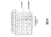
本明細書に記述した実施形態の一部の態様では、送達装置は、液体担体媒体中の一つま
たは複数の薬剤と流体連通している送達構造2~50,000個を備えるアレイを含み、
送達装置は投与流量を制御する手段を備え、送達構造は少なくとも表皮の最表層を透過す
るための手段を備え、液体担体媒体中の一つまたは複数の薬剤は送達構造によって被験者
の生育可能な表皮内の複数の深さに送達され、それによって一つまたは複数の薬剤の2~
50,000部分用量を投与しうる。In some aspects of the presently described embodiments, the delivery device comprises an array comprising 2-50,000 delivery structures in fluid communication with one or more agents in a liquid carrier medium;
The delivery device comprises means for controlling the rate of administration, the delivery structure comprises means for penetrating at least the superficial layers of the epidermis, and the one or more agents in the liquid carrier medium are delivered to the subject's viable epidermis by the delivery structure. are delivered to multiple depths within the body, whereby one or more agents are delivered from two to
50,000 sub-doses may be administered.
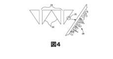
本明細書に記述した実施形態の一部の態様では、送達構造は標準的または非標準的幾何
学形状を備える。In some aspects of the embodiments described herein, the delivery structure comprises standard or non-standard geometries.
本明細書に記述した実施形態の一部の態様では、送達構造はニードルを備える。 In some aspects of the embodiments described herein, the delivery structure comprises a needle.
本明細書に記述した実施形態の一部の態様では、一つまたは複数の薬剤は、送達構造あ
たり約0.01μl/時間~約100μl/時間の制御投与流量で投与される。In some aspects of the embodiments described herein, one or more agents are administered at a controlled dose rate of from about 0.01 μl/hour to about 100 μl/hour per delivery structure.
本明細書に記述した実施形態の一部の態様では、表皮内の複数の深さへの一つまたは複
数の薬剤の全体的な制御投与流量は、被験者の皮膚と接触している送達装置の合計表面積
に基づいて、約0.02μl/時間/cm2~約50,000μl/時間/cm2である
。In some aspects of the embodiments described herein, the overall controlled administration rate of one or more agents to multiple depths within the epidermis is controlled by the delivery device in contact with the subject's skin. Based on total surface area, from about 0.02 μl/hr/cm2 to about 50,000 μl/hr/cm2 .
本明細書に記述した実施形態の一部の態様では、一つまたは複数の薬剤は、約0.7m
m3~約2,500mm3の表皮内の一つまたは複数の薬剤のその後の拡散または移動前
の一つまたは複数の薬剤を取り囲む表皮の組織容積に送達される。In some aspects of the embodiments described herein, the one or more agents are about 0.7m
m3 to about 2,500 mm3 of tissue volume of the epidermis surrounding the agent(s) prior to subsequent diffusion or migration of the agent(s) within the epidermis.
本明細書に記述した実施形態の一部の態様では、一つまたは複数の薬剤は、約0.1時
間~約96時間の間、被験者に連続的に投与される。In some aspects of the embodiments described herein, one or more agents are administered to the subject continuously for between about 0.1 hours and about 96 hours.
本明細書に記述した実施形態の一部の態様では、一つまたは複数の透過性促進剤は、一
つまたは複数の化学的、物理的、または電気的透過性促進剤である。In some aspects of the embodiments described herein, the one or more permeability enhancers are one or more chemical, physical, or electrical permeability enhancers.
本明細書に記述した実施形態の一部の態様では、物理的透過性促進剤はナノ構造または
ナノトポグラフィー表面を備える。In some aspects of the embodiments described herein, the physical permeability enhancer comprises a nanostructured or nanotopographic surface.
本明細書に記述した実施形態の一部の態様では、ナノトポグラフィー表面は、本明細書
に記述した送達構造の表面上に作製される。In some aspects of the embodiments described herein, nanotopographic surfaces are created on the surfaces of the delivery structures described herein.
本明細書に記述した実施形態の一部の態様では、皮膚内の複数の深さに投与された一つ
または複数の薬剤は、表皮を通り表皮の基底層を通って、下にある生育可能な真皮の少な
くとも一部分へと深く移動または拡散する。In some aspects of the embodiments described herein, the one or more agents administered to multiple depths within the skin pass through the epidermis through the basal layer of the epidermis to the underlying viable migrate or diffuse deep into at least a portion of the dermis.
本明細書に記述した実施形態の一部の態様では、一つまたは複数の薬剤の投与は、下に
ある真皮の約1mmHg~約15mmHgの皮膚間質液圧力を達成する。In some aspects of the embodiments described herein, administration of one or more agents achieves a skin interstitial fluid pressure of about 1 mmHg to about 15 mmHg of the underlying dermis.
本明細書に記述した実施形態の一部の態様では、一つまたは複数の薬剤は、表皮への送
達後、一つまたは複数の感受性毛細リンパ管叢によって吸収される。In some aspects of the embodiments described herein, the one or more agents are absorbed by one or more sensitive lymphatic capillary plexuses after delivery to the epidermis.
本明細書に記述した実施形態の一部の態様では、一つまたは複数の薬剤は、毛細リンパ
管叢およびリンパ管構造を通って、一つまたは複数のリンパ組織へと循環する。In some aspects of the embodiments described herein, the one or more agents circulate through the lymphatic plexus and lymphatic structures to one or more lymphoid tissues.
本明細書に開示した実施形態の一部の態様では、一つまたは複数のリンパ組織は一つま
たはリンパ節を含む。In some aspects of the presently disclosed embodiments, the one or more lymphoid tissues comprises one or lymph nodes.
本明細書に開示した実施形態の一部の態様では、一つまたは複数のリンパ節中の一つま
たは複数の薬剤はリンパ節組織のグラムあたりの初期投投薬量の約0.5%~約30%で
ある。In some aspects of the presently disclosed embodiments, the one or more agents in the one or more lymph nodes is about 0.5% to about 0.5% of the initial dosage per gram of lymph node tissue. 30%.
本明細書に記述した実施形態の一部の態様では、一つまたは複数の感受性リンパ組織内
の一つまたは複数の薬剤の濃度は、同一の一つまたは複数の薬剤の静脈内、皮内、または
皮下送達よりも約1.25倍~約50倍高い。In some aspects of the embodiments described herein, the concentration of the one or more agents within the one or more susceptible lymphoid tissues is the same one or more agents intravenously, intradermally, or about 1.25 times to about 50 times higher than subcutaneous delivery.
本明細書に記述した実施形態の一部の態様では、一つまたは複数の薬剤の血清吸収速度
は、一つまたは複数の薬剤の皮内送達および皮下送達と同等である。In some aspects of the embodiments described herein, the serum absorption rate of the one or more agents is comparable to intradermal and subcutaneous delivery of the one or more agents.
本明細書に記述した実施形態の一部の態様では、リンパ節組織に対する全血液組織のグ
ラムあたりの局在化した一つまたは複数の薬剤の初期用量の比率は約36時間後に約5:
1~約15:1である。In some aspects of the embodiments described herein, the ratio of the initial dose of the one or more agents localized per gram of whole blood tissue to lymph node tissue is about 5 after about 36 hours:
1 to about 15:1.
本明細書に記述した実施形態の一部の態様では、リンパ節組織に対する皮膚組織のグラ
ムあたりの局在化した一つまたは複数の薬剤の初期用量の比率は約36時間後に約0.5
:1~約1:1である。In some aspects of the embodiments described herein, the ratio of the initial dose of the one or more agents localized per gram of skin tissue to lymph node tissue is about 0.5 after about 36 hours.
:1 to about 1:1.
本明細書に記述した実施形態の一部の態様では、一つまたは複数の薬剤は生物活性薬剤
を含む。In some aspects of the embodiments described herein, the one or more agents comprise bioactive agents.
本明細書に記述した実施形態の一部の態様では、生物活性薬剤は、その治療を必要とす
る患者の疾患を治療する、その進行を遅らせる、発症を遅延する、その予防、その寛解、
または症状を減少するために有用である。In some aspects of the embodiments described herein, the bioactive agent treats, slows progression of, delays onset of, prevents, ameliorates, or treats a disease in a patient in need thereof.
or useful for reducing symptoms.
薬剤がリンパ組織によって制御可能かつ再現性良く吸収される、被験者への生物活性薬
剤の制御送達方法に対するニーズがある。従って、本明細書には、皮膚への一つまたは複
数の薬剤の制御送達の方法が記述されている。本明細書に記述した制御送達方法は、リン
パ組織、特に生育可能な皮膚内の一つまたは複数の感受性毛細リンパ管叢による、一つま
たは複数の薬剤の取り込みを提供する。There is a need for a method of controlled delivery of bioactive agents to a subject in which the agents are controllably and reproducibly absorbed by lymphoid tissue. Accordingly, described herein are methods for controlled delivery of one or more agents to the skin. The controlled delivery methods described herein provide for uptake of one or more agents by one or more sensitive lymphatic capillary plexuses within lymphoid tissue, particularly viable skin.
「生育可能な皮膚」という用語は本明細書で使用する場合、真皮を含む表皮の角質層の
すぐ下にあるが、皮下組織層より上にある皮膚の領域を指す。この用語は生育可能な表皮
および生育可能な真皮の両方を包含する。生育可能な皮膚の実際の深さは、皮膚の場所、
年齢、および所与の被験者の生理機能に応じて異なる。生育可能な皮膚という用語は、皮
膚のこの部分が有核生細胞、しばしば有糸分裂を含むことをさらに指定する。本明細書に
記述した一部の態様では、生育可能な皮膚は、少なくとも一つまたは複数の毛細リンパ管
叢および/または一つまたは複数の毛細血管叢も備える。The term "viable skin" as used herein refers to the area of skin that is just below the stratum corneum layer of the epidermis, including the dermis, but above the subcutaneous tissue layer. The term includes both viable epidermis and viable dermis. The actual depth of viable skin depends on the location of the skin,
Varies depending on age and physiology of a given subject. The term viable skin further specifies that this part of the skin contains nucleated living cells, often mitosis. In some aspects described herein, viable skin also comprises at least one or more lymphatic and/or one or more capillary plexuses.
「生育可能な真皮」という用語は本明細書で使用する場合、表皮の基底層のすぐ下にあ
るが、皮下組織層より上にある皮膚の領域を指す。生育可能な真皮は、真皮の乳頭および
網状層の両方を備え、さらに例えば、その他の組織タイプの中でも特に毛細血管および毛
細リンパ管を備える。The term "viable dermis" as used herein refers to the area of skin immediately below the basal layer of the epidermis but above the subcutaneous tissue layer. The viable dermis comprises both the papillary and reticular layers of the dermis and, for example, capillaries and lymphatic vessels, among other tissue types.
「生育可能な表皮」という用語は本明細書で使用する場合、角質層のすぐ下にある皮膚
の領域を指す。生育可能な表皮は、基底層または胚芽層、扁平上皮細胞層または有棘層お
よび顆粒細胞層または顆粒層を備える。The term "viable epidermis" as used herein refers to the area of skin just below the stratum corneum. The viable epidermis comprises a basal or germinal layer, a squamous cell layer or spinous layer and a granular cell layer or stratum granulosum.
「薬剤」という用語は本明細書で使用する場合、送達される化合物、物質、組成物、ま
たは分子を指す。例示的および非限定的例には、生物活性薬剤、核酸(例えば、マイクロ
RNA)、染料(例えば、造影剤および蛍光レポーター)、ワクチンなどが含まれる。The term "agent" as used herein refers to a compound, substance, composition, or molecule to be delivered. Illustrative and non-limiting examples include bioactive agents, nucleic acids (eg, microRNA), dyes (eg, imaging agents and fluorescent reporters), vaccines, and the like.
「生物活性薬剤」という用語は本明細書で使用する場合、細胞応答を引き出す任意の生
体適合性薬剤を指す。生物活性薬剤という用語には、任意の薬物、活性成分、活性原薬、
またはワクチンを含む。例えば、本明細書の実施形態に記述した生物活性薬剤には、小分
子薬物、バイオシミラー薬物、生物製剤などの薬物、ナノ粒子、脂質、リポソーム、タン
パク質(例えば、組み換えタンパク質、抗体など)および同種のものが含まれうる。The term "bioactive agent" as used herein refers to any biocompatible agent that elicits a cellular response. The term bioactive agent includes any drug, active ingredient, active drug substance,
or including vaccines. For example, bioactive agents described in the embodiments herein include drugs such as small molecule drugs, biosimilar drugs, biologics, nanoparticles, lipids, liposomes, proteins (e.g., recombinant proteins, antibodies, etc.) and The same kind can be included.
「薬物」、「活性成分」、「活性原薬」、または「活性薬剤」という用語は本明細書で
使用する場合、しばしば有益な薬理効果を提供する活性成分、化合物、または物質、組成
物、またはそれらの混合物を指す。特定の活性成分への言及には、該当する場合、活性成
分およびその薬学的に許容可能な塩またはエステルのすべてが含まれる。「投薬量」また
は「用量」という用語は、単回投与で治療効果を生じるのに十分な量を含む活性成分製剤
の任意の形態を示す。The terms "drug", "active ingredient", "active drug substance", or "active agent" as used herein often refer to an active ingredient, compound or substance, composition that provides a beneficial pharmacological effect, or any mixture thereof. A reference to a specific active ingredient includes all active ingredients and their pharmaceutically acceptable salts or esters, as applicable. The terms "dosage" or "dosage" refer to any form of active ingredient formulation containing a sufficient amount to produce a therapeutic effect in a single administration.
「タイトレーション」という用語は本明細書で使用する場合、最適治療効果を提供する
レベルへの薬物投薬量または投与速度の増加を指す。The term "titration," as used herein, refers to an increase in drug dosage or rate of administration to levels that provide optimal therapeutic effect.
「制御送達」という用語は本明細書で使用する場合、所望の時間にわたって一つまたは
複数の薬剤の制御可能な送達をもたらす投与方法を指す。The term "controlled delivery," as used herein, refers to methods of administration that result in controllable delivery of one or more agents over a desired period of time.
「制御送達」は本明細書で使用する場合、「修正送達」、「持続性送達」、「延長送達
」および「遅延送達」という用語を包含する。本明細書に記述した一部の態様では、制御
送達方法は、最大時間、治療閾値を達成するための、一つまたは複数の薬剤または活性原
薬の送達をもたらす。"Controlled delivery" as used herein encompasses the terms "modified delivery", "sustained delivery", "extended delivery" and "delayed delivery". In some aspects described herein, the controlled delivery method results in delivery of one or more drugs or active drug substances to achieve a therapeutic threshold for a maximum amount of time.
「遅延送達」という用語は本明細書で使用する場合、生理学的条件下またはインビトロ
試験で、長期間にわたって望ましいプロファイルに従って一つまたは複数の薬剤を送達す
ることを指す。「長期間」とは、少なくとも約20分、約30分、約1時間、約2時間、
約4時間、約6時間、約8時間、約10時間、約12時間、約14時間、約16時間、約
18時間、約20時間、約24時間、またはそれより長い時間の連続的期間を意味する。The term "delayed delivery" as used herein refers to delivery of one or more agents according to a desired profile over an extended period of time under physiological conditions or in vitro tests. "Long term" means at least about 20 minutes, about 30 minutes, about 1 hour, about 2 hours,
continuous periods of about 4 hours, about 6 hours, about 8 hours, about 10 hours, about 12 hours, about 14 hours, about 16 hours, about 18 hours, about 20 hours, about 24 hours, or longer means.
「修正送達」という用語は本明細書で使用する場合、生理学的条件下またはインビトロ
試験で、一つまたは複数の薬剤を、即時送達製剤よりも遅い速度で送達することを指す。The term "modified delivery" as used herein refers to delivery of one or more agents at a slower rate than an immediate delivery formulation under physiological conditions or in vitro tests.
「持続性送達」という用語は本明細書で使用する場合、活性成分が部分的に初期に放出
されるように、例えば、数分、数時間、または数日などの長期間にわたって一つまたは複
数の薬剤を送達することを指す。持続性放出速度は、例えば、生理学的条件下またはイン
ビトロ試験の、一定時間にわたる一つまたは複数の薬剤または活性原薬のある特定量の送
達を提供しうる。The term "sustained delivery" as used herein means that the active ingredient is partially released initially, e.g. refers to the delivery of drugs for Sustained release rates can provide delivery of certain amounts of one or more drugs or active drug substances over a period of time, eg, under physiological conditions or in vitro tests.
「延長送達」という用語は本明細書で使用する場合、少なくとも約20分、約30分、
約1時間、約2時間、約4時間、約6時間、約8時間、約10時間、約12時間、約14
時間、約16時間、約18時間、約20時間、約24時間、約48時間、約72時間、ま
たはそれより長い時間など、長期間にわたる一つまたは複数の薬剤の送達を指す。The term "extended delivery" as used herein is at least about 20 minutes, about 30 minutes,
About 1 hour, about 2 hours, about 4 hours, about 6 hours, about 8 hours, about 10 hours, about 12 hours, about 14 hours
Refers to delivery of one or more agents over an extended period of time, such as hours, about 16 hours, about 18 hours, about 20 hours, about 24 hours, about 48 hours, about 72 hours, or longer.
「初期送達」または「初期に送達された」という用語は、薬剤が最初に接触する組織の
場所を指す。本明細書に記述した一部の態様では、初期送達は、一つまたは複数の薬剤が
送達装置または送達装置の一つまたは複数の送達構造を通して送達された後、最初に接触
する皮膚(例えば、生育不能表皮、生育可能な表皮、または生育可能な真皮)内の場所を
指す場合がある。The terms "initial delivery" or "initially delivered" refer to the tissue location where the agent first contacts. In some aspects described herein, initial delivery refers to the skin (e.g., non-viable epidermis, viable epidermis, or viable dermis).
本明細書で使用する場合、「従来型送達」とは、静脈内(i.v.)、イオン導入、皮
下(s.c.)、筋肉内(i.m.)、または皮内(i.d.)注射、または局所製剤と
類似の生物学的動力学または活性を持つ、一つまたは複数の物質を送達するために本技術
分野で使用される、本発明前の任意の方法を意味する。例示的方法には、US 5,80
0,420、US 20050180952、ならびにXie et al., Exp
ert Opin. Drug Deliv., 6(8), 785-792 (20
09)、ZhangおよびWei-Yue., Cancer Biol Med.,
(11), 247-254 (2014)(これらのそれぞれは、従来型送達方法の一
般的記述に関して、参照により本明細書に組み込まれる)に記述されたものなど、皮下、
イオン導入、および皮内送達方法が含まれる。As used herein, "conventional delivery" includes intravenous (i.v.), iontophoresis, subcutaneous (s.c.), intramuscular (i.m.), or intradermal (i.m.) d.) Any method, prior to the invention, used in the art to deliver one or more substances with similar biological kinetics or activity to injections or topical formulations do. Exemplary methods include US 5,80
0,420, US 20050180952, and Xie et al. , Exp
ert Opin. Drug Deliv. , 6(8), 785-792 (20
09), Zhang and Wei-Yue. , Cancer Biol Med. ,
(11), 247-254 (2014), each of which is incorporated herein by reference for a general description of conventional delivery methods;
Included are iontophoresis, and intradermal delivery methods.
「BCSクラスI、II、II、またはIV」という用語は、化合物または活性原薬が
高いまたは低い透過性および高いまたは低い溶解性(例えば、溶解が不十分)を持つかど
うかを指す。BCSクラスI薬物は高い透過性および高い溶解性を持ち、BCSクラスI
I薬物は高い透過性および低い溶解性を持ち、BCSクラスIII薬物は低い透過性およ
び高い溶解性を持ち、BCSクラスIV薬物は低い透過性および低い溶解性を持つ。即時
放出原薬は、最高用量強度が、37±1℃、1~7.5のpH範囲で250mL以下の水
性媒体中に溶ける時、高度に可溶性と見なされる。pH-溶解性プロファイルを正確に規
定するためには、十分な数のpH条件を評価すべきである。胃腸管での不安定性を示す証
拠がない場合、即時放出原薬は、物質収支測定に基づいてまたは静脈内参照用量と比較し
て、ヒトでの吸収の程度が投与用量の90%以上である時、高度に透過性と見なされる。
透過性は物質収支、絶対バイオアベイラビリティ、または腸灌流アプローチを使用して決
定できる。1つの方法で透過性分類を確定的に決定できない時は、2つの異なる方法を使
用するのが得策でありうる。製剤は、USP装置Iを使用して100rpm(または装置
IIを使用して50rpm)で、以下の媒体のそれぞれの900ml以下の容量に、原薬
の表示量の85%以上が30分以内に溶解する時、迅速溶解性と見なされる:(1)0.
1 N HClまたは酵素を含まないUSP人工胃液、(2)pH 4.5緩衝液、およ
び(3)pH 6.8緩衝液または酵素を含まないUSP人工腸液。業界用FDAガイド
ラインを参照のこと:Waiver of In Vivo Bioavailabil
ity and Bioequivalence Studies for Immed
iate-Release Solid Oral Dosage Forms Bas
ed on a Biopharmaceutics Classification
System. (2000年8月)、これはこのような教示について参照によって本明
細書に組み込まれる。The term "BCS Class I, II, II, or IV" refers to whether a compound or active drug substance has high or low permeability and high or low solubility (eg, poor solubility). BCS Class I drugs have high permeability and high solubility, and BCS Class I
I drugs have high permeability and low solubility, BCS class III drugs have low permeability and high solubility, and BCS class IV drugs have low permeability and low solubility. An immediate release drug substance is considered highly soluble when the highest dose strength is soluble in 250 mL or less of an aqueous medium at 37±1° C. and a pH range of 1-7.5. A sufficient number of pH conditions should be evaluated to accurately define the pH-solubility profile. In the absence of evidence of instability in the gastrointestinal tract, an immediate-release drug substance has an extent of absorption in humans of ≥90% of the administered dose based on mass balance measurements or compared to an intravenous reference dose. Sometimes considered highly permeable.
Permeability can be determined using mass balance, absolute bioavailability, or intestinal perfusion approaches. When one method cannot definitively determine the permeability classification, it may be advisable to use two different methods. The formulation was dissolved at 100 rpm using USP Apparatus I (or 50 rpm using Apparatus II) in a volume of 900 ml or less of each of the following media with 85% or more of the labeled amount of drug substance dissolved within 30 minutes: It is considered fast dissolving when: (1)0.
1 N HCl or enzyme-free USP simulated gastric fluid, (2) pH 4.5 buffer, and (3) pH 6.8 buffer or enzyme-free USP simulated intestinal fluid. See FDA guidelines for industry: Waiver of In Vivo Bioavailability
Nature and Bioequivalence Studies for Immediate
iate-Release Solid Oral Dosage Forms Bass
Ed on a Biopharmaceutics Classification
System. (August 2000), which is incorporated herein by reference for such teachings.
本明細書で使用する場合、「バイオアベイラビリティ」とは、血液コンパートメントに
達する、所与投薬量の投与薬剤の合計量を意味する。これは一般的に、濃度・時間のプロ
ットの曲線下面積として測定される。As used herein, "bioavailability" means the total amount of administered drug at a given dose that reaches the blood compartment. It is commonly measured as the area under the curve of a concentration versus time plot.
本明細書で使用する場合「組織」とは、皮膚組織、リンパ組織(例えば、リンパ節)、
粘膜組織、生殖組織、頸部組織、膣組織を含むがこれらに限定されない、機能を一緒に実
施する細胞の群または層、および肺、脾臓、結腸、胸腺を含むがこれらに限定されない、
異なるタイプの組織から成り、特定の機能を実施する体の任意の部分(すなわち、器官)
を指す。本明細書で使用する場合、組織には、環境(例えば、皮膚または粘膜組織)と相
互作用する、または環境にアクセスできる任意の組織が含まれる。As used herein, "tissue" includes skin tissue, lymphatic tissue (e.g., lymph nodes),
Groups or layers of cells that together perform a function, including but not limited to mucosal tissue, reproductive tissue, cervical tissue, vaginal tissue, and lung, spleen, colon, thymus, including but not limited to;
Any part of the body (i.e., organ) that consists of different types of tissue and performs a specific function
point to As used herein, tissue includes any tissue that interacts with or has access to the environment (eg, skin or mucosal tissue).
本明細書で使用する場合、「組織バイオアベイラビリティ」とは、特定の組織のインビ
ボで生物学的に利用できる薬剤の量を意味する。これらの量は、結合、標識化、検出、輸
送、安定性、生物学的効果、または診断および/もしくは療法のために有用なその他の測
定可能な特性に関連しうる活動として一般的に測定される。さらに、「組織バイオアベイ
ラビリティ」は、特定組織での使用に利用可能な薬剤の量も含むことが理解される。「組
織バイオアベイラビリティ」には、特定組織に蓄積された薬剤の合計量、特定組織に提示
される薬剤の量、特定組織の質量/容積あたり蓄積された薬剤の量、特定組織の特定質量
/容積に単位時間あたり蓄積された薬剤の量を含む。組織バイオアベイラビリティには、
脈管構造および/または体の様々な器官を作るもの(例えば、リンパ管構造または血液な
ど、異なるタイプの組織から成り、特定機能を実行する体の部分)など、特定組織または
組織の一群中のインビボで利用可能な薬剤の量を含む。As used herein, "tissue bioavailability" means the amount of a drug that is bioavailable in vivo in a particular tissue. These amounts are generally measured as activities that can be associated with binding, labeling, detection, transport, stability, biological effect, or other measurable properties useful for diagnosis and/or therapy. be. Further, "tissue bioavailability" is understood to include the amount of drug available for use in a particular tissue. "Tissue bioavailability" includes total amount of drug accumulated in a specific tissue, amount of drug presented to a specific tissue, amount of drug accumulated per mass/volume of a specific tissue, specific mass/volume of a specific tissue contains the amount of drug accumulated per unit time. Tissue bioavailability includes:
A particular tissue or group of tissues, such as the vasculature and/or those that make up the various organs of the body (e.g., parts of the body that are composed of different types of tissue and perform a particular function, such as lymphatic structures or blood) Including amount of drug available in vivo.
「Cmax」という用語は本明細書で使用する場合、最大観察血液(血漿、血清、また
は全血)濃度または濃度・時間曲線から計算もしくは予測された最大血液濃度を指し、該
当する場合、mg/Lまたはng/mLの単位で表される。The term “Cmax ” as used herein refers to the maximum observed blood (plasma, serum, or whole blood) concentration or the maximum blood concentration calculated or predicted from a concentration-time curve, and where applicable, mg expressed in units of /L or ng/mL.
「Cmin」という用語は本明細書で使用する場合、最小観察血液(血漿、血清、また
は全血)濃度または濃度・時間曲線から計算もしくは予測された最小血液濃度を指し、該
当する場合、mg/Lまたはng/mLの単位で表される。The term “Cmin ” as used herein refers to the minimum observed blood (plasma, serum, or whole blood) concentration or the minimum blood concentration calculated or predicted from a concentration-time curve, and where applicable, mg expressed in units of /L or ng/mL.
「Cavg」という用語は本明細書で使用する場合、投与間隔内の薬物の血液(血漿、
血清、または全血)濃度を指し、AUC/投与間隔として計算され、該当する場合、mg
/Lまたはng/mLの単位で表される。The term “Cavg ” as used herein refers to blood (plasma,
(serum, or whole blood) concentration, calculated as AUC/dose interval, mg when applicable
expressed in units of /L or ng/mL.
「Tmax」という用語は本明細書で使用する場合、Cmaxが起こる、投与後の時間
を指し、該当する場合、時間(h)または分(min)の単位で表される。The term "Tmax " as used herein refers to the time after administration at which Cmax occurs, expressed in units of hours (h) or minutes (min), as applicable.
「AUC0→τ」という用語は本明細書で使用する場合、定常状態の投与間隔にわたる
時間ゼロから時間タウ(τ)までの血液(血漿、血清、または全血)濃度・時間曲線下面
積を指し、該当する場合、h・mg/Lまたはh・ng/mLの単位で表される。例えば
、用語AUC0→12は本明細書で使用する場合、0~12時間の濃度・時間曲線下面積
を指す。The term “AUC0→τ ” as used herein is the area under the blood (plasma, serum, or whole blood) concentration-time curve from time zero to time tau (τ) over the steady-state dosing interval. expressed in units of h·mg/L or h·ng/mL as applicable. For example, the term AUC0→12 as used herein refers to the area under the concentration-time curve from 0 to 12 hours.
「AUC0→∞」という用語は本明細書で使用する場合、時間0から無限大までの血液
(血漿、血清、または全血)濃度・時間曲線下面積を指し、該当する場合、h・mg/L
またはh・ng/mLの単位で表される。The term "AUC0→∞ " as used herein refers to the area under the blood (plasma, serum, or whole blood) concentration-time curve from
Or expressed in units of h·ng/mL.
「AUCoverall」という用語は本明細書で使用する場合、血液(血漿、血清、
または全血)濃度・時間曲線下複合面積を指し、本明細書に記述した医薬組成物の少なく
とも一つまたは複数の用量に対してh・mg/L(またはh・ng/mL)の単位で表さ
れる。一態様では、「AUCoverall」は、本明細書に記述した医薬組成物の少な
くとも2用量に対する血液濃度・時間曲線下複合面積を指す。The term "AUCoverall " as used herein means blood (plasma, serum,
or whole blood) refers to the combined area under the concentration-time curve, in units of h·mg/L (or h·ng/mL) for at least one or more doses of the pharmaceutical compositions described herein. expressed. In one aspect, "AUCoverall " refers to the combined area under the blood concentration-time curve for at least two doses of the pharmaceutical compositions described herein.
「治療する」という用語は、障害に関連する状態、症状、またはパラメータを改善する
ために有効な量、方法、またはモードで療法を実施することを指す。The term "treating" refers to administering therapy in an effective amount, method, or mode to ameliorate the condition, symptoms, or parameters associated with the disorder.
「予防」という用語は、障害の進行を、統計的に有意な程度まで、または当業者に検出
可能な程度まで防止または減少することを指す。The term "prevention" refers to preventing or reducing the progression of a disorder to a degree that is statistically significant or detectable by one of ordinary skill in the art.
「実質的に」という用語は本明細書で使用する場合、大きなまたは有意な程度であるが
完全にではないことを意味する。一部の態様では、実質的にとは、本明細書に記述した様
々な実施形態の90%~99%を意味し、指定範囲内の各整数を含む。The term "substantially" as used herein means to a large or significant degree but not completely. In some aspects, substantially refers to 90% to 99% of the various embodiments described herein, including each integer within the specified range.
本明細書で使用する場合、「被験者」および「患者」という用語は互換的に使用される
。本明細書で使用する場合、被験者は、非霊長類(例えば、ウシ、ブタ、ウマ、ネコ、イ
ヌ、ラット、ウサギなど)または霊長類(例えば、サルおよびヒト)などの哺乳類である
ことが好ましい。As used herein, the terms "subject" and "patient" are used interchangeably. As used herein, the subject is preferably a mammal, such as a non-primate (e.g., bovine, porcine, equine, feline, canine, rat, rabbit, etc.) or primate (e.g., monkey and human). .
本明細書で使用する場合、「障害」および「疾患」という用語は被験者の状態を指すた
めに互換的に使用される。疾患には、身体機能、システムまたは器官の中断、中止、また
は障害が含まれる。As used herein, the terms "disorder" and "disease" are used interchangeably to refer to the condition of a subject. Diseases include disruption, cessation, or impairment of bodily functions, systems, or organs.
本明細書で使用される場合、「治療する」、「治療すること」および「治療」という用
語は、疾患または障害の症状の根絶、減少または寛解を指す。一部の実施形態では、治療
とは、一つまたは複数の治療薬の投与から生じる原発性、領域性、または転移性癌組織の
根絶、除去、変更または制御を指す。特定の実施形態では、このような用語は、このよう
な疾患を持つ被験者への一つまたは複数の治療薬の投与から生じる、癌の広がりの最小化
または遅延を指す。As used herein, the terms "treat", "treating" and "treatment" refer to eradication, reduction or amelioration of the symptoms of a disease or disorder. In some embodiments, treatment refers to eradication, removal, alteration or control of primary, regional, or metastatic cancerous tissue resulting from administration of one or more therapeutic agents. In certain embodiments, such terms refer to minimizing or delaying the spread of cancer resulting from administration of one or more therapeutic agents to a subject with such disease.
本明細書で使用する場合、「管理する」、「管理すること」、および「管理」という用
語は、被験者が予防薬または治療薬の投与から得る有益な効果であって、疾患の治癒をも
たらさないものを指す。特定の実施形態では、被験者は、疾患の進行または悪化を防止す
るために、一つまたは複数の予防薬または治療薬を投与して「管理」する。As used herein, the terms "manage,""managing," and "management" refer to the beneficial effects that a subject obtains from administration of a prophylactic or therapeutic agent that results in cure of a disease. pointing to nothing. In certain embodiments, the subject is "managed" by administering one or more prophylactic or therapeutic agents to prevent progression or worsening of the disease.
本明細書で使用する場合、「予防する」、「予防すること」および「予防」という用語
は、予防薬または治療薬の投与からもたらされる、被験者の障害の一つまたは複数の症状
の再発または発症の防止を指す。As used herein, the terms "prevent,""preventing," and "prevention" refer to recurrence or recurrence of one or more symptoms of a disorder in a subject resulting from administration of a prophylactic or therapeutic agent. Refers to prevention of onset.
本明細書で使用する場合、「副作用」という語句は、予防薬または治療薬の望ましくな
い有害作用を包含する。有害作用はいつも望ましくないが、望ましくない作用が必ずしも
有害なわけではない。予防薬または治療薬からの有害作用は、有害または不快または危険
でありうる。化学療法からの副作用には、早発性および遅発性の下痢および鼓腸などであ
るがこれらに限定されない胃腸毒性、悪心、嘔吐、摂食障害、白血球減少、貧血、好中球
減少、無力、腹部痙攣、発熱、疼痛、体重減少、脱水、脱毛、呼吸困難、不眠、めまい、
粘膜炎、口内乾燥、および腎不全、ならびに便秘、神経および筋肉への影響、一時的また
は永久的な腎臓および膀胱への損傷、インフルエンザ様症状、水分貯留、および一時的ま
たは永久的な不妊が含まれるがこれらに限定されない。放射線療法からの副作用には、疲
労、口内乾燥、食欲不振が含まれるがこれらに限定されない。生物学的療法/免疫療法か
らの副作用には、投与部位の発疹または腫れ、発熱、悪寒および疲労などのインフルエン
ザ様症状、消化管の問題ならびにアレルギー反応が含まれるがこれらに限定されない。ホ
ルモン療法からの副作用には、悪心、不妊の問題、うつ病、食欲不振、眼の問題、頭痛、
および体重変動が含まれるがこれらに限定されない。患者が典型的に経験する追加的な望
ましくない作用は多くあり、当技術分野で知られている。例えば、Physicians
' Desk Reference (69th ed., 2015)(これは参照に
よりその全体が本明細書に組み込まれる)を参照のこと。As used herein, the phrase "side effects" includes unwanted adverse effects of prophylactic or therapeutic agents. Adverse effects are always unwanted, but unwanted effects are not necessarily adverse. Adverse effects from prophylactic or therapeutic agents can be harmful or unpleasant or dangerous. Side effects from chemotherapy include, but are not limited to, gastrointestinal toxicity, nausea, vomiting, eating disorders, leukopenia, anemia, neutropenia, asthenia, early-onset and late-onset diarrhea and flatulence. Abdominal cramps, fever, pain, weight loss, dehydration, hair loss, dyspnea, insomnia, dizziness,
Includes mucositis, dry mouth, and renal failure, as well as constipation, nerve and muscle effects, temporary or permanent kidney and bladder damage, flu-like symptoms, water retention, and temporary or permanent infertility. include but are not limited to: Side effects from radiation therapy include, but are not limited to, fatigue, dry mouth, and loss of appetite. Side effects from biologic therapy/immunotherapy include, but are not limited to, rash or swelling at the site of administration, flu-like symptoms such as fever, chills and fatigue, gastrointestinal problems and allergic reactions. Side effects from hormone therapy include nausea, fertility problems, depression, loss of appetite, eye problems, headaches,
and weight fluctuations. Additional undesirable effects typically experienced by patients are numerous and known in the art. For example, Physicians
See 'Desk Reference (69th ed., 2015), which is incorporated herein by reference in its entirety.
本明細書で使用する場合、「感受性組織への送達」または「生育可能な組織への送達」
という語句は、例えば、皮膚、血管構造、リンパ管構造、およびリンパ節など、生きた組
織または組織構造への一つまたは複数の薬剤の送達を指す。本明細書に記述した一部の実
施形態では、本明細書にさらに記述した方法、組成物、および装置は、生きた組織または
組織構造の構造を調整し、一つまたは複数の薬剤の吸収を促進しうる。一部の態様では、
生きた組織または組織構造は、皮膚および皮膚を含む個別の生育可能な細胞を含む。本明
細書に記述した一部の態様では、送達方法は、特定の細胞または組織(例えば、生育可能
な皮膚)を、その特定組織への一つまたは複数の薬剤の送達の影響を受けやすいように誘
導する。本明細書に記述した一部の態様では、生きた組織または組織構造は、表皮および
下にある真皮の生育可能な層など、生育可能な皮膚の一つまたは複数の層を備える。本明
細書に記述した一部の態様では、生きた組織または組織構造は、毛細リンパ管(例えば、
感受性毛細リンパ管叢への送達)を備える。As used herein, "delivery to sensitive tissue" or "delivery to viable tissue"
The phrase refers to delivery of one or more agents to living tissue or tissue structures such as, for example, skin, vasculature, lymphatic structures, and lymph nodes. In some embodiments described herein, the methods, compositions, and devices further described herein modulate the structure of living tissue or tissue structures to promote absorption of one or more agents. can be promoted. In some aspects,
A living tissue or tissue structure comprises individual viable cells, including skin and skin. In some aspects described herein, the delivery method comprises making a particular cell or tissue (e.g., viable skin) amenable to delivery of one or more agents to that particular tissue. lead to In some aspects described herein, the living tissue or tissue structure comprises one or more layers of viable skin, such as the epidermis and underlying viable layers of the dermis. In some aspects described herein, the living tissue or tissue structure is a lymphatic capillary (e.g.,
delivery to the sensitive lymphatic capillary plexus).
皮膚に薬剤を送達するための方法および装置が本明細書に記述されている。特定の実施
形態では、リンパ管構造に薬剤を送達するための方法が本明細書に記述されている。Methods and devices for delivering drugs to the skin are described herein. In certain embodiments, methods are described herein for delivering agents to lymphatic vasculature.
本明細書の実施形態には、一つまたは複数の薬剤を皮膚に送達するための方法が記述さ
れている。一部の態様では、一つまたは複数の薬剤は、生育可能または生育不能な皮膚の
少なくとも一つの部分または領域に送達される。一部の態様では、一つまたは複数の薬剤
は、生育可能な表皮の少なくとも一つの部分または領域に送達される。一部の態様では、
一つまたは複数の薬剤は、生育不能な表皮の少なくとも一つの部分または領域に送達され
る。本明細書にさらに記述するように、一つまたは複数の薬剤は生育可能な表皮を通過し
て真皮に入ることができ、それによって一つ複数の毛細血管および毛細リンパ管の近傍に
入る。Embodiments herein describe methods for delivering one or more agents to the skin. In some aspects, one or more agents are delivered to at least one portion or region of viable or non-viable skin. In some aspects, one or more agents are delivered to at least one portion or region of the viable epidermis. In some aspects,
One or more agents are delivered to at least one portion or region of the non-viable epidermis. As further described herein, one or more agents can pass through the viable epidermis and enter the dermis, thereby entering the vicinity of one or more capillaries and lymphatic capillaries.
皮膚への送達は、皮膚の機能を提供するバリアに基づいたいくつかの問題を示す。解剖
学的に、皮膚は大まかに、外側の表皮および下にある真皮の2つの主な組織層からなり、
これらが共に皮膚を構成する。より広い外皮系は、皮膚、毛、爪、外分泌腺、および皮下
組織を含む。このバリア機能の結果、送達された物質が表皮の一つまたは複数の層内に保
持されるので、表皮を通り生育可能な真皮へと入る皮膚への送達のための多くの経皮また
はマイクロニードルアプローチが不成功に終わっている。Delivery to the skin presents several problems based on the barrier that provides the skin's function. Anatomically, the skin is roughly composed of two main tissue layers, the outer epidermis and the underlying dermis,
Together they make up the skin. The broader integumentary system includes skin, hair, nails, exocrine glands, and subcutaneous tissue. As a result of this barrier function, many transdermal or microneedles for delivery to the skin pass through the epidermis into the viable dermis, as the delivered substance is retained within one or more layers of the epidermis. approach has been unsuccessful.
表皮は4つの主な層に細分される。下から上の順に、基底膜、基底層または胚芽層、鱗
状細胞層または有棘層、顆粒細胞層または顆粒層、および角化層または角質層である。こ
れらの3層のうち、下の3層(すなわち、胚芽層、有棘層、および顆粒層)が表皮の生き
た層を構成する。The epidermis is subdivided into four main layers. From bottom to top, the basement membrane, the basal or germinal layer, the squamous or spinous layer, the granulocyte or stratum granulosum, and the cornified or stratum corneum layer. Of these three layers, the lower three layers (ie, stratum germinativum, stratum spinosum, and stratum granulosum) constitute the living layers of the epidermis.
表皮の3つの生きた層は皮膚のバリア機能のために重要であり、これは基底部にある幹
細胞の自己再生および分化に依存して、皮膚の上層を再生しバリア層または角質層に除核
細胞を提供する。表皮のバリア機能は主に、高分子(例えば、タンパク質)、微生物、お
よびその他の潜在的に有毒な化学物質の通過を防止する密着結合の存在の結果である。こ
のように、これらの密着結合は、隣接した原形質膜(例えば、クローディン、オクルジン
、および接合部接着分子)および複数のプラーク・タンパク質(例えば、ZO-1、ZO
-2、ZO-3、シングリン、シンプレキン)に埋め込まれた膜貫通タンパク質のネット
ワークを含むバリア構造である。密着結合は、内部上皮(例えば、腸上皮、血液脳関門、
血管、リンパ管)を含む組織のバリアタイプのすべてのほぼすべてのタイプ、ならびに皮
膚の生育可能な表皮の至る所に見られる。The three living layers of the epidermis are critical for skin barrier function, which relies on the self-renewal and differentiation of basal stem cells to regenerate the upper layers of the skin and enucleate into the barrier or stratum corneum. Provide cells. The barrier function of the epidermis is primarily a result of the presence of tight junctions that prevent passage of macromolecules (eg, proteins), microorganisms, and other potentially toxic chemicals. Thus, these tight junctions are linked to adjacent plasma membranes (eg, claudins, occludins, and junctional adhesion molecules) and multiple plaque proteins (eg, ZO-1, ZO
-2, ZO-3, shingrin, symplekin) is a barrier structure comprising a network of transmembrane proteins embedded in the membrane. Tight junctions form internal epithelia (e.g., intestinal epithelium, blood-brain barrier,
It is found throughout almost all types of tissue barrier types, including blood vessels, lymphatics), as well as the viable epidermis of the skin.
皮膚の厚さは場所および年齢に応じて異なる。例えば、まぶたは約0.2mm未満のか
なり薄い層の表皮を持ち、手のひらおよび足の裏は1.5mm近くあるかなり厚い層の表
皮を持つ。真皮の厚さも組織の場所に応じて異なり、背中の真皮は表皮よりも30~40
倍厚い。William D. James, Timothy Berger, an
d Dirk Elston., Clinical Dermatology (1
1th ed., 2011)を参照のこと(これは参照によりその全体が本明細書に組
み込まれる)。Skin thickness varies with location and age. For example, the eyelid has a fairly thin layer of epidermis, less than about 0.2 mm, and the palms and soles have a fairly thick layer of epidermis, close to 1.5 mm. The thickness of the dermis also varies depending on the location of the tissue, with the dermis on the back being 30-40 mm thicker than the epidermis.
Double thick. William D. James, Timothy Berger, an
d Dirk Elston. , Clinical Dermatology (1
1sted . , 2011), which is incorporated herein by reference in its entirety.
表皮の下には真皮があり、これは真皮乳頭層と呼ばれる最外部および真皮網状層と呼ば
れる深層の2つの層を含む。真皮乳頭層は広大な微小循環血管叢およびリンパ管叢を含む
。対照的に、真皮網状層は比較的非細胞性であり、コラーゲン性で弾力のある密集した結
合組織で構成される。表皮および真皮の下は、下皮とも呼ばれる皮下組織であり、結合組
織および脂肪組織から成る。Physiology, Biochemistry, a
nd Molecular Biology of the Skin, Second
Edition, (L.A. Goldsmith, Ed., 2nd ed.
Oxford University Press, New York, 1991)
を参照のこと(これは参照によりその全体が本明細書に組み込まれる)。Beneath the epidermis is the dermis, which contains two layers, the outermost layer called the papillary dermis and the deeper layer called the reticular dermis. The papillary dermis contains extensive microcirculatory and lymphatic plexuses. In contrast, the reticular dermis is relatively acellular, composed of dense connective tissue that is collagenous and elastic. Beneath the epidermis and dermis is the subcutaneous tissue, also called the hypodermis, which consists of connective and fatty tissue. Physiology, Biochemistry, a
nd Molecular Biology of the Skin, Second
Edition, (LA Goldsmith, Ed., 2nded .
Oxford University Press, New York, 1991)
, which is incorporated herein by reference in its entirety.
一部の実施形態では、本明細書に記述した方法は、一つまたは複数の薬剤の一つまたは
複数のリンパ組織への送達の増加を提供する。一部の態様では、一つまたは複数の薬剤は
、リンパ管構造を通って一つまたは複数のリンパ節に移動する。本明細書にさらに記述し
たように、リンパ管構造は、毛細リンパ管、より大きなリンパ管、および集合管を構成す
るリンパ管内皮細胞のすべてを含む。リンパ管構造内の液体およびこの液体中のすべての
生体材料は、やがては一つまたは複数のリンパ節の中、最後には血流の中に排出されて、
体循環に入る。リンパ生理学の完全レビューについては、William N. Cha
rman and Valentino J. Stella, Lymphatic
Transport of Drugs (1992)を参照のこと(これは参照により
その全体が本明細書に組み込まれる)。In some embodiments, the methods described herein provide increased delivery of one or more agents to one or more lymphoid tissues. In some aspects, one or more agents travel through the lymphatic vasculature to one or more lymph nodes. As further described herein, lymphatic structures include all of the lymphatic endothelial cells that make up the lymphatic capillaries, larger lymphatic vessels, and collecting ducts. The fluid within the lymphatic vasculature and any biomaterial in this fluid eventually drains into one or more lymph nodes and finally into the bloodstream,
enter the systemic circulation. For a complete review of lymphatic physiology, see William N. et al. Cha
Rman and ValentinoJ. Stella, Lymphatic
See Transport of Drugs (1992), which is incorporated herein by reference in its entirety.
リンパ系は免疫系の一部であり、感染症および外来生物による侵入から体を守る。リン
パ球およびマクロファージは、侵入ウィルス、細菌、腫瘍細胞、異種タンパク質、毒素、
損傷および死にかけている細胞、ならびに異種組織移植片などの異種細胞を探して体の組
織のほとんどを巡回する。リンパ管はほとんどの組織と連通し、免疫細胞をリンパ節なら
びに、脾臓および胸腺などのリンパ器官に運ぶリンパ液を輸送する。リンパ管構造とも呼
ばれるリンパ管は、血管のように、体の全体にわたる組織へと枝分かれする薄い不透明な
管状構造のネットワークである。ヒトを含む哺乳類では、ほとんどの組織および器官はリ
ンパ系によって排出される。The lymphatic system is part of the immune system and protects the body from infections and invasion by foreign organisms. Lymphocytes and macrophages contain invading viruses, bacteria, tumor cells, foreign proteins, toxins,
It patrols most of the body's tissues in search of damaged and dying cells, as well as xenogeneic cells such as xenografts. Lymphatic vessels communicate with most tissues and transport lymph, which carries immune cells to lymph nodes and lymphoid organs such as the spleen and thymus. Lymphatic vessels, also called lymphatic vasculature, are networks of thin, opaque tubular structures that, like blood vessels, branch into tissues throughout the body. In mammals, including humans, most tissues and organs are drained by the lymphatic system.
循環系とは異なり、リンパ系は閉じておらず中央ポンプを持たない。リンパ系は心臓に
向かって一方向流れシステムを形成する。毛細リンパ管の精巧なネットワークは組織から
の間質液を排出し、その後、この液体はリンパ液と呼ばれる。軟骨および角膜などの多く
の結合組織は、血管を持たず、リンパ管も欠く。リンパは、蠕動収縮からゆっくりと低い
圧力下で移動する。Unlike the circulatory system, the lymphatic system is not closed and has no central pump. The lymphatic system forms a unidirectional flow system towards the heart. An elaborate network of lymphatic capillaries drains interstitial fluid from tissues, and this fluid is then called lymph. Many connective tissues, such as cartilage and cornea, are not vascularized and lack lymphatics. Lymph moves slowly under low pressure from peristaltic contractions.
これらの毛細リンパ管は10~50マイクロメートルの直径である。それらは盲嚢から
、または吻合管から始まる。内皮は、不完全な基底膜を持つ単一層である。それらは、血
漿タンパク質および炭素粒子などを含む大きな粒子、ウィルス、細菌細胞、および寄生生
物などの病原体、例えば、免疫細胞および腫瘍細胞などを含む細胞、ならびに細胞破片に
対して高度に透過性のギャップ結合を有する。毛細リンパ管は、流れが一方向のみである
ことを確実にする一方向弁を持つ。毛細リンパ管の外側の間質液の圧力が毛細管の内側の
圧力より大きい時、フラップが開いて液体が入ることを可能にする。逆に、毛細管の内側
の圧力がより大きい時、フラップは閉じることを強いられ、管からリンパが漏れ出るのを
防止する。炎症の間、毛細管は、より大きな分子および細胞破片の取り込みを可能にする
さらなる開口部を生じる。These lymphatic capillaries are 10-50 micrometers in diameter. They originate from the cecum or from the anastomosis tract. The endothelium is a single layer with an incomplete basement membrane. They are highly permeable gaps to large particles, including plasma proteins and carbon particles, pathogens such as viruses, bacterial cells, and parasites, cells including immune cells and tumor cells, and cell debris. have a bond. Lymphatic capillaries have one-way valves to ensure that flow is in one direction only. When the pressure of the interstitial fluid outside the lymphatic capillaries is greater than the pressure inside the capillaries, the flaps open to allow fluid to enter. Conversely, when the pressure inside the capillary is greater, the flap is forced closed, preventing lymph from leaking out of the vessel. During inflammation, capillaries generate additional openings that allow the uptake of larger molecules and cellular debris.
リンパは毛細管から集合リンパ管へと流れ込み、そこで多くのリンパ節の最初のものと
遭遇する。これらの「輸入」リンパ管はリンパをリンパ節に運び、「輸出」リンパ管はリ
ンパをリンパ節から運び去る。リンパは無色の水様液であり、間質液に由来する。リンパ
は循環系の毛細血管床から失われた血漿として源を発し、周辺組織に漏れ出る。循環系の
毛細管はそれらを通過して間質組織へ移動する液体の体積の約1%のみを失うが、多くの
血液が循環するので、平均的な人体の累積液体損失は1日当たり約3リットルである。リ
ンパ系は毛細リンパ管への拡散によってこの液体を取り戻し、様々なリンパ節を通してそ
れをろ過し、胸管によってそれを循環系に戻す。リンパ系内に一旦入ると、液体はリンパ
と呼ばれるが、元の間質液とほとんど同じ組成を持つ。Lymph flows from capillaries into collecting lymphatic vessels, where it encounters the first of many lymph nodes. These "afferent" lymphatic vessels carry lymph to the lymph nodes and "efferent" lymphatic vessels carry lymph away from the lymph nodes. Lymph is a colorless, watery fluid derived from interstitial fluid. Lymph originates as lost plasma from the capillary beds of the circulatory system and leaks into the surrounding tissues. Although the capillaries of the circulatory system lose only about 1% of the volume of fluid that moves through them to the interstitial tissue, much blood circulates, so the average human body's cumulative fluid loss is about 3 liters per day. is. The lymphatic system retrieves this fluid by diffusion into the lymphatic capillaries, filtering it through various lymph nodes and returning it to the circulatory system through the thoracic ducts. Once inside the lymphatic system, the fluid, called lymph, has almost the same composition as the original interstitial fluid.
毛細リンパ管は遍在し、体の至る所で見られる。このような場所の非限定的例には、生
育可能な皮膚(真皮)、腱、横紋筋、筋鞘、骨の骨膜、関節嚢、肋膜、腹膜、および心膜
腔の中皮ライニングの下、消化管、唾液腺、肝臓、脾臓、鼻腔、気管、気管支、甲状腺、
胸腺、副腎、腎臓、膀胱、尿道、前立腺、精巣、子宮、卵巣、および心臓が含まれる。Lymphatic capillaries are ubiquitous and found throughout the body. Non-limiting examples of such locations include viable skin (dermis), tendons, striated muscles, sarcolemma, periosteum of bones, joint sac, pleura, peritoneum, and under the mesothelial lining of the pericardial space. , gastrointestinal tract, salivary gland, liver, spleen, nasal cavity, trachea, bronchi, thyroid,
Includes thymus, adrenal gland, kidney, bladder, urethra, prostate, testis, uterus, ovary, and heart.
リンパ節は、細菌およびウィルスを収集して破壊するリンパ球で満たされた結合組織の
内部ハニカムでリンパをろ過する。リンパ節はリンパ球および抗体も生成する。体が感染
と戦っている時、これらのリンパ球は素早く増殖し、リンパ節の特徴的な腫脹を生じる。
リンパは、より大きなリンパ管に次第に輸送され、ついに(右上半身からのリンパについ
ては)右リンパ本幹および(体の残りについては)胸管に入る。これらの管は肩の近くの
左右の鎖骨下静脈で循環系に排出される。リンパ管のネットワークに沿って、リンパ結節
、パイエル板、扁桃腺、リンパ節、胸腺、および脾臓を含む、一連のさまざまなリンパ組
織および器官がある。Lymph nodes filter lymph through internal honeycombs of connective tissue filled with lymphocytes that collect and destroy bacteria and viruses. Lymph nodes also produce lymphocytes and antibodies. As the body fights infection, these lymphocytes proliferate quickly, resulting in the characteristic swelling of the lymph nodes.
Lymph is progressively transported to larger lymph vessels, finally entering the right lymphatic trunk (for lymph from the right upper body) and the thoracic duct (for the rest of the body). These tubes drain into the circulatory system in the left and right subclavian veins near the shoulders. Along the network of lymphatic vessels is a series of different lymphoid tissues and organs, including lymph nodes, Peyer's patches, tonsils, lymph nodes, thymus, and spleen.
リンパ結節は、感染の部位で形成しその後消失する、リンパ球の一時的集団である。結
節を周辺細胞および液体から分離する被嚢または外側被覆はなく、これは結節へと直接浸
透する。リンパ節は硬い被嚢内に多くのリンパ結節を被包し、血管およびリンパを持つ。
リンパ節は、リンパ管によってそれらに送達されたリンパをろ過する。このように、リン
パ節は、リンパ節が位置する毛細リンパ管床から排出するリンパをろ過する。パイエル板
は腸の壁にあるリンパ球の集団であり、扁桃腺は咽頭の粘膜内に包まれた結節組織の嚢で
ある。パイエル板および扁桃腺は、それぞれ消化管および気道からの抗原を途中で捕らえ
るように位置している。Lymphatic nodules are transient clusters of lymphocytes that form at sites of infection and then disappear. There is no capsule or outer covering that separates the nodule from surrounding cells and fluid, and it penetrates directly into the nodule. A lymph node encapsulates many lymph nodes within a hard capsule and has blood vessels and lymph.
Lymph nodes filter lymph delivered to them by lymph vessels. Thus, lymph nodes filter lymph that drains from the lymphatic capillary bed in which they are located. Peyer's patches are clusters of lymphocytes in the wall of the intestine, and tonsils are sacs of nodular tissue encased within the mucosa of the pharynx. The Peyer's patches and tonsils are positioned to intercept antigens from the digestive and respiratory tract, respectively.
脾臓、リンパ節、および副リンパ組織(扁桃腺および虫垂を含む)は二次リンパ器官で
ある。これらの器官は、循環BおよびTリンパ球および、例えばマクロファージ、樹状細
胞および好酸球などの、その他の免疫細胞を保持する結合組織の足場材料から成る。微生
物が体に侵入した時、または体がその他の抗原と遭遇した時、抗原は典型的には組織から
リンパに輸送される。リンパはリンパ管の中を所属リンパ節まで運ばれる。リンパ節では
、マクロファージおよび樹状細胞が抗原を貪食し、抗原を処理し、抗原をリンパ球に提示
し、次にこれは抗体を生成し始めるか、または将来再び抗原を認識するために記憶細胞と
しての役割を果たす。このように、リンパおよびリンパ組織は抗体および免疫細胞を含む
。The spleen, lymph nodes, and accessory lymphoid tissues (including tonsils and appendix) are secondary lymphoid organs. These organs consist of a connective tissue scaffold that retains circulating B and T lymphocytes and other immune cells such as macrophages, dendritic cells and eosinophils. Antigens are typically transported from tissues to the lymph when microorganisms invade the body, or when the body encounters other antigens. Lymph is carried in the lymphatic vessels to the regional lymph nodes. In lymph nodes, macrophages and dendritic cells phagocytose antigens, process antigens, present antigens to lymphocytes, which in turn begin to generate antibodies or memory cells to recognize antigens again in the future. as a role. Thus, lymph and lymphoid tissue contain antibodies and immune cells.
間質組織からの液体のリンパ吸収速度は幅広い。例えば、リンパ管によって腸の場所か
ら排除される水のパーセントは、1%~85%近くの範囲であると予測されており、その
他の予測はリンパ系が間質液の15~20%の吸収を担当していることを示す。この食い
違いは、測定されたリンパ組織の目下の生理状態による可能性が高い。There is a wide range of rates of lymphatic absorption of fluid from interstitial tissue. For example, the percentage of water excluded from intestinal sites by lymphatics is predicted to range from 1% to nearly 85%, with other projections that the lymphatic system absorbs 15-20% of interstitial fluid. indicate that you are responsible for This discrepancy is likely due to the current physiological state of the measured lymphoid tissue.
毛細リンパ管は哺乳類の皮膚の全体に渡って広く分布する。特に、指および手のひらな
らびに足および足指の足底面ならびに陰嚢など、特定の領域はリンパ管の分布が最も高い
ことが認められている。皮膚リンパは、皮膚構造の外側3分の2の部分から真皮乳頭層へ
と上向きに延びる真皮の表面リンパ管叢で主に構成される。より深い叢は、真皮網状層の
領域の皮下組織境界に近い真皮内にある。一般的に、表皮または皮下組織層内に、リンパ
組織はほとんどまたは全く見られない。リンパ管は典型的に、より厚い皮層を持つ皮膚の
領域ではより均一である(例えば、手の手掌面および足の足底面)。腸組織と同様に、皮
膚の間質液およびタンパク質の吸収は大きなばらつきがあり、例えば、動物モデルでは皮
膚領域のリンパの流れは約1ml/時間/組織100gであり、リンパ管構造を取り囲む
局所生理機能に応じて10倍以上増加しうる。静脈圧、周辺組織および血管の収縮、なら
びに呼吸数を含む様々な因子が、リンパ吸収に影響することが分かっている。例えば、ス
ターリングの式は、半透性毛細管壁を横切る静水力および膠質浸透力の競合による間質液
の生成を説明する。このように、血管内の静水圧の増加もしくは膠質浸透圧の減少、また
は毛細管透過性の増加は、間質液体積および、毛細リンパ管によるその後の液体吸収また
は浮腫を促進する傾向がある。Lymphatic capillaries are widely distributed throughout mammalian skin. In particular, certain areas such as the fingers and palms and the plantar surface of the feet and toes and the scrotum have been found to have the highest distribution of lymphatic vessels. Cutaneous lymph is primarily composed of the superficial lymphatic plexus of the dermis that extends upward from the outer two-thirds of the skin structure to the papillary dermis. The deeper plexuses are within the dermis near the subcutaneous tissue border in the area of the reticular dermis. Generally, little or no lymphoid tissue is found within the epidermis or subcutaneous tissue layers. Lymphatic vessels are typically more uniform in areas of skin with a thicker cortex (eg, the palmar surface of the hand and the plantar surface of the foot). Similar to intestinal tissue, interstitial fluid and protein absorption in the skin is highly variable, for example, in animal models, the lymphatic flow in the skin area is approximately 1 ml/h/100 g of tissue, and the local physiology surrounding the lymphatic vasculature. It can increase by a factor of 10 or more depending on the function. Various factors are known to affect lymphatic absorption, including venous pressure, constriction of surrounding tissues and blood vessels, and respiratory rate. For example, the Stirling equation describes the production of interstitial fluid by competing hydrostatic and oncotic forces across semipermeable capillary walls. Thus, an increase in intravascular hydrostatic pressure or a decrease in oncotic pressure, or an increase in capillary permeability, tends to promote interstitial fluid volume and subsequent fluid absorption or edema by the lymphatic capillaries.
タンパク質および脂質の吸収も同様に大きなばらつきがあり、液体吸収速度、皮膚内の
場所、および分子サイズに大きく依存する。一般的に、分子のサイズおよび親油性または
親水性はその相対的吸収に大きな役割を果たす。例えば、いかなる理論にも制限されるも
のではないが、10kDaよりも小さな分子は毛細血管および毛細リンパ管によってほぼ
同じ速度で吸収されるのに対して、20kDaよりも大きな分子は、本明細書に記述した
所与の局所毛細リンパ管の生理学的状態に応じて、リンパ管に入る可能性が高いと考えら
れる。Absorption of proteins and lipids is similarly highly variable and highly dependent on fluid absorption rate, location within the skin, and molecular size. Generally, the size and lipophilicity or hydrophilicity of a molecule play a large role in its relative absorption. For example, without being bound by any theory, molecules smaller than 10 kDa are absorbed by capillaries and lymphatic capillaries at approximately the same rate, whereas molecules larger than 20 kDa Depending on the physiological state of a given local lymphatic capillary described, entry into a lymphatic vessel may be likely.
このように、毛細リンパ管の偏在性および異なるサイズの薬剤の過多を吸収する能力の
ために、リンパ系への送達が大いに望ましいことが広く認められている。上述の炎症およ
び様々な癌の発生および播種ならびに感染症の間のリンパ系の関与は、炎症および癌の局
所および全身治療の両方に重要な代替経路を与える。Thus, it is widely accepted that delivery to the lymphatic system is highly desirable due to the ubiquity of lymphatic capillaries and their ability to absorb excesses of drugs of different sizes. The involvement of the lymphatic system during the initiation and dissemination of inflammation and various cancers and infections described above provides important alternative pathways for both local and systemic treatment of inflammation and cancer.
本明細書に記述した一部の実施形態では、一つまたは複数の薬剤は液体担体溶液中で送
達されうる。一態様では、液体担体の張性は、毛細血管または毛細リンパ管内の液体より
高張でありうる。別の態様では、液体担体溶液の張性は、毛細血管または毛細リンパ管内
の液体より低張でありうる。別の態様では、液体担体溶液の張性は、毛細血管または毛細
リンパ管内の液体と等張でありうる。液体担体溶液は、一つまたは複数の薬学的に許容可
能な賦形剤、希釈剤、共溶媒、微粒子、またはコロイドをさらに含みうる。液体担体溶液
に使用するための薬学的に許容可能な賦形剤が知られている。例えば、Pharmace
utics: Basic Principles and Application
to Pharmacy Practice (Alekha Dash et al.
eds., 1st ed. 2013)を参照のこと(これはその教示について、参
照により本明細書に組み込まれる)。In some embodiments described herein, one or more agents can be delivered in a liquid carrier solution. In one aspect, the tonicity of the liquid carrier can be more hypertonic than the liquid in capillaries or lymphatic capillaries. In another aspect, the tonicity of the liquid carrier solution can be less tonic than the liquid within the capillaries or lymphatic capillaries. In another aspect, the tonicity of the liquid carrier solution can be isotonic with the liquid within the capillaries or lymphatic capillaries. Liquid carrier solutions may further comprise one or more pharmaceutically acceptable excipients, diluents, co-solvents, microparticles, or colloids. Pharmaceutically acceptable excipients are known for use in liquid carrier solutions. For example, Pharmace
Utics: Basic Principles and Applications
to Pharmacy Practice (Alekha Dash et al.
eds. , 1sted . 2013), which is incorporated herein by reference for its teachings.
本明細書に記述した一部の実施形態では、一つまたは複数の薬剤は表皮内のある位置に
直接送達される。一部の態様では、一つまたは複数の薬剤は、リンパ管構造に近接した位
置に、拡散、移動、流入、または移行する。本明細書に記述したように、本明細書に記述
した方法の後の表皮内のこの配置は、表皮を通して生育可能な表皮中に入る薬剤の拡散ま
たは移動をもたらし、所与の場所の間質液をリンパ系の他の場所に排出するように生理的
に機能する、最表面にある毛細リンパ管床または別名リンパ液排出床もしくは毛細リンパ
管叢への薬剤の直接接触を可能にする。本明細書に記述した毛細リンパ管床への一つまた
は複数の薬剤の送達方法は、「一次」リンパ節とも呼ばれる、毛細リンパ管床を排出する
第一のリンパ節への薬剤の送達をさらにもたらしうる。一部の態様では、一つまたは複数
の薬剤の限局性送達は、「二次」リンパ節とも呼ばれる、一次リンパ節の下流の追加的リ
ンパ節への薬物の送達ももたらしうる。一部の態様では、薬剤はやがては血流に入り、全
身的に送達される。In some embodiments described herein, one or more agents are delivered directly to a location within the epidermis. In some aspects, the one or more agents diffuse, migrate, enter, or migrate to a location proximate to the lymphatic vasculature. As described herein, this placement within the epidermis after the methods described herein results in diffusion or movement of the drug through the epidermis into the viable epidermis, resulting in interstitial growth at a given location. Allows direct contact of the drug to the overlying lymphatic capillary bed or aka lymphatic drainage bed or lymphatic plexus, which functions physiologically to drain fluid elsewhere in the lymphatic system. The methods of delivery of one or more agents to the lymphatic capillary bed described herein further include delivery of the agent to the first lymph node that drains the lymphatic capillary bed, also referred to as the "primary" lymph node. can bring In some aspects, localized delivery of one or more agents can also result in delivery of the drug to additional lymph nodes downstream of the primary lymph node, also referred to as "secondary" lymph nodes. In some aspects, the drug eventually enters the bloodstream and is delivered systemically.
本明細書に記述した一部の実施形態は、一つまたは複数の毛細リンパ管、リンパ結節、
リンパ節、パイエル板、および/または扁桃腺を含むリンパ組織への一つまたは複数の薬
剤の送達の方法を含む。一部の態様では、本明細書に記述した方法は、体の任意の組織ま
たは領域の一つまたは複数のリンパ節への送達に適している。適切な非限定的例は、手、
足、大腿(大腿リンパ節)、腕、脚、腋(腋窩リンパ節)、鼠径部(鼠径リンパ節)、首
(頸部リンパ節)、胸(胸筋リンパ節)、腹部(腸骨リンパ節)、膝窩リンパ節、胸骨傍
リンパ節、外側大動脈リンパ節、傍大動脈リンパ節、オトガイ下リンパ節、耳下腺リンパ
節、顎下リンパ節、鎖骨上リンパ節、肋間リンパ節、横隔膜リンパ節、膵リンパ節、乳糜
槽、腰部リンパ節、仙骨リンパ節、閉鎖リンパ節、腸間膜リンパ節、結腸間膜リンパ節、
縦隔リンパ節、胃リンパ節、肝リンパ節、および脾リンパ節に見られるリンパ節を含む。Some embodiments described herein provide one or more lymphatic capillaries, lymph nodes,
Methods of delivery of one or more agents to lymphoid tissues including lymph nodes, Peyer's patches, and/or tonsils are included. In some aspects, the methods described herein are suitable for delivery to one or more lymph nodes in any tissue or region of the body. Suitable non-limiting examples are hands,
Legs, thighs (femoral lymph nodes), arms, legs, armpits (axillary lymph nodes), groin (inguinal lymph nodes), neck (cervical lymph nodes), chest (pectoral lymph nodes), abdomen (iliac lymph nodes) ), popliteal, parasternal, lateral aortic, para-aortic, inframental, parotid, submandibular, supraclavicular, intercostal, diaphragmatic , pancreatic lymph nodes, chyle, lumbar lymph nodes, sacral lymph nodes, obturator lymph nodes, mesenteric lymph nodes, mescolonic lymph nodes,
Includes lymph nodes found in the mediastinal, gastric, hepatic, and splenic lymph nodes.
一部の実施形態では、少なくとも一つの毛細リンパ管叢を持つ一つまたは複数の組織内
の、被験者の一つまたは複数の毛細リンパ管叢に一つまたは複数の薬剤を送達する方法が
本明細書に記述されている。一部の態様では、一つまたは複数の薬剤は、皮膚の生育可能
な層内に位置する一つまたは複数の毛細リンパ管叢に送達されうる。一部の態様では、一
つまたは複数の薬剤は、生育可能な真皮内に位置する一つまたは複数の毛細リンパ管叢に
よって吸収されうる。一部の態様では、送達された一つまたは複数の薬剤は、生育可能な
真皮乳頭層内に位置する一つまたは複数の毛細リンパ管叢によって吸収されうる。一部の
態様では、送達された一つまたは複数の薬剤は、生育可能な真皮網状層内に位置する一つ
または複数の毛細リンパ管叢によって吸収されうる。In some embodiments, methods of delivering one or more agents to one or more lymphatic plexuses of a subject in one or more tissues having at least one lymphatic plexus are described herein. described in the book. In some aspects, one or more agents can be delivered to one or more lymphatic capillary plexuses located within the viable layers of the skin. In some aspects, one or more agents can be absorbed by one or more lymphatic capillary plexuses located within the viable dermis. In some aspects, the delivered one or more agents can be absorbed by one or more lymphatic capillary plexuses located within the viable papillary dermis. In some aspects, the delivered one or more agents can be absorbed by one or more lymphatic capillary plexuses located within the viable reticular dermis.
一部の実施形態では、一つまたは複数の薬剤を皮膚内のさまざまな深さに送達するため
の方法が本明細書に記述されている。一部の態様では、一つまたは複数の薬剤は、生育不
能な表皮(例えば、角質層)および生育不能な表皮の下にある生育可能な表皮の両方を備
える表皮に送達される。生育可能な皮膚の深さは、本明細書に記述するように、場所、年
齢、および所与の被験者の生理機能に応じて異なりうる。一つまたは複数の薬剤の送達の
皮膚の全体的深さは、本明細書に記述した方法を使用した一つまたは複数の薬剤の初期投
与後に一つまたは複数の薬剤が位置しうる複数の深さの分布として説明されうる。一つま
たは複数の活性薬剤の送達の深さの総分布は、本明細書にさらに記述するように、投与速
度、容積、および送達構造の皮膚内の深さに依存する。従って、合計送達薬剤の部分は、
より浅薄な深さまたはより深い深さにある可能性があり、合計送達薬剤は、さまざまな送
達深さの平均送達深さおよび標準偏差を持つ。従って、一部の態様では、本明細書に記述
した一つまたは複数の薬剤の皮膚への送達は、皮膚内でシンプルな正規分布(すなわち、
ガウス分布)に従いうる。その他一部の態様では、一つまたは複数の薬剤の生育可能な皮
膚への送達は、皮膚内の深さの多モード分布に従いうる。In some embodiments, methods are described herein for delivering one or more agents to various depths within the skin. In some aspects, one or more agents are delivered to the epidermis, which comprises both non-viable epidermis (eg, stratum corneum) and viable epidermis underlying the non-viable epidermis. The viable skin depth can vary depending on the location, age, and physiology of a given subject, as described herein. The overall skin depth of delivery of one or more agents may be multiple depths at which one or more agents may be located after initial administration of one or more agents using the methods described herein. can be described as a distribution of The total distribution of depth of delivery of one or more active agents will depend on the rate of administration, volume, and depth within the skin of the delivery structure, as further described herein. Therefore, the portion of total delivered drug is
There may be shallower depths or greater depths, and the total drug delivered has an average delivery depth and standard deviation for various delivery depths. Thus, in some aspects, delivery of one or more agents described herein to the skin results in a simple normal distribution within the skin (i.e.,
Gaussian distribution). In some other aspects, delivery of one or more agents to viable skin may follow a multimodal distribution of depth within the skin.
送達された一つまたは複数の薬剤が表皮内の深さの分布を示す一つまたは複数の薬剤の
表皮への送達は、潜在的な皮膚の毛細リンパ管のすべてのレベルを接触させる、これまで
に未実現の態様を提供する。本明細書に記述した方法は、皮膚のバリア機能の空隙率を可
逆的に増大させて、表皮のすべての層を通した生育可能な真皮の中への薬剤の下向き(上
部から下部へ)の拡散または移動を促進する工程をさらに含む。本明細書に記述した一部
の態様では、表皮への送達は、表皮の基底膜のすぐ下の初期毛細リンパ管を逃す可能性が
あり、リンパ取り込みの減少をもたらす、直接的皮内送達技術などの代替的な非経口送達
方法と比べて、より大きなリンパ取り込みを生じる。いかなる理論にも束縛されるもので
はないが、これは、薬剤が真皮網状層を通って皮下組織中に下向きに自由に移動しうるた
めに起こる可能性がある。従って、本明細書に記述したように複数の流量で表皮を通した
薬剤の拡散または移動を許容する方法を提供することにより、表面リンパ管および真皮内
の深部リンパ管が薬剤と接触でき、これは一つまたは複数の感受性毛細リンパ管による薬
剤の吸収速度または量を増加させる。The delivered agent or agents exhibit a depth distribution within the epidermis. provides an unrealized aspect of The methods described herein reversibly increase the porosity of the skin's barrier function, allowing the downward (top to bottom) flow of drugs through all layers of the epidermis and into the viable dermis. It further includes the step of facilitating diffusion or migration. In some aspects described herein, delivery to the epidermis is a direct intradermal delivery technique that may miss early lymphatic capillaries just below the basement membrane of the epidermis, resulting in reduced lymphatic uptake. resulting in greater lymphatic uptake compared to alternative parenteral delivery methods such as Without being bound by any theory, this may occur because the drug is free to move downward through the reticular dermis into the subcutaneous tissue. Thus, by providing a method that allows diffusion or movement of a drug through the epidermis at multiple flow rates as described herein, surface lymphatics and deep lymphatics within the dermis can be contacted with the drug, increases the rate or amount of drug absorption by one or more sensitive lymphatic capillaries.
本明細書に記述した一部の実施形態では、一つまたは複数の薬剤の少なくとも一部分ま
たはすべてが、生育不能な表皮および/または生育可能な表皮を備える皮膚の初期深さに
直接送達または投与されうる。一部の態様では、一つまたは複数の薬剤の一部分は、表皮
に加えて生育可能な真皮にも直接送達されうる。送達深さの範囲は、治療される疾患およ
び所与の被験者の皮膚生理機能に依存する。送達のこの初期深さは、皮膚内の場所として
定義することができ、投与薬剤はまず本明細書に記述したように接触する。いかなる理論
にも束縛されるものではないが、投与された一つまたは複数の薬剤は、送達の初期部位(
例えば、生育不能な表皮、生育可能な表皮、または生育可能な真皮)から生育可能な皮膚
内のより深い位置に移動(例えば、拡散)しうると考えられる。例えば、投与薬剤の一部
分またはすべてが、生育不能な表皮に送達され、その後引き続き生育可能な表皮の中に移
動(例えば、拡散)し、生育可能な表皮の基底層を通り越して、生育可能な真皮の中に入
りうる。あるいは、投与薬剤の一部分またはすべてが、生育可能な表皮(すなわち、角質
層のすぐ下)に送達され、その後引き続き生育可能な表皮の基底層を通り越して移動(例
えば、拡散)し、生育可能な真皮の中に入りうる。最後に、投与薬剤の一部分またはすべ
てが生育可能な真皮に送達されうる。皮膚の隅から隅までの一つまたは複数の活性薬剤の
移動は多因子的であり、例えば、液体担体組成物(例えば、その粘度)、投与速度、送達
構造などに依存する。表皮を通した真皮の中へのこの移動は輸送現象としてさらに定義す
ることができ、物質移動速度および/または流体力学(例えば、質量流量)によって定量
化される。In some embodiments described herein, at least a portion or all of one or more agents are delivered or administered directly to the initial depth of skin comprising non-viable and/or viable epidermis. sell. In some aspects, a portion of the one or more agents may be delivered directly to the viable dermis in addition to the epidermis. The range of delivery depths will depend on the disease being treated and the skin physiology of a given subject. This initial depth of delivery can be defined as the location within the skin where the administered agent first contacts as described herein. Without wishing to be bound by any theory, it is believed that the administered agent(s) may be at the initial site of delivery (
For example, it may migrate (eg, diffuse) from the non-viable epidermis, viable epidermis, or viable dermis) to deeper locations within the viable skin. For example, some or all of the administered agent is delivered to the non-viable epidermis and subsequently migrates (eg, diffuses) into the viable epidermis, across the basal layer of the viable epidermis, and into the viable dermis. can go inside Alternatively, part or all of the administered agent is delivered to the viable epidermis (i.e., just below the stratum corneum) and subsequently migrates (e.g., diffuses) past the basal layer of the viable epidermis and can enter the dermis. Finally, part or all of the administered agent may be delivered to the viable dermis. The movement of one or more active agents across the skin is multifactorial and depends, for example, on the liquid carrier composition (eg, its viscosity), rate of administration, delivery structure, and the like. This movement through the epidermis and into the dermis can be further defined as a transport phenomenon, quantified by mass transfer rates and/or fluid dynamics (eg, mass flow rate).
このように、本明細書に記述した一部の実施形態では、一つまたは複数の薬剤は表皮の
ある深さに送達され、一つまたは複数の薬剤は生育可能な表皮の基底層を通り越して生育
可能な真皮の中に移動する。本明細書に記述した一部の態様では、一つまたは複数の薬剤
は、その後一つまたは複数の感受性毛細リンパ管叢または毛細血管によって吸収される。Thus, in some embodiments described herein, the one or more agents are delivered to a depth of the epidermis and the one or more agents are delivered across the viable basal layer of the epidermis. migrate into the viable dermis. In some aspects described herein, the one or more agents are then absorbed by one or more sensitive lymphatic plexuses or capillaries.
本明細書に記述した一部の実施形態では、一つまたは複数の薬剤の少なくとも一部分ま
たはすべてが初期に送達される皮膚の深さの分布は、約5μm~約4,500μmの範囲
であり、指定範囲内の各整数を含む。一部の態様では、一つまたは複数の薬剤の送達のた
めの皮膚の深さは、約5μm~約2,000μmの範囲であり、指定範囲内の各整数を含
む。一部の態様では、一つまたは複数の薬剤の送達のための皮膚の深さは、約5μm~約
1,000μmの範囲であり、指定範囲内の各整数を含む。一部の態様では、一つまたは
複数の薬剤の送達のための皮膚の深さは、約5μm~約500μmの範囲であり、指定範
囲内の各整数を含む。一部の態様では、一つまたは複数の薬剤の送達のための皮膚の深さ
は、約5μm~約250μmの範囲であり、指定範囲内の各整数を含む。一部の態様では
、一つまたは複数の薬剤の送達のための皮膚の深さは、約5μm~約100μmの範囲で
あり、指定範囲内の各整数を含む。一部の態様では、一つまたは複数の薬剤を送達するた
めの皮膚の平均深さは、約5μm、約10μm、約20μm、約30μm、約40μm、
約50μm、約70μm、約80μm、約90μm、約100μm、約125μm、約1
50μm、約175μm、約200μm、約225μm、約250μm、約275μm、
約300μm、約350μm、約400μm、約450μm、約500μm、約550μ
m、約600μm、約650μm、約700μm、約750μm、約800μm、約85
0μm、約900μm、約950μm、約1,000μm、約1,100μm、約1,2
00μm、約1,300μm、約1,400μm、約1,500μm、約1,600μm
、約1,700μm、約1,800μm、約1,900μm、約2,000μm、約2,
250μm、約2,500μm、約2,750μm、約3,000μm、約3,250μ
m、約3,500μm、約3,750μm、または約4,000μmである。In some embodiments described herein, the skin depth distribution to which at least a portion or all of the one or more agents are initially delivered ranges from about 5 μm to about 4,500 μm; Contains each integer in the specified range. In some aspects, the skin depth for delivery of one or more agents ranges from about 5 μm to about 2,000 μm, including each integer within the specified range. In some aspects, the skin depth for delivery of one or more agents ranges from about 5 μm to about 1,000 μm, including each integer within the specified range. In some aspects, the skin depth for delivery of one or more agents ranges from about 5 μm to about 500 μm, including each integer within the specified range. In some aspects, the skin depth for delivery of one or more agents ranges from about 5 μm to about 250 μm, including each integer within the specified range. In some aspects, the skin depth for delivery of one or more agents ranges from about 5 μm to about 100 μm, including each integer within the specified range. In some aspects, the average depth of skin for delivering one or more agents is about 5 μm, about 10 μm, about 20 μm, about 30 μm, about 40 μm,
about 50 μm, about 70 μm, about 80 μm, about 90 μm, about 100 μm, about 125 μm, about 1
50 μm, about 175 μm, about 200 μm, about 225 μm, about 250 μm, about 275 μm,
about 300 μm, about 350 μm, about 400 μm, about 450 μm, about 500 μm, about 550 μm
m, about 600 μm, about 650 μm, about 700 μm, about 750 μm, about 800 μm, about 85
0 μm, about 900 μm, about 950 μm, about 1,000 μm, about 1,100 μm, about 1,2
00 μm, about 1,300 μm, about 1,400 μm, about 1,500 μm, about 1,600 μm
, about 1,700 μm, about 1,800 μm, about 1,900 μm, about 2,000 μm, about 2,
250 μm, about 2,500 μm, about 2,750 μm, about 3,000 μm, about 3,250 μm
m, about 3,500 μm, about 3,750 μm, or about 4,000 μm.
本明細書に記述した一部の実施形態では、一つまたは複数の薬剤は生育可能な皮膚に初
期送達され、一つまたは複数の薬剤の送達のための生育可能な皮膚の深さの分布は、表皮
の角質層を通り越してすぐであるが皮下組織より上である。一部の態様では、一つまたは
複数の薬剤を送達するための生育可能な皮膚の深さは、角質層を約1μm~約4,000
μm超えているが、まだ皮下組織より上の生育可能な皮膚内の範囲である。薬剤が表皮内
であるか真皮内であるかは、表皮の厚さに依存し、例えば、角質層を約1μm~約250
μm通り越した所を含む、送達のより浅い深さは生育可能な表皮内であると予測されるで
あろう。400μm、500μm、または700μmより大きな深さは、生育可能な真皮
の少なくとも最表部分(例えば、真皮乳頭層)内であると予測される可能性が高いであろ
う。一部の態様では、一つまたは複数の薬剤を送達するための生育可能な皮膚の深さは、
角質層を約1μm~約5,000μm超えているが、まだ皮下組織より上の生育可能な皮
膚内の範囲であり、指定範囲内の各整数を含む。一部の態様では、一つまたは複数の薬剤
を送達するための生育可能な皮膚の深さは、角質層を約1μm~約3,500μm超えて
いるが、まだ皮下組織より上の生育可能な皮膚内の範囲であり、指定範囲内の各整数を含
む。一部の態様では、一つまたは複数の薬剤を送達するための生育可能な皮膚の深さは、
角質層を約1μm~約2,000μm超えているが、まだ皮下組織より上の生育可能な皮
膚内の範囲であり、指定範囲内の各整数を含む。一部の態様では、一つまたは複数の薬剤
を送達するための生育可能な皮膚の深さは、角質層を約1μm~約1,000μm超えて
いるが、まだ皮下組織より上の生育可能な皮膚内の範囲であり、指定範囲内の各整数を含
む。一部の態様では、一つまたは複数の薬剤を送達するための生育可能な皮膚の深さは、
角質層を約1μm~約500μm超えているが、まだ皮下組織より上の生育可能な皮膚内
の範囲であり、指定範囲内の各整数を含む。一部の態様では、一つまたは複数の薬剤を送
達するための生育可能な皮膚の深さは、角質層を約1μm~約250μm超えているが、
まだ皮下組織より上の生育可能な皮膚内の範囲であり、指定範囲内の各整数を含む。一部
の態様では、一つまたは複数の薬剤を送達するための生育可能な皮膚の深さは、角質層を
約1μm~約100μm超えているが、まだ皮下組織より上の生育可能な皮膚内の範囲で
あり、指定範囲内の各整数を含む。一部の態様では、一つまたは複数の薬剤を送達するた
めの生育可能な皮膚の深さは、角質層を約1μm~約50μm超えているが、まだ皮下組
織より上の生育可能な皮膚内の範囲であり、指定範囲内の各整数を含む。一部の態様では
、一つまたは複数の薬剤を送達するための皮膚の平均深さは、角質層を約1μm、約5μ
m、約10μm、約20μm、約30μm、約40μm、約50μm、約60μm、5約
70μm、約80μm、約90μm、約100μm、約150μm、約250μm、約3
50μm、約450μm、約550μm、約650μm、約750μm、約850μm、
約950μm、約1,000μm、約1,100μm、約1,200μm、約1,300
μm、約1,400μm、約1,500μm、約1,600μm、約1,700μm、約
1,800μm、約1,900μm、約2,000μm、約2,250μm、約2,50
0μm、約2,750μm、約3,000μm、約3,250μm、約3,500μm、
約3,750μm、約4,000μm、約4,500μm、または約5,000μm超え
ているが、まだ皮下組織より上の生育可能な皮膚内にある深さである。In some embodiments described herein, the one or more agents are initially delivered to the viable skin, and the depth distribution of the viable skin for delivery of the one or more agents is , just beyond the stratum corneum of the epidermis but above the subcutaneous tissue. In some aspects, the viable skin depth for delivery of one or more agents is from about 1 μm to about 4,000 μm through the stratum corneum.
μm, but still within the viable range in the skin above the subcutaneous tissue. Whether the agent is intraepidermal or intradermal depends on the thickness of the epidermis, for example, the thickness of the stratum corneum from about 1 μm to about 250 μm.
Shallower depths of delivery, including beyond μm, would be expected to be within the viable epidermis. A depth greater than 400 μm, 500 μm, or 700 μm would likely be expected to be within at least the superficial portion of the viable dermis (eg, the papillary dermis). In some embodiments, the viable skin depth for delivering one or more agents is
A range within viable skin from about 1 μm to about 5,000 μm beyond the stratum corneum but still above the subcutaneous tissue, including each integer within the specified range. In some aspects, the viable skin depth for delivery of one or more agents is from about 1 μm to about 3,500 μm beyond the stratum corneum, but still viable above the subcutaneous tissue. A range within the skin, including each integer within the specified range. In some embodiments, the viable skin depth for delivering one or more agents is
A range within viable skin from about 1 μm to about 2,000 μm beyond the stratum corneum but still above the subcutaneous tissue, including each integer within the specified range. In some aspects, the viable skin depth for delivery of one or more agents is from about 1 μm to about 1,000 μm beyond the stratum corneum, but still viable above the subcutaneous tissue. A range within the skin, including each integer within the specified range. In some embodiments, the viable skin depth for delivering one or more agents is
A range within viable skin from about 1 μm to about 500 μm beyond the stratum corneum but still above the subcutaneous tissue, including each integer within the specified range. In some aspects, the viable skin depth for delivery of one or more agents is from about 1 μm to about 250 μm beyond the stratum corneum, but
The range within the viable skin that is still above the subcutaneous tissue, including each integer within the specified range. In some aspects, the viable skin depth for delivery of one or more agents is about 1 μm to about 100 μm beyond the stratum corneum, but still within the viable skin above the subcutaneous tissue. , inclusive of each integer in the specified range. In some aspects, the viable skin depth for delivery of one or more agents is about 1 μm to about 50 μm beyond the stratum corneum, but still within the viable skin above the subcutaneous tissue. , inclusive of each integer in the specified range. In some aspects, the average depth of skin for delivery of one or more agents is about 1 μm through the stratum corneum, about 5 μm
m, about 10 μm, about 20 μm, about 30 μm, about 40 μm, about 50 μm, about 60 μm, 5 about 70 μm, about 80 μm, about 90 μm, about 100 μm, about 150 μm, about 250 μm, about 3
50 μm, about 450 μm, about 550 μm, about 650 μm, about 750 μm, about 850 μm,
about 950 μm, about 1,000 μm, about 1,100 μm, about 1,200 μm, about 1,300
μm, about 1,400 μm, about 1,500 μm, about 1,600 μm, about 1,700 μm, about 1,800 μm, about 1,900 μm, about 2,000 μm, about 2,250 μm, about 2,50 μm
0 μm, about 2,750 μm, about 3,000 μm, about 3,250 μm, about 3,500 μm,
A depth greater than about 3,750 μm, about 4,000 μm, about 4,500 μm, or about 5,000 μm, but still within viable skin above the subcutaneous tissue.
皮膚の初期送達深さを評価するための非限定的検査は侵襲的(例えば、生検)または非
侵襲的(例えば、撮像)でありうる。拡散反射法、蛍光分光法、光熱分光法、または光干
渉断層撮影(OCT)を含む従来型の非侵襲的光学方法論を使用して、皮膚中への薬剤の
送達深さを評価しうる。撮像を使用する方法は、初期送達深さを評価するためにリアルタ
イムで実施しうる。あるいは、侵襲的な皮膚生検を薬剤の投与直後に実施し、その後標準
的な組織学的および染色方法論を行って、薬剤の送達深さを決定しうる。投与薬剤の皮膚
貫通深さを決定するために有用な光学撮像方法の例については、Sennhenn et
al., Skin Pharmacol., 6(2), 152-160 (19
93), Gotter et al., Skin Pharmacol. Phys
iol., 21, 156-165 (2008), or Mogensen et
al., Semin. Cutan. Med. Surg,. 28, 196-
202 (2009)を参照のこと(これらのそれぞれはその教示について、参照により
本明細書に組み込まれる)。Non-limiting tests for assessing initial delivery depth of skin can be invasive (eg, biopsy) or non-invasive (eg, imaging). Conventional non-invasive optical methodologies including diffuse reflectance, fluorescence spectroscopy, photothermal spectroscopy, or optical coherence tomography (OCT) can be used to assess the depth of drug delivery into the skin. Methods using imaging may be performed in real time to assess initial delivery depth. Alternatively, an invasive skin biopsy can be performed immediately after drug administration, followed by standard histological and staining methodologies to determine the depth of drug delivery. For examples of optical imaging methods useful for determining skin penetration depth of administered agents, see Sennhenn et al.
al. , Skin Pharmacol. , 6(2), 152-160 (19
93), Gotter et al. , Skin Pharmacol. Phys
iol. , 21, 156-165 (2008), or Mogensen et al.
al. , Semin. Cutan. Med. Surg,. 28, 196-
202 (2009), each of which is incorporated herein by reference for its teachings.
一部の実施形態では、一つまたは複数の薬剤の少なくとも一部分を被験者の感受性毛細
リンパ管叢に間接的または直接的に送達する方法が本明細書に記述されている。一部の態
様では、一つまたは複数の薬剤の少なくとも一部分が、生育不能な表皮および生育可能な
表皮を含む表皮に初期送達され、送達は、一つまたは複数の薬剤の本明細書に記述した生
育可能な真皮への拡散または移動後、一つまたは複数の薬剤の感受性毛細リンパ管叢への
間接的送達をもたらす。一部のその他の態様では、一つまたは複数の薬剤の少なくとも一
部分が、感受性毛細リンパ管叢を持つ皮膚の生育可能な領域に直接送達される。一部の態
様では、感受性毛細リンパ管叢は本明細書に記述した生育可能な真皮の領域内に位置する
。一部の態様では、感受性毛細リンパ管叢は、局所的周囲皮膚間質組織から一つまたは複
数の薬剤を容易に吸収する。一部の態様では、感受性毛細リンパ管叢は、局所生理環境の
ために、局所的周囲皮膚間質組織からの一つまたは複数の薬剤の吸収を受けやすい。一部
の態様では、局所生理環境は、炎症の存在、リンパ管内圧と比べて高い皮膚間質液圧、よ
り高い毛細血管液体交換速度、組織収縮、呼吸またはこれらの因子の組み合わせのために
、一つまたは複数の薬剤の吸収を受けやすい。一態様では、感受性毛細リンパ管叢は、一
つまたは複数の薬剤が位置する周囲皮膚間質液と比べて毛細リンパ管内部の圧力が低く、
それによって毛細リンパ管叢による一つまたは複数の薬剤の局所吸収速度が増加する。別
の態様では、感受性毛細リンパ管叢および一つまたは複数の薬剤は炎症の領域内に位置し
、ここでは局所炎症が毛細リンパ管叢の多孔度を促進し、これによりそこに送達される一
つまたは複数の薬剤の局所吸収速度が増加する。その他の一部の態様では、毛細リンパ管
叢の多孔度は、本明細書にさらに記述するように一つまたは複数の透過性促進剤によって
増加される。In some embodiments, methods of indirectly or directly delivering at least a portion of one or more agents to a sensitive lymphatic capillary plexus of a subject are described herein. In some embodiments, at least a portion of one or more agents is initially delivered to the epidermis, including non-viable epidermis and viable epidermis, and delivery is performed using one or more agents described herein. Diffusion or migration into the viable dermis results in indirect delivery of one or more agents to the sensitive lymphatic capillary plexus. In some other aspects, at least a portion of the one or more agents is delivered directly to viable areas of the skin with sensitive lymphatic capillary plexuses. In some aspects, the sensitive lymphatic capillary plexus is located within the viable dermal regions described herein. In some aspects, the sensitive lymphatic capillary plexus readily absorbs one or more agents from the local surrounding skin stromal tissue. In some aspects, the sensitive lymphatic capillary plexus is susceptible to absorption of one or more agents from the local surrounding skin stromal tissue due to the local physiological environment. In some embodiments, the local physiological environment is characterized by the presence of inflammation, increased cutaneous interstitial fluid pressure compared to intralymphatic pressure, higher capillary fluid exchange rates, tissue contraction, respiration, or a combination of these factors. Susceptible to absorption of one or more drugs. In one aspect, the sensitive lymphatic capillary plexus has a lower pressure inside the lymphatic capillaries compared to the surrounding skin interstitial fluid in which the one or more agents are located;
Thereby increasing the rate of local absorption of the drug(s) by the lymphatic capillary plexus. In another embodiment, the sensitive lymphatic capillary plexus and the one or more agents are located within an area of inflammation, where local inflammation promotes porosity of the lymphatic capillary plexus, thereby delivering to it. Local absorption rate of one or more drugs is increased. In some other aspects, the porosity of the lymphatic capillary plexus is increased by one or more permeability enhancers as further described herein.
一部の実施形態では、一つまたは複数の感受性毛細リンパ管叢を持つ生育可能な皮膚(
例えば、生育可能な真皮)内の領域に一つまたは複数の薬剤を間接的に送達する方法が本
明細書に記述されている。一部の態様では、送達方法は、適切な媒体中の一つまたは複数
の薬剤を制御速度で皮膚の表皮に投与する工程を含み、送達は、一つまたは複数の薬剤が
表皮を通り、薬剤が一つまたは複数の感受性毛細リンパ管叢を取り囲む生育可能な真皮の
中へと下向きに拡散する結果をもたらす。一部の態様では、一つまたは複数の薬剤は、液
体担体中の懸濁液または溶液として送達される。いかなる理論にも束縛されるものではな
いが、液体担体溶液中の一つまたは複数の薬剤を制御速度で投与することは、一つまたは
複数の毛細リンパ管叢が一つまたは複数の薬剤を含む溶液によって取り囲まれている場合
、毛細リンパ管叢による溶液の吸収を促進しうると考えられる。ここでもいかなる理論に
も束縛されるものではないが、一つまたは複数の薬剤を含む溶液の吸収は、毛細リンパ管
叢の周りの局所皮膚間質液圧の増加のためであると考えられる。In some embodiments, viable skin with one or more susceptible lymphatic capillary plexuses (
Described herein are methods of indirectly delivering one or more agents to a region within the viable dermis (eg, the viable dermis). In some embodiments, the method of delivery comprises administering one or more agents in a suitable vehicle to the epidermis of the skin at a controlled rate, wherein the delivery is such that the one or more agents pass through the epidermis and results in downward diffusion into the viable dermis surrounding one or more susceptible lymphatic capillary plexuses. In some aspects, one or more agents are delivered as a suspension or solution in a liquid carrier. While not wishing to be bound by any theory, administering one or more agents in a liquid carrier solution at a controlled rate may cause one or more lymphatic capillary plexuses to contain one or more agents. It is believed that when surrounded by a solution, absorption of the solution by the lymphatic capillary plexus may be facilitated. Again, without being bound by any theory, absorption of solutions containing one or more agents is believed to be due to an increase in local cutaneous interstitial fluid pressure around the lymphatic capillary plexus.
一部の実施形態では、一つまたは複数の薬剤を持つ液体担体溶液と一つまたは複数の感
受性毛細リンパ管叢の生理的吸収速度を一致させるための方法が本明細書に記述されてい
る。本明細書に記述した一部の態様では、感受性毛細リンパ管叢の組織吸収速度を一致さ
せる方法は、一つまたは複数の毛細リンパ管叢を取り囲む局所組織液圧または組織静水圧
を増加させる工程を含む。毛細リンパ管叢の吸収速度は、本明細書にさらに記述するよう
な多くの因子に依存する(例えば、毛細血管流量およびろ過速度、組織膠質浸透圧および
静水圧、ならびに組織コンプライアンスなど)。いかなる理論にも束縛されるものではな
いが、一般的に、局所皮膚間質組織静水圧の比較的小さな増加がリンパ吸収速度を増加さ
せる。低めの組織間質組織吸収速度では、リンパ管が液体除去の主な経路である。間質液
が間質組織液から毛細リンパ管へと排出される時に、様々なタンパク質およびその他の生
体粒子が間質液と共に運ばれる。結果として生じる組織静水圧の増加は、組織膠質浸透圧
を有意に低下させることなく、リンパ管の水力学的コンダクタンスの全般的増加により、
液体を毛細血管にではなくリンパ中に優先的に押し込む。対照的に、比較的高い間質組織
吸収速度では、一般的に毛細管が液体除去の主要経路である。これは典型的には、増加し
た液体が間質組織に押し込まれる時、組織膠質浸透圧が減少して、比較的高い毛細血管膠
質浸透圧のために液体が毛細血管中に押し込まれるからである。さらに、中等度~高いレ
ベルの間質組織液の存在は、細胞外マトリックスへの圧力を増加させ、組織コンプライア
ンス(例えば、組織伸展)を増加し、静水圧を全般的に低下する。これはリンパ排液の減
少をもたらす。このように、間質静水圧および膠質浸透圧は毛細管壁に力を加えるのに対
し、組織圧のみがリンパ排液に影響を与える。In some embodiments, methods are described herein for matching the physiological absorption rate of one or more susceptible lymphatic capillary plexuses with a liquid carrier solution having one or more drugs. In some aspects described herein, the method of matching tissue resorption rates of sensitive lymphatic capillary plexuses comprises increasing local tissue fluid pressure or tissue hydrostatic pressure surrounding one or more lymphatic capillary plexuses. include. Absorption rate of the lymphatic capillary plexus depends on many factors, such as capillary flow and filtration rate, tissue oncotic and hydrostatic pressure, and tissue compliance, as further described herein. Without being bound by any theory, generally, relatively small increases in local dermal interstitial tissue hydrostatic pressure increase the rate of lymphatic absorption. At lower tissue-interstitial tissue absorption rates, the lymphatics are the primary pathway for fluid removal. Various proteins and other bioparticles are carried with the interstitial fluid as it drains from the interstitial tissue fluid into the lymphatic capillaries. The resulting increase in tissue hydrostatic pressure, without significantly lowering tissue oncotic pressure, is due to a general increase in lymphatic hydrodynamic conductance.
It preferentially forces fluid into the lymph rather than into the capillaries. In contrast, at relatively high interstitial tissue absorption rates, capillaries are generally the primary pathway for fluid removal. This is typically because when increased fluid is forced into the interstitial tissue, the tissue oncotic pressure decreases and fluid is forced into the capillaries due to the relatively high capillary oncotic pressure. . In addition, the presence of moderate to high levels of interstitial tissue fluid increases pressure on the extracellular matrix, increases tissue compliance (eg, tissue stretching), and generally reduces hydrostatic pressure. This results in decreased lymphatic drainage. Thus, interstitial hydrostatic pressure and oncotic pressure exert forces on capillary walls, whereas only tissue pressure affects lymphatic drainage.
本明細書に記述した一部の実施形態では、感受性毛細リンパ管叢による一つまたは複数
の薬剤の吸収速度は、一つまたは複数の毛細リンパ管叢の生理学的皮膚間質液組織吸収速
度の約10%~約99%である可能性があり、指定範囲内の各整数を含む。一部の態様で
は、感受性毛細リンパ管叢による一つまたは複数の薬剤の吸収速度は、生理学的皮膚間質
液組織吸収速度の約25%~約99%である可能性があり、指定範囲内の各整数を含む。
一部の態様では、感受性毛細リンパ管叢による一つまたは複数の薬剤の吸収速度は、生理
学的皮膚間質液組織吸収速度の約50%~約99%である可能性があり、指定範囲内の各
整数を含む。一部の態様では、感受性毛細リンパ管叢による一つまたは複数の薬剤の吸収
速度は、生理学的皮膚間質液組織吸収速度の約75%~約99%である可能性があり、指
定範囲内の各整数を含む。一部の態様では、感受性毛細リンパ管叢による一つまたは複数
の薬剤の吸収速度は、生理学的皮膚間質液組織吸収速度の約10%、約15%、約20%
、約25%、約30%、約40%、約50%、約60%、約70%、約80%、約90%
、または99%でありうる。In some embodiments described herein, the absorption rate of the one or more agents by the sensitive lymphatic capillary plexus is less than or equal to the physiological cutaneous interstitial fluid tissue absorption rate of the one or more lymphatic capillary plexus. It can be from about 10% to about 99%, including each integer within the specified range. In some aspects, the rate of absorption of one or more agents by sensitive lymphatic capillary plexuses can be from about 25% to about 99% of the physiological cutaneous interstitial fluid tissue absorption rate, and within the specified range contains each integer in .
In some aspects, the rate of absorption of one or more agents by sensitive lymphatic capillary plexuses can be from about 50% to about 99% of the rate of physiological cutaneous interstitial fluid tissue absorption; contains each integer in . In some aspects, the rate of absorption of one or more agents by sensitive lymphatic capillary plexuses can be from about 75% to about 99% of the rate of physiological cutaneous interstitial fluid tissue absorption; contains each integer in . In some aspects, the rate of absorption of one or more agents by sensitive lymphatic capillary plexuses is about 10%, about 15%, about 20% of the physiological cutaneous interstitial fluid tissue absorption rate.
, about 25%, about 30%, about 40%, about 50%, about 60%, about 70%, about 80%, about 90%
, or 99%.
本明細書に記述した一部の実施形態では、一つまたは複数の薬剤を含む液体の投与は、
局所皮膚間質液圧の増加を達成し一つまたは複数の薬剤のリンパ取り込みを促進する。い
かなる理論にも制限されるものではないが、約1mmHg~約3mmHgの間質液圧が間
質液(例えば、皮膚間質液)リンパ排液をもたらすと考えられる。この程度の圧力は、本
明細書に記述した圧力感応リンパ弁の開放をもたらし、間質液を毛細リンパ管に排出させ
る。一部の態様では、本明細書に記述した一つまたは複数の薬剤の投与のために皮膚間質
液圧が増加している真皮の領域は、表皮内の投与部位より下である。一部の態様では、特
定の皮膚間質組織圧力値は約1mmHg~約15mmHgでありうる。一部の態様では、
皮膚間質組織圧力値は本明細書に記述した方法によって増加し、約1mmHg~約10m
mHgとなりうる。一部の態様では、特定の皮膚間質組織圧力値は本明細書に記述した方
法によって増加し、約1mmHg~約5mmHgとなりうる。一部の態様では、特定の皮
膚間質組織圧値は本明細書に記述した方法によって増加し、約1mmHgより大きい、約
2mmHgより大きい、約3mmHgより大きい、約4mmHgより大きい、約5mmH
gより大きい、約6mmHgより大きい、約7mmHgより大きい、約8mmHgより大
きい、約9mmHgより大きい、約10mmHgより大きい、約11mmHgより大きい
、約12mmHgより大きい、約13mmHgより大きい、約14mmHgより大きい、
約15mmHgより大きい、約16mmHgより大きい、約17mmHgより大きい、約
18mmHgより大きい、約19mmHg、または約20mmHgより大きい場合がある
。In some embodiments described herein, administering a liquid comprising one or more agents is
It achieves an increase in local cutaneous interstitial fluid pressure to facilitate lymphatic uptake of one or more agents. Without being bound by any theory, it is believed that an interstitial fluid pressure of about 1 mmHg to about 3 mmHg causes interstitial fluid (eg, skin interstitial fluid) lymphatic drainage. This degree of pressure causes the pressure-sensitive lymphatic valves described herein to open, allowing interstitial fluid to drain into the lymphatic capillaries. In some aspects, the region of the dermis where skin interstitial fluid pressure is increased due to administration of one or more agents described herein is below the site of administration within the epidermis. In some aspects, the specific skin interstitial tissue pressure value can be from about 1 mmHg to about 15 mmHg. In some aspects,
Skin interstitial tissue pressure values are increased by the methods described herein and range from about 1 mm Hg to about 10 m
mHg. In some aspects, specific skin interstitial tissue pressure values are increased by the methods described herein and can be from about 1 mmHg to about 5 mmHg. In some aspects, certain skin interstitial tissue pressure values are increased by the methods described herein and are greater than about 1 mmHg, greater than about 2 mmHg, greater than about 3 mmHg, greater than about 4 mmHg, greater than about 5 mmHg
greater than about 6 mmHg greater than about 7 mmHg greater than about 8 mmHg greater than about 9 mmHg greater than about 10 mmHg greater than about 11 mmHg greater than about 12 mmHg greater than about 13 mmHg greater than about 14 mmHg;
It may be greater than about 15 mmHg, greater than about 16 mmHg, greater than about 17 mmHg, greater than about 18 mmHg, greater than about 19 mmHg, or greater than about 20 mmHg.
本技術分野で知られている任意の間質液圧評価方法を使用しうる。例えば、マイクロピ
ペット、wick-in-needle、またはwick catheter技術は高レ
ベルのアッセイ内精度を示しており、本明細書に記述した方法を使用して薬剤を投与する
間または薬剤の投与後に、被験者の間質空間(例えば、皮膚間質空間)に挿入されたプロ
ーブの部分を取り囲む静水圧を評価するために使用しうる。例えば、Wiig and
Swartz., Phsiol. Rev., 1005-1060 (2012)を
参照のこと(これはその教示について、参照により本書に組み込まれる)。Any interstitial fluid pressure assessment method known in the art may be used. For example, micropipette, wick-in-needle, or wick catheter techniques have shown a high level of intra-assay precision, and subject It can be used to assess the hydrostatic pressure surrounding the portion of the probe inserted into the interstitial space of the human body (eg, the interstitial space of the skin). For example, Wiig and
Swartz. , Phsiol. Rev. , 1005-1060 (2012), which is incorporated herein by reference for its teachings.
一部の実施形態では、本明細書に記述した一つまたは複数の薬剤の延長送達(または投
与)のための方法が記述されている。一部の態様では、一つまたは複数の薬剤は、約0.
1時間~約96時間の時間にわたって送達され、指定範囲内の時間の各整数を含む。一部
の態様では、一つまたは複数の薬剤は、約0.1時間~約72時間の時間にわたって送達
され、指定範囲内の時間の各整数を含む。一部の態様では、一つまたは複数の薬剤は、約
0.5時間~約48時間の時間にわたって送達され、指定範囲内の時間の各整数を含む。
一部の態様では、一つまたは複数の薬剤は、約0.5時間~約24時間の時間にわたって
送達され、指定範囲内の時間の各整数を含む。一部の態様では、一つまたは複数の薬剤は
、約0.5時間~約12時間の時間にわたって送達され、指定範囲内の時間の各整数を含
む。一部の態様では、一つまたは複数の薬剤は、約0.5時間~約6時間の時間にわたっ
て送達され、指定範囲内の時間の各整数を含む。一部の態様では、一つまたは複数の薬剤
は、約0.5時間、約1時間、約1.5時間、約2時間、約2.5時間、約3時間、約3
.5時間、約4時間、約4.5時間、約5時間、約6時間、約7時間、約8時間、約9時
間、約10時間、約1.1時間、約12時間、約16時間、約20時間、約24時間、約
28時間、約32時間、約36時間、約40時間、約44時間、約48時間、約52時間
、約56時間、約60時間、約64時間、約68時間、または約72時間の時間にわたっ
て送達される。In some embodiments, methods are described for extended delivery (or administration) of one or more agents described herein. In some aspects, the one or more agents is about 0.00.
Delivered over a period of time from 1 hour to about 96 hours, including each integer number of hours within the specified range. In some aspects, the one or more agents are delivered over a period of time from about 0.1 hours to about 72 hours, including each integer number of hours within the specified range. In some aspects, the one or more agents are delivered over a period of time from about 0.5 hours to about 48 hours, including each integer number of hours within the specified range.
In some aspects, the one or more agents are delivered over a period of time from about 0.5 hours to about 24 hours, including each integer number of hours within the specified range. In some aspects, the one or more agents are delivered over a period of time from about 0.5 hours to about 12 hours, including each integer number of hours within the specified range. In some aspects, the one or more agents are delivered over a period of time from about 0.5 hours to about 6 hours, including each integer number of hours within the specified range. In some aspects, one or more agents are administered for about 0.5 hours, about 1 hour, about 1.5 hours, about 2 hours, about 2.5 hours, about 3 hours, about 3 hours.
. 5 hours, about 4 hours, about 4.5 hours, about 5 hours, about 6 hours, about 7 hours, about 8 hours, about 9 hours, about 10 hours, about 1.1 hours, about 12 hours, about 16 hours , about 20 hours, about 24 hours, about 28 hours, about 32 hours, about 36 hours, about 40 hours, about 44 hours, about 48 hours, about 52 hours, about 56 hours, about 60 hours, about 64 hours, about Delivered over a period of 68 hours, or about 72 hours.
本明細書に記述した一部の実施形態では、液体担体溶液中の一つまたは複数の薬剤は、
皮膚の初期のおよその空間に投与される。(例えば、その後の移動または拡散前に)皮膚
に初期送達された液体担体溶液中の一つまたは複数の薬剤は、皮膚のおよその三次元体積
内に分配されるか、またはそれによって取り囲まれうる。このように、本明細書にさらに
記述するように、一つまたは複数の初期送達薬剤は、送達深さのガウス分布を示し、皮膚
組織の三次元体積内にもガウス分布を有することになる。一部の態様では、液体担体溶液
中の一つまたは複数の薬剤は皮膚に投与することができ、液体担体溶液中の一つまたは複
数の薬剤を含む組織体積は約0.7mm3~約2,500mm3であり、指定範囲内の各
整数を含む。一部の態様では、液体担体溶液中の一つまたは複数の薬剤は皮膚に投与する
ことができ、一つまたは複数の薬剤を含む投薬液体担体溶液の合計三次元表面積は約18
mm2~約20,000mm2であり、指定範囲内の各整数を含む。一部の態様では、液
体担体溶液中の一つまたは複数の薬剤は皮膚に投与することができ、一つまたは複数の薬
剤を含む投薬液体担体溶液の合計三次元表面積対体積比は約35mm-1~約5mm-1
であり、指定範囲内の各整数を含む。例示的な体積、表面積、および表面積対体積比は、
局所的生理投与部位、送達装置のサイズ、送達深さ、および治療される疾患に応じて異な
りうる。In some embodiments described herein, one or more agents in the liquid carrier solution are
It is administered to the approximate initial space of the skin. One or more agents in a liquid carrier solution initially delivered to the skin (e.g., before subsequent movement or diffusion) can be distributed within or surrounded by the approximate three-dimensional volume of the skin. . Thus, as further described herein, the one or more initial delivery agents will exhibit a Gaussian distribution of delivery depth and will also have a Gaussian distribution within the three-dimensional volume of skin tissue. In some aspects, the one or more agents in the liquid carrier solution can be administered to the skin and the tissue volume comprising the one or more agents in the liquid carrier solution is about 0.7 mm3 to about 2 mm 3 . , 500 mm3 , including each integer within the specified range. In some aspects, the one or more agents in the liquid carrier solution can be administered to the skin, and the total three-dimensional surface area of the dosing liquid carrier solution comprising the one or more agents is about 18
mm2 to about 20,000 mm2 , including each integer within the specified range. In some aspects, the one or more agents in the liquid carrier solution can be administered to the skin, and the total three-dimensional surface area to volume ratio of the dosing liquid carrier solution comprising the one or more agents is about 35 mm− 1 to about 5 mm-1
, containing each integer in the specified range. Exemplary volumes, surface areas, and surface-to-volume ratios are
It may vary depending on the local physiological administration site, the size of the delivery device, the depth of delivery, and the disease being treated.
送達薬剤の組織体積、表面積、および表面積対体積比は、被験者と接触している送達装
置の全体的寸法(幅および長さ)および初期投与薬剤の最深送達深さを、本明細書に記述
した送達深さ測定の標準方法および技術を使用して測定した後に、標準幾何学計算を使用
して決定しうる。The tissue volume, surface area, and surface-to-volume ratio of the delivered drug, the overall dimensions (width and length) of the delivery device in contact with the subject and the maximum delivery depth of the initial dose of drug are described herein. It can be determined using standard geometric calculations after being measured using standard methods and techniques of delivery depth measurement.
本明細書に記述した一部の実施形態では、本明細書に記述した液体担体溶液中の一つま
たは複数の薬剤の複数投薬量は、被験者の皮膚に同時に投与される。一部の態様では、液
体担体溶液中の一つまたは複数の薬剤は、2~50,000部分用量で同時に投与され、
指定範囲の各整数を含む。一部の態様では、液体担体溶液中の一つまたは複数の薬剤は、
2~25,000部分用量で同時に投与される。一部の態様では、液体担体溶液中の一つ
または複数の薬剤は、2~15,000部分用量で同時に投与され、指定範囲の各整数を
含む。一部の態様では、液体担体溶液中の一つまたは複数の薬剤は、2~10,000部
分用量で同時に投与され、指定範囲の各整数を含む。一部の態様では、液体担体溶液中の
一つまたは複数の薬剤は、2~5,000部分用量で同時に投与され、指定範囲の各整数
を含む。一部の態様では、液体担体溶液中の一つまたは複数の薬剤は、2~1,000部
分用量で同時に投与され、指定範囲の各整数を含む。一部の態様では、液体担体溶液中の
一つまたは複数の薬剤は、2~500部分用量で同時に投与され、指定範囲の各整数を含
む。一部の態様では、液体担体溶液中の一つまたは複数の薬剤は、2~250部分用量で
同時に投与され、指定範囲の各整数を含む。一部の態様では、液体担体溶液中の一つまた
は複数の薬剤は、2~150部分用量で同時に投与され、指定範囲の各整数を含む。一部
の態様では、液体担体溶液中の一つまたは複数の薬剤は、2~100部分用量で同時に投
与される。一部の態様では、液体担体溶液中の一つまたは複数の薬剤は、2~50部分用
量で同時に投与され、指定範囲の各整数を含む。一部の態様では、液体担体溶液中の一つ
または複数の薬剤は、2~25部分用量で同時に投与され、指定範囲の各整数を含む。一
部の態様では、液体担体溶液中の一つまたは複数の薬剤は、2~15部分用量で同時に投
与され、指定範囲の各整数を含む。一部の態様では、液体担体溶液中の一つまたは複数の
薬剤は、2~10部分用量で同時に投与される。部分用量一部の態様では、液体担体溶液
中の一つまたは複数の薬剤は、約2部分用量、約5部分用量、約10部分用量、約15部
分用量、約20部分用量、約25部分用量、約30部分用量、約35部分用量、約40部
分用量、約45部分用量、約50部分用量、約75部分用量、約80部分用量、約85部
分用量、約90部分用量、約95部分用量、約100部分用量、約150部分用量、約2
00部分用量、約250部分用量、約300部分用量、約350部分用量、約400部分
用量、約450部分用量、約500部分用量、約600部分用量、約700部分用量、約
800部分用量、約900部分用量、約1,000部分用量、約2,000部分用量、約
3,000部分用量、約4,000部分用量、約5,000部分用量、約6,000部分
用量、約7,000部分用量、約8,000部分用量、約9,000部分用量、約10,
000部分用量、約15,000部分用量、約20,000部分用量、約25,000部
分用量、約30,000部分用量、約35,000部分用量、約40,000部分用量、
約45,000部分用量、または約50,000部分用量で同時に投与される。一部の態
様では、上述の部分用量は、本明細書でさらに記述する適切な送達装置で投与しうる。In some embodiments described herein, multiple dosages of one or more agents in liquid carrier solutions described herein are administered to the subject's skin simultaneously. In some aspects, one or more agents in a liquid carrier solution are administered simultaneously in 2 to 50,000 sub-doses,
Contains each integer in the specified range. In some aspects, the one or more agents in the liquid carrier solution are
2 to 25,000 sub-doses are administered simultaneously. In some aspects, one or more agents in a liquid carrier solution are administered simultaneously in 2 to 15,000 sub-doses, including each integer in the specified range. In some aspects, one or more agents in a liquid carrier solution are administered simultaneously in 2 to 10,000 sub-doses, including each integer in the specified range. In some aspects, one or more agents in a liquid carrier solution are administered simultaneously in 2 to 5,000 sub-doses, including each integer in the specified range. In some aspects, one or more agents in a liquid carrier solution are administered simultaneously in 2 to 1,000 sub-doses, including each integer in the specified range. In some aspects, one or more agents in a liquid carrier solution are administered simultaneously in 2 to 500 sub-doses, including each integer in the specified range. In some aspects, one or more agents in a liquid carrier solution are administered simultaneously in 2 to 250 sub-doses, including each integer in the specified range. In some aspects, one or more agents in a liquid carrier solution are administered simultaneously in 2 to 150 sub-doses, including each integer in the specified range. In some aspects, one or more agents in a liquid carrier solution are administered simultaneously in 2-100 sub-doses. In some aspects, one or more agents in a liquid carrier solution are administered simultaneously in 2 to 50 sub-doses, including each integer in the specified range. In some aspects, one or more agents in a liquid carrier solution are administered simultaneously in 2 to 25 sub-doses, including each integer in the specified range. In some aspects, one or more agents in a liquid carrier solution are administered simultaneously in 2 to 15 sub-doses, including each integer in the specified range. In some aspects, one or more agents in a liquid carrier solution are administered simultaneously in 2-10 sub-doses. In some aspects, the one or more agents in the liquid carrier solution is about 2 part doses, about 5 part doses, about 10 part doses, about 15 part doses, about 20 part doses, about 25 part doses , about 30 partial doses, about 35 partial doses, about 40 partial doses, about 45 partial doses, about 50 partial doses, about 75 partial doses, about 80 partial doses, about 85 partial doses, about 90 partial doses, about 95 partial doses , about 100 part doses, about 150 part doses, about 2
About 900 sub-doses, about 1,000 sub-doses, about 2,000 sub-doses, about 3,000 sub-doses, about 4,000 sub-doses, about 5,000 sub-doses, about 6,000 sub-doses, about 7,000 partial doses, about 8,000 partial doses, about 9,000 partial doses, about 10,
000 sub-doses, about 15,000 sub-doses, about 20,000 sub-doses, about 25,000 sub-doses, about 30,000 sub-doses, about 35,000 sub-doses, about 40,000 sub-doses,
About 45,000 sub-doses, or about 50,000 sub-doses are administered simultaneously. In some aspects, the partial doses described above may be administered with a suitable delivery device as further described herein.
本明細書に記述した一部の実施形態では、本明細書にさらに記述する独立送達装置を利
用して、一つまたは複数の薬剤の各部分用量を被験者の皮膚に投与しうる。一部の実施形
態では、本明細書に記述した単一送達構造あたりの一つまたは複数の投薬薬剤の流量は、
約0.01μl/時間~約500μl/時間とすることができ、指定範囲内の各整数を含
む。一部の態様では、本明細書に記述した単一送達構造あたりの一つまたは複数の投薬薬
剤の制御流量は、約0.01μl/時間~約250μl/時間とすることができ、指定範
囲内の各整数を含む。一部の態様では、本明細書に記述した単一送達構造あたりの一つま
たは複数の投薬薬剤の制御流量は、約0.01μl/時間~約150μl/時間とするこ
とができ、指定範囲内の各整数を含む。一部の態様では、本明細書に記述した単一送達構
造あたりの一つまたは複数の投薬薬剤の制御流量は、約0.01μl/時間~約100μ
l/時間とすることができ、指定範囲内の各整数を含む。一部の態様では、本明細書に記
述した単一送達構造あたりの一つまたは複数の投薬薬剤の制御流量は、約0.01μl/
時間~約50μl/時間とすることができ、指定範囲内の各整数を含む。一部の態様では
、本明細書に記述した単一送達構造あたりの一つまたは複数の投薬薬剤の制御流量は、約
0.01μl/時間~約25μl/時間とすることができ、指定範囲内の各整数を含む。
一部の態様では、本明細書に記述した単一送達構造あたりの一つまたは複数の投薬薬剤の
制御流量は、約0.01μl/時間~約10μl/時間とすることができ、指定範囲内の
各整数を含む。一部の態様では、本明細書に記述した単一送達構造あたりの一つまたは複
数の投薬薬剤の制御流量は、約0.01μl/時間、約0.5μl/時間、約1μl/時
間、約1.5μl/時間、約2μl/時間、約2.5μl/時間、約3μl/時間、約3
.5μl/時間、約4μl/時間、約4.5μl/時間、約5μl/時間、約10μl/
時間、約15μl/時間、約20μl/時間、約25μl/時間、約30μl/時間、約
35μl/時間、約40μl/時間、約45μl/時間、約50μl/時間、約60μl
/時間、約70μl/時間、約80μl/時間、約90μl/時間、約100μl/時間
、約125μl/時間、約150μl/時間、約175μl/時間、約200μl/時間
、約225μl/時間、約250μl/時間、約300μl/時間、約350μl/時間
、約400μl/時間、約450μl/時間、または約500μl/時間でありうる。In some embodiments described herein, individual sub-doses of one or more agents may be administered to the subject's skin using independent delivery devices as further described herein. In some embodiments, the flow rate of one or more dosing agents per single delivery structure described herein is
It can be from about 0.01 μl/hour to about 500 μl/hour, including each integer within the specified range. In some aspects, the controlled flow rate of one or more dosing agents per single delivery structure described herein can be from about 0.01 μl/hour to about 250 μl/hour, and within the specified range. contains each integer in . In some aspects, the controlled flow rate of one or more dosage agents per single delivery structure described herein can be from about 0.01 μl/hour to about 150 μl/hour, and within the specified range. contains each integer in . In some aspects, the controlled flow rate of one or more dosage agents per single delivery structure described herein is from about 0.01 μl/hr to about 100 μl/hr.
l/hour, including each integer in the specified range. In some aspects, the controlled flow rate of one or more dosage agents per single delivery structure described herein is about 0.01 μl/
hours to about 50 μl/hour, including each integer within the specified range. In some aspects, the controlled flow rate of one or more dosing agents per single delivery structure described herein can be from about 0.01 μl/hour to about 25 μl/hour, and within the specified range. contains each integer in .
In some aspects, the controlled flow rate of one or more dosing agents per single delivery structure described herein can be from about 0.01 μl/hour to about 10 μl/hour, and within the specified ranges. contains each integer in . In some aspects, the controlled flow rate of one or more dosage agents per single delivery structure described herein is about 0.01 μl/hour, about 0.5 μl/hour, about 1 μl/hour, about 1.5 μl/hour, about 2 μl/hour, about 2.5 μl/hour, about 3 μl/hour, about 3
. 5 μl/hour, about 4 μl/hour, about 4.5 μl/hour, about 5 μl/hour, about 10 μl/hour
time, about 15 μl/hour, about 20 μl/hour, about 25 μl/hour, about 30 μl/hour, about 35 μl/hour, about 40 μl/hour, about 45 μl/hour, about 50 μl/hour, about 60 μl
/hour, about 70 μl/hour, about 80 μl/hour, about 90 μl/hour, about 100 μl/hour, about 125 μl/hour, about 150 μl/hour, about 175 μl/hour, about 200 μl/hour, about 225 μl/hour, about 250 μl /hour, about 300 μl/hour, about 350 μl/hour, about 400 μl/hour, about 450 μl/hour, or about 500 μl/hour.
本明細書に記述した一部の実施形態では、本明細書に記述した送達構造を通した被験者
の皮膚への一つまたは複数の投薬薬剤の複合全体的制御流量は、約0.02μl/時間~
約50,000μl/時間とすることができ、指定範囲内の各整数を含む。一部の態様で
は、本明細書に記述した一つまたは複数の投薬薬剤の全体的制御流量は、約0.02μl
/時間~約25,000μl/時間とすることができ、指定範囲内の各整数を含む。一部
の態様では、本明細書に記述した一つまたは複数の投薬薬剤の全体的制御流量は、約0.
02μl/時間~約15,000μl/時間とすることができ、指定範囲内の各整数を含
む。一部の態様では、本明細書に記述した一つまたは複数の投薬薬剤の全体的制御流量は
、約0.02μl/時間~約10,000μl/時間とすることができ、指定範囲内の各
整数を含む。一部の態様では、本明細書に記述した一つまたは複数の投薬薬剤の全体的制
御流量は、約0.02μl/時間~約5,000μl/時間とすることができ、指定範囲
内の各整数を含む。一部の態様では、本明細書に記述した一つまたは複数の投薬薬剤の全
体的制御流量は、約0.02μl/時間~約2,500μl/時間とすることができ、指
定範囲内の各整数を含む。一部の態様では、本明細書に記述した一つまたは複数の投薬薬
剤の全体的制御流量は、約0.02μl/時間~約1,250μl/時間とすることがで
き、指定範囲内の各整数を含む。一部の態様では、本明細書に記述した一つまたは複数の
投薬薬剤の全体的制御流量は、約0.02μl/時間~約500μl/時間とすることが
でき、指定範囲内の各整数を含む。一部の態様では、本明細書に記述した一つまたは複数
の投薬薬剤の全体的制御流量は、約0.02μl/時間~約250μl/時間とすること
ができ、指定範囲内の各整数を含む。一部の態様では、本明細書に記述した一つまたは複
数の投薬薬剤の全体的制御流量は、約0.02μl/時間~約125μl/時間とするこ
とができ、指定範囲内の各整数を含む。一部の態様では、本明細書に記述した一つまたは
複数の投薬薬剤の全体的制御流量は、約0.02μl/時間~約50μl/時間とするこ
とができ、指定範囲内の各整数を含む。一部の態様では、本明細書に記述した一つまたは
複数の投薬薬剤の全体的制御流量は、約0.02μl/時間~約25μl/時間とするこ
とができ、指定範囲内の各整数を含む。一部の態様では、本明細書に記述した一つまたは
複数の投薬薬剤の全体的制御流量は、約0.02μl/時間~約10μl/時間とするこ
とができ、指定範囲内の各整数を含む。一部の態様では、本明細書に記述した一つまたは
複数の薬剤の全体的制御流量は、約0.02μl/時間、約0.5μl/時間、約1μl
/時間、約1.5μl/時間、約2μl/時間、約2.5μl/時間、約3μl/時間、
約3.5μl/時間、約4μl/時間、約4.5μl/時間、約30 5μl/時間、約
10μl/時間、約15μl/時間、約20μl/時間、約25μl/時間、約30μl
/時間、約35μl/時間、約40μl/時間、約45μl/時間、約50μl/時間、
約60μl/時間、約70μl/時間、約80μl/時間、約90μl/時間、約100
μl/時間、約125μl/時間、約150μl/時間、約175μl/時間、約200
μl/時間、約225μl/時間、約250μl/時間、約300μl/時間、約350
μl/時間、約400μl/時間、約450μl/時間、約500μl/時間、約550
μl/時間、約600μl/時間、約650μl/時間、約700μl/時間、約750
μl/時間、約800μl/時間、約850μl/時間、約900μl/時間、約950
μl/時間、約1,000μl/時間、約1,250μl/時間、約1,500μl/時
間、約1,750μl/時間、約2,000μl/時間、約2,250μl/時間、約2
,500μl/時間、約2,750μl/時間、約3,000μl/時間、約3,250
μl/時間、約3,500μl/時間、約10 3,750μl/時間、約4,000μ
l/時間、約4,250μl/時間、約4,500μl/時間、約4,750μl/時間
、約5,000μl/時間、約5,500μl/時間、約6,000μl/時間、約6,
500μl/時間、約7 ,000μl/時間、約7,500μl/時間、約8,000
μl/時間、約8,500μl/時間、約9,000μl/時間、約9,500μl/時
間、約10,000μl/時間、約20,000μl/時間、約30,000μl/時間
、約40,000μl時間、または約50,000μl/時間でありうる。In some embodiments described herein, the combined overall controlled flow rate of one or more dosage agents to the subject's skin through the delivery structures described herein is about 0.02 μl/hr. ~
It can be about 50,000 μl/hour, including each integer within the specified range. In some aspects, the overall controlled flow rate of one or more dosage agents described herein is about 0.02 μl
/hr to about 25,000 μl/hr, including each integer within the specified range. In some aspects, the overall controlled flow rate of one or more dosage agents described herein is about 0.00.
02 μl/hour to about 15,000 μl/hour, including each integer within the specified range. In some aspects, the overall controlled flow rate of one or more dosage agents described herein can be from about 0.02 μl/hour to about 10,000 μl/hour, with each Contains integers. In some aspects, the overall controlled flow rate of one or more dosage agents described herein can be from about 0.02 μl/hour to about 5,000 μl/hour, with each Contains integers. In some aspects, the overall controlled flow rate of one or more dosage agents described herein can be from about 0.02 μl/hour to about 2,500 μl/hour, with each Contains integers. In some aspects, the overall controlled flow rate of one or more dosage agents described herein can be from about 0.02 μl/hour to about 1,250 μl/hour, with each Contains integers. In some aspects, the overall controlled flow rate of one or more dosage agents described herein can be from about 0.02 μl/hour to about 500 μl/hour, with each integer within the specified range include. In some aspects, the overall controlled flow rate of one or more dosage agents described herein can be from about 0.02 μl/hour to about 250 μl/hour, with each integer within the specified range include. In some aspects, the overall controlled flow rate of one or more dosage agents described herein can be from about 0.02 μl/hour to about 125 μl/hour, with each integer within the specified range include. In some aspects, the overall controlled flow rate of one or more dosage agents described herein can be from about 0.02 μl/hour to about 50 μl/hour, with each integer within the specified range include. In some aspects, the overall controlled flow rate of one or more dosage agents described herein can be from about 0.02 μl/hour to about 25 μl/hour, with each integer within the specified range include. In some aspects, the overall controlled flow rate of one or more dosing agents described herein can be from about 0.02 μl/hour to about 10 μl/hour, where each integer within the specified range is include. In some aspects, the overall controlled flow rate of one or more agents described herein is about 0.02 μl/hour, about 0.5 μl/hour, about 1 μl
/hour, about 1.5 μl/hour, about 2 μl/hour, about 2.5 μl/hour, about 3 μl/hour,
About 3.5 μl/hour, about 4 μl/hour, about 4.5 μl/hour, about 305 μl/hour, about 10 μl/hour, about 15 μl/hour, about 20 μl/hour, about 25 μl/hour, about 30 μl
/hour, about 35 μl/hour, about 40 μl/hour, about 45 μl/hour, about 50 μl/hour,
about 60 μl/hour, about 70 μl/hour, about 80 μl/hour, about 90 μl/hour, about 100
μl/hour, about 125 μl/hour, about 150 μl/hour, about 175 μl/hour, about 200
μl/hour, about 225 μl/hour, about 250 μl/hour, about 300 μl/hour, about 350
μl/hour, about 400 μl/hour, about 450 μl/hour, about 500 μl/hour, about 550
μl/hour, about 600 μl/hour, about 650 μl/hour, about 700 μl/hour, about 750
μl/hour, about 800 μl/hour, about 850 μl/hour, about 900 μl/hour, about 950
μl/hour, about 1,000 μl/hour, about 1,250 μl/hour, about 1,500 μl/hour, about 1,750 μl/hour, about 2,000 μl/hour, about 2,250 μl/hour, about 2
, 500 μl/hour, about 2,750 μl/hour, about 3,000 μl/hour, about 3,250
μl/hour, about 3,500 μl/hour, about 10 3,750 μl/hour, about 4,000 μl
l/h, about 4,250 µl/h, about 4,500 µl/h, about 4,750 µl/h, about 5,000 µl/h, about 5,500 µl/h, about 6,000 µl/h, about 6,
500 μl/hour, about 7,000 μl/hour, about 7,500 μl/hour, about 8,000
μl/hour, about 8,500 μl/hour, about 9,000 μl/hour, about 9,500 μl/hour, about 10,000 μl/hour, about 20,000 μl/hour, about 30,000 μl/hour, about 40,000 μl hours, or about 50,000 μl/hour.
本明細書に記述した一部の実施形態では、本明細書に記述した被験者の皮膚への一つま
たは複数の薬剤の複合全体的制御投与流量は、本明細書にさらに記述する被験者の皮膚と
接触している送達装置の合計表面積に基づいて、約0.02μl/時間/cm2~約50
,000μl/時間/cm2の範囲とすることができ、指定範囲内の各整数を含む。一態
様では、送達装置の合計表面積は、被験者の皮膚と接触している送達装置裏打ち基材の二
次元表面積を指す。別の態様では、送達装置の合計表面積は、被験者の皮膚と接触してい
る独立送達装置それぞれの二次元断面表面積を合わせたものを指す。一部の態様では、本
明細書に記述した一つまたは複数の投薬薬剤の全体的制御流量は、約0.02μl/時間
/cm2~約50,000μl/時間/cm2の範囲とすることができ、指定範囲内の各
整数を含む。一部の態様では、本明細書に記述した一つまたは複数の投薬薬剤の全体的制
御流量は、約0.02μl/時間/cm2~約15,000μl/時間/cm2の範囲と
することができ、指定範囲内の各整数を含む。一部の態様では、本明細書に記述した一つ
または複数の投薬薬剤の全体的制御流量は、約0.02μl/時間/cm2~約10,0
00μl/時間/cm2の範囲とすることができ、指定範囲内の各整数を含む。一部の態
様では、本明細書に記述した一つまたは複数の投薬薬剤の全体的制御流量は、約0.02
μl/時間/cm2~約5,000μl/時間/cm2の範囲とすることができ、指定範
囲内の各整数を含む。一部の態様では、本明細書に記述した一つまたは複数の投薬薬剤の
全体的制御流量は、約0.02μl/時間/cm2~約2,500μl/時間/cm2の
範囲とすることができ、指定範囲内の各整数を含む。一部の態様では、本明細書に記述し
た一つまたは複数の投薬薬剤の全体的制御流量は、約0.02μl/時間/cm2~約1
,250μl/時間/cm2の範囲とすることができ、指定範囲内の各整数を含む。一部
の態様では、本明細書に記述した一つまたは複数の投薬薬剤の全体的制御流量は、約0.
02μl/時間/cm2~約500μl/時間/cm2の範囲とすることができ、指定範
囲内の各整数を含む。一部の態様では、本明細書に記述した一つまたは複数の投薬薬剤の
全体的制御流量は、約0.02μl/時間/cm2~約250μl/時間/cm2の範囲
とすることができ、指定範囲内の各整数を含む。一部の態様では、本明細書に記述した一
つまたは複数の投薬薬剤の全体的制御流量は、約0.02μl/時間/cm2~約125
μl/時間/cm2の範囲とすることができ、指定範囲内の各整数を含む。一部の態様で
は、本明細書に記述した一つまたは複数の投薬薬剤の全体的制御流量は、約0.02μl
/時間/cm2~約50μl/時間/cm2の範囲とすることができ、指定範囲内の各整
数を含む。一部の態様では、本明細書に記述した一つまたは複数の投薬薬剤の全体的制御
流量は、約0.02μl/時間/cm2~約25μl/時間/cm2の範囲とすることが
でき、指定範囲内の各整数を含む。一部の態様では、本明細書に記述した一つまたは複数
の投薬薬剤の全体的制御流量は20、約0.02μl/時間/cm2~約μl/時間/c
m2の範囲とすることができ、指定範囲内の各整数を含む。一部の態様では、本明細書に
記述した一つまたは複数の投薬薬剤の全体的制御流量は、約0.02μl/時間/cm2
~約10μl/時間/cm2の範囲とすることができ、指定範囲内の各整数を含む。一部
の態様では、本明細書に記述した一つまたは複数の薬剤の全体的制御流量は、約0.02
μl/時間/cm2、約0.5μl/時間/cm2、約1μl/時間/cm2、約1.5
μl/時間/cm2、約2μl/時間/cm2、約2.5μl/時間/cm2、約3μl
/時間/cm2、約3.5μl/時間/cm2、約4μl/時間/cm2、約4.5μl
/時間/cm2、約5μl/時間/cm2、約10μl/時間/cm2、約15μl/時
間/cm2、約20μl/時間/cm2、約25μl/時間/cm2、約30μl/時間
/cm2、約35μl/時間/cm2、約40μl/時間/cm2、約45μl/時間/
cm2、約50μl/時間/cm2、約60μl/時間/cm2、約70μl/時間/c
m2、約80μl/時間/cm2、約90μl/時間/cm2、約100μl/時間/c
m2、約125μl/時間/cm2、30約150μl/時間/cm2、約175μl/
時間/cm2、約200μl/時間/cm2、約225μl/時間/cm2、約250μ
l/時間/cm2、約300μl/時間/cm2、約350μl/時間/cm2、約40
0μl/時間/cm2、約450μl/時間/cm2、約500μl/時間/cm2、約
550μl/時間/cm2、約600μl/時間/cm2、約650μl/時間/cm2
、約700μl/時間/cm2、約750μl/時間/cm2、約800μl/時間/c
m2、約850μl/時間/cm2、約900μl/時間/cm2、約950μl/時間
/cm2、約1,000μl/時間/cm2、約1,250μl/時間/cm2、約1,
500μl/時間/cm2、約1,750μl/時間/cm2、約2,000μl/時間
/cm2、約2,250μl/時間/cm2、約2,500μl/時間/cm2、約5
2,750μl/時間/cm2、約3,000μl/時間/cm2、約3,250μl/
時間/cm2、約3,500μl/時間/cm2、約3,750μl/時間/cm2、約
4,000μl/時間/cm2、約4,250μl/時間/cm2、約4,500μl/
時間/cm2、約4,750μl/時間/cm2、約5,000μl/時間/cm2、約
5,500μl/時間/cm2、約6,000μl/時間/cm2、約6,500μl/
時間/cm2、約7,000μl/時間/cm2、約7,500μl/時間/cm2、約
8,000μl/時間/cm2、約8,500μl/時間/cm2、約9,000μl/
時間/cm2、約9,500μl/時間/cm2、約10,000μl/時間/cm2、
約20,000μl/時間/cm2、約30,000μl/時間/cm2、約40,00
0μl/時間/cm2、または約50,000μl/時間/cm2でありうる。In some embodiments described herein, the combined overall controlled administration rate of one or more agents to the skin of the subject described herein is the skin of the subject further described herein. Based on the total surface area of the delivery device in contact, from about 0.02 μl/hr/cm2 to about 50
,000 μl/hr/cm2 , including each integer within the specified range. In one aspect, the total surface area of the delivery device refers to the two-dimensional surface area of the delivery device backing substrate that is in contact with the skin of the subject. In another aspect, the total surface area of the delivery device refers to the combined two-dimensional cross-sectional surface area of each individual delivery device in contact with the subject's skin. In some aspects, the overall controlled flow rate of one or more dosage agents described herein ranges from about 0.02 μl/hr/cm2 to about 50,000 μl/hr/cm2 . , containing each integer in the specified range. In some aspects, the overall controlled flow rate of one or more dosage agents described herein ranges from about 0.02 μl/hr/cm2 to about 15,000 μl/hr/cm2 . , containing each integer in the specified range. In some aspects, the overall controlled flow rate of one or more dosage agents described herein is from about 0.02 μl/hr/cm2 to about 10,00 μl/hr/cm 2 .
00 μl/hr/cm2 , including each integer within the specified range. In some aspects, the overall controlled flux of one or more dosage agents described herein is about 0.02
It can range from μl/hr/cm2 to about 5,000 μl/hr/cm2 , including each integer within the specified range. In some aspects, the overall controlled flow rate of one or more dosage agents described herein ranges from about 0.02 μl/hr/cm2 to about 2,500 μl/hr/cm2 . , containing each integer in the specified range. In some aspects, the overall controlled flow rate of one or more dosage agents described herein is from about 0.02 μl/hr/cm2 to about 1 μl/hr/cm 2 .
, 250 μl/hr/cm2 , including each integer within the specified range. In some aspects, the overall controlled flow rate of one or more dosage agents described herein is about 0.00.
It can range from 02 μl/hr/cm2 to about 500 μl/hr/cm2 , including each integer within the specified range. In some aspects, the overall controlled flow rate of one or more dosage agents described herein can range from about 0.02 μl/hr/cm2 to about 250 μl/hr/cm2 . , containing each integer in the specified range. In some aspects, the overall controlled flow rate of one or more dosage agents described herein is from about 0.02 μl/hr/cm2 to about 125 μl/hr/cm 2 .
It can be in the range μl/hr/cm2 , including each integer within the specified range. In some aspects, the overall controlled flow rate of one or more dosage agents described herein is about 0.02 μl
/hr/cm2 to about 50 μl/hr/cm2 , including each integer within the specified range. In some aspects, the overall controlled flow rate of one or more dosage agents described herein can range from about 0.02 μl/hr/cm2 to about 25 μl/hr/cm2 . , containing each integer in the specified range. In some aspects, the overall controlled flow rate of one or more dosage agents described herein is 20, about 0.02 μl/hr/cm2 to about μl/hr/c
m can be a range of2 , including each integer within the specified range. In some aspects, the overall controlled flow rate of one or more dosage agents described herein is about 0.02 μl/hr/cm2
to about 10 μl/hr/cm2 , including each integer within the specified range. In some aspects, the overall controlled flux of one or more agents described herein is about 0.02
μl/h/cm2 , about 0.5 μl/h/cm2 , about 1 μl/h/cm2 , about 1.5
μl/h/cm2 , about 2 μl/h/cm2 , about 2.5 μl/h/cm2 , about 3 μl
/hr/cm2 , about 3.5 μl/hr/cm2 , about 4 μl/hr/cm2 , about 4.5 μl
/hr/cm2 , about 5 μl/hr/cm2 , about 10 μl/hr/cm2 , about 15 μl/hr/cm2 , about 20 μl/hr/cm2 , about 25 μl/hr/cm2 , about 30 μl/hr /cm2 , about 35 μl/hr/cm2 , about 40 μl/hr/cm2 , about 45 μl/hr/cm 2 .
cm2 , about 50 μl/h/cm2 , about 60 μl/h/cm2 , about 70 μl/h/c
m2 , about 80 μl/h/cm2 , about 90 μl/h/cm2 , about 100 μl/h/c
m2 , about 125 μl/hr/cm2 , 30-about 150 μl/hr/cm2 , about 175 μl/cm 2 .
h/cm2 , about 200 μl/h/cm2 , about 225 μl/h/cm2 , about 250 μl
l/hr/cm2 , about 300 μl/hr/cm2 , about 350 μl/hr/cm2 , about 40
0 μl/hour/cm2 , about 450 μl/hour/cm2 , about 500 μl/hour/cm2 , about 550 μl/hour/cm2 , about 600 μl/hour/cm2 , about 650 μl/hour/cm2
, about 700 μl/h/cm2 , about 750 μl/h/cm2 , about 800 μl/h/c
m2 , about 850 μl/hr/cm2 , about 900 μl/hr/cm2 , about 950 μl/hr/cm2 , about 1,000 μl/hr/cm2 , about 1,250 μl/hr/cm2 , about 1,
500 μl/hr/cm2 , 1,750 μl/hr/cm2 , 2,000 μl/hr/cm2 , 2,250 μl/hr/cm2 , 2,500 μl/hr/cm2 , 5
2,750 μl/h/cm2 , about 3,000 μl/h/cm2 , about 3,250 μl/
3,500 μl/hour/cm2 , 3,750 μl/hour/cm2 , 4,000 μl/hour/cm2 , 4,250 μl/hour/cm2 , 4,500 μl/cm2
4,750 μl/hour/cm2 , 5,000 μl/hour/cm2 , 5,500 μl/hour/cm2 , 6,000 μl/hour/cm2 , 6,500 μl/cm2
7,000 μl/hour/cm2 , 7,500 μl/hour/cm2 , 8,000 μl/hour/cm2 , 8,500 μl/hour/cm2 , 9,000 μl/cm2
hr/cm2 , about 9,500 μl/hr/cm2 , about 10,000 μl/hr/cm2 ,
about 20,000 μl/hr/cm2 , about 30,000 μl/hr/cm2 , about 40,00
It can be 0 μl/hr/cm2 , or about 50,000 μl/hr/cm2 .
一部の実施形態では、本明細書に記述した液体担体液中の一つまたは複数の薬剤の制御
送達方法が本明細書に記述されている。一部の態様では、方法は、表皮の少なくとも最も
表面の層を本明細書に記述した送達構造で貫通する工程と、表皮に一つまたは複数の透過
性促進剤を接触させる工程と、液体担体溶液中の一つまたは複数の薬剤を約2~50,0
00部分用量で投与する工程とを含み、部分用量は表皮の最表層を約10μm~約4,5
00μmまたは約1μm~約4,000μm超えているが、まだ皮下組織より上の生育可
能な皮膚内である深さで皮膚(例えば、生育不能な表皮および/または生育可能な表皮お
よび/または生育可能な真皮)に投与され、投与は(a)組織リンパ排出速度と一致する
投与流量、(b)送達装置または送達構造の表面積に基づいた約0.02μl/時間/c
m2~約50,000μl/時間/cm2の全体的投与流量、(c)一つまたは複数の感
受性毛細リンパ管叢の局所近傍の約2mmHgより大きな間質液圧、および(d)液体担
体内の一つまたは複数の薬剤を皮膚に送達する工程であって、送達液体が約0.7mm3
~約2,500mm3の三次元組織体積と接触しているかまたはそれによって取り囲まれ
ている工程のうち一つまたは複数を含む。In some embodiments, described herein are methods for controlled delivery of one or more agents in liquid carrier fluids described herein. In some aspects, a method comprises the steps of: penetrating at least the superficial layer of the epidermis with a delivery structure described herein; contacting the epidermis with one or more permeation enhancers; one or more agents in solution from about 2 to 50,000
00 sub-dose, wherein the sub-dose covers the superficial layer of the epidermis from about 10 μm to about 4,5 μm.
00 μm or about 1 μm to about 4,000 μm but still within the viable skin above the subcutaneous tissue (e.g., non-viable epidermis and/or viable epidermis and/or viable epidermis). 0.02 μl/hr/c based on (a) the dose rate consistent with the tissue lymphatic drainage rate, and (b) the surface area of the delivery device or delivery structure.
(c) an interstitial fluid pressure greaterthan about2 mmHg in the local vicinity of one or more sensitive lymphatic plexuses, and (d) a liquid carrier. to the skin, wherein the delivered liquid is about 0.7 mm3
contacting or being surrounded by a three-dimensional tissue volume of ˜2,500 mm3 .
一部の実施形態では、一つまたは複数の活性原薬を生育可能な皮膚内の感受性毛細リン
パ管叢に制御送達するための方法が本明細書に記述されている。一部の態様では、液体担
体中の一つまたは複数の活性原薬の全体用量が、表皮に送達され、その後生育可能な皮膚
内の一つまたは複数の感受性毛細リンパ管叢に送達される。この全体用量は本明細書に記
述したように2~50,000部分用量を含みうる。一部の態様では、一つまたは複数の
活性原薬の全体用量は体重の約0.0001mg/kg~体重の約100mg/kgを含
むことができ、指定範囲内の各整数を含む。一部の態様では、一つまたは複数の活性原薬
の全体用量は体重の約0.001mg/kg~体重の約100mg/kgを含むことがで
き、指定範囲内の各整数を含む。一部の態様では、一つまたは複数の活性原薬の全体用量
は体重の約0.01mg/kg~体重の約100mg/kgを含むことができ、指定範囲
内の各整数を含む。一部の態様では、一つまたは複数の活性原薬の全体用量は体重の約0
.1mg/kg~体重の約100mg/kgを含むことができ、指定範囲内の各整数を含
む。一部の態様では、一つまたは複数の活性原薬の全体用量は体重の約0.1mg/kg
~体重の約50mg/kgを含むことができ、指定範囲内の各整数を含む。一部の態様で
は、一つまたは複数の活性原薬の全体用量は体重の約0.1mg/kg~体重の約25m
g/kgを含むことができ、指定範囲内の各整数を含む。一部の態様では、一つまたは複
数の活性原薬の全体用量は体重の約0.1mg/kg~体重の約25mg/kgを含むこ
とができ、指定範囲内の各整数を含む。一部の態様では、一つまたは複数の活性原薬の全
体用量は体重の約0.1mg/kg~体重の約10mg/kgを含むことができ、指定範
囲内の各整数を含む。一部の態様では、一つまたは複数の活性原薬の全体用量は体重の約
0.1mg/kg~体重の約5mg/kgを含むことができ、指定範囲内の各整数を含む
。一部の態様では、一つまたは複数の活性原薬の全体用量は、0.0001mg/kg、
約0.001mg/kg、約0.01mg/kg、約0.2mg/kg、約0.5mg/
kg、約1mg/kg、約2mg/kg、約3mg/kg、約4mg/kg、約5mg/
kg、約6mg/kg、約7mg/kg、約8mg/kg、約9mg/kg、約10mg
/kg、約20mg/kg、約30mg/kg、約40mg/kg、約50mg/kg、
約60mg/kg、約25mg/kg、約70mg/kg、約80mg/kg、約90m
g/kg、または約100mg/kgを含みうる。In some embodiments, methods are described herein for the controlled delivery of one or more active drug substances to viable sensitive lymphatic capillary plexuses within the skin. In some aspects, a total dose of one or more active drug substances in a liquid carrier is delivered to the epidermis and then to one or more viable sensitive lymphatic capillary plexuses within the skin. This total dose may contain from 2 to 50,000 sub-doses as described herein. In some embodiments, a total dose of one or more active drug substances can comprise from about 0.0001 mg/kg of body weight to about 100 mg/kg of body weight, including each integer within the specified ranges. In some embodiments, a total dose of one or more active drug substances can comprise from about 0.001 mg/kg of body weight to about 100 mg/kg of body weight, including each integer within the specified ranges. In some embodiments, a total dose of one or more active drug substances can comprise from about 0.01 mg/kg of body weight to about 100 mg/kg of body weight, including each integer within the specified ranges. In some embodiments, the total dose of one or more active drug substances is about 0% of body weight.
. 1 mg/kg to about 100 mg/kg of body weight, including each integer within the specified range. In some embodiments, the total dose of one or more active drug substances is about 0.1 mg/kg of body weight
to about 50 mg/kg of body weight, including each integer within the specified range. In some embodiments, the total dose of one or more active drug substances is from about 0.1 mg/kg of body weight to about 25 mg/kg of body weight.
g/kg, including each integer within the specified range. In some embodiments, a total dose of one or more active drug substances can comprise from about 0.1 mg/kg of body weight to about 25 mg/kg of body weight, including each integer within the specified ranges. In some embodiments, a total dose of one or more active drug substances can comprise from about 0.1 mg/kg of body weight to about 10 mg/kg of body weight, including each integer within the specified ranges. In some embodiments, a total dose of one or more active drug substances can comprise from about 0.1 mg/kg of body weight to about 5 mg/kg of body weight, including each integer within the specified ranges. In some aspects, the total dose of one or more active drug substances is 0.0001 mg/kg,
about 0.001 mg/kg, about 0.01 mg/kg, about 0.2 mg/kg, about 0.5 mg/kg
kg, about 1 mg/kg, about 2 mg/kg, about 3 mg/kg, about 4 mg/kg, about 5 mg/kg
kg, about 6 mg/kg, about 7 mg/kg, about 8 mg/kg, about 9 mg/kg, about 10 mg
/kg, about 20 mg/kg, about 30 mg/kg, about 40 mg/kg, about 50 mg/kg,
about 60 mg/kg, about 25 mg/kg, about 70 mg/kg, about 80 mg/kg, about 90 mg/kg
g/kg, or about 100 mg/kg.
本明細書に記述した一部の実施形態では、本明細書に記述した制御送達の方法は、一つ
または複数の薬剤が一つまたは複数のリンパ節またはリンパ組織に蓄積されるという結果
をもたらす。一部の態様では、一つまたは複数の活性原薬の濃度は、リンパ節組織のグラ
ムあたり初期投薬量の約0.5%~約75%であり、指定範囲内の各整数を含む。一部の
態様では、一つまたは複数の活性原薬の濃度は、リンパ節組織のグラムあたり初期投薬量
の約0.5%~約50%であり、指定範囲内の各整数を含む。一部の態様では、一つまた
は複数の活性原薬の濃度は、リンパ節組織のグラムあたり初期投薬量の約0.5%~約2
5%であり、指定範囲内の各整数を含む。一部の態様では、一つまたは複数の活性原薬の
濃度は、リンパ節組織のグラムあたり初期投薬量の約0.5%~約15%であり、指定範
囲内の各整数を含む。一部の態様では、一つまたは複数の活性原薬の濃度は、リンパ節組
織のグラムあたり初期投薬量の約0.5%~約10%であり、指定範囲内の各整数を含む
。一部の態様では、一つまたは複数の活性原薬の濃度は、リンパ節組織のグラムあたり初
期投薬量の約0.5%~約5%であり、指定範囲内の各整数を含む。一部の態様では、一
つまたは複数の活性原薬の濃度は、リンパ節組織のグラムあたり初期投薬量の約10%~
約60%であり、指定範囲内の各整数を含む。一部の態様では、一つまたは複数の活性原
薬の濃度は、リンパ節組織のグラムあたり初期投薬量の約30%~約55%であり、指定
範囲内の各整数を含む。一部の態様では、一つまたは複数の活性原薬の濃度は、リンパ節
組織のグラムあたり初期投薬量の約40%~約50%であり、指定範囲内の各整数を含む
。一部の態様では、一つまたは複数の活性原薬の濃度は、リンパ節組織のグラムあたり初
期投薬量の約0.5%、約3%、約4%、約5%、約6%、約7%、約8%、約9%、約
10%、約15%、約20%、約25%、約30%、約35%、約40%、約45%、約
50%、約55%、約60%、約65%、約70%、または約75%である。In some embodiments described herein, the methods of controlled delivery described herein result in the accumulation of one or more agents in one or more lymph nodes or tissues. . In some embodiments, the concentration of one or more active drug substances is from about 0.5% to about 75% of the initial dosage per gram of lymph node tissue, including each integer within the specified range. In some embodiments, the concentration of one or more active drug substances is from about 0.5% to about 50% of the initial dosage per gram of lymph node tissue, including each integer within the specified range. In some embodiments, the concentration of one or more active drug substances is from about 0.5% to about 2% of the initial dosage per gram of lymph node tissue.
5%, including each integer in the specified range. In some embodiments, the concentration of one or more active drug substances is from about 0.5% to about 15% of the initial dosage per gram of lymph node tissue, including each integer within the specified range. In some embodiments, the concentration of one or more active drug substances is from about 0.5% to about 10% of the initial dosage per gram of lymph node tissue, including each integer within the specified range. In some aspects, the concentration of one or more active drug substances is from about 0.5% to about 5% of the initial dosage per gram of lymph node tissue, including each integer within the specified range. In some embodiments, the concentration of one or more active drug substances is from about 10% of the initial dosage per gram of lymph node tissue to
Approximately 60%, including each integer within the specified range. In some aspects, the concentration of one or more active drug substances is from about 30% to about 55% of the initial dosage per gram of lymph node tissue, including each integer within the specified range. In some embodiments, the concentration of one or more active drug substances is from about 40% to about 50% of the initial dosage per gram of lymph node tissue, including each integer within the specified range. In some aspects, the concentration of one or more active drug substances is about 0.5%, about 3%, about 4%, about 5%, about 6% of the initial dosage per gram of lymph node tissue, about 7%, about 8%, about 9%, about 10%, about 15%, about 20%, about 25%, about 30%, about 35%, about 40%, about 45%, about 50%, about 55 %, about 60%, about 65%, about 70%, or about 75%.
本明細書に記述した一部の実施形態では、本明細書に記述した制御送達の方法は、一つ
または複数の活性原薬が一つまたは複数のリンパ節に蓄積されるという結果をもたらし、
リンパ節組織に対する全血液組織のグラムあたり局在化した一つまたは複数の薬剤の初期
用量の比率は約36時間後に約2:1~約50:1であり、指定範囲内のすべての比率を
含む。一部の態様では、リンパ節組織に対する全血液組織のグラムあたり局在化した一つ
または複数の薬剤の初期用量の比率は約36時間後に約2:1~約25:1であり、指定
範囲内のすべての比率を含む。一部の態様では、リンパ節組織に対する全血液組織のグラ
ムあたり局在化した一つまたは複数の薬剤の初期用量の比率は約36時間後に約2:1~
約15:1であり、指定範囲内のすべての比率を含む。一部の態様では、リンパ節組織に
対する全血液組織の25グラムあたり局在化した一つまたは複数の薬剤の初期用量の比率
は約36時間後に約2:1~約10:1であり、指定範囲内のすべての比率を含む。一部
の態様では、リンパ節組織に対する全血液組織のグラムあたり局在化した一つまたは複数
の薬剤の初期用量の比率は約36時間後に約2:1~約5:1であり、指定範囲内のすべ
ての比率を含む。一部の態様では、リンパ節組織に対する全血液組織のグラムあたり局在
化した一つまたは複数の薬剤の初期用量の比率は、約2:1、約3:1、約4:1、約5
:1、約6:1、約7:1、約8:1、約9:1、約10:1、約12:1、約14:1
、約16:1、約18:1、約20:1、約25:1、約30:1、約35:1、約40
:1、約45:1、または約50:1である。In some embodiments described herein, the methods of controlled delivery described herein result in accumulation of one or more active drug substances in one or more lymph nodes,
The ratio of the initial dose of the one or more agents localized per gram of whole blood tissue to lymph node tissue is from about 2:1 to about 50:1 after about 36 hours, and all ratios within the specified range. include. In some aspects, the ratio of the initial dose of the one or more agents localized per gram of whole blood tissue to lymph node tissue is from about 2:1 to about 25:1 after about 36 hours, and the specified range Includes all ratios within . In some aspects, the ratio of the initial dose of one or more agents localized per gram of whole blood tissue to lymph node tissue is from about 2:1 after about 36 hours
Approximately 15:1, including all ratios within the specified range. In some aspects, the ratio of the initial dose of the one or more agents localized per 25 grams of whole blood tissue to lymph node tissue is from about 2:1 to about 10:1 after about 36 hours; Includes all ratios within the range. In some aspects, the ratio of the initial dose of the one or more agents localized per gram of whole blood tissue to lymph node tissue is from about 2:1 to about 5:1 after about 36 hours, and the specified range Includes all ratios within . In some aspects, the ratio of the initial dose of one or more agents localized per gram of whole blood tissue to lymph node tissue is about 2:1, about 3:1, about 4:1, about 5
: 1, about 6:1, about 7:1, about 8:1, about 9:1, about 10:1, about 12:1, about 14:1
, about 16:1, about 18:1, about 20:1, about 25:1, about 30:1, about 35:1, about 40
:1, about 45:1, or about 50:1.
本明細書に記述した一部の実施形態では、本明細書に記述した制御送達の方法は、一つ
または複数の活性原薬が一つまたは複数のリンパ節に蓄積されるという結果をもたらし、
リンパ節組織に対する皮膚のグラムあたり局在化した一つまたは複数の薬剤の初期用量の
比率は約36時間後に約0.2:1~約3:1であり、指定範囲内のすべての比率を含む
。一部の態様では、リンパ節組織に対する皮膚のグラムあたり局在化した一つまたは複数
の薬剤の初期用量の比率は約36時間後に約0.25:1~約3:1であり、指定範囲内
のすべての比率を含む。一部の態様では、リンパ節組織に対する皮膚のグラムあたり局在
化した一つまたは複数の薬剤の初期用量の比率は約36時間後に約0.5:1~約3:1
であり、指定範囲内のすべての比率を含む。一部の態様では、リンパ節組織に対する皮膚
のグラムあたり局在化した一つまたは複数の薬剤の初期用量の比率は約36時間後に約1
:1~約3:1であり、指定範囲内のすべての比率を含む。一部の態様では、リンパ節組
織15個に対する皮膚のグラムあたり局在化した一つまたは複数の薬剤の初期用量の比率
は、約0.2:1、約0.2:1、約0.4:1、約0.6:1、約0.8:1、約1:
1、約2:1、または約3:1である。In some embodiments described herein, the methods of controlled delivery described herein result in accumulation of one or more active drug substances in one or more lymph nodes,
The ratio of the initial dose of the one or more agents localized per gram of skin to lymph node tissue is from about 0.2:1 to about 3:1 after about 36 hours, and all ratios within the specified ranges include. In some embodiments, the ratio of the initial dose of the one or more agents localized per gram of skin to lymph node tissue is from about 0.25:1 to about 3:1 after about 36 hours, and the specified range Includes all ratios within . In some aspects, the ratio of the initial dose of one or more agents localized per gram of skin to lymph node tissue is about 0.5:1 to about 3:1 after about 36 hours
, including all ratios within the specified range. In some aspects, the ratio of the initial dose of one or more agents localized per gram of skin to lymph node tissue is about 1 after about 36 hours.
:1 to about 3:1, including all ratios within the specified range. In some aspects, the ratio of the initial dose of one or more agents localized per gram of skin to 15 lymph node tissues is about 0.2:1, about 0.2:1, about 0.2:1, 4:1, about 0.6:1, about 0.8:1, about 1:
1, about 2:1, or about 3:1.
薬物生体分布および組織による吸収薬物動態の非侵襲的定量法はよく知られており、初
期用量の組織グラムあたりの吸収薬物のパーセントを評価するために使用される。本明細
書に記述したリンパ節組織のグラム当たりに送達された一つまたは複数の薬剤の初期投薬
量パーセントは、一つまたは複数の薬剤を検出可能な放射標識で直接標識した後、本明細
書に記述した方法を使用して薬剤を投与することにより定量化しうる。放射標識薬剤の撮
像および定量化は、標準ポジトロン放出断層撮影(PET)もしくは単一光子放射型コン
ピュータ断層撮影法(SPECT)、またはこれらの技術とX線コンピュータ断層撮影(
CT)もしくは核磁気共鳴画像法(MRI)との組み合わせを使用して評価しうる。例え
ば、Ding and Wu., Theranostics, 2(11), 104
0-1053 (2012)を参照のこと(これはその教示について、参照により本明細
書に組み込まれる)。有用な放射標識は、11C、15O、18F、68Ga、64Cu
、76Br、89Zr、124Iなどの、短寿命または長寿命同位体を含みうる。選択さ
れる放射標識は、試験される薬剤および当技術分野でよく知られている特定の標識プロト
コルに依存する。PETまたはSPECT撮像のいずれかを使用して最初に測定されたリ
ンパ節組織の吸収初期用量/グラムのパーセントは、標準放射線医薬品線量計および組織
密度表を使用して計算しうる。Bolch et al., J. Nucl. Med
., 50(3), 477 (2009)を参照のこと(これはその教示について、参
照により本書に組み込まれる)。Non-invasive methods for quantifying drug biodistribution and tissue absorption pharmacokinetics are well known and are used to assess the percent of drug absorbed per gram of initial dose tissue. The percent initial dosage of one or more agents delivered per gram of lymph node tissue as described herein is can be quantified by administering the drug using the methods described in . Imaging and quantification of radiolabeled agents can be performed using standard positron emission tomography (PET) or single photon emission computed tomography (SPECT), or these techniques in conjunction with X-ray computed tomography (
CT) or in combination with magnetic resonance imaging (MRI). For example, Ding and Wu. , Theranostics, 2(11), 104
0-1053 (2012), which is incorporated herein by reference for its teachings. Useful radiolabels are11 C,15 O,18 F,68 Ga,64 Cu
,76 Br,89 Zr,124 I, and other short-lived or long-lived isotopes. The radiolabel chosen will depend on the agent being tested and the particular labeling protocol well known in the art. Percentage of initial dose/gram absorbed in lymph node tissue initially measured using either PET or SPECT imaging can be calculated using standard radiopharmaceutical dosimeters and tissue density tables. Bolch et al. , J. Nucl. Med
. , 50(3), 477 (2009), which is incorporated herein by reference for its teachings.
その他の比較方法論を使用して、リンパ組織に送達された、組織のグラムあたりの薬物
量を評価しうる。適切な比較方法論は、上述の短寿命または長寿命同位体のいずれかで一
つまたは複数の薬剤を放射標識する工程と、本明細書に記述した方法を使用して標識され
た一つまたは複数の薬剤を適切な比較試験被験者に投与する工程とを含む。被験者は、ラ
ット、モルモット、マウスまたはサルなどの実験動物を含みうる。リンパ組織のグラムあ
たりに送達された初期用量の生体分布およびパーセントを決定するために、その他の相対
的器官の中でもとりわけリンパ組織(例えば、一つまたは複数のリンパ節)を、被験動物
から採取することができ、その組織内の比放射能計数値を、よく知られた標準技術を使用
して測定・定量化し、初期投薬量の放射能測定値と比較することができる。Other comparative methodologies can be used to assess the amount of drug per gram of tissue delivered to lymphoid tissue. A suitable comparative methodology involves radiolabeling one or more agents with either the short-lived or long-lived isotopes described above and one or more labeled using the methods described herein. to suitable controlled test subjects. Subjects can include laboratory animals such as rats, guinea pigs, mice or monkeys. Lymphoid tissue (e.g., one or more lymph nodes), among other relative organs, is harvested from the subject animal to determine the biodistribution and percent of the initial dose delivered per gram of lymphoid tissue. The specific radioactivity count in the tissue can be measured and quantified using well-known standard techniques and compared to the initial dose radioactivity measurement.
本明細書に記述した一部の実施形態では、本明細書に記述した制御送達の方法は、静脈
内(i.v.)、皮下(s.c.)、筋肉内(i.m.)、または皮内(i.d.)注射
経路または従来的な経皮パッチなど、その他の従来型送達経路と比べて、一つまたは複数
の薬剤の初期投薬量のより多くが一つまたは複数の感受性毛細リンパ管叢に吸収されると
いう結果をもたらす。一部の態様では、本明細書に記述した制御送達方法は、i.v.、
s.c.、i.m.、またはi.d.非経口送達経路と比べて、一つまたは複数の薬剤の
リンパ送達の約1.25倍~約50倍の増加をもたらし、指定範囲内の各整数を含む。一
部の態様では、本明細書に記述した制御送達方法は、i.v.、s.c.、i.m.、ま
たはi.d.非経口送達経路と比べて、一つまたは複数の薬剤のリンパ送達の約1.25
倍~約20倍の増加をもたらし、指定範囲内の各整数を含む。一部の態様では、本明細書
に記述した制御送達方法は、i.v.、s.c.、i.m.、またはi.d.非経口送達
経路と比べて、一つまたは複数の薬剤のリンパ送達の約1.25倍~約10倍の増加をも
たらし、指定範囲内の各整数を含む。一部の態様では、本明細書に記述した制御送達方法
は、i.v.、s.c.、i.m.、またはi.d.非経口送達経路と比べて、一つまた
は複数の薬剤のリンパ送達の約1.25倍~約5倍の増加をもたらし、指定範囲内の各整
数を含む。In some embodiments described herein, the methods of controlled delivery described herein include intravenous (i.v.), subcutaneous (s.c.), intramuscular (i.m.) , or other conventional delivery routes such as intradermal (i.d.) injection routes or conventional transdermal patches. Resulting in absorption into sensitive lymphatic capillary plexuses. In some aspects, the controlled delivery methods described herein comprise i. v. ,
s. c. , i. m. , or i. d. Provides about 1.25-fold to about 50-fold increase in lymphatic delivery of one or more agents compared to parenteral delivery routes, including each integer within the specified range. In some aspects, the controlled delivery methods described herein comprise i. v. , s. c. , i. m. , or i. d. Approximately 1.25% of lymphatic delivery of one or more agents compared to parenteral delivery routes
Resulting in an increase of 1-fold to about 20-fold, including each integer within the specified range. In some aspects, the controlled delivery methods described herein comprise i. v. , s. c. , i. m. , or i. d. Provides about 1.25-fold to about 10-fold increase in lymphatic delivery of one or more agents compared to parenteral delivery routes, including each integer within the specified range. In some aspects, the controlled delivery methods described herein comprise i. v. , s. c. , i. m. , or i. d. Provides about 1.25-fold to about 5-fold increase in lymphatic delivery of one or more agents compared to parenteral delivery routes, including each integer within the specified range.
一つまたは複数の毛細リンパ管叢による本明細書に記述した方法を使用して送達した薬
剤の取り込みの評価および比較は、本明細書に記述した方法を使用して送達される薬剤ま
たは生物活性薬剤に付着した一つまたは複数の造影剤を使用することによって決定しうる
。これらの造影剤を使用して、例えば、一つまたは複数の毛細リンパ管叢またはリンパ節
組織などの、毛細リンパ管および組織を撮像することができる。適切な造影剤は、生体適
合性であり、生物活性または副作用を持たない任意の薬剤でありうる。例示的および非限
定的な造影剤は、直接的または間接的X線リンパ管造影に使用される一つまたは複数の薬
剤、核磁気共鳴画像法(MRI)に使用される一つまたは複数の造影剤、蛍光マイクロリ
ンパ管造影(FML)に使用される一つまたは複数の蛍光造影剤、またはCGリンバ管造
影で使用するための組織貫通近赤外光(NIR)で励起する一つまたは複数の蛍光造影剤
でありうる。リンパ管造影に使用される技術および薬剤は当技術で知られており、例えば
、Sevick-Muraca et al., J. Clin Invest.,
124(3), 905-914 (2014)を参照のこと(これはその教示について
、参照により本明細書に組み込まれる)。標準的画像解析アルゴリズムおよびソフトウェ
アを使用して、上述の造影剤で標識またはタグ付けされた送達薬剤の発蛍光団強度を計算
し、従来型のi.v.、s.c.、i.m.、またはi.d.非経口送達経路と比較しう
る。Evaluating and comparing the uptake of an agent delivered using the methods described herein by one or more of the lymphatic capillary plexuses can be used to determine whether the agent delivered using the method described herein or the biological activity It can be determined by using one or more contrast agents attached to the drug. These contrast agents can be used to image lymphatic capillaries and tissues, such as, for example, one or more lymphatic plexuses or lymph node tissue. A suitable contrast agent can be any agent that is biocompatible and has no biological activity or side effects. Exemplary and non-limiting contrast agents include one or more agents used in direct or indirect X-ray lymphangiography, one or more contrast agents used in magnetic resonance imaging (MRI) agent, one or more fluorescent contrast agents for use in fluorescent microlymphangiography (FML), or one or more excited with tissue penetrating near-infrared light (NIR) for use in CG lymphangiography It can be a fluorescent contrast agent. Techniques and agents used for lymphangiography are known in the art, see, for example, Sevick-Muraca et al. , J. Clin Invest. ,
124(3), 905-914 (2014), which is incorporated herein by reference for its teachings. Standard image analysis algorithms and software are used to calculate the fluorophore intensity of the delivery agents labeled or tagged with the imaging agents described above, and conventional i.v. v. , s. c. , i. m. , or i. d. Parenteral routes of delivery may be compared.
一部の実施形態では、本明細書に記述した制御送達方法は、本明細書に記述したように
比較的高い速度のリンパ送達を維持しながら、i.v.、s.c.、i.m.、またはi
.d.非経口送達経路と比べて、本明細書に記述した一つまたは複数の薬剤の同等の血清
吸収速度をもたらす。いかなる理論にも束縛されるものではないが、送達の速度は、胸管
を通り血液循環中に入る一つまたは複数の薬剤のリンパ循環の結果でありうる。放射免疫
測定法、高速液体クロマトグラフィー(HPLC)、蛍光偏光免疫測定法(FPIA)、
酵素免疫測定法(EMIT)または酵素結合免疫吸着測定法(ELISA)など、当技術
分野でよく知られている標準的な高正確性で高精度の方法論を、望ましい時点での血清濃
度測定および治療モニタリングのために使用しうる。上述の方法を使用して吸収速度を計
算するためには、投与直後から開始してその後徐々にCmax値が確立されるまでいくつ
かの時点での薬剤濃度を測定し、関連する吸収速度を計算すべきである。In some embodiments, the controlled delivery methods described herein maintain a relatively high rate of lymphatic delivery as described herein, while i. v. , s. c. , i. m. , or i
. d. Provides comparable serum absorption rates of one or more agents described herein compared to parenteral delivery routes. Without being bound by any theory, the rate of delivery may be a result of lymphatic circulation of one or more agents through the thoracic duct and into the blood circulation. radioimmunoassay, high performance liquid chromatography (HPLC), fluorescence polarization immunoassay (FPIA),
Standard high-accuracy and high-precision methodologies well known in the art, such as enzyme-linked immunosorbent assay (EMIT) or enzyme-linked immunosorbent assay (ELISA), were used for serum concentration measurements and treatment at desired time points. Can be used for monitoring. To calculate the rate of absorption using the method described above, the drug concentration is measured at several time points starting immediately after dosing and then gradually until aCmax value is established, and the associated rate of absorption is calculated. should be calculated.
一部の実施形態では、本明細書に記述した制御送達方法は、既知の有効治療閾値より上
の生物活性薬剤(例えば、活性原薬)レベルの生理学的延長をもたらす。一部の態様では
、本明細書に記述した制御送達方法は、本明細書に記述した一つまたは複数の活性原薬の
ボーラス投与を減少または予防する。一部の態様では、これは、一つまたは複数の生物活
性薬剤の全身血漿循環中の潜在的に危険なスパイクを制限することにより、一つまたは複
数の生物活性薬剤の安全性プロファイル増加をもたらしうる。さらに、本明細書に記述し
た制御送達方法はさらに、治療または有益効果のために必要な用量を低下させることによ
り、一つまたは複数の投与活性原薬の治療可能比を増加させうる。In some embodiments, the controlled delivery methods described herein result in a physiological prolongation of bioactive agent (eg, active drug substance) levels above a known effective therapeutic threshold. In some aspects, the controlled delivery methods described herein reduce or prevent bolus administration of one or more active drug substances described herein. In some aspects, this results in an increased safety profile of the one or more bioactive agents by limiting potentially dangerous spikes in the systemic plasma circulation of the one or more bioactive agents. sell. Moreover, the controlled delivery methods described herein may also increase the therapeutic ratio of one or more administered active drug substances by lowering the dose required for therapeutic or beneficial effect.
一部の実施形態では、本明細書に記述した制御送達方法は、一つまたは複数の薬剤が疾
患の部位(例えば、一つまたは複数のリンパ節)に保持されて、全身に広がらないという
結果をもたらし、活性原薬の既知の副作用の発生率減少をもたらす。例えば、炎症を伴う
疾患(例えば、癌、感染症、関節炎)では、一つまたは複数の薬剤は感受性毛細リンパ管
叢によって吸収されて炎症の部位に分配され、体循環中にはさらに分配されない可能性が
ある。ここでも疾患に応じて、一つまたは複数の薬剤は、リンパ管構造およびリンパ組織
を通り、胸管を通って全身血液循環に入って循環することにより、全身的に分配されうる
。In some embodiments, the controlled delivery methods described herein result in one or more agents being retained at the site of disease (e.g., one or more lymph nodes) and not spreading throughout the body. resulting in reduced incidence of known side effects of the active drug substance. For example, in diseases involving inflammation (e.g., cancer, infections, arthritis), one or more agents may be absorbed by susceptible lymphatic capillary plexuses and distributed to sites of inflammation without further distribution into the systemic circulation. have a nature. Again, depending on the disease, one or more agents may be distributed systemically by circulating through the lymphatic vasculature and tissue, through the thoracic duct and into the systemic blood circulation.
一部の実施形態では、本明細書に記述した制御送達方法は、有効治療閾値より上に一つ
または複数の活性原薬の生理的延長レベルを維持しながら、最大治療有効性またはTma
xまでの比較的短い時間をもたらす。Tmaxは活性原薬のCmaxまたは合計血清濃度
とは独立している場合があり、疾患または状態の寛解の認知によって評価することができ
る。本明細書に記述した一部の実施形態では、本明細書に記述した方法によって、皮膚お
よび感受性毛細リンパ管叢に送達される一つまたは複数の生物活性薬剤は、活性原薬を含
みうる。例えば、活性原薬は、細胞受容体または表面タンパク質に作用し、作動薬、拮抗
薬、逆作動薬などとして機能できる化合物(例えば、小分子)とすることができ、これは
疾患経路の変更および、しばしば本明細書に記述した疾患または障害に苦しむ被験者に対
する有効で有益な転帰をもたらす。活性原薬は、癌細胞に対して毒性を示すか、または殺
菌または抗ウィルス活性を持ちうる。適切な活性原薬は、治療される疾患または障害およ
び特定活性原薬の投与に対する被験者の寛容性に依存する。本明細書に記述した適切な活
性原薬は、活性原薬が親水性、親油性、または両親媒性であるかどうかにかかわらず投与
しうる。活性原薬は、水性環境に難溶性または高度に可溶性であり、低いまたは高い全身
透過性を示しうる(例えば、任意のBCSクラスI,II、IIIまたはIV薬物)。さ
らに本明細書に記述した活性原薬は、抗体(例えば、ヒト化抗体)など、任意のタンパク
質薬物も含みうる。In some embodiments, the controlled delivery methods described herein achieve maximal therapeutic efficacy orTma while maintaining physiological prolongation levels of one or more active drug substances above the effective therapeutic threshold
gives a relatively short time tox . Tmax may be independent of the Cmax or total serum concentration of the active drug substance and can be assessed by perceived remission of the disease or condition. In some embodiments described herein, the one or more bioactive agents delivered to the skin and sensitive lymphatic capillary plexus by the methods described herein can comprise an active drug substance. For example, active drug substances can be compounds (e.g., small molecules) that act on cellular receptors or surface proteins and can function as agonists, antagonists, inverse agonists, etc., which alter disease pathways and often provide efficacious and beneficial outcomes for subjects afflicted with the diseases or disorders described herein. Active drug substances may be toxic to cancer cells or have bactericidal or antiviral activity. Suitable active drug substances will depend on the disease or disorder being treated and the subject's tolerance to administration of the particular active drug substance. Suitable active drug substances described herein may be administered regardless of whether the active drug substance is hydrophilic, lipophilic, or amphipathic. Active drug substances may be poorly or highly soluble in an aqueous environment and exhibit low or high systemic permeability (eg, any BCS Class I, II, III or IV drug). Additionally, active drug substances described herein can also include any protein drug, such as an antibody (eg, a humanized antibody).
例示的活性原薬は小分子を含みうる。一部の態様では、小分子は約50g/モル~約1
,000g/モル(すなわち、≒50Da~1,000Da)の分子量を持つことができ
、指定範囲内の各整数を含む。一部の態様では、小分子は、約50g/モル、約100g
/モル、約150g/モル、約200g/モル、約250g/モル、約300g/モル、
約350g/モル、約400g/モル、約450g/モル、約500g/モル、約550
g/モル、約600g/モル、約650g/モル、約700g/モル、約750g/モル
、約800g/モル、約850g/モル、約900g/モル、約950g/モル、または
約1000g/モルの分子量を持ちうる。Exemplary active drug substances can include small molecules. In some aspects, small molecules are from about 50 g/mole to about 1
,000 g/mole (ie, ≈50 Da to 1,000 Da), including each integer within the specified range. In some aspects, the small molecule is about 50 g/mole, about 100 g
/mol, about 150 g/mol, about 200 g/mol, about 250 g/mol, about 300 g/mol,
about 350 g/mol, about 400 g/mol, about 450 g/mol, about 500 g/mol, about 550
g/mole, about 600 g/mole, about 650 g/mole, about 700 g/mole, about 750 g/mole, about 800 g/mole, about 850 g/mole, about 900 g/mole, about 950 g/mole, or about 1000 g/mole can have a molecular weight.
その他の適切な活性原薬はより大きな化合物またはタンパク質を含みうる。一部の態様
では、化合物またはタンパク質は、約1kDa~約250kDaの原子量を含むことがで
き、指定範囲内の各整数を含む。一部の態様では、化合物またはタンパク質は約1kDa
、約5kDa、約10kDa、約15kDa、約20kDa、約25kDa、約50kD
a、約75kDa、約100kDa、約125kDa、約150kDa、約175kDa
、約200kDa、約225kDa、または約250kDaの原子量を持ちうる。Other suitable active drug substances may include larger compounds or proteins. In some aspects, the compound or protein can comprise an atomic weight from about 1 kDa to about 250 kDa, including each integer within the specified range. In some aspects, the compound or protein is about 1 kDa
, about 5 kDa, about 10 kDa, about 15 kDa, about 20 kDa, about 25 kDa, about 50 kD
a, about 75 kDa, about 100 kDa, about 125 kDa, about 150 kDa, about 175 kDa
, about 200 kDa, about 225 kDa, or about 250 kDa.
一部の実施形態では、本明細書に記述した方法によって、一つまたは複数の生物活性薬
剤を被験者の皮膚(例えば、表皮)に送達することによる、疾患、障害、または感染症に
関連する一つまたは複数の症状を予防、治療、または改善するための、一つまたは複数の
生物活性薬剤の動物(好ましくは哺乳類、および最も好ましくはヒト)への投与方法が記
述されている。本明細書に記述した方法は、リンパ系の疾患または障害、原発または転移
性腫瘍性疾患(すなわち、癌)、自己免疫障害、および感染症の治療または予防に有用で
ある。生物活性薬剤は、本技術分野で知られているか、または本明細書に記述した薬学的
に許容可能な組成物または製剤で提供しうる。In some embodiments, one or more bioactive agents associated with a disease, disorder, or infection are delivered to the skin (e.g., epidermis) of a subject by the methods described herein. Methods of administering one or more bioactive agents to animals (preferably mammals, and most preferably humans) to prevent, treat, or ameliorate one or more symptoms are described. The methods described herein are useful for treating or preventing diseases or disorders of the lymphatic system, primary or metastatic neoplastic diseases (ie, cancer), autoimmune disorders, and infectious diseases. A bioactive agent may be provided in a pharmaceutically acceptable composition or formulation as known in the art or as described herein.
本明細書に記述した一部の実施形態では、一つまたは複数の生物活性薬剤は、実質的に
溶解した溶液、懸濁液、またはコロイド懸濁液として液体担体中に存在する。少なくとも
米国薬局方(USP)規格を満たす任意の適切な液体担体溶液を使用することができ、こ
のような溶液の張性は変更することができ、例えば、Remington: The S
cience and Practice of Pharmacy (Lloyd V
. Allen Jr. ed., 22nd ed.2012)を参照のこと。例示的
な非限定的液体担体溶液は、投与される生物活性薬剤に応じて、水性、半水溶性、または
非水性でありうる。例えば、水性液体担体は、水ならびに、水混和性媒体エチルアルコー
ル、液体(低分子量)ポリエチレングリコール、および同種のものの任意の一つまたは組
み合わせを含みうる。非水性担体は、トウモロコシ油、綿実油、ピーナッツ油、またはゴ
マ油、および同種のものなど、不揮発性油を含みうる。適切な液体担体溶液は、保存剤、
抗酸化剤、錯化促進剤、緩衝剤、酸性化剤、生理食塩水、電解質、粘度促進剤、粘度減少
剤、アルカリ化剤、抗菌剤、抗真菌剤、溶解性増強剤またはこれらの組み合わせをさらに
含みうる。In some embodiments described herein, one or more bioactive agents are present in the liquid carrier as a substantially dissolved solution, suspension, or colloidal suspension. Any suitable liquid carrier solution that meets at least United States Pharmacopeia (USP) standards can be used, and the tonicity of such solutions can vary, e.g., Remington: The S
Science and Practice of Pharmacy (Lloyd V.
. Allen Jr. ed. , 22nded . 2012). Exemplary, non-limiting liquid carrier solutions can be aqueous, semi-aqueous, or non-aqueous, depending on the bioactive agent to be administered. For example, the aqueous liquid carrier can include water and any one or combination of the water-miscible medium ethyl alcohol, liquid (low molecular weight) polyethylene glycol, and the like. Non-aqueous carriers can include fixed oils, such as corn, cottonseed, peanut, or sesame oil, and the like. Suitable liquid carrier solutions include preservatives,
Antioxidants, complexing agents, buffering agents, acidifying agents, saline, electrolytes, viscosity enhancing agents, viscosity reducing agents, alkalizing agents, antimicrobial agents, antifungal agents, solubility enhancing agents or combinations thereof. It can contain more.
本明細書に記述した一部の実施形態は、それを必要とする被験者の疾患または障害を治
療する、予防する、可能性を減少する、改善する、または管理する方法を含み、方法は、
一つまたは複数の生物活性薬剤(例えば、活性原薬)の治療有効用量または予防有効用量
を、それを必要とする被験者の皮膚(例えば、表皮)に投与する工程を含み、一つまたは
複数の生物活性薬剤は感受性毛細リンパ管叢によって吸収される。一部の態様では、一つ
または複数の生物活性薬剤を被験者の生育可能または生育不能な表皮に送達することによ
って被験者の疾患を治療または予防する方法は、従来型経路(例えば、i.v.、s.c
.、i.m.、またはi.d.注射)よりも効果的である。Some embodiments described herein include methods of treating, preventing, reducing the likelihood of, ameliorating, or managing a disease or disorder in a subject in need thereof, the methods comprising:
administering a therapeutically or prophylactically effective dose of one or more bioactive agents (e.g., active drug substance) to the skin (e.g., epidermis) of a subject in need thereof; Bioactive agents are absorbed by sensitive lymphatic capillary plexuses. In some aspects, methods of treating or preventing disease in a subject by delivering one or more bioactive agents to the subject's viable or non-viable epidermis include conventional routes (eg, iv. , sc
. , i. m. , or i. d. injection).
本明細書に記述した方法は、表皮(生育不能な表皮および生育可能な表皮)への送達に
よってさまざまな生物活性薬剤の直接リンパ標的を提供し、用量節約、薬物有効性の増加
、副作用の減少、転移可能性の減少、炎症の減少、および被験者の生存延長を含む、過去
に未達の有益な治療転帰をもたらす。よって、本明細書に記述した方法は、i.v.、s
.c.、i.m.、またはi.d.注射法と比べた時、治療薬の全身および/または局所
蓄積増加を提供する。このように、任意の適切な疾患または状態を本明細書に記述した方
法で治療することができる。The methods described herein provide direct lymphatic targeting of various bioactive agents by delivery to the epidermis (non-viable and viable epidermis) resulting in dose savings, increased drug efficacy, and reduced side effects. , leading to previously unattained beneficial therapeutic outcomes, including reduced metastatic potential, reduced inflammation, and longer subject survival. Thus, the method described herein comprises: i. v. , s
. c. , i. m. , or i. d. Provides increased systemic and/or local accumulation of therapeutic agents when compared to injection methods. As such, any suitable disease or condition can be treated with the methods described herein.
一部の実施形態では、表皮を通した投与薬剤の拡散または移動は、一つまたは複数の透
過性または浸透性促進剤の投与または被験者の表皮との接触によって増加しうる。一部の
態様では、透過性または浸透性促進剤は化学的、物理的、または電気的でありうる。透過
性促進剤は、表皮の角質層を通り生育可能な表皮へと入る一つまたは複数の薬剤の移動ま
たは拡散を増加するように機能する。透過性促進剤は、生育可能な表皮の基底膜を含む生
育可能な表皮を通り下にある生育可能な真皮に入る一つまたは複数の投与薬剤の移動また
は拡散をさらに促進しうる。例えば、Prasunitz and Langer.,
Nature Biotechnol., 26(11), 1261-1268 (2
008)を参照のこと(これは、経皮薬物送達の表皮透過性促進剤の使用の教示について
、参照により本明細書に組み込まれる)。In some embodiments, diffusion or movement of an administered agent across the epidermis can be increased by administration of one or more permeability or penetration enhancers or contact with the epidermis of a subject. In some aspects, the permeability or permeability enhancer can be chemical, physical, or electrical. A permeability enhancer functions to increase the migration or diffusion of one or more drugs through the stratum corneum of the epidermis and into the viable epidermis. A permeability enhancer may further facilitate the movement or diffusion of one or more administered agents through the viable epidermis, including the basement membrane of the viable epidermis, and into the underlying viable dermis. For example, Prasunitz and Langer. ,
Nature Biotechnol. , 26(11), 1261-1268 (2
008), which is incorporated herein by reference for its teaching of the use of epidermal permeation enhancers for transdermal drug delivery.
本明細書に記述した一部の実施形態では、一つまたは複数の化学的透過性促進剤の有効
量を表皮に送達しうる。いかなる理論にも束縛されるものではないが、本明細書に記述し
た化学的透過性促進剤は、変性細胞内ケラチンによって、投与薬剤に対する角質層の浸透
性を増進し、水和作用のために腫脹を起こし、角質細胞接着を維持しているデスモソーム
に影響を与えるか、または脂質二重層内の脂質を生成しているバリアを変更しうると考え
られる。化学的透過性促進剤の非限定的例には、スルホキシド(ジメチルスルホキシドお
よびドデシルメチルスルホキシドなど)、尿素、アルコール(エタノール、カプリルアル
コールおよびプロピレングリコールなど)、ピロリドンおよび誘導体(N-メチル-2-
ピロリドンおよび2-ピロリドン)、アゾンおよび誘導体(1-ドデシルアザシクロヘプ
タン-2-オンなど)、ジオキソラン誘導体、アニオン、カチオン、非イオン、または双
性イオン界面活性剤(ラウリル硫酸ナトリウム、セチルトリメチルアンモニウムブロミド
、ソルビタンモノラウレート、ポリソルベート80、ドデシルジメチルアンモニオプロパ
ンサルフェートなど)、テルペン(メントールまたはリモネンなど)、脂肪酸(オレイン
酸またはウンデカン酸など)、または水の水和量が含まれうる。In some embodiments described herein, an effective amount of one or more chemical permeability enhancers may be delivered to the epidermis. Without being bound by any theory, it is believed that the chemical permeability enhancers described herein enhance the permeability of the stratum corneum to administered agents through modified intracellular keratins, and for hydration. It is thought that it may affect the desmosomes that cause swelling and maintain corneocyte adhesion, or alter the lipid-producing barrier within the lipid bilayer. Non-limiting examples of chemical permeability enhancers include sulfoxides (such as dimethylsulfoxide and dodecylmethylsulfoxide), urea, alcohols (such as ethanol, capryl alcohol and propylene glycol), pyrrolidones and derivatives (N-methyl-2-
pyrrolidone and 2-pyrrolidone), azones and derivatives (such as 1-dodecylazacycloheptan-2-one), dioxolane derivatives, anionic, cationic, nonionic, or zwitterionic surfactants (sodium lauryl sulfate, cetyltrimethylammonium bromide , sorbitan monolaurate, polysorbate 80, dodecyldimethylammoniopropane sulfate, etc.), terpenes (such as menthol or limonene), fatty acids (such as oleic acid or undecanoic acid), or hydrated amounts of water.
本明細書に記述した一部の実施形態では、物理的透過性促進剤を使用して、投与薬剤に
対する表皮の透過性を増加しうる。一部の態様では、物理的透過性促進剤は、表皮の透過
性を増加するために、音、電界の適用、または表皮との特定構造相互作用に依拠しうる。
非限定的例には、ソノフォレーシス(例えば、超音波)、イオン泳動、電気穿孔法、また
はナノ構造接触面(例えば、ナノトポグラフィー)が含まれる。In some embodiments described herein, physical permeability enhancers may be used to increase the permeability of the epidermis to the administered drug. In some aspects, physical permeability enhancers may rely on sound, application of an electric field, or specific structural interactions with the epidermis to increase the permeability of the epidermis.
Non-limiting examples include sonophoresis (eg, ultrasound), iontophoresis, electroporation, or nanostructured interfaces (eg, nanotopography).
本明細書に記述した一部の実施形態では、本明細書に記述した感受性毛細リンパ管叢に
一つまたは複数の薬剤を制御送達する方法は、角質層を貫通して皮膚の送達深さおよび体
積を得て、本明細書に記述した投与速度で一つまたは複数の薬剤を制御送達することがで
きる、2~50,000個の送達構造を備える送達装置で一つまたは複数の薬剤を送達す
る工程を含みうる。送達構造は、角質層を貫通し一つまたは複数の薬剤を送達するために
、送達装置の裏打ち基材に取り付け、一つまたは複数の異なる角度に配置しうる。本明細
書に記述した一部の態様では、送達構造を含む裏打ち基材は、被験者の皮膚と接触するこ
とができ、円筒形、長方形、または幾何学的に不規則な形状を持ちうる。裏打ち基材は、
一部の態様では約1mm2~約10,000mm2でありうる二次元表面積をさらに含む
。In some embodiments described herein, the methods of controlled delivery of one or more agents to sensitive lymphatic capillary plexuses described herein include penetration of the stratum corneum to the depth of skin delivery and Delivering one or more agents with a delivery device comprising between 2 and 50,000 delivery structures capable of obtaining volume and controlled delivery of one or more agents at the dosing rates described herein may include the step of The delivery structures may be attached to the backing substrate of the delivery device and positioned at one or more different angles to penetrate the stratum corneum and deliver one or more agents. In some aspects described herein, the backing substrate comprising the delivery structure can contact the skin of the subject and can have a cylindrical, rectangular, or geometrically irregular shape. The backing base material is
Some embodiments further include a two-dimensional surface area that can be from about 1 mm2 to about 10,000 mm2 .
一部の態様では、送達構造は任意の幾何学形状(例えば、円筒形、長方形または幾何学
的に不規則な形状)を備えうる。さらに、送達構造は長さおよび断面表面積を備えうる。
一部の態様では、送達構造は、断面直径または幅よりも大きな全長を持ちうる。一部の態
様では、送達構造は、全長よりも大きな断面直径または幅を持ちうる。一部の態様では、
送達構造のそれぞれの断面幅は約5μm~約140μmとすることができ、断面積は約2
5μm2~約15,000μm2とすることができ、指定範囲内の各整数を含む。一部の
態様では、送達構造のそれぞれの長さは約10μm~約1,000μmとすることができ
、指定範囲内の各整数を含む。本明細書に記述した表面積および断面積は、当技術分野で
知られている標準幾何学的計算を使用して決定しうる。In some aspects, the delivery structure can comprise any geometry (eg, cylindrical, rectangular, or geometrically irregular). Additionally, the delivery structure can have a length and a cross-sectional surface area.
In some aspects, the delivery structure can have an overall length that is greater than its cross-sectional diameter or width. In some aspects, the delivery structure can have a cross-sectional diameter or width that is greater than its overall length. In some aspects,
Each of the delivery structures can have a cross-sectional width of about 5 μm to about 140 μm and a cross-sectional area of about 2 μm.
It can be from 5 μm2 to about 15,000 μm2 , including each integer within the specified range. In some aspects, the length of each of the delivery structures can be from about 10 μm to about 1,000 μm, including each integer within the specified range. Surface areas and cross-sectional areas described herein may be determined using standard geometric calculations known in the art.
本明細書に記述した送達構造は互いに同一である必要はない。複数の送達構造を持つ装
置はそれぞれ、様々な長さ、外径、内径、断面形状、ナノトポグラフィー表面、および/
またはそれぞれの送達構造間の間隔を持ちうる。例えば、送達構造は、例えば長方形もし
くは正方形のグリッドまたは同心円など、均一に間隔を置きうる。間隔は、送達構造の高
さおよび幅、ならびに送達構造を通して送達することが意図される薬剤の量およびタイプ
を含む、多くの因子に依存しうる。一部の態様では、各送達構造の間隔は約1μm~約8
00μmとすることができ、指定範囲内の各整数を含む。The delivery structures described herein need not be identical to each other. Devices with multiple delivery structures each have varying lengths, outer diameters, inner diameters, cross-sectional shapes, nanotopographic surfaces, and/or
Or it can have a spacing between each delivery structure. For example, the delivery structures may be evenly spaced, eg, in a rectangular or square grid or concentric circles. Spacing can depend on many factors, including the height and width of the delivery structure and the amount and type of drug intended to be delivered through the delivery structure. In some aspects, the spacing between each delivery structure is from about 1 μm to about 8 μm.
00 μm, including each integer in the specified range.
一部の実施形態では、送達構造は一つまたは複数の薬剤を含む液体担体媒体と流体連通
したニードルアレイを備えうる。一部の態様では、ニードルアレイは皮膚貫通および皮膚
への液体送達(例えば、皮膚への貫通および送達)を制御するための構造的手段を持つ複
数のニードルを含みうる。例えば、US2015/0367117を参照のこと(これは
参照によりその全体が本明細書に組み込まれる)。一部の実施形態では、ニードルアレイ
は、皮膚貫通および皮膚への液体送達を制御するための構造的手段を持つニードル2~5
0,000本を含みうる。その他一部の態様では、ニードルアレイは、各ニードルの上に
製作されたランダムなまたは構造化されたナノトポグラフィーをさらに含みうる。ニード
ルまたはニードルアレイは、流体送達速度コントロール、皮膚に取り付けるための接着剤
、流体ポンプ、および同種のものを含むより大きな薬物送達装置に取り付けうる。必要に
応じて、薬剤の送達速度は、圧力生成手段によって可変的に制御しうる。本明細書に記述
した表皮への望ましい送達速度は、圧力、またはポンプ、シリンジ、ペン、エラストマー
膜、ガス圧、圧電、電動、電磁もしくは浸透圧ポンプを含むその他の駆動手段の適用、も
しくは流量制御膜の使用またはこれらの組み合わせによって、本明細書に記述した一つま
たは複数の薬剤を駆動することによって、開始されうる。一つまたは複数の薬剤を表皮に
制御可能に送達する手段を備える特定の例示的構造および装置が、US2011/027
0221、US2012/0187814、US2013/0144217、US201
3/0144257、US2013/0150822、US2013/0158505、
US2013/0165861、US2014/0343532、US2015/036
0018、US2015/0367117、US2016/0106965に記述されて
おり、それぞれが参照によりその全体が本明細書に組み込まれる。In some embodiments, the delivery structure can comprise an array of needles in fluid communication with a liquid carrier medium containing one or more drugs. In some aspects, the needle array can include a plurality of needles with structural means for controlling skin penetration and liquid delivery to the skin (eg, penetration and delivery to the skin). See, for example, US2015/0367117, which is incorporated herein by reference in its entirety. In some embodiments, the needle array comprises needles 2-5 with structural means for controlling skin penetration and liquid delivery to the skin.
May contain 0,000. In some other aspects, the needle array can further include random or structured nanotopography fabricated on each needle. The needles or needle arrays can be attached to larger drug delivery devices including fluid delivery rate controls, adhesives for attachment to the skin, fluid pumps, and the like. If desired, the rate of drug delivery may be variably controlled by the pressure generating means. The desired rate of delivery to the epidermis described herein depends on the application of pressure or other actuation means including pumps, syringes, pens, elastomeric membranes, gas pressure, piezoelectric, electric, electromagnetic or osmotic pumps, or flow control. Initiation can be initiated by driving one or more of the agents described herein through the use of membranes or combinations thereof. Certain exemplary structures and devices comprising means for controllably delivering one or more agents to the epidermis are disclosed in US2011/027
0221, US2012/0187814, US2013/0144217, US201
3/0144257, US2013/0150822, US2013/0158505,
US2013/0165861, US2014/0343532, US2015/036
0018, US2015/0367117, US2016/0106965, each of which is incorporated herein by reference in its entirety.
本明細書に記述された一部の実施形態では、送達装置はパッチの形態のニードルアレイ
を備えうる。一部の態様では、ニードルアレイは、角質層の最表層を貫通し、本明細書に
記述した一つまたは複数の薬剤を生育不能な表皮の少なくとも一部分またはすべて、生育
可能な表皮の少なくとも一部分またはすべて、および/または被験者の生育可能な真皮の
少なくとも一部分に初期送達することができる。これらのニードルは、ランダムまたは組
織立ったパターンのニードルの表面上にナノトポグラフィーをさらに備えうる。一部の態
様では、ナノトポグラフィーパターンはフラクタル形状を示しうる。In some embodiments described herein, the delivery device can comprise a needle array in the form of a patch. In some embodiments, the needle array penetrates the topmost layer of the stratum corneum and applies one or more agents described herein to at least a portion or all of the non-viable epidermis, at least a portion of the viable epidermis or Initial delivery can be to all and/or at least a portion of the subject's viable dermis. These needles may further comprise nanotopography on the surface of the needles in random or systematic patterns. In some aspects, the nanotopography pattern can exhibit fractal shapes.
皮膚に一つまたは複数の薬剤を送達するための例示的および非限定的装置および構造を
図3および4に示す。図3に示すように、図示したニードル組立品は、上面44および下
面46を持ち、上面44と下面46の間に複数の開口部50を画定するサポート42を含
みうる。さらに、ニードル組立品は、底面46から外向きに延長する複数のニードル48
も含みうる。上述のように、各ニードル48は、開口部50と流体連結している一つまた
は複数のチャネル56を画定しうる。従って、適切なレザバー内に含まれる本明細書に記
述した液体担体中の活性製剤は、使用者の皮膚へのその後の送達のために、サポート42
の上面44から開口部50を通り、ニードル48の中へと方向付けられる場合がある。本
明細書に記述した方法は、皮膚の生育可能な層および最終的には感受性毛細リンパ管叢お
よび/または毛細血管叢への一つまたは複数の薬剤の送達を提供する。Exemplary and non-limiting devices and structures for delivering one or more agents to the skin are shown in FIGS. As shown in FIG. 3, the illustrated needle assembly can include a
can also include As noted above, each
may be directed from the
上述のニードルは本明細書に記述したナノトポグラフィーをさらに備えうる。図4は、
2個の代表的なニードル22の端部を模式的に図示する。この特定実施形態では、ニード
ル22は、本明細書に記述したニードルアレイの各ニードル22による薬剤の送達に使用
されうる中央出口管腔24を画定する。その他一部の実施形態では、ニードルは、ニード
ルによる薬剤の送達のために複数の出口管腔を持ちうる。ニードル22の表面25はナノ
トポグラフィー領域26を画定しうる。この特定実施形態では、ナノトポグラフィー26
は、ニードル22の表面25上にランダムパターンを画定するが、その他一部の実施形態
では、ナノトポグラフィーは構造化されているか、または部分的に構造化/非構造化され
た様式でありうる。The needles described above may further comprise nanotopography as described herein. Figure 4 shows
The ends of two
defines a random pattern on the
本明細書に記述した一部の実施形態では、本明細書に記述したナノトポグラフィーを持
つニードルアレイを含む送達装置は、透過性促進剤として機能し、表皮を通した一つまた
は複数の薬剤の送達を増加させうる。本明細書に記述したように、この送達は、経細胞輸
送機構(例えば、能動または受動機構)の調整を通して、または傍細胞浸透を通して起こ
りうる。いかなる理論にも束縛されるものではないが、ナノ構造またはナノトポグラフィ
ー表面は、細胞間密着結合を変更して傍細胞または細胞能動輸送経路(例えば、経細胞輸
送)の変更を可能にし、生育可能な表皮を通り下にある生育可能な真皮中への投与薬剤の
拡散もしくは移動および/または能動輸送を許すことにより、上皮基底膜を含む生育可能
な表皮の一つまたは複数の層の透過性を増加させうる。この効果は、細胞間密着結合タン
パク質の遺伝子発現の調整によるものでありうる。すでに述べたように、密着結合は生育
可能な皮膚内、特に生育可能な表皮内に見られる。密着結合の開口部は、以前は皮膚を通
した送達が遮断されていたものなど、任意の薬剤の送達改善のための傍細胞経路を提供し
うる。In some embodiments described herein, a delivery device comprising a needle array with nanotopography described herein acts as a permeation enhancer, allowing one or more drugs to pass through the epidermis. delivery of As described herein, this delivery can occur through modulation of transcellular transport mechanisms (eg, active or passive mechanisms) or through paracellular penetration. Without wishing to be bound by any theory, nanostructured or nanotopographic surfaces can alter intercellular tight junctions to allow alteration of paracellular or cellular active transport pathways (e.g., transcellular transport) and growth. Permeability of one or more layers of the viable epidermis, including the epithelial basement membrane, by allowing diffusion or movement and/or active transport of an administered agent through the viable epidermis into the underlying viable dermis. can increase This effect may be due to modulation of gene expression of intercellular tight junction proteins. As already mentioned, tight junctions are found in viable skin, especially in viable epidermis. Tight junction openings can provide a paracellular pathway for improved delivery of any drug, including those previously blocked from delivery through the skin.
本明細書に記述した一部の実施形態では、ナノトポグラフィーを持つニードルアレイを
備える送達装置は、一つまたは複数の生育可能な上皮細胞タイプ(例えば、生育可能な上
皮、真皮皮膚細胞、毛細血管細胞または毛細リンパ管細胞)の細胞間接触遺伝子の遺伝子
核酸発現を調整する。一部の態様では、一つまたは複数の細胞間接触タンパク質の核酸遺
伝子発現が増加する。一部の態様では、一つまたは複数の細胞間接触タンパク質の核酸遺
伝子発現が減少する。PCR、RT-PCR、qRT-PCR、マイクロアレイ、ノーザ
ンブロッティング、RNAシークエンシング、および同種のものを含むがこれらに限定さ
れない遺伝子発現レベルの任意の測定方法を使用しうる。In some embodiments described herein, a delivery device comprising a needle array with nanotopography is used to treat one or more viable epithelial cell types (e.g., viable epithelium, dermal skin cells, capillaries). regulate gene nucleic acid expression of cell-to-cell contact genes of vascular cells or capillary lymphatic cells). In some aspects, nucleic acid gene expression of one or more cell-cell contact proteins is increased. In some aspects, nucleic acid gene expression of one or more cell-cell contact proteins is reduced. Any method of measuring gene expression levels may be used including, but not limited to, PCR, RT-PCR, qRT-PCR, microarrays, Northern blotting, RNA sequencing, and the like.
個別細胞とナノトポグラフィー構造との間の相互作用は、上皮組織(例えば、表皮)の
透過性を増加し、バリア細胞を通した薬剤の通過を誘発し、経細胞輸送を促進しうる。例
えば、生育可能な表皮のケラチノサイトとの相互作用は、ケラチノサイト中への薬剤の分
配に続いて、細胞を通して、脂質二重層を再び横切る拡散を促進しうる。さらに、ナノト
ポグラフィー構造と角質層の角質細胞との相互作用は、バリア脂質またはコルネオデスモ
ソーム内に変化を誘発し、角質層を通り下にある生育可能な表皮層中への薬剤の拡散をも
たらしうる。薬剤は傍細胞および経細胞経路に従ってバリアを横切りうるが、優勢な輸送
経路は薬剤の性質に応じて異なりうる。Interactions between individual cells and nanotopographic structures can increase the permeability of epithelial tissues (eg, the epidermis), induce drug passage through barrier cells, and facilitate transcellular transport. For example, interaction with viable epidermal keratinocytes may facilitate diffusion of the drug through the cell and back across the lipid bilayer following partitioning of the drug into the keratinocytes. Furthermore, the interaction of nanotopographic structures with stratum corneum corneocytes induces changes in barrier lipids or corneodesmosomes, leading to drug diffusion through the stratum corneum and into the underlying viable epidermal layers. sell. Drugs may cross barriers following paracellular and transcellular pathways, but the predominant transport pathway may differ depending on the nature of the drug.
本明細書に記述した一部の実施形態では、ナノトポグラフィーを持つニードルアレイを
備える送達装置は、一つまたは複数の生育可能な上皮細胞タイプ(例えば、生育可能な表
皮、真皮皮膚細胞、毛細血管細胞または毛細リンパ管細胞)の細胞間接触遺伝子のタンパ
ク質発現を調整する。一部の態様では、一つまたは複数の細胞間接触タンパク質のタンパ
ク質発現が増加する。一部の態様では、一つまたは複数の細胞間接触タンパク質のタンパ
ク質発現が減少する。ウェスタンブロッティング、組織画像法(例えば、蛍光または化学
発光画像法)、質量分析法、および同種のものを含むがこれらに限定されないタンパク質
発現レベルの任意の測定方法を使用しうる。In some embodiments described herein, a delivery device comprising a needle array with nanotopography is used to treat one or more viable epithelial cell types (e.g., viable epidermal, dermal skin cells, epidermal regulates protein expression of cell-to-cell contact genes in vascular or lymphatic cells). In some aspects, protein expression of one or more cell-cell contact proteins is increased. In some aspects, protein expression of one or more cell-cell contact proteins is decreased. Any method of measuring protein expression levels may be used including, but not limited to, Western blotting, tissue imaging (eg, fluorescence or chemiluminescence imaging), mass spectrometry, and the like.
本明細書に記述した一部の実施形態では、装置は上皮組織の一つまたは複数の構成要素
と相互作用して組織の多孔度を増加し、傍細胞および/または経細胞輸送機構の影響を受
けやすくしうる。上皮組織は体の主な組織タイプの一つである。より多孔性にされうる上
皮組織には、ケラチン性上皮および移行上皮の両方を含む、単層および重層上皮の両方が
含まれうる。さらに、本明細書で包含する上皮組織には、ケラチノサイト、内皮細胞、リ
ンパ管内皮細胞、扁平上皮細胞、円柱細胞、立方細胞および多列上皮を含むがこれらに限
定されない上皮層の任意の細胞タイプが含まれうる。任意の上皮透過性アッセイを含むが
これに限定されない、任意の多孔度測定方法を使用しうる。例えば、ホールマウント透過
性アッセイを使用して、上皮(例えば、皮膚)多孔度またはバリア機能をインビボで測定
しうる。一実施形態では、ホールマウント透過性アッセイは5-ブロモ-4-クロロ-3
-インドリル-β、D-ガラクトピラノシド(X-Gal)を使用する。固定されていな
い未処理試料をリン酸緩衝生理食塩水(PBS)ですすぎ、短時間乾燥する。試料を、p
Hを4.5に調整した標準X-Gal反応混合物中に浸漬する。37℃で8~10時間培
養した後、試料をPBSで1~2分間洗浄し、解析する。一実施形態では、ホールマウン
ト透過性アッセイは、トルイジンブルーまたはヘマトキシリンなどであるがこれらに限定
されない組織学的染料を使用する。固定されていない未処理試料をメタノール中、1~5
分間培養し、PBSですすぐ。0.5%ヘマトキシリンまたは0.1%トルイジンブルー
中、試料を培養し、その後分析のためにアガロースに包埋する。一実施形態では、試料分
析は調製サンプルを写真撮影することによって実施され、染料浸透の程度に基づいて評価
される。本技術分野で知られているその他の方法も使用しうる。例えば、Indra a
nd Leid, Methods Mol Biol. (763) 73-81を参
照のこと(これはその教示について、参照により本書に組み込まれる)。In some embodiments described herein, the device interacts with one or more components of epithelial tissue to increase tissue porosity and influence paracellular and/or transcellular transport mechanisms. make it easier to receive. Epithelial tissue is one of the main tissue types of the body. Epithelial tissues that can be made more porous can include both simple and stratified epithelia, including both keratinous and transitional epithelia. Furthermore, epithelial tissue encompassed herein includes any cell type of the epithelial layer including, but not limited to, keratinocytes, endothelial cells, lymphatic endothelial cells, squamous cells, columnar cells, cuboidal cells and multistratified epithelium. can be included. Any porosimetry method may be used including, but not limited to, any epithelial permeability assay. For example, whole-mount permeability assays can be used to measure epithelial (eg, skin) porosity or barrier function in vivo. In one embodiment, the whole mount permeability assay is 5-bromo-4-chloro-3
- Using indolyl-β, D-galactopyranoside (X-Gal). Unfixed untreated samples are rinsed with phosphate buffered saline (PBS) and briefly dried. the sample, p
Immerse in standard X-Gal reaction mixture with H adjusted to 4.5. After 8-10 hours incubation at 37° C., samples are washed with PBS for 1-2 minutes and analyzed. In one embodiment, whole mount permeability assays use histological stains such as, but not limited to, toluidine blue or hematoxylin. Unfixed untreated sample in methanol, 1-5
Incubate for 1 minute and rinse with PBS. Samples are incubated in 0.5% hematoxylin or 0.1% toluidine blue and then embedded in agarose for analysis. In one embodiment, sample analysis is performed by photographing the prepared sample and assessed based on the degree of dye penetration. Other methods known in the art may also be used. For example, Indra
nd Leid, Methods Mol Biol. (763) 73-81, which is incorporated herein by reference for its teachings.
本明細書に記述した一部の実施形態では、バリア細胞上のナノトポグラフィー表面の存
在によって誘発される構造変化は一時的および可逆的である。驚くことに、ナノ構造化さ
れたナノトポグラフィー表面の使用は、結合安定性および動力学を変化させることにより
、上皮組織の多孔度を一時的および完全に可逆的に増加させることが分かり、いかなる理
論にも束縛されるものではないが、これは表皮を通って生育可能な真皮中に入る投与薬剤
の傍細胞および経細胞輸送の一時的増加をもたらしうる。このように一部の態様では、一
つまたは複数の薬剤の傍細胞または経細胞拡散または移動の促進など、ナノトポグラフィ
ーによって誘発される表皮または上皮組織の透過性の増加は、ナノトポグラフィーの除去
後は、上皮細胞をナノトポグラフィーと接触させる前に存在した正常な生理状態に戻る。
こうして、バリア細胞(例えば、表皮細胞)の正常なバリア機能が回復し、被験者の組織
内の分子の正常な生理的拡散または移動を超えた、分子のさらなる拡散または移動は起こ
らない。In some embodiments described herein, structural changes induced by the presence of nanotopographic surfaces on barrier cells are transient and reversible. Surprisingly, the use of nanostructured nanotopographic surfaces was found to transiently and completely reversibly increase epithelial tissue porosity by altering binding stability and dynamics. Without wishing to be bound by theory, this may result in a transient increase in paracellular and transcellular transport of the administered drug across the epidermis and into the viable dermis. Thus, in some embodiments, nanotopography-induced increases in epidermal or epithelial tissue permeability, such as enhanced paracellular or transcellular diffusion or translocation of one or more agents, are associated with nanotopography. After removal, the epithelial cells return to the normal physiological state that existed prior to contact with the nanotopography.
Thus, the normal barrier function of the barrier cells (eg, epidermal cells) is restored and no further diffusion or movement of molecules beyond the normal physiological diffusion or movement of molecules within the tissue of the subject occurs.
ナノトポグラフィーによって誘発されるこれらの可逆的な構造変化は、二次的皮膚感染
、有害毒素の吸収を制限し、真皮の刺激を制限しうる。表皮の上層から基底層への表皮透
過性の漸進的逆転は、表皮を通って真皮中に入る一つまたは複数の薬剤の下向きの移動を
促進し、一つまたは複数の薬剤が表皮中に逆流または逆拡散するのを防ぎうる。These reversible structural changes induced by nanotopography can limit secondary skin infections, absorption of harmful toxins, and limit dermal irritation. The gradual reversal of epidermal permeability from the upper layers of the epidermis to the basal layer promotes downward migration of the drug or drugs through the epidermis into the dermis, causing the drug or drugs to flow back into the epidermis. or prevent it from despreading.
一部の代替的実施形態では、一つまたは複数の薬剤を皮膚に送達する方法は、ニードル
、マイクロニードル、またはナノニードルベースの注射手段だけでなく、液体のニードル
なしまたはニードルフリーバリスティック注射、イオン泳動技術、および液体、固体また
はその他の投薬形態の皮膚の表皮中への直接堆積など、その他の送達方法を含む。In some alternative embodiments, methods of delivering one or more agents to the skin include needle-, microneedle-, or nanoneedle-based injection means, as well as needle-free or needle-free ballistic injection of liquids; Other delivery methods include iontophoretic techniques and direct deposition of liquid, solid or other dosage forms into the epidermis of the skin.
一部の実施形態では、本明細書に記述した疾患または障害の治療のために、少なくとも
二つ以上の送達構造を持つ装置を被験者の皮膚の表面に適用する方法が本明細書に記述さ
れている。一部の態様では、装置は被験者の皮膚の領域に適用され、体の皮膚のこの場所
は毛細リンパ管が密集している。毛細リンパ管の密集ネットワークを持つ皮膚の一つまた
は複数の場所に複数の装置を適用しうる。一部の態様では、1、2、3、4、5個または
それ以上の装置を適用しうる。これらの装置は、空間的に離れてまたは互いに近接もしく
は並置して適用されうる。リンパ管が密集する例示的な非限定的場所は、手の掌側面、陰
嚢、足の足底面および下腹部を含む。In some embodiments, methods are described herein for applying a device having at least two or more delivery structures to the surface of the skin of a subject for treatment of the diseases or disorders described herein. there is In some embodiments, the device is applied to an area of the subject's skin, where the skin of the body is dense with lymphatic capillaries. Multiple devices may be applied to one or more locations on the skin that have a dense network of lymphatic capillaries. In some aspects, 1, 2, 3, 4, 5 or more devices may be applied. These devices can be applied spatially apart or in close proximity or juxtaposition to each other. Exemplary, non-limiting locations with dense lymphatic vessels include the palmar surface of the hand, the scrotum, the plantar surface of the foot, and the lower abdomen.
本明細書の任意の実施形態または態様の範囲から逸脱することなく、本明細書に記述し
た組成物、方法および適用に対する適切な変更および適用を行いうることが当業者には明
らかであろう。提供された組成物および方法は例示的であり、指定実施形態の範囲を限定
することを意図しない。本明細書に開示した様々な実施形態、態様および選択肢のすべて
をすべてのバリエーションで組み合わせることができる。本明細書に記述した組成物、製
剤、方法およびプロセスの範囲には、本明細書に開示した実施形態、態様、選択肢、例、
および好みのすべての実際または潜在的組み合わせが含まれる。本明細書に引用されたす
べての特許および出版物は、その特定の教示について、参照により本明細書に組み込まれ
る。It will be apparent to those skilled in the art that suitable modifications and adaptations can be made to the compositions, methods and applications described herein without departing from the scope of any embodiment or aspect herein. The compositions and methods provided are exemplary and are not intended to limit the scope of the specified embodiments. All of the various embodiments, aspects and options disclosed herein can be combined in all variations. The scope of the compositions, formulations, methods and processes described herein include the embodiments, aspects, options, examples,
and all actual or potential combinations of preferences. All patents and publications cited in this specification are hereby incorporated by reference for their specific teachings.
皮膚のダイアグラム
真皮および表皮を含む皮膚の全体構造が図1に示されている。真皮は無数の組織タイプ
から成り、一般的に約500μm~約4,000μmの範囲の全厚を示す。リンパおよび
毛細血管はしばしば図示するように一緒に見られるか、別々の実体として存在することが
できる。図示したように、血液および毛細リンパ管はしばしば、真皮乳頭層の一部内の真
皮の上部内(例えば、表皮真皮または表皮基底膜の近く)に位置する。大きめの管は下部
真皮網状層(例えば、示した血管)内に一般的に見られる。皮膚機能に重要なその他の組
織タイプには、大きめの動脈、細動脈、汗腺管、皮脂腺、神経小体、結合組織および細胞
外基質、平滑筋、ならびに毛包が含まれる。真皮網状層の下には皮下組織層があり、これ
は主に脂肪組織から成り、一般的にどのリンパ管または血管構造もない。Diagram of Skin The overall structure of skin, including the dermis and epidermis, is shown in FIG. The dermis consists of myriad tissue types and generally exhibits a total thickness ranging from about 500 μm to about 4,000 μm. Lymphs and capillaries are often seen together as shown, or can exist as separate entities. As illustrated, blood and lymphatic capillaries are often located within the upper portion of the dermis (eg, near the epidermal dermis or epidermal basement membrane) within a portion of the papillary dermis. Larger ducts are commonly found within the lower reticular dermis (eg, the vessels shown). Other tissue types important to skin function include larger arteries, arterioles, sweat gland ducts, sebaceous glands, neurospheres, connective tissue and extracellular matrix, smooth muscle, and hair follicles. Beneath the reticular dermis is the hypodermis layer, which consists primarily of adipose tissue and is generally devoid of any lymphatic or vascular structures.
図2Aに示すように、皮膚上皮層は、真皮のその他多くの組織タイプ(例えば、血液お
よび毛細リンパ管など)を欠く4つの主な細胞相から形成され、一般的な厚さは約20μ
m~約400μmの範囲である。上から下の順に示すように、基底膜に続いて、基底層ま
たは胚芽層、鱗状細胞層または有棘層(棘層)、顆粒細胞層または顆粒層、および角化層
または角質層である。表皮は主に非有糸分裂であり、生育不能な除核バリア提供細胞を含
む角質層を持つが、図2Bに示すように、基底層は、角質の再生のための対称分割幹細胞
およびその他のTA細胞から構成される。As shown in FIG. 2A, the skin epithelial layer is formed from four main cell phases devoid of many other tissue types of the dermis (such as blood and lymphatic capillaries) and is typically about 20 microns thick.
m to about 400 μm. The basement membrane is followed, in order from top to bottom, by the basal or germinal layer, the squamous or spinous layer (spinous layer), the granular cell layer or stratum granulosum, and the cornified or stratum corneum layer. The epidermis is predominantly non-mitotic, with a stratum corneum containing non-viable, enucleated barrier-providing cells, whereas the basal layer contains symmetrically divided stem cells for regeneration of stratum corneum and other cells, as shown in Figure 2B. Consists of TA cells.
皮膚への染料貫通の深さ
ニードルアレイをパッチ上に製作し、成体モルモットの皮膚内の薬剤送達深さの平均範
囲を予測するために使用した。図5に示すように、メチレンブルー染料を約92μmの平
均深さに投与し、約5μm~約200μmの深さ分布の範囲を示した(深さのガウス分布
)。図6に示すように、皮膚の構造および深さは光干渉断層撮影技術を使用して予測しう
る。ニードルアレイを適用した後の皮膚の構造は、皮膚の個別水平切片を調べることによ
り視覚化することができる。Depth of Dye Penetration into Skin Needle arrays were fabricated on patches and used to predict the average range of drug delivery depths within the skin of adult guinea pigs. As shown in FIG. 5, methylene blue dye was applied to an average depth of about 92 μm and exhibited a range of depth distributions from about 5 μm to about 200 μm (Gaussian distribution of depth). As shown in FIG. 6, skin structure and depth can be predicted using optical coherence tomography techniques. The structure of the skin after application of the needle array can be visualized by examining individual horizontal sections of the skin.
表皮密着結合タンパク質の調整
ナノトポグラフィー表面を持つニードルアレイをパッチ上に製作し、Caco-2上皮
細胞のインビトロ単層上で試験した。図7Aに示すように、ZO-1密着結合タンパク質
は正常な染色パターンを示す。しかし、ナノトポグラフィー表面を持つニードルアレイを
Caco-2細胞の近接部内に配置すると、破壊された染色パターンを見ることができる
。このひだ状のパターンは、ナノトポグラフィーが位置した領域の接合部再構築を示す(
図7B)。ナノトポグラフィーを持つニードルアレイを取り除くと染色パターンは正常に
戻り、ZO-1などの密着結合タンパク質への空間的および時間的効果を示している(図
7C)。Modulation of Epidermal Tight Junction Proteins Needle arrays with nanotopographic surfaces were fabricated on patches and tested on in vitro monolayers of Caco-2 epithelial cells. As shown in Figure 7A, the ZO-1 tight junction protein exhibits a normal staining pattern. However, when a needle array with a nanotopographic surface is placed in close proximity to Caco-2 cells, a disrupted staining pattern can be seen. This wrinkled pattern indicates junctional reconstruction in the area where the nanotopography is located (
FIG. 7B). Removal of the nanotopography-bearing needle array returned the staining pattern to normal, indicating spatial and temporal effects on tight junction proteins such as ZO-1 (Fig. 7C).
リンパ管構造へのエタネルセプト(抗炎症剤エンブレル(登録商標)として販売)のリ
ンパ送達のインビボ造影およびラット中の生体分布
皮膚への投与によってタンパク質治療薬エタネルセプトをリンパ系に直接送達する能力
を試験した。既述のように近赤外光を使用してインビボで可視化するために、エタネルセ
プトを蛍光標識した。Sevick-Muraca et al., J. Clin
Invest., 124(3), 905-914 (2014)を参照のこと(これ
はその教示について、参照により本明細書に組み込まれる)。ラットの背部にニードルア
レイを備える送達装置を配置することにより、エタネルセプトをラットの皮膚に投与した
。図8および図9に示すように、エタネルセプトの皮膚への投与は、リンパ管構造による
取り込みならびにその後の一次および二次リンパ節組織への分布をもたらした。In Vivo Imaging of Lymphatic Delivery of Etanercept (Marketed as the Anti-Inflammatory Agent Enbrel®) to Lymphatic Vasculature and Biodistribution in Rats The ability to deliver the protein therapeutic Etanercept directly to the lymphatic system by skin administration was tested. . Etanercept was fluorescently labeled for visualization in vivo using near-infrared light as previously described. Sevick-Muraca et al. , J. Clin
Invest. , 124(3), 905-914 (2014), which is incorporated herein by reference for its teachings. Etanercept was administered to the skin of rats by placing a delivery device with a needle array on the rat's back. As shown in Figures 8 and 9, cutaneous administration of etanercept resulted in uptake by lymphatic structures and subsequent distribution to primary and secondary lymph node tissues.
送達後の複数組織タイプにわたるエタネルセプトの生体分布を調べた。結果的に、皮膚
へのエタネルセプトの送達は、従来型i.v.、s.c.、またはi.d.方法と比べて
、腋窩および鼠径部リンパ節内にはるかに高いレベルを生じた(図10)。The biodistribution of etanercept across multiple tissue types after delivery was investigated. Consequently, delivery of etanercept to the skin is less than conventional i.v. v. , s. c. , or i. d. It produced much higher levels in the axillary and inguinal lymph nodes compared to the method (Fig. 10).
図11AおよびBにさらに示すように、従来型皮下送達方法と比べて、皮膚に投与され
た時、後肢関節炎ラットモデルの炎症組織により高いレベルのエタネルセプトが局在化し
た。さらに、足首の直径で測定した時、エタネルセプトの同じ3mg投薬量の投与後、こ
れらの炎症組織の腫脹の17%減少が見られた(図12)。As further shown in FIGS. 11A and B, higher levels of etanercept were localized to inflamed tissue in the rat model of hindlimb arthritis when administered subcutaneously compared to conventional subcutaneous delivery methods. In addition, a 17% reduction in swelling of these inflamed tissues was seen after administration of the same 3 mg dosage of etanercept as measured by ankle diameter (Figure 12).
皮膚への送達後のエタネルセプトの血清吸収速度 図13に示すように、皮膚への投与
後のエタネルセプトの血清吸収速度はi.v.、s.c.、またはi.d.方法とほぼ同
じである。
(項目1)
1つ以上の薬剤を被験者の1つ以上のリンパ組織に送達するべく構成された薬剤送達装
置であって、
前記薬剤送達装置は、2~50,000個の送達構造を有し、前記送達構造は、液体
担体媒体に含められた前記1以上の薬剤と連通しており、前記1以上の薬剤を、皮膚リン
パ管構造を持つ皮膚の1つ以上の部位に送達するように構成され、
前記薬剤送達装置は、表皮の1つ以上の層と、少なくとも前記1つ以上の薬剤に対する
前記表皮の1つ以上のバリア細胞の透過性の可逆的な増加を誘発する化学的、物理的また
は電気的透過性促進剤を含む1つ以上の可逆的透過性促進剤とを接触させ、
前記透過性促進剤がナノ構造またはナノトポグラフィー表面を含み、
前記薬剤送達装置は、前記送達構造のそれぞれあたり0.01μl/時間~100μ
l/時間の制御投与流量、および前記被験者の前記皮膚と接触している前記薬剤送達装置
の合計表面積に基づいて0.02μl/時間/cm2~50,000μl/時間/cm2
の合計複合制御投与流量で、1つ以上の薬剤の2~50,000部分用量の合計液体投薬
量を投与できるように構成され、
前記1つ以上の薬剤の各部分用量は、前記被験者の前記表皮の最表面層を1μm~5
00μm超えているが、まだ前記被験者の前記表皮内である範囲の複数の独立深さに独立
して投与され、前記1つ以上の薬剤の前記表皮内のすべてのその後の拡散または移動前に
、前記表皮内の送達深さのガウス分布を示し、
前記薬剤送達装置は、前記投与の後、前記1つ以上の薬剤が、前記表皮を通り前記表
皮の基底層を通って、下にある生育可能な真皮の少なくとも一部分の中へとより深く移動
または拡散して、1つ以上の毛細リンパ管叢によって前記1つ以上の生物活性薬剤の一部
分の取り込みを達成し、
前記薬剤送達装置は、前記投与の後、同一の1つ以上の薬剤の静脈内、皮内、または
皮下送達と比べて、前記1つ以上の生物活性薬剤の前記リンパ管構造へのより大きな取り
込みを達成し、
投与後、前記1つ以上のバリア細胞の前記透過性が、前記表皮と前記1つ以上の透過
性促進剤との前記接触前の状態に戻るように構成される、薬剤送達装置。
(項目2)
前記表皮が生育不能な表皮および生育可能な表皮の両方を含む、項目1に記載の薬剤
送達装置。
(項目3)
前記1つ以上の薬剤の合計液体投薬量が、1つ以上の生育可能な表皮層のみから成り、
生育不能な表皮層は含まない前記表皮内の複数の深さに投与される、項目1に記載の薬
剤送達装置。
(項目4)
前記生育可能な表皮内の前記複数の深さが、前記最も深い生育不能な表皮層を1μm~
250μm超えるが、それでもまだ生育可能な表皮内にある深さである、項目1~3の
いずれか一項に記載の薬剤送達装置。
(項目5)
前記独立した複数の深さの平均が、前記表皮の最表面層を70μm~175μm超えた
表皮内の複合平均部分用量送達深さを示す、項目1~4のいずれか一項に記載の薬剤送
達装置。
(項目6)
前記複数の独立した深さが、前記表皮内投与の複合平均深さを持ち、独立して投与され
る部分用量それぞれが、前記表皮内の深さがより深い深さ、より浅い深さ、または同じ深
さである、項目1~5のいずれか一項に記載の薬剤送達装置。
(項目7)
項目1~6のいずれか一項に記載の薬剤送達装置であって、
前記送達装置が前記制御投与流量を制御する手段を更に備え、
前記送達構造が、前記表皮の少なくとも最表層を貫通するための手段を備え、
液体担体媒体中の前記1つ以上の薬剤が、前記送達構造によって被験者の前記生育可
能な表皮内の前記複数の深さに送達されるように構成される、薬剤送達装置。
(項目8)
前記送達構造が標準的または非標準的幾何学形状を含む、項目1~7のいずれか一項
に記載の薬剤送達装置。
(項目9)
前記送達構造がニードルを含む、項目1~8のいずれか一項に記載の薬剤送達装置。
(項目10)
前記1つ以上の薬剤が、前記表皮内の前記1つ以上の薬剤のその後の拡散または移動前
の前記1つ以上の薬剤を取り囲む表皮の0.7mm3~2,500mm3の組織容積に送
達されるように構成される、項目1~9のいずれか一項に記載の薬剤送達装置。
(項目11)
前記1つ以上の薬剤が0.1時間~96時間の間、被験者に連続的に投与される、請求
項1~10のいずれか一項に記載の薬剤送達装置。
(項目12)
前記ナノトポグラフィー表面が前記送達構造の前記表面上に製作される、項目7~9
のいずれか一項に記載の薬剤送達装置。
(項目13)
1つ以上の薬剤の投与が、投与部位の下にある真皮の1mmHg~15mmHgの皮膚
間質液圧を達成する、項目1~12のいずれか一項に記載の薬剤送達装置。
(項目14)
前記1つ以上のリンパ組織内の前記1つ以上の薬剤の相対濃度が、同一の1つ以上の薬
剤の静脈内、皮内、または皮下送達よりも1.25倍~50倍高い、項目1~13のい
ずれか一項に記載の薬剤送達装置。
(項目15)
前記1つ以上の薬剤の血清吸収速度が、同一の1つ以上の薬剤の皮内送達および皮下送
達と同等である、項目1~14のいずれか一項に記載の薬剤送達装置。
(項目16)
リンパ節組織に対する全血液組織のグラムあたり局在化した1つ以上の薬剤の初期用量
の比率が36時間後に5:1~15:1である、項目1~15のいずれか一項に記載の
薬剤送達装置。
(項目17)
リンパ節組織に対する皮膚組織のグラムあたり局在化した1つ以上の薬剤の初期用量の
比率が36時間後に0.5:1~1:1である、項目1~16のいずれか一項に記載の
薬剤送達装置。
(項目18)
リンパ節炎の前記1つ以上の部位に1つ以上の生物活性薬剤を送達するべく構成された
薬剤送達装置であって、1つ以上の生物活性薬剤が被験者に送達され、
前記薬剤送達装置は、2~50,000個の送達構造を有し、前記送達構造は、液体
担体媒体に含められた前記1以上の薬剤と連通しており、前記1以上の薬剤を、リンパ管
構造を持つ皮膚の1つ以上の部位に送達するように構成され、
前記送達装置は、表皮の1つ以上の層と、少なくとも前記1つ以上の生物活性薬剤に対
する前記表皮の1つ以上のバリア細胞の透過性の可逆的な増加を誘発する1つ以上の可逆
的透過性促進剤とを接触させ、
前記透過性促進剤がナノ構造またはナノトポグラフィー表面を含み、
前記薬剤送達装置は、前記送達装置を通して制御された投与流量で、前記1つ以上の
生物活性薬剤の2~50,000部分用量の合計液体投与量を投与できるように構成され
、
前記1つ以上の生物活性薬剤の各部分用量が、前記表皮内の前記1つ以上の生物活性
薬剤のその後の拡散または移動前に、前記表皮内の複数の独立深さに独立して投与され、
前記薬剤送達装置は、前記投与の後、前記1つ以上の生物活性薬剤が、前記表皮を通
り前記表皮の基底層を通って、下にある生育可能な真皮の少なくとも一部分の中へとより
深く移動または拡散して、1つ以上の毛細リンパ管叢によって前記1つ以上の生物活性薬
剤の一部分の取り込みを達成し、同一の1つ以上の生物活性薬剤の静脈内、皮内、または
皮下送達と比べて、リンパ管構造への前記1つ以上の生物活性薬剤のより大きな送達を達
成し、
投与および取り込み後に、前記1つ以上の生物活性薬剤が、前記リンパ管構造を通り
、1つ以上のリンパ節を含む1つ以上の二次リンパ組織へと循環するように構成される、
薬剤送達装置。
(項目19)
前記表皮が生育不能な表皮および生育可能な表皮の両方を含む、項目18に記載の薬
剤送達装置。
(項目20)
前記表皮内の複数の深さに投与された前記1つ以上の生物活性薬剤の合計液体投薬量が
、生育不能な表皮の少なくとも一部分および/または生育可能な表皮の少なくとも一部分
内の深さへの投与を含む、項目18~19のいずれか一項に記載の薬剤送達装置。
(項目21)
前記表皮内の前記複数の深さが、前記被験者の前記表皮の最表面層を1μm~500μ
m超えた深さである、項目18~19のいずれか一項に記載の薬剤送達装置。
(項目22)
前記1つ以上の生物活性薬剤の合計液体投薬量が、1つ以上の生育可能な表皮層のみか
ら成り、生育不能な表皮層は含まない前記表皮内の複数の深さに投与される、項目18
に記載の薬剤送達装置。
(項目23)
前記生育可能な表皮内の前記複数の深さが、前記最も深い生育不能な表皮層を1μm~
250μm超えるが、それでもまだ生育可能な表皮内にある深さである、項目22に記
載の薬剤送達装置。
(項目24)
前記独立した複数の深さの平均が、前記表皮の最表面層を70μm~175μm超えた
表皮内の複合平均部分用量送達深さを示す、項目18~23のいずれか一項に記載の薬
剤送達装置。
(項目25)
前記複数の独立した深さが、前記表皮内投与の複合平均深さを持ち、独立して投与され
る部分用量それぞれが、前記表皮内の深さがより深い深さ、より浅い深さ、または同じ深
さである、項目18~24のいずれか一項に記載の薬剤送達装置。
(項目26)
前記生育可能および/または生育不能な表皮内の独立した部分用量投与深さのそれぞれ
の頻度が、深さのガウス分布を示す、項目18~25のいずれか一項に記載の薬剤送達
装置。
(項目27)
項目18~26のいずれか一項に記載の1つ以上の生物活性薬剤であって、前記送達
装置が、液体担体媒体中の前記1つ以上の生物活性薬剤と流体連通している2~50,0
00個の送達構造を含む配列を含み、
前記送達装置が前記投与流量を制御する手段を備え、
前記送達構造が、前記表皮の少なくとも最表層を貫通するための手段を備え、
液体担体媒体中の前記1つ以上の生物活性薬剤が、前記送達構造によって被験者の前
記生育可能な表皮内の前記複数の深さに送達されるように構成される、薬剤送達装置。
(項目28)
前記送達構造が標準的または非標準的幾何学形状を含む、項目27に記載の薬剤送達
装置。
(項目29)
前記送達構造がニードルを含む、項目27~28のいずれか一項に記載の薬剤送達装
置。
(項目30)
前記1つ以上の生物活性薬剤が、送達構造あたり0.01μl/時間~100μl/時
間の制御投与流量で投与される、項目27~29のいずれか一項に記載の薬剤送達装置
。
(項目31)
前記表皮内の前記複数の深さへの前記1つ以上の生物活性薬剤の前記制御投与流量が、
被験者の皮膚と接触している送達装置の合計表面積に基づいて、0.02μl/時間/c
m2~50,000μl/時間/cm2である、項目18~30のいずれか一項に記載
の薬剤送達装置。
(項目32)
前記1つ以上の生物活性薬剤が、前記表皮内の前記1つ以上の生理薬剤のその後の拡散
または移動前の前記1つ以上の生理薬剤を取り囲む表皮の0.7mm3~2,500mm
3の組織容積に送達される、項目18~31のいずれか一項に記載の薬剤送達装置。
(項目33)
前記1つ以上の生物活性薬剤が0.1時間~96時間の間、被験者に連続的に投与され
る、項目18~32のいずれか一項に記載の薬剤送達装置。
(項目34)
前記ナノトポグラフィー表面が前記送達構造の前記表面上に製作される、項目27~
29のいずれか一項に記載の薬剤送達装置。
(項目35)
1つ以上の生物活性薬剤の投与が、投与部位の下にある真皮の1mmHg~15mmH
gの皮膚間質液圧を達成する、項目18~34のいずれか一項に記載の薬剤送達装置。
(項目36)
前記1つ以上のリンパ組織内の前記1つ以上の生物活性薬剤の相対濃度が、同一の1つ
以上の生物活性薬剤の静脈内、皮内、または皮下送達よりも1.25倍~50倍高い、請
求項18~35のいずれか一項に記載の薬剤送達装置。
(項目37)
前記1つ以上の生物活性薬剤の血清吸収速度が、同一の1つ以上の生物活性薬剤の皮内
送達および皮下送達と同等である、項目18~36のいずれか一項に記載の薬剤送達装
置。
(項目38)
リンパ節組織に対する全血液組織のグラムあたり局在化した1つ以上の生物活性薬剤の
初期用量の比率が36時間後に5:1~15:1である、項目18~37のいずれか一
項に記載の薬剤送達装置。
(項目39)
リンパ節組織に対する皮膚組織のグラムあたり局在化した1つ以上の生物活性薬剤の初
期用量の比率が36時間後に0.5:1~1:1である、項目18~38のいずれか一
項に記載の薬剤送達装置。
(項目40)
前記生物活性薬剤が、少なくとも1つ以上の領域のリンパ節炎を含む自己免疫疾患を治
療する、その進行を遅らせる、発症を遅延する、その予防、その寛解、または症状を減少
するために有用である、項目18~39のいずれか一項に記載の薬剤送達装置。
Serum Absorption Rate of Etanercept After Skin Delivery As shown in Figure 13, the serum absorption rate of etanercept after skin administration was i. v. , s. c. , or i. d. method is almost the same.
(Item 1)
A drug delivery device configured to deliver one or more drugs to one or more lymphoid tissues of a subject, comprising:
The drug delivery device has between 2 and 50,000 delivery structures in communication with the one or more drugs contained in a liquid carrier medium, the one or more drugs delivered to the skin. configured for delivery to one or more sites of skin with lymphatic vasculature;
The drug delivery device is a chemical, physical or electrical agent that induces a reversible increase in permeability of one or more layers of the epidermis and one or more barrier cells of the epidermis to at least the one or more drugs. with one or more reversible permeability enhancers, including reversible permeability enhancers;
the permeability enhancer comprises a nanostructured or nanotopographic surface;
The drug delivery device is configured to provide 0.01 μl/hr to 100 μl/h for each of the delivery structures.
0.02 μl/hr/cm2 to 50,000 μl/hr/cm2 based on the controlled dose rate of l/hr and the total surface area of the drug delivery device in contact with the skin of the subject.
configured to administer a total liquid dosage of between 2 and 50,000 sub-doses of one or more agents at a total combined controlled dosage rate of
Each sub-dose of the one or more agents covers the superficial layer of the epidermis of the subject from 1 μm to 5 μm.
00 μm but still within the epidermis of the subject independently administered to a range of independent depths, prior to any subsequent diffusion or migration within the epidermis of the one or more agents, showing a Gaussian distribution of delivery depth within said epidermis;
The drug delivery device is configured such that, after said administration, said one or more drugs migrate through said epidermis through the basal layer of said epidermis and deeper into at least a portion of the underlying viable dermis or diffuse to effect uptake of a portion of said one or more bioactive agents by one or more lymphatic capillary plexuses;
The drug delivery device provides greater uptake of the one or more bioactive agents into the lymphatic vasculature after said administration compared to intravenous, intradermal, or subcutaneous delivery of the same one or more agents. achieve
After administration, the permeability of the one or more barrier cells is configured to return to the state prior to the contact of the epidermis with the one or more permeability enhancers.
(Item 2)
2. The drug delivery device of
(Item 3)
wherein the total liquid dosage of said one or more agents consists only of one or more viable epidermal layers;
2. The drug delivery device of
(Item 4)
The plurality of depths within the viable epidermis ranges from 1 μm through the deepest non-viable epidermis layer.
4. A drug delivery device according to any one of
(Item 5)
5. The drug delivery of any one of items 1-4, wherein the average of the independent plurality of depths indicates a composite average partial dose delivery depth within the epidermis of 70 μm to 175 μm beyond the topmost layer of the epidermis. Device.
(Item 6)
wherein the plurality of independent depths has a composite average depth of administration within the epidermis, and each independently administered sub-dose has a greater depth within the epidermis, a shallower depth, or A drug delivery device according to any one of
(Item 7)
The drug delivery device according to any one of
said delivery device further comprising means for controlling said controlled dose rate;
said delivery structure comprising means for penetrating at least the superficial layer of said epidermis;
A drug delivery device configured to deliver said one or more drugs in a liquid carrier medium to said plurality of depths within said viable epidermis of a subject by said delivery structure.
(Item 8)
8. A drug delivery device according to any one of items 1-7, wherein the delivery structure comprises a standard or non-standard geometry.
(Item 9)
9. A drug delivery device according to any one of items 1-8, wherein the delivery structure comprises a needle.
(Item 10)
The one or more agents are delivered to a tissue volume of 0.7 mm3 to 2,500 mm3 of the epidermis surrounding the one or more agents prior to subsequent diffusion or migration of the one or more agents within the epidermis. 10. A drug delivery device according to any one of
(Item 11)
11. The drug delivery device of any one of claims 1-10, wherein the one or more drugs are administered to the subject continuously for a period of 0.1 hour to 96 hours.
(Item 12)
Items 7-9, wherein said nanotopographic surface is fabricated on said surface of said delivery structure
A drug delivery device according to any one of the preceding claims.
(Item 13)
13. A drug delivery device according to any one of
(Item 14)
(Item 15)
15. A drug delivery device according to any one of
(Item 16)
16. The method of any one of items 1-15, wherein the ratio of the initial dose of the one or more agents localized per gram of whole blood tissue to lymph node tissue is 5:1 to 15:1 after 36 hours. Drug delivery device.
(Item 17)
17. Any one of items 1-16, wherein the ratio of the initial dose of the one or more agents localized per gram of skin tissue to lymph node tissue is 0.5:1 to 1:1 after 36 hours. drug delivery device.
(Item 18)
a drug delivery device configured to deliver one or more bioactive agents to the one or more sites of lymphadenitis, wherein the one or more bioactive agents are delivered to the subject;
The drug delivery device has between 2 and 50,000 delivery structures in communication with the one or more drugs contained in a liquid carrier medium and delivering the one or more drugs to the lymphatic system. configured for delivery to one or more sites of skin having tubular structures;
The delivery device comprises one or more layers of the epidermis and one or more reversible agents that induce a reversible increase in permeability of one or more barrier cells of the epidermis to at least the one or more bioactive agents. contacting with a permeability enhancer;
the permeability enhancer comprises a nanostructured or nanotopographic surface;
said drug delivery device is configured to administer a total liquid dose of between 2 and 50,000 sub-doses of said one or more bioactive agents at a controlled dose rate through said delivery device;
Each sub-dose of said one or more bioactive agents is independently administered to multiple independent depths within said epidermis prior to subsequent diffusion or migration of said one or more bioactive agents within said epidermis. ,
After said administration, said one or more bioactive agents pass through said epidermis, through the basal layer of said epidermis and deeper into at least a portion of the underlying viable dermis. translocating or diffusing to effect uptake of a portion of said one or more bioactive agents by one or more lymphatic plexuses to deliver the same one or more bioactive agents intravenously, intradermally, or subcutaneously achieving greater delivery of said one or more bioactive agents to the lymphatic vasculature compared to
following administration and uptake, the one or more bioactive agents are configured to circulate through the lymphatic vasculature to one or more secondary lymphoid tissues, including one or more lymph nodes;
Drug delivery device.
(Item 19)
19. The drug delivery device of item 18, wherein said epidermis comprises both non-viable epidermis and viable epidermis.
(Item 20)
The total liquid dosage of the one or more bioactive agents administered to multiple depths within the epidermis is to a depth within at least a portion of the non-viable epidermis and/or at least a portion of the viable epidermis. 20. A drug delivery device according to any one of items 18-19, comprising dosing.
(Item 21)
The plurality of depths within the epidermis cover the outermost layer of the epidermis of the subject from 1 μm to 500 μm.
20. A drug delivery device according to any one of items 18-19, which is more than m deep.
(Item 22)
wherein the total liquid dosage of said one or more bioactive agents is administered at multiple depths within said epidermis consisting only of one or more viable epidermal layers and excluding non-viable epidermal layers. 18
4. The drug delivery device according to .
(Item 23)
The plurality of depths within the viable epidermis ranges from 1 μm through the deepest non-viable epidermis layer.
23. A drug delivery device according to
(Item 24)
24. The drug delivery of any one of items 18-23, wherein the average of the independent multiple depths indicates a composite average partial dose delivery depth within the epidermis of 70 μm to 175 μm beyond the topmost layer of the epidermis. Device.
(Item 25)
wherein the plurality of independent depths has a composite average depth of administration within the epidermis, and each independently administered sub-dose has a greater depth within the epidermis, a shallower depth, or 25. A drug delivery device according to any one of items 18-24, which are of the same depth.
(Item 26)
26. A drug delivery device according to any one of items 18 to 25, wherein the frequency of each independent sub-dose depth within the viable and/or non-viable epidermis exhibits a Gaussian distribution of depth.
(Item 27)
One or more bioactive agents according to any one of items 18-26, wherein said delivery device is in fluid communication with said one or more bioactive agents in a liquid carrier medium 2-50 , 0
a sequence containing 00 delivery structures;
said delivery device comprising means for controlling said dose rate;
said delivery structure comprising means for penetrating at least the superficial layer of said epidermis;
A drug delivery device configured to deliver said one or more bioactive agents in a liquid carrier medium to said plurality of depths within said viable epidermis of a subject by said delivery structure.
(Item 28)
28. A drug delivery device according to item 27, wherein the delivery structure comprises standard or non-standard geometries.
(Item 29)
29. A drug delivery device according to any one of items 27-28, wherein said delivery structure comprises a needle.
(Item 30)
30. The drug delivery device of any one of items 27-29, wherein said one or more bioactive agents are administered at a controlled dose rate of between 0.01 μl/hr and 100 μl/hr per delivery structure.
(Item 31)
said controlled dose rate of said one or more bioactive agents to said plurality of depths within said epidermis,
0.02 μl/hr/c based on the total surface area of the delivery device in contact with the subject's skin
31. A drug delivery device according to any one of items 18 to 30, wherein m2 to 50,000 μl/h/cm2 .
(Item 32)
Between 0.7 mm3 and 2,500 mm of the epidermis where the one or more bioactive agents surround the one or more physiological agents prior to subsequent diffusion or migration of the one or more physiological agents within the epidermis.
32. A drug delivery device according to any one of items 18-31, delivered tothree tissue volumes.
(Item 33)
33. The drug delivery device of any one of items 18-32, wherein the one or more bioactive agents are administered to the subject continuously for a period of 0.1 hours to 96 hours.
(Item 34)
Items 27-, wherein said nanotopographic surface is fabricated on said surface of said delivery structure
30. A drug delivery device according to any one of Claims 29.
(Item 35)
Administration of the one or more bioactive agents may cause the dermis underlying the site of administration to reach between 1 mmHg and 15 mmH
35. A drug delivery device according to any one of items 18 to 34, which achieves a skin interstitial fluid pressure of g.
(Item 36)
the relative concentration of said one or more bioactive agents in said one or more lymphoid tissues is 1.25- to 50-fold greater than intravenous, intradermal, or subcutaneous delivery of the same one or more bioactive agents 36. A drug delivery device according to any one of claims 18-35, high.
(Item 37)
37. The drug delivery device of any one of items 18-36, wherein the serum absorption rate of said one or more bioactive agents is comparable to intradermal and subcutaneous delivery of the same one or more bioactive agents. .
(Item 38)
38. Any one of items 18-37, wherein the ratio of the initial dose of the one or more bioactive agents localized per gram of whole blood tissue to lymph node tissue is 5:1 to 15:1 after 36 hours. A drug delivery device as described.
(Item 39)
Any one of items 18-38, wherein the ratio of the initial dose of the one or more bioactive agents localized per gram of skin tissue to lymph node tissue is 0.5:1 to 1:1 after 36 hours. 4. The drug delivery device according to .
(Item 40)
Said bioactive agent is useful for treating, slowing progression of, delaying onset of, prevention of, amelioration of, or reduction of symptoms of an autoimmune disease comprising at least one or more areas of lymphadenitis. 40. The drug delivery device according to any one of items 18-39, wherein
Claims (1)
Translated fromJapaneseApplications Claiming Priority (6)
| Application Number | Priority Date | Filing Date | Title |
|---|---|---|---|
| US201562196568P | 2015-07-24 | 2015-07-24 | |
| US201562196558P | 2015-07-24 | 2015-07-24 | |
| US62/196,568 | 2015-07-24 | ||
| US62/196,558 | 2015-07-24 | ||
| JP2018500418AJP6929829B2 (en) | 2015-07-24 | 2016-07-22 | Drug delivery device |
| JP2021131298AJP7167272B2 (en) | 2015-07-24 | 2021-08-11 | drug delivery device |
Related Parent Applications (1)
| Application Number | Title | Priority Date | Filing Date |
|---|---|---|---|
| JP2021131298ADivisionJP7167272B2 (en) | 2015-07-24 | 2021-08-11 | drug delivery device |
Publications (1)
| Publication Number | Publication Date |
|---|---|
| JP2023015135Atrue JP2023015135A (en) | 2023-01-31 |
Family
ID=57885089
Family Applications (3)
| Application Number | Title | Priority Date | Filing Date |
|---|---|---|---|
| JP2018500418AActiveJP6929829B2 (en) | 2015-07-24 | 2016-07-22 | Drug delivery device |
| JP2021131298AActiveJP7167272B2 (en) | 2015-07-24 | 2021-08-11 | drug delivery device |
| JP2022171479AWithdrawnJP2023015135A (en) | 2015-07-24 | 2022-10-26 | drug delivery device |
Family Applications Before (2)
| Application Number | Title | Priority Date | Filing Date |
|---|---|---|---|
| JP2018500418AActiveJP6929829B2 (en) | 2015-07-24 | 2016-07-22 | Drug delivery device |
| JP2021131298AActiveJP7167272B2 (en) | 2015-07-24 | 2021-08-11 | drug delivery device |
Country Status (15)
| Country | Link |
|---|---|
| US (3) | US10737082B2 (en) |
| EP (2) | EP3922238A1 (en) |
| JP (3) | JP6929829B2 (en) |
| KR (1) | KR20180035210A (en) |
| CN (1) | CN107949418B (en) |
| AU (3) | AU2016297832B2 (en) |
| BR (1) | BR112018001435A2 (en) |
| CA (1) | CA2991459A1 (en) |
| DK (1) | DK3325081T3 (en) |
| ES (1) | ES2893673T3 (en) |
| HU (1) | HUE056424T2 (en) |
| MX (2) | MX2018000126A (en) |
| PL (1) | PL3325081T3 (en) |
| RU (1) | RU2719946C2 (en) |
| WO (1) | WO2017019535A2 (en) |
Families Citing this family (11)
| Publication number | Priority date | Publication date | Assignee | Title |
|---|---|---|---|---|
| US10737082B2 (en)* | 2015-07-24 | 2020-08-11 | Sorrento Therapeutics, Inc. | Methods for lymphatic delivery of active agents |
| CA2991455C (en)* | 2015-07-24 | 2023-10-10 | Kimberly-Clark Worldwide, Inc. | Methods for better delivery of active agents to tumors |
| EP3677283A4 (en)* | 2017-08-31 | 2021-05-12 | Tohoku University | APPROPRIATE OSMOTIC PRESSURE RANGE OF A SOLUTION CONTAINING AN EFFECTIVE DRUG IN A LYMPHATIC SYSTEM DRUG DELIVERY SYSTEM |
| AU2020397052A1 (en) | 2019-12-05 | 2022-07-14 | Vivasor, Inc. | Method of treating cancer by administration of an anti-PD-1 or anti-PD-L1 therapeutic agent via a lymphatic delivery device |
| JP7509402B2 (en)* | 2020-02-26 | 2024-07-02 | 国立大学法人 和歌山大学 | Analysis device, sensing system, operation method of analysis device, and computer program |
| CA3205976A1 (en) | 2021-01-22 | 2022-07-28 | Russell F. Ross | Device for microliter-scale lymphatic delivery of coronavirus vaccines |
| WO2022192594A2 (en) | 2021-03-11 | 2022-09-15 | Sorrento Therapeutics, Inc. | Nucleic acid molecules and vaccines comprising same for the prevention and treatment of coronavirus infections and disease |
| CN117916264A (en) | 2021-06-09 | 2024-04-19 | 索伦托药业有限公司 | Methods of treating cancer by administering anti-PD-1 or anti-PD-L1 therapeutic agents via lymphatic microneedle delivery devices |
| CN113368385A (en)* | 2021-06-21 | 2021-09-10 | 温州医科大学慈溪生物医药研究院 | Delivery system for delivery of biomacromolecule drugs into the brain using lymphatic pathways |
| KR20240056524A (en) | 2021-08-18 | 2024-04-30 | 소렌토 쎄라퓨틱스, 인코포레이티드 | Therapeutic agents targeting the lymphatic system |
| WO2025129128A1 (en) | 2023-12-15 | 2025-06-19 | Vivasor, Inc. | Compositions and methods for non-viral delivery of therapeutic compounds |
Citations (7)
| Publication number | Priority date | Publication date | Assignee | Title |
|---|---|---|---|---|
| JP2004525712A (en)* | 2001-04-13 | 2004-08-26 | ベクトン・ディキンソン・アンド・カンパニー | Method and apparatus for delivery of high molecular weight substances |
| JP2006516006A (en)* | 2003-01-16 | 2006-06-15 | ベクトン・ディキンソン・アンド・カンパニー | Intradermal cell delivery using a fine gauge microcannula |
| JP2007500251A (en)* | 2003-06-13 | 2007-01-11 | ベクトン・ディキンソン・アンド・カンパニー | Improved intradermal delivery of biologically active agents |
| JP2013524985A (en)* | 2010-04-28 | 2013-06-20 | キンバリー クラーク ワールドワイド インコーポレイテッド | Device for the delivery of rheumatoid arthritis drugs |
| JP2013524986A (en)* | 2010-04-28 | 2013-06-20 | キンバリー クラーク ワールドワイド インコーポレイテッド | Composite microneedle array with nanostructures on the surface |
| US20140112921A1 (en)* | 2011-10-27 | 2014-04-24 | Kimberly-Clark Worldwide, Inc. | Increased Bioavailability of Transdermally Delivered Agents |
| JP6929829B2 (en)* | 2015-07-24 | 2021-09-01 | ソレント・セラピューティクス・インコーポレイテッド | Drug delivery device |
Family Cites Families (23)
| Publication number | Priority date | Publication date | Assignee | Title |
|---|---|---|---|---|
| IE72524B1 (en) | 1994-11-04 | 1997-04-23 | Elan Med Tech | Analyte-controlled liquid delivery device and analyte monitor |
| US6503231B1 (en)* | 1998-06-10 | 2003-01-07 | Georgia Tech Research Corporation | Microneedle device for transport of molecules across tissue |
| BR0106680A (en)* | 2000-06-01 | 2002-04-30 | Watson Pharmaceuticals Inc | Transdermal release of lasofoxifene |
| CN100349629C (en)* | 2001-09-12 | 2007-11-21 | 贝克顿迪肯森公司 | Microneedle-based pen device for drug delivery and method of using same |
| EP1658078A4 (en) | 2003-08-26 | 2009-05-06 | Becton Dickinson Co | Methods for intradermal delivery of therapeutics agents |
| WO2006127962A2 (en) | 2005-05-25 | 2006-11-30 | Becton, Dickinson And Comapny | Particulate formulations for intradermal delivery of biologically active agents |
| ES2556974T3 (en) | 2005-08-12 | 2016-01-21 | Jiang Liu | Devices for lymphatic system orientation |
| US20070087029A1 (en) | 2005-10-14 | 2007-04-19 | Pakala Syamasundar V | Localized delivery to the lymphatic system |
| US8197435B2 (en)* | 2006-05-02 | 2012-06-12 | Emory University | Methods and devices for drug delivery to ocular tissue using microneedle |
| EP2029106A2 (en) | 2006-06-07 | 2009-03-04 | Foamix Ltd. | Foamable vehicle comprising polypropylene glycol alkyl ether and pharmaceutical compositions thereof |
| EP2100850A1 (en)* | 2008-03-11 | 2009-09-16 | Stichting Voor De Technische Wetenschappen | Microneedle array and a method for manufacturing microneedles |
| US8784385B2 (en)* | 2008-10-31 | 2014-07-22 | The Invention Science Fund I, Llc | Frozen piercing implements and methods for piercing a substrate |
| MX2012012567A (en)* | 2010-04-28 | 2012-11-21 | Kimberly Clark Co | Method for increasing permeability of an epithelial barrier. |
| EP2563451B1 (en) | 2010-04-28 | 2017-11-01 | Kimberly-Clark Worldwide, Inc. | MEDICAL DEVICES FOR DELIVERY OF siRNA |
| US20120187814A1 (en) | 2011-01-25 | 2012-07-26 | Fryling Nicholas A | Furniture System and a Frame Assembly for a Furniture System |
| US20120245445A1 (en)* | 2011-03-21 | 2012-09-27 | Michael Darryl Black | Glucose Monitoring System |
| CN107058101B (en)* | 2011-10-17 | 2021-06-01 | 麻省理工学院 | intracellular delivery |
| KR102388880B1 (en)* | 2011-10-27 | 2022-04-22 | 소렌토 쎄라퓨틱스, 인코포레이티드 | Transdermal delivery of high viscosity bioactive agents |
| EP2841147A4 (en)* | 2012-05-01 | 2016-04-13 | Univ Pittsburgh | MICRO-NEEDLES NETWORKS WITH CHARGED END FOR TRANSDERMAL INSERTION |
| AU2014222308B2 (en) | 2013-02-28 | 2018-11-08 | Vivasor, Inc. | Transdermal drug delivery device |
| US9861801B2 (en) | 2013-02-28 | 2018-01-09 | Kimberly-Clark Worldwide, Inc. | Drug delivery device |
| EP2999510A1 (en) | 2013-05-23 | 2016-03-30 | Kimberly-Clark Worldwide, Inc. | Microneedles with improved open channel cross-sectional geometries |
| CA2991455C (en)* | 2015-07-24 | 2023-10-10 | Kimberly-Clark Worldwide, Inc. | Methods for better delivery of active agents to tumors |
- 2016
- 2016-07-22USUS15/744,385patent/US10737082B2/enactiveActive
- 2016-07-22RURU2018104104Apatent/RU2719946C2/enactive
- 2016-07-22PLPL16831144Tpatent/PL3325081T3/enunknown
- 2016-07-22ESES16831144Tpatent/ES2893673T3/enactiveActive
- 2016-07-22BRBR112018001435Apatent/BR112018001435A2/ennot_activeApplication Discontinuation
- 2016-07-22DKDK16831144.7Tpatent/DK3325081T3/enactive
- 2016-07-22WOPCT/US2016/043656patent/WO2017019535A2/ennot_activeCeased
- 2016-07-22CNCN201680050966.3Apatent/CN107949418B/enactiveActive
- 2016-07-22CACA2991459Apatent/CA2991459A1/enactivePending
- 2016-07-22AUAU2016297832Apatent/AU2016297832B2/ennot_activeCeased
- 2016-07-22EPEP21189701.2Apatent/EP3922238A1/enactivePending
- 2016-07-22JPJP2018500418Apatent/JP6929829B2/enactiveActive
- 2016-07-22EPEP16831144.7Apatent/EP3325081B1/enactiveActive
- 2016-07-22MXMX2018000126Apatent/MX2018000126A/enunknown
- 2016-07-22KRKR1020187003327Apatent/KR20180035210A/ennot_activeCeased
- 2016-07-22HUHUE16831144Apatent/HUE056424T2/enunknown
- 2018
- 2018-01-08MXMX2022014634Apatent/MX2022014634A/enunknown
- 2020
- 2020-07-10USUS16/925,548patent/US20200338329A1/ennot_activeAbandoned
- 2020-09-18AUAU2020233748Apatent/AU2020233748B2/ennot_activeCeased
- 2021
- 2021-08-11JPJP2021131298Apatent/JP7167272B2/enactiveActive
- 2022
- 2022-10-26JPJP2022171479Apatent/JP2023015135A/ennot_activeWithdrawn
- 2022-11-21AUAU2022275393Apatent/AU2022275393A1/ennot_activeAbandoned
- 2024
- 2024-02-15USUS18/442,568patent/US20240181235A1/enactivePending
Patent Citations (8)
| Publication number | Priority date | Publication date | Assignee | Title |
|---|---|---|---|---|
| JP2004525712A (en)* | 2001-04-13 | 2004-08-26 | ベクトン・ディキンソン・アンド・カンパニー | Method and apparatus for delivery of high molecular weight substances |
| JP2006516006A (en)* | 2003-01-16 | 2006-06-15 | ベクトン・ディキンソン・アンド・カンパニー | Intradermal cell delivery using a fine gauge microcannula |
| JP2007500251A (en)* | 2003-06-13 | 2007-01-11 | ベクトン・ディキンソン・アンド・カンパニー | Improved intradermal delivery of biologically active agents |
| JP2013524985A (en)* | 2010-04-28 | 2013-06-20 | キンバリー クラーク ワールドワイド インコーポレイテッド | Device for the delivery of rheumatoid arthritis drugs |
| JP2013524986A (en)* | 2010-04-28 | 2013-06-20 | キンバリー クラーク ワールドワイド インコーポレイテッド | Composite microneedle array with nanostructures on the surface |
| JP2013524987A (en)* | 2010-04-28 | 2013-06-20 | キンバリー クラーク ワールドワイド インコーポレイテッド | Nanopatterned medical device with enhanced cell-cell interaction |
| US20140112921A1 (en)* | 2011-10-27 | 2014-04-24 | Kimberly-Clark Worldwide, Inc. | Increased Bioavailability of Transdermally Delivered Agents |
| JP6929829B2 (en)* | 2015-07-24 | 2021-09-01 | ソレント・セラピューティクス・インコーポレイテッド | Drug delivery device |
Also Published As
| Publication number | Publication date |
|---|---|
| JP7167272B2 (en) | 2022-11-08 |
| CN107949418A (en) | 2018-04-20 |
| US10737082B2 (en) | 2020-08-11 |
| DK3325081T3 (en) | 2021-10-11 |
| MX2018000126A (en) | 2018-03-23 |
| CA2991459A1 (en) | 2017-02-02 |
| KR20180035210A (en) | 2018-04-05 |
| US20200338329A1 (en) | 2020-10-29 |
| EP3922238A1 (en) | 2021-12-15 |
| BR112018001435A2 (en) | 2018-09-11 |
| EP3325081B1 (en) | 2021-09-08 |
| JP2018522002A (en) | 2018-08-09 |
| AU2016297832B2 (en) | 2020-07-16 |
| PL3325081T3 (en) | 2021-12-20 |
| ES2893673T3 (en) | 2022-02-09 |
| AU2020233748B2 (en) | 2022-08-25 |
| US20180193625A1 (en) | 2018-07-12 |
| WO2017019535A2 (en) | 2017-02-02 |
| JP2021185160A (en) | 2021-12-09 |
| JP6929829B2 (en) | 2021-09-01 |
| EP3325081A4 (en) | 2019-04-10 |
| RU2719946C2 (en) | 2020-04-23 |
| MX2022014634A (en) | 2023-01-18 |
| AU2022275393A1 (en) | 2023-01-05 |
| EP3325081A2 (en) | 2018-05-30 |
| HUE056424T2 (en) | 2022-02-28 |
| AU2016297832A1 (en) | 2018-02-15 |
| CN107949418B (en) | 2024-07-02 |
| RU2018104104A (en) | 2019-08-26 |
| AU2020233748A1 (en) | 2020-10-08 |
| RU2018104104A3 (en) | 2019-10-31 |
| US20240181235A1 (en) | 2024-06-06 |
Similar Documents
| Publication | Publication Date | Title |
|---|---|---|
| JP7167272B2 (en) | drug delivery device | |
| US20240238570A1 (en) | Methods for better delivery of active agents to tumors | |
| US20210228853A1 (en) | Drug delivery methods targeting the lymphatic system | |
| GHANEM | A review on recent advances in transdermal drug delivery systems of tamsulosin | |
| HK40066012A (en) | Methods for lymphatic delivery of active agents | |
| HK40066379A (en) | Methods for better delivery of active agents to tumors |
Legal Events
| Date | Code | Title | Description |
|---|---|---|---|
| A621 | Written request for application examination | Free format text:JAPANESE INTERMEDIATE CODE: A621 Effective date:20221026 | |
| A131 | Notification of reasons for refusal | Free format text:JAPANESE INTERMEDIATE CODE: A131 Effective date:20230804 | |
| A601 | Written request for extension of time | Free format text:JAPANESE INTERMEDIATE CODE: A601 Effective date:20231102 | |
| A601 | Written request for extension of time | Free format text:JAPANESE INTERMEDIATE CODE: A601 Effective date:20231222 | |
| A521 | Request for written amendment filed | Free format text:JAPANESE INTERMEDIATE CODE: A523 Effective date:20240205 | |
| A131 | Notification of reasons for refusal | Free format text:JAPANESE INTERMEDIATE CODE: A131 Effective date:20240524 | |
| A761 | Written withdrawal of application | Free format text:JAPANESE INTERMEDIATE CODE: A761 Effective date:20240826 |