JP2022136109A - Method and device for combined detection of viral and bacterial infections - Google Patents
Method and device for combined detection of viral and bacterial infectionsDownload PDFInfo
- Publication number
- JP2022136109A JP2022136109AJP2022107859AJP2022107859AJP2022136109AJP 2022136109 AJP2022136109 AJP 2022136109AJP 2022107859 AJP2022107859 AJP 2022107859AJP 2022107859 AJP2022107859 AJP 2022107859AJP 2022136109 AJP2022136109 AJP 2022136109A
- Authority
- JP
- Japan
- Prior art keywords
- sample
- lateral flow
- test strip
- flow chromatography
- chromatography test
- Prior art date
- Legal status (The legal status is an assumption and is not a legal conclusion. Google has not performed a legal analysis and makes no representation as to the accuracy of the status listed.)
- Pending
Links
- 238000001514detection methodMethods0.000titleclaimsabstractdescription203
- 230000003612virological effectEffects0.000titleclaimsabstractdescription114
- 208000036142Viral infectionDiseases0.000titleclaimsabstractdescription76
- 238000000034methodMethods0.000titleclaimsabstractdescription75
- 208000035143Bacterial infectionDiseases0.000titleabstractdescription64
- 208000022362bacterial infectious diseaseDiseases0.000titleabstractdescription63
- 238000012360testing methodMethods0.000claimsabstractdescription555
- 102100032752C-reactive proteinHuman genes0.000claimsabstractdescription286
- 108010074051C-Reactive ProteinProteins0.000claimsabstractdescription282
- 238000004587chromatography analysisMethods0.000claimsabstractdescription161
- 230000001580bacterial effectEffects0.000claimsabstractdescription108
- 230000009385viral infectionEffects0.000claimsabstractdescription51
- 210000004369bloodAnatomy0.000claimsabstractdescription48
- 239000008280bloodSubstances0.000claimsabstractdescription48
- 208000015181infectious diseaseDiseases0.000claimsabstractdescription24
- 238000004458analytical methodMethods0.000claimsabstractdescription10
- 239000003153chemical reaction reagentSubstances0.000claimsdescription240
- 238000009739bindingMethods0.000claimsdescription166
- 230000027455bindingEffects0.000claimsdescription165
- 239000003795chemical substances by applicationSubstances0.000claimsdescription70
- 230000002934lysing effectEffects0.000claimsdescription58
- 210000002966serumAnatomy0.000claimsdescription47
- 238000011144upstream manufacturingMethods0.000claimsdescription43
- 101001128393Homo sapiens Interferon-induced GTP-binding protein Mx1Proteins0.000claimsdescription38
- 230000009089cytolysisEffects0.000claimsdescription38
- 239000012491analyteSubstances0.000claimsdescription22
- 241000700605VirusesSpecies0.000claimsdescription19
- 210000000265leukocyteAnatomy0.000claimsdescription17
- SQVRNKJHWKZAKO-OQPLDHBCSA-Nsialic acidChemical compoundCC(=O)N[C@@H]1[C@@H](O)C[C@@](O)(C(O)=O)OC1[C@H](O)[C@H](O)COSQVRNKJHWKZAKO-OQPLDHBCSA-N0.000claimsdescription15
- SQVRNKJHWKZAKO-UHFFFAOYSA-Nbeta-N-Acetyl-D-neuraminic acidNatural productsCC(=O)NC1C(O)CC(O)(C(O)=O)OC1C(O)C(O)COSQVRNKJHWKZAKO-UHFFFAOYSA-N0.000claimsdescription13
- 238000007689inspectionMethods0.000claimsdescription13
- 239000002105nanoparticleSubstances0.000claimsdescription13
- 239000000203mixtureSubstances0.000claimsdescription7
- 239000000039congenerSubstances0.000claimsdescription5
- 230000002401inhibitory effectEffects0.000claimsdescription2
- 230000001268conjugating effectEffects0.000claims1
- 230000009977dual effectEffects0.000abstractdescription13
- 230000002902bimodal effectEffects0.000abstractdescription7
- 230000004069differentiationEffects0.000abstract1
- 239000000523sampleSubstances0.000description344
- 239000003550markerSubstances0.000description68
- 239000012146running bufferSubstances0.000description36
- 238000003556assayMethods0.000description32
- 108090000623proteins and genesProteins0.000description31
- 102000004169proteins and genesHuman genes0.000description29
- 239000007788liquidSubstances0.000description28
- 239000002250absorbentSubstances0.000description25
- 230000002745absorbentEffects0.000description25
- 238000002965ELISAMethods0.000description21
- 230000003834intracellular effectEffects0.000description20
- 239000000975dyeSubstances0.000description17
- 108020004707nucleic acidsProteins0.000description17
- 102000039446nucleic acidsHuman genes0.000description17
- 150000007523nucleic acidsChemical class0.000description17
- 239000000463materialSubstances0.000description15
- 239000011324beadSubstances0.000description13
- 239000004816latexSubstances0.000description12
- 229920000126latexPolymers0.000description12
- 239000000126substanceSubstances0.000description12
- 206010037660PyrexiaDiseases0.000description11
- 206010057190Respiratory tract infectionsDiseases0.000description11
- 230000000875corresponding effectEffects0.000description11
- 239000002699waste materialSubstances0.000description11
- 210000004027cellAnatomy0.000description10
- 230000000295complement effectEffects0.000description10
- 239000003599detergentSubstances0.000description10
- 238000003018immunoassayMethods0.000description10
- 238000012125lateral flow testMethods0.000description10
- 230000009870specific bindingEffects0.000description10
- 239000000725suspensionSubstances0.000description10
- 238000004090dissolutionMethods0.000description9
- 230000002452interceptive effectEffects0.000description9
- FAPWRFPIFSIZLT-UHFFFAOYSA-MSodium chlorideChemical compound[Na+].[Cl-]FAPWRFPIFSIZLT-UHFFFAOYSA-M0.000description8
- 239000000872bufferSubstances0.000description8
- PCHJSUWPFVWCPO-UHFFFAOYSA-NgoldChemical compound[Au]PCHJSUWPFVWCPO-UHFFFAOYSA-N0.000description8
- 238000011065in-situ storageMethods0.000description8
- 239000012528membraneSubstances0.000description8
- 239000002245particleSubstances0.000description8
- 230000000007visual effectEffects0.000description8
- 102000014150InterferonsHuman genes0.000description7
- 108010050904InterferonsProteins0.000description7
- 230000001154acute effectEffects0.000description7
- 230000003115biocidal effectEffects0.000description7
- 210000002421cell wallAnatomy0.000description7
- 210000003743erythrocyteAnatomy0.000description7
- 230000004807localizationEffects0.000description7
- 230000035945sensitivityEffects0.000description7
- 208000003322CoinfectionDiseases0.000description6
- 206010035664PneumoniaDiseases0.000description6
- 238000009825accumulationMethods0.000description6
- 230000029586bacterial cell surface bindingEffects0.000description6
- 230000004888barrier functionEffects0.000description6
- 239000001045blue dyeSubstances0.000description6
- 239000003086colorantSubstances0.000description6
- 230000008878couplingEffects0.000description6
- 238000010168coupling processMethods0.000description6
- 238000005859coupling reactionMethods0.000description6
- 239000012530fluidSubstances0.000description6
- 241000894007speciesSpecies0.000description6
- 208000024891symptomDiseases0.000description6
- 238000012546transferMethods0.000description6
- UMCMPZBLKLEWAF-BCTGSCMUSA-N3-[(3-cholamidopropyl)dimethylammonio]propane-1-sulfonateChemical compoundC([C@H]1C[C@H]2O)[C@H](O)CC[C@]1(C)[C@@H]1[C@@H]2[C@@H]2CC[C@H]([C@@H](CCC(=O)NCCC[N+](C)(C)CCCS([O-])(=O)=O)C)[C@@]2(C)[C@@H](O)C1UMCMPZBLKLEWAF-BCTGSCMUSA-N0.000description5
- 208000005577GastroenteritisDiseases0.000description5
- 241000283973Oryctolagus cuniculusSpecies0.000description5
- 230000002776aggregationEffects0.000description5
- 238000004220aggregationMethods0.000description5
- 239000000427antigenSubstances0.000description5
- 102000036639antigensHuman genes0.000description5
- 108091007433antigensProteins0.000description5
- 239000003365glass fiberSubstances0.000description5
- 230000028993immune responseEffects0.000description5
- 210000005259peripheral bloodAnatomy0.000description5
- 239000011886peripheral bloodSubstances0.000description5
- 230000000241respiratory effectEffects0.000description5
- 238000012216screeningMethods0.000description5
- 239000000243solutionSubstances0.000description5
- 239000000758substrateSubstances0.000description5
- 201000007100PharyngitisDiseases0.000description4
- 102000007056Recombinant Fusion ProteinsHuman genes0.000description4
- 108010008281Recombinant Fusion ProteinsProteins0.000description4
- PXIPVTKHYLBLMZ-UHFFFAOYSA-NSodium azideChemical compound[Na+].[N-]=[N+]=[N-]PXIPVTKHYLBLMZ-UHFFFAOYSA-N0.000description4
- 239000011449brickSubstances0.000description4
- 210000000170cell membraneAnatomy0.000description4
- 238000003745diagnosisMethods0.000description4
- 238000010828elutionMethods0.000description4
- 230000006698inductionEffects0.000description4
- 229940047124interferonsDrugs0.000description4
- 230000007246mechanismEffects0.000description4
- 230000002441reversible effectEffects0.000description4
- 239000011780sodium chlorideSubstances0.000description4
- 241000894006BacteriaSpecies0.000description3
- 241000287828Gallus gallusSpecies0.000description3
- 206010022004Influenza like illnessDiseases0.000description3
- 241000712899Lymphocytic choriomeningitis mammarenavirusSpecies0.000description3
- 108010052285Membrane ProteinsProteins0.000description3
- WCUXLLCKKVVCTQ-UHFFFAOYSA-MPotassium chlorideChemical compound[Cl-].[K+]WCUXLLCKKVVCTQ-UHFFFAOYSA-M0.000description3
- 230000002411adverseEffects0.000description3
- 239000003242anti bacterial agentSubstances0.000description3
- 229940088710antibiotic agentDrugs0.000description3
- 206010064097avian influenzaDiseases0.000description3
- 239000000090biomarkerSubstances0.000description3
- 239000004568cementSubstances0.000description3
- 230000001419dependent effectEffects0.000description3
- 238000010790dilutionMethods0.000description3
- 239000012895dilutionSubstances0.000description3
- 201000010099diseaseDiseases0.000description3
- 208000037265diseases, disorders, signs and symptomsDiseases0.000description3
- 230000000694effectsEffects0.000description3
- 230000001747exhibiting effectEffects0.000description3
- 229940079322interferonDrugs0.000description3
- 239000000693micelleSubstances0.000description3
- 210000002381plasmaAnatomy0.000description3
- 238000012123point-of-care testingMethods0.000description3
- 239000001044red dyeSubstances0.000description3
- 208000023504respiratory system diseaseDiseases0.000description3
- 150000003839saltsChemical class0.000description3
- 239000002904solventSubstances0.000description3
- 230000009885systemic effectEffects0.000description3
- JKMHFZQWWAIEOD-UHFFFAOYSA-N2-[4-(2-hydroxyethyl)piperazin-1-yl]ethanesulfonic acidChemical compoundOCC[NH+]1CCN(CCS([O-])(=O)=O)CC1JKMHFZQWWAIEOD-UHFFFAOYSA-N0.000description2
- NLXLAEXVIDQMFP-UHFFFAOYSA-NAmmonia chlorideChemical compound[NH4+].[Cl-]NLXLAEXVIDQMFP-UHFFFAOYSA-N0.000description2
- 206010010741ConjunctivitisDiseases0.000description2
- 206010011224CoughDiseases0.000description2
- 102000004190EnzymesHuman genes0.000description2
- 108090000790EnzymesProteins0.000description2
- 108050001049Extracellular proteinsProteins0.000description2
- 102000013446GTP PhosphohydrolasesHuman genes0.000description2
- 108091006109GTPasesProteins0.000description2
- 239000007995HEPES bufferSubstances0.000description2
- 208000005176Hepatitis CDiseases0.000description2
- 206010020751HypersensitivityDiseases0.000description2
- 208000002979Influenza in BirdsDiseases0.000description2
- 108010002352Interleukin-1Proteins0.000description2
- 102000000589Interleukin-1Human genes0.000description2
- 108090001005Interleukin-6Proteins0.000description2
- 102000018697Membrane ProteinsHuman genes0.000description2
- 239000000020NitrocelluloseSubstances0.000description2
- 208000037581Persistent InfectionDiseases0.000description2
- 229920001213Polysorbate 20Polymers0.000description2
- 239000013504Triton X-100Substances0.000description2
- 229920004890Triton X-100Polymers0.000description2
- 108060008682Tumor Necrosis FactorProteins0.000description2
- 102000000852Tumor Necrosis Factor-alphaHuman genes0.000description2
- 239000000853adhesiveSubstances0.000description2
- 230000001070adhesive effectEffects0.000description2
- 230000004520agglutinationEffects0.000description2
- 230000007815allergyEffects0.000description2
- 239000004599antimicrobialSubstances0.000description2
- 239000011230binding agentSubstances0.000description2
- 238000004820blood countMethods0.000description2
- 210000001124body fluidAnatomy0.000description2
- 206010006451bronchitisDiseases0.000description2
- 239000007853buffer solutionSubstances0.000description2
- 229910052799carbonInorganic materials0.000description2
- 125000002091cationic groupChemical group0.000description2
- 238000006243chemical reactionMethods0.000description2
- 230000002596correlated effectEffects0.000description2
- 238000004132cross linkingMethods0.000description2
- 230000007423decreaseEffects0.000description2
- 238000011161developmentMethods0.000description2
- 229940088598enzymeDrugs0.000description2
- 239000000835fiberSubstances0.000description2
- 238000004108freeze dryingMethods0.000description2
- 239000010931goldSubstances0.000description2
- 229910052737goldInorganic materials0.000description2
- 102000048122human MX1Human genes0.000description2
- JYGXADMDTFJGBT-VWUMJDOOSA-NhydrocortisoneChemical compoundO=C1CC[C@]2(C)[C@H]3[C@@H](O)C[C@](C)([C@@](CC4)(O)C(=O)CO)[C@@H]4[C@@H]3CCC2=C1JYGXADMDTFJGBT-VWUMJDOOSA-N0.000description2
- 238000010324immunological assayMethods0.000description2
- 230000003993interactionEffects0.000description2
- 238000002372labellingMethods0.000description2
- 230000002101lytic effectEffects0.000description2
- 230000003211malignant effectEffects0.000description2
- 238000004519manufacturing processMethods0.000description2
- 229920001220nitrocellulosPolymers0.000description2
- 239000002736nonionic surfactantSubstances0.000description2
- 230000005298paramagnetic effectEffects0.000description2
- 244000052769pathogenSpecies0.000description2
- -1polyethylene terephthalatePolymers0.000description2
- 229920000139polyethylene terephthalatePolymers0.000description2
- 239000005020polyethylene terephthalateSubstances0.000description2
- 239000000256polyoxyethylene sorbitan monolaurateSubstances0.000description2
- 235000010486polyoxyethylene sorbitan monolaurateNutrition0.000description2
- 230000003389potentiating effectEffects0.000description2
- 230000008569processEffects0.000description2
- 230000010076replicationEffects0.000description2
- 230000000638stimulationEffects0.000description2
- MZOFCQQQCNRIBI-VMXHOPILSA-N(3s)-4-[[(2s)-1-[[(2s)-1-[[(1s)-1-carboxy-2-hydroxyethyl]amino]-4-methyl-1-oxopentan-2-yl]amino]-5-(diaminomethylideneamino)-1-oxopentan-2-yl]amino]-3-[[2-[[(2s)-2,6-diaminohexanoyl]amino]acetyl]amino]-4-oxobutanoic acidChemical compoundOC[C@@H](C(O)=O)NC(=O)[C@H](CC(C)C)NC(=O)[C@H](CCCN=C(N)N)NC(=O)[C@H](CC(O)=O)NC(=O)CNC(=O)[C@@H](N)CCCCNMZOFCQQQCNRIBI-VMXHOPILSA-N0.000description1
- 1020000074452',5'-Oligoadenylate SynthetaseHuman genes0.000description1
- 1080100862412',5'-Oligoadenylate SynthetaseProteins0.000description1
- 206010048998Acute phase reactionDiseases0.000description1
- 108010062271Acute-Phase ProteinsProteins0.000description1
- 102000011767Acute-Phase ProteinsHuman genes0.000description1
- 102000004506Blood ProteinsHuman genes0.000description1
- 108010017384Blood ProteinsProteins0.000description1
- 241000283690Bos taurusSpecies0.000description1
- 206010006448BronchiolitisDiseases0.000description1
- 108090000932Calcitonin Gene-Related PeptideProteins0.000description1
- 102000004414Calcitonin Gene-Related PeptideHuman genes0.000description1
- UXVMQQNJUSDDNG-UHFFFAOYSA-LCalcium chlorideChemical compound[Cl-].[Cl-].[Ca+2]UXVMQQNJUSDDNG-UHFFFAOYSA-L0.000description1
- 241000282472Canis lupus familiarisSpecies0.000description1
- OKTJSMMVPCPJKN-UHFFFAOYSA-NCarbonChemical compound[C]OKTJSMMVPCPJKN-UHFFFAOYSA-N0.000description1
- 102000011727CaspasesHuman genes0.000description1
- 108010076667CaspasesProteins0.000description1
- 241000282693CercopithecidaeSpecies0.000description1
- 108010077840Complement C3aProteins0.000description1
- 101800000115CopeptinProteins0.000description1
- 102400000060CopeptinHuman genes0.000description1
- 102000004127CytokinesHuman genes0.000description1
- 108090000695CytokinesProteins0.000description1
- 229920004934Dacron®Polymers0.000description1
- QRLVDLBMBULFAL-UHFFFAOYSA-NDigitoninNatural productsCC1CCC2(OC1)OC3C(O)C4C5CCC6CC(OC7OC(CO)C(OC8OC(CO)C(O)C(OC9OCC(O)C(O)C9OC%10OC(CO)C(O)C(OC%11OC(CO)C(O)C(O)C%11O)C%10O)C8O)C(O)C7O)C(O)CC6(C)C5CCC4(C)C3C2CQRLVDLBMBULFAL-UHFFFAOYSA-N0.000description1
- KCXVZYZYPLLWCC-UHFFFAOYSA-NEDTAChemical compoundOC(=O)CN(CC(O)=O)CCN(CC(O)=O)CC(O)=OKCXVZYZYPLLWCC-UHFFFAOYSA-N0.000description1
- 238000008157ELISA kitMethods0.000description1
- 241000283086EquidaeSpecies0.000description1
- 241000282326Felis catusSpecies0.000description1
- 102000003886GlycoproteinsHuman genes0.000description1
- 108090000288GlycoproteinsProteins0.000description1
- 241000282412HomoSpecies0.000description1
- 108060003951ImmunoglobulinProteins0.000description1
- 206010061218InflammationDiseases0.000description1
- 102000002227Interferon Type IHuman genes0.000description1
- 108010014726Interferon Type IProteins0.000description1
- 102100037850Interferon gammaHuman genes0.000description1
- 102000006992Interferon-alphaHuman genes0.000description1
- 108010047761Interferon-alphaProteins0.000description1
- 108010074328Interferon-gammaProteins0.000description1
- 108090000174Interleukin-10Proteins0.000description1
- 108090001007Interleukin-8Proteins0.000description1
- JVTAAEKCZFNVCJ-UHFFFAOYSA-MLactateChemical compoundCC(O)C([O-])=OJVTAAEKCZFNVCJ-UHFFFAOYSA-M0.000description1
- 102000004856LectinsHuman genes0.000description1
- 108090001090LectinsProteins0.000description1
- 108010053632Lipopolysaccharide-binding proteinProteins0.000description1
- 102000052508Lipopolysaccharide-binding proteinHuman genes0.000description1
- 102000004895LipoproteinsHuman genes0.000description1
- 108090001030LipoproteinsProteins0.000description1
- 201000009906MeningitisDiseases0.000description1
- 241001465754MetazoaSpecies0.000description1
- 239000004677NylonSubstances0.000description1
- 240000007594Oryza sativaSpecies0.000description1
- 235000007164Oryza sativaNutrition0.000description1
- 206010033078Otitis mediaDiseases0.000description1
- 241000282579PanSpecies0.000description1
- 241000282520PapioSpecies0.000description1
- 241001494479PecoraSpecies0.000description1
- 229930182555PenicillinNatural products0.000description1
- JGSARLDLIJGVTE-MBNYWOFBSA-NPenicillin GChemical compoundN([C@H]1[C@H]2SC([C@@H](N2C1=O)C(O)=O)(C)C)C(=O)CC1=CC=CC=C1JGSARLDLIJGVTE-MBNYWOFBSA-N0.000description1
- 108010038512Platelet-Derived Growth FactorProteins0.000description1
- 102000010780Platelet-Derived Growth FactorHuman genes0.000description1
- 206010058859Pneumococcal bacteraemiaDiseases0.000description1
- 239000004743PolypropyleneSubstances0.000description1
- 241000282405Pongo abeliiSpecies0.000description1
- 108010048233ProcalcitoninProteins0.000description1
- 101800004937Protein CProteins0.000description1
- 102000017975Protein CHuman genes0.000description1
- 101800001700Saposin-DProteins0.000description1
- BUGBHKTXTAQXES-UHFFFAOYSA-NSeleniumChemical compound[Se]BUGBHKTXTAQXES-UHFFFAOYSA-N0.000description1
- 229920005654SephadexPolymers0.000description1
- 239000012507Sephadex™Substances0.000description1
- 229920002684SepharosePolymers0.000description1
- VYPSYNLAJGMNEJ-UHFFFAOYSA-NSilicium dioxideChemical compoundO=[Si]=OVYPSYNLAJGMNEJ-UHFFFAOYSA-N0.000description1
- DBMJMQXJHONAFJ-UHFFFAOYSA-MSodium laurylsulphateChemical compound[Na+].CCCCCCCCCCCCOS([O-])(=O)=ODBMJMQXJHONAFJ-UHFFFAOYSA-M0.000description1
- 241000282887SuidaeSpecies0.000description1
- 102000004903TroponinHuman genes0.000description1
- 108090001027TroponinProteins0.000description1
- XSQUKJJJFZCRTK-UHFFFAOYSA-NUreaChemical compoundNC(N)=OXSQUKJJJFZCRTK-UHFFFAOYSA-N0.000description1
- 108010073929Vascular Endothelial Growth Factor AProteins0.000description1
- 102000005789Vascular Endothelial Growth FactorsHuman genes0.000description1
- 108010019530Vascular Endothelial Growth FactorsProteins0.000description1
- 150000001242acetic acid derivativesChemical class0.000description1
- 239000002253acidSubstances0.000description1
- 239000013543active substanceSubstances0.000description1
- 208000021240acute bronchiolitisDiseases0.000description1
- 208000038016acute inflammationDiseases0.000description1
- 230000006022acute inflammationEffects0.000description1
- 208000016150acute pharyngitisDiseases0.000description1
- 230000004658acute-phase responseEffects0.000description1
- 239000000654additiveSubstances0.000description1
- 230000000996additive effectEffects0.000description1
- 238000007818agglutination assayMethods0.000description1
- 208000026935allergic diseaseDiseases0.000description1
- 230000004075alterationEffects0.000description1
- 235000019270ammonium chlorideNutrition0.000description1
- 230000002924anti-infective effectEffects0.000description1
- 230000001028anti-proliverative effectEffects0.000description1
- 230000000840anti-viral effectEffects0.000description1
- 239000003146anticoagulant agentSubstances0.000description1
- 229940127219anticoagulant drugDrugs0.000description1
- 229960005475antiinfective agentDrugs0.000description1
- 210000000227basophil cell of anterior lobe of hypophysisAnatomy0.000description1
- 230000008901benefitEffects0.000description1
- 229960000686benzalkonium chlorideDrugs0.000description1
- CADWTSSKOVRVJC-UHFFFAOYSA-Nbenzyl(dimethyl)azanium;chlorideChemical compound[Cl-].C[NH+](C)CC1=CC=CC=C1CADWTSSKOVRVJC-UHFFFAOYSA-N0.000description1
- 230000015572biosynthetic processEffects0.000description1
- 210000001185bone marrowAnatomy0.000description1
- 239000001110calcium chlorideSubstances0.000description1
- 235000011148calcium chlorideNutrition0.000description1
- 229910001628calcium chlorideInorganic materials0.000description1
- 239000004202carbamideSubstances0.000description1
- 229920002301cellulose acetatePolymers0.000description1
- 230000008859changeEffects0.000description1
- 229940099352cholateDrugs0.000description1
- BHQCQFFYRZLCQQ-OELDTZBJSA-Ncholic acidChemical compoundC([C@H]1C[C@H]2O)[C@H](O)CC[C@]1(C)[C@@H]1[C@@H]2[C@@H]2CC[C@H]([C@@H](CCC(O)=O)C)[C@@]2(C)[C@@H](O)C1BHQCQFFYRZLCQQ-OELDTZBJSA-N0.000description1
- 238000002967competitive immunoassayMethods0.000description1
- 230000009918complex formationEffects0.000description1
- 239000012141concentrateSubstances0.000description1
- 238000012790confirmationMethods0.000description1
- 229910000365copper sulfateInorganic materials0.000description1
- ARUVKPQLZAKDPS-UHFFFAOYSA-Lcopper(II) sulfateChemical compound[Cu+2].[O-][S+2]([O-])([O-])[O-]ARUVKPQLZAKDPS-UHFFFAOYSA-L0.000description1
- 210000000805cytoplasmAnatomy0.000description1
- 230000001934delayEffects0.000description1
- 239000003398denaturantSubstances0.000description1
- 229940009976deoxycholateDrugs0.000description1
- KXGVEGMKQFWNSR-LLQZFEROSA-Ndeoxycholic acidChemical compoundC([C@H]1CC2)[C@H](O)CC[C@]1(C)[C@@H]1[C@@H]2[C@@H]2CC[C@H]([C@@H](CCC(O)=O)C)[C@@]2(C)[C@@H](O)C1KXGVEGMKQFWNSR-LLQZFEROSA-N0.000description1
- 238000002405diagnostic procedureMethods0.000description1
- UVYVLBIGDKGWPX-KUAJCENISA-NdigitoninChemical compoundO([C@@H]1[C@@H]([C@]2(CC[C@@H]3[C@@]4(C)C[C@@H](O)[C@H](O[C@H]5[C@@H]([C@@H](O)[C@@H](O[C@H]6[C@@H]([C@@H](O[C@H]7[C@@H]([C@@H](O)[C@H](O)CO7)O)[C@H](O)[C@@H](CO)O6)O[C@H]6[C@@H]([C@@H](O[C@H]7[C@@H]([C@@H](O)[C@H](O)[C@@H](CO)O7)O)[C@@H](O)[C@@H](CO)O6)O)[C@@H](CO)O5)O)C[C@@H]4CC[C@H]3[C@@H]2[C@@H]1O)C)[C@@H]1C)[C@]11CC[C@@H](C)CO1UVYVLBIGDKGWPX-KUAJCENISA-N0.000description1
- UVYVLBIGDKGWPX-UHFFFAOYSA-NdigitonineNatural productsCC1C(C2(CCC3C4(C)CC(O)C(OC5C(C(O)C(OC6C(C(OC7C(C(O)C(O)CO7)O)C(O)C(CO)O6)OC6C(C(OC7C(C(O)C(O)C(CO)O7)O)C(O)C(CO)O6)O)C(CO)O5)O)CC4CCC3C2C2O)C)C2OC11CCC(C)CO1UVYVLBIGDKGWPX-UHFFFAOYSA-N0.000description1
- 238000007598dipping methodMethods0.000description1
- 238000009826distributionMethods0.000description1
- 239000003814drugSubstances0.000description1
- 210000000959ear middleAnatomy0.000description1
- 206010014599encephalitisDiseases0.000description1
- 230000003511endothelial effectEffects0.000description1
- 239000002158endotoxinSubstances0.000description1
- 238000005516engineering processMethods0.000description1
- 230000002255enzymatic effectEffects0.000description1
- 238000011156evaluationMethods0.000description1
- 238000002474experimental methodMethods0.000description1
- 239000007850fluorescent dyeSubstances0.000description1
- 239000012634fragmentSubstances0.000description1
- 239000003102growth factorSubstances0.000description1
- 208000002672hepatitis BDiseases0.000description1
- 229930195733hydrocarbonNatural products0.000description1
- 150000002430hydrocarbonsChemical class0.000description1
- 229960000890hydrocortisoneDrugs0.000description1
- 239000000017hydrogelSubstances0.000description1
- 230000002209hydrophobic effectEffects0.000description1
- 230000003100immobilizing effectEffects0.000description1
- 230000002519immonomodulatory effectEffects0.000description1
- 230000001900immune effectEffects0.000description1
- 230000003053immunizationEffects0.000description1
- 238000002649immunizationMethods0.000description1
- 230000000984immunochemical effectEffects0.000description1
- 238000003317immunochromatographyMethods0.000description1
- 238000010166immunofluorescenceMethods0.000description1
- 102000018358immunoglobulinHuman genes0.000description1
- 229960003444immunosuppressant agentDrugs0.000description1
- 239000003018immunosuppressive agentSubstances0.000description1
- 230000006872improvementEffects0.000description1
- 238000000338in vitroMethods0.000description1
- 208000027866inflammatory diseaseDiseases0.000description1
- 230000004054inflammatory processEffects0.000description1
- 206010022000influenzaDiseases0.000description1
- 230000015788innate immune responseEffects0.000description1
- 239000002563ionic surfactantSubstances0.000description1
- 238000002955isolationMethods0.000description1
- 238000009533lab testMethods0.000description1
- IZWSFJTYBVKZNK-UHFFFAOYSA-Nlauryl sulfobetaineChemical compoundCCCCCCCCCCCC[N+](C)(C)CCCS([O-])(=O)=OIZWSFJTYBVKZNK-UHFFFAOYSA-N0.000description1
- 239000002523lectinSubstances0.000description1
- 230000003290leukocytolytic effectEffects0.000description1
- 239000003446ligandSubstances0.000description1
- 230000000670limiting effectEffects0.000description1
- 150000002632lipidsChemical class0.000description1
- 229920006008lipopolysaccharidePolymers0.000description1
- 210000004185liverAnatomy0.000description1
- 238000011068loading methodMethods0.000description1
- 210000002540macrophageAnatomy0.000description1
- 239000011159matrix materialSubstances0.000description1
- 238000010339medical testMethods0.000description1
- 108020004999messenger RNAProteins0.000description1
- 238000010197meta-analysisMethods0.000description1
- 229910052751metalInorganic materials0.000description1
- 239000002184metalSubstances0.000description1
- 150000002739metalsChemical class0.000description1
- 230000005012migrationEffects0.000description1
- 238000013508migrationMethods0.000description1
- 238000002156mixingMethods0.000description1
- 238000003032molecular dockingMethods0.000description1
- 238000012544monitoring processMethods0.000description1
- 201000006417multiple sclerosisDiseases0.000description1
- 230000009871nonspecific bindingEffects0.000description1
- 229920001778nylonPolymers0.000description1
- YYELLDKEOUKVIQ-UHFFFAOYSA-Noctaethyleneglycol monododecyl etherChemical compoundCCCCCCCCCCCCOCCOCCOCCOCCOCCOCCOCCOCCOYYELLDKEOUKVIQ-UHFFFAOYSA-N0.000description1
- HEGSGKPQLMEBJL-RKQHYHRCSA-Noctyl beta-D-glucopyranosideChemical compoundCCCCCCCCO[C@@H]1O[C@H](CO)[C@@H](O)[C@H](O)[C@H]1OHEGSGKPQLMEBJL-RKQHYHRCSA-N0.000description1
- 230000003287optical effectEffects0.000description1
- 229940049954penicillinDrugs0.000description1
- 210000003819peripheral blood mononuclear cellAnatomy0.000description1
- 230000002093peripheral effectEffects0.000description1
- 210000002826placentaAnatomy0.000description1
- 230000036470plasma concentrationEffects0.000description1
- 239000004033plasticSubstances0.000description1
- 229920003023plasticPolymers0.000description1
- 229920000728polyesterPolymers0.000description1
- 238000003752polymerase chain reactionMethods0.000description1
- 235000010482polyoxyethylene sorbitan monooleateNutrition0.000description1
- 229920001155polypropylenePolymers0.000description1
- 229920000053polysorbate 80Polymers0.000description1
- 239000013641positive controlSubstances0.000description1
- 239000001103potassium chlorideSubstances0.000description1
- 235000011164potassium chlorideNutrition0.000description1
- 108010012004proadrenomedullinProteins0.000description1
- 102000034567proadrenomedullinHuman genes0.000description1
- CWCXERYKLSEGEZ-KDKHKZEGSA-NprocalcitoninChemical compoundC([C@@H](C(=O)N1CCC[C@H]1C(=O)N[C@@H](CCC(N)=O)C(=O)N[C@H](C(=O)N[C@@H](C)C(=O)N[C@@H]([C@@H](C)CC)C(=O)NCC(=O)N[C@@H](C(C)C)C(=O)NCC(=O)N[C@@H](C)C(=O)N1[C@@H](CCC1)C(=O)NCC(O)=O)[C@@H](C)O)NC(=O)[C@@H](NC(=O)[C@H](CC=1NC=NC=1)NC(=O)[C@H](CC=1C=CC=CC=1)NC(=O)[C@H](CCCCN)NC(=O)[C@H](CC(N)=O)NC(=O)[C@H](CC=1C=CC=CC=1)NC(=O)[C@H](CC(O)=O)NC(=O)[C@H](CCC(N)=O)NC(=O)[C@@H](NC(=O)[C@H](CC=1C=CC(O)=CC=1)NC(=O)[C@@H](NC(=O)CNC(=O)[C@H](CC(C)C)NC(=O)[C@H](CCSC)NC(=O)[C@H]1NC(=O)[C@H]([C@@H](C)O)NC(=O)[C@H](CO)NC(=O)[C@H](CC(C)C)NC(=O)[C@H](CC(N)=O)NC(=O)CNC(=O)[C@@H](N)CSSC1)[C@@H](C)O)[C@@H](C)O)[C@@H](C)O)C1=CC=CC=C1CWCXERYKLSEGEZ-KDKHKZEGSA-N0.000description1
- 108090000765processed proteins & peptidesProteins0.000description1
- 229960000856protein cDrugs0.000description1
- 238000011002quantificationMethods0.000description1
- 238000002601radiographyMethods0.000description1
- 238000003753real-time PCRMethods0.000description1
- 210000002345respiratory systemAnatomy0.000description1
- 208000020029respiratory tract infectious diseaseDiseases0.000description1
- 235000009566riceNutrition0.000description1
- 229910001112rose goldInorganic materials0.000description1
- 238000004062sedimentationMethods0.000description1
- 238000009589serological testMethods0.000description1
- 208000026425severe pneumoniaDiseases0.000description1
- 239000000741silica gelSubstances0.000description1
- 229910002027silica gelInorganic materials0.000description1
- 201000009890sinusitisDiseases0.000description1
- 239000000344soapSubstances0.000description1
- 238000001228spectrumMethods0.000description1
- 230000007480spreadingEffects0.000description1
- 238000003892spreadingMethods0.000description1
- 230000000087stabilizing effectEffects0.000description1
- 210000000130stem cellAnatomy0.000description1
- 238000003860storageMethods0.000description1
- 239000004094surface-active agentSubstances0.000description1
- 230000002195synergetic effectEffects0.000description1
- 229920002994synthetic fiberPolymers0.000description1
- 238000002560therapeutic procedureMethods0.000description1
- 230000002103transcriptional effectEffects0.000description1
- 238000002211ultraviolet spectrumMethods0.000description1
- 241000701161unidentified adenovirusSpecies0.000description1
- 208000019206urinary tract infectionDiseases0.000description1
- 108010088854urinastatinProteins0.000description1
- 238000001429visible spectrumMethods0.000description1
- 238000011179visual inspectionMethods0.000description1
- XLYOFNOQVPJJNP-UHFFFAOYSA-NwaterSubstancesOXLYOFNOQVPJJNP-UHFFFAOYSA-N0.000description1
- 238000009736wettingMethods0.000description1
- 239000000080wetting agentSubstances0.000description1
- 239000002759woven fabricSubstances0.000description1
Images


Classifications
- G—PHYSICS
- G01—MEASURING; TESTING
- G01N—INVESTIGATING OR ANALYSING MATERIALS BY DETERMINING THEIR CHEMICAL OR PHYSICAL PROPERTIES
- G01N33/00—Investigating or analysing materials by specific methods not covered by groups G01N1/00 - G01N31/00
- G01N33/48—Biological material, e.g. blood, urine; Haemocytometers
- G01N33/50—Chemical analysis of biological material, e.g. blood, urine; Testing involving biospecific ligand binding methods; Immunological testing
- G01N33/53—Immunoassay; Biospecific binding assay; Materials therefor
- G01N33/543—Immunoassay; Biospecific binding assay; Materials therefor with an insoluble carrier for immobilising immunochemicals
- G01N33/54366—Apparatus specially adapted for solid-phase testing
- G01N33/54386—Analytical elements
- G01N33/54387—Immunochromatographic test strips
- G01N33/54388—Immunochromatographic test strips based on lateral flow
- G01N33/54389—Immunochromatographic test strips based on lateral flow with bidirectional or multidirectional lateral flow, e.g. wherein the sample flows from a single, common sample application point into multiple strips, lanes or zones
- G—PHYSICS
- G01—MEASURING; TESTING
- G01N—INVESTIGATING OR ANALYSING MATERIALS BY DETERMINING THEIR CHEMICAL OR PHYSICAL PROPERTIES
- G01N33/00—Investigating or analysing materials by specific methods not covered by groups G01N1/00 - G01N31/00
- G01N33/48—Biological material, e.g. blood, urine; Haemocytometers
- G01N33/50—Chemical analysis of biological material, e.g. blood, urine; Testing involving biospecific ligand binding methods; Immunological testing
- G01N33/53—Immunoassay; Biospecific binding assay; Materials therefor
- G01N33/569—Immunoassay; Biospecific binding assay; Materials therefor for microorganisms, e.g. protozoa, bacteria, viruses
- G01N33/56911—Bacteria
- G—PHYSICS
- G01—MEASURING; TESTING
- G01N—INVESTIGATING OR ANALYSING MATERIALS BY DETERMINING THEIR CHEMICAL OR PHYSICAL PROPERTIES
- G01N33/00—Investigating or analysing materials by specific methods not covered by groups G01N1/00 - G01N31/00
- G01N33/48—Biological material, e.g. blood, urine; Haemocytometers
- G01N33/50—Chemical analysis of biological material, e.g. blood, urine; Testing involving biospecific ligand binding methods; Immunological testing
- G01N33/53—Immunoassay; Biospecific binding assay; Materials therefor
- G01N33/569—Immunoassay; Biospecific binding assay; Materials therefor for microorganisms, e.g. protozoa, bacteria, viruses
- G01N33/56983—Viruses
- G—PHYSICS
- G01—MEASURING; TESTING
- G01N—INVESTIGATING OR ANALYSING MATERIALS BY DETERMINING THEIR CHEMICAL OR PHYSICAL PROPERTIES
- G01N2333/00—Assays involving biological materials from specific organisms or of a specific nature
- G01N2333/435—Assays involving biological materials from specific organisms or of a specific nature from animals; from humans
- G01N2333/46—Assays involving biological materials from specific organisms or of a specific nature from animals; from humans from vertebrates
- G01N2333/47—Assays involving proteins of known structure or function as defined in the subgroups
- G01N2333/4701—Details
- G01N2333/4737—C-reactive protein
- G—PHYSICS
- G01—MEASURING; TESTING
- G01N—INVESTIGATING OR ANALYSING MATERIALS BY DETERMINING THEIR CHEMICAL OR PHYSICAL PROPERTIES
- G01N2333/00—Assays involving biological materials from specific organisms or of a specific nature
- G01N2333/90—Enzymes; Proenzymes
- G01N2333/914—Hydrolases (3)
Landscapes
- Health & Medical Sciences (AREA)
- Life Sciences & Earth Sciences (AREA)
- Immunology (AREA)
- Engineering & Computer Science (AREA)
- Chemical & Material Sciences (AREA)
- Hematology (AREA)
- Molecular Biology (AREA)
- Biomedical Technology (AREA)
- Urology & Nephrology (AREA)
- General Health & Medical Sciences (AREA)
- Analytical Chemistry (AREA)
- Food Science & Technology (AREA)
- General Physics & Mathematics (AREA)
- Cell Biology (AREA)
- Biotechnology (AREA)
- Medicinal Chemistry (AREA)
- Physics & Mathematics (AREA)
- Pathology (AREA)
- Biochemistry (AREA)
- Microbiology (AREA)
- Virology (AREA)
- Tropical Medicine & Parasitology (AREA)
- Investigating Or Analysing Biological Materials (AREA)
- Measuring Or Testing Involving Enzymes Or Micro-Organisms (AREA)
- Apparatus Associated With Microorganisms And Enzymes (AREA)
- Clinical Laboratory Science (AREA)
- Chemical Kinetics & Catalysis (AREA)
- Structures Of Non-Positive Displacement Pumps (AREA)
Abstract
Description
Translated fromJapanese<関連出願への参照>
本出願は、以下の係属中の特許出願:2013年3月7日出願の米国特許出願第13/788,616号、表題「MULTIPLANAR LATERAL FLOW ASSAY WITH DIVERTING ZONE」;2013年3月8日出願の米国特許出願第13/790,125号、表題「METHOD AND DEVICE FOR COMBINED DETECTION OF VIRAL AND BACTERIAL INFECTIONS」;2013年3月8日出願の米国特許出願第13/790,160号、表題「METHOD AND DEVICE FOR COMBINED DETECTION OF VIRAL AND BACTERIAL INFECTIONS」からの優先権を主張するものである。<Reference to related application>
This application is subject to the following pending patent applications: U.S. patent application Ser. U.S. patent application Ser. FOR COMBINED DETECTION OF VIRAL AND BACTERIAL INFECTIONS.
<発明の分野>
本発明は側方流動イムノアッセイの分野に属する。より具体的に、本発明は、ウイルス感染と細菌感染を迅速に検出する、側方流動イムノアッセイに属する。<Field of Invention>
The present invention is in the field of lateral flow immunoassays. More specifically, the present invention pertains to lateral flow immunoassays for rapid detection of viral and bacterial infections.
<関連技術の詳細>
発熱は、幼児期に家庭医療及び小児科施設の双方の応急手当センターを訪れる一般的な原因である。最も一般的に、これは、呼吸器感染症又は胃腸炎のいずれかに関係する。小児期における発熱の高い発生率と不必要な抗生物質の慎重な投与とが、ウイルス感染及び/又は細菌感染を示すバイオマーカーのための迅速なスクリーニング検査を開発する理由である。<Details of related technology>
Fever is a common cause of visits to first aid centers in both family medicine and pediatric facilities during childhood. Most commonly, it is associated with either respiratory infections or gastroenteritis. The high incidence of fever and unnecessary prudent administration of antibiotics in childhood are reasons for developing rapid screening tests for biomarkers indicative of viral and/or bacterial infections.
ウイルス感染を細菌感染と区別することが困難なときが頻繁にある。これは特に、自分の症状を言語化できない幼い子どもや、検査診断の利用が高額且つ時間を消費し、結果を得るまでに数日を要する外来診療の場において真実である。最近になって、多くの新しい診断マーカーが知られてきた。このようなマーカーの幾つかは、ウイルス感染を細菌感染と区別するのに大いに有望である。2つのこのようなタンパク質は、MxAとC反応性タンパク質(CRP)を含む。ほとんどの呼吸器感染は咽頭炎に関し、咽頭炎の40%はウイルスによって引き起こされ、25-50%はA群β溶血性連鎖球菌によって引き起こされる。少ないほうの原因は、急性細気管支炎と肺炎である。 Viral infections are often difficult to distinguish from bacterial infections. This is especially true in young children who may not be able to verbalize their symptoms, and in ambulatory settings where laboratory diagnostics are expensive and time consuming, with results requiring days. Recently, many new diagnostic markers have become known. Some of these markers show great promise in distinguishing viral from bacterial infections. Two such proteins include MxA and C-reactive protein (CRP). Most respiratory infections are associated with pharyngitis, with 40% of pharyngitis caused by viruses and 25-50% by group A β-hemolytic streptococci. Lesser causes are acute bronchiolitis and pneumonia.
重篤な市中肺炎は約60%のケースで細菌感染によって引き起こされ、患者の約10%が集中治療室(ICU)への入院を必要とする。残りの30%は、呼吸系ウイルスに関係する。 Severe community-acquired pneumonia is caused by bacterial infection in approximately 60% of cases, and approximately 10% of patients require admission to an intensive care unit (ICU). The remaining 30% are associated with respiratory viruses.
全ての抗菌剤の約80%はプライマリケアで処方され、このような抗菌剤の最大で80%までが気道の適用のためのものである。気道感染は、一次医療において、咳の非常に最多の一般的な原因である。広域スペクトル抗生物質は、急性気管支炎を含む咳のために頻繁に処方され、このような処方箋の多くは、仮にあるとしても、患者にはごくわずかに有効なだけで、副作用を引き起こして、抗生物質に対する耐性を強化しかねない。医師に抗生物質を与えさせる要因は、細菌感染の適切な診断マーカーの欠如、患者の経過観察を行っていないことへの懸念、及び、時間的制約(time pressure)を含む。 About 80% of all antimicrobials are prescribed in primary care, and up to 80% of such antimicrobials are for respiratory tract applications. Respiratory tract infections are by far the most common cause of cough in primary care. Broad-spectrum antibiotics are frequently prescribed for coughs, including acute bronchitis, and many such prescriptions are only marginally, if at all, effective in patients and cause side effects, leading to adverse side effects. It may strengthen the resistance to the substance. Factors driving physicians to give antibiotics include the lack of adequate diagnostic markers for bacterial infection, concerns over the lack of patient follow-up, and time pressure.
Mxタンパク質は、高分子量のGTPアーゼの上科のメンバーである。従って、これらのGTPアーゼは、I型アルファ/ベータ又はII型インターフェロン(IFN)によって上方制御される。MxGTPアーゼは、IFNアルファ/ベータ・インターフェロンでもっぱら発現されるが、IFNガンマ処理細胞では発現されない。I型インターフェロンは先天性免疫応答に重要な役割を果たし、免疫調節性の、抗増殖性の、及び抗ウイルス性の作用を有する。ヒトMxAの78kDaタンパク質は、IFN処理細胞の細胞質で蓄積するが、様々なウイルスの複製を阻害する。MxAタンパク質は、基礎濃度が低く、半減期が長く(2.3日)、且つ誘導が速いため、ウイルス感染のためのマーカーとしては、2’,5’-オリゴアデニル酸シンセターゼなどの他の誘導タンパク質を上回る特定の利点を提供することができる。MxAのmRNAは、IFN誘導の1-2時間以内にIFNで刺激した、分離した末梢血管の白血球細胞中で検出可能であり、MxAタンパク質はその後まもなく蓄積し始める。 Mx proteins are members of the superfamily of high molecular weight GTPases. These GTPases are therefore upregulated by type I alpha/beta or type II interferons (IFNs). MxGTPases are exclusively expressed in IFN alpha/beta interferons, but not in IFN gamma treated cells. Type I interferons play an important role in the innate immune response and have immunomodulatory, antiproliferative, and antiviral effects. The human MxA 78 kDa protein accumulates in the cytoplasm of IFN-treated cells but inhibits replication of various viruses. Because of its low basal concentration, long half-life (2.3 days), and rapid induction, the MxA protein is a marker for viral infection other inductions such as 2′,5′-oligoadenylate synthetase. It can offer certain advantages over proteins. MxA mRNA is detectable in isolated peripheral leukocytes stimulated with IFN within 1-2 hours of IFN induction, and MxA protein begins to accumulate shortly thereafter.
諸研究によって、末梢血中でのMxAタンパク質の発現は、ウイルス感染のための感受性マーカー、及び、特異的マーカーであることが分かっている。細菌感染群と比較して、ウイルス感染群でMxAレベルが高いのは、MxAタンパク質がI型IFNによってもっぱら誘導され、IFNガンマ、IL-1、TNF-アルファ、又は、細菌感染による他のサイトカインのいずれかによっては誘導されないという事実から説明することができる。血清のI型IFNレベルは、重度の細菌感染を患う患者においてさえ、正常限界内に残る。 Studies have shown that MxA protein expression in peripheral blood is a susceptibility and specific marker for viral infection. Higher MxA levels in virus-infected groups compared to bacterial-infected groups may be due to the fact that MxA protein is induced exclusively by type I IFNs and is dependent on IFN-gamma, IL-1, TNF-alpha, or other cytokines by bacterial infection. It can be explained from the fact that it is not induced by either. Serum type I IFN levels remain within normal limits even in patients with severe bacterial infections.
同様に、ほとんどのウイルス感染は、急性の位相応答をあまり引き起こさないことが報告されており、低いC反応性タンパク質(CRP)濃度が、ウイルス性由来の病気と細菌性病因の病気を区別するために使用されてきた。CRPの血漿濃度が刺激後に急速に増加し、半減期が短いために急速に減少することから、CRPは感染と炎症性疾患とを診断及び監視するのに非常に役に立つ道具であり得る。スカンジナビアにおいて、ポイントオブケアCRP検査は、一般診療における呼吸器感染症患者の日常的な評価の一部であり、その使用は費用効率が良いことが証明されている。一般診療において、CRPは細菌性疾患の診断において有用であり、及び、細菌感染とウイルス感染を区別するのにも有用であることが分かっている。しばしば、CRPの診断値は、赤血球沈降速度(ESR)の診断値よりも優れていることが分かっており、白血球数(WBC)の診断値と同等であるか、又は、それよりも優れている。 Similarly, most viral infections have been reported to elicit poorly acute phase responses, with low C-reactive protein (CRP) concentrations discriminating illness of viral from bacterial etiology. has been used in CRP may be a very useful tool in diagnosing and monitoring infections and inflammatory diseases, as the plasma concentration of CRP increases rapidly after stimulation and decreases rapidly due to its short half-life. In Scandinavia, point-of-care CRP testing is part of the routine evaluation of patients with respiratory infections in general practice, and its use has proven cost-effective. In general practice, CRP has been found to be useful in diagnosing bacterial diseases and also useful in distinguishing between bacterial and viral infections. Often, the diagnostic value of CRP has been found to be superior to the diagnostic value of erythrocyte sedimentation rate (ESR) and is comparable or superior to the diagnostic value of white blood cell count (WBC). .
臨床的に、特定の全身性のウイルス感染と細菌感染を区別することは困難になりかねない。肺炎などの重篤な感染の場合、又は、誤った診断の結果が連鎖球菌性咽頭炎などの重篤な合併症につながりかねない場合、細菌の培養が通常は行われる。多くの場合、培養物を得るのは困難である。残念なことに、ウイルス培養は、結果を知る時間が著しく遅れてしまうために日常的には行われない。新しいウイルススクリーニングPCRパネルは役に立つが、高価であり、ポイントオブケアで情報を提供できない。故に、ウイルス感染と細菌感染を区別することができる診断検査を容易に使用しやすくすることが依然として必要とされている。 Clinically, it can be difficult to distinguish between certain systemic viral and bacterial infections. Bacterial cultures are usually performed in cases of serious infections such as pneumonia, or where an incorrect diagnosis could lead to serious complications such as strep throat. Cultures are often difficult to obtain. Unfortunately, viral cultures are not routinely performed due to significant delays in knowing the results. New viral screening PCR panels are useful, but they are expensive and do not provide information at the point of care. Thus, there remains a need for readily available diagnostic tests that can distinguish between viral and bacterial infections.
本発明は、ウイルス感染と細菌感染を検出し且つ区別することができる、側方流動アッセイを提供する。組み合わされたポイントオブケア診断装置は、ウイルス感染及び細菌感染の迅速な区別に効果的に役立てるために、ウイルス感染のマーカーと細菌感染のマーカーとを検査する。1つの好ましい実施形態において、細菌マーカーはCRPである。別の好ましい実施形態において、ウイルスマーカーはMxAである。本発明の幾つかの実施形態において、装置に適用する前にサンプル中で細胞を溶解する必要はない。 The present invention provides a lateral flow assay that can detect and distinguish between viral and bacterial infections. A combined point-of-care diagnostic device tests for markers of viral and bacterial infections to effectively help in rapidly distinguishing between viral and bacterial infections. In one preferred embodiment, the bacterial marker is CRP. In another preferred embodiment, the viral marker is MxA. In some embodiments of the invention, it is not necessary to lyse the cells in the sample prior to application to the device.
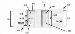
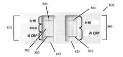
1つの好ましい実施形態において、方法は、最初にサンプルを採取することにより、感染が細菌性及び/又はウイルス性であるかどうかを判定する。その後、サンプルは、デュアルユーズの2つのストリップのサンプル分析装置(dual use two strip sample analysis device)に移される。サンプル分析装置は、第1の試薬領域と第2の試薬領域を伴う、第1の側方流動クロマトグラフィ検査ストリップを含む。第1の試薬領域は、低レベルのC反応性タンパク質に特異的な少なくとも1つの第1の試薬を含み、サンプルが第1の試薬に接触すると、低レベルのC反応性タンパク質がサンプルに存在する場合に第1の標識化した複合体が形成される。第2の試薬領域は、MxAに特異的な少なくとも1つの第2の試薬を含み、サンプルが第2の試薬に接触すると、MxAがサンプルに存在する場合に第2の標識化した複合体が形成される。第1の側方流動クロマトグラフィ検査ストリップはまた、第1の標識化した複合体に結合する第1の結合パートナーと、第2の標識化した複合体に結合する第2の結合パートナーとを含む、第1の検出領域を含む。2つのストリップの側方流動アッセイ装置はまた、第1の側方流動クロマトグラフィ検査ストリップへの側方流動方向と平行な、第2の側方流動クロマトグラフィ検査ストリップを含む。第2の側方流動クロマトグラフィ検査ストリップは、高レベルのC反応性タンパク質に特異的な少なくとも1つの第3の試薬を含む少なくとも1つの第3の試薬領域を含み、サンプルが第3の試薬に接触すると、高レベルのC反応性タンパク質がサンプルに存在する場合に第3の標識化した複合体が形成される。第2の側方流動クロマトグラフィ検査ストリップ上の第3の試薬は、第1の側方流動クロマトグラフィ検査ストリップ上の第2の試薬により検出されるC反応性タンパク質のレベルよりも高い、C反応性タンパク質のレベルを検出するのみである。第2の側方流動クロマトグラフィ検査ストリップはまた、第3の標識化した複合体に結合する第3の結合パートナーを伴う第2の検出領域を含む。サンプルはまた、低レベルのC反応性タンパク質、MxA、及び高レベルのC反応性タンパク質の存在のために分析される。 In one preferred embodiment, the method determines whether the infection is bacterial and/or viral by first taking a sample. The sample is then transferred to a dual use two strip sample analysis device. The sample analyzer includes a first lateral flow chromatography test strip with a first reagent area and a second reagent area. The first reagent region comprises at least one first reagent specific for low levels of C-reactive protein, and low levels of C-reactive protein are present in the sample when the sample contacts the first reagent. A first labeled complex is formed in some cases. The second reagent region comprises at least one second reagent specific for MxA, and when the sample contacts the second reagent, a second labeled complex forms when MxA is present in the sample. be done. the first lateral flow chromatography test strip also includes a first binding partner that binds the first labeled complex and a second binding partner that binds the second labeled complex; A first detection region is included. The two strip lateral flow assay device also includes a second lateral flow chromatography test strip parallel to the lateral flow direction to the first lateral flow chromatography test strip. The second lateral flow chromatography test strip includes at least one third reagent region containing at least one third reagent specific for high levels of C-reactive protein, the sample contacting the third reagent. A third labeled complex is then formed when high levels of C-reactive protein are present in the sample. The third reagent on the second lateral flow chromatography test strip has a higher level of C-reactive protein than the level of C-reactive protein detected by the second reagent on the first lateral flow chromatography test strip. It only detects the level of A second lateral flow chromatography test strip also includes a second detection region with a third binding partner that binds a third labeled complex. Samples are also analyzed for the presence of low levels of C-reactive protein, MxA, and high levels of C-reactive protein.
別の好ましい実施形態において、デュアルユーズの2つのストリップの側方流動アッセイ装置は、サンプル中の細菌マーカー及び/又はウイルスマーカーを検出する。前記装置は、第1の試薬領域と第2の試薬領域を伴う、第1の側方流動クロマトグラフィ検査ストリップを含む。第1の試薬領域は、低レベルのC反応性タンパク質に特異的な少なくとも1つの第1の試薬を含み、サンプルが第1の試薬に接触すると、低レベルのC反応性タンパク質がサンプルに存在する場合に第1の標識化した複合体が形成される。第2の試薬領域は、MxAに特異的な少なくとも1つの第2の試薬を含み、サンプルが第2の試薬に接触すると、MxAがサンプルに存在する場合に第2の標識化した複合体が形成される。第1の側方流動クロマトグラフィ検査ストリップはまた、第1の標識化した複合体に結合する第1の結合パートナーと、第2の標識化した複合体に結合する第2の結合パートナーとを含む、第1の検出領域を含む。2つのストリップの側方流動アッセイ装置はまた、第1の側方流動クロマトグラフィ検査ストリップへの側方流動方向と平行な、第2の側方流動クロマトグラフィ検査ストリップを含む。第2の側方流動クロマトグラフィ検査ストリップは、高レベルのC反応性タンパク質に特異的な少なくとも1つの第3の試薬を含む少なくとも1つの第3の試薬領域を含み、サンプルが第3の試薬に接触すると、高レベルのC反応性タンパク質がサンプルに存在する場合に第3の標識化した複合体が形成される。第2の側方流動クロマトグラフィ検査ストリップ上の第3の試薬は、第1の側方流動クロマトグラフィ検査ストリップ上の第2の試薬により検出されるC反応性タンパク質のレベルよりも高い、C反応性タンパク質のレベルを検出するのみである。第2の側方流動クロマトグラフィ検査ストリップはまた、第3の標識化した複合体に結合する第3の結合パートナーを伴う第2の検出領域を含む。 In another preferred embodiment, a dual-use two-strip lateral flow assay device detects bacterial and/or viral markers in a sample. The device includes a first lateral flow chromatography test strip with a first reagent area and a second reagent area. The first reagent region comprises at least one first reagent specific for low levels of C-reactive protein, and low levels of C-reactive protein are present in the sample when the sample contacts the first reagent. A first labeled complex is formed in some cases. The second reagent region comprises at least one second reagent specific for MxA, and when the sample contacts the second reagent, a second labeled complex forms when MxA is present in the sample. be done. the first lateral flow chromatography test strip also includes a first binding partner that binds the first labeled complex and a second binding partner that binds the second labeled complex; A first detection region is included. The two strip lateral flow assay device also includes a second lateral flow chromatography test strip parallel to the lateral flow direction to the first lateral flow chromatography test strip. The second lateral flow chromatography test strip includes at least one third reagent region containing at least one third reagent specific for high levels of C-reactive protein, the sample contacting the third reagent. A third labeled complex is then formed when high levels of C-reactive protein are present in the sample. The third reagent on the second lateral flow chromatography test strip has a higher level of C-reactive protein than the level of C-reactive protein detected by the second reagent on the first lateral flow chromatography test strip. It only detects the level of A second lateral flow chromatography test strip also includes a second detection region with a third binding partner that binds a third labeled complex.
別の好ましい実施形態において、感染が細菌性及び/又はウイルス性であるかどうかを判定する方法は、サンプルを採取する工程を含む。その後、サンプルは、サンプル分析装置に移される。サンプル分析装置はサンプル圧縮機を含み、該圧縮機は、第1試薬領域と第2試薬領域とを備え、第1試薬領域は、低レベルのC反応性タンパク質に特異的な少なくとも1つの第1の試薬であって、サンプルが第1の試薬に接触すると、低レベルのC反応性タンパク質がサンプルに存在する場合に第1の標識化した複合体が形成される、少なくとも1つの第1の試薬と、MxAに特異的な少なくとも1つの第2の試薬であって、サンプルが第2の試薬に接触すると、MxAがサンプルに存在する場合に第2の標識化した複合体が形成される、少なくとも1つの第2の試薬とを含み、第2の試薬領域は、高レベルのC反応性タンパク質に特異的な少なくとも1つの第3の試薬を含み、第3の試薬は、第2の試薬によって検知されたC反応性タンパク質のレベルよりも高いC反応性タンパク質のレベルを検出するのみであり、サンプルが第3の試薬に接触すると、高レベルのC反応性タンパク質がサンプルに存在する場合に第3の標識化した複合体が形成される。前記装置はまた、第1の検出領域と第1の分岐領域とを含む第1の側方流動クロマトグラフィ検査ストリップを含み、第1の検出領域は、第1の標識化した複合体に結合する第1の結合パートナーと、第2の標識化した複合体に結合する第2の結合パートナーとを含み、第1の分岐領域は、側方流動クロマトグラフィ検査ストリップ上の第1の検出領域の上流に位置する。第1の分岐領域は、第1の側方流動クロマトグラフィ検査ストリップ上で側方流動を阻止する。前記装置はまた、第1の側方流動クロマトグラフィ検査ストリップへの側方流動方向と平行な、第2の側方流動クロマトグラフィ検査ストリップを含む。第2の側方流動クロマトグラフィ検査ストリップは、第3の標識化した複合体に結合する第3の結合パートナーを含む第2の検出領域と、側方流動クロマトグラフィ検査ストリップ上の第1の検出領域の上流に位置する第2の分岐領域とを含む。第2の分岐領域は、第2の側方流動クロマトグラフィ検査ストリップ上で側方流動を阻止する。前記装置はまた、サンプルがサンプル分析装置の上に置かれる第1のサンプル適用領域を含む。第1のサンプル適用領域は、次のものから成る群から選択された場所に位置する:i)検出領域の上流にある第1の側方流動クロマトグラフィ検査スリップの上、及び、ii)サンプル圧縮機の第1の試薬領域の上。前記装置はまた、サンプルがサンプル分析装置の上に置かれる第2のサンプル適用領域を含む。第2のサンプル適用領域は、次のものから成る群から選択された場所に位置する:i)検出領域の上流にある第2の側方流動クロマトグラフィ検査ストリップの上、及び、ii)サンプル圧縮機の第2の試薬領域の上。サンプル圧縮機は、第1の側方流動クロマトグラフィ検査ストリップ、及び第2の側方流動クロマトグラフィ検査ストリップとは異なる面にある。サンプル圧縮機の第1の試薬領域は、第1の分岐領域にわたり橋(bridge)を作成し、サンプル圧縮機の第2の試薬領域は、第2の分岐領域にわたり橋を作成し、これらの橋は、サンプル圧縮機上に流れを分岐し、第1の分岐領域と第2の分岐領域の端部にある第1のクロマトグラフィ検査ストリップと第2のクロマトグラフィ検査ストリップに流れを戻す。サンプルは、低レベルのC反応性タンパク質、MxA、及び高レベルのC反応性タンパク質の存在のために分析される。 In another preferred embodiment, the method of determining whether an infection is bacterial and/or viral comprises taking a sample. The sample is then transferred to the sample analyzer. The sample analyzer includes a sample compressor comprising a first reagent region and a second reagent region, the first reagent region containing at least one first reagent specific for low levels of C-reactive protein. wherein when a sample contacts the first reagent, a first labeled complex is formed when low levels of C-reactive protein are present in the sample. and at least one second reagent specific for MxA, wherein when the sample contacts the second reagent, a second labeled complex is formed when MxA is present in the sample, at least a second reagent, the second reagent region comprising at least one third reagent specific for high levels of C-reactive protein, the third reagent being detected by the second reagent only detect a level of C-reactive protein that is higher than the level of C-reactive protein that has been tested, and when the sample is contacted with a third reagent, the third reagent if high levels of C-reactive protein are present in the sample. of the labeled complex is formed. The device also includes a first lateral flow chromatography test strip including a first detection region and a first bifurcation region, the first detection region binding to the first labeled complex. one binding partner and a second binding partner that binds to a second labeled complex, wherein the first branched region is located upstream of the first detection region on the lateral flow chromatography test strip. do. The first bifurcated region prevents lateral flow on the first lateral flow chromatography test strip. The device also includes a second lateral flow chromatography test strip parallel to the lateral flow direction to the first lateral flow chromatography test strip. The second lateral flow chromatography test strip has a second detection area comprising a third binding partner that binds to the third labeled complex and a first detection area on the lateral flow chromatography test strip. and a second branch region located upstream. A second bifurcated region prevents lateral flow on a second lateral flow chromatography test strip. The device also includes a first sample application area where a sample is placed over the sample analyzer. The first sample application area is located at a location selected from the group consisting of: i) above the first lateral flow chromatography test slip upstream of the detection area, and ii) the sample compressor. above the first reagent area of . The device also includes a second sample application area where a sample is placed over the sample analyzer. A second sample application area is located at a location selected from the group consisting of: i) above the second lateral flow chromatography test strip upstream of the detection area, and ii) the sample compressor. above the second reagent area of . The sample compressor is in a different plane than the first lateral flow chromatography test strip and the second lateral flow chromatography test strip. A first reagent region of the sample compressor creates a bridge over the first bifurcated region, a second reagent region of the sample compressor creates a bridge over the second bifurcated region, and the bridges bifurcates the flow onto the sample compressor and returns the flow to the first and second chromatographic test strips at the ends of the first and second bifurcation regions. Samples are analyzed for the presence of low levels of C-reactive protein, MxA, and high levels of C-reactive protein.
別の好ましい実施形態は、サンプルにおける分析物を検知するための側方流動装置である。該装置はサンプル圧縮機を含み、該圧縮機は、第1試薬領域と第2試薬領域とを備え、第1試薬領域は、低レベルのC反応性タンパク質に特異的な少なくとも1つの第1の試薬であって、サンプルが第1の試薬に接触すると、低レベルのC反応性タンパク質がサンプルに存在する場合に第1の標識化した複合体が形成される、少なくとも1つの第1の試薬と、MxAに特異的な少なくとも1つの第2の試薬であって、サンプルが第2の試薬に接触すると、MxAがサンプルに存在する場合に第2の標識化した複合体が形成される、少なくとも1つの第2の試薬とを含み、第2の試薬領域は、高レベルのC反応性タンパク質に特異的な少なくとも1つの第3の試薬を含み、ここで、第3の試薬は、第2の試薬によって検知されたC反応性タンパク質のレベルよりも高いC反応性タンパク質のレベルを検知するのみであり、サンプルが第3の試薬に接触すると、高レベルのC反応性タンパク質がサンプルに存在する場合に第3の標識化した複合体が形成される。前記装置はまた、第1の検出領域と第1の分岐領域とを含む第1の側方流動クロマトグラフィ検査ストリップを含み、第1の検出領域は、第1の標識化した複合体に結合する第1の結合パートナーと、第2の標識化した複合体に結合する第2の結合パートナーとを含み、第1の分岐領域は、第1の側方流動クロマトグラフィ検査ストリップ上の第1の検出領域の上流に位置する。第1の分岐領域は、第1の側方流動クロマトグラフィ検査ストリップ上で側方流動を阻止する。前記装置はまた、第1の側方流動クロマトグラフィ検査ストリップへの側方流動方向と平行な、第2の側方流動クロマトグラフィ検査ストリップを含む。第2の側方流動クロマトグラフィ検査ストリップは、第3の標識化した複合体に合する第3の結合パートナーを含む第2の検出領域と、側方流動クロマトグラフィ検査ストリップ上の第1の検出領域の上流に位置する第2分岐領域とを含む。第2の分岐領域は、第2の側方流動クロマトグラフィ検査ストリップ上で側方流動を阻止する。前記装置はまた、サンプルがサンプル分析装置の上に置かれる第1のサンプル適用領域を含む。第1のサンプル適用領域は、次のものから成る群から選択された場所に位置する:i)検出領域の上流にある第1の側方流動クロマトグラフィ検査ストリップの上、及び、ii)サンプル圧縮機の第1の試薬領域の上。前記装置はまた、サンプルがサンプル分析装置の上に置かれる第2のサンプル適用領域を含む。第2のサンプル適用領域は、次のものから成る群から選択された場所に位置する:i)検出領域の上流にある第2の側方流動クロマトグラフィ検査ストリップの上、及び、ii)サンプル圧縮機の第2の試薬領域の上。サンプル圧縮機は、第1の側方流動クロマトグラフィ検査ストリップ、及び第2の側方流動クロマトグラフィ検査ストリップとは異なる面にある。サンプル圧縮機の第1の試薬領域は、第1の分岐領域にわたり橋(bridge)を作成し、サンプル圧縮機の第2の試薬領域は、第2の分岐領域にわたり橋を作成し、これらの橋は、サンプル圧縮機上に流れを分岐し、第1の分岐領域と第2の分岐領域の端部にある第1のクロマトグラフィ検査ストリップと第2のクロマトグラフィ検査ストリップに流れを戻す。 Another preferred embodiment is a lateral flow device for detecting analytes in a sample. The apparatus includes a sample compressor comprising a first reagent zone and a second reagent zone, the first reagent zone containing at least one first sample specific for low levels of C-reactive protein. at least one first reagent, wherein when the sample contacts the first reagent, a first labeled complex is formed when low levels of C-reactive protein are present in the sample; , at least one second reagent specific for MxA, wherein when the sample contacts the second reagent, a second labeled complex is formed when MxA is present in the sample. and two second reagents, the second reagent region comprising at least one third reagent specific for high levels of C-reactive protein, wherein the third reagent is the second reagent only detects a level of C-reactive protein that is higher than the level of C-reactive protein detected by and when the sample is contacted with a third reagent, if high levels of C-reactive protein are present in the sample, A third labeled complex is formed. The device also includes a first lateral flow chromatography test strip including a first detection region and a first bifurcation region, the first detection region binding to the first labeled complex. one binding partner and a second binding partner that binds to a second labeled complex, the first branched region being the first detection region on the first lateral flow chromatography test strip. Located upstream. The first bifurcated region prevents lateral flow on the first lateral flow chromatography test strip. The device also includes a second lateral flow chromatography test strip parallel to the lateral flow direction to the first lateral flow chromatography test strip. The second lateral flow chromatography test strip has a second detection area comprising a third binding partner that binds to the third labeled complex and a first detection area on the lateral flow chromatography test strip. and a second branch region located upstream. A second bifurcated region prevents lateral flow on a second lateral flow chromatography test strip. The device also includes a first sample application area where a sample is placed over the sample analyzer. The first sample application area is located at a location selected from the group consisting of: i) above the first lateral flow chromatography test strip upstream of the detection area, and ii) the sample compressor. above the first reagent area of . The device also includes a second sample application area where a sample is placed over the sample analyzer. A second sample application area is located at a location selected from the group consisting of: i) above the second lateral flow chromatography test strip upstream of the detection area, and ii) the sample compressor. above the second reagent area of . The sample compressor is in a different plane than the first lateral flow chromatography test strip and the second lateral flow chromatography test strip. A first reagent region of the sample compressor creates a bridge over the first bifurcated region, a second reagent region of the sample compressor creates a bridge over the second bifurcated region, and the bridges bifurcates the flow onto the sample compressor and returns the flow to the first and second chromatographic test strips at the ends of the first and second bifurcation regions.
別の好ましい実施形態において、方法は、サンプルを採取してサンプル分析装置にサンプルを移すことにより、少なくとも1つの細胞外の分析物と、少なくとも1つの細胞内の分析物とを同時に検出する。サンプルはまた溶解され、細胞外の分析物と細胞内の分析物は、同じサンプル分析装置上で同時に検出される。1つの好ましい実施形態において、細胞外の分析物はC反応性タンパク質であり、細胞内の分析物はMxAタンパク質である。 In another preferred embodiment, the method simultaneously detects at least one extracellular and at least one intracellular analyte by taking a sample and transferring the sample to a sample analyzer. The sample is also lysed and extracellular and intracellular analytes are detected simultaneously on the same sample analyzer. In one preferred embodiment, the extracellular analyte is C-reactive protein and the intracellular analyte is MxA protein.
別の好ましい実施形態において、サンプルにおけるMxAタンパク質とC反応性タンパク質を検出する方法は、第1の標識に結合したMxAタンパク質に対する抗体と、第1の標識とは異なる第2の標識に結合したC反応性タンパク質に対する抗体との混合物にサンプルを加える工程、MxAタンパク質に対する抗体が蓄積したかどうかを判定することによりMxAタンパク質の存在を検出する工程、及び、C反応性タンパク質に対する抗体が蓄積したかどうかを判定することによりC反応性タンパク質の存在を検出する工程を含む。 In another preferred embodiment, the method of detecting MxA protein and C-reactive protein in a sample comprises antibody to MxA protein bound to a first label and C bound to a second label different from the first label. adding the sample to the mixture with antibodies to the reactive protein, detecting the presence of the MxA protein by determining whether antibodies to the MxA protein have accumulated, and whether antibodies to the C-reactive protein have accumulated. detecting the presence of C-reactive protein by determining the
別の好ましい実施形態において、サンプルにおける未知のウイルス感染の存在を検出する方法は、最初にサンプルを採取する。その後、サンプルは、サンプル分析装置のサンプル適用領域に移される。サンプル分析装置は、ナノミセルの内部に標識を伴うシアル酸ナノミセルを含む結合領域と、シアル酸同族体ナノ粒子を含むサンプル適用領域から横方向で下流にある検出領域とを含む。サンプルは、検出領域における陽性の結果に関して分析される。 In another preferred embodiment, the method of detecting the presence of an unknown viral infection in a sample first collects the sample. The sample is then transferred to the sample application area of the sample analyzer. The sample analysis device includes a binding region comprising sialic acid nanomicelles with a label inside the nanomicelles and a detection region laterally downstream from the sample application region comprising sialic acid congener nanoparticles. Samples are analyzed for positive results in the detection zone.
別の好ましい実施形態において、サンプルにおける未知のウイルス感染の存在を検出する方法は、最初にサンプルを採取する。その後、サンプルは、サンプル分析装置のサンプル適用領域に移される。サンプル分析装置は、次のものから成る群から選択された分子を備えた結合領域を含む:ウイルス感染を引き起こす特定のウイルスに対する結合パートナーと標識を含むナノミセル、及び、ナノミセルの内部にラベルを含むシアル酸同族体ナノミセル。サンプル分析装置はまた、ウイルス感染を引き起こすウイルスに対して特異的なナノ粒子を備える、サンプル適用領域から横方向で下流にある検出領域を含む。サンプルは、検出領域における陽性の結果に関して分析される。 In another preferred embodiment, the method of detecting the presence of an unknown viral infection in a sample first collects the sample. The sample is then transferred to the sample application area of the sample analyzer. The sample analyzer includes a binding region with a molecule selected from the group consisting of: nanomicelles containing binding partners and labels for specific viruses that cause viral infection, and sials containing labels inside the nanomicelles. Acid congeners nanomicelles. The sample analyzer also includes a detection region laterally downstream from the sample application region comprising nanoparticles specific for viruses that cause viral infection. Samples are analyzed for positive results in the detection zone.
本発明は、ウイルス感染と細菌感染を区別することができる側方流動アッセイを提供する。特定の細菌感染又はウイルス感染に特有の分析物について検査する代わりに、本明細書に記載される側方流動アッセイは、一般的な無指定の細菌感染と一般的な無指定のウイルス感染に反応した宿主において明確に作られる、診断のマーカーについて検査する。診断マーカーは好ましくは、細菌由来又はウイルス由来の、無指定の及び/又は未知のマーカーである。好ましい実施形態において、診断マーカーは、無指定の及び/又は未知の細菌感染及び/又はウイルス感染に対する免疫応答のための特異的マーカーである。 The present invention provides a lateral flow assay that can distinguish between viral and bacterial infections. Instead of testing for analytes specific to a particular bacterial or viral infection, the lateral flow assays described herein respond to common non-specific bacterial infections and common non-specific viral infections. Test for diagnostic markers that are specifically produced in infected hosts. Diagnostic markers are preferably nonspecific and/or unknown markers of bacterial or viral origin. In a preferred embodiment, the diagnostic marker is a specific marker for an immune response against unspecified and/or unknown bacterial and/or viral infections.
組み合わされたポイントオブケア診断装置は、ウイルス感染と細菌感染の両方のマーカーを検査し、例えば、外来患者診療室で、又は、応急手当外来の間に、ウイルス感染と細菌感染とを迅速に区別するのに効果的に役立ち得る。この能力は、誤診とその後の抗生物質の過剰使用とを制限することによって、医療費を劇的に削減することができる。上記を実践することで、抗生物質アレルギー、有害な事象、及び抗生物質耐性を制限することができる。検査から結果がすぐに得られることで、患者が従事者の検査をまだ受けている間に診断を下すことも可能となる。好ましい実施形態において、検査結果は、装置にサンプルを適用して10分後には得られ、好ましくは約10分で読み取られる。高い陽性のサンプルにおいて、検査ラインは約1乃至5分以内に視認可能である。 A combined point-of-care diagnostic device tests for markers of both viral and bacterial infections and rapidly distinguishes between viral and bacterial infections, e.g., in outpatient clinics or during first aid visits can effectively help to This ability can dramatically reduce healthcare costs by limiting misdiagnosis and subsequent antibiotic overuse. Practicing the above can limit antibiotic allergies, adverse events, and antibiotic resistance. Immediate results from the test also allow the diagnosis to be made while the patient is still being examined by the practitioner. In preferred embodiments, test results are obtained 10 minutes after application of the sample to the device, and are preferably read in about 10 minutes. In highly positive samples, the test line is visible within about 1-5 minutes.
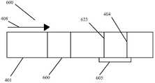
本発明の好ましい実施形態において、本発明の側方流動イムノアッセイ装置は、サンプルを輸送する液体を含み、この液体は緩衝液でもよい。該装置は、サンプルが流れる毛細管の特徴を備えた1以上のフリース素材又は膜を包含するクロマトグラフィ検査ストリップを含む。検査ストリップのための幾つかの好ましい材料又は膜には、Dacron(登録商標)繊維などのポリエチレンテレフタラート(PET)繊維、ニトロセルロース、ポリエステル、ナイロン、酢酸セルロース、ポリプロピレン、ガラス繊維、及び、このような材料及び裏当て材(backings)の組み合わせが挙げられるが、これらに限定されない。本発明の幾つかの実施形態において、サンプルを検査ストリップに適用する前に、任意の方法でサンプル中の細胞を溶解させるか、又はサンプルを処理する必要はない。 In a preferred embodiment of the invention, the lateral flow immunoassay device of the invention comprises a sample-carrying liquid, which may be a buffer. The device includes a chromatographic test strip that includes one or more fleece materials or membranes with capillary features through which the sample flows. Some preferred materials or membranes for test strips include polyethylene terephthalate (PET) fibers such as Dacron® fibers, nitrocellulose, polyester, nylon, cellulose acetate, polypropylene, glass fibers, and the like. materials and backings in combination, but are not limited to these. In some embodiments of the invention, it is not necessary to lyse the cells in the sample or treat the sample in any way prior to applying the sample to the test strip.
本発明の1つの好ましい方法は、感染が細菌性かウイルス性かどうかを判定するために、例えば、クロマトグラフィ検査ストリップなどのサンプル分析装置を用いる。この方法において、サンプルが採取され、クロマトグラフィ検査ストリップに移される。好ましい実施形態において、サンプルは白血球を含むサンプルである。検査ストリップは試薬領域を含む。試薬領域は、好ましくは、細菌マーカーに特異的な少なくとも1つの第1の試薬を含み、サンプル中に存在する細菌マーカーがこの第1の試薬に接触すると、第1の標識化した複合体が形成される。試薬領域は、好ましくは、ウイルスマーカーに特異的な少なくとも1つの第2の試薬も含み、サンプル中に存在するウイルスマーカーがこの第2の試薬に接触すると、第2の標識化した複合体が形成される。検出領域は、第1の標識化した複合体に結合する細菌マーカーの結合パートナーと、第2の標識化した複合体に結合するウイルスマーカーの結合パートナーを両方含む。サンプルはその後、ウイルスマーカー及び/又は細菌マーカーの存在について分析される。 One preferred method of the invention uses a sample analyzer, such as a chromatographic test strip, to determine whether an infection is bacterial or viral. In this method, a sample is taken and transferred to a chromatographic test strip. In preferred embodiments, the sample is a sample containing white blood cells. The test strip includes a reagent area. The reagent region preferably comprises at least one first reagent specific to a bacterial marker, and when a bacterial marker present in the sample contacts this first reagent, a first labeled complex is formed. be done. The reagent region preferably also contains at least one second reagent specific to a viral marker, and when a viral marker present in the sample contacts this second reagent, a second labeled complex is formed. be done. The detection region contains both a binding partner for a bacterial marker that binds to the first labeled complex and a binding partner for a viral marker that binds to the second labeled complex. Samples are then analyzed for the presence of viral and/or bacterial markers.
本発明の装置の好ましい実施形態は、サンプル適用領域を含む。本装置は試薬領域も含み、該領域は細菌マーカーに特異的な少なくとも1つの第1の試薬を含み、サンプル中に存在する細菌マーカーが第1の試薬に接触すると、第1の標識化した複合体が形成され、該領域は、細菌マーカーに特異的な少なくとも1つの第2の試薬を含み、サンプル中に存在するウイルスマーカーが第2の試薬に接触すると、第2の標識化した複合体が形成される。装置上の検出領域は、第1の標識化した複合体と結合する細菌マーカーと、第2の標識化した複合体と結合するウイルスマーカーとを含む。使用可能な装置の一例は、クロマトグラフィ検査ストリップである。他の好ましい実施形態において、装置の領域の幾つかは1以上のクロマトグラフィ検査ストリップ上にあり、一方で他の領域(例えば、試薬領域、サンプル適用領域、及び/又は、対照結合パートナー)は、サンプル圧縮機上にあり、互いに離れ、及び、クロマトグラフィ検査ストリップとは異なる平面にある。 A preferred embodiment of the device of the invention includes a sample application area. The device also includes a reagent region, the region including at least one first reagent specific for a bacterial marker, and when a bacterial marker present in the sample contacts the first reagent, the first labeled complex a second labeled complex is formed, the region comprising at least one second reagent specific for a bacterial marker, and a second labeled complex when a viral marker present in the sample contacts the second reagent It is formed. A detection region on the device includes a bacterial marker that binds to the first labeled complex and a viral marker that binds to the second labeled complex. One example of a device that can be used is a chromatographic test strip. In other preferred embodiments, some of the regions of the device are over one or more chromatographic test strips, while other regions (e.g., reagent regions, sample application regions, and/or control binding partners) are It is on the compressor, separate from each other, and in a different plane than the chromatographic test strip.
好ましい実施形態において、ウイルスマーカー又は細菌マーカーの存在は、肉眼で見える検査ラインによって示される。ウイルスマーカーの存在は、第1の検査ラインによって示され得、その一方で、細菌マーカーは第2の検査ラインによって示される。幾つかの実施形態において、第1の検査ラインは陽性の際に第1の色を表示し、第2の検査ラインは陽性の際に第1の色とは異なる第2の色を表示する。第1の検査ラインと第2の検査ラインの両方がサンプル分析装置上の同じ空間内に位置する実施形態において、第3の色は、第1の検査ラインと第2の検査ラインの両方が陽性の際に好ましくは形成される。他の実施形態において、2つの検査ラインは装置上で互いから空間的に離れている。 In preferred embodiments, the presence of viral or bacterial markers is indicated by a visible test line. The presence of viral markers can be indicated by a first test line, while bacterial markers are indicated by a second test line. In some embodiments, the first test line displays a first color when positive and the second test line displays a second color different than the first color when positive. In embodiments where both the first test line and the second test line are located in the same space on the sample analyzer, the third color indicates that both the first test line and the second test line are positive. is preferably formed during In other embodiments, the two test lines are spatially separated from each other on the device.
ウイルス感染と細菌感染は非常に伝染性であり、そして、全身性の抗生物質の過剰処方、及び抗生物質抵抗性の促進へと頻繁に通じる、兆候と症状における著しい重複のため、臨床的に区別するのが困難なものである。先進国において、急性呼吸器感染症は、医療相談の20%、研究欠席(absences from work)の30%、及び全ての抗生物質処方の75%を占める、罹患率の主要原因である。米国において、急性呼吸器感染症に関して毎年、およそ7600万の医師の来院が存在する。感染に対する免疫応答を検出する能力は、ウイルスの病因及び/又は細菌の病因から結果として生じる感染を区別するための臨床診断能力を補助する。 Viral and bacterial infections are highly contagious and are clinically distinguished because of significant overlap in signs and symptoms, frequently leading to systemic antibiotic overprescription and the development of antibiotic resistance. It is difficult to do. In developed countries, acute respiratory infections are the leading cause of morbidity, accounting for 20% of medical consultations, 30% of absences from work, and 75% of all antibiotic prescriptions. In the United States, there are approximately 76 million physician visits each year for acute respiratory infections. The ability to detect an immune response to infection aids clinical diagnostic capabilities for distinguishing consequent infections from viral and/or bacterial etiologies.
1つの好ましい実施形態において、細菌マーカーはCRPである。別の好ましい実施形態において、ウイルスマーカーはMxAである。幾つかの好ましい実施形態において、検出領域は、装置が作動している際に、肉眼で見える対照ラインを同様に含む。 In one preferred embodiment, the bacterial marker is CRP. In another preferred embodiment, the viral marker is MxA. In some preferred embodiments, the detection area also includes a control line that is visible to the naked eye when the device is in operation.
1つの好ましい実施形態において、ウイルス感染のためのマーカーはMxAであり、細菌感染のためのマーカーはC反応性タンパク質(CRP)である。高MxAタンパク質レベルは、全身性ウイルス感染と強く相関し、CRPの増加は細菌感染に多く関連する。本発明は、サンプル中のMxAとCRPとを識別するための、迅速な感染のスクリーニング検査を含む。MxAは白血球(leukocite、white blood cells)中に存在する。したがって、サンプルは、白血球が利用可能な場所で、例えば、末梢血サンプル、鼻咽頭吸引物、涙、髄液、及び、中耳吸引物において、採取され得る。 In one preferred embodiment, the marker for viral infection is MxA and the marker for bacterial infection is C-reactive protein (CRP). High MxA protein levels are strongly correlated with systemic viral infection, and increased CRP is often associated with bacterial infection. The present invention includes a rapid infection screening test to distinguish between MxA and CRP in a sample. MxA is present in leukocytes (white blood cells). Thus, samples can be taken where leukocytes are available, such as peripheral blood samples, nasopharyngeal aspirates, tears, spinal fluid, and middle ear aspirates.
CRPとMxAを含有する単一の検査ストリップを伴う幾つかの好ましい実施形態において、陽性の結果を誘発するために必要とされるサンプル中のCRPの閾値濃度は、およそ6-15mg/Lである。他の好ましい実施形態において、陽性の結果を誘発するサンプル中のMxAの閾値濃度は、およそ15ナノグラム/mlほどの低さでもよい;しかし、閾値濃度は、およそ20ng/ml乃至およそ250ng/mlまでの範囲よりも高いか、又はその範囲内にあってもよい。適用可能な場合、閾値濃度は、検査ストリップに適用されるサンプルの大きさ、同様に、その希釈度にも依存してもよい。 In some preferred embodiments with a single test strip containing CRP and MxA, the threshold concentration of CRP in the sample required to elicit a positive result is approximately 6-15 mg/L. . In other preferred embodiments, the threshold concentration of MxA in a sample that elicits a positive result may be as low as about 15 nanograms/ml; however, the threshold concentration is from about 20 ng/ml to about 250 ng/ml. may be higher than or within the range of Where applicable, the threshold concentration may depend on the size of the sample applied to the test strip as well as its dilution.
幾つかの実施形態において、本明細書に記載される装置及び方法は、辺縁の全血から直接MxAとCRPの両方を、迅速に、視認可能に、定質的にインビトロで検知することを可能にする。1つの好ましい実施形態において、検査は、呼吸器疾患と一致する呼吸器症状、及び急性咽頭炎又は市中感染性肺炎の推定診断と共に、熱の発症の7日以内に存在する、1歳より高齢の患者における疑わしいウイルス感染及び/又は細菌感染に対する免疫応答を測定する。陰性の結果は必ずしも呼吸器感染症を妨げるものではなく、診断、処置、又は他の管理の決定に関する唯一の基準として使用されるべきでない。幾つかの実施形態において、追加の実験室検査(例えば、細菌培養及びウイルス培養、免疫蛍光、ウイルスポリメラーゼ連鎖反応、及びX線撮影法)と臨床像(clinical presentation)の使用は、特異的な低度の呼吸器及び咽頭の病原菌が存在するかどうかを確認するために追加で使用されるのが好ましい。 In some embodiments, the devices and methods described herein provide rapid, visible, and qualitative in vitro detection of both MxA and CRP directly from limbal whole blood. to enable. In one preferred embodiment, the test will include respiratory symptoms consistent with respiratory illness and a presumed diagnosis of acute pharyngitis or community-acquired pneumonia, present within 7 days of onset of fever, older than 1 year. to measure immune responses to suspected viral and/or bacterial infections in patients with A negative result does not necessarily preclude respiratory infection and should not be used as the sole basis for diagnosis, treatment, or other management decisions. In some embodiments, the use of additional laboratory tests (e.g., bacterial and viral cultures, immunofluorescence, viral polymerase chain reaction, and radiography) and clinical presentation are used to determine specific low It is preferably additionally used to determine if any respiratory and pharyngeal pathogens are present.
加えて、誤った偽陽性又は偽陰性に通じる幾つかの疾病が存在する。これらは、限定されないが、サンプルを提供する患者による免疫抑制薬の現在の使用、サンプルを提供する患者による経口抗感染薬の現在の使用、サンプルを提供する患者によるインターフェロン療法(例えば、多発性硬化症、HIV、HBV、HCVのため)の現在の使用、及び、サンプルを提供する患者による過去30日以内の生きたウイルスの免疫化を含む。レベルが治療により変動し得るため、偽陰性と偽陽性の両方が可能である。 Additionally, there are several diseases that lead to false positives or false negatives. These include, but are not limited to, current use of immunosuppressants by the sample providing patient, current use of oral anti-infectives by the sample providing patient, interferon therapy (e.g., multiple sclerosis) by the sample providing patient. disease, HIV, HBV, HCV) and live virus immunization within the past 30 days by the patient providing the sample. Both false negatives and false positives are possible because levels can vary with treatment.
好ましい実施形態において、装置及び方法は、外来患者診療室又は応急手当臨床における専門的使用を意図したものであり、他の臨床的(実験室又はX線写真術)及び疫学の情報と共に使用されねばならない。 In preferred embodiments, the apparatus and methods are intended for professional use in the outpatient clinic or first aid clinic and must be used with other clinical (laboratory or radiographic) and epidemiological information. not.
好ましい実施形態において、デュアルユーズのデュアルクロマトグラフィ検査ストリップのアッセイは、結果の多重化したパターンを使用して、患者におけるウイルス感染及び/又は細菌感染に対する身体の免疫応答を検出する。1つの特異的な好ましい実施形態において、前記アッセイは、ミクソウイルス抵抗性A(MxA)、低レベルのC反応性タンパク質(「低」CRP)、及び高レベルのC反応性タンパク質(「高」CRP)を検査する。2つの検査ストリップが好ましくは使用される。幾つかの実施形態において、クロマトグラフィ検査ストリップとは異なる面にあるサンプル圧縮機も、使用される。第1の検査ストリップは、MxAと低レベルのC反応性タンパク質をアッセイし、第2の検査ストリップは、高レベルのC反応性タンパク質のアッセイである。第1の検査ストリップ及び/又はサンプル圧縮機は、MxAタンパク質と低レベルのC反応性タンパク質を検出するための試薬を含む。第2の検査ストリップ及び/又はサンプル圧縮機は、高レベルのC反応性タンパク質を検出するための試薬を含む。2つの検査ストリップは好ましくは、並行して実行され、各ストリップは好ましくは対照ラインも含む。対照試薬は好ましくは、検査ストリップ又はサンプル圧縮機上にある。これらの検査は、生物感染を検出し、それをウイルス、細菌、又はウイルスと細菌の同時感染として分類する。幾つかの好ましい実施形態において、デュアルユーズのデュアルクロマトグラフィ検査ストリップのアッセイは、発熱性の呼吸器疾患の患者からのサンプルを検出するために使用される。 In a preferred embodiment, the dual-use dual chromatography test strip assay uses multiplexed patterns of results to detect the body's immune response to viral and/or bacterial infections in a patient. In one specific preferred embodiment, the assay measures myxovirus resistance A (MxA), low levels of C-reactive protein (“low” CRP), and high levels of C-reactive protein (“high” CRP). ). Two test strips are preferably used. In some embodiments, a sample compressor on a different side than the chromatographic test strip is also used. The first test strip assays for MxA and low levels of C-reactive protein and the second test strip assays for high levels of C-reactive protein. A first test strip and/or sample compressor contains reagents for detecting MxA protein and low levels of C-reactive protein. A second test strip and/or sample compressor contains reagents for detecting high levels of C-reactive protein. Two test strips are preferably run in parallel, each strip preferably also including a control line. The control reagent is preferably on the test strip or sample compressor. These tests detect biological infections and classify them as viral, bacterial, or viral and bacterial co-infections. In some preferred embodiments, the dual-use dual chromatography test strip assay is used to detect samples from patients with febrile respiratory illness.
2つの検査ストリップを伴う幾つかの好ましい実施形態において、第1の検査ストリップ上で、サンプル中のおよそ6-15mg/L(血清カットオフ値)のCRP(「低」CRPレベル)の閾値濃度は、陽性の結果を誘発するために必要とされ、サンプル中の少なくとも15ng/mlのMxAの閾値濃度は、陽性の結果を誘発するために必要とされる。他の好ましい実施形態において、MxAの閾値濃度は、陽性の結果を誘発するために、およそ15ng/ml乃至およそ250ng/mlの範囲にあってもよい。適用可能な場合、閾値濃度は、検査ストリップに適用されるサンプルの大きさ、同様に、その希釈度にも依存してもよい。1つの好ましい実施形態において、例えば血液サンプルからの細胞外の血清中の低CRPの閾値濃度は、血清カットオフ値に関して10mg/Lと同等である、指穿刺カットオフ値に関して7mg/Lである。1つの好ましい実施形態において、例えば血液サンプルからの末梢血単核細胞中のMxAの閾値濃度は、40ng/mlの静脈血カットオフ値と同等である、指穿刺カットオフ値に関して40ng/mlである。第2の検査ストリップ上で、サンプル中のおよそ60-100mg/LのCRP(「高」CRPレベル)の閾値濃度が、幾つかの好ましい実施形態において陽性の結果を誘発するために必要とされる。1つの特に好ましい実施形態において、第2の検査ストリップ上の高CRPの閾値濃度は、フィンガープリントのカットオフ値に関しておよそ80mg/Lである。 In some preferred embodiments involving two test strips, the threshold concentration of CRP (“low” CRP level) in a sample of approximately 6-15 mg/L (serum cutoff) on the first test strip is , is required to induce a positive result, and a threshold concentration of MxA of at least 15 ng/ml in the sample is required to induce a positive result. In other preferred embodiments, the threshold concentration of MxA may range from approximately 15 ng/ml to approximately 250 ng/ml to induce a positive result. Where applicable, the threshold concentration may depend on the size of the sample applied to the test strip as well as its dilution. In one preferred embodiment, the threshold concentration for low CRP in extracellular serum, eg from a blood sample, is 7 mg/L for finger stick cut-off, which is equivalent to 10 mg/L for serum cut-off. In one preferred embodiment, the threshold concentration of MxA in peripheral blood mononuclear cells, e.g. from a blood sample, is 40 ng/ml with respect to the finger stick cut-off, which is equivalent to the venous blood cut-off of 40 ng/ml. . On the second test strip, a threshold concentration of approximately 60-100 mg/L CRP (“high” CRP level) in the sample is required to induce a positive result in some preferred embodiments. . In one particularly preferred embodiment, the threshold concentration for high CRP on the second test strip is approximately 80 mg/L for the fingerprint cutoff value.
他の実施形態において、ウイルス感染及び/又は細菌感染のための他のマーカーが使用されてもよい。例えば、宿主遺伝子のおよそ12%が、リンパ球性脈絡髄膜炎ウイルス(LCMV)感染後にそれらの発現を変更し、これら遺伝子の亜群が、悪性LCMV感染と非悪性LCMVを区別することが可能となる。主要な転写の変化は、定量PCRとタンパク質研究とによって予備確認を与えられ、アレナウイルス性疾患のためのバイオマーカーとして潜在的に価値のある候補である。細菌感染のための他のマーカーには、プロカルシトニン、尿性トリプシンインヒビター(uTi)、リポ多糖類、IL-1、IL-6、IL-8、IL-10、ESR、及び、白血球数の上昇(結合(band)の増加)、乳酸、トロポニン、血管内皮増殖因子、血小板由来増殖因子、コルチゾール、プロアドレノメデュリン、マクロファージ遊走阻止マーカー、活性化タンパク質C、CD4、8、13、14、又は64、カスパーゼ、胎盤由来増殖因子、カルシトニン遺伝子関連ペプチド、高移動度群1、コペプチン、ナトリウム利尿ペプチド(naturietic peptide)、リポ多糖結合タンパク質、腫瘍壊死因子α、循環血管内皮前駆細胞、補体3a、及び、骨髄系細胞(trem-1)上で発現した誘発受容体が挙げられるが、これらに限定されない。 In other embodiments, other markers for viral and/or bacterial infection may be used. For example, approximately 12% of host genes have their expression altered following lymphocytic choriomeningitis virus (LCMV) infection, and a subgroup of these genes can distinguish malignant from non-malignant LCMV infection. becomes. The major transcriptional alterations, given preliminary confirmation by quantitative PCR and protein studies, are potentially valuable candidates as biomarkers for arenaviral disease. Other markers for bacterial infection include procalcitonin, urinary trypsin inhibitor (uTi), lipopolysaccharides, IL-1, IL-6, IL-8, IL-10, ESR, and elevated white blood cell count. (increased band), lactate, troponin, vascular endothelial growth factor, platelet-derived growth factor, cortisol, proadrenomedullin, macrophage migration inhibitory marker, activated protein C, CD4, 8, 13, 14, or 64, caspase , placenta-derived growth factor, calcitonin gene-related peptide, high mobility group 1, copeptin, natrietic peptide, lipopolysaccharide binding protein, tumor necrosis factor α, circulating endothelial progenitor cells, complement 3a, and bone marrow These include, but are not limited to, triggering receptors expressed on lineage cells (trem-1).
1つの好ましい実施形態において、区別される感染は呼吸器感染である。他の実施形態において、細菌性又はウイルス性の他の感染の種類は、本発明のシステムを用いて区別される。幾つかの実施例には、脳炎、髄膜炎、胃腸炎、発熱性呼吸器疾患(気管支炎、咽頭炎、肺炎を含む)、副鼻腔炎、中耳炎、尿路感染、及び結膜炎が挙げられるが、これらに限定されない。 In one preferred embodiment, the infections to be distinguished are respiratory infections. In other embodiments, other types of infections, bacterial or viral, are distinguished using the system of the present invention. Some examples include encephalitis, meningitis, gastroenteritis, febrile respiratory diseases (including bronchitis, pharyngitis, pneumonia), sinusitis, otitis media, urinary tract infections, and conjunctivitis. , but not limited to.
側方流動装置は既知のものであり、例えば、米国公開特許出願第2005/0175992号及び同第2007/0059682号に記載されている。これらの出願の両方の内容は、参照により本明細書に組み込まれる。当該技術分野で知られている他の側方流動装置は、本発明のシステム及び方法とともに交互に使用することができる。 Lateral flow devices are known and are described, for example, in US Published Patent Application Nos. 2005/0175992 and 2007/0059682. The contents of both of these applications are incorporated herein by reference. Other lateral flow devices known in the art can be used interchangeably with the systems and methods of the present invention.
米国公開特許第2007/0059682号は、分析物と、1以上の妨害物質を包含可能なサンプルとを検出することを開示している。この公報は、クロマトグラフィ担体上で妨害物質を捕捉することによって妨害物質と分析物とを区別すること、妨害物質から分離された担体上の分析物を検出することを教示している。 US Publication No. 2007/0059682 discloses detecting an analyte and a sample that can contain one or more interfering substances. This publication teaches distinguishing between interfering substances and analytes by trapping the interfering substances on a chromatographic support, detecting the analytes on the support separated from the interfering substances.
米国公開特許出願第2007/0175992号は、ヒトの体液サンプルが綿棒部材などの採取装置によって採取される場合に、ヒトの体液において標的(例えば、病原体及び/又はアレルギー関連成分など)を検出する方法を開示している。サンプルは綿棒部材からサンプル分析装置へと移され、その装置上で、標的の分析を免疫化学的手段又は酵素的手段によって行うことができる。検査結果は非常に短時間で表示することができ、ユーザーによって直接読み取られ得る。このことは、患者の訪問の間に結果を入手できるポイントオブケア検査を可能にする。この同時係属出願において開示される発明は、結膜炎の診断に特に有利である。 U.S. Published Patent Application No. 2007/0175992 describes a method for detecting targets (such as pathogens and/or allergy-related components) in human bodily fluids when the human bodily fluid sample is collected by a collection device such as a swab member. is disclosed. The sample is transferred from the swab member to a sample analyzer on which target analysis can be performed by immunochemical or enzymatic means. Test results can be displayed very quickly and can be read directly by the user. This allows for point-of-care testing where results are available during patient visits. The invention disclosed in this co-pending application is particularly advantageous for diagnosing conjunctivitis.
本発明の方法において、分析されるサンプルはクロマトグラフィ担体上に適用される。担体は、1つの単一のクロマトグラフィ材料で作ることができるか、又は好ましくは、同じ材料又は異なる材料から作られ且つ担体裏打ち材(carrier backing)上に固定される、様々な毛細管の活性物質で作ることができる。これらの材料は互いに密接に接触することによって輸送経路を形成し、この輸送経路に沿って、毛細管力によって運ばれる液体が、適用領域から試薬領域を通って、1以上の検出領域と任意で担体の他の端部にある廃棄領域へと流れる。他の実施形態において、液体は、サンプル適用領域に流れ込む前に試薬領域を通過する。特に好ましい実施形態において、担体はクロマトグラフィ検査ストリップである。他の好ましい実施形態において、サンプルは、クロマトグラフィ検査ストリップとは異なる面にあるサンプル圧縮機に適用され、その後、サンプル圧縮機によってクロマトグラフィ検査ストリップに移されてもよい。 In the method of the invention the sample to be analyzed is applied onto a chromatographic support. The carrier can be made of one single chromatographic material or preferably various capillary active substances made of the same or different materials and immobilized on a carrier backing. can be made. These materials form a transport path by being in intimate contact with each other, along which transport path a liquid carried by capillary forces passes from the application area through the reagent area to the one or more detection areas and optionally the carrier. to a waste area at the other end of the In other embodiments, the liquid passes through the reagent area before flowing into the sample application area. In particularly preferred embodiments, the carrier is a chromatographic test strip. In other preferred embodiments, the sample may be applied to the sample compressor on a different side than the chromatographic test strip and then transferred to the chromatographic test strip by the sample compressor.
幾つかの実施形態において、サンプルは、担体の適用領域をサンプルに浸すことによって担体に直接適用される。代替的に、サンプルの担体への適用は、乾燥したふき取り要素又は濡れたふき取り要素でサンプルを採取することによって行うことができ、サンプルは前記要素から、任意で湿潤後に、担体の適用領域に移され得る。通常は、ふき取り要素は無菌であり、乾燥しているか、又は、採取工程の前に流体によって事前に処理されてもよい。本発明に係るふき取り要素に適切な材料は、合成材料、織布、又は繊維ウェブを含んでもよい。そのようなふき取り要素の幾つかの例は、ドイツ特許DE4439429号と同DE19622503号に記載され、それらは参照により本明細書に組み込まれる。他の実施形態において、サンプルは、ピペットなどの採取容器によって採取され、担体に直接移されてもよい。 In some embodiments, the sample is applied directly to the carrier by dipping the application area of the carrier into the sample. Alternatively, application of the sample to the carrier can be done by taking the sample with a dry or wet wiping element, the sample being transferred from said element, optionally after wetting, to the application area of the carrier. can be Typically, the wiping element is sterile and dry or may be pretreated with a fluid prior to the collection step. Materials suitable for wiping elements according to the present invention may include synthetic materials, woven fabrics or fibrous webs. Some examples of such wiping elements are described in German Patents DE 44 39 429 and DE 196 22 503, which are incorporated herein by reference. In other embodiments, the sample may be collected by a collection container such as a pipette and transferred directly to the carrier.
検出方法の種類に依存して、様々な試薬が担体の試薬領域に存在し、幾つかの実施形態において、試薬領域は好ましくは、適用領域と検出領域との間に位置し、又は、他の実施形態において、好ましくは適用領域の前に位置する。また他の実施形態において、試薬は、検出領域を含む担体から離れたサンプル圧縮機上にあり、及び該担体とは異なる面にあってもよい。 Depending on the type of detection method, various reagents are present in the reagent area of the carrier, and in some embodiments the reagent area is preferably located between the application area and the detection area, or other In embodiments it is preferably located in front of the application area. In still other embodiments, the reagents may be on the sample compressor separate from the carrier containing the detection area, and on a different side from the carrier.
サンドイッチ免疫測定法において、検出される各々の細菌マーカー及びウイルスマーカーに特異的な試薬領域中に、標識化した非固定化試薬を有するのが好ましい。故に、サンプル中に存在するウイルスマーカー又は細菌マーカーが、試薬領域中に存在する対応する標識化したウイルス試薬又は細菌試薬に触れると、標識化した複合体がマーカーと対応する標識化した試薬との間に形成される。標識化した複合体は、次に、検出領域内の検査ラインにて、固定化されたウイルス又は細菌マーカーの結合パートナーを備えた更なる複合体を形成することができる。競合イムノアッセイにおいて、試薬領域は、好ましくは、標識化した非固定化マーカーアナログを含み、該アナログは、検出領域内の固定化されたマーカー結合パートナーのためのマーカーと競合する。試薬領域及び検出領域中のマーカー結合パートナーは、好ましくは、モノクローナル抗体、ポリクローナル抗体、又は、組み換え抗体、或いは、対応するマーカーに特異的に結合することができる抗体のフラグメントである。 In sandwich immunoassays, it is preferred to have labeled, non-immobilized reagents in the reagent region specific for each bacterial and viral marker to be detected. Thus, when a viral or bacterial marker present in the sample touches the corresponding labeled viral or bacterial reagent present in the reagent area, the labeled complex forms between the marker and the corresponding labeled reagent. formed between The labeled complex can then form a further complex with an immobilized viral or bacterial marker binding partner at the test line in the detection area. In competitive immunoassays, the reagent zone preferably contains a labeled, non-immobilized marker analogue that competes with the marker for an immobilized marker binding partner in the detection zone. The marker binding partners in the reagent region and detection region are preferably monoclonal, polyclonal or recombinant antibodies or fragments of antibodies capable of specifically binding to the corresponding marker.
好ましい実施形態において、本発明は、検査されるサンプル中に存在する妨害物質を減少させる。妨害物質、例えば、ヒトの抗マウス抗体(HAMA)は、試薬領域の標識化した非固定化された試薬と検出領域の固定化された結合パートナーとを備えた複合体を形成することもでき、故にイムノアッセイでは陽性の検査結果を示すため、担体は少なくとも1つの捕捉領域を更に備えることができる。各々の捕捉領域は、特定の妨害物質に特異的に結合する固定化された捕捉試薬を含み、これによって、捕捉領域中の妨害物質を固定化する。捕捉領域は空間によって検出領域から離されており、サンプルは担体の検出領域に到達する前に、試薬領域と捕捉領域上で移動し始めるため、本方法は、妨害物質を所望の分析物から分離することができる。好ましくは、捕捉領域は試薬領域と検出領域の間に位置する。しかしながら、捕捉領域は適用領域と試薬領域の間に配することもできる。 In preferred embodiments, the present invention reduces interfering substances present in the sample being tested. An interfering substance, such as a human anti-mouse antibody (HAMA), can also form a complex with a labeled, non-immobilized reagent in the reagent region and an immobilized binding partner in the detection region, The carrier may further comprise at least one capture region, thus indicating a positive test result in an immunoassay. Each capture region contains an immobilized capture reagent that specifically binds to a particular interfering substance, thereby immobilizing the interfering substance in the capture region. Because the capture region is separated from the detection region by a space and the sample begins to migrate over the reagent and capture regions before reaching the detection region of the carrier, the method separates interfering substances from the desired analyte. can do. Preferably, the capture zone is located between the reagent zone and the detection zone. However, the capture area can also be arranged between the application area and the reagent area.
マーカーの検出は検出領域で達成され得る。結合分子は、検出領域内の免疫反応又は他の反応によって、標識化した複合体又は標識化したマーカーアナログを固定化することで、プロセス中に検出領域内で視認可能な検査ラインを構築する。好ましくは、標識は光学的に検出が可能な標識である。検査ラインにて複合体を形成することで、標識を集中させて固定化し、検査ラインが肉眼で見えるようになり、陽性の検査結果を示す。特に好ましいのは直接標識であり、より具体的には、肉眼でもっとも認識されやすい金標識が好ましい。付加的に、電子読み出し装置(例えば、測光トランスデューサー、音響トランスデューサー、インピーダンス変換器(impedimetrical transducer)、電位差変換器、及び/又は電流測定変換器に基づく)は、より正確な結果と分析物の半定量化を得るために使用することができる。他の標識は、ラテックス、フルオロフォア、又はホスホロフォア(phosphorophore)でもよい。 Detection of the markers can be accomplished with a detection region. The binding molecules immobilize the labeled complexes or labeled marker analogs by an immunological or other reaction within the detection region, creating a test line visible within the detection region during the process. Preferably, the label is an optically detectable label. Complex formation at the test line concentrates and immobilizes the label, making the test line visible to the naked eye and indicating a positive test result. Direct labeling is particularly preferred, and more specifically gold labeling, which is most recognizable to the naked eye, is preferred. Additionally, electronic readout devices (e.g., based on photometric, acoustic, impedimetric, potentiometric, and/or amperometric transducers) provide more accurate results and analyte Can be used to obtain semi-quantification. Other labels may be latex, fluorophores, or phosphorophores.
1つの実施形態において、視覚的に読み取られる側方流動イムノアッセイ検査の感度は、少量の蛍光染料又は蛍光ラテックスビーズ接合体を、最初の接合体材料に加えることによって高められる。可視スペクトル検査ラインが視認可能に存在すると、検査結果が観察され、記録される。しかしながら、明白に目に見える検査ラインを生じさせるものではない弱陽性の場合、UVスペクトルなどの適切なスペクトルの光線が、検査ラインに投げかけられることによって、検査ラインにて視認可能な色を鮮やかにするために検査ラインで結合される蛍光ラテックスビーズを励起するとともに、該蛍光ラテックスビーズの蛍光を発する。 In one embodiment, the sensitivity of the visually read lateral flow immunoassay test is enhanced by adding a small amount of fluorescent dye or fluorescent latex bead conjugate to the initial conjugate material. Once the visible spectrum test line is visible, test results are observed and recorded. However, for weak positives that do not produce a clearly visible test line, light of an appropriate spectrum, such as the UV spectrum, is projected onto the test line to brighten the colors visible on the test line. To do so, the fluorescent latex beads bound in the test line are excited and the fluorescent latex beads fluoresce.
好ましい実施形態において、ウイルスマーカーの存在に対応する視認可能な検査ラインが細菌マーカーの存在に対応する検査ラインから分離するように、試薬が形成される。したがって、検出領域中の検査ラインの展開位置だけにより、サンプルが細菌マーカー又はウイルスマーカー(或いはその両方)を含んでいたかどうかをすぐに判定することができる。別の好ましい実施形態において、様々な色の検査ラインが展開されるように、試薬が選択されてもよい。即ち、ウイルスマーカーの存在は、細菌マーカーの存在によって展開したラインとは異なる色のラインを展開させることになる。例えば、ウイルスマーカーを認識する試薬に対応する標識は赤であり、一方で、細菌マーカーを認識する試薬に対応する標識は緑であってもよい。非固定化試薬に取り付けられ得る、異なるように色付けされた標識は、周知である。幾つかの実施例には、コロイド金、コロイドセレン、コロイド状炭素、ラテックスビーズ、常磁性ビーズ、蛍光標識及び化学発光標識、及び、それらの混合物が挙げられるが、これらに限定されない。 In a preferred embodiment, the reagents are formed such that the visible test line corresponding to the presence of viral markers is separate from the test line corresponding to the presence of bacterial markers. Therefore, it can be readily determined whether the sample contained bacterial or viral markers (or both) simply by the deployed position of the test line in the detection area. In another preferred embodiment, reagents may be selected such that test lines of different colors are developed. That is, the presence of viral markers will develop a line of a different color than the line developed by the presence of bacterial markers. For example, labels corresponding to reagents that recognize viral markers may be red, while labels corresponding to reagents that recognize bacterial markers may be green. Differentially colored labels that can be attached to non-immobilized reagents are well known. Some examples include, but are not limited to colloidal gold, colloidal selenium, colloidal carbon, latex beads, paramagnetic beads, fluorescent and chemiluminescent labels, and mixtures thereof.
図4A及び図4Bは、ウイルスマーカーの存在に対応する検査ライン(402)と、細菌マーカーの存在を検出する第2の別の検査ライン(403)とを備えた、クロマトグラフィ検査ストリップ(400)を示す。サンプルは、クロマトグラフィ検査ストリップ(400)の適用領域(401)に適用される。図4Aに示されるように、サンプルはその後、少なくとも1つの標識化したウイルス結合パートナーと、少なくとも1つの標識化した細菌結合パートナーとを含む試薬領域(460)を通過し、該結合パートナーは、サンプル輸送液体(例えば、緩衝溶液)によって溶出され、その後、該サンプル輸送液体とともに移動することができる。代替的に、図4Bに示されるように、試薬領域中の標識化した結合パートナーがサンプル輸送液体によって溶出されてサンプルへと移動するように、試薬領域(460)はサンプル適用領域(401)の上流に位置する。標識化したウイルス結合パートナーは、対象のウイルスマーカーと特異的に結合することができ、検出領域内の別の特定の試薬又は結合パートナーと交互に特異的に結合することが可能な複合体を形成する。標識化した細菌結合パートナーは対象の細菌マーカーと特異的に結合することができることで、検出領域中の別の特定の試薬又は結合パートナーと交互に特異的に結合可能な複合体を形成する。これらの図には示されていないが、吸収パッドと、同様に、他に知られている側方流動イムノアッセイの構成要素(読み出される結果のための、廃棄領域、キャリア基材、ハウジング、及び、ハウジング内の開口部を含むが、これらに限定されない)は、これらの実施形態において、任意で検査ストリップ(400)の構成要素であってもよい。 Figures 4A and 4B show a chromatographic test strip (400) with a test line (402) corresponding to the presence of viral markers and a second separate test line (403) detecting the presence of bacterial markers. show. A sample is applied to the application area (401) of the chromatography test strip (400). As shown in Figure 4A, the sample then passes through a reagent region (460) containing at least one labeled viral binding partner and at least one labeled bacterial binding partner, the binding partners It can be eluted by a transport liquid (eg a buffer solution) and then moved with the sample transport liquid. Alternatively, as shown in FIG. 4B, the reagent area (460) is in the sample application area (401) such that the labeled binding partners in the reagent area are eluted by the sample transport liquid and transferred to the sample. Located upstream. A labeled viral binding partner is capable of specifically binding to a viral marker of interest, forming a complex capable of alternately specifically binding to another specific reagent or binding partner within the detection region. do. A labeled bacterial binding partner is capable of specifically binding a bacterial marker of interest, thereby forming a complex capable of specifically binding alternately with another specific reagent or binding partner in the detection region. Although not shown in these figures, absorbent pads, as well as other known components of lateral flow immunoassays (waste area, carrier substrate, housing, and An opening in the housing, including but not limited to, may optionally be a component of test strip (400) in these embodiments.
検査ストリップ(400)は、例えば、検査ライン(402)などのウイルスマーカーの検出のための少なくとも1つの第1区域を含む検出領域(405)を更に含み、該区域は、ウイルスマーカーによって形成されたウイルス試薬複合体に相補的な固定化された特定の結合パートナーと、その標識化した結合パートナーとを含む。したがって、検査ライン(402)にて、検出領域結合パートナーは、結合されたウイルスマーカーとともに、試薬領域(460)から標識化したウイルス結合パートナーを捕捉する。標識化した結合パートナーによるウイルスマーカーのこの局在化によって、検査ライン(402)にて表示が生じる。検査ライン(402)にて、ウイルスマーカーの存在は、標識化した結合パートナーの蓄積に起因する検査ライン(402)の表示の、質的な及び/又は量的な読み出しによって判定される。 The test strip (400) further comprises a detection region (405) comprising at least one first zone for detection of viral markers, e.g. a test line (402), said zone formed by the viral markers. It contains an immobilized specific binding partner complementary to the viral reagent complex and its labeled binding partner. Thus, at the test line (402), the detection zone binding partner captures the labeled viral binding partner from the reagent zone (460) along with the bound viral marker. This localization of the viral marker by the labeled binding partner produces an indication at the test line (402). At the test line (402), the presence of viral markers is determined by qualitative and/or quantitative readout of the test line (402) display due to accumulation of labeled binding partners.
検出領域(405)は、例えば、検査ライン(403)などの、細菌マーカーの検出のための少なくとも1つの第2区域も含み、該区域は、細菌マーカーによって形成された細菌試薬複合体に相補的な、固定化された特定の結合パートナーと、その標識化した結合パートナーとを含む。したがって、検査ライン(403)にて、検出領域結合パートナーは、結合された細菌マーカーとともに、試薬領域(460)から標識化した細菌結合パートナーを捕捉する。標識化した結合パートナーによる細菌マーカーのこの局在化によって、検査ライン(403)にて表示が生じる。検査ライン(403)にて、細菌マーカーの存在は、標識化した結合パートナーの蓄積に起因する検査ライン(403)の表示の、質的な及び/又は量的な読み出しによって判定される。検査ライン(402)が図中の流れ(408)の方向に対して検査ライン(403)の上流にある一方で、代替的な実施形態では、検査ライン(403)は検査ライン(402)の上流にある。また他の実施形態において、検査ライン(402)と(403)は、検査ストリップ上の同じ位置に位置する。 The detection region (405) also comprises at least one second zone for the detection of bacterial markers, such as for example a test line (403), said zone being complementary to the bacterial reagent complexes formed by the bacterial markers. Also includes immobilized specific binding partners and their labeled binding partners. Thus, at the test line (403), the detection zone binding partner captures the labeled bacterial binding partner from the reagent zone (460) along with the bound bacterial marker. This localization of the bacterial marker by the labeled binding partner produces an indication at the test line (403). At the test line (403), the presence of bacterial markers is determined by qualitative and/or quantitative readout of the display of the test line (403) due to accumulation of labeled binding partners. While the test line (402) is upstream of the test line (403) with respect to the direction of flow (408) in the figure, in an alternative embodiment the test line (403) is upstream of the test line (402). It is in. In yet another embodiment, test lines (402) and (403) are located at the same location on the test strip.
任意に、検出領域(405)は、対照ライン(404)と同様に、他のウイルスマーカー及び/又は細菌マーカーを検出するための更なる検査ラインを含むことができる。対照ライン(404)は、標識化した特異的な結合パートナーがどのウイルスマーカー又は細菌マーカーも結合させていないかもしれないにもかかわらず、アッセイの長さにわたって移動したことを示しており、したがって、アッセイの適切な操作を確認している。図4A乃至4Bに示されるように、対照ライン(404)は、好ましくは、検査領域(402)と(403)の下流にある。しかしながら、他の実施形態において、対照ライン(404)は、検査領域(402)と(403)の何れか又は両方の上流に位置してもよい。 Optionally, the detection region (405) can include additional test lines for detecting other viral and/or bacterial markers, as well as control lines (404). The control line (404) shows that the labeled specific binding partner migrated over the length of the assay even though it may not have bound any viral or bacterial markers, thus Ensures proper operation of the assay. As shown in Figures 4A-4B, the control line (404) is preferably downstream of the inspection regions (402) and (403). However, in other embodiments, the control line (404) may be located upstream of either or both of the inspection regions (402) and (403).
好ましい実施形態において、対照ライン(404)は、検査で用いられている溶出培地の成分又は他の組成物と結合する抗体又は他の組み換えタンパク質を含む。核酸が標的である実施形態において、対照ライン(404)は、好ましくは、標的核酸のための結合パートナーとして用いられる標識化した核酸に相補的な核酸を含む。 In preferred embodiments, the control line (404) contains an antibody or other recombinant protein that binds to a component or other composition of the elution medium being used in the test. In embodiments where nucleic acids are targets, the control line (404) preferably contains nucleic acids complementary to labeled nucleic acids used as binding partners for the target nucleic acids.
ウイルスおよび細菌のマーカーの各々に対する図面において、1つの検査ラインのみが示されるが、ウイルスおよび細菌のマーカーの両方またはいずれかに対する複数の検査ラインが、本発明の精神内で使用されてもよい。複数の細菌の及び/又はウイルスの標的がある幾つかの実施形態では、各標的の存在は、好ましくは、別々の検査ライン(402)または(403)に対応する。他の実施形態では、細菌マーカーおよびウイルスマーカーの両方は、単一の検査ライン上で検出される。これら実施形態では、同じ検査ライン上の細菌マーカーおよびウイルスマーカーの両方の存在は、細菌マーカーまたはウイルスマーカーのいずれかの存在とは異なる特徴を有する。例えば、同じ検査ライン上の細菌マーカーおよびウイルスマーカーの両方の存在は、細菌マーカーまたはウイルスマーカーのいずれか単独の存在とは異なる色によって視覚的に示され得る。 Although only one test line is shown in the figures for each of the viral and bacterial markers, multiple test lines for both or either viral and bacterial markers may be used within the spirit of the present invention. In some embodiments where there are multiple bacterial and/or viral targets, the presence of each target preferably corresponds to a separate test line (402) or (403). In other embodiments, both bacterial and viral markers are detected on a single test line. In these embodiments, the presence of both bacterial and viral markers on the same test line has distinct characteristics from the presence of either bacterial or viral markers. For example, the presence of both bacterial and viral markers on the same test line can be visually indicated by a different color than the presence of either bacterial or viral markers alone.
ウイルス感染(インフルエンザ様症状および>100.5°Fの熱)の症状を示す患者の新鮮な全血サンプルを、どれほどのレベルの血中のMxAが、本明細書に記載される側方流動検査によって検出され得るかを測定するために検査した。これらの実験で使用される側方流動アッセイは、細菌マーカーの存在に対する第2検査ラインなしで、上に記載される図4Bで示される装置と類似する構成を有していた。より具体的には、検査ストリップは、サンプル適用領域の上流に試薬領域を含んだ。試薬領域は、コロイド金で標識化されたMxA(協和発酵キリン株式会社、東京、日本)に対する可動性の(mobilizable)抗体を含んだ。検査ストリップはまた、検出領域に検査ラインを含んだ。検査ラインは、MxA(協和発酵キリン株式会社、東京、日本)に対する固定化抗体を含んだ。検出領域での対照ラインは、青色ラテックスビーズで標識化された可動性のニワトリIgYに結合する、(追加の安定効果のための)ウサギ抗ニワトリ抗体+ウサギIgを含んだ。 Fresh whole blood samples of patients exhibiting symptoms of viral infection (flu-like symptoms and fever >100.5° F.) were tested for levels of MxA in the blood by the lateral flow test described herein. was tested to determine if it could be detected by The lateral flow assay used in these experiments had a configuration similar to the apparatus shown in Figure 4B described above, without a second test line for the presence of bacterial markers. More specifically, the test strip included a reagent area upstream of the sample application area. The reagent area contained a mobilizable antibody to MxA (Kyowa Hakko Kirin Co., Ltd., Tokyo, Japan) labeled with colloidal gold. The test strip also included a test line in the detection area. The test line contained immobilized antibody against MxA (Kyowa Hakko Kirin Co., Ltd., Tokyo, Japan). A control line in the detection area contained a rabbit anti-chicken antibody (for an additional stabilizing effect) plus rabbit Ig, which binds to mobile chicken IgY labeled with blue latex beads.
全血サンプルを、抗凝血薬としてのEDTAで採取した。これらの検査では、血液サンプル中のMxAタンパク質の量を、MxAタンパク質ELISA試験キット(協和発酵キリン株式会社、東京、日本)を使用して測定した。血液を、検査ストリップに適用される前に、キットに提供される溶解液によって1:10に溶解した。溶解した血液の100μlを、ELISA試験で試験した。溶解した血液の10μlを、MxAの側方流動検査でサンプルとして使用した。 Whole blood samples were taken with EDTA as anticoagulant. In these tests, the amount of MxA protein in blood samples was measured using the MxA protein ELISA test kit (Kyowa Hakko Kirin Co., Ltd., Tokyo, Japan). The blood was lysed 1:10 with the lysis solution provided in the kit before being applied to the test strips. 100 μl of lysed blood was tested in the ELISA test. 10 μl of lysed blood was used as a sample in the MxA lateral flow test.
溶解した血液サンプルを、検査ストリップの適用領域に適用した。試薬中の標識化したMxA抗体を、サンプル輸送液体によって溶出し、血液サンプルに移動させた。検査ラインでは、固定化MxA抗体が、MxAに結合した試薬領域から任意の標識化MxA抗体を捕捉した。MxAの、その標識化抗体によるこの局在化によって、十分な濃度のMxAがあった場合に、検査ラインで赤色の視覚表示が生じた。 A lysed blood sample was applied to the application area of the test strip. The labeled MxA antibody in the reagent was eluted by the sample transport liquid and transferred to the blood sample. In the test line, immobilized MxA antibody captured any labeled MxA antibody from the reagent area bound to MxA. This localization of MxA by its labeled antibody resulted in a red visual indication at the test line when there was sufficient concentration of MxA.

表1は、検査命令につき実行されたMxA ELISAキット基準を示す。表1に示されるように、24ng/mlのMxA濃度は、側方流動検査で陽性の結果をもたらした。キット標準を、標準曲線を生成するために使用し、そこからMxA濃度を測定した。 Table 1 shows the MxA ELISA kit criteria run per test order. As shown in Table 1, a MxA concentration of 24 ng/ml produced positive results in the lateral flow test. Kit standards were used to generate a standard curve from which MxA concentrations were measured.
表2は、ウイルス感染(インフルエンザ様症状および>100.5°Fの熱)の症状を示す患者の臨床的な新鮮な全血サンプルの結果を示す。 Table 2 shows the results of clinical fresh whole blood samples of patients exhibiting symptoms of viral infection (flu-like symptoms and fever >100.5°F).

サンプル中のMxA濃度を測定するために、OD(光学密度)値を、キットの標準からの標準曲線と組み合わせて使用した。濃度(ng/ml)の列は、溶解剤で希釈された濃度であった。濃度×希釈(lO×)(ng/ml)の列は、全血サンプル中の有効濃度であった。表で示されるように、側方流動検査は、サンプルCおよびF中のMxAに対して陽性の結果をもたらし、それぞれ、サンプル中で、およそ105ng/mlのMxAおよびおよそ22ng/mlのMxAを有した。 OD (optical density) values were used in combination with a standard curve from the kit standards to measure the MxA concentration in the samples. The concentration (ng/ml) column was the concentration diluted in lysis agent. The column Concentration×Dilution (lO×) (ng/ml) was the effective concentration in the whole blood sample. As shown in the table, the lateral flow test yielded positive results for MxA in samples C and F, with approximately 105 ng/ml MxA and approximately 22 ng/ml MxA in the samples, respectively. did.
表3は、テネシー血液バンクからの正常な個体から凍結された全血サンプルの結果を示す。血液サンプルはどれも、識別可能な量のMxAを有しておらず、すべてが側方流動検査で陰性であった。 Table 3 shows the results of frozen whole blood samples from normal individuals from the Tennessee Blood Bank. None of the blood samples had discernible amounts of MxA and all were negative by lateral flow test.

表4は、ウイルス感染(インフルエンザ様症状および>100.5°Fの熱)の明らかな症状を示す患者のBioReclamation (BioReclamation, Hicksville, NY)からの新たに凍結された全血サンプルを示す。これらの患者は誰も、およそ8ng/mlより高いMxAレベルに対応するODを有していなかった。これらのサンプルはすべて、側方流動検査で陰性であった。 Table 4 shows freshly frozen whole blood samples from BioReclamation (BioReclamation, Hicksville, NY) of patients exhibiting overt symptoms of viral infection (flu-like symptoms and fever >100.5°F). None of these patients had an OD corresponding to MxA levels higher than approximately 8 ng/ml. All these samples were negative by lateral flow test.

これらの検査の結果は、本明細書に記載される側方流動検査が、10μlサンプル(1:10に希釈した)中に少なくともおよそ20ng/mlもの低MxAレベルを検出することができることを示している。 The results of these tests indicate that the lateral flow test described herein can detect MxA levels as low as at least approximately 20 ng/ml in a 10 μl sample (diluted 1:10). there is
ウイルスと細菌の感染を識別するための迅速なスクリーニングテストの一例を、図1に示す。上で議論されるように、MxAは、ウイルス感染に対する診断マーカーであり、一方でCRPは、細菌感染に対する診断マーカーである。本例では、青色ライン(図のA-Dにおける「対照ライン」)は、対照を表わす。緑色ラインは、C反応性タンパク質(CRP)レベル>15mg/Lであることを表わす(図のA-Dにおける「CRP検査」)。赤色ラインは、MxAレベル>20ng/mlであることを表わす(図のA-Dにおける「MxA検査」)。CRPタンパク質に対する陰性の結果を有する、MxAタンパク質に対する陽性の結果は、ウイルス感染のみを示す(視覚検査結果A)。MxAタンパク質に対する陰性の結果を有する、(CRP)に対する陽性の結果は、細菌感染のみを示す(視覚検査結果B)。MxAおよびCRPの両方に対する陽性の結果は、同時感染(細菌およびウイルスの両方による感染)を示す(視覚検査結果C)。MxAおよびCRPの両方に対する陰性の結果によって、細菌またはウイルスの感染は示されていない(視覚検査結果D)。特定色のラインが本例で議論されているが、ウイルスまたは細菌のマーカーを示す、他の色、または検査ストリップ上の異なる位置での同じ色は、本発明の精神内にある。 An example of a rapid screening test to distinguish between viral and bacterial infections is shown in FIG. As discussed above, MxA is a diagnostic marker for viral infections, while CRP is a diagnostic marker for bacterial infections. In this example, the blue line (“control line” in AD of the figure) represents the control. The green line represents C-reactive protein (CRP) levels >15 mg/L (“CRP test” in AD of the figure). The red line represents MxA levels >20 ng/ml (“MxA test” in AD of the figure). A positive result for MxA protein with a negative result for CRP protein indicates viral infection only (visual test result A). A positive result for (CRP) with a negative result for MxA protein indicates only bacterial infection (visual test result B). A positive result for both MxA and CRP indicates co-infection (infection with both bacteria and virus) (visual test result C). Negative results for both MxA and CRP do not indicate bacterial or viral infection (visual examination result D). Although lines of a particular color are discussed in this example, other colors, or the same color at different locations on the test strip, that indicate viral or bacterial markers are within the spirit of the invention.
異なる色のラインの開発が利用されるときに、そのラインは、空間によって分離されてもされなくてもよい。後者の例では、両方のマーカーが存在するときに見られる色が、個々のマーカーが存在するときに見られる色とは異なるように、標識が選択される。例えば、ウイルスマーカーの存在は、赤色ラインによって示されてもよく、細菌マーカーの存在は、青色ラインによって示されてもよく、および両方の存在は、紫色ライン(赤色と青色の組み合わせ)によって示されてもよい。 When a development of lines of different colors is used, the lines may or may not be separated by space. In the latter example, the labels are chosen such that the color seen when both markers are present is different than the color seen when the individual markers are present. For example, the presence of a viral marker may be indicated by a red line, the presence of a bacterial marker may be indicated by a blue line, and the presence of both indicated by a purple line (a combination of red and blue). may
急性感染症と慢性感染症を識別するための二色の使用は、図2に示される。第1カセットでは、IgM抗体のみが存在し、これは急性感染症を示す。このカセットでは、検査ラインは赤色である。第2カセットでは、免疫グロブリンがIgGであるため、検査ラインは青色である。第3カセットは、IgM抗体およびIgG抗体の両方が存在する、中間の事例を示す。結果的に、検査ラインは紫色である。本例は、IgMおよびIgGに対して検査するように示されているが、同じ概念が、感染に対するウイルスおよび細菌のマーカーの両方を検出する単一のラインを用いて代替的に使用される。 The use of two colors to distinguish between acute and chronic infections is shown in FIG. In the first cassette, only IgM antibodies are present, indicating acute infection. In this cassette the test line is red. In the second cassette, the immunoglobulin is IgG, so the test line is blue. The third cassette represents an intermediate case where both IgM and IgG antibodies are present. As a result, the test line is purple. Although this example is shown testing for IgM and IgG, the same concept could alternatively be used with a single line that detects both viral and bacterial markers for infection.
別の好ましい実施形態では、検査ストリップはまた、検査ストリップの機能性を示す対照部分(control section)を含んでもよい。図1は、対照ラインを示す。図2は、すべての3つのカセットに対して対照部分がある例を示す。対照部分は、存在する場合、装置が作動しているというシグナルをユーザーに伝えるように設計され得る。例えば、対照部分は、試薬領域から標識化した試薬に結合する試薬(例えば、抗体)を含み得る。1つの好ましい実施形態では、ウサギ抗ニワトリは、対照ラインとして使用され、標識、例えば、青色ラテックスビーズに結合したニワトリIgYは、対照の結合体(control conjugate)である。代替的に、対照部分は、湿らされたときに、色変化または色形成をもたらす無水試薬、例えば、水性のサンプルによって湿らされたときに青色になる無水硫酸銅を含む。さらなる代替案として、対照部分は、試薬領域からの超過の標識化された試薬と反応する、固定化されたウイルスおよび細菌のマーカーを含み得る。対照部分は、検出領域から上流にまたは下流に位置し得る。陽性対照の指標は、サンプルが検査装置を介して必要とされる距離に浸透したことをユーザーに伝えている。 In another preferred embodiment, the test strip may also include a control section that indicates the functionality of the test strip. Figure 1 shows the control line. FIG. 2 shows an example with control portions for all three cassettes. The control portion, if present, can be designed to signal to the user that the device is working. For example, the control moiety can contain reagents (eg, antibodies) that bind to labeled reagents from the reagent region. In one preferred embodiment, rabbit anti-chicken is used as a control line and a label, eg, chicken IgY bound to blue latex beads, is the control conjugate. Alternatively, the control portion contains an anhydrous reagent that causes a color change or formation when wetted, such as anhydrous copper sulfate, which turns blue when wetted with an aqueous sample. As a further alternative, the control portion may contain immobilized viral and bacterial markers that react with excess labeled reagent from the reagent area. Control moieties can be located upstream or downstream from the detection region. The positive control indication tells the user that the sample has penetrated the required distance through the testing device.
図3は、2枚の検査ストリップ、「アデノ(Adeno)1」と「アデノHS」を比較し、両方とも対照ラインを含む。アデノ1では、対照ライン(各カセット上の上部のライン)および検査ライン(各カセット上の上部のライン)の両方は赤色である。アデノHSでは、対照ラインは青色であり、検査ラインは赤色である。対照ラインが検査ラインとは異なる色である実施形態では、2本のラインを識別し、検査が機能していることを確かめることはより容易である。 Figure 3 compares two test strips, "Adeno 1" and "Adeno HS", both containing a control line. For Adeno 1, both the control line (top line on each cassette) and test line (top line on each cassette) are red. In Adeno HS, the control line is blue and the test line is red. In embodiments where the control line is a different color than the test line, it is easier to distinguish between the two lines and verify that the test is working.
幾つかの好ましい実施形態では、本発明の装置および方法は、ウイルスおよび細菌の感染を識別するのに有用である溶解領域を含む。これらの実施形態では、採取されたサンプルは、サンプル分析装置への採取および移動の前に溶解されない。これによって、分析のためにサンプルを採取且つ準備するために必要とされる工程の数が減少される。溶解剤がアッセイ効率を向上させる1つの状況は、MxAの存在をアッセイする際にある。本明細書で議論されるように、このタンパク質の存在は、熱のある子供において細菌およびウイルスの感染を識別するのに助けとなり得る。溶解剤として1%乃至6%の重量/体積のCHAPSと0.5%乃至2%の重量/体積のNP40の組み合わせを使用する、インサイツでの溶解は、新鮮な又は凍結した全血中のMxAの検出を向上させる。 In some preferred embodiments, the devices and methods of the present invention include a lysing zone that is useful in identifying viral and bacterial infections. In these embodiments, the collected sample is not lysed prior to collection and transfer to the sample analyzer. This reduces the number of steps required to take and prepare the sample for analysis. One situation in which lysing agents improve assay efficiency is in assaying for the presence of MxA. As discussed herein, the presence of this protein can help distinguish between bacterial and viral infections in febrile children. In situ lysis using a combination of 1% to 6% w/v CHAPS and 0.5% to 2% w/v NP40 as the lysing agent was performed on MxA in fresh or frozen whole blood. improve the detection of
溶解剤を利用する実施形態では、サンプル充填(sample loading)後に、輸送液体(緩衝液)によって移動するサンプルは、溶解剤に遭遇する。溶解剤は、好ましくは、検査ストリップ上へと予め充填され、輸送液体によって溶出される。いくつかの好ましい実施形態では、溶解剤は、検査ストリップへと乾燥させられる。代替的に、溶解剤は、冷凍乾燥または凍結乾燥によって予め乾燥させられ得、その後、検査ストリップへと予め充填され得る。他の実施形態では、溶解剤は、検査ストリップ上で吸収され得る、吸着され得るか、組み込まれ得るか、または捕捉され得る。最初に乾燥した溶解剤は、好ましくは、サンプル適用領域と試薬領域との間で局在化される。試薬領域がサンプル適用領域の上流にある実施形態では、溶解領域は、サンプル適用領域の下流にある。溶解剤は、好ましくは、サンプル輸送液体中で可溶性であり、サンプル輸送液体との接触で、可溶化および活性化される。その後、サンプル輸送液体は、溶液または懸濁液中の溶解剤および懸濁液中のサンプル成分の両方を含有している。その後、懸濁液中で溶解剤にさらされている、サンプル中のあらゆる溶解感受性の成分は、それら自体がインサイツで溶解される。その後、泳動用(running)緩衝液は、あらゆる溶解感受性の成分を含む分析物を、検出領域に運ぶ。 In embodiments utilizing a lysing agent, after sample loading, the sample displaced by the transport liquid (buffer) encounters the lysing agent. The lysing agent is preferably pre-loaded onto the test strip and eluted by the transport liquid. In some preferred embodiments, the lysing agent is dried into the test strip. Alternatively, the dissolution agent can be pre-dried by freeze-drying or freeze-drying and then pre-filled into the test strip. In other embodiments, the lysing agent can be absorbed, adsorbed, incorporated, or entrapped on the test strip. The initially dried lysis agent is preferably localized between the sample application area and the reagent area. In embodiments where the reagent region is upstream of the sample application region, the lysis region is downstream of the sample application region. Lysis agents are preferably soluble in the sample transport liquid and are solubilized and activated on contact with the sample transport liquid. The sample transport liquid then contains both the lysing agent in solution or suspension and the sample components in suspension. Any dissolution-sensitive components in the sample that are exposed to the dissolution agent in suspension are then themselves dissolved in situ. A running buffer then carries the analyte, including any lysis-sensitive components, to the detection region.
溶解剤が予め充填および乾燥される位置は、必要に応じて様々であり得る。サンプルが溶解剤と相互作用しなければならない時間を最大限にするために、および検出領域に達する溶解剤の量を最小限にするために、乾燥されたか、吸収されたか、吸着されたか、組み込まれたか、または捕捉された溶解剤は、サンプル適用領域に、またはそのちょうど下流に位置され得る。あるいは、溶解生成物が試薬領域に達する前に移動しなければならない距離を最小限にするために、乾燥した溶解剤は、試薬領域により接近して位置され得る。
他の実施形態では、溶解剤は、泳動用緩衝液に含まれてもよい。The location where the dissolving agent is preloaded and dried can vary as desired. In order to maximize the time the sample has to interact with the lysing agent and to minimize the amount of lysing agent reaching the detection area, the dried, absorbed, adsorbed, or incorporated The trapped or entrapped lysing agent can be located at or just downstream of the sample application area. Alternatively, the dry lysing agent can be positioned closer to the reagent area to minimize the distance the lysing product must travel before reaching the reagent area.
In other embodiments, a lysing agent may be included in the running buffer.
検査ストリップ上に予め充填された溶解剤の濃度は、好ましくは、0.001%から5%重量/体積の間である。予め充填された体積は、溶解剤がどこで予め充填されるかに依存する。適正範囲は、サンプルコレクターのフリース(sample collector fleece)(サンプル適用領域)に予め充填されたときに、1乃至10マイクロリットル、あるいは吸収パッドまたは検査ストリップ内の他の位置に予め充填されたときに、5乃至50マイクロリットルである。理想的には、予め充填された量は、サンプルコレクターのフリースに予め充填されたおよそ3マイクロリットル、または吸収パッドまたは検査ストリップ内の他の位置に予め充填されたおよそ10マイクロリットルであるべきである。 The concentration of lysing agent preloaded onto the test strip is preferably between 0.001% and 5% weight/volume. The prefilled volume depends on where the dissolving agent is prefilled. Suitable ranges are 1 to 10 microliters when prefilled in the sample collector fleece (sample application area), or when prefilled in an absorbent pad or other location within a test strip. , 5 to 50 microliters. Ideally, the prefilled volume should be approximately 3 microliters prefilled into the fleece of the sample collector, or approximately 10 microliters prefilled into an absorbent pad or other location within the test strip. be.
特定の溶解環境および溶解剤の選択、ウイルスおよび細菌のマーカーおよびアッセイに依存する。pHおよびイオン強度は、溶解環境の鍵となる。溶解剤によって設定されたpHに関して、4.0未満のpHは、物質、特にタンパク質を沈殿させる傾向がある。およそ10.0を超える、より高いpHは、タンパク質および細胞壁などの物質を溶解する傾向がある。それ故、およそ10.0以上のpHは、多くの用途に好ましい。代替的に、より低いpHは、核酸標的に好ましいかもしれない。 Dependent on the particular lysis environment and choice of lysis agent, viral and bacterial markers and assays. pH and ionic strength are key to the dissolution environment. With respect to the pH set by the lysing agent, a pH below 4.0 tends to precipitate substances, especially proteins. Higher pH, above approximately 10.0, tends to dissolve substances such as proteins and cell walls. Therefore, a pH of approximately 10.0 or higher is preferred for many applications. Alternatively, a lower pH may be preferred for nucleic acid targets.
溶解剤によって設定されたイオン強度に関して、高いイオン強度および低いイオン強度の両方が、溶解に使用され得る。例えば、より低いイオン強度(低張性)は、赤血球を分解する傾向がある。例えば、水は、単独で赤血球を溶解することができる。より高いイオン強度環境が、特定の細胞壁および細胞膜を破裂させるために使用され得る。 With respect to the ionic strength set by the lysing agent, both high and low ionic strengths can be used for lysis. For example, lower ionic strength (hypotonicity) tends to degrade red blood cells. For example, water alone can lyse red blood cells. Higher ionic strength environments can be used to rupture certain cell walls and membranes.
特定の溶解剤に関して、それらは、グループ化され、それらの以下の特性に基づいて選択され得る:塩、両性およびカチオン性の薬剤、イオン性および非イオン性の界面活性剤。塩(塩化アンモニウム(NH4C1)は、赤血球を溶解する。限定されないが、高濃度の塩化ナトリウム(NaCl)および塩化カリウム(KCl)を含む、他の塩は、特定の細胞壁および細胞膜を破裂させ得る。他の溶解剤は、限定されないが、Lyso PC、CHAPS、およびZwittergentを含む、両性薬剤である。代替的に、限定されないが、C16 TABおよび塩化ベンザルコニウムを含む、カチオン性薬剤は、溶解剤として使用され得る。イオン性および非イオン性の界面活性剤の両方は、しばしば、リポタンパク質および糖タンパク質などの、細胞壁または細胞膜の成分を破壊または溶解するために使用される。一般的なイオン性界面活性剤は、限定されないが、SDS、コール酸塩、およびデオキシコール酸塩を含む。イオン性界面活性剤は、優れた可溶化剤である。抗体は、0.1%以下のSDS中でそれらの活性を保持する。一般的な非イオン性界面活性剤は、限定されないが、オクチルグルコシド、ジギトニン、C12E8、ルブロール、トリトンX-100、ノニデット(Noniodet)P-40、トゥイーン20、およびトウィーン80を含む。非イオン性界面活性剤および軽度のイオン性界面活性剤は、より弱い変性剤であり、しばしば、ウイルス表面タンパク質などの膜タンパク質を可溶性にするために使用される。追加の溶解剤は、限定されないが、尿素および酵素を含む。異なる溶解剤の組み合わせが、溶解環境を最適化するために使用されてもよい。With respect to specific solubilizers, they can be grouped and selected based on their following properties: salts, amphoteric and cationic agents, ionic and nonionic surfactants. Salts (ammonium chloride( NH4C1)) lyse red blood cells. Other salts, including but not limited to high concentrations of sodium chloride (NaCl) and potassium chloride (KCl), rupture certain cell walls and membranes. Other lytic agents are amphoteric agents, including but not limited to Lyso PC, CHAPS, and Zwittergent Alternatively, cationic agents, including but not limited to C16 TAB and benzalkonium chloride, Can be used as lysing agents.Both ionic and non-ionic detergents are often used to disrupt or lyse components of cell walls or membranes, such as lipoproteins and glycoproteins. Ionic detergents include, but are not limited to, SDS, cholate, and deoxycholate.Ionic detergents are excellent solubilizers.Antibodies should contain no more than 0.1% SDS. Common nonionic surfactants include, but are not limited to, Octylglucoside, Digitonin, C12E8, Lubrol, Triton X-100, Noniodet P-40, Tween 20, and including Tween 80. Nonionic detergents and mildly ionic detergents are weaker denaturants and are often used to solubilize membrane proteins, such as viral surface proteins, without additional lysis. Agents include, but are not limited to, urea and enzymes Combinations of different lysis agents may be used to optimize the lysis environment.
界面活性剤は、一般に、湿潤剤であり、液体の表面張力を低下させる。これによって、その後、液体間の界面張力を低下させることによる、より容易な展着が可能となる。そのため、界面活性剤は、抗原と抗体またはリガンドと受容体の自然な結合を妨害することができる。その濃度は、それ故、溶解剤の各クラスに応じて実験的に選択される。一旦溶解が生じると、所望の結合反応が妨害されないことが重要である。一般に、0.001%の溶解剤濃度が、下限と考えられ、上限はおよそ1%である。溶解剤の組み合わせが使用されるときに、相加効果または相乗効果がある。これは、およそ0.001%乃至1%から実行する濃度の使用範囲を拡張している。最終的に、トゥイーン20の5%の濃度で、幾つかの望ましくない非特異的結合が予防され得る。すべての場合において、個々の検査ストリップのすべての位置上に予め充填された溶解剤の総量は、免疫検出に対する障壁を溶解して、検査ストリップの実際の操作を可能にするのに十分な量でなければならない。 Surfactants are generally wetting agents and reduce the surface tension of liquids. This then allows easier spreading by lowering the interfacial tension between the liquids. As such, detergents can interfere with the natural binding of antigens and antibodies or ligands and receptors. Its concentration is therefore chosen empirically for each class of lysing agent. Once lysis occurs, it is important that the desired binding reaction is not disturbed. Generally, a lysant concentration of 0.001% is considered the lower limit, and the upper limit is approximately 1%. There is an additive or synergistic effect when combinations of solubilizers are used. This extends the usable range of concentrations running from approximately 0.001% to 1%. Finally, a 5% concentration of Tween 20 may prevent some unwanted non-specific binding. In all cases, the total amount of dissolution agent pre-loaded on all locations of the individual test strips was sufficient to dissolve the barriers to immunodetection and allow practical manipulation of the test strips. There must be.
溶解剤自体は、あらゆる他のアッセイ検出器または指標の薬剤を妨害すべきではなく、アッセイの実際の操作を防ぐ程度まで、あらゆる他のアッセイの相互作用および反応を妨害しない。溶解剤は、ポイントオブケア検査での検査ストリップの使用前に、製造、分配および保管を可能にするのに十分な貯蔵寿命を有しているべきである。 The lysing agent itself should not interfere with any other assay detector or indicator agent or interfere with any other assay interactions and reactions to the extent that it prevents the actual operation of the assay. The dissolving agent should have sufficient shelf life to allow manufacturing, distribution and storage prior to use of the test strip in point-of-care testing.
MxAがウイルスマーカーである好ましい実施形態において、溶解剤として1%乃至6%の重量/体積のCHAPSと0.5%乃至2%の重量/体積のNのP40の組み合わせを使用する、インサイツでの溶解が、好ましくは使用される。より具体的な例として、150mMの塩化ナトリウム、0.1%のBSA、および0.1%のアジ化ナトリウムとともに、5%のCHAPSおよび2%のNP-40を含有している、2マイクロリットルの100mM HEPES緩衝液(pH8.0)(すべてのパーセンテージが重量/体積)が、検査ストリップの溶解領域上で乾燥させられる。 In a preferred embodiment where MxA is a viral marker, in situ using a combination of 1% to 6% w/v CHAPS and 0.5% to 2% w/v N P40 as a lysing agent. Lysis is preferably used. As a more specific example, 2 microliters containing 5% CHAPS and 2% NP-40 with 150 mM sodium chloride, 0.1% BSA, and 0.1% sodium azide. of 100 mM HEPES buffer (pH 8.0) (all percentages weight/volume) is dried onto the lysis area of the test strip.
好ましい実施形態では、図5Aから図5Dに示されるように、サンプルは、クロマトグラフィの検査ストリップ(200)上の適用領域(201)に適用される。サンプルは溶解領域(250)を通過し、ここで、溶解剤は、好ましくは、検査ストリップ上に予め充填され、輸送液体によって溶出される。溶解剤は、インサイツでサンプル中のあらゆる溶解感受性の成分を溶解する。 In a preferred embodiment, the sample is applied to an application area (201) on a chromatographic test strip (200), as shown in Figures 5A-5D. The sample passes through a lysis region (250) where a lysing agent is preferably pre-loaded onto the test strip and eluted by the transport liquid. A lysing agent dissolves any lysis-sensitive components in a sample in situ.
クロマトグラフィの検査ストリップは、サンプル適用領域(201)、溶解剤を含む溶解領域(250)、およびサンプル輸送液体(例えば緩衝液)によって溶出され、その後それと遊走することができる、ウイルスマーカーに結合する少なくとも1つの標識化された結合パートナーおよび細菌マーカーに結合する少なくとも1つの標識化された結合パートナーを含む試薬領域(260)、を含む。試薬領域(260)は、これらの図においてサンプル適用領域の下流に示されるが、代替的な実施形態において、試薬領域(260)は、サンプルが溶解領域に達して、効果的に溶解された後のある時点で、試薬がサンプルに遭遇する限り、サンプル適用領域(図4Bを参照)の上流にあり得る。標識化された結合パートナーは、対象のウイルスまたは細菌のマーカーに特異的に結合することができ、次に検出領域で別の具体的な試薬または結合パートナーに結合することができる複合体を形成する。これらの図に示されていないが、吸収パッド、さらに、限定されないが、読み出される結果のための、廃棄領域、キャリア基材、ハウジング、およびハウジングにおける開口部を含む、他の既知の側方流動の免疫学的アッセイの構成要素は、随意に、これらの実施形態において、検査ストリップ(200)の構成要素でもあり得る。 The chromatographic test strip comprises a sample application area (201), a lysing area (250) containing a lysing agent, and at least one that binds to viral markers that are eluted by, and subsequently migrate with, a sample transport liquid (e.g., buffer). one labeled binding partner and a reagent region (260) comprising at least one labeled binding partner that binds to a bacterial marker. Although the reagent region (260) is shown downstream of the sample application region in these figures, in an alternative embodiment the reagent region (260) is located after the sample reaches the lysis region and is effectively lysed. can be upstream of the sample application area (see FIG. 4B) as long as the reagent encounters the sample at some point. A labeled binding partner is capable of specifically binding to a viral or bacterial marker of interest, forming a complex that can then bind to another specific reagent or binding partner in the detection region. . Not shown in these figures are absorbent pads, as well as other known lateral flow, including but not limited to waste areas, carrier substrates, housings, and openings in housings for results to be read out. can optionally also be a component of a test strip (200) in these embodiments.
好ましい実施形態では、溶解剤は、サンプル適用領域(201)と試薬領域(260)との間の溶解領域(250)で局在化される。溶解剤は、好ましくは、サンプル輸送液体中で可溶性または混和性であり、溶解剤は、サンプル輸送液体との接触で可溶化および活性化される。その後、サンプル輸送液体は、溶液または懸濁液中に溶解剤および懸濁液中にサンプル成分の両方を含む。その後、懸濁液中で溶解剤にさらされている、サンプル中のあらゆる溶解感受性の成分は、それら自体がインサイツで溶解される。その後、泳動用緩衝液は、あらゆる溶解のない成分を含むサンプルを検出領域(205)に運ぶ。 In a preferred embodiment, the lysing agent is localized in the lysing region (250) between the sample application region (201) and the reagent region (260). The lysing agent is preferably soluble or miscible in the sample transport liquid, and the lysing agent is solubilized and activated on contact with the sample transport liquid. The sample transport liquid then contains both the lysing agent in solution or suspension and the sample components in suspension. Any dissolution-sensitive components in the sample that are exposed to the dissolution agent in suspension are then themselves dissolved in situ. The running buffer then carries the sample, including any non-lysed components, to the detection region (205).
溶解領域(250)は、好ましくは、図5Aで示されるように、サンプル適用領域(201)と試薬領域(260)との間に位置する。他の実施形態では、溶解領域(250)は、図5B、5C、および5Dで示されるように、それぞれ、サンプル適用領域(201)、試薬領域(260)、またはサンプル適用領域(201)および試薬領域(260)の両方に重なる。図が概要であり、寸法が合わせられていないことに留意。(図5Bから図5Dに示されるような)異なる領域間の重なりの量は、非常に変わり易い。 Lysis region (250) is preferably located between sample application region (201) and reagent region (260), as shown in Figure 5A. In other embodiments, the lysis region (250) is a sample application region (201), a reagent region (260), or a sample application region (201) and a reagent region (201), as shown in FIGS. 5B, 5C, and 5D, respectively. It overlaps both regions (260). Note that the figures are schematic and not to scale. The amount of overlap between different regions (as shown in FIGS. 5B-5D) is highly variable.
検査ストリップ(200)はまた、細菌マーカーおよびその標識化された結合パートナーによって形成された細菌の結合体に相補的である、固定化された特異的結合パートナーを含む、少なくとも1つの細菌マーカーの検出のための第1部分、例えば検査ライン(203)を含む、検出領域(205)を含む。したがって、検査ライン(203)では、検出領域を結合するパートナーは、細菌の標識化された結合パートナーを、試薬領域(260)から、その結合された細菌マーカーに沿って捕捉する。その標識化された結合パートナーによる細菌マーカーのこの局在化は、検査ライン(203)での表示を生じさせる。
検査ライン(203)では、細菌マーカーの存在は、標識化された結合パートナーの蓄積から結果として生じる、検査ライン(203)の表示の質的及び/又は量的な読み出しによって測定される。The test strip (200) also includes an immobilized specific binding partner that is complementary to the bacterial conjugate formed by the bacterial marker and its labeled binding partner for detection of at least one bacterial marker. includes a detection area (205), including a first part for, for example, an inspection line (203). Thus, in the test line (203), the partner binding detection zone captures the bacterial labeled binding partner from the reagent zone (260) along with its bound bacterial marker. This localization of the bacterial marker by its labeled binding partner gives rise to display at the test line (203).
In the test line (203), the presence of bacterial markers is determined by qualitative and/or quantitative readouts of the test line (203) display resulting from the accumulation of labeled binding partners.
検出領域(205)はまた、ウイルスマーカーおよびその標識化された結合パートナーによって形成されたウイルスの結合体に相補的である、固定化された特異的結合パートナーを含む、少なくとも1つのウイルスマーカーの検出のための第2部分、例えば検査ライン(202)を含む。したがって、検査ライン(202)では、検出領域を結合するパートナーは、ウイルスの標識化された結合パートナーを、試薬領域(260)から、その結合されたウイルスマーカーに沿って捕捉する。その標識化された結合パートナーによるウイルスマーカーのこの局在化は、検査ライン(202)での表示を生じさせる。検査ライン(202)では、ウイルスマーカーの存在は、標識化された結合パートナーの蓄積から結果として生じる、検査ライン(202)の表示の質的及び/又は量的な読み出しによって測定される。検査ライン(203)は、図面において流れ方向(208)に対する検査ライン(202)の上流にあり、一方で、代替的な実施形態において、検査ライン(202)は、検査ライン(203)の上流にある。さらに他の実施形態では、検査ライン(202)および(203)は、検査ストリップ上の同じ位置に位置付けられる。 The detection region (205) also includes an immobilized specific binding partner for detection of at least one viral marker that is complementary to the viral complex formed by the viral marker and its labeled binding partner. includes a second part, for example an inspection line (202). Thus, in the test line (202), the partner binding detection region captures the labeled binding partner of the virus from the reagent region (260) along with its bound viral marker. This localization of the viral marker by its labeled binding partner gives rise to display at the test line (202). In the test line (202), the presence of viral markers is determined by qualitative and/or quantitative readout of the display of the test line (202) resulting from the accumulation of labeled binding partners. The inspection line (203) is upstream of the inspection line (202) with respect to the flow direction (208) in the drawing, while in an alternative embodiment the inspection line (202) is upstream of the inspection line (203). be. In still other embodiments, test lines (202) and (203) are positioned at the same location on the test strip.
随意に、検出領域(205)は、他の細菌及び/又はウイルスのマーカーを検出するための更なる対照ライン(204)に加えて、検査ラインも含み得る。対照ライン(204)は、標識化された特異的結合パートナーが、マーカーに結合しなくても、アッセイの期間にわたって移動し、それ故、アッセイの適切な操作を確証したことを示している。図5Aから図5Dに示されるように、対照領域(204)は、好ましくは、検査ライン(203)および(202)の下流にある。しかしながら、他の実施形態では、対照領域(204)は、検査ライン(203)および(202)のいずれか又はその両方の上流に位置し得る。 Optionally, the detection region (205) may also include test lines, in addition to additional control lines (204) for detecting other bacterial and/or viral markers. Control line (204) shows that the labeled specific binding partner migrated over the duration of the assay without binding the marker, thus confirming proper operation of the assay. As shown in Figures 5A-5D, the control area (204) is preferably downstream of the test lines (203) and (202). However, in other embodiments, the control region (204) may be located upstream of either or both of the test lines (203) and (202).
好ましい実施形態では、対照ライン(204)は、検査で使用されている溶出培地または他の組成物の成分に結合する抗体または他の組換え型タンパク質を含む。核酸が標的である実施形態では、対照ライン(204)は、好ましくは、標的核酸のための結合パートナーとして使用されている標識化された核酸に相補的な、核酸を含む。 In preferred embodiments, the control line (204) contains an antibody or other recombinant protein that binds to a component of the elution medium or other composition being used in the test. In embodiments where a nucleic acid is the target, the control line (204) preferably contains a nucleic acid complementary to the labeled nucleic acid used as a binding partner for the target nucleic acid.
1つの検査ラインだけが図面で示されているが、複数の検査ラインが、本発明の精神内にある。複数の標的がある幾つかの実施形態では、各標的の存在は、好ましくは、別々の検査ライン(202)に対応する。複数の標的がある他の実施形態では、複数の標的の存在は、1つを超える標的の存在が、単一の標的の存在とは異なる特性を有する検査ラインと同じ検査ライン上で示され得る。例えば、同じ検査ライン上の複数の標的の存在は、標的単独の各々の存在とは異なる色によって、視覚的に示され得る。 Although only one test line is shown in the drawings, multiple test lines are within the spirit of the invention. In some embodiments with multiple targets, the presence of each target preferably corresponds to a separate test line (202). In other embodiments with multiple targets, the presence of multiple targets can be indicated on the same test line where the presence of more than one target has different properties than the presence of a single target. . For example, the presence of multiple targets on the same test line can be visually indicated by a different color than each presence of targets alone.
他の実施形態では、泳動用緩衝液自体において1つ以上の軽度の溶解剤を有することは可能である。これらの実施形態では、下流となる試薬領域に対する悪影響はなく、サンプルは、試薬領域の上流または下流のいずれかに存在することができる。泳動用緩衝液中の溶解酵素は、その基質を「標的とする」ことができ、それを切断して、細胞膜または細胞壁を開くことができる。例として、ペニシリンは、感受性の細菌において切除することができるか又は「孔を開ける(punch a hole)」ことができる。他の実施形態では、溶解剤は、サンプル採取物質(sample collection material)に適用され、その後、試薬領域は、サンプル適用領域の上流にあり得る。 In other embodiments, it is possible to have one or more mild lysing agents in the running buffer itself. In these embodiments, the sample can reside either upstream or downstream of the reagent zone without adverse effects on the downstream reagent zone. Lytic enzymes in the running buffer can "target" the substrate and cleave it, opening the cell membrane or cell wall. As an example, penicillin can be excised or "punch a hole" in susceptible bacteria. In other embodiments, the lysing agent can be applied to the sample collection material, and then the reagent zone can be upstream of the sample application zone.
例として、1つ以上の溶解剤は、側方流動ストリップのサンプル適用領域上に乾燥させられる。ストリップごとに基づいて、溶解剤は、150mMの塩化ナトリウム、0.1%のBSA、および0.1%のアジ化ナトリウムとともに、5%のCHAPSおよび2%のNP-40を含有している、およそ2マイクロリットルの100mM HEPES緩衝液(pH8.0)(すべてのパーセンテージが重量/体積)で作られる。その後、最大10マイクロリットルの全血は、サンプル適用領域に加えられ、インサイツで溶解される。MxAタンパク質は、白血球の内部から放出され、ビジュアルタグ(コロイド金または可視性のラテックスビーズ)上でMxAモノクローナル抗体と反応する。この複合体は、トリトンX-100を含有している泳動用緩衝液により横断し(traverses with)、ニトロセルロース膜の検査ラインに固定化されたMxAモノクローナル抗体によって捕捉される。検査ラインでのこの結合は、視覚表示を生じさせる。 By way of example, one or more lysing agents are dried onto the sample application area of the lateral flow strip. On a per strip basis, the lysis agent contains 5% CHAPS and 2% NP-40 with 150 mM sodium chloride, 0.1% BSA, and 0.1% sodium azide. Made up with approximately 2 microliters of 100 mM HEPES buffer (pH 8.0) (all percentages weight/volume). Up to 10 microliters of whole blood is then added to the sample application area and lysed in situ. The MxA protein is released from inside the leukocyte and reacts with the MxA monoclonal antibody on a visual tag (colloidal gold or visible latex beads). This complex is traversed with a running buffer containing Triton X-100 and captured by the MxA monoclonal antibody immobilized on the test line of the nitrocellulose membrane. This coupling at the inspection line produces a visual indication.
二峰性のデュアル検査ストリップ(Bimodal Dual Test strips)を用いるサンプル分析装置
MxAは、ウイルス感染の存在下で高められる(elevated)が、特定のタイプのウイルスに特異的ではない、インターフェロンアルファ/ベータ細胞の誘導体である。末梢血中のMxAタンパク質発現は、ウイルス感染に対する感受性および特定のマーカーである。Sample analyzer using Bimodal Dual Test strips MxA is elevated in the presence of viral infection, but is not specific for a particular type of virus, interferon alpha/beta cells is a derivative of MxA protein expression in peripheral blood is a susceptibility and specific marker for viral infection.
MxAは、広範囲のウイルスの複製を阻害する。MxAは、低い基礎濃度[50ng/ml]未満および速い誘発[1-2時間]を有する。それは、16時間でピークに達し、高まったインターフェロンの存在下で高い状態を維持する。MxAはまた、インターフェロネミア(interferonemia)の存在下で、長い半減期[2.3日]および一定の力価を有する。ウイルス感染は、CRPレベルの適度な増加を有するだけで、MxAレベルを高める。 MxA inhibits replication of a wide range of viruses. MxA has a low basal concentration [less than 50 ng/ml] and a fast induction [1-2 hours]. It peaks at 16 hours and remains elevated in the presence of increased interferon. MxA also has a long half-life [2.3 days] and constant potency in the presence of interferonemia. Viral infection elevates MxA levels with only modest increases in CRP levels.
ELISAを使用する1つの予期される臨床試験(Towbin H et al. J Interferon Res 1992; 12:67-74、引用によって本明細書に組み込まれる)において、試験には、87人の正常な健康な成人が登録された。MxAレベルは、成人の66%において<5ng/ml、成人の29%において5-50ng/ml、および成人の5%において50ng/ml以上であると測定された。 In one prospective clinical trial (Towbin H et al. J Interferon Res 1992; 12:67-74, incorporated herein by reference) using ELISA, the study included 87 normal healthy subjects. adults were enrolled. MxA levels were measured to be <5 ng/ml in 66% of adults, 5-50 ng/ml in 29% of adults, and 50 ng/ml or greater in 5% of adults.
ELISAを使用する別の予期される臨床試験(Chieux V et al, J Virol Methods 1998;70: 183-191、引用によって本明細書に組み込まれる)には、174人の子供が登録された。これらの子供たちのうち45人は、急性発熱(呼吸器感染症及び/又は胃腸炎)を有しており、30の年齢が一致した対照がいた。MxA値は、30の年齢が一致した対照において7ng/ml±7ng/mlの±だった。MxA値は、13の確認された細菌感染において6ng/ml±10ng/mlであった。 Another prospective clinical trial using ELISA (Chieux V et al, J Virol Methods 1998; 70: 183-191, incorporated herein by reference) enrolled 174 children. Forty-five of these children had acute fever (respiratory infection and/or gastroenteritis) and there were 30 age-matched controls. MxA values were ±7 ng/ml ±7 ng/ml in 30 age-matched controls. MxA values were 6 ng/ml±10 ng/ml in 13 confirmed bacterial infections.
ELISAを使用する別の予期される臨床試験には、60人の患者が登録された(Kawamura M et al. J Clin Lab Anal 2012;26: 174-183、引用によって本明細書に組み込まれる)。患者のうちの42人は、急性発熱(呼吸器感染症及び/又は胃腸炎)を有しており、18の年齢が一致した対照がいた。中間のMxA値は、31の確認されたウイルス感染において110.0ng/mlであった。中間のMxA値は、11の確認された細菌感染において10.6ng/mlであった。中間のMxA値は、18の年齢が一致した対照において2.0ng/mlであった。ELISA試験は、ウイルス感染と細菌感染を識別するために、87.1%の感受性および90.9%の特異性を有していたが、この決定を下すためにMxA値を試験しただけであった。ウイルス感染を有する患者を、100%の感受性および特異性を有する健康な対照とはっきり区別した。36.7ng/mlのMxAのカットオフを、この研究においてELISAによってウイルス感染を測定するために使用した。 Another prospective clinical trial using ELISA enrolled 60 patients (Kawamura M et al. J Clin Lab Anal 2012; 26: 174-183, incorporated herein by reference). Forty-two of the patients had acute fever (respiratory infection and/or gastroenteritis) and there were 18 age-matched controls. The median MxA value was 110.0 ng/ml in 31 confirmed viral infections. The median MxA value was 10.6 ng/ml in 11 confirmed bacterial infections. The median MxA value was 2.0 ng/ml in 18 age-matched controls. Although the ELISA test had a sensitivity of 87.1% and a specificity of 90.9% for discriminating between viral and bacterial infections, only MxA values were tested to make this determination. rice field. Patients with viral infection were clearly distinguished from healthy controls with 100% sensitivity and specificity. A MxA cutoff of 36.7 ng/ml was used to measure virus infection by ELISA in this study.
ELISAを使用する別の予期される臨床試験には、174人の子供が登録された(Nakabayashi M et al, Pediatr Res 2006;60:770-774、引用によって本明細書に組み込まれる、再較正されたELISA標準のために訂正されたデータ)。子供のうちの122人は、急性発熱(呼吸器感染症及び/又は胃腸炎)を有しており、52の年齢が一致した対照がいた。平均のMxA値は、95の確認されたウイルス感染において123.7ng/ml±83.0ng/mlであった。平均のMxA値は、27の確認された細菌感染において12.3ng/ml±10.0ng/mlであった。平均のMxA値は、52の年齢が一致した対照において14.5ng/ml±11.0ng/mlであった。試験は、ウイルス感染を正確に識別するために、92.6%の特異性および13.1の陽性の尤度比を示した。36.7ng/mlのMxAのカットオフを、この研究においてELISAによってウイルス感染を測定するために使用した。 Another prospective clinical trial using ELISA enrolled 174 children (Nakabayashi M et al, Pediatr Res 2006; 60:770-774, incorporated herein by reference; data corrected for ELISA standards). 122 of the children had acute fever (respiratory infection and/or gastroenteritis) and there were 52 age-matched controls. The mean MxA value was 123.7 ng/ml±83.0 ng/ml in 95 confirmed viral infections. Mean MxA values were 12.3 ng/ml ± 10.0 ng/ml in 27 confirmed bacterial infections. The mean MxA value was 14.5 ng/ml±11.0 ng/ml in 52 age-matched controls. The test showed a specificity of 92.6% and a positive likelihood ratio of 13.1 for accurately discriminating viral infection. A cutoff of MxA of 36.7 ng/ml was used to measure virus infection by ELISA in this study.
ELISA試験でのカットオフは、人工的であり、陽性と陰性を区別するために選ばれる。それ故、このカットオフから10%のCVを定期的に割り当てることが好ましい。医療検査の時点で、100%の人が、>40ng/mlで検査ラインを目で確かめることができるが、より低いレベルで陽性の結果を確かめることができるのは数人であった。 The cutoff in the ELISA test is artificial and chosen to distinguish between positives and negatives. Therefore, it is preferable to periodically assign a CV of 10% from this cutoff. At the time of medical testing, 100% of people were able to see a test line at >40 ng/ml, but few were able to confirm a positive result at lower levels.
CRPは、細菌感染の存在下で高まるが、特定のタイプの細菌には特異的ではない。CRPは、急性炎症の存在に対する非特異的な指標であり、細菌感染の存在下で高められる。CRPは、肝臓によって合成された急性期タンパク質である。IL-6は、CRP産生の主要なメディエータである。細菌感染は、著しいCRP上昇の強力な刺激である。抗生物質治療後に、CRPレベルは急速に低下する。細菌感染は、CRPレベルを劇的に高めるが、MxAレベルは低いままである。細菌感染は、数時間以内に生じる血清CRPレベルの著しい上昇を有するCRPの強力な刺激である。CRPレベルは、刺激後の4-6時間以内に高まり、36時間後にピークに達する。CRPの血清濃度は、通常、3mg/L未満である。重度の感染または炎症により、CRPは、500mg/Lを超えて上昇する。 CRP is elevated in the presence of bacterial infection, but is not specific for any particular type of bacteria. CRP is a non-specific indicator for the presence of acute inflammation and is elevated in the presence of bacterial infection. CRP is an acute phase protein synthesized by the liver. IL-6 is the major mediator of CRP production. Bacterial infection is a potent stimulus of marked CRP elevation. CRP levels decline rapidly after antibiotic treatment. Bacterial infection dramatically increases CRP levels, but MxA levels remain low. Bacterial infection is a potent stimulator of CRP with significant elevations in serum CRP levels occurring within hours. CRP levels rise within 4-6 hours after stimulation and reach a peak after 36 hours. Serum concentrations of CRP are usually less than 3 mg/L. Severe infection or inflammation raises CRP above 500 mg/L.
肺炎は、血清CRPレベルを高めた(>10mg/L)。血清CRPレベルは、重度の肺炎に対して、典型的に100mg/L以上である。肺炎球菌性菌血症を有する患者の32%は、60mg/L未満の血清CRPを有した。血清CRPは、通常、ウイルス感染において10mg/Lを超えて高まることはない。侵襲性のアデノウイルスおよびインフルエンザは、CRPを10-80mg/Lまで高め得る。非常に希ではあるが、CRPレベルは、ウイルス感染において60mg/Lを超える。 Pneumonia elevated serum CRP levels (>10 mg/L). Serum CRP levels are typically 100 mg/L or higher for severe pneumonia. Thirty-two percent of patients with pneumococcal bacteremia had serum CRP less than 60 mg/L. Serum CRP does not normally rise above 10 mg/L in viral infections. Invasive adenoviruses and influenza can elevate CRP to 10-80 mg/L. Although very rare, CRP levels exceed 60 mg/L in viral infections.
細菌性疾患に対するカットオフとして使用される、血清CRPに対する単一の値を注目した、10の研究のメタ分析(Aouifi et al, Crit care Med. 2000, 28:3171-6; Hatherill et al, Arch Dis Child 1999: 81 : 417-21; Muller et al, Crit Care Med. 2000, 28: 977-83; Penel et al, Rev Med Interne 2001 :22: 706-714; Rothenberger et al, Clin Chem Lab Med, 1999, 37: 275-9; Schwarz et al, Crit Care Med 2000, 28: 1828-32; Selberg et al, Crit Care Med 2000, 28: 2793-8; Suprin et al, Intensive Care Med 2000, 26: 1232-8; Ugarte et al, Crit Care Med 1999, 27: 498-504; Viallon et al, Intensive Care Med 2000, 26: 1082-8、すべては引用によって本明細書に組み込まれる)は、結果的に二峰性の結果をもたらした。研究の3つ(Aouifi et al, Crit care Med. 2000, 28:3171-6; Penel et al, Rev Med Interne 2001:22: 706-714; Schwarz et al, Crit Care Med 2000, 28: 1828-32)が、CRPカットオフ値を、6-15mg/Lに設定するように推奨した一方で、他の7つの研究(Hatherill et al, Arch Dis Child 1999: 81 : 417-21; Muller et al, Crit Care Med. 2000, 28: 977-83; Rothenberger et al, Clin Chem Lab Med, 1999, 37: 275-9; Selberg et al, Crit Care Med 2000, 28: 2793-8; Suprin et al, Intensive Care Med 2000, 26: 1232-8; Ugarte et al, Crit Care Med 1999, 27: 498-504; Viallon et al, Intensive Care Med 2000, 26: 1082-8)は、60-100mg/Lのカットオフを推奨した。 A meta-analysis of 10 studies looking at a single value for serum CRP used as a cutoff for bacterial disease (Aouifi et al, Crit care Med. 2000, 28:3171-6; Hatherill et al, Arch Dis Child 1999: 81: 417-21; Muller et al, Crit Care Med. 1999, 37: 275-9; Schwarz et al, Crit Care Med 2000, 28: 1828-32; Selberg et al, Crit Care Med 2000, 28: 2793-8; -8; Ugarte et al, Crit Care Med 1999, 27: 498-504; Viallon et al, Intensive Care Med 2000, 26: 1082-8, all incorporated herein by reference) result in two yielded peaked results. Three of the studies (Aouifi et al, Crit Care Med. 2000, 28:3171-6; Penel et al, Rev Med Interne 2001:22: 706-714; Schwarz et al, Crit Care Med. 2000, 28: 1828-32 ) recommended that the CRP cut-off be set at 6-15 mg/L, while seven other studies (Hatherill et al, Arch Dis Child 1999: 81: 417-21; Muller et al, Crit Care Med.2000, 28: 977-83; Rothenberger et al, Clin Chem Lab Med, 1999, 37: 275-9; 2000, 26: 1232-8; Ugarte et al, Crit Care Med 1999, 27: 498-504; Viallon et al, Intensive Care Med 2000, 26: 1082-8) recommend a cut-off of 60-100 mg/L. did.
孤立して、MxAまたはCRP単独は、ウイルスおよび細菌の感染の両方の識別に感受性も特異性もない。CRPの低いカットオフ値は、細菌感染の検出に高い感受性および低い特異性を示す。CRPの高いカットオフ値は、細菌感染の検出に低い感受性および高い特異性を示す。MxAは、ウイルス感染の特定に特異的であるが、細菌感染には感受性はない。低CRP、高CRP、およびMxAの医療的判断のポイントを反映したカットオフレベルを含む結果の多重化したパターンは共に、ウイルス及び/又は細菌の感染に対する免疫反応を特定する、感受性の及び特異的な方法を提供する。 In isolation, MxA or CRP alone are neither sensitive nor specific in discriminating both viral and bacterial infections. A low cut-off value for CRP indicates high sensitivity and low specificity for detecting bacterial infection. A high cut-off value for CRP indicates low sensitivity and high specificity for detecting bacterial infection. MxA is specific for identifying viral infections but is not susceptible to bacterial infections. Multiplexed patterns of results, including cut-off levels that reflect the clinical decision points of low CRP, high CRP, and MxA, together identify susceptible and specific immune responses to viral and/or bacterial infections. provide a way.
多重化した側方流動免疫学的アッセイの1つの好ましい実施形態では、検査結果のフィンガースティックの血液パターンは、およそ10mg/Lの低CRPレベルのカットオフに対する血清相当物、およそ80mg/Lの高CRPレベルのカットオフに対する血清相当物、およびおよそ40ng/mlのMxAカットオフを有する、陽性の結果を示している。これらの好ましい値を、表5に示す。 In one preferred embodiment of the multiplexed lateral flow immunoassay, the resulting fingerstick blood pattern is serum equivalent to a low CRP level cutoff of approximately 10 mg/L, a high CRP level of approximately 80 mg/L. Positive results are shown, with serum equivalent to CRP level cutoffs and an MxA cutoff of approximately 40 ng/ml. These preferred values are shown in Table 5.

検査の特異性は、意図した用途の制限によってさらに高められる。例えば、好ましい実施形態では、特定年齢の患者集団のみが検査され(好ましくは1歳以上)、及び/又は交絡因子につながり得る特定の基礎症状を有する患者が検査されないことが好ましい。 Test specificity is further enhanced by limitations of intended use. For example, in a preferred embodiment, it is preferred that only patient populations of a certain age (preferably 1 year and older) are tested and/or patients with certain underlying conditions that may lead to confounding factors are not tested.
表6に示されるように、迅速な、ポイントオブケアのMxA免疫学的アッセイを開発し、発熱性呼吸器疾患を有する患者からの25の末梢血サンプル中のMxA ELISAと比較した。表7は、ELISA試験での最も低い量のMxAから最も高い量のMxAからの同じデータを分類している。 A rapid, point-of-care MxA immunoassay was developed and compared to the MxA ELISA in 25 peripheral blood samples from patients with febrile respiratory disease, as shown in Table 6. Table 7 sorts the same data from the lowest amount of MxA to the highest amount of MxA in the ELISA test.
MxA ELISAのカットオフ値は、36.7ng/ml(+/-10%CV=33ng/mlから40/5ng/ml)であった。患者19は、たとえELISAの結果が40ng/mlよりかなり低くても(15ng/ml)、MxAの迅速なポイントオブケア検査において陽性の結果を有した。しかしながら、MxA免疫学的アッセイは、100%(9/9)の感受性および94%(15/16)の特異性を実証した。 The MxA ELISA cut-off value was 36.7 ng/ml (+/- 10% CV = 33 ng/ml to 40/5 ng/ml). Patient 19 had a positive result in a rapid point-of-care test for MxA, even though the ELISA result was well below 40 ng/ml (15 ng/ml). However, the MxA immunoassay demonstrated 100% (9/9) sensitivity and 94% (15/16) specificity.


迅速な、ポイントオブケアの低CRPレベルおよび高CRPレベルの免疫学的アッセイを開発し、発熱性呼吸器疾患を有する患者からの25の末梢血サンプル中のCRP ELISAと比較した。これらの患者は、表6および表7においてMxAに関して検査した患者と同じ患者である。結果を表8に示す。 A rapid, point-of-care immunological assay for low and high CRP levels was developed and compared with the CRP ELISA in 25 peripheral blood samples from patients with febrile respiratory disease. These patients are the same patients tested for MxA in Tables 6 and 7. Table 8 shows the results.

患者番号6および8は、たとえELISAの結果が80mg/L未満であっても、陽性のCRPの結果を示した。患者23は、ELISAの結果がたとえ80mg/Lであっても、陰性の結果を示した。患者番号9は、たとえその患者に対するELISAの結果が10mg/Lを超えていても、陰性の低CRPの検査結果を示した。しかし、全体として、CRP値は、両方のカットオフ値でCRP ELISAと十分に相互関連があった。 Patient numbers 6 and 8 showed positive CRP results even though the ELISA results were less than 80 mg/L. Patient 23 had a negative result even though the ELISA result was 80 mg/L. Patient number 9 had a negative low CRP test result even though the ELISA result for that patient was >10 mg/L. Overall, however, CRP values correlated well with the CRP ELISA at both cutoff values.
MxAおよびCRP ELISAを使用して、RPSは、高まったMxAおよびCRPの存在または欠如に対する25の健康で、正常な血液バンクサンプルを分析した。血漿中の平均CRP濃度は、1.6mg/Lであることが示された。CRP濃度は、0.1乃至3.7mg/Lの範囲であることが示された。結果を表9に示す。 Using the MxA and CRP ELISA, RPS analyzed 25 healthy and normal blood bank samples for the presence or absence of elevated MxA and CRP. Mean CRP concentration in plasma was shown to be 1.6 mg/L. CRP concentrations were shown to range from 0.1 to 3.7 mg/L. Table 9 shows the results.

二峰性のデュアル検査ストリップが、ヒトにおいて細菌感染とウイルス感染を識別するために使用され得るが、動物のための獣医学的な適用にも使用されてもよい。CRPが種によって異なるため、種間にCRPに対する共通の抗体はない。それ故、獣医学的検査は、検査されている特定の種に特異的なCRPを含む必要がある。MxAは、種間でよく保存されており、そのため、獣医学的検査でヒトMxAを使用することが可能である。しかしながら、特定の種に対するMxAが、特異性をさらに増加させるために代替的に使用され得る。本明細書に記載される二峰性のデュアル検査ストリップを使用する獣医学的検査が、限定されないが、猫、イヌ、ウサギ、ブタ、ヒツジ、馬、雌ウシ、サル、チンパンジー、ヒヒ、およびオランウータンを含む、特定の種のために開発され得る。 Bimodal dual test strips can be used to distinguish between bacterial and viral infections in humans, but may also be used in veterinary applications for animals. There are no common antibodies against CRP between species because CRP varies between species. Veterinary testing therefore needs to include CRP specific to the particular species being tested. MxA is well conserved across species, which makes it possible to use human MxA in veterinary testing. However, MxA against a particular species can alternatively be used to further increase specificity. Veterinary testing using the bimodal dual test strips described herein includes, but is not limited to, cats, dogs, rabbits, pigs, sheep, horses, cows, monkeys, chimpanzees, baboons, and orangutans. can be developed for specific species, including
MxAおよび低CRPによる検査ストリップが、任意の構成、例えば、図4A、図4B、または図5A乃至図5Dで示される構成によって作られ、ここでMxAは検出されているウイルスマーカーであり、比較的低レベルのCRPは検出されている細菌マーカーである。他の実施形態では、MxAの検査ラインおよびCRPの検査ラインは、検査ストリップ上で重なり得るか、または同じ位置にあり得る。これらの実施形態では、同じ検査ライン上の低CRPおよびMxAの存在は、細菌マーカーまたはウイルスマーカーのいずれか単独の存在とは異なる特徴を有する。例えば、同じ検査ライン上の低CRPおよびMxAの両方の存在は、MxAまたは低CRPのいずれか単独の存在とは異なる色によって視覚的に示され得る。これらの実施形態では、MxAに対する陽性の結果は、低CRPに対する陽性の結果とは異なる色または表示を与え、その結果、アッセイを読む人は、MxAに対する完全に陰性の結果、MxAに対する陽性の結果、低CRPに対する陽性の結果、およびMxAおよび低CRPの両方に対する陽性の結果を識別することができるだろう。例えば、MxAに対する陽性の結果は、赤色の検査ラインを結果としてもたらし、低CRPに対す陽性の結果は、青色の検査ラインを結果としてもたらし得る。そのため、サンプルがMxAおよび低CRPの両方に対して陽性であるときに、その検査ラインは目に見えて紫色である。 A test strip with MxA and low CRP can be made by any configuration, such as the configuration shown in FIGS. 4A, 4B, or 5A-5D, where MxA is the viral marker being detected and Low levels of CRP are a detected bacterial marker. In other embodiments, the MxA test line and the CRP test line may overlap or be co-located on the test strip. In these embodiments, the presence of low CRP and MxA on the same test line has distinct characteristics from the presence of either bacterial or viral markers alone. For example, the presence of both low CRP and MxA on the same test line can be visually indicated by a different color than the presence of either MxA or low CRP alone. In these embodiments, a positive result for MxA will give a different color or indication than a positive result for low CRP, so that the reader of the assay will see a completely negative result for MxA, a positive result for MxA, and a completely negative result for MxA. , positive results for low CRP, and positive results for both MxA and low CRP. For example, a positive result for MxA may result in a red test line and a positive result for low CRP may result in a blue test line. Therefore, when a sample is positive for both MxA and low CRP, the test line is visibly purple.
高レベルのCRPを検出する側方流動アッセイ装置に対する幾つかの実施形態を、図6A-図6Bおよび図7A-7Dに示す。これらの構成は、ウイルスマーカーに対する検査ラインのない、図4A-図4Bおよび図5A-図5Dで示される構成に類似しており、検査ストリップ(600)、(700)の同じ構成要素に対して、同じ参照符号を使用する。 Some embodiments for lateral flow assay devices that detect high levels of CRP are shown in Figures 6A-6B and Figures 7A-7D. These configurations are similar to those shown in FIGS. 4A-4B and 5A-5D, without test lines for viral markers, and for the same components of test strips (600), (700). , using the same reference signs.
図6Aおよび図6Bは、高レベルのCRPなどの細菌マーカーの存在を検出する検査ライン(623)を有するクロマトグラフィの検査ストリップ(600)を示す。サンプルは、クロマトグラフィの検査ストリップ(600)の適用領域(401)に適用される。その後、図6Aで示されるように、サンプルは、サンプル輸送液体(例えば緩衝液)によって溶出され、その後、それと遊走することができる、少なくとも1つの標識化された細菌結合パートナーを含む試薬領域(660)を通過する。代替的に、図6Bで示されるように、試薬領域における標識化された結合パートナーが、サンプル輸送液体によって溶出されて、サンプルに移動するように、試薬領域(660)は、サンプル適用領域(401)の上流に位置する。標識化された細菌結合パートナーは、対象の細菌マーカー、例えば、高レベルのCRPに特異的に結合することができ、次に、検出領域で別の特異的な試薬または結合パートナーに特異的に結合することができる、複合体を形成する。これらの図面に示されていないが、吸収パッド、さらに、限定されないが、読み出される結果のための、廃棄領域、キャリア基材、ハウジング、およびハウジングにおける開口部を含む、他の既知の側方流動の免疫学的アッセイの構成要素は、随意に、これらの実施形態において、検査ストリップ(600)の構成要素でもあり得る。 Figures 6A and 6B show a chromatographic test strip (600) having a test line (623) that detects the presence of high levels of bacterial markers such as CRP. A sample is applied to the application area (401) of the chromatographic test strip (600). The sample is then eluted by a sample transport liquid (e.g., buffer) and then reagent region (660) containing at least one labeled bacterial binding partner capable of migrating with it, as shown in FIG. 6A. ). Alternatively, as shown in FIG. 6B, the reagent region (660) is combined with the sample application region (401 ) upstream. A labeled bacterial binding partner can specifically bind to a bacterial marker of interest, e.g. high levels of CRP, and then specifically bind to another specific reagent or binding partner in the detection region. can form a complex. Not shown in these figures are absorbent pads, as well as other known lateral flow, including but not limited to waste areas, carrier substrates, housings, and openings in housings for results to be read out. can optionally also be a component of test strip (600) in these embodiments.
検査ストリップ(600)はまた、細菌マーカーおよびその標識化された結合パートナーによって形成された細菌の試薬結合体に相補的である、固定化された特異的結合パートナーを含む、細菌マーカーの検出のための部分、例えば検査ライン(623)を含む、検出領域(605)を含む。したがって、検査ライン(623)では、検出領域を結合するパートナーは、標識化された細菌結合パートナーを、試薬領域(660)から、その結合された細菌マーカーに沿って捕捉する。その標識化された結合パートナーによる細菌マーカーのこの局在化は、検査ライン(623)での表示を生じさせる。検査ライン(623)では、細菌マーカーの存在は、標識化された結合パートナーの蓄積から結果として生じる、検査ライン(623)の表示の質的及び/又は量的な読み出しによって測定される。 The test strip (600) also includes immobilized specific binding partners that are complementary to bacterial reagent conjugates formed by the bacterial marker and its labeled binding partner for detection of bacterial markers. portion of, for example, the detection area (605) containing the test line (623). Thus, in test line (623), the partner binding detection region captures labeled bacterial binding partners from reagent region (660) along with their bound bacterial markers. This localization of the bacterial marker by its labeled binding partner gives rise to display at test line (623). In the test line (623), the presence of bacterial markers is determined by qualitative and/or quantitative readouts of the display of the test line (623) resulting from the accumulation of labeled binding partners.
随意に、検出領域(605)は、他の細菌及び/又はウイルスのマーカーを検出するための更なる検査ラインに加えて、対照ラインも含み得る。対照ライン(404)は、標識化された特異的結合パートナーが、細菌マーカーに結合しなくても、アッセイの期間にわたって移動し、それ故、アッセイの適切な操作を確証したことを示している。図6Aから図6Bで示されるように、対照領域(404)は、好ましくは、検査ライン(623)の下流にある。しかしながら、他の実施形態では、対照領域(404)は、検査ライン(623)の上流に位置し得る。 Optionally, the detection region (605) may also include control lines in addition to additional test lines for detecting other bacterial and/or viral markers. Control line (404) shows that the labeled specific binding partner migrated over the duration of the assay without binding to the bacterial marker, thus confirming proper operation of the assay. As shown in Figures 6A-6B, the control area (404) is preferably downstream of the test line (623). However, in other embodiments, control region (404) may be located upstream of test line (623).
好ましい実施形態では、対照ライン(404)は、検査で使用されている溶出培地または他の組成物の成分に結合する抗体または他の組換え型タンパク質を含む。核酸が標的である実施形態では、対照ライン(404)は、好ましくは、標的核酸のための結合パートナーとして使用されている標識化された核酸に相補的な、核酸を含む。 In preferred embodiments, the control line (404) contains an antibody or other recombinant protein that binds to a component of the elution medium or other composition being used in the test. In embodiments where a nucleic acid is the target, the control line (404) preferably contains a nucleic acid complementary to the labeled nucleic acid used as a binding partner for the target nucleic acid.
高CRPレベルなどの細菌マーカーに対して検査するための他の好ましい実施形態では、図7Aから図7Dで示されるように、サンプルは溶解領域(250)を通過し、ここで、溶解剤は、好ましくは、検査ストリップ上に予め充填され、輸送液体によって溶出される。溶解剤は、インサイツでサンプル中のあらゆる溶解感受性の成分を溶解する。 In another preferred embodiment for testing for bacterial markers such as high CRP levels, the sample passes through a lysis zone (250), as shown in Figures 7A-7D, where the lysis agent is Preferably, it is preloaded onto the test strip and eluted by the transport liquid. A lysing agent dissolves any lysis-sensitive components in a sample in situ.
クロマトグラフィの検査ストリップ(700)は、サンプル適用領域(201)、溶解剤を含む溶解領域(250)、およびサンプル輸送液体(例えば緩衝液)によって溶出され、その後それと遊走することができる、細菌マーカー、例えば、高レベルのCRPに結合する少なくとも1つの標識化された結合パートナーを含む試薬領域(260)、を含む。試薬領域(760)は、これらの図においてサンプル適用領域の下流に示されるが、代替的な実施形態において、試薬領域(760)は、サンプルが溶解領域に達して、効果的に溶解された後のある時点で、試薬がサンプルに遭遇する限り、サンプル適用領域(図6Bを参照)の上流にあり得る。標識化された結合パートナーは、対象の細菌マーカー、例えば、高レベルのCRPに特異的に結合することができ、次に、検出領域で別の特異的な試薬または結合パートナーに特異的に結合することができる、複合体を形成する。これらの図面に示されていないが、吸収パッド、さらに、限定されないが、読み出される結果のための、廃棄領域、キャリア基材、ハウジング、およびハウジングにおける開口部を含む、他の既知の側方流動の免疫学的アッセイの構成要素は、随意に、これらの実施形態において、検査ストリップ(700)の構成要素でもあり得る。 The chromatographic test strip (700) includes a sample application area (201), a lysis area (250) containing a lysis agent, and a sample transport liquid (e.g., buffer) that can be eluted and subsequently migrate with it, bacterial markers, For example, a reagent region (260) comprising at least one labeled binding partner that binds high levels of CRP. Although the reagent region (760) is shown downstream of the sample application region in these figures, in an alternative embodiment the reagent region (760) is located after the sample reaches the lysis region and is effectively lysed. can be upstream of the sample application area (see FIG. 6B) as long as the reagent encounters the sample at some point. A labeled binding partner can specifically bind to a bacterial marker of interest, e.g. high levels of CRP, and then specifically bind to another specific reagent or binding partner in the detection region. can form a complex. Not shown in these figures are absorbent pads, as well as other known lateral flow, including but not limited to waste areas, carrier substrates, housings, and openings in housings for results to be read out. can optionally also be a component of a test strip (700) in these embodiments.
好ましい実施形態では、溶解剤は、サンプル適用領域(201)と試薬領域(760)との間の溶解領域(250)で局在化される。溶解剤は、好ましくは、サンプル輸送液体中で可溶性または混和性であり、溶解剤は、サンプル輸送液体との接触で可溶化および活性化される。その後、サンプル輸送液体は、溶液または懸濁液中に溶解剤および懸濁液中にサンプル成分の両方を含む。その後、懸濁液中で溶解剤にさらされている、サンプル中のあらゆる溶解感受性の成分は、それら自体がインサイツで溶解される。その後、泳動用緩衝液は、あらゆる溶解のない成分を含むサンプルを検出領域(705)に運ぶ。 In a preferred embodiment, the lysing agent is localized in the lysing region (250) between the sample application region (201) and the reagent region (760). The lysing agent is preferably soluble or miscible in the sample transport liquid, and the lysing agent is solubilized and activated on contact with the sample transport liquid. The sample transport liquid then contains both the lysing agent in solution or suspension and the sample components in suspension. Any dissolution-sensitive components in the sample that are exposed to the dissolution agent in suspension are then themselves dissolved in situ. The running buffer then carries the sample, including any non-lysed components, to the detection region (705).
溶解領域(250)は、好ましくは、図7Aで示されるように、サンプル適用領域(201)と試薬領域(760)との間に位置する。他の実施形態では、溶解領域(250)は、図7B、7C、および7Dで示されるように、それぞれ、サンプル適用領域(201)、試薬領域(760)、またはサンプル適用領域(201)および試薬領域(260)の両方に重なる。図が概要であり、寸法が合わせられていないことに留意。(図7Bから図7Dに示されるような)異なる領域間の重なりの量は、非常に変わり易い。 Lysis region (250) is preferably located between sample application region (201) and reagent region (760), as shown in FIG. 7A. In other embodiments, lysis region (250) is sample application region (201), reagent region (760), or sample application region (201) and reagent region (201), as shown in FIGS. 7B, 7C, and 7D, respectively. It overlaps both regions (260). Note that the figures are schematic and not to scale. The amount of overlap between different regions (as shown in Figures 7B-7D) is highly variable.
検査ストリップ(700)はまた、細菌マーカーおよびその標識化された結合パートナーによって形成された細菌の結合体に相補的である、固定化された特異的結合パートナー、例えば、高レベルのCRPに対する特異的結合パートナーを含む、少なくとも1つの細菌マーカーの検出のための部分、例えば検査ライン(723)を含む、検出領域(705)を含む。したがって、検査ライン(723)では、検出領域を結合するパートナーは、細菌の標識化された結合パートナーを、試薬領域(760)から、その結合された細菌マーカーに沿って捕捉する。その標識化された結合パートナーによる細菌マーカーのこの局在化は、検査ライン(723)での表示を生じさせる。検査ライン(723)では、細菌マーカーの存在は、標識化された結合パートナーの蓄積から結果として生じる、検査ライン(723)の表示の質的及び/又は量的な読み出しによって測定される。 The test strip (700) also contains an immobilized specific binding partner that is complementary to the bacterial conjugate formed by the bacterial marker and its labeled binding partner, e.g. It includes a detection region (705) that includes a portion for detection of at least one bacterial marker, including a binding partner, such as a test line (723). Thus, in the test line (723), the partner binding detection region captures the bacterially labeled binding partner from the reagent region (760) along with its bound bacterial marker. This localization of the bacterial marker by its labeled binding partner results in display at test line (723). In the test line (723), the presence of bacterial markers is determined by qualitative and/or quantitative readouts of the display of the test line (723) resulting from the accumulation of labeled binding partners.
随意に、検出領域(705)は、他の細菌及び/又はウイルスのマーカーを検出するための更なる検査ラインに加えて、対照ライン(204)も含み得る。対照ライン(204)は、標識化された特異的結合パートナーが、マーカーに結合しなくても、アッセイの期間にわたって移動し、それ故、アッセイの適切な操作を確証したことを示している。図7Aから図7Dで示されるように、対照領域(204)は、好ましくは、検査ライン(723)の下流にある。しかしながら、他の実施形態では、対照領域(204)は、検査ライン(723)の上流に位置し得る。 Optionally, the detection region (705) may also include a control line (204) in addition to additional test lines for detecting other bacterial and/or viral markers. Control line (204) shows that the labeled specific binding partner migrated over the duration of the assay without binding the marker, thus confirming proper operation of the assay. As shown in Figures 7A-7D, the control area (204) is preferably downstream of the test line (723). However, in other embodiments, control region (204) may be located upstream of test line (723).
好ましい実施形態では、対照ライン(204)は、検査で使用されている溶出培地または他の組成物の成分に結合する抗体または他の組換え型タンパク質を含む。核酸が標的である実施形態では、対照ライン(204)は、好ましくは、標的核酸のための結合パートナーとして使用されている標識化された核酸に相補的な、核酸を含む。 In preferred embodiments, the control line (204) contains an antibody or other recombinant protein that binds to a component of the elution medium or other composition being used in the test. In embodiments where a nucleic acid is the target, the control line (204) preferably contains a nucleic acid complementary to the labeled nucleic acid used as a binding partner for the target nucleic acid.
二峰性のデュアル検査ストリップのサンプル分析装置のための1つの好ましい構成を、図8Aから図8Cに示す。サンプル分析装置またはテストカード(test card)(800)は、2つの側面(836)、(837)および脊柱(spine)または蝶着部(831)を有する、閉鎖可能なハウジング(835)を含む。1つの好ましい実施形態では、テストカード(800)は、2つの側面(836)、(837)が閉じられているときに、およそ11.5cmの長さ(L)×7cmの幅(W)である。しかしながら、構成要素のすべてを収容する、あらゆるサイズのテストカード(800)が使用されてよい。ハウジング(835)の第1側面(836)内に、2つの検査ストリップ(815)、(825)があり、その各々が、受信パッド(845)、分岐領域(850)、移動パッド(855)および検出領域(805)を含む。第1側面(836)はまた、吸収パッド(840)および好ましくは廃棄パッド(860)を含む。第1検査ストリップ(815)は、好ましくは、MxAの検査ライン(802)、低CRPの検査ライン(803)および対照ライン(804)を有する検出領域(805)を含む。第2検査ストリップ(825)は、好ましくは、高CRPの検査ライン(823)および対照ライン(804)を有する検出領域(805)を含む。検査ラインはすべて、ハウジング(835)が閉じているときに、ハウジング(835)の第2側面(837)上の窓(865)を介して視認することができる。吸収パッド(840)は、好ましくは、側方流動を始めるために、泳動用緩衝液が加えられる単一のパッドである。同様に、廃棄パッド(860)は、好ましくは、検査の終わりに過剰な泳動用緩衝液を採取する単一のパッドである。しかしながら、他の実施形態では、各ストリップは、別々の吸収パッド(840)及び/又は廃棄パッド(860)を有し得る。 One preferred configuration for a bimodal dual test strip sample analyzer is shown in FIGS. 8A-8C. A sample analyzer or test card (800) includes a closable housing (835) having two sides (836), (837) and a spine or hinge (831). In one preferred embodiment, the test card (800) measures approximately 11.5 cm long (L) by 7 cm wide (W) when the two sides (836), (837) are closed. be. However, any size test card (800) that accommodates all of the components may be used. Within the first side (836) of the housing (835) are two test strips (815), (825), each of which includes a receiving pad (845), a bifurcation area (850), a transfer pad (855) and a test strip (825). It includes a detection region (805). The first side (836) also includes an absorbent pad (840) and preferably a disposal pad (860). The first test strip (815) preferably includes a detection region (805) having an MxA test line (802), a low CRP test line (803) and a control line (804). The second test strip (825) preferably includes a detection region (805) having a high CRP test line (823) and a control line (804). All test lines are visible through window (865) on second side (837) of housing (835) when housing (835) is closed. Absorbent pad (840) is preferably a single pad to which running buffer is added to initiate lateral flow. Similarly, the waste pad (860) is preferably a single pad that collects excess running buffer at the end of the test. However, in other embodiments, each strip may have separate absorbent pads (840) and/or disposal pads (860).
ハウジング(835)の第2側面(837)は、3つの別々の部分(838)、(839)および(870)を含む。中央の部分である、サンプル圧縮機またはフラップ(870)は、好ましくは、2つの結合領域(872)、(874)を含み、その各々は、少なくとも1つの分析物に対する標識化した結合パートナー、および標識化した対照を含む。ハウジングの第2側面(837)の下部部分(838)に窓(843)が位置付けられ、それによって、ハウジング(835)が閉じているときに、吸収パッド(840)に緩衝液が加えられ得る。検出領域(805)に対する視界窓(865)は、ハウジング(835)の第2側面(837)の上部部分(839)上にある。 A second side (837) of housing (835) includes three separate portions (838), (839) and (870). The central portion, the sample compressor or flap (870), preferably contains two binding regions (872), (874), each of which is labeled binding partner for at least one analyte, and A labeled control is included. A window (843) is positioned in the lower portion (838) of the second side (837) of the housing by which buffer solution can be applied to the absorbent pad (840) when the housing (835) is closed. A viewing window (865) for the detection area (805) is on the upper portion (839) of the second side (837) of the housing (835).
ハウジング(835)の第2側面(837)の上部部分(839)および下部部分(838)は各々、好ましくは、1つ以上の穴部(895)と対である少なくとも1つのノブ、ペグまたは突部(875)を含み、それによって、上部部分(839)および下部部分(838)は、ハウジング(835)の第1側面(836)上に容易に固定され得る。好ましい実施形態では、ハウジング(835)の第1側面(836)上の吸収パッド(840)に隣接する(flanking)2つの穴部(895)と対である、下部部分(838)上の2つのペグ(875)、およびハウジング(835)の第1側面(836)上の廃棄パッド(860)に隣接する2つの穴部(895)と対である、上部部分(839)上の2つのペグ(875)がある。他の実施形態では、穴部(895)は、ハウジング(835)の第2側面(837)上にあり、ペグ(875)は、ハウジング(835)の第1側面(836)上にある。さらに他の実施形態では、ハウジング(835)の第2側面(837)の上部部分(839)及び/又は下部部分(838)を、ハウジング(835)の第1側面(836)に固定するために、他の可逆的な固定機構が使用され得る。他の実施形態では、上部部分(839)および下部部分(838)は、使用前に、例えば接着剤を使用して、恒久的に閉じられる。 Upper portion (839) and lower portion (838) of second side (837) of housing (835) each preferably include at least one knob, peg or protrusion that mates with one or more holes (895). A portion (875) is included by which upper portion (839) and lower portion (838) may be easily secured on first side (836) of housing (835). In a preferred embodiment, two holes (895) on the lower portion (838) mate with two holes (895) flanking the absorbent pad (840) on the first side (836) of the housing (835). Two pegs (875) on top portion (839) that mate with pegs (875) and two holes (895) adjacent waste pad (860) on first side (836) of housing (835). 875). In other embodiments, holes (895) are on second side (837) of housing (835) and pegs (875) are on first side (836) of housing (835). In yet another embodiment, to secure upper portion (839) and/or lower portion (838) of second side (837) of housing (835) to first side (836) of housing (835). , other reversible fixation mechanisms may be used. In other embodiments, the upper portion (839) and lower portion (838) are permanently closed, for example using an adhesive, prior to use.
ハウジングの第2側面(837)上の、サンプル圧縮機としても知られている、フラップ(870)は、2つの結合領域(872)、(874)および2つのサンプル適用領域(873)、(876)を含み、容易に開閉され得る。フラップ(870)はまた、好ましくは、1つ以上の穴部(895)と対である少なくとも1つのノブ、ペグまたは突部(875)を含み、それによって、サンプルがサンプル適用領域(873)、(876)に加えられた後、フラップ(870)は、ハウジング(835)の第1側面(836)上に、容易に正しく閉じられる。他の実施形態では、穴部(895)は、ハウジング(835)の第2側面(837)上にあり、ペグ(875)は、ハウジング(835)の第1側面(836)上にある。さらに他の実施形態では、フラップ(870)をハウジング(835)の第1側面(836)に固定するために、他の可逆的な固定機構が使用され得る。 A flap (870), also known as a sample compressor, on the second side (837) of the housing has two binding areas (872), (874) and two sample application areas (873), (876). ) and can be easily opened and closed. Flap (870) also preferably includes at least one knob, peg or protrusion (875) that mates with one or more holes (895) so that the sample is applied to sample application area (873), After being applied to (876), flap (870) is easily closed properly on first side (836) of housing (835). In other embodiments, holes (895) are on second side (837) of housing (835) and pegs (875) are on first side (836) of housing (835). In still other embodiments, other reversible securing mechanisms may be used to secure flap (870) to first side (836) of housing (835).
結合領域(872)、(874)およびサンプル適用領域(873)、(876)は、好ましくは、重なる。好ましい実施形態では、結合領域(872)、(874)は、サンプルの結合体およびサンプルの対照における色素によって色づけされ、サンプルは、フラップ(870)の色づけした部分に直接置かれる。1つの好ましい実施形態では、第1検査ストリップ(815)に使用される結合領域(872)は、赤色色素で標識化されるMxA結合パートナー、黒色色素で標識化される低CRP結合パートナー、および青色色素で標識化される対照結合パートナーを含む。本実施形態では、結合領域(872)は、紫色がかったように現われる。もう一方の結合領域(874)は、黒色色素で標識化される高CRP結合パートナー、および青色色素で標識化される対照結合パートナーを含む。本実施形態では、結合領域(874)は、青色がかったように現われる。 The binding areas (872), (874) and sample application areas (873), (876) preferably overlap. In a preferred embodiment, binding areas (872), (874) are colored with dyes in the sample conjugate and sample control, and the sample is placed directly on the colored portion of flap (870). In one preferred embodiment, the binding regions (872) used in the first test strip (815) are an MxA binding partner labeled with a red dye, a low CRP binding partner labeled with a black dye, and a blue dye. Include a control binding partner that is labeled with a dye. In this embodiment, the bond area (872) appears purplish. The other binding region (874) contains a high CRP binding partner labeled with a black dye and a control binding partner labeled with a blue dye. In this embodiment, the bond area (874) appears bluish.
分岐領域(850)は、好ましくは、側方流動を妨害する間隙または障壁を含み、泳動用緩衝液を、結合領域(872)、(874)およびサンプル適用領域(873)、(876)を含むフラップ(870)へと分岐する。 The bifurcation region (850) preferably contains gaps or barriers that impede lateral flow and contains running buffer, binding regions (872), (874) and sample application regions (873), (876). Branch to flap (870).
操作中に、ハウジング(835)の第2側面(837)の上部部分(839)および下部部分(838)は、好ましくは、ペグ(875)を穴部(895)に固定することによって、使用前にしっかりと(snap)閉じられる。サンプル分析装置、またはテストカード(800)は、好ましくは、平面上に置かれる。フラップ(870)が既に開いていないときに、ユーザーは、サンプル適用領域(873)、(876)にアクセスするためにそれを開く。検査される血液サンプルは、患者から得られる。サンプルは、当該技術分野に既知の任意の処置によって得られ得る。好ましい実施形態では、血液の5μlのサンプルが、サンプル適用領域(873)、(876)の各々に加えられ、その後、フラップ(870)が閉じられる。5μlのサンプルの各々は、好ましくは、互いに別々に採取される。血液サンプルは、好ましくは、前処理なしで、装置(800)に直接加えられる。 In operation, upper portion (839) and lower portion (838) of second side (837) of housing (835) are preferably secured prior to use by securing pegs (875) to holes (895). is snapped closed. The sample analyzer, or test card (800) is preferably placed on a flat surface. When the flap (870) is not already open, the user opens it to access the sample application areas (873), (876). A blood sample to be tested is obtained from the patient. Samples may be obtained by any procedure known in the art. In a preferred embodiment, a 5 μl sample of blood is applied to each of the sample application areas (873), (876), after which flap (870) is closed. Each 5 μl sample is preferably taken separately from each other. Blood samples are added directly to the device (800), preferably without pretreatment.
サンプル圧縮機またはフラップ(870)が正確に閉じられていることを確かなものとするために、圧力は、好ましくは、ペグ(875)上のハウジング(835)に加えられて、ペグ(875)をしっかりと閉じる。検査を適切に行うために、フラップ(870)の上部は、ハウジング(835)の第2側面(837)の残りの上部と同一平面上にある(flush with)必要がある。泳動用緩衝液が、吸収パッド(840)に加えられ、側方流動(885)を開始する。好ましい実施形態では、泳動用緩衝液は、血液サンプルを溶解し、サンプル中に細胞内MxAをさらすために、1つ以上の溶解剤、例えば洗浄剤を含む。泳動用緩衝液は、分岐領域(850)に達するときに、フラップ(870)へと分岐される。それは、結合領域(872)、(874)を通って移動し、対照の結合体と同様に、サンプル中のMxA結合パートナーとMxAとの間、サンプル中の低CRP結合パートナーと低レベルのCRPとの間、およびサンプル中の高CRP結合パートナーと高レベルのCRPとの間で形成された、あらゆる複合体を採取する。 To ensure that the sample compressor or flap (870) is properly closed, pressure is preferably applied to the housing (835) on the peg (875) to force the peg (875) to close. close firmly. For proper inspection, the top of flap (870) must be flush with the remaining top of second side (837) of housing (835). Running buffer is added to the absorbent pad (840) to initiate lateral flow (885). In preferred embodiments, the running buffer contains one or more lysing agents, eg, detergents, to lyse the blood sample and expose the intracellular MxA in the sample. The running buffer diverges into the flap (870) when it reaches the bifurcation region (850). It migrates through the binding regions (872), (874), similar to the control conjugate, between the MxA binding partner and MxA in the sample, between the low CRP binding partner and low levels of CRP in the sample. Any complexes formed between and between high CRP binding partners and high levels of CRP in the sample are collected.
結合領域(872)、(874)が、側方流動の検査ストリップ(815)、(825)上の分岐領域(850)をつなげる(bridge)ため、ここで上に記載されるサンプル、結合体、および複合体を含む、泳動用緩衝液は、移動パッド(855)へと移動し、検査ストリップ(815)、(825)の各々の上の検出領域(805)に移動する。MxAがサンプル中に存在する場合、第1検査ストリップ(815)上のMxAの検査ライン(802)は赤色になる。低い閾値レベルのCRPがサンプル中に存在する場合、第1検査ストリップ(815)上の低CRPの検査ライン(803)は黒色になる。高い閾値レベルのCRPがサンプル中に存在する場合、第2検査ストリップ(825)上の高CRPの検査ライン(823)は黒色になる。検査が正確に実行されると、両方の第1検査ストリップ(815)および第2検査ストリップ(825)の両方の上の対照ライン(804)は青色になる。好ましい実施形態では、検出のレベルは、MxAに対して40ng/ml、第1検査ストリップ(815)上の低CRPに対して10mg/L、および第2検査ストリップ(825)上の高CRPに対して80mg/Lである。検査の結果は、およそ5-20分後に、好ましくは約10分以内に、視認できるはずである。 Samples, conjugates, as described herein above because the coupling regions (872), (874) bridge the bifurcation regions (850) on the lateral flow test strips (815), (825). The running buffer containing the conjugates and complexes moves to the transfer pad (855) and to the detection region (805) on each of the test strips (815), (825). If MxA is present in the sample, the MxA test line (802) on the first test strip (815) will be red. If a low threshold level of CRP is present in the sample, the low CRP test line (803) on the first test strip (815) will be black. If a high threshold level of CRP is present in the sample, the high CRP test line (823) on the second test strip (825) will be black. If the test is performed correctly, the control line (804) on both the first test strip (815) and the second test strip (825) will turn blue. In a preferred embodiment, the level of detection is 40 ng/ml for MxA, 10 mg/L for low CRP on the first test strip (815), and 10 mg/L for high CRP on the second test strip (825). 80 mg/L. The results of the test should be visible after approximately 5-20 minutes, preferably within about 10 minutes.
対照結合パートナーが、検査ストリップ(815)、(825)のいずれかの上ではなく、サンプル圧縮機またはフラップ(870)上にあるため、この構成に対する真の手順制御(procedural control)が存在する。フラップ(870)が適切に閉じられていない場合、検出領域(805)には何も現われず、これは、検査が不適当に実行されたことを示している。 There is true procedural control over this configuration as the control binding partner is on the sample compressor or flap (870) and not on either of the test strips (815), (825). If the flap (870) is not properly closed, nothing will appear in the detection area (805), indicating that the test was improperly performed.
図9Aから図9Fは、2枚の検査ストリップ(815)、(825)が並んだ、図8Aから図8Cに示される装置(800)を使用する検査結果を示し、ここで、第1検査ストリップ(815)は、MxAおよび低レベルのCRPの両方の存在に対して検査し、第2検査ストリップ(825)は、高レベルのCRPに対して検査する。 Figures 9A-9F show test results using the apparatus (800) shown in Figures 8A-8C with two test strips (815), (825) side by side, where the first test strip (815) tests for the presence of both MxA and low levels of CRP, and a second test strip (825) tests for high levels of CRP.
図9Aは、第1検査ストリップ(815)上のMxAの検査ライン(802)での陰性の結果および低CRPの検査ライン(803)での陰性の結果、および第2検査ストリップ(825)上の高CRPの検査ライン(823)での陰性の結果を示す。より具体的には、側方流動アッセイ(800)の検出領域(805)における可視性のラインは、2つの青色の対照ライン(804)だけである。この結果は、サンプルが、ウイルスおよび細菌の感染の両方に対して陰性であることを示している。 FIG. 9A shows a negative result on the MxA test line (802) on the first test strip (815) and a negative result on the low CRP test line (803) and a negative result on the second test strip (825). A negative result is shown for the high CRP test line (823). More specifically, the only visible lines in the detection region (805) of the lateral flow assay (800) are the two blue control lines (804). The results indicate that the samples are negative for both viral and bacterial infection.
図9Bおよび図9Cは、ウイルス感染に対して陽性である。図9Bでは、2つの青色の対照ライン(804)および赤色のMxAライン(802)の存在は、ウイルス感染を示す。図8Cでは、2つの青色の対照ライン(804)および赤色のMxAライン(802)の存在は、ウイルス感染を示す。図9Cにおいて黒色の低CRPライン(803)もあるため、細菌の同時感染の可能性があるが、高CRPライン(823)は存在しない。 Figures 9B and 9C are positive for viral infection. In Figure 9B, the presence of two blue control lines (804) and a red MxA line (802) indicates viral infection. In Figure 8C, the presence of two blue control lines (804) and a red MxA line (802) indicates viral infection. There is also a black low CRP line (803) in FIG. 9C, suggesting bacterial co-infection, but there is no high CRP line (823).
図9Dおよび図9Eは、細菌感染に対して陽性である。図9Dでは、2つの青色の対照ライン(804)および1つの黒色の低CRPライン(803)の存在は、細菌感染を示す。図9Eでは、2つの青色の対照ライン(804)、1つの黒色の低CRPライン(803)、および1つの黒色の高CRPライン(823)の存在もまた、細菌感染を示す。MxAラインは、図9Dおよび図9Eの両方において存在せず、これは、ウイルス感染が存在しないことを示している。 Figures 9D and 9E are positive for bacterial infection. In Figure 9D, the presence of two blue control lines (804) and one black low CRP line (803) indicates bacterial infection. In FIG. 9E, the presence of two blue control lines (804), one black low CRP line (803), and one black high CRP line (823) also indicate bacterial infection. The MxA line is absent in both Figures 9D and 9E, indicating the absence of viral infection.
図9Fは、同時感染(細菌およびウイルスの両方の感染)を示す。2の青色の対照ライン(804)、1つの赤色のMxAライン(802、1つの黒色の低CRPライン(803)、および1つの黒色の高CRPライン(823)の存在は、ウイルスのおよび細菌の感染の両方の存在を示している。 FIG. 9F shows co-infection (both bacterial and viral infection). The presence of 2 blue control lines (804), 1 red MxA line (802, 1 black low CRP line (803), and 1 black high CRP line (823) indicate viral and bacterial indicating the presence of both infections.
二峰性のデュアル検査ストリップのサンプル分析装置(1000)のための別の好ましい構成は、図10Aから図10Cに示される。この構成は、図8Aから図8Cに示される構成(800)に類似しているが、サンプル適用領域(1073)、(1076)は、分岐領域(850)の下流の、検査ストリップ(1015)、(1025)の各々の上に位置付けられる。サンプル分析装置またはテストカード(1000)は、2つの側面(836)、(837)および脊柱または蝶着部(831)を有する閉鎖可能なハウジング(835)を含む。1つの好ましい実施形態では、テストカード(1000)は、2つの側面(836)、(837)が閉じられているときに、およそ11.5cmの長さ(L)×7cmの幅(W)である。しかしながら、構成要素のすべてを収容する、あらゆるサイズのテストカード(1000)が使用されてよい。ハウジング(835)の第1側面(836)内に、2つの検査ストリップ(1015)、(1025)があり、その各々が、受信パッド(845)、分岐領域(850)、移動パッド(1055)および検出領域(805)を含む。第1側面(836)はまた、吸収パッド(840)および好ましくは廃棄パッド(860)を含む。第1検査ストリップ(1015)は、好ましくは、MxAの検査ライン(802)、低CRPの検査ライン(803)および対照ライン(804)を有する検出領域(805)を含む。第2検査ストリップ(1025)は、好ましくは、高CRPの検査ライン(823)および対照ライン(804)を有する検出領域(805)を含む。検査ラインはすべて、ハウジング(835)が閉じているときに、ハウジング(835)の第2側面(837)上の窓(865)を介して視認することができる。吸収パッド(840)は、好ましくは、側方流動を始めるために、泳動用緩衝液が加えられる単一のパッドである。同様に、廃棄パッド(860)は、好ましくは、検査の終わりに過剰な泳動用緩衝液を採取する単一のパッドである。しかしながら、他の実施形態では、各ストリップは、別々の吸収パッド(840)及び/又は廃棄パッド(860)を有し得る。 Another preferred configuration for a bimodal dual test strip sample analyzer (1000) is shown in FIGS. 10A-10C. This configuration is similar to the configuration (800) shown in FIGS. 8A-8C, but the sample application areas (1073), (1076) are downstream of the bifurcation area (850), the test strip (1015), (1025). The sample analyzer or test card (1000) includes a closable housing (835) having two sides (836), (837) and a spine or hinge (831). In one preferred embodiment, the test card (1000) measures approximately 11.5 cm long (L) by 7 cm wide (W) when the two sides (836), (837) are closed. be. However, any size test card (1000) that accommodates all of the components may be used. Within the first side (836) of the housing (835) are two test strips (1015), (1025), each of which includes a receiving pad (845), a bifurcation area (850), a moving pad (1055) and a test strip (1025). It includes a detection region (805). The first side (836) also includes an absorbent pad (840) and preferably a disposal pad (860). The first test strip (1015) preferably includes a detection region (805) having an MxA test line (802), a low CRP test line (803) and a control line (804). The second test strip (1025) preferably includes a detection region (805) having a high CRP test line (823) and a control line (804). All test lines are visible through window (865) on second side (837) of housing (835) when housing (835) is closed. Absorbent pad (840) is preferably a single pad to which running buffer is added to initiate lateral flow. Similarly, the waste pad (860) is preferably a single pad that collects excess running buffer at the end of the test. However, in other embodiments, each strip may have separate absorbent pads (840) and/or disposal pads (860).
ハウジング(835)の第2側面(837)は、3つの別々の部分(838)、(839)および(1070)を含む。中央の部分である、サンプル圧縮機またはフラップ(1070)は、好ましくは、2つの結合領域(872)、(874)を含み、その各々は、少なくとも1つの分析物に対する標識化した結合パートナー、および標識化した対照を含む。ハウジングの第2側面(837)の下部部分(838)に窓(843)が位置付けられ、それによって、ハウジング(835)が閉じているときに、緩衝液が加えられ得る。検出領域(805)に対する視界窓(865)は、ハウジング(835)の第2側面(837)の上部部分(839)上にある。 Second side (837) of housing (835) includes three separate portions (838), (839) and (1070). The central portion, the sample compressor or flap (1070), preferably contains two binding regions (872), (874), each of which is labeled binding partner for at least one analyte, and A labeled control is included. A window (843) is positioned in the lower portion (838) of the second side (837) of the housing so that buffer can be added when the housing (835) is closed. A viewing window (865) for the detection area (805) is on the upper portion (839) of the second side (837) of the housing (835).
ハウジング(835)の第2側面(837)の上部部分(839)および下部部分(838)は各々、好ましくは、1つ以上の穴部(895)と対である少なくとも1つのノブ、ペグまたは突部(875)を含み、それによって、上部部分(839)および下部部分(838)は、ハウジング(835)の第1側面(836)上に容易に固定され得る。好ましい実施形態では、ハウジング(835)の第1側面(836)上の吸収パッド(840)に隣接する2つの穴部(895)と対である、下部部分(838)上の2つのペグ(875)、およびハウジング(835)の第1側面(836)上の廃棄パッド(860)に隣接する2つの穴部(895)と対である、上部部分(839)上の2つのペグ(875)がある。他の実施形態では、穴部(895)は、ハウジング(835)の第2側面(837)上にあり、ペグ(875)は、ハウジング(835)の第1側面(836)上にある。さらに他の実施形態では、ハウジング(835)の第2側面(837)の上部部分(839)及び/又は下部部分(838)を、ハウジング(835)の第1側面(836)に固定するために、他の可逆的な固定機構が使用され得る。他の実施形態では、上部部分(839)および下部部分(838)は、使用前に、例えば接着剤を使用して、恒久的に閉じられる。 Upper portion (839) and lower portion (838) of second side (837) of housing (835) each preferably include at least one knob, peg or protrusion that mates with one or more holes (895). A portion (875) is included by which upper portion (839) and lower portion (838) may be easily secured on first side (836) of housing (835). In a preferred embodiment, two pegs (875) on the lower portion (838) mate with two holes (895) adjacent to the absorbent pad (840) on the first side (836) of the housing (835). ), and two pegs (875) on the upper portion (839) that mate with two holes (895) adjacent to the disposal pad (860) on the first side (836) of the housing (835). be. In other embodiments, holes (895) are on second side (837) of housing (835) and pegs (875) are on first side (836) of housing (835). In yet another embodiment, to secure upper portion (839) and/or lower portion (838) of second side (837) of housing (835) to first side (836) of housing (835). , other reversible fixation mechanisms may be used. In other embodiments, the upper portion (839) and lower portion (838) are permanently closed, for example using an adhesive, prior to use.
ハウジングの第2側面(837)上のフラップ(1070)は、2つの結合領域(872)、(874)を含み、容易に開閉され得る。フラップ(1070)はまた、好ましくは、1つ以上の穴部(895)と対である少なくとも1つのノブ、ペグまたは突部(875)を含み、それによって、サンプルが検査ストリップ(1015)、(1025)上のサンプル適用領域(1073)、(1076)に加えられた後、フラップ(1070)は、ハウジング(835)の第1側面(836)上に、容易に正しく閉じられる。他の実施形態では、穴部(895)は、ハウジング(835)の第2側面(837)上にあり、ペグ(875)は、ハウジング(835)の第1側面(836)上にある。さらに他の実施形態では、フラップ(1070)をハウジング(835)の第1側面(836)に固定するために、他の可逆的な固定機構が使用され得る。 A flap (1070) on the second side (837) of the housing includes two coupling areas (872), (874) and can be easily opened and closed. Flap (1070) also preferably includes at least one knob, peg or protuberance (875) that mates with one or more holes (895), thereby allowing a sample to pass through test strip (1015), ( After being applied to the sample application areas (1073), (1076) on 1025), the flap (1070) is easily closed properly on the first side (836) of the housing (835). In other embodiments, holes (895) are on second side (837) of housing (835) and pegs (875) are on first side (836) of housing (835). In still other embodiments, other reversible securing mechanisms may be used to secure flap (1070) to first side (836) of housing (835).
好ましい実施形態では、結合領域(872)、(874)は、サンプルの結合体および対照の結合体における色素によって色づけされる。1つの好ましい実施形態では、第1検査ストリップ(1015)に使用される結合領域(872)は、赤色色素で標識化されるMxA結合パートナー、黒色色素で標識化される低CRP結合パートナー、および青色色素で標識化される対照結合パートナーを含む。本実施形態では、結合領域(872)は、紫色がかったように現われる。もう一方の結合領域(874)は、黒色色素で標識化される高CRP結合パートナー、および青色色素で標識化される対照結合パートナーを含む。本実施形態では、結合領域(874)は、青色がかったように現われる。 In a preferred embodiment, the binding regions (872), (874) are colored by the dyes in the sample and control conjugates. In one preferred embodiment, the binding regions (872) used in the first test strip (1015) are an MxA binding partner labeled with a red dye, a low CRP binding partner labeled with a black dye, and a blue dye. Include a control binding partner that is labeled with a dye. In this embodiment, the bond area (872) appears purplish. The other binding region (874) contains a high CRP binding partner labeled with a black dye and a control binding partner labeled with a blue dye. In this embodiment, the bond area (874) appears bluish.
好ましくは間隙または障壁を含む、分岐領域(850)は、側方流動を妨害し、泳動用緩衝液を、結合領域(872)、(874)を含むフラップ(1070)へと分岐する。 A bifurcation region (850), preferably comprising a gap or barrier, impedes lateral flow and diverges the running buffer into flaps (1070) containing binding regions (872), (874).
操作中に、ハウジング(835)の第2側面(837)の上部部分(839)および下部部分(838)は、好ましくは、ペグ(875)を穴部(895)に固定することによって、使用前にしっかりと閉じられる。サンプル分析装置、またはテストカード(1000)は、好ましくは、平面上に置かれる。フラップ(1070)が既に開いていないときに、ユーザーは、サンプル適用領域(1073)、(1076)にアクセスするためにそれを開く。サンプル適用領域(1073)、(1076)は、移動パッド(1055)の任意の部分に位置付けられ得る。検査される血液サンプルは、患者から得られる。サンプルは、当該技術分野に既知の任意の処置によって得られ得る。好ましい実施形態では、血液の5μlのサンプルが、サンプル適用領域(1073)、(1076)の各々に加えられ、その後、フラップ(1070)が閉じられる。5μlのサンプルの各々は、好ましくは、互いに別々に採取される。血液は、好ましくは、前処理なしで、装置(1000)に直接加えられる。好ましい実施形態では、矢印(1002)または(図10Aで示される)他の表示、例えば用語は「ここにサンプルを加える(add sample here)」は、検査ストリップ(1015)、(1025)上のサンプルをどこに置くべきかをユーザーに示している。 In operation, upper portion (839) and lower portion (838) of second side (837) of housing (835) are preferably secured prior to use by securing pegs (875) to holes (895). tightly closed. The sample analyzer, or test card (1000) is preferably placed on a flat surface. When the flap (1070) is not already open, the user opens it to access the sample application areas (1073), (1076). Sample application areas (1073), (1076) may be positioned on any portion of transfer pad (1055). A blood sample to be tested is obtained from the patient. Samples may be obtained by any procedure known in the art. In a preferred embodiment, a 5 μl sample of blood is applied to each of sample application areas (1073), (1076), after which flap (1070) is closed. Each 5 μl sample is preferably taken separately from each other. Blood is added directly to the device (1000), preferably without pretreatment. In a preferred embodiment, arrows (1002) or other indications (shown in FIG. 10A) such as the term "add sample here" indicate the presence of samples on test strips (1015), (1025). tells the user where to put the .
フラップ(1070)が正確に閉じられていることを確かなものとするために、圧力は、好ましくは、ペグ(875)上のハウジング(835)に加えられて、ペグ(875)をしっかりと閉じる。検査を適切に行うために、フラップ(1070)の上部は、ハウジング(835)の第2側面(837)の残りの上部と同一平面上にある必要がある。泳動用緩衝液が、吸収パッド(840)に加えられ、側方流動(885)を開始する。好ましい実施形態では、泳動用緩衝液は、血液サンプルを溶解し、サンプル中に細胞内MxAをさらすために、1つ以上の溶解剤、例えば洗浄剤を含む。泳動用緩衝液は、分岐領域(850)に達するときに、フラップ(870)へと分岐される。それは、結合領域(872)、(874)を通って移動し、対照の結合体と同様に、MxA結合パートナー、低CRP結合パートナー、および高CRP結合パートナーを採取する。 To ensure that flap (1070) is properly closed, pressure is preferably applied to housing (835) on peg (875) to firmly close peg (875). . For proper inspection, the top of flap (1070) must be flush with the remaining top of second side (837) of housing (835). Running buffer is added to the absorbent pad (840) to initiate lateral flow (885). In preferred embodiments, the running buffer contains one or more lysing agents, eg, detergents, to lyse the blood sample and expose the intracellular MxA in the sample. The running buffer diverges into the flap (870) when it reaches the bifurcation region (850). It migrates through binding regions (872), (874) and picks up MxA binding partners, CRP low binding partners, and CRP high binding partners, as well as control binders.
結合領域(872)、(874)が、側方流動の検査ストリップ(1015)、(1025)上の分岐領域(850)をつなげるため、ここで結合体を含む、泳動用緩衝液は、移動パッド(1055)へと移動し、検査ストリップ(1015)、(1025)の各々の上の検出領域(805)に移動する。MxAがサンプル中に存在する場合、第1検査ストリップ(1015)上のMxAの検査ライン(802)は赤色になる。低い閾値レベルのCRPがサンプル中に存在する場合、第1検査ストリップ(1015)上の低CRPの検査ライン(803)は黒色になる。高い閾値レベルのCRPがサンプル中に存在する場合、 第2検査ストリップ(1025)上の高CRPの検査ライン(823)は黒色になる。好ましい実施形態では、検出のレベルは、MxAに対して40ng/ml、第1検査ストリップ(1015)上の低CRPに対して10mg/L、および第2検査ストリップ(1025)上の高CRPに対して80mg/Lである。検査の結果は、およそ5-20分後に、好ましくは約10分以内に、視認できるはずである。検査が正確に実行されると、第1検査ストリップ(815)および第2検査ストリップ(825)の両方の上の対照ライン(804)は青色になる。 Because the binding regions (872), (874) connect the branch regions (850) on the lateral flow test strips (1015), (1025), the running buffer, now containing the Move to (1055) and move to the detection area (805) on each of the test strips (1015), (1025). If MxA is present in the sample, the MxA test line (802) on the first test strip (1015) will be red. If a low threshold level of CRP is present in the sample, the low CRP test line (803) on the first test strip (1015) will be black. If a high threshold level of CRP is present in the sample, the high CRP test line (823) on the second test strip (1025) will be black. In a preferred embodiment, the level of detection is 40 ng/ml for MxA, 10 mg/L for low CRP on the first test strip (1015), and 10 mg/L for high CRP on the second test strip (1025). 80 mg/L. The results of the test should be visible after approximately 5-20 minutes, preferably within about 10 minutes. If the test is performed correctly, the control line (804) on both the first test strip (815) and the second test strip (825) will turn blue.
対照結合パートナーが、検査ストリップ(1015)、(1025)のいずれかの上ではなく、フラップ(1070)上にあるため、この構成に対する真の手順制御が存在する。フラップ(1070)が適切に閉じられていない場合、検出領域(805)には何も現われず、これは、検査が不適当に実行されたことを示している。 Since the control binding partner is on the flap (1070) and not on either of the test strips (1015), (1025), there is true procedural control over this configuration. If the flap (1070) is not properly closed, nothing will appear in the detection area (805), indicating that the test was improperly performed.
代替的な実施形態では、サンプル適用領域(1073)、(1076)は、分岐領域(850)の前に、受信パッド(845)上に位置付けられる。本実施形態では、泳動用緩衝液は、サンプル適用領域(1073)、(1076)を通って移動し、その後、フラップ(1070)へと分岐される。 In an alternative embodiment, the sample application areas (1073), (1076) are positioned on the receive pad (845) before the bifurcation area (850). In this embodiment, the running buffer moves through the sample application areas (1073), (1076) and then diverges to the flap (1070).
図8Aから図8Cおよび図10Aから図10Cに示される構成の好ましい実施形態では、およそ1.2ml以上の泳動用緩衝液が、吸収パッド(840)に入れられる。分岐領域(850)が間隙である実施形態に1.0ml未満が加えられる場合、間隙がおよそ1.0mlを保持するために、緩衝液は、間隙で止められる(gets stalled)。 In a preferred embodiment of the configurations shown in FIGS. 8A-8C and 10A-10C, approximately 1.2 ml or more of running buffer is placed in absorbent pad (840). If less than 1.0 ml is added to the embodiment where the bifurcation region (850) is the gap, the buffer gets stalled in the gap so that the gap holds approximately 1.0 ml.
図11に示されるように、1つの好ましい実施形態では、キット(1100)は、サンプル分析装置(800)、(1000)、ランセット(1102)、1つ以上のピペット(1101)、および泳動用緩衝液(1103)を含む。ランセット(1102)は、皮膚に穴を開けるために使用され、1つ以上のピペット(1101)は、穴部位から血液を採取するために使用される。好ましい実施形態では、血液の5μlは、第1ピペット(1101)から第1結合領域(872)に移され、血液の別の5μlは、第2ピペット(1101)から移され、第2結合領域(874)に加えられる。図8Aから図8Cおよび図10Aから図10Cで記載されるように、フラップ(870)は閉じられ、泳動用緩衝液(1103)が吸収パッド(840)に加えられる。 As shown in FIG. 11, in one preferred embodiment, the kit (1100) includes a sample analyzer (800), (1000), a lancet (1102), one or more pipettes (1101), and a running buffer. Contains liquid (1103). A lancet (1102) is used to puncture the skin and one or more pipettes (1101) are used to draw blood from the puncture site. In a preferred embodiment, 5 μl of blood is transferred from the first pipette (1101) to the first binding area (872) and another 5 μl of blood is transferred from the second pipette (1101) to the second binding area ( 874). Flap (870) is closed and running buffer (1103) is added to absorbent pad (840), as described in FIGS. 8A-8C and 10A-10C.
分岐領域(850)は、好ましくは、流れが生じる面における流れを阻止する少なくとも1つの特徴を含む。分岐領域は、障壁、間隙、溝、またはこれらの特徴の任意の組み合わせを含み得る。障壁は、好ましくは、液体の流れが同じ面において流れ続けることを防ぐ任意の材料で作られ得る、不透膜(または略不透な膜)である。障壁のための幾つかの材料は、限定されないが、不活性材料、半透過性材料、プラスチック、炭化水素、金属、疎水性材料、セファデックス、セファロース、アセテート、吸湿性材料(例えばCaCl2、CaS04またはシリカゲル)、またはヒドロゲルを含む。間隙または溝は、流れを止めるのに十分な深さまで及ぶ側方流動の検査ストリップの面における任意の切れ目(break)である。1つの好ましい実施形態では、間隙は、好ましくは、少なくともおよそ0.1mmの深さである。The bifurcation region (850) preferably includes at least one feature that blocks flow at the surface from which the flow occurs. A bifurcated region may include barriers, gaps, grooves, or any combination of these features. The barrier is preferably an impermeable membrane (or substantially impermeable membrane) that can be made of any material that prevents liquid flow from continuing to flow in the same plane. Some materials for barriers include, but are not limited to, inert materials, semi-permeable materials, plastics, hydrocarbons, metals, hydrophobic materials, Sephadex, Sepharose, acetates, hygroscopic materials( e.g. CaCl2, CaSO4 or silica gel), or hydrogels. A gap or groove is any break in the face of a lateral flow test strip that extends to a sufficient depth to stop flow. In one preferred embodiment, the gap is preferably at least approximately 0.1 mm deep.
図8Aから図8Cおよび図10Aから図10Cにおける分岐領域(850)は、サンプル圧縮機/フラップ(870)、(1070)が装置の残りと接触させられるまで、流れを遅らせるか又は完全に止め、流体が流れることができる橋を作り出す。サンプル圧縮機(870)、(1070)は、橋として作用し、流れを異なる面へと再び配向する。流れは、サンプル圧縮機(870)、(1070)へと分岐される。これは、サンプル圧縮機(870)、(1070)上の試薬の採取を増加させる。例えば、結合体がサンプル圧縮機(870)、(1070)上にある実施形態では、結合体の採取は、分岐領域(850)を有する装置において増加する。サンプル適用領域(873)、(876)、(1073)、(1076)および結合体の両方が、サンプル圧縮機(870)、(1070)上にある実施形態では、サンプルおよび結合体の両方は、サンプル圧縮機(870)、(1070)へと分岐されるときに泳動用緩衝液に遭遇し、分析物がサンプルの中にある場合に泳動用緩衝液が分岐されて検査ストリップに戻される前に、(分析物のための第2結合パートナーがサンプル分析装置上のどこに位置付けられるかによって)1/2のサンドイッチまたは完全なサンドイッチが形成される。分岐領域(850)およびサンプル圧縮機(870)(1070)(1070)を有する実施形態では、速度が増加し、結合体とサンプルのより優れた相互作用が可能となり、およびより多くの結合体が流体に入れられるために、より一層の感受性が可能となる。これらの実施形態では、流体のすべてが、好ましくは、結合体と相互作用する。これは、流体のおよそ20-30%が結合体と相互作用する、再配向のない圧縮機の実施形態よりも著しい改善である。 The diverging region (850) in FIGS. 8A-8C and 10A-10C slows or completely stops flow until the sample compressor/flaps (870), (1070) are brought into contact with the rest of the device, Creates a bridge through which fluid can flow. The sample compressors (870), (1070) act as bridges and redirect the flow to different planes. The flow is split to sample compressors (870), (1070). This increases the uptake of reagents on the sample compressors (870), (1070). For example, in embodiments where the conjugates are on the sample compressors (870), (1070), the collection of conjugates is increased in devices with diverging regions (850). In embodiments where both the sample application areas (873), (876), (1073), (1076) and the conjugate are on the sample compressors (870), (1070), both the sample and the conjugate are The running buffer is encountered as it is diverted to the sample compressors (870), (1070) and before the running buffer is diverted back to the test strip if the analyte is in the sample. , forming a half sandwich or a complete sandwich (depending on where the second binding partner for the analyte is positioned on the sample analyzer). Embodiments with diverging region (850) and sample compressors (870)(1070)(1070) increase speed, allow better interaction of conjugates with sample, and allow more conjugates to Due to being placed in the fluid, more sensitivity is possible. In these embodiments, all of the fluid preferably interacts with the conjugate. This is a significant improvement over the compressor embodiment without reorientation, where approximately 20-30% of the fluid interacts with the conjugate.
二峰性のデュアル検査ストリップのサンプル分析装置(1200)のための別の好ましい構成を、図12に示す。この構成は、ハウジング(1235)の第2部分(837)または分岐領域(850)のない、図8Aから図8Cおよび図10Aから図10Cに示される構成(800)、(1000)に類似している。代わりに、検査の構成要素のすべては、同じ面に位置し、流れは、吸収パッド(840)から廃棄パッド(860)に横方向に進む。本実施形態が、吸収パッド(840)への緩衝液の適用を促進する窓、サンプルを装置(1200)に適用するための各サンプル適用領域(1273)、(1276)上に位置する窓、および検出領域(805)のための視界窓とを備える、ハウジングも含み得ることに留意されたい。1つの好ましい実施形態では、サンプル分析装置(1200)は、およそ11.5cmの長さ(L)×7cmの幅(W)である。しかしながら、構成要素のすべてを収容する、あらゆるサイズのテストカード(1200)が使用されてよい。2つの検査ストリップ(1215)、(1225)があり、その各々が、受信パッド(845)、結合領域(1272)、(1274)、サンプル適用領域(1273)、(1276)を含む移動パッド(1240)、検出領域(805)、および廃棄パッド(860)、を含む。装置(1200)はまた、好ましくは、吸収パッド(840)および廃棄パッド(860)を含む。結合領域(1272)、(1274)が、この図でサンプル適用領域(1273)、(1276)の上流に示されるが、他の実施形態において、結合領域(1272)、(1274)の1つまたは両方が、サンプル適用領域(1273)、(1276)に下流に位置する。第1検査ストリップ(1215)の検出領域(805)は、好ましくは、MxA検査ライン(802)、低CRP検査ライン(803)および対照ライン(804)を含む。第2検査ストリップ(1225)上の検出領域(805)もまた、好ましくは、高CRP検査ライン(823)および対照ライン(804)を含む。吸収パッド(840)は、好ましくは、側方流動を始めるために、泳動用緩衝液が加えられる単一のパッドである。同様に、廃棄パッド(860)は、好ましくは、検査の終わりに過剰な泳動用緩衝液を採取する単一のパッドである。しかしながら、他の実施形態では、各ストリップは、別々の吸収パッド(840)及び/又は廃棄パッド(860)を有し得る。 Another preferred configuration for a bimodal dual test strip sample analyzer (1200) is shown in FIG. This configuration is similar to the configurations (800), (1000) shown in FIGS. 8A-8C and 10A-10C without the second portion (837) of housing (1235) or diverging region (850). there is Instead, all of the test components are located in the same plane and the flow travels laterally from absorbent pad (840) to waste pad (860). The present embodiment includes windows facilitating the application of buffer to the absorbent pad (840), windows located on each sample application area (1273), (1276) for applying sample to the device (1200), and Note that the housing may also include a viewing window for the detection area (805). In one preferred embodiment, the sample analyzer (1200) is approximately 11.5 cm long (L) by 7 cm wide (W). However, any size test card (1200) that accommodates all of the components may be used. There are two test strips (1215), (1225), each of which includes a transfer pad (1240) containing a receiving pad (845), coupling areas (1272), (1274), sample application areas (1273), (1276). ), detection area (805), and waste pad (860). The device (1200) also preferably includes an absorbent pad (840) and a disposal pad (860). Although binding regions (1272), (1274) are shown upstream of sample application regions (1273), (1276) in this figure, in other embodiments, one of binding regions (1272), (1274) or Both are located downstream from the sample application areas (1273), (1276). The detection area (805) of the first test strip (1215) preferably includes an MxA test line (802), a low CRP test line (803) and a control line (804). The detection area (805) on the second test strip (1225) also preferably includes a high CRP test line (823) and a control line (804). Absorbent pad (840) is preferably a single pad to which running buffer is added to initiate lateral flow. Similarly, the waste pad (860) is preferably a single pad that collects excess running buffer at the end of the test. However, in other embodiments, each strip may have separate absorbent pads (840) and/or disposal pads (860).
好ましい実施形態では、結合領域(1272)、(1274)は、サンプルの結合体および対照の結合体における色素によって色づけされる。1つの好ましい実施形態では、第1検査ストリップ(1215)に使用される結合領域(1272)は、赤色色素で標識化されるMxA結合パートナー、黒色色素で標識化される低CRP結合パートナー、および青色色素で標識化される対照結合パートナーを含む。本実施形態では、結合領域(1272)は、紫色がかったように現われる。もう一方の結合領域(1274)は、黒色色素で標識化される高CRP結合パートナー、および青色色素で標識化される対照結合パートナーを含む。本実施形態では、結合領域(1274)は、青色がかったように現われる。 In a preferred embodiment, the binding regions (1272), (1274) are colored by the dyes in the sample and control conjugates. In one preferred embodiment, the binding regions (1272) used in the first test strip (1215) are an MxA binding partner labeled with a red dye, a low CRP binding partner labeled with a black dye, and a blue dye. Include a control binding partner that is labeled with a dye. In this embodiment, the bond area (1272) appears purplish. The other binding region (1274) contains a high CRP binding partner labeled with a black dye and a control binding partner labeled with a blue dye. In this embodiment, the bond area (1274) appears bluish.
操作中に、検査される血液サンプルは、患者から得られる。サンプルは、当該技術分野に既知の任意の処置によって得られ得る。好ましい実施形態では、血液の5μlのサンプルが、サンプル適用領域(1273)、(1276)の各々に加えられる。5μlのサンプルの各々は、好ましくは、互いに別々に採取される。好ましい実施形態では、矢印(1002)または(図10Aで示される)他の表示、例えば用語は「ここにサンプルを加える」は、検査ストリップ(1215)、(1225)上のサンプルをどこに置くべきかをユーザーに示している。 During operation, a blood sample to be tested is obtained from the patient. Samples may be obtained by any procedure known in the art. In a preferred embodiment, a 5 μl sample of blood is applied to each of the sample application areas (1273), (1276). Each 5 μl sample is preferably taken separately from each other. In a preferred embodiment, arrows (1002) or other indications (shown in FIG. 10A), such as the term "add sample here" indicate where to place samples on test strips (1215), (1225). is shown to the user.
血液は、好ましくは、前処理なしで、装置(1200)に直接加えられる。泳動用緩衝液が、吸収パッド(840)に加えられ、側方流動(1285)を開始する。好ましい実施形態では、泳動用緩衝液は、血液サンプルを溶解し、サンプル中に細胞内MxAをさらすために、1つ以上の溶解剤、例えば洗浄剤を含む。それは、結合領域(1272)、(1274)を通って移動し、対照の結合体と同様に、MxA結合パートナー、低CRP結合パートナー、高CRP結合パートナーを採取する。 Blood is added directly to the device (1200), preferably without pretreatment. Running buffer is added to the absorbent pad (840) to initiate lateral flow (1285). In preferred embodiments, the running buffer contains one or more lysing agents, eg, detergents, to lyse the blood sample and expose the intracellular MxA in the sample. It migrates through binding regions (1272), (1274) and picks up MxA binding partners, CRP low binding partners, high CRP binding partners, as well as control binders.
ここで結合体を含む、泳動用緩衝液は、サンプル適用領域(1273)、(1276)を含む、移動パッド(1255)へと移動し、検査ストリップ(1015)、(1025)の各々の上の検出領域(805)に移動する。MxAがサンプル中に存在する場合、第1検査ストリップ(1215)上のMxAの検査ライン(802)は赤色になる。低い閾値レベルのCRPがサンプル中に存在する場合、第1検査ストリップ(1215)上の低CRPの検査ライン(803)は黒色になる。高い閾値レベルのCRPがサンプル中に存在する場合、 第2検査ストリップ(1225)上の高CRPの検査ライン(823)は黒色になる。好ましい実施形態では、検出のレベルは、MxAに対して40ng/ml、第1検査ストリップ(1215)上の低CRPに対して10mg/L、および第2検査ストリップ(1225)上の高CRPに対して80mg/Lである。検査の結果は、およそ5-20分後に、好ましくは約10分以内に、視認できるはずである。検査が正確に実行されると、第1検査ストリップ(1215)および第2検査ストリップ(1225)の両方の上の対照ライン(804)は青色になる。 The running buffer, now containing the conjugates, is transferred to the transfer pad (1255) containing the sample application areas (1273), (1276) and onto each of the test strips (1015), (1025). Move to the detection area (805). If MxA is present in the sample, the MxA test line (802) on the first test strip (1215) will be red. If a low threshold level of CRP is present in the sample, the low CRP test line (803) on the first test strip (1215) will be black. If a high threshold level of CRP is present in the sample, the high CRP test line (823) on the second test strip (1225) will be black. In a preferred embodiment, the level of detection is 40 ng/ml for MxA, 10 mg/L for low CRP on the first test strip (1215), and 10 mg/L for high CRP on the second test strip (1225). 80 mg/L. The results of the test should be visible after approximately 5-20 minutes, preferably within about 10 minutes. If the test is performed correctly, the control line (804) on both the first test strip (1215) and the second test strip (1225) will turn blue.
代替的な実施形態では、サンプル適用領域(1273)、(1276)は、結合領域(1272)、(1274)の上流に位置付けられる。本実施形態では、泳動用緩衝液は、サンプル適用領域(1273)、(1276)を通って移動し、その後、結合領域(1272)、(1274)に移動する。さらに他の実施形態では、結合領域(1272)、(1274)は、サンプル適用領域(1273)、(1276)に重なる。さらに他の実施形態では、結合領域(1272)、(1274)、及び/又はサンプル適用領域(1273)、(1276)は、受信パッド(845)に位置付けられ得る。 In an alternative embodiment, the sample application areas (1273), (1276) are positioned upstream of the binding areas (1272), (1274). In this embodiment, the running buffer moves through the sample application areas (1273), (1276) and then to the binding areas (1272), (1274). In yet another embodiment, the coupling regions (1272), (1274) overlap the sample application regions (1273), (1276). In still other embodiments, the coupling areas (1272), (1274) and/or the sample application areas (1273), (1276) may be positioned on the receive pads (845).
図4Aから図8C、図10Aから図10Cおよび12で示される構成の好ましい実施形態では、対照は、ウサギ抗ニワトリであり、対照の結合体は、ニワトリIgYに結合された青色ラテックスビーズである。他の好ましい実施形態では、少なくとも1つの溶解剤、好ましくは、泳動用緩衝液中の洗浄剤がある。 In preferred embodiments of the configurations shown in FIGS. 4A-8C, 10A-10C and 12, the control is rabbit anti-chicken and the control conjugate is blue latex beads conjugated to chicken IgY. In other preferred embodiments there is at least one lysing agent, preferably a detergent in the running buffer.
細胞内タンパク質および細胞外タンパク質の同時検出
細胞外の分析物および細胞内の分析物がある。各々の検出は、しばしば、別々の事象である。内在化された分析物が外在化されて、検査で利用可能となるように、細胞を溶解することによって、細胞内の分析物は、抽出されなければならない。Simultaneous Detection of Intracellular and Extracellular Proteins There are extracellular and intracellular analytes. Each detection is often a separate event. By lysing the cells, the intracellular analytes must be extracted so that the internalized analytes can be externalized and made available for testing.
本明細書で開示される方法は、少なくとも1つの細胞外の分析物および少なくとも1つの細胞内の分析物を同時に検出する。「細胞内の」標的または分析物は、本実施形態に記載されるように、細胞の内部にあり、細胞内のもの(表面タンパク質、細胞壁、または内表面など)に接触しない、分析物である。「細胞外の」標的または分析物は、本実施形態に記載されるように、細胞外のものに接触しない、完全に細胞外の分析物である。例えば、細胞外の分析物は、細胞を含まない、血漿に存在する。細胞は、完全に取り除かれ得、細胞外の分析物は、それでも採取され得る。 The methods disclosed herein simultaneously detect at least one extracellular and at least one intracellular analyte. An "intracellular" target or analyte, as described in this embodiment, is an analyte that is inside the cell and does not contact anything intracellular (such as surface proteins, cell walls, or internal surfaces). . An "extracellular" target or analyte, as described in this embodiment, is a completely extracellular analyte that does not contact anything extracellular. For example, extracellular analytes are present in plasma, which does not contain cells. Cells can be completely removed and extracellular analytes can still be collected.
対照法的に、ウイルス粒子は、細胞外にいながら、細胞壁に付けられる。細胞内の結合画分および表面の結合画分を検出するために抗体のカクテルを混合することは、当該技術分野に周知である。しかし、本明細書に記載される方法は異なり、溶解された、細胞内の部分および分離された血清タンパク質を検出する。 In contrast, virus particles are attached to the cell wall while outside the cell. Mixing cocktails of antibodies to detect intracellular and surface bound fractions is well known in the art. However, the methods described herein differ and detect lysed, intracellular fractions and separated serum proteins.
1つの好ましい実施形態では、細胞外の分析物はC反応性タンパク質であり、細胞内の分析物はMxAタンパク質である。これは、CRPおよびMxAの抗原の検出のための血清学的検査である。MxAは、白血球の内部の細胞内タンパク質である。CRPは、全血、血漿および血清に見られる細胞外タンパク質である。 In one preferred embodiment, the extracellular analyte is C-reactive protein and the intracellular analyte is MxA protein. This is a serological test for the detection of CRP and MxA antigens. MxA is an intracellular protein inside leukocytes. CRP is an extracellular protein found in whole blood, plasma and serum.
1つの好ましい方法では、Watman GDのフィルターなどのグラスファイバーは、赤血球を物理的に捕捉する。特異的な赤血球結合性レクチン及び/又は抗体が、グラスファイバーのマトリックスおいて、赤血球に物理的に結合するために加えられる。白血球を溶解して、細胞内のMxAを放出する、白血球溶解性の溶液は、グラスファイバーのフィルターに組み込まれ得る。 In one preferred method, glass fibers, such as Watman GD filters, physically trap red blood cells. Specific erythrocyte-binding lectins and/or antibodies are added to physically bind to erythrocytes in the glass fiber matrix. A leukolytic solution, which lyses leukocytes and releases intracellular MxA, can be incorporated into the glass fiber filter.
幾つかの好ましい実施形態では、全血サンプルは、グラスファイバーのフィルターに加えられる。液体の血液は、白血球を溶解する、組み込まれた溶解剤を溶かす。側方流動の免疫クロマトグラフィは、泳動用緩衝液を加えることによって開始される。その後、泳動用緩衝液は、細胞外のCRPおよび新たに放出された細胞内のMxAに結合する適切な抗体の結合体を運ぶ。固定化された特異抗体がそれぞれの複合体を捕捉する検出領域へと、複合体全体が移動し、サンドイッチを形成される。異なる検査ラインが、MxAおよびCRPを用いて形成される。これらの検査ラインは、ビジュアル、蛍光、リン光、化学発光、常磁性、またはこれらの任意の組み合わせであり得る。MxAおよびCRPの検査ラインを識別するために、異なる色素が組み込まれ得る。本明細書に記載される側方流動アッセイおよび構成のいずれかが、MxAおよびCRPまたは他の細胞内および細胞外の分析物を検出するために使用され得る。 In some preferred embodiments, the whole blood sample is applied to a glass fiber filter. Liquid blood dissolves incorporated lysing agents that lyse white blood cells. Lateral flow immunochromatography is initiated by adding running buffer. The running buffer then carries conjugates of appropriate antibodies that bind extracellular CRP and newly released intracellular MxA. The entire complex migrates to a detection area where immobilized specific antibodies capture each complex, forming a sandwich. Different test lines are formed using MxA and CRP. These test lines can be visual, fluorescent, phosphorescent, chemiluminescent, paramagnetic, or any combination thereof. Different dyes can be incorporated to distinguish between MxA and CRP test lines. Any of the lateral flow assays and configurations described herein can be used to detect MxA and CRP or other intracellular and extracellular analytes.
MxAおよびCRPの凝集
幾つかの実施形態は、血中のMxAおよびCRPに対する簡単な凝集検査を含む。類似点として、本発明の発明者は、レンガ壁中のセメントが、レンガを一緒に保持する代わりに、レンガを別々に保持すると考えている。その根拠は、セメントが除去されると、レンガは、合体して、塊となって一緒に落ちる(fall together into a heap)からである。それ故、セメントが「不活性化される」または除去されると、個々のレンガは合体する。MxA and CRP Aggregation Some embodiments include simple agglutination tests for MxA and CRP in blood. By analogy, the inventors of the present invention believe that the cement in the brick wall holds the bricks apart instead of holding them together. The rationale is that when the cement is removed, the bricks coalesce and fall together into a heap. Therefore, when the cement is "deactivated" or removed, the individual bricks coalesce.
金の結合体およびラテックスビーズは、互いに反発するコロイド粒子であり、それ故、懸濁液に一緒に集められる。反発力が除去されるか、または個々のコロイド粒子がともに交差結合されると、それらは合体し、凝集した粒子を視覚化することができる。この自然な反発作用に打ち勝つ交差結合は、問題の抗原分析物の存在によって達成される。 Gold conjugates and latex beads are colloidal particles that repel each other and are therefore collected together in suspension. When the repulsive forces are removed or the individual colloidal particles are cross-linked together, they coalesce and aggregated particles can be visualized. Cross-linking overcoming this natural repulsion is achieved by the presence of the antigen analyte of interest.
1つの好ましい実施形態では、MxAのモノクローナルのKM1124及び/又はKM1135が、赤色のコロイド金粒子に結合する。抗CRPモノクローナル抗体は、適切なサイズの緑色ラテックスビーズに結合される。MxAの存在下では、モノクローナル抗体KM1124及び/又はKM1135が結合し、KM1124及び/又はKM1135によって認識されたエピトープの多様性があるために、コロイド金の自然な交差結合は起こり、赤色のコロイド金粒子の凝集を確認できる。このプロセスは、抗原依存性であって、かなり急速であり、一般に1分または2分以内に生じる。同じ現象が、CRP分析物の存在下で、適切なモノクローナル抗体でコーティングされた緑色のコロイド状のラテックスビーズとともに生じる。 In one preferred embodiment, MxA monoclonals KM1124 and/or KM1135 bind to red colloidal gold particles. Anti-CRP monoclonal antibody is bound to green latex beads of appropriate size. In the presence of MxA, monoclonal antibodies KM1124 and/or KM1135 bind, and due to the diversity of epitopes recognized by KM1124 and/or KM1135, natural cross-linking of colloidal gold occurs, resulting in red colloidal gold particles. aggregation can be confirmed. This process is antigen dependent and rather rapid, generally occurring within a minute or two. The same phenomenon occurs with green colloidal latex beads coated with the appropriate monoclonal antibody in the presence of CRP analyte.
赤色の金粒子の凝集は、MxAの少なくとも閾値量がサンプルにあることを意味し、これはウイルス感染を示している。緑色のビーズの凝集は、CRPの少なくとも閾値量がサンプルにあることを意味し、これは細菌感染を示している。赤色の粒子および緑色の粒子の凝集は、同時感染、または不確定な結果の暗示を意味する。凝集の欠如は、サンプルがウイルスおよび細菌の感染に対して陰性であることを示す。 Aggregation of red gold particles means that at least a threshold amount of MxA is present in the sample, indicating viral infection. Aggregation of green beads means that at least a threshold amount of CRP is present in the sample, indicating bacterial infection. Aggregation of red and green particles signifies co-infection or indeterminate results. Lack of agglutination indicates that the sample is negative for viral and bacterial infection.
1つの好ましい実施形態では、C反応タンパク質の閾値濃度は、およそ6-15mg/LのC反応性タンパク質と等しいか又はそれより高く、MxAタンパク質の閾値濃度は、およそ15-250ng/mlの血清相当物と等しいか又はそれより高い。MxAが、細胞内のバイオマーカーであるため、血液サンプルは、好ましくは、白血球を溶解し、MxA抗原を外在化するアッセイ中に溶解される。他の実施形態では、サンプルは、アッセイを行う前に溶解される。当該技術分野で既知の任意の免疫学的アッセイのフォーマットが、凝集アッセイに使用され得る。試薬が加えられ、その後、ユーザーは、試薬がサンプルの存在下で凝集するかどうかを観察する。 In one preferred embodiment, the threshold concentration of C-reactive protein is equal to or greater than approximately 6-15 mg/L C-reactive protein and the threshold concentration of MxA protein is approximately 15-250 ng/ml serum equivalent. Equal to or higher than something. Since MxA is an intracellular biomarker, blood samples are preferably lysed during the assay to lyse leukocytes and externalize MxA antigen. In other embodiments, the sample is lysed prior to performing the assay. Any immunological assay format known in the art can be used for the agglutination assay. A reagent is added and then the user observes whether the reagent agglutinates in the presence of the sample.
ウイルスの診断におけるナノ粒子
MxAレベルは、かなり多数ではあるがすべてではないウイルス感染において高まる。感染中の高まったMxAレベルに対する幾つかの例外は、B型肝炎、および恐らくHIVおよびC型肝炎を含む慣性的なウイルス感染である。シアル酸は数多くのウイルス上に存在する。しかし、シアル酸は、コックスウイルスなどの少数のウイルス上にはない。Nanoparticle MxA levels in viral diagnostics are elevated in many, but not all, viral infections. Some exceptions to elevated MxA levels during infection are inertial viral infections, including hepatitis B and possibly HIV and hepatitis C. Sialic acid is present on many viruses. However, sialic acid is absent on a few viruses, such as coxviruses.
上に記載されるMxA-CRPの組み合わせの検出器装置は、好ましくは、ウイルスおよび細菌の感染を区別するために、熱を有する人において使用される。シアル酸のナノ粒子検査は、好ましくは、慣性的なウイルス感染を含む、すべての他の疑われるウイルス感染(発熱を伴うまたは伴わない)で使用される。しかしながら、幾つかの好ましい実施形態では、シアル酸は、本明細書に記載される方法および装置においてMxAに取って代わる。 The MxA-CRP combination detector device described above is preferably used in persons with fever to distinguish between viral and bacterial infections. The sialic acid nanoparticle test is preferably used in all other suspected viral infections (with or without fever), including inert viral infections. However, in some preferred embodiments, sialic acid replaces MxA in the methods and devices described herein.
別の好ましい実施形態では、検査は、MxA、CRP、およびシアル酸同族体のナノ粒子を含む。 In another preferred embodiment, the test comprises nanoparticles of MxA, CRP, and sialic acid congeners.
1つの好ましい実施形態では、検査ラインは、特定のウイルスに特異的なナノ粒子である。幾つかの例は、限定されないが、鳥インフルエンザ、およびHIVまたはC型肝炎などの慢性感染症を引き起こすウイルスを含む。結合体は、a)である、色素を中に有するウイルス特異的なナノミセル、またはb)色素を中に有するシアル酸同族体のナノミセルのいずれかである。 In one preferred embodiment, the test line is a nanoparticle specific to a particular virus. Some examples include, but are not limited to, bird flu and viruses that cause chronic infections such as HIV or hepatitis C. The conjugate is either a) a virus-specific nanomicelle with a dye in it or b) a nanomicelle of a sialic acid homologue with a dye in it.
例えば、Nanoviricides,Inc.(West Haven, Connecticut)には鳥インフルエンザに特異的なナノ粒子を所持している。そのナノ粒子は、検査ラインおよび鳥インフルエンザを検出するための結合体としてのシアル酸同族体のナノミセルで使用され得る。 For example, Nanoviricides, Inc.; (West Haven, Conn.) have nanoparticles specific for avian influenza. The nanoparticles can be used in test lines and nanomicelles of sialic acid analogues as conjugates for detecting avian influenza.
別の実施形態は、広域スペクトルのウイルス検出器を含む。本実施形態では、検査ラインは、すべてのウイルスを捕らえるシアル酸同族体のナノ粒子であり、結合体は、色素を中に有するシアル酸のナノミセルである。 Another embodiment includes a broad spectrum virus detector. In this embodiment, the test line is a nanoparticle of sialic acid congener that captures all viruses, and the conjugate is a nanomicelle of sialic acid with dye therein.
好ましい実施形態では、検査ラインは、ナノ粒子を含むナノミセルではなく、ナノ粒子で構成される。一方で結合体は、好ましくは、ミセル内に色素を占めるナノミセル(シャボン玉に類似している)である。ミセルの脂質部分は、ミセルの「スライミング(sliming)」特性の要因である。 In a preferred embodiment, the test line is composed of nanoparticles rather than nanomicelles containing nanoparticles. On the other hand, the conjugates are preferably nanomicelles (similar to soap bubbles) that occupy the dye within the micelles. The lipid portion of the micelles is responsible for the "sliming" properties of the micelles.
シアル酸の実施形態は、図4-8および図10-12に示されるものを含む、本明細書に記載される、または当該技術分野に既知の検査ストリップ構成いずれか、あるいは当該技術分野に既知の他のアッセイ構成を使用し得る。 Embodiments of sialic acid are any of the test strip configurations described herein or known in the art, including those shown in FIGS. 4-8 and 10-12, or other assay configurations may be used.
したがって、本明細書に記載される本発明の実施形態が、本発明の原理の適用の例証にすぎないことを理解されたい。本明細書での例証される実施形態の詳細に対する参照は、請求項の範囲を限定するようには意図されておらず、それら自体は、本発明にとって不可欠であると考えられるこれらの特徴を詳述している。 Therefore, it is to be understood that the embodiments of the invention described herein are merely illustrative of the application of the principles of the invention. References herein to details of the illustrated embodiments are not intended to limit the scope of the claims, as they themselves describe those features considered essential to the invention. describes.
Claims (82)
Translated fromJapanesea)サンプルを採取する工程;
b)サンプルをサンプル分析装置に移す工程であって、該サンプル分析装置が:
i)サンプル圧縮機であって:
A)第1のレベルのC反応性タンパク質に特異的な少なくとも1つの第1の試薬とMxAに特異的な少なくとも第2の試薬とを含む第1の試薬領域であって、サンプルが第1の試薬と接触すると、第1のレベルのC反応性タンパク質がサンプルに存在する場合に第1の標識化した複合体が形成され、サンプルが第2の試薬と接触すると、MxAがサンプルに存在する場合に第2の標識化した複合体が形成される、第1の試薬領域;及び
B)第2のレベルのC反応性タンパク質に特異的な少なくとも1つの第3の試薬を含む第2の試薬領域であって、第3の試薬は、第1の試薬によって検出された第1のレベルのC反応性タンパク質より高い第2のレベルのC反応性タンパク質を検出するだけであり、サンプルが第3の試薬と接触すると、第2のレベルのC反応性タンパク質がサンプルに存在する場合に第3の標識化した複合体が形成される、第2の試薬領域
を含むサンプル圧縮機;
ii)第1の側方流動クロマトグラフィ検査ストリップであって:
A)第1の検出領域であって、第1の標識化した複合体に結合する第1の結合パートナーと、第2の標識化した複合体に結合する第2の結合パートナーとを含む、第1の検出領域;及び
B)側方流動クロマトグラフィ検査ストリップ上の第1の検出領域の上流に位置する第1の分岐領域であって、第1の分岐領域は、第1の側方流動クロマトグラフィ検査ストリップ上で側方流動を阻止する、第1の分岐領域
を含む第1の側方流動クロマトグラフィ検査ストリップ;及び
iii)第1の側方流動クロマトグラフィ検査ストリップへの側方流動方向と平行な、第2の側方流動クロマトグラフィ検査ストリップであって:
A)第3の標識化した複合体に結合する第3の結合パートナーを含む第2の検出領域;及び
B)第2の側方流動クロマトグラフィ検査ストリップ上の第2の検出領域の上流に位置する第2の分岐領域であって、該分岐領域は、第2の側方流動クロマトグラフィ検査ストリップ上で側方流動を阻止する、第2の分岐領域
を含む第2の側方流動クロマトグラフィ検査ストリップ;
iv)第1のサンプル適用領域であって、サンプルはサンプル分析装置の上に置かれ、第1のサンプル適用領域は、i)検出領域の上流にある第1の側方流動クロマトグラフィ検査ストリップの上、及び、ii)サンプル圧縮機の第1の試薬領域の上から成る群から選択された場所に位置する、第1のサンプル適用領域;及び
v)第2のサンプル適用領域であって、サンプルはサンプル分析装置の上に置かれ、第2のサンプル適用領域は、i)検出領域の上流にある第2の側方流動クロマトグラフィ検査ストリップの上、及び、ii)サンプル圧縮機の第2の試薬領域の上から成る群から選択された場所に位置する、第2のサンプル適用領域
を含み;
ここで、サンプル圧縮機は、第1の側方流動クロマトグラフィ検査ストリップ、及び第2の側方流動クロマトグラフィ検査ストリップとは異なる面にあり;
ここで、サンプル圧縮機の第1の試薬領域は、第1の分岐領域にわたり橋を作成し、サンプル圧縮機の第2の試薬領域は、第2の分岐領域にわたり橋を作成し、これらの橋は、サンプル圧縮機上に流れを分岐し、第1の分岐領域と第2の分岐領域の端部にある第1のクロマトグラフィ検査ストリップと第2のクロマトグラフィ検査ストリップに流れを戻す、工程;及び
c)第1のレベルのC反応性タンパク質、MxA、及び第2のレベルのC反応性タンパク質の存在のためサンプルを分析する工程
を含む、感染が細菌性及び/又はウイルス性であるかどうかを判定する方法A method of determining whether an infection is bacterial and/or viral, said method comprising:
a) taking a sample;
b) transferring the sample to a sample analyzer, wherein the sample analyzer:
i) a sample compressor comprising:
A) a first reagent region comprising at least one first reagent specific for a first level of C-reactive protein and at least a second reagent specific for MxA, wherein the sample is a first Upon contacting the reagent, a first labeled complex is formed if a first level of C-reactive protein is present in the sample, and upon contacting the sample with a second reagent, if MxA is present in the sample and B) a second reagent region comprising at least one third reagent specific for the second level of C-reactive protein. wherein the third reagent only detects a second level of C-reactive protein that is higher than the first level of C-reactive protein detected by the first reagent, and the sample is a third a sample compressor comprising a second reagent region that upon contact with a reagent forms a third labeled complex when a second level of C-reactive protein is present in the sample;
ii) a first lateral flow chromatography test strip comprising:
A) a first detection region comprising a first binding partner that binds the first labeled complex and a second binding partner that binds the second labeled complex; and B) a first bifurcated area located upstream of the first detection area on the lateral flow chromatography test strip, the first bifurcated area being the first lateral flow chromatography test. a first lateral flow chromatography test strip including a first diverging region that inhibits lateral flow across the strip; and iii) a second lateral flow chromatography test strip parallel to the direction of lateral flow into the first lateral flow chromatography test strip. 2 lateral flow chromatography test strips comprising:
A) a second detection region comprising a third binding partner that binds a third labeled complex; and B) located upstream of the second detection region on a second lateral flow chromatography test strip. a second lateral flow chromatography test strip comprising a second bifurcated region, the bifurcated region inhibiting lateral flow on the second lateral flow chromatography test strip;
iv) a first sample application area, wherein the sample is placed on the sample analyzer, the first sample application area i) above the first lateral flow chromatography test strip upstream of the detection area; and ii) a first sample application area located at a location selected from the group consisting of above the first reagent area of the sample compressor; and v) a second sample application area, wherein the sample is Overlying the sample analyzer, a second sample application area i) over a second lateral flow chromatography test strip upstream of the detection area, and ii) a second reagent area of the sample compressor. including a second sample application area located at a location selected from the group consisting of above;
wherein the sample compressor is in a different plane than the first lateral flow chromatography test strip and the second lateral flow chromatography test strip;
wherein the first reagent area of the sample compressor creates a bridge over the first bifurcated area, the second reagent area of the sample compressor creates a bridge over the second bifurcated area, and these bridges bifurcating the flow onto the sample compressor and returning the flow to the first chromatographic test strip and the second chromatographic test strip at the ends of the first bifurcation region and the second bifurcation region; and c. a) determining whether the infection is bacterial and/or viral, comprising analyzing the sample for the presence of a first level of C-reactive protein, MxA, and a second level of C-reactive protein; how to
i)サンプル分析装置上のサンプルを溶出するサブ工程;及び
ii)検出領域からの結果を視覚的に判定するサブ工程
を含む、ことを特徴とする請求項1に記載の方法。Said step c) is
2. The method of claim 1, comprising the substeps of i) eluting the sample on the sample analyzer; and ii) visually determining the results from the detection region.
a)サンプル圧縮機であって:
i)第1のレベルのC反応性タンパク質に特異的な少なくとも1つの第1の試薬とMxAに特異的な少なくとも第2の試薬とを含む第1の試薬領域であって、サンプルが第1の試薬と接触すると、第1のレベルのC反応性タンパク質がサンプルに存在する場合に第1の標識化した複合体が形成され、サンプルが第2の試薬と接触すると、MxAがサンプルに存在する場合に第2の標識化した複合体が形成される、第1の試薬領域;及び
ii)第2のレベルのC反応性タンパク質に特異的な少なくとも1つの第3の試薬を含む第2の試薬領域であって、第3の試薬は、第2の試薬によって検出された第1のレベルのC反応性タンパク質より高い第2のレベルのC反応性タンパク質を検出するだけであり、サンプルが第3の試薬と接触すると、第2のレベルのC反応性タンパク質がサンプルに存在する場合に第3の標識化した複合体が形成される、第2の試薬領域
を含む、サンプル圧縮機;
b)第1の側方流動クロマトグラフィ検査ストリップであって:
i)第1の検出領域であって、第1の標識化した複合体に結合する第1の結合パートナーと、第2の標識化した複合体に結合する第2の結合パートナーとを含む、第1の検出領域;及び
ii)側方流動クロマトグラフィ検査ストリップ上の第1の検出領域の上流に位置する第1の分岐領域であって、第1の分岐領域は、第1の側方流動クロマトグラフィ検査ストリップ上で側方流動を阻止する、第1の分岐領域
を含む、第1の側方流動クロマトグラフィ検査ストリップ;及び
c)第1の側方流動クロマトグラフィ検査ストリップへの側方流動方向と平行な、第2の側方流動クロマトグラフィ検査ストリップであって:
i)第3の標識化した複合体に結合する第3の結合パートナーを含む第2の検出領域;及び
ii)側方流動クロマトグラフィ検査ストリップ上の第1の検出領域の上流に位置する第2の分岐領域であって、第2の分岐領域は、第2の側方流動クロマトグラフィ検査ストリップ上で側方流動を阻止する、第2の分岐領域
を含む、第2の側方流動クロマトグラフィ検査ストリップ;及び
d)第1のサンプル適用領域であって、サンプルはサンプル分析装置の上に置かれ、第1のサンプル適用領域は、i)検出領域の上流にある第1の側方流動クロマトグラフィ検査ストリップの上、及び、ii)サンプル圧縮機の第1の試薬領域の上から成る群から選択された場所に位置する、第1のサンプル適用領域;及び
e)第2のサンプル適用領域であって、サンプルはサンプル分析装置の上に置かれ、第2のサンプル適用領域は、i)検出領域の上流にある第2の側方流動クロマトグラフィ検査ストリップの上、及び、ii)サンプル圧縮機の第2の試薬領域の上から成る群から選択された場所に位置する、第2のサンプル適用領域
を含み;
ここで、サンプル圧縮機は、第1の側方流動クロマトグラフィ検査ストリップ、及び第2の側方流動クロマトグラフィ検査ストリップとは異なる面にあり;
ここで、サンプル圧縮機の第1の試薬領域は、第1の分岐領域にわたり橋を作成し、サンプル圧縮機の第2の試薬領域は、第2の分岐領域にわたり橋を作成し、これらの橋は、サンプル圧縮機上に流れを分岐し、第1の分岐領域と第2の分岐領域の端部にある第1のクロマトグラフィ検査ストリップと第2のクロマトグラフィ検査ストリップに流れを戻す
ことを特徴とする、サンプルにおける分析物を検知するための側方流動装置。A lateral flow device for detecting an analyte in a sample, the device comprising:
a) a sample compressor comprising:
i) a first reagent region comprising at least one first reagent specific for a first level of C-reactive protein and at least a second reagent specific for MxA, wherein the sample is a first Upon contacting the reagent, a first labeled complex is formed if a first level of C-reactive protein is present in the sample, and upon contacting the sample with a second reagent, if MxA is present in the sample and ii) a second reagent region comprising at least one third reagent specific for the second level of C-reactive protein. wherein the third reagent only detects a second level of C-reactive protein that is higher than the first level of C-reactive protein detected by the second reagent, and the sample is the third a sample compressor comprising a second reagent region that upon contact with a reagent forms a third labeled complex when a second level of C-reactive protein is present in the sample;
b) a first lateral flow chromatography test strip comprising:
i) a first detection region comprising a first binding partner that binds the first labeled complex and a second binding partner that binds the second labeled complex; and ii) a first bifurcated area located upstream of the first detection area on the lateral flow chromatography test strip, the first bifurcated area being the first lateral flow chromatography test. a first lateral flow chromatography test strip comprising a first diverging region that inhibits lateral flow across the strip; and c) parallel to the direction of lateral flow into the first lateral flow chromatography test strip, A second lateral flow chromatography test strip comprising:
i) a second detection region comprising a third binding partner that binds to the third labeled complex; and ii) a second detection region located upstream of the first detection region on the lateral flow chromatography test strip. a second lateral flow chromatography test strip comprising a second branched region, wherein the second branched region inhibits lateral flow on the second lateral flow chromatography test strip; and d) a first sample application area, wherein the sample is placed on the sample analyzer, the first sample application area i) above the first lateral flow chromatography test strip upstream of the detection area; and ii) a first sample application area located at a location selected from the group consisting of above the first reagent area of the sample compressor; and e) a second sample application area, wherein the sample is Overlying the sample analyzer, a second sample application area i) over a second lateral flow chromatography test strip upstream of the detection area, and ii) a second reagent area of the sample compressor. including a second sample application area located at a location selected from the group consisting of above;
wherein the sample compressor is in a different plane than the first lateral flow chromatography test strip and the second lateral flow chromatography test strip;
wherein the first reagent area of the sample compressor creates a bridge over the first bifurcated area, the second reagent area of the sample compressor creates a bridge over the second bifurcated area, and these bridges is characterized by bifurcating the flow onto the sample compressor and returning the flow to a first chromatographic test strip and a second chromatographic test strip at the ends of the first bifurcation region and the second bifurcation region. , a lateral flow device for detecting an analyte in a sample.
a)サンプルを採取する工程;
b)サンプルをサンプル分析装置に移す工程であって、該サンプル分析装置は:
i)第1の側方流動クロマトグラフィ検査ストリップであって:
第1のレベルのC反応性タンパク質に特異的な少なくとも1つの第1の試薬を含む第1の試薬領域であって、サンプルが第1の試薬に接触すると、第1のレベルのC反応性タンパク質がサンプルに存在する場合に第1の標識化した複合体が形成される、第1の試薬領域;
MxAに特異的な少なくとも1つの第2の試薬を含む第2の試薬領域であって、サンプルが第2の試薬に接触すると、MxAがサンプルに存在する場合に第2の標識化した複合体が形成される、第2の試薬領域;及び
第1の標識化した複合体に結合する第1の結合パートナーと第2の標識化した複合体に結合する第2の結合パートナーとを含む検出領域
を含む、第1の側方流動クロマトグラフィ検査ストリップ;及び
ii)第1の側方流動クロマトグラフィ検査ストリップへの側方流動方向と平行な、第2の側方流動クロマトグラフィ検査ストリップであって:
少なくとも1つの第3の試薬領域であって:
第2のレベルのC反応性タンパク質に特異的な少なくとも1つの第3の試薬であって、第3の試薬は、第1の試薬によって検出された第1のレベルのC反応性タンパク質より高い第2のレベルのC反応性タンパク質を検出するだけであり、サンプルが第3の試薬と接触すると、第2のレベルのC反応性タンパク質がサンプルに存在する場合に第3の標識化した複合体が形成される、少なくとも1つの第3の試薬;及び
第3の標識化した複合体に結合する第3の結合パートナーを含む検出領域
を含む少なくとも1つの第3の試薬領域
を含む第2の側方流動クロマトグラフィ検査ストリップ;及び
c)第1のレベルのC反応性タンパク質、MxA、及び第2のレベルのC反応性タンパク質の存在のためサンプルを分析する工程
を含む、感染が細菌性及び/又はウイルス性であるかどうかを判定する方法。A method of determining whether an infection is bacterial and/or viral, said method comprising:
a) taking a sample;
b) transferring the sample to a sample analysis device, said sample analysis device comprising:
i) a first lateral flow chromatography test strip comprising:
a first reagent region comprising at least one first reagent specific for the first level of C-reactive protein, wherein the first level of C-reactive protein is a first reagent region in which a first labeled complex is formed when is present in the sample;
a second reagent region comprising at least one second reagent specific for MxA, wherein when the sample contacts the second reagent, a second labeled complex is produced when MxA is present in the sample; forming a second reagent region; and a detection region comprising a first binding partner that binds to the first labeled complex and a second binding partner that binds to the second labeled complex. a first lateral flow chromatography test strip comprising; and ii) a second lateral flow chromatography test strip parallel to the lateral flow direction to the first lateral flow chromatography test strip, wherein:
at least one third reagent region comprising:
at least one third reagent specific for the second level of C-reactive protein, the third reagent being higher than the first level of C-reactive protein detected by the first reagent; 2 levels of C-reactive protein are only detected, and when the sample is contacted with a third reagent, a third labeled complex is produced if the second level of C-reactive protein is present in the sample. at least one third reagent formed; and a second side comprising at least one third reagent region comprising a detection region comprising a third binding partner that binds to a third labeled complex. and c) analyzing the sample for the presence of a first level of C-reactive protein, MxA, and a second level of C-reactive protein, wherein the infection is bacterial and/or viral. method of determining whether a
i)サンプル分析装置上のサンプルを溶出するサブ工程;及び
ii)検出領域からの結果を視覚的に判定するサブ工程
を含む、ことを特徴とする請求項41に記載の方法。Said step c) is
42. The method of claim 41, comprising the substeps of i) eluting the sample on the sample analyzer; and ii) visually determining the results from the detection region.
a)第1の側方流動クロマトグラフィ検査ストリップであって:
第1のレベルのC反応性タンパク質に特異的な少なくとも1つの第1の試薬を含む第1の試薬領域であって、サンプルが第1の試薬に接触すると、第1のレベルのC反応性タンパク質がサンプルに存在する場合に第1の標識化した複合体が形成される、第1の試薬領域;
MxAに特異的な少なくとも1つの第2の試薬を含む第2の試薬領域であって、サンプルが第2の試薬に接触すると、MxAがサンプルに存在する場合に第2の標識化した複合体が形成される、第2の試薬領域;及び
第1の標識化した複合体に結合する第1の結合パートナーと第2の標識化した複合体に結合する第2の結合パートナーとを含む第1の検出領域
を含む、第1の側方流動クロマトグラフィ検査ストリップ;及び
b)第1の側方流動クロマトグラフィ検査ストリップへの側方流動方向と平行な、第2の側方流動クロマトグラフィ検査ストリップであって:
第2のレベルのC反応性タンパク質に特異的な少なくとも1つの第3の試薬を含む少なくとも1つの第3の試薬領域であって、第3の試薬は、第1の試薬によって検出された第1のレベルのC反応性タンパク質より高い第2のレベルのC反応性タンパク質を検出するだけであり、サンプルが第3の試薬と接触すると、第2のレベルのC反応性タンパク質がサンプルに存在する場合に第3の標識化した複合体が形成される、少なくとも1つの第3の試薬領域;及び
第3の標識化した複合体に結合する第3の結合パートナーを含む第2の検出領域
を含む、第2の側方流動クロマトグラフィ検査ストリップ
を含む、サンプルにおける細菌マーカー及び/又はウイルスマーカーの検出のための装置。A device for the detection of bacterial and/or viral markers in a sample, said device comprising:
a) a first lateral flow chromatography test strip comprising:
a first reagent region comprising at least one first reagent specific for the first level of C-reactive protein, wherein the first level of C-reactive protein is a first reagent region in which a first labeled complex is formed when is present in the sample;
a second reagent region comprising at least one second reagent specific for MxA, wherein when the sample contacts the second reagent, a second labeled complex is produced when MxA is present in the sample; a second reagent region formed; and a first binding partner that binds to the first labeled complex and a second binding partner that binds to the second labeled complex. a first lateral flow chromatography test strip comprising a detection region; and b) a second lateral flow chromatography test strip parallel to the lateral flow direction to the first lateral flow chromatography test strip, wherein:
at least one third reagent region comprising at least one third reagent specific for the second level of C-reactive protein, wherein the third reagent comprises the first If only a second level of C-reactive protein is detected that is higher than the level of C-reactive protein of and the second level of C-reactive protein is present in the sample when the sample is contacted with a third reagent at least one third reagent region in which a third labeled complex is formed; and a second detection region comprising a third binding partner that binds to the third labeled complex. A device for detection of bacterial and/or viral markers in a sample, comprising a second lateral flow chromatography test strip.
a)第1の標識に結合したMxAタンパク質に対する抗体と、第1の標識と異なる第2の標識に結合したC反応性タンパク質に対する抗体との混合物に、サンプルを加える工程;
b)MxAタンパク質に対する抗体が凝集したかを判定することによりMxAタンパク質の存在を検出する工程;及び
c)C反応性タンパク質に対する抗体が凝集したかを判定することによりC反応性タンパク質の存在を検出する工程
を含む、サンプルにおけるMxAタンパク質とC反応性タンパク質を検出する方法。A method of detecting MxA protein and C-reactive protein in a sample, the method comprising:
a) adding the sample to a mixture of antibodies against MxA protein bound to a first label and antibodies against C-reactive protein bound to a second label different from the first label;
b) detecting the presence of MxA protein by determining if antibodies to MxA protein were aggregated; and c) detecting the presence of C-reactive protein by determining if antibodies to C-reactive protein were aggregated. A method of detecting MxA protein and C-reactive protein in a sample, comprising the step of:
d)第1の標識に、MxAタンパク質に対する抗体を結合させる工程;
e)第2の標識に、C反応性タンパク質に対する抗体を結合させる工程
を更に含む、請求項79に記載の方法。Before said step a),
d) binding an antibody to the MxA protein to the first label;
80. The method of claim 79, further comprising e) conjugating an antibody against C-reactive protein to the second label.
a)サンプルを採取する工程;
b)サンプルを、サンプル分析装置のサンプル適用領域に移す工程であって、該サンプル分析装置は:
i)ナノミセルの内部に標識を含むシアル酸ナノミセルを含む結合領域;及び
ii)シアル酸同族体ナノ粒子を含むサンプル適用領域から横方向で下流にある検出領域
を含む、工程;
c)検出領域における陽性の結果に関してサンプルを分析する工程
を含む、未知のウイルス感染の存在を検出する方法。A method of detecting the presence of an unknown viral infection, the method comprising:
a) taking a sample;
b) transferring the sample to a sample application area of a sample analyzer, the sample analyzer comprising:
i) a binding region comprising sialic acid nanomicelles containing a label inside the nanomicelles; and ii) a detection region laterally downstream from the sample application region comprising sialic acid congener nanoparticles;
c) A method of detecting the presence of an unknown viral infection comprising analyzing the sample for positive results in the detection zone.
a)サンプルを採取する工程;
b)サンプルを、サンプル分析装置のサンプル適用領域に移す工程であって、該サンプル分析装置は:
i)ウイルス感染を引き起こす特異的なウイルスの結合パートナーと標識とを含むナノミセル、及び、ナノミセルの内部に標識を含むシアル酸同族体ナノミセルから成る群から選択された分子を含む接合領域;及び
ii)サンプル適用領域から横方向で下流にある検出領域であって、サンプル適用領域は、ウイルス感染を引き起こすウイルスに対して特異的なナノ粒子を含む、検出領域
を含む、工程
c)検出領域における陽性の結果に関してサンプルを分析する工程
を含む、特異的なウイルス感染の存在を検出する方法。A method of detecting the presence of a specific viral infection, the method comprising:
a) taking a sample;
b) transferring the sample to a sample application area of a sample analyzer, the sample analyzer comprising:
i) a junction region comprising a molecule selected from the group consisting of nanomicelles containing specific viral binding partners and labels that cause viral infection, and sialic acid analogue nanomicelles containing labels inside the nanomicelles; and ii) a detection region laterally downstream from the sample application region, the sample application region comprising a detection region comprising nanoparticles specific for viruses that cause viral infection, step c) positive detection in the detection region A method of detecting the presence of a specific viral infection comprising analyzing a sample for results.
Applications Claiming Priority (7)
| Application Number | Priority Date | Filing Date | Title |
|---|---|---|---|
| US13/788,616US8815609B2 (en) | 2008-05-20 | 2013-03-07 | Multiplanar lateral flow assay with diverting zone |
| US13/788,616 | 2013-03-07 | ||
| US13/790,160US8962260B2 (en) | 2008-05-20 | 2013-03-08 | Method and device for combined detection of viral and bacterial infections |
| US13/790,125 | 2013-03-08 | ||
| US13/790,125US20130196310A1 (en) | 2008-05-20 | 2013-03-08 | Method and Device for Combined Detection of Viral and Bacterial Infections |
| US13/790,160 | 2013-03-08 | ||
| JP2021011320AJP2021081437A (en) | 2013-03-07 | 2021-01-27 | Method and device for combined detection of viral and bacterial infections |
Related Parent Applications (1)
| Application Number | Title | Priority Date | Filing Date |
|---|---|---|---|
| JP2021011320ADivisionJP2021081437A (en) | 2013-03-07 | 2021-01-27 | Method and device for combined detection of viral and bacterial infections |
Publications (1)
| Publication Number | Publication Date |
|---|---|
| JP2022136109Atrue JP2022136109A (en) | 2022-09-15 |
Family
ID=51491815
Family Applications (5)
| Application Number | Title | Priority Date | Filing Date |
|---|---|---|---|
| JP2015561488AExpired - Fee RelatedJP6293797B2 (en) | 2013-03-07 | 2014-03-03 | Multi-sided lateral flow assay with diverting region |
| JP2015561486AActiveJP6521525B2 (en) | 2013-03-07 | 2014-03-03 | Method and apparatus for integrated detection of viral and bacterial infections |
| JP2019080579AActiveJP6892890B2 (en) | 2013-03-07 | 2019-04-20 | Methods and equipment for integrated detection of viral and bacterial infections |
| JP2021011320APendingJP2021081437A (en) | 2013-03-07 | 2021-01-27 | Method and device for combined detection of viral and bacterial infections |
| JP2022107859APendingJP2022136109A (en) | 2013-03-07 | 2022-07-04 | Method and device for combined detection of viral and bacterial infections |
Family Applications Before (4)
| Application Number | Title | Priority Date | Filing Date |
|---|---|---|---|
| JP2015561488AExpired - Fee RelatedJP6293797B2 (en) | 2013-03-07 | 2014-03-03 | Multi-sided lateral flow assay with diverting region |
| JP2015561486AActiveJP6521525B2 (en) | 2013-03-07 | 2014-03-03 | Method and apparatus for integrated detection of viral and bacterial infections |
| JP2019080579AActiveJP6892890B2 (en) | 2013-03-07 | 2019-04-20 | Methods and equipment for integrated detection of viral and bacterial infections |
| JP2021011320APendingJP2021081437A (en) | 2013-03-07 | 2021-01-27 | Method and device for combined detection of viral and bacterial infections |
Country Status (10)
| Country | Link |
|---|---|
| EP (3) | EP2906947B1 (en) |
| JP (5) | JP6293797B2 (en) |
| KR (4) | KR102130186B1 (en) |
| AU (3) | AU2014226175A1 (en) |
| BR (2) | BR112015021199B1 (en) |
| CA (2) | CA2897494A1 (en) |
| DK (2) | DK2909331T3 (en) |
| ES (2) | ES2743128T3 (en) |
| HK (1) | HK1213985A1 (en) |
| WO (2) | WO2014137860A2 (en) |
Families Citing this family (24)
| Publication number | Priority date | Publication date | Assignee | Title |
|---|---|---|---|---|
| US20130196310A1 (en) | 2008-05-20 | 2013-08-01 | Rapid Pathogen Screening, Inc. | Method and Device for Combined Detection of Viral and Bacterial Infections |
| GB2528657B (en)* | 2014-07-24 | 2019-03-13 | Intelligent Fingerprinting Ltd | Sample analysing device |
| KR20160140186A (en)* | 2015-05-29 | 2016-12-07 | 삼성전자주식회사 | Strip for analysis, apparaturs and system using the strip for analysis |
| JP6595818B2 (en)* | 2015-06-30 | 2019-10-23 | 田中貴金属工業株式会社 | Immunochromatography analyzer and immunochromatography analysis method |
| DE102015218442A1 (en)* | 2015-09-25 | 2017-03-30 | Robert Bosch Gmbh | Biosensor and method for the detection of germs |
| US10808287B2 (en) | 2015-10-23 | 2020-10-20 | Rapid Pathogen Screening, Inc. | Methods and devices for accurate diagnosis of infections |
| KR20170099739A (en)* | 2016-02-23 | 2017-09-01 | 노을 주식회사 | Contact-type staining-assist patch, manufacturing method thereof and staining method using the patch |
| EP3287785B9 (en)* | 2016-08-24 | 2019-02-13 | Protzek Gesellschaft für Biomedizinische Technik GmbH | Device for visual detection of analytes in a liquid sample |
| KR101948408B1 (en)* | 2016-11-18 | 2019-02-14 | 주식회사 켈스 | Preconcentration Kit for Lateral Flow Assay Strip |
| WO2018093175A1 (en)* | 2016-11-18 | 2018-05-24 | 주식회사 켈스 | Concentration kit for lateral flow assay strip |
| ES2938645T3 (en)* | 2017-06-28 | 2023-04-13 | Becton Dickinson Co | Sandwich assays that use decreasing signal portions of the dose response curve to measure analytes, including high concentration analytes |
| EP3489686B1 (en)* | 2017-11-22 | 2020-12-30 | Dewact Labs GmbH | Method and device for discriminating between viral and bacterial infections |
| KR102147368B1 (en)* | 2018-03-30 | 2020-08-24 | (주)타스컴 | Immunochromatography Sensor Cartrige |
| KR102147374B1 (en)* | 2018-03-30 | 2020-08-24 | (주)타스컴 | Apparatus and Method for Testing Immunochromatography |
| US10928394B2 (en) | 2019-02-07 | 2021-02-23 | Julian R. K. Kage | Rapid test for lyme bacteria |
| WO2020237308A1 (en)* | 2019-05-27 | 2020-12-03 | Ellume Limited | Lateral flow test strips with competitive assay control |
| CN210626494U (en)* | 2019-07-23 | 2020-05-26 | 英科新创(厦门)科技股份有限公司 | Blood group antigen detection subassembly |
| WO2021205433A1 (en)* | 2020-04-05 | 2021-10-14 | Rapid Bio Pass Ltd. | Lateral flow test utensils |
| JP2023520813A (en) | 2020-04-09 | 2023-05-19 | ベー.エル.アー.ハー.エム.エス ゲゼルシャフト ミット ベシュレンクテル ハフツング | Biomarkers for the diagnosis of respiratory tract infections |
| CA3175366A1 (en)* | 2020-04-13 | 2021-10-21 | David Okrongly | Simultaneous detection of humoral and inflammatory biomarkers |
| EP3904879A1 (en)* | 2020-04-27 | 2021-11-03 | Abacuslabs Ltd. | A method for distinguishing healthy individuals from individuals having infectious or inflammatory conditions |
| CN113466461B (en)* | 2021-08-20 | 2022-03-18 | 广东省第二人民医院(广东省卫生应急医院) | A kind of phospholipase A2 lateral chromatography detection test strip, detection card and application thereof |
| GB202216036D0 (en)* | 2022-10-30 | 2022-12-14 | Wilson Alistair Richard Ian | A diagnostic device for the early identification of microbial infection types, in biological samples |
| WO2024119245A1 (en)* | 2022-12-08 | 2024-06-13 | Diag-Nose Medical Pty Ltd | Methods for diagnosing and treating viral and bacterial sinonasal infection |
Citations (3)
| Publication number | Priority date | Publication date | Assignee | Title |
|---|---|---|---|---|
| US20100297611A1 (en)* | 2008-05-20 | 2010-11-25 | Rapid Pathogen Screening, Inc. | Method and Device For Combined Detection Of Viral And Bacterial Infections |
| JP2012503170A (en)* | 2008-07-15 | 2012-02-02 | ラピッド パトゲン スクリーニング,インク. | Lysis of cells in situ in lateral flow immunoassay |
| JP2012503205A (en)* | 2008-09-22 | 2012-02-02 | ラピッド パトゲン スクリーニング,インク. | Method and apparatus for integrated detection of viral and bacterial infections |
Family Cites Families (32)
| Publication number | Priority date | Publication date | Assignee | Title |
|---|---|---|---|---|
| US5648274A (en)* | 1991-05-29 | 1997-07-15 | Smithkline Diagnostics, Inc. | Competitive immunoassay device |
| FI92882C (en)* | 1992-12-29 | 1995-01-10 | Medix Biochemica Ab Oy | Disposable test strip and process for its manufacture |
| US5415994A (en)* | 1993-08-02 | 1995-05-16 | Quidel Corporation | Lateral flow medical diagnostic assay device with sample extraction means |
| DE4439429C2 (en) | 1994-07-25 | 1997-11-20 | Boehringer Mannheim Gmbh | Method for the detection of contamination of a surface with an analyte |
| US6077665A (en)* | 1996-05-07 | 2000-06-20 | The Board Of Trustees Of The Leland Stanford Junior University | Rapid assay for infection in neonates |
| DE19622503C2 (en) | 1996-06-05 | 1998-07-09 | Securetec Sicherheitstechnolog | Method for detecting analytes on a surface |
| US6297020B1 (en)* | 1999-03-01 | 2001-10-02 | Bayer Corporation | Device for carrying out lateral-flow assays involving more than one analyte |
| DE10054093B4 (en)* | 2000-11-01 | 2006-02-09 | peS Gesellschaft für medizinische Diagnose-Systeme mbH | Method for determining the antigen concentration in a sample by means of fluorescent immunoassays |
| EP1221449A1 (en)* | 2000-12-22 | 2002-07-10 | Dsm N.V. | Peroxide compositions with reactive diluents |
| US6818452B2 (en)* | 2001-04-23 | 2004-11-16 | Branan Medical Corp. | Lateral flow contact test apparatus |
| EP1489416A4 (en)* | 2002-03-22 | 2005-09-28 | Kyowa Medex Co Ltd | Method of judging viral infection |
| JP4233382B2 (en)* | 2002-05-22 | 2009-03-04 | シスメックス株式会社 | Immunoassay method, immunoassay device, and immunoassay reagent |
| JP4447009B2 (en)* | 2003-06-20 | 2010-04-07 | エフ ホフマン−ラ ロッシュ アクチェン ゲゼルシャフト | Test strip with slot vent opening |
| JP5630936B2 (en)* | 2004-02-09 | 2014-11-26 | ラピッド パトゲン スクリーニング インコーポレイテッドRapid Pathogen Screening Inc. | A method for detecting diseases at high speed by identifying targets in human body fluids |
| US7488298B2 (en)* | 2004-10-08 | 2009-02-10 | Roche Diagnostics Operations, Inc. | Integrated lancing test strip with capillary transfer sheet |
| JP2006199798A (en)* | 2005-01-19 | 2006-08-03 | Mitsubishi Rayon Co Ltd | Fine particles with reactive functional groups |
| EP1686378B1 (en)* | 2005-01-28 | 2010-11-17 | DNT Scientific Research, LLC | Fluid flow diffuser for immunological testing devices and method |
| US20070059682A1 (en) | 2005-09-13 | 2007-03-15 | Rapid Pathogen Screening Inc. | Method to increase specificity and/or accuracy of lateral flow immunoassays |
| WO2006098804A2 (en)* | 2005-03-11 | 2006-09-21 | Chembio Diagnostic Systems, Inc. | Dual path immunoassay device |
| US7803319B2 (en)* | 2005-04-29 | 2010-09-28 | Kimberly-Clark Worldwide, Inc. | Metering technique for lateral flow assay devices |
| US8153444B2 (en)* | 2005-10-13 | 2012-04-10 | Auric Enterprises, Llc | Immuno gold lateral flow assay |
| US7279136B2 (en)* | 2005-12-13 | 2007-10-09 | Takeuchi James M | Metering technique for lateral flow assay devices |
| CA2591496C (en)* | 2006-12-18 | 2014-09-02 | Japan Science And Technology Agency | Method of measuring interaction between biomaterial and sugar chain, method of evaluating biomaterial in sugar chain selectivity, method of screening biomaterial, method of patterning biomaterials, and kits for performing these methods |
| WO2009108224A1 (en) | 2007-11-16 | 2009-09-03 | Eugene Tu | Viral detection apparatus and method |
| US8609433B2 (en)* | 2009-12-04 | 2013-12-17 | Rapid Pathogen Screening, Inc. | Multiplanar lateral flow assay with sample compressor |
| US8962260B2 (en)* | 2008-05-20 | 2015-02-24 | Rapid Pathogen Screening, Inc. | Method and device for combined detection of viral and bacterial infections |
| US8815609B2 (en)* | 2008-05-20 | 2014-08-26 | Rapid Pathogen Screening, Inc. | Multiplanar lateral flow assay with diverting zone |
| WO2009152209A2 (en)* | 2008-06-10 | 2009-12-17 | Rapid Pathogen Screening, Inc. | Combined visual/fluorescence analyte detection test |
| US20110212849A1 (en)* | 2008-10-24 | 2011-09-01 | Vereniging VU-Windesheim; Vereniging voor Christelijk Hoger Onderwijs, | Biomarkers for predicting the development of chronic autoimmune diseases |
| US20100173423A1 (en)* | 2009-01-06 | 2010-07-08 | Inverness Medical Switzerland Gmbh | Multiple testing apparatus and method |
| RU2564911C2 (en)* | 2009-12-04 | 2015-10-10 | Рэпид Пэтоджен Скрининг, Инк. | Multi-layered analysis with horizontal flow with press for samples |
| WO2011132086A2 (en)* | 2010-04-21 | 2011-10-27 | MeMed Diagnostics, Ltd. | Signatures and determinants for distinguishing between a bacterial and viral infection and methods of use thereof |
- 2014
- 2014-03-03DKDK14760721.2Tpatent/DK2909331T3/enactive
- 2014-03-03AUAU2014226175Apatent/AU2014226175A1/ennot_activeAbandoned
- 2014-03-03WOPCT/US2014/019773patent/WO2014137860A2/enactiveApplication Filing
- 2014-03-03DKDK14760824.4Tpatent/DK2906947T3/enactive
- 2014-03-03KRKR1020157027223Apatent/KR102130186B1/ennot_activeExpired - Fee Related
- 2014-03-03BRBR112015021199-2Apatent/BR112015021199B1/enactiveIP Right Grant
- 2014-03-03KRKR1020217035374Apatent/KR102489679B1/enactiveActive
- 2014-03-03EPEP14760824.4Apatent/EP2906947B1/enactiveActive
- 2014-03-03BRBR112015021317Apatent/BR112015021317A2/ennot_activeApplication Discontinuation
- 2014-03-03HKHK16101859.0Apatent/HK1213985A1/enunknown
- 2014-03-03EPEP19175740.0Apatent/EP3591397A1/ennot_activeCeased
- 2014-03-03CACA2897494Apatent/CA2897494A1/enactivePending
- 2014-03-03EPEP14760721.2Apatent/EP2909331B1/enactiveActive
- 2014-03-03ESES14760721Tpatent/ES2743128T3/enactiveActive
- 2014-03-03AUAU2014226173Apatent/AU2014226173B2/enactiveActive
- 2014-03-03JPJP2015561488Apatent/JP6293797B2/ennot_activeExpired - Fee Related
- 2014-03-03KRKR1020217002397Apatent/KR102322094B1/enactiveActive
- 2014-03-03JPJP2015561486Apatent/JP6521525B2/enactiveActive
- 2014-03-03WOPCT/US2014/019771patent/WO2014137858A1/enactiveApplication Filing
- 2014-03-03CACA2897495Apatent/CA2897495C/enactiveActive
- 2014-03-03ESES14760824.4Tpatent/ES2620384T3/enactiveActive
- 2014-03-03KRKR1020157027376Apatent/KR102209489B1/enactiveActive
- 2019
- 2019-04-20JPJP2019080579Apatent/JP6892890B2/enactiveActive
- 2020
- 2020-09-17AUAU2020233741Apatent/AU2020233741B2/enactiveActive
- 2021
- 2021-01-27JPJP2021011320Apatent/JP2021081437A/enactivePending
- 2022
- 2022-07-04JPJP2022107859Apatent/JP2022136109A/enactivePending
Patent Citations (3)
| Publication number | Priority date | Publication date | Assignee | Title |
|---|---|---|---|---|
| US20100297611A1 (en)* | 2008-05-20 | 2010-11-25 | Rapid Pathogen Screening, Inc. | Method and Device For Combined Detection Of Viral And Bacterial Infections |
| JP2012503170A (en)* | 2008-07-15 | 2012-02-02 | ラピッド パトゲン スクリーニング,インク. | Lysis of cells in situ in lateral flow immunoassay |
| JP2012503205A (en)* | 2008-09-22 | 2012-02-02 | ラピッド パトゲン スクリーニング,インク. | Method and apparatus for integrated detection of viral and bacterial infections |
Also Published As
Similar Documents
| Publication | Publication Date | Title |
|---|---|---|
| JP6892890B2 (en) | Methods and equipment for integrated detection of viral and bacterial infections | |
| US10379121B2 (en) | Method and device for combined detection of viral and bacterial infections | |
| US8962260B2 (en) | Method and device for combined detection of viral and bacterial infections | |
| US9933423B2 (en) | Method and device for combined detection of viral and bacterial infections | |
| EP2335072B1 (en) | Method and device for combined detection of viral and bacterial infections | |
| WO2010009206A2 (en) | In situ lysis of cells in lateral flow immunoassays | |
| WO2021222610A2 (en) | Method and device for detection of severe acute respiratory syndrome coronavirus 2 using mxa proteins | |
| HK40019586A (en) | Method and device for combined detection of viral and bacterial infections | |
| HK1214308B (en) | Method and device for combined detection of viral and bacterial infections |
Legal Events
| Date | Code | Title | Description |
|---|---|---|---|
| A521 | Request for written amendment filed | Free format text:JAPANESE INTERMEDIATE CODE: A523 Effective date:20220725 | |
| A621 | Written request for application examination | Free format text:JAPANESE INTERMEDIATE CODE: A621 Effective date:20220726 | |
| A977 | Report on retrieval | Free format text:JAPANESE INTERMEDIATE CODE: A971007 Effective date:20230731 | |
| A131 | Notification of reasons for refusal | Free format text:JAPANESE INTERMEDIATE CODE: A131 Effective date:20230807 | |
| A601 | Written request for extension of time | Free format text:JAPANESE INTERMEDIATE CODE: A601 Effective date:20231030 | |
| A521 | Request for written amendment filed | Free format text:JAPANESE INTERMEDIATE CODE: A523 Effective date:20231219 | |
| A02 | Decision of refusal | Free format text:JAPANESE INTERMEDIATE CODE: A02 Effective date:20240403 |