JP2022120003A - Apparatus, systems and methods for mapping of tissue oxygenation - Google Patents
Apparatus, systems and methods for mapping of tissue oxygenationDownload PDFInfo
- Publication number
- JP2022120003A JP2022120003AJP2022092169AJP2022092169AJP2022120003AJP 2022120003 AJP2022120003 AJP 2022120003AJP 2022092169 AJP2022092169 AJP 2022092169AJP 2022092169 AJP2022092169 AJP 2022092169AJP 2022120003 AJP2022120003 AJP 2022120003A
- Authority
- JP
- Japan
- Prior art keywords
- tissue
- oxygenation
- oxygen
- imaging
- sensing
- Prior art date
- Legal status (The legal status is an assumption and is not a legal conclusion. Google has not performed a legal analysis and makes no representation as to the accuracy of the status listed.)
- Withdrawn
Links
Images


Classifications
- A—HUMAN NECESSITIES
- A61—MEDICAL OR VETERINARY SCIENCE; HYGIENE
- A61B—DIAGNOSIS; SURGERY; IDENTIFICATION
- A61B5/00—Measuring for diagnostic purposes; Identification of persons
- A61B5/145—Measuring characteristics of blood in vivo, e.g. gas concentration or pH-value ; Measuring characteristics of body fluids or tissues, e.g. interstitial fluid or cerebral tissue
- A61B5/1455—Measuring characteristics of blood in vivo, e.g. gas concentration or pH-value ; Measuring characteristics of body fluids or tissues, e.g. interstitial fluid or cerebral tissue using optical sensors, e.g. spectral photometrical oximeters
- A61B5/14551—Measuring characteristics of blood in vivo, e.g. gas concentration or pH-value ; Measuring characteristics of body fluids or tissues, e.g. interstitial fluid or cerebral tissue using optical sensors, e.g. spectral photometrical oximeters for measuring blood gases
- A61B5/14556—Measuring characteristics of blood in vivo, e.g. gas concentration or pH-value ; Measuring characteristics of body fluids or tissues, e.g. interstitial fluid or cerebral tissue using optical sensors, e.g. spectral photometrical oximeters for measuring blood gases by fluorescence
- A—HUMAN NECESSITIES
- A61—MEDICAL OR VETERINARY SCIENCE; HYGIENE
- A61B—DIAGNOSIS; SURGERY; IDENTIFICATION
- A61B1/00—Instruments for performing medical examinations of the interior of cavities or tubes of the body by visual or photographical inspection, e.g. endoscopes; Illuminating arrangements therefor
- A61B1/00002—Operational features of endoscopes
- A61B1/00043—Operational features of endoscopes provided with output arrangements
- A61B1/00045—Display arrangement
- A61B1/0005—Display arrangement combining images e.g. side-by-side, superimposed or tiled
- A—HUMAN NECESSITIES
- A61—MEDICAL OR VETERINARY SCIENCE; HYGIENE
- A61B—DIAGNOSIS; SURGERY; IDENTIFICATION
- A61B1/00—Instruments for performing medical examinations of the interior of cavities or tubes of the body by visual or photographical inspection, e.g. endoscopes; Illuminating arrangements therefor
- A61B1/005—Flexible endoscopes
- A—HUMAN NECESSITIES
- A61—MEDICAL OR VETERINARY SCIENCE; HYGIENE
- A61B—DIAGNOSIS; SURGERY; IDENTIFICATION
- A61B1/00—Instruments for performing medical examinations of the interior of cavities or tubes of the body by visual or photographical inspection, e.g. endoscopes; Illuminating arrangements therefor
- A61B1/012—Instruments for performing medical examinations of the interior of cavities or tubes of the body by visual or photographical inspection, e.g. endoscopes; Illuminating arrangements therefor characterised by internal passages or accessories therefor
- A61B1/0125—Endoscope within endoscope
- A—HUMAN NECESSITIES
- A61—MEDICAL OR VETERINARY SCIENCE; HYGIENE
- A61B—DIAGNOSIS; SURGERY; IDENTIFICATION
- A61B1/00—Instruments for performing medical examinations of the interior of cavities or tubes of the body by visual or photographical inspection, e.g. endoscopes; Illuminating arrangements therefor
- A61B1/012—Instruments for performing medical examinations of the interior of cavities or tubes of the body by visual or photographical inspection, e.g. endoscopes; Illuminating arrangements therefor characterised by internal passages or accessories therefor
- A61B1/018—Instruments for performing medical examinations of the interior of cavities or tubes of the body by visual or photographical inspection, e.g. endoscopes; Illuminating arrangements therefor characterised by internal passages or accessories therefor for receiving instruments
- A—HUMAN NECESSITIES
- A61—MEDICAL OR VETERINARY SCIENCE; HYGIENE
- A61B—DIAGNOSIS; SURGERY; IDENTIFICATION
- A61B1/00—Instruments for performing medical examinations of the interior of cavities or tubes of the body by visual or photographical inspection, e.g. endoscopes; Illuminating arrangements therefor
- A61B1/04—Instruments for performing medical examinations of the interior of cavities or tubes of the body by visual or photographical inspection, e.g. endoscopes; Illuminating arrangements therefor combined with photographic or television appliances
- A61B1/043—Instruments for performing medical examinations of the interior of cavities or tubes of the body by visual or photographical inspection, e.g. endoscopes; Illuminating arrangements therefor combined with photographic or television appliances for fluorescence imaging
- A—HUMAN NECESSITIES
- A61—MEDICAL OR VETERINARY SCIENCE; HYGIENE
- A61B—DIAGNOSIS; SURGERY; IDENTIFICATION
- A61B1/00—Instruments for performing medical examinations of the interior of cavities or tubes of the body by visual or photographical inspection, e.g. endoscopes; Illuminating arrangements therefor
- A61B1/06—Instruments for performing medical examinations of the interior of cavities or tubes of the body by visual or photographical inspection, e.g. endoscopes; Illuminating arrangements therefor with illuminating arrangements
- A61B1/0661—Endoscope light sources
- A61B1/0676—Endoscope light sources at distal tip of an endoscope
- A—HUMAN NECESSITIES
- A61—MEDICAL OR VETERINARY SCIENCE; HYGIENE
- A61B—DIAGNOSIS; SURGERY; IDENTIFICATION
- A61B5/00—Measuring for diagnostic purposes; Identification of persons
- A61B5/0033—Features or image-related aspects of imaging apparatus, e.g. for MRI, optical tomography or impedance tomography apparatus; Arrangements of imaging apparatus in a room
- A61B5/0035—Features or image-related aspects of imaging apparatus, e.g. for MRI, optical tomography or impedance tomography apparatus; Arrangements of imaging apparatus in a room adapted for acquisition of images from more than one imaging mode, e.g. combining MRI and optical tomography
- A—HUMAN NECESSITIES
- A61—MEDICAL OR VETERINARY SCIENCE; HYGIENE
- A61B—DIAGNOSIS; SURGERY; IDENTIFICATION
- A61B5/00—Measuring for diagnostic purposes; Identification of persons
- A61B5/0059—Measuring for diagnostic purposes; Identification of persons using light, e.g. diagnosis by transillumination, diascopy, fluorescence
- A61B5/0071—Measuring for diagnostic purposes; Identification of persons using light, e.g. diagnosis by transillumination, diascopy, fluorescence by measuring fluorescence emission
- A—HUMAN NECESSITIES
- A61—MEDICAL OR VETERINARY SCIENCE; HYGIENE
- A61B—DIAGNOSIS; SURGERY; IDENTIFICATION
- A61B5/00—Measuring for diagnostic purposes; Identification of persons
- A61B5/0059—Measuring for diagnostic purposes; Identification of persons using light, e.g. diagnosis by transillumination, diascopy, fluorescence
- A61B5/0082—Measuring for diagnostic purposes; Identification of persons using light, e.g. diagnosis by transillumination, diascopy, fluorescence adapted for particular medical purposes
- A61B5/0084—Measuring for diagnostic purposes; Identification of persons using light, e.g. diagnosis by transillumination, diascopy, fluorescence adapted for particular medical purposes for introduction into the body, e.g. by catheters
- A—HUMAN NECESSITIES
- A61—MEDICAL OR VETERINARY SCIENCE; HYGIENE
- A61B—DIAGNOSIS; SURGERY; IDENTIFICATION
- A61B5/00—Measuring for diagnostic purposes; Identification of persons
- A61B5/01—Measuring temperature of body parts ; Diagnostic temperature sensing, e.g. for malignant or inflamed tissue
- A61B5/015—By temperature mapping of body part
- A—HUMAN NECESSITIES
- A61—MEDICAL OR VETERINARY SCIENCE; HYGIENE
- A61B—DIAGNOSIS; SURGERY; IDENTIFICATION
- A61B1/00—Instruments for performing medical examinations of the interior of cavities or tubes of the body by visual or photographical inspection, e.g. endoscopes; Illuminating arrangements therefor
- A61B1/00002—Operational features of endoscopes
- A61B1/00004—Operational features of endoscopes characterised by electronic signal processing
- A61B1/00009—Operational features of endoscopes characterised by electronic signal processing of image signals during a use of endoscope
- A61B1/000094—Operational features of endoscopes characterised by electronic signal processing of image signals during a use of endoscope extracting biological structures
- A—HUMAN NECESSITIES
- A61—MEDICAL OR VETERINARY SCIENCE; HYGIENE
- A61B—DIAGNOSIS; SURGERY; IDENTIFICATION
- A61B2560/00—Constructional details of operational features of apparatus; Accessories for medical measuring apparatus
- A61B2560/02—Operational features
- A61B2560/0242—Operational features adapted to measure environmental factors, e.g. temperature, pollution
- A61B2560/0247—Operational features adapted to measure environmental factors, e.g. temperature, pollution for compensation or correction of the measured physiological value
- A—HUMAN NECESSITIES
- A61—MEDICAL OR VETERINARY SCIENCE; HYGIENE
- A61B—DIAGNOSIS; SURGERY; IDENTIFICATION
- A61B5/00—Measuring for diagnostic purposes; Identification of persons
- A61B5/145—Measuring characteristics of blood in vivo, e.g. gas concentration or pH-value ; Measuring characteristics of body fluids or tissues, e.g. interstitial fluid or cerebral tissue
- A61B5/1455—Measuring characteristics of blood in vivo, e.g. gas concentration or pH-value ; Measuring characteristics of body fluids or tissues, e.g. interstitial fluid or cerebral tissue using optical sensors, e.g. spectral photometrical oximeters
- A61B5/1459—Measuring characteristics of blood in vivo, e.g. gas concentration or pH-value ; Measuring characteristics of body fluids or tissues, e.g. interstitial fluid or cerebral tissue using optical sensors, e.g. spectral photometrical oximeters invasive, e.g. introduced into the body by a catheter
Landscapes
- Health & Medical Sciences (AREA)
- Life Sciences & Earth Sciences (AREA)
- Surgery (AREA)
- Physics & Mathematics (AREA)
- Engineering & Computer Science (AREA)
- General Health & Medical Sciences (AREA)
- Animal Behavior & Ethology (AREA)
- Veterinary Medicine (AREA)
- Public Health (AREA)
- Biophysics (AREA)
- Biomedical Technology (AREA)
- Heart & Thoracic Surgery (AREA)
- Medical Informatics (AREA)
- Molecular Biology (AREA)
- Pathology (AREA)
- Nuclear Medicine, Radiotherapy & Molecular Imaging (AREA)
- Optics & Photonics (AREA)
- Radiology & Medical Imaging (AREA)
- Spectroscopy & Molecular Physics (AREA)
- Signal Processing (AREA)
- Endoscopes (AREA)
- Investigating, Analyzing Materials By Fluorescence Or Luminescence (AREA)
Abstract
Translated fromJapanese
Description
Translated fromJapanese(関連出願の相互参照)
本願は、2014年4月5日に出願された米国仮特許出願第61/975,742号および2014年10月7日に出願された米国仮特許出願第62/061,079号に対する優先権を主張するものであり、該米国仮特許出願の各々は、その全体が参照により本明細書中に援用される。(Cross reference to related applications)
This application claims priority to U.S. Provisional Patent Application No. 61/975,742 filed April 5, 2014 and U.S. Provisional Patent Application No. 62/061,079 filed October 7, 2014. Each of the US provisional patent applications is hereby incorporated by reference in its entirety.
(政府の利害関係)
本発明は、国立衛生研究所支援CA153571のもとでの政府支援により全体的にまたは部分的になされたものである。政府は、本発明に一定の権利を有する。(Government interests)
This invention was made in whole or in part with government support under National Institutes of Health support CA153571. The Government has certain rights in this invention.
本発明は、外科手術用器具および医療用撮像システムならびに器具およびシステムによって使用される分子剤に関し、具体的には、生物組織の特性を検出するために使用されるセンサを伴う、外科手術用器具および撮像システムならびにセンサによって集められる情報を利用するためのシステムに関する。感知システムは、複数の場所における組織の生理学的特性のマッピングを得るように構成されることができる。さらに、複数の感知モダリティからの情報は、改良された測定正確度をもたらすためにともに使用されることができる。 The present invention relates to surgical instruments and medical imaging systems and molecular agents used by instruments and systems, particularly surgical instruments with sensors used to detect properties of biological tissue. and imaging systems and systems for utilizing information gathered by sensors. The sensing system can be configured to obtain a mapping of physiological properties of tissue at multiple locations. Additionally, information from multiple sensing modalities can be used together to provide improved measurement accuracy.
生物は、細胞から成る。細胞は、生命を維持し、再生可能な最小構造である。細胞は、異なる構造を有し、異なるタスクを行う。組織は、それらの間に可変量および種類の非生体の細胞間物質を伴う、多数の類似細胞の編成体である。器官は、特殊機能を行うことができるようにともに配列される、いくつかの異なる種類の組織の編成体である。 Living organisms consist of cells. Cells are the smallest structures that can sustain life and reproduce. Cells have different structures and perform different tasks. A tissue is an organization of many similar cells with varying amounts and types of non-living intercellular material between them. An organ is an organization of several different kinds of tissue that are arranged together so that they can perform a specialized function.
外科手術は、手術手技を要求する、疾患に関する医学の分野として定義される。 Surgery is defined as the branch of medicine involving disease that requires surgical procedures.
95%の割合の大腸癌は、腫瘍、すなわち、ポリープとして始まる、10~15年の期間にわたる十分に理解されている一連の遺伝子変異に伴って発症する。その生涯にわたって、成人の約3分の1から2分の1は、1つまたはそれを上回るポリープを発症し、そのうち約10%は、継続して癌となるであろう。したがって、大腸癌の大多数は、悪性に転換する前の初期段階でポリープを識別および除去することによって回避されることができる。内視鏡検査は、米国人口が良性および悪性ポリープに関してスクリーニングされる主な手段である。大腸内視鏡検査は、最大95%の癌性病変を検出することができるが、ポリープは、現在の「向上した内視鏡検査」技術を利用しても、約25%の割合で見逃される。 The 95% rate of colon cancer develops with a well-understood set of genetic mutations over a period of 10-15 years that begin as tumors, ie, polyps. About one-third to one-half of adults will develop one or more polyps during their lifetime, and about 10% of them will continue to develop cancer. Therefore, the majority of colon cancers can be avoided by identifying and removing polyps at an early stage before they transform into malignant. Endoscopy is the primary means by which the US population is screened for benign and malignant polyps. Colonoscopies can detect up to 95% of cancerous lesions, but polyps are missed about 25% of the time, even with current "improved endoscopies" technology. .
本発明は、組織の生理学的特性を測定可能な医療デバイスおよびシステムに関する。本システムの一実施形態では、組織酸素化は、燐光の酸素依存消光技法を利用して査定される。それによって、燐光は、生来の生物組織から、または注入された燐光酸素感知分子プローブを介して産生される。代替実施形態では、他の燐光体または分子マーカが、具体的標的を特定する、もしくは他の生理学的パラメータを査定するために使用されてもよい。酸素化を査定するための技法および器具構成は、「Apparatus, Systems and Methods for Determining Tissue Oxygenation」と題されたPCT特許出願第PCT/US14/31267号に開示されており、本開示は、参照することによってその全体として本明細書に組み込まれる。 The present invention relates to medical devices and systems capable of measuring physiological properties of tissue. In one embodiment of the system, tissue oxygenation is assessed using the oxygen-dependent quenching technique of phosphorescence. Phosphorescence is thereby produced either from native biological tissue or via injected phosphorescent oxygen-sensing molecular probes. In alternative embodiments, other phosphorescent or molecular markers may be used to identify specific targets or assess other physiological parameters. Techniques and instrumentation for assessing oxygenation are disclosed in PCT Patent Application No. PCT/US14/31267 entitled "Apparatus, Systems and Methods for Determining Tissue Oxygenation," the disclosure of which is incorporated by reference. incorporated herein by reference in its entirety.
本発明は、生理学的状態またはその代用物を解像し、マップする、撮像システムを含む。撮像システムは、2つまたはそれを上回る感知モダリティから得られた情報を利用して、生理学的状態を解像する。併用される付加的モダリティは、生理学的状態または測定における絶対測定の改良された正確度を提供する。本発明の一実施形態は、マルチモダリティ撮像システムの形態をとり、1つのモダリティは、媒体の燐光および/または蛍光寿命減衰を査定し、別のモダリティは、媒体またはその近傍の温度を査定する。1つの構成では、媒体は、対象生物組織の近傍/常駐/近接/その場酸素濃度/緊張に関する、燐光寿命を伴う、注入可能プローブである。温度測定は、酸素化をより正確に解像するために使用されるプローブの寿命の精密な温度依存較正係数の選択を可能にする。 The present invention includes imaging systems that resolve and map physiological conditions or their proxies. Imaging systems utilize information obtained from two or more sensing modalities to resolve physiological conditions. Additional modalities used in combination provide improved accuracy of absolute measurements in physiological conditions or measurements. One embodiment of the invention takes the form of a multi-modality imaging system, one modality assessing the phosphorescence and/or fluorescence lifetime decay of the medium and another modality assessing the temperature at or near the medium. In one configuration, the medium is an injectable probe with a phosphorescent lifetime of proximity/resident/proximity/in situ oxygen concentration/tension of the target biological tissue. Temperature measurements allow the selection of precise temperature dependent calibration factors for probe lifetimes that are used to more accurately resolve oxygenation.
撮像システムの実施形態は、1つまたはそれを上回る励起波長における媒体の照明から生じる燐光および/または蛍光媒体によって放出される光の検出ならびに減衰寿命の測定のために構成される、光学センサを備え、光学センサの視野内の1つまたはそれを上回る点における温度を検出するための温度センサを備える。本システムはさらに、温度測定を使用して、燐光および/または蛍光応答の温度依存寿命変動を補償するように構成される、プロセッサを備える。本発明の実施形態は、燐光寿命撮像(PLI)システムを含み、本システムは、燐光寿命をマップするための光学検出器と、温度を検出するための光学検出器の両方を備える。本システムは、加えて、温度および寿命画像を位置合わせし、各マップされた点における燐光寿命および温度の両方を利用して、対象組織内の対応する酸素化濃度を判定可能である。 An embodiment of the imaging system comprises an optical sensor configured for detection and decay lifetime measurement of light emitted by a phosphorescent and/or fluorescent medium resulting from illumination of the medium at one or more excitation wavelengths. , a temperature sensor for detecting temperature at one or more points within the field of view of the optical sensor. The system further comprises a processor configured to use the temperature measurements to compensate for temperature dependent lifetime variations of the phosphorescence and/or fluorescence response. Embodiments of the present invention include a phosphorescence lifetime imaging (PLI) system that includes both an optical detector for mapping phosphorescence lifetime and an optical detector for detecting temperature. The system can additionally register temperature and lifetime images and utilize both phosphorescence lifetime and temperature at each mapped point to determine the corresponding oxygenation concentration within the tissue of interest.
本発明の実施形態は、領域内の注入可能プローブの燐光寿命に基づいて、生物組織酸素化のマップを生成するために構成される、撮像システムを含む。本システムは、領域内の燐光プローブの燐光減衰寿命を検出するために構成されるカメラベースのデバイス等の光学センサを備え、さらに、領域の温度をマップするように構成される、温度センサを備える。1つの構成では、温度センサは、熱撮像カメラである。領域内の両センサによる測定場所間の対応は、酸素化に対する寿命からの温度依存較正を補償するように識別される。 Embodiments of the invention include an imaging system configured to generate a map of biological tissue oxygenation based on the phosphorescence lifetime of an injectable probe within the region. The system comprises an optical sensor, such as a camera-based device, configured to detect the phosphorescence decay lifetime of the phosphorescent probe within the region, and further comprising a temperature sensor configured to map the temperature of the region. . In one configuration, the temperature sensor is a thermal imaging camera. Correspondence between measurement locations by both sensors within the field is identified to compensate for temperature dependent calibration from lifetime to oxygenation.
本発明の実施形態は、周囲酸素による注入可能燐光/蛍光プローブの燐光/蛍光寿命の消光に基づいて、組織酸素化を測定し、酸素化のマップを生成するために構成される、内視鏡システム(限定ではないが、大腸内視鏡検査システム等)である。内視鏡検査システムはさらに、酸素化のマップに対応する領域内の温度を検出する手段を備え、酸素感知内視鏡システムは、温度検出器の熱測定に基づいて、酸素化測定の温度依存パラメータを補償するように構成される。本発明の別の実施形態は、内視鏡または大腸内視鏡等のスコープから独立して動作し、かつスコープと連動して動作する、感知システムの形態をとる。本システムは、燐光寿命およびスコープの先端における温度の両方をマップし、温度マップを燐光寿命マップと併用して、温度補償絶対組織酸素化マップを生成するように構成される。独立して動作する感知システムの1つの構成は、マイクロカメラベースの熱撮像カメラを組み込む、酸素感知システムの形態をとる。独立して動作する感知システムの代替構成は、温度の遠隔感知を可能にする赤外線範囲内における伝送を伴う、コヒーレント光ファイバ撮像束に結合される熱カメラを備える、酸素感知システムの形態をとる。 Embodiments of the present invention are endoscopes configured to measure tissue oxygenation and generate a map of oxygenation based on quenching of the phosphorescence/fluorescence lifetime of an injectable phosphorescence/fluorescence probe by ambient oxygen. A system (such as, but not limited to, a colonoscopy system). The endoscopy system further comprises means for detecting the temperature within the region corresponding to the map of oxygenation, the oxygen sensing endoscopy system based on the thermal measurements of the temperature detector, the temperature dependence of the oxygenation measurements. configured to compensate parameters. Another embodiment of the invention takes the form of a sensing system that operates independently of and in conjunction with a scope, such as an endoscope or colonoscope. The system is configured to map both phosphorescence lifetime and temperature at the tip of the scope, and to combine the temperature map with the phosphorescence lifetime map to produce a temperature-compensated absolute tissue oxygenation map. One configuration of an independently operating sensing system takes the form of an oxygen sensing system that incorporates a microcamera-based thermal imaging camera. An alternative configuration for an independently operating sensing system takes the form of an oxygen sensing system comprising a thermal camera coupled to a coherent fiber optic imaging bundle with transmission in the infrared range that allows remote sensing of temperature.
本発明の実施形態は、燐光および/または蛍光を使用するプローブに基づく、撮像システムを含む。プローブは、ナノセンサ分子、量子ドット、または他の分子タグもしくはマーカであってもよい。プローブは、全身もしくは局所的に注入可能である、または別様に身体の中に導入されてもよい。撮像システムの代替実施形態は、組織の自然または自己蛍光を撮像するように構成される。撮像システムはさらに、少なくとも1つまたはそれを上回る生理学的もしくは環境パラメータを測定し、その測定を使用して、最終測定画像の較正を調節し、環境または生理学的パラメータを補償するための手段を備える。環境および生理学的パラメータは、温度、pH、存在する吸収体の他の化合物の濃度、付加的プローブの測定、および参照プローブまたは二次参照放出を伴うプローブの測定のうちの少なくとも1つを含んでもよい。撮像システムの実施形態は、導入されるプローブもしくは自然発生相互作用からの燐光および/または蛍光応答に基づいて生理学的パラメータを表す、画像を生成するように構成される。本システムは、温度または他の環境もしくは生理学的パラメータを利用し、画像内に表される測定を補償可能である。 Embodiments of the present invention include imaging systems based on probes using phosphorescence and/or fluorescence. Probes may be nanosensor molecules, quantum dots, or other molecular tags or markers. Probes may be systemically or locally injectable, or otherwise introduced into the body. Alternative embodiments of the imaging system are configured to image the natural or autofluorescence of tissue. The imaging system further comprises means for measuring at least one or more physiological or environmental parameters and using the measurements to adjust the calibration of the final measured image to compensate for the environmental or physiological parameters. . The environmental and physiological parameters may include at least one of temperature, pH, concentrations of other compounds in the absorber present, measurements of additional probes, and measurements of reference probes or probes with secondary reference emissions. good. Embodiments of the imaging system are configured to generate images representing physiological parameters based on phosphorescent and/or fluorescent responses from introduced probes or spontaneous interactions. The system can utilize temperature or other environmental or physiological parameters to compensate the measurements represented in the images.
本発明の実施形態は、画像を生成し、画像オーバーレイまたは他の拡張現実ビュー内に組み合わせるためのシステムおよび方法を説明する。本発明は、内視鏡ビデオ画像上への生理学的パラメータのオーバーレイのためのアプローチを含む。さらに、酸素化マップ(または他の生理学的特性のマップ)と可視光または他のビデオ画像を位置合わせするための方法を含む。本発明の実施形態は、酸素化マップまたは対応する前駆体寿命マップを熱マップ/画像に位置合わせし、位置合わせされた温度情報を利用して、酸素化測定における温度依存変動を補償するための方法を組み込む。位置合わせのための1つの方法は、各カメラ(可視光内視鏡検査カメラおよび生理学的パラメータ感知カメラ等)によって検出される近赤外線(NIR)帯域内のもの等の光の波長を使用して、かつ位置合わせのための画像間の相互情報および/または他の特徴を使用して、複数のカメラから画像を取得するステップを含む。本発明に含まれるのは、熱画像、可視光画像、および燐光寿命画像のうちの少なくとも2つが、そのアプローチを使用して位置合わせされる、本発明の実施形態である。本発明の1つの構成では、内視鏡撮像器具は、胃腸管の組織酸素化をマップするように構成される。 Embodiments of the present invention describe systems and methods for generating and combining images into image overlays or other augmented reality views. The present invention includes an approach for overlaying physiological parameters onto endoscopic video images. Further included are methods for registering oxygenation maps (or other physiological property maps) with visible light or other video images. Embodiments of the present invention register oxygenation maps or corresponding precursor lifetime maps to heat maps/images and utilize the registered temperature information to compensate for temperature dependent variations in oxygenation measurements. Incorporate method. One method for alignment uses wavelengths of light, such as those in the near-infrared (NIR) band, detected by each camera (such as visible light endoscopy cameras and physiological parameter sensing cameras). , and using mutual information and/or other features between images for registration, acquiring images from multiple cameras. Included in the present invention are embodiments of the invention in which at least two of the thermal image, the visible light image and the phosphorescence lifetime image are registered using that approach. In one configuration of the invention, an endoscopic imaging instrument is configured to map tissue oxygenation of the gastrointestinal tract.
本システムはさらに、前癌性ポリープまたは病変等の疑わしい病変を識別するように構成される。組み込まれるのは、燐光寿命または酸素化の静的画像のパターンマッチングを利用して、ポリープ等の病変と健康な腸壁組織を区別するための方法である。静的画像は、時系列の画像とは対照的に、個々に捕捉された画像を指す。静的画像は、継続的に更新され得る。病変と健康な組織を区別するための代替方法は、時系列の画像の寿命における動的変化を利用する。本発明に含まれるのは、組織酸素化をマップし、その情報を使用して、非癌性、前癌性、または癌性病変のうちの少なくとも1つの特定を誘導するように構成される、器具である。さらなる実施形態は、組織酸素化のマップを生成するように構成される、内視鏡撮像システムであって、酸素化マップは、病変の特定を誘導する。本システムは、組織酸素化のマップを通して、潜在的に疑わしい病変(ポリープ等)を識別し、随意に、アラートを生成するための方法を組み込む。内視鏡撮像器具は、腸壁の組織酸素化をマップするように構成される。本システムはさらに、前癌性ポリープ等の疑わしい病変を識別するように構成される。本システムの1つの構成は、燐光寿命または酸素化の静的画像のパターンマッチングを利用して、ポリープと健康な腸壁組織を区別するための方法を組み込む。ポリープと健康な腸壁組織を区別するための代替方法は、時系列の画像の寿命における動的変化を利用する。本システムのさらなる構成はまた、輪郭抽出、区画化、および境界検出を組み込む。検出は、能動的輪郭モデル、レベル設定方法、エッジ検出、またはその他等の技法を組み込んでもよい。識別された領域内の酸素化のヒストグラムを使用して、病変の特性をさらに識別または分類するためのアプローチもまた、含まれる。代替構成では、本システムは、燐光寿命撮像(または関連アプローチ)を利用して生体構造を検出するために構成され、さらに、血管系の場所を判定し、および/またはそれをハイライトするように構成されてもよい。 The system is further configured to identify suspicious lesions, such as precancerous polyps or lesions. Incorporated are methods for distinguishing lesions such as polyps from healthy intestinal wall tissue using pattern matching of static images of phosphorescence lifetime or oxygenation. Static images refer to individually captured images, as opposed to time-series images. Static images can be continuously updated. An alternative method to distinguish between diseased and healthy tissue utilizes dynamic changes in the lifetime of time-series images. Included in the present invention is configured to map tissue oxygenation and use that information to guide the identification of at least one of noncancerous, precancerous, or cancerous lesions. It is an instrument. A further embodiment is an endoscopic imaging system configured to generate a map of tissue oxygenation, wherein the oxygenation map guides lesion identification. The system identifies potentially suspicious lesions (such as polyps) through a map of tissue oxygenation and optionally incorporates methods for generating alerts. An endoscopic imaging instrument is configured to map tissue oxygenation of the intestinal wall. The system is further configured to identify suspicious lesions such as precancerous polyps. One configuration of the system incorporates a method to distinguish between polyps and healthy intestinal wall tissue using pattern matching of static images of phosphorescence lifetime or oxygenation. An alternative method to distinguish between polyps and healthy intestinal wall tissue utilizes dynamic changes in the lifetime of time-series images. Further configurations of the system also incorporate contour extraction, segmentation, and boundary detection. Detection may incorporate techniques such as active contour models, level setting methods, edge detection, or others. Also included are approaches for further identifying or classifying lesion characteristics using histograms of oxygenation within the identified regions. In an alternative configuration, the system is configured to detect anatomy utilizing phosphorescence lifetime imaging (or related approaches) and further to determine the location of and/or highlight vasculature. may be configured.
本発明の実施形態は、標準的内視鏡システムの中に統合される、またはその付属品である、感知スコープを含む。スコープは、従来の内視鏡上の作業チャネルまたは器具ポートと結合する、またはそれを通して導入されてもよい。さらに、感知スコープは、限定ではないが、燐光および/または蛍光寿命、可視光画像、および温度測定を含む、マルチモダリティ撮像を提供可能である。本発明はさらに、感知器具またはスコープの除去もしくは無効化後、取得された酸素マップ(または他の特性)の特徴を追跡し、整合を維持するための方法を含む。本方法は、画像が病変の生検または除去等の介入を誘導するために使用され得るように、可視光画像上に識別された場所を維持する。本方法はさらに、位置合わせ技法を使用して、病変の場所データを維持し、介入中、可視光スコープの移動を可能にする。 Embodiments of the present invention include a sensing scope that is integrated into or an accessory to a standard endoscopic system. The scope may be coupled to or introduced through a working channel or instrument port on a conventional endoscope. Additionally, the sensing scope can provide multi-modality imaging including, but not limited to, phosphorescence and/or fluorescence lifetime, visible light imaging, and temperature measurements. The present invention further includes methods for tracking and maintaining alignment of acquired oxygen map (or other characteristics) characteristics after removal or deactivation of the sensing instrument or scope. The method maintains the identified location on the visible light image so that the image can be used to guide intervention such as biopsy or removal of the lesion. The method further uses registration techniques to maintain lesion location data and allow movement of the visible light scope during the intervention.
一実施形態は、既存のまたは標準的内視鏡検査システムとインターフェースをとるためのアダプタもしくは結合器を組み込み、結合器は、既存の光チャネルを通して感知システムのための変調された光を導入する。さらに、アダプタは、導入または既存の撮像チャネルを通して熱撮像を可能にし、チャネルは、剛性光ガイドまたはフレキシブル光ファイバ束であってもよい。一実施形態は、フレキシブル内視鏡検査デバイスを組み込み、フレキシブルコヒーレント光ファイバ束は、赤外線熱撮像および照明の両方のために利用されてもよい。代替実施形態では、フレキシブルコヒーレント光ファイバ束が、PLIおよび熱撮像の両方のために利用され得ることも検討される。ファイバ束は、熱撮像機の感度波長(すなわち、最大約15μm)に対応する十分に高伝送の赤外線放射を有するように構成されてもよい。実施形態は、フレキシブル内視鏡検査システムを備え、照明ファイバ束は、照明および感知の両方におけるその使用を可能にするために多重化される。ファイバは、発光プローブの白色光照明または光励起のために使用されてもよい。ファイバは、白色光画像、燐光放出画像、または赤外線熱画像を受信するために使用されてもよい。 One embodiment incorporates an adapter or coupler for interfacing with existing or standard endoscopy systems, where the coupler introduces modulated light for the sensing system through the existing optical channel. Additionally, the adapter allows thermal imaging through installed or existing imaging channels, which may be rigid light guides or flexible fiber optic bundles. One embodiment incorporates a flexible endoscopy device and a flexible coherent fiber optic bundle may be utilized for both infrared thermal imaging and illumination. In alternative embodiments, it is also contemplated that flexible coherent fiber optic bundles may be utilized for both PLI and thermal imaging. The fiber bundle may be configured to have sufficiently high transmission infrared radiation corresponding to the sensitivity wavelength of the thermal imager (ie, up to about 15 μm). Embodiments comprise a flexible endoscopy system and an illumination fiber bundle is multiplexed to allow its use for both illumination and sensing. The fiber may be used for white light illumination or optical excitation of the luminescent probe. The fiber may be used to receive white light images, phosphorescent images, or infrared thermal images.
本発明の実施形態は、燐光プローブを励起するための光源がまた、広帯域白色光エミッタ等を備える、カメラベースの燐光寿命撮像システムを教示する。本システムは、可視光画像およびPLI測定の両方を提供可能であって、光源からの出力は、要求に応じて変調されてもよい。さらに検討されるのは、カメラベースの燐光寿命撮像システムであって、燐光プローブを励起するための光源は、カメラレンズの周囲に円周方向に位置するエミッタを備える。リングライトと称される、円周方向に位置するエミッタは、カメラの視野内の着目領域への光の指向を可能にする。リングライトは、1つまたはそれを上回るレンズを組み込んでもよい。リングライトは、燐光応答を励起し、可視光を提供するための両方の光源を組み込んでもよい。代替実施形態では、組み合わせられた光源が、外部に位置し、着目領域に指向されることが検討され、1つのさらなる実施形態では、光源は、手技/手術室(OR)用ライトとともに搭載される、またはその中に組み込まれる。 Embodiments of the present invention teach a camera-based phosphorescence lifetime imaging system in which the light source for exciting the phosphorescence probe also comprises a broadband white light emitter or the like. The system can provide both visible light images and PLI measurements, and the output from the light source may be modulated on demand. Also contemplated is a camera-based phosphorescence lifetime imaging system, wherein the light source for exciting the phosphorescence probe comprises an emitter positioned circumferentially around the camera lens. Circumferentially positioned emitters, referred to as ring lights, enable the directing of light to an area of interest within the field of view of the camera. A ring light may incorporate one or more lenses. A ring light may incorporate both light sources to excite the phosphorescent response and provide visible light. Alternative embodiments contemplate that the combined light source is located externally and directed to the region of interest, and in one further embodiment the light source is mounted with procedure/operating room (OR) lights. , or incorporated therein.
本発明は、媒体内における少なくとも2つの一意のプローブタイプの併用を教示し、一方は、改良された正確度のために、他方の読取値を補償するための参照としての役割を果たす。アプローチの一実施形態では、他の要因によって有意に影響されない、温度依存プローブが、酸素敏感プローブおよび温度依存の応答とともに導入され、実質的酸素不感プローブは、酸素敏感プローブの測定を補償するために使用される。1つの構成では、温度依存減衰寿命を有する、蛍光または燐光プローブが、酸素敏感プローブとともに導入され、2つのプローブは、別々に異なる励起および/または放出波長を有する。別の実施形態では、異なる温度依存性を伴う、2つの酸素依存プローブタイプが、導入され、プローブからの寿命は、温度変動にロバストな酸素化測定を正確に産生するために使用され、2つのプローブは、別々に異なる励起および/または放出波長を有する。さらなる実施形態では、2つのプローブは、混合され、注入に応じて、組織内に実質的類似分布を可能にする構成である。プローブは、異なるスペクトルおよび時間的応答特性を有する異なるコア材料を伴う、同一構造であってもよい。2つのプローブの寿命は、交互パターンで読み取られてもよい、または一方は、リアルタイムで感知するために繰り返し読み取られ、他方は、温度補償のために減少した割合で読み取られてもよい。 The present invention teaches the combination of at least two unique probe types within the medium, one serving as a reference to compensate the readings of the other for improved accuracy. In one embodiment of the approach, a temperature dependent probe, not significantly affected by other factors, is introduced along with the oxygen sensitive probe and a temperature dependent response, and a substantially oxygen insensitive probe is used to compensate for the measurements of the oxygen sensitive probe. used. In one configuration, a fluorescent or phosphorescent probe with a temperature dependent decay lifetime is introduced along with an oxygen sensitive probe, the two probes separately having different excitation and/or emission wavelengths. In another embodiment, two oxygen-dependent probe types with different temperature dependencies are introduced and the lifetime from the probe is used to accurately produce oxygenation measurements that are robust to temperature fluctuations. The probes have different excitation and/or emission wavelengths. In a further embodiment, the two probes are mixed and configured to allow substantially similar distribution within the tissue upon injection. The probes may be of identical construction, with different core materials having different spectral and temporal response characteristics. The lifetimes of the two probes may be read in an alternating pattern, or one may be read repeatedly for real-time sensing and the other at a reduced rate for temperature compensation.
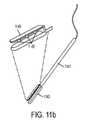
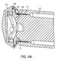
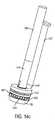
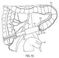
本発明はまた、燐光寿命に基づいて取り込まれた酸素を感知する能力を伴う、外科手術用ステープラアンビルを含むことができる。さらなる実施形態では、アンビルは、感知に利用されるカメラを備える。外科手術用ステープラアンビルの1つの構成は、燐光寿命に基づいて、2つまたはそれを上回る点に酸素をマップする能力を組み込む。さらなる実施形態では、アンビルは、温度感知を組み込み、温度マップは、酸素測定のマップを補償するために使用される。プローブのマイクロ注入のために統合される針を伴う、感知器具が、本発明において教示される。1つの構成はさらに、外科手術用ステープラアンビルと結合し、媒体をアンビルの作業表面(すなわち、ステープル形成表面)またはその近傍の組織の中に注入するための注入器を備える。媒体は、1つまたはそれを上回る燐光酸素感知プローブ変形を含有する。本発明はまた、外科手術用ステープラアンビルに結合し、アンビルの作業表面(すなわち、ステープル形成表面)またはその近傍の組織の酸素化を査定する、燐光寿命に基づいて取り込まれた酸素を感知する能力を伴う、独立型器具を含む。器具は、回転する、または別様に吻合部を完全に撮像するように構成される、1つまたはそれを上回るセンサを含有する。また、含まれるのは、プローブを送達する、および/または組織酸素化を感知するための統合された注入器を伴う、インテロゲータワンドである。注入器は、結腸壁の内側等の組織内部注入または結腸壁を通して外部から等の外部注入を可能にしてもよい。器具はさらに、作業表面における組織の温度を測定するための手段を備える。本発明のさらなる実施形態では、カメラベースの燐光寿命撮像システムは、外科手術用ステープラのアンビルを取り付けるための手段を備え、取付は、アンビルへのクイックリリース式結合器を通して行われる。本システムは、外科手術中、吻合部の酸素化マップを生成するように構成される。本システムはさらに、PLIシステムと実質的に同一領域を撮像し、その測定を使用して、酸素化測定の正確度を向上させる、熱撮像カメラを備える。 The present invention can also include a surgical stapler anvil with the ability to sense entrapped oxygen based on phosphorescence lifetime. In a further embodiment, the anvil comprises a camera that is used for sensing. One configuration of a surgical stapler anvil incorporates the ability to map oxygen to two or more points based on phosphorescence lifetime. In a further embodiment, the anvil incorporates temperature sensing and the temperature map is used to compensate the oximetry map. A sensing instrument is taught in the present invention with a needle integrated for microinjection of the probe. One configuration further includes an injector for coupling with the surgical stapler anvil for injecting a medium into tissue at or near the working surface (ie, staple forming surface) of the anvil. The medium contains one or more phosphorescent oxygen-sensing probe variants. The present invention also provides the ability to sense incorporated oxygen based on phosphorescence lifetime coupled to a surgical stapler anvil to assess oxygenation of tissue at or near the working surface (i.e., staple forming surface) of the anvil. including stand-alone instruments with The instrument contains one or more sensors that are configured to rotate or otherwise fully image the anastomosis. Also included is an interrogator wand with an integrated injector for delivering probes and/or sensing tissue oxygenation. The injector may allow for internal tissue injection, such as inside the colon wall, or external injection, such as externally through the colon wall. The instrument further comprises means for measuring tissue temperature at the work surface. In a further embodiment of the invention, the camera-based phosphorescence lifetime imaging system comprises means for attaching an anvil of a surgical stapler, attachment being through a quick release coupler to the anvil. The system is configured to generate an anastomotic oxygenation map during a surgical procedure. The system further comprises a thermal imaging camera that images substantially the same area as the PLI system and uses the measurements to improve the accuracy of the oxygenation measurements.
本発明の一実施形態は、既存の大腸内視鏡の作業チャネルに嵌合し、結腸壁の酸素マップを生成する、CMOSマイクロカメラ等の小型の二次撮像システムに基づく。本システムは、同期取得された従来のスコープビデオ画像上のグラフィックオーバーレイを使用して、マップを表示し、および/または疑わしい病変をハイライトする。疑わしい病変が識別される場合、本システムは、ビデオモニタ上にハイライトされた病変を保定/追跡しながら、酸素マップカメラを別の器具に交換することが可能となるであろう。酸素マッピングは、一実施形態では、酸素敏感全身注入分子プローブの燐光寿命撮像(PLI)を使用して実現されるであろう。本発明の一実施形態では、温度感知は、PLIと結合され、酸素濃度の温度補償マップを生成する。本発明は、結腸壁組織内の癌性病変を査定するために、大腸内視鏡とのみ結合されることに制限されない。本発明は、フレキシブルおよび剛性、全ての内部および外部組織の監視または可視化、ならびに組織のパラメータにおける任意のタイプの変動の識別を含む、全てのスコープおよびカメラタイプならびに構成を含む。 One embodiment of the present invention is based on a miniature secondary imaging system, such as a CMOS micro-camera, that fits into the working channel of an existing colonoscope and produces an oxygen map of the colon wall. The system uses graphical overlays on synchronously acquired conventional scope video images to display maps and/or highlight suspicious lesions. If a suspicious lesion is identified, the system would be able to replace the oxygen map camera with another instrument while retaining/tracking the highlighted lesion on the video monitor. Oxygen mapping, in one embodiment, will be accomplished using phosphorescence lifetime imaging (PLI) of an oxygen-sensitive systemically injected molecular probe. In one embodiment of the invention, temperature sensing is combined with PLI to produce a temperature compensated map of oxygen concentration. The present invention is not limited to being coupled solely with a colonoscope to assess cancerous lesions within colon wall tissue. The present invention includes all scope and camera types and configurations, including flexible and rigid, all internal and external tissue monitoring or visualization, and identification of any type of variation in tissue parameters.
本発明の1つの代表的用途は、組織皮弁の生成および監視におけるものである。種々のタイプ、すなわち、乳房、皮膚等の癌は、多くの場合、治癒的切除における試み中、有意な組織体積の除去を生じさせる。外傷性傷害は、四肢の切断または組織の一部の剥離をもたらし得る。結果として生じる組織損失は、多くの場合、患者の身体の他の部分から転置された生来の組織によって置換される。遊離組織皮弁は、供給血管茎とともに、その生来の位置から完全に除去される皮弁である。遊離皮弁血管系は、次いで、組織空隙近傍の脈管に再接続される。血管吻合部は、不適切な血塊形成からの漏出、狭窄、または閉塞に起因して失敗し得る。本発明は、組織潅流の術中確認および術後監視の両方のために、組織酸素化のマップを通した皮弁酸素化を解像することを可能にする。現在の技術は、血流の定質的測定に限定される。本発明は、組織酸素化のリアルタイムな定量的査定を提示する。本発明の実施形態は、カメラベースの燐光寿命検出器と熱撮像カメラを結合し、位置合わせされた温度マップは、燐光寿命を酸素濃度に変換するために使用される較正係数を補正するために使用される。類似構成が、内部および外部組織の両方を監視するために使用されてもよい。別の実施例用途は、末梢血管疾患(PVD)の治療を診断、査定、または監視する際におけるものである。 One exemplary application of the present invention is in the creation and monitoring of tissue flaps. Cancers of various types, ie, breast, skin, etc., often result in the removal of significant tissue volumes during attempts at curative resection. Traumatic injury can result in amputation of a limb or detachment of part of tissue. The resulting tissue loss is often replaced by native tissue displaced from other parts of the patient's body. A free tissue flap is a flap that has been completely removed from its native position along with the feeding vascular pedicle. The free flap vasculature is then reconnected to the vessels near the tissue space. Vascular anastomoses can fail due to leakage, stenosis, or occlusion from improper clot formation. The present invention allows resolving flap oxygenation through a map of tissue oxygenation for both intraoperative confirmation and postoperative monitoring of tissue perfusion. Current technology is limited to qualitative measurements of blood flow. The present invention presents a real-time quantitative assessment of tissue oxygenation. Embodiments of the present invention combine a camera-based phosphorescence lifetime detector with a thermal imaging camera, and the registered temperature map is used to correct the calibration factor used to convert phosphorescence lifetime to oxygen concentration. used. Similar configurations may be used to monitor both internal and external tissue. Another example application is in diagnosing, assessing, or monitoring treatment of peripheral vascular disease (PVD).
他の潜在的用途として、限定ではないが、移植された器官または付属器官、頭蓋内、髄腔内、眼内、大動脈内、鼻腔内、洞様毛細血管内、咽頭内、喉頭内、食道内、気管内、胸郭内、気管支内、心膜内、心臓内、血管内、腹部内、胃内、胆嚢内、腸内、結腸内、直腸内、嚢胞内、尿管内、子宮内、膣内、陰嚢内、脳内、肺動脈内、肝臓内、膵臓内、腎臓内、副腎内、脾臓内、卵巣内、精巣内、陰茎内、筋肉内、骨内、および皮膚内生理学的/生体力学的パラメータの監視/記録が挙げられる。
本発明は、例えば、以下を提供する。
(項目1)
生理学的状態またはその代用物を解像し、マップする、撮像システムであって、前記撮像システムは、2つまたはそれを上回る感知モダリティから得られた情報を利用して、前記生理学的状態またはその代用物を解像し、前記感知モダリティは、前記生理学的状態または測定の絶対測定の改良された正確度を提供するために併用される、撮像システム。
(項目2)
1つの感知モダリティは、媒体と関連付けられた燐光および/または蛍光寿命を査定し、別の感知モダリティは、前記媒体またはその近傍の温度を査定する、項目1に記載の撮像システム。
(項目3)
前記媒体は、近傍酸素濃度に関する燐光寿命を伴うプローブを備え、前記温度測定は、酸素化に対する前記プローブの寿命の温度依存較正係数における変動を補償するために使用される、項目2に記載の撮像システム。
(項目4)
前記システムは、
外部から照明された後の燐光および/または蛍光媒体の減衰寿命を検出するために構成される、光学センサと、
前記光学センサの視野内の1つまたはそれを上回る点において前記温度を検出するための温度センサと、
温度測定を使用して、前記媒体の温度依存寿命変動を補償するように構成される、プロセッサと、
を備える、項目1に記載の撮像システム。
(項目5)
1つのモダリティは、燐光寿命撮像のために構成され、前記システムは、燐光寿命をマップするための光学検出器と、温度を検出するための光学検出器との両方を備え、前記システムは、前記温度および寿命画像を位置合わせし、各マップされた点における燐光寿命および温度の両方を利用して、対応する酸素化を判定するように構成される、項目2に記載の撮像システム。
(項目6)
酸素化を感知し、注入可能プローブの燐光寿命に基づいて、前記酸素化のマップを生成するように構成される、内視鏡撮像システム。
(項目7)
前記酸素化のマップに対応する領域内の温度を検出するためのセンサをさらに備え、前記撮像システムは、熱測定に基づいて、温度依存酸素化測定を補償するように構成される、項目6に記載の撮像システム。
(項目8)
二次撮像スコープが、酸素化を感知するために構成され、前記二次撮像スコープは、一次スコープから独立して動作し、かつ前記一次スコープと連動して動作する、項目6に記載の内視鏡撮像システム。
(項目9)
前記一次スコープは、フレキシブル内視鏡であり、前記二次スコープは、前記内視鏡の器具ポートを通して通過するように構成される、項目8に記載の内視鏡撮像システム。
(項目10)
前記システムは、ビデオ画像および前記酸素化マップを位置合わせするように構成され、さらに、前記システムは、前記ビデオ画像を前記酸素化マップのオーバーレイとともに表示するように構成される、項目8に記載の撮像システム。
(項目11)
前記システムは、酸素化を感知するために構成される前記二次撮像スコープの除去または無効化後、特徴を追跡し、取得された酸素マップの整合を維持する、項目10に記載の撮像システム。
(項目12)
前記システムは、組織内の燐光応答を選択的に励起するように構成され、前記選択的励起は、組織の層または深度と関連付けられた酸素化を一意に識別するための手段を提供する、項目6に記載の撮像システム。
(項目13)
延在アームの先端に取り付けられる光源を備え、前記アームは、組織の領域または層を選択的に照明可能である、項目12に記載の撮像システム。
(項目14)
酸素化マップまたはその対応する前駆体寿命マップを熱画像に位置合わせするステップと、前記位置合わせされた情報を利用し、酸素化測定における温度依存変動を補償するステップとを含む、方法。
(項目15)
複数のカメラからの画像が、近赤外線照明を使用して取得され、前記近赤外線照明は、可視光内視鏡検査カメラを用いて検出可能であり、カメラが、燐光または蛍光応答を検出するように構成され、前記複数のカメラからの画像間の相互情報が、位置合わせのための使用される、項目14に記載の方法。
(項目16)
腸壁の組織酸素化をマップするステップをさらに含む、項目14に記載の方法。
(項目17)
病変と健康な腸壁組織を区別するステップをさらに含む、項目16に記載の方法。
(項目18)
燐光寿命または酸素化の静的画像のパターンマッチングに基づいて、ポリープを区別するステップをさらに含む、項目17に記載の方法。
(項目19)
時系列の画像の動的変化に基づいて、ポリープを区別するステップをさらに含む、項目18に記載の方法。
(項目20)
前記組織酸素化のマップは、前記病変の特定を誘導し、前記病変は、内視鏡ビデオ画像上にオーバーレイされた識別子を用いて識別される、項目18に記載の方法。Other potential uses include, but are not limited to, transplanted organs or adnexa, intracranial, intrathecal, intraocular, intraaortic, intranasal, intrasinusoidal, intrapharyngeal, intralaryngeal, intraesophageal. , intratracheal, intrathoracic, intrabronchial, intrapericardial, intracardiac, intravascular, intraabdominal, intragastric, intragallbladder, intraintestinal, intracolonic, intrarectal, intracystic, intraureteral, intrauterine, intravaginal, Intrascrotal, intracerebral, intrapulmonary, intrahepatic, intrapancreatic, intrarenal, intraadrenal, intrasplenic, intraovarian, intratesticular, intrapenis, intramuscular, intraosseous, and intradermal physiological/biomechanical parameters monitoring/recording;
The present invention provides, for example, the following.
(Item 1)
An imaging system for resolving and mapping a physiological state or proxies thereof, said imaging system utilizing information obtained from two or more sensing modalities to determine said physiological state or its proxies. An imaging system that resolves a surrogate and wherein said sensing modalities are combined to provide improved accuracy of absolute measurement of said physiological condition or measurement.
(Item 2)
The imaging system of item 1, wherein one sensing modality assesses phosphorescence and/or fluorescence lifetime associated with a medium, and another sensing modality assesses temperature at or near said medium.
(Item 3)
Imaging according to item 2, wherein the medium comprises a probe with a phosphorescent lifetime with respect to nearby oxygen concentration, and the temperature measurement is used to compensate for variations in a temperature dependent calibration factor of the lifetime of the probe with respect to oxygenation. system.
(Item 4)
The system includes:
an optical sensor configured to detect the decay lifetime of a phosphorescent and/or fluorescent medium after being externally illuminated;
a temperature sensor for detecting the temperature at one or more points within the field of view of the optical sensor;
a processor configured to compensate for temperature dependent lifetime variations of said media using temperature measurements;
The imaging system according to item 1, comprising:
(Item 5)
One modality is configured for phosphorescence lifetime imaging, said system comprising both an optical detector for mapping phosphorescence lifetime and an optical detector for detecting temperature, said system comprising said 3. The imaging system of item 2, wherein the imaging system is configured to register temperature and lifetime images and utilize both phosphorescence lifetime and temperature at each mapped point to determine corresponding oxygenation.
(Item 6)
An endoscopic imaging system configured to sense oxygenation and generate a map of said oxygenation based on the phosphorescence lifetime of an injectable probe.
(Item 7)
7. In item 6, further comprising a sensor for detecting temperature within a region corresponding to the map of oxygenation, wherein the imaging system is configured to compensate for temperature dependent oxygenation measurements based on thermal measurements. The imaging system described.
(Item 8)
7. The endoscope of item 6, wherein a secondary imaging scope is configured for sensing oxygenation, said secondary imaging scope operating independently of and in conjunction with said primary scope. Mirror imaging system.
(Item 9)
9. The endoscopic imaging system of item 8, wherein the primary scope is a flexible endoscope and the secondary scope is configured to pass through an instrument port of the endoscope.
(Item 10)
9. The system of item 8, wherein the system is configured to align a video image and the oxygenation map, and further wherein the system is configured to display the video image with an overlay of the oxygenation map. imaging system.
(Item 11)
11. The imaging system of item 10, wherein the system tracks features and maintains alignment of acquired oxygen maps after removal or disabling of the secondary imaging scope configured to sense oxygenation.
(Item 12)
wherein said system is configured to selectively excite a phosphorescent response in tissue, said selective excitation providing a means for uniquely identifying oxygenation associated with a layer or depth of tissue; 7. The imaging system according to 6.
(Item 13)
13. The imaging system of item 12, comprising a light source attached to a distal end of an extending arm, said arm being capable of selectively illuminating regions or layers of tissue.
(Item 14)
A method comprising registering an oxygenation map or its corresponding precursor lifetime map to a thermal image, and utilizing said registered information to compensate for temperature dependent variations in oxygenation measurements.
(Item 15)
Images from multiple cameras are acquired using near-infrared illumination, said near-infrared illumination detectable with a visible light endoscopy camera, such that the camera detects phosphorescence or fluorescence responses. 15. The method of item 14, wherein mutual information between images from said multiple cameras is used for registration.
(Item 16)
15. The method of item 14, further comprising mapping tissue oxygenation of the intestinal wall.
(Item 17)
17. The method of item 16, further comprising distinguishing between diseased and healthy intestinal wall tissue.
(Item 18)
18. The method of item 17, further comprising distinguishing polyps based on pattern matching of static images of phosphorescence lifetime or oxygenation.
(Item 19)
19. The method of item 18, further comprising distinguishing polyps based on dynamic changes in time-series images.
(Item 20)
19. The method of item 18, wherein the map of tissue oxygenation guides the identification of the lesion, and the lesion is identified using an identifier overlaid on the endoscopic video image.
本発明の付加的特徴、利点、および実施形態が、以下の発明を実施するための形態、図面、ならびに請求項に記載されている、またはその検討から明白である。さらに、前述の発明の概要および以下の発明を実施するための形態は両方とも、例示であって、本発明の範囲を請求される通り限定することなく、さらなる説明を提供することを意図することを理解されたい。 Additional features, advantages, and embodiments of the invention are set forth in or apparent from consideration of the following detailed description, drawings, and claims. Furthermore, both the foregoing Summary of the Invention and the following Detailed Description are intended to be exemplary and to provide further explanation without limiting the scope of the invention as claimed. Please understand.
組織パラメータは、種々の方法によって測定されることができる。本発明によって利用される1つの技法は、測定方法を開示し、参照することによってその全体として本明細書に組み込まれる、米国特許第4,947,850号、米国特許第5,837,865号、米国特許第6,362,175号、米国特許第6,165,741号、米国特許第6,274,086号、米国特許第7,575,890号、および米国特許出願公開第2013/0224874号に開示されるように、酸素測定のための全身または局所的に注入された燐光酸素感知分子プローブを介し、燐光の酸素依存消光を利用することを介して、組織酸素化レベルを測定する。燐光酸素感知プローブは、発色団を環境との直接接触から隔離し、酸素拡散を制御し、プローブのダイナミックレンジおよび感度にわたる制御を可能にする、保護シェルを形成する、疎水性デンドリマーの内側にカプセル化された燐光性金属ポルフィリンコアを備える。金属ポルフィリンコアは、異なる元素とともに構築されることができる。パラジウムおよび白金は、利用され得る2つの元素である。パラジウム系コアに優る白金系コアの利点は、その量子効率である。燐光体の量子効率の増加は、Pd系分子と比較して、光出力の有意な増加を可能にする。分子あたり返される光が多いほど、デバイスに返される同一信号を達成するために、より少ない分子の使用を可能にする。代替として、同一量の分子の注入は、低敏感性(安価な)光検出器の使用を可能にする。デンドリマー分岐の周辺ペグ化は、生物学的マクロ分子との相互作用を防止しながら、プローブの高水性溶解度を確実にする。分子プローブの全体的サイズは、腎臓によって排除されるプローブの能力に影響を及ぼす。排除が速いほど、患者への剤の暴露を限定する。サイズは、デンドリマーの長さ、デンドリマーの数、およびPEGのサイズ/ペグ化の範囲の修正を通して変動されることができる。 Tissue parameters can be measured by a variety of methods. One technique utilized by the present invention is U.S. Pat. No. 4,947,850, U.S. Pat. , U.S. Pat. No. 6,362,175, U.S. Pat. No. 6,165,741, U.S. Pat. No. 6,274,086, U.S. Pat. Tissue oxygenation levels are measured via systemically or locally implanted phosphorescent oxygen-sensing molecular probes for oxygen measurements, as disclosed in US Pat. Phosphorescent oxygen-sensing probes are encapsulated inside a hydrophobic dendrimer that forms a protective shell that isolates the chromophore from direct contact with the environment, controls oxygen diffusion, and allows control over the dynamic range and sensitivity of the probe. with a phosphorescent metallo-porphyrin core. Metalloporphyrin cores can be constructed with different elements. Palladium and platinum are two elements that may be utilized. The advantage of platinum-based cores over palladium-based cores is their quantum efficiency. The increased quantum efficiency of phosphors allows a significant increase in light output compared to Pd-based molecules. More light returned per molecule allows the use of fewer molecules to achieve the same signal returned to the device. Alternatively, injection of the same amount of molecules allows the use of less sensitive (cheaper) photodetectors. Peripheral pegylation of dendrimer branches ensures high aqueous solubility of the probe while preventing interactions with biological macromolecules. The overall size of a molecular probe affects its ability to be cleared by the kidney. Faster elimination limits exposure of the agent to the patient. Size can be varied through modification of dendrimer length, dendrimer number, and PEG size/pegylation range.
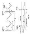
プローブの一実施形態では、コアであるPd-メソ-テトラ-(3,5-ジカルボキシフェニル)テトラベンゾポルフィリン(PdTBP)は、第8世代2ポリ-アリールグリシン(AG2)デンドロンによってカプセル化され、それぞれ、平均して21~22単量体-(CH2CH2O)-単位を有する、モノメトキシ-ポリエチレングリコールアミン(PEG-NH2)基(平均MW1,000Da)でペグ化される。プローブデンドリマーの分子量は、MALDI質量分光法によって判定されるように、最大35,354Daを伴う、約26,000~44,000Daの範囲内であることが見出された。燐光消光法は、環境内の励起された三重項状態分子の燐光を消光する分子酸素(O2)の能力に依拠する。生物系では、酸素による燐光消光は、拡散制御方式で生じ、O2は、十分に高濃度中に存在する唯一の小分子動的消光剤であるため、O2に非常に特異的である。生物濃縮の範囲を通した酸素分圧(pO2)への燐光寿命(τ)の依存性は、Stern-Volmerの方程式:1/τ=1/τ0+kq×pO2によって詳しく説明されており、式中、τは、指定された酸素圧力pO2における燐光寿命であって、τ0は、酸素不在(pO2=0)における燐光寿命であって、kqは、消光定数である。1つの分子酸素プローブは、消光定数kq約326mmHg-1s-1、生理学的pH6.2~7.8の範囲にわたって210μsのτ0、および一定温度36.5℃を有する。In one embodiment of the probe, the core Pd-meso-tetra-(3,5-dicarboxyphenyl)tetrabenzoporphyrin (PdTBP) is encapsulated by generation 8 2-poly-arylglycine (AG2) dendrons, Each is pegylated with a monomethoxy-polyethyleneglycolamine (PEG-NH2) group (average MW 1,000 Da), which has an average of 21-22 monomer-(CH2CH2O)- units. The molecular weight of the probe dendrimer was found to be in the range of about 26,000-44,000 Da with a maximum of 35,354 Da as determined by MALDI mass spectroscopy. The phosphorescence quenching method relies on the ability of molecular oxygen (O2 ) to quench the phosphorescence of excited triplet state molecules in the environment. In biological systems, phosphorescence quenching by oxygen occurs in a diffusion- controlled manner and is highly specific forO2 , as it is the only small-molecule dynamic quencher present in sufficiently high concentrations. The dependence of phosphorescence lifetime (τ) on oxygen partial pressure (pO2 ) throughout the range of bioaccumulation is elaborated by the Stern-Volmer equation: 1/τ = 1/τ0 + kq x pO2 , where where τ is the phosphorescence lifetime at the specified oxygen pressure pO2 ,
プローブの較正パラメータkqおよびτ0は、温度に対して線形に変化する。消光定数kqは、温度係数7.8mmHg-1s-1/℃に対応する、22℃~38℃の温度の上昇に伴って、211mmHg-1s-1~338mmHg-1s-1に増加する。プローブの吸収スペクトルは、燐光放出最大値813nmを伴う、約448nmおよび637nmにおいて最大値を有する。複数の波長における励起は、異なる透過深度または層における組織特性を調べ、区別可能である用途特異的利点をもたらす。視野内の複数のpO2値の組み合わせは、寿命の組み合わせ(指数関数的減衰の和)として現れるであろう。複数のpO2値および対応する濃度は、本明細書に説明される手段を通して判定されることができる。The probe calibration parameters kq and τ0 vary linearly with temperature. The quenching constant kq increases from 211 mmHg−1 s−1 to 338 mmHg−1 s−1 with increasing temperature from 22° C. to 38° C., corresponding to a temperature coefficient of 7.8 mmHg−1 s−1 /°C. . The absorption spectrum of the probe has maxima at about 448 nm and 637 nm with a phosphorescent emission maximum of 813 nm. Excitation at multiple wavelengths provides the application-specific advantage of probing and distinguishing tissue properties at different penetration depths or layers. Combinations of multiplepO2 values in the field will appear as lifetime combinations (sums of exponential decays). MultiplepO2 values and corresponding concentrations can be determined through the means described herein.
温度への測定された燐光寿命の依存性に起因して、測定部位における温度を査定し、その情報を使用して、燐光寿命と酸素濃度との間の適切な関係を適用することが必須である。測定点における温度を測定することによって、適切な温度依存消光係数kqが、選択され、その点における酸素濃度測定の改良された正確度を可能にし得る。測定領域の平均温度は、正確度を改良するために使用され得るが、さらなる場所依存補償が、酸素濃度に変換するとき、複数の点に温度をマップし、それらの点の対応性と寿命測定を関連させることを通して得られ得る。酸素濃度および酸素化は、本開示では同じ意味で使用され得、両方とも、組織内に存在する酸素の量に関連することに留意されたい。 Due to the dependence of the measured phosphorescence lifetime on temperature, it is essential to assess the temperature at the measurement site and use that information to apply the appropriate relationship between phosphorescence lifetime and oxygen concentration. be. By measuring the temperature at the point of measurement, an appropriate temperature dependent extinction coefficient kq can be selected to allow improved accuracy of oxygen concentration measurements at that point. The average temperature of the measurement area can be used to improve accuracy, but additional location-dependent compensation, when converted to oxygen concentration, maps the temperature to multiple points and the correspondence between those points and the lifetime measurement. can be obtained through associating Note that oxygen concentration and oxygenation may be used interchangeably in this disclosure and both relate to the amount of oxygen present in tissue.
本発明の実施形態は、周囲正常組織に対して測定されるとき、非癌性、前癌性、および癌性病変の間質組織酸素化における定量的差異を検出することが意図される。さらに説明される具体的実施形態は、胃腸管内の病変を識別することを目的とする。本発明の1つの用途は、前癌性結腸ポリープの検出の向上を対象とする。ビデオ大腸内視鏡検査中の間質組織酸素化のマッピングを通して、本発明は、従来の白色光および「拡張」内視鏡技法と比較して、大腸内視鏡検査スクリーニング中の前腫瘍性および腫瘍性病変の検出を改良することを目的とする。さらに、本発明は、組織酸素化のパターンに基づいて、種々の悪性潜在性の病変を分化することを目的とする。本願の目的のために、「白色光」および「可視光」撮像は、同じ意味で使用され得ることに留意されたい。同一着目領域内に温度を同時にマップすることによって、組織酸素化に対する寿命の温度依存較正を使用することにより、感知正確度を改良することができる。本願の目的のために、燐光寿命画像/撮像(PLI)とは、酸素化等の生理学的パラメータを計算するために使用される前駆体を指し、マイクロ秒単位で測定されるような実際の較正される寿命であってもよく、またはクロックサイクル、カメラフレーム、位相遅延、もしくは他の測定パラメータを含む、関連未加工データによって表されてもよいことに留意されたい。 Embodiments of the present invention are intended to detect quantitative differences in interstitial tissue oxygenation of non-cancerous, precancerous, and cancerous lesions when measured against surrounding normal tissue. Further described specific embodiments are directed to identifying lesions within the gastrointestinal tract. One application of the present invention is directed to improved detection of precancerous colonic polyps. Through mapping interstitial tissue oxygenation during video colonoscopy, the present invention demonstrates the potential for preneoplastic and preneoplastic disease during colonoscopy screening compared to conventional white light and "extended" endoscopic techniques. The aim is to improve the detection of neoplastic lesions. Furthermore, the present invention aims at differentiating different malignant potential lesions based on tissue oxygenation patterns. Note that for the purposes of this application, "white light" and "visible light" imaging may be used interchangeably. By simultaneously mapping temperature within the same region of interest, sensing accuracy can be improved by using a temperature dependent calibration of lifetime to tissue oxygenation. For the purposes of this application, phosphorescence lifetime imaging/imaging (PLI) refers to the precursor used to calculate physiological parameters such as oxygenation and the actual calibration as measured in microseconds. , or represented by relevant raw data, including clock cycles, camera frames, phase delays, or other measured parameters.
現在、大腸内視鏡検査中、組織酸素化を定量的に査定する臨床上実践的方法、またはそのような情報を利用して、ポリープ検出を改良する方法は、存在しない。本アプローチはまた、限定ではないが、結腸または直腸吻合部等の外科手術手技を誘導するための胃腸撮像を含む、種々の他の組織撮像のために使用されてもよい。用語「撮像」は、複数の場所において測定を行うことを指す。これは、限定ではないが、カメラベースのセンサ等の2Dマップまたは器具上の複数のセンサ要素等の離散点のアレイを含む。 Currently, there is no clinically practical method to quantitatively assess tissue oxygenation during colonoscopy, or to use such information to improve polyp detection. This approach may also be used for various other tissue imaging, including but not limited to gastrointestinal imaging to guide surgical procedures such as colonic or rectal anastomosis. The term "imaging" refers to taking measurements at multiple locations. This includes, but is not limited to, a 2D map such as a camera-based sensor or an array of discrete points such as multiple sensor elements on an instrument.
図1は、酸素感知分子プローブ101と、燐光寿命撮像インターフェース103と、二次カメラ105とを備える、本発明の一実施形態の構成要素である、内視鏡システム100の表現を示す。本システムは、健康な組織と病変との間に存在する酸素差異の利用を通して、非癌性、前癌性、および癌性病変の検出を補助する。本システムは、胃腸管の定量的酸素マッピングを生成する。病変の実施例は、結腸内に見出される、結腸ポリープである。酸素敏感燐光酸素感知プローブ101は、血流の中に全身にわたって、または組織間質空間の中に局所的に注入されたナノセンサである。燐光寿命撮像インターフェース103は、燐光プローブ101の光学応答(燐光寿命の酸素依存消光に関連)に基づいて、組織酸素化を判定する。燐光寿命は、大腸内視鏡等の従来のスコープの作業チャネルを通して通過される、二次カメラ105によって撮像される。一実施形態では、二次撮像システム105は、従来の大腸内視鏡の作業チャネルの近位端の中に挿入され、大腸内視鏡の遠位端まで通過される、小型マイクロカメラ内視鏡である。カメラは、フレキシブルカニューレの中に統合されてもよく、単回使用または限定寿命デバイスであってもよい。別の実施形態では、二次撮像システムは、作業チャネルの近位端の中に挿入され、遠位に通過される、光ファイバ撮像束を備える。撮像束の近位端は、カメラに結合される。別の実施形態では、撮像束の近位端は、カメラに結合される、画像増強装置に結合される。カメラ自体は、画像増強光学を採用してもよい。撮像束の中にまたはそこから通過する光は、光学フィルタを通して通過してもよい。本システムはさらに、酸素化測定の領域内の温度をマップ可能な熱撮像システムを備えてもよい。一実施形態では、赤外線放射が、コヒーレント光ファイバ束を通して熱撮像カメラまで通過される。光ファイバ束は、二次撮像システム内で使用されるものと同一である、照明のためにも使用される束である、または代替束であってもよい。 FIG. 1 shows a representation of an endoscopic system 100 comprising an oxygen-sensing
本発明は、身体またはその一部内で制御された滞留時間を可能にする、酸素依存消光分子プローブ101の構成を含む。プローブのサイズおよび形状の制御された変動は、滞留時間および排除率に影響を及ぼす。一実施形態では、プローブは、24時間未満のうちに身体から排泄される。分子プローブ101は、注入後、長時間、ある部位に十分なプローブを維持するように、完全または部分的に、生体吸収性ビーズもしくは他の物体の中に統合される、またはそれと結合されてもよい。プローブをある部位に維持する、またはプローブをある部位に向かって指向する、代替アプローチとして、その位置の制御のため、または長時間十分なプローブをある部位に維持するための、分子プローブと磁気担体の結合が挙げられる。本発明はまた、グルコースレベル、pH、乳酸、または疾患マーカ等、酸素化以外の種々の生理学的パラメータのために、燐光体および他のマーカを組み込んでもよい。複数の生理学的パラメータの測定は、同時に生じることができる。 The present invention includes the construction of oxygen-dependent quenching
図2aは、システムが、同期取得された内視鏡ビデオ画像201上への対象組織内のポリープもしくは他の異常を含む病変または血管系(203、209)の場所を識別するグラフィックオーバーレイ(205、211)を生成する、本発明の実施形態を描写する。組織201は、結腸組織を表すが、しかしながら、内視鏡システムは、任意の生体組織を撮像するために使用されることができる。図2bは、擬色半透明オーバーレイ219として現れ得る、組織酸素化(221、233)のマップを提示する、オーバーレイの実施形態を示す。他の実施形態では、他の生理学的特性が、対応する解剖学的撮像とともに表示されてもよい。本システムは、組織襞等のいくつかの視覚的閉塞を通して酸素化を測定可能であって、典型的には、襞215または他の障害物の背後に隠された病変を特定可能である。一実施形態では、本システムは、自動的に、疑わしい病変(203、209)を識別し、解像された酸素化(221、223)に基づいて疑わしい病変(205、211)をハイライトする、オーバーレイを生成する。オーバーレイは、一般的マーク(例えば、十字線または図2aにおけるようなボックス)、病変の輪郭、確率マップ、および図2bに示されるようなオーバーレイされた酸素マップのうちの1つであってもよい。本識別プロセスは、測定が、非癌性、前癌性、または癌性病変、もしくは他の着目特徴の存在を示すかどうかを査定する際、統計的データを組み込んでもよい。結腸病変は、限定ではないが、炎症性、肥厚性、腺腫性、または管状絨毛性ポリープを含むことができる。さらに、査定と関連付けられた確実性のレベルを組み込んでもよい。オーバーレイされた酸素マップ(または他の特性)は、整合を確実にするように、内視鏡ビデオに位置合わせされてもよい。さらなる実施形態では、内視鏡ビデオ内の特徴は、二次撮像システム/カメラシステムが除去される場合でも(器具を作業チャネルを辿って挿入するように)、画像オーバーレイを維持するように、双方向で更新される画像ベースの位置合わせプロセスを通して追跡される。一実施形態では、電磁追跡センサまたは慣性測定センサ等のセンサが、撮像システムの定位置における絶対位置または相対変化を監視するために使用される。本システムは、酸素化に基づいて、病変または他の生理学的構造を自動的に検出するように構成されてもよく、ライブ酸素マップビデオフィードまたは静的画像に測定を表す擬色(すなわち、色マップ)でレンダリングされてもよい。本システムは、血管系を検出および特定し、1つの構成では、本情報を使用して、外科手術介入を誘導するように構成されてもよい。そのような誘導は、脈管を特定するのに役立てるために使用されてもよく、脈管への非意図的損傷を回避するために使用されてもよい。 FIG. 2a shows a graphical overlay (205, 209) in which the system identifies the location of lesions or vasculature (203, 209) containing polyps or other abnormalities in the tissue of interest onto synchronously acquired
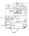
図3は、酸素マッピングシステムが、制御ユニット303と、光源305(組み合わせられたまたは別個の構成要素であってもよい)とを含む、市販の内視鏡インターフェースユニットとシームレスに結合する、酸素マッピングシステム制御ユニット301を含む、本発明の実施形態を示す。光経路内の結合器309は、二次光源311から生じる要求される変調された光を既存の照明ファイバ束315の中に注入し、必要に応じて、フレキシブル内視鏡319を通して、潅注または他の接続を通して通過するために使用されることができる。疑わしい病変が識別される場合、本システムは、ビデオモニタ上のハイライトされた病変を保定/追跡しながら、別の器具との酸素マッピングカメラ323の交換を可能にすることができる。酸素マッピングは、全身に注入された分子プローブを利用する燐光の酸素依存消光を使用して、実現されることができる。一実施形態では、二次ビデオフィード323は、PLI撮像のために使用されることができ、カテーテルの遠位先端におけるマイクロカメラへの電気接続の形態をとってもよい。代替実施形態では、二次ビデオフィード323は、光を酸素マッピングシステム制御ユニットの内側の撮像システムに指向する、コヒーレント光ファイバ束の形態をとってもよい。撮像システムは、マイクロカメラ(CMOS画像センサ等)、従来のカメラ(CMOSまたはCCDカメラユニット等)であってもよく、または本開示にさらに説明されるような増感科学撮像システムであってもよい。一実施形態では、酸素マッピングシステムはまた、温度測定能力を備える。1つの構成では、赤外線光を通過可能なコヒーレント光ファイバ束が、熱撮像のために使用されることができる。光ファイバ束は、独立束、照明ファイバ束の多重化使用、または酸素マッピング二次撮像システムのファイバ束の多重化使用であってもよい。代替構成では、熱電対等の離散点温度が、撮像部位における組織温度を査定するために使用されることができる。なおもさらなる構成では、深部体温を測定するための外部感知システムが、PLIシステムの中にフィードすることができる。 FIG. 3 illustrates oxygen mapping where the oxygen mapping system mates seamlessly with a commercially available endoscopic interface unit, including
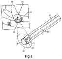
図4は、大腸内視鏡等の内視鏡範囲405の器具チャネル作業ポート403内に嵌合する、マイクロカメラ内視鏡401の一実施形態の拡大図を示す。入射光が、光ファイバ409またはLED等の統合された光源を通して放出され、標的組織もしくは物体からの燐光、蛍光、または他の光再放出応答を引き出す。制御回路を含み得るマイクロCMOSセンサ等のカメラ413が、入射光を除去する、再放出された光を残す、光学フィルタ415(ロングパスフィルタ)の近位に置かれる。感知アプローチは、時間ドメイン、周波数ドメイン、または代替方法であってもよい。時間ドメイン方法の使用は、フィルタ415の必要性を排除する、またはその要求される光学密度を低減させ得る。広角レンズ419が、広視野を得るために使用されてもよい。マイクロレンズが、カメラセンサ上に組み込まれてもよい。カメラ413およびレンズ419は、組織の角度付けられたビューを提供するように構成されてもよい。マイクロ熱撮像カメラがさらに、撮像部位における組織温度を査定するために組み込まれてもよい。カメラは、独立デバイスまたはPLIおよび温度マッピングの両方が可能な組み合わせられた撮像機であってもよい。1つまたはそれを上回る点における温度測定のための当分野で公知の付加的アプローチもまた、組み込まれてもよい。カメラまたは複数のカメラは、そのデータをケーブル423に沿って通過させる。二次撮像システムは、フレキシブル外側シース427内に含有されることができる。 FIG. 4 shows an enlarged view of one embodiment of a
遠位撮像(例えば、先端におけるマイクロカメラ)または近位撮像(例えば、外部カメラまでのファイバ束)のいずれかに基づくPLIシステムの一実施形態は、超広視野角を提供することができる。大視野角を提供することによって、ポリープ、組織襞、狭窄、または吻合部等の物体の背後を視覚化することが可能となり得る。撮像システムの遠位端は、物体の後側を見るために十分なビューを提供するように、能動的に撓曲されることが可能であってもよい。本実施形態は、延在されると所定の湾曲形状を提供するように、事前屈曲形状記憶合金を組み込んでもよい。一実施形態では、円筒プリズム状デバイスが、非常に大きい屈折角度、したがって、180度を上回るビューを生成するために使用される。別の構成では、高屈折率媒体の積層された層が、超広視野角レンズを作成するために利用される。 An embodiment of a PLI system based on either distal imaging (eg, a micro-camera at the tip) or proximal imaging (eg, a fiber bundle to an external camera) can provide ultra-wide viewing angles. By providing a large viewing angle, it may be possible to visualize behind objects such as polyps, tissue folds, strictures, or anastomoses. The distal end of the imaging system may be able to be actively flexed to provide a sufficient view to see the back side of the object. This embodiment may incorporate a pre-bent shape memory alloy to provide a predetermined curved shape when extended. In one embodiment, a cylindrical prismatic device is used to produce a very large angle of refraction and therefore a view greater than 180 degrees. In another configuration, stacked layers of high refractive index media are utilized to create an ultra-wide viewing angle lens.
本発明の一実施形態では、媒体は、蛍光または燐光酸素感知分子プローブを含有することができる。光源は、LEDもしくはレーザ等の狭帯域光源であってもよく、または白色光源等の広帯域源であってもよい。狭帯域源のピーク放出波長は、媒体内の分子プローブの吸収ピークまたはその近傍であるように選択されることができる。光学フィルタが、入射光を分子プローブの吸収波長領域内またはその近傍の波長にさらに制限するために使用されてもよい。分子プローブは、光を再放出することができ、これは、次いで、随意に、フィルタを通して通過し、放出光を入射光から隔離する。光検出器は、受信された光の強度を感知することができる。1つの構成では、検出器は、PD、APD、SiPM、または類似デバイス等の単一点検出器であることができる。代替構成では、検出器は、カメラ等の多点検出器もしくは画像センサまたは単一点検出器のアレイであることができる。カメラは、CCD、CMOS、または他の技術であってもよく、器具の組織接触表面に直接ある、または光ファイバ束等を通して遠隔場所に光学的に結合されてもよい。単一点検出器のアレイは、PDアレイ、SiPMアレイ、線形CCD、または他の技術であってもよい。光源は、広い面積にわたって指向される、または着目点に精密に指向され、走査されてもよい。光検出器は、広い面積にわたって指向される、または着目点に精密に指向され、走査されてもよい。1つの構成では、プロセッサは、光源からの光パルスをコマンドし、時間ドメイン信号処理技法を使用して、検出器によって受信された信号の時間応答を分析する。代替構成では、プロセッサは、1つまたはそれを上回る光源から正弦波強度プロファイル等の変調された光をコマンドすることができ、周波数ドメイン信号処理技法を通して、検出器から測定された信号を分析し、位相遅れを判定することができる。1つの構成では、媒体は、燐光分子プローブを含有することができる。プローブは、プローブの吸収帯内の光の波長によって励起されると、燐光を発する。燐光寿命は、燐光を消光する酸素の能力に起因して、プローブ近傍内の酸素含有量に応答し得る。酸素化と燐光寿命間の関係は、Stern-Volmer関係に従い得る。時間ドメインまたは周波数ドメイン技法が、信号プロセッサによって使用され、組織の単一場所または複数の場所における対応する酸素含有量もしくは濃度を定量的に解像してもよい。用語「解像する」とは、標的組織内の酸素含有量または濃度に関する解を計算、算出、判定、査定、または取得することを広く意味するように解釈されることが意図される。時間ドメインまたは周波数ドメイン技法の例示的実装は、米国特許第6,701,168号に開示されており、参照することによってその全体として本明細書に組み込まれる。酸素含有量は、器具または外部ディスプレイユニット上に、数として表される、または酸素化のマップとして示されてもよい。酸素含有量は、外科手術手技の成功もしくは失敗の可能性を予測する、または外科手術手技を誘導するために使用されてもよい。予測または誘導技法の例示的実装は、米国特許公開第2009/0054908A1号に開示されている。一実施形態では、器具は、内視鏡撮像システムである。別の実施形態では、器具は、外科手術用ステープラアンビルの付属品等の外科手術器具の付属物であってもよい。 In one embodiment of the invention, the medium may contain fluorescent or phosphorescent oxygen-sensing molecular probes. The light source may be a narrowband source such as an LED or laser, or a broadband source such as a white light source. The peak emission wavelength of the narrowband source can be chosen to be at or near the absorption peak of the molecular probe in the medium. Optical filters may be used to further limit the incident light to wavelengths within or near the absorption wavelength region of the molecular probe. The molecular probe can re-emit light, which is then optionally passed through a filter to isolate the emitted light from the incident light. A photodetector can sense the intensity of the light received. In one configuration, the detector can be a single point detector such as a PD, APD, SiPM, or similar device. In alternative configurations, the detector can be a multi-point detector such as a camera or an image sensor or an array of single-point detectors. The camera may be a CCD, CMOS, or other technology, and may be directly on the tissue-contacting surface of the instrument, or optically coupled to a remote location, such as through a fiber optic bundle. The array of single point detectors may be a PD array, SiPM array, linear CCD, or other technology. The light source may be directed over a large area or precisely directed to a point of interest and scanned. The photodetector may be directed over a large area or precisely directed to a point of interest and scanned. In one configuration, the processor commands light pulses from the light source and uses time domain signal processing techniques to analyze the time response of the signal received by the detector. In an alternative arrangement, the processor can command modulated light, such as a sinusoidal intensity profile, from one or more light sources, analyze the measured signal from the detector through frequency domain signal processing techniques, A phase lag can be determined. In one configuration, the medium can contain phosphorescent molecular probes. The probe phosphoresces when excited by a wavelength of light within the absorption band of the probe. Phosphorescence lifetime can be responsive to oxygen content within the vicinity of the probe due to oxygen's ability to quench phosphorescence. The relationship between oxygenation and phosphorescence lifetime can follow the Stern-Volmer relationship. Time-domain or frequency-domain techniques may be used by the signal processor to quantitatively resolve the corresponding oxygen content or concentration at a single location or multiple locations in the tissue. The term "resolve" is intended to be interpreted broadly to mean calculating, calculating, determining, assessing, or obtaining a solution regarding oxygen content or concentration within a target tissue. An exemplary implementation of time-domain or frequency-domain techniques is disclosed in US Pat. No. 6,701,168, incorporated herein by reference in its entirety. Oxygen content may be expressed as a number or shown as a map of oxygenation on the instrument or external display unit. Oxygen content may be used to predict the likelihood of success or failure of a surgical procedure, or to guide a surgical procedure. An exemplary implementation of predictive or guided techniques is disclosed in US Patent Publication No. 2009/0054908A1. In one embodiment, the instrument is an endoscopic imaging system. In another embodiment, the instrument may be a surgical instrument appendage, such as a surgical stapler anvil appendage.
図5は、外部制御ユニット505から既存の内視鏡システム511の光経路509の中へと光学経路503(光ファイバケーブル等)に沿って変調された光の注入を可能にすることができる、結合器501の一実施形態を実証する。光経路509は、典型的には、内視鏡カメラコントローラ511内またはそれと関連付けられた標準的内視鏡検査光源からの白色光を通過させる。これは、従来のビデオ撮像のための白色光およびPLI等の感知を組み込むための変調された波長特異的光を可能にするように、従来の内視鏡519の光ファイバ515の多重化を可能にする。光源結合部523は、内視鏡検査光源の光ポート509と噛合する。一実施形態では、モータ付きミラーユニット527が、白色光入力源523と酸素マッピングシステム503からの変調された光源との間で切り替えることができる。代替実施形態では、DLP状デバイス等の固体状態またはMEM切替もしくはミラーが、使用されてもよい。内視鏡結合部529は、標準的内視鏡519に結合し、組み合わせられた光出力を光経路515の中に通過させることができる。 FIG. 5 can enable injection of modulated light from an
図6aおよび6bは、マイクロカメラ内視鏡または光ファイバスコープ601が従来の市販のもしくはカスタマイズされたスコープ603の作業ポートの内側に嵌合し得る、システムの実施形態を示す。図5に説明されるような光結合器607が、PLI制御ユニット611からの光を既存のスコープ照明/光ファイバ613の中に注入することができる。図6aは、PLIシステムのための専用光チャネルと、スコープインターフェースのためのシャッタユニットとを示す。図6bは、光が既存の光経路613に沿って注入される、代替実施形態を示す。 6a and 6b show an embodiment of the system in which a microcamera endoscope or
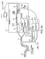
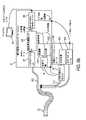
図6cは、コヒーレント光ファイバ撮像束621が内視鏡603の作業チャネル/ポート623を辿って通過するように構成される、撮像システムの実施形態を描写する。内視鏡603は、光ファイバ撮像フレキシブル内視鏡または遠位端625に統合されたマイクロカメラを伴うフレキシブル内視鏡であってもよい。二次撮像ファイバ束621は、撮像システム631と結合する。一実施形態では、撮像システム631は、ゲート画像増強装置と、高感度高速カメラとを備える。撮像システム631は、燐光寿命撮像システム635と結合されることができる。PLIシステム635は、カメラ暴露タイミング、増強装置ゲーティング、および光源639の変調を制御する。変調された源639からの励起光は、結合器607を用いて、従来の市販の内視鏡コントローラ645の可視光源643と組み合わせられ、光ファイバ613を介して、スコープ603の中にフィードされてもよい。従来の市販の内視鏡撮像システム645からのカメラ制御ユニット649のビデオフィードは、内視鏡ビデオ画像をPLIシステム611に伝送することができる。PLIシステム611の画像処理ユニット653は、燐光寿命撮像および白色光内視鏡検査撮像からのビデオ画像を位置合わせすることができる。市販の内視鏡システム645からのビデオ画像は、白色光ビデオのみであってもよく、または白色光画像ならびに赤外線画像の組み合わせであってもよい。 FIG. 6 c depicts an embodiment of an imaging system in which a coherent fiber
変調された光639とともに結合器641の中にフィードされる光源637(図6aおよび図6bに示され、また、図6cに説明される実施形態に適用されてもよい)からの照明に基づく、赤外線(IR)画像の使用は、共通特徴が、カメラユニット649および撮像機631によって捕捉された両画像において可視となり、位置合わせを補助することを可能にすることができる。PLIシステム611の視覚的出力は、内部または外部ディスプレイ657上に表示されることができ、図2aおよび図2bに説明される教示を組み込んでもよい。図6cに関して提供される詳細な説明はまた、図6a、図6b、および本発明の他の実施形態にも適用されることに留意されたい。 Based on illumination from light source 637 (shown in FIGS. 6a and 6b and may also be applied to the embodiment illustrated in FIG. 6c) fed into
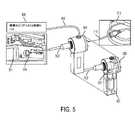
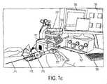
図7aは、外部感知カメラシステム701が測定を生成するために使用されることができる、本発明の一実施形態の概略図を示す。本システムは、外部撮像もしくは観血外科手術手技等のために、レンズに直接結合してもよく、または剛性もしくはフレキシブル内視鏡705に結合してもよい。一実施形態では、対象は、酸素依存燐光プローブが全身にわたって注入されることができ、次いで、対象組織の酸素マップならびにビデオ画像を得るために、システムを用いて撮像される。光源709が、対象の組織711内のプローブを照明および励起するために使用される。代替として、組織自体の蛍光または燐光が、分子プローブの有無にかかわらず、直接照明によって検出されてもよい。本光源709は、異なる分子プローブ、分子プローブの異なる吸収ピークを励起し、光透過深度を変動させるために、複数の波長を含んでもよい(図7aに示される代表的波長は、他の波長が排他的であることを意図するものではない)。離散波長ならびに広帯域源が、使用されてもよい。光源は、LED、レーザ、または他の源であってもよい。源は、光制御システム715によって変調され、時間ドメイン、周波数ドメイン、または他の感知技法を可能にしてもよい。スプリッタ721が、白色光内視鏡画像を得るための撮像カメラ723(可視光カメラ等)と感知カメラ701との間に光を指向するために使用されることができる。一実施形態では、感知カメラは、高速増感科学カメラ701である。フィルタ763は、プローブまたは生来の組織から再放出された光のみの感知カメラへの通過を可能にすることができる。スプリッタ721は、ビームスプリッタ、調節可能ミラー、または光を分割するための別の方法であってもよい。1つの構成では、光は、再放出される燐光を発したIR光を感知カメラに送信するための波長に基づいて分割されることができる一方、可視光は、撮像カメラに向かって指向されることができる。図7bは、システムの一実施形態の詳細を示す。図7cは、臨床前試験におけるシステムの一実施形態を描写する。デバイスは、ヒトにおいて臨床的に、動物用途のために、または実験室シナリオにおいて使用されてもよい。 Figure 7a shows a schematic diagram of one embodiment of the invention in which an external
一実施形態では、プロセッサは、感知カメラ701、撮像カメラ723、および光源709とインターフェースをとることができる。一実施形態では、コンピューティングシステム731は、感知カメラ701に接続されることができ、コンピューティングシステム731のプロセッサは、収集された画像データに計算を行うことができる。計算は、蛍光または燐光寿命もしくは関連パラメータを判定し、マップするために使用されてもよい。コンピューティングシステム731のプロセッサは、マイクロプロセッサおよび/またはグラフィック処理ユニット(GPU)であってもよい。代替構成では、1つまたはそれを上回るカメラからのデータは、フィールドプログラマブルゲートアレイ(FPGA)の中に通過され、FPGAは、蛍光または燐光寿命もしくは関連パラメータの判定およびマッピング等、データ処理の一部または全部を行うように構成される。本発明の一実施形態は、高速撮像センサに結合される、ゲート画像増強装置を組み込む。撮像センサは、FPGAに通信可能に結合される。FPGAは、画像増強装置の撮像(暴露タイミングを含む)およびゲーティングを制御する。FPGAはまた、パルス状または変調された光源を制御することができる。FPGAは、タイミングおよび画像取得を制御することができる。FPGAはまた、取得された画像に画像処理を行う。一実施形態では、FPGAは、測定サイクル毎に燐光または蛍光寿命のマップを判定する。計算の1つのアプローチは、画素毎に指数関数的減衰時間定数を査定することである。FPGA内でオンボード計算を行うことは、高速データ転送の必要性を低減させ、したがって、実施形態は、USB、Ethernet(登録商標)、Firewire、VGAまたはHDMI(登録商標)等の標準的PCビデオ、複合ビデオ、コンポーネントビデオ、または類似物等、従来の通信チャネルを経由した典型的内視鏡カメラに類似するフレームレートで酸素または寿命マップの出力を有し得る。 In one embodiment, the processor can interface with
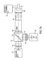
図7bは、剛性またはフレキシブルであり得る、内視鏡741と結合するために構成される、酸素マッピングシステムの一実施形態の概略を示す。光源743は、内視鏡の照明ポートの中にフィードし、白色光および変調された/パルス状励起光を含有してもよい。アダプタ(C-マウント内視鏡アダプタ等)745は、集束光学749を含む、レンズ管747に結合することができる。スプリッタボックス(キューブホルダ等)751は、波長依存ホット(IR)ミラースプリッタの形態をとり得る、スプリッタ753を含有することができる。調節可能レンズ管およびアダプタを含む、集束光学は、スプリッタ751の1つの出力を可視光内視鏡検査カメラ757に結合することができる。スプリッタの他の出力は、集束光学761およびロングパスまたはバンドパス光学フィルタ763を通して通過し、感知カメラ765に到達することができる。ロングパスフィルタは、入射光を効果的に除去し、再放出された光の通過のみ可能にすることができる。フィルタの波長選択性は、使用されるプローブならびに入射光源の光学吸収および放出特性に依存するであろう。感知カメラは、燐光寿命撮像のために使用されることができ、図7aに説明されるような形態をとってもよい。 Figure 7b shows a schematic of one embodiment of an oxygen mapping system configured for coupling with an
図7cは、剛性内視鏡771を用いた小動物追尾のために構成される、代表的酸素マッピングシステムを示す。スコープ771はまた、送気チャネル773を含有することができる、またはそれと結合されることができる。遠隔で選択可能な白色光およびパルス状/変調された光を含む、多波長LED光源775は、スコープ771の照明ポートに結合する。(図7bに説明されるような)フィルタブロック777を伴うレンズアセンブリおよびスプリッタが、スコープ771を感知撮像システム781および可視光撮像システム783に結合する。本実施形態では、感知撮像システム781は、高速ゲーティングを伴う、IR敏感増感カメラである。データ取得システム(DAQ)787等の制御ユニットが、照明波形およびカメラ同期制御を提供し、制御コンピュータ791に結合されてもよい。光源775の照明は、変調光ドライバ789によって制御されることができる。用語「変調光」は、時間ドメインアプローチの場合、パルス状光を、周波数ドメインアプローチの場合、正弦波入力を指し得る。コンピュータ791のディスプレイは、白色光ビデオ内視鏡出力793ならびに計算された酸素および/または燐光寿命マップ795を示すことができる。実験評価のために、ガスミキサ797が、対象の吸入されるO2濃度の制御を可能にする。FIG. 7 c shows a representative oxygen mapping system configured for small animal tracking using a
図8は、マイクロカメラデバイス803が内視鏡の作業チャネル器具ポートを通してその遠位先端807まで通過し、対象組織809を撮像する、システムの実施形態を描写する。本実施形態は、ファイバおよび統合されたビデオスコープの両方との結合に互換性があることができる。本実施形態は、図7aのものと同様に動作することができるが、カメラ803は、スコープの遠位先端807に位置することができる。描写されるシステムは、マイクロカメラに接続される燐光寿命撮像制御システムと、光をスコープの照明ポート815の中に注入するための光結合器811とを示す。PLI制御システム819は、カメラコントローラ821、光源825、およびマップ発生機能性829を制御することができる。一実施形態では、また、外部カメラユニットまたは別の源から可視光画像を取得することができ、組織酸素化またはその代用物のマップをビデオ画像に位置合わせし、拡張現実画像オーバーレイを通して、組織酸素化または他の情報を表示する。 FIG. 8 depicts an embodiment of the system in which a
図9は、感知システムの一実施形態の例示的タイミング図を示す。図は、感知の周波数ドメインアプローチを描写し、励起光が、903として識別される周期Tmod=1/fmodに伴う周波数において変調901され、本システムは、905として識別される繰り返される周期Ts中、時限画像を取得する。Tsは、インクリメントk毎のTs(k)=k×N×Tmod+k×dqとして定義され、式中、k=サンプル番号(0から開始する)であって、N=サンプリング間の周期の数(カメラフレームレートに基づく)であって、Tmod=励起変調正弦波の周期長(1/fmod)であって、dq=後続サンプル毎の周期に沿ったインクリメント(サンプリング間隔に匹敵する)であって、q=k×dq=サンプリングをトリガするための現在のサイクルにおける周期の開始からのオフセットである。光変調波形(上のプロット)は、所望のサンプル数×Nに対する周波数fmodを伴う、正弦波901から成る。各周期は、周期的応答の全範囲を通して撮像トリガ913を掃引するために、909として識別される少量のqだけずらされる。カメラトリガ波形921(下のプロット)は、サンプルk毎に時間Ts(k)において立ち上がりエッジを伴うパルス列から成る。多重累積または積分が、シャッタもしくはゲートを周期の一部と同期させることによって行われてもよい。本部分は、全体的波形を取得するために漸増的にシフトされることができる。波形内に誘発される位相遅れは、次いで、酸素化に関連することができる。複数の周波数が、よりロバストな測定を可能にする、フィルタを通される入射光の除去を補助する、または酸素化スペクトルを判定するために使用されてもよい。代替実施形態では、線形チャープ信号等の時変周波数が、プローブを励起し、多数の周波数から情報を得るために使用される。取得は、複数の周期の繰り返しにわたって連続して行われてもよく、または高速カメラユニットを用いて取得されてもよい。カメラセンサ画素の走査またはビン化は、小サブセットの視野の高速撮像を得るために使用されてもよい。別の実施形態では、類似アプローチが、時間ドメイン測定のために使用されてもよい。本アプローチでは、光学応答に沿った複数の点は、励起パルスが時変位相遅延において一連の繰り返された励起にわたって読み取られた後に減衰する。含まれるのは、複数の連続周期の同期されたゲート撮像を通して低減されたサンプルレートまたはフレームレート要件のためのアプローチである。さらなるアプローチでは、所与の周期の間の複数の取得または累積が、ともに総和され、測定される信号を増加する、もしくはその周期の間の信号対雑音比(SNR)を改良し得る。 FIG. 9 shows an exemplary timing diagram for one embodiment of the sensing system. The figure depicts a frequency domain approach to sensing, in which the excitation light is modulated 901 at a frequency with a period Tmod=1/fmod identified as 903 and the system modulates during repeated periods Ts identified as 905 Acquire timed images. Ts is defined as Ts(k) for each k increment=k×N×Tmod+k×dq, where k=sample number (starting from 0) and N=number of periods between samples (camera frame rate), Tmod = the period length of the excitation modulated sine wave (1/fmod), dq = the increment along the period for each subsequent sample (comparable to the sampling interval), and q = k x dq = offset from start of period in current cycle to trigger sampling. The optical modulation waveform (top plot) consists of a
本発明に含まれるのは、周波数ドメインアプローチに基づいて酸素化を判定するためのアルゴリズムである。アプローチは、単一変調光励起周波数、残留励起光の影響を低減させるための2つの周波数、または複数の酸素化レベルの存在および量(すなわち、酸素化のスペクトルのマップ)を解像するための複数の周波数を含むことができる。実施形態は、周波数がほぼ固定された位相を維持するように調節される、アプローチを含む。本発明は、取得のための最適周波数を判定するための最適化プロセスを含むことができる。ある実施形態では、酸素化計算は、時間ドメインアプローチおよび最大エントロピーアプローチに基づくことができる。代替実施形態は、2つの光子励起技法を利用することができる。 Included in the present invention are algorithms for determining oxygenation based on a frequency domain approach. Approaches include a single modulated optical excitation frequency, two frequencies to reduce the effects of residual excitation light, or multiple frequencies to resolve the presence and abundance of multiple oxygenation levels (i.e., a map of the spectrum of oxygenation). can contain frequencies of Embodiments include approaches in which the frequency is adjusted to maintain a substantially fixed phase. The invention can include an optimization process for determining the optimum frequency for acquisition. In some embodiments, oxygenation calculations can be based on time domain and maximum entropy approaches. Alternative embodiments may utilize two photon excitation techniques.
本発明は、時間ドメイン酸素化測定のためのアプローチを含み、温度測定情報が、測定された燐光寿命から酸素化への変換に組み込まれることができる。さらに含まれるのは、周波数ドメイン酸素化測定のためのアプローチであって、温度測定情報が、燐光応答の測定された位相から酸素化への変化に組み込まれることができる。 The present invention includes an approach for time-domain oxygenation measurements, where thermometric information can be incorporated into the conversion from measured phosphorescence lifetime to oxygenation. Also included are approaches for frequency-domain oxygenation measurements, in which thermometric information can be incorporated into the measured phase-to-oxygenation change of the phosphorescence response.
図10aは、統合されたセンサを伴う、外科手術器具を描写する。一実施形態では、外科手術用ステープラアンビル1001またはアンビルに結合する付属デバイスは、センサを組み込む。アンビルの作業表面1021(ステープル圧着を形成する表面)上のセンサ1005は、器具の作業表面上の組織の燐光寿命撮像を行うための光エミッタおよび受信機を含んでもよい。感知アンビル1001の一実施形態は、アンビル面のカットアウト1007内に位置する、センサ要素1005を備える。感知アンビルは、オンボードバッテリ1011によって給電されるワイヤレス送受信機1009に結合される、制御電子機器を含有する。感知構成要素は、キャップ1013の内側にカプセル化される。 FIG. 10a depicts a surgical instrument with integrated sensors. In one embodiment, the
図10bは、アンビル作業表面1021の拡大図を示す。1つの構成ではLED光源1025およびフォトダイオード1027は、ステープル形成部間のカットアウト1007内に挟入される。圧力センサ1031もまた、組織相互作用力を査定するためにカットアウト1007内にある。器具の一実施形態はさらに、熱電対または抵抗温度検出器(RTD)等の1つまたはそれを上回る温度センサ1033を備える。1つの構成では、温度センサが、ステープル形成部1029間に挟入される、アンビル面1021の周囲に円周方向に分散される、またはステープル形成部間のカットアウト1007内に位置してもよい。代替構成では、カメラが、光ファイバまたは他の光ガイドを通して組織を撮像するように、感知アンビルの中に統合されることができる。本実施形態は、前述のマイクロカメラ内視鏡と機能的に類似することができる。さらなる代替実施形態では、センサは、デバイスを横断して掃引し、複数の点において測定を行うことができる。 10b shows an enlarged view of the
図11aは、感知クリップの形態をとる統合されたセンサを伴う、医療デバイスの実施形態を描写する。ある実施形態では、クリップ1101は、腸組織を横断して封入および感知するように構成されることができる。組織は、上側表面1103と圧縮表面1105との間に置かれることができる。クラスプ1107は、センサを組織の周囲に閉鎖して保持することができる一方、組織圧縮ブラダまたはバルーン1105は、空気もしくは流体接続1109を通して、組織を規定された圧力まで圧縮することができる。1つまたはそれを上回るセンサ1111が、表面1103の組織接触部分に沿って位置付けられる。1つの構成では、酸素化センサの線形アレイが、組織内の酸素化の線形2Dマップまたは3Dアレイを生成することができる。センサ1111は、PLI測定のための光エミッタおよび受信機を備えることができる。センサはさらに、各測定点と関連付けられ、酸素化測定の温度補償を可能にし得る、1つまたはそれを上回る温度センサを備えることができる。センサは、制御電子機器(LEDまたはレーザドライバおよび光検出器増幅器を含む)およびマイクロコントローラまたは他のプロセッサ1113とインターフェースをとり、オンボードバッテリ1115によって給電されることができる。本システムは、ワイヤレス送受信機1117を使用して、ワイヤレスで通信することができる。感知システムは、封止材および/またはキャップ1119で封入されることができる。図11bは、内視鏡ワンド1141として構成される低侵襲的外科手術器具の形態をとる統合されたセンサを伴う、医療デバイスの実施形態を描写する。感知ヘッド1143は、制御電子機器1149とインターフェースをとる、センサ1145のアレイを備えることができる。器具上のセンサのアレイは、酸素化センサ、圧力センサ、および温度センサのうちの1つまたはそれを上回るものを備えることができる。 FIG. 11a depicts an embodiment of a medical device with integrated sensors in the form of sensing clips. In some embodiments,
本発明は、種々の感知外科手術器具および撮像システム構成を含む。1つまたはそれを上回る感知外科手術器具は、撮像システムと併用されてもよい。本システムの1つの使用では、(図3に説明されるような)内視鏡PLIシステムが、結腸の内側で使用され、(図11bにおけるような)ワンド状デバイスが、外側表面上で使用されることができ、(図10aに説明されるような)感知アンビルが、吻合部位における組織酸素化を査定するために使用されることができる。センサは、基地局とワイヤレスで通信してもよい。本基地局はまた、PLI撮像システムを備えてもよい。 The present invention includes various sensing surgical instrument and imaging system configurations. One or more sensing surgical instruments may be used in conjunction with the imaging system. In one use of the system, an endoscopic PLI system (as illustrated in FIG. 3) is used inside the colon and a wand-like device (as in FIG. 11b) is used on the outer surface. and a sensing anvil (as illustrated in FIG. 10a) can be used to assess tissue oxygenation at the anastomosis site. A sensor may communicate wirelessly with a base station. The base station may also comprise a PLI imaging system.
図12は、統合されたセンサを伴う、医療デバイス1201の実施形態の断面図を描写する。本実施形態は、外科手術用ステープラのアンビル1203と着脱可能に結合する自給式器具内に1つまたはそれを上回るセンサ要素を含有することができる。器具1201の本体1205は、グリップまたはハンドルとして作用し、組織接触表面1209は、組織1211をアンビル1203の面1213に対して圧縮する。1つの構成では、組織1211は、吻合部が行われるべき結腸組織等の腸組織の近位端上の部位である。アンビル1203のヘッドが、腸組織1215(例えば、結腸直腸吻合部の近位端)の中に挿入されることができ、巾着式閉鎖部1217が、組織1211をアンビル柄部1221に対して締め付けることができる。アンビル柄部1221は、柄部結合器空洞1223の中に挿入され、アンビルとデバイス1201を整合させることができる。代替として、柄部結合器1223(噛合部材)は、アンビル柄部1221の中に挿入する、円形外科手術用ステープラの遠位端におけるものに類似する、ピン(スパイク)である。一実施形態では、組織接触表面1209は、センサ窓として作用することができ、実質的に光学的に透明であって、センサ窓を通して光学感知を可能にすることができる。組織接触表面1209は、1つまたはそれを上回る圧力センサ要素1229を含み、プロセッサ1231が組織圧縮圧力を判定することを可能にしてもよい。組織圧縮圧力は、デバイスの酸素化測定をゲートするために使用されてもよい。 FIG. 12 depicts a cross-sectional view of an embodiment of
一実施形態では、外側筐体1205内の内部構造1235は、1つまたはそれを上回るセンサ要素1239を回転させ、吻合部の周囲における円周方向の完全読取をもたらすことができる。一実施形態では、回転センサ要素1239は、少なくとも1つの光源と、1つの光検出器とを備えることができる。センサは、酸素測定、蛍光撮像、燐光寿命撮像、または他の光学感知アプローチのために使用されてもよい。さらなる実施形態では、光源は、酸素感知する燐光プローブ内に燐光応答を励起するように構成される、LEDであることができ、光検出器は、プローブの燐光応答を検出するように構成される、フォトダイオードであることができる。回転センサ要素1239はまた、熱電対または抵抗温度検出器(RTD)等の1つもしくはそれを上回る温度センサを備えてもよい。温度センサはまた、本体1205に固定され、非回転式であってもよい。信号プロセッサ1231は、1つまたはそれを上回る光源を制御し、光検出器から信号を受信し、分析することができる。信号プロセッサ1231は、燐光寿命を判定するために使用されてもよい。吻合部組織1211の周囲の一式の読取値(すなわち、酸素マップ)を得るために、内部構造1235は、軸1245を中心として回転することができる。一実施形態では、回転は、モータまたは他の回転式アクチュエータ1241によって行われることができ、別の実施形態では、内部構造は、手動で回転されることができる。角度センサ1243は、外側筐体1205に対する内部構造1235の回転角度を判定するために使用されることができる。代替構成では、ステッパモータが、使用されることができ、相対的回転角度は、運動制御信号から推定されることができる。一実施形態では、内部構造1235は、再使用可能かつ耐久性のある器具であって、外側筐体1205は、使い捨てかつ単回使用である。信号プロセッサ1231は、光学センサ要素を利用して、定義された回転角度において測定を生成することができる、または読取時間にその角度を記録する。信号プロセッサ1231は、読取が行われた時間にセンサ要素位置に対応する測定のマップを再構築することができる。一実施形態では、組織酸素化の360度マップが、感知要素1239を回転させ、離散間隔において読取を行うことによって、腸吻合部の表面に対して生成されることができる。一実施形態では、ワイヤレス送受信機1247が、データを基地局に伝送し、コマンドを基地局から受信してもよい。1つまたはそれを上回るインジケータ1251が、器具および/または測定されている組織のステータスを表示するために使用されてもよい。感知器具1201は、内部バッテリ1255によって給電されてもよい。 In one embodiment, an
一実施形態では、組織の領域が、欠陥/異常があり、注意を要すると判定される場合(例えば、不良酸素化)、回転感知構造が、欠陥位置を示すために回転することができる。さらなる実施形態では、器具は、損なわれた酸素化を伴う組織の領域と整合し、次いで、それを照明し、ユーザに通知することができる。ここに説明される実施形態は、概して、別の外科手術器具(円形ステープラアンビル1203または筐体等)と結合し、組織表面1211上の1つまたはそれを上回るセンサ読取を行う、少なくとも1つのセンサ要素1239を伴う、器具を指す。より具体的構成では、センサ要素は、PLIのために構成されることができ、かつ吻合部1211の提案される部位における円形ステープラアンビル1213の表面上の腸組織の酸素マップを判定するように回転することができる。読取は、多数の回転角度において行われてもよく、かつ多数の半径方向距離で行われてもよい。半径方向留置は、ステープル形成部の内側、アンビル形態(提案されるステープル線に沿って)、およびステープル形成部の外側のうちの1つまたはそれを上回るものにおいて行われてもよい。 In one embodiment, if an area of tissue is determined to be defective/abnormal and requiring attention (eg, poor oxygenation), the rotation sensing structure can be rotated to indicate the location of the defect. In a further embodiment, the instrument can align with an area of tissue with compromised oxygenation and then illuminate it and notify the user. The embodiments described herein generally include at least one sensor that couples with another surgical instrument (such as
デバイスは、身体空洞の内部または身体空洞の外部で使用されてもよい。デバイスは、注入器ユニットを組み込んでもよく、または独立注入器ユニットと協働してもよい。器具は、外部マークまたはインジケータを有し、腸の対腸間膜側等の外部解剖学的構造とのシステム整合を促進してもよい。外部マークまたはインジケータは、内部システム電子機器と機械的、電気的、または磁気的に位置合わせされ、システムと外部から整合される生体構造の位置関係の認識をもたらすことができる。器具は、統合された噛合部材1223を有し、アンビルまたは筐体との安定した確実な結合(接続)を可能にしてもよい。一実施形態では、噛合部材は、アンビル柄部と噛合する、アンビルが対合されるステープラ上のスパイクの形態をとることができる。確実な接続は、器具動作中の安定性をもたらすが、いったん器具動作が完了すると、アンビルの容易な解放を可能にするように構成されることができる。容易な解放機能性は、器具からのアンビルの結合解除中、組織傷害を防止する。噛合部材は、器具に固定される、または移動可能に結合されてもよい。一実施形態では、噛合部材は、器具の中心軸を中心として置かれ、アンビル柄部に可撤性に結合することができる。噛合部材は、軸に沿った進行を可能にする同軸ロッドを受け取る、中心ボアを有する。噛合部材は、一定力ばね、またはアンビルと器具の組織接触表面との間の組織に付与される接触圧力等の組織相互作用力の精密な制御を可能にするための線形ステージもしくはソレノイド等のモータ付きスライドによって、器具に機械的に結合されてもよい。器具は、プロセッサがモータ付きスライドの作動によって相互作用力を設定範囲に調整することを可能にする、統合された相互作用力センサを有してもよい。同様に、プロセッサは、変換された相互作用力の大きさに応答して、エンドユーザに状態を示してもよい。 The device may be used inside a body cavity or outside a body cavity. The device may incorporate an injector unit or may work with a separate injector unit. The instrument may have external markings or indicators to facilitate system alignment with external anatomy, such as the anti-mesenteric side of the intestine. External marks or indicators can be mechanically, electrically, or magnetically aligned with the internal system electronics to provide recognition of the positional relationship of the anatomy that is externally aligned with the system. The instrument may have an integrated
本発明の1つの構成では、デバイスは、多層組織内の酸素化を感知する、または組織の異なる深度における酸素化を判別するように構成されることができる。媒体中で複数の吸収波長を有する燐光酸素感知プローブを使用することによって、デバイスは、組織内の透過深度が波長依存性であるため、デバイスから放出される励起波長に基づいて、組織の中に注入されたプローブのサブセットを照射および励起することができる。酸素化は、吸収ピークまたはその近傍における複数の放出波長を用いて組織を連続して励起し、対応する消光寿命応答を感知するを判定することによって、2つまたはそれを上回る深度もしくは層において判別されることができる。より深い値の感知は、複数の層の和となり、より深い層における酸素化は、より浅い層において感知された酸素を考慮することによって判定されることができる。代替アプローチでは、異種発光系(すなわち、組織サンプル内の混合酸素化)内の種々の酸素化レベルの燐光減衰は、酸素化のスペクトルを産生するためのデコンボリューション法を通して判定されることができる。感知医療デバイスの一実施形態では、多数の正弦変調された励起光出力が、生成され(同時に、別個に、またはチャープ等の時変周波数信号に組み合わせられる)、周波数ドメイン技法が、注入された燐光媒体から受信された信号の位相遅れのスペクトルを判定するために利用される。各位相遅れの相対的寄与を判定することによって、組織酸素化の定量的スペクトルが、生成され得る。別の実施形態では、時間ドメイン技法が、光のパルスに対する媒体の時間応答を判定するために利用されることができる。減衰の多重指数関数適合が、組織酸素化の定量的スペクトルを生成するために使用されることができる。 In one configuration of the invention, the device can be configured to sense oxygenation within multi-layered tissue or to discriminate oxygenation at different depths of tissue. By using phosphorescent oxygen-sensing probes with multiple absorption wavelengths in the medium, the device can be driven into tissue based on the excitation wavelength emitted from the device because the depth of penetration into tissue is wavelength dependent. A subset of the injected probes can be illuminated and excited. Oxygenation is determined at two or more depths or layers by sequentially exciting tissue with multiple emission wavelengths at or near the absorption peak and sensing the corresponding extinction lifetime responses. can be The deeper value sensing is the sum of multiple layers, and the oxygenation in the deeper layers can be determined by considering the oxygen sensed in the shallower layers. In an alternative approach, the phosphorescence decay of various oxygenation levels within a heterogeneous luminescent system (i.e., mixed oxygenation within a tissue sample) can be determined through deconvolution methods to produce a spectrum of oxygenation. In one embodiment of the sensing medical device, multiple sinusoidally modulated excitation light outputs are generated (simultaneously, separately, or combined into a time-varying frequency signal such as a chirp) and frequency domain techniques are used to detect injected phosphorescence. It is used to determine the phase lag spectrum of the signal received from the medium. By determining the relative contribution of each phase lag, a quantitative spectrum of tissue oxygenation can be generated. In another embodiment, time domain techniques can be utilized to determine the temporal response of a medium to pulses of light. A multi-exponential fit of decay can be used to generate a quantitative spectrum of tissue oxygenation.
図12に説明されるシステムは、組織の円周方向測定を行うための独立型感知器具を教示する。説明される器具は、最小限の数の感知要素を用いて、最大数の測定点を可能にするように回転することができる。しかしながら、図10aに説明されるような複数の固定感知要素もまた、器具1201のための類似構成において利用されてもよいことを理解されたい。また、本明細書に説明される方法は、感知器具の複数の構成に適用可能であることができ、図12に示される構成のみに限定されるように解釈されるべきではない。 The system illustrated in FIG. 12 teaches a standalone sensing instrument for making circumferential measurements of tissue. The described instrument can be rotated to allow the maximum number of measurement points with the minimum number of sensing elements. However, it should be understood that multiple fixed sensing elements as illustrated in FIG. 10a may also be utilized in a similar configuration for
図13aは、組織の領域を選択的に照明可能な光源を伴う、撮像システムの実施形態を描写する。撮像システム1301は、内視鏡であることができ、一実施形態では、吻合部接合部の遠位端において結腸組織1303の中に経肛門的に挿入される、大腸内視鏡等のフレキシブル内視鏡であることができる。撮像システムは、吻合部1305を撮像し、その生存性を査定するために使用されてもよい(片側または両側の酸素化のマッピングを通して等)。撮像システム1301は、吻合部表面1307および吻合部の近位側1309ならびに吻合部表面1311および吻合部の遠位側1313内に注入された燐光または蛍光プローブから再放出された光を撮像するために使用されることができるように構成される。撮像システム1301は、照明1323を提供し、吻合部1305の遠位1311および/または近位1307組織内に常駐する光再放出プローブを励起するために使用される、光源1321を備える。励起されたプローブは、次いで、燐光または蛍光応答を再放出し、これは、撮像機1325によって撮像される。撮像機1325は、撮像システム1301の遠位端内に埋め込まれ、制御電子機器および/または信号プロセッサ1327に結合される、カメラの形態をとってもよい。代替実施形態では、撮像機1325は、光を遠隔カメラに伝達する、コヒーレント光ファイバ束の先端である。撮像機1325によって受信された光は、レンズ1329を通して集束され、これはまた、光学フィルタ処理を組み込んでもよい。 Figure 13a depicts an embodiment of an imaging system with a light source capable of selectively illuminating regions of tissue.
図13bは、逆行または再帰ビュー光源が延在された撮像システム1301を描写する。光源1341は、それが延在および回転することを可能にする、延在アーム1341を有する。アーム1341は、ワイヤを延在および後退させることによって画定された曲率をもたらす、事前屈曲ニチノールワイヤを備えてもよい。一実施形態では、光源は、管腔の内側の組織場所の(図13aに示されるような)前進および(図13bに示されるような)後退両方の照明を提供するように構成される。さらなる構成では、光源は、腸吻合部1305の近位1307および遠位1311表面を照明するように構成される。 FIG. 13b depicts an
複数の組織層における組織酸素化を判定および区別する方法は、1)燐光酸素敏感プローブ(または他の光再放出プローブ)を局所的または全身にわたって組織の中に注入するステップと、2)剛性またはフレキシブルシャフト1347の先端における撮像システム1301を組織1323の管腔の中に挿入するステップと、3)組織内で浅い透過深度を有する燐光プローブの吸収波長(遠位組織1311を越えて有意に透過しないであろう青色からUV範囲等)における光1323を用いて、遠位表面1311上の吻合部を照明するステップと(図13aに示されるように)、4)撮像機1325(カメラシステムに対するCCD、CMOS、またはファイバ束等)を使用して、信号を取得し、オンボードまたは外部信号プロセッサ1327を使用して、遠位表面1311の酸素マップを生成するステップと、5)湾曲アーム1341に沿って光源1321(または付加的/代替光源)を延在させるステップと、6)同様に組織内で浅い透過深度を有する燐光プローブの吸収波長における光1343(青色からUV範囲)を用いて、近位表面1307上の吻合部を照明するステップと(図13bに示されるように)、7)撮像機(カメラシステムに対するCCD、CMOS、またはファイバ束等)を使用して、信号を取得し、オンボードまたは外部信号プロセッサを使用して、近位表面の酸素マップを生成するステップとを含む。説明される方法は、ほぼ腸壁の厚さの浅い深度における組織酸素化の測定を可能にし、したがって、近位側から照明することによって、近位側におけるプローブのみ(または実質的にそれのみ)が、励起される。遠位側から照明されると、遠位側におけるプローブのみ(または実質的にそれのみ)が、励起される。燐光応答は、赤色からIR範囲内であって、撮像機の片側または両側の組織を通して透過するであろう。代替実施形態では、光源1321は、少なくとも2つの波長のためのエミッタを含有する。一方の波長は、組織透過が乏しいプローブの吸収ピークにあって、遠位側のみを撮像するために使用され、別の波長は、高組織透過深度を有するプローブの吸収ピークにあって、両層を照明するために使用される。遠位組織および両層の組み合わせの酸素化を測定することによって、信号プロセッサは、遠位および近位層酸素化を判別する。 A method for determining and distinguishing tissue oxygenation in multiple tissue layers includes the steps of 1) injecting a phosphorescent oxygen-sensitive probe (or other light re-emitting probe) into the tissue locally or systemically; 3) inserting
図13aおよび図13bに説明される本発明は、コヒーレント光ファイバ束を通して結合される内部撮像機または外部撮像機1325のいずれかとともに構成されてもよい。いずれの場合も、酸素マップを生成するために使用される、画像プロセッサ1327は、外部に位置してもよい。内部カメラに関して、信号処理は、オンボード、外部、またはそれらの組み合わせであってもよい。光源1321は、器具先端に位置する電子エミッタであってもよく、または外部に位置し、光ファイバ束もしくは他の手段を通して器具先端に光学的に結合されてもよい。光源は、近位および遠位側の両方からの酸素化を判別するように、吻合部の両側を照明するように構成されてもよい。1つまたはそれを上回る温度センサ1351が、撮像システム器具1301の中に統合されてもよく、または温度は、外部熱撮像機に結合する光ファイバを通して査定されてもよい。温度情報は、対象組織酸素化に対する温度依存燐光寿命を補償/較正するために使用されてもよい。 The invention illustrated in Figures 13a and 13b may be configured with either an internal imager or an
図13bは、組織の領域を選択的に照明するための逆行光源を示す。逆行光源の構成は、腸吻合部の遠位または近位側を照明し、1度に片側のみの酸素感知分子プローブを励起可能である。さらなる構成では、光源は、赤外線光を伝達可能なコヒーレント光ファイバ束を備え、ファイバ束は、組織の照明を提供し、かつ温度マッピングのために光を組織から熱撮像システムに伝達する両方のために使用されてもよい。順行/逆行撮像システムは、説明されるように、腸吻合部の遠位および近位側の両方からのPLI測定を生成するように構成されてもよい。本システムの実施形態は、マルチモダリティ感知が可能であって、一実施形態では、熱撮像能力を組み込む。一実施形態では、器具は、前方に面したまたは実質的に前方に面したカメラを用いて吻合部の遠側の酸素マップを生成するために構成される大視野を伴う、撮像システムの形態をとる。本システムは、大視野を生成するために、プリズムまたは積層された高屈折率要素とともに構成されることができる。本発明は、注入可能プローブと、層を選択的に照明する光源とを使用して、種々の層内の生理学的特性を区別するため方法を教示する。さらなる方法では、順行/逆行光源は、組織の照明または背面を選択的に照明する。 Figure 13b shows a retrograde light source for selectively illuminating regions of tissue. A retrograde light source configuration can illuminate the distal or proximal side of the intestinal anastomosis and excite the oxygen-sensing molecular probes on only one side at a time. In a further configuration, the light source comprises a coherent fiber optic bundle capable of transmitting infrared light, the fiber bundle both providing illumination of the tissue and transmitting light from the tissue to the thermal imaging system for temperature mapping. may be used for The anterograde/retrograde imaging system may be configured to produce PLI measurements from both the distal and proximal sides of the intestinal anastomosis, as described. Embodiments of the system are capable of multi-modality sensing and, in one embodiment, incorporate thermal imaging capabilities. In one embodiment, the instrument takes the form of an imaging system with a large field of view configured to generate an oxygen map of the distal side of the anastomosis using a front-facing or substantially front-facing camera. Take. The system can be configured with prisms or stacked high index elements to produce a large field of view. The present invention teaches methods for distinguishing physiological properties within various layers using injectable probes and light sources that selectively illuminate the layers. In a further method, the anterograde/retrograde light source selectively illuminates the illumination or back of the tissue.
図14aは、外科手術用ステープラアンビル1403に結合する、注入器システム1401を示す。本注入器1401は、媒体1405をアンビルを含有する腸組織の中に注入するために使用される。ハンドヘルドデバイスは、媒体1405で充填された注入器1407と結合する。注入器は、単一注入量または複数の用量の媒体1405で充填されてもよい。注入器はまた、従来の単純プランジャ1409、または手動もしくは電動で計量される注入器のいずれかを用いて複数の計量された用量を提供可能な注入器であってもよい。内部流体チャネル1413は、媒体1405を注入器1407から内部ルアーロックまたはスリップ先端継手1415を通して針1417までもたらす。組織は、ハンドヘルドデバイス本体組織接触表面1421とアンビル1403の作業表面との間に常駐する。アンビルの柄部1423は、アンビル柄部結合器1425を通して器具1401に接続し、それと整合される。1つの構成では、整合は、針注入点1417が図10aおよび図10bに説明されるような感知バージョンのアンビル1403のセンサ場所と整合するように、アンビル柄部1423の回転を合わせるステップを含む。小針1413が、組織の中に突出する。図14bに示されるように、針1417は、1つまたはそれを上回る側方孔1431および固体先端を有し、媒体を組織の中に指向させる。デバイス本体1435の組織接触表面は、媒体をさらに指向するために、異なる場所において異なる圧縮をアンビル作業表面1437に対する組織に印加するように輪郭が付けられた表面を有してもよい。一実施形態では、表面は、外向きに向いた孔を有する針から媒体を半径方向外向きに指向するように傾斜される。半径方向留置の観点から、針は、媒体をステープル形成部1439の内側、ステープル形成部、またはステープル形成部の外側に注入するために使用されてもよい。一実施形態では、媒体は、燐光酸素感知プローブを含有してもよく、酸素化は、感知アンビル(図10aおよび図10bに説明されるように)、独立型デバイス(図12に説明されるように)、および/または撮像システム(図13aおよび図13bに説明されるように)から検出される。図14cは、円形外科手術用ステープラ1403と噛合し、媒体をアンビルの作業表面の周囲の組織の中に円周方向に注入する、注入器ユニット1443の代替実施形態を示す。ハンドル1445は、注入器の本体1443に取り付けられる。注入器147は、ルアーロックまたはスリップ先端コネクタ1449を通して流体チャネル1447に接続する。標準的流体継手1449の使用は、事前調製され、事前充填された注入器1407を可能にする。 FIG. 14 a
図15は、本システムが結腸直腸吻合部の酸素化を査定するように構成される、本発明の構成を示す。結腸1501は、外科手術中に分割され、図10aおよび図10bに説明されるような感知外科手術用ステープラアンビル1503(結腸の内側にあって、図から隠されている)が、切除された結腸1501の近位端1505の中に挿入される。注入器1511(図14a、図4b、および図14cに説明されるように)は、酸素敏感燐光プローブ1513を針1521を通して近位端1505またはその近傍の吻合部位の結腸組織1523の中に注入する。感知アンビル1503は、吻合部表面1523の位置における1つまたはそれを上回る点で酸素化を測定する。一実施形態では、感知アンビル1503は、燐光寿命感知のために構成されるステープル形成部間に挟入された12のセンサ集合を用いて、酸素化を測定する。さらなる実施形態では、温度センサもまた、酸素化測定の温度補償のために、感知アンビル1503の中に統合される。図15に示されるように、切除された結腸1501の遠位端1505は、遊離されたままであるが、しかしながら、本発明の代替構成は、結腸1501の近位1505および遠位1507端部の両方で注入および感知するために適合されることができる。図10は、低位前方切除術(LAR)のための代表的手技を示すが、本発明は、全ての結腸直腸および結腸肛門切除術ならびに他の胃腸手技および身体内の他の場所に対する用途も含む。 FIG. 15 shows a configuration of the invention in which the system is configured to assess oxygenation of a colorectal anastomosis. The
一実施形態では、圧力センサが、感知アンビル1503の中に組み込まれ、吻合部1523内の組織の圧縮圧力を検出し、さらに、組織圧縮圧力を標準化するために使用されてもよい。1つの使用方法では、感知アンビル1503は、手技における種々の点において、吻合部の酸素化マップを生成するようにアクティブ化される。代表的実施例では、感知アンビルは、吻合部の作成前、近位1523および遠位1507組織の接近中、およびステープラを発射し、組織を継合し、吻合部を作成した直後、近位吻合部組織1523を調べることができる。本情報は、補正措置に影響を及ぼすように、外科手術手技を誘導するために使用されてもよい。代替として、結果は、患者の吻合失敗のリスクを分類し、手術チームが一時的または恒久的造瘻を形成する決定を補助するために使用されてもよい。 In one embodiment, a pressure sensor may be incorporated into
図16aは、蛍光および/または燐光寿命を査定するように構成される、撮像システムの代表的実施形態を示す。一実施形態では、本システムは、組織の中に注入された酸素敏感プローブの燐光寿命撮像を通して組織酸素化を検出するように構成される。1つの構成では、PLIシステムは、高速ゲート画像増強装置1603に結合される、高度に敏感かつ低雑音である、高速科学カメラ1601を備える。しかしながら、他の実施形態では、より低いコストの従来のカメラベースまたは他の撮像システムが、利用されてもよい。増強装置1603およびカメラ1601は、増強装置ゲーティングおよびカメラ暴露のタイミングを制御し、分析のために、画像をカメラからプロセッサにストリームする、結合器1607を介して、インターフェースユニット1605に結合される。一実施形態では、コンピュータが、カメラインターフェースおよび画像処理のために使用され、コンピュータ制御式データ取得デバイスが、動的遅延発生器(DDG)として作用することによって、タイミング制御を提供する。増強装置1603およびカメラ1601は、別個の構成要素であってもよく、または増感カメラの中に統合されてもよい。さらに、図7a、図7b、および図7cに説明されるように、制御インターフェースユニット1605の機能性の一部または全部は、カメラの中に組み合わせられてもよい。一実施形態では、組み合わせられた増感カメラは、画像を事前処理し、したがって、外部インターフェースユニット1605への接続1607の帯域幅要件を低減させるためのFPGAまたはプロセッサを備える。 Figure 16a shows a representative embodiment of an imaging system configured to assess fluorescence and/or phosphorescence lifetimes. In one embodiment, the system is configured to detect tissue oxygenation through phosphorescence lifetime imaging of an oxygen sensitive probe injected into tissue. In one configuration, the PLI system comprises a highly sensitive, low noise, high speed
カメラシステムは、プローブ1615が常駐する(局所または全身注入のいずれかを通して)組織の着目領域に焦点を当てる、レンズ1611を備える。オーバーヘッドタイプシステムでは、レンズ1611は、観血手術における外部組織または可視組織のいずれかに焦点を当てるであろう。レンズは、励起光1633によって励起された、プローブ1615から再放出された光1637を受信する。再放出された光は、フィルタ1639を通してレンズ1611に選択的に通過される。本構成では、本システムは、天井搭載または床搭載ブームアーム等の搭載アーム1619に取り付けられてもよい。本システムはまた、外科手術顕微鏡のものに類似する平衡搭載アームに取り付けられてもよく、さらに、ヘッドは、ロボット手段を通してその位置および整合の制御を可能にするような作動を有してもよい。代替構成では、示される撮像システムは、標準的オートフォーカスカメラの使用と同様に、組織酸素化のスナップショットを容易に可能にするように構成される、ハンドヘルドユニットである。 The camera system comprises a
撮像システムは、照明器光源1631を組み込み、組織1613内のプローブ1615を励起する。照明器1631光は、変調され、時間ドメイン測定のためのパルス状光または周波数ドメイン測定のための正弦波励起であってもよい。1つの構成では、照明器1631は、カメラレンズ1611の光学軸の周囲に円周方向リングを形成する、多数の光エミッタを含有する。照明光1633は、カメラレンズと同一標的領域の中に集束される。一実施形態では、照明器は、複数の波長におけるプローブ1615の励起を提供する、または複数の異なるプローブタイプを選択的に励起するように、複数の波長の光エミッタを含有する。さらに、照明器1631は、酸素化撮像と交互して、生体構造のクリアな可視化を可能にするように切り替えられる、または多重化される、励起光および可視光の両方を備えてもよい。別の構成では、照明器は、標的組織に照準された独立光源であって、必ずしも、カメラの光学軸に沿って整合されなくてもよい。 The imaging system incorporates an
一実施形態では、撮像システムはさらに、プローブを含有する組織の温度を査定する手段を備える。対象組織温度の査定は、プローブの温度依存燐光減衰の温度補償を可能にする。温度測定の使用は、組織温度に対して不変である、絶対酸素濃度測定の向上した正確度およびロバスト性を可能にする。他の生理学的および環境要因もまた、酸素濃度計算に対する寿命を補償するために同様に測定されてもよい。一実施形態では、温度は、接触(例えば、熱電対、RTD)または非接触(例えば、光学)手段を通して、1つまたはそれを上回る離散点において感知される。1つの構成では、熱撮像機1641は、撮像システムと結合され、寿命感知を行うカメラと実質的に同一領域の温度マップを作成する。熱撮像カメラ1641は、機械的結合器1643を介して、カメラシステム1601に堅く結合されてもよい。位置合わせが、寿命画像と温度マップ内の点間の対応を判定するために行われてもよい。本対応は、画像ベースの位置合わせ技法を利用してリアルタイムで行われてもよく、または所与の構成のために先験的に行われてもよい。本発明の一実施形態は、赤外線熱撮像カメラと結合される、カメラベースのPLIシステムを組み込み、燐光寿命および温度の両方を検出する。所与の画素または領域の温度および寿命は両方とも、対応する酸素化の判定において利用される。1つのアプローチでは、温度は、燐光寿命から酸素化の変換を計算する際に、直接、燐光消光プロセスの先験的既知の温度係数を用いて、明示的に計算および使用される。熱撮像機1641は、赤外線反射を最小限にする/低減するために、偏光または他のフィルタを組み込んでもよい。 In one embodiment, the imaging system further comprises means for assessing the temperature of tissue containing the probe. Assessing the target tissue temperature allows for temperature compensation of the temperature dependent phosphorescence decay of the probe. The use of temperature measurements allows for improved accuracy and robustness of absolute oximetry measurements, which are invariant to tissue temperature. Other physiological and environmental factors may also be measured as well to compensate for life to oxygen concentration calculations. In one embodiment, temperature is sensed at one or more discrete points through contact (eg, thermocouple, RTD) or non-contact (eg, optical) means. In one configuration, a
PLIシステムは、カメラシステム1601および/または光源1631に添着されるレーザもしくは他の整合デバイスを備え、所望の視野1615への整合の指向を補助してもよい。整合デバイスは、点源、十字線、および領域を表すような形状のうちの1つであってもよい。PLIシステムはまた、アーム1619の代わりに、またはそれに結合される、電気機械的に作動されるヘッドを備えてもよい。作動されるヘッドは、ロボットデバイスであってもよい。一実施形態では、ヘッドは、運動/不整合に起因する標的または着目領域の動的追跡のために構成される。 A PLI system may include a laser or other alignment device affixed to
図16aに説明される撮像システムは、種々の用途のために使用されてもよい。1つの用途では、大腸癌切除術中の結腸直腸組織等の胃腸組織を撮像するために利用される。さらに、器官移植もしくは血管外科手術中の組織内の酸素化および/または潅流を査定するために使用されてもよい。本システムの1つの使用は、その酸素化を測定することによって、皮膚皮弁の生存性を査定するためのものである。1つの使用方法では、本システムは、周辺生体構造の酸素化を査定し、その酸素化を利用して、末梢血管疾患をスクリーニングする、および/または末梢血管疾患のための介入を誘導する。本方法のシステムは、ブーム搭載撮像機、ハンドヘルド撮像器具、または代替構成であってもよい。本システムは、規定された繰り返し率で規定された時間経過にわたって、個々の画像または複数の画像を生成してもよい。絶対組織酸素化および/または酸素化の時間依存変動が、提示されてもよい。 The imaging system illustrated in Figure 16a may be used for a variety of applications. One application is utilized to image gastrointestinal tissue, such as colorectal tissue, during colon cancer resection. Additionally, it may be used to assess oxygenation and/or perfusion in tissues during organ transplantation or vascular surgery. One use of the system is for assessing skin flap viability by measuring its oxygenation. In one method of use, the system assesses oxygenation of surrounding anatomy and utilizes that oxygenation to screen for and/or guide intervention for peripheral vascular disease. The system of the method may be a boom-mounted imager, a handheld imaging device, or alternative configurations. The system may generate individual images or multiple images over a defined time course at a defined repetition rate. Absolute tissue oxygenation and/or time-dependent changes in oxygenation may be presented.
図16bは、吻合部位、具体的には、前述のような結腸組織吻合部における組織の蛍光および/または燐光寿命を査定するように構成される、図16aに説明される撮像システムのさらなる代表的実施形態を示す。本システムは、加えて、限定ではないが、食道、胃、および小腸等の他の器官の他の胃腸吻合部を撮像するために使用されることができる。1つの構成では、撮像システムは、注入された酸素敏感プローブの燐光寿命を査定する。プローブ1651は、外科手術吻合に備えてその中に挿入される外科手術用ステープラアンビル1655を有する、結腸直腸組織1653(典型的には、図15に説明されるような近位端)の中に注入される。アンビル1655は、従来の非感知アンビルまたは図10aおよび図10bに説明されるような感知アンビルであってもよい。プローブ1651は図14a、図15b、および図14cに説明されるような注入器、または他の手段を利用して、組織1653の中に注入される。作業表面(すなわち、アンビルのステープル形成部表面)と接触する組織は、プローブを含有し、撮像システム1601の光学軸に向かって指向される。撮像システムはさらに、カメラ1601、増強装置1603、およびレンズ1611の光学軸と同軸の整合ガイド1659を備える。整合ガイドは、迅速接続継手1661を介して、繰り返し、レンズ1611に取り付けられ、そこから除去されてもよい。迅速接続タイプ接続であり得る、結合器1655は、整合ガイド1659と外科手術用ステープラアンビル1655の柄部1667を継合する。本システムは、吻合部1671の近位端がPLIシステムを用いて完全に撮像されることを確実にする。一実施形態では、PLIシステムはさらに、熱撮像機1641を備え、図16bに説明されるようなPLIによって撮像される同一組織の温度を判定する。温度は、寿命から酸素濃度の変換の際、温度依存燐光寿命を補償するために使用されてもよい。温度測定は、温度の査定を補助する注入可能プローブ、温度の接触測定、または他の手段を利用して、標的領域の撮像システムを介して行われてもよい。 Figure 16b is further representative of the imaging system described in Figure 16a configured to assess tissue fluorescence and/or phosphorescence lifetime at an anastomosis site, specifically a colonic tissue anastomosis as described above. 1 shows an embodiment. The system can additionally be used to image other gastrointestinal anastomoses of other organs such as, but not limited to, the esophagus, stomach, and small intestine. In one configuration, the imaging system assesses the phosphorescence lifetime of the injected oxygen-sensitive probe.
1つの使用方法では、図16bにおけるシステムは、切除後および遠位端との継合に先立って、吻合部の近位端の酸素マップを生成し、その情報は、手技を誘導するために使用される。誘導は、緊張を低減させ、血液供給を改善するために、付加的解離等の補正措置を組み込んでもよい。整合ガイドが除去された、図16bに説明されるものと同一システムであり得る、図16aにおけるシステムは、次いで、遠位および/または近位端を外部から撮像するために使用されてもよい。本システムはまた、図10aおよび図10bに説明されるような感知アンビルと併用されてもよい。 In one method of use, the system in FIG. 16b generates an oxygen map of the proximal end of the anastomosis after resection and prior to joining with the distal end, and that information is used to guide the procedure. be done. Induction may incorporate corrective measures such as additional dissection to reduce tension and improve blood supply. The system in Figure 16a, which can be the same system described in Figure 16b with the alignment guide removed, may then be used to externally image the distal and/or proximal ends. The system may also be used with a sensing anvil as illustrated in Figures 10a and 10b.
本発明は、限定ではないが、燐光寿命に基づいて、組織酸素化を感知およびマップするステップを含む。本感知技術は、他の技術と併用されてもよい。本発明と関連付けられた感知技術は、機械的または生物学的特性を感知してもよい。感知器具は、1つまたはそれを上回る感知モダリティを含んでもよい。感知モダリティは、対象組織の特性を示す信号を生成するために、機械、光学、化学、電気、または他の手段を含んでもよい。一実施形態では、感知要素は、組織の中に送達される燐光プローブまたは燐光体を含有する媒体の使用を通して、酸素化を測定する。他の実施形態は、酸素測定ベースの技法を通して酸素化を測定する。さらなる実施形態は、組織の中に導入される蛍光または燐光媒体の時間応答を通して、潅流もしくは流率を測定する。 The present invention includes, but is not limited to, sensing and mapping tissue oxygenation based on phosphorescence lifetime. This sensing technique may be used in conjunction with other techniques. Sensing technologies associated with the present invention may sense mechanical or biological properties. A sensing instrument may include one or more sensing modalities. Sensing modalities may include mechanical, optical, chemical, electrical, or other means to produce signals indicative of properties of target tissue. In one embodiment, the sensing element measures oxygenation through the use of a phosphorescent probe or phosphor-containing medium delivered into the tissue. Other embodiments measure oxygenation through oximetry-based techniques. A further embodiment measures perfusion or flow rate through the time response of fluorescent or phosphorescent media introduced into the tissue.
故に、一実施形態は、感知外科手術器具と、関連付けられたプローブ、注入器、処理、および可視化とを含む。器具は、多数の離散点において燐光寿命感知を行い、燐光寿命測定を使用して、温度補償酸素マップを生成可能である。 Accordingly, one embodiment includes a sensing surgical instrument and associated probes, injectors, processing, and visualization. The instrument can perform phosphorescence lifetime sensing at a number of discrete points and use the phosphorescence lifetime measurements to generate a temperature compensated oxygen map.
別の実施形態は、撮像システムと、関連付けられたプローブ、注入器、処理、および可視化とを含む。撮像システムは、点のアレイの燐光寿命撮像を行い、燐光寿命測定を使用して、温度補償酸素マップを生成可能であって、酸素マップは、内視鏡ビデオ画像と位置合わせされ、酸素化測定に基づいて、疑わしい領域を識別するために使用される。 Another embodiment includes an imaging system and associated probes, injectors, processing, and visualization. The imaging system performs phosphor lifetime imaging of an array of points, and the phosphor lifetime measurements can be used to generate a temperature-compensated oxygen map, which is registered with the endoscopic video image to obtain an oxygenation measurement. is used to identify suspicious areas based on
ある実施形態では、感知構成要素は、外科手術器具の中に組み込まれる、またはそれに結合される。器具は、従来の観血、腹腔鏡、内視鏡、気管支鏡、耳鏡、検眼鏡、喉頭鏡、膀胱鏡、膣鏡、血管内、管腔内、ロボット、または把持装置、針ドライバ、ステープラ、クリップアプライヤ、カテーテル、鋏、焼灼器、もしくは開創器等の専用組織インテロゲータもしくは器具付き標準的器具等の他の低侵襲的ツールを含んでもよい。器具はまた、低侵襲的であってもよく、またはそうでなくてもよい、インテロゲータもしくは他のデバイスを含んでもよい。代替実施形態では、感知構成要素は、内視鏡検査のための一次または二次撮像システムの中に組み込まれる。 In some embodiments, the sensing component is incorporated into or coupled to a surgical instrument. Instruments may be conventional invasive, laparoscope, endoscope, bronchoscope, otoscope, ophthalmoscope, laryngoscope, cystoscope, colposcope, intravascular, intraluminal, robotic or grasping device, needle driver, stapler. , clip appliers, catheters, scissors, cauterizers, or other minimally invasive tools such as specialized tissue interrogators or instrumented standard instruments. Instruments may also include interrogators or other devices, which may or may not be minimally invasive. In alternative embodiments, the sensing component is incorporated into a primary or secondary imaging system for endoscopy.
本撮像システムは、診断手技または外科手術の監視もしくは誘導のために使用されてもよい。本技術は、剛性またはフレキシブル内視鏡検査機器の中に組み込まれる、もしくはそれと関連付けられてもよい。本技術はさらに、レンズまたは光ファイバを通した光伝送に基づく、内視鏡検査機器と結合されてもよく、または遠位端にマイクロカメラを伴うデジタル撮像システムと統合されてもよい。さらなる実施形態では、本発明に開示される撮像システムは、独立型カメラベースのシステムであってもよい。本カメラベースのシステムは、組織(皮膚皮弁等)の外部監視、観血外科手術手技または低侵襲的内視鏡手技のいずれかを通した内部撮像、ロボット外科手術と併用される、網膜酸素化の精密なマッピング、または他の手段のために使用されてもよい。前述のように、本発明は、励起に対する燐光応答の酸素依存消光を伴う燐光体の燐光寿命撮像を含む。本発明はまた、他の生理学的パラメータを感知する、他の蛍光または燐光プローブを使用して感知する、組織からの固有の蛍光または燐光応答を測定する、または撮像剤もしくは量子ドット等の他のバイオマーカもしくはタグを撮像するステップを含む。光学感知要素は、限定ではないが、発光ダイオード(LED)およびレーザダイオードを含む、光エミッタと、フォトダイオード(アバランシェフォトダイオード、光電子増倍管、シリコン光電子増倍器、および類似感度強化検出器を含む)、フォトダイオードアレイ、CCDアレイ(電子増倍EMCCD等の感度強化検出器を含む)、CMOSセンサ、カメラ、ホログラフィック撮像システム、画像増強装置(他の検出器と結合される、またはその中に統合され得る)、および分光計を含む、光受信機とを含む。 The imaging system may be used for monitoring or guiding diagnostic procedures or surgical procedures. The technology may be incorporated into or associated with rigid or flexible endoscopy instruments. The technology may also be coupled with endoscopy equipment, based on light transmission through lenses or optical fibers, or integrated with a digital imaging system with a micro-camera at the distal end. In further embodiments, the imaging system disclosed in the present invention may be a stand-alone camera-based system. The camera-based system can be used in conjunction with external monitoring of tissue (such as skin flaps), internal imaging through either open surgical procedures or minimally invasive endoscopic procedures, robotic surgery, retinal oxygenation, may be used for precise mapping of densities, or other means. As mentioned above, the present invention involves phosphorescence lifetime imaging of phosphors with oxygen-dependent quenching of the phosphorescence response to excitation. The invention is also applicable to sensing other physiological parameters, sensing using other fluorescent or phosphorescent probes, measuring intrinsic fluorescent or phosphorescent responses from tissue, or other biomarkers such as imaging agents or quantum dots. Imaging the biomarkers or tags. Optical sensing elements include light emitters, including but not limited to light emitting diodes (LEDs) and laser diodes, and photodiodes (avalanche photodiodes, photomultiplier tubes, silicon photomultipliers, and similar enhanced sensitivity detectors). ), photodiode arrays, CCD arrays (including sensitivity-enhanced detectors such as electron-multiplying EMCCDs), CMOS sensors, cameras, holographic imaging systems, image intensifiers (combined with or in other detectors). ), and an optical receiver, including a spectrometer.
光学感知要素は、酸素測定、燐光技法、または分光技法を使用して、組織酸素化、酸素送達、酸素利用、組織特性評価、および組織の全体的な健康のうちの少なくとも1つと、蛍光または燐光ベースの技法を使用して、組織潅流、組織流動態、組織酸素含有量、組織化学組成物、組織免疫活性、組織病原濃度、または組織水含有量のうちの少なくとも1つとを測定するように構成される。蛍光および燐光ベースの技法は、限定ではないが、蛍光媒体の注入または活性化に応答する蛍光応答の強度および時間経過を監視ならびに分析するステップ、強度および時間解像法の両方によって、ルテニウム等の感光材料を使用して、蛍光または燐光放射の酸素依存消光を測定することによって、酸素量を判定するステップ、注入可能酸素敏感燐光プローブの消光時間応答に基づいて、酸素濃度を判定するステップ、光再放出特性を組み込む量子ドットまたは他のバイオマーカの使用を含む、定量的蛍光または燐光法によって、標的組織特性を判定するステップを含む。1つの構成では、デバイスは、フルオレセイン、またはIC緑色、もしくは他の撮像剤を使用して、潅流を感知する。1つの他の構成では、デバイスは、生来の組織燐光の酸素消光を感知する。 The optical sensing element detects at least one of tissue oxygenation, oxygen delivery, oxygen utilization, tissue characterization, and overall tissue health using oximetry, phosphorescence techniques, or spectroscopic techniques, and fluorescence or phosphorescence. configured to measure at least one of tissue perfusion, tissue flux dynamics, tissue oxygen content, histochemical composition, tissue immune activity, tissue pathogen concentration, or tissue water content using the underlying technique be done. Fluorescence and phosphorescence-based techniques include, but are not limited to, monitoring and analyzing the intensity and time course of the fluorescence response in response to injection or activation of a fluorescent medium, by both intensity and time resolution methods, such as ruthenium. determining oxygen content by measuring oxygen-dependent quenching of fluorescent or phosphorescent emission using a photosensitive material; determining oxygen concentration based on the quenching time response of an injectable oxygen-sensitive phosphorescent probe; Determining target tissue properties by quantitative fluorescence or phosphorescence methods, including the use of quantum dots or other biomarkers that incorporate re-emission properties. In one configuration, the device uses fluorescein, or IC green, or other imaging agents to sense perfusion. In one other configuration, the device senses oxygen quenching of native tissue phosphorescence.
本発明に含まれるのは、燐光寿命撮像システムの信号取得を生理学的パラメータにゲートするための方法である。組織酸素化または他の組織特性の測定は、測定を標準化し、比較を可能にするためのゲート方式で測定されることができる。ゲート画像取得の1つの代表的実施例は、パルスおよび/または呼吸ならびに/もしくは蠕動運動でトリガされる。ゲート取得はまた、蠕動、呼吸運動、心臓運動、心臓出力または拍動流、EEG読取値、EMG読取値、運動センサ、または他の入力の測定に基づいてもよい。さらなる方法は、呼吸、心臓出力(すなわち、パルス)、蠕動、または他の内部もしくは外部運動のうちの少なくとも1つでゲートされたPLI測定を捕捉する。さらなる方法は、生理学的サイクル内の2つまたはそれを上回る時間点においてPLI測定を動的に比較するステップを提供する。1つの方法は、PLIシステムによって取得された画像からゲートサイクルを判定し、さらに、本方法は、血管系の取得された画像に基づいて、心臓サイクルゲートを判定するステップを提供する。 Included in the present invention is a method for gating the signal acquisition of a phosphorescence lifetime imaging system to a physiological parameter. Measurements of tissue oxygenation or other tissue properties can be measured in a gated fashion to standardize the measurements and allow comparisons. One representative example of gated image acquisition is pulse and/or respiration and/or peristalsis triggered. Gated acquisition may also be based on measurements of peristalsis, respiratory motion, cardiac motion, cardiac output or pulsatile flow, EEG readings, EMG readings, motion sensors, or other inputs. A further method captures PLI measurements gated on at least one of respiration, cardiac output (ie, pulse), peristalsis, or other internal or external motion. A further method provides for dynamically comparing PLI measurements at two or more time points within a physiological cycle. One method determines a gated cycle from images acquired by a PLI system, and the method further provides determining a cardiac cycle gate based on the acquired image of the vasculature.
本発明の1つの構成では、器具は、多層組織内の酸素化を感知する、または組織の異なる深度における酸素化を判別するように構成される。複数の吸収波長を有する燐光酸素感知プローブを使用することによって、器具は、組織内の透過深度が波長依存性であるため、デバイスから放出される励起波長に基づいて、組織の中に注入されたプローブのサブセットを照射および励起することができる。酸素化は、吸収ピークまたはその近傍の複数の放出波長を用いて組織を連続して励起し、対応する消光応答を判定することによって、2つまたはそれを上回る深度もしくは層において判別される。より深い値の感知は、複数の層の和であって、より深い層における酸素化は、より浅い層で感知された酸素を考慮することによって判定されることができる。代替アプローチでは、異種発光系内の種々の酸素化レベル(すなわち、組織サンプル内の混合された酸素化)の燐光減衰が、酸素化のスペクトルを産生するためのデコンボリューション法を通して判定されることができる。 In one configuration of the invention, the instrument is configured to sense oxygenation within multi-layered tissue or to discriminate oxygenation at different depths of tissue. By using phosphorescent oxygen-sensing probes with multiple absorption wavelengths, the instrument was injected into tissue based on the excitation wavelength emitted from the device, since the depth of penetration into tissue is wavelength dependent. A subset of probes can be illuminated and excited. Oxygenation is determined at two or more depths or layers by sequentially exciting the tissue with multiple emission wavelengths at or near the absorption peak and determining the corresponding extinction response. The deeper value sensing is the sum of multiple layers and the oxygenation in the deeper layers can be determined by considering the oxygen sensed in the shallower layers. In an alternative approach, the phosphorescence decay of different oxygenation levels within a heterogeneous luminescent system (i.e., mixed oxygenation within a tissue sample) can be determined through a deconvolution method to produce a spectrum of oxygenation. can.
本発明は、注入可能プローブまたは自然自己蛍光の燐光もしくは蛍光寿命の査定のための医療撮像システム、プローブ、および方法を含む。1つの構成では、少なくとも1つのセンサは、注入可能プローブの燐光の酸素依存消光の技法を利用して、多数の点における生体組織酸素化を得るように構成される。別の実施形態では、本発明は、身体内または上におけるマーカもしくは他のプローブの寿命を測定する。さらなる実施形態では、生来の生物組織から産生される燐光または蛍光の寿命が、査定される。本発明に含まれるのは、1つまたはそれを上回る点において、内視鏡または他の器具の先端からプローブまたは撮像剤のマイクロ注入を行うためのシステムおよび方法と、外科手術用ステープラアンビルの作業表面の円周方向における組織の中へのプローブまたは撮像剤のマイクロ注入を行うためのデバイスとである。 The present invention includes injectable probes or medical imaging systems, probes, and methods for phosphorescence or fluorescence lifetime assessment of native autofluorescence. In one configuration, at least one sensor is configured to obtain biological tissue oxygenation at multiple points utilizing the technique of oxygen-dependent quenching of the phosphorescence of the injectable probe. In another embodiment, the invention measures the longevity of markers or other probes in or on the body. In a further embodiment, the lifetime of phosphorescence or fluorescence produced from native biological tissue is assessed. Included in the present invention, in one or more respects, are systems and methods for microinjection of a probe or imaging agent from the tip of an endoscope or other instrument and the operation of surgical stapler anvils. and a device for microinjection of a probe or imaging agent into tissue in the circumferential direction of the surface.
前述の実施形態は、酸素敏感プローブが組織の酸素マッピングのために撮像システムとともに利用され得る方法を実証する。これらの実施形態は、例証目的を意図している。説明される感知構成およびアプローチは、他の外科手術器具のための説明される機能性を提供するように適合されることができる。さらに、論じられる技法は、燐光酸素感知プローブとの併用のみに限定されないものと解釈されたい。 The foregoing embodiments demonstrate how oxygen sensitive probes can be utilized with imaging systems for tissue oxygen mapping. These embodiments are intended for illustrative purposes. The sensing configurations and approaches described can be adapted to provide the described functionality for other surgical instruments. Furthermore, the techniques discussed are not to be construed as limited to use only with phosphorescent oxygen-sensing probes.
本発明は、従来の材料、方法論、および機器を採用することによって実践されることができる。故に、そのような材料、機器、および方法論の詳細は、本明細書に詳細に記載されていない。前述の説明では、本発明の完全な理解を提供するために、具体的材料、構造、化学物質、プロセス等、多数の具体的詳細が、記載されている。しかしながら、本発明は、具体的に記載される詳細に頼ることなく実践され得ることを認識されたい。他の事例では、周知の処理構造は、本発明を不必要に曖昧にしないために、詳細に説明されていない。 The present invention can be practiced by employing conventional materials, methodologies and equipment. Accordingly, details of such materials, equipment, and methodologies are not described in detail herein. In the foregoing description, numerous specific details are set forth, such as specific materials, structures, chemicals, processes, etc., in order to provide a thorough understanding of the present invention. However, it should be recognized that the invention may be practiced without resort to the details specifically described. In other instances, well known processing structures have not been described in detail in order not to unnecessarily obscure the present invention.
本発明の例示的実施形態およびその多用途性のいくつかの実施例のみ、本開示に図示および説明される。本発明は、種々の他の組み合わせおよび環境において使用が可能であって、かつ本明細書に現れるような本発明の概念の範囲内において変更または修正が可能であることを理解されたい。 Only exemplary embodiments of the present invention and a few examples of its versatility are shown and described in this disclosure. It is to be understood that the invention is capable of use in various other combinations and environments and is capable of changes or modifications within the scope of the inventive concept as expressed herein.
前述の説明は、本発明の好ましい実施形態を対象とするが、他の変動および修正も、当業者に明白となり、本発明の精神または範囲から逸脱することなく行われ得ることに留意されたい。さらに、本発明の一実施形態に関連して説明される特徴は、明示的に前述されていない場合でも、他の実施形態と併用されてもよい。 It should be noted that while the foregoing description is directed to preferred embodiments of the invention, other variations and modifications will be apparent to those skilled in the art and can be made without departing from the spirit or scope of the invention. Moreover, features described in connection with one embodiment of the invention may be used in conjunction with other embodiments, even if not explicitly described above.
Claims (1)
Translated fromJapanesePriority Applications (1)
| Application Number | Priority Date | Filing Date | Title |
|---|---|---|---|
| JP2024113465AJP2024150553A (en) | 2014-04-05 | 2024-07-16 | DEVICES, SYSTEMS AND METHODS FOR TISSUE OXYGENATION MAPPING - Patent application |
Applications Claiming Priority (5)
| Application Number | Priority Date | Filing Date | Title |
|---|---|---|---|
| US201461975742P | 2014-04-05 | 2014-04-05 | |
| US61/975,742 | 2014-04-05 | ||
| US201462061079P | 2014-10-07 | 2014-10-07 | |
| US62/061,079 | 2014-10-07 | ||
| JP2020102336AJP2020142152A (en) | 2014-04-05 | 2020-06-12 | Devices, systems, and methods for mapping tissue oxygenation |
Related Parent Applications (1)
| Application Number | Title | Priority Date | Filing Date |
|---|---|---|---|
| JP2020102336ADivisionJP2020142152A (en) | 2014-04-05 | 2020-06-12 | Devices, systems, and methods for mapping tissue oxygenation |
Related Child Applications (1)
| Application Number | Title | Priority Date | Filing Date |
|---|---|---|---|
| JP2024113465ADivisionJP2024150553A (en) | 2014-04-05 | 2024-07-16 | DEVICES, SYSTEMS AND METHODS FOR TISSUE OXYGENATION MAPPING - Patent application |
Publications (1)
| Publication Number | Publication Date |
|---|---|
| JP2022120003Atrue JP2022120003A (en) | 2022-08-17 |
Family
ID=54208660
Family Applications (5)
| Application Number | Title | Priority Date | Filing Date |
|---|---|---|---|
| JP2017503804AWithdrawnJP2017513664A (en) | 2014-04-05 | 2015-04-06 | Apparatus, system, and method for tissue oxygenation mapping |
| JP2019135416AActiveJP6905274B2 (en) | 2014-04-05 | 2019-07-23 | Devices, systems, and methods for mapping tissue oxygenation |
| JP2020102336AWithdrawnJP2020142152A (en) | 2014-04-05 | 2020-06-12 | Devices, systems, and methods for mapping tissue oxygenation |
| JP2022092169AWithdrawnJP2022120003A (en) | 2014-04-05 | 2022-06-07 | Apparatus, systems and methods for mapping of tissue oxygenation |
| JP2024113465APendingJP2024150553A (en) | 2014-04-05 | 2024-07-16 | DEVICES, SYSTEMS AND METHODS FOR TISSUE OXYGENATION MAPPING - Patent application |
Family Applications Before (3)
| Application Number | Title | Priority Date | Filing Date |
|---|---|---|---|
| JP2017503804AWithdrawnJP2017513664A (en) | 2014-04-05 | 2015-04-06 | Apparatus, system, and method for tissue oxygenation mapping |
| JP2019135416AActiveJP6905274B2 (en) | 2014-04-05 | 2019-07-23 | Devices, systems, and methods for mapping tissue oxygenation |
| JP2020102336AWithdrawnJP2020142152A (en) | 2014-04-05 | 2020-06-12 | Devices, systems, and methods for mapping tissue oxygenation |
Family Applications After (1)
| Application Number | Title | Priority Date | Filing Date |
|---|---|---|---|
| JP2024113465APendingJP2024150553A (en) | 2014-04-05 | 2024-07-16 | DEVICES, SYSTEMS AND METHODS FOR TISSUE OXYGENATION MAPPING - Patent application |
Country Status (5)
| Country | Link |
|---|---|
| US (3) | US20150282749A1 (en) |
| EP (1) | EP3128892A4 (en) |
| JP (5) | JP2017513664A (en) |
| CN (2) | CN107072505B (en) |
| WO (1) | WO2015154094A1 (en) |
Families Citing this family (65)
| Publication number | Priority date | Publication date | Assignee | Title |
|---|---|---|---|---|
| US9155544B2 (en)* | 2002-03-20 | 2015-10-13 | P Tech, Llc | Robotic systems and methods |
| US10433917B2 (en)* | 2009-05-29 | 2019-10-08 | Jack Wade | System and method for enhanced data analysis with video enabled software tools for medical environments |
| US20180289443A1 (en)* | 2009-05-29 | 2018-10-11 | Jack Wade | System and method for enhanced data analysis with specialized video enabled software tools for medical environments |
| US10507065B2 (en)* | 2009-05-29 | 2019-12-17 | Jack Wade | System and method for enhanced data analysis with video enabled software tools for medical environments |
| US11744441B2 (en)* | 2009-05-29 | 2023-09-05 | Jack Wade | System and method for enhanced data analysis with video enabled software tools for medical environments |
| US11744668B2 (en)* | 2009-05-29 | 2023-09-05 | Jack Wade | System and method for enhanced data analysis with specialized video enabled software tools for medical environments |
| US10517688B2 (en)* | 2009-05-29 | 2019-12-31 | Jack Wade | Method for enhanced data analysis with specialized video enabled software tools for medical environments |
| US10219864B2 (en) | 2013-04-16 | 2019-03-05 | Calcula Technologies, Inc. | Basket and everting balloon with simplified design and control |
| US8974472B2 (en) | 2013-04-16 | 2015-03-10 | Calcula Technologies, Inc. | Method for removing kidney stones |
| US10188411B2 (en) | 2013-04-16 | 2019-01-29 | Calcula Technologies, Inc. | Everting balloon for medical devices |
| CN107072505B (en) | 2014-04-05 | 2021-03-12 | 手术感应设备公司 | Devices, systems and methods for mapping of tissue oxygenation |
| US20150305603A1 (en)* | 2014-04-23 | 2015-10-29 | Calcula Technologies, Inc. | Integrated medical imaging system |
| WO2016019458A1 (en)* | 2014-08-05 | 2016-02-11 | Neurescence Inc. | Miniature multi-target optical imaging apparatus |
| US10105044B2 (en)* | 2016-02-12 | 2018-10-23 | Richard Jonathan Mackool | Endo-optic illuminator |
| CN109074421B (en)* | 2016-03-30 | 2024-04-16 | 皇家飞利浦有限公司 | Automatic personnel identification and location and automatic process monitoring |
| JP6758893B2 (en) | 2016-04-19 | 2020-09-23 | マクセル株式会社 | Work support device and work support system |
| KR102011488B1 (en)* | 2016-09-07 | 2019-08-16 | 한국전자통신연구원 | Endoscope for monitoring the thermal distribution |
| IL265763B2 (en) | 2016-10-04 | 2023-04-01 | Univ California | Multi-frequency harmonic acoustography for target identification and border detection |
| WO2018075679A1 (en)* | 2016-10-18 | 2018-04-26 | The Johns Hopkins University | Hyperspectral imaging for passive detection of colorectal cancers |
| WO2018092059A1 (en)* | 2016-11-16 | 2018-05-24 | Navix International Limited | Tissue model dynamic visual rendering |
| US10709507B2 (en) | 2016-11-16 | 2020-07-14 | Navix International Limited | Real-time display of treatment-related tissue changes using virtual material |
| DE102017204082A1 (en)* | 2017-03-13 | 2018-09-13 | Hamilton Bonaduz Ag | Device for the temperature-compensated optical detection of an oxygen content of a fluid |
| US11723579B2 (en) | 2017-09-19 | 2023-08-15 | Neuroenhancement Lab, LLC | Method and apparatus for neuroenhancement |
| EP3704482A4 (en)* | 2017-11-01 | 2021-08-18 | The Regents of the University of California | IMAGING METHOD AND SYSTEM FOR INTRAOPERATIVE SURGICAL EDGE EVALUATION |
| EP3482684A1 (en)* | 2017-11-09 | 2019-05-15 | Koninklijke Philips N.V. | Blood oxygenation measurement and display in mr-guided catheterizations |
| US11717686B2 (en) | 2017-12-04 | 2023-08-08 | Neuroenhancement Lab, LLC | Method and apparatus for neuroenhancement to facilitate learning and performance |
| US11273283B2 (en) | 2017-12-31 | 2022-03-15 | Neuroenhancement Lab, LLC | Method and apparatus for neuroenhancement to enhance emotional response |
| US12280219B2 (en) | 2017-12-31 | 2025-04-22 | NeuroLight, Inc. | Method and apparatus for neuroenhancement to enhance emotional response |
| US11925308B2 (en)* | 2018-02-16 | 2024-03-12 | Board Of Supervisors Of Louisiana State University And Agricultural And Mechanical College | Ionizing radiation-free dental imaging by near-infrared fluorescence, and related systems |
| US11364361B2 (en) | 2018-04-20 | 2022-06-21 | Neuroenhancement Lab, LLC | System and method for inducing sleep by transplanting mental states |
| WO2019236847A1 (en)* | 2018-06-08 | 2019-12-12 | East Carolina University | Determining peripheral oxygen saturation (spo2) and hemoglobin concentration using multi-spectral laser imaging (msli) methods and systems |
| CO2018007716A1 (en) | 2018-07-25 | 2020-01-31 | Univ Antonio Narino | Receiver for low-power optical signals operating in high backlight conditions and in communication with visible light |
| WO2020026028A1 (en)* | 2018-08-02 | 2020-02-06 | Cyrcadia Data Services (CDS) Limited | Systems and methods for tissue assessment |
| US20230414138A1 (en)* | 2018-08-14 | 2023-12-28 | Heralife Medical, Inc. | System for monitoring fetal status during childbirth |
| JP2021533907A (en)* | 2018-08-17 | 2021-12-09 | ケムイメージ コーポレーション | Identification of stones and tissues by molecular chemistry imaging |
| EP3847660A2 (en) | 2018-09-05 | 2021-07-14 | East Carolina University | Systems for detecting vascular and arterial disease in asymptomatic patients and related methods |
| EP3849410A4 (en) | 2018-09-14 | 2022-11-02 | Neuroenhancement Lab, LLC | SLEEP ENHANCEMENT SYSTEM AND METHOD |
| CN109285118B (en)* | 2018-09-26 | 2023-03-07 | 电子科技大学 | A mosaic method of infrared thermal image with adding attachment layer |
| CN109363768A (en)* | 2018-10-10 | 2019-02-22 | 南京诺源医疗器械有限公司 | 785nm wavelength light source near-infrared fluorescence angiography surgery guidance system |
| CN109700432B (en)* | 2018-12-21 | 2024-06-07 | 上海英诺伟医疗器械股份有限公司 | Detection packaging structure and in-vivo detection device based on flexible pipe |
| CN113518910B (en)* | 2019-01-17 | 2025-08-05 | 莫勒库莱特股份有限公司 | Modular system for multimodal imaging and analysis |
| US11647889B2 (en) | 2019-03-26 | 2023-05-16 | East Carolina University | Near-infrared fluorescence imaging for blood flow and perfusion visualization and related systems and computer program products |
| US11786694B2 (en) | 2019-05-24 | 2023-10-17 | NeuroLight, Inc. | Device, method, and app for facilitating sleep |
| US11213194B2 (en) | 2019-06-20 | 2022-01-04 | Cilag Gmbh International | Optical fiber waveguide in an endoscopic system for hyperspectral, fluorescence, and laser mapping imaging |
| US11375886B2 (en)* | 2019-06-20 | 2022-07-05 | Cilag Gmbh International | Optical fiber waveguide in an endoscopic system for laser mapping imaging |
| US11191423B1 (en) | 2020-07-16 | 2021-12-07 | DOCBOT, Inc. | Endoscopic system and methods having real-time medical imaging |
| WO2021010985A1 (en)* | 2019-07-16 | 2021-01-21 | DOCBOT, Inc. | Real-time deployment of machine learning systems |
| US11423318B2 (en) | 2019-07-16 | 2022-08-23 | DOCBOT, Inc. | System and methods for aggregating features in video frames to improve accuracy of AI detection algorithms |
| US10671934B1 (en) | 2019-07-16 | 2020-06-02 | DOCBOT, Inc. | Real-time deployment of machine learning systems |
| EP3805835B1 (en)* | 2019-10-10 | 2023-10-04 | Leica Instruments (Singapore) Pte. Ltd. | Optical imaging system and related apparatus, method and computer program |
| US12290258B2 (en)* | 2019-12-13 | 2025-05-06 | Dinesh Vyas | Stapler apparatus and methods for use |
| US11925347B2 (en) | 2019-12-13 | 2024-03-12 | Dinesh Vyas | Stapler apparatus and methods for use |
| US20230056943A1 (en)* | 2019-12-13 | 2023-02-23 | Dinesh Vyas | Stapler apparatus and methods for use |
| US11478124B2 (en) | 2020-06-09 | 2022-10-25 | DOCBOT, Inc. | System and methods for enhanced automated endoscopy procedure workflow |
| US11872355B2 (en)* | 2020-09-04 | 2024-01-16 | Covidien Lp | Medical device for detecting fluid parameters using fluorescent probes |
| US11100373B1 (en) | 2020-11-02 | 2021-08-24 | DOCBOT, Inc. | Autonomous and continuously self-improving learning system |
| US20220146429A1 (en)* | 2020-11-11 | 2022-05-12 | Salvo Technologies, Inc. | Single wavelength raman analyzer |
| US20240016401A1 (en)* | 2020-11-13 | 2024-01-18 | Vikas Khurana | Systems and methods for analysis of pulse and physiological, pathological, and emotional state and applications thereof |
| US20220192500A1 (en)* | 2020-12-18 | 2022-06-23 | Perfusion Tech Aps | System and method for automatic perfusion measurement |
| US20240090922A1 (en)* | 2021-05-05 | 2024-03-21 | Intravu Inc. | Miniature Angle-View Endoscope with Image Orientation Correction |
| ES2929671A1 (en)* | 2021-05-31 | 2022-11-30 | Univ Madrid Carlos Iii | DEVICE FOR MONITORING THE PERFUSION STATUS OF SKIN FLAPS |
| WO2023096582A2 (en)* | 2021-11-26 | 2023-06-01 | Agency For Science, Technology And Research | Multimodal microscopy system |
| WO2023124516A1 (en)* | 2021-12-29 | 2023-07-06 | 深圳迈瑞生物医疗电子股份有限公司 | Endoscope camera and endoscope imaging system |
| CN114563879B (en)* | 2022-01-20 | 2022-12-27 | 浙江大学 | Multimode fiber stable imaging method and device based on frequency domain tracking |
| US20240023811A1 (en)* | 2022-07-20 | 2024-01-25 | California Institute Of Technology | Systems for Biomarker Detection and Methods Thereof |
Family Cites Families (134)
| Publication number | Priority date | Publication date | Assignee | Title |
|---|---|---|---|---|
| US4515165A (en) | 1980-02-04 | 1985-05-07 | Energy Conversion Devices, Inc. | Apparatus and method for detecting tumors |
| US4476870A (en) | 1982-03-30 | 1984-10-16 | The United States Of America As Represented By The Department Of Health And Human Services | Fiber optic PO.sbsb.2 probe |
| JPS59500896A (en) | 1982-03-30 | 1984-05-24 | アメリカ合衆国 | Oxygen partial pressure measuring device using optical fiber |
| DE3313047A1 (en) | 1983-04-12 | 1984-10-18 | Max Planck Gesellschaft zur Förderung der Wissenschaften e.V., 3400 Göttingen | ARRANGEMENT FOR MEASURING DIFFUSING PARTICLES |
| US4895156A (en) | 1986-07-02 | 1990-01-23 | Schulze John E | Sensor system using fluorometric decay measurements |
| US4947850A (en) | 1988-03-11 | 1990-08-14 | Trustees Of The University Of Pennsylvania | Method and apparatus for imaging an internal body portion of a host animal |
| US4900422A (en)* | 1988-07-05 | 1990-02-13 | Bryan Avron I | System for monitoring and reporting the operability and calibration status of a dissolved oxygen sensor |
| JPH02309929A (en) | 1989-05-24 | 1990-12-25 | Sumitomo Electric Ind Ltd | Liver function testing device |
| US5071417A (en) | 1990-06-15 | 1991-12-10 | Rare Earth Medical Lasers, Inc. | Laser fusion of biological materials |
| US5186173A (en) | 1990-08-14 | 1993-02-16 | Drexel University | Method for in vivo measurement of oxygen concentration levels |
| DE69032898T2 (en) | 1990-08-22 | 1999-07-29 | Nellcor Puritan Bennett Inc., Pleasanton, Calif. | Fetal pulse oxygen meter |
| US5318023A (en) | 1991-04-03 | 1994-06-07 | Cedars-Sinai Medical Center | Apparatus and method of use for a photosensitizer enhanced fluorescence based biopsy needle |
| US6362175B1 (en)* | 1991-09-20 | 2002-03-26 | The Trustees Of The University Of Pennsylvania | Porphyrin compounds for imaging tissue oxygen |
| US5772597A (en) | 1992-09-14 | 1998-06-30 | Sextant Medical Corporation | Surgical tool end effector |
| US5593899A (en)* | 1993-02-25 | 1997-01-14 | Trustees Of The University Of Pennsylvania | Device and method for measuring tissue oxygenation through the skin using oxygen dependent quenching of phosphorescence |
| US5987346A (en) | 1993-02-26 | 1999-11-16 | Benaron; David A. | Device and method for classification of tissue |
| US5837865A (en) | 1993-10-15 | 1998-11-17 | Trustees Of The University Of Pennsylvania | Phosphorescent dendritic macromolecular compounds for imaging tissue oxygen |
| US20020169394A1 (en)* | 1993-11-15 | 2002-11-14 | Eppstein Jonathan A. | Integrated tissue poration, fluid harvesting and analysis device, and method therefor |
| US5421329A (en) | 1994-04-01 | 1995-06-06 | Nellcor, Inc. | Pulse oximeter sensor optimized for low saturation |
| US5515864A (en)* | 1994-04-21 | 1996-05-14 | Zuckerman; Ralph | Method and apparatus for the in vivo measurement of oxygen concentration levels by the indirect determination of fluoescence lifetime |
| DE19535114B4 (en)* | 1994-09-21 | 2013-09-05 | Hoya Corp. | Endoscope system with fluorescence diagnosis |
| US5790503A (en) | 1995-11-22 | 1998-08-04 | Matsushita Electric Industrial Co., Ltd. | Optical head |
| US5833603A (en) | 1996-03-13 | 1998-11-10 | Lipomatrix, Inc. | Implantable biosensing transponder |
| GB9623363D0 (en) | 1996-11-09 | 1997-01-08 | Moor Instr Ltd | Apparatus for measuring microvascular blood flow |
| US6274086B1 (en)* | 1996-12-16 | 2001-08-14 | The Trustees Of The University Of Pennsylvania | Apparatus for non-invasive imaging oxygen distribution in multi-dimensions |
| US5860917A (en) | 1997-01-15 | 1999-01-19 | Chiron Corporation | Method and apparatus for predicting therapeutic outcomes |
| US6317624B1 (en) | 1997-05-05 | 2001-11-13 | The General Hospital Corporation | Apparatus and method for demarcating tumors |
| US6165741A (en) | 1997-05-30 | 2000-12-26 | The Trustees Of The University Of Pennsylvania | Method for rapid detection of bacterial growth in cultures |
| US6422994B1 (en)* | 1997-09-24 | 2002-07-23 | Olympus Optical Co., Ltd. | Fluorescent diagnostic system and method providing color discrimination enhancement |
| EP0973440B1 (en) | 1998-01-22 | 2006-08-02 | Biosense Webster, Inc. | Intrabody measurement |
| US6174291B1 (en) | 1998-03-09 | 2001-01-16 | Spectrascience, Inc. | Optical biopsy system and methods for tissue diagnosis |
| US6391023B1 (en) | 1998-05-28 | 2002-05-21 | Pearl Technology Holdings, Llc | Thermal radiation facelift device |
| GB9814506D0 (en) | 1998-07-03 | 1998-09-02 | Stanley Christopher J | Optical sensor for insitu measurement of analytes |
| KR100682488B1 (en) | 1998-08-26 | 2007-02-15 | 센서즈 포 메드슨 앤드 사이언스 인코포레이티드 | Optical system |
| PT1117328E (en) | 1998-09-30 | 2008-12-02 | Univ North Carolina State | Methods, systems, and associated implantable devices for dynamic monitoring of tumors |
| US6402689B1 (en) | 1998-09-30 | 2002-06-11 | Sicel Technologies, Inc. | Methods, systems, and associated implantable devices for dynamic monitoring of physiological and biological properties of tumors |
| US6623494B1 (en) | 1999-04-16 | 2003-09-23 | Integrated Vascular Interventional Technologies, L.C. (Ivit, Lc) | Methods and systems for intraluminally directed vascular anastomosis |
| US6248117B1 (en) | 1999-04-16 | 2001-06-19 | Vital Access Corp | Anastomosis apparatus for use in intraluminally directed vascular anastomosis |
| AU5908000A (en)* | 1999-07-02 | 2001-01-22 | Hypermed Imaging, Inc. | Imaging apparatus with means for fusing thermal and hyperspectral images |
| US6701168B1 (en) | 1999-10-14 | 2004-03-02 | Trustees Of The University Of Pennsylvania | Apparatus for measuring an oxygen concentration gradient and method of use thereof |
| US6652452B1 (en)* | 1999-10-25 | 2003-11-25 | Advanced Medical Electronics Corporation | Infrared endoscope with sensor array at the distal tip |
| JP3796086B2 (en) | 1999-12-27 | 2006-07-12 | 株式会社日立製作所 | Biological light measurement device |
| CN100594848C (en) | 1999-12-30 | 2010-03-24 | 珍品技术控股有限公司 | cosmetic equipment |
| IL138632A (en) | 2000-09-21 | 2008-06-05 | Minelu Zonnenschein | Multiple view endoscopes |
| US6690816B2 (en)* | 2000-04-07 | 2004-02-10 | The University Of North Carolina At Chapel Hill | Systems and methods for tubular object processing |
| US6975898B2 (en)* | 2000-06-19 | 2005-12-13 | University Of Washington | Medical imaging, diagnosis, and therapy using a scanning single optical fiber system |
| EP1731087A3 (en) | 2000-07-14 | 2008-08-06 | Novadaq Technologies Inc. | Compact fluorescent endoscopy video system |
| US6494882B1 (en) | 2000-07-25 | 2002-12-17 | Verimetra, Inc. | Cutting instrument having integrated sensors |
| DE10059070C1 (en) | 2000-11-28 | 2002-02-14 | Pulsion Medical Sys Ag | Device for determining tissue perfusion has source and expansion optics arranged in safety housing so only expanded beam of intensity within safety limits for persons near device emanates |
| US6519485B2 (en) | 2000-12-13 | 2003-02-11 | The General Hospital Corporation | Minimally invasive system for assessment of organ function |
| WO2002064195A2 (en) | 2001-02-14 | 2002-08-22 | Acist Medical Systems, Inc. | Catheter fluid control system |
| JP4090699B2 (en) | 2001-04-02 | 2008-05-28 | 株式会社日立製作所 | Biological light measurement device |
| US6664111B2 (en) | 2001-08-22 | 2003-12-16 | 3M Innovative Properties Company | Fluorescence based oxygen sensor systems |
| US20030105300A1 (en)* | 2001-10-17 | 2003-06-05 | Mallinckrodt Inc. | Tumor targeted photodiagnostic-phototherapeutic agents |
| US7769432B2 (en) | 2001-12-10 | 2010-08-03 | Board Of Trustees Of The University Of Arkansas | Minimally invasive diagnosis and treatment for breast cancer |
| US7720532B2 (en) | 2004-03-23 | 2010-05-18 | Dune Medical Ltd. | Clean margin assessment tool |
| US20060241496A1 (en) | 2002-01-15 | 2006-10-26 | Xillix Technologies Corp. | Filter for use with imaging endoscopes |
| US8620410B2 (en)* | 2002-03-12 | 2013-12-31 | Beth Israel Deaconess Medical Center | Multi-channel medical imaging system |
| US20030187319A1 (en)* | 2002-03-29 | 2003-10-02 | Olympus Optical Co., Ltd. | Sentinel lymph node detecting apparatus, and method thereof |
| WO2003090630A2 (en) | 2002-04-25 | 2003-11-06 | Tyco Healthcare Group, Lp | Surgical instruments including micro-electromechanical systems (mems) |
| WO2003094754A1 (en) | 2002-05-09 | 2003-11-20 | Tyco Healthcare Group Lp | Organ retractor and method of using the same |
| US20050119548A1 (en) | 2002-07-05 | 2005-06-02 | Vanderbilt University | Method and apparatus for optical spectroscopic detection of cell and tissue death |
| WO2004006816A2 (en) | 2002-07-17 | 2004-01-22 | Novadaq Technologies Inc. | Combined photocoagulation and photodynamic therapy |
| US7322971B2 (en) | 2003-02-07 | 2008-01-29 | Alfred E. Mann Institute For Biomedical Engineering At The University Of Southern California | Surgical drain with sensors for monitoring internal tissue condition by transmittance |
| US7702381B2 (en)* | 2003-08-19 | 2010-04-20 | Cornell Research Foundation, Inc. | Optical fiber delivery and collection method for biological applications such as multiphoton microscopy, spectroscopy, and endoscopy |
| GB0320973D0 (en)* | 2003-09-08 | 2003-10-08 | Isis Innovation | Improvements in or relating to similarity measures |
| WO2005028062A2 (en) | 2003-09-23 | 2005-03-31 | Research Foundation Of The City University Of New York | Method and apparatus for the separation of fluorescence and elastic scattering produced by broadband illumination using polarization discrimination techniques |
| EP3175780A1 (en) | 2003-12-18 | 2017-06-07 | Metronom Health, Inc. | Implantable biosensor and methods of use thereof |
| US8948836B2 (en) | 2003-12-26 | 2015-02-03 | Medtronic Minimed, Inc. | Implantable apparatus for sensing multiple parameters |
| US7899508B2 (en) | 2004-03-11 | 2011-03-01 | Board Of Regents, The University Of Texas System | Intracorporeal impedance and leak monitoring device |
| US8082015B2 (en) | 2004-04-13 | 2011-12-20 | The Trustees Of The University Of Pennsylvania | Optical measurement of tissue blood flow, hemodynamics and oxygenation |
| US20070208252A1 (en) | 2004-04-21 | 2007-09-06 | Acclarent, Inc. | Systems and methods for performing image guided procedures within the ear, nose, throat and paranasal sinuses |
| US7720521B2 (en) | 2004-04-21 | 2010-05-18 | Acclarent, Inc. | Methods and devices for performing procedures within the ear, nose, throat and paranasal sinuses |
| US7640046B2 (en) | 2004-06-18 | 2009-12-29 | Cardiac Pacemakers, Inc. | Methods and apparatuses for localizing myocardial infarction during catheterization |
| US8135449B2 (en) | 2004-08-26 | 2012-03-13 | Oxygen Enterprises, Ltd. | Imaging of oxygen by phosphorescence quenching |
| WO2006085166A2 (en)* | 2005-01-10 | 2006-08-17 | Koninklijke Philips Electronics N.V. | Lung lesion detection during tomographic heart screening |
| US7530948B2 (en)* | 2005-02-28 | 2009-05-12 | University Of Washington | Tethered capsule endoscope for Barrett's Esophagus screening |
| US9204830B2 (en) | 2005-04-15 | 2015-12-08 | Surgisense Corporation | Surgical instruments with sensors for detecting tissue properties, and system using such instruments |
| US20060239921A1 (en) | 2005-04-26 | 2006-10-26 | Novadaq Technologies Inc. | Real time vascular imaging during solid organ transplant |
| US8185176B2 (en) | 2005-04-26 | 2012-05-22 | Novadaq Technologies, Inc. | Method and apparatus for vasculature visualization with applications in neurosurgery and neurology |
| WO2006119349A2 (en) | 2005-04-29 | 2006-11-09 | Novadaq Technologies, Inc. | Choroid and retinal imaging and treatment system |
| EP1742038A1 (en)* | 2005-07-06 | 2007-01-10 | Academisch Medisch Centrum bij de Universiteit van Amsterdam | Device and method for determining the concentration of a substance |
| CA2618443A1 (en) | 2005-08-10 | 2007-02-15 | Novadaq Technologies, Inc. | Intra-operative head & neck nerve mapping |
| US8472682B2 (en)* | 2005-09-12 | 2013-06-25 | Dvp Technologies Ltd. | Medical image processing |
| JP4734074B2 (en) | 2005-09-30 | 2011-07-27 | オリンパスメディカルシステムズ株式会社 | Endoscope device |
| US7420151B2 (en) | 2005-10-17 | 2008-09-02 | Novadaq Technologies Inc. | Device for short wavelength visible reflectance endoscopy using broadband illumination |
| US7486977B2 (en) | 2005-10-27 | 2009-02-03 | Smiths Medical Pm, Inc. | Single use pulse oximeter |
| CN101312685A (en)* | 2005-11-23 | 2008-11-26 | 皇家飞利浦电子股份有限公司 | Device for imaging an interior of a turbid medium |
| US20110105869A1 (en) | 2006-01-04 | 2011-05-05 | The Trustees Of The University Of Pennsylvania | Sensor for Internal Monitoring of Tissue O2 and/or pH/CO2 In Vivo |
| EP1973503B1 (en) | 2006-01-04 | 2013-09-25 | The Trustees of the University of Pennsylvania | Oxygen sensor for internal monitoring of tissue oxygen in vivo |
| US7575890B2 (en) | 2006-01-18 | 2009-08-18 | Oxygen Enterprises, Ltd. | Method for rapid detection and evaluation of cultured cell growth |
| WO2007106624A2 (en) | 2006-02-07 | 2007-09-20 | Novadaq Technologies Inc. | Near infrared imaging |
| US20100202966A1 (en) | 2006-03-28 | 2010-08-12 | Yossi Gross | Implantable Sensor |
| JP2009538419A (en)* | 2006-05-24 | 2009-11-05 | コーニンクレッカ フィリップス エレクトロニクス エヌ ヴィ | Imaging apparatus for spatial imaging combining temperature and light emission of subject |
| WO2008076464A2 (en) | 2006-06-21 | 2008-06-26 | Surgisense Corporation | Wireless medical telemetry system and methods using radio-frequency energized biosensors |
| US10010277B2 (en) | 2006-06-22 | 2018-07-03 | The General Hospital Corporation | Cancer detection by optical measurement of compression-induced transients |
| US20080051646A1 (en) | 2006-07-24 | 2008-02-28 | Papkovsky Dmitri B | Probe for cellular oxygen |
| JP5228161B2 (en)* | 2007-03-09 | 2013-07-03 | 富士フイルム株式会社 | Endoscope bending device |
| JP2009063637A (en)* | 2007-09-04 | 2009-03-26 | Fujifilm Corp | Optical scanning probe, optical scanning probe device, and optical scanning probe control method |
| GB0718291D0 (en)* | 2007-09-19 | 2007-10-31 | King S College London | Imaging apparatus and method |
| US8118206B2 (en) | 2008-03-15 | 2012-02-21 | Surgisense Corporation | Sensing adjunct for surgical staplers |
| AU2009234286B2 (en) | 2008-04-11 | 2015-02-12 | Physcient, Inc. | Methods and devices to decrease tissue trauma during surgery |
| US8010210B2 (en) | 2008-04-30 | 2011-08-30 | Medizinische Hochschule Hannover | Apparatus and system for insertion of an implant |
| CA3194784A1 (en)* | 2008-05-20 | 2009-11-26 | University Health Network | Device and method for fluorescence-based imaging and monitoring |
| JP5339838B2 (en) | 2008-10-01 | 2013-11-13 | キヤノン株式会社 | Genetic testing equipment |
| US8332007B2 (en) | 2009-01-12 | 2012-12-11 | The Board Of Trustees Of The University Of Illinois | Quantitative three-dimensional mapping of oxygen tension |
| JP5543126B2 (en)* | 2009-04-16 | 2014-07-09 | キヤノン株式会社 | Medical image processing apparatus and control method thereof |
| US9155471B2 (en) | 2009-05-27 | 2015-10-13 | Lumicell, Inc'. | Methods and systems for spatially identifying abnormal cells |
| JP2011005002A (en)* | 2009-06-26 | 2011-01-13 | Hoya Corp | Endoscope apparatus |
| DK3689237T3 (en) | 2009-07-23 | 2021-08-16 | Abbott Diabetes Care Inc | Method of preparation and system for continuous analyte measurement |
| WO2011041488A2 (en) | 2009-09-30 | 2011-04-07 | Mayo Foundation For Medical Education And Research | Tissue capture and occlusion systems and methods |
| US8694069B1 (en) | 2009-12-21 | 2014-04-08 | Kosense, LLC | Fiber-optic probe with embedded peripheral sensors for in-situ continuous monitoring |
| LU91641B1 (en)* | 2010-01-21 | 2011-07-22 | Ct De Rech Public Gabriel Lippmann | Cancerous or pre-cancerous tissue visualization method and device |
| WO2011091439A2 (en) | 2010-01-25 | 2011-07-28 | Oregon Health & Science University | Fiberoptic probe for measuring tissue oxygenation and method for using same |
| JP5571407B2 (en)* | 2010-02-17 | 2014-08-13 | アズビル株式会社 | Measuring system and measuring method |
| JP2011185842A (en)* | 2010-03-10 | 2011-09-22 | Fujifilm Corp | Method and device for analyzing low oxygen region of biosample by time resolving measurement of light induced self-fluorescence |
| JP5452300B2 (en) | 2010-03-19 | 2014-03-26 | 富士フイルム株式会社 | Electronic endoscope system, processor device for electronic endoscope, operation method of electronic endoscope system, pathological observation device, and pathological microscope device |
| JP5634755B2 (en)* | 2010-06-08 | 2014-12-03 | 富士フイルム株式会社 | Electronic endoscope system, processor device for electronic endoscope, and method for operating electronic endoscope system |
| JP5616304B2 (en) | 2010-08-24 | 2014-10-29 | 富士フイルム株式会社 | Electronic endoscope system and method for operating electronic endoscope system |
| US20120053433A1 (en) | 2010-08-24 | 2012-03-01 | Nellcor Puritan Bennett Llc | SYSTEM AND METHOD TO DETERMINE SpO2 VARIABILITY AND ADDITIONAL PHYSIOLOGICAL PARAMETERS TO DETECT PATIENT STATUS |
| JP5231511B2 (en) | 2010-11-09 | 2013-07-10 | 富士フイルム株式会社 | Endoscopic diagnosis device |
| US20120153188A1 (en) | 2010-12-16 | 2012-06-21 | University Of Miami | Microscope accessory and microplate apparatus for measuring phosphorescence and cellular oxygen consumption |
| JP5501210B2 (en)* | 2010-12-16 | 2014-05-21 | 富士フイルム株式会社 | Image processing device |
| WO2012122096A2 (en) | 2011-03-04 | 2012-09-13 | Sterling Point Research, Llc | Systems and methods for optimizing medical care through data monitoring and feedback treatment |
| US9580523B2 (en) | 2011-09-21 | 2017-02-28 | Exxonmobil Chemical Patents Inc. | Pharmaceutical elastomeric articles |
| US9220415B2 (en)* | 2011-10-25 | 2015-12-29 | Andreas Mandelis | Systems and methods for frequency-domain photoacoustic phased array imaging |
| WO2013094748A1 (en) | 2011-12-21 | 2013-06-27 | キリンホールディングス株式会社 | Temperature-sensitive fluorescent probe for introduction into cell |
| EA201491551A1 (en) | 2012-02-27 | 2015-02-27 | Сергей Виноградов | IMPROVED PHOSPHORESCENT MOLECULES TO MEASURE OXYGEN CONTENT AND VISUALIZATION METHODS |
| JP5774563B2 (en)* | 2012-09-04 | 2015-09-09 | 富士フイルム株式会社 | ENDOSCOPE SYSTEM, PROCESSOR DEVICE THEREOF, AND METHOD FOR OPERATING ENDOSCOPY SYSTEM |
| US10231626B2 (en)* | 2013-03-15 | 2019-03-19 | The Regents Of The University Of California | Imaging system and method for fluorescence guided surgery |
| US9420967B2 (en) | 2013-03-19 | 2016-08-23 | Surgisense Corporation | Apparatus, systems and methods for determining tissue oxygenation |
| JP2014057898A (en)* | 2014-01-06 | 2014-04-03 | Univ Of Washington | Monitoring of esophageal arrangement of capsule endoscope with tether |
| CN107072505B (en) | 2014-04-05 | 2021-03-12 | 手术感应设备公司 | Devices, systems and methods for mapping of tissue oxygenation |
| DE202019102347U1 (en) | 2019-04-26 | 2019-05-06 | Igus Gmbh | Device for supplying a consumer |
- 2015
- 2015-04-06CNCN201580029932.1Apatent/CN107072505B/enactiveActive
- 2015-04-06JPJP2017503804Apatent/JP2017513664A/ennot_activeWithdrawn
- 2015-04-06CNCN202011473160.XApatent/CN112932389A/enactivePending
- 2015-04-06EPEP15773739.6Apatent/EP3128892A4/enactivePending
- 2015-04-06USUS14/679,707patent/US20150282749A1/ennot_activeAbandoned
- 2015-04-06WOPCT/US2015/024586patent/WO2015154094A1/enactiveApplication Filing
- 2018
- 2018-10-19USUS16/165,222patent/US20190282146A1/ennot_activeAbandoned
- 2019
- 2019-07-23JPJP2019135416Apatent/JP6905274B2/enactiveActive
- 2020
- 2020-06-12JPJP2020102336Apatent/JP2020142152A/ennot_activeWithdrawn
- 2021
- 2021-10-21USUS17/507,015patent/US12419550B2/enactiveActive
- 2022
- 2022-06-07JPJP2022092169Apatent/JP2022120003A/ennot_activeWithdrawn
- 2024
- 2024-07-16JPJP2024113465Apatent/JP2024150553A/enactivePending
Also Published As
| Publication number | Publication date |
|---|---|
| JP6905274B2 (en) | 2021-07-21 |
| US20150282749A1 (en) | 2015-10-08 |
| CN107072505B (en) | 2021-03-12 |
| JP2024150553A (en) | 2024-10-23 |
| CN112932389A (en) | 2021-06-11 |
| JP2019195682A (en) | 2019-11-14 |
| EP3128892A4 (en) | 2018-05-09 |
| JP2020142152A (en) | 2020-09-10 |
| US20190282146A1 (en) | 2019-09-19 |
| US20220287603A1 (en) | 2022-09-15 |
| WO2015154094A1 (en) | 2015-10-08 |
| CN107072505A (en) | 2017-08-18 |
| JP2017513664A (en) | 2017-06-01 |
| EP3128892A1 (en) | 2017-02-15 |
| US12419550B2 (en) | 2025-09-23 |
Similar Documents
| Publication | Publication Date | Title |
|---|---|---|
| JP6905274B2 (en) | Devices, systems, and methods for mapping tissue oxygenation | |
| US10314490B2 (en) | Method and device for multi-spectral photonic imaging | |
| Marcu | Fluorescence lifetime techniques in medical applications | |
| US20030167007A1 (en) | Apparatus and method for spectroscopic examination of the colon | |
| JP6127072B2 (en) | Capsule endoscope with tether for Barrett's esophageal screening | |
| JP2010537771A (en) | Monitoring the placement of tethered capsule endoscopes in the esophagus | |
| US20160183802A1 (en) | Fluorescent image acquisition and projection apparatus for real-time visualization of invisible fluorescent signal | |
| Waterhouse et al. | Emerging optical methods for endoscopic surveillance of Barrett's oesophagus | |
| US9686484B2 (en) | Apparatus for acquiring and projecting broadband image capable of implementing visible light optical image and invisible light fluorescence image together | |
| JP2025513281A (en) | Multimodal capsule-based optical transmission, collection and detection system and method | |
| JP2014057898A (en) | Monitoring of esophageal arrangement of capsule endoscope with tether | |
| RU203175U1 (en) | VIDEO FLUORESCENCE DEVICE FOR ANALYSIS OF THE INTRATUAL DISTRIBUTION OF PHOTOSENSIBILIZERS OF THE FAR RED AND NEXT INFRARED RANGE OF MALIGNANT NAVIGATIONS OF THE HEAD AND NECK | |
| HK1249383A1 (en) | Apparatus, systems, and methods for mapping of tissue oxygenation | |
| HK1249383B (en) | Apparatus, systems, and methods for mapping of tissue oxygenation | |
| WO2024099594A1 (en) | Imaging device and method | |
| Winkler et al. | In vivo imaging using a VEGF-based near-infrared fluorescent probe for early cancer diagnosis in the AOM-treated mouse model |
Legal Events
| Date | Code | Title | Description |
|---|---|---|---|
| A621 | Written request for application examination | Free format text:JAPANESE INTERMEDIATE CODE: A621 Effective date:20220607 | |
| A131 | Notification of reasons for refusal | Free format text:JAPANESE INTERMEDIATE CODE: A131 Effective date:20230616 | |
| A601 | Written request for extension of time | Free format text:JAPANESE INTERMEDIATE CODE: A601 Effective date:20230815 | |
| A02 | Decision of refusal | Free format text:JAPANESE INTERMEDIATE CODE: A02 Effective date:20240313 | |
| A521 | Request for written amendment filed | Free format text:JAPANESE INTERMEDIATE CODE: A523 Effective date:20240716 | |
| A911 | Transfer to examiner for re-examination before appeal (zenchi) | Free format text:JAPANESE INTERMEDIATE CODE: A911 Effective date:20240802 | |
| A761 | Written withdrawal of application | Free format text:JAPANESE INTERMEDIATE CODE: A761 Effective date:20240806 |