JP2021526880A - Implantable ventricular assist system and how to operate it - Google Patents
Implantable ventricular assist system and how to operate itDownload PDFInfo
- Publication number
- JP2021526880A JP2021526880AJP2020567819AJP2020567819AJP2021526880AJP 2021526880 AJP2021526880 AJP 2021526880AJP 2020567819 AJP2020567819 AJP 2020567819AJP 2020567819 AJP2020567819 AJP 2020567819AJP 2021526880 AJP2021526880 AJP 2021526880A
- Authority
- JP
- Japan
- Prior art keywords
- impedance
- volume
- ventricular
- auxiliary system
- flow rate
- Prior art date
- Legal status (The legal status is an assumption and is not a legal conclusion. Google has not performed a legal analysis and makes no representation as to the accuracy of the status listed.)
- Granted
Links
Images


Classifications
- A—HUMAN NECESSITIES
- A61—MEDICAL OR VETERINARY SCIENCE; HYGIENE
- A61M—DEVICES FOR INTRODUCING MEDIA INTO, OR ONTO, THE BODY; DEVICES FOR TRANSDUCING BODY MEDIA OR FOR TAKING MEDIA FROM THE BODY; DEVICES FOR PRODUCING OR ENDING SLEEP OR STUPOR
- A61M60/00—Blood pumps; Devices for mechanical circulatory actuation; Balloon pumps for circulatory assistance
- A61M60/10—Location thereof with respect to the patient's body
- A61M60/122—Implantable pumps or pumping devices, i.e. the blood being pumped inside the patient's body
- A61M60/126—Implantable pumps or pumping devices, i.e. the blood being pumped inside the patient's body implantable via, into, inside, in line, branching on, or around a blood vessel
- A61M60/13—Implantable pumps or pumping devices, i.e. the blood being pumped inside the patient's body implantable via, into, inside, in line, branching on, or around a blood vessel by means of a catheter allowing explantation, e.g. catheter pumps temporarily introduced via the vascular system
- A—HUMAN NECESSITIES
- A61—MEDICAL OR VETERINARY SCIENCE; HYGIENE
- A61M—DEVICES FOR INTRODUCING MEDIA INTO, OR ONTO, THE BODY; DEVICES FOR TRANSDUCING BODY MEDIA OR FOR TAKING MEDIA FROM THE BODY; DEVICES FOR PRODUCING OR ENDING SLEEP OR STUPOR
- A61M60/00—Blood pumps; Devices for mechanical circulatory actuation; Balloon pumps for circulatory assistance
- A61M60/10—Location thereof with respect to the patient's body
- A61M60/122—Implantable pumps or pumping devices, i.e. the blood being pumped inside the patient's body
- A61M60/165—Implantable pumps or pumping devices, i.e. the blood being pumped inside the patient's body implantable in, on, or around the heart
- A61M60/178—Implantable pumps or pumping devices, i.e. the blood being pumped inside the patient's body implantable in, on, or around the heart drawing blood from a ventricle and returning the blood to the arterial system via a cannula external to the ventricle, e.g. left or right ventricular assist devices
- A—HUMAN NECESSITIES
- A61—MEDICAL OR VETERINARY SCIENCE; HYGIENE
- A61M—DEVICES FOR INTRODUCING MEDIA INTO, OR ONTO, THE BODY; DEVICES FOR TRANSDUCING BODY MEDIA OR FOR TAKING MEDIA FROM THE BODY; DEVICES FOR PRODUCING OR ENDING SLEEP OR STUPOR
- A61M60/00—Blood pumps; Devices for mechanical circulatory actuation; Balloon pumps for circulatory assistance
- A61M60/20—Type thereof
- A61M60/205—Non-positive displacement blood pumps
- A61M60/216—Non-positive displacement blood pumps including a rotating member acting on the blood, e.g. impeller
- A—HUMAN NECESSITIES
- A61—MEDICAL OR VETERINARY SCIENCE; HYGIENE
- A61M—DEVICES FOR INTRODUCING MEDIA INTO, OR ONTO, THE BODY; DEVICES FOR TRANSDUCING BODY MEDIA OR FOR TAKING MEDIA FROM THE BODY; DEVICES FOR PRODUCING OR ENDING SLEEP OR STUPOR
- A61M60/00—Blood pumps; Devices for mechanical circulatory actuation; Balloon pumps for circulatory assistance
- A61M60/50—Details relating to control
- A61M60/508—Electronic control means, e.g. for feedback regulation
- A61M60/515—Regulation using real-time patient data
- A61M60/523—Regulation using real-time patient data using blood flow data, e.g. from blood flow transducers
- A—HUMAN NECESSITIES
- A61—MEDICAL OR VETERINARY SCIENCE; HYGIENE
- A61M—DEVICES FOR INTRODUCING MEDIA INTO, OR ONTO, THE BODY; DEVICES FOR TRANSDUCING BODY MEDIA OR FOR TAKING MEDIA FROM THE BODY; DEVICES FOR PRODUCING OR ENDING SLEEP OR STUPOR
- A61M60/00—Blood pumps; Devices for mechanical circulatory actuation; Balloon pumps for circulatory assistance
- A61M60/80—Constructional details other than related to driving
- A61M60/802—Constructional details other than related to driving of non-positive displacement blood pumps
- A61M60/81—Pump housings
- A61M60/816—Sensors arranged on or in the housing, e.g. ultrasound flow sensors
- A—HUMAN NECESSITIES
- A61—MEDICAL OR VETERINARY SCIENCE; HYGIENE
- A61M—DEVICES FOR INTRODUCING MEDIA INTO, OR ONTO, THE BODY; DEVICES FOR TRANSDUCING BODY MEDIA OR FOR TAKING MEDIA FROM THE BODY; DEVICES FOR PRODUCING OR ENDING SLEEP OR STUPOR
- A61M2205/00—General characteristics of the apparatus
- A61M2205/33—Controlling, regulating or measuring
- A61M2205/3317—Electromagnetic, inductive or dielectric measuring means
- A—HUMAN NECESSITIES
- A61—MEDICAL OR VETERINARY SCIENCE; HYGIENE
- A61M—DEVICES FOR INTRODUCING MEDIA INTO, OR ONTO, THE BODY; DEVICES FOR TRANSDUCING BODY MEDIA OR FOR TAKING MEDIA FROM THE BODY; DEVICES FOR PRODUCING OR ENDING SLEEP OR STUPOR
- A61M2205/00—General characteristics of the apparatus
- A61M2205/33—Controlling, regulating or measuring
- A61M2205/3375—Acoustical, e.g. ultrasonic, measuring means
Landscapes
- Health & Medical Sciences (AREA)
- Engineering & Computer Science (AREA)
- Heart & Thoracic Surgery (AREA)
- Cardiology (AREA)
- Life Sciences & Earth Sciences (AREA)
- Public Health (AREA)
- Biomedical Technology (AREA)
- Hematology (AREA)
- Mechanical Engineering (AREA)
- Animal Behavior & Ethology (AREA)
- General Health & Medical Sciences (AREA)
- Anesthesiology (AREA)
- Veterinary Medicine (AREA)
- Medical Informatics (AREA)
- Vascular Medicine (AREA)
- External Artificial Organs (AREA)
- Measuring Pulse, Heart Rate, Blood Pressure Or Blood Flow (AREA)
- Measurement And Recording Of Electrical Phenomena And Electrical Characteristics Of The Living Body (AREA)
Abstract
Translated fromJapaneseDescription
Translated fromJapanese本発明は、埋め込まれた心室補助システム、その処理ユニット、および埋め込み可能な心室補助システムを操作するための方法に関する。本発明は、特に(完全に)埋め込まれた左心補助システム(LVAD[左心室補助装置])に使用される。 The present invention relates to an implanted ventricular assist system, a processing unit thereof, and a method for operating an implantable ventricular assist system. The present invention is particularly used for (fully) implanted left ventricular assist devices (LVAD [left ventricular assist device]).
埋め込まれた左心補助システム(LVAD)は、主に二つの設計バリアントで存在する。一方で、(経皮的)低侵襲左心補助システムがある。第二のバリアントは、胸郭の下に侵襲的に埋め込まれた左心補助システムである。第一のバリアントは、(経皮的)低侵襲左心補助システムが大動脈弁の中心に位置するため、左心室から大動脈に直接血液を循環させる。第二のバリアントは、バイパス管を介して左心室から大動脈へと尖部領域から血液を循環させる。 Embedded left heart assist devices (LVADs) exist in two main design variants. On the other hand, there is a (percutaneous) minimally invasive left heart assist system. The second variant is the left heart assist system, which is invasively implanted under the thorax. The first variant circulates blood directly from the left ventricle to the aorta because the (percutaneous) minimally invasive left heart assist system is located in the center of the aortic valve. The second variant circulates blood from the apical region from the left ventricle to the aorta via a bypass tube.
心臓補助システムのタスクは、血液を循環させることである。いわゆる心臓時間容積(通常、毎分リットルで表されるHerz−Zeit−Volumen[HTV])は、本事例において高い臨床的関連性を有する。言い換えれば、心臓時間容積は、心室、特に左心室から大動脈への血液の総体積流量に影響を与える。したがって、初期タスクは、心臓補助システムが動作している間に、このパラメータを計測値として決定することである。 The task of the cardiac assist system is to circulate blood. The so-called cardiac output (usually expressed in liters per minute, Herz-Zeit-Volumen [HTV]) has high clinical relevance in this case. In other words, cardiac time volume affects the total volumetric flow of blood from the ventricles, especially the left ventricle to the aorta. Therefore, the initial task is to determine this parameter as a measurement while the cardiac assist system is operating.
補助システムのポンプなどの循環手段によって運ばれる心室から大動脈への体積流量の割合を表す補助のレベルに応じて、特定の体積流量は、大動脈弁を通る生理学的経路を介して大動脈に到達する。したがって、心臓時間容積または心室から大動脈への総体積流量(QHTV)は通常、ポンプ体積流量(Qp)および大動脈弁体積流量(Qa)の総和である。Depending on the level of assistance, which represents the percentage of volumetric flow from the ventricles to the aorta carried by circulating means such as the pump of the assistive system, a particular volumetric flow reaches the aorta via a physiological pathway through the aortic valve. Therefore, the cardiac time volume or total volume flow from the ventricles to the aortic (QHTV ) is usually the sum of the pump volume flow (Qp ) and the aortic valve volume flow (Qa).
臨床現場で心臓時間容積(QHTV)を決定するための確立された方法は、希釈法の使用であるが、すべて経皮挿入カテーテルに依存しているため、心臓外科手術中の心臓時間容積測定データしか提供することができない。ポンプ体積流量(Qp)を測定するための確立された方法は、補助システムの動作パラメータ、主に電力消費量の相関関係であり、血圧などのさらなる生理学的パラメータによって補われる可能性がある。また、専用の超音波測定技術を補助システムに組み込むことも既に提案されている。A well-established method for determining cardiac time volume (QHTV ) in the clinical setting is the use of dilution methods, but because they all rely on percutaneous insertion catheters, cardiac time volume measurement during cardiac surgery. Only data can be provided. Established methods for measuring the pump volume flow (Qp), the operation parameters of the auxiliary systems are mainly correlation of power consumption, which may be supplemented by further physiological parameters, such as blood pressure. It has already been proposed to incorporate dedicated ultrasonic measurement technology into the auxiliary system.
特に補助システム自体による、心臓時間容積、すなわち、QHTVの(完全に)埋め込まれた記録は、まだ提案も実施もされていない。この場合、完全に埋め込まれたとは、特に、記録に必要な手段が完全に患者の体内にあり、そこに留まっていることを意味する。これにより、心臓手術以外でも、心臓時間容積を記録することが可能になる。The cardiac time volume, i.e., the (fully) embedded record of QHTV , in particular by the auxiliary system itself, has not yet been proposed or implemented. In this case, fully implanted means, in particular, that the means required for recording are completely within the patient's body and remain there. This makes it possible to record the cardiac time volume other than cardiac surgery.
インピーダンス心拍記録法は、体外インピーダンス測定を使用して、いわゆる心拍出量を決定するための方法である。これには四つの電極が使用される。小さな交流が二つの電極によって供給され、結果として生じる電圧降下が二つの追加電極で測定される。血液は周囲組織よりも高い導電率を有するため、特に空気で満たされた肺と比較して、胸部の血液量変化は、心周期にわたるインピーダンス変化として検出され得る。インピーダンス心拍記録法は、通常、首と腹部の周りにリング電極または接着電極を使用して、体外で使用される。 Impedance heart rate recording is a method for determining so-called cardiac output using extracorporeal impedance measurements. Four electrodes are used for this. A small alternating current is supplied by the two electrodes and the resulting voltage drop is measured by the two additional electrodes. Because blood has a higher conductivity than surrounding tissue, changes in chest blood volume can be detected as changes in impedance over the cardiac cycle, especially when compared to air-filled lungs. Impedance heart rate recording is typically used outside the body with ring or adhesive electrodes around the neck and abdomen.
LVAD患者における心筋の異常な機械的収縮を検出するために、心電図と共に左心室インピーダンスを使用することも知られている。この場合、心臓内ECGは、左心室インピーダンス測定と組み合わせて、心室の異常な収縮を検出する。測定値はECGによって同期される。測定値は定性的であり、容積は決定されない。 It is also known to use left ventricular impedance with an electrocardiogram to detect abnormal mechanical contractions of the myocardium in LVAD patients. In this case, the intracardiac ECG, in combination with the left ventricular impedance measurement, detects abnormal contraction of the ventricle. The measurements are synchronized by the ECG. The measurements are qualitative and the volume is not determined.
定量的容積決定のためのインピーダンス測定値の使用は、膀胱容積の領域で公知である。カテーテルの外側には四つの電極が使用されており、これは測定目的のためにのみ膀胱内に挿入され、その後再び抜去される。測定されたインピーダンスは、膀胱の容積に反比例することが示されている。しかし、この方法の問題点は、尿の導電率に対する強い依存性である。 The use of impedance measurements for quantitative volume determination is known in the area of bladder volume. Four electrodes are used on the outside of the catheter, which are inserted into the bladder only for measurement purposes and then removed again. The measured impedance has been shown to be inversely proportional to the volume of the bladder. However, the problem with this method is its strong dependence on the conductivity of urine.
これに基づいて、本発明は、特別なパラメータを記録するための埋め込まれた心室補助システムを操作するための方法をさらに改善し、対応する有利なシステムを指定するという課題に基づく。 Based on this, the present invention is based on the task of further improving the method for manipulating the embedded ventricular assist system for recording special parameters and specifying the corresponding advantageous system.
この課題を達成するために、独立請求項に明記された特徴の組み合わせが提案される。本発明の有利な構成およびさらなる展開が、従属請求項に示される。 To achieve this task, a combination of features specified in the independent claims is proposed. Advantageous configurations and further developments of the present invention are set forth in the dependent claims.
埋め込まれた心室補助システムを操作するための方法が、
a)補助システムを使用して、第一の時点で第一のインピーダンスパラメータを決定するステップと、
b)補助システムを使用して、第二の時点で第二のインピーダンスパラメータを決定するステップと、
c)第一のインピーダンスパラメータおよび第二のインピーダンスパラメータを使用して、インピーダンスパラメータの少なくとも変化を決定するか、または少なくとも第一もしくは第二のインピーダンスパラメータを閾値と比較するステップと、を含み、請求項1に従って本明細書で提案される。The way to operate the implanted ventricular assist system is
a) The step of determining the first impedance parameter at the first point using the auxiliary system,
b) The step of determining the second impedance parameter at the second point using the auxiliary system,
c) Claimed, including the step of using the first impedance parameter and the second impedance parameter to determine at least a change in the impedance parameter, or to compare at least the first or second impedance parameter with a threshold. It is proposed herein according to item 1.
補助システムは、流体を循環させる目的を果たすことが好ましい。心室補助システムは、心臓補助システムであることが好ましい。本方法は、心臓の心室から、特に心臓の(左)心室から(完全に)埋め込まれた(左)心室(心臓)補助システムの領域内の大動脈への(補助システムの領域内の断面を通る)総流体体積流量を決定する、および/または補助システムを通過して流れる大動脈弁またはバイパス体積流量を決定するのに役立つことが好ましい。流体は通常、血液である。補助システムは、心臓の左心室または左心室の出口に配置されることが好ましい。補助システムは、特に大動脈弁位置に配置されることが好ましい。特に、総体積流量は、血管を通るか、または血管の断面を通る総体積流量として定義される。血管は、例えば、特に左心補助システムの場合には大動脈、または特に右心補助システムの場合には二つの肺動脈への肺動脈幹(Truncus pulmonalis)である。本方法は、特に、大動脈弁位置に(完全に)埋め込まれた左心室心臓補助システム(LVAD)を用いて、および/または補助システム自体を介して、患者の総心臓時間容積(式記号QHTVを有するHTV)を決定するのに特に好適である。The auxiliary system preferably serves the purpose of circulating the fluid. The ventricular assist system is preferably a cardiac assist system. The method passes through a cross section within the area of the (auxiliary system) from the ventricle of the heart, especially from the (left) ventricle of the heart to the aorta within the area of the (left) ventricular (cardiac) auxiliary system that is (fully) implanted. ) It is preferable to help determine the total fluid volume flow and / or the aortic valve or bypass volume flow flowing through the auxiliary system. The fluid is usually blood. The assistive system is preferably located at the left ventricle of the heart or at the exit of the left ventricle. The auxiliary system is particularly preferably located at the aortic valve position. In particular, total volume flow is defined as total volume flow through a blood vessel or across a cross section of a blood vessel. Blood vessels are, for example, the aorta, especially in the case of the left heart assistive system, or the trunk trunk (Truncus pulponalis) to two pulmonary arteries, especially in the case of the right heart assistive system. The method specifically uses the left ventricular cardiac assist system (LVAD), which is (fully) implanted in the aortic valve position, and / or through the assist system itself, for the patient's total cardiac time volume (formulaQHTV). Is particularly suitable for determining HTVs).
本方法は、特に、心室インピーダンス測定、特にインピーダンス分光法の(左)心室(心臓)補助システム(LVAD)への統合に基づいており、好ましくは心臓時間容積(HTV)および/または大動脈弁もしくはバイパス体積流量を決定するためのものである。さらに、手順は、補助のレベルの決定に寄与し得る。本方法は、有利なことに、心臓時間容積および/またはバイパス体積流量が、希釈カテーテルの使用に匹敵する品質で、外科手術シナリオの外で提供されることを可能にする。これは、心臓時間容積(QHTV)が、補助システム自体を通る流れを定量化するだけの、より一般的に使用されるポンプ体積流量(Qp)よりも臨床的関連性が高いため、特に有利である。ここで提案する解決策は、特にLVADに厳密に統合されたセンサの使用によって特徴付けられる。例えば、大動脈の周りに別個の超音波カフを必要としない。The method is particularly based on the integration of ventricular impedance measurements, especially impedance spectroscopy, into the (left) ventricular (cardiac) assistive system (LVAD), preferably cardiac time volume (HTV) and / or aortic valve or bypass. It is for determining the volume flow rate. In addition, the procedure can contribute to determining the level of assistance. The method advantageously allows cardiac time volume and / or bypass volume flow to be provided outside the surgical scenario with quality comparable to the use of diluting catheters. This is especially because the cardiac time volume (QHTV ) is more clinically relevant thanthe more commonly used pump volume flow (Q p ), which only quantifies the flow through the auxiliary system itself. It is advantageous. The solution proposed here is characterized by the use of sensors that are tightly integrated with the LVAD. For example, it does not require a separate ultrasonic cuff around the aorta.
ステップa)では、(少なくとも)第一のインピーダンスパラメータは、補助システムによって第一の時点で決定される。第一の心室容積は、ステップa)の第一の時点で、補助システムによるインピーダンス測定によって、または第一のインピーダンスパラメータによって決定されることが好ましい。言い換えれば、これは特に、補助システム自体が、インピーダンスパラメータまたは心室容積を、特に測定して決定することを意味する。ステップb)では、(少なくとも)第二のインピーダンスパラメータは、補助システムによって第二の時点で決定される。第二の心室容積は、第二の時点でステップb)で、補助システムによるインピーダンス測定によって決定されることが好ましい。言い換えれば、これは特に、ステップa)およびb)で、少なくとも一つのインピーダンスパラメータおよび/または一つの心室容積が経時的に決定されることを意味する。第一の時点および第二の時点は、互いに異なっている。第二の時点は、第一の時点の後であることが好ましい。複数のインピーダンスパラメータまたは心室容積は、他の時点で決定され得る。時点間の短いギャップは、体積流量を決定するための差を形成するために特に有利である。心室容積は、特に、心室壁または袋状に形成された心室腔壁によって、および二つのうちの一つは大動脈弁とも呼ばれる心臓弁によって制限される。心室容積は、心室内の流体量(特に、ここでは血液量)によって本質的に決定される。心室容積の差は、特に心筋の収縮から生じ、通常、ヒトまたは該当する場合には患者の心臓血管系による血液の循環に寄与する。 In step a), the (at least) first impedance parameter is determined by the auxiliary system at the first time point. The first ventricular volume is preferably determined at the first point in step a) by impedance measurement by the auxiliary system or by the first impedance parameter. In other words, this means that the auxiliary system itself specifically measures and determines impedance parameters or ventricular volume. In step b), the (at least) second impedance parameter is determined by the auxiliary system at the second time point. The second ventricular volume is preferably determined in step b) at the second time point by impedance measurement by an auxiliary system. In other words, this means that at least one impedance parameter and / or one ventricular volume is determined over time, especially in steps a) and b). The first and second time points are different from each other. The second time point is preferably after the first time point. Multiple impedance parameters or ventricular volume can be determined at other times. Short gaps between time points are particularly advantageous for forming differences to determine volumetric flow rates. Ventricular volume is limited, in particular, by the ventricular wall or the sac-shaped ventricular cavity wall, and one of the two by a heart valve, also called the aortic valve. Ventricular volume is essentially determined by the amount of fluid in the ventricle (particularly, blood volume here). Differences in ventricular volume arise specifically from the contraction of the myocardium and usually contribute to blood circulation by the human or, where applicable, the cardiovascular system of the patient.
インピーダンスパラメータは、例えば、補助システムの領域内の流体および/または組織の(バイオ)インピーダンスであってもよい。インピーダンス測定装置のアナログ/デジタル変換器(単位なし値)の生データも、インピーダンスパラメータとして役立ち得る。インピーダンスパラメータは、心室インピーダンスであることが好ましい。言い換えれば、これは特に、心室インピーダンスが、ステップa)およびb)で決定されることを意味する。心室インピーダンスは、通常、心室容積、特に心室内の流体量、および流体導電率(特に血液導電率)の関数である。さらに、心室を囲む筋肉は、インピーダンス素子も提供することができる。血液導電率の影響は、公知のサンプル量における別個のインピーダンス測定によって決定することができる。特に、(入口)カニューレの画定された体積での測定値が、この場合の候補である。インピーダンス測定は、特に、収縮期中の心室の拍動容積の変化を決定する役割を果たす。有利な実施形態では、インピーダンスデータは、圧力センサデータによって検証することができる。 The impedance parameter may be, for example, the (bio) impedance of the fluid and / or tissue within the region of the auxiliary system. The raw data of the analog-to-digital converter (unitless value) of the impedance measuring device can also be useful as an impedance parameter. The impedance parameter is preferably ventricular impedance. In other words, this means that the ventricular impedance is determined in steps a) and b) in particular. Ventricular impedance is usually a function of ventricular volume, especially the amount of fluid in the ventricles, and fluid conductivity (particularly blood conductivity). In addition, the muscles surrounding the ventricles can also provide impedance elements. The effect of blood conductivity can be determined by separate impedance measurements at known sample volumes. In particular, measurements at the defined volume of the (entrance) cannula are candidates in this case. Impedance measurements play a particular role in determining changes in the pulsatile volume of the ventricles during contraction. In an advantageous embodiment, the impedance data can be verified by the pressure sensor data.
(容積不明の)心室のインピーダンスは、補助システム内のおよび/または補助システムに統合された電極によって決定されることが特に好ましい。(心室インピーダンス測定用の)電極は、表面上または表面内、特に補助システムの外面上または外面内(円周方向)に配置されることが好ましい。電極は、補助システムの(入口)カニューレの外面上または外面内(円周方向)に配置されることが特に好ましい。 It is particularly preferred that the impedance of the ventricle (of unknown volume) be determined by electrodes within and / or integrated into the auxiliary system. The electrodes (for measuring ventricular impedance) are preferably located on or in the surface, particularly on or in the outer surface (circumferential) of the auxiliary system. It is particularly preferred that the electrodes be located on or within the outer surface (circumferential) of the (inlet) cannula of the auxiliary system.
ステップc)では、インピーダンスパラメータの(経時的)変化は、第一のインピーダンスパラメータおよび第二のインピーダンスパラメータを使用して決定される。ステップc)は、代替的にまたは累積的に、少なくとも第一または第二のインピーダンスパラメータを閾値と比較することができる。心室インピーダンスの(経時的)変化は、第一のインピーダンスパラメータおよび第二のインピーダンスパラメータを使用して決定されることが好ましい。閾値は、(予め)定義された、および/または一定の閾値であることが好ましい。閾値は、例えば、インピーダンスパラメータまたは対応する生データ値に対する特に低い閾値として、較正(in vivo)を用いて、特に指定して決定することができる。低い閾値は、特に、補助システムの心室内壁への吸引につながる、心室の虚脱および/または吸引効果を回避することができるように、設定されている。ステップc)は、第一のインピーダンスパラメータおよび/または第一の心室容積、ならびに第二のインピーダンスパラメータおよび/または第二の心室容積を使用して、心室容積の(経時的な)変化を決定することが好ましい。ステップc)は、代替的にまたは累積的に、第一および/または第二のインピーダンスパラメータおよび/または心室容積を、最小心室容積と比較することができる。インピーダンスパラメータまたは心室容積の変化は、補助システムが部分補助(低〜高補助レベル)で患者に埋め込まれる場合に、特に決定される。閾値または最小心室容積との比較は、補助システムが完全な補助(補助レベル=100%)を有する患者に埋め込まれる場合に、特に行われる。閾値または最小心室容積との一つの比較のみを実施する場合、ステップa)またはb)のうちの一つを省略することができる。加えて、変化の決定および比較はまた、例えば、回復期の短期(完全)補助患者において、累積的に、特に同時に、または少なくとも部分的に平行して行うことができる。代替的にまたは累積的に、低い閾値または最小心室容積に対して、高い閾値または最大心室容積を比較のために指定することができる。高い閾値および/または最小心室容積は、過度な負荷、特に心室壁の拡張を回避できるように、特に設定されている。 In step c), the (time) change of the impedance parameter is determined using the first impedance parameter and the second impedance parameter. Step c) can optionally or cumulatively compare at least the first or second impedance parameter to the threshold. The (time) change in ventricular impedance is preferably determined using the first impedance parameter and the second impedance parameter. The threshold is preferably a (pre-) defined and / or constant threshold. The threshold can be determined specifically, for example, using calibration (in vivo) as a particularly low threshold for impedance parameters or corresponding raw data values. Low thresholds are set specifically to avoid ventricular collapse and / or aspiration effects that lead to aspiration of the assistive system into the ventricular wall. Step c) uses the first impedance parameter and / or the first ventricular volume, and the second impedance parameter and / or the second ventricular volume to determine the (time) change in ventricular volume. Is preferable. In step c), alternative or cumulatively, the first and / or second impedance parameters and / or ventricular volume can be compared with the minimum ventricular volume. Changes in impedance parameters or ventricular volume are particularly determined when the assistive system is implanted in the patient with partial assist (low to high assist levels). Comparison with threshold or minimum ventricular volume is especially made when the assist system is implanted in a patient with full assist (assist level = 100%). If only one comparison with the threshold or minimum ventricular volume is performed, one of steps a) or b) can be omitted. In addition, changes can also be determined and compared, for example, in convalescent short-term (complete) assisted patients, cumulatively, especially simultaneously, or at least partially in parallel. Alternatively or cumulatively, for a low threshold or minimum ventricular volume, a high threshold or maximum ventricular volume can be specified for comparison. High thresholds and / or minimum ventricular volume are specifically set to avoid excessive loading, especially dilation of the ventricular wall.
部分補助を有する患者では、収縮期(心臓の収縮または血液流出段階)のインピーダンスパラメータまたは心室容積の変化が、心臓血管系を介した血液の輸送に寄与することができ、言い換えれば、心室からの総流体量または心臓時間容積の維持または増加に寄与することができる。これらの患者では特に、インピーダンスパラメータまたは心室容積の変化の決定は特に重要である。拍動インピーダンスパラメータまたは心室の容積変化は、収縮期に決定されることが好ましい。言い換えれば、これは、特に、ステップc)で、インピーダンスパラメータまたは心室容積の(脈拍または筋肉によって誘発される)変化が、収縮期に決定されることが好ましいことを意味する。インピーダンスパラメータまたは心室容積間の差は、収縮期の開始時に決定され、インピーダンスパラメータまたは心室容積の間の差は、収縮期の終了時に決定されることが特に好ましい。この目的のために、第一の時点は、例えば、収縮期の開始であり、第二の時点は収縮期の終了であり得る。しかしながら、この例示的な図では、インピーダンスパラメータまたは心室容積のサンプリングレートは、予想される心室収縮のナイキスト定理を満たすのに十分な高さ、例えば、60 1/sであるべきことを考慮に入れる必要がある。 In patients with partial assistance, changes in systolic (cardiac contraction or blood outflow stage) impedance parameters or ventricular volume can contribute to the transport of blood through the cardiovascular system, in other words, from the ventricles. It can contribute to the maintenance or increase of total fluid volume or cardiac time volume. Determining changes in impedance parameters or ventricular volume is particularly important in these patients. The pulsatile impedance parameter or ventricular volume change is preferably determined during systole. In other words, this means that, in particular, in step c), changes in impedance parameters or ventricular volume (induced by the pulse or muscle) are preferably determined during systole. It is particularly preferred that the difference between the impedance parameter or ventricular volume is determined at the beginning of systole and the difference between impedance parameter or ventricular volume is determined at the end of systole. For this purpose, the first time point can be, for example, the beginning of systole and the second time point can be the end of systole. However, this exemplary figure takes into account that the impedance parameter or the sampling rate of the ventricular volume should be high enough to satisfy the expected Nyquist theorem of ventricular contraction, eg, 601 / s. There is a need.
完全な補助(特に連続的に動作する補助システム)を受けている患者では、インピーダンスパラメータまたは心室容積の変化が、特にむしろ従属的な役割を果たす。むしろ、これらの患者では、インピーダンスパラメータまたは心室容積が、低い閾値または最小心室容積を下回らないように注意する必要がある。最小心室容積は、画定された、または所定の、および/または一定の最小心室容積であることが好ましい。最小心室容積は、特に、補助システムの心室内壁への吸引につながる、心室の虚脱および/または吸引効果を回避することができるように、設定されている。比較は、処理ユニット内で実施することができる。この目的のために、測定装置は、(第一および/または第二の)インピーダンスパラメータまたは(第一および/または第二の)心室容積を、処理ユニットの初期値として提供することができる。インピーダンスパラメータまたは心室容積は、補助システムの制御パラメータを表すことができ、これは、低い閾値または最小心室容積より上に保持されることが好ましい。さらに、閾値に対するインピーダンスパラメータ、または最小心室容積差に対する心室容積は、補助システムの制御パラメータを表すことができる。補助システムの処理ユニットは、この制御パラメータを出力パラメータとして、特に、補助システムの制御ユニットの出力パラメータとして提供することができ、これは、補助システムの電気モーターの出力を調整し、したがって、特に、補助システムの(血液)循環量も調整することが好ましい。例えば、補助システムの循環量は、心室壁の吸引のリスクがある場合に、減少させることができる。さらに、補助システムの循環量は、例えば、心室壁の過度な拡張のリスクがある場合に、増加させることができる。 Changes in impedance parameters or ventricular volume play a particularly rather subordinate role in patients receiving full assistance, especially assistive systems that operate continuously. Rather, care must be taken in these patients so that the impedance parameter or ventricular volume does not fall below a low threshold or minimum ventricular volume. The minimum ventricular volume is preferably a defined or predetermined and / or constant minimum ventricular volume. The minimum ventricular volume is set specifically to avoid ventricular collapse and / or aspiration effects that lead to aspiration of the assistive system into the ventricular wall. The comparison can be performed within the processing unit. For this purpose, the measuring device can provide the impedance parameter (first and / or second) or the ventricular volume (first and / or second) as the initial value of the processing unit. The impedance parameter or ventricular volume can represent the control parameter of the auxiliary system, which is preferably held above a low threshold or minimum ventricular volume. In addition, the impedance parameter for the threshold, or the ventricular volume for the minimum ventricular volume difference, can represent the control parameters of the auxiliary system. The processing unit of the auxiliary system can provide this control parameter as an output parameter, in particular as the output parameter of the control unit of the auxiliary system, which regulates the output of the electric motor of the auxiliary system, and therefore in particular, It is also preferable to adjust the (blood) circulation volume of the auxiliary system. For example, the circulation of the auxiliary system can be reduced when there is a risk of aspiration of the ventricular wall. In addition, the circulation of the auxiliary system can be increased, for example, when there is a risk of excessive dilation of the ventricular wall.
特に、複数のインピーダンスパラメータまたは心室容積がステップa)およびb)で決定される場合、少なくとも一つのモデルグラフが、インピーダンスパラメータまたは心室容積に調整されることが特に有利である。その場合、処理がモデルパラメータまたは少なくとも一つのモデルグラフで継続される場合にも好ましい。 In particular, when multiple impedance parameters or ventricular volumes are determined in steps a) and b), it is particularly advantageous that at least one model graph is adjusted to the impedance parameters or ventricular volume. In that case, it is also preferable that the processing is continued with model parameters or at least one model graph.
有利な実施形態によれば、心室容積は、(それぞれ)インピーダンスパラメータを用いて決定されることが提案されている。言い換えれば、これは、特に、インピーダンス測定が、心室容積を決定するために実施されるか、または心室容積がインピーダンス測定から決定されることを意味する。第一のインピーダンスパラメータは、第一の心室容積を決定するために使用され、第二のインピーダンスパラメータは、第二の心室容積を決定するために使用されることが好ましい。 According to an advantageous embodiment, it has been proposed that the ventricular volume is determined using (each) impedance parameters. In other words, this means that, in particular, the impedance measurement is performed to determine the ventricular volume, or the ventricular volume is determined from the impedance measurement. It is preferred that the first impedance parameter is used to determine the first ventricular volume and the second impedance parameter is used to determine the second ventricular volume.
さらに有利な実施形態によれば、(それぞれ)インピーダンスパラメータが、補助システム上に配置された少なくとも二つの電極によって測定されることが提案されている。第一および第二のインピーダンスパラメータ(それぞれの心室インピーダンス)は、少なくとも二つの、有利には四つの電極によって測定されることが特に好ましい。この目的のために、(より小さな)患者補助電流、例えば、50kHz、100μA(交流)を、測定に使用することができる。電極はまた、それらの検出範囲または検出量が心室容積を含むように、互いに間隔を置いているのが好ましい。検出範囲または検出量は、特に、電極間の電流経路によって覆われる領域または体積に関する。言い換えれば、これは特に、電流経路が電極間に延在することを介して、またはそのような領域または体積に関する。少なくとも二つの電極は、好ましくは、少なくとも二つの電極対を含み、特にそれぞれが電流電極および電圧測定電極を含む。一対の電極は、互いに近接して置かれるべきである。しかし、対は互いに分離されるべきである。電流電極が外側に位置し、電圧測定電極が内側(4線測定の形態で)に位置する配置が有利である。検出範囲または検出量は、電極対間の距離の増加と共に一般的に増加する。距離は、心室の全体積を可能な限り検出できるが、周囲の組織および/または周囲の器官、特に肺が、可能な限り検出範囲にないように決定されることが好ましい。 According to a more advantageous embodiment, it has been proposed that the (each) impedance parameter be measured by at least two electrodes arranged on the auxiliary system. It is particularly preferred that the first and second impedance parameters (each ventricular impedance) be measured by at least two, preferably four electrodes. For this purpose, a (smaller) patient auxiliary current, such as 50 kHz, 100 μA (alternating current), can be used for the measurement. The electrodes are also preferably spaced apart from each other so that their detection range or amount includes the ventricular volume. The detection range or amount particularly relates to a region or volume covered by a current path between electrodes. In other words, this is particularly relevant through the extension of the current path between the electrodes, or with respect to such a region or volume. The at least two electrodes preferably include at least two pairs of electrodes, each particularly including a current electrode and a voltage measuring electrode. The pair of electrodes should be placed in close proximity to each other. However, the pairs should be separated from each other. An arrangement in which the current electrode is located on the outside and the voltage measurement electrode is located on the inside (in the form of 4-wire measurement) is advantageous. The detection range or amount generally increases with increasing distance between electrode pairs. The distance can detect the total volume of the ventricles as much as possible, but it is preferably determined so that the surrounding tissues and / or surrounding organs, especially the lungs, are as close to the detection range as possible.
さらなる有利な実施形態によれば、インピーダンス測定は、異なる(交流)周波数において、特に、第一および/または第二のインピーダンスパラメータを決定するために実施されることが提案されている。これは有利には、周囲の組織、特に心筋のインピーダンス測定への影響の低減に貢献し得る。特に好ましくは、(バイオ)インピーダンス分光法が実施される。好ましくは、周波数は、心筋のバックグラウンドインピーダンスが、特に好ましくは定期的に決定され得るか、または決定されるように選択される。このバックグラウンドインピーダンスは、心室容積のインピーダンス測定のために考慮することができる。 According to a further advantageous embodiment, it is proposed that the impedance measurement is performed at different (alternating current) frequencies, especially to determine the first and / or second impedance parameters. This can advantageously contribute to reducing the effect on impedance measurements of surrounding tissues, especially myocardium. Particularly preferably, (bio) impedance spectroscopy is performed. Preferably, the frequency is chosen so that the background impedance of the myocardium can or is determined, particularly preferably periodically. This background impedance can be considered for impedance measurements of ventricular volume.
有利な実施形態によれば、流体の導電率は、補助システムの画定された体積でのインピーダンス測定によって決定されることが提案されている。これは特に、補助システム自体が、インピーダンス測定に影響を与える、流体の導電率を決定することができるという事実に寄与する。画定された体積は、補助システムの(入口)カニューレ内に配置されることが好ましい。画定された体積は、特に、補助システムのカニューレの内面または内面の一部によって制限されることが好ましい。さらに、(カニューレに沿った方向の)画定された体積は、二つの電極によって制限され得る。好ましくは、(制限)電極の少なくとも一つは、この場合、カニューレの内面上または内面内に配置される。さらに、カニューレの内面上または内面内(円周方向)の導電率測定のために、少なくとも二つ、有利には四つの電極が配置されることが好ましい。 According to an advantageous embodiment, it has been proposed that the conductivity of a fluid is determined by impedance measurements in a defined volume of the auxiliary system. This in particular contributes to the fact that the auxiliary system itself can determine the conductivity of the fluid, which affects the impedance measurement. The defined volume is preferably located within the (entrance) cannula of the auxiliary system. The defined volume is particularly preferably limited by the inner surface or part of the inner surface of the cannula of the auxiliary system. In addition, the defined volume (in the direction along the cannula) can be limited by the two electrodes. Preferably, at least one of the (restricted) electrodes is located on or within the inner surface of the cannula in this case. Further, it is preferable that at least two, preferably four, electrodes are arranged for the measurement of conductivity on or in the inner surface (circumferential direction) of the cannula.
有利な実施形態によれば、補助システムを通って流れる流体体積流量も決定されることが提案されている。言い換えれば、これは、特に、補助システム自体を通ってのみ流れる流体体積流量に関する。流体体積流量は、通常、補助システム自体を通る流れを定量化するに過ぎない、いわゆるポンプ体積流量(Qp)である。この値が総体積流量または心臓時間容積(QHTV)に加えて公知である場合、いわゆる補助のレベルは、QpのQHTVに対する比率(すなわち、QP/QHTV)から計算することができる。ポンプ体積流量を決定するために、先行技術に関連して上述した確立された方法を、ポンプ体積流量を測定するために使用することができる。例えば、ポンプ体積流量は、差圧およびモーター特性フィールドに基づいて、または明示的なドップラー超音波測定によって決定することができる。ポンプ体積流量は、例えば、補助システムの先端に収容され得る、超音波センサによって決定されることが好ましい。流体体積流量グラフ対時間またはポンプ体積流量が決定されることが好ましい。後者は、経時的な総流体体積グラフと比較することができる。補助レベルは、補助システムの制御パラメータとして提供され得る。According to an advantageous embodiment, it has been proposed that the volumetric flow rate of the fluid flowing through the auxiliary system is also determined. In other words, this specifically relates to the volumetric flow rate of the fluid flowing only through the auxiliary system itself. Fluid volume flow rate, usually only to quantify the flow through the auxiliary system itself, the so-called pumping speed (Qp). If this value is known in addition to total volume flow or cardiac time volume (QHTV ), the so-called level of assistance can be calculated from theratio of Q p to QHTV (ie, QP / QHTV). .. To determine the pump volume flow rate, the established methods described above in connection with the prior art can be used to measure the pump volume flow rate. For example, pump volume flow can be determined based on differential pressure and motor characteristic fields, or by explicit Doppler ultrasound measurements. The pump volume flow rate is preferably determined by, for example, an ultrasonic sensor that can be accommodated at the tip of the auxiliary system. Fluid Volume Flow Graph It is preferred that the time vs. pump volume flow be determined. The latter can be compared with the total fluid volume graph over time. Auxiliary levels can be provided as control parameters for auxiliary systems.
有利な実施形態によれば、総流体体積流量は、補助システムの領域内で決定されることが提案されている。総流体体積流量は、第一の心室容積および第二の心室容積を使用して決定されることが好ましい。総流体体積流量が、心室容積の時間関連関数に本質的に対応する場合、総流体体積流量は、差比の形態で(おおよそ)決定され得、差比は、時間差(Δt=第二の時点(例えば、収縮期終了)−第一の時点(例えば、収縮期開始))に対する心室容積差(ΔV=第二の心室容積−第一の心室容積)の比率(ΔV/Δt)である。総流体体積流量が心室容積の時間関連関数に本質的に対応する場合は、特に、例えば、心室の圧力センサまたはECGセンサによって制御される、低補助レベルおよび/または拍動動作補助システムで起こり得る。さらに、測定されたポンプ体積流量および測定された心室容積を使用して、総心臓時間容積(HTV)の推定値も作成することができる。好ましくは、総流体体積流量または心臓時間容積は、心室容積および流体体積流量またはポンプ体積流量対時間をサンプリングすることによって計算される。総流体体積流量または心臓時間容積(QHTV)は、一般的に(少なくとも連続的動作補助システムに対して)、心室容積対時間の変化(δV/δt)と基本ポンプ体積流量(QP,Diastole)との間の以下の関係から決定され得る。
決定された総流体体積流量は、補助システムの制御パラメータとして提供されることが好ましい。補助システムの処理ユニットは、この制御パラメータを出力パラメータとして、特に、補助システムの制御ユニットの出力パラメータとして提供することができ、これは、補助システムの電気モーターの出力を調整し、したがって、特に、補助システムの(血液)循環量も調整することが好ましい。 The determined total fluid volume flow rate is preferably provided as a control parameter of the auxiliary system. The processing unit of the auxiliary system can provide this control parameter as an output parameter, in particular as the output parameter of the control unit of the auxiliary system, which regulates the output of the electric motor of the auxiliary system, and therefore in particular, It is also preferable to adjust the (blood) circulation volume of the auxiliary system.
さらに、大動脈弁またはバイパス体積流量(QA)が、決定されることが好ましい。特に好ましくは、バイパス流量(大動脈弁またはバイパス体積流量)は、心室容積変化およびポンプ体積流量によって、補助システムへのバイパス、および大動脈弁を通るバイパスとして定量化される:
バイパス体積流量は、補助システムの制御パラメータとして提供され得る。 Bypass volume flow can be provided as a control parameter for the auxiliary system.
さらなる態様によれば、本明細書で提案された方法を実施するように構成された処理ユニットが提案されている。処理ユニットは、メモリにアクセスできるマイクロプロセッサを含むことができる。処理ユニットは、好ましくは、測定装置からデータを受信する。 According to a further aspect, processing units configured to carry out the methods proposed herein have been proposed. The processing unit can include a microprocessor that has access to memory. The processing unit preferably receives data from the measuring device.
本発明のさらなる態様によれば、埋め込み可能な心室補助システムが提案され、
−第一の時点での第一のインピーダンスパラメータおよび第二の時点での第二のインピーダンスパラメータを決定するように構成される、測定装置と、
−少なくとも第一のインピーダンスパラメータおよび第二のインピーダンスパラメータを使用して、(経時的な)インピーダンスパラメータの変化を決定するように、または少なくとも第一もしくは第二のインピーダンスパラメータを閾値と比較するように構成される、処理ユニットと、を備える。According to a further aspect of the invention, an implantable ventricular assist system has been proposed.
-A measuring device and a measuring device configured to determine the first impedance parameter at the first time point and the second impedance parameter at the second time point.
-At least the first and second impedance parameters are used to determine changes in the impedance parameters (over time), or at least the first or second impedance parameters are compared to the threshold. It includes a processing unit to be configured.
補助システムは、好ましくは、左心室心臓補助システム(LVAD)または経皮的で低侵襲性の左心補助システムである。さらに、補助システムを完全に埋め込むことができることが好ましい。言い換えれば、これは特に、感知に必要な手段、特にインピーダンスセンサおよび/またはポンプ体積流量センサが患者の体内に完全に位置し、そこに留まっていることを意味する。補助システムは、特に、好ましくは、心室に、好ましくは心臓の左心室に、および/または大動脈に、特に、大動脈弁位置に少なくとも部分的に配置されるように構成されると、好適である。 The assist system is preferably the left ventricular assist system (LVAD) or a percutaneous, minimally invasive left ventricular assist system. Further, it is preferable that the auxiliary system can be completely embedded. In other words, this specifically means that the means necessary for sensing, especially the impedance sensor and / or the pump volume flow sensor, are perfectly located and stay in the patient's body. Auxiliary systems are particularly preferably configured to be located at least partially in the ventricles, preferably in the left ventricle of the heart and / or in the aorta, especially in the aortic valve position.
さらに、補助システムは、好ましくは、カニューレ、特に入口カニューレ、ポンプおよび/または電気モーターなどの流れ機械を含む。電気モーターは、通常、流れ機械の構成要素である。(入口)カニューレは、埋め込まれた状態で、流体を心臓の(左)心室から流れ機械に誘導することができるように、配置されることが好ましい。補助システムは、好ましくは細長いおよび/または管状である。入口カニューレおよび流れ機械は、補助システムの両端で領域内に配置されることが好ましい。 In addition, the auxiliary system preferably includes a flow machine such as a cannula, in particular an inlet cannula, a pump and / or an electric motor. Electric motors are usually a component of flow machines. The (entrance) cannula, in the implanted state, is preferably placed so that fluid can be directed from the (left) ventricle of the heart to the flow machine. Auxiliary systems are preferably elongated and / or tubular. The inlet cannula and flow machine are preferably located in the area at both ends of the auxiliary system.
有利な実施形態によれば、測定装置は、インピーダンスを測定することができる少なくとも二つの電極を備えることが提案される。心室インピーダンスの測定のために、少なくとも四つの電極が提供されることが好ましい。特に好ましくは、電極は、(全)心室のインピーダンスが決定され得るように構成され、配置される。 According to an advantageous embodiment, it is proposed that the measuring device comprises at least two electrodes capable of measuring impedance. It is preferred that at least four electrodes be provided for the measurement of ventricular impedance. Particularly preferably, the electrodes are configured and arranged so that the impedance of the (total) ventricle can be determined.
本方法に関連して論じた詳細、特徴、および有利な実施形態は、それに応じて、本明細書に提示される処理ユニットにおいて、および/または補助システムにおいて発生し得、逆もまた同様である。この点に関して、特徴のより詳細な説明について、その記述を完全に参照する。 The details, features, and advantageous embodiments discussed in connection with this method may occur accordingly in the processing units presented herein and / or in auxiliary systems, and vice versa. .. In this regard, the description is fully referenced for a more detailed description of the features.
本明細書で提示する解決策とその技術環境については、図を参照して以下で詳しく説明する。本発明は、示される例示的な実施形態によって限定されるものではないことを指摘すべきである。特に、別段の明示的な記載がない限り、図で説明される事実の部分的な態様を抽出し、それらを他の構成要素および/または他の図および/または本説明からの洞察と組み合わせることも可能である。以下に概略的に示す。 The solutions presented herein and their technical environment will be described in detail below with reference to the figures. It should be pointed out that the present invention is not limited by the exemplary embodiments presented. In particular, unless otherwise explicitly stated, extract partial aspects of the facts described in the figures and combine them with other components and / or other figures and / or insights from this description. Is also possible. It is outlined below.
図1は、埋め込み可能な補助システム2を概略的に示す。補助システム2は、この場合、左心室補助システム(LVAD)の一例である。補助システム2は、センサを含み得る先端10、流体(ここでは、血液)を吸引するための入口開口部12を有する入口ケージ11、可撓性カニューレ13、タービンホイール(ここには図示せず)および血液用の出口開口部15を有するインペラケージ14、電気モーター16、センサを収容できる後端部17(いわゆるバックエンド)、および接続ケーブル18を備える。 FIG. 1 schematically shows an implantable
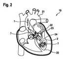
図2は、心臓19における埋め込まれた心室補助システム2を概略的に示す。補助システム2は、(左)心室20から大動脈21への血液の循環を補助することによって、心臓19を補助する。この目的のために、図2に示すように、補助システム2は大動脈弁22内に固定される。100%の補助レベルで、補助システム2(LVAD)は、完全な血液の体積流量を循環させる。補助レベルは、心室20から大動脈21への血液の総体積流量に対する、補助システム2のポンプなどの運ぶ手段によって、または補助システム2を通って循環される体積流量の割合を表す。 FIG. 2 schematically shows an implanted ventricular assist
100%の補助レベルでは、心室20からの総流体体積流量1、心室20への心臓弁体積流量23、および補助システム2を通る流体体積流量7は、したがって同一である。したがって、大動脈弁またはバイパス体積流量24(式記号Qa)はゼロである。総流体体積流量1は、(総)心臓時間容積(HTV:式記号QHTV)として記述されてもよい。流体体積流量7はまた、補助システム自体を通る流量のみを定量化する、いわゆるポンプ体積流量(式記号Qp)とも呼ぶことができる。したがって、補助のレベルは、QP/QHTVの比率から計算することができる。At a 100% auxiliary level, the total fluid volume flow rate 1 from the
低レベルの補助および強い心室収縮を伴う健康な心臓の場合、心臓19はある程度までその機能を果たし続けるため、心臓または大動脈弁22を通る拍動体積流量分率24(バイパス)が収縮期に生成される(心筋が収縮し、心室20の容積を減少させることによって、大動脈21に血液を移す)。同時に、補助システム2上の圧力差は、特に、補助システム2の通常提供されるポンプ(ここでは図示せず)上で減少し、したがって、補助システム2は、収縮期に増加した流体体積流量7も運ぶ。 In the case of a healthy heart with low levels of assistance and strong ventricular contraction, the
本明細書で提案する解決策は、特にインピーダンス測定によって経時的な心室容積の変化を検出することに基づく。次に、公知の(または経時的に記録された)流体体積流量7またはポンプ体積流量(Qp)について、そうでなければほとんど測定不可能な大動脈弁またはバイパス体積流量24は、差を決定することによって有利に定量化することができる。The solution proposed herein is based specifically on detecting changes in ventricular volume over time, especially by impedance measurements. Then, (or otherwise recorded over time) known
図3は、図2の補助システム2を概略的に示す。特定の時点での心室容積を決定するために、図2および図3は、(左心室)心臓内インピーダンス測定による実現形態を示す。インピーダンス測定は、流体または液体の体積全体および電気抵抗の測定が比較的単純な技術的手段で実施され得るという利点を有する。本明細書に示す例のように、四つの電極4、5、27および28(4線測定)を使用することにより、インピーダンス測定は、測定電極の接触抵抗にほとんど依存しないため、測定結果は、患者への長期埋め込みにも適している。この場合、電極4、5、27および28は、補助システム2の例示的な測定装置9を形成する。図3はまた、超音波ベースのポンプ体積流量センサ25が先端10に組み込まれ得ることを例として図示する。ポンプ体積流量センサ25は、流体体積流量7および/またはポンプ体積流量(Qp)を経時的に検出および供給することができる。FIG. 3 schematically shows the
インピーダンス測定の場合、少なくとも二つの電極4、27は、測定電流の電流経路26が心室容積を可能な限り最良に記録できるように、心室20の領域に組み込まれるべきである。入口ケージ11の領域は、この目的に適しており、例えば、入口開口部12の近位および/または上流および遠位および/または下流である。少なくとも二つの電極4、27は、電気回路を閉じるために技術的に必要とされる。電極から血液への接触インピーダンスの影響は無視できるため、四つの電極4、5、27および28の使用は有利であり、電極表面の長期的な変化は、したがって測定結果にまったく影響しないか、またはごくわずかな影響しか与えない。例えば、電流電極4、27は外側に、および電圧測定電極5、28は、それらの間に配置されることが好ましい。この場合、電流経路26は、電極4から電極27(電流電極)に延在する。ここに示されていない等電位線は、それらの間に形成され、電圧電極28および5によって高抵抗で測定することができる。 For impedance measurements, at least two
検出量は、特に、測定対(4と28から5と27)の距離に依存するため、一つの対4、28を、可能な限り遠位に、または先端10の領域に配置し、一つの対5、27を、可能な限り近位に、または補助システム2の電気モーター16に向かって配置することが有利である。この場合、四つの電極4、5、27、および28すべてが、それでも心室20の領域に配置され、近位対5、27が大動脈弁面を超えて大動脈21内に移動しない場合、測定品質に有利であり、この設計に基づいて、測定も考えられるか、または実行可能である。 Since the amount of detection depends in particular on the distance of the measurement pairs (4 and 28 to 5 and 27), one
電極対間の測定された電気導電率は、周囲の流体または流体体積とその導電率の関数である。血液中のイオン濃度は、腎臓によって狭い範囲内に保たれるため、所定のおよび/または一定の導電率を仮定することができる。それにもかかわらず、ここでは、血液導電率の明示的な決定が特に好ましい。この目的のために、例えば(入口)カニューレ13の内部に存在するように、画定された体積6が必要である。したがって、さらに一〜四個の電極29、30、31、および32を(入口管とも呼ぶことができる)カニューレ13内に配置することができる。心室容積の測定と同様に、この場合、測定は、電極対29および30における電流注入および電圧測定によって、または電極対29と30との間の電流注入および電極対31と32との間の電圧測定によって行うことができる。心室測定の少なくとも一つの電極4、5、27、または28が、領域内または入口開口部12の近くに位置する場合、対29、31は省略されてもよい。次いで、測定は、例えば、電極対27、30における電流注入、および電極対5と32との間の電圧測定によって実施することができる。電極対5、27は、領域内または入口開口部12の近くに位置するため、画定された体積6は、電極対5、27と電極対30、32との間のカニューレ13内に結果として生じる。したがって、厳密に二点測定を行うには、三つの電極が特に最小限必要である。次いで、例として、心室インピーダンス測定は、電極4と5との間で実施され、導電率測定は、電極5と30との間で実施される。 The measured electrical conductivity between a pair of electrodes is a function of the surrounding fluid or fluid volume and its conductivity. Since the ion concentration in blood is kept within a narrow range by the kidneys, a given and / or constant conductivity can be assumed. Nevertheless, explicit determination of blood conductivity is particularly preferred here. For this purpose, a defined volume 6 is required, for example to be inside the (entrance)
ここに示す電極を、電流電極または電圧測定電極として割り当てることは、例示にすぎない。この場合の電流電極は、特に、電流源に接続できる、または電流源に接続される電極である。電圧測定電極は、特に、電圧測定装置に接続できる、または電圧測定装置に接続される電極である。ここで提示する(心室容積および/または導電率の)インピーダンス測定の目的を満たすことができる任意の割り当てが可能である。電圧源および/または電圧計は、測定装置9の一部とすることができる。電流源および電圧計が、電気供給ライン18によって補助システム2から空間的に分離される場合、供給ラインケーブルは、特に有利には、能動的遮蔽を備えた三軸設計を有する。 Assigning the electrodes shown here as current electrodes or voltage measuring electrodes is merely an example. The current electrode in this case is, in particular, an electrode that can be connected to or is connected to a current source. A voltage measuring electrode is, in particular, an electrode that can be connected to or connected to a voltage measuring device. Any assignment is possible that can meet the objectives of impedance measurements (of ventricular volume and / or conductivity) presented here. The voltage source and / or voltmeter can be part of the measuring device 9. If the current source and voltmeter are spatially separated from the
(心室容積および/または導電率の)測定のために、例えば、交流は電流電極によって注入することができ、結果として生じる電圧降下は、例えば、電圧測定電極によって測定することができる。しかし、電圧も印加することもでき、結果として生じる電流の流れを測定することができる。 For measurement (of ventricular volume and / or conductivity), for example, alternating current can be injected by a current electrode and the resulting voltage drop can be measured, for example, by a voltage measuring electrode. However, a voltage can also be applied and the resulting current flow can be measured.
測定は、単一の(交流)周波数で実施することができる。生体電気インピーダンス解析およびインピーダンス断層撮影用に、50kHzの範囲の値が確立されている。血液量に加えて、周囲の心筋も測定されたインピーダンスにわずかに寄与する。これは、血液中の細胞材料とは異なる構造を有する細胞材料であるため、影響は、いわゆる生体電気インピーダンス分光法によって低減され得る。この場合、測定は、固定周波数ではなく、いくつかの周波数で実施される。結果は、周波数の関数としての電気インピーダンスである(分散、コールチャートを参照)。 Measurements can be performed on a single (alternating current) frequency. Values in the 50 kHz range have been established for bioelectrical impedance analysis and impedance tomography. In addition to blood volume, the surrounding myocardium also contributes slightly to the measured impedance. Since this is a cell material having a structure different from that of the cell material in blood, the effect can be reduced by so-called bioelectric impedance spectroscopy. In this case, the measurement is carried out at several frequencies rather than at a fixed frequency. The result is electrical impedance as a function of frequency (dispersion, see call chart).
例えば、測定は、1kHz〜1MHzの範囲で実施することができる。分光測定は、例えば、正弦波周波数のシーケンス、いわゆるチャープ、または広帯域バイナリシーケンス(擬似ランダムノイズ)によって実施され得る。 For example, the measurement can be carried out in the range of 1 kHz to 1 MHz. Spectral measurements can be performed, for example, by sinusoidal frequency sequences, so-called chirps, or wideband binary sequences (pseudo-random noise).
心室インピーダンスのサンプリングレートは、予想される心室収縮周波数、例えば60 1/sなど、ナイキスト定理を満たすのに十分な高さであるべきである。心室の容積変化とは対照的に、心筋のインピーダンス変化は非常にゆっくりとしか予想されないため、すべての測定で完全な周波数範囲の掃引が必要ではない。心筋のバックグラウンドインピーダンスは、定期的に決定することができる。また、いくつかの心拍にわたるバックグラウンドインピーダンスの周波数測定点を決定することも有利である。この目的のために、測定は、高サンプリングレートで二つの周波数で実施することができる。第一の周波数は、好ましくは固定され、第二の周波数は、例えば、心拍から心拍へ変化する(例えば、第一の周波数のインピーダンスプロットに基づいて決定される)。別個の測定値のスペクトルは、例として、第一の周波数のインピーダンスプロットを使用して組み合わされる。第一の周波数の代わりに、例えば、圧力センサの圧力プロットに基づいて、同期することも可能である。 The sampling rate of the ventricular impedance should be high enough to satisfy the Nyquist theorem, such as the expected ventricular contraction frequency, eg, 60 1 / s. In contrast to changes in ventricular volume, changes in myocardial impedance are expected only very slowly, so a complete frequency range sweep is not required for all measurements. The background impedance of the myocardium can be determined on a regular basis. It is also advantageous to determine frequency measurement points for background impedance over several heartbeats. For this purpose, the measurements can be carried out at two frequencies at a high sampling rate. The first frequency is preferably fixed and the second frequency changes, for example, from heartbeat to heartbeat (eg, determined based on the impedance plot of the first frequency). The spectra of the separate measurements are combined using, for example, the impedance plot of the first frequency. Instead of the first frequency, it is also possible to synchronize, for example, based on the pressure plot of the pressure sensor.
図4は、埋め込まれた心室補助システム2を概略的に示す。図4は、心臓19における結果として生じた体積流量を図示する。左の容積は、心室または心室容積3を表し、右の容積は、大動脈21を表す。質量保存により、左心房から心室への(心臓弁)体積流量23および大動脈への流出は、総流体体積流量1または(総)心臓時間容積に対応する。弁閉鎖不全または心室間の油圧短絡は例外である。 FIG. 4 schematically shows an implanted ventricular assist
補助システム2(LVAD)は、例えば、補助システム2のポンプ(ここに示さず)の出力によって、循環体積流量33を運ぶ。連続的補助システムでは、流量は一定であり、拍動補助システムでは、この流量は時間変調される。心室の収縮により、心室内の圧力が増加し、補助システム2上の圧力差が減少し、それによって、補助システム2は、収縮期に一定の機械的出力で追加の収縮期体積流量34を送達する。心室収縮が十分に強い場合(例えば、短期補助患者の場合)、心室圧は大動脈血圧を超えることがあり、これにより大動脈弁が開く(ここには示さず)。追加のバイパス流量(式記号Qa)が形成され、これは大動脈弁またはバイパス体積流量24として図4に示されている。この流量部分(バイパス24)は、補助システム2の流量センサ、例えば、補助システム2の先端にある超音波センサによって(直接)検出することはできないが、本質的には、心室容積δV/δtの変化とポンプ体積流量Qpとの間の差に対応する。この場合、補助システム2自体を通る流量を定量化するのみであり、本明細書では流体体積流量7とも呼称されるポンプ体積流量Qpは、循環体積流量33と収縮期体積流量34との和から得られる。経時的な心室容積測定および経時的なポンプ流量測定を組み合わせることによって、バイパス流量、すなわち大動脈弁またはバイパス体積流量24(式記号QA)は、以下の式に従って決定することができる。
この関係ならびに心臓時間容積の例示的導出を、以下の式1〜13を用いて図示する。使用される式記号は、最初に簡単に説明されている。
QHTV 心臓時間容積
QP ポンプを通る時間依存性体積流量
QA 大動脈弁を通る時間依存性体積流量
QV 心室の貯蔵容積への時間依存性体積流量(心室容積の変化)
QD 左心房から大動脈への時間依存性体積流量(仮定:ポンプを通るベース流量)
V 心室容積
t 時間
QHTV Cardiac time volume QP Time-dependent volume flow through the pump QA Time-dependent volume flow through the aortic valve QV Time-dependent volume flow through the ventricular storage volume (change in ventricular volume)
QD time-dependent volume flow from the left atrium to the aorta (assuming: the base flow rate through the pump)
V ventricular volume t time
(バイオ)インピーダンス測定から、以下が公知である。
例示的な超音波測定から、以下が公知である。
さらに、拡張期に以下が適用されると仮定できる。
したがって、バイパス流量QAは、以下によって決定され得る。
さらに、心臓時間容積QHTVは、以下により決定され得る。
さらに、補助システムを特定の体積測定モードに置くことも好ましい。この場合、ポンプは、入口ケージおよびインペラケージの圧力が同一になるように、統合された圧力センサによって調整され、すなわち、体積流量がなく、ポンプの回転が速いだけで、大動脈からポンプを通って心室への還流はない。同時に、心拍出量、最終拡張期血液量、および/または心室の収縮強度は、経時的なインピーダンス測定データ(そこで測定された)グラフに基づいて決定され、および/または補助システムを制御するために使用され得る。 It is also preferred to place the auxiliary system in a particular volumetric mode. In this case, the pump is tuned by an integrated pressure sensor so that the pressures in the inlet and impeller cages are the same, i.e. There is no return to the ventricles. At the same time, cardiac output, final diastolic blood volume, and / or ventricular contractile intensity are determined based on the impedance measurement data (measured there) graph over time and / or to control the auxiliary system. Can be used for.
体積流量に加えて、心室容積3も興味深いパラメータである。完全な補助システム(ポンプのみが血液を循環させる)では、吸引効果または心室の虚脱が起こる可能性がある。この場合、補助システム2は、左心房から流入するよりも多くの血液を循環させる。補助システム2(またはそのポンプ)は心室を空にする。結果として、心室壁は入口ケージに近づき、補助システム2(またはそのポンプ)は、「心室壁」に対してそれ自体を吸引することができる。心室血圧が50mmHgの値を下回ると、周囲圧力が高くなるために心室も虚脱し得る。この場合、心室容積3は、特にその循環またはポンプ出力を減少させることによって、補助システム2の出力を低減するための制御パラメータとして使用することができ、その結果、最小の心室容積を保証することができる。 In addition to volumetric flow rate,
図5は、説明された方法を概略的な形で再び示す。この方法は、a)補助システム2によって第一の時点で第一のインピーダンスパラメータを決定するステップと、b)補助システム2によって第二の時点で第二のインピーダンスパラメータを決定するステップと、c)少なくとも第一のインピーダンスパラメータおよび第二のインピーダンスパラメータを使用して、インピーダンスパラメータの変化を決定するか、または少なくとも第一もしくは第二のインピーダンスパラメータを閾値と比較するステップが、連続的に実施される。 FIG. 5 shows the described method again in schematic form. In this method, a) the
本明細書で提案する解決策は、特に以下の利点のうち一つまたは複数を可能にする。
・補助システムに厳密に組み込まれたセンサの使用が可能になる。例えば、大動脈の周囲に別個の超音波カフは不要である。
・この方法は、総心臓時間容積の決定ならびに補助レベル(総心臓時間容積のポンプ体積流量の割合)の決定を可能にする。
・電気インピーダンス測定技術の統合は、大動脈超音波カフまたは類似のものと比較して実施が容易である。
・総心臓時間容積は、システムによって連続的に測定されるため、LVADは、心臓時間容積ならびに補助レベルの両方に基づいて調整することができ、これは短期補助システム(キーワード離脱)では特に有利である。
・この方法は、例えば、最小心室容積に達していないときに、補助システムを調整できるように、心室容積を決定する。
The solutions proposed herein allow one or more of the following advantages in particular:
-It enables the use of sensors that are strictly built into the auxiliary system. For example, no separate ultrasonic cuff is needed around the aorta.
• This method allows determination of total cardiac time volume as well as auxiliary levels (percentage of pump volume flow rate of total cardiac time volume).
• Integration of electrical impedance measurement techniques is easier to perform compared to aortic ultrasound cuffs or similar.
LVAD can be adjusted based on both cardiac time volume and assist level, as total cardiac time volume is continuously measured by the system, which is especially advantageous in short-term assist systems (keyword withdrawal). be.
• This method determines the ventricular volume so that the assistive system can be adjusted, for example, when the minimum ventricular volume has not been reached.
Claims (13)
Translated fromJapanesea)前記補助システム(2)を使用して、第一の時点で第一のインピーダンスパラメータを決定するステップと、
b)前記補助システム(2)を使用して、第二の時点で第二のインピーダンスパラメータを決定するステップと、
c)前記第一のインピーダンスパラメータおよび前記第二のインピーダンスパラメータを使用して、前記インピーダンスパラメータの変化を少なくとも決定するか、または少なくとも前記第一もしくは第二のインピーダンスパラメータを閾値と比較するステップと、を含む、方法。A method for operating the implanted ventricular assist system (2).
a) Using the auxiliary system (2), the step of determining the first impedance parameter at the first time point and
b) The step of determining the second impedance parameter at the second time point using the auxiliary system (2),
c) A step of using the first impedance parameter and the second impedance parameter to at least determine a change in the impedance parameter, or at least compare the first or second impedance parameter to a threshold. Including the method.
第一の時点での第一のインピーダンスパラメータおよび第二の時点での第二のインピーダンスパラメータを決定するように構成された、測定装置(9)と、
少なくとも前記第一のインピーダンスパラメータおよび前記第二のインピーダンスパラメータを使用して、前記インピーダンスパラメータの変化を決定するように、または少なくとも前記第一もしくは第二のインピーダンスパラメータを閾値と比較するように構成された、処理ユニット(8)と、を備える、補助システム。An implantable ventricular assist system (2)
A measuring device (9) configured to determine the first impedance parameter at the first time point and the second impedance parameter at the second time point.
It is configured to use at least the first impedance parameter and the second impedance parameter to determine changes in the impedance parameter, or to compare at least the first or second impedance parameter to a threshold. An auxiliary system including a processing unit (8).
The auxiliary system according to any one of claims 9 to 12, further comprising an apparatus for determining a fluid volume flow rate (7).
Applications Claiming Priority (3)
| Application Number | Priority Date | Filing Date | Title |
|---|---|---|---|
| DE102018208913.2ADE102018208913A1 (en) | 2018-06-06 | 2018-06-06 | A method of operating an implanted ventricular assist device |
| DE102018208913.2 | 2018-06-06 | ||
| PCT/EP2019/064779WO2019234148A1 (en) | 2018-06-06 | 2019-06-06 | Implantable ventricular assist system and method for operating same |
Publications (3)
| Publication Number | Publication Date |
|---|---|
| JP2021526880Atrue JP2021526880A (en) | 2021-10-11 |
| JPWO2019234148A5 JPWO2019234148A5 (en) | 2022-06-17 |
| JP7626431B2 JP7626431B2 (en) | 2025-02-04 |
Family
ID=66821244
Family Applications (1)
| Application Number | Title | Priority Date | Filing Date |
|---|---|---|---|
| JP2020567819AActiveJP7626431B2 (en) | 2018-06-06 | 2019-06-06 | IMPLANTABLE VENTRICULAR ASSIST SYSTEM AND METHOD OF OPERATION THEREOF - Patent application |
Country Status (6)
| Country | Link |
|---|---|
| US (2) | US12257424B2 (en) |
| EP (1) | EP3801674B1 (en) |
| JP (1) | JP7626431B2 (en) |
| CN (1) | CN112584890B (en) |
| DE (1) | DE102018208913A1 (en) |
| WO (1) | WO2019234148A1 (en) |
Families Citing this family (23)
| Publication number | Priority date | Publication date | Assignee | Title |
|---|---|---|---|---|
| DE102018208538A1 (en) | 2018-05-30 | 2019-12-05 | Kardion Gmbh | Intravascular blood pump and process for the production of electrical conductors |
| DE102018208899A1 (en) | 2018-06-06 | 2019-12-12 | Kardion Gmbh | A method for determining the speed of sound in a fluid in the region of an implanted vascular support system |
| DE102018208933A1 (en) | 2018-06-06 | 2019-12-12 | Kardion Gmbh | A method of determining a flow rate of fluid flowing through an implanted vascular support system |
| DE102018208913A1 (en) | 2018-06-06 | 2019-12-12 | Kardion Gmbh | A method of operating an implanted ventricular assist device |
| DE102018208879A1 (en) | 2018-06-06 | 2020-01-30 | Kardion Gmbh | Method for determining a total fluid volume flow in the area of an implanted, vascular support system |
| DE102018208945A1 (en) | 2018-06-06 | 2019-12-12 | Kardion Gmbh | An analysis device and method for analyzing a viscosity of a fluid |
| DE102018208929A1 (en) | 2018-06-06 | 2019-12-12 | Kardion Gmbh | A method of determining a flow rate of fluid flowing through an implanted vascular support system |
| DE102018208862A1 (en) | 2018-06-06 | 2019-12-12 | Kardion Gmbh | Implantable vascular support system |
| DE102018208936A1 (en) | 2018-06-06 | 2019-12-12 | Kardion Gmbh | Determining device and method for determining a viscosity of a fluid |
| DE102018210076A1 (en) | 2018-06-21 | 2019-12-24 | Kardion Gmbh | Method and device for detecting a state of wear of a cardiac support system, method and device for operating a cardiac support system and cardiac support system |
| USD1092716S1 (en)* | 2019-12-31 | 2025-09-09 | Abiomed, Inc. | Outflow cage for blood pump |
| KR20230082639A (en) | 2020-10-07 | 2023-06-08 | 아비오메드 유럽 게엠베하 | Patch electrode assemblies for conductivity and admittance measurements |
| AU2021381515A1 (en) | 2020-11-20 | 2023-07-06 | Kardion Gmbh | Purgeless mechanical circulatory support system with magnetic drive |
| CA3199176A1 (en) | 2020-11-20 | 2022-05-27 | Marvin MITZE | Mechanical circulatory support system with guidewire aid |
| US11911602B2 (en)* | 2021-04-22 | 2024-02-27 | Board Of Regents, The University Of Texas System | Method and apparatus for assisting a heart |
| US11918799B2 (en)* | 2021-04-22 | 2024-03-05 | Board Of Regents, The University Of Texas System | Method and apparatus for assisting a heart |
| WO2022225950A1 (en)* | 2021-04-22 | 2022-10-27 | Cardio Vol, Llc | Method and apparatus for assisting a heart |
| US11931563B2 (en) | 2021-04-22 | 2024-03-19 | Board Of Regents, The University Of Texas System | Method and apparatus for assisting a heart |
| US12290675B2 (en)* | 2021-04-22 | 2025-05-06 | Board Of Regents, The University Of Texas System | Method and apparatus for assisting a heart |
| EP4387708A4 (en)* | 2021-08-19 | 2025-05-21 | Cardiovol, LLC | Method and apparatus for assisting a heart |
| US20230173250A1 (en) | 2021-12-03 | 2023-06-08 | Kardion Gmbh | Cardiac pump with optical fiber for laser doppler |
| DE102023118223A1 (en) | 2022-07-11 | 2024-01-11 | Kardion Gmbh | LASER DOPPLER VELOCIMETERY FLOW MEASUREMENT |
| WO2024243154A1 (en) | 2023-05-25 | 2024-11-28 | Kardion Gmbh | Heart pump tips and delivery system couplings for mechanical circulatory support systems |
Citations (8)
| Publication number | Priority date | Publication date | Assignee | Title |
|---|---|---|---|---|
| JP2003047656A (en)* | 2001-05-16 | 2003-02-18 | Levram Medical Devices Ltd | Ventricular assisting device and ventricular assisting method |
| JP2006528006A (en)* | 2003-07-18 | 2006-12-14 | ヴェントラコー リミテッド | Blood pressure detection device and system |
| JP2012520157A (en)* | 2009-03-13 | 2012-09-06 | プロテウス バイオメディカル インコーポレイテッド | Volume detection |
| US20120310037A1 (en)* | 2010-11-12 | 2012-12-06 | LibraHeat, Inc.V | Ventricular assist device cannula and ventricular assist device including the same |
| US20130066142A1 (en)* | 2011-09-14 | 2013-03-14 | Thomas Doerr | Implantable cardiac therapy device |
| JP2013128792A (en)* | 2013-03-27 | 2013-07-04 | San Medical Gijutsu Kenkyusho:Kk | Artificial heart control device and artificial heart system |
| US20140114202A1 (en)* | 2011-06-10 | 2014-04-24 | Marc Hein | Blood withdrawal cannula of a pump replacing or assisting activity of the heart |
| US20150306290A1 (en)* | 2012-11-30 | 2015-10-29 | The Penn State Research Foundation | Smart Tip LVAD Inlet Cannula |
Family Cites Families (587)
| Publication number | Priority date | Publication date | Assignee | Title |
|---|---|---|---|---|
| US1707624A (en) | 1927-05-25 | 1929-04-02 | Brown Instr Co | Gas-comparison apparatus |
| US2347624A (en) | 1942-05-12 | 1944-04-25 | Warren Carl | Distributing means for coarse aggregates |
| US2989124A (en) | 1960-01-14 | 1961-06-20 | Roger H Lapp | Pressure operated water valve |
| US3088323A (en) | 1960-02-10 | 1963-05-07 | Gulton Ind Inc | Piezoresistive transducer |
| US4023562A (en) | 1975-09-02 | 1977-05-17 | Case Western Reserve University | Miniature pressure transducer for medical use and assembly method |
| NO150015C (en) | 1981-11-13 | 1984-08-08 | Vingmed As | METHOD OF BLOOD FLOW SPEED MEASUREMENT WITH ULTRO SOUND, COMBINED WITH ECO-AMPLITUDE IMAGE, FOR THE INVESTIGATION OF LIVING BIOLOGICAL STRUCTURES |
| JPS5980229A (en) | 1982-10-29 | 1984-05-09 | 株式会社島津製作所 | Pulse doppler ultrasonic blood flow meter |
| JPS6015771A (en) | 1983-07-08 | 1985-01-26 | Hitachi Ltd | Memory controller |
| JPS61125329A (en) | 1984-11-21 | 1986-06-13 | テルモ株式会社 | Heart pulse output measuring apparatus |
| JPS62113555A (en) | 1985-11-13 | 1987-05-25 | Canon Inc | Ink jet recorder |
| JPS62204733A (en) | 1986-03-04 | 1987-09-09 | アロカ株式会社 | Ultrasonic doppler diagnostic apparatus |
| JPS62282284A (en) | 1986-05-30 | 1987-12-08 | Tokyo Keiki Co Ltd | Method and apparatus for measuring distance by ultrasonic wave |
| US4902272A (en) | 1987-06-17 | 1990-02-20 | Abiomed Cardiovascular, Inc. | Intra-arterial cardiac support system |
| US4781525A (en) | 1987-07-17 | 1988-11-01 | Minnesota Mining And Manufacturing Company | Flow measurement system |
| JPS6468236A (en) | 1987-09-07 | 1989-03-14 | Aisin Seiki | Cannula equipped with detection electrode |
| US4889131A (en) | 1987-12-03 | 1989-12-26 | American Health Products, Inc. | Portable belt monitor of physiological functions and sensors therefor |
| US4888011A (en) | 1988-07-07 | 1989-12-19 | Abiomed, Inc. | Artificial heart |
| US4965713A (en) | 1988-08-15 | 1990-10-23 | Viking Pump Inc. | Terminal element |
| US4989609A (en) | 1989-01-26 | 1991-02-05 | Minnesota Mining And Manufacturing Company | Doppler blood flow system and method using special zero flow rate analysis |
| US5045051A (en) | 1989-03-14 | 1991-09-03 | Abiomed, Inc. | Leak detector |
| CA2004295C (en) | 1989-11-30 | 1998-02-10 | William F. Hayes | Primary fluid actuated, secondary fluid propelling system |
| AU1279092A (en) | 1991-02-04 | 1992-10-06 | Kensey Nash Corporation | Apparatus and method for determining viscosity of the blood of a living being |
| JP2952438B2 (en) | 1991-09-20 | 1999-09-27 | トキコ株式会社 | Thermal flow meter |
| US5676651A (en) | 1992-08-06 | 1997-10-14 | Electric Boat Corporation | Surgically implantable pump arrangement and method for pumping body fluids |
| JP3312759B2 (en) | 1993-01-22 | 2002-08-12 | テルモ株式会社 | Medical pump drive |
| US5456715A (en) | 1993-05-21 | 1995-10-10 | Liotta; Domingo S. | Implantable mechanical system for assisting blood circulation |
| US5289821A (en) | 1993-06-30 | 1994-03-01 | Swartz William M | Method of ultrasonic Doppler monitoring of blood flow in a blood vessel |
| JPH0747025A (en) | 1993-08-06 | 1995-02-21 | Itoki Co Ltd | Flexible partition |
| US5527159A (en) | 1993-11-10 | 1996-06-18 | The United States Of America As Represented By The Administrator Of The National Aeronautics And Space Administration | Rotary blood pump |
| GB9404321D0 (en) | 1994-03-04 | 1994-04-20 | Thoratec Lab Corp | Driver and method for driving pneumatic ventricular assist devices |
| US5581038A (en) | 1994-04-04 | 1996-12-03 | Sentir, Inc. | Pressure measurement apparatus having a reverse mounted transducer and overpressure guard |
| NO942222D0 (en) | 1994-06-14 | 1994-06-14 | Vingmed Sound As | Method for determining blood flow velocity / time spectrum |
| JPH0857042A (en) | 1994-08-24 | 1996-03-05 | Terumo Corp | Medical pump |
| US5685989A (en) | 1994-09-16 | 1997-11-11 | Transonic Systems, Inc. | Method and apparatus to measure blood flow and recirculation in hemodialysis shunts |
| US5453576A (en) | 1994-10-24 | 1995-09-26 | Transonic Systems Inc. | Cardiovascular measurements by sound velocity dilution |
| US5613935A (en) | 1994-12-16 | 1997-03-25 | Jarvik; Robert | High reliability cardiac assist system |
| JPH08327527A (en) | 1995-05-31 | 1996-12-13 | Toyobo Co Ltd | Capillary type viscometer |
| US5752976A (en) | 1995-06-23 | 1998-05-19 | Medtronic, Inc. | World wide patient location and data telemetry system for implantable medical devices |
| US5720771A (en) | 1995-08-02 | 1998-02-24 | Pacesetter, Inc. | Method and apparatus for monitoring physiological data from an implantable medical device |
| GB9604665D0 (en) | 1996-03-05 | 1996-05-01 | Montec Int Ltd | Flow measurement |
| US5980465A (en) | 1996-03-18 | 1999-11-09 | Medtronic, Inc. | Method for detecting changes in a patient s blood volume |
| US5911685A (en) | 1996-04-03 | 1999-06-15 | Guidant Corporation | Method and apparatus for cardiac blood flow assistance |
| JPH1052489A (en) | 1996-08-12 | 1998-02-24 | Buaayu:Kk | Cannula and supplemental circulation device |
| US5888242A (en) | 1996-11-01 | 1999-03-30 | Nimbus, Inc. | Speed control system for implanted blood pumps |
| DE69724781T2 (en) | 1997-01-03 | 2004-07-01 | Biosense, Inc., Miami | STENT FOR MEASURING PRESSURE |
| US5957861A (en) | 1997-01-31 | 1999-09-28 | Medtronic, Inc. | Impedance monitor for discerning edema through evaluation of respiratory rate |
| US5964694A (en) | 1997-04-02 | 1999-10-12 | Guidant Corporation | Method and apparatus for cardiac blood flow assistance |
| CN1222862A (en) | 1997-04-02 | 1999-07-14 | 激励心脏技术有限公司 | Intracardiac pump device |
| US5865759A (en) | 1997-04-11 | 1999-02-02 | Texon Technologies Ltd. | Method and apparatus for non-invasive assessment of cardiac function by monitoring acceleration of the heart |
| US5827203A (en) | 1997-04-21 | 1998-10-27 | Nita; Henry | Ultrasound system and method for myocardial revascularization |
| US6731976B2 (en) | 1997-09-03 | 2004-05-04 | Medtronic, Inc. | Device and method to measure and communicate body parameters |
| AU7360798A (en) | 1997-09-24 | 1999-04-12 | Cleveland Clinic Foundation, The | Flow controlled blood pump system |
| US6183412B1 (en) | 1997-10-02 | 2001-02-06 | Micromed Technology, Inc. | Implantable pump system |
| US6610004B2 (en) | 1997-10-09 | 2003-08-26 | Orqis Medical Corporation | Implantable heart assist system and method of applying same |
| US6398734B1 (en) | 1997-10-14 | 2002-06-04 | Vascusense, Inc. | Ultrasonic sensors for monitoring the condition of flow through a cardiac valve |
| US6007478A (en) | 1997-11-13 | 1999-12-28 | Impella Cardiotechnik Aktiengesellschaft | Cannula having constant wall thickness with increasing distal flexibility and method of making |
| US6314322B1 (en) | 1998-03-02 | 2001-11-06 | Abiomed, Inc. | System and method for treating dilated cardiomyopathy using end diastolic volume (EDV) sensing |
| US5904708A (en) | 1998-03-19 | 1999-05-18 | Medtronic, Inc. | System and method for deriving relative physiologic signals |
| CN1192351A (en) | 1998-03-26 | 1998-09-09 | 王明时 | Instrument for quick measuring blood viscosity |
| US6176822B1 (en) | 1998-03-31 | 2001-01-23 | Impella Cardiotechnik Gmbh | Intracardiac blood pump |
| US5955924A (en) | 1998-04-21 | 1999-09-21 | Applied Micro Circuits Corporation | Differential metal-oxide semiconductor (CMOS) push-pull buffer |
| US6023641A (en) | 1998-04-29 | 2000-02-08 | Medtronic, Inc. | Power consumption reduction in medical devices employing multiple digital signal processors |
| US6024704A (en) | 1998-04-30 | 2000-02-15 | Medtronic, Inc | Implantable medical device for sensing absolute blood pressure and barometric pressure |
| DE19821307C1 (en) | 1998-05-13 | 1999-10-21 | Impella Cardiotech Gmbh | Intra-cardiac blood pump |
| US7083588B1 (en) | 1998-05-26 | 2006-08-01 | Medtronic Vascular, Inc. | Apparatus for providing coronary retroperfusion and methods of use |
| US6575927B1 (en) | 1998-09-25 | 2003-06-10 | The Regents Of The University Of Michigan | System and method for determining blood flow rate in a vessel |
| US6167765B1 (en) | 1998-09-25 | 2001-01-02 | The Regents Of The University Of Michigan | System and method for determining the flow rate of blood in a vessel using doppler frequency signals |
| DE29821563U1 (en) | 1998-12-02 | 2000-07-13 | Impella Cardiotechnik AG, 52074 Aachen | Pressure sensor |
| US6245007B1 (en) | 1999-01-28 | 2001-06-12 | Terumo Cardiovascular Systems Corporation | Blood pump |
| US6210318B1 (en) | 1999-03-09 | 2001-04-03 | Abiomed, Inc. | Stented balloon pump system and method for using same |
| US6438409B1 (en) | 1999-03-25 | 2002-08-20 | Medtronic, Inc. | Methods of characterizing ventricular operations and applications thereof |
| IT1315206B1 (en) | 1999-04-27 | 2003-02-03 | Salvatore Romano | METHOD AND APPARATUS FOR MEASURING HEART RATE. |
| US6190324B1 (en) | 1999-04-28 | 2001-02-20 | Medtronic, Inc. | Implantable medical device for tracking patient cardiac status |
| AUPQ090499A0 (en) | 1999-06-10 | 1999-07-01 | Peters, William S | Heart assist device and system |
| US6890329B2 (en) | 1999-06-15 | 2005-05-10 | Cryocath Technologies Inc. | Defined deflection structure |
| EP1063753B1 (en) | 1999-06-22 | 2009-07-22 | Levitronix LLC | Electric rotary drive comprising a magnetically suspended rotor |
| US6231498B1 (en) | 1999-06-23 | 2001-05-15 | Pulsion Medical Systems Ag | Combined catheter system for IABP and determination of thermodilution cardiac output |
| US7138776B1 (en) | 1999-07-08 | 2006-11-21 | Heartware, Inc. | Method and apparatus for controlling brushless DC motors in implantable medical devices |
| US6512949B1 (en) | 1999-07-12 | 2003-01-28 | Medtronic, Inc. | Implantable medical device for measuring time varying physiologic conditions especially edema and for responding thereto |
| AU7354400A (en) | 1999-09-03 | 2001-04-10 | A-Med Systems, Inc. | Guidable intravascular blood pump and related methods |
| US7022100B1 (en) | 1999-09-03 | 2006-04-04 | A-Med Systems, Inc. | Guidable intravascular blood pump and related methods |
| US6579257B1 (en) | 1999-09-21 | 2003-06-17 | Medtronic, Inc. | Automated occlusion clamp for centrifugal blood pumps |
| US20010039828A1 (en) | 1999-11-12 | 2001-11-15 | Visco Technologies, Inc. | Mass detection capillary viscometer |
| EP1123687A3 (en) | 2000-02-10 | 2004-02-04 | Aloka Co., Ltd. | Ultrasonic diagnostic apparatus |
| US6406422B1 (en) | 2000-03-02 | 2002-06-18 | Levram Medical Devices, Ltd. | Ventricular-assist method and apparatus |
| US6561975B1 (en) | 2000-04-19 | 2003-05-13 | Medtronic, Inc. | Method and apparatus for communicating with medical device systems |
| US6530876B1 (en) | 2000-04-25 | 2003-03-11 | Paul A. Spence | Supplemental heart pump methods and systems for supplementing blood through the heart |
| US6432136B1 (en) | 2000-04-25 | 2002-08-13 | The Penn State Research Foundation | Apparatus and method for removing a pocket of air from a blood pump |
| US6540658B1 (en) | 2000-05-30 | 2003-04-01 | Abiomed, Inc. | Left-right flow control algorithm in a two chamber cardiac prosthesis |
| DE10040403A1 (en) | 2000-08-18 | 2002-02-28 | Impella Cardiotech Ag | Intracardiac blood pump |
| IL138073A0 (en) | 2000-08-24 | 2001-10-31 | Glucon Inc | Photoacoustic assay and imaging system |
| US6808508B1 (en) | 2000-09-13 | 2004-10-26 | Cardiacassist, Inc. | Method and system for closed chest blood flow support |
| GB0023412D0 (en) | 2000-09-23 | 2000-11-08 | Khaghani Asghar | Aortic counterpulsator |
| US6602182B1 (en) | 2000-11-28 | 2003-08-05 | Abiomed, Inc. | Cardiac assistance systems having multiple fluid plenums |
| US6540659B1 (en) | 2000-11-28 | 2003-04-01 | Abiomed, Inc. | Cardiac assistance systems having bi-directional pumping elements |
| DE10059714C1 (en) | 2000-12-01 | 2002-05-08 | Impella Cardiotech Ag | Intravasal pump has pump stage fitted with flexible expandible sleeve contricted during insertion through blood vessel |
| DE10060275A1 (en) | 2000-12-05 | 2002-06-13 | Impella Cardiotech Ag | Method for calibrating a pressure sensor or a flow sensor on a rotary pump |
| US6912423B2 (en) | 2000-12-15 | 2005-06-28 | Cardiac Pacemakers, Inc. | Terminal connector assembly for a medical device and method therefor |
| US20020147495A1 (en) | 2001-04-09 | 2002-10-10 | Christopher Petroff | Reduced-size replacement heart |
| DE10164898B4 (en) | 2001-04-30 | 2010-09-23 | Berlin Heart Gmbh | Method for controlling a support pump for pulsatile pressure fluid delivery systems |
| US6879126B2 (en) | 2001-06-29 | 2005-04-12 | Medquest Products, Inc | Method and system for positioning a movable body in a magnetic bearing system |
| JP3882069B2 (en) | 2001-07-06 | 2007-02-14 | 独立行政法人産業技術総合研究所 | Abnormality determination method and abnormality determination device for artificial heart pump |
| US7191000B2 (en)* | 2001-07-31 | 2007-03-13 | Cardiac Pacemakers, Inc. | Cardiac rhythm management system for edema |
| JP4440499B2 (en) | 2001-08-29 | 2010-03-24 | 泉工医科工業株式会社 | Centrifugal pump drive |
| DE10144269A1 (en) | 2001-09-08 | 2003-03-27 | Bosch Gmbh Robert | Sensor element for measuring a physical variable between two bodies which move relative to each other and are subjected to high tribological strain, whereby the element has very high wear resistance to increase its service life |
| US6666826B2 (en) | 2002-01-04 | 2003-12-23 | Cardiac Pacemakers, Inc. | Method and apparatus for measuring left ventricular pressure |
| US8323173B2 (en) | 2002-01-07 | 2012-12-04 | Micromed Technology, Inc. | Method and system for physiologic control of an implantable blood pump |
| US7396327B2 (en) | 2002-01-07 | 2008-07-08 | Micromed Technology, Inc. | Blood pump system and method of operation |
| ATE485850T1 (en) | 2002-01-08 | 2010-11-15 | Micromed Technology Inc | SYSTEM FOR DETECTING VENTRICULAR COLLAPSE |
| DK2298370T3 (en) | 2002-02-21 | 2014-01-27 | Design Mentor Inc | fluid pump |
| US7238151B2 (en) | 2002-02-26 | 2007-07-03 | Frazier O Howard | Permanent heart assist system |
| US6669624B2 (en) | 2002-03-26 | 2003-12-30 | O. Howard Frazier | Temporary heart-assist system |
| US10155082B2 (en) | 2002-04-10 | 2018-12-18 | Baxter International Inc. | Enhanced signal detection for access disconnection systems |
| US6991595B2 (en) | 2002-04-19 | 2006-01-31 | Thoratec Corporation | Adaptive speed control for blood pump |
| US6969369B2 (en) | 2002-04-22 | 2005-11-29 | Medtronic, Inc. | Implantable drug delivery system responsive to intra-cardiac pressure |
| US7024244B2 (en) | 2002-04-22 | 2006-04-04 | Medtronic, Inc. | Estimation of stroke volume cardiac output using an intracardiac pressure sensor |
| DE10226305C1 (en) | 2002-06-13 | 2003-10-30 | Infratec Gmbh Infrarotsensorik | Tunable narrowband filter for IR spectral measurements based on electrostatically-driven micromechanical Fabry-Perot interferometer |
| DE10227918A1 (en) | 2002-06-21 | 2004-01-15 | Bühler AG | Method for determining rheological parameters of a fluid |
| AU2003238656A1 (en) | 2002-06-25 | 2004-01-06 | Glucon Inc. | Method and apparatus for performing myocardial revascularization |
| US6949066B2 (en) | 2002-08-21 | 2005-09-27 | World Heart Corporation | Rotary blood pump diagnostics and cardiac output controller |
| AU2002951685A0 (en) | 2002-09-30 | 2002-10-17 | Ventrassist Pty Ltd | Physiological demand responsive control system |
| US6943434B2 (en) | 2002-10-03 | 2005-09-13 | Fairchild Semiconductor Corporation | Method for maintaining solder thickness in flipchip attach packaging processes |
| US7207939B2 (en) | 2002-10-03 | 2007-04-24 | Coulter International Corp. | Apparatus and method for analyzing a liquid in a capillary tube of a hematology instrument |
| JP4465349B2 (en) | 2003-01-24 | 2010-05-19 | プロテウス バイオメディカル インコーポレイテッド | Method and system for measuring cardiac parameters |
| WO2004066825A2 (en) | 2003-01-31 | 2004-08-12 | The Board Of Trustees Of The Leland Stanford Junior University | Detection of apex motion for monitoring cardiac dysfunction |
| US6887207B2 (en) | 2003-02-26 | 2005-05-03 | Medtronic, Inc. | Methods and apparatus for estimation of ventricular afterload based on ventricular pressure measurements |
| US20040199052A1 (en) | 2003-04-01 | 2004-10-07 | Scimed Life Systems, Inc. | Endoscopic imaging system |
| CN1202871C (en) | 2003-04-18 | 2005-05-25 | 清华大学 | Optimal non-constant speed control method for miniature axial flow type blood pumps |
| US7118525B2 (en) | 2003-04-23 | 2006-10-10 | Coleman Edward J | Implantable cardiac assist device |
| US20080262289A1 (en) | 2003-05-28 | 2008-10-23 | Goldowsky Michael P | Blood Pump Having A Passive Non-Contacting Bearing Suspension |
| US20040241019A1 (en) | 2003-05-28 | 2004-12-02 | Michael Goldowsky | Passive non-contacting smart bearing suspension for turbo blood-pumps |
| US7078796B2 (en) | 2003-07-01 | 2006-07-18 | Freescale Semiconductor, Inc. | Corrosion-resistant copper bond pad and integrated device |
| US7128538B2 (en) | 2003-07-07 | 2006-10-31 | Terumo Corporation | Centrifugal fluid pump apparatus |
| US7951129B2 (en) | 2003-08-07 | 2011-05-31 | Medtronic, Inc. | Diastolic coronary perfusion detection for timed delivery of therapeutic and/or diagnostic agents |
| DE10336902C5 (en) | 2003-08-08 | 2019-04-25 | Abiomed Europe Gmbh | Intracardiac pumping device |
| US7245117B1 (en) | 2004-11-01 | 2007-07-17 | Cardiomems, Inc. | Communicating with implanted wireless sensor |
| US20140296677A1 (en) | 2003-09-18 | 2014-10-02 | New Paradigm Concepts, LLC | Method of measuring total vascular hemoglobin mass |
| US7559894B2 (en) | 2003-09-18 | 2009-07-14 | New Paradigm Concepts, LLC | Multiparameter whole blood monitor and method |
| US20050137614A1 (en) | 2003-10-08 | 2005-06-23 | Porter Christopher H. | System and method for connecting implanted conduits |
| US8428717B2 (en) | 2003-10-14 | 2013-04-23 | Medtronic, Inc. | Method and apparatus for monitoring tissue fluid content for use in an implantable cardiac device |
| US7765003B2 (en) | 2003-10-31 | 2010-07-27 | Sunshine Heart Pty Ltd | Synchronization control system |
| US7520850B2 (en) | 2003-11-19 | 2009-04-21 | Transoma Medical, Inc. | Feedback control and ventricular assist devices |
| AU2004294569A1 (en) | 2003-11-26 | 2005-06-16 | Separation Technology, Inc. | Method and apparatus for ultrasonic determination of hematocrit and hemoglobin concentrations |
| TWI231749B (en) | 2003-12-24 | 2005-05-01 | Mau-Chin Shen | Restoring/positioning device for slide rail of drawer |
| JP2005192687A (en) | 2003-12-29 | 2005-07-21 | Sunao Kitamura | Indirect measuring method for pressure, flow rate and natural cardiac output in partial assist using rotating artificial heart pump |
| JP2005241546A (en) | 2004-02-27 | 2005-09-08 | Fuji Electric Systems Co Ltd | Doppler type ultrasonic flowmeter, its processing unit, program |
| US11819192B2 (en) | 2004-03-23 | 2023-11-21 | Boston Scientific Scimed, Inc. | In-vivo visualization system |
| US7160243B2 (en) | 2004-03-25 | 2007-01-09 | Terumo Corporation | Method and system for controlling blood pump flow |
| US7591777B2 (en) | 2004-05-25 | 2009-09-22 | Heartware Inc. | Sensorless flow estimation for implanted ventricle assist device |
| US7513864B2 (en) | 2004-07-09 | 2009-04-07 | Kantrowitz Allen B | Synchronization system between aortic valve and cardiac assist device |
| WO2006006163A2 (en) | 2004-07-12 | 2006-01-19 | Coreolis Inc. | Apparatus and method for multiple organ assist |
| US20070299325A1 (en) | 2004-08-20 | 2007-12-27 | Brian Farrell | Physiological status monitoring system |
| JP2008512195A (en) | 2004-09-07 | 2008-04-24 | マイクロメツド・カーデイオバスキユラー・インコーポレイテツド | Method and system for physiological control of a blood pump |
| WO2006049538A1 (en) | 2004-11-02 | 2006-05-11 | St. Jude Medical Ab | Device for evaluating positions of an implantable medical device |
| CN101056663B (en) | 2004-11-16 | 2010-10-27 | 心血管微创医疗公司 | Remote data monitor for cardiac pump system |
| US7339275B2 (en) | 2004-11-22 | 2008-03-04 | Freescale Semiconductor, Inc. | Multi-chips semiconductor device assemblies and methods for fabricating the same |
| DE102005003632A1 (en) | 2005-01-20 | 2006-08-17 | Fraunhofer-Gesellschaft zur Förderung der angewandten Forschung e.V. | Catheter for the transvascular implantation of heart valve prostheses |
| WO2006080011A2 (en) | 2005-01-25 | 2006-08-03 | Ramot At Tel Aviv University Ltd. | Using pulsed-wave ultrasonography for determining an aliasing-free radial velocity spectrum of matter moving in a region |
| US8594790B2 (en) | 2005-01-27 | 2013-11-26 | Medtronic, Inc. | System and method for monitoring a ventricular pressure index to predict worsening heart failure |
| WO2006086490A1 (en) | 2005-02-07 | 2006-08-17 | Medtronic, Inc. | Ion imbalance detector |
| US7563248B2 (en) | 2005-03-17 | 2009-07-21 | Smisson-Cartledge Biomedical Llc | Infusion fluid heat exchanger and cartridge |
| DE102005017546A1 (en) | 2005-04-16 | 2006-10-19 | Impella Cardiosystems Gmbh | Method for controlling a blood pump |
| US20070016069A1 (en) | 2005-05-06 | 2007-01-18 | Sorin Grunwald | Ultrasound sensor |
| AU2006255059A1 (en) | 2005-06-06 | 2006-12-14 | Foster-Miller, Inc. | Blood pump |
| EP1893075A4 (en) | 2005-06-08 | 2013-06-19 | Micromed Technology Inc | Artificial heart system |
| WO2006138728A2 (en) | 2005-06-17 | 2006-12-28 | The Research Foundation Of State University Of New York | Method of determining cardiac indicators |
| CA2613241A1 (en) | 2005-06-21 | 2007-01-04 | Cardiomems, Inc. | Method of manufacturing implantable wireless sensor for in vivo pressure measurement |
| DE102005039446B4 (en) | 2005-08-18 | 2009-06-25 | Ilias-Medical Gmbh | Device for accumulating and depleting substances in a liquid |
| US8657875B2 (en) | 2005-09-26 | 2014-02-25 | Abiomed, Inc. | Method and apparatus for pumping blood |
| DE102005046008B4 (en) | 2005-09-26 | 2007-05-24 | Infineon Technologies Ag | Semiconductor sensor component with sensor chip and method for producing the same |
| US20070073352A1 (en) | 2005-09-28 | 2007-03-29 | Euler David E | Method and apparatus for regulating a cardiac stimulation therapy |
| US20070088214A1 (en) | 2005-10-14 | 2007-04-19 | Cardiac Pacemakers Inc. | Implantable physiologic monitoring system |
| US20070142923A1 (en) | 2005-11-04 | 2007-06-21 | Ayre Peter J | Control systems for rotary blood pumps |
| EP1813302A1 (en) | 2006-01-25 | 2007-08-01 | Debiotech S.A. | Fluid volume measurement device for medical use |
| DE102006001180B4 (en) | 2006-01-06 | 2010-12-23 | Technische Universität Chemnitz | Rheometer and evaluation method for the determination of flow curve and viscosity function of optically transparent Newtonian and non-Newtonian fluids |
| US20110022057A1 (en) | 2006-02-03 | 2011-01-27 | Pacesetter, Inc. | Apparatus and methods for transferring an implanted elongate body to a remote site |
| EP1990066B1 (en) | 2006-02-23 | 2017-03-08 | Thoratec Delaware LLC | A pump-outflow-cannula and a blood managing system |
| AU2007201127B2 (en) | 2006-03-23 | 2012-02-09 | Thoratec Corporation | System For Preventing Diastolic Heart Failure |
| CN102380135A (en) | 2006-03-23 | 2012-03-21 | 宾州研究基金会 | Heart assist device with expandable impeller pump |
| AT503628B1 (en) | 2006-04-25 | 2008-06-15 | Vc Trust Holding Gmbh | METHOD FOR MONITORING THE MAXIMUM DISTANCE OF TWO OBJECTS |
| US20070255352A1 (en) | 2006-04-27 | 2007-11-01 | Roline Glen M | Implantable sensors having current-based switches for improved fault tolerance |
| US7549964B2 (en) | 2006-05-04 | 2009-06-23 | Viasys Healthcare, Inc. | Multiple frequency doppler ultrasound probe |
| US7850594B2 (en) | 2006-05-09 | 2010-12-14 | Thoratec Corporation | Pulsatile control system for a rotary blood pump |
| US7654424B1 (en) | 2006-05-15 | 2010-02-02 | Kramp David W | All terrain vehicle utility storage box |
| US7468039B2 (en) | 2006-06-02 | 2008-12-23 | Cook Vascular Incorporated | Adjustable tension cuff assembly |
| US7909770B2 (en) | 2006-07-05 | 2011-03-22 | Cardiomems, Inc. | Method for using a wireless pressure sensor to monitor pressure inside the human heart |
| DE102006032583A1 (en) | 2006-07-13 | 2008-01-17 | Biotronik Crm Patent Ag | introducer |
| DE102006035548B4 (en) | 2006-07-27 | 2009-02-12 | Deutsches Zentrum für Luft- und Raumfahrt e.V. | artificial heart |
| US20080097595A1 (en) | 2006-08-22 | 2008-04-24 | Shlomo Gabbay | Intraventricular cardiac prosthesis |
| EP2061531B1 (en) | 2006-09-14 | 2016-04-13 | CircuLite, Inc. | Intravascular blood pump and catheter |
| EP1903000B1 (en) | 2006-09-25 | 2019-09-18 | Sorin CRM SAS | Implantable biocompatible component including an integrated active element such as a sensor for measuring a physiological parameter, electromechanical microsystem or electronic circuit |
| US7963905B2 (en) | 2006-10-11 | 2011-06-21 | Thoratec Corporation | Control system for a blood pump |
| US20080091239A1 (en) | 2006-10-16 | 2008-04-17 | St. Jude Medical Ab | Cardiac assist device and method using epicardially placed microphone |
| US20080133006A1 (en) | 2006-10-27 | 2008-06-05 | Ventrassist Pty Ltd | Blood Pump With An Ultrasonic Transducer |
| JP5283888B2 (en) | 2006-11-02 | 2013-09-04 | 株式会社東芝 | Ultrasonic diagnostic equipment |
| US20080108930A1 (en) | 2006-11-03 | 2008-05-08 | The Regents Of The University Of Michigan | Methods and Systems for Determining Volume Flow in a Blood or Fluid Conduit, Motion, and Mechanical Properties of Structures Within the Body |
| US8202224B2 (en) | 2006-11-13 | 2012-06-19 | Pacesetter, Inc. | System and method for calibrating cardiac pressure measurements derived from signals detected by an implantable medical device |
| AT504990B1 (en) | 2007-02-27 | 2008-12-15 | Miracor Medizintechnik Handels | CATHETER FOR SUPPORTING THE PERFORMANCE OF A HEART |
| US9974894B2 (en) | 2007-04-05 | 2018-05-22 | Reliantheart, Inc. | Blood pump system |
| EP1987774A1 (en) | 2007-05-03 | 2008-11-05 | BrainLAB AG | Measurement of sonographic acoustic velocity using a marker device |
| US8075472B2 (en) | 2007-05-03 | 2011-12-13 | Leviticus-Cardio Ltd. | Permanent ventricular assist device for treating heart failure |
| JP5266464B2 (en) | 2007-05-10 | 2013-08-21 | ライニッシュ−ヴェストフェリッシェ・テクニッシェ・ホッホシューレ・アーヘン | Cardiac function change evaluation device |
| EP2000159A1 (en) | 2007-06-07 | 2008-12-10 | NewCorTec S.p.A. | A duct for a ventricular assistance device |
| JP5201887B2 (en) | 2007-06-20 | 2013-06-05 | テルモ株式会社 | Blood pump system for artificial heart and device monitoring system |
| US20090024042A1 (en) | 2007-07-03 | 2009-01-22 | Endotronix, Inc. | Method and system for monitoring ventricular function of a heart |
| DE602007004842D1 (en) | 2007-07-18 | 2010-04-01 | Surgery In Motion Ltd | Device for assisting in cardiac treatment |
| US20090025459A1 (en) | 2007-07-23 | 2009-01-29 | Cardiac Pacemakers, Inc. | Implantable viscosity monitoring device and method therefor |
| EP2020246A1 (en) | 2007-08-03 | 2009-02-04 | Berlin Heart GmbH | Control of rotary blood pump with selectable therapeutic options |
| EP2037236A3 (en) | 2007-09-11 | 2011-01-19 | Levitronix LLC | Calibration method of a flow measurement in a flow system and a flow system for carrying out the method |
| US10226234B2 (en) | 2011-12-01 | 2019-03-12 | Maui Imaging, Inc. | Motion detection using ping-based and multiple aperture doppler ultrasound |
| US20160166747A1 (en) | 2007-10-01 | 2016-06-16 | Oscar H. Frazier | Intraatrial ventricular assist device |
| US8439859B2 (en) | 2007-10-08 | 2013-05-14 | Ais Gmbh Aachen Innovative Solutions | Catheter device |
| US20090105799A1 (en) | 2007-10-23 | 2009-04-23 | Flowmedica, Inc. | Renal assessment systems and methods |
| US8323202B2 (en) | 2007-11-16 | 2012-12-04 | Pneumrx, Inc. | Method and system for measuring pulmonary artery circulation information |
| US7794384B2 (en) | 2007-12-07 | 2010-09-14 | Terumo Heart, Inc. | Dual communication interface for artificial heart system |
| EP2072150B1 (en) | 2007-12-19 | 2023-09-27 | Ueda Japan Radio Co., Ltd. | Ultrasonic transducer |
| CN101214158A (en) | 2007-12-29 | 2008-07-09 | 同济大学附属东方医院 | Implantable real-time flow detector |
| US7856335B2 (en) | 2008-01-25 | 2010-12-21 | Micromed Technology, Inc. | Device, method, and system for calibration of a flow meter used in conjunction with a ventricular assist device |
| WO2009102613A2 (en) | 2008-02-11 | 2009-08-20 | Cardiac Pacemakers, Inc. | Methods of monitoring hemodynamic status for ryhthm discrimination within the heart |
| JP5170751B2 (en) | 2008-03-28 | 2013-03-27 | テルモ株式会社 | Blood pump device |
| US8211028B2 (en) | 2008-04-30 | 2012-07-03 | Medtronic, Inc. | System and method of determining arterial blood pressure and ventricular fill parameters from ventricular blood pressure waveform data |
| CN101579233A (en) | 2008-05-14 | 2009-11-18 | 深圳市盛力康实业发展有限公司 | Method, system and device for detecting cardiovascular function |
| US20090312650A1 (en) | 2008-06-12 | 2009-12-17 | Cardiac Pacemakers, Inc. | Implantable pressure sensor with automatic measurement and storage capabilities |
| DE102008040266A1 (en) | 2008-07-09 | 2010-01-14 | Biotronik Crm Patent Ag | Implantable measuring arrangement |
| US9713701B2 (en) | 2008-07-31 | 2017-07-25 | Medtronic, Inc. | Using multiple diagnostic parameters for predicting heart failure events |
| WO2010014066A1 (en) | 2008-07-31 | 2010-02-04 | Medtronic, Inc. | Using multiple diagnostic parameters for predicting heart failure events |
| AU2009292201B2 (en) | 2008-09-10 | 2015-04-16 | Heartware, Inc. | TET system for implanted medical device |
| EP2346401B1 (en)* | 2008-09-30 | 2013-05-22 | St. Jude Medical AB | Heart failure detector |
| US20100087742A1 (en) | 2008-09-30 | 2010-04-08 | Ihc Intellectual Asset Management, Llc | Physiological characteristic determination for a medical device user |
| CA2738976A1 (en) | 2008-10-01 | 2010-04-08 | Irumedi Co., Ltd. | Cardiovascular analyzer |
| US8435182B1 (en) | 2008-10-02 | 2013-05-07 | Hitachi Aloka Medical, Ltd. | Methods and apparatus for ultrasound imaging |
| WO2010080717A1 (en) | 2009-01-12 | 2010-07-15 | The Board Of Trustees Of The Leland Stanford Junior University | Drainage device and method |
| DE102009007216A1 (en)* | 2009-02-03 | 2010-08-12 | Siemens Aktiengesellschaft | Blood pump e.g. right ventricular impella blood pump, for insertion into heart of patient, has position sensor for determining position and/or location of pump in patient's body, where pump is connected to catheter at proximal end |
| US20100222633A1 (en) | 2009-02-27 | 2010-09-02 | Victor Poirier | Blood pump system with controlled weaning |
| US20100222635A1 (en) | 2009-02-27 | 2010-09-02 | Thoratec Corporation | Maximizing blood pump flow while avoiding left ventricle collapse |
| US8562507B2 (en) | 2009-02-27 | 2013-10-22 | Thoratec Corporation | Prevention of aortic valve fusion |
| US20100222878A1 (en) | 2009-02-27 | 2010-09-02 | Thoratec Corporation | Blood pump system with arterial pressure monitoring |
| US8449444B2 (en) | 2009-02-27 | 2013-05-28 | Thoratec Corporation | Blood flow meter |
| DE102009011726A1 (en) | 2009-03-04 | 2010-09-09 | Siemens Aktiengesellschaft | Medical device for controlling location of e.g. left-ventricular, minimally invasive catheter-based cardiac assist device-blood pump in heart of patient, has reference position sensor arranged at surface of heart of patient |
| WO2010111355A1 (en) | 2009-03-24 | 2010-09-30 | Norcross Corporation | In-line viscometer with no moving parts and methods and computer-readable media for maintaining a desired viscosity |
| CN201437016U (en) | 2009-03-26 | 2010-04-14 | 同济大学附属东方医院 | implantable ventricular assist device |
| EP2429405B1 (en) | 2009-05-13 | 2018-07-18 | Koninklijke Philips N.V. | Ultrasonic blood flow doppler audio with pitch shifting |
| US8231519B2 (en) | 2009-05-20 | 2012-07-31 | Thoratec Corporation | Multi-lumen cannula |
| US9782527B2 (en) | 2009-05-27 | 2017-10-10 | Tc1 Llc | Monitoring of redundant conductors |
| JP5224221B2 (en) | 2009-06-09 | 2013-07-03 | 独立行政法人産業技術総合研究所 | Vascular function testing device |
| DE102009025464A1 (en) | 2009-06-12 | 2011-01-27 | Technische Universität Dresden | Arrangement and method for the combined determination of sound velocities and distances in liquid and solid media by means of ultrasound |
| WO2010142286A1 (en) | 2009-06-12 | 2010-12-16 | Technische Universität Dresden | Assembly and method for the combined determination of sonic speeds and distances in media using ultrasound |
| US20100324378A1 (en) | 2009-06-17 | 2010-12-23 | Tran Binh C | Physiologic signal monitoring using ultrasound signals from implanted devices |
| DE102009027195A1 (en) | 2009-06-25 | 2010-12-30 | Sorin Group Deutschland Gmbh | Device for pumping blood in an extracorporeal circuit |
| WO2011002564A1 (en) | 2009-07-02 | 2011-01-06 | Cardiac Pacemakers, Inc. | Vascular pressure sensor with electrocardiogram electrodes |
| US20160008531A1 (en) | 2009-08-11 | 2016-01-14 | W-Z Biotech, Llc | Dual lumen cannula for artificial lung and right ventricular assist device |
| US8628460B2 (en) | 2009-09-21 | 2014-01-14 | Heartware, Inc. | Hard-wired implanted controller system |
| US9943236B2 (en) | 2009-09-30 | 2018-04-17 | Medtronic, Inc. | Methods for guiding heart failure decompensation therapy |
| DE102009047845A1 (en) | 2009-09-30 | 2011-03-31 | Abiomed Europe Gmbh | Ventricular Assist Device |
| CN101711683A (en) | 2009-10-30 | 2010-05-26 | 中国人民解放军第三军医大学第一附属医院 | Method for measuring flow velocity of arterial blood |
| CN201658687U (en) | 2009-11-17 | 2010-12-01 | 陈洵 | Micro screw rod-type blood pump |
| EP2333514A1 (en) | 2009-11-30 | 2011-06-15 | Berlin Heart GmbH | Device and method for measuring material parameters of a fluid which affect flow mechanics |
| ES2785751T3 (en) | 2009-12-03 | 2020-10-07 | Univ Oregon Health & Science | Total artificial heart |
| EP2338539A1 (en) | 2009-12-23 | 2011-06-29 | ECP Entwicklungsgesellschaft mbH | Pump device with a detection device |
| DE102009060668A1 (en) | 2009-12-28 | 2011-06-30 | Fresenius Medical Care Deutschland GmbH, 61352 | Apparatus and method for monitoring extracorporeal blood treatment |
| WO2011090927A1 (en) | 2010-01-19 | 2011-07-28 | Heartware, Inc. | Physiologically responsive vad |
| US9028413B2 (en) | 2010-03-08 | 2015-05-12 | Siemens Medical Solutions Usa, Inc. | Prediction-based flow estimation for ultrasound diagnostic imaging |
| DE102010011798B4 (en) | 2010-03-17 | 2017-07-13 | Fresenius Medical Care Deutschland Gmbh | Method and device for determining the pressure or volume flow of medical fluids |
| DE102010012042B4 (en) | 2010-03-19 | 2025-02-06 | Tdk Corporation | Component with a chip in a cavity and a stress-reduced attachment |
| WO2011150116A1 (en) | 2010-05-26 | 2011-12-01 | Corbett Scott C | Anatomic fit of a percutaneous vad for right heart support |
| JP5898190B2 (en) | 2010-06-22 | 2016-04-06 | ソラテック コーポレーション | Fluid delivery system and method for monitoring a fluid delivery system |
| WO2012012552A1 (en) | 2010-07-22 | 2012-01-26 | Thoratec Corporation | Controlling implanted blood pumps |
| WO2012018917A1 (en) | 2010-08-03 | 2012-02-09 | World Heart Corporation | Conformal cannula device and related methods |
| EP2605809B1 (en) | 2010-08-20 | 2017-10-11 | Tc1 Llc | Implantable blood pump |
| CA2811606C (en) | 2010-09-24 | 2018-10-23 | Thoratec Corporation | Generating artificial pulse |
| US10517667B2 (en) | 2014-05-16 | 2019-12-31 | Biosense Webster (Israel) Ltd. | Catheter tip with microelectrodes |
| AT510914B1 (en) | 2011-01-03 | 2012-10-15 | Lang Leonh | MEDICAL ELECTRODE WITH PRINTED INTRODUCTION AND METHOD FOR THE PRODUCTION THEREOF |
| WO2012094535A2 (en) | 2011-01-06 | 2012-07-12 | Thoratec Corporation | Percutaneous heart pump |
| US9492601B2 (en) | 2011-01-21 | 2016-11-15 | Heartware, Inc. | Suction detection on an axial blood pump using BEMF data |
| AU2012207146B2 (en) | 2011-01-21 | 2016-10-06 | Heartware, Inc. | Flow estimation in a blood pump |
| US9283315B2 (en) | 2011-02-08 | 2016-03-15 | Fresenius Medical Care Holdings, Inc. | Apparatus and method for real time measurement of a constituent of blood to monitor blood volume |
| WO2012112378A2 (en) | 2011-02-18 | 2012-08-23 | Vascor Inc. | Blood flow assist systems |
| EP2693609B1 (en) | 2011-03-28 | 2017-05-03 | Thoratec Corporation | Rotation and drive device and centrifugal pump device using same |
| EP2505847B1 (en) | 2011-03-29 | 2019-09-18 | ABB Schweiz AG | Method of detecting wear in a pump driven with a frequency converter |
| US8818478B2 (en) | 2011-03-31 | 2014-08-26 | Adidas Ag | Sensor garment |
| WO2012170837A2 (en) | 2011-06-08 | 2012-12-13 | Nader Najafi | Implantable wireless sensor systems |
| US8897873B2 (en) | 2011-06-27 | 2014-11-25 | Heartware, Inc. | Flow estimation in a blood pump |
| DE112012002922T5 (en) | 2011-07-11 | 2014-03-27 | Vascor, Inc. | Transcutaneous power transmission and communication for implanted cardiac support and other devices |
| EP2739347B1 (en) | 2011-08-05 | 2016-07-06 | CircuLite, Inc. | Cannula lined with tissue in-growth material and method of using the same |
| US8613696B2 (en) | 2011-08-15 | 2013-12-24 | Thoratec Corporation | Non-invasive diagnostics for ventricle assist device |
| KR102062132B1 (en) | 2011-08-17 | 2020-01-03 | 플로우 포워드 메디컬, 인크. | Blood pump systems and methods |
| US8849398B2 (en) | 2011-08-29 | 2014-09-30 | Minnetronix, Inc. | Expandable blood pump for cardiac support |
| DE202011110389U1 (en) | 2011-09-05 | 2013-09-26 | Ecp Entwicklungsgesellschaft Mbh | Medical device having a functional element for invasive use in the body of a patient |
| EP2564771A1 (en) | 2011-09-05 | 2013-03-06 | ECP Entwicklungsgesellschaft mbH | Medicinal product with a functional element for invasive use in the body of a patient |
| US9517348B2 (en) | 2011-09-14 | 2016-12-13 | Biotronik Se & Co. Kg | Implantable cardiac therapy device |
| US8864643B2 (en) | 2011-10-13 | 2014-10-21 | Thoratec Corporation | Pump and method for mixed flow blood pumping |
| US20140243688A1 (en) | 2011-10-28 | 2014-08-28 | Three Rivers Cardiovascular Systems Inc. | Fluid temperature and flow sensor apparatus and system for cardiovascular and other medical applications |
| US20130116575A1 (en) | 2011-11-04 | 2013-05-09 | Marlin Mickle | Implantable doppler blood flow monitor and doppler probe |
| US8714017B2 (en) | 2011-11-15 | 2014-05-06 | Hema-Q, Inc. | Apparatus for non-invasive determination of sound velocity in a liquid and determining a parameter of the liquid from the sound velocity |
| DK2782618T3 (en) | 2011-11-23 | 2020-09-07 | Abiomed Inc | TRANSPLANT FOR USE WITH MODPULSATION DEVICE |
| JP2015515429A (en) | 2011-12-22 | 2015-05-28 | ユーオーピー エルエルシー | Layered conversion synthesis of zeolite. |
| EP2617443B1 (en) | 2012-01-17 | 2015-10-21 | PulseCath B.V. | Pressure actuated single-lumen blood pumping device |
| NL2008276C2 (en) | 2012-02-13 | 2013-09-02 | Egbert Jan Constant Ottevanger | Method and system for detecting cardiac tamponade in a patient. |
| EP2830675B1 (en) | 2012-03-26 | 2025-06-25 | Procyrion, Inc. | Systems and methods for fluid flows and/or pressures for circulation and perfusion enhancement |
| DE102012207056B4 (en) | 2012-04-27 | 2021-11-11 | Abiomed Europe Gmbh | CATHETHER SYSTEM AND INTRAVASAL BLOOD PUMP WITH THIS CATHETER SYSTEM |
| DE102012207042B4 (en) | 2012-04-27 | 2017-09-07 | Abiomed Europe Gmbh | PULSATIONSBLUTPUMPE |
| DE102012207049A1 (en) | 2012-04-27 | 2015-08-13 | Abiomed Europe Gmbh | INTRAVASAL ROTATION BLOOD PUMP |
| DE102012207053A1 (en) | 2012-04-27 | 2013-10-31 | Abiomed Europe Gmbh | INTRAVASAL ROTATION BLOOD PUMP |
| EP3566636B1 (en) | 2012-06-13 | 2024-02-21 | Boston Scientific Scimed, Inc. | Medical device visualization system |
| TR201207222A2 (en) | 2012-06-21 | 2012-11-21 | Oran B�Lent | Intravenous heart support device. |
| EP4186557A1 (en) | 2012-07-03 | 2023-05-31 | Tc1 Llc | Motor assembly for catheter pump |
| WO2014011915A2 (en) | 2012-07-11 | 2014-01-16 | Robert Bosch Gmbh | Self-powered pressure sensor assembly |
| WO2014015300A1 (en) | 2012-07-19 | 2014-01-23 | Regents Of The University Of Minnesota | Cardiac assist device with pulse wave analysis |
| JP5660737B2 (en) | 2012-07-20 | 2015-01-28 | 日本ライフライン株式会社 | Electrode catheter and method for producing the same |
| KR20150052215A (en) | 2012-09-05 | 2015-05-13 | 하트웨어, 인코포레이티드 | Vad integrated flow sensor |
| US10342905B2 (en) | 2012-09-13 | 2019-07-09 | Circulite, Inc. | Blood flow system with variable speed control |
| WO2014044287A1 (en) | 2012-09-21 | 2014-03-27 | Rheinisch-Westfälische Technische Hochschule Aachen | Method of controlling the speed of an ventricular assist device (vad) and ventricular assist device. |
| DE102013012391A1 (en) | 2012-09-26 | 2014-03-27 | CircuLite GmbH | Pump, system with a blood pump and method of making a blood pump |
| EP2908880B1 (en) | 2012-10-16 | 2018-12-05 | Paul A. Spence | Devices for facilitating flow from the heart to a blood pump |
| WO2014066470A1 (en) | 2012-10-24 | 2014-05-01 | Evergreen Medical Technologies, Inc. | Flex circuit ribbon based elongated members and attachments |
| US9792412B2 (en) | 2012-11-01 | 2017-10-17 | Boston Scientific Neuromodulation Corporation | Systems and methods for VOA model generation and use |
| US8968174B2 (en) | 2013-01-16 | 2015-03-03 | Thoratec Corporation | Motor fault monitor for implantable blood pump |
| US8834345B2 (en) | 2013-01-16 | 2014-09-16 | Thoratec Corporation | Backflow detection for centrifugal blood pump |
| US9371826B2 (en) | 2013-01-24 | 2016-06-21 | Thoratec Corporation | Impeller position compensation using field oriented control |
| US9556873B2 (en) | 2013-02-27 | 2017-01-31 | Tc1 Llc | Startup sequence for centrifugal pump with levitated impeller |
| WO2014164136A1 (en) | 2013-03-13 | 2014-10-09 | Thoratec Corporation | Fluid handling system |
| US10583231B2 (en) | 2013-03-13 | 2020-03-10 | Magenta Medical Ltd. | Blood pump |
| EP4233702A3 (en) | 2013-03-13 | 2023-12-20 | Magenta Medical Ltd. | Manufacture of an impeller |
| US11033728B2 (en) | 2013-03-13 | 2021-06-15 | Tc1 Llc | Fluid handling system |
| US9789236B2 (en) | 2013-03-14 | 2017-10-17 | Yale University | Implantable heart pump controller |
| US8882477B2 (en) | 2013-03-14 | 2014-11-11 | Circulite, Inc. | Magnetically levitated and driven blood pump and method for using the same |
| US9919088B2 (en) | 2013-03-14 | 2018-03-20 | Yale University | Implantable heart pump controller |
| CN105246522B (en) | 2013-03-15 | 2017-10-20 | 华思科公司 | thoracic aortic ventricular assist system |
| US9848899B2 (en) | 2013-03-15 | 2017-12-26 | St. Jude Medical, Atrial Fibrillation Division, Inc. | Pressure sensing of irrigant backpressure for aligning directional medical devices with target tissue |
| EP4190376A1 (en) | 2013-03-15 | 2023-06-07 | Tc1 Llc | Catheter pump assembly including a stator |
| DE102013013700A1 (en) | 2013-04-05 | 2014-10-09 | CircuLite GmbH | Implantable blood pump, blood pump system and method for data transmission in a blood pump system |
| EP2796156A1 (en) | 2013-04-24 | 2014-10-29 | ETH Zurich | Biomedical apparatus for pumping blood of a human or an animal patient through a secondary intra- or extracorporeal blood circuit |
| US10052420B2 (en) | 2013-04-30 | 2018-08-21 | Tc1 Llc | Heart beat identification and pump speed synchronization |
| US9713663B2 (en) | 2013-04-30 | 2017-07-25 | Tc1 Llc | Cardiac pump with speed adapted for ventricle unloading |
| US9631997B2 (en) | 2013-05-14 | 2017-04-25 | Infineon Technologies Ag | Apparatus for measuring a pressure, a method for manufacturing an apparatus for measuring a pressure and a battery |
| CN105682544B (en) | 2013-05-22 | 2019-09-24 | 波士顿科学国际有限公司 | Pressure detecting godet system including optical connector optical cable |
| US9427508B2 (en) | 2013-06-04 | 2016-08-30 | Heartware, Inc. | Axial flow pump pressure algorithm |
| WO2014197558A2 (en) | 2013-06-04 | 2014-12-11 | Heartware, Inc. | Suction detection in an axial blood pump using bemf data |
| DE102013106352A1 (en) | 2013-06-18 | 2014-12-18 | Universität Zu Lübeck | Cardiac support system and cardiac assistive procedure |
| GB201311494D0 (en) | 2013-06-27 | 2013-08-14 | Univ Oslo Hf | Monitoring of a cardiac assist device |
| US9968719B2 (en) | 2013-07-30 | 2018-05-15 | Heartware, Inc. | Wire scaffold device for ventricular assist device |
| EP2851100A1 (en) | 2013-09-20 | 2015-03-25 | Berlin Heart GmbH | Blood pump control system and method for controlling a blood pump |
| EP2851099A1 (en) | 2013-09-20 | 2015-03-25 | Berlin Heart GmbH | Blood pump control system for controlling a blood pump |
| EP2859911A1 (en) | 2013-10-11 | 2015-04-15 | qSTAR Medical SAS | Vascular access port devices with incorporated sensors |
| EP2860399A1 (en) | 2013-10-14 | 2015-04-15 | ECP Entwicklungsgesellschaft mbH | Method for operating a supply device that supplies a channel with a liquid, and supply device |
| CN103519847A (en) | 2013-10-25 | 2014-01-22 | 中国科学院深圳先进技术研究院 | Doppler blood flow velocity estimation method and system based on ultrasonic echo radio frequency signals |
| CN107773273B (en) | 2013-11-19 | 2023-12-01 | 港大科桥有限公司 | Ultrasonic fluid vector imaging device and method thereof |
| CN105813553B (en) | 2013-12-06 | 2021-03-26 | 火山公司 | Devices, systems and methods for assessing intravascular pressure |
| US20150157216A1 (en) | 2013-12-06 | 2015-06-11 | Volcano Corporation | Device, system, and method for assessing intravascular pressure |
| CN103709076B (en) | 2013-12-20 | 2015-09-16 | 万华化学集团股份有限公司 | A kind of method of continuous production biuret polyisocyanate |
| JP2015122448A (en) | 2013-12-24 | 2015-07-02 | 住友電工プリントサーキット株式会社 | Fluororesin substrate, printed wiring board, biological information measuring device and artificial organ |
| US20150365738A1 (en) | 2014-01-09 | 2015-12-17 | Rick Purvis | Telemetry arrangements for implantable devices |
| US9707402B2 (en) | 2014-02-14 | 2017-07-18 | Boston Scientific Neuromodulation Corporation | Plug-in accessory for configuring a mobile device into an external controller for an implantable medical device |
| US9616159B2 (en) | 2014-03-05 | 2017-04-11 | Medtronic Vascular Galway | Modular implantable ventricular assist device |
| JP6301696B2 (en) | 2014-03-25 | 2018-03-28 | テルモ株式会社 | Flow sensor, extracorporeal circulation apparatus provided with flow sensor, and control method thereof |
| DE102015004177A1 (en) | 2014-04-02 | 2015-10-08 | Em-Tec Gmbh | Implantable sensor technology for integration in cardiac assist systems, heart assist systems and methods for controlling and controlling a sensor system |
| EP3131599B1 (en) | 2014-04-15 | 2019-02-20 | Tc1 Llc | Catheter pump with access ports |
| WO2015160943A1 (en) | 2014-04-15 | 2015-10-22 | Thoratec Corporation | Sensors for catheter pumps |
| US9849224B2 (en) | 2014-04-15 | 2017-12-26 | Tc1 Llc | Ventricular assist devices |
| US10583232B2 (en) | 2014-04-15 | 2020-03-10 | Tc1 Llc | Catheter pump with off-set motor position |
| US9694123B2 (en) | 2014-04-15 | 2017-07-04 | Tc1 Llc | Methods and systems for controlling a blood pump |
| WO2015160993A1 (en) | 2014-04-15 | 2015-10-22 | Thoratec Corporation | Methods and systems for providing battery feedback to patient |
| DE102014105861B4 (en) | 2014-04-25 | 2015-11-05 | Infineon Technologies Ag | Sensor device and method for producing a sensor device |
| US10293090B2 (en) | 2014-04-25 | 2019-05-21 | Yale University | Percutaneous device and method for promoting movement of a bodily fluid |
| EP3145559B1 (en) | 2014-05-19 | 2020-08-05 | Magenta Medical Ltd. | Blood pump |
| AU2015268105B2 (en) | 2014-05-29 | 2019-12-12 | Boston Scientific Scimed, Inc. | Ventricular assist device method and apparatus |
| DE102014108530A1 (en) | 2014-06-17 | 2015-12-17 | B. Braun Avitum Ag | Method for sterilizing a hollow fiber filter module, hollow fiber filter module with closure and oxygen absorbing closure |
| CN107073183A (en) | 2014-06-18 | 2017-08-18 | 心脏器械股份有限公司 | Method and apparatus for recognizing suction event |
| US9308305B2 (en) | 2014-06-18 | 2016-04-12 | Ch Biomedical (Usa) Inc. | Implantable blood pump with integrated controller |
| US20160183808A1 (en) | 2014-06-26 | 2016-06-30 | Cardiovascular Systems, Inc. | Methods, devices and systems for sensing, measuring and/or characterizing vessel and/or lesion compliance and/or elastance changes during vascular procedures |
| EP2962710A1 (en) | 2014-07-03 | 2016-01-06 | Berlin Heart GmbH | Method and heart support system for determining an outlet pressure |
| US9345824B2 (en) | 2014-07-07 | 2016-05-24 | Assistocor Gmbh & Co Kg | Ventricular assist device |
| DE102014213233A1 (en) | 2014-07-08 | 2016-01-14 | Continental Automotive Gmbh | Device for determining a speed of sound of a sound signal in a fluid |
| DE102015112098A1 (en) | 2014-07-25 | 2016-01-28 | Minnetronix, Inc. | Coil parameters and control |
| JP6474896B2 (en) | 2014-08-01 | 2019-02-27 | ヴァドヴェイションズ,インコーポレイテッド | Coring dilator that defines an opening in the tissue wall |
| US20160302672A1 (en) | 2014-08-04 | 2016-10-20 | Yamil Kuri | System and Method for Determining Arterial Compliance and Stiffness |
| WO2016028583A1 (en) | 2014-08-18 | 2016-02-25 | St. Jude Medical, Cardiology Division, Inc. | Sensors for prosthetic heart devices |
| EP3200846B1 (en) | 2014-10-01 | 2020-01-15 | Heartware, Inc. | Backup controller system with updating |
| EP3212097B1 (en)* | 2014-10-30 | 2018-07-11 | Peter Osypka Stiftung | Transmyocardial insertion unit |
| JP6725178B2 (en)* | 2014-11-19 | 2020-07-15 | エピックス セラピューティクス,インコーポレイテッド | Ablation apparatus, systems and methods using high resolution electrode assemblies |
| US20160144166A1 (en) | 2014-11-25 | 2016-05-26 | Medtronic Bakken Research Center B.V. | Medical lead with thin film |
| EP3231461B1 (en) | 2014-12-12 | 2019-08-14 | Terumo Kabushiki Kaisha | Extracorporeal circulation device |
| EP3256183B1 (en) | 2015-02-11 | 2025-08-13 | Tc1 Llc | Heart beat identification and pump speed synchronization |
| US10371152B2 (en) | 2015-02-12 | 2019-08-06 | Tc1 Llc | Alternating pump gaps |
| JP2018507085A (en) | 2015-02-24 | 2018-03-15 | ハートウェア、インコーポレイテッド | Blood pump for bradycardia treatment |
| EP3069741A1 (en) | 2015-03-17 | 2016-09-21 | Berlin Heart GmbH | Heart pump device and method of operating the same |
| US10545189B2 (en) | 2015-10-27 | 2020-01-28 | Nvidia Corporation | Granular dynamic test systems and methods |
| EP3088016A1 (en) | 2015-04-29 | 2016-11-02 | Berlin Heart GmbH | Pump and method for operating a pump for liquids |
| US20160317043A1 (en) | 2015-04-30 | 2016-11-03 | Withings | Weighing scale with extended functions |
| IL255599B (en) | 2015-05-11 | 2022-08-01 | White Swell Medical Ltd | System and method for reducing outlet pressure from a canal |
| EP3103391B1 (en) | 2015-05-21 | 2018-10-10 | BIOTRONIK SE & Co. KG | Implantable device with an oxygen sensor |
| US10391683B2 (en) | 2015-05-31 | 2019-08-27 | Purdue Research Foundation | Method to genberate an ultra-stretchable electrical and heat conductive arrangement |
| DE202015009422U1 (en) | 2015-06-16 | 2017-07-12 | Berlin Heart Gmbh | Implantable heart pump |
| EP3115755B1 (en) | 2015-07-06 | 2022-02-16 | ABB Schweiz AG | System and method for measuring a speed of sound in a liquid or gaseous medium |
| CN106333707B (en) | 2015-07-09 | 2020-12-01 | 深圳迈瑞生物医疗电子股份有限公司 | Ultrasonic Doppler spectrum correction method and device and ultrasonic diagnosis system |
| EP3324840A4 (en) | 2015-07-20 | 2019-03-20 | Tc1 Llc | TENSIOMETER FOR FLOW ESTIMATION |
| US9901666B2 (en) | 2015-07-20 | 2018-02-27 | Tc1 Llc | Flow estimation using hall-effect sensors for measuring impeller eccentricity |
| CA2994362C (en) | 2015-07-20 | 2023-12-12 | L.I.F.E. Corporation S.A. | Flexible fabric ribbon connectors for garments with sensors and electronics |
| EP3135326A1 (en) | 2015-08-24 | 2017-03-01 | Berlin Heart GmbH | Heart pump and method for operating a heart pump |
| EP3135325A1 (en) | 2015-08-24 | 2017-03-01 | Berlin Heart GmbH | Controlling device and method for a heart pump |
| JP6582129B2 (en) | 2015-09-04 | 2019-09-25 | ボストン サイエンティフィック サイムド,インコーポレイテッドBoston Scientific Scimed,Inc. | Medical device and pressure sensing guidewire |
| EP4290081A3 (en) | 2015-09-25 | 2024-02-21 | Procyrion, Inc. | Non-occluding intravascular blood pump providing reduced hemolysis |
| US10206651B2 (en) | 2015-09-30 | 2019-02-19 | General Electric Company | Methods and systems for measuring cardiac output |
| DE102015219263A1 (en) | 2015-10-06 | 2017-04-06 | Robert Bosch Gmbh | Pump for a tribological load sensor ventricular assist device and use |
| US20180271445A1 (en) | 2015-10-14 | 2018-09-27 | St. Jude Medical, Cardiology Division, Inc. | Vascular sensor implantation devices and methods |
| US10632240B2 (en) | 2015-10-23 | 2020-04-28 | Heartware, Inc. | Physiologically responsive blood pump for ischemia detection and treatment |
| US10368757B2 (en) | 2015-11-02 | 2019-08-06 | Heartware, Inc. | Methods and systems for adverse event prediction using pump operating data |
| DE102015222199A1 (en) | 2015-11-11 | 2017-05-11 | Robert Bosch Gmbh | Cardiac assist pump, method of operating a cardiac assist pump and controller |
| US10117983B2 (en) | 2015-11-16 | 2018-11-06 | Tc1 Llc | Pressure/flow characteristic modification of a centrifugal pump in a ventricular assist device |
| US10130743B2 (en) | 2015-11-17 | 2018-11-20 | Dale J. Yeatts | Wireless diagnostic system for indirect flow measurement in artificial heart pumps |
| EP3377135B1 (en) | 2015-11-20 | 2020-05-06 | Tc1 Llc | Blood pump controllers having daisy-chained batteries |
| EP3377133B1 (en) | 2015-11-20 | 2021-07-14 | Tc1 Llc | System architecture that allows patient replacement of vad controller/interface module without disconnection of old module |
| EP3377136B1 (en) | 2015-11-20 | 2020-05-06 | Tc1 Llc | Energy management of blood pump controllers |
| US11324442B1 (en) | 2015-11-25 | 2022-05-10 | Maquet Cardiovascular Llc | Broadband impedance spectroscopy and its use for tissue welding |
| EP3389738B1 (en) | 2015-12-14 | 2019-08-14 | Heartware, Inc. | Blood pump with restart lockout |
| EP3181163A1 (en) | 2015-12-14 | 2017-06-21 | Berlin Heart GmbH | Blood pump used for cardiac support and method of operating the same |
| EP3397147B1 (en) | 2015-12-28 | 2024-03-27 | Boston Scientific Scimed, Inc. | Medical devices with antithrombogenic coatings |
| US10493190B2 (en) | 2015-12-28 | 2019-12-03 | Heartware, Inc. | VAD controller tester |
| CN108430532B (en) | 2015-12-28 | 2020-12-11 | 心脏器械股份有限公司 | Pump Motor Control with Adaptive Start |
| US9883836B2 (en) | 2016-02-08 | 2018-02-06 | International Business Machines Corporation | Embedded device for flow monitoring |
| EP3205360B1 (en) | 2016-02-11 | 2018-08-29 | Abiomed Europe GmbH | Blood pump |
| EP3205359B1 (en) | 2016-02-11 | 2018-08-29 | Abiomed Europe GmbH | Blood pump system |
| EP3419688A4 (en) | 2016-02-24 | 2019-08-14 | Oscar H. Frazier | Intraatrial ventricular assist device |
| JP6757400B2 (en) | 2016-03-08 | 2020-09-16 | テルモ株式会社 | Component measuring device, component measuring method and component measuring program |
| JP2017176719A (en) | 2016-03-31 | 2017-10-05 | 日本ゼオン株式会社 | catheter |
| AU2017257508B2 (en) | 2016-04-29 | 2021-10-14 | Artio Medical, Inc. | Conduit tips and systems and methods for use |
| CN206007680U (en) | 2016-05-16 | 2017-03-15 | 北京精密机电控制设备研究所 | A kind of Implanted ventricular assist device |
| US10342906B2 (en) | 2016-06-06 | 2019-07-09 | Abiomed, Inc. | Blood pump assembly having a sensor and a sensor shield |
| EP3263148B1 (en) | 2016-06-29 | 2019-05-08 | Berlin Heart GmbH | Method for determining the operating parameters of a blood pump |
| CN109414534B (en) | 2016-07-01 | 2021-07-06 | 心脏器械股份有限公司 | System and method for maintaining fluid balance |
| WO2018017563A1 (en) | 2016-07-19 | 2018-01-25 | Heartware, Inc. | Ventricular assist devices and integrated sensors thereof |
| US10258724B2 (en) | 2016-08-01 | 2019-04-16 | Heartware, Inc. | VAD with aortic valve opening detection |
| EP3490629B1 (en) | 2016-08-01 | 2020-05-13 | Heartware, Inc. | Heart rate determination based on vad current waveform |
| US10660998B2 (en) | 2016-08-12 | 2020-05-26 | Tci Llc | Devices and methods for monitoring bearing and seal performance |
| ES2754405T3 (en) | 2016-08-23 | 2020-04-17 | Abiomed Europe Gmbh | Ventricular assist device |
| WO2018048800A1 (en) | 2016-09-06 | 2018-03-15 | Heartware, Inc. | Integrated sensors for intraventricular vad |
| MX2019002735A (en) | 2016-09-08 | 2019-08-26 | Kk Advance | System for managing information relating to differences between individuals in dialysis treatment. |
| DK3515523T3 (en) | 2016-09-19 | 2021-05-17 | Abiomed Inc | CARDIOVASCULAR AID SYSTEM THAT QUANTIFIES HEART FUNCTION AND PROMOTES HEART RESTORATION |
| EP3520705B1 (en) | 2016-09-29 | 2024-11-06 | Terumo Kabushiki Kaisha | Control device and program |
| CN109789292B (en) | 2016-10-05 | 2022-11-01 | 祥丰医疗私人有限公司 | Modular vascular catheter |
| EP3311859B1 (en) | 2016-10-19 | 2019-12-04 | Abiomed Europe GmbH | Ventricular assist device control |
| DE102016013334B3 (en) | 2016-11-10 | 2018-04-05 | Fresenius Medical Care Deutschland Gmbh | Medical device with a connection piece for establishing a fluid connection between fluid-carrying lines |
| EP3335741A1 (en) | 2016-12-14 | 2018-06-20 | Berlin Heart GmbH | Control apparatus for an implantable heart pump with two implantable controllers and with an implantable switch connected to these |
| JP6309606B1 (en) | 2016-12-21 | 2018-04-11 | 三井電気精機株式会社 | Centrifuge system |
| ES2856975T3 (en) | 2017-02-07 | 2021-09-28 | Abiomed Europe Gmbh | Blood pump |
| AU2018239421B2 (en) | 2017-03-21 | 2023-03-16 | Abiomed, Inc. | System and method for determining native cardiac output while continuing support to the heart with a catheter-mounted intracardiac blood pump having an imbedded thermistor |
| US10780209B2 (en) | 2017-03-29 | 2020-09-22 | Tc1 Llc | Adjusting pump protocol based on irregular heart rhythm |
| WO2018183568A1 (en) | 2017-03-29 | 2018-10-04 | Tc1 Llc | Pressure sensing ventricular assist devices and methods of use |
| US11065436B2 (en) | 2017-03-29 | 2021-07-20 | Tc1 Llc | Communication methods and architecture for heart treatment systems |
| EP3600479B1 (en) | 2017-03-31 | 2024-11-06 | CorWave SA | Implantable pump system having a rectangular membrane |
| US10863915B2 (en) | 2017-04-18 | 2020-12-15 | Boston Scientific Scimed Inc | Annotation histogram |
| EP3615103B1 (en) | 2017-04-25 | 2021-03-24 | Heartware, Inc. | Anti-thrombus surface potential ceramic element |
| US10404093B2 (en) | 2017-04-26 | 2019-09-03 | Biosense Webster (Israel) Ltd. | Using location transmission signals for charging a wireless medical tool of an electromagnetic navigation system |
| ES2863628T3 (en) | 2017-05-04 | 2021-10-11 | Abiomed Europe Gmbh | Balloon intravascular blood pump |
| WO2018213089A1 (en) | 2017-05-16 | 2018-11-22 | Heartware, Inc. | Intra ventricular ambulatory implantable pv loop system |
| US11944495B2 (en) | 2017-05-31 | 2024-04-02 | Foundry Innovation & Research 1, Ltd. | Implantable ultrasonic vascular sensor |
| AU2018279862B2 (en) | 2017-06-09 | 2023-07-06 | Abiomed, Inc. | Determination of cardiac parameters for modulation of blood pump support |
| US10959627B2 (en) | 2017-06-20 | 2021-03-30 | Boston Scientific Scimed, Inc. | Devices and methods for determining blood flow around a body lumen |
| US11217344B2 (en) | 2017-06-23 | 2022-01-04 | Abiomed, Inc. | Systems and methods for capturing data from a medical device |
| WO2019013088A1 (en) | 2017-07-10 | 2019-01-17 | テルモ株式会社 | Pressure sensor device and extracorporeal circulation apparatus |
| CN110799224A (en) | 2017-07-13 | 2020-02-14 | 心脏器械股份有限公司 | HVAD circadian rhythm tracker (PHI +) |
| EP3443993A1 (en) | 2017-08-17 | 2019-02-20 | Berlin Heart GmbH | Pump with a rotor sensor for recording physiologic parameters, flow and motion parameters |
| CN110997032A (en) | 2017-08-18 | 2020-04-10 | 心脏器械股份有限公司 | Thrombus detection and removal using flexible electronic sensors and emitters |
| WO2019036206A1 (en) | 2017-08-18 | 2019-02-21 | Heartware, Inc. | Therapeutic uv blood treatment in a blood pump |
| CN107632167B (en) | 2017-08-21 | 2019-12-06 | 天津大学 | Two-phase Flow Velocity Measurement Method Based on Ultrasonic Pulse Doppler and Electrical Multi-sensor |
| ES2950636T3 (en) | 2017-09-19 | 2023-10-11 | Abiomed Inc | Management of temporary one-time passwords for medical devices |
| WO2019067230A1 (en) | 2017-09-26 | 2019-04-04 | Heartware, Inc. | Instrumented driveline using a flexible artificial skin sensory array |
| WO2019074705A1 (en) | 2017-10-13 | 2019-04-18 | Heartware, Inc. | Dynamic hq for closed loop control |
| ES2822984T3 (en) | 2017-10-20 | 2021-05-05 | Pulsecath B V | Catheter Pump System |
| JP2019080749A (en) | 2017-10-30 | 2019-05-30 | テルモ株式会社 | Treatment method |
| WO2019112825A1 (en) | 2017-12-05 | 2019-06-13 | Heartware, Inc. | Blood pump with impeller rinse operation |
| CN111698958A (en) | 2017-12-12 | 2020-09-22 | 波士顿科学国际有限公司 | Rotary medical device |
| US11273299B2 (en) | 2017-12-19 | 2022-03-15 | Heartware, Inc. | Heart rate measurement using blood pump impeller location |
| WO2019126721A1 (en) | 2017-12-21 | 2019-06-27 | Abiomed, Inc. | Systems and methods for predicting patient health status |
| EP3508230A1 (en) | 2018-01-09 | 2019-07-10 | Abiomed Europe GmbH | Method and apparatus for calibration and use in estimating blood flow in an intravascular blood pump |
| IL320637A (en) | 2018-01-10 | 2025-07-01 | Tufts Medical Ct Inc | Systems and methods for left ventricular unloading in treating myocardial infarction |
| WO2019140121A1 (en) | 2018-01-10 | 2019-07-18 | Boston Scientific Scimed, Inc. | Rotational medical device |
| EP3638336B1 (en) | 2018-01-10 | 2022-04-06 | Magenta Medical Ltd. | Ventricular assist device |
| US10905808B2 (en) | 2018-01-10 | 2021-02-02 | Magenta Medical Ltd. | Drive cable for use with a blood pump |
| US11154702B2 (en) | 2018-01-26 | 2021-10-26 | Heartware, Inc. | Early warning of LVAD thrombus formation |
| WO2019152363A1 (en) | 2018-01-31 | 2019-08-08 | Heartware, Inc. | Axial blood pump with impeller rinse operation |
| US11540732B2 (en) | 2018-02-22 | 2023-01-03 | Welling Medical B.V. | Dual pressure sensor aortic-valve catheter |
| US11311196B2 (en) | 2018-02-23 | 2022-04-26 | Boston Scientific Scimed, Inc. | Methods for assessing a vessel with sequential physiological measurements |
| EP4461341A3 (en) | 2018-03-15 | 2025-01-15 | Tc1 Llc | Systems for preventing right heart failure |
| IL277236B2 (en) | 2018-03-16 | 2024-12-01 | Abiomed Inc | Systems and methods for estimating a position of a heart pump |
| WO2019183126A1 (en) | 2018-03-20 | 2019-09-26 | Tc1 Llc | Mechanical gauge for estimating inductance changes in resonant power transfer systems with flexible coils for use with implanted medical devices |
| US11389641B2 (en) | 2018-03-21 | 2022-07-19 | Tc1 Llc | Modular flying lead cable and methods for use with heart pump controllers |
| WO2019182691A1 (en) | 2018-03-21 | 2019-09-26 | Tc1 Llc | Improved driveline connectors and methods for use with heart pump controllers |
| EP3768156B1 (en) | 2018-03-23 | 2023-09-20 | Boston Scientific Scimed, Inc. | Medical device with pressure sensor |
| CN111970975B (en) | 2018-03-29 | 2024-04-09 | 泰尔茂株式会社 | Photographing apparatus |
| CN111989127B (en) | 2018-04-06 | 2024-09-24 | 心脏器械股份有限公司 | Multiple-input velocity response algorithm for blood pumps |
| WO2019193604A1 (en) | 2018-04-06 | 2019-10-10 | Singru Kanha Vijay | Ventricular decompression and assisting apparatus |
| CN119564167A (en) | 2018-04-18 | 2025-03-07 | 波士顿科学国际有限公司 | System for vascular assessment using continuous physiological measurements |
| US11298519B2 (en) | 2018-05-08 | 2022-04-12 | Abiomed, Inc. | Use of cardiac assist device to improve kidney function |
| CN111757761B (en) | 2018-05-10 | 2024-07-12 | 心脏器械股份有限公司 | Axial flow pump pressure algorithm with field oriented control |
| US11141580B2 (en) | 2018-05-15 | 2021-10-12 | Cardiovascular Systems, Inc. | Intravascular blood pump system with integrated conductor(s) in housing and methods thereof |
| US11167121B2 (en) | 2018-05-15 | 2021-11-09 | Cardiovascular Systems, Inc. | Intravascular pump with integrated isolated conductor(s) and methods thereof |
| DE102018207611A1 (en) | 2018-05-16 | 2019-11-21 | Kardion Gmbh | Rotor bearing system |
| EP3793631B1 (en) | 2018-05-17 | 2024-06-26 | HeartWare, Inc. | Current-speed relationship for instantaneous suction detection algorithm in lvads |
| EP3574932A1 (en) | 2018-05-28 | 2019-12-04 | Berlin Heart GmbH | Blood pump |
| DE102018208536A1 (en) | 2018-05-30 | 2019-12-05 | Kardion Gmbh | A lead apparatus for directing blood flow to a cardiac assist system, method of making a lead apparatus, and method of assembling a cardiac assist system |
| DE102018208538A1 (en) | 2018-05-30 | 2019-12-05 | Kardion Gmbh | Intravascular blood pump and process for the production of electrical conductors |
| WO2019232080A1 (en) | 2018-05-31 | 2019-12-05 | Tc1 Llc | Improved blood pump controllers |
| US10668195B2 (en) | 2018-06-01 | 2020-06-02 | Fbr Medical, Inc. | Catheter pump with fixed-diameter impeller |
| DE102018208870A1 (en) | 2018-06-06 | 2019-12-12 | Kardion Gmbh | A method of determining a fluid volume flow through an implanted vascular support system |
| DE102018208913A1 (en) | 2018-06-06 | 2019-12-12 | Kardion Gmbh | A method of operating an implanted ventricular assist device |
| DE102018208916A1 (en) | 2018-06-06 | 2019-12-12 | Kardion Gmbh | Sensor unit for an implantation system for medical support of a patient and method for producing a sensor unit |
| DE102018208911A1 (en) | 2018-06-06 | 2019-12-12 | Kardion Gmbh | A lead device for a cardiac assist system and method of manufacturing a lead device |
| DE102018208927A1 (en) | 2018-06-06 | 2019-12-12 | Kardion Gmbh | An implantable device for determining a fluid volume flow through a blood vessel |
| DE102018208945A1 (en) | 2018-06-06 | 2019-12-12 | Kardion Gmbh | An analysis device and method for analyzing a viscosity of a fluid |
| DE102018208931A1 (en) | 2018-06-06 | 2019-12-12 | Kardion Gmbh | Apparatus for determining cardiac output for a cardiac assist system, cardiac assistive system and method for determining cardiac output |
| DE102018208929A1 (en) | 2018-06-06 | 2019-12-12 | Kardion Gmbh | A method of determining a flow rate of fluid flowing through an implanted vascular support system |
| DE102018208879A1 (en) | 2018-06-06 | 2020-01-30 | Kardion Gmbh | Method for determining a total fluid volume flow in the area of an implanted, vascular support system |
| DE102018208862A1 (en) | 2018-06-06 | 2019-12-12 | Kardion Gmbh | Implantable vascular support system |
| DE102018208936A1 (en) | 2018-06-06 | 2019-12-12 | Kardion Gmbh | Determining device and method for determining a viscosity of a fluid |
| DE102018208933A1 (en) | 2018-06-06 | 2019-12-12 | Kardion Gmbh | A method of determining a flow rate of fluid flowing through an implanted vascular support system |
| DE102018208899A1 (en) | 2018-06-06 | 2019-12-12 | Kardion Gmbh | A method for determining the speed of sound in a fluid in the region of an implanted vascular support system |
| DE102018208892A1 (en) | 2018-06-06 | 2019-12-12 | Kardion Gmbh | A sensor head device for a minimally invasive cardiac assist system and method of manufacturing a sensor head device for a cardiac assist system |
| EP3809960B1 (en) | 2018-06-19 | 2024-12-11 | Abiomed, Inc. | Systems for system identification |
| DE102018210076A1 (en) | 2018-06-21 | 2019-12-24 | Kardion Gmbh | Method and device for detecting a state of wear of a cardiac support system, method and device for operating a cardiac support system and cardiac support system |
| US11241570B2 (en) | 2018-07-17 | 2022-02-08 | Tc1 Llc | Systems and methods for inertial sensing for VAD diagnostics and closed loop control |
| DE102018212153A1 (en) | 2018-07-20 | 2020-01-23 | Kardion Gmbh | Inlet line for a pump unit of a cardiac support system, cardiac support system and method for producing an inlet line for a pump unit of a cardiac support system |
| DE102018213151A1 (en) | 2018-08-07 | 2020-02-13 | Kardion Gmbh | Implantable vascular support system |
| DE102018213350A1 (en) | 2018-08-08 | 2020-02-13 | Kardion Gmbh | Device and method for monitoring a patient's health |
| US11120908B2 (en) | 2018-09-20 | 2021-09-14 | Abiomed, Inc. | Data storage and retrieval system for non-contiguous medical device operational data |
| EP3852833A1 (en) | 2018-09-21 | 2021-07-28 | Abiomed, Inc. | Use of optical fiber sensor as a diagnostic tool in catheter-based medical devices |
| DE102018216305A1 (en) | 2018-09-25 | 2020-03-26 | Kardion Gmbh | Method for determining a flow rate of a fluid flowing through an implanted vascular support system |
| EP3856274B1 (en) | 2018-09-25 | 2024-04-17 | Tc1 Llc | Adaptive speed control algorithms and controllers for optimizing flow in ventricular assist devices |
| WO2020068480A1 (en) | 2018-09-27 | 2020-04-02 | Heartware, Inc. | Map estimation on vad patients |
| DE102018218770A1 (en) | 2018-11-02 | 2020-05-07 | Kardion Gmbh | System and method for controlling a cardiac assist system |
| CN209790495U (en) | 2018-11-15 | 2019-12-17 | 安徽通灵仿生科技有限公司 | Pulsating catheter device for assisting left ventricle function |
| EP3656411A1 (en) | 2018-11-26 | 2020-05-27 | Berlin Heart GmbH | Blood pump for supporting a cardiac function and method for producing a pump housing of a blood pump |
| DE102018220658A1 (en) | 2018-11-30 | 2020-06-04 | Kardion Gmbh | Cardiac support system and method for optical spectroscopy using a cardiac support system |
| EP4115919B1 (en) | 2018-12-19 | 2024-01-31 | Boston Scientific Scimed, Inc. | Dampening element for fluid management system |
| DE102018222505A1 (en) | 2018-12-20 | 2020-06-25 | Kardion Gmbh | Stretchy textile and carrying system for use on a patient's body and cardiac support system |
| EP3873554B1 (en) | 2018-12-21 | 2024-11-20 | Tc1 Llc | Implantable blood pump assembly including pressure sensor and methods of assembling same |
| KR20210104864A (en) | 2018-12-21 | 2021-08-25 | 아비오메드, 인크. | How to find adverse events using natural language processing |
| IL318685A (en) | 2019-01-16 | 2025-03-01 | Abiomed Inc | Left ventricular volume and cardiac output estimation using a machine learning model |
| AU2020211431B2 (en) | 2019-01-24 | 2024-10-24 | Magenta Medical Ltd | Ventricular assist device |
| CA3131860A1 (en) | 2019-02-28 | 2020-09-03 | Tc1 Llc | Inflow cannula including expandable sleeve and methods of implanting same |
| US11413445B2 (en) | 2019-03-12 | 2022-08-16 | Heartware, Inc. | Method of monitoring health conditions of a patient having an implantable blood pump |
| US12097016B2 (en) | 2019-03-14 | 2024-09-24 | Abiomed, Inc. | Blood flow rate measurement system |
| US11337724B2 (en) | 2019-03-15 | 2022-05-24 | Terumo Kabushiki Kaisha | Method and system for controlling rotational speed of an agitator or catheter |
| US10824898B2 (en) | 2019-03-21 | 2020-11-03 | Abiomed, Inc. | Intelligent image segmentation prior to optical character recognition (OCR) |
| US12257021B2 (en) | 2019-03-26 | 2025-03-25 | Abiomed, Inc. | Dynamically adjustable frame rate from medical device controller |
| US20200312450A1 (en) | 2019-03-30 | 2020-10-01 | Abiomed, Inc. | Medical Device Location and Tracking System |
| CN109939282A (en) | 2019-04-23 | 2019-06-28 | 四川大学 | A percutaneous left ventricular assisted circulatory system |
| CN210020563U (en) | 2019-04-23 | 2020-02-07 | 四川大学 | A percutaneous left ventricular assisted circulatory system |
| US12364385B2 (en) | 2019-04-30 | 2025-07-22 | Gentuity, Llc | Imaging probe with fluid pressurization element |
| JP7147079B2 (en) | 2019-05-17 | 2022-10-04 | ボストン サイエンティフィック サイムド,インコーポレイテッド | Medical imaging device and system |
| CA3140416A1 (en) | 2019-05-17 | 2020-11-26 | Opsens Inc. | Pressure based structural heart assessment systems and methods |
| EP3972661B1 (en) | 2019-05-23 | 2024-09-11 | Magenta Medical Ltd. | Blood pumps |
| SG11202112525VA (en) | 2019-05-31 | 2021-12-30 | Abiomed Inc | Intra-aortic pressure forecasting |
| WO2020247457A1 (en) | 2019-06-07 | 2020-12-10 | Boston Scientific Scimed, Inc. | Zero force catheter |
| EP3753594A1 (en) | 2019-06-18 | 2020-12-23 | Abiomed Europe GmbH | System and method for preparing a catheter before use |
| US11524165B2 (en) | 2019-06-28 | 2022-12-13 | Abiomed, Inc. | Blood pump with capability of electrocardiogram (EKG) monitoring, defibrillation and pacing |
| US11527322B2 (en) | 2019-06-30 | 2022-12-13 | Abiomed, Inc. | Context-based user interface to medical database |
| EP4501393A3 (en) | 2019-09-25 | 2025-04-09 | Shifamed Holdings, LLC | Catheter blood pumps and collapsible pump housings |
| WO2021087297A1 (en) | 2019-10-31 | 2021-05-06 | Terumo Cardiovascular Systems Corporation | Heart-lung machine with augmented reality display |
| WO2021087228A1 (en) | 2019-11-01 | 2021-05-06 | Terumo Cardiovascular Systems Corporation | Semi-autonomous medical systems and methods |
| US11707617B2 (en) | 2019-11-22 | 2023-07-25 | Heartware, Inc. | Method to extract and quantify the cardiac end diastolic point/mitral valve closing point from the HVAD estimated flow waveform |
| WO2021126683A1 (en) | 2019-12-20 | 2021-06-24 | Tc1 Llc | Systems and methods for personalized cardiovascular analyses |
| US11806518B2 (en) | 2020-01-10 | 2023-11-07 | Heartware, Inc. | Passive thrust bearing angle |
| US11832868B2 (en) | 2020-01-28 | 2023-12-05 | Boston Scientific Scimed, Inc. | Measuring the presence time of a catheter in a patient during a medical procedure |
| DE102020102473A1 (en) | 2020-01-31 | 2021-08-05 | Kardion Gmbh | Pump for conveying a fluid and method for manufacturing a pump |
| EP3884970A1 (en) | 2020-03-27 | 2021-09-29 | Abiomed Europe GmbH | Device and method for determination of a co2 partial pressure value on a blood side of an oxygenator |
| CN115552454A (en) | 2020-04-06 | 2022-12-30 | 波士顿科学国际有限公司 | Image processing system and method of using the same |
| EP3984589B1 (en) | 2020-04-07 | 2023-08-23 | Magenta Medical Ltd. | Magnetic phase sensing |
| WO2021217055A1 (en) | 2020-04-23 | 2021-10-28 | Shifamed Holdings, Llc | Intracardiac sensors with switchable configurations and associated systems and methods |
| US11694539B2 (en) | 2020-06-16 | 2023-07-04 | Heartware, Inc. | Notification system for low-level preventative LVAD alerts |
| CN116419706A (en) | 2020-09-25 | 2023-07-11 | 波士顿科学国际有限公司 | medical imaging device |
| USD1017699S1 (en) | 2020-09-25 | 2024-03-12 | Boston Scientific Scimed, Inc. | Indicator sticker with combined inner and outer portions |
| CN118843094A (en) | 2020-09-30 | 2024-10-25 | 波士顿科学神经调制公司 | Broadcast interval adjustment in communication between an implantable medical device and an external device |
| KR20230082639A (en) | 2020-10-07 | 2023-06-08 | 아비오메드 유럽 게엠베하 | Patch electrode assemblies for conductivity and admittance measurements |
| AU2021381515A1 (en) | 2020-11-20 | 2023-07-06 | Kardion Gmbh | Purgeless mechanical circulatory support system with magnetic drive |
| WO2022173970A1 (en) | 2021-02-10 | 2022-08-18 | Shifamed Holdings, Llc | Catheter blood pumps with collapsible pump housing and sensor system |
| EP4405034A1 (en) | 2021-09-23 | 2024-07-31 | Kardion GmbH | Method and apparatus for manufacturing a cardiac support system |
| US20230191141A1 (en) | 2021-10-07 | 2023-06-22 | Kardion Gmbh | Transcutaneous energy transfer |
| WO2023062453A1 (en) | 2021-10-11 | 2023-04-20 | Magenta Medical Ltd | Ventricular assist device |
| USD1014552S1 (en) | 2021-11-02 | 2024-02-13 | Abiomed, Inc. | Display panel or portion thereof with graphical user interface |
| USD1017634S1 (en) | 2021-11-02 | 2024-03-12 | Abiomed, Inc. | Display panel or portion thereof with graphical user interface |
| US20230173250A1 (en) | 2021-12-03 | 2023-06-08 | Kardion Gmbh | Cardiac pump with optical fiber for laser doppler |
| USD1001145S1 (en) | 2021-12-10 | 2023-10-10 | Abiomed, Inc. | Display panel or portion thereof with graphical user interface |
| USD1001146S1 (en) | 2021-12-10 | 2023-10-10 | Abiomed, Inc. | Display panel or portion thereof with graphical user interface |
| USD1012284S1 (en) | 2022-02-09 | 2024-01-23 | Boston Scientific Scimed, Inc. | Medical device system and removable connectors set |
| DE102023118223A1 (en) | 2022-07-11 | 2024-01-11 | Kardion Gmbh | LASER DOPPLER VELOCIMETERY FLOW MEASUREMENT |
| DE102023123778A1 (en) | 2022-09-06 | 2024-03-07 | Kardion Gmbh | HOLDING AND MOUNTING SYSTEM FOR MEDICAL DEVICES |
- 2018
- 2018-06-06DEDE102018208913.2Apatent/DE102018208913A1/enactivePending
- 2019
- 2019-06-06USUS15/734,342patent/US12257424B2/enactiveActive
- 2019-06-06CNCN201980051744.7Apatent/CN112584890B/enactiveActive
- 2019-06-06JPJP2020567819Apatent/JP7626431B2/enactiveActive
- 2019-06-06WOPCT/EP2019/064779patent/WO2019234148A1/ennot_activeCeased
- 2019-06-06EPEP19729720.3Apatent/EP3801674B1/enactiveActive
- 2025
- 2025-03-24USUS19/088,678patent/US20250222247A1/enactivePending
Patent Citations (8)
| Publication number | Priority date | Publication date | Assignee | Title |
|---|---|---|---|---|
| JP2003047656A (en)* | 2001-05-16 | 2003-02-18 | Levram Medical Devices Ltd | Ventricular assisting device and ventricular assisting method |
| JP2006528006A (en)* | 2003-07-18 | 2006-12-14 | ヴェントラコー リミテッド | Blood pressure detection device and system |
| JP2012520157A (en)* | 2009-03-13 | 2012-09-06 | プロテウス バイオメディカル インコーポレイテッド | Volume detection |
| US20120310037A1 (en)* | 2010-11-12 | 2012-12-06 | LibraHeat, Inc.V | Ventricular assist device cannula and ventricular assist device including the same |
| US20140114202A1 (en)* | 2011-06-10 | 2014-04-24 | Marc Hein | Blood withdrawal cannula of a pump replacing or assisting activity of the heart |
| US20130066142A1 (en)* | 2011-09-14 | 2013-03-14 | Thomas Doerr | Implantable cardiac therapy device |
| US20150306290A1 (en)* | 2012-11-30 | 2015-10-29 | The Penn State Research Foundation | Smart Tip LVAD Inlet Cannula |
| JP2013128792A (en)* | 2013-03-27 | 2013-07-04 | San Medical Gijutsu Kenkyusho:Kk | Artificial heart control device and artificial heart system |
Also Published As
| Publication number | Publication date |
|---|---|
| DE102018208913A1 (en) | 2019-12-12 |
| CN112584890A (en) | 2021-03-30 |
| US12257424B2 (en) | 2025-03-25 |
| CN112584890B (en) | 2025-03-18 |
| US20210346675A1 (en) | 2021-11-11 |
| JP7626431B2 (en) | 2025-02-04 |
| EP3801674B1 (en) | 2024-05-08 |
| US20250222247A1 (en) | 2025-07-10 |
| WO2019234148A1 (en) | 2019-12-12 |
| EP3801674A1 (en) | 2021-04-14 |
Similar Documents
| Publication | Publication Date | Title |
|---|---|---|
| JP7626431B2 (en) | IMPLANTABLE VENTRICULAR ASSIST SYSTEM AND METHOD OF OPERATION THEREOF - Patent application | |
| US20240157114A1 (en) | Medical product comprising a functional element for the invasive use in a patient's body | |
| US12213771B2 (en) | Thermistor imbedded therapeutic catheter | |
| JP6998942B2 (en) | A cardiovascular assistive system that quantifies cardiac function and promotes cardiac recovery | |
| US5735284A (en) | Method and system for non-invasive determination of the main cardiorespiratory parameters of the human body | |
| KR101498305B1 (en) | Methods and systems for non-invasive measurement of cardiac indexes | |
| US20120095355A1 (en) | Volume Sensing | |
| JP2000508563A (en) | Apparatus and method for bioelectric impedance analysis of blood flow | |
| US20250262426A1 (en) | Method and Apparatus for Assisting a Heart | |
| CN118613207A (en) | Pulse wave velocity determination using co-registration between intravascular data and extraluminal images and associated systems, devices and methods | |
| CN105310672B (en) | Biological information measurement device and biological information measurement method | |
| Jasińska-Gniadzik et al. | Haemodynamic monitoring in acute heart failure–what you need to know | |
| WO2023022923A1 (en) | Method and apparatus for assisting a heart | |
| US20240130633A1 (en) | Cannula for detection of the volume of a cavity within a body and corresponding items | |
| Caro Zapata | Novel technique for Pulse Wave Detection: Electrical Bio-Impedance Plethysmography | |
| HK1249062B (en) | Medical product comprising a functional element for the invasive use in a patient's body | |
| Sugai et al. | Influence of rotary blood pumps over preload recruitable stroke work |
Legal Events
| Date | Code | Title | Description |
|---|---|---|---|
| A621 | Written request for application examination | Free format text:JAPANESE INTERMEDIATE CODE: A621 Effective date:20220606 | |
| A521 | Request for written amendment filed | Free format text:JAPANESE INTERMEDIATE CODE: A523 Effective date:20220609 | |
| A977 | Report on retrieval | Free format text:JAPANESE INTERMEDIATE CODE: A971007 Effective date:20230616 | |
| A131 | Notification of reasons for refusal | Free format text:JAPANESE INTERMEDIATE CODE: A131 Effective date:20230627 | |
| A521 | Request for written amendment filed | Free format text:JAPANESE INTERMEDIATE CODE: A523 Effective date:20230921 | |
| A131 | Notification of reasons for refusal | Free format text:JAPANESE INTERMEDIATE CODE: A131 Effective date:20240109 | |
| A521 | Request for written amendment filed | Free format text:JAPANESE INTERMEDIATE CODE: A523 Effective date:20240405 | |
| A131 | Notification of reasons for refusal | Free format text:JAPANESE INTERMEDIATE CODE: A131 Effective date:20240702 | |
| A521 | Request for written amendment filed | Free format text:JAPANESE INTERMEDIATE CODE: A523 Effective date:20241001 | |
| TRDD | Decision of grant or rejection written | ||
| A01 | Written decision to grant a patent or to grant a registration (utility model) | Free format text:JAPANESE INTERMEDIATE CODE: A01 Effective date:20250107 | |
| A61 | First payment of annual fees (during grant procedure) | Free format text:JAPANESE INTERMEDIATE CODE: A61 Effective date:20250116 | |
| R150 | Certificate of patent or registration of utility model | Ref document number:7626431 Country of ref document:JP Free format text:JAPANESE INTERMEDIATE CODE: R150 |