JP2019086794A - Surgical simulation system and method - Google Patents
Surgical simulation system and methodDownload PDFInfo
- Publication number
- JP2019086794A JP2019086794AJP2019022507AJP2019022507AJP2019086794AJP 2019086794 AJP2019086794 AJP 2019086794AJP 2019022507 AJP2019022507 AJP 2019022507AJP 2019022507 AJP2019022507 AJP 2019022507AJP 2019086794 AJP2019086794 AJP 2019086794A
- Authority
- JP
- Japan
- Prior art keywords
- layer
- simulated
- simulation system
- surgical
- surgical simulation
- Prior art date
- Legal status (The legal status is an assumption and is not a legal conclusion. Google has not performed a legal analysis and makes no representation as to the accuracy of the status listed.)
- Granted
Links
Images


Classifications
- G—PHYSICS
- G09—EDUCATION; CRYPTOGRAPHY; DISPLAY; ADVERTISING; SEALS
- G09B—EDUCATIONAL OR DEMONSTRATION APPLIANCES; APPLIANCES FOR TEACHING, OR COMMUNICATING WITH, THE BLIND, DEAF OR MUTE; MODELS; PLANETARIA; GLOBES; MAPS; DIAGRAMS
- G09B23/00—Models for scientific, medical, or mathematical purposes, e.g. full-sized devices for demonstration purposes
- G09B23/28—Models for scientific, medical, or mathematical purposes, e.g. full-sized devices for demonstration purposes for medicine
- G09B23/30—Anatomical models
- G—PHYSICS
- G09—EDUCATION; CRYPTOGRAPHY; DISPLAY; ADVERTISING; SEALS
- G09B—EDUCATIONAL OR DEMONSTRATION APPLIANCES; APPLIANCES FOR TEACHING, OR COMMUNICATING WITH, THE BLIND, DEAF OR MUTE; MODELS; PLANETARIA; GLOBES; MAPS; DIAGRAMS
- G09B23/00—Models for scientific, medical, or mathematical purposes, e.g. full-sized devices for demonstration purposes
- G09B23/28—Models for scientific, medical, or mathematical purposes, e.g. full-sized devices for demonstration purposes for medicine
- G09B23/285—Models for scientific, medical, or mathematical purposes, e.g. full-sized devices for demonstration purposes for medicine for injections, endoscopy, bronchoscopy, sigmoidscopy, insertion of contraceptive devices or enemas
Landscapes
- Engineering & Computer Science (AREA)
- Physics & Mathematics (AREA)
- General Physics & Mathematics (AREA)
- Health & Medical Sciences (AREA)
- Mathematical Analysis (AREA)
- Pure & Applied Mathematics (AREA)
- Medical Informatics (AREA)
- Algebra (AREA)
- Computational Mathematics (AREA)
- General Health & Medical Sciences (AREA)
- Chemical & Material Sciences (AREA)
- Mathematical Optimization (AREA)
- Mathematical Physics (AREA)
- Medicinal Chemistry (AREA)
- Business, Economics & Management (AREA)
- Educational Administration (AREA)
- Educational Technology (AREA)
- Theoretical Computer Science (AREA)
- Pulmonology (AREA)
- Radiology & Medical Imaging (AREA)
- Instructional Devices (AREA)
- Surgical Instruments (AREA)
Abstract
Description
Translated fromJapanese〔関連出願への相互参照〕
本出願は、その全体が引用によって本明細書に組み込まれる2013年3月1日出願の「高度手術シミュレーション構成及び方法(Advanced surgical simulation constructions and methods)」という名称の米国特許仮出願第61/771,316号に対する優先権及びその利益を主張するものである。[Cross-reference to related applications]
This application is related to US Provisional Patent Application No. 61/771, entitled "Advanced surgical simulation constructions and methods", filed March 1, 2013, which is incorporated herein by reference in its entirety. Claim priority to, and benefits from, No. 316.
本出願は、一般的に手術訓練ツールに関し、特に、様々な外科技術及び手順を教育かつ練習するための臓器又は組織を模擬する解剖学的モデルに関する。 The present application relates generally to surgical training tools, and more particularly to anatomical models that simulate organs or tissues for teaching and practicing various surgical techniques and procedures.
新しい外科技術を学ぶ医学生のみならず、経験豊富な医者であっても、人間の患者に対して手術を実施する資格が与えられる前には、広範囲にわたる訓練を受けなければならない。訓練は、様々な組織タイプを切断、貫通、圧着、握持、ステープル吻合、及び縫合するための様々な医療デバイスを用いて適正な技術を教育しなければならない。訓練生が遭遇する場合がある可能性の範囲は広大である。例えば、異なる臓器、患者の解剖学的構造、及び疾患が提示される。様々な組織層の厚み及び整合性は、身体の1つの部分から次の部分へ、更に1人の患者から別の患者に変化することにもなる。従って、必要とされる技術及び器具の技能も変化することになる。更に、訓練生は、容易にアクセス可能な観血手術の位置及び腹腔鏡を用いてアクセスする位置で技術を練習しなければならない。 Not only medical students who learn new surgical techniques, but also experienced doctors must receive extensive training before being qualified to perform surgery on human patients. Training should educate the proper technique using various medical devices to cut, penetrate, crimp, grasp, staple anastomoses, and suture various tissue types. The range of possibilities that trainees may encounter is vast. For example, different organs, patient anatomy and disease are presented. The thickness and integrity of the various tissue layers will also change from one part of the body to the next, and also from one patient to another. Thus, the skills required and the equipment skills will also change. In addition, the trainee must practice the technique in an easily accessible open surgery position and a position to access with a laparoscope.
多くの教育補助器、訓練指導者、シミュレータ、及びモデル臓器が、手術訓練の1つ又はそれよりも多くの態様に対して利用可能である。しかし、腫瘍又は他の組織構造の除去を含む内視鏡、腹腔鏡、経肛門、低侵襲性、又は他の外科手順において遭遇する可能性が高いモデル臓器又は模擬組織要素に対する必要性が存在する。例えば、腫瘍又は他の望ましくない組織を除去し、更に同じ外科手順の一部として縫合又はステープル吻合によってターゲット区域の閉鎖が続く反復可能な練習のための現実的なモデル臓器に対する必要性が存在する。 Many educational aids, training leaders, simulators, and model organs are available for one or more aspects of surgical training. However, there is a need for model organs or simulated tissue elements that are likely to be encountered in endoscopes, laparoscopes, transanals, minimally invasive, or other surgical procedures, including removal of tumors or other tissue structures. . For example, there is a need for a realistic model organ for repeatable practice to remove tumors or other unwanted tissue and further follow the closure of the target area by suture or staple anastomosis as part of the same surgical procedure. .
上記を踏まえて、本発明の目的は、手術中に遭遇するそのような特定の状況を現実的に模擬する手術訓練デバイスを提供することである。本発明の医療訓練及びシミュレーションシステム及びデバイスは、生の外科手順において実在する状況をエミュレートする視覚的、触覚的、及び技術的な特質をユーザに提供する。エミュレーションは、手術シミュレーションにおいて実際の手術の条件又は効果と等しくしようとするか又はそれを凌ごうとする手法である。 In light of the above, it is an object of the present invention to provide a surgical training device that realistically simulates such specific situations encountered during surgery. The medical training and simulation system and device of the present invention provide the user with visual, tactile and technical attributes that emulate the real situation in a live surgical procedure. Emulation is a technique that attempts to equal or simulate the actual surgical conditions or effects in a surgical simulation.
訓練を簡素化し、手術訓練及び練習における解剖のための死体の使用を最少にするために、本発明は、手術条件下で手術器具の動作に応じて人間又は動物の組織の特質、反応、及び特性をエミュレートするように配合、構成、かつ組み合わされた合成材料の使用を企図する。そのような条件及び動作は、切開、貫通、解剖、閉塞、吻合、接近、及び切除などを含むことができる。 In order to simplify training and minimize the use of corpses for dissection in surgical training and practice, the present invention relates to the characteristics, responses, and properties of human or animal tissue depending on the operation of the surgical instrument under surgical conditions. It contemplates the use of synthetic materials that are formulated, configured and combined to emulate properties. Such conditions and operations can include incisions, penetrations, dissections, occlusions, anastomosis, access, resections, and the like.
多くの外科手順は、電気手術メス、電気手術プローブ、電気手術剪刀、電気手術把持器、及び電気手術解剖器などのようなエネルギベースの手術器具の使用を伴っている。電気手術は、一般的に、切断又は破壊の目的での組織に高電圧高周波電気エネルギの印加であると考えられる。電気焼灼は、電流が、組織を切断又は破壊するのに組織に印加するほど十分に高い抵抗加熱を器具内に発生させるタイプの電気手術である。更に、多くの手順が、高周波音に基づくエネルギデバイスを利用する。これらの器具は、ほぼ労力を要さない切断及び解剖、並びにほぼ瞬時の熱止血の利便性を外科医に提供する。そのような器具は、外科学会においては標準になっており、普通に使用されている。 Many surgical procedures involve the use of energy-based surgical instruments such as electrosurgical scalpels, electrosurgical probes, electrosurgical scissors, electrosurgical graspers, electrosurgical dissectors, and the like. Electrical surgery is generally considered to be the application of high voltage, high frequency electrical energy to tissue for cutting or breaking purposes. Electrocautery is a type of electrosurgery in which a resistive heating is generated in the instrument that is high enough that a current is applied to the tissue to cut or destroy it. Furthermore, many procedures utilize high frequency sound based energy devices. These instruments provide the surgeon with the convenience of near labor-saving cutting and dissection, and near instantaneous thermostasis. Such instruments are standard in the surgical society and are commonly used.
あらゆる模造臓器、臓器シミュレーションモジュール、又は訓練モジュールは、エネルギベースの外科器具の使用下で訓練を行う機能を含まなければならないことは容易に明らかである。既存の訓練モジュール又はシミュレーションモジュールのうちの多くは、採取された動物組織、食塩水溶液で湿潤又は浸潤しなければならない合成材料、又は導電性を有し、エネルギベースの外科技術訓練に適するように埋め込み金属粒子を有する材料の使用を必要とする。シリコーンゴム、ラテックス、ビニル、ポリエステル、及びポリウレタンなどのような最も好ましい合成材料は、エネルギベースの手術器具及び手術デバイスに対してこれらの器具を実際の外科手順に対して使用するようにユーザを訓練するという要求を満たすようには応えていない。従って、本発明の一態様は、一部が誘電特性を有し、一部が導電性を有するが、それにも関わらず自然な組織の物理的特質と、エネルギベースの手術器具及び手術デバイスの作用とを模倣する合成材料の組合せを提供することである。これに加えて、本発明は、様々な身体の部位、導管、臓器、嚢腫、及び腫瘍などを構成するための本物類似の合成サンプルを与える方法を提供する。 It is readily apparent that any simulated organ, organ simulation module, or training module must include the ability to train in the use of energy based surgical instruments. Many of the existing training modules or simulation modules have the animal tissue harvested, a synthetic material that must be wetted or infiltrated with saline solution, or a conductive material and be implanted to be suitable for energy based surgical skills training. It requires the use of a material having metal particles. Most preferred synthetic materials such as silicone rubber, latex, vinyl, polyester, polyurethane and the like train users to use these instruments for energy based surgical instruments and devices for actual surgical procedures Have not responded to meet the demand to Thus, one aspect of the present invention is the physical properties of natural tissue, although some have dielectric properties and some have electrical conductivity, yet the action of energy-based surgical instruments and devices. Providing a combination of synthetic materials that mimics and. In addition to this, the present invention provides methods of providing synthetic samples of similar nature to construct various body parts, conduits, organs, cysts, tumors and the like.
本発明の一態様により、手術シミュレーションシステムを提供する。手術シミュレーションシステムは、ベースを有するトレイを含み、周囲と少なくとも1つの直立壁によって形成された1つ又はそれよりも多くの解剖学的レセプタクル部分とが、実質的に協動し、かつ1つ又はそれよりも多くのレセプタクル部分内に位置付けられた1つ又はそれよりも多くの模擬身体臓器とサイズ及び形状において適合するように構成される。システムは、1つ又はそれよりも多くのレセプタクル部分内のベース上に置かれた1つ又はそれよりも多くの模擬身体臓器を含む。1つ又はそれよりも多くの模擬身体臓器の上には、少なくとも1つのカバー層が置かれる。カバー層は、1つ又はそれよりも多くの模擬身体臓器に少なくとも1つの位置で取り付けられる。訓練環境において電気手術を模擬するために、1つ又はそれよりも多くの模擬身体臓器及びカバー層のうちの少なくとも1つは、電流印加の下で作動可能に切断可能な導電性ゲルを含む。 According to one aspect of the present invention, a surgical simulation system is provided. The surgical simulation system includes a tray having a base, substantially cooperating with one or more surrounding and one or more anatomical receptacle portions formed by the at least one upright wall. It is configured to fit in size and shape with one or more simulated body organs located in more receptacle portions. The system includes one or more simulated body organs placed on a base in one or more receptacle portions. At least one cover layer is placed over one or more simulated body organs. The cover layer is attached to the one or more simulated body organs at at least one location. In order to simulate electrosurgery in a training environment, at least one of the one or more simulated body organs and the cover layer comprises an electrically conductive gel that is operatively severable under the application of an electrical current.
本発明の別の態様により、電気手術動作の練習のための手術シミュレーションシステムを提供する。手術シミュレーションシステムは、外側層に隣接してそれと接触状態にある内側層を含む模擬組織構造を含む。内側層は、発泡体材料を含み、外側層は、弾性ヒドロゲルを含む。内側層は、内部空洞を定め、内側層と外側層の両方は、子宮の少なくとも一部分の形状を定める。手術シミュレーションシステムはまた、内側層に隣接して位置付けられるか又はそこに埋め込まれる模擬病変部を含む。模擬病変部は、模擬組織構造から取外し可能である。弾性ヒドロゲルは、それが、訓練環境において電気手術を模擬するために電流印加の下で作動可能に切断可能であるように導電性である。 According to another aspect of the present invention, a surgical simulation system for practicing electrosurgical operation is provided. The surgical simulation system includes a simulated tissue structure that includes an inner layer adjacent to and in contact with the outer layer. The inner layer comprises a foam material and the outer layer comprises an elastic hydrogel. The inner layer defines an inner cavity, and both the inner and outer layers define the shape of at least a portion of the uterus. The surgical simulation system also includes a simulated lesion located adjacent to or embedded in the inner layer. The simulated lesion is removable from the simulated tissue structure. The elastic hydrogel is electrically conductive such that it is operatively severable under current application to simulate electrosurgery in a training environment.
本発明の別の態様により、手術シミュレーションのための方法を提供する。本方法は、1つ又はそれよりも多くの模擬身体臓器が上に置かれたベースを有する臓器トレイを与える段階を含む。1つ又はそれよりも多くの模擬身体臓器の上にカバー層が置かれる。カバー層は、非導電材料の第1の平面層と導電性ゲルの第2の平面層とを含む。カバー層は、第2の層が1つ又はそれよりも多くの模擬身体臓器に隣接するように1つ又はそれよりも多くの模擬身体臓器の上に置かれる。臓器トレイは、臓器トレイが練習生による直接の視覚的観察から少なくとも部分的に遮蔽されるように手術訓練デバイスの内部空洞内に置かれる。手術訓練デバイスは、ベースから離間した上部カバーを含む。内部空洞は、上部カバーとベースの間に定められる。手術訓練デバイスは、上部カバー内に開口又は貫通可能模擬組織領域を含む。本方法は、開口又は貫通可能模擬組織領域を通して訓練デバイスの内部空洞内に内部空洞のビデオを取り込むように構成されたスコープを挿入する段階を更に含む。開口又は貫通可能模擬組織領域を通して訓練デバイスの内部空洞内に少なくとも1つの器具が挿入される。本方法は、少なくとも1つの器具を用いて第1の層を第2の層から分離する段階を含む。 According to another aspect of the present invention, a method is provided for surgical simulation. The method includes providing an organ tray having a base on which one or more simulated body organs are placed. A cover layer is placed over one or more simulated body organs. The cover layer comprises a first planar layer of non-conductive material and a second planar layer of conductive gel. The cover layer is placed on one or more simulated body organs such that the second layer is adjacent to the one or more simulated body organs. The organ tray is placed within the internal cavity of the surgical training device such that the organ tray is at least partially shielded from direct visual observation by the trainee. The surgical training device includes an upper cover spaced from the base. An internal cavity is defined between the top cover and the base. The surgical training device includes an open or pierceable simulated tissue area in the upper cover. The method further includes inserting a scope configured to capture a video of the internal cavity into the internal cavity of the training device through the open or pierceable simulated tissue region. At least one instrument is inserted into the internal cavity of the training device through the open or pierceable simulated tissue region. The method includes separating the first layer from the second layer using at least one device.
本発明の一態様により、模擬腫瘍を作る方法を提供する。腫瘍は、未硬化シリコーンゴムを未処理ヒュームド二酸化珪素と混合することによって作られる。混合物は、次に、成形され、硬化されて模擬腫瘍を形成する。 According to one aspect of the invention, there is provided a method of making a simulated tumor. Tumors are created by mixing uncured silicone rubber with untreated fumed silicon dioxide. The mixture is then shaped and cured to form a simulated tumor.
本発明の一態様により、手術訓練のための模擬組織構造を提供する。構造は、臓器トレイと、トレイ上に置かれた模擬臓器と、カバー層とを含む。カバー層は、シリコーンゴムの半透明シートを含む。 According to one aspect of the invention, a simulated tissue structure for surgical training is provided. The structure includes an organ tray, a simulated organ placed on the tray, and a cover layer. The cover layer comprises a translucent sheet of silicone rubber.
本発明の一態様により、手術訓練のための模擬組織構造を提供する。構造は、臓器トレイと、トレイ上に置かれた模擬臓器と、カバー層とを含む。カバー層は、シリコーンゴムの半透明シートと、ヒドロゲル材料の半透明シートとを含む。 According to one aspect of the invention, a simulated tissue structure for surgical training is provided. The structure includes an organ tray, a simulated organ placed on the tray, and a cover layer. The cover layer comprises a translucent sheet of silicone rubber and a translucent sheet of hydrogel material.
本発明の一態様により、模擬組織を含有するトレイのためのカバー層を形成する方法は、プラチナ又は錫のような導電材料を液体シリコーン中に混合する段階を含む。混合物は、ポリエチレン発泡体の第1の層上に延展される。シリコーン層の上にポリエチレン発泡体の第2の層が置かれる。発泡体の発泡体層間のシリコーン材料をカレンダー製作するために、発泡体の第2の層の面の上でテクスチャ付きローラ又は箔押デバイスが移動される。シリコーン層は、発泡体層の間から除去される。 According to one aspect of the invention, a method of forming a cover layer for a tray containing simulated tissue includes mixing a conductive material such as platinum or tin into liquid silicone. The mixture is spread on a first layer of polyethylene foam. A second layer of polyethylene foam is placed on top of the silicone layer. To calender the silicone material between foam layers of foam, a textured roller or foiling device is moved over the face of the second layer of foam. The silicone layer is removed from between the foam layers.
本発明の別の態様により、子宮の模擬臓器モデルを提供する。モデルは、軟質シリコーンの外側シェルと発泡体の内側層とを含み、外側シェルと内側層の間に模擬腫瘍が位置付けられる。 According to another aspect of the present invention, a simulated organ model of the uterus is provided. The model includes an outer shell of soft silicone and an inner layer of foam, with a simulated tumor located between the outer shell and the inner layer.
本発明の別の態様により、子宮の模擬臓器モデルを提供する。モデルは、軟質シリコーンの外側シェルと発泡体の内側層とを含み、内側発泡体層の内側に模擬腫瘍が位置付けられる。 According to another aspect of the present invention, a simulated organ model of the uterus is provided. The model includes an outer shell of soft silicone and an inner layer of foam, with a simulated tumor located inside the inner foam layer.
本発明の別の態様により、子宮の模擬臓器モデルを提供する。モデルは、導電材料を含有するシリコーンの卵管を含む。卵管は、第1の端部と第2の端部の間を延びる内腔と、第2の端部の近くに第2の端部で漏斗形状に遷移し、漏斗部分内に複数の軸線方向切断部を有する球根状部分とを含む。内腔の少なくとも一部分は、軟質繊維性材料を含む。 According to another aspect of the present invention, a simulated organ model of the uterus is provided. The model includes a silicone oviduct containing a conductive material. The fallopian tube transitions into a funnel shape at a second end near the second end, a lumen extending between the first end and the second end, and a plurality of axes within the funnel portion And a bulbous portion having a directional cut. At least a portion of the lumen comprises a soft fibrous material.
本発明の別の態様により、子宮の模擬臓器モデルを提供する。モデルは、導電材料を含有するシリコーンの卵管を含む。卵管は、第1の端部と第2の端部の間で延びる内腔と、第2の端部の近くに第2の端部で漏斗形状に遷移し、漏斗部分内に複数の軸線方向切断部を有する球根状部分とを含む。内腔の少なくとも一部分は、軟質繊維性材料を含み、球根状部分の内側には模擬子宮外妊娠部が置かれる。模擬子宮外妊娠部は、シリコーンゴムと未処理ヒュームド二酸化珪素とで作られる。 According to another aspect of the present invention, a simulated organ model of the uterus is provided. The model includes a silicone oviduct containing a conductive material. The fallopian tube transitions into a funnel shape at a second end near the second end and a lumen extending between the first end and the second end, and a plurality of axes within the funnel portion And a bulbous portion having a directional cut. At least a portion of the lumen comprises soft fibrous material, and a simulated ectopic pregnancy site is placed inside the bulbous portion. The simulated ectopic pregnancy site is made of silicone rubber and untreated fumed silicon dioxide.
本発明の別の態様により、胃の模擬臓器モデルを提供する。モデルは、近位開口部と遠位開口部を有する中空胃形状嚢を含む。モデルは、予め決められた経路に沿って胃の少なくとも一部分の切除を練習するための予め決められた経路を含む。予め決められた経路は、互いに接合する胃モデルの2つの対向する内面の部分によって定められる。 According to another aspect of the present invention, a simulated organ model of the stomach is provided. The model includes a hollow gastric pouch having a proximal opening and a distal opening. The model includes a predetermined path for practicing ablation of at least a portion of the stomach along a predetermined path. The predetermined path is defined by the portions of the two opposing inner surfaces of the stomach model joining one another.
本発明の別の態様により、モデル臓器を受け入れるためのトレイを提供する。トレイは、底面と、少なくとも1つの臓器を受け入れるための少なくとも1つのレセプタクル部分とを含む。少なくとも1つのレセプタクル部分は、レセプタクル部分の中に置かれることになる臓器の高さ、形状、及びサイズに実質的に適合する高さ及び形状を有する直立壁によって形成される。 According to another aspect of the invention, a tray is provided for receiving a model organ. The tray includes a bottom surface and at least one receptacle portion for receiving at least one organ. The at least one receptacle portion is formed by an upstanding wall having a height and shape that substantially matches the height, shape, and size of the organ to be placed in the receptacle portion.
腹部のような患者の本体を模倣するように構成された手術訓練デバイス10を図1に示している。手術訓練デバイス10は、モデル臓器、模擬組織、又は生組織20を受け入れるためのユーザからは実質的に掩蔽された模擬体腔18を提供する。体腔18は、その中に置かれることを見ることができる組織又は臓器20に対する外科技術を練習するためにデバイスを用いてユーザが貫通を行う組織模擬領域19を通してアクセスされる。体腔18を組織模擬領域19を通してアクセス可能であるように示すが、これに代えて、2011年9月29日に出願され、全体が引用によって本明細書に組み込まれている「携帯腹腔鏡訓練機(Portable Laparoscopic Trainer)」という名称の米国特許出願第13/248,449号明細書に記載されているように、体腔18にアクセスするための手補助アクセスデバイス又は単一サイトポートデバイスを使用することができる。手術訓練デバイス10は、腹腔鏡外科手順又は他の低侵襲性外科手順を練習するのに特に良く適している。 A
手術訓練デバイス10は、ベース12と、ベース12に接続され、そこから離間した上部カバー14とを含み、上部カバー14とベース12の間に内部体腔18を定める。少なくとも1つの脚16が、上部カバー14とベース12とを相互接続し、離間させる。モデル臓器又は模擬組織20は、体腔18内に置かれる。図1に示すモデル臓器20は、上部カバー14からテザー22によって懸架され、少なくとも1つの脚24に接続されるように示す部分的な結腸又は腸である。少なくとも1つの脚24は、内部体腔18に向く開口(図示せず)を有する。モデル結腸20は、近位端と遠位端を有するチューブ26を含む。チューブ26の近位端は、脚24の開口がチューブ26の内腔へのアクセスポートを与えるように、この開口と相互接続される。図1では、アクセスポート及び開口は、チューブ26の密封遠位端との組合せで、吸入ポート30を通して送出可能な流体を用いた吸入に適合されたモデル臓器20を供給するアクセスデバイス28を用いて塞がれているように示している。シリコーンのような軟質材料で作られた任意的なインサート32が、アクセスポートに対する現実的なインタフェースを生成する。チューブ26の遠位端は、体腔18内に延び、体腔18内で懸架される。模擬臓器20のチューブ26の内部は、脚24のアクセスポートを通して、又は組織模擬領域19又は器具の挿入ポート34を通してアクセス可能である。アクセスポートを通して体腔18又は臓器20内に挿入される内視鏡カメラは、図1に閉扉位置に示す折り畳みビデオスクリーン36上への表示に向けて生映像を発生させる。内視鏡は、中空構造を観察するのに使用される可視化デバイスである。図1の模擬臓器20は、経肛門低侵襲性手術に関する実施手順に対して理想的であるが、あらゆる模擬臓器又は模擬組織部分を使用することができる。臓器20の1つの特定の態様は、少なくとも1つの腫瘍又は欠陥38が臓器に付与されて接続されることである。図1に示すように、腫瘍38は、臓器チューブ26の壁に接続される。 The
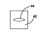
ここで図2Aに移ると、腫瘍38を含む模擬臓器20の一部分の部分側面断面図が示されている。模擬臓器又は模擬組織20は、ベース層又は臓器壁40を含む。臓器壁40は、現実の生組織を模倣するように構成されたシリコーン又は他のポリマーのような材料から作られ、適切に染色される。壁40の全体を構成するのに、様々な厚み及び配色の1つ又はそれよりも多くのベース層40を使用することができる。一変形では、臓器壁40は剛性を有し、ポリマー材料で作られる。ベース層40の上方には、第2の層又は欠陥層42が存在する。欠陥層42は、ベース層40と同じサイズのもの又はそれよりも小さいサイズのものであり、腫瘍38に対する隆起したプラットフォームを形成する。欠陥層42は、単一ユニットとしてベース層40と一体的に形成されることを含み、接着剤又は当業者に公知の他の手段によってベース層40に接続される。欠陥層42は、ベース層40の背景の中に溶け込むようにシリコーンで作られ、一変形ではベース層40と同じ色のもので作られる。欠陥層42は、少なくとも1つの欠陥又は間隙44を含む。一変形では、欠陥44は、断裂、切断、除去、又は他の外科手順からもたらされ、欠陥を閉鎖するのに縫合及びステープル吻合などを用いた手術的な配慮を必要とする現実の組織内の切開部、間隙、又は他の空隙を模倣する欠陥層42内に事前製作された裂開部である。そのような状況は、予防的に腫瘍全体が切除されることを確実にするために腫瘍38と共に周囲の組織も除去されて組織内に残存欠陥が残る腫瘍38の除去において最も多くの場合に生じる。欠陥44は、間に間隙を定める2つの対向する側部又は面を含む。隣接する側部又は面をベース層40に対して垂直であるように示すが、本発明は、これに限定されることはなく、並置される面又は側部は、あらゆる形状を有することができ、例えば、湾曲させることができる。欠陥44は、図3A〜図3Fに関して以下に解説するようないずれかの形状とすることができる。 Turning now to FIG. 2A, a partial side cross-sectional view of a portion of a
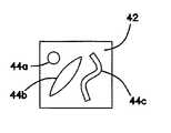
ここで図3Aに移ると、円形欠陥44を有する欠陥層42の上面図が示されている。図3Bには、細長形、長円形、又は楕円形の欠陥44を有する欠陥層42が示されている。図3Cに示すように、欠陥44は、無定形又はあらゆる形状とすることができる。欠陥層42は、図3Dに示すように多部分とすることができ、並置されて間に少なくとも1つの欠陥44を生成する2つ又はそれよりも多くの隣接欠陥層部分42a、42bを含む。図3Eには、複数の隣接欠陥層部分42a、42b、及び42cが、これらの間に1つ又はそれよりも多くの欠陥44を形成する別の多部分欠陥層42が示されている。当然ながら、欠陥層42は、図3Fに示すように、複数の欠陥44a、44b、及び44cを含むことができる。欠陥44は、全てを同じものとするか又は図3Fに示すように異なる形状を有することができる。欠陥の形状、厚み、及びサイズは、外科医訓練生が、変化する難度を有する欠陥の両側を縫合する練習を行うことを可能にする。一変形では、欠陥層42は、同一の厚みのものではない。同一の厚みの代わりに、欠陥層42の厚みは、欠陥44の位置で変化して欠陥を縫合又は閉鎖する難度を高める。 Turning now to FIG. 3A, a top view of a
再度図2Aを参照すると、腫瘍38は、欠陥層42の上に置かれる。好ましくは、訓練生によって容易に識別可能であるように、腫瘍38は、ベース層40又は欠陥層42又はこれらの両方と異なる色のものである。好ましくは、腫瘍38は、シリコーン又は他のポリマー材料で作られ、色が赤色、黒色、又は暗褐色である。一般的に、腫瘍38は、スコープを通して見た時にベース層40又は欠陥層42よりも濃い色のもの又はそうでなければ対照的な色のものである。一変形では、腫瘍38は、接着剤又は当業者に公知の他の手段によって欠陥層42に接続される。別の変形では、腫瘍38は、欠陥層42に接続又は取り付けられず、その上に取外し可能に位置付けられる。 Referring again to FIG. 2A, a
尚も図2Aを参照すると、模擬組織構成20は、腫瘍38の上に置かれたカバー層46を含む。一変形では、カバー層46は、腫瘍38、欠陥層42、及びベース層40の上に重なる。好ましくは、カバー層46は、色が透明又は半透明であり、シリコーンのようなポリマー材料で作られる。別の変形では、カバー層46は、ベース層40又は欠陥層42と同じ色のものである。カバー層46は、ベース層40又は欠陥層42と少なくとも同程度の厚みのものであり、一変形では、欠陥層42よりも薄肉であり、別の変形では、ベース層40よりも薄肉である。カバー層46は、腫瘍38及び欠陥層42の全域を覆うようにサイズが決定され、一変形では、ベース層40に接触するように十分に大きい。別の変形では、カバー層46は、腫瘍38の全域を覆い、欠陥層42に接触するようにサイズが決定される。カバー層46は、ベース層40、欠陥層42、腫瘍38、又はこれら3つの層のうちの1つよりも多いものに接着剤又は当業者に公知の他の手段を用いて接続される。別の変形では、カバー層46は小さめであり、欠陥層42だけに接続される。更に別の変形では、カバー層46は、欠陥層42とベース層40の両方に接着剤又は当業者に公知の他の手段を用いて接続される。カバー層46は、あらゆる形状又はサイズとすることができ、外科医に対して、人工腫瘍の位置に対する層状の面の代わりに滑らかな面を与えるように構成することができる。一変形では、カバー層46、腫瘍38、欠陥層42、又はベース層40は面テクスチャを含む。更に、カバー層46は、腫瘍38及び欠陥層42をカバー層46とベース層40の間に挟まれた状態に保つのを助け、これは、腫瘍38が欠陥層42に接着されない変形に対して有利である。図4には、ベース層40、欠陥層42、カバー層46、及び腫瘍38の上部平面図が示されている。一変形では、ベース層40、欠陥層42、及びカバー層46のうちのいずれか1つ又はそれよりも多くは、シリコーンの層が一体化メッシュ構造支持体又は他のタイプの補強体を有するように、ナイロン又は寒冷紗のような紡織材料、繊維材料、又はメッシュ材料の上にモールド成形されたシリコーンで形成される。これらの層38、40、42、46のうちのいずれか1つ又はそれよりも多くは、シリコーンのような弾性ポリマーと組み合わされた繊維又はメッシュの補強体を含むことができる。メッシュ支持体は、間隙44を閉鎖するように縫合糸が引っ張られる時に、縫合糸、ステープル、又は縫合針が、層のうちの少なくとも1つ、特に欠陥層42を断裂させるのを防ぐのを助ける。 Still referring to FIG. 2A,
図2Bでは、ベース層40から切除される腫瘍38とカバー層46の一部分とが示されている。この切除は、訓練生により、腫瘍38を除去するためのメス又は他の医療器具を用いて実施される。訓練生は、図2Bに示すように、腫瘍38の周りのカバー層46を切開し、腫瘍38を単離し、この部位から離れるように腫瘍38を持ち上げて除去して下層の欠陥44を露出させることになる。ここで図2Cに示すように、訓練生は、手術縫合糸を用いて欠陥44を縫合し、図2Dに示すように欠陥層42の唇部又は縁部を接合し、それによって腫瘍38の手術除去によって生成された間隙又は創傷の閉鎖を練習する。少なくとも1つの層を切断して開口部を生成し、人工腫瘍を除去して間隙を縫合する段階は、模擬組織構造が、ユーザの視界から少なくとも部分的に掩蔽されるように手術訓練デバイスの模擬体腔18の内側に置かれている間に実施される。 In FIG. 2B, the
ここで図5に移ると、第2の層又は欠陥層42内に事前形成された間隙又は欠陥が存在しない別の変形が示されている。事前形成された間隙又は欠陥が存在しない代わりに、腫瘍38を切除するときに、カバー層46、欠陥層42、ベース層40、及びユーザによって除去されなかったいずれかの残留腫瘍部分のうちの1つ又はそれよりも多くの中にユーザによって欠陥が生成される。次いで、ユーザは、これらの層38、40、42、46のうちのいずれかの中に生成された欠陥を縫合する練習を行うことになる。1つのそのような変形では、欠陥層42又はベース層40の一方は、構成から割愛される。別の変形では、欠陥層42が腫瘍38の上に存在するように、腫瘍38はベース層40上に置かれ、欠陥層42は腫瘍38の上に置かれる。そのような変形では、カバー層46は含まれても含まれなくてもよい。カバー層46が含まれる場合に、それを欠陥層と共に別個の単層として一体的に形成することができる。図2〜図5に関して上述した構成のうちのいずれにおいても、現実の組織の模擬効果を与えるために、必要に応じて層の厚み及び色を相応に調節した状態で、構成を逆さに反転させる、層を逆順で配置する、又は他に構成をユーザが上方向又は下方向のいずれからも接近可能にすることができる。 Turning now to FIG. 5, another variation is shown in which there are no pre-formed gaps or defects in the second layer or defect layer. One of the
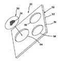
ここで図6A及び図6Bに移ると、本明細書における変形のうちのいずれにおいても、模擬組織構成は、全体の模擬臓器20と一体的に形成されず、その代わりに取外し可能で交換可能なモジュール50として構成されるようにモジュラーとすることができる。1つ又はそれよりも多くのモジュール50は、モジュール支持体52内に支持されるか又は含まれる。モジュール支持体52は、第1の面51と、第2の面53と、支持体52内に形成された1つ又はそれよりも多くの腫瘍モジュール受け入れ部分54、56、58とを含む。腫瘍支持体52は、剛性又は柔軟性を有することができ、ポリマー材料で作ることができる。腫瘍支持体52は、エラストマー材料シートを含むことができる。モジュール受け入れ部分54、56、58の各々は、相応にサイズ決定され、かつそのように構成されたモジュール50を受け入れるようにサイズ決定され、かつそのように構成される。図6では、モジュール50とモジュール受け入れ部分54、56、58とを円形であるように示すが、腫瘍モジュール50は、相補的な形の受け入れ部分がモジュール支持体52内に形成された状態であらゆる形状とすることができる。支持体52の厚みは、異なるとすることができ、様々な深さの腫瘍モジュール50の位置を有する構成が与えられる。モジュール受け入れ部分54、56、58は、腫瘍モジュール50が載ることができる底壁を含むことができる。これに代えて、腫瘍受け入れ部分54、56、58は、第1の面51内の開口部と第2の面53の開口部の間で延び、腫瘍38は、いかなる面51、53にもある開口部の間に接続されるか、又はこれらの開口部の一方に接続されるか、又は腫瘍受け入れ部分内に懸架される。一変形では、単一腫瘍モジュール50は、1つ又はそれよりも多くの腫瘍38を含む。モジュール支持体52には、1つ又はそれよりも多くの腫瘍モジュール50が装荷され、模擬組織構成20は、手術訓練デバイス10の体腔18、フレーム、又は他の本体モデル内に挿入される。模擬組織構成20は、訓練デバイス10のベース12上に配置するか又は訓練デバイス10の体腔18内に懸架することができる。模擬組織構成20及び/又は訓練デバイスは、訓練デバイス10への模擬組織構成20の配置、懸架、又は接続のためにクリップ、ファスナ、ワイヤ、フックアンドループ式ファスナなどのような取り付け機構が具備される。 Turning now to FIGS. 6A and 6B, in any of the variations herein, the simulated tissue configuration is not integrally formed with the entire
特に図6Bを参照すると、1つよりも多い層を含むモジュール支持体52が示されている。図6Bのモジュール支持体52は、第2の層55に接続した第1の層57を含む。一変形では、第1の層57は、エラストマー材料シートで作られ、第2の層55は、低密度弾性発泡体のようなあらゆる適切なポリマー材料で作られる。第2の層55は、第1の層57に対する支持体として機能する。第2の層55はまた、モジュール支持体52に深さも有利に与え、モジュール50内の腫瘍38を第1の面51に対してモジュール支持体52内に深く配置することを可能にする。モジュール受け入れ部分54、56、58は、第1の層57及び第2の層55のうちの1つ又は1つよりも多いものに形成される。第2の層55内に形成されるモジュール受け入れ部分54、56、58は、同じモジュール受け入れ部分54、56、58が第1の層57内に有する形状とは異なる形状を有することができる。一変形では、腫瘍モジュール50は、第2の層55の内側に組み込まれた又は埋め込まれた少なくとも模擬腫瘍38だけを含み、第1の層57又は第2の層55のうちの少なくとも一方は、ユーザが閉鎖を練習することができる欠陥層を構成する。代替として、第1の層57は、モジュール受け入れ部分を含まず、これに代えて、ユーザが、第2の層55内に形成された腫瘍受け入れ部分に位置付けられた腫瘍38にアクセスするために切開を練習するカバー層として機能する。そのような変形では、第1の層57は、シリコーンのようなエラストマー材料シートとすることができ、第2の層55は、低密度弾性発泡体の層である。モジュール支持体52は、図6A及び図6Bに示すように平面であり、又はこれに代えて、人間の解剖学的構造、組織、又は臓器の一部分を模倣するように成形される。 Referring particularly to FIG. 6B, a
例えば、図7は、人間の子宮を模倣するように成形された支持体52を示している。支持体52は、第2の層55に接続した第1の層57を含む。一変形では、第1の層57は、エラストマー材料シートのようなあらゆる適切なポリマー材料で作られ、第2の層55は、低密度弾性発泡体のようないずれかの適切ポリマー材料で作られる。第2の層55は、第1の層57に対する支持体として機能し、モジュール50内にある腫瘍38又は独立して存在する腫瘍38を支持体52に有利に接続され、現実に即して支持体52内に深く延び込ませ、図7に示すように第1の層57内に埋め込むことを含み、支持体52を通して様々な位置及び向きに分散させることを可能にする。腫瘍又はモジュールの受け入れ部分61は、第1の層57及び第2の層55のうちの少なくとも一方の中に形成される。腫瘍受け入れ部分61は、第2の層55内に事前形成されたポケットとすることができ、又はユーザが、第2の層55内にスリットを切り込むことによって形成することができる。一変形では、腫瘍38は、人間の子宮内に一般的に見つかる類線維腫を模倣するように構成される。支持体に置かれた腫瘍38によって模擬される類線維腫の例は、有茎粘膜下筋腫、漿膜下筋腫、粘膜下筋腫、有茎漿膜下筋腫、及び壁内筋腫というタイプの筋腫のうちの1つ又はそれよりも多くを含むが、これらに限定されない。ユーザは、模擬腫瘍38を切除するために、第1の面51又は第2の面53からアクセスチャネル又は開口部63を通して支持体52に接近することができる。一変形では、開口部63は、中空部分59への唯一の開口部として機能し、又はこれに代えて支持体52は、ユーザに平面C字形構造の上又は下から利用可能なアクセスを有する実質的にC字形の平面構成を有することができる。 For example, FIG. 7 shows a
一変形では、変形のうちのいかなるものにおけるモジュール支持体52も平面ではなく、曲面又は他の構造、山及び谷、並びに様々なテクスチャを含む輪郭が与えられる。異なる輪郭は、腫瘍位置を掩蔽する可能性がある人工物及び特徴部を避けて進むことをユーザに要求しながら、各腫瘍位置に接近するのに様々な難易度をユーザに与える。腫瘍支持体52内のこれらの構造的人工物は、腫瘍支持体52と一体的に形成することができ、又は解剖学的構造の輪郭モジュールを取外し可能かつ交換可能にするように、腫瘍モジュール50と類似の構造においてモジュラーとすることができる。腫瘍モジュール50は、例えば、モジュール支持体52の上面及び下面51、53のうちの1つ又はそれよりも多くから外向き又は内向きに延びるシリコーン又は他の材料で作られた特徴部及び人工物又はテクスチャを含む非腫瘍モジュールと交換可能である。そのような非腫瘍モジュール内の特徴部は、隣接臓器の構造又は組織を含む解剖学的構造を模倣するための様々な形状を有することができる。例えば、非腫瘍モジュールは、腸を模倣するためのシリコーンの管状形態を含むことができる。非腫瘍モジュール及び腫瘍モジュール50は、当業者に公知のいずれかの手段によってモジュール支持体52に取外し可能に接続され、ユーザが、使用後にモジュールを廃棄し、次いで、廃棄したモジュールを交換し、モジュール支持体52内の隣接モジュール50に移動するか又は腫瘍モジュール50を異なる特徴部又は難易度を有する別の腫瘍モジュール50に入れ替えることによって練習を続けることを可能にする。 In one variation, the
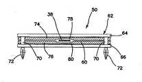
図8及び図9には、腫瘍モジュールの変形50を示している。この腫瘍モジュール50は、支持体62に接続した模擬組織部分60を含む。図示の変形では、支持体62は、底枠66に接続した上枠64を含む。上枠64及び底枠66のうちの少なくとも一方は窓を含む。図8には、窓68を有する上枠64を示している。底枠66は、窓を含んでも含まなくてもよい。窓が上枠64と底枠66の両方内に設けられる場合に、これらの窓は、少なくとも部分的に位置合わせされる。支持体62は、上枠64と底枠66の間に模擬組織部分60を受け入れるようにサイズ決定され、かつそのように構成される。上枠64は、単一模擬組織部分60又は複数の層から形成され、一変形では切断可能な模擬組織部分60を捕捉するように底枠66に接続可能である。一変形では、枠64、66は、スペーサ70を用いて互いに離間する。更に、上枠64及び底枠66のうちの少なくとも一方は、腫瘍モジュール50を腫瘍支持体52(図示せず)に固定するように構成された1つ又はそれよりも多くの接続特徴部72を含む。図9には、接続特徴部72を腫瘍支持体52内に形成された対応する穴内への挿入のためのスナップフィット係合を与える延長ペグとして示している。モジュール50を支持体52に取外し可能方式で接続するために、摩擦嵌め手段又はフックアンドループ式材料のような他のファスナ手段又は接続手段をモジュール50及びモジュール支持体52上に使用することができる。 8 and 9 show a
尚も図8及び図9を参照すると、模擬組織部分60は、図2〜図5を参照して上述した構成のうちのいずれかとすることができる。第1の枠64と第2の枠66の両方内に形成された窓を使用すると、モジュール50のいずれ側からも模擬組織部分60に接近することができる。カバー層として上述のいかなる層も、模擬組織部分60がいかなる側又は方向から接近されるかに基づいて、上層又は底層として機能することができる。例えば、ベース層も、模擬組織部分60がいかなる側又は方向から接近されるかに基づいて、上層又は底層として機能することができる。そのような双方向構成では、望ましい模擬効果を与えるために、層の厚み及び色を相応に調節することができる。 Still referring to FIGS. 8 and 9, the
図9の模擬組織部分60は、第1の層74と第2の層76を含む。第1及び第2の層74、76は、現実の生組織を模倣するように構成されたシリコーン又は他のポリマーのようなポリマー材料から作られ、いずれか1つ又はそれよりも多くの適切な色の染料、又はメッシュ、繊維、又は他の補強体を含むことができる。層74、76の各々は、それぞれ腫瘍受け入れ部分78、80を含む。各腫瘍受け入れ部分78、80は、層74、76内に形成された凹部、窪み、半ポケット、又は小幅層厚の場所である。腫瘍受け入れ部分78、80は、実質的に位置合わせされて腫瘍38に対するポケットを形成する。図9には、各層74、76を腫瘍受け入れ部分78、80を有するように示すが、一変形では、第1及び第2の層74、76のうちの少なくとも一方内に単一腫瘍受け入れ部分が形成される。腫瘍38は、1つ又はそれよりも多くの層74、76内に形成された1つ又はそれよりも多くの腫瘍受け入れ部分78、80によって形成されるポケット内に置かれる。腫瘍38は、いずれかの層74、76に接着するか又はポケット内で浮遊させることができる。図9に示すように層内に形成された腫瘍受け入れ部分は、欠陥の1つのタイプと考えることができ、図9の変形は、腫瘍を挟む2つの欠陥層を含む模擬組織構成を説明する。ユーザが模擬組織部分60に接近するときに、ユーザは、ターゲットの腫瘍位置を見ることになる。ターゲット腫瘍38の可視化は、凹部又はポケットによって与えられる層の薄肉化により、腫瘍受け入れ部分の厚みが層の残りの部分と比較して薄肉であることによって改善する。次いで、ユーザは、大体の腫瘍位置で切断を行うことになり、腫瘍38を除去するために層74、76のうちの少なくとも一方内に切り込みを行う。1つ又はそれよりも多くの層を切開することによって間隙又は完全欠陥の生成が完了し、次いで、ユーザは、それを縫合、又は他に閉じ合わせる練習をすることができる。別の変形では、層74、76内に形成された腫瘍受け入れ部分は存在しない。そのような変形では、2つの層74、76の間に少なくとも1つの腫瘍が位置付けられ、層74、76は、実質的に均一の厚みを有し、腫瘍38は、層内に軽微な膨らみを生成する。 The
ここで図10A、図10B、図11A、図11B、及び図12に移ると、模擬組織部分の別の変形86が示されている。上述のように、組織部分86は、一体的又はモジュラーとすることができる。組織部分86は、縫合糸を保持する間又は縫合される間に断裂を食止めることになる繊維、メッシュ、ナイロン、又は他の補強材のような補強材又は充填材を含んでも含まなくてもよいシリコーン又は他の弾性ポリマーのようなあらゆる適切な弾性ポリマーで形成されたベース層88を含む。ベース層88は、その上に重ねられる欠陥層90に接続される。欠陥層90は、ベース層88から上方に延びる複数の突出部を含む。欠陥層90は、ベース層88と一体的に形成するか又はベース層88に接着された別個の層とすることができる。図10A、図11A、及び図12で分るように、欠陥層90は、ベース層88の上で隆起するような又はベース層88から上方に突出するような格子の形状のパターンに構成される。格子パターンは例示的なものであり、欠陥層90により、複数の隣接突出部を含むようなあらゆる形状を形成することができる。ベース層90のこれらの突出部は、容易な切除に向けてベース層88の上に腫瘍38a、38bを隆起させるプラットフォームとしての縫合針を引っ掛ける位置をユーザに提供する。腫瘍38a、38bは、欠陥層90に接着することができ、一変形では、カバー層92を含めることができる。図10A及び図11Aは、ベース層88と、欠陥層90と、腫瘍38a、38bと、カバー層92とを模擬組織部分86の半分解組立図に示しており、カバー層92は、他の層の上に隆起する。図10aの腫瘍38aは実質的に平面であり、図10Bではカバー層92によって覆われるように示されている。図11Aの腫瘍38bは、より大きい高さを有し、形状が実質的に球形であり、図11Bは、構成内に隆起部分又は突出部を残しながら、カバー層92で覆われた球形腫瘍38bを示している。図12は、欠陥がカバー層92の下で又はそれを通してアクセスされた状態でベース層88内に残存欠陥94を残しながら除去されている腫瘍38と、欠陥94内の間隙を横断する縫合針とを示している。 Turning now to FIGS. 10A, 10B, 11A, 11B, and 12, another
生きている組織の特性を模倣する合成材料は、シリコーン弾性体、天然ラテックス、ポリウレタン弾性体、ヒドロゲル、及びスチレンブロックコポリマーを含むことができる。一般的に、エラストマー材料は、特殊処理が行われない限り誘電材料である。一般的に、弾性体は、天然ゴムのものに似た弾力的特質を有する様々なポリマーのうちのいずれかである。一般的に、ヒドロゲルは、50%と99%の間の水を含有する親水性ポリマーである。一般的に、熱可塑性は、加熱と冷却によって繰り返し軟質と硬質にすることができる材料に属する。熱可塑性は、非導電性を有し、トレイ又はベース、骨格、及び他の類似の構造を生成するのに適している。一般的に、熱硬化性樹脂は、加熱又は硬化されて永久に硬化するエラストマー材料に属する。熱硬化性プラスチックは、シリコーン及びポリエステルのように非導電性を有し、病変部及び腫瘍などを形成するのに適している。シリコーン弾性体は、通常は非常に軟質で安定しており、かつ非導電性を有し、従って、肝臓、腎臓、脾臓、卵巣、胆嚢、胃、主幹動脈、結腸、腸、主幹静脈、網、腸間膜のような人工臓器、病変部、及び他の解剖学的構造を形成するのに適している。天然ラテックスは非常に弾性が高く、非導電性を有し、人工の筋肉及び軟骨などを形成するのに適している。ポリウレタンの弾性体及び発泡体は非導電性を有し、中空構造及び骨などを充填するのに適している。ヒドロゲルSBCは、導電性を有することができ、電気手術によって手術されるいずれの軟質構造にも良好である。 Synthetic materials that mimic the properties of living tissue can include silicone elastomers, natural latexes, polyurethane elastomers, hydrogels, and styrene block copolymers. In general, elastomeric materials are dielectric materials unless special treatment is performed. Generally, an elastic is any of a variety of polymers having elastic properties similar to those of natural rubber. Generally, hydrogels are hydrophilic polymers that contain between 50% and 99% water. In general, thermoplasticity belongs to materials which can be repeatedly made soft and hard by heating and cooling. Thermoplasticity is non-conductive and suitable for producing trays or bases, frameworks and other similar structures. In general, thermosetting resins belong to elastomeric materials which are heated or cured to permanently cure. Thermosetting plastics, like silicones and polyesters, have non-conductivity and are suitable for forming lesions and tumors. The silicone elastomer is usually very soft, stable, and nonconductive, and therefore has liver, kidney, spleen, ovary, gallbladder, stomach, trunk artery, colon, intestine, trunk vein, mesh, It is suitable for forming artificial organs such as mesentery, lesions, and other anatomical structures. Natural latex is very elastic, nonconductive, and suitable for forming artificial muscles and cartilage. Polyurethane elastomers and foams have non-conductivity and are suitable for filling hollow structures, bones and the like. The hydrogel SBC can be electrically conductive and is good for any soft structure operated by electrosurgery.
一変形では、腹腔鏡法及び電気手術法を含む外科技術を練習するための腹腔鏡訓練器10内に挿入可能な手術シミュレーショントレイは、ベースと、解剖学的臓器の配置と、カバー層とを含む。ベースは、手術訓練デバイス10の内部又はその上に嵌合するようにサイズ決定され、かつそのように構成された剛性又は半剛性の構造を含む。これに加えて、ベースには、レセプタクル部分の内部又はベースの上への身体臓器の配置とサイズ及び形状において協動及び適合する直立壁によって形成された解剖学的な支持特徴部又はレセプタクル部分が与えられる。エラストマー材料で作られる身体臓器は、訓練デバイスの特定の必要性及び/又はターゲット解剖学的構造に則してベースの内部又はその上に計画的に置かれる。アセンブリ全体の上に又はその特定の区域の上には、少なくとも1つのカバー層を配置することができる。カバー層は、網、腸間膜、脂肪、結合組織、腹膜、中皮、及び広間膜などうちの1つ又はそれよりも多くを表すようにサイズ決定され、かつそのように構成される。カバー層は、非導電性を有するシリコーン弾性体を含むことができる。非導電性カバー層は、カバー層に対して電気手術動作が使用されない場合に適している。電気手術動作が企図される場合に、カバー層は、ヒドロゲルのような導電性ゲルからなる。導電性層と非導電性層の組合せは、電気手術が層のうちの1つに向けられる場合に設けられる。 In one variation, a surgical simulation tray insertable into the
ベースの内部又はその上に置かれた臓器に加えて、臓器と相対的に又は模擬臓器自体内には、同じく計画的に置かれた複数の病変部又は欠陥を存在させることができる。病変部又は欠陥は、腫瘍、嚢腫、又は子宮外妊娠部などを表すことができる。例えば、図7に関して上述したように、シリコーンゴムの外側層と、軟質ポリウレタン発泡体の実質的に中空の内側層とを有する子宮を形成することができる。手術訓練生による識別及び除去に向けて、シリコーン層と発泡体層の間の様々な位置に合成類線維腫を配置することができる。合成類線維腫の1つの模擬構成は、少量の超軟質未硬化シリコーンゴムを含む。未硬化シリコーンゴムは、充填材及び流動制御器として機能する一定量の無定形未処理ヒュームド二酸化珪素と混合される。未硬化シリコーンゴムと二酸化珪素の組合せは、成形されて硬化される。完全に硬化すると、この組合せは、人間の類線維腫に似た不規則形状の幾分繊維性を有する構造をもたらす。次いで、この模擬人間類線維腫の構成は、子宮のもののような模擬臓器モデルに置かれる。この腫瘍シミュレーションは、婦人科モデルにおける腫瘍を模倣するための使用に限定されず、腫瘍の除去を練習するために腫瘍を含む他の臓器モデルに対して使用することができる。シリコーンゴムと二酸化珪素とで構成される硬化混合物を含むこの腫瘍シミュレーションは、婦人科手術の関連において見られる現実の腫瘍に非常に似ており、外科技術を練習するときに無定形の現実的な外観及び感触を提供する。この混合物には、全体を通して硬化及び混合する前に、赤色又は黒色のような暗色の染料を追加することができる。この構成は、訓練デバイス10内に置かれた模擬臓器の模擬卵管内への挿入のための模擬子宮外妊娠部を構成するために使用することができる。非常に乾燥しており、かつ賦形性の高いシリコーンと充填材との混合状態の稠度は、実際の身体状態を模倣するために腫瘍又は他の病変部を非常に創造的に、容易に、かつあらゆるサイズで形成することを有利に可能にする。シリコーンと充填材とで作られる腫瘍は、非導電性を有し、適切に取り扱われないと破断又は断裂する可能性がある。 In addition to the organs placed in or on the base, relative to the organ or in the simulated organ itself, there can be a plurality of lesions or defects also placed systematically. The lesion or defect can represent a tumor, a cyst, or an ectopic pregnancy site or the like. For example, as described above with respect to FIG. 7, a uterus can be formed having an outer layer of silicone rubber and a substantially hollow inner layer of flexible polyurethane foam. Synthetic fibroids can be placed at various locations between the silicone layer and the foam layer for identification and removal by surgical trainees. One simulated configuration of synthetic fibroids contains small amounts of ultra-soft uncured silicone rubber. The uncured silicone rubber is mixed with a quantity of amorphous untreated fumed silicon dioxide which acts as a filler and flow controller. The combination of uncured silicone rubber and silicon dioxide is molded and cured. When fully cured, this combination results in an irregularly shaped, somewhat fibrous structure resembling human fibroids. This simulated human fibroma composition is then placed in a simulated organ model, such as that of the uterus. This tumor simulation is not limited to use for mimicking tumors in gynecologic models, but can be used for other organ models including tumors to practice tumor removal. This tumor simulation, which includes a hardened mixture composed of silicone rubber and silicon dioxide, is very similar to the real tumor seen in the context of gynecologic surgery and is amorphous realistic when practicing surgical techniques Provide an appearance and feel. To this mixture, a dark dye such as red or black can be added prior to curing and mixing throughout. This configuration can be used to construct a simulated ectopic pregnancy site for insertion of a simulated organ placed in the
次いで、導電性部分と非導電性部分の組合せを含む臓器模擬モデルの一部の例を解説する。肝臓切除の外科手順では、電気外科手順を訓練するための模擬臓器モデルは、導電性ヒドロゲルの肝臓、包嚢管、及び腸間膜を有することになる。モデルのうちのこれらの導電性部分は、同じ臓器又は異なる臓器を含む解剖学的構造の非導電性部分に隣接して位置付けられる。例えば、胆嚢摘除の外科手順を練習するためには、臓器モデルは、導電性ヒドロゲルから作られた包嚢管及び中皮を含み、肝臓及び胆嚢は非導電性を有する。スリーブ状胃切除術を練習するためには、模擬臓器モデルは、導電性ヒドロゲル材料で作られた胃の大彎に沿う血管及び大網/腸間膜のうちの1つ又はそれよりも多くと、非導電材料で作られた胃、大腸、及び小腸のうちの1つ又はそれよりも多くとを含む。胃バイパス術を練習するためには、模擬臓器モデルは、導電性ヒドロゲル材料で作られた胃の大彎に沿う短胃血管及び腸間膜/網のうちの1つ又はそれよりも多くと、非導電材料で作られた胃とを含む。一変形では、空腸及び/又は胃の少なくとも一部分は、導電性ヒドロゲルで作られる。類線維腫の除去、子宮外妊娠、卵巣嚢腫の治療、及び子宮摘出のような卵巣の手順を練習するためには、訓練モデルは、導電材料と非導電材料の両方を含む。例えば、臓器モデルは、導電性ヒドロゲルで作られた1つ又はそれよりも多くの卵管、円靭帯、卵巣靭帯、IP靭帯、広靭帯、膀胱弁、子宮動脈/静脈、基靱帯、子宮仙骨靭帯のシミュレーションを含むことができ、子宮、卵巣、直腸、膀胱、尿管、及び腎臓のうちの1つ又はそれよりも多くは非導電性を有する。一変形では、子宮頸部の直上及び/又は直下の位置は、子宮頸管上部腟切開術又は全腟切開術を練習するために導電性ヒドロゲルで作られる。結腸、小腸、S状結腸、又は直腸が係わる手順は、特定の部分が導電性を有することを要求する場合もある。これらの導電性部分は、非導電性部分に隣接して位置付けられる。例えば、腫瘍の局所切除のための経肛門低侵襲手術を練習するためには、臓器モデルは、腫瘍を取り囲む導電性ヒドロゲル材料で作られることになる区域を除き、非導電性エラストマー材料で作られた結腸及び/又は直腸、並びに腫瘍を含むことになる。別の変形では、経肛門直腸間膜全切除の練習などのためには、直腸の少なくとも一部分は、導電性ヒドロゲルで作られる。虫垂切除術の練習では、模擬臓器モデルは、導電性ヒドロゲルで作られた腸間膜/虫垂間膜、虫垂動脈、及び血管のうちの1つ又はそれよりも多くと、非導電性エラストマー材料で作られた虫垂、盲腸、及び回腸終末部のうちの1つ又はそれよりも多くとを含むことができる。結腸切除術を練習するためには、模擬臓器モデルは、導電性ヒドロゲルで作られた腸間膜、回結腸動脈、中結腸動脈、右結腸動脈、下腸間膜動脈、下腸間膜静脈、左結腸動脈、S状結腸動脈、直腸動脈、辺縁動脈、対応する静脈、網、トルト白線、後腹膜腔への腸間膜取り付け部、及び直腸間膜のうちの1つ又はそれよりも多くと、非導電材料で作られた結腸、肝臓、脾臓、胃、腎臓、十二指腸、後腹膜腔のうちの1つ又はそれよりも多くとを含むことができる。ヒドロゲル材料は、十分な導電性を有するためには水和させる必要があり、従って、長い有効保存寿命を維持するのは困難である場合がある。 Next, some examples of organ simulation models including combinations of conductive and non-conductive portions will be described. In a liver resection surgical procedure, a simulated organ model for training an electrosurgical procedure will have a conductive hydrogel liver, a cystic canal, and the mesentery. These conductive portions of the model are positioned adjacent to non-conductive portions of the anatomic structure including the same or different organs. For example, to practice the surgical procedure of cholecystectomy, an organ model includes a cystic canal and mesothelium made of a conductive hydrogel, and the liver and gallbladder have non-conductivity. In order to practice sleeve gastrectomy, the simulated organ model comprises one or more of the blood vessels and the omentum / mesenterium along the gastric antrum made of a conductive hydrogel material. , One or more of a stomach, a large intestine, and a small intestine made of a non-conductive material. In order to practice gastric bypass surgery, the simulated organ model may be one or more of a short gastric vessel and a mesenteric / mesh, along a gastric antrum made of a conductive hydrogel material. And a stomach made of a non-conductive material. In one variation, at least a portion of the jejunum and / or the stomach are made of a conductive hydrogel. In order to practice ovary procedures such as fibroid removal, ectopic pregnancy, treatment of ovarian cysts, and hysterectomy, the training model includes both conductive and non-conductive materials. For example, the organ model may be one or more oviducts made of a conductive hydrogel, a ligament, an ligament, an ligament, an IP ligament, a ligament, a bladder valve, a uterine artery / vein, a ligament, a urinary sacral ligament Simulation of one or more of uterus, ovary, rectum, bladder, ureter, and kidney are non-conductive. In one variation, the position just above and / or just below the cervix is made of a conductive hydrogel to practice an upper cervical incision or a total open incision. Procedures involving the colon, small intestine, sigmoid colon, or rectum may require certain parts to be electrically conductive. These conductive portions are positioned adjacent to the nonconductive portions. For example, to practice transanal minimally invasive surgery for local excision of a tumor, an organ model is made of a non-conductive elastomeric material, except in areas that are to be made of a conductive hydrogel material surrounding the tumor. Colon and / or rectum, as well as tumors. In another variation, at least a portion of the rectum is made of a conductive hydrogel, such as for the practice of transanal interrectal excision. In the practice of appendectomy, the simulated organ model is a non-conductive elastomeric material with one or more of the mesentery / intermediate, appendiceal artery, and blood vessel made of a conductive hydrogel. It may include one or more of an appendix, a cecum, and an end ileum made. In order to practice colectomy, a simulated organ model is the mesenteric, ili-colon artery, middle colon artery, right colon artery, inferior mesenteric artery, inferior mesenteric vein, made of conductive hydrogel, One or more of left colon artery, sigmoid artery, rectum artery, marginal artery, corresponding vein, mesh, tort white line, mesenteric attachment to retroperitoneal space, and interrectum And one or more of the colon, liver, spleen, stomach, kidney, duodenum, retroperitoneal cavity made of non-conductive material. The hydrogel material needs to be hydrated in order to have sufficient electrical conductivity, so it can be difficult to maintain a long effective shelf life.
基靱帯に関しては、一変形において、カバー層は、テクスチャ及び自然に発生したものであるように見える仕上がりを有するようにカレンダー製作又はプレス形成されたシリコーンゴムの薄肉の半透明シートを含む。カバー層の別の変形は、スラリから硬化され、硬化するときに面特徴部を発達させたヒドロゲル材料の薄肉の半透明シートを更に含むことができる。ヒドロゲル材料は、水和すると導電性を有するようになり、電気手術デバイスの使用を可能にする。カバー層に対する複合構造は、2つのシリコーン弾性非導電性層の間に挟まれた導電性ゲル層を含む。そのような場合に、導電性ゲル層を露出させるために、外側非導電性層のうちの1つ又はそれよりも多くが除去される。非導電性シリコーン層は、ヒドロゲル層に対して有利に密封を施し、導電性ゲルの流体成分を保持する。 With respect to the ligament base, in one variation, the cover layer comprises a thin translucent sheet of silicone rubber that has been calendered or pressed to have a texture and finish that appears to be naturally occurring. Another variation of the cover layer can further include a thin translucent sheet of hydrogel material that has been cured from the slurry and developed surface features upon curing. The hydrogel material becomes conductive when hydrated, enabling the use of the electrosurgical device. The composite structure for the cover layer comprises a conductive gel layer sandwiched between two silicone elastic non-conductive layers. In such cases, one or more of the outer nonconductive layers are removed to expose the conductive gel layer. The nonconductive silicone layer advantageously seals against the hydrogel layer and retains the fluid component of the conductive gel.
カバー層の別の変形では、テクスチャが付けられたポリエチレン発泡体のシート上には、2部分構成のプラチナ硬化又は錫硬化の液体シリコーンの薄膜が置かれる。次いで、ノッチこて又は延展器を用いて、第1の発泡体層の面の上に、材料厚の不規則パターンを残しながらシリコーン材料が延展される。第1の発泡体層の上には、第2のテクスチャ付きポリエチレン発泡体層が、これらの間にシリコーンを残しながら置かれる。次いで、発泡体層の間のシリコーン材料をカレンダー製作するために、第2の発泡体層の面上でテクスチャ付きローラ又は箔押デバイスが移動される。得られるシリコーンシートは、硬化されると粘着性を持たず、網、腸間膜、脂肪のような特性を示す。このシートは、機械的解剖器具及び剪刀の使用を実演するために使用することができる高強度領域と低強度領域を有利に有する。 In another variation of the cover layer, a two-part platinum-cured or tin-cured thin film of liquid silicone is placed on the textured sheet of polyethylene foam. Then, using a notch trowel or spreader, the silicone material is spread on the face of the first foam layer, leaving an irregular pattern of material thickness. A second textured polyethylene foam layer is placed on top of the first foam layer, leaving silicone between them. The textured roller or foiling device is then moved over the face of the second foam layer to calender the silicone material between the foam layers. The resulting silicone sheet, when cured, has no tack and exhibits properties such as mesh, mesentery, fat. This sheet advantageously has high and low strength areas that can be used to demonstrate the use of mechanical dissectors and scissors.
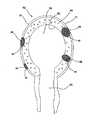
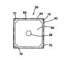
手術シミュレーションデバイス内に使用することができる特定の臓器は、図13に示すような子宮100を含む。子宮は、子宮形態の上にモールド成形された軟質シリコーンゴムからなるシェルを含む。このシェルは、完全に硬化されると、実質的に中空であり、約7ミリメートルから9ミリメートルまでの厚みの壁を有するモールド成形発泡体ゴム子宮形態の上に置かれる。シリコーンシェルと発泡体壁の間には、様々な病変部を配置することができる。壁内腫瘍、卵管102、又は嚢腫を模倣するために、一部の病変部は、発泡体壁内に挿入することができる。シリコーン/発泡体構造内には、卵管104、卵巣靭帯106、及び他の付帯構造を挿入し、接着剤を用いて取り付けることができる。卵巣嚢腫124を設け、同じ腫瘍材料で作ることができる。付帯構造は、大動脈114と、内腸骨動脈116と、卵巣動脈118と、子宮動脈120と、膣動脈121と、子宮仙骨靭帯122とを含むことができる。子宮シェルは、手術される主要部分である。一変形では、子宮シェルは、シリコーン弾性体からなり、従って、訓練において子宮モデルを切断又は切開することが意図される場合に適している。子宮モデルに対して電気手術が練習されている場合に、導電性ゲルを含む子宮モデルが選択される。手術方式に基づいて、接続構造及び接続チューブは、シリコーン弾性体又は導電性ゲルからなる。 Specific organs that can be used in the surgical simulation device include the
2部分構成のプラチナ硬化又は錫硬化のシリコーンからなる卵管104は、第1の開口端部と第2の開口端部と貫通内腔とを含む。第1の開口端部は、約20センチメートルの距離を延びて約6.5ミリメートルの直径と約1〜1.5ミリメートルの非常に薄肉の壁とを有する管状構造を形成する。管状構造の端部に向けて、約1.5センチメートルの直径と約3センチメートルの長さとを有する球根状部分が形成される。球根状部分は、約7ミリメートルまでの管状構造の狭窄部に遷移する。次いで、狭窄管状構造は、約2センチメートルの最終開口径を有し、約3.5センチメートルの長さにわたる漏斗形構造に徐々に拡大する。卵管104をそれが上に作られた形態から除去する前に、第2の拡大開口端部に複数の軸線方向切断部108が作られる。形態から除去されると、これらの切り込みは、シリコーン材料が、人間の線毛に似た方式で移動することを可能にする。卵管104の球根状部分の中に、子宮外妊娠部のような病変部110を帰属化又は切除に向けて挿入することができる。更に、折り畳まれた時に卵管の薄壁導管部分の形状を維持するために、編み物に使用される或る長さの軟質繊維糸条を内腔内に置くことができる。 An
この模擬子宮モデルでは、卵巣112は、2部分構成のプラチナ硬化又は錫硬化のシリコーンから形成された中空球根状構造である。卵巣構造内には、軟質ポリウレタン発泡体支持体が置かれる。ポリウレタン支持体は、卵巣シェル内にきれいに嵌合し、卵巣嚢腫124のような病変部に対するネスト又はレセプタクルを有するようにサイズ決定され、かつそのように構成される。訓練生は、病変部を除去し、その後に欠陥を縫合して閉鎖するために、卵巣壁を通ってポリウレタン発泡体内に切開することができる。卵巣は、非導電材料で作られ、剪刀又はメスを用いて切断される。別の変形では、卵巣は、導電性ゲルで作られ、従って、電気手術によって切断することができる。嚢腫は、非導電材料で作られる。 In this simulated uterine model, the
別の模擬臓器モデルでは、胃は、第1の開口端部と、第2の開口端部と、拡大中心部分とを有する中空の胃形の嚢を含む。拡大中心部分は、第1の開口端部の近くから第2の開口端部の近くまで延びる経路によって分割される。この経路は、計画的に胃の小彎に隣接する望ましい軌道に沿って置かれたシリコーン接着剤領域を含む。胃嚢の対向する壁は、互いに接近し、接着剤によって保持される。特定の手順を模擬するために、胃を接着剤経路に沿って分割することができる。すなわち、接着剤経路は、好ましい手術経路に沿ってステープル吻合又は切断するように訓練生を誘導する。接着剤は、手術ステープル吻合器内の切断要素が配備される前に数列のステープルが置かれると考えられる状態を模擬する。その結果、解剖胃部分は、堅固にステープル吻合されたように見え、残留胃部分は気密で堅固なものである。別の変形では、胃の接着部分は、胃の非導電性隣接部分に隣接する導電性ゲル材料で形成される。更に別の変形では、胃又は他の臓器を横切る予め決められた手術経路は、同じ臓器の非導電材料に隣接するか又は異なる臓器及び解剖学的構造の非導電材料に隣接する導電性ゲル材料からなる。 In another simulated organ model, the stomach includes a hollow stomach-shaped sac having a first open end, a second open end, and an enlarged central portion. The enlarged central portion is divided by a path extending from near the first open end to near the second open end. This pathway includes a silicone adhesive area strategically placed along the desired trajectory adjacent to the stomach blebs. The opposing walls of the gastric pouch approach one another and are held by the adhesive. The stomach can be split along the adhesive path to simulate a specific procedure. That is, the adhesive pathway guides trainees to staple or sever staples along the preferred surgical pathway. The adhesive simulates a situation where several rows of staples are considered to be placed before the cutting element in the surgical staple anastomosis device is deployed. As a result, the dissected stomach section appears to be firmly stapled and the remaining stomach section is tight and tight. In another variation, the adhesive portion of the stomach is formed of a conductive gel material adjacent to the non-conductive adjacent portion of the stomach. In yet another variation, the predetermined surgical path across the stomach or other organ is a conductive gel material adjacent to the non-conductive material of the same organ or adjacent to the non-conductive material of a different organ and anatomical structure It consists of
別の模擬臓器モデルでは、ヒドロゲルからなる肝臓は、手順が電気手術解剖を伴うと考えられる訓練モジュール10内に置くことができる。一変形では、訓練モジュール10のベース又はトレイは、シリコーン肝臓又はヒドロゲル肝臓のいずれかを受け入れて所定位置に保持する。受け入れ特徴部は、特定の訓練モジュールの必要性に基づいてシリコーン、ヒドロゲル、又は発泡体ゴムの肝臓を予め決められた位置に維持するようにサイズ決定され、かつそのように構成されたネスト、ポケット、又はレセプタクルを含むことができる。手順が、肝臓切除のような電気手術動作を必要とする場合に、肝臓は、導電性ゲルで作られる。ベース又はトレイは、特定の手順に基づいてゲル、シリコーン、又は発泡体で作られた肝臓を受け入るように構成される。練習される手順が電気手術を含まない場合に、シリコーン又は発泡体モデルを使用するのが遥かに経済的である。 In another simulated organ model, a liver consisting of a hydrogel can be placed in
ある一定の実施形態をその例示的実施形態を参照して具体的に図示かつ説明したが、以下に続く特許請求の範囲によって定められるようなその精神及び範囲から逸脱することなく形態及び詳細の様々な変更をそこに行うことができることは当業者によって理解されるであろう。 While certain embodiments have been specifically illustrated and described with reference to the illustrative embodiments, various changes in form and detail may be made without departing from the spirit and scope as defined by the claims that follow. It will be understood by those skilled in the art that various modifications can be made there.
Claims (22)
Translated fromJapanese周囲と少なくとも1つの直立壁によって形成された1つ又はそれよりも多くの解剖学的レセプタクル部分とが、実質的に協動し、かつ該1つ又はそれよりも多くのレセプタクル部分内に位置付けられた1つ又はそれよりも多くの模擬身体臓器とサイズ及び形状において適合するように構成されたベースを有するトレイと、
前記1つ又はそれよりも多くのレセプタクル部分内で前記ベース上の置かれた1つ又はそれよりも多くの模擬身体臓器と、
前記1つ又はそれよりも多くの模擬身体臓器の上に置かれ、少なくとも1つの位置で該1つ又はそれよりも多くの模擬身体臓器に取り付けられている少なくとも1つのカバー層と、を有し、
前記1つ又はそれよりも多くの模擬身体臓器及びカバー層のうちの少なくとも一方が、訓練環境内で電気手術を模擬するために電流の印加の下で作動可能に切断可能な導電性ゲルを備えている、手術シミュレーションシステム。A surgical simulation system,
A perimeter and one or more anatomical receptacle portions formed by the at least one upstanding wall substantially co-operating and positioned within the one or more receptacle portions A tray having a base configured to fit in size and shape with one or more simulated body organs;
One or more simulated body organs placed on the base in the one or more receptacle portions;
And at least one cover layer disposed on the one or more simulated body organs and attached to the one or more simulated body organs at at least one location. ,
At least one of the one or more simulated body organs and the cover layer comprises an electrically conductive gel which is activatable to cut the application of current to simulate electrosurgery in a training environment Have a surgical simulation system.
ベースと、
練習生による直接観察から少なくとも部分的に遮蔽される内部空洞を上部カバーと前記ベースの間に定めるために該ベースに接続されてそこから離間した上部カバーと、を有し、
前記トレイ、模擬身体臓器、及びカバー層が、前記内部空洞の内側に置かれている請求項1に記載の手術シミュレーションシステム。The training environment is a surgical training device, which comprises:
Base and
A top cover connected to the base and spaced therefrom for defining an internal cavity at least partially shielded from direct observation by a trainee, between the base and the top cover;
The surgical simulation system according to claim 1, wherein the tray, the simulated body organ, and the cover layer are placed inside the internal cavity.
外側層に隣接してそれと接触している内側層であって、該内側層が、発泡体材料を含み、該外側層が、弾性ヒドロゲルを含み、該内側層が、内部空洞を定め、該内側層及び該外側層の両方が、子宮の少なくとも一部分の形状を定める前記内側層、
を含む模擬組織構造と、
前記内側層に隣接して位置付けられるか又はそこに埋め込まれ、前記模擬組織構造から取外し可能である模擬病変部と、を有し、
前記弾性ヒドロゲルは、それが訓練環境内で電気手術を模擬するための電流の印加の下で作動可能に切断可能であるように導電性である手術シミュレーションシステム。A surgical simulation system for practicing electrosurgical operation, comprising:
An inner layer adjacent to and in contact with an outer layer, the inner layer comprising a foam material, the outer layer comprising an elastic hydrogel, the inner layer defining an inner cavity, the inner layer Said inner layer, wherein both the layer and the outer layer define the shape of at least a portion of the uterus,
Simulated tissue structure, including
And a simulated lesion positioned adjacent to or embedded in the inner layer and removable from the simulated tissue structure;
The surgical simulation system wherein the elastic hydrogel is electrically conductive so that it is activatable in the training environment under the application of current to simulate electrosurgery.
前記臓器トレイを該臓器トレイが練習生による直接の視覚的観察から少なくとも部分的に遮蔽されるように手術訓練デバイスの内部空洞内に置く段階であって、該手術訓練デバイスが、前記ベースから離間した上部カバーを有し、該内部空洞が、該上部カバー及びベース間に定められ、該手術訓練デバイスが、該上部カバーに開口又は貫通可能模擬組織領域を含む前記置く段階と、
前記内部空洞のビデオを取り込むように構成されたスコープを前記開口又は貫通可能模擬組織領域を通して前記訓練デバイスの該内部空洞内に挿入する段階と、
少なくとも1つの器具を前記開口又は貫通可能模擬組織領域を通して前記訓練デバイスの前記内部空洞内に挿入する段階と、
前記少なくとも1つの器具を用いて前記第1の層を前記第2の層から分離する段階と、 を有する方法。Providing an organ tray having a base with one or more simulated body organs, wherein a cover layer is placed on the one or more simulated body organs; The layer comprises a first planar layer of non-conductive material and a second planar layer of conductive gel, the cover layer being a second layer being the one or more simulated body organs The step of placing on the one or more simulated body organs so as to be adjacent;
Placing the organ tray within an internal cavity of a surgical training device such that the organ tray is at least partially shielded from direct visual observation by a trainee, the surgical training device being spaced from the base Placing the internal cover defined between the upper cover and the base, the surgical training device including an opening or penetrable simulated tissue area in the upper cover;
Inserting a scope configured to capture a video of the internal cavity through the opening or penetrable simulated tissue region into the internal cavity of the training device;
Inserting at least one instrument through the opening or penetrable simulated tissue region into the internal cavity of the training device;
Separating the first layer from the second layer using the at least one device.
方法が、更に、電気手術動作を模擬するために前記器具からの電流の印加を用いて前記第2の層を切断する段階を有する請求項20に記載の方法。The step of inserting the at least one device comprises inserting the at least one device configured to deliver an electrical current at the distal end of the device;
21. The method of claim 20, further comprising: cutting the second layer using application of current from the instrument to simulate electrosurgical operation.
Priority Applications (3)
| Application Number | Priority Date | Filing Date | Title |
|---|---|---|---|
| JP2022059052AJP2022089871A (en) | 2013-03-01 | 2022-03-31 | Surgical simulation system and method |
| JP2023130003AJP7514986B2 (en) | 2013-03-01 | 2023-08-09 | Surgery simulation system and method |
| JP2024105972AJP2024137959A (en) | 2013-03-01 | 2024-07-01 | Surgery simulation system and method |
Applications Claiming Priority (2)
| Application Number | Priority Date | Filing Date | Title |
|---|---|---|---|
| US201361771316P | 2013-03-01 | 2013-03-01 | |
| US61/771,316 | 2013-03-01 |
Related Parent Applications (1)
| Application Number | Title | Priority Date | Filing Date |
|---|---|---|---|
| JP2015560391ADivisionJP6482478B2 (en) | 2013-03-01 | 2014-03-03 | Surgical simulation system and method |
Related Child Applications (1)
| Application Number | Title | Priority Date | Filing Date |
|---|---|---|---|
| JP2022059052ADivisionJP2022089871A (en) | 2013-03-01 | 2022-03-31 | Surgical simulation system and method |
Publications (2)
| Publication Number | Publication Date |
|---|---|
| JP2019086794Atrue JP2019086794A (en) | 2019-06-06 |
| JP7053515B2 JP7053515B2 (en) | 2022-04-12 |
Family
ID=50280535
Family Applications (5)
| Application Number | Title | Priority Date | Filing Date |
|---|---|---|---|
| JP2015560391AActiveJP6482478B2 (en) | 2013-03-01 | 2014-03-03 | Surgical simulation system and method |
| JP2019022507AActiveJP7053515B2 (en) | 2013-03-01 | 2019-02-12 | Surgical simulation system and method |
| JP2022059052APendingJP2022089871A (en) | 2013-03-01 | 2022-03-31 | Surgical simulation system and method |
| JP2023130003AActiveJP7514986B2 (en) | 2013-03-01 | 2023-08-09 | Surgery simulation system and method |
| JP2024105972APendingJP2024137959A (en) | 2013-03-01 | 2024-07-01 | Surgery simulation system and method |
Family Applications Before (1)
| Application Number | Title | Priority Date | Filing Date |
|---|---|---|---|
| JP2015560391AActiveJP6482478B2 (en) | 2013-03-01 | 2014-03-03 | Surgical simulation system and method |
Family Applications After (3)
| Application Number | Title | Priority Date | Filing Date |
|---|---|---|---|
| JP2022059052APendingJP2022089871A (en) | 2013-03-01 | 2022-03-31 | Surgical simulation system and method |
| JP2023130003AActiveJP7514986B2 (en) | 2013-03-01 | 2023-08-09 | Surgery simulation system and method |
| JP2024105972APendingJP2024137959A (en) | 2013-03-01 | 2024-07-01 | Surgery simulation system and method |
Country Status (8)
| Country | Link |
|---|---|
| US (4) | US9940849B2 (en) |
| EP (3) | EP2962291A1 (en) |
| JP (5) | JP6482478B2 (en) |
| KR (4) | KR20250004119A (en) |
| AU (5) | AU2014224004B2 (en) |
| CA (1) | CA2897832A1 (en) |
| ES (1) | ES2897418T3 (en) |
| WO (1) | WO2014134597A1 (en) |
Cited By (1)
| Publication number | Priority date | Publication date | Assignee | Title |
|---|---|---|---|---|
| JPWO2023189132A1 (en)* | 2022-03-30 | 2023-10-05 |
Families Citing this family (79)
| Publication number | Priority date | Publication date | Assignee | Title |
|---|---|---|---|---|
| EP2622594B1 (en) | 2010-10-01 | 2018-08-22 | Applied Medical Resources Corporation | Portable laparoscopic trainer |
| US9218753B2 (en) | 2011-10-21 | 2015-12-22 | Applied Medical Resources Corporation | Simulated tissue structure for surgical training |
| KR101953187B1 (en) | 2011-12-20 | 2019-02-28 | 어플라이드 메디컬 리소시스 코포레이션 | Advanced surgical simulation |
| CA2880277A1 (en) | 2012-08-03 | 2014-02-06 | Applied Medical Resources Corporation | Simulated stapling and energy based ligation for surgical training |
| JP2015532450A (en) | 2012-09-26 | 2015-11-09 | アプライド メディカル リソーシーズ コーポレイション | Surgical training model for laparoscopic procedures |
| US10679520B2 (en) | 2012-09-27 | 2020-06-09 | Applied Medical Resources Corporation | Surgical training model for laparoscopic procedures |
| US9959786B2 (en) | 2012-09-27 | 2018-05-01 | Applied Medical Resources Corporation | Surgical training model for laparoscopic procedures |
| WO2014052612A1 (en) | 2012-09-27 | 2014-04-03 | Applied Medical Resources Corporation | Surgical training model for laparoscopic procedures |
| EP2901439A1 (en) | 2012-09-28 | 2015-08-05 | Applied Medical Resources Corporation | Surgical training model for laparoscopic procedures |
| CA2885314C (en) | 2012-09-28 | 2021-01-19 | Applied Medical Resources Corporation | Surgical training model for transluminal laparoscopic procedures |
| AU2014224004B2 (en) | 2013-03-01 | 2018-04-05 | Applied Medical Resources Corporation | Advanced surgical simulation constructions and methods |
| US9472122B2 (en)* | 2013-03-07 | 2016-10-18 | Syndaver Labs, Inc. | Central line simulation and training device |
| CA3139494A1 (en) | 2013-05-15 | 2014-11-20 | Applied Medical Resources Corporation | Hernia model |
| KR102607634B1 (en) | 2013-06-18 | 2023-11-29 | 어플라이드 메디컬 리소시스 코포레이션 | Gallbladder model for teaching and practicing surgical procedures |
| US10198966B2 (en) | 2013-07-24 | 2019-02-05 | Applied Medical Resources Corporation | Advanced first entry model for surgical simulation |
| AU2014293036B2 (en) | 2013-07-24 | 2017-12-21 | Applied Medical Resources Corporation | First entry model |
| ES2891756T3 (en) | 2014-03-26 | 2022-01-31 | Applied Med Resources | Simulated dissectable tissue |
| AU2015347077B2 (en) | 2014-11-13 | 2021-08-12 | Applied Medical Resources Corporation | Simulated tissue models and methods |
| KR102586607B1 (en)* | 2015-02-19 | 2023-10-10 | 어플라이드 메디컬 리소시스 코포레이션 | Simulated tissue structures and methods |
| ES2716924T3 (en) | 2015-05-14 | 2019-06-18 | Applied Med Resources | Synthetic tissue structures for training and electrosurgical stimulation |
| EP4167212B1 (en)* | 2015-05-27 | 2025-10-01 | Applied Medical Resources Corporation | Surgical training model for laparoscopic procedures |
| AU2016276771B2 (en)* | 2015-06-09 | 2022-02-03 | Applied Medical Resources Corporation | Hysterectomy model |
| JP2017021137A (en)* | 2015-07-09 | 2017-01-26 | 株式会社 タナック | Organ models |
| EP3300057B1 (en)* | 2015-07-10 | 2020-03-25 | Kotobuki Giken Inc. | Use of a simulated animal organ |
| KR102697097B1 (en) | 2015-07-16 | 2024-08-21 | 어플라이드 메디컬 리소시스 코포레이션 | Simulated exciseable tissue |
| WO2017015438A1 (en) | 2015-07-22 | 2017-01-26 | Applied Medical Resources Corporation | Appendectomy model |
| CA2939744A1 (en)* | 2015-08-20 | 2017-02-20 | Uti Limited Partnership | Suturing training device and method |
| KR20250099424A (en)* | 2015-10-02 | 2025-07-01 | 어플라이드 메디컬 리소시스 코포레이션 | Hysterectomy Model |
| CN108352132A (en)* | 2015-10-16 | 2018-07-31 | 维塔医疗股份公司 | ultrasonic simulation method |
| JP6886975B2 (en)* | 2015-11-20 | 2021-06-16 | アプライド メディカル リソーシーズ コーポレイション | Simulated incisable tissue |
| JP6055069B1 (en)* | 2015-12-10 | 2016-12-27 | サンアロー株式会社 | Organ, tissue or organ model |
| JPWO2017126313A1 (en)* | 2016-01-19 | 2018-11-22 | 株式会社ファソテック | Surgical training and simulation system using biological texture organs |
| US10235903B2 (en)* | 2016-04-18 | 2019-03-19 | Vivienne Souter | Simulator for training medical personnel to perform uterine procedures |
| EP3449474B1 (en)* | 2016-04-26 | 2020-09-16 | Applied Medical Resources Corporation | Residual stress features in organ models |
| EP3252737A1 (en)* | 2016-06-03 | 2017-12-06 | Sofradim Production | Abdominal model for laparoscopic abdominal wall repair/reconstruction simulation |
| ES2946810T3 (en) | 2016-06-27 | 2023-07-26 | Applied Med Resources | simulated abdominal wall |
| EP3291208B1 (en)* | 2016-08-31 | 2020-09-30 | Ricoh Company, Ltd. | Hydrogel structure, blood vessel, internal organ model, practice tool for medical procedure, and method of manufacturing the hydrogel structure |
| JP6868239B2 (en)* | 2016-10-24 | 2021-05-12 | 学校法人 名城大学 | Dural model |
| US20190287423A1 (en) | 2016-11-22 | 2019-09-19 | PraxiCut, LLC | Surgical simulation systems, methods, and compositions |
| WO2018118858A1 (en) | 2016-12-19 | 2018-06-28 | National Board Of Medical Examiners | Medical training and performance assessment instruments, methods, and systems |
| ES3004046T3 (en) | 2017-02-14 | 2025-03-11 | Applied Med Resources | Laparoscopic training system |
| US10847057B2 (en) | 2017-02-23 | 2020-11-24 | Applied Medical Resources Corporation | Synthetic tissue structures for electrosurgical training and simulation |
| JP6860162B2 (en)* | 2017-04-21 | 2021-04-14 | 学校法人日本医科大学 | Blood vessel puncture practice model |
| US11847932B2 (en) | 2017-09-07 | 2023-12-19 | Intuitive Surgical Operations, Inc. | Modified animal organs for use in surgical simulators |
| CN107749227B (en)* | 2017-11-07 | 2023-09-08 | 天津天堰科技股份有限公司 | Craniotomy training device with interlayer positioning and throwing device |
| AU2018368439B2 (en)* | 2017-11-14 | 2024-08-29 | Applied Medical Resources Corporation | Hysterectomy model |
| US11501662B2 (en) | 2017-11-15 | 2022-11-15 | Applied Medical Resources Corporation | Suturing skills surgical training model |
| EP3727172A1 (en) | 2017-12-19 | 2020-10-28 | Applied Medical Resources Corporation | Total mesorectal excision surgical simulator |
| US11373554B2 (en)* | 2018-02-14 | 2022-06-28 | The Charlotte Mecklenburg Hospital Authority | Pelvic model for robotic, laparoscopic, and abdominal/open approach surgical training |
| EP3803837A1 (en) | 2018-06-01 | 2021-04-14 | Applied Medical Resources Corporation | Renal hilum surgical simulation system |
| US11417241B2 (en) | 2018-12-01 | 2022-08-16 | Syndaver Labs, Inc. | Artificial canine model |
| JP2020103674A (en)* | 2018-12-28 | 2020-07-09 | 株式会社三洋物産 | Game machine |
| JP2020103669A (en)* | 2018-12-28 | 2020-07-09 | 株式会社三洋物産 | Game machine |
| JP2020108445A (en)* | 2018-12-28 | 2020-07-16 | 株式会社三洋物産 | Game machine |
| JP2020103675A (en)* | 2018-12-28 | 2020-07-09 | 株式会社三洋物産 | Game machine |
| JP2020103673A (en)* | 2018-12-28 | 2020-07-09 | 株式会社三洋物産 | Game machine |
| JP2020103667A (en)* | 2018-12-28 | 2020-07-09 | 株式会社三洋物産 | Game machine |
| JP2020108443A (en)* | 2018-12-28 | 2020-07-16 | 株式会社三洋物産 | Game machine |
| JP2020108442A (en)* | 2018-12-28 | 2020-07-16 | 株式会社三洋物産 | Game machine |
| JP2020108441A (en)* | 2018-12-28 | 2020-07-16 | 株式会社三洋物産 | Game machine |
| JP2020103668A (en)* | 2018-12-28 | 2020-07-09 | 株式会社三洋物産 | Game machine |
| JP2020108444A (en)* | 2018-12-28 | 2020-07-16 | 株式会社三洋物産 | Amusement machine |
| JP2020103676A (en)* | 2018-12-28 | 2020-07-09 | 株式会社三洋物産 | Game machine |
| JP2020103677A (en)* | 2018-12-28 | 2020-07-09 | 株式会社三洋物産 | Game machine |
| JP2020108446A (en)* | 2018-12-28 | 2020-07-16 | 株式会社三洋物産 | Amusement machine |
| JP2020146307A (en)* | 2019-03-14 | 2020-09-17 | 株式会社三洋物産 | Game machine |
| JP2020146306A (en)* | 2019-03-14 | 2020-09-17 | 株式会社三洋物産 | Game machine |
| JP2020146305A (en)* | 2019-03-14 | 2020-09-17 | 株式会社三洋物産 | Game machine |
| CO2019010338A1 (en)* | 2019-09-25 | 2019-12-10 | Pontificia Univ Javeriana | Morphological models for minimally invasive surgery training, its manufacturing method and system for practice |
| BR102019024459A2 (en)* | 2019-11-21 | 2021-06-01 | Pedro Henrique Alves De Morais | MULTIMODAL MODEL FOR LAPAROSCOPIC TRAINING |
| CN110910737A (en)* | 2019-12-02 | 2020-03-24 | 大理大学 | A model of appendectomy |
| JP6908296B2 (en)* | 2019-12-24 | 2021-07-21 | 泰弘 山本 | Organ model manufacturing equipment, organ model manufacturing method |
| CN111653138A (en)* | 2020-04-26 | 2020-09-11 | 齐予渲 | A surgical simulation operation platform |
| KR20230113600A (en)* | 2020-12-03 | 2023-07-31 | 인튜어티브 서지컬 오퍼레이션즈 인코포레이티드 | Systems and methods for assessing surgical performance |
| US12354496B2 (en)* | 2021-01-14 | 2025-07-08 | Intuitive Surgical Operations, Inc. | Colorectal and pelvic surgical simulation model systems and methods |
| JP2024528855A (en)* | 2021-07-30 | 2024-08-01 | アプライド メディカル リソーシーズ コーポレイション | Gynecologic pathology surgery simulation model and system for surgical training |
| CA3234698A1 (en)* | 2021-10-22 | 2023-04-27 | Jacqueline FOSS | Colpotomy model |
| CN114822139B (en)* | 2022-06-23 | 2024-11-08 | 高银光 | Tumor whole separation simulation training equipment and simulation training method |
| US20240029585A1 (en)* | 2022-07-19 | 2024-01-25 | The Children's Medical Center Corporation | Surgical repair simulation device |
Citations (9)
| Publication number | Priority date | Publication date | Assignee | Title |
|---|---|---|---|---|
| US5061187A (en)* | 1990-04-12 | 1991-10-29 | Ravinder Jerath | Ultrasound training apparatus |
| JPH0817590A (en)* | 1994-06-28 | 1996-01-19 | Achilles Corp | Static elimination roll |
| US5785531A (en)* | 1996-06-06 | 1998-07-28 | Wilson-Cook Medical Incorporated | Cuttable papilla and sphincterotomy training apparatus |
| DE19716341A1 (en)* | 1997-03-19 | 1998-10-15 | Erbe Elektromedizin | Model of human torso with simulated organs for surgical training |
| JP2004049479A (en)* | 2002-07-18 | 2004-02-19 | Olympus Corp | Training device for endoscope |
| JP2009519476A (en)* | 2005-12-13 | 2009-05-14 | エルベ エレクトロメディジン ゲーエムベーハー | Training model for endoscopic examination and treatment of luminal organs |
| US20090246747A1 (en)* | 2008-03-25 | 2009-10-01 | Operative Experience, Inc. | Simulator for major surgical operations |
| JP2011113056A (en)* | 2009-11-30 | 2011-06-09 | Kagoshima Tlo Co Ltd | Device for simulating operation using mirror |
| JP2013544373A (en)* | 2010-10-01 | 2013-12-12 | アプライド メディカル リソーシーズ コーポレイション | Portable laparoscopic trainer |
Family Cites Families (441)
| Publication number | Priority date | Publication date | Assignee | Title |
|---|---|---|---|---|
| US184573A (en) | 1876-11-21 | Improvement in gas-cocks | ||
| US2127774A (en) | 1936-04-27 | 1938-08-23 | Jacobs Julian Bay | Apparatus for teaching obstetrics |
| US2324702A (en) | 1938-11-30 | 1943-07-20 | Karl F Hoffmann | Surgical simulacra and process of preparing same |
| US2284888A (en) | 1941-04-14 | 1942-06-02 | Arc Diaphragm & Drug Co | Demonstrating device for vaginal diaphragms |
| US2345489A (en) | 1943-04-10 | 1944-03-28 | Frederic P Lord | Anatomical model |
| US2495568A (en) | 1948-12-30 | 1950-01-24 | Holland Rantos Company Inc | Clinical model |
| US3766666A (en) | 1971-10-13 | 1973-10-23 | Robins Co Inc A H | Uterine simulator trainer |
| US3789518A (en) | 1972-04-12 | 1974-02-05 | Weatherby Nasco Inc | Simulated human limb |
| US3775865A (en) | 1972-07-24 | 1973-12-04 | R Rowan | Simulator for teaching suturing techniques |
| US3991490A (en) | 1973-04-09 | 1976-11-16 | Markman H David | Teaching aid for sigmoidoscope and the like |
| US3921311A (en) | 1975-01-06 | 1975-11-25 | Pathfinder Fund | Clinical demonstration model |
| US4001951A (en) | 1975-03-25 | 1977-01-11 | Fasse Wolfgang G | Breast cancer detection training device |
| US4001952A (en) | 1975-10-24 | 1977-01-11 | Kleppinger Trygve M | Hysteroscopy teaching aid |
| US4321047A (en) | 1980-06-05 | 1982-03-23 | Bradley Landis | Simulator and process for teaching surgical knot tying techniques |
| US4323350A (en) | 1980-09-22 | 1982-04-06 | Bowden Jr Robert L | Anatomical model |
| US4371345A (en) | 1980-10-17 | 1983-02-01 | National Research Development Corporation | Multi-dimensional display equipment |
| US4332569A (en) | 1981-03-16 | 1982-06-01 | University Of Kentucky Research Foundation | Instructional device for use of a bronchoscope |
| ES260340Y (en) | 1981-08-31 | 1982-10-16 | LEARNING DEVICE FOR ENDOSCOPES | |
| US4386917A (en) | 1981-09-16 | 1983-06-07 | Forrest Leonard E | Suturing training device and method |
| US4481001A (en) | 1983-05-26 | 1984-11-06 | Collagen Corporation | Human skin model for intradermal injection demonstration or training |
| US4596528A (en) | 1984-07-02 | 1986-06-24 | Lewis Leonard A | Simulated skin and method |
| US4726772A (en) | 1986-12-01 | 1988-02-23 | Kurt Amplatz | Medical simulator |
| US4737109A (en) | 1987-03-03 | 1988-04-12 | Abramson Daniel J | Breast cancer detection training device |
| US4832978A (en) | 1987-04-24 | 1989-05-23 | Lesser Jary M | Simulated connective tissue for construction of models and prostheses |
| US4789340A (en) | 1987-08-18 | 1988-12-06 | Zikria Bashir A | Surgical student teaching aid |
| JP2663165B2 (en) | 1987-08-24 | 1997-10-15 | プラカス,ヴィクター・マニュエル | Human figure |
| IL84752A (en) | 1987-12-08 | 1991-11-21 | Elscint Ltd | Anatomical models and methods for manufacturing such models |
| US4907973A (en) | 1988-11-14 | 1990-03-13 | Hon David C | Expert system simulator for modeling realistic internal environments and performance |
| US4867686A (en) | 1989-02-09 | 1989-09-19 | Goldstein Mark K | Breast cancer detection model and method for using same |
| US4938696A (en) | 1989-07-25 | 1990-07-03 | Foster-Pickard International, Inc. | Model demonstrating human organ systems |
| US5104328A (en) | 1990-04-18 | 1992-04-14 | Lounsbury Katherine L | Anatomical model |
| US5149270A (en) | 1990-10-29 | 1992-09-22 | Mckeown M J | Apparatus for practicing surgical procedures |
| US5279547A (en) | 1991-01-03 | 1994-01-18 | Alcon Surgical Inc. | Computer controlled smart phacoemulsification method and apparatus |
| DE4105892A1 (en) | 1991-02-14 | 1992-08-27 | Arnold Dipl Ing Dr Med Pier | Simulation training appts. for surgery - comprises sealed housing and cover releasably connected to housing, excess pressure being formable in housing, and having flat insert in cover top side opening |
| DE9102218U1 (en) | 1991-02-14 | 1991-05-16 | Pier, Arnold, Dipl.-Ing. Dr.med., 5138 Heinsberg | Training device for laparoscopic surgical technique |
| WO1992017121A1 (en) | 1991-04-05 | 1992-10-15 | Metcal, Inc. | Instrument for cutting, coagulating and ablating tissue |
| US5403191A (en) | 1991-10-21 | 1995-04-04 | Tuason; Leo B. | Laparoscopic surgery simulator and method of use |
| US5180308A (en) | 1992-01-06 | 1993-01-19 | Garito Jon C | Medical demonstration model |
| US5318448A (en) | 1992-01-06 | 1994-06-07 | Garito Jon C | Demonstration model for gynecological procedure |
| US5775916A (en) | 1992-01-15 | 1998-07-07 | Limbs & Things Limited | Method of making a surgical and/or clinical apparatus |
| FR2691826A1 (en) | 1992-06-01 | 1993-12-03 | Allal Hossein | Coelio-surgery simulator for teaching and training of surgeons - uses semi-rigid envelope in form of human body, with coeloscope connected to video camera and to video screen |
| US5273435B1 (en) | 1992-07-16 | 1995-12-05 | Wisconsin Med College Inc | Tumor localization phantom |
| US5230630A (en) | 1992-07-20 | 1993-07-27 | Richard Burgett | Suture training device |
| US5368487A (en) | 1992-07-31 | 1994-11-29 | Medina; Marelyn | Laparoscopic training device and method of use |
| US5762458A (en) | 1996-02-20 | 1998-06-09 | Computer Motion, Inc. | Method and apparatus for performing minimally invasive cardiac procedures |
| DE9320422U1 (en)* | 1992-09-07 | 1994-06-30 | Bastert, G., Prof. Dr., 69115 Heidelberg | Medical training device |
| EP0611469B1 (en) | 1992-09-07 | 1999-12-15 | Karl Storz GmbH & Co. | Medical training apparatus |
| US5310348A (en) | 1992-10-09 | 1994-05-10 | United States Surgical Corporation | Suture demonstration portfolio |
| US5295694A (en) | 1992-10-27 | 1994-03-22 | Levin John M | Laparoscopic surgery simulating game |
| US5769640A (en) | 1992-12-02 | 1998-06-23 | Cybernet Systems Corporation | Method and system for simulating medical procedures including virtual reality and control method and system for use therein |
| US5358408A (en) | 1993-03-09 | 1994-10-25 | Marelyn Medina | Tissue specimen suspension device |
| US5320537A (en) | 1993-03-16 | 1994-06-14 | Triangle Research And Development Corporation | Microsurgical training apparatus |
| US5472345A (en) | 1993-04-14 | 1995-12-05 | Gaumard Scientific Company, Inc. | Gynecological simulator |
| US5425644A (en) | 1993-05-13 | 1995-06-20 | Gerhard Szinicz | Surgical training apparatus and method |
| US5518407A (en) | 1993-11-02 | 1996-05-21 | Greenfield; Cathy L. | Anatomically correct artificial organ replicas for use as teaching aids |
| US5518406A (en) | 1993-11-24 | 1996-05-21 | Waters; Tammie C. | Percutaneous endoscopic gastrostomy teaching device |
| US5380207A (en) | 1993-12-27 | 1995-01-10 | Siepser; Steven B. | Slip suture practice kit |
| TW369558B (en)* | 1994-01-28 | 1999-09-11 | Minnesota Mining & Mfg | Polymerized microemulsion pressure sensitive adhesive compositions and methods of preparing and using same |
| DE4414832A1 (en) | 1994-04-28 | 1995-11-02 | Laerdal Asmund S As | Teaching model for practising blood taking or injection of blood vessels |
| US5541304A (en) | 1994-05-02 | 1996-07-30 | Hercules Incorporated | Crosslinked hydrogel compositions with improved mechanical performance |
| US5623582A (en) | 1994-07-14 | 1997-04-22 | Immersion Human Interface Corporation | Computer interface or control input device for laparoscopic surgical instrument and other elongated mechanical objects |
| WO1996010725A1 (en) | 1994-09-30 | 1996-04-11 | Tovarischestvo S Ogranichennoi Otvetstvennostiu 'anter Ltd.' | Target |
| US5720742A (en) | 1994-10-11 | 1998-02-24 | Zacharias; Jaime | Controller and actuating system for surgical instrument |
| US6106524A (en) | 1995-03-03 | 2000-08-22 | Neothermia Corporation | Methods and apparatus for therapeutic cauterization of predetermined volumes of biological tissue |
| US5882206A (en) | 1995-03-29 | 1999-03-16 | Gillio; Robert G. | Virtual surgery system |
| US5649956A (en) | 1995-06-07 | 1997-07-22 | Sri International | System and method for releasably holding a surgical instrument |
| US5814038A (en) | 1995-06-07 | 1998-09-29 | Sri International | Surgical manipulator for a telerobotic system |
| US5620326A (en) | 1995-06-09 | 1997-04-15 | Simulab Corporation | Anatomical simulator for videoendoscopic surgical training |
| US5803746A (en) | 1996-01-23 | 1998-09-08 | Medisim Corporation | Body part model and method of making same |
| US5855583A (en) | 1996-02-20 | 1999-01-05 | Computer Motion, Inc. | Method and apparatus for performing minimally invasive cardiac procedures |
| US5743730A (en) | 1996-05-07 | 1998-04-28 | Clester; Kenneth E. | Dental porcelain shading guide and method of use therefore |
| US5722836A (en) | 1996-05-21 | 1998-03-03 | Simulab Corporation | Reflected-image videoendoscopic surgical trainer and method of training |
| US6929481B1 (en) | 1996-09-04 | 2005-08-16 | Immersion Medical, Inc. | Interface device and method for interfacing instruments to medical procedure simulation systems |
| US5727948A (en) | 1996-09-05 | 1998-03-17 | Jordan; Lynette S. | Syringe injection practice device |
| DE59608936D1 (en) | 1996-12-04 | 2002-04-25 | Erbe Elektromedizin | ARTIFICIAL FABRIC |
| JP3679535B2 (en) | 1997-01-29 | 2005-08-03 | オリンパス株式会社 | Colonoscopy insertion practice device |
| US6271278B1 (en) | 1997-05-13 | 2001-08-07 | Purdue Research Foundation | Hydrogel composites and superporous hydrogel composites having fast swelling, high mechanical strength, and superabsorbent properties |
| GB9712987D0 (en) | 1997-06-19 | 1997-08-27 | Limbs & Things Ltd | Surgical training apparatus |
| US5873863A (en) | 1997-08-29 | 1999-02-23 | United States Surgical Corporation | Vascular surgery demonstration/training kit |
| JP2893178B2 (en) | 1997-09-01 | 1999-05-17 | 工業技術院長 | Biological optical phantom and method of manufacturing the same |
| US5873732A (en) | 1997-09-26 | 1999-02-23 | Hasson; Harrith M. | Apparatus for training for the performance of a medical procedure |
| US5947743A (en) | 1997-09-26 | 1999-09-07 | Hasson; Harrith M. | Apparatus for training for the performance of a medical procedure |
| IL123073A0 (en) | 1998-01-26 | 1998-09-24 | Simbionix Ltd | Endoscopic tutorial system |
| GB2349730B (en) | 1998-01-28 | 2003-04-09 | Ht Medical Systems Inc | Interface device and method for interfacing instruments to medical procedure simulation system |
| US8016823B2 (en) | 2003-01-18 | 2011-09-13 | Tsunami Medtech, Llc | Medical instrument and method of use |
| US6511325B1 (en) | 1998-05-04 | 2003-01-28 | Advanced Research & Technology Institute | Aortic stent-graft calibration and training model |
| US5908302A (en) | 1998-06-12 | 1999-06-01 | Goldfarb; Michael A. | Inguinal hernia model |
| US6113395A (en) | 1998-08-18 | 2000-09-05 | Hon; David C. | Selectable instruments with homing devices for haptic virtual reality medical simulation |
| GB9827496D0 (en) | 1998-12-14 | 1999-02-10 | Pharmabotics Limited | Simulated body tissue |
| US6169155B1 (en) | 1999-01-14 | 2001-01-02 | Dow Corning Corporation | Silicone gel composition and silicone gel produced therefrom |
| EP1169693A2 (en) | 1999-03-02 | 2002-01-09 | Peter Yong | Thoracic training model for endoscopic cardiac surgery |
| JP2001005378A (en) | 1999-06-24 | 2001-01-12 | Yasuhisa Koki:Kk | Simulator for training of operation technique |
| US20030031993A1 (en) | 1999-08-30 | 2003-02-13 | Carla Pugh | Medical examination teaching and measurement system |
| US6398557B1 (en) | 1999-09-17 | 2002-06-04 | The University Of Iowa Research Foundation | Devices, methods and kits for training in surgical techniques |
| US6488507B1 (en) | 1999-11-29 | 2002-12-03 | Ethicon, Inc. | Portable surgical trainer |
| US6497902B1 (en) | 1999-12-01 | 2002-12-24 | The Regents Of The University Of Michigan | Ionically crosslinked hydrogels with adjustable gelation time |
| BR9906054A (en) | 1999-12-29 | 2001-09-18 | Thadeu Rezende Provenza | Female human mammary gland simulator device |
| US6817973B2 (en) | 2000-03-16 | 2004-11-16 | Immersion Medical, Inc. | Apparatus for controlling force for manipulation of medical instruments |
| US7819799B2 (en) | 2000-03-16 | 2010-10-26 | Immersion Medical, Inc. | System and method for controlling force applied to and manipulation of medical instruments |
| US6939138B2 (en) | 2000-04-12 | 2005-09-06 | Simbionix Ltd. | Endoscopic tutorial system for urology |
| CN2421706Y (en) | 2000-04-26 | 2001-02-28 | 佟玉章 | Multifunctional exerciser for operating tying and suturing |
| US6969480B2 (en) | 2000-05-12 | 2005-11-29 | Matregen Corp. | Method of producing structures using centrifugal forces |
| CA2420240A1 (en) | 2000-08-23 | 2002-02-28 | The Royal Alexandra Hospital For Children | A laparoscopic trainer |
| US6589057B1 (en) | 2000-09-27 | 2003-07-08 | Becton, Dickinson & Company | Incision trainer for ophthalmological surgery |
| US7850454B2 (en) | 2000-10-23 | 2010-12-14 | Toly Christopher C | Simulated anatomical structures incorporating an embedded image layer |
| US7857626B2 (en) | 2000-10-23 | 2010-12-28 | Toly Christopher C | Medical physiological simulator including a conductive elastomer layer |
| US8556635B2 (en)* | 2000-10-23 | 2013-10-15 | Christopher C. Toly | Physiological simulator for use as a brachial plexus nerve block trainer |
| US6780016B1 (en) | 2000-10-23 | 2004-08-24 | Christopher C. Toly | Human surgical trainer and methods for training |
| AU2002230578A1 (en) | 2000-10-30 | 2002-05-15 | Naval Postgraduate School | Method and apparatus for motion tracking of an articulated rigid body |
| US6517354B1 (en) | 2000-11-17 | 2003-02-11 | David Levy | Medical simulation apparatus and related method |
| US6659776B1 (en) | 2000-12-28 | 2003-12-09 | 3-D Technical Services, Inc. | Portable laparoscopic trainer |
| US7526112B2 (en) | 2001-04-30 | 2009-04-28 | Chase Medical, L.P. | System and method for facilitating cardiac intervention |
| US6485308B1 (en) | 2001-07-09 | 2002-11-26 | Mark K. Goldstein | Training aid for needle biopsy |
| US7056123B2 (en) | 2001-07-16 | 2006-06-06 | Immersion Corporation | Interface apparatus with cable-driven force feedback and grounded actuators |
| NL1018874C2 (en) | 2001-09-03 | 2003-03-05 | Michel Petronella Hub Vleugels | Surgical instrument. |
| DE10148341A1 (en) | 2001-09-29 | 2003-04-24 | Friedhelm Brassel | Process for the production of a model system for vascular malformations |
| GB2380594B (en) | 2001-10-02 | 2004-02-04 | Keymed | Improvements in endoscopy training apparatus |
| US6773263B2 (en) | 2001-10-09 | 2004-08-10 | Robert J. Nicholls | Medical simulator |
| US7464847B2 (en) | 2005-06-03 | 2008-12-16 | Tyco Healthcare Group Lp | Surgical stapler with timer and feedback display |
| EP1453432B1 (en) | 2001-12-04 | 2012-08-01 | Tyco Healthcare Group LP | System and method for calibrating a surgical instrument |
| FR2838185B1 (en) | 2002-04-05 | 2004-08-06 | Commissariat Energie Atomique | DEVICE FOR CAPTURING ROTATIONAL MOVEMENTS OF A SOLID |
| WO2003089506A1 (en) | 2002-04-22 | 2003-10-30 | Purdue Research Foundation | Hydrogels having enhanced elasticity and mechanical strength properties |
| WO2003090630A2 (en) | 2002-04-25 | 2003-11-06 | Tyco Healthcare Group, Lp | Surgical instruments including micro-electromechanical systems (mems) |
| US7080984B1 (en) | 2002-04-29 | 2006-07-25 | Bonnie Cohen | Simulated disposable foreskin for training surgical procedure of infant circumcision |
| MXPA02004422A (en) | 2002-05-03 | 2003-11-07 | Univ Mexico Nacional Autonoma | Training device for surgery of minimal invasion. |
| IES20030352A2 (en) | 2002-05-10 | 2003-10-15 | Haptica Ltd | A surgical training simulator |
| US6950025B1 (en) | 2002-05-17 | 2005-09-27 | Li Nguyen | Medical surgery safety device |
| US6997719B2 (en) | 2002-06-26 | 2006-02-14 | Ethicon, Inc. | Training model for endoscopic vessel harvesting |
| US7018327B1 (en) | 2002-08-27 | 2006-03-28 | Conti James C | Test apparatus providing pulsatile flow service for test samples |
| WO2004032095A1 (en) | 2002-10-07 | 2004-04-15 | Xitact S.A. | Interactive medical training system and method |
| US6854976B1 (en) | 2002-11-02 | 2005-02-15 | John S. Suhr | Breast model teaching aid and method |
| US7997903B2 (en) | 2003-01-22 | 2011-08-16 | Realsim Systems, Llc | Medical training apparatus |
| US20070166682A1 (en) | 2003-01-22 | 2007-07-19 | Realsim Systems, Llc. | Medical training apparatus |
| US6866514B2 (en) | 2003-01-31 | 2005-03-15 | Von Enterprises, Inc. | Gel electrophoresis training aid and training kit |
| US20050142525A1 (en) | 2003-03-10 | 2005-06-30 | Stephane Cotin | Surgical training system for laparoscopic procedures |
| WO2004082486A1 (en) | 2003-03-18 | 2004-09-30 | Anke Gasche | Apparatus and method for colonoscopic appendectomy |
| US20050008997A1 (en) | 2003-07-08 | 2005-01-13 | Mayo Foundation For Medical Education And Research | Portable endoscopic training and research device and methods of use |
| US7850456B2 (en) | 2003-07-15 | 2010-12-14 | Simbionix Ltd. | Surgical simulation device, system and method |
| FR2858453B1 (en) | 2003-08-01 | 2006-04-28 | Centre Nat Rech Scient | FUNCTIONAL SIMULATOR AND ANATOMIC DELIVERY |
| US7594815B2 (en) | 2003-09-24 | 2009-09-29 | Toly Christopher C | Laparoscopic and endoscopic trainer including a digital camera |
| US8007281B2 (en) | 2003-09-24 | 2011-08-30 | Toly Christopher C | Laparoscopic and endoscopic trainer including a digital camera with multiple camera angles |
| KR100713726B1 (en) | 2003-10-16 | 2007-05-02 | 나고야 인더스트리얼 사이언스 리서치 인스티튜트 | Three-dimensional model |
| US10041822B2 (en) | 2007-10-05 | 2018-08-07 | Covidien Lp | Methods to shorten calibration times for powered devices |
| US7147650B2 (en) | 2003-10-30 | 2006-12-12 | Woojin Lee | Surgical instrument |
| US7901348B2 (en) | 2003-12-12 | 2011-03-08 | University Of Washington | Catheterscope 3D guidance and interface system |
| WO2005071639A1 (en) | 2004-01-09 | 2005-08-04 | Board Of Regents, The University Of Texas System | Models imitating internal organs and the real anatomy |
| US7802990B2 (en) | 2004-01-23 | 2010-09-28 | Korndorffer James R Jr | Laparoscopic camera navigation trainer |
| WO2005083653A1 (en) | 2004-02-24 | 2005-09-09 | Cedars-Sinai Medical Center | Laparoscopic surgery training device with adjustable instrument placement |
| US20050196739A1 (en) | 2004-03-08 | 2005-09-08 | Olympus Corporation | Endoscopic simulator system and training method for endoscopic manipulation using endoscopic simulator |
| US20050196740A1 (en) | 2004-03-08 | 2005-09-08 | Olympus Corporation | Simulator system and training method for endoscopic manipulation using simulator |
| CN1950862B (en) | 2004-03-08 | 2011-12-28 | 约翰·霍普金斯大学 | device and method for medical training and evaluation |
| US7255565B2 (en) | 2004-03-15 | 2007-08-14 | Brian Keegan | Anthropomorphic phantoms and method |
| US8403674B2 (en) | 2004-03-23 | 2013-03-26 | Laerdal Medical As | Vascular-access simulation system with ergonomic features |
| US20070275359A1 (en) | 2004-06-22 | 2007-11-29 | Rotnes Jan S | Kit, operating element and haptic device for use in surgical simulation systems |
| US7968085B2 (en) | 2004-07-05 | 2011-06-28 | Ascendis Pharma A/S | Hydrogel formulations |
| US8021162B2 (en) | 2004-08-06 | 2011-09-20 | The Chinese University Of Hong Kong | Navigation surgical training model, apparatus having the same and method thereof |
| US7465168B2 (en) | 2004-09-03 | 2008-12-16 | Birth Injury Prevention, Llc | Birthing simulator |
| US8128658B2 (en) | 2004-11-05 | 2012-03-06 | Biomet Sports Medicine, Llc | Method and apparatus for coupling soft tissue to bone |
| CN2751372Y (en) | 2004-12-15 | 2006-01-11 | 武彪 | Laparoscope simulated training table |
| US7467075B2 (en) | 2004-12-23 | 2008-12-16 | Covidien Ag | Three-dimensional finite-element code for electrosurgery and thermal ablation simulations |
| JP2006187566A (en) | 2005-01-05 | 2006-07-20 | Tamotsu Sato | Table for injection and blood collection |
| US7272766B2 (en) | 2005-04-04 | 2007-09-18 | Christopher Sakezles | Method of making tissue simulating analog materials and models made from same |
| EP1848332A4 (en) | 2005-02-03 | 2011-11-02 | Christopher Sakezles | Models and methods of using same for testing medical devices |
| US7427199B2 (en) | 2005-02-03 | 2008-09-23 | Christopher Sakezles | Models and methods of using same for testing medical devices |
| US7699615B2 (en) | 2005-02-03 | 2010-04-20 | Christopher Sakezles | Joint replica models and methods of using same for testing medical devices |
| US8137110B2 (en) | 2005-02-03 | 2012-03-20 | Christopher Sakezles | Dielectric properties models and methods of using same |
| WO2006085564A1 (en) | 2005-02-09 | 2006-08-17 | Koken Co., Ltd. | Medical training model device |
| US9427496B2 (en) | 2005-02-18 | 2016-08-30 | Drexel University | Method for creating an internal transport system within tissue scaffolds using computer-aided tissue engineering |
| US8465771B2 (en) | 2005-03-30 | 2013-06-18 | The University Of Western Ontario | Anisotropic hydrogels |
| US8945095B2 (en) | 2005-03-30 | 2015-02-03 | Intuitive Surgical Operations, Inc. | Force and torque sensing for surgical instruments |
| US20110020779A1 (en) | 2005-04-25 | 2011-01-27 | University Of Washington | Skill evaluation using spherical motion mechanism |
| US20060252019A1 (en) | 2005-05-06 | 2006-11-09 | David Burkitt | Knot tying training apparatus |
| US20060275741A1 (en) | 2005-06-02 | 2006-12-07 | Depuy Spine, Inc. | Spine simulator system |
| US7717312B2 (en) | 2005-06-03 | 2010-05-18 | Tyco Healthcare Group Lp | Surgical instruments employing sensors |
| US20070077544A1 (en) | 2005-06-16 | 2007-04-05 | Gottfried Lemperle | Life-like anatomic feature for testing injection of soft tissue fillers |
| US8573462B2 (en) | 2006-05-19 | 2013-11-05 | Ethicon Endo-Surgery, Inc. | Electrical surgical instrument with optimized power supply and drive |
| US7544062B1 (en) | 2005-08-02 | 2009-06-09 | Ams Research Corporation | Abdominopelvic region male anatomic model |
| US7775916B1 (en) | 2005-08-05 | 2010-08-17 | Thomas Henry Mahoney | Soccer goal structure |
| CN101305052B (en) | 2005-09-09 | 2012-10-10 | 渥太华健康研究所 | Interpenetrating networks, and related methods and compositions |
| US7648367B1 (en) | 2005-09-23 | 2010-01-19 | Acclarent, Inc. | Anatomical models and methods for training and demonstration of medical procedures |
| CA2520942C (en) | 2005-09-23 | 2013-03-19 | Queen's University At Kingston | Tactile amplification instrument and method of use |
| US20070078484A1 (en) | 2005-10-03 | 2007-04-05 | Joseph Talarico | Gentle touch surgical instrument and method of using same |
| US20070074584A1 (en) | 2005-10-03 | 2007-04-05 | Joseph Talarico | Gentle touch surgical instrument and method of using same |
| US7549866B2 (en) | 2005-12-15 | 2009-06-23 | Kimberly-Clark Worldwide, Inc. | Mannequin with more skin-like properties |
| DE602006017160D1 (en) | 2005-12-22 | 2010-11-11 | Zimmer Inc | Perfluorocyclobutane crosslinked hydrogels |
| US7866983B2 (en) | 2006-01-13 | 2011-01-11 | East Tennessee State University Research Foundation | Surgical simulator system |
| DE102006001884A1 (en) | 2006-01-13 | 2007-07-19 | Siemens Ag | Medical instrument`s e.g. intracranial catheter, guidance visual aiding method, involves marking input point and/or target point in spatial representation of hollow organ section of examination object by medical personnel |
| US8708213B2 (en) | 2006-01-31 | 2014-04-29 | Ethicon Endo-Surgery, Inc. | Surgical instrument having a feedback system |
| US7845537B2 (en) | 2006-01-31 | 2010-12-07 | Ethicon Endo-Surgery, Inc. | Surgical instrument having recording capabilities |
| US20120292367A1 (en) | 2006-01-31 | 2012-11-22 | Ethicon Endo-Surgery, Inc. | Robotically-controlled end effector |
| EP1815949A1 (en) | 2006-02-03 | 2007-08-08 | The European Atomic Energy Community (EURATOM), represented by the European Commission | Medical robotic system with manipulator arm of the cylindrical coordinate type |
| US20070197895A1 (en) | 2006-02-17 | 2007-08-23 | Sdgi Holdings, Inc. | Surgical instrument to assess tissue characteristics |
| CN2909427Y (en) | 2006-03-21 | 2007-06-06 | 南方医科大学珠江医院 | Surgical basic skill training box |
| US20070225734A1 (en) | 2006-03-22 | 2007-09-27 | Minos Medical | Systems and methods for less invasive resolution of maladies of tissue including the appendix, gall bladder, and hemorrhoids |
| US7837473B2 (en) | 2006-04-11 | 2010-11-23 | Koh Charles H | Surgical training device and method |
| EP2012803A4 (en) | 2006-04-20 | 2012-08-01 | Univ Utah Res Found | POLYMER COMPOSITIONS AND METHODS OF PRODUCTION AND USE THEREOF |
| US7621749B2 (en) | 2006-05-05 | 2009-11-24 | Wallcur, Inc. | Kit, injectable object, aids and a method of using them for practicing hypodermic needle insertion techniques |
| US7553159B1 (en) | 2006-05-12 | 2009-06-30 | Ams Research Corporation | Abdominopelvic region surgical training model |
| US20080076101A1 (en) | 2006-05-12 | 2008-03-27 | Abbott Laboratories | Forming vascular diseases within anatomical models |
| EP2023811B1 (en) | 2006-05-17 | 2018-08-08 | NuVasive, Inc. | Surgical trajectory monitoring system |
| US8403676B2 (en) | 2006-05-19 | 2013-03-26 | Olympus Endo Technology America Inc. | Anatomical model |
| US7854612B2 (en) | 2006-05-19 | 2010-12-21 | Spirus Medical, Inc. | Anatomical model |
| US20080032273A1 (en) | 2006-06-21 | 2008-02-07 | Boston Scientific Scimed, Inc. | Anatomical model |
| US20080097501A1 (en) | 2006-06-22 | 2008-04-24 | Tyco Healthcare Group Lp | Ultrasonic probe deflection sensor |
| MX2009000046A (en) | 2006-07-06 | 2009-06-05 | Abbott Respiratory Llc | Superporous hydrogels for heavy-duty applications. |
| US7575434B2 (en) | 2006-08-01 | 2009-08-18 | Palakodeti Ratna K | Surgery practice kit |
| US7431189B2 (en) | 2006-08-02 | 2008-10-07 | Ethicon Endo-Surgery, Inc. | Pneumatically powered surgical cutting and fastening instrument with mechanical linkage coupling end effector and trigger motion |
| US7419376B2 (en) | 2006-08-14 | 2008-09-02 | Artahn Laboratories, Inc. | Human tissue phantoms and methods for manufacturing thereof |
| US20080064017A1 (en) | 2006-08-29 | 2008-03-13 | Grundmeyer Ramond Iii | Suture training device |
| ITMI20061726A1 (en) | 2006-09-11 | 2008-03-12 | Fidia Farmaceutici | CROSSLINKATI DERIVATIVES BASED ON HYALURONIC ACID RETICULATED VIA CLICK CHEMISTRY |
| GB2437763B (en) | 2006-10-05 | 2008-05-07 | Hi Tec Medical Services Ltd | Endotrainer |
| US8807414B2 (en) | 2006-10-06 | 2014-08-19 | Covidien Lp | System and method for non-contact electronic articulation sensing |
| US8460002B2 (en) | 2006-10-18 | 2013-06-11 | Shyh-Jen Wang | Laparoscopic trainer and method of training |
| US8116847B2 (en) | 2006-10-19 | 2012-02-14 | Stryker Corporation | System and method for determining an optimal surgical trajectory |
| WO2008048688A2 (en) | 2006-10-20 | 2008-04-24 | Femsuite, Llc | Optical surgical device and methods of use |
| EP1915963A1 (en) | 2006-10-25 | 2008-04-30 | The European Atomic Energy Community (EURATOM), represented by the European Commission | Force estimation for a minimally invasive robotic surgery system |
| US7780451B2 (en) | 2006-11-07 | 2010-08-24 | Arthrex, Inc. | Shoulder model for shoulder arthroscopy |
| WO2008055284A1 (en) | 2006-11-10 | 2008-05-15 | Bayer Healthcare Llc | Training aid |
| WO2008075303A1 (en) | 2006-12-21 | 2008-06-26 | Koninklijke Philips Electronics N.V. | Anatomically and functionally accurate soft tissue phantoms and method for generating same |
| US8439687B1 (en) | 2006-12-29 | 2013-05-14 | Acclarent, Inc. | Apparatus and method for simulated insertion and positioning of guidewares and other interventional devices |
| US20110174861A1 (en) | 2007-01-10 | 2011-07-21 | Shelton Iv Frederick E | Surgical Instrument With Wireless Communication Between Control Unit and Remote Sensor |
| US8459520B2 (en) | 2007-01-10 | 2013-06-11 | Ethicon Endo-Surgery, Inc. | Surgical instrument with wireless communication between control unit and remote sensor |
| US8684253B2 (en) | 2007-01-10 | 2014-04-01 | Ethicon Endo-Surgery, Inc. | Surgical instrument with wireless communication between a control unit of a robotic system and remote sensor |
| US20080188948A1 (en) | 2007-02-05 | 2008-08-07 | Flatt Terry J | Liner system and liner for prosthetics and method for using and making |
| WO2008103383A1 (en) | 2007-02-20 | 2008-08-28 | Gildenberg Philip L | Videotactic and audiotactic assisted surgical methods and procedures |
| US8893946B2 (en) | 2007-03-28 | 2014-11-25 | Ethicon Endo-Surgery, Inc. | Laparoscopic tissue thickness and clamp load measuring devices |
| US8647125B2 (en) | 2007-03-30 | 2014-02-11 | Emory University | Apparatuses and methods for simulating microlaryngeal surgery |
| US7931471B2 (en) | 2007-05-24 | 2011-04-26 | Anthony Senagore | Surgical training aid apparatus |
| CN101313842A (en) | 2007-05-29 | 2008-12-03 | 高永东 | Peritoneoscope vermiform appendix ablation lancing retractor |
| US8157145B2 (en) | 2007-05-31 | 2012-04-17 | Ethicon Endo-Surgery, Inc. | Pneumatically powered surgical cutting and fastening instrument with electrical feedback |
| FR2917876B1 (en) | 2007-06-22 | 2010-01-15 | Michel Bams | ANATOMICAL PEDAGOGICAL DEVICE OF THE VERTEBRAL COLUMN TYPE WHICH ENABLES TO DEMONSTRATE AND TEACH THE IMPORTANCE OF THE ROLE OF FLEXIBILITY |
| WO2009000939A1 (en) | 2007-06-22 | 2008-12-31 | Gmv, S.A. | Laparoscopic surgical simulator |
| US20110046659A1 (en) | 2007-07-09 | 2011-02-24 | Immersion Corporation | Minimally Invasive Surgical Tools With Haptic Feedback |
| WO2009010898A2 (en) | 2007-07-13 | 2009-01-22 | Koninklijke Philips Electronics N.V. | Phantom for ultrasound guided needle insertion and method for making the phantom |
| EP2170229A2 (en) | 2007-08-02 | 2010-04-07 | Össur HF | Liner for prosthetic and orthopedic systems |
| JP4866816B2 (en) | 2007-09-06 | 2012-02-01 | 株式会社八光 | Endoscopic surgical training instrument |
| US8469715B2 (en) | 2007-09-26 | 2013-06-25 | Rose Marie Ambrozio | Dynamic human model |
| US8197464B2 (en) | 2007-10-19 | 2012-06-12 | Cordis Corporation | Deflecting guide catheter for use in a minimally invasive medical procedure for the treatment of mitral valve regurgitation |
| DE112008002851B4 (en) | 2007-10-24 | 2018-06-21 | Nuvasive, Inc. | Surgical pathway monitoring system and related procedures |
| JP4580973B2 (en) | 2007-11-29 | 2010-11-17 | オリンパスメディカルシステムズ株式会社 | Treatment instrument system |
| US8454368B2 (en) | 2007-11-29 | 2013-06-04 | Cedars-Sinai Medical Center | Medical training methods and devices |
| EP2068294A1 (en) | 2007-12-03 | 2009-06-10 | Endosim Limited | Laparoscopic training apparatus |
| US20110046637A1 (en) | 2008-01-14 | 2011-02-24 | The University Of Western Ontario | Sensorized medical instrument |
| CA2711116C (en) | 2008-01-22 | 2017-08-29 | Applied Medical Resources Corporation | Surgical instrument access device |
| JP2009236963A (en) | 2008-03-25 | 2009-10-15 | Panasonic Electric Works Co Ltd | Training device for endoscopic surgery, and skill evaluation method for endoscopic surgery |
| EP2127604A1 (en) | 2008-05-30 | 2009-12-02 | Nederlandse Organisatie voor toegepast- natuurwetenschappelijk onderzoek TNO | An instrument for minimally invasive surgery |
| US8221129B2 (en) | 2008-06-03 | 2012-07-17 | Techline Technologies, Inc. | Wearable wound simulant |
| US9280917B2 (en) | 2008-06-03 | 2016-03-08 | Techline Technologies, Inc. | Simulant with vascular element mechanically responsive to a tourniquet |
| US8491309B2 (en) | 2008-06-03 | 2013-07-23 | Techline Technologies, Inc. | Wearable wound simulant |
| US20090314550A1 (en) | 2008-06-18 | 2009-12-24 | Layton Michael D | Touchpad designed in a planar configuration that can be molded to conform to a non-planar object |
| JP4974253B2 (en) | 2008-07-16 | 2012-07-11 | 学校法人早稲田大学 | Mold for manufacturing simulated blood vessel, method for manufacturing simulated blood vessel, and simulated blood vessel |
| CA2675217C (en) | 2008-08-13 | 2016-10-04 | National Research Council Of Canada | Tissue-mimicking phantom for prostate cancer brachytherapy |
| US9017080B1 (en) | 2008-08-29 | 2015-04-28 | Otto J. Placik | System and method for teaching injection techniques of the human head and face |
| US8342851B1 (en) | 2008-09-19 | 2013-01-01 | Devicor Medical Products, Inc. | Tissue model for testing biopsy needles |
| US20100248200A1 (en) | 2008-09-26 | 2010-09-30 | Ladak Hanif M | System, Method and Computer Program for Virtual Reality Simulation for Medical Procedure Skills Training |
| CA2739648A1 (en) | 2008-10-14 | 2010-04-22 | Lawrence Stock | Training device for medical procedures |
| WO2010046772A1 (en) | 2008-10-21 | 2010-04-29 | Eugenio Agro Felice | Mannequin for medical training |
| US20100099066A1 (en) | 2008-10-21 | 2010-04-22 | Warsaw Orthopedics, Inc. | Surgical Training System and Model With Simulated Neural Responses and Elements |
| US8083691B2 (en) | 2008-11-12 | 2011-12-27 | Hansen Medical, Inc. | Apparatus and method for sensing force |
| CN201364679Y (en) | 2008-12-05 | 2009-12-16 | 天津市天堰医教科技开发有限公司 | Genital cutting demonstration model |
| US8535062B2 (en) | 2008-12-23 | 2013-09-17 | Simskin, Llc | Cutaneous surgical training model of the head, neck and shoulders |
| US20100167248A1 (en) | 2008-12-31 | 2010-07-01 | Haptica Ltd. | Tracking and training system for medical procedures |
| US20100167253A1 (en) | 2008-12-31 | 2010-07-01 | Haptica Ltd. | Surgical training simulator |
| US20100167249A1 (en) | 2008-12-31 | 2010-07-01 | Haptica Ltd. | Surgical training simulator having augmented reality |
| US20100167250A1 (en) | 2008-12-31 | 2010-07-01 | Haptica Ltd. | Surgical training simulator having multiple tracking systems |
| EP2218570A1 (en) | 2009-01-26 | 2010-08-18 | VKR Holding A/S | Roofing components having vacuum-formed thermoset materials and related manufacturing methods |
| FR2941556B1 (en) | 2009-01-27 | 2023-09-15 | Thierry Mousques | EDUCATIONAL DEVICE FOR INCISIONS AND SUTURES |
| US8459094B2 (en) | 2009-01-30 | 2013-06-11 | Research In Motion Limited | Method for calibrating an accelerometer of an electronic device, an accelerometer, and an electronic device having an accelerometer with improved calibration features |
| CN102365058A (en)* | 2009-02-06 | 2012-02-29 | 奥林巴斯医疗株式会社 | handling tools |
| CN102317992B (en) | 2009-02-17 | 2015-01-07 | 泰尔茂株式会社 | Biological model for training and method for producing biological model for training |
| US8297982B2 (en) | 2009-02-18 | 2012-10-30 | University Of Maryland, Baltimore | Simulated abdominal wall |
| FR2942392B1 (en) | 2009-02-20 | 2011-04-22 | Commissariat Energie Atomique | DEVICE FOR CONNECTING SUTURE TO TWO HOLLOW BIOLOGICAL TISSUES. |
| US20130066304A1 (en) | 2009-02-27 | 2013-03-14 | Modular Surgical, Inc. | Apparatus and methods for hybrid endoscopic and laparoscopic surgery |
| JP5726850B2 (en) | 2009-03-20 | 2015-06-03 | ザ ジョンズ ホプキンス ユニバーシティ | Method and system for quantifying technical skills |
| US20100273136A1 (en) | 2009-04-24 | 2010-10-28 | Sangampalyam Vedanayagam Kandasami | Svk's real time turp simulator |
| EP2426657B1 (en) | 2009-04-28 | 2016-08-03 | Yuugengaisha Seiwadental | Organ model |
| US8360786B2 (en) | 2009-04-29 | 2013-01-29 | Scott Duryea | Polysomnography training apparatus |
| EP2429600B1 (en) | 2009-05-15 | 2017-09-06 | Nanyang Technological University | Composition for manufacturing a scaffold for tissue engineering, and a method of making it |
| US8888498B2 (en) | 2009-06-02 | 2014-11-18 | National Research Council Of Canada | Multilayered tissue phantoms, fabrication methods, and use |
| CN104246855B (en)* | 2009-06-29 | 2017-08-15 | 皇家飞利浦电子股份有限公司 | Tumour ablation training system |
| US9218453B2 (en)* | 2009-06-29 | 2015-12-22 | Roche Diabetes Care, Inc. | Blood glucose management and interface systems and methods |
| US8205779B2 (en) | 2009-07-23 | 2012-06-26 | Tyco Healthcare Group Lp | Surgical stapler with tactile feedback system |
| US8641423B2 (en) | 2009-08-14 | 2014-02-04 | Covidien Lp | Circumcision testing and training model |
| KR101231565B1 (en) | 2009-09-04 | 2013-02-08 | 한양대학교 산학협력단 | Preparation method of dna-carbon nanotube hydrogel fiber and dna-carbon nanotube hydrogel fiber thereof |
| KR101103423B1 (en) | 2009-09-04 | 2012-01-06 | 아주대학교산학협력단 | Bio-injectable tissue adhesive hydrogels and their biomedical uses |
| JP5065525B2 (en) | 2009-09-07 | 2012-11-07 | 学校法人昭和大学 | Small intestine endoscope practice model |
| DE102009042438A1 (en) | 2009-09-22 | 2011-03-31 | Erbe Elektromedizin Gmbh | surgical device |
| CA2774995C (en) | 2009-09-22 | 2017-07-18 | The University Of Western Ontario | Surgical training aids and methods of fabrication thereof |
| EP2489028A1 (en) | 2009-10-15 | 2012-08-22 | Douglas W. Miyazaki | Pelvic surgery training model |
| JP3162161U (en) | 2009-11-26 | 2010-08-26 | 株式会社ワインレッド | Endoscopic surgery / inspection training organ placement device and pulsation device |
| CN102413786B (en)* | 2010-01-18 | 2014-07-02 | 奥林巴斯医疗株式会社 | Treatment device for endoscope |
| US20110200976A1 (en) | 2010-02-12 | 2011-08-18 | Mari Hou | Method and apparatus for in vitro testing for medical devices |
| WO2011103489A2 (en) | 2010-02-19 | 2011-08-25 | Gaumard Scientific Company, Inc. | Ultrasound phantom models, materials, and methods |
| US20110218550A1 (en) | 2010-03-08 | 2011-09-08 | Tyco Healthcare Group Lp | System and method for determining and adjusting positioning and orientation of a surgical device |
| US20110244436A1 (en) | 2010-04-01 | 2011-10-06 | Campo Theresa M | Incision and drainage simulator |
| WO2011127379A2 (en) | 2010-04-09 | 2011-10-13 | University Of Florida Research Foundation Inc. | Interactive mixed reality system and uses thereof |
| US8469716B2 (en) | 2010-04-19 | 2013-06-25 | Covidien Lp | Laparoscopic surgery simulator |
| FR2959409B1 (en) | 2010-05-03 | 2012-06-29 | Gen Electric | METHOD FOR DETERMINING A TOOL INSERTION PATH IN A DEFORMABLE TISSUE MATRIX AND ROBOTIC SYSTEM USING THE METHOD |
| EP2386493B1 (en) | 2010-05-12 | 2017-09-06 | Mondi Halle GmbH | Standing pouch made of a heat sealable plastic film |
| CN102906802B (en) | 2010-05-31 | 2016-06-15 | 挪度医疗器械有限公司 | Iv training system |
| US9226799B2 (en) | 2010-06-23 | 2016-01-05 | Mako Surgical Corp. | Inertially tracked objects |
| US8708707B2 (en) | 2010-07-15 | 2014-04-29 | Colorado State University Research Foundation | Surgical simulator, simulated organs and method of making same |
| US9959785B2 (en) | 2010-08-24 | 2018-05-01 | Vti Medical, Inc. | Apparatus and method for laparoscopic skills training |
| US9345534B2 (en) | 2010-10-04 | 2016-05-24 | Covidien Lp | Vessel sealing instrument |
| US9299476B2 (en) | 2010-10-22 | 2016-03-29 | Newsouth Innovations Pty Limited | Polymeric material |
| US9072523B2 (en) | 2010-11-05 | 2015-07-07 | Ethicon Endo-Surgery, Inc. | Medical device with feature for sterile acceptance of non-sterile reusable component |
| US9782214B2 (en) | 2010-11-05 | 2017-10-10 | Ethicon Llc | Surgical instrument with sensor and powered control |
| US20120115118A1 (en) | 2010-11-08 | 2012-05-10 | Marshall M Blair | Suture training device |
| US20120115117A1 (en) | 2010-11-08 | 2012-05-10 | Marshall M Blair | Suture training device |
| US8679279B2 (en) | 2010-11-16 | 2014-03-25 | Allergan, Inc. | Methods for creating foam-like texture |
| CN201955979U (en) | 2010-11-17 | 2011-08-31 | 天津市医学堂科技有限公司 | Abdominal operation opening-suturing model |
| US8480703B2 (en) | 2010-11-19 | 2013-07-09 | Covidien Lp | Surgical device |
| CA2819622C (en) | 2010-12-14 | 2021-02-09 | Khan, Aslam | Stylus and treatment head for use with a medical device |
| JP5550050B2 (en) | 2010-12-14 | 2014-07-16 | 株式会社ティー・エム・シー | Partial model of human body |
| WO2012082987A1 (en) | 2010-12-15 | 2012-06-21 | Allergan, Inc. | Anatomical model |
| ES2384853B1 (en) | 2010-12-17 | 2013-05-20 | Jesus HERNANDEZ JUANPERA | AUTONOMOUS PORTABLE DEVICE, SPECIFICALLY APPLICABLE IN SURGERY, MICRO-COMPONENT AND SIMILAR HANDLING |
| GB2488994A (en) | 2011-03-14 | 2012-09-19 | Marek Stefan Cynk | Surgical Training Model |
| US9026247B2 (en) | 2011-03-30 | 2015-05-05 | University of Washington through its Center for Communication | Motion and video capture for tracking and evaluating robotic surgery and associated systems and methods |
| WO2012132463A1 (en) | 2011-03-31 | 2012-10-04 | 国立大学法人神戸大学 | Method for manufacturing three-dimensional molded model and support tool for medical treatment, medical training, research, and education |
| US8932063B2 (en) | 2011-04-15 | 2015-01-13 | Ams Research Corporation | BPH laser ablation simulation |
| US10354555B2 (en) | 2011-05-02 | 2019-07-16 | Simbionix Ltd. | System and method for performing a hybrid simulation of a medical procedure |
| WO2012149606A1 (en) | 2011-05-05 | 2012-11-08 | University Of New England | Artificial bowel model |
| KR20140048128A (en) | 2011-05-05 | 2014-04-23 | 더 존스 홉킨스 유니버시티 | Method and system for analyzing a task trajectory |
| US20140357977A1 (en) | 2011-05-12 | 2014-12-04 | William Beaumont Hospital | Catheter Placement Detection System and Method for Surgical Procedures |
| WO2012168287A1 (en) | 2011-06-06 | 2012-12-13 | Lapskill Medical As | Artificial organs for surgical simulation training and method of producing artificial organs |
| JP6188685B2 (en) | 2011-06-10 | 2017-08-30 | コーニンクレッカ フィリップス エヌ ヴェKoninklijke Philips N.V. | Fiber optic sensing determines real-time changes in applicator placement for interventional therapy |
| GB2492115B (en) | 2011-06-22 | 2014-03-05 | Royal Brompton & Harefield Nhs Foundation Trust | Simulation apparatus |
| US9498231B2 (en) | 2011-06-27 | 2016-11-22 | Board Of Regents Of The University Of Nebraska | On-board tool tracking system and methods of computer assisted surgery |
| EP2760491A1 (en) | 2011-09-26 | 2014-08-06 | Allergan, Inc. | Silicone implant with imprinted texture |
| US20150037773A1 (en) | 2011-10-06 | 2015-02-05 | Cesar Quirarte Catano | Tissue-Simulation Device for Learning and Training in Basic Techniques of Laparoscopic, Endoscopic or Minimally-Invasive Surgery |
| CN103050040A (en) | 2011-10-11 | 2013-04-17 | 天津艾劢奇科技有限公司 | Surgical planar model for use in simulation teaching of laparoscope gynecological tumor surgery |
| US9218753B2 (en) | 2011-10-21 | 2015-12-22 | Applied Medical Resources Corporation | Simulated tissue structure for surgical training |
| EP2770937B1 (en) | 2011-10-26 | 2016-10-05 | Intuitive Surgical Operations, Inc. | Cartridge status and presence detection |
| TWI452999B (en) | 2011-10-31 | 2014-09-21 | Iner Aec Executive Yuan | Medical prostheses for medical imaging systems |
| US8801438B2 (en) | 2011-11-23 | 2014-08-12 | Christopher Sakezles | Artificial anatomic model |
| US8911238B2 (en) | 2011-11-28 | 2014-12-16 | BrachyTech LLC | Prostate brachytherapy simulator |
| WO2013085832A1 (en) | 2011-12-06 | 2013-06-13 | Ohio University | Active colonoscopy training model and method of using the same |
| JP5865694B2 (en) | 2011-12-16 | 2016-02-17 | 株式会社 タナック | Simulated organ placement table and surgical training device |
| KR101953187B1 (en) | 2011-12-20 | 2019-02-28 | 어플라이드 메디컬 리소시스 코포레이션 | Advanced surgical simulation |
| US20130171288A1 (en) | 2011-12-29 | 2013-07-04 | Allergan, Inc. | Device for facilitating molding of breast implant shells |
| US9387276B2 (en) | 2012-01-05 | 2016-07-12 | President And Fellows Of Harvard College | Interpenetrating networks with covalent and Ionic Crosslinks |
| CN202443680U (en) | 2012-01-19 | 2012-09-19 | 德州学院 | Teaching model for abdominal operation |
| US9472123B2 (en) | 2012-01-27 | 2016-10-18 | Gaumard Scientific Company, Inc. | Human tissue models, materials, and methods |
| US9123261B2 (en) | 2012-01-28 | 2015-09-01 | Gaumard Scientific Company, Inc. | Surgical simulation models, materials, and methods |
| US20130218166A1 (en) | 2012-02-21 | 2013-08-22 | Ranell Elmore | Surgical Angulation Measurement Instrument for Orthopedic Instumentation System |
| US9489869B2 (en) | 2012-02-24 | 2016-11-08 | Arizona Board Of Regents, On Behalf Of The University Of Arizona | Portable low cost computer assisted surgical trainer and assessment system |
| CN202694651U (en) | 2012-03-15 | 2013-01-23 | 中国人民解放军第二军医大学 | Laparoscopic surgery puncture operation training device |
| US8740919B2 (en) | 2012-03-16 | 2014-06-03 | Ethicon, Inc. | Devices for dispensing surgical fasteners into tissue while simultaneously generating external marks that mirror the number and location of the dispensed surgical fasteners |
| US20130253480A1 (en) | 2012-03-22 | 2013-09-26 | Cory G. Kimball | Surgical instrument usage data management |
| PT106230A (en) | 2012-03-27 | 2013-09-27 | David Serrano Faustino Angelo | SURGICAL TRAINING PLATFORM |
| USD699297S1 (en) | 2012-03-30 | 2014-02-11 | Ali Nehme Bahsoun | Laparoscopic trainer |
| US20130302771A1 (en) | 2012-04-17 | 2013-11-14 | Suzanne Renee Alderete | Three-dimensional muscle and fascial pieces |
| US9788851B2 (en) | 2012-04-18 | 2017-10-17 | Ethicon Llc | Surgical instrument with tissue density sensing |
| WO2013165256A1 (en) | 2012-04-30 | 2013-11-07 | Laerdal Global Health As | Postpartum uterus model |
| US9008989B2 (en) | 2012-05-02 | 2015-04-14 | Microsoft Technology Licensing, Llc | Wireless controller |
| WO2013165529A2 (en) | 2012-05-03 | 2013-11-07 | Poniatowski Lauren H | Systems and methods for analyzing surgical techniques |
| CN202601055U (en) | 2012-05-17 | 2012-12-12 | 谢梅芳 | Perineum cutting and suturing simulation teaching model |
| CN202563792U (en) | 2012-05-17 | 2012-11-28 | 北京日正华瑞科技发展有限公司 | Base laparoscope simulator |
| US9572592B2 (en) | 2012-05-31 | 2017-02-21 | Ethicon Endo-Surgery, Llc | Surgical instrument with orientation sensing |
| US9681884B2 (en) | 2012-05-31 | 2017-06-20 | Ethicon Endo-Surgery, Llc | Surgical instrument with stress sensor |
| CN104394764B (en) | 2012-06-28 | 2018-01-05 | 皇家飞利浦有限公司 | The private subscribers interface intervened between tissue for MR guiding |
| US20140030682A1 (en) | 2012-07-26 | 2014-01-30 | William Jackson THILENIUS | Training device and method for spaying and/or suturing animals |
| CA2880277A1 (en) | 2012-08-03 | 2014-02-06 | Applied Medical Resources Corporation | Simulated stapling and energy based ligation for surgical training |
| WO2014028209A1 (en) | 2012-08-14 | 2014-02-20 | The Trustees Of The University Of Pennsylvania | Stabilizing shear-thinning hydrogels |
| US20140051049A1 (en) | 2012-08-17 | 2014-02-20 | Intuitive Surgical Operations, Inc. | Anatomical model and method for surgical training |
| US20140081659A1 (en) | 2012-09-17 | 2014-03-20 | Depuy Orthopaedics, Inc. | Systems and methods for surgical and interventional planning, support, post-operative follow-up, and functional recovery tracking |
| JP2015532450A (en) | 2012-09-26 | 2015-11-09 | アプライド メディカル リソーシーズ コーポレイション | Surgical training model for laparoscopic procedures |
| WO2014052612A1 (en) | 2012-09-27 | 2014-04-03 | Applied Medical Resources Corporation | Surgical training model for laparoscopic procedures |
| US9959786B2 (en) | 2012-09-27 | 2018-05-01 | Applied Medical Resources Corporation | Surgical training model for laparoscopic procedures |
| CA2885314C (en) | 2012-09-28 | 2021-01-19 | Applied Medical Resources Corporation | Surgical training model for transluminal laparoscopic procedures |
| EP2901439A1 (en) | 2012-09-28 | 2015-08-05 | Applied Medical Resources Corporation | Surgical training model for laparoscopic procedures |
| US9713564B2 (en) | 2012-10-05 | 2017-07-25 | Wsm Investment Llc | Model dolls and methods for making the same |
| US20140106328A1 (en) | 2012-10-17 | 2014-04-17 | The Cleveland Clinic Foundation | Surgical training apparatus |
| EP2915157B1 (en) | 2012-10-30 | 2019-05-08 | Truinject Corp. | System for injection training |
| US9070306B2 (en) | 2012-11-02 | 2015-06-30 | Digital Surgicals Pte. Ltd. | Apparatus, method and system for microsurgical suture training |
| JP5904110B2 (en) | 2012-12-06 | 2016-04-13 | ソニー株式会社 | Manufacturing method of shaped objects |
| CN203038549U (en) | 2012-12-12 | 2013-07-03 | 内蒙古自治区人民医院医学工程处 | Endoscopic surgery operation training device |
| CA2895083A1 (en) | 2012-12-13 | 2014-06-19 | Allergan, Inc. | Device and method for making a variable surface breast implant |
| US20140170623A1 (en) | 2012-12-19 | 2014-06-19 | John S. Jarstad | Cataract surgical trainer |
| US9682177B2 (en) | 2012-12-28 | 2017-06-20 | Boston Scientific Scimed, Inc. | Methods, compositions and kits for surgical repair |
| US10265090B2 (en) | 2013-01-16 | 2019-04-23 | Covidien Lp | Hand held electromechanical surgical system including battery compartment diagnostic display |
| CN203013103U (en) | 2013-01-16 | 2013-06-19 | 黄磊 | A uterus operation teaching and training model |
| US20140212861A1 (en) | 2013-01-29 | 2014-07-31 | Peter Joseph Romano | Educational suturing apparatus |
| US9011158B2 (en) | 2013-02-07 | 2015-04-21 | The Johns Hopkins University | Human surrogate neck model |
| US20140220527A1 (en) | 2013-02-07 | 2014-08-07 | AZ Board of Regents, a body corporate of the State of AZ, acting for & on behalf of AZ State | Video-Based System for Improving Surgical Training by Providing Corrective Feedback on a Trainee's Movement |
| US9468438B2 (en) | 2013-03-01 | 2016-10-18 | Eticon Endo-Surgery, LLC | Sensor straightened end effector during removal through trocar |
| AU2014224004B2 (en) | 2013-03-01 | 2018-04-05 | Applied Medical Resources Corporation | Advanced surgical simulation constructions and methods |
| US9675272B2 (en) | 2013-03-13 | 2017-06-13 | DePuy Synthes Products, Inc. | Methods, systems, and devices for guiding surgical instruments using radio frequency technology |
| US20140272870A1 (en) | 2013-03-14 | 2014-09-18 | 7-Sigma, Inc. | Responsive model with sensors |
| US9808244B2 (en) | 2013-03-14 | 2017-11-07 | Ethicon Llc | Sensor arrangements for absolute positioning system for surgical instruments |
| US9087458B2 (en) | 2013-03-15 | 2015-07-21 | Smartummy Llc | Dynamically-changeable abdominal simulator system |
| US9600138B2 (en) | 2013-03-15 | 2017-03-21 | Synaptive Medical (Barbados) Inc. | Planning, navigation and simulation systems and methods for minimally invasive therapy |
| US10105149B2 (en) | 2013-03-15 | 2018-10-23 | Board Of Regents Of The University Of Nebraska | On-board tool tracking system and methods of computer assisted surgery |
| US9117377B2 (en) | 2013-03-15 | 2015-08-25 | SmarTummy, LLC | Dynamically-changeable abdominal simulator system |
| GB201304947D0 (en) | 2013-03-18 | 2013-05-01 | Cass Anthony E G | Biomimetic collagen |
| EP2981218A1 (en) | 2013-03-29 | 2016-02-10 | Koninklijke Philips N.V. | Force feedback gripping device with a magnetorheological based actuator |
| US20140303660A1 (en) | 2013-04-04 | 2014-10-09 | Elwha Llc | Active tremor control in surgical instruments |
| KR20140121581A (en) | 2013-04-08 | 2014-10-16 | 삼성전자주식회사 | Surgical robot system |
| CA3139494A1 (en) | 2013-05-15 | 2014-11-20 | Applied Medical Resources Corporation | Hernia model |
| US11361678B2 (en) | 2013-06-06 | 2022-06-14 | Board Of Regents Of The University Of Nebraska | Portable camera aided simulator (PortCAS) for minimally invasive surgical training |
| EP2811479B1 (en) | 2013-06-07 | 2017-08-02 | Surgical Science Sweden AB | A user interface for a surgical simulation system |
| KR102607634B1 (en) | 2013-06-18 | 2023-11-29 | 어플라이드 메디컬 리소시스 코포레이션 | Gallbladder model for teaching and practicing surgical procedures |
| CN203338651U (en) | 2013-07-09 | 2013-12-11 | 金黑鹰 | Laparoscope exercising machine |
| CN103396562B (en) | 2013-07-09 | 2015-07-08 | 西安交通大学 | A kind of preparation method based on sodium alginate-polyacrylamide hydrogel |
| US9666102B2 (en) | 2013-07-18 | 2017-05-30 | Biotras Holdings, Llc | Spinal injection trainer and methods therefor |
| AU2014293036B2 (en) | 2013-07-24 | 2017-12-21 | Applied Medical Resources Corporation | First entry model |
| US10198966B2 (en) | 2013-07-24 | 2019-02-05 | Applied Medical Resources Corporation | Advanced first entry model for surgical simulation |
| CN203397593U (en) | 2013-08-22 | 2014-01-15 | 马常兰 | Obstetric perineum cutting and stitching skill training model |
| CN110074755A (en) | 2013-09-25 | 2019-08-02 | 柯惠Lp公司 | Surgical instrument with magnetic sensor |
| US9817019B2 (en) | 2013-11-13 | 2017-11-14 | Intuitive Surgical Operations, Inc. | Integrated fiber bragg grating accelerometer in a surgical instrument |
| CN203562128U (en) | 2013-11-29 | 2014-04-23 | 刘兰峰 | Porous laparoscope simulation teaching aid |
| CN103845757B (en) | 2013-12-13 | 2015-12-09 | 天津大学 | A kind of artificial articular cartilage material and preparation method thereof |
| US9801679B2 (en) | 2014-01-28 | 2017-10-31 | Ethicon Llc | Methods and devices for controlling motorized surgical devices |
| US9802033B2 (en) | 2014-01-28 | 2017-10-31 | Ethicon Llc | Surgical devices having controlled tissue cutting and sealing |
| US10342623B2 (en) | 2014-03-12 | 2019-07-09 | Proximed, Llc | Surgical guidance systems, devices, and methods |
| CN103886797B (en) | 2014-03-13 | 2017-06-20 | 西安交通大学 | A kind of height emulation laparoscopic surgery simulation trainer |
| US20150262511A1 (en) | 2014-03-17 | 2015-09-17 | Henry Lin | Systems and methods for medical device simulator scoring |
| US10013049B2 (en) | 2014-03-26 | 2018-07-03 | Ethicon Llc | Power management through sleep options of segmented circuit and wake up control |
| ES2891756T3 (en) | 2014-03-26 | 2022-01-31 | Applied Med Resources | Simulated dissectable tissue |
| US20150272580A1 (en) | 2014-03-26 | 2015-10-01 | Ethicon Endo-Surgery, Inc. | Verification of number of battery exchanges/procedure count |
| US10420577B2 (en) | 2014-03-31 | 2019-09-24 | Covidien Lp | Apparatus and method for tissue thickness sensing |
| US10653339B2 (en) | 2014-04-29 | 2020-05-19 | Nxp B.V. | Time and frequency domain based activity tracking system |
| US9613545B2 (en) | 2014-05-16 | 2017-04-04 | Kimberly Jean Alexander | Kit for simulated animal spaying |
| US9987095B2 (en) | 2014-06-26 | 2018-06-05 | Covidien Lp | Adapter assemblies for interconnecting electromechanical handle assemblies and surgical loading units |
| US10369045B2 (en) | 2014-07-29 | 2019-08-06 | The Johns Hopkins University | Micromanipulation systems and methods |
| WO2016033337A1 (en) | 2014-08-27 | 2016-03-03 | The Cleveland Clinic Foundation | Biocompatible tissue graft |
| US10135242B2 (en) | 2014-09-05 | 2018-11-20 | Ethicon Llc | Smart cartridge wake up operation and data retention |
| US10820939B2 (en) | 2014-09-15 | 2020-11-03 | Covidien Lp | Vessel-sealing device including force-balance interface and electrosurgical system including same |
| US9830834B2 (en) | 2014-10-09 | 2017-11-28 | Douglas Miyazaki | Pelvic model |
| US20160125762A1 (en) | 2014-11-05 | 2016-05-05 | Illinois Tool Works Inc. | System and method for welding system clamp assembly |
| AU2015347077B2 (en) | 2014-11-13 | 2021-08-12 | Applied Medical Resources Corporation | Simulated tissue models and methods |
| US9734732B2 (en) | 2014-11-18 | 2017-08-15 | Ibrahim Ihsan Jabbour | Collapsible surgical training apparatus and method for laparoscopic procedures |
| KR102586607B1 (en) | 2015-02-19 | 2023-10-10 | 어플라이드 메디컬 리소시스 코포레이션 | Simulated tissue structures and methods |
| CN107530295A (en) | 2015-02-27 | 2018-01-02 | 韦恩州立大学 | The method and composition related to biocompatible implant |
| US10548504B2 (en) | 2015-03-06 | 2020-02-04 | Ethicon Llc | Overlaid multi sensor radio frequency (RF) electrode system to measure tissue compression |
| ES2716924T3 (en) | 2015-05-14 | 2019-06-18 | Applied Med Resources | Synthetic tissue structures for training and electrosurgical stimulation |
| AU2016276771B2 (en) | 2015-06-09 | 2022-02-03 | Applied Medical Resources Corporation | Hysterectomy model |
| EP3103485A1 (en) | 2015-06-11 | 2016-12-14 | Commissariat A L'energie Atomique Et Aux Energies Alternatives | Material comprising a polymer capable of forming a hydrogel and nanoparticles |
| PT3347061T (en) | 2015-09-09 | 2021-08-25 | Eth Zuerich | Injectable macroporous hydrogels |
| CN105194740B (en) | 2015-09-20 | 2018-06-26 | 哈尔滨工业大学 | A kind of post-operation adhesion preventing hydrogel and preparation method thereof |
| CN105504166B (en) | 2016-01-20 | 2018-06-08 | 武汉理工大学 | A kind of sodium alginate-acrylamide composite hydrogel and its preparation method and application |
- 2014
- 2014-03-03AUAU2014224004Apatent/AU2014224004B2/enactiveActive
- 2014-03-03EPEP14710488.9Apatent/EP2962291A1/ennot_activeWithdrawn
- 2014-03-03CACA2897832Apatent/CA2897832A1/enactivePending
- 2014-03-03KRKR1020247040623Apatent/KR20250004119A/enactivePending
- 2014-03-03JPJP2015560391Apatent/JP6482478B2/enactiveActive
- 2014-03-03WOPCT/US2014/019840patent/WO2014134597A1/enactiveApplication Filing
- 2014-03-03EPEP21191452.8Apatent/EP3929895A3/enactivePending
- 2014-03-03KRKR1020157020883Apatent/KR102380465B1/enactiveActive
- 2014-03-03ESES20153338Tpatent/ES2897418T3/enactiveActive
- 2014-03-03KRKR1020237017292Apatent/KR102742242B1/enactiveActive
- 2014-03-03USUS14/195,327patent/US9940849B2/enactiveActive
- 2014-03-03KRKR1020227009906Apatent/KR102537277B1/enactiveActive
- 2014-03-03EPEP20153338.7Apatent/EP3660816B1/enactiveActive
- 2018
- 2018-03-21USUS15/927,968patent/US10991270B2/enactiveActive
- 2018-06-15AUAU2018204281Apatent/AU2018204281B2/enactiveActive
- 2019
- 2019-02-12JPJP2019022507Apatent/JP7053515B2/enactiveActive
- 2020
- 2020-05-25AUAU2020203427Apatent/AU2020203427B2/enactiveActive
- 2021
- 2021-04-21USUS17/236,947patent/US20210256876A1/ennot_activeAbandoned
- 2022
- 2022-03-31JPJP2022059052Apatent/JP2022089871A/enactivePending
- 2022-06-09AUAU2022204028Apatent/AU2022204028B2/enactiveActive
- 2023
- 2023-08-09JPJP2023130003Apatent/JP7514986B2/enactiveActive
- 2023-08-28USUS18/457,245patent/US20240062679A1/enactivePending
- 2024
- 2024-07-01JPJP2024105972Apatent/JP2024137959A/enactivePending
- 2024-09-11AUAU2024219615Apatent/AU2024219615A1/enactivePending
Patent Citations (9)
| Publication number | Priority date | Publication date | Assignee | Title |
|---|---|---|---|---|
| US5061187A (en)* | 1990-04-12 | 1991-10-29 | Ravinder Jerath | Ultrasound training apparatus |
| JPH0817590A (en)* | 1994-06-28 | 1996-01-19 | Achilles Corp | Static elimination roll |
| US5785531A (en)* | 1996-06-06 | 1998-07-28 | Wilson-Cook Medical Incorporated | Cuttable papilla and sphincterotomy training apparatus |
| DE19716341A1 (en)* | 1997-03-19 | 1998-10-15 | Erbe Elektromedizin | Model of human torso with simulated organs for surgical training |
| JP2004049479A (en)* | 2002-07-18 | 2004-02-19 | Olympus Corp | Training device for endoscope |
| JP2009519476A (en)* | 2005-12-13 | 2009-05-14 | エルベ エレクトロメディジン ゲーエムベーハー | Training model for endoscopic examination and treatment of luminal organs |
| US20090246747A1 (en)* | 2008-03-25 | 2009-10-01 | Operative Experience, Inc. | Simulator for major surgical operations |
| JP2011113056A (en)* | 2009-11-30 | 2011-06-09 | Kagoshima Tlo Co Ltd | Device for simulating operation using mirror |
| JP2013544373A (en)* | 2010-10-01 | 2013-12-12 | アプライド メディカル リソーシーズ コーポレイション | Portable laparoscopic trainer |
Cited By (3)
| Publication number | Priority date | Publication date | Assignee | Title |
|---|---|---|---|---|
| JPWO2023189132A1 (en)* | 2022-03-30 | 2023-10-05 | ||
| WO2023189132A1 (en)* | 2022-03-30 | 2023-10-05 | デンカ株式会社 | Conductive resin composition-metal laminate |
| JP7706013B2 (en) | 2022-03-30 | 2025-07-10 | デンカ株式会社 | Conductive resin composition-metal laminate |
Also Published As
Similar Documents
| Publication | Publication Date | Title |
|---|---|---|
| AU2022204028B2 (en) | Advanced surgical simulation constructions and methods | |
| US12014652B2 (en) | Simulated tissue structure for surgical training | |
| JP7324881B2 (en) | simulated incisable tissue | |
| US20210043115A1 (en) | Simulated tissue models and methods | |
| JP7752734B2 (en) | Total Mesorectal Resection Surgery Simulator | |
| JP2024152732A (en) | Total Mesorectal Resection Surgery Simulator |
Legal Events
| Date | Code | Title | Description |
|---|---|---|---|
| A521 | Request for written amendment filed | Free format text:JAPANESE INTERMEDIATE CODE: A523 Effective date:20190313 | |
| A621 | Written request for application examination | Free format text:JAPANESE INTERMEDIATE CODE: A621 Effective date:20190313 | |
| A977 | Report on retrieval | Free format text:JAPANESE INTERMEDIATE CODE: A971007 Effective date:20200226 | |
| A131 | Notification of reasons for refusal | Free format text:JAPANESE INTERMEDIATE CODE: A131 Effective date:20200406 | |
| A521 | Request for written amendment filed | Free format text:JAPANESE INTERMEDIATE CODE: A523 Effective date:20200615 | |
| A131 | Notification of reasons for refusal | Free format text:JAPANESE INTERMEDIATE CODE: A131 Effective date:20201126 | |
| A521 | Request for written amendment filed | Free format text:JAPANESE INTERMEDIATE CODE: A523 Effective date:20210210 | |
| A131 | Notification of reasons for refusal | Free format text:JAPANESE INTERMEDIATE CODE: A131 Effective date:20210825 | |
| A601 | Written request for extension of time | Free format text:JAPANESE INTERMEDIATE CODE: A601 Effective date:20211125 | |
| A521 | Request for written amendment filed | Free format text:JAPANESE INTERMEDIATE CODE: A523 Effective date:20211208 | |
| TRDD | Decision of grant or rejection written | ||
| A01 | Written decision to grant a patent or to grant a registration (utility model) | Free format text:JAPANESE INTERMEDIATE CODE: A01 Effective date:20220302 | |
| A61 | First payment of annual fees (during grant procedure) | Free format text:JAPANESE INTERMEDIATE CODE: A61 Effective date:20220331 | |
| R150 | Certificate of patent or registration of utility model | Ref document number:7053515 Country of ref document:JP Free format text:JAPANESE INTERMEDIATE CODE: R150 | |
| R250 | Receipt of annual fees | Free format text:JAPANESE INTERMEDIATE CODE: R250 |