JP2017534577A - Method for treating cancer using PD-1 axis binding antagonist and IL-17 binding antagonist - Google Patents
Method for treating cancer using PD-1 axis binding antagonist and IL-17 binding antagonistDownload PDFInfo
- Publication number
- JP2017534577A JP2017534577AJP2017514474AJP2017514474AJP2017534577AJP 2017534577 AJP2017534577 AJP 2017534577AJP 2017514474 AJP2017514474 AJP 2017514474AJP 2017514474 AJP2017514474 AJP 2017514474AJP 2017534577 AJP2017534577 AJP 2017534577A
- Authority
- JP
- Japan
- Prior art keywords
- binding antagonist
- expression
- antibody
- seq
- sequence
- Prior art date
- Legal status (The legal status is an assumption and is not a legal conclusion. Google has not performed a legal analysis and makes no representation as to the accuracy of the status listed.)
- Withdrawn
Links
Images


Classifications
- A—HUMAN NECESSITIES
- A61—MEDICAL OR VETERINARY SCIENCE; HYGIENE
- A61K—PREPARATIONS FOR MEDICAL, DENTAL OR TOILETRY PURPOSES
- A61K39/00—Medicinal preparations containing antigens or antibodies
- A61K39/395—Antibodies; Immunoglobulins; Immune serum, e.g. antilymphocytic serum
- A61K39/39533—Antibodies; Immunoglobulins; Immune serum, e.g. antilymphocytic serum against materials from animals
- A61K39/39558—Antibodies; Immunoglobulins; Immune serum, e.g. antilymphocytic serum against materials from animals against tumor tissues, cells, antigens
- C—CHEMISTRY; METALLURGY
- C07—ORGANIC CHEMISTRY
- C07K—PEPTIDES
- C07K16/00—Immunoglobulins [IGs], e.g. monoclonal or polyclonal antibodies
- C07K16/18—Immunoglobulins [IGs], e.g. monoclonal or polyclonal antibodies against material from animals or humans
- C07K16/24—Immunoglobulins [IGs], e.g. monoclonal or polyclonal antibodies against material from animals or humans against cytokines, lymphokines or interferons
- C07K16/244—Interleukins [IL]
- A—HUMAN NECESSITIES
- A61—MEDICAL OR VETERINARY SCIENCE; HYGIENE
- A61K—PREPARATIONS FOR MEDICAL, DENTAL OR TOILETRY PURPOSES
- A61K47/00—Medicinal preparations characterised by the non-active ingredients used, e.g. carriers or inert additives; Targeting or modifying agents chemically bound to the active ingredient
- A61K47/50—Medicinal preparations characterised by the non-active ingredients used, e.g. carriers or inert additives; Targeting or modifying agents chemically bound to the active ingredient the non-active ingredient being chemically bound to the active ingredient, e.g. polymer-drug conjugates
- A61K47/51—Medicinal preparations characterised by the non-active ingredients used, e.g. carriers or inert additives; Targeting or modifying agents chemically bound to the active ingredient the non-active ingredient being chemically bound to the active ingredient, e.g. polymer-drug conjugates the non-active ingredient being a modifying agent
- A61K47/62—Medicinal preparations characterised by the non-active ingredients used, e.g. carriers or inert additives; Targeting or modifying agents chemically bound to the active ingredient the non-active ingredient being chemically bound to the active ingredient, e.g. polymer-drug conjugates the non-active ingredient being a modifying agent the modifying agent being a protein, peptide or polyamino acid
- A—HUMAN NECESSITIES
- A61—MEDICAL OR VETERINARY SCIENCE; HYGIENE
- A61P—SPECIFIC THERAPEUTIC ACTIVITY OF CHEMICAL COMPOUNDS OR MEDICINAL PREPARATIONS
- A61P35/00—Antineoplastic agents
- A—HUMAN NECESSITIES
- A61—MEDICAL OR VETERINARY SCIENCE; HYGIENE
- A61P—SPECIFIC THERAPEUTIC ACTIVITY OF CHEMICAL COMPOUNDS OR MEDICINAL PREPARATIONS
- A61P43/00—Drugs for specific purposes, not provided for in groups A61P1/00-A61P41/00
- C—CHEMISTRY; METALLURGY
- C07—ORGANIC CHEMISTRY
- C07K—PEPTIDES
- C07K16/00—Immunoglobulins [IGs], e.g. monoclonal or polyclonal antibodies
- C07K16/18—Immunoglobulins [IGs], e.g. monoclonal or polyclonal antibodies against material from animals or humans
- C07K16/28—Immunoglobulins [IGs], e.g. monoclonal or polyclonal antibodies against material from animals or humans against receptors, cell surface antigens or cell surface determinants
- C07K16/2803—Immunoglobulins [IGs], e.g. monoclonal or polyclonal antibodies against material from animals or humans against receptors, cell surface antigens or cell surface determinants against the immunoglobulin superfamily
- C07K16/2827—Immunoglobulins [IGs], e.g. monoclonal or polyclonal antibodies against material from animals or humans against receptors, cell surface antigens or cell surface determinants against the immunoglobulin superfamily against B7 molecules, e.g. CD80, CD86
- C—CHEMISTRY; METALLURGY
- C07—ORGANIC CHEMISTRY
- C07K—PEPTIDES
- C07K16/00—Immunoglobulins [IGs], e.g. monoclonal or polyclonal antibodies
- C07K16/18—Immunoglobulins [IGs], e.g. monoclonal or polyclonal antibodies against material from animals or humans
- C07K16/28—Immunoglobulins [IGs], e.g. monoclonal or polyclonal antibodies against material from animals or humans against receptors, cell surface antigens or cell surface determinants
- C07K16/30—Immunoglobulins [IGs], e.g. monoclonal or polyclonal antibodies against material from animals or humans against receptors, cell surface antigens or cell surface determinants from tumour cells
- C07K16/3053—Skin, nerves, brain
- A—HUMAN NECESSITIES
- A61—MEDICAL OR VETERINARY SCIENCE; HYGIENE
- A61K—PREPARATIONS FOR MEDICAL, DENTAL OR TOILETRY PURPOSES
- A61K39/00—Medicinal preparations containing antigens or antibodies
- A61K2039/505—Medicinal preparations containing antigens or antibodies comprising antibodies
- A—HUMAN NECESSITIES
- A61—MEDICAL OR VETERINARY SCIENCE; HYGIENE
- A61K—PREPARATIONS FOR MEDICAL, DENTAL OR TOILETRY PURPOSES
- A61K39/00—Medicinal preparations containing antigens or antibodies
- A61K2039/505—Medicinal preparations containing antigens or antibodies comprising antibodies
- A61K2039/507—Comprising a combination of two or more separate antibodies
- A—HUMAN NECESSITIES
- A61—MEDICAL OR VETERINARY SCIENCE; HYGIENE
- A61K—PREPARATIONS FOR MEDICAL, DENTAL OR TOILETRY PURPOSES
- A61K2300/00—Mixtures or combinations of active ingredients, wherein at least one active ingredient is fully defined in groups A61K31/00 - A61K41/00
- C—CHEMISTRY; METALLURGY
- C07—ORGANIC CHEMISTRY
- C07K—PEPTIDES
- C07K2317/00—Immunoglobulins specific features
- C07K2317/30—Immunoglobulins specific features characterized by aspects of specificity or valency
- C07K2317/33—Crossreactivity, e.g. for species or epitope, or lack of said crossreactivity
Landscapes
- Health & Medical Sciences (AREA)
- Chemical & Material Sciences (AREA)
- Immunology (AREA)
- Life Sciences & Earth Sciences (AREA)
- Organic Chemistry (AREA)
- Medicinal Chemistry (AREA)
- General Health & Medical Sciences (AREA)
- Proteomics, Peptides & Aminoacids (AREA)
- Genetics & Genomics (AREA)
- Biophysics (AREA)
- Molecular Biology (AREA)
- Biochemistry (AREA)
- Engineering & Computer Science (AREA)
- Veterinary Medicine (AREA)
- Public Health (AREA)
- Pharmacology & Pharmacy (AREA)
- Animal Behavior & Ethology (AREA)
- Bioinformatics & Cheminformatics (AREA)
- Epidemiology (AREA)
- Biomedical Technology (AREA)
- Chemical Kinetics & Catalysis (AREA)
- General Chemical & Material Sciences (AREA)
- Nuclear Medicine, Radiotherapy & Molecular Imaging (AREA)
- Cell Biology (AREA)
- Neurology (AREA)
- Oncology (AREA)
- Microbiology (AREA)
- Mycology (AREA)
- Medicines That Contain Protein Lipid Enzymes And Other Medicines (AREA)
- Medicines Containing Antibodies Or Antigens For Use As Internal Diagnostic Agents (AREA)
- Peptides Or Proteins (AREA)
Abstract
Translated fromJapaneseDescription
Translated fromJapanese関連出願の相互参照
本出願は、2014年9月15日出願の米国仮出願第62/050,745号の優先権利益を主張するものであり、この仮出願は、その全体において参照により本明細書に組み込まれる。CROSS REFERENCE TO RELATED APPLICATIONS This application claims the priority benefit of US Provisional Application No. 62 / 050,745, filed September 15, 2014, which is hereby incorporated by reference in its entirety. Embedded in the book.
ASCIIテキストファイルでの配列表の提出
ASCIIテキストファイルでの次の提出物の内容は、その全体において参照により本明細書に組み込まれる:コンピュータ可読形態(CRF)の配列表(ファイル名:146392027140SeqList.txt、記録日:2015年9月14日、サイズ:41KB)。Submitting Sequence Listings in ASCII Text Files The contents of the following submissions in ASCII text files are hereby incorporated by reference in their entirety: Sequence Listing in computer readable form (CRF) (file name: 1439202027140 SeqList.txt , Recording date: September 14, 2015, size: 41 KB).
本開示は、PD−1軸結合拮抗薬及びIL−17結合拮抗薬を投与することによる、がんの治療方法に関する。 The present disclosure relates to a method for treating cancer by administering a PD-1 axis binding antagonist and an IL-17 binding antagonist.
2つの明確に異なるシグナルのT細胞への供給は、抗原提示細胞(APC)による休止Tリンパ球のリンパ球活性化のために広く許容されているモデルである。Lafferty et al,Aust.J.Exp.Biol.Med.Sci.53:27−42(1975)。このモデルは、自己寛容と非自己寛容及び免疫寛容との区別をさらに提供する。Bretscher et al,Science 169:1042−1049(1970)、Bretscher,P.A.,P.N.A.S.USA 96:185−190(1999)、Jenkins et al,J.Exp.Med.165:302−319(1987)。一次シグナル、すなわち抗原特異性シグナルは、主要組織適合性複合体(MHC)との関連で提示される外来性抗原ペプチドの認識後に、T細胞受容体(TCR)を介して伝達される。第2の、すなわち同時刺激性(co−stimulatory)シグナルは、抗原提示細胞(APC)上で発現される同時刺激性分子によってT細胞に送達され、T細胞にクローン増殖、サイトカイン分泌、及びエフェクター機能を促進させる。Lenschow et al.,Ann.Rev.Immunol.14:233(1996)。同時刺激の非存在下では、T細胞は、抗原刺激に対して不応性になり得、有効な免疫応答を開始せず、さらに、外来性抗原に対する疲労または寛容をもたらす場合がある。 Supplying two distinct signals to T cells is a widely accepted model for lymphocyte activation of resting T lymphocytes by antigen presenting cells (APC). Lufferty et al, Aust. J. et al. Exp. Biol. Med. Sci. 53: 27-42 (1975). This model further provides a distinction between self-tolerance and non-self tolerance and immune tolerance. Bretscher et al, Science 169: 1042-1049 (1970), Bretscher, P. et al. A. , P.M. N. A. S. USA 96: 185-190 (1999), Jenkins et al, J. MoI. Exp. Med. 165: 302-319 (1987). Primary signals, ie antigen-specific signals, are transmitted through the T cell receptor (TCR) after recognition of foreign antigen peptides presented in the context of the major histocompatibility complex (MHC). A second, or co-stimulatory signal, is delivered to the T cell by a costimulatory molecule expressed on the antigen presenting cell (APC), where clonal proliferation, cytokine secretion, and effector function To promote. Lenschow et al. , Ann. Rev. Immunol. 14: 233 (1996). In the absence of co-stimulation, T cells can become refractory to antigen stimulation, do not initiate an effective immune response, and may lead to fatigue or tolerance to foreign antigens.
この2シグナルモデルでは、T細胞は、正と負との両方の二次同時刺激性シグナルを受ける。かかる正及び負のシグナルの制御は、免疫寛容を維持し、自己免疫を防止しながら、宿主の防御免疫応答を最大にするために非常に重要である。負の二次シグナルはT細胞寛容の誘発に必要と思われ、一方で、正のシグナルはT細胞活性化を促進する。この単純な2シグナルモデルは依然としてナイーブリンパ球に関する妥当な説明を提供するが、宿主の免疫応答は動的プロセスであり、同時刺激性シグナルは抗原曝露T細胞にも提供され得る。同時刺激性シグナルの操作は、細胞ベースの免疫応答を増強するかまたは終結させるかいずれかの手段を提供することが示されているため、同時刺激の機序は、治療学的な関心の対象である。近年、T細胞機能障害またはアネルギーは、阻害性受容体であるプログラム死1ポリペプチド(PD−1)の誘発された持続性の発現と同時発生的に起こることが発見された。結果として、PD−1、ならびにPD−1との相互作用を介してシグナル伝達する他の分子、例えばプログラム死リガンド1(PDL1)及びプログラム死リガンド2(PDL2)等の治療的標的化は、強い関心の対象範囲である。 In this two-signal model, T cells receive both positive and negative secondary costimulatory signals. Control of such positive and negative signals is very important for maximizing the host's protective immune response while maintaining immune tolerance and preventing autoimmunity. Negative secondary signals appear to be necessary for the induction of T cell tolerance, while positive signals promote T cell activation. Although this simple two-signal model still provides a reasonable explanation for naive lymphocytes, the host immune response is a dynamic process and costimulatory signals can also be provided to antigen-exposed T cells. Since the manipulation of costimulatory signals has been shown to provide a means to either enhance or terminate cell-based immune responses, the mechanism of costimulation is of interest for therapeutic interest It is. Recently, it has been discovered that T cell dysfunction or anergy occurs concomitantly with the induced sustained expression of the inhibitory receptor programmed
PDL1は、多くのがんにおいて過剰発現され、予後不良に関連することが多い(Okazaki T et al.,Intern.Immun.2007 19(7):813)(Thompson RH et al.,Cancer Res 2006,66(7):3381)。興味深いことに、腫瘍浸潤性Tリンパ球の大部分は、正常組織内のTリンパ球及び末梢血Tリンパ球とは対照的に、主にPD−1を発現し、腫瘍反応性T細胞上のPD−1の上方制御が抗腫瘍免疫応答の障害に寄与し得ることを示している(Blood 2009 114(8):1537)。これは、T細胞活性化の減衰及び免疫監視の回避をもたらす、PD−1発現T細胞と相互作用するPDL1発現腫瘍細胞によって媒介されるPDL1シグナル伝達の利用に起因し得る(Sharpe et al.,Nat Rev 2002)(Keir ME et al.,2008 Annu.Rev.Immunol.26:677)。したがって、PDL1/PD−1相互作用の阻害は、腫瘍のCD8+T細胞媒介性殺滅を増強し得る。 PDL1 is overexpressed in many cancers and is often associated with poor prognosis (Okazaki T et al., Internal. Immun. 2007 19 (7): 813) (Thompson RH et al., Cancer Res 2006, 66 (7): 3381). Interestingly, the majority of tumor infiltrating T lymphocytes predominantly express PD-1 in contrast to T lymphocytes in normal tissue and peripheral blood T lymphocytes, and on tumor reactive T cells. It has been shown that PD-1 upregulation can contribute to impairment of anti-tumor immune responses (Blood 2009 114 (8): 1537). This may be due to the use of PDL1 signaling mediated by PDL1-expressing tumor cells that interact with PD-1-expressing T cells, resulting in attenuated T cell activation and avoidance of immune surveillance (Sharp et al.,). Nat Rev 2002) (Kair ME et al., 2008 Annu. Rev. Immunol. 26: 677). Thus, inhibition of the PDL1 / PD-1 interaction may enhance CD8 + T cell mediated killing of tumors.
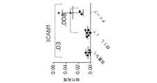
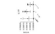
IL−17は、上皮細胞、内皮細胞、及び線維芽細胞を刺激してIL−6、IL−8、G−CSF、及びMCP−1を含む他の炎症性サイトカイン及びケモカインを産生させる炎症促進性分子である[Yao,Z.et al.,J.Immunol.,122(12):5483−5486(1995)、Yao,Z.et al,Immunity,3(6):811−821(1995)、Fossiez,F.,et al.,J.Exp.Med.,183(6):2593−2603(1996)、Kennedy,J.,et al.,J.Interferon Cytokine Res.,16(8):611−7(1996)、Cai,X.Y.,et al.,Immunol.Lett,62(1):51−8(1998)、Jovanovic,D.V.,et al.,J.Immunol.,160(7):3513−21(1998)、Laan,M.,et al.,J.Immunol.,162(4):2347−52(1999)、Linden,A.,et al.,Eur Respir J,15(5):973−7(2000)、及びAggarwal,S.and Gurney,A.L.,J Leukoc Biol.71(1):1−8(2002)を参照されたい]。IL−17はまた、TNF−α及びIL−1βを含む他のサイトカインと協同して、ケモカイン発現をさらに誘発する(Chabaud,M.,et al.,J.Immunol.161(1):409−14(1998))。インターロイキン17(IL−17)は、様々な細胞型で多面的な生物活性を示す。IL−17は、ICAM−1表面発現、T細胞の増殖、ならびにCD34+ヒト前駆体の好中球への成長及び分化を誘発する能力も有する。IL-17 stimulates epithelial cells, endothelial cells, and fibroblasts to produce other inflammatory cytokines and chemokines, including IL-6, IL-8, G-CSF, and MCP-1. It is a molecule [Yao, Z. et al. , J .; Immunol. 122 (12): 5483-5486 (1995), Yao, Z .; et al, Immunity, 3 (6): 811-821 (1995), Fossiez, F .; , Et al. , J .; Exp. Med. , 183 (6): 2593-2603 (1996), Kennedy, J. et al. , Et al. , J .; Interferon Cytokine Res. 16 (8): 611-7 (1996), Cai, X .; Y. , Et al. , Immunol. Lett, 62 (1): 51-8 (1998), Jovanovic, D. et al. V. , Et al. , J .; Immunol. 160 (7): 3513-21 (1998), Laan, M .; , Et al. , J .; Immunol. 162 (4): 2347-52 (1999); Linden, A .; , Et al. , Eur Respir J, 15 (5): 973-7 (2000), and Aggarwal, S .; and Gurney, A .; L. , J Leukoc Biol. 71 (1): 1-8 (2002)]. IL-17 also cooperates with other cytokines including TNF-α and IL-1β to further induce chemokine expression (Chabaud, M., et al., J. Immunol. 161 (1): 409-). 14 (1998)). Interleukin 17 (IL-17) exhibits pleiotropic biological activity in various cell types. IL-17 also has the ability to induce ICAM-1 surface expression, T cell proliferation, and growth and differentiation of CD34+ human precursors to neutrophils.
様々ながんを治療し、安定化させ、その発症を防止し、及び/または遅延させるために、そのような最適な療法が依然として必要とされている。 There remains a need for such optimal therapy to treat, stabilize, prevent and / or delay the development of various cancers.
特許出願、特許公報、及びUniProtKB/Swiss−Prot受入番号を含め、本明細書に引用される全ての参照物は、各個別の参照物が参照により組み込まれることが具体的かつ個別に示されているかのように、それらの全体において参照により本明細書に組み込まれる。 All references cited herein, including patent applications, patent publications, and UniProtKB / Swiss-Prot accession numbers, are specifically and individually indicated that each individual reference is incorporated by reference. As if, in their entirety, they are incorporated herein by reference.
本開示は、有効量のPD−1軸結合拮抗薬及びIL−17結合拮抗薬を含む併用治療を記載する。 The present disclosure describes a combination treatment comprising an effective amount of a PD-1 axis binding antagonist and an IL-17 binding antagonist.
ある特定の態様において、本開示は、個体におけるがんの治療方法またはその進行の遅延方法であって、該個体に、有効量のPD−1軸結合拮抗薬及びIL−17結合拮抗薬を投与することを含む方法を提供する。別の態様では、本開示は、PD−1軸結合拮抗薬とIL−17結合拮抗薬との有効量の組み合わせを投与することを含む、がんを有する個体における免疫機能の増強方法を提供する。 In certain aspects, the disclosure provides a method of treating cancer or delaying its progression in an individual, wherein the individual is administered an effective amount of a PD-1 axis binding antagonist and an IL-17 binding antagonist. Providing a method comprising: In another aspect, the present disclosure provides a method of enhancing immune function in an individual having cancer, comprising administering an effective amount of a combination of a PD-1 axis binding antagonist and an IL-17 binding antagonist. .
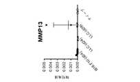
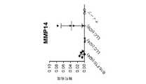
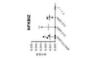
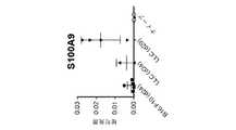
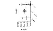
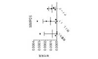
別の態様では、本開示は、PD−1軸結合拮抗薬及びIL−17結合拮抗薬を用いた治療のための、がんを有する個体の識別方法を提供し、本方法は、(a)個体におけるがんから得られる生検試料中のIL−17の発現を検出することと、(b)生検試料がIL−17の発現を示す場合、または生検試料が参照若しくは参照試料と比較したときIL−17の発現の増加を示す場合、該個体に、有効量のPD−1軸結合拮抗薬及びIL−17結合拮抗薬を投与することと、を含む。別の態様では、本開示は、PD−1軸結合拮抗薬及びIL−17結合拮抗薬を用いた治療のための、がんを有する個体の識別方法を提供し、本方法は、(a)個体におけるがんから得られる生検試料中のIL−17遺伝子シグネチャー(例えば、IL−17A、IL−17F、IL−8、CSF3、CXCL1、CXCL3、及びCCL20から選択される1個以上の遺伝子等)の発現を検出することと、(b)生検試料がIL−17遺伝子シグネチャーの発現を示す場合、または生検試料が参照若しくは参照試料と比較したときIL−17遺伝子シグネチャーの発現の増加を示す場合、該個体に、有効量のPD−1軸結合拮抗薬及びIL−17結合拮抗薬を投与することと、を含む。別の態様では、本開示は、PD−1軸結合拮抗薬及びIL−17結合拮抗薬を用いた治療のための、がんを有する個体の識別方法を提供し、本方法は、(a)個体におけるがんから得られる生検試料中のIL−17遺伝子シグネチャー(例えば、CD4、CD8a、IL17A、IL17B、IL17C、IL17D、IL17F、IL17RA、IL17RC、C3、CCL2、CCL20、CSF2、CSF3、CXCL1、CXCL2、CXCL3、CXCL5、CXCL10、CXCR1、CXCR2、ICAM1、IL6、IL8、MMP1、MMP2、MMP3、MMP8、MMP9、MMP13、MMP14、MMP25、NCF4、NFKBIZ、S100A8、S100A9、SAA2、SAA1、SAA3、SAA4、TIMP1、TIMP2、TIMP3、及びTIMP4から選択される1個以上の遺伝子等)の発現を検出することと、(b)生検試料がIL−17遺伝子シグネチャーの発現を示す場合、または生検試料が参照若しくは参照試料と比較したときIL−17遺伝子シグネチャーの発現の増加を示す場合、該個体に、有効量のPD−1軸結合拮抗薬及びIL−17結合拮抗薬を投与することと、を含む。別の態様では、本開示は、PD−1軸結合拮抗薬及びIL−17結合拮抗薬を用いた治療のための、がんを有する個体の識別方法を提供し、本方法は、個体におけるがんから得られる生検試料中のIL−17遺伝子シグネチャー(例えば、CD4、CD8a、IL17A、IL17B、IL17C、IL17D、IL17F、IL17RA、IL17RC、C3、CCL2、CCL20、CSF2、CSF3、CXCL1、CXCL2、CXCL3、CXCL5、CXCL10、CXCR1、CXCR2、ICAM1、IL6、IL8、MMP1、MMP2、MMP3、MMP8、MMP9、MMP13、MMP14、MMP25、NCF4、NFKBIZ、S100A8、S100A9、SAA2、SAA1、SAA3、SAA4、TIMP1、TIMP2、TIMP3、及びTIMP4から選択される1個以上の遺伝子等)の発現を検出することを含み、該個体は、生検試料がIL−17遺伝子シグネチャーの発現を示す場合、または生検試料が参照若しくは参照試料と比較したときIL−17遺伝子シグネチャーの発現の増加を示す場合、治療のために識別される。 In another aspect, the disclosure provides a method of identifying an individual with cancer for treatment with a PD-1 axis binding antagonist and an IL-17 binding antagonist, the method comprising: (a) Detecting the expression of IL-17 in a biopsy sample obtained from cancer in an individual, and (b) if the biopsy sample shows IL-17 expression, or the biopsy sample is compared to a reference or reference sample If the individual exhibits increased expression of IL-17, the subject comprises administering an effective amount of a PD-1 axis binding antagonist and an IL-17 binding antagonist to the individual. In another aspect, the disclosure provides a method of identifying an individual with cancer for treatment with a PD-1 axis binding antagonist and an IL-17 binding antagonist, the method comprising: (a) IL-17 gene signature in a biopsy sample obtained from cancer in an individual (eg, one or more genes selected from IL-17A, IL-17F, IL-8, CSF3, CXCL1, CXCL3, and CCL20, etc.) ) Expression of (b), and (b) when the biopsy sample exhibits expression of the IL-17 gene signature, or when the biopsy sample is a reference or an increase in expression of the IL-17 gene signature when compared to the reference sample. Where indicated, administering to the individual an effective amount of a PD-1 axis binding antagonist and an IL-17 binding antagonist. In another aspect, the disclosure provides a method of identifying an individual with cancer for treatment with a PD-1 axis binding antagonist and an IL-17 binding antagonist, the method comprising: (a) IL-17 gene signature in a biopsy sample obtained from cancer in an individual (eg, CD4, CD8a, IL17A, IL17B, IL17C, IL17D, IL17F, IL17RA, IL17RC, C3, CCL2, CCL20, CSF2, CSF3, CXCL1, CXCL2, CXCL3, CXCL5, CXCL10, CXCR1, CXCR2, ICAM1, IL6, IL8, MMP1, MMP2, MMP3, MMP8, MMP9, MMP13, MMP14, MMP25, NCF4, NFKBIZ, S100A8, S3A1, SA3A, SA3A1, SA3A Detecting the expression of one or more genes selected from IMP1, TIMP2, TIMP3, and TIMP4), and (b) if the biopsy sample shows expression of an IL-17 gene signature, or Administering an effective amount of a PD-1 axis binding antagonist and an IL-17 binding antagonist to the individual if it exhibits increased expression of the IL-17 gene signature when compared to a reference or reference sample. . In another aspect, the present disclosure provides a method of identifying an individual having cancer for treatment with a PD-1 axis binding antagonist and an IL-17 binding antagonist, wherein the method comprises: IL-17 gene signature (eg, CD4, CD8a, IL17A, IL17B, IL17C, IL17D, IL17F, IL17RA, IL17RC, C3, CCL2, CCL20, CSF2, CSF3, CXCL1, CXCL2, CXCL3 , CXCL5, CXCL10, CXCR1, CXCR2, ICAM1, IL6, IL8, MMP1, MMP2, MMP3, MMP8, MMP9, MMP13, MMP14, MMP25, NCF4, NFKBIZ, S100A8, S100A9, SAA2, SAA1, SAA3, SAA1, 4 1, one or more genes selected from TIMP2, TIMP3, and TIMP4), wherein the individual has a biopsy sample showing expression of an IL-17 gene signature, or a biopsy A sample is identified for treatment if it exhibits increased expression of the IL-17 gene signature when compared to a reference or reference sample.
いくつかの実施形態では、PD−1軸結合拮抗薬は、PD−1結合拮抗薬、PDL1結合拮抗薬、及びPDL2結合拮抗薬からなる群から選択される。 In some embodiments, the PD-1 axis binding antagonist is selected from the group consisting of a PD-1 binding antagonist, a PDL1 binding antagonist, and a PDL2 binding antagonist.
いくつかの実施形態では、PD−1軸結合拮抗薬は、PD−1結合拮抗薬である。いくつかの実施形態では、PD−1結合拮抗薬は、PD−1がそのリガンド結合パートナーに結合することを阻害する。いくつかの実施形態では、PD−1結合拮抗薬は、PD−1がPDL1に結合することを阻害する。いくつかの実施形態では、PD−1結合拮抗薬は、PD−1がPDL2に結合することを阻害する。いくつかの実施形態では、PD−1結合拮抗薬は、PD−1がPDL1とPDL2との両方に結合することを阻害する。いくつかの実施形態では、PD−1結合拮抗薬は、抗体である。いくつかの実施形態では、抗PD−1抗体は、モノクローナル抗体である。いくつかの実施形態では、抗PD−1抗体は、Fab、Fab’−SH、Fv、scFv、及び(Fab’)2断片からなる群から選択される抗体断片である。いくつかの実施形態では、PD−1結合拮抗薬は、ニボルマブ、ペンブロリズマブ、CT−011、またはAMP−224である。In some embodiments, the PD-1 axis binding antagonist is a PD-1 binding antagonist. In some embodiments, the PD-1 binding antagonist inhibits PD-1 from binding to its ligand binding partner. In some embodiments, the PD-1 binding antagonist inhibits PD-1 from binding to PDL1. In some embodiments, the PD-1 binding antagonist inhibits PD-1 from binding to PDL2. In some embodiments, the PD-1 binding antagonist inhibits PD-1 from binding to both PDL1 and PDL2. In some embodiments, the PD-1 binding antagonist is an antibody. In some embodiments, the anti-PD-1 antibody is a monoclonal antibody. In some embodiments, the anti-PD-1 antibody is an antibody fragment selected from the group consisting of Fab, Fab′-SH, Fv, scFv, and (Fab ′)2 fragments. In some embodiments, the PD-1 binding antagonist is nivolumab, pembrolizumab, CT-011, or AMP-224.
いくつかの実施形態では、PD−1軸結合拮抗薬は、PDL1結合拮抗薬である。いくつかの実施形態では、PDL1結合拮抗薬は、PDL1がPD−1に結合することを阻害する。いくつかの実施形態では、PDL1結合拮抗薬は、PDL1がB7−1に結合することを阻害する。いくつかの実施形態では、PDL1結合拮抗薬は、PDL1がPD−1とB7−1との両方に結合することを阻害する。いくつかの実施形態では、PDL1結合拮抗薬は、抗PDL1抗体である。いくつかの実施形態では、抗PDL1抗体は、モノクローナル抗体である。いくつかの実施形態では、抗PDL1抗体は、Fab、Fab’−SH、Fv、scFv、及び(Fab’)2断片からなる群から選択される抗体断片である。いくつかの実施形態では、抗PDL1抗体は、ヒト化抗体またはヒト抗体である。いくつかの実施形態では、PDL1結合拮抗薬は、YW243.55.S70、MPDL3280A、MDX−1105、及びMEDI4736からなる群から選択される。In some embodiments, the PD-1 axis binding antagonist is a PDL1 binding antagonist. In some embodiments, the PDL1 binding antagonist inhibits PDL1 from binding to PD-1. In some embodiments, the PDL1 binding antagonist inhibits PDL1 from binding to B7-1. In some embodiments, the PDL1 binding antagonist inhibits PDL1 from binding to both PD-1 and B7-1. In some embodiments, the PDL1 binding antagonist is an anti-PDL1 antibody. In some embodiments, the anti-PDL1 antibody is a monoclonal antibody. In some embodiments, the anti-PDL1 antibody is an antibody fragment selected from the group consisting of Fab, Fab′-SH, Fv, scFv, and (Fab ′)2 fragments. In some embodiments, the anti-PDL1 antibody is a humanized antibody or a human antibody. In some embodiments, the PDL1 binding antagonist is YW243.55. Selected from the group consisting of S70, MPDL3280A, MDX-1105, and MEDI4736.
いくつかの実施形態では、抗PDL1抗体は、配列番号15のHVR−H1配列、配列番号16のHVR−H2配列、及び配列番号3のHVR−H3配列を含む重鎖、ならびに配列番号17のHVR−L1配列、配列番号18のHVR−L2配列、及び配列番号19のHVR−L3配列を含む軽鎖を含む。いくつかの実施形態では、抗PDL1抗体は、配列番号24または配列番号28のアミノ酸配列を含む重鎖可変領域、及び配列番号21のアミノ酸配列を含む軽鎖可変領域を含む。いくつかの実施形態では、抗PDL1抗体は、配列番号26のアミノ酸配列を含む重鎖、及び/または配列番号27のアミノ酸配列を含む軽鎖を含む。 In some embodiments, the anti-PDL1 antibody comprises a heavy chain comprising the HVR-H1 sequence of SEQ ID NO: 15, the HVR-H2 sequence of SEQ ID NO: 16, and the HVR-H3 sequence of SEQ ID NO: 3, and the HVR of SEQ ID NO: 17. A light chain comprising the L1 sequence, the HVR-L2 sequence of SEQ ID NO: 18 and the HVR-L3 sequence of SEQ ID NO: 19. In some embodiments, the anti-PDL1 antibody comprises a heavy chain variable region comprising the amino acid sequence of SEQ ID NO: 24 or SEQ ID NO: 28, and a light chain variable region comprising the amino acid sequence of SEQ ID NO: 21. In some embodiments, the anti-PDL1 antibody comprises a heavy chain comprising the amino acid sequence of SEQ ID NO: 26 and / or a light chain comprising the amino acid sequence of SEQ ID NO: 27.
いくつかの実施形態では、PD−1軸結合拮抗薬は、PDL2結合拮抗薬である。いくつかの実施形態では、PDL2結合拮抗薬は、抗体である。いくつかの実施形態では、抗PDL2抗体は、モノクローナル抗体である。いくつかの実施形態では、抗PDL2抗体は、Fab、Fab’−SH、Fv、scFv、及び(Fab’)2断片からなる群から選択される抗体断片である。いくつかの実施形態では、PDL2結合拮抗薬は、イムノアドヘシンである。In some embodiments, the PD-1 axis binding antagonist is a PDL2 binding antagonist. In some embodiments, the PDL2 binding antagonist is an antibody. In some embodiments, the anti-PDL2 antibody is a monoclonal antibody. In some embodiments, the anti-PDL2 antibody is an antibody fragment selected from the group consisting of Fab, Fab′-SH, Fv, scFv, and (Fab ′)2 fragments. In some embodiments, the PDL2 binding antagonist is an immunoadhesin.
いくつかの実施形態では、IL−17結合拮抗薬は、IL−17がIL−17受容体に結合することを阻害する。いくつかの実施形態では、IL−17結合拮抗薬は、抗体である。いくつかの実施形態では、IL−17結合拮抗薬は、モノクローナル抗体である。いくつかの実施形態では、IL−17結合拮抗薬は、Fab、Fab’−SH、Fv、scFv、及び(Fab’)2断片からなる群から選択される抗体断片である。いくつかの実施形態では、IL−17結合拮抗薬は、ヒト化抗体またはヒト抗体である。In some embodiments, the IL-17 binding antagonist inhibits IL-17 from binding to the IL-17 receptor. In some embodiments, the IL-17 binding antagonist is an antibody. In some embodiments, the IL-17 binding antagonist is a monoclonal antibody. In some embodiments, the IL-17 binding antagonist is an antibody fragment selected from the group consisting of Fab, Fab′-SH, Fv, scFv, and (Fab ′)2 fragments. In some embodiments, the IL-17 binding antagonist is a humanized antibody or a human antibody.
いくつかの実施形態では、抗IL−17抗体は、配列番号32のCDR−H1配列、配列番号33のCDR−H2配列、及び配列番号34のCDR−H3配列を含む重鎖、ならびに配列番号35のCDR−L1配列、配列番号36のCDR−L2配列、及び配列番号37のCDR−L3配列を含む軽鎖を含む。いくつかの実施形態では、抗IL−17抗体は、配列番号30のアミノ酸配列を含む重鎖可変領域、及び配列番号31のアミノ酸配列を含む軽鎖可変領域を含む。 In some embodiments, the anti-IL-17 antibody comprises a heavy chain comprising a CDR-H1 sequence of SEQ ID NO: 32, a CDR-H2 sequence of SEQ ID NO: 33, and a CDR-H3 sequence of SEQ ID NO: 34, and SEQ ID NO: 35 A light chain comprising a CDR-L1 sequence of SEQ ID NO: 36, a CDR-L2 sequence of SEQ ID NO: 36, and a CDR-L3 sequence of SEQ ID NO: 37. In some embodiments, the anti-IL-17 antibody comprises a heavy chain variable region comprising the amino acid sequence of SEQ ID NO: 30 and a light chain variable region comprising the amino acid sequence of SEQ ID NO: 31.
いくつかの実施形態では、抗IL−17抗体は、配列番号40のCDR−H1配列、配列番号41のCDR−H2配列、及び配列番号42のCDR−H3配列を含む重鎖、ならびに配列番号43のCDR−L1配列、配列番号44のCDR−L2配列、及び配列番号45のCDR−L3配列を含む軽鎖を含む。いくつかの実施形態では、抗IL−17抗体は、配列番号38のアミノ酸配列を含む重鎖可変領域、及び配列番号39のアミノ酸配列を含む軽鎖可変領域を含む。 In some embodiments, the anti-IL-17 antibody comprises a heavy chain comprising a CDR-H1 sequence of SEQ ID NO: 40, a CDR-H2 sequence of SEQ ID NO: 41, and a CDR-H3 sequence of SEQ ID NO: 42, and SEQ ID NO: 43 A light chain comprising a CDR-L1 sequence of SEQ ID NO: 44, a CDR-L2 sequence of SEQ ID NO: 44, and a CDR-L3 sequence of SEQ ID NO: 45. In some embodiments, the anti-IL-17 antibody comprises a heavy chain variable region comprising the amino acid sequence of SEQ ID NO: 38, and a light chain variable region comprising the amino acid sequence of SEQ ID NO: 39.
いくつかの実施形態では、抗IL−17抗体は、配列番号48のCDR−H1配列、配列番号49のCDR−H2配列、及び配列番号50のCDR−H3配列を含む重鎖、ならびに配列番号51のCDR−L1配列、配列番号52のCDR−L2配列、及び配列番号53のCDR−L3配列を含む軽鎖を含む。いくつかの実施形態では、抗IL−17抗体は、配列番号46のアミノ酸配列を含む重鎖可変領域、及び配列番号47のアミノ酸配列を含む軽鎖可変領域を含む。 In some embodiments, the anti-IL-17 antibody comprises a heavy chain comprising a CDR-H1 sequence of SEQ ID NO: 48, a CDR-H2 sequence of SEQ ID NO: 49, and a CDR-H3 sequence of SEQ ID NO: 50, and SEQ ID NO: 51 A light chain comprising a CDR-L1 sequence of SEQ ID NO: 52, a CDR-L2 sequence of SEQ ID NO: 52, and a CDR-L3 sequence of SEQ ID NO: 53. In some embodiments, the anti-IL-17 antibody comprises a heavy chain variable region comprising the amino acid sequence of SEQ ID NO: 46, and a light chain variable region comprising the amino acid sequence of SEQ ID NO: 47.
いくつかの実施形態では、抗IL−17抗体は、配列番号56のCDR−H1配列、配列番号57のCDR−H2配列、及び配列番号58のCDR−H3配列を含む重鎖、ならびに配列番号59のCDR−L1配列、配列番号60のCDR−L2配列、及び配列番号61のCDR−L3配列を含む軽鎖を含む。いくつかの実施形態では、抗IL−17抗体は、配列番号54のアミノ酸配列を含む重鎖可変領域、及び配列番号55のアミノ酸配列を含む軽鎖可変領域を含む。 In some embodiments, the anti-IL-17 antibody comprises a heavy chain comprising a CDR-H1 sequence of SEQ ID NO: 56, a CDR-H2 sequence of SEQ ID NO: 57, and a CDR-H3 sequence of SEQ ID NO: 58, and SEQ ID NO: 59 A light chain comprising a CDR-L1 sequence of SEQ ID NO: 60, a CDR-L2 sequence of SEQ ID NO: 60, and a CDR-L3 sequence of SEQ ID NO: 61. In some embodiments, the anti-IL-17 antibody comprises a heavy chain variable region comprising the amino acid sequence of SEQ ID NO: 54 and a light chain variable region comprising the amino acid sequence of SEQ ID NO: 55.
いくつかの実施形態では、IL−17結合拮抗薬は、抗IL−17抗体である。いくつかの実施形態では、抗IL−17抗体は、IL−17Aに特異的に結合する。いくつかの実施形態では、抗IL−17抗体は、IL−17Fに特異的に結合する。いくつかの実施形態では、抗IL−17抗体は、IL−17A及びIL−17Fに特異的に結合する。いくつかの実施形態では、抗IL−17抗体は、イキセキズマブ、ビメキズマブ、またはセクキヌマブである。 In some embodiments, the IL-17 binding antagonist is an anti-IL-17 antibody. In some embodiments, the anti-IL-17 antibody specifically binds to IL-17A. In some embodiments, the anti-IL-17 antibody specifically binds to IL-17F. In some embodiments, the anti-IL-17 antibody specifically binds IL-17A and IL-17F. In some embodiments, the anti-IL-17 antibody is ixekizumab, bimekizumab, or secukinumab.
いくつかの実施形態では、IL−17結合拮抗薬は、抗IL−17受容体抗体である。いくつかの実施形態では、抗IL−17受容体抗体は、ブロダルマブである。 In some embodiments, the IL-17 binding antagonist is an anti-IL-17 receptor antibody. In some embodiments, the anti-IL-17 receptor antibody is brodalumab.
いくつかの実施形態では、IL−17結合拮抗薬は、IL−17受容体由来の少なくとも1つのエクソンを含む可溶性ポリペプチドである。いくつかの実施形態では、可溶性ポリペプチドは、IL−17RA由来の少なくとも1つのエクソン及びIL−17RC由来の少なくとも1つのエクソンを含む。 In some embodiments, the IL-17 binding antagonist is a soluble polypeptide comprising at least one exon from the IL-17 receptor. In some embodiments, the soluble polypeptide comprises at least one exon from IL-17RA and at least one exon from IL-17RC.
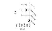
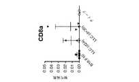
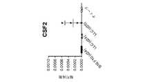
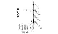
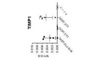
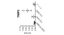
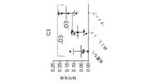
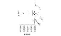
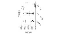
いくつかの実施形態では、本方法は、PD−1軸結合拮抗薬及びIL−17結合拮抗薬を投与する前または後に、個体のがんからの生検試料中のバイオマーカー発現を検出するステップを、さらに含む。いくつかの実施形態では、個体のがんから得られる生検試料は、IL−17の発現を示す。いくつかの実施形態では、IL−17の発現は、IL−17 mRNAの発現である。いくつかの実施形態では、IL−17の発現は、IL−17タンパク質の発現である。いくつかの実施形態では、がんから得られる生検試料は、参照若しくは参照試料と比較したとき、IL−17の上昇した発現を示す。いくつかの実施形態では、個体のがんから得られる生検試料は、IL−17A、IL−17F、IL−8、CSF3、CXCL1、CXCL3、及びCCL20からなる群から選択される1個以上の遺伝子の発現を示す。いくつかの実施形態では、個体から得られる生検試料は、参照若しくは参照試料と比較したとき、IL−17A、IL−17F、IL−8、CSF3、CXCL1、CXCL3、及びCCL20からなる群から選択される1個以上の遺伝子の上昇した発現を示す。いくつかの実施形態では、がんは、腎細胞がん腫、膀胱がん、非小細胞肺がん、扁平上皮非小細胞肺がん、非扁平上皮非小細胞肺がん、結腸直腸がん、黒色腫、卵巣がん、乳がん、ホルモン受容体陽性乳がん、HER2陽性乳がん、及びトリプルネガティブ乳がんからなる群から選択される。いくつかの実施形態では、個体のがんから得られる生検試料は、CD4、CD8a、IL17A、IL17B、IL17C、IL17D、IL17F、IL17RA、IL17RC、C3、CCL2、CCL20、CSF2、CSF3、CXCL1、CXCL2、CXCL3、CXCL5、CXCL10、CXCR1、CXCR2、ICAM1、IL6、IL8、MMP1、MMP2、MMP3、MMP8、MMP9、MMP13、MMP14、MMP25、NCF4、NFKBIZ、S100A8、S100A9、SAA2、SAA1、SAA3、SAA4、TIMP1、TIMP2、TIMP3、及びTIMP4からなる群から選択される1個以上の遺伝子の発現を示す。いくつかの実施形態では、個体のがんから得られる生検試料は、CD4、CD8a、IL17A、IL17B、IL17C、IL17D、IL17F、IL17RA、IL17RC、C3、CCL2、CCL20、CSF2、CSF3、CXCL1、CXCL2、CXCL3、CXCL5、CXCL10、CXCR1、CXCR2、ICAM1、IL6、IL8、MMP1、MMP2、MMP3、MMP8、MMP9、MMP13、MMP14、MMP25、NCF4、NFKBIZ、S100A8、S100A9、SAA2、SAA1、SAA3、SAA4、TIMP1、TIMP2、TIMP3、及びTIMP4から選択される、少なくとも1個、少なくとも2個、少なくとも3個、少なくとも4個、少なくとも5個、少なくとも6個、少なくとも7個、少なくとも8個、少なくとも9個、少なくとも10個、少なくとも11個、少なくとも12個、少なくとも13個、少なくとも14個、少なくとも15個、少なくとも16個、少なくとも17個、少なくとも18個、少なくとも19個、少なくとも20個、少なくとも21個、少なくとも22個、少なくとも23個、少なくとも24個、少なくとも25個、少なくとも26個、少なくとも27個、少なくとも28個、少なくとも29個、少なくとも30個、少なくとも31個、少なくとも32個、少なくとも33個、少なくとも34個、少なくとも35個、少なくとも36個、少なくとも37個、少なくとも38個、少なくとも39個、少なくとも40個、少なくとも41個、少なくとも42個、少なくとも43個、または少なくとも44個の遺伝子の発現を示す。いくつかの実施形態では、がんから得られる生検試料は、参照若しくは参照試料と比較したとき、CD4、CD8a、IL17A、IL17B、IL17C、IL17D、IL17F、IL17RA、IL17RC、C3、CCL2、CCL20、CSF2、CSF3、CXCL1、CXCL2、CXCL3、CXCL5、CXCL10、CXCR1、CXCR2、ICAM1、IL6、IL8、MMP1、MMP2、MMP3、MMP8、MMP9、MMP13、MMP14、MMP25、NCF4、NFKBIZ、S100A8、S100A9、SAA2、SAA1、SAA3、SAA4、TIMP1、TIMP2、TIMP3、及びTIMP4からなる群から選択される1個以上の遺伝子の上昇した発現を示す。いくつかの実施形態では、がんから得られる生検試料は、参照若しくは参照試料と比較したとき、CD4、CD8a、IL17A、IL17B、IL17C、IL17D、IL17F、IL17RA、IL17RC、C3、CCL2、CCL20、CSF2、CSF3、CXCL1、CXCL2、CXCL3、CXCL5、CXCL10、CXCR1、CXCR2、ICAM1、IL6、IL8、MMP1、MMP2、MMP3、MMP8、MMP9、MMP13、MMP14、MMP25、NCF4、NFKBIZ、S100A8、S100A9、SAA2、SAA1、SAA3、SAA4、TIMP1、TIMP2、TIMP3、及びTIMP4から選択される、少なくとも1個、少なくとも2個、少なくとも3個、少なくとも4個、少なくとも5個、少なくとも6個、少なくとも7個、少なくとも8個、少なくとも9個、少なくとも10個、少なくとも11個、少なくとも12個、少なくとも13個、少なくとも14個、少なくとも15個、少なくとも16個、少なくとも17個、少なくとも18個、少なくとも19個、少なくとも20個、少なくとも21個、少なくとも22個、少なくとも23個、少なくとも24個、少なくとも25個、少なくとも26個、少なくとも27個、少なくとも28個、少なくとも29個、少なくとも30個、少なくとも31個、少なくとも32個、少なくとも33個、少なくとも34個、少なくとも35個、少なくとも36個、少なくとも37個、少なくとも38個、少なくとも39個、少なくとも40個、少なくとも41個、少なくとも42個、少なくとも43個、または少なくとも44個の遺伝子の上昇した発現を示す。いくつかの実施形態では、個体のがんから得られる生検試料は、NFKBIZ、S100A8、及びS100A9からなる群から選択される1個以上の遺伝子の発現を示す。いくつかの実施形態では、がんから得られる生検試料は、参照若しくは参照試料と比較したとき、NFKBIZ、S100A8、及びS100A9からなる群から選択される1個以上の遺伝子の上昇した発現を示す。 In some embodiments, the method detects biomarker expression in a biopsy sample from an individual's cancer before or after administering a PD-1 axis binding antagonist and an IL-17 binding antagonist. Is further included. In some embodiments, a biopsy sample obtained from an individual's cancer exhibits IL-17 expression. In some embodiments, the expression of IL-17 is the expression of IL-17 mRNA. In some embodiments, the expression of IL-17 is the expression of IL-17 protein. In some embodiments, a biopsy sample obtained from cancer exhibits increased expression of IL-17 when compared to a reference or reference sample. In some embodiments, the biopsy sample obtained from an individual's cancer is one or more selected from the group consisting of IL-17A, IL-17F, IL-8, CSF3, CXCL1, CXCL3, and CCL20. Gene expression is shown. In some embodiments, the biopsy sample obtained from the individual is selected from the group consisting of IL-17A, IL-17F, IL-8, CSF3, CXCL1, CXCL3, and CCL20 when compared to a reference or reference sample. Shows elevated expression of one or more genes that are expressed. In some embodiments, the cancer is renal cell carcinoma, bladder cancer, non-small cell lung cancer, squamous non-small cell lung cancer, non-squamous non-small cell lung cancer, colorectal cancer, melanoma, ovary Selected from the group consisting of cancer, breast cancer, hormone receptor positive breast cancer, HER2 positive breast cancer, and triple negative breast cancer. In some embodiments, a biopsy sample obtained from an individual's cancer is CD4, CD8a, IL17A, IL17B, IL17C, IL17D, IL17F, IL17RA, IL17RC, C3, CCL2, CCL20, CSF2, CSF3, CXCL1, CXCL2 , CXCL3, CXCL5, CXCL10, CXCR1, CXCR2, ICAM1, IL6, IL8, MMP1, MMP2, MMP3, MMP8, MMP9, MMP13, MMP14, MMP25, NCF4, NFKBIZ, S100A8, S100A9, SAA2, AA1, SAA2, AAA3 , Expression of one or more genes selected from the group consisting of TIMP2, TIMP3, and TIMP4. In some embodiments, a biopsy sample obtained from an individual's cancer is CD4, CD8a, IL17A, IL17B, IL17C, IL17D, IL17F, IL17RA, IL17RC, C3, CCL2, CCL20, CSF2, CSF3, CXCL1, CXCL2 , CXCL3, CXCL5, CXCL10, CXCR1, CXCR2, ICAM1, IL6, IL8, MMP1, MMP2, MMP3, MMP8, MMP9, MMP13, MMP14, MMP25, NCF4, NFKBIZ, S100A8, S100A9, SAA2, AA1, SAA2, AAA3 , TIMP2, TIMP3, and TIMP4, at least 1, at least 2, at least 3, at least 4, at least 5, at least 6, At least 7, at least 8, at least 9, at least 10, at least 11, at least 12, at least 13, at least 14, at least 15, at least 16, at least 17, at least 18, at least 19, at least 20, at least 21, at least 22, at least 23, at least 24, at least 25, at least 26, at least 27, at least 28, at least 29, at least 30, at least 31 , At least 32, at least 33, at least 34, at least 35, at least 36, at least 37, at least 38, at least 39, at least 40, at least 41, at least 42, at least 43, or Even without showing the expression of 44 genes. In some embodiments, the biopsy sample obtained from cancer is CD4, CD8a, IL17A, IL17B, IL17C, IL17D, IL17F, IL17RA, IL17RC, C3, CCL2, CCL20, when compared to a reference or reference sample. CSF2, CSF3, CXCL1, CXCL2, CXCL3, CXCL5, CXCL10, CXCR1, CXCR2, ICAM1, IL6, IL8, MMP1, MMP2, MMP3, MMP8, MMP9, MMP13, MMP14, MMP25, NCF4, NFKB8S,
いくつかの実施形態では、治療は、治療の休止後の個体における持続性応答をもたらす。 In some embodiments, the treatment results in a sustained response in the individual after cessation of treatment.
いくつかの実施形態では、IL−17結合拮抗薬及び/またはPD−1軸結合拮抗薬は、連続的または断続的に投与される。いくつかの実施形態では、IL−17結合拮抗薬は、PD−1軸結合拮抗薬の前に投与される。いくつかの実施形態では、IL−17結合拮抗薬は、PD−1軸結合拮抗薬と同時に投与される。いくつかの実施形態では、IL−17結合拮抗薬及びPD−1軸結合拮抗薬は、同じ組成物中に製剤化される。いくつかの実施形態では、IL−17結合拮抗薬は、PD−1軸結合拮抗薬の後に投与される。いくつかの実施形態では、PD−1軸結合拮抗薬またはIL−17結合拮抗薬は、静脈内に、筋肉内に、皮下に、局所的に、経口的に、経皮的に、腹腔内に、眼窩内に、埋め込みによって、吸入によって、髄腔内に、脳室内に、または鼻腔内に投与される。 In some embodiments, the IL-17 binding antagonist and / or PD-1 axis binding antagonist is administered continuously or intermittently. In some embodiments, the IL-17 binding antagonist is administered prior to the PD-1 axis binding antagonist. In some embodiments, the IL-17 binding antagonist is administered concurrently with the PD-1 axis binding antagonist. In some embodiments, the IL-17 binding antagonist and the PD-1 axis binding antagonist are formulated in the same composition. In some embodiments, the IL-17 binding antagonist is administered after the PD-1 axis binding antagonist. In some embodiments, the PD-1 axis binding antagonist or IL-17 binding antagonist is intravenous, intramuscular, subcutaneously, topically, orally, transdermally, intraperitoneally. Administered intraorbitally, by implantation, by inhalation, intrathecally, intraventricularly, or intranasally.
別の態様では、本開示は、PD−1軸結合拮抗薬と、PD−1軸結合拮抗薬をIL−17結合拮抗薬と組み合わせて使用して個体におけるがんを治療するか、またはその進行を遅延させるための指示書を含む添付文書と、を含む、キットを提供する。別の態様では、本開示は、PD−1軸結合拮抗薬及びIL−17結合拮抗薬と、PD−1軸結合拮抗薬及びIL−17結合拮抗薬を使用して個体におけるがんを治療するか、またはその進行を遅延させるための指示書を含む添付文書と、を含む、キットを提供する。いくつかの実施形態では、PD−1軸結合拮抗薬及びIL−17結合拮抗薬は、同じ組成物中に製剤化される。別の態様では、本開示は、IL−17結合拮抗薬と、IL−17結合拮抗薬をPD−1軸結合拮抗薬と組み合わせて使用して個体におけるがんを治療するか、またはその進行を遅延させるための指示書を含む添付文書と、を含む、キットを提供する。別の態様では、本開示は、PD−1軸結合拮抗薬と、PD−1軸結合拮抗薬をIL−17結合拮抗薬と組み合わせて使用してがんを有する個体における免疫機能を増強するための指示書を含む添付文書と、を含む、キットを提供する。別の態様では、本開示は、PD−1軸結合拮抗薬及びIL−17結合拮抗薬と、PD−1軸結合拮抗薬及びIL−17結合拮抗薬を使用してがんを有する個体における免疫機能を増強するための指示書を含む添付文書と、を含む、キットを提供する。いくつかの実施形態では、PD−1軸結合拮抗薬及びIL−17結合拮抗薬は、同じ組成物中に製剤化される。別の態様では、本開示は、IL−17結合拮抗薬と、IL−17結合拮抗薬をPD−1軸結合拮抗薬と組み合わせて使用してがんを有する個体における免疫機能を増強するための指示書を含む添付文書と、を含む、キットを提供する。 In another aspect, the present disclosure treats or progresses cancer in an individual using a PD-1 axis binding antagonist and a PD-1 axis binding antagonist in combination with an IL-17 binding antagonist. And a package insert including instructions for delaying. In another aspect, the present disclosure treats cancer in an individual using a PD-1 axis binding antagonist and an IL-17 binding antagonist, and a PD-1 axis binding antagonist and an IL-17 binding antagonist. Or a package insert that includes instructions for delaying its progression. In some embodiments, the PD-1 axis binding antagonist and the IL-17 binding antagonist are formulated in the same composition. In another aspect, the disclosure uses an IL-17 binding antagonist and an IL-17 binding antagonist in combination with a PD-1 axis binding antagonist to treat cancer in an individual or to progress thereof. And a package insert including instructions for delaying. In another aspect, the disclosure uses a PD-1 axis binding antagonist and a PD-1 axis binding antagonist in combination with an IL-17 binding antagonist to enhance immune function in an individual with cancer. And a package insert including the instructions. In another aspect, the disclosure provides for immunization in an individual having cancer using a PD-1 axis binding antagonist and an IL-17 binding antagonist, and a PD-1 axis binding antagonist and an IL-17 binding antagonist. And a package insert including instructions for enhancing functionality. In some embodiments, the PD-1 axis binding antagonist and the IL-17 binding antagonist are formulated in the same composition. In another aspect, the disclosure provides an IL-17 binding antagonist and an IL-17 binding antagonist in combination with a PD-1 axis binding antagonist to enhance immune function in an individual with cancer. A kit is provided, including a package insert including instructions.
別の態様では、本開示は、個体におけるがんの治療方法またはその進行の遅延方法であって、該個体に、有効量の多重特異性(例えば、二重特異性)抗体を投与することを含む方法を提供し、該多重特異性抗体は、(a)PD−1、PDL1、及び/またはPDL2に対する第1の結合特異性、ならびに(b)IL−17及び/またはIL−17Rに対する第2の結合特異性を含む。 In another aspect, the disclosure provides a method of treating cancer or delaying its progression in an individual comprising administering to the individual an effective amount of a multispecific (eg, bispecific) antibody. Wherein the multispecific antibody comprises (a) a first binding specificity for PD-1, PDL1, and / or PDL2, and (b) a second for IL-17 and / or IL-17R. Including the binding specificity of
上記及び本明細書に記載される様々な実施形態の特性のうちの1つ、一部、または全てを組み合わせて、本発明の他の実施形態を形成しても良いことを理解されたい。本発明のこれら及び他の態様は、当業者には明らかとなるであろう。本発明のこれら及び他の実施形態は、以下に続く「発明を実施するための形態」によってさらに説明される。 It should be understood that one, some, or all of the characteristics of the various embodiments described above and herein may be combined to form other embodiments of the invention. These and other aspects of the invention will be apparent to those skilled in the art. These and other embodiments of the present invention are further illustrated by the detailed description that follows.
I.一般的技法
本明細書に記載または参照される技法及び手順は、当業者には概して十分に理解されており、従来の方法論、例えば、Sambrook et al.,Molecular Cloning:A Laboratory Manual 3d edition(2001)Cold Spring Harbor Laboratory Press,Cold Spring Harbor,N.Y.、Current Protocols in Molecular Biology(F.M.Ausubel,et al.eds.,(2003))、シリーズのMethods in Enzymology(Academic Press,Inc.):PCR 2:A Practical Approach(M.J.MacPherson,B.D.Hames and G.R.Taylor eds.(1995)),Harlow and Lane,eds.(1988)Antibodies,A Laboratory Manual,and Animal Cell Culture(R.I.Freshney,ed.(1987))、Oligonucleotide Synthesis(M.J.Gait,ed.,1984)、Methods in Molecular Biology,Humana Press、Cell Biology:A Laboratory Notebook(J.E.Cellis,ed.,1998)Academic Press、Animal Cell Culture(R.I.Freshney),ed.,1987)、Introduction to Cell and Tissue Culture(J.P.Mather and P.E.Roberts,1998)Plenum Press、Cell and Tissue Culture:Laboratory Procedures(A.Doyle,J.B.Griffiths,and D.G.Newell,eds.,1993−8)J.Wiley and Sons、Handbook of Experimental Immunology(D.M.Weir and C.C.Blackwell,eds.)、Gene Transfer Vectors for Mammalian Cells(J.M.Miller and M.P.Calos,eds.,1987)、PCR:The Polymerase Chain Reaction,(Mullis et al.,eds.,1994)、Current Protocols in Immunology(J.E.Coligan et al.,eds.,1991)、Short Protocols in Molecular Biology(Wiley and Sons,1999)、Immunobiology(C.A.Janeway and P.Travers,1997)、Antibodies(P.Finch,1997);Antibodies:A Practical Approach(D.Catty.,ed.,IRL Press,1988−1989)、Monoclonal Antibodies:A Practical Approach(P.Shepherd and C.Dean,eds.,Oxford University Press,2000);Using Antibodies:A Laboratory Manual(E.Harlow and D.Lane(Cold Spring Harbor Laboratory Press,1999)、The Antibodies(M.Zanetti and J.D.Capra,eds.,Harwood Academic Publishers,1995)、及びCancer:Principles and Practice of Oncology(V.T.DeVita et al.,eds.,J.B.Lippincott Company,1993)に記載される、広く利用されている方法論等を使用して、一般的に用いられる。I. General Techniques The techniques and procedures described or referenced herein are generally well understood by those skilled in the art and are well known in the art, for example, Sambrook et al. , Molecular Cloning: A Laboratory Manual 3d edition (2001) Cold Spring Harbor Laboratory Press, Cold Spring Harbor, N .; Y. , Current Protocols in Molecular Biology (FM Ausubel, et al. Eds., (2003)), Series of Methods in Enzymology (Academic Press, Inc.): PCR 2: A Practical Bio. BD Hames and GR Taylor eds. (1995)), Harlow and Lane, eds. (1988) Antibodies, A Laboratory Manual, and Animal Cell Culture (R. I. Freshney, ed. (1987)), Oligonucleotide Synthesis (M. J. Gait, ed., 1984), Met. Cell Biology: A Laboratory Notebook (JE Cellis, ed., 1998) Academic Press, Animal Cell Culture (R. I. Freshney), ed. , 1987), Introduction to Cell and Tissue Culture (JP Master and PE Roberts, 1998) Plenum Press, Cell and Tissue Culture, Laboratory Procedures, A. DoyG. Newell, eds., 1993-8) J. Am. Wiley and Sons, Handbook of Experimental Immunology (DM Weir and CC Blackwell, eds.), Gene Transfer Vectors for Mammalian Cells (J.M. PCR: The Polymerase Chain Reaction, (Mullis et al., Eds., 1994), Current Protocols in Immunology (J. E. Coligan et al., Eds., 1991), Short Protocols in Mol. ), Immunobiolo y (C. A. Janeway and P. Travers, 1997), Antibodies (P. Finch, 1997); Antibodies: A Practical Approach (D. Catty., ed., IRL Press, 1988-1989), MonoAclo: MonAb: MonoAbn Practical Approach (P. Shepherd and C. Dean, eds., Oxford University Press, 2000); Using Antibodies: A Laboratory Manual (L. Zanett and JD Capra, eds., Harwood Academic Publishers, 1995), and Cancer: Principles and Practice of Oncology (described in VT DeVita et al., eds. It is generally used using widely used methodologies.
II.定義
本発明を詳細に説明する前に、本発明は特定の組成物または生物系に限定されず、組成物及び生物系は当然ながら様々であり得ることを理解されたい。本明細書に使用される用語は特定の実施形態を説明することのみを目的とするものであり、限定的であるよう意図されるものではないことも理解されたい。II. Definitions Before describing the present invention in detail, it is to be understood that the present invention is not limited to a particular composition or biological system, and the composition and biological system can of course vary. It is also to be understood that the terminology used herein is for the purpose of describing particular embodiments only and is not intended to be limiting.
本明細書及び添付の「特許請求の範囲」において使用される場合、「a」、「or」、及び「the」という単数形は、文脈に別段の明確な定めがない限り、複数の参照対象を含む。 As used herein and in the appended claims, the singular forms “a”, “or”, and “the” refer to plural references unless the context clearly dictates otherwise. including.
本明細書における「約」の値またはパラメータは、その値またはパラメータ自体に関する変形形態を含む(そして説明する)。例えば、「約X」に言及する説明は、「X」の説明を含む。 As used herein, “about” a value or parameter includes (and describes) variations that are related to the value or parameter itself. For example, description referring to “about X” includes description of “X”.
本明細書に記載される本発明の態様及び変形形態が、態様及び変形形態「からなる(consisting)」及び/または「から本質的になる(consisting essentially of)」を含むことは理解される。 It is understood that aspects and variations of the invention described herein include aspects and variations “consisting of” and / or “consisting essentially of”.
「拮抗薬」という用語は、最も広義に使用され、本明細書に開示される天然ポリペプチドの生物活性を部分的若しくは完全に遮断、阻害、または中和する任意の分子を含む。同様の様式で、「作動薬」という用語は、最も広義に使用され、本明細書に開示される天然ポリペプチドの生物活性を模倣する任意の分子を含む。好適な作動薬または拮抗薬分子には、天然ポリペプチド、ペプチド、アンチセンスオリゴヌクレオチド、有機小分子の作動薬若しくは拮抗薬抗体、または抗体断片、断片若しくはアミノ酸配列変異形が具体的に含まれる。ポリペプチドの作動薬または拮抗薬の識別方法は、ポリペプチドを候補作動薬または拮抗薬分子と接触させること、及びポリペプチドに通常関連する1つ以上の生物活性の検出可能な変化を測定することを含んでも良い。 The term “antagonist” is used in the broadest sense and includes any molecule that partially or fully blocks, inhibits, or neutralizes the biological activity of a native polypeptide disclosed herein. In a similar manner, the term “agonist” is used in the broadest sense and includes any molecule that mimics the biological activity of a natural polypeptide disclosed herein. Suitable agonist or antagonist molecules specifically include natural polypeptides, peptides, antisense oligonucleotides, small organic molecule agonist or antagonist antibodies, or antibody fragments, fragments or amino acid sequence variants. A method for identifying an agonist or antagonist of a polypeptide comprises contacting the polypeptide with a candidate agonist or antagonist molecule and measuring a detectable change in one or more biological activities normally associated with the polypeptide. May be included.
「アプタマー」という用語は、ポリペプチド等の標的分子に結合することができる核酸分子を指す。例えば、本発明のアプタマーは、IL−17またはIL−17受容体ポリペプチドに特異的に結合することができる。アプタマーの生成及び治療用途は、当該技術分野で十分に確立されている。例えば、米国特許第5,475,096号、及び加齢性黄斑変性症を治療するためのMacugen(登録商標)(Eyetech、New York)の治療有効性を参照されたい。 The term “aptamer” refers to a nucleic acid molecule that can bind to a target molecule, such as a polypeptide. For example, the aptamer of the present invention can specifically bind to IL-17 or IL-17 receptor polypeptide. Aptamer production and therapeutic applications are well established in the art. See, for example, US Pat. No. 5,475,096 and the therapeutic efficacy of Macugen® (Eyetech, New York) for treating age-related macular degeneration.
本明細書で使用される「PD−1軸結合拮抗薬」という用語は、PD−1シグナル伝達軸におけるシグナル伝達からもたらされるT細胞機能障害を除去し、結果としてT細胞機能(例えば、増殖、サイトカイン産生、標的細胞殺滅)が修復または増強されるように、PD−1軸結合パートナーと、その結合パートナーのうちの1つまたは複数との相互作用を阻害する、分子を指す。本明細書で使用されるとき、PD−1軸結合拮抗薬には、PD−1結合拮抗薬、PDL1結合拮抗薬、及びPDL2結合拮抗薬が含まれる。 As used herein, the term “PD-1 axis binding antagonist” removes T cell dysfunction resulting from signaling in the PD-1 signaling axis, resulting in T cell function (eg, proliferation, A molecule that inhibits the interaction of a PD-1 axis binding partner and one or more of its binding partners such that cytokine production, target cell killing) is repaired or enhanced. As used herein, PD-1 axis binding antagonists include PD-1 binding antagonists, PDL1 binding antagonists, and PDL2 binding antagonists.
本明細書で使用される「PD−1結合拮抗薬」という用語は、PD−1と、その結合パートナー(PDL1、PDL2等)のうちの1つ以上との相互作用から生じるシグナル伝達を、減少させるか、遮断するか、阻害するか、抑止するか、またはそれに干渉する、分子を指す。いくつかの実施形態では、PD−1結合拮抗薬は、PD−1がその結合パートナーに結合することを阻害する分子である。具体的な態様では、PD−1結合拮抗薬は、PD−1がPDL1及び/またはPDL2に結合することを阻害する。例えば、PD−1結合拮抗薬には、PDL1及び/またはPDL2とのPD−1の相互作用から生じるシグナル伝達を、減少させるか、遮断するか、阻害するか、抑止するか、またはそれに干渉する、抗PD−1抗体、その抗原結合断片、イムノアドヘシン、融合タンパク質、オリゴペプチド、及び他の分子が含まれる。一実施形態において、PD−1結合拮抗薬は、機能障害性T細胞の機能障害性を低下させる(例えば、抗原認識へのエフェクター応答を増強する)ように、PD−1を介してTリンパ球媒介シグナル伝達で発現された細胞表面タンパク質によって、またはそれを介して媒介される、負の同時刺激性シグナルを低減させる。いくつかの実施形態において、PD−1結合拮抗薬は、抗PD−1抗体である。具体的な態様では、PD−1結合拮抗薬は、本明細書に記載されるニボルマブである(MDX−1106−04、MDX−1106、ONO−4538、BMS−936558、及びOPDIVO(登録商標)としても知られる)。別の具体的な態様では、PD−1結合拮抗薬は、本明細書に記載されるペンブロリズマブである(MK−3475、Merck 3475、KEYTRUDA(登録商標)、及びSCH−900475としても知られる)。別の具体的な態様では、PD−1結合拮抗薬は、本明細書に記載されるCT−011である(hBATまたはhBAT−1としても知られる)。なおも別の具体的な態様では、PD−1結合拮抗薬は、本明細書に記載される(B7−DCIgとしても知られる)AMP−224である。 As used herein, the term “PD-1 binding antagonist” reduces the signal transduction resulting from the interaction of PD-1 with one or more of its binding partners (PDL1, PDL2, etc.). Refers to a molecule that causes, blocks, inhibits, deters, or interferes with. In some embodiments, a PD-1 binding antagonist is a molecule that inhibits PD-1 from binding to its binding partner. In a specific aspect, the PD-1 binding antagonist inhibits PD-1 from binding to PDL1 and / or PDL2. For example, PD-1 binding antagonists reduce, block, inhibit, abolish, or interfere with signaling resulting from PD-1 interaction with PDL1 and / or PDL2. , Anti-PD-1 antibodies, antigen-binding fragments thereof, immunoadhesins, fusion proteins, oligopeptides, and other molecules. In one embodiment, the PD-1 binding antagonist is T lymphocytes via PD-1 to reduce the dysfunction of dysfunctional T cells (eg, enhance effector response to antigen recognition). Reduces negative costimulatory signals mediated by or mediated by cell surface proteins expressed in mediated signaling. In some embodiments, the PD-1 binding antagonist is an anti-PD-1 antibody. In a specific aspect, the PD-1 binding antagonist is nivolumab as described herein (MDX-1106-04, MDX-1106, ONO-4538, BMS-936558, and OPDIVO®). Also known). In another specific embodiment, the PD-1 binding antagonist is pembrolizumab as described herein (also known as MK-3475, Merck 3475, KEYTRUDA®, and SCH-900475). In another specific embodiment, the PD-1 binding antagonist is CT-011 as described herein (also known as hBAT or hBAT-1). In yet another specific embodiment, the PD-1 binding antagonist is AMP-224 (also known as B7-DCIg) described herein.
本明細書で使用される「PDL1結合拮抗薬」という用語は、PDL1と、その結合パートナー(PD−1、B7−1等)のうちの1つまたは複数との相互作用から生じるシグナル伝達を、減少させるか、遮断するか、阻害するか、抑止するか、またはそれに干渉する、分子を指す。いくつかの実施形態では、PDL1結合拮抗薬は、PDL1がその結合パートナーに結合することを阻害する分子である。具体的な態様では、PDL1結合拮抗薬は、PDL1がPD−1及び/またはB7−1に結合することを阻害する。いくつかの実施形態において、PDL1結合拮抗薬には、PDL1と、その結合パートナー(PD−1、B7−1等)のうちの1つ以上との相互作用から生じるシグナル伝達を、減少させるか、遮断するか、阻害するか、抑止するか、またはそれに干渉する、抗PDL1抗体、その抗原結合断片、イムノアドヘシン、融合タンパク質、オリゴペプチド、及び他の分子が含まれる。一実施形態において、PDL1結合拮抗薬は、機能障害性T細胞の機能障害性を低下させる(例えば、抗原認識へのエフェクター応答を増強する)ように、PDL1を介してTリンパ球媒介シグナル伝達で発現された細胞表面タンパク質によって、またはそれを介して媒介される、負の同時刺激性シグナルを低減させる。いくつかの実施形態では、PDL1結合拮抗薬は、抗PDL1抗体である。具体的な態様では、抗PDL1抗体は、本明細書に記載されるYW243.55.S70である。別の具体的な態様では、抗PDL1抗体は、本明細書に記載されるMDX−1105である(BMS−936559としても知られる)。さらに別の具体的な態様では、抗PDL1抗体は、本明細書に記載されるMPDL3280Aである。さらに別の具体的な態様では、抗PDL1抗体は、本明細書に記載されるMEDI4736である。 As used herein, the term “PDL1 binding antagonist” refers to signaling resulting from the interaction of PDL1 with one or more of its binding partners (PD-1, B7-1, etc.), Refers to a molecule that decreases, blocks, inhibits, deters, or interferes with. In some embodiments, a PDL1 binding antagonist is a molecule that inhibits PDL1 from binding to its binding partner. In a specific embodiment, the PDL1 binding antagonist inhibits PDL1 from binding to PD-1 and / or B7-1. In some embodiments, the PDL1 binding antagonist reduces the signal transduction resulting from the interaction of PDL1 and one or more of its binding partners (PD-1, B7-1, etc.), Included are anti-PDL1 antibodies, antigen-binding fragments thereof, immunoadhesins, fusion proteins, oligopeptides, and other molecules that block, inhibit, suppress or interfere with. In one embodiment, the PDL1 binding antagonist is T lymphocyte mediated signaling via PDL1 to reduce dysfunction of dysfunctional T cells (eg, enhance effector response to antigen recognition). Reduces negative costimulatory signals mediated by or via expressed cell surface proteins. In some embodiments, the PDL1 binding antagonist is an anti-PDL1 antibody. In a specific aspect, the anti-PDL1 antibody is YW243.55. S70. In another specific aspect, the anti-PDL1 antibody is MDX-1105 described herein (also known as BMS-936559). In yet another specific aspect, the anti-PDL1 antibody is MPDL3280A as described herein. In yet another specific aspect, the anti-PDL1 antibody is MEDI4736 as described herein.
本明細書で使用される「PDL2結合拮抗薬」という用語は、PDL2と、その結合パートナー(PD−1等)のうちの1つまたは複数との相互作用から生じるシグナル伝達を、減少させるか、遮断するか、阻害するか、抑止するか、またはそれに干渉する、分子を指す。いくつかの実施形態では、PDL2結合拮抗薬は、PDL2がその結合パートナーに結合することを阻害する分子である。具体的な態様では、PDL2結合拮抗薬は、PDL2がPD−1に結合することを阻害する。いくつかの実施形態では、PDL2拮抗薬には、PDL2と、その結合パートナー(PD−1等)のうちの1つまたは複数との相互作用から生じるシグナル伝達を、減少させるか、遮断するか、阻害するか、抑止するか、またはそれに干渉する、抗PDL2抗体、その抗原結合断片、イムノアドヘシン、融合タンパク質、オリゴペプチド、及び他の分子が含まれる。一実施形態において、PDL2結合拮抗薬は、機能障害性T細胞の機能障害性を低下させる(例えば、抗原認識へのエフェクター応答を増強する)ように、PDL2を介してTリンパ球媒介シグナル伝達で発現された細胞表面タンパク質によって、またはそれを介して媒介される、負の同時刺激性シグナルを低減させる。いくつかの実施形態では、PDL2結合拮抗薬は、イムノアドヘシンである。 As used herein, the term “PDL2 binding antagonist” reduces the signal transduction resulting from the interaction of PDL2 with one or more of its binding partners (such as PD-1), Refers to a molecule that blocks, inhibits, deters, or interferes with. In some embodiments, a PDL2 binding antagonist is a molecule that inhibits PDL2 from binding to its binding partner. In a specific aspect, the PDL2 binding antagonist inhibits PDL2 from binding to PD-1. In some embodiments, the PDL2 antagonist may reduce or block signaling resulting from the interaction of PDL2 with one or more of its binding partners (such as PD-1), Included are anti-PDL2 antibodies, antigen binding fragments thereof, immunoadhesins, fusion proteins, oligopeptides, and other molecules that inhibit, inhibit or interfere with. In one embodiment, the PDL2 binding antagonist is T lymphocyte-mediated signaling via PDL2 to reduce dysfunction of dysfunctional T cells (eg, enhance effector response to antigen recognition). Reduces negative costimulatory signals mediated by or via expressed cell surface proteins. In some embodiments, the PDL2 binding antagonist is an immunoadhesin.
免疫機能障害の文脈における「機能障害」という用語は、抗原刺激に対する免疫応答性が低減した状態を指す。この用語には、抗原認識が起こり得るが、後に続く免疫応答は感染または腫瘍成長の制御に効果がない、疲労及び/またはアネルギーの両方の共通要素が含まれる。 The term “dysfunction” in the context of immune dysfunction refers to a state of reduced immune responsiveness to antigenic stimulation. The term includes common elements of both fatigue and / or anergy where antigen recognition can occur, but subsequent immune responses are ineffective in controlling infection or tumor growth.
本明細書で使用される「機能障害性」という用語には、抗原認識に対する不応性または無応答性、具体的には、抗原認識を、増殖、サイトカイン産生(例えば、IL−2)、及び/または標的細胞殺滅等の下流T細胞エフェクター機能に翻訳する能力の障害も含まれる。 The term “dysfunctional” as used herein includes refractory or unresponsiveness to antigen recognition, specifically antigen recognition, proliferation, cytokine production (eg, IL-2), and / or Also included is an impaired ability to translate into downstream T cell effector functions such as target cell killing.
「アネルギー」という用語は、T細胞受容体を介して送達される不完全または不十分なシグナルから生じる抗原刺激(例えば、ras活性化の非存在下での細胞内Ca+2の増加)に対して無応答性の状態を指す。T細胞アネルギーはまた、同時刺激の非存在下での抗原による刺激の際に生じ得、結果として細胞は、同時刺激という状況においてでさえ、抗原による後の活性化に対して不応性になる。無応答状態は、多くの場合、インターロイキン−2の存在によって無効化することができる。アネルギー性T細胞は、クローン増殖を経ず、及び/またはエフェクター機能を獲得しない。The term “anergy” refers to antigenic stimulation (eg, increase in intracellular Ca+2 in the absence of ras activation) resulting from incomplete or inadequate signals delivered via the T cell receptor. Refers to an unresponsive state. T cell anergy can also occur upon stimulation with an antigen in the absence of costimulation, resulting in the cell becoming refractory to subsequent activation by the antigen, even in the context of costimulation. The unresponsive state can often be overridden by the presence of interleukin-2. Anergic T cells do not undergo clonal expansion and / or do not acquire effector function.
「疲労」という用語は、多くの慢性感染及びがんの間に起こる持続性TCRシグナル伝達から生じるT細胞機能障害の状態としてのT細胞疲労を指す。これは、不完全な、または欠損したシグナル伝達を介してではなく、持続性のシグナル伝達から生じるという点で、アネルギーと区別される。これは、エフェクター機能不良、阻害性受容体の持続性発現、及び機能的エフェクターまたはメモリーT細胞のものとは明確に異なる転写状態によって定義される。疲労は、感染及び腫瘍の最適な制御を妨げる。疲労は、外因性の負の制御経路(例えば、免疫制御性サイトカイン)と、細胞固有の負の制御(同時刺激性)経路との両方から生じ得る。 The term “fatigue” refers to T cell fatigue as a state of T cell dysfunction resulting from persistent TCR signaling that occurs during many chronic infections and cancers. This is distinguished from anergy in that it arises from sustained signaling rather than through incomplete or missing signaling. This is defined by effector dysfunction, sustained expression of inhibitory receptors, and transcriptional states distinct from those of functional effectors or memory T cells. Fatigue prevents optimal control of infection and tumors. Fatigue can arise from both exogenous negative regulatory pathways (eg, immunoregulatory cytokines) and cell-specific negative regulatory (costimulatory) pathways.
「T細胞機能を増強すること」とは、T細胞が持続または増幅した生物学的機能を有すること、あるいは疲労した若しくは不活性のT細胞を再生または再活性化させることを誘発するか、その原因となるか、またはそれを刺激することを意味する。T細胞機能の増強の例としては、介入前のレベルと比べた、CD8+T細胞からのγインターフェロンの分泌の増加、増殖の増加、抗原応答性(例えば、ウイルス、病原体、または腫瘍のクリアランス)の上昇が挙げられる。一実施形態において、増強のレベルは、最小として50%、代替的には60%、70%、80%、90%、100%、120%、150%、200%である。この増強を測定する様式は、当業者に既知である。“Enhancing T cell function” means that a T cell has a sustained or amplified biological function, or that a fatigued or inactive T cell is regenerated or reactivated, Means to cause or stimulate it. Examples of enhanced T cell function include increased secretion of gamma interferon from CD8+ T cells, increased proliferation, antigen responsiveness (eg, clearance of viruses, pathogens, or tumors) compared to pre-intervention levels Increase. In one embodiment, the level of enhancement is a minimum of 50%, alternatively 60%, 70%, 80%, 90%, 100%, 120%, 150%, 200%. The manner in which this enhancement is measured is known to those skilled in the art.
「T細胞機能障害性疾患」は、抗原刺激に対する(例えば、免疫原を発現する腫瘍に対する)応答性の減少によって特徴付けられるT細胞の障害または病態である。いくつかの実施形態では、T細胞機能障害性疾患は、T細胞がアネルギー性であるか、または、サイトカインを分泌する能力、増殖する能力、若しくは細胞溶解活性を行使する能力が減少しているものである。具体的な態様では、応答性の減少は、免疫原を発現する腫瘍の効果的でない制御をもたらす。T細胞機能障害によって特徴付けられるT細胞機能障害性疾患の例としては、腫瘍免疫及びがんが挙げられる。 A “T cell dysfunctional disease” is a T cell disorder or condition characterized by a decreased responsiveness to antigenic stimulation (eg, to a tumor expressing an immunogen). In some embodiments, a T cell dysfunctional disease is one in which the T cell is anergic or has a reduced ability to secrete, proliferate, or exercise cytolytic activity. It is. In a specific embodiment, the reduced responsiveness results in ineffective control of tumors that express the immunogen. Examples of T cell dysfunctional diseases characterized by T cell dysfunction include tumor immunity and cancer.
「腫瘍免疫」は、腫瘍が免疫認識及びクリアランスを回避するプロセスを指す。したがって、治療的概念としては、腫瘍免疫は、かかる回避が軽減され、腫瘍が免疫系により認識され攻撃されるときに「処置される」。腫瘍認識の例としては、腫瘍結合、腫瘍収縮、及び腫瘍クリアランスが挙げられる。 “Tumor immunity” refers to the process by which a tumor avoids immune recognition and clearance. Thus, as a therapeutic concept, tumor immunity is “treated” when such avoidance is mitigated and the tumor is recognized and attacked by the immune system. Examples of tumor recognition include tumor binding, tumor contraction, and tumor clearance.
「免疫原性」は、免疫応答を引き起こす特定の物質の能力を指す。腫瘍は免疫原性であり、腫瘍免疫原性の増強は、免疫応答による腫瘍細胞のクリアランスを助ける。腫瘍免疫原性の増強の例としては、PD−1軸結合拮抗薬及びIL−17結合拮抗薬を用いた治療が挙げられるが、これに限定されない。 “Immunogenic” refers to the ability of a particular substance to elicit an immune response. Tumors are immunogenic and the enhancement of tumor immunogenicity helps tumor cell clearance by the immune response. Examples of enhancing tumor immunogenicity include, but are not limited to, treatment with PD-1 axis binding antagonists and IL-17 binding antagonists.
「持続性応答」は、治療の休止後の腫瘍成長の低減に対する持続性効果を指す。例えば、腫瘍サイズは、投与期の開始時のサイズと比較して、同じままであっても、より小さくてもよい。いくつかの実施形態では、持続性応答は、治療の持続時間と少なくとも同じか、治療の持続時間の長さの少なくとも1.5倍、2.0倍、2.5倍、または3.0倍の持続時間を有する。 “Sustained response” refers to the sustained effect on reducing tumor growth after cessation of treatment. For example, the tumor size may remain the same or smaller compared to the size at the beginning of the dosing phase. In some embodiments, the sustained response is at least as long as the duration of treatment or at least 1.5 times, 2.0 times, 2.5 times, or 3.0 times the length of treatment duration Has a duration of.
本明細書で使用されるとき、「がん」及び「がん性」は、制御されていない細胞成長によって典型的に特徴付けられる、哺乳動物における生理的状態を指すか、または説明する。この定義には、良性がん及び悪性がん、ならびに休止状態の腫瘍または微小転移が含まれる。がんの例としては、がん腫、リンパ腫、芽細胞腫、肉腫、及び白血病が挙げられるが、これらに限定されない。かかるがんのより具体的な例としては、扁平上皮細胞がん、肺がん(小細胞肺がん、非小細胞肺がん、肺の腺がん、及び肺の扁平上皮がんを含む)、黒色腫、腎細胞がん腫、腹膜のがん、肝細胞がん、胃がんまたは胃のがん(消化管がんを含む)、膵臓がん、膠芽腫、子宮頸がん、卵巣がん、肝臓がん、膀胱がん、肝細胞腫、乳がん、結腸がん、結腸直腸がん、子宮内膜がんまたは子宮がん、唾液腺がん、腎臓がんまたは腎がん、肝臓がん、前立腺がん、外陰部がん、甲状腺がん、肝細胞がん、及び様々なタイプの頭頸部がん、ならびにB細胞リンパ腫(低悪性度/濾胞性非ホジキンリンパ腫(NHL)、小リンパ球性(SL)NHL、中悪性度/濾胞性NHL、中悪性度びまん性NHL、高悪性度免疫芽細胞性NHL、高悪性度リンパ芽球性NHL、高悪性度小型非開裂細胞性NHL、巨大病変性NHL、マントル細胞リンパ腫、AIDS関連リンパ腫、及びヴァルデンストレームマクログロブリン血症を含む);慢性リンパ性白血病(CLL);急性リンパ芽球性白血病(ALL);有毛細胞性白血病;慢性骨髄芽球性白血病;及び移植後リンパ増殖性障害(PTLD)、ならびに母斑症、浮腫(例えば脳腫瘍に関連するもの等)、及びメイグス症候群に関連する異常血管増殖が挙げられるが、これらに限定されない。がんの例としては、上記のタイプのいずれかのがんの原発性腫瘍、または上記のタイプのいずれかのがんに由来する第2の部位における転移性腫瘍を挙げても良い。 As used herein, “cancer” and “cancerous” refer to or describe the physiological condition in mammals that is typically characterized by unregulated cell growth. This definition includes benign and malignant cancers, as well as dormant tumors or micrometastases. Examples of cancer include, but are not limited to, carcinoma, lymphoma, blastoma, sarcoma, and leukemia. More specific examples of such cancer include squamous cell carcinoma, lung cancer (including small cell lung cancer, non-small cell lung cancer, lung adenocarcinoma, and lung squamous cell carcinoma), melanoma, kidney Cell carcinoma, peritoneal cancer, hepatocellular carcinoma, stomach cancer or stomach cancer (including gastrointestinal cancer), pancreatic cancer, glioblastoma, cervical cancer, ovarian cancer, liver cancer , Bladder cancer, hepatocellular carcinoma, breast cancer, colon cancer, colorectal cancer, endometrial cancer or uterine cancer, salivary gland cancer, kidney cancer or kidney cancer, liver cancer, prostate cancer, Vulvar cancer, thyroid cancer, hepatocellular carcinoma, and various types of head and neck cancer, and B cell lymphoma (low grade / follicular non-Hodgkin lymphoma (NHL), small lymphocytic (SL) NHL Intermediate grade / follicular NHL, moderate grade diffuse NHL, high grade immunoblastic NHL, high grade lymphoblast NHL, high-grade small non-cleavable cellular NHL, giant lesion NHL, mantle cell lymphoma, AIDS-related lymphoma, and Waldenstrom macroglobulinemia); chronic lymphocytic leukemia (CLL); acute lymphoblasts Leukemic leukemia (ALL); hairy cell leukemia; chronic myeloblastic leukemia; and post-transplant lymphoproliferative disorder (PTLD), as well as nevus, edema (eg, related to brain tumors), and Maygs syndrome Related abnormal vessel growth includes, but is not limited to. Examples of cancer may include primary tumors of any of the above types of cancer or metastatic tumors in a second site derived from any of the above types of cancer.
本明細書で使用されるとき、「転移」とは、体内におけるがんのその原発部位から他の場所への伝播を意味する。がん細胞は、原発性腫瘍から抜け出し、リンパ管及び血管に浸透し、血流を循環し、体内の他の場所の正常組織内にある遠隔の病巣内で成長する(転移する)ことができる。転移は、局所であっても遠隔であっても良い。転移は、腫瘍細胞が原発性腫瘍から抜け出し、血流を通って移動し、遠隔部位で止まることを条件とする、逐次的プロセスである。この新しい部位において、細胞は、血液供給を確立し、成長して、生命を脅かす腫瘤を形成し得る。腫瘍細胞内の刺激性と阻害性との両方の分子経路がこの挙動を制御し、遠隔部位における腫瘍細胞と宿主細胞との間の相互作用もまた重要である。 As used herein, “metastasis” refers to the spread of cancer in the body from its primary site to another location. Cancer cells can escape from the primary tumor, penetrate the lymph vessels and blood vessels, circulate in the bloodstream, and grow (metastasize) in distant lesions in normal tissues elsewhere in the body. . The metastasis may be local or remote. Metastasis is a sequential process in which tumor cells escape from the primary tumor, move through the bloodstream, and stop at a distant site. At this new site, the cells can establish a blood supply and grow to form a life-threatening mass. Both stimulatory and inhibitory molecular pathways within tumor cells control this behavior, and the interaction between tumor cells and host cells at remote sites is also important.
「抗体」という用語には、モノクローナル抗体(免疫グロブリンFc領域を有する完全長抗体を含む)、ポリエピトープ性(polyepitopic)特異性を有する抗体組成物、多重特異性抗体(例えば、二重特異性抗体、ダイアボディ、及び一本鎖分子、ならびに抗体断片(例えば、Fab、F(ab’)2、及びFv)が含まれる。「免疫グロブリン」(Ig)という用語は、本明細書において「抗体」と互換的に使用される。The term “antibody” includes monoclonal antibodies (including full-length antibodies having an immunoglobulin Fc region), antibody compositions with polyepitopic specificity, multispecific antibodies (eg, bispecific antibodies , Diabodies, and single chain molecules, and antibody fragments (eg, Fab, F (ab ′)2 , and Fv) The term “immunoglobulin” (Ig) is used herein to refer to “antibody”. Used interchangeably with.
基本的な4本鎖抗体ユニットは、2つの同一の軽(L)鎖及び2つの同一の重(H)鎖から構成されるヘテロ四量体糖タンパク質である。IgM抗体は、5つの基本的なヘテロ四量体ユニットと併せて、J鎖と呼ばれるさらなるポリペプチドからなり、10個の抗原結合部位を含有し、一方で、IgA抗体は、重合してJ鎖と組み合わさった多価集合体を形成することができる、2〜5つの基本的な4本鎖ユニットを含む。IgGの場合、4本鎖ユニットは、概して約150,000ダルトンである。各L鎖は、1つのジスルフィド共有結合によってH鎖に連結されており、一方で、2つのH鎖は、H鎖アイソタイプに応じて、1つ以上のジスルフィド結合によって互いに連結されている。各H鎖及びL鎖は、規則的に離隔した鎖内ジスルフィド架橋をまた有する。各H鎖は、N末端に可変ドメイン(VH)、続いて、α鎖及びγ鎖の各々に対して3つの定常ドメイン(CH)、ならびにμ及びεアイソタイプに対して4つのCHドメインを有する。各L鎖は、N末端に可変ドメイン(VL)、続いてその他方の端部に定常ドメインを有する。VLはVHと並び、CLは、重鎖の第1の定常ドメイン(CH1)と並んでいる。特定のアミノ酸残基は、軽鎖及び重鎖の可変ドメイン間に界面を形成すると考えられている。VH及びVL一緒のペアリングは、単一の抗原−結合部位を形成する。異なるクラスの抗体の構造及び特性については、例えば、Basic and Clinical Immunology,8th Edition,Daniel P.Sties,Abba I.Terr and Tristram G.Parsolw(eds),Appleton & Lange,Norwalk,CT,1994の71ページ及び第6章を参照されたい。任意の脊椎動物種由来のL鎖は、それらの定常ドメインのアミノ酸配列に基づいて、カッパ及びラムダと呼ばれる2つの明確に異なる型のうちの一方に割り当てられ得る。それらの重鎖の定常ドメイン(CH)のアミノ酸配列に応じて、免疫グロブリンが、異なるクラスまたはアイソタイプに割り当てられ得る。5つのクラスの免疫グロブリン:IgA、IgD、IgE、IgG、及びIgMが存在し、それぞれ、α、δ、ε、γ、及びμと表記される重鎖を有する。γ及びαクラスは、CH配列及び機能の比較的わずかな差異に基づいてサブクラスにさらに分割され、例えば、ヒトは、次のサブクラス:IgG1、IgG2A、IgG2B、IgG3、IgG4、IgA1、及びIgA2を発現する。The basic four chain antibody unit is a heterotetrameric glycoprotein composed of two identical light (L) chains and two identical heavy (H) chains. IgM antibodies consist of an additional polypeptide called J chain, combined with five basic heterotetramer units, containing 10 antigen binding sites, while IgA antibody polymerizes to J chain. It contains 2 to 5 basic four-stranded units that can form multivalent assemblies in combination with. In the case of IgG, the four chain unit is generally about 150,000 daltons. Each L chain is linked to the H chain by one disulfide covalent bond, while the two H chains are linked to each other by one or more disulfide bonds, depending on the H chain isotype. Each H and L chain also has regularly spaced intrachain disulfide bridges. Each heavy chain has a variable domain (VH ) at the N-terminus, followed by three constant domains (CH ) for each of the α and γ chains, and four CH domains for the μ and ε isotypes. Have Each L chain has a variable domain (VL ) at the N-terminus followed by a constant domain at the other end. VL is aligned with VH and CL is aligned with the first constant domain (CH 1) of the heavy chain. Certain amino acid residues are thought to form an interface between the light and heavy chain variable domains. Pairing together VH and VL forms a single antigen-binding site. For the structure and properties of different classes of antibodies, see, eg, Basic and Clinical Immunology, 8th Edition, Daniel P. et al. Sties, Abba I.I. Terr and Tristram G. See page 71 and Chapter 6 of Parsolw (eds), Appleton & Lange, Norwalk, CT, 1994. Light chains from any vertebrate species can be assigned to one of two distinct types called kappa and lambda, based on the amino acid sequence of their constant domains. Depending on the amino acid sequence of the constant domain (CH) of their heavy chains, immunoglobulins can be assigned to different classes or isotypes. There are five classes of immunoglobulins: IgA, IgD, IgE, IgG, and IgM, each with heavy chains denoted α, δ, ε, γ, and μ. The γ and α classes are further divided into subclasses based on relatively minor differences in CH sequence and function, for example, humans express the following subclasses: IgG1, IgG2A, IgG2B, IgG3, IgG4, IgA1, and IgA2 To do.
抗体の「可変領域」または「可変ドメイン」は、抗体の重鎖または軽鎖のアミノ末端ドメインを指す。重鎖及び軽鎖の可変ドメインは、それぞれ「VH」及び「VL」と称されても良い。これらのドメインは、一般的に、抗体の最も可変性の部分であり(同じクラスの他の抗体と比べて)、抗原結合部位を含有する。 The “variable region” or “variable domain” of an antibody refers to the amino-terminal domain of the heavy or light chain of the antibody. The variable domains of the heavy and light chains may be referred to as “VH” and “VL”, respectively. These domains are generally the most variable parts of an antibody (compared to other antibodies of the same class) and contain an antigen binding site.
「可変」という用語は、抗体間で、可変ドメインのある特定のセグメントが、配列において大きく異なるという事実を指す。Vドメインは、抗原結合を媒介し、特定の抗体の、その特定の抗原に対する特異性を定義する。しかしながら、可変性は、可変ドメインの全長にわたって均一に分布していない。むしろ、それは、軽鎖と重鎖との両方の可変ドメイン内にある超可変領域(HVR)と呼ばれる3つのセグメントに集中している。可変ドメインのうちより高度に保存される部分は、フレームワーク領域(FR)と呼ばれる。天然の重鎖及び軽鎖の可変ドメインは、各々4つのFR領域を含み、これらは、概してベータシート構造をとり、3つのHVRによって接続されており、この3つのHVRは、ベータシート構造を接続し、一部の場合ではその一部を形成する、ループを形成する。各鎖内のHVRは、FR領域によって近接して互いに結び付いており、他方の鎖のHVRと共に、抗体の抗原結合部位の形成に寄与する(Kabat et al.,Sequences of Immunological Interest,Fifth Edition,National Institute of Health,Bethesda,MD(1991)を参照されたい)。定常ドメインは、抗原に対する抗体の結合に直接関与しないが、抗体依存性細胞傷害における抗体の関与等の様々なエフェクター機能を示す。 The term “variable” refers to the fact that certain segments of variable domains vary greatly in sequence between antibodies. The V domain mediates antigen binding and defines the specificity of a particular antibody for that particular antigen. However, variability is not evenly distributed over the entire length of the variable domain. Rather, it is concentrated in three segments called hypervariable regions (HVRs) that are within the variable domains of both the light and heavy chains. The more highly conserved portions of variable domains are called the framework region (FR). The natural heavy and light chain variable domains each contain four FR regions, which generally take a beta sheet structure and are connected by three HVRs, which connect the beta sheet structures. In some cases, a loop is formed that forms part of the loop. The HVRs in each chain are closely linked to each other by the FR region, and together with the HVR of the other chain, contribute to the formation of the antigen binding site of the antibody (Kabat et al., Sequences of Immunological Institute, Fifth Edition, National (See Institute of Health, Bethesda, MD (1991)). Constant domains do not directly participate in antibody binding to antigen, but exhibit various effector functions such as antibody involvement in antibody-dependent cellular cytotoxicity.
本明細書で使用される「モノクローナル抗体」という用語は、実質的に同種の抗体の集団から得られる抗体を指し、すなわち、その集団に含まれる個々の抗体は、微量で存在し得る可能性のある自然発生突然変異及び/または翻訳後修飾(例えば、異性化、アミド化)を除いて同一である。モノクローナル抗体は、単一の抗原部位に対して、高度に特異的である。異なる決定基(エピトープ)に対する異なる抗体を典型的に含むポリクローナル抗体調製物とは対照的に、各モノクローナル抗体は、抗原上の単一の決定基に対する。それらの特異性に加えて、モノクローナル抗体が有利であるのは、他の免疫グロブリンによって汚染されず、ハイブリドーマ培養によって合成されるという点である。「モノクローナル」という修飾語は、実質的に同種の抗体の集団から得られるという抗体の特徴を示すものであり、いずれかの特定の方法による抗体の産生を必要とするものとして解釈されないものとする。例えば、本発明による使用対象のモノクローナル抗体は、例えば、ハイブリドーマ法(例えば、Kohler and Milstein.,Nature,256:495−97(1975)、Hongo et al.,Hybridoma,14(3):253−260(1995),Harlow et al.,Antibodies:A Laboratory Manual,(Cold Spring Harbor Laboratory Press,2nded.1988)、Hammerling et al.,in:Monoclonal Antibodies and T−Cell Hybridomas 563−681(Elsevier,N.Y.,1981))、組み換えDNA法(例えば、米国特許第4,816,567号を参照されたい)、ファージディスプレイ技術(例えば、Clackson et al.,Nature,352:624−628(1991)、Marks et al.,J.Mol.Biol.222:581−597(1992)、Sidhu et al.,J.Mol.Biol.338(2):299−310(2004)、Lee et al.,J.Mol.Biol.340(5):1073−1093(2004)、Fellouse,Proc.Natl.Acad.Sci.USA 101(34):12467−12472(2004)、及びLee et al.,J.Immunol.Methods 284(1−2):119−132(2004)を参照されたい)、及び、ヒト免疫グロブリン遺伝子座またはヒト免疫グロブリン配列をコードする遺伝子の一部若しくは全てを有するヒト抗体またはヒト様抗体を動物において産生するための技術(例えば、WO 1998/24893、WO 1996/34096、WO 1996/33735、WO 1991/10741、Jakobovits et al.,Proc.Natl.Acad.Sci.USA 90:2551(1993)、Jakobovits et al.,Nature 362:255−258(1993)、Bruggemann et al.,Year in Immunol.7:33(1993)、米国特許第5,545,807号、同第5,545,806号、同第5,569,825号、同第5,625,126号、同第5,633,425号、及び同第5,661,016号、Marks et al.,Bio/Technology 10:779−783(1992)、Lonberg et al.,Nature 368:856−859(1994)、Morrison,Nature 368:812−813(1994)、Fishwild et al.,Nature Biotechnol.14:845−851(1996)、Neuberger,Nature Biotechnol.14:826(1996)、ならびにLonberg and Huszar,Intern.Rev.Immunol.13:65−93(1995)を参照されたい)を含む、多様な技法によって作製されて良い。The term “monoclonal antibody” as used herein refers to an antibody obtained from a population of substantially homogeneous antibodies, ie, the individual antibodies contained in that population may be present in trace amounts. Identical except for certain spontaneous mutations and / or post-translational modifications (eg, isomerization, amidation). Monoclonal antibodies are highly specific for a single antigenic site. In contrast to polyclonal antibody preparations that typically include different antibodies directed against different determinants (epitopes), each monoclonal antibody is directed against a single determinant on the antigen. In addition to their specificity, monoclonal antibodies are advantageous in that they are not contaminated by other immunoglobulins and are synthesized by hybridoma culture. The modifier “monoclonal” indicates the character of the antibody as being obtained from a population of substantially homogeneous antibodies and shall not be construed as requiring production of the antibody by any particular method. . For example, the monoclonal antibody to be used according to the present invention can be obtained by, for example, the hybridoma method (for example, Kohler and Milstein., Nature, 256: 495-97 (1975), Hongo et al., Hybridoma, 14 (3): 253-260. (1995), Harlow et al, Antibodies:. A Laboratory Manual, (Cold Spring Harbor Laboratory Press, 2 nd ed.1988), Hammerling et al, in:. Monoclonal Antibodies and T-Cell Hybridomas 563-681 (Elsevier, N Y., 1981)), recombinant DNA methods (eg, US Pat. No. 4,816,56). No.), phage display technology (eg, Clackson et al., Nature, 352: 624-628 (1991), Marks et al., J. Mol. Biol. 222: 581-597 (1992), Sidhu. et al., J. Mol. Biol. 338 (2): 299-310 (2004), Lee et al., J. Mol. Biol. 340 (5): 1073-1093 (2004), Fellouse, Proc. Acad.Sci.USA 101 (34): 12467-12472 (2004), and Lee et al., J. Immunol.Methods 284 (1-2): 119-132 (2004)), and Human immunoglobulin loci or human Techniques for producing human antibodies or human-like antibodies having part or all of a gene encoding an immunoglobulin sequence in an animal (for example, WO 1998/24893, WO 1996/34096, WO 1996/33735, WO 1991/10741) Jakobovits et al., Proc. Natl. Acad. Sci. USA 90: 2551 (1993), Jakobovits et al., Nature 362: 255-258 (1993), Bruggemann et al., Mmear 7: Year in 7: 1993), U.S. Patent Nos. 5,545,807, 5,545,806, 5,569,825, 5,625,126, 5,633,425, and No. 5,661,0 No. 6, Marks et al. Bio / Technology 10: 779-783 (1992), Lonberg et al. , Nature 368: 856-859 (1994), Morrison, Nature 368: 812-813 (1994), Fishwild et al. , Nature Biotechnol. 14: 845-851 (1996), Neuberger, Nature Biotechnol. 14: 826 (1996), and Lonberg and Huszar, Intern. Rev. Immunol. 13: 65-93 (1995)).
「裸の抗体」という用語は、細胞傷害性部分または放射標識に複合されていない抗体を指す。 The term “naked antibody” refers to an antibody that is not conjugated to a cytotoxic moiety or radiolabel.
「完全長抗体」、「インタクト抗体」、または「全抗体」という用語は、抗体断片とは対照的に、その実質的にインタクトな形態にある抗体を指すように、互換的に使用される。具体的には、全抗体は、Fc領域を含む重鎖及び軽鎖を有するものを含む。定常ドメインは、天然配列定常ドメイン(例えば、ヒト天然配列定常ドメイン)またはそのアミノ酸配列変異形であっても良い。一部の場合では、インタクト抗体は、1つ以上のエフェクター機能を有しても良い。 The terms “full length antibody”, “intact antibody”, or “total antibody” are used interchangeably to refer to an antibody in its substantially intact form, as opposed to an antibody fragment. Specifically, all antibodies include those having heavy and light chains including an Fc region. The constant domain may be a native sequence constant domain (eg, a human native sequence constant domain) or an amino acid sequence variant thereof. In some cases, an intact antibody may have one or more effector functions.
「抗体断片」は、インタクト抗体の一部分、好ましくはインタクト抗体の抗原結合領域及び/または可変領域を含む。抗体断片の例としては、Fab、Fab’、F(ab’)2、及びFv断片;ダイアボディ;線状抗体(米国特許第5,641,870号、実施例2;Zapata et al.,Protein Eng.8(10):1057−1062[1995]を参照されたい);一本鎖抗体分子、及び抗体断片から形成された多重特異性抗体が挙げられる。抗体のパパイン消化は、「Fab」断片、及び残りの「Fc」断片(容易に結晶化する能力を反映する名称)と呼ばれる、2つの同一の抗原結合断片を産生した。Fab断片は、L鎖全体と併せてH鎖の可変領域ドメイン(VH)、及び1つの重鎖の第1の定常ドメイン(CH1)からなる。各Fab断片は、抗原結合に関しては一価であり、すなわち、単一の抗原−結合部位を有する。抗体のペプシン処理は、単一の大型F(ab’)2断片をもたらし、これは、異なる抗原結合活性を有する2つのジスルフィド結合したFab断片にほぼ対応し、依然として抗原を架橋することができる。Fab’断片は、CH1ドメインのカルボキシ末端に、抗体ヒンジ領域からの1つ以上のシステインを含む、いくつかのさらなる残基を有することにより、Fab断片とは異なる。Fab’−SHは、定常ドメインのシステイン残基(複数可)が遊離チオール基を有するFab’に対する、本明細書における名称である。F(ab’)2抗体断片は、元々、ヒンジシステインを間に有するFab’断片の対として産生された。抗体断片の他の化学的カップリングも知られている。“Antibody fragments” comprise a portion of an intact antibody, preferably the antigen binding and / or variable region of the intact antibody. Examples of antibody fragments include Fab, Fab ′, F (ab ′)2 , and Fv fragments; diabodies; linear antibodies (US Pat. No. 5,641,870, Example 2; Zapata et al., Protein) Eng. 8 (10): 1057-1062 [1995]); single-chain antibody molecules, and multispecific antibodies formed from antibody fragments. Papain digestion of the antibody produced two identical antigen-binding fragments called the “Fab” fragment and the remaining “Fc” fragment (name reflecting its ability to crystallize easily). The Fab fragment consists of the variable region domain of the heavy chain (VH ) together with the entire light chain, and the first constant domain (CH 1) of one heavy chain. Each Fab fragment is monovalent for antigen binding, ie, has a single antigen-binding site. Pepsin treatment of the antibody results in a single large F (ab ′)2 fragment, which roughly corresponds to two disulfide-bonded Fab fragments with different antigen binding activities and can still crosslink the antigen. Fab ′ fragments differ from Fab fragments by having several additional residues at the carboxy terminus of the
Fc断片は、ジスルフィドによって互いに結び付けられたH鎖両方のカルボキシ末端部分を含む。抗体のエフェクター機能は、Fc領域内の配列によって決定され、この領域はまた、ある特定の型の細胞上に見出されるFc受容体(FcR)によって認識される。 Fc fragments contain the carboxy-terminal portions of both heavy chains linked together by disulfides. Antibody effector functions are determined by sequences within the Fc region, which region is also recognized by Fc receptors (FcRs) found on certain types of cells.
「Fv」は、完全な抗原認識部位及び抗原結合部位を含有する最小の抗体断片である。この断片は、密接な非共有性会合状態にある、1つの重鎖可変領域ドメインと1つの軽鎖可変領域ドメインとの二量体からなる。これら2つのドメインの折り畳み構造から、抗原結合のためのアミノ酸残基を寄与し、抗原結合特異性を抗体に付与する、6つの超可変ループ(H鎖及びL鎖から各々3つのループ)が発出する。しかしながら、単一の可変ドメイン(すなわち、抗原に対して特異的な3つのHVRのみを含むFvの半分)でさえ、結合部位全体よりも低い親和性であるにせよ、抗原を認識しそれに結合する能力を有する。 “Fv” is the minimum antibody fragment which contains a complete antigen recognition and binding site. This fragment consists of a dimer of one heavy chain variable region domain and one light chain variable region domain in close non-covalent association. From the folded structure of these two domains, 6 hypervariable loops (3 loops each from H and L chains) that contribute amino acid residues for antigen binding and confer antigen binding specificity to the antibody are emitted. To do. However, even a single variable domain (ie, half of the Fv containing only three HVRs specific for the antigen) recognizes and binds to the antigen, even though it has a lower affinity than the entire binding site. Have the ability.
「sFv」または「scFv」とも略される「一本鎖Fv」は、接続されて単一のポリペプチド鎖になるVH及びVL抗体ドメインを含む抗体断片である。好ましくは、sFvポリペプチドは、VHドメインとVLドメインとの間にポリペプチドリンカーをさらに含み、このリンカーにより、sFvは、抗原結合のための所望の構造を形成することができる。sFvの概説については、Pluckthun in The Pharmacology of Monoclonal Antibodies,vol.113,Rosenburg and Moore eds.,Springer−Verlag,New York,pp.269−315(1994)を参照されたい。“Single-chain Fv”, also abbreviated as “sFv” or “scFv”, is an antibody fragment comprising VH and VL antibody domains that are joined into a single polypeptide chain. Preferably, sFv polypeptide further comprises a polypeptide linker between the VH and VL domains, this linker, sFv may form the desired structure for antigen binding. For a review of sFv, see Plugthun in The Pharmacology of Monoclonal Antibodies, vol. 113, Rosenburg and Moore eds. Springer-Verlag, New York, pp. 269-315 (1994).
本発明の抗体の「機能性断片」は、概して、インタクト抗体の抗原結合領域若しくは可変領域、または、改変されたFcR結合能力を保持するか若しくは有する抗体のFc領域を含む、インタクト抗体の一部分を含む。抗体断片の例としては、線状抗体、一本鎖抗体分子、及び抗体断片から形成された多重特異性抗体が挙げられる。 A “functional fragment” of an antibody of the invention generally comprises a portion of an intact antibody comprising the antigen binding or variable region of an intact antibody, or the Fc region of an antibody that retains or has a modified FcR binding ability. Including. Examples of antibody fragments include linear antibodies, single chain antibody molecules, and multispecific antibodies formed from antibody fragments.
「ダイアボディ」という用語は、sFv断片(前出の段落を参照されたい)を構築することによリ調製される小さな抗体断片を指し、VHドメインとVLドメインとの間に短いリンカー(約5〜10)残基)を有し、その結果、Vドメインの鎖内ペアリングではなく鎖間ペアリングが達成され、それにより、二価断片、すなわち、2個の抗原−結合部位を有する断片がもたらされる。二重特異性ダイアボディは、2つの抗体のVH及びVLドメインが異なるポリペプチド鎖上に存在する2つの「クロスオーバー」sFv断片のヘテロ二量体である。ダイアボディは、例えば、EP 404,097、WO 93/11161、Hollinger et al.,Proc.Natl.Acad.Sci.USA 90:6444−6448(1993)において、より詳細に記載されている。The term “diabody” refers to a small antibody fragment that is prepared by constructing an sFv fragment (see previous paragraph), with a short linker between the VH andVL domains ( About 5-10) residues), so that inter-chain pairing rather than intra-chain pairing of the V domain is achieved, thereby having a bivalent fragment, ie, two antigen-binding sites Fragments are produced. A bispecific diabody is a heterodimer of two “crossover” sFv fragments in which the VH and VL domains of the two antibodies reside on different polypeptide chains. Diabodies are described, for example, in EP 404,097, WO 93/11161, Hollinger et al. , Proc. Natl. Acad. Sci. USA 90: 6444-6448 (1993), described in more detail.
本明細書におけるモノクローナル抗体には、それらが所望の生物活性を示す限り、重鎖及び/または軽鎖の一部分が、特定の種に由来するか、または特定の抗体クラス若しくはサブクラスに属する抗体における、対応する配列と同一または相同であり、鎖(複数可)の残部が、別の種に由来するか、または別の抗体クラス若しくはサブクラスに属する抗体、ならびにかかる抗体の断片における、対応する配列と同一または相同である、「キメラ」抗体(免疫グロブリン)が具体的に含まれる(米国特許第4,816,567号、Morrison et al.,Proc.Natl.Acad.Sci.USA,81:6851−6855(1984))。本明細書における目的のキメラ抗体には、PRIMATIZED(登録商標)抗体が含まれ、この抗体の抗原結合領域は、例えば、マカクザルを目的の抗原で免疫化することによって産生される抗体に由来する。本明細書で使用されるとき、「ヒト化抗体」は、「キメラ抗体」の部分集合として使用される。 Monoclonal antibodies herein are those antibodies in which a portion of the heavy and / or light chain is derived from a particular species or belongs to a particular antibody class or subclass as long as they exhibit the desired biological activity. The same or homologous to the corresponding sequence, and the remainder of the chain (s) is identical to the corresponding sequence in an antibody from another species or belonging to another antibody class or subclass, as well as fragments of such an antibody Alternatively, homologous “chimeric” antibodies (immunoglobulins) are specifically included (US Pat. No. 4,816,567, Morrison et al., Proc. Natl. Acad. Sci. USA, 81: 6851-6855). (1984)). Chimeric antibodies of interest herein include PRIMATIZED® antibodies, and the antigen binding region of this antibody is derived, for example, from antibodies produced by immunizing macaque monkeys with the antigen of interest. As used herein, “humanized antibodies” are used as a subset of “chimeric antibodies”.
「ヒト化」型の非ヒト(例えば、マウス)抗体は、非ヒト免疫グロブリンに由来する最小の配列を含有するキメラ抗体である。一実施形態において、ヒト化抗体は、受容者のHVR(以下に定義)からの残基が、所望の特異性、親和性、及び/若しくは能力を有する、マウス、ラット、ウサギ、または非ヒト霊長類等の非ヒト種(ドナー抗体)のHVRからの残基で置き換えられている、ヒト免疫グロブリン(レシピエント抗体)である。いくつかの事例では、ヒト免疫グロブリンのフレームワーク(「FR」)残基は、対応する非ヒト残基で置き換えられる。さらに、ヒト化抗体は、レシピエント抗体またはドナー抗体に見出されない残基を含んでも良い。こうした改変は、結合親和性等の抗体性能をさらに改良するために行われて良い。概して、ヒト化抗体は、少なくとも1つ、典型的には2つの可変ドメインの実質的に全てを含むことになり、ここで、超可変ループの全てまたは実質的に全ては、非ヒト免疫グロブリン配列のそれらに対応し、FR領域の全てまたは実質的に全ては、ヒト免疫グロブリン配列のものであるが、このFR領域は、結合親和性、異性化、免疫原性等の抗体性能を向上させる1つ以上の個別のFR残基置換を含んでも良い。FR内のこうしたアミノ酸置換の数は、典型的には、H鎖内で6以下であり、L鎖内では3以下である。ヒト化抗体は、任意に、免疫グロブリン定常領域(Fc)の少なくとも一部分、典型的にはヒト免疫グロブリンのそれを含む。さらなる詳細については、例えば、Jones et al.,Nature 321:522−525(1986)、Riechmann et al.,Nature 332:323−329(1988)、及びPresta,Curr.Op.Struct.Biol.2:593−596(1992)を参照されたい。また、例えば、Vaswani and Hamilton,Ann.Allergy,Asthma & Immunol.1:105−115(1998)、Harris,Biochem.Soc.Transactions 23:1035−1038(1995)、Hurle and Gross,Curr.Op.Biotech.5:428−433(1994)、ならびに米国特許第6,982,321号及び同第7,087,409号も参照されたい。 “Humanized” forms of non-human (eg, murine) antibodies are chimeric antibodies that contain minimal sequence derived from non-human immunoglobulin. In one embodiment, a humanized antibody is a mouse, rat, rabbit, or non-human primate whose residues from the recipient's HVR (defined below) have the desired specificity, affinity, and / or ability. Human immunoglobulins (recipient antibodies) that have been replaced with residues from the HVR of non-human species (donor antibodies) such as In some cases, framework (“FR”) residues of the human immunoglobulin are replaced with corresponding non-human residues. Furthermore, humanized antibodies may comprise residues that are not found in the recipient antibody or donor antibody. Such modifications may be made to further improve antibody performance such as binding affinity. Generally, a humanized antibody will comprise substantially all of at least one, typically two variable domains, wherein all or substantially all of the hypervariable loops are non-human immunoglobulin sequences. All or substantially all of the FR region is of human immunoglobulin sequence, but this FR region improves antibody performance such as binding affinity, isomerization, immunogenicity, etc. One or more individual FR residue substitutions may be included. The number of such amino acid substitutions in the FR is typically 6 or less in the H chain and 3 or less in the L chain. A humanized antibody optionally comprises at least a portion of an immunoglobulin constant region (Fc), typically that of a human immunoglobulin. For further details see, for example, Jones et al. , Nature 321: 522-525 (1986), Riechmann et al. , Nature 332: 323-329 (1988), and Presta, Curr. Op. Struct. Biol. 2: 593-596 (1992). Also see, for example, Vaswani and Hamilton, Ann. Allergy, Asthma & Immunol. 1: 105-115 (1998), Harris, Biochem. Soc. Transactions 23: 1035-1038 (1995), Hurle and Gross, Curr. Op. Biotech. 5: 428-433 (1994) and U.S. Patent Nos. 6,982,321 and 7,087,409.
「ヒト抗体」は、ヒトによって産生された抗体、及び/または本明細書に開示されるヒト抗体を作製するための技法のいずれかを使用して作製されている抗体のアミノ酸配列に対応する、アミノ酸配列を保有する抗体である。ヒト抗体のこの定義は、非ヒト抗原結合残基を含むヒト化抗体を具体的に除外する。ヒト抗体は、ファージディスプレイライブラリを含む、当該技術分野で既知の様々な技法を使用して産生することができる。Hoogenboom and Winter,J.Mol.Biol.,227:381(1991)、Marks et al.,J.Mol.Biol.,222:581(1991)。ヒトモノクローナル抗体の調製には、Cole et al.,Monoclonal Antibodies and Cancer Therapy,Alan R.Liss,p.77(1985)、Boerner et al.,J.Immunol.,147(1):86−95(1991)に記載されている方法も利用可能である。また、van Dijk and van de Winkel,Curr.Opin.Pharmacol.,5:368−74(2001)も参照されたい。ヒト抗体は、抗原曝露に応答してかかる抗体を産生するように改変されているが内在性遺伝子座が不能にされているトランスジェニック動物、例えば、免疫化ゼノマウス(xenomice)に、抗原を投与することによって、調製することができる(例えば、XENOMOUSE(商標)技術に関する米国特許第6,075,181号及び同第6,150,584号を参照されたい)。また、ヒトB細胞ハイブリドーマ技術により生成されるヒト抗体に関して、例えば、Li et al.,Proc.Natl.Acad.Sci.USA,103:3557−3562(2006)を参照されたい。 A “human antibody” corresponds to the amino acid sequence of an antibody produced using any of the antibodies produced by humans and / or the techniques for making human antibodies disclosed herein, An antibody having an amino acid sequence. This definition of a human antibody specifically excludes humanized antibodies that contain non-human antigen binding residues. Human antibodies can be produced using various techniques known in the art, including phage display libraries. Hoogenboom and Winter, J.A. Mol. Biol. , 227: 381 (1991), Marks et al. , J .; Mol. Biol. 222: 581 (1991). For the preparation of human monoclonal antibodies, Cole et al. , Monoclonal Antibodies and Cancer Therapy, Alan R .; Liss, p. 77 (1985), Boerner et al. , J .; Immunol. 147 (1): 86-95 (1991) can also be used. Also, van Dijk and van de Winkel, Curr. Opin. Pharmacol. 5: 368-74 (2001). Human antibodies administer antigens to transgenic animals that have been modified to produce such antibodies in response to antigen exposure, but have lost endogenous loci, such as immunized xenomic mice (See, for example, US Pat. Nos. 6,075,181 and 6,150,584 for XENOMOUSE ™ technology). Regarding human antibodies produced by human B cell hybridoma technology, see, for example, Li et al. , Proc. Natl. Acad. Sci. USA, 103: 3557-3562 (2006).
「超可変領域」、「HVR」、または「HV」という用語は、本明細書で使用される場合、配列が超可変性であり、及び/または構造的に定義されたループを形成する、抗体可変ドメインの領域を指す。一般に、抗体は6つのHVRを含み、このうち3つがVH(H1、H2、H3)にあり、3つがVL(L1、L2、L3)にある。天然抗体において、H3及びL3は、6つのHVRのうち最も高い多様性を示し、特にH3は、精細な特異性を抗体に付与する特有の役割を果たすと考えられている。例えば、Xu et al.,Immunity 13:37−45(2000)、Johnson and Wu,in Methods in Molecular Biology 248:1−25(Lo,ed.,Human Press,Totowa,NJ,2003)を参照されたい。実際、重鎖のみからなる自然発生のラクダ科抗体は、軽鎖の非存在下で機能的かつ安定である。例えば、Hamers−Casterman et al.,Nature 363:446−448(1993)、Sheriff et al.,Nature Struct.Biol.3:733−736(1996)を参照されたい。 The terms “hypervariable region”, “HVR” or “HV” as used herein are antibodies whose sequence is hypervariable and / or forms a structurally defined loop. Refers to the region of the variable domain. In general, an antibody contains six HVRs, three of which are in VH (H1, H2, H3) and three of which are in VL (L1, L2, L3). In natural antibodies, H3 and L3 show the highest diversity among the six HVRs, and in particular H3 is thought to play a unique role in conferring fine specificity to the antibody. For example, Xu et al. , Immunity 13: 37-45 (2000), Johnson and Wu, Methods in Molecular Biology 248: 1-25 (Lo, ed., Human Press, Toyota, NJ, 2003). Indeed, naturally occurring camelid antibodies consisting only of heavy chains are functional and stable in the absence of light chains. See, for example, Hamers-Casterman et al. , Nature 363: 446-448 (1993), Sheriff et al. , Nature Struct. Biol. 3: 733-736 (1996).
いくつかのHVR記述法が使用されており、本明細書に包含される。Kabat相補性決定領域(CDR)は、配列可変性に基づくものであり、最も一般的に使用されている(Kabat et al.,Sequences of Proteins of Immunological Interest,5th Ed.Public Health Service,National Institutes of Health,Bethesda,MD.(1991))。Chothiaは、その代わりに、構造的ループの位置に言及する(Chothia and Lesk,J.Mol.Biol.196:901−917(1987))。AbM HVRは、KabatのHVRとChothiaの構造的ループとの折衷物であり、Oxford MolecularのAbM抗体モデリングソフトウェアによって使用されている。「接触」HVRは、利用可能な錯体結晶構造の分析に基づく。これらHVRの各々からの残基を以下に記す。
HVRは、次のような「拡張型HVR」を含んでも良い:VL内の24〜36または24〜34(L1)、46〜56または50〜56(L2)、及び89〜97または89〜96(L3)、ならびにVH内の26〜35(H1)、50〜65若しくは49〜65(H2)、及び93〜102、94−102、または95〜102(H3)。可変ドメイン残基は、こうした定義の各々について、Kabat et al.(上記)に従って付番される。 HVRs may include “extended HVRs” such as: 24-36 or 24-34 (L1), 46-56 or 50-56 (L2), and 89-97 or 89-96 within the VL. (L3), and 26-35 (H1), 50-65 or 49-65 (H2), and 93-102, 94-102, or 95-102 (H3) in VH. Variable domain residues are described in Kabat et al. Numbered according to (above).
「Kabatにあるような可変ドメイン残基付番」または「Kabatにあるようなアミノ酸位置付番」という表現、及びそれらの変形形態は、Kabat et al.(上記)における抗体の編成において重鎖可変ドメインまたは軽鎖可変ドメインに使用されている付番方式を指す。この付番方式を使用すると、実際の線状アミノ酸配列は、可変ドメインのFR若しくはHVRの短縮またはその中への挿入に対応して、より少ないアミノ酸またはさらなるアミノ酸を含有する場合がある。例えば、重鎖可変ドメインは、H2の残基52後に単一のアミノ酸挿入(Kabatによると残基52a)を、そして重鎖FR残基82後に挿入された残基(例えば、Kabatによると残基82a、82b、及び82c等)を含んでも良い。残基のKabat付番は、所与の抗体について、抗体の配列相同領域における「標準的な」Kabat付番配列とのアライメントによって決定されても良い。 The expressions “variable domain residue numbering as in Kabat” or “amino acid position numbering as in Kabat” and variations thereof are described in Kabat et al. Refers to the numbering system used for heavy chain variable domains or light chain variable domains in antibody assembly in (above). Using this numbering scheme, the actual linear amino acid sequence may contain fewer amino acids or additional amino acids, corresponding to a shortened or inserted into the FR or HVR of the variable domain. For example, the heavy chain variable domain has a single amino acid insertion after
「フレームワーク」または「FR」残基は、本明細書に定義されるHVR残基以外の可変ドメイン残基である。 “Framework” or “FR” residues are those variable domain residues other than the HVR residues as herein defined.
「ヒトコンセンサスフレームワーク」または「アクセプターヒトフレームワーク」は、選ばれたヒト免疫グロブリンVLまたはVHフレームワーク配列において最も一般的に生じるアミノ酸残基を代表するフレームワークである。一般に、選ばれたヒト免疫グロブリンVLまたはVH配列は、可変ドメイン配列の下位集団からのものである。概して、配列の下位集団は、Kabat et al.,Sequences of Proteins of Immunological Interest,5th Ed.Public Health Service,National Institutes of Health,Bethesda,MD(1991)にあるような下位集団である。例は、VLについて含み、下位集団は、Kabat et al.(上記)にあるような下位集団カッパI、カッパII、カッパIII、またはカッパIVであり得る。さらに、VHについては、下位集団は、Kabat et al.(上記)にあるような下位集団I、下位集団II、または下位集団IIIであり得る。代替的には、ヒトコンセンサスフレームワークは、特定の残基、例えば、ドナーフレームワーク配列を様々なヒトフレームワーク配列の集合とアライメントすることによって、ヒトフレームワーク残基が、ドナーフレームワークに対するその相同性に基づいて選択される場合等、上記のものに由来し得る。ヒト免疫グロブリンフレームワークまたはヒトコンセンサスフレームワーク「に由来する」アクセプターヒトフレームワークは、それと同じアミノ酸配列を含んでも良く、既存のアミノ酸配列変化を含有しても良い。いくつかの実施形態では、既存のアミノ酸変化の数は、10以下、9以下、8以下、7以下、6以下、5以下、4以下、3以下、または2以下である。A “human consensus framework” or “acceptor human framework” is a framework that represents the most commonly occurring amino acid residues in a selected human immunoglobulin VL or VH framework sequence. In general, the chosen human immunoglobulin VL or VH sequence is from a subpopulation of variable domain sequences. In general, a subpopulation of sequences is the Kabat et al., Sequences of Proteins of Immunological Interest, 5 th Ed. It is a subgroup as in Public Health Service, National Institutes of Health, Bethesda, MD (1991). Examples include for VL and the subpopulation is Kabat et al. It can be a subpopulation kappa I, kappa II, kappa III, or kappa IV as in (above). In addition, for VH, the subpopulation is Kabat et al. It can be subgroup I, subgroup II, or subgroup III as in (above). Alternatively, the human consensus framework aligns a particular residue, eg, a donor framework sequence, with a collection of various human framework sequences so that the human framework residue has its homology to the donor framework. It may be derived from the above, such as when selected on the basis of sex. An acceptor human framework “derived from” a human immunoglobulin framework or a human consensus framework may contain the same amino acid sequence, or may contain existing amino acid sequence changes. In some embodiments, the number of existing amino acid changes is 10 or less, 9 or less, 8 or less, 7 or less, 6 or less, 5 or less, 4 or less, 3 or less, or 2 or less.
「VH下位集団IIIコンセンサスフレームワーク」は、Kabat et al.(上記)の可変重鎖下位集団IIIにおけるアミノ酸配列から得られるコンセンサス配列を含む。一実施形態において、VH下位集団IIIコンセンサスフレームワークアミノ酸配列は、次の配列の各々の少なくとも一部分または全てを含む:EVQLVESGGGLVQPGGSLRLSCAAS(HC−FR1)(配列番号4)、WVRQAPGKGLEWV(HC−FR2)、(配列番号5)、RFTISADTSKNTAYLQMNSLRAEDTAVYYCAR(HC−FR3、配列番号6)、WGQGTLVTVSA(HC−FR4)、(配列番号7)。 “VH Subpopulation III Consensus Framework” is described in Kabat et al. A consensus sequence derived from the amino acid sequence in the variable heavy chain subgroup III of (above). In one embodiment, the VH subpopulation III consensus framework amino acid sequence comprises at least a portion or all of each of the following sequences: EVQLVESGGGLVQPGGSLRRLSCAAS (HC-FR1) (SEQ ID NO: 4), WVRRQAPGKGLEEWV (HC-FR2), (sequence) No. 5), RFTISADTSKNTTAYLQMNSLRAEDTAVYYCAR (HC-FR3, SEQ ID NO: 6), WGQGTLVTVSA (HC-FR4), (SEQ ID NO: 7).
「VLカッパIコンセンサスフレームワーク」は、Kabat et al.(上記)の可変軽鎖カッパ下位集団Iにおけるアミノ酸配列から得られるコンセンサス配列を含む。一実施形態において、VH下位集団Iコンセンサスフレームワークアミノ酸配列は、次の配列の各々の少なくとも一部分または全てを含む:DIQMTQSPSSLSASVGDRVTITC(LC−FR1)(配列番号11)、WYQQKPGKAPKLLIY(LC−FR2)(配列番号12)、GVPSRFSGSGSGTDFTLTISSLQPEDFATYYC(LC−FR3)(配列番号13)、FGQGTKVEIKR(LC−FR4)(配列番号14)。 “VL Kappa I Consensus Framework” is described in Kabat et al. A consensus sequence derived from the amino acid sequence in the variable light chain kappa subgroup I of (above). In one embodiment, the VH subpopulation I consensus framework amino acid sequence comprises at least a portion or all of each of the following sequences: DIQMTQSPSSLSASVGDRVTITC (LC-FR1) (SEQ ID NO: 11), WYQQKPGKAPKLLIY (LC-FR2) (SEQ ID NO: 12), GVPSRFSGSGSGTDFTLTISSLQPEDFATYYC (LC-FR3) (SEQ ID NO: 13), FGQGTKVEEIKR (LC-FR4) (SEQ ID NO: 14).
例えばFc領域の規定位置における「アミノ酸修飾」は、規定の残基の置換若しくは欠失、または規定の残基に隣接する少なくとも1個のアミノ酸残基の挿入を指す。規定の残基に「隣接する」挿入とは、その1〜2個の残基内の挿入を意味する。挿入は、特定の残基に対してN末端またはC末端にあっても良い。本明細書において好ましいアミノ酸修飾は、置換である。 For example, an “amino acid modification” at a defined position in an Fc region refers to a substitution or deletion of a defined residue, or insertion of at least one amino acid residue adjacent to a defined residue. An insertion “adjacent” to a defined residue means an insertion within the one to two residues. The insertion may be at the N-terminus or C-terminus relative to a particular residue. A preferred amino acid modification herein is a substitution.
「親和性成熟」抗体は、その1つ以上のHVR内に、変化(複数可)を保有しない親抗体と比較して、抗原に対する抗体の親和性の向上をもたらす、1つ以上の変化を有するものである。一実施形態において、親和性成熟抗体は、標的抗原に対してナノモルまたはさらにはピコモルの親和性を有する。親和性成熟抗体は、当該技術分野で既知の手順によって産生される。例えば、Marks et al.,Bio/Technology 10:779−783(1992)は、VH及びVLドメインシャフリングによる親和性成熟を記載している。HVR及び/またはフレームワーク残基のランダム突然変異生成は、例えば、Barbas et al.Proc Nat.Acad.Sci.USA 91:3809−3813(1994)、Schier et al.Gene 169:147−155(1995)、Yelton et al.J.Immunol.155:1994−2004(1995)、Jackson et al.,J.Immunol.154(7):3310−9(1995)、及びHawkins et al,J.Mol.Biol.226:889−896(1992)に記載されている。 An “affinity matured” antibody has one or more changes in its one or more HVRs that result in improved affinity of the antibody for the antigen compared to the parent antibody that does not carry the change (s). Is. In one embodiment, the affinity matured antibody has nanomolar or even picomolar affinity for the target antigen. Affinity matured antibodies are produced by procedures known in the art. For example, Marks et al. Bio / Technology 10: 779-783 (1992) describe affinity maturation by VH and VL domain shuffling. Random mutagenesis of HVR and / or framework residues is described in, for example, Barbas et al. Proc Nat. Acad. Sci. USA 91: 3809-3813 (1994), Schier et al. Gene 169: 147-155 (1995), Yelton et al. J. et al. Immunol. 155: 1994-2004 (1995), Jackson et al. , J .; Immunol. 154 (7): 3310-9 (1995), and Hawkins et al, J. MoI. Mol. Biol. 226: 889-896 (1992).
本明細書で使用されるとき、「に特異的に結合する」または「に対して特異的」であるという用語は、生体分子を含む分子の異種集団の存在下で標的の存在を決定するものである、標的と抗体との間の結合等の測定可能かつ再現可能な相互作用を指す。例えば、標的(これはエピトープであり得る)に特異的に結合する抗体は、他の標的に結合するよりも高い親和性、結合力で、より容易に、及び/またはより長い持続時間でこの標的に結合する、抗体である。一実施形態において、抗体が無関係の標的に結合する程度は、例えば、放射免疫測定法(RIA)によって測定した場合、標的に対する抗体の結合の約10%未満である。ある特定の実施形態において、標的に特異的に結合する抗体は、1μM以下、100nM以下、10nM以下、1nM以下、または0.1nM以下の解離定数(Kd)を有する。ある特定の実施形態において、抗体は、異なる種由来のタンパク質間で保存されるタンパク質上のエピトープに特異的に結合する。別の実施形態では、特異的結合は、排他的結合を含み得るが、それを必要とはしない。 As used herein, the term “specifically binds to” or “specific for” is what determines the presence of a target in the presence of a heterogeneous population of molecules, including biomolecules. Refers to a measurable and reproducible interaction, such as binding between a target and an antibody. For example, an antibody that specifically binds to a target (which may be an epitope) is more easily and / or longer in duration than the target binds to other targets An antibody that binds to In one embodiment, the degree to which an antibody binds to an irrelevant target is less than about 10% of the binding of the antibody to the target, as measured, for example, by radioimmunoassay (RIA). In certain embodiments, an antibody that specifically binds to a target has a dissociation constant (Kd) of 1 μM or less, 100 nM or less, 10 nM or less, 1 nM or less, or 0.1 nM or less. In certain embodiments, the antibody specifically binds to an epitope on the protein that is conserved among proteins from different species. In another embodiment, specific binding can include, but does not require, exclusive binding.
本明細書で使用されるとき、「イムノアドヘシン」という用語は、異種タンパク質(「アドへシン」)の結合特異性を免疫グロブリン定常ドメインのエフェクター機能と組み合わせた、抗体様分子を指す。構造的には、イムノアドヘシンは、抗体の抗原認識及び結合部位以外である所望の結合特異性を有する(すなわち、「異種性」である)アミノ酸配列と、免疫グロブリン定常ドメイン配列との融合物を含む。イムノアドヘシン分子のアドへシン部分は、典型的には、受容体またはリガンドの結合部位を少なくとも含む、近接したアミノ酸配列である。イムノアドヘシンにおける免疫グロブリン定常ドメイン配列は、IgG−1、IgG−2(IgG2A及びIgG2Bを含む)、IgG−3、またはIgG−4サブタイプ、IgA(IgA−1及びIgA−2を含む)、IgE、IgD、またはIgM等の任意の免疫グロブリンから得ることができる。Ig融合物は、好ましくは、Ig分子内の少なくとも1つの可変領域の場所に、本明細書に記載されるポリペプチドまたは抗体のドメインの置換を含む。特に好ましい実施形態では、免疫グロブリン融合物は、IgG1分子のヒンジCH2及びCH3、またはヒンジCH1、CH2、及びCH3領域を含む。免疫グロブリン融合物の産生については、1995年6月27日交付の米国特許第5,428,130号も参照されたい。細胞表面受容体のIg FcとECDとのイムノアドヘシンの組み合わせは、時折、可溶性受容体と呼称される。 As used herein, the term “immunoadhesin” refers to an antibody-like molecule that combines the binding specificity of a heterologous protein (“adhesin”) with the effector function of an immunoglobulin constant domain. Structurally, an immunoadhesin is a fusion of an amino acid sequence having a desired binding specificity other than the antigen recognition and binding site of an antibody (ie, “heterologous”) and an immunoglobulin constant domain sequence. including. The adhesin portion of an immunoadhesin molecule is typically a contiguous amino acid sequence that includes at least a receptor or ligand binding site. Immunoglobulin constant domain sequences in immunoadhesins are IgG-1, IgG-2 (including IgG2A and IgG2B), IgG-3, or IgG-4 subtypes, IgA (including IgA-1 and IgA-2), It can be obtained from any immunoglobulin, such as IgE, IgD, or IgM. An Ig fusion preferably includes a substitution of a domain of a polypeptide or antibody described herein at the location of at least one variable region within the Ig molecule. In particularly preferred embodiments, the immunoglobulin fusion comprises hinge CH2 and CH3 or hinge CH1, CH2, and CH3 regions of an IgG1 molecule. See also US Pat. No. 5,428,130 issued Jun. 27, 1995 for the production of immunoglobulin fusions. The immunoadhesin combination of the cell surface receptors Ig Fc and ECD is sometimes referred to as a soluble receptor.
「融合タンパク質」及び「融合ポリペプチド」は、一緒に共有結合された2つの部分を有するポリペプチドを指し、この部分の各々は、異なる特性を有するポリペプチドである。この特性は、インビトロまたはインビボの活性等の生物学的特性であり得る。この特性はまた、標的分子に対する結合、反応の触媒作用等の、単純な化学特性または物理特性であっても良い。2つの部分は、単一のペプチド結合によって直接的に、またはペプチドリンカーを介して連結され得るが、互いの読み枠内にある。 “Fusion protein” and “fusion polypeptide” refer to a polypeptide having two moieties covalently linked together, each of which is a polypeptide having different properties. This property can be a biological property such as in vitro or in vivo activity. This property may also be a simple chemical or physical property, such as binding to a target molecule, catalysis of a reaction, etc. The two parts can be linked directly by a single peptide bond or via a peptide linker, but within the reading frame of each other.
「PD−1オリゴペプチド」、「PDL1オリゴペプチド」、または「PDL2オリゴペプチド」は、それぞれ、PD−1、PDL1、またはPDL2陰性同時刺激性ポリペプチドに、好ましくは特異的に結合するオリゴペプチドであり、本明細書に記載されるように、それぞれ、受容体、リガンド、またはシグナル伝達構成要素を含む。かかるオリゴペプチドは、既知のオリゴペプチド合成方法論を使用して化学合成されても良いし、または、組み換え技術を使用して調製及び精製されても良い。かかるオリゴペプチドは、通常は少なくとも約5アミノ酸長であり、代替的には、少なくとも約6、7、8、9、10、11、12、13、14、15、16、17、18、19、20、21、22、23、24、25、26、27、28、29、30、31、32、33、34、35、36、37、38、39、40、41、42、43、44、45、46、47、48、49、50、51、52、53、54、55、56、57、58、59、60、61、62、63、64、65、66、67、68、69、70、71、72、73、74、75、76、77、78、79、80、81、82、83、84、85、86、87、88、89、90、91、92、93、94、95、96、97、98、99、または100アミノ酸長以上である。かかるオリゴペプチドは、周知の技法を使用して特定されても良い。これに関して、ポリペプチド標的に特異的に結合可能なオリゴペプチドについてオリゴペプチドライブラリをスクリーニングするための技法が、当該技術分野において周知であることに留意されたい(例えば、米国特許第5,556,762号、同第5,750,373号、同第4,708,871号、同第4,833,092号、同第5,223,409号、同第5,403,484号、同第5,571,689号、同第5,663,143号、PCT公開第WO 84/03506号及び同第WO84/03564号、Geysen et al.,Proc.Natl.Acad.Sci.U.S.A.,81:3998−4002(1984)、Geysen et al.,Proc.Natl.Acad.Sci.U.S.A.,82:178−182(1985)、Geysen et al.,in Synthetic Peptides as Antigens,130−149(1986)、Geysen et al.,J.Immunol.Meth.,102:259−274(1987)、Schoofs et al.,J.Immunol.,140:611−616(1988),Cwirla,S.E.et al.Proc.Natl.Acad.Sci.USA,87:6378(1990)、Lowman,H.B.et al.Biochemistry,30:10832(1991)、Clackson,T.et al.Nature,352:624(1991)、Marks,J.D.et al.,J.Mol.Biol.,222:581(1991)、Kang,A.S.et al.Proc.Natl.Acad.Sci.USA,88:8363(1991)、及びSmith,G.P.,Current Opin.Biotechnol.,2:668(1991)を参照されたい。 A “PD-1 oligopeptide”, “PDL1 oligopeptide”, or “PDL2 oligopeptide” is an oligopeptide that preferably specifically binds to a PD-1, PDL1, or PDL2 negative costimulatory polypeptide, respectively. Each, including a receptor, a ligand, or a signaling component, as described herein. Such oligopeptides may be chemically synthesized using known oligopeptide synthesis methodologies, or may be prepared and purified using recombinant techniques. Such oligopeptides are usually at least about 5 amino acids long, alternatively, at least about 6, 7, 8, 9, 10, 11, 12, 13, 14, 15, 16, 17, 18, 19, 20, 21, 22, 23, 24, 25, 26, 27, 28, 29, 30, 31, 32, 33, 34, 35, 36, 37, 38, 39, 40, 41, 42, 43, 44, 45, 46, 47, 48, 49, 50, 51, 52, 53, 54, 55, 56, 57, 58, 59, 60, 61, 62, 63, 64, 65, 66, 67, 68, 69, 70, 71, 72, 73, 74, 75, 76, 77, 78, 79, 80, 81, 82, 83, 84, 85, 86, 87, 88, 89, 90, 91, 92, 93, 94, 95, 96, 97, 98, 99, or 100 amino acids in length It is. Such oligopeptides may be identified using well-known techniques. In this regard, it should be noted that techniques for screening oligopeptide libraries for oligopeptides that can specifically bind to a polypeptide target are well known in the art (eg, US Pat. No. 5,556,762). No. 5,750,373, No. 4,708,871, No. 4,833,092, No. 5,223,409, No. 5,403,484, No. 5 571, 689, 5,663,143, PCT Publication Nos. WO 84/03506 and WO 84/03564, Geysen et al., Proc. Natl. Acad. Sci. 81: 3998-4002 (1984), Geysen et al., Proc. Natl. Acad. Sci. 2: 178-182 (1985), Geysen et al., In Synthetic Peptides as Antigens, 130-149 (1986), Geysen et al., J. Immunol. Meth., 102: 259-274 (1987), Schoffs et al., J. Immunol., 140: 611-616 (1988), Cwirla, SE et al. Proc. Natl. Acad. Sci. USA, 87: 6378 (1990), Lowman, H. B. et. al. Biochemistry, 30: 10832 (1991), Clackson, T. et al. Nature, 352: 624 (1991), Marks, JD et al., J. Mol. 2: 581 (1991), Kang, AS et al. Proc. Natl. Acad. Sci. USA, 88: 8363 (1991), and Smith, GP, Current Opin. Biotechnol., 2: 668. (1991).
「遮断」抗体または「拮抗薬」抗体は、それが結合する抗原の生物活性を阻害または低減するものである。いくつかの実施形態では、遮断抗体または拮抗薬抗体は、抗原の生物活性を実質的または完全に阻害する。本発明の抗PDL1抗体は、T細胞による機能的応答(例えば、増殖、サイトカイン産生、標的細胞殺滅)を機能障害性状態から抗原刺激へと復元するように、PD−1によるシグナル伝達を遮断する。 A “blocking” or “antagonist” antibody is one that inhibits or reduces the biological activity of the antigen to which it binds. In some embodiments, the blocking antibody or antagonist antibody substantially or completely inhibits the biological activity of the antigen. The anti-PDL1 antibody of the present invention blocks signal transduction by PD-1 so as to restore functional responses by T cells (eg, proliferation, cytokine production, target cell killing) from a dysfunctional state to antigenic stimulation. To do.
「作動薬」または活性化抗体は、それが結合する抗原によるシグナル伝達を増強または開始するものである。いくつかの実施形態では、作動薬抗体は、天然のリガンドの存在なしでシグナル伝達を引き起こすか、または活性化させる。 An “agonist” or activating antibody is one that enhances or initiates signaling by the antigen to which it binds. In some embodiments, agonist antibodies cause or activate signal transduction without the presence of natural ligands.
本明細書における「Fc領域」という用語は、天然配列Fc領域及び変異形Fc領域を含む、免疫グロブリン重鎖のC末端領域を定義するように使用される。免疫グロブリン重鎖のFc領域の境界は様々であり得るが、ヒトIgG重鎖Fc領域は通常、Cys226位のアミノ酸残基から、またはPro230から、そのカルボキシル末端まで伸びると定義される。Fc領域のC末端リジン(EU付番方式によると残基447)は、例えば、抗体の産生若しくは精製の間に、または抗体の重鎖をコードする核酸を組み換え操作することによって、除去されても良い。したがって、インタクト抗体の組成物は、全K447残基が除去された抗体集団、除去されたK447残基がない抗体集団、及びK447残基を有する抗体と有しない抗体との混合物を有する抗体集団を含んでも良い。本発明の抗体における使用に好適な天然配列Fc領域には、ヒトIgG1、IgG2(IgG2A、IgG2B)、IgG3、及びIgG4が含まれる。 The term “Fc region” herein is used to define the C-terminal region of an immunoglobulin heavy chain, including native sequence Fc regions and variant Fc regions. Although the boundaries of the Fc region of an immunoglobulin heavy chain can vary, the human IgG heavy chain Fc region is usually defined as extending from the amino acid residue at position Cys226 or from Pro230 to its carboxyl terminus. The C-terminal lysine of the Fc region (residue 447 according to the EU numbering system) may be removed, for example, during antibody production or purification, or by recombinant manipulation of the nucleic acid encoding the antibody heavy chain. good. Thus, an intact antibody composition comprises an antibody population with all K447 residues removed, an antibody population with no removed K447 residues, and an antibody population with a mixture of antibodies with and without K447 residues. May be included. Native sequence Fc regions suitable for use in the antibodies of the present invention include human IgG1, IgG2 (IgG2A, IgG2B), IgG3, and IgG4.
「Fc受容体」または「FcR」は、抗体のFc領域に結合する受容体を説明する。好ましいFcRは、天然配列ヒトFcRである。さらに、好ましいFcRは、IgG抗体(ガンマ受容体)に結合するものであり、FcγRI、FcγRII、及びFcγRIIIサブクラスの受容体(これらの受容体の対立遺伝子変異形及び代替的にスプライス型を含む)を含み、FcγRII受容体には、FcγRIIA(「活性化受容体」)及びFcγRIIB(「阻害性受容体」)が含まれ、これらは、それらの細胞質側ドメインが主に異なる、同様のアミノ酸配列を有する。活性化受容体FcγRIIAは、その細胞質側ドメイン内に免疫受容体チロシン系活性化モチーフ(ITAM)を含有する。阻害性受容体FcγRIIBは、その細胞質側ドメイン内に免疫受容体チロシン系阻害モチーフ(ITIM)を含有する。(M.Daeron,Annu.Rev.Immunol.15:203−234(1997)を参照されたい。FcRは、Ravetch and Kinet,Annu.Rev.Immunol.9:457−92(1991)、Capel et al.,Immunomethods 4:25−34(1994)、及びde Haas et al.,J.Lab.Clin.Med.126:330−41(1995)において概説されている。他のFcRは、将来特定されるものを含め、「FcR」という用語によって本明細書に包含される。 “Fc receptor” or “FcR” describes a receptor that binds to the Fc region of an antibody. A preferred FcR is a native sequence human FcR. Furthermore, preferred FcRs are those that bind to IgG antibodies (gamma receptors), including FcγRI, FcγRII, and FcγRIII subclass receptors, including allelic variants of these receptors and alternatively splice forms. FcγRII receptors include FcγRIIA (“activating receptor”) and FcγRIIB (“inhibitory receptor”), which have similar amino acid sequences that differ primarily in their cytoplasmic domains . Activating receptor FcγRIIA contains an immunoreceptor tyrosine-based activation motif (ITAM) in its cytoplasmic domain. The inhibitory receptor FcγRIIB contains an immunoreceptor tyrosine-based inhibition motif (ITIM) in its cytoplasmic domain. (See M. Daeron, Annu. Rev. Immunol. 15: 203-234 (1997). FcR is described in Ravetch and Kinet, Annu. Rev. Immunol. 9: 457-92 (1991), Capel et al. , Immunomethods 4: 25-34 (1994), and de Haas et al., J. Lab.Clin.Med.126: 330-41 (1995) Other FcRs may be identified in the future. Is encompassed herein by the term “FcR”.
「Fc受容体」または「FcR」という用語にはまた、胎児への母体IgGの移入を担う、新生児型受容体であるFcRnも含まれる。Guyer et al.,J.Immunol.117:587(1976)及びKim et al.,J.Immunol.24:249(1994)。FcRnに対する結合の測定方法は既知である(例えば、Ghetie and Ward,Immunol.Today 18:(12):592−8(1997)、Ghetie et al.,Nature Biotechnology 15(7):637−40(1997)、Hinton et al.,J.Biol.Chem.279(8):6213−6(2004)、WO 2004/92219(Hintonら)を参照されたい。ヒトFcRn高親和性結合ポリペプチドのインビボでのFcRnへの結合及び血清中半減期は、例えば、ヒトFcRnを発現するトランスジェニックマウス若しくはトランスフェクトヒト細胞株において、または変異形Fc領域を有するポリペプチドが投与される霊長類においてアッセイすることができる。WO 2004/42072(Presta)は、FcRへの結合を向上または減少させた抗体変異形を記載している。また、例えば、Shields et al.,J.Biol.Chem.9(2):6591−6604(2001)を参照されたい。 The terms “Fc receptor” or “FcR” also include FcRn, a neonatal receptor responsible for the transfer of maternal IgG to the fetus. Guyer et al. , J .; Immunol. 117: 587 (1976) and Kim et al. , J .; Immunol. 24: 249 (1994). Methods for measuring binding to FcRn are known (eg, Ghetie and Ward, Immunol. Today 18: (12): 592-8 (1997), Ghetie et al., Nature Biotechnology 15 (7): 637-40 (1997). ), Hinton et al., J. Biol.Chem.279 (8): 6213-6 (2004), WO 2004/92219 (Hinton et al.) In vivo of human FcRn high affinity binding polypeptides. Binding to FcRn and serum half-life are assayed, for example, in transgenic mice expressing human FcRn or transfected human cell lines, or in primates to which a polypeptide having a variant Fc region is administered. WO 2004/42072 (Presta) describes antibody variants with improved or reduced binding to FcR, eg, Shields et al., J. Biol. ): 6591-6604 (2001).
「実質的に低減した」または「実質的に異なる」という表現は、本明細書で使用されるとき、2つの数値(概して、一方はある分子に関連し、他方は参照/比較用分子に関連する)間の十分に高い度合いの差異を表し、これは、当業者が、2つの値の間の差異を、該値(例えば、Kd値)によって測定される生物学的特徴の文脈において統計的有意性があるものと見なすようなものである。該2つの値の間の差異は、例えば、参照/比較用分子に関する値の関数として、約10%超、約20%超、約30%超、約40%超、及び/または約50%超である。 The expressions “substantially reduced” or “substantially different” as used herein are two numerical values (generally one is associated with one molecule and the other is associated with a reference / comparison molecule). This represents a sufficiently high degree of difference between the two values, which allows a person skilled in the art to calculate the difference between two values in the context of the biological characteristic measured by the value (eg, Kd value). It is something that is considered significant. The difference between the two values is, for example, greater than about 10%, greater than about 20%, greater than about 30%, greater than about 40%, and / or greater than about 50% as a function of the value for the reference / comparative molecule. It is.
「実質的に同様の」または「実質的に同じ」という用語は、本明細書で使用されるとき、2つの数値(例えば、一方は本発明の抗体に関連し、他方は参照/比較用抗体に関連する)間の十分に高い度合いの類似性を表し、これは、当業者が、2つの値の間の差異を、該値(例えば、Kd値)によって測定される生物学的特徴の文脈において生物学的及び/または統計的有意性がほとんど若しくは全くないものと見なすようなものである。該2つの値の間の差異は、例えば、参照/比較用値の関数として、約50%未満、約40%未満、約30%未満、約20%未満、及び/または約10%未満である。 The terms “substantially similar” or “substantially the same” as used herein are two numerical values (eg, one relates to an antibody of the invention and the other is a reference / comparison antibody). Represents a sufficiently high degree of similarity between the two values), which allows a person skilled in the art to determine the difference between two values as the biological feature context measured by the value (eg, Kd value). Is considered to have little or no biological and / or statistical significance. The difference between the two values is, for example, less than about 50%, less than about 40%, less than about 30%, less than about 20%, and / or less than about 10% as a function of the reference / comparison value. .
本明細書で使用される「担体」には、用いられる投与量及び濃度でそれに曝露されている細胞または哺乳動物にとって無毒である、薬学的に許容される担体、賦形剤、または安定剤が含まれる。多くの場合、生理的に許容される担体は、pH緩衝水溶液である。生理的に許容される担体の例としては、リン酸塩、クエン酸塩、及び他の有機酸等の緩衝液;アスコルビン酸を含む抗酸化剤;低分子量(約10残基未満)ポリペプチド;血清アルブミン、ゼラチン、若しくは免疫グロブリン等のタンパク質;ポリビニルピロリドン等の親水性ポリマー;グリシン、グルタミン、アスパラギン、アルギニン、若しくはリジン等のアミノ酸;単糖類、二糖類、及びグルコース、マンノース、若しくはデキストリンを含む他の炭水化物;EDTA等のキレート剤;マンニトール若しくはソルビトール等の糖アルコール;ナトリウム等の塩形成対イオン;ならびに/またはTWEEN(商標)、ポリエチレングリコール(PEG)、及びPLURONICS(商標)等の非イオン性界面活性剤が挙げられる。 As used herein, “carrier” includes a pharmaceutically acceptable carrier, excipient, or stabilizer that is non-toxic to the cell or mammal being exposed to it at the dosage and concentration employed. included. Often the physiologically acceptable carrier is an aqueous pH buffered solution. Examples of physiologically acceptable carriers include buffers such as phosphate, citrate, and other organic acids; antioxidants including ascorbic acid; low molecular weight (less than about 10 residues) polypeptides; Proteins such as serum albumin, gelatin, or immunoglobulin; hydrophilic polymers such as polyvinylpyrrolidone; amino acids such as glycine, glutamine, asparagine, arginine, or lysine; monosaccharides, disaccharides, and others including glucose, mannose, or dextrin Carbohydrates; chelating agents such as EDTA; sugar alcohols such as mannitol or sorbitol; salt-forming counterions such as sodium; and / or nonionic interfaces such as TWEEN ™, polyethylene glycol (PEG), and PLURONICS ™ An activator is mentioned.
「添付文書」は、適応症、使用法、投与量、投与、禁忌症、パッケージングされた産物と組み合わせられる他の薬に関する情報、及び/またはかかる薬の使用に関する警告等を含む、薬の商用のパッケージに通例含まれる指示に関する情報を含む、薬の商用のパッケージに通例含まれる指示書を指す。 The “package insert” includes the indication, usage, dosage, administration, contraindications, information about other drugs combined with the packaged product, and / or warnings regarding the use of such drugs, etc. Refers to instructions that are typically included in a commercial package of drugs, including information about the instructions that are typically included in the package.
本明細書で使用されるとき、「治療」または「治療する」という用語は、臨床病理、例えば、がんまたは腫瘍免疫の過程の間に個体または細胞が治療される自然過程を変えるように設計された、臨床介入を指す。治療の望ましい効果には、疾患の発生率の低下、病状の回復または一時緩和、及び寛解または予後の改善が含まれる。例えば、がん性細胞の増殖の低減(またはその破壊)、疾患から生じる症状の減少、疾患を患うものの生活の質の上昇、疾患を治療するために必要な他の薬物の用量の減少、疾患の進行の遅延、及び/または個体の生存期間の延長を含むがこれらに限定されない、がんに関連する1つ以上の症状の軽減または排除があった場合、個体は成功裏に「治療される」。 As used herein, the term “treatment” or “treat” is designed to alter the natural process by which an individual or cell is treated during the course of clinical pathology, eg, cancer or tumor immunity. Refers to clinical intervention. Desirable effects of treatment include reduced incidence of disease, recovery or temporary alleviation of the condition, and improvement in remission or prognosis. For example, reducing (or destroying) the growth of cancerous cells, reducing symptoms resulting from the disease, increasing the quality of life of those suffering from the disease, reducing the dose of other drugs needed to treat the disease, disease An individual is successfully “treated” if there has been a reduction or elimination of one or more symptoms associated with cancer, including but not limited to, delaying progression of the disease and / or prolonging the survival of the individual. "
本明細書で使用されるとき、「疾患の進行を遅延させること」とは、疾患(がんまたは腫瘍免疫等)の発達を延期する、阻止する、減速させる、遅滞させる、安定化させる、及び/または延滞させることを意味する。この遅延は、病歴及び/または処置されている個体に応じて、様々な長さの時間のものとすることができる。当業者には明白であるように、十分または著しい遅延には、個体が疾患を発症しないという意味で、防止が事実上包含され得る。例えば、転移の発症等の後期がんが遅延されても良い。 As used herein, “delaying the progression of a disease” refers to delaying, preventing, slowing, delaying, stabilizing, and the development of a disease (such as cancer or tumor immunity), and Means delaying. This delay can be of varying lengths of time depending on the medical history and / or the individual being treated. As will be apparent to those skilled in the art, a sufficient or significant delay can effectively include prevention in the sense that the individual does not develop the disease. For example, late-stage cancer such as the onset of metastasis may be delayed.
本明細書で使用されるとき、「がん再発を低減または阻害すること」とは、腫瘍若しくはがんの再発、または腫瘍若しくはがんの進行を低減または阻害することを意味する。本明細書に開示されるように、がん再発及び/またはがん進行には、がん転移が含まれるが、これに限定されない。 As used herein, “reducing or inhibiting cancer recurrence” means reducing or inhibiting tumor or cancer recurrence or tumor or cancer progression. As disclosed herein, cancer recurrence and / or cancer progression includes, but is not limited to, cancer metastasis.
「有効量」は、特定の障害の測定可能な改善または防止をもたらすのに必要とされる少なくとも最低限の濃度である。本明細書における有効量は、患者の病状、年齢、性別、及び体重、ならびに個体において所望の応答を誘起する抗体の能力等の要因に従って様々であって良い。有効量はまた、治療上有益な作用が、治療のいかなる毒性作用または有害作用をも上回るものである。予防用途では、有益な結果または所望の結果には、疾患の生化学的、組織学的、及び/または行動学的症状、その合併症、ならびに疾患の発達中に現れる中間病理学的表現型を含む、疾患のリスクの排除若しくは低減、その重症度の低下、またはその発症の遅延等の結果が含まれる。治療用途では、有益な結果または所望の結果には、疾患から生じる1つ以上の症状の減少、疾患を患うものの生活の質の上昇、疾患を治療するために必要な他の薬物の用量の減少、疾患を標的化すること、疾患の進行を遅延させること等による、別の薬物の効果の増強、及び/または生存期間の延長等の臨床結果が含まれる。がんまたは腫瘍の場合、有効量の薬物は、がん細胞の数を低減させること;腫瘍サイズを低減させること;末梢器官へのがん細胞の浸潤を阻害すること(すなわち、ある程度減速させること、または望ましくは停止させること);腫瘍転移を阻害する(すなわち、ある程度減速させ、望ましくは停止させる);腫瘍成長をある程度阻害すること;及び/または障害に関連する症状のうちの1つ以上をある程度緩和することにおいて、効果を有し得る。有効量は、1回以上の投与で投与することができる。本発明の目的では、薬物、化合物、または薬学的組成物の有効量は、直接的または間接的のいずれにせよ、予防的または治療的処置を遂行するのに十分な量である。臨床的文脈において理解されるように、薬物、化合物、または薬学的組成物の有効量は、別の薬物、化合物、または薬学的組成物と併せて達成されても良いし、達成されなくても良い。したがって、「有効量」は、1つ以上の治療剤を投与するという文脈で考慮されても良く、また、1つ以上の他の薬剤と併せて望ましい結果が達成され得るまたは達成される場合、単一の薬剤が有効量で与えられると見なされても良い。 An “effective amount” is at least the minimum concentration required to provide a measurable improvement or prevention of a particular disorder. The effective amount herein may vary according to factors such as the patient's condition, age, sex, and weight, and the ability of the antibody to elicit a desired response in the individual. An effective amount is also one in which the therapeutically beneficial effect exceeds any toxic or adverse effects of the treatment. For prophylactic use, beneficial or desired outcomes include biochemical, histological and / or behavioral symptoms of the disease, its complications, and intermediate pathological phenotypes that appear during the development of the disease. Results such as elimination or reduction of the risk of disease, reduction of its severity, or delay of its onset. In therapeutic applications, beneficial or desired results include a reduction in one or more symptoms resulting from the disease, an increase in the quality of life of the disease but a reduction in the dose of other drugs required to treat the disease. Clinical outcomes such as enhancing the effect of another drug by targeting the disease, delaying the progression of the disease, etc., and / or extending the survival time. In the case of cancer or tumor, an effective amount of the drug reduces the number of cancer cells; reduces the size of the tumor; inhibits the invasion of cancer cells into peripheral organs (ie slows down to some extent) Inhibit tumor metastasis (ie slow down, preferably stop); inhibit tumor growth to some extent; and / or one or more of the symptoms associated with the disorder It can have an effect in relaxing to some extent. An effective amount can be administered in one or more administrations. For purposes of the present invention, an effective amount of a drug, compound, or pharmaceutical composition is an amount sufficient to effect a prophylactic or therapeutic treatment, either directly or indirectly. As will be understood in the clinical context, an effective amount of a drug, compound, or pharmaceutical composition may or may not be achieved in conjunction with another drug, compound, or pharmaceutical composition. good. Thus, an “effective amount” may be considered in the context of administering one or more therapeutic agents, and if a desired result can be achieved or achieved in conjunction with one or more other agents, A single drug may be considered to be given in an effective amount.
本明細書で使用されるとき、「と併せて」は、1つの治療様式を別の治療様式に加えて施すことを指す。したがって、「と併せて」は、1つの治療様式を、他方の治療様式を施す前に、その間に、またはその後に、個体に施すことを指す。「と組み合わせて」という用語は、本明細書において互換的に使用され得る。 As used herein, “in conjunction with” refers to the administration of one treatment modality in addition to another treatment modality. Thus, “in conjunction with” refers to applying one treatment modality to an individual before, during, or after the other treatment modality. The term “in combination with” may be used interchangeably herein.
本明細書で使用されるとき、「個体」及び「対象」という用語は、互換的に使用される場合があり、ヒト、またはウシ、ウマ、イヌ、ヒツジ、若しくはネコ等の非ヒト哺乳動物を含むがこれらに限定されない、哺乳動物を指す。好ましくは、個体または対象は、ヒトである。患者もまた、本明細書における個体または対象である。 As used herein, the terms “individual” and “subject” may be used interchangeably and refer to a human or non-human mammal such as a cow, horse, dog, sheep, or cat. Refers to a mammal, including but not limited to. Preferably, the individual or subject is a human. A patient is also an individual or subject herein.
本明細書で使用されるとき、「完全奏効」または「CR」は、標的病変(例えば、ヒト病変)全ての消失を指し、「部分奏効」または「PR」は、標的病変(例えば、ヒト病変)の合計最長直径(SLD)の少なくとも30%の減少を指し(ベースラインSLDを参照と見なす)、「安定状態」または「SD」は、治療が開始してから最小のSLDを参照と見なして、PRと認定されるのに十分な標的病変の収縮も、PDと認定されるのに十分な増加もないことを指す(例えば、ヒト病変)。 As used herein, “complete response” or “CR” refers to the disappearance of all target lesions (eg, human lesions) and “partial response” or “PR” refers to target lesions (eg, human lesions). ) Refers to a reduction of at least 30% of the total longest diameter (SLD) (see baseline SLD as reference), “stable” or “SD” considers the smallest SLD as reference after treatment begins , Refers to no contraction of the target lesion sufficient to qualify as PR or increase enough to qualify as PD (eg, human lesion).
本明細書で使用されるとき、「進行状態」または「PD」は、治療が開始してから記録された最初のSLDを参照と見なして、標的病変(例えば、ヒト病変)のSLDの少なくとも20%の増加、または1つ以上の新たな病変の存在を指す。 As used herein, “progressive state” or “PD” refers to at least 20 SLDs of a target lesion (eg, human lesion), taking as reference the first SLD recorded since treatment began. % Increase, or the presence of one or more new lesions.
本明細書で使用されるとき、「無進行生存期間」(PFS)は、治療されている疾患(例えば、がん)が悪化しない、治療中及び治療後の時間の長さを指す。無進行生存期間は、患者が完全奏効または部分奏効を経験した時間量、ならびに患者が安定状態を経験した時間量を含んでも良い。 As used herein, “progressive survival” (PFS) refers to the length of time during and after treatment that the disease being treated (eg, cancer) does not worsen. Progression-free survival may include the amount of time that the patient experienced a complete or partial response, as well as the amount of time that the patient experienced a steady state.
本明細書で使用されるとき、「全奏効率」(ORR)は、完全奏効(CR)率及び部分奏効(PR)率の合計を指す。 As used herein, “total response rate” (ORR) refers to the sum of the complete response (CR) rate and the partial response (PR) rate.
本明細書で使用されるとき、「全生存率」は、特定期間後に生きている可能性が高い個体の、一群における割合を指す。 As used herein, “total survival” refers to the percentage of individuals in a group that are likely to be alive after a specified period of time.
本明細書で使用されるとき、「RECIST応答」は、がん患者における腫瘍の状態、すなわち、応答中、安定中、または進行中を判定するための、公開されている一式のガイドラインに従って判定される、応答を指す。これらのガイドラインのより詳細な考察については、Therasse,P.,et al.J.Natl.Cancer Inst.92:205−16(2000)を参照されたい。 As used herein, a “RECIST response” is determined according to a set of published guidelines for determining the status of a tumor in a cancer patient, ie, responding, stable, or ongoing. Refers to the response. For a more detailed discussion of these guidelines, see Therase, P .; , Et al. J. et al. Natl. Cancer Inst. 92: 205-16 (2000).
「細胞増殖性障害」及び「増殖性障害」という用語は、ある程度の異常な細胞増殖に関連する障害を指す。一実施形態において、細胞増殖性障害は、がんである。一実施形態において、細胞増殖性障害は、腫瘍である。 The terms “cell proliferative disorder” and “proliferative disorder” refer to disorders associated with some degree of abnormal cell proliferation. In one embodiment, the cell proliferative disorder is cancer. In one embodiment, the cell proliferative disorder is a tumor.
本明細書で使用される「腫瘍」は、悪性か良性かを問わず、全ての腫瘍性細胞の成長及び増殖、ならびに全ての前がん性及びがん性細胞ならびに組織を指す。「がん」、「がん性」、「細胞増殖性障害」、「増殖性障害」、及び「腫瘍」という用語は、本明細書で言及されるとき、相互排他的ではない。 “Tumor” as used herein refers to the growth and proliferation of all neoplastic cells, whether malignant or benign, and all precancerous and cancerous cells and tissues. The terms “cancer”, “cancerous”, “cell proliferative disorder”, “proliferative disorder”, and “tumor” are not mutually exclusive as referred to herein.
「化学療法剤」は、がんの治療に有用な化学化合物である。化学療法剤の非限定的な例としては、チオテパ及びシクロホスファミド(CYTOXAN(登録商標))等のアルキル化剤;ブスルファン、イムプロスルファン、及びピポスルファン等のスルホン酸アルキル;ベンゾドーパ、カルボコン、メツレドーパ、及びウレドーパ等のアジリジン;アルトレタミン、トリエチレンメラミン、トリエチレンホスホラミド(trietylenephosphoramide)、トリエチイレンチオホスホラミド(triethiylenethiophosphoramide)、及びトリメチロロメラミン(trimethylolomelamine)を含む、エチレンイミン及びメチラメラミン(methylamelamine);アセトゲニン(特にブラタシン及びブラタシノン;デルタ−9−テトラヒドロカンナビノール(ドロナビノール、MARINOL(登録商標));ベータ−ラパコン;ラパコール;コルヒチン;ベツリン酸;カンプトセシン(合成類似体トポテカン(HYCAMTIN(登録商標))、CPT−11(イリノテカン、CAMPTOSAR(登録商標))、アセチルカンプトテシン、スコポレクチン(scopolectin)、及び9−アミノカンプトテシンを含む);ブリオスタチン;ペメトレキセド;カリスタチン;CC−1065(そのアドゼレシン、カルゼレシン、及びビセレシン合成類似体を含む);ポドフィロトキシン;ポドフィリン酸;テニポシド;クリプトフィシン(特にクリプトフィシン1及びクリプトフィシン8);ドラスタチン;デュオカルマイシン(合成類似体KW−2189及びCB1−TM1を含む);エリュテロビン;パンクラチスタチン;TLK−286;CDP323、経口アルファ−4インテグリン阻害剤;サルコジクチン;スポンギスタチン;クロラムブシル、クロルナファジン、コロホスファミド(cholophosphamide)、エストラムスチン、イホスファミド、メクロレタミン、メクロレタミンオキシド塩酸塩、メルファラン、ノブエンビキン、フェネステリン、プレドニムスチン、トロフォスファミド、ウラシルマスタード等の窒素マスタード類;カルムスチン、クロロゾトシン、フォテムスチン、ロムスチン、ニムスチン、及びラニムヌスチン(ranimnustine)等のニトロス尿素類(nitrosureas);エンジイン抗生物質(例えば、カリチアマイシン、特にカリチアマイシンγ1I及びカリチアマイシンωI1(例えば、Nicolaou et al.,Angew.Chem Intl.Ed.Engl.,33:183−186(1994)を参照されたい);ダイネマイシンAを含むダイネマイシン;エスペラマイシン;ならびにネオカルチノスタチン発色団及び関連する色素タンパクエンジイン抗生物質発色団)、アクラシノマイシン、アクチノマイシン、オースラマイシン(authramycin)、アザセリン、ブレオマイシン、カクチノマイシン、カラビシン(carabicin)、カルミノマイシン、カルジノフィリン、クロモマイシニス(chromomycinis)、ダクチノマイシン、ダウノルビシン、デトルビシン、6−ジアゾ−5−オキソ−L−ノルロイシン、ドキソルビシン(ADRIAMYCIN(登録商標)、モルホリノ−ドキソルビシン、シアノモルホリノ−ドキソルビシン、2−ピロリノ−ドキソルビシン、ドキソルビシンHClリポソーム注射剤(DOXIL(登録商標))、及びデオキシドキソルビシンを含む)、エピルビシン、エソルビシン、イダルビシン、マルセロマイシン、マイトマイシンC等のマイトマイシン、ミコフェノール酸、ノガラマイシン、オリボマイシン、ペプロマイシン、ポトフィロマイシン(potfiromycin)、ピューロマイシン、ケラマイシン(quelamycin)、ロドルビシン、ストレプトニグリン、ストレプトゾシン、ツベルシジン、ウベニメクス、ジノスタチン、ゾルビシン等の、抗生物質;メトトレキサート、ゲムシタビン(GEMZAR(登録商標))、テガフール(UFTORAL(登録商標))、カペシタビン(XELODA(登録商標))、エポチロン、及び5−フルオロウラシル(5−FU)等の代謝拮抗薬;デノプテリン、メトトレキサート、プテロプテリン、トリメトレキサート等の葉酸類似体;フルダラビン、6−メルカプトプリン、チアミプリン、チオグアニン等のプリン類似体;アンシタビン、アザシチジン、6−アザウリジン、カルモフール、シタラビン、ジデオキシウリジン、ドキシフルリジン、エノシタビン、フロクスウリジン、及びイマチニブ(2−フェニルアミノピリミジン誘導体)等のピリミジン類似体、ならびに他のc−Kit阻害剤;アミノグルテチミド、ミトタン、トリロスタン等の抗副腎剤(anti−adrenals);フロリン酸(frolinic acid)等の葉酸補充剤;アセグラトン;アルドホスファミドグリコシド;アミノレブリン酸;エニルウラシル;アムサクリン;ベストラブシル;ビサントレン;エダトラキサート(edatraxate);デフォファミン(defofamine);デメコルチン;ジアジコン;エルフォルニチン(elfornithine);酢酸エリプチニウム;エトグルシド;硝酸ガリウム;ヒドロキシ尿素;レンチナン;ロニダイニン(lonidainine);マイタンシン及びアンサミトシン等のマイタンシノイド;ミトグアゾン;ミトキサントロン;モピダンモール(mopidanmol);ニトラエリン(nitraerine);ペントスタチン;フェナメト(phenamet);ピラルビシン;ロソキサントロン;2−エチルヒドラジド;プロカルバジン;PSK(登録商標)多糖類複合体(JHS Natural Products,Eugene,OR);ラゾキサン;リゾキシン;シゾフィラン;スピロゲルマニウム;テヌアゾン酸;トリアジコン;2,2’,2”−トリクロロトリエチルアミン;トリコテセン(trichothecenes)(特に、T−2トキシン、ベラクリンA(verracurin A)、ロリジンA(roridin A)、及びアングイジン);ウレタン;ビンデシン(ELDISINE(登録商標)、FILDESIN(登録商標));ダカルバジン;マンノムスチン;ミトブロニトール;ミトラクトール;ピポブロマン;ガシトシン(gacytosine);アラビノシド(「Ara−C」);チオテパ;タキソイド、例えば、パクリタキセル(TAXOL(登録商標))、パクリタキセルのアルブミン操作ナノ粒子製剤(ABRAXANE(商標))、及びドキセタキセル(doxetaxel)(TAXOTERE(登録商標));クロランブシル;6−チオグアニン;メルカプトプリン;メトトレキサート;シスプラチン及びカルボプラチン等の白金類似体;ビンブラスチン(VELBAN(登録商標));白金;エトポシド(VP−16);イホスファミド;ミトキサントロン;ビンクリスチン(ONCOVIN(登録商標));オキサリプラチン;ロイコボビン(leucovovin);ビノレルビン(NAVELBINE(登録商標));ノバントロン(novantrone);エダトレキサート;ダウノマイシン;アミノプテリン;イバンドロネート;トポイソメラーゼ阻害剤RFS 2000;ジフルオロメチルオルニチン(difluorometlhylornithine)(DMFO);レチノイン酸等のレチノイド;上記のうちいずれかの薬学的に許容される塩、酸、または誘導体;ならびに、CHOP(シクロホスファミド、ドキソルビシン、ビンクリスチン、及びプレドニゾロンの併用療法の略語)、及びFOLFOX(5−FU及びロイコボビンと組み合わせたオキサリプラチン(ELOXATIN(商標))を用いた治療レジメンの略語)等の上記の2つ以上の組み合わせが挙げられる。 A “chemotherapeutic agent” is a chemical compound useful in the treatment of cancer. Non-limiting examples of chemotherapeutic agents include alkylating agents such as thiotepa and cyclophosphamide (CYTOXAN®); alkyl sulfonates such as busulfan, improsulfan, and piperosulfan; benzodopa, carbocon, Aziridines such as methredopa and ureodopa; including altretamine, triethylenemelamine, triethylenephosphoramide, triethyylenethiophosphoramide, and trimethylomelamine. Acetogenin (especially bratacin and bratacinone; Delta-9 Tetrahydrocannabinol (dronabinol, MARINOL®); beta-lapachone; lapachol; colchicine; betulinic acid; camptothecin (synthetic analogues topotecan (HYCAMTIN®), CPT-11 (irinotecan, CAMTOSAR®) , Acetylcamptothecin, scolectin, and 9-aminocamptothecin); bryostatin; pemetrexed; calistatin; CC-1065 (including its adzelesin, calzeresin, and biselesin synthetic analogs); podophyllotoxin; Teniposide; cryptophycin (especially cryptophycin 1 and cryptophycin 8); dolastatin; duocarmycin (synthetic analogue KW-2189); Pancreatistatin; TLK-286; CDP323, oral alpha-4 integrin inhibitor; sarcodictin; spongistatin; chlorambucil, chlornaphazine, cholophosphamide, estramustine, Nitrogen mustards such as ifosfamide, mechloretamine, mechloretamine oxide hydrochloride, melphalan, nobubiquin, phenesterine, prednisomycin, trophosphamide, uracil mustard; carmustine, chlorozotocin, fotemustine, lomustine, nimustine, ranimustine Nitrosureas; enediyne antibiotics (eg calicheamicin, in particular Calithiamycin γ1I and calicheamicin ωI1 (see, eg, Nicolaou et al. , Angew. Chem Intl. Ed. Engl. 33: 183-186 (1994)); dynemycin including dynemycin A; esperamycin; and the neocalcinostatin and related chromoprotein enediyne antibiotic chromophores), aclacinomycin, actino Mycin, autramycin, azaserine, bleomycin, cactinomycin, carabicin, carminomycin, cardinophyllin, chromomycinis, dactinomycin, daunorubicin, detorubicin, 6-diazo-5-oxo- L-norleucine, doxorubicin (ADRIAMYCIN (registered trademark), morpholino-doxorubicin, cyanomorpholino-doxorubicin, 2-pyrrolino-doxoruby , Doxorubicin HCl liposome injection (including DOXIL®) and deoxyxorubicin, epirubicin, esorubicin, idarubicin, marcelomycin, mitomycin C and other mitomycins, mycophenolic acid, nogaramycin, olivomycin, pepromycin, podophyllo Antibiotics such as Myr (potfilomycin), Puromycin, Keramycin, Rhodolubicin, Streptonigrin, Streptozocin, Tubercidin, Ubenimex, Dinostatin, Zorubicin, etc .; Registered trademark)), capecitabine (XELODA®), epothilone, and 5-fluoro Antimetabolites such as syl (5-FU); folic acid analogs such as denopterin, methotrexate, pteropterin, trimethrexate; purine analogs such as fludarabine, 6-mercaptopurine, thiampurine, thioguanine; ancitabine, azacitidine, 6-azauridine , Carmofur, cytarabine, dideoxyuridine, doxyfluridine, enositabine, floxuridine, and pyrimidine analogs such as imatinib (2-phenylaminopyrimidine derivative), and other c-Kit inhibitors; aminoglutethimide, mitotane, trilostane, etc. Anti-adrenals; folic acid supplements such as florinic acid; acegraton; aldophosphamide glycoside; aminolevulinic acid; eniluracil; Phospho; bestlabsyl; bisantren; edatraxate; defofamine; demecoltin; diadicon; erfornitine; elliptinium acetate; ethogluside; gallium nitrate; hydroxyurea; lentinine; lonidineine; Maytansinoids such as ansamitocin; mitoguazone; mitoxantrone; mopiddanmol; nitrerarine; pentostatin; phenamet; pirarubicin; rosoxanthrone; 2-ethylhydrazide; procarbazine; PSK® polysaccharide complex Body (JHS Natural Products, Eug ne, OR); Razoxan; Rhizoxin; Schizophyllan; Spirogermanium; Tenuazonic acid; Triazicon; 2,2 ', 2 "-Trichlorotriethylamine; Trichothecenes (especially T-2 toxin, veraccurin A), loridine A (roridin A) and anguidine); urethane; vindesine (ELDISINE®, FILDESIN®); dacarbazine; mannomustine; mitoblonitol; mitractol; pipobroman; gacytosine; arabinoside (“Ara-C”) Thiotepa; taxoids, eg paclitaxel (TAXOL®), paclitaxel albumin engineered nanoparticle formulation (ABRAXA) E (TM)), and doxetaxel (TAXOTERE (R)); chlorambucil; 6-thioguanine; mercaptopurine; methotrexate; platinum analogs such as cisplatin and carboplatin; vinblastine (VELBAN (R)); platinum; Etoposide (VP-16); ifosfamide; mitoxantrone; vincristine (ONCOVIN®); oxaliplatin; leucobovin; vinorelbine (NAVELBINE®); novantrone; edatrexate; Ibandronate; topoisomerase inhibitor RFS 2000; difluoromethylornithine (difluorometlhy) of retinoids such as retinoic acid; pharmaceutically acceptable salts, acids or derivatives of any of the above; and CHOP (cyclophosphamide, doxorubicin, vincristine, and prednisolone) Abbreviations), and FOLFOX (an abbreviation for treatment regimens using OXARIplatin (ELOXATIN ™) in combination with 5-FU and leucobobin) and the like.
「化学療法剤」には、限定されないが、がんの成長を促進し得るホルモンの効果を制御、低減、遮断、または阻害するように作用し、かつ、全身性、すなわち身体全体の治療の形態であることが多い、抗ホルモン剤も含まれる。それら自体がホルモンであっても良い。非限定的な例としては、例えば、タモキシフェン(NOLVADEX(登録商標)タモキシフェンを含む)、ラロキシフェン(EVISTA(登録商標))、ドロロキシフェン、4−ヒドロキシタモキシフェン、トリオキシフェン、ケオキシフェン、LY117018、オナプリストン、及びトレミフェン(FARESTON(登録商標))を含む、抗エストロゲン薬ならびに選択的エストロゲン受容体モジュレーター(SERM);抗プロゲステロン薬;エストロゲン受容体下方制御剤(ERD);フルベストラント(FASLODEX(登録商標))等のエストロゲン受容体拮抗薬;卵巣を抑制するすなわち活動停止させるように機能する薬剤、例えば、酢酸リュープロリド(LUPRON(登録商標)及びELIGARD(登録商標))、酢酸ゴセレリン、酢酸ブセレリン、及びトリプテレリン(tripterelin)等の黄体化(leutinizing)ホルモン放出ホルモン(LHRH)作動薬;フルタミド、ニルタミド、及びビカルタミド等の抗アンドロゲン薬;ならびに、副腎内のエストロゲン産生を制御する酵素アロマターゼを阻害するアロマターゼ阻害剤、例えば、4(5)−イミダゾール、アミノグルテチミド、酢酸メゲストロール(MEGASE(登録商標))、エキセメスタン(AROMASIN(登録商標))、ホルメスタニー(formestanie)、ファドロゾール、ボロゾール(RIVISOR(登録商標))、レトロゾール(FEMARA(登録商標))、及びアナストロゾール(ARIMIDEX(登録商標))等が挙げられる。加えて、化学療法剤のかかる定義には、クロドロネート(例えば、BONEFOS(登録商標)またはOSTAC(登録商標))、エチドロネート(DIDROCAL(登録商標))、NE−58095、ゾレドロン酸/ゾレドロネート(ZOMETA(登録商標))、アレンドロネート(FOSAMAX(登録商標))、パミドロネート(AREDIA(登録商標))、チルドロネート(SKELID(登録商標))、またはリセドロネート(ACTONEL(登録商標))等のビスホスホネート類;ならびにトロキサシタビン(1,3−ジオキソランヌクレオシドシトシン類似体);アンチセンスオリゴヌクレオチド、特に、例えば、PKC−α、Raf、H−Ras、及び上皮成長因子受容体(EGF−R)等の、接着細胞の増殖に関与するシグナル伝達経路における遺伝子の発現を阻害するもの;THERATOPE(登録商標)ワクチン及び遺伝子療法ワクチン等のワクチン類、例えば、ALLOVECTIN(登録商標)ワクチン、LEUVECTIN(登録商標)ワクチン、及びVAXID(登録商標)ワクチン;トポイソメラーゼ1阻害剤(例えば、LURTOTECAN(登録商標));フルベストラント等の抗エストロゲン薬;イマチニブまたはEXEL−0862(チロシンキナーゼ阻害剤)等のKit阻害剤;エルロチニブまたはセツキシマブ等のEGFR阻害剤;ベバシズマブ等の抗VEGF阻害剤;アリノテカン(arinotecan);rmRH(例えば、ABARELIX(登録商標));ラパチニブ及びラパチニブジトシレート(GW572016としても知られるErbB−2及びEGFR二重チロシンキナーゼ小分子阻害剤);17AAG(熱ショックタンパク質(Hsp)90毒であるゲルダナマイシン誘導体)、ならびに上記のいずれかの薬学的に許容される塩、酸、または誘導体が含まれる。 “Chemotherapeutic agents” include, but are not limited to, acting to control, reduce, block, or inhibit the effects of hormones that can promote cancer growth, and are systemic, ie forms of whole body treatment. Often, antihormonal agents are also included. They may themselves be hormones. Non-limiting examples include, for example, tamoxifen (including NOLVADEX® tamoxifen), raloxifene (EVISTA®), droloxifene, 4-hydroxy tamoxifen, trioxyphene, keoxifen, LY11018, onapristone And estrogen and selective estrogen receptor modulators (SERM); antiprogesterone drugs; estrogen receptor down-regulators (ERD); fulvestrant (FASLODEX®), and toremifene (FARESTON®) Estrogen receptor antagonists such as: agents that function to suppress or deactivate the ovary, such as leuprolide acetate (LUPRON® and ELIGARD®) Control luteinizing hormone-releasing hormone (LHRH) agonists, such as goserelin acetate, goserelin acetate, buserelin acetate, and tripterelin; antiandrogen drugs such as flutamide, nilutamide, and bicalutamide; and control estrogen production in the adrenal gland Aromatase inhibitors that inhibit the enzyme aromatase, such as 4 (5) -imidazole, aminoglutethimide, megestrol acetate (MEGASE®), exemestane (AROMASIN®), formestanie, fadrozole , Borozole (RIVISOR (registered trademark)), letrozole (FEMARA (registered trademark)), anastrozole (ARIMIDEX (registered trademark)), and the like. In addition, such definitions of chemotherapeutic agents include clodronate (eg, BONEFOS® or OSTAC®), etidronate (DIDROCAL®), NE-58095, zoledronic acid / zoledronate (ZOMETA® Bisphosphonates such as alendronate (FOSAMAX®), pamidronate (AREDIA®), tiludronate (SKELID®), or risedronate (ACTONEL®); and toloxacitabine ( 1,3-dioxolane nucleoside cytosine analogues); antisense oligonucleotides, in particular for the proliferation of adherent cells such as, for example, PKC-α, Raf, H-Ras, and epidermal growth factor receptor (EGF-R) Which inhibit the expression of genes in the signaling pathways; vaccines such as THERATEPE® vaccines and gene therapy vaccines, such as ALLOVECTIN® vaccines, LEUVECTIN® vaccines, and VAXID® Vaccines; Topoisomerase 1 inhibitors (eg LURTOTECAN®); Antiestrogens such as fulvestrant; Kit inhibitors such as imatinib or EXEL-0862 (tyrosine kinase inhibitors); EGFR inhibitors such as erlotinib or cetuximab An anti-VEGF inhibitor such as bevacizumab; arnotecan; rmRH (eg, ABARELIX®); lapatinib and lapatinib ditosylate (GW5720); ErbB-2 and EGFR double tyrosine kinase small molecule inhibitor, also known as 16); 17AAG (geldanamycin derivative that is a heat shock protein (Hsp) 90 venom), and any of the pharmaceutically acceptable drugs listed above Salts, acids, or derivatives are included.
「放射線療法」とは、正常に機能する細胞の能力を限定するため、または細胞全体を破壊するために、細胞に対して十分な損傷を誘発するための、定方向のガンマ線またはベータ線の使用を意味する。投与量及び治療の持続時間を決定するための、当該技術分野で既知の多くの方法が存在することは理解されるであろう。典型的な治療は、1回投与として与えられ、典型的な投与量は、1日当たり10〜200単位(グレイ)の範囲である。 “Radiotherapy” means the use of directed gamma or beta rays to limit the ability of a cell to function normally or to induce sufficient damage to the cell to destroy the whole cell Means. It will be appreciated that there are many methods known in the art for determining dosage and duration of treatment. A typical treatment is given as a single dose, with a typical dose ranging from 10 to 200 units (gray) per day.
本明細書で使用されるとき、「サイトカイン」という用語は、別の細胞上で細胞間媒介物として作用するか、またはタンパク質を産生している細胞に自己分泌作用を及ぼす、1つの細胞集団によって放出されるタンパク質を総称的に指す。かかるサイトカインの例としては、リンホカイン、モノカイン;インターロイキン(「IL」)、例えばIL−1、IL−1α、IL−2、IL−3、IL−4、IL−5、IL−6、IL−7、IL−8、IL−9、IL−10、IL−11、IL−12、IL−13、IL−15、IL−17A−F、IL−18〜IL−29(IL−23等)、IL−31等(PROLEUKIN(登録商標)rIL−2を含む);TNF−αまたはTNF−β、TGF−β1〜3等の腫瘍壊死因子;ならびに白血病阻害因子(「LIF」)、毛様体神経栄養因子(「CNTF」)、CNTF様サイトカイン(「CLC」)、カルジオトロフィン(「CT」)、及びkitリガンド(「KL」)を含む他のポリペプチド因子が挙げられる。 As used herein, the term “cytokine” refers to a cell population that acts as an intercellular mediator on another cell or exerts an autocrine action on a protein producing cell. Generically refers to released protein. Examples of such cytokines include lymphokines, monokines; interleukins (“IL”) such as IL-1, IL-1α, IL-2, IL-3, IL-4, IL-5, IL-6, IL− 7, IL-8, IL-9, IL-10, IL-11, IL-12, IL-13, IL-15, IL-17A-F, IL-18 to IL-29 (IL-23 etc.), IL-31 and the like (including PROLEUKIN® rIL-2); tumor necrosis factor such as TNF-α or TNF-β, TGF-β1-3; and leukemia inhibitory factor (“LIF”), ciliary nerve Other polypeptide factors include trophic factors (“CNTF”), CNTF-like cytokines (“CLC”), cardiotrophin (“CT”), and kit ligands (“KL”).
本明細書で使用されるとき、「ケモカイン」という用語は、白血球の化学遊走及び活性化を選択的に誘発する能力を有する可溶性因子(例えば、サイトカイン)を指す。それらはまた、血管新生、炎症、創傷治癒、及び腫瘍発生のプロセスの誘因となる。例となるケモカインには、IL−8、マウスケラチノサイト化学遊走物質(KC)のヒト相同体が含まれる。 As used herein, the term “chemokine” refers to a soluble factor (eg, cytokine) that has the ability to selectively induce leukocyte chemotaxis and activation. They also trigger the processes of angiogenesis, inflammation, wound healing, and tumor development. Exemplary chemokines include human homologues of IL-8, mouse keratinocyte chemoattractant (KC).
本明細書で使用される「IL−17」は、サイトカインのIL−17ファミリーを指す。別途規定されない限り、IL−17への言及は、例えば、IL−17A、IL−17B、IL−17C、IL−17D、IL−17E、及びIL−17Fを含む、サイトカインのIL−17ファミリーのうちの1つ以上のメンバーを指す場合がある。加えて、別途規定されない限り、IL−17は、単一のIL−17ファミリーサイトカインポリペプチド、またはIL−17ファミリーサイトカインモノマー(例えば、IL−17AA、IL−17FF、またはIL−17AF)の二量体を指す場合がある。 As used herein, “IL-17” refers to the IL-17 family of cytokines. Unless otherwise specified, references to IL-17 include, for example, of the IL-17 family of cytokines, including IL-17A, IL-17B, IL-17C, IL-17D, IL-17E, and IL-17F May refer to one or more members of In addition, unless otherwise specified, IL-17 is a dimer of a single IL-17 family cytokine polypeptide, or IL-17 family cytokine monomer (eg, IL-17AA, IL-17FF, or IL-17AF). May refer to the body.
本明細書で使用される「IL−17受容体(IL−17R)」は、IL−17受容体のファミリーを指す。別途規定されない限り、IL−17受容体への言及は、例えば、IL−17RA、IL−17RB、IL−17RC、IL−17RD、及びIL−17REを含む、IL−17受容体ファミリーのうちの1つ以上のメンバーを指す場合がある。加えて、別途規定されない限り、IL−17受容体は、単一のIL−17受容体ポリペプチド、またはIL−17受容体モノマーの二量体(例えば、IL−17RA/IL−17RCまたはIL−17RA/IL−17RB等の受容体複合体)を指す場合がある。 As used herein, “IL-17 receptor (IL-17R)” refers to a family of IL-17 receptors. Unless otherwise specified, reference to IL-17 receptor includes one of the IL-17 receptor family, including, for example, IL-17RA, IL-17RB, IL-17RC, IL-17RD, and IL-17RE. May refer to more than one member. In addition, unless otherwise specified, an IL-17 receptor is a single IL-17 receptor polypeptide, or a dimer of IL-17 receptor monomers (eg, IL-17RA / IL-17RC or IL- 17RA / IL-17RB etc. receptor complex).
本明細書で使用される「IL−17結合拮抗薬」という用語は、IL−17サイトカインと1つ以上のIL−17受容体との相互作用から生じるシグナル伝達を、減少させるか、遮断するか、阻害するか、抑止するか、またはそれに干渉する、分子を指す。IL−17結合拮抗薬の種類の例としては、IL−17ファミリーサイトカインに結合し、IL−17受容体とのその相互作用を阻害する分子(例えば、IL−17ファミリーサイトカインに特異的に結合する抗体、またはIL−17受容体の少なくとも1つのエクソンを含有する可溶性ポリペプチド)、及び/または、IL−17受容体に結合し、IL−17ファミリーサイトカインとのその相互作用を阻害する分子(例えば、IL−17受容体に特異的に結合する抗体)を挙げても良い。いくつかの実施形態では、IL−17結合拮抗薬は、IL−17サイトカイン、例えば、IL−17F、IL−17A、及び/またはIL−17A/IL−17Fヘテロ二量体複合体の生物活性を、調節するか、遮断するか、阻害するか、低減させるか、それに拮抗するか、それを中和するか、または別様にそれに干渉する。いくつかの実施形態では、IL−17結合拮抗薬は、IL−17受容体、例えば、IL−17RA/IL−17RC受容体複合体及び/またはIL−17RA/IL−17RB受容体複合体の生物活性を、調節するか、遮断するか、阻害するか、低減させるか、それに拮抗するか、それを中和するか、または別様にそれに干渉する。いくつかの実施形態では、IL−17結合拮抗薬は、小分子を含んでも良い。 As used herein, the term “IL-17 binding antagonist” is used to reduce or block signaling resulting from the interaction of an IL-17 cytokine with one or more IL-17 receptors. Refers to a molecule that inhibits, inhibits or interferes with. Examples of types of IL-17 binding antagonists include molecules that bind to IL-17 family cytokines and inhibit their interaction with IL-17 receptors (eg, specifically bind to IL-17 family cytokines). Antibodies, or soluble polypeptides containing at least one exon of the IL-17 receptor) and / or molecules that bind to the IL-17 receptor and inhibit its interaction with IL-17 family cytokines (eg, And an antibody that specifically binds to the IL-17 receptor). In some embodiments, the IL-17 binding antagonist is a biological activity of an IL-17 cytokine, eg, IL-17F, IL-17A, and / or IL-17A / IL-17F heterodimeric complex. Modulate, block, inhibit, reduce, antagonize, neutralize, or otherwise interfere with it. In some embodiments, the IL-17 binding antagonist is an IL-17 receptor, eg, an IL-17RA / IL-17RC receptor complex and / or an IL-17RA / IL-17RB receptor complex organism. The activity is modulated, blocked, inhibited, reduced, antagonized, neutralized, or otherwise interfered with. In some embodiments, the IL-17 binding antagonist may comprise a small molecule.
本明細書で使用される「薬学的に許容される塩」という表現は、本発明の化合物の薬学的に許容される有機塩または無機塩を指す。例示的な塩としては、硫酸塩、クエン酸塩、酢酸塩、シュウ酸塩、塩化物、臭化物、ヨウ化物、硝酸塩、硫酸水素塩、リン酸塩、酸リン酸塩、イソニコチン酸塩、乳酸塩、サリチル酸塩、酸クエン酸塩、酒石酸塩、オレイン酸塩、タンニン酸塩、パントテン酸塩、酒石酸水素塩、アスコルビン酸塩、コハク酸塩、マレイン酸塩、ゲンチシン酸塩、フマル酸塩、グルコン酸塩、グルクロン酸塩、糖酸塩、ギ酸塩、安息香酸塩、グルタミン酸塩、メタンスルホン酸塩「メシル酸塩」、エタンスルホン酸塩、ベンゼンスルホン酸塩、p−トルエンスルホン酸塩、パモ酸塩(すなわち、1,1’−メチレン−ビス−(2−ヒドロキシ−3−ナフトエート))塩、アルカリ金属(例えば、ナトリウム及びカリウム)塩、アルカリ土類金属(例えば、マグネシウム)塩、及びアンモニウム塩が挙げられるが、これらに限定されない。薬学的に許容される塩は、酢酸イオン、コハク酸イオン、及び他の対イオン等の別の分子の封入を伴っても良い。対イオンは、親化合物上の電荷を安定化させる任意の有機部分または無機部分であって良い。さらに、薬学的に許容される塩は、1つを超える荷電原子をその構造内に有しても良い。複数の荷電原子が薬学的に許容される塩の一部である事例は、複数の対イオンを有することができる。よって、薬学的に許容される塩は、1つ以上の荷電原子及び/または1つ以上の対イオンを有することができる。 The expression “pharmaceutically acceptable salt” as used herein refers to a pharmaceutically acceptable organic or inorganic salt of a compound of the invention. Exemplary salts include sulfate, citrate, acetate, oxalate, chloride, bromide, iodide, nitrate, hydrogen sulfate, phosphate, acid phosphate, isonicotinate, lactic acid Salt, salicylate, acid citrate, tartrate, oleate, tannate, pantothenate, hydrogen tartrate, ascorbate, succinate, maleate, gentisate, fumarate, glucone Acid salt, glucuronate, sugar salt, formate, benzoate, glutamate, methanesulfonate "mesylate", ethanesulfonate, benzenesulfonate, p-toluenesulfonate, pamoic acid Salt (ie, 1,1′-methylene-bis- (2-hydroxy-3-naphthoate)) salt, alkali metal (eg, sodium and potassium) salt, alkaline earth metal (eg, magnesium Siumu) salts, and ammonium salts include, but are not limited to. Pharmaceutically acceptable salts may involve the inclusion of other molecules such as acetate ions, succinate ions, and other counter ions. The counter ion can be any organic or inorganic moiety that stabilizes the charge on the parent compound. Furthermore, a pharmaceutically acceptable salt may have more than one charged atom in its structure. Cases where multiple charged atoms are part of a pharmaceutically acceptable salt can have multiple counter ions. Thus, a pharmaceutically acceptable salt can have one or more charged atoms and / or one or more counterion.
本発明の化合物が塩基である場合、所望の薬学的に許容される塩は、当該技術分野で利用可能な任意の好適な方法、例えば、塩酸、臭化水素酸、硫酸、硝酸、メタンスルホン酸、リン酸等の無機酸を用いる、または、酢酸、マレイン酸、コハク酸、マンデル酸、フマル酸、マロン酸、ピルビン酸、シュウ酸、グリコール酸、サリチル酸、例えばグルクロン酸若しくはガラクツロン酸等のピラノシジル酸、例えばクエン酸若しくは酒石酸等のアルファヒドロキシ酸、例えばアスパラギン酸若しくはグルタミン酸等のアミノ酸、例えば安息香酸若しくは桂皮酸等の芳香族酸、例えばp−トルエンスルホン酸若しくはエタンスルホン酸等のスルホン酸等の有機酸を用いる遊離塩基の処理によって、調製されても良い。 When the compound of the present invention is a base, the desired pharmaceutically acceptable salt can be obtained by any suitable method available in the art, such as hydrochloric acid, hydrobromic acid, sulfuric acid, nitric acid, methanesulfonic acid. Inorganic acids such as phosphoric acid or acetic acid, maleic acid, succinic acid, mandelic acid, fumaric acid, malonic acid, pyruvic acid, oxalic acid, glycolic acid, salicylic acid, eg pyranosidic acid such as glucuronic acid or galacturonic acid Organic acids such as alpha hydroxy acids such as citric acid or tartaric acid, amino acids such as aspartic acid or glutamic acid, aromatic acids such as benzoic acid or cinnamic acid, sulfonic acids such as p-toluenesulfonic acid or ethanesulfonic acid, etc. It may be prepared by treatment of the free base with an acid.
本発明の化合物が酸である場合、所望の薬学的に許容される塩は、任意の好適な方法、例えば、アミン(第一級、第二級、または第三級)、アルカリ金属水酸化物、若しくはアルカリ土類金属水酸化物等の無機塩基または有機塩基での遊離酸の処理によって、調製されても良い。好適な塩の具体例としては、グリシン及びアルギニン等のアミノ酸、アンモニア、第一級、第二級、及び第三級アミン、ならびにピペリジン、モルホリン、及びピペラジン等の環状アミンから誘導される有機塩、そして、ナトリウム、カルシウム、カリウム、マグネシウム、マンガン、鉄、銅、亜鉛、アルミニウム、及びリチウムから誘導される無機塩が挙げられるが、これらに限定されない。 When the compound of the present invention is an acid, the desired pharmaceutically acceptable salt can be obtained by any suitable method, such as an amine (primary, secondary, or tertiary), alkali metal hydroxide. Alternatively, it may be prepared by treatment of the free acid with an inorganic or organic base such as an alkaline earth metal hydroxide. Specific examples of suitable salts include organic salts derived from amino acids such as glycine and arginine, ammonia, primary, secondary, and tertiary amines, and cyclic amines such as piperidine, morpholine, and piperazine, Examples of inorganic salts derived from sodium, calcium, potassium, magnesium, manganese, iron, copper, zinc, aluminum, and lithium include, but are not limited to these.
「薬学的に許容される」という表現は、物質または組成物が、製剤に含まれるその他の成分、及び/またはそれで処置されている哺乳動物と、化学的及び/または毒物学的に適合性でなければならないことを示す。 The expression “pharmaceutically acceptable” means that the substance or composition is chemically and / or toxicologically compatible with the other ingredients in the formulation and / or the mammal being treated therewith. Indicates that it must be done.
「検出」という用語には、直接的及び間接的な検出を含む、任意の検出手段が含まれる。 The term “detection” includes any detection means, including direct and indirect detection.
本明細書で使用される「バイオマーカー」という用語は、試料中で検出され得る、例えば、予測的、診断的、及び/または予後の、指標を指す。バイオマーカーは、ある特定の、分子的、病理学的、組織学的、及び/または臨床的な特徴によって特徴付けられる、疾患または障害(例えば、がん)の特定のサブタイプの指標として働き得る。いくつかの実施形態では、バイオマーカーは、遺伝子である。バイオマーカーとしては、ポリヌクレオチド(例えば、DNA、及び/またはRNA)、ポリヌクレオチドコピー数変化(例えば、DNAコピー数)、ポリペプチド、ポリペプチド修飾及びポリヌクレオチド修飾(例えば翻訳後修飾)、炭水化物、ならびに/または糖脂質%ased分子マーカーが挙げられるが、これらに限定されない。 The term “biomarker” as used herein refers to an indicator that can be detected in a sample, eg, predictive, diagnostic, and / or prognostic. A biomarker can serve as an indicator of a particular subtype of a disease or disorder (eg, cancer) characterized by certain specific molecular, pathological, histological, and / or clinical features. . In some embodiments, the biomarker is a gene. Biomarkers include polynucleotides (eg, DNA and / or RNA), polynucleotide copy number changes (eg, DNA copy number), polypeptides, polypeptide modifications and polynucleotide modifications (eg, post-translational modifications), carbohydrates, As well as, but not limited to, glycolipid% ased molecular marker.
「バイオマーカーシグネチャー」、「シグネチャー」、「バイオマーカー発現シグネチャー」、または「発現シグネチャー」という用語は、本明細書において互換的に使用され、発現が、例えば、予測的、診断的、及び/または予後の指標である、バイオマーカーのうちの1つまたはその組み合わせを指す。バイオマーカーシグネチャーは、ある特定の、分子的、病理学的、組織学的、及び/または臨床的な特徴によって特徴付けられる、疾患または障害(例えば、がん)の特定のサブタイプの指標として働き得る。いくつかの実施形態では、バイオマーカーシグネチャーは、「遺伝子シグネチャー」である。「遺伝子シグネチャー」という用語は、「遺伝子発現シグネチャー」と互換的に使用され、発現が、例えば、予測的、診断的、及び/または予後の指標である、ポリヌクレオチドのうちの1つまたはその組み合わせを指す。いくつかの実施形態では、バイオマーカーシグネチャーは、「タンパク質シグネチャー」である。「タンパク質シグネチャー」という用語は、「タンパク質発現シグネチャー」と互換的に使用され、発現が、例えば、予測的、診断的、及び/または予後の指標である、ポリペプチドのうちの1つまたはその組み合わせを指す。 The terms “biomarker signature”, “signature”, “biomarker expression signature”, or “expression signature” are used interchangeably herein and expression is, for example, predictive, diagnostic, and / or Refers to one or a combination of biomarkers that is a prognostic indicator. A biomarker signature serves as an indicator of a particular subtype of a disease or disorder (eg, cancer) characterized by certain specific molecular, pathological, histological, and / or clinical features. obtain. In some embodiments, the biomarker signature is a “gene signature”. The term “gene signature” is used interchangeably with “gene expression signature” and one or a combination of one or more of the polynucleotides whose expression is, for example, a predictive, diagnostic, and / or prognostic indicator Point to. In some embodiments, the biomarker signature is a “protein signature”. The term “protein signature” is used interchangeably with “protein expression signature” and one or a combination of polypeptides, where expression is a predictive, diagnostic, and / or prognostic indicator, for example. Point to.
個体に対する臨床的利益の増加に関連するバイオマーカーの「量」または「レベル」は、生体試料中で検出可能なレベルである。それらは、当業者に既知であり、かつ本明細書にも開示される方法によって、測定することができる。査定されるバイオマーカーの発現レベルまたは量は、治療への応答を判定するために使用することができる。 A “quantity” or “level” of a biomarker that is associated with an increased clinical benefit to an individual is a level that is detectable in a biological sample. They can be measured by methods known to those skilled in the art and also disclosed herein. The expression level or amount of the biomarker assessed can be used to determine response to therapy.
一般に、「発現のレベル」または「発現レベル」という用語は、互換的に使用され、概して、生体試料中のバイオマーカーの量を指す。「発現」は、概して、情報(例えば、遺伝子コード化された、及び/またはエピジェネティックな)が、細胞内に存在し動作する構造に変換されるプロセスを指す。したがって、本明細書で使用されるとき、「発現」は、ポリヌクレオチドへの転写、ポリペプチドへの翻訳、またはさらにはポリヌクレオチド修飾及び/若しくはポリペプチド修飾(例えば、ポリペプチドの翻訳後修飾)を指す場合がある。転写されたポリヌクレオチドの断片、翻訳されたポリペプチド、またはポリヌクレオチド修飾及び/若しくはポリペプチド修飾(例えば、ポリペプチドの翻訳後修飾)もまた、それらが選択的スプライシング若しくは分解転写物によって生成された転写物を起源とするか、または、例えばタンパク質分解によるポリペプチドの翻訳後プロセシングを起源とするかを問わず、発現されるものと見なされるものとする。「発現遺伝子」には、mRNAとしてポリヌクレオチドへと転写され、次いでポリペプチドへと翻訳されるものと、また、RNAへと転写されるがポリペプチドへと翻訳されないもの(例えば、トランスファーRNA及びリボソームRNA)とが含まれる。 In general, the terms “level of expression” or “expression level” are used interchangeably and generally refer to the amount of a biomarker in a biological sample. “Expression” generally refers to the process by which information (eg, gene-encoded and / or epigenetic) is converted into a structure that exists and operates within a cell. Thus, as used herein, “expression” refers to transcription into a polynucleotide, translation into a polypeptide, or even polynucleotide modification and / or polypeptide modification (eg, post-translational modification of a polypeptide). May be pointed to. Transcribed polynucleotide fragments, translated polypeptides, or polynucleotide modifications and / or polypeptide modifications (eg, post-translational modifications of polypeptides) are also produced by alternatively spliced or degraded transcripts. Regardless of whether it originates from the transcript or from post-translational processing of the polypeptide, for example by proteolysis, it shall be considered expressed. “Expressed genes” include those that are transcribed as mRNA into a polynucleotide and then translated into a polypeptide, and those that are transcribed into RNA but not translated into a polypeptide (eg, transfer RNA and ribosomes). RNA).
「上昇した発現」、「上昇した発現レベル」、または「上昇したレベル」は、対照(例えば、疾患若しくは障害(例えば、がん)を患っていない個体(複数可)、または内部対照(例えば、ハウスキーピングバイオマーカー)等)と比べた、ある個体におけるバイオマーカーの増加した発現または上昇したレベルを指す。 “Elevated expression”, “increased expression level”, or “increased level” refers to a control (eg, an individual (s) not suffering from a disease or disorder (eg, cancer)) or an internal control (eg, It refers to the increased expression or increased level of a biomarker in an individual compared to a housekeeping biomarker)).
「低減した発現」、「低減した発現レベル」、または「低減したレベル」は、対照(例えば、疾患若しくは障害(例えば、がん)を患っていない個体(複数可)、または内部対照(例えば、ハウスキーピングバイオマーカー)等)と比べた、ある個体におけるバイオマーカーの減少した発現または低下したレベルを指す。いくつかの実施形態では、低減した発現は、発現がほとんどまたは全くないことである。 “Reduced expression”, “reduced expression level”, or “reduced level” refers to a control (eg, an individual (s) not suffering from a disease or disorder (eg, cancer)) or an internal control (eg, It refers to a decreased expression or decreased level of a biomarker in an individual compared to a housekeeping biomarker)). In some embodiments, reduced expression is little or no expression.
「ハウスキーピングバイオマーカー」という用語は、典型的に全細胞型において同様に存在する、バイオマーカーまたはバイオマーカーの一群(例えば、ポリヌクレオチド及び/またはポリペプチド)を指す。いくつかの実施形態では、ハウスキーピングバイオマーカーは、「ハウスキーピング遺伝子」である。「ハウスキーピング遺伝子」は、本明細書において、活性が細胞機能の維持に必須であり、かつ典型的に全細胞型において同様に存在する、タンパク質をコードする、遺伝子または遺伝子の一群を指す。 The term “housekeeping biomarker” refers to a biomarker or group of biomarkers (eg, polynucleotides and / or polypeptides) that are typically present in all cell types as well. In some embodiments, the housekeeping biomarker is a “housekeeping gene”. A “housekeeping gene” as used herein refers to a gene or group of genes that encodes a protein whose activity is essential for maintenance of cellular function and is typically present in all cell types as well.
本明細書で使用される「増幅」は、概して、所望の配列の複数のコピーを生成するプロセスを指す。「複数のコピー」とは、少なくとも2つのコピーを意味する。「コピー」とは、鋳型配列に対する完全な配列相補性または同一性を必ずしも意味しない。例えば、コピーには、デオキシイノシン等のヌクレオチド類似体、意図的な配列変化(鋳型にハイブリダイズ可能だが相補的ではない配列を含むプライマーによって導入される配列変化等)、及び/または増幅中に生じる配列エラーが含まれ得る。 “Amplification” as used herein generally refers to the process of generating multiple copies of a desired sequence. “Multiple copies” means at least two copies. “Copy” does not necessarily mean complete sequence complementarity or identity to the template sequence. For example, copies may occur during nucleotide analogs such as deoxyinosine, intentional sequence changes (such as sequence changes introduced by primers that contain sequences that can hybridize to the template but are not complementary), and / or during amplification. An alignment error may be included.
「多重PCR」という用語は、2つ以上のDNA配列を単一の反応で増幅させる目的で、1つを超えるプライマーセットを使用して、単一源(例えば、個体)から得られた核酸上で行われる単一のPCR反応を指す。 The term “multiplex PCR” refers to nucleic acids obtained from a single source (eg, an individual) using more than one primer set for the purpose of amplifying two or more DNA sequences in a single reaction. Refers to a single PCR reaction performed in
ハイブリダイゼーション反応の「ストリンジェンシー」は、当業者によって容易に決定可能であり、概して、プローブの長さ、線状温度、及び塩濃度に依存する経験的計算である。一般に、より長いプローブは、適切なアニーリングのためにより高い温度を必要とし、一方で、より短いプローブは、より低い温度を必要とする。ハイブリダイゼーションは、概して、相補鎖がその溶融温度未満の環境に存在するときに変性DNAが再アニーリングする能力に依存する。プローブとハイブリダイズ可能な配列との間の所望の相同性の度合いが高いほど、使用することができる相対温度は高くなる。結果として、より高い相対温度は反応条件をよりストリンジェントにする傾向があることになり、より低い温度はその傾向が低いということになる。ハイブリダイゼーション反応のストリンジェンシーのさらなる詳細及び説明については、Ausubel et al.,Current Protocols in Molecular Biology,Wiley Interscience Publishers,(1995)を参照されたい。 The “stringency” of a hybridization reaction can be readily determined by one skilled in the art and is generally an empirical calculation that depends on the length of the probe, the linear temperature, and the salt concentration. In general, longer probes require higher temperatures for proper annealing, while shorter probes require lower temperatures. Hybridization generally depends on the ability of denatured DNA to reanneal when the complementary strand is present in an environment below its melting temperature. The higher the degree of desired homology between the probe and hybridizable sequence, the higher the relative temperature that can be used. As a result, higher relative temperatures will tend to make the reaction conditions more stringent, and lower temperatures will tend to be less prone. For further details and explanation of the stringency of hybridization reactions, see Ausubel et al. , Current Protocols in Molecular Biology, Wiley Interscience Publishers, (1995).
本明細書に定義される「ストリンジェントな条件」または「高ストリンジェンシー条件」は、(1)洗浄に低いイオン強度及び高温を用いるもの、例えば、50℃の0.015M塩化ナトリウム/0.0015Mクエン酸ナトリウム/0.1%ドデシル硫酸ナトリウム、(2)ハイブリダイゼーション中にホルムアミド等の変性剤を用いるもの、例えば、42℃の0.1%ウシ血清アルブミンを含む50%(v/v)ホルムアミド/0.1% Ficoll/0.1%ポリビニルピロリドン/750mM塩化ナトリウム、75mMクエン酸ナトリウムを含む50mMリン酸ナトリウム緩衝液(pH6.5)、または(3)0.2×SSC(塩化ナトリウム/クエン酸ナトリウム)中で42℃における10分間の洗浄、続いて55℃のEDTAを含有する0.1×SSCからなる10分間の高ストリンジェンシー洗浄を伴う、42℃の50%ホルムアミド、5×SSC(0.75M NaCl、0.075Mクエン酸ナトリウム)、50mMリン酸ナトリウム(pH6.8)、0.1%ピロリン酸ナトリウム、5×デンハート液、超音波処理サケ精子DNA(50μg/mL)、0.1% SDS、及び10%デキストラン硫酸を用いる溶液中での一晩のハイブリダイゼーションによって、識別することができる。 “Stringent conditions” or “high stringency conditions” as defined herein are (1) those using low ionic strength and high temperature for washing, eg, 0.015 M sodium chloride / 0.0015 M at 50 ° C. Sodium citrate / 0.1% sodium dodecyl sulfate, (2) using a denaturing agent such as formamide during hybridization, eg 50% (v / v) formamide containing 0.1% bovine serum albumin at 42 ° C. /0.1% Ficoll / 0.1% polyvinylpyrrolidone / 750 mM sodium chloride, 50 mM sodium phosphate buffer (pH 6.5) containing 75 mM sodium citrate, or (3) 0.2 × SSC (sodium chloride / quen Acid wash) at 42 ° C for 10 minutes followed by 55 °
「中等度にストリンジェントな条件」は、Sambrook et al.,Molecular Cloning:A Laboratory Manual,New York:Cold Spring Harbor Press,1989に記載されているように識別することができ、洗浄液の使用、及び上述のものよりもストリンジェントでないハイブリダイゼーション条件(例えば、温度、イオン強度、及びSDS%)を含む。中等度にストリンジェントな条件の一例は、20%ホルムアミド、5×SSC(150mM NaCl、15mMクエン酸三ナトリウム)、50mMリン酸ナトリウム(pH7.6)、5×デンハート液、10%デキストラン硫酸、及び20mg/mlの変性剪断サケ精子DNAを含む溶液中で37℃における一晩のインキュベーション、続いて約37〜50℃における1×SSC中でのフィルタの洗浄である。当業者であれば、プローブの長さ等の要素に適応するように、必要に応じて温度、イオン強度等を調整する方法が分かるであろう。 “Moderately stringent conditions” are described in Sambrook et al. , Molecular Cloning: A Laboratory Manual, New York: Cold Spring Harbor Press, 1989, which can be identified using washing solutions and hybridization conditions that are less stringent than those described above (eg, temperature , Ionic strength, and SDS%). Examples of moderately stringent conditions include 20% formamide, 5 × SSC (150 mM NaCl, 15 mM trisodium citrate), 50 mM sodium phosphate (pH 7.6), 5 × Denhardt's solution, 10% dextran sulfate, and Overnight incubation at 37 ° C. in a solution containing 20 mg / ml denatured sheared salmon sperm DNA, followed by washing of the filter in 1 × SSC at about 37-50 ° C. One skilled in the art will know how to adjust temperature, ionic strength, etc. as needed to accommodate factors such as probe length.
本明細書で使用される「ポリメラーゼ連鎖反応」または「PCR」の技法は、概して、核酸、RNA、及び/またはDNAの微量の特定の一片が、1987年7月28日交付の米国特許第4,683,195号に記載されているように増幅される、手順を指す。概して、オリゴヌクレオチドプライマーが設計され得るように、目的の領域の端部以降からの配列情報が利用可能である必要がある;こうしたプライマーは、増幅される鋳型の逆鎖と、配列において同一または同様である。2つのプライマーの5’末端ヌクレオチドは、増幅される材料の端部と重なっても良い。PCRは、特定のRNA配列、ゲノムDNA全体からの特定のDNA配列、及び細胞RNA配列、バクテリオファージ配列、またはプラスミド配列全体から転写されたcDNAを増幅させるために使用することができる。概して、Mullis et al.,Cold Spring Harbor Symp.Quant.Biol.,51:263(1987)、Erlich,ed.,PCR Technology,(Stockton Press,NY,1989)を参照されたい。本明細書で使用されるとき、PCRは、プライマーとして既知の核酸(DNAまたはRNA)を使用することを含む、核酸被験試料を増幅させるための核酸ポリメラーゼ反応法の一例と見なされるが、唯一の例とは見なされず、核酸の特定の一片を増幅若しくは生成するため、または、特定の核酸に対して相補的である核酸の特定の一片を増幅若しくは生成するために、核酸ポリメラーゼを用いるものである。 As used herein, the “polymerase chain reaction” or “PCR” technique generally refers to US Pat. No. 4, issued on Jul. 28, 1987, in which a minor piece of nucleic acid, RNA, and / or DNA is issued. , 683, 195, refers to a procedure that is amplified. In general, sequence information from the end of the region of interest must be available so that oligonucleotide primers can be designed; such primers are identical or similar in sequence to the reverse strand of the template to be amplified. It is. The 5 'terminal nucleotides of the two primers may overlap the ends of the material to be amplified. PCR can be used to amplify specific RNA sequences, specific DNA sequences from whole genomic DNA, and cDNA transcribed from cellular RNA sequences, bacteriophage sequences, or whole plasmid sequences. See generally Mullis et al. , Cold Spring Harbor Symp. Quant. Biol. 51: 263 (1987), Erlich, ed. , PCR Technology, (Stockton Press, NY, 1989). As used herein, PCR is considered an example of a nucleic acid polymerase reaction method for amplifying a nucleic acid test sample, including using a known nucleic acid (DNA or RNA) as a primer, It is not considered an example and uses a nucleic acid polymerase to amplify or generate a specific piece of nucleic acid or to amplify or generate a specific piece of nucleic acid that is complementary to a specific nucleic acid .
「定量的リアルタイムポリメラーゼ連鎖反応」または「qRT−PCR」は、PCR産物の量がPCR反応の各ステップにおいて測定される、PCRの一形態を指す。この技法は、Cronin et al.,Am.J.Pathol.164(1):35−42(2004)、及びMa et al.,Cancer Cell 5:607−616(2004)を含む、様々な刊行物に記載されている。 “Quantitative real-time polymerase chain reaction” or “qRT-PCR” refers to a form of PCR where the amount of PCR product is measured at each step of the PCR reaction. This technique is described in Cronin et al. , Am. J. et al. Pathol. 164 (1): 35-42 (2004), and Ma et al. , Cancer Cell 5: 607-616 (2004).
「マイクロアレイ」という用語は、基質上の、ハイブリダイズ可能なアレイ要素、好ましくはポリヌクレオチドプローブの秩序配列を指す。 The term “microarray” refers to an ordered array of hybridizable array elements, preferably polynucleotide probes, on a substrate.
「オリゴヌクレオチド」という用語は、一本鎖デオキシリボヌクレオチド、一本鎖または二本鎖リボヌクレオチド、RNA:DNAハイブリッド、及び二本鎖DNAを含むがこれらに限定されない、比較的短いポリヌクレオチドを指す。一本鎖DNAプローブオリゴヌクレオチド等のオリゴヌクレオチドは、化学的方法によって、例えば市販されている自動オリゴヌクレオチド合成機を使用して、合成されることが多い。しかしながら、オリゴヌクレオチドは、インビトロ組み換えDNA媒介技法を含む多様な他の方法によって、また細胞及び生物におけるDNAの発現によって作製されても良い。 The term “oligonucleotide” refers to relatively short polynucleotides including, but not limited to, single-stranded deoxyribonucleotides, single-stranded or double-stranded ribonucleotides, RNA: DNA hybrids, and double-stranded DNA. Oligonucleotides such as single-stranded DNA probe oligonucleotides are often synthesized by chemical methods, for example using a commercially available automated oligonucleotide synthesizer. However, oligonucleotides may be made by a variety of other methods, including in vitro recombinant DNA-mediated techniques, and by expression of DNA in cells and organisms.
「ポリヌクレオチド」という用語は、単数形または複数形で使用されるとき、概して、ポリリボヌクレオチドまたはポリデオキシリボヌクレオチドを指し、これは、未修飾のRNA若しくはDNA、または修飾されたRNA若しくはDNAであっても良い。したがって、例えば、本明細書に定義されるポリヌクレオチドには、一本鎖及び二本鎖DNA、一本鎖領域及び二本鎖領域を含むDNA、一本鎖及び二本鎖RNA、ならびに一本鎖領域及び二本鎖領域を含むRNA、一本鎖若しくはより典型的には二本鎖であり得るか、または一本鎖領域及び二本鎖領域を含んでも良い、DNA及びRNAを含むハイブリッド分子が含まれるが、これらに限定されない。加えて、本明細書で使用される「ポリヌクレオチド」という用語は、RNA若しくはDNAまたはRNAとDNAとの両方を含む、三重鎖領域を指す。かかる領域内の鎖は、同じ分子に由来しても異なる分子に由来しても良い。こうした領域は、分子のうちの1つ以上の全てを含んでも良いが、より典型的には、分子のいくつかのうち、ある領域のみを含む。三重らせん領域の分子のうちの1つは、多くの場合オリゴヌクレオチドである。「ポリヌクレオチド」という用語には、cDNAが具体的に含まれる。この用語には、1つ以上の修飾塩基を含有するDNA(cDNAを含む)及びRNAが含まれる。したがって、安定性のために、または他の理由のために骨格が修飾されているDNAまたはRNAは、その用語が本明細書において意図されるような「ポリヌクレオチド」である。さらに、イノシン等の特殊な塩基若しくはトリチウム標識塩基等の修飾塩基を含むDNAまたはRNAが、本明細書に定義される「ポリヌクレオチド」の用語の範囲内に含まれる。概して、「ポリヌクレオチド」という用語は、未修飾ポリヌクレオチドの化学修飾型、酵素修飾型、及び/または代謝修飾型、ならびに、ウイルス及び細胞(単純細胞及び複雑型細胞を含む)に特徴的なDNA及びRNAの化学形態の全てを包含する。 The term “polynucleotide”, when used in the singular or plural, generally refers to polyribonucleotides or polydeoxyribonucleotides, which are unmodified RNA or DNA, or modified RNA or DNA. May be. Thus, for example, a polynucleotide as defined herein includes single and double stranded DNA, single stranded and double stranded DNA, single and double stranded RNA, and single stranded DNA. RNA comprising a stranded region and a double stranded region, a hybrid molecule comprising DNA and RNA, which may be single stranded or more typically double stranded or may comprise a single stranded region and a double stranded region Is included, but is not limited thereto. In addition, the term “polynucleotide” as used herein refers to triple-stranded regions comprising RNA or DNA or both RNA and DNA. The chains in such a region may be derived from the same molecule or from different molecules. Such regions may include all of one or more of the molecules, but more typically include only certain regions of some of the molecules. One of the molecules in the triple helix region is often an oligonucleotide. The term “polynucleotide” specifically includes cDNA. The term includes DNA (including cDNA) and RNA containing one or more modified bases. Thus, a DNA or RNA whose backbone has been modified for stability or for other reasons is a “polynucleotide” as that term is intended herein. In addition, DNA or RNA containing special bases such as inosine or modified bases such as tritium labeled bases are included within the term “polynucleotide” as defined herein. In general, the term “polynucleotide” refers to chemically, enzymatically, and / or metabolically modified forms of unmodified polynucleotides and DNA characteristic of viruses and cells (including simple and complex cells). And all of the chemical forms of RNA.
「診断」という用語は、本明細書において、分子的若しくは病理学的な状態、疾患、または病態(例えば、がん)の識別または分類を指すように使用される。例えば、「診断」は、特定のタイプのがんの識別を指す場合がある。「診断」はまた、例えば、病理組織学的基準による、または分子的特徴による、特定のサブタイプのがんの分類(例えば、バイオマーカー(例えば、特定の遺伝子または該遺伝子によってコードされるタンパク質)のうちの1つまたはその組み合わせの発現によって特徴付けられるサブタイプ)を指す場合もある。 The term “diagnosis” is used herein to refer to the identification or classification of a molecular or pathological condition, disease, or condition (eg, cancer). For example, “diagnosis” may refer to the identification of a particular type of cancer. “Diagnosis” also refers to the classification of a particular subtype of cancer, eg, by histopathological criteria or by molecular characteristics (eg, a biomarker (eg, a particular gene or protein encoded by the gene) Or a subtype characterized by the expression of one or a combination thereof.
「診断を助けること」という用語は、本明細書において、疾患若しくは障害(例えば、がん)の特定の種類の症状または病態の存在あるいはその性質に関する、臨床的判定を行うことを補助する方法を指すように使用される。例えば、疾患または病態(例えば、がん)の診断を助ける方法は、個体からの生体試料中のある特定のバイオマーカーを測定することを含み得る。 The term “helping diagnosis” as used herein refers to a method that assists in making a clinical determination regarding the presence or nature of a particular type of symptom or condition of a disease or disorder (eg, cancer). Used to point. For example, a method that aids in the diagnosis of a disease or condition (eg, cancer) can include measuring certain biomarkers in a biological sample from an individual.
本明細書で使用される「試料」という用語は、例えば、物理的、生化学的、化学的、及び/若しくは生理的特徴に基づいて特徴付けられ、ならびに/または識別されることになる、細胞的要素及び/または他の分子的要素を含有する、目的の対象及び/または個体から得られる、すなわちそれに由来する、組成物を指す。例えば、「疾患試料」という用語、及びその変形形態は、特徴付けられることになる細胞的要素及び/若しくは分子的要素を含有することが予想されるか、または含有することが知られている、目的の対象から得られた任意の試料を指す。試料としては、初代細胞若しくは培養細胞または細胞株、細胞上清、細胞溶解物、血小板、血清、血漿、硝子体液、リンパ液、滑液、卵胞液、精液、羊膜液、乳、全血、血液由来細胞、尿、脳脊髄液、唾液、痰、涙、汗、粘液、腫瘍溶解物、及び組織培養培地、組織抽出物、例えば均質化組織、腫瘍組織、細胞抽出物、ならびにこれらの組み合わせが挙げられるが、これらに限定されない。 As used herein, the term “sample” refers to a cell that will be characterized and / or identified based on, for example, physical, biochemical, chemical, and / or physiological characteristics. Refers to a composition obtained from, i.e. derived from, a subject of interest and / or an individual that contains a genetic element and / or other molecular elements. For example, the term “disease sample” and variations thereof are expected or known to contain cellular and / or molecular elements to be characterized, Refers to any sample obtained from the subject of interest. Samples include primary or cultured cells or cell lines, cell supernatants, cell lysates, platelets, serum, plasma, vitreous humor, lymph, synovial fluid, follicular fluid, semen, amniotic fluid, milk, whole blood, blood origin Examples include cells, urine, cerebrospinal fluid, saliva, sputum, tears, sweat, mucus, tumor lysate, and tissue culture media, tissue extracts such as homogenized tissue, tumor tissue, cell extracts, and combinations thereof However, it is not limited to these.
「組織試料」または「細胞試料」とは、対象または個体の組織から得られる同様の細胞の集合を意味する。組織または細胞試料の供給源は、新鮮な、凍結した、及び/若しくは保存された器官、組織試料、生検、ならびに/または吸引液からの固形組織;血液または血漿等の任意の血液成分;脳脊髄液、羊膜液、腹水、または間質液等の体液;対象の妊娠期間または発育中のいかなる時間における細胞であっても良い。組織試料はまた、初代細胞若しくは培養細胞または細胞株であっても良い。任意に、組織または細胞試料は、疾患組織/器官から得られる。組織試料は、天然の組織に自然には混ざり合わない化合物、例えば防腐剤、抗凝固剤、緩衝液、固定剤、栄養素、抗生物質等を含有しても良い。 By “tissue sample” or “cell sample” is meant a collection of similar cells obtained from the tissue of a subject or individual. The source of the tissue or cell sample is a fresh, frozen and / or stored organ, tissue sample, biopsy, and / or solid tissue from aspirate; any blood component such as blood or plasma; brain Body fluid such as spinal fluid, amniotic fluid, ascites, or interstitial fluid; cells at any time during pregnancy or development of the subject. The tissue sample may also be primary cells or cultured cells or cell lines. Optionally, the tissue or cell sample is obtained from a diseased tissue / organ. The tissue sample may contain compounds that do not naturally mix with natural tissues, such as preservatives, anticoagulants, buffers, fixatives, nutrients, antibiotics, and the like.
本明細書における目的では、組織試料の「切片」とは、組織試料の単一部分すなわち一片、例えば、組織試料から切り取られた組織または細胞の薄切片を意味する。組織試料の同じ切片が、形態学的レベルと分子的レベルとの両方で分析されても、ポリペプチドとポリヌクレオチドとの両方に関して分析されても良いことが理解されるならば、組織試料の複数切片を採取し、分析に供しても良いことが理解される。 For purposes herein, a “section” of a tissue sample means a single portion or piece of tissue sample, eg, a thin section of tissue or cells cut from a tissue sample. If it is understood that the same section of a tissue sample may be analyzed at both the morphological and molecular levels, or may be analyzed for both polypeptides and polynucleotides, multiple tissue samples It will be appreciated that sections may be taken and subjected to analysis.
「相関する」または「相関すること」とは、任意の方法で、第1の分析若しくはプロトコルの性能及び/または結果を、第2の分析若しくはプロトコルの性能及び/または結果と比較することを意味する。例えば、第1の分析若しくはプロトコルの結果を、第2のプロトコルを行う際に使用しても良いし、及び/または、第1の分析若しくはプロトコルの結果を使用して、第2の分析若しくはプロトコルを行うべきかどうかを決定しても良い。ポリペプチド分析またはプロトコルの実施形態については、ポリペプチド発現分析またはプロトコルの結果を使用して、特定の治療レジメンを行うべきかどうかを決定しても良い。ポリヌクレオチド分析またはプロトコルの実施形態については、ポリヌクレオチド発現分析またはプロトコルの結果を使用して、特定の治療レジメンを行うべきかどうかを決定しても良い。 “Correlating” or “correlating” means comparing the performance and / or results of the first analysis or protocol with the performance and / or results of the second analysis or protocol in any way. To do. For example, the results of the first analysis or protocol may be used in performing the second protocol and / or the results of the first analysis or protocol are used to generate the second analysis or protocol. You may decide whether to do. For embodiments of a polypeptide analysis or protocol, the results of the polypeptide expression analysis or protocol may be used to determine whether a particular treatment regimen should be performed. For embodiments of polynucleotide analysis or protocol, the results of polynucleotide expression analysis or protocol may be used to determine whether a particular treatment regimen should be performed.
「個体応答」または「応答」は、(1)減速及び完全抑止を含む、疾患進行(例えば、がん進行)のある程度の阻害、(2)腫瘍サイズの低減、(3)隣接する末梢器官及び/または組織へのがん細胞浸潤の阻害(すなわち、低減、減速、または完全停止)、(4)転移の阻害(すなわち、低減、減速、または完全停止)、(5)疾患または障害(例えば、がん)に関連する1つ以上の症状のある程度の緩和、(6)全生存期間及び無進行生存期間を含む生存期間の長さの増加すなわち延長、ならびに/または(9)治療後の所与の時間点における死亡率の低下を含むがこれらに限定されない、個体に対する利益を示す任意のエンドポイントを使用して、査定することができる。 An “individual response” or “response” is (1) some inhibition of disease progression (eg, cancer progression), including slowing and complete suppression, (2) reduction of tumor size, (3) adjacent peripheral organs and Inhibition of cancer cell invasion into tissue (ie, reduction, deceleration, or complete cessation), (4) inhibition of metastasis (ie, reduction, deceleration, or complete cessation), (5) disease or disorder (eg, Some relief of one or more symptoms associated with (cancer), (6) increased or prolonged survival, including overall survival and progression-free survival, and / or (9) given after treatment Any endpoint that shows a benefit to the individual can be assessed, including but not limited to a decrease in mortality at the time point.
薬を用いた治療に対する患者の「有効な応答」または患者の「応答性」、及び同様の表現は、がん等の疾患若しくは障害のリスクがあるか、またはそれを患っている患者に与えられる、臨床上または治療上の利益を指す。一実施形態において、かかる利益には、生存期間(全生存期間及び無進行生存期間を含む)が延長すること、客観的応答(完全奏効または部分奏効を含む)が生じること、または、がんの徴候若しくは症状が改善することのうちの任意の1つ以上が含まれる。一実施形態において、バイオマーカー(例えば、例としてIHCを使用して判定されるPD−L1発現)は、バイオマーカーを発現しない患者と比べて、薬(例えば、抗PDL1抗体)を用いた治療に対して応答性である可能性が上昇することが予測される患者を識別するために使用される。一実施形態において、バイオマーカー(例えば、例としてIHCを使用して判定されるPD−L1発現)は、同じレベルでバイオマーカーを発現しない患者と比べて、薬(例えば、抗PDL1抗体)を用いた治療に対して応答性である可能性が上昇することが予測される患者を識別するために使用される。一実施形態において、バイオマーカーの存在は、バイオマーカーの存在を有しない患者と比べて、薬を用いた治療に応答する可能性がより高い患者を識別するために使用される。別の実施形態では、バイオマーカーの存在は、バイオマーカーの存在を有しない患者と比べて、ある患者が、薬を用いた治療からの利益の上昇可能性を有することを判定するために使用される。 Patient's “effective response” or patient “responsiveness” to treatment with drugs, and similar expressions are given to patients at risk of or suffering from a disease or disorder such as cancer , Refers to clinical or therapeutic benefit. In one embodiment, such benefits include an extended survival (including overall and progression free survival), an objective response (including complete or partial response), or cancer Any one or more of improving signs or symptoms is included. In one embodiment, a biomarker (eg, PD-L1 expression as determined using IHC as an example) is used for treatment with a drug (eg, an anti-PDL1 antibody) compared to a patient that does not express the biomarker. Used to identify patients who are expected to be more likely to be responsive. In one embodiment, a biomarker (eg, PD-L1 expression determined using IHC as an example) uses a drug (eg, an anti-PDL1 antibody) compared to a patient that does not express the biomarker at the same level. Used to identify patients who are expected to be more likely to be responsive to the treatment they had. In one embodiment, the presence of a biomarker is used to identify patients who are more likely to respond to treatment with a drug compared to patients who do not have the presence of a biomarker. In another embodiment, the presence of a biomarker is used to determine that a patient has an increased benefit from treatment with a drug compared to a patient who does not have the presence of a biomarker. The
「生存期間が延長すること」とは、未処置の患者と比べて(すなわち、薬で治療されていない患者と比べて)、または指定されたレベルでバイオマーカーを発現しない患者と比べて、及び/または承認されている抗腫瘍剤で治療された患者と比べて、治療される患者において全生存期間または無進行生存期間が増加することを意味する。客観的応答は、完全奏効(CR)または部分奏効(PR)を含む、測定可能な応答を指す。 “Extended survival” means compared to an untreated patient (ie, compared to a patient not treated with a drug) or compared to a patient who does not express a biomarker at a specified level, and It means an increase in overall survival or progression free survival in the treated patient as compared to a patient treated with an approved anti-tumor agent. An objective response refers to a measurable response that includes a complete response (CR) or a partial response (PR).
III.PD−1軸結合拮抗薬
個体におけるがんの治療方法またはその進行の遅延方法であって、該個体に、有効量のPD−1軸結合拮抗薬及びIL−17結合拮抗薬を投与することを含む方法が、本明細書において提供される。例えば、PD−1軸結合拮抗薬には、PD−1結合拮抗薬、PDL1結合拮抗薬、及びPDL2結合拮抗薬が含まれる。「PD−1」の代替名には、CD279及びSLEB2が含まれる。「PDL1」の代替名には、B7−H1、B7−4、CD274、及びB7−Hが含まれる。「PDL2」の代替名には、B7−DC、Btdc、及びCD273が含まれる。いくつかの実施形態では、PD−1、PDL1、及びPDL2は、ヒトPD−1、PDL1、及びPDL2である。III. PD-1 axis binding antagonist A method of treating cancer in an individual or a method of delaying progression thereof, comprising administering an effective amount of a PD-1 axis binding antagonist and an IL-17 binding antagonist to the individual Methods of including are provided herein. For example, PD-1 axis binding antagonists include PD-1 binding antagonists, PDL1 binding antagonists, and PDL2 binding antagonists. Alternative names for “PD-1” include CD279 and SLEB2. Alternative names for “PDL1” include B7-H1, B7-4, CD274, and B7-H. Alternative names for “PDL2” include B7-DC, Btdc, and CD273. In some embodiments, PD-1, PDL1, and PDL2 are human PD-1, PDL1, and PDL2.
いくつかの実施形態では、PD−1結合拮抗薬は、PD−1がそのリガンド結合パートナーに結合することを阻害する分子である。具体的な態様では、PD−1リガンド結合パートナーは、PDL1及び/またはPDL2である。別の実施形態では、PDL1結合拮抗薬は、PDL1がその結合パートナーに結合することを阻害する分子である。具体的な態様では、PDL1結合パートナーは、PD−1及び/またはB7−1である。別の実施形態では、PDL2結合拮抗薬は、PDL2がその結合パートナーに結合することを阻害する分子である。具体的な態様では、PDL2結合パートナーは、PD−1である。拮抗薬は、抗体、その抗原結合断片、イムノアドヘシン、融合タンパク質、またはオリゴペプチドであっても良い。 In some embodiments, a PD-1 binding antagonist is a molecule that inhibits PD-1 from binding to its ligand binding partner. In a specific aspect, the PD-1 ligand binding partner is PDL1 and / or PDL2. In another embodiment, a PDL1 binding antagonist is a molecule that inhibits PDL1 from binding to its binding partner. In a specific aspect, the PDL1 binding partner is PD-1 and / or B7-1. In another embodiment, a PDL2 binding antagonist is a molecule that inhibits PDL2 from binding to its binding partner. In a specific aspect, the PDL2 binding partner is PD-1. The antagonist may be an antibody, an antigen-binding fragment thereof, an immunoadhesin, a fusion protein, or an oligopeptide.
いくつかの実施形態では、PD−1結合拮抗薬は、抗PD−1抗体(例えば、ヒト抗体、ヒト化抗体、またはキメラ抗体)である。いくつかの実施形態では、抗PD−1抗体は、ニボルマブ、ペンブロリズマブ、及びCT−011からなる群から選択される。いくつかの実施形態では、抗PD−1抗体は、ニボルマブ、ペンブロリズマブ、CT−011、MEDI−0680(AMP−514)、PDR001、REGN2810、BGB−108、及びBGB−A317からなる群から選択される。いくつかの実施形態では、PD−1結合拮抗薬は、イムノアドヘシン(例えば、定常領域(例えば、免疫グロブリン配列のFc領域)に融合されたPDL1またはPDL2の細胞外部分若しくはPD−1結合部分を含む、イムノアドヘシンである。いくつかの実施形態では、PD−1結合拮抗薬は、AMP−224である。MDX−1106−04、MDX−1106、ONO−4538、BMS−936558、及びOPDIVO(登録商標)としても知られるニボルマブは、WO2006/121168に記載されている抗PD−1抗体である。MK−3475、Merck 3475、ランブロリズマブ、KEYTRUDA(登録商標)、及びSCH−900475としても知られるペンブロリズマブは、WO2009/114335に記載されている抗PD−1抗体である。hBAT、hBAT−1、及びピディリズマブとしても知られるCT−011は、WO2009/101611に記載されている抗PD−1抗体である。B7−DCIgとしても知られるAMP−224は、WO2010/027827及びWO2011/066342に記載されているPDL2−Fc融合可溶性受容体である。 In some embodiments, the PD-1 binding antagonist is an anti-PD-1 antibody (eg, a human antibody, a humanized antibody, or a chimeric antibody). In some embodiments, the anti-PD-1 antibody is selected from the group consisting of nivolumab, pembrolizumab, and CT-011. In some embodiments, the anti-PD-1 antibody is selected from the group consisting of nivolumab, pembrolizumab, CT-011, MEDI-0680 (AMP-514), PDR001, REGN2810, BGB-108, and BGB-A317. . In some embodiments, the PD-1 binding antagonist is an extracellular or PD-1 binding portion of PDL1 or PDL2 fused to an immunoadhesin (eg, a constant region (eg, an Fc region of an immunoglobulin sequence)). In some embodiments, the PD-1 binding antagonist is AMP-224, MDX-1106-04, MDX-1106, ONO-4538, BMS-936558, and OPDIVO Nivolumab, also known as ®, is an anti-PD-1 antibody described in WO 2006/121168. Also known as MK-3475, Merck 3475, Lambrolizumab, KEYTRUDA®, and SCH-900475. Pembrolizumab is available from WO2009 / 114335 CT-011, also known as hBAT, hBAT-1, and pidilizumab, is an anti-PD-1 antibody described in WO2009 / 101611, also as B7-DCIg. Known AMP-224 is a PDL2-Fc fusion soluble receptor described in WO2010 / 027827 and WO2011 / 063342.
いくつかの実施形態では、抗PD−1抗体は、ニボルマブ(CAS登録番号:946414−94−4)である。なおもさらなる実施形態では、配列番号22の重鎖可変領域アミノ酸配列を含む重鎖可変領域、及び/または配列番号23の軽鎖可変領域アミノ酸配列を含む軽鎖可変領域を含む、単離抗PD−1抗体が提供される。なおもさらなる実施形態では、重鎖及び/または軽鎖配列を含む単離抗PD−1抗体が提供され、
(a)重鎖配列は、重鎖配列:QVQLVESGGGVVQPGRSLRLDCKASGITFSNSGMHWVRQAPGKGLEWVAVIWY DGSKRYYADSVKGRFTISRDNSKNTLFLQMNSLRAEDTAVYYCATNDDYWGQGTLVTVSSASTKGPSVFPLAPCSRSTSESTAALGCLVKDYFPEPVTVSWNSGALTSGVHTFPAVLQSSGLYSLSSVVTVPSSSLGTKTYTCNVDHKPSNTKVDKRVESKYGPPCPPCPAPEFLGGPSVFLFPPKPKDTLMISRTPEVTCVVVDVSQEDPEVQFNWYVDGVEVHNAKTKPREEQFNSTYRVVSVLTVLHQDWLNGKEYKCKVSNKGLPSSIEKTISKAKGQPREPQVYTLPPSQEEMTKNQVSLTCLVKGFYPSDIAVEWESNGQPENNYKTTPPVLDSDGSFFLYSRLTVDKSRWQEGNVFSCSVMHEALHNHYTQKSLSLSLGK(配列番号22)に対して、少なくとも85%、少なくとも90%、少なくとも91%、少なくとも92%、少なくとも93%、少なくとも94%、少なくとも95%、少なくとも96%、少なくとも97%、少なくとも98%、少なくとも99%、若しくは100%の配列同一性を有するか、または、
(b)軽鎖配列は、軽鎖配列:EIVLTQSPATLSLSPGERATLSCRASQSVSSYLAWYQQKPGQAPRLLIYDASNRAT GIPARFSGSGSGTDFTLTISSLEPEDFAVYYCQQSSNWPRTFGQGTKVEIKRTVAAPSVFIFPPSDEQLKSGTASVVCLLNNFYPREAKVQWKVDNALQSGNSQESVTEQDSKDSTYSLSSTLTLSKADYEKHKVYACEVTHQGLSSPVTKSFNRGEC(配列番号23)に対して、少なくとも85%、少なくとも90%、少なくとも91%、少なくとも92%、少なくとも93%、少なくとも94%、少なくとも95%、少なくとも96%、少なくとも97%、少なくとも98%、少なくとも99%、若しくは100%の配列同一性を有する。In some embodiments, the anti-PD-1 antibody is nivolumab (CAS Registry Number: 946414-94-4). In still further embodiments, an isolated anti-PD comprising a heavy chain variable region comprising the heavy chain variable region amino acid sequence of SEQ ID NO: 22 and / or a light chain variable region comprising the light chain variable region amino acid sequence of SEQ ID NO: 23. -1 antibody is provided. In yet a further embodiment, an isolated anti-PD-1 antibody comprising heavy and / or light chain sequences is provided,
(A) a heavy chain sequence, heavy chain sequence: QVQLVESGGGVVQPGRSLRLDCKASGITFSNSGMHWVRQAPGKGLEWVAVIWY DGSKRYYADSVKGRFTISRDNSKNTLFLQMNSLRAEDTAVYYCATNDDYWGQGTLVTVSSASTKGPSVFPLAPCSRSTSESTAALGCLVKDYFPEPVTVSWNSGALTSGVHTFPAVLQSSGLYSLSSVVTVPSSSLGTKTYTCNVDHKPSNTKVDKRVESKYGPPCPPCPAPEFLGGPSVFLFPPKPKDTLMISRTPEVTCVVVDVSQEDPEVQFNWYVDGVEVHNAKTKPREEQFNSTYRVVSVLTVLHQDWLNGKEYKCKVSNK GLPSSIEKTISKKGQPREPQVYTLPPSQEEMTKNQVSLTTCLVKGFYPSDIAVEWEESNGQPENYNYKTTPPVLDSDGSFFLYSRLTVDKSRWQEGNFVSFSVMHENHNHYTQKSLSLSLG (at least 85%) Have at least 99%, or 100% sequence identity, or
(B) light chain sequences, light chain sequence: IaibuierutikyuesuPieitieruesueruesuPijiiarueitieruesushiarueiesukyuesubuiesuesuYLAWYQQKPGQAPRLLIYDASNRAT respect JiaiPieiaruefuesujiesujiesujitidiefutierutiaiesuesueruiPiidiefueibuiwaiwaishikyukyuesuesuenudaburyuPiarutiefujikyujitikeibuiiaikeiarutibuieieiPiesubuiefuaiefuPiPiesudiikyuerukeiesujitieiesubuibuishierueruenuenuefuwaiPiaruieikeibuikyudaburyukeibuidienueierukyuesujienuesukyuiesubuitiikyudiesukeidiesutiwaiesueruesuesutierutieruesukeieidiwaiikeieichiKVYACEVTHQGLSSPVTKSFNRGEC (SEQ ID NO: 23), at least 85%, at least 90%, at least 91%, at least 92%, at least 93%, at least 94%, at least 95% , At least 96%, at least 97%, at least 98%, low It has at least 99% or 100% sequence identity.
いくつかの実施形態では、抗PD−1抗体は、ペンブロリズマブ(CAS登録番号:1374853−91−4)である。なおもさらなる実施形態では、配列番号62の重鎖可変領域アミノ酸配列を含む重鎖可変領域、及び/または配列番号63の軽鎖可変領域アミノ酸配列を含む軽鎖可変領域を含む、単離抗PD−1抗体が提供される。なおもさらなる実施形態では、重鎖及び/または軽鎖配列を含む単離抗PD−1抗体が提供され、
(a)重鎖配列は、重鎖配列:QVQLVQSGVE VKKPGASVKV SCKASGYTFT NYYMYWVRQA PGQGLEWMGG INPSNGGTNF NEKFKNRVTL TTDSSTTTAY MELKSLQFDD TAVYYCARRDYRFDMGFDYW GQGTTVTVSS ASTKGPSVFP LAPCSRSTSE STAALGCLVKDYFPEPVTVS WNSGALTSGV HTFPAVLQSS GLYSLSSVVT VPSSSLGTKTYTCNVDHKPS NTKVDKRVES KYGPPCPPCP APEFLGGPSV FLFPPKPKDTLMISRTPEVT CVVVDVSQED PEVQFNWYVD GVEVHNAKTK PREEQFNSTYRVVSVLTVLH QDWLNGKEYK CKVSNKGLPS SIEKTISKAK GQPREPQVYTLPPSQEEMTK NQVSLTCLVK GFYPSDIAVE WESNGQPENN YKTTPPVLDSDGSFFLYSRL TVDKSRWQEG NVFSCSVMHE ALHNHYTQKS LSLSLGK(配列番号62)に対して、少なくとも85%、少なくとも90%、少なくとも91%、少なくとも92%、少なくとも93%、少なくとも94%、少なくとも95%、少なくとも96%、少なくとも97%、少なくとも98%、少なくとも99%、若しくは100%の配列同一性を有するか、または、In some embodiments, the anti-PD-1 antibody is pembrolizumab (CAS registry number: 1348553-91-4). In still further embodiments, an isolated anti-PD comprising a heavy chain variable region comprising the heavy chain variable region amino acid sequence of SEQ ID NO: 62, and / or a light chain variable region comprising the light chain variable region amino acid sequence of SEQ ID NO: 63. -1 antibody is provided. In yet a further embodiment, an isolated anti-PD-1 antibody comprising heavy and / or light chain sequences is provided,
(A) a heavy chain sequences, the heavy chain sequence: QVQLVQSGVE VKKPGASVKV SCKASGYTFT NYYMYWVRQA PGQGLEWMGG INPSNGGTNF NEKFKNRVTL TTDSSTTTAY MELKSLQFDD TAVYYCARRDYRFDMGFDYW GQGTTVTVSS ASTKGPSVFP LAPCSRSTSE STAALGCLVKDYFPEPVTVS WNSGALTSGV HTFPAVLQSS GLYSLSSVVT VPSSSLGTKTYTCNVDHKPS NTKVDKRVES KYGPPCPPCP APEFLGGPSV FLFPPKPKDTLMISRTPEVT CVVVDVSQED PEVQFNWYVD GVEVHNAKTK PREEQFNSTYRVVSVLTVLH DWLNGKEYK CKVSNKGLPS SIEKTISKAK GQPREPQVYTLPPSQEEMTK NQVSLTCLVK GFYPSDIAVE WESNGQPENN YKTTPPVLDSDGSFFLYSRL TVDKSRWQEG NVFSCSVMHE ALHNHYTQKS respect LSLSLGK (SEQ ID NO: 62), at least 85%, at least 90%, at least 91%, at least 92%, at least 93%, at least 94%, at least 95%, at least Have 96%, at least 97%, at least 98%, at least 99%, or 100% sequence identity, or
(b)軽鎖配列は、軽鎖配列:EIVLTQSPAT LSLSPGERATLSCRASKGVSTSGYSYLHWYQQKPGQAPRL LIYLASYLES GVPARFSGSG SGTDFTLTISSLEPEDFAVYYCQHSRDLPLTFGGGTKVEI KRTVAAPSVF IFPPSDEQLK SGTASVVCLL NNFYPREAKVQWKVDNALQS GNSQESVTEQ DSKDSTYSLSSTLTLSKADYEKHKVYACEVTHQGLSSPVT KSFNRGEC(配列番号63)に対して、少なくとも85%、少なくとも90%、少なくとも91%、少なくとも92%、少なくとも93%、少なくとも94%、少なくとも95%、少なくとも96%、少なくとも97%、少なくとも98%、少なくとも99%、若しくは100%の配列同一性を有する。 (B) light chain sequences, light chain sequence: EIVLTQSPAT LSLSPGERATLSCRASKGVSTSGYSYLHWYQQKPGQAPRL LIYLASYLES GVPARFSGSG SGTDFTLTISSLEPEDFAVYYCQHSRDLPLTFGGGTKVEI KRTVAAPSVF IFPPSDEQLK SGTASVVCLL NNFYPREAKVQWKVDNALQS GNSQESVTEQ DSKDSTYSLSSTLTLSKADYEKHKVYACEVTHQGLSSPVT respect KSFNRGEC (SEQ ID NO: 63), at least 85%, at least 90%, at least 91%, at least 92%, at least 93%, at least 94%, at least 95%, at least 96%, at least 97%, Have at least 98%, at least 99%, or 100% sequence identity.
いくつかの実施形態では、PDL1結合拮抗薬は、抗PDL1抗体である。いくつかの実施形態では、抗PDL1結合拮抗薬は、YW243.55.S70、MPDL3280A、MDX−1105、及びMEDI4736からなる群から選択される。いくつかの実施形態では、抗PDL1結合拮抗薬は、YW243.55.S70、MPDL3280A(アテゾリズマブとしても知られる)、MDX−1105、MEDI4736(デュルバルマブとしても知られる)、及びMSB0010718C(アベルマブとしても知られる)からなる群から選択される。BMS−936559としても知られるMDX−1105は、WO2007/005874に記載されている抗PDL1抗体である。抗体YW243.55.S70(重鎖及び軽鎖可変領域配列は、それぞれ配列番号20及び21に示されている)は、WO 2010/077634 A1に記載されている抗PDL1である。MEDI4736は、WO2011/066389及びUS2013/034559に記載されている抗PDL1抗体である。 In some embodiments, the PDL1 binding antagonist is an anti-PDL1 antibody. In some embodiments, the anti-PDL1 binding antagonist is YW243.55. Selected from the group consisting of S70, MPDL3280A, MDX-1105, and MEDI4736. In some embodiments, the anti-PDL1 binding antagonist is YW243.55. Selected from the group consisting of S70, MPDL3280A (also known as atezolizumab), MDX-1105, MEDI4736 (also known as durvalumab), and MSB0010718C (also known as avelumab). MDX-1105, also known as BMS-936559, is an anti-PDL1 antibody described in WO2007 / 005874. Antibody YW243.55. S70 (heavy and light chain variable region sequences are shown in SEQ ID NOs: 20 and 21, respectively) is anti-PDL1 described in WO 2010/077634 A1. MEDI4736 is an anti-PDL1 antibody described in WO2011 / 066389 and US2013 / 034559.
本発明の方法に有用な抗PDL1抗体の例、及びそれらの作製方法は、PCT特許出願第WO 2010/077634 A1号及び米国特許第8,217,149号に記載されており、これらは参照により本明細書に組み込まれる。 Examples of anti-PDL1 antibodies useful in the methods of the present invention and methods for making them are described in PCT Patent Application No. WO 2010/077634 A1 and US Pat. No. 8,217,149, which are incorporated by reference. Incorporated herein.
いくつかの実施形態では、PD−1軸結合拮抗薬は、抗PDL1抗体である。いくつかの実施形態では、抗PDL1抗体は、PDL1とPD−1との間、及び/またはPDL1とB7−1との間の結合を阻害することができる。いくつかの実施形態では、抗PDL1抗体は、モノクローナル抗体である。いくつかの実施形態では、抗PDL1抗体は、Fab、Fab’−SH、Fv、scFv、及び(Fab’)2断片からなる群から選択される抗体断片である。いくつかの実施形態では、抗PDL1抗体は、ヒト化抗体である。いくつかの実施形態では、抗PDL1抗体は、ヒト抗体である。In some embodiments, the PD-1 axis binding antagonist is an anti-PDL1 antibody. In some embodiments, the anti-PDL1 antibody can inhibit binding between PDL1 and PD-1 and / or between PDL1 and B7-1. In some embodiments, the anti-PDL1 antibody is a monoclonal antibody. In some embodiments, the anti-PDL1 antibody is an antibody fragment selected from the group consisting of Fab, Fab′-SH, Fv, scFv, and (Fab ′)2 fragments. In some embodiments, the anti-PDL1 antibody is a humanized antibody. In some embodiments, the anti-PDL1 antibody is a human antibody.
本発明において有用な抗PDL1抗体は、かかる抗体を含有する組成物、例えばWO 2010/077634 A1に記載されているもの等を含め、がんを治療するために、IL−17結合拮抗薬と組み合わせて使用されても良い。いくつかの実施形態では、抗PDL1抗体は、配列番号20のアミノ酸配列を含む重鎖可変領域、及び配列番号21のアミノ酸配列を含む軽鎖可変領域を含む。 Anti-PDL1 antibodies useful in the present invention may be combined with IL-17 binding antagonists to treat cancer, including compositions containing such antibodies, such as those described in WO 2010/077634 A1 May be used. In some embodiments, the anti-PDL1 antibody comprises a heavy chain variable region comprising the amino acid sequence of SEQ ID NO: 20 and a light chain variable region comprising the amino acid sequence of SEQ ID NO: 21.
一実施形態において、抗PDL1抗体は、HVR−H1、HVR−H2、及びHVR−H3配列を含む重鎖可変領域ポリペプチドを含有し、
具体的な一態様では、X1は、Dであり、X2は、Sであり、X3は、Tである。別の態様では、該ポリペプチドは、式:(HC−FR1)−(HVR−H1)−(HC−FR2)−(HVR−H2)−(HC−FR3)−(HVR−H3)−(HC−FR4)に従ってHVR間に並置された可変領域重鎖フレームワーク配列をさらに含む。なおも別の態様では、フレームワーク配列は、ヒトコンセンサスフレームワーク配列に由来する。さらなる態様では、フレームワーク配列は、VH下位集団IIIコンセンサスフレームワークである。なおもさらなる態様では、フレームワーク配列のうちの少なくとも1つは、次の通りである。
HC−FR1は、EVQLVESGGGLVQPGGSLRLSCAAS(配列番号4)であり、
HC−FR2は、WVRQAPGKGLEWV(配列番号5)であり、
HC−FR3は、RFTISADTSKNTAYLQMNSLRAEDTAVYYCAR(配列番号6)であり、
HC−FR4は、WGQGTLVTVSA(配列番号7)である。In one specific aspect, X1 is D, X2 is S, and X3 is T. In another embodiment, the polypeptide has the formula: (HC-FR1)-(HVR-H1)-(HC-FR2)-(HVR-H2)-(HC-FR3)-(HVR-H3)-(HC Further comprising a variable region heavy chain framework sequence juxtaposed between HVRs according to -FR4). In yet another aspect, the framework sequence is derived from a human consensus framework sequence. In a further aspect, the framework sequence is a VH subpopulation III consensus framework. In a still further aspect, at least one of the framework sequences is as follows.
HC-FR1 is EVQLVESGGGLVQPGGSLRLSCAAS (SEQ ID NO: 4),
HC-FR2 is WVRQAPGKGLEWV (SEQ ID NO: 5),
HC-FR3 is RFTISADTSKNTAYLQMNSLRAEDTAVYYCAR (SEQ ID NO: 6),
HC-FR4 is WGQGTLVTVSA (SEQ ID NO: 7).
なおもさらなる態様では、重鎖ポリペプチドは、HVR−L1、HVR−L2、及びHVR−L3を含む可変領域軽鎖とさらに組み合わされ、
(a)HVR−L1配列は、RASQX4X5X6TX7X8A(配列番号8)であり、
(b)HVR−L2配列は、SASX9LX10S(配列番号9)であり、
(c)HVR−L3配列は、QQX11X12X13X14PX15T(配列番号10)であり、
さらに、式中、X4は、DまたはVであり、X5は、VまたはIであり、X6は、SまたはNであり、X7は、AまたはFであり、X8は、VまたはLであり、X9は、FまたはTであり、X10は、YまたはAであり、X11は、Y、G、F、またはSであり、X12は、L、Y、F、またはWであり、X13は、Y、N、A、T、G、F、またはIであり、X14は、H、V、P、T、またはIであり、X15は、A、W、R、P、またはTである。In a still further aspect, the heavy chain polypeptide is further combined with a variable region light chain comprising HVR-L1, HVR-L2, and HVR-L3,
(A) the HVR-L1 sequence is RASQX4 X5 X6 TX7 X8 A (SEQ ID NO: 8);
(B) HVR-L2 sequence is SASX9 LX10 S (SEQ ID NO: 9);
(C) The HVR-L3 sequence is QQX11 X12 X13 X14 PX15 T (SEQ ID NO: 10),
Further, in the formula, X4 is D or V, X5 is V or I, X6 is S or N, X7 is A or F, and X8 is V or V Or L, X9 is F or T, X10 is Y or A, X11 is Y, G, F, or S, and X12 is L, Y, F, Or W, X13 is Y, N, A, T, G, F, or I, X14 is H, V, P, T, or I, and X15 is A, W , R, P, or T.
なおもさらなる態様では、X4は、Dであり、X5は、Vであり、X6は、Sであり、X7は、Aであり、X8は、Vであり、X9は、Fであり、X10は、Yであり、X11は、Yであり、X12は、Lであり、X13は、Yであり、X14は、Hであり、X15は、Aである。なおもさらなる態様では、軽鎖は、式:(LC−FR1)−(HVR−L1)−(LC−FR2)−(HVR−L2)−(LC−FR3)−(HVR−L3)−(LC−FR4)に従ってHVR間に並置された可変領域軽鎖フレームワーク配列をさらに含む。なおもさらなる態様では、フレームワーク配列は、ヒトコンセンサスフレームワーク配列に由来する。なおもさらなる態様では、フレームワーク配列は、VLカッパIコンセンサスフレームワークである。なおもさらなる態様では、フレームワーク配列のうちの少なくとも1つは、次の通りである。
LC−FR1は、DIQMTQSPSSLSASVGDRVTITC(配列番号11)であり、
LC−FR2は、WYQQKPGKAPKLLIY(配列番号12)であり、
LC−FR3は、GVPSRFSGSGSGTDFTLTISSLQPEDFATYYC(配列番号13)であり、
LC−FR4は、FGQGTKVEIKR(配列番号14)である。In a still further aspect, X4 is D, X5 is V, X6 is S, X7 is A, X8 is V, and X9 is F, X10 is Y, X11 is Y, X12 is L, X13 is Y, X14 is H, and X15 is A. is there. In a still further aspect, the light chain has the formula: (LC-FR1)-(HVR-L1)-(LC-FR2)-(HVR-L2)-(LC-FR3)-(HVR-L3)-(LC Further comprising a variable region light chain framework sequence juxtaposed between the HVRs according to -FR4). In a still further aspect, the framework sequence is derived from a human consensus framework sequence. In a still further aspect, the framework sequence is a VL kappa I consensus framework. In a still further aspect, at least one of the framework sequences is as follows.
LC-FR1 is DIQMTQSPSSLSASVGDRVTITC (SEQ ID NO: 11),
LC-FR2 is WYQQKPGKAPKLLIY (SEQ ID NO: 12),
LC-FR3 is GVPSRFSGSGSGTDFLTTISSLQPEDFATYYC (SEQ ID NO: 13),
LC-FR4 is FGQGTKVEIKR (SEQ ID NO: 14).
別の実施形態では、重鎖及び軽鎖可変領域配列を含む、単離抗PDL1抗体または抗原結合断片が提供され、
(a)重鎖は、HVR−H1、HVR−H2、及びHVR−H3を含み、さらに、
(i)HVR−H1配列は、GFTFSX1SWIH(配列番号1)であり、
(ii)HVR−H2配列は、AWIX2PYGGSX3YYADSVKG(配列番号2)であり、
(iii)HVR−H3配列は、RHWPGGFDY(配列番号3)であり、かつ、
(b)軽鎖は、HVR−L1、HVR−L2、及びHVR−L3を含み、さらに、
(i)HVR−L1配列は、RASQX4X5X6TX7X8A(配列番号8)であり、
(ii)HVR−L2配列は、SASX9LX10S(配列番号9)であり、
(iii)HVR−L3配列は、QQX11X12X13X14PX15T(配列番号10)である。
さらに、X1は、DまたはGであり、X2は、SまたはLであり、X3は、TまたはSであり、X4は、DまたはVであり、X5は、VまたはIであり、X6は、SまたはNであり、X7は、AまたはFであり、X8は、VまたはLであり、X9は、FまたはTであり、X10は、YまたはAであり、X11は、Y、G、F、またはSであり、X12は、L、Y、FまたはWであり、X13は、Y、N、A、T、G、F、またはIであり、X14は、H、V、P、T、またはIであり、X15は、A、W、R、P、またはTである。In another embodiment, an isolated anti-PDL1 antibody or antigen-binding fragment comprising heavy and light chain variable region sequences is provided,
(A) the heavy chain comprises HVR-H1, HVR-H2, and HVR-H3;
(I) the HVR-H1 sequence is GTFSX1 SWIH (SEQ ID NO: 1);
(Ii) the HVR-H2 sequence is AWIX2 PYGGSX3 YYADSVKG (SEQ ID NO: 2);
(Iii) the HVR-H3 sequence is RHWPGGFDY (SEQ ID NO: 3), and
(B) the light chain comprises HVR-L1, HVR-L2, and HVR-L3;
(I) the HVR-L1 sequence is RASQX4 X5 X6 TX7 X8 A (SEQ ID NO: 8);
(Ii) the HVR-L2 sequence is SASX9 LX10 S (SEQ ID NO: 9);
(Iii) HVR-L3 sequenceisQQX 11 X 12 X 13 X 14 PX 15 T ( SEQ ID NO: 10).
Further, X1 is D or G, X2 is S or L, X3 is T or S, X4 is D or V, and X5 is V or I. X6 is S or N, X7 is A or F, X8 is V or L, X9 is F or T, X10 is Y or A Yes, X11 is Y, G, F, or S, X12 is L, Y, F, or W, and X13 is Y, N, A, T, G, F, or I Yes, X14 is H, V, P, T, or I, and X15 is A, W, R, P, or T.
具体的な態様では、X1は、Dであり、X2は、Sであり、X3は、Tである。別の態様では、X4は、Dであり、X5は、Vであり、X6は、Sであり、X7は、Aであり、X8は、Vであり、X9は、Fであり、X10は、Yであり、X11は、Yであり、X12は、Lであり、X13は、Yであり、X14は、Hであり、X15は、Aである。なおも別の態様では、X1は、Dであり、X2は、Sであり、X3は、Tであり、X4は、Dであり、X5は、Vであり、X6は、Sであり、X7は、Aであり、X8は、Vであり、X9は、Fであり、X10は、Yであり、X11は、Yであり、X12は、Lであり、X13は、Yであり、X14は、Hであり、X15は、Aである。In a specific embodiment, X1 is D, X2 is S, and X3 is T. In another aspect, X4 is D, X5 is V, X6 is S, X7 is A, X8 is V, and X9 is F And X10 is Y, X11 is Y, X12 is L, X13 is Y, X14 is H, and X15 is A. . In yet another aspect, X1 is D, X2 is S, X3 is T, X4 is D, X5 is V, and X6 is , S, X7 is A, X8 is V, X9 is F, X10 is Y, X11 is Y, X12 is L And X13 is Y, X14 is H, and X15 is A.
さらなる態様では、重鎖可変領域は、(HC−FR1)−(HVR−H1)−(HC−FR2)−(HVR−H2)−(HC−FR3)−(HVR−H3)−(HC−FR4)として、HVR間に並置された1つ以上のフレームワーク配列を含み、軽鎖可変領域は、(LC−FR1)−(HVR−L1)−(LC−FR2)−(HVR−L2)−(LC−FR3)−(HVR−L3)−(LC−FR4)として、HVR間に並置された1つ以上のフレームワーク配列を含む。なおもさらなる態様では、フレームワーク配列は、ヒトコンセンサスフレームワーク配列に由来する。なおもさらなる態様では、重鎖フレームワーク配列は、Kabat下位集団I、II、またはIIIの配列に由来する。なおもさらなる態様では、重鎖フレームワーク配列は、VH下位集団IIIコンセンサスフレームワークである。なおもさらなる態様では、重鎖フレームワーク配列のうちの1つ以上は、次の通りである。
HC−FR1EVQLVESGGGLVQPGGSLRLSCAAS(配列番号4)
HC−FR2WVRQAPGKGLEWV(配列番号5)
HC−FR3RFTISADTSKNTAYLQMNSLRAEDTAVYYCAR(配列番号6)
HC−FR4WGQGTLVTVSA(配列番号7)。In a further aspect, the heavy chain variable region is (HC-FR1)-(HVR-H1)-(HC-FR2)-(HVR-H2)-(HC-FR3)-(HVR-H3)-(HC-FR4). ) As one or more framework sequences juxtaposed between the HVRs, the light chain variable region being (LC-FR1)-(HVR-L1)-(LC-FR2)-(HVR-L2)-( LC-FR3)-(HVR-L3)-(LC-FR4) includes one or more framework sequences juxtaposed between the HVRs. In a still further aspect, the framework sequence is derived from a human consensus framework sequence. In a still further aspect, the heavy chain framework sequence is derived from the sequence of Kabat subpopulation I, II, or III. In a still further aspect, the heavy chain framework sequence is a VH subpopulation III consensus framework. In a still further aspect, one or more of the heavy chain framework sequences are as follows:
HC-FR1EVQLVESGGGLVQPGGSLRLSCCAAS (SEQ ID NO: 4)
HC-FR2WVRQAPGKGLEWV (SEQ ID NO: 5)
HC-FR3RFTISTADTKSNTAYLQMNSLRAEDTAVYYCAR (SEQ ID NO: 6)
HC-FR4WGQGTLVTVSA (SEQ ID NO: 7).
なおもさらなる態様では、軽鎖フレームワーク配列は、KabatカッパI、II、II、またはIV下位集団配列に由来する。なおもさらなる態様では、軽鎖フレームワーク配列は、VLカッパIコンセンサスフレームワークである。なおもさらなる態様では、軽鎖フレームワーク配列のうちの1つ以上は、次の通りである。
LC−FR1DIQMTQSPSSLSASVGDRVTITC(配列番号11)
LC−FR2WYQQKPGKAPKLLIY(配列番号12)
LC−FR3GVPSRFSGSGSGTDFTLTISSLQPEDFATYYC(配列番号13)
LC−FR4FGQGTKVEIKR(配列番号14)。In a still further aspect, the light chain framework sequence is derived from a Kabat kappa I, II, II, or IV subpopulation sequence. In a still further aspect, the light chain framework sequence is a VL kappa I consensus framework. In a still further aspect, one or more of the light chain framework sequences are as follows:
LC-FR1DIQMTQSPSSLSASVGDRVTITC (SEQ ID NO: 11)
LC-FR2WYQQKPGKAPKLLIY (SEQ ID NO: 12)
LC-FR3GVPSRFSGGSGSGTDFLTTISSLQPEDFATYYC (SEQ ID NO: 13)
LC-FR4FGQGTKVEIKR (SEQ ID NO: 14).
なおもさらなる具体的な態様では、本抗体は、ヒトまたはマウス定常領域をさらに含む。なおもさらなる態様では、ヒト定常領域は、IgG1、IgG2、IgG2、IgG3、IgG4からなる群から選択される。なおもさらなる具体的な態様では、ヒト定常領域は、IgG1である。なおもさらなる態様では、マウス定常領域は、IgG1、IgG2A、IgG2B、IgG3からなる群から選択される。なおもさらなる態様では、マウス定常領域は、IgG2Aである。なおもさらなる具体的な態様では、本抗体は、低減された、または最小のエフェクター機能を有する。なおもさらなる具体的な態様では、最小のエフェクター機能は、「エフェクターなし(effector−less)Fc突然変異」または非グリコシル化(aglycosylation)から生じる。なおもさらなる実施形態では、エフェクターなしFc突然変異は、定常領域内のN297AまたはD265A/N297A置換である。 In yet a further specific aspect, the antibody further comprises a human or mouse constant region. In a still further aspect, the human constant region is selected from the group consisting of IgG1, IgG2, IgG2, IgG3, IgG4. In yet a further specific aspect, the human constant region is IgG1. In a still further aspect, the mouse constant region is selected from the group consisting of IgG1, IgG2A, IgG2B, IgG3. In a still further aspect, the mouse constant region is IgG2A. In yet a further specific aspect, the antibody has reduced or minimal effector function. In yet a further specific aspect, the minimal effector function results from an “effector-less Fc mutation” or aglycosylation. In a still further embodiment, the effectorless Fc mutation is a N297A or D265A / N297A substitution in the constant region.
なおも別の実施形態では、重鎖及び軽鎖可変領域配列を含む抗PDL1抗体が提供され、
(a)重鎖は、それぞれ、GFTFSDSWIH(配列番号15)、AWISPYGGSTYYADSVKG(配列番号16)、及びRHWPGGFDY(配列番号3)に対して少なくとも85%の配列同一性を有する、HVR−H1、HVR−H2、及びHVR−H3配列をさらに含むか、または、
(b)軽鎖は、それぞれ、RASQDVSTAVA(配列番号17)、SASFLYS(配列番号18)、及びQQYLYHPAT(配列番号19)に対して少なくとも85%の配列同一性を有する、HVR−L1、HVR−L2、及びHVR−L3配列をさらに含む。In yet another embodiment, an anti-PDL1 antibody comprising heavy and light chain variable region sequences is provided,
(A) The heavy chain has at least 85% sequence identity to GFTFSDSWHIH (SEQ ID NO: 15), AWISPYGGSTYADSSVKG (SEQ ID NO: 16), and RHWPCGGFDY (SEQ ID NO: 3), respectively, HVR-H1, HVR-H2 And the HVR-H3 sequence, or
(B) the light chain has at least 85% sequence identity to RASQDVSTAVA (SEQ ID NO: 17), SASFLYS (SEQ ID NO: 18), and QQYLYHPAT (SEQ ID NO: 19), respectively, HVR-L1, HVR-L2 And the HVR-L3 sequence.
具体的な態様では、配列同一性は、86%、87%、88%、89%、90%、91%、92%、93%、94%、95%、96%、97%、98%、99%、または100%である。別の態様では、重鎖可変領域は、(HC−FR1)−(HVR−H1)−(HC−FR2)−(HVR−H2)−(HC−FR3)−(HVR−H3)−(HC−FR4)として、HVR間に並置された1つ以上のフレームワーク配列を含み、軽鎖可変領域は、(LC−FR1)−(HVR−L1)−(LC−FR2)−(HVR−L2)−(LC−FR3)−(HVR−L3)−(LC−FR4)として、HVR間に並置された1つ以上のフレームワーク配列を含む。なおも別の態様では、フレームワーク配列は、ヒトコンセンサスフレームワーク配列に由来する。なおもさらなる態様では、重鎖フレームワーク配列は、Kabat下位集団I、II、またはIIIの配列に由来する。なおもさらなる態様では、重鎖フレームワーク配列は、VH下位集団IIIコンセンサスフレームワークである。なおもさらなる態様では、重鎖フレームワーク配列のうちの1つ以上は、次の通りである。
HC−FR1EVQLVESGGGLVQPGGSLRLSCAAS(配列番号4)
HC−FR2WVRQAPGKGLEWV(配列番号5)
HC−FR3RFTISADTSKNTAYLQMNSLRAEDTAVYYCAR(配列番号6)
HC−FR4WGQGTLVTVSA(配列番号7)。In a specific embodiment, the sequence identity is 86%, 87%, 88%, 89%, 90%, 91%, 92%, 93%, 94%, 95%, 96%, 97%, 98%, 99% or 100%. In another aspect, the heavy chain variable region is (HC-FR1)-(HVR-H1)-(HC-FR2)-(HVR-H2)-(HC-FR3)-(HVR-H3)-(HC- FR4) includes one or more framework sequences juxtaposed between HVRs, the light chain variable region is (LC-FR1)-(HVR-L1)-(LC-FR2)-(HVR-L2)- (LC-FR3)-(HVR-L3)-(LC-FR4) includes one or more framework sequences juxtaposed between the HVRs. In yet another aspect, the framework sequence is derived from a human consensus framework sequence. In a still further aspect, the heavy chain framework sequence is derived from the sequence of Kabat subpopulation I, II, or III. In a still further aspect, the heavy chain framework sequence is a VH subpopulation III consensus framework. In a still further aspect, one or more of the heavy chain framework sequences are as follows:
HC-FR1EVQLVESGGGLVQPGGSLRLSCCAAS (SEQ ID NO: 4)
HC-FR2WVRQAPGKGLEWV (SEQ ID NO: 5)
HC-FR3RFTISTADTKSNTAYLQMNSLRAEDTAVYYCAR (SEQ ID NO: 6)
HC-FR4WGQGTLVTVSA (SEQ ID NO: 7).
なおもさらなる態様では、軽鎖フレームワーク配列は、KabatカッパI、II、II、またはIV下位集団配列に由来する。なおもさらなる態様では、軽鎖フレームワーク配列は、VLカッパIコンセンサスフレームワークである。なおもさらなる態様では、軽鎖フレームワーク配列のうちの1つ以上は、次の通りである。
LC−FR1DIQMTQSPSSLSASVGDRVTITC(配列番号11)
LC−FR2WYQQKPGKAPKLLIY(配列番号12)
LC−FR3GVPSRFSGSGSGTDFTLTISSLQPEDFATYYC(配列番号13)
LC−FR4FGQGTKVEIKR(配列番号14)。In a still further aspect, the light chain framework sequence is derived from a Kabat kappa I, II, II, or IV subpopulation sequence. In a still further aspect, the light chain framework sequence is a VL kappa I consensus framework. In a still further aspect, one or more of the light chain framework sequences are as follows:
LC-FR1DIQMTQSPSSLSASVGDRVTITC (SEQ ID NO: 11)
LC-FR2WYQQKPGKAPKLLIY (SEQ ID NO: 12)
LC-FR3GVPSRFSGGSGSGTDFLTTISSLQPEDFATYYC (SEQ ID NO: 13)
LC-FR4FGQGTKVEIKR (SEQ ID NO: 14).
なおもさらなる具体的な態様では、本抗体は、ヒトまたはマウス定常領域をさらに含む。なおもさらなる態様では、ヒト定常領域は、IgG1、IgG2、IgG2、IgG3、IgG4からなる群から選択される。なおもさらなる具体的な態様では、ヒト定常領域は、IgG1である。なおもさらなる態様では、マウス定常領域は、IgG1、IgG2A、IgG2B、IgG3からなる群から選択される。なおもさらなる態様では、マウス定常領域は、IgG2Aである。なおもさらなる具体的な態様では、本抗体は、低減された、または最小のエフェクター機能を有する。なおもさらなる具体的な態様では、最小のエフェクター機能は、「エフェクターなし(effector−less)Fc突然変異」または非グリコシル化(aglycosylation)から生じる。なおもさらなる実施形態では、エフェクターなしFc突然変異は、定常領域内のN297AまたはD265A/N297A置換である。 In yet a further specific aspect, the antibody further comprises a human or mouse constant region. In a still further aspect, the human constant region is selected from the group consisting of IgG1, IgG2, IgG2, IgG3, IgG4. In yet a further specific aspect, the human constant region is IgG1. In a still further aspect, the mouse constant region is selected from the group consisting of IgG1, IgG2A, IgG2B, IgG3. In a still further aspect, the mouse constant region is IgG2A. In yet a further specific aspect, the antibody has reduced or minimal effector function. In yet a further specific aspect, the minimal effector function results from an “effector-less Fc mutation” or aglycosylation. In a still further embodiment, the effectorless Fc mutation is a N297A or D265A / N297A substitution in the constant region.
なおもさらなる実施形態では、重鎖及び軽鎖可変領域配列を含む単離抗PDL1抗体が提供され、
(a)重鎖配列は、重鎖配列:EVQLVESGGGLVQPGGSLRLSCAASGFTFSDSWIHWVRQAPGKGLEWVAWIS PYGGSTYYADSVKGRFTISADTSKNTAYLQMNSLRAEDTAVYYCARRHWPGGFDYWGQGTLVTVSA(配列番号20)に対して、少なくとも85%の配列同一性を有するか、または、
(b)軽鎖配列は、軽鎖配列:DIQMTQSPSSLSASVGDRVTITCRASQDVSTAVAWYQQKPGKAPKLLIY SASF LYSGVPSRFSGSGSGTDFTLTISSLQPEDFATYYCQQYLYHPATFGQGTKVEIKR(配列番号21)に対して、少なくとも 85%の配列同一性を有する。In yet a further embodiment, an isolated anti-PDL1 antibody comprising heavy and light chain variable region sequences is provided,
(A) The heavy chain sequence has the heavy chain sequence: EVQLVESGGGLVQPGGSLRRLSCAASGFTFSDSWIHWVRQAPGKGLEWVAWIS PYGGSTYYDSVGKGRFTISADTSKNTAYQMNSLRAEDTAVYCARRHWPGGFDYWQ
(B) The light chain sequence has the light chain sequence: DIQMTQSPSSLSASVGDRVTITCRASQDVSTAVAWYQQKPGKAPKLLIY SASF LYSGVPSFSGGSGSGTFTLTISSLQPEDFATYCQQYLYHPATFGQGTKVEIKR (sequence number 85)
具体的な態様では、配列同一性は、86%、87%、88%、89%、90%、91%、92%、93%、94%、95%、96%、97%、98%、99%、または100%である。別の態様では、重鎖可変領域は、(HC−FR1)−(HVR−H1)−(HC−FR2)−(HVR−H2)−(HC−FR3)−(HVR−H3)−(HC−FR4)として、HVR間に並置された1つ以上のフレームワーク配列を含み、軽鎖可変領域は、(LC−FR1)−(HVR−L1)−(LC−FR2)−(HVR−L2)−(LC−FR3)−(HVR−L3)−(LC−FR4)として、HVR間に並置された1つ以上のフレームワーク配列を含む。なおも別の態様では、フレームワーク配列は、ヒトコンセンサスフレームワーク配列に由来する。さらなる態様では、重鎖フレームワーク配列は、Kabat下位集団I、II、またはIIIの配列に由来する。なおもさらなる態様では、重鎖フレームワーク配列は、VH下位集団IIIコンセンサスフレームワークである。なおもさらなる態様では、重鎖フレームワーク配列のうちの1つ以上は、次の通りである。
HC−FR1EVQLVESGGGLVQPGGSLRLSCAAS(配列番号4)
HC−FR2WVRQAPGKGLEWV(配列番号5)
HC−FR3RFTISADTSKNTAYLQMNSLRAEDTAVYYCAR(配列番号6)
HC−FR4WGQGTLVTVSA(配列番号7)。In a specific embodiment, the sequence identity is 86%, 87%, 88%, 89%, 90%, 91%, 92%, 93%, 94%, 95%, 96%, 97%, 98%, 99% or 100%. In another aspect, the heavy chain variable region is (HC-FR1)-(HVR-H1)-(HC-FR2)-(HVR-H2)-(HC-FR3)-(HVR-H3)-(HC- FR4) includes one or more framework sequences juxtaposed between HVRs, the light chain variable region is (LC-FR1)-(HVR-L1)-(LC-FR2)-(HVR-L2)- (LC-FR3)-(HVR-L3)-(LC-FR4) includes one or more framework sequences juxtaposed between the HVRs. In yet another aspect, the framework sequence is derived from a human consensus framework sequence. In a further aspect, the heavy chain framework sequence is derived from a sequence of Kabat subpopulations I, II, or III. In a still further aspect, the heavy chain framework sequence is a VH subpopulation III consensus framework. In a still further aspect, one or more of the heavy chain framework sequences are as follows:
HC-FR1EVQLVESGGGLVQPGGSLRLSCCAAS (SEQ ID NO: 4)
HC-FR2WVRQAPGKGLEWV (SEQ ID NO: 5)
HC-FR3RFTISTADTKSNTAYLQMNSLRAEDTAVYYCAR (SEQ ID NO: 6)
HC-FR4WGQGTLVTVSA (SEQ ID NO: 7).
なおもさらなる態様では、軽鎖フレームワーク配列は、KabatカッパI、II、II、またはIV下位集団配列に由来する。なおもさらなる態様では、軽鎖フレームワーク配列は、VLカッパIコンセンサスフレームワークである。なおもさらなる態様では、軽鎖フレームワーク配列のうちの1つ以上は、次の通りである。
LC−FR1DIQMTQSPSSLSASVGDRVTITC(配列番号11)
LC−FR2WYQQKPGKAPKLLIY(配列番号12)
LC−FR3GVPSRFSGSGSGTDFTLTISSLQPEDFATYYC(配列番号13)
LC−FR4FGQGTKVEIKR(配列番号14)。In a still further aspect, the light chain framework sequence is derived from a Kabat kappa I, II, II, or IV subpopulation sequence. In a still further aspect, the light chain framework sequence is a VL kappa I consensus framework. In a still further aspect, one or more of the light chain framework sequences are as follows:
LC-FR1DIQMTQSPSSLSASVGDRVTITC (SEQ ID NO: 11)
LC-FR2WYQQKPGKAPKLLIY (SEQ ID NO: 12)
LC-FR3GVPSRFSGGSGSGTDFLTTISSLQPEDFATYYC (SEQ ID NO: 13)
LC-FR4FGQGTKVEIKR (SEQ ID NO: 14).
なおもさらなる具体的な態様では、本抗体は、ヒトまたはマウス定常領域をさらに含む。なおもさらなる態様では、ヒト定常領域は、IgG1、IgG2、IgG2、IgG3、IgG4からなる群から選択される。なおもさらなる具体的な態様では、ヒト定常領域は、IgG1である。なおもさらなる態様では、マウス定常領域は、IgG1、IgG2A、IgG2B、IgG3からなる群から選択される。なおもさらなる態様では、マウス定常領域は、IgG2Aである。なおもさらなる具体的な態様では、本抗体は、低減された、または最小のエフェクター機能を有する。なおもさらなる具体的な態様では、最小のエフェクター機能は、原核細胞内での産生から生じる。なおもさらなる具体的な態様では、最小のエフェクター機能は、「エフェクターなしFc突然変異」または非グリコシル化から生じる。なおもさらなる実施形態では、エフェクターなしFc突然変異は、定常領域内のN297AまたはD265A/N297A置換である。 In yet a further specific aspect, the antibody further comprises a human or mouse constant region. In a still further aspect, the human constant region is selected from the group consisting of IgG1, IgG2, IgG2, IgG3, IgG4. In yet a further specific aspect, the human constant region is IgG1. In a still further aspect, the mouse constant region is selected from the group consisting of IgG1, IgG2A, IgG2B, IgG3. In a still further aspect, the mouse constant region is IgG2A. In yet a further specific aspect, the antibody has reduced or minimal effector function. In yet a further specific embodiment, the minimal effector function results from production in prokaryotic cells. In yet a further specific embodiment, the minimal effector function results from an “effectorless Fc mutation” or aglycosylation. In a still further embodiment, the effectorless Fc mutation is a N297A or D265A / N297A substitution in the constant region.
別のさらなる実施形態では、重鎖及び軽鎖可変領域配列を含む単離抗PDL1抗体が提供され、
(a)重鎖配列は、重鎖配列:EVQLVESGGGLVQPGGSLRLSCAASGFTFSDSWIHWVRQAPGKGLEWVAWISPYGGSTYYADSVKGRFTISADTSKNTAYLQMNSLRAEDTAVYYCARRHWPGGFDYWGQGTLVTVSS(配列番号24)に対して、少なくとも85%の配列同一性を有するか、または、
(b)軽鎖配列は、軽鎖配列:DIQMTQSPSSLSASVGDRVTITCRASQDVSTAVAWYQQKPGKAPKLLIY SASF LYSGVPSRFSGSGSGTDFTLTISSLQPEDFATYYCQQYLYHPATFGQGTKVEIKR(配列番号21)に対して、少なくとも 85%の配列同一性を有する。In another further embodiment, an isolated anti-PDL1 antibody comprising heavy and light chain variable region sequences is provided,
(A) The heavy chain sequence has the heavy chain sequence: EVQLVESGGGLVQPGGSLRRLSCAASGFTFSDSWIHWVRQAPGKGLEWVAWISPYGGSTTYYADSVKGRFTISTADTSKNTTAYQMNSLRAEDTAVYCARRHWPGGFDYWQ
(B) The light chain sequence has the light chain sequence: DIQMTQSPSSLSASVGDRVTITCRASQDVSTAVAWYQQKPGKAPKLLIY SASF LYSGVPSFSGGSGSGTFTLTISSLQPEDFATYCQQYLYHPATFGQGTKVEIKR (sequence number 85)
なおもさらなる実施形態では、重鎖及び軽鎖可変領域配列を含む単離抗PDL1抗体が提供され、
(a)重鎖配列は、重鎖配列:EVQLVESGGGLVQPGGSLRLSCAASGFTFSDSWIHWVRQAPGKGLEWVAWI SPYGGSTYYADSVKGRFTISADTSKNTAYLQMNSLRAEDTAVYYCARRHWPGGFDYWGQGTLVTVSSASTK(配列番号28)に対して、少なくとも85%の配列同一性を有するか、または、
(b)軽鎖配列は、軽鎖配列:DIQMTQSPSSLSASVGDRVTITCRASQDVSTAVAWYQQKPGKAPKLLIYSASF LYSGVPSRFSGSGSGTDFTLTISSLQPEDFATYYCQQYLYHPATFGQGTKVEIKR(配列番号29)に対して、少なくとも85%の配列同一性を有する。In yet a further embodiment, an isolated anti-PDL1 antibody comprising heavy and light chain variable region sequences is provided,
(A) The heavy chain sequence has the heavy chain sequence: EVQLVESGGGLVQPGGSLRRLSCAASGFTFSDSWIHWVRQAPG
(B) The light chain sequence has the light chain sequence: DIQMTQSPSSLSASVGDRVTITCRASQDVSTAVAWYQQKPGKAPKLLIYSASF LYSGVPSRFSGSGSGDTFTLTTISLQPEDAFTYYCQQYLYHPATFGQGTTKVEEIKR (SEQ ID NO: 29)
具体的な態様では、配列同一性は、86%、87%、88%、89%、90%、91%、92%、93%、94%、95%、96%、97%、98%、99%、または100%である。別の態様では、重鎖可変領域は、(HC−FR1)−(HVR−H1)−(HC−FR2)−(HVR−H2)−(HC−FR3)−(HVR−H3)−(HC−FR4)として、HVR間に並置された1つ以上のフレームワーク配列を含み、軽鎖可変領域は、(LC−FR1)−(HVR−L1)−(LC−FR2)−(HVR−L2)−(LC−FR3)−(HVR−L3)−(LC−FR4)として、HVR間に並置された1つ以上のフレームワーク配列を含む。なおも別の態様では、フレームワーク配列は、ヒトコンセンサスフレームワーク配列に由来する。さらなる態様では、重鎖フレームワーク配列は、Kabat下位集団I、II、またはIIIの配列に由来する。なおもさらなる態様では、重鎖フレームワーク配列は、VH下位集団IIIコンセンサスフレームワークである。なおもさらなる態様では、重鎖フレームワーク配列のうちの1つ以上は、次の通りである。
HC−FR1EVQLVESGGGLVQPGGSLRLSCAAS(配列番号4)
HC−FR2WVRQAPGKGLEWV(配列番号5)
HC−FR3RFTISADTSKNTAYLQMNSLRAEDTAVYYCAR(配列番号6)
HC−FR4WGQGTLVTVSS(配列番号25)。In a specific embodiment, the sequence identity is 86%, 87%, 88%, 89%, 90%, 91%, 92%, 93%, 94%, 95%, 96%, 97%, 98%, 99% or 100%. In another aspect, the heavy chain variable region is (HC-FR1)-(HVR-H1)-(HC-FR2)-(HVR-H2)-(HC-FR3)-(HVR-H3)-(HC- FR4) includes one or more framework sequences juxtaposed between HVRs, the light chain variable region is (LC-FR1)-(HVR-L1)-(LC-FR2)-(HVR-L2)- (LC-FR3)-(HVR-L3)-(LC-FR4) includes one or more framework sequences juxtaposed between the HVRs. In yet another aspect, the framework sequence is derived from a human consensus framework sequence. In a further aspect, the heavy chain framework sequence is derived from a sequence of Kabat subpopulations I, II, or III. In a still further aspect, the heavy chain framework sequence is a VH subpopulation III consensus framework. In a still further aspect, one or more of the heavy chain framework sequences are as follows:
HC-FR1EVQLVESGGGLVQPGGSLRLSCCAAS (SEQ ID NO: 4)
HC-FR2WVRQAPGKGLEWV (SEQ ID NO: 5)
HC-FR3RFTISTADTKSNTAYLQMNSLRAEDTAVYYCAR (SEQ ID NO: 6)
HC-FR4WGQGTLVTVSS (SEQ ID NO: 25).
なおもさらなる態様では、軽鎖フレームワーク配列は、KabatカッパI、II、II、またはIV下位集団配列に由来する。なおもさらなる態様では、軽鎖フレームワーク配列は、VLカッパIコンセンサスフレームワークである。なおもさらなる態様では、軽鎖フレームワーク配列のうちの1つ以上は、次の通りである。
LC−FR1DIQMTQSPSSLSASVGDRVTITC(配列番号11)
LC−FR2WYQQKPGKAPKLLIY(配列番号12)
LC−FR3GVPSRFSGSGSGTDFTLTISSLQPEDFATYYC(配列番号13)
LC−FR4FGQGTKVEIKR(配列番号14)。In a still further aspect, the light chain framework sequence is derived from a Kabat kappa I, II, II, or IV subpopulation sequence. In a still further aspect, the light chain framework sequence is a VL kappa I consensus framework. In a still further aspect, one or more of the light chain framework sequences are as follows:
LC-FR1DIQMTQSPSSLSASVGDRVTITC (SEQ ID NO: 11)
LC-FR2WYQQKPGKAPKLLIY (SEQ ID NO: 12)
LC-FR3GVPSRFSGGSGSGTDFLTTISSLQPEDFATYYC (SEQ ID NO: 13)
LC-FR4FGQGTKVEIKR (SEQ ID NO: 14).
なおもさらなる具体的な態様では、本抗体は、ヒトまたはマウス定常領域をさらに含む。なおもさらなる態様では、ヒト定常領域は、IgG1、IgG2、IgG2、IgG3、IgG4からなる群から選択される。なおもさらなる具体的な態様では、ヒト定常領域は、IgG1である。なおもさらなる態様では、マウス定常領域は、IgG1、IgG2A、IgG2B、IgG3からなる群から選択される。なおもさらなる態様では、マウス定常領域は、IgG2Aである。なおもさらなる具体的な態様では、本抗体は、低減された、または最小のエフェクター機能を有する。なおもさらなる具体的な態様では、最小のエフェクター機能は、原核細胞内での産生から生じる。なおもさらなる具体的な態様では、最小のエフェクター機能は、「エフェクターなしFc突然変異」または非グリコシル化から生じる。なおもさらなる実施形態では、エフェクターなしFc突然変異は、定常領域内のN297AまたはD265A/N297A置換である。 In yet a further specific aspect, the antibody further comprises a human or mouse constant region. In a still further aspect, the human constant region is selected from the group consisting of IgG1, IgG2, IgG2, IgG3, IgG4. In yet a further specific aspect, the human constant region is IgG1. In a still further aspect, the mouse constant region is selected from the group consisting of IgG1, IgG2A, IgG2B, IgG3. In a still further aspect, the mouse constant region is IgG2A. In yet a further specific aspect, the antibody has reduced or minimal effector function. In yet a further specific embodiment, the minimal effector function results from production in prokaryotic cells. In yet a further specific embodiment, the minimal effector function results from an “effectorless Fc mutation” or aglycosylation. In a still further embodiment, the effectorless Fc mutation is a N297A or D265A / N297A substitution in the constant region.
なおも別の実施形態では、抗PDL1抗体は、MPDL3280A(CAS登録番号:1422185−06−5)である。なおもさらなる実施形態では、配列番号24若しくは配列番号28の重鎖可変領域アミノ酸配列を含む重鎖可変領域、及び/または配列番号21の軽鎖可変領域アミノ酸配列を含む軽鎖可変領域を含む、単離抗PDL1抗体が提供される。なおもさらなる実施形態では、重鎖及び/または軽鎖配列を含む単離抗PDL1抗体が提供され、
(a)重鎖配列は、重鎖配列:EVQLVESGGGLVQPGGSLRLSCAASGFTFSDSWIHWVRQAPGKGLEWVAWISPYGGSTYYADSVKGRFTISADTSKNTAYLQMNSLRAEDTAVYYCARRHWPGGFDYWGQGTLVTVSSASTKGPSVFPLAPSSKSTSGGTAALGCLVKDYFPEPVTVSWNSGALTSGVHTFPAVLQSSGLYSLSSVVTVPSSSLGTQTYICNVNHKPSNTKVDKKVEPKSCDKTHTCPPCPAPELLGGPSVFLFPPKPKDTLMISRTPEVTCVVVDVSHEDPEVKFNWYVDGVEVHNAKTKPREEQYASTYRVVSVLTVLHQDWLNGKEYKCKVSNKALPAPIEKTISKAKGQPREPQVYTLPPSREEMTKNQVSLTCLVKGFYPSDIAVEWESNGQPENNYKTTPPVLDSDGSFFLYSKLTVDKSRWQQGNVFSCSVMHEALHNHYTQKSLSLSPG(配列番号26)に対して、少なくとも85%、少なくとも90%、少なくとも91%、少なくとも92%、少なくとも93%、少なくとも94%、少なくとも95%、少なくとも96%、少なくとも97%、少なくとも98%、少なくとも99%、若しくは100%の配列同一性を有するか、または、
(b)軽鎖配列は、軽鎖配列:DIQMTQSPSSLSASVGDRVTITCRASQDVSTAVAWYQQKPGKAPKLLIYSASFLYSGVPSRFSGSGSGTDFTLTISSLQPEDFATYYCQQYLYHPATFGQGTKVEIKRTVAAPSVFIFPPSDEQLKSGTASVVCLLNNFYPREAKVQWKVDNALQSGNSQESVTEQDSKDSTYSLSSTLTLSKADYEKHKVYACEVTHQGLSSPVTKSFNRGEC(配列番号27)に対して、少なくとも85%、少なくとも90%、少なくとも91%、少なくとも92%、少なくとも93%、少なくとも94%、少なくとも95%、少なくとも96%、少なくとも97%、少なくとも98%、少なくとも99%、若しくは100%の配列同一性を有する。In yet another embodiment, the anti-PDL1 antibody is MPDL3280A (CAS registration number: 1422185-06-5). In still further embodiments, comprising a heavy chain variable region comprising the heavy chain variable region amino acid sequence of SEQ ID NO: 24 or SEQ ID NO: 28, and / or a light chain variable region comprising the light chain variable region amino acid sequence of SEQ ID NO: 21, Isolated anti-PDL1 antibodies are provided. In a still further embodiment, an isolated anti-PDL1 antibody comprising heavy and / or light chain sequences is provided,
(A) a heavy chain sequence, heavy chain sequence: EVQLVESGGGLVQPGGSLRLSCAASGFTFSDSWIHWVRQAPGKGLEWVAWISPYGGSTYYADSVKGRFTISADTSKNTAYLQMNSLRAEDTAVYYCARRHWPGGFDYWGQGTLVTVSSASTKGPSVFPLAPSSKSTSGGTAALGCLVKDYFPEPVTVSWNSGALTSGVHTFPAVLQSSGLYSLSSVVTVPSSSLGTQTYICNVNHKPSNTKVDKKVEPKSCDKTHTCPPCPAPELLGGPSVFLFPPKPKDTLMISRTPEVTCVVVDVSHEDPEVKFNWYVDGVEVHNAKTKPREEQYASTYRVVSVLTVLHQDWLNGKE KCKVSNKALPAPIEKTISKAKQQPREPQVYTLPPSREEMTKNQVSLTCLVKGFYPSDIAVEWESGNGQPENNYKTTTPVLDSDGSFFLYSKLT Have at least 99%, or 100% sequence identity, or
(B) light chain sequences, light chain sequence: For DiaikyuemutikyuesuPiesuesueruesueiesubuijidiarubuitiaitishiarueiesukyudibuiesutieibuieidaburyuwaikyukyukeiPijikeieiPikeierueruaiwaiesueiesuefueruwaiesujibuiPiesuaruefuesujiesujiesujitidiefutierutiaiesuesuerukyuPiidiefueitiwaiwaishikyukyuwaieruwaieichiPieitiefujikyujitikeibuiiaikeiarutibuieieiPiesubuiefuaiefuPiPiesudiikyuerukeiesujitieiesubuibuishierueruenuenuefuwaiPiaruieikeibuikyudaburyukeibuidienueierukyuesujienuesukyuiesubuitiikyudiesukeidiesutiwaiesueruesuesutierutieruesukeieidiwaiikeieichiKVYACEVTHQGLSSPVTKSFNRGEC (SEQ ID NO: 27), at least 85%, at least 90%, at least 91%, at least 92%, at least 93%, at least 94%, at least 95% , At least 96%, at least 97%, at least 98%, low Both 99%, or has 100% sequence identity.
なおもさらなる実施形態では、本発明は、上述の抗PDL1抗体のいずれかを、少なくとも1つの薬学的に許容される担体と組み合わせて含む、組成物を提供する。 In a still further embodiment, the present invention provides a composition comprising any of the anti-PDL1 antibodies described above in combination with at least one pharmaceutically acceptable carrier.
なおもさらなる実施形態では、抗PDL1抗体の軽鎖または重鎖可変領域配列をコードする単離核酸が提供され、
(a)重鎖は、それぞれ、GFTFSDSWIH(配列番号15)、AWISPYGGSTYYADSVKG(配列番号16)、及びRHWPGGFDY(配列番号3)に対して少なくとも85%の配列同一性を有する、HVR−H1、HVR−H2、及びHVR−H3配列をさらに含み、
(b)軽鎖は、それぞれ、RASQDVSTAVA(配列番号17)、SASFLYS(配列番号18)、及びQQYLYHPAT(配列番号19)に対して少なくとも85%の配列同一性を有する、HVR−L1、HVR−L2、及びHVR−L3配列をさらに含む。In a still further embodiment, an isolated nucleic acid encoding the light chain or heavy chain variable region sequence of the anti-PDL1 antibody is provided,
(A) The heavy chain has at least 85% sequence identity to GFTFSDSWIH (SEQ ID NO: 15), AWISPYGGSTYADSSVKG (SEQ ID NO: 16), and RHWPCGGFDY (SEQ ID NO: 3), respectively, HVR-H1, HVR-H2 And further comprising an HVR-H3 sequence,
(B) the light chain has at least 85% sequence identity to RASQDVSTAVA (SEQ ID NO: 17), SASFLYS (SEQ ID NO: 18), and QQYLYHPAT (SEQ ID NO: 19), respectively, HVR-L1, HVR-L2 And the HVR-L3 sequence.
具体的な態様では、配列同一性は、86%、87%、88%、89%、90%、91%、92%、93%、94%、95%、96%、97%、98%、99%、または100%である。態様では、重鎖可変領域は、(HC−FR1)−(HVR−H1)−(HC−FR2)−(HVR−H2)−(HC−FR3)−(HVR−H3)−(HC−FR4)として、HVR間に並置された1つ以上のフレームワーク配列を含み、軽鎖可変領域は、(LC−FR1)−(HVR−L1)−(LC−FR2)−(HVR−L2)−(LC−FR3)−(HVR−L3)−(LC−FR4)として、HVR間に並置された1つ以上のフレームワーク配列を含む。なおも別の態様では、フレームワーク配列は、ヒトコンセンサスフレームワーク配列に由来する。さらなる態様では、重鎖フレームワーク配列は、Kabat下位集団I、II、またはIIIの配列に由来する。なおもさらなる態様では、重鎖フレームワーク配列は、VH下位集団IIIコンセンサスフレームワークである。なおもさらなる態様では、重鎖フレームワーク配列のうちの1つ以上は、次の通りである。
HC−FR1EVQLVESGGGLVQPGGSLRLSCAAS(配列番号4)
HC−FR2WVRQAPGKGLEWV(配列番号5)
HC−FR3RFTISADTSKNTAYLQMNSLRAEDTAVYYCAR(配列番号6)
HC−FR4WGQGTLVTVSA(配列番号7)。In a specific embodiment, the sequence identity is 86%, 87%, 88%, 89%, 90%, 91%, 92%, 93%, 94%, 95%, 96%, 97%, 98%, 99% or 100%. In an aspect, the heavy chain variable region comprises (HC-FR1)-(HVR-H1)-(HC-FR2)-(HVR-H2)-(HC-FR3)-(HVR-H3)-(HC-FR4) As one or more framework sequences juxtaposed between HVRs, wherein the light chain variable region is (LC-FR1)-(HVR-L1)-(LC-FR2)-(HVR-L2)-(LC -FR3)-(HVR-L3)-(LC-FR4) contains one or more framework sequences juxtaposed between the HVRs. In yet another aspect, the framework sequence is derived from a human consensus framework sequence. In a further aspect, the heavy chain framework sequence is derived from a sequence of Kabat subpopulations I, II, or III. In a still further aspect, the heavy chain framework sequence is a VH subpopulation III consensus framework. In a still further aspect, one or more of the heavy chain framework sequences are as follows:
HC-FR1EVQLVESGGGLVQPGGSLRLSCCAAS (SEQ ID NO: 4)
HC-FR2WVRQAPGKGLEWV (SEQ ID NO: 5)
HC-FR3RFTISTADTKSNTAYLQMNSLRAEDTAVYYCAR (SEQ ID NO: 6)
HC-FR4WGQGTLVTVSA (SEQ ID NO: 7).
なおもさらなる態様では、軽鎖フレームワーク配列は、KabatカッパI、II、II、またはIV下位集団配列に由来する。なおもさらなる態様では、軽鎖フレームワーク配列は、VLカッパIコンセンサスフレームワークである。なおもさらなる態様では、軽鎖フレームワーク配列のうちの1つ以上は、次の通りである。
LC−FR1DIQMTQSPSSLSASVGDRVTITC(配列番号11)
LC−FR2WYQQKPGKAPKLLIY(配列番号12)
LC−FR3GVPSRFSGSGSGTDFTLTISSLQPEDFATYYC(配列番号13)
LC−FR4FGQGTKVEIKR(配列番号14)。In a still further aspect, the light chain framework sequence is derived from a Kabat kappa I, II, II, or IV subpopulation sequence. In a still further aspect, the light chain framework sequence is a VL kappa I consensus framework. In a still further aspect, one or more of the light chain framework sequences are as follows:
LC-FR1DIQMTQSPSSLSASVGDRVTITC (SEQ ID NO: 11)
LC-FR2WYQQKPGKAPKLLIY (SEQ ID NO: 12)
LC-FR3GVPSRFSGGSGSGTDFLTTISSLQPEDFATYYC (SEQ ID NO: 13)
LC-FR4FGQGTKVEIKR (SEQ ID NO: 14).
なおもさらなる具体的な態様では、本明細書に記載される抗体(抗PD−1抗体、抗PDL1抗体、または抗PDL2抗体等)は、ヒトまたはマウス定常領域をさらに含む。なおもさらなる態様では、ヒト定常領域は、IgG1、IgG2、IgG2、IgG3、IgG4からなる群から選択される。なおもさらなる具体的な態様では、ヒト定常領域は、IgG1である。なおもさらなる態様では、マウス定常領域は、IgG1、IgG2A、IgG2B、IgG3からなる群から選択される。なおもさらなる態様では、マウス定常領域は、IgG2Aである。なおもさらなる具体的な態様では、本抗体は、低減された、または最小のエフェクター機能を有する。なおもさらなる具体的な態様では、最小のエフェクター機能は、原核細胞内での産生から生じる。なおもさらなる具体的な態様では、最小のエフェクター機能は、「エフェクターなしFc突然変異」または非グリコシル化から生じる。なおもさらなる態様では、エフェクターなしFc突然変異は、定常領域内のN297AまたはD265A/N297A置換である。 In yet a further specific aspect, the antibodies described herein (such as anti-PD-1 antibody, anti-PDL1 antibody, or anti-PDL2 antibody) further comprise a human or mouse constant region. In a still further aspect, the human constant region is selected from the group consisting of IgG1, IgG2, IgG2, IgG3, IgG4. In yet a further specific aspect, the human constant region is IgG1. In a still further aspect, the mouse constant region is selected from the group consisting of IgG1, IgG2A, IgG2B, IgG3. In a still further aspect, the mouse constant region is IgG2A. In yet a further specific aspect, the antibody has reduced or minimal effector function. In yet a further specific embodiment, the minimal effector function results from production in prokaryotic cells. In yet a further specific embodiment, the minimal effector function results from an “effectorless Fc mutation” or aglycosylation. In a still further aspect, the effectorless Fc mutation is a N297A or D265A / N297A substitution in the constant region.
なおもさらなる態様では、本明細書に記載される抗体のいずれかをコードする核酸が、本明細書において提供される。いくつかの実施形態では、本核酸は、以前に説明された抗PDL1抗体、抗PD−1抗体、または抗PDL2抗体のいずれかをコードする核酸の発現に好適なベクターをさらに含む。なおもさらなる具体的な態様では、このベクターはさらに、核酸の発現に好適な宿主細胞に含まれる。なおもさらなる具体的な態様では、宿主細胞は、真核細胞または原核細胞である。なおもさらなる具体的な態様では、真核細胞は、チャイニーズハムスター卵巣(CHO)等の哺乳類細胞である。 In a still further aspect, provided herein is a nucleic acid encoding any of the antibodies described herein. In some embodiments, the nucleic acid further comprises a vector suitable for expression of a nucleic acid encoding any of the previously described anti-PDL1 antibodies, anti-PD-1 antibodies, or anti-PDL2 antibodies. In yet a further specific embodiment, the vector is further included in a suitable host cell for expression of the nucleic acid. In yet a further specific embodiment, the host cell is a eukaryotic cell or a prokaryotic cell. In a still further specific aspect, the eukaryotic cell is a mammalian cell such as a Chinese hamster ovary (CHO).
本抗体またはその抗原結合断片は、当該技術分野で既知の方法を使用して、例えば、以前に説明された抗PDL1抗体、抗PD−1抗体、若しくは抗PDL2抗体、または抗原結合断片のいずれかをコードする核酸を含有する宿主細胞を、発現に好適な形態で、かかる抗体若しくは断片を産生するのに好適な条件下で培養すること、及び抗体または断片を回収することを含むプロセスによって、作製されても良い。 The antibody or antigen-binding fragment thereof can be obtained using methods known in the art, for example, any of the previously described anti-PDL1, anti-PD-1 or anti-PDL2 antibodies, or antigen-binding fragments. Produced by a process comprising culturing host cells containing a nucleic acid encoding in a form suitable for expression under conditions suitable for producing such antibodies or fragments and recovering the antibodies or fragments. May be.
いくつかの実施形態では、単離抗PDL1抗体は、非グリコシル化される。抗体のグリコシル化は、典型的に、N結合型またはO結合型である。N結合型は、アスパラギン残基の側鎖に対する炭水化物部分の結合を指す。トリペプチド配列であるアスパラギン−X−セリン及びアスパラギン−X−スレオニン(Xは、プロリンを除いた任意のアミノ酸である)は、アスパラギン側鎖に対する炭水化物部分の酵素的結合のための認識配列である。したがって、ポリペプチド内にこれらのトリペプチド配列のうちのいずれかが存在すると、潜在的なグリコシル化部位が作り出される。O結合型グリコシル化は、ヒドロキシアミノ酸、最も一般的にはセリンまたはスレオニンに対する、糖類N−アセイルガラクトサミン(N−aceylgalactosamine)、ガラクトース、またはキシロースのうちの1つの結合を指すが、5−ヒドロキシプロリンまたは5−ヒドロキシリジンが使用されても良い。抗体からのグリコシル化部位の除去は、(N結合型グリコシル化部位に関する)上述のトリペプチド配列のうちの1つが除去されるように、アミノ酸配列を変化させることによって、簡便に遂行される。この変化は、グリコシル化部位内のアスパラギン、セリン、またはスレオニン残基を別のアミノ酸残基(例えば、グリシン、アラニン、または保存的置換)と置換することによってなされても良い。 In some embodiments, the isolated anti-PDL1 antibody is aglycosylated. Antibody glycosylation is typically N-linked or O-linked. N-linked refers to the attachment of a carbohydrate moiety to the side chain of an asparagine residue. The tripeptide sequences asparagine-X-serine and asparagine-X-threonine (X is any amino acid except proline) are recognition sequences for the enzymatic attachment of the carbohydrate moiety to the asparagine side chain. Thus, the presence of any of these tripeptide sequences within a polypeptide creates a potential glycosylation site. O-linked glycosylation refers to the linkage of one of the sugars N-aceylgalactosamine, galactose, or xylose to a hydroxy amino acid, most commonly serine or threonine, but 5-hydroxyproline. Alternatively, 5-hydroxylysine may be used. Removal of the glycosylation site from the antibody is conveniently accomplished by changing the amino acid sequence such that one of the above-described tripeptide sequences (relative to the N-linked glycosylation site) is removed. This change may be made by replacing an asparagine, serine, or threonine residue within the glycosylation site with another amino acid residue (eg, glycine, alanine, or a conservative substitution).
本明細書における実施形態のいずれにおいても、単離抗PDL1抗体は、ヒトPDL1、例えばUniProtKB/Swiss−Prot受入番号Q9NZQ7.1に示されるヒトPDL1、またはその変異形に結合することができる。 In any of the embodiments herein, the isolated anti-PDL1 antibody can bind to human PDL1, eg, human PDL1 set forth in UniProtKB / Swiss-Prot accession number Q9NZQ7.1, or a variant thereof.
なおもさらなる実施形態では、本発明は、本明細書において提供される抗PDL1、抗PD−1、若しくは抗PDL2抗体、またはその抗原結合断片と、少なくとも1つの薬学的に許容される担体とを含む、組成物を提供する。いくつかの実施形態では、個体に投与される抗PDL1、抗PD−1、若しくは抗PDL2抗体、またはその抗原結合断片は、1つ以上の薬学的に許容される担体を含む組成物である。本明細書に記載されるか、または当該技術分野で既知である、薬学的に許容される担体のうち、いずれが使用されても良い。 In a still further embodiment, the present invention comprises an anti-PDL1, anti-PD-1, or anti-PDL2 antibody provided herein, or an antigen-binding fragment thereof, and at least one pharmaceutically acceptable carrier. A composition comprising is provided. In some embodiments, the anti-PDL1, anti-PD-1, or anti-PDL2 antibody, or antigen-binding fragment thereof administered to an individual is a composition comprising one or more pharmaceutically acceptable carriers. Any of the pharmaceutically acceptable carriers described herein or known in the art may be used.
いくつかの実施形態では、本明細書に記載される抗PDL1抗体は、約60mg/mLの量の抗体、約20mMの濃度のヒスチジンアセテート、約120mMの濃度のスクロース、及び0.04%(w/v)の濃度のポリソルベート(例えば、ポリソルベート20)を含む製剤中に存在し、この製剤は、約5.8のpHを有する。いくつかの実施形態では、本明細書に記載される抗PDL1抗体は、約125mg/mLの量の抗体、約20mMの濃度のヒスチジンアセテート、約240mMの濃度のスクロース、及び0.02%(w/v)の濃度のポリソルベート(例えば、ポリソルベート20)を含む製剤中に存在し、この製剤は、約5.5のpHを有する。 In some embodiments, an anti-PDL1 antibody described herein comprises an antibody in an amount of about 60 mg / mL, histidine acetate at a concentration of about 20 mM, sucrose at a concentration of about 120 mM, and 0.04% (w Present in a formulation comprising a polysorbate at a concentration of / v) (eg, polysorbate 20), which has a pH of about 5.8. In some embodiments, an anti-PDL1 antibody described herein comprises an antibody in an amount of about 125 mg / mL, histidine acetate at a concentration of about 20 mM, sucrose at a concentration of about 240 mM, and 0.02% (w Present in a formulation comprising a polysorbate at a concentration of / v) (eg, polysorbate 20), which formulation has a pH of about 5.5.
IV.IL−17結合拮抗薬
個体におけるがんの治療方法またはその進行の遅延方法であって、該個体に、有効量のPD−1軸結合拮抗薬及びIL−17結合拮抗薬を投与することを含む方法が、本明細書において提供される。IV. IL-17 binding antagonist A method of treating cancer or delaying its progression in an individual comprising administering to said individual an effective amount of a PD-1 axis binding antagonist and an IL-17 binding antagonist A method is provided herein.
いくつかの実施形態では、IL−17結合拮抗薬は、IL−17がIL−17受容体に結合することを阻害する。IL−17の活性は、その特有の細胞表面受容体であるIL−17Rへの結合によって媒介されることが実証されている(より詳細な説明は、米国出願公開第20100055103号を参照されたい)。IL−17及びIL−17Rのさらなる説明は、Gaffen,Nat.Rev.Immunol.,9:556−67(2009)で確認することができる。 In some embodiments, the IL-17 binding antagonist inhibits IL-17 from binding to the IL-17 receptor. It has been demonstrated that the activity of IL-17 is mediated by binding to its unique cell surface receptor, IL-17R (see US Application Publication No. 20100055103 for a more detailed description). . A further description of IL-17 and IL-17R can be found in Gaffen, Nat. Rev. Immunol. 9: 556-67 (2009).
本明細書に記載されるように、「IL−17」という用語は、サイトカインのIL−17ファミリーのうちの1つ以上のメンバー、例えばIL−17A及びIL−17F等(とりわけ)、ならびに単一のIL−17ファミリーサイトカインポリペプチド、またはIL−17ファミリーサイトカインモノマー(例えば、IL−17AA、IL−17FF、またはIL−17AF)の二量体を包含する。加えて、「IL−17」という用語は、「完全長」及び未処理のIL−17、ならびに細胞内でのプロセシングから生じる任意の形態のIL−17(例えば、成熟タンパク質)をさらに包含する。この用語には、IL−17の自然発生変異形及びアイソフォーム、例えば、スプライス変異形または対立遺伝子変異形も包含される。例示的なIL−17ファミリーメンバーの説明及び配列は、www.uniprot.org/uniprot/Q16552及びwww.uniprot.org/uniprot/Q96PD4において提供されている。 As described herein, the term “IL-17” refers to one or more members of the IL-17 family of cytokines, such as IL-17A and IL-17F (among others), and single Of IL-17 family cytokine polypeptides, or dimers of IL-17 family cytokine monomers (eg, IL-17AA, IL-17FF, or IL-17AF). In addition, the term “IL-17” further encompasses “full length” and untreated IL-17, as well as any form of IL-17 (eg, mature protein) resulting from processing in the cell. The term also includes naturally occurring variants and isoforms of IL-17, such as splice variants or allelic variants. Descriptions and sequences of exemplary IL-17 family members can be found at www. uniprot. org / uniprot / Q16552 and www. uniprot. org / uniprot / Q96PD4.
いくつかの実施形態では、IL−17結合拮抗薬は、IL−17Aホモ二量体がIL−17受容体に結合することを阻害する。いくつかの実施形態では、IL−17結合拮抗薬は、IL−17Fホモ二量体がIL−17受容体に結合することを阻害する。いくつかの実施形態では、IL−17結合拮抗薬は、IL−17A/IL−17Fヘテロ二量体がIL−17受容体に結合することを阻害する。 In some embodiments, the IL-17 binding antagonist inhibits IL-17A homodimer from binding to the IL-17 receptor. In some embodiments, the IL-17 binding antagonist inhibits IL-17F homodimer from binding to the IL-17 receptor. In some embodiments, the IL-17 binding antagonist inhibits the IL-17A / IL-17F heterodimer from binding to the IL-17 receptor.
いくつかの実施形態では、IL−17結合拮抗薬は、抗体(例えば、ヒト抗体、ヒト化抗体、またはキメラ抗体)である。いくつかの実施形態では、IL−17結合拮抗薬は、モノクローナル抗体である。いくつかの実施形態では、IL−17結合拮抗薬は、Fab、Fab’−SH、Fv、scFv、及び(Fab’)2断片からなる群から選択される抗体断片である。いくつかの実施形態では、IL−17結合拮抗薬は、ヒト化抗体またはヒト抗体である。In some embodiments, the IL-17 binding antagonist is an antibody (eg, a human antibody, a humanized antibody, or a chimeric antibody). In some embodiments, the IL-17 binding antagonist is a monoclonal antibody. In some embodiments, the IL-17 binding antagonist is an antibody fragment selected from the group consisting of Fab, Fab′-SH, Fv, scFv, and (Fab ′)2 fragments. In some embodiments, the IL-17 binding antagonist is a humanized antibody or a human antibody.
いくつかの実施形態では、IL−17結合拮抗薬は、抗IL−17抗体である。いくつかの抗IL−17抗体が当該技術分野で知られている。いくつかの例示的な抗IL−17抗体、配列、及び抗IL−17抗体をより詳細に説明する参照物を、以下に提供する。いくつかの実施形態では、抗IL−17抗体は、IL−17Aホモ二量体、IL−17Fホモ二量体、及びIL−17A/IL−17Fヘテロ二量体のうちの1つ以上に結合する。 In some embodiments, the IL-17 binding antagonist is an anti-IL-17 antibody. Several anti-IL-17 antibodies are known in the art. References describing in more detail some exemplary anti-IL-17 antibodies, sequences, and anti-IL-17 antibodies are provided below. In some embodiments, the anti-IL-17 antibody binds to one or more of IL-17A homodimer, IL-17F homodimer, and IL-17A / IL-17F heterodimer. To do.
いくつかの実施形態では、抗IL−17抗体は、米国特許第8,771,697号に記載されている抗IL−17抗体である。例えば、いくつかの実施形態では、抗IL−17抗体は、米国特許第8,771,697号に記載されている30D12BFまたはその変異形である。いくつかの実施形態では、IL−17抗体は、米国特許第8,771,697号に記載されている抗体30D12BFのCDRを1つ、2つ、3つ、4つ、5つ、または6つ含む。いくつかの実施形態では、IL−17抗体は、米国特許第8,771,697号に記載されている抗体30D12BFの重鎖可変領域及び/または軽鎖可変領域を含む。いくつかの実施形態では、いくつかの実施形態では、抗IL−17抗体は、アミノ酸配列:QVQLVQSGAEVKKPGASVKVSCKASGYTFTSYDINWVRQATGQGLEWMGWLNPDSGVIRYAQKFQGRVTMTRDTSISTAYMELSSLRSEDTAVYYCAREWFGELPSYYFYSGMDVWGQGTTVTVSS(配列番号30)に対して少なくとも85%の配列同一性を有する重鎖可変領域(VH領域)、及び/またはアミノ酸配列:EIVLTQSPATLSLSPGERATLSCRASQSVSSYLAWYQQKPGQAPRLLIYDASNRATGIPARFSGSGSGTDFTLTISSLEPEDFAVYYCQQRSNWPPTFGPGTKVDIK(配列番号31)に対して少なくとも85%の配列同一性を有する軽鎖可変領域(VL領域)を含む。 In some embodiments, the anti-IL-17 antibody is an anti-IL-17 antibody described in US Pat. No. 8,771,697. For example, in some embodiments, the anti-IL-17 antibody is 30D12BF or a variant thereof described in US Pat. No. 8,771,697. In some embodiments, the IL-17 antibody has one, two, three, four, five, or six CDRs of antibody 30D12BF described in US Pat. No. 8,771,697. Including. In some embodiments, the IL-17 antibody comprises the heavy chain variable region and / or light chain variable region of antibody 30D12BF described in US Pat. No. 8,771,697. In some embodiments, in some embodiments, the anti-IL-17 antibody has at least the amino acid sequence: QVQLVQSGAEVKKPGGASVKVSCKASGGY , And / or amino acid sequence: EIVLTQSPATLSLPSGERATLSCRASQSVSSYLAWYQQKPGQAPRLLIYDASSNRATGIPAFAGSGSGSGTDFLTTISLEPEDFAVYYCQQRSNWPPPTGPGTKVDIK (sequence A light chain variable region having at least 85% sequence identity to issue 31) including (VL region).
具体的な態様では、配列同一性は、86%、87%、88%、89%、90%、91%、92%、93%、94%、95%、96%、97%、98%、99%、または100%である。 In a specific embodiment, the sequence identity is 86%, 87%, 88%, 89%, 90%, 91%, 92%, 93%, 94%, 95%, 96%, 97%, 98%, 99% or 100%.
一実施形態において、抗IL−17抗体は、CDR−H1、CDR−H2、及びCDR−H3配列を含む重鎖可変領域ポリペプチドを含有し、
(a)CDR−H1配列は、SYDIN(配列番号32)であり、
(b)CDR−H2配列は、WLNPDSGVIRYAQKFQG(配列番号33)であり、
(c)CDR−H3配列は、EWFGELPSYYFYSGMDV(配列番号34)である。In one embodiment, the anti-IL-17 antibody comprises a heavy chain variable region polypeptide comprising CDR-H1, CDR-H2, and CDR-H3 sequences;
(A) the CDR-H1 sequence is SYDIN (SEQ ID NO: 32);
(B) the CDR-H2 sequence is WLNPDSSGVRIYAQKFQG (SEQ ID NO: 33);
(C) The CDR-H3 sequence is EWFGELPSYYFYSGMDV (SEQ ID NO: 34).
一実施形態において、抗IL−17抗体は、CDR−L1、CDR−L2、及びCDR−L3配列を含む軽鎖可変領域ポリペプチドを含有し、
(a)CDR−L1配列は、RASQSVSSYLA(配列番号35)であり、
(b)CDR−L2配列は、DASNRAT(配列番号36)であり、
(c)CDR−L3配列は、QQRSNWPPT(配列番号37)である。In one embodiment, the anti-IL-17 antibody comprises a light chain variable region polypeptide comprising CDR-L1, CDR-L2, and CDR-L3 sequences;
(A) The CDR-L1 sequence is RASQSVSSYLA (SEQ ID NO: 35),
(B) the CDR-L2 sequence is DASNRAT (SEQ ID NO: 36);
(C) The CDR-L3 sequence is QQRSNWPPT (SEQ ID NO: 37).
いくつかの実施形態では、抗IL−17抗体は、米国特許第8,771,697号に記載されている29D8またはその変異形である。いくつかの実施形態では、IL−17抗体は、米国特許第8,771,697号に記載されている抗体29D8のCDRを1つ、2つ、3つ、4つ、5つ、または6つ含む。いくつかの実施形態では、IL−17抗体は、米国特許第8,771,697号に記載されている抗体29D8の重鎖可変領域及び/または軽鎖可変領域を含む。いくつかの実施形態では、抗IL−17抗体は、アミノ酸配列:QVQLVQSGAEVKKPGASVKVSCKAFAYTFSTYGISWVRQAPGQGLEWMGWISAYNSNTNYAQKVQGRITMTTDTSTRTAYMELRGLRSDDTAVYFCATFFGGHSGYHYGLDVWGQGTTVTVSS(配列番号38)に対して少なくとも85%の配列同一性を有する重鎖可変領域(VH領域)、及び/またはアミノ酸配列:EIVLXQSPATLSLSPGERATLSCRASQSVSSYLAWYQQKPGQAPRLLXYDASNRATGIPARFSGSGSGTDFTLTISSLEPEDFAVYYCQQRSNWPPYTFGQGTKLEIK(配列番号39)に対して少なくとも85%の配列同一性を有する軽鎖可変領域(VL領域)を含む。 In some embodiments, the anti-IL-17 antibody is 29D8 or a variant thereof described in US Pat. No. 8,771,697. In some embodiments, the IL-17 antibody has one, two, three, four, five, or six CDRs of antibody 29D8 described in US Pat. No. 8,771,697. Including. In some embodiments, the IL-17 antibody comprises the heavy chain variable region and / or light chain variable region of antibody 29D8, described in US Pat. No. 8,771,697. In some embodiments, the anti-IL-17 antibody has the amino acid sequence: QVQLVQSGAEVKKPGASVKVSCKAFAYTFSTGY At least for EIVLXQSPATLLSPGERAATLSSCRASQVSSYSYLAWYQQKPGQAPRLLXYDASNNRATIPARFSGGSGTDFTLTISLEPEDFAVYYCQRSWWPYTFQQGTKLEIK (SEQ ID NO: 39) A light chain variable region comprising 5% sequence identity including (VL region).
具体的な態様では、配列同一性は、86%、87%、88%、89%、90%、91%、92%、93%、94%、95%、96%、97%、98%、99%、または100%である。 In a specific embodiment, the sequence identity is 86%, 87%, 88%, 89%, 90%, 91%, 92%, 93%, 94%, 95%, 96%, 97%, 98%, 99% or 100%.
一実施形態において、抗IL−17抗体は、CDR−H1、CDR−H2、及びCDR−H3配列を含む重鎖可変領域ポリペプチドを含有し、
(a)CDR−H1配列は、TYGIS(配列番号40)であり、
(b)CDR−H2配列は、WISAYNSNTNYAQKVQG(配列番号41)であり、
(c)CDR−H3配列は、FFGGHSGYHYGLDV(配列番号42)である。In one embodiment, the anti-IL-17 antibody comprises a heavy chain variable region polypeptide comprising CDR-H1, CDR-H2, and CDR-H3 sequences;
(A) The CDR-H1 sequence is TYGIS (SEQ ID NO: 40),
(B) the CDR-H2 sequence is WISAYNSNTNYAQKVQG (SEQ ID NO: 41);
(C) The CDR-H3 sequence is FFGGHSSGYHYGLV (SEQ ID NO: 42).
一実施形態において、抗IL−17抗体は、CDR−L1、CDR−L2、及びCDR−L3配列を含む軽鎖可変領域ポリペプチドを含有し、
(a)CDR−L1配列は、RASQSVSSYLA(配列番号43)であり、
(b)CDR−L2配列は、DASNRAT(配列番号44)であり、
(c)CDR−L3配列は、QQRSNWPPYT(配列番号45)である。In one embodiment, the anti-IL-17 antibody comprises a light chain variable region polypeptide comprising CDR-L1, CDR-L2, and CDR-L3 sequences;
(A) The CDR-L1 sequence is RASQSVSSYLA (SEQ ID NO: 43),
(B) the CDR-L2 sequence is DASNRAT (SEQ ID NO: 44);
(C) The CDR-L3 sequence is QQRSNWPPYT (SEQ ID NO: 45).
いくつかの実施形態では、抗IL−17抗体は、米国特許第8,771,697号に記載されている15E6FKまたはその変異形である。いくつかの実施形態では、IL−17抗体は、米国特許第8,771,697号に記載されている抗体15E6FKのCDRを1つ、2つ、3つ、4つ、5つ、または6つ含む。いくつかの実施形態では、IL−17抗体は、米国特許第8,771,697号に記載されている抗体15E6FKの重鎖可変領域及び/または軽鎖可変領域を含む。いくつかの実施形態では、抗IL−17抗体は、アミノ酸配列:EVQLVESGGGLVQPGRSLRLSCAASGFTFDDYAMHWVRQAPGKGLEWVSGINWSSGGIGYADSVKGRFTISRDNAKNSLYLQMNSLRAEDTALYYCARDIGGFGEFYWNFGLWGRGTLVTVSS(配列番号46)に対して少なくとも85%の配列同一性を有する重鎖可変領域(VH領域)、及び/またはアミノ酸配列:EIVLTQSPATLSLSPGERATLSCRASQSVRSYLAWYQQKPGQAPRLLIYDASNRATGIPARFSGSGSGTDFTLTISSLEPEDFAVYYCQQRSNWPPATFGGGTKVEIK(配列番号47)に対して少なくとも85%の配列同一性を有する軽鎖可変領域(VL領域)を含む。 In some embodiments, the anti-IL-17 antibody is 15E6FK as described in US Pat. No. 8,771,697 or a variant thereof. In some embodiments, the IL-17 antibody has one, two, three, four, five, or six CDRs of antibody 15E6FK as described in US Pat. No. 8,771,697. Including. In some embodiments, the IL-17 antibody comprises the heavy chain variable region and / or light chain variable region of antibody 15E6FK described in US Pat. No. 8,771,697. In some embodiments, the anti-IL-17 antibody has the amino acid sequence: EVQLVESGGGLVQPGRSLRLSCAASGFTFFDDYAMHWVRQAPGKGGLEWVSGINSWSSGIGYADSVKGRFTTISRDNAKNSLFLGSVN At least for EIVLTQSPATLLSPGERAATLSSCRASQSVRSYLAWYQQKPGQAPRLLIYDASNRATGIPAFSGSGSGTDFLTISSLEPEDFAVYYCQRSNPWPPATFGGGTKVEIK (SEQ ID NO: 47) A light chain variable region comprising 5% sequence identity including (VL region).
具体的な態様では、配列同一性は、86%、87%、88%、89%、90%、91%、92%、93%、94%、95%、96%、97%、98%、99%、または100%である。 In a specific embodiment, the sequence identity is 86%, 87%, 88%, 89%, 90%, 91%, 92%, 93%, 94%, 95%, 96%, 97%, 98%, 99% or 100%.
一実施形態において、抗IL−17抗体は、CDR−H1、CDR−H2、及びCDR−H3配列を含む重鎖可変領域ポリペプチドを含有し、
(a)CDR−H1配列は、DYAMH(配列番号48)であり、
(b)CDR−H2配列は、GINWSSGGIGYADSVKG(配列番号49)であり、
(c)CDR−H3配列は、DIGGFGEFYWNFGL(配列番号50)である。In one embodiment, the anti-IL-17 antibody comprises a heavy chain variable region polypeptide comprising CDR-H1, CDR-H2, and CDR-H3 sequences;
(A) the CDR-H1 sequence is DYAMH (SEQ ID NO: 48);
(B) the CDR-H2 sequence is GINWSSGGIGIADSVKG (SEQ ID NO: 49);
(C) The CDR-H3 sequence is DIGGFGEFYWNFGL (SEQ ID NO: 50).
一実施形態において、抗IL−17抗体は、CDR−L1、CDR−L2、及びCDR−L3配列を含む軽鎖可変領域ポリペプチドを含有し、
(a)CDR−L1配列は、RASQSVRSYLA(配列番号51)であり、
(b)CDR−L2配列は、DASNRAT(配列番号52)であり、
(c)CDR−L3配列は、QQRSNWPPAT(配列番号53)である。In one embodiment, the anti-IL-17 antibody comprises a light chain variable region polypeptide comprising CDR-L1, CDR-L2, and CDR-L3 sequences;
(A) the CDR-L1 sequence is RASQSVRSYLA (SEQ ID NO: 51);
(B) the CDR-L2 sequence is DASNRAT (SEQ ID NO: 52);
(C) The CDR-L3 sequence is QQRSNWPPAT (SEQ ID NO: 53).
いくつかの実施形態では、抗IL−17抗体は、米国特許第8,771,697号に記載されている39F12Aまたはその変異形である。いくつかの実施形態では、IL−17抗体は、米国特許第8,771,697号号に記載されている抗体39F12AのCDRを1つ、2つ、3つ、4つ、5つ、または6つ含む。いくつかの実施形態では、IL−17抗体は、米国特許第8,771,697号に記載されている抗体39F12Aの重鎖可変領域及び/または軽鎖可変領域を含む。いくつかの実施形態では、抗IL−17抗体は、アミノ酸配列:QVQLVQSGAEVKKPGSSVKVSCKASGGTLSSYAFSWVRQAPGQGLEWMGGIIPFFGTTNYAQKFQGRVIITADESTNTAYMELSGLRSEDTAVYYCARDRDYYGLGSPFYYYGMDVWGQGTTVTVSS(配列番号54)に対して少なくとも85%の配列同一性を有する重鎖可変領域(VH領域)、及び/またはアミノ酸配列:EIVLTQSPDFQSVTPKEKVTITCRASQSIGSSLHWYQQKPDQSPKLLIKYASQSFSGVPSRFSGSGSGTDFTLTINSLEAEDAATYYCHQSSSLPWTFGQGTKVEIK(配列番号55)に対して少なくとも85%の配列同一性を有する軽鎖可変領域(VL領域)を含む。 In some embodiments, the anti-IL-17 antibody is 39F12A or a variant thereof described in US Pat. No. 8,771,697. In some embodiments, the IL-17 antibody has one, two, three, four, five, or six CDRs of antibody 39F12A as described in US Pat. No. 8,771,697. Including In some embodiments, the IL-17 antibody comprises the heavy and / or light chain variable region of antibody 39F12A as described in US Pat. No. 8,771,697. In some embodiments, the anti-IL-17 antibody has at least the amino acid sequence: QVQLVQSGAEVKKPGSSVKVSCKASGGGTLSSYASWVRQAPGQGLEWMGGIIPFFGTTNY Less than EIVLTQSPDFQSVTPKEKVTITCRASQSIGSSLHWYQQKPDQSPKLLLIKYASQSFSGVPPSRFSGGSGTDFTLTINSLEAEDAATYYCHQSSSLPWTFGQGTKVEIK (sequence number 55) Both a light chain variable region (VL region) having a 85% sequence identity.
具体的な態様では、配列同一性は、86%、87%、88%、89%、90%、91%、92%、93%、94%、95%、96%、97%、98%、99%、または100%である。 In a specific embodiment, the sequence identity is 86%, 87%, 88%, 89%, 90%, 91%, 92%, 93%, 94%, 95%, 96%, 97%, 98%, 99% or 100%.
一実施形態において、抗IL−17抗体は、CDR−H1、CDR−H2、及びCDR−H3配列を含む重鎖可変領域ポリペプチドを含有し、
(a)CDR−H1配列は、SYAFS(配列番号56)であり、
(b)CDR−H2配列は、GIIPFFGTTNYAQKFQG(配列番号57)であり、
(c)CDR−H3配列は、DRDYYGLGSPFYYYGMDV(配列番号58)である。In one embodiment, the anti-IL-17 antibody comprises a heavy chain variable region polypeptide comprising CDR-H1, CDR-H2, and CDR-H3 sequences;
(A) CDR-H1 sequence is SYAFS (SEQ ID NO: 56);
(B) the CDR-H2 sequence is GIIPFFGTTTNYAQKFQG (SEQ ID NO: 57);
(C) The CDR-H3 sequence is DRDYYGLGSPFYYYGMDV (SEQ ID NO: 58).
一実施形態において、抗IL−17抗体は、CDR−L1、CDR−L2、及びCDR−L3配列を含む軽鎖可変領域ポリペプチドを含有し、
(a)CDR−L1配列は、RASQSIGSSLH(配列番号59)であり、
(b)CDR−L2配列は、YASQSFS(配列番号60)であり、
(c)CDR−L3配列は、HQSSSLPWT(配列番号61)である。In one embodiment, the anti-IL-17 antibody comprises a light chain variable region polypeptide comprising CDR-L1, CDR-L2, and CDR-L3 sequences;
(A) The CDR-L1 sequence is RASQSIGSSLH (SEQ ID NO: 59),
(B) the CDR-L2 sequence is YASQSFS (SEQ ID NO: 60);
(C) The CDR-L3 sequence is HQSSSLPWT (SEQ ID NO: 61).
いくつかの実施形態では、抗IL−17抗体は、IL−17Aに特異的に結合する。いくつかの実施形態では、抗IL−17抗体は、IL−17Fに特異的に結合する。 In some embodiments, the anti-IL-17 antibody specifically binds to IL-17A. In some embodiments, the anti-IL-17 antibody specifically binds to IL-17F.
いくつかの実施形態では、抗IL−17抗体は、米国特許第7,807,155号に記載されている抗IL−17抗体(例えば、IL−17AA及びIL−17AFに結合する抗体)である。一実施形態において、抗IL−17抗体は、セクキヌマブである。 In some embodiments, the anti-IL-17 antibody is an anti-IL-17 antibody (eg, an antibody that binds to IL-17AA and IL-17AF) described in US Pat. No. 7,807,155. . In one embodiment, the anti-IL-17 antibody is secukinumab.
いくつかの実施形態では、抗IL−17抗体は、米国特許第7,838,638号に記載されている抗IL−17抗体(例えば、IL−17AA及びIL−17AFに結合する抗体)である。一実施形態において、抗IL−17抗体は、イキセキズマブである。 In some embodiments, the anti-IL-17 antibody is an anti-IL-17 antibody (eg, an antibody that binds to IL-17AA and IL-17AF) described in US Pat. No. 7,838,638. . In one embodiment, the anti-IL-17 antibody is ixekizumab.
IL−17A/F(すなわち、IL−17A及びIL−17Fモノマーを含むヘテロ二量体IL−17)は、様々な免疫媒介性疾患を治療するための標的として説明されており(Chang and Dong,Cell Res.17(5):435−40(2007))、マウスTh17細胞により産生されたIL−17A/Fタンパク質は、気道好中球動員を誘発し、したがって気道好中球増加症におけるインビボ機能を有することが示されており(Liang et al.,J Immunol 179(11):7791−9(2007))、ヒトIL−17A/Fヘテロ二量体サイトカインは、IL−17RA/IL−17RC受容体複合体を通してシグナル伝達することが報告されている(Wright et al.,J Immunol 181(4):2799−805(2008))。 IL-17A / F (ie, heterodimeric IL-17 comprising IL-17A and IL-17F monomers) has been described as a target for treating various immune-mediated diseases (Chang and Dong, Cell Res.17 (5): 435-40 (2007)), IL-17A / F protein produced by mouse Th17 cells induces airway neutrophil recruitment and thus in vivo function in airway neutropenia (Liang et al., J Immunol 179 (11): 7791-9 (2007)) and human IL-17A / F heterodimeric cytokines are IL-17RA / IL-17RC receptor Signaling through body complexes has been reported (Wright et al., J Im unol 181 (4): 2799-805 (2008)).
いくつかの実施形態では、抗IL−17抗体は、IL−17A及びIL−17Fに特異的に結合する。かかる抗体は、当該技術分野では交差反応性抗体として知られている。「交差反応性抗体」は、1つを超える抗原上の同一または同様のエピトープを認識する抗体を指す場合がある。したがって、本開示の交差反応性抗体は、IL−17AとIL−17Fとの両方に存在する同一または同様のエピトープを認識し得る。特定の実施形態では、交差反応性抗体は、同じまたは本質的に同じパラトープを使用して、IL−17AとIL−17Fとの両方に結合する(本明細書で使用されるとき、「パラトープ」という用語は、標的抗原に結合する抗体の部分を指す場合がある)。好ましくは、本明細書における交差反応性抗体はまた、IL−17AとIL−17Fとの両方の機能(活性)を遮断する。交差反応性IL−17抗体の性能のさらなる説明、及び交差反応性IL−17抗体を生成するための例示的な方法は、例えば、参照により本明細書に組み込まれる米国特許公開第US20100055103号で確認することができる。 In some embodiments, the anti-IL-17 antibody specifically binds IL-17A and IL-17F. Such antibodies are known in the art as cross-reactive antibodies. A “cross-reactive antibody” may refer to an antibody that recognizes the same or similar epitope on more than one antigen. Accordingly, cross-reactive antibodies of the present disclosure can recognize the same or similar epitopes present on both IL-17A and IL-17F. In certain embodiments, cross-reactive antibodies bind to both IL-17A and IL-17F using the same or essentially the same paratope (as used herein, “paratope” The term may refer to the portion of the antibody that binds to the target antigen). Preferably, the cross-reactive antibody herein also blocks the function (activity) of both IL-17A and IL-17F. A further description of the performance of cross-reactive IL-17 antibodies and exemplary methods for generating cross-reactive IL-17 antibodies can be found, for example, in US Patent Publication No. US20100055103, incorporated herein by reference. can do.
いくつかの実施形態では、抗IL−17抗体は、参照により本明細書に組み込まれる、PCT公開第WO2007106769号に記載されている抗IL−17抗体(例えば、交差反応性抗体)である。 In some embodiments, the anti-IL-17 antibody is an anti-IL-17 antibody (eg, a cross-reactive antibody) described in PCT Publication No. WO2007106769, which is incorporated herein by reference.
いくつかの実施形態では、抗IL−17抗体は、参照により本明細書に組み込まれる、PCT公開第WO2012095662号に記載されている抗IL−17抗体(例えば、交差反応性抗体)である。一実施形態において、抗IL−17抗体は、ビメキズマブである。 In some embodiments, the anti-IL-17 antibody is an anti-IL-17 antibody (eg, a cross-reactive antibody) described in PCT Publication No. WO2012095662, incorporated herein by reference. In one embodiment, the anti-IL-17 antibody is bimekizumab.
いくつかの実施形態では、IL−17結合拮抗薬は、抗IL−17受容体抗体である。いくつかの実施形態では、抗IL−17受容体抗体は、IL−17A及び/またはIL−17F(例えば、IL−17Aホモ二量体、IL−17Fホモ二量体、またはIL−17A/IL−17Fヘテロ二量体)と相互作用するIL−17受容体に特異的に結合する。いくつかの実施形態では、抗IL−17受容体抗体は、IL−17受容体の細胞外ドメイン上のエピトープに結合する。例示的なIL−17受容体の説明及び配列は、www.uniprot.org/uniprot/Q96F46及びwww.uniprot.org/uniprot/Q8NAC3において提供されている。 In some embodiments, the IL-17 binding antagonist is an anti-IL-17 receptor antibody. In some embodiments, the anti-IL-17 receptor antibody is IL-17A and / or IL-17F (eg, IL-17A homodimer, IL-17F homodimer, or IL-17A / IL Specifically binds to the IL-17 receptor interacting with the (-17F heterodimer). In some embodiments, the anti-IL-17 receptor antibody binds to an epitope on the extracellular domain of the IL-17 receptor. Descriptions and sequences of exemplary IL-17 receptors can be found at www. uniprot. org / uniprot / Q96F46 and www. uniprot. org / uniprot / Q8NAC3.
いくつかの状況では、IL−17受容体のヘテロ二量体複合体は、IL−17シグナル伝達を媒介することが知られている(例えば、Wright et al.,J Immunol 181(4):2799−805(2008)を参照されたい)。いくつかの実施形態では、抗IL−17受容体抗体は、IL−17RA/IL−17RC受容体複合体またはIL−17RA/IL−17RB受容体複合体等のIL−17受容体複合体に特異的に結合する。 In some situations, IL-17 receptor heterodimeric complexes are known to mediate IL-17 signaling (eg, Wright et al., J Immunol 181 (4): 2799). -805 (2008)). In some embodiments, the anti-IL-17 receptor antibody is specific for an IL-17 receptor complex, such as an IL-17RA / IL-17RC receptor complex or an IL-17RA / IL-17RB receptor complex. Join.
抗IL−17受容体抗体は、当該技術分野で周知である。例えば、いくつかの実施形態では、米国特許第7,767,206号に記載されている抗IL−17受容体抗体。一実施形態において、抗IL−17受容体抗体は、ブロダルマブである。 Anti-IL-17 receptor antibodies are well known in the art. For example, in some embodiments, an anti-IL-17 receptor antibody described in US Pat. No. 7,767,206. In one embodiment, the anti-IL-17 receptor antibody is brodalumab.
いくつかの実施形態では、IL−17結合拮抗薬は、IL−17受容体由来の少なくとも1つのエクソンを含む可溶性ポリペプチドである。かかるポリペプチドは、IL−17ファミリーサイトカインと相互作用し、内在性IL−17受容体に結合するその能力を阻害し得る。いくつかの実施形態では、IL−17受容体由来の少なくとも1つのエクソンを含む可溶性ポリペプチドは、IL−17受容体由来の細胞外ドメインの一部分を含む。いくつかの実施形態では、可溶性ポリペプチドは、IL−17RA由来の少なくとも1つのエクソン及びIL−17RC由来の少なくとも1つのエクソンを含む。いくつかの実施形態では、IL−17受容体由来の少なくとも1つのエクソンを含む可溶性ポリペプチドは、PCT公開第WO2007038703号に記載されている可溶性ポリペプチドである。 In some embodiments, the IL-17 binding antagonist is a soluble polypeptide comprising at least one exon from the IL-17 receptor. Such polypeptides can interact with IL-17 family cytokines and inhibit their ability to bind to endogenous IL-17 receptors. In some embodiments, the soluble polypeptide comprising at least one exon from the IL-17 receptor comprises a portion of the extracellular domain from the IL-17 receptor. In some embodiments, the soluble polypeptide comprises at least one exon from IL-17RA and at least one exon from IL-17RC. In some embodiments, the soluble polypeptide comprising at least one exon from the IL-17 receptor is a soluble polypeptide described in PCT Publication No. WO2007038703.
いくつかの実施形態では、単離された抗IL−17及び/または抗IL−17受容体抗体は、非グリコシル化される。抗体のグリコシル化は、典型的に、N結合型またはO結合型である。N結合型は、アスパラギン残基の側鎖に対する炭水化物部分の結合を指す。トリペプチド配列であるアスパラギン−X−セリン及びアスパラギン−X−スレオニン(Xは、プロリンを除いた任意のアミノ酸である)は、アスパラギン側鎖に対する炭水化物部分の酵素的結合のための認識配列である。したがって、ポリペプチド内にこれらのトリペプチド配列のうちのいずれかが存在すると、潜在的なグリコシル化部位が作り出される。O結合型グリコシル化は、ヒドロキシアミノ酸、最も一般的にはセリンまたはスレオニンに対する、糖類N−アセイルガラクトサミン(N−aceylgalactosamine)、ガラクトース、またはキシロースのうちの1つの結合を指すが、5−ヒドロキシプロリンまたは5−ヒドロキシリジンが使用されても良い。抗体からのグリコシル化部位の除去は、(N結合型グリコシル化部位に関する)上述のトリペプチド配列のうちの1つが除去されるように、アミノ酸配列を変化させることによって、簡便に遂行される。この変化は、グリコシル化部位内のアスパラギン、セリン、またはスレオニン残基を別のアミノ酸残基(例えば、グリシン、アラニン、または保存的置換)と置換することによってなされても良い。 In some embodiments, the isolated anti-IL-17 and / or anti-IL-17 receptor antibody is aglycosylated. Antibody glycosylation is typically N-linked or O-linked. N-linked refers to the attachment of a carbohydrate moiety to the side chain of an asparagine residue. The tripeptide sequences asparagine-X-serine and asparagine-X-threonine (X is any amino acid except proline) are recognition sequences for the enzymatic attachment of the carbohydrate moiety to the asparagine side chain. Thus, the presence of any of these tripeptide sequences within a polypeptide creates a potential glycosylation site. O-linked glycosylation refers to the linkage of one of the sugars N-aceylgalactosamine, galactose, or xylose to a hydroxy amino acid, most commonly serine or threonine, but 5-hydroxyproline. Alternatively, 5-hydroxylysine may be used. Removal of the glycosylation site from the antibody is conveniently accomplished by changing the amino acid sequence such that one of the above-described tripeptide sequences (relative to the N-linked glycosylation site) is removed. This change may be made by replacing an asparagine, serine, or threonine residue within the glycosylation site with another amino acid residue (eg, glycine, alanine, or a conservative substitution).
いくつかの実施形態では、本明細書において提供される、抗IL−17抗体若しくはその抗原結合断片、抗IL−17受容体抗体若しくはその抗原結合断片、及び/またはIL−17受容体由来の少なくとも1つのエクソンを含む可溶性ポリペプチドは、1つ以上の薬学的に許容される担体を含む組成物中で、個体に投与される。本明細書に記載されるか、または当該技術分野で既知である、薬学的に許容される担体のうち、いずれが使用されても良い。 In some embodiments, provided herein is an anti-IL-17 antibody or antigen-binding fragment thereof, an anti-IL-17 receptor antibody or antigen-binding fragment thereof, and / or at least derived from an IL-17 receptor. A soluble polypeptide comprising one exon is administered to an individual in a composition comprising one or more pharmaceutically acceptable carriers. Any of the pharmaceutically acceptable carriers described herein or known in the art may be used.
V.抗体調製物
上述の通り、いくつかの実施形態では、PD−1結合拮抗薬は、抗体(例えば、抗PD−1抗体、抗PDL1抗体、または抗PDL2抗体)である。いくつかの実施形態では、IL−17結合拮抗薬は、抗体(例えば、抗IL−17抗体、または抗IL−17受容体抗体)である。本明細書に記載される抗体は、抗体を生成するために当該技術分野で利用可能な技法を使用して調製されて良く、その例示的な方法は、以下の節でより詳細に説明される。V. Antibody Preparation As described above, in some embodiments, the PD-1 binding antagonist is an antibody (eg, an anti-PD-1 antibody, an anti-PDL1 antibody, or an anti-PDL2 antibody). In some embodiments, the IL-17 binding antagonist is an antibody (eg, an anti-IL-17 antibody, or an anti-IL-17 receptor antibody). The antibodies described herein may be prepared using techniques available in the art to generate antibodies, exemplary methods of which are described in more detail in the following sections. .
本抗体は、目的の抗原に対するものである。例えば、本抗体は、PD−1(ヒトPD−1等)、PDL1(ヒトPDL1等)、PDL2(ヒトPDL2等)、IL−17(IL−17A及び/またはIL−17F等、ヒトIL−17A及び/またはヒトIL−17Fを含む)、またはIL−17受容体(IL−17RA及び/またはIL−17RC等、ヒトIL−17RA及び/またはヒトIL−17RCを含む)に対するものであっても良い。好ましくは、本抗原は、生物学的に重要なポリペプチドであり、障害を患う哺乳動物への本抗体の投与は、その哺乳動物における治療上の利益をもたらすことができる。 This antibody is directed against the antigen of interest. For example, the antibody may be PD-1 (such as human PD-1), PDL1 (such as human PDL1), PDL2 (such as human PDL2), IL-17 (such as IL-17A and / or IL-17F, human IL-17A, etc. And / or human IL-17F), or IL-17 receptor (including IL-17RA and / or IL-17RC, including human IL-17RA and / or human IL-17RC). . Preferably, the antigen is a biologically important polypeptide, and administration of the antibody to a mammal suffering from a disorder can provide a therapeutic benefit in the mammal.
ある特定の実施形態において、本明細書に記載される抗体は、1μM以下、150nM以下、100nM以下、50nM以下、10nM以下、1nM以下、0.1nM以下、0.01nM以下、または0.001nM以下(例えば、10−8M以下、例えば10−8M〜10−13M、例えば、10−9M〜10−13M)の解離定数(Kd)を有する。In certain embodiments, the antibodies described herein have 1 μM or less, 150 nM or less, 100 nM or less, 50 nM or less, 10 nM or less, 1 nM or less, 0.1 nM or less, 0.01 nM or less, or 0.001 nM or less. (For example, 10−8 M or less, such as 10−8 M to 10−13 M, such as 10−9 M to 10−13 M).
一実施形態において、Kdは、次のアッセイにより説明されるように、目的の抗体のFabバージョン及びその抗原を用いて行われる放射標識抗原結合アッセイ(RIA)によって測定される。抗原に対するFabの溶液結合親和性は、一連の用量設定の未標識抗原の存在下で、最小濃度の(125I)標識抗原とFabを平衡化し、次いで、抗Fab抗体コーティングプレートで結合した抗原を捕捉することによって測定される(例えば、Chen et al.,J.Mol.Biol.293:865−881(1999)を参照されたい)。アッセイの条件を確立するためには、MICROTITER(登録商標)マルチウェルプレート(Thermo Scientific)を、50mMの炭酸ナトリウム(pH9.6)中5μg/mlの捕捉抗Fab抗体(Cappel Labs)で一晩コーティングし、その後、PBS中2%(w/v)のウシ血清アルブミンにより、室温(およそ23℃)で2〜5時間ブロッキングする。非吸着性のプレート(Nunc番号269620)内で、100pMまたは26pMの[125I]−抗原を、目的のFabの段階希釈液と混合する。目的のFabを次いで一晩インキュベートするが、インキュベーションは、平衡に到達することを確実にするために、より長い期間(例えば、約65時間)継続しても良い。その後、混合物を、室温での(例えば、1時間にわたる)インキュベーションのために、捕捉プレートに移す。次いでこの溶液を除去し、プレートを、PBS中0.1%のポリソルベート20(TWEEN−20(登録商標))で8回洗浄する。プレートが乾燥したら、150μL/ウェルのシンチラント(scintillant)(MICROSCINT−20(商標)、Packard)を添加し、TOPCOUNT(商標)ガンマ計数器(Packard)でプレートを10分間計数する。最大結合の20%以下をもたらす各Fabの濃度を、競合結合アッセイにおいて使用するために選定する。In one embodiment, Kd is measured by a radiolabeled antigen binding assay (RIA) performed with the Fab version of the antibody of interest and its antigen, as described by the following assay. The solution binding affinity of the Fab to the antigen was determined by equilibrating the Fab with a minimum concentration of (125I ) labeled antigen in the presence of a series of dosed unlabeled antigens, and then binding the bound antigen with an anti-Fab antibody coated plate. Measured by capture (see, eg, Chen et al., J. Mol. Biol. 293: 865-881 (1999)). To establish assay conditions, MICROTITER® multiwell plates (Thermo Scientific) were coated overnight with 5 μg / ml capture anti-Fab antibody (Cappel Labs) in 50 mM sodium carbonate (pH 9.6). Then, it is blocked with 2% (w / v) bovine serum albumin in PBS at room temperature (approximately 23 ° C.) for 2 to 5 hours. In a non-adsorbing plate (Nunc number 269620), 100 pM or 26 pM [125 I] -antigen is mixed with serial dilutions of the Fab of interest. The target Fab is then incubated overnight, but the incubation may continue for a longer period (eg, about 65 hours) to ensure that equilibrium is reached. The mixture is then transferred to a capture plate for incubation at room temperature (eg, over 1 hour). The solution is then removed and the plate is washed 8 times with 0.1% polysorbate 20 (TWEEN-20®) in PBS. When the plate is dry, 150 μL / well scintillant (MICROSCINT-20 ™, Packard) is added and the plate is counted for 10 minutes with a TOPCOUNT ™ gamma counter (Packard). The concentration of each Fab that results in 20% or less of maximum binding is chosen for use in the competitive binding assay.
別の実施形態によれば、Kdは、BIACORE(登録商標)−2000またはBIACORE(登録商標)−3000(BIAcore,Inc.,Piscataway,NJ)を用いた表面プラズモン共鳴アッセイを使用し、25℃で、約10応答単位(RU)で固定化された抗原CM5チップを用いて測定される。簡潔に述べると、カルボキシメチル化デキストランバイオセンサチップ(CM5、BIACORE,Inc.)を、供給業者の指示に従って、N−エチル−N’−(3−ジメチルアミノプロピル)−カルボジイミド塩酸塩(EDC)及びN−ヒドロキシスクシンイミド(NHS)を用いて活性化させる。抗原を10mMの酢酸ナトリウム(pH4.8)で5μg/mL(約0.2μM)まで希釈した後、カップリングされたタンパク質のおよそ10応答単位(RU)を達成するように、5μL/分の流速で注射する。抗原の注射後、1Mのエタノールアミンを注射して、未反応の基をブロッキングする。動態測定については、Fabの2倍段階希釈液(0.78nM〜500nM)を、0.05%ポリソルベート20(TWEEN−20(商標))界面活性剤を含むPBS(PBST)に、25℃で、およそ25μL/分の流速で注射する。会合速度(kon)及び解離速度(koff)は、単純な1対1のLangmuir結合モデル(BIACORE(登録商標)Evaluation Softwareバージョン3.2)を使用して、会合センサグラムと解離センサグラムとを同時に当てはめることによって計算される。平衡解離定数(Kd)は、比率koff/konとして計算する。例えば、Chen et al.,J.Mol.Biol.293:865−881(1999)を参照されたい。上記の表面プラズモン共鳴アッセイによって、オン速度が106M−1s−1を超える場合、このオン速度は、撹拌されたキュベットを備えるストップフロー装着分光光度計(Aviv Instruments)または8000シリーズSLM−AMINCO(商標)分光光度計(ThermoSpectronic)等の分光計において測定される、漸増濃度の抗原の存在下で、PBS(pH7.2)中20nMの抗抗原抗体(Fab型)の25℃における蛍光発光強度(励起=295nm、発光=340nm、16nm帯域通過)の上昇または減少を測定する、蛍光消光技法を使用することによって、決定することができる。According to another embodiment, the Kd is measured using a surface plasmon resonance assay using BIACORE®-2000 or BIACORE®-3000 (BIAcore, Inc., Piscataway, NJ) at 25 ° C. , Measured using an antigen CM5 chip immobilized at approximately 10 response units (RU). Briefly, a carboxymethylated dextran biosensor chip (CM5, BIACORE, Inc.) was prepared according to the supplier's instructions with N-ethyl-N ′-(3-dimethylaminopropyl) -carbodiimide hydrochloride (EDC) and Activated with N-hydroxysuccinimide (NHS). After diluting the antigen with 10 mM sodium acetate (pH 4.8) to 5 μg / mL (approximately 0.2 μM), a flow rate of 5 μL / min to achieve approximately 10 response units (RU) of the coupled protein. Inject. After injection of antigen, 1M ethanolamine is injected to block unreacted groups. For kinetic measurements, Fab 2-fold serial dilutions (0.78 nM to 500 nM) were added to PBS containing 0.05% polysorbate 20 (TWEEN-20 ™) surfactant (PBST) at 25 ° C. Inject at a flow rate of approximately 25 μL / min. The association rate (kon ) and dissociation rate (koff ) were determined using a simple one-to-one Langmuir binding model (BIACORE® Evaluation Software Version 3.2) and Is calculated by simultaneously applying. The equilibrium dissociation constant (Kd) is calculated as the ratio koff / kon . For example, Chen et al. , J .; Mol. Biol. 293: 865-881 (1999). According to the surface plasmon resonance assay described above, if the on-rate exceeds 106 M−1 s−1 , this on-rate is measured by a stop flow equipped spectrophotometer (Aviv Instruments) with a stirred cuvette or 8000 series SLM-AMINCO Fluorescence emission intensity at 25 ° C. of 20 nM anti-antigen antibody (Fab type) in PBS (pH 7.2) in the presence of increasing concentrations of antigen, measured in a spectrometer such as a Thermospectrometer (ThermoSpectrophotometer) It can be determined by using a fluorescence quenching technique that measures the increase or decrease of (excitation = 295 nm, emission = 340 nm, 16 nm bandpass).
いくつかの実施形態では、本明細書に記載される抗IL−17抗体は、ヒトIL−17Aホモ二量体に対して少なくとも100pM以下の結合親和性、ヒトIL−17Fホモ二量体に対して少なくとも300pM以下の結合親和性、ヒトIL−17A/IL−17Fヘテロ二量体複合体に対して少なくとも400pM以下の結合親和性、ヒトIL−17Aホモ二量体に対して少なくとも40nM以下の中和能力、ヒトIL−17Fホモ二量体に対して少なくとも120nM以下の中和能力、及びヒトIL−17A/IL−17Fヘテロ二量体複合体に対して少なくとも31nM以下の中和能力を示す。これらの実施形態では、結合親和性は、米国特許第8,771,697号に記載されるように、表面プラズモン共鳴法によって測定されても良く、中和能力は、米国特許第8,771,697号に記載されるように、ヒトIL−17Aホモ二量体、ヒトIL−17Fホモ二量体、若しくはヒトIL−17A/IL−17Fヘテロ二量体複合体で刺激されたマウスまたはヒト胎仔線維芽細胞によるIL−6分泌を測定することによって判定されても良い。 In some embodiments, an anti-IL-17 antibody described herein has a binding affinity for human IL-17A homodimer of at least 100 pM, for human IL-17F homodimer. A binding affinity of at least 300 pM or less, a binding affinity of at least 400 pM for the human IL-17A / IL-17F heterodimer complex, or at least 40 nM or less for the human IL-17A homodimer It exhibits a sum capacity, a neutralizing ability of at least 120 nM or less for human IL-17F homodimer, and a neutralizing ability of at least 31 nM or less for human IL-17A / IL-17F heterodimer complex. In these embodiments, binding affinity may be measured by surface plasmon resonance, as described in US Pat. No. 8,771,697, and neutralizing ability is measured in US Pat. No. 8,771,697. Mouse or human fetus stimulated with human IL-17A homodimer, human IL-17F homodimer, or human IL-17A / IL-17F heterodimer complex as described in US Pat. No. 697 It may also be determined by measuring IL-6 secretion by fibroblasts.
抗体断片
ある特定の実施形態において、本明細書に記載される抗体は、抗体断片である。抗体断片には、Fab、Fab’、Fab’−SH、F(ab’)2、Fv、及びscFv断片、ならびに以下に記載される他の断片が含まれるが、これらに限定されない。ある特定の抗体断片の概説については、Hudson et al.Nat.Med.9:129−134(2003)を参照されたい。scFv断片の概説については、例えば、Pluckthun,in The Pharmacology of Monoclonal Antibodies,vol.113,Rosenburg and Moore eds.,(Springer−Verlag,New York),pp.269−315(1994)を参照されたく、また、WO 93/16185、ならびに米国特許第5,571,894号及び同第5,587,458号も参照されたい。サルベージ受容体結合エピトープ残基を含み、増加したインビボ半減期を有するFab及びF(ab’)2断片の考察については、米国特許第5,869,046号を参照されたい。Antibody Fragments In certain embodiments, the antibodies described herein are antibody fragments. Antibody fragments include, but are not limited to, Fab, Fab ′, Fab′-SH, F (ab ′)2 , Fv, and scFv fragments, and other fragments described below. For a review of certain antibody fragments, see Hudson et al. Nat. Med. 9: 129-134 (2003). For a review of scFv fragments, see, for example, Plugthun, in The Pharmacology of Monoclonal Antibodies, vol. 113, Rosenburg and Moore eds. , (Springer-Verlag, New York), pp. 269-315 (1994), see also WO 93/16185 and US Pat. Nos. 5,571,894 and 5,587,458. For a discussion of Fab and F (ab ′)2 fragments that contain salvage receptor binding epitope residues and have increased in vivo half-life, see US Pat. No. 5,869,046.
ダイアボディは、二価性または二重特異性であっても良い、2個の抗原結合部位を有する抗体断片である。例えば、EP 404,097、WO 1993/01161、Hudson et al.,Nat.Med.9:129−134(2003)、及びHollinger et al.,Proc.Natl.Acad.Sci.USA 90:6444−6448(1993)を参照されたい。トリアボディ(Triabodies)及びテトラボディ(tetrabodies)もまた、Hudson et al.,Nat.Med.9:129−134(2003)に記載されている。 A diabody is an antibody fragment having two antigen binding sites, which may be bivalent or bispecific. For example, EP 404,097, WO 1993/01161, Hudson et al. Nat. Med. 9: 129-134 (2003), and Hollinger et al. , Proc. Natl. Acad. Sci. USA 90: 6444-6448 (1993). Triabodies and tetrabodies are also described by Hudson et al. Nat. Med. 9: 129-134 (2003).
単一ドメイン抗体は、抗体の重鎖可変ドメインの全て若しくは一部分または軽鎖可変ドメインの全て若しくは一部分を含む、抗体断片である。ある特定の実施形態において、単一ドメイン抗体は、ヒト単一ドメイン抗体である(Domantis,Inc.,Waltham,MA;例えば、米国特許第6,248,516 B1号を参照されたい)。 Single domain antibodies are antibody fragments that contain all or part of an antibody heavy chain variable domain or all or part of a light chain variable domain. In certain embodiments, the single domain antibody is a human single domain antibody (Domantis, Inc., Waltham, MA; see, eg, US Pat. No. 6,248,516 B1).
抗体断片は、本明細書に記載されるように、インタクト抗体のタンパク質分解消化、ならびに組み換え宿主細胞(例えば、大腸菌(E.coli)またはファージ)による産生を含むがこれらに限定されない、様々な技法によって作製することができる。 Antibody fragments include various techniques, including but not limited to proteolytic digestion of intact antibodies, as well as production by recombinant host cells (eg, E. coli or phage), as described herein. Can be produced.
キメラ抗体及びヒト化抗体
ある特定の実施形態において、本明細書に記載される抗体は、キメラ抗体である。ある特定のキメラ抗体は、例えば、米国特許第4,816,567号、及びMorrison et al.,Proc.Natl.Acad.Sci.USA,81:6851−6855(1984))に記載されている。一例において、キメラ抗体は、非ヒト可変領域(例えば、マウス、ラット、ハムスター、ウサギ、またはサル等の非ヒト霊長類に由来する可変領域)及びヒト定常領域を含む。さらなる実施例において、キメラ抗体は、クラスまたはサブクラスが親抗体のそれから変更された、「クラススイッチされた」抗体である。キメラ抗体には、それらの抗原結合断片が含まれる。Chimeric and humanized antibodies In certain embodiments, the antibodies described herein are chimeric antibodies. Certain chimeric antibodies are described, for example, in US Pat. No. 4,816,567 and Morrison et al. , Proc. Natl. Acad. Sci. USA, 81: 6851-6855 (1984)). In one example, a chimeric antibody comprises a non-human variable region (eg, a variable region derived from a non-human primate such as a mouse, rat, hamster, rabbit, or monkey) and a human constant region. In a further example, a chimeric antibody is a “class switched” antibody whose class or subclass is altered from that of the parent antibody. Chimeric antibodies include those antigen-binding fragments.
ある特定の実施形態において、キメラ抗体は、ヒト化抗体である。典型的に、非ヒト抗体は、親の非ヒト抗体の特異性及び親和性を保持しながら、ヒトに対する免疫原性を低減させるために、ヒト化される。一般に、ヒト化抗体は、HVR、例えば、CDR(またはそれらの部分)が非ヒト抗体に由来し、FR(またはそれらの部分)がヒト抗体配列に由来する、1つ以上の可変ドメインを含む。ヒト化抗体は、任意に、ヒト定常領域の少なくとも一部分も含む。いくつかの実施形態において、ヒト化抗体におけるいくつかのFR残基は、例えば、抗体特異性または親和性を復元若しくは向上させるために、非ヒト抗体(例えば、HVR残基が由来する抗体)由来の対応する残基で置換される。 In certain embodiments, the chimeric antibody is a humanized antibody. Typically, non-human antibodies are humanized to reduce immunogenicity to humans while retaining the specificity and affinity of the parent non-human antibody. In general, a humanized antibody comprises one or more variable domains in which an HVR, eg, CDR (or portion thereof) is derived from a non-human antibody and FR (or portion thereof) is derived from a human antibody sequence. A humanized antibody optionally also comprises at least a portion of a human constant region. In some embodiments, some FR residues in the humanized antibody are derived from non-human antibodies (eg, antibodies from which HVR residues are derived), eg, to restore or improve antibody specificity or affinity. Of the corresponding residues.
ヒト化抗体及びそれらの作製方法は、例えば、Almagro and Fransson,Front.Biosci.13:1619−1633(2008)に概説されており、また、例えば、Riechmann et al.,Nature 332:323−329(1988)、Queen et al.,Proc.Nat’l Acad.Sci.USA 86:10029−10033(1989)、米国特許第5,821,337号、同第7,527,791号、同第6,982,321号、及び同第7,087,409号、Kashmiri et al.,Methods 36:25−34(2005)(SDR(a−CDR)グラフティングを記載する)、Padlan,Mol.Immunol.28:489−498(1991)(「リサーフェシング(resurfacing)」を記載する)、Dall’Acqua et al.,Methods 36:43−60(2005)(「FRシャフリング」を記載する)、ならびにOsbourn et al.,Methods 36:61−68(2005)及びKlimka et al.,Br.J.Cancer,83:252−260(2000)(FRシャフリングへの「誘導選択」アプローチを記載する)にさらに記載されている。 Humanized antibodies and methods for producing them are described, for example, in Almagro and Francesson, Front. Biosci. 13: 1619-1633 (2008) and is described, for example, in Riechmann et al. , Nature 332: 323-329 (1988), Queen et al. , Proc. Nat'l Acad. Sci. USA 86: 10029-10033 (1989), US Pat. Nos. 5,821,337, 7,527,791, 6,982,321, and 7,087,409, Kashmiri et al. , Methods 36: 25-34 (2005) (describing SDR (a-CDR) grafting), Padlan, Mol. Immunol. 28: 489-498 (1991) (describing “resurfacing”), Dall'Acqua et al. , Methods 36: 43-60 (2005) (describing “FR shuffling”), and Osbourn et al. , Methods 36: 61-68 (2005) and Klimka et al. , Br. J. et al. Cancer, 83: 252-260 (2000), which describes a “guided selection” approach to FR shuffling.
ヒト化に使用され得るヒトフレームワーク領域には、「ベストフィット」法を使用して選択されるフレームワーク領域(例えば、Sims et al.J.Immunol.151:2296(1993)を参照されたい)、軽鎖または重鎖可変領域の特定の下位集団のヒト抗体のコンセンサス配列に由来するフレームワーク領域(例えば、Carter et al.Proc.Natl.Acad.Sci.USA,89:4285(1992)、及びPresta et al.J.Immunol.,151:2623(1993)を参照されたい)、ヒト成熟(体細胞成熟)フレームワーク領域またはヒト生殖系列フレームワーク領域(例えば、Almagro and Fransson,Front.Biosci.13:1619−1633(2008)を参照されたい)、及びFRライブラリのスクリーニングに由来するフレームワーク領域(例えば、Baca et al.,J.Biol.Chem.272:10678−10684(1997)及びRosok et al.,J.Biol.Chem.271:22611−22618(1996)を参照されたい)が含まれるが、これらに限定されない。 Human framework regions that can be used for humanization include framework regions selected using the “best fit” method (see, eg, Sims et al. J. Immunol. 151: 2296 (1993)). Framework regions derived from the consensus sequence of human antibodies of a particular subpopulation of light or heavy chain variable regions (eg Carter et al. Proc. Natl. Acad. Sci. USA, 89: 4285 (1992), and Presta et al., J. Immunol., 151: 2623 (1993)), human maturation (somatic maturation) framework regions or human germline framework regions (eg, Almagro and Francesson, Front. Biosci. 13). : 161 9-1633 (2008)), and framework regions derived from screening of FR libraries (see, eg, Baca et al., J. Biol. Chem. 272: 10678-10684 (1997) and Rosok et al. , J. Biol. Chem. 271: 22611-22618 (1996)), but is not limited thereto.
ヒト抗体
ある特定の実施形態において、本明細書に記載される抗体は、ヒト抗体である。ヒト抗体は、当該技術分野で既知の様々な技法を使用して産生することができる。ヒト抗体は、概して、van Dijk and van de Winkel,Curr.Opin.Pharmacol.5:368−74(2001)及びLonberg,Curr.Opin.Immunol.20:450−459(2008)に記載されている。Human antibodies In certain embodiments, the antibodies described herein are human antibodies. Human antibodies can be produced using various techniques known in the art. Human antibodies are generally described in van Dijk and van de Winkel, Curr. Opin. Pharmacol. 5: 368-74 (2001) and Lonberg, Curr. Opin. Immunol. 20: 450-459 (2008).
ヒト抗体は、抗原曝露に応答してインタクトなヒト抗体またはヒト可変領域を含むインタクト抗体を産生するように改変されているトランスジェニック動物に、免疫原を投与することによって、調製されても良い。かかる動物は典型的に、内在性免疫グロブリン遺伝子座を置き換える、または染色体外に存在するか、若しくは動物の染色体中にランダムに組み込まれる、ヒト免疫グロブリン遺伝子座の全てまたは一部分を含有する。かかるトランスジェニックマウスにおいて、内在性免疫グロブリン遺伝子座は、概して、不活性化されている。トランスジェニック動物からヒト抗体を得るための方法の概説については、Lonberg,Nat.Biotech.23:1117−1125(2005)を参照されたい。また、例えば、XENOMOUSE(商標)技術を記載する米国特許第6,075,181号及び同第6,150,584号、HuMab(登録商標)技術を記載する米国特許第5,770,429号、K−M MOUSE(登録商標)技術を記載する米国特許第7,041,870号、ならびにVelociMouse(登録商標)技術を記載する米国特許出願公開第US 2007/0061900号も参照されたい)。かかる動物によって生成されるインタクトな抗体由来のヒト可変領域は、例えば、異なるヒト定常領域と組み合わせることによって、さらに修飾されても良い。 Human antibodies may be prepared by administering an immunogen to a transgenic animal that has been modified to produce an intact human antibody or an intact antibody comprising a human variable region in response to antigen exposure. Such animals typically contain all or a portion of a human immunoglobulin locus that replaces an endogenous immunoglobulin locus or is either extrachromosomal or randomly integrated into the animal's chromosome. In such transgenic mice, the endogenous immunoglobulin locus is generally inactivated. For a review of methods for obtaining human antibodies from transgenic animals, see Lonberg, Nat. Biotech. 23: 11171-1125 (2005). Also, for example, US Pat. Nos. 6,075,181 and 6,150,584 describing XENOMOUSE ™ technology, US Pat. No. 5,770,429 describing HuMab® technology, (See also US Pat. No. 7,041,870 describing KM MOUSE® technology and US Patent Application Publication No. US 2007/0061900 describing Velocimouse® technology). Intact antibody-derived human variable regions produced by such animals may be further modified, for example, by combining with different human constant regions.
ヒト抗体はまた、ハイブリドーマベースの方法によって作製することもできる。ヒトモノクローナル抗体の産生のためのヒト骨髄腫細胞株及びマウス−ヒト異種骨髄腫(heteromyeloma)細胞株が説明されている。(例えば、Kozbor J.Immunol.,133:3001(1984)、Brodeur et al.,Monoclonal Antibody Production Techniques and Applications,pp.51−63(Marcel Dekker,Inc.,New York,1987)、及びBoerner et al.,J.Immunol.,147:86(1991)を参照されたい。)ヒトB細胞ハイブリドーマ技術により生成されるヒト抗体もまた、Li et al.,Proc.Natl.Acad.Sci.USA,103:3557−3562(2006)に記載されている。さらなる方法には、例えば、米国特許第7,189,826号(ハイブリドーマ細胞株由来のモノクローナルヒトIgM抗体の産生を記載する)、及びNi,Xiandai Mianyixue,26(4):265−268(2006)(ヒト−ヒトハイブリドーマを記載する)に記載されるものが含まれる。ヒトハイブリドーマ技術(Trioma technology)は、Vollmers and Brandlein,Histology and Histopathology,20(3):927−937(2005)、及びVollmers and Brandlein,Methods and Findings in Experimental and Clinical Pharmacology,27(3):185−91(2005)にも記載されている。 Human antibodies can also be made by hybridoma-based methods. Human myeloma cell lines and mouse-human heteromyeloma cell lines for the production of human monoclonal antibodies have been described. (See, for example, Kozbor J. Immunol., 133: 3001 (1984), Brodeur et al., Monoclonal Antibody Production Techniques and Applications, pp. 51-63 (Marcel Dek, Inc., Inc.). , J. Immunol., 147: 86 (1991).) Human antibodies produced by human B cell hybridoma technology are also described in Li et al. , Proc. Natl. Acad. Sci. USA, 103: 3557-3562 (2006). Additional methods include, for example, US Pat. No. 7,189,826 (which describes the production of monoclonal human IgM antibodies from hybridoma cell lines), and Ni, Xiandai Mianix, 26 (4): 265-268 (2006). (Describe human-human hybridomas) are included. Human hybridoma technology is described in Volmers and Brandlein, Histology and Histopathology, 20 (3): 927-937 (2005), and Volmers and Brandin, Method in the United States. 91 (2005).
ヒト抗体はまた、ヒト由来ファージディスプレイライブラリから選択されるFvクローン可変ドメイン配列を単離することによって生成されても良い。かかる可変ドメイン配列は、次いで、所望のヒト定常ドメインと組み合わされても良い。抗体ライブラリからヒト抗体を選択するための技法が、以下に記載される。 Human antibodies may also be generated by isolating Fv clone variable domain sequences selected from human-derived phage display libraries. Such variable domain sequences may then be combined with the desired human constant domain. Techniques for selecting human antibodies from antibody libraries are described below.
ライブラリ由来抗体
抗体は、コンビナトリアルライブラリを、所望の活性(複数可)を有する抗体についてスクリーニングすることによって、単離されても良い。例えば、ファージディスプレイライブラリを生成し、かかるライブラリを、所望の結合特性を保有する抗体についてスクリーニングするための、多様な方法が当該技術分野で知られている。かかる方法は、例えば、Hoogenboom et al.in Methods in Molecular Biology 178:1−37(O’Brien et al.,ed.,Human Press,Totowa,NJ,2001)に概説されており、また、例えば、McCafferty et al.,Nature 348:552−554、Clackson et al.,Nature 352:624−628(1991)、Marks et al.,J.Mol.Biol.222:581−597(1992)、Marks and Bradbury,in Methods in Molecular Biology 248:161−175(Lo,ed.,Human Press,Totowa,NJ,2003)、Sidhu et al.,J.Mol.Biol.338(2):299−310(2004)、Lee et al.,J.Mol.Biol.340(5):1073−1093(2004)、Fellouse,Proc.Natl.Acad.Sci.USA 101(34):12467−12472(2004)、及びLee et al.,J.Immunol.Methods 284(1−2):119−132(2004)にさらに記載されている。Library-derived antibodies Antibodies may be isolated by screening combinatorial libraries for antibodies having the desired activity (s). For example, a variety of methods are known in the art for generating phage display libraries and screening such libraries for antibodies that possess the desired binding properties. Such methods are described, for example, by Hoogenboom et al. in Methods in Molecular Biology 178: 1-37 (O'Brien et al., ed., Human Press, Totowa, NJ, 2001) and are described, for example, in McCafferty et al. , Nature 348: 552-554, Clackson et al. , Nature 352: 624-628 (1991), Marks et al. , J .; Mol. Biol. 222: 581-597 (1992), Marks and Bradbury, in Methods in Molecular Biology 248: 161-175 (Lo, ed., Human Press, Toyota, NJ, 2003), Sidhu et al. , J .; Mol. Biol. 338 (2): 299-310 (2004), Lee et al. , J .; Mol. Biol. 340 (5): 1073-1093 (2004), Fellouse, Proc. Natl. Acad. Sci. USA 101 (34): 12467-12472 (2004), and Lee et al. , J .; Immunol. Methods 284 (1-2): 119-132 (2004).
ある特定のファージディスプレイ法では、VH及びVL遺伝子のレパートリーを、ポリメラーゼ連鎖反応(PCR)によって別々にクローニングし、ファージライブラリ内でランダムに組み換え、次いでそれらを、Winter et al.,Ann.Rev.Immunol.,12:433−455(1994)に記載されるように、抗原結合ファージについてスクリーニングすることができる。ファージは典型的に、一本鎖Fv(scFv)断片またはFab断片のいずれかとして、抗体断片を提示する。免疫された源からのライブラリは、ハイブリドーマを構築する必要なしに、免疫原に対する高親和性抗体を提供する。代替的に、Griffiths et al.,EMBO J,12:725−734(1993)に記載されるように、ナイーブレパートリーを(例えば、ヒトから)クローニングして、いかなる免疫も伴わずに、広範な非自己抗原及びまた自己抗原に対する単一抗体源を提供することができる。最後に、ナイーブライブラリは、Hoogenboom and Winter,J.Mol.Biol.,227:381−388(1992)に記載されるように、再配列されていないV遺伝子セグメントを幹細胞からクローニングし、ランダム配列を含有するPCRプライマーを使用して高度可変CDR3領域をコードし、インビトロで再配列を遂行することによって、合成的に作製することもできる。ヒト抗体ファージライブラリを記載する特許公開には、例えば、米国特許第5,750,373号、ならびに米国特許公開第2005/0079574号、同第2005/0119455号、同第2005/0266000号、同第2007/0117126号、同第2007/0160598号、同第2007/0237764号、同第2007/0292936号、及び同第2009/0002360号が含まれる。 In one particular phage display method, repertoires of VH and VL genes are cloned separately by polymerase chain reaction (PCR) and recombined randomly in a phage library, which are then combined with Winter et al. , Ann. Rev. Immunol. 12: 433-455 (1994) can be screened for antigen-binding phages. Phages typically display antibody fragments as either single chain Fv (scFv) fragments or Fab fragments. Libraries from immunized sources provide high affinity antibodies to the immunogen without the need to construct hybridomas. Alternatively, Griffiths et al. , EMBO J, 12: 725-734 (1993), a naïve repertoire has been cloned (eg, from a human) to produce a wide range of non-self and also self-antigens without any immunity. One antibody source can be provided. Finally, the naive library is described in Hoogenboom and Winter, J.A. Mol. Biol. , 227: 381-388 (1992), a non-rearranged V gene segment is cloned from stem cells and PCR primers containing random sequences are used to encode the highly variable CDR3 region, in vitro. It can also be made synthetically by performing rearrangement in Patent publications describing human antibody phage libraries include, for example, US Pat. No. 5,750,373 and US Patent Publication Nos. 2005/0079574, 2005/0119455, 2005/0266000, 2007/0117126, 2007/0160598, 2007/0237764, 2007/0292936, and 2009/0002360.
ヒト抗体ライブラリから単離された抗体または抗体断片は、本明細書においてヒト抗体またはヒト抗体断片と見なされる。 An antibody or antibody fragment isolated from a human antibody library is considered herein a human antibody or human antibody fragment.
多重特異性抗体
ある特定の実施形態において、本明細書に記載される抗体は、多重特異性抗体、例えば、二重特異性抗体である。多重特異性抗体は、少なくとも2つの異なる部位に対する結合特異性を有するモノクローナル抗体である。いくつかの実施形態では、結合特異性のうちの1つは、PD−1軸構成要素(例えば、PD−1、PDL1、またはPDL2)に対するものであり、その他は、任意の他の抗原に対するものである。いくつかの実施形態では、結合特異性のうちの1つは、IL−17またはIL−17Rに対するものであり、その他は、任意の他の抗原に対するものである。ある特定の実施形態において、二重特異性抗体は、PD−1軸構成要素(例えば、PD−1、PDL1、またはPDL2)、IL−17、またはIL−17Rの2つの異なるエピトープに結合しても良い。二重特異性抗体は、完全長抗体または抗体断片として調製することができる。Multispecific Antibodies In certain embodiments, the antibodies described herein are multispecific antibodies, eg, bispecific antibodies. Multispecific antibodies are monoclonal antibodies that have binding specificities for at least two different sites. In some embodiments, one of the binding specificities is for a PD-1 axis component (eg, PD-1, PDL1, or PDL2) and the other is for any other antigen. It is. In some embodiments, one of the binding specificities is for IL-17 or IL-17R and the other is for any other antigen. In certain embodiments, the bispecific antibody binds to two different epitopes of a PD-1 axis component (eg, PD-1, PDL1, or PDL2), IL-17, or IL-17R. Also good. Bispecific antibodies can be prepared as full length antibodies or antibody fragments.
いくつかの実施形態では、結合特異性のうちの1つは、PD−1軸構成要素(例えば、PD−1、PDL1、またはPDL2)に対するものであり、その他は、IL−17またはIL−17Rに対するものである。個体におけるがんの治療方法またはその進行の遅延方法であって、該個体に、有効量の多重特異性抗体を投与することを含む方法が、本明細書において提供され、該多重特異性抗体は、PD−1軸構成要素(例えば、PD−1、PDL1、またはPDL2)に対する第1の結合特異性、及びIL−17またはIL−17Rに対する第2の結合特異性を含む。いくつかの実施形態では、多重特異性抗体は、本明細書及び以下に記載される技法のいずれによって作製されても良い。 In some embodiments, one of the binding specificities is for a PD-1 axis component (eg, PD-1, PDL1, or PDL2) and the other is IL-17 or IL-17R. Is against. Provided herein is a method of treating cancer or a method of delaying its progression in an individual, comprising administering to the individual an effective amount of a multispecific antibody, wherein the multispecific antibody is A first binding specificity for a PD-1 axis component (eg, PD-1, PDL1, or PDL2) and a second binding specificity for IL-17 or IL-17R. In some embodiments, multispecific antibodies may be made by any of the techniques described herein and below.
いくつかの実施形態では、結合特異性のうちの1つは、IL−17Aに対するものであり、他方は、IL−17Fに対するものである。個体におけるがんの治療方法またはその進行の遅延方法であって、該個体に、有効量の多重特異性抗体を投与することを含む方法が、本明細書において提供され、該多重特異性抗体は、IL−17Aに対する第1の結合特異性、及びIL−17Fに対する第2の結合特異性を含む。いくつかの実施形態では、結合特異性の一方または両方が、IL−17A及びIL−17Fに対する交差反応性である。いくつかの実施形態では、多重特異性抗体は、本明細書及び以下に記載される技法のいずれによって作製されても良い。 In some embodiments, one of the binding specificities is for IL-17A and the other is for IL-17F. Provided herein is a method of treating cancer or a method of delaying its progression in an individual, comprising administering to the individual an effective amount of a multispecific antibody, wherein the multispecific antibody is , A first binding specificity for IL-17A, and a second binding specificity for IL-17F. In some embodiments, one or both of the binding specificities is cross-reactive with IL-17A and IL-17F. In some embodiments, multispecific antibodies may be made by any of the techniques described herein and below.
多重特異性抗体を作製するための技法としては、異なる特異性を有する2つの免疫グロブリン重鎖−軽鎖対の組み換え同時発現(Milstein and Cuello,Nature 305:537(1983))、WO 93/08829、及びTraunecker et al.,EMBO J.10:3655(1991)を参照されたい)、及び「ノブ・イン・ホール(knob−in−hole)」操作(例えば、米国特許第5,731,168号を参照されたい)が挙げられるが、これらに限定されない。多重特異性抗体はまた、静電的ステアリング(electrostatic steering)効果を操作して抗体Fc−ヘテロ二量体分子を作製すること(WO 2009/089004A1)、2つ以上の抗体または断片を架橋すること(例えば、米国特許第4,676,980号、及びBrennan et al.,Science,229:81(1985)を参照されたい)、ロイシンジッパーを使用して二重特異性抗体を産生すること(例えば、Kostelny et al.,J.Immunol.,148(5):1547−1553(1992)を参照されたい)、「ダイアボディ」技術を使用して二重特異性抗体断片を作製すること(例えば、Hollinger et al.,Proc.Natl.Acad.Sci.USA,90:6444−6448(1993)を参照されたい)、及び一本鎖Fv(sFv)二量体を使用すること(例えば、Gruber et al.,J.Immunol.,152:5368(1994)を参照されたい)、ならびに例えば、Tutt et al.J.Immunol.147:60(1991)に記載される三重特異性抗体を調製することによって、作製されても良い。 Techniques for making multispecific antibodies include recombinant co-expression of two immunoglobulin heavy chain-light chain pairs with different specificities (Milstein and Cuello, Nature 305: 537 (1983)), WO 93/08829. And Traunecker et al. , EMBO J. et al. 10: 3655 (1991)), and “knob-in-hole” operations (see, eg, US Pat. No. 5,731,168), It is not limited to these. Multispecific antibodies can also manipulate the electrostatic steering effect to produce antibody Fc-heterodimeric molecules (WO 2009 / 089004A1) and cross-link two or more antibodies or fragments. (See, eg, US Pat. No. 4,676,980 and Brennan et al., Science, 229: 81 (1985)), using leucine zippers to produce bispecific antibodies (eg, Kostelny et al., J. Immunol., 148 (5): 1547-1553 (1992)), generating bispecific antibody fragments using “diabody” technology (eg, Hollinger et al., Proc. Natl. Acad. Sc i. USA, 90: 6444-6448 (1993)), and using single chain Fv (sFv) dimers (eg, Gruber et al., J. Immunol., 152: 5368). 1994)), as well as, for example, Tutt et al. J. et al. Immunol. 147: 60 (1991) may be prepared by preparing the trispecific antibody.
「オクトパス(Octopus)抗体」を含む、3つ以上の機能的抗原結合部位を有する操作された抗体もまた、本明細書に含まれる(例えば、US 2006/0025576A1を参照されたい)。 Engineered antibodies having more than two functional antigen binding sites, including “Octopus antibodies” are also included herein (see, eg, US 2006 / 0025576A1).
本明細書における抗体または断片にはまた、PD−1軸構成要素(例えば、PD−1、PDL1、またはPDL2)、IL−17、またはIL−17R、ならびに別の異なる抗原に結合する抗原結合部位を含む、「二重作用(Dual Acting)FAb」または「DAF」が含まれる(例えば、US 2008/0069820を参照されたい)。 The antibody or fragment herein also includes an antigen binding site that binds to a PD-1 axis component (eg, PD-1, PDL1, or PDL2), IL-17, or IL-17R, and another different antigen. Including "Dual Acting FAb" or "DAF" (see, eg, US 2008/0069820).
抗体変異形
ある特定の実施形態において、本明細書に記載される抗体のアミノ酸配列変異形が企図される。例えば、抗体の結合親和性及び/または他の生物学的特性を向上させることが望ましい場合がある。抗体のアミノ酸配列変異形は、抗体をコードするヌクレオチド配列中に適切な修飾を導入することによって、またはペプチド合成によって、調製されても良い。かかる修飾には、例えば、抗体のアミノ酸配列からの残基の欠失、及び/またはそこへの残基の挿入、及び/またはその中での残基の置換が含まれる。最終構築物に到達するように、欠失、挿入、及び置換を任意に組み合わせることができるが、但し、その最終構築物が、所望の特性、例えば、抗原結合性を保有することを条件とする。Antibody Variants In certain embodiments, amino acid sequence variants of the antibodies described herein are contemplated. For example, it may be desirable to improve the binding affinity and / or other biological properties of the antibody. Amino acid sequence variants of the antibody may be prepared by introducing appropriate modifications into the nucleotide sequence encoding the antibody, or by peptide synthesis. Such modifications include, for example, deletion of residues from the amino acid sequence of the antibody and / or insertion of residues therein and / or substitution of residues therein. Deletions, insertions, and substitutions can be arbitrarily combined to arrive at the final construct, provided that the final construct possesses the desired properties, eg, antigen binding.
置換、挿入、及び欠失変異形
ある特定の実施形態において、1つ以上のアミノ酸置換を有する抗体変異形が記載される。置換型突然変異生成に対する目的の部位には、HVR及びFRが含まれる。保存的置換は、表1において、「保存的置換」の見出しの下に示される。より実質的な変化は、表1において、「例示的な置換」の見出しの下に提供され、またアミノ酸側鎖クラスを参照して以下にさらに記載される。アミノ酸置換が目的の抗体中に導入され、その産物が、所望の活性、例えば、保持された/向上した抗原結合、減少した免疫原性、または向上したADCC若しくはCDCについて、スクリーニングされても良い。
アミノ酸は、次の一般的な側鎖特性に従って分類されても良い。
a.疎水性:ノルロイシン、Met、Ala、Val、Leu、Ile、
b.中性親水性:Cys、Ser、Thr、Asn、Gln、
c.酸性:Asp、Glu、
d.塩基性:His、Lys、Arg、
e.鎖配向に影響する残基:Gly、Pro、
f.芳香族:Trp、Tyr、Phe。Amino acids may be classified according to the following general side chain properties:
a. Hydrophobicity: norleucine, Met, Ala, Val, Leu, Ile,
b. Neutral hydrophilicity: Cys, Ser, Thr, Asn, Gln,
c. Acidity: Asp, Glu,
d. Basic: His, Lys, Arg,
e. Residues that affect chain orientation: Gly, Pro,
f. Aromatic: Trp, Tyr, Phe.
非保存的置換は、これらのクラスのうちの1つのメンバーを別のクラスと交換することを伴う。 Non-conservative substitutions involve exchanging a member of one of these classes for another class.
置換型変異形の種類の1つは、親抗体(例えば、ヒト化抗体またはヒト抗体)の1個以上の超可変領域残基を置換することを伴う。一般に、さらなる研究のために選択される、結果として生じる変異形(複数可)は、親抗体と比べて、ある特定の生物学的特性における改変(例えば、向上)(例えば、増加した親和性、低減した免疫原性)を有することになり、及び/または親抗体の、実質的に保持されたある特定の生物学的特性を有することになる。例示的な置換型変異形は、例えば、本明細書に記載されるもの等のファージディスプレイベースの親和性成熟技法を使用して、簡便に生成され得る、親和性成熟抗体である。簡潔に述べると、1個以上のHVR残基を突然変異させ、変異形抗体をファージ上で提示させ、特定の生物活性(例えば、結合親和性)についてスクリーニングする。 One type of substitutional variant involves substituting one or more hypervariable region residues of a parent antibody (eg, a humanized or human antibody). In general, the resulting variant (s) that are selected for further study are altered (eg, improved) (eg, increased affinity) in certain biological properties relative to the parent antibody. Reduced immunogenicity) and / or have certain biological properties substantially retained of the parent antibody. Exemplary substitutional variants are affinity matured antibodies that can be conveniently generated using, for example, phage display-based affinity maturation techniques such as those described herein. Briefly, one or more HVR residues are mutated and variant antibodies are displayed on phage and screened for specific biological activity (eg, binding affinity).
変化(例えば、置換)をHVRにおいて行って、例えば、抗体親和性を向上させても良い。かかる変化は、HVR「ホットスポット」、すなわち、体細胞成熟プロセス中に高頻度で突然変異を経るコドンによってコードされる残基(例えば、Chowdhury,Methods Mol.Biol.207:179−196(2008)を参照されたい)、及び/またはSDR(a−CDR)において行われても良く、結果として生じる変異形VHまたはVLが、結合親和性について試験される。二次ライブラリを構築し、そこから再選択することによる親和性成熟は、例えば、Hoogenboom et al.in Methods in Molecular Biology 178:1−37(O’Brien et al.,ed.,Human Press,Totowa,NJ,(2001)に記載されている。)親和性成熟のいくつかの実施形態において、多様性が、多様な方法(例えば、エラープローンPCR、鎖シャフリング、またはオリゴヌクレオチド指向性突然変異生成)のうちのいずれかによって、成熟のために選定された可変遺伝子中に導入される。次いで、二次ライブラリが作り出される。次いでこのライブラリは、所望の親和性を有する任意の抗体変異形を特定するために、スクリーニングされる。多様性を導入するための別の方法は、数個のHVR残基(例えば、1回に4〜6個の残基)がランダム化される、HVR指向性アプローチを伴う。抗原結合に関与するHVR残基は、例えば、アラニンスキャニング突然変異生成またはモデリングを使用して、具体的に特定されても良い。特に、CDR−H3及びCDR−L3が標的とされることが多い。 Changes (eg, substitutions) may be made in the HVR to, for example, improve antibody affinity. Such changes are HVR “hot spots”, ie, residues encoded by codons that are frequently mutated during the somatic maturation process (eg, Chowdhury, Methods MoI. Biol. 207: 179-196 (2008)). And / or SDR (a-CDR), and the resulting variant VH or VL is tested for binding affinity. Affinity maturation by constructing secondary libraries and reselecting from them is described, for example, by Hoogenboom et al. in Methods in Molecular Biology 178: 1-37 (described in O'Brien et al., ed., Human Press, Totowa, NJ, (2001).) In some embodiments of affinity maturation, various. Sex is introduced into variable genes selected for maturation by any of a variety of methods (eg, error-prone PCR, strand shuffling, or oligonucleotide-directed mutagenesis). A secondary library is then created. This library is then screened to identify any antibody variants with the desired affinity. Another way to introduce diversity involves an HVR-directed approach where several HVR residues are randomized (eg, 4-6 residues at a time). HVR residues involved in antigen binding may be specifically identified using, for example, alanine scanning mutagenesis or modeling. In particular, CDR-H3 and CDR-L3 are often targeted.
ある特定の実施形態において、置換、挿入、または欠失は、かかる変化が、抗原に結合する抗体の能力を実質的に低減させない限り、1つ以上のHVR内で生じても良い。例えば、結合親和性を実質的に低減させない保存的変化(例えば、本明細書に提供される保存的置換)が、HVRにおいて行われても良い。かかる変化は、HVR「ホットスポット」またはSDRの外側にあっても良い。上記に提供された変異形VH及びVL配列のある特定の実施形態において、各HVRは、未変化であるか、またはわずか1つ、2つ、若しくは3つのアミノ酸置換を含有するかのいずれかである。 In certain embodiments, substitutions, insertions or deletions may occur within one or more HVRs as long as such changes do not substantially reduce the ability of the antibody to bind to the antigen. For example, conservative changes that do not substantially reduce binding affinity (eg, conservative substitutions provided herein) may be made in HVRs. Such a change may be outside the HVR “hot spot” or SDR. In certain embodiments of the variant VH and VL sequences provided above, each HVR is either unchanged or contains only one, two, or three amino acid substitutions. is there.
突然変異生成のための標的とされ得る抗体の残基または領域を特定するための有用な方法の1つは、Cunningham and Wells(1989)Science,244:1081−1085に記載される、「アラニンスキャニング変異生成」と呼ばれるものである。この方法では、標的残基(例えば、arg、asp、his、lys、及びglu等の荷電残基)のうちのある残基または基を特定し、中性または負荷電アミノ酸(例えば、アラニンまたはポリアラニン)によって置き換えて、抗体の抗原との相互作用が影響を受けるかどうかを判定する。さらなる置換が、最初の置換に対する機能的感度を示すアミノ酸の場所に導入されても良い。代替的に、または追加的に、抗体と抗原との間の接触点を識別するための抗原−抗体複合体の結晶構造。かかる接触残基及び隣接する残基は、置換の候補として標的とされるか、または排除されても良い。変異形をスクリーニングして、それらが所望の特性を含有するかどうかを判定しても良い。 One useful method for identifying antibody residues or regions that can be targeted for mutagenesis is described in Cunningham and Wells (1989) Science, 244: 1081-1085, “Alanine Scanning”. This is called “mutation”. In this method, certain residues or groups of target residues (eg, charged residues such as arg, asp, his, lys, and glu) are identified and neutral or negatively charged amino acids (eg, alanine or poly). To determine whether the antibody's interaction with the antigen is affected. Additional substitutions may be introduced at amino acid positions that show functional sensitivity to the first substitution. Alternatively or additionally, a crystal structure of an antigen-antibody complex for identifying contact points between an antibody and an antigen. Such contact residues and adjacent residues may be targeted or excluded as candidates for substitution. Variants may be screened to determine if they contain the desired property.
アミノ酸配列挿入には、1個の残基から100個以上の残基を含有するポリペプチドの範囲の長さである、アミノ末端融合及び/またはカルボキシル末端融合、ならびに単一または複数のアミノ酸残基の配列内(intrasequence)挿入が含まれる。末端挿入の例としては、N末端メチオニル残基を有する抗体が挙げられる。抗体分子の他の挿入型変異形には、抗体の血清中半減期を増加させる酵素(例えば、ADEPTのための)またはポリペプチドに対する抗体のN末端若しくはC末端への融合が含まれる。 Amino acid sequence insertions include amino-terminal and / or carboxyl-terminal fusions, and single or multiple amino acid residues, ranging in length from a single residue to a polypeptide containing 100 or more residues Intrasequence insertions are included. Examples of terminal insertion include antibodies having an N-terminal methionyl residue. Other insertional variants of the antibody molecule include an N-terminal or C-terminal fusion of the antibody to an enzyme (eg, for ADEPT) or a polypeptide that increases the serum half-life of the antibody.
グリコシル化変異形
ある特定の実施形態において、本明細書に記載される抗体は、抗体がグリコシル化される程度を増加または減少させるように変化させられる。抗体へのグリコシル化部位の付加または欠失は、1個以上のグリコシル化部位が作り出されるか、または除去されるように、アミノ酸配列を変化させることによって、簡便に遂行されても良い。Glycosylation variants In certain embodiments, the antibodies described herein are altered to increase or decrease the extent to which the antibody is glycosylated. Addition or deletion of glycosylation sites to the antibody may be conveniently accomplished by changing the amino acid sequence such that one or more glycosylation sites are created or removed.
抗体がFc領域を含む場合、そこに結合した炭水化物が変化させられても良い。哺乳類細胞によって産生される天然抗体は、典型的に、概してN−結合によってFc領域のCH2ドメインのAsn297に結合される、分岐した二分岐オリゴ糖を含む。例えば、Wright et al.TIBTECH 15:26−32(1997)を参照されたい。オリゴ糖類には、様々な炭水化物、例えば、マンノース、N−アセチルグルコサミン(GlcNAc)、ガラクトース、及びシアル酸、ならびに二分岐オリゴ糖類構造の「ステム」においてGlcNAcに結合したフコースが含まれる。いくつかの実施形態において、本発明の抗体におけるオリゴ糖の修飾は、ある特定の向上した特性を有する抗体変異形を作り出すために行われても良い。 If the antibody contains an Fc region, the carbohydrate attached thereto may be altered. Natural antibodies produced by mammalian cells typically contain a branched, biantennary oligosaccharide that is linked to Asn297 of the CH2 domain of the Fc region, typically by N-linkage. For example, Wright et al. See TIBTECH 15: 26-32 (1997). Oligosaccharides include various carbohydrates such as mannose, N-acetylglucosamine (GlcNAc), galactose, and sialic acid, and fucose attached to GlcNAc in the “stem” of the biantennary oligosaccharide structure. In some embodiments, modification of oligosaccharides in the antibodies of the present invention may be performed to create antibody variants with certain improved properties.
一実施形態において、Fc領域を含む抗体変異形が提供され、このFc領域に結合した炭水化物構造は、低減したフコースを有するかまたはフコースを欠いており、これにより、ADCC機能が向上し得る。具体的には、野生型CHO細胞内で産生される同じ抗体上のフコースの量と比べて低減したフコースを有する抗体が、本明細書において企図される。すなわち、それらは、さもなければ天然CHO細胞(例えば、天然FUT8遺伝子を含有するCHO細胞等の、天然グリコシル化パターンを作るCHO細胞)によって産生された場合にそれらが有するであろう量よりも低い量のフコースを有することによって、特徴付けられる。ある特定の実施形態において、本抗体は、その上のN結合型グリカンの約50%、40%、30%、20%、10%、または5%未満がフコースを含むものである。例えば、かかる抗体におけるフコースの量は、1%〜80%、1%〜65%、5%〜65%、または20%〜40%であっても良い。ある特定の実施形態において、本抗体は、その上のN結合型グリカンのいずれもフコースを含まないもの、すなわち、本抗体がフコースを全く含まないか、またはフコースを有しないか、または非フコシル化(afucosylated)されているものである。フコースの量は、例えば、WO 2008/077546に記載されるように、MALDI−TOF質量分析法によって測定した場合の、Asn 297に結合した全ての糖鎖構造(例えば、複合体、ハイブリッド、及び高マンノース構造)の合計と比べた、Asn297における糖鎖内のフコースの平均料を計算することによって判定される。Asn297は、Fc領域における約297位(Fc領域残基のEu付番)に位置するアスパラギン残基を指すが、Asn297はまた、抗体における小規模な配列変異に起因して、297位から約±3アミノ酸上流または下流、すなわち、294位〜300位の間に位置しても良い。かかるフコシル化変異形は、向上したADCC機能を有し得る。例えば、米国特許公開第US 2003/0157108号(Presta,L.)、同第US 2004/0093621号(Kyowa Hakko Kogyo Co.,Ltd)を参照されたい。「脱フコシル化」または「フコース欠損」抗体変異形に関連する刊行物の例としては、US 2003/0157108、WO 2000/61739、WO 2001/29246、US 2003/0115614、US 2002/0164328、US 2004/0093621、US 2004/0132140、US 2004/0110704、US 2004/0110282、US 2004/0109865、WO 2003/085119、WO 2003/084570、WO 2005/035586、WO 2005/035778、WO2005/053742、WO2002/031140、Okazaki et al.J.Mol.Biol.336:1239−1249(2004)、Yamane−Ohnuki et al.Biotech.Bioeng.87:614(2004)が挙げられる。脱フコシル化抗体を産生することができる細胞株の例としては、タンパク質フコシル化が欠損したLec13 CHO細胞(Ripka et al.Arch.Biochem.Biophys.249:533−545(1986)、米国特許出願第US 2003/0157108 A1号(Presta,L)、及びWO 2004/056312 A1(Adamsら)(特に実施例11における))、及びα−1,6−フコシルトランスフェラーゼ遺伝子、FUT8、ノックアウトCHO細胞等のノックアウト細胞株(例えば、Yamane−Ohnuki et al.Biotech.Bioeng.87:614(2004)、Kanda,Y.et al.,Biotechnol.Bioeng.,94(4):680−688(2006)、及びWO2003/085107を参照されたい)が挙げられる。 In one embodiment, an antibody variant comprising an Fc region is provided, wherein the carbohydrate structure attached to the Fc region has reduced fucose or lacks fucose, which may improve ADCC function. In particular, antibodies having reduced fucose compared to the amount of fucose on the same antibody produced in wild type CHO cells are contemplated herein. That is, they are lower than the amount they would have if produced by native CHO cells (eg, CHO cells that produce a natural glycosylation pattern, such as CHO cells containing the native FUT8 gene). Characterized by having an amount of fucose. In certain embodiments, the antibody is one in which less than about 50%, 40%, 30%, 20%, 10%, or 5% of the N-linked glycans thereon comprise fucose. For example, the amount of fucose in such an antibody may be 1% -80%, 1% -65%, 5% -65%, or 20% -40%. In certain embodiments, the antibody is one in which none of the N-linked glycans on it contains fucose, ie, the antibody does not contain any fucose, has no fucose, or is not fucosylated. (Afucosylated). The amount of fucose is determined by the total sugar chain structure (eg, complex, hybrid, and high) bound to Asn 297 as measured by MALDI-TOF mass spectrometry, as described, for example, in WO 2008/0775546. It is determined by calculating the average charge of fucose in the sugar chain in Asn297 compared to the sum of the mannose structure. Asn297 refers to the asparagine residue located at about position 297 in the Fc region (Eu numbering of the Fc region residue), but Asn297 also is about ± from position 297 due to minor sequence variation in the antibody. It may be located 3 amino acids upstream or downstream, i.e. between positions 294-300. Such fucosylated variants may have improved ADCC function. See, for example, US Patent Publication No. US 2003/0157108 (Presta, L.), US 2004/0093621 (Kyowa Hako Kogyo Co., Ltd.). Examples of publications related to “defucosylated” or “fucose-deficient” antibody variants include US 2003/0157108, WO 2000/61739, WO 2001/29246, US 2003/0115614, US 2002/0164328, US 2004. / 0093621, US 2004/0132140, US 2004/0110704, US 2004/0110282, US 2004/0109865, WO 2003/085119, WO 2003/084570, WO 2005/035586, WO 2005/035778, WO 2005/03051140, WO 2002/031140 Okazaki et al. J. et al. Mol. Biol. 336: 1239-1249 (2004), Yamane-Ohnuki et al. Biotech. Bioeng. 87: 614 (2004). Examples of cell lines capable of producing defucosylated antibodies include Lec13 CHO cells deficient in protein fucosylation (Ripka et al. Arch. Biochem. Biophys. 249: 533-545 (1986), US Patent Application No. US 2003/0157108 A1 (Presta, L), and WO 2004/056312 A1 (Adams et al.) (Especially in Example 11), and knockouts of α-1,6-fucosyltransferase genes, FUT8, knockout CHO cells, etc. Cell lines (eg, Yamane-Ohnuki et al. Biotech. Bioeng. 87: 614 (2004), Kanda, Y. et al., Biotechnol. Bioeng., 94 (4): 680-68). 8 (2006), and WO 2003/085107).
二分されたオリゴ糖類を有する、例えば、抗体のFc領域に結合した二分岐オリゴ糖類がGlcNAcによって二分されている、抗体変異形がさらに提供される。かかる抗体変異形は、低減したフコシル化及び/または向上したADCC機能を有し得る。かかる抗体変異形の例は、例えば、WO 2003/011878(Jean−Mairetら)、米国特許第6,602,684号(Umanaら)、US 2005/0123546(Umanaら)、及びFerrara et al.,Biotechnology and Bioengineering,93(5):851−861(2006)に記載されている。Fc領域に結合したオリゴ糖類において少なくとも1個のガラクトース残基を有する、抗体変異形もまた提供される。かかる抗体変異形は、向上したCDC機能を有し得る。かかる抗体変異形は、例えば、WO 1997/30087(Patelら)、WO 1998/58964(Raju,S.)、及びWO 1999/22764(Raju,S.)に記載されている。 Further provided are antibody variants that have a bisected oligosaccharide, eg, a biantennary oligosaccharide attached to an Fc region of an antibody is bisected by GlcNAc. Such antibody variants may have reduced fucosylation and / or improved ADCC function. Examples of such antibody variants are described, for example, in WO 2003/011878 (Jean-Mairet et al.), US Pat. No. 6,602,684 (Umana et al.), US 2005/0123546 (Umana et al.), And Ferrara et al. , Biotechnology and Bioengineering, 93 (5): 851-861 (2006). Antibody variants are also provided having at least one galactose residue in the oligosaccharide attached to the Fc region. Such antibody variants may have improved CDC function. Such antibody variants are described, for example, in WO 1997/30087 (Patel et al.), WO 1998/58964 (Raju, S.), and WO 1999/22764 (Raju, S.).
ある特定の実施形態において、本明細書に記載されるFc領域を含む抗体変異形は、FcγRIIIに結合することができる。ある特定の実施形態において、本明細書に記載されるFc領域を含む抗体変異形は、ヒトエフェクター細胞の存在下のADCC活性を有するか、または、ヒト野生型IgG1Fc領域を含むこと以外は同じ抗体と比較して、上昇したヒトエフェクター細胞の存在下のADCC活性を有する。 In certain embodiments, antibody variants comprising Fc regions described herein can bind to FcγRIII. In certain embodiments, an antibody variant comprising an Fc region described herein has ADCC activity in the presence of human effector cells or is the same antibody except that it comprises a human wild-type IgG1 Fc region As compared to ADCC activity in the presence of elevated human effector cells.
Fc領域変異形
ある特定の実施形態において、1つ以上のアミノ酸修飾が、本明細書に記載される抗体のFc領域に導入され、それによってFc領域変異形を生成しても良い。Fc領域変異形は、1つ以上のアミノ酸位置にアミノ酸修飾(例えば、置換)を含む、ヒトFc領域配列(例えば、ヒトIgG1、IgG2、IgG3、またはIgG4 Fc領域)を含んでも良い。Fc region variants In certain embodiments, one or more amino acid modifications may be introduced into the Fc region of an antibody described herein, thereby generating an Fc region variant. An Fc region variant may comprise a human Fc region sequence (eg, a human IgG1, IgG2, IgG3, or IgG4 Fc region) that includes an amino acid modification (eg, substitution) at one or more amino acid positions.
ある特定の実施形態において、本発明は、全部ではなく一部のエフェクター機能を保有し、それにより、インビボでの抗体の半減期が重要であるが、ある特定のエフェクター機能(例えば補体及びADCC等)が不必要または有害である用途に関して望ましい候補となる、抗体変異形を企図する。インビトロ及び/またはインビボ細胞傷害性アッセイを実行して、CDC及び/またはADCC活性の低減/枯渇を確認することができる。例えば、Fc受容体(FcR)結合アッセイを実行して、抗体がFcγR結合を欠いている(よって、ADCC活性を欠いている可能性が高い)が、FcRn結合能力を保持していることを確実にすることができる。ADCCを媒介するための主要な細胞であるNK細胞は、Fc(RIIIのみを発現するが、一方で単球は、Fc(RI、Fc(RII、及びFc(RIIIを発現する。造血細胞上でのFcR発現は、Ravetch and Kinet,Annu.Rev.Immunol.9:457−492(1991)の464ページで表3に要約されている。目的の分子のADCC活性を査定するためのインビトロアッセイの非限定的な例は、米国特許第5,500,362号(例えば、Hellstrom,I.et al.Proc.Nat’l Acad.Sci.USA 83:7059−7063(1986)を参照されたい)、及びHellstrom,I et al.,Proc.Nat’l Acad.Sci.USA 82:1499−1502(1985)、同第5,821,337号(Bruggemann,M.et al.,J.Exp.Med.166:1351−1361(1987)を参照されたい)に記載されている。代替的に、非放射性アッセイ法が用いられても良い(例えば、フローサイトメトリーのためのACTI(商標)非放射性細胞傷害性アッセイ(CellTechnology,Inc.Mountain View,CA、及びCytoTox 96(登録商標)非放射性細胞傷害性アッセイ(Promega,Madison,WI)を参照されたい。かかるアッセイに有用なエフェクター細胞には、末梢血単核細胞(PBMC)及びナチュラルキラー(NK)細胞が含まれる。代替的に、または追加的に、目的の分子のADCC活性は、インビボで、例えば、Clynes et al.Proc.Nat’l Acad.Sci.USA 95:652−656(1998)に開示されるもの等の動物モデルにおいて、査定されても良い。また、C1q結合アッセイを行って、抗体がC1qに結合不可能であり、よってCDC活性を欠いていることを確認しても良い。例えば、WO 2006/029879及びWO 2005/100402におけるC1q及びC3c結合ELISAを参照されたい。補体活性化を査定するために、CDCアッセイを行っても良い(例えば、Gazzano−Santoro et al.,J.Immunol.Methods 202:163(1996)、Cragg,M.S.et al.,Blood 101:1045−1052(2003)、及びCragg,M.S.and M.J.Glennie,Blood 103:2738−2743(2004)を参照されたい)。FcRn結合及びインビボクリアランス/半減期の判定も、当該技術分野で既知の方法を使用して行うことができる(例えば、Petkova,S.B.et al.,Int’l.Immunol.18(12):1759−1769(2006)を参照されたい)。 In certain embodiments, the present invention possesses some but not all effector functions, whereby the half-life of the antibody in vivo is important, but certain effector functions (eg, complement and ADCC). Contemplate antibody variants that are desirable candidates for applications where, etc.) are unnecessary or harmful. In vitro and / or in vivo cytotoxicity assays can be performed to confirm the reduction / depletion of CDC and / or ADCC activity. For example, an Fc receptor (FcR) binding assay is performed to ensure that the antibody lacks FcγR binding (and thus is likely to lack ADCC activity) but retains FcRn binding ability. Can be. NK cells, the primary cells for mediating ADCC, express Fc (RIII only, whereas monocytes express Fc (RI, Fc (RII, and Fc (RIII. On hematopoietic cells. FcR expression is summarized in Table 3 on page 464 of Ravetch and Kinet, Annu.Rev.Immunol.9: 457-492 (1991) Non-in vitro assay to assess ADCC activity of molecules of interest For a limiting example, see US Pat. No. 5,500,362 (see, eg, Hellstrom, I. et al. Proc. Nat'l Acad. Sci. USA 83: 7059-7063 (1986)), and Hellstrom, I et al., Proc. Nat'l Acad. Sci. 99-1502 (1985), 5,821,337 (see Bruggemann, M. et al., J. Exp. Med. 166: 1351-1361 (1987)). In particular, non-radioactive assays may be used (eg, ACTI ™ non-radioactive cytotoxicity assay for flow cytometry (CellTechnology, Inc. Mountain View, CA, and CytoTox 96® non- See radioactive cytotoxicity assays (Promega, Madison, Wis.) Effector cells useful in such assays include peripheral blood mononuclear cells (PBMC) and natural killer (NK) cells. Or in addition, the ADCC activity of the molecule of interest May be assessed in vivo, for example in animal models such as those disclosed in Clynes et al. Proc. Nat'l Acad. Sci. USA 95: 652-656 (1998) and C1q binding. An assay may be performed to confirm that the antibody cannot bind to C1q and thus lacks CDC activity, see, eg, the C1q and C3c binding ELISA in WO 2006/029879 and WO 2005/100402. CDC assays may be performed to assess complement activation (eg, Gazzano-Santoro et al., J. Immunol. Methods 202: 163 (1996), Cragg, M. et al. S. et al. , Blood 101: 1045-1052 (2003), and Cragg, M. et al. S. and M.M. J. et al. See Glennie, Blood 103: 2738-2743 (2004)). Determination of FcRn binding and in vivo clearance / half-life can also be performed using methods known in the art (eg, Petkova, SB et al., Int'l. Immunol. 18 (12) : 1759-1769 (2006)).
低減したエフェクター機能を有する抗体には、Fc領域残基238、265、269、270、297、327、及び329のうちの1つ以上の置換を有するものが含まれる(米国特許第6,737,056号)。かかるFc突然変異体には、アラニンへの残基265及び297の置換を有するいわゆる「DANA」Fc突然変異体を含む、アミノ酸265位、269位、270位、297位、及び327位のうちの2つ以上における置換を有するFc突然変異体が含まれる(米国特許第7,332,581号)。 Antibodies with reduced effector function include those with substitutions of one or more of Fc region residues 238, 265, 269, 270, 297, 327, and 329 (US Pat. No. 6,737, 056). Such Fc mutants include amino acid positions 265, 269, 270, 297, and 327, including so-called “DANA” Fc mutants with substitutions of residues 265 and 297 to alanine. Fc mutants with substitutions in more than one are included (US Pat. No. 7,332,581).
FcRに対する向上または減少した結合性を有する、ある特定の抗体変異形が記載される。(例えば、米国特許第6,737,056号、WO 2004/056312、及びShields et al.,J.Biol.Chem.9(2):6591−6604(2001)を参照されたい)。 Certain antibody variants with improved or reduced binding to FcR are described. (See, eg, US Pat. No. 6,737,056, WO 2004/056312, and Shields et al., J. Biol. Chem. 9 (2): 6591-6604 (2001)).
いくつかの実施形態において、例えば、米国特許第6,194,551号、WO 99/51642、及びIdusogie et al.J.Immunol.164:4178−4184(2000)に記載されるように、変化した(すなわち、向上したかまたは減少したかのいずれかの)C1q結合及び/または補体依存性細胞傷害(CDC)をもたらす変化が、Fc領域において行われる。 In some embodiments, see, for example, US Pat. No. 6,194,551, WO 99/51642, and Idusogie et al. J. et al. Immunol. 164: 4178-4184 (2000), there are changes that result in altered (ie, either improved or reduced) C1q binding and / or complement dependent cytotoxicity (CDC). In the Fc region.
増加した半減期及び新生児型Fc受容体(FcRn)に対する向上した結合性を有する抗体は、胎児への母体IgGの移入に関与し(Guyer et al.,J.Immunol.117:587(1976)及びKim et al.,J.Immunol.24:249(1994))、US2005/0014934A1(Hintonら))に記載されている。これらの抗体は、FcRnに対するFc領域の結合性を向上させる1つ以上の置換を中に有するFc領域を含む。かかるFc変異形には、Fc領域残基238、256、265、272、286、303、305、307、311、312、317、340、356、360、362、376、378、380、382、413、424、または434のうちの1つ以上における置換、例えば、Fc領域残基434の置換を有するものが含まれる(米国特許第7,371,826号)。 Antibodies with increased half-life and improved binding to the neonatal Fc receptor (FcRn) are involved in the transfer of maternal IgG to the fetus (Guyer et al., J. Immunol. 117: 587 (1976) and Kim et al., J. Immunol. 24: 249 (1994)), US 2005/0014934 A1 (Hinton et al.)). These antibodies comprise an Fc region having one or more substitutions therein that improve the binding of the Fc region to FcRn. Such Fc variants include Fc region residues 238, 256, 265, 272, 286, 303, 305, 307, 311, 312, 317, 340, 356, 360, 362, 376, 378, 380, 382, 413. 424, or those having substitutions at one or more of 434, eg, Fc region residue 434 (US Pat. No. 7,371,826).
Fc領域変異形の他の例に関しては、Duncan & Winter,Nature 322:738−40(1988)、米国特許第5,648,260号、米国特許第5,624,821号、及びWO 94/29351も参照されたい。 For other examples of Fc region variants, see Duncan & Winter, Nature 322: 738-40 (1988), US Pat. No. 5,648,260, US Pat. No. 5,624,821, and WO 94/29351. See also
システイン操作された抗体変異形
ある特定の実施形態において、抗体の1個以上の残基がシステイン残基で置換されている、システイン操作された抗体、例えば、「チオマブ(thioMAb)」を作り出すことが望ましい場合がある。具体的な実施形態において、置換残基は、抗体の利用しやすい部位において生じる。これらの残基をシステインで置換することによって、反応性のチオール基がそれにより抗体の利用しやすい部位に位置付けられ、それを使用して、薬物部分またはリンカー−薬物部分等の他の部分に抗体を複合して、本明細書にさらに記載される免疫複合体を作り出しても良い。ある特定の実施形態において、次の残基のうちの任意の1個以上が、システインで置換されても良い:軽鎖のV205(Kabat付番)、重鎖のA118(EU付番)、及び重鎖Fc領域のS400(EU付番)。システイン操作された抗体は、例えば、米国特許第7,521,541号に記載されるように生成されても良い。Cysteine engineered antibody variants In certain embodiments, creating a cysteine engineered antibody, eg, “thioMAb”, in which one or more residues of the antibody are replaced with cysteine residues. It may be desirable. In a specific embodiment, the substituted residue occurs at an accessible site of the antibody. By substituting these residues with cysteine, the reactive thiol group is thereby positioned at an accessible site of the antibody and used to antibody the drug moiety or other moiety such as a linker-drug moiety May be combined to create an immune complex as further described herein. In certain embodiments, any one or more of the following residues may be substituted with cysteine: light chain V205 (Kabat numbering), heavy chain A118 (EU numbering), and S400 of heavy chain Fc region (EU numbering). Cysteine engineered antibodies may be generated, for example, as described in US Pat. No. 7,521,541.
抗体誘導体
ある特定の実施形態において、本明細書に記載される抗体は、当該技術分野で既知であり、容易に入手可能な、さらなる非タンパク質性部分を含有するように、さらに修飾されても良い。抗体の誘導体化に好適な部分には、水溶性ポリマーが含まれるが、これらに限定されない。水溶性ポリマーの非限定的な例としては、ポリエチレングリコール(PEG)、エチレングリコール/プロピレングリコールのコポリマー、カルボキシメチルセルロース、デキストラン、ポリビニルアルコール、ポリビニルピロリドン、ポリ−1,3−ジオキソラン、ポリ−1,3,6−トリオキサン、エチレン/無水マレイン酸コポリマー、ポリアミノ酸(ホモポリマーまたはランダムコポリマーのいずれか)、及びデキストランまたはポリ(n−ビニルピロリドン)ポリエチレングリコール、プロプロピレン(propropylene)グリコールホモポリマー、プロリプロピレン(prolypropylene)オキシド/エチレンオキシドコポリマー、ポリオキシエチル化ポリオール(例えば、グリセロール)、ポリビニルアルコール、ならびにそれらの混合物が挙げられるが、これらに限定されない。ポリエチレングリコールプロピオンアルデヒドは、水中でのその安定性に起因して、製造における利点を有し得る。ポリマーは、任意の分子量のものであっても良く、分岐していても非分岐であっても良い。抗体に結合したポリマーの数は様々であり得、1つを超えるポリマーが結合される場合、それらは、同じ分子または異なる分子であり得る。一般に、誘導体化に使用されるポリマーの数及び/または種類は、向上させるべき抗体の特定の特性または機能、抗体誘導体が規定の条件下で療法において使用されるかどうか等を含むがこれらに限定されない検討事項に基づいて、決定することができる。Antibody Derivatives In certain embodiments, the antibodies described herein may be further modified to contain additional non-proteinaceous moieties that are known in the art and readily available. . Portions suitable for derivatization of antibodies include, but are not limited to, water soluble polymers. Non-limiting examples of water-soluble polymers include polyethylene glycol (PEG), ethylene glycol / propylene glycol copolymers, carboxymethyl cellulose, dextran, polyvinyl alcohol, polyvinyl pyrrolidone, poly-1,3-dioxolane, poly-1,3. , 6-trioxane, ethylene / maleic anhydride copolymer, polyamino acid (either homopolymer or random copolymer), and dextran or poly (n-vinylpyrrolidone) polyethylene glycol, propylene glycol homopolymer, propropylene ( polypropylene) oxide / ethylene oxide copolymer, polyoxyethylated polyol (eg glycerol), polyvinyl alcohol Lumpur, and mixtures thereof, without limitation. Polyethylene glycol propionaldehyde may have advantages in manufacturing due to its stability in water. The polymer may be of any molecular weight and may be branched or unbranched. The number of polymers attached to the antibody can vary, and if more than one polymer is attached, they can be the same molecule or different molecules. In general, the number and / or type of polymer used for derivatization includes, but is not limited to, the specific properties or functions of the antibody to be improved, whether the antibody derivative is used in therapy under defined conditions, etc. Decisions can be made based on considerations that are not.
別の実施形態では、放射線への曝露によって選択的に加熱されても良い、抗体と非タンパク質性部分との複合体が提供される。一実施形態において、非タンパク質性部分は、カーボンナノチューブである(Kam et al.,Proc.Natl.Acad.Sci.USA 102:11600−11605(2005))。放射線は、任意の波長のものであって良く、これには、一般の細胞を害さないが、抗体−非タンパク質性部分に近位の細胞が殺滅される温度まで非タンパク質性部分を加熱する波長が含まれるが、これらに限定されない。 In another embodiment, a complex of an antibody and a non-proteinaceous moiety is provided that may be selectively heated by exposure to radiation. In one embodiment, the non-proteinaceous moiety is a carbon nanotube (Kam et al., Proc. Natl. Acad. Sci. USA 102: 11600-11605 (2005)). The radiation may be of any wavelength, which does not harm general cells but heats the non-proteinaceous part to a temperature at which cells adjacent to the antibody-non-proteinaceous part are killed. Including, but not limited to, wavelength.
組み換え法及び組成物
抗体は、例えば、米国特許第4,816,567号に記載される、組み換え法及び組成物を使用して産生されても良い。抗体をコードする単離核酸は、抗体のVLを含むアミノ酸配列及び/またはVHを含むアミノ酸配列(例えば、抗体の軽鎖及び/または重鎖)をコードしても良い。かかる核酸を含む1つ以上のベクター(例えば、発現ベクター)が使用されても良い。かかる核酸を含む宿主細胞が提供される。1つのかかる実施形態において、宿主細胞は、(1)抗体のVLを含むアミノ酸配列及び抗体のVHを含むアミノ酸配列をコードする核酸を含むベクター、または(2)抗体のVLを含むアミノ酸配列をコードする核酸を含む第1のベクター、及び抗体のVHを含むアミノ酸配列をコードする核酸を含む第2のベクターを含む(例えば、それらで形質転換されている)。一実施形態において、宿主細胞は、真核性、例えば、チャイニーズハムスター卵巣(CHO)細胞またはリンパ系細胞(例えば、Y0、NS0、Sp20細胞)である。抗体の作製方法は、上記の提供される抗体をコードする核酸を含む宿主細胞を、抗体の発現に好適な条件下で培養すること、及び、任意に、抗体を宿主細胞(または宿主細胞培養培地)から回収することを含んでも良い。Recombinant methods and compositions Antibodies may be produced using recombinant methods and compositions as described, for example, in US Pat. No. 4,816,567. An isolated nucleic acid encoding an antibody may encode an amino acid sequence that includes the VL of the antibody and / or an amino acid sequence that includes the VH (eg, the light and / or heavy chain of the antibody). One or more vectors (eg, expression vectors) containing such nucleic acids may be used. Host cells containing such nucleic acids are provided. In one such embodiment, the host cell encodes (1) a vector comprising a nucleic acid encoding an amino acid sequence comprising the antibody VL and an amino acid sequence comprising the antibody VH, or (2) encoding an amino acid sequence comprising the antibody VL. And a second vector comprising a nucleic acid encoding an amino acid sequence comprising the antibody VH (eg, transformed therewith). In one embodiment, the host cell is eukaryotic, eg, a Chinese hamster ovary (CHO) cell or a lymphoid cell (eg, Y0, NS0, Sp20 cell). A method for producing an antibody comprises culturing a host cell containing a nucleic acid encoding the antibody provided above under conditions suitable for the expression of the antibody, and optionally, the antibody is transformed into a host cell (or host cell culture medium). ) May be collected.
抗体の組み換え産生については、例えば、上述の抗体をコードする核酸が単離され、宿主細胞内でのさらなるクローニング及び/または発現のために、1つ以上のベクター中に挿入される。かかる核酸は、慣例の手順を使用して(例えば、抗体の重鎖及び軽鎖をコードする遺伝子に特異的に結合可能である、オリゴヌクレオチドプローブを使用することによって)、容易に単離され、配列決定され得る。 For recombinant production of antibodies, for example, nucleic acids encoding the above-described antibodies are isolated and inserted into one or more vectors for further cloning and / or expression in a host cell. Such nucleic acids are readily isolated using conventional procedures (eg, by using oligonucleotide probes that are capable of specifically binding to the genes encoding the heavy and light chains of the antibody) Can be sequenced.
抗体コードベクターのクローニングまたは発現に好適な宿主細胞には、本明細書に記載される原核細胞または真核細胞が含まれる。例えば、抗体は、特にグリコシル化及びFcエフェクター機能が必要とされない場合に、細菌において産生されても良い。細菌内での抗体断片及びポリペプチドの発現については、例えば、米国特許第5,648,237号、同第5,789,199号、及び同第5,840,523号を参照されたい。(また、大腸菌における抗体断片の発現を記載する、Charlton,Methods in Molecular Biology,Vol.248(B.K.C.Lo,ed.,Humana Press,Totowa,NJ,2003),pp.245−254も参照されたい。)発現後、抗体は、可溶性画分において細菌細胞ペーストから単離されても良く、またさらに精製されても良い。 Suitable host cells for cloning or expression of antibody-encoding vectors include prokaryotic or eukaryotic cells described herein. For example, antibodies may be produced in bacteria, particularly where glycosylation and Fc effector function are not required. See, for example, US Pat. Nos. 5,648,237, 5,789,199, and 5,840,523 for expression of antibody fragments and polypeptides in bacteria. (Also describing the expression of antibody fragments in E. coli, Charleston, Methods in Molecular Biology, Vol. 248 (BKC Lo, ed., Humana Press, Toyota, NJ, 2003), pp. 245-254. See also.) After expression, the antibody may be isolated from the bacterial cell paste in the soluble fraction and may be further purified.
原核生物に加えて、糸状菌または酵母等の真核微生物は、抗体コードベクターに好適なクローニングまたは発現宿主であり、それには、グリコシル化経路が「ヒト化」されており、部分的または完全ヒトグリコシル化パターンを有する抗体の産生をもたらす、真菌及び酵母株が含まれる。Gerngross,Nat.Biotech.22:1409−1414(2004)、及びLi et al.,Nat.Biotech.24:210−215(2006)を参照されたい。 In addition to prokaryotes, eukaryotic microorganisms such as filamentous fungi or yeast are suitable cloning or expression hosts for antibody-encoding vectors, in which the glycosylation pathway is “humanized” and partially or fully human. Fungal and yeast strains that result in the production of antibodies having a glycosylation pattern are included. Gerngross, Nat. Biotech. 22: 1409-1414 (2004), and Li et al. Nat. Biotech. 24: 210-215 (2006).
グリコシル化抗体の発現に好適な宿主細胞はまた、多細胞生物(無脊椎動物及び脊椎動物)にも由来する。無脊椎動物細胞の例としては、植物及び昆虫細胞が挙げられる。昆虫細胞と併せて、特にSpodoptera frugiperda細胞のトランスフェクションのために使用され得る、多数のバキュロウイルス株が特定されている。 Suitable host cells for the expression of glycosylated antibody are also derived from multicellular organisms (invertebrates and vertebrates). Examples of invertebrate cells include plant and insect cells. A number of baculovirus strains have been identified that can be used in conjunction with insect cells, particularly for transfection of Spodoptera frugiperda cells.
植物細胞培養物もまた、宿主として利用することができる。例えば、米国特許第5,959,177号、同第6,040,498号、同第6,420,548号、同第7,125,978号、及び同第6,417,429号(トランスジェニック植物において抗体を産生するためのPLANTIBODIES(商標)技術を記載している)を参照されたい。 Plant cell cultures can also be utilized as hosts. For example, US Pat. Nos. 5,959,177, 6,040,498, 6,420,548, 7,125,978, and 6,417,429 (transformers) See PLANTIBODIES ™ technology for producing antibodies in transgenic plants).
脊椎動物細胞もまた、宿主として使用され得る。例えば、懸濁液中で成長するように適合される哺乳類細胞株が、有用であり得る。有用な哺乳類宿主細胞株の他の例は、SV40によって形質転換されたサル腎臓CV1株(COS−7);ヒト胚性腎臓株(例えば、Graham et al.,J.Gen Virol.36:59(1977)に記載される293または293細胞);ベビーハムスター腎臓細胞(BHK);マウスセルトリ細胞(例えば、Mather,Biol.Reprod.23:243−251(1980)に記載されるTM4細胞;サル腎臓細胞(CV1);アフリカミドリザル腎臓細胞(VERO−76);ヒト子宮頸がん細胞(HELA);イヌ腎臓細胞(MDCK;バッファローラット肝臓細胞(BRL 3A);ヒト肺細胞(W138);ヒト肝臓細胞(Hep G2);マウス乳房腫瘍(MMT 060562);例えば、Mather et al.,Annals N.Y.Acad.Sci.383:44−68(1982)に記載されるTRI細胞;MRC 5細胞;及びFS4細胞である。他の有用な哺乳類宿主細胞株には、DHFR−CHO細胞(Urlaub et al.,Proc.Natl.Acad.Sci.USA 77:4216(1980))を含むチャイニーズハムスター卵巣(CHO)細胞、ならびにY0、NS0、及びSp2/0等の骨髄腫細胞株が含まれる。抗体産生に好適なある特定の哺乳類宿主細胞株の概説については、例えば、Yazaki and Wu,Methods in Molecular Biology,Vol.248(B.K.C.Lo,ed.,Humana Press,Totowa,NJ),pp.255−268(2003)を参照されたい。Vertebrate cells can also be used as hosts. For example, mammalian cell lines that are adapted to grow in suspension may be useful. Other examples of useful mammalian host cell lines include monkey kidney CV1 strain (COS-7) transformed with SV40; human embryonic kidney strains (eg, Graham et al., J. Gen Virol. 36:59 ( 293 or 293 cells described in 1977); baby hamster kidney cells (BHK); mouse Sertoli cells (eg TM4 cells described in Mather, Biol. Reprod. 23: 243-251 (1980); monkey kidney cells (CV1); African green monkey kidney cells (VERO-76); human cervical cancer cells (HELA); canine kidney cells (MDCK; buffalo rat liver cells (BRL 3A); human lung cells (W138); human liver cells ( Hep G2); mouse breast tumor (MMT 060562); for example, Mother et al., Anals NY Acad.Sci.383: 44-68 (1982):
アッセイ
本明細書に記載される抗体は、当該技術分野で既知の様々なアッセイによって、それらの物理/化学特性及び/または生物活性について特定されるか、スクリーニングされるか、または特徴付けられても良い。Assays The antibodies described herein may be identified, screened, or characterized for their physical / chemical properties and / or biological activity by various assays known in the art. good.
一態様において、抗体は、例えば、ELISA、ウェスタンブロット等の既知の方法によって、その抗原結合活性について試験されても良い。 In one embodiment, the antibody may be tested for its antigen binding activity by known methods such as, for example, ELISA, Western blot.
別の態様では、競合アッセイを使用して、エピトープ(例えば、PD−1、PDL1、若しくはPDL2等のPD−1軸構成要素、IL−17、またはIL−17Rに由来するエピトープ)に特異的に結合する抗体と競合する抗体を特定しても良い。ある特定の実施形態において、かかる競合抗体は、エピトープ(例えば、PD−1、PDL1、若しくはPDL2等のPD−1軸構成要素、IL−17、またはIL−17Rに由来するエピトープ)に特異的に結合する抗体によって結合されるものと同じエピトープ(例えば、直線状または立体構造エピトープ)に結合する。抗体が結合するエピトープをマッピングするための詳細な例示的な方法は、Morris(1996)“Epitope Mapping Protocols,”in Methods in Molecular Biology vol.66(Humana Press,Totowa,NJ)に提供されている。 In another aspect, a competition assay is used to specifically bind to an epitope (eg, an epitope derived from a PD-1 axis component, such as PD-1, PDL1, or PDL2, IL-17, or IL-17R). Antibodies that compete with the antibodies that bind may be identified. In certain embodiments, such competing antibodies are specifically directed to an epitope (eg, an epitope derived from a PD-1 axis component such as PD-1, PDL1, or PDL2, IL-17, or IL-17R). It binds to the same epitope (eg, a linear or conformational epitope) that is bound by the binding antibody. Detailed exemplary methods for mapping epitopes to which antibodies bind are described in Morris (1996) “Epitope Mapping Protocols,” in Methods in Molecular Biology vol. 66 (Humana Press, Totowa, NJ).
例示的な競合アッセイでは、エピトープ(例えば、PD−1、PDL1、若しくはPDL2等のPD−1軸構成要素、IL−17、またはIL−17Rに由来するエピトープ)に特異的に結合する固定化抗体、または、細胞表面上のエピトープ(例えば、PD−1、PDL1、若しくはPDL2等のPD−1軸構成要素、IL−17、またはIL−17Rに由来するエピトープ)に特異的に結合する抗体を発現する細胞を、エピトープ(例えば、PD−1、PDL1、若しくはPDL2等のPD−1軸構成要素、IL−17、またはIL−17Rに由来するエピトープ)に特異的に結合する第1の標識抗体と、エピトープへの結合に関して第1の抗体と競合するその能力について試験されている第2の未標識抗体とを含む溶液中で、インキュベートする。第2の抗体は、ハイブリドーマ上清中に存在しても良い。対照として、エピトープ(例えば、PD−1、PDL1、若しくはPDL2等のPD−1軸構成要素、IL−17、またはIL−17Rに由来するエピトープ)に特異的に結合する固定化抗体、または、エピトープ(例えば、PD−1、PDL1、若しくはPDL2等のPD−1軸構成要素、IL−17、またはIL−17Rに由来するエピトープ)に特異的に結合する抗体を発現する細胞を、第1の標識抗体を含むが第2の未標識抗体を含まない溶液中でインキュベートする。エピトープに対する第1の抗体の結合を許容する条件下でのインキュベーションの後、過剰の未結合抗体を除去し、エピトープに特異的に結合する固定化抗体またはエピトープに特異的に結合する抗体を発現する細胞に関連する標識の量を測定する。エピトープに特異的に結合する固定化抗体またはエピトープに特異的に結合する抗体を発現する細胞に関連する標識の量が、対照試料と比べて被験試料中で実質的に低減している場合、それは、第2の抗体がエピトープへの結合に関して第1の抗体と競合していることを示す。Harlow and Lane(1988)Antibodies:A Laboratory Manual ch.14(Cold Spring Harbor Laboratory,Cold Spring Harbor,NY)を参照されたい。 In an exemplary competition assay, an immobilized antibody that specifically binds to an epitope (eg, an epitope derived from a PD-1 axis component such as PD-1, PDL1, or PDL2, IL-17, or IL-17R). Or expressing an antibody that specifically binds to an epitope on the cell surface (eg, an epitope derived from a PD-1 axis component such as PD-1, PDL1, or PDL2, IL-17, or IL-17R) A first labeled antibody that specifically binds a cell that binds to an epitope (e.g., an epitope derived from a PD-1 axis component such as PD-1, PDL1, or PDL2, IL-17, or IL-17R) In a solution comprising a second unlabeled antibody being tested for its ability to compete with the first antibody for binding to the epitope. To over door. The second antibody may be present in the hybridoma supernatant. As a control, an immobilized antibody that specifically binds to an epitope (eg, an epitope derived from a PD-1 axis component such as PD-1, PDL1, or PDL2, IL-17, or IL-17R), or an epitope A cell that expresses an antibody that specifically binds to (eg, an epitope derived from a PD-1 axis component such as PD-1, PDL1, or PDL2, IL-17, or IL-17R) is first labeled. Incubate in a solution containing antibody but no second unlabeled antibody. After incubation under conditions that allow binding of the first antibody to the epitope, excess unbound antibody is removed and an immobilized antibody that specifically binds to the epitope or an antibody that specifically binds to the epitope is expressed. Measure the amount of label associated with the cells. If the amount of label associated with an immobilized antibody that specifically binds to an epitope or a cell that expresses an antibody that specifically binds to an epitope is substantially reduced in a test sample compared to a control sample, it is , Indicating that the second antibody is competing with the first antibody for binding to the epitope. Harlow and Lane (1988) Antibodies: A Laboratory Manual ch. 14 (Cold Spring Harbor Laboratory, Cold Spring Harbor, NY).
活性アッセイ
上述のように産生された抗体は、治療的見地、または抗体の生物活性を保持する製剤及び条件を選択することから有益な特性を有する抗体を選択するために、1つ以上の活性アッセイに供しても良い。抗体は、抗体を産生した対象となる抗原に結合するその能力について試験されても良い(例えば、上述のように)。例えば、当該技術分野で既知の方法(ELISA、ウェスタンブロット等)を使用しても良い。Activity Assays Antibodies produced as described above can be used in one or more activity assays to select antibodies having beneficial properties from selecting therapeutic aspects or formulations and conditions that retain the biological activity of the antibody. You may use for. The antibody may be tested for its ability to bind to the antigen of interest for which the antibody was produced (eg, as described above). For example, methods known in the art (ELISA, Western blot, etc.) may be used.
例えば、抗体の抗原結合特性は、抗体エピトープを含有する分子に特異的に結合する抗体の能力を検出するアッセイで評価することができる。いくつかの実施形態では、抗体の結合性は、例えば、飽和結合、ELISA、及び/または競合アッセイ(例えばRIA)によって判定しても良い。また、例えば、治療薬としての抗体の有効性を評価するために、抗体を他の生物活性アッセイに供しても良い。かかるアッセイは、当該技術分野で既知であり、標的抗原及び抗体の用途に依存する。例えば、抗体によるPD−1軸遮断またはIL−17遮断の生物学的効果は、PD−1及び/若しくはIL−17シグナル伝達の動物モデル、細胞培養モデル、またはインビトロモデルにおいて査定することができる(PD−1については、例えば、米国特許第8,217,149号の説明を参照されたい)。 For example, the antigen binding properties of an antibody can be assessed in an assay that detects the ability of the antibody to specifically bind to a molecule containing the antibody epitope. In some embodiments, antibody binding may be determined, for example, by saturation binding, ELISA, and / or competition assays (eg, RIA). Also, for example, the antibody may be subjected to other biological activity assays to assess the effectiveness of the antibody as a therapeutic agent. Such assays are known in the art and depend on the target antigen and antibody application. For example, the biological effects of PD-1 axis blockade or IL-17 blockade by antibodies can be assessed in animal, cell culture, or in vitro models of PD-1 and / or IL-17 signaling ( For PD-1, see, for example, the description of US Pat. No. 8,217,149).
目的の抗原上の特定のエピトープに結合する抗体についてスクリーニングするためには、通例のクロスブロッキングアッセイ、例えばAntibodies,A Laboratory Manual,Cold Spring Harbor Laboratory,Ed Harlow and David Lane(1988)に記載されるもの等を行うことができる。代替的には、エピトープマッピング、例えば、Champe et al.,J.Biol.Chem.270:1388−1394(1995)に記載されるものを行って、抗体が目的のエピトープに結合するかどうかを判定することができる。 To screen for antibodies that bind to a specific epitope on the antigen of interest, a conventional cross-blocking assay, such as that described in Antibodies, A Laboratory Manual, Cold Spring Harbor Laboratory, Ed Harlow and David Lane (1988) Etc. can be performed. Alternatively, epitope mapping, eg, Champe et al. , J .; Biol. Chem. 270: 1388-1394 (1995) can be performed to determine whether an antibody binds to an epitope of interest.
VI.方法
一態様において、個体におけるがんの治療方法またはその進行の遅延方法であって、該個体に、有効量のPD−1軸結合拮抗薬及びIL−17結合拮抗薬を投与することを含む方法が、本明細書において提供される。別の態様では、PD−1軸結合拮抗薬とIL−17結合拮抗薬との有効量の組み合わせを投与することを含む、がんを有する個体における免疫機能の増強方法が、本明細書において提供される。本開示の方法は、とりわけ、がんまたはT細胞機能障害性疾患の治療のための腫瘍免疫原性の増加等、免疫原性の増強が所望される病態を治療する際に、用途を見出し得る。これらの方法によって、多様ながんが治療され得るか、または、それらの進行が遅延され得る。VI. Method In one aspect, a method of treating cancer or delaying its progression in an individual comprising administering to said individual an effective amount of a PD-1 axis binding antagonist and an IL-17 binding antagonist. Are provided herein. In another aspect, provided herein is a method of enhancing immune function in an individual having cancer, comprising administering an effective amount of a combination of a PD-1 axis binding antagonist and an IL-17 binding antagonist. Is done. The methods of the present disclosure may find use, particularly in treating conditions where enhanced immunogenicity is desired, such as increased tumor immunogenicity for the treatment of cancer or T cell dysfunctional diseases. . By these methods, various cancers can be treated or their progression can be delayed.
いくつかの実施形態では、本開示の方法による治療対象のがんとしては、結腸直腸がん、腎細胞がん(例えば、腎細胞がん腫)、黒色腫、膀胱がん、卵巣がん、乳がん(例えば、トリプルネガティブ乳がん、HER2陽性乳がん、またはホルモン受容体陽性がん)、及び非小細胞肺がん(例えば、扁平上皮非小細胞肺がんまたは非扁平上皮非小細胞肺がん)が挙げられるが、これらに限定されない。いくつかの実施形態では、本開示の方法による治療対象のがんとしては、がん腫、リンパ腫、芽細胞腫、肉腫、及び白血病が挙げられるが、これらに限定されない。いくつかの実施形態では、本開示の方法による治療対象のがんとしては、扁平上皮細胞がん、肺がん(小細胞肺がん、非小細胞肺がん、肺の腺がん、及び肺の扁平上皮がんを含む)、黒色腫、腎細胞がん腫、腹膜のがん、肝細胞がん、胃がんまたは胃のがん(消化管がんを含む)、膵臓がん、膠芽腫、子宮頸がん、卵巣がん、肝臓がん、膀胱がん、肝細胞腫、乳がん、結腸がん、結腸直腸がん、子宮内膜がんまたは子宮がん、唾液腺がん、腎臓がんまたは腎がん、肝臓がん、前立腺がん、外陰部がん、甲状腺がん、肝細胞がん、及び様々なタイプの頭頸部がん、ならびにB細胞リンパ腫(低悪性度/濾胞性非ホジキンリンパ腫(NHL)、小リンパ球性(SL)NHL、中悪性度/濾胞性NHL、中悪性度びまん性NHL、高悪性度免疫芽細胞性NHL、高悪性度リンパ芽球性NHL、高悪性度小型非開裂細胞性NHL、巨大病変性NHL、マントル細胞リンパ腫、AIDS関連リンパ腫、及びヴァルデンストレームマクログロブリン血症を含む);慢性リンパ性白血病(CLL);急性リンパ芽球性白血病(ALL);有毛細胞性白血病;慢性骨髄芽球性白血病;及び移植後リンパ増殖性障害(PTLD)、ならびに母斑症、浮腫(例えば脳腫瘍に関連するもの等)、及びメイグス症候群に関連する異常血管増殖が挙げられるが、これらに限定されない。いくつかの実施形態では、がんは、早期がんであっても後期がんであっても良い。いくつかの実施形態では、がんは、原発性腫瘍であっても良い。いくつかの実施形態では、がんは、上記のタイプのいずれかのがんに由来する第2の部位における転移性腫瘍であっても良い。 In some embodiments, cancers to be treated by the methods of the present disclosure include colorectal cancer, renal cell carcinoma (eg, renal cell carcinoma), melanoma, bladder cancer, ovarian cancer, Breast cancer (eg, triple negative breast cancer, HER2-positive breast cancer, or hormone receptor positive cancer), and non-small cell lung cancer (eg, squamous non-small cell lung cancer or non-squamous non-small cell lung cancer). It is not limited to. In some embodiments, cancers to be treated by the methods of the present disclosure include, but are not limited to, carcinomas, lymphomas, blastomas, sarcomas, and leukemias. In some embodiments, cancers to be treated by the methods of the present disclosure include squamous cell carcinoma, lung cancer (small cell lung cancer, non-small cell lung cancer, lung adenocarcinoma, and lung squamous cell carcinoma). ), Melanoma, renal cell carcinoma, peritoneal cancer, hepatocellular carcinoma, stomach cancer or stomach cancer (including gastrointestinal cancer), pancreatic cancer, glioblastoma, cervical cancer , Ovarian cancer, liver cancer, bladder cancer, hepatocellular carcinoma, breast cancer, colon cancer, colorectal cancer, endometrial cancer or uterine cancer, salivary gland cancer, kidney cancer or kidney cancer, Liver cancer, prostate cancer, vulva cancer, thyroid cancer, hepatocellular carcinoma, and various types of head and neck cancer, and B cell lymphoma (low-grade / follicular non-Hodgkin lymphoma (NHL), Small lymphocytic (SL) NHL, intermediate / follicular NHL, intermediate-grade diffuse NHL, high-grade immunoblast NHL, high grade lymphoblastic NHL, high grade small non-cleavable cellular NHL, giant lesion NHL, mantle cell lymphoma, AIDS-related lymphoma, and Waldenstrom macroglobulinemia); chronic lymph Leukemia (CLL); acute lymphoblastic leukemia (ALL); hairy cell leukemia; chronic myeloblastic leukemia; and post-transplant lymphoproliferative disorder (PTLD), and nevus, edema (eg, in brain tumors) And the like), and abnormal vascular proliferation associated with Maygs syndrome, but is not limited thereto. In some embodiments, the cancer may be an early stage cancer or a late stage cancer. In some embodiments, the cancer may be a primary tumor. In some embodiments, the cancer may be a metastatic tumor at a second site derived from any of the above types of cancer.
いくつかの実施形態では、個体は、がんを有するか、またはがんを発症するリスクがある。いくつかの実施形態では、治療は、治療の休止後の個体における持続性応答をもたらす。いくつかの実施形態では、個体は、早期であっても後期であっても良いがんを有する。いくつかの実施形態では、個体は、ヒトである。いくつかの実施形態では、個体は、家畜(例えば、ウシ、ヒツジ、ネコ、イヌ、及びウマ)、霊長類(例えば、ヒト、及びサル等の非ヒト霊長類)、ウサギ、及び齧歯類(例えば、マウス及びラット)等の哺乳動物である。 In some embodiments, the individual has cancer or is at risk of developing cancer. In some embodiments, the treatment results in a sustained response in the individual after cessation of treatment. In some embodiments, the individual has cancer that may be early or late. In some embodiments, the individual is a human. In some embodiments, the individual is a livestock (eg, cattle, sheep, cats, dogs, and horses), primates (eg, non-human primates such as humans and monkeys), rabbits, and rodents ( For example, mammals such as mice and rats).
いくつかの実施形態では、本発明の併用療法は、PD−1軸結合拮抗薬及びIL−17結合拮抗薬の投与を含む。PD−1軸結合拮抗薬及びIL−17結合拮抗薬は、当該技術分野で既知の任意の好適な様式で投与されて良い。例えば、PD−1軸結合拮抗薬及びIL−17結合拮抗薬は、逐次的に(異なる時間に)または同時発生的に(同じ時間に)投与されても良い。 In some embodiments, the combination therapies of the invention comprise the administration of a PD-1 axis binding antagonist and an IL-17 binding antagonist. The PD-1 axis binding antagonist and IL-17 binding antagonist may be administered in any suitable manner known in the art. For example, the PD-1 axis binding antagonist and the IL-17 binding antagonist may be administered sequentially (at different times) or concurrently (at the same time).
いくつかの実施形態では、PD−1軸結合拮抗薬またはIL−17結合拮抗薬は、連続的に投与される。いくつかの実施形態では、PD−1軸結合拮抗薬またはIL−17結合拮抗薬は、断続的に投与される。いくつかの実施形態では、IL−17結合拮抗薬は、PD−1軸結合拮抗薬の投与の前に投与される。いくつかの実施形態では、IL−17結合拮抗薬は、PD−1軸結合拮抗薬(例えば、同じ組成物中の製剤)の投与と同時に投与される。いくつかの実施形態では、IL−17結合拮抗薬は、PD−1軸結合拮抗薬の投与の後に投与される。いくつかの実施形態では、IL−17結合拮抗薬は、PD−1軸結合拮抗薬と同じ日に投与される。いくつかの実施形態では、IL−17結合拮抗薬は、PD−1軸結合拮抗薬の投与の2日以内、3日以内、4日以内、5日以内、6日以内、1週間以内、2週間以内、3週間以内、または1か月以内に投与される。 In some embodiments, the PD-1 axis binding antagonist or IL-17 binding antagonist is administered continuously. In some embodiments, the PD-1 axis binding antagonist or IL-17 binding antagonist is administered intermittently. In some embodiments, the IL-17 binding antagonist is administered prior to administration of the PD-1 axis binding antagonist. In some embodiments, the IL-17 binding antagonist is administered concurrently with the administration of a PD-1 axis binding antagonist (eg, a formulation in the same composition). In some embodiments, the IL-17 binding antagonist is administered after administration of the PD-1 axis binding antagonist. In some embodiments, the IL-17 binding antagonist is administered on the same day as the PD-1 axis binding antagonist. In some embodiments, the IL-17 binding antagonist is within 2 days, within 3 days, within 4 days, within 5 days, within 6 days, within 1 week, 2 weeks of administration of the PD-1 axis binding antagonist. It is administered within a week, within 3 weeks, or within a month.
いくつかの実施形態では、個体におけるがんの治療方法またはその進行の遅延方法であって、該個体に、有効量のPD−1軸結合拮抗薬及びIL−17結合拮抗薬を投与することを含み、さらなる療法を施すことをさらに含む方法が、提供される。さらなる療法は、放射線療法、手術(例えば、乳腺腫瘍摘出術及び乳房切除術)、化学療法、遺伝子療法、DNA療法、ウイルス療法、RNA療法、免疫療法、骨髄移植、ナノ療法、モノクローナル抗体療法、または前述のものの組み合わせであっても良い。さらなる療法は、アジュバントまたはネオアジュバント療法の形態であっても良い。いくつかの実施形態では、さらなる療法は、小分子酵素阻害剤または抗転移剤の投与である。いくつかの実施形態では、さらなる療法は、副作用制限剤(例えば、治療の副作用の発生及び/または重症度を減少させることを目的とする薬剤、例えば抗悪心剤等)の投与である。いくつかの実施形態では、さらなる療法は、放射線療法である。いくつかの実施形態では、さらなる療法は、手術である。いくつかの実施形態では、さらなる療法は、放射線療法と手術との組み合わせである。いくつかの実施形態では、さらなる療法は、ガンマ線照射である。いくつかの実施形態では、さらなる療法は、PI3K/AKT/mTOR経路を標的とする療法、HSP90阻害剤、チューブリン阻害剤、アポトーシス阻害剤、及び/または化学防御剤である。さらなる療法は、上述の化学療法剤のうちの1つ以上であっても良い。 In some embodiments, a method of treating cancer or delaying its progression in an individual comprising administering an effective amount of a PD-1 axis binding antagonist and an IL-17 binding antagonist to the individual. A method is provided that further comprises administering additional therapy. Further therapies include radiation therapy, surgery (eg, breast tumor extraction and mastectomy), chemotherapy, gene therapy, DNA therapy, viral therapy, RNA therapy, immunotherapy, bone marrow transplantation, nanotherapy, monoclonal antibody therapy, or A combination of the foregoing may also be used. Further therapy may be in the form of adjuvant or neoadjuvant therapy. In some embodiments, the additional therapy is administration of a small molecule enzyme inhibitor or antimetastatic agent. In some embodiments, the additional therapy is the administration of a side effect limiting agent (eg, an agent intended to reduce the occurrence and / or severity of side effects of treatment, such as an anti-nausea agent). In some embodiments, the additional therapy is radiation therapy. In some embodiments, the additional therapy is surgery. In some embodiments, the additional therapy is a combination of radiation therapy and surgery. In some embodiments, the additional therapy is gamma irradiation. In some embodiments, the additional therapy is a therapy that targets the PI3K / AKT / mTOR pathway, an HSP90 inhibitor, a tubulin inhibitor, an apoptosis inhibitor, and / or a chemoprotectant. The additional therapy may be one or more of the chemotherapeutic agents described above.
PD−1軸結合拮抗薬及びIL−17結合拮抗薬は、同じ投与経路によって投与されても、異なる投与経路によって投与されても良い。いくつかの実施形態では、PD−1軸結合拮抗薬は、静脈内に、筋肉内に、皮下に、局所的に、経口的に、経皮的に、腹腔内に、眼窩内に、埋め込みによって、吸入によって、髄腔内に、脳室内に、または鼻腔内に投与される。いくつかの実施形態では、IL−17結合拮抗薬は、静脈内に、筋肉内に、皮下に、局所的に、経口的に、経皮的に、腹腔内に、眼窩内に、埋め込みによって、吸入によって、髄腔内に、脳室内に、または鼻腔内に投与される。有効量のPD−1軸結合拮抗薬及びIL−17結合拮抗薬は、疾患の防止または治療のために投与されて良い。PD−1軸結合拮抗薬及び/またはIL−17結合拮抗薬の適切な投与量は、治療対象の疾患の種類、PD−1軸結合拮抗薬及びIL−17結合拮抗薬の種類、疾患の重症度及び経過、個体の臨床状態、個体の臨床歴及び治療への応答、ならびに担当医の裁量に基づいて決定されて良い。 The PD-1 axis binding antagonist and the IL-17 binding antagonist may be administered by the same route of administration or by different routes of administration. In some embodiments, the PD-1 axis binding antagonist is implanted intravenously, intramuscularly, subcutaneously, topically, orally, transdermally, intraperitoneally, intraorbitally, by implantation. Administered by inhalation, intrathecally, intraventricularly, or intranasally. In some embodiments, the IL-17 binding antagonist is intravenously, intramuscularly, subcutaneously, topically, orally, transdermally, intraperitoneally, intraorbitally, by implantation, Administered by inhalation, intrathecally, intraventricularly, or intranasally. An effective amount of a PD-1 axis binding antagonist and an IL-17 binding antagonist may be administered for the prevention or treatment of a disease. Appropriate doses of PD-1 axis binding antagonist and / or IL-17 binding antagonist are determined depending on the type of disease to be treated, the type of PD-1 axis binding antagonist and IL-17 binding antagonist, the severity of the disease. It may be determined based on the degree and course, the individual's clinical condition, the individual's clinical history and response to treatment, and the discretion of the attending physician.
いくつかの実施形態では、個体に有効量のPD−1軸結合拮抗薬及びIL−17結合拮抗薬を投与することを含む(任意に、上述のさらなる療法を施すことをさらに含む)治療は、治療の休止後の個体における持続性応答をもたらす。 In some embodiments, the treatment comprising administering to the individual an effective amount of a PD-1 axis binding antagonist and an IL-17 binding antagonist (optionally further comprising administering the additional therapy described above) comprises: Produces a sustained response in the individual after cessation of treatment.
いくつかの実施形態では、個体は、まず、治療対象のがんに対する現在の標準治療に従って治療され、次いで、有効量のPD−1軸結合拮抗薬及びIL−17結合拮抗薬を投与される(任意に、上述のさらなる療法を施すことをさらに含む)。本明細書に記載されるがんのうちのいずれに対する標準治療も、臨床腫瘍学の当業者には既知である。本明細書に記載される方法は、とりわけ、現在の標準治療に対して応答性でない患者を治療する際に、用途を見出し得る。 In some embodiments, the individual is first treated according to current standard treatment for the cancer to be treated and then administered an effective amount of a PD-1 axis binding antagonist and an IL-17 binding antagonist ( Optionally further including the additional therapy described above). Standard treatments for any of the cancers described herein are known to those skilled in clinical oncology. The methods described herein may find use, inter alia, in treating patients who are not responsive to current standard treatments.
いくつかの実施形態では、個体のがんから得られる生検試料は、IL−17の発現を示す。いくつかの実施形態では、個体のがんから得られる生検試料は、IL−17遺伝子シグネチャー(例えば、IL−17A、IL−17F、IL−8、CSF3、CXCL1、CXCL3、及びCCL20から選択される1個以上の遺伝子等、その発現がIL−17シグナル伝達と機能的に関連若しくは相関すると考えられるかまたは予測される、遺伝子またはタンパク質の一群)の発現を示す。いくつかの実施形態では、個体のがんから得られる生検試料は、IL−17遺伝子シグネチャー(例えば、CD4、CD8a、IL17A、IL17B、IL17C、IL17D、IL17F、IL17RA、IL17RC、C3、CCL2、CCL20、CSF2、CSF3、CXCL1、CXCL2、CXCL3、CXCL5、CXCL10、CXCR1、CXCR2、ICAM1、IL6、IL8、MMP1、MMP2、MMP3、MMP8、MMP9、MMP13、MMP14、MMP25、NCF4、NFKBIZ、S100A8、S100A9、SAA2、SAA1、SAA3、SAA4、TIMP1、TIMP2、TIMP3、及びTIMP4から選択される1個以上の遺伝子等、その発現がIL−17シグナル伝達と機能的に関連若しくは相関すると考えられるかまたは予測される、遺伝子またはタンパク質の一群)の発現を示す。ある特定の実施形態において、IL−17遺伝子シグネチャーには、NFKBIZ、S100A8、及びS100A9から選択される1個以上の遺伝子、またはそれらの任意の組み合わせが含まれる。いくつかの実施形態では、個体のがんから得られる生検試料は、CD4、CD8a、IL17A、IL17B、IL17C、IL17D、IL17F、IL17RA、IL17RC、C3、CCL2、CCL20、CSF2、CSF3、CXCL1、CXCL2、CXCL3、CXCL5、CXCL10、CXCR1、CXCR2、ICAM1、IL6、IL8、MMP1、MMP2、MMP3、MMP8、MMP9、MMP13、MMP14、MMP25、NCF4、NFKBIZ、S100A8、S100A9、SAA2、SAA1、SAA3、SAA4、TIMP1、TIMP2、TIMP3、及びTIMP4から選択される、少なくとも1個、少なくとも2個、少なくとも3個、少なくとも4個、少なくとも5個、少なくとも6個、少なくとも7個、少なくとも8個、少なくとも9個、少なくとも10個、少なくとも11個、少なくとも12個、少なくとも13個、少なくとも14個、少なくとも15個、少なくとも16個、少なくとも17個、少なくとも18個、少なくとも19個、少なくとも20個、少なくとも21個、少なくとも22個、少なくとも23個、少なくとも24個、少なくとも25個、少なくとも26個、少なくとも27個、少なくとも28個、少なくとも29個、少なくとも30個、少なくとも31個、少なくとも32個、少なくとも33個、少なくとも34個、少なくとも35個、少なくとも36個、少なくとも37個、少なくとも38個、少なくとも39個、少なくとも40個、少なくとも41個、少なくとも42個、少なくとも43個、または少なくとも44個の遺伝子の発現を示す。ある特定の実施形態において、IL−17遺伝子シグネチャーには、NFKBIZ、S100A8、及びS100A9から選択される1個以上の遺伝子、またはそれらの任意の組み合わせが含まれる。例えば、生検試料によって、がんにおけるIL−17及び/またはIL−17遺伝子シグネチャーの発現について、個体を試験しても良い。IL−17及び/またはIL−17遺伝子シグネチャーの発現が生検試料中で検出された場合、その個体は、本明細書に記載される方法のうちのいずれによって治療されても良い。 In some embodiments, a biopsy sample obtained from an individual's cancer exhibits IL-17 expression. In some embodiments, a biopsy sample obtained from an individual's cancer is selected from an IL-17 gene signature (eg, IL-17A, IL-17F, IL-8, CSF3, CXCL1, CXCL3, and CCL20). Of one or more genes, such as a group of genes or proteins whose expression is considered or predicted to be functionally related or correlated with IL-17 signaling. In some embodiments, a biopsy sample obtained from an individual's cancer has an IL-17 gene signature (eg, CD4, CD8a, IL17A, IL17B, IL17C, IL17D, IL17F, IL17RA, IL17RC, C3, CCL2, CCL20 , CSF2, CSF3, CXCL1, CXCL2, CXCL3, CXCL5, CXCL10, CXCR1, CXCR2, ICAM1, IL6, IL8, MMP1, MMP2, MMP3, MMP8, MMP9, MMP13, MMP14, MMP25, NCF4, NFKB8, NCF4, NFKB8 One or more genes selected from SAA1, SAA3, SAA4, TIMP1, TIMP2, TIMP3, and TIMP4, etc., the expression of which is functional with IL-17 signaling Related or is or predicted believed to correlate shows the expression of a group) gene or protein. In certain embodiments, the IL-17 gene signature includes one or more genes selected from NFKBIZ, S100A8, and S100A9, or any combination thereof. In some embodiments, a biopsy sample obtained from an individual's cancer is CD4, CD8a, IL17A, IL17B, IL17C, IL17D, IL17F, IL17RA, IL17RC, C3, CCL2, CCL20, CSF2, CSF3, CXCL1, CXCL2 , CXCL3, CXCL5, CXCL10, CXCR1, CXCR2, ICAM1, IL6, IL8, MMP1, MMP2, MMP3, MMP8, MMP9, MMP13, MMP14, MMP25, NCF4, NFKBIZ, S100A8, S100A9, SAA2, AA1, SAA2, AAA3 , TIMP2, TIMP3, and TIMP4, at least 1, at least 2, at least 3, at least 4, at least 5, at least 6, At least 7, at least 8, at least 9, at least 10, at least 11, at least 12, at least 13, at least 14, at least 15, at least 16, at least 17, at least 18, at least 19, at least 20, at least 21, at least 22, at least 23, at least 24, at least 25, at least 26, at least 27, at least 28, at least 29, at least 30, at least 31 , At least 32, at least 33, at least 34, at least 35, at least 36, at least 37, at least 38, at least 39, at least 40, at least 41, at least 42, at least 43, or Even without showing the expression of 44 genes. In certain embodiments, the IL-17 gene signature includes one or more genes selected from NFKBIZ, S100A8, and S100A9, or any combination thereof. For example, an individual may be tested for expression of an IL-17 and / or IL-17 gene signature in cancer by a biopsy sample. If expression of IL-17 and / or IL-17 gene signature is detected in a biopsy sample, the individual may be treated by any of the methods described herein.
試料中のIL−17及び/またはIL−17遺伝子シグネチャーの発現を検出するための例示的な方法が、本明細書に記載される。例えば、生検試料中のIL−17若しくはIL−17遺伝子シグネチャーの量が、特定のアッセイによって検出可能である場合、または、生検試料中のIL−17若しくはIL−17遺伝子シグネチャーの量が、閾量を超えて検出される場合、生検試料は、IL−17若しくはIL−17遺伝子シグネチャーの発現を示し得る。例えば、閾量には、q−PCRによって測定されるときの1個以上の遺伝子について検出される30未満の生閾値サイクル(Ct);RNA−Seqによって判定されるときの遺伝子発現の未加工の(raw)量(例えば、RPKM閾値);(例えば、以下の実施例に記載される)1個以上のハウスキーピング遺伝子と比べた発現の閾量が含まれ得るが、これらに限定されない。 Exemplary methods for detecting expression of IL-17 and / or IL-17 gene signature in a sample are described herein. For example, if the amount of IL-17 or IL-17 gene signature in a biopsy sample is detectable by a particular assay, or the amount of IL-17 or IL-17 gene signature in a biopsy sample is If detected above a threshold amount, the biopsy sample may show expression of IL-17 or IL-17 gene signature. For example, the threshold amount may be less than 30 raw threshold cycles (Ct) detected for one or more genes as measured by q-PCR; the raw of gene expression as determined by RNA-Seq (Raw) amount (eg, RPKM threshold); may include, but is not limited to, a threshold amount of expression relative to one or more housekeeping genes (eg, as described in the Examples below).
がん由来の生検試料、例えば腫瘍生検試料等は、複数の細胞型を含有し得る。例えば、生検試料は、腫瘍細胞、様々な型の免疫細胞、及び他の血液細胞、腫瘍間質等を含有して良い。IL−17は、これらの細胞型のうちの1つ以上において発現され得る。IL−17は分泌型サイトカインであるため、腫瘍に関連する細胞から一旦放出されると、IL−17は、IL−17受容体を発現するいくつかの異なる細胞型と相互作用することができる。いくつかの実施形態では、IL−17は、生検試料中のT細胞によって発現される。いくつかの実施形態では、IL−17は、生検試料中の好中球によって発現される。いくつかの実施形態では、IL−17は、生検試料中のマクロファージによって発現される。IL−17を発現し得る細胞型、及び腫瘍におけるIL−17の役割(複数可)のさらなる考察は、例えば、Fontao,L.,et al.Br.J.Dermatol.166:687−9(2012)及びChung,A.S.,et al.Nat.Med.19(9):1114−23(2013)で確認することができる。いくつかの実施形態では、生検試料は、腫瘍試料のホルマリン固定パラフィン包埋(FFPE)切片であっても良い。 A cancer-derived biopsy sample, such as a tumor biopsy sample, may contain multiple cell types. For example, a biopsy sample may contain tumor cells, various types of immune cells, and other blood cells, tumor stroma, and the like. IL-17 can be expressed in one or more of these cell types. Since IL-17 is a secreted cytokine, once released from tumor associated cells, IL-17 can interact with several different cell types that express the IL-17 receptor. In some embodiments, IL-17 is expressed by T cells in the biopsy sample. In some embodiments, IL-17 is expressed by neutrophils in the biopsy sample. In some embodiments, IL-17 is expressed by macrophages in the biopsy sample. Further discussion of cell types that can express IL-17 and the role (s) of IL-17 in tumors can be found in, for example, Fontao, L. et al. , Et al. Br. J. et al. Dermatol. 166: 687-9 (2012) and Chung, A .; S. , Et al. Nat. Med. 19 (9): 1114-23 (2013). In some embodiments, the biopsy sample may be a formalin fixed paraffin embedded (FFPE) section of a tumor sample.
いくつかの実施形態では、IL−17遺伝子発現シグネチャーにおける1個以上の遺伝子は、IL−17を発現する細胞によって発現される。いくつかの実施形態では、IL−17遺伝子発現シグネチャーにおける1個以上の遺伝子は、IL−17受容体を発現する細胞(例えば、IL−17シグナル伝達が、IL−17とIL−17受容体との間の相互作用によって活性化されている細胞)によって発現される。いくつかの実施形態では、IL−17遺伝子発現シグネチャーは、少なくとも2個の遺伝子を含有し、IL−17遺伝子発現シグネチャーにおける1個以上の遺伝子は、IL−17を発現する細胞内で発現され、IL−17遺伝子発現シグネチャーにおける1個以上の遺伝子は、IL−17受容体を発現する細胞内で発現される。 In some embodiments, one or more genes in the IL-17 gene expression signature are expressed by cells that express IL-17. In some embodiments, one or more genes in the IL-17 gene expression signature is a cell that expresses an IL-17 receptor (eg, IL-17 signaling is associated with IL-17 and IL-17 receptor). Cells that are activated by the interaction between). In some embodiments, the IL-17 gene expression signature contains at least two genes, and the one or more genes in the IL-17 gene expression signature are expressed in cells that express IL-17, One or more genes in the IL-17 gene expression signature are expressed in cells that express the IL-17 receptor.
いくつかの実施形態では、IL−17の発現は、IL−17をコードするmRNAの発現を指す場合がある。限定されないが、定量的PCR(例えば、qRT−PCRまたはTaqman qPCR)、インサイツハイブリダイゼーション、ノーザンブロッティング、半定量的PCR、RNAマイクロアレイ、高スループットRNA配列決定(例えば、RNA−Seq)、NanoStringアッセイ等の、当該技術分野で既知の様々な方法を使用して、生検試料中のIL−17 mRNAを測定することができる(例えば、Geiss,G.K.,et al.Nat.Biotechnol.26(3):317−25(2008)を参照されたい)。IL−17 mRNAのレベルは、絶対量で測定されても良く、ハウスキーピング遺伝子、rRNA等の1個以上の対照遺伝子の発現レベル、または生検試料から単離されるmRNAの合計量に正規化されても良い。 In some embodiments, expression of IL-17 may refer to expression of mRNA encoding IL-17. Without limitation, quantitative PCR (eg, qRT-PCR or Taqman qPCR), in situ hybridization, Northern blotting, semi-quantitative PCR, RNA microarray, high-throughput RNA sequencing (eg, RNA-Seq), NanoString assay, etc. Various methods known in the art can be used to measure IL-17 mRNA in biopsy samples (see, eg, Geiss, GK, et al. Nat. Biotechnol. 26 (3 ): 317-25 (2008)). The level of IL-17 mRNA may be measured in absolute amounts and is normalized to the expression level of one or more control genes such as housekeeping genes, rRNA, or the total amount of mRNA isolated from a biopsy sample. May be.
いくつかの実施形態では、IL−17の発現は、IL−17タンパク質発現を指す場合がある。限定されないが、ウェスタンブロッティング、質量分析、ペプチドマイクロアレイ、免疫沈降、免疫組織化学染色等の、当該技術分野で既知の様々な方法を使用して、生検試料中のIL−17タンパク質を測定することができる。IL−17タンパク質のレベルは、絶対量で測定されても良く、ハウスキーピングタンパク質、リボソームタンパク質等の1つ以上の対照タンパク質の発現レベル、または生検試料から単離されるタンパク質の合計量に正規化されても良い。 In some embodiments, IL-17 expression may refer to IL-17 protein expression. Measuring IL-17 protein in a biopsy sample using various methods known in the art including, but not limited to, Western blotting, mass spectrometry, peptide microarray, immunoprecipitation, immunohistochemical staining, etc. Can do. The level of IL-17 protein may be measured in absolute amounts and normalized to the expression level of one or more control proteins such as housekeeping proteins, ribosomal proteins, or the total amount of protein isolated from a biopsy sample May be.
いくつかの実施形態では、がんから得られる生検試料は、参照試料と比較したとき、IL−17の上昇した発現を示す。本明細書で使用される「参照試料」、「参照細胞」、「参照組織」、「対照試料」、「対照細胞」、または「対照組織」は、比較目的で使用される試料、細胞、組織、標準、またはレベルを指す。当該技術分野で既知のいかなる好適な参照試料が使用されても良い。例えば、参照試料は、がんを有しない個体から採取された生検試料と同じ組織型の試料を指す場合がある。他の実施形態では、参照試料は、本明細書に記載されるPD−1軸結合拮抗薬を用いた治療に対して既知のまたは予測される応答性を有する個体から採取された生検試料と同じ腫瘍型の試料を指す場合がある。別の実施形態では、参照試料、参照細胞、参照組織、対照試料、対照細胞、または対照組織は、同じ対象または個体の身体の健常かつ/若しくは非疾患の部分(例えば、組織または細胞)から得られる。例えば、罹患細胞または組織に隣接する健常かつ/若しくは非疾患の細胞または組織(例えば、腫瘍に隣接する細胞または組織)。いくつかの実施形態では、遺伝子または遺伝子シグネチャーの上昇した発現は、発現の絶対量を指す場合がある。いくつかの実施形態では、遺伝子または遺伝子シグネチャーの上昇した発現は、平均(average)、平均(mean)、または中央値の発現レベル(例えば、複数の異なる遺伝子にわたる平均(average)/平均(mean)/中央値の発現レベル、または複数の異なる試料にわたる1個以上の遺伝子の平均(average)/平均(mean)/中央値の発現レベル)を指す場合がある。 In some embodiments, a biopsy sample obtained from cancer exhibits increased expression of IL-17 when compared to a reference sample. As used herein, a “reference sample”, “reference cell”, “reference tissue”, “control sample”, “control cell”, or “control tissue” is a sample, cell, tissue used for comparison purposes. , Standard, or level. Any suitable reference sample known in the art may be used. For example, a reference sample may refer to a sample of the same tissue type as a biopsy sample taken from an individual who does not have cancer. In other embodiments, the reference sample is a biopsy sample taken from an individual having a known or predicted responsiveness to treatment with a PD-1 axis binding antagonist described herein. May refer to samples of the same tumor type. In another embodiment, the reference sample, reference cell, reference tissue, control sample, control cell, or control tissue is obtained from a healthy and / or non-disease part (eg, tissue or cell) of the same subject or individual's body. It is done. For example, healthy and / or non-disease cells or tissues adjacent to diseased cells or tissues (eg, cells or tissues adjacent to tumors). In some embodiments, elevated expression of a gene or gene signature may refer to the absolute amount of expression. In some embodiments, elevated expression of a gene or gene signature is average, mean, or median expression level (eg, average / mean across multiple different genes) / Median expression level, or average / mean of one or more genes across multiple different samples).
いくつかの実施形態では、個体のがんから得られる生検試料は、IL−17遺伝子シグネチャー(例えば、IL−17A、IL−17F、IL−8、CSF3、CXCL1、CXCL3、CCL20から選択される1個以上の遺伝子、またはそれらの任意の組み合わせ)の発現を示す。いくつかの実施形態では、個体のがんから得られる生検試料は、IL−17遺伝子シグネチャー(例えば、CD4、CD8a、IL17A、IL17B、IL17C、IL17D、IL17F、IL17RA、IL17RC、C3、CCL2、CCL20、CSF2、CSF3、CXCL1、CXCL2、CXCL3、CXCL5、CXCL10、CXCR1、CXCR2、ICAM1、IL6、IL8、MMP1、MMP2、MMP3、MMP8、MMP9、MMP13、MMP14、MMP25、NCF4、NFKBIZ、S100A8、S100A9、SAA2、SAA1、SAA3、SAA4、TIMP1、TIMP2、TIMP3、及びTIMP4から選択される1個以上の遺伝子、またはそれらの任意の組み合わせ)の発現を示す。いくつかの実施形態では、個体のがんから得られる生検試料は、CD4、CD8a、IL17A、IL17B、IL17C、IL17D、IL17F、IL17RA、IL17RC、C3、CCL2、CCL20、CSF2、CSF3、CXCL1、CXCL2、CXCL3、CXCL5、CXCL10、CXCR1、CXCR2、ICAM1、IL6、IL8、MMP1、MMP2、MMP3、MMP8、MMP9、MMP13、MMP14、MMP25、NCF4、NFKBIZ、S100A8、S100A9、SAA2、SAA1、SAA3、SAA4、TIMP1、TIMP2、TIMP3、及びTIMP4から選択される、少なくとも1個、少なくとも2個、少なくとも3個、少なくとも4個、少なくとも5個、少なくとも6個、少なくとも7個、少なくとも8個、少なくとも9個、少なくとも10個、少なくとも11個、少なくとも12個、少なくとも13個、少なくとも14個、少なくとも15個、少なくとも16個、少なくとも17個、少なくとも18個、少なくとも19個、少なくとも20個、少なくとも21個、少なくとも22個、少なくとも23個、少なくとも24個、少なくとも25個、少なくとも26個、少なくとも27個、少なくとも28個、少なくとも29個、少なくとも30個、少なくとも31個、少なくとも32個、少なくとも33個、少なくとも34個、少なくとも35個、少なくとも36個、少なくとも37個、少なくとも38個、少なくとも39個、少なくとも40個、少なくとも41個、少なくとも42個、少なくとも43個、または少なくとも44個の遺伝子の発現を示す。いくつかの実施形態では、IL−17 mRNAまたはタンパク質の発現を検出するよりもむしろ、IL−17シグナル伝達を反映するかまたはそれと相関する遺伝子シグネチャーの発現を検出しても良い。IL−17遺伝子シグネチャーは、発現がIL−17シグナル伝達と機能的に関連若しくは相関すると考えられるかまたは予測される、遺伝子(またはタンパク質)の一群を指す場合がある。例えば、IL−17遺伝子シグネチャーは、IL−17シグナル伝達によって発現が(陽性または陰性に)制御され得る1個以上の遺伝子を含んでも良く、発現がIL−17シグナル伝達と相関し得る1個以上の遺伝子を含んでも良い。かかる遺伝子シグネチャーには、IL−17(例えば、IL−17A及び/またはIL−17F)ならびにIL−17制御または関連遺伝子(例えば、T細胞マーカーCD4、CD8a;IL17受容体IL17RA、IL17RC;ならびにIL17誘発性遺伝子C3、CCL2、CCL20、CSF2、CSF3、CXCL1、CXCL2、CXCL3、CXCL5、CXCL10、CXCR1、CXCR2、ICAM1、IL6、IL8、MMP1、MMP2、MMP3、MMP8、MMP9、MMP13、MMP14、MMP25、NCF4、NFKBIZ、S100A8、S100A9、SAA2、SAA1、SAA3、SAA4、TIMP1、TIMP2、TIMP3、及びTIMP4;またはそれらの任意の組み合わせ)の発現が含まれ得る。ある特定の実施形態において、IL−17遺伝子シグネチャーには、NFKBIZ、S100A8、及びS100A9から選択される1個以上の遺伝子、またはそれらの任意の組み合わせが含まれる。いくつかの実施形態では、IL−17遺伝子シグネチャーの発現は、上述のように、mRNA発現を指す場合がある。いくつかの実施形態では、IL−17遺伝子シグネチャーの発現は、上述のように、タンパク質発現を指す場合がある。 In some embodiments, a biopsy sample obtained from an individual's cancer is selected from an IL-17 gene signature (eg, IL-17A, IL-17F, IL-8, CSF3, CXCL1, CXCL3, CCL20). Expression of one or more genes, or any combination thereof). In some embodiments, a biopsy sample obtained from an individual's cancer has an IL-17 gene signature (eg, CD4, CD8a, IL17A, IL17B, IL17C, IL17D, IL17F, IL17RA, IL17RC, C3, CCL2, CCL20 , CSF2, CSF3, CXCL1, CXCL2, CXCL3, CXCL5, CXCL10, CXCR1, CXCR2, ICAM1, IL6, IL8, MMP1, MMP2, MMP3, MMP8, MMP9, MMP13, MMP14, MMP25, NCF4, NFKB8, NCF4, NFKB8 , One or more genes selected from SAA1, SAA3, SAA4, TIMP1, TIMP2, TIMP3, and TIMP4, or any combination thereof) . In some embodiments, a biopsy sample obtained from an individual's cancer is CD4, CD8a, IL17A, IL17B, IL17C, IL17D, IL17F, IL17RA, IL17RC, C3, CCL2, CCL20, CSF2, CSF3, CXCL1, CXCL2 , CXCL3, CXCL5, CXCL10, CXCR1, CXCR2, ICAM1, IL6, IL8, MMP1, MMP2, MMP3, MMP8, MMP9, MMP13, MMP14, MMP25, NCF4, NFKBIZ, S100A8, S100A9, SAA2, AA1, SAA2, AAA3 , TIMP2, TIMP3, and TIMP4, at least 1, at least 2, at least 3, at least 4, at least 5, at least 6, At least 7, at least 8, at least 9, at least 10, at least 11, at least 12, at least 13, at least 14, at least 15, at least 16, at least 17, at least 18, at least 19, at least 20, at least 21, at least 22, at least 23, at least 24, at least 25, at least 26, at least 27, at least 28, at least 29, at least 30, at least 31 , At least 32, at least 33, at least 34, at least 35, at least 36, at least 37, at least 38, at least 39, at least 40, at least 41, at least 42, at least 43, or Even without showing the expression of 44 genes. In some embodiments, rather than detecting expression of IL-17 mRNA or protein, expression of a gene signature that reflects or correlates with IL-17 signaling may be detected. An IL-17 gene signature may refer to a group of genes (or proteins) whose expression is considered or predicted to be functionally related or correlated with IL-17 signaling. For example, an IL-17 gene signature may include one or more genes whose expression can be controlled (positive or negative) by IL-17 signaling, and one or more whose expression can be correlated with IL-17 signaling. The gene may also be included. Such gene signatures include IL-17 (eg, IL-17A and / or IL-17F) and IL-17 regulatory or related genes (eg, T cell markers CD4, CD8a; IL17 receptors IL17RA, IL17RC; and IL17 induction Sex genes C3, CCL2, CCL20, CSF2, CSF3, CXCL1, CXCL2, CXCL3, CXCL5, CXCL10, CXCR1, CXCR2, ICAM1, IL6, IL8, MMP1, MMP2, MMP3, MMP8, MMP9, MMP13, MMP25, MMP25 NFKBIZ, S100A8, S100A9, SAA2, SAA1, SAA3, SAA4, TIMP1, TIMP2, TIMP3, and TIMP4; or any combination thereof) It may be included. In certain embodiments, the IL-17 gene signature includes one or more genes selected from NFKBIZ, S100A8, and S100A9, or any combination thereof. In some embodiments, expression of the IL-17 gene signature may refer to mRNA expression, as described above. In some embodiments, expression of the IL-17 gene signature may refer to protein expression, as described above.
いくつかの実施形態では、がんから得られる生検試料は、IL−17遺伝子シグネチャー(例えば、IL−17A、IL−17F、IL−8、CSF3、CXCL1、CXCL3、CCL20から選択される1個以上の遺伝子、またはそれらの任意の組み合わせ)の上昇した発現を示す。いくつかの実施形態では、がんから得られる生検試料は、IL−17遺伝子シグネチャー(例えば、CD4、CD8a、IL17A、IL17B、IL17C、IL17D、IL17F、IL17RA、IL17RC、C3、CCL2、CCL20、CSF2、CSF3、CXCL1、CXCL2、CXCL3、CXCL5、CXCL10、CXCR1、CXCR2、ICAM1、IL6、IL8、MMP1、MMP2、MMP3、MMP8、MMP9、MMP13、MMP14、MMP25、NCF4、NFKBIZ、S100A8、S100A9、SAA2、SAA1、SAA3、SAA4、TIMP1、TIMP2、TIMP3、及びTIMP4から選択される1個以上の遺伝子、またはそれらの任意の組み合わせ)の上昇した発現を示す。いくつかの実施形態では、がんから得られる生検試料は、CD4、CD8a、IL17A、IL17B、IL17C、IL17D、IL17F、IL17RA、IL17RC、C3、CCL2、CCL20、CSF2、CSF3、CXCL1、CXCL2、CXCL3、CXCL5、CXCL10、CXCR1、CXCR2、ICAM1、IL6、IL8、MMP1、MMP2、MMP3、MMP8、MMP9、MMP13、MMP14、MMP25、NCF4、NFKBIZ、S100A8、S100A9、SAA2、SAA1、SAA3、SAA4、TIMP1、TIMP2、TIMP3、及びTIMP4から選択される、少なくとも1個、少なくとも2個、少なくとも3個、少なくとも4個、少なくとも5個、少なくとも6個、少なくとも7個、少なくとも8個、少なくとも9個、少なくとも10個、少なくとも11個、少なくとも12個、少なくとも13個、少なくとも14個、少なくとも15個、少なくとも16個、少なくとも17個、少なくとも18個、少なくとも19個、少なくとも20個、少なくとも21個、少なくとも22個、少なくとも23個、少なくとも24個、少なくとも25個、少なくとも26個、少なくとも27個、少なくとも28個、少なくとも29個、少なくとも30個、少なくとも31個、少なくとも32個、少なくとも33個、少なくとも34個、少なくとも35個、少なくとも36個、少なくとも37個、少なくとも38個、少なくとも39個、少なくとも40個、少なくとも41個、少なくとも42個、少なくとも43個、または少なくとも44個の遺伝子の上昇した発現を示す。ある特定の実施形態において、IL−17遺伝子シグネチャーには、NFKBIZ、S100A8、及びS100A9から選択される1個以上の遺伝子、またはそれらの任意の組み合わせが含まれる。IL−17遺伝子シグネチャーを検出することは、2個以上の個別の遺伝子の発現を(例えば、mRNAまたはタンパク質レベルにより)測定することと、シグネチャー全体としての平均発現レベルを導くこととを伴い得る。この平均発現レベルを、任意に、上述の参照試料と比較しても良い。例えば、参照試料中の1個以上のハウスキーピング遺伝子の発現、またはmRNA/タンパク質の合計量を、生検試料中で測定されたIL−17遺伝子シグネチャーの発現と比較しても良い。 In some embodiments, the biopsy sample obtained from the cancer is an IL-17 gene signature (eg, one selected from IL-17A, IL-17F, IL-8, CSF3, CXCL1, CXCL3, CCL20). The above genes, or any combination thereof) show increased expression. In some embodiments, a biopsy sample obtained from cancer has an IL-17 gene signature (eg, CD4, CD8a, IL17A, IL17B, IL17C, IL17D, IL17F, IL17RA, IL17RC, C3, CCL2, CCL20, CSF2 , CSF3, CXCL1, CXCL2, CXCL3, CXCL5, CXCL10, CXCR1, CXCR2, ICAM1, IL6, IL8, MMP1, MMP2, MMP3, MMP8, MMP9, MMP13, MMP14, MMP25, NCF4, NFKB1Z, S100A8 , One or more genes selected from SAA3, SAA4, TIMP1, TIMP2, TIMP3, and TIMP4, or any combination thereof) It is. In some embodiments, the biopsy sample obtained from cancer is CD4, CD8a, IL17A, IL17B, IL17C, IL17D, IL17F, IL17RA, IL17RC, C3, CCL2, CCL20, CSF2, CSF3, CXCL1, CXCL2, CXCL3 , CXCL5, CXCL10, CXCR1, CXCR2, ICAM1, IL6, IL8, MMP1, MMP2, MMP3, MMP8, MMP9, MMP13, MMP14, MMP25, NCF4, NFKBIZ, S100A8, S100A9, SAA2, MP1 , TIMP3, and TIMP4 selected from at least 1, at least 2, at least 3, at least 4, at least 5, at least 6, less 7, at least 8, at least 9, at least 10, at least 11, at least 12, at least 13, at least 14, at least 15, at least 16, at least 17, at least 18, at least 19 At least 20, at least 21, at least 22, at least 23, at least 24, at least 25, at least 26, at least 27, at least 28, at least 29, at least 30, at least 31, At least 32, at least 33, at least 34, at least 35, at least 36, at least 37, at least 38, at least 39, at least 40, at least 41, at least 42, at least 43, or less Also shows elevated expression of the 44 genes. In certain embodiments, the IL-17 gene signature includes one or more genes selected from NFKBIZ, S100A8, and S100A9, or any combination thereof. Detecting an IL-17 gene signature can involve measuring the expression of two or more individual genes (eg, by mRNA or protein level) and deriving an average expression level for the entire signature. This average expression level may optionally be compared to the reference sample described above. For example, the expression of one or more housekeeping genes in a reference sample, or the total amount of mRNA / protein may be compared to the expression of an IL-17 gene signature measured in a biopsy sample.
本明細書に記載される方法のいずれか(例えば、PD−1軸結合拮抗薬とIL−17結合拮抗薬との有効量の組み合わせを投与することを含む併用治療)の有効性は、当該技術分野で既知の様々なモデル、例えば臨床モデルまたは前臨床モデル等において試験されても良い。好適な前臨床モデルには、ID8卵巣がん、GEMモデル、B16黒色腫、RENCA腎細胞がん、及びCloudman黒色腫がんモデルが含まれ得るが、これらに限定されない。 The efficacy of any of the methods described herein (eg, combination therapy including administering an effective amount of a combination of a PD-1 axis binding antagonist and an IL-17 binding antagonist) is known in the art. It may be tested in various models known in the art, such as clinical models or preclinical models. Suitable preclinical models may include, but are not limited to, ID8 ovarian cancer, GEM model, B16 melanoma, RENCA renal cell carcinoma, and Cloudman melanoma cancer model.
本明細書に記載される方法のいずれか(例えば、PD−1軸結合拮抗薬とIL−17結合拮抗薬との有効量の組み合わせを投与することを含む併用治療)の有効性は、ID8卵巣がんモデルにおいて試験されても良い。例えば、ID8細胞をマウスに注射して、腫瘍を発達させる。マウスを、抗PDL1と抗IL−17との併用治療または対照処置を受ける処置群にランダムに選択する。処置の過程の間、腫瘍サイズ(例えば、腫瘍体積)を測定し、全生存率も監視する。ID8モデルのさらなる説明については、例えば、Janat−Amsbury,M.M.,et al.Anticancer Res.26:3223−8(2006)を参照されたい。 The efficacy of any of the methods described herein (eg, combination therapy including administering an effective amount of a combination of a PD-1 axis binding antagonist and an IL-17 binding antagonist) is ID8 ovary It may be tested in a cancer model. For example, mice are injected with ID8 cells to develop tumors. Mice are randomly selected for treatment groups receiving anti-PDL1 and anti-IL-17 combination therapy or control treatment. During the course of treatment, tumor size (eg, tumor volume) is measured and overall survival is also monitored. For further description of the ID8 model, see, for example, Janat-Amsbury, M .; M.M. , Et al. Anticancer Res. 26: 3223-8 (2006).
本明細書に記載される方法のいずれか(例えば、PD−1軸結合拮抗薬とIL−17結合拮抗薬との有効量の組み合わせを投与することを含む併用治療)の有効性は、非小細胞肺がん、膵管腺がん、または黒色腫のGEMモデルを含むがこれらに限定されない、腫瘍を発達させるGEMモデルにおいて試験されても良い。例えば、アデノウイルスリコンビナーゼ処置後に、p53ヌルバックグラウンドにおいてKrasG12Dを発現するマウス(Jackson,E.L.,et al.(2001)Genes Dev.15(24):3243−8(KrasG12Dの説明)及びLee,C.L.,et al.(2012)Dis.Model Mech.5(3):397−402(FRT媒介性p53ヌル対立遺伝子)に記載される)を、非小細胞肺がんの前臨床モデルとして使用しても良い。別の例として、p16/p19ヌルバックグラウンドにおいてKrasG12Dを発現するマウス(Jackson,E.L.,et al.(2001)Genes Dev.15(24):3243−8(KrasG12Dの説明)及びAguirre,A.J.,et al.(2003)Genes Dev.17(24):3112−26(p16/p19ヌル対立遺伝子)に記載される)を、膵管腺がん(PDAC)の前臨床モデルとして使用しても良い。さらなる一例として、誘発性(例えば、4−OHT処置)リコンビナーゼ処置後に、メラニン細胞特異性PTENヌルバックグラウンドにおいて、BrafV600Eを発現するメラニン細胞を有するマウス(Dankort,D.,et al.(2007)Genes Dev.21(4):379−84(BrafV600Eの説明)及びTrotman,L.C.,et al.(2003)PLoS Biol.1(3):E59(PTENヌル対立遺伝子)に記載される)を、黒色腫の前臨床モデルとして使用しても良い。これらの例示的なモデルのいずれについても、腫瘍を発達させた後、マウスを、抗PDL1と抗IL−17との併用治療または対照処置を受ける処置群にランダムに選択する。処置の過程の間、腫瘍サイズ(例えば、腫瘍体積)を測定し、全生存率も監視する。The efficacy of any of the methods described herein (eg, combination therapy including administering an effective amount combination of a PD-1 axis binding antagonist and an IL-17 binding antagonist) is non-small It may be tested in GEM models that develop tumors, including but not limited to GEM models of cellular lung cancer, pancreatic ductal adenocarcinoma, or melanoma. For example, mice expressing KrasG12D in a p53null background after adenovirus recombinase treatment (Jackson,EL , et al. (2001) Genes Dev. 15 (24): 3243-8 (Explanation of KrasG12D ) And Lee, CL, et al. (2012) Dis.Model Mech.5 (3): 397-402 (FRT-mediated p53null allele) in preclinical non-small cell lung cancer. It may be used as a model. As another example, mice expressing KrasG12D in a p16 / p19null background (Jackson,EL , et al. (2001) Genes Dev. 15 (24): 3243-8 (Description of KrasG12D ) and Aguirre, AJ, et al. (2003) Genes Dev. 17 (24): 3112-26 (p16 / p19null allele) is a preclinical model of pancreatic ductal adenocarcinoma (PDAC). May be used as As a further example, mice with melanocytes expressing BrafV600E in a melanocyte-specific PTENnull background after inducible (eg, 4-OHT treatment) recombinase treatment (Dankort, D., et al. (2007). Genes Dev.21 (4): 379-84 (Description of BrafV600E ) and Trotman, LC, et al. (2003) PLoS Biol.1 (3): E59 (PTENnull allele) ) May be used as a preclinical model for melanoma. For any of these exemplary models, after tumor development, mice are randomly selected into treatment groups that receive anti-PDL1 and anti-IL-17 combination therapy or control treatment. During the course of treatment, tumor size (eg, tumor volume) is measured and overall survival is also monitored.
本明細書に記載される方法のいずれか(例えば、PD−1軸結合拮抗薬とIL−17結合拮抗薬との有効量の組み合わせを投与することを含む併用治療)の有効性は、黒色腫のマウスモデル、例えば、Overwijk,W.W.and Restifo,N.P.(2001)Curr.Protoc.Immunol.Chapter 20:Unit 20.1に記載されるB16細胞ベース皮下黒色腫モデル等において試験されても良い。腫瘍を発達させた後、マウスを、抗PDL1と抗IL−17との併用治療または対照処置を受ける処置群にランダムに選択する。処置の過程の間、腫瘍サイズ(例えば、腫瘍体積)を測定し、全生存率も監視する。 The efficacy of any of the methods described herein (eg, combination therapy comprising administering a combination of effective amounts of a PD-1 axis binding antagonist and an IL-17 binding antagonist) is melanoma Mouse models such as Overwijk, W. et al. W. and Restifo, N .; P. (2001) Curr. Protoc. Immunol. It may be tested in the B16 cell-based subcutaneous melanoma model described in Chapter 20: Unit 20.1. After tumor development, mice are randomly selected into treatment groups receiving anti-PDL1 and anti-IL-17 combination therapy or control treatment. During the course of treatment, tumor size (eg, tumor volume) is measured and overall survival is also monitored.
本明細書に記載される方法のいずれか(例えば、PD−1軸結合拮抗薬とIL−17結合拮抗薬との有効量の組み合わせを投与することを含む併用治療)の有効性は、腎がんのマウスモデル、例えば、Wiltrout,R.H.,et al.,pp.13−19 in Immunotherapy of Renal Cell Carcinoma,Debruyne,F.M.J.,et al.,eds.,Springer Berlin Heidelberg(1991)に記載されるRENCA細胞ベースモデル等において試験されても良い。腫瘍を発達させた後、マウスを、抗PDL1と抗IL−17との併用治療または対照処置を受ける処置群にランダムに選択する。処置の過程の間、腫瘍サイズ(例えば、腫瘍体積)を測定し、全生存率も監視する。 Efficacy of any of the methods described herein (eg, combination therapy comprising administering an effective amount of a combination of a PD-1 axis binding antagonist and an IL-17 binding antagonist) Mouse models such as Wiltrout, R .; H. , Et al. , Pp. 13-19 in Immunotherapy of Renal Cell Carcinoma, Debrune, F.M. M.M. J. et al. , Et al. , Eds. , Springer Berlin Heidelberg (1991). After tumor development, mice are randomly selected into treatment groups receiving anti-PDL1 and anti-IL-17 combination therapy or control treatment. During the course of treatment, tumor size (eg, tumor volume) is measured and overall survival is also monitored.
本明細書に記載される方法のいずれか(例えば、PD−1軸結合拮抗薬とIL−17結合拮抗薬との有効量の組み合わせを投与することを含む併用治療)の有効性は、黒色腫のマウスモデル、例えば、Nordlund,J.J.and Gershon,R.K.(1975).J.Immunol.114(5):1486−90に記載されるCloudman細胞ベースモデル等において試験されても良い。腫瘍を発達させた後、マウスを、抗PDL1と抗IL−17との併用治療または対照処置を受ける処置群にランダムに選択する。処置の過程の間、腫瘍サイズ(例えば、腫瘍体積)を測定し、全生存率も監視する。 The efficacy of any of the methods described herein (eg, combination therapy comprising administering a combination of effective amounts of a PD-1 axis binding antagonist and an IL-17 binding antagonist) is melanoma Mouse models such as Nordlund, J .; J. et al. and Gershon, R.A. K. (1975). J. et al. Immunol. 114 (5): 1486-90, etc. After tumor development, mice are randomly selected into treatment groups receiving anti-PDL1 and anti-IL-17 combination therapy or control treatment. During the course of treatment, tumor size (eg, tumor volume) is measured and overall survival is also monitored.
腫瘍進行におけるIL−17の役割は、例えば、PD−1軸結合拮抗薬ベースの療法(例えば、抗PDL1抗体を用いた治療)への応答性に対するIL−17シグナル伝達の寄与を試験するための、IL−17シグナル伝達の障害または抑止を有するマウスモデルにおいて試験されても良い。例えば、1個以上のIL−17受容体遺伝子を欠いているマウスノックアウトを使用して、正常なIL−17機能を有するマウスと比較したときの、抗PDL1処置に対する応答性をモデリングしても良い。腫瘍の発達が(例えば、上述のように腫瘍細胞株を注射することによって)誘発された後、IL−17受容体ノックアウト及び野生型対照マウスは、抗PDL1または対照処置を受ける。処置の過程の間、腫瘍サイズ(例えば、腫瘍体積)を4つの条件全てに対して測定し、全生存率も監視する。 The role of IL-17 in tumor progression is, for example, to test the contribution of IL-17 signaling to responsiveness to PD-1 axis binding antagonist-based therapies (eg, treatment with anti-PDL1 antibodies) , May be tested in a mouse model with impaired or abrogated IL-17 signaling. For example, a mouse knockout lacking one or more IL-17 receptor genes may be used to model responsiveness to anti-PDL1 treatment when compared to mice with normal IL-17 function. . After tumor development is induced (eg, by injecting a tumor cell line as described above), IL-17 receptor knockout and wild type control mice receive anti-PDL1 or control treatment. During the course of treatment, tumor size (eg, tumor volume) is measured for all four conditions and overall survival is also monitored.
別の態様では、PD−1軸結合拮抗薬とIL−17結合拮抗薬との有効量の組み合わせを投与することを含む、がんを有する個体における免疫機能の増強方法が、本明細書において提供される。 In another aspect, provided herein is a method of enhancing immune function in an individual having cancer, comprising administering an effective amount of a combination of a PD-1 axis binding antagonist and an IL-17 binding antagonist. Is done.
本開示の方法のいくつかの実施形態では、がんは、上昇したレベルのT細胞浸潤を有する。本明細書で使用されるとき、がんのT細胞浸潤は、がん組織内にあるかまたは別様にそれに関連する、腫瘍浸潤性リンパ球(TIL)等のT細胞の存在を指す場合がある。当該技術分野では、T細胞浸潤が、ある特定のがんにおける臨床成績の改善に関連し得ることが知られている(例えば、Zhang et al.,N.Engl.J.Med.348(3):203−213(2003)を参照されたい)。 In some embodiments of the disclosed methods, the cancer has an elevated level of T cell infiltration. As used herein, cancer T cell invasion may refer to the presence of T cells, such as tumor infiltrating lymphocytes (TIL), that are in or otherwise associated with cancer tissue. is there. It is known in the art that T cell infiltration can be associated with improved clinical outcome in certain cancers (eg, Zhang et al., N. Engl. J. Med. 348 (3) : 203-213 (2003)).
しかしながら、T細胞疲労は、多くの腫瘍浸潤性リンパ球(TIL)が高レベルの阻害性共受容体を発現し、エフェクターサイトカインを産生する能力を欠いている、がんの主要な免疫学的特徴でもある(Wherry,E.J.Nature immunology 12:492−499(2011)、Rabinovich,G.A.,et al.,Annual review of immunology 25:267−296(2007))。本開示の方法のいくつかの実施形態では、個体は、T細胞機能障害性疾患を有する。本開示の方法のいくつかの実施形態では、T細胞機能障害性疾患は、T細胞アネルギー、またはサイトカインを分泌する能力、増殖する能力、若しくは細胞溶解活性を行使する能力の減少によって特徴付けられる。本開示の方法のいくつかの実施形態では、T細胞機能障害性疾患は、T細胞疲労によって特徴付けられる。本開示の方法のいくつかの実施形態では、T細胞は、CD4+及びCD8+T細胞である。 However, T cell fatigue is a major immunological feature of cancer where many tumor infiltrating lymphocytes (TILs) express high levels of inhibitory co-receptors and lack the ability to produce effector cytokines. (Wherry, EJ Nature immunology 12: 492-499 (2011), Rabinovich, GA, et al., Annual review of immunology 25: 267-296 (2007)). In some embodiments of the disclosed methods, the individual has a T cell dysfunctional disease. In some embodiments of the disclosed methods, the T cell dysfunctional disease is characterized by a decrease in T cell anergy, or the ability to secrete, proliferate, or exercise cytolytic activity. In some embodiments of the disclosed methods, the T cell dysfunctional disease is characterized by T cell fatigue. In some embodiments of the disclosed methods, the T cells are CD4 + and CD8 + T cells.
本開示の方法のいくつかの実施形態では、個体における活性化したCD4及び/またはCD8 T細胞は、γ−IFN+を産生するCD4及び/若しくはCD8 T細胞、ならびに/またはその組み合わせの投与前と比べて増強された細胞溶解活性によって特徴付けられる。γ−IFN+は、例えば、細胞固定化、透過処理、及びγ−IFNに対する抗体での染色を伴う細胞内サイトカイン染色(ICS)を含む、当該技術分野で既知の任意の手段によって測定されて良い。細胞溶解活性は、当該技術分野で既知の任意の手段によって、例えば、混合されたエフェクター及び標的細胞を用いる細胞殺滅アッセイを使用することで、測定されて良い。In some embodiments of the disclosed methods, the activated CD4 and / or CD8 T cells in the individual are prior to administration of CD4 and / or CD8 T cells that produce γ-IFN+ , and / or combinations thereof, and It is characterized by enhanced cytolytic activity. γ-IFN+ may be measured by any means known in the art including, for example, cell fixation, permeabilization, and intracellular cytokine staining (ICS) with staining with antibodies to γ-IFN. . Cytolytic activity may be measured by any means known in the art, for example using a cell killing assay using mixed effector and target cells.
本開示の方法のいくつかの実施形態では、CD4及び/またはCD8 T細胞は、IFN−γ、TNF−α、及びインターロイキンからなる群から選択されるサイトカインの放出の増加を示す。サイトカイン放出は、当該技術分野で既知の任意の手段によって、例えば、ウェスタンブロット、ELISA、または免疫組織化学アッセイを使用して、CD4及び/またはCD8 T細胞を含有する試料中で放出されたサイトカインの存在を検出することで、測定されて良い。 In some embodiments of the disclosed methods, the CD4 and / or CD8 T cells exhibit increased release of a cytokine selected from the group consisting of IFN-γ, TNF-α, and interleukin. Cytokine release can be achieved by any means known in the art, eg, using a Western blot, ELISA, or immunohistochemical assay, for cytokines released in a sample containing CD4 and / or CD8 T cells. It can be measured by detecting its presence.
本開示の方法のいくつかの実施形態では、CD4及び/またはCD8 T細胞は、エフェクターメモリーT細胞である。本開示の方法のいくつかの実施形態では、CD4及び/またはCD8エフェクターメモリーT細胞は、CD44高CD62L低の発現を有することによって特徴付けられる。CD44高CD62L低の発現は、当該技術分野で既知の任意の手段によって、例えば、組織(例えば、がん組織)の単一の細胞懸濁液を調製し、CD44及びCD62Lに対する市販の抗体を使用して、表面染色及びフローサイトメトリーを行うことによって検出され得る。In some embodiments of the disclosed methods, the CD4 and / or CD8 T cells are effector memory T cells. In some embodiments of the disclosed methods, the CD4 and / or CD8 effector memory T cells are characterized by having a CD44high CD62Llow expression. The expression of CD44high CD62Llow is prepared by any means known in the art, eg, preparing a single cell suspension of tissue (eg, cancer tissue) and using commercially available antibodies against CD44 and CD62L And can be detected by performing surface staining and flow cytometry.
当該技術分野で既知であるか、または本明細書に記載される、PD−1軸結合拮抗薬及びIL−17結合拮抗薬のいずれが、本開示の方法に使用されても良い。 Any of the PD-1 axis binding antagonists and IL-17 binding antagonists known in the art or described herein may be used in the methods of the present disclosure.
VI.キットまたは製品
別の態様では、添付文書ならびにPD−1軸結合拮抗薬及び/若しくはIL−17結合拮抗薬を含むキットまたは製品が、本明細書において提供される。かかるキットまたは製品は、個体におけるがんを治療するか、若しくはその進行を遅延させるため、及び/または、がんを有する個体における免疫機能を増強するために、使用され得る。いくつかの実施形態では、添付文書は、本キットまたは製品を使用するための指示書を含む。VI. Kit or Product In another aspect, provided herein is a kit or product comprising a package insert and a PD-1 axis binding antagonist and / or an IL-17 binding antagonist. Such kits or products can be used to treat or delay the progression of cancer in an individual and / or enhance immune function in an individual with cancer. In some embodiments, the package insert includes instructions for using the kit or product.
いくつかの実施形態では、PD−1軸結合拮抗薬と、PD−1軸結合拮抗薬をIL−17結合拮抗薬と組み合わせて使用して個体におけるがんを治療するか、またはその進行を遅延させるための指示書を含む添付文書と、を含むキットが、本明細書において提供される。いくつかの実施形態では、PD−1軸結合拮抗薬及びIL−17結合拮抗薬と、PD−1軸結合拮抗薬及びIL−17結合拮抗薬を使用して個体におけるがんを治療するか、またはその進行を遅延させるための指示書を含む添付文書と、を含むキットが、本明細書において提供される。いくつかの実施形態では、IL−17結合拮抗薬と、IL−17結合拮抗薬をPD−1軸結合拮抗薬と組み合わせて使用して個体におけるがんを治療するか、またはその進行を遅延させるための指示書を含む添付文書と、を含むキットが、本明細書において提供される。いくつかの実施形態では、PD−1軸結合拮抗薬と、PD−1軸結合拮抗薬をIL−17結合拮抗薬と組み合わせて使用してがんを有する個体における免疫機能を増強するための指示書を含む添付文書と、を含むキットが、本明細書において提供される。いくつかの実施形態では、PD−1軸結合拮抗薬及びIL−17結合拮抗薬と、PD−1軸結合拮抗薬及びIL−17結合拮抗薬を使用してがんを有する個体における免疫機能を増強するための指示書を含む添付文書と、を含むキットが、本明細書において提供される。いくつかの実施形態では、IL−17結合拮抗薬と、IL−17結合拮抗薬をPD−1軸結合拮抗薬と組み合わせて使用してがんを有する個体における免疫機能を増強するための指示書を含む添付文書と、を含むキットが、本明細書において提供される。 In some embodiments, a PD-1 axis binding antagonist and a PD-1 axis binding antagonist are used in combination with an IL-17 binding antagonist to treat cancer or delay its progression in an individual. Provided herein is a kit comprising a package insert including instructions for causing In some embodiments, a PD-1 axis binding antagonist and an IL-17 binding antagonist and a PD-1 axis binding antagonist and an IL-17 binding antagonist are used to treat cancer in the individual, Or a package containing a package insert containing instructions for delaying its progression, is provided herein. In some embodiments, an IL-17 binding antagonist and an IL-17 binding antagonist are used in combination with a PD-1 axis binding antagonist to treat cancer or delay its progression in an individual. Provided herein is a kit that includes a package insert including instructions for. In some embodiments, instructions for using PD-1 axis binding antagonists and PD-1 axis binding antagonists in combination with IL-17 binding antagonists to enhance immune function in individuals with cancer Provided herein is a kit comprising a package insert containing the certificate. In some embodiments, PD-1 axis binding antagonists and IL-17 binding antagonists and PD-1 axis binding antagonists and IL-17 binding antagonists are used to increase immune function in individuals with cancer. Provided herein is a kit comprising a package insert including instructions for augmentation. In some embodiments, an IL-17 binding antagonist and instructions for using the IL-17 binding antagonist in combination with a PD-1 axis binding antagonist to enhance immune function in an individual with cancer Provided herein is a kit comprising a package insert comprising:
いくつかの実施形態では、PD−1軸結合拮抗薬及びIL−17結合拮抗薬は、同じ容器内または別々の容器内にある。好適な容器としては、例えば、ボトル、バイアル、バッグ、及びシリンジが挙げられる。容器は、ガラス、プラスチック(ポリ塩化ビニルまたはポリオレフィン等)、または金属合金(ステンレス鋼またはハステロイ等)といった多様な材料から形成されて良い。いくつかの実施形態では、容器は、製剤を保持し、容器上にあるか、または容器に関連するラベルは、使用上の指示を示して良い。本製品またはキットは、他の緩衝液、希釈剤、フィルタ、針、シリンジ、及び使用のための指示書を含んだ添付文書を含む、商業的観点及び使用者の観点から望ましい他の材料をさらに含んでも良い。いくつかの実施形態では、本製品は、別の薬剤(例えば、化学療法剤及び抗腫瘍剤)のうちの1つ以上をさらに含む。1つ以上の薬剤に好適な容器としては、例えば、ボトル、バイアル、バッグ、及びシリンジが挙げられる。 In some embodiments, the PD-1 axis binding antagonist and the IL-17 binding antagonist are in the same container or in separate containers. Suitable containers include, for example, bottles, vials, bags, and syringes. The container may be formed from a variety of materials such as glass, plastic (such as polyvinyl chloride or polyolefin), or metal alloy (such as stainless steel or Hastelloy). In some embodiments, the container holds the formulation and is on the container or a label associated with the container may indicate directions for use. The product or kit further includes other materials desirable from a commercial and user standpoint, including other buffers, diluents, filters, needles, syringes, and package inserts containing instructions for use. May be included. In some embodiments, the product further comprises one or more of another agent (eg, a chemotherapeutic agent and an anti-tumor agent). Suitable containers for one or more drugs include, for example, bottles, vials, bags, and syringes.
いくつかの実施形態では、本キットは、本明細書に記載されるPD−1軸結合拮抗薬及びIL−17結合拮抗薬のうちの1つ以上を含む容器を含む。好適な容器としては、例えば、ボトル、バイアル(例えば、二重チャンババイアル)、シリンジ(単一または二重チャンバシリンジ等)、及び試験管が挙げられる。容器は、ガラスまたはプラスチック等の多様な材料から形成されて良い。いくつかの実施形態では、本キットは、ラベル(例えば、容器上にあるか、または容器に関連する)、または添付文書を含んでも良い。ラベルまたは添付文書は、中に含まれる化合物が、個体におけるがんを治療するか、若しくはその進行を遅延させるため、または、がんを有する個体の免疫機能を増強するために有用であるか、またはそれを目的とすることを示しても良い。本キットは、他の緩衝液、希釈剤、フィルタ、針、及びシリンジを含む、商業的観点及び使用者の観点から望ましい他の材料をさらに含んでも良い。 In some embodiments, the kit comprises a container comprising one or more of the PD-1 axis binding antagonists and IL-17 binding antagonists described herein. Suitable containers include, for example, bottles, vials (eg, dual chamber vials), syringes (such as single or dual chamber syringes), and test tubes. The container may be formed from a variety of materials such as glass or plastic. In some embodiments, the kit may include a label (eg, on or associated with the container), or a package insert. The label or package insert is useful for the compound contained therein to treat or delay the progression of cancer in an individual or to enhance the immune function of an individual with cancer, Or you may show that it aims. The kit may further include other materials desirable from a commercial and user standpoint, including other buffers, diluents, filters, needles, and syringes.
本明細書は、当業者が本発明を実践することを可能にするのに十分であると考えられる。本明細書に示され説明されるものに加えて、本発明の様々な改変形態が、前述の説明から当業者には明らかになるであろう。それらは、添付の「特許請求の範囲」の範囲に含まれるものとする。本明細書に引用される全ての刊行物、特許、及び特許出願は、あらゆる目的のために、それらの全体において参照により本明細書に組み込まれる。 This specification is considered to be sufficient to enable one skilled in the art to practice the invention. In addition to what is shown and described herein, various modifications of the present invention will become apparent to those skilled in the art from the foregoing description. They are intended to be included within the scope of the appended claims. All publications, patents, and patent applications cited herein are hereby incorporated by reference in their entirety for all purposes.
本発明は、以下の実施例を参照することによってさらに理解され得るが、実施例は、例示として提供されるものであり、限定的であることを意図するものではない。 The present invention may be further understood by reference to the following examples, which are provided by way of illustration and are not intended to be limiting.
実施例1:EMT6乳がん腫モデルにおいて抗PDL1及び抗IL−17を使用する併用治療の有効性試験
多くの要因が、がん治療、特に抗腫瘍免疫を標的とするものの全体的な有効性に寄与し得る。特定の患者コホートにおいて、IL−17Fの発現が、抗PDL1処置に対する非応答性または遅い応答と相関することが観察されている。したがって、抗PDL1及び抗IL−17を含む併用治療の有効性を、EMT6同系腫瘍モデルにおいて試験した。Example 1: Efficacy study of combination treatment using anti-PDL1 and anti-IL-17 in EMT6 breast carcinoma model Many factors contribute to the overall efficacy of cancer treatment, especially those targeting anti-tumor immunity Can do. In certain patient cohorts, it has been observed that IL-17F expression correlates with unresponsiveness or slow response to anti-PDL1 treatment. Therefore, the efficacy of the combination treatment including anti-PDL1 and anti-IL-17 was tested in the EMT6 syngeneic tumor model.
90匹のBALB/cマウスの左第4乳房脂肪パッド内に、100マイクロリットルのHBSS+マトリゲル(BD Biosciences)中10万個のEMT6細胞を皮下接種した。マウスに腫瘍を成長させた。腫瘍がおよそ150mm3の平均腫瘍体積に達したとき(0日目、接種のおよそ10日後)、動物を以下に概説される処置群に選択した(余剰の動物は安楽死させた)。 90 BALB / c mice were inoculated subcutaneously with 100,000 EMT6 cells in 100 microliters of HBSS + Matrigel (BD Biosciences) in the left fourth breast fat pad. Mice were allowed to grow tumors. When tumors reached an average tumor volume of approximately 150 mm 3 (
処置は、1日目に開始した。以下に記載されるように、第1の用量を静脈内に(IV)与え、残りの用量は、3週間にわたり3回/週で腹腔内注射(IP)によって投与した(すなわち、10回の合計用量、そのうち1回はIVであり、残りの9回はIPである)。gp120対照及び抗IL−17処置に対して、投薬は、1日目に開始した。用量体積は100μLであった。 Treatment started on
次の用量を受ける4つの処置群にマウスを分割した。(1)第1の用量をIV投薬し、次いで3週間にわたり3回/週でIP投薬した、マウスIgG1抗gp120 9338(20mg/kg)及びマウスIgG2a(10mg/kg)、(2)第1の用量をIV投薬し、次いで3週間にわたり3回/週でIP投薬した、IL−17A及びIL−17Fを認識する抗IL17交差反応性抗体(10mg/kg)、ならびにIL−17Fを認識する抗IL17抗体(10mg/kg)、(3)第1の用量をIV投薬し、次いで3週間にわたり3回/週でIP投薬した、抗PDL1(10mg/kg)、ならびに(4)第1の用量をIV投薬し、次いで3週間にわたり3回/週でIP投薬した、抗PDL1(10mg/kg)、IL−17A及びIL−17Fを認識する抗IL17交差反応性抗体(10mg/kg)、ならびにIL−17Fを認識する抗IL17抗体(10mg/kg)。 Mice were divided into 4 treatment groups receiving the next dose. (1) Mouse IgG1 anti-gp120 9338 (20 mg / kg) and mouse IgG2a (10 mg / kg), dosed first with IV and then IP dosed 3 times / week for 3 weeks, (2) First Anti-IL17 cross-reactive antibody recognizing IL-17A and IL-17F (10 mg / kg) and anti-IL17 recognizing IL-17F, dosed IV and then dosed
測定値及び体重を、1週間に2回収集した。15%超の体重減少を示す動物を毎日秤量し、それらが20%超の体重を失ったら安楽死させた。有害な臨床問題を示す動物をより頻繁に、重症度に応じて最大毎日観察し、瀕死の場合は安楽死させた。腫瘍体積が3,000mm3を超えた場合、または腫瘍が生じなかった場合は3か月後に、マウスを安楽死させた。以前の研究では、8週間後に、残りの腫瘍が低減した成長率を有し、侵攻性が著しく低いことが示された。これら残りの腫瘍を、1週間に1回測定及び秤量した。大型腫瘍または侵攻性に成長している腫瘍が8週間後に存在した場合、これらの特定のマウスの測定値及び体重を、1週間に2回収集した。研究の全体にわたって、全マウスの臨床的観察を1週間に2回行った。 Measurements and body weight were collected twice a week. Animals showing weight loss greater than 15% were weighed daily and euthanized when they lost more than 20% body weight. Animals with adverse clinical problems were observed more frequently, up to daily, depending on severity, and euthanized in the case of moribundity. Mice were euthanized if the tumor volume exceeded 3,000 mm3, or 3 months later when no tumors occurred. Previous studies have shown that after 8 weeks, the remaining tumor has a reduced growth rate and is significantly less aggressive. These remaining tumors were measured and weighed once a week. If large tumors or aggressively growing tumors were present after 8 weeks, measurements and body weights of these specific mice were collected twice a week. Throughout the study, clinical observations of all mice were made twice a week.
図30に示されるように、IL−17抗体を単独で用いた処置は、対照抗体を用いた処置と比較したとき、腫瘍サイズのわずかな減少をもたらした。抗PDL1を用いた処置は、抗IL−17抗体を用いた処置と比較したとき、腫瘍サイズの減少がわずかに大きいという結果をもたらした。しかしながら、抗PDL1抗体及び抗IL−17抗体を用いた併用治療は、単一処置または対照処置のいずれと比較しても、腫瘍サイズ及び腫瘍成長率の劇的な減少をもたらした。これらの結果は、PD−1軸結合拮抗薬(例えば、抗PDL1)とIL−17結合拮抗薬(例えば、抗IL−17)との両方を含む併用治療が、特に、対照処置及び/または各処置単独と比較すると、がん治療における優れた有効性を有することを示す。 As shown in FIG. 30, treatment with the IL-17 antibody alone resulted in a slight decrease in tumor size when compared to treatment with the control antibody. Treatment with anti-PDL1 resulted in a slightly greater reduction in tumor size when compared to treatment with anti-IL-17 antibody. However, combination therapy with anti-PDL1 antibody and anti-IL-17 antibody resulted in a dramatic decrease in tumor size and tumor growth rate compared to either single treatment or control treatment. These results indicate that combination therapy involving both a PD-1 axis binding antagonist (eg, anti-PDL1) and an IL-17 binding antagonist (eg, anti-IL-17), in particular, control treatment and / or each Compared to treatment alone, it shows excellent efficacy in cancer treatment.
実施例2:抗PDL1処置中のIL−17発現及び疾患進行の分析
先の実施例は、PD−1軸結合拮抗薬とIL−17結合拮抗薬との両方を含む併用治療が、各処置単独と比較したとき、がん治療における優れた有効性を示すことを実証する。したがって、IL−17発現が、PD−1軸結合拮抗薬及びIL−17結合拮抗薬を用いた治療のために患者を選択することに関して、バイオマーカーとして働き得るかどうかを判定することは、関心の対象である。この実施例は、IL−17ファミリーサイトカインの発現と、複数のがんタイプに関する抗PDL1処置に対する応答との間の相関性を分析する。Example 2: Analysis of IL-17 expression and disease progression during anti-PDL1 treatment The previous example shows that a combination therapy comprising both a PD-1 axis binding antagonist and an IL-17 binding antagonist, each treatment alone Demonstrates superior efficacy in cancer treatment when compared to. Accordingly, it is of interest to determine whether IL-17 expression can serve as a biomarker for selecting patients for treatment with PD-1 axis binding antagonists and IL-17 binding antagonists. It is a target of. This example analyzes the correlation between IL-17 family cytokine expression and response to anti-PDL1 treatment for multiple cancer types.
材料及び方法
遺伝子発現分析
ヘマトキシリン−エオシン(H&E)切片を、全ての試料に対して調製し、また、診断を確認し、腫瘍の内容物を査定するために、病理学者によって審査した。RNA抽出及び遺伝子発現分析を、Schleifman and colleagues(Schleifman EB,et al.,(2014)PloS one 9(2):e88401)に記載されているように行った。簡潔に述べると、FFPE切片を、顕微鏡を使わずに解剖して(macrodissected)腫瘍性組織を濃縮し、続いて、ハイピュアFFPE RNA Microキット(Roche Applied Sciences、Indianapolis,IN)を使用したRNA抽出を行った。次いで、InvitrogenのPlatinum Taq/逆転写酵素ミックス、及びプールされたTaqMan(登録商標)遺伝子発現アッセイ(Life Technologies、Carlsbad,CA)を使用したワンステップcDNA合成/前増幅反応に、RNAを供した。次いで、BioMark(商標)HDシステム(Fluidigm Corporation、South San Francisco,CA)を使用して、Fluidigm 96.96動的アレイ上で、定量的PCR(qPCR)を実行した。サイクル閾値(Ct)を正規化した。遺伝子発現のNanoString分析は、Geiss,G.K.,et al.Nat.Biotechnol.26(3):317−25(2008)に記載されているように実行される。Materials and Methods Gene expression analysis Hematoxylin-eosin (H & E) sections were prepared for all samples and reviewed by a pathologist to confirm diagnosis and assess tumor content. RNA extraction and gene expression analysis were performed as described in Schleifman and collagues (Schleifman EB, et al., (2014) PloSone 9 (2): e88401). Briefly, FFPE sections were dissected macroscopically to concentrate tumorous tissue, followed by RNA extraction using the High Pure FFPE RNA Micro kit (Roche Applied Sciences, Indianapolis, IN). went. RNA was then subjected to a one-step cDNA synthesis / preamplification reaction using Invitrogen's Platinum Taq / reverse transcriptase mix and pooled TaqMan® gene expression assay (Life Technologies, Carlsbad, Calif.). Quantitative PCR (qPCR) was then performed on a Fluidigm 96.96 dynamic array using the BioMark ™ HD system (Fluidigm Corporation, South San Francisco, Calif.). The cycle threshold (Ct) was normalized. NanoString analysis of gene expression is described in Geiss, G. et al. K. , Et al. Nat. Biotechnol. 26 (3): 317-25 (2008).
IHCICは、免疫細胞内のPDL1発現に特異的な免疫組織化学染色を指す。試料をPDL1発現について染色し、陽性染色を伴う腫瘍範囲の割合に基づいて等級分けした。染色を評価するための等級分け基準は、腫瘍タイプに依存する。例えば、非小細胞肺がん試料については、IHCIC 2+の等級は、腫瘍範囲の5%超〜10%未満がPDL1陽性染色細胞によって占められている試料を指し、IHCIC 3+の等級は、腫瘍範囲の10%超がPDL1陽性染色細胞によって占められている試料を指す。 IHCIC refers to immunohistochemical staining specific for PDL1 expression in immune cells. Samples were stained for PDL1 expression and graded based on the percentage of tumor area with positive staining. The grading criteria for assessing staining depends on the tumor type. For example, for non-small cell lung cancer samples, an
抗PDL1処置及びRECIST応答
全患者を1〜20mg/kg以上の抗PDL1で処置し、ベースライン腫瘍査定を行った。客観応答率をRECIST v1.1によって査定した。黒色腫患者、腎細胞がん腫患者、及び非小細胞肺がん患者については、確定応答(confirmed response)(BOCR)を使用した。膀胱がん患者については、未確定応答(unconfirmed response)(BURSP)を使用した。Anti-PDL1 treatment and RECIST response All patients were treated with 1-20 mg / kg or more of anti-PDL1 for baseline tumor assessment. Objective response rate was assessed by RECIST v1.1. A confirmed response (BOCR) was used for melanoma patients, renal cell carcinoma patients, and non-small cell lung cancer patients. For bladder cancer patients, an unconfirmed response (BURSP) was used.
RECIST応答との遺伝子発現シグネチャーの関連性については、p値は、応答を成績として、また連続的遺伝子発現を独立変数として用いる、ロジスティック回帰モデルから導いた。腫瘍細胞におけるIHCIC(分類別)及びIHCTC、または免疫組織化学染色のためにモデルを調整した(50%以上、分類別)。黒色:IHCIC0、橙色:IHCIC1、赤紫色:IHCIC2、赤色:IHCIC3。三角形の記号は、IHC TC≧50%を示す。 For the association of gene expression signatures with RECIST responses, p-values were derived from logistic regression models using responses as performance and continuous gene expression as independent variables. Models were adjusted for IHCIC (by category) and IHCTC, or immunohistochemical staining in tumor cells (> 50%, by category). Black: IHCIC0, orange: IHCIC1, magenta: IHCIC2, red: IHCIC3. The triangle symbol indicates IHC TC ≧ 50%.
ROC分析
様々なバイオマーカーカットオフにおける感度対1−特異度をプロッティングすることによって、ROC曲線を生成した。完全奏効または部分奏効の患者と、安定状態または進行状態の患者との比較において、感度は、完全奏効または部分奏効の患者として正しく識別された患者の割合として定義された。特異性は、安定状態または進行状態の患者として正しく識別された患者の割合として定義される。完全奏効、部分奏効、または安定状態の患者と、進行状態の患者との比較では、感度は、完全奏効若しくは部分奏効または安定状態の患者として正しく識別された患者の割合として定義された。特異性は、進行状態の患者として正しく識別された患者の割合として定義される。ROC analysis ROC curves were generated by plotting sensitivity versus 1-specificity at various biomarker cutoffs. In comparing complete or partial response patients to stable or advanced patients, sensitivity was defined as the percentage of patients correctly identified as complete or partial response patients. Specificity is defined as the percentage of patients correctly identified as stable or advanced patients. In comparing complete, partial, or stable patients to advanced patients, sensitivity was defined as the percentage of patients that were correctly identified as complete, partial, or stable patients. Specificity is defined as the percentage of patients that are correctly identified as advanced patients.
IL−17及びTeff遺伝子発現シグネチャーの分析
正規化されたサイクル閾値(Ct)を、アレイ上の96個全ての遺伝子を使用して推定される遺伝子発現中央値を減算することによって、相対発現値(負のデルタCt)に変換した。IL−17A、IL−17F、及びRORCの発現を組み合わせることによって、Th17シグネチャーを生成した。Teffシグネチャーは、CD8、IFNガンマ、グランザイムA、グランザイムB、及びタンパク質の発現を含む。Analysis of IL-17 and Teff gene expression signatures Relative expression values (Ct) were subtracted from the median gene expression estimated using all 96 genes on the array by subtracting the median gene expression. Converted to negative delta Ct). A Th17 signature was generated by combining the expression of IL-17A, IL-17F, and RORC. The Teff signature includes CD8, IFN-gamma, granzyme A, granzyme B, and protein expression.
免疫組織化学染色
免疫組織化学染色は、ベンダーから入手した非小細胞肺がん組織(n=13)及び結腸直腸がん組織(n=12)のホルマリン固定パラフィン包埋腫瘍試料上で行った。抗ヒトIL−17A親和性精製ヤギポリクローナル抗体を、標準的な免疫組織化学染色法に従って3μg/mLで、検出のために使用した(R&D Systemsカタログ番号AF−317)。Immunohistochemical staining Immunohistochemical staining was performed on formalin-fixed, paraffin-embedded tumor samples of non-small cell lung cancer tissues (n = 13) and colorectal cancer tissues (n = 12) obtained from vendors. Anti-human IL-17A affinity purified goat polyclonal antibody was used for detection at 3 μg / mL according to standard immunohistochemical staining methods (R & D Systems catalog number AF-317).
結果
抗腫瘍免疫に対するIL−17サイトカインファミリーメンバーの潜在的な寄与を研究するために、IL−17A及びIL−17Fの発現を、様々ながんタイプの腫瘍組織内で測定した。図1A〜3Bは、様々な腫瘍組織における、これら2つのサイトカインの発生率を示す。各タイプのがん組織からの試料を、IL−17A若しくはIL−17F発現を示さないもの、IL−17F発現のみを示すもの、IL−17A発現のみを示すもの、またはIL−17AとIL−17Fとの両方の発現を示すものとして分類した。その遺伝子が30未満の生閾値サイクル(Ct)をもたらした場合に、試料が発現を示すものと見なした。研究したがん組織のタイプには、結腸直腸がん(図1A)、ホルモン受容体陽性乳がん(図1B)、非扁平上皮非小細胞肺がん(図1C)、扁平上皮非小細胞肺がん(図1D)、トリプルネガティブ乳がん(図2A)、HER2陽性乳がん(図2B)、腎細胞がん腫(図2C)、黒色腫(図2D)、卵巣がん(図3A)、及び膀胱がん(図3B)が含まれた。Results To study the potential contribution of IL-17 cytokine family members to anti-tumor immunity, the expression of IL-17A and IL-17F was measured in tumor tissue of various cancer types. 1A-3B show the incidence of these two cytokines in various tumor tissues. Samples from each type of cancer tissue are those that do not show IL-17A or IL-17F expression, those that show only IL-17F expression, those that show only IL-17A expression, or IL-17A and IL-17F. And were classified as showing both expression. A sample was considered to exhibit expression if that gene produced a raw threshold cycle (Ct) of less than 30. The types of cancer tissue studied included colorectal cancer (FIG. 1A), hormone receptor positive breast cancer (FIG. 1B), non-squamous non-small cell lung cancer (FIG. 1C), squamous non-small cell lung cancer (FIG. 1D). ), Triple negative breast cancer (FIG. 2A), HER2 positive breast cancer (FIG. 2B), renal cell carcinoma (FIG. 2C), melanoma (FIG. 2D), ovarian cancer (FIG. 3A), and bladder cancer (FIG. 3B). ) Was included.
図1A〜3Bに示されるように、がんにおけるIL−17A及びIL−17Fの存在は、がん間でかなり異なった。例えば、60%超の扁平上皮非小細胞肺がん組織及び結腸直腸がん組織がIL−17AとIL−17Fとの両方の発現を示したが、それと比較して、試験したHER2陽性乳がん組織のいずれもそれを示さなかった。非扁平上皮非小細胞肺がん、卵巣がん、膀胱がん、腎細胞がん腫、黒色腫、及びトリプルネガティブ乳がんもまた、およそ25〜55%の範囲のIL−17A及びIL−17F発現の発生率を示した。これらの結果は、IL−17ファミリーサイトカイン発現はがん間で可変性であるが、多くの明確に異なる腫瘍タイプにおいて存在することを実証する。 As shown in FIGS. 1A-3B, the presence of IL-17A and IL-17F in cancer was significantly different between cancers. For example, over 60% of squamous non-small cell lung cancer tissue and colorectal cancer tissue showed expression of both IL-17A and IL-17F, but compared to any of the HER2-positive breast cancer tissues tested Also did not show it. Non-squamous non-small cell lung cancer, ovarian cancer, bladder cancer, renal cell carcinoma, melanoma, and triple negative breast cancer also develop an IL-17A and IL-17F expression in the range of approximately 25-55% Showed the rate. These results demonstrate that IL-17 family cytokine expression is variable between cancers but exists in many distinct tumor types.
IL−17発現が抗PDL1処置に対する応答と相関するかどうかを判定するために、IL−17A及びIL−17Fの発現を、異なるがんタイプに対して抗PDL1処置を受ける患者のコホートにおいて測定した。各コホートからの試料を、IL−17A/F(本明細書で使用されるとき、IL−17A/Fは、IL−17A、IL−17F、IL−17A、及びIL−17F、ならびにIL−17A及びIL−17Fの欠如の分析を集合的に指す場合がある)発現と処置に対する応答との間の関連性、IL−17A/F及びIL−8遺伝子発現シグネチャーの分析、患者全体とIHCIC2+等級がん患者との両方におけるIL−17A/F及びIL−8発現と抗PDL1処置に対する応答性との間の相関性、RECIST応答とIL−17遺伝子シグネチャーとの関連性、ならびに、抗PDL1処置で安定状態若しくは進行状態の患者と比較して、抗PDL1処置に対して完全奏効若しくは部分奏効の患者における、IL−17遺伝子シグネチャーのROC分析(または、抗PDL1処置で進行状態の患者と比較して、抗PDL1処置で完全奏効、部分奏効、若しくは安定状態の患者における、IL−17遺伝子シグネチャーのROC分析)といった、多様な要素について試験した。黒色腫患者、腎細胞がん腫患者、膀胱がん患者、及び非小細胞肺がん患者のコホートを使用して、各分析を行った。 To determine whether IL-17 expression correlates with response to anti-PDL1 treatment, IL-17A and IL-17F expression was measured in a cohort of patients receiving anti-PDL1 treatment for different cancer types. . Samples from each cohort were referred to as IL-17A / F (as used herein, IL-17A / F is IL-17A, IL-17F, IL-17A, and IL-17F, and IL-17A. Analysis of IL-17A / F and IL-8 gene expression signatures, total patient and IHCIC2 + grade Between IL-17A / F and IL-8 expression and responsiveness to anti-PDL1 treatment in both cancer patients, association between RECIST response and IL-17 gene signature, and stable with anti-PDL1 treatment Of the IL-17 gene signature in patients with complete response or partial response to anti-PDL1 treatment compared to patients with status or progression For various factors such as OC analysis (or ROC analysis of IL-17 gene signature in patients with complete response, partial response, or stable response with anti-PDL1 treatment compared to patients with advanced treatment with anti-PDL1 treatment) Tested. Each analysis was performed using a cohort of melanoma patients, renal cell carcinoma patients, bladder cancer patients, and non-small cell lung cancer patients.
図4は、抗PDL1処置に対して完全奏効または部分奏効を示す患者、抗PDL1処置で安定状態を示す患者、及び抗PDL1処置で進行状態を示す患者における、IL−17A/F遺伝子発現の存在を報告する。示されるように、IL−17AとIL−17Fとの両方の発現の非存在を示す患者は、抗PDL1処置の増加した臨床的利益(すなわち、完全奏効若しくは部分奏効または安定状態)を示す傾向がある。 FIG. 4 shows the presence of IL-17A / F gene expression in patients showing complete or partial response to anti-PDL1 treatment, patients showing stable status with anti-PDL1 treatment, and patients showing progress with anti-PDL1 treatment To report. As shown, patients showing absence of expression of both IL-17A and IL-17F tend to show an increased clinical benefit of anti-PDL1 treatment (ie, complete or partial response or steady state). is there.
図5A〜5Dに示されるように、IL−17A(図5A)及びIL−17F(図5B)発現と、抗PDL1処置に対するRECIST応答との間の関連性を分析した。抗PDL1処置に対するRECIST応答と、IL−17誘発性遺伝子IL−8との間の関連性も分析した(図5C)。最後に、IL−17遺伝子発現シグネチャーと、抗PDL1処置に対するRECIST応答との間の関連性を分析した(図5D)。このIL−17遺伝子発現シグネチャーは、3個全ての遺伝子(IL−17A、IL−17F、及びIL−8)の平均遺伝子発現レベルから導かれた。全患者を分析すると、進行状態の患者においてIL−17発現及びIL−17遺伝子シグネチャーの発生率が高いという一般的傾向が現れた。 As shown in FIGS. 5A-5D, the association between IL-17A (FIG. 5A) and IL-17F (FIG. 5B) expression and the RECIST response to anti-PDL1 treatment was analyzed. The association between the RECIST response to anti-PDL1 treatment and the IL-17-inducible gene IL-8 was also analyzed (FIG. 5C). Finally, the association between the IL-17 gene expression signature and the RECIST response to anti-PDL1 treatment was analyzed (FIG. 5D). This IL-17 gene expression signature was derived from the average gene expression levels of all three genes (IL-17A, IL-17F, and IL-8). When all patients were analyzed, a general trend of high incidence of IL-17 expression and IL-17 gene signatures in advanced patients emerged.
図6A〜6Dは、進行状態とIL−17発現との間のこの関連性が、IHCIC 2+患者の患者のみを分析したときに非常に明確であったことを示す。これは、IL−17A(図6A)及びIL−17F(図6B)発現について特に当てはまった。 FIGS. 6A-6D show that this association between progression and IL-17 expression was very clear when only patients with
ROC分析はまた、抗PDL1処置に対する応答の指標としてのIL−17遺伝子発現シグネチャー(上述の通り)に関する臨床的利益を予測した(図7)。ROC分析は、PDL1処置で完全奏効または部分奏効を示す患者を、PDL1処置で安定状態または進行状態を示す患者と比較することによって行った。また、ROC分析は、PDL1処置で完全奏効、部分奏効、または安定状態を示す患者を、PDL1処置で進行状態を示す患者と比較することによっても行った。両方の場合で、AUC値は0.5超であり、IL−17遺伝子発現シグネチャーに関する臨床的利益が示された。 ROC analysis also predicted clinical benefit for the IL-17 gene expression signature (as described above) as an indicator of response to anti-PDL1 treatment (FIG. 7). ROC analysis was performed by comparing patients with complete or partial response with PDL1 treatment to patients with stable or advanced status with PDL1 treatment. ROC analysis was also performed by comparing patients showing complete response, partial response, or steady state with PDL1 treatment to patients showing progression with PDL1 treatment. In both cases, the AUC value was greater than 0.5, indicating clinical benefit for the IL-17 gene expression signature.
これらの分析を、腎細胞がん腫患者について繰り返した。図8は、IL−17A及びIL−17F発現に対して陰性の患者における、より高い応答性への傾向を示す。図9A〜9Dは、より高度なIL−17発現が、特にIL−17F発現に関して、抗PDL1処置で安定状態または進行状態の患者と有意に相関したことを示す(図9B)。加えて、IL−17遺伝子発現シグネチャーの分析は、抗PDL1処置に対する安定状態または進行状態の応答との、極度に統計的に有意な相関性を示した(図9D)。 These analyzes were repeated for patients with renal cell carcinoma. FIG. 8 shows a trend towards higher responsiveness in patients negative for IL-17A and IL-17F expression. FIGS. 9A-9D show that higher IL-17 expression was significantly correlated with stable or advanced patients with anti-PDL1 treatment, particularly with respect to IL-17F expression (FIG. 9B). In addition, analysis of IL-17 gene expression signatures showed an extremely statistically significant correlation with stable or advanced responses to anti-PDL1 treatment (FIG. 9D).
図10A〜10Dは、抗PDL1処置で進行状態を示すIHCIC 2+患者の中での、より高いIL−17A及びIL−17F発現への傾向を示す。応答性患者対非応答性患者のROC分析はまた、特に、完全奏効または部分奏効を有する患者を、安定状態または進行状態の患者と比較したときの、抗PDL1処置に対する応答(図11)の指標としてのIL−17遺伝子発現シグネチャーに関する臨床的利益を予測した(AUC=0.88)。 FIGS. 10A-10D show a trend towards higher IL-17A and IL-17F expression in
これらの分析を、膀胱がん患者について繰り返した。図12は、IL−17A及びIL−17F発現に対して陰性の患者における、抗PDL1処置に対する完全奏効または部分奏効のより高い相対発生率を示す。図13A〜13Dは、抗PDL1処置で安定状態または進行状態の患者における、より高いIL−17A、IL−17F、IL−8、及びIL−17遺伝子シグネチャー発現への一般的傾向を示す。図14A〜14Dは、IHCIC 2+試料中のこの傾向を裏付ける。ROC分析はまた、抗PDL1処置に対する応答の指標としてのIL−17遺伝子発現シグネチャーに関する臨床的利益を予測した(図15)。 These analyzes were repeated for bladder cancer patients. FIG. 12 shows a higher relative incidence of complete or partial response to anti-PDL1 treatment in patients negative for IL-17A and IL-17F expression. FIGS. 13A-13D show a general trend towards higher IL-17A, IL-17F, IL-8, and IL-17 gene signature expression in patients stable or advanced with anti-PDL1 treatment. Figures 14A-14D support this trend in
これらの分析を、非小細胞肺がん患者について繰り返した。図16は、IL−17A/F発現が抗PDL1処置に対する応答と同じ傾向を有することが観察されなかったことを示す。同様に、図17A〜17D及び18A〜18Dは、抗PDL1処置に対する応答性とIL−17A/F発現との間の明確な関連性を示さなかった。最後に、ROC分析は、抗PDL1処置に対する応答の指標としてのIL−17遺伝子発現シグネチャーに関する明確な臨床的利益を予測せず、AUC値は0.45及び0.56であった(図19)。 These analyzes were repeated for patients with non-small cell lung cancer. FIG. 16 shows that IL-17A / F expression was not observed to have the same trend as response to anti-PDL1 treatment. Similarly, FIGS. 17A-17D and 18A-18D did not show a clear association between responsiveness to anti-PDL1 treatment and IL-17A / F expression. Finally, ROC analysis did not predict a clear clinical benefit for the IL-17 gene expression signature as an indicator of response to anti-PDL1 treatment, with AUC values of 0.45 and 0.56 (FIG. 19) .
まとめると、これらの結果は、IL−17A/F発現が、抗PDL1処置に対する非応答性の予測子として有用であり得ることを実証する。具体的には、多くのがん(例えば、黒色腫、膀胱がん、及び腎細胞がん腫等)において、腫瘍中のIL−17A及びIL−17F発現の欠如は、IL−17A/Fの発現を示す腫瘍に対するものよりも肯定的な抗PDL1処置の臨床的利益を予測し得る。 Taken together, these results demonstrate that IL-17A / F expression can be useful as a non-responsive predictor for anti-PDL1 treatment. Specifically, in many cancers (eg, melanoma, bladder cancer, renal cell carcinoma, etc.), the lack of IL-17A and IL-17F expression in tumors is associated with IL-17A / F. The positive clinical benefit of anti-PDL1 treatment can be predicted over that for tumors that show expression.
異なる方法を使用してこれらの結果を裏付け、非小細胞肺がんにおける抗PDL1処置に対するIL−17A/F発現の効果をさらに調査するために、NanoString分析を使用してIL−17発現をまた検査した。これらの分析には、IL−17誘導性遺伝子IL−8、CSF3、CXCL1、CXCL3、及びCCL20と併せて、IL−17A及びIL−17Fの発現を組み込んだ、より大規模なIL−17遺伝子発現シグネチャーが含まれた。 In order to support these results using different methods and further investigate the effect of IL-17A / F expression on anti-PDL1 treatment in non-small cell lung cancer, IL-17 expression was also examined using NanoString analysis. . These analyzes include larger IL-17 gene expression incorporating IL-17A and IL-17F expression in conjunction with IL-17-inducible genes IL-8, CSF3, CXCL1, CXCL3, and CCL20. Signature included.
図20A〜20Hは、IL−17A(図20A)、IL−17F(図20B)、IL−8(図20C)、CSF3(図20D)、CXCL1(図20E)、CXCL3(図20F)、及びCCL20(図20G)の発現と、抗PDL1処置に対する応答性との間の相関性、ならびに、IL−17遺伝子シグネチャー全体の平均と、抗PDL1処置に対する応答性との間の相関性(図20H)を示す。図21A〜21Hは、IHCIC 2+患者に関するこれらの分析を繰り返す。図20A〜20H及び21A〜21Hに示されるように、IL−17遺伝子発現と抗PDL1処置に対する応答性との間の相関性の明確な証拠は観察されなかった。こうしたデータにおける相関性の欠如は、ROC分析によって予測された、抗PDL1処置におけるIL−17遺伝子発現に関する臨床的利益の欠如によって裏付けられた(図22)。これらのNanoString結果は、IL−17遺伝子発現と、非小細胞肺がんに対する抗PDL1応答との間の相関性の欠如を裏付け、この所見は、以前の結果と一致するが、分析のためのより大規模なIL−17遺伝子発現シグネチャーを組み込んだものである。 20A-20H are IL-17A (FIG. 20A), IL-17F (FIG. 20B), IL-8 (FIG. 20C), CSF3 (FIG. 20D), CXCL1 (FIG. 20E), CXCL3 (FIG. 20F), and CCL20. Correlation between the expression of (FIG. 20G) and responsiveness to anti-PDL1 treatment, and the correlation between the average of the entire IL-17 gene signature and responsiveness to anti-PDL1 treatment (FIG. 20H). Show. 21A-21H repeat these analyzes for
IL−17及びTエフェクター(Teff)遺伝子シグネチャーもまた、様々なタイプのがんにおいて測定した。これらの結果は、特に結腸直腸がんにおいて、IL−17遺伝子シグネチャーの明確な増加、及びTeff遺伝子シグネチャーの減少を実証した(図23A&23B)。図24A〜24Cに示されるように、結腸直腸がんもまた、IL−17A発現(図24A)、IL−17F発現(図24B)、及びIL−17A/F発現(図24C)の顕著な増加を示した。こうした所見は、抗腫瘍免疫における、特に結腸直腸がんにおける、これらのサイトカインの負の制御性役割を示唆し得る。IL-17 and Teffector (Teff) gene signatures were also measured in various types of cancer. These results demonstrated a clear increase in IL-17 gene signature and a decrease in Teff gene signature, especially in colorectal cancer (FIGS. 23A & 23B). As shown in FIGS. 24A-24C, colorectal cancer also shows a marked increase in IL-17A expression (FIG. 24A), IL-17F expression (FIG. 24B), and IL-17A / F expression (FIG. 24C). It was. These findings may suggest a negative regulatory role for these cytokines in anti-tumor immunity, particularly in colorectal cancer.
図25は、IL−17A発現の増加と、黒色腫、膀胱がん、及び腎がんの適応症全体で観察された進行状態との間の相関性をさらに示す。加えて、腎細胞がん腫の抗PDL1処置に対して非応答性であった患者は、IL−17Fのより高度な発現への傾向を示した。これは、全患者データの分析において明白であった(図26A&26B)。抗PDL1処置に対する陽性応答(完全奏効または部分奏効)と比較して、抗PDL1処置で進行状態の患者がIL−17Fをより高度に発現するという観察された傾向(図27A)、及び抗PDL1処置に対する後期レスポンダー(6か月超)においてIL−17F発現がより高いという観察された傾向(図27B)が、さらにより明白であった。 FIG. 25 further shows the correlation between increased IL-17A expression and the progression observed across melanoma, bladder cancer, and kidney cancer indications. In addition, patients who were unresponsive to anti-PDL1 treatment of renal cell carcinoma showed a trend towards higher expression of IL-17F. This was evident in the analysis of all patient data (FIGS. 26A & 26B). Observed trend that patients progressing with anti-PDL1 treatment are more highly expressed IL-17F compared to positive response (complete response or partial response) to anti-PDL1 treatment (FIG. 27A), and anti-PDL1 treatment The observed trend of higher IL-17F expression in late responders (over 6 months) to (Figure 27B) was even more evident.
これらのデータは、より高いIL−17発現と、様々な適応症に関する抗PDL1処置に対する応答性の欠如との間の相関性を強調する。例えば、これらのデータは、より高いIL−17発現が、少なくとも膀胱がん、黒色腫、及び腎細胞がん腫に関する抗PDL1処置に対する応答性の欠如と相関することを示唆する。これらのデータは、IL−17及び/またはIL−17遺伝子発現シグネチャーの増加した発現が、他のタイプのがん、特に結腸直腸がん及び卵巣がんにおいても検出され得ることを、さらに示唆する。 These data highlight the correlation between higher IL-17 expression and lack of responsiveness to anti-PDL1 treatment for various indications. For example, these data suggest that higher IL-17 expression correlates with a lack of responsiveness to anti-PDL1 treatment for at least bladder cancer, melanoma, and renal cell carcinoma. These data further suggest that increased expression of IL-17 and / or IL-17 gene expression signature can also be detected in other types of cancer, particularly colorectal and ovarian cancers. .
実施例3:腫瘍組織におけるIL−17発現の局在化
先の実施例は、IL−17発現が、抗PDL1処置に対する臨床的応答の欠如と相関することを実証する。IL−17が異なる腫瘍組織内でどのように発現されるかを理解するために、IL−17タンパク質発現を、免疫組織化学染色によって可視化した。Example 3: Localization of IL-17 expression in tumor tissue The previous example demonstrates that IL-17 expression correlates with a lack of clinical response to anti-PDL1 treatment. In order to understand how IL-17 is expressed in different tumor tissues, IL-17 protein expression was visualized by immunohistochemical staining.
IL−17タンパク質についての様々な組織の免疫組織化学染色を、上述のように行った。使用した検出試薬は、抗ヒトIL−17A抗体AF−317であった。この試薬は、ヒト組織内のIL−17を検出するために広く使用されている。例えば、この抗体は、皮膚T細胞リンパ腫におけるIL−17A発現細胞を可視化するために使用されている(Fontao,L.,et al.Br.J.Dermatol.166:687−9(2012))。Fontaoらは、IL−17染色がT細胞内のCD3染色と相関したことを実証したが、加えて、CD3発現を示さなかった他のIL−17陽性細胞も識別された。これらの細胞は、好中球と、またミエロペルオキシダーゼ(MPO)のための染料と、形態的に同様であることが見出され、好中球もまたIL−17発現を示し得ることが示唆された。 Immunohistochemical staining of various tissues for IL-17 protein was performed as described above. The detection reagent used was anti-human IL-17A antibody AF-317. This reagent is widely used to detect IL-17 in human tissue. For example, this antibody has been used to visualize IL-17A expressing cells in cutaneous T-cell lymphoma (Fontao, L., et al. Br. J. Dermatol. 166: 687-9 (2012)). Fontao et al. Demonstrated that IL-17 staining correlated with CD3 staining in T cells, but in addition, other IL-17 positive cells that did not show CD3 expression were identified. These cells were found to be morphologically similar to neutrophils and also to dyes for myeloperoxidase (MPO), suggesting that neutrophils may also exhibit IL-17 expression. It was.
非小細胞肺がん組織をIL−17Aについて染色した。図28A〜28Dに示されるように、IL−17A染色が、リンパ球、及び腫瘍間質に浸潤する他の小型円形細胞と一致して、単核細胞内で観察された。IL−17A発現を示す好中球のクラスタも識別された(例えば、図28Cを参照されたい)。 Non-small cell lung cancer tissue was stained for IL-17A. As shown in FIGS. 28A-28D, IL-17A staining was observed in mononuclear cells, consistent with lymphocytes and other small round cells infiltrating the tumor stroma. A cluster of neutrophils exhibiting IL-17A expression was also identified (see, eg, FIG. 28C).
IL−17A染色をまた、結腸直腸がんにおいてアッセイした。図29A〜29Cは、結腸腺がん試料中のIL−17A染色を示す。非小細胞肺がんでもそうであったように、より低い強度で染色した好中球等の他の細胞型に加えて、これらの試料においても、散在したIL−17A陽性単核細胞が観察された。 IL-17A staining was also assayed in colorectal cancer. Figures 29A-29C show IL-17A staining in colon adenocarcinoma samples. In addition to other cell types such as neutrophils stained at lower intensity, as in non-small cell lung cancer, scattered IL-17A positive mononuclear cells were also observed in these samples. .
まとめると、これらの免疫組織化学染色データは、複数の腫瘍タイプにおける、IL−17A陽性単核細胞ならびに他のIL−17A陽性細胞(例えば好中球等)の存在を明確に実証する。 Taken together, these immunohistochemical staining data clearly demonstrate the presence of IL-17A positive mononuclear cells as well as other IL-17A positive cells (such as neutrophils) in multiple tumor types.
実施例4:マウス腫瘍モデルにおけるIL−17誘発性遺伝子発現
IL−17は、腫瘍化促進性(pro−tumorigenic)経路に関与する多数の遺伝子の発現を誘発する。マウス腫瘍モデルにおけるIL−17経路の寄与を究明するために、遺伝子のパネルをIL−17誘発性遺伝子シグネチャーとして選択した。Example 4: IL-17-induced gene expression in a mouse tumor model IL-17 induces the expression of a number of genes involved in the pro-tumorogenic pathway. In order to determine the contribution of the IL-17 pathway in a mouse tumor model, a panel of genes was selected as the IL-17 inducible gene signature.
同系マウスに、マウス腫瘍細胞株のパネルを皮下接種した(すなわち、5、6匹のマウスのコホートに単一の腫瘍細胞株を皮下接種した)。一旦腫瘍が確立し、およそ150〜200mm3の腫瘍体積に達したら、腫瘍を切除し、RNA−Seq分析のためにRNAについてプロセシングした。分析に含まれた腫瘍のタイプは、肺(NSCLC 082A及びNSCLC 095A、TC−1)、乳房(4T1、EMT6.luc、JC)、結腸(51BLIM10、CT26、MC38)、黒色腫(クローンM−3、B16.F10、MEL−BR−1、SM1)、及び膵臓(KPR_3070、PAN 02 X1)に由来するものであった。Syngeneic mice were inoculated subcutaneously with a panel of mouse tumor cell lines (ie, a single tumor cell line was inoculated subcutaneously in a cohort of 5, 6 mice). Once the tumor was established and reached a tumor volume of approximately 150-200 mm3 , the tumor was excised and processed for RNA for RNA-Seq analysis. Tumor types included in the analysis included lung (
図31に示されるように、IL−17誘発性遺伝子シグネチャーの相対強度は、腫瘍間で様々であった。例えば、B16.F10黒色腫は、弱い遺伝子シグネチャー発現を示したが、別の黒色腫SM1は、比較的高い遺伝子シグネチャー発現を有した。IL−17誘発性遺伝子シグネチャーにおける個別の遺伝子構成要素の発現も可変性であった。例えば、EMT6は、全体的な遺伝子シグネチャーに対して比較的高いS100A8及びS100A9の寄与を有したが、別の乳腫瘍であるJCは、比較的高いMMP及びTIMP寄与を有した。前臨床腫瘍モデルについて見られるように、IL−17誘発性遺伝子シグネチャーは、単一の薬剤としてのIL−17結合拮抗薬、または抗PDL1と組み合わせたIL−17結合拮抗薬に対して応答性のがんを予測するものであり得る。EMT6は、比較的高いIL−17誘発性遺伝子シグネチャーを示し、PD−1軸結合拮抗薬とIL−17結合拮抗薬との両方の組み合わせを用いた処置は、実施例1に示されるように、EMT6モデルにおいて腫瘍サイズ及び成長率を劇的に低減させた。 As shown in FIG. 31, the relative intensity of the IL-17-inducible gene signature varied between tumors. For example, B16. F10 melanoma showed weak gene signature expression, while another melanoma SM1 had relatively high gene signature expression. The expression of individual gene components in the IL-17-inducible gene signature was also variable. For example, EMT6 had a relatively high S100A8 and S100A9 contribution to the overall gene signature, while another breast tumor, JC, had a relatively high MMP and TIMP contribution. As seen for preclinical tumor models, IL-17-induced gene signatures are responsive to IL-17 binding antagonists as a single agent, or IL-17 binding antagonists in combination with anti-PDL1. Can be predictive of cancer. EMT6 exhibits a relatively high IL-17-inducible gene signature and treatment with a combination of both a PD-1 axis binding antagonist and an IL-17 binding antagonist, as shown in Example 1, Tumor size and growth rate were dramatically reduced in the EMT6 model.
次に、同所性肺腫瘍の確立のために、同系B6(Cg)−Tyrc−2J/Jマウスに、100,000個のLewis肺がん腫(LLC)またはB16.10黒色腫細胞を、尾静脈注射によって静脈内接種した。B16.F10の接種24日後、またはLLCの接種後24日目若しくは29日目に、肺を採取した。 Next, for establishment of orthotopic lung tumors, syngeneic B6 (Cg) -Tyrc-2J / J mice were treated with 100,000 Lewis lung carcinoma (LLC) or B16.10 melanoma cells in the tail vein. Inoculated intravenously by injection. B16. Lungs were collected 24 days after F10 inoculation, or 24 or 29 days after LLC inoculation.
抗IL17抗体を用いた処置については、動物に、HBSS中100マイクロリットルの体積の100万個のLLC細胞を、尾静脈を介して接種した。接種した全てのマウスを、最初のマイクロCTスキャンに基づいてグループ化した。腫瘍接種後13日目の肺内に検出可能な腫瘍を有するマウスを、2つの群にランダム化した。1つの群は処置しなかった。第2の群は、第1の用量をIV投薬し、次いで3回/週でIP投薬した、IL−17A及びIL−17Fを認識する抗IL−17交差反応性抗体(10mg/kg)、ならびにIL−17Fを認識する抗IL−17抗体(10mg/kg)で処置した。処置は、接種後14日目に開始した。ナイーブマウスにはLLC細胞を接種せず、また抗IL−17抗体で処置もしなかった。 For treatment with anti-IL17 antibody, animals were inoculated via the tail vein with 1 million LLC cells in a volume of 100 microliters in HBSS. All inoculated mice were grouped based on the initial micro CT scan. Mice with detectable tumors in the lung 13 days after tumor inoculation were randomized into two groups. One group was not treated. The second group is anti-IL-17 cross-reactive antibody (10 mg / kg) that recognizes IL-17A and IL-17F, IV dosed with the first dose and then IP dosed 3 times / week, and Treated with anti-IL-17 antibody (10 mg / kg) that recognizes IL-17F. Treatment started 14 days after inoculation. Naive mice were not inoculated with LLC cells and were not treated with anti-IL-17 antibodies.
測定値及び体重を、1週間に2回収集した。15%超の体重減少を示す動物を毎日秤量し、それらが20%超の体重を失ったら安楽死させた。有害な臨床問題を示す動物をより頻繁に、重症度に応じて最大毎日観察し、瀕死の場合は安楽死させた。接種後21日目、抗IL−17処置の1週間後に、マウスを屠殺した。マイクロCTスキャンを20日目に行って、肺組織体積を査定した。腫瘍を有する肺からRNAを精製し、遺伝子発現のためのFluidigm動的アレイチップ、及びBioMarkリアルタイムPCRシステムを使用して、遺伝子発現を判定した。 Measurements and body weight were collected twice a week. Animals showing weight loss greater than 15% were weighed daily and euthanized when they lost more than 20% body weight. Animals with adverse clinical problems were observed more frequently, up to daily, depending on severity, and euthanized in the case of moribundity. On day 21 after inoculation, mice were sacrificed one week after anti-IL-17 treatment. A micro CT scan was performed on
遺伝子発現プローブは次の通りであった:ハウスキーピング遺伝子ACTB、GAPDH、RPL19;T細胞マーカーCD4、CD8a;IL17サイトカインIL17A、IL17B、IL17C、IL17D、IL17F;IL17受容体IL17RA、IL17RC;IL17誘発性遺伝子C3、CCL2、CCL20、CSF2、CSF3、CXCL1、CXCL2、CXCL3、CXCL5、CXCL10、CXCR1、CXCR2、ICAM1、IL6、MMP1、MMP2、MMP3、MMP8、MMP9、MMP13、MMP14、MMP25、NCF4、NFKBIZ、S100A8、S100A9、SAA1.2(すなわち、SAA1とSAA2との両方を検出するプローブ)、SAA1、SAA3、SAA4、TIMP1、TIMP2、TIMP3、TIMP4。 The gene expression probes were as follows: housekeeping genes ACTB, GAPDH, RPL19; T cell markers CD4, CD8a; IL17 cytokines IL17A, IL17B, IL17C, IL17D, IL17F; IL17 receptors IL17RA, IL17RC; IL17 inducible gene C3, CCL2, CCL20, CSF2, CSF3, CXCL1, CXCL2, CXCL3, CXCL5, CXCL10, CXCR1, CXCR2, ICAM1, IL6, MMP1, MMP2, MMP3, MMP8, MMP9, MMP13, MMP14, MMP25, NCF4, SMP25, NCF4 S100A9, SAA1.2 (ie, a probe that detects both SAA1 and SAA2), SAA1, SAA3, SAA4, TI P1, TIMP2, TIMP3, TIMP4.
図32A〜32W及び33A〜33Tに示されるように、IL−17誘発性遺伝子シグネチャーを含む個別の遺伝子の発現は、同所性B16.F10とLLC肺腫瘍との間で異なった。全体として、LLC肺腫瘍は、B16.F10よりも高いIL−17誘発性遺伝子シグネチャー構成要素の発現を有した。さらに、これらの遺伝子の相対発現レベルは、24日目と比較して29日目のLLC肺腫瘍において上昇し、腫瘍が進行するにつれてIL−17遺伝子シグネチャーがより顕著になることが示された。これらのデータは、IL−17遺伝子シグネチャーが、IL−17経路が関与するがんを識別するために使用され得、したがってIL−17結合拮抗薬に適し得ることを実証する。 As shown in FIGS. 32A-32W and 33A-33T, the expression of individual genes containing the IL-17-inducible gene signature was sympathetic to B16. There were differences between F10 and LLC lung tumors. Overall, LLC lung tumors are B16. Has higher expression of IL-17-inducible gene signature component than F10. Furthermore, the relative expression levels of these genes were elevated in LLC lung tumors on
図34A〜34W及び35A〜35Tは、同所性LLC肺腫瘍におけるIL−17誘発性遺伝子の発現に対する抗IL−17処置の効果を示す。1週間の処置後、遺伝子発現の阻害が、いくつかの遺伝子、特にNFKBIZ(図35J)、S100A8(図35K)、及びS100A9(図35L)について観察された。これは、抗IL−17抗体処置により、腫瘍進行に寄与する遺伝子の発現を調節可能であることを示す。IL−17遺伝子シグネチャー発現の変化を監視することは、腫瘍学の設定においてIL−17結合拮抗薬の効果を査定するためのバイオマーカーツールとして働き得る。 Figures 34A-34W and 35A-35T show the effect of anti-IL-17 treatment on IL-17-induced gene expression in orthotopic LLC lung tumors. After 1 week of treatment, inhibition of gene expression was observed for several genes, particularly NFKBIZ (FIG. 35J), S100A8 (FIG. 35K), and S100A9 (FIG. 35L). This indicates that anti-IL-17 antibody treatment can regulate the expression of genes that contribute to tumor progression. Monitoring changes in IL-17 gene signature expression can serve as a biomarker tool to assess the effects of IL-17 binding antagonists in an oncology setting.
本明細書に引用される全ての特許、特許出願、文献、及び論文は、それらの全体において参照により本明細書に組み込まれる。 All patents, patent applications, literature, and articles cited herein are hereby incorporated by reference in their entirety.
Claims (78)
Translated fromJapanese(a)前記個体における前記がんから得られる生検試料中のIL−17の発現を検出することと、
(b)前記生検試料がIL−17の発現を示す場合、または前記生検試料が参照若しくは参照試料と比較したときIL−17の発現の増加を示す場合、前記個体に、有効量のPD−1軸結合拮抗薬及びIL−17結合拮抗薬を投与することと、を含む、前記方法。A method for identifying an individual having cancer for treatment with a PD-1 axis binding antagonist and an IL-17 binding antagonist comprising:
(A) detecting the expression of IL-17 in a biopsy sample obtained from the cancer in the individual;
(B) if the biopsy sample exhibits IL-17 expression, or if the biopsy sample exhibits increased IL-17 expression when compared to a reference or reference sample, the individual is provided with an effective amount of PD Administering a uniaxial binding antagonist and an IL-17 binding antagonist.
(a)前記個体における前記がんから得られる生検試料中の、CD4、CD8a、IL17A、IL17B、IL17C、IL17D、IL17F、IL17RA、IL17RC、C3、CCL2、CCL20、CSF2、CSF3、CXCL1、CXCL2、CXCL3、CXCL5、CXCL10、CXCR1、CXCR2、ICAM1、IL6、IL8、MMP1、MMP2、MMP3、MMP8、MMP9、MMP13、MMP14、MMP25、NCF4、NFKBIZ、S100A8、S100A9、SAA2、SAA1、SAA3、SAA4、TIMP1、TIMP2、TIMP3、及びTIMP4からなる群から選択される1個以上の遺伝子の発現を検出することと、
(b)前記生検試料が、CD4、CD8a、IL17A、IL17B、IL17C、IL17D、IL17F、IL17RA、IL17RC、C3、CCL2、CCL20、CSF2、CSF3、CXCL1、CXCL2、CXCL3、CXCL5、CXCL10、CXCR1、CXCR2、ICAM1、IL6、IL8、MMP1、MMP2、MMP3、MMP8、MMP9、MMP13、MMP14、MMP25、NCF4、NFKBIZ、S100A8、S100A9、SAA2、SAA1、SAA3、SAA4、TIMP1、TIMP2、TIMP3、及びTIMP4からなる群から選択される前記1個以上の遺伝子の発現を示す場合、または前記生検試料が、参照若しくは参照試料と比較したとき、CD4、CD8a、IL17A、IL17B、IL17C、IL17D、IL17F、IL17RA、IL17RC、C3、CCL2、CCL20、CSF2、CSF3、CXCL1、CXCL2、CXCL3、CXCL5、CXCL10、CXCR1、CXCR2、ICAM1、IL6、IL8、MMP1、MMP2、MMP3、MMP8、MMP9、MMP13、MMP14、MMP25、NCF4、NFKBIZ、S100A8、S100A9、SAA2、SAA1、SAA3、SAA4、TIMP1、TIMP2、TIMP3、及びTIMP4からなる群から選択される前記1個以上の遺伝子の発現の増加を示す場合、前記個体に、有効量のPD−1軸結合拮抗薬及びIL−17結合拮抗薬を投与することと、を含む、前記方法。A method for identifying an individual having cancer for treatment with a PD-1 axis binding antagonist and an IL-17 binding antagonist comprising:
(A) CD4, CD8a, IL17A, IL17B, IL17C, IL17D, IL17F, IL17RA, IL17RC, C3, CCL2, CCL20, CSF2, CSF3, CXCL1, CXCL2, in a biopsy sample obtained from the cancer in the individual CXCL3, CXCL5, CXCL10, CXCR1, CXCR2, ICAM1, IL6, IL8, MMP1, MMP2, MMP3, MMP8, MMP9, MMP13, MMP14, MMP25, NCF4, NFKBIZ, S100A8, S100A9, SAA3, SAA1, SAA3 Detecting the expression of one or more genes selected from the group consisting of TIMP2, TIMP3, and TIMP4;
(B) The biopsy sample is CD4, CD8a, IL17A, IL17B, IL17C, IL17D, IL17F, IL17RA, IL17RC, C3, CCL2, CCL20, CSF2, CSF3, CXCL1, CXCL2, CXCL3, CXCL5, CXCL10, CXCR1, CXCR2 , ICAM1, IL6, IL8, MMP1, MMP2, MMP3, MMP8, MMP9, MMP13, MMP14, MMP25, NCF4, NFKBIZ, S100A8, S100A9, SAA2, SAA1, SAA3, SAA4, TIMP1, TIMP2, TIMP3, and TIMP4 CD4, CD8a, IL17A when exhibiting expression of the one or more genes selected from or when the biopsy sample is compared to a reference or reference sample IL17B, IL17C, IL17D, IL17F, IL17RA, IL17RC, C3, CCL2, CCL20, CSF2, CSF3, CXCL1, CXCL2, CXCL3, CXCL5, CXCL10, CXCR1, CXCR2, ICAM1, IL6, IL8, MMP3, MMP3 Increased expression of the one or more genes selected from the group consisting of MMP9, MMP13, MMP14, MMP25, NCF4, NFKBIZ, S100A8, S100A9, SAA2, SAA1, SAA3, SAA4, TIMP1, TIMP2, TIMP3, and TIMP4 Where indicated, said method comprising administering to said individual an effective amount of a PD-1 axis binding antagonist and an IL-17 binding antagonist.
Applications Claiming Priority (3)
| Application Number | Priority Date | Filing Date | Title |
|---|---|---|---|
| US201462050745P | 2014-09-15 | 2014-09-15 | |
| US62/050,745 | 2014-09-15 | ||
| PCT/US2015/050051WO2016044189A1 (en) | 2014-09-15 | 2015-09-14 | Methods of treating cancer using pd-1 axis binding antagonist and il-17 binding antagonists |
Related Child Applications (1)
| Application Number | Title | Priority Date | Filing Date |
|---|---|---|---|
| JP2020143036ADivisionJP2021001176A (en) | 2014-09-15 | 2020-08-26 | Methods of treating cancer using pd-1 axis binding antagonists and il-17 binding antagonists |
Publications (2)
| Publication Number | Publication Date |
|---|---|
| JP2017534577Atrue JP2017534577A (en) | 2017-11-24 |
| JP2017534577A5 JP2017534577A5 (en) | 2018-10-25 |
Family
ID=54325043
Family Applications (2)
| Application Number | Title | Priority Date | Filing Date |
|---|---|---|---|
| JP2017514474AWithdrawnJP2017534577A (en) | 2014-09-15 | 2015-09-14 | Method for treating cancer using PD-1 axis binding antagonist and IL-17 binding antagonist |
| JP2020143036APendingJP2021001176A (en) | 2014-09-15 | 2020-08-26 | Methods of treating cancer using pd-1 axis binding antagonists and il-17 binding antagonists |
Family Applications After (1)
| Application Number | Title | Priority Date | Filing Date |
|---|---|---|---|
| JP2020143036APendingJP2021001176A (en) | 2014-09-15 | 2020-08-26 | Methods of treating cancer using pd-1 axis binding antagonists and il-17 binding antagonists |
Country Status (5)
| Country | Link |
|---|---|
| US (3) | US20170274073A1 (en) |
| EP (1) | EP3194440A1 (en) |
| JP (2) | JP2017534577A (en) |
| CN (1) | CN106687135A (en) |
| WO (1) | WO2016044189A1 (en) |
Cited By (3)
| Publication number | Priority date | Publication date | Assignee | Title |
|---|---|---|---|---|
| WO2020218322A1 (en)* | 2019-04-23 | 2020-10-29 | 国立大学法人東北大学 | Method for predicting therapeutic effect of immune checkpoint inhibitor using blood chemokine |
| JP2022527345A (en)* | 2019-04-03 | 2022-06-01 | オレガ・バイオテック | Combined therapy based on PD1 and IL-17B inhibitors |
| JP2023525234A (en)* | 2020-04-30 | 2023-06-15 | ヤンセン ファーマシューティカ エヌ.ベー. | Methods of identifying modulators of the IL-17 pathway |
Families Citing this family (22)
| Publication number | Priority date | Publication date | Assignee | Title |
|---|---|---|---|---|
| WO2015109391A1 (en) | 2014-01-24 | 2015-07-30 | Children's Hospital Of Eastern Ontario Research Institute Inc. | Smc combination therapy for the treatment of cancer |
| KR20180069903A (en) | 2015-11-02 | 2018-06-25 | 파이브 프라임 테라퓨틱스, 인크. | CD80 extracellular domain polypeptides and their use in the treatment of cancer |
| BR112018011029A2 (en) | 2016-01-08 | 2018-11-21 | Hoffmann La Roche | methods for treating or delaying cancer progression and improving immune function in a cancer individual, uses of a binding antagonist and a bispecific antibody, compositions and kits |
| US20190033322A1 (en)* | 2016-02-03 | 2019-01-31 | The Cleveland Clinic Foundation | Detection and treatment of il-17 and il-13 related conditions |
| JP2019518970A (en)* | 2016-04-08 | 2019-07-04 | サントル ナショナル ドゥ ラ ルシェルシュ シアンティフィック | Methods and kits for predicting sensitivity to cancer treatment of subjects suffering from kidney cancer |
| WO2018027524A1 (en)* | 2016-08-09 | 2018-02-15 | Innovent Biologics (Suzhou) Co., Ltd. | Pd-1 antibody formulation |
| WO2018075740A1 (en)* | 2016-10-21 | 2018-04-26 | Merck Sharp & Dohme Corp. | Treating cancer with a combination of pd-1 antagonist and an il-27 antagonist |
| ES2899616T3 (en) | 2017-04-28 | 2022-03-14 | Five Prime Therapeutics Inc | CD80 extracellular domain polypeptides for use in increasing central memory T cells |
| AR111830A1 (en)* | 2017-05-25 | 2019-08-21 | Bristol Myers Squibb Co | MONOCLONAL ANTI-BODIES ANTAGONISTS AGAINST CD40 AND ITS USES |
| CA3065553A1 (en)* | 2017-06-18 | 2018-12-27 | Kindred Biosciences, Inc. | Il17a antibodies and antagonists for veterinary use |
| US12297272B2 (en) | 2017-08-15 | 2025-05-13 | Eianco US inc. | IgG Fc variants for veterinary use |
| EP3483180A1 (en)* | 2017-11-14 | 2019-05-15 | Affilogic | Multi specific molecules |
| CN111770936A (en)* | 2018-01-12 | 2020-10-13 | 百时美施贵宝公司 | Anti-IL-8 Antibody and Anti-PD-1 Antibody Combination Therapy for Cancer |
| US12109249B2 (en) | 2018-01-30 | 2024-10-08 | University Of Louisville Research Foundation | Compositions and methods for treating inflammation and cancer |
| WO2019226514A2 (en)* | 2018-05-21 | 2019-11-28 | Nanostring Technologies, Inc. | Molecular gene signatures and methods of using same |
| CN110563842B (en)* | 2018-06-06 | 2022-07-29 | 浙江博锐生物制药有限公司 | Antibodies to programmed death ligand (PD-L1) and uses thereof |
| KR102091637B1 (en)* | 2018-07-10 | 2020-03-20 | 연세대학교 산학협력단 | Methods for poviding information about responses to cancer immunotherapy and kits using the same |
| CN109053891B (en)* | 2018-09-17 | 2021-12-21 | 苏州泓迅生物科技股份有限公司 | anti-PD-L1 antibody, and preparation method and application thereof |
| WO2020150208A1 (en)* | 2019-01-14 | 2020-07-23 | Board Of Regents, The University Of Texas System | Compositions and methods for treating cancer using il-17 signaling inhibitors and immune checkpoint inhibitors |
| WO2021202959A1 (en)* | 2020-04-03 | 2021-10-07 | Genentech, Inc. | Therapeutic and diagnostic methods for cancer |
| WO2024021059A1 (en)* | 2022-07-29 | 2024-02-01 | Jinfeng Laboratory | Non-human mammalian model expressing il-8 and use thereof |
| WO2025003035A1 (en)* | 2023-06-30 | 2025-01-02 | Universitätsklinikum Essen | Il-17 based therapy |
Citations (2)
| Publication number | Priority date | Publication date | Assignee | Title |
|---|---|---|---|---|
| WO2011141823A2 (en)* | 2010-05-14 | 2011-11-17 | Orega Biotech | Methods of treating and/or preventing cell proliferation disorders with il-17 antagonists |
| WO2013019906A1 (en)* | 2011-08-01 | 2013-02-07 | Genentech, Inc. | Methods of treating cancer using pd-1 axis binding antagonists and mek inhibitors |
Family Cites Families (3)
| Publication number | Priority date | Publication date | Assignee | Title |
|---|---|---|---|---|
| TW201018482A (en)* | 2008-08-08 | 2010-05-16 | Glaxo Wellcome Mfg Pte Ltd | Novel treatment |
| RU2014109985A (en)* | 2011-08-17 | 2015-09-27 | Дженентек, Инк. | INHIBITION OF ANGIOGENESIS IN REFRACTOR TUMORS |
| KR101463325B1 (en)* | 2013-01-14 | 2014-11-20 | 가톨릭대학교 산학협력단 | Composition for treatment or prevention of autoimmune diseases comprising HtrA2 |
- 2015
- 2015-09-14JPJP2017514474Apatent/JP2017534577A/ennot_activeWithdrawn
- 2015-09-14CNCN201580048941.5Apatent/CN106687135A/enactivePending
- 2015-09-14WOPCT/US2015/050051patent/WO2016044189A1/enactiveApplication Filing
- 2015-09-14EPEP15780971.6Apatent/EP3194440A1/ennot_activeWithdrawn
- 2017
- 2017-03-02USUS15/448,437patent/US20170274073A1/ennot_activeAbandoned
- 2019
- 2019-06-26USUS16/453,650patent/US20200155676A1/ennot_activeAbandoned
- 2020
- 2020-02-07USUS16/784,805patent/US20200405855A1/ennot_activeAbandoned
- 2020-08-26JPJP2020143036Apatent/JP2021001176A/enactivePending
Patent Citations (2)
| Publication number | Priority date | Publication date | Assignee | Title |
|---|---|---|---|---|
| WO2011141823A2 (en)* | 2010-05-14 | 2011-11-17 | Orega Biotech | Methods of treating and/or preventing cell proliferation disorders with il-17 antagonists |
| WO2013019906A1 (en)* | 2011-08-01 | 2013-02-07 | Genentech, Inc. | Methods of treating cancer using pd-1 axis binding antagonists and mek inhibitors |
Non-Patent Citations (1)
| Title |
|---|
| CANCER LETTERS, 2013, VOL.336, P.253-259, JPN6019032971, ISSN: 0004102897* |
Cited By (3)
| Publication number | Priority date | Publication date | Assignee | Title |
|---|---|---|---|---|
| JP2022527345A (en)* | 2019-04-03 | 2022-06-01 | オレガ・バイオテック | Combined therapy based on PD1 and IL-17B inhibitors |
| WO2020218322A1 (en)* | 2019-04-23 | 2020-10-29 | 国立大学法人東北大学 | Method for predicting therapeutic effect of immune checkpoint inhibitor using blood chemokine |
| JP2023525234A (en)* | 2020-04-30 | 2023-06-15 | ヤンセン ファーマシューティカ エヌ.ベー. | Methods of identifying modulators of the IL-17 pathway |
Also Published As
| Publication number | Publication date |
|---|---|
| CN106687135A (en) | 2017-05-17 |
| JP2021001176A (en) | 2021-01-07 |
| WO2016044189A1 (en) | 2016-03-24 |
| US20170274073A1 (en) | 2017-09-28 |
| US20200155676A1 (en) | 2020-05-21 |
| WO2016044189A9 (en) | 2017-05-11 |
| US20200405855A1 (en) | 2020-12-31 |
| WO2016044189A8 (en) | 2017-03-23 |
| EP3194440A1 (en) | 2017-07-26 |
Similar Documents
| Publication | Publication Date | Title |
|---|---|---|
| JP2021001176A (en) | Methods of treating cancer using pd-1 axis binding antagonists and il-17 binding antagonists | |
| KR102500659B1 (en) | Therapeutic and diagnostic methods for cancer | |
| JP6312659B2 (en) | Cancer treatment method using PD-1 binding antagonist and VEGF antagonist | |
| JP2021061838A (en) | Methods and biomarkers for predicting efficacy and evaluation of ox40 agonist treatment | |
| CN106796235B (en) | Assays for detecting T cell immune subsets and methods of use thereof | |
| KR20200075860A (en) | How to diagnose and treat cancer | |
| JP2019535237A (en) | Treatment and diagnosis methods for cancer | |
| JP2019521641A (en) | Methods for monitoring and treating cancer | |
| JP2017537090A (en) | Combination therapy comprising OX40 binding agonist and PD-1 axis binding antagonist | |
| JP2018518483A (en) | Methods of treating cancer using anti-OX40 antibodies and PD-1 axis binding antagonists | |
| JP2017501167A (en) | Combination therapy comprising OX40 binding agonist and PD-1 axis binding antagonist | |
| KR20160030936A (en) | Methods of treating cancer using pd-1 axis binding antagonists and tigit inhibitors | |
| HK1250376A1 (en) | Therapeutic and diagnostic methods for cancer | |
| KR20180066236A (en) | Gene traits for measuring ICOS expression | |
| KR20190003957A (en) | Cancer monitoring and treatment methods | |
| JP2023536602A (en) | Diagnostic and therapeutic methods for lymphoma | |
| KR20230024368A (en) | Treatment with anti-TIGIT antibodies and PD-1 axis binding antagonists | |
| ES2955032T3 (en) | Diagnostic methods for triple negative breast cancer | |
| KR20220070237A (en) | Dosing for treatment with anti-TIGIT and anti-PD-L1 antagonist antibodies | |
| JP2023520515A (en) | Therapeutic and diagnostic methods for cancer | |
| CN114269376A (en) | Methods of treating cancer with anti-PD-L1 antibodies | |
| JP2023523450A (en) | Methods and compositions for non-small cell lung cancer immunotherapy | |
| HK1237659A1 (en) | Methods of treating cancer using pd-1 axis binding antagonists and il-17 binding antagonists | |
| KR20230025691A (en) | Methods and compositions for treating triple negative breast cancer | |
| KR20180012753A (en) | Treatment and Diagnosis Methods for Cancer |
Legal Events
| Date | Code | Title | Description |
|---|---|---|---|
| A521 | Request for written amendment filed | Free format text:JAPANESE INTERMEDIATE CODE: A523 Effective date:20180914 | |
| A621 | Written request for application examination | Free format text:JAPANESE INTERMEDIATE CODE: A621 Effective date:20180914 | |
| A131 | Notification of reasons for refusal | Free format text:JAPANESE INTERMEDIATE CODE: A131 Effective date:20190827 | |
| A601 | Written request for extension of time | Free format text:JAPANESE INTERMEDIATE CODE: A601 Effective date:20191118 | |
| A601 | Written request for extension of time | Free format text:JAPANESE INTERMEDIATE CODE: A601 Effective date:20200123 | |
| A02 | Decision of refusal | Free format text:JAPANESE INTERMEDIATE CODE: A02 Effective date:20200428 | |
| A521 | Request for written amendment filed | Free format text:JAPANESE INTERMEDIATE CODE: A523 Effective date:20200826 | |
| A761 | Written withdrawal of application | Free format text:JAPANESE INTERMEDIATE CODE: A761 Effective date:20200828 |