JP2017504575A - Transdermal delivery of DNA vaccine using non-thermal plasma - Google Patents
Transdermal delivery of DNA vaccine using non-thermal plasmaDownload PDFInfo
- Publication number
- JP2017504575A JP2017504575AJP2016536112AJP2016536112AJP2017504575AJP 2017504575 AJP2017504575 AJP 2017504575AJP 2016536112 AJP2016536112 AJP 2016536112AJP 2016536112 AJP2016536112 AJP 2016536112AJP 2017504575 AJP2017504575 AJP 2017504575A
- Authority
- JP
- Japan
- Prior art keywords
- plasma
- skin
- dna vaccine
- dna
- treatment
- Prior art date
- Legal status (The legal status is an assumption and is not a legal conclusion. Google has not performed a legal analysis and makes no representation as to the accuracy of the status listed.)
- Withdrawn
Links
Images


Classifications
- A—HUMAN NECESSITIES
- A61—MEDICAL OR VETERINARY SCIENCE; HYGIENE
- A61N—ELECTROTHERAPY; MAGNETOTHERAPY; RADIATION THERAPY; ULTRASOUND THERAPY
- A61N1/00—Electrotherapy; Circuits therefor
- A61N1/44—Applying ionised fluids
- A—HUMAN NECESSITIES
- A61—MEDICAL OR VETERINARY SCIENCE; HYGIENE
- A61M—DEVICES FOR INTRODUCING MEDIA INTO, OR ONTO, THE BODY; DEVICES FOR TRANSDUCING BODY MEDIA OR FOR TAKING MEDIA FROM THE BODY; DEVICES FOR PRODUCING OR ENDING SLEEP OR STUPOR
- A61M37/00—Other apparatus for introducing media into the body; Percutany, i.e. introducing medicines into the body by diffusion through the skin
- A—HUMAN NECESSITIES
- A61—MEDICAL OR VETERINARY SCIENCE; HYGIENE
- A61N—ELECTROTHERAPY; MAGNETOTHERAPY; RADIATION THERAPY; ULTRASOUND THERAPY
- A61N5/00—Radiation therapy
- A61N5/06—Radiation therapy using light
- A61N5/0613—Apparatus adapted for a specific treatment
- A61N5/0616—Skin treatment other than tanning
- A—HUMAN NECESSITIES
- A61—MEDICAL OR VETERINARY SCIENCE; HYGIENE
- A61P—SPECIFIC THERAPEUTIC ACTIVITY OF CHEMICAL COMPOUNDS OR MEDICINAL PREPARATIONS
- A61P35/00—Antineoplastic agents
- A—HUMAN NECESSITIES
- A61—MEDICAL OR VETERINARY SCIENCE; HYGIENE
- A61P—SPECIFIC THERAPEUTIC ACTIVITY OF CHEMICAL COMPOUNDS OR MEDICINAL PREPARATIONS
- A61P37/00—Drugs for immunological or allergic disorders
- A61P37/02—Immunomodulators
- A61P37/04—Immunostimulants
- C—CHEMISTRY; METALLURGY
- C12—BIOCHEMISTRY; BEER; SPIRITS; WINE; VINEGAR; MICROBIOLOGY; ENZYMOLOGY; MUTATION OR GENETIC ENGINEERING
- C12N—MICROORGANISMS OR ENZYMES; COMPOSITIONS THEREOF; PROPAGATING, PRESERVING, OR MAINTAINING MICROORGANISMS; MUTATION OR GENETIC ENGINEERING; CULTURE MEDIA
- C12N15/00—Mutation or genetic engineering; DNA or RNA concerning genetic engineering, vectors, e.g. plasmids, or their isolation, preparation or purification; Use of hosts therefor
- C12N15/09—Recombinant DNA-technology
- C12N15/87—Introduction of foreign genetic material using processes not otherwise provided for, e.g. co-transformation
- H—ELECTRICITY
- H05—ELECTRIC TECHNIQUES NOT OTHERWISE PROVIDED FOR
- H05H—PLASMA TECHNIQUE; PRODUCTION OF ACCELERATED ELECTRICALLY-CHARGED PARTICLES OR OF NEUTRONS; PRODUCTION OR ACCELERATION OF NEUTRAL MOLECULAR OR ATOMIC BEAMS
- H05H1/00—Generating plasma; Handling plasma
- H05H1/24—Generating plasma
- H05H1/2406—Generating plasma using dielectric barrier discharges, i.e. with a dielectric interposed between the electrodes
- H—ELECTRICITY
- H05—ELECTRIC TECHNIQUES NOT OTHERWISE PROVIDED FOR
- H05H—PLASMA TECHNIQUE; PRODUCTION OF ACCELERATED ELECTRICALLY-CHARGED PARTICLES OR OF NEUTRONS; PRODUCTION OR ACCELERATION OF NEUTRAL MOLECULAR OR ATOMIC BEAMS
- H05H1/00—Generating plasma; Handling plasma
- H05H1/24—Generating plasma
- H05H1/47—Generating plasma using corona discharges
- A—HUMAN NECESSITIES
- A61—MEDICAL OR VETERINARY SCIENCE; HYGIENE
- A61K—PREPARATIONS FOR MEDICAL, DENTAL OR TOILETRY PURPOSES
- A61K48/00—Medicinal preparations containing genetic material which is inserted into cells of the living body to treat genetic diseases; Gene therapy
- A—HUMAN NECESSITIES
- A61—MEDICAL OR VETERINARY SCIENCE; HYGIENE
- A61M—DEVICES FOR INTRODUCING MEDIA INTO, OR ONTO, THE BODY; DEVICES FOR TRANSDUCING BODY MEDIA OR FOR TAKING MEDIA FROM THE BODY; DEVICES FOR PRODUCING OR ENDING SLEEP OR STUPOR
- A61M37/00—Other apparatus for introducing media into the body; Percutany, i.e. introducing medicines into the body by diffusion through the skin
- A61M2037/0007—Other apparatus for introducing media into the body; Percutany, i.e. introducing medicines into the body by diffusion through the skin having means for enhancing the permeation of substances through the epidermis, e.g. using suction or depression, electric or magnetic fields, sound waves or chemical agents
- A—HUMAN NECESSITIES
- A61—MEDICAL OR VETERINARY SCIENCE; HYGIENE
- A61N—ELECTROTHERAPY; MAGNETOTHERAPY; RADIATION THERAPY; ULTRASOUND THERAPY
- A61N1/00—Electrotherapy; Circuits therefor
- A61N1/02—Details
- A61N1/04—Electrodes
- A61N1/0404—Electrodes for external use
- A61N1/0408—Use-related aspects
- A61N1/0412—Specially adapted for transcutaneous electroporation, e.g. including drug reservoirs
- A—HUMAN NECESSITIES
- A61—MEDICAL OR VETERINARY SCIENCE; HYGIENE
- A61N—ELECTROTHERAPY; MAGNETOTHERAPY; RADIATION THERAPY; ULTRASOUND THERAPY
- A61N1/00—Electrotherapy; Circuits therefor
- A61N1/18—Applying electric currents by contact electrodes
- A61N1/32—Applying electric currents by contact electrodes alternating or intermittent currents
- A61N1/327—Applying electric currents by contact electrodes alternating or intermittent currents for enhancing the absorption properties of tissue, e.g. by electroporation
- H—ELECTRICITY
- H05—ELECTRIC TECHNIQUES NOT OTHERWISE PROVIDED FOR
- H05H—PLASMA TECHNIQUE; PRODUCTION OF ACCELERATED ELECTRICALLY-CHARGED PARTICLES OR OF NEUTRONS; PRODUCTION OR ACCELERATION OF NEUTRAL MOLECULAR OR ATOMIC BEAMS
- H05H2240/00—Testing
- H05H2240/10—Testing at atmospheric pressure
- H—ELECTRICITY
- H05—ELECTRIC TECHNIQUES NOT OTHERWISE PROVIDED FOR
- H05H—PLASMA TECHNIQUE; PRODUCTION OF ACCELERATED ELECTRICALLY-CHARGED PARTICLES OR OF NEUTRONS; PRODUCTION OR ACCELERATION OF NEUTRAL MOLECULAR OR ATOMIC BEAMS
- H05H2240/00—Testing
- H05H2240/20—Non-thermal plasma
- H—ELECTRICITY
- H05—ELECTRIC TECHNIQUES NOT OTHERWISE PROVIDED FOR
- H05H—PLASMA TECHNIQUE; PRODUCTION OF ACCELERATED ELECTRICALLY-CHARGED PARTICLES OR OF NEUTRONS; PRODUCTION OR ACCELERATION OF NEUTRAL MOLECULAR OR ATOMIC BEAMS
- H05H2245/00—Applications of plasma devices
- H05H2245/30—Medical applications
- H05H2245/32—Surgery, e.g. scalpels, blades or bistoury; Treatments inside the body
- H—ELECTRICITY
- H05—ELECTRIC TECHNIQUES NOT OTHERWISE PROVIDED FOR
- H05H—PLASMA TECHNIQUE; PRODUCTION OF ACCELERATED ELECTRICALLY-CHARGED PARTICLES OR OF NEUTRONS; PRODUCTION OR ACCELERATION OF NEUTRAL MOLECULAR OR ATOMIC BEAMS
- H05H2245/00—Applications of plasma devices
- H05H2245/30—Medical applications
- H05H2245/34—Skin treatments, e.g. disinfection or wound treatment
Landscapes
- Health & Medical Sciences (AREA)
- Engineering & Computer Science (AREA)
- Life Sciences & Earth Sciences (AREA)
- Biomedical Technology (AREA)
- General Health & Medical Sciences (AREA)
- Physics & Mathematics (AREA)
- Veterinary Medicine (AREA)
- Animal Behavior & Ethology (AREA)
- Public Health (AREA)
- Genetics & Genomics (AREA)
- Plasma & Fusion (AREA)
- Nuclear Medicine, Radiotherapy & Molecular Imaging (AREA)
- Chemical & Material Sciences (AREA)
- Organic Chemistry (AREA)
- Bioinformatics & Cheminformatics (AREA)
- Radiology & Medical Imaging (AREA)
- Biophysics (AREA)
- Biotechnology (AREA)
- Zoology (AREA)
- General Engineering & Computer Science (AREA)
- Wood Science & Technology (AREA)
- Immunology (AREA)
- Pharmacology & Pharmacy (AREA)
- Medicinal Chemistry (AREA)
- Spectroscopy & Molecular Physics (AREA)
- Molecular Biology (AREA)
- Chemical Kinetics & Catalysis (AREA)
- General Chemical & Material Sciences (AREA)
- Microbiology (AREA)
- Biochemistry (AREA)
- Plant Pathology (AREA)
- Medical Informatics (AREA)
- Heart & Thoracic Surgery (AREA)
- Anesthesiology (AREA)
- Pathology (AREA)
- Hematology (AREA)
- Dermatology (AREA)
- Epidemiology (AREA)
- Medicines That Contain Protein Lipid Enzymes And Other Medicines (AREA)
- Medicinal Preparation (AREA)
Abstract
Translated fromJapaneseDescription
Translated fromJapanese本出願は2013年12月4日に出願された米国特許仮出願第61/911,536号「非熱プラズマを用いるDNAワクチンの経皮デリバリ」の優先権を主張し、当該米国出願の内容はその全体が本出願に組み込まれたものとする。 This application claims priority to US Provisional Application No. 61 / 911,536, “Transdermal Delivery of DNA Vaccines Using Non-thermal Plasma”, filed December 4, 2013, the contents of which are The entirety of which is incorporated into this application.
本発明は、概して、DNAワクチンのデリバリに関し、とりわけ、スキン、組織又は腫瘍を超える細胞間のデリバリのための非熱プラズマ(コールドプラズマ)を用いるDNAワクチンをデリバリすること及び/又はDNAワクチンの細胞間摂取を促進することに関する。組織とは、上皮、粘膜、結合組織及び筋肉組織をいう。The present invention relates generally to the delivery of DNA vaccines, and in particular to delivering DNA vaccines using non-thermal plasma (cold plasma) for delivery between cells across skins, tissues or tumors and / or cells of DNA vaccines Related to promoting ingestion. Tissue refers to epithelium, mucosa, connective tissue and muscle tissue.
ワクチンは、近代医学で最も重要な発見であり、医師が患者へ提供できる最も有益な治療である。それでもたくさんのワクチン予防可能な疾病が治療又は防御免疫応答を引き出すテクノロジを待っている。ほとんどのワクチンは、免疫応答を引き起すが、細胞媒介性免疫応答は、CD8 T細胞を含む細胞内のバクテリア性、ウィルス性疾患および真菌性疾患、慢性疾患、糖尿病を含む、がんその他と同様に死に至るエボラのような疾患を防ぎ、コントロールし、又は治療するのに必要である。 Vaccines are the most important discovery in modern medicine and the most beneficial treatment that doctors can offer patients. Yet many vaccine preventable diseases are waiting for technology to elicit therapeutic or protective immune responses. Most vaccines elicit an immune response, but cell-mediated immune responses are similar to cancer, including intracellular bacterial, viral and fungal diseases, chronic diseases, diabetes, including intracellular CD8 T cells It is necessary to prevent, control, or treat diseases such as Ebola that cause death.
DNAワクチン接種は、宿主細胞に統合されるわけではなく、生産及び保管に低コストであり、組織、及び/又は細胞タイプに高い特効とするのも可能であり、複数のエージェントに同時にワクチン接種も可能とされる。大きな表面領域及び細胞をランゲルハウス及び皮膚樹状細胞のような抗原提示細胞の存在によって、スキンはDNAワクチンのターゲットに理想的であり、免疫誘導に専らとされている。 DNA vaccination is not integrated into host cells, is low in production and storage, can be highly specific to tissues and / or cell types, and vaccinates multiple agents simultaneously It is possible. Due to the large surface area and the presence of antigen-presenting cells such as Langerhaus and skin dendritic cells, the skin is ideal for DNA vaccine targets and is dedicated to immune induction.
DNAワクチンは、細胞媒介性免疫応答及び抗体応答の双方を引き起し可能性である。したがって、DNAワクチンは他のワクチン接種モードに対して魅力的な代替である。DNAワクチンはプラスミド(plasmid、DNAの輪)から成り、それは保護を引き出すのに必要RNAの転写に必要なDNAの配列、哺乳類細胞でのタンパク質翻訳、及び哺乳類細胞でなくバクテリアでのプラスミドの増幅である。DNAワクチンへの免疫応答はウィルス感染への応答に似ているが、より安全である。DNAは伝染したり、病気の原因となったりはしないからである。DNAは比較的生産が容易であり、環境中のプロアテーゼ及びヌクレアーゼに対して安定である。DNAワクチンは、例えば、HSV.AIDS,C型肝炎、がん、エボラ、及び、より伝統的な手段によっては開発されていない類の疾病を予防又は治療するのに必要な免疫応答を生成するのに使用される可能性がある。 DNA vaccines can elicit both cell-mediated immune responses and antibody responses. DNA vaccines are therefore an attractive alternative to other vaccination modes. DNA vaccines consist of plasmids, which are DNA sequences necessary for RNA transcription necessary to elicit protection, protein translation in mammalian cells, and amplification of plasmids in bacteria but not in mammalian cells. is there. An immune response to a DNA vaccine is similar to a response to a viral infection but is safer. This is because DNA does not transmit or cause illness. DNA is relatively easy to produce and is stable to environmental protheses and nucleases. DNA vaccines are described, for example, in HSV. May be used to generate the immune response necessary to prevent or treat AIDS, hepatitis C, cancer, Ebola, and other types of diseases that have not been developed by more traditional means .
予防あるいは治療のワクチン接種のためのDNAワクチンの受け入れの障害は、効率のよいデリバリ及び細胞摂取及び細胞に関係する適切な免疫応答の促進の困難性にある。低発現あるいは免疫の認識の欠落のいずれかの理由によって、プラスミドDNAの注射だけでは、予防接種の十分に強い免疫応答を引き出さない。脂質によって媒介されるデリバリ、ジェットインジェクション、遺伝子ガン及びソノポレーション(sonoporation)を含む、DNAワクチンのデリバリ及び摂取のためのいくつかの方法が、テストされてきたが十分な成功をおさめていない。 The obstacle to accepting DNA vaccines for prophylactic or therapeutic vaccination is the difficulty of efficient delivery and cell uptake and the promotion of appropriate immune responses associated with cells. Due to either low expression or lack of immune recognition, plasmid DNA injection alone does not elicit a sufficiently strong immune response for vaccination. Several methods for delivery and ingestion of DNA vaccines have been tested but have not been fully successful, including lipid-mediated delivery, jet injection, gene cancer and sonoporation.
DNAワクチンの遺伝学及びDNAワクチンの生体内デリバリを目的とするエレクトロポレーション(electroporation)の使用についての近年の発展は、実用的なレベルの発現の効能を増加してきた。エレクトロポレーションはパルス電流を使用し細胞膜に複数の孔(pores、以下単に孔と記載する)を開口し(透過処理(permeabilization)と呼ばれるプロセス)そして、皮内に注射されたDNAがスキン細胞によって摂取されスキン内の生細胞に免疫を起こさせることを可能とする。エレクトロポレーションはスキン又は筋肉内へのDNA注射を要求し、電極のスキンへの直接コンタクト又は電極の筋肉への挿入を要し、細胞のDNA摂取を促進するために直の電流処置を要求する。 Recent developments in the genetics of DNA vaccines and the use of electroporation for in vivo delivery of DNA vaccines have increased the efficacy of expression at a practical level. Electroporation uses a pulsed current to open a plurality of pores (hereinafter simply referred to as pores) in the cell membrane (a process called permeabilization) and the DNA injected into the skin is transferred by skin cells. Ingested, allows live cells in the skin to be immunized. Electroporation requires DNA injection into the skin or muscle, requires direct contact of the electrode skin or insertion into the muscle of the electrode, and requires direct current treatment to promote cellular DNA uptake. .
ドラッグのデリバリ方法としてのエレクトロポレーションは、いくつかのマイナス点があり、処置に伴う痛み、筋肉収縮及び電流は組織のダメージを引き起こしもする。これらのマイナス点はその広い採用を限定してきた。実際、エレクトロポレーションに伴う痛みはひどいもので医師あるいはケアプロバイダはその使用を子供あるいはシニアに勧めようとしていない。加えて、エレクトロポレーションは約5 mm2 と約7 mm2の間の領域に使用可能にすぎない。Electroporation as a drug delivery method has several disadvantages, and the pain, muscle contraction and current associated with the procedure can also cause tissue damage. These negative points have limited their wide adoption. In fact, the pain associated with electroporation is so severe that doctors or care providers are not trying to recommend it to children or seniors. In addition, electroporation can only be used in an area between about 5 mm2 and about 7 mm2 .
リチャード J コノリー、サウスフロリダ大学の一研究、題目プラズマを介する分子デリバリは、非熱プラズマがパルス電気場もスキンへデリバリ可能であり、この方法は安全に細胞が皮内に注射されたDNAワクチンの摂取を促進することを示した。実験では、100μ\ochgのJRFLgp120 容量50μlのプラスミド皮内に注射された。ボーラス(bolus)は、正負の両極性プラズマに10分間晒された。エレクトロポレーションのように、この方法はスキンへの針でのDNA注射を要求し、痛みがある処置方法であり、注意深く廃棄しなければならないバイオ有害廃棄物を生成する結果になる。しかも、この提案された方法はプラズマを生成するために高価なヘリウムのような希ガスの使用を含む。
経皮の、針を必要としないワクチンのデリバリが、あるグループの、子供やシニアのように注射の痛みに耐えられない人々に望まれる。鼻腔内のデリバリは興味を引く魅力的であるが、デリバリ可能なワクチンの総容量に限界がありチャレンジである。デリバーされたワクチンは粘膜分泌物に希釈され、プロテアーゼやヌクレアーゼに攻撃され、上皮性関門によって除外される。したがって、相対的に大きなワクチンのドーズ(doses)が要求され、正確にどれだけのドーズが実際に粘膜を超えているのかを決定することは困難である。加えて、鼻腔内の空洞の限界のため、小容量が処置されるに過ぎない。Richard J. Connolly, University of South Florida study, subject to molecular delivery via plasma, non-thermal plasma can deliver pulsed electric fields to the skin as well, this method is safe for DNA vaccines injected into the skin It has been shown to promote intake. In the experiment, 100 μ \ ochg of JRFLgp120 volume 50 μl was injected into the plasmid skin. The bolus was exposed to a positive and negative bipolar plasma for 10 minutes. Like electroporation, this method requires needle injection of DNA into the skin and is a painful treatment method, resulting in biohazardous waste that must be carefully discarded. Moreover, this proposed method involves the use of a noble gas such as expensive helium to generate a plasma.
Transdermal, needle-free vaccine delivery is desired by a group of people who cannot tolerate injection pain, such as children and seniors. Intranasal delivery is attractive and attractive, but it is challenging because of the limited total volume of vaccine that can be delivered. Delivered vaccines are diluted into mucosal secretions, attacked by proteases and nucleases, and excluded by the epithelial barrier. Therefore, relatively large doses of vaccine are required and it is difficult to determine exactly how much dose actually exceeds the mucosa. In addition, only a small volume is treated due to the limitations of the intranasal cavity.
DNAワクチンをデリバーする例示システムと方法がここに開示される。DNAワクチンをデリバーする一実施メソドロジは、処置エリア、例えば、スキン、組織あるいは腫瘍に対して十分な期間、一又は複数の孔を開けるためにプラズマ処置用のプラズマジェネレータを提供することを含む。外用DNAワクチンを前記処置エリアに処置しある期間外用DNAワクチンが前記一又は複数の孔を通じて輸送可能となるまで待つ。実施メソドロジはさらに、同処置エリアに細胞への前記DNAワクチン摂取を十分に促進する設定で前記処置エリアへプラズマ処置することを含む。Exemplary systems and methods for delivering DNA vaccines are disclosed herein. One implementation methodology for delivering DNA vaccines includes providing a plasma generator for plasma treatment to open one or more holes for a sufficient period of time for a treatment area, eg, skin, tissue or tumor. The external DNA vaccine is treated in the treatment area and waits until the external DNA vaccine can be transported through the one or more holes for a period of time. The implementation methodology further includes plasma treating the treatment area in a setting that sufficiently facilitates uptake of the DNA vaccine into the cells in the treatment area.
一実施形態の非侵襲性のDNAワクチン接種システムは、外用DNAワクチンを表面に処置する、例えば、スキン、組織又は腫瘍上に処置することを含む。プラズマジェネレータは、例えば、スキン、組織又は腫瘍上へ前記スキン、組織又は腫瘍に一又は複数の孔を開口するための第一のプラズマ処置を提供し、同領域上へ第二のプラズマ処置を提供し、一又は複数の細胞へDNAワクチンの摂取を起こさせる。 In one embodiment, the non-invasive DNA vaccination system comprises treating a topical DNA vaccine on the surface, eg, on a skin, tissue or tumor. The plasma generator, for example, provides a first plasma treatment to open one or more holes in the skin, tissue or tumor on the skin, tissue or tumor and provides a second plasma treatment on the same region And ingesting the DNA vaccine into one or more cells.
DNAワクチンで身体にワクチン接種するメソドロジの他の実施形態は、第一パラメータセットを用いてマイクロ秒又はナノ秒パルスプラズマあるいはナノ秒パルスコロナを処置エリアに処置すること、及びDNAワクチンを処置エリアに外用処置することを含む。方法は、さらにプラスミドDNAが第一のプラズマ処置で作成された孔を通じて移動するのを可能とすることを含み、それからマイクロ秒あるいはナノ秒パルスDBDプラズマ又はナノ秒パルスコロナ処置を外用処置されたプラスミドDNAの細胞摂取を起こさせるに十分なパワーセッティングで処置することをさらに含む。 Other embodiments of the method of vaccinating the body with a DNA vaccine include treating a treatment area with a microsecond or nanosecond pulsed plasma or nanosecond pulse corona using a first parameter set, and a DNA vaccine in the treatment area. Including topical treatment. The method further comprises allowing plasmid DNA to migrate through the holes created in the first plasma treatment, and then externally treated plasmid with microsecond or nanosecond pulse DBD plasma or nanosecond pulse corona treatment It further includes treating with a power setting sufficient to cause cellular uptake of DNA.
他のDNAワクチンで身体にワクチン接種するメソドロジの一実施形態は、マイクロ秒又はナノ秒パルスDBDプラズマ又はナノ秒パルスコロナをあるパラメータセットで、スキン、組織又は腫瘍上の処置エリアで処置し、そして前記DNAワクチンを前記処置エリアのスキン、組織又は腫瘍に外用処置することを含む。この方法で使用されるパラメータセットは、プラスミドDNAがスキン、組織又は腫瘍に生成された孔を通じて移動可能に、かつ、組織又は腫瘍の細胞がプラズマ処置で細胞に生成された孔を介して摂取されるに十分なものである。 One embodiment of a methodology to vaccinate the body with other DNA vaccines treats microsecond or nanosecond pulsed DBD plasma or nanosecond pulse corona with a parameter set in a treatment area on a skin, tissue or tumor, and Including externally treating the DNA vaccine to a skin, tissue or tumor in the treatment area. The parameter set used in this method allows plasmid DNA to move through pores created in skins, tissues or tumors, and tissue or tumor cells are ingested through pores created in cells by plasma treatment. It's enough.
DNAワクチンで身体にワクチン接種するメソドロジの他の実施形態は、DNAをスキンへ表皮と真皮の間の皮内に注射して、注射箇所上マイクロ秒又はナノ秒パルスの高電圧電源を使用し非熱DBDプラズマを局所的に生成し又は注射箇所上にナノ秒パルス高電圧電源を使用してパルスコロナを局所的に生成することを含む。プラズマ処置は一又は複数の孔を細胞内に生成するが、注射されたDNAの細胞内摂取を可能とする。 Another embodiment of the method of vaccinating the body with a DNA vaccine is to inject DNA into the skin intradermally between the epidermis and dermis, using a high voltage power source with microsecond or nanosecond pulses on the injection site. Including locally generating a thermal DBD plasma or locally generating a pulse corona using a nanosecond pulsed high voltage power supply on the injection site. Plasma treatment creates one or more pores in the cell, but allows intracellular uptake of the injected DNA.
これら及び他の本発明の特徴及び特長は以下の記述及び添付の図面についてより理解されるであろう。すなわち: These and other features and advantages of the present invention will be better understood with reference to the following description and accompanying drawings. Ie:
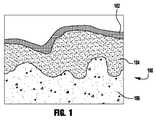
図1は、スキン層の一例を描いた図である。 FIG. 1 is a diagram depicting an example of a skin layer.
図2は、一実施形態に係るスキン、組織又は腫瘍を通って移動する分子についてのデリバリシステムを描く。 FIG. 2 depicts a delivery system for molecules that move through a skin, tissue or tumor according to one embodiment.
図3は、他のスキン、組織又は腫瘍を超えて移動する分子に関する他の実施形態を描く。 FIG. 3 depicts another embodiment for molecules that move across other skins, tissues or tumors.
図4は、第三のスキン、組織又は腫瘍を超えて移動する分子に関する他の実施形態を描く。 FIG. 4 depicts another embodiment for a molecule that moves beyond a third skin, tissue or tumor.
図5は、スキン、組織又は腫瘍を超えて移動する分子に関するさらなる他の実施形態を描く。 FIG. 5 depicts yet another embodiment for molecules that move across skins, tissues, or tumors.
図6は、図5の電極の平面図である。 FIG. 6 is a plan view of the electrode of FIG.
図7は、プラズマを使用するDNAワクチンの経皮デリバリのメソドロジの実施形態の模式図である。 FIG. 7 is a schematic diagram of an embodiment of a method for transdermal delivery of a DNA vaccine using plasma.
図8は、スキン、組織又は腫瘍を超えて移動する分子の移動に関するデリバリシステムの他の実施形態を描く。 FIG. 8 depicts another embodiment of a delivery system for the movement of molecules that move across skins, tissues or tumors.
図9は、処置表面をプラズマで孔を開口しDNAワクチンを処置エリアに処置する装置の一実施形態の断面図である。 FIG. 9 is a cross-sectional view of one embodiment of an apparatus for opening a treatment surface with plasma and treating a DNA vaccine in a treatment area.
出願人は、冷プラズマを使用しDNAをスキン層、細胞間(細胞の間)も細胞内(細胞の内部へ)の双方を超えて移動する技術を開発した。出願人は、US特許出願番号No.14/500144「組織層を超えた分子のデリバリのための方法及び装置」、2014年9月29日を出願し、それはここに参照によって全体が組み込まれている。いくつかの出願人の例示の方法は、安全で非接触の経皮デリバリ及びDNAワクチンの細胞摂取を提供するプラズマを利用しているが、ここではそれをプラズマポレーション(plasmaporation)ともいう。 Applicants have developed a technology that uses cold plasma to move DNA across skin layers, between cells (between cells) and inside cells (into cells). Applicants are assigned US patent application no. 14/500144 “Methods and apparatus for delivery of molecules across tissue layers”, September 29, 2014, which is hereby incorporated by reference in its entirety. Some Applicant's exemplary methods utilize plasma that provides safe and contactless transdermal delivery and cellular uptake of DNA vaccines, also referred to herein as plasmaporation.
経皮デリバリは分子がスキンをパススルーすることを要求する。図1は、スキン100の層を描く。スキン100の外層は、角質層(SCという,stratum corneum)102。SC102は死んだ、平坦でケラチンが豊富な細胞、角質細胞から構成されている。これらの濃密な細胞は複雑な細胞間脂質、セラミド、遊離脂肪酸、コレステロール及びコレステロール硫酸のミックスによって囲まれている。分子がSCを超える有力な拡散パスは細胞間に見える。残りのスキンの層は、表皮(生表皮)104及び真皮106である。 Transdermal delivery requires molecules to pass through the skin. FIG. 1 depicts the layers of
経皮的には少量パーセントのコンパウンドがデリバリ可能であるに過ぎない、理由は、スキン100は非常にバリア特性を持っており、すなわち、高い親油性のあるSC102は、分子がスキンを超えて浸透したり、拡散するのを防止するからである。その結果、相対的に小さな分子量(MW)500ダルトン(Dalton)以下の分子が経皮投与可能である。皮膚科の外用治療、経皮全身治療あるいは、ワクチン接種が目的の場合、経皮的処置のためのイノベーティブなコンパウンドの開発は、分子量500ダルトン以下に制限される。加えて、ほとんどのスキンを超えるドラッグの輸送は非常にゆっくりとしており、一定の流量に至るには数時間のラグタイムが測定される。治療上有効なドラッグレベルの達成には、したがって、強ケミカル浸透エンハンサーを使用する人工的なスキン浸透のエンハンスなしでは、治療的に有効なドラッグレベルの達成は難しい。 Only a small percentage of the compound can be delivered transcutaneously because the
プラズマポレーションは非熱プラズマ又はコールドプラズマを、経皮性のDNAワクチンのスキン及び免疫細胞へのデリバリに関して第四の状態を使用する。非熱プラズマは、部分的に大気圧下で大気空気又は他のガス及び電気を使用し生成されイオン化されたガスである。それは、二つの電極の間に現れる空気又は他のガスの分解によって生成され、しばしば、それらのうちの一つは絶縁され、十分な高電圧での処置である。第二の電極は、しばしば、生スキン、組織又は腫瘍のように誘電体も可能である。パルス電場は、プラズマ生成に並びにスキン、組織又は腫瘍及び細胞膜に経皮的なデリバリ及びマクロ分子の細胞摂を促進する一時的(リバーシブルな)孔開けに使用されている。いくつかの実施形態では、一時的な孔は約1から約5分間開いたままである。非熱プラズマを生成するのに使用される電気パラメータは、リバーシブルプラズマポレーションを達成するために制御可能である。電極は、スキンに非接触であり、針は不要で、非熱プラズマへの接触には痛みは伴わず、安全である。一又は複数の電極が絶縁されるココンフィグレーションでは、非熱プラズマは誘電体バリア放電(「DBD」)プラズマであり、例え、絶縁電極でスキン、組織又は腫瘍に接触しても、安全無痛である。 Plasmaporation uses non-thermal plasma or cold plasma in the fourth state for delivery of transdermal DNA vaccine to skin and immune cells. Non-thermal plasma is a gas that is generated and ionized using atmospheric air or other gases and electricity in part under atmospheric pressure. It is produced by the decomposition of air or other gas appearing between the two electrodes, often one of them is insulated and is a sufficiently high voltage treatment. The second electrode can also be a dielectric, often as a raw skin, tissue or tumor. Pulsed electric fields have been used for plasma generation as well as for temporary (reversible) drilling that facilitates percutaneous delivery to the skin, tissue or tumor and cell membranes and cellular uptake of macromolecules. In some embodiments, the temporary holes remain open for about 1 to about 5 minutes. The electrical parameters used to generate the non-thermal plasma can be controlled to achieve reversible plasma poration. The electrode is non-contact with the skin, no needle is required, and contact with non-thermal plasma is painless and safe. In co-configurations where one or more electrodes are insulated, the non-thermal plasma is a dielectric barrier discharge (“DBD”) plasma that is safe and painless even if it comes into contact with skin, tissue or tumor with an insulated electrode. is there.
ここに記述されているプラズマポレーション技術は、無痛非侵襲法でのDNAワクチンデリバリ効率的で迅速な手段である。いくつかの実施形態では、技術はDNAのスキンへの注射は要求されない。増加されたDNAデリバリ効率は、結果として一回当たりの免疫化に要求されるDNAをより少量にし、DNA単位あたりのドーズ数を増加させ、DNAワクチンの容易で迅速な生産に寄与し、より小規模の生産設備に寄与し、新しいワクチンのニーズに対するより早い応答及び低コストに寄与する。大規模な免疫化プログラムが開発されるところではどこでも、バイオハザード廃物、鋭利物(注射針)の廃棄に関して最小のコンサーンがホットになる。 The plasma poration technique described herein is an efficient and rapid means of delivering DNA vaccines in a painless non-invasive manner. In some embodiments, the technique does not require injection of DNA into the skin. Increased DNA delivery efficiency results in a smaller amount of DNA required for a single immunization, increasing the number of doses per DNA unit, contributing to easy and rapid production of DNA vaccines, and smaller Contributes to scale production facilities, contributes to faster response to new vaccine needs and lower costs. Wherever large-scale immunization programs are developed, the minimum concern regarding the disposal of biohazardous waste and sharps (needles) becomes hot.
ここに記述されているプラズマポレーション技術は、生スキンに外用処置されたDNAワクチン効率的な細胞摂取及び作用を促進する。ブタのスキンはヒトのスキンに形及び機能がとても近いので、実施例としてブタの生体研究実験が実行された。以下で記述される実験では、スキン細胞へのプラスミドDNA細胞間摂取がプラスミドDNAの遺伝子に緑色蛍光タンパク質(Green Fluorescent Protein,GFP)マーカーにエンコードされたタンパク質表現によって示されている。いくつかの実施形態では、GFPエンコードプラスミドのソース及び準備は、非常に安定なAcGFP1(Aequorea coerulescens、オワンクラゲGFP) 緑色蛍光タンパク質(Clontech Laboratories、クローンテック ラボラトリ)の形をエンコードするpEGFP-N1ベーシックベクターを含む。プラスミドDNAは、E. coli で増幅され商業上利用可能なキット(Qiagen Inc.)で精製される。プラスミドDNAは、指示濃度でリン酸緩衝食塩水に希釈されている。いくつかの実施形態では緑色蛍光タンパク質をエンコードしているプラスミドDNAが、アルデブロン社(Aldevron, Inc., Fargo, ND(ノースダコタ州)),から商業的に取得できた。このケースでプラスミドDNAはDNAse/RNAseフリーの超純水に溶解され投与された。プラスミドDNAの作業濃度は、2 mg/ml及び100 μlの容量で外用又は皮内注射処置された。エンコードされた表現の細胞によって細胞内に完全DNAが摂取されない場合を除き信号は得られない。 The plasma poration technique described here promotes efficient cell uptake and action of DNA vaccines that have been externally treated on live skin. Since pig skins are very similar in shape and function to human skins, pig biological research experiments were performed as examples. In the experiments described below, plasmid DNA intercellular uptake into skin cells is shown by the protein expression encoded in the plasmid DNA gene by the Green Fluorescent Protein (GFP) marker. In some embodiments, the source and preparation of the GFP-encoding plasmid is a pEGFP-N1 basic vector that encodes a very stable form of AcGFP1 (Aequorea coerulescens, Owan jellyfish GFP) green fluorescent protein (Clontech Laboratories, Clontech Laboratories). Including. Plasmid DNA is amplified in E. coli and purified with a commercially available kit (Qiagen Inc.). Plasmid DNA is diluted in phosphate buffered saline at the indicated concentration. In some embodiments, plasmid DNA encoding a green fluorescent protein could be obtained commercially from Aldevron, Inc., Fargo, ND (North Dakota). In this case, plasmid DNA was dissolved and administered in DNAse / RNAse-free ultrapure water. The working concentration of plasmid DNA was treated externally or intradermally in a volume of 2 mg / ml and 100 μl. No signal is obtained unless the complete expression of the DNA is taken up by the encoded expression cell.
これらの実験はプラズマポレーションが安全で無痛のエレクトロポレーション及び他のDNAの経皮的なデリバリ手段の代替であることを示す。非熱プラズマ生成の電気的パラメータ、電極ジオメトリ、DNA濃度、プラスミドDNA構成及びスキンへのプラスミドDNA処置モード(注射あるいはスキン表面への外用処方)は、プロセス最適化に変更可能とされる。上述のこれらの用法又は他の多くの用法に加えて、DNAワクチンのプラズマポレーションは、ウィルス感染予防(例えば、単純ヘルペスウィルス、エボラ等)及びがん治療(Her-2/neu乳がん)に使用されてもよく、ワクチンターゲットは現在研究途上である。 These experiments show that plasmaporation is an alternative to safe and painless electroporation and other means of transdermal delivery of DNA. The non-thermal plasma generation electrical parameters, electrode geometry, DNA concentration, plasmid DNA composition and mode of plasmid DNA treatment to skin (injection or topical formulation on skin surface) can be changed to process optimization. In addition to these or many other uses mentioned above, plasmaporation of DNA vaccines is used for viral infection prevention (eg, herpes simplex virus, Ebola, etc.) and cancer treatment (Her-2 / neu breast cancer) Vaccine targets are currently under study.
いくつかの実験例では、プラズマポレーションは、平面DBDプラズマジェネレータ(図3及び4)又はDBDジェットプラズマジェネレータ(図2及び5)又はパルスコロナジェネレータ(図8)が針を要しないマクロ分子の経皮デリバリのために関与している。プラズマ生成に使用されたパラメータによって(処置時間、負荷電圧、パルス繰り返し周波数、パルス時間、処置パルス回数、デューティサイクル等)、マクロ分子の浸透深さは制御されターゲット層、例えば、スキンの表皮又は真皮へのデリバリが保証される。このように、ここに記載された非熱プラズマポレーション技術は早い経皮的なデリバリを促進し、及び、大気圧下及び室温での十分なDNAワクチンの細胞摂取をエンハンスし、エレクトロポレーション及び他の従来技術で必要とされた電極又は針の廃棄を要さない。 In some experimental examples, plasma poration is performed through a macromolecular process where a planar DBD plasma generator (FIGS. 3 and 4) or a DBD jet plasma generator (FIGS. 2 and 5) or a pulse corona generator (FIG. 8) does not require a needle. Involved for skin delivery. Depending on the parameters used to generate the plasma (treatment time, load voltage, pulse repetition frequency, pulse time, number of treatment pulses, duty cycle, etc.), the penetration depth of the macromolecule is controlled and the target layer, eg skin epidermis or dermis Delivery to is guaranteed. Thus, the non-thermal plasma poration technique described herein facilitates rapid transdermal delivery and enhances cellular uptake of sufficient DNA vaccines at atmospheric pressure and at room temperature, There is no need to discard the electrodes or needles required by other prior art.
出願人は、前に、US特許出願番号No.14/500144、「組織複層を超えて分子のデリバリのための方法及び装置」2014年9月29日、に記載されているようにプラズマポレーションは、分子量70kDaまでのデキストラン分子、タンパク質では115kDaまで、そして50nm径ナノ粒子で生体ブタのスキンに15分以内の外用処方でスキンに損傷を発生させず経皮的なデリバリをエンハンス可能であることを示したが、それは、ここに参照されて取り込まれている。 The applicant has previously described US patent application no. 14/500144, “Methods and Apparatus for Delivery of Molecules Over Tissue Multilayers”, September 29, 2014, plasmaporation is a dextran molecule up to a molecular weight of 70 kDa, 115 kDa for proteins. Until now, it has been shown that 50 nm diameter nanoparticles can enhance skin transdermal delivery without causing skin damage with topical formulations within 15 minutes for living pig skin, which is referred to here. It has been captured.
大気圧下での非熱プラズマはマクロ分子を表面から生体ブタのスキン内へ、スキンを全く傷つけずに導入可能である。非熱プラズマによって可能とされたスキンポレーションは、経皮的なデリバリ及び細胞のDNAワクチン摂取を室温大気圧下で、苦痛もなく筋肉収縮や他のエレクトロポレーションに伴う副作用もなく、非侵襲及び安全な手段で提供する。当該処方は、ディスポーザル電極や針を要求せず、バイオ廃棄物の廃棄の必要もなく、バイオ消耗品廃棄物の不正な再利用もない。非熱プラズマを使用する追加の利益としては、生成された活性種は、プラズマポレーションの間スキンを殺菌可能である。 Non-thermal plasma at atmospheric pressure can introduce macromolecules from the surface into the skin of a living pig without damaging the skin at all. Skinporation enabled by non-thermal plasma is a non-invasive method for transdermal delivery and cellular DNA vaccine ingestion at room temperature and atmospheric pressure, without pain and without side effects associated with muscle contraction or other electroporation. And provide it in a safe way. The prescription does not require a disposable electrode or needle, there is no need to dispose of biowaste, and there is no unauthorized reuse of bioconsumable waste. As an additional benefit of using a non-thermal plasma, the generated active species can sterilize the skin during plasma poration.
プラズマのフェーズでは、中性ガス原子(又は分子)、電子、陽/陰イオン、及び高エネルギラディカルが生成される。これらの生成及び濃縮は、部分的に、プラズマ生成に使用されるガス及びケミカルの物性、プラズマ生成に使用されるエレクトロニックパラメータとデバイスデザインに依存する。スキン上に非熱プラズマによって生成される電場は、プラズマ処置時間;電極とスキンの間のギャップ;組織又は腫瘍;処置電圧;パルス期間;パルス処置回数、パルス繰り返し周波数及びデリバリを局所化するデューティサイクルを変更することによってチューン可能である。これらのパラメータは、マクロ分子のスキン、組織又は腫瘍を超えての深さ及びデリバリ量のコントロールを可能とし、ターゲットのスキン、組織又は腫瘍を最適ドーズで、高価なDNAワクチンの各処置の量を最小化する潜在能力を有している。 In the plasma phase, neutral gas atoms (or molecules), electrons, positive / negative ions, and high energy radicals are generated. These generation and concentration depend in part on the gas and chemical properties used for plasma generation, the electronic parameters used for plasma generation, and the device design. The electric field generated by the non-thermal plasma on the skin is: plasma treatment time; gap between electrode and skin; tissue or tumor; treatment voltage; pulse duration; pulse treatment frequency, pulse repetition frequency and duty cycle to localize delivery Tuning is possible by changing. These parameters allow control of depth and delivery volume across macromolecular skins, tissues, or tumors, and target skins, tissues, or tumors at optimal doses, and the amount of each treatment with expensive DNA vaccines. Has the potential to minimize.
プラズマオペレーションパラメータは変動してもよく、マイクロ秒パルスプラズマ処置。パワーサプライセッティング(外用処置でも注射処置でも双方の)はパルス繰り返し周波数50−3500Hzの間でパルス期間が約1 - 10 μsの間で電圧が約11 - 20 kVの間でデューティサイクルが約1−100%の間で電圧上昇時間が約1−5 V/nsの間の範囲に内に設定可能である。処置時間は、約5秒と約180秒の間であってよい。同様に、ナノ秒パルスプラズマ処置では(外用処置でも注射の実験でも双方)、パワーセッティングは、連続処置の間、パルス期間約1 ns - 500 nsの間で約2 - 20000 Hzのパルス繰り返し周波数及び電圧上昇時間約0.5 - 10 kV/nsの間で約3 - 20 kVの間の電圧というものでよい。ディスクリートパルス処置のために、セッティングは、パルス期間約 1 ns - 500 ns、電圧上昇時間0.5 - 10 kV/nsの間の電圧約3−20kV の間で約1 - 100パルスの間でもよい。処置時間は、約1秒と約300秒の間でよい。パルスは正又は負極性のいずれでもよい。加えて、ナノ秒パルスコロナ処置のパワーセッティングは、パルス期間約1 ns - 60 ns及び電圧上昇時間0.5 - 10 kV/nsの間の電圧約3 - 20 kVの間でパルス繰り返し周波数約1−1000Hz又は連続処置でよい。ディスクリートパルスコロナ処置については、セッティングは、約1−100パルスを、電圧上昇時間0.5 - 10 kV/nsの間の電圧約3 - 20 kVでパルス期間約1 ns - 60 ns処置するものでよい。 Plasma operating parameters may vary, microsecond pulsed plasma treatment. The power supply setting (both for external and injection treatments) has a pulse repetition frequency of 50-3500 Hz, a pulse duration of about 1-10 μs, a voltage of about 11-20 kV and a duty cycle of about 1- Between 100%, the voltage rise time can be set within a range between about 1-5 V / ns. The treatment time may be between about 5 seconds and about 180 seconds. Similarly, for nanosecond pulsed plasma treatments (both topical treatments and injection experiments), the power setting is about 2-20000 Hz pulse repetition frequency and between about 1 ns-500 ns during the continuous treatment and The voltage rise time may be between about 0.5 and 10 kV / ns and between about 3 and 20 kV. For discrete pulse treatment, the setting may be between about 1-100 pulses with a pulse duration of about 1 ns-500 ns and a voltage rise time of 0.5-10 kV / ns for a voltage of about 3-20 kV. The treatment time may be between about 1 second and about 300 seconds. The pulse may be positive or negative. In addition, the power setting of the nanosecond pulse corona treatment is such that the pulse repetition frequency is about 1-1000 Hz between a voltage of about 3-20 kV with a pulse duration of about 1 ns-60 ns and a voltage rise time of 0.5-10 kV / ns. Or it may be a continuous treatment. For discrete pulse corona treatment, the setting may be to treat about 1-100 pulses with a voltage duration of about 3-20 kV for a voltage rise time of 0.5-10 kV / ns and a pulse duration of about 1 ns-60 ns.
いくつかの実施形態では、安全(火傷又は他のスキンへの損傷を発生しない)な細胞DNA摂取及びロバストにエンコードされたGFPが実現される最適パラメータでヘリウムDBDジェットが使用された。デバイスは、電極チップから5−50mm離れスキン上に焦点を絞ったプラズマビームを生成し、よりリモート処置を可能とする。図2は、実施実験例のDNAプラスミドをスキン220を介してデリバリするデリバリシステム200を描く。デリバリシステム200の実施例は、高電圧チューブメタル電極202及び誘電体として働くホウケイ酸ガラスチューブ204を含む非熱プラズマジェネレータ201を含む。プラズマジェネレータ201は、プラズマジェット206を生成する浮き電極の誘電バリア放電(dielectric barrier discharge (DBD))プラズマジェネレータである。 In some embodiments, a helium DBD jet was used with optimal parameters to achieve safe (no burns or other skin damage) cellular DNA uptake and robustly encoded GFP. The device generates a focused plasma beam on the skin 5-50 mm away from the electrode tip, allowing for more remote treatment. FIG. 2 depicts a
プラズマジェネレータ201はガスフィード215を含む。実施例のガスはHe, He + O2, N2, He + N2, Ar, Ar + O2, Ar + N2及びその類を含むプラズマジェットをフィードするのに使用される。液体溶液の蒸発から発生するガスの使用も可能である。気化液には、水、エタノール、有機溶媒及びその類が含まれてよい。これらの気化液は、医薬物質、浸透エンハンサー等の添加剤コンパウンドとミックスされてもよい。気化液及び添加剤は、上記に示されたガスと共に様々な濃度であるいはガスなしで使用されてもよい。Plasma generator 201 includes a
プラズマジェネレータ201は、図示しないパワーサプライを含む。パワーサプライは高電圧電源であり、例えば、直流、ランプアップ、ランプダウン、パルス、ナノ秒パルス、マイクロ秒パルス、矩形、SIN、減衰SIN、ランダム、同位相、異位相及びその類の多数の異なる波形をもつことが可能である。いくつかの実施形態では、パワーサプライはマイクロ秒パルスパワーサプライであった。いくつかの実施形態では、パワーサプライはナノ秒パルスパワーサプライであった。いくつかの実施形態では、プラズマ206は、交番極性パルス電圧であった。いくつかの実施形態では、電圧はパルス幅約1 - 10 μsの間(パルス繰り返し周波数:50 Hzから 3.5 kHz)の立ち上がり時間5V/nsで振幅約〜20 kV( ピークからピーク)のパワー密度1−10W/cm2であった。オペレーション中、プラズマジェット206は、スキン220と直接接触している。Plasma generator 201 includes a power supply (not shown). The power supply is a high voltage power supply, for example, DC, ramp up, ramp down, pulse, nanosecond pulse, microsecond pulse, rectangular, SIN, attenuated SIN, random, in-phase, out-of-phase and many others It is possible to have different waveforms. In some embodiments, the power supply was a microsecond pulse power supply. In some embodiments, the power supply was a nanosecond pulse power supply. In some embodiments, the
プラズマは電場がスキンに到達し電気チャージを貯めてスキンを超えて電圧ポテンシャルを高め、細胞内及び細胞間ポレーションをもたらす。プラズマ電極は組織又は処置物体に接触しないからプラズマポレーションは、非侵襲である。 The plasma causes the electric field to reach the skin, accumulate the electrical charge, increase the voltage potential across the skin, and cause intracellular and intercellular poration. Plasma poration is non-invasive because the plasma electrode does not contact the tissue or treatment object.
細胞内のポレーション(poration, 孔形成)に関して、流体脂質二分子層膜の膜貫通電圧は少なくとも0.2Vに達する必要がある。膜貫通電圧は、脂質二分子層膜をチャージし、膜内で急速の局所的な構造再配列を起こさせ、水で満たされた膜構造へ遷移させ、膜に“水通路”又は“孔”形を穿つ。水通路又は孔はイオン及び分子の移動を総じて増加する。膜貫通電圧は、最小半径約およそ1 nm で主膜の“孔”を作ると考えられている。加えて、負荷電場は急速な極性状態の変化に至らせメカニカルに拘束のない細胞膜変形を起こさせ(例えば、つるされた小嚢と細胞)、及び電解質電導によって支配されるイオンチャージ再配布を起こさせる。 With respect to intracellular poration, the transmembrane voltage of the fluid lipid bilayer membrane needs to reach at least 0.2V. Transmembrane voltage charges lipid bilayer membranes, causing rapid local structural rearrangements within the membrane, transitioning to a water-filled membrane structure, and “water passage” or “pores” in the membrane Wear a shape. Water passages or holes generally increase the movement of ions and molecules. The transmembrane voltage is believed to create a “hole” in the main membrane with a minimum radius of about 1 nm. In addition, the negative electric field can lead to a rapid change of polarity, causing mechanically unconstrained cell membrane deformation (eg, suspended vesicles and cells), and ion charge redistribution dominated by electrolyte conduction. Let
プラズマジェット206を生成するのに使用される電気パルスは、細胞間ポレーションの元にもなる。SCは、約15−25μm厚さであるが、スキンでは最も電気抵抗性がある部位である。プラズマジェット206を生成するのに使用される電気パルスの負荷は、約50Vと約100Vの間の経皮電圧を起こすが、SC内のマルチラメラ二層のポレーションを起こす。このレベルの負荷経皮電圧では、細胞ライニングの汗腺と毛包のポレーションも発生する可能性もある。 The electrical pulse used to generate the
処置エリアからプラズマ源を取り除くと同時に、孔は再び閉じ始め、したがって、プロセスはリバーシブルである。いくつかの孔は、ある期間、開いたままに残り、その間は分子は細胞膜間を拡散し続けることができる。いくつかの他のケースでは、負荷されている膜貫通ポテンシャルはしきい値を超え、その結果。形成された孔は永久に開いたままである。このプロセスは、不可逆のポレーションとしてがん治療で有益である。 Upon removal of the plasma source from the treatment area, the hole begins to close again, and therefore the process is reversible. Some pores remain open for a period of time, during which time molecules can continue to diffuse between cell membranes. In some other cases, the loaded transmembrane potential exceeds the threshold, and as a result. The formed hole remains open permanently. This process is beneficial in cancer treatment as irreversible poration.
電気パルスがスキンに処置されるとき、吸収されたエネルギーが局所的な加熱を発生させスキンにダメージを起こし得る。エネルギー100 J/cm2以上がスキンに離れてかけられて二度の火傷と下に離れているスキンへ熱ダメージを与える結果となる。この問題に打ち勝つ一つの方法は、短時間パルスを間欠的に繰り返し負荷することであり、同量のエネルギーはその方法でなければダメージを生じさせるものが局所的な加熱もスキンダメージも生じずに伝達される。いくつかの実施形態では、離れたスキンにかけられるエネルギーは約50 J/cm2であり、いくつかの実施形態では、離れたスキンにかけられるエネルギーは約25 J/cm2であり、いくつかの実施形態では、離れたスキンにかけられるエネルギーは約10 J/cm2であり、いくつかの実施形態では、離れたスキンにかけられるエネルギーは約5 J/cm2であり、そして、いくつかの実施形態では、離れたスキンにかけられるエネルギーは約3 J/cm2である。しかし、外傷を処置するときには、エネルギーは増加されてもよく、例えば、500 J/cm2にしても火傷は生じない。いくつかの実施形態では500 J/cm2のレンジのエネルギーが止血に使用される場合もある。When an electrical pulse is applied to the skin, the absorbed energy can cause local heating and cause damage to the skin. More than 100 J / cm2 of energy is applied away from the skin, resulting in double burns and thermal damage to the skin that is underneath. One way to overcome this problem is to intermittently repeatedly apply short-term pulses, and the same amount of energy would otherwise cause damage without causing local heating or skin damage. Communicated. In some embodiments, the energy applied to the distant skin is about 50 J / cm2 , and in some embodiments, the energy applied to the distant skin is about 25 J / cm2 , In form, the energy applied to the distant skin is about 10 J / cm2 , in some embodiments, the energy applied to the distant skin is about 5 J / cm2 , and in some embodiments , the energy applied to the skin at a distance of about 3 J / cm2. However, when treating trauma, the energy may be increased, eg, 500 J / cm2 will not cause a burn. In some embodiments, energy in the range of 500 J / cm2 may be used for hemostasis.
加えて、ダメージはパワーレベル、処置時間、周波数、デューティサイクル及びパワーサプライのパルス期間を下げることによって避けることが可能であり、プラズマ電極とスキン、組織又は腫瘍の間のすきまを増加することによって回避可能である。 In addition, damage can be avoided by reducing the power level, treatment time, frequency, duty cycle and power supply pulse duration, and by increasing the clearance between the plasma electrode and skin, tissue or tumor. Is possible.
プラズマ処置後にプラズマポレーションを起こさせ、一旦、プラズマ生成デバイス206が止められたら、水通路のマルチラメラシステムはある期間開いたままであり、それは約二、三分から二、三時間までかもしれない。 After plasma treatment, plasma poration occurs and once the
他のタイプのプラズマジェネレータがデリバリシステムのために使用されてもよく、例えば、ナノ秒パルスDBDプラズマ、マイクロ秒パルスDBDプラズマ、SIN波DBDプラズマ、抵抗バリアディスチャージプラズマ、サーフェスDBDプラズマ、2−Dあるいは3−Dアレイナノ秒パルスコロナ、2−Dあるいは連続モードでの3−Dアレイプラズマジェットオペレーション、あるいは、コントロールされた1から100%のデューティサイクルのような類のディスペンサーシステムが使用されてもよい、すべてのプラズマジェネレータがポレーション(poration、孔を導出)するのに成功裏に使用されているわけではない。高電流をデリバリするプラズマジェネレータ、あるいは、処置対象の温度上昇が高くなるプラズマジェネレータは、熱プラズマ、グライディングアークディスチャージ、プラズマトロンも含めプラズマポレーションには向かない。このようなプラズマジェネレータは、電気ショック、重大な熱ダメージ、筋肉収縮及び痛みの原因となり、あるいは、処置される対象物に十分なチャージをデリバーせず、全くあるいは非常に弱い負荷電場を意味し、したがって、ポレーションされないのである。 Other types of plasma generators may be used for the delivery system, for example, nanosecond pulse DBD plasma, microsecond pulse DBD plasma, SIN wave DBD plasma, resistance barrier discharge plasma, surface DBD plasma, 2-D or Kinds of dispenser systems such as 3-D array nanosecond pulse corona, 3-D array plasma jet operation in 2-D or continuous mode, or controlled 1 to 100% duty cycle may be used. Not all plasma generators have been used successfully for poration. A plasma generator that delivers a high current or a plasma generator that increases the temperature of the treatment target is not suitable for plasma poration, including thermal plasma, gliding arc discharge, and plasmatron. Such plasma generators can cause electric shock, significant heat damage, muscle contraction and pain, or do not deliver enough charge to the object being treated, meaning no or very weak load electric field, Therefore, it is not porated.
好適なプラズマジェネレータは、変位電流である支配電流を低パワー及び/又は高周波数で保つ。変位電流は、電流密度の単位を有し、ちょうど関連する電磁場は、ちょうど誘導電流が有するように、しかし、移動電荷の電流ではなく、むしろ時間依存の電場である。絶縁されてスキンに接触していない電極によって電場がスキンにかけられ、最小の伝導電流がスキンに流れる。強い伝導電流は電気ショックを起こさせ、熱ダメージ、筋肉収縮及びエレクトロポレーションに関係する痛みを起こさせる。 A suitable plasma generator keeps the dominant current, which is the displacement current, at low power and / or high frequency. Displacement current has units of current density and just the associated electromagnetic field is just a time-dependent electric field rather than a mobile charge current, just as an induced current has. An electric field is applied to the skin by an insulated electrode that is not in contact with the skin, and minimal conduction current flows through the skin. A strong conduction current causes an electric shock and causes pain associated with heat damage, muscle contraction and electroporation.
より広い処置エリアについて、電極構成は複数のプラズマジェットあるいはより大きなエリアのフラットな電極(図示しない)が使用されてもよい。より複雑な3Dサーフェスのケースでは、コントロールされたプラズマモジュール(図示しない)が静止ターゲットのまわりを動いてもよく、あるいは、プラズマに晒されるサーフェスは可動ステージ上に置かれてもよい。いくつかの実施形態では、一又は複数のプラズマはあるいはロボットアームにアタッチされてもよいが一又は複数のターゲット領域をプラズマプルーム(plasma plume)又はジェットに晒されるように動くようにプログラムされている。 For larger treatment areas, electrode configurations may use multiple plasma jets or larger area flat electrodes (not shown). In the case of more complex 3D surfaces, a controlled plasma module (not shown) may move around a stationary target, or the surface exposed to the plasma may be placed on a movable stage. In some embodiments, one or more plasmas may alternatively be attached to a robotic arm, but programmed to move one or more target areas to be exposed to a plasma plume or jet. .
加えて、いくつかの実施形態では、プラズマジェネレータ201はバイオ分子/ドラッグデリバリシステムに結合されたもので、分子は処置エリアに針のない注射、圧力ガスの負荷、気化、スプレー及び又はミスト化によって輸送されてもよい。いくつかの実施形態では、これはサーフェスの前処置でアシストしてもよい。 In addition, in some embodiments, the plasma generator 201 is coupled to a biomolecule / drug delivery system, where the molecules are injected by needleless injection, pressure gas loading, vaporization, spraying and / or misting into the treatment area. It may be transported. In some embodiments, this may be assisted with a surface preparation.
いくつかの実施形態では、プラズマ温度を減少するのが本質的であり、プラズマポレーション後のスキン透過をエンハンスし、He, Ar, Ne, Xe及びその類、空気又は希ガスにO2 及び N2のような他のガスを小パーセント(0.1% - 20%)で混合したもの及び希ガスと水、ジメチルスルホキシド(DMSO)、エタノール、イソプロピルアルコール、n−ブタノールの気化液の混合物に添加物及びその類とともにあるいは加えないものを使用して非熱プラズマを生成することは有益である。In some embodiments, it is essential to reduce the plasma temperature, enhance skin permeation after plasma poration, He, Ar, Ne, Xe and the like, O2 and N in air or noble gases Additives to mixtures of other gases such as2 in small percentages (0.1%-20%) and mixtures of vapors of noble gases with water, dimethyl sulfoxide (DMSO), ethanol, isopropyl alcohol, n-butanol It is beneficial to generate a non-thermal plasma with or without the like.
いくつかの実施形態では、非熱平面DBDプラズマジェネレータが経皮的なデリバリ及びブタのスキンのサーフェスに外用処置されたプラスミドDNAの細胞摂取を促進するのに使用されている。図3は、非熱平面DBDデリバリシステム300の実施例を描く。デリバリシステム300は、プラズマジェネレータ301を含む。プラズマジェネレータ301は第一の端で電極302に接続され、第二の端で高電圧パワーサプライ(図示しない)に接続されている高電圧ワイヤー303を含む。好適な高電圧サプライが上述されている。誘電バリア304は高電圧電極302の下に配置されている。加えて、高電圧電極302はハウジング305内に配置されている。プラズマジェネレータ301は非熱誘電バリアディスチャージ(DBD)ジェネレータである。プラズマ306は、プラズマジェネレータ301によって生成される。図3は、スキン320も含む。ここに開示されている実験例については、スキン320は生ブタスキンである。DBDプラズマ源がスキンのサーフェスから1−5mmの距離に置かれると、コールドプラズマがスキンに直接接して生成される。 In some embodiments, a non-thermal planar DBD plasma generator is used to facilitate percutaneous delivery and cellular uptake of plasmid DNA applied topically to the surface of a pig skin. FIG. 3 depicts an example of a non-thermal planar
ダイレクトプラズマ306は、交番極性パルス電圧が電極302に負荷されることによって生成される。いくつかの実施形態では、負荷電圧は、約1 - 10 μsの間のパルス幅(パルス繰り返し周波数:50 Hzから30 kHz)で振幅約~20 kV(ピークからピーク)及び電圧立ち上がり時間約1 - 10 V/ns.である。パワーサプライ(図示しない)は、可変電圧かつ周波数可変のパワーサプライである。1mm厚の透明な石英スライド、アルミナ又はテフロンが絶縁誘電バリア304として使用されてもよく、電極302をカバーする。電極302は2.54 cm径の銅、真鍮又は他の導電性材料でよい。誘電バリア304とブタのスキン320間のディスチャージギャップは、約4 mm ± 1 mmでよい。いくつかの実施形態では、パルス波形は振幅約22kV(ピークからピーク)、約9 μs,の期間、立ち上がり時間約5 V/nsである。ディスチャージパワー密度は約0.1 W/cm2 から 2.08 W/cm2である。J/cm2でのプラズマ処置ドーズ(dose)は、プラズマディスチャージパワー密度にプラズマ処理期間を乗じて算出してもよい。The
加えて、間接プラズマ406はプラズマジェネレータ401で作られてもよい。プラズマジェネレータ401はプラズマジェネレータ301と同様であるが、プラズマジェネレータ401はプラズマ406をフィルタするメタルメッシュ330を含む以外は同じである。メタルメッシュ300は、チャージイオン及び電子のパススルーを防止するが、中性種がパススルーしスキンとコンタクト可能とする。中性種は“残光”(“afterglow”)とも呼ばれる。 In addition, the
図5は、他の実施例に係るデリバリシステム500の斜視図である。図6は、デリバリシステム500の電極の平面図である。デリバリシステム500は、複数のDBDジェットを含む。実施例のデリバリシステム500は、ハニカム形のDBDジェットのアレイを含む;しかし、多くの他のコンフィグレーションでは、リニア、三角、矩形、ペンタゴン、ヘキサゴン、オクタゴン等のように使用されてもよい。 FIG. 5 is a perspective view of a
DBDジェットは、ガラスチューブ504A, 504B, 504C, 504D, 504E, 504F 及び 504Gを有する。メタル電極502は複数の円筒形の開口502A, 502B, 502C, 502D, 502E, 502F, 及び502Gであって各々に対応するガラスチューブ504A, 504B, 504C, 504D, 504E, 504F,及び504Gを受ける開口を含む。オプションとして、複数のメタル電極が使用されてもよい。メタル電極502はショックを防ぐ絶縁カバー(図示しない)を有してもよい。メタル電極502は上述の高電圧電源に接続されている。 The DBD jet has
DBDジェットは、第一の端にガス流入口を有し、他端にプラズマジェット516A, 516B, 516C, 516D, 516E, 516F 及び 516G出口を有する。上述のように、ガスは、例えば、He, Ar, Ne, Xe, air, He+Air, Ar+Air, Ne+Air, Xe+Air,又はその類でよい。加えて、各々のガラスチューブ504A, 504B, 504C, 504D, 504E, 504F及び504Gは、気化液添加剤を受けるためにガラスチューブに沿って入口508A, 508B, 508C, 508D, 508E, 508F,及び508Gを有する。これらの入口は、電極502の上方又は下方に配置されている。実施例の経皮的なデリバリシステム500はスキン、組織又は腫瘍を接地電極として利用している。 The DBD jet has a gas inlet at the first end and
図2の実施例では、スキン、組織又は腫瘍220は、中性及びチャージされている種を含むプラズマ206に直接暴露されている。同様に、図3の実施例では、直接プラズマジェネレータ301で、電気的なディスチャージが誘電バリア304とスキン320との間で起こり、スキンは中性種及びチャージされた粒子に直接暴露されている。 In the embodiment of FIG. 2, the skin, tissue or
プラズマジェネレータ401によって作られる間接プラズマは接地銅メッシュ(16 x 16 メッシュサイズ、0.011" ワイヤー径で0.052" の開口サイズ)高電圧電極とスキンの間に置かれて、チャージ粒子を消滅させ暴露されているスキンのサーフェスに接触するのを防止した。 The indirect plasma produced by the
図7は、DNAワクチンの経皮デリバリのためのメソドロジ700実施例である。実施例に係るメソドロジ700はブロック702から開始する。ブロック704では、プラズマはスキンに一又は複数の孔を開けるために処置エリアへ処置される。ブロック706では、DNAワクチンがスキン、組織又は腫瘍上のターゲットエリアへ外用処置され、ブロック708で待ち期間が生じる。いくつかの実施形態では、待ち期間は約3時間以下であり、いくつかの実施形態では、待ち期間は約2時間以下であり、いくつかの実施形態では、待ち期間は約1時間以下であり、いくつかの実施形態では、待ち期間は約30分以下である。第二のプラズマ処置がブロック710で発生し、スキン、組織又は腫瘍の細胞にポレートし、DNAワクチンの細胞内摂取エンハンスを主導し実施例のメソドロジはブロック712で終了する。 FIG. 7 is an
図8は、安全で効率的なプラスミドDNA外用処置の経皮デリバリを可能とし、スキン、組織又は腫瘍の細胞によってDNAの細胞内摂取を促進するプラズマシステムのさらなる他の実施例800である。この実施例800は、パルスコナノ秒パルスパワーサプライ(図示しない)によるパルスコロナアレイを利用している。パルスコロナアレイは複数のシャープなチップ802で作られ、この実施例ではステンレススチールの機械加工されたチップ0.1016 mm厚及び1 mm互いに離れているものである。シャープなチップ802は、パルスコロナ電極の2Dアレイを形成している。 FIG. 8 is yet another
図9は、プラズマでサーフェス処置しサーフェス及び/又は細胞に孔を開けDNAワクチンを処置エリアに処方するワクチン接種装置900の実施例の断面図である。ワクチン接種装置900は、ハウジング902を含み、オンオフスイッチ904、サーフェスコンタクトリング906、高電圧電極908、処置チャンバー910、DNAワクチンチャンバー912及びプランジャ912を含む。加えて、ワクチン接種装置900は、ここに識別されたすべてのパワーセッティングを含む要求パワーを供給可能であるパワーサプライ(図示しない)を含む。いくつかの実施形態では、サーフェスコンタクトリング906は取り外し可能であり、各使用後に交換可能である。加えて、いくつかの実施形態では、サーフェスコンタクトリング906は接地リング(図示しない)を含む。 FIG. 9 is a cross-sectional view of an embodiment of a
ワクチン接種装置900はいくつかの異なるモードでオペレーション可能である。一実施例では、パワーセッティングはパワーサプライ(図示しない)で選択されオペレータは、サーフェスコンタクトリング906を例えば、ヒトのスキン、組織又は腫瘍又はその類のようなサーフェスに置く。オペレータはスタートボタン904を押し、所望の電圧(複数)を高電圧電極908に適用し処置チャンバー910内にプラズマを生成する。十分な期間経過後、スキンサーフェスに処置されたプラズマはスキンに孔を開ける。オペレータはプランジャ914を下方に押し、ワクチンチャンバー912にストアされているDNAワクチンを処置チャンバー910に注入させる。十分な時間が経過後、DNAワクチンはスキン、組織又は腫瘍の類の開孔をパススルーし、所望のパワーが高電圧電極に負荷されてプラズマを生成し一又は複数の細胞に孔を開口しDNAワクチンの摂取を発生させる。 The
もうひとつの実施例では、パワーセッティングはパワーサプライ(図示しない)で選択されてオペレータはサーフェスコンタクトリング906を例えば、ヒトのスキン、組織又は腫瘍の類のサーフェスに置く。オペレータはスタートボタン904を押し、選択された電圧(複数)が高電圧電極908へ負荷され処置チャンバー910内にプラズマを生成する。プラズマはサーフェスに処置されスキン、組織又は腫瘍の類に孔を開け細胞内に孔を開口する。オペレータはプランジャ914を下方に押し、ワクチンチャンバー912にストアされている、DNAワクチンを処置チャンバー910内に注入させる。十分な時間が経過後には、DNAワクチンは開口された孔をパススルー可能であり、細胞に摂取される。
実験結果In another embodiment, the power setting is selected with a power supply (not shown) and the operator places the
Experimental result
多数の実験が生動物で実施された。3頭の5−7月齢のユカタン仔ブタが3つの生動物実験に利用された。各々の研究は、一頭の仔ブタに多数の実験が含まれている。実験コントロールには、以下の:プラズマ処置なし、プラスミドDNA処置なし;プラズマ処置なしでプラスミドDNAの皮内注射付;そしてプラスミドDNAの皮内注射後にエレクトロポレーション(従来技術)を含む。実験サンプルは、以下の:皮内注射後マイクロ秒パルスプラズマ;皮内注射後ナノ秒パルスプラズマ;マイクロ秒パルスプラズマ後に外用プラスミドDNA処置;そしてナノ秒パルスプラズマ後に外用プラスミドDNA処置を含む。 A number of experiments have been performed on live animals. Three 5-7 month old Yucatan piglets were used for three live animal experiments. Each study involves numerous experiments in one piglet. Experimental controls include the following: no plasma treatment, no plasmid DNA treatment; with intradermal injection of plasmid DNA without plasma treatment; and electroporation (prior art) after intradermal injection of plasmid DNA. Experimental samples include: microsecond pulse plasma after intradermal injection; nanosecond pulse plasma after intradermal injection; external plasmid DNA treatment after microsecond pulse plasma; and external plasmid DNA treatment after nanosecond pulse plasma.
第一のコントロールはプラスミドDNAが関与せず、結果は、動物処置2経過観察では緑色蛍光タンパク質(GFP)の発現は見られなかった。 The first control did not involve plasmid DNA, and as a result, expression of green fluorescent protein (GFP) was not observed in the animal treatment 2 follow-up.
第二のコントロールでは、プラスミドDNAは皮内に(“intradermally、ID”)注射され、その結果、いくつかのGFPが発現した。GFP発現は、しかし、ほとんど真皮に局所化され、表皮には極めて稀にしか発現しなかった(表皮の発現は処方されたワクチンへのロバストな免疫反応を獲得するという点で重要である)。生検 から得られたデータに基づき正規化強度が導出された。正規化強度は以下のチャートIに示されている。
第三のコントロールでは、プラスミドDNAの皮内注射後に、二針の電極が200 V/cmの電場で, 150 ms の期間に16 パルス 周波数1 Hzで、8パルス後に電極を 90° 回転し、エレクトロポレーションされた。エレクトロポレーション後、GFPが表皮に強く発現したが、真皮にはほとんど発現しなかった。生検 から得られたデータに基づき正規化強度が導出された。正規化強度は以下のチャートIIに示されている。上述のように、エレクトロポレーションは、痛みを含む多数の副作用がある。加えて、エレクトロポレーションは、処置前にプラスミドDNAの皮内注射を要求する。プラズマ処置と異なり、それは、角質層又は組織又は腫瘍の高い抵抗を超えて、外用処置されたプラスミドDNAの細胞内デリバリを促進するのに使用できないからである。
第1セットの実験では、プラスミドDNA(2 mg/mlの100 μl)が皮内注射され、注射後にプラズマ処置がマイクロ秒パルスDBDプラズマでパワーサプライ3500 Hz, パルス期間5 μs, 電圧 15 kV 及び 100% デューティサイクルを使用して行われた。以下のチャートで何度も処置された領域だった。DBDプラズマ処置2日後、蛍光にエンコードされたロバストなタンパク質発現が表皮にも真皮下にも観察された。生検 から得られたデータに基づき正規化強度が導出された。正規化強度は以下のチャートIIIに示されている。
マイクロ秒パルス連続DBDプラズマ処置を用いたデータは、それぞれ、注射コントロールに対して137%, 66%, 166% 及び 102%,の細胞摂取増加を示す。加えて、マイクロ秒パルス連続DBDプラズマはエレクトロポレーションに対して細胞摂取を起こす点に関して優っていることをデータは示す。データは、マイクロ秒パルスDBDプラズマ処置は、エレクトロポレーションに対してそれぞれ14%, 20%, 21% 及び 86%の細胞摂取及びGFP発現の増加を示す。 Data using microsecond pulsed continuous DBD plasma treatment show an increase in cell uptake of 137%, 66%, 166% and 102%, respectively, relative to the injection control. In addition, the data show that microsecond pulsed continuous DBD plasma is superior to electroporation in terms of causing cellular uptake. The data show that microsecond pulsed DBD plasma treatment shows 14%, 20%, 21% and 86% increase in cell uptake and GFP expression relative to electroporation, respectively.
第二セットの実験では、スキンはマイクロ秒パルスDBDプラズマで処置された。電源は、3500 Hzにセットされパルス期間5 μs及び電圧 15 kVだった。処置時間は120秒間であった。チャートで識別されるようにプラズマで処置されるエリアに100 μlプラスミドDNA容液がある期間(“保持時間”)塗られた。第二のDBDプラズマ処置は、電源セットは3500 Hzでパルス期間5 μs 電圧15 kV 、60 秒間 である。 In the second set of experiments, the skin was treated with a microsecond pulsed DBD plasma. The power supply was set at 3500 Hz with a pulse duration of 5 μs and a voltage of 15 kV. The treatment time was 120 seconds. A 100 μl plasmid DNA solution was applied for a period of time (“retention time”) in the plasma treated area as identified in the chart. In the second DBD plasma treatment, the power supply set is 3500 Hz, the pulse duration is 5 μs, the voltage is 15 kV, and the time is 60 seconds.
ロバストなGFP発現が表皮及び真皮に観察された。これらの実験は、プラズマポレーションが細胞間のプラスミドDNAデリバリでスキンの正層に到達可能であること及び細胞内プラスミドDNAデリバリが表皮及び真皮細胞によって摂取達成が可能であることが示された。加えて、実験のメソドロジは、針スティックを不要とし、付随するバイオ的危険性のある廃棄物及び鋭利な廃棄物の不正再使用をなくす。正規化強度は以下のチャートIVにリストされている。
データは、スキンをマイクロ秒パルス連続DBDプラズマで処置し、プラスミドDNAを外用処方し第二のマイクロ秒パルス連続DBDプラズマ処置すればそれぞれ10%, 153%, 71% and及び 11%,の細胞摂取が注射コントロールの細胞摂取に対して改善を示している。いくつかのデータでは、この外用処方メソドロジは、エレクトロポレーションと同等か又はよりよいことを、痛みがなく他のエレクトロポレーションに伴う副作用がないことが示された。しかし、いくつかの実験では、このメソドロジは修正が必要であって、及び/又はパラメータが調整される櫃等があることを示す。結果は、また、一又は複数のプラズマ処置を、例えば、マイクロ秒パルス連続DBDプラズマをスキンに処置し、孔を開口し、ナノ秒パルス処置(以下に開示される)を用いる。 The data are 10%, 153%, 71% and 11% cell uptake, respectively, if the skin is treated with microsecond pulsed continuous DBD plasma, plasmid DNA is externally formulated and the second microsecond pulsed continuous DBD plasma treatment Shows an improvement over the cell intake of the injection control. Some data showed that this topical formulation methodology is equivalent or better than electroporation, without pain and without the side effects associated with other electroporations. However, in some experiments, this methodology indicates that correction is necessary and / or that there is a habit of adjusting the parameters. The results also use one or more plasma treatments, for example, a microsecond pulsed continuous DBD plasma on the skin, an aperture opened, and a nanosecond pulse treatment (disclosed below).
第三の実験セットでは、プラスミドDNA(2 mg/ml を100 μl)が皮内に注射され、そしてその後、多数回パルスのDBDプラズマ処置が、電圧20 kV、パルス期間500nsでナノ秒パルスパワーサプライされた。ロバストなGFP現出が上皮にも真皮にもプラズマ処置2日経過後に観察された。実験は、いくつかの実施形態では、わずか25パルスのみ必要とされ、したがって、この実験メソドロジは極めて迅速で、安全で、そして、プラズマは非常に短期間処置されてよい。加えて、このメソドロジはより省パワーで済み、手持ちバッテリーを用いプラズマ処置装置に給電可能である。正規化強度は以下のチャートVにリストされている。
データは、ナノ秒パルスDBDプラズマ処置は、注射コントロールに対して細胞摂取がそれぞれ143%, 170%, 171%, 74% 及び117%向上することが示されている。加えて、データはナノ秒パルスDBDプラズマがエレクトロポレーションに対して細胞接種のエンハンスについて優れていることが示されている。デーは、ナノ秒パルスDBDプラズマ処置は、17%, 28%, 28%, -18% 及び100% の各増加を示し、エレクトロポレーションに比して細胞摂取に優れている。 Data show that nanosecond pulsed DBD plasma treatment improves cell uptake by 143%, 170%, 171%, 74%, and 117%, respectively, relative to injection controls. In addition, the data show that nanosecond pulsed DBD plasma is superior for cell inoculation enhancement over electroporation. Day shows that nanosecond pulsed DBD plasma treatment shows increases of 17%, 28%, 28%, -18%, and 100%, superior to cell uptake compared to electroporation.
第4セットの実験では、プラスミドDNA(2 mg/ml で100 μl )が皮内注射されてその後プラズマ処置を何秒間も行った。プラズマ処置は、連続ナノ秒パルスDBDプラズマでパワーサプライセット20 kV, 200 Hz 及びパルス期間 200 nsである。ロバストなGFP現出が、上皮にも真皮にも、連続ナノ秒パルスDBDプラズマ処置2日経過後に観察された。正規化強度は以下のチャートVIにリストされている。
データは、連続ナノ秒パルスDBDプラズマ処置の使用は、注射コントロールに対して各々26%, 33%, 150% そして -3.82%の細胞摂取改善を示す。結果は、これらのセッティングがエレクトロポレーションに優越するものではないことを示す。The data show that the use of continuous nanosecond pulsed DBD plasma treatment improved cell uptake by 26%, 33%, 150% and -3.82%, respectively, relative to the injection control. The results show that these settings are not superior to electroporation.
第5の実験セットでは、120秒の連続ナノ秒パルスDBDプラズマ200 Hz, 20 kV パルス期間 200 nsのパワーサプライセットで処置する。100 μl プラスミドDNA溶液が処置エリアに30分又は120分間、外用処方された。第二の120秒連続ナノ秒パルスDBDプラズマ処置が200 Hz, 20 kV パルス期間 200 nsのパワーサプライセットで処置エリアに適用された。弱いGFP現出が主に真皮で観察され、表皮層ではGFP現出は観察されなかった。 In a fifth experimental set, treatment is performed with a 120 s continuous nanosecond pulsed
第6の実験では、プラスミドDNA(2 mg/mlの100 μl )が皮内に注射されその後2Dパルスコロナアレイを用いてのパルス又は連続処置が行われた。一例では、スキンは25パルス期間500 ns適用電圧20 kVで処置され、そして、第二例では、スキンは周波数100Hz 期間80ns適用電圧20 kVで処置された。双方のケースでは共に、ロバストなGFP現出が上皮及び真皮下兵両方にパルスコロナアレイでプラズマ処置2日後に観察された。正規化強度は以下のチャートVIIにリストされている。
データは、ナノ秒パルスコロナ処置でそれぞれ117% 及び 97%、注射コレステロールに対して細胞摂取の改善が示されている。加えて、データはナノ秒パルスコロナ処置が細胞摂取の点でエレクトロポレーションに勝っていることが示されている。データは、パルスナノ秒コロナの使用は、それぞれ100% 及び 82%の増加、エレクトロポレーションよりも細胞摂取に優越することを示している。実験では、いくつかの実施形態では、25パルスのみ必要であり、したがって、この実験メソドロジが極めて迅速である可能性があり、そして、プラズマは、非常に短期間処置されるのでもよい。加えて、このメソドロジは、より小さな電力で済み、ハンド携帯バッテリーで給電されるプラズマ装置を使用可能であろう。 Data show improved cellular uptake for injected cholesterol by 117% and 97%, respectively, with nanosecond pulse corona treatment. In addition, the data show that nanosecond pulse corona treatment is superior to electroporation in terms of cell uptake. Data show that the use of pulsed nanosecond coronas is 100% and 82% increase, respectively, superior to cell uptake over electroporation. In experiments, in some embodiments, only 25 pulses are required, so this experimental methodology can be very rapid and the plasma may be treated for a very short period of time. In addition, this methodology requires less power and could use a plasma device powered by a handheld battery.
本発明に係る実施の形態を説明したが、ここに記載された実施形態は、かなり詳細に記載されている。しかしながら、出願人は、添付する特許請求の範囲をこのような詳細な記載にいかようにも制限、限定する意図はない。追加の利点及び変形は当業者にすぐにわかるであろう。例えば、フレキシブルでウェアラブルな電極が開発されて、非熱プラズマの生成が経皮的なデリバリへの最適化も可能であろう。ここに記述された方法は、DNAワクチンに加えて、他のマクロ分子(例えば、抗体、ドラッグ等)の細胞摂取を起こすのに使用されてもよい。したがって、発明は、広い面で、特定の詳細事項、ここに開示され、記載された機器及、実施例に限定されない。こうして、出願人の一般的発明概念の精神とスコープから乖離しないで、これらの詳細から離れることもあり得る。
While embodiments of the present invention have been described, the embodiments described herein are described in considerable detail. However, the applicant does not intend to limit or limit the appended claims to such detailed description in any way. Additional advantages and modifications will be readily apparent to those skilled in the art. For example, flexible and wearable electrodes could be developed and non-thermal plasma generation could be optimized for percutaneous delivery. The methods described herein may be used to cause cellular uptake of other macromolecules (eg, antibodies, drugs, etc.) in addition to DNA vaccines. The invention in its broader aspects is therefore not limited to the specific details, equipment and examples disclosed and described herein. Thus, it is possible to depart from these details without departing from the spirit and scope of the applicant's general inventive concept.
Claims (20)
Translated fromJapanese処置エリアに対して一又は複数の孔(pores)を開けるために十分な期間の間プラズマ処置するためのプラズマジェネレータを提供すること;、
外用DNAワクチンを前記処置エリアに処置すること;、
ある期間外用DNAワクチンを処置して前記一又は複数の孔を通じて輸送可能となるまで待つこと;、
細胞への前記DNAワクチン摂取を十分に促進するために前記処置エリアへプラズマ処置すること;
を含むDNAワクチンをデリバーする方法。Methods for delivering DNA vaccines are as follows:
Providing a plasma generator for plasma treatment for a period of time sufficient to open one or more pores to a treatment area;
Treating an external DNA vaccine in the treatment area;
Waiting for a period of time to treat the topical DNA vaccine until it can be transported through the one or more pores;
Plasma treating the treatment area to sufficiently facilitate uptake of the DNA vaccine into cells;
A method for delivering a DNA vaccine comprising:
スキン、組織又は腫瘍の表面に処置するための外用DNAワクチン;そして
スキン、組織又は腫瘍へ第一のプラズマ処置を提供して前記スキン、組織又は腫瘍に一又は複数の孔を開口するためのプラズマジェネレータ及びスキン、組織又は腫瘍へ第二のプラズマ処置を提供し細胞が前記DNAワクチンを一又は複数の前記スキン、組織又は腫瘍へ細胞摂取を起こさせるプラズマジェネレータ;
を含む非侵襲性DNAワクチン接種システム。Non-invasive DNA vaccination systems include:
An external DNA vaccine for treating the surface of a skin, tissue or tumor; and a plasma for providing a first plasma treatment to the skin, tissue or tumor to open one or more holes in the skin, tissue or tumor A generator and a plasma generator that provides a second plasma treatment to the skin, tissue or tumor and causes the cell to cause uptake of the DNA vaccine into one or more of the skin, tissue or tumor;
A non-invasive DNA vaccination system comprising:
パルスプラズマで領域を処置すること;、
前記処置されたエリア上にDNAワクチンを外用処置すること;そして
プラズマをプラスミド(plasmid)DNAの細胞摂取を起こさせるのに十分なパワーセッティングで前記処置されたエリアの前記表面にプラズマ処置すること;
を含むDNAワクチンで身体にワクチン接種する方法。The method of vaccinating the body with a DNA vaccine is as follows:
Treating the area with a pulsed plasma;
Applying a DNA vaccine externally on the treated area; and plasma treating the surface of the treated area with a power setting sufficient to cause cellular uptake of plasmid DNA;
A method of vaccinating the body with a DNA vaccine comprising
前記スキン、組織又は腫瘍の表面に前記スキン、組織又は腫瘍に孔を開口し細胞に孔を開けるのに十分なパワーセッティングでプラズマ処置すること;、
DNAワクチンを前記処置エリアに外用処置しそれをスキン、組織又は腫瘍に作られた孔を通じて移動させ及び前記スキン、組織又は腫瘍の前記細胞に作られた前記複数の孔を介して細胞内に摂取されること;
を含むDNAワクチンで身体にワクチン接種する方法。The method of vaccinating the body with a DNA vaccine is as follows:
Plasma treating the skin, tissue or tumor with a power setting sufficient to open a hole in the skin, tissue or tumor and to open a cell;
A DNA vaccine is applied externally to the treatment area, moved through a hole made in the skin, tissue or tumor, and taken into the cell through the plurality of holes made in the cell of the skin, tissue or tumor To be done;
A method of vaccinating the body with a DNA vaccine comprising
Applications Claiming Priority (3)
| Application Number | Priority Date | Filing Date | Title |
|---|---|---|---|
| US201361911536P | 2013-12-04 | 2013-12-04 | |
| US61/911,536 | 2013-12-04 | ||
| PCT/US2014/068522WO2015085043A1 (en) | 2013-12-04 | 2014-12-04 | Transdermal delivery of dna vaccines using non-thermal plasma |
Publications (2)
| Publication Number | Publication Date |
|---|---|
| JP2017504575Atrue JP2017504575A (en) | 2017-02-09 |
| JP2017504575A5 JP2017504575A5 (en) | 2018-01-18 |
Family
ID=52232437
Family Applications (1)
| Application Number | Title | Priority Date | Filing Date |
|---|---|---|---|
| JP2016536112AWithdrawnJP2017504575A (en) | 2013-12-04 | 2014-12-04 | Transdermal delivery of DNA vaccine using non-thermal plasma |
Country Status (10)
| Country | Link |
|---|---|
| US (1) | US20150151135A1 (en) |
| EP (1) | EP3077042A1 (en) |
| JP (1) | JP2017504575A (en) |
| KR (1) | KR20160093658A (en) |
| CN (1) | CN105792885A (en) |
| AU (1) | AU2014360491A1 (en) |
| CA (1) | CA2932264A1 (en) |
| IL (1) | IL245717A0 (en) |
| MX (1) | MX2016007304A (en) |
| WO (1) | WO2015085043A1 (en) |
Cited By (1)
| Publication number | Priority date | Publication date | Assignee | Title |
|---|---|---|---|---|
| JP2023534149A (en)* | 2020-06-26 | 2023-08-08 | イリノイ トゥール ワークス インコーポレイティド | Systems and methods for grafting molecular codes onto materials by atmospheric plasma treatment |
Families Citing this family (18)
| Publication number | Priority date | Publication date | Assignee | Title |
|---|---|---|---|---|
| US10892140B2 (en)* | 2018-07-27 | 2021-01-12 | Eagle Harbor Technologies, Inc. | Nanosecond pulser bias compensation |
| US11490947B2 (en) | 2015-05-15 | 2022-11-08 | Clear Intradermal Technologies, Inc. | Tattoo removal using a liquid-gas mixture with plasma gas bubbles |
| US10716611B2 (en) | 2015-05-15 | 2020-07-21 | ClearIt, LLC | Systems and methods for tattoo removal using cold plasma |
| DE102015111401B3 (en)* | 2015-07-14 | 2016-09-01 | Cinogy Gmbh | Treatment device for treatment with a dielectrically impeded plasma |
| CN105193836A (en)* | 2015-09-11 | 2015-12-30 | 陶烈晖 | Method of supplementing nucleotide and preparation for external use |
| CN105472856A (en)* | 2015-12-21 | 2016-04-06 | 华中科技大学 | Low-temperature plasma generator with hexagon pipe-type structure |
| WO2017197071A1 (en) | 2016-05-12 | 2017-11-16 | EP Technologies LLC | Methods and systems for trans-tissue substance delivery using plasmaporation |
| CA3030938C (en)* | 2016-07-19 | 2022-03-22 | National Agriculture And Food Research Organization | Method for introducing substance into plant cell using plasma |
| EP3490476A4 (en) | 2016-08-01 | 2020-03-25 | Drexel University | DEVICES AND METHODS FOR TREATING SKIN DISEASES |
| US10692704B2 (en) | 2016-11-10 | 2020-06-23 | Gojo Industries Inc. | Methods and systems for generating plasma activated liquid |
| DE102017100192A1 (en)* | 2017-01-06 | 2018-07-12 | Cinogy Gmbh | Permanent wound dressing with plasma electrode |
| US11540380B2 (en)* | 2017-09-29 | 2022-12-27 | Korea Institute Of Materials Science | Flexible active species generator and use thereof |
| CN108322983B (en)* | 2018-01-26 | 2024-05-31 | 中国科学院西安光学精密机械研究所 | Floating electrode reinforced dielectric barrier discharge dispersion plasma jet generating device |
| AU2019402973B2 (en) | 2018-12-19 | 2024-10-24 | Clear Intradermal Technologies, Inc. | Systems and methods for tattoo removal using an applied electric field |
| CN110743099A (en)* | 2019-11-06 | 2020-02-04 | 中国科学技术大学 | Low-temperature plasma-based subcutaneous drug delivery device |
| CN111228471A (en)* | 2019-12-20 | 2020-06-05 | 中国科学院合肥物质科学研究院 | Preparation method of tumor cell vaccine based on low-temperature plasma |
| EP4120942A1 (en)* | 2020-03-19 | 2023-01-25 | Caps Medical Ltd. | Plasma system with a plurality of plasma generating sites |
| CN113633893B (en)* | 2021-08-20 | 2024-11-01 | 北京北卓医疗科技发展有限责任公司 | Use of plasma in preparation of medicine for improving or treating cerebral infarction and cerebral edema |
Family Cites Families (4)
| Publication number | Priority date | Publication date | Assignee | Title |
|---|---|---|---|---|
| CA2351018A1 (en)* | 2001-07-09 | 2003-01-09 | Universite De Sherbrooke | Dna vaccine against staphylococcus aureus |
| US8455228B2 (en)* | 2003-07-16 | 2013-06-04 | University Of South Florida | Method to facilitate directed delivery and electroporation using a charged stream |
| DE102008045830A1 (en)* | 2008-09-05 | 2010-03-11 | Cinogy Gmbh | A method of treating a living cell containing biological material |
| DE102009028462A1 (en)* | 2009-08-11 | 2011-03-24 | Leibniz-Institut für Plasmaforschung und Technologie e.V. | Apparatus and method for the treatment of living cells by means of a plasma |
- 2014
- 2014-12-04KRKR1020167017383Apatent/KR20160093658A/ennot_activeWithdrawn
- 2014-12-04CNCN201480066524.9Apatent/CN105792885A/enactivePending
- 2014-12-04EPEP14821007.3Apatent/EP3077042A1/ennot_activeWithdrawn
- 2014-12-04AUAU2014360491Apatent/AU2014360491A1/ennot_activeAbandoned
- 2014-12-04CACA2932264Apatent/CA2932264A1/ennot_activeAbandoned
- 2014-12-04JPJP2016536112Apatent/JP2017504575A/ennot_activeWithdrawn
- 2014-12-04WOPCT/US2014/068522patent/WO2015085043A1/enactiveApplication Filing
- 2014-12-04USUS14/560,343patent/US20150151135A1/ennot_activeAbandoned
- 2014-12-04MXMX2016007304Apatent/MX2016007304A/enunknown
- 2016
- 2016-05-18ILIL245717Apatent/IL245717A0/enunknown
Cited By (2)
| Publication number | Priority date | Publication date | Assignee | Title |
|---|---|---|---|---|
| JP2023534149A (en)* | 2020-06-26 | 2023-08-08 | イリノイ トゥール ワークス インコーポレイティド | Systems and methods for grafting molecular codes onto materials by atmospheric plasma treatment |
| JP7717099B2 (en) | 2020-06-26 | 2025-08-01 | イリノイ トゥール ワークス インコーポレイティド | Systems and methods for grafting molecular codes onto materials by atmospheric plasma treatment |
Also Published As
| Publication number | Publication date |
|---|---|
| WO2015085043A1 (en) | 2015-06-11 |
| KR20160093658A (en) | 2016-08-08 |
| CN105792885A (en) | 2016-07-20 |
| CA2932264A1 (en) | 2015-06-11 |
| US20150151135A1 (en) | 2015-06-04 |
| EP3077042A1 (en) | 2016-10-12 |
| IL245717A0 (en) | 2016-07-31 |
| MX2016007304A (en) | 2017-03-03 |
| AU2014360491A1 (en) | 2016-07-21 |
Similar Documents
| Publication | Publication Date | Title |
|---|---|---|
| JP2017504575A (en) | Transdermal delivery of DNA vaccine using non-thermal plasma | |
| US11724078B2 (en) | Methods and systems for trans-tissue substance delivery using plasmaporation | |
| US8455228B2 (en) | Method to facilitate directed delivery and electroporation using a charged stream | |
| US20170246468A1 (en) | Methods and systems for controlling or tuning the electric field generated in skin or tissue during cold plasma skin treatments | |
| US20150209595A1 (en) | Method and apparatus for intracellular and intercellular delivery of molecules, drugs, vaccines and the like | |
| Escobar‐Chávez et al. | Electroporation as an efficient physical enhancer for skin drug delivery | |
| US6972013B1 (en) | Enhanced delivery of naked DNA to skin by non-invasive in vivo electroporation | |
| US20160220670A1 (en) | Boosting the efficacy of dna-based vaccines with non-thermal dbd plasma | |
| US10173057B2 (en) | Device and method of electroporating drug-delivering by using hollow needle electrode | |
| CN105658200A (en) | Methods and devices for the delivery of molecules across tissue layers | |
| WO2000002621A1 (en) | Skin and muscle-targeted gene therapy by pulsed electrical field | |
| WO2014106258A1 (en) | Cold plasma electroporation of medication and associated methods | |
| US20210353939A1 (en) | Multiple Tissue Layer Electroporation Applicator and Device | |
| US20230190640A1 (en) | Devices, Systems, and Methods for Facilitating Tissue Delivery of Drug | |
| Connolly et al. | Plasma facilitated delivery of DNA to skin | |
| CN204147418U (en) | A kind of device utilizing the administration of coreless armature pin electroporation | |
| Jing et al. | Dielectric Barrier Discharge Plasma Combined With Iontophoresis to Enhance Transdermal Drug Delivery Efficiency | |
| SE0951039A1 (en) | Electroporation device |
Legal Events
| Date | Code | Title | Description |
|---|---|---|---|
| A521 | Request for written amendment filed | Free format text:JAPANESE INTERMEDIATE CODE: A523 Effective date:20171201 | |
| A621 | Written request for application examination | Free format text:JAPANESE INTERMEDIATE CODE: A621 Effective date:20171201 | |
| A761 | Written withdrawal of application | Free format text:JAPANESE INTERMEDIATE CODE: A761 Effective date:20180508 | |
| A521 | Request for written amendment filed | Free format text:JAPANESE INTERMEDIATE CODE: A821 Effective date:20180508 |