JP2015532144A - System and method for predicting scoliosis progression - Google Patents
System and method for predicting scoliosis progressionDownload PDFInfo
- Publication number
- JP2015532144A JP2015532144AJP2015535942AJP2015535942AJP2015532144AJP 2015532144 AJP2015532144 AJP 2015532144AJP 2015535942 AJP2015535942 AJP 2015535942AJP 2015535942 AJP2015535942 AJP 2015535942AJP 2015532144 AJP2015532144 AJP 2015532144A
- Authority
- JP
- Japan
- Prior art keywords
- curve
- reconstructed
- spine
- patient
- vertebra
- Prior art date
- Legal status (The legal status is an assumption and is not a legal conclusion. Google has not performed a legal analysis and makes no representation as to the accuracy of the status listed.)
- Pending
Links
Images


Classifications
- G—PHYSICS
- G06—COMPUTING OR CALCULATING; COUNTING
- G06T—IMAGE DATA PROCESSING OR GENERATION, IN GENERAL
- G06T7/00—Image analysis
- G06T7/0002—Inspection of images, e.g. flaw detection
- G06T7/0012—Biomedical image inspection
- A—HUMAN NECESSITIES
- A61—MEDICAL OR VETERINARY SCIENCE; HYGIENE
- A61B—DIAGNOSIS; SURGERY; IDENTIFICATION
- A61B5/00—Measuring for diagnostic purposes; Identification of persons
- A61B5/103—Measuring devices for testing the shape, pattern, colour, size or movement of the body or parts thereof, for diagnostic purposes
- A61B5/107—Measuring physical dimensions, e.g. size of the entire body or parts thereof
- A61B5/1071—Measuring physical dimensions, e.g. size of the entire body or parts thereof measuring angles, e.g. using goniometers
- A—HUMAN NECESSITIES
- A61—MEDICAL OR VETERINARY SCIENCE; HYGIENE
- A61B—DIAGNOSIS; SURGERY; IDENTIFICATION
- A61B5/00—Measuring for diagnostic purposes; Identification of persons
- A61B5/45—For evaluating or diagnosing the musculoskeletal system or teeth
- A61B5/4538—Evaluating a particular part of the muscoloskeletal system or a particular medical condition
- A61B5/4566—Evaluating the spine
- G—PHYSICS
- G16—INFORMATION AND COMMUNICATION TECHNOLOGY [ICT] SPECIALLY ADAPTED FOR SPECIFIC APPLICATION FIELDS
- G16H—HEALTHCARE INFORMATICS, i.e. INFORMATION AND COMMUNICATION TECHNOLOGY [ICT] SPECIALLY ADAPTED FOR THE HANDLING OR PROCESSING OF MEDICAL OR HEALTHCARE DATA
- G16H50/00—ICT specially adapted for medical diagnosis, medical simulation or medical data mining; ICT specially adapted for detecting, monitoring or modelling epidemics or pandemics
- G16H50/20—ICT specially adapted for medical diagnosis, medical simulation or medical data mining; ICT specially adapted for detecting, monitoring or modelling epidemics or pandemics for computer-aided diagnosis, e.g. based on medical expert systems
- G—PHYSICS
- G16—INFORMATION AND COMMUNICATION TECHNOLOGY [ICT] SPECIALLY ADAPTED FOR SPECIFIC APPLICATION FIELDS
- G16H—HEALTHCARE INFORMATICS, i.e. INFORMATION AND COMMUNICATION TECHNOLOGY [ICT] SPECIALLY ADAPTED FOR THE HANDLING OR PROCESSING OF MEDICAL OR HEALTHCARE DATA
- G16H50/00—ICT specially adapted for medical diagnosis, medical simulation or medical data mining; ICT specially adapted for detecting, monitoring or modelling epidemics or pandemics
- G16H50/50—ICT specially adapted for medical diagnosis, medical simulation or medical data mining; ICT specially adapted for detecting, monitoring or modelling epidemics or pandemics for simulation or modelling of medical disorders
- G—PHYSICS
- G06—COMPUTING OR CALCULATING; COUNTING
- G06T—IMAGE DATA PROCESSING OR GENERATION, IN GENERAL
- G06T2207/00—Indexing scheme for image analysis or image enhancement
- G06T2207/10—Image acquisition modality
- G06T2207/10116—X-ray image
- G—PHYSICS
- G06—COMPUTING OR CALCULATING; COUNTING
- G06T—IMAGE DATA PROCESSING OR GENERATION, IN GENERAL
- G06T2207/00—Indexing scheme for image analysis or image enhancement
- G06T2207/30—Subject of image; Context of image processing
- G06T2207/30004—Biomedical image processing
- G06T2207/30008—Bone
- G06T2207/30012—Spine; Backbone
Landscapes
- Health & Medical Sciences (AREA)
- Engineering & Computer Science (AREA)
- Medical Informatics (AREA)
- Life Sciences & Earth Sciences (AREA)
- Public Health (AREA)
- General Health & Medical Sciences (AREA)
- Biomedical Technology (AREA)
- Pathology (AREA)
- Physics & Mathematics (AREA)
- Animal Behavior & Ethology (AREA)
- Dentistry (AREA)
- Biophysics (AREA)
- Heart & Thoracic Surgery (AREA)
- Veterinary Medicine (AREA)
- Molecular Biology (AREA)
- Surgery (AREA)
- Oral & Maxillofacial Surgery (AREA)
- Data Mining & Analysis (AREA)
- Databases & Information Systems (AREA)
- Primary Health Care (AREA)
- Epidemiology (AREA)
- Nuclear Medicine, Radiotherapy & Molecular Imaging (AREA)
- Radiology & Medical Imaging (AREA)
- Quality & Reliability (AREA)
- Computer Vision & Pattern Recognition (AREA)
- General Physics & Mathematics (AREA)
- Theoretical Computer Science (AREA)
- Rheumatology (AREA)
- Orthopedic Medicine & Surgery (AREA)
- Physical Education & Sports Medicine (AREA)
- Measuring And Recording Apparatus For Diagnosis (AREA)
- Apparatus For Radiation Diagnosis (AREA)
Abstract
Translated fromJapaneseDescription
Translated fromJapanese本発明は、側弯症の予後を評価する方法に関する。特に、本発明は、側弯症の進行を予測し、側弯症を有する対象を層別化し、側弯症を有する対象に対する装具の効力を評価する方法及びシステムに関する。 The present invention relates to a method for evaluating the prognosis of scoliosis. In particular, the present invention relates to a method and system for predicting the progression of scoliosis, stratifying subjects with scoliosis, and evaluating the efficacy of the brace for subjects with scoliosis.
関連出願の相互参照
本特許出願は、米国特許法第119(e)条に基づき、2012年10月12日に出願された米国特許仮出願第61/713,226号(参照により、その内容が本明細書に組み込まれる)に対する優先権を主張するものである。CROSS REFERENCE TO RELATED APPLICATIONS This patent application is based on US Patent Act 119 (e), filed on October 12, 2012, US Provisional Patent Application No. 61 / 713,226, which is hereby incorporated by reference. Claim priority) (incorporated herein).
脊椎変形、特に側弯症は、小児及び青年において最も多く見られるタイプの整形外科的変形である。青年期特発性側弯症(AIS)は、コブ角が10°以上である三次元的な脊椎変形であり、6〜17歳の小児における有病率は1.34%である。 Spine deformities, especially scoliosis, are the most common type of orthopedic deformation in children and adolescents. Adolescent idiopathic scoliosis (AIS) is a three-dimensional spinal deformity with a Cobb angle of 10 ° or more, with a prevalence of 1.34% in children aged 6 to 17 years.
骨格成熟度、初期コブ角、及び弯曲のタイプなどの古典的な危険因子によって、最終的なコブ角が予測されるが、ある程度しか予測されないことが明らかになった。個人の弯曲が進行していくのか、その進行がどの程度深刻になるのか予測するための確実な方法は、依然として存在しない。現状の治療手段は、弯曲が>25°の患者にしか利用できない。 It turns out that classic risk factors such as skeletal maturity, initial hump angle, and type of curvature predict the final hump angle, but only to some extent. There is still no reliable way to predict how a person's fold will progress and how serious it will be. Current treatment measures are only available for patients with curvature> 25 °.
弯曲が中程度(<40°であるが>25°)の患者が現在利用できる唯一の治療手段は、外側への装具装着である。装具装着は、カーブを矯正するのではなく、青年が成長期の間、カーブを安定させるが、その有効性には疑問の余地がある(装具を装着しているだけの人の50%は、恩恵を受けていない)。装具装着は典型的に、3人中2人の患者で、効果がないことも示されている。弯曲が>40°の患者にとっては、現状の選択肢は外科的矯正である。 The only treatment currently available for patients with moderate curvature (<40 ° but> 25 °) is external bracing. Wearing an orthosis does not correct the curve, but the adolescent stabilizes the curve during the growth period, but its effectiveness is questionable (50% of people who only wear an orthosis, Not benefited). Prosthetic wear has also been shown to be ineffective for 2 out of 3 patients. For patients with curvature> 40 °, the current option is surgical correction.
残念なことに、進行リスクに基づき、どの罹患小児又は青年が治療を必要とし得るか確認するために利用できる実証済みの方法は存在しない。そのため、顕著な変形が検出されるまで、又は顕著な進行が明らかに示されるまで、現在の治療法の適用を遅らせる結果、治療が遅れるとともに、治療の至適度が低下する。また、カーブの進行と転帰に関する不確実性が、側弯症の患者と家族の不安と、装具治療と関連する無用な心理社会的ストレスを生んでいる。進行リスクを正確に予測できないことにより、不適切な治療と、不要な受診及びX線撮影に至ることもある。 Unfortunately, there are no proven methods available to identify which affected children or adolescents may need treatment based on progression risk. As a result, delaying the application of the current treatment until the significant deformation is detected or until significant progress is clearly shown, results in a delay in treatment and a reduction in the optimality of treatment. Uncertainty about curve progression and outcome also creates anxiety for patients with scoliosis and family and unnecessary psychosocial stress associated with brace treatment. Failure to accurately predict risk of progression can lead to inappropriate treatment and unnecessary visits and radiography.
したがって、特に、側弯症と診断された個人に対する治療法の決定において、側弯症のカーブの進行を予測する方法に対するニーズが存在する。 Accordingly, there is a need for a method for predicting the progression of scoliosis curves, particularly in determining treatment for individuals diagnosed with scoliosis.
本明細書では、予測因子の組み合わせの測定に基づき、側弯症のカーブの進行を予測する方法及びシステムについて説明する。弯曲のタイプ、骨格成熟度、及び3次元(3D)脊椎パラメーターに基づき、予測モデルを作成する。すなわち、この予測モデルにより、側弯症の早期予後、側弯症を有する対象の層別化、及び側弯症の進行を緩和するための早期の臨床的介入を可能にできる。侵襲性の低い治療方法に関する臨床試験の対象の選定も可能にすることができる。 This specification describes a method and system for predicting the progression of scoliosis curves based on the measurement of a combination of predictors. A predictive model is created based on the type of curvature, skeletal maturity, and 3D (3D) spine parameters. That is, this predictive model can enable early prognosis of scoliosis, stratification of subjects with scoliosis, and early clinical intervention to alleviate the progression of scoliosis. It is also possible to select subjects for clinical trials regarding treatment methods that are less invasive.
3D脊椎パラメーターは、最大弯曲面の角度、初期コブ角(後弯、前弯)、3D楔状度(頂椎、頂椎間板)、回転度(上下の移行椎、頂椎、胸腰移行部、及び頂椎周囲の椎間の平均)回転度、捻転(幾何学的及び/又は力学的捻転)、並びに細長度(高さ/幅の比率)という6つのカテゴリーの3D測定値又はパラメーターのうちの1つ以上から選択する。 3D spine parameters are: maximum heel surface angle, initial cobb angle (rear heel, anterior heel), 3D wedge degree (vertical vertebrae, apical disc), rotation (upper and lower transition vertebrae, apex vertebrae, thoracolumbar area, and One of six categories of 3D measurements or parameters: average between intervertebral vertebrae, degree of rotation, torsion (geometric and / or mechanical torsion), and slenderness (height / width ratio) Choose from more than one.
広範な態様によれば、特発性側弯症の最終的なコブ角予測値を生成するシステムであって、3Dの形態学的脊椎パラメーター、カーブタイプ、及び骨格成熟度に基づく予測モデルが記憶されているメモリーと、プロセッサーと、メモリーに記憶されている少なくとも1つのアプリケーションで、患者固有の3Dの形態学的脊椎パラメーター、選択したカーブタイプ、及び選択した骨格成熟度を受け取り、予測モデルを取り出し、特発性側弯症の進行曲線をモデリングして最終的なコブ角予測値を生成するように、プロセッサーによって実行可能である少なくとも1つのアプリケーションとを備えるシステムを提供する。 According to a broad aspect, a system for generating a final Cobb angle prediction for idiopathic scoliosis, storing a prediction model based on 3D morphological spine parameters, curve type, and skeletal maturity A memory, a processor, and at least one application stored in the memory, receives patient-specific 3D morphological spine parameters, selected curve types, and selected skeletal maturity, retrieves predictive models, and idiopathic A system is provided comprising at least one application executable by a processor to model a progression curve of scoliosis and generate a final Cobb angle prediction.
いくつかの実施形態では、少なくとも1つのアプリケーションは更に、2次元脊椎データを受け取り、3次元の脊椎形態に再構成し、その形態から、患者固有の3Dの形態学的脊椎パラメーターを抽出するように構成されている。 In some embodiments, the at least one application further receives the 2D spine data, reconstructs it into a 3D spine morphology, and extracts patient specific 3D morphological spine parameters from the morphology. It is configured.
いくつかの実施形態では、患者固有の3Dの形態学的脊椎パラメーターは、初期コブ角、最大変形面、椎体及び椎間板の3次元楔状度、頂部、上部、及び下部の移行部レベル及び胸腰レベルの椎間軸回転度、細長度、並びに捻転のうちの少なくとも1つを含む。 In some embodiments, patient-specific 3D morphological spine parameters include initial Cobb angle, maximum deformed surface, 3D wedge of vertebral body and disc, apex, upper and lower transition levels and thoracolumbar Including at least one of a level of intervertebral axis rotation, slenderness, and torsion.
いくつかの実施形態では、少なくとも1つのアプリケーションは、再構成した3次元の脊椎形態の前額面、再構成した3次元の脊椎形態の矢状面、及び最大変形面のうちの少なくとも1つで、初期コブ角を計算するように、プロセッサーによって実行可能である。 In some embodiments, the at least one application is at least one of a reconstructed 3D spine-form frontal plane, a reconstructed 3D spine-form sagittal plane, and a maximum deformation surface, It can be executed by the processor to calculate the initial hoop angle.
いくつかの実施形態では、少なくとも1つのアプリケーションは、初期コブ角から最終的なコブ角予測値(最終的なコブ角予測値は、選択した骨格成熟度における特発性側弯症の進展予想の指標となる)までの進行曲線をモデリングするために、取り出された予測モデルに対して、患者固有の3Dの形態学的脊椎パラメーター、選択したカーブタイプ、及び選択した骨格成熟度を適用するように、プロセッサーによって実行可能である。 In some embodiments, the at least one application may include an initial hump angle to a final hump angle prediction value (the final hump angle prediction value may be an indicator of a predicted idiopathic scoliosis progression at a selected skeletal maturity level). Processor) to apply the patient-specific 3D morphological spine parameters, the selected curve type, and the selected skeletal maturity to the retrieved predictive model to model the progression curve Is feasible.
いくつかの実施形態では、少なくとも1つのアプリケーションは、再構成した3次元の脊椎形態内の面であって、初期コブ角が最大である方向を中心に延びる軸角を有する面として、最大変形面を計算するように、プロセッサーによって実行可能である。 In some embodiments, the at least one application is a surface in the reconstructed three-dimensional spinal morphology, wherein the maximum deformation surface as a surface having an axial angle extending about a direction in which the initial Cobb angle is maximum. Can be executed by the processor to calculate
いくつかの実施形態では、少なくとも1つのアプリケーションは、再構成した3次元の脊椎形態の移行部及び頂椎周囲の椎間板レベルの3次元楔状度、及び再構成した3次元の脊椎形態のすべての胸椎及び腰椎の椎間板の3次元楔状度の和を計算するように、プロセッサーによって実行可能である。 In some embodiments, the at least one application includes a reconstructed three-dimensional spinal morphology transition and a perivertebral disc level three-dimensional wedge degree and a reconstructed three-dimensional spinal morphology of all thoracic vertebrae. And a processor to calculate the sum of the three-dimensional wedge degrees of the lumbar disc.
いくつかの実施形態では、少なくとも1つのアプリケーションは、再構成した3次元の脊椎形態の上側椎骨の、再構成した3次元の脊椎形態の下側椎骨に対する椎間軸回転度を計算するように、プロセッサーによって実行可能であり、下側椎骨は、上側椎骨に隣接しており、上側椎骨及び下側椎骨に対してそれぞれ、再構成した3次元の脊椎形態において、第1の軸を含むローカル軸面が定義されており、上側椎骨の第1の軸を下側椎骨のローカル軸面の上に投影することによって、上記の回転度を計算する。 In some embodiments, the at least one application calculates the degree of intervertebral axis rotation of the reconstructed three-dimensional spinal form of the upper vertebra with respect to the reconstructed three-dimensional spinal form of the lower vertebra. A local axial surface comprising a first axis in a three-dimensional spinal configuration reconfigurable with respect to the upper vertebra and the lower vertebra, respectively, wherein the lower vertebra is adjacent to the upper vertebra and is executable by the processor Is calculated, and the degree of rotation is calculated by projecting the first axis of the upper vertebra onto the local axis of the lower vertebra.
いくつかの実施形態では、少なくとも1つのアプリケーションは、再構成した3次元の脊椎形態の胸椎及び腰椎の1つ1つの椎体の高さの幅に対する比率として、細長度を計算するようにプロセッサーによって実行可能である。 In some embodiments, at least one application may be executed by the processor to calculate the slenderness as a ratio of the height of each vertebral body of the thoracic and lumbar vertebrae in the reconstructed three-dimensional spine form. It is feasible.
いくつかの実施形態では、少なくとも1つのアプリケーションは、力学的捻転及び幾何学的捻転のうちの少なくとも1つを含む、患者固有の3Dの形態学的脊椎パラメーターを受け取るように、プロセッサーによって実行可能である。 In some embodiments, at least one application is executable by the processor to receive patient-specific 3D morphological spine parameters including at least one of mechanical and geometric torsion. is there.
いくつかの実施形態では、少なくとも1つのアプリケーションは、再構成した3次元の脊椎形態における主要な特発性側弯症カーブの第1の半弯曲内の全椎骨の椎間軸回転度の第1の和、主要なカーブの第2の半弯曲内の全椎骨の椎間軸回転度の第2の和、及び第1の和と第2の和の平均を計算することによって、力学的捻転を算出するように、プロセッサーによって実行可能であり、第1の半弯曲は、主要なカーブの上端椎骨と頂椎との間に画定され、第2の半弯曲は、主要なカーブの下端椎骨と頂椎との間に画定される。 In some embodiments, the at least one application is a first sum of intervertebral axis rotations of all vertebrae within a first half-fold of the primary idiopathic scoliosis curve in a reconstructed three-dimensional spinal morphology. Calculate the mechanical torsion by calculating the second sum of the intervertebral axis rotations of all vertebrae in the second semi-fold of the main curve and the average of the first sum and the second sum The first semi-curvature is defined between the upper vertebra of the main curve and the top vertebra and the second semi-curvature is the lower vertebra and the top vertebra of the main curve. Is defined between.
いくつかの実施形態では、少なくとも1つのアプリケーションは、胸部の右シングルカーブ、胸部主要カーブを有するダブルカーブ、腰部主要カーブを有するダブルカーブ、トリプルカーブ、胸腰部の左シングルカーブ、腰部の左シングルカーブ、及び胸部の左カーブ−腰部の右カーブのうちの1つを含む、選択したカーブタイプを受け取るように、プロセッサーによって実行可能である。 In some embodiments, at least one application includes: a chest right single curve, a double curve with a chest main curve, a double curve with a waist main curve, a triple curve, a chest left single curve, a waist left single curve And a left curve of the chest—executable by the processor to receive a selected curve type, including one of the right curve of the waist.
いくつかの実施形態では、少なくとも1つのアプリケーションは、第1ステージの骨格成熟度及び第2ステージの骨格成熟度のうちの1つの指標となる骨格成熟度データを含む、選択した骨格成熟度を受け取るように、プロセッサーによって実行可能であり、第1ステージの骨格成熟度は、開いたY軟骨且つリッサーグレード0によって特徴付けられ、第2ステージの骨格成熟度は、リッサーグレード1、及び閉じたY軟骨且つリッサーグレード0のうちの1つによって特徴付けられる。 In some embodiments, the at least one application receives a selected skeletal maturity level that includes skeletal maturity data indicative of one of a first stage skeletal maturity level and a second stage skeletal maturity level. As such, the first stage skeletal maturity is characterized by open Y cartilage and
いくつかの実施形態では、メモリーには、それぞれ特発性側弯症の治療に適する複数の治療選択肢が記憶されており、最終的なコブ角の範囲、及び特発性側弯症のカーブの進行の変化率のうちの少なくとも1つが、治療選択肢と関連付けられており、更に、少なくとも1つのアプリケーションは、最終的なコブ角予測値、及びモデリングした進行曲線のうちの少なくとも1つをメモリーに照会して、複数の治療選択肢のうちの選択した1つを検索し、最終的なコブ角予測値、及び選択した治療選択肢を出力するように、プロセッサーによって実行可能である。 In some embodiments, the memory stores a plurality of treatment options each suitable for the treatment of idiopathic scoliosis, the range of the final Cobb angle range, and the rate of change in the progression of the idiopathic scoliosis curve. At least one of which is associated with the treatment option, and at least one application queries the memory for at least one of the final Cobb angle prediction value and the modeled progression curve to The selected one of the treatment options can be retrieved and executed by the processor to output a final Cobb angle prediction value and the selected treatment option.
いくつかの実施形態では、メモリーには、最終的なコブ角予測値を、選択した予測変数と関連付ける一般線形統計モデルを含む予測モデルが記憶されており、選択した予測変数は、3Dの形態学的脊椎パラメーター、カーブタイプ、及び骨格成熟度を含み、逆方向の選択手順によって決定される。 In some embodiments, the memory stores a prediction model that includes a general linear statistical model that associates a final Cobb angle prediction value with a selected predictor variable, the selected predictor variable having a 3D morphology. Spinal parameters, curve types, and skeletal maturity, determined by a reverse selection procedure.
別の広範な態様によれば、特発性側弯症の最終的なコブ角予測値を生成するためのコンピューター実施方法であって、患者固有の3Dの形態学的脊椎パラメーター、選択したカーブタイプ、及び選択した骨格成熟度を受け取ることと、3Dの形態学的脊椎パラメーター、カーブタイプ、及び骨格成熟度に基づく予測モデルに対して、患者固有の3Dの形態学的脊椎パラメーター、選択したカーブタイプ、及び選択した骨格成熟度を適用することと、特発性側弯症の進行曲線をモデリングすることによって、最終的なコブ角予測値を生成することとを含む方法を提供する。 According to another broad aspect, a computer-implemented method for generating a final Cobb angle prediction for idiopathic scoliosis, comprising patient-specific 3D morphological spine parameters, a selected curve type, and For receiving a selected skeletal maturity and predictive model based on 3D morphological spine parameters, curve type, and skeletal maturity, patient specific 3D morphological spine parameters, selected curve type, and Applying a selected skeletal maturity and generating a final Cobb angle prediction by modeling the progression curve of idiopathic scoliosis is provided.
いくつかの実施形態では、この方法は、2次元脊椎データを受け取り、3次元の脊椎形態に再構成し、その3次元形態から、患者固有の3Dの形態学的脊椎パラメーターを抽出することを更に含む。 In some embodiments, the method further comprises receiving the 2D spine data, reconstructing it into a 3D spine morphology, and extracting from the 3D morphology patient specific 3D morphological spine parameters. Including.
いくつかの実施形態では、患者固有の3Dの形態学的脊椎パラメーターを受け取ることは、初期コブ角、最大変形面、椎体及び椎間板の3次元楔状度、頂部、上部、及び下部の移行部レベル及び胸腰レベルの椎間軸回転度、細長度、並びに捻転のうちの少なくとも1つを受け取ることを含む。 In some embodiments, receiving patient-specific 3D morphological spine parameters includes initial cobb angle, maximum deformation surface, 3D wedge degree of vertebral bodies and discs, apex, upper and lower transition levels And receiving at least one of thoracolumbar intervertebral axis rotation, slenderness, and torsion.
いくつかの実施形態では、患者固有の3Dの形態学的脊椎パラメーターを受け取ることは、再構成した3次元の脊椎形態の前額面、再構成した3次元の脊椎形態の矢状面、及び最大変形面のうちの少なくとも1つで計算した初期コブ角を受け取ることを含む。 In some embodiments, receiving patient-specific 3D morphological spine parameters includes a reconstructed 3D spine form frontal plane, a reconstructed 3D spine form sagittal plane, and a maximum deformation Receiving an initial hump angle calculated on at least one of the faces.
いくつかの実施形態では、患者固有の3Dの形態学的脊椎パラメーターを受け取ることは、再構成した3次元の脊椎形態内の面であって、初期コブ角が最大である方向を中心に延びる軸角を有する面として、最大変形面を受け取ることを含む。 In some embodiments, receiving the patient-specific 3D morphological spine parameter is a surface in the reconstructed three-dimensional spinal morphology that extends about a direction with a maximum initial cobb angle Receiving a maximum deformation surface as a surface having a corner.
いくつかの実施形態では、患者固有の3Dの形態学的脊椎パラメーターを受け取ることは、再構成した3次元の脊椎形態の移行部及び頂椎周囲の椎間板レベルの3次元楔状度、並びに再構成した3次元の脊椎形態のすべての胸椎及び腰椎の椎間板の3次元楔状度の和を受け取ることを含む。 In some embodiments, receiving patient-specific 3D morphological spine parameters includes a reconstructed 3D spinal morphology transition and a 3D wedge degree at the intervertebral disc level, and a reconstructed Receiving the sum of the 3D wedge degrees of all thoracic and lumbar discs in 3D spine form.
いくつかの実施形態では、患者固有の3Dの形態学的脊椎パラメーターを受け取ることは、再構成した3次元の脊椎形態の下側椎骨を基準として、再構成した3次元の脊椎形態の上側椎骨について計算した椎間軸回転度を受け取ることを含み、この下側椎骨は、上側椎骨に隣接しており、上側椎骨及び下側椎骨に対してそれぞれ、再構成した3次元の脊椎形態において、第1の軸を含むローカル軸面が定義されており、上側椎骨の第1の軸を下側椎骨のローカル軸面の上に投影することによって、回転度を計算する。 In some embodiments, receiving the patient-specific 3D morphological spine parameter is relative to the lower vertebra of the reconstructed three-dimensional spine, relative to the lower vertebra of the reconstructed three-dimensional spine. Receiving the calculated intervertebral axis rotation, wherein the lower vertebra is adjacent to the upper vertebra and in a reconstructed three-dimensional spine configuration with respect to the upper vertebra and the lower vertebra, respectively, A local axis plane is defined that includes the first axis of the upper vertebra and the degree of rotation is calculated by projecting the first axis of the upper vertebra onto the local axis plane of the lower vertebra.
いくつかの実施形態では、患者固有の3Dの形態学的脊椎パラメーターを受け取ることは、再構成した3次元の脊椎形態の胸椎及び腰椎の1つ1つの椎体の高さの幅に対する比率として計算した細長度を受け取ることを含む。 In some embodiments, receiving patient-specific 3D morphological spine parameters is calculated as a ratio of the height of each vertebral body height of the thoracic and lumbar vertebrae in the reconstructed three-dimensional spine. Including receiving the specified slenderness.
いくつかの実施形態では、患者固有の3Dの形態学的脊椎パラメーターを受け取ることは、再構成した3次元の脊椎形態における主要な特発性側弯症カーブの第1の半弯曲内の全椎骨の椎間軸回転度の第1の和、主要なカーブの第2の半弯曲内の全椎骨の椎間軸回転度の第2の和、及び第1の和と第2の和の平均を計算することによって得た捻転を受け取ることを含み、第1の半弯曲は、主要なカーブの上端椎骨と頂椎との間に画定され、第2の半弯曲は、主要なカーブの下端椎骨と頂椎との間に画定される。 In some embodiments, receiving patient-specific 3D morphological spine parameters is the vertebrae of all vertebrae within the first half-fold of the primary idiopathic scoliosis curve in the reconstructed three-dimensional spinal morphology Calculate a first sum of interaxial rotations, a second sum of intervertebral shaft rotations of all vertebrae within a second semi-fold of the main curve, and an average of the first and second sums The first half-curvature is defined between the upper and lower vertebrae of the main curve, and the second half-curvature is received between the lower and upper vertebrae of the main curve. Is defined between.
いくつかの実施形態では、上記の方法は、生成した最終的なコブ角予測値及びモデリングした進行曲線のうちの少なくとも1つをメモリーに照会して、メモリーに記憶されている複数の治療選択肢のうちの選択した1つを検索することと(複数の治療選択肢のそれぞれは、特発性側弯症の治療に適しており、最終的なコブ角の範囲、及び特発性側弯症のカーブの進行の変化率のうちの少なくとも1つが、治療選択肢と関連付けられている)、最終的なコブ角予測値及び選択した治療選択肢を出力することとを更に含む。 In some embodiments, the method includes querying memory for at least one of the generated final hump angle prediction value and the modeled progression curve to determine a plurality of treatment options stored in the memory. Search for one of them (each of the treatment options is suitable for the treatment of idiopathic scoliosis, the range of the final Cobb angle and the progression of the curve of idiopathic scoliosis) Outputting at least one of the rates associated with the treatment option), a final Cobb angle prediction value, and the selected treatment option.
更に別の広範な態様によれば、プロセッサーによって実行可能なプログラムコードであって、特発性側弯症の最終的なコブ角予測値を生成するプログラムコードが記憶されているコンピューターリーダブルメディアであって、患者固有の3Dの形態学的脊椎パラメーター、選択したカーブタイプ、及び選択した骨格成熟度を受け取ることと、3Dの形態学的脊椎パラメーター、カーブタイプ、及び骨格成熟度に基づく予測モデルに対して、患者固有の3Dの形態学的脊椎パラメーター、選択したカーブタイプ、及び選択した骨格成熟度を適用することと、特発性側弯症の進行曲線をモデリングすることによって、最終的なコブ角予測値を生成することとを行うように、プログラムコードが実行可能であるコンピューターリーダブルメディアを提供する。 According to yet another broad aspect, there is computer readable media that stores program code that is executable by a processor and that generates a final Cobb angle prediction value for idiopathic scoliosis, For receiving a patient-specific 3D morphological spine parameter, selected curve type, and selected skeletal maturity, and for predictive models based on 3D morphological spine parameters, curve type, and skeletal maturity, Apply final patient-specific 3D morphological spine parameters, selected curve type, and selected skeletal maturity, and model the idiopathic scoliosis progression curve to generate the final Cobb angle prediction Computer readable media on which program code is executable Subjected to.
側弯症のカーブの進行を予測するこの技法は、AIS患者をモニタリングする助けとなり得るとともに、それに従い、その患者の治療計画を調整する助けとなり得る。 This technique of predicting the progression of scoliosis curves can help monitor AIS patients and accordingly adjust the patient's treatment plan.
本明細書では、「コブ角」は、X線写真上で行った測定値から割り出した、脊椎の弯曲の測定値を指す。具体的には、側弯症は、コブ角によって定義する。コブ角は、例示的には、AISにおける変形に関わる一番上の椎骨の上側終板に対して平行に(又は垂直に)引いた線と、一番下の椎骨の下側終板に対して平行に(又は垂直に)引いた線との間に形成される角度として計算する。コブ角が>10°の脊椎の側弯及び回転性の脊椎弯曲を側弯症と定義する。「リッサーサイン」は、骨格成熟度の測定値を指す。骨格成熟度は、1)リッサー0且つ開いたY軟骨、2)リッサー0且つ閉じたY軟骨、又はリッサー1、及び3)リッサー2以上という3つの順次的なステージに分類できる。第2ステージは、急成長期と相関している。更に精密には、リッサーサインは、腸骨骨端核に存在する石灰化の量によって定義し、四分位数で分類され、前外側から後内側への骨化の進行を測定する。リッサーグレード1は、最大で25パーセントの腸骨骨端核の骨化を示し、100パーセントの骨化を示すグレード4まで上がっていく。リッサーグレード5は、100パーセントの骨化後、腸骨骨端核が腸骨稜に融合したことを意味する。小児は通常、最も急速な骨格成長期に、2年間かけて、リッサーグレード1からグレード5まで進む。 As used herein, “cove angle” refers to a measured value of a spinal curvature calculated from a measured value taken on an X-ray photograph. Specifically, scoliosis is defined by the Cobb angle. Cobb angles are illustratively drawn with a line drawn parallel (or perpendicular) to the upper endplate of the top vertebra involved in the deformation in the AIS and the bottom endplate of the bottom vertebra And the angle formed between the lines drawn parallel (or perpendicular). A scoliosis with a Cobb angle> 10 ° and a rotational spinal curvature are defined as scoliosis. “Lisser sign” refers to a measure of skeletal maturity. Skeletal maturity can be classified into three sequential stages: 1)
本明細書の発明を実施するための形態について検討すれば、本発明の他の多くの用途及び利点は、当業者には明らかになるであろう。下記の項では、単に議論を明確にするために、非限定例によって、本発明について説明する。 Many other uses and advantages of the present invention will become apparent to those skilled in the art upon review of the detailed description. In the following paragraphs, the present invention will be described by way of non-limiting examples for the sake of clarity only.
初診時に入手可能な情報に基づき、特発性側弯症における最終的なコブ角を予測するための方法及びシステムを説明する。一実施形態では、この方法及びシステムは、本明細書に記載されているように、AISに適用する。しかしながら、早発性特発性側弯症などの他のタイプの側弯症にも適用できることを理解されたい。進行の危険因子として、最大弯曲面を供給する。予測モデルを得るためには、弯曲のタイプ、骨格成熟度、初期コブ角、最大弯曲面の角度、移行部及び頂椎周囲の椎間板(例えばT3〜T4、T8〜T9、T11〜T12の椎間板)の3D楔状度、並びに胸椎及び腰椎の椎間板の3D楔状度の和という予測因子のうちの1つ以上を組み合わせる。 A method and system for predicting the final hump angle in idiopathic scoliosis based on information available at the first visit is described. In one embodiment, the method and system apply to AIS as described herein. However, it should be understood that it can be applied to other types of scoliosis such as premature idiopathic scoliosis. As a risk factor for progression, a maximum saddle surface is supplied. To obtain a predictive model, the type of curvature, skeletal maturity, initial cobb angle, angle of maximum curvature surface, transitional and perivertebral discs (eg, T3-T4, T8-T9, T11-T12 intervertebral discs) Combine one or more of the following predictive factors: 3D wedge degree and the sum of thoracic and
骨格成熟度、初期コブ角、及び弯曲のタイプのような古典的な危険因子から、最終的なコブ角が、ある程度予測されることが分かっている。胸椎及び腰椎レベルの椎間板楔状度の和、並びに3つの具体的な移行部及び頂椎周囲の椎間板の3D楔状度レベル(例えばT3〜T4、T8〜T9、T11〜T12)に加えて、最大弯曲面を加えると、最終的なコブ角の全体的予測が向上する。 From classic risk factors such as skeletal maturity, initial hump angle, and type of fold, it has been found that the final hump angle is predicted to some extent. In addition to the sum of the thoracic and lumbar level disc wedges and the three specific transition and 3D wedge levels around the apex (eg, T3-T4, T8-T9, T11-T12), the maximum curvature Adding a surface improves the overall prediction of the final hump angle.
3D脊椎パラメーターに基づき、青年期特発性側弯症における最終的なコブ角の予測モデルを作成する目的で、試験を行った。2006年1月から2010年5月まで、単一施設で、前向きコホートを募集した。組み入れ基準は、(1)整形外科医による初診でAISと診断されたこと、(2)コブ角が11〜40度であること、及び(3)リッサーサインが0又は1であることであった。除外基準は、(1)先天性側弯症、神経・筋原性側弯症、又は症候性側弯症であった。リッサーサインが2以上の患者も除外した。カーブが40度を超える場合も除外した。医師によっては融合術を検討するカテゴリーに当たるからである。 Based on the 3D spinal parameters, a study was conducted with the aim of creating a predictive model of the final Cobb angle in adolescent idiopathic scoliosis. From January 2006 to May 2010, we recruited a prospective cohort at a single facility. The inclusion criteria were (1) AIS was diagnosed at the first visit by an orthopedic surgeon, (2) Cobb angle was 11-40 degrees, and (3) Lisser sign was 0 or 1. Exclusion criteria were (1) congenital scoliosis, neuro / myogenic scoliosis, or symptomatic scoliosis. Patients with a Lissasign sign of 2 or more were also excluded. The case where the curve exceeded 40 degrees was also excluded. This is because some doctors fall into the category of examining fusion techniques.
初診及びその後の診察において、各患者に、脊椎の側面像及びPA像のX線撮影を行った。患者に対し、4人の脊椎外科医のうちの1人による追跡を、治療担当の外科医の選択した追跡間隔で行った。この試験の終了時点は、患者が、骨格の成熟に至ったか(少なくともリッサー4)、又は融合術を行った時とした。装具治療は、治療担当の医師に従えば認められたが、装具は、診察予約の前夜に外すものとした。 At the first visit and subsequent visits, each patient was x-rayed of the lateral and PA images of the spine. Patients were followed by one of four spine surgeons at the follow-up interval selected by the treating surgeon. The end point of this study was when the patient had reached skeletal maturity (at least Riser 4) or performed fusion. The brace treatment was approved by the treating physician, but the brace was removed the night before the appointment.
すべての患者において、カーブタイプを、胸部の右シングルカーブ、胸部主要カーブを有するダブルカーブ、腰部主要カーブを有するダブルカーブ、トリプルカーブ、胸腰部の左シングルカーブ、腰部の左シングルカーブ、又はその他(胸部の左カーブ及び腰部の右カーブ)として定義した。リッサーサイン及びY軟骨の状態(開いた状態又は閉じた状態)を初診時に評価した。骨格成熟度は、ステージ0(開いたY軟骨且つリッサー0)、又はステージ1(リッサー0且つ閉じたY軟骨、若しくはリッサー1)のいずれかとして定めた。 In all patients, the curve type can be a single right curve on the chest, a double curve with the main chest curve, a double curve with the main waist curve, a triple curve, a single left curve on the chest waist, a single left curve on the waist, or others ( Left chest curve and waist right curve). The condition of the Risser sign and Y cartilage (open or closed) was evaluated at the first visit. Skeletal maturity was defined as either stage 0 (open Y cartilage and Lisser 0) or stage 1 (
すべての患者に、初診時に、PA及び側面のX線撮影から、脊椎の3D脊椎への再構成を行った。再構成は、その技法に詳しい1人の研究助手が、Spine 3D(カナダ、モントリオールのLIS3D)及びIdefX(カナダ、モントリオールのLIO)という2つのソフトウェアによって行った。この試験で用いた2つのX線画像撮影システムのそれぞれに適した仕様に沿うように、2種類のソフトウェアを用いた。Spine 3Dは、Fujiのシステムとともに使用し(コホートの患者の最初の58人)、IdefXは、EOS(商標)のシステムとともに使用した(コホートの患者の最後の75人)。Spine 3Dソフトウェアは、非立体対応点アルゴリズム(NSCP)と組み合わせた直接線形変換に基づくアルゴリズムを使用し、これは、立体的なX線撮影から得られる椎骨上の対応する解剖学的ランドマークの識別に基づく。IdefXソフトウェアは、先見的知識に基づき、半自動的(SA)な方法を用いる。いずれのソフトウェアも、同等の精度の3D再構成を行った。立体X線画像(NSCP及びSA)から得た3D椎骨モデルと、CTスキャンからの再構成から得た3D椎骨モデルとの間には、平均誤差の点で、差はない。これらの再構成の精度は、従来のCTスキャンからの再構成と比べると、ポイント対サーフェス誤差の平均が1.5mm未満、角度測定値についても2度未満であり、申し分ないことが分かった。 All patients were reconstructed into 3D vertebrae from the PA and lateral radiographs at the first visit. Reconstruction was performed by two research assistants familiar with the technique with two softwares:
すべての測定値は、すべての再構成において同じカスタムソフトウェアIdefX(カナダ、モントリオールのLIO)を用いて行った、コンピューターによる3D放射線測定値であった。 All measurements were computerized 3D radiation measurements made with the same custom software IdefX (LIO, Montreal, Canada) in all reconstructions.
算出した3Dパラメーターは、例示的には、グローバル(脊椎全体)、リージョナル(側弯部分)、又はローカル(椎骨)な記述子からなる6つのカテゴリーに分類した。各椎骨の重心は、椎骨の上部終板の中心と、下部終板の中心との間の中間点として定義する。グローバル軸系は、下記のように、SRSの3D用語群によって定義されている。原点は、上部終板S1の中心にあり、《z》軸は垂直線(重心線)であり、《y》軸は、上前腸骨棘間の軸であり、左に向いている。ローカルの椎骨軸系は、下記のように、SRSの3D用語群によって定義されている。原点は、椎体の重心であり、ローカルの‘z’軸は、上部終板と下部終板の中心を通り、頭方向に向いており、‘y’軸は、右椎弓根と左椎弓根の底の上の類似のランドマークを結ぶ線に対して平行であり、左に向いている。各パラメーターカテゴリーの例示的な3Dパラメーターセットは、以下のとおりである。各パラメーターカテゴリーは、複数の3Dパラメーターを含んでよいことを理解されたい。 The calculated 3D parameters were illustratively grouped into six categories consisting of global (whole spine), regional (flank), or local (vertebral) descriptors. The centroid of each vertebra is defined as the midpoint between the center of the upper and lower endplates of the vertebra. The global axis system is defined by the 3D term group of SRS as follows. The origin is at the center of the upper endplate S1, the << z >> axis is a vertical line (center of gravity), and the << y >> axis is the axis between the superior anterior iliac spines and faces left. The local vertebral axis system is defined by the
1−コブ角:コブ角は、カーブの各終椎の上下の終板間の角度と定義した。コブ角は、胸椎後弯(T4〜T12)及び腰椎前弯(L1〜L5)について、前額面、3Dにおける最大変形面、及び矢状面で測定した。 1-Cobb Angle: Cobb angle was defined as the angle between the upper and lower endplates of each end vertebra of the curve. Cobb angles were measured for the thoracic kyphosis (T4 to T12) and lumbar lordosis (L1 to L5) on the frontal plane, the maximum deformation surface in 3D, and the sagittal plane.
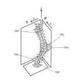
2−最大変形面:図1を参照すると、最大変形面102が示されている。面102の軸角(図示なし)は、コブ角が最大である方向、例えばグローバルz軸を中心とする軸角である。脊椎106の各弯曲の最大変形面102は、例えば、胸部隣接部のカーブについては三角形1041、胸部の主要な弯曲については三角形1042、腰椎部の弯曲については三角形1043によって例示的に表されている。2-Maximum deformation surface: Referring to FIG. 1, the
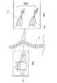
3−椎体及び椎間板の3次元楔状度:図2a及び2bは、椎体及び椎間板の3次元楔状度θ3Dを示している。最大変形面102(3D面)の頂椎202の楔状度と、2041、2042におけるような2つの頂椎間板の最大3D楔状度の平均が示されている。最大3D楔状度は、上記平面で測定した楔状度を表しており、その楔状度の値は、縦軸の周りで最大である。頂部が椎間板である場合(図2b参照)、両方の頂椎の3D楔状度θ13D、θ23Dの平均を算出し、2つの頂椎間板の平均の代わりに、頂椎間板の3D楔状度のみを報告した。3Dの椎間板楔状度は、胸椎及び腰椎のすべてのレベル(T1〜T2からL4〜L5まで)について分析した。3—3D wedge degree of vertebral body and disc: FIGS. 2 a and 2 b show the3D wedge degree θ3D of the vertebral body and disc. Shown is the wedge degree of the
4−頂部、上部、及び下部の移行部レベル及び胸腰レベルの椎間軸回転度:これは、図3aに示されている。特には、下側椎骨3022のローカル軸系を基準として、上部、頂部、及び下部のカーブレベル、並びに胸腰移行部(T12〜L1)における2つの隣接する椎骨3021、3022間の回転度が示されている。上側椎骨3021の下側椎骨3022に対する回転度θAXIALは、上側椎骨3021のローカルx軸を下側椎骨3022のローカル軸系のx−y面に投影した後に算出した。SRSの3D用語群の椎間回転度に関する定義は、2つの隣接する椎骨のローカル軸間の投影角である。4—vertical, upper and lower transitional and thoracolumbar intervertebral axis rotations: This is shown in FIG. 3a. In particular, based on the local axis system of the lower vertebra 3022, upper, top, and bottom of the curve level, and rotation between the vertebrae3021,3022 two adjacent in the thoracolumbar transition (T12~L1) Degrees are shown. Rotation oftheta AXIAL relative to the lower vertebra 302and second upper vertebra 3021 was calculated after projecting the local x-axis of the upper vertebra 3021 to the x-y plane of the local axis system of the inferior vertebra 3022. The definition of
5−細長度:図3bは、細長度(T6、T12、及びL4というローカル、並びにT1〜L5というリージョナルな細長度)、すなわち、T6、T12、及びL4の椎骨の椎体の高さh(椎骨の中心における、上側終板と下側終板との距離)と幅w(高さ線に対して垂直な中外側方向線を用いて、椎骨の中心で測定)との比率を示している。比率は、T1〜L5の脊椎の長さと、T6、T12、及びL4の椎体の幅の平均との間で得てもよい。幅の代わりに奥行(高さ線に対して垂直な、椎骨の前後方向中心線)を用いて、同じ計算を行った。T1〜L5の長さは、T1の上部終板の中心から始まり、すべての椎骨の重心を通り、L5の下部終板の中心に至る線の長さである。この線は、3次スプライン関数を用いて平滑化した。T6とL4を選択し、胸腰部のランドマークとしてT12を加えた。しかしながら、細長度の算出は、T6、T12、及びL4の椎骨に限らず、いずれの胸椎又は腰椎にも適用してよいことを理解されたい。
6−捻転:図4は、力学的捻転、すなわち、脊椎106の主要な側弯カーブ402の2つの半弯曲部(上端椎骨と頂椎との間の半弯曲部、及び下端椎骨と頂椎との間の半弯曲部。図示なし)に含まれるすべての椎骨の椎間軸回転度(下側椎骨のローカルリファレンスに従って測定)の和の平均を示している。この目的のために、第1の半弯曲(図示なし)内の全椎骨の椎間軸回転度の第1の和ΣθAXIAL1を計算する。第2の半弯曲(図示なし)内の全椎骨の椎間軸回転度の第2の和ΣθAXIAL2を更に計算する。続いて、第1の和ΣθAXIAL1と第2の和ΣθAXIAL2の平均を計算して、捻転値を得る。上述のように、幾何学的捻転も適用してよい。5-slenderness: FIG. 3b shows the slenderness (local lengths T6, T12 and L4 and regional lengths T1 to L5), ie the vertebral body height h (T6, T12 and L4) Shows the ratio of the distance between the upper and lower endplates at the center of the vertebra and the width w (measured at the center of the vertebra using a medial-lateral direction line perpendicular to the height line) . A ratio may be obtained between the length of the spine from T1-L5 and the average of the widths of the vertebral bodies at T6, T12, and L4. The same calculation was performed using depth (vertical vertebral centerline perpendicular to the height line) instead of width. The length of T1-L5 is the length of the line starting from the center of the upper end plate of T1, passing through the center of gravity of all vertebrae, and reaching the center of the lower end plate of L5. This line was smoothed using a cubic spline function. T6 and L4 were selected and T12 was added as a thoracolumbar landmark. However, it should be understood that the slenderness calculation may be applied to any thoracic or lumbar vertebra, not limited to T6, T12, and L4 vertebrae.
6—Torsion: FIG. 4 shows mechanical torsion, ie, two half-curves of the
ある具体的な実施形態では、予測方法の出力は、骨格成熟に至っていた(最低でもリッサー4)最先の受診時、又は融合術の直前に、後前方向(PA)X線撮影で測定される主要なコブ角として定義した。 In one specific embodiment, the output of the prediction method is measured by posterior anterior (PA) radiography at the earliest visit that has reached skeletal maturity (at least Lesser 4) or just before the fusion procedure. Was defined as the main hump angle.
図5は、予測モデル500を生成する例示的な方法のフローチャートである。第1の工程502は、頻度ヒストグラムからの出力データ、及び正規分布の主観的分析からの出力データの正常性を評価するためのものであった。 FIG. 5 is a flowchart of an exemplary method for generating a
変数が多いので、第2の工程504は、単変量分析を行って、多変量分析に含めるべき予測変数のうち、特に関連性の高い予測変数を選択するためのものであった。初めに、0.1以下のp値と関連付けられるパラメーターを識別するために、骨格成熟時の最終的なコブ角と、脊椎のローカルパラメーター、リージョナルパラメーター、及びグローバルパラメーターとの間の相関付けを行うことができる。 Since there are many variables, the
第3の工程506は、カーブタイプのカテゴリー数を減らすために行った。カーブタイプをマージし、その結果、同様な、骨格成熟時の最終的なコブ角を得られるように、一元配置分散分析(ANOVA)を行って、骨格成熟時の最終的なコブ角に関して、0.05の有意レベルで、6種類のカーブタイプを比較することができる。この工程の目的は、このモデルにおけるカーブ入力のタイプの様々なカテゴリーの数を減らすことであった。 The
最後の工程508は、一般線形モデル(GLM)に基づき、予測モデルを作成することにあった。逆方向の選択手順アプローチを行って、予測変数を選択した。まず、フルモデルに含まれる各予測因子(カーブタイプ及び骨格成熟ステージを固定因子として含め、残りのすべての脊椎パラメーターを共変量として含めた)について、P値を得た。交互作用をカテゴリー変数間に加えて、ある1つの変数の単純主効果の、第2の効果のレベルに対する変化が有意であるか検定した。 The
続いて、p値が大きい予測因子を除外し、モデルを最適合した。残りのすべての予測変数が、0.05に設定された停止基準よりも小さいp値と関連付けられるまで、これを行った。GLMでは、骨格成熟時の最終的なコブ角と、選択した予測変数との間の関連性を評価し、ベータ係数(β係数)及び95%信頼区間(CI)として表した。 Subsequently, predictors with large p-values were excluded and the model was optimally matched. This was done until all remaining predictors were associated with a p-value that was less than the stop criterion set at 0.05. GLM evaluated the association between the final hump angle at skeletal maturity and selected predictors and expressed as beta coefficient (β coefficient) and 95% confidence interval (CI).
すべての統計分析は、SPSS20.0というソフトウェアパッケージ(米国イリノイ州シカゴのSPSS,inc.)で行った。 All statistical analyzes were performed with the SPSS20.0 software package (SPSS, Inc., Chicago, Illinois, USA).
例示的な一実施形態では、133人のAISの前向きコホートを、骨格未熟期から成熟期まで(平均37カ月)追跡した。合計172人のAIS患者をコホートに入れた。分析時点で、133人の患者を含めることができた(77.3%)。全体的には、17人は追跡不能、13人は骨格が未熟なままで、9人の患者は、キャリブレーションエラーのために、3D再構成が不可能であった。コホートの記述的特徴は、n(サンプル数)、TR(Y軟骨)、RT(胸部の右カーブ)、RT−LL(胸部の右カーブ−腰部の左カーブ)、LL−RT(腰部の左カーブ−胸部の右カーブ)、LTL(胸腰部の左カーブ)、その他(胸部の左カーブ、腰部の右カーブ)という頭字語を用いて、表1に示されている。 In one exemplary embodiment, a prospective cohort of 133 AIS was followed from skeletal immaturity to maturity (average 37 months). A total of 172 AIS patients were enrolled in the cohort. At the time of analysis, 133 patients could be included (77.3%). Overall, 17 were untraceable, 13 remained immature in the skeleton, and 9 patients were unable to 3D reconstruction due to calibration errors. Descriptive features of the cohort are: n (number of samples), TR (Y cartilage), RT (chest right curve), RT-LL (chest right curve-waist left curve), LL-RT (lumbar left curve) -Chest right curve), LTL (chest waist left curve), and others (chest left curve, waist right curve) acronyms are shown in Table 1.

初診時の再構成した3D脊椎X線画像に対して、コンピューターによる測定を行った。測定値又はパラメーターのカテゴリーは6個あり、各カテゴリーは、最大弯曲面の角度、コブ角(後弯、前弯)、3D楔状度(頂椎、頂椎間板)、回転度(上下の移行椎、頂椎、胸腰移行部)、頂椎周囲の椎間回転度の平均(幾何学的及び/又は力学的捻転)、並びに細長度(高さ/幅の比率)という複数の測定値又はパラメーターを含む。転帰としての最終的なコブ角(手術直前又は骨格成熟時のいずれかのコブ角)、及び予測変数としての3D脊椎パラメーターを用いて、逆方向手順による一般線形モデル分析を行った。骨格成熟ステージと弯曲のタイプもモデルに含めた。 Computer measurement was performed on the reconstructed 3D spine X-ray image at the first visit. There are six categories of measured values or parameters. Each category includes the angle of the maximum heel surface, the hump angle (rear heel, lordosis), 3D wedge degree (vertical vertebrae, apex intervertebral disc), rotation degree (upper and lower transitional vertebrae, Multiple measurements or parameters such as the apical vertebrae, thoracolumbar transition), the average degree of intervertebral rotation around the vertebrae (geometric and / or mechanical torsion), and slenderness (height / width ratio) Including. General linear model analysis with a reverse procedure was performed using the final Cobb angle as the outcome (cobb angle either immediately before surgery or at skeletal maturity) and the 3D spinal parameters as predictors. The skeletal maturity stage and type of folding were also included in the model.
ある具体的な実施形態では、決定係数0.715で予測モデルを得た。含めた予測変数は、3段階の骨格成熟システムと弯曲のタイプであった。最大弯曲面の角度とともに、初期の前方向コブ角も含めた。最終的なコブ角の他の4つの予測因子は、T3〜T4、T8〜T9、及びT11〜T12の椎間板の3D楔状度、並びにすべての胸椎及び腰椎の椎間板の3D楔状度の和であった。上述のように、別の実施形態では、T3〜T4、T8〜T9、及びT11〜T12以外の移行部及び頂椎周囲の椎間板レベルの3D楔状度を適用してよいことを理解されたい。 In a specific embodiment, the prediction model was obtained with a determination factor of 0.715. The predictors included were a three-stage skeletal maturation system and a type of folding. In addition to the angle of the maximum saddle surface, the initial forward bump angle was included. The other four predictors of the final Cobb angle were the 3D wedge degree of T3-T4, T8-T9, and T11-T12, and the sum of 3D wedge degree of all thoracic and lumbar discs . As mentioned above, it should be understood that in other embodiments, transition levels other than T3-T4, T8-T9, and T11-T12 and the
最終的なコブ角分布は、図6に示されているヒストグラムによって示されているように、正規分布に従っていた。 The final Cobb angle distribution followed a normal distribution as shown by the histogram shown in FIG.
合計41個の脊椎パラメーターについて、最終的なコブ角とのピアソン相関関係を行った。0.1未満のp値と関連付けられた相関関係が得られたパラメーターは30個であった。相関関係分析の結果は表2に示されている。 A total of 41 spinal parameters were subjected to Pearson correlation with the final Cobb angle. There were 30 parameters that yielded correlations associated with p values less than 0.1. The results of the correlation analysis are shown in Table 2.
弯曲のタイプに関しては、ANOVA分析は、6つのカテゴリーを、(1)胸部の右カーブと、主要カーブとしての腰部の左カーブとのダブル、及びその他のタイプ(胸部の左カーブ、腰部の右カーブ)、(2)トリプル、(3)胸腰部の左カーブ、並びに(4)主要カーブとしての胸部の右カーブとのダブルという4つのタイプに減少させた。 Regarding the type of curvature, the ANOVA analysis consists of 6 categories: (1) Double of the right curve of the chest and the left curve of the waist as the main curve, and other types (the left curve of the chest, the right curve of the waist) ), (2) Triple, (3) Chest / Lumbar left curve, and (4) Double with Chest right curve as the main curve.
GLM分析に関しては、骨格成熟度、カーブタイプ、2Dの初期コブ角、最大変形面の角度、T3〜T4、T8〜T9、T11〜T12の椎間板楔状度、腰椎及び胸椎の楔状度の和が、最終的なコブ角の予測変数であることが分かった。表3は、最終的なコブ角の予測変数を決定するためのGLM(R2=0.715、F=22.956、p<0,000)を示している。For GLM analysis, the sum of skeletal maturity, curve type, 2D initial cobb angle, maximum deformed surface angle, T3-T4, T8-T9, T11-T12 intervertebral disc wedge, lumbar and thoracic spine wedges, It turned out to be a predictor of the final bump angle. Table 3 shows the GLM (R2 = 0.715, F = 22.956, p <0000) for determining the final Cobb angle predictor.
β係数がマイナス(−0.134)である椎間板楔状度の和を除き、すべての連続予測変数が、コブ角の最終的な値を上昇させた。初期コブ角の係数は0.714である。患者の骨格成熟ステージが0である場合、骨格成熟ステージが1である同様の患者と比較するときには、最終的なコブ角予測値に8.7°を足す。弯曲のタイプに関しては、カーブがタイプ4である同様の患者と比較するときには、4.6°(タイプ1)若しくは3.2°(タイプ3)を最終的なコブ角から減じ、タイプ2では4.0°を足す。これは、交互作用の寄与によって調節する。成熟ステージが0のタイプ1では、最終的なコブ角予測値から2.9°を減じることになり、成熟ステージが0のタイプ2では、最終的なコブ角予測値に9.0°を足すことになり、成熟ステージが0のタイプ3では最終的なコブ角予測値から14.6°を減じることになる。この予測モデルのR2は0.715であり、これは、分散の71.5%を説明することを意味する。 All continuous predictors increased the final value of the hump angle, except for the sum of the intervertebral disc wedge degrees where the β coefficient was negative (−0.134). The coefficient of the initial bump angle is 0.714. If the patient's skeletal maturity stage is 0, 8.7 ° is added to the final Cobb angle prediction when compared to similar patients with a skeletal maturity stage of 1. For the type of curvature, subtract 4.6 ° (type 1) or 3.2 ° (type 3) from the final hoop angle when compared to similar patients with curves of type 4 and 4 for
カテゴリー予測変数のp値の一部は、GLMにおけるそれらの変数の主効果を評価すると、0.05超である。しかしながら、これらのカテゴリー予測変数は、モデル内に保持した。これらの寄与度は、互いの交互作用について検討したところ、有意であったからである。 Some of the p-values for categorical predictors are greater than 0.05 when the main effects of those variables in GLM are evaluated. However, these categorical predictors were retained in the model. This is because these contributions were significant when the mutual interaction was examined.
未熟なAIS患者において、骨格成熟度が中程度となり、コブ角が11°〜40°である中程度なカーブとなるまで、進行の予測を促すことになる進行予測変数を識別した。この予測モデルは、初診から得られた情報のみを用いて、骨格成熟時の最終的なコブ角の分散の71.5%を説明できる。 In immature AIS patients, a progression predictor variable was identified that would facilitate the prediction of progression until the skeletal maturity was moderate and the curve was a moderate curve with a Cobb angle of 11 ° to 40 °. This prediction model can explain 71.5% of the final hump angle variance at the time of skeletal maturation using only the information obtained from the first visit.
このモデルに含まれる基本的な予測変数は、初診時のコブ角、弯曲のタイプ、及び骨格成熟度である。 The basic predictors included in this model are the hump angle at the first visit, the type of curvature, and skeletal maturity.
このモデルに含まれる1つの3Dパラメーターは、最大変形面の角度である。このパラメーターは、カーブの回転度と関連付けられ、従来のコブ角よりも進行性AISを敏感に検知できる。 One 3D parameter included in this model is the angle of the maximum deformation surface. This parameter is related to the degree of rotation of the curve and can detect progressive AIS more sensitively than the conventional hoop angle.
このモデルに含まれる他の4つの予測変数は、椎間板楔状度(移行部及び頂椎周囲の椎間板レベル、例えばT3〜T4、T8〜T9、T11〜T12の椎間板楔状度、並びにすべての和)である。識別したT3〜T4及びT11〜T12のレベルは通常、弯曲のタイプに応じて、移行部レベル及びT8〜T9の移行部又は頂椎レベルを表す(胸椎カーブにおいては、頂椎レベルを表すことになり、胸腰カーブにおいては、移行部レベルを表すことになる)。T3〜T4の椎間板の楔状度は、最終的なコブ角予測に対して最も大きな効果を有する。 The other four predictors included in this model are the intervertebral disc wedge (intervertebral and perivertebral disc levels, eg, T3-T4, T8-T9, T11-T12 intervertebral disc wedge, and all sums). is there. The identified T3-T4 and T11-T12 levels usually represent the transitional level and the transitional or apical level of T8-T9, depending on the type of curvature (in the thoracic curve, it represents the vertebral level). In the thoraco-lumbar curve, it represents the transition level). The wedge degree of the T3-T4 disc has the greatest effect on the final bump angle prediction.
選択した統計モデルは、予測変数を選択するための逆方向手順によるGLMであった。ステップワイズ変数選択は、医療用途で広く用いられており、最も適合するモデルを見出すのに優れた選択なので、これを選択した。線形モデリングには、100を超えるサンプル数が必要となることが認められている。線形モデリングのサンプル数を求める別の方法は、モデルに含まれる自由度を少なくとも10倍にすることである。このモデルの自由度は、13自由度であるので(連続予測変数が6個、成熟ステージに関するものが1つ、カーブタイプに関するものが3つ、成熟ステージと弯曲のタイプとの組み合わせに関するものが3つ)、133個のサンプル数が適切である。 The selected statistical model was a GLM with a reverse procedure for selecting predictors. Stepwise variable selection was chosen because it is widely used in medical applications and is an excellent choice for finding the best fit model. It has been observed that linear modeling requires more than 100 samples. Another way to determine the number of samples for linear modeling is to increase the degree of freedom included in the model by at least 10 times. Since this model has 13 degrees of freedom (6 continuous predictors, 1 for the mature stage, 3 for the curve type, 3 for the combination of the mature stage and the type of fold) 1) A sample number of 133 is appropriate.
以下では、図7を参照して、AISの進行曲線を予測する際の支援を医療提供者に提供する通信システム700について説明していく。システム700は、702にあるように、ネットワーク706を介して予測モデルシステム704と通信するように構成された複数の装置を含む。装置702は、インターネット、公衆交換電話網(PSTN)、携帯電話網、又は当業者に知られている他のネットワークのようなネットワーク706を介して通信するように構成されているいずれかの装置(パーソナルコンピューター、携帯情報端末、スマートフォンなど)を含む。予測モデルシステム704は、装置702と分離しているとともに、装置702から離れているものとして図示されているが、予測モデルシステム704は、ダウンロードソフトウェアアプリケーション、ファームウェアアプリケーション、又はこれらの組み合わせのいずれかとして、装置702と一体化してもよいことを理解されたい。 In the following, referring to FIG. 7, a
図示されているように、1つ以上のデータベース708を予測モデルシステム704に直接一体化しても、予測モデルシステム704とは別に、及び/又は予測モデルシステム704から離して搭載してもよい。データベース708へのリモートアクセスの場合、上述のように、いずれかのタイプのネットワーク706を介してアクセスを行ってよい。データベース708は、コンピューターによる迅速なサーチ及び検索のために、整理したデータ又は情報収集物として搭載してよい。データベース708は、各種のデータ処理動作と連動して、データの記憶、検索、修正、及び削除を容易にするように構築してよい。データベース708は、レコード(各レコードは1つ以上のフィールドからなる)に分解できるファイル又はファイルセットからなってよい。データベースの情報は、フィールドを迅速にサーチし、再編成し、グループ化し、選択するために、キーワードとソートコマンドを用いるクエリ−によって取り出してよい。データベース708は、1つ以上のサーバーのようなデータ記憶メディア上のいずれかのデータ編成であってよい。 As shown, one or more databases 708 may be integrated directly into the
一実施形態では、データベース708は、トランスポートレイヤーセキュリティ(TLS)を支援できるセキュアウェブサーバー及びハイパーテキストトランスファープロトコルセキュア(HTTPS)(データへのアクセスのために用いられるプロトコル)である。セキュアウェブサーバーへの通信及びセキュアウェブサーバーからの通信は、セキュアソケットレイヤー(SSL)を用いて確保してよい。ユーザーの本人確認は、いずれのユーザーにおいても、ユーザーネーム及びパスワードを用いて行ってよい。各種レベルのアクセス権を複数のレベルのユーザーに提供してよい。 In one embodiment, the database 708 is a secure web server that can support Transport Layer Security (TLS) and Hypertext Transfer Protocol Secure (HTTPS) (a protocol used to access data). Communication to and from the secure web server may be secured using a secure socket layer (SSL). The identity verification of the user may be performed by using any user name and password for any user. Various levels of access rights may be provided to multiple levels of users.
あるいは、コンピューターネットワーク内の装置が情報を交換できるようにするいずれかの既知の通信プロトコルを用いてもよい。プロトコルの例は、IP(インターネットプロトコル)、UDP(ユーザーデータグラムプロトコル)、TCP(トランスミッションコントロールプロトコル)、DHCP(ダイナミックホストコンフィギュレーションプロトコル)、HTTP(ハイパーテキストトランスファープロトコル)、FTP(ファイルトランスファープロトコル)、Telnet(テルネットリモートプロトコル)、SSH(セキュアシェルリモートプロトコル)である。 Alternatively, any known communication protocol that allows devices in a computer network to exchange information may be used. Examples of protocols are IP (Internet Protocol), UDP (User Datagram Protocol), TCP (Transmission Control Protocol), DHCP (Dynamic Host Configuration Protocol), HTTP (Hypertext Transfer Protocol), FTP (File Transfer Protocol), Telnet (telnet remote protocol), SSH (secure shell remote protocol).
図8を参照すると、予測モデルシステム704は、例示的には、ユーザーが予測モデルシステム704とやりとりできるようにするユーザーインターフェース802を含む。特には、下記に更に詳細に論じられているように、ユーザー(例えば医師)は、ユーザーインターフェース802を用いて、情報を予測モデルシステム704に提示できる。上述のように、その情報は、初診の際に得ることができ、コブ角、弯曲のタイプ、骨格成熟度、3Dの形態学的パラメーターのような基本的な予測変数を含む。ユーザーインターフェース802を用いて、予測モデルシステム704のローカル又は遠隔にあるメモリー806から情報にアクセスできる。 Referring to FIG. 8, the
予測モデルシステム704は、プロセッサー804を更に含み、プロセッサー804は、データに関わる動作を実行できるいずれかの装置であってよい。例は、中央演算処理装置(CPU)、フロントエンドプロセッサー、マイクロプロセッサー、グラフィックスプロセッシングユニット(GPU/VPU)、フィジックスプロセッシングユニット(PPU)、デジタルシグナルプロセッサー、及びネットワークプロセッサーである。ユーザーインターフェース802を介して入力された情報に基づき、最終的なコブ角予測値を出力するために、複数のアプリケーション808a…808nは、例示的には、プロセッサー804で必要とされる動作を実行するプロセッサー804上で実行される。本明細書に示されているアプリケーション808a…808nは、別個のものとして図示及び説明されているが、様々な形で組み合わせても、分離させてもよいことを理解されたい。 The
プロセッサー804は、データを受け取って記憶できるメモリー806と通信している。メモリー806は、高速ランダムアクセスメモリー(RAM)のようなメインメモリーであっても、ハードディスク又はフラッシュメモリーのような補助記憶ユニットであってよい。メモリー806は、リードオンリーメモリー(ROM)、消去可能なプログラマブルリードオンリーメモリー(EPROM)、又は光学式記憶メディア(ビデオディスク及びコンパクトディスクなど)のようないずれかの他のタイプのメモリーであってもよい。 The
図9は、最終的なコブ角予測値を生成するアプリケーション808aを例示的に示している。X線撮像システム又はその他の撮像システムから得られる画像のような2次元脊椎画像を脊椎再構成モジュール902に供給する。すなわち、3次元の脊椎形態を供給し、3Dパラメーター抽出モジュール904が、3Dデータを受け取って、そのデータから、初期コブ角、最大変形面、椎体及び椎間板の3次元楔状度、頂部、上部、及び下部の移行部レベル及び胸腰レベルの椎間軸回転度、細長度、並びに捻転のようなパラメーターを抽出するように構成されている。これらのパラメーターをモデリングユニット906に供給し、骨格成熟度及びカーブタイプのパラメーターと組み合わせて、AISの進行曲線をモデリングして、最終的なコブ角予測値を出力する。予測モデルシステム704の出力は、進行リスクが追加の治療を行うに値するかを治療担当の医師が判断する助けとなる。 FIG. 9 exemplarily shows an application 808a that generates a final bump angle prediction value. A two-dimensional spine image, such as an image obtained from an x-ray imaging system or other imaging system, is provided to
いくつかの実施形態では、予測モデルシステム704は更に、初期コブ角と最終的なコブ角を用いて進行曲線を描くように構成されている。この曲線は、ユーザーインターフェース802又は別の出力装置(プリンターなど)を介して、ユーザーに対して出力してよい。いくつかの実施形態では、予測モデルシステム704は、最終的なコブ角、並びに/又は初期及び最終的なコブ角を用いて生成される進行曲線の関数として、一連の推奨される治療選択肢から選択するようにも構成されている。その選択が、最適な推奨される治療で構成されるように、治療選択肢は、最終的なコブ角の範囲、及び/又は進行曲線の変化率の関数として分類してもよい。続いて、レンダリングするための装置702に、ユーザーインターフェース802又はその他の出力装置を介して、選択した治療(1つ又は複数)を出力してよい。最終的なコブ角予測値を生成したら、治療選択肢によって、治療担当の医師を支援するためのその他の実施形態は、当業者には容易に分かるであろう。 In some embodiments, the
本明細書のブロック図では、別個のデータシグナル接続を介して、相互に通信する別個のコンポーネント群として示されているが、一部のコンポーネントが、ハードウェア又はソフトウェアシステムの所定の機能又は動作によって実施され、図示されているデータパスの多くが、コンピューターアプリケーション又はオペレーティングシステム内のデータ通信によって実施される状態で、ハードウェアコンポーネントとソフトウェアコンポーネントとの組み合わせによって、本発明の実施形態がもたらされることは、当業者には分かるであろう。したがって、図示されている構造は、本実施形態の教示の効率性のために提供されている。本発明は方法として実施でき、システム内、又はコンピューターリーダブルメディア上で具体化できることに留意されたい。本発明の上記の実施形態は、例示のために過ぎない。したがって、本発明の範囲は、添付の特許請求の範囲によってのみ限定するように意図されている。 Although the block diagrams herein are shown as separate components that communicate with each other via separate data signal connections, some components may depend on a predetermined function or operation of the hardware or software system. It is understood that the combination of hardware and software components provides an embodiment of the present invention with many of the implemented and illustrated data paths being implemented by data communication within a computer application or operating system. Those skilled in the art will understand. Accordingly, the illustrated structure is provided for the efficiency of the teaching of the present embodiment. It should be noted that the present invention can be implemented as a method and embodied in a system or on computer readable media. The above-described embodiments of the present invention are for illustration only. Accordingly, the scope of the invention is intended to be limited only by the scope of the appended claims.
Claims (26)
Translated fromJapanese3Dの形態学的脊椎パラメーター、カーブタイプ、及び骨格成熟度に基づく予測モデルが記憶されているメモリーと、
プロセッサーと、
前記メモリーに記憶されている少なくとも1つのアプリケーションで、患者固有の3Dの形態学的脊椎パラメーター、選択したカーブタイプ、及び選択した骨格成熟度を受け取り、前記予測モデルを取り出し、前記特発性側弯症の進行曲線をモデリングして、前記最終的なコブ角予測値を生成するように、前記プロセッサーによって実行可能である少なくとも1つのアプリケーションと、
を備えるシステム。A system for generating a final bump angle prediction value for idiopathic scoliosis,
A memory storing a prediction model based on 3D morphological spine parameters, curve types, and skeletal maturity;
A processor;
At least one application stored in the memory receives patient-specific 3D morphological spine parameters, selected curve type, and selected skeletal maturity, retrieves the predictive model, and extracts the idiopathic scoliosis At least one application executable by the processor to model a progress curve to generate the final Cobb angle prediction;
A system comprising:
患者固有の3Dの形態学的脊椎パラメーター、選択したカーブタイプ、及び選択した骨格成熟度を受け取ることと、
3Dの形態学的脊椎パラメーター、カーブタイプ、及び骨格成熟度に基づく予測モデルに対して、前記患者固有の3Dの形態学的脊椎パラメーター、選択したカーブタイプ、及び選択した骨格成熟度を適用することと、
前記特発性側弯症の進行曲線をモデリングすることによって、前記最終的なコブ角予測値を生成することと、
を含む方法。A computer-implemented method for generating a final Cobb angle prediction for idiopathic scoliosis, comprising:
Receiving patient-specific 3D morphological spine parameters, selected curve type, and selected skeletal maturity;
Applying the patient-specific 3D morphological spine parameters, selected curve type, and selected skeletal maturity to a predictive model based on 3D morphological spine parameters, curve type, and skeletal maturity When,
Generating the final Cobb angle prediction by modeling the progression curve of the idiopathic scoliosis;
Including methods.
患者固有の3Dの形態学的脊椎パラメーター、選択したカーブタイプ、及び選択した骨格成熟度を受け取ることと、
3Dの形態学的脊椎パラメーター、カーブタイプ、及び骨格成熟度に基づく予測モデルに対して、前記患者固有の3Dの形態学的脊椎パラメーター、選択したカーブタイプ、及び選択した骨格成熟度を適用することと、
前記特発性側弯症の進行曲線をモデリングすることによって、前記最終的なコブ角予測値を生成することと、
を行うように、前記プログラムコードが実行可能であるコンピューターリーダブルメディア。A computer readable medium that stores program code that is executable by a processor and that generates a final value of predicted Cobb angle for idiopathic scoliosis,
Receiving patient-specific 3D morphological spine parameters, selected curve type, and selected skeletal maturity;
Applying the patient-specific 3D morphological spine parameters, selected curve type, and selected skeletal maturity to a predictive model based on 3D morphological spine parameters, curve type, and skeletal maturity When,
Generating the final Cobb angle prediction by modeling the progression curve of the idiopathic scoliosis;
A computer readable medium in which the program code is executable.
Applications Claiming Priority (3)
| Application Number | Priority Date | Filing Date | Title |
|---|---|---|---|
| US201261713226P | 2012-10-12 | 2012-10-12 | |
| US61/713,226 | 2012-10-12 | ||
| PCT/CA2013/000884WO2014056098A1 (en) | 2012-10-12 | 2013-10-15 | System and method for predicting scoliosis progression |
Publications (1)
| Publication Number | Publication Date |
|---|---|
| JP2015532144Atrue JP2015532144A (en) | 2015-11-09 |
Family
ID=50476826
Family Applications (1)
| Application Number | Title | Priority Date | Filing Date |
|---|---|---|---|
| JP2015535942APendingJP2015532144A (en) | 2012-10-12 | 2013-10-15 | System and method for predicting scoliosis progression |
Country Status (5)
| Country | Link |
|---|---|
| US (1) | US9547897B2 (en) |
| EP (1) | EP2906109B1 (en) |
| JP (1) | JP2015532144A (en) |
| CA (1) | CA2927955C (en) |
| WO (1) | WO2014056098A1 (en) |
Cited By (4)
| Publication number | Priority date | Publication date | Assignee | Title |
|---|---|---|---|---|
| JP2017158842A (en)* | 2016-03-10 | 2017-09-14 | 透 本田 | Centrum deformation diagnostic system, information processing method, and program |
| KR20210157684A (en)* | 2020-06-22 | 2021-12-29 | 한국전자통신연구원 | Apparatus and method for analyzing medical image |
| KR20220134375A (en)* | 2021-03-26 | 2022-10-05 | 전주대학교 산학협력단 | A machine learning based rotation measurement system for scoliosis on x-ray image |
| JP2023013453A (en)* | 2021-07-16 | 2023-01-26 | 国立研究開発法人国立がん研究センター | Image processing device, method, program and storage medium |
Families Citing this family (25)
| Publication number | Priority date | Publication date | Assignee | Title |
|---|---|---|---|---|
| FR3010628B1 (en) | 2013-09-18 | 2015-10-16 | Medicrea International | METHOD FOR REALIZING THE IDEAL CURVATURE OF A ROD OF A VERTEBRAL OSTEOSYNTHESIS EQUIPMENT FOR STRENGTHENING THE VERTEBRAL COLUMN OF A PATIENT |
| FR3012030B1 (en) | 2013-10-18 | 2015-12-25 | Medicrea International | METHOD FOR REALIZING THE IDEAL CURVATURE OF A ROD OF A VERTEBRAL OSTEOSYNTHESIS EQUIPMENT FOR STRENGTHENING THE VERTEBRAL COLUMN OF A PATIENT |
| US10709509B2 (en) | 2014-06-17 | 2020-07-14 | Nuvasive, Inc. | Systems and methods for planning, performing, and assessing spinal correction during surgery |
| CN105719273A (en)* | 2014-12-05 | 2016-06-29 | Ge医疗系统环球技术有限公司 | Method and device for measuring vertebra rotation parameters on medical imaging |
| US9265463B1 (en)* | 2014-12-22 | 2016-02-23 | Medical Metrics, Inc. | Methods for determining spine instability and for eliminating the impact of patient effort on stability determinations |
| EP3376987B1 (en)* | 2015-11-19 | 2020-10-28 | EOS Imaging | Method of preoperative planning to correct spine misalignment of a patient |
| US11331039B2 (en) | 2016-02-15 | 2022-05-17 | Keio University | Spinal-column arrangement estimation-apparatus, spinal-column arrangement estimation method, and spinal-column arrangement estimation program |
| WO2018109556A1 (en) | 2016-12-12 | 2018-06-21 | Medicrea International | Systems and methods for patient-specific spinal implants |
| EP3612122B1 (en) | 2017-04-21 | 2023-12-20 | Medicrea International | A system for developing one or more patient-specific spinal implants |
| CN107481228B (en)* | 2017-07-28 | 2021-02-09 | 电子科技大学 | Human back scoliosis angle measuring method based on computer vision |
| US10918422B2 (en) | 2017-12-01 | 2021-02-16 | Medicrea International | Method and apparatus for inhibiting proximal junctional failure |
| CN108670302B (en)* | 2018-06-06 | 2020-11-06 | 西北工业大学 | A 3D structure reconstruction method of spine based on 2.5D ultrasound wide-field imaging |
| EP3840683A4 (en) | 2018-07-16 | 2022-06-29 | Medtronic Sofamor Danek USA, Inc. | Spinal surgery outcome prediction |
| RU2701049C1 (en)* | 2018-11-27 | 2019-09-24 | Виктор Павлович Каюмов | Automated system for processing laryngosal angles data of patients |
| US11925417B2 (en) | 2019-04-02 | 2024-03-12 | Medicrea International | Systems, methods, and devices for developing patient-specific spinal implants, treatments, operations, and/or procedures |
| US11944385B2 (en) | 2019-04-02 | 2024-04-02 | Medicrea International | Systems and methods for medical image analysis |
| US11877801B2 (en) | 2019-04-02 | 2024-01-23 | Medicrea International | Systems, methods, and devices for developing patient-specific spinal implants, treatments, operations, and/or procedures |
| US11769251B2 (en) | 2019-12-26 | 2023-09-26 | Medicrea International | Systems and methods for medical image analysis |
| CN112274164B (en)* | 2020-07-29 | 2023-02-21 | 深圳市智影医疗科技有限公司 | Scoliosis prediction method, scoliosis prediction device, electronic device, and storage medium |
| CN112381757A (en)* | 2020-10-09 | 2021-02-19 | 温州医科大学附属第二医院、温州医科大学附属育英儿童医院 | System and method for measuring and calculating scoliosis Cobb angle through full-length X-ray film of spine based on artificial intelligence-image recognition |
| CN112381869B (en)* | 2020-10-09 | 2022-12-13 | 温州医科大学附属第二医院、温州医科大学附属育英儿童医院 | Scoliosis Measurement and Calculation Device for Scoliosis Measurement and Calculation by Full-length Spine X-ray Films |
| CN112802019B (en)* | 2021-04-01 | 2021-06-29 | 成都成电金盘健康数据技术有限公司 | Leke typing method based on spine AIS image |
| US12318144B2 (en) | 2021-06-23 | 2025-06-03 | Medicrea International SA | Systems and methods for planning a patient-specific spinal correction |
| CN113724268B (en)* | 2021-08-05 | 2024-03-29 | 西安交通大学 | Orthopedic region dividing method and system for scoliosis orthosis |
| CN113870098B (en)* | 2021-09-09 | 2024-09-24 | 武汉大学 | Automatic Cobb angle measurement method based on spine layered reconstruction |
Citations (2)
| Publication number | Priority date | Publication date | Assignee | Title |
|---|---|---|---|---|
| JP2009090022A (en)* | 2007-10-11 | 2009-04-30 | Shimadzu Corp | Medical image diagnostic apparatus, medical image diagnostic program, and X-ray imaging apparatus |
| JP2010259452A (en)* | 2007-08-21 | 2010-11-18 | Konica Minolta Medical & Graphic Inc | Diagnostic imaging support apparatus and diagnostic imaging support method |
Family Cites Families (6)
| Publication number | Priority date | Publication date | Assignee | Title |
|---|---|---|---|---|
| US7137958B2 (en)* | 2001-08-27 | 2006-11-21 | Nihon University | Human spinal column measurement and display system |
| US20090035772A1 (en)* | 2007-07-03 | 2009-02-05 | Axial Biotech, Inc. | Genetic Markers Associated With Scoliosis And Uses Thereof |
| US20090104620A1 (en)* | 2007-07-03 | 2009-04-23 | Axial Biotech, Inc. | Simplified Method of Determining Predisposition to Scoliosis |
| WO2006063324A1 (en)* | 2004-12-10 | 2006-06-15 | Virginia Tech Intellectual Properties, Inc. | Systems and methods for multi-dimensional characterization and classification of spinal shape |
| TWI282268B (en)* | 2005-09-15 | 2007-06-11 | Univ Chung Shan Medical | Medical image system and method for measuring vertebral axial rotation |
| CA2788445C (en)* | 2010-01-28 | 2017-11-21 | Pecsi Tudomanyegyetem | A method and a system for multi-dimensional visualization of the spinal column by vertebra vectors, sacrum vector, sacrum plateau vector and pelvis vectors |
- 2013
- 2013-10-15JPJP2015535942Apatent/JP2015532144A/enactivePending
- 2013-10-15USUS14/434,944patent/US9547897B2/enactiveActive
- 2013-10-15EPEP13846081.1Apatent/EP2906109B1/enactiveActive
- 2013-10-15CACA2927955Apatent/CA2927955C/enactiveActive
- 2013-10-15WOPCT/CA2013/000884patent/WO2014056098A1/enactiveApplication Filing
Patent Citations (2)
| Publication number | Priority date | Publication date | Assignee | Title |
|---|---|---|---|---|
| JP2010259452A (en)* | 2007-08-21 | 2010-11-18 | Konica Minolta Medical & Graphic Inc | Diagnostic imaging support apparatus and diagnostic imaging support method |
| JP2009090022A (en)* | 2007-10-11 | 2009-04-30 | Shimadzu Corp | Medical image diagnostic apparatus, medical image diagnostic program, and X-ray imaging apparatus |
Non-Patent Citations (8)
| Title |
|---|
| B. STEPHENS RICHARDS ET. AL.: "Standardization of Criteria for Adolescent Idiopathic Scoliosis Brace Studies", SPINE, vol. Vol. 30、No. 18, JPN6017000451, 2005, pages pp. 2068-2075* |
| JE LONSTEIN ET. AL.: "The prediction of curve progression in untreated idiopathic scoliosis during growth", THE JOURNAL OF BONE & JOINT SURGERY, vol. Vol. 66A、No. 7, JPN6017000447, September 1984 (1984-09-01), pages pp. 1061-1071* |
| P. N. SOUCACOS ET. AL.: "Assessment of curve progression in idiopathic scoliosis", EUR SPINE J, vol. Vol. 7, JPN6017000449, 1998, pages pp. 270-277* |
| 宮之原 啓 他: "「上尾市における28年間の側弯症学校検診の報告および進行関連因子の進行予測検討」", JOURNAL OF SPINE RESEARCH, vol. Vol. 2、No. 11, JPN6017000453, 25 November 2011 (2011-11-25), pages pp. 1725-1729* |
| 小橋 芳浩 他: "「突発性側弯症における側弯変形増強の予測−3次元解析による新しい基準設定−」", 整形外科と災害外科, vol. Vol. 43、No. 4, JPN6017000444, 1994, pages pp. 1279-1282* |
| 山口 卓夫 他: "「Risk factorの多変量解析による初診時特発性側彎症進行予測の試み」", 脊柱変形, vol. 第3巻、第1号, JPN6017000452, 27 March 1988 (1988-03-27), pages pp. 80-85* |
| 河野 真介 他: "「腰椎変形側弯症における弯曲進行予測因子のX線学的検討」", 日本脊椎髄病学会雑誌, vol. Vol. 14、No. 1, JPN6017000454, 20 February 2003 (2003-02-20), pages pp. 29* |
| 重野 利幸 他: "「第4腰椎変性辷り症の経年的変性形式 レントゲン正面像での考察」", 東海北陸理学療法学術大会誌, vol. Vol. 23, JPN6017000446, 14 September 2007 (2007-09-14), pages pp. 52* |
Cited By (7)
| Publication number | Priority date | Publication date | Assignee | Title |
|---|---|---|---|---|
| JP2017158842A (en)* | 2016-03-10 | 2017-09-14 | 透 本田 | Centrum deformation diagnostic system, information processing method, and program |
| KR20210157684A (en)* | 2020-06-22 | 2021-12-29 | 한국전자통신연구원 | Apparatus and method for analyzing medical image |
| KR102610915B1 (en) | 2020-06-22 | 2023-12-06 | 한국전자통신연구원 | Apparatus and method for analyzing medical image |
| KR20220134375A (en)* | 2021-03-26 | 2022-10-05 | 전주대학교 산학협력단 | A machine learning based rotation measurement system for scoliosis on x-ray image |
| KR102514246B1 (en) | 2021-03-26 | 2023-03-27 | 전주대학교 산학협력단 | A machine learning based rotation measurement system for scoliosis on x-ray image and the measurement method thereof |
| JP2023013453A (en)* | 2021-07-16 | 2023-01-26 | 国立研究開発法人国立がん研究センター | Image processing device, method, program and storage medium |
| JP7643962B2 (en) | 2021-07-16 | 2025-03-11 | 国立研究開発法人国立がん研究センター | Image processing device, method, program, and storage medium |
Also Published As
| Publication number | Publication date |
|---|---|
| EP2906109B1 (en) | 2017-10-11 |
| US20150287184A1 (en) | 2015-10-08 |
| CA2927955C (en) | 2018-08-28 |
| WO2014056098A1 (en) | 2014-04-17 |
| CA2927955A1 (en) | 2014-04-17 |
| US9547897B2 (en) | 2017-01-17 |
| EP2906109A4 (en) | 2016-05-25 |
| EP2906109A1 (en) | 2015-08-19 |
Similar Documents
| Publication | Publication Date | Title |
|---|---|---|
| JP2015532144A (en) | System and method for predicting scoliosis progression | |
| Diebo et al. | Adult spinal deformity | |
| Dubousset et al. | Use of EOS imaging for the assessment of scoliosis deformities: application to postoperative 3D quantitative analysis of the trunk | |
| Nault et al. | Three-dimensional spinal morphology can differentiate between progressive and nonprogressive patients with adolescent idiopathic scoliosis at the initial presentation: a prospective study | |
| Reinhold et al. | AO spine injury classification system: a revision proposal for the thoracic and lumbar spine | |
| JP2020518312A (en) | A system that provides intraoperative tracking to assist spinal surgery | |
| Somoskeöy et al. | Clinical validation of coronal and sagittal spinal curve measurements based on three-dimensional vertebra vector parameters | |
| Fazal et al. | Does the presence of the nerve root sedimentation sign on MRI correlate with the operative level in patients undergoing posterior lumbar decompression for lumbar stenosis? | |
| US20210315515A1 (en) | Assessment of spinal column integrity | |
| Sullivan et al. | Thoracic idiopathic scoliosis severity is highly correlated with 3D measures of thoracic kyphosis | |
| Shen et al. | Towards a new 3D classification for adolescent idiopathic scoliosis | |
| Harris et al. | A comprehensive review of thoracic deformity parameters in scoliosis | |
| Sullivan et al. | A novel method for estimating three-dimensional apical vertebral rotation using two-dimensional coronal Cobb angle and thoracic kyphosis | |
| Siasios et al. | Cervical sagittal balance parameters after single-level anterior cervical discectomy and fusion: correlations with clinical and functional outcomes | |
| Weng et al. | Automatic recognition of whole-spine sagittal alignment and curvature analysis through a deep learning technique | |
| Engelke et al. | Automated quantitative morphometry of vertebral heights on spinal radiographs: comparison of a clinical workflow tool with standard 6-point morphometry | |
| Yang et al. | Semi-automatic ultrasound curve angle measurement for adolescent idiopathic scoliosis | |
| Abdel et al. | Supine thoracolumbar sagittal spine alignment: comparing computerized tomography and plain radiographs | |
| Puvanesarajah et al. | The deformity angular ratio: can three-dimensional computed tomography improve prediction of intraoperative neuromonitoring events? | |
| Duong et al. | Three-dimensional subclassification of Lenke type 1 scoliotic curves | |
| Kluck et al. | Predictors of spontaneous lumbar curve correction in thoracic-only fusions: 3D analysis in AIS | |
| Menon | Classification systems in adolescent idiopathic scoliosis revisited: Is a three-dimensional classification needed? | |
| Pizones et al. | Relationship between the different torsion-related thoracic deformity parameters of adolescent idiopathic scoliosis | |
| Sowula et al. | Assessing progressive changes in axial plane vertebral deformity in adolescent idiopathic scoliosis using sequential magnetic resonance imaging | |
| Fitzgerald et al. | Three-dimensional radiographic analysis of two distinct Lenke 1A curve patterns |
Legal Events
| Date | Code | Title | Description |
|---|---|---|---|
| A711 | Notification of change in applicant | Free format text:JAPANESE INTERMEDIATE CODE: A711 Effective date:20160222 | |
| A711 | Notification of change in applicant | Free format text:JAPANESE INTERMEDIATE CODE: A711 Effective date:20160223 | |
| A521 | Request for written amendment filed | Free format text:JAPANESE INTERMEDIATE CODE: A821 Effective date:20160223 | |
| A621 | Written request for application examination | Free format text:JAPANESE INTERMEDIATE CODE: A621 Effective date:20160422 | |
| A977 | Report on retrieval | Free format text:JAPANESE INTERMEDIATE CODE: A971007 Effective date:20161227 | |
| A131 | Notification of reasons for refusal | Free format text:JAPANESE INTERMEDIATE CODE: A131 Effective date:20170117 | |
| A601 | Written request for extension of time | Free format text:JAPANESE INTERMEDIATE CODE: A601 Effective date:20170414 | |
| A601 | Written request for extension of time | Free format text:JAPANESE INTERMEDIATE CODE: A601 Effective date:20170414 | |
| A521 | Request for written amendment filed | Free format text:JAPANESE INTERMEDIATE CODE: A523 Effective date:20170606 | |
| A02 | Decision of refusal | Free format text:JAPANESE INTERMEDIATE CODE: A02 Effective date:20171121 |