JP2014534822A - Coronary artery calcium scoring based on a model - Google Patents
Coronary artery calcium scoring based on a modelDownload PDFInfo
- Publication number
- JP2014534822A JP2014534822AJP2014517064AJP2014517064AJP2014534822AJP 2014534822 AJP2014534822 AJP 2014534822AJP 2014517064 AJP2014517064 AJP 2014517064AJP 2014517064 AJP2014517064 AJP 2014517064AJP 2014534822 AJP2014534822 AJP 2014534822A
- Authority
- JP
- Japan
- Prior art keywords
- coronary
- image
- heart
- calcium
- coronary artery
- Prior art date
- Legal status (The legal status is an assumption and is not a legal conclusion. Google has not performed a legal analysis and makes no representation as to the accuracy of the status listed.)
- Ceased
Links
Images


Classifications
- A—HUMAN NECESSITIES
- A61—MEDICAL OR VETERINARY SCIENCE; HYGIENE
- A61B—DIAGNOSIS; SURGERY; IDENTIFICATION
- A61B6/00—Apparatus or devices for radiation diagnosis; Apparatus or devices for radiation diagnosis combined with radiation therapy equipment
- A61B6/02—Arrangements for diagnosis sequentially in different planes; Stereoscopic radiation diagnosis
- A61B6/03—Computed tomography [CT]
- A61B6/032—Transmission computed tomography [CT]
- A—HUMAN NECESSITIES
- A61—MEDICAL OR VETERINARY SCIENCE; HYGIENE
- A61B—DIAGNOSIS; SURGERY; IDENTIFICATION
- A61B6/00—Apparatus or devices for radiation diagnosis; Apparatus or devices for radiation diagnosis combined with radiation therapy equipment
- A61B6/46—Arrangements for interfacing with the operator or the patient
- A61B6/461—Displaying means of special interest
- A61B6/463—Displaying means of special interest characterised by displaying multiple images or images and diagnostic data on one display
- A—HUMAN NECESSITIES
- A61—MEDICAL OR VETERINARY SCIENCE; HYGIENE
- A61B—DIAGNOSIS; SURGERY; IDENTIFICATION
- A61B6/00—Apparatus or devices for radiation diagnosis; Apparatus or devices for radiation diagnosis combined with radiation therapy equipment
- A61B6/46—Arrangements for interfacing with the operator or the patient
- A61B6/461—Displaying means of special interest
- A61B6/466—Displaying means of special interest adapted to display 3D data
- A—HUMAN NECESSITIES
- A61—MEDICAL OR VETERINARY SCIENCE; HYGIENE
- A61B—DIAGNOSIS; SURGERY; IDENTIFICATION
- A61B6/00—Apparatus or devices for radiation diagnosis; Apparatus or devices for radiation diagnosis combined with radiation therapy equipment
- A61B6/50—Apparatus or devices for radiation diagnosis; Apparatus or devices for radiation diagnosis combined with radiation therapy equipment specially adapted for specific body parts; specially adapted for specific clinical applications
- A61B6/503—Apparatus or devices for radiation diagnosis; Apparatus or devices for radiation diagnosis combined with radiation therapy equipment specially adapted for specific body parts; specially adapted for specific clinical applications for diagnosis of the heart
- A—HUMAN NECESSITIES
- A61—MEDICAL OR VETERINARY SCIENCE; HYGIENE
- A61B—DIAGNOSIS; SURGERY; IDENTIFICATION
- A61B6/00—Apparatus or devices for radiation diagnosis; Apparatus or devices for radiation diagnosis combined with radiation therapy equipment
- A61B6/50—Apparatus or devices for radiation diagnosis; Apparatus or devices for radiation diagnosis combined with radiation therapy equipment specially adapted for specific body parts; specially adapted for specific clinical applications
- A61B6/504—Apparatus or devices for radiation diagnosis; Apparatus or devices for radiation diagnosis combined with radiation therapy equipment specially adapted for specific body parts; specially adapted for specific clinical applications for diagnosis of blood vessels, e.g. by angiography
- A—HUMAN NECESSITIES
- A61—MEDICAL OR VETERINARY SCIENCE; HYGIENE
- A61B—DIAGNOSIS; SURGERY; IDENTIFICATION
- A61B6/00—Apparatus or devices for radiation diagnosis; Apparatus or devices for radiation diagnosis combined with radiation therapy equipment
- A61B6/52—Devices using data or image processing specially adapted for radiation diagnosis
- A61B6/5211—Devices using data or image processing specially adapted for radiation diagnosis involving processing of medical diagnostic data
- A61B6/5217—Devices using data or image processing specially adapted for radiation diagnosis involving processing of medical diagnostic data extracting a diagnostic or physiological parameter from medical diagnostic data
- G—PHYSICS
- G06—COMPUTING OR CALCULATING; COUNTING
- G06T—IMAGE DATA PROCESSING OR GENERATION, IN GENERAL
- G06T7/00—Image analysis
- G06T7/0002—Inspection of images, e.g. flaw detection
- G06T7/0012—Biomedical image inspection
- G—PHYSICS
- G16—INFORMATION AND COMMUNICATION TECHNOLOGY [ICT] SPECIALLY ADAPTED FOR SPECIFIC APPLICATION FIELDS
- G16H—HEALTHCARE INFORMATICS, i.e. INFORMATION AND COMMUNICATION TECHNOLOGY [ICT] SPECIALLY ADAPTED FOR THE HANDLING OR PROCESSING OF MEDICAL OR HEALTHCARE DATA
- G16H50/00—ICT specially adapted for medical diagnosis, medical simulation or medical data mining; ICT specially adapted for detecting, monitoring or modelling epidemics or pandemics
- G16H50/30—ICT specially adapted for medical diagnosis, medical simulation or medical data mining; ICT specially adapted for detecting, monitoring or modelling epidemics or pandemics for calculating health indices; for individual health risk assessment
- G—PHYSICS
- G06—COMPUTING OR CALCULATING; COUNTING
- G06T—IMAGE DATA PROCESSING OR GENERATION, IN GENERAL
- G06T2200/00—Indexing scheme for image data processing or generation, in general
- G06T2200/04—Indexing scheme for image data processing or generation, in general involving 3D image data
- G—PHYSICS
- G06—COMPUTING OR CALCULATING; COUNTING
- G06T—IMAGE DATA PROCESSING OR GENERATION, IN GENERAL
- G06T2207/00—Indexing scheme for image analysis or image enhancement
- G06T2207/10—Image acquisition modality
- G06T2207/10072—Tomographic images
- G06T2207/10081—Computed x-ray tomography [CT]
- G—PHYSICS
- G06—COMPUTING OR CALCULATING; COUNTING
- G06T—IMAGE DATA PROCESSING OR GENERATION, IN GENERAL
- G06T2207/00—Indexing scheme for image analysis or image enhancement
- G06T2207/20—Special algorithmic details
- G06T2207/20076—Probabilistic image processing
- G—PHYSICS
- G06—COMPUTING OR CALCULATING; COUNTING
- G06T—IMAGE DATA PROCESSING OR GENERATION, IN GENERAL
- G06T2207/00—Indexing scheme for image analysis or image enhancement
- G06T2207/20—Special algorithmic details
- G06T2207/20081—Training; Learning
- G—PHYSICS
- G06—COMPUTING OR CALCULATING; COUNTING
- G06T—IMAGE DATA PROCESSING OR GENERATION, IN GENERAL
- G06T2207/00—Indexing scheme for image analysis or image enhancement
- G06T2207/20—Special algorithmic details
- G06T2207/20084—Artificial neural networks [ANN]
- G—PHYSICS
- G06—COMPUTING OR CALCULATING; COUNTING
- G06T—IMAGE DATA PROCESSING OR GENERATION, IN GENERAL
- G06T2207/00—Indexing scheme for image analysis or image enhancement
- G06T2207/20—Special algorithmic details
- G06T2207/20092—Interactive image processing based on input by user
- G06T2207/20101—Interactive definition of point of interest, landmark or seed
- G—PHYSICS
- G06—COMPUTING OR CALCULATING; COUNTING
- G06T—IMAGE DATA PROCESSING OR GENERATION, IN GENERAL
- G06T2207/00—Indexing scheme for image analysis or image enhancement
- G06T2207/20—Special algorithmic details
- G06T2207/20112—Image segmentation details
- G06T2207/20124—Active shape model [ASM]
- G—PHYSICS
- G06—COMPUTING OR CALCULATING; COUNTING
- G06T—IMAGE DATA PROCESSING OR GENERATION, IN GENERAL
- G06T2207/00—Indexing scheme for image analysis or image enhancement
- G06T2207/20—Special algorithmic details
- G06T2207/20112—Image segmentation details
- G06T2207/20128—Atlas-based segmentation
- G—PHYSICS
- G06—COMPUTING OR CALCULATING; COUNTING
- G06T—IMAGE DATA PROCESSING OR GENERATION, IN GENERAL
- G06T2207/00—Indexing scheme for image analysis or image enhancement
- G06T2207/30—Subject of image; Context of image processing
- G06T2207/30004—Biomedical image processing
- G06T2207/30048—Heart; Cardiac
- G—PHYSICS
- G06—COMPUTING OR CALCULATING; COUNTING
- G06T—IMAGE DATA PROCESSING OR GENERATION, IN GENERAL
- G06T2207/00—Indexing scheme for image analysis or image enhancement
- G06T2207/30—Subject of image; Context of image processing
- G06T2207/30004—Biomedical image processing
- G06T2207/30101—Blood vessel; Artery; Vein; Vascular
Landscapes
- Health & Medical Sciences (AREA)
- Engineering & Computer Science (AREA)
- Life Sciences & Earth Sciences (AREA)
- Medical Informatics (AREA)
- General Health & Medical Sciences (AREA)
- Nuclear Medicine, Radiotherapy & Molecular Imaging (AREA)
- Physics & Mathematics (AREA)
- Radiology & Medical Imaging (AREA)
- Public Health (AREA)
- Pathology (AREA)
- Biomedical Technology (AREA)
- Heart & Thoracic Surgery (AREA)
- Optics & Photonics (AREA)
- Molecular Biology (AREA)
- Surgery (AREA)
- Animal Behavior & Ethology (AREA)
- High Energy & Nuclear Physics (AREA)
- Veterinary Medicine (AREA)
- Biophysics (AREA)
- Computer Vision & Pattern Recognition (AREA)
- Oral & Maxillofacial Surgery (AREA)
- Dentistry (AREA)
- Theoretical Computer Science (AREA)
- Human Computer Interaction (AREA)
- General Physics & Mathematics (AREA)
- Cardiology (AREA)
- Quality & Reliability (AREA)
- Physiology (AREA)
- Vascular Medicine (AREA)
- Pulmonology (AREA)
- Databases & Information Systems (AREA)
- Data Mining & Analysis (AREA)
- Epidemiology (AREA)
- Primary Health Care (AREA)
- Apparatus For Radiation Diagnosis (AREA)
- Ultra Sonic Daignosis Equipment (AREA)
- Image Processing (AREA)
- Magnetic Resonance Imaging Apparatus (AREA)
Abstract
Translated fromJapaneseDescription
Translated fromJapanese以下に説明するシステム及び方法は、動脈におけるカルシウム沈着の検出及びスコアリングに関し、より具体的には、冠動脈カルシウムの検出、分類及びスコアリングを改善するために解剖学的なモデル画像を用いることに関する。 The systems and methods described below relate to the detection and scoring of calcifications in arteries, and more particularly to using anatomical model images to improve coronary calcium detection, classification and scoring. .
医用画像は、疾患或いは他の生理学的な異常の診断又は検査のような医療目的で人体の画像を作成する分野である。磁気共鳴撮影法(MRI)、X線撮影法(X線)、コンピュータ断層撮影法(CT)、超音波検査法(US)やその他のさまざまなタイプの画像モダリティにより、医用画像が作成される。医用画像において、注目対象は、通常、頭部、心臓又は胸部のような人体領域について選択される。 Medical images are the field of creating images of the human body for medical purposes such as diagnosis or examination of diseases or other physiological abnormalities. Medical images are created by magnetic resonance imaging (MRI), X-ray imaging (X-ray), computed tomography (CT), ultrasonography (US), and various other types of image modalities. In a medical image, a target object is usually selected for a human body region such as the head, heart, or chest.
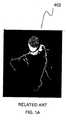
図1A及び1Bは、心臓の3次元CT画像を示す。図1Aにおいて、画像402は心臓を示し、一方、図1Bにおいて、画像404は冠動脈のみを示す。 1A and 1B show a three-dimensional CT image of the heart. In FIG. 1A,
冠動脈疾患を検出するための一般的な方法として、胸部領域のCT画像を撮影し、冠動脈及び心臓のカルシウム化を定量化することが行われる。冠動脈壁の動脈内膜におけるカルシウム化又はカルシウム塩の存在は、アテローム性動脈硬化症又は動脈壁肥厚の検出における主要なファクターであり、これは冠動脈疾患の原因となる。特に、カルシウム化の数及び量は、一般的に、具体的には動脈内のプラーク又は老廃物の量を示すサインである。CTスキャンを用いた冠動脈疾患の早期検出は、この疾患の高死亡率の低減における重要なファクターである。 As a general method for detecting coronary artery disease, CT images of the chest region are taken to quantify coronary artery and heart calciumation. The presence of calcium or calcium salt in the intima of the coronary artery wall is a major factor in the detection of atherosclerosis or artery wall thickening, which causes coronary artery disease. In particular, the number and amount of calciumation is generally a sign that specifically indicates the amount of plaque or waste in the artery. Early detection of coronary artery disease using CT scan is an important factor in reducing the high mortality of this disease.
CTスキャンを用いて冠動脈疾患を診断するために、カルシウム化の数を識別し、次に、その重要度を決定するために「スコアリング」することが必要である。冠動脈カルシウム(CAC)スコアリングは、主に、各プラークが関連するカルシウム化であるのか、及び、どの動脈内にあるのか識別するステップがあるために、時間のかかる処理である。この識別ステップでは、一般的に、循環器専門医又は熟練した技術者による手動で各プラークの識別が行われることが必要である。 In order to diagnose coronary artery disease using a CT scan, it is necessary to identify the number of calcifications and then “score” to determine their importance. Coronary artery calcium (CAC) scoring is a time consuming process primarily because there is a step to identify which plaque is associated with and within which artery. This identification step generally requires that each plaque be identified manually by a cardiologist or a skilled technician.
スコアリング方法としては、アガツトン法でカルシウム化を定量化することが一般的である。この方法では、心臓内の全ての動脈カルシウムを定量化したアガツトンスコアという1つのスコアが得られる。他の方法として、体積スコアリングや質量スコアリングもある。これらの方法及びその変形における処理は本質的に似ており、1)閾値処理によってプラークを見つけ、2)各プラークが冠動脈内の関連するカルシウム化であるか識別し(また、その場合、どの動脈内か)、3)加重和を算出する、という処理を行う。 As a scoring method, calcification is generally quantified by the Agatsuton method. This method yields a single score, the Agaton score, which quantifies all arterial calcium in the heart. Other methods include volume scoring and mass scoring. The processing in these methods and variations thereof is essentially similar: 1) find plaques by thresholding, 2) identify each plaque as an associated calcification in the coronary artery (and in that case, which artery Or 3) calculate a weighted sum.
カルシウムスコアリング処理を自動化する試みは、ある程度は成功している。1つの全体スコアを得ることができる方法もあるし、個別の動脈について個別のスコアを得るよう試みている方法もある。しかし、個別のスコアを得る方法では、正確に正しい動脈を識別し、更に、関連しないプラーク(骨組織、ノイズ、血管外の心臓カルシウム等)のような偽陽性を検出することが困難である。 Attempts to automate the calcium scoring process have been somewhat successful. Some methods can obtain one overall score, while others attempt to obtain individual scores for individual arteries. However, the method of obtaining individual scores is difficult to accurately identify the correct artery and to detect false positives such as unrelated plaques (bone tissue, noise, extravascular cardiac calcium, etc.).
このように、正確でロバストな冠動脈カルシウムスコアリング方法が必要とされている。 Thus, there is a need for an accurate and robust coronary calcium scoring method.
本発明の様々な実施形態は、医用画像におけるモデルに基づいた冠動脈のカルシウム(CAC)のスコアリングをするシステム及び方法に関し、より具体的には、画像内の冠動脈と他の解剖学的特徴の識別に役立てるために、患者の心臓領域の画像と心臓領域のモデル画像を比較することにより、特定の冠動脈内の関連するカルシウムプラークの位置を識別することに関する。画像とモデル画像との比較に基づいて、関連するカルシウムプラークを識別することができ、特定の冠動脈内でのプラークの位置に基づいて関連するカルシウムプラークをラベリングすることができる。その後、患者の心臓領域全体についての総合スコアに加えて、各関連する動脈(例えば、冠動脈)についてスコアが計算される。これにより、患者の冠動脈疾患のリスクをより正確に評価することができる。 Various embodiments of the present invention relate to systems and methods for coronary artery calcium (CAC) scoring based on models in medical images, and more specifically, coronary arteries and other anatomical features in images. To aid in identification, it relates to identifying the location of the relevant calcium plaque within a particular coronary artery by comparing an image of the patient's heart region with a model image of the heart region. Based on the comparison between the image and the model image, the relevant calcium plaques can be identified, and the relevant calcium plaques can be labeled based on the location of the plaque within a particular coronary artery. A score is then calculated for each associated artery (eg, coronary artery) in addition to the overall score for the entire patient's heart region. Thereby, a patient's risk of coronary artery disease can be more accurately evaluated.
例示的な一実施形態では、冠動脈カルシウム(CAC)のスコアリング方法は、カルシウムプラークを含む可能性がある心臓の画像を取得することと、画像内の心臓の解剖学的特徴とモデル画像の解剖学的特徴とをアライメントして、心臓のどの領域が各冠動脈の領域に対応するのかについての情報を得ることとを含む、冠動脈カルシウムスコアのスコアリング方法を備え、この情報は、後で、どの動脈にプラークが存在する可能性が最も高いのか、及び、その可能性についての情報をラベリングステップに与えるために用いることができる。この方法は、心臓の画像上の各冠動脈上にプラークが存在する場合、心臓の画像上の各冠動脈にプラークをラベリングすることと、各冠動脈についての個別のスコア及び心臓についての総合スコアをスコアリングし決定することと、個別のスコア及び総合スコアを表示することと、を更に含む。 In one exemplary embodiment, a coronary artery calcium (CAC) scoring method obtains an image of a heart that may contain calcium plaques, and anatomical features of the heart and model images in the image. A scoring method for coronary artery calcium scores, including obtaining information about which areas of the heart correspond to areas of each coronary artery, It can be used to provide the labeling step with information about and most likely the presence of plaque in the artery. This method labels plaques on each coronary artery on the heart image, if there are plaques on each coronary artery on the heart image, and scores an individual score for each coronary artery and a total score for the heart And determining and displaying an individual score and an overall score.
他の例示的な一実施形態では、冠動脈カルシウムのスコアリングのためのシステムは、カルシウムプラークを含む可能性がある心臓の画像を取得する入力部と、画像内の心臓の解剖学的特徴とモデル画像の解剖学的特徴とをアライメントするアライメント部と、心臓の画像上の各冠動脈上にプラークが存在する場合、心臓の画像上の各冠動脈にプラークをラベリングするラベリング部と、を備える。このシステムは、各冠動脈についての個別のスコア及び心臓についての総合スコアを決定するスコアリング部と、個別のスコア及び総合スコアを表示するためのグラフィカル・ユーザー・インターフェースを作成する表示部と、を更に備える。 In another exemplary embodiment, a system for coronary calcium scoring includes an input for acquiring an image of a heart that may include calcium plaques, and an anatomical feature and model of the heart in the image An alignment unit that aligns the anatomical features of the image, and a labeling unit that labels the plaque on each coronary artery on the heart image when the plaque exists on the coronary artery on the heart image. The system further includes a scoring unit that determines an individual score for each coronary artery and a total score for the heart, and a display unit that creates a graphical user interface for displaying the individual score and the total score. Prepare.
更なる他の例示的な一実施形態では、冠動脈カルシウムのスコアリングのためのコンピュータプログラム製品が提供される。コンピュータプログラムは、コンピュータで読み取り可能な媒体に具現化された製品であり、コンピュータによって実行された場合に、カルシウムプラークを含む可能性がある心臓の画像を取得することと、画像内の心臓の解剖学的特徴とモデル画像の解剖学的特徴とをアライメントすることと、心臓の画像上の各冠動脈上にプラークが存在する場合、心臓の画像上の各冠動脈にプラークをラベリングすることと、を含む方法を行う。この方法は、各冠動脈についての個別のスコア及び心臓についての総合スコアをスコアリングすることと、個別のスコア及び総合スコアのうちの少なくとも一方を表示することと、を更に含む。 In yet another exemplary embodiment, a computer program product for coronary calcium scoring is provided. A computer program is a product embodied in a computer readable medium that, when executed by a computer, obtains an image of the heart that may contain calcium plaques and dissects the heart in the image. Aligning the anatomical features with the anatomical features of the model image and, if plaques are present on each coronary artery on the heart image, labeling the plaques on each coronary artery on the heart image Do the way. The method further includes scoring an individual score for each coronary artery and an overall score for the heart and displaying at least one of the individual score and the overall score.
上記の例示的な実施形態において、この技術分野で知られているように、心臓はプラークを含んでいてもよいし、含んでいなくてもよい。心臓がプラークを含んでいるか否かにかかわらず、スコアリングを行う。CACがない場合、スコアはゼロになる。個別の動脈のいずれか及び/又は心臓についても同様である。 In the exemplary embodiments described above, the heart may or may not include plaque, as is known in the art. Scoring is performed regardless of whether the heart contains plaque. If there is no CAC, the score is zero. The same is true for any individual artery and / or heart.
本発明に関する更なる実施形態については、一部は以下の説明において述べられ、一部は説明から明白であるか、又は、本発明の実施によって習得することができる。本発明の実施形態は、要素、様々な要素の組み合わせ、後述の詳細説明において特に指摘された態様、及び添付の特許請求の範囲を用いて、実現し達成することが可能である。 Additional embodiments relating to the invention will be set forth in part in the description which follows, and in part will be obvious from the description, or may be learned by practice of the invention. The embodiments of the invention may be realized and attained by means of the elements, combinations of the various elements, aspects particularly pointed out in the detailed description that follows, and the appended claims.
上記及び下記の説明は、例示及び説明のためであり、いかなる形においても、特許請求された発明又はその応用を限定する趣旨ではない。 The above and following description is for purposes of illustration and description, and is not intended to limit the claimed invention or its application in any way.
本明細書に援用され、その一部を構成する添付の図面は、説明とともに本発明の様々な実施形態を例示し、発明技術の原理を説明し図示するのに資する。 The accompanying drawings, which are incorporated in and constitute a part of this specification, illustrate various embodiments of the invention together with the description, and serve to explain and illustrate the principles of the inventive technology.
下記の詳細な説明では添付の図面を参照しており、これらの図において同一の機能要素は同じ参照符号で示される。上記の添付図面は、限定としてではなく実例として示しており、具合的な実施形態及び実施例は本発明の原理と一致するものである。これらの実施例は、当業者が本発明を実施することができるほど十分に詳細に説明されており、本発明の範囲及び思想を逸脱することなく、他の実施例を利用すること、及び、構成の変更及び/又は様々な要素の置き換えがなされ得ることが理解されるであろう。従って、下記の詳細な説明は、狭義に解釈されるべきものではない。更に、説明したように、本発明の様々な実施形態は、汎用コンピュータ上で実行されるソフトウェアという形態、専用のハードウェアという形態、又は、ソフトウェアとハードウェアの組み合わせで実現することができる。「少なくとも1つ」等の表現は、要素のリストの前に記載されている場合は、要素のリスト全体を修飾するものであり、リストの中の各要素を修飾するものではない。 In the following detailed description, reference will be made to the accompanying drawing (s), in which identical functional elements are designated with like reference numerals. The accompanying drawings are presented by way of illustration and not limitation, and specific embodiments and examples are consistent with the principles of the invention. These embodiments are described in sufficient detail to enable those skilled in the art to practice the invention, and other embodiments can be utilized without departing from the scope and spirit of the invention, and It will be appreciated that configuration changes and / or replacement of various elements may be made. The following detailed description is, therefore, not to be construed in a narrow sense. Further, as described, various embodiments of the present invention can be implemented in the form of software running on a general purpose computer, in the form of dedicated hardware, or a combination of software and hardware. An expression such as “at least one”, when described before an element list, modifies the entire list of elements and does not qualify each element in the list.
本発明の実施形態は、冠動脈疾患のリスクを評価するカルシウムスコアリングを自動化し、心臓病の診断と報告書をより容易、より正確、より時間がかからないようにする。モデルに基づいた(モデルベースの)アプローチは、患者の心臓領域の画像を心臓のモデル画像と比較するために用いられる。心臓のモデル画像は、冠動脈と他の解剖学的特徴を示し、及び/又は、各空間領域に対して、その領域内に特定の冠動脈が存在する確率を与える。この比較は、カルシウムプラークが冠動脈内に存在するか否か識別し、存在する場合は、どの冠動脈内に存在するのか識別する際に役立つ。よって、画像内の関連しないカルシウムプラーク及び他のノイズを無視することができ、冠動脈内の関連するカルシウムプラークを個別に重みづけして、冠動脈疾患についての患者のリスクのより正確でロバストな評価を提供することができる。 Embodiments of the present invention automate calcium scoring to assess coronary artery disease risk, making heart disease diagnosis and reporting easier, more accurate, and less time consuming. A model-based (model-based) approach is used to compare an image of a patient's heart region with a model image of the heart. The model image of the heart shows the coronary arteries and other anatomical features and / or gives each spatial region the probability that a particular coronary artery is present in that region. This comparison helps to identify whether calcium plaque is present in the coronary artery, and if so, in which coronary artery it is present. Thus, unrelated calcium plaques and other noise in the image can be ignored, and related calcium plaques in the coronary arteries can be individually weighted to provide a more accurate and robust assessment of the patient's risk for coronary artery disease. Can be provided.
モデルに基づいたアプローチにより、4つの主要冠動脈のうちのどの冠動脈内に特定のカルシウムプラークが位置するのかについて決定することが可能になる。4つの主要冠動脈とは、左冠動脈(LM)、左前下行動脈(LAD)、左回旋動脈(LCX)及び右冠動脈(RCA)である。モデル内の冠動脈と一致しないカルシウムプラークを関連しないプラークとして無視し、後続の動脈スコアリングから除外することができる。次に、画像とモデルとの比較によってプラークを動脈でラベリングすることができるため、カルシウムスコアを心臓全体についてだけでなく、個別の冠動脈についても計算することができる。プラークが発見されない場合、スコアはゼロになる。スコアリング結果は、心臓の領域を近似した直線を用いて心臓を複数の領域に単純に分割するよりも正確になる。 A model-based approach makes it possible to determine in which of the four main coronary arteries a particular calcium plaque is located. The four main coronary arteries are the left coronary artery (LM), the left anterior descending artery (LAD), the left circumflex artery (LCX), and the right coronary artery (RCA). Calcium plaques that do not match the coronary arteries in the model can be ignored as unrelated plaques and excluded from subsequent arterial scoring. The plaque can then be labeled with the artery by comparing the image with the model, so that the calcium score can be calculated not only for the entire heart but also for individual coronary arteries. If no plaque is found, the score is zero. The scoring result is more accurate than simply dividing the heart into multiple regions using straight lines approximating the region of the heart.
各々の関連するプラークを特定の冠動脈に割り当てることにより、本発明の例示の実施形態は自動スコアリングを支援し、各動脈に心臓疾患又は心臓発作のリスクを示すスコアを与えることができる。動脈の個別のスコアにより、医師はより優れた診断情報を得ることができる。たとえば、LM動脈内のカルシウムは、重大な疾患リスク、及び、心臓発作のリスクの主なファクターであると考えられる。 By assigning each associated plaque to a particular coronary artery, exemplary embodiments of the invention can assist in automatic scoring and give each artery a score indicative of the risk of heart disease or stroke. With individual arterial scores, doctors can obtain better diagnostic information. For example, calcium in LM arteries is considered to be a major factor in significant disease risk and risk of heart attack.
ここで説明される方法では、ユーザが手動で画像(例えば、X線)内の冠動脈を識別し、カルシウム化がどの動脈に属するのか、又は、少なくともカルシウム化が動脈兄に存在するのか否かによって、画像内の各カルシウム化をラベリングすることが不要である。例示の、非限定的スコアは、以下のとおりである。 In the method described here, a user manually identifies coronary arteries in an image (eg, x-ray) and depends on which artery the calcification belongs to, or at least whether the calcification exists in the arterial brother. It is not necessary to label each calcification in the image. Exemplary, non-limiting scores are as follows:
この例では、3つのプラークが存在する。 In this example, there are three plaques.
プラーク1:LM内、面積40mm、最大X線減衰150HU
プラーク2:LCX内、面積25mm、最大X線減衰250HU
プラーク3:LCX内、面積70mm、最大X線減衰140HU
各々のプラークについての個別のスコアは、重みづけを考慮して以下のように計算される。Plaque 1: LM, area 40mm, maximum X-ray attenuation 150HU
Plaque 2: LCX, area 25mm, maximum X-ray attenuation 250HU
Plaque 3: LCX, area 70mm, maximum X-ray attenuation 140HU
The individual score for each plaque is calculated as follows, taking into account the weighting.
プラーク1のスコア: 面積40 × 重み1 = 40
プラーク2のスコア: 面積25 × 重み2 = 50
プラーク3のスコア: 面積70 × 重み1 = 70
上記のプラークの個別のスコアに基づいて、冠動脈についてのスコアが以下のように計算される。Plaque 1 score: area 40 x weight 1 = 40
Plaque 2 score: area 25 x weight 2 = 50
Plaque 3 score: area 70 x weight 1 = 70
Based on the individual plaque scores, the score for the coronary artery is calculated as follows:
LMのスコア = P1のスコア = 40
LADのスコア = 0
LCXのスコア = P2及びP3のスコアの合計 =50+70 = 120
RCAのスコア = 0
従って、心臓についての総合スコアは以下のように計算される。LM score = P1 score = 40
LAD score = 0
LCX score = Sum of P2 and P3 scores = 50 + 70 = 120
RCA score = 0
Thus, the total score for the heart is calculated as follows:
LM+LAD+LCX+RCA = 40+0+120+0 = 160
上記の例では、重みづけはCT値(例えば、アガツトンスコアリングにおいて131−200HUについては1、201−300HUについては2等)によって決定することができる。重みは位置又は動脈によらない。上記の重みづけ手順において、HUは、X線減衰の単位であるハウンズフィールド・ユニットを示す。LM + LAD + LCX + RCA = 40 + 0 + 120 + 0 = 160
In the above example, the weighting can be determined by the CT value (e.g., 1 for 201-200 HU and 2 for 201-300 HU, etc. in Agaton scoring). The weight is independent of position or artery. In the above weighting procedure, HU represents a Hounsfield unit, which is a unit of X-ray attenuation.
関連技術では、自動的に個別のスコアを算出していない。対照的に、上記の例では、プラークの合計である1つのスコアのみが算出される。 Related technology does not automatically calculate individual scores. In contrast, in the above example, only one score that is the sum of plaques is calculated.
スコア = すべてのプラークのスコアの合計 = 40+50+70 = 160
関連技術では、LM、LAD、LCX及びRCAスコアは算出されない。Score = Sum of all plaque scores = 40 + 50 + 70 = 160
In the related art, LM, LAD, LCX and RCA scores are not calculated.
1.モデルに基づくアプローチ方法
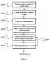
図2に示す冠動脈カルシウム(CAC)スコアリングの1つの例示の方法において、心臓領域の画像が取得される(ステップS102)。CT画像は空間測定3次元(3D)コンピュータ断層撮影(CT)画像でもよく、このCT画像は、連続する2次元CT画像を積み重ねたものである。ステップS104において、心臓が抽出され、画像内の骨、肺関連の構造体及び他の関連しない解剖学的構造体を除外するために境界が識別される。心臓の抽出は、当業者に理解されている方法で行うことができる。たとえば、限定する趣旨ではないが、エッジに基づく区分法、領域形成区分法、レベル設定区分法、能動輪郭区分法、又はモデルに基づく区分法を適用することができる。1. Model-Based Approach Method In one exemplary method of coronary calcium (CAC) scoring shown in FIG. 2, an image of the heart region is acquired (step S102). The CT image may be a spatially measured three-dimensional (3D) computed tomography (CT) image, which is a stack of successive two-dimensional CT images. In step S104, the heart is extracted and boundaries are identified to exclude bones, lung-related structures and other unrelated anatomical structures in the image. Extraction of the heart can be performed by methods understood by those skilled in the art. For example, but not limited to, edge-based segmentation, region formation segmentation, level setting segmentation, active contour segmentation, or model-based segmentation can be applied.
次に、ステップS106において、心臓内で閾値処理を行う。ハウンズフィールド・スケールで高い値を有するピクセル(2D)又はボクセル(3D)のように、密度によって画像内でカルシウム化を容易に規定することができる。よって、カルシウム化は単純な閾値処理によって識別される。たとえば、限定する趣旨ではないがアガツトンスコアリング法では、130HUより高いCT値を有するボクセルをカルシウムプラーク候補とみなす。 Next, in step S106, threshold processing is performed in the heart. As with pixels (2D) or voxels (3D) with high values on the Hounsfield scale, calcification can be easily defined in the image by density. Thus, calciumation is identified by simple thresholding. For example, although not intended to be limiting, in the Agaton scoring method, voxels having a CT value higher than 130HU are considered as calcium plaque candidates.
次に、ステップS106において、連結成分解析を行い、一意なラベルを各々の連結成分に割り当てる。連結成分解析は、当業者に理解されている方法で行うことができる。 Next, in step S106, connected component analysis is performed, and a unique label is assigned to each connected component. Connected component analysis can be performed by methods understood by those skilled in the art.
その後、3次元CT画像と3次元モデルとの間の位置合わせが行われ(ステップS108)、この処理において、3次元モデルの解剖学的特徴を3次元CT画像上にマッピングできるように、3次元モデルが3次元画像とアライメントされる。3次元CT画像内の冠動脈を適切に識別するためには、3次元モデルは3次元CT画像と正確に重なり合っている必要がある。以下に、この位置合わせについてより詳細に説明する。位置合わせ結果に基づいて、ステップS110において、識別された冠動脈の1つの中に位置する各カルシウムプラークが特定の冠動脈内にあるか否かについて、より確実に自動的に識別することができる。カルシウムプラークは、そのカルシウムプラークが内部に位置する冠動脈にラベリングされる。関連するプラークをラベリングすることにより、識別された冠動脈内に存在していないプラークは、それがために、関連しないプラークであるため、1つ又は複数の冠動脈についての後続のスコアリング処理についての検討から除外される。これらの関連しないプラークとしては、大動脈カルシウム沈着又は心臓の外の構造体(例えば、骨)が挙げられる。 Thereafter, alignment between the 3D CT image and the 3D model is performed (step S108). In this process, the 3D model is mapped so that the anatomical features of the 3D model can be mapped onto the 3D CT image. The model is aligned with the 3D image. In order to properly identify the coronary artery in the 3D CT image, the 3D model needs to overlap exactly with the 3D CT image. Hereinafter, this alignment will be described in more detail. Based on the registration results, in step S110, it can be more automatically identified whether each calcium plaque located in one of the identified coronary arteries is within a particular coronary artery. Calcium plaques are labeled into the coronary arteries in which the calcium plaques are located. By labeling the relevant plaques, a plaque that is not present in the identified coronary arteries is therefore an unrelated plaque and therefore a subsequent scoring process for one or more coronary arteries Excluded from. These unrelated plaques include aortic calcifications or structures outside the heart (eg, bone).
続いて、ステップS112において、閾値処理結果及び位置合わせ結果に基づいてスコアリングが行われる。上記のように、個別の動脈に個々にスコアリングし、次に、総合スコアが心臓全体について計算することができる。ある動脈内にカルシウムプラークが存在しない場合、その動脈についてのスコアはゼロである。最後に、ステップS114において、ユーザが選択したその他の画像とともに、ユーザにスコアを表示する。 Subsequently, in step S112, scoring is performed based on the threshold processing result and the alignment result. As described above, individual arteries can be scored individually, and then a total score can be calculated for the entire heart. If there is no calcium plaque in an artery, the score for that artery is zero. Finally, in step S114, the score is displayed to the user together with the other images selected by the user.
位置合わせ及びラベリングステップについて、以下により詳しく記載する。位置合わせ処理には2つのオプションがある。1つはラフな位置合わせ、又は、(例えば、左動脈口の位置のユーザ入力を用いた)2つの画像間の単純な位置合わせであり、もう1つは、2つの画像から識別されたランドマークの収束による、もっと複雑な位置合わせである。 The alignment and labeling steps are described in more detail below. There are two options for the alignment process. One is a rough alignment, or a simple alignment between two images (eg, using user input of the position of the left arterial ostium) and the other is a land identified from the two images. More complex alignment due to mark convergence.
ラフな(大まかな)位置合わせでは、2つの画像間の必要最小限のアライメントを行って3次元CT画像内の冠動脈のおおよその位置を得る。このラフ位置合わせは、回転、拡大縮小又は変形をしないで、又はこれらをわずかに行うだけで、画像内のランドマークの位置と一致するように、モデルのランドマークの位置を移動させることによって行うことができる。たとえば、ユーザ入力又はアルゴリズムによって左動脈口の位置が既知である場合、その位置が画像内の実際の左動脈口とアライメントされ、これにより、画像内の他の部分も大まかにアライメントされる。これによって、複雑な位置合わせをしなくとも、4つの冠動脈の各々に対応する領域であるLM、LAD、LCX及びRCAに、心臓領域を大まかに分割し、カルシウムプラークに冠動脈ラベルを割り当てる際に、合理的な精度を得ることができる。 In rough (rough) registration, the minimum necessary alignment between two images is performed to obtain an approximate position of the coronary artery in the 3D CT image. This rough alignment is done by moving the model landmark position so that it matches the landmark position in the image, with little or no rotation, scaling or deformation. be able to. For example, if the position of the left arterial ostium is known by user input or algorithm, that position is aligned with the actual left arterial ostium in the image, so that other parts in the image are also roughly aligned. This allows the heart region to be roughly divided into LM, LAD, LCX and RCA, which are regions corresponding to each of the four coronary arteries, without assigning complex alignments, and assigning coronary artery labels to calcium plaques Reasonable accuracy can be obtained.
より複雑な収束型の位置合わせにおいて、2つの画像が収束するまで複数回反復計算した後、合致するランドマークのアライメントパラメータを推定することによって、CT画像内の心臓の3次元体積において識別されたランドマークが、3次元モデル画像内で識別されたランドマークとアライメントされる。複雑な収束型の位置合わせにおいて、図3に示すように、ステップS202は、モデル画像とCT画像とを取得することである。CT画像において、ステップS204で、例えば、大動脈、冠状動脈口、弁又は心尖のようなランドマークが識別される。一般的に、ランドマークを見つけて識別することは容易にできるため、これらをユーザによるマニュアルで識別することにしてもよい。あるいは、ランドマークの一般的な位置及び特性に基づいて、画像処理アルゴリズムを用いて自動的にランドマークを識別してもよい。限定する趣旨ではないが、例えば、ハフ(Hough)変換に基づく画像処理アルゴリズムによって大動脈を自動的に識別してもよい。 In more complex convergent alignments, after multiple iterations until the two images converge, they are identified in the 3D volume of the heart in the CT image by estimating the alignment parameters of the matching landmarks The landmark is aligned with the landmark identified in the 3D model image. In complicated convergence type alignment, as shown in FIG. 3, step S202 is to acquire a model image and a CT image. In the CT image, in step S204, landmarks such as aorta, coronary ostia, valves or apex are identified. Generally, since landmarks can be easily found and identified, they may be identified manually by a user. Alternatively, the landmark may be automatically identified using an image processing algorithm based on the general position and characteristics of the landmark. Although not intended to be limiting, for example, the aorta may be automatically identified by an image processing algorithm based on a Hough transform.
次に、ステップS206において、冠動脈が抽出される。動脈の完全な画像を識別することは容易ではないため、この抽出は、通常、動脈の断片を識別することによって行われる。この抽出は、検査対象となる画像内の管状の構造体を識別することによって、行うことができる。限定する趣旨ではないが、例えば、参照によりその内容が本願に援用されている米国特許出願公開20110135172号で、例示的な抽出処理が開示されている。前の処理で識別されたランドマークを用いてアライメントパラメータが決定される(ステップS208)。このアライメントパラメータは、3次元モデルを3次元CT画像にアライメントする一連の角度と移動とを定義する。限定する趣旨ではないが、例えば、初期アライメントは、3次元CT画像の大動脈を3次元モデル内の大動脈とアライメントすることである。この処理の完了後、当業者に理解されている方法で、より具体的なランドマークについてもっと詳細なアライメントを行ってもよい。例えば、剛体位置合わせにおいては、拡大縮小、移動及び回転パラメータを適用することができる。一方、非剛体位置合わせにおいては、各グリッド(又はメッシュ)ポイントに、ずれ(displacement)があってもよく、通常、ポイントのずれは、近隣のポイントと略同じであると仮定される。 Next, in step S206, a coronary artery is extracted. This extraction is usually done by identifying arterial fragments, since it is not easy to identify a complete image of an artery. This extraction can be performed by identifying a tubular structure in the image to be examined. Although not intended to be limiting, for example, US Patent Application Publication No. 20110135172, the contents of which are incorporated herein by reference, discloses an exemplary extraction process. An alignment parameter is determined using the landmark identified in the previous process (step S208). This alignment parameter defines a series of angles and movements that align the 3D model with the 3D CT image. Although not intended to be limiting, for example, the initial alignment is to align the aorta of the 3D CT image with the aorta in the 3D model. After this process is complete, more detailed alignment may be performed for more specific landmarks in a manner understood by those skilled in the art. For example, scaling, movement and rotation parameters can be applied in rigid body alignment. On the other hand, in non-rigid registration, each grid (or mesh) point may have a displacement, which is usually assumed to be approximately the same as neighboring points.
その後、ステップS210において、各モデルポイントにおいて、候補の中で最もよくマッチした動脈ポイントを抽出し、次に、CT画像とモデル画像との間の対応関係を取得する。次に、ステップS212において、この対応関係を用いてアライメントパラメータが推定される。次に、CT画像とモデル画像とが収束するまで、最もよくマッチした動脈ポイントの抽出及びアライメントパラメータの推定を繰り返す(ステップS214)。 Thereafter, in step S210, the best matching arterial point among the candidates is extracted at each model point, and then the correspondence between the CT image and the model image is acquired. Next, in step S212, alignment parameters are estimated using this correspondence. Next, until the CT image and the model image converge, the extraction of the best matching arterial point and the estimation of the alignment parameter are repeated (step S214).
位置合わせの後、各カルシウムプラークに、そのプラークがどの冠動脈の中に位置するのかを識別するラベルが割り当てられる。モデルとの位置合わせの結果、又は、確率マップとの位置合わせを、特定のカルシウムプラークが識別された動脈内に位置するか否かを決定するために用いてもよい。位置合わせ処理により、特定のカルシウムプラークが動脈の線又はエッジに沿って存在し、それゆえに、カルシウム化をスコアリングする際に考慮されるべき血管ベースのカルシウム化である可能性が高いか否かを識別する性能を向上させることができる。 After alignment, each calcium plaque is assigned a label that identifies in which coronary artery the plaque is located. The result of registration with the model or registration with the probability map may be used to determine whether a particular calcium plaque is located within the identified artery. Whether the alignment process causes certain calcium plaques to be present along arterial lines or edges and therefore likely to be vascular-based calcification to be considered when scoring calcification The performance of identifying can be improved.
次に、単純な算術演算によってスコアリングが行われる。通常、この算術演算は、各動脈及び動脈全体についての個別のCACプラークのスコアの加重和である。 Next, scoring is performed by a simple arithmetic operation. Typically, this arithmetic operation is a weighted sum of individual CAC plaque scores for each artery and for the entire artery.
上述の手順の目的は、各カルシウムプラークについて、そのプラークが、大動脈カルシウム又は心臓の外側の構造体のような偽陽性であるか否か、偽陽性でない場合は、どの冠動脈(LM、LAD、LCX、RCA)にそのプラークが属するのか、自動的に識別することである。 The purpose of the above procedure is to determine for each calcium plaque whether the plaque is false positive, such as aortic calcium or structures outside the heart, and if not, which coronary artery (LM, LAD, LCX , RCA) automatically identifies whether the plaque belongs to.
モデル画像は、例えば、アメリカ心臓協会発行、循環(Circulation)78巻(1988年)、1167-1180頁、J.T.ドッジ・ジュニア他著、「Intrathoracic Spatial Location of Specified Coronary Segments on the Normal Human Heart」などに基づく点モデルでもよい。改良モデルを得ることができる別の方法としては、既存のCAT区分/ラベリングを用いてモデルを増強することである。取得済みの多数の区分け/分類分けされたデータを用いて、既存のCAT区分/ラベリングデータをモデルに追加することも可能である。モデルは、点モデル、形状モデル、確率モデル、又は、当業者に理解可能ないかなるモデルであってよい。データが多い方が、分散(variance)が減り、モデル画像の精度を向上させることができるであろう。 The model image is, for example, published by the American Heart Association, Circulation 78 (1988), pages 1167-1180, J. Am. T. T. et al. It may be a point model based on Dodge Jr. et al., “Intrathoracic Spatial Location of Specified Coronary Segments on the Normal Human Heart”. Another way in which an improved model can be obtained is to augment the model with existing CAT segmentation / labeling. It is also possible to add existing CAT classification / labeling data to the model using a large number of acquired classification / classification data. The model may be a point model, a shape model, a probabilistic model, or any model understandable to those skilled in the art. More data will reduce the variance and improve the accuracy of the model image.
冠動脈の断片を抽出する際(S206)、造影CT画像内の冠動脈抽出と同程度に完全に区分けする必要はない。位置合わせは、患者の画像内の心臓のどの部分が通常、特定の冠動脈が位置している領域に対応するのか、大まかに識別するのに十分な精度であることが要求されているだけであるから、位置合わせは高精度である必要はない。この意味で、冠動脈の正確な区分けは必要ではなく、大まか若しくは中断して不完全な(discontinued)区分け、又は、区分けがなくても、十分である。例えば、区分けによって妥当な血管候補がほとんど得られないような最悪の場合であっても、アライメントパラメータを初期化(S208)、最もよくマッチした動脈ポイントの位置を見つけ(S210)、アライメントパラメータを推定し(S212)、収束するまで繰り返す(S214)処理で構成される位置合わせの代わりに、1)最初にモデルと心臓を位置合わせし、2)次にモデル内で最も距離が近い動脈を各プラークに割り当てるという方法を行うことも可能である。 When extracting a coronary artery fragment (S206), it is not necessary to segment completely as much as coronary artery extraction in a contrast CT image. Registration is only required to be accurate enough to roughly identify which part of the heart in the patient's image usually corresponds to the region where a particular coronary artery is located. Therefore, the alignment need not be highly accurate. In this sense, an accurate segmentation of the coronary arteries is not necessary, and it is sufficient if the segment is roughly or interrupted and discontinued or no segmentation. For example, even in the worst case where reasonable vessel candidates are hardly obtained by segmentation, the alignment parameters are initialized (S208), the best matching arterial point position is found (S210), and the alignment parameters are estimated. (S212) and repeat until the convergence (S214), instead of the alignment configured in the process (1), 1) first align the model and the heart, 2) next to the closest artery in the model each plaque It is also possible to perform the method of assigning to
多くの場合、冠動脈カルシウムではなく、大動脈カルシウム、骨、肺組織、ノイズのような別の構造体であるプラークが存在する。連結成分にラベルを割り当てることにより(S110)、スコアリングが行われる際に、ラベリングされていない関連しないプラークを考慮対象から除外することができる。ステップS108において、どのプラークがどの動脈の領域内にあるのかについての情報が提供されるが、プラークが実際に冠動脈カルシウムであるか否か決定する際に考慮するべきファクターはもっと多く存在する。たとえプラークが特定の冠動脈の領域内にあると判定された場合でも、その判定が偽陽性である可能性がある。そのため、ステップS110において、プラークを特定の冠動脈にラベリングすべきか否か、又は、プラークが関連しないものであるか否か決定する前に、向き、血管らしさ(vesselness)、他のプラークとの関係、形状等のような、プラークについての様々なファクターが考慮される。この決定処理において、サポートベクターマシーン(support vector machine、SVM)、ニューラルネットワーク、線形分類器等のようなパターン分類法を用いることができる。 Often there are plaques that are not coronary calcium, but other structures such as aortic calcium, bone, lung tissue, noise. By assigning labels to the connected components (S110), unlabeled plaques that are not labeled can be excluded from consideration when scoring is performed. In step S108, information is provided as to which plaque is in which arterial region, but there are more factors to consider when determining whether a plaque is actually coronary calcium. Even if it is determined that the plaque is within the area of a particular coronary artery, the determination may be false positive. Therefore, in step S110, before deciding whether plaque should be labeled to a particular coronary artery or whether the plaque is unrelated, orientation, vesselness, relationship to other plaques, Various factors about the plaque, such as shape, are considered. In this determination process, a pattern classification method such as a support vector machine (SVM), a neural network, a linear classifier, or the like can be used.
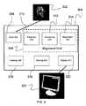
本発明のシステムは上述のステップにしたがって医用画像を受け付け、それを処理するコンピュータ上に実装することができる。本発明のシステムは、コンピュータプログラム製品として具現化することができ、または、ソフトウェア及びハードウェアの組合せによって実現してもよい。図4に示すように、入力部306において画像302をコンピュータ304に入力することができる。次に、アライメント部308は、画像内の心臓の解剖学的特徴を、モデル画像の解剖学的特徴と一致させる(マッチさせる)。アライメント部308は、抽出部310と、連結部312と、位置合わせ部314とを備えてもよい。抽出部310は、心臓の境界を抽出し、骨、肺組織及び他の関連しないプラークを除外する(S206)。連結部312では、心臓の内側で閾値処理が行われ、連結成分解析が行われる。次に、位置合わせ部314を介して、CT画像と3次元モデルとの位置合わせが行われる。ラベリング部316では、大動脈プラーク又は心臓の外側の構造体(例えば、骨)のような関連しないプラークを、後続のスコアリング処理の考慮対象から除外することができる。位置合わせ結果によって、冠動脈ラベルを各連結成分に割り当てることも可能になる。次に、閾値処理の結果と位置合わせの結果に基づいてスコアリング部318においてスコアリングが行われる。最後に、表示部320は、スクリーン322上でユーザが選択した他の画像とともに、スコアをユーザに表示するためのグラフィカル・ユーザー・インターフェース(GUI)を作成する。 The system of the present invention can be implemented on a computer that accepts and processes medical images according to the steps described above. The system of the present invention may be embodied as a computer program product or may be realized by a combination of software and hardware. As shown in FIG. 4, an
本発明のシステム及び方法は、限定する趣旨ではないが、例えば、磁気共鳴撮影法(MRI)、X線撮影法(X線)、コンピュータ断層撮影法(CT)、超音波検査法(US)のような多くのタイプの医用画像に適用可能である。 The system and method of the present invention are not intended to be limiting, but include, for example, magnetic resonance imaging (MRI), X-ray imaging (X-ray), computed tomography (CT), and ultrasonography (US). It can be applied to many types of medical images.
2.カルシウムスコアのダイナミックな更新
他の実施形態では、モデルに基づく手法(アプローチ)を、ダイナミックなカルシウムスコアリングの更新に用いることができる。当初は、完全なスコアリングをすぐに得ることができない場合がある。もし、できない場合、ユーザが画像上で1つのカルシウムプラークを選択して分類したら、直ちにその選択に基づいてダイナミックに特定の血管が存在する位置についての把握内容を変更する。従って、システムは、プラークが選択された時に、ダイナミックに動脈をスコアし直すことができる。例えば、プラークがLADとしてマークが付けられ次第、血管の合理的な経路たどっていると思われる全ての「下流の」プラークは、LADプラークである可能性が高い。この処理において、モデルとプラークとの比較が重要な役割を果たすであろう。2. Dynamic Update of Calcium Score In another embodiment, a model-based approach (approach) can be used for dynamic calcium scoring update. Initially, full scoring may not be immediately available. If it is not possible, when the user selects and classifies one calcium plaque on the image, the grasp contents about the position where the specific blood vessel exists is changed dynamically based on the selection immediately. Thus, the system can dynamically re-score the artery when a plaque is selected. For example, as soon as a plaque is marked as LAD, all “downstream” plaques that appear to be following a reasonable path of the vessel are likely to be LAD plaques. In this process, the comparison between the model and the plaque will play an important role.
3.コンピュータによる実施形態
図5は、本発明に係る方法の実施形態が実現されるコンピュータ/サーバシステム1500の実施形態を示すブロック図である。システム1500は、当業者に知られているように、命令を実行するプロセッサ1502とメモリ1503とを含むコンピュータ/サーバプラットフォーム1501を備える。ここで用いられる「コンピュータで読み取り可能な記録媒体」という用語は、ディスク又は半導体メモリのような任意の有形の媒体であって、プロセッサ1502に実行させるための命令を与えることに関係する有形の媒体を称する。さらに、コンピュータプラットフォーム1501は、キーボード、マウス、タッチデバイス又は音声コマンドのような複数の入力装置1504から入力を受ける。コンピュータプラットフォーム1501は、さらに、コンピュータが実行可能なコードを読み取ることができる、ポータブルハードディスクドライブ、光媒体(CD又はDVD)、ディスク媒体又は任意のその他有形の媒体のようなリムーバブルな(着脱可能な)記録装置1505に接続されてもよい。コンピュータプラットフォームは、さらに、インターネット又は公共若しくはプライベートネットワークから成るその他コンポーネントに接続するネットワーク資源1506に接続されてもよい。ネットワーク資源1506は、ネットワーク1507上のリモート位置から、命令及びデータをコンピュータプラットフォームに提供することができる。ネットワーク資源1506との接続は、802.11規格、ブルートゥース(登録商標)若しくはセルラープロトコルのようなワイヤレスプロトコルを介してもよいし、又は、ケーブル若しくは光ファイバーのような物理的な伝送媒体を介してもよい。ネットワーク資源は、コンピュータプラットフォーム1501とは別の場所にある、データ及び実行可能な命令を記録する記録装置を含んでもよい。コンピュータは、ディスプレイ1508と相互に作用し、ユーザに更なる命令及び入力を要求するとともに、データ及び他の情報をユーザに出力する。従って、ディスプレイ1508は、さらに、ユーザとやりとりする入力装置1504としても機能する。3. Computer Embodiment FIG. 5 is a block diagram that illustrates an embodiment of a computer /
本発明の種々の代表的な実施形態が、ある程度詳細に上述されたが、当業者は、本明細書及び特許請求の範囲において述べられる本発明の主題の趣旨又は範囲から逸脱することなく、開示される実施形態に対して多数の変更をなすことが可能である。本明細書において直接的に又は間接的に述べられた方法において、種々のステップ及び作業が1つの可能な作業順序で説明されるが、これらのステップ及び作業は、本発明の趣旨及び範囲から必ずしも逸脱することなく、再構成、置換、又は省略され得ることが、当業者には理解されよう。また、説明される実施形態の種々の態様及び/又は構成要素を、コンピュータ化されたストレージシステムにおいて、単独で又は任意の組合せで使用することができる。上述の説明中に含まれる、又は、添付の図面中に示される全ての事項は、単なる例示としてであり、非限定的なものとして解釈されるものである。 While various representative embodiments of the present invention have been described above in some detail, those skilled in the art will disclose without departing from the spirit or scope of the inventive subject matter described in the specification and claims. Numerous changes can be made to the embodiment being described. In the method set forth directly or indirectly herein, the various steps and operations are described in one possible order of operation, but these steps and operations are not necessarily from the spirit and scope of the invention. Those skilled in the art will appreciate that reconfiguration, substitution, or omission may be made without departing. Also, various aspects and / or components of the described embodiments can be used alone or in any combination in a computerized storage system. All matters contained in the above description or shown in the accompanying drawings are to be interpreted as illustrative only and not limiting.
302 画像、 304 コンピュータ、 306 入力部、 308 アライメント部、 310 抽出部、 312 連結部、 314 位置合わせ部、 316 ラベリング部、 318 スコアリング部、 320 表示部、 322 スクリーン、 1500 システム、 1501 コンピュータ/サーバプラットフォーム、 1502 プロセッサ、 1503 メモリ、 1504 入力装置、 1505 リムーバブルな記録装置、1506 ネットワーク資源、 1507 ネットワーク、 1508 ディスプレイ 302 images, 304 computers, 306 input units, 308 alignment units, 310 extraction units, 312 connection units, 314 alignment units, 316 labeling units, 318 scoring units, 320 display units, 322 screens, 1500 systems, 1501 computers / servers Platform, 1502 processor, 1503 memory, 1504 input device, 1505 removable recording device, 1506 network resources, 1507 network, 1508 display
Claims (17)
Translated fromJapanese心臓の画像を取得することと、
前記画像内の前記心臓の解剖学的特徴とモデル画像の解剖学的特徴とをアライメントすることと、
複数の冠動脈のうちの1つの中にプラークが存在する場合、前記心臓の前記画像上で、前記複数の冠動脈のうちの前記1つの中の前記プラークにラベリングすることと、
前記複数の冠動脈の各々にスコアリングして、各冠動脈についての個別の冠動脈カルシウムスコア、及び、前記心臓についての総合冠動脈カルシウムスコアを決定することと、
前記個別の冠動脈カルシウムスコア及び前記総合冠動脈カルシウムスコアを表示することと、
を含む、方法。A coronary calcium scoring method comprising:
Acquiring an image of the heart,
Aligning the anatomical features of the heart in the image with the anatomical features of the model image;
Labeling the plaque in the one of the plurality of coronary arteries on the image of the heart if a plaque is present in one of the plurality of coronary arteries;
Scoring each of the plurality of coronary arteries to determine an individual coronary calcium score for each coronary artery and a total coronary calcium score for the heart;
Displaying the individual coronary calcium score and the total coronary calcium score;
Including a method.
心臓の画像を取得する入力部と、
前記画像内の前記心臓の解剖学的特徴とモデル画像の解剖学的特徴とをマッチさせるアライメント部と、
複数の冠動脈のうちの1つの中にプラークが存在する場合、前記心臓の前記画像上で、前記複数の冠動脈のうちの前記1つの中の前記プラークにラベリングするラベリング部と、
前記複数の冠動脈の各々にスコアリングして、各冠動脈についての個別の冠動脈カルシウムスコア、及び、前記心臓についての総合冠動脈カルシウムスコアを決定するスコアリング部と、
前記個別の冠動脈カルシウムスコア及び前記総合冠動脈カルシウムスコアを表示する表示部と、
を備える、システム。A system for coronary calcium scoring,
An input unit for acquiring an image of the heart;
An alignment unit that matches the anatomical features of the heart in the image with the anatomical features of the model image;
A labeling portion for labeling the plaque in the one of the plurality of coronary arteries on the image of the heart when a plaque is present in the one of the plurality of coronary arteries;
A scoring unit for scoring each of the plurality of coronary arteries to determine an individual coronary artery calcium score for each coronary artery and a total coronary artery calcium score for the heart;
A display unit for displaying the individual coronary artery calcium score and the total coronary artery calcium score;
A system comprising:
心臓の画像を取得することと、
前記画像内の前記心臓の解剖学的特徴とモデル画像の解剖学的特徴とをアライメントすることと、
複数の冠動脈のうちの1つの中にプラークが存在する場合、前記心臓の前記画像上で、前記複数の冠動脈のうちの前記1つの中の前記プラークにラベリングすることと、
前記複数の冠動脈の各々にスコアリングして、各冠動脈についての個別の冠動脈カルシウムスコア、及び、前記心臓についての総合冠動脈カルシウムスコアを決定することと、
前記個別の冠動脈カルシウムスコア及び前記総合冠動脈カルシウムスコアを表示することと、
を含む、方法を行うコンピュータプログラム製品。A computer program product for coronary calcium scoring embodied in a computer readable medium when executed by a computer,
Acquiring an image of the heart,
Aligning the anatomical features of the heart in the image with the anatomical features of the model image;
Labeling the plaque in the one of the plurality of coronary arteries on the image of the heart if a plaque is present in one of the plurality of coronary arteries;
Scoring each of the plurality of coronary arteries to determine an individual coronary calcium score for each coronary artery and a total coronary calcium score for the heart;
Displaying the individual coronary calcium score and the total coronary calcium score;
A computer program product for performing a method.
Applications Claiming Priority (3)
| Application Number | Priority Date | Filing Date | Title |
|---|---|---|---|
| US13/274,055 | 2011-10-14 | ||
| US13/274,055US8867822B2 (en) | 2011-10-14 | 2011-10-14 | Model-based coronary artery calcium scoring |
| PCT/JP2012/077016WO2013054947A1 (en) | 2011-10-14 | 2012-10-12 | Model-based coronary artery calcium scoring |
Publications (2)
| Publication Number | Publication Date |
|---|---|
| JP2014534822Atrue JP2014534822A (en) | 2014-12-25 |
| JP2014534822A5 JP2014534822A5 (en) | 2015-02-26 |
Family
ID=48081986
Family Applications (1)
| Application Number | Title | Priority Date | Filing Date |
|---|---|---|---|
| JP2014517064ACeasedJP2014534822A (en) | 2011-10-14 | 2012-10-12 | Coronary artery calcium scoring based on a model |
Country Status (4)
| Country | Link |
|---|---|
| US (1) | US8867822B2 (en) |
| EP (1) | EP2765915A4 (en) |
| JP (1) | JP2014534822A (en) |
| WO (1) | WO2013054947A1 (en) |
Cited By (13)
| Publication number | Priority date | Publication date | Assignee | Title |
|---|---|---|---|---|
| WO2018070285A1 (en)* | 2016-10-14 | 2018-04-19 | 株式会社日立製作所 | Image processing device and image processing method |
| JP2018511443A (en)* | 2015-04-13 | 2018-04-26 | ケース ウエスタン リザーブ ユニバーシティ | Dual energy X-ray coronary calcium grading |
| WO2020101264A1 (en)* | 2018-11-14 | 2020-05-22 | 울산대학교 산학협력단 | Method and apparatus for calculating coronary artery calcium score |
| JP2022037581A (en)* | 2020-08-25 | 2022-03-09 | キヤノンメディカルシステムズ株式会社 | Medical image processing equipment, systems and methods |
| JP2023509514A (en)* | 2020-01-07 | 2023-03-08 | クリールリー、 インコーポレーテッド | Systems, Methods, and Devices for Medical Image Analysis, Diagnosis, Severity Classification, Decision Making, and/or Disease Tracking |
| KR20240092729A (en)* | 2022-12-15 | 2024-06-24 | 주식회사 엑스큐브 | Method and device for processing medical image |
| KR102755371B1 (en)* | 2024-08-14 | 2025-01-22 | 주식회사 엑스큐브 | Method and device for detecting lesion and extracting target region in chest computed tomography using artificial intelligence |
| US12245882B2 (en) | 2020-01-07 | 2025-03-11 | Cleerly, Inc. | Systems, methods, and devices for medical image analysis, diagnosis, risk stratification, decision making and/or disease tracking |
| US12283046B2 (en) | 2020-01-07 | 2025-04-22 | Cleerly, Inc. | Systems, methods, and devices for medical image analysis, diagnosis, risk stratification, decision making and/or disease tracking |
| US12299885B2 (en) | 2022-03-10 | 2025-05-13 | Cleerly, Inc. | Systems, devices, and methods for non-invasive image-based plaque analysis and risk determination |
| US12324696B2 (en) | 2022-03-10 | 2025-06-10 | Cleerly, Inc. | Systems, devices, and methods for non-invasive image-based plaque analysis and risk determination |
| US12380560B2 (en) | 2022-03-10 | 2025-08-05 | Cleerly, Inc. | Systems, methods, and devices for image-based plaque analysis and risk determination |
| US12440180B2 (en) | 2024-02-29 | 2025-10-14 | Cleerly, Inc. | Systems, devices, and methods for non-invasive image-based plaque analysis and risk determination |
Families Citing this family (23)
| Publication number | Priority date | Publication date | Assignee | Title |
|---|---|---|---|---|
| JP6100772B2 (en)* | 2011-07-15 | 2017-03-22 | コーニンクレッカ フィリップス エヌ ヴェKoninklijke Philips N.V. | Image processing method and computing apparatus |
| US9734626B2 (en)* | 2012-11-20 | 2017-08-15 | Koninklijke Philips N.V. | Automatic positioning of standard planes for real-time fetal heart evaluation |
| US9805463B2 (en) | 2013-08-27 | 2017-10-31 | Heartflow, Inc. | Systems and methods for predicting location, onset, and/or change of coronary lesions |
| CN104091346B (en)* | 2014-07-24 | 2017-02-15 | 东南大学 | Full-automatic CT image coronary artery calcification score calculating method |
| US10504252B2 (en)* | 2014-12-15 | 2019-12-10 | Canon Medical Systems Corporation | Method of, and apparatus for, registration and segmentation of medical imaging data |
| US11471367B2 (en) | 2015-02-17 | 2022-10-18 | The Brigham And Women's Hospital, Inc. | Systems and methods for promotion of angiogenesis and adipogenesis in tissues through application of mechanical forces |
| US10108882B1 (en)* | 2015-05-16 | 2018-10-23 | Sturfee, Inc. | Method to post and access information onto a map through pictures |
| US9652846B1 (en)* | 2015-10-22 | 2017-05-16 | International Business Machines Corporation | Viewpoint recognition in computer tomography images |
| WO2017109662A1 (en) | 2015-12-22 | 2017-06-29 | Koninklijke Philips N.V. | Heart model guided coronary artery segmentation |
| CN108496205B (en) | 2015-12-30 | 2023-08-15 | 皇家飞利浦有限公司 | Three-dimensional model of body part |
| JP6509446B2 (en) | 2015-12-30 | 2019-05-08 | コーニンクレッカ フィリップス エヌ ヴェKoninklijke Philips N.V. | Synthetic representation of vascular structure |
| EP3381362A1 (en)* | 2017-03-31 | 2018-10-03 | Koninklijke Philips N.V. | Magnetic resonance image quality determination |
| US11301994B2 (en)* | 2017-08-30 | 2022-04-12 | Koninklijke Philips N.V. | Coronary artery health state prediction based on a model and imaging data |
| EP3454301A1 (en) | 2017-09-08 | 2019-03-13 | Siemens Healthcare GmbH | Method for detecting and labelling coronary artery calcium |
| US10395773B2 (en)* | 2017-11-06 | 2019-08-27 | International Business Machines Corporation | Automatic characterization of Agatston score from coronary computed tomography |
| US11481964B2 (en)* | 2018-04-27 | 2022-10-25 | Hewlett-Packard Development Company, L.P. | Three dimensional volume imaging |
| US11030743B2 (en) | 2019-05-16 | 2021-06-08 | Tencent America LLC | System and method for coronary calcium deposits detection and labeling |
| JP2023505924A (en)* | 2019-09-19 | 2023-02-14 | ニー・アン・ポリテクニック | Automated system and method for monitoring anatomy |
| CN110827255A (en)* | 2019-10-31 | 2020-02-21 | 杨本强 | A method and system for predicting plaque stability based on coronary CT images |
| KR102283673B1 (en)* | 2020-11-30 | 2021-08-03 | 주식회사 코어라인소프트 | Medical image reading assistant apparatus and method for adjusting threshold of diagnostic assistant information based on follow-up exam |
| CN112927275B (en)* | 2021-02-22 | 2022-03-01 | 北京安德医智科技有限公司 | Image processing method and device, electronic equipment and storage medium |
| US12307660B2 (en)* | 2021-03-05 | 2025-05-20 | Shenzhen Keya Medical Technology Corporation | Method and system for automatic calcium scoring from medical images |
| CN113077432B (en)* | 2021-03-30 | 2024-01-05 | 中国人民解放军空军军医大学 | Patient risk grading system based on coronary artery CTA image atherosclerosis plaque comprehensive characteristics |
Citations (3)
| Publication number | Priority date | Publication date | Assignee | Title |
|---|---|---|---|---|
| JP2008126078A (en)* | 2006-11-22 | 2008-06-05 | General Electric Co <Ge> | Method and apparatus for creating risk metrics for soft plaque inside blood vessels |
| JP2008161688A (en)* | 2007-01-02 | 2008-07-17 | General Electric Co <Ge> | Automatic coronary artery calcium detection and labeling system |
| JP2010125330A (en)* | 2008-11-26 | 2010-06-10 | General Electric Co <Ge> | System and method for automatic diagnosis |
Family Cites Families (19)
| Publication number | Priority date | Publication date | Assignee | Title |
|---|---|---|---|---|
| US6233304B1 (en) | 1998-11-25 | 2001-05-15 | General Electric Company | Methods and apparatus for calcification scoring |
| US6674834B1 (en) | 2000-03-31 | 2004-01-06 | Ge Medical Systems Global Technology Company, Llc | Phantom and method for evaluating calcium scoring |
| US6697451B2 (en) | 2001-09-05 | 2004-02-24 | Ge Medical Systems Global Technology Company, Llc | Dynamic phantom and method for evaluating calcium scoring |
| US7127096B2 (en) | 2001-11-20 | 2006-10-24 | Accuimage Diagnostics Corp. | Method and software for improving coronary calcium scoring consistency |
| US20110206247A1 (en)* | 2001-11-21 | 2011-08-25 | Dachille Frank C | Imaging system and methods for cardiac analysis |
| AU2002348241A1 (en) | 2001-11-24 | 2003-06-10 | Image Analysis, Inc. | Automatic detection and quantification of coronary and aortic calcium |
| US6996262B2 (en) | 2002-05-20 | 2006-02-07 | General Electric Company | Method and apparatus of scoring an arterial obstruction |
| US20040017936A1 (en) | 2002-07-26 | 2004-01-29 | Priya Gopinath | Method, system and computer product for calculating mass scores |
| US7149331B1 (en) | 2002-09-03 | 2006-12-12 | Cedara Software Corp. | Methods and software for improving thresholding of coronary calcium scoring |
| US20040116796A1 (en) | 2002-12-17 | 2004-06-17 | Jianying Li | Methods and apparatus for scoring a substance |
| US7330576B2 (en) | 2003-12-03 | 2008-02-12 | The Board Of Trustees Of The Leland Stanford Junior University | Quantification method of vessel calcification |
| JP4206044B2 (en) | 2004-01-20 | 2009-01-07 | ジーイー・メディカル・システムズ・グローバル・テクノロジー・カンパニー・エルエルシー | Calcium score measuring method and apparatus |
| US20060013640A1 (en) | 2004-07-19 | 2006-01-19 | Federal Package Network, Inc. | Propel/repel dispenser |
| US7340083B2 (en) | 2005-06-29 | 2008-03-04 | University Of Washington | Method and system for atherosclerosis risk scoring |
| US7983459B2 (en)* | 2006-10-25 | 2011-07-19 | Rcadia Medical Imaging Ltd. | Creating a blood vessel tree from imaging data |
| EP2155064A2 (en)* | 2007-05-08 | 2010-02-24 | Koninklijke Philips Electronics N.V. | Coronary artery selective calcium assignment using low dose calcium scoring scans |
| US8370293B2 (en)* | 2008-08-21 | 2013-02-05 | Terarecon Inc. | Workflow template management for medical image data processing |
| AU2010220015A1 (en)* | 2009-03-03 | 2010-09-10 | Fujifilm Corporation | Image processing device and method, and program |
| DE102009053471B4 (en)* | 2009-11-16 | 2018-08-02 | Siemens Healthcare Gmbh | Method and device for identifying and assigning coronary calculus to a coronary vessel and computer program product |
- 2011
- 2011-10-14USUS13/274,055patent/US8867822B2/ennot_activeExpired - Fee Related
- 2012
- 2012-10-12WOPCT/JP2012/077016patent/WO2013054947A1/enactiveApplication Filing
- 2012-10-12EPEP12839597.7Apatent/EP2765915A4/ennot_activeWithdrawn
- 2012-10-12JPJP2014517064Apatent/JP2014534822A/ennot_activeCeased
Patent Citations (3)
| Publication number | Priority date | Publication date | Assignee | Title |
|---|---|---|---|---|
| JP2008126078A (en)* | 2006-11-22 | 2008-06-05 | General Electric Co <Ge> | Method and apparatus for creating risk metrics for soft plaque inside blood vessels |
| JP2008161688A (en)* | 2007-01-02 | 2008-07-17 | General Electric Co <Ge> | Automatic coronary artery calcium detection and labeling system |
| JP2010125330A (en)* | 2008-11-26 | 2010-06-10 | General Electric Co <Ge> | System and method for automatic diagnosis |
Cited By (20)
| Publication number | Priority date | Publication date | Assignee | Title |
|---|---|---|---|---|
| JP2018511443A (en)* | 2015-04-13 | 2018-04-26 | ケース ウエスタン リザーブ ユニバーシティ | Dual energy X-ray coronary calcium grading |
| JP2018061771A (en)* | 2016-10-14 | 2018-04-19 | 株式会社日立製作所 | Image processing apparatus and image processing method |
| WO2018070285A1 (en)* | 2016-10-14 | 2018-04-19 | 株式会社日立製作所 | Image processing device and image processing method |
| US11877880B2 (en) | 2018-11-14 | 2024-01-23 | The Asan Foundation | Method and apparatus for calculating coronary artery calcium score |
| WO2020101264A1 (en)* | 2018-11-14 | 2020-05-22 | 울산대학교 산학협력단 | Method and apparatus for calculating coronary artery calcium score |
| US12245882B2 (en) | 2020-01-07 | 2025-03-11 | Cleerly, Inc. | Systems, methods, and devices for medical image analysis, diagnosis, risk stratification, decision making and/or disease tracking |
| US12396695B2 (en) | 2020-01-07 | 2025-08-26 | Cleerly, Inc. | Systems, methods, and devices for medical image analysis, diagnosis, risk stratification, decision making and/or disease tracking |
| US12283046B2 (en) | 2020-01-07 | 2025-04-22 | Cleerly, Inc. | Systems, methods, and devices for medical image analysis, diagnosis, risk stratification, decision making and/or disease tracking |
| US12324695B2 (en) | 2020-01-07 | 2025-06-10 | Cleerly, Inc. | Systems, methods, and devices for medical image analysis, diagnosis, risk stratification, decision making and/or disease tracking |
| JP2023509514A (en)* | 2020-01-07 | 2023-03-08 | クリールリー、 インコーポレーテッド | Systems, Methods, and Devices for Medical Image Analysis, Diagnosis, Severity Classification, Decision Making, and/or Disease Tracking |
| JP7443197B2 (en) | 2020-08-25 | 2024-03-05 | キヤノンメディカルシステムズ株式会社 | Medical image processing device, system and method |
| JP2022037581A (en)* | 2020-08-25 | 2022-03-09 | キヤノンメディカルシステムズ株式会社 | Medical image processing equipment, systems and methods |
| US12380560B2 (en) | 2022-03-10 | 2025-08-05 | Cleerly, Inc. | Systems, methods, and devices for image-based plaque analysis and risk determination |
| US12406365B2 (en) | 2022-03-10 | 2025-09-02 | Cleerly, Inc. | Systems, devices, and methods for non-invasive image-based plaque analysis and risk determination |
| US12299885B2 (en) | 2022-03-10 | 2025-05-13 | Cleerly, Inc. | Systems, devices, and methods for non-invasive image-based plaque analysis and risk determination |
| US12324696B2 (en) | 2022-03-10 | 2025-06-10 | Cleerly, Inc. | Systems, devices, and methods for non-invasive image-based plaque analysis and risk determination |
| KR102825896B1 (en)* | 2022-12-15 | 2025-06-26 | 주식회사 엑스큐브 | Method and device for processing medical image |
| KR20240092729A (en)* | 2022-12-15 | 2024-06-24 | 주식회사 엑스큐브 | Method and device for processing medical image |
| US12440180B2 (en) | 2024-02-29 | 2025-10-14 | Cleerly, Inc. | Systems, devices, and methods for non-invasive image-based plaque analysis and risk determination |
| KR102755371B1 (en)* | 2024-08-14 | 2025-01-22 | 주식회사 엑스큐브 | Method and device for detecting lesion and extracting target region in chest computed tomography using artificial intelligence |
Also Published As
| Publication number | Publication date |
|---|---|
| US20130094749A1 (en) | 2013-04-18 |
| EP2765915A4 (en) | 2015-06-03 |
| US8867822B2 (en) | 2014-10-21 |
| WO2013054947A1 (en) | 2013-04-18 |
| EP2765915A1 (en) | 2014-08-20 |
Similar Documents
| Publication | Publication Date | Title |
|---|---|---|
| JP2014534822A (en) | Coronary artery calcium scoring based on a model | |
| JP6058093B2 (en) | Computer-aided analysis device for medical images and computer program for medical image analysis | |
| CN106108925B (en) | Method and system for whole body bone removal and vessel visualization in medical images | |
| JP2014534822A5 (en) | Coronary calcium scoring method, system and computer program product | |
| US8605978B2 (en) | Medical image processing apparatus and method, and computer readable recording medium on which is recorded program for the same | |
| US9471989B2 (en) | Vascular anatomy modeling derived from 3-dimensional medical image processing | |
| JP4918048B2 (en) | Image processing apparatus and method | |
| US8311303B2 (en) | Method and system for semantics driven image registration | |
| US8953856B2 (en) | Method and system for registering a medical image | |
| Candemir et al. | Atlas-based rib-bone detection in chest X-rays | |
| US9336457B2 (en) | Adaptive anatomical region prediction | |
| JP2018011958A (en) | Medical image processing apparatus and medical image processing program | |
| CN102007515B (en) | Segmenting pulmonary arteries | |
| JP2015154918A (en) | Apparatus and method for lesion detection | |
| US12125208B2 (en) | Method and arrangement for automatically localizing organ segments in a three-dimensional image | |
| CN119487545A (en) | Analyzing medical images | |
| US10628963B2 (en) | Automatic detection of an artifact in patient image | |
| Gu et al. | Segmentation of coronary arteries images using global feature embedded network with active contour loss | |
| US20100266173A1 (en) | Computer-aided detection (cad) of a disease | |
| JP2022509155A (en) | A device for identifying areas in brain images | |
| CN114255207A (en) | Method and system for determining importance scores | |
| JP7456928B2 (en) | Abnormal display control method of chest X-ray image, abnormal display control program, abnormal display control device, and server device | |
| Sweetlin et al. | Patient–Specific Model Based Segmentation of Lung Computed Tomography Images | |
| JP5403431B2 (en) | Tomographic image processing method and apparatus | |
| O'Brien et al. | Machine Learning Based Cardiac Scar Detection in Computed Tomography |
Legal Events
| Date | Code | Title | Description |
|---|---|---|---|
| A521 | Request for written amendment filed | Free format text:JAPANESE INTERMEDIATE CODE: A523 Effective date:20150105 | |
| A621 | Written request for application examination | Free format text:JAPANESE INTERMEDIATE CODE: A621 Effective date:20150105 | |
| A01 | Written decision to grant a patent or to grant a registration (utility model) | Free format text:JAPANESE INTERMEDIATE CODE: A01 Effective date:20151023 | |
| A045 | Written measure of dismissal of application [lapsed due to lack of payment] | Free format text:JAPANESE INTERMEDIATE CODE: A045 Effective date:20160218 |