JP2014518069A - Mutation signatures to predict survival in subjects with myelodysplastic syndrome - Google Patents
Mutation signatures to predict survival in subjects with myelodysplastic syndromeDownload PDFInfo
- Publication number
- JP2014518069A JP2014518069AJP2014516047AJP2014516047AJP2014518069AJP 2014518069 AJP2014518069 AJP 2014518069AJP 2014516047 AJP2014516047 AJP 2014516047AJP 2014516047 AJP2014516047 AJP 2014516047AJP 2014518069 AJP2014518069 AJP 2014518069A
- Authority
- JP
- Japan
- Prior art keywords
- mutations
- subject
- mutation
- mds
- risk
- Prior art date
- Legal status (The legal status is an assumption and is not a legal conclusion. Google has not performed a legal analysis and makes no representation as to the accuracy of the status listed.)
- Pending
Links
Images


Classifications
- C—CHEMISTRY; METALLURGY
- C12—BIOCHEMISTRY; BEER; SPIRITS; WINE; VINEGAR; MICROBIOLOGY; ENZYMOLOGY; MUTATION OR GENETIC ENGINEERING
- C12Q—MEASURING OR TESTING PROCESSES INVOLVING ENZYMES, NUCLEIC ACIDS OR MICROORGANISMS; COMPOSITIONS OR TEST PAPERS THEREFOR; PROCESSES OF PREPARING SUCH COMPOSITIONS; CONDITION-RESPONSIVE CONTROL IN MICROBIOLOGICAL OR ENZYMOLOGICAL PROCESSES
- C12Q1/00—Measuring or testing processes involving enzymes, nucleic acids or microorganisms; Compositions therefor; Processes of preparing such compositions
- C12Q1/68—Measuring or testing processes involving enzymes, nucleic acids or microorganisms; Compositions therefor; Processes of preparing such compositions involving nucleic acids
- C12Q1/6876—Nucleic acid products used in the analysis of nucleic acids, e.g. primers or probes
- C12Q1/6883—Nucleic acid products used in the analysis of nucleic acids, e.g. primers or probes for diseases caused by alterations of genetic material
- C12Q1/6886—Nucleic acid products used in the analysis of nucleic acids, e.g. primers or probes for diseases caused by alterations of genetic material for cancer
- C—CHEMISTRY; METALLURGY
- C12—BIOCHEMISTRY; BEER; SPIRITS; WINE; VINEGAR; MICROBIOLOGY; ENZYMOLOGY; MUTATION OR GENETIC ENGINEERING
- C12Q—MEASURING OR TESTING PROCESSES INVOLVING ENZYMES, NUCLEIC ACIDS OR MICROORGANISMS; COMPOSITIONS OR TEST PAPERS THEREFOR; PROCESSES OF PREPARING SUCH COMPOSITIONS; CONDITION-RESPONSIVE CONTROL IN MICROBIOLOGICAL OR ENZYMOLOGICAL PROCESSES
- C12Q2600/00—Oligonucleotides characterized by their use
- C12Q2600/118—Prognosis of disease development
- C—CHEMISTRY; METALLURGY
- C12—BIOCHEMISTRY; BEER; SPIRITS; WINE; VINEGAR; MICROBIOLOGY; ENZYMOLOGY; MUTATION OR GENETIC ENGINEERING
- C12Q—MEASURING OR TESTING PROCESSES INVOLVING ENZYMES, NUCLEIC ACIDS OR MICROORGANISMS; COMPOSITIONS OR TEST PAPERS THEREFOR; PROCESSES OF PREPARING SUCH COMPOSITIONS; CONDITION-RESPONSIVE CONTROL IN MICROBIOLOGICAL OR ENZYMOLOGICAL PROCESSES
- C12Q2600/00—Oligonucleotides characterized by their use
- C12Q2600/156—Polymorphic or mutational markers
Landscapes
- Chemical & Material Sciences (AREA)
- Life Sciences & Earth Sciences (AREA)
- Health & Medical Sciences (AREA)
- Proteomics, Peptides & Aminoacids (AREA)
- Organic Chemistry (AREA)
- Genetics & Genomics (AREA)
- Analytical Chemistry (AREA)
- Zoology (AREA)
- Wood Science & Technology (AREA)
- Engineering & Computer Science (AREA)
- Pathology (AREA)
- Immunology (AREA)
- Microbiology (AREA)
- General Engineering & Computer Science (AREA)
- Biotechnology (AREA)
- Biophysics (AREA)
- Physics & Mathematics (AREA)
- Biochemistry (AREA)
- Bioinformatics & Cheminformatics (AREA)
- Molecular Biology (AREA)
- General Health & Medical Sciences (AREA)
- Hospice & Palliative Care (AREA)
- Oncology (AREA)
- Measuring Or Testing Involving Enzymes Or Micro-Organisms (AREA)
- Other Investigation Or Analysis Of Materials By Electrical Means (AREA)
- Investigating Or Analysing Biological Materials (AREA)
Abstract
Translated fromJapaneseDescription
Translated fromJapanese関連出願
本出願は、その全体が参照により本明細書に組み入れられる、2011年6月17日に提出された米国特許仮出願第61/498,497号の恩典を主張する。Related Applications This application claims the benefit of US Provisional Application No. 61 / 498,497, filed Jun. 17, 2011, which is incorporated herein by reference in its entirety.
発明の分野
本発明は、骨髄異形成症候群対象の生存率を予測する方法に関する。The present invention relates to a method for predicting the survival rate of a myelodysplastic syndrome subject.
政府の権利
本発明は、米国国立衛生研究所によって与えられた政府助成金番号R01 DK087992およびR01 HL082945、ならびに米国国立癌研究所によって与えられた助成金番号P01 CA108631および3K12 CA087723によって米国政府の支援を受けて行われた。米国政府は本発明において一定の権利を有する。GOVERNMENT RIGHTS This invention supports the Government of the United States with government grant numbers R01 DK087992 and R01 HL082945 awarded by the National Institutes of Health and grant numbers P01 CA108631 and 3K12 CA087723 awarded by the National Cancer Institute. It was done. The US government has certain rights in this invention.
発明の背景
骨髄異形成症候群(MDS)は、無効な造血および異形成を特徴とする、クローンによる血液障害の不均一な群である。これは、ゲノムの異常が造血幹細胞に蓄積して、それによって多系列の分化が障害された結果として様々な重症度の末梢血球減少症が起こり、初期相で骨髄(BM)のアポトーシスが起こる血液障害である。この疾患の罹病率および死亡率は、血球減少症または急性骨髄性白血病への移行に起因し、これらはいずれも、血液細胞の機能障害および減少によって引き起こされる重篤な感染疾患、貧血、または出血を生じる可能性がある。とりわけ5番、7番染色体の欠失を含む、付随する細胞遺伝学異常が存在する。BACKGROUND OF THE INVENTION Myelodysplastic syndrome (MDS) is a heterogeneous group of clonal blood disorders characterized by ineffective hematopoiesis and dysplasia. This is due to the accumulation of genomic abnormalities in hematopoietic stem cells, resulting in impaired multilineage differentiation, resulting in peripheral cytopenias of varying severity, and blood in which bone marrow (BM) apoptosis occurs in the early phase It is an obstacle. The morbidity and mortality of this disease is due to cytopenias or transition to acute myeloid leukemia, both of which are severe infectious diseases, anemia, or bleeding caused by blood cell dysfunction and reduction May occur. There are associated cytogenetic abnormalities, including, among other things, deletions of
MDSの診断は現在、血液学的、形態学的、および細胞遺伝学分析を含む多くの専門分野にわたるアプローチを必要とし、患者の少なくとも50%が1種類またはそれより少ない血球減少症を呈し、細胞遺伝学的異常を示すのは患者の約50%に過ぎないという事実により、診断することが難しい。MDSを処置するために用いられる治療の選択は主に、疾患の重症度および進行疾患への進行のリスクに依存する。それゆえ、予後を正確に予測できることは、患者ケアの不可欠な要素である。現在用いられている予後判定システムは、MDS患者をリスク群に分類するために、核型の異常および特定の臨床的特徴を考慮している。5番染色体長腕欠失などのいくつかの核型異常は、予後を確立するために役立ち、特異的な臨床表現型に関連づけることができる1。しかし、MDS患者の50%より多くが正常な核型を有し、患者は同一の染色体異常を有してもなおも臨床的に不均一である2,3。現在のところ、単一遺伝子変異は、予後判定システムにおいて利用されていないが、臨床表現型および全生存(OS)の重要な駆動要素である可能性がある4-6。様々な遺伝子の変異の臨床での影響を理解すれば、MDS患者の予後の予測を改善し、特異的治療の選択の情報提供をすることができるであろう。Diagnosis of MDS currently requires a multidisciplinary approach, including hematological, morphological, and cytogenetic analysis, with at least 50% of patients presenting one or fewer cytopenias and cellular Diagnosis is difficult due to the fact that only about 50% of patients show genetic abnormalities. The choice of therapy used to treat MDS depends primarily on the severity of the disease and the risk of progression to advanced disease. Therefore, being able to accurately predict prognosis is an essential part of patient care. Currently used prognostic systems consider karyotypic abnormalities and certain clinical features to classify MDS patients into risk groups. Several karyotypic abnormalities, such as the deletion of the long arm of
本発明は、特定の生物マーカー(本明細書において「シグネチャー」と呼ばれる)の変異が、MDSなどの血液障害に存在して、より低い全生存および当該疾患のより侵襲性の進行を有するリスクを示すという発見に部分的に関する。 The present invention addresses the risk that mutations in certain biomarkers (referred to herein as “signatures”) are present in blood disorders such as MDS, resulting in lower overall survival and more invasive progression of the disease. Partially related to the discovery of showing.
したがって、本発明は、骨髄異形成症候群(MDS)に罹患している対象における全生存(増加または減少)を、既定の予測可能性レベルで評価する方法を提供する。対象において全生存が減少するリスクは、ETV6、EZH2、RUNX1、ASXL1、DNMT3A、SRSF2、U2AF1、およびSF3Bから選択される任意の1つまたは複数の遺伝子における1つまたは複数の変異の存在を検出することによって決定される。1つの遺伝子において1つまたは複数の非サイレント変異の存在が検出されれば、これらの変異を有しない対象と比較して、対象の全生存が減少することを示している。TET2およびSF3B1の両方に1つまたは複数の変異が存在すれば、TET2変異のみを有しない対象と比較して、対象の全生存が増加することを示している。DNMT3およびSF3B1の両方に1つまたは複数の変異が存在すれば、DNMT3変異のみを有しない対象と比較して対象の全生存が増加することを示している。対象がRARS型のMDSを有する場合、SF3B1に1つまたは複数の変異があれば、変異を有しない対象と比較して対象の全生存が増加することを示している。 Thus, the present invention provides a method for assessing overall survival (increase or decrease) in a subject suffering from myelodysplastic syndrome (MDS) at a predetermined level of predictability. The risk of reducing overall survival in a subject detects the presence of one or more mutations in any one or more genes selected from ETV6, EZH2, RUNX1, ASXL1, DNMT3A, SRSF2, U2AF1, and SF3B Is determined by Detecting the presence of one or more non-silent mutations in a gene indicates that the subject's overall survival is reduced compared to a subject without these mutations. The presence of one or more mutations in both TET2 and SF3B1 indicates that the overall survival of the subject is increased compared to a subject without only the TET2 mutation. The presence of one or more mutations in both DNMT3 and SF3B1 indicates that the overall survival of the subject is increased compared to a subject that does not have only the DNMT3 mutation. If the subject has RARS-type MDS, one or more mutations in SF3B1 indicate that the subject's overall survival is increased compared to a subject without the mutation.
もう1つの局面において、本発明は、対象におけるMDSまたはMDSを有するリスクを診断する方法を提供する。表6から選択される2つもしくはそれより多くの遺伝子に1つもしくは複数の変異が存在すれば、またはETV6、EZH2、RUNX1、ASXL1、DNMT3A、SRSF2、U2AF1、およびSF3Bから選択される任意の1つもしくは複数の遺伝子に1つもしくは複数の変異が存在すれば、対象がMDSを有するまたはMDSを発症するリスクを有することを示している。 In another aspect, the present invention provides a method of diagnosing MDS or a risk of having MDS in a subject. If one or more mutations are present in two or more genes selected from Table 6, or any one selected from ETV6, EZH2, RUNX1, ASXL1, DNMT3A, SRSF2, U2AF1, and SF3B The presence of one or more mutations in one or more genes indicates that the subject has MDS or is at risk of developing MDS.
さらなる局面において、本発明は、表6から選択される2つもしくはそれより多くの遺伝子、またはETV6、EZH2、RUNX1、ASXL1、DNMT3A、SRSF2、U2AF1、およびSF3Bから選択される任意の1つもしくは複数の遺伝子における一定期間での変異対立遺伝子頻度を決定することによって、既定の予測可能性レベルで、処置の有効性をモニターする方法、またはMDSの治療計画を選択する方法を提供する。試料は、処置前、処置のあいだ、または処置後の対象から得ることができる。 In a further aspect, the invention relates to two or more genes selected from Table 6, or any one or more selected from ETV6, EZH2, RUNX1, ASXL1, DNMT3A, SRSF2, U2AF1, and SF3B By determining the frequency of mutant alleles over a period of time in a given gene, a method of monitoring the effectiveness of treatment or selecting a treatment plan for MDS at a predetermined level of predictability is provided. Samples can be obtained from subjects before, during, or after treatment.
本発明はまた、表6から選択される2つもしくはそれより多くの遺伝子、またはETV6、EZH2、RUNX1、ASXL1、DNMT3A、SRSF2、U2AF1、およびSF3Bから選択される任意の1つもしくは複数の遺伝子における一定期間での変異対立遺伝子頻度を決定することによって、対象におけるMDSの進行を既定の予測可能性レベルで評価する方法を提供する。 The invention also relates to two or more genes selected from Table 6, or any one or more genes selected from ETV6, EZH2, RUNX1, ASXL1, DNMT3A, SRSF2, U2AF1, and SF3B By determining the mutation allele frequency over a period of time, a method is provided to assess the progression of MDS in a subject at a predetermined level of predictability.
変異対立遺伝子頻度は、所与の試料中の所与の変異対立遺伝子(たとえば、変異を含む配列)の発生頻度を意味する。 Mutant allele frequency refers to the frequency of occurrence of a given mutant allele (eg, a sequence containing a mutation) in a given sample.
シグネチャーには、たとえば表6に記載される遺伝子が含まれる。1個、2個、3個、4個、5個、10個、またはそれより多くのシグネチャーが検出される。いくつかの態様において、表6に記載される遺伝子から選択される少なくとも2つのシグネチャーが検出される。好ましくは、ETV6、EZH2、RUNX1、ASXL1、DNMT3A、SRSF2、U2AF1、およびSF3Bが検出される。任意で、本発明の方法はさらに、MDSなどの血液障害に関連する少なくとも1つの標準パラメータを測定する段階を含む。標準パラメータはたとえば、IPSSスコアである。 Signatures include, for example, the genes listed in Table 6. One, two, three, four, five, ten, or more signatures are detected. In some embodiments, at least two signatures selected from the genes listed in Table 6 are detected. Preferably, ETV6, EZH2, RUNX1, ASXL1, DNMT3A, SRSF2, U2AF1, and SF3B are detected. Optionally, the method of the invention further comprises measuring at least one standard parameter associated with a blood disorder such as MDS. The standard parameter is, for example, an IPSS score.
シグネチャー核酸の変異は、たとえば、サンガーシークエンシング、次世代ゲノムシークエンシング、および/または質量分析による遺伝子型決定などの、当技術分野において公知の任意の方法によって検出される。核酸試料は、対象の骨髄または血液から単離される。 Signature nucleic acid mutations are detected by any method known in the art, such as, for example, Sanger sequencing, next generation genomic sequencing, and / or genotyping by mass spectrometry. The nucleic acid sample is isolated from the bone marrow or blood of the subject.
生物試料は、DNAを含む任意の体組織または体液である。好ましくは、試料は骨髄である。対象は好ましくは哺乳動物である。哺乳動物は、たとえばヒト、非ヒト霊長類、マウス、ラット、イヌ、ネコ、ウマ、またはウシでありうる。 A biological sample is any body tissue or fluid that contains DNA. Preferably, the sample is bone marrow. The subject is preferably a mammal. The mammal can be, for example, a human, non-human primate, mouse, rat, dog, cat, horse, or cow.
対象は、MDSなどの血液障害を有する。いくつかの局面において、試料は、MDSに関して既に処置されている対象に関して採取される。または、試料は、MDSに関して処置される前の対象から採取される。 The subject has a blood disorder such as MDS. In some aspects, the sample is taken with respect to a subject that has already been treated for MDS. Alternatively, the sample is taken from a subject prior to being treated for MDS.
様々な態様において、評価/モニタリングは、既定の予測可能性レベルで行われる。既定の予測可能性レベルとは、方法が、許容可能なレベルの臨床または診断精度を提供することを意味する。臨床および診断精度は、当技術分野において公知の方法によって決定される。 In various aspects, the evaluation / monitoring is performed at a predetermined predictability level. A predetermined level of predictability means that the method provides an acceptable level of clinical or diagnostic accuracy. Clinical and diagnostic accuracy is determined by methods known in the art.
本発明はさらに、表6から選択される対応する遺伝子を検出する複数の検出試薬を含むキット、またはETV6、EZH2、RUNX1、ASXL1、DNMT3A、SRSF2、U2AF1、およびSF3Bから選択される1つもしくは複数の遺伝子を検出するための試薬を含むキット、ならびにキットを用いるための説明書をさらに提供する。キットは、TP53を検出するための試薬をさらに含むことができる。 The present invention further includes a kit comprising a plurality of detection reagents for detecting a corresponding gene selected from Table 6, or one or more selected from ETV6, EZH2, RUNX1, ASXL1, DNMT3A, SRSF2, U2AF1, and SF3B Further provided are kits containing reagents for detecting the genes of, as well as instructions for using the kits. The kit can further include a reagent for detecting TP53.
本発明はまた、ETV6、EZH2、RUNX1、ASXL1、DNMT3A、SRSF2、U2AF1、およびSF3Bから選択される1つまたは複数の変異のパターンを含むMDS発現プロファイルを提供する。同様に、本発明に従うMDS発現プロファイルを含む機器読み取り可能な媒体も含まれる。 The present invention also provides an MDS expression profile comprising a pattern of one or more mutations selected from ETV6, EZH2, RUNX1, ASXL1, DNMT3A, SRSF2, U2AF1, and SF3B. Similarly, an instrument readable medium comprising an MDS expression profile according to the present invention is also included.
それ以外であると定義している場合を除き、本明細書において用いられる科学技術用語は全て、本発明が属する当業者によって一般的に理解される意味と同じ意味を有する。本明細書において記述される方法および材料と類似または同等の方法および材料を本発明の実践または試験において用いることができるが、適した方法および材料を以下に記述する。本明細書において言及される全ての刊行物、特許出願、特許、および他の参考文献は、その全体が参照により本明細書に組み入れられる。矛盾する場合は、定義を含めて本明細書が優先される。さらに、材料、方法、および実施例は実例であるに過ぎず、制限することを意図しない。 Unless defined otherwise, all technical and scientific terms used herein have the same meaning as commonly understood by one of ordinary skill in the art to which this invention belongs. Although methods and materials similar or equivalent to those described herein can be used in the practice or testing of the present invention, suitable methods and materials are described below. All publications, patent applications, patents, and other references mentioned herein are hereby incorporated by reference in their entirety. In case of conflict, the present specification, including definitions, will control. In addition, the materials, methods, and examples are illustrative only and not intended to be limiting.
本発明の他の特色および利点は、以下の詳細な説明および添付の特許請求の範囲から明らかとなるであろう。 Other features and advantages of the invention will be apparent from the following detailed description and the appended claims.
発明の詳細な説明
本発明は、体細胞が変異した場合に、骨髄異形成症候群(MDS)、急性骨髄性白血病(AML)、急性リンパ芽球性白血病(ALL)、慢性リンパ球性白血病(CLL)、慢性骨髄性白血病(CML)などの血液障害を有する対象、または血液障害を発症するリスクを有する対象の有害な予後に関連するシグネチャーの同定に関する。これらのシグネチャーは、既存の臨床的または分子的危険因子とは無関係である。DETAILED DESCRIPTION OF THE INVENTION The present invention relates to myelodysplastic syndrome (MDS), acute myeloid leukemia (AML), acute lymphoblastic leukemia (ALL), chronic lymphocytic leukemia (CLL) when somatic cells are mutated. ), Relating to the identification of a signature associated with an adverse prognosis in a subject having a blood disorder such as chronic myelogenous leukemia (CML) or at risk of developing a blood disorder. These signatures are independent of existing clinical or molecular risk factors.
MDSにおけるいくつかの変異が予後にとって重要であることはこれまでに報告されているが、これまでの研究は、一般に小さい規模の試料を調べており、1つまたは少数の遺伝子の分析に限定されるか、またはMDSの特定のサブタイプにもっぱら重点を置いていた。臨床表現型およびOSに対する変異の独立した関与を区別するために、癌関連遺伝子の広いスペクトルにおける体細胞変異に関して大規模なMDS患者試料を調べた。 Although several mutations in MDS have been previously reported to be important for prognosis, previous studies have generally examined small-scale samples and are limited to the analysis of one or a few genes. Or focused exclusively on specific subtypes of MDS. To distinguish the independent involvement of mutations in clinical phenotype and OS, a large MDS patient sample was examined for somatic mutations in a broad spectrum of cancer-related genes.
本発明は、任意の1つまたは複数のシグネチャー遺伝子における1つまたは複数の変異の検出によって、骨髄異形成症候群(MDS)などの血液障害に罹患している対象における全生存(たとえば増加または減少)のリスクを評価する方法を提供する。これらのシグネチャー遺伝子はまた、MDSの処置および治療を受けている対象をモニターするために、ならびにそのような処置および治療の選択および使用が、腫瘍の進行を遅らせる、またはその発症を実質的に遅らせるもしくは防止する、またはMDSの発生を減少もしくは予防する、MDSを有する対象において有効である治療および処置を選択または修正するためにも有用である。 The present invention relates to overall survival (eg, increase or decrease) in a subject suffering from a blood disorder such as myelodysplastic syndrome (MDS) by detecting one or more mutations in any one or more signature genes. Provide a way to assess the risk of These signature genes are also used to monitor subjects undergoing MDS treatment and therapy, and the selection and use of such treatments and therapies slows tumor progression or substantially delays its onset It is also useful for selecting or modifying the therapies and treatments that are effective in subjects with MDS that prevent or reduce or prevent the occurrence of MDS.
定義
本発明の文脈において、「シグネチャー」は、その多型、変異、変種、改変体、サブユニット、断片、および他の分析物または試料由来の測定値と共に核酸を包含するがこれらに限定されない。シグネチャーはまた、変異した核酸を含むことができる。Definitions In the context of the present invention, a “signature” includes, but is not limited to, nucleic acids along with measurements from polymorphisms, mutations, variants, variants, subunits, fragments, and other analytes or samples. Signatures can also include mutated nucleic acids.
個々のシグネチャーを表6に要約し、これらを本明細書において、特に集合的に「MDS関連遺伝子」、「MDS関連核酸」、「シグネチャー遺伝子」、または「シグネチャー核酸」と呼ぶ。 Individual signatures are summarized in Table 6, which are specifically referred to herein as “MDS-related genes”, “MDS-related nucleic acids”, “signature genes”, or “signature nucleic acids”.
「精度」は、測定または計算された量(試験報告値)がその実際の(または真の)値に一致する程度を意味する。臨床精度は、真の転帰の割合(真の陽性転帰(TP)または真の陰性転帰(TN)対誤分類された転帰(偽陽性(FP)または偽陰性(FN)))に関し、感度、特異性、正の予測値(PPV)もしくは負の予測値(NPV)、またはいくつかの尺度の中でも特に、尤度、オッズ比として示されうる。 “Accuracy” means the degree to which a measured or calculated quantity (test report value) matches its actual (or true) value. Clinical accuracy is sensitive, specific, with respect to the percentage of true outcomes (true positive outcome (TP) or true negative outcome (TN) vs. misclassified outcome (false positive (FP) or false negative (FN))) Gender, positive predictive value (PPV) or negative predictive value (NPV), or, among other measures, can be expressed as likelihood, odds ratio.
「FN」は偽陰性であり、これは疾患状態の試験に関して、疾患対象を非疾患または正常であると不正確に分類することを意味する。 “FN” is a false negative, meaning that the disease subject is incorrectly classified as non-disease or normal with respect to testing the disease state.
「FP」は偽陽性であり、これは疾患状態の試験に関して、正常な対象を、疾患を有すると不正確に分類することを意味する。 “FP” is a false positive, meaning that, for testing disease states, normal subjects are incorrectly classified as having a disease.
「式」、「アルゴリズム」、または「モデル」は、1つもしくは複数の連続的またはカテゴリー入力(本明細書において「パラメータ」と呼ぶ)を得て、出力値、時に「指数」または「指数」値と呼ばれる出力値を計算する任意の数式、アルゴリズムによる、分析的もしくはプログラムされたプロセス、または統計学的技術である。「式」の非制限的な例には、合計、割合、係数または指数などの回帰オペレータ、バイオマーカー値変換、および標準化(性別、年齢、または民族などの臨床パラメータに基づく標準化スキームを含むがこれらに限定されるわけではない)、規則およびガイドライン、統計分類モデル、および過去の集団に関して訓練したニューラルネットワークが挙げられる。特に有用であるのは、対象試料中に検出されたシグネチャー核酸の変異と、対象がより低い全生存を有するリスクまたはMDSなどの血液障害を発症するリスクとの関係を決定するための線形および非線形等式、ならびに統計分類分析である。パネルおよび組み合わせを作製する場合、特に重要であるのは、構造および協同作用の統計分類アルゴリズム、ならびに中でも相互相関、主成分分析(PCA)、因子の回転、ロジスティック回帰(LogReg)、線形判別解析(LDA)、Eigengene線形判別分析(ELDA)、サポートベクターマシン(SVM)、ランダムフォレスト(RF)、再帰分割ツリー(RPART)と共に他の関連する決定ツリー分類技術、Shrunkenセントロイド(SC)、ステップAIC、K近傍法、ブースティング、決定ツリー、ニューラルネットワーク、ベイジアンネットワーク、サポートベクターマシンおよび隠れマルコフモデルなどの確立された技術を含むパターン認識特色を利用するリスク指数構築法である。当業者に周知であるCox、ワイブル、カプランマイヤー、およびグリーンウッドモデルを含む他の技術を、生存および事象発生までの時間のハザード分析において用いてもよい。これらの技術の多くは、前進選択、後進選択、もしくはステップワイズ選択、所与のサイズの起こりうる全てのパネルの完全な計数、遺伝子アルゴリズムなどのシグネチャー選択技術と有用に組み合わせられるか、またはそれ自身、自身の技術にバイオマーカー選択方法論を含みうる。さらなるバイオマーカーとモデル改善のあいだの妥協点を定量するために、および過剰適合を最小限にするために役立つように、これらを、赤池情報量規準(AIC)またはベイズ情報量規準(BIC)などの情報基準と組み合わせてもよい。得られた予測モデルを他の試験において検証してもよく、またはブートストラップ、Leave-One-Out(LOO)および10倍交差検証(10倍CV)などの技術を用いて、それらが最初に訓練された試験において交差検証してもよい。様々な段階で、当技術分野において公知の技術に従う値の入れ替えによって、偽発見率を推定してもよい。 An “formula”, “algorithm”, or “model” takes one or more continuous or categorical inputs (referred to herein as “parameters”) and outputs values, sometimes “exponential” or “exponential” Any mathematical formula, algorithmic, analytical or programmed process, or statistical technique that calculates an output value called a value. Non-limiting examples of “formulas” include standardization schemes based on regression operators such as sums, percentages, coefficients or indices, biomarker value transformations, and standardization (clinical parameters such as gender, age, or ethnicity) And rules and guidelines, statistical classification models, and neural networks trained on past populations. Particularly useful are linear and non-linear to determine the relationship between signature nucleic acid mutations detected in a subject sample and the risk of the subject having a lower overall survival or developing a blood disorder such as MDS Equations, as well as statistical classification analysis. Of particular importance when creating panels and combinations are statistical classification algorithms for structure and cooperation, and among others, cross-correlation, principal component analysis (PCA), factor rotation, logistic regression (LogReg), linear discriminant analysis ( LDA), Eigengene Linear Discriminant Analysis (ELDA), Support Vector Machine (SVM), Random Forest (RF), Recursive Partition Tree (RPART) and other related decision tree classification techniques, Shrunken Centroid (SC), Step AIC, It is a risk index construction method that utilizes pattern recognition features including established techniques such as K-neighbors, boosting, decision trees, neural networks, Bayesian networks, support vector machines and hidden Markov models. Other techniques known to those skilled in the art, including Cox, Weibull, Kaplan Meier, and Greenwood models, may be used in hazard analysis of survival and time to event occurrence. Many of these techniques can be usefully combined with signature selection techniques such as forward selection, reverse selection, or stepwise selection, complete counting of all possible panels of a given size, genetic algorithms, or themselves Your own technology can include biomarker selection methodologies. These can be used to quantify compromises between further biomarkers and model improvements, and to help minimize overfitting, such as the Akaike Information Criterion (AIC) or Bayesian Information Criterion (BIC) It may be combined with the information standard. The resulting predictive model may be verified in other tests, or they are first trained using techniques such as bootstrap, leave-one-out (LOO) and 10-fold cross-validation (10-fold CV) Cross-validation may be performed in the tests performed. At various stages, the false discovery rate may be estimated by exchanging values according to techniques known in the art.
「健康経済学的効用関数」は、標準治療に診断的または治療的介入を導入する前後での、理想化された適応可能患者集団における臨床転帰の範囲の予想確率の組み合わせに由来する式である。これは、そのような介入の精度、有効性、および成績特徴の推定値、ならびに各々の転帰に関連する費用および/または価値の測定(効用)を包含し、これらは、各転記をもたらす実際の医療制度の治療費用(サービス、供給品、装置、および薬物等)から、および/または質調整生存年(QALY)あたりの推定許容値として、導き出すことができる。各々の転帰の予測有用性を乗じた転帰についての予測集団サイズの積の全ての予測転帰に及ぶ合計が、所与の治療標準の総健康経済学的効用である。(i)介入を行った治療標準に関して計算した総健康経済学的効用と(ii)介入を行わない治療標準に関する総健康経済学的効用との差によって、介入の健康経済学費用または価値の全測定が得られる。これをそれ自身、分析される全患者群で(または単に介入群によって)除して、単位介入あたりの費用を得てもよく、かつ、医療制度が受け入れられる市場での位置づけ、価格、および仮定などの決定を誘導してもよい。そのような健康経済学的効用関数は通常、介入の費用効果を比較するために用いられるが、また、医療制度が進んで支払うQALYあたりの許容値、または新しい介入を必要とする許容される費用効果の高い臨床成績特徴を推定するためにも変換されうる。 A “health economic utility function” is an expression derived from a combination of expected probabilities of the range of clinical outcomes in an idealized patient population before and after introducing diagnostic or therapeutic intervention into standard treatment . This includes estimates of the accuracy, effectiveness and performance characteristics of such interventions, as well as cost and / or value measurements (utilities) associated with each outcome, which are the actual results that result in each posting. It can be derived from the medical system treatment costs (services, supplies, equipment, drugs, etc.) and / or as an estimated tolerance per quality-adjusted life year (QALY). The sum over all predicted outcomes of the product of the predicted population size for outcomes multiplied by the predictive utility of each outcome is the total health economic utility of a given treatment standard. The total health economic cost or value of the intervention depends on the difference between (i) the total health economic utility calculated for the treatment standard with intervention and (ii) the total health economic utility for the treatment standard without intervention. A measurement is obtained. This may itself be divided by the total patient group analyzed (or simply by the intervention group) to obtain the cost per unit intervention, and the position, price, and assumptions in the market where the health care system is accepted Such a decision may be guided. Such health economic utility functions are usually used to compare the cost-effectiveness of interventions, but are also acceptable per QALY that the health care system is willing to pay, or acceptable costs that require new interventions. It can also be transformed to estimate highly effective clinical performance characteristics.
本発明の診断的(または予後的)介入に関して、各々の転帰(疾患分類診断試験においてTP、FP、TN、またはFNでありうる)は異なる費用を有することから、健康経済学的効用関数は、臨床状況および個々の転帰の費用および価値に基づいて、特異性よりも感度、またはNPVよりもPPVが優先的に有利である可能性があり、したがって、より直接的な臨床的または分析的成績測定とは異なりうる健康経済学的成績および価値のもう1つの測定を提供する。これらの異なる測定および相対的妥協点は、一般的に、全ての成績測定にとって程度は異なるものの不完全な試験より好都合である完全な試験の場合に限って、誤差率ゼロで(ゼロ予測対象転帰誤分類またはFPおよびFNとしても知られる)収束するであろう。 For each diagnostic (or prognostic) intervention of the present invention, each outcome (which can be TP, FP, TN, or FN in a disease classification diagnostic test) has a different cost, so the health economic utility function is Based on the clinical situation and the cost and value of individual outcomes, sensitivity may be preferred over specificity, or PPV may be favored over NPV, and thus more direct clinical or analytical performance measurement Provides another measure of health economic performance and value that may differ. These different measurements and relative compromises generally have zero error rates (zero predictive outcomes) only for complete tests that are more favorable than incomplete tests to varying degrees for all performance measurements. Will converge (also known as misclassification or FP and FN).
「測定する」もしくは「測定」、または「検出する」もしくは「検出」とは、物質の定性的または定量的濃度レベルの導出を含む、臨床試料または対象由来の試料中の所与の物質の有無、量(quantitiy)、または量(amount)(有効量でありうる)を評価すること、またはそうでなければ対象の非分析物臨床パラメータの値もしくは分類を評価することを意味する。 “Measuring” or “measurement” or “detecting” or “detection” is the presence or absence of a given substance in a clinical sample or sample from a subject, including derivation of qualitative or quantitative concentration levels of the substance Quantitiy, or amount (which may be an effective amount), or otherwise assessing the value or classification of a subject's non-analyte clinical parameter.
「臨床パラメータ」または「危険因子」は、年齢(Age)、民族(RACE)、性別(Sex)、家族の既往(FamHX)、国際予後判定システム(IPSS)スコア、核型、芽球比率、または血球減少症などの、しかしこれらに限定されない、対象の健康状態または他の特徴の全ての非試料または非分析物バイオマーカーを包含する。 “Clinical parameters” or “risk factors” include age (Age), ethnicity (RACE), gender (Sex), family history (FamHX), international prognostic system (IPSS) score, karyotype, blast ratio, or Includes all non-sample or non-analyte biomarkers of the subject's health status or other characteristics, such as but not limited to cytopenia.
「負の予測値」または「NPV」は、TN/(TN+FN)、または全ての負の試験結果の真の負の画分によって計算される。これはまた、疾患の有病率および試験されることが意図される集団の試験前確率によって固有に影響を受ける。 “Negative predictive value” or “NPV” is calculated by TN / (TN + FN), or the true negative fraction of all negative test results. This is also inherently affected by the prevalence of the disease and the pre-test probability of the population intended to be tested.
たとえば、試験、たとえば臨床診断試験の特異性、感度、ならびに正および負の予測値について考察しているO'Marcaigh AS, Jacobson RM, "Estimating The Predictive Value Of A Diagnostic Test, How To Prevent Misleading Or Confusing Results," Clin. Ped. 1993, 32(8): 485-491を参照されたい。しばしば、連続的な診断試験測定を用いるバイナリ疾患状態分類アプローチに関して、感度および特異性は、Pepe et al, "Limitations of the Odds Ratio in Gauging the Performance of a Diagnostic, Prognostic, or Screening Marker," Am. J. Epidemiol 2004, 159 (9): 882-890に従う受信者操作特性(ROC)曲線によって要約され、および単なる1つの値を有する試験(またはアッセイ)カットポイントの全範囲よりも、1つの試験、アッセイ、もしくは方法の感度および特異性の表示を可能にする指標である曲線下面積(AUC)またはc-統計量によって要約される。同様に、Shultz, "Clinical Interpretation Of Laboratory Procedures," chapter 14 in Teitz, Fundamentals of Clinical Chemistry, Burtis and Ashwood (eds.), 4th edition 1996, W.B. Saunders Company, pages 192-199; and Zweig et al., "ROC Curve Analysis: An Example Showing The Relationships Among Serum Lipid And Apolipoprotein Concentrations In Identifying Subjects With Coronory Artery Disease," Clin. Chem., 1992, 38(8): 1425-1428を参照されたい。尤度関数、オッズ比、情報理論、予測値、較正(適合度を含む)、および再分類測定を用いる代替アプローチは、Cook, "Use and Misuse of the Receiver Operating Characteristic Curve in Risk Prediction," Circulation 2007, 115: 928-935より要約される。For example, O'Marcaigh AS, Jacobson RM, "Estimating The Predictive Value Of A Diagnostic Test, How To Prevent Misleading Or Confusing, which discusses the specificity, sensitivity, and positive and negative predictive value of tests, such as clinical diagnostic tests. Results, "Clin. Ped. 1993, 32 (8): 485-491. Often, with respect to binary disease state classification approaches that use continuous diagnostic test measurements, sensitivity and specificity are described by Pepe et al, "Limitations of the Odds Ratio in Gauging the Performance of a Diagnostic, Prognostic, or Screening Marker," Am. J. Epidemiol 2004, 159 (9): One test, rather than the full range of test (or assay) cutpoints, summarized by the receiver operating characteristic (ROC) curve according to 882-890 and having just one value, Summarized by the area under the curve (AUC) or c-statistic, which is an indicator that allows an indication of the sensitivity and specificity of the assay or method. Similarly, Shultz, "Clinical Interpretation Of Laboratory Procedures,"
最後に、1つの試験によって定義された対象コホート内のハザード比ならびに絶対的および相対的リスク比は、臨床精度および有用性のさらなる測定法である。参照限界、識別限界、およびリスク閾値を含む、異常値または疾患値を定義するために多数の方法がしばしば用いられている。 Finally, hazard ratios and absolute and relative risk ratios within a subject cohort defined by one study are additional measures of clinical accuracy and utility. A number of methods are often used to define outlier or disease values, including reference limits, identification limits, and risk thresholds.
「分析精度」は、測定プロセスそのものの再現性および予測可能性を意味し、そのような測定において、変動係数の測定値、ならびに異なる時間、ユーザー、機器、および/または試薬による同じ試料または対照の一致および較正の試験などにおいて要約されうる。新しいバイオマーカーを評価するためのこれらおよび他の検討はまた、Vasan, 2006においても要約されている。 “Analytical accuracy” means the reproducibility and predictability of the measurement process itself, in which the measurement of the coefficient of variation and the same sample or control by different times, users, instruments and / or reagents. It can be summarized such as in agreement and calibration tests. These and other considerations for evaluating new biomarkers are also summarized in Vasan, 2006.
「成績」とは、とりわけ臨床および分析精度、使用特徴(たとえば、安定性、使いやすさ)、健康経済学上の価値、および試験の成分の相対的費用などの他の分析およびプロセス特徴を含む、診断試験または予後予測試験の全体的な有用性および品質に関連する用語である。これらの要因はいずれも、試験の優れた成績およびこのように有用性の起源でありえて、関連するAUC、結果までの時間、有効期限等などの適切な「成績測定値」によって測定されうる。 “Performance” includes other analysis and process characteristics such as clinical and analytical accuracy, usage characteristics (eg, stability, ease of use), health economic value, and relative cost of components of the study, among others , A term related to the overall utility and quality of a diagnostic or prognostic test. Any of these factors may be the origin of the test's superior performance and thus usefulness, and can be measured by appropriate “performance measures” such as the associated AUC, time to outcome, expiration date, etc.
「正の予測値」または「PPV」は、TP/(TP+FP)によって、または全ての陽性試験結果の真の陽性画分によって計算される。これは疾患の有病率および試験されることが意図される集団の試験前確率によって固有に影響を受ける。 “Positive predictive value” or “PPV” is calculated by TP / (TP + FP) or by the true positive fraction of all positive test results. This is inherently affected by the prevalence of the disease and the pre-test probability of the population intended to be tested.
「感度」は、TP/(TP+FN)によって、または疾患対象の真の陽性画分によって計算される。 “Sensitivity” is calculated by TP / (TP + FN) or by the true positive fraction of the disease subject.
「特異性」は、TN/(TN+FP)によって、または非疾患もしくは正常対象の真の陰性画分によって計算される。 “Specificity” is calculated by TN / (TN + FP) or by the true negative fraction of non-disease or normal subjects.
本発明の文脈における「リスク」は、ある事象が特定の期間に起こる確率に関し、対象の「絶対」リスクまたは「相対」リスクを意味することができる。絶対リスクは、関連する時間のコホートに関する測定後の実際の観察に関連して、または適切な期間追跡されている統計学的に有効な過去のコホートから得た指標値に関連して測定することができる。相対リスクは、低リスクコホートの絶対リスクまたは平均集団リスクのいずれかと比較した対象の絶対リスクの比を意味し、これは臨床危険因子を評価する方法によって変化しうる。オッズ比、所与の試験結果に関する陽性事象対陰性事象の比率も同様に、一般的に無変換で用いられる(オッズは式p/(1-p)に従い、式中pは事象の確率であり、(1-p)は非事象の確率である)。 “Risk” in the context of the present invention may relate to the “absolute” risk or “relative” risk of a subject, with respect to the probability that an event will occur in a particular time period. Absolute risk should be measured in relation to actual observations after measurement of the relevant time cohort, or in relation to an index value obtained from a statistically valid past cohort that has been tracked for an appropriate period of time. Can do. Relative risk refers to the ratio of a subject's absolute risk compared to either absolute risk or average population risk in a low-risk cohort, which can vary depending on how clinical risk factors are assessed. The odds ratio, the ratio of positive to negative events for a given test result, is also commonly used without conversion (odds is according to the formula p / (1-p), where p is the probability of the event (1-p) is the probability of a non-event).
本発明の文脈における「リスク評価」または「リスクの評価」は、ある事象または疾患状態が起こりうる確率、オッズ、または尤度の予測を行う段階を包含する。リスク評価はまた、既に測定された集団に関連する絶対的または相対的見地のいずれかでの、将来の臨床パラメータ、従来の危険因子検査値、またはMDSなどの血液障害の他の指標の予測を含むことができる。本発明の方法は、MDSのリスクの連続的またはカテゴリー測定を行うために用いられ、このように血液障害に関してリスクがあると定義される対象のカテゴリーのリスクスペクトルを診断および定義するために用いられうる。 “Risk assessment” or “assessment of risk” in the context of the present invention includes making a prediction of the probability, odds, or likelihood that an event or disease state may occur. Risk assessment also predicts future clinical parameters, conventional risk factor test values, or other indicators of blood disorders such as MDS, either in absolute or relative terms related to an already measured population. Can be included. The method of the present invention is used to make a continuous or categorical measurement of the risk of MDS and thus is used to diagnose and define the risk spectrum of a subject category that is defined as at risk for blood disorders. sell.
「統計学的有意」とは、変化が偶然のみによって起こる(「偽陽性」でありうる)と予想される場合より大きいことを意味する。統計学的有意性は当技術分野において公知の任意の方法によって決定することができる。一般的に用いられる有意性の測定はp値を含み、これはデータ点が偶然のみの結果であると仮定して、所与のデータ点と少なくとも同程度に極端である結果を得る確率を表す。結果はしばしば、0.05またはそれ未満のp値で非常に有意であると見なされる。 “Statistical significance” means greater than if the change is expected to occur only by chance (which may be “false positive”). Statistical significance can be determined by any method known in the art. Commonly used significance measures include p-values, which represent the probability of obtaining a result that is at least as extreme as a given data point, assuming that the data point is a coincidence-only result . Results are often considered very significant at ap value of 0.05 or less.
「変異した遺伝子」または「変異」または「機能的変異」は、変異遺伝子を有しない対象と比較して変異遺伝子を有する対象の表現型を変化させることができる遺伝子の対立遺伝子型を意味する。変異によって引き起こされた変化した表現型を、ある物質によって修正または補正することができる。対象が、変化した表現型を有するためにこの変異に関してホモ接合でなければならない場合、変異は、劣性であると言われる。対象の表現型を変化させるために変異遺伝子の1コピーで十分である場合、変異は優性であると言われる。対象が変異遺伝子1コピーを有し、(その遺伝子に関する)ホモ接合対象とヘテロ接合対象の中間の表現型を有する場合、変異は、共優性であると言われる。「変異」という用語は、それがタンパク質の構造もしくは機能を変化させるか否か、または野生型配列と比較して効果を有しないか否かによらず、核酸配列における任意の塩基対変化を意味する。本明細書において用いられる「生殖系列変異」という用語は、体の細胞に含まれるあらゆる核小体に存在する1つの遺伝子の対立遺伝子における有害な変化を示す。「体細胞変異」という用語は、体のあらゆる細胞において見いだされるわけではないが、単離された細胞のみに見いだされる少なくとも1つの遺伝子の対立遺伝子における有害な変化を意味する。本明細書において用いられる体細胞変異の特徴は、それらが特定の組織または組織の一部または組織内の細胞に限定され、組織または細胞を有する生物全体には存在しない点である。体細胞変異の例には、増殖しつつある細胞の細胞分裂サイクルの過程におけるゲノムDNAの複製の際のヌクレオチドのミスマッチ取り込みによって生じる変異が挙げられる。本発明のいくつかの局面において、表6に記載される遺伝子の変異は、骨髄から単離された核酸において検出される。 A “mutated gene” or “mutation” or “functional mutation” refers to an allelic form of a gene that can change the phenotype of a subject having a mutated gene compared to a subject not having the mutated gene. The altered phenotype caused by the mutation can be corrected or corrected by a substance. If a subject has to be homozygous for this mutation to have an altered phenotype, the mutation is said to be recessive. A mutation is said to be dominant if one copy of the mutated gene is sufficient to change the phenotype of the subject. A mutation is said to be codominant if the subject has one copy of the mutated gene and has an intermediate phenotype between the homozygous and heterozygous subjects (for that gene). The term “mutation” means any base pair change in a nucleic acid sequence, whether it alters the structure or function of the protein, or whether it has no effect compared to the wild-type sequence. To do. As used herein, the term “germline mutation” refers to a deleterious change in the allele of a single gene present in any nucleolus contained in a body cell. The term “somatic mutation” means a deleterious change in an allele of at least one gene that is not found in every cell of the body, but is found only in isolated cells. A feature of somatic mutations as used herein is that they are limited to a particular tissue or part of a tissue or cells within a tissue and are not present in the entire organism having the tissue or cells. Examples of somatic mutations include mutations caused by nucleotide mismatch incorporation during genomic DNA replication during the proliferating cell's cell division cycle. In some aspects of the invention, mutations in the genes listed in Table 6 are detected in nucleic acids isolated from bone marrow.
「野生型」という用語は、天然に存在する起源から単離された場合にその遺伝子または遺伝子産物の特徴を有する遺伝子または遺伝子産物を意味する。野生型遺伝子は、集団において最も頻繁に観察され、したがって任意で遺伝子の「正常」型または「野生」型と呼ばれる遺伝子である。 The term “wild-type” means a gene or gene product that has the characteristics of that gene or gene product when isolated from a naturally occurring source. A wild-type gene is the gene that is most frequently observed in a population and is therefore optionally referred to as the “normal” or “wild” form of the gene.
本発明の文脈における「試料」は、対象から単離された生物試料であり、制限ではなく例として骨髄、組織生検、全血液、血清、血漿、血球、内皮細胞、循環中の腫瘍細胞、リンパ液、腹水、間隙液(「細胞外液」としても知られ、特に歯肉溝浸出液を含む、細胞間の間隙に見いだされる液体を包含する)、脳脊髄液(CSF)、唾液、粘液、喀痰、汗、尿、または他の任意の分泌物、排泄物、または他の体液を挙げることができる。 A “sample” in the context of the present invention is a biological sample isolated from a subject, including but not limited to bone marrow, tissue biopsy, whole blood, serum, plasma, blood cells, endothelial cells, circulating tumor cells, Lymph, ascites, interstitial fluid (also known as “extracellular fluid”, especially including fluids found in intercellular spaces, including gingival crevicular fluid), cerebrospinal fluid (CSF), saliva, mucus, sputum, Mention may be made of sweat, urine, or any other secretion, excretion, or other body fluid.
本発明の文脈における「対象」は、好ましくは哺乳動物である。哺乳動物は、ヒト、非ヒト霊長類、マウス、ラット、イヌ、ネコ、ウマ、またはウシでありうるが、これらの例に限定されるわけではない。ヒト以外の哺乳動物は、動物の腫瘍転移モデルを表す対象として都合よく用いることができる。対象は雄性または雌性でありうる。対象は、MDSなどの血液障害を有すると既に診断されたまたは有すると同定されている、および任意で疾患の治療介入を既に受けたまたは受けている対象でありうる。または、対象は、MDSなどの血液障害を有するとこれまで診断されていない対象でありうる。たとえば、対象は、MDSなどの血液障害に関する1つまたは複数の危険因子を示す対象でありうる。 A “subject” in the context of the present invention is preferably a mammal. The mammal can be a human, non-human primate, mouse, rat, dog, cat, horse, or cow, but is not limited to these examples. Mammals other than humans can be conveniently used as subjects representing animal tumor metastasis models. The subject can be male or female. A subject can be a subject that has already been diagnosed or identified as having a blood disorder, such as MDS, and optionally has already received or has received therapeutic intervention for a disease. Alternatively, the subject can be a subject that has not been previously diagnosed as having a blood disorder such as MDS. For example, the subject can be a subject that exhibits one or more risk factors for blood disorders such as MDS.
「全生存(OS)」は、正確な死因が明記されていない場合、試験群または処置群において診断後に所与の期間生存している人の百分率を示す。本発明におけるOSは、試料収集時からあらゆる原因による死亡時間までで測定される;最後に生存していることがわかった患者は、その時点で打ち切りとした。OS曲線を、カプランとマイヤーの方法を用いて作製して、ログランク検定を用いて比較した。P値は全て両側検定に基づいた。OSはまた、未調整および調整済みCox比例ハザード回帰モデリングを用いて全ての患者に関して評価する;モデルは、試料収集時のIPSSリスク群に関して調整した。 “Overall Survival (OS)” indicates the percentage of people who are alive for a given period after diagnosis in a test or treatment group if the exact cause of death is not specified. The OS in the present invention is measured from the time of sample collection to the time of death from any cause; patients who were found to be alive last were censored at that time. OS curves were generated using Kaplan and Meyer's method and compared using log rank test. All P values were based on a two-sided test. OS is also evaluated for all patients using unadjusted and adjusted Cox proportional hazards regression modeling; the model was adjusted for the IPSS risk group at the time of sample collection.
「核酸」、「ポリヌクレオチド」、および「オリゴヌクレオチド」という用語は、互換的に用いられ、一本鎖型または二本鎖型のいずれかのデオキシリボヌクレオチドまたはリボヌクレオチドポリマーを意味する。本開示の目的に関して、これらの用語は、ポリマーの長さに関して制限的であると解釈されない。この用語は、天然のヌクレオチドの公知のアナログ、ならびに塩基、糖、および/またはリン酸塩部分が修飾されているヌクレオチドを包含することができる。一般的に、特定のヌクレオチドのアナログは同じ塩基対形成特異性を有する;すなわち、AのアナログはTと塩基対を形成する。 The terms “nucleic acid”, “polynucleotide”, and “oligonucleotide” are used interchangeably and mean either a single-stranded or double-stranded deoxyribonucleotide or ribonucleotide polymer. For the purposes of this disclosure, these terms are not to be construed as limiting with respect to the length of the polymer. The term can encompass known analogs of natural nucleotides, as well as nucleotides in which the base, sugar, and / or phosphate moieties are modified. In general, an analog of a particular nucleotide has the same base-pairing specificity; ie, an analog of A forms a base pair with T.
本発明の方法および使用
本明細書において開示される方法は、MDSを発症するリスクを有する対象、または急性リンパ芽球性白血病(ALL)、急性骨髄性白血病(AML)、慢性リンパ球性白血病(CLL)、または慢性骨髄性白血病(CML)、または他のタイプの血液障害などの血液障害を有する他の対象、およびMDSまたは他のタイプの血液障害のための処置および/または治療を受けている対象について用いられる。本発明の方法は、骨髄異形成症候群(MDS)に罹患している対象における全生存(OS)(すなわち、増加または減少)のリスクを評価するために用いることができる。本発明の方法はまた、MDSおよび他のタイプの血液障害を有する対象に関する治療計画をモニターまたは選択するために、およびMDSの危険因子を示す対象などの、MDSまたは他のタイプの血液障害を有するとまだ診断されていない対象をスクリーニングするために用いることができる。好ましくは、本発明の方法は、MDSおよび他のタイプの血液障害に関して無症候性である対象を同定および/または診断するために用いられる。「無症候性」とは、従来の徴候および症状を示さないことを意味する。より好ましくは、本発明は、IPSSスコア、核型および/または年齢などの他の危険因子によって中間から高い生存率を有すると予測されるMDS罹患対象における全生存が減少するリスクを評価する方法を提供する。Methods and Uses of the Invention Methods disclosed herein can be used in subjects at risk of developing MDS, or acute lymphoblastic leukemia (ALL), acute myeloid leukemia (AML), chronic lymphocytic leukemia ( CLL), or other subjects with blood disorders such as chronic myelogenous leukemia (CML), or other types of blood disorders, and undergoing treatment and / or treatment for MDS or other types of blood disorders Used for subjects. The methods of the invention can be used to assess the risk of overall survival (OS) (ie, increase or decrease) in a subject suffering from myelodysplastic syndrome (MDS). The methods of the invention also have MDS or other types of blood disorders, such as to monitor or select a treatment plan for subjects with MDS and other types of blood disorders, and subjects that are at risk for MDS. It can then be used to screen subjects that have not yet been diagnosed. Preferably, the methods of the invention are used to identify and / or diagnose subjects who are asymptomatic with respect to MDS and other types of blood disorders. “Asymptomatic” means not showing the conventional signs and symptoms. More preferably, the present invention provides a method for assessing the risk of reducing overall survival in subjects with MDS who are predicted to have intermediate to high survival rates due to other risk factors such as IPSS score, karyotype and / or age. provide.
MDSまたは他のタイプの血液障害に罹患している対象における全生存が減少するリスクは、対象からの核酸試料において、ETV6、EZH2、RUNX1、ASXL1、DNMT3A、SRSF2、U2AF1、およびSF3B1から選択される遺伝子の任意の1つにおける1つまたは複数の変異の存在を検出することによって決定することができ、変異が存在すれば、変異を有しない対象と比較して対象の全生存が減少することを示している。 The risk of reduced overall survival in subjects suffering from MDS or other types of blood disorders is selected from ETV6, EZH2, RUNX1, ASXL1, DNMT3A, SRSF2, U2AF1, and SF3B1 in nucleic acid samples from subjects Can be determined by detecting the presence of one or more mutations in any one of the genes, and that the presence of the mutation reduces the overall survival of the subject compared to a subject without the mutation. Show.
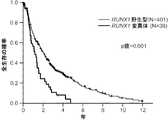
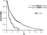
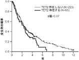
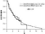
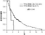
1つの遺伝子において1つまたは複数の非サイレント変異の存在が検出されれば、これらの変異を有しない対象と比較して、対象の全生存が減少することを示している。TET2およびSF3B1の両方に1つまたは複数の変異が存在すれば、TET2変異のみを有しない対象と比較して、対象の全生存が増加することを示している。DNMT3およびSF3B1の両方に1つまたは複数の変異が存在すれば、DNMT3変異のみを有しない対象と比較して、対象の全生存が増加することを示している。対象がRARS型MDSを有し、かつSF3B1に1つまたは複数の変異を有する場合、変異を有しない対象と比較して、対象の全生存が増加することを示している。 Detecting the presence of one or more non-silent mutations in a gene indicates that the subject's overall survival is reduced compared to a subject without these mutations. The presence of one or more mutations in both TET2 and SF3B1 indicates that the overall survival of the subject is increased compared to a subject without only the TET2 mutation. The presence of one or more mutations in both DNMT3 and SF3B1 indicates that the overall survival of the subject is increased compared to a subject without only the DNMT3 mutation. When a subject has RARS MDS and has one or more mutations in SF3B1, it indicates that the subject's overall survival is increased compared to a subject without the mutation.
MDSおよび他のタイプの血液障害を有する対象は、表6から選択される任意の2つもしくはそれより多くの遺伝子における1つもしくは複数の変異、またはETV6、EZH2、RUNX1、ASXL1、DNMT3A、SRSF2、U2AF1、およびSF3B1から選択される任意の1つもしくは複数の遺伝子における1つもしくは複数の変異の存在を検出することによって同定することができ、任意の1つまたは複数の変異が存在すれば、対象がMDSまたはそれに対する素因を有することを示している。 Subjects with MDS and other types of blood disorders are one or more mutations in any two or more genes selected from Table 6, or ETV6, EZH2, RUNX1, ASXL1, DNMT3A, SRSF2, Can be identified by detecting the presence of one or more mutations in any one or more genes selected from U2AF1 and SF3B1, and if any one or more mutations are present, the subject Has a predisposition to MDS.
MDSおよび他のタイプの血液障害の進行、または治療計画の有効性は、表6から選択される2つもしくはそれより多くの遺伝子における変異対立遺伝子頻度、またはETV6、EZH2、RUNX1、ASXL1、DNMT3A、SRSF2、U2AF1、およびSF3B1から選択される任意の1つもしくは複数の遺伝子における変異対立遺伝子頻度を経時的に決定する段階、および少なくとも1つの変異を含む1つまたは複数の遺伝子の変異対立遺伝子頻度を比較する段階によってモニターすることができる。たとえば、第一の試料は、対象が処置を受ける前に得ることができ、1つまたは複数のその後の試料は、対象の処置の後またはあいだに採取される。MDSおよび他のタイプの血液障害は、変異遺伝子における変異対立遺伝子頻度が時間と共に増加すれば進行性(または処置が進行を妨げない)であると見なされるが、変異遺伝子における変異対立遺伝子頻度が、時間が経っても一定のままである場合には、MDSまたは他のタイプの血液障害は進行性ではない。たとえば、本発明の方法は、MDSの侵襲性を識別するおよび/または進行期を評価するために用いることができる。これによって、患者を高または低リスク群に分類して、それに従って処置することができる。たとえば、全生存がより低いと予想されるMDS患者を、アザシチジン(Vidaza(登録商標)、デシタビン(Dacogen(登録商標)、レナリドミド(Revlimid)による処置、または骨髄移植などの、より積極的な治療によって処置することができる。 The progression of MDS and other types of blood disorders, or the effectiveness of the treatment plan, is the frequency of mutant alleles in two or more genes selected from Table 6, or ETV6, EZH2, RUNX1, ASXL1, DNMT3A, Determining the mutated allele frequency over time in any one or more genes selected from SRSF2, U2AF1, and SF3B1, and the mutated allele frequency of one or more genes containing at least one mutation It can be monitored by the stage of comparison. For example, a first sample can be obtained before a subject receives treatment, and one or more subsequent samples are taken after or during treatment of the subject. MDS and other types of blood disorders are considered progressive (or treatment does not interfere with progression) if the mutant allele frequency in the mutant gene increases over time, but the mutant allele frequency in the mutant gene is MDS or other types of blood disorders are not progressive if they remain constant over time. For example, the methods of the present invention can be used to identify the invasiveness of MDS and / or assess progression. This allows patients to be classified into high or low risk groups and treated accordingly. For example, patients with MDS who are expected to have lower overall survival may be treated with more aggressive therapies such as azacitidine (Vidaza®, decitabine (Dacogen®, lenalidomide (Revlimid), or bone marrow transplantation). Can be treated.
さらに、MDSもしくは他のタイプの血液障害を有する対象、またはMDSもしくは他のタイプの血液障害を発症するリスクを有する対象において用いるための処置または治療計画を、対象から得られた試料中の変異遺伝子の変異対立遺伝子頻度に基づいて選択することができる。MDSまたは他のタイプの血液障害の発症を遅らせる、または進行を遅らせるためにどの処置または治療計画が対象において用いるのに最も有効であるかを決定するために、2つまたはそれより多くの処置または治療計画を、同時に評価することができる。 In addition, a mutated gene in a sample obtained from a subject for treatment or treatment planning for use in a subject with MDS or other type of blood disorder or at risk of developing MDS or other type of blood disorder Can be selected based on the frequency of the mutant allele. Two or more treatments or more to determine which treatment or treatment plan is most effective to use in the subject to delay the onset or slow progression of MDS or other types of blood disorders Treatment plans can be evaluated simultaneously.
変異対立遺伝子頻度とは、試料中に存在する変異対立遺伝子の頻度を意味する。変異対立遺伝子頻度は、当技術分野において公知の方法によって決定される。たとえば、コピー数は、リアルタイムポリメラーゼ連鎖反応、一塩基多型(SNP)アレイ、または分裂間期蛍光インサイチューハイブリダイゼーション(FISH)分析によって決定される。 Mutant allele frequency means the frequency of mutant alleles present in a sample. Mutant allele frequency is determined by methods known in the art. For example, copy number is determined by real-time polymerase chain reaction, single nucleotide polymorphism (SNP) arrays, or interphase fluorescence in situ hybridization (FISH) analysis.
「有効な」とは、処置によって、シグネチャー遺伝子における変異対立遺伝子頻度が減少することを意味する。本明細書において開示される危険因子の評価は、標準的な臨床プロトコールを用いて行うことができる。有効性は、血液障害を診断、同定、または処置するために任意の公知の方法に関連して決定することができる。 “Effective” means that the treatment reduces the mutation allele frequency in the signature gene. The assessment of risk factors disclosed herein can be performed using standard clinical protocols. Efficacy can be determined in connection with any known method for diagnosing, identifying, or treating a blood disorder.
MDS患者に関する処置の決定に関する情報は、患者由来の試料中のシグネチャー遺伝子のいずれか1つにおける1つまたは複数の変異に関する情報を得る段階、およびシグネチャー遺伝子における変異対立遺伝子頻度が臨床的に有意に変化する場合、患者におけるMDSを予防または減少させる治療計画を選択する段階によって得ることができる。 Information on treatment decisions for MDS patients can be obtained when obtaining information about one or more mutations in any one of the signature genes in a sample from the patient, and the mutation allele frequency in the signature gene is clinically significant If so, it can be obtained by selecting a treatment plan that prevents or reduces MDS in the patient.
本発明はまた、1つまたは複数のシグネチャー核酸に結合する検出試薬を有するキットを含む。同様に、検出試薬のアレイ、たとえば1つまたは複数のシグネチャー核酸に結合することができるオリゴヌクレオチドのアレイも本発明によって提供される。キットはまた、1つまたは複数のシグネチャー遺伝子における変異を検出するための1つまたは複数の試薬、たとえば質量分析による遺伝子型決定のためのプライマー、および対象の試料中の1つまたは複数の変異シグネチャー遺伝子における変異対立遺伝子頻度を決定するための1つまたは複数の試薬を含む。 The invention also includes a kit having a detection reagent that binds to one or more signature nucleic acids. Similarly, an array of detection reagents, eg, an array of oligonucleotides that can bind to one or more signature nucleic acids is also provided by the present invention. The kit also includes one or more reagents for detecting mutations in one or more signature genes, such as primers for genotyping by mass spectrometry, and one or more mutation signatures in a sample of interest. Contains one or more reagents for determining the mutation allele frequency in a gene.
特定の遺伝子または染色体領域における変異対立遺伝子頻度を評価する方法は、当業者に周知であり、これにはハイブリダイゼーションに基づくアッセイおよび増幅に基づくアッセイが挙げられる。 Methods for assessing mutant allele frequencies in specific genes or chromosomal regions are well known to those of skill in the art and include hybridization-based assays and amplification-based assays.
ハイブリダイゼーションに基づくアッセイ
ハイブリダイゼーションに基づくアッセイは、サザンブロットまたはインサイチューハイブリダイゼーション(たとえばFISH)などの従来の「直接プローブ」法、および比較ゲノムハイブリダイゼーション(CGH)などの「比較プローブ」法を含むがこれらに限定されるわけではない。方法は、以下に記述されるように基質(たとえば、メンブレンまたはガラス)結合法またはアレイに基づくアプローチを含むがこれらに限定されるわけではない広く多様なフォーマットで用いることができる。Hybridization-based assays Hybridization-based assays include traditional “direct probe” methods such as Southern blots or in situ hybridization (eg FISH), and “comparison probe” methods such as comparative genomic hybridization (CGH). However, it is not limited to these. The methods can be used in a wide variety of formats including, but not limited to, substrate (eg, membrane or glass) binding methods or array based approaches as described below.
インサイチューハイブリダイゼーションアッセイは周知である(たとえば、Angerer (1987) Meth. Enzymol 152: 649)。一般的に、インサイチューハイブリダイゼーションは、以下の主要な段階を含む:(1)分析される組織または生物構造の固定;(2)標的DNAの接触性を増加させるためおよび非特異的結合を減少させるための生物構造のハイブリダイゼーション前処置;(3)核酸混合物と、生物構造または組織中の核酸とのハイブリダイゼーション;(4)ハイブリダイゼーションにおいて結合していない核酸断片を除去するためのハイブリダイゼーション後洗浄、および(5)ハイブリダイズした核酸断片の検出。これらの段階の各々において用いられる試薬および用いるための条件は、具体的な応用に応じて異なる。 In situ hybridization assays are well known (eg, Angerer (1987) Meth. Enzymol 152: 649). In general, in situ hybridization involves the following major steps: (1) fixation of the tissue or biological structure being analyzed; (2) to increase target DNA accessibility and to reduce non-specific binding. Pre-hybridization of biological structures for; (3) hybridization of nucleic acid mixtures with nucleic acids in biological structures or tissues; (4) post-hybridization washing to remove unbound nucleic acid fragments in the hybridization. And (5) detection of hybridized nucleic acid fragments. The reagents used in each of these stages and the conditions for use vary depending on the specific application.
典型的なインサイチューハイブリダイゼーションアッセイにおいて、細胞は固相支持体、典型的にスライドガラスに固定される。核酸がプロービングされる場合、細胞を典型的に、熱またはアルカリによって変性させる。次に、細胞を中等度の温度のハイブリダイゼーション溶液に接触させて、タンパク質をコードする核酸配列に対して特異的な標識プローブをアニールさせる。次に、標的(たとえば、細胞)を、既定のストリンジェンシーで、または適切なシグナル対ノイズ比が得られるまでストリンジェンシーを増加させて、洗浄する。 In a typical in situ hybridization assay, cells are fixed to a solid support, typically a glass slide. When nucleic acids are probed, cells are typically denatured by heat or alkali. The cells are then contacted with a moderate temperature hybridization solution to anneal the labeled probe specific for the nucleic acid sequence encoding the protein. The target (eg, cell) is then washed with a predetermined stringency or with increasing stringency until an appropriate signal to noise ratio is obtained.
プローブは典型的に、たとえば放射性同位元素または蛍光レポーターによって標識される。好ましいサイズの範囲は、約200 bpから約1000塩基、より好ましくは二本鎖、ニック翻訳核酸に関して約400から約800 bpである。 The probe is typically labeled with, for example, a radioisotope or a fluorescent reporter. A preferred size range is from about 200 bp to about 1000 bases, more preferably double stranded, about 400 to about 800 bp for nick translated nucleic acids.
いくつかの応用において、反復配列のハイブリダイズ能を阻止することが必要である。このように、非特異的ハイブリダイゼーションを阻止するために、ヒトゲノムDNAまたはCot-1 DNAを用いる。 In some applications it is necessary to block the hybridizing ability of repetitive sequences. Thus, human genomic DNA or Cot-1 DNA is used to prevent non-specific hybridization.
比較ゲノムハイブリダイゼーション法において、(試料)核酸(たとえば、可能性がある腫瘍から)の第一のコレクションを第一の標識によって標識して、(対照)核酸(たとえば、健康な細胞/組織から)の第二のコレクションを第二の標識によって標識する。核酸のハイブリダイゼーションの比率を、2つの(第一および第二の)標識がアレイにおける各々の繊維に結合する比率によって決定する。染色体の欠失または重複が存在する場合、2つの標識からのシグナルの比率の差を検出すれば、比率は、コピー数の測定値を提供するであろう。 In comparative genomic hybridization methods, a first collection of (sample) nucleic acids (eg, from a potential tumor) is labeled with a first label, and (control) nucleic acids (eg, from healthy cells / tissues) The second collection of is labeled with a second label. The rate of nucleic acid hybridization is determined by the rate at which two (first and second) labels bind to each fiber in the array. If a chromosomal deletion or duplication is present, detecting the difference in the ratio of signals from the two labels will provide a measure of copy number.
本発明の方法と共に用いるために適した他のハイブリダイゼーションプロトコールは、たとえばAlbertson (1984) EMBO J. 3: 1227-1234; Pinkel (1988) Proc. Natl. Acad. Sci. USA 85: 9138-9142;欧州特許出願公開第430,402号; Methods in Molecular Biology, Vol. 33: In Situ Hybridization Protocols, Choo, ed., Humana Press, Totowa, N.J. (1994)等に記述されている。 Other hybridization protocols suitable for use with the methods of the present invention are, for example, Albertson (1984) EMBO J. 3: 1227-1234; Pinkel (1988) Proc. Natl. Acad. Sci. USA 85: 9138-9142; European Patent Application Publication No. 430,402; Methods in Molecular Biology, Vol. 33: In Situ Hybridization Protocols, Choo, ed., Humana Press, Totowa, NJ (1994).
本発明の方法は、アレイに基づくハイブリダイゼーションフォーマットにとって特によく適している。アレイは、1つまたは複数の表面(たとえば、固体、メンブレン、またはゲル)に結合した、多数の異なる「プローブ」または「標的」核酸(または他の化合物)である。多数の核酸(または他の部分)が、1つの連続する表面に、または互いに近接する多数の表面に結合する。 The method of the invention is particularly well suited for array-based hybridization formats. An array is a number of different “probes” or “target” nucleic acids (or other compounds) bound to one or more surfaces (eg, solids, membranes, or gels). Numerous nucleic acids (or other moieties) bind to one continuous surface or to many surfaces in close proximity to each other.
アレイのフォーマットでは、多数の異なるハイブリダイゼーション反応を本質的に「同時に」行うことができる。これは、多数のハイブリダイゼーションの迅速で本質的に同時の評価を1回の「実験」で提供する。アレイに基づくフォーマットでハイブリダイゼーション反応を行う方法は、当業者に周知である(たとえば、Pastinen (1997) Genome Res. 7: 606-614; Jackson (1996) Nature Biotechnology 14: 1685; Chee (1995) Science 274: 610; WO 96/17958を参照されたい)。 In an array format, a number of different hybridization reactions can be performed essentially “simultaneously”. This provides a rapid and essentially simultaneous assessment of multiple hybridizations in a single “experiment”. Methods for performing hybridization reactions in an array-based format are well known to those skilled in the art (eg, Pastinen (1997) Genome Res. 7: 606-614; Jackson (1996) Nature Biotechnology 14: 1685; Chee (1995) Science 274: 610; see WO 96/17958).
アレイ、特に核酸アレイは、当業者に周知の広く多様な方法に従って作製することができる。たとえば、単純な態様において、「低密度」アレイは、固相支持体(たとえば、ガラス表面、メンブレン等)上の異なる位置に異なる核酸をスポットすることによって(たとえば、ピペットを用いて手で)単純に作製することができる。 Arrays, particularly nucleic acid arrays, can be made according to a wide variety of methods well known to those skilled in the art. For example, in a simple embodiment, a “low density” array is simple by spotting different nucleic acids at different locations on a solid support (eg, glass surface, membrane, etc.) (eg, by hand with a pipette). Can be produced.
この単純なスポッティングアプローチは、高密度スポットアレイを生じるために自動化されている(たとえば、米国特許第5,807,522号を参照されたい)。この特許は、少量の生物試料を沈着させるために表面に対して微小毛細管をタップする自動システムを用いることを記述する。このプロセスを繰り返して高密度アレイを作製する。アレイはまた、オリゴヌクレオチド合成技術を用いても作製することができる。このように、たとえば米国特許第5,143,854号およびPCT特許公開公報WO90/15070および92/10092は、高密度オリゴヌクレオチドアレイの光指向性コンビナトリアル合成を用いることを教示している。 This simple spotting approach has been automated to produce high density spot arrays (see, eg, US Pat. No. 5,807,522). This patent describes using an automated system that taps a microcapillary against a surface to deposit a small amount of biological sample. This process is repeated to produce a high density array. Arrays can also be made using oligonucleotide synthesis techniques. Thus, for example, US Pat. No. 5,143,854 and PCT Patent Publications WO90 / 15070 and 92/10092 teach the use of light-directed combinatorial synthesis of high-density oligonucleotide arrays.
スポットアレイは、ゲノムDNA、たとえば関心対象領域に対応するアンプリコンの高解像度スキャンを提供する重なり合うクローンを含むことができる。アンプリコン核酸は、たとえばMACs、YACs、BACs、PACs、Pls、コスミド、プラスミド、ゲノムクローンのinter-Alu PCR産物、ゲノムクローンの制限消化物、cDNAクローン、増幅(たとえばPCR)産物、およびその他から得ることができる。 The spot array can include overlapping clones that provide high resolution scans of genomic DNA, eg, amplicons corresponding to the region of interest. Amplicon nucleic acids are obtained from, for example, MACs, YACs, BACs, PACs, Pls, cosmids, plasmids, genomic clone inter-Alu PCR products, genomic clone restriction digests, cDNA clones, amplification (eg, PCR) products, and others be able to.
アレイ核酸は、本発明の標的配列に及ぶまたは標的配列を含むクローンの既にマッピングされたライブラリ、ならびに以下に記述されるゲノムの他の領域からのクローンに由来する。アレイは、1つの試料核酸集団とハイブリダイズすることができ、または(試験試料および参照試料に関して)2つの異なるように標識されたコレクションと共に用いることができる。 Array nucleic acids are derived from already mapped libraries of clones that span or contain the target sequence of the invention, as well as clones from other regions of the genome described below. The array can hybridize with one sample nucleic acid population or can be used with two differently labeled collections (with respect to the test sample and reference sample).
多様な固体表面上に核酸を固定するための多くの方法が当技術分野において公知である。天然および合成の他の材料のみならず、広く多様な有機および無機ポリマーを、固体表面の材料として使用することができる。例示的な固体表面には、たとえばニトロセルロース、ナイロン、ガラス、石英、ジアゾ化メンブレン(紙またはナイロン)、シリコン、ポリホルムアルデヒド、セルロース、および酢酸セルロースが挙げられる。さらに、ポリエチレン、ポリプロピレン、ポリスチレン、およびその他などのプラスチックを用いることができる。用いられうる他の材料には、紙、セラミクス、金属、メタロイド、半導体材料、サーメット、またはその他が挙げられる。さらに、ゲルを形成する物質を用いることができる。そのような材料には、たとえばタンパク質(たとえばゼラチン)、リポ多糖類、ケイ酸塩、アガロースおよびポリアクリルアミドが挙げられる。固体表面が多孔性である場合、システムの性質に応じて様々な孔のサイズを使用してもよい。 Many methods are known in the art for immobilizing nucleic acids on a variety of solid surfaces. A wide variety of organic and inorganic polymers can be used as solid surface materials, as well as other natural and synthetic materials. Exemplary solid surfaces include, for example, nitrocellulose, nylon, glass, quartz, diazotized membrane (paper or nylon), silicon, polyformaldehyde, cellulose, and cellulose acetate. In addition, plastics such as polyethylene, polypropylene, polystyrene, and others can be used. Other materials that can be used include paper, ceramics, metals, metalloids, semiconductor materials, cermets, or others. Further, a substance that forms a gel can be used. Such materials include, for example, proteins (eg gelatin), lipopolysaccharides, silicates, agarose and polyacrylamide. If the solid surface is porous, various pore sizes may be used depending on the nature of the system.
ハイブリダイゼーション複合体を検出するには、標的およびプローブポリヌクレオチドまたは核酸の二重鎖にシグナル生成複合体が結合する必要がありうる。典型的に、そのような結合は、リガンド結合プローブとシグナルに結合した抗リガンドとの相互作用などといった、リガンドと抗リガンドとの相互作用を通して起こる。 In order to detect the hybridization complex, the signal generating complex may need to bind to the duplex of the target and probe polynucleotide or nucleic acid. Typically, such binding occurs through the interaction between the ligand and the anti-ligand, such as the interaction between the ligand-binding probe and the signal-bound anti-ligand.
ハイブリダイゼーションアッセイの感度は、検出される標的核酸を増加する核酸増幅システムを用いることを通して増強されうる。そのようなシステムの例には、ポリメラーゼ連鎖反応(PCR)システムおよびリガーゼ連鎖反応(LCR)システムが挙げられる。当技術分野において最近記述された他の方法は、核酸配列ベース増幅(NASBAO, Cangene, Mississauga, Ontario)、およびQβレプリカーゼシステムである。 The sensitivity of the hybridization assay can be enhanced through the use of a nucleic acid amplification system that increases the target nucleic acid to be detected. Examples of such systems include the polymerase chain reaction (PCR) system and the ligase chain reaction (LCR) system. Other methods recently described in the art are nucleic acid sequence-based amplification (NASBAO, Cangene, Mississauga, Ontario), and the Qβ replicase system.
ハイブリダイゼーション条件を最適化する方法は当業者に周知である(たとえば、Tijssen (1993) Laboratory Techniques in Biochemistry and Molecular Biology, Vol. 24: Hybridization With Nucleic Acid Probes, Elsevier, N.Y.を参照されたい)。 Methods for optimizing hybridization conditions are well known to those skilled in the art (see, eg, Tijssen (1993) Laboratory Techniques in Biochemistry and Molecular Biology, Vol. 24: Hybridization With Nucleic Acid Probes, Elsevier, N.Y.).
増幅に基づくアッセイ
もう1つの態様において、増幅に基づくアッセイを用いて変異対立遺伝子頻度を測定することができる。そのような増幅に基づくアッセイにおいて、核酸配列は、増幅反応(たとえば、ポリメラーゼ連鎖反応(PCR))における鋳型として作用する。定量的増幅において、増幅産物の量は、当初の試料中の鋳型の量に比例するであろう。適切な対照(たとえば、健康な組織)との比較により、所望の標的核酸配列の変異対立遺伝子頻度の測定が提供される。「定量的」増幅法は当業者に周知である。たとえば、定量的PCRは、同じプライマーを用いて対照配列の公知の量を同時に共増幅する段階を伴う。これは、PCR反応を較正するために用いられうる内部標準を提供する。定量的PCRの詳細なプロトコールは、Innis et al. (1990) PCR Protocols, A Guide to Methods and Applications, Academic Press, Inc. N.Y.において提供される。Amplification-based assays In another embodiment, an amplification-based assay can be used to measure mutant allele frequency. In such amplification-based assays, the nucleic acid sequence acts as a template in an amplification reaction (eg, polymerase chain reaction (PCR)). In quantitative amplification, the amount of amplification product will be proportional to the amount of template in the original sample. Comparison with an appropriate control (eg, healthy tissue) provides a measure of the mutant allele frequency of the desired target nucleic acid sequence. “Quantitative” amplification methods are well known to those of skill in the art. For example, quantitative PCR involves co-amplifying known amounts of control sequences simultaneously using the same primers. This provides an internal standard that can be used to calibrate the PCR reaction. Detailed protocols for quantitative PCR are provided in Innis et al. (1990) PCR Protocols, A Guide to Methods and Applications, Academic Press, Inc. NY.
他の適した増幅法には、リガーゼ連鎖反応(LCR)(Wu and Wallace (1989) Genomics 4: 560, Landegren et al. (1988) Science 241: 1077, and Barringer et al. (1990) Gene 89: 117を参照されたい);転写増幅(Kwoh et al. (1989) Proc. Natl. Acad. Sci. USA 86: 1173):および自家持続配列複製(Guatelli et al. (1990) Proc. Nat. Acad. Sci. USA 87: 1874)が挙げられるがこれらに限定されるわけではない。 Other suitable amplification methods include ligase chain reaction (LCR) (Wu and Wallace (1989) Genomics 4: 560, Landegren et al. (1988) Science 241: 1077, and Barringer et al. (1990) Gene 89: 117); transcription amplification (Kwoh et al. (1989) Proc. Natl. Acad. Sci. USA 86: 1173): and self-sustained sequence replication (Guatelli et al. (1990) Proc. Nat. Acad. Sci. USA 87: 1874), but is not limited to these.
特定の遺伝子または染色体領域の体細胞変異を検出する方法は当業者に周知であり、これには質量分析による遺伝子型決定および次世代ピロシークエンシングが挙げられる。個々の遺伝子または染色体における変異の存在を検出するために、多様な方法が利用可能である。この分野での進歩により、正確かつ容易で安価な大規模遺伝子型決定が提供されている。より最近では、たとえば動的対立遺伝子特異的ハイブリダイゼーション(DASH)、マイクロプレートアレイ直交ゲル電気泳動(MADGE)、ピロシークエンシング、オリゴヌクレオチド特異的ライゲーション、TaqManシステムならびにAffymetrix SNPチップなどの様々なDNA「チップ」技術を含むいくつかの新しい技術が記述されている。これらの方法は、典型的にPCRによる標的遺伝子領域の増幅を必要とする。侵襲性切断によって小さいシグナル分子が生成された後に質量分析または固定パドロックプローブおよびローリングサークル増幅を行うなお他の新たに開発された方法により、最終的にPCRの必要性がなくなる可能性がある。特異的な一塩基多型を検出するために当技術分野において公知の方法のいくつかを以下に要約する。本発明の方法は、利用可能な全ての方法を含むと理解される。 Methods for detecting somatic mutations in a particular gene or chromosomal region are well known to those of skill in the art and include genotyping by mass spectrometry and next generation pyrosequencing. A variety of methods are available for detecting the presence of mutations in individual genes or chromosomes. Advances in this area have provided accurate, easy and inexpensive large-scale genotyping. More recently, a variety of DNA " Several new technologies have been described, including “chip” technology. These methods typically require amplification of the target gene region by PCR. Still other newly developed methods of performing mass spectrometry or fixed padlock probes and rolling circle amplification after invasive cleavage produces small signal molecules may ultimately eliminate the need for PCR. Some of the methods known in the art for detecting specific single nucleotide polymorphisms are summarized below. It is understood that the method of the present invention includes all available methods.
タンパク質翻訳の早期の終了を生じる変異に関して、タンパク質切断試験(PTT)は、有効な診断アプローチを提供する(Roest, et. al., (1993) Hum. Mol. Genet. 2: 1719-21 ; van der Luijt, et. al., (1994) Genomics 20: 1-4)。PTTに関して、まず、RNAを、利用可能な組織から単離し、逆転写し、関心対象セグメントをPCRによって増幅する。次に、逆転写PCRの産物を、RNAポリメラーゼプロモーターおよび真核細胞の翻訳を開始させるための配列を含むプライマーと共にネステッドPCR増幅の鋳型として用いる。関心対象領域の増幅後、プライマーに組み入れられる特有のモチーフにより、PCR産物の連続的なインビトロ転写および翻訳が可能となる。翻訳産物のドデシル硫酸ナトリウムポリアクリルアミドゲル電気泳動による、切断されたポリペプチドの出現は、早期の翻訳終止を引き起こす変異が存在すること示す。この技術の変化形では、関心対象標的領域が1つのエキソンに由来する場合には、PCR鋳型としてDNA(RNAではなくて)を用いる。 For mutations that result in early termination of protein translation, the protein truncation test (PTT) provides an effective diagnostic approach (Roest, et. Al., (1993) Hum. Mol. Genet. 2: 1719-21; van der Luijt, et. al., (1994) Genomics 20: 1-4). For PTT, RNA is first isolated from available tissue, reverse transcribed, and the segment of interest amplified by PCR. The product of reverse transcription PCR is then used as a template for nested PCR amplification along with an RNA polymerase promoter and primers containing sequences for initiating eukaryotic translation. After amplification of the region of interest, the unique motif incorporated into the primer allows continuous in vitro transcription and translation of the PCR product. The appearance of cleaved polypeptides by translation product sodium dodecyl sulfate polyacrylamide gel electrophoresis indicates the presence of mutations that cause premature translation termination. In a variation of this technique, if the target region of interest is derived from one exon, DNA (not RNA) is used as a PCR template.
単なる例示的な態様において、方法は、(i)患者から細胞試料を収集する段階、(ii)試料の細胞から核酸(たとえば、ゲノム、mRNA、または両方)を単離する段階、(iii)対立遺伝子のハイブリダイゼーションと増幅が起こるような条件下で、表6に記載される1つのシグネチャー遺伝子の少なくとも1つの対立遺伝子の5'および3'に特異的にハイブリダイズする1つまたは複数のプライマーを、核酸試料に接触させる段階、ならびに(iv)増幅産物を検出する段階を含む。これらの検出スキームは、そのような分子が非常に少数で存在する場合に、核酸分子を検出するために特に有用である。 In merely exemplary embodiments, the method comprises (i) collecting a cell sample from a patient, (ii) isolating nucleic acid (eg, genome, mRNA, or both) from cells of the sample, (iii) alleles One or more primers that specifically hybridize to 5 ′ and 3 ′ of at least one allele of one signature gene listed in Table 6 under conditions that allow for hybridization and amplification of the gene Contacting the nucleic acid sample, and (iv) detecting the amplification product. These detection schemes are particularly useful for detecting nucleic acid molecules when such molecules are present in very small numbers.
本発明のいくつかの局面において、対立遺伝子を直接シークエンシングするために、当技術分野において公知の多様な任意のシークエンシング反応を用いることができる。例示的なシークエンシング反応は、マクサム-ギルバート((1977) Proc. Natl Acad Sci USA 74:560)またはサンガー(Sanger et al (1977) Proc. Nat. Acad. Sci USA 74:5463)によって開発された技術に基づく反応である。同様に、質量分析によるシークエンシング(たとえば、PCT公開番号WO 94/16101 ; Cohen et al. (1996) Adv Chromatogr 36: 127-162; and Griffin et al. (1993) Appl Biochem Biotechnol 38:147-159を参照されたい)を含む対象アッセイ(たとえば、Biotechniques (1995) 19:448を参照されたい)を行う場合には、任意の多様な自動シークエンシング技法を利用してもよいと企図される。ある態様に関して、シークエンシング反応において決定する必要があるのは、核酸塩基の1個のみ、2個、または3個の存在であることは当業者に明らかであろう。例として、たとえば1個のみの核酸が検出される場合、A-トラックまたはその他を行うことができる。 In some aspects of the invention, any of a variety of sequencing reactions known in the art can be used to sequence alleles directly. Exemplary sequencing reactions were developed by Maxam-Gilbert ((1977) Proc. Natl Acad Sci USA 74: 560) or Sanger (Sanger et al (1977) Proc. Nat. Acad. Sci USA 74: 5463). It is a reaction based on technology. Similarly, sequencing by mass spectrometry (eg, PCT Publication No. WO 94/16101; Cohen et al. (1996) Adv Chromatogr 36: 127-162; and Griffin et al. (1993) Appl Biochem Biotechnol 38: 147-159 It is contemplated that any of a variety of automated sequencing techniques may be utilized when performing subject assays (see, for example, Biotechniques (1995) 19: 448). It will be apparent to one skilled in the art that for certain embodiments, only one, two, or three nucleobases need to be determined in a sequencing reaction. By way of example, if only one nucleic acid is detected, an A-track or others can be performed.
本発明の診断および予後指標
本発明は、いくつかのタイプの中でもMDSなどの血液障害の予後および診断を可能にする。MDSなどの疾患に罹患している対象がより低い全生存を有するリスクは、試験試料中のETV6、EZH2、RUNX1、ASXL1、DNMT3A、SRSF2、U2AF1、およびSF3B1から選択される任意の1つもしくは複数の遺伝子における1つまたは複数の変異を検出することによって決定することができる。全生存が減少するリスクを有すると同定された対象を任意で、対象の疾患の進行を遅らせる、減少させる、または予防するために、アザシチジン(Vidaza(登録商標)、デシタビン(Dacogen(登録商標)、レナリドミド(Revlimid)の投与、または骨髄移植を受けることなどのより積極的な治療計画を受けるように選択することができる。Diagnosis and Prognostic Indicators of the Invention The present invention allows for the prognosis and diagnosis of blood disorders such as MDS, among other types. The risk that a subject suffering from a disease such as MDS has a lower overall survival is any one or more selected from ETV6, EZH2, RUNX1, ASXL1, DNMT3A, SRSF2, U2AF1, and SF3B1 in the test sample Can be determined by detecting one or more mutations in the gene. Optionally, azacitidine (Vidaza®, decitabine (Dacogen®), a subject identified to be at risk of decreasing overall survival, to slow, reduce or prevent progression of the subject's disease One can choose to receive a more aggressive treatment regimen such as taking lenalidomide (Revlimid) or receiving a bone marrow transplant.
本発明は、表6から選択される2つもしくはそれより多くの遺伝子における1つもしくは複数の変異、またはETV6、EZH2、RUNX1、ASXL1、DNMT3A、SRSF2、U2AF1、およびSF3B1から選択される任意の1つもしくは複数の遺伝子における1つまたは複数の変異を検出することによって、対象を診断するために用いられうる。 The invention relates to one or more mutations in two or more genes selected from Table 6, or any one selected from ETV6, EZH2, RUNX1, ASXL1, DNMT3A, SRSF2, U2AF1, and SF3B1 It can be used to diagnose a subject by detecting one or more mutations in one or more genes.
シグネチャー遺伝子の変異は、次世代シークエンシングまたは質量分析による遺伝子型決定によって検出することができる。ETV6、EZH2、RUNX1、ASXL1、DNMT3A、SRSF2、U2AF1、およびSF3B1から選択される任意の1つまたは複数の遺伝子に1つまたは複数の非サイレント変異が存在すれば、MDSなどの血液障害に罹患している対象の全生存が減少するリスクを示している。 Signature gene mutations can be detected by next generation sequencing or genotyping by mass spectrometry. If you have one or more non-silent mutations in any one or more genes selected from ETV6, EZH2, RUNX1, ASXL1, DNMT3A, SRSF2, U2AF1, and SF3B1, you will suffer from a blood disorder such as MDS The risk that the overall survival of the subject will be reduced.
変異したシグネチャー遺伝子における変異対立遺伝子頻度によって、MDSなどの血液障害の処置の経過をモニターすることができる。この方法では、生物試料を、治療計画、たとえばMDSに関する薬物処置を受ける対象から提供することができる。必要に応じ、生物試料は、処置の前、あいだ、または後の様々な時点で対象から得られる。 The course of treatment for blood disorders such as MDS can be monitored by the frequency of mutant alleles in the mutated signature genes. In this method, a biological sample can be provided from a subject undergoing a treatment plan, eg, drug treatment for MDS. If desired, biological samples are obtained from the subject at various time points before, during, or after treatment.
変異シグネチャー遺伝子における変異対立遺伝子頻度は、当技術分野において公知の任意の方法によって、たとえばリアルタイムポリメラーゼ連鎖反応、一塩基多型(SNP)アレイ、または分裂間期蛍光インサイチューハイブリダイゼーション(FISH)分析によって決定することができる。 Mutation allele frequencies in mutation signature genes are determined by any method known in the art, for example, by real-time polymerase chain reaction, single nucleotide polymorphism (SNP) arrays, or interphase fluorescence in situ hybridization (FISH) analysis can do.
本発明はまた、任意の数の状況において患者または対象集団をスクリーニングするために用いることができる。たとえば、健康維持機構、公衆衛生団体または学校保健プログラムは、先に記述したように、介入を必要とする対象を同定するために、または疫学的データを収集するために、対象の群をスクリーニングしうる。保険会社(たとえば、健康、生命、または障害保険)は、可能性がある介入に関する適用範囲または価格決定、または既存のクライアントを決定するプロセスにおいて申込者をスクリーニングしうる。そのような集団において収集されたデータは、特に癌または転移事象のような状態への任意の臨床上の進行と結びつく場合、たとえば健康維持機構、公衆衛生プログラムおよび保険会社の事業において貴重であろう。そのようなデータのアレイまたはコレクションを、機器読み取り可能な媒体に保存して、医療サービスの向上、費用効果の高い医療、保険事業の向上等を提供するために、任意の数の健康関連データ管理システムにおいて用いることができる。たとえば、米国特許出願第2002/0038227号;米国特許出願第2004/0122296号;米国特許出願第2004/ 0122297号;および米国特許第5,018,067号を参照されたい。そのようなシステムは、本明細書においてさらに詳細に記述されるように、内部データ保存場所から直接、または離れた1つもしくは複数のデータ保存サイトから、データにアクセスすることができる。 The present invention can also be used to screen patients or subject populations in any number of situations. For example, a health maintenance organization, public health organization, or school health program may screen a group of subjects, as described above, to identify subjects that require intervention or to collect epidemiological data. sell. Insurers (eg, health, life, or disability insurance) may screen applicants in the process of determining coverage or pricing for potential interventions, or existing clients. Data collected in such populations may be valuable, for example, in health maintenance organizations, public health programs and insurance company operations, especially when linked to any clinical progression to conditions such as cancer or metastatic events . Manage any number of health-related data to store such an array or collection of data on a device-readable medium to provide improved medical services, cost-effective medical care, improved insurance business, etc. Can be used in the system. See, for example, U.S. Patent Application No. 2002/0038227; U.S. Patent Application No. 2004/0122296; U.S. Patent Application No. 2004/0122297; and U.S. Patent No. 5,018,067. Such a system can access data directly from an internal data storage location or from one or more data storage sites as described in more detail herein.
機器読み取り可能な保存媒体は、データを用いるための命令によってプログラムされた機器を用いる場合、経時的なMDS危険因子または薬物治療に反応したMDS危険因子などの血液障害に関連する対象の情報などの、しかしこれらに限定されない多様な目的のために用いることができる機器読み取り可能なデータまたはデータアレイによって暗号化されたデータ保存材料を含むことができる。シグネチャー遺伝子の変異、および本発明の少なくとも1つの変異を有するシグネチャー遺伝子における変異対立遺伝子頻度、および/またはそれらのバイオマーカーから得られたリスクの評価は、とりわけプロセッサ、データ保存システム(揮発性および非揮発性メモリおよび/または保存エレメント)、少なくとも1つの入力装置、および少なくとも1つの出力装置を含む、プログラム可能なコンピューター上で実行されるコンピュータープログラムにおいて実行することができる。入力したデータにプログラム暗号を適用して、上記の機能を行うことができ、出力情報を生成することができる。出力情報は、当技術分野において公知の方法に従って、1つまたは複数の出力装置に適用することができる。コンピューターは、たとえばパーソナルコンピューター、マイクロコンピューター、または通常設計のワークステーションでありうる。 Device-readable storage media, such as information on subjects related to blood disorders, such as MDS risk factors over time or MDS risk factors in response to medication, when using devices programmed with instructions for using the data However, it can include device readable data or data storage material encrypted with a data array that can be used for a variety of purposes, including but not limited to these. Signature gene mutations, and mutation allele frequencies in signature genes having at least one mutation of the present invention, and / or assessment of risk obtained from their biomarkers, among others, processors, data storage systems (volatile and non-volatile) Volatile memory and / or storage elements), at least one input device, and at least one output device can be executed in a computer program running on a programmable computer. By applying program encryption to the input data, the above functions can be performed, and output information can be generated. The output information can be applied to one or more output devices according to methods known in the art. The computer can be, for example, a personal computer, a microcomputer, or a normally designed workstation.
各プログラムは、コンピューターシステムと通信するための高レベル手続き型またはオブジェクト指向プログラム言語において実行することができる。しかし、プログラムは、必要に応じアセンブリまたはマシン言語で実行することができる。言語は、コンパイラまたはインタプリタ言語でありうる。各々のそのようなコンピュータープログラムは、保存媒体または装置が本明細書において記述される技法を行うためにコンピューターによって読み取られる場合にコンピューターを設定および操作するように、一般的または特殊な目的のプログラム可能なコンピューターによって読み取り可能な保存媒体または装置(たとえば、ROMまたは磁気ディスケットまたは本開示において他所で定義されるその他)に保存することができる。本発明の健康関連データ管理システムはまた、そのように設定された保存媒体がコンピューターに、本明細書において記述される様々な機能を行うように、特異的および既定の様式で操作させる場合、コンピュータープログラムによって設定されるコンピューター読み取り可能な保存媒体として実行されると見なされうる。 Each program can execute in a high level procedural or object oriented programming language for communicating with a computer system. However, the program can be executed in assembly or machine language as required. The language can be a compiler or interpreted language. Each such computer program can be programmed for general or special purpose to set up and operate the computer when the storage medium or device is read by the computer to perform the techniques described herein. Can be stored on any computer readable storage medium or device (eg, ROM or magnetic diskette or others as defined elsewhere in this disclosure). The health-related data management system of the present invention can also be used when a storage medium so configured causes a computer to operate in a specific and predefined manner to perform various functions described herein. It can be considered to be executed as a computer-readable storage medium set by a program.
対象の遺伝的構成の差によって、MDSなどの血液障害の症状または危険因子を調節しうる様々な薬物の相対的代謝能の差が起こりうる。MDSを有するまたはMDSを発症するリスクを有する対象は、年齢、民族、および他のパラメータが多様でありうる。したがって、本明細書において開示されるシグネチャーを用いることにより、単独でおよび薬物代謝に関する公知の遺伝的要因と共に、選択された対象において試験される推定の治療または予防が対象における癌または転移事象を処置または予防するために適しているであろうという既定の予測可能性レベルが可能となる。 Differences in the genetic makeup of the subject can cause differences in the relative metabolic capacity of various drugs that can modulate blood disorder symptoms or risk factors such as MDS. Subjects with MDS or at risk of developing MDS can have varying age, ethnicity, and other parameters. Thus, by using the signatures disclosed herein, putative treatment or prevention tested in selected subjects, alone and together with known genetic factors for drug metabolism, treat cancer or metastatic events in the subject Or a predefined level of predictability that would be suitable for prevention is possible.
本発明の上記の方法は、MDSなどの血液障害と診断されている、および薬物処置を受けた対象の進行および/または改善を評価またはモニターするために用いることができる。 The above methods of the invention can be used to assess or monitor the progression and / or improvement of a subject diagnosed with a blood disorder such as MDS and who has undergone drug treatment.
本発明の成績および精度測定
本発明の成績ならびにこのように絶対的および相対的臨床有用性は、先に述べたように多数の方法で評価されうる。成績の様々な評価において、本発明は臨床診断および予後の精度を提供すると意図される。診断または予後に関する試験、アッセイ、または方法の精度は、試験、アッセイ、または方法が、MDSを有する対象またはより低い全生存のリスクを有する対象を識別できることに関係し、対象が表6に記載される1つまたは複数のシグネチャー遺伝子において非サイレント変異を有するか否かに基づく。いくつかの態様において、1つのみのシグネチャー遺伝子における1つまたは複数の変異は、リスクの統計学的に有意な評価を提供することができる。以下に示すように、そして本発明を何ら制限することなく、統計学的有意性に達すること、したがって好ましい分析、診断、および臨床精度は、必ずしもいくつかのシグネチャーの組み合わせを共に用いることを必要としない。数学的アルゴリズムは、統計学的に有意な指標を達成するために必ずしも必要ではない。Performance and Accuracy Measurements of the Invention The performance of the invention and thus the absolute and relative clinical utility can be evaluated in a number of ways as described above. In various assessments of performance, the present invention is intended to provide clinical diagnosis and prognostic accuracy. The accuracy of a test, assay, or method for diagnosis or prognosis relates to the ability of the test, assay, or method to identify subjects with MDS or subjects with a lower overall survival risk, and subjects are listed in Table 6. Based on whether one or more signature genes have non-silent mutations. In some embodiments, one or more mutations in only one signature gene can provide a statistically significant assessment of risk. As shown below, and without limiting the invention in any way, reaching statistical significance and thus favorable analysis, diagnosis, and clinical accuracy necessarily requires the use of several signature combinations together. do not do. A mathematical algorithm is not always necessary to achieve a statistically significant index.
疾患状態のカテゴリー診断において、試験(またはアッセイ)のカットポイントまたは閾値を変化させることは、通常、感度および特異性を変化させるが、定量的に反比例して変化させる。それゆえ、対象の状態を評価するために提唱される医学的試験、アッセイ、または方法の精度および有用性を評価する場合、感度および特異性はカットポイントの範囲に対して有意に変化しうることから、常に、感度と特異性の両方を考慮に入れるべきであり、感度と特異性が報告されているのはどのカットポイントであるかに注意すべきである。全ての起こりうるカットポイント値を包含するAUCなどの統計量を用いることは、本発明を用いるほとんどのカテゴリーリスク測定にとって好ましいが、連続的リスク測定の場合、適合度の統計量および観察された結果に対する較正または他の至適基準が好ましい。 In categorical diagnosis of disease states, changing the test (or assay) cutpoint or threshold usually changes sensitivity and specificity, but quantitatively and inversely. Therefore, when assessing the accuracy and usefulness of a medical test, assay, or method proposed to assess a subject's condition, sensitivity and specificity can vary significantly over the range of cut points. Therefore, you should always take into account both sensitivity and specificity, and you should note which cut points are reported for sensitivity and specificity. Using statistics such as AUC that encompass all possible cutpoint values is preferred for most category risk measurements using the present invention, but for continuous risk measurements, goodness-of-fit statistics and observed results Calibration or other optimal criteria for is preferred.
既定の予測可能性レベルとは、方法が許容可能なレベルの臨床または診断精度を提供することを意味する。そのような統計量を用いて、「許容可能な程度の診断精度」は、本明細書において、AUC(試験またはアッセイのROC曲線下面積)が少なくとも0.60、望ましくは少なくとも0.65、より望ましくは少なくとも0.70、好ましくは少なくとも0.75、より好ましくは少なくとも0.80、および最も好ましくは少なくとも0.85である、試験またはアッセイ(シグネチャー遺伝子の変異の臨床的に有意な存在を決定して、それによってMDSおよび/またはより低い生存率を有するリスクの存在を示す本発明の試験など)として定義される。 A predetermined level of predictability means that the method provides an acceptable level of clinical or diagnostic accuracy. Using such statistics, an “acceptable degree of diagnostic accuracy” is used herein to have an AUC (area under the ROC curve of a test or assay) of at least 0.60, preferably at least 0.65, more preferably at least 0.70. A test or assay, preferably at least 0.75, more preferably at least 0.80, and most preferably at least 0.85 (determining the clinically significant presence of a signature gene mutation, thereby reducing MDS and / or lower survival Defined as a test of the present invention showing the presence of a risk with a rate)
「非常に高い程度の診断精度」とは、AUC(試験またはアッセイのROC曲線下面積)が少なくとも0.75、0.80、望ましくは少なくとも0.85、より望ましくは少なくとも0.875、好ましくは少なくとも0.90、より好ましくは少なくとも0.925、および最も好ましくは少なくとも0.95である試験またはアッセイを意味する。 “Very high diagnostic accuracy” means that the AUC (area under the ROC curve of the test or assay) is at least 0.75, 0.80, desirably at least 0.85, more desirably at least 0.875, preferably at least 0.90, more preferably at least 0.925. , And most preferably a test or assay that is at least 0.95.
または、本発明は、MDSなどの血液障害の有無、または治療に対する反応を、少なくとも75%の精度で、より好ましくは80%、85%、90%、95%、97%、98%、99%、またはそれより高い精度で予測する。 Alternatively, the present invention provides for the presence or absence of a blood disorder such as MDS or response to treatment with an accuracy of at least 75%, more preferably 80%, 85%, 90%, 95%, 97%, 98%, 99% Predict with greater accuracy.
任意の試験の予測値は、試験の感度および特異性、ならびに試験される集団における疾患の有病率に依存する。ベイズの定理に基づくこの考え方は、スクリーニングされる疾患が個体または集団に存在する可能性がより高ければ(試験前確率)、正の試験の有効性はより大きくなり、結果が真の陽性である確率はより高くなる。このように、疾患が存在する可能性が低い任意の集団において試験を用いることの問題は、陽性結果が限られた値を有する点(すなわち、偽陽性である可能性がより高い)である。同様に、非常に高いリスクを有する集団において、陰性結果は偽陰性である可能性がより高い。 The predictive value of any test will depend on the sensitivity and specificity of the test and the prevalence of the disease in the population being tested. This notion based on Bayes' theorem is that if the disease being screened is more likely to exist in an individual or population (pre-test probability), the effectiveness of the positive test is greater and the result is a true positive The probability is higher. Thus, the problem of using a test in any population where the disease is unlikely to be present is that the positive result has a limited value (ie, more likely to be false positive). Similarly, in a population with a very high risk, a negative result is more likely to be a false negative.
結果として、ROCおよびAUCは、疾患有病率が低い試験集団(年間の発生率が1%未満、または明記された計画対象期間での累積有病率が10%未満である集団として定義される)における試験の臨床有用性に関して誤った方向に導く可能性がある。または、本開示において他所で定義される絶対リスク比および相対リスク比を、臨床有用性の程度を決定するために使用することができる。試験される対象の集団はまた、試験の測定値によって四分位数に分類することができ、この場合、上位四分位(集団の25%)は、血液障害を発症する相対リスクが最も高い対象の群を含み、下位四分位は、血液障害を発症する相対リスクが最も低い対象の群を含む。一般的に、有病率が低い集団における上位四分位から下位四分位までの相対リスクの2.5倍以上を有する試験またはアッセイに由来する値は、「高い程度の診断精度」を有すると見なされ、各四分位の相対リスクの5倍から7倍の値は、「非常に高い程度の診断精度」を有すると見なされる。それにもかかわらず、各四分位に関する相対リスクのわずか1.2倍から2.5倍を有する試験またはアッセイに由来する値は、なおも臨床的に有用であり、疾患の危険因子として広く用いられ、それは総コレステロールおよび将来の転移事象の予測に関する多くの炎症バイオマーカーの場合に当てはまる。しばしば、診断精度がより低いそのような試験は、上記の総合的なリスク評価指標の場合と同様に、治療介入に関して意味のある臨床閾値を導き出すためにはさらなるパラメータと組み合わせなければならない。 As a result, ROC and AUC are defined as study populations with low disease prevalence (populations with an annual incidence of less than 1% or a cumulative prevalence of less than 10% over the specified planning period) ) May lead to the wrong direction regarding the clinical usefulness of the study. Alternatively, absolute and relative risk ratios defined elsewhere in this disclosure can be used to determine the degree of clinical utility. The population of subjects to be tested can also be classified into quartiles by test measurements, where the top quartile (25% of the population) has the highest relative risk of developing a blood disorder. Including the group of subjects, the lower quartile includes the group of subjects with the lowest relative risk of developing a blood disorder. In general, a value derived from a test or assay that has at least 2.5 times the relative risk from the upper quartile to the lower quartile in a low prevalence population is considered to have a “high degree of diagnostic accuracy”. A value of 5 to 7 times the relative risk of each quartile is considered to have a “very high degree of diagnostic accuracy”. Nevertheless, values derived from tests or assays with only 1.2 to 2.5 times the relative risk for each quartile are still clinically useful and widely used as disease risk factors, This is the case for many inflammatory biomarkers related to the prediction of cholesterol and future metastatic events. Often, such tests with lower diagnostic accuracy must be combined with additional parameters to derive meaningful clinical thresholds for therapeutic intervention, as in the case of the overall risk measure described above.
健康経済学的効用関数は、各々に関する臨床的および経済的価値の実際の測定に基づいて可能性があるカテゴリー試験転帰に重み付けする段階からなる、所与の試験の成績および臨床値を測定するなおもう1つの手段である。健康経済学的な成績は、健康経済学的効用関数が、試験対象の正確な分類の利益および誤分類の費用に関して経済的価値を特異的に割付することから、精度に密接に関連している。成績の測定として、試験が、試験の目標価格を超えて一試験あたりの健康経済学的価値(試験前費用)を増加させる成績レベルを達成する必要があることは珍しくない。 The health economic utility function measures the performance and clinical value of a given trial, consisting of weighting the possible categorical trial outcome based on the actual measurement of clinical and economic value for each. Another means. Health economic performance is closely related to accuracy because the health economic utility function specifically assigns an economic value with respect to the benefits of the correct classification of the test object and the cost of misclassification . As a measure of performance, it is not uncommon for a test to achieve a performance level that increases the health economic value per test (pre-test cost) beyond the target price of the test.
一般的に、疾患カテゴリーまたはリスクカテゴリー(転移事象を有することに関するそれらのアティック(attic)リスクなどの)が、関連する医学会および医療の実践によってまだ明確に定義されていない場合、治療的使用の閾値がまだ確立されていない場合、または疾患にかかる前の診断に関する既存の至適基準がない場合、診断精度を決定する代替法が、連続測定のために通常用いられる。リスクの連続測定に関して、計算された指数の診断精度の測定は典型的に、予想される連続値と実際の観察値(または過去の指標計算値)とのあいだの曲線の適合および較正に基づき、R二乗、ホスマー・レメショウP値統計量および信頼区間などの測定を利用する。Genomic Health, Inc.(Redwood City, California)によって商品化された将来の乳癌再発のリスクに関する試験のように、そのようなアルゴリズムを用いて、過去に観察されたコホートの予測に基づいて信頼区間(通常90%または95%CI)を含む予測値が報告されることは珍しくない。 In general, if a disease category or risk category (such as their attic risk for having a metastatic event) has not yet been clearly defined by the relevant medical association and practice, If the threshold has not yet been established, or if there are no existing optimal criteria for a previous diagnosis of disease, an alternative method for determining diagnostic accuracy is usually used for continuous measurements. With respect to continuous measurement of risk, the measurement of diagnostic accuracy of the calculated index is typically based on fitting and calibration of the curve between the expected continuous value and the actual observed value (or historical index calculated value) Use measurements such as R-square, Hosmer-Remeshaw P-value statistic and confidence interval. Like the trial for risk of future breast cancer recurrence commercialized by Genomic Health, Inc. (Redwood City, California), such an algorithm can be used to establish confidence intervals (based on previously observed cohort predictions) It is not uncommon for predictive values to be reported, including typically 90% or 95% CI).
一般的に、診断精度の程度、すなわちROC曲線上のカットポイントを定義することによって、許容可能なAUC値を定義することによって、および本発明の少なくとも1つの変異を有するシグネチャー遺伝子における相対的変異対立遺伝子頻度の許容可能な範囲を決定することによって、当業者は、既定の予測可能性レベルおよび成績で、対象を同定、診断、または予後を決定するためにシグネチャー遺伝子の変異を用いることができる。 In general, by defining the degree of diagnostic accuracy, i.e., the cut point on the ROC curve, by defining an acceptable AUC value, and relative mutation alleles in a signature gene having at least one mutation of the present invention By determining an acceptable range of gene frequencies, one of skill in the art can use signature gene mutations to identify, diagnose, or determine a prognosis with a predetermined level of predictability and performance.
当業者は、上記のシグネチャーが、血液障害に関連するとは通常認められていない多くの経路を含む、生理学的および生物学的経路の多様な組に由来することを認めるであろう。異なるシグネチャーのこれらの群分けは、たとえ高い有意性区分内であっても、疾患の進行の段階または速度の異なるシグナルを予言する可能性がある。シグネチャーのそのような独特な群分けによって、複数のシグネチャーシグナルを組み合わせたシグネチャーアルゴリズム内のパターン認識の機会と共に、シグネチャーからのより生物学的に詳細で臨床的に有用なシグナルが可能となりうる。 One skilled in the art will appreciate that the above signature is derived from a diverse set of physiological and biological pathways, including many pathways not normally recognized as associated with blood disorders. These groupings of different signatures may predict signals with different stages or rates of disease progression, even within high significance categories. Such a unique grouping of signatures may allow more biologically detailed and clinically useful signals from signatures, as well as pattern recognition opportunities within signature algorithms that combine multiple signature signals.
記載のシグネチャーの1つまたは複数、2つまたはそれより多くを本発明の実践において検出することができる。たとえば、2個、3個、4個、5個、10個、15個またはそれより多くのシグネチャーを検出することができる。 One or more, two or more of the described signatures can be detected in the practice of the present invention. For example, 2, 3, 4, 5, 10, 15, or more signatures can be detected.
いくつかの局面において、本明細書において記載される20個全てのシグネチャーを検出することができる。シグネチャーの数を検出することができる好ましい範囲は、1から20のあいだから選択される任意の最小値、特に2個、4個、5個、10個、20個、50個、またはそれより多くによって境界を定められる範囲を含む。 In some aspects, all 20 signatures described herein can be detected. The preferred range in which the number of signatures can be detected is between 1 and 20, and any minimum value chosen, especially 2, 4, 5, 10, 20, 50, or more Including the range delimited by.
キット
本発明はまた、キットの形で共に包装されるシグネチャー核酸の部分と相補的なオリゴヌクレオチド配列などの相同な核酸配列を有することによって、1つまたは複数のシグネチャー核酸を特異的に同定する核酸などの、シグネチャー遺伝子の変異を検出するための試薬、および少なくとも1つの非サイレント変異を有するシグネチャー遺伝子における変異対立遺伝子頻度を決定するための試薬を含む。オリゴヌクレオチドは、シグネチャー遺伝子の断片でありうる。たとえば、オリゴヌクレオチドは、長さがヌクレオチド200個、150個、100個、50個、25個、10個またはそれ未満でありうる。キットは、マトリクスにそれらを結合させるための試薬、対照製剤(陽性および/または陰性)、および/またはとりわけフルオレセイン、緑色蛍光タンパク質、ローダミン、シアニン色素、Alexa色素、ルシフェラーゼ、放射標識などの検出可能な標識を、異なる容器にまたは個別包装して含みうる。アッセイを行うための説明書(たとえば、書面、テープ、VCR、CD-ROM等)をキットに含めてもよい。アッセイはたとえば、当技術分野において公知であるノザンハイブリダイゼーションまたはサンドイッチELISAの形式であってもよい。または、キットは、1つまたは複数の核酸配列を含む核酸基質アレイを含む。Kits The invention also provides nucleic acids that specifically identify one or more signature nucleic acids by having homologous nucleic acid sequences, such as oligonucleotide sequences that are complementary to portions of the signature nucleic acids that are packaged together in the form of a kit. Reagents for detecting signature gene mutations, and reagents for determining mutant allele frequencies in signature genes having at least one non-silent mutation. The oligonucleotide can be a fragment of a signature gene. For example, the oligonucleotide can be 200, 150, 100, 50, 25, 10, or less nucleotides in length. The kit can detect reagents for binding them to the matrix, control formulations (positive and / or negative), and / or, among others, fluorescein, green fluorescent protein, rhodamine, cyanine dye, Alexa dye, luciferase, radiolabel, etc. The label may be included in different containers or individually packaged. Instructions (eg, written, tape, VCR, CD-ROM, etc.) for performing the assay may be included in the kit. The assay may be, for example, in the form of a Northern hybridization or a sandwich ELISA known in the art. Alternatively, the kit includes a nucleic acid substrate array comprising one or more nucleic acid sequences.
本発明をその詳細な説明と共に記述してきたが、前述の記述は、本発明を説明することを意図しており、添付の特許請求の範囲によって定義される本発明の範囲を制限することを意図しない。他の局面、長所、および改変は添付の特許請求の範囲の範囲内である。 Although the invention has been described in conjunction with the detailed description thereof, the foregoing description is intended to illustrate the invention and is intended to limit the scope of the invention as defined by the appended claims. do not do. Other aspects, advantages, and modifications are within the scope of the appended claims.
本明細書において言及される特許および科学文献は、当業者に利用可能である知識を規定する。本明細書において引用された全ての米国特許および公表されたまたは公表されていない米国特許出願は、参照により本明細書に組み入れられる。本明細書において引用された全ての公表された外国特許、特許出願、およびその他の参考文献は、参照により本明細書に組み入れられる。本明細書において引用されるアクセッション番号によって示されるGenbankおよびNCBI提出物は、参照により本明細書に組み入れられる。 The patent and scientific literature referred to herein defines the knowledge that is available to those with skill in the art. All United States patents and published or unpublished United States patent applications cited herein are hereby incorporated by reference. All published foreign patents, patent applications, and other references cited herein are hereby incorporated by reference. Genbank and NCBI submissions indicated by accession numbers cited herein are hereby incorporated by reference.
本発明を、その好ましい態様に関連して詳しく示し、記述してきたが、形式および詳細において様々な変更を行ってもよく、それらも添付の特許請求の範囲に包含される本発明の範囲に含まれることは当業者に理解されるであろう。 Although the present invention has been particularly shown and described in connection with preferred embodiments thereof, various changes in form and detail may be made and are within the scope of the invention as encompassed by the appended claims. It will be understood by those skilled in the art.
実施例1:一般的方法
患者試料
MDS患者の全骨髄吸引液および口腔内スワブ試料を、Rush University Medical Center、the University of Massachusetts Medical Center、およびthe MD Anderson Cancer Centerから得た。試料は、各施設の施設内倫理委員会によって承認されたプロトコールに従って、1994年から2008年のあいだに同意を得た患者から得た。DNAを単離して、全ゲノムを増幅した(Qiagen, Valencia, CA)。増幅したDNAを変異の発見のために用いた。患者の人口統計学を以下の表1に詳述する。試料収集時にIPSSリスク群を再計算した。生存分析により、本発明者らの試料セットにおけるこの再計算の予後的妥当性が確認された(表2)。追跡期間の中央値は4.44年(95%CI 4.12、6.19)であり、そのあいだに患者332人が死亡して、107人は、生存していることがわかっている最後の日付で打ち切りとした。Example 1: General method
Patient sample
Whole bone marrow aspirate and oral swab samples from MDS patients were obtained from Rush University Medical Center, the University of Massachusetts Medical Center, and the MD Anderson Cancer Center. Samples were obtained from patients who had consented between 1994 and 2008 according to protocols approved by the institutional ethics committee at each institution. DNA was isolated and the entire genome was amplified (Qiagen, Valencia, CA). The amplified DNA was used for mutation discovery. Patient demographics are detailed in Table 1 below. The IPSS risk group was recalculated at the time of sample collection. Survival analysis confirmed the prognostic validity of this recalculation in our sample set (Table 2). The median follow-up was 4.44 years (95% CI 4.12, 6.19), during which time 332 patients died and 107 were censored on the last date known to be alive .
(表1)患者の特徴と生存期間の中央値との関連
全体で患者31人が治療関連MDSを有すると報告された。IPSSはこの亜群において検証されておらず、および治療に曝露された経歴を有する患者が本当に治療関連MDSを有することを確認することは難しいことから、本発明者らは全ての患者に対してIPSSリスク群を割付した。本発明者らの試料セットにおける患者10人のみが幹細胞移植を受けた。他の可能性がある処置の詳細は、必ずしも全ての患者に関してわかっていない。しかし、MDS患者の全生存を延長させることが証明された治療のFDA承認前に、大部分の患者が死亡した。追跡期間の際に死亡したことがわかった患者332人中、167人(50.3%)がアザシチジンの承認日(5/19/2004)より前に死亡し、201人(63.3%)がデシタビンの承認日(5/2/2006)より前に死亡して、および193人(58.1%)がレナリドミドの承認日(12/27/2005)より前に死亡した。それゆえ、本試験において調べた患者の多くは、生命を延長させることが可能な処置を受けなかった可能性がある。 A total of 31 patients were reported to have treatment-related MDS. Since IPSS has not been validated in this subgroup and it is difficult to confirm that patients with a history of treatment exposure really have treatment-related MDS, we have An IPSS risk group was assigned. Only 10 patients in our sample set received a stem cell transplant. Details of other possible treatments are not necessarily known for all patients. However, most patients died prior to FDA approval of treatments that proved to prolong overall survival of MDS patients. Of the 332 patients who were found to have died during the follow-up period, 167 (50.3%) died prior to the date of azacitidine approval (5/19/2004) and 201 (63.3%) approved decitabine Died prior to the day (5/2/2006) and 193 (58.1%) died prior to the approval date for lenalidomide (12/27/2005). Therefore, many of the patients studied in this study may not have received treatments that can extend life.
(表2)国際予後判定システム(IPSS)リスク群の決定
3つの予後予測変数の各々に、以下の表に概要されるようにスコアを割付する。これらのスコアの合計を用いてIPSSリスク群を決定する。
A score is assigned to each of the three prognostic predictors as outlined in the table below. The sum of these scores is used to determine the IPSS risk group.
質量分析による遺伝子型決定
遺伝子111個を代表する変異953個の遺伝子型決定を、OncoMapアッセイの完全なセットを用いて既に記述されたように19,20、増幅されたDNAに関するiPlex(Sequenom, San Diego, CA)伸長ケミストリーおよび質量分析器検出によって行った。この技術は、良好に特徴が調べられた位置に限定されるそれらの再発性の腫瘍遺伝子変異をハイスループットで同定することができることから選択された。質量分析による遺伝子型決定によって同定された全ての候補変異を、既に記述されたように21、相同Mass-Extend(hME)ケミストリーを用いて同じ個体からの非増幅または個々に増幅されたDNAにおいて再設計アッセイによって検証した。この技術は、10%またはそれより高い頻度で存在する変異を信頼可能に検出することができる。IPlex Representative mutations 953 amino genotyping the 111 aminogenotyping geneby mass spectrometry,19 and 20 as previously described using a full set of OncoMap assays relates amplified DNA (Sequenom, San Diego, Calif.) By extension chemistry and mass spectrometer detection. This technique was chosen because it can identify those recurrent oncogene mutations that are limited to well-characterized locations with high throughput. All candidate mutations identified by mass spectrometry genotyping were re-expressed in unamplified or individually amplified DNA from the same individual using homologous Mass-Extend (hME) chemistry as previously described21 . Validated by design assay. This technique can reliably detect mutations that are present at a frequency of 10% or higher.
DNAシークエンシング
TET2、RUNX1、TP53、CDKN2A、PTEN、NPM1-エキソン11、ならびにCBL-エキソン8および9のPCR増幅エキソンの次世代ピロシークエンシングを、454 Life Sciences(Branford, CT)のシークエンシングプラットフォームを用いて行った。公知の一塩基多型(SNP)、スプライス接合部から6塩基以上のイントロン多型、およびサイレント変異を、さらなる分析から除外した。ASXL1、EZH2、KDM6A、IDH1エキソン4、IDH2エキソン4、およびETV6をサンガーシークエンシングによって分析した。全ゲノム増幅DNAにおいて検出された候補変異を、非増幅DNAを用いて検証した(図4および図7)。IPSSリスク群を試料収集時に決定して、各患者に関する当初のIPSS分類と比較した。図4に示されるように、患者73人は、そのIPSSリスク群が再分類時に変化した。患者2人のみが1つより多くのカテゴリー(中間リスク2から低リスクへの移動が1人および低リスクから中間リスク2への移動が1人)を移動した。図4Bは、その再分類IPSSに基づく患者の全生存を比較するカプランマイヤー生存プロットである。対応のある比較により、各群の生存が、他のあらゆる群と有意差があることが確認される。DNA sequencing
Next-generation pyrosequencing of TET2, RUNX1, TP53, CDKN2A, PTEN, NPM1-
サンガーDNAシークエンシングおよび分析:個々の患者試料における標的領域をPCR増幅して、標準的な技術を用いてシークエンシングし、平均品質スコア20またはそれより上を有する全てのトリミングされたリードの95.9%を生じた。クロマトグラムデータをMutation Surveyor v3.30(State College, PA)によって分析した。全ての軌跡を肉眼で検分して、変異候補体を同定および確認した。 Sanger DNA sequencing and analysis: PCR amplification of target regions in individual patient samples, sequencing using standard techniques, 95.9% of all trimmed reads with an average quality score of 20 or above Produced. Chromatogram data was analyzed by Mutation Surveyor v3.30 (State College, PA). All trajectories were examined with the naked eye to identify and confirm candidate mutations.
454 DNAシークエンシングおよび分析:4個または5個の試料のプールを混合した後、標的領域のPCR増幅を行った。GS Amplicon Variant Analyzerソフトウェアを用いて候補変異をコールして、2%またはそれより高い頻度に関してフィルタリングした。候補の挿入および一塩基欠失を、各個々の整列させたリードを分析して、2%未満の頻度で存在するまたは主に一方向に存在する変種を除去することによってコールした。候補変異を全て相互に再検討してアラインメントを確認した。一方向で少なくとも5回のリードを行って両方向のリードで発見された変異を、hMEまたはサンガーシークエンシングによる検証のために選択した。 454 DNA sequencing and analysis: A pool of 4 or 5 samples was mixed before PCR amplification of the target region. Candidate mutations were called using GS Amplicon Variant Analyzer software and filtered for a frequency of 2% or higher. Candidate insertions and single base deletions were called by analyzing each individual aligned read to remove variants that exist less frequently than 2% or predominantly in one direction. All candidate mutations were reviewed with each other to confirm alignment. Mutations found in both directions with at least 5 reads in one direction were selected for validation by hME or Sanger sequencing.
生殖系列変異分析
本試験において分析した試料439個中219個(49.9%)に関して、マッチさせた口腔内DNAが利用可能であった。既に生殖系列として公表されているまたは本発明者らのコホートにおける任意の患者からの口腔内試料に存在する一塩基多型(dbSNP)ビルド130のデータベースに記載される変異は、生殖系列変異であると見なされ、さらなる分析から除外した(表3)。Germline mutation analysis Matched oral DNA was available for 219 (49.9%) of 439 samples analyzed in this study. Mutations described in the database of single nucleotide polymorphism (dbSNP) build 130 that are already published as germline or present in oral samples from any patient in our cohort are germline mutations Were considered and excluded from further analysis (Table 3).
一塩基多型(SNP)アレイ
MDS患者試料75個からDNAを調製して、Affymetrix Genome-Wide Human SNP 6.0 Array GeneChipマイクロアレイに、製造元のプロトコール(Affymetrix, Santa Clara, CA)に従ってハイブリダイズさせた。コピー数変種をUltrasome aberration caller(Nilsson et al. Ultrasome: efficient aberration caller for copy number studies of ultra-high resolution. Bioinformatics 2009;25: 1078-9)を用いて検出した。Single nucleotide polymorphism (SNP) array
DNA was prepared from 75 MDS patient samples and hybridized to Affymetrix Genome-Wide Human SNP 6.0 Array GeneChip microarray according to the manufacturer's protocol (Affymetrix, Santa Clara, CA). Copy number variants were detected using the Ultrasome aberration caller (Nilsson et al. Ultrasome: efficient aberration caller for copy number studies of ultra-high resolution. Bioinformatics 2009; 25: 1078-9).
統計分析
患者の特徴を、分類データに関してFisherの正確確率検定を用いて、順位のあるカテゴリーデータに関してヨンクヒール・タプストラ検定を用いて、および連続データに関してはウィルコキソン順位和検定を用いて群のあいだで比較した(Conover WJ. Practical nonparametric statistics. 3rd ed. New York: Wiley; 1999)。試料収集時から任意の原因による死亡時までの全生存(OS)を測定した;最後に生存していることがわかった患者は、その時点で打ち切りとした。OS曲線を、カプラン-マイヤーの方法を用いて作製して、ログランク検定を用いて比較した。P値は全て両側検定に基づいた。臨床特徴と18個の変異の各々との関連を調べる単変量解析に関して、個々の患者の臨床特色の多重性を反映するためにP≦0.01が統計学的に有意であると見なされた。他の全ての評価に関して、名目上のp値を表す。Statistical analysis Patient characteristics between groups using Fisher's exact test for classification data, Jonkheel Tapsta test for ranking categorical data, and Wilcoxon rank sum test for continuous data (Conover WJ. Practical nonparametric statistics. 3rd ed. New York: Wiley; 1999). Overall survival (OS) from the time of sample collection to death of any cause was measured; patients who were found to be alive last were censored at that time. OS curves were generated using the Kaplan-Meier method and compared using the log rank test. All P values were based on a two-sided test. For a univariate analysis examining the association between clinical features and each of the 18 mutations, P ≦ 0.01 was considered statistically significant to reflect the multiplicity of clinical characteristics of individual patients. For all other evaluations, it represents the nominal p-value.
OSを、未調整および調整済みCox比例ハザード回帰モデリングを用いて全ての患者に関して評価した;モデルを、試料収集時のIPSSリスク群に関して調整した。各変異の予後的重要性を、-2対数尤度統計量を用いて評価したステップアップモデルを用いて決定した(Collett D. Modelling survival data in medical research. 2nd ed. Boca Raton, Fla.: Chapman & Hall/CRC; 2003)。変異の状態およびIPSSリスクを含む完全なモデルを、-2対数尤度の差を用いてIPSSリスクのみを含むヌルモデルと比較して、適切な自由度のカイ二乗分布を用いて検定した。ステップワイズCox回帰モデリングにおける候補となる説明変数は、年齢(55歳未満対55歳以上として分類される)、IPSSリスク、性別、および頻度が1%以上である変異13個(可能性がある予後予測特色として調べた18個中)を含んだ。前進変数選択技法を用いて同じ最終モデルを得た。年齢のカテゴリーは、OSに基づく再帰分配アルゴリズムを用いて決定した。SASバージョン9.2およびRバージョン2.8.0を全ての分析に用いた。 OS was evaluated for all patients using unadjusted and adjusted Cox proportional hazards regression modeling; the model was adjusted for the IPSS risk group at the time of sample collection. The prognostic significance of each mutation was determined using a step-up model evaluated using the -2 log likelihood statistic (Collett D. Modeling survival data in medical research. 2nd ed. Boca Raton, Fla .: Chapman & Hall / CRC; 2003). The complete model including mutation status and IPSS risk was tested using a chi-square distribution with appropriate degrees of freedom compared to a null model containing only IPSS risk using a difference in -2 log likelihood. Candidate explanatory variables in stepwise Cox regression modeling are 13 mutations (possible prognosis) with age (classified as under 55 vs. over 55), IPSS risk, gender, and frequency> 1% 18 out of 18 surveyed as predictive features. The same final model was obtained using the forward variable selection technique. Age categories were determined using a recursive distribution algorithm based on OS. SAS version 9.2 and R version 2.8.0 were used for all analyses.
実施例2:骨髄異形成症候群における変異は全生存の独立した予測因子であり、臨床特色に関連する
MDSにおける新規体細胞変異の同定
MDSを有する患者の罹病率および死亡率を変化させる変異を同定するために、本発明者らはMDS患者からの骨髄吸引液の大きい臨床注釈付きコレクションの遺伝子分析を行った。本発明者らは最初に、MDSにおける変異の組を定義するために試料のサブセットの詳しい遺伝子分析を行って、その後図8における概要に例証されるように、本発明者らの完全な試料セットにおいて検証された変異を有する遺伝子の全てを分析した。Example 2: Mutations in myelodysplastic syndrome are independent predictors of overall survival and are associated with clinical features
Identification of novel somatic mutations in MDS
To identify mutations that change morbidity and mortality in patients with MDS, we performed genetic analysis of a large clinically annotated collection of bone marrow aspirates from MDS patients. We first performed a detailed genetic analysis of a subset of samples to define the set of mutations in MDS and then our complete sample set, as illustrated in the overview in FIG. All of the genes with mutations verified in were analyzed.
質量分析による遺伝子型決定を用いて、本発明者らは、癌関連遺伝子111個における再発性変異953個に関してMDS試料191例を調査した20。本発明者らは遺伝子10個:NRAS、KRAS、BRAF、JAK2、GNAS、FLNB、MET、EGFR、CDH1、およびPTPN11における変異を同定および検証した。口腔内スワブからの生殖系列DNAの遺伝子型決定により、各遺伝子における変異が、MET(E168D、3例)、EGFR(T790M、1例)、およびCDH1(A617T、3例)の変異を除き体細胞変異であることが証明された。生殖系列試料は、FLNB(R566Q)に変異を有する患者1人に関して入手できなかった。それゆえ、これらの結果は、MDSにおいて変異していることがわかっている5つの遺伝子(NRAS、KRAS、BRAF、JAK2、PTPN11)における体細胞変異を確認して、血液悪性疾患においてこれまでに報告されていないGNASにおける再発性の変異を発見した。Using mass spectrometry genotyping, we investigated 191 MDS samples for 953 recurrent mutations in 111 cancer-related genes20 . We identified and verified mutations in 10 genes: NRAS, KRAS, BRAF, JAK2, GNAS, FLNB, MET, EGFR, CDH1, and PTPN11. According to the genotyping of germline DNA from intraoral swabs, mutations in each gene are somatic except for mutations in MET (E168D, 3 cases), EGFR (T790M, 1 case), and CDH1 (A617T, 3 cases) Proven to be a mutation. A germline sample was not available for one patient with a mutation in FLNB (R566Q). Therefore, these results confirm somatic mutations in five genes known to be mutated in MDS (NRAS, KRAS, BRAF, JAK2, PTPN11) and have been reported so far in hematological malignancies Unrecognized recurrent mutations in GNAS.
本発明者らは、患者75人の試料サブセットにおいて、Affymetrix 6.0 SNPアレイを用いてコピー数変化に関するゲノム全体の分析を行った。公知の細胞遺伝学異常に加えて、本発明者らは、急性白血病における転座に繰り返し関係するが、MDSにおいて変異することが知られていない遺伝子であるETV6(TEL)を含む限局性の欠失を有する1例を同定した(図9)。他の試料におけるこの遺伝子のその後のシークエンシングにより、マッチさせた口腔内スワブDNAの検査により体細胞であることが確認されたいくつかの点突然変異を明らかとなった。 We performed a genome-wide analysis for copy number changes using Affymetrix 6.0 SNP arrays in a sample subset of 75 patients. In addition to known cytogenetic abnormalities, the inventors have identified a localized deficiency including ETV6 (TEL), a gene that is repeatedly associated with translocations in acute leukemia but is not known to be mutated in MDS. One case with loss was identified (Figure 9). Subsequent sequencing of this gene in other samples revealed several point mutations that were confirmed to be somatic cells by examination of the matched oral swab DNA.
MDS試料439個における変異の調査
MDSにおける変異の臨床での影響を調べるために、本発明者らは、上記で同定された全ての遺伝子に加えて、MDSを有する患者の一般的集団を代表する人口統計学を有する患者439人の試料中の血液悪性疾患において変異していることが既に報告されている遺伝子の組13個を評価した(表1)。その骨髄吸引液を収集した時点で、この群は年齢70歳の中央値を有し、男性70%を含み、66%がより低いIPSSリスクを有し、58%が正常な細胞遺伝学を有し、および13%が複雑な細胞遺伝学を有し、全ての値が公表された疫学的研究において報告された値と同等であった22-24,2。Investigation of mutations in 439 MDS samples
To examine the clinical impact of mutations in MDS, we have 439 patients with demographics that represent a general population of patients with MDS in addition to all the genes identified above. Thirteen gene sets that were previously reported to be mutated in hematological malignancies in these samples were evaluated (Table 1). At the time the bone marrow aspirate was collected, this group had a median age of 70 years, including 70% males, 66% had a lower IPSS risk, and 58% had normal cytogenetics And 13% had complex cytogenetics and all values were equivalent to those reported in published epidemiological studies22-24,2 .
本発明者らは、本発明者らのMDS試料の組439個中18個の遺伝子において変異を同定した(表6)。少なくとも1つの変異が226個(51.5%)の試料に存在した。KDM6A(UTX)の異常は、試料3例において見いだされたが、これらのミスセンス変異は、体細胞であると確認することができず、先に記載した全体に含まれない。 We identified mutations in 18 genes in a set of 439 of our MDS samples (Table 6). At least one mutation was present in 226 (51.5%) samples. Abnormalities in KDM6A (UTX) were found in 3 samples, but these missense mutations cannot be identified as somatic cells and are not included in the whole described above.
共存する体細胞変異の頻度は、癌の分子回路に洞察を与えることができる。2つまたはそれより多くの遺伝子の変異が試料79個(18.0%)に存在した(図1、表4)。既に報告されたように、チロシンシグナル伝達経路に関係する遺伝子(JAK2、CBL、およびNRAS/KRAS/BRAF)の変異は、主に相互に排他的であった。対照的に、TET2変異は、ほぼあらゆる他の変異遺伝子の病変と重なっており、TET2変異が他の異常とは少なくとも部分的に無関係である病原性役割を有することを示唆している。 The frequency of coexisting somatic mutations can provide insights into the molecular circuitry of cancer. Two or more gene mutations were present in 79 samples (18.0%) (Figure 1, Table 4). As already reported, mutations in genes involved in the tyrosine signaling pathway (JAK2, CBL, and NRAS / KRAS / BRAF) were largely mutually exclusive. In contrast, TET2 mutations overlap with lesions of almost any other mutated gene, suggesting that TET2 mutations have a pathogenic role that is at least partially independent of other abnormalities.
変異とOSとの関連
7個の遺伝子の異常が単変量解析においてOSの不良に有意に関連した(表6、図10)。6個の遺伝子、すなわちASXL1、RUNX1、TP53、EZH2、CBL、およびETV6における変異は、IPSSリスク群に関する調整後、OS不良の有意な予測因子であり、正常な細胞遺伝学を有する患者255人中74人(29.0%)に見いだされた。Mutation and OS
Seven gene abnormalities were significantly associated with poor OS in univariate analysis (Table 6, Figure 10). Mutations in six genes, namely ASXL1, RUNX1, TP53, EZH2, CBL, and ETV6, are a significant predictor of poor OS after adjustment for the IPSS risk group, in 255 patients with normal cytogenetics It was found by 74 people (29.0%).
細胞遺伝学および血球減少症と変異との関連
MDSにおける点突然変異の予後的重要性は、IPSSなどの既存の臨床リスクスコアによって捕らえられた核型、芽球比率、および血球減少症を含む危険因子とこれらの変異との関連によって促進されうる。それゆえ、本発明者らは、各々の変異を有する患者の臨床特徴を、それぞれの病変を有しない患者と比較した。Cytogenetics and cytopenia and mutations
The prognostic importance of point mutations in MDS can be facilitated by the association of these mutations with risk factors including karyotype, blast ratio, and cytopenia captured by existing clinical risk scores such as IPSS . Therefore, we compared the clinical features of patients with each mutation with those without each lesion.
特異的核型群に最も強く関連した変異遺伝子は、TET2およびTP53であった。TET2の変異は、正常な細胞遺伝学を有する試料において過剰発現されたが(p=0.005、表5)、TP53変異は複雑な細胞遺伝学に強く関連した(p<0.001)。TP53変異体試料33個中8個(24.2%)が、17番染色体の異常を有し(p<0.0001)、変異と染色体の欠損の組み合わせがしばしば協調して、野生型TP53活性を排除することを示唆している。対照的に、7番染色体長腕の遠位部分に存在するEZH2遺伝子の変異は、7番染色体長腕欠失に関連しなかった。 The mutant genes most strongly associated with specific karyotype groups were TET2 and TP53. While mutations in TET2 were overexpressed in samples with normal cytogenetics (p = 0.005, Table 5), TP53 mutations were strongly associated with complex cytogenetics (p <0.001). Eight out of 33 TP53 mutant samples (24.2%) have
MDSは、無効な造血および分化障害を特徴として、末梢血の血球減少症に至るが、特定の血球減少症に対する特異的遺伝子型の関与は不明である。本発明者らは、RUNX1、TP53、およびNRASの変異が各々、重度の血小板減少症に強く関連することを観察した(図2A、各遺伝子に関してp<0.001)。これらの遺伝子の変異を有する患者はまた、上昇した芽球比率を有する可能性がより高かった(図2B)が、好中球減少症または貧血の程度に差はなかった(図2C)。これらの知見は、いくつかの変異と生存不良との関連が、血球減少症、芽球比率、および核型との関連によりIPSSによって間接的にどのように捕らえられうるかを証明している。 MDS is characterized by ineffective hematopoiesis and differentiation disorders, leading to peripheral blood cytopenias, although specific genotype involvement in specific cytopenias is unclear. We observed that RUNX1, TP53, and NRAS mutations were each strongly associated with severe thrombocytopenia (FIG. 2A, p <0.001 for each gene). Patients with mutations in these genes were also more likely to have an elevated blast ratio (Figure 2B), but there was no difference in the extent of neutropenia or anemia (Figure 2C). These findings demonstrate how the association between several mutations and poor survival can be captured indirectly by IPSS through association with cytopenia, blast ratio, and karyotype.
多変量生存モデル
多数の遺伝子の変異は、単変量解析においてOSと関連した(表6)。しかし、これらの変異はしばしば互いに同時に存在して、いくつかは、確立された予後予測マーカーに関連した。OSに対する変異状態の相対的関与を決定するために、本発明者らは、年齢、性別、IPSS、および本試験において同定された最も頻繁な変異遺伝子13個に関する変異の状態を組み入れるステップワイズ変数選択技法を用いて多変量Coxモデルを作製した。予想されたように、患者の年齢およびIPSSリスク群はOSに強く関連した(表7)。Multivariate survival model Numerous gene mutations were associated with OS in univariate analysis (Table 6). However, these mutations often coexisted with each other and some were associated with established prognostic markers. To determine the relative contribution of mutation status to OS, we selected stepwise variable selection that incorporates mutation status for age, gender, IPSS, and the 13 most frequent mutant genes identified in this study. A multivariate Cox model was created using the technique. As expected, patient age and IPSS risk groups were strongly related to OS (Table 7).
TP53、EZH2、ETV6、RUNX1、およびASXL1の変異は、独立した生存予測因子として出現した。これまでに予後不良のマーカーとして報告されていたNRASの変異は、おそらく、腫瘍遺伝子NRAS変異とIPSSの成分とのあいだの強い関連により、このモデルでは生存とのその関連を保持しなかった。公知の予後予測マーカーに関連しないEZH2変異は、モデルにおいてその高いハザード比を保持した。この分析は、TP53、EZH2、ETV6、RUNX1、およびASXL1に関する変異の状態の評価が、MDS患者における既存の予後予測スコアに対して最も多くの情報を追加することを示している。 Mutations in TP53, EZH2, ETV6, RUNX1, and ASXL1 appeared as independent survival predictors. The NRAS mutations previously reported as poor prognostic markers did not retain that association with survival in this model, presumably due to the strong association between the oncogene NRAS mutations and components of IPSS. EZH2 mutations that are not associated with known prognostic markers maintained their high hazard ratio in the model. This analysis shows that assessing mutation status for TP53, EZH2, ETV6, RUNX1, and ASXL1 adds the most information to the existing prognostic score in MDS patients.
IPSSに変異を組み入れる1つのアプローチは、これらの5つの予後予測遺伝子の1つまたは複数の変異に関する変数を含めることであろう(図3)。全てのしかし最高リスクの患者において、これらの変異は、次に高いIPSSリスク群の患者のOSと同等のOSに関連する。 One approach to incorporating mutations into IPSS would include variables for one or more mutations in these five prognostic genes (Figure 3). In all but highest risk patients, these mutations are associated with an OS equivalent to that of patients in the next highest IPSS risk group.
所見
MDSの初回試料439個における変異の広い調査において、本発明者らは、この障害において変異しているとこれまで報告されていなかった2個(ETV6およびGNAS)を含む18個の遺伝子において点突然変異を同定した。本発明者らは、これらの遺伝子病変のいくつかが、特異的血球減少症、芽球比率、細胞遺伝学異常、およびOSを含む臨床表現型の重要な特色と強く相関することを見いだした。臨床パラメータおよび他の変異を含む多変量解析において、TP53、EZH2、ETV6、RUNX1、およびASXL1変異は各々独立してOSの減少に関連した。これらの遺伝子の1つまたは複数における変異は、患者439人中137人(31.2%)に存在した。これらの知見は、特異的遺伝子の変異が、MDSの臨床での不均一性を説明するために役立つこと、およびこれらの異常を同定すれば、MDS患者の予後の予測を改善して、適切な治療の選択を助けるであろうことを示している。Observation
In a broad investigation of mutations in the first 439 samples of MDS, we found point mutations in 18 genes, including two previously reported to be mutated in this disorder (ETV6 and GNAS) Mutations were identified. The inventors have found that some of these genetic lesions are strongly correlated with important features of clinical phenotypes including specific cytopenias, blast ratios, cytogenetic abnormalities, and OS. In multivariate analysis, including clinical parameters and other mutations, TP53, EZH2, ETV6, RUNX1, and ASXL1 mutations were each independently associated with decreased OS. Mutations in one or more of these genes were present in 137 (31.2%) of 439 patients. These findings indicate that specific gene mutations can help explain the clinical heterogeneity of MDS, and identifying these abnormalities can improve the prognostic prediction of MDS patients and Indicates that it will help select treatment.
SNPアレイによるコピー数変化およびハイスループット遺伝子型決定による腫瘍遺伝子変異を分析して、本発明者らはETV6およびGNASにおいて新規変異を同定した。まれなETV6転座がMDSにおいて記述されており、変異はAMLにおいて同定されたが、本発明者らの知る限り、ETV6変異はMDSにおいてこれまで報告されていなかった25,26。本発明者らはまた、ヘテロ三量体GSタンパク質複合体のGsα-サブユニットをコードする遺伝子であるGNASにおけるアミノ酸R201の活性化変異を有する症例3例を同定した。GNASの同一の体細胞活性化変異がいくつかの固形腫瘍タイプにおいて同定されているが、血液悪性疾患では同定されていない27-30。より一般的に、本発明者らのデータは、腫瘍遺伝子の活性化変異がMDSでは比較的まれであるという知見を支持する。癌関連遺伝子111個における900個を超える変異に関する本発明者らの調査により、患者試料の10%未満に存在する変異腫瘍遺伝子6個のみが同定された。Analyzing oncogene mutations by copy number changes with SNP arrays and high-throughput genotyping, we identified novel mutations in ETV6 and GNAS. Although rare ETV6 translocations have been described in MDS and mutations have been identified in AML, to the best of our knowledge, ETV6 mutations have never been reported in MDS25,26 . We also identified three cases with an activating mutation of amino acid R201 in GNAS, a gene encoding theGsα -subunit of the heterotrimeric GS protein complex. Although the same somatic activating mutations of GNAS have been identified in several solid tumor types, it has not been identified in blood malignancies27-30. More generally, our data support the finding that activating mutations in oncogenes are relatively rare in MDS. Our investigation of over 900 mutations in 111 cancer-related genes identified only 6 mutant oncogenes present in less than 10% of patient samples.
予後的に重要な体細胞変異は、全てのリスク群の患者に起こった。EZH2またはASXL1変異を有するほとんどの患者は、低リスクから中間リスク-1のIPSSリスクを有した(それぞれ、86%および73%)。EZH2変異は、2.13(95%CI 1.36〜3.33)という高い死亡のハザード比を有し、年齢、性別、IPSSリスク群および他の変異の存在を含むステップワイズ多変量モデルにおいてOSの減少に強く関連した(p<0.001)。ASXL1変異は、ハザード比1.38(95%CI 1.00〜1.89)を有したが、本試験において同定された次に最も一般的な変異遺伝子は、最大数の患者に対してさらに有害なリスクに寄与した。それゆえ、EZH2およびASXL1変異を有する低リスクMDS患者は、IPSSによって予測されるより積極的な処置を必要としうる。 Prognostically important somatic mutations occurred in patients in all risk groups. Most patients with EZH2 or ASXL1 mutations had low to intermediate risk-1 IPSS risk (86% and 73%, respectively). EZH2 mutations have a high mortality hazard ratio of 2.13 (95% CI 1.36 to 3.33) and are strongly associated with OS reduction in a stepwise multivariate model including age, gender, IPSS risk group and the presence of other mutations (P <0.001). The ASXL1 mutation had a hazard ratio of 1.38 (95% CI 1.00 to 1.89), but the next most common mutant gene identified in this study contributed to a more harmful risk for the largest number of patients . Therefore, low-risk MDS patients with EZH2 and ASXL1 mutations may require more aggressive treatment as predicted by IPSS.
対照的に、TP53変異は中間リスク-2および高リスクのIPSSリスクを有する患者(79%)において主に観察され、血小板減少症、芽球比率の上昇、および複雑な細胞遺伝学に強く関連した。たとえこれらの測定をIPSSに組み込んでも、TP53変異は、IPSSリスク群に関して調整後であってもより短いOSに強く関連したままであり(p<0.001)、これらの変異が追加の手段を通して生存に有害な影響を及ぼすことを示した(図11)。さらに、変異体TP53および複雑な細胞遺伝学を有する患者は、他の遺伝子の変異を少数有し、この群が独特な病原性機序を有するMDSの明確な分子サブクラスであると見なされうることを示唆した。 In contrast, TP53 mutations were mainly observed in patients with intermediate risk-2 and high-risk IPSS risk (79%) and were strongly associated with thrombocytopenia, increased blast ratio, and complex cytogenetics . Even if these measures are incorporated into IPSS, TP53 mutations remain strongly associated with shorter OS, even after adjustment for the IPSS risk group (p <0.001), and these mutations become viable through additional measures. It was shown to have a harmful effect (Figure 11). In addition, patients with mutant TP53 and complex cytogenetics have a small number of mutations in other genes, and this group can be considered a distinct molecular subclass of MDS with unique pathogenic mechanisms Suggested.
TET2変異は、本発明者らの試料セットにおいて同定された最も有病率の高い遺伝子異常であった。これらの変異は、血球減少症および芽球比率などの臨床測定値に強く関連せず、TET2変異は、造血細胞の分化の欠損を特徴としない骨髄増殖性新生物を含む多様な骨髄悪性疾患において起こるという知見と一貫する。TET2変異を有する試料の4分の1を超える(25.6%)試料がこの遺伝子において2つの別個の変異を有し、このことは、野生型TET2の二対立遺伝子の喪失が症例のサブセットにおいてMDSの病因に寄与することを示唆している。より小さい試料セットによるこれまでの報告とは対照的に、一対立遺伝子または二対立遺伝子変異のいずれもIPSSリスクまたはOSに関連しなかった(図12)11,31。さらに、TET2および他の遺伝子に変異を有する試料中の変異対立遺伝子負荷の分析から、TET2変異が唯一の初期病原性事象である場合に予測される最大の頻度でTET2変異が必ずしも存在するわけではないことが示される(図13)。TET2変異は、ASXL1およびEZH2などの他の後成的調節因子の異常と共存した32-34。これらの遺伝子の変異は、OSを含む臨床表現型に対して異なる関連を有し、これらの染色質修飾遺伝子がMDSへの細胞の移行の別個の相加的な局面を促進することを示唆した。予後的に重要な変異の各々は、遺伝的病変と後成的病変の組み合わせにおける複雑な相互作用により、細胞遺伝学異常の場合と同様に特有の方法でMDSの生物学および表現型を変化させる。とはいえ、単純化された予後判定スキームは大きい臨床有用性を有する。1つのアプローチは、独立した予後的重要性を有する任意の遺伝子の変異が次のリスクレベルまでスコアを増加させる1つのさらなる変数をIPSSに含めることであろう。The TET2 mutation was the most prevalent genetic abnormality identified in our sample set. These mutations are not strongly associated with clinical measures such as cytopenias and blast ratios, and TET2 mutations are present in a variety of myeloid malignancies, including myeloproliferative neoplasms that are not characterized by defective hematopoietic cell differentiation. Consistent with the finding that it happens. More than a quarter (25.6%) of samples with TET2 mutations had two distinct mutations in this gene, indicating that the loss of the wild-type TET2 biallele was associated with MDS in a subset of cases Suggests that it contributes to etiology. In contrast to previous reports with smaller sample sets, neithermonoallelic or biallelic mutations were associated with IPSS risk or OS (Figure 12)11,31 . In addition, analysis of mutation allele loading in samples with mutations in TET2 and other genes suggests that TET2 mutations are not necessarily present at the maximum frequency expected when TET2 mutations are the only early pathogenic event. Not shown (Figure 13). TET2 mutation coexists with abnormalities of other epigenetic regulatory factors such ASXL1 and EZH232-34. Mutations in these genes have different associations with clinical phenotypes, including OS, suggesting that these chromatin-modifying genes promote a distinct additive aspect of cell transition to MDS . Each prognostically important mutation alters the biology and phenotype of MDS in a unique way, as in cytogenetic abnormalities, due to complex interactions in the combination of genetic and epigenetic lesions . Nevertheless, a simplified prognostic scheme has great clinical utility. One approach would be to include in the IPSS one additional variable that causes mutations of any gene with independent prognostic significance to increase the score to the next risk level.
本発明者らが本明細書において証明したように、いくつかの遺伝子の体細胞変異は、血球減少症、芽球比率、他の分子病変が共存する可能性、およびOSに対して明確な効果に関連する。臨床的に、感度のよい遺伝子型決定法によって末梢血中の広範囲の点突然変異を検出することがまもなく可能となり、これによってMDSの予後が改善されるのみならず、これらの障害の診断、疾患進行の評価、および処置に対する反応のモニタリングが促進されるであろう。診断分類および予後判定システムに変異の評価を組み込むことは、多様なMDSを、より一貫した臨床表現型、予後、および治療応答を有する明確な疾患の組へと分解するために有望である。 As we have demonstrated herein, somatic mutations in some genes have a clear effect on cytopenia, blast ratio, the possibility of coexistence of other molecular lesions, and OS is connected with. Clinically, sensitive genotyping will soon be able to detect a wide range of point mutations in peripheral blood, which not only improves the prognosis of MDS, but also diagnoses and diseases of these disorders Progress assessment and monitoring of response to treatment will be facilitated. Incorporating mutational assessments into diagnostic classification and prognostic systems is promising to break down diverse MDS into a well-defined set of diseases with a more consistent clinical phenotype, prognosis, and therapeutic response.
実施例3:LR-PSSの検証
本発明者らは最初に、IPSSスコアによって決定した場合に、低リスク疾患を有するMDS患者における予後の予測を改善させうる臨床パラメータを評価した。LR-PSSは、患者856人のコホートにおいてこの目的のために開発されたが、独立したコホートの患者において検証されていない。本発明者らは、低リスクまたは中間リスク-1 IPSSリスクのMDSを有し、疫学研究に記述される低リスクMDS患者を代表する臨床特徴を有する患者288人の十分な注釈付きコホートにLR-PSSを適用した(表8)。LR-PSSをこのコホートに適用したところ、患者57人(19.8%)がリスクカテゴリー1に割付され、生存期間の中央値は5.19年(95%信頼区間[CI]、3.01〜10.34)であり;患者160人(55.6%)がカテゴリー2に割付され、生存期間の中央値は2.65年(CI、2.18〜3.30)であり;および71人(24.7%)がカテゴリー3に割付され、生存期間の中央値は1.11年(CI、0.82〜1.51、図14、表11)であった。Example 3: Validation of LR-PSS We first evaluated clinical parameters that could improve prognostic prediction in MDS patients with low-risk disease as determined by IPSS score. LR-PSS was developed for this purpose in a cohort of 856 patients but has not been validated in patients from an independent cohort. We have identified LR- in a fully annotated cohort of 288 patients with low-risk or intermediate-risk-1 IPSS risk MDS and clinical features representative of low-risk MDS patients described in epidemiological studies. PSS was applied (Table 8). When LR-PSS was applied to this cohort, 57 patients (19.8%) were assigned to risk
本発明者らのコホートにおける患者のLR-PSSカテゴリー間の全生存の差は、非常に有意であり(各比較に関してp≦0.001)、LR-PSSに関する当初の記述の差と同等であった(カテゴリー1〜3においてそれぞれ、6.7、2.3、および1.2年)。カテゴリー3に割付された患者の転帰は、中間リスク-2 IPSSリスクを有する患者に関して公表された生存期間の中央値と類似であり、これらの患者を、より高リスクのMDSに関して一般的に指定される治療に関して検討すべきであることを示している。これらの知見は、独立した患者コホートにおけるLR-PSSを検証する。 The difference in overall survival between patients' LR-PSS categories in our cohort was highly significant (p ≦ 0.001 for each comparison) and was equivalent to the difference in the original description for LR-PSS ( 6.7, 2.3, and 1.2 years in categories 1-3, respectively). The outcome of patients assigned to
実施例4:低IPSSリスクMDSの遺伝的特徴付け
個々の遺伝子の変異は、一般的にMDS患者におけるIPSSスコアとは無関係である予後情報を提供することができるが、変異の予後的重要性は、低リスクMDSの患者では詳しく調べられていない。本発明者らのコホートにおける患者288人の骨髄吸引液は、TET2、ASXL1、TP53、RUNX1、EZH2、ETV6、およびNRASを含む遺伝子18個の変異に関して既に検査された。MDSにおけるDNMT3A、SF3B1、SRSF2、およびU2AF1の変異に関する最近の報告に従って、本発明者らは、全ての試料においてこれらの遺伝子の繰り返し変異する領域をシークエンシングした。Example 4: Genetic Characterization of Low IPSS Risk MDS Individual gene mutations can provide prognostic information that is generally independent of IPSS scores in MDS patients, but the prognostic importance of mutations is It has not been studied in detail in patients with low-risk MDS. Bone marrow aspirate from 288 patients in our cohort has already been tested for 18 gene mutations including TET2, ASXL1, TP53, RUNX1, EZH2, ETV6, and NRAS. In accordance with recent reports on DNMT3A, SF3B1, SRSF2, and U2AF1 mutations in MDS, we sequenced repetitive mutation regions of these genes in all samples.
低リスクMDSにおいて最も一般的に変異した遺伝子は、TET2(試料の23%)、SF3B1(22%)、U2AF1(16%)、ASXL1(15%)、SRSF2(15%)、およびDNMT3A(13%)であった。合計すると、本発明者らは、正常な核型を有する症例の70%を含む、低リスクMDS患者試料288個中204個(コホートの71%)において変異を同定した。変異の分布および同時発生を図15に示す。 The most commonly mutated genes in low-risk MDS are TET2 (23% of the sample), SF3B1 (22%), U2AF1 (16%), ASXL1 (15%), SRSF2 (15%), and DNMT3A (13% )Met. In total, we identified mutations in 204 of 288 low-risk MDS patient samples (71% of the cohort), including 70% of cases with normal karyotype. The distribution and co-occurrence of mutations is shown in FIG.
実施例5:DNMT3AおよびSF3B1変異は通常、同時発生する
DNMT3AおよびSF3B1変異は、他の任意の頻繁に変異する遺伝子の変異と共存したが、偶然に予想されるよりも有意に頻繁に互いに同時に発生し(p<0.001)、これらの2つの遺伝的病変のあいだにこれまで認識されていない分子相乗効果が存在することを示唆している。具体的に、DNMT3A変異を有する患者36人中、20人(56%)が同様にSF3B1変異を有した。既に報告されたように、SF3B1の変異は、環状鉄芽球を伴う不応性貧血(RARS)患者由来の試料において非常に多く存在し、非RARS症例の13%に対し、症例の78%に存在した(p<0.001)。Example 5: DNMT3A and SF3B1 mutations usually co-occur
DNMT3A and SF3B1 mutations coexisted with mutations in any other frequently mutated gene, but occurred at the same time significantly more frequently than expected by chance (p <0.001), and these two genetic lesions This suggests that there is an unrecognized molecular synergistic effect. Specifically, of 36 patients with DNMT3A mutation, 20 (56%) also had the SF3B1 mutation. As previously reported, mutations in SF3B1 are very prevalent in samples from patients with refractory anemia (RARS) with cyclic iron blasts, compared to 13% of non-RARS cases and 78% of cases (P <0.001).
実施例6:予後予測特色に関連する変異遺伝子
変異は、LR-PSSによって正確に捉えられるように臨床パラメータを変化させうる。または、いくつかの変異は、標準的な臨床変数では十分に捕らえられないMDS表現型に関して統計学的に独立した情報を生じうる。これらの可能性に取り組むために、本発明者らは、LR-PSSに含まれる臨床パラメータと変異との関連を調べた。高齢であることは、1つまたは複数の変異の存在に関連したが(60歳未満で48%に対し、60歳以上では77%、p≦0.001)、個々の遺伝子変異は年齢に有意に関連しなかった。ASXL1、RUNX1、およびEZH2の変異は、10 gm/dL未満のヘモグロビンレベルに関連した(各々の比較に関してp≦0.008)。4%またはそれより多くの骨髄芽球数は、SRSF2、ASXL1、RUNX1、NRAS、およびCBLの変異に関連したが(各々の比較に関してp<0.005)、U2AF1、ASXL1、RUNX1、およびNRASの変異は、<50×109個/Lの血小板数に関連した(各々の比較に関してp<0.01)。対照的に、SF3B1変異は、正常または上昇した血小板数に関連した(<50×109個/Lの4%に対し50〜200×109個/Lの15%、>200×109個/Lの51%、p<0.001)。Example 6: Mutant genes associated with prognostic features Mutations can change clinical parameters so that they can be accurately captured by LR-PSS. Alternatively, some mutations can yield statistically independent information about MDS phenotypes that are not well captured by standard clinical variables. To address these possibilities, we examined the association between clinical parameters contained in LR-PSS and mutations. Older age was associated with the presence of one or more mutations (48% below 60 years, 77% above 60 years, p ≦ 0.001), but individual genetic mutations were significantly associated with age I did not. ASXL1, RUNX1, and EZH2 mutations were associated with hemoglobin levels below 10 gm / dL (p ≦ 0.008 for each comparison). 4% or more myeloblast counts were associated with mutations in SRSF2, ASXL1, RUNX1, NRAS, and CBL (p <0.005 for each comparison), but mutations in U2AF1, ASXL1, RUNX1, and NRAS , <50 × 109 platelets / L related (p <0.01 for each comparison). In contrast, SF3B1 mutations were associated with normal or elevated platelet counts (<50 × 109 cells /
これらの知見は、変異がLR-PSSを計算するために使用される特異的パラメータに有意に関連することを証明している。それゆえ、本発明者らは、高リスク特色に関連する変異が、高リスクLR-PSSカテゴリーにおいて不釣り合いに表現されるか否かを調べた。実際に、ASXL1、U2AF1、SRSF2、RUNX1、NRAS、およびCBLにおいて変異を有する患者は、最高リスクのLR-PSSカテゴリーへと上方に表現された(各々の比較に関してp≦0.005、図16〜17)。対照的に、予後的に有害な臨床測定に関連しなかったSF3B1変異を有する患者は、カテゴリー3へと有意に下方に表現された(p<0.001)。これらの知見は、予後的臨床変数と変異との関連を証明し、LR-PSSが特定の変異によって促進される生物学をより正確に捉えうることを示唆している。 These findings demonstrate that the mutation is significantly related to the specific parameters used to calculate LR-PSS. Therefore, the inventors investigated whether mutations associated with high-risk features are disproportionately expressed in the high-risk LR-PSS category. In fact, patients with mutations in ASXL1, U2AF1, SRSF2, RUNX1, NRAS, and CBL were expressed upward into the highest risk LR-PSS category (p ≦ 0.005 for each comparison, FIGS. 16-17) . In contrast, patients with SF3B1 mutations that were not associated with adverse prognostic clinical measures were expressed significantly down to Category 3 (p <0.001). These findings demonstrate an association between prognostic clinical variables and mutations, suggesting that LR-PSS can more accurately capture the biology promoted by specific mutations.
実施例7:全生存の差に関連する変異遺伝子
本発明者らは次に、本発明者らの低リスクMDSコホートにおける全生存と変異の状態との関連を調べた。単変量解析において、ASXL1、RUNX1、EZH2、SRSF2、U2AF1、およびNRASの変異は、表9に示されるハザード比および図19における生存曲線により、より短い全生存に関連した。SF3B1の変異のみが生存を延長させる方向に有意でない傾向を示した(HR 0.76、[CI、0.54〜1.07]、p=0.12)。Example 7: Mutant Genes Associated with Differences in Overall Survival We next examined the association between overall survival and mutation status in our low-risk MDS cohort. In univariate analysis, mutations in ASXL1, RUNX1, EZH2, SRSF2, U2AF1, and NRAS were associated with shorter overall survival due to the hazard ratios shown in Table 9 and the survival curve in FIG. Only the mutation of SF3B1 showed an insignificant tendency to prolong survival (HR 0.76, [CI, 0.54 to 1.07], p = 0.12).
本発明者らは次に、変異がLR-PSSに関して調整後の予後を予測するか否かを調べた。変異した遺伝子のほとんどに関する予後的重要性は、LR-PSSリスクカテゴリーに関する調整後ではそれほど顕著ではなく、LR-PSSに組み込まれた臨床パラメータが、点突然変異の予後的重要性のいくつかを捕らえることを示した(表9)。調整済みハザード比は、ASXL1変異に関して1.56(CI、1.08〜2.26)、およびRUNX1変異に関して1.67(CI、1.07〜2.61)であった。NRAS、U2AF1、およびSRSF2の変異は、LR-PSSに関する調整後ではもはや有意ではなかった。TP53の変異は、IPSS(HR 2.43[CI、1.07〜5.52])またはLR-PSS(HR 2.63[CI、1.16〜5.99])のいずれかに関する調整後、より短い全生存を予測したが、低リスクMDS患者(n=7)のこのコホートではまれであった。重要なことは、EZH2変異はなおも、LR-PSSリスクカテゴリーに関する調整後の全生存の強力かつ有意な予測因子であった(HR 2.90[CI、1.85〜4.52])。 We next examined whether the mutation predicts an adjusted prognosis for LR-PSS. The prognostic importance of most of the mutated genes is less pronounced after adjustment for the LR-PSS risk category, and the clinical parameters built into LR-PSS capture some of the prognostic importance of point mutations (Table 9). The adjusted hazard ratio was 1.56 (CI, 1.08 to 2.26) for the ASXL1 mutation and 1.67 (CI, 1.07 to 2.61) for the RUNX1 mutation. NRAS, U2AF1, and SRSF2 mutations were no longer significant after adjustment for LR-PSS. Mutations in TP53 predicted shorter overall survival after adjustment for either IPSS (HR 2.43 [CI, 1.07 to 5.52]) or LR-PSS (HR 2.63 [CI, 1.16 to 5.99]), but low risk It was rare in this cohort of patients with MDS (n = 7). Importantly, the EZH2 mutation was still a strong and significant predictor of adjusted overall survival for the LR-PSS risk category (HR 2.90 [CI, 1.85 to 4.52]).
変異の予測力の有意な部分がLR-PSSによって捕らえられることから、本発明者らは、ステップワイズ多変量Cox回帰分析を行って、既存の予後判定システムに加えて全生存の予測に有意に寄与して、それゆえ臨床的に分析するために最も有用である変異を同定した。本発明者らは最初に、モデルにおける候補変数として、患者の年齢(60歳未満対60歳以上)、性別、IPSSリスク群および症例の1%より多くにおいて変異した遺伝子15個の各々の変異の状態を考慮して、IPSSを調べた(表10)。年齢およびIPSSリスク群に加えて、EZH2、NRAS、およびASXL1の変異は各々が、このモデルにおいてより高い死亡リスクに独立して関連した。全体で、患者の21%がこれらの遺伝子において1つまたは複数の変異を有し、IPSSによって低リスクMDSとして分類された患者の5分の1より多くが、悪い予後に関連する変異を有することを示した。 Since a significant part of the predictive power of the mutation is captured by LR-PSS, we performed a stepwise multivariate Cox regression analysis to significantly improve overall survival in addition to the existing prognostic system. We have identified mutations that contribute and are therefore most useful for clinical analysis. We first look at the candidate variables in the model for each mutation of each of the 15 genes mutated in more than 1% of the patient's age (under 60 years vs. over 60 years), gender, IPSS risk group and cases. IPSS was examined considering the condition (Table 10). In addition to age and IPSS risk groups, EZH2, NRAS, and ASXL1 mutations were each independently associated with higher mortality risk in this model. Overall, 21% of patients have one or more mutations in these genes, and more than one fifth of patients classified as low-risk MDS by IPSS have mutations associated with poor prognosis showed that.
年齢およびIPSSリスク群の代わりにLR-PSSリスクカテゴリーを考慮する類似のモデルにおいて、LR-PSSリスク群に加えてEZH2変異のみがなおもより短い全生存の有意な予測因子のままであった(HR 2.90[CI、1.85〜4.52]、図19)。この分析は、LR-PSSがより短い全生存に関連する遺伝子変異に連鎖する予後情報の多くを捕らえる臨床特徴を考慮することを証明する。それにもかかわらず、EZH2の変異は、全てのモデルにおいて≧2.84のハザード比で全生存の非常に有意な予測因子であり、EZH2変異の影響はIPSSまたはLR-PSSのいずれによっても捕らえられない。それゆえ、EZH2の遺伝子分析は低リスクMDSにおける予後の予測を有意に改善するであろう。 In a similar model that considers the LR-PSS risk category instead of age and IPSS risk group, only the EZH2 mutation in addition to the LR-PSS risk group still remains a significant predictor of overall shorter survival ( HR 2.90 [CI, 1.85 to 4.52], Figure 19). This analysis demonstrates that LR-PSS considers clinical features that capture much of the prognostic information linked to genetic mutations associated with shorter overall survival. Nevertheless, EZH2 mutations are very significant predictors of overall survival with a hazard ratio of ≧ 2.84 in all models, and the effects of EZH2 mutations are not captured by either IPSS or LR-PSS. Therefore, genetic analysis of EZH2 will significantly improve prognostic prediction in low-risk MDS.
(表3)タンパク質配列を崩壊させると予測され、分析に含まれる変異の一覧
(表4)生殖系列であることが証明されるか、または生殖系列であるとこれまでに報告され、dbSNPに含まれていない変異の一覧
(表5)変異の一致
この表は、任意の所与の遺伝子対において変異を有した試料の数を示す。対角線上の灰色の欄は、その遺伝子のみにおいて変異を有する試料の数を示す。上の行の欄はその列に記載される遺伝子に変異を有する試料の総数を記載する。2つより多くの変異を有する試料は、行および列あたり1つより多くの欄において計数される。それゆえ、変異対の総数(下の行に記載される)は、上の行に示される変異試料の数より多い可能性がある。
(表6)変異した遺伝子の頻度と生存期間の中央値との関連
(表7)多変量生存モデルにおける変異および予後
(表8)低リスク予後判定システム(LR-PSS)
(表9)遺伝子15個の変異に関連する単変量および調整済みハザード比
(表10)IPSSおよびLR-PSSに関する多変量全生存モデル
(表11)患者の特徴
(表12)IPSS低リスク群からLR-PSSリスクカテゴリーへの割付
A. IPSS低リスク群のマッピング(n=288)
A. Mapping of IPSS low risk group (n = 288)
(表13A)多変量COX生存モデル−本発明者らのNEJM論文原本から
(表13B)多変量COX生存モデル−DNMT3Aおよびスプライス遺伝子を含む
同等物
当業者は、単なるルーチンの実験を用いて本明細書において記述される特異的技法に対する多数の同等物を認識するか、または確認することができるであろう。そのような同等物は、本発明の範囲内であると考えられ、以下の特許請求の範囲に含まれる。様々な置換、変更、および修飾を本発明に行ってもよく、それらも特許請求の範囲によって定義される本発明の精神および範囲に含まれる。他の局面、長所、および修飾は、本発明の範囲内である。本出願を通して引用された全ての参考文献、公布された特許、および公表された特許出願の内容は、参照により本明細書に組み入れられる。それらの特許、出願、および他の文書の適切な成分、プロセス、および方法を、本発明およびその態様のために選択してもよい。Equivalents Those skilled in the art will recognize, or be able to ascertain using no more than routine experimentation, many equivalents to the specific techniques described herein. Such equivalents are considered to be within the scope of this invention and are covered by the following claims. Various substitutions, changes, and modifications may be made to the invention, which are within the spirit and scope of the invention as defined by the claims. Other aspects, advantages, and modifications are within the scope of the invention. The contents of all references, issued patents, and published patent applications cited throughout this application are hereby incorporated by reference. The appropriate components, processes, and methods of those patents, applications, and other documents may be selected for the present invention and embodiments thereof.
参考文献
Claims (31)
Translated fromJapanesea.対象から核酸試料を得る段階;および
b.該核酸試料において、ETV6、EZH2、RUNX1、ASXL1、DNMT3A、SRSF2、U2AF1、およびSF3B1からなる群より選択される任意の1つまたは複数の遺伝子における1つまたは複数の変異の存在を検出する段階。A method of assessing overall survival at a pre-determined predictability level in a subject suffering from myelodysplastic syndrome (MDS), including the following stages:
a. Obtaining a nucleic acid sample from the subject; and
b. Detecting the presence of one or more mutations in any one or more genes selected from the group consisting of ETV6, EZH2, RUNX1, ASXL1, DNMT3A, SRSF2, U2AF1, and SF3B1 in the nucleic acid sample.
a.対象から核酸試料を得る段階;および
b.該核酸試料において、
i. 表6から選択される2つもしくはそれより多くの遺伝子における1つもしくは複数の変異、または
ii. ETV6、EZH2、RUNX1、ASXL1、DNMT3A、SRSF2、U2AF1、およびSF3B1からなる群より選択される任意の1つもしくは複数の遺伝子における1つもしくは複数の変異
の存在を検出する段階であって、該変異の存在が、該対象がMDSまたはそれに対する素因を有することを示す、段階。A method of diagnosing MDS or a predisposition to it in a subject comprising the following steps:
a. Obtaining a nucleic acid sample from the subject; and
b. In the nucleic acid sample,
i. one or more mutations in two or more genes selected from Table 6, or
ii. detecting the presence of one or more mutations in any one or more genes selected from the group consisting of ETV6, EZH2, RUNX1, ASXL1, DNMT3A, SRSF2, U2AF1, and SF3B1, The presence of the mutation indicates that the subject is predisposed to or against MDS.
a.対象由来の第一の核酸試料において、
i. 1つもしくは複数の変異を有する、表6から選択される2つもしくはそれより多くの遺伝子、または
ii. 1つもしくは複数の変異を有する、ETV6、EZH2、RUNX1、ASXL1、DNMT3A、SRSF2、U2AF1、およびSF3B1からなる群より選択される任意の1つもしくは複数の遺伝子
における、第一の期間での変異対立遺伝子頻度を決定する段階;
b.対象由来の第二の核酸試料において、
i. 1つもしくは複数の変異を有する、表6から選択される2つもしくはそれより多くの遺伝子、または
ii. 1つもしくは複数の変異を有する、ETV6、EZH2、RUNX1、ASXL1、DNMT3A、SRSF2、U2AF1、およびSF3B1からなる群より選択される任意の1つもしくは複数の遺伝子
における、第二の期間での変異対立遺伝子頻度を決定する段階;
段階(a)および段階(b)において決定された変異対立遺伝子頻度を比較する段階
を含み、処置の有効性が、対象から検出された少なくとも1つの前記変異を含む前記遺伝子における変異対立遺伝子頻度の変化によってモニターされる、前記方法。A method of monitoring the effectiveness of treatment for MDS at a predetermined predictability level,
a. In the first nucleic acid sample from the subject,
i. two or more genes selected from Table 6 having one or more mutations, or
ii. in any one or more genes selected from the group consisting of ETV6, EZH2, RUNX1, ASXL1, DNMT3A, SRSF2, U2AF1, and SF3B1 having one or more mutations in the first period Determining the mutation allele frequency;
b. In a second nucleic acid sample from the subject,
i. two or more genes selected from Table 6 having one or more mutations, or
ii. in any one or more genes selected from the group consisting of ETV6, EZH2, RUNX1, ASXL1, DNMT3A, SRSF2, U2AF1, and SF3B1 having one or more mutations in a second period Determining the mutation allele frequency;
Comparing the mutant allele frequencies determined in step (a) and step (b), wherein the efficacy of the treatment is of the mutant allele frequency in said gene comprising at least one said mutation detected from the subject Said method being monitored by change.
a.対象由来の第一の核酸試料において、
i. 1つもしくは複数の変異を有する、表6から選択される2つもしくはそれより多くの遺伝子、または
ii. 1つもしくは複数の変異を有する、ETV6、EZH2、RUNX1、ASXL1、DNMT3A、SRSF2、U2AF1、およびSF3B1からなる群より選択される任意の1つもしくは複数の遺伝子
における、第一の期間での変異対立遺伝子頻度を決定する段階;
b.対象由来の第二の核酸試料において、
i. 1つもしくは複数の変異を有する、表6から選択される2つもしくはそれより多くの遺伝子、または
ii. 1つもしくは複数の変異を有する、ETV6、EZH2、RUNX1、ASXL1、DNMT3A、SRSF2、U2AF1、およびSF3B1からなる群より選択される任意の1つもしくは複数の遺伝子
における、第二の期間での変異対立遺伝子頻度を決定する段階;
c. 段階(a)および段階(b)において決定された変異対立遺伝子頻度を比較する段階
を含み、対象におけるMDSの進行が、対象から検出された少なくとも1つの前記変異を含む前記遺伝子における変異対立遺伝子頻度の変化によって評価される、前記方法。A method of assessing the progression of MDS in a subject at a predetermined predictability level,
a. In the first nucleic acid sample from the subject,
i. two or more genes selected from Table 6 having one or more mutations, or
ii. in any one or more genes selected from the group consisting of ETV6, EZH2, RUNX1, ASXL1, DNMT3A, SRSF2, U2AF1, and SF3B1 having one or more mutations in the first period Determining the mutation allele frequency;
b. In a second nucleic acid sample from the subject,
i. two or more genes selected from Table 6 having one or more mutations, or
ii. in any one or more genes selected from the group consisting of ETV6, EZH2, RUNX1, ASXL1, DNMT3A, SRSF2, U2AF1, and SF3B1 having one or more mutations in a second period Determining the mutation allele frequency;
c. comparing the mutation allele frequencies determined in steps (a) and (b), wherein the progression of MDS in the subject comprises at least one said mutation detected from the subject. Said method, evaluated by a change in gene frequency.
a.対象由来の第一の核酸試料において、
i. 1つもしくは複数の変異を有する、表6から選択される2つもしくはそれより多くの遺伝子、または
ii. 1つもしくは複数の変異を有する、ETV6、EZH2、RUNX1、ASXL1、DNMT3A、SRSF2、U2AF1、およびSF3B1からなる群より選択される任意の1つもしくは複数の遺伝子
における、第一の期間での変異対立遺伝子頻度を決定する段階;
b.任意で、対象由来の第二の核酸試料において、
i. 1つもしくは複数の変異を有する、表6から選択される2つもしくはそれより多くの遺伝子、または
ii. 1つもしくは複数の変異を有する、ETV6、EZH2、RUNX1、ASXL1、DNMT3A、SRSF2、U2AF1、およびSF3B1からなる群より選択される任意の1つもしくは複数の遺伝子
における、第二の期間での変異対立遺伝子頻度を決定する段階;
c. 段階(a)および段階(b)において決定された変異対立遺伝子頻度を比較する段階
を含み、対象のための治療計画が、対象から検出された少なくとも1つの前記変異を含む前記遺伝子における変異対立遺伝子頻度の変化によって決定される、前記方法。A method of selecting a treatment plan for a subject diagnosed with MDS at a predetermined level of predictability, comprising:
a. In the first nucleic acid sample from the subject,
i. two or more genes selected from Table 6 having one or more mutations, or
ii. in any one or more genes selected from the group consisting of ETV6, EZH2, RUNX1, ASXL1, DNMT3A, SRSF2, U2AF1, and SF3B1 having one or more mutations in the first period Determining the mutation allele frequency;
b. Optionally, in a second nucleic acid sample from the subject,
i. two or more genes selected from Table 6 having one or more mutations, or
ii. in any one or more genes selected from the group consisting of ETV6, EZH2, RUNX1, ASXL1, DNMT3A, SRSF2, U2AF1, and SF3B1 having one or more mutations in a second period Determining the mutation allele frequency;
c. comparing the mutation allele frequencies determined in step (a) and step (b), wherein the treatment plan for the subject comprises at least one said mutation detected from the subject, a mutation in said gene Said method, determined by a change in allele frequency.
Applications Claiming Priority (3)
| Application Number | Priority Date | Filing Date | Title |
|---|---|---|---|
| US201161498497P | 2011-06-17 | 2011-06-17 | |
| US61/498,497 | 2011-06-17 | ||
| PCT/US2012/042734WO2012174419A2 (en) | 2011-06-17 | 2012-06-15 | Signatures for predicting the survivability of myelodysplastic syndrome subjects |
Publications (2)
| Publication Number | Publication Date |
|---|---|
| JP2014518069Atrue JP2014518069A (en) | 2014-07-28 |
| JP2014518069A5 JP2014518069A5 (en) | 2015-07-30 |
Family
ID=46420545
Family Applications (1)
| Application Number | Title | Priority Date | Filing Date |
|---|---|---|---|
| JP2014516047APendingJP2014518069A (en) | 2011-06-17 | 2012-06-15 | Mutation signatures to predict survival in subjects with myelodysplastic syndrome |
Country Status (5)
| Country | Link |
|---|---|
| US (1) | US10711308B2 (en) |
| EP (1) | EP2721176A2 (en) |
| JP (1) | JP2014518069A (en) |
| AU (1) | AU2012271386A1 (en) |
| WO (1) | WO2012174419A2 (en) |
Cited By (1)
| Publication number | Priority date | Publication date | Assignee | Title |
|---|---|---|---|---|
| JP2019528260A (en)* | 2016-08-03 | 2019-10-10 | セルジーン コーポレイション | Method of treating myelodysplastic syndrome |
Families Citing this family (15)
| Publication number | Priority date | Publication date | Assignee | Title |
|---|---|---|---|---|
| EP2318515A4 (en) | 2008-08-06 | 2012-11-14 | Emergent Product Dev Uk Ltd | Vaccines against clostridium difficile and methods of use |
| EP2757153B1 (en)* | 2011-08-02 | 2019-07-03 | The University of Tokyo | Method for assessing myelodysplastic syndrome or myeloid tumor predisposition, polypeptide and antibody therefor, and candidate screening method for therapeutic drug or prophylactic drug therefor |
| US20130316014A1 (en)* | 2012-04-02 | 2013-11-28 | Jaroslaw P. Maciejewski | Methods for predicting prognosis of a subject with a myeloid malignancy |
| US11015223B2 (en) | 2013-05-10 | 2021-05-25 | The Regents Of The University Of California | Methods for determining response to a hypomethylating agent |
| EP3004387A4 (en)* | 2013-05-31 | 2017-04-12 | Onconova Therapeutics, Inc. | Methods and compositions for predicting therapeutic efficacy of kinase inhibitors in patients with myelodysplastic syndrome or related disorders |
| WO2015077058A2 (en) | 2013-11-08 | 2015-05-28 | The Broad Institute, Inc. | Compositions and methods for selecting a treatment for b-cell neoplasias |
| EP3218520B1 (en)* | 2014-11-12 | 2023-09-20 | Neogenomics Laboratories, Inc. | Deep sequencing of peripheral blood plasma dna is reliable for confirming the diagnosis of myelodysplastic syndrome |
| WO2016085876A1 (en) | 2014-11-25 | 2016-06-02 | The Broad Institute Inc. | Clonal haematopoiesis |
| EP3626832B1 (en) | 2014-11-25 | 2024-06-12 | The Brigham and Women's Hospital, Inc. | Method of identifying a person having a predisposition to or afflicted with a cardiometabolic disease |
| US11168345B2 (en) | 2015-09-11 | 2021-11-09 | The Broad Institute, Inc. | Methods of identifying a polypeptide target for degradation by a modulator of CRBN |
| EP3347717A4 (en) | 2015-09-11 | 2019-06-05 | The Brigham and Women's Hospital, Inc. | METHODS OF CHARACTERIZING RESISTANCE TO CERTIFIED MODULATORS |
| WO2018026894A1 (en)* | 2016-08-03 | 2018-02-08 | Celgene Corporation | Methods of treatment of myelodysplastic syndrome |
| WO2018093797A1 (en) | 2016-11-15 | 2018-05-24 | The Schepens Eye Research Institute, Inc. | Compositions and methods for the treatment of aberrant angiogenesis |
| KR101934107B1 (en)* | 2016-12-15 | 2018-12-31 | 가톨릭대학교산학협력단 | Composition for Predicting Survival and the Response of a Patient with Myelodysplastic Syndromes to Hypomethylating Agent |
| WO2019099560A1 (en) | 2017-11-14 | 2019-05-23 | The Schepens Eye Research Institute, Inc. | Runx1 inhibition for treatment of proliferative vitreoretinopathy and conditions associated with epithelial to mesenchymal transition |
Citations (1)
| Publication number | Priority date | Publication date | Assignee | Title |
|---|---|---|---|---|
| WO2010087702A1 (en)* | 2009-01-30 | 2010-08-05 | Stichting Katholieke Universiteit | TET2 gene as a marker for diagnosing a myelodysuplastic syndrome (MDS) or an acute myeloid leukemia (AML) and determining the prognosis in a subject |
Family Cites Families (10)
| Publication number | Priority date | Publication date | Assignee | Title |
|---|---|---|---|---|
| US5018067A (en) | 1987-01-12 | 1991-05-21 | Iameter Incorporated | Apparatus and method for improved estimation of health resource consumption through use of diagnostic and/or procedure grouping and severity of illness indicators |
| US5143854A (en) | 1989-06-07 | 1992-09-01 | Affymax Technologies N.V. | Large scale photolithographic solid phase synthesis of polypeptides and receptor binding screening thereof |
| AU647741B2 (en) | 1989-12-01 | 1994-03-31 | Regents Of The University Of California, The | Methods and compositions for chromosome-specific staining |
| JPH06504997A (en) | 1990-12-06 | 1994-06-09 | アフィメトリックス, インコーポレイテッド | Synthesis of immobilized polymers on a very large scale |
| JPH08509857A (en) | 1993-01-07 | 1996-10-22 | シーケノム・インコーポレーテッド | DNA sequencing method by mass spectrometry |
| US5807522A (en) | 1994-06-17 | 1998-09-15 | The Board Of Trustees Of The Leland Stanford Junior University | Methods for fabricating microarrays of biological samples |
| US5830645A (en) | 1994-12-09 | 1998-11-03 | The Regents Of The University Of California | Comparative fluorescence hybridization to nucleic acid arrays |
| US20020038227A1 (en) | 2000-02-25 | 2002-03-28 | Fey Christopher T. | Method for centralized health data management |
| US7468032B2 (en) | 2002-12-18 | 2008-12-23 | Cardiac Pacemakers, Inc. | Advanced patient management for identifying, displaying and assisting with correlating health-related data |
| US20040122296A1 (en) | 2002-12-18 | 2004-06-24 | John Hatlestad | Advanced patient management for triaging health-related data |
- 2012
- 2012-06-15JPJP2014516047Apatent/JP2014518069A/enactivePending
- 2012-06-15USUS14/125,432patent/US10711308B2/enactiveActive
- 2012-06-15EPEP12731246.0Apatent/EP2721176A2/ennot_activeWithdrawn
- 2012-06-15AUAU2012271386Apatent/AU2012271386A1/ennot_activeAbandoned
- 2012-06-15WOPCT/US2012/042734patent/WO2012174419A2/enactiveApplication Filing
Patent Citations (1)
| Publication number | Priority date | Publication date | Assignee | Title |
|---|---|---|---|---|
| WO2010087702A1 (en)* | 2009-01-30 | 2010-08-05 | Stichting Katholieke Universiteit | TET2 gene as a marker for diagnosing a myelodysuplastic syndrome (MDS) or an acute myeloid leukemia (AML) and determining the prognosis in a subject |
Cited By (2)
| Publication number | Priority date | Publication date | Assignee | Title |
|---|---|---|---|---|
| JP2019528260A (en)* | 2016-08-03 | 2019-10-10 | セルジーン コーポレイション | Method of treating myelodysplastic syndrome |
| JP7093764B2 (en) | 2016-08-03 | 2022-06-30 | セルジーン コーポレイション | How to treat myelodysplastic syndrome |
Also Published As
| Publication number | Publication date |
|---|---|
| AU2012271386A1 (en) | 2014-01-16 |
| WO2012174419A2 (en) | 2012-12-20 |
| WO2012174419A8 (en) | 2013-07-11 |
| EP2721176A2 (en) | 2014-04-23 |
| US20140127690A1 (en) | 2014-05-08 |
| WO2012174419A3 (en) | 2013-02-28 |
| US10711308B2 (en) | 2020-07-14 |
Similar Documents
| Publication | Publication Date | Title |
|---|---|---|
| US10711308B2 (en) | Mutation signatures for predicting the survivability of myelodysplastic syndrome subjects | |
| US12221656B2 (en) | Methods and materials for assessing homologous recombination deficiency | |
| JP7128853B2 (en) | Methods and Materials for Assessing Loss of Heterozygosity | |
| AU2014248007B2 (en) | Methods and materials for assessing homologous recombination deficiency | |
| JP2015512630A (en) | Methods and compositions for diagnosis, prognosis, and treatment of acute myeloid leukemia | |
| JP7197915B2 (en) | Methods and compositions for predicting activity of enzastaurin | |
| US20160024591A1 (en) | Methods and compositions for correlating genetic markers with risk of aggressive prostate cancer | |
| US20150218637A1 (en) | Genetic markers for predicting responsiveness to fgf-18 compound | |
| AU2013301607A1 (en) | Prognosis biomarkers in cartilage disorders | |
| WO2017112738A1 (en) | Methods for measuring microsatellite instability | |
| US11015223B2 (en) | Methods for determining response to a hypomethylating agent | |
| CA2657493A1 (en) | Prognostic method | |
| US20220205043A1 (en) | Detecting cancer risk | |
| WO2017165270A1 (en) | Homologous recombination deficiency to predict neoadjuvant chemotherapy necessity in bladder cancer | |
| WO2017106365A1 (en) | Methods for measuring mutation load | |
| US20220017967A1 (en) | Molecular signature | |
| US20230212653A1 (en) | Methods and materials for assessing homologous recombination deficiency in breast cancer subtypes | |
| Haja Mohideen | Analysis of variations in hypoxia-pathway genes and mitochondrial DNA as prognostic markers in colorectal cancer patients |
Legal Events
| Date | Code | Title | Description |
|---|---|---|---|
| RD04 | Notification of resignation of power of attorney | Free format text:JAPANESE INTERMEDIATE CODE: A7424 Effective date:20150331 | |
| A521 | Request for written amendment filed | Free format text:JAPANESE INTERMEDIATE CODE: A523 Effective date:20150612 | |
| A621 | Written request for application examination | Free format text:JAPANESE INTERMEDIATE CODE: A621 Effective date:20150612 | |
| A131 | Notification of reasons for refusal | Free format text:JAPANESE INTERMEDIATE CODE: A131 Effective date:20160413 | |
| A601 | Written request for extension of time | Free format text:JAPANESE INTERMEDIATE CODE: A601 Effective date:20160712 | |
| A521 | Request for written amendment filed | Free format text:JAPANESE INTERMEDIATE CODE: A523 Effective date:20161013 | |
| A131 | Notification of reasons for refusal | Free format text:JAPANESE INTERMEDIATE CODE: A131 Effective date:20170125 | |
| A02 | Decision of refusal | Free format text:JAPANESE INTERMEDIATE CODE: A02 Effective date:20170816 |