JP2014176517A - Eye speculum - Google Patents
Eye speculumDownload PDFInfo
- Publication number
- JP2014176517A JP2014176517AJP2013052650AJP2013052650AJP2014176517AJP 2014176517 AJP2014176517 AJP 2014176517AJP 2013052650 AJP2013052650 AJP 2013052650AJP 2013052650 AJP2013052650 AJP 2013052650AJP 2014176517 AJP2014176517 AJP 2014176517A
- Authority
- JP
- Japan
- Prior art keywords
- medicine
- eyelid
- heel
- presser
- arm
- Prior art date
- Legal status (The legal status is an assumption and is not a legal conclusion. Google has not performed a legal analysis and makes no representation as to the accuracy of the status listed.)
- Granted
Links
- 239000003814drugSubstances0.000claimsabstractdescription124
- 210000000744eyelidAnatomy0.000claimsabstractdescription75
- 238000003825pressingMethods0.000claimsabstractdescription19
- 238000003860storageMethods0.000claimsdescription51
- 229940079593drugDrugs0.000claimsdescription35
- 229920005989resinPolymers0.000claimsdescription11
- 239000011347resinSubstances0.000claimsdescription11
- 206010014801endophthalmitisDiseases0.000abstractdescription19
- 210000001508eyeAnatomy0.000description34
- 238000001356surgical procedureMethods0.000description22
- 239000000126substanceSubstances0.000description16
- 210000004175meibomian glandAnatomy0.000description15
- 210000004087corneaAnatomy0.000description14
- 238000000034methodMethods0.000description14
- 241000894006BacteriaSpecies0.000description13
- 239000003795chemical substances by applicationSubstances0.000description13
- 210000002374sebumAnatomy0.000description12
- 238000003780insertionMethods0.000description10
- 230000037431insertionEffects0.000description10
- 241000233866FungiSpecies0.000description9
- 208000002874Acne VulgarisDiseases0.000description8
- 206010000496acneDiseases0.000description8
- 210000000720eyelashAnatomy0.000description7
- 238000001746injection mouldingMethods0.000description7
- 210000005252bulbus oculiAnatomy0.000description6
- 239000007788liquidSubstances0.000description6
- 238000005086pumpingMethods0.000description6
- 229920005992thermoplastic resinPolymers0.000description6
- 229940124350antibacterial drugDrugs0.000description5
- 230000000694effectsEffects0.000description5
- 238000002347injectionMethods0.000description5
- 239000007924injectionSubstances0.000description5
- 239000000463materialSubstances0.000description5
- 239000003242anti bacterial agentSubstances0.000description4
- 230000007246mechanismEffects0.000description4
- 238000004659sterilization and disinfectionMethods0.000description4
- 229920000742CottonPolymers0.000description3
- 230000002745absorbentEffects0.000description3
- 239000002250absorbentSubstances0.000description3
- 230000008901benefitEffects0.000description3
- 238000011161developmentMethods0.000description3
- 230000004304visual acuityEffects0.000description3
- XLYOFNOQVPJJNP-UHFFFAOYSA-NwaterSubstancesOXLYOFNOQVPJJNP-UHFFFAOYSA-N0.000description3
- 208000002177CataractDiseases0.000description2
- 208000035473Communicable diseaseDiseases0.000description2
- 206010061218InflammationDiseases0.000description2
- 229940121369angiogenesis inhibitorDrugs0.000description2
- 239000004037angiogenesis inhibitorSubstances0.000description2
- 210000002159anterior chamberAnatomy0.000description2
- 230000008859changeEffects0.000description2
- 238000010586diagramMethods0.000description2
- 238000004049embossingMethods0.000description2
- 239000004744fabricSubstances0.000description2
- 208000015181infectious diseaseDiseases0.000description2
- 230000004054inflammatory processEffects0.000description2
- 230000004048modificationEffects0.000description2
- 238000012986modificationMethods0.000description2
- 238000000465mouldingMethods0.000description2
- 230000002980postoperative effectEffects0.000description2
- 230000002265preventionEffects0.000description2
- 230000008569processEffects0.000description2
- 230000001954sterilising effectEffects0.000description2
- VEEGZPWAAPPXRB-BJMVGYQFSA-N(3e)-3-(1h-imidazol-5-ylmethylidene)-1h-indol-2-oneChemical classO=C1NC2=CC=CC=C2\C1=C/C1=CN=CN1VEEGZPWAAPPXRB-BJMVGYQFSA-N0.000description1
- CPKVUHPKYQGHMW-UHFFFAOYSA-N1-ethenylpyrrolidin-2-one;molecular iodineChemical compoundII.C=CN1CCCC1=OCPKVUHPKYQGHMW-UHFFFAOYSA-N0.000description1
- 206010002091AnaesthesiaDiseases0.000description1
- 201000004569BlindnessDiseases0.000description1
- 241000222120Candida <Saccharomycetales>Species0.000description1
- 206010052129Ciliary hyperaemiaDiseases0.000description1
- 206010015958Eye painDiseases0.000description1
- 208000010412GlaucomaDiseases0.000description1
- 241001420836OphthalmitisSpecies0.000description1
- 206010036346Posterior capsule opacificationDiseases0.000description1
- 229920000153Povidone-iodinePolymers0.000description1
- 201000002154PterygiumDiseases0.000description1
- 208000033809SuppurationDiseases0.000description1
- 108010059993VancomycinProteins0.000description1
- 230000009471actionEffects0.000description1
- 206010064930age-related macular degenerationDiseases0.000description1
- 230000037005anaesthesiaEffects0.000description1
- 229940124599anti-inflammatory drugDrugs0.000description1
- 238000013459approachMethods0.000description1
- 238000005452bendingMethods0.000description1
- 230000036760body temperatureEffects0.000description1
- 238000005219brazingMethods0.000description1
- 229960000484ceftazidimeDrugs0.000description1
- NMVPEQXCMGEDNH-TZVUEUGBSA-Nceftazidime pentahydrateChemical compoundO.O.O.O.O.S([C@@H]1[C@@H](C(N1C=1C([O-])=O)=O)NC(=O)\C(=N/OC(C)(C)C(O)=O)C=2N=C(N)SC=2)CC=1C[N+]1=CC=CC=C1NMVPEQXCMGEDNH-TZVUEUGBSA-N0.000description1
- 210000000078clawAnatomy0.000description1
- 239000000470constituentSubstances0.000description1
- 238000005336crackingMethods0.000description1
- 230000003247decreasing effectEffects0.000description1
- 239000000645desinfectantSubstances0.000description1
- 230000000249desinfective effectEffects0.000description1
- 238000001647drug administrationMethods0.000description1
- 230000008020evaporationEffects0.000description1
- 238000001704evaporationMethods0.000description1
- 230000001747exhibiting effectEffects0.000description1
- 239000003889eye dropSubstances0.000description1
- 230000006870functionEffects0.000description1
- 230000006872improvementEffects0.000description1
- 235000021174kaisekiNutrition0.000description1
- 208000002780macular degenerationDiseases0.000description1
- 238000004519manufacturing processMethods0.000description1
- 230000003340mental effectEffects0.000description1
- 239000002184metalSubstances0.000description1
- 208000010403panophthalmitisDiseases0.000description1
- 230000010412perfusionEffects0.000description1
- 230000001766physiological effectEffects0.000description1
- 230000000379polymerizing effectEffects0.000description1
- 229960001621povidone-iodineDrugs0.000description1
- 238000004393prognosisMethods0.000description1
- 238000011084recoveryMethods0.000description1
- 230000009467reductionEffects0.000description1
- 230000002787reinforcementEffects0.000description1
- 238000002271resectionMethods0.000description1
- 230000004044responseEffects0.000description1
- 210000001525retinaAnatomy0.000description1
- 230000002207retinal effectEffects0.000description1
- 210000003786scleraAnatomy0.000description1
- 238000007790scrapingMethods0.000description1
- 210000001732sebaceous glandAnatomy0.000description1
- 239000007787solidSubstances0.000description1
- 239000000243solutionSubstances0.000description1
- 208000024891symptomDiseases0.000description1
- 230000008719thickeningEffects0.000description1
- 210000001519tissueAnatomy0.000description1
- 229960003165vancomycinDrugs0.000description1
- MYPYJXKWCTUITO-LYRMYLQWSA-NvancomycinChemical compoundO([C@@H]1[C@@H](O)[C@H](O)[C@@H](CO)O[C@H]1OC1=C2C=C3C=C1OC1=CC=C(C=C1Cl)[C@@H](O)[C@H](C(N[C@@H](CC(N)=O)C(=O)N[C@H]3C(=O)N[C@H]1C(=O)N[C@H](C(N[C@@H](C3=CC(O)=CC(O)=C3C=3C(O)=CC=C1C=3)C(O)=O)=O)[C@H](O)C1=CC=C(C(=C1)Cl)O2)=O)NC(=O)[C@@H](CC(C)C)NC)[C@H]1C[C@](C)(N)[C@H](O)[C@H](C)O1MYPYJXKWCTUITO-LYRMYLQWSA-N0.000description1
- MYPYJXKWCTUITO-UHFFFAOYSA-NvancomycinNatural productsO1C(C(=C2)Cl)=CC=C2C(O)C(C(NC(C2=CC(O)=CC(O)=C2C=2C(O)=CC=C3C=2)C(O)=O)=O)NC(=O)C3NC(=O)C2NC(=O)C(CC(N)=O)NC(=O)C(NC(=O)C(CC(C)C)NC)C(O)C(C=C3Cl)=CC=C3OC3=CC2=CC1=C3OC1OC(CO)C(O)C(O)C1OC1CC(C)(N)C(O)C(C)O1MYPYJXKWCTUITO-UHFFFAOYSA-N0.000description1
- 210000004127vitreous bodyAnatomy0.000description1
- 238000004804windingMethods0.000description1
Images


Landscapes
- Media Introduction/Drainage Providing Device (AREA)
Abstract
Translated fromJapaneseDescription
Translated fromJapanese本発明は開瞼器に関するものであり、特に、眼科手術の際や眼内への薬剤投与の際などに、上下の瞼を開くために使用される開瞼器に関するものである。 The present invention relates to an open device, and more particularly to an open device used to open the upper and lower eyelids during ophthalmic surgery or when administering a drug into the eye.
近年、先進諸国では、中途失明原因のトップである加齢黄斑変性症の治療法として血管新生阻害剤の硝子体内局所投与が有効とされ、これら薬剤の認可に伴い硝子体注射が爆発的に増えている。この血管新生阻害剤の硝子体注射は毎月〜隔月の頻度で実施される。この実施の際、その都度、開瞼器を用いて開瞼し、点眼麻酔が効いた後に注射処置を行う。このように、最近では開瞼器の使用が非常に増えている。 In recent years, in the developed countries, intravitreal local administration of angiogenesis inhibitors has been effective as a treatment for age-related macular degeneration, the leading cause of blindness, and with the approval of these drugs, vitreous injection has increased explosively. ing. Vitreous injections of this angiogenesis inhibitor are performed at a frequency of every month to every other month. In this case, each time, the eyelid is opened using an opening device, and the injection treatment is performed after the eye drop anesthesia is effective. Thus, recently, the use of eyelids has increased greatly.
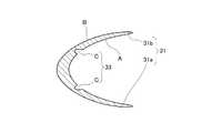
図1(a)は、開瞼器100を患者の右眼に装用した際の概略図である。図1(a)に示す開瞼器100は、U字状に湾曲させて形成した腕部200の両端に、上瞼401と下瞼402にそれぞれ引っ掛ける2つの湾曲プレートよりなる瞼押え部300をろう付け等により接合したもので、腕部200のバネ反発力によって開瞼状態を保つものである。 FIG. 1A is a schematic diagram when the
各瞼押え部(310,320)は、瞼の内側に差し込まれる内側挿入片(311,321、図示せず)と瞼の外側を押える外側押え片(312,322)とを持ちつつ、断面U字状に形成されている。そして、患者自身の閉瞼力に負けないよう、腕部200には高い曲げ弾性率を有する線材が使用されている。 Each of the heel pressers (310, 320) has an inner insertion piece (311, 321; not shown) inserted inside the heel and an outer presser piece (312, 322) that presses the outer side of the heel while having a cross-section U It is formed in a letter shape. And the wire material which has a high bending elastic modulus is used for the
これらの開瞼器100は、腕部200の両端に設けた瞼押え部300を眼400の上瞼401と下瞼402に引っ掛けることにより、睫毛403を瞼押え部(310,320)により押えつつ、腕部200のバネ反発力によって上瞼401と下瞼402を開くことができる。 These
これまでの開瞼器は金属製のものが主流であった(特許文献1参照)。そこで、本発明者は、使い捨てを可能として滅菌の面倒を無くす一方、ばね作用を発揮する腕部を持つタイプでありながら、しなやかな弾力性と柔らかな接触感を発揮する開瞼器を発明するに至っている(特許文献2及び3参照)。使い捨てタイプの開瞼器は、使い捨てタイプであるが故に、開瞼器を使いまわすことに起因する感染症のリスクを無くすことができ、その結果、使い捨てタイプの開瞼器が著しく普及しつつある。 Up until now, most of the openers have been made of metal (see Patent Document 1). Therefore, the present inventor invents an opener that can be disposable and eliminates the hassle of sterilization, while exhibiting supple elasticity and soft contact feeling while being a type having an arm portion that exhibits a spring action. (See
白内障手術、網膜硝子体手術、緑内障手術などの内眼手術や翼状片切除などの外眼手術を行う場合、まず、角結膜及びその周囲を消毒する必要がある。その上で、フィルム状のドレープを患者の顔の上に掛けた後、開瞼器を使って開瞼した状態で処置を行っている。 When performing intraocular surgery such as cataract surgery, retinal vitreous surgery, glaucoma surgery, or external eye surgery such as pterygium resection, it is first necessary to disinfect the keratoconjunctiva and its surroundings. After that, a film-like drape is put on the patient's face, and then the treatment is performed with the eyelid open.
手術の際、角結膜及びその周囲を消毒することは極めて重要である。なぜならば、術後眼内炎が発生するか否かは、この消毒が適切に行われるか否かに大きく依存するためである。 During surgery, it is extremely important to disinfect the keratoconjunctiva and its surroundings. This is because whether or not postoperative endophthalmitis occurs depends largely on whether or not this disinfection is performed appropriately.
術後眼内炎(以降、単に「眼内炎」とも言う。)は感染症の一種であり、眼科手術によって眼球に形成された傷口から、眼球内が細菌や真菌(例えば、かび、カンジダ菌、アクネ(ニキビ)菌等)で感染し、毛様充血、前房混濁、前房蓄膿、硝子体混濁が生じ、網膜や硝子体等が炎症を起こす状態を指す。患者が眼内炎に罹ると、視力低下、眼痛などの症状が現われる。更に悪化すると、角膜、強膜、眼球周囲組織にまで炎症が波及してしまい、患者は全眼球炎に罹り、重篤な状態となるおそれもある。 Postoperative endophthalmitis (hereinafter simply referred to as “endophthalmitis”) is a type of infectious disease. Bacteria and fungi (for example, fungi, Candida spp.) Appear inside the eyeball from the wound formed in the eyeball by ophthalmic surgery. , Acne (acne), etc.), ciliary hyperemia, anterior chamber turbidity, anterior chamber pus, and vitreous opacification, and inflammation of the retina and vitreous body. When patients suffer from endophthalmitis, symptoms such as decreased visual acuity and eye pain appear. When it gets worse, inflammation spreads to the cornea, sclera, and surrounding tissues, and the patient may suffer from ophthalmitis and become seriously ill.
患者が眼内炎に罹った場合、再び眼科手術を行い、眼球内を洗浄することになるが、視力の回復が困難となり、視力予後に重大な影響を与えるおそれもある。そのため、術者にとっては、患者が眼内炎に罹らないようにするために、細心の注意を払う必要がある。 When a patient suffers from endophthalmitis, ophthalmic surgery is performed again, and the inside of the eyeball is washed. However, recovery of visual acuity becomes difficult, and there is a possibility that the visual acuity prognosis may be seriously affected. Therefore, it is necessary for the surgeon to pay close attention to prevent the patient from suffering from endophthalmitis.
その具体的な手法としては、手術前に角結膜及びその周囲を消毒液により消毒する手法、硝子体灌流液内に抗菌薬を入れつつ硝子体手術をする手法、手術後に抗菌薬を硝子体内に注射する手法、手術後に抗菌薬や抗炎症薬を点眼する手法等が知られている。 Specific methods include disinfection of the keratoconjunctiva and its surroundings with a disinfectant before surgery, a method of performing vitreous surgery while putting an antibacterial agent in the vitreous perfusion solution, and an antibacterial agent in the vitreous after surgery. Methods for injection, methods for instilling antibacterial drugs and anti-inflammatory drugs after surgery, and the like are known.
その一方、日本だけでも年間約100万件の白内障手術が行われている。現在、白内障の手術後の眼内炎の発症確率は500分の1ないし3000分の1と言われている。そうなると、年間約300〜2000件、眼内炎が発症していることになる。 On the other hand, about 1 million cataract surgery is performed annually in Japan alone. At present, it is said that the probability of developing endophthalmitis after cataract surgery is 1/500 to 1/3000. In that case, about 300 to 2000 cases of endophthalmitis occur annually.
眼科医である術者にとって、患者が眼内炎に罹るか否かは、術者にとっての危機管理と言う点を考慮すると、極めて敏感にならざるを得ない事項である。眼内炎が生じるおそれに関して、術者にとっての精神的負担は極めて大きい。そのため、眼内炎の発症を抑制することは、早急に解決すべき課題である。 For the surgeon who is an ophthalmologist, whether or not the patient suffers from endophthalmitis is a matter that must be extremely sensitive considering the point of crisis management for the surgeon. Regarding the possibility of endophthalmitis, the mental burden on the surgeon is extremely large. Therefore, suppressing the onset of endophthalmitis is a problem to be solved immediately.
そこで本発明は、眼内炎の発症を効果的に抑制可能な開瞼器を提供することを、主たる目的とする。 Then, this invention sets it as the main objective to provide the eyelid device which can suppress the onset of endophthalmitis effectively.
術者からの強い要望を受け、本発明者は上記の課題を解決する手段について検討を加えた。この検討に際し、まず、眼内炎の発症原因について検討を加えた。眼内炎の発症原因は、先にも述べたように細菌や真菌が眼内で感染することにある。そして、この細菌や真菌は、もちろん患者の体外に由来するものもあるが、患者の体内に由来するものもある。その中の一つが、上下瞼(401,402)の縁にあるマイボーム腺から排出される皮脂に含まれるアクネ菌である。マイボーム腺開口部404を概略的に示したのが、図1(b)である。 In response to a strong request from the surgeon, the present inventor has studied a means for solving the above-described problems. In this study, first, the cause of the development of endophthalmitis was examined. The cause of endophthalmitis is that bacteria and fungi are infected in the eye as described above. Of course, some of these bacteria and fungi are derived from outside the patient's body, while others are derived from the patient's body. One of them is acne bacteria contained in sebum discharged from the meibomian glands at the edges of the upper and lower eyelids (401, 402). FIG. 1B schematically shows the meibomian gland opening 404.
マイボーム腺は、皮脂を供給する皮脂腺の一つである。上下瞼(401,402)の縁に存在するマイボーム腺開口部404から、皮脂が供給される。この皮脂により、涙液膜の蒸発を防いだり、涙液膜が眼(角膜)400上に保持されたり、瞼を閉じた際に瞼内を気密にしたりすることが可能となる。 The meibomian gland is one of the sebaceous glands that supply sebum. Sebum is supplied from the meibomian gland opening 404 present at the edges of the upper and lower eyelids (401, 402). This sebum makes it possible to prevent evaporation of the tear film, to hold the tear film on the eye (cornea) 400, and to make the inside of the eyelid airtight when the eyelid is closed.
上述の通り、通常、皮脂は人間の役に立つものであるが、それと同時に皮脂はニキビの原因となるアクネ菌などの細菌を含むものでもある。手術中であっても、皮脂は、マイボーム腺により供給され続ける。これは、手術中、眼内にアクネ菌が入り込む余地が存在することを意味する。仮に、硝子体灌流液内に抗菌薬を入れつつ硝子体手術を行ったとしても、手術後、このアクネ菌が硝子体内に侵入し、爆発的に感染が起こる可能性も否定できない。 As mentioned above, sebum is usually useful to humans, but at the same time, sebum contains bacteria such as acne that causes acne. Even during surgery, sebum continues to be supplied by the meibomian glands. This means that there is room for acne bacteria to enter the eye during surgery. Even if a vitreous surgery is performed while putting an antibacterial agent in the vitreous perfusate, it cannot be denied that this acne bacteria invades the vitreous after the operation and an infection may occur explosively.
上記の点を鑑みて、本発明者は、皮脂がマイボーム腺により供給され続けたとしても、それと並行して、細菌や真菌を殺す薬剤を患者の眼に対して投与し続ければ、眼内にアクネ菌が入り込む余地を著しく減らせるのではないかと考えた。 In view of the above points, the present inventor, even if sebum continues to be supplied by the meibomian glands, in parallel, if a drug that kills bacteria and fungi is continuously administered to the eye of the patient, We thought that the room for acne bacteria to enter could be significantly reduced.
それに加え、術者の作業工程が増加しないような配慮、新たな手術器具を用いることによる作業の煩雑化の防止を、本発明者は考慮に入れた。その結果、開瞼器に対し、薬剤を予め貯留自在である構造、且つ、貯留しておいた薬剤が手術中に自ずと投与され得る構造を設けるという知見を得た。 In addition, the present inventor has taken into consideration the consideration that the operator's work process does not increase and the prevention of complication of work by using a new surgical instrument. As a result, the inventors have found that the opener is provided with a structure in which the medicine can be stored in advance and a structure in which the stored medicine can be naturally administered during surgery.
以上の知見に基づいて成された本発明の態様は、以下の通りである。
本発明の第1の態様は、
腕部と、前記腕部の両端にそれぞれ設けられて上瞼と下瞼にそれぞれ引っ掛ける複数の瞼押え部とを有する開瞼器であって、
前記複数の瞼押え部のうち少なくともいずれかに、開口を有する薬剤貯留部が設けられたことを特徴とする開瞼器である。Aspects of the present invention based on the above findings are as follows.
The first aspect of the present invention is:
An opener having an arm part and a plurality of scissor pressing parts respectively provided on both ends of the arm part and hooked on the upper arm and the lower arm,
The eyelid opening device is characterized in that a medicine storage part having an opening is provided in at least one of the plurality of eyelid holding parts.
本発明の第2の態様は、第1の態様に記載の発明において、
前記薬剤貯留部は、前記複数の瞼押え部の全てに設けられたことを特徴とする。According to a second aspect of the present invention, in the invention according to the first aspect,
The medicine storage section is provided in all of the plurality of heel pressing sections.
本発明の第3の態様は、第2の態様に記載の発明において、
前記薬剤貯留部は、前記瞼押え部の主表面に設けられた凹部を有することを特徴とする。According to a third aspect of the present invention, in the invention according to the second aspect,
The medicine storage part has a recess provided on a main surface of the heel pressing part.
本発明の第4の態様は、第3の態様に記載の発明において、
前記凹部は複数設けられていることを特徴とする。According to a fourth aspect of the present invention, in the invention according to the third aspect,
A plurality of the recesses are provided.
本発明の第5の態様は、第4の態様に記載の発明において、
全体が樹脂の一体成型品として構成されたことを特徴とする。According to a fifth aspect of the present invention, in the invention described in the fourth aspect,
The whole is configured as an integrally molded product of resin.
本発明によれば、眼内炎の発症を効果的に抑制可能な開瞼器を提供できる。 ADVANTAGE OF THE INVENTION According to this invention, the eyelid device which can suppress the onset of endophthalmitis effectively can be provided.
以下、本発明の実施の形態について、図2及び図3を参照しつつ詳細に説明する。なお、一部、図1の符号を再び使用する。図2は、本実施形態における開瞼器を示す概略図であり、(a)は平面図、(b)はα方向から見た側面図、(c)はβ方向から見た正面図、(d)は(c)における瞼押え部の拡大図である。 図3は、本実施形態における開瞼器の瞼押え部を図2(a)のX−X’線にて断面視した概略図である。
なお、以降では、患者の右眼に開瞼器1を装着した例について述べる。ただ、開瞼器1の向きを上下反転すれば、もちろん左眼にも使用可能である。
本実施形態においては、次の順序で説明を行う。
1.開瞼器
A)開瞼器の全体構造
B)腕部
C)瞼押え部
a)薬剤貯留部
2.開瞼器の使用方法(メカニズム)
3.実施の形態による効果
4.変形例等
なお、以下に記載が無い構成については、特許文献2(特開2010−119433号公報)や特許文献3(特開2010−227457号公報)等、公知の文献に記載の構成を採用しても構わない。Hereinafter, embodiments of the present invention will be described in detail with reference to FIGS. 2 and 3. In part, the reference numerals in FIG. 1 are used again. 2A and 2B are schematic views showing the eyelid opening device in the present embodiment, where FIG. 2A is a plan view, FIG. 2B is a side view seen from the α direction, and FIG. 2C is a front view seen from the β direction. d) is an enlarged view of the heel presser part in (c). FIG. 3 is a schematic view of the bar holding portion of the opening device in the present embodiment as viewed in section along the line XX ′ in FIG.
Hereinafter, an example in which the
In the present embodiment, description will be given in the following order.
1. Opening device A) Overall structure of the opening device B) Arm part C) Presser foot holding part a) Drug storage part Usage (mechanism)
3. 3. Effects of the embodiment Modified example
In addition, about the structure which is not described below, even if the structure described in well-known literature, such as patent document 2 (Unexamined-Japanese-Patent No. 2010-119433) and Patent Document 3 (Unexamined-Japanese-Patent No. 2010-227457), is employ | adopted. I do not care.
<1.開瞼器1>
A)開瞼器1の全体構造
図2に示すように、本実施形態における開瞼器1は、バネ性を持つよう射出成型により湾曲形成された熱可塑性樹脂からなる1本のワイヤー状の腕部2と、その腕部2の両端にそれぞれ結合されて眼の上瞼401と下瞼402にそれぞれ引っ掛ける2つの瞼押え部(31,32、まとめて瞼押え部3とも言う。)とを有している。ただ、腕部2や瞼押え部3の素材や形状、そして腕部2や瞼押え部3の機能を奏する部材(後述のアーム2や内側挿入片(31a,32a)、外側押え片(31b,32b)、湾曲プレートの数)の構成要素はあくまで一つの実施形態であり、これに限定されない。<1.
A) Overall Structure of the
この開瞼器1は、少なくとも腕部2は熱可塑性樹脂の成型品として構成されており、腕部2のバネ反発力によって開瞼状態を保つものである。そして、瞼押え部3には、開口Cを有する薬剤貯留部33が形成されている。もちろん、バネ反発力によって開瞼状態を保つもの以外であっても構わないが、本実施形態においては上記の例について述べる。
以下、開瞼器1の各部構成について説明する。In the
Hereinafter, each part structure of the
B)腕部2
本実施形態の腕部2は、U字状の1本のワイヤー形状を有している。そして、その両端に、2つの瞼押え部3が設けられている。この腕部2は、バネ性を持つよう射出成型により湾曲形成された熱可塑性樹脂からなる。以降、この腕部2のことを「アーム2」とも言う。B)
The
アーム2を設ける際、特許文献3に記載のように、瞼押え部(31,32)同士を接近させる方向に荷重を加えた際に発生するバネ反発力を調整する開瞼力調整部21をアーム2内側に設けても構わない。 When the
こうすることにより、アーム2を過度に太くすることなく開瞼器1の開瞼力を確保できるため、手術器具がぶつかるなど手術の妨げを抑制すると共に、術野への圧迫感を軽減することができる。更に、アーム2の補強という点から見ると、開瞼力調整部21を設けることにより、荷重に対する支点を複数箇所とすることができ、引張応力が1箇所に集中することを防ぐことができる。これにより、瞼押え部(31,32)同士を接近させた際におけるアーム2の破損のおそれを低減することができる。 By doing this, the opening force of the
なお、図2(c)は、図2(a)の開瞼器1をβ方向から見た図(即ち正面図)であるが、図2(c)を見ればわかるように、U字状のアーム2の底の部分は、瞼押え部3の位置から下方へとシフトしている。これは、患者の右眼に開瞼器1を装用した際に、患者の顔の形状に沿って開瞼器1を配置し、省スペース化を図るための構成である。 FIG. 2C is a view (ie, a front view) of the
C)瞼押え部3
本実施形態の上下の瞼押え部(31,32)は、瞼(401,402)の内側に差し込まれる内側挿入片(31a,32a)と、瞼(401,402)の外側を押える外側押え片(31b,32b)とを持つ断面U字状の湾曲プレートにより構成されている。そして、この瞼押え部3は、射出成型によりアーム2と一体に成型されたものである。つまり、本実施形態の開瞼器1は、全体が樹脂の一体成形品として構成されている。なお、この樹脂は、任意のものを使用して良く、例えば熱可塑性樹脂のようなエネルギーを受けて硬度が変化する樹脂を挙げることができる。C)
The upper and lower heel pressers (31, 32) of the present embodiment include an inner insertion piece (31a, 32a) that is inserted inside the heel (401, 402) and an outer presser piece that holds the outer side of the heel (401, 402). (31b, 32b) and a U-shaped curved plate. And this
なお、本実施形態の開瞼器1のように、アーム2と瞼押え部3とが樹脂からなり、瞼押え部3とアーム2とが一体成型された開瞼器1ならば、異なる開瞼力を有する開瞼器1を多種多量に生産することができ、この利点はさらに増大する。射出成型の際には、特許文献3に記載のように、瞼押え部(31,32)同士を接近させる方向に荷重を加える際に発生する引張応力が最大となる部分以外の部分に、アーム2の樹脂注入部を設けておくのが好ましい。 In the case of the
また、断面U字状の上下の瞼押え部(31,32)は、腕部2のある側と反対側に若干の角度θ(約20°)だけ開き気味に形成されている。また、断面U字状の上下の瞼押え部(31,32)の背中部分(U字の底に相当する部分)は半径Rの曲率で凹状に湾曲している。 Further, the upper and lower heel pressers (31, 32) having a U-shaped cross section are formed so as to open slightly by a slight angle θ (about 20 °) on the side opposite to the side where the
また、特許文献3に記載のように、内側挿入片(31a,32a)が瞼(401,402)の縁に沿う方向の長さLaよりも、外側押え片(31b,32b)が瞼(401,402)の縁に沿う方向の長さLbの方が大きく設定されていてもよい。こうすることにより、サイズの大きい外側押え片(31b,32b)によって、上下の瞼(401,402)の多くの睫毛403やその根元を覆い隠すことができる。 Further, as described in
また、図2(d)に示すように、瞼押え部(31,32)を構成する湾曲プレートの厚みが、内側挿入片(31a,32a)と外側押え片(31b,32b)のつながるU字状の湾曲部から両先端部に向けて徐々に薄くなるように変化していても良い。 Moreover, as shown in FIG.2 (d), the thickness of the curved plate which comprises the eaves presser part (31, 32) is the U-character with which the inner insertion piece (31a, 32a) and the outer presser piece (31b, 32b) are connected. It may change so that it may become thin gradually toward the both front-end | tip parts from a curved shape part.
なお、瞼押え部3を構成する湾曲プレートの厚みの変化は、特許文献3に記載のように、連続的に設けてあるのが好ましいが、段階的に設けてあってもよい。 In addition, although the change of the thickness of the curved plate which comprises the eaves-holding
図2(d)で言うと、寸法t1よりも寸法t2、t3の方が小さくなっている(t1>t2=t3)。この場合、例えば、t1=約0.6mm、t2=t3=約0.4mmになっている。t1の寸法の許容幅は、例えば、0.5〜0.7mmの範囲であることが好ましく、t2及びt3の寸法の許容幅は、例えば0.3〜0.5mmの範囲であることが好ましい。あまりにも最小厚み部分を薄くすると割れやすくなる。そのため、最低でも割れが起きにくい厚みを確保しておく必要がある。しかも、後述するように、瞼押え部3に対し、薬剤貯留部33として窪みを形成するため、ある程度の厚みを確保する必要がある。
以下、本実施形態の特徴である薬剤貯留部33について説明する。In FIG. 2D, the dimensions t2 and t3 are smaller than the dimension t1 (t1> t2 = t3). In this case, for example, t1 = about 0.6 mm and t2 = t3 = about 0.4 mm. The allowable width of the dimension of t1 is preferably in the range of 0.5 to 0.7 mm, for example, and the allowable width of the dimension of t2 and t3 is preferably in the range of, for example, 0.3 to 0.5 mm. . If the minimum thickness part is made too thin, it will break easily. For this reason, it is necessary to secure a thickness at which cracking is unlikely to occur at least. In addition, as will be described later, since a depression is formed as the
Hereinafter, the
a)薬剤貯留部33
本実施形態における薬剤貯留部33は、上下の瞼押え部(31,32)各々の主表面に設けられている。そして、本実施形態における薬剤貯留部33は、その名の通り、薬剤を貯留自在な部分である。つまり、手術中において、皮脂がマイボーム腺により供給され続けたとしても、それと並行して、瞼押え部3に貯留しておいた薬剤を、患者の眼に対して断続的に投与し続けられる構成が、薬剤貯留部33である。この薬剤貯留部33は開口Cを有するように、瞼押え部3の主表面に設けられている。a)
The
本実施形態においては、意図的に、開口Cを有する薬剤貯留部33を設けることにより、本実施形態の開瞼器1は、従来のように瞼押え部3を構成するU字状の湾曲プレートそれ自体の底部に液が溜まるという構成とは全く異なる構成を有している。 In the present embodiment, by intentionally providing the
なお、本実施形態における薬剤貯留部33は、凹部(窪み)を有する構成である。図3に示すように、開口Cとは、この凹部の一部であり、開口Cから薬剤が出入りすることになる。薬剤が開口Cから凹部に流れ込み、この凹部に薬剤が貯留された状態で、本実施形態の開瞼器1は使用される。この薬剤としては、特に限定はないが、バンコマイシン、セフタジジムのような液体状の抗菌薬やポビドンヨードのような消毒液が挙げられる。 In addition, the chemical | medical
なお、上記の凹部は複数設けられているのが好ましい。その方が、薬剤の貯留量を多く確保することが可能になり、手術中に、眼内炎の発症の原因の細菌や真菌を殺すことが可能となり、ひいては眼内炎の発症を更に効果的に抑制することが可能となる。 In addition, it is preferable that a plurality of the recesses are provided. In that way, it becomes possible to secure a large amount of drug storage, and it is possible to kill bacteria and fungi that cause the development of endophthalmitis during surgery, and more effectively the development of endophthalmitis. Can be suppressed.
また、瞼押え部3において上記の薬剤貯留部33が設けられる面としては、図2(c)や図2(d)に示すように、瞼(401,402)と接触する側の面Aでも構わないし、その反対側の面Bに形成されていても構わない。なお、瞼(401,402)と接触する側の面とは、瞼押え部3のU字状の湾曲部において内側の湾曲面Aのことを指す。また、その反対側の面とは、瞼押え部3のU字状の湾曲部において外側の湾曲面Bのことを指す。 In addition, as shown in FIGS. 2 (c) and 2 (d), the surface on the side in contact with the heel (401, 402) is also used as the surface on the
後で<2.開瞼器1の使用方法(メカニズム)>にて述べるが、患者の瞼(401,402)の動きに合わせて瞼押え部3に加わる圧力が変動し、それに伴い薬剤貯留部33に加わる圧力も変動する。 Later <2. As described in the usage method (mechanism) of the
瞼(401,402)と接触する側の面Aに薬剤貯留部33が形成されていれば、薬剤貯留部33が圧力変動を受けやすくなり、薬剤貯留部33が変形しやすくなる。その結果、薬剤貯留部33に貯留された薬剤がポンピングにより患者の眼球(主に角膜表面)(以降、眼(角膜)400と言う。)へと投与されやすくなる。 If the
その一方、瞼(401,402)と接触する側の面Aでは、睫毛404と共にフィルム状のドレープを巻き込みながら瞼が開かれる。そのため、ドレープの存在により、瞼(401,402)と瞼押え部(31,32)との直接接触が妨げられていることがままある。また、ドレープにより、薬剤貯留部33に貯留された薬剤がマイボーム腺開口部404や眼(角膜)400に行き届きにくくなることも考えられる。そこで、反対側の面Bに薬剤貯留部33が形成されていれば、ドレープを気にせず、薬剤をポンピングにより眼(角膜)400へと投与することも可能になると考えられる。 On the other hand, on the surface A on the side in contact with the eyelids (401, 402), the eyelids are opened while the film-shaped drape is wound together with the
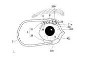
なお、反対側の面Bに薬剤貯留部33を形成する場合、薬剤が眼(角膜)400に効果的に届くように、以下のような構成上の工夫を行うことも可能である。この工夫について、図4を用いて説明する。図4は、本実施形態における別の一例である開瞼器を患者の右眼に装用した際の概略図である。
この例によれば、瞼押え部(31,32)の少なくともいずれかにおいて、外側押え片(31b,32b)における外側の湾曲面Bに、複数の凹部からなる薬剤貯留部33を設ける。そして、同じく外側の湾曲面Bに対し、薬剤貯留部33における複数の凹部から連通する形で内側挿入片(31a,32a)に向かう溝34を形成する。こうすることにより、薬剤貯留部33に貯留された薬剤が、溝34を伝って眼(角膜)400へと届きやすくなる。この溝34は、内側挿入片(31a,32a)の面Bと面Aの境界まで延伸しているのが好ましい。
開瞼器1を用いた手術中だと、外側押え片(31b,32b)は天地方向の天の方向に位置し、内側挿入片(31a,32a)は天地方向の地の方向に位置する。そして、内側挿入片(31a,32a)の更に地の方向に眼(角膜)400が位置する。そのため、外側の湾曲面Bにおいて、外側押え片(31b,32b)に形成された薬剤貯留部33に貯留された薬剤は開口Cから出て地の方向に向かうため、溝34を伝い、内側挿入片(31a,32a)を越えて眼(角膜)400へと容易に届くことになる。もちろん、上述の薬剤のポンピングが行われるという効果も奏する。In addition, when forming the
According to this example, in at least one of the heel pressing portions (31, 32), the
During the operation using the
また、上記の構成を採用することにより、開瞼器1に対して薬剤を追加的に供給することも可能となる。一例を挙げると、図4に示すように、薬剤が染み込んだ脱脂綿等の吸水性物質(例えば手術用スポンジ500(破線で記載))を外側押え片(31b,32b)の上(手術中においては外側押え片(31b,32b)から見て天の方向)に載置しておく。こうすることにより、薬剤貯留部33に貯留された薬剤が不足しても、手術用スポンジ500から薬剤が薬剤貯留部33に供給される。そして、薬剤貯留部33に貯留された薬剤、そして手術用スポンジ500に染み込ませた薬剤が共に不足してきたら、手術用スポンジ500にスポイト等で薬剤を補給する。そうすれば、手術用スポンジ500から十分な量の薬剤が薬剤貯留部33に供給されることになり、結果的に薬剤貯留部33から眼(角膜)400へと十分な量の薬剤を投与することが可能となる。上記の構成によれば、本実施形態の効果に加え、開瞼器1に対して容易に、薬剤を追加的に供給することが可能となり、術者の負担を軽減することが可能となる。 In addition, by adopting the above configuration, it is possible to additionally supply a medicine to the
ただ、結局のところ、薬剤貯留部33は、上記の両面(A,B)に設けても構わないし、いずれかの面に設けても構わない。また、手術中に投与する薬剤の量に応じて、いずれかの面に薬剤貯留部33を設けるかを決定しても構わない。 However, after all, the
なお、上記の薬剤貯留部33が凹部を有する場合、当該凹部は溝形状でも構わないし、非貫通の孔形状でも構わない。本実施形態においては、非貫通の孔形状である場合について述べる。それをわかりやすく図示したものが、図2(b)である。 In addition, when said chemical | medical
図2(b)においては、当該凹部を平面視楕円状の窪みとしたうえで、それらの配置状況が示されている。図1(a)に示されるように患者の右眼に開瞼器1を装着した際、上瞼401において眼外から眼内へと向かう方向(図2(b)においては左側から右側に向かう方向)へと、複数の凹部が2列にわたり形成されている。また、平面視楕円状の窪みの長径を2mm、短径を1mm、孔の深さを0.5mmに設定し、各列における孔同士の間隔を1mmとしている。また、複数の凹部の各列は、瞼押え部3の上下方向中心から各々1mm離れた部分に孔の中心が位置するように配置されている。
もちろん、上記の薬剤貯留部33は一例に過ぎず、薬剤を貯留することができるのならば、薬剤貯留部33の形状及び数はこれに限定されるものではない。その他の変形例については、後で<4.変形例等>にて述べる。In FIG.2 (b), after making the said recessed part into a hollow of planar view ellipse, those arrangement | positioning conditions are shown. When the
Of course, the
<2.開瞼器1の使用方法(メカニズム)>
以下、本実施形態における開瞼器1の使用方法(メカニズム)について説明する。<2. Usage (Mechanism) of
Hereinafter, the usage method (mechanism) of the eye-
まず、フィルム状のドレープを患者の顔の上に掛け、その上に手術布を患者の上に掛ける。ドレープ及び手術布は、患者の眼球の部分は切り抜かれ、患者の眼球が露出するようになっている。そして、角結膜及びその周囲(特にマイボーム腺開口部404が存在する上下瞼(401,402)の縁)を消毒する。 First, a film-like drape is hung on the patient's face, and a surgical cloth is hung on the patient. In the drape and the surgical cloth, a part of the patient's eyeball is cut out so that the patient's eyeball is exposed. Then, the keratoconjunctiva and its surroundings (especially the edges of the upper and lower eyelids (401, 402) where the
その一方、本実施形態の開瞼器1の薬剤貯留部33に抗菌薬を貯留させる作業を行う。なお、貯留方法としては任意のもので構わない。一例を挙げるとすると、開瞼器1を抗菌薬に漬けることにより、凹部(窪み)に抗菌薬が貯まるようにするという手法が挙げられる。 On the other hand, an operation for storing the antibacterial drug in the
その後、予め抗菌薬が貯留されている開瞼器1を使って、患者の瞼(401,402)を開瞼する。内側挿入片(31a,32a)が、瞼(401,402)の内側に差し込まれることになる。それと共に、外側押え片(31b,32b)が、ドレープの一部を巻き込みつつ、睫毛403やその根元を覆い隠す。 Thereafter, the patient's heel (401, 402) is opened using the
患者の瞼(401,402)は閉じようとする方向に力が働いており、開瞼器1の瞼押え部3には瞼(401,402)により力が加わっている。そして、手術中、患者に麻酔が効いていたとしても、患者の瞼(401,402)は僅かながら生理的に振動する。その結果、瞼押え部3に加わる力(ひいては薬剤貯留部33に加わる力)も変動する。この力の変動により、瞼押え部3ひいては薬剤貯留部33が僅かに変形し、貯留された薬剤が薬剤貯留部33の開口Cから少しずつ押し出され、いわゆるポンピングが行われる。つまり、瞼(401,402)によって薬剤貯留部33に加わる力に応じて、薬剤貯留部33内にある薬剤が開口Cから外に出る。 A force is acting in a direction to close the patient's heel (401, 402), and force is applied to the
開瞼器1からの薬剤の投与が、上記のようにポンピングを利用して開瞼器1により自ずと行われることにより、薬剤の投与を少しずつ行うことが可能となり、少なくとも手術中は、断続的に薬剤を患者の眼(主に角膜)400に投与し続けることが可能となる。しかもそれは、術者の手を煩わせることなく行われる。 The administration of the drug from the
<3.実施の形態による効果>
本実施形態によれば、以下の効果を奏する。<3. Advantages of the embodiment>
According to this embodiment, the following effects can be obtained.
まず、皮脂がマイボーム腺により供給され続けたとしても、それと並行して、細菌や真菌を殺す薬剤を患者の眼に対して投与し続けることが可能になり、眼内に細菌や真菌が入り込む余地を著しく減らすことが可能になる。 First, even if sebum continues to be supplied by the meibomian glands, it will be possible to continue to administer drugs that kill bacteria and fungi to the patient's eye, leaving room for bacteria and fungi to enter the eye. Can be significantly reduced.
それに加え、開瞼器1に対し、薬剤を予め貯留自在である構造、且つ、貯留しておいた薬剤が手術中に自ずと投与され得る構造という比較的簡素な構造を設けることにより、術者の作業工程の増加を防ぐことができるし、新たな手術器具を用いることによる作業の煩雑化を防止することもできる。 In addition, by providing the
また、本実施形態における開瞼器1を、全体を熱可塑性樹脂の一体成型で構成する場合、容易に低コストで大量生産することができる。これにより、使い捨ての使用が可能となり、滅菌処理を不要として、大量の使用要求に応えることができる。 Moreover, when the
なお、本実施形態における開瞼器1が創出される際には、「瞼におけるマイボーム腺から排出される細菌や真菌への対策」という課題を本発明者は認識し、その上で「患者の瞼(401,402)によって開瞼器1に力が加わっており、患者の瞼は僅かながら生理的に変動すること」に本発明者は着目している。この患者の瞼(401,402)の変動を、貯留された薬剤を開口Cから外に押し出すポンピングに利用するという知見に基づき、本実施形態における開瞼器1が創出されている。このような課題についても、上記の生理学的性質を利用した薬剤投与を自在とする開瞼器についても、本発明者が知る限りでは、いずれの文献にも記載されていない。 In addition, when the
以上の通り、本実施形態の開瞼器1によれば、眼内炎の発症を効果的に抑制することが可能となる。 As described above, according to the
<4.変形例等>
本発明の技術的範囲は上述した実施の形態に限定されるものではなく、発明の構成要件やその組み合わせによって得られる特定の効果を導き出せる範囲において、種々の変更や改良を加えた形態も含む。<4. Modified example>
The technical scope of the present invention is not limited to the above-described embodiments, and includes various modifications and improvements as long as the specific effects obtained by the constituent elements of the invention and combinations thereof can be derived.
上記の実施形態では、上瞼401を押える瞼押え部31と、下瞼402を押える瞼押え部32との両方に、薬剤貯留部33を設ける場合について述べた。上下の瞼押え部(31,32)に薬剤貯留部33を設けた方が、貯留される薬剤の量が多くなる。ただ、所定の量の薬剤を薬剤貯留部33にて貯留できるのならば、一方の瞼押え部3にのみ薬剤貯留部33を設けても構わない。 In the above-described embodiment, the case where the
また、マイボーム腺は、下瞼402よりも上瞼401に数多く存在する。それを鑑みて、上瞼401を押える瞼押え部31にのみ薬剤貯留部33を設けても構わない。また、上下の瞼押え部(31,32)に薬剤貯留部33を設けつつ、薬剤貯留部33における凹部(窪み)の数を、相違させても構わない。具体的に言うと、下瞼402を押える瞼押え部32よりも、上瞼401を押える瞼押え部31に数多く凹部を形成しても構わない。また、上瞼401と下瞼402との開瞼力の違い、即ち上瞼401と下瞼402との間でのポンピングされ得る薬剤の量の違いに応じて、薬剤貯留部33により貯留される薬剤の量が調整されるように薬剤貯留部33を形成しても構わない。 In addition, the meibomian glands are present more in the
また、薬剤貯留部33が凹部を有する場合、開口Cの大きさは任意で構わないが、凹部における開口Cの大きさを、凹部における他の部分に比べて小さくするのが好ましい。例えて言えば、図3において、薬剤貯留部33における凹部を、開口Cが比較的狭くなっているタコツボ形状とするのが好ましい。こうすることにより、薬剤貯留部33に薬剤を貯留しやすくなる一方、手術中に薬剤が一度に投与されることを抑制し、徐々に薬剤を投与することが可能となる。 Moreover, when the chemical | medical
上記の実施形態では、薬剤貯留部33が凹部を複数有する場合について述べた。その一方、単数の凹部が瞼押え部3の主表面に設けられても構わない。具体例を挙げるとすれば、平面視複数本の溝であって互いに連通することにより薬剤の貯留量を所定量確保している凹部が挙げられる。 In said embodiment, the case where the chemical | medical
上記の実施形態では、薬剤貯留部33が凹部(窪み)である場合について述べた。その一方、薬剤貯留部33が凸部(出っ張り)により構成されていても構わない。具体的に言うと、瞼押え部3の主表面に凸部(出っ張り)を形成しておく。この凸部が、瞼押え部3の主表面の一部を囲むように形成されることにより、薬剤溜まりを形成することができる。そして、この薬剤溜まりを複数形成するように、瞼押え部3の主表面に凸部を形成しても構わない。また、薬剤貯留部33が、上記の凹部(窪み)及び凸部(出っ張り)の組み合わせにより形成されていても構わない。ただ、瞼押え部3の主表面に、例えばシボ加工のように単に凹凸を形成しても、薬剤を貯留することは困難である。そのため、本実施形態における薬剤貯留部33は、あくまで、皮脂がマイボーム腺により供給され続けたとしても、それと並行して、瞼押え部3に貯留しておいた薬剤を、患者の眼に対して断続的に供給し続けられる、開口Cを有する構成であり、単なるシボ加工により形成される形状とは異なる。 In said embodiment, the case where the chemical | medical
上記の実施形態では、薬剤が液体である場合について述べた。その一方、薬剤は液体ないし固体でも構わず、開瞼器1を患者に装着すると固体だった薬剤が体温で液体となるような物質を薬剤として用いても構わない。また、薬剤貯留部33に脱脂綿等の吸水性物質を予め設けておき、吸水性物質に薬剤を染み込ませたうえで開瞼器1を患者に装着可能な構造を開瞼器1に採用しても構わない。その一例が、上述した例である手術用スポンジ500を用いた例である。別の例としては、凹部の中に脱脂綿等の吸水性物質を設ける例も挙げられる。 In the above embodiment, the case where the drug is a liquid has been described. On the other hand, the drug may be liquid or solid, and a substance that becomes a liquid at body temperature when the
上記の実施形態では、瞼押え部3をプレート状としたが、ワイヤー状の瞼押え部3に薬剤貯留部33を設けても構わない。ただ、プレート状の瞼押え部3の方が貯留可能な薬剤の量を多くすることができるので、瞼押え部3をプレート状とするのが好ましい。 In the above embodiment, the
上記の実施形態では、アーム2と瞼押え部3とが、樹脂の一体成形品として構成される場合について記載しているが、アーム2と瞼押え部3とが別体成形品として構成されても構わない。ただ、大量生産する上でのコストダウンという点から、一体に設けられているのが好ましい。 In the above embodiment, the case is described in which the
なお、上記の実施形態における開瞼器1は、射出成型により製造されている。その一方、ブロック状の樹脂材から構造物を削り出す方法、樹脂製型中もしくは金型中で重合硬化する方法、または熱可塑性樹脂を用いた射出成型による方法などの樹脂成型方法を用いても構わない。ただ、大量の製品を製造できるという点では、射出成型が好ましい。 In addition, the
上記の実施形態は、手術中に薬剤を患者の眼(角膜)400に単に供給するものではなく、薬剤を瞼押え部3に貯留しておき、手術中に徐々に薬剤が投与されていく構成を具体化したものである。これは、術者の作業工程が増加しないような配慮、新たな手術器具を用いることによる作業の煩雑化の防止を、本発明者が考慮に入れることにより創出された構成である。しかしながら、上記の問題点を考慮から外すのならば、「貯留部」ではなく、薬剤を供給するための流路のような「供給部」を開瞼器1に設けることも選択肢として考えられる。そして、手術中に開瞼器1の外部から薬剤を開瞼器1内の流路に供給し、最終的に流路の末端の開口Cから患者の眼(角膜)400へと薬剤を投与する構造も、選択肢として考えられる。また、瞼押え部3以外の部分(例えば腕部2)に薬剤貯留部33を設けたり、薬剤を供給できるような構成を備えさせたりすることも選択肢として考えられる。ただ、上記の実施形態のように、複数の瞼押え部3のうち少なくともいずれかに、開口Cを有する薬剤貯留部33が設けられる方が、薬剤を効果的に投与する点及び術者の作業効率と言う点では好ましいことは言うまでもない。 In the above embodiment, the medicine is not simply supplied to the patient's eye (cornea) 400 during the operation, but the medicine is stored in the
1 開瞼器
2 腕部
21 開瞼力調整部
3 瞼押え部
31 (上瞼の)瞼押え部
31a (上瞼の)内側挿入片
31b (上瞼の)外側押え片
32 (下瞼の)瞼押え部
32a (下瞼の)内側挿入片
32b (下瞼の)外側押え片
33 薬剤貯留部
34 溝
100 開瞼器
200 腕部
300 瞼押え部
310 (上瞼の)瞼押え部
311 (上瞼の)内側挿入片
312 (上瞼の)外側押え片
320 (下瞼の)瞼押え部
321 (下瞼の)内側挿入片
322 (下瞼の)外側押え片
400 眼(角膜)
401 上瞼
402 下瞼
403 睫毛
404 マイボーム腺開口部
500 手術用スポンジ
A 瞼押え部のU字状の湾曲部において内側の湾曲面
B 瞼押え部のU字状の湾曲部において外側の湾曲面
C (薬剤貯留部の)開口
DESCRIPTION OF
401
B Opening of the outer curved surface C (of the drug reservoir) in the U-shaped curved part of the presser foot
Claims (5)
Translated fromJapanese前記複数の瞼押え部のうち少なくともいずれかに、開口を有する薬剤貯留部が設けられたことを特徴とする開瞼器。An opener having an arm part and a plurality of scissor pressing parts respectively provided on both ends of the arm part and hooked on the upper arm and the lower arm,
The eyelid opening device characterized in that a drug storage part having an opening is provided in at least one of the plurality of eyelid holding parts.
Priority Applications (1)
| Application Number | Priority Date | Filing Date | Title |
|---|---|---|---|
| JP2013052650AJP6247009B2 (en) | 2013-03-15 | 2013-03-15 | Open device |
Applications Claiming Priority (1)
| Application Number | Priority Date | Filing Date | Title |
|---|---|---|---|
| JP2013052650AJP6247009B2 (en) | 2013-03-15 | 2013-03-15 | Open device |
Publications (2)
| Publication Number | Publication Date |
|---|---|
| JP2014176517Atrue JP2014176517A (en) | 2014-09-25 |
| JP6247009B2 JP6247009B2 (en) | 2017-12-13 |
Family
ID=51697181
Family Applications (1)
| Application Number | Title | Priority Date | Filing Date |
|---|---|---|---|
| JP2013052650AActiveJP6247009B2 (en) | 2013-03-15 | 2013-03-15 | Open device |
Country Status (1)
| Country | Link |
|---|---|
| JP (1) | JP6247009B2 (en) |
Cited By (6)
| Publication number | Priority date | Publication date | Assignee | Title |
|---|---|---|---|---|
| CN106037841A (en)* | 2016-07-11 | 2016-10-26 | 张磊 | Eye speculum for children |
| CN106726115A (en)* | 2016-12-22 | 2017-05-31 | 上海交通大学医学院附属第九人民医院 | A kind of eyelid function corrector |
| WO2018198686A1 (en)* | 2017-04-28 | 2018-11-01 | 富士ゼロックス株式会社 | Eyelid opening device |
| KR102243321B1 (en) | 2020-02-13 | 2021-04-22 | 학교법인 건국대학교 | Eyelid fastener |
| CN115300364A (en)* | 2022-07-18 | 2022-11-08 | 许昌口腔医院 | An eyeball cleaning device for ophthalmic cataractectomy |
| US20230000675A1 (en)* | 2020-01-08 | 2023-01-05 | Azura Ophthalmics Ltd. | Device for administering drug to eyelid |
Citations (7)
| Publication number | Priority date | Publication date | Assignee | Title |
|---|---|---|---|---|
| US4895566A (en)* | 1986-07-25 | 1990-01-23 | C. R. Bard, Inc. | Coating medical devices with cationic antibiotics |
| US20030171656A1 (en)* | 2002-01-22 | 2003-09-11 | Foulkes Richard B. | Ophthalmic sulcus speculum |
| JP2008534142A (en)* | 2005-03-31 | 2008-08-28 | コナー・ミッドシステムズ・インコーポレイテッド | System and method for loading a beneficial substance into a medical device |
| JP2009530022A (en)* | 2006-03-24 | 2009-08-27 | ボストン サイエンティフィック リミテッド | Medical device with nanoporous coating for controlled therapeutic agent delivery |
| US20090287060A1 (en)* | 2008-05-14 | 2009-11-19 | Physcient, Inc. | Methods and devices to decrease tissue trauma during surgery |
| US20110077468A1 (en)* | 2009-09-30 | 2011-03-31 | Finger Paul T | Drug eluting eyelid speculum |
| WO2012144437A1 (en)* | 2011-04-22 | 2012-10-26 | 株式会社アールテック・ウエノ | Drug solution applicator |
- 2013
- 2013-03-15JPJP2013052650Apatent/JP6247009B2/enactiveActive
Patent Citations (7)
| Publication number | Priority date | Publication date | Assignee | Title |
|---|---|---|---|---|
| US4895566A (en)* | 1986-07-25 | 1990-01-23 | C. R. Bard, Inc. | Coating medical devices with cationic antibiotics |
| US20030171656A1 (en)* | 2002-01-22 | 2003-09-11 | Foulkes Richard B. | Ophthalmic sulcus speculum |
| JP2008534142A (en)* | 2005-03-31 | 2008-08-28 | コナー・ミッドシステムズ・インコーポレイテッド | System and method for loading a beneficial substance into a medical device |
| JP2009530022A (en)* | 2006-03-24 | 2009-08-27 | ボストン サイエンティフィック リミテッド | Medical device with nanoporous coating for controlled therapeutic agent delivery |
| US20090287060A1 (en)* | 2008-05-14 | 2009-11-19 | Physcient, Inc. | Methods and devices to decrease tissue trauma during surgery |
| US20110077468A1 (en)* | 2009-09-30 | 2011-03-31 | Finger Paul T | Drug eluting eyelid speculum |
| WO2012144437A1 (en)* | 2011-04-22 | 2012-10-26 | 株式会社アールテック・ウエノ | Drug solution applicator |
Cited By (6)
| Publication number | Priority date | Publication date | Assignee | Title |
|---|---|---|---|---|
| CN106037841A (en)* | 2016-07-11 | 2016-10-26 | 张磊 | Eye speculum for children |
| CN106726115A (en)* | 2016-12-22 | 2017-05-31 | 上海交通大学医学院附属第九人民医院 | A kind of eyelid function corrector |
| WO2018198686A1 (en)* | 2017-04-28 | 2018-11-01 | 富士ゼロックス株式会社 | Eyelid opening device |
| US20230000675A1 (en)* | 2020-01-08 | 2023-01-05 | Azura Ophthalmics Ltd. | Device for administering drug to eyelid |
| KR102243321B1 (en) | 2020-02-13 | 2021-04-22 | 학교법인 건국대학교 | Eyelid fastener |
| CN115300364A (en)* | 2022-07-18 | 2022-11-08 | 许昌口腔医院 | An eyeball cleaning device for ophthalmic cataractectomy |
Also Published As
| Publication number | Publication date |
|---|---|
| JP6247009B2 (en) | 2017-12-13 |
Similar Documents
| Publication | Publication Date | Title |
|---|---|---|
| JP6247009B2 (en) | Open device | |
| Gote et al. | Ocular drug delivery: present innovations and future challenges | |
| US10004635B2 (en) | Nasolacrimal implants and related methods for tear stimulation | |
| RU2618194C2 (en) | Eye inserter and methods | |
| US7985180B2 (en) | Eyelid retractor | |
| KR20100050566A (en) | Lacrimal implants and related methods | |
| CN109199692B (en) | Auxiliary device for intravitreal injection | |
| CN103371885A (en) | Drainage tube, method for implanting drainage tube into eyeball and application of drainage tube | |
| JP5285398B2 (en) | Open device | |
| RU2211687C2 (en) | Method for treating retinal detachment | |
| JP5467665B2 (en) | Open device | |
| O'Brien et al. | Patient pain during stretching of small pupils in phacoemulsification performed using topical anesthesia | |
| CN205913445U (en) | Former wife who has died pressure stability valve among cataract surgery | |
| CN209474945U (en) | Eye fixation device for intravitreal injection | |
| CN207506689U (en) | Open conjunctival sac vault supports tension ring | |
| Fechter | Improvised 3-0 polypropylene plug for the glaucoma drainage tube during phacoemulsification | |
| Bhartiya et al. | NEW MIGS ON THE BLOCK | |
| RU2233147C1 (en) | Method for surgical treating diseases of internal membranes of an eyeball and vitreous body | |
| RAHMOUN et al. | Glaucoma in Dogs and its Treatment with Timolol | |
| RU2375995C2 (en) | Method of surgical treatment of glaucoma | |
| Brodrick et al. | Mechanical dilation of the pupil | |
| RU2238704C1 (en) | Method for treating the cases of progressive and aggravated myopia | |
| Gnanadickam | The modern intracapsular operation for senile cataract | |
| Amarjeet et al. | A review on post-operative complications and its care after cataract surgery | |
| Guise | Sub-Tenon's Block |
Legal Events
| Date | Code | Title | Description |
|---|---|---|---|
| A621 | Written request for application examination | Free format text:JAPANESE INTERMEDIATE CODE: A621 Effective date:20151211 | |
| A131 | Notification of reasons for refusal | Free format text:JAPANESE INTERMEDIATE CODE: A131 Effective date:20160809 | |
| A521 | Request for written amendment filed | Free format text:JAPANESE INTERMEDIATE CODE: A523 Effective date:20161007 | |
| A131 | Notification of reasons for refusal | Free format text:JAPANESE INTERMEDIATE CODE: A131 Effective date:20161220 | |
| A521 | Request for written amendment filed | Free format text:JAPANESE INTERMEDIATE CODE: A523 Effective date:20170213 | |
| A02 | Decision of refusal | Free format text:JAPANESE INTERMEDIATE CODE: A02 Effective date:20170802 | |
| A521 | Request for written amendment filed | Free format text:JAPANESE INTERMEDIATE CODE: A523 Effective date:20171005 | |
| A911 | Transfer to examiner for re-examination before appeal (zenchi) | Free format text:JAPANESE INTERMEDIATE CODE: A911 Effective date:20171018 | |
| TRDD | Decision of grant or rejection written | ||
| A01 | Written decision to grant a patent or to grant a registration (utility model) | Free format text:JAPANESE INTERMEDIATE CODE: A01 Effective date:20171114 | |
| A61 | First payment of annual fees (during grant procedure) | Free format text:JAPANESE INTERMEDIATE CODE: A61 Effective date:20171116 | |
| R150 | Certificate of patent or registration of utility model | Ref document number:6247009 Country of ref document:JP Free format text:JAPANESE INTERMEDIATE CODE: R150 | |
| R250 | Receipt of annual fees | Free format text:JAPANESE INTERMEDIATE CODE: R250 | |
| R250 | Receipt of annual fees | Free format text:JAPANESE INTERMEDIATE CODE: R250 | |
| R250 | Receipt of annual fees | Free format text:JAPANESE INTERMEDIATE CODE: R250 | |
| R250 | Receipt of annual fees | Free format text:JAPANESE INTERMEDIATE CODE: R250 | |
| R250 | Receipt of annual fees | Free format text:JAPANESE INTERMEDIATE CODE: R250 | |
| S111 | Request for change of ownership or part of ownership | Free format text:JAPANESE INTERMEDIATE CODE: R313113 | |
| R350 | Written notification of registration of transfer | Free format text:JAPANESE INTERMEDIATE CODE: R350 |