JP2014064824A - Shortest route searching device, method and program of tubular structure - Google Patents
Shortest route searching device, method and program of tubular structureDownload PDFInfo
- Publication number
- JP2014064824A JP2014064824AJP2012213408AJP2012213408AJP2014064824AJP 2014064824 AJP2014064824 AJP 2014064824AJP 2012213408 AJP2012213408 AJP 2012213408AJP 2012213408 AJP2012213408 AJP 2012213408AJP 2014064824 AJP2014064824 AJP 2014064824A
- Authority
- JP
- Japan
- Prior art keywords
- path
- route
- divided
- search
- end point
- Prior art date
- Legal status (The legal status is an assumption and is not a legal conclusion. Google has not performed a legal analysis and makes no representation as to the accuracy of the status listed.)
- Granted
Links
Images


Classifications
- A—HUMAN NECESSITIES
- A61—MEDICAL OR VETERINARY SCIENCE; HYGIENE
- A61B—DIAGNOSIS; SURGERY; IDENTIFICATION
- A61B5/00—Measuring for diagnostic purposes; Identification of persons
- A61B5/103—Measuring devices for testing the shape, pattern, colour, size or movement of the body or parts thereof, for diagnostic purposes
- A61B5/107—Measuring physical dimensions, e.g. size of the entire body or parts thereof
- A61B5/1075—Measuring physical dimensions, e.g. size of the entire body or parts thereof for measuring dimensions by non-invasive methods, e.g. for determining thickness of tissue layer
- G—PHYSICS
- G06—COMPUTING OR CALCULATING; COUNTING
- G06T—IMAGE DATA PROCESSING OR GENERATION, IN GENERAL
- G06T19/00—Manipulating 3D models or images for computer graphics
- A—HUMAN NECESSITIES
- A61—MEDICAL OR VETERINARY SCIENCE; HYGIENE
- A61B—DIAGNOSIS; SURGERY; IDENTIFICATION
- A61B5/00—Measuring for diagnostic purposes; Identification of persons
- A61B5/0059—Measuring for diagnostic purposes; Identification of persons using light, e.g. diagnosis by transillumination, diascopy, fluorescence
- A61B5/0073—Measuring for diagnostic purposes; Identification of persons using light, e.g. diagnosis by transillumination, diascopy, fluorescence by tomography, i.e. reconstruction of 3D images from 2D projections
- A—HUMAN NECESSITIES
- A61—MEDICAL OR VETERINARY SCIENCE; HYGIENE
- A61B—DIAGNOSIS; SURGERY; IDENTIFICATION
- A61B5/00—Measuring for diagnostic purposes; Identification of persons
- A61B5/02—Detecting, measuring or recording for evaluating the cardiovascular system, e.g. pulse, heart rate, blood pressure or blood flow
- A61B5/02007—Evaluating blood vessel condition, e.g. elasticity, compliance
- A—HUMAN NECESSITIES
- A61—MEDICAL OR VETERINARY SCIENCE; HYGIENE
- A61B—DIAGNOSIS; SURGERY; IDENTIFICATION
- A61B5/00—Measuring for diagnostic purposes; Identification of persons
- A61B5/05—Detecting, measuring or recording for diagnosis by means of electric currents or magnetic fields; Measuring using microwaves or radio waves
- A61B5/055—Detecting, measuring or recording for diagnosis by means of electric currents or magnetic fields; Measuring using microwaves or radio waves involving electronic [EMR] or nuclear [NMR] magnetic resonance, e.g. magnetic resonance imaging
- A—HUMAN NECESSITIES
- A61—MEDICAL OR VETERINARY SCIENCE; HYGIENE
- A61B—DIAGNOSIS; SURGERY; IDENTIFICATION
- A61B5/00—Measuring for diagnostic purposes; Identification of persons
- A61B5/48—Other medical applications
- A61B5/4887—Locating particular structures in or on the body
- A61B5/489—Blood vessels
- G—PHYSICS
- G06—COMPUTING OR CALCULATING; COUNTING
- G06T—IMAGE DATA PROCESSING OR GENERATION, IN GENERAL
- G06T7/00—Image analysis
- G06T7/10—Segmentation; Edge detection
- G06T7/12—Edge-based segmentation
- A—HUMAN NECESSITIES
- A61—MEDICAL OR VETERINARY SCIENCE; HYGIENE
- A61B—DIAGNOSIS; SURGERY; IDENTIFICATION
- A61B6/00—Apparatus or devices for radiation diagnosis; Apparatus or devices for radiation diagnosis combined with radiation therapy equipment
- A61B6/02—Arrangements for diagnosis sequentially in different planes; Stereoscopic radiation diagnosis
- A61B6/03—Computed tomography [CT]
- A61B6/032—Transmission computed tomography [CT]
- G—PHYSICS
- G06—COMPUTING OR CALCULATING; COUNTING
- G06T—IMAGE DATA PROCESSING OR GENERATION, IN GENERAL
- G06T2207/00—Indexing scheme for image analysis or image enhancement
- G06T2207/30—Subject of image; Context of image processing
- G06T2207/30004—Biomedical image processing
- G06T2207/30048—Heart; Cardiac
- G—PHYSICS
- G06—COMPUTING OR CALCULATING; COUNTING
- G06T—IMAGE DATA PROCESSING OR GENERATION, IN GENERAL
- G06T2207/00—Indexing scheme for image analysis or image enhancement
- G06T2207/30—Subject of image; Context of image processing
- G06T2207/30004—Biomedical image processing
- G06T2207/30101—Blood vessel; Artery; Vein; Vascular
- G—PHYSICS
- G06—COMPUTING OR CALCULATING; COUNTING
- G06T—IMAGE DATA PROCESSING OR GENERATION, IN GENERAL
- G06T2210/00—Indexing scheme for image generation or computer graphics
- G06T2210/41—Medical
- G—PHYSICS
- G06—COMPUTING OR CALCULATING; COUNTING
- G06V—IMAGE OR VIDEO RECOGNITION OR UNDERSTANDING
- G06V2201/00—Indexing scheme relating to image or video recognition or understanding
- G06V2201/03—Recognition of patterns in medical or anatomical images
- G06V2201/031—Recognition of patterns in medical or anatomical images of internal organs
Landscapes
- Health & Medical Sciences (AREA)
- Life Sciences & Earth Sciences (AREA)
- Engineering & Computer Science (AREA)
- Physics & Mathematics (AREA)
- Medical Informatics (AREA)
- Veterinary Medicine (AREA)
- Pathology (AREA)
- Molecular Biology (AREA)
- Animal Behavior & Ethology (AREA)
- Heart & Thoracic Surgery (AREA)
- Biomedical Technology (AREA)
- Surgery (AREA)
- Public Health (AREA)
- General Health & Medical Sciences (AREA)
- Biophysics (AREA)
- Nuclear Medicine, Radiotherapy & Molecular Imaging (AREA)
- Vascular Medicine (AREA)
- Theoretical Computer Science (AREA)
- General Physics & Mathematics (AREA)
- Radiology & Medical Imaging (AREA)
- High Energy & Nuclear Physics (AREA)
- Computer Hardware Design (AREA)
- General Engineering & Computer Science (AREA)
- Computer Graphics (AREA)
- Software Systems (AREA)
- Physiology (AREA)
- Cardiology (AREA)
- Computer Vision & Pattern Recognition (AREA)
- Dentistry (AREA)
- Oral & Maxillofacial Surgery (AREA)
- Apparatus For Radiation Diagnosis (AREA)
- Magnetic Resonance Imaging Apparatus (AREA)
- Measuring And Recording Apparatus For Diagnosis (AREA)
- Pulmonology (AREA)
- Optics & Photonics (AREA)
Abstract
Translated fromJapaneseDescription
Translated fromJapanese本発明は、被検体の画像から血管等の管状構造を抽出し、抽出した管状構造の最短経路を探索する装置および方法並びにプログラムに関するものである。 The present invention relates to an apparatus, method, and program for extracting a tubular structure such as a blood vessel from an image of a subject and searching for the shortest path of the extracted tubular structure.
近年、医療機器(例えば多検出器型CT等)の進歩により質の高い3次元画像が画像診断に用いられるようになってきている。しかしながら、3次元画像は多数の2次元画像から構成され情報量が多いため、医師が所望の観察部位を見つけ診断することに時間を要する場合がある。そこで、注目する臓器を抽出しMIP、VR、CPR等の表示を行うことにより、臓器全体や病変の視認性を高め診断の効率化を図ることが行われている。 In recent years, high-quality three-dimensional images have been used for image diagnosis due to advances in medical equipment (for example, multi-detector CT). However, since a three-dimensional image is composed of a large number of two-dimensional images and has a large amount of information, it may take time for a doctor to find and diagnose a desired observation site. Therefore, by extracting the organ of interest and displaying MIP, VR, CPR, etc., the visibility of the whole organ and the lesion is improved and the efficiency of the diagnosis is improved.
一方、心臓CT画像に対する解析、特に冠動脈解析あるいは脳血管の解析を行う際に、血管経路に沿って展開したCPR画像、狭窄部および狭窄率といった診断に有効な情報を得るために、管状構造を含む3次元画像から、冠動脈等の血管の中心経路をグラフ構造として抽出して表示することが行われている。例えば、特許文献1には、血管等の管状構造の始点から目標とする部位との間において、目標とする部位に通じる複数の管状構造の中心を通る中心線を抽出し、複数の中心線から最短となる最短経路を探索し、最短経路を対応する管状構造の表示画面に重ねて表示する手法が提案されている。 On the other hand, when performing analysis on cardiac CT images, particularly coronary artery analysis or cerebral blood vessel analysis, in order to obtain information useful for diagnosis such as CPR images, stenosis and stenosis rate developed along the vascular route, a tubular structure is used. A central path of a blood vessel such as a coronary artery is extracted and displayed as a graph structure from the included three-dimensional image. For example, in Patent Document 1, a center line passing through the centers of a plurality of tubular structures leading to a target portion is extracted between the start point of a tubular structure such as a blood vessel and the target portion, and the plurality of center lines are extracted. There has been proposed a method of searching for the shortest path that is the shortest, and displaying the shortest path on a display screen of a corresponding tubular structure.
しかしながら、特許文献1に記載された手法においては、プラーク等により血管が細くなっている部分、あるいは脂肪等の管状構造とは異なる組織が付与してしまった部分等において、中心線が分断されてしまう場合がある。このように中心線が分断されてしまうと、最短経路を探索できなくなる。このため、中心線が分断されている場合に、ユーザによる途切れた部分の指定を受け付けることにより、途切れた部分を接続して、最短経路を探索できるようにする手法が提案されている(特許文献2参照)。 However, in the method described in Patent Document 1, the center line is divided at a portion where a blood vessel is narrowed by plaque or the like, or a portion where a tissue different from a tubular structure such as fat is given. May end up. If the center line is divided in this way, the shortest path cannot be searched. For this reason, when the center line is divided, a method has been proposed in which the designation of the interrupted part by the user is accepted, so that the interrupted part can be connected to search for the shortest path (Patent Document). 2).
しかしながら、特許文献2に記載された手法においては、分断されている箇所をユーザが指定する必要がある。このため、管状構造の最短経路の確認の作業が煩雑なものとなり、作業を行うユーザの負担が大きい。 However, in the method described in
本発明は、上記事情に鑑みなされたものであり、ユーザに負担をかけることなく、3次元画像から管状構造の最短経路を探索できるようにすることを目的とする。 The present invention has been made in view of the above circumstances, and an object thereof is to enable searching for the shortest path of a tubular structure from a three-dimensional image without imposing a burden on the user.
本発明による管状構造の最短経路探索装置は、管状構造を含む3次元画像から管状構造の経路を抽出する経路抽出手段と、
経路において、分断されている分断経路を検出する分断経路検出手段と、
分断経路を接続する検索用経路を生成する検索用経路生成手段と、
経路および検索用経路に基づいて、経路の始点および終点の間の最短経路を探索する探索手段とを備えたことを特徴とするものである。A shortest path search device for a tubular structure according to the present invention includes a path extracting means for extracting a path of a tubular structure from a three-dimensional image including the tubular structure;
In the route, a divided route detection means for detecting a divided route that is divided;
A search route generation means for generating a search route for connecting the divided routes;
Searching means for searching for the shortest path between the start point and the end point of the path based on the path and the search path is provided.
なお、本発明による最短経路探索装置においては、分断経路検出手段を、経路の端点と端点の周囲にある他の経路との距離が特定距離以内である場合に、端点と他の経路との間を分断経路として検出する手段としてもよい。 In the shortest route search device according to the present invention, the divided route detection means is configured to detect the interval between the end point and the other route when the distance between the end point of the route and the other route around the end point is within a specific distance. It is good also as a means to detect as a division | segmentation path | route.
また、本発明による最短経路探索装置においては、分断経路検出手段を、経路の端点における管状構造の径と、分断された経路の周囲にある複数の他の経路における管状構造の径とにより定まる距離を算出し、距離が最も短い他の経路と端点との間を分断経路として検出する手段としてもよい。 In the shortest path search device according to the present invention, the divided path detecting means is a distance determined by the diameter of the tubular structure at the end point of the path and the diameters of the tubular structures in a plurality of other paths around the divided path. It is good also as a means to calculate and to detect between another path | route with the shortest distance and an end point as a division | segmentation path | route.
また、本発明による最短経路探索装置においては、経路の始点および終点を設定する始点終点設定手段をさらに備えるものとしてもよい。 The shortest path search apparatus according to the present invention may further include a start point / end point setting means for setting a start point and an end point of a route.
また、本発明による最短経路探索装置においては、経路抽出手段を、抽出された経路に対する結合および/または分断の指示に基づいて、経路を修正する手段としてもよい。 In the shortest route search apparatus according to the present invention, the route extraction means may be a means for correcting a route based on an instruction to combine and / or divide the extracted route.
また、本発明による最短経路探索装置においては、分断経路検出手段を、分断の指示がなされて修正された経路においては、分断経路としての検出を禁止する手段としてもよい。 Further, in the shortest path search device according to the present invention, the divided path detecting means may be a means for prohibiting detection as a divided path in a route corrected by an instruction for dividing.
また、本発明による最短経路探索装置においては、探索手段を、検索用経路に対して優先度を低くして最短経路を探索する手段としてもよい。 In the shortest path search device according to the present invention, the search means may be a means for searching for the shortest path with a lower priority than the search path.
また、本発明による最短経路探索装置においては、経路抽出手段を、分断経路において経路の再度の抽出を行う手段とし、
検索用経路生成手段を、経路の再度の抽出結果に基づいて、検索用経路を生成する手段としてもよい。Further, in the shortest route search apparatus according to the present invention, the route extraction means is a means for re-extracting the route in the divided route,
The search route generation means may be a means for generating a search route based on the result of extracting the route again.
本発明による最短経路探索方法は、管状構造を含む3次元画像から管状構造の経路を抽出し、
経路において、分断されている分断経路を検出し、
分断経路を接続する検索用経路を生成し、
経路および検索用経路に基づいて、経路の始点および終点の間の最短経路を探索することを特徴とするものである。The shortest path search method according to the present invention extracts a path of a tubular structure from a three-dimensional image including the tubular structure,
In the route, detect the divided route that is divided,
Generate a search route that connects the divided routes,
Based on the route and the search route, the shortest route between the start point and the end point of the route is searched.
なお、本発明による管状構造の最短経路探索方法をコンピュータに実行させるためのプログラムとして提供してもよい。 In addition, you may provide as a program for making a computer perform the shortest path | route search method of the tubular structure by this invention.
本発明によれば、管状構造を含む3次元画像から管状構造の経路が抽出され、経路において、分断されている分断経路が検出され、分断経路を接続する検索用経路が生成され、経路および検索用経路に基づいて、経路の始点および終点の間の最短経路が探索される。このため、ユーザは何ら手を加えなくても、分断経路が接続されて、最短経路が探索されることとなる。したがって、3次元画像から管状構造の最短経路を探索する際のユーザの負担を軽減できる。 According to the present invention, a path of a tubular structure is extracted from a three-dimensional image including the tubular structure, a divided path that is divided is detected in the path, and a search path that connects the divided paths is generated. The shortest route between the start point and the end point of the route is searched based on the work route. For this reason, even if a user does not do anything, a divided route will be connected and a shortest route will be searched. Therefore, the burden on the user when searching for the shortest path of the tubular structure from the three-dimensional image can be reduced.
また、経路の端点と端点の周囲にある他の経路との距離が特定距離以内である場合に、端点と他の経路との間を分断経路として検出することにより、本来連続した経路でない端点が、経路として探索されてしまうことを防止できるため、最短経路探索の精度を向上できる。 In addition, when the distance between the end point of the route and the other route around the end point is within a specific distance, the end point that is not originally a continuous route is detected by detecting a break between the end point and the other route. Since it is possible to prevent the route from being searched, the accuracy of the shortest route search can be improved.
また、経路の端点における管状構造の径と、分断された経路の周囲にある複数の他の経路における管状構造の径とにより定まる距離を算出し、この距離が最も短い他の経路と端点との間を分断経路として検出することによっても、本来連続した経路でない端点が、経路として探索されてしまうことを防止できるため、最短経路探索の精度を向上できる。 In addition, a distance determined by the diameter of the tubular structure at the end point of the path and the diameter of the tubular structure in a plurality of other paths around the divided path is calculated, and the distance between the other path and the end point with the shortest distance is calculated. By detecting the gap as a divided route, it is possible to prevent end points that are not originally continuous routes from being searched for as a route, so that the accuracy of the shortest route search can be improved.
また、経路の始点および終点を検出することにより、ユーザは経路の始点および終点を指定する必要が無くなるため、ユーザの負担をさらに軽減することができる。 Further, by detecting the start point and end point of the route, the user does not need to specify the start point and end point of the route, so that the burden on the user can be further reduced.
また、抽出された経路に対する結合および/または分断の指示を受け付け、この指示に基づいて、経路を修正することにより、ユーザの意図を反映させて最短経路を探索することができる。 In addition, it is possible to search for the shortest path reflecting the user's intention by accepting an instruction to combine and / or divide the extracted path and correcting the path based on the instruction.
この場合、分断の指示がなされて修正された経路においては、分断経路としての検出を禁止することにより、ユーザが意図的に分断した経路が検索用経路により接続されてしまうことを防止できる。 In this case, it is possible to prevent a route intentionally divided by a user from being connected by a search route by prohibiting detection as a divided route in a route that has been corrected by a division instruction.
また、検索用経路に対して優先度を低くして最短経路を探索することにより、検索用経路が優先的に最短経路として探索されることが無くなるため、誤った経路が探索されてしまう可能性を低減できる。 In addition, by searching for the shortest route by lowering the priority with respect to the search route, the search route is not preferentially searched as the shortest route, and thus an incorrect route may be searched. Can be reduced.
また、分断経路の端点間において経路の再度の抽出を行うことにより、本来存在すべき経路が探索される場合がある。このため、経路の再度の抽出結果に基づいて、検索用経路を生成することにより、本来存在すべき経路の可能性が高い検索用経路を用いて最短経路を探索できるため、最短経路探索の精度を向上できる。 In addition, a route that should originally exist may be searched by extracting a route again between end points of a divided route. For this reason, since the search route is generated based on the re-extraction result of the route, the shortest route can be searched using the search route that is highly likely to be the original route. Can be improved.
以下、図面を参照して本発明の実施形態について説明する。図1は本発明の実施形態による管状構造の最短経路探索装置を適用した3次元画像表示装置の構成を示す概略ブロック図である。なお、図1に示す3次元画像表示装置1の構成は、補助記憶装置に読み込まれた3次元画像表示プログラムをコンピュータ上で実行することにより実現される。このプログラムは、CD−ROM等の記録媒体に記録され、もしくはインターネット等のネットワークを介して配布され、コンピュータにインストールされる。なお、以降の説明においては、3次元画像を人体の胸部を撮影することにより取得されたものとし、管状構造を冠動脈とする。 Hereinafter, embodiments of the present invention will be described with reference to the drawings. FIG. 1 is a schematic block diagram showing a configuration of a three-dimensional image display apparatus to which a tubular structure shortest path searching apparatus according to an embodiment of the present invention is applied. The configuration of the three-dimensional image display device 1 shown in FIG. 1 is realized by executing a three-dimensional image display program read into the auxiliary storage device on the computer. This program is recorded on a recording medium such as a CD-ROM or distributed via a network such as the Internet and installed in a computer. In the following description, it is assumed that a three-dimensional image is acquired by photographing the chest of a human body, and the tubular structure is a coronary artery.
本実施形態による3次元画像表示装置1は、画像取得部10、記憶部12、構造物抽出部14、経路抽出部16、分断経路検出部18、検索用経路生成部20、始点終点設定部22、経路探索部24、表示制御部26、入力部28および表示部30を備える。 The three-dimensional image display device 1 according to the present embodiment includes an
画像取得部10は、マルチスライスCT装置またはMRI装置等のモダリティ2において、被写体の胸部を撮影して得られた3次元画像V0を取得する、通信インターフェースの機能を有する。なお、本実施形態においては、モダリティ2はマルチスライスCT装置であるものとする。3次元画像群V0はLAN経由でモダリティ2から送信される。 The
ここで、3次元画像V0は、診断対象となる胸部を断層面に垂直な方向に沿って順に得られる2次元の断層画像を積層することによって取得されるものであり、本実施形態においては、モダリティ2において撮影された複数の断層画像を重ね合わせることにより生成される。なお、CT装置を用いて取得した3次元画像は、3次元空間上での格子点を構成するボクセル(すなわち画素位置)毎にX線の吸収量を蓄えたデータとなり、各画素位置に対して1つの信号値(CT装置で撮影した場合は、X線の吸収量を示す値)が与えられたデータとなる。 Here, the three-dimensional image V0 is acquired by laminating a two-dimensional tomographic image obtained in order along the direction perpendicular to the tomographic plane on the chest to be diagnosed. In the present embodiment, It is generated by superimposing a plurality of tomographic images taken in the
なお、3次元画像V0には、DICOM(Digital Imaging and Communications in Medicine)規格で規定された付帯情報が付加される。付帯情報は、例えば、3次元画像を識別するための画像ID、被写体を識別するための患者ID、検査を識別するための検査ID、画像情報毎に割り振られるユニークなID(UID)、その画像情報が生成された検査日、検査時刻、その画像情報を取得するための検査で使用されたモダリティの種類、患者氏名、年齢、性別等の患者情報、検査部位(撮影部位、本実施形態においては胸部)、撮影条件(造影剤の使用有無や、放射線量等)、1回の検査で複数の画像を取得したときのシリーズ番号あるいは採取番号等の情報が含まれうる。 Incidentally, incidental information defined by the DICOM (Digital Imaging and Communications in Medicine) standard is added to the three-dimensional image V0. The incidental information includes, for example, an image ID for identifying a three-dimensional image, a patient ID for identifying a subject, an examination ID for identifying an examination, a unique ID (UID) assigned for each piece of image information, and the image Examination date when the information was created, examination time, type of modality used in the examination to obtain the image information, patient information such as patient name, age, gender, examination part (imaging part, in this embodiment Chest)), imaging conditions (contrast agent used, radiation dose, etc.), and information such as a series number or collection number when a plurality of images are acquired in one examination may be included.
記憶部12は、ハードディスク等の大容量の記憶装置であり、3次元画像V0が記憶される。 The
構造物抽出部14は、3次元画像V0から心臓を抽出する。心臓を抽出する手法としては、例えば特開平2008−259682号公報に記載されたように、部位認識を行い、その結果に基づいて心臓を抽出する手法を用いることができる。特開平2008−259682号公報に記載された手法は、入力された3次元画像V0を構成する断層画像を正規化し、正規化された断層画像から多数の特徴量を算出し、正規化した断層画像毎に算出された特徴量を、AdaBoost手法によって得られた判別器に入力して、部位らしさを表す部位毎のスコアを算出し、算出された部位スコアを入力として、動的計画法を用いて、人体の体部の並び順が保たれるように断層画像に表された部位(すなわち心臓)を決定する手法である。また、テンプレートマッチングによる方法(例えば特開2002−253539号公報参照)および、各部位(すなわち心臓)の固有画像を用いた方法(例えば特開2003−10166号公報参照)等を用いることもできる。 The
経路抽出部16は、心臓における冠動脈の経路を抽出する。具体的には、構造物抽出部14が抽出した心臓内の局所領域ごとに、3×3のヘシアン(Hessian)行列の固有値を算出することにより線状構造の探索を行う。線状構造が含まれる領域では、ヘシアン行列の3つの固有値のうち1つは0に近い値となり、他の2つは相対的に大きな値となる。また、値が0に近い固有値に対応する固有ベクトルは、線状構造の主軸方向を示すものとなる。経路抽出部16は、この関係を利用して、局所領域ごとに、ヘシアン行列の固有値に基づいて線状構造らしさを判定し、線状構造が識別された局所領域については、その中心点を候補点として検出する。 The
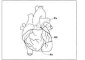
そして、探索により検出された候補点を、所定のアルゴリズムに基づいて連結する。これにより、候補点および候補点同士を連結する血管枝(エッジ)からなる木構造が構築され、この木構造を冠動脈の経路として抽出する。なお、経路上における複数の候補点の座標情報や、血管枝の方向を示すベクトル情報は、候補点や血管枝の識別子とともに記憶部12に記憶される。続いて、検出された候補点ごとに、周辺のボクセルの値(CT値)に基づき、冠動脈の経路に垂直な断面において、血管の輪郭(血管の外壁)を識別する。形状の識別は、Graph-Cutsに代表される公知のセグメンテーション手法を用いて行う。以上の処理により、図2に示すような管状構造である冠動脈の経路K0および冠動脈が抽出され、その抽出された経路K0および冠動脈の特定に必要な情報が生成され、記憶部12に記憶される。 Then, the candidate points detected by the search are connected based on a predetermined algorithm. Thereby, a tree structure composed of candidate points and blood vessel branches (edges) connecting the candidate points is constructed, and this tree structure is extracted as a coronary artery path. Note that the coordinate information of a plurality of candidate points on the route and vector information indicating the direction of the blood vessel branch are stored in the
経路抽出部16は、上述したように冠動脈の中心線を経路として抽出するが、プラーク等により血管が細くなっている部分、あるいは脂肪等の血管とは異なる組織が付与してしまった部分等において、本来あるべき経路が分断されてしまう場合がある。分断経路検出部18は、このような本来あるべき経路が分断されてしまっている分断経路を検出する。このため、分断経路検出部18は、まず経路抽出部16が抽出した経路の端点を検出する。この端点の検出は、経路K0が途切れている点を検出することにより行う。 As described above, the
そして、検出した端点を中心とした所定距離Th0以内に、抽出された経路K0が存在するか否かを判定し、存在する場合にその端点と抽出された経路との間は分断されているものとして、そこを分断経路として検出する。図3は分断経路の検出を説明するための図である。分断経路検出部18は、図3(a)に示すように端点Ptを中心とした所定距離Th0の円形領域A0を設定し、円形領域A0内に他の経路が存在する場合には、端点Ptとその経路との間を分断経路として検出する。一方、図3(b)に示すように、円形領域A0内に他の経路が存在しない場合、端点Ptとその経路との間は分断経路としては検出しない。また、図3(c)に示すように、円形領域A0内に他の複数の経路K1,K2が存在する場合には、端点Ptと端点Ptに最も近い方の経路K1との間を分断経路として検出する。ここで、所定距離Th0はユーザが任意の値に設定することが可能である。 Then, it is determined whether or not the extracted route K0 exists within a predetermined distance Th0 centered on the detected end point, and when it exists, the end point and the extracted route are divided. As such, it is detected as a divided path. FIG. 3 is a diagram for explaining the detection of the divided path. As shown in FIG. 3A, the divided
なお、このような分断経路の検出に代えて、経路の端点とその周囲にある他の経路との距離を算出し、この距離が最も短くなる他の経路と端点との間を分断経路として検出してもよい。この際、端点が位置する経路を抽出した冠動脈の径、および距離を算出する対象となる他の経路を抽出した冠動脈の径を用いて、端点とその周囲の他の経路との距離を判断してもよい。すなわち、図4に示すように、経路K3の端点Ptと経路K4との距離を算出するに際し、経路K3を抽出した血管の半径R3および他の経路K4を抽出した血管の半径R4を、端点Ptと経路K4との距離L0から減算した距離L1を算出し、その距離L1が最小となる他の経路と端点Ptとの間を分断経路として検出してもよい。なお、上記距離L0は、端点Ptと他の経路K4との最短距離である。 Instead of detecting such a divided route, the distance between the end point of the route and other routes around it is calculated, and the distance between the other route and the end point where the distance is the shortest is detected as a divided route. May be. At this time, using the diameter of the coronary artery from which the path where the end point is extracted and the diameter of the coronary artery from which another path is calculated, the distance between the end point and other paths around it is determined. May be. That is, as shown in FIG. 4, in calculating the distance between the end point Pt of the path K3 and the path K4, the radius R3 of the blood vessel from which the path K3 has been extracted and the radius R4 of the blood vessel from which the other path K4 has been extracted are determined as the end point Pt. And a distance L1 obtained by subtracting the distance L0 from the path K4 may be calculated, and the distance between the other path and the end point Pt that minimizes the distance L1 may be detected as a divided path. The distance L0 is the shortest distance between the end point Pt and the other route K4.
検索用経路生成部20は、分断経路検出部18が検出した分断経路を接続する検索用経路を生成する。すなわち、図5に示すように、経路K5の端点Ptと端点Ptに近い最も近い経路K6との間に、破線で示す検索用経路Kk0を生成する。この際、検索用経路Kk0は、端点Ptと経路K6との最短距離を通るように生成すればよい。また、経路抽出部16において、分断経路すなわち端点Ptと経路K6との間の領域に対して、再度の経路抽出を行うようにしてもよい。このように再度の抽出処理を行う際には、端点Ptから経路K6に向かう直線の進め方を小さくする、さらには、端点Ptから経路K6の方向にのみ探索を行う等して、抽出の精度を向上させることが好ましい。このように抽出精度を高めることにより、経路が抽出される可能性があり、このように経路が抽出された場合には、その経路を検索用経路として用いればよい。 The search
始点終点設定部22は、経路抽出部16が抽出した冠動脈の経路K0に始点および終点を設定する。具体的には、図6に示すように、表示制御部26により、心臓に経路K0をマッピングしたボリュームレンダリング画像(VR画像)を表示部30に表示し、ユーザによる入力部28を操作しての始点Psおよび終点Peの指定を受け付けることにより、始点Psおよび終点Peを設定する。なお、VR画像の表示については後述する。ここで、本実施形態においては、始点Psは冠動脈と大動脈との接続部分であり、終点Peは冠動脈の末梢端とする。なお、始点Psおよび終点Peを自動で検出するようにしてもよい。これにより、ユーザは経路の始点Psおよび終点Peを指定する必要が無くなるため、ユーザの負担を軽減することができる。 The start point / end
経路探索部24は、経路抽出部16が抽出した経路および検索用経路に基づいて、始点Psおよび終点Peの間の最短経路を探索する。すなわち、経路探索部24は、経路抽出部16が抽出した経路および検索用経路に沿って、始点Psから終点Peまでの長さ(距離)を検出するトラッキング処理を行う。そして、このトラッキング処理により始点Psから終点Peまでの最短経路を検出する。具体的には、始点Psから終点Peまでの長さが検出された経路のうち、最短の経路(中心線の長さが一番小さくなる経路の探索ベクトルデータ)を検出し、この経路を始点Psから終点Peまでの最短経路として検出する。 The
なお、このように最短経路を探索するに際し、検索用経路についてはトラッキングの際に、優先度を低くすることが好ましい。具体的には、抽出した経路よりも長さを長く見せるように重みづけをすることが好ましい。 In searching for the shortest route in this way, it is preferable to lower the priority of the search route during tracking. Specifically, weighting is preferably performed so that the length is longer than the extracted route.
表示制御部26は、構造物抽出部14が3次元画像V0から抽出した心臓および経路抽出部16が抽出した冠動脈を、ボリュームレンダリング法を用いてボリュームレンダリング(VR)表示する。すなわち、抽出した心臓および冠動脈に対して投影面から仮想的な光線を照射し、3次元画像V0の各信号値に対応した色(R,G,B)および不透明度(オパシティ)に基づいて、物体内部からの仮想的な反射光による3次元画像を作成し、この3次元画像から投影面に物体内部の3次元構造を透視する投影画像を生成して、これをVR画像として表示する。さらに、表示制御部26は、経路探索部24が探索した冠動脈上に最短経路をマッピング表示する。図7は最短経路がマッピングされたVR画像を示す図である。図7に示すように、冠動脈上に最短経路Ksがマッピングされている。 The
入力部28は、キーボードおよびマウス等の公知の入力装置からなる。 The
表示部30は、液晶、CRT等の公知の表示装置からなる。 The
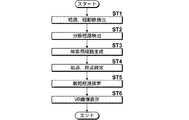
次いで、本実施形態において行われる処理について説明する。図8は本実施形態において行われる処理を示すフローチャートである。なお、3次元画像V0は画像取得部10により取得されて記憶部12に記憶され、さらに構造物抽出部14により、3次元画像V0から心臓が抽出されているものとする。まず、経路抽出部16が、冠動脈の経路および冠動脈を抽出し(ステップST1)、さらに分断経路検出部18が、本来あるべき経路が分断されてしまっている分断経路を検出する(ステップST2)。 Next, processing performed in the present embodiment will be described. FIG. 8 is a flowchart showing processing performed in the present embodiment. It is assumed that the three-dimensional image V0 is acquired by the
そして、検索用経路生成部20が、分断経路を接続する検索用経路を生成し(ステップST3)、始点終点設定部22が、経路抽出部16が抽出した冠動脈に始点Psおよび終点Peを設定する(ステップST4)。そして、経路探索部24が、経路抽出部16が抽出した経路および検索用経路に基づいて、始点Psおよび終点Peの間の最短経路を探索する(ステップST5)。さらに、表示制御部26が、心臓および冠動脈のVR画像に最短経路をマッピングし、最短経路がマッピングされたVR画像を表示部30に表示し(ステップST6)、処理を終了する。 Then, the search
このように、本実施形態においては、3次元画像V0から冠動脈の経路を抽出し、経路において分断経路を検出し、分断経路に検索用経路を生成し、抽出した経路および検索用経路に基づいて、始点Psおよび終点Peの間の最短経路を探索するようにしたものである。このため、ユーザは何ら手を加えなくても、分断経路が接続されて、最短経路が探索されることとなる。したがって、3次元画像V0から冠動脈等の管状構造の最短経路を探索する際のユーザの負担を軽減できる。 As described above, in this embodiment, a coronary artery route is extracted from the three-dimensional image V0, a divided route is detected in the route, a search route is generated in the divided route, and the extracted route and the search route are used. The shortest path between the start point Ps and the end point Pe is searched. For this reason, even if a user does not do anything, a divided route will be connected and a shortest route will be searched. Therefore, the burden on the user when searching for the shortest path of a tubular structure such as a coronary artery from the three-dimensional image V0 can be reduced.
また、経路の端点と端点の周囲にある他の経路との距離が所定距離Th0以内である場合に、端点と他の経路との間を分断経路として検出しているため、本来連続した経路でない端点が経路として探索されてしまうことを防止でき、これにより、最短経路探索の精度を向上できる。 In addition, when the distance between the end point of the route and the other route around the end point is within the predetermined distance Th0, since the distance between the end point and the other route is detected as a divided route, the route is not originally continuous. It is possible to prevent the end point from being searched as a route, thereby improving the accuracy of the shortest route search.
また、検索用経路に対して優先度を低くして最短経路を探索しているため、検索用経路が優先的に最短経路として探索されることが無くなり、これにより、誤った経路が探索されてしまう可能性を低減できる。 In addition, since the search route is searched for the shortest route with a lower priority than the search route, the search route is not preferentially searched as the shortest route, and thus an incorrect route is searched. The possibility that it will end up can be reduced.
また、分断経路において経路の再度の抽出を行うことにより、本来存在すべき経路が探索される場合がある。このため、経路の再度の抽出結果に基づいて、検索用経路を生成することにより、本来存在すべき経路の可能性が高い検索用経路を用いて最短経路を探索できるため、最短経路探索の精度を向上できる。 In addition, there is a case where a route that should originally exist may be searched for by extracting the route again in the divided route. For this reason, since the search route is generated based on the re-extraction result of the route, the shortest route can be searched using the search route that is highly likely to be the original route. Can be improved.
なお、上記実施形態においては、最短経路を探索した後に、VR画像とともに最短経路を表示しているが、経路抽出部16が抽出した経路を、VR画像にマッピングして表示部30に表示し、ユーザによる入力部28を用いての経路の結合および/または分断の指示を受け付け、経路抽出部16がこの指示に応じて抽出した経路を修正するようにしてもよい。これにより、ユーザの意図を反映させて最短経路を探索することができる。また、この場合において、ユーザは経路を分断する指示を行う場合があるが、ユーザによる分断の指示がなされた経路においては、分断経路としての検出を禁止することが好ましい。これにより、ユーザが意図的に分断した経路が検索用経路により接続されてしまうことを防止できる。 In the above embodiment, after searching for the shortest route, the shortest route is displayed together with the VR image. However, the route extracted by the
また、上記実施形態においては、管状構造として、心臓の冠動脈を用いているが、これに限定されるものではなく、脳血管、肺の気管支、肝臓の血管(動脈、静脈、門脈)等、人体を構成する管状構造を有する構造物の最短経路の探索に、本発明を適用することができる。 In the above embodiment, the coronary artery of the heart is used as the tubular structure. However, the present invention is not limited to this, and the brain blood vessel, the bronchi of the lung, the blood vessel of the liver (artery, vein, portal vein), etc. The present invention can be applied to search for the shortest path of a structure having a tubular structure constituting a human body.
1 3次元画像表示装置
10 画像取得部
12 記憶部
14 構造物抽出部
16 経路抽出部
18 分断経路検出部
20 検索用経路生成部
22 始点終点設定部
24 経路探索部
26 表示制御部
28 入力部
30 表示部DESCRIPTION OF SYMBOLS 1 3D
Claims (10)
Translated fromJapanese前記経路において、分断されている分断経路を検出する分断経路検出手段と、
該分断経路を接続する検索用経路を生成する検索用経路生成手段と、
前記経路および前記検索用経路に基づいて、前記経路の始点および終点の間の最短経路を探索する探索手段とを備えたことを特徴とする管状構造の最短経路探索装置。Path extraction means for extracting a path of the tubular structure from a three-dimensional image including the tubular structure;
In the route, a divided route detection means for detecting a divided route that is divided;
Search path generation means for generating a search path connecting the divided paths;
A shortest path search device having a tubular structure, comprising: search means for searching for a shortest path between a start point and an end point of the path based on the path and the search path.
前記検索用経路生成手段は、前記経路の再度の抽出結果に基づいて、前記検索用経路を生成する手段であることを特徴とする請求項1から7のいずれか1項記載の最短経路探索装置。The route extraction means is means for performing re-extraction of the route in the divided route,
The shortest path search device according to claim 1, wherein the search path generation unit is a unit that generates the search path based on a result of extracting the path again. .
前記経路において、分断されている分断経路を検出し、
該分断経路を接続する検索用経路を生成し、
前記経路および前記検索用経路に基づいて、前記経路の始点および終点の間の最短経路を探索することを特徴とする管状構造の最短経路探索方法。Extracting a path of the tubular structure from a three-dimensional image including the tubular structure;
In the route, a divided route that is divided is detected,
Generate a search route that connects the divided routes,
A shortest path search method for a tubular structure, wherein a shortest path between a start point and an end point of the path is searched based on the path and the search path.
前記経路において、分断されている分断経路を検出する手順と、
該分断経路を接続する検索用経路を生成する手順と、
前記経路および前記検索用経路に基づいて、前記経路の始点および終点の間の最短経路を探索する手順とを有する管状構造の最短経路探索方法をコンピュータに実行させることを特徴とするプログラム。Extracting a path of the tubular structure from a three-dimensional image including the tubular structure;
A procedure for detecting a divided route that is divided in the route;
A procedure for generating a search path connecting the divided paths;
A program for causing a computer to execute a shortest path search method for a tubular structure having a procedure for searching for a shortest path between a start point and an end point of the path based on the path and the search path.
Priority Applications (2)
| Application Number | Priority Date | Filing Date | Title |
|---|---|---|---|
| JP2012213408AJP5934071B2 (en) | 2012-09-27 | 2012-09-27 | Apparatus, method and program for searching for shortest path of tubular structure |
| US14/033,200US9198603B2 (en) | 2012-09-27 | 2013-09-20 | Device, method and program for searching for the shortest path in a tubular structure |
Applications Claiming Priority (1)
| Application Number | Priority Date | Filing Date | Title |
|---|---|---|---|
| JP2012213408AJP5934071B2 (en) | 2012-09-27 | 2012-09-27 | Apparatus, method and program for searching for shortest path of tubular structure |
Publications (3)
| Publication Number | Publication Date |
|---|---|
| JP2014064824Atrue JP2014064824A (en) | 2014-04-17 |
| JP2014064824A5 JP2014064824A5 (en) | 2015-01-29 |
| JP5934071B2 JP5934071B2 (en) | 2016-06-15 |
Family
ID=50339534
Family Applications (1)
| Application Number | Title | Priority Date | Filing Date |
|---|---|---|---|
| JP2012213408AActiveJP5934071B2 (en) | 2012-09-27 | 2012-09-27 | Apparatus, method and program for searching for shortest path of tubular structure |
Country Status (2)
| Country | Link |
|---|---|
| US (1) | US9198603B2 (en) |
| JP (1) | JP5934071B2 (en) |
Cited By (3)
| Publication number | Priority date | Publication date | Assignee | Title |
|---|---|---|---|---|
| WO2018221508A1 (en)* | 2017-06-02 | 2018-12-06 | テルモ株式会社 | Route selection assist system, recording medium for recording route selection assist program, route selection assist method, and diagnosis method |
| WO2021162355A1 (en)* | 2020-02-10 | 2021-08-19 | 재단법인 아산사회복지재단 | Method and apparatus for providing guide data for intravascular medical tool insertion device |
| JP2023531821A (en)* | 2020-06-29 | 2023-07-25 | バイオセンス・ウエブスター・(イスラエル)・リミテッド | Efficient automatic discovery of minimal ear-nose-throat (EAR-NOSE-THROAT, ENT) pathways for probes |
Families Citing this family (6)
| Publication number | Priority date | Publication date | Assignee | Title |
|---|---|---|---|---|
| US20140253544A1 (en)* | 2012-01-27 | 2014-09-11 | Kabushiki Kaisha Toshiba | Medical image processing apparatus |
| US10360672B2 (en)* | 2013-03-15 | 2019-07-23 | University Of Iowa Research Foundation | Automated separation of binary overlapping trees |
| US9895803B1 (en)* | 2015-06-19 | 2018-02-20 | X Development Llc | Calculating trajectory corridor for robot end effector |
| JP7171579B2 (en)* | 2017-01-05 | 2022-11-15 | コーニンクレッカ フィリップス エヌ ヴェ | Visualization of pathways of vasculature with occlusion |
| WO2020217807A1 (en)* | 2019-04-25 | 2020-10-29 | 富士フイルム株式会社 | Image orientation setting device, method, and program |
| CN115953495B (en)* | 2023-03-14 | 2023-08-01 | 北京唯迈医疗设备有限公司 | Intelligent path planning device, system and storage medium based on two-dimensional contrast image |
Citations (5)
| Publication number | Priority date | Publication date | Assignee | Title |
|---|---|---|---|---|
| JPH0765154A (en)* | 1993-08-31 | 1995-03-10 | Toshiba Corp | Quantitative analysis apparatus for blood vessel image and quantitative analysis method thereof |
| JP2004283373A (en)* | 2003-03-20 | 2004-10-14 | Toshiba Corp | Analytical processing device for luminal structure |
| JP2005508680A (en)* | 2001-10-16 | 2005-04-07 | コーニンクレッカ フィリップス エレクトロニクス エヌ ヴィ | Branch selection method for probe alignment |
| JP2009268741A (en)* | 2008-05-08 | 2009-11-19 | Toshiba Corp | Mri image diagnostic apparatus and mr image forming method |
| US20120237094A1 (en)* | 2011-03-18 | 2012-09-20 | Fujifilm Corporation | Network construction apparatus, method and program |
Family Cites Families (12)
| Publication number | Priority date | Publication date | Assignee | Title |
|---|---|---|---|---|
| JP4053117B2 (en) | 1997-10-17 | 2008-02-27 | 東芝医用システムエンジニアリング株式会社 | Image processing device |
| JP4545971B2 (en) | 2001-03-05 | 2010-09-15 | 日本電信電話株式会社 | Medical image identification system, medical image identification processing method, medical image identification program, and recording medium thereof |
| JP3766033B2 (en) | 2001-04-25 | 2006-04-12 | 富士写真フイルム株式会社 | Image processing method, apparatus, and program |
| US8463065B2 (en)* | 2005-12-07 | 2013-06-11 | Commonwealth Scientific And Industrial Research Organisation | Linear feature detection method and apparatus |
| DE102006001884A1 (en)* | 2006-01-13 | 2007-07-19 | Siemens Ag | Medical instrument`s e.g. intracranial catheter, guidance visual aiding method, involves marking input point and/or target point in spatial representation of hollow organ section of examination object by medical personnel |
| US8781193B2 (en)* | 2007-03-08 | 2014-07-15 | Sync-Rx, Ltd. | Automatic quantitative vessel analysis |
| JP2008259682A (en) | 2007-04-12 | 2008-10-30 | Fujifilm Corp | Region recognition result correction apparatus, method, and program |
| US8126232B2 (en)* | 2008-03-05 | 2012-02-28 | Siemens Aktiengesellschaft | System and method for 3D vessel segmentation with minimal cuts |
| US9679389B2 (en)* | 2009-05-19 | 2017-06-13 | Algotec Systems Ltd. | Method and system for blood vessel segmentation and classification |
| US8934686B2 (en)* | 2009-11-26 | 2015-01-13 | Algotec Systems Ltd. | User interface for selecting paths in an image |
| DE102011076188B4 (en)* | 2011-05-20 | 2013-03-21 | Siemens Aktiengesellschaft | Apparatus and method for planning an endovascular procedure with a medical instrument |
| EP2747695B1 (en)* | 2011-10-26 | 2017-09-13 | Koninklijke Philips N.V. | Endoscopic registration of vessel tree images |
- 2012
- 2012-09-27JPJP2012213408Apatent/JP5934071B2/enactiveActive
- 2013
- 2013-09-20USUS14/033,200patent/US9198603B2/enactiveActive
Patent Citations (5)
| Publication number | Priority date | Publication date | Assignee | Title |
|---|---|---|---|---|
| JPH0765154A (en)* | 1993-08-31 | 1995-03-10 | Toshiba Corp | Quantitative analysis apparatus for blood vessel image and quantitative analysis method thereof |
| JP2005508680A (en)* | 2001-10-16 | 2005-04-07 | コーニンクレッカ フィリップス エレクトロニクス エヌ ヴィ | Branch selection method for probe alignment |
| JP2004283373A (en)* | 2003-03-20 | 2004-10-14 | Toshiba Corp | Analytical processing device for luminal structure |
| JP2009268741A (en)* | 2008-05-08 | 2009-11-19 | Toshiba Corp | Mri image diagnostic apparatus and mr image forming method |
| US20120237094A1 (en)* | 2011-03-18 | 2012-09-20 | Fujifilm Corporation | Network construction apparatus, method and program |
Cited By (5)
| Publication number | Priority date | Publication date | Assignee | Title |
|---|---|---|---|---|
| WO2018221508A1 (en)* | 2017-06-02 | 2018-12-06 | テルモ株式会社 | Route selection assist system, recording medium for recording route selection assist program, route selection assist method, and diagnosis method |
| JPWO2018221508A1 (en)* | 2017-06-02 | 2020-04-02 | テルモ株式会社 | Route selection support system, recording medium storing a route selection support program, route selection support method, and diagnosis method |
| US11596352B2 (en) | 2017-06-02 | 2023-03-07 | Terumo Kabushiki Kaisha | Route selection assistance system, recording medium on which route selection assistance program is recorded, route selection assistance method, and diagnosis method |
| WO2021162355A1 (en)* | 2020-02-10 | 2021-08-19 | 재단법인 아산사회복지재단 | Method and apparatus for providing guide data for intravascular medical tool insertion device |
| JP2023531821A (en)* | 2020-06-29 | 2023-07-25 | バイオセンス・ウエブスター・(イスラエル)・リミテッド | Efficient automatic discovery of minimal ear-nose-throat (EAR-NOSE-THROAT, ENT) pathways for probes |
Also Published As
| Publication number | Publication date |
|---|---|
| US20140088416A1 (en) | 2014-03-27 |
| JP5934071B2 (en) | 2016-06-15 |
| US9198603B2 (en) | 2015-12-01 |
Similar Documents
| Publication | Publication Date | Title |
|---|---|---|
| JP5934071B2 (en) | Apparatus, method and program for searching for shortest path of tubular structure | |
| JP7309986B2 (en) | Medical image processing method, medical image processing apparatus, medical image processing system, and medical image processing program | |
| JP6175073B2 (en) | Real-time display of vascular view for optimal device navigation | |
| US8630467B2 (en) | Diagnosis assisting system using three dimensional image data, computer readable recording medium having a related diagnosis assisting program recorded thereon, and related diagnosis assisting method | |
| CN101336844B (en) | Medical image processing device and medical image diagnosis device | |
| JP5584006B2 (en) | Projection image generation apparatus, projection image generation program, and projection image generation method | |
| US9662083B2 (en) | Medical image display apparatus and medical image display system | |
| JP4937397B2 (en) | Medical image diagnosis support apparatus and method, and program | |
| JP6080249B2 (en) | Three-dimensional image display apparatus and method, and program | |
| JP6080248B2 (en) | Three-dimensional image display apparatus and method, and program | |
| JP5685605B2 (en) | System for navigating an interventional device and its operating method, computer program element and computer readable medium | |
| CN102395320B (en) | Medical equipment and control method for medical equipment | |
| JP5572470B2 (en) | Diagnosis support apparatus, method, and program | |
| EP2925216B1 (en) | Stenosis therapy planning | |
| JP5700964B2 (en) | Medical image processing apparatus, method and program | |
| JP2011025005A (en) | Diagnosis supporting device, diagnosis supporting program, and diagnosis supporting method | |
| JP2010528750A (en) | Inspection of tubular structures | |
| JP5415245B2 (en) | MEDICAL IMAGE DISPLAY DEVICE, METHOD, AND PROGRAM | |
| JP6301277B2 (en) | Diagnostic auxiliary image generation apparatus, diagnostic auxiliary image generation method, and diagnostic auxiliary image generation program | |
| JP2009018005A (en) | Bulls eye map creation device, bulls eye map creation method, and image processing device | |
| US9123163B2 (en) | Medical image display apparatus, method and program | |
| JP6734111B2 (en) | Finding information creation device and system | |
| JP2007135858A (en) | Image processor | |
| JP5159195B2 (en) | Medical image processing device |
Legal Events
| Date | Code | Title | Description |
|---|---|---|---|
| A521 | Request for written amendment filed | Free format text:JAPANESE INTERMEDIATE CODE: A523 Effective date:20141205 | |
| A621 | Written request for application examination | Free format text:JAPANESE INTERMEDIATE CODE: A621 Effective date:20141205 | |
| A977 | Report on retrieval | Free format text:JAPANESE INTERMEDIATE CODE: A971007 Effective date:20150819 | |
| A131 | Notification of reasons for refusal | Free format text:JAPANESE INTERMEDIATE CODE: A131 Effective date:20150908 | |
| A521 | Request for written amendment filed | Free format text:JAPANESE INTERMEDIATE CODE: A523 Effective date:20151106 | |
| TRDD | Decision of grant or rejection written | ||
| A01 | Written decision to grant a patent or to grant a registration (utility model) | Free format text:JAPANESE INTERMEDIATE CODE: A01 Effective date:20160419 | |
| A61 | First payment of annual fees (during grant procedure) | Free format text:JAPANESE INTERMEDIATE CODE: A61 Effective date:20160506 | |
| R150 | Certificate of patent or registration of utility model | Ref document number:5934071 Country of ref document:JP Free format text:JAPANESE INTERMEDIATE CODE: R150 | |
| R250 | Receipt of annual fees | Free format text:JAPANESE INTERMEDIATE CODE: R250 | |
| R250 | Receipt of annual fees | Free format text:JAPANESE INTERMEDIATE CODE: R250 | |
| R250 | Receipt of annual fees | Free format text:JAPANESE INTERMEDIATE CODE: R250 | |
| R250 | Receipt of annual fees | Free format text:JAPANESE INTERMEDIATE CODE: R250 | |
| R250 | Receipt of annual fees | Free format text:JAPANESE INTERMEDIATE CODE: R250 | |
| R250 | Receipt of annual fees | Free format text:JAPANESE INTERMEDIATE CODE: R250 |