JP2013537971A - Highly sensitive monoclonal antibody residue detection assay - Google Patents
Highly sensitive monoclonal antibody residue detection assayDownload PDFInfo
- Publication number
- JP2013537971A JP2013537971AJP2013528381AJP2013528381AJP2013537971AJP 2013537971 AJP2013537971 AJP 2013537971AJP 2013528381 AJP2013528381 AJP 2013528381AJP 2013528381 AJP2013528381 AJP 2013528381AJP 2013537971 AJP2013537971 AJP 2013537971A
- Authority
- JP
- Japan
- Prior art keywords
- antibody
- product
- antigen
- antibodies
- binding
- Prior art date
- Legal status (The legal status is an assumption and is not a legal conclusion. Google has not performed a legal analysis and makes no representation as to the accuracy of the status listed.)
- Withdrawn
Links
- 238000001514detection methodMethods0.000titleclaimsdescription49
- 238000003556assayMethods0.000titledescription46
- 238000000034methodMethods0.000claimsabstractdescription95
- 239000000427antigenSubstances0.000claimsabstractdescription64
- 230000027455bindingEffects0.000claimsabstractdescription62
- 108091007433antigensProteins0.000claimsabstractdescription50
- 102000036639antigensHuman genes0.000claimsabstractdescription50
- 239000012634fragmentSubstances0.000claimsabstractdescription44
- 238000012544monitoring processMethods0.000claimsabstractdescription28
- 238000012360testing methodMethods0.000claimsdescription44
- 108090000623proteins and genesProteins0.000claimsdescription27
- 102000004169proteins and genesHuman genes0.000claimsdescription27
- 239000000758substrateSubstances0.000claimsdescription24
- 238000005406washingMethods0.000claimsdescription10
- 108010021625Immunoglobulin FragmentsProteins0.000claimsdescription7
- 102000008394Immunoglobulin FragmentsHuman genes0.000claimsdescription7
- 102000004190EnzymesHuman genes0.000claimsdescription5
- 108090000790EnzymesProteins0.000claimsdescription5
- 108010067060Immunoglobulin Variable RegionProteins0.000claimsdescription4
- 102000017727Immunoglobulin Variable RegionHuman genes0.000claimsdescription4
- 102000039446nucleic acidsHuman genes0.000claimsdescription3
- 108020004707nucleic acidsProteins0.000claimsdescription3
- 150000007523nucleic acidsChemical class0.000claimsdescription3
- 239000000813peptide hormoneSubstances0.000claimsdescription2
- 238000012790confirmationMethods0.000abstractdescription26
- 239000000203mixtureSubstances0.000abstractdescription19
- 238000004519manufacturing processMethods0.000abstractdescription18
- 238000004140cleaningMethods0.000abstractdescription11
- 238000003018immunoassayMethods0.000abstractdescription9
- 239000000047productSubstances0.000description99
- 235000018102proteinsNutrition0.000description25
- 239000008186active pharmaceutical agentSubstances0.000description20
- 238000011084recoveryMethods0.000description15
- YBJHBAHKTGYVGT-ZKWXMUAHSA-N(+)-BiotinChemical compoundN1C(=O)N[C@@H]2[C@H](CCCCC(=O)O)SC[C@@H]21YBJHBAHKTGYVGT-ZKWXMUAHSA-N0.000description10
- 230000008569processEffects0.000description10
- 238000002415sodium dodecyl sulfate polyacrylamide gel electrophoresisMethods0.000description10
- 238000002965ELISAMethods0.000description9
- 238000004458analytical methodMethods0.000description9
- 230000016784immunoglobulin productionEffects0.000description9
- 238000001262western blotMethods0.000description9
- 238000011002quantificationMethods0.000description8
- 239000000872bufferSubstances0.000description7
- 239000012491analyteSubstances0.000description6
- 238000002474experimental methodMethods0.000description6
- 238000005070samplingMethods0.000description6
- 230000035945sensitivityEffects0.000description6
- 238000000926separation methodMethods0.000description6
- 108010001336Horseradish PeroxidaseProteins0.000description5
- 241001529936MurinaeSpecies0.000description5
- 229960002685biotinDrugs0.000description5
- 235000020958biotinNutrition0.000description5
- 239000011616biotinSubstances0.000description5
- 239000012895dilutionSubstances0.000description5
- 238000010790dilutionMethods0.000description5
- 239000003814drugSubstances0.000description5
- 238000005259measurementMethods0.000description5
- 239000000243solutionSubstances0.000description5
- 108010047041Complementarity Determining RegionsProteins0.000description4
- 229920002684SepharosePolymers0.000description4
- 238000002835absorbanceMethods0.000description4
- 229940088679drug related substanceDrugs0.000description4
- 238000005516engineering processMethods0.000description4
- 229940088598enzymeDrugs0.000description4
- 230000036541healthEffects0.000description4
- 210000004408hybridomaAnatomy0.000description4
- 239000000463materialSubstances0.000description4
- 239000011159matrix materialSubstances0.000description4
- -1polyethylenePolymers0.000description4
- 102000004196processed proteins & peptidesHuman genes0.000description4
- 108090000765processed proteins & peptidesProteins0.000description4
- 239000012898sample dilutionSubstances0.000description4
- 238000003118sandwich ELISAMethods0.000description4
- XLYOFNOQVPJJNP-UHFFFAOYSA-NwaterSubstancesOXLYOFNOQVPJJNP-UHFFFAOYSA-N0.000description4
- 108060003951ImmunoglobulinProteins0.000description3
- 108010090804StreptavidinProteins0.000description3
- 230000000903blocking effectEffects0.000description3
- 150000001720carbohydratesChemical class0.000description3
- 238000006243chemical reactionMethods0.000description3
- 239000003153chemical reaction reagentSubstances0.000description3
- 238000011161developmentMethods0.000description3
- 239000012149elution bufferSubstances0.000description3
- 230000004927fusionEffects0.000description3
- 102000018358immunoglobulinHuman genes0.000description3
- 238000011534incubationMethods0.000description3
- 238000011028process validationMethods0.000description3
- 230000009257reactivityEffects0.000description3
- 230000001225therapeutic effectEffects0.000description3
- 238000010200validation analysisMethods0.000description3
- 108090001008AvidinProteins0.000description2
- 238000009010Bradford assayMethods0.000description2
- OKTJSMMVPCPJKN-UHFFFAOYSA-NCarbonChemical compound[C]OKTJSMMVPCPJKN-UHFFFAOYSA-N0.000description2
- 229920002307DextranPolymers0.000description2
- 108010054477Immunoglobulin Fab FragmentsProteins0.000description2
- 102000001706Immunoglobulin Fab FragmentsHuman genes0.000description2
- 241001465754MetazoaSpecies0.000description2
- 108090000854OxidoreductasesProteins0.000description2
- 102000004316OxidoreductasesHuman genes0.000description2
- NBIIXXVUZAFLBC-UHFFFAOYSA-NPhosphoric acidChemical compoundOP(O)(O)=ONBIIXXVUZAFLBC-UHFFFAOYSA-N0.000description2
- 108020004511Recombinant DNAProteins0.000description2
- 238000011948assay developmentMethods0.000description2
- 238000002820assay formatMethods0.000description2
- 239000011324beadSubstances0.000description2
- 230000033228biological regulationEffects0.000description2
- 210000004899c-terminal regionAnatomy0.000description2
- 229910052799carbonInorganic materials0.000description2
- 239000011248coating agentSubstances0.000description2
- 238000000576coating methodMethods0.000description2
- 238000011109contaminationMethods0.000description2
- 238000007796conventional methodMethods0.000description2
- 229920006037cross link polymerPolymers0.000description2
- 230000009260cross reactivityEffects0.000description2
- 239000003599detergentSubstances0.000description2
- 239000013024dilution bufferSubstances0.000description2
- 239000011521glassSubstances0.000description2
- 229910000856hastalloyInorganic materials0.000description2
- 210000005260human cellAnatomy0.000description2
- 238000009396hybridizationMethods0.000description2
- 239000000017hydrogelSubstances0.000description2
- 230000003278mimic effectEffects0.000description2
- 238000012986modificationMethods0.000description2
- 230000004048modificationEffects0.000description2
- 239000012514monoclonal antibody productSubstances0.000description2
- 238000007837multiplex assayMethods0.000description2
- 230000009871nonspecific bindingEffects0.000description2
- 229920001184polypeptidePolymers0.000description2
- 229920002451polyvinyl alcoholPolymers0.000description2
- 238000012797qualificationMethods0.000description2
- 238000011160researchMethods0.000description2
- 230000004044responseEffects0.000description2
- 239000007790solid phaseSubstances0.000description2
- 229910001220stainless steelInorganic materials0.000description2
- 239000010935stainless steelSubstances0.000description2
- 238000003860storageMethods0.000description2
- 229940124597therapeutic agentDrugs0.000description2
- KGLPWQKSKUVKMJ-UHFFFAOYSA-N2,3-dihydrophthalazine-1,4-dioneChemical compoundC1=CC=C2C(=O)NNC(=O)C2=C1KGLPWQKSKUVKMJ-UHFFFAOYSA-N0.000description1
- HJBUBXIDMQBSQW-UHFFFAOYSA-N4-(4-diazoniophenyl)benzenediazoniumChemical compoundC1=CC([N+]#N)=CC=C1C1=CC=C([N+]#N)C=C1HJBUBXIDMQBSQW-UHFFFAOYSA-N0.000description1
- CJIJXIFQYOPWTF-UHFFFAOYSA-N7-hydroxycoumarinNatural productsO1C(=O)C=CC2=CC(O)=CC=C21CJIJXIFQYOPWTF-UHFFFAOYSA-N0.000description1
- 229920000936AgarosePolymers0.000description1
- 102000002260Alkaline PhosphataseHuman genes0.000description1
- 108020004774Alkaline PhosphataseProteins0.000description1
- 108091003079Bovine Serum AlbuminProteins0.000description1
- 239000004971Cross linkerSubstances0.000description1
- IGXWBGJHJZYPQS-SSDOTTSWSA-ND-LuciferinChemical compoundOC(=O)[C@H]1CSC(C=2SC3=CC=C(O)C=C3N=2)=N1IGXWBGJHJZYPQS-SSDOTTSWSA-N0.000description1
- 108020004414DNAProteins0.000description1
- CYCGRDQQIOGCKX-UHFFFAOYSA-NDehydro-luciferinNatural productsOC(=O)C1=CSC(C=2SC3=CC(O)=CC=C3N=2)=N1CYCGRDQQIOGCKX-UHFFFAOYSA-N0.000description1
- ZNZYKNKBJPZETN-WELNAUFTSA-NDialdehyde 11678Chemical compoundN1C2=CC=CC=C2C2=C1[C@H](C[C@H](/C(=C/O)C(=O)OC)[C@@H](C=C)C=O)NCC2ZNZYKNKBJPZETN-WELNAUFTSA-N0.000description1
- 238000012286ELISA AssayMethods0.000description1
- 101000957776Escherichia coli O157:H7 Mannose-1-phosphate guanylyltransferase 1Proteins0.000description1
- BJGNCJDXODQBOB-UHFFFAOYSA-NFivefly LuciferinNatural productsOC(=O)C1CSC(C=2SC3=CC(O)=CC=C3N=2)=N1BJGNCJDXODQBOB-UHFFFAOYSA-N0.000description1
- 108010073178Glucan 1,4-alpha-GlucosidaseProteins0.000description1
- 102100022624GlucoamylaseHuman genes0.000description1
- 108700005091Immunoglobulin GenesProteins0.000description1
- 102000006496Immunoglobulin Heavy ChainsHuman genes0.000description1
- 108010019476Immunoglobulin Heavy ChainsProteins0.000description1
- 102000013463Immunoglobulin Light ChainsHuman genes0.000description1
- 108010065825Immunoglobulin Light ChainsProteins0.000description1
- 108060001084LuciferaseProteins0.000description1
- 239000005089LuciferaseSubstances0.000description1
- DDWFXDSYGUXRAY-UHFFFAOYSA-NLuciferinNatural productsCCc1c(C)c(CC2NC(=O)C(=C2C=C)C)[nH]c1Cc3[nH]c4C(=C5/NC(CC(=O)O)C(C)C5CC(=O)O)CC(=O)c4c3CDDWFXDSYGUXRAY-UHFFFAOYSA-N0.000description1
- 102000016943MuramidaseHuman genes0.000description1
- 108010014251MuramidaseProteins0.000description1
- 241000699666Mus <mouse, genus>Species0.000description1
- 241000699670Mus sp.Species0.000description1
- 108010062010N-Acetylmuramoyl-L-alanine AmidaseProteins0.000description1
- 108091005461Nucleic proteinsChemical group0.000description1
- 108090000526PapainProteins0.000description1
- 102000057297Pepsin AHuman genes0.000description1
- 108090000284Pepsin AProteins0.000description1
- 206010035226Plasma cell myelomaDiseases0.000description1
- 241000276498Pollachius virensSpecies0.000description1
- 239000004698PolyethyleneSubstances0.000description1
- 239000004743PolypropyleneSubstances0.000description1
- 239000004793PolystyreneSubstances0.000description1
- 239000004365ProteaseSubstances0.000description1
- 229920005654SephadexPolymers0.000description1
- 239000012507Sephadex™Substances0.000description1
- 108010003723Single-Domain AntibodiesProteins0.000description1
- 230000002378acidificating effectEffects0.000description1
- 238000007792additionMethods0.000description1
- 125000003275alpha amino acid groupChemical group0.000description1
- 229910000147aluminium phosphateInorganic materials0.000description1
- 235000001014amino acidNutrition0.000description1
- 150000001413amino acidsChemical class0.000description1
- 238000012801analytical assayMethods0.000description1
- 229940124691antibody therapeuticsDrugs0.000description1
- 238000013459approachMethods0.000description1
- 210000003719b-lymphocyteAnatomy0.000description1
- 230000008901benefitEffects0.000description1
- 239000012620biological materialSubstances0.000description1
- 229940098773bovine serum albuminDrugs0.000description1
- 238000004364calculation methodMethods0.000description1
- 150000001718carbodiimidesChemical class0.000description1
- 239000000969carrierSubstances0.000description1
- 210000004027cellAnatomy0.000description1
- 238000004587chromatography analysisMethods0.000description1
- 230000000295complement effectEffects0.000description1
- 230000021615conjugationEffects0.000description1
- 230000008878couplingEffects0.000description1
- 239000007822coupling agentSubstances0.000description1
- 238000010168coupling processMethods0.000description1
- 238000005859coupling reactionMethods0.000description1
- ATDGTVJJHBUTRL-UHFFFAOYSA-Ncyanogen bromideChemical compoundBrC#NATDGTVJJHBUTRL-UHFFFAOYSA-N0.000description1
- 125000000151cysteine groupChemical groupN[C@@H](CS)C(=O)*0.000description1
- 125000001295dansyl groupChemical group[H]C1=C([H])C(N(C([H])([H])[H])C([H])([H])[H])=C2C([H])=C([H])C([H])=C(C2=C1[H])S(*)(=O)=O0.000description1
- 238000012217deletionMethods0.000description1
- 230000037430deletionEffects0.000description1
- 238000011026diafiltrationMethods0.000description1
- 238000010586diagramMethods0.000description1
- 230000029087digestionEffects0.000description1
- 231100000673dose–response relationshipToxicity0.000description1
- 229940079593drugDrugs0.000description1
- 239000012636effectorSubstances0.000description1
- 230000000694effectsEffects0.000description1
- 230000007613environmental effectEffects0.000description1
- 238000011156evaluationMethods0.000description1
- 230000001747exhibiting effectEffects0.000description1
- 238000000605extractionMethods0.000description1
- GNBHRKFJIUUOQI-UHFFFAOYSA-NfluoresceinChemical compoundO1C(=O)C2=CC=CC=C2C21C1=CC=C(O)C=C1OC1=CC(O)=CC=C21GNBHRKFJIUUOQI-UHFFFAOYSA-N0.000description1
- 239000007850fluorescent dyeSubstances0.000description1
- 125000000524functional groupChemical group0.000description1
- 150000004676glycansChemical class0.000description1
- 150000002343goldChemical class0.000description1
- PCHJSUWPFVWCPO-UHFFFAOYSA-NgoldChemical compound[Au]PCHJSUWPFVWCPO-UHFFFAOYSA-N0.000description1
- 239000010931goldSubstances0.000description1
- 229910052737goldInorganic materials0.000description1
- 125000000623heterocyclic groupChemical group0.000description1
- 230000028993immune responseEffects0.000description1
- 210000000987immune systemAnatomy0.000description1
- 230000003053immunizationEffects0.000description1
- 238000002649immunizationMethods0.000description1
- 230000005764inhibitory processEffects0.000description1
- 238000012482interaction analysisMethods0.000description1
- 239000003446ligandSubstances0.000description1
- 229960000274lysozymeDrugs0.000description1
- 239000004325lysozymeSubstances0.000description1
- 235000010335lysozymeNutrition0.000description1
- 238000012423maintenanceMethods0.000description1
- 230000014759maintenance of locationEffects0.000description1
- 239000003550markerSubstances0.000description1
- 238000001823molecular biology techniqueMethods0.000description1
- 201000000050myeloid neoplasmDiseases0.000description1
- 230000006548oncogenic transformationEffects0.000description1
- 238000005457optimizationMethods0.000description1
- 229940055729papainDrugs0.000description1
- 235000019834papainNutrition0.000description1
- 239000002245particleSubstances0.000description1
- 239000011236particulate materialSubstances0.000description1
- 229940111202pepsinDrugs0.000description1
- 239000000825pharmaceutical preparationSubstances0.000description1
- 229940127557pharmaceutical productDrugs0.000description1
- 229920003023plasticPolymers0.000description1
- 239000004033plasticSubstances0.000description1
- 229920000573polyethylenePolymers0.000description1
- 229920002704polyhistidinePolymers0.000description1
- 229920000642polymerPolymers0.000description1
- 229920001155polypropylenePolymers0.000description1
- 229920001282polysaccharidePolymers0.000description1
- 239000005017polysaccharideSubstances0.000description1
- 229920002223polystyrenePolymers0.000description1
- 229920000915polyvinyl chloridePolymers0.000description1
- 239000004800polyvinyl chlorideSubstances0.000description1
- 125000002924primary amino groupChemical group[H]N([H])*0.000description1
- 238000011165process developmentMethods0.000description1
- 238000003908quality control methodMethods0.000description1
- 150000003254radicalsChemical class0.000description1
- 230000002285radioactive effectEffects0.000description1
- 238000003127radioimmunoassayMethods0.000description1
- 229910052761rare earth metalInorganic materials0.000description1
- 150000002910rare earth metalsChemical class0.000description1
- 238000010188recombinant methodMethods0.000description1
- PYWVYCXTNDRMGF-UHFFFAOYSA-Nrhodamine BChemical compound[Cl-].C=12C=CC(=[N+](CC)CC)C=C2OC2=CC(N(CC)CC)=CC=C2C=1C1=CC=CC=C1C(O)=OPYWVYCXTNDRMGF-UHFFFAOYSA-N0.000description1
- 238000013341scale-upMethods0.000description1
- 238000012216screeningMethods0.000description1
- 238000013207serial dilutionMethods0.000description1
- 150000004756silanesChemical class0.000description1
- 210000001082somatic cellAnatomy0.000description1
- 230000000392somatic effectEffects0.000description1
- 125000006850spacer groupChemical group0.000description1
- 210000004988splenocyteAnatomy0.000description1
- 239000000126substanceSubstances0.000description1
- 229940126585therapeutic drugDrugs0.000description1
- 229940126622therapeutic monoclonal antibodyDrugs0.000description1
- 231100000419toxicityToxicity0.000description1
- 230000001988toxicityEffects0.000description1
- 239000003053toxinSubstances0.000description1
- 231100000765toxinToxicity0.000description1
- 108700012359toxinsProteins0.000description1
- 230000014723transformation of host cell by virusEffects0.000description1
- 238000000108ultra-filtrationMethods0.000description1
- ORHBXUUXSCNDEV-UHFFFAOYSA-NumbelliferoneChemical compoundC1=CC(=O)OC2=CC(O)=CC=C21ORHBXUUXSCNDEV-UHFFFAOYSA-N0.000description1
- HFTAFOQKODTIJY-UHFFFAOYSA-NumbelliferoneNatural productsCc1cc2C=CC(=O)Oc2cc1OCC=CC(C)(C)OHFTAFOQKODTIJY-UHFFFAOYSA-N0.000description1
- 238000012795verificationMethods0.000description1
- 238000012800visualizationMethods0.000description1
Images


Classifications
- G—PHYSICS
- G01—MEASURING; TESTING
- G01N—INVESTIGATING OR ANALYSING MATERIALS BY DETERMINING THEIR CHEMICAL OR PHYSICAL PROPERTIES
- G01N33/00—Investigating or analysing materials by specific methods not covered by groups G01N1/00 - G01N31/00
- G01N33/48—Biological material, e.g. blood, urine; Haemocytometers
- G01N33/50—Chemical analysis of biological material, e.g. blood, urine; Testing involving biospecific ligand binding methods; Immunological testing
- G01N33/53—Immunoassay; Biospecific binding assay; Materials therefor
- C—CHEMISTRY; METALLURGY
- C07—ORGANIC CHEMISTRY
- C07K—PEPTIDES
- C07K16/00—Immunoglobulins [IGs], e.g. monoclonal or polyclonal antibodies
- C07K16/42—Immunoglobulins [IGs], e.g. monoclonal or polyclonal antibodies against immunoglobulins
- G—PHYSICS
- G01—MEASURING; TESTING
- G01N—INVESTIGATING OR ANALYSING MATERIALS BY DETERMINING THEIR CHEMICAL OR PHYSICAL PROPERTIES
- G01N33/00—Investigating or analysing materials by specific methods not covered by groups G01N1/00 - G01N31/00
- G01N33/48—Biological material, e.g. blood, urine; Haemocytometers
- G01N33/50—Chemical analysis of biological material, e.g. blood, urine; Testing involving biospecific ligand binding methods; Immunological testing
- G01N33/68—Chemical analysis of biological material, e.g. blood, urine; Testing involving biospecific ligand binding methods; Immunological testing involving proteins, peptides or amino acids
- G01N33/6854—Immunoglobulins
Landscapes
- Health & Medical Sciences (AREA)
- Life Sciences & Earth Sciences (AREA)
- Immunology (AREA)
- Chemical & Material Sciences (AREA)
- Engineering & Computer Science (AREA)
- Molecular Biology (AREA)
- Biomedical Technology (AREA)
- Hematology (AREA)
- Urology & Nephrology (AREA)
- General Health & Medical Sciences (AREA)
- Biochemistry (AREA)
- Medicinal Chemistry (AREA)
- Food Science & Technology (AREA)
- Microbiology (AREA)
- Physics & Mathematics (AREA)
- Analytical Chemistry (AREA)
- Cell Biology (AREA)
- Biotechnology (AREA)
- General Physics & Mathematics (AREA)
- Pathology (AREA)
- Proteomics, Peptides & Aminoacids (AREA)
- Organic Chemistry (AREA)
- Biophysics (AREA)
- Genetics & Genomics (AREA)
- Peptides Or Proteins (AREA)
Abstract
Translated fromJapaneseDescription
Translated fromJapanese本出願は、全体が本明細書に参照により組み込まれている、2010年9月13日に出願された米国仮出願第61/382,413号からの優先権の利益を主張するものである。 This application claims the benefit of priority from US Provisional Application No. 61 / 382,413, filed Sep. 13, 2010, which is incorporated by reference herein in its entirety.
本発明は、バイオテクノロジー生成物の製造において、生成物キャリーオーバーをモニタリングする時にバイオテクノロジー生成物残留物を検出するための、および/または洗浄確認のための、組成物および高感度な方法に関する。特に、本発明は、1つまたは複数の捕捉抗体またはこの抗原結合断片が、バイオテクノロジー生成物の生成に伴う残留物を検出するのに使用される免疫アッセイを対象とする。 The present invention relates to compositions and sensitive methods for detecting biotechnological product residues and / or for cleaning confirmation in the production of biotechnological products when monitoring product carryover. In particular, the present invention is directed to immunoassays in which one or more capture antibodies or antigen binding fragments thereof are used to detect residues associated with the production of biotechnology products.
工程装置の洗浄確認は、医薬品の製造管理および品質管理基準(「GMP」)遵守に必要とされ、このような遵守は、治療薬が有すると主張する、または有すると示される品質特性および純度特性を、治療薬が満たすことを確保することを目的としている。GMP遵守の一環として、全有機炭素(「TOC」)アッセイが、バイオテクノロジー生成物残留物を除去するための洗浄手順の有効性を検証するのに伝統的に使用されている。TOCアッセイは、個々の生成実行の間のほか、生成場所が別個の生成物の間で交換される場合に使用することができる。しかし、TOCアッセイの重要な限界は、検出限界、<0.1ppmが、最大許容キャリーオーバー限界がTOC検出限界より低い、特定の抗体治療薬などの極低用量生成物に関する生成物キャリーオーバーのモニタリングおよび/または洗浄確認に適さない場合があることである。 Process equipment cleaning checks are required for pharmaceutical manufacturing control and quality control standards (“GMP”) compliance, and such compliance claims quality characteristics and purity characteristics that the therapeutic agent claims or is indicated to have. The goal is to ensure that therapeutic drugs are met. As part of GMP compliance, total organic carbon (“TOC”) assays are traditionally used to validate the effectiveness of washing procedures to remove biotechnology product residues. The TOC assay can be used during individual production runs as well as when production sites are exchanged between separate products. However, an important limitation of the TOC assay is the monitoring of product carryover for very low dose products such as certain antibody therapeutics where the detection limit, <0.1 ppm is lower than the maximum allowable carryover limit than the TOC detection limit And / or may not be suitable for cleaning confirmation.
極低用量生成物の遵守に関する問題に加えて、2001年に米国食品医薬品局に採択された日米欧医薬品規制調和国際会議(International Conference On Harmonization Of Technical Requirements For Registration Of Pharmaceuticals For Human Use)(「ICH」)文書ICH Q7A、およびカナダ保健省ガイド−0028(Health Canada Guide−0028)はいずれも、生成物キャリーオーバーのモニタリングおよび/または洗浄確認のための原薬(「API」)特異的アッセイの開発を推奨している。TOCアッセイはAPI特異的ではなく、むしろ源を問わず全有機炭素を測定することから、このような推奨に適合しない。 In addition to issues related to adherence to extremely low dose products, the International Conference on Harmonization of Pharmaceutical Regulations for Pharmaceuticals Pharmaceutics in Japan, the US Food and Drug Administration in 2001 ICH ") Document ICH Q7A, and Health Canada Guide-0028, both of API (" API ") specific assays for product carryover monitoring and / or wash confirmation. Recommended for development. The TOC assay is not API specific and rather does not meet such recommendations because it measures total organic carbon regardless of source.
TOCアッセイに固有の限界を考慮すると、より感度の高い、API特異的なアッセイが、生成物キャリーオーバーをモニタリングする時にバイオテクノロジー生成物残留物を検出するための、および/または洗浄確認のための代替方法または補助的方法として望ましいであろう。本発明は、生成物キャリーオーバーのモニタリングおよび/または洗浄確認に使用することができる、モノクローナル抗体残留物を含む、微量の核酸またはタンパク質残留物を検出する一般的な分析アッセイを導入することにより、この必要性を満たすものである。 Given the limitations inherent in TOC assays, a more sensitive, API-specific assay can be used to detect biotechnological product residues when monitoring product carryover and / or for wash confirmation It may be desirable as an alternative or auxiliary method. The present invention introduces a general analytical assay to detect trace nucleic acid or protein residues, including monoclonal antibody residues, which can be used for product carryover monitoring and / or wash confirmation. It meets this need.
(発明の要旨)
特定の実施形態において、本発明は、バイオテクノロジー生成物残留物を検出する方法であって、テスト試料が、捕捉抗体またはこの抗原結合断片を含む基質と接触し、抗体−抗原複合体の検出がバイオテクノロジー生成物残留物の存在を示す方法を対象とする。(Summary of the Invention)
In certain embodiments, the present invention is a method for detecting biotechnology product residue, wherein a test sample is contacted with a capture antibody or a substrate comprising an antigen-binding fragment thereof, and detection of an antibody-antigen complex is performed. It is directed to methods that indicate the presence of biotechnological product residues.
特定の実施形態において、本発明は、バイオテクノロジー生成物残留物を検出する方法であって、テスト試料が、捕捉抗体またはこの抗原結合断片を含む基質と接触し、抗体−抗原複合体の検出がバイオテクノロジー生成物残留物の存在を示し、抗体−抗原複合体の検出が、抗体−抗原複合体を結合することができる標識された検出抗体の結合をモニタリングすることにより達成される方法を対象とする。 In certain embodiments, the present invention is a method for detecting biotechnology product residue, wherein a test sample is contacted with a capture antibody or a substrate comprising an antigen-binding fragment thereof, and detection of an antibody-antigen complex is performed. For methods wherein the presence of a biotechnology product residue is indicated and the detection of the antibody-antigen complex is accomplished by monitoring the binding of a labeled detection antibody capable of binding the antibody-antigen complex. To do.
特定の実施形態において、本発明は、バイオテクノロジー生成物残留物を検出する方法であって、テスト試料が、捕捉抗体またはこの抗原結合断片を含む基質と接触し、抗体−抗原複合体の検出がバイオテクノロジー生成物残留物の存在を示し、バイオテクノロジー生成物残留物の存在が生成物キャリーオーバーを示す方法を対象とする。 In certain embodiments, the present invention is a method for detecting biotechnology product residue, wherein a test sample is contacted with a capture antibody or a substrate comprising an antigen-binding fragment thereof, and detection of an antibody-antigen complex is performed. It is directed to a method that indicates the presence of a biotechnological product residue and where the presence of the biotechnological product residue indicates product carryover.
特定の実施形態において、本発明は、バイオテクノロジー生成物残留物を検出する方法であって、テスト試料が、捕捉抗体またはこの抗原結合断片を含む基質と接触し、抗体−抗原複合体の検出がバイオテクノロジー生成物残留物の存在を示し、バイオテクノロジー生成物残留物の存在が不十分な洗浄工程を示す方法を対象とする。 In certain embodiments, the present invention is a method for detecting biotechnology product residue, wherein a test sample is contacted with a capture antibody or a substrate comprising an antigen-binding fragment thereof, and detection of an antibody-antigen complex is performed. It is directed to a method that indicates the presence of a biotechnology product residue and indicates a washing step in which the presence of biotechnology product residue is insufficient.
特定の実施形態において、本発明は、バイオテクノロジー生成物残留物を検出する方法であって、テスト試料が、捕捉抗体またはこの抗原結合断片を含む基質と接触し、抗体−抗原複合体の検出がバイオテクノロジー生成物残留物の存在を示し、抗原が核酸生成物に由来する方法を対象とする。 In certain embodiments, the present invention is a method for detecting biotechnology product residue, wherein a test sample is contacted with a capture antibody or a substrate comprising an antigen-binding fragment thereof, and detection of an antibody-antigen complex is performed. It is directed to methods that indicate the presence of biotechnological product residues and where the antigen is derived from a nucleic acid product.
特定の実施形態において、本発明は、バイオテクノロジー生成物残留物を検出する方法であって、テスト試料が、捕捉抗体またはこの抗原結合断片を含む基質と接触し、抗体−抗原複合体の検出がバイオテクノロジー生成物残留物の存在を示し、抗原が、酵素、ペプチドホルモン、ポリクローナル抗体、ヒトモノクローナル抗体、ヒト化モノクローナル抗体、キメラモノクローナル抗体、単鎖抗体、Fab抗体断片、F(ab’)2抗体断片、Fd抗体断片、Fv抗体断片、単離されたCDR、ダイアボディおよび免疫接着物(immunoadhesion)に由来する方法を対象とする。 In certain embodiments, the present invention is a method for detecting biotechnology product residue, wherein a test sample is contacted with a capture antibody or a substrate comprising an antigen-binding fragment thereof, and detection of an antibody-antigen complex is performed. Indicates presence of biotechnological product residue, antigen is enzyme, peptide hormone, polyclonal antibody, human monoclonal antibody, humanized monoclonal antibody, chimeric monoclonal antibody, single chain antibody, Fab antibody fragment, F (ab ′) 2 antibody It is directed to methods derived from fragments, Fd antibody fragments, Fv antibody fragments, isolated CDRs, diabodies and immunoadhesions.
特定の実施形態において、本発明は、バイオテクノロジー生成物残留物を検出する方法であって、テスト試料が、捕捉抗体またはこの抗原結合断片を含む基質と接触し、抗体−抗原複合体の検出がバイオテクノロジー生成物残留物の存在を示し、抗原が抗体生成物に由来する方法を対象とする。 In certain embodiments, the present invention is a method for detecting biotechnology product residue, wherein a test sample is contacted with a capture antibody or a substrate comprising an antigen-binding fragment thereof, and detection of an antibody-antigen complex is performed. It is directed to methods that indicate the presence of biotechnological product residues and where the antigen is derived from the antibody product.
特定の実施形態において、本発明は、バイオテクノロジー生成物残留物を検出する方法であって、テスト試料が、捕捉抗体またはこの抗原結合断片を含む基質と接触し、抗体−抗原複合体の検出がバイオテクノロジー生成物残留物の存在を示し、捕捉抗体またはこの抗原結合部分が、抗ヒトIgG(H+L)抗体、抗ヒトIgG F(ab’)2抗体、抗ヒトIgGカッパ抗体、抗ヒトIgGラムダ抗体、抗マウスIgG(H+L)抗体およびこれらの組み合わせからなる群から選択される方法を対象とする。 In certain embodiments, the present invention is a method for detecting biotechnology product residue, wherein a test sample is contacted with a capture antibody or a substrate comprising an antigen-binding fragment thereof, and detection of an antibody-antigen complex is performed. Indicates the presence of biotechnological product residues and the capture antibody or antigen binding portion thereof is anti-human IgG (H + L) antibody, anti-human IgG F (ab ′) 2 antibody, anti-human IgG kappa antibody, anti-human IgG lambda antibody And a method selected from the group consisting of anti-mouse IgG (H + L) antibodies and combinations thereof.
特定の実施形態において、本発明は、バイオテクノロジー生成物残留物を検出する方法であって、テスト試料が、別個の結合特異性を有する2つ以上の捕捉抗体またはこの抗原結合断片を含む基質と接触し、抗体−抗原複合体の検出が、基質が含むバイオテクノロジー生成物残留物の存在を示す方法を対象とする。特定の実施形態において、別個の特異性を有する捕捉抗体は、特定の比で基質上に存在する。 In certain embodiments, the present invention is a method of detecting biotechnology product residues, wherein the test sample comprises a substrate comprising two or more capture antibodies or antigen binding fragments thereof having distinct binding specificities. In contact, the detection of the antibody-antigen complex is directed to a method that indicates the presence of a biotechnology product residue contained in the substrate. In certain embodiments, capture antibodies with distinct specificities are present on the substrate at specific ratios.
本発明は、バイオテクノロジー生成物の製造において、生成物キャリーオーバーをモニタリングする時にバイオテクノロジー生成物残留物を検出するための、および/または洗浄確認のための組成物および高感度な方法に関する。特に、本発明は、1つまたは複数の捕捉抗体またはこの抗原結合断片が、バイオテクノロジー生成物の生成に伴う残留物を検出するのに使用される免疫アッセイを対象とする。 The present invention relates to compositions and sensitive methods for detecting biotechnological product residues and / or for cleaning confirmation in the production of biotechnological products when monitoring product carryover. In particular, the present invention is directed to immunoassays in which one or more capture antibodies or antigen binding fragments thereof are used to detect residues associated with the production of biotechnology products.
5.1 キャリーオーバーモニタリングおよび洗浄確認
本発明の組成物および方法によるキャリーオーバーモニタリングおよび洗浄確認は、材料の汚染またはキャリーオーバーがAPI品質に最大のリスクをもたらす状況および工程段階に対して行うことができる。しかし、本発明の組成物および方法は、API品質に最小限のリスクをもたらすか直接のリスクをもたらさない状況および工程段階に同様に適用できるが、その他の点では本発明の組成物および方法の使用は望ましいこのような実施の望ましさに影響を与える要因には、従業員の健康および安全性、工程装置保守、効率的な工程開発最適化の必要性、ならびにバイオテクノロジー生成物の生成に使用される設備および/または工程装置に関する環境条件を含むが、これらに限定されない。5.1 Carryover Monitoring and Cleaning Confirmation Carryover monitoring and cleaning confirmation with the compositions and methods of the present invention may be performed for situations and process steps where contamination or carryover of the material poses the greatest risk to API quality. it can. However, the compositions and methods of the present invention are equally applicable to situations and process steps that pose minimal or no direct risk to API quality, but otherwise Use is desirable Factors that influence the desirability of such implementation include employee health and safety, process equipment maintenance, the need for efficient process development optimization, and the use of biotechnology products. Including, but not limited to, environmental conditions related to the equipment and / or process equipment being used.
特定の実施形態において、本発明の組成物および方法を用いたキャリーオーバーのモニタリングおよび/または洗浄手順の確認は、実際の装置使用パターンを反映するように設計される。限定するものではないが、例えば、1つを超えるAPIが同じ装置を用いて製造され、および装置が同じ工程により洗浄される状況では、代表的なAPIが本発明の組成物および方法によるバリデーションに選択され得る。代替の実施形態において、同じ装置を用いて製造された1つもしくは複数のAPIの組み合わせ、または全てのAPIの組み合わせが、本発明の組成物および方法によるバリデーションに選択され得る。特定の実施形態において、APIまたはAPIの組み合わせの選択は、APIの溶解度および洗浄の困難さなどの(ただし、これらに限定されない)要因に基づく。さらに、特定の実施形態において、残留限界の計算は、APIの効力、毒性および安定性などの(ただし、これらに限定されない)要因に基づく。 In certain embodiments, carryover monitoring and / or confirmation of cleaning procedures using the compositions and methods of the present invention is designed to reflect actual device usage patterns. Without limitation, for example, in situations where more than one API is manufactured using the same equipment, and the equipment is cleaned by the same process, a representative API is useful for validation with the compositions and methods of the present invention. Can be selected. In alternative embodiments, a combination of one or more APIs manufactured using the same device, or all API combinations may be selected for validation by the compositions and methods of the present invention. In certain embodiments, the choice of API or API combination is based on factors such as, but not limited to, API solubility and washability difficulties. Further, in certain embodiments, the calculation of residual limits is based on factors such as (but not limited to) API potency, toxicity and stability.
特定の実施形態において、本発明の方法は、キャリーオーバーをモニタリングおよび/または洗浄手順を確認するために、特定の工程段階での試料採取を伴う。特定の実施形態において、試料採取には、スワブ法(swabbing)、リンス法(rinsing)、およびテスト材料を得るための当業者に公知の任意の代替方法(例えば、直接抽出)が含まれるが、これらに限定されない。特定の実施形態において、試料採取は、不溶性残留物を検出するために実施される。代替の実施形態において、試料採取は、可溶性残留物を検出するために使用される。特定の実施形態において、試料採取は、不溶性および可溶性残留物の両方を検出するために使用される。 In certain embodiments, the methods of the present invention involve sampling at specific process steps to monitor carryover and / or confirm washing procedures. In certain embodiments, sampling includes swabbing, rinsing, and any alternative method known to those skilled in the art for obtaining test materials (eg, direct extraction) It is not limited to these. In certain embodiments, sampling is performed to detect insoluble residues. In an alternative embodiment, sampling is used to detect soluble residues. In certain embodiments, sampling is used to detect both insoluble and soluble residues.
特定の実施形態において洗浄手順は、手順が日常的な生成の中で使用される場合に依然として有効であることを確保するために、最初のバリデーション後特定の間隔でモニタリングされる。特定の実施形態において、該間隔は各生成実行後となる。しかし、代替の間隔、例えば(ただし、これらに限定されない)、特定の回数の生成実行後、フィルターもしくはベントなどの特定の工程装置が交換された後、または培地などの特定の生成構成要素が導入された後は、当技術分野で公知である。 In certain embodiments, the washing procedure is monitored at specific intervals after the initial validation to ensure that the procedure is still effective when used in routine production. In certain embodiments, the interval is after each generation run. However, at alternative intervals, such as (but not limited to), after a certain number of production runs, after a particular process device such as a filter or vent is replaced, or a particular production component such as media is introduced Once done, it is known in the art.
5.2 バイオテクノロジー生成物残留物の検出アッセイ
本発明は、目的とする生成物残留物を結合することができる1つまたは複数の捕捉抗体またはこの抗原結合断片を含む、バイオテクノロジー生成物残留物検出アッセイに関する。特定の実施形態において、捕捉抗体またはこの抗原結合断片は、免疫アッセイとの関連で使用される。このような免疫アッセイには、サンドイッチELISA、阻害ELISA、放射免疫アッセイ、蛍光免疫アッセイおよび化学発光免疫アッセイが含まれるが、これらに限定されない。本発明の具体的な実施形態において、サンドイッチELISAが使用される。5.2 Biotechnology Product Residue Detection Assay The present invention relates to a biotechnology product residue comprising one or more capture antibodies or antigen-binding fragments thereof that are capable of binding the product residue of interest. Relates to detection assays. In certain embodiments, the capture antibody or antigen binding fragment thereof is used in the context of an immunoassay. Such immunoassays include, but are not limited to, sandwich ELISA, inhibition ELISA, radioimmunoassay, fluorescent immunoassay and chemiluminescent immunoassay. In a specific embodiment of the invention, a sandwich ELISA is used.
本発明の特定の実施形態において、サンドイッチELISAが、バイオテクノロジー生成物残留物の試料をアッセイするために使用される。一般的に言えば、サンドイッチELISAでは、表面は最初に、多量の捕捉抗体が結合され得るように調製される。調製された表面上のいずれの非特異的結合部位も、次いでブロックされる。このようなブロックは、ウシ血清アルブミンなどの(ただし、これに限定されない)ブロッキングタンパク質に表面を曝露させることを含む(ただし、これに限定されない)、当技術分野で一般的に公知の方法により達成することができる。非特異的結合部位のブロック後、表面はテスト試料と接触する。表面は、生成物残留物と捕捉抗体の間の結合を可能にするためにテスト試料と共にインキュベートされ、任意の生成物残留物が存在するはずである。インキュベーション期間が完了すると、表面は、非結合分子を除去するために洗浄される。特定の実施形態において、生成物残留物を結合することができる第二の非標識抗体が、次いで添加される。このような実施形態において、二次抗体の定常領域に特異的な第三の酵素結合抗体、またはさもなければ標識抗体が添加される。表面は次いで、非結合抗体−標識コンジュゲートが洗い流されるように洗浄される。標識からのシグナル、例えば、測定することができるカラーシグナルまたは電気化学的シグナルを誘発するために、化学薬品が次いで添加される。シグナルは、標的断片の存在および量の指標として測定される。代替の実施形態において二次抗体は、これ自体が標識されまたは酵素に結合され、これにより三次抗体の必要性を現実的に意味のないものにする。 In certain embodiments of the invention, a sandwich ELISA is used to assay a sample of biotechnology product residue. Generally speaking, in a sandwich ELISA, the surface is first prepared such that a large amount of capture antibody can be bound. Any non-specific binding sites on the prepared surface are then blocked. Such blocking includes, but is not limited to, exposing the surface to a blocking protein, such as but not limited to bovine serum albumin, by methods generally known in the art. can do. After blocking non-specific binding sites, the surface comes into contact with the test sample. The surface is incubated with the test sample to allow binding between the product residue and the capture antibody, and any product residue should be present. When the incubation period is complete, the surface is washed to remove unbound molecules. In certain embodiments, a second unlabeled antibody that can bind the product residue is then added. In such embodiments, a third enzyme-linked antibody specific for the constant region of the secondary antibody, or otherwise labeled antibody is added. The surface is then washed such that unbound antibody-label conjugate is washed away. Chemicals are then added to induce a signal from the label, such as a color signal or an electrochemical signal that can be measured. The signal is measured as an indicator of the presence and amount of the target fragment. In alternative embodiments, the secondary antibody is itself labeled or conjugated to an enzyme, thereby making the need for a tertiary antibody practically meaningless.
特定の実施形態において、免疫アッセイが実施される表面として使用される固相は、表面、粒子、多孔性マトリックス等の形態での支持体/担体を含む、本質的に水不溶性である任意の不活性支持体または担体であってもよい。例示的な支持体/担体には、小シート、セファデックス(商標)、ポリ塩化ビニル、プラスチックビーズ、および96ウェルマイクロタイタープレートを含む、ポリエチレン、ポリプロピレン、ポリスチレン等から製造されるアッセイプレートまたは試験管のほか、濾紙、アガロース、架橋デキストランおよび他の多糖類などの粒子材料が含まれる。あるいは、臭化シアン活性化炭水化物などの反応性水不溶性マトリックスおよび反応性基質が、捕捉試薬固定化に適切に使用されてもよい。 In certain embodiments, the solid phase used as the surface on which the immunoassay is performed includes any support that is essentially water insoluble, including a support / carrier in the form of a surface, particles, porous matrix, etc. It may be an active support or carrier. Exemplary supports / carriers include assay plates or test tubes made from polyethylene, polypropylene, polystyrene, etc., including small sheets, Sephadex ™, polyvinyl chloride, plastic beads, and 96-well microtiter plates. In addition, particulate materials such as filter paper, agarose, cross-linked dextran and other polysaccharides are included. Alternatively, a reactive water-insoluble matrix such as cyanogen bromide activated carbohydrate and a reactive substrate may be suitably used for capture reagent immobilization.
具体的な実施形態において、固定化捕捉抗体は、マイクロタイタープレートにコーティングされる。限定するものではなく、例えば、固相は、いくつかの試料を同時に分析するのに使用することができるマルチウェルマイクロタイタープレートであってもよい。マイクロタイタープレートの例には、Maxisorb(商標)またはImmulon(商標)(Nunc、ロスキレ、デンマーク)などの96ウェルElisaプレートが含まれる。さらに、Luminex(商標)技術などの多重ELISA技術は、数百のELISAを同時に行うのに使用することができる(Millipore)。このような多重ELISAアッセイ法は、当技術分野でよく知られており、Luminex(商標)技術のほかに、多数の分析物を同時に調べるための複数の捕捉および/または検出抗体の使用を伴うさまざまな技術を含む。このような多重アッセイにおけるさまざまな検出抗体の識別は、複数の別個の標識の使用を伴う。代替の多重アッセイ技術には、LiquiChip(商標)システム(Qiagen)およびBD(商標)Cytometric Bead Array(BD Bioscience)が含まれる。 In a specific embodiment, the immobilized capture antibody is coated on a microtiter plate. For example, without limitation, the solid phase may be a multi-well microtiter plate that can be used to analyze several samples simultaneously. Examples of microtiter plates include 96-well Elisa plates such as Maxisorb ™ or Immulon ™ (Nunc, Roskilde, Denmark). In addition, multiple ELISA techniques such as Luminex ™ technology can be used to perform hundreds of ELISAs simultaneously (Millipore). Such multiplex ELISA assays are well known in the art and include, in addition to Luminex ™ technology, a variety of methods that involve the use of multiple capture and / or detection antibodies to examine multiple analytes simultaneously. Technology. Identification of the various detection antibodies in such a multiplex assay involves the use of multiple distinct labels. Alternative multiplex assay techniques include the LiquiChip ™ system (Qiagen) and the BD ™ Cytometric Bead Array (BD Bioscience).
本発明は、本明細書に記載された必要とされる特徴を示す全ての捕捉抗体を包含する。限定するものではないが、例えば、本明細書に例証された反応性のパターンを有する抗体は、該抗体が属する免疫グロブリンクラスまたはサブクラスにかかわらず本発明の範囲内である。特定の実施形態において適切なモノクローナル抗体は、クラスIgG1、IgG2a、IgG2b、IgG3、またはクラスIgM、IgA、または任意の他の公知のIgクラスもしくはサブクラスの抗体であり得る。 The invention encompasses all capture antibodies exhibiting the required characteristics described herein. For example, without limitation, an antibody having the pattern of reactivity exemplified herein is within the scope of the invention regardless of the immunoglobulin class or subclass to which the antibody belongs. In certain embodiments, a suitable monoclonal antibody may be a class IgG1, IgG2a, IgG2b, IgG3, or class IgM, IgA, or any other known Ig class or subclass antibody.
本発明の選択された捕捉抗体またはこの抗原結合断片は、当業者に利用可能な任意の方法により適切な基質に固定化される。特定の実施形態において抗体または抗原結合断片は、基質上の選択された官能基に直接結合される。あるいは、抗体または抗原結合断片は、リンカーまたはスペーサーを介して基質に間接的に結合される。特定の非限定的な実施形態において、結合は、捕捉抗体またはこの抗原結合断片をストレプトアビジン(またはビオチン)に共有結合させて達成することができ、次いで、基質に共有結合されたビオチン(またはストレプトアビジン)部分を介して基質への結合が間接的に生じる。あるいは、チオール末端シランが基質表面のコーティングに使用されてもよく、ヘテロ二官能性架橋剤、例えば、N−ガンマ−マレイミドブチリルオキシスクシンイミドエステル(GMBS)が、抗体または抗原結合断片結合に使用されてもよい。米国特許第5,077,210号を参照のこと。この方法により、抗体またはこの抗原結合断片は、高密度(例えば、2ng/mm2)で固定化され得る。 The selected capture antibody or antigen-binding fragment thereof of the present invention is immobilized on a suitable substrate by any method available to those skilled in the art. In certain embodiments, the antibody or antigen-binding fragment is directly conjugated to a selected functional group on the substrate. Alternatively, the antibody or antigen-binding fragment is indirectly bound to the substrate via a linker or spacer. In certain non-limiting embodiments, conjugation can be accomplished by covalently coupling a capture antibody or antigen-binding fragment thereof to streptavidin (or biotin), and then biotin (or streptoid covalently bound to a substrate). Binding to the substrate occurs indirectly via the (avidin) moiety. Alternatively, thiol-terminated silanes may be used for coating the substrate surface, and heterobifunctional crosslinkers such as N-gamma-maleimidobutyryloxysuccinimide ester (GMBS) are used for antibody or antigen binding fragment binding. May be. See U.S. Pat. No. 5,077,210. By this method, the antibody or antigen-binding fragment thereof can be immobilized at high density (eg, 2 ng / mm 2).
別の種類の表面固定化法は、ポリマーヒドロゲルマトリックスを使用する。これらの材料は、典型的には大量の水を含有し、柔らかく、生体不活性である。例には、ポリ(ビニルアルコール)の架橋ポリマーフィルムおよびカルボキシメチルデキストランの架橋ポリマーフィルムが含まれる。Kobayashi,J.およびY.Ikada、「Covalent Immobilization of Proteins Onto the Surface of Poly(vinyl alcohol)Hydrogel」、Biomaterials、12、(1991)、747−751頁;Johnsson,B.ら、「Immobilization of Proteins to a Carboxymethyldextran−Modified Gold Surface for Biospecific Interaction Analysis in Surface Plasmon Resonance Sensors」、Anal.Biochem.、196、(1991)、268−277頁;Lofas,S.およびB.Johnsson、「A Novel Hydrogel Matrix on Gold Surfaces in Surface Plasmon Resonance Sensors for Fast and Efficient Covalent Immobilization of Ligands」、J.Chem.Soc.Commun.、(1990)、1526−1528頁)を参照のこと。 Another type of surface immobilization method uses a polymer hydrogel matrix. These materials typically contain large amounts of water, are soft and bioinert. Examples include a crosslinked polymer film of poly (vinyl alcohol) and a crosslinked polymer film of carboxymethyl dextran. Kobayashi, J. et al. And Y. Ikada, “Covalent Immobilization of Proteins On the Surface of Poly (vinyl alcohol) Hydrogel”, Biomaterials, 12, (1991), pages 747-751; "Immobilization of Proteins to a Carboxymethyldextran-Modified Gold Surface for Biospecific Interaction Analysis in Surface Resonance Sensor Resonance. Biochem. 196, (1991), 268-277; Lofas, S .; And B. Johnsson, “A Novell Hydrologic Matrix on Gold Surfaces in Surface Plasson Resonance Sensors for Fast and Efficient Covalent Immobilization of Ligand. Chem. Soc. Commun. (1990), pages 1526-1528).
本発明の捕捉抗体には、α Hu IgG(H+L)、α Hu IgG F(ab’)2、α Hu IgG λ、α Hu IgG κおよびα Ms IgG(H+L)が含まれるが、これらに限定されない。 Capture antibodies of the invention include, but are not limited to, α Hu IgG (H + L), α Hu IgG F (ab ′) 2, α Hu IgG λ, α Hu IgG κ, and α Ms IgG (H + L). .
さらに、捕捉抗体またはこの抗原結合断片により提供される機能、例えば、基質表面への特定のバイオテクノロジー生成物残留物の結合は、伝統的なモノクローナル抗体の代替物を用いて達成されてもよい。伝統的なモノクローナル抗体の代替物には、アフィボディ、ドメイン抗体、ナノボディおよびユニボディなどの抗体模倣体が含まれるが、これらに限定されない。 In addition, the function provided by the capture antibody or antigen binding fragment thereof, such as binding of a particular biotechnology product residue to the substrate surface, may be achieved using alternatives to traditional monoclonal antibodies. Alternatives to traditional monoclonal antibodies include, but are not limited to, antibody mimics such as affibodies, domain antibodies, nanobodies and unibodies.
特定の実施形態において、別個の結合特異性を有する複数の捕捉抗体またはこの抗原結合断片が、基質表面に結合される。複数の捕捉抗体またはこの抗原結合断片の包含は、複数のバイオテクノロジー生成物残留物を同時に検出することを可能にする。このようなバイオテクノロジー生成物残留物は、同じまたは異なる生成物から得ることができる。限定するものではないが、例えば、単一のモノクローナル抗体生成物の生成は、重鎖残留物、軽鎖残留物、可変領域残留物、および定常領域残留物を含むが、これらに限定されないさまざまな生成物残留物をもたらし得る。特定の実施形態において、さまざまな捕捉抗体またはこの抗原結合断片は、特定の比で存在する。捕捉抗体の混合物のような1つの非限定的な例は、下の表5に概説されており、下記の抗体:α Hu IgG(H+L)、α Hu IgG F(ab’)2、α Hu IgG λ、α Hu κ、およびα Ms IgG(H+L)が、下記の比:1:2:16:8:8で存在している。 In certain embodiments, multiple capture antibodies or antigen binding fragments thereof with distinct binding specificities are bound to the substrate surface. Inclusion of multiple capture antibodies or antigen-binding fragments thereof allows multiple biotechnology product residues to be detected simultaneously. Such biotechnological product residues can be obtained from the same or different products. For example, but not limited to, the production of a single monoclonal antibody product includes a variety of, including but not limited to heavy chain residue, light chain residue, variable region residue, and constant region residue. May result in product residue. In certain embodiments, the various capture antibodies or antigen binding fragments thereof are present in specific ratios. One non-limiting example, such as a mixture of capture antibodies, is outlined in Table 5 below and includes the following antibodies: α Hu IgG (H + L), α Hu IgG F (ab ′) 2, α Hu Hu IgG λ, α Hu κ, and α Ms IgG (H + L) are present in the following ratio: 1: 2: 16: 8: 8.
選択された捕捉抗体またはこの抗原結合断片を適切な基質に固定化し、およびこの捕捉抗体またはこの抗原結合断片を、キャリーオーバーのモニタリングおよび/または洗浄確認のための試料に曝露させた後、バイオテクノロジー生成物残留物の結合を可視化することが必要である。このような可視化は、バイオテクノロジー生成物残留物上のエピトープを特異的に結合する検出抗体の添加により、一般に達成される。 The selected capture antibody or antigen binding fragment thereof is immobilized on a suitable substrate, and the capture antibody or antigen binding fragment is exposed to a sample for carryover monitoring and / or washing confirmation before biotechnology It is necessary to visualize the binding of product residues. Such visualization is generally achieved by the addition of a detection antibody that specifically binds an epitope on the biotechnology product residue.
検出抗体は、蛍光色素、化学発光および放射性標識などの部分を通じて直接検出されてもよく、または酵素などの、反応もしくは誘導体化されなければならない部分を通じて間接的に検出されてもよい。直接検出することができる部分の例には、放射性同位体、希土類キレートまたはフルオレセインおよびこの誘導体;ローダミンおよびこの誘導体などのフルオロフォアが含まれる。反応または誘導体化されなければならない部分の例には、ダンシル、ウンベリフェロン、ルシフェラーゼ、ルシフェリン、2,3−ジヒドロフタラジンジオン、西洋ワサビペルオキシダーゼ;アルカリホスファターゼ、b−ガラクトシダーゼ、グルコアミラーゼ、リゾチーム、サッカライドオキシダーゼ、ヘテロサイクリックオキシダーゼ、ビオチン/アビジン、ビオチン/ストレプトアビジン、ビオチン/ストレプトアビジン−HRP、スピン標識、バセリオファージ(baceriophage)標識、安定なフリーラジカル等が含まれるが、これらに限定されない。これらの検出可能な部分を検出抗体に共有結合するには、従来の方法が利用可能である。例えば、ジアルデヒド、カルボジイミド、ジマレイミド、ビス−イミデート、ビス−ジアゾ化ベンキシジン(bis−diazotized benxidine)等のカップリング剤が、抗体を上記標識でタグ付けするのに使用されてもよい。 Detection antibodies may be detected directly through moieties such as fluorescent dyes, chemiluminescent and radioactive labels, or indirectly through moieties that must be reacted or derivatized, such as enzymes. Examples of moieties that can be detected directly include radioisotopes, rare earth chelates or fluorescein and derivatives thereof; fluorophores such as rhodamine and derivatives thereof. Examples of moieties that must be reacted or derivatized include dansyl, umbelliferone, luciferase, luciferin, 2,3-dihydrophthalazinedione, horseradish peroxidase; alkaline phosphatase, b-galactosidase, glucoamylase, lysozyme, saccharide Examples include, but are not limited to, oxidase, heterocyclic oxidase, biotin / avidin, biotin / streptavidin, biotin / streptavidin-HRP, spin label, baseriophage label, stable free radical, and the like. Conventional methods can be used to covalently attach these detectable moieties to the detection antibody. For example, a coupling agent such as dialdehyde, carbodiimide, dimaleimide, bis-imidate, bis-diazotized benzidine may be used to tag the antibody with the label.
サンドイッチアッセイにおいて2つの抗体は、各々が標的抗原を結合し、しかし互いに相互作用または結合しない場合に最も有効に作動するため、検出抗体による反応性について、固定化捕捉抗体もしくはこの抗原結合断片がテストされ、または3つの抗体系の場合、二次抗体および検出抗体がテストされる。標的抗原に対する高い親和性を保持しつつ、捕捉抗体またはこの抗原結合断片と検出抗体の間では低い反応性が好ましい。さらに、抗体対は、各々が別個の位置で標的バイオテクノロジー生成物残留物を結合する場合に最も有効に作動する。したがって、抗体は、重複する結合特異性を有する抗体の対形成を避けるように評価されるべきである。 The two antibodies in the sandwich assay work best when each binds the target antigen but does not interact or bind to each other, so the immobilized capture antibody or this antigen-binding fragment is tested for reactivity with the detection antibody. Or in the case of three antibody systems, the secondary antibody and the detection antibody are tested. A low reactivity is preferred between the capture antibody or antigen-binding fragment thereof and the detection antibody while retaining high affinity for the target antigen. Furthermore, antibody pairs work most effectively when each binds a target biotechnology product residue at a distinct location. Thus, antibodies should be evaluated to avoid antibody pairing with overlapping binding specificities.
バイオテクノロジー生成物残留物に対する捕捉抗体またはこの抗原結合断片の使用は、キャリーオーバーをモニタリングする、および/または洗浄をバリデーションするための有利なアプローチである。モノクローナル抗体の高い特異性および感受性のため、本発明の組成物および方法は、TOCアッセイの現在の限界よりはるかに低い極低用量のバイオテクノロジー生成物残留物を検出することができる。さらに、このような抗体検出方法は、目的とするAPIの存在を特異的に同定することができ、故にICH Q7Aおよびカナダ保健省ガイド−0028の推奨範囲に入る。 The use of capture antibodies or antigen binding fragments thereof against biotechnological product residues is an advantageous approach for monitoring carryover and / or validating washing. Because of the high specificity and sensitivity of monoclonal antibodies, the compositions and methods of the present invention are capable of detecting very low dose biotechnology product residues that are well below the current limits of TOC assays. Furthermore, such antibody detection methods can specifically identify the presence of the API of interest and are therefore within the recommended range of ICH Q7A and Health Canada Guide-0028.
5.3 抗体生成のキャリーオーバーモニタリングおよび洗浄確認
特定の実施形態において、本発明は、APIが抗体である医薬品の生成との関連で、生成物キャリーオーバーのモニタリングおよび/または洗浄確認のための組成物および方法を対象とする。特定の実施形態において、抗体は、裸の抗体として、(毒素または標識などの、ただし、これらに限定されない)パートナー分子にコンジュゲートされた抗体として医薬品との関連で使用されてもよく、またはさもなければ修飾されてもよい(修飾アミノ酸の包含、または炭水化物側鎖の付加、欠失もしくは修飾によるなど(ただし、これらに限定されない))。特定の実施形態において、このような生成物キャリーオーバーのモニタリングおよび/または洗浄確認は、ポリクローナル抗体の生成との関連においてのものである。特定の実施形態においてこのような生成物キャリーオーバーのモニタリングおよび/または洗浄確認は、モノクローナル抗体の生成との関連においてのものである。特定の実施形態においてこのような生成物キャリーオーバーのモニタリングおよび/または洗浄確認は、研究または診断用途で使用する抗体の生成との関連においてのものである。特定の実施形態において、このような生成物キャリーオーバーのモニタリングおよび/または洗浄確認は、治療用途の抗体の生成との関連においてのものである。5.3 Carryover Monitoring and Wash Confirmation of Antibody Production In certain embodiments, the present invention relates to a composition for product carryover monitoring and / or washing confirmation in the context of producing a pharmaceutical product whose API is an antibody. For objects and methods. In certain embodiments, the antibodies may be used in the context of pharmaceuticals as naked antibodies, as antibodies conjugated to partner molecules (such as but not limited to toxins or labels), or Otherwise, it may be modified (including but not limited to inclusion of modified amino acids, or addition, deletion or modification of carbohydrate side chains). In certain embodiments, such product carryover monitoring and / or wash confirmation is in the context of polyclonal antibody production. In certain embodiments, such product carryover monitoring and / or wash confirmation is in the context of monoclonal antibody production. In certain embodiments, such product carryover monitoring and / or wash confirmation is in the context of generating antibodies for use in research or diagnostic applications. In certain embodiments, such product carryover monitoring and / or wash confirmation is in the context of generating antibodies for therapeutic use.
特定の実施形態において生成物キャリーオーバーのモニタリングおよび/または洗浄確認は、治療用途のモノクローナル抗体の生成との関連においてのものである。商業的生成では現在、治療的モノクローナル抗体分子の3つの幅広いカテゴリー:完全ヒト抗体、ヒト化抗体およびキメラ抗体がある。完全ヒト抗体分子は、ヒト細胞に由来、またはヒト免疫グロブリン遺伝子を発現する非ヒト細胞に由来する抗体である。対照的に、ヒト化抗体は、マウスなどのヒト以外の動物において得られる抗体であるが、ヒト抗体配列を反映するようにこの後に修飾されている。この修飾は主に、抗体の投与に対する免疫学的応答を最小化するように生じる。しかし、特定の実施形態において、ヒト化抗体は、1つまたは複数の最初の非ヒト配列を保持する。非ヒト配列のこのような保持は、一般に、抗体の治療的に有効な活性を保存するために実施される。最後に、キメラ抗体分子は、ヒト化定常領域および非ヒト可変領域からなる。キメラ抗体は、一般に、ヒト以外の動物で生成された抗体の可変(抗原結合)領域を、ヒト抗体の定常(免疫系エフェクター)領域に作動可能に結合するための組換えDNA技術を用いて生成される。 In certain embodiments, product carryover monitoring and / or wash confirmation is in the context of generating monoclonal antibodies for therapeutic use. There are currently three broad categories of therapeutic monoclonal antibody molecules in commercial production: fully human antibodies, humanized antibodies and chimeric antibodies. A fully human antibody molecule is an antibody derived from a human cell or from a non-human cell that expresses a human immunoglobulin gene. In contrast, humanized antibodies are antibodies obtained in non-human animals such as mice, but have been subsequently modified to reflect human antibody sequences. This modification primarily occurs to minimize the immunological response to antibody administration. However, in certain embodiments, the humanized antibody retains one or more initial non-human sequences. Such retention of non-human sequences is generally performed to preserve the therapeutically effective activity of the antibody. Finally, a chimeric antibody molecule consists of a humanized constant region and a non-human variable region. Chimeric antibodies are generally produced using recombinant DNA technology to operably bind the variable (antigen binding) regions of antibodies produced in non-human animals to the constant (immune system effector) regions of human antibodies. Is done.
特定の実施形態において、本発明は、完全ヒト抗体生成との関連で、生成物キャリーオーバーのモニタリングおよび/または洗浄確認のための組成物および方法を対象とする。代替の実施形態において、本発明は、ヒト化抗体生成との関連で、生成物キャリーオーバーのモニタリングおよび/または洗浄確認のための組成物および方法を対象とする。代替の実施形態において、本発明は、キメラ抗体生成との関連で、生成物キャリーオーバーのモニタリングおよび/または洗浄確認のための組成物および方法を対象とする。特定の実施形態、具体的には、複数の抗体が同じ設備および/または工程装置を用いて生成されている実施形態において、本発明は、完全ヒトおよびヒト化、完全ヒトおよびキメラ、ヒト化およびキメラ、または完全ヒト、ヒト化およびキメラ抗体生成との関連で、生成物キャリーオーバーのモニタリングおよび/または洗浄確認のための組成物および方法を対象とする。 In certain embodiments, the present invention is directed to compositions and methods for product carryover monitoring and / or wash confirmation in the context of fully human antibody production. In an alternative embodiment, the present invention is directed to compositions and methods for product carryover monitoring and / or wash confirmation in the context of humanized antibody production. In an alternative embodiment, the present invention is directed to compositions and methods for product carryover monitoring and / or wash confirmation in the context of chimeric antibody production. In certain embodiments, particularly those in which multiple antibodies are generated using the same equipment and / or process equipment, the present invention relates to fully human and humanized, fully human and chimeric, humanized and Compositions and methods for product carryover monitoring and / or wash confirmation in the context of chimeric or fully human, humanized and chimeric antibody production are directed.
治療的抗体を含むがこれに限定されない抗体の間のさらなる多様性は、特定の抗体を含む特異的重鎖および軽鎖に関連して生じる。免疫グロブリン分子は、ジスルフィド結合により相互接続された4つのポリペプチド鎖、2つの重(H)鎖および2つの軽(L)鎖からなる。各重鎖は、重鎖可変領域(本明細書ではHCVRまたはVHと略される)および重鎖定常領域(CH)からなる。重鎖定常領域は、3つのドメイン、CH1、CH2およびCH3からなる。各軽鎖は、軽鎖可変領域(本明細書ではLCVRまたはVLと略される)および軽鎖定常領域からなる。軽鎖定常領域は、1つのドメイン、CLからなる。VHおよびVL領域は、フレームワーク領域(FR)と呼ばれるより保存された領域が散在した、相補性決定領域(CDR)と呼ばれる超可変性の領域にさらに細分することができる。各VHおよびVLは、3つのCDRおよび4つのFRからなり、アミノ末端からカルボキシ末端に下記の順番:FR1、CDR1、FR2、CDR2、FR3、CDR3、FR4で並んでいる。例えば、ヒト抗体は、重鎖のアミノ酸配列に基づき下記の特定のアイソタイプ:IgA1、IgA2、IgD、IgE、IgG1、IgG2、IgG3、IgG4およびIgMに分類することができる。さらに、抗体は、軽鎖の配列がカッパ(κ)またはラムダ(λ)軽鎖配列かどうかに応じて、2つの群に分類することができる。Further diversity among antibodies, including but not limited to therapeutic antibodies, occurs in relation to specific heavy and light chains, including specific antibodies. An immunoglobulin molecule consists of four polypeptide chains, two heavy (H) chains and two light (L) chains interconnected by disulfide bonds. Each heavy chain is comprised of a heavy chain variable region (abbreviated herein as HCVR or VH) and a heavy chain constant region (CH). The heavy chain constant region is comprised of three domains, CH1, CH2 and CH3. Each light chain is comprised of a light chain variable region (abbreviated herein as LCVR or VL) and a light chain constant region. The light chain constant region is comprised of one domain, CL. The VH and VL regions can be further subdivided into hypervariable regions called complementarity determining regions (CDRs) interspersed with more conserved regions called framework regions (FR). Each VH and VL consists of 3 CDRs and 4 FRs, arranged in the following order from the amino terminus to the carboxy terminus: FR1, CDR1, FR2, CDR2, FR3, CDR3, FR4. For example, human antibodies can be classified into the following specific isotypes based on the heavy chain amino acid sequence: IgA1 , IgA2 , IgD, IgE, IgG1 , IgG2 , IgG3 , IgG4 and IgM. Furthermore, antibodies can be divided into two groups depending on whether the light chain sequence is a kappa (κ) or lambda (λ) light chain sequence.
特定の実施形態において、本発明は、IgA1、IgA2、IgD、IgE、IgG1、IgG2、IgG3、IgG4およびIgM抗体生成との関連で、生成物キャリーオーバーのモニタリングおよび/または洗浄確認のための組成物および方法を対象とする。代替の実施形態において、本発明は、カッパまたはラムダ抗体生成との関連で、生成物キャリーオーバーのモニタリングおよび/または洗浄確認のための組成物および方法を対象とする。特定の実施形態、具体的には、複数の抗体が同じ設備および/または工程装置を用いて生成されている実施形態において、本発明は、抗体アイソタイプおよび軽鎖利用の任意の組み合わせを有する抗体の生成との関連で、生成物キャリーオーバーのモニタリングおよび/または洗浄確認のための組成物および方法を対象とする。In certain embodiments, the present invention provides monitoring and / or washing of product carryover in the context of IgA1 , IgA2 , IgD, IgE, IgG1 , IgG2 , IgG3 , IgG4 and IgM antibody production. It is directed to compositions and methods for verification. In an alternative embodiment, the present invention is directed to compositions and methods for product carryover monitoring and / or wash confirmation in the context of kappa or lambda antibody production. In certain embodiments, particularly those in which multiple antibodies are generated using the same equipment and / or process equipment, the present invention provides for antibodies having any combination of antibody isotype and light chain utilization. In the context of production, it is directed to compositions and methods for product carryover monitoring and / or cleaning confirmation.
完全長抗体に加えて、特定の抗体断片が、研究、開発においておよび治療薬としてしばしば使用される。限定するものではないが、例えば、このような抗体断片は、完全長抗体またはこの部分の抗原結合部分全体を含むことができる。用語、抗体の「抗原結合部分」内に包含される結合断片の例には、(i)VL、VH、CLおよびCH1ドメインを含む一価断片、Fab断片、(ii)ヒンジ領域でジスルフィド架橋により結合された2つのFab断片を含む二価断片、F(ab’)2断片、(iii)VHおよびCH1ドメインを含むFd断片、(iv)抗体の単一アームのVLおよびVHドメインを含むFv断片、(v)VHドメインを含むdAb断片(Wardら、(1989)Nature 341:544−546頁、この教示全体が参照により本明細書に組み込まれている)、および(vi)単離された相補性決定領域(CDR)が含まれる。さらに、Fv断片の2つのドメイン、VLおよびVHは、別々の遺伝子によりコードされるが、これらは、VLおよびVH領域が対になって一価分子を形成する単一のタンパク質鎖として作製できるようにする合成リンカーにより、組換え方法を用いて連結することができる(単鎖Fv(scFv)として知られる;例えば、Birdら(1988)Science 242:423−426頁;およびHustonら(1988)Proc. Natl. Acad. Sci. USA 85:5879−5883頁(これらの教示全体が参照により本明細書に組み込まれている)を参照のこと)。このような単鎖抗体も、用語、抗体の「抗原結合部分」内に包含されることが意図される。ダイアボディなどの、単鎖抗体の他の形態もまた包含される。ダイアボディは、VHおよびVLドメインが単一のポリペプチド鎖上に発現される二価の二重特異性抗体であるが、同じ鎖上の2つのドメイン間の対形成を可能にするには短すぎるリンカーを用い、これによりドメインを別の鎖の相補性ドメインと対形成させ、2つの抗原結合部位を作る(例えば、Holliger,P.ら(1993)Proc. Natl. Acad. Sci. USA 90:6444−6448頁;Poljak,R. J.ら(1994)Structure 2:1121−1123頁(これらの教示全体が参照により本明細書に組み込まれている)を参照のこと)。さらに、抗体またはこの抗原結合部分は、1つまたは複数の他のタンパク質またはペプチドとの抗体または抗体部分の共有または非共有結合性会合により形成される、より大きな免疫接着分子の部分であってもよい。このような免疫接着分子の例には、四量体scFv分子を作製するためのストレプトアビジンコア領域の使用(Kipriyanov,S. M.ら(1995)Human Antibodies and Hybridomas 6:93−101頁)、ならびに二価およびビオチン化scFv分子を作製するためのシステイン残基、マーカーペプチドおよびC末端ポリヒスチジンタグの使用(Kipriyanov,S. M.ら(1994)Mol. Immunol. 31:1047−1058頁)が含まれる。FabおよびF(ab’)2断片などの抗体部分は、それぞれ、抗体全体のパパインまたはペプシン消化などの従来手法を用いて、抗体全体から調製することができる。さらに、抗体、抗体部分および免疫接着分子は、標準的組換えDNA法を用いて得ることができる。1つの態様において、抗原結合部分は、完全なドメインまたは完全なドメインの対である。 In addition to full-length antibodies, specific antibody fragments are often used in research, development and as therapeutic agents. For example, without limitation, such antibody fragments can include full length antibodies or the entire antigen binding portion of this portion. Examples of binding fragments encompassed within the term “antigen-binding portion” of an antibody include (i) monovalent fragments comprising VL, VH, CL and CH1 domains, Fab fragments, and (ii) disulfide bridges at the hinge region. A bivalent fragment comprising two linked Fab fragments, an F (ab ′) 2 fragment, (iii) an Fd fragment comprising the VH and CH1 domains, (iv) an Fv fragment comprising the single arm VL and VH domains of the antibody (V) a dAb fragment comprising a VH domain (Ward et al. (1989) Nature 341: 544-546, the entire teaching of which is incorporated herein by reference), and (vi) isolated complementation A sex determining region (CDR) is included. Furthermore, the two domains of the Fv fragment, VL and VH, are encoded by separate genes so that they can be made as a single protein chain in which the VL and VH regions are paired to form a monovalent molecule. Can be linked using recombinant methods (known as single chain Fv (scFv); for example, Bird et al. (1988) Science 242: 423-426; and Huston et al. (1988) Proc. Natl.Acad.Sci.USA 85: 5879-5883, the entire teachings of which are incorporated herein by reference). Such single chain antibodies are also intended to be encompassed within the term “antigen-binding portion” of an antibody. Other forms of single chain antibodies are also encompassed, such as diabodies. Diabodies are bivalent bispecific antibodies in which the VH and VL domains are expressed on a single polypeptide chain, but short to allow pairing between two domains on the same chain. Using too much linker, this pairs the domain with the complementary domain of another chain, creating two antigen binding sites (see, eg, Holliger, P. et al. (1993) Proc. Natl. Acad. Sci. USA 90: Poljak, RJ et al. (1994) Structure 2: 1121-1123, the entire teachings of which are incorporated herein by reference). Furthermore, the antibody or antigen-binding portion thereof may be part of a larger immunoadhesion molecule formed by the covalent or non-covalent association of the antibody or antibody portion with one or more other proteins or peptides. Good. Examples of such immunoadhesion molecules include the use of the streptavidin core region to generate tetrameric scFv molecules (Kipriyanov, SM et al. (1995) Human Antibodies and Hybridomas 6: 93-101), And the use of cysteine residues, marker peptides and C-terminal polyhistidine tags to create bivalent and biotinylated scFv molecules (Kipriyanov, SM et al. (1994) Mol. Immunol. 31: 1047-1058). included. Antibody portions, such as Fab and F (ab ') 2 fragments, can each be prepared from whole antibodies using conventional techniques such as papain or pepsin digestion of whole antibodies. In addition, antibodies, antibody portions and immunoadhesion molecules can be obtained using standard recombinant DNA methods. In one embodiment, the antigen binding portion is a complete domain or a complete domain pair.
特定の実施形態において、本発明は、下記の非限定的な群:Fab、F(ab’)2、Fd、Fv、scFv、単離されたCDR、ダイアボディ、および免疫接着物から選択される1つもしくは複数の、または任意の組み合わせの抗体断片および抗体断片含有分子を検出するための組成物および方法を含む。 In certain embodiments, the present invention is selected from the following non-limiting group: Fab, F (ab ′) 2, Fd, Fv, scFv, isolated CDR, diabody, and immunoadhesive Compositions and methods for detecting one or more or any combination of antibody fragments and antibody fragment-containing molecules are included.
5.4 捕捉および検出抗体の生成
用語「捕捉抗体」および「検出抗体」は、本セクションで使用されるとき、インタクトな抗体またはこの抗原結合断片を指す。本開示の捕捉および検出抗体は、目的とする抗原による動物の免疫化と、これに続く従来のモノクローナル抗体の方法(例えば、KohlerおよびMilstein(1975)Nature 256:495頁の標準的体細胞ハイブリダイゼーション法)を含むさまざまな手法により得ることができる。原則として、体細胞ハイブリダイゼーション手順が好ましいが、モノクローナル抗体を生成するための他の手法が使用されてもよい(例えば、Bリンパ球のウイルス形質転換または発がん性形質転換)。5.4 Generation of Capture and Detection Antibodies The terms “capture antibody” and “detection antibody” as used in this section refer to an intact antibody or antigen-binding fragment thereof. The capture and detection antibodies of the present disclosure can be used to immunize animals with an antigen of interest followed by conventional monoclonal antibody methods (eg, Kohler and Milstein (1975) Nature 256: 495, Standard Somatic Hybridization). Method). In principle, somatic cell hybridization procedures are preferred, but other techniques for generating monoclonal antibodies may be used (eg, viral or oncogenic transformation of B lymphocytes).
ハイブリドーマを調製するための1つの好ましい動物系は、ネズミ系である。ハイブリドーマ生成は、極めて十分に確立された手順である。融合のために免疫化脾細胞を単離する免疫化プロトコルおよび手法は、当技術分野で公知である。融合パートナー(例えば、ネズミ骨髄腫細胞)および融合手順も公知である。 One preferred animal system for preparing hybridomas is the murine system. Hybridoma production is a very well-established procedure. Immunization protocols and techniques for isolating immunized splenocytes for fusion are known in the art. Fusion partners (eg, murine myeloma cells) and fusion procedures are also known.
抗体は、好ましくは、ヒト、キメラまたはヒト化抗体であってもよい。本開示のキメラまたはヒト化抗体は、上記のように調製された非ヒトモノクローナル抗体の配列に基づき調製することができる。重鎖および軽鎖免疫グロブリンをコードするDNAは、目的とする非ヒトハイブリドーマから得ることができ、標準的分子生物学手法を用いて、非ネズミ(例えば、ヒト)免疫グロブリン配列を含有するように改変することができる。例えば、キメラ抗体を作るには、ネズミ可変領域が、当技術分野で公知の方法を用いてヒト定常領域に結合されてもよい(例えば、Cabillyらに対する米国特許第4,816,567号を参照のこと)。ヒト化抗体を作るには、ネズミCDR領域が、当技術分野で公知の方法を用いてヒトフレームワークに挿入されてもよい(例えば、Winterに対する米国特許第5,225,539号、ならびにQueenらに対する米国特許第5,530,101号;第5,585,089号;第5,693,762号および第6,180,370号を参照のこと)。 The antibody may preferably be a human, chimeric or humanized antibody. A chimeric or humanized antibody of the present disclosure can be prepared based on the sequence of a non-human monoclonal antibody prepared as described above. DNA encoding heavy and light chain immunoglobulins can be obtained from the desired non-human hybridoma and contains non-murine (eg, human) immunoglobulin sequences using standard molecular biology techniques. Can be modified. For example, to make a chimeric antibody, a murine variable region may be linked to a human constant region using methods known in the art (see, eg, US Pat. No. 4,816,567 to Cabilly et al. ) To make humanized antibodies, murine CDR regions may be inserted into the human framework using methods known in the art (eg, US Pat. No. 5,225,539 to Winter, as well as Queen et al. U.S. Pat. Nos. 5,530,101; 5,585,089; 5,693,762 and 6,180,370).
特定の実施形態において、本発明は、生成されている特定のバイオテクノロジー生成物に対する適切な捕捉および/または検出抗体またはこの抗原結合断片を同定するための、捕捉および/または検出抗体またはこの抗原結合断片のパネルの評価を伴う。例示的だが非限定的なパネルには、抗ヒトIgG(H+L)抗体、抗ヒトIgG F(ab’)2抗体、抗ヒトIgGカッパ抗体、抗ヒトIgGラムダ抗体および抗マウスIgG(H+L)抗体が含まれる。抗ヒトIgG(H+L)は、(完全ヒト/ヒト化抗体)生成物分子全体を捕捉および/または検出するために使用され;抗ヒトIgG F(ab’)2、抗ヒトIgGカッパおよび抗ヒトIgGラムダは、生成物結合部位がヒトまたはヒト化されている場合、生成物結合部位を捕捉および/または検出し、抗マウスIgG(H+L)は、キメラ生成物に対する特異性を提供する。特定の実施形態、特に、装置洗浄後に生じるような実施形態において、残留生成物が断片化され、分解されまたはさもなければ変性される可能性がある場合、さまざまな捕捉および/または検出抗体の組み合わせが、できる限り多くの生成物残留物の検出を確保する。 In certain embodiments, the present invention provides capture and / or detection antibodies or antigen binding thereof to identify appropriate capture and / or detection antibodies or antigen binding fragments thereof for the particular biotechnology product being produced. With evaluation of a panel of fragments. Exemplary but non-limiting panels include anti-human IgG (H + L) antibody, anti-human IgG F (ab ′) 2 antibody, anti-human IgG kappa antibody, anti-human IgG lambda antibody and anti-mouse IgG (H + L) antibody. included. Anti-human IgG (H + L) is used to capture and / or detect the entire (fully human / humanized antibody) product molecule; anti-human IgG F (ab ′) 2, anti-human IgG kappa and anti-human IgG Lambda captures and / or detects the product binding site when the product binding site is human or humanized, and anti-mouse IgG (H + L) provides specificity for the chimeric product. In certain embodiments, particularly those that occur after device cleaning, various capture and / or detection antibody combinations where the residual product may be fragmented, degraded or otherwise denatured However, it ensures the detection of as much product residue as possible.
6.1 ELISA検出アッセイ
本発明は、特定の実施形態において、1つまたは複数の捕捉抗体またはこの結合断片が、バイオテクノロジー生成物の生成に伴う残留物を検出するのに使用される免疫アッセイを対象とする。表1に示されているように、5つの市販の捕捉抗体を、最初のアッセイ開発に関連して評価した。西洋ワサビペルオキシダーゼ(HRP)とコンジュゲートしたこれらの同じ5つの抗体(「Ab」)も、検出抗体としてこの試験において評価した。6.1 ELISA Detection Assays The present invention provides in certain embodiments an immunoassay in which one or more capture antibodies or binding fragments thereof are used to detect residues associated with the production of a biotechnology product. set to target. As shown in Table 1, five commercially available capture antibodies were evaluated in connection with initial assay development. These same five antibodies (“Ab”) conjugated with horseradish peroxidase (HRP) were also evaluated in this test as detection antibodies.

選択した検出抗体(HRPコンジュゲート)を、ウェスタンブロッティングを用いて2つの生成物:この試験における主な標的であるAb#1および洗浄剤に曝露後の認識をテストするためのモデルとしてのAb#2の認識についてテストした。ストリンジェントな設備洗浄条件を模倣するために、Ab#1およびAb#2を、75°Cで30分間、CIP−100(2.5%)およびCIP−200(2.5%)と共にインキュベートした。インキュベーション後、テスト試料を中和した。インタクトな分子および中和処理した試料をSDS−PAGEに添加し、抗ヒトIgG(H+L)と比較してブロットした。 The selected detection antibody (HRP conjugate) was used to model two products using Western blotting:
図1に示されているように、Ab#1およびAb#2残留物は、抗ヒトIgG(H+L)により検出された。CIP−100処理した試料が最も分解されていた。分解された生成物は検出されなかったが、残留生成物は依然として検出された。CIP−200処理した試料からいくつかの凝集物およびかなりの量のAb残留物が生じた。凝集物および残留生成物は、抗ヒトIgG(H+L)により検出された。抗ヒトIgG(H+L)は、モノクローナル抗体生成物を検出するのに適している。洗浄溶液にスパイクした抗体濃度は、生成切り替えからの実際の洗浄試料より約1000倍高いことが予測された。より高いタンパク質濃度が、アッセイ開発のためのELISA検出試薬を選択するのに必要とされた。 As shown in FIG. 1,
一般的なmAb分子の検出Ab認識をさらに試験するために、完全ヒト、ヒト化、キメラ、カッパおよびラムダ軽鎖モノクローナル抗体を、表1の5つの市販の検出Abに対してテストした。結果を図2から図7に示す。 Detection of generic mAb molecules To further test Ab recognition, fully human, humanized, chimeric, kappa and lambda light chain monoclonal antibodies were tested against the five commercially available detection Abs in Table 1. The results are shown in FIGS.
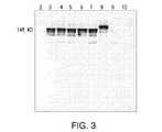
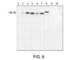
全ての5つの検出Abは、予測された生成物を認識した(表2)。抗ヒトIgG(H+L)および抗ヒトIgG F(ab’)2は、全ての生成物を認識した(図3および図4)。抗ヒトIgGラムダのみが、ラムダ軽鎖を有するAb#1およびAb#3を認識した(図5)。抗ヒトIgGカッパは、カッパ軽鎖を有するAb#2、Ab#11、Ab#13およびAb#9を強く認識したが、他の分子ともわずかに交差反応性があった(図6)。抗マウスIgG(H+L)は、マウスおよびヒトキメラ生成物、Ab#11を強く認識したが、他のヒト生成物もわずかに検出した(図7)。このわずかな交差反応性現象は、ウェスタンブロットでは、この方法が定量的でないことから用量依存的であり、無負荷量試験(no load quantity study)を実施した。テスト分子間の交差反応性を識別するためのさらなる試験を以下に記載する。 All five detection Abs recognized the expected product (Table 2). Anti-human IgG (H + L) and anti-human IgG F (ab ') 2 recognized all products (Figures 3 and 4). Only anti-human IgG lambda recognized
各単一捕捉および検出Abを、最初の評価のため個々にELISAにより個々に評価した。プレートを10μg/mL捕捉Abでコーティングし、2および5μg/mL検出Abで検出した。これらのスクリーニング結果に基づき、関連生成物に対し、各Abを3つの捕捉条件および6つの検出条件でテストする実験を実施して、各単一Ab量をさらに試験した。Ab#1が本プロジェクトにおける主な標的であることから、抗ヒトIgG(H+L)およびIgG F(ab’)2をAb#1に適用した。条件および実験結果を表3に示す。 Each single capture and detection Ab was individually assessed by ELISA for initial assessment. Plates were coated with 10 μg / mL capture Ab and detected with 2 and 5 μg / mL detection Ab. Based on these screening results, experiments were performed on the related products to test each Ab with 3 capture conditions and 6 detection conditions to further test each single Ab amount. Anti-human IgG (H + L) and IgG F (ab ') 2 were applied to
単一Abテストの目的は、各Abについて最高の感受性を達成する最適な組み合わせを見出すことであった。表3の太字にした条件は、0.25から8ng/mLのダイナミックレンジにおいて、最小シグナルはバックグラウンドを上回る信頼できる測定値である0.1を超え、最大シグナルはプレートリーダーの最大値に達したことを示した。これらの条件下で、アッセイは、必要とされるAb#1より10倍を超える感受性に達した。表4は、Ab#1の許容可能キャリーオーバー限界を列挙している。 The purpose of the single Ab test was to find the optimal combination that achieved the highest sensitivity for each Ab. The bolded conditions in Table 3 indicate that in the dynamic range of 0.25 to 8 ng / mL, the minimum signal exceeds the reliable measurement above background of 0.1 and the maximum signal reaches the plate reader maximum. It showed that. Under these conditions, the assay reached more than 10 times more sensitive than the required
表5に示されているように、5つの単一Abを上記実験結果に従って組み合わせた。 As shown in Table 5, five single Abs were combined according to the experimental results.
組み合わせたAbを、Ab#1、Ab#2およびAb#11検出のための感受性および適合性について再確認した。図8は、これらの生成物に関する標準曲線を示している。曲線は全て、二次フィットと0.9999の相関係数を有し、各曲線範囲は0.25から8ng/mLであった。 The combined Ab was reconfirmed for sensitivity and suitability for detection of
コーティングおよび検出Abプールを調製する目的は、1000個のプレートに対して十分であるようにするためである。組み合わせ比に基づき、大きな捕捉および検出Abプールを、表6および表7に従って調製した。Abプールは、アッセイ適格性確認に使用した。 The purpose of preparing the coating and detection Ab pool is to be sufficient for 1000 plates. Based on the combination ratio, large capture and detection Ab pools were prepared according to Tables 6 and 7. The Ab pool was used for assay qualification.
アッセイの信頼性を確認するために、Ab#1スケールアップ実行からのSP溶出液および最終限外濾過/透析濾過(「UF/DF」)をアッセイ適格性確認に使用した。これらの試料は約5mg/mLであることが報告され、したがってこの試験のために希釈された。 To confirm the reliability of the assay, SP eluate from the
通常の条件下で、薬剤物質試料をテスト前に−80℃で貯蔵した。見込まれる実験室環境を網羅するために、4時間、周囲温度、48時間、2から8℃、3回の凍結/解凍サイクルでの貯蔵条件を検証した。結果を表8にまとめる。タンパク質結果は、異なる貯蔵条件により著しい影響を受けなかった(<10%)。 Under normal conditions, drug substance samples were stored at −80 ° C. prior to testing. In order to cover the expected laboratory environment, the storage conditions were verified at 4 hours, ambient temperature, 48 hours, 2 to 8 ° C., 3 freeze / thaw cycles. The results are summarized in Table 8. Protein results were not significantly affected by different storage conditions (<10%).
ELISA基質発色時間は14分間を予定した。時間変動性を評価するために、14±1分間をテストした。結果は、1分のウィンドウ中に影響を受けなかった(表9)。 The ELISA substrate color development time was scheduled to be 14 minutes. To assess time variability, 14 ± 1 minutes were tested. Results were not affected during the 1 minute window (Table 9).
テストプレートを、2Mリン酸の添加後すぐに読み取って反応を止め、次いで周囲温度に30分間放置した後、再び読み取った。著しい差は観察されなかった(<10%)(表10)。 The test plate was read immediately after the addition of 2M phosphoric acid to stop the reaction and then left at ambient temperature for 30 minutes before reading again. No significant difference was observed (<10%) (Table 10).
分析方法の「精度」は、方法が均一試料の複数回の試料採取に反復して適用される場合、個々のテスト結果の一致度である。 The “accuracy” of an analytical method is the degree of agreement of individual test results when the method is applied repeatedly to multiple samplings of a homogeneous sample.
「アッセイ反復性」は、分析手順が一様な分析条件下で複数回実施される場合、該手順の精度を測定する。SP溶出液および最終UF/DFの6アリコートを1つのプレート内でテストした。タンパク質濃度はアリコートごとに計算した。濃度値のパーセント相対標準偏差は、全ての6つの測定値を用いて計算した。結果を表11にまとめる。タンパク質測定値の相対標準偏差は<7%であった。 “Assay repeatability” measures the accuracy of an analytical procedure when it is performed multiple times under uniform analytical conditions. Six aliquots of SP eluate and final UF / DF were tested in one plate. Protein concentration was calculated for each aliquot. The percent relative standard deviation of the concentration values was calculated using all 6 measurements. The results are summarized in Table 11. The relative standard deviation of the protein measurements was <7%.
「中間精度」は、分析手順が、異なる日または異なる分析者により実施される場合、実験室内でもたらされたテスト結果の分散の測定値である。 “Intermediate accuracy” is a measure of the variance of the test results produced in the laboratory when the analysis procedure is performed on different days or by different analysts.
SP溶出液および最終UF/DFそれぞれの3アリコートをテストした。この実験は、異なる日に異なる分析者によりさらに2回反復された。試験のアッセイ反復部からの最初の3つの結果を、この試験の1日目に使用した。結果を表12に示す。タンパク質測定値のパーセント相対標準偏差は、<5%であり、20%の予測された精度範囲内であった。 Three aliquots of each of the SP eluate and final UF / DF were tested. This experiment was repeated two more times by different analysts on different days. The first three results from the assay replicate of the test were used on
標準曲線の最低点、定量化限界(quantification limit)を推定するために、8ng/mL Ab#1標準物質の2倍連続希釈を6回調製し、6つの異なるプレートに添加した。結果を表13に示す。ICHガイドラインによれば、アッセイの定量化限界は、シグナル対ノイズ比が10:1より高い最低濃度、および相対標準偏差が20%未満である。ブランク測定値のノイズは、0.004AUであった。0.25ng/mL標準物質の平均シグナルは、0.122AUであった。0.125ng/mLは基準を満たすが、0.25を、アッセイ頑健性を確保するためのLOQとして選択した。定量限界は、シグナル対ノイズ比30.5:1、および相対標準偏差14%の0.25ng/mLに設定した。 To estimate the lowest point of the standard curve, the quantification limit, two 2-fold serial dilutions of 8 ng /
分析方法の「直線性」は、目的とする分析物の濃度に正比例する、または十分に定義された数学的変換により目的とする分析物の濃度に比例するテスト結果を導く該方法の能力である。 “Linearity” of an analytical method is the ability of the method to produce test results that are directly proportional to the concentration of the analyte of interest or that are well-defined mathematically transformed to the concentration of the analyte of interest. .
0.25から8ng/mLの範囲の標準物質をAb#1薬剤物質から調製した。標準物質および緩衝液ブランクをプレートに添加し、テストした。プロットを図9に示す。二次フィットの相関係数は、0.999より大きかった。結果は、標準物質の吸光反応が二次フィットによるタンパク質濃度に比例することを示している。 Standards ranging from 0.25 to 8 ng / mL were prepared from
アッセイの正確度は、スパイクおよび回収方法を用いて試験した。SP溶出液および最終UF/DFを、1.0、2.0および4.0ng/mLの最終標準レベルでスパイクした。アッセイは、回収されたタンパク質の濃度をスパイクの実験濃度で割った比として計算し、パーセンテージとして表した。この試験の結果を表14に示す。スパイクの回収率は、全てのスパイクレベルについて100±7.9%の範囲にあった。 The accuracy of the assay was tested using spike and recovery methods. SP eluate and final UF / DF were spiked at final standard levels of 1.0, 2.0 and 4.0 ng / mL. The assay was calculated as the ratio of the recovered protein concentration divided by the experimental concentration of spikes and expressed as a percentage. The results of this test are shown in Table 14. Spike recovery was in the range of 100 ± 7.9% for all spike levels.
SP溶出液および最終UF/DF試料を、参照標準曲線校正範囲で2倍連続希釈して試料希釈直線性を評価した。表15に示されているように、試料希釈は結果に影響を与えなかった。変数は10%未満であった。3つの希釈点のみは、未知試料テストについて試料希釈を確認する必要があった。 The SP eluate and the final UF / DF sample were serially diluted 2-fold within the reference standard curve calibration range to evaluate sample dilution linearity. As shown in Table 15, sample dilution did not affect the results. The variable was less than 10%. Only three dilution points required confirmation of sample dilution for the unknown sample test.
分析方法の「範囲」は、分析物の上位と下位の間の区間であり、該区間は、書かれているような方法を用いて適切なレベルの精度、正確度および直線性により決定することができる。アッセイの範囲は、直線性、正確度、精度および定量限界に基づき0.25から8ng/mLとして確立した。二次フィットの相関係数値は、0.999より大きかった。これは、0.25から8ng/mLの範囲において、吸光反応が二次フィットによるタンパク質濃度に比例することを示している。スパイク回収試験は、2つの異なる工程内試料中の1.0、2.0および4.0ng/mLレベルの標準物質で実施した。回収率は100±7.9%であった。これは、アッセイが推定範囲において厳密および正確であることを示している。 The “range” of the analytical method is the interval between the upper and lower parts of the analyte, which should be determined with the appropriate level of accuracy, accuracy and linearity using the method as written. Can do. The range of the assay was established as 0.25 to 8 ng / mL based on linearity, accuracy, precision and quantification limits. The correlation coefficient value of the secondary fit was greater than 0.999. This indicates that in the range of 0.25 to 8 ng / mL, the absorbance reaction is proportional to the protein concentration due to the second fit. Spike recovery tests were performed with 1.0, 2.0 and 4.0 ng / mL levels of standards in two different in-process samples. The recovery rate was 100 ± 7.9%. This indicates that the assay is accurate and accurate in the estimated range.
商業的供給のさらなる必要性をカバーするため、各抗体の第2ロットを評価した。表16は、大きいプールに使用した第1ロットおよび比較のための第2ロットを示している。 To cover the further need for commercial supply, a second lot of each antibody was evaluated. Table 16 shows the first lot used for the large pool and the second lot for comparison.
結果を表17にまとめ、第1および第2ロットからの参照標準曲線を図10に示す。第2ロットは結果に影響を与えなかった。 The results are summarized in Table 17, and reference standard curves from the first and second lots are shown in FIG. The second lot did not affect the results.
装置リンス液(rinsate)中の残留生成物タンパク質レベルを検出するためのアッセイ適合性を評価するために、装置表面からのタンパク質の回収率を試験した。生成装置は、ステンレス鋼、ハスタロイ(hastalloy)またはガラス製であった。各材料の代表的クーポン(すなわち、スワブ、リンス液、または他の試料形態などの代表的試料)を2および3ppmに希釈した40mLのAb#1薬剤物質でスポッティングした。表面を3から4時間空気乾燥させ、次いで40mLのMilliQ水ですすいだ。リンス液を回収した。表面からのタンパク質回収率は、リンス液中のタンパク質の濃度をスパイクの濃度で割った比として計算し、パーセンテージとして表した。 To assess the suitability of the assay to detect residual product protein levels in the instrument rinsate, the recovery of protein from the instrument surface was tested. The generator was made of stainless steel, hastalloy or glass. A representative coupon of each material (ie, a representative sample such as a swab, rinse solution, or other sample form) was spotted with 40 mL of
表18に示されているように、平均回収率は、ガラス表面が76%、ステンレス鋼が87%およびハスタロイが76%である。結果は、合格基準(50−150%)を満たし、アッセイが生成装置すすぎ試料テストに適していることを実証した。 As shown in Table 18, the average recoveries are 76% for the glass surface, 87% for stainless steel and 76% for Hastelloy. The results met the acceptance criteria (50-150%) and demonstrated that the assay is suitable for generator rinse sample testing.
Ab#1工程バリデーションは、生成物キャリーオーバーを評価するための生成物工程実行後のブランク実行を含む。ブランク実行は、プロテインA、SPセファロースおよびQセファロースクロマトグラフィーおよびUF/DF操作を含む。生成物AからAのキャリーオーバーはBradfordアッセイを用いて評価されるが、生成物AからBの残留物キャリーオーバー限界(RCL)は、BradfordアッセイLOQ(1.25ug/mL)より低い4ng/mL(VR−2249、VR−2250およびVR−2251)である。mAbアッセイは、Ab#1 AからBへのキャリーオーバー試験を裏付けるための適合性について評価した。アッセイへの工程緩衝液干渉は、緩衝液スパイクおよび回収実験を実施して評価した。未希釈および5倍希釈(十分な希釈緩衝液による)での各工程緩衝液を、1.0、2.0および4.0ng/mLの最終標準レベルでスパイクした。
表19に示されているように、回収率は、Qセファロース溶出緩衝液SR−488およびSPセファロース溶出(eluation)緩衝液SR−610について約70%であり、これは合格基準(50から150%)内であったが、プロテインA溶出緩衝液SR−493のみは、抗原および抗体結合を妨げる酸性条件のため1.1%を回収した。 As shown in Table 19, the recovery rate is about 70% for Q Sepharose Elution Buffer SR-488 and SP Sepharose Elution Buffer SR-610, which is an acceptable basis (50 to 150% However, only protein A elution buffer SR-493 recovered 1.1% due to acidic conditions that hinder antigen and antibody binding.
希釈緩衝液のスパイクおよび回収結果を表20にまとめる。タンパク質回収率は、5倍に希釈した場合、許容可能な110.3から121.9%であった。アッセイ定量限界は、5倍希釈で1.25ng/mLである。したがって、頑健なアッセイ操作には、5倍希釈が3種類の工程緩衝液全てに推奨される。1.25ng/mLのLOQによるこのアッセイフォーマットは、生成物AからBのキャリーオーバーを評価するのに適している。 Dilution buffer spikes and recovery results are summarized in Table 20. Protein recovery was acceptable from 110.3 to 121.9% when diluted 5-fold. The assay quantitation limit is 1.25 ng / mL at 5-fold dilution. Therefore, for robust assay operations, a 5-fold dilution is recommended for all three process buffers. This assay format with a 1.25 ng / mL LOQ is suitable for assessing carryover of products A to B.
全体的な残留mAb関連生成物を検出するための新たなアッセイフォーマットおよび条件を開発した。上記結果は、アッセイの再現性(reproducibiliry)を実証するものでる。新たなアッセイは、Ab#1については0.25ng/mLの定量限界で微量レベルのmAbタンパク質を定量化することができる。故に、新たに開発されたmAb ELISAは、設備切り換えおよび洗浄確認のために残留mAb関連分子を定量化する貴重なツールを提供する。 New assay formats and conditions were developed to detect overall residual mAb related products. The above results demonstrate the reproducibility of the assay. The new assay can quantify trace levels of mAb protein with a quantification limit of 0.25 ng / mL for
工程内試料におけるAb#1生成物定量化のためのアッセイの性能は、適格とされた。アッセイ頑健性、精度、定量化限界、直線性、正確度、試料希釈直線性、範囲、第2ロット市販試薬を試験した。全ての上記の結果は、ELISAの予測された正確度および精度内である。アッセイ条件を表21にまとめる。さらに、回収試験は、このアッセイがリンス液試験およびAb#1工程キャリーオーバー試験に適していることを証明した。しかし、ブランク実行については、緩衝液干渉を排除するために5倍希釈が必要とされる。 The performance of the assay for
上記アッセイを、GMP−1後に抗体#1工程中間体を含有するバイオリアクター、回収および下流工程タンク、フレックスホースならびにフィルターハウジンングで実施した洗浄サイクルからの最終すすぎをテストするのに使用した。最終すすぎは全てLOQより低かった。さらに、中間すすぎを、バイオリアクターZ−3200およびタンクTK−2830について試料採取した。表23に示されているように、Z−3200およびTK−2830からの最初のUSP水すすぎのみが、極めて低いレベルでタンパク質残留物を検出した。結果は、上記アッセイの感受性および特異性をさらに証明した。 The above assay was used to test the final rinse from the wash cycle performed in the bioreactor containing GMP-1 followed by
工程バリデーション実行に関連した装置からの最終すすぎ試料を、上記アッセイによりテストした。テストした全ての試料はLOQより低かった。工程バリデーション実行1(「PV−1」)からの結果を表24に列挙する。洗浄後、報告対象の抗体#1レベル(0.25ng/ml)は、最大許容キャリーオーバー限界(4−13ng/mL)より少なくとも16倍低い。 The final rinse sample from the instrument associated with the process validation run was tested by the above assay. All samples tested were below the LOQ. The results from Process Validation Execution 1 (“PV-1”) are listed in Table 24. After washing, the reported
高いアッセイ感受性のため、実験室で日常的に一緒に作業されるmAb生成物は、このアッセイでは汚染の潜在的な原因になり得る。したがって、試料テスト中に使用される装置および環境ができる限り清浄であることを確保するための予防措置が講じられることが推奨される。 Because of the high assay sensitivity, mAb products that are routinely worked together in the laboratory can be a potential source of contamination in this assay. It is therefore recommended that precautions be taken to ensure that the equipment and environment used during sample testing is as clean as possible.
さまざまな刊行物が本明細書で引用されており、その内容は全体が本明細書に組み込まれている。 Various publications are cited herein, the contents of which are incorporated herein in their entirety.
Claims (9)
Translated fromJapaneseApplications Claiming Priority (3)
| Application Number | Priority Date | Filing Date | Title |
|---|---|---|---|
| US38241310P | 2010-09-13 | 2010-09-13 | |
| US61/382,413 | 2010-09-13 | ||
| PCT/US2011/051356WO2012037095A1 (en) | 2010-09-13 | 2011-09-13 | A highly sensitive monoclonal antibody residual detection assay |
Publications (1)
| Publication Number | Publication Date |
|---|---|
| JP2013537971Atrue JP2013537971A (en) | 2013-10-07 |
Family
ID=44674913
Family Applications (1)
| Application Number | Title | Priority Date | Filing Date |
|---|---|---|---|
| JP2013528381AWithdrawnJP2013537971A (en) | 2010-09-13 | 2011-09-13 | Highly sensitive monoclonal antibody residue detection assay |
Country Status (7)
| Country | Link |
|---|---|
| US (1) | US8945860B2 (en) |
| EP (1) | EP2616807A1 (en) |
| JP (1) | JP2013537971A (en) |
| CN (1) | CN103221823A (en) |
| CA (1) | CA2811003A1 (en) |
| MX (1) | MX2013002870A (en) |
| WO (1) | WO2012037095A1 (en) |
Cited By (1)
| Publication number | Priority date | Publication date | Assignee | Title |
|---|---|---|---|---|
| CN107478705A (en)* | 2017-08-17 | 2017-12-15 | 上海上药第生化药业有限公司 | A kind of method for detecting residue of sodium tetradecyl sulfate |
Families Citing this family (3)
| Publication number | Priority date | Publication date | Assignee | Title |
|---|---|---|---|---|
| WO2021247892A1 (en)* | 2020-06-04 | 2021-12-09 | Amgen Inc. | Assessment of cleaning procedures of a biotherapeutic manufacturing process |
| CN112285366B (en)* | 2020-12-25 | 2021-06-08 | 南京广祺医药科技有限公司 | ELISA method for determining bispecific antibody BsAb in serum and application thereof |
| CN117169518B (en)* | 2023-11-03 | 2024-01-19 | 赛德特(北京)生物工程有限公司 | Method and kit for detecting CD3 antibody residues in T lymphocyte preparation |
Family Cites Families (14)
| Publication number | Priority date | Publication date | Assignee | Title |
|---|---|---|---|---|
| US4816567A (en) | 1983-04-08 | 1989-03-28 | Genentech, Inc. | Recombinant immunoglobin preparations |
| US5225539A (en) | 1986-03-27 | 1993-07-06 | Medical Research Council | Recombinant altered antibodies and methods of making altered antibodies |
| US5429925A (en)* | 1988-01-26 | 1995-07-04 | The Regents Of The University Of California | Method for immunodiagnostic detection of dioxins at low concentrations |
| US4983530A (en)* | 1988-01-29 | 1991-01-08 | E. I. Du Pont De Nemours And Company | Sandwich immunoassay for determination of total monoclonal IGG |
| US5530101A (en) | 1988-12-28 | 1996-06-25 | Protein Design Labs, Inc. | Humanized immunoglobulins |
| US5077210A (en) | 1989-01-13 | 1991-12-31 | Eigler Frances S | Immobilization of active agents on substrates with a silane and heterobifunctional crosslinking agent |
| EP0634016B1 (en) | 1992-03-27 | 2001-09-19 | Abbott Laboratories | Assay verification control for analytical methods |
| US5449611A (en)* | 1993-07-27 | 1995-09-12 | Ensys, Inc. | Polyaromatic hydrocarbon (PAH) immunoassay method, its components and a kit for use in performing the same |
| JP2001041958A (en) | 1999-07-28 | 2001-02-16 | Takeda Chem Ind Ltd | Method for immunologically measuring and analyzing environmental pollutant |
| US20030054424A1 (en)* | 2001-04-05 | 2003-03-20 | Allen Randy L. | Rapid and sensitive method for measuring PCB contamination |
| US20040072236A1 (en) | 2002-09-27 | 2004-04-15 | Neil Cashman | PrPSc -interacting molecules and uses thereof |
| GB0310347D0 (en)* | 2003-05-07 | 2003-06-11 | Delta Biotechnology Ltd | Assay |
| US20060035242A1 (en) | 2004-08-13 | 2006-02-16 | Michelitsch Melissa D | Prion-specific peptide reagents |
| US20080135490A1 (en) | 2005-01-07 | 2008-06-12 | Board Of Trustees Of The University Of Arkansas | Quantum dot biolabeling and immunomagnetic separation for detection of contaminants |
- 2011
- 2011-09-13JPJP2013528381Apatent/JP2013537971A/ennot_activeWithdrawn
- 2011-09-13EPEP11760651.7Apatent/EP2616807A1/ennot_activeWithdrawn
- 2011-09-13CNCN201180054320XApatent/CN103221823A/enactivePending
- 2011-09-13CACA2811003Apatent/CA2811003A1/ennot_activeAbandoned
- 2011-09-13MXMX2013002870Apatent/MX2013002870A/ennot_activeApplication Discontinuation
- 2011-09-13WOPCT/US2011/051356patent/WO2012037095A1/enactiveApplication Filing
- 2011-09-13USUS13/231,280patent/US8945860B2/ennot_activeExpired - Fee Related
Cited By (1)
| Publication number | Priority date | Publication date | Assignee | Title |
|---|---|---|---|---|
| CN107478705A (en)* | 2017-08-17 | 2017-12-15 | 上海上药第生化药业有限公司 | A kind of method for detecting residue of sodium tetradecyl sulfate |
Also Published As
| Publication number | Publication date |
|---|---|
| CN103221823A (en) | 2013-07-24 |
| CA2811003A1 (en) | 2012-03-22 |
| US20120129196A1 (en) | 2012-05-24 |
| EP2616807A1 (en) | 2013-07-24 |
| WO2012037095A1 (en) | 2012-03-22 |
| MX2013002870A (en) | 2013-06-18 |
| US8945860B2 (en) | 2015-02-03 |
Similar Documents
| Publication | Publication Date | Title |
|---|---|---|
| US20040048323A1 (en) | Antibody pair screening methods | |
| KR101566090B1 (en) | ANTI-HUMAN IgG1 ANTIBODY | |
| AU2018294758B2 (en) | Method for measuring serum amyloid A of various animals and reagent for measurement thereof | |
| CN114217067A (en) | Quantitative detection method of SARS-CoV-2 antibody | |
| JP2013537971A (en) | Highly sensitive monoclonal antibody residue detection assay | |
| CN109069600B (en) | IL-21 antibodies and uses thereof | |
| JP7416485B2 (en) | Switching binder, method for producing the same, pharmaceutical composition using the same, test kit, and method for analyzing antigen and antibody | |
| EP1653233B1 (en) | Method for determining antibodies of a particular class using an immune complex-specific antibody | |
| CN117264072B (en) | anti-SN 38 monoclonal antibody and application thereof | |
| JP7424718B2 (en) | How to process antigens. | |
| US20230063268A1 (en) | High ionic strength dissociation assay for high drug tolerant testing | |
| JP2023181275A (en) | Mycoplasma pneumoniae immunoassay method and immunoassay device | |
| KR102601835B1 (en) | Monoclonal Antibody specific for Equine influenza virus H3N8 and Composition for detecting Equine influenza virus using the same | |
| CN113811770B (en) | Suppression of interference in pharmacokinetic immunoassays | |
| TWI741216B (en) | Monoclonal antibody or antigen-binding fragment for specifically inhibiting or alleviate the binding of ptx3 and ptx3 receptor and use of the same | |
| CN103907026B (en) | Calibration reagent and method | |
| EP3177312B1 (en) | Method and kit for detecting bacterial infection | |
| JP7222501B2 (en) | Monoclonal antibody that specifically binds to a sugar chain having a terminal sialic acid residue linked to galactose via an α2,3 bond, and method for measuring sugar chain having a terminal sialic acid residue linked to galactose via an α2,3 bond | |
| CN116143929B (en) | Antibody against recombinant human blood coagulation factor VIIa-Fc fusion protein and its application | |
| CN111208307B (en) | Method for screening molecules having the same or different target protein binding to reference molecule | |
| US20250052768A1 (en) | In vitro methods for detecting dll1 | |
| WO2023192829A2 (en) | Alpha-1 antitrypsin z- and m-specific binding proteins | |
| KR20240087864A (en) | Diagnostic kit using SARS-CoV-2 spike protein-specific antibody | |
| TW202342979A (en) | Detection method and detection reagent | |
| CN118043666A (en) | Determination of quantitative drug and target concentrations |
Legal Events
| Date | Code | Title | Description |
|---|---|---|---|
| A300 | Application deemed to be withdrawn because no request for examination was validly filed | Free format text:JAPANESE INTERMEDIATE CODE: A300 Effective date:20141202 |