JP2012525239A - Transcutaneous methods and devices for treating dissociation (priority information and incorporation by reference) - Google Patents
Transcutaneous methods and devices for treating dissociation (priority information and incorporation by reference)Download PDFInfo
- Publication number
- JP2012525239A JP2012525239AJP2012508800AJP2012508800AJP2012525239AJP 2012525239 AJP2012525239 AJP 2012525239AJP 2012508800 AJP2012508800 AJP 2012508800AJP 2012508800 AJP2012508800 AJP 2012508800AJP 2012525239 AJP2012525239 AJP 2012525239A
- Authority
- JP
- Japan
- Prior art keywords
- cover
- dissociation
- blood vessel
- frame
- entry point
- Prior art date
- Legal status (The legal status is an assumption and is not a legal conclusion. Google has not performed a legal analysis and makes no representation as to the accuracy of the status listed.)
- Pending
Links
- 238000010494dissociation reactionMethods0.000titleclaimsabstractdescription64
- 230000005593dissociationsEffects0.000titleclaimsabstractdescription64
- 238000000034methodMethods0.000titleclaimsabstractdescription33
- 238000010348incorporationMethods0.000title1
- 210000004204blood vesselAnatomy0.000claimsabstractdescription40
- 210000000709aortaAnatomy0.000claimsabstractdescription38
- 238000002224dissectionMethods0.000claimsabstractdescription17
- 238000004873anchoringMethods0.000claimsabstractdescription9
- 230000017531blood circulationEffects0.000claimsabstractdescription9
- 210000003270subclavian arteryAnatomy0.000claimsdescription21
- 210000001519tissueAnatomy0.000claimsdescription10
- 102000004169proteins and genesHuman genes0.000claimsdescription8
- 108090000623proteins and genesProteins0.000claimsdescription8
- 238000011282treatmentMethods0.000claimsdescription8
- 239000000463materialSubstances0.000claimsdescription7
- 239000008280bloodSubstances0.000claimsdescription6
- 210000004369bloodAnatomy0.000claimsdescription6
- 239000011159matrix materialSubstances0.000claimsdescription5
- 239000003431cross linking reagentSubstances0.000claimsdescription4
- 102000008186CollagenHuman genes0.000claimsdescription3
- 108010035532CollagenProteins0.000claimsdescription3
- 102000016942ElastinHuman genes0.000claimsdescription3
- 108010014258ElastinProteins0.000claimsdescription3
- 241001465754MetazoaSpecies0.000claimsdescription3
- 229920001436collagenPolymers0.000claimsdescription3
- 229920002549elastinPolymers0.000claimsdescription3
- 230000000968intestinal effectEffects0.000claimsdescription3
- 210000003516pericardiumAnatomy0.000claimsdescription3
- 229920000728polyesterPolymers0.000claimsdescription3
- 210000004876tela submucosaAnatomy0.000claimsdescription3
- 230000002792vascularEffects0.000claimsdescription3
- 229920001661ChitosanPolymers0.000claimsdescription2
- 102000009123FibrinHuman genes0.000claimsdescription2
- 108010073385FibrinProteins0.000claimsdescription2
- BWGVNKXGVNDBDI-UHFFFAOYSA-NFibrin monomerChemical compoundCNC(=O)CNC(=O)CNBWGVNKXGVNDBDI-UHFFFAOYSA-N0.000claimsdescription2
- SXRSQZLOMIGNAQ-UHFFFAOYSA-NGlutaraldehydeChemical compoundO=CCCCC=OSXRSQZLOMIGNAQ-UHFFFAOYSA-N0.000claimsdescription2
- 229920002683GlycosaminoglycanPolymers0.000claimsdescription2
- 239000000853adhesiveSubstances0.000claimsdescription2
- 230000001070adhesive effectEffects0.000claimsdescription2
- 210000001367arteryAnatomy0.000claimsdescription2
- 150000001718carbodiimidesChemical class0.000claimsdescription2
- 229920000295expanded polytetrafluoroethylenePolymers0.000claimsdescription2
- 229950003499fibrinDrugs0.000claimsdescription2
- 150000004676glycansChemical class0.000claimsdescription2
- HLXZNVUGXRDIFK-UHFFFAOYSA-Nnickel titaniumChemical compound[Ti].[Ti].[Ti].[Ti].[Ti].[Ti].[Ti].[Ti].[Ti].[Ti].[Ti].[Ni].[Ni].[Ni].[Ni].[Ni].[Ni].[Ni].[Ni].[Ni].[Ni].[Ni].[Ni].[Ni].[Ni]HLXZNVUGXRDIFK-UHFFFAOYSA-N0.000claimsdescription2
- 229910001000nickel titaniumInorganic materials0.000claimsdescription2
- 150000008442polyphenolic compoundsChemical class0.000claimsdescription2
- 235000013824polyphenolsNutrition0.000claimsdescription2
- 229920001282polysaccharidePolymers0.000claimsdescription2
- 239000005017polysaccharideSubstances0.000claimsdescription2
- 239000004814polyurethaneSubstances0.000claimsdescription2
- 229920002635polyurethanePolymers0.000claimsdescription2
- 210000003491skinAnatomy0.000claimsdescription2
- 235000018553tanninNutrition0.000claimsdescription2
- 229920001864tanninPolymers0.000claimsdescription2
- 239000001648tanninSubstances0.000claimsdescription2
- 210000001715carotid arteryAnatomy0.000claims1
- 238000010586diagramMethods0.000description14
- 208000002251Dissecting AneurysmDiseases0.000description7
- 210000002376aorta thoracicAnatomy0.000description7
- 206010002895aortic dissectionDiseases0.000description7
- 210000002321radial arteryAnatomy0.000description5
- 230000008901benefitEffects0.000description3
- 210000001105femoral arteryAnatomy0.000description3
- 210000005166vasculatureAnatomy0.000description3
- 239000004971Cross linkerSubstances0.000description2
- 238000012276Endovascular treatmentMethods0.000description2
- 208000007536ThrombosisDiseases0.000description2
- 230000001154acute effectEffects0.000description2
- 230000033001locomotionEffects0.000description2
- 239000011148porous materialSubstances0.000description2
- 210000000115thoracic cavityAnatomy0.000description2
- 206010002329AneurysmDiseases0.000description1
- 102000010834Extracellular Matrix ProteinsHuman genes0.000description1
- 108010037362Extracellular Matrix ProteinsProteins0.000description1
- 208000032984Intraoperative ComplicationsDiseases0.000description1
- 241000124008MammaliaSpecies0.000description1
- 208000035965Postoperative ComplicationsDiseases0.000description1
- 206010057765Procedural complicationDiseases0.000description1
- 230000015572biosynthetic processEffects0.000description1
- 230000036772blood pressureEffects0.000description1
- 230000001684chronic effectEffects0.000description1
- 210000002744extracellular matrixAnatomy0.000description1
- 239000004744fabricSubstances0.000description1
- 230000012953feeding on blood of other organismEffects0.000description1
- 239000000835fiberSubstances0.000description1
- 230000006870functionEffects0.000description1
- 230000000004hemodynamic effectEffects0.000description1
- 238000002513implantationMethods0.000description1
- 230000010354integrationEffects0.000description1
- 230000009545invasionEffects0.000description1
- 230000003902lesionEffects0.000description1
- 230000007774longtermEffects0.000description1
- 238000007726management methodMethods0.000description1
- 210000000056organAnatomy0.000description1
- 230000010412perfusionEffects0.000description1
- 239000012781shape memory materialSubstances0.000description1
- 238000011477surgical interventionMethods0.000description1
- 230000017423tissue regenerationEffects0.000description1
- 230000007704transitionEffects0.000description1
- 230000003966vascular damageEffects0.000description1
Images


Classifications
- A—HUMAN NECESSITIES
- A61—MEDICAL OR VETERINARY SCIENCE; HYGIENE
- A61B—DIAGNOSIS; SURGERY; IDENTIFICATION
- A61B17/00—Surgical instruments, devices or methods
- A61B17/12—Surgical instruments, devices or methods for ligaturing or otherwise compressing tubular parts of the body, e.g. blood vessels or umbilical cord
- A61B17/12022—Occluding by internal devices, e.g. balloons or releasable wires
- A61B17/12099—Occluding by internal devices, e.g. balloons or releasable wires characterised by the location of the occluder
- A61B17/12109—Occluding by internal devices, e.g. balloons or releasable wires characterised by the location of the occluder in a blood vessel
- A61B17/12113—Occluding by internal devices, e.g. balloons or releasable wires characterised by the location of the occluder in a blood vessel within an aneurysm
- A61B17/12118—Occluding by internal devices, e.g. balloons or releasable wires characterised by the location of the occluder in a blood vessel within an aneurysm for positioning in conjunction with a stent
- A—HUMAN NECESSITIES
- A61—MEDICAL OR VETERINARY SCIENCE; HYGIENE
- A61B—DIAGNOSIS; SURGERY; IDENTIFICATION
- A61B17/00—Surgical instruments, devices or methods
- A61B17/12—Surgical instruments, devices or methods for ligaturing or otherwise compressing tubular parts of the body, e.g. blood vessels or umbilical cord
- A61B17/12022—Occluding by internal devices, e.g. balloons or releasable wires
- A61B17/12131—Occluding by internal devices, e.g. balloons or releasable wires characterised by the type of occluding device
- A61B17/12168—Occluding by internal devices, e.g. balloons or releasable wires characterised by the type of occluding device having a mesh structure
- A61B17/12172—Occluding by internal devices, e.g. balloons or releasable wires characterised by the type of occluding device having a mesh structure having a pre-set deployed three-dimensional shape
- A—HUMAN NECESSITIES
- A61—MEDICAL OR VETERINARY SCIENCE; HYGIENE
- A61B—DIAGNOSIS; SURGERY; IDENTIFICATION
- A61B17/00—Surgical instruments, devices or methods
- A61B17/12—Surgical instruments, devices or methods for ligaturing or otherwise compressing tubular parts of the body, e.g. blood vessels or umbilical cord
- A61B17/12022—Occluding by internal devices, e.g. balloons or releasable wires
- A61B17/12099—Occluding by internal devices, e.g. balloons or releasable wires characterised by the location of the occluder
- A61B17/12109—Occluding by internal devices, e.g. balloons or releasable wires characterised by the location of the occluder in a blood vessel
- A61B17/12113—Occluding by internal devices, e.g. balloons or releasable wires characterised by the location of the occluder in a blood vessel within an aneurysm
- A—HUMAN NECESSITIES
- A61—MEDICAL OR VETERINARY SCIENCE; HYGIENE
- A61B—DIAGNOSIS; SURGERY; IDENTIFICATION
- A61B17/00—Surgical instruments, devices or methods
- A61B17/12—Surgical instruments, devices or methods for ligaturing or otherwise compressing tubular parts of the body, e.g. blood vessels or umbilical cord
- A61B17/12022—Occluding by internal devices, e.g. balloons or releasable wires
- A61B2017/1205—Introduction devices
- A—HUMAN NECESSITIES
- A61—MEDICAL OR VETERINARY SCIENCE; HYGIENE
- A61F—FILTERS IMPLANTABLE INTO BLOOD VESSELS; PROSTHESES; DEVICES PROVIDING PATENCY TO, OR PREVENTING COLLAPSING OF, TUBULAR STRUCTURES OF THE BODY, e.g. STENTS; ORTHOPAEDIC, NURSING OR CONTRACEPTIVE DEVICES; FOMENTATION; TREATMENT OR PROTECTION OF EYES OR EARS; BANDAGES, DRESSINGS OR ABSORBENT PADS; FIRST-AID KITS
- A61F2/00—Filters implantable into blood vessels; Prostheses, i.e. artificial substitutes or replacements for parts of the body; Appliances for connecting them with the body; Devices providing patency to, or preventing collapsing of, tubular structures of the body, e.g. stents
- A61F2/02—Prostheses implantable into the body
- A61F2/04—Hollow or tubular parts of organs, e.g. bladders, tracheae, bronchi or bile ducts
- A61F2/06—Blood vessels
- A61F2002/061—Blood vessels provided with means for allowing access to secondary lumens
- A—HUMAN NECESSITIES
- A61—MEDICAL OR VETERINARY SCIENCE; HYGIENE
- A61F—FILTERS IMPLANTABLE INTO BLOOD VESSELS; PROSTHESES; DEVICES PROVIDING PATENCY TO, OR PREVENTING COLLAPSING OF, TUBULAR STRUCTURES OF THE BODY, e.g. STENTS; ORTHOPAEDIC, NURSING OR CONTRACEPTIVE DEVICES; FOMENTATION; TREATMENT OR PROTECTION OF EYES OR EARS; BANDAGES, DRESSINGS OR ABSORBENT PADS; FIRST-AID KITS
- A61F2/00—Filters implantable into blood vessels; Prostheses, i.e. artificial substitutes or replacements for parts of the body; Appliances for connecting them with the body; Devices providing patency to, or preventing collapsing of, tubular structures of the body, e.g. stents
- A61F2/82—Devices providing patency to, or preventing collapsing of, tubular structures of the body, e.g. stents
- A61F2002/823—Stents, different from stent-grafts, adapted to cover an aneurysm
Landscapes
- Health & Medical Sciences (AREA)
- Surgery (AREA)
- Life Sciences & Earth Sciences (AREA)
- Heart & Thoracic Surgery (AREA)
- Molecular Biology (AREA)
- Vascular Medicine (AREA)
- Engineering & Computer Science (AREA)
- Biomedical Technology (AREA)
- Reproductive Health (AREA)
- Medical Informatics (AREA)
- Nuclear Medicine, Radiotherapy & Molecular Imaging (AREA)
- Animal Behavior & Ethology (AREA)
- General Health & Medical Sciences (AREA)
- Public Health (AREA)
- Veterinary Medicine (AREA)
- Neurosurgery (AREA)
- Prostheses (AREA)
- Materials For Medical Uses (AREA)
- Surgical Instruments (AREA)
Abstract
Translated fromJapaneseDescription
Translated fromJapaneseこの出願は2009年5月1日に出願された米国仮特許出願第61/174,888号の利益を主張するものであって、参照により前述の仮特許出願の内容全体が本明細書に組み入れられ、また、その内容全体が本明細書の一部と見なされるべきである。更に、2008年4月11日に出願された(「Bifurcated Graft Deployment Systems And Methods」と題する)米国特許出願第12/101,863号も、参照により、あたかも本明細書で完全に記述されているかの如くにして、その内容全体が本明細書に組み入れられる。 This application claims the benefit of US Provisional Patent Application No. 61 / 174,888, filed May 1, 2009, which is incorporated herein by reference in its entirety. And the entire contents of which are to be considered part of this specification. In addition, US patent application Ser. No. 12 / 101,863, filed Apr. 11, 2008 (titled “Bifurcated Graft Deployment Systems And Methods”), is hereby fully incorporated by reference. As such, the entire contents thereof are incorporated herein.
(本開示の背景)
本発明の開示は大動脈解離を治療するための方法および装置に関する。(Background of this disclosure)
The present disclosure relates to a method and apparatus for treating aortic dissection.
(本開示の背景)
大動脈解離は死亡率の高い危険な状態である。大動脈解離になると、典型的には、大動脈の内膜に裂けが生じ、その裂けが血管壁に沿って広がり、大動脈の外層から内層を剥離させてしまう。偽腔を作り出しているこれらの層の間の空間に血液が入り込む。大動脈の真腔と上述の偽腔との間にいくつかの付加的な裂けまたは侵入ポイントが作り出されることがある。急性期の場合には、解離が大動脈から生命の維持に必要不可欠な重要な器官への潅流を塞いでしまうことがある。慢性期の場合には、その脆弱化した組織が高じて動脈瘤になり、最終的に破裂してしまうことがある。上行大動脈が関与する解離はA型の解離と呼ばれている。一方、下行大動脈のみが関与する解離はB型の解離と呼ばれている。(Background of this disclosure)
Aortic dissection is a dangerous condition with high mortality. When an aortic dissection occurs, the aorta typically tears in the intima and spreads along the vessel wall, causing the inner layer to detach from the outer layer of the aorta. Blood enters the space between these layers creating the false lumen. Several additional tears or points of entry may be created between the true lumen of the aorta and the false lumen described above. In the acute phase, dissociation can block perfusion from the aorta to vital organs essential for life support. In the chronic phase, the weakened tissue may become an aneurysm and eventually rupture. Dissociation involving the ascending aorta is called type A dissociation. On the other hand, dissociation involving only the descending aorta is called B-type dissociation.
現在の解離の治療法は、患者の血圧を下げて、その病変血管に及ぼされる血行力学的応力を低減することを目的とした医学的管理を含む。もし解離が症候性の場合には、外科的介入が必要になる。その場合、大動脈の病変部分が外科的なグラフトと取り替えられ、解離のフラップが取り付けし直される。もっと最近では、偽腔に血栓を形成し、且つ、真の管腔の開存性を維持することを目標として、その偽腔内への一次的侵入ポイントを塞ぐべく、ステントグラフトが用いられている。 Current treatments for dissociation include medical management aimed at lowering the patient's blood pressure and reducing the hemodynamic stress exerted on the diseased blood vessels. If dissociation is symptomatic, surgical intervention is required. In that case, the lesion of the aorta is replaced with a surgical graft and the dissociation flap is reattached. More recently, stent grafts have been used to occlude primary entry points into the pseudolumen with the goal of forming a thrombus in the pseudolumen and maintaining true lumen patency. .
胸部大動脈ステントグラフトを用いる大動脈解離の血管内治療は、術中合併症および術後合併症を伴うリスクを負っている。胸部ステントグラフトのカテーテル送達システムは典型的には20−24フレンチ(Fr)の形状を有しており、そのため、静脈切開または送達用導管が必要になる。大きな送達用カテーテルを用いることによる血管損傷はよく起こる事象である。また、ステントグラフトは、胸部大動脈を流れる血流量が多量であるため、胸部大動脈内で正確に展開することは難しい。更に、ステントグラフトの近位側端部、特にカバーされていないステント区域は、解離による裂けが大動脈弓内へ向かって近位側に広がる状態を引き起こす可能性がある。 Endovascular treatment of aortic dissection using thoracic aortic stent grafts carries the risk of intraoperative and postoperative complications. Thoracic stent graft catheter delivery systems typically have the shape of 20-24 French (Fr), which requires a phlebotomy or delivery conduit. Vascular damage from using large delivery catheters is a common event. In addition, since the stent graft has a large amount of blood flow through the thoracic aorta, it is difficult to accurately deploy the stent graft in the thoracic aorta. In addition, the proximal end of the stent graft, particularly the uncovered stent area, can cause dissection tears to spread proximally into the aortic arch.
大動脈解離を治療するための改善された方法に対する明らかなニーズが存在する。本出願は、大動脈に及ぼす影響を最小化しながら、大動脈解離の治療に関する解決策を提供する特定の実施形態について開述する。 There is a clear need for improved methods for treating aortic dissection. This application describes certain embodiments that provide solutions for the treatment of aortic dissection while minimizing the impact on the aorta.
(いくつかの実施形態の概要)
本開示のいくつかの実施形態は血管の解離を治療するための方法を対象としており、その方法は:フレームに接続されたカバーと折り畳まれた固定要素とを含むカテーテルを、第1の血管を通じて、解離の侵入ポイントまで進めるステップ;第1の血管と連通している第2の血管に前記固定要素を確保するステップ;第1の血管内で上述のフレームを拡張するステップ;および上述の侵入ポイントの少なくとも一部上で上述のカバーを広げ、且つ、位置決定するステップ;を含んでいる。いくつかの実施形態においては、前記カバーは、上述の侵入ポイント内への血流を低減させることができる。(Summary of some embodiments)
Some embodiments of the present disclosure are directed to a method for treating vessel dissection, the method comprising: passing a catheter including a cover connected to a frame and a folded fixation element through a first vessel. Advancing to an entry point of dissociation; securing the anchoring element in a second blood vessel in communication with the first blood vessel; expanding the frame in the first blood vessel; and the intrusion point Unfolding and locating the above-described cover on at least a portion of. In some embodiments, the cover can reduce blood flow into the entry point described above.
いくつかの実施形態は血管の解離を治療するための方法を対象としており、その方法は:解離の侵入ポイントに対するプロテーゼを担持しているカテーテルを進めるステップであって、ここで、前述の解離の侵入ポイントが第1の血管にあり、また、前記プロテーゼがカバーおよびそのカバーと連通している折り畳み式の固定要素を含んでいる、カテーテルの前進ステップ;第1の血管と連通している第2の血管に前記固定要素を確保するステップ;第1の血管内において上述のカバーを拡張するステップ;および上述の解離の侵入ポイントの少なくとも一部上で前記カバーをポジショニングするステップ;を含んでいる。いくつかの実施形態においては、上述のカバーは、解離の侵入ポイント内への血流を低減させることができるように構成されていてよい。 Some embodiments are directed to a method for treating dissociation of a blood vessel, the method comprising: advancing a catheter carrying a prosthesis against the entry point of dissociation, wherein: An advancing step of the catheter, wherein the entry point is in the first blood vessel, and the prosthesis includes a cover and a folding fixation element in communication with the cover; a second in communication with the first blood vessel Securing the anchoring element in the blood vessel; expanding the cover within the first blood vessel; and positioning the cover over at least a portion of the entry point of the dissociation. In some embodiments, the cover described above may be configured to reduce blood flow into the entry point of dissociation.
いくつかの実施形態は血管の解離を治療するための装置を対象としており、その装置は:固定要素;前記固定要素により担持されたフレーム;および前記フレームにより担持されたカバー;を含んでいる。前記カバーは、侵入ポイント内への血流を少なくとも実質的に低減させるべく、解離への侵入ポイント上で第1の血管の壁の一部を覆うことができるように構成されていてよい。前記固定要素は、第1の血管と連通している第2の血管により担持されるように構成することができる。いくつかの実施形態は血管の解離を治療するための装置を対象としていて、その装置は固定要素とその固定要素により担持されたカバーとを含んでおり、ここで、前記カバーは、解離への侵入ポイント上で第1の血管の壁の一部のみを覆うように構成されていて、且つ、侵入ポイント内への血流を少なくとも実質的に低減させることができるように構成されており、さらに、前記固定要素は、第1の血管と連通している第2の血管内で展開することができるように構成されている。 Some embodiments are directed to a device for treating vascular dissociation, the device including: a fixation element; a frame carried by the fixation element; and a cover carried by the frame. The cover may be configured to cover a portion of the wall of the first blood vessel on the entry point to dissociation so as to at least substantially reduce blood flow into the entry point. The fixation element can be configured to be carried by a second blood vessel in communication with the first blood vessel. Some embodiments are directed to a device for treating vascular dissociation, the device including a fixation element and a cover carried by the fixation element, wherein the cover is free from dissociation. Configured to cover only a portion of the wall of the first blood vessel on the entry point and configured to at least substantially reduce blood flow into the entry point; and The anchoring element is configured to be deployable in a second blood vessel in communication with the first blood vessel.
次に、添付図面を参照しながら、いくつかの非排他的な実施形態との関連において、本開示のこれらの特徴、態様および利点、ならびに他の特徴、態様および利点を説明する。しかしながら、ここで説明されている実施形態は単なる実施例であって、本発明を限定することを意図したものではない。以下の記述は添付図面の簡単な説明であり、また、それらの図面は一定の縮尺では描かれていない可能性がある。 These features, aspects and advantages of the present disclosure, as well as other features, aspects and advantages will now be described in connection with some non-exclusive embodiments with reference to the accompanying drawings. However, the embodiments described herein are merely examples and are not intended to limit the invention. The following description is a brief description of the accompanying drawings, and the drawings may not be drawn to scale.
(いくつかの例証的実施形態の詳細な説明)
本出願のいくつかの実施形態は大動脈解離を治療する方法に関係する。限定するものではないが、特に、これらの方法は、下行大動脈が関与する急性B型解離の治療、または人間もしくは動物の血管構造におけるいずれかの場所で生じた解離もしくは裂けの治療に非常に適していると言える。(Detailed description of some exemplary embodiments)
Some embodiments of the present application relate to a method of treating aortic dissection. In particular, but not exclusively, these methods are very suitable for the treatment of acute type B dissociation involving the descending aorta, or the treatment of dissociation or tears occurring anywhere in the human or animal vasculature. It can be said that.
解離の血管内治療で現在用いられている装置は、20−24Frの送達システムを必要とする大動脈ステントグラフトである。これらのステントグラフトは、その大動脈内での固定を目的とした大きな直径のステントに依存しており、且つ、その大動脈の辺縁全体を覆うグラフトを有している。 The device currently used in endovascular treatment of dissection is an aortic stent graft that requires a 20-24 Fr delivery system. These stent grafts rely on large diameter stents for fixation within the aorta and have a graft that covers the entire margin of the aorta.
本開示のいくつかの実施形態の場合のように、血管枝に本装置を固定させることにより、大動脈ステントの必要性は排除される。また、本明細書で開示されているいくつかの実施形態の場合のように、解離のフラップ部分のみを覆うことにより、覆うための材料を少しで済ませることができる。以下でもっと詳しく説明されているように、本プロテーゼのいくつかの実施形態は12Fr未満の細いカテーテルで送達することができ、また、いくつかの実施形態では更に8Fr未満の細いカテーテルで送達することができる。その上、本装置を血管枝から直接的に送達することもできる。 As with some embodiments of the present disclosure, securing the device to the vessel branch eliminates the need for an aortic stent. Also, as in some embodiments disclosed herein, covering only the flap portion of the dissociation requires less material to cover. As described in more detail below, some embodiments of the prosthesis can be delivered with a thin catheter of less than 12 Fr, and in some embodiments, with a thin catheter of less than 8 Fr. Can do. Moreover, the device can be delivered directly from the vessel branch.
本明細書で開述されているいくつかの実施形態は、解離の一次的侵入ポイントをステントグラフトの代わりにパッチまたはカバーで塞ぐことにより、解離の偽腔に血栓形成を創出するための装置および方法に関係する。B型の解離においては、典型的には、一次的侵入ポイントは、鎖骨下動脈の遠位側であって、大動脈弓から下行胸部大動脈への移行領域に在る。本明細書で開示されているいくつかの実施形態では、パッチをその解離の侵入ポイント上に位置付けすることができる。そのパッチは、例えば、限定するものではないが、鎖骨下動脈に固定または確保することができる。例えば、いくつかの実施形態においては、本装置を鎖骨下動脈内へ送達するために橈骨動脈を穿刺することができる。解離への侵入ポイントがその大動脈の異なる区域に在る場合には、他の血管枝を利用して本装置を固定することができる。いくつかの実施形態においては、上述のパッチは、その血管の内面全体(例えば、これに限定するものではないが、その大動脈の内面全体)を覆うのではなく、その血管の壁の一部のみを覆うことができるように構成されていてよい。 Some embodiments described herein include an apparatus and method for creating a thrombus formation in a dissection pseudolumen by plugging a primary entry point of dissection with a patch or cover instead of a stent graft. Related to. In type B dissociation, the primary entry point is typically distal to the subclavian artery, in the transition region from the aortic arch to the descending thoracic aorta. In some embodiments disclosed herein, a patch can be positioned on the point of entry of its dissociation. The patch can be fixed or secured to, for example, but not limited to, the subclavian artery. For example, in some embodiments, the radial artery can be punctured to deliver the device into the subclavian artery. If the point of entry for dissociation is in a different area of the aorta, the device can be secured using other vessel branches. In some embodiments, the patch described above does not cover the entire inner surface of the blood vessel (eg, but not limited to the entire inner surface of the aorta), but only a portion of the wall of the blood vessel. It may be comprised so that it can cover.
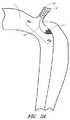
図1は大動脈におけるB型の解離を示す概略図である。鎖骨下動脈2の遠位側での大動脈1の内層における裂けまたは侵入ポイント3は、血液がその大動脈壁内へ入り込み、大動脈の外層5から内層4を剥離させてしまいかねない程充分に重症であり得る。図1中の矢印A1は大動脈壁内への血液の流れを表している。血液によりそれら2つの層の間に創出されたスペースは偽腔6と呼ばれている。上述の裂け3は、その偽腔への侵入ポイントと呼ばれている。また、分離された内層4はフラップと呼ばれている。いくつかの実施形態においては、この解離は、血液が偽腔6に入り込めず、そして、偽腔6を加圧することができなくなるように、前記侵入ポイントを塞ぐことにより治療される。 FIG. 1 is a schematic diagram showing B-type dissection in the aorta. The tear or
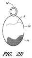
図2Aおよび2Bは、大動脈におけるB型解離の侵入ポイントを実質的または完全に塞ぐための治療装置の1つの実施形態を示している。いくつかの実施形態においては、本装置または本プロテーゼは、カバー11および支持部材または固定要素12を含むことができる。いくつかの実施形態においては、本カバー11、または本明細書で開示されているあらゆる他の装置もしくはプロテーゼのあらゆるカバーは、展開された後、その標的血管内で自立できる程度に充分に剛性であってよい。しかしながら、ここで例示されている実施形態のように、本装置は、カバー11を担持することができるように構成された折り畳み式のフレーム10を有することができる。また、いくつかの実施形態においては、カバー11または本明細書で開示されているあらゆる他のカバーは、付加的なフレームを必要としないような仕方で、一体的なフレームまたは一体的な支持構造を有することができるように構成されていてよい。更に、これらに限定するものではないが、カバー11およびフレーム10を含め、本明細書で開示されているカバーまたはフレームのいくつかの実施形態は、適切な送達装置から展開された際に、解離の侵入ポイントが所在する位置で、その血管壁に寄せて、またはその血管壁に隣接して、偏向または移動するようにバイアスがかけられてよく、または別な仕方でそのような偏向もしくは移動が可能なように構成されていてよい。 FIGS. 2A and 2B show one embodiment of a treatment device for substantially or completely occluding the point of entry of type B dissection in the aorta. In some embodiments, the device or the prosthesis can include a cover 11 and a support member or anchoring
本装置は、治療部位へ経皮的に送達するために、細い送達用カテーテル内に折り畳むことができる。好適には、本装置は、橈骨動脈または大腿動脈から送達することができる。折り畳み式のフレーム10は、これに限定するものではないが、ニチノールを含む形状記憶材料で製作することができる。いくつかの実施形態においては、フレーム10の形状は、大動脈壁に合わせて楕円形であってよい。カバー11はフレーム10内に吊り下げておくことができる。 The device can be folded into a thin delivery catheter for percutaneous delivery to the treatment site. Suitably, the device can be delivered from the radial or femoral artery. The
いくつかの実施形態においては、カバー11は、生体適合性を有し、且つ、柔軟性を有する薄い材料から製作することができる。可能性のある材料は、これらに限定するものではないが、ポリエステル、ePTFE、ポリウレタン、シルク、動物組織、または長期間の埋め込みに適した他のあらゆる材料を含む。いくつかの実施形態においては、カバー11は、組織の修復や大動脈壁内へのカバー11の一体化を促進または補助する材料から製作することができる。いくつかの実施形態においては、カバー11は、組織工学によって作製されたグラフトに対する足場(スカフォールド)として作用すべく設計された基質(マトリックス)から製作することができる。カバー11は、例えば、これらに限定するものではないが、大動脈壁における細胞外マトリックスの天然ビルディングブロックであるであるコラーゲンおよびエラスチンのようなタンパク質を含むことができる。 In some embodiments, the cover 11 can be made from a thin material that is biocompatible and flexible. Possible materials include, but are not limited to, polyester, ePTFE, polyurethane, silk, animal tissue, or any other material suitable for long term implantation. In some embodiments, the cover 11 can be made of a material that facilitates or assists in tissue repair and integration of the cover 11 into the aortic wall. In some embodiments, the cover 11 can be fabricated from a matrix designed to act as a scaffold for a tissue engineered graft. Cover 11 can include, for example, but not limited to proteins such as collagen and elastin, which are natural building blocks of the extracellular matrix in the aortic wall.
いくつかの実施形態においては、カバー11はフィブリンや、キトサンまたはグリコサミノグリカンのような多糖類を含むことができる。いくつかの実施形態においては、上述のタンパク質は適切な架橋剤によって架橋することができる。適切な架橋剤は、これらに限定するものではないが、グルタルアルデヒド、カルボジイミド、タンニン、ポリフェノールおよび光活性化架橋剤を含むことができる。いくつかの実施形態においては、上述のタンパク質層は哺乳動物から採取することができる。哺乳動物由来タンパク質層の可能なソースは、これらに限定するものではないが、心膜、小腸粘膜下層、血管および皮膚を含む。 In some embodiments, the cover 11 can include fibrin or a polysaccharide such as chitosan or glycosaminoglycan. In some embodiments, the proteins described above can be cross-linked by a suitable cross-linking agent. Suitable crosslinkers can include, but are not limited to, glutaraldehyde, carbodiimide, tannin, polyphenols, and photoactivated crosslinkers. In some embodiments, the protein layer described above can be obtained from a mammal. Possible sources of mammalian derived protein layers include, but are not limited to, the pericardium, small intestinal submucosa, blood vessels and skin.
いくつかの実施形態においては、カバー11の厚みは約0.0001インチから約0.01インチまでの範囲であってよい。いくつかの実施形態においては、カバー11の厚みは約0.0005インチから約0.0020インチまでであり得る。カバー11は非多孔質材料、多孔質材料もしくはメッシュから作製されてよく、または編物繊維もしくは織物繊維から作製されてもよい。いくつかの実施形態においては、カバー11の生化学的特性および表面特性は大動脈壁への本カバーの付着を促進するものであってよい。例えば、カバー11はポリエステルの編物またはシルクの織物から作製することができる。また、カバー11は縫合糸もしくは接着剤でフレーム10に取り付けられてよく、またはあらゆる他の適切な締結手段もしくは技術を用いてフレーム10に取り付けることができる。いくつかの実施形態においては、カバー11はフレーム上に直接的に成形することができる。 In some embodiments, the thickness of the cover 11 can range from about 0.0001 inches to about 0.01 inches. In some embodiments, the cover 11 can have a thickness from about 0.0005 inches to about 0.0020 inches. The cover 11 may be made from a non-porous material, a porous material or a mesh, or may be made from knitted or woven fibers. In some embodiments, the biochemical and surface properties of the cover 11 may promote attachment of the cover to the aortic wall. For example, the cover 11 can be made from a polyester knit or a silk fabric. The cover 11 may also be attached to the
フレーム10は、展開中の折り畳まれた状態から大動脈内への展開後における拡張された状態へ移動することができる。いくつかの実施形態においては、フレーム10は、展開中にカバー11を広げることができる。血液の流れはそのカバーを大動脈壁に押し付けることができ、これはカバー11を標的位置に固定するのに役立つ。柔軟性のあるカバー11は大動脈1の壁に合わせることができ、且つ、侵入ポイント3を封鎖することができる。カバー11の移動を防止するため、フレーム10は、その大動脈の側枝内、好適には鎖骨下動脈2内に位置付けされ得る固定要素12に接続することができる。固定要素12は、これらに限定するものではないが、頸動脈または腕頭動脈を含め、その大動脈のあらゆる適切な血管枝に位置付けすることができる。前記固定要素12、または何らかの他の固定要素もしくは支持部材は、自己拡張型のステント、バルーン拡張型のステント、コイル、フック、鉤状構造物、バルーン、ステントグラフト、ネジ、ステープル、または他の同様なもしくは適切な装置であってよい。 The
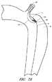
図3Aおよび3Bは、解離を治療するための装置の別の実施形態を例示している。図3Aを参照しながら説明すると、いくつかの実施形態では、カバー22は半円形状または卵形状を有することができる。フレーム21は弓形状のワイヤーを含むことができる。いくつかの実施形態においては、カバー22は遠位側端部が支えられていなくてよい。図3Bには、カバー32の更なる代替的な実施形態が示されている。図3Bに示されているように、カバー32のいくつかの実施形態は固定要素33を越えて近位側に延びていてよく、この実施形態は、侵入ポイント3が鎖骨下動脈または鎖骨下動脈の近位側に在るような状況に適していると考えられる。そのような場合に、鎖骨下動脈をカバー32で覆うことができる。いくつかの実施形態においては、カバー32は、血液が鎖骨下動脈内に進入するのを可能に成すための開口を有していてよい。いくつかの実施形態においては、フレーム31は、カバー31を担持するため、またはカバー31を大動脈壁に押し付けるため、付加的な中央のストラット34を有することができる。 3A and 3B illustrate another embodiment of a device for treating dissociation. Referring to FIG. 3A, in some embodiments, the
図4Aおよび4Bは、解離を治療するための装置の別の実施形態を描いている。図4Aを参照しながら説明すると、本装置は、固定要素43に接続することができる1つもしくはそれ以上の支持具41a−eを含んでいてよい。これらの支持具41a−eは固定要素43の近位側端部により一端を支えられていてよい。代替的に、図4Bに示されているように、支持具51a−eを固定要素53の遠位側端部により支えることもできる。 4A and 4B depict another embodiment of a device for treating dissociation. Referring to FIG. 4A, the apparatus may include one or more supports 41a-e that can be connected to the securing element 43. These supports 41 a-e may be supported at one end by the proximal end of the fixing element 43. Alternatively, the supports 51a-e can be supported by the distal end of the securing element 53, as shown in FIG. 4B.
図5は解離を治療するための装置の別の実施形態を描いており、この実施形態は、大動脈内におけるB型解離の侵入ポイントを実質的にまたは完全に塞ぐことができるように構成されている。いくつかの実施形態においては、本装置は図2に示されているものと同様であってよい。いくつかの実施形態においては、支持要素64をフレーム61の遠位側端部に接続することができる。いくつかの実施形態においては、支持要素64はフレーム61およびカバー62をその解離のフラップ4に押し付けることができる。支持要素64は楕円状の形状を有していてよく、また、フレーム61と同様な材料から作製することができる。上述の楕円は、支持要素64がフラップ4および対向する大動脈壁65と接触した状態になり得るように、大動脈1の直径よりも僅かに大きくてよい。支持要素64は、カバー62の遠位側部分がフラップ4から引き離されてしまったり、またはフラップ4から離れる方向に移動してしまうのを防止もしくは阻止するため、フレーム61の遠位側部分を担持することができるように構成されていてよい。 FIG. 5 depicts another embodiment of a device for treating dissociation, which is configured to substantially or completely occlude the point of entry of type B dissociation within the aorta. Yes. In some embodiments, the apparatus may be similar to that shown in FIG. In some embodiments, the support element 64 can be connected to the distal end of the
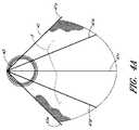
図6Aおよび6Bは、大動脈内におけるB型解離の侵入ポイントを実質的にまたは完全に塞ぐことができるように構成された装置の別の実施形態を描いている。いくつかの実施形態においては、フレーム71は、開かれたケージを形成することができる2つの楕円状の要素71aおよび71bを含むことができる。カバー72は、そのフレーム71の一方の半分と合わせることができるように、またはフレーム71の一方の半分によって担持され得るように構成することができる。大動脈1内に配置されたとき、そのフレームのいくつかの実施形態は、カバー72がフラップ4から引き離されてしまったり、またはフラップ4から離れる方向に移動してしまうのを防止することができる。更に、このフレームのいくつかの実施形態は、その大動脈の局所的な断面領域に合わせることができる。 FIGS. 6A and 6B depict another embodiment of a device configured to be able to substantially or completely occlude the point of entry of type B dissociation within the aorta. In some embodiments, the
図7Aおよび7Bは、大動脈内におけるB型解離の侵入ポイントを実質的にまたは完全に塞ぐことを目的とした装置の別の実施形態を描いている。いくつかの実施形態においては、本装置は、以下で述べられている点を除き、図2に示されている装置と同様であってよい。支持要素84をアンカ要素81により担持することができる。支持要素84は侵入ポイント3を通じて偽腔6内へ進めることができ、且つ、その偽腔6内からフレーム81を担持することができる。フレーム81および支持要素84はそれらの間にフラップ4を効果的に挟むことができ、また、カバー82がフラップ4から引き離されてしまうのを防止することができる。いくつかの実施形態においては、フレーム81または支持要素84はクリップ様の機能を果たすことができる。 7A and 7B depict another embodiment of a device intended to substantially or completely occlude the point of entry of type B dissociation within the aorta. In some embodiments, the apparatus may be similar to the apparatus shown in FIG. 2 except as noted below. The
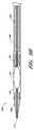
図8Aおよび8Bは解離を治療するための装置100の1つの実施形態の概略的な側面図を表しており、図8Aでは支持部材92が折り畳まれた状態で示されており、一方の図8Bでは拡張された状態で示されている。装置100は、本明細書で開示されている解離を治療するための装置のあらゆる他の実施形態の場合と同一のどのような特徴、コンポーネントまたは他の詳細事項をも有することができる。従って、支持部材92(本明細書ではアンカ部材と呼ばれることもある)は、これらに限定するものではないが、自己拡張型のステント、バルーン拡張型のステント、またはそこに解離を有する血管に隣接した血管内で展開することができる、本明細書で開示されているあらゆる他の固定要素を含め、何らかの適切なステントであってよい。支持部材92は送達中には折り畳んでおくことができ、そして、支持部材92が標的血管の場所に位置付けられた時点で拡張することができる。ここで描かれているように、支持部材92にカバー部材94を取り付けることができ、そのカバー部材94は、解離への侵入ポイントを実質的にまたは完全に覆うように構成することができる。 8A and 8B represent a schematic side view of one embodiment of a
図9Aは、これに限定するものではないが、上で説明されている装置90などの本明細書で開示されている解離を治療するための装置のいくつかの実施形態を展開するために使用することができる、送達用カテーテル100の部分的な断面概略図である。いくつかの実施形態においては、送達用カテーテル100は、2008年4月11日に出願された「Bifurcated Graft Deployment Systems And Methods」と題する米国出願第12/101,863号に開示されている送達用カテーテルの実施形態のいずれかの特徴、コンポーネントもしくは他の詳細事項、またはそのような特徴、コンポーネントもしくは他の詳細事項のあらゆる組み合わせを有することができ、前記米国出願は、参照により、本明細書で完全に記述されているかの如くにして本明細書に組み入れられる。 FIG. 9A is used to deploy some embodiments of a device for treating dissociation disclosed herein, such as, but not limited to,
図9Aを参照しながら説明すると、送達用カテーテル100は、その送達用カテーテル100の近位側端部から外側シース104の管腔を通じて延びていてよい内部コア102を有することができる。前記内部コア102は外側シース104内で軸方向および回転方向に関して移動可能であってよい。内部コア102の遠位側終端部分からチューブ部材106が延びていてよく、このチューブ部材106は非外傷性の遠位側先端部108を担持することができる。遠位側先端部108、チューブ部材106および内部コア102の軸方向の中心線を通じてガイドワイヤー用の管腔110が形成されていてよい。 Referring to FIG. 9A, the
図9Bは、図9Aに示されている送達用カテーテルの部分的な断面概略図であり、その内部に担持されている解離を治療するための装置90の実施形態を示している。いくつかの実施形態においては、外側シース104は固定部材92およびカバー94を抑えるために使用することができ、前記固定部材92とカバー94との両者は折り畳まれた配置構成で送達用カテーテル100内に担持することができる。これに加え、いくつかの実施形態においては、必須ということではないが、固定部材92にフレーム部材を取り付けることができ、カバー106を担持するためにそのフレーム部材を使用することができる。本装置100はチューブ部材106により担持することができ、また、以降で説明されているように、外側シース104により半径方向に抑えることができる。以降で説明されているように、前記半径方向に関する抑えは、内部コア102との相対的な関係において外側シース104を引っ込め、これにより本装置100を露出させることによって解除することができる。 FIG. 9B is a partial cross-sectional schematic view of the delivery catheter shown in FIG. 9A, showing an embodiment of a
次に、前記説明を活かしながら、解離を治療するための装置を展開するための方法に関するいくつかの装置について説明する。そこにB型の解離を有する患者の大動脈の概略図である図10を参照しながら説明すると、患者の鎖骨下動脈2を通じてガイドワイヤー120を大動脈7内へ進めることができる。いくつかの実施形態においては、そのガイドワイヤーは患者の橈骨動脈を通じて鎖骨下動脈2内へ進めることができる。図11は、ガイドワイヤー120上に沿って進められている送達用カテーテル100を示す概略図である。この装置においては、送達用カテーテル100は、患者の橈骨動脈を通じて鎖骨下動脈および大動脈内へ進めることができる。 Next, while utilizing the above description, several devices relating to a method for deploying a device for treating dissociation will be described. Referring to FIG. 10, which is a schematic view of a patient's aorta having B-type dissection there, a
図12および13は、送達用カテーテル100から展開された状態での図9Bの解離を治療するための装置90を示す概略図である。図12に描かれているように、カバー94は、チューブ部材106との関係において相対的に外側シース104を軸方向に引っ込め、これによりカバー部材94を露出する送達用カテーテル100から展開することができる。送達用カテーテル100およびプロテーゼ90は、その送達用カテーテル100および/またはプロテーゼ90に担持されている1つもしくはそれ以上の放射線不透過性マーカーを利用して適切に位置付けすることができる。送達用カテーテル100が鎖骨下動脈2を通じて大動脈7内に進められたため、内部コア102およびチューブ部材106に対して相対的な外側シース104の更なる軸方向の引き込みは、アンカ部材92が送達用カテーテル100から鎖骨下動脈2内へ展開される状態をもたらすことができる。その後、展開用カテーテル100は、図14に示されているように、鎖骨下動脈および橈骨動脈を通じて軸方向に引っ込め、身体から取り外すことができる。この後、解離を治療するための装置90のみを残し、ガイドワイヤー120を取り除くことができる。 12 and 13 are schematic diagrams illustrating an
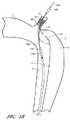
いくつかの実施形態においては、装置90のカバー94は、図15に描かれているように、最初に患者の大腿動脈を通じてガイドワイヤー120をその大動脈および鎖骨下動脈に進めることにより、患者の大動脈内で展開することができる。その後、図16に描かれているように、そのガイドワイヤー120上に沿って、上で説明されている展開用カテーテル100を進めることができる。一旦送達用カテーテル100が目標の場所、例えば鎖骨下動脈2の内部の目標位置に達すると、図17に描かれているように、チューブ部材106および装置90に対して相対的に外側シース104を軸方向に引っ込めることにより、固定部材92をその鎖骨下動脈2内で展開することができる。外側シース104を更に引き込むことにより、装置90の残りのコンポーネントが患者の血管内で展開される状態をもたらすことができる。例えば、図18を参照しながら説明すると、外側シース104の更なる引き込みは、カバー部材94が展開されて、解離の侵入ポイント3が実質的にまたは完全に覆われる状態をもたらすことができる。 In some embodiments, the
その後、展開用カテーテル100は、図19に示されているように、その大動脈および大腿動脈を通じて軸方向に引っ込め、身体から取り外すことができる。この後、解離を治療するための装置90のみを残し、ガイドワイヤー120を取り除くことができる。 The
図2−19は本開示の装置および方法に関するいくつかの実施形態を描いている。ここで提示されている侵入ポイントを塞ぐ方法を支持する他の実施形態も本開示の範囲内である。例えば、いくつかの実施形態においては、細いカテーテルをベースとした送達システムを用いて送達することができる折り畳み式の装置を侵入ポイント上に配置し、大動脈の血管枝に固定することができる。 FIGS. 2-19 depict several embodiments of the disclosed apparatus and method. Other embodiments supporting the method of closing the intrusion point presented here are also within the scope of this disclosure. For example, in some embodiments, a collapsible device that can be delivered using a thin catheter-based delivery system can be placed over the entry point and secured to the aortic vessel branch.
本発明を好適な実施形態および実施例との絡みにおいて開示してきたが、当業者であれば、本開示はそれらの特定的に開示された実施形態を越えて、本発明の他の代替的な実施形態および/または使用、ならびに本発明の明らかな変形態様および同等物にまで広がることが理解されよう。更に、本発明の数多くのバリエーションが開述され且つ詳細に説明されてきたが、当業者にとっては、この開示をベースとすれば、この発明の範囲内に収まる他の様々な変形態様が苦も無く明らかになるであろう。また、それらの実施形態の個別の特徴および態様の様々な組み合わせもしくはサブコンビネーションを作ることができるが、そのような組み合わせもしくはサブコンビネーションは尚も本発明の範囲内に収まることが想定されている。従って、ここで開示されている実施形態の様々な特徴および態様は、その開示されている発明の変形様式を形成するために、相互に組み合わせることができ、または相互に置換することができるものと理解すべきである。それ故、ここで開示されている本開示の範囲はこれまでの箇所で開述されている個別の開示された実施形態に限定されるべきではないということが意図されていると言える。 While the present invention has been disclosed in connection with preferred embodiments and examples, those skilled in the art will appreciate that the present disclosure goes beyond those specifically disclosed embodiments and other alternatives of the present invention. It will be understood that the invention extends to embodiments and / or uses, as well as obvious variations and equivalents of the invention. In addition, while numerous variations of the invention have been described and described in detail, those skilled in the art will appreciate many other variations that fall within the scope of the invention based on this disclosure. It will become clear without it. Also, various combinations or sub-combinations of the individual features and aspects of those embodiments can be made, but such combinations or sub-combinations are still contemplated to be within the scope of the present invention. Accordingly, various features and aspects of the embodiments disclosed herein may be combined with each other or substituted for each other to form variations of the disclosed invention. Should be understood. Therefore, it can be said that the scope of the present disclosure disclosed herein is not intended to be limited to the individual disclosed embodiments described above.
Claims (31)
Translated fromJapaneseプロテーゼを担持しているカテーテルを該解離の侵入ポイントにまで進めるステップであって、ここで、上記解離の侵入ポイントが第1の血管内に在り、そして、上記プロテーゼがカバーおよび前記カバーと連通した折り畳み式の固定要素を含んでいる、カテーテルの前進ステップ;
上記固定要素を前記第1の血管と連通している第2の血管に固定するステップ;
該第1の血管内で上記カバーを拡張するステップ;および
上記カバーを該解離の侵入ポイントの少なくとも一部上にポジショニングするステップ;
を含み、
ここで、上記カバーが該解離の侵入ポイント内への血流を低減することができるように構成されている;
血管の解離の治療方法。A method for treating vascular dissociation comprising:
Advancing a catheter carrying a prosthesis to the dissociation entry point, wherein the dissociation entry point is in a first blood vessel and the prosthesis is in communication with the cover and the cover Advancement step of the catheter, including a foldable fixation element;
Securing the anchoring element to a second blood vessel in communication with the first blood vessel;
Expanding the cover within the first blood vessel; and positioning the cover over at least a portion of the entry point of dissociation;
Including
Wherein the cover is configured to reduce blood flow into the dissociation entry point;
Treatment method of blood vessel dissociation.
固定要素;および
上記固定要素によって担持されたカバー;
を含み、ここで:
上記カバーが、該解離への侵入ポイント上で第1の血管の壁の一部のみを覆うように構成されていて、且つ、前記侵入ポイント内に流入する血液の流れを少なくとも実質的に低減することができるように構成されており;そして
上記固定要素が、前記第1の血管と連通した第2の血管内で展開することができるように構成されている;
血管の解離を治療する装置。A device for treating dissection of a blood vessel, the device comprising:
A fixing element; and a cover carried by the fixing element;
Including, where:
The cover is configured to cover only a portion of the wall of the first blood vessel on the entry point to the dissociation, and at least substantially reduces the flow of blood flowing into the entry point. And the fixation element is configured to be deployable in a second blood vessel in communication with the first blood vessel;
A device to treat the dissociation of blood vessels.
The apparatus of claim 15, wherein the cover has a thickness between about 0.0001 inches and about 0.01 inches.
Applications Claiming Priority (3)
| Application Number | Priority Date | Filing Date | Title |
|---|---|---|---|
| US17488809P | 2009-05-01 | 2009-05-01 | |
| US61/174,888 | 2009-05-01 | ||
| PCT/US2010/033274WO2010127305A2 (en) | 2009-05-01 | 2010-04-30 | Percutaneous method and device to treat dissections |
Publications (1)
| Publication Number | Publication Date |
|---|---|
| JP2012525239Atrue JP2012525239A (en) | 2012-10-22 |
Family
ID=42317676
Family Applications (1)
| Application Number | Title | Priority Date | Filing Date |
|---|---|---|---|
| JP2012508800APendingJP2012525239A (en) | 2009-05-01 | 2010-04-30 | Transcutaneous methods and devices for treating dissociation (priority information and incorporation by reference) |
Country Status (4)
| Country | Link |
|---|---|
| US (1) | US9579103B2 (en) |
| EP (1) | EP2424447A2 (en) |
| JP (1) | JP2012525239A (en) |
| WO (1) | WO2010127305A2 (en) |
Families Citing this family (12)
| Publication number | Priority date | Publication date | Assignee | Title |
|---|---|---|---|---|
| US8523931B2 (en) | 2007-01-12 | 2013-09-03 | Endologix, Inc. | Dual concentric guidewire and methods of bifurcated graft deployment |
| US20150164630A1 (en)* | 2008-01-04 | 2015-06-18 | Eric Johnson | Filter support members |
| US10772717B2 (en) | 2009-05-01 | 2020-09-15 | Endologix, Inc. | Percutaneous method and device to treat dissections |
| JP2012525239A (en) | 2009-05-01 | 2012-10-22 | エンドロジックス、インク | Transcutaneous methods and devices for treating dissociation (priority information and incorporation by reference) |
| WO2011008989A2 (en) | 2009-07-15 | 2011-01-20 | Endologix, Inc. | Stent graft |
| US9393100B2 (en) | 2010-11-17 | 2016-07-19 | Endologix, Inc. | Devices and methods to treat vascular dissections |
| US8911468B2 (en)* | 2011-01-31 | 2014-12-16 | Vatrix Medical, Inc. | Devices, therapeutic compositions and corresponding percutaneous treatment methods for aortic dissection |
| WO2013134452A1 (en)* | 2012-03-08 | 2013-09-12 | Medtronic Ardian Luxembourg Sarl | Spinal neuromodulation and associated systems and methods |
| GB2523291B (en)* | 2013-10-16 | 2016-02-24 | Cook Medical Technologies Llc | Vascular occluder with crossing frame elements |
| EP3648705A4 (en) | 2017-07-07 | 2021-03-24 | Endologix LLC | Endovascular graft systems and methods for deployment in main and branch arteries |
| FI3941392T3 (en) | 2019-03-20 | 2025-07-28 | Inqb8 Medical Tech Llc | Aortic dissection implant |
| SE2351228A1 (en)* | 2023-10-27 | 2025-04-28 | Rafael Martin Astudillo | Aortic dissection implant and aortic dissection system for remodelling and treating blood flow within a false lumen |
Family Cites Families (576)
| Publication number | Priority date | Publication date | Assignee | Title |
|---|---|---|---|---|
| US2127903A (en) | 1936-05-05 | 1938-08-23 | Davis & Geck Inc | Tube for surgical purposes and method of preparing and using the same |
| US2437542A (en) | 1944-05-05 | 1948-03-09 | American Catheter Corp | Catheter-type instrument |
| US2845959A (en) | 1956-03-26 | 1958-08-05 | John B Sidebotham | Bifurcated textile tubes and method of weaving the same |
| US2990605A (en) | 1957-01-30 | 1961-07-04 | Demsyk Paul | Method of forming artificial vascular members |
| US3096560A (en) | 1958-11-21 | 1963-07-09 | William J Liebig | Process for synthetic vascular implants |
| US3029819A (en) | 1959-07-30 | 1962-04-17 | J L Mcatee | Artery graft and method of producing artery grafts |
| US3805301A (en) | 1972-07-28 | 1974-04-23 | Meadox Medicals Inc | Tubular grafts having indicia thereon |
| US3996938A (en) | 1975-07-10 | 1976-12-14 | Clark Iii William T | Expanding mesh catheter |
| DE2760437C2 (en) | 1976-04-05 | 1990-03-22 | Agence Nationale De Valorisation De La Recherche (Anvar), Paris, Fr | |
| WO1980000007A1 (en) | 1978-06-02 | 1980-01-10 | A Rockey | Medical sleeve |
| US4362156A (en) | 1979-04-18 | 1982-12-07 | Riverain Corporation | Intravenous infusion assembly |
| US4503568A (en) | 1981-11-25 | 1985-03-12 | New England Deaconess Hospital | Small diameter vascular bypass and method |
| US4501263A (en) | 1982-03-31 | 1985-02-26 | Harbuck Stanley C | Method for reducing hypertension of a liver |
| US4473067A (en) | 1982-04-28 | 1984-09-25 | Peter Schiff | Introducer assembly for intra-aortic balloons and the like incorporating a sliding, blood-tight seal |
| US4638803A (en) | 1982-09-30 | 1987-01-27 | Rand Robert W | Medical apparatus for inducing scar tissue formation in a body |
| US4525157A (en) | 1983-07-28 | 1985-06-25 | Manresa, Inc. | Closed system catheter with guide wire |
| US4592754A (en) | 1983-09-09 | 1986-06-03 | Gupte Pradeep M | Surgical prosthetic vessel graft and catheter combination and method |
| US5275622A (en) | 1983-12-09 | 1994-01-04 | Harrison Medical Technologies, Inc. | Endovascular grafting apparatus, system and method and devices for use therewith |
| US6221102B1 (en) | 1983-12-09 | 2001-04-24 | Endovascular Technologies, Inc. | Intraluminal grafting system |
| US5669936A (en) | 1983-12-09 | 1997-09-23 | Endovascular Technologies, Inc. | Endovascular grafting system and method for use therewith |
| US5104399A (en) | 1986-12-10 | 1992-04-14 | Endovascular Technologies, Inc. | Artificial graft and implantation method |
| US5108424A (en) | 1984-01-30 | 1992-04-28 | Meadox Medicals, Inc. | Collagen-impregnated dacron graft |
| US4562596A (en) | 1984-04-25 | 1986-01-07 | Elliot Kornberg | Aortic graft, device and method for performing an intraluminal abdominal aortic aneurysm repair |
| US4617932A (en) | 1984-04-25 | 1986-10-21 | Elliot Kornberg | Device and method for performing an intraluminal abdominal aortic aneurysm repair |
| US4580568A (en) | 1984-10-01 | 1986-04-08 | Cook, Incorporated | Percutaneous endovascular stent and method for insertion thereof |
| US4728328A (en) | 1984-10-19 | 1988-03-01 | Research Corporation | Cuffed tubular organic prostheses |
| US4650466A (en) | 1985-11-01 | 1987-03-17 | Angiobrade Partners | Angioplasty device |
| DE3640745A1 (en) | 1985-11-30 | 1987-06-04 | Ernst Peter Prof Dr M Strecker | Catheter for producing or extending connections to or between body cavities |
| US4878906A (en) | 1986-03-25 | 1989-11-07 | Servetus Partnership | Endoprosthesis for repairing a damaged vessel |
| US4756307A (en) | 1987-02-09 | 1988-07-12 | Zimmer, Inc. | Nail device |
| US4744364A (en) | 1987-02-17 | 1988-05-17 | Intravascular Surgical Instruments, Inc. | Device for sealing percutaneous puncture in a vessel |
| USRE34866E (en) | 1987-02-17 | 1995-02-21 | Kensey Nash Corporation | Device for sealing percutaneous puncture in a vessel |
| US4800882A (en) | 1987-03-13 | 1989-01-31 | Cook Incorporated | Endovascular stent and delivery system |
| US4907336A (en) | 1987-03-13 | 1990-03-13 | Cook Incorporated | Method of making an endovascular stent and delivery system |
| IT1210722B (en) | 1987-05-11 | 1989-09-20 | Sorin Biomedica Spa | DEVICES FOR THE CONDITIONING OF BLOOD FLOWS |
| US4816028A (en) | 1987-07-01 | 1989-03-28 | Indu Kapadia | Woven vascular graft |
| JPH0425755Y2 (en) | 1987-10-16 | 1992-06-19 | ||
| US5133732A (en) | 1987-10-19 | 1992-07-28 | Medtronic, Inc. | Intravascular stent |
| US4840940A (en) | 1987-10-21 | 1989-06-20 | Sottiurai Vikrom S | Method for reducing the occurrence of distal anastomotic intimal hyperplasia using fractionated heparin |
| FR2624747A1 (en) | 1987-12-18 | 1989-06-23 | Delsanti Gerard | REMOVABLE ENDO-ARTERIAL DEVICES FOR REPAIRING ARTERIAL WALL DECOLLEMENTS |
| JPH0784524B2 (en) | 1987-12-24 | 1995-09-13 | 東ソー株式会社 | Method for producing aromatic sulfide amide polymer |
| US5019090A (en) | 1988-09-01 | 1991-05-28 | Corvita Corporation | Radially expandable endoprosthesis and the like |
| US4981478A (en) | 1988-09-06 | 1991-01-01 | Advanced Cardiovascular Systems | Composite vascular catheter |
| US4994069A (en) | 1988-11-02 | 1991-02-19 | Target Therapeutics | Vaso-occlusion coil and method |
| US4856516A (en) | 1989-01-09 | 1989-08-15 | Cordis Corporation | Endovascular stent apparatus and method |
| CH678393A5 (en) | 1989-01-26 | 1991-09-13 | Ulrich Prof Dr Med Sigwart | |
| US5078726A (en) | 1989-02-01 | 1992-01-07 | Kreamer Jeffry W | Graft stent and method of repairing blood vessels |
| US5178634A (en) | 1989-03-31 | 1993-01-12 | Wilson Ramos Martinez | Aortic valved tubes for human implants |
| US5015232A (en) | 1989-04-20 | 1991-05-14 | Cook Incorporated | Decompression enteroclysis balloon catheter |
| US4994071A (en) | 1989-05-22 | 1991-02-19 | Cordis Corporation | Bifurcating stent apparatus and method |
| US5104400A (en) | 1989-05-26 | 1992-04-14 | Impra, Inc. | Blood vessel patch |
| NL8901350A (en) | 1989-05-29 | 1990-12-17 | Wouter Matthijs Muijs Van De M | CLOSURE ASSEMBLY. |
| US5622188A (en) | 1989-08-18 | 1997-04-22 | Endovascular Instruments, Inc. | Method of restoring reduced or absent blood flow capacity in an artery |
| US5034001A (en) | 1989-09-08 | 1991-07-23 | Advanced Cardiovascular Systems, Inc. | Method of repairing a damaged blood vessel with an expandable cage catheter |
| US5035706A (en) | 1989-10-17 | 1991-07-30 | Cook Incorporated | Percutaneous stent and method for retrieval thereof |
| GB8927282D0 (en) | 1989-12-01 | 1990-01-31 | Univ Strathclyde | Vascular surgical devices |
| US5041093A (en) | 1990-01-31 | 1991-08-20 | Boston Scientific Corp. | Catheter with foraminous anchor |
| US5145620A (en) | 1990-02-09 | 1992-09-08 | Ngk Insulators, Ltd. | Method of producing a silicon nitride sintered body |
| US5123917A (en) | 1990-04-27 | 1992-06-23 | Lee Peter Y | Expandable intraluminal vascular graft |
| US5116349A (en) | 1990-05-23 | 1992-05-26 | United States Surgical Corporation | Surgical fastener apparatus |
| US5578071A (en) | 1990-06-11 | 1996-11-26 | Parodi; Juan C. | Aortic graft |
| US5360443A (en) | 1990-06-11 | 1994-11-01 | Barone Hector D | Aortic graft for repairing an abdominal aortic aneurysm |
| US5156619A (en) | 1990-06-15 | 1992-10-20 | Ehrenfeld William K | Flanged end-to-side vascular graft |
| US5064435A (en) | 1990-06-28 | 1991-11-12 | Schneider (Usa) Inc. | Self-expanding prosthesis having stable axial length |
| US5449372A (en) | 1990-10-09 | 1995-09-12 | Scimed Lifesystems, Inc. | Temporary stent and methods for use and manufacture |
| DE9117152U1 (en) | 1990-10-09 | 1996-07-11 | Cook Inc., Bloomington, Ind. | Stent |
| US5330500A (en) | 1990-10-18 | 1994-07-19 | Song Ho Y | Self-expanding endovascular stent with silicone coating |
| US5147334A (en) | 1991-01-02 | 1992-09-15 | Moss James P | Catheter for cholangiography |
| CA2060067A1 (en) | 1991-01-28 | 1992-07-29 | Lilip Lau | Stent delivery system |
| US5156620A (en) | 1991-02-04 | 1992-10-20 | Pigott John P | Intraluminal graft/stent and balloon catheter for insertion thereof |
| US5135536A (en) | 1991-02-05 | 1992-08-04 | Cordis Corporation | Endovascular stent and method |
| US5669934A (en) | 1991-02-13 | 1997-09-23 | Fusion Medical Technologies, Inc. | Methods for joining tissue by applying radiofrequency energy to performed collagen films and sheets |
| US5628783A (en) | 1991-04-11 | 1997-05-13 | Endovascular Technologies, Inc. | Bifurcated multicapsule intraluminal grafting system and method |
| CA2065634C (en) | 1991-04-11 | 1997-06-03 | Alec A. Piplani | Endovascular graft having bifurcation and apparatus and method for deploying the same |
| US5554118A (en) | 1991-05-24 | 1996-09-10 | Jang; G. David | Universal mode vascular catheter system |
| US5304200A (en) | 1991-05-29 | 1994-04-19 | Cordis Corporation | Welded radially expandable endoprosthesis and the like |
| US5135535A (en) | 1991-06-11 | 1992-08-04 | Advanced Cardiovascular Systems, Inc. | Catheter system with catheter and guidewire exchange |
| US5314472A (en) | 1991-10-01 | 1994-05-24 | Cook Incorporated | Vascular stent |
| US5197976A (en) | 1991-09-16 | 1993-03-30 | Atrium Medical Corporation | Manually separable multi-lumen vascular graft |
| US5443498A (en) | 1991-10-01 | 1995-08-22 | Cook Incorporated | Vascular stent and method of making and implanting a vacsular stent |
| US5464450A (en) | 1991-10-04 | 1995-11-07 | Scimed Lifesystems Inc. | Biodegradable drug delivery vascular stent |
| US5366504A (en) | 1992-05-20 | 1994-11-22 | Boston Scientific Corporation | Tubular medical prosthesis |
| US5282860A (en) | 1991-10-16 | 1994-02-01 | Olympus Optical Co., Ltd. | Stent tube for medical use |
| US5720776A (en) | 1991-10-25 | 1998-02-24 | Cook Incorporated | Barb and expandable transluminal graft prosthesis for repair of aneurysm |
| US5387235A (en) | 1991-10-25 | 1995-02-07 | Cook Incorporated | Expandable transluminal graft prosthesis for repair of aneurysm |
| US5693084A (en) | 1991-10-25 | 1997-12-02 | Cook Incorporated | Expandable transluminal graft prosthesis for repair of aneurysm |
| CA2081424C (en) | 1991-10-25 | 2008-12-30 | Timothy A. Chuter | Expandable transluminal graft prosthesis for repair of aneurysm |
| US5211658A (en) | 1991-11-05 | 1993-05-18 | New England Deaconess Hospital Corporation | Method and device for performing endovascular repair of aneurysms |
| US5316023A (en) | 1992-01-08 | 1994-05-31 | Expandable Grafts Partnership | Method for bilateral intra-aortic bypass |
| US5507767A (en) | 1992-01-15 | 1996-04-16 | Cook Incorporated | Spiral stent |
| US5405377A (en) | 1992-02-21 | 1995-04-11 | Endotech Ltd. | Intraluminal stent |
| US5683448A (en) | 1992-02-21 | 1997-11-04 | Boston Scientific Technology, Inc. | Intraluminal stent and graft |
| US5234448A (en) | 1992-02-28 | 1993-08-10 | Shadyside Hospital | Method and apparatus for connecting and closing severed blood vessels |
| US5282823A (en) | 1992-03-19 | 1994-02-01 | Medtronic, Inc. | Intravascular radially expandable stent |
| US5370683A (en) | 1992-03-25 | 1994-12-06 | Cook Incorporated | Vascular stent |
| US5201757A (en) | 1992-04-03 | 1993-04-13 | Schneider (Usa) Inc. | Medial region deployment of radially self-expanding stents |
| US5263932A (en) | 1992-04-09 | 1993-11-23 | Jang G David | Bailout catheter for fixed wire angioplasty |
| US5354308A (en) | 1992-05-01 | 1994-10-11 | Beth Israel Hospital Association | Metal wire stent |
| EP0639958A1 (en) | 1992-05-08 | 1995-03-01 | Schneider (Usa) Inc. | Esophageal stent and delivery tool |
| US5507771A (en) | 1992-06-15 | 1996-04-16 | Cook Incorporated | Stent assembly |
| US5342387A (en) | 1992-06-18 | 1994-08-30 | American Biomed, Inc. | Artificial support for a blood vessel |
| US5496365A (en) | 1992-07-02 | 1996-03-05 | Sgro; Jean-Claude | Autoexpandable vascular endoprosthesis |
| US5514379A (en) | 1992-08-07 | 1996-05-07 | The General Hospital Corporation | Hydrogel compositions and methods of use |
| US5530528A (en) | 1992-09-28 | 1996-06-25 | Fujitsu Limited | Image forming apparatus having contact type, one-component developing unit |
| US5383897A (en) | 1992-10-19 | 1995-01-24 | Shadyside Hospital | Method and apparatus for closing blood vessel punctures |
| DE59206251D1 (en) | 1992-10-31 | 1996-06-13 | Schneider Europ Ag | Arrangement for implanting self-expanding endoprostheses |
| BE1006440A3 (en) | 1992-12-21 | 1994-08-30 | Dereume Jean Pierre Georges Em | Luminal endoprosthesis AND METHOD OF PREPARATION. |
| US5256141A (en) | 1992-12-22 | 1993-10-26 | Nelson Gencheff | Biological material deployment method and apparatus |
| JPH08500757A (en) | 1992-12-30 | 1996-01-30 | シュナイダー・(ユーエスエイ)・インコーポレーテッド | Device for deploying a stent implantable in the body |
| US5370691A (en) | 1993-01-26 | 1994-12-06 | Target Therapeutics, Inc. | Intravascular inflatable stent |
| US5403280A (en) | 1993-02-16 | 1995-04-04 | Wang; James C. | Inflatable perfusion catheter |
| NL9300500A (en) | 1993-03-22 | 1994-10-17 | Industrial Res Bv | Expandable hollow sleeve for locally supporting and / or strengthening a body vessel, as well as a method for manufacturing it. |
| US5843167A (en) | 1993-04-22 | 1998-12-01 | C. R. Bard, Inc. | Method and apparatus for recapture of hooked endoprosthesis |
| AU689094B2 (en) | 1993-04-22 | 1998-03-26 | C.R. Bard Inc. | Non-migrating vascular prosthesis and minimally invasive placement system therefor |
| CA2158757C (en) | 1993-04-23 | 2000-01-04 | Joseph E. Laptewicz Jr. | Covered stent and stent delivery device |
| DE69317548T2 (en) | 1993-04-23 | 1998-08-13 | Schneider (Europe) Gmbh, Buelach | Stent with a coating of elastic material and method for applying the coating on the stent |
| US5464650A (en) | 1993-04-26 | 1995-11-07 | Medtronic, Inc. | Intravascular stent and method |
| US5643171A (en) | 1993-05-04 | 1997-07-01 | Neocardia, Llc | Method and apparatus for uniform radiation treatment of vascular lumens |
| US5320602A (en) | 1993-05-14 | 1994-06-14 | Wilson-Cook Medical, Inc. | Peel-away endoscopic retrograde cholangio pancreatography catheter and a method for using the same |
| US5338298A (en) | 1993-06-04 | 1994-08-16 | C. R. Bard, Inc. | Double-tapered balloon |
| US5425765A (en) | 1993-06-25 | 1995-06-20 | Tiefenbrun; Jonathan | Surgical bypass method |
| US5458615A (en) | 1993-07-06 | 1995-10-17 | Advanced Cardiovascular Systems, Inc. | Stent delivery system |
| US5464449A (en) | 1993-07-08 | 1995-11-07 | Thomas J. Fogarty | Internal graft prosthesis and delivery system |
| CA2125258C (en) | 1993-08-05 | 1998-12-22 | Dinah B Quiachon | Multicapsule intraluminal grafting system and method |
| AU6987594A (en) | 1993-08-18 | 1995-03-14 | W.L. Gore & Associates, Inc. | A tubular intraluminal graft |
| US5735892A (en) | 1993-08-18 | 1998-04-07 | W. L. Gore & Associates, Inc. | Intraluminal stent graft |
| US6159565A (en) | 1993-08-18 | 2000-12-12 | W. L. Gore & Associates, Inc. | Thin-wall intraluminal graft |
| US6027779A (en) | 1993-08-18 | 2000-02-22 | W. L. Gore & Associates, Inc. | Thin-wall polytetrafluoroethylene tube |
| US5669880A (en) | 1993-08-24 | 1997-09-23 | Cordis Corporation | Stent delivery system |
| WO1995008289A2 (en) | 1993-09-16 | 1995-03-30 | Scimed Life Systems, Inc. | Percutaneous repair of cardiovascular anomalies and repair compositions |
| KR970004845Y1 (en) | 1993-09-27 | 1997-05-21 | 주식회사 수호메디테크 | Endoscopic expansion medical equipment |
| WO1995008966A1 (en) | 1993-09-30 | 1995-04-06 | White Geoffrey H | Intraluminal graft |
| ATE165231T1 (en) | 1993-10-20 | 1998-05-15 | Schneider Europ Ag | ENDOPROSTHESIS |
| US5723004A (en) | 1993-10-21 | 1998-03-03 | Corvita Corporation | Expandable supportive endoluminal grafts |
| US5639278A (en) | 1993-10-21 | 1997-06-17 | Corvita Corporation | Expandable supportive bifurcated endoluminal grafts |
| US5632772A (en) | 1993-10-21 | 1997-05-27 | Corvita Corporation | Expandable supportive branched endoluminal grafts |
| US5989280A (en) | 1993-10-22 | 1999-11-23 | Scimed Lifesystems, Inc | Stent delivery apparatus and method |
| ES2135520T3 (en) | 1993-11-04 | 1999-11-01 | Bard Inc C R | NON-MIGRANT VASCULAR PROSTHESIS. |
| AU1091095A (en) | 1993-11-08 | 1995-05-29 | Harrison M. Lazarus | Intraluminal vascular graft and method |
| JP2703510B2 (en) | 1993-12-28 | 1998-01-26 | アドヴァンスド カーディオヴァスキュラー システムズ インコーポレーテッド | Expandable stent and method of manufacturing the same |
| US5609627A (en) | 1994-02-09 | 1997-03-11 | Boston Scientific Technology, Inc. | Method for delivering a bifurcated endoluminal prosthesis |
| US6051020A (en) | 1994-02-09 | 2000-04-18 | Boston Scientific Technology, Inc. | Bifurcated endoluminal prosthesis |
| US5507769A (en) | 1994-10-18 | 1996-04-16 | Stentco, Inc. | Method and apparatus for forming an endoluminal bifurcated graft |
| US5443477A (en) | 1994-02-10 | 1995-08-22 | Stentco, Inc. | Apparatus and method for deployment of radially expandable stents by a mechanical linkage |
| US6039749A (en) | 1994-02-10 | 2000-03-21 | Endovascular Systems, Inc. | Method and apparatus for deploying non-circular stents and graftstent complexes |
| US5653746A (en) | 1994-03-08 | 1997-08-05 | Meadox Medicals, Inc. | Radially expandable tubular prosthesis |
| US5733303A (en) | 1994-03-17 | 1998-03-31 | Medinol Ltd. | Flexible expandable stent |
| US5415664A (en) | 1994-03-30 | 1995-05-16 | Corvita Corporation | Method and apparatus for introducing a stent or a stent-graft |
| US5554181A (en) | 1994-05-04 | 1996-09-10 | Regents Of The University Of Minnesota | Stent |
| US5824044A (en) | 1994-05-12 | 1998-10-20 | Endovascular Technologies, Inc. | Bifurcated multicapsule intraluminal grafting system |
| US5456694A (en) | 1994-05-13 | 1995-10-10 | Stentco, Inc. | Device for delivering and deploying intraluminal devices |
| DE4418336A1 (en) | 1994-05-26 | 1995-11-30 | Angiomed Ag | Stent for widening and holding open receptacles |
| US5683451A (en) | 1994-06-08 | 1997-11-04 | Cardiovascular Concepts, Inc. | Apparatus and methods for deployment release of intraluminal prostheses |
| US5824041A (en) | 1994-06-08 | 1998-10-20 | Medtronic, Inc. | Apparatus and methods for placement and repositioning of intraluminal prostheses |
| DE69528216T2 (en) | 1994-06-17 | 2003-04-17 | Terumo K.K., Tokio/Tokyo | Process for the production of a permanent stent |
| US5522881A (en) | 1994-06-28 | 1996-06-04 | Meadox Medicals, Inc. | Implantable tubular prosthesis having integral cuffs |
| US6123715A (en) | 1994-07-08 | 2000-09-26 | Amplatz; Curtis | Method of forming medical devices; intravascular occlusion devices |
| US5846261A (en) | 1994-07-08 | 1998-12-08 | Aga Medical Corp. | Percutaneous catheter directed occlusion devices |
| US5397355A (en) | 1994-07-19 | 1995-03-14 | Stentco, Inc. | Intraluminal stent |
| US5575816A (en) | 1994-08-12 | 1996-11-19 | Meadox Medicals, Inc. | High strength and high density intraluminal wire stent |
| US5591230A (en) | 1994-09-07 | 1997-01-07 | Global Therapeutics, Inc. | Radially expandable stent |
| US6015429A (en) | 1994-09-08 | 2000-01-18 | Gore Enterprise Holdings, Inc. | Procedures for introducing stents and stent-grafts |
| US5653743A (en) | 1994-09-09 | 1997-08-05 | Martin; Eric C. | Hypogastric artery bifurcation graft and method of implantation |
| US5562727A (en) | 1994-10-07 | 1996-10-08 | Aeroquip Corporation | Intraluminal graft and method for insertion thereof |
| US5534024A (en) | 1994-11-04 | 1996-07-09 | Aeroquip Corporation | Intraluminal stenting graft |
| CA2175720C (en) | 1996-05-03 | 2011-11-29 | Ian M. Penn | Bifurcated stent and method for the manufacture and delivery of same |
| CA2134997C (en) | 1994-11-03 | 2009-06-02 | Ian M. Penn | Stent |
| JP3182301B2 (en) | 1994-11-07 | 2001-07-03 | キヤノン株式会社 | Microstructure and method for forming the same |
| EP0790810B1 (en) | 1994-11-09 | 2004-04-28 | Endotex Interventional Systems, Inc. | Kit of delivery catheter and graft for aneurysm repair |
| AU3783195A (en) | 1994-11-15 | 1996-05-23 | Advanced Cardiovascular Systems Inc. | Intraluminal stent for attaching a graft |
| US5507770A (en) | 1994-11-23 | 1996-04-16 | Aeroquip Corporation | Intraluminal grafting stent and method for implanting same in a blood vessel |
| US5616114A (en) | 1994-12-08 | 1997-04-01 | Neocardia, Llc. | Intravascular radiotherapy employing a liquid-suspended source |
| US5630829A (en) | 1994-12-09 | 1997-05-20 | Intervascular, Inc. | High hoop strength intraluminal stent |
| US5879366A (en) | 1996-12-20 | 1999-03-09 | W.L. Gore & Associates, Inc. | Self-expanding defect closure device and method of making and using |
| NL9500094A (en) | 1995-01-19 | 1996-09-02 | Industrial Res Bv | Y-shaped stent and method of deployment. |
| US5755770A (en) | 1995-01-31 | 1998-05-26 | Boston Scientific Corporatiion | Endovascular aortic graft |
| US5575818A (en) | 1995-02-14 | 1996-11-19 | Corvita Corporation | Endovascular stent with locking ring |
| US5522883A (en) | 1995-02-17 | 1996-06-04 | Meadox Medicals, Inc. | Endoprosthesis stent/graft deployment system |
| AU719980B2 (en) | 1995-02-22 | 2000-05-18 | Menlo Care, Inc. | Covered expanding mesh stent |
| US5683449A (en) | 1995-02-24 | 1997-11-04 | Marcade; Jean Paul | Modular bifurcated intraluminal grafts and methods for delivering and assembling same |
| US5662675A (en) | 1995-02-24 | 1997-09-02 | Intervascular, Inc. | Delivery catheter assembly |
| US6818014B2 (en) | 1995-03-01 | 2004-11-16 | Scimed Life Systems, Inc. | Longitudinally flexible expandable stent |
| US5681345A (en) | 1995-03-01 | 1997-10-28 | Scimed Life Systems, Inc. | Sleeve carrying stent |
| EP0813397A4 (en) | 1995-03-10 | 1999-10-06 | Cardiovascular Concepts Inc | Tubular endoluminar prosthesis having oblique ends |
| JP3507503B2 (en) | 1995-03-10 | 2004-03-15 | インプラ・インコーポレーテッド | Sealable stent for body cavity, method for producing the same, and method for introducing the same into body cavity |
| US6124523A (en) | 1995-03-10 | 2000-09-26 | Impra, Inc. | Encapsulated stent |
| US6039755A (en) | 1997-02-05 | 2000-03-21 | Impra, Inc., A Division Of C.R. Bard, Inc. | Radially expandable tubular polytetrafluoroethylene grafts and method of making same |
| US5591197A (en) | 1995-03-14 | 1997-01-07 | Advanced Cardiovascular Systems, Inc. | Expandable stent forming projecting barbs and method for deploying |
| US5647857A (en) | 1995-03-16 | 1997-07-15 | Endotex Interventional Systems, Inc. | Protective intraluminal sheath |
| CA2171896C (en) | 1995-03-17 | 2007-05-15 | Scott C. Anderson | Multi-anchor stent |
| US5643278A (en) | 1995-04-06 | 1997-07-01 | Leocor, Inc. | Stent delivery system |
| EP0740928B1 (en) | 1995-04-12 | 2004-07-07 | Corvita Europe | Self-expanding stent for introducing a medical device in a body cavity and manufacturing process |
| DE69626108T2 (en) | 1995-04-14 | 2003-11-20 | Boston Scientific Ltd., St. Michael | STENTING DEVICE WITH ROLLING MEMBRANE |
| US5641373A (en) | 1995-04-17 | 1997-06-24 | Baxter International Inc. | Method of manufacturing a radially-enlargeable PTFE tape-reinforced vascular graft |
| US5609628A (en) | 1995-04-20 | 1997-03-11 | Keranen; Victor J. | Intravascular graft and catheter |
| EP0738520B1 (en) | 1995-04-21 | 1999-01-27 | C.R. Bard, Inc. | Interlocking catheter assembly |
| FI974063L (en) | 1995-04-26 | 1997-12-19 | Medinol Ltd | Articulated expander |
| US5591198A (en) | 1995-04-27 | 1997-01-07 | Medtronic, Inc. | Multiple sinusoidal wave configuration stent |
| FR2733682B1 (en) | 1995-05-04 | 1997-10-31 | Dibie Alain | ENDOPROSTHESIS FOR THE TREATMENT OF STENOSIS ON BIFURCATIONS OF BLOOD VESSELS AND LAYING EQUIPMENT THEREFOR |
| US5662614A (en) | 1995-05-09 | 1997-09-02 | Edoga; John K. | Balloon expandable universal access sheath |
| US5628786A (en) | 1995-05-12 | 1997-05-13 | Impra, Inc. | Radially expandable vascular graft with resistance to longitudinal compression and method of making same |
| WO1996036297A1 (en) | 1995-05-19 | 1996-11-21 | Kanji Inoue | Transplantation instrument, method of bending same and method of transplanting same |
| US6151404A (en) | 1995-06-01 | 2000-11-21 | Medical Media Systems | Anatomical visualization system |
| DE69635659T2 (en) | 1995-06-01 | 2006-07-06 | Meadox Medicals, Inc. | IMPLANTABLE INTRALUMINARY PROSTHESIS |
| AU5950696A (en) | 1995-06-05 | 1996-12-24 | Creative Products Resource, Inc. | Dry-cleaning kit for in-dryer use |
| US6312407B1 (en) | 1995-06-05 | 2001-11-06 | Medtronic Percusurge, Inc. | Occlusion of a vessel |
| US5591199A (en) | 1995-06-07 | 1997-01-07 | Porter; Christopher H. | Curable fiber composite stent and delivery system |
| RU2157146C2 (en) | 1995-06-13 | 2000-10-10 | ВИЛЬЯМ КУК Европа, A/S | Device for performing implantation in blood vessels and hollow organs |
| US5676685A (en) | 1995-06-22 | 1997-10-14 | Razavi; Ali | Temporary stent |
| US5785679A (en) | 1995-07-19 | 1998-07-28 | Endotex Interventional Systems, Inc. | Methods and apparatus for treating aneurysms and arterio-venous fistulas |
| US5766203A (en) | 1995-07-20 | 1998-06-16 | Intelliwire, Inc. | Sheath with expandable distal extremity and balloon catheters and stents for use therewith and method |
| US5697968A (en) | 1995-08-10 | 1997-12-16 | Aeroquip Corporation | Check valve for intraluminal graft |
| US5769882A (en) | 1995-09-08 | 1998-06-23 | Medtronic, Inc. | Methods and apparatus for conformably sealing prostheses within body lumens |
| US5562697A (en) | 1995-09-18 | 1996-10-08 | William Cook, Europe A/S | Self-expanding stent assembly and methods for the manufacture thereof |
| WO1997010757A1 (en) | 1995-09-22 | 1997-03-27 | Autogenics | Sewing ring with integral retaining springs |
| US5824037A (en) | 1995-10-03 | 1998-10-20 | Medtronic, Inc. | Modular intraluminal prostheses construction and methods |
| US6193745B1 (en) | 1995-10-03 | 2001-02-27 | Medtronic, Inc. | Modular intraluminal prosteheses construction and methods |
| AU7458596A (en) | 1995-10-20 | 1997-05-07 | Bandula Wijay | Vascular stent |
| US5669924A (en) | 1995-10-26 | 1997-09-23 | Shaknovich; Alexander | Y-shuttle stent assembly for bifurcating vessels and method of using the same |
| US5591195A (en) | 1995-10-30 | 1997-01-07 | Taheri; Syde | Apparatus and method for engrafting a blood vessel |
| US6348066B1 (en) | 1995-11-07 | 2002-02-19 | Corvita Corporation | Modular endoluminal stent-grafts and methods for their use |
| US5628788A (en) | 1995-11-07 | 1997-05-13 | Corvita Corporation | Self-expanding endoluminal stent-graft |
| US6045557A (en) | 1995-11-10 | 2000-04-04 | Baxter International Inc. | Delivery catheter and method for positioning an intraluminal graft |
| ATE177928T1 (en) | 1995-11-14 | 1999-04-15 | Schneider Europ Gmbh | DEVICE FOR STENT IMPLANTATION |
| US5665117A (en) | 1995-11-27 | 1997-09-09 | Rhodes; Valentine J. | Endovascular prosthesis with improved sealing means for aneurysmal arterial disease and method of use |
| US5593417A (en) | 1995-11-27 | 1997-01-14 | Rhodes; Valentine J. | Intravascular stent with secure mounting means |
| US6576009B2 (en) | 1995-12-01 | 2003-06-10 | Medtronic Ave, Inc. | Bifurcated intraluminal prostheses construction and methods |
| US5824040A (en) | 1995-12-01 | 1998-10-20 | Medtronic, Inc. | Endoluminal prostheses and therapies for highly variable body lumens |
| US6042605A (en) | 1995-12-14 | 2000-03-28 | Gore Enterprose Holdings, Inc. | Kink resistant stent-graft |
| EP0950385A3 (en) | 1995-12-14 | 1999-10-27 | Prograft Medical, Inc. | Stent-graft deployment apparatus and method |
| US5752974A (en) | 1995-12-18 | 1998-05-19 | Collagen Corporation | Injectable or implantable biomaterials for filling or blocking lumens and voids of the body |
| DK0876165T3 (en) | 1995-12-18 | 2007-08-06 | Angiotech Biomaterials Corp | Crosslinked polymer compositions and methods for their preparation |
| US5800407A (en) | 1995-12-21 | 1998-09-01 | Eldor; Joseph | Multiple hole epidural catheter |
| US5693066A (en) | 1995-12-21 | 1997-12-02 | Medtronic, Inc. | Stent mounting and transfer device and method |
| US5604435A (en) | 1995-12-29 | 1997-02-18 | General Electric Company | Spiral scanning method for monitoring physiological changes |
| ATE290832T1 (en) | 1996-01-05 | 2005-04-15 | Medtronic Inc | EXPANDABLE ENDOLUMINAL PROSTHESES |
| US5690642A (en) | 1996-01-18 | 1997-11-25 | Cook Incorporated | Rapid exchange stent delivery balloon catheter |
| US5800512A (en) | 1996-01-22 | 1998-09-01 | Meadox Medicals, Inc. | PTFE vascular graft |
| US6168622B1 (en) | 1996-01-24 | 2001-01-02 | Microvena Corporation | Method and apparatus for occluding aneurysms |
| AUPN775296A0 (en) | 1996-01-25 | 1996-02-22 | Endogad Research Pty Limited | Directional catheter |
| US6017363A (en) | 1997-09-22 | 2000-01-25 | Cordis Corporation | Bifurcated axially flexible stent |
| CA2244080A1 (en) | 1996-02-02 | 1997-08-07 | Transvascular, Inc. | Methods and apparatus for blocking flow through blood vessels |
| US5871537A (en) | 1996-02-13 | 1999-02-16 | Scimed Life Systems, Inc. | Endovascular apparatus |
| US5690643A (en) | 1996-02-20 | 1997-11-25 | Leocor, Incorporated | Stent delivery system |
| US5695516A (en) | 1996-02-21 | 1997-12-09 | Iso Stent, Inc. | Longitudinally elongating balloon expandable stent |
| US5810836A (en) | 1996-03-04 | 1998-09-22 | Myocardial Stents, Inc. | Device and method for trans myocardial revascularization (TMR) |
| ATE193820T1 (en) | 1996-03-13 | 2000-06-15 | Medtronic Inc | ENDOLUMINAL PROSTHESIS FOR MULTIPLE BRANCH BODY LUMEN SYSTEMS |
| US6458096B1 (en) | 1996-04-01 | 2002-10-01 | Medtronic, Inc. | Catheter with autoinflating, autoregulating balloon |
| US5843160A (en) | 1996-04-01 | 1998-12-01 | Rhodes; Valentine J. | Prostheses for aneurysmal and/or occlusive disease at a bifurcation in a vessel, duct, or lumen |
| US5630830A (en) | 1996-04-10 | 1997-05-20 | Medtronic, Inc. | Device and method for mounting stents on delivery systems |
| JP3410922B2 (en) | 1996-04-23 | 2003-05-26 | 株式会社東芝 | Clock control circuit |
| US6096053A (en) | 1996-05-03 | 2000-08-01 | Scimed Life Systems, Inc. | Medical retrieval basket |
| US5921954A (en) | 1996-07-10 | 1999-07-13 | Mohr, Jr.; Lawrence G. | Treating aneurysms by applying hardening/softening agents to hardenable/softenable substances |
| US6544276B1 (en) | 1996-05-20 | 2003-04-08 | Medtronic Ave. Inc. | Exchange method for emboli containment |
| FR2749160B1 (en) | 1996-05-28 | 1999-05-21 | Patrice Bergeron | MODULAR BIFURCED VASCULAR PROSTHESIS |
| US6190402B1 (en) | 1996-06-21 | 2001-02-20 | Musc Foundation For Research Development | Insitu formable and self-forming intravascular flow modifier (IFM) and IFM assembly for deployment of same |
| US5928279A (en) | 1996-07-03 | 1999-07-27 | Baxter International Inc. | Stented, radially expandable, tubular PTFE grafts |
| GB9614950D0 (en) | 1996-07-16 | 1996-09-04 | Anson Medical Ltd | A ductus stent and delivery catheter |
| US5980514A (en) | 1996-07-26 | 1999-11-09 | Target Therapeutics, Inc. | Aneurysm closure device assembly |
| US5676697A (en) | 1996-07-29 | 1997-10-14 | Cardiovascular Dynamics, Inc. | Two-piece, bifurcated intraluminal graft for repair of aneurysm |
| US5725535A (en) | 1996-09-20 | 1998-03-10 | Hegde; Anant V. | Multiple balloon stent delivery catheter and method |
| DE69734667T2 (en) | 1996-09-26 | 2006-06-08 | Boston Scientific Scimed, Inc., Maple Grove | COMBINED MEDICAL DEVICE CONSISTING OF A SUPPORT STRUCTURE AND A MEMBRANE |
| US6395017B1 (en) | 1996-11-15 | 2002-05-28 | C. R. Bard, Inc. | Endoprosthesis delivery catheter with sequential stage control |
| US5860998A (en) | 1996-11-25 | 1999-01-19 | C. R. Bard, Inc. | Deployment device for tubular expandable prosthesis |
| US6551350B1 (en) | 1996-12-23 | 2003-04-22 | Gore Enterprise Holdings, Inc. | Kink resistant bifurcated prosthesis |
| US6352561B1 (en) | 1996-12-23 | 2002-03-05 | W. L. Gore & Associates | Implant deployment apparatus |
| US6015431A (en) | 1996-12-23 | 2000-01-18 | Prograft Medical, Inc. | Endolumenal stent-graft with leak-resistant seal |
| EP0850607A1 (en) | 1996-12-31 | 1998-07-01 | Cordis Corporation | Valve prosthesis for implantation in body channels |
| US5879321A (en) | 1997-01-22 | 1999-03-09 | The University Of Kentucky Research Foundation | Portocaval-right atrial shunt |
| JP4042998B2 (en) | 1997-01-29 | 2008-02-06 | クック インコーポレイテッド | Bell bottom modular stent graft |
| US6203735B1 (en) | 1997-02-03 | 2001-03-20 | Impra, Inc. | Method of making expanded polytetrafluoroethylene products |
| US5827321A (en) | 1997-02-07 | 1998-10-27 | Cornerstone Devices, Inc. | Non-Foreshortening intraluminal prosthesis |
| US5720735A (en) | 1997-02-12 | 1998-02-24 | Dorros; Gerald | Bifurcated endovascular catheter |
| US6090128A (en) | 1997-02-20 | 2000-07-18 | Endologix, Inc. | Bifurcated vascular graft deployment device |
| US6951572B1 (en) | 1997-02-20 | 2005-10-04 | Endologix, Inc. | Bifurcated vascular graft and method and apparatus for deploying same |
| US5855565A (en) | 1997-02-21 | 1999-01-05 | Bar-Cohen; Yaniv | Cardiovascular mechanically expanding catheter |
| WO1998038947A1 (en) | 1997-03-05 | 1998-09-11 | Scimed Life Systems, Inc. | Conformal laminate stent device |
| US5814064A (en) | 1997-03-06 | 1998-09-29 | Scimed Life Systems, Inc. | Distal protection device |
| US5824053A (en) | 1997-03-18 | 1998-10-20 | Endotex Interventional Systems, Inc. | Helical mesh endoprosthesis and methods of use |
| US6059812A (en) | 1997-03-21 | 2000-05-09 | Schneider (Usa) Inc. | Self-expanding medical device for centering radioactive treatment sources in body vessels |
| US5868783A (en) | 1997-04-16 | 1999-02-09 | Numed, Inc. | Intravascular stent with limited axial shrinkage |
| US5911734A (en) | 1997-05-08 | 1999-06-15 | Embol-X, Inc. | Percutaneous catheter and guidewire having filter and medical device deployment capabilities |
| AUPO700897A0 (en) | 1997-05-26 | 1997-06-19 | William A Cook Australia Pty Ltd | A method and means of deploying a graft |
| CA2424551A1 (en) | 1997-05-27 | 1998-11-27 | Schneider (Usa) Inc. | Stent and stent-graft for treating branched vessels |
| US5906641A (en) | 1997-05-27 | 1999-05-25 | Schneider (Usa) Inc | Bifurcated stent graft |
| US6007575A (en) | 1997-06-06 | 1999-12-28 | Samuels; Shaun Laurence Wilkie | Inflatable intraluminal stent and method for affixing same within the human body |
| EP0884029B1 (en) | 1997-06-13 | 2004-12-22 | Gary J. Becker | Expandable intraluminal endoprosthesis |
| GB9713624D0 (en) | 1997-06-28 | 1997-09-03 | Anson Medical Ltd | Expandable device |
| US6070589A (en) | 1997-08-01 | 2000-06-06 | Teramed, Inc. | Methods for deploying bypass graft stents |
| US5855599A (en) | 1997-09-02 | 1999-01-05 | Sitek, Inc. | Silicon micro machined occlusion implant |
| US6520988B1 (en) | 1997-09-24 | 2003-02-18 | Medtronic Ave, Inc. | Endolumenal prosthesis and method of use in bifurcation regions of body lumens |
| US6086611A (en) | 1997-09-25 | 2000-07-11 | Ave Connaught | Bifurcated stent |
| US5957901A (en) | 1997-10-14 | 1999-09-28 | Merit Medical Systems, Inc. | Catheter with improved spray pattern for pharmaco-mechanical thrombolysis therapy |
| US5893887A (en) | 1997-10-14 | 1999-04-13 | Iowa-India Investments Company Limited | Stent for positioning at junction of bifurcated blood vessel and method of making |
| ES1038606Y (en) | 1997-10-28 | 1999-01-01 | Costilla Garcia Serafin Marcos | FORKED SELF-EXPANDING PROSTHESIS CONVEYOR EQUIPMENT. |
| US5961548A (en) | 1997-11-18 | 1999-10-05 | Shmulewitz; Ascher | Bifurcated two-part graft and methods of implantation |
| AUPP083597A0 (en) | 1997-12-10 | 1998-01-08 | William A Cook Australia Pty Ltd | Endoluminal aortic stents |
| US6626939B1 (en) | 1997-12-18 | 2003-09-30 | Boston Scientific Scimed, Inc. | Stent-graft with bioabsorbable structural support |
| US6475170B1 (en) | 1997-12-30 | 2002-11-05 | Remon Medical Technologies Ltd | Acoustic biosensor for monitoring physiological conditions in a body implantation site |
| US6074398A (en) | 1998-01-13 | 2000-06-13 | Datascope Investment Corp. | Reduced diameter stent/graft deployment catheter |
| BR9907209A (en) | 1998-01-26 | 2000-10-03 | Anson Medical Ltd | Graft with a flexible sheet sewn to a reinforcement wire |
| US6395019B2 (en) | 1998-02-09 | 2002-05-28 | Trivascular, Inc. | Endovascular graft |
| US6395018B1 (en) | 1998-02-09 | 2002-05-28 | Wilfrido R. Castaneda | Endovascular graft and process for bridging a defect in a main vessel near one of more branch vessels |
| JP2003522550A (en) | 1998-02-10 | 2003-07-29 | アーテミス・メディカル・インコーポレイテッド | Occlusion, fixation, tensioning, and diverting devices and methods of use |
| DE69929036T2 (en)* | 1998-02-12 | 2006-08-31 | Thomas R. North Vancouver Marotta | ENDOVASCULAR PROSTHESIS |
| US5931866A (en) | 1998-02-24 | 1999-08-03 | Frantzen; John J. | Radially expandable stent featuring accordion stops |
| US6280467B1 (en) | 1998-02-26 | 2001-08-28 | World Medical Manufacturing Corporation | Delivery system for deployment and endovascular assembly of a multi-stage stented graft |
| US6077296A (en) | 1998-03-04 | 2000-06-20 | Endologix, Inc. | Endoluminal vascular prosthesis |
| US6129756A (en) | 1998-03-16 | 2000-10-10 | Teramed, Inc. | Biluminal endovascular graft system |
| CA2322639C (en) | 1998-03-18 | 2006-10-10 | Boston Scientific Limited | Improved ptfe vascular prosthesis and method of manufacture |
| US6096021A (en) | 1998-03-30 | 2000-08-01 | The University Of Virginia Patent Foundation | Flow arrest, double balloon technique for occluding aneurysms or blood vessels |
| US6887268B2 (en) | 1998-03-30 | 2005-05-03 | Cordis Corporation | Extension prosthesis for an arterial repair |
| US6063111A (en) | 1998-03-31 | 2000-05-16 | Cordis Corporation | Stent aneurysm treatment system and method |
| US6511325B1 (en) | 1998-05-04 | 2003-01-28 | Advanced Research & Technology Institute | Aortic stent-graft calibration and training model |
| US6093203A (en) | 1998-05-13 | 2000-07-25 | Uflacker; Renan | Stent or graft support structure for treating bifurcated vessels having different diameter portions and methods of use and implantation |
| US6463317B1 (en) | 1998-05-19 | 2002-10-08 | Regents Of The University Of Minnesota | Device and method for the endovascular treatment of aneurysms |
| US6296603B1 (en) | 1998-05-26 | 2001-10-02 | Isostent, Inc. | Radioactive intraluminal endovascular prosthesis and method for the treatment of aneurysms |
| EP1087727B1 (en) | 1998-06-02 | 2004-11-10 | Cook Incorporated | Multiple-sided intraluminal medical device |
| DE69933560T2 (en) | 1998-06-19 | 2007-08-30 | Endologix, Inc., Irvine | SELF-EXPANDING, CRUSHING, ENOVOVASCULAR PROSTHESIS |
| US6093199A (en) | 1998-08-05 | 2000-07-25 | Endovascular Technologies, Inc. | Intra-luminal device for treatment of body cavities and lumens and method of use |
| US6159239A (en) | 1998-08-14 | 2000-12-12 | Prodesco, Inc. | Woven stent/graft structure |
| US6152943A (en) | 1998-08-14 | 2000-11-28 | Incept Llc | Methods and apparatus for intraluminal deposition of hydrogels |
| US6514281B1 (en) | 1998-09-04 | 2003-02-04 | Scimed Life Systems, Inc. | System for delivering bifurcation stents |
| US6196230B1 (en) | 1998-09-10 | 2001-03-06 | Percardia, Inc. | Stent delivery system and method of use |
| US6368345B1 (en) | 1998-09-30 | 2002-04-09 | Edwards Lifesciences Corporation | Methods and apparatus for intraluminal placement of a bifurcated intraluminal garafat |
| EP1726271B1 (en) | 1998-09-30 | 2012-07-25 | Bard Peripheral Vascular, Inc. | Selective adherence of stentgraft coverings, mandrel and method of making stent-graft device |
| US6273909B1 (en) | 1998-10-05 | 2001-08-14 | Teramed Inc. | Endovascular graft system |
| US6051014A (en) | 1998-10-13 | 2000-04-18 | Embol-X, Inc. | Percutaneous filtration catheter for valve repair surgery and methods of use |
| US6183496B1 (en) | 1998-11-02 | 2001-02-06 | Datascope Investment Corp. | Collapsible hemostatic plug |
| US6083259A (en) | 1998-11-16 | 2000-07-04 | Frantzen; John J. | Axially non-contracting flexible radially expandable stent |
| US6413273B1 (en) | 1998-11-25 | 2002-07-02 | Israel Aircraft Industries Ltd. | Method and system for temporarily supporting a tubular organ |
| US6197049B1 (en) | 1999-02-17 | 2001-03-06 | Endologix, Inc. | Articulating bifurcation graft |
| US6660030B2 (en) | 1998-12-11 | 2003-12-09 | Endologix, Inc. | Bifurcation graft deployment catheter |
| US6187036B1 (en) | 1998-12-11 | 2001-02-13 | Endologix, Inc. | Endoluminal vascular prosthesis |
| US6733523B2 (en) | 1998-12-11 | 2004-05-11 | Endologix, Inc. | Implantable vascular graft |
| WO2000033769A1 (en) | 1998-12-11 | 2000-06-15 | Endologix, Inc. | Endoluminal vascular prosthesis |
| US20100318174A1 (en) | 1998-12-11 | 2010-12-16 | Endologix, Inc. | Implantable vascular graft |
| US6187034B1 (en) | 1999-01-13 | 2001-02-13 | John J. Frantzen | Segmented stent for flexible stent delivery system |
| US6022359A (en) | 1999-01-13 | 2000-02-08 | Frantzen; John J. | Stent delivery system featuring a flexible balloon |
| CA2329213C (en) | 1999-01-22 | 2005-08-09 | Gore Enterprise Holdings, Inc. | Low profile stent and graft combination |
| CA2360168C (en) | 1999-02-01 | 2008-10-14 | Hideki Hyodoh | Woven bifurcated and trifurcated stents and methods for making the same |
| US6558414B2 (en) | 1999-02-02 | 2003-05-06 | Impra, Inc. | Partial encapsulation of stents using strips and bands |
| US6361557B1 (en) | 1999-02-05 | 2002-03-26 | Medtronic Ave, Inc. | Staplebutton radiopaque marker |
| US6231597B1 (en)* | 1999-02-16 | 2001-05-15 | Mark E. Deem | Apparatus and methods for selectively stenting a portion of a vessel wall |
| US6120524A (en) | 1999-02-16 | 2000-09-19 | Taheri; Syde A. | Device for closing an arterial puncture and method |
| US20030225453A1 (en) | 1999-03-03 | 2003-12-04 | Trivascular, Inc. | Inflatable intraluminal graft |
| GB9904722D0 (en) | 1999-03-03 | 1999-04-21 | Murch Clifford R | A tubular intraluminal graft |
| US8034100B2 (en) | 1999-03-11 | 2011-10-11 | Endologix, Inc. | Graft deployment system |
| US6261316B1 (en) | 1999-03-11 | 2001-07-17 | Endologix, Inc. | Single puncture bifurcation graft deployment system |
| US6537299B1 (en) | 1999-04-05 | 2003-03-25 | Ethicon, Inc. | Intravascular hemostasis device and method |
| NL1011779C2 (en) | 1999-04-13 | 2000-10-16 | Elephant Dental Bv | Biomedical device or implant. |
| US6614026B1 (en) | 1999-04-15 | 2003-09-02 | Applied Materials, Inc. | Charged particle beam column |
| EP1095635A4 (en) | 1999-05-06 | 2007-06-20 | Kanji Inoue | Apparatus for folding instrument and use of the same apparatus |
| US6206907B1 (en) | 1999-05-07 | 2001-03-27 | Cardia, Inc. | Occlusion device with stranded wire support arms |
| US6440161B1 (en) | 1999-07-07 | 2002-08-27 | Endologix, Inc. | Dual wire placement catheter |
| US6663607B2 (en) | 1999-07-12 | 2003-12-16 | Scimed Life Systems, Inc. | Bioactive aneurysm closure device assembly and kit |
| US6565597B1 (en) | 1999-07-16 | 2003-05-20 | Med Institute, Inc. | Stent adapted for tangle-free deployment |
| US6309367B1 (en)* | 1999-07-23 | 2001-10-30 | Neurovasx, Inc. | Aneurysm shield |
| AU7373700A (en) | 1999-09-13 | 2001-04-17 | Rex Medical, Lp | Vascular closure |
| US7267679B2 (en) | 1999-09-13 | 2007-09-11 | Rex Medical, L.P | Vascular hole closure device |
| US7341595B2 (en) | 1999-09-13 | 2008-03-11 | Rex Medical, L.P | Vascular hole closure device |
| US6409757B1 (en) | 1999-09-15 | 2002-06-25 | Eva Corporation | Method and apparatus for supporting a graft assembly |
| US6231561B1 (en) | 1999-09-20 | 2001-05-15 | Appriva Medical, Inc. | Method and apparatus for closing a body lumen |
| US6312462B1 (en) | 1999-09-22 | 2001-11-06 | Impra, Inc. | Prosthesis for abdominal aortic aneurysm repair |
| US6183481B1 (en) | 1999-09-22 | 2001-02-06 | Endomed Inc. | Delivery system for self-expanding stents and grafts |
| AUPQ302899A0 (en) | 1999-09-23 | 1999-10-21 | Endogad Research Pty Limited | Implants for the use in the treatment of aneurysmal disease |
| US6514282B1 (en) | 1999-10-04 | 2003-02-04 | Kanji Inoue | Method of folding transplanting instrument and transplanting instrument |
| US6585758B1 (en) | 1999-11-16 | 2003-07-01 | Scimed Life Systems, Inc. | Multi-section filamentary endoluminal stent |
| US6610087B1 (en) | 1999-11-16 | 2003-08-26 | Scimed Life Systems, Inc. | Endoluminal stent having a matched stiffness region and/or a stiffness gradient and methods for providing stent kink resistance |
| WO2001035715A2 (en) | 1999-11-18 | 2001-05-25 | Petrus Besselink | Method for placing bifurcated stents |
| US6375666B1 (en) | 1999-12-09 | 2002-04-23 | Hans Alois Mische | Methods and devices for treatment of neurological disorders |
| US20010041914A1 (en) | 1999-11-22 | 2001-11-15 | Frazier Andrew G.C. | Tissue patch deployment catheter |
| US6673042B1 (en) | 1999-11-22 | 2004-01-06 | Wilfred J. Samson | Expandable venous cannula and method of use |
| US6280466B1 (en) | 1999-12-03 | 2001-08-28 | Teramed Inc. | Endovascular graft system |
| US6533763B1 (en) | 1999-12-06 | 2003-03-18 | James A. Schneiter | Harmonic flow catheter |
| US6331184B1 (en) | 1999-12-10 | 2001-12-18 | Scimed Life Systems, Inc. | Detachable covering for an implantable medical device |
| US6663667B2 (en) | 1999-12-29 | 2003-12-16 | Edwards Lifesciences Corporation | Towel graft means for enhancing tissue ingrowth in vascular grafts |
| US6540722B1 (en) | 1999-12-30 | 2003-04-01 | Advanced Cardiovascular Systems, Inc. | Embolic protection devices |
| US6398807B1 (en) | 2000-01-31 | 2002-06-04 | Scimed Life Systems, Inc. | Braided branching stent, method for treating a lumen therewith, and process for manufacture therefor |
| US6622604B1 (en) | 2000-01-31 | 2003-09-23 | Scimed Life Systems, Inc. | Process for manufacturing a braided bifurcated stent |
| US6312463B1 (en) | 2000-02-01 | 2001-11-06 | Endotex Interventional Systems, Inc. | Micro-porous mesh stent with hybrid structure |
| AU769900B2 (en) | 2000-03-03 | 2004-02-05 | Cook Medical Technologies Llc | Endovascular device having a stent |
| US6416474B1 (en) | 2000-03-10 | 2002-07-09 | Ramon Medical Technologies Ltd. | Systems and methods for deploying a biosensor in conjunction with a prosthesis |
| DK1263349T3 (en) | 2000-03-14 | 2009-10-19 | Cook Inc | Endovascular stent graft |
| US7201770B2 (en) | 2000-03-21 | 2007-04-10 | Cordis Corporation | Everting balloon stent delivery system having tapered leading edge |
| US6468301B1 (en) | 2000-03-27 | 2002-10-22 | Aga Medical Corporation | Repositionable and recapturable vascular stent/graft |
| AU2001245687A1 (en) | 2000-03-30 | 2001-10-15 | Advanced Cardiovascular Systems Inc. | Bifurcated stent system |
| US6517573B1 (en) | 2000-04-11 | 2003-02-11 | Endovascular Technologies, Inc. | Hook for attaching to a corporeal lumen and method of manufacturing |
| US20020026217A1 (en) | 2000-04-26 | 2002-02-28 | Steven Baker | Apparatus and method for repair of perigraft flow |
| US6942691B1 (en) | 2000-04-27 | 2005-09-13 | Timothy A. M. Chuter | Modular bifurcated graft for endovascular aneurysm repair |
| US6729356B1 (en) | 2000-04-27 | 2004-05-04 | Endovascular Technologies, Inc. | Endovascular graft for providing a seal with vasculature |
| US6692486B2 (en) | 2000-05-10 | 2004-02-17 | Minnesota Medical Physics, Llc | Apparatus and method for treatment of cerebral aneurysms, arterial-vascular malformations and arterial fistulas |
| WO2001087184A1 (en) | 2000-05-16 | 2001-11-22 | Frantzen John J | Radially expandable stent featuring aneurysm covering surface |
| AU2001275376A1 (en)* | 2000-06-08 | 2001-12-17 | John J Frantzen | Radially expandable stent featuring covering tip primarily for bifurcated arteryaneurysms |
| US6652579B1 (en) | 2000-06-22 | 2003-11-25 | Advanced Cardiovascular Systems, Inc. | Radiopaque stent |
| US6572648B1 (en) | 2000-06-30 | 2003-06-03 | Vascular Architects, Inc. | Endoluminal prosthesis and tissue separation condition treatment method |
| US6773454B2 (en) | 2000-08-02 | 2004-08-10 | Michael H. Wholey | Tapered endovascular stent graft and method of treating abdominal aortic aneurysms and distal iliac aneurysms |
| US20060030881A1 (en) | 2004-08-05 | 2006-02-09 | Cardiokinetix, Inc. | Ventricular partitioning device |
| US6475166B1 (en) | 2000-08-18 | 2002-11-05 | Endovascular Technologies, Inc. | Guidewire placement system for delivery of an aneurysm graft limb |
| EP1365819B1 (en) | 2000-08-23 | 2005-12-07 | Thoratec Corporation | Coated vascular grafts and methods of use |
| US6695833B1 (en) | 2000-09-27 | 2004-02-24 | Nellix, Inc. | Vascular stent-graft apparatus and forming method |
| US6730119B1 (en) | 2000-10-06 | 2004-05-04 | Board Of Regents Of The University Of Texas System | Percutaneous implantation of partially covered stents in aneurysmally dilated arterial segments with subsequent embolization and obliteration of the aneurysm cavity |
| US6565601B2 (en) | 2000-11-15 | 2003-05-20 | Micro Therapeutics, Inc. | Methods for vascular reconstruction of diseased arteries |
| WO2002039888A2 (en) | 2000-11-15 | 2002-05-23 | Endologix, Inc. | Implantable vascular graft |
| US6942692B2 (en) | 2000-11-16 | 2005-09-13 | Cordis Corporation | Supra-renal prosthesis and renal artery bypass |
| US7314483B2 (en) | 2000-11-16 | 2008-01-01 | Cordis Corp. | Stent graft with branch leg |
| US7267685B2 (en) | 2000-11-16 | 2007-09-11 | Cordis Corporation | Bilateral extension prosthesis and method of delivery |
| US6582460B1 (en) | 2000-11-20 | 2003-06-24 | Advanced Cardiovascular Systems, Inc. | System and method for accurately deploying a stent |
| US20020082620A1 (en) | 2000-12-27 | 2002-06-27 | Elaine Lee | Bioactive materials for aneurysm repair |
| US20020169497A1 (en) | 2001-01-02 | 2002-11-14 | Petra Wholey | Endovascular stent system and method of providing aneurysm embolization |
| US6858019B2 (en) | 2001-01-09 | 2005-02-22 | Rex Medical, L.P. | Dialysis catheter and methods of insertion |
| US20030097169A1 (en) | 2001-02-26 | 2003-05-22 | Brucker Gregory G. | Bifurcated stent and delivery system |
| EP1372534B1 (en) | 2001-03-28 | 2006-11-29 | Cook Incorporated | Set of sections for a modular stent graft assembly |
| US6602269B2 (en) | 2001-03-30 | 2003-08-05 | Scimed Life Systems | Embolic devices capable of in-situ reinforcement |
| US6673105B1 (en) | 2001-04-02 | 2004-01-06 | Advanced Cardiovascular Systems, Inc. | Metal prosthesis coated with expandable ePTFE |
| US6756007B2 (en) | 2001-04-04 | 2004-06-29 | Bard Peripheral Vascular, Inc. | Method for preparing an implantable prosthesis for loading into a delivery apparatus |
| US6761733B2 (en) | 2001-04-11 | 2004-07-13 | Trivascular, Inc. | Delivery system and method for bifurcated endovascular graft |
| US20040138734A1 (en) | 2001-04-11 | 2004-07-15 | Trivascular, Inc. | Delivery system and method for bifurcated graft |
| US6969373B2 (en) | 2001-04-13 | 2005-11-29 | Tricardia, Llc | Syringe system |
| US6796960B2 (en) | 2001-05-04 | 2004-09-28 | Wit Ip Corporation | Low thermal resistance elastic sleeves for medical device balloons |
| US7201940B1 (en) | 2001-06-12 | 2007-04-10 | Advanced Cardiovascular Systems, Inc. | Method and apparatus for thermal spray processing of medical devices |
| GB0114918D0 (en) | 2001-06-19 | 2001-08-08 | Vortex Innovation Ltd | Devices for repairing aneurysms |
| US6994722B2 (en) | 2001-07-03 | 2006-02-07 | Scimed Life Systems, Inc. | Implant having improved fixation to a body lumen and method for implanting the same |
| US20030014075A1 (en) | 2001-07-16 | 2003-01-16 | Microvention, Inc. | Methods, materials and apparatus for deterring or preventing endoleaks following endovascular graft implanation |
| US8465516B2 (en) | 2001-07-26 | 2013-06-18 | Oregon Health Science University | Bodily lumen closure apparatus and method |
| US9861517B2 (en) | 2001-07-26 | 2018-01-09 | Cook Medical Technologies Llc | Vessel closure member, delivery apparatus, and method of inserting the member |
| US6811560B2 (en) | 2001-09-20 | 2004-11-02 | Cordis Neurovascular, Inc. | Stent aneurysm embolization method and device |
| US6802851B2 (en) | 2001-09-20 | 2004-10-12 | Gordia Neurovascular, Inc. | Stent aneurysm embolization method using collapsible member and embolic coils |
| US6767359B2 (en) | 2001-09-28 | 2004-07-27 | Ethicon, Inc. | Prosthesis for the repair of thoracic or abdominal aortic aneurysms and method therefor |
| US7192441B2 (en) | 2001-10-16 | 2007-03-20 | Scimed Life Systems, Inc. | Aortic artery aneurysm endovascular prosthesis |
| US7033389B2 (en) | 2001-10-16 | 2006-04-25 | Scimed Life Systems, Inc. | Tubular prosthesis for external agent delivery |
| AUPR847201A0 (en) | 2001-10-26 | 2001-11-15 | Cook Incorporated | Endoluminal graft |
| US7029496B2 (en) | 2001-11-07 | 2006-04-18 | Scimed Life Systems, Inc. | Interlocking endoluminal device |
| US20060292206A1 (en) | 2001-11-26 | 2006-12-28 | Kim Steven W | Devices and methods for treatment of vascular aneurysms |
| US20090112303A1 (en) | 2001-11-28 | 2009-04-30 | Lee Bolduc | Devices, systems, and methods for endovascular staple and/or prosthesis delivery and implantation |
| AU2002353807B2 (en) | 2001-11-28 | 2008-08-14 | Aptus Endosystems, Inc. | Endovascular aneurysm repair system |
| US6929661B2 (en) | 2001-11-28 | 2005-08-16 | Aptus Endosystems, Inc. | Multi-lumen prosthesis systems and methods |
| US7637932B2 (en) | 2001-11-28 | 2009-12-29 | Aptus Endosystems, Inc. | Devices, systems, and methods for prosthesis delivery and implantation |
| WO2004054650A1 (en) | 2001-12-14 | 2004-07-01 | The General Hospital Corporation | Dynamic cannula |
| US7014653B2 (en) | 2001-12-20 | 2006-03-21 | Cleveland Clinic Foundation | Furcated endovascular prosthesis |
| US7125464B2 (en) | 2001-12-20 | 2006-10-24 | Boston Scientific Santa Rosa Corp. | Method for manufacturing an endovascular graft section |
| FR2834199B1 (en) | 2001-12-27 | 2004-10-15 | Doron Carmi | ENDOPROSTHESIS ADAPTED TO THE ENDOLUMINAL ENVIRONMENT |
| US20030130725A1 (en) | 2002-01-08 | 2003-07-10 | Depalma Donald F. | Sealing prosthesis |
| US20030135269A1 (en) | 2002-01-16 | 2003-07-17 | Swanstrom Lee L. | Laparoscopic-assisted endovascular/endoluminal graft placement |
| US7029494B2 (en) | 2002-02-08 | 2006-04-18 | Scimed Life Systems, Inc. | Braided modular stent with hourglass-shaped interfaces |
| US7235095B2 (en) | 2002-02-22 | 2007-06-26 | Scimed Life Systems, Inc. | Method and system for deploying multi-part endoluminal devices |
| US7004964B2 (en) | 2002-02-22 | 2006-02-28 | Scimed Life Systems, Inc. | Apparatus and method for deployment of an endoluminal device |
| US7063707B2 (en) | 2002-03-06 | 2006-06-20 | Scimed Life Systems, Inc. | Medical retrieval device |
| US20030216710A1 (en) | 2002-03-26 | 2003-11-20 | Hurt Robert F. | Catheter |
| US7131991B2 (en) | 2002-04-24 | 2006-11-07 | Medtronic Vascular, Inc. | Endoluminal prosthetic assembly and extension method |
| US6918926B2 (en) | 2002-04-25 | 2005-07-19 | Medtronic Vascular, Inc. | System for transrenal/intraostial fixation of endovascular prosthesis |
| US20030204249A1 (en) | 2002-04-25 | 2003-10-30 | Michel Letort | Endovascular stent graft and fixation cuff |
| US7122048B2 (en) | 2002-05-03 | 2006-10-17 | Scimed Life Systems, Inc. | Hypotube endoluminal device |
| US7189256B2 (en) | 2002-05-10 | 2007-03-13 | Scimed Life Systems, Inc. | Endoluminal device and system and method for detecting a change in pressure differential across an endoluminal device |
| US7270675B2 (en) | 2002-05-10 | 2007-09-18 | Cordis Corporation | Method of forming a tubular membrane on a structural frame |
| US6780170B2 (en) | 2002-05-15 | 2004-08-24 | Liebel-Flarsheim Company | Hydraulic remote for a medical fluid injector |
| US7261733B1 (en) | 2002-06-07 | 2007-08-28 | Endovascular Technologies, Inc. | Endovascular graft with sensors design and attachment methods |
| US7264632B2 (en) | 2002-06-07 | 2007-09-04 | Medtronic Vascular, Inc. | Controlled deployment delivery system |
| US6748953B2 (en) | 2002-06-11 | 2004-06-15 | Scimed Life Systems, Inc. | Method for thermal treatment of type II endoleaks in arterial aneurysms |
| US20030236565A1 (en) | 2002-06-21 | 2003-12-25 | Dimatteo Kristian | Implantable prosthesis |
| US6833003B2 (en) | 2002-06-24 | 2004-12-21 | Cordis Neurovascular | Expandable stent and delivery system |
| US7122051B1 (en) | 2002-07-12 | 2006-10-17 | Endovascular Technologies, Inc. | Universal length sizing and dock for modular bifurcated endovascular graft |
| JP2004063081A (en) | 2002-07-24 | 2004-02-26 | Renesas Technology Corp | Socket for semiconductor package |
| US7175652B2 (en) | 2002-08-20 | 2007-02-13 | Cook Incorporated | Stent graft with improved proximal end |
| AU2003258337A1 (en) | 2002-08-23 | 2004-03-11 | Cook Incorporated | Asymmetric stent graft attachment |
| US7264631B2 (en) | 2002-09-16 | 2007-09-04 | Scimed Life Systems, Inc. | Devices and methods for AAA management |
| EP1542616B1 (en) | 2002-09-20 | 2015-04-22 | Endologix, Inc. | Stent-graft with positioning anchor |
| US20040098096A1 (en) | 2002-10-22 | 2004-05-20 | The University Of Miami | Endograft device to inhibit endoleak and migration |
| US7588825B2 (en) | 2002-10-23 | 2009-09-15 | Boston Scientific Scimed, Inc. | Embolic compositions |
| US20040167597A1 (en) | 2002-10-23 | 2004-08-26 | Costantino Peter D. | Aneurysm treatment devices and methods |
| US6923829B2 (en) | 2002-11-25 | 2005-08-02 | Advanced Bio Prosthetic Surfaces, Ltd. | Implantable expandable medical devices having regions of differential mechanical properties and methods of making same |
| US20040106199A1 (en) | 2002-12-02 | 2004-06-03 | Eliseev Alexey V. | Charged cyclodextrin derivatives and their use in plant cell and tissue culture growth media |
| DE60326000D1 (en) | 2002-12-04 | 2009-03-12 | Cook Inc | METHOD AND DEVICE FOR TREATMENT IN AORTASE ACTION |
| US6849084B2 (en) | 2002-12-31 | 2005-02-01 | Intek Technology L.L.C. | Stent delivery system |
| FR2849602B1 (en) | 2003-01-06 | 2008-09-05 | Assist Publ Hopitaux De Paris | INJECTABLE GELIFYING SOLUTIONS BASED ON RETICULATED AND NON-RETICULATED POLYMERS, AND THEIR APPLICATIONS |
| US7025779B2 (en) | 2003-02-26 | 2006-04-11 | Scimed Life Systems, Inc. | Endoluminal device having enhanced affixation characteristics |
| EP1613242B1 (en) | 2003-03-26 | 2013-02-20 | The Foundry, LLC | Devices for treatment of abdominal aortic aneurysms |
| US6984244B2 (en) | 2003-03-27 | 2006-01-10 | Endovascular Technologies, Inc. | Delivery system for endoluminal implant |
| US8109987B2 (en) | 2003-04-14 | 2012-02-07 | Tryton Medical, Inc. | Method of treating a lumenal bifurcation |
| US7241308B2 (en) | 2003-06-09 | 2007-07-10 | Xtent, Inc. | Stent deployment systems and methods |
| US20050028484A1 (en) | 2003-06-20 | 2005-02-10 | Littlewood Richard W. | Method and apparatus for sleeving compressed bale materials |
| US8784472B2 (en) | 2003-08-15 | 2014-07-22 | Boston Scientific Scimed, Inc. | Clutch driven stent delivery system |
| US20050060025A1 (en) | 2003-09-12 | 2005-03-17 | Mackiewicz David A. | Radiopaque markers for medical devices |
| US7651519B2 (en) | 2003-09-16 | 2010-01-26 | Cook Incorporated | Prosthesis deployment system |
| US20050059994A1 (en) | 2003-09-17 | 2005-03-17 | Steven Walak | Fatigue resistant medical devices |
| US20050065592A1 (en) | 2003-09-23 | 2005-03-24 | Asher Holzer | System and method of aneurism monitoring and treatment |
| US7122052B2 (en) | 2003-09-29 | 2006-10-17 | Stout Medical Group Lp | Integral support stent graft assembly |
| US20050113693A1 (en) | 2003-10-03 | 2005-05-26 | Smith Stephen W. | Kits including 3-D ultrasound imaging catheters, connectable deployable tools, and deployment devices for use in deployment of such tools |
| US7967829B2 (en) | 2003-10-09 | 2011-06-28 | Boston Scientific Scimed, Inc. | Medical device delivery system |
| JP4406649B2 (en) | 2003-10-10 | 2010-02-03 | ザ クリーブランド クリニック ファウンデイション | Intraluminal prosthesis for interconnection module related applications |
| US7553324B2 (en) | 2003-10-14 | 2009-06-30 | Xtent, Inc. | Fixed stent delivery devices and methods |
| US20050090804A1 (en) | 2003-10-22 | 2005-04-28 | Trivascular, Inc. | Endoluminal prosthesis endoleak management |
| EP3424463A1 (en) | 2003-11-08 | 2019-01-09 | Cook Medical Technologies LLC | Aorta and branch vessel stent grafts and system |
| US7597705B2 (en) | 2003-12-03 | 2009-10-06 | St. Jude Medical Puerto Rico Llc | Vascular puncture seal anchor nest |
| JP2007517614A (en) | 2004-01-07 | 2007-07-05 | ボストン サイエンティフィック サンタ ローザ コーポレイション | Method for occluding a body lumen, occlusive composition and device |
| EP1706069B1 (en) | 2004-01-20 | 2009-07-01 | Cook Incorporated | Endoluminal stent-graft with sutured attachment |
| US20050165480A1 (en) | 2004-01-23 | 2005-07-28 | Maybelle Jordan | Endovascular treatment devices and methods |
| US20050240153A1 (en) | 2004-01-23 | 2005-10-27 | Opie John C | Vascular sheath |
| DE602005019029D1 (en) | 2004-01-27 | 2010-03-11 | Med Inst Inc | RESISTANCE TO FIXING TO A MEDICAL PROSTHESIS |
| US7803178B2 (en) | 2004-01-30 | 2010-09-28 | Trivascular, Inc. | Inflatable porous implants and methods for drug delivery |
| US7727228B2 (en) | 2004-03-23 | 2010-06-01 | Medtronic Cryocath Lp | Method and apparatus for inflating and deflating balloon catheters |
| JP4443278B2 (en) | 2004-03-26 | 2010-03-31 | テルモ株式会社 | Catheter with expansion body |
| US7674284B2 (en) | 2004-03-31 | 2010-03-09 | Cook Incorporated | Endoluminal graft |
| EP1729677B1 (en) | 2004-03-31 | 2010-04-21 | Cook Incorporated | Graft material and stent graft comprising extracellular collagen matrix and method of preparation |
| US20050245891A1 (en) | 2004-04-13 | 2005-11-03 | Mccormick Paul | Method and apparatus for decompressing aneurysms |
| US20070078506A1 (en) | 2004-04-13 | 2007-04-05 | Mccormick Paul | Method and apparatus for decompressing aneurysms |
| US7311861B2 (en) | 2004-06-01 | 2007-12-25 | Boston Scientific Scimed, Inc. | Embolization |
| US20050271727A1 (en) | 2004-06-07 | 2005-12-08 | Callisyn Pharmaceuticals, Inc. | Biodegradable and biocompatible crosslinked polymer hydrogel prepared from PVA and/or PEG macromer mixtures |
| US8048145B2 (en) | 2004-07-22 | 2011-11-01 | Endologix, Inc. | Graft systems having filling structures supported by scaffolds and methods for their use |
| ATE540640T1 (en) | 2004-07-22 | 2012-01-15 | Nellix Inc | SYSTEMS FOR THE TREATMENT OF ENDOVASCULAR ANEURYSMS |
| EP1621161B1 (en) | 2004-07-28 | 2009-11-18 | Cordis Corporation | Device having connected bifurcated legs to treat abdominal aortic aneurysm |
| US20060030911A1 (en) | 2004-08-03 | 2006-02-09 | Medtronic Vascular, Inc. | Stabilization of aortic iliac neck diameter by use of radio frequency |
| DE602005024585D1 (en) | 2004-09-28 | 2010-12-16 | Cook William Europ | DEVICE FOR TREATING AORTIAL DISEASE |
| US20060074481A1 (en) | 2004-10-04 | 2006-04-06 | Gil Vardi | Graft including expandable cuff |
| US7699883B2 (en) | 2004-10-25 | 2010-04-20 | Myles Douglas | Vascular graft and deployment system |
| WO2006071707A1 (en) | 2004-12-23 | 2006-07-06 | Wilson-Cook Medical Inc. | Inflatable biliary stent |
| US7476232B2 (en) | 2005-03-04 | 2009-01-13 | Boston Scientific Scimed, Inc. | Access catheter having dilation capability and related methods |
| US7766959B2 (en) | 2005-03-25 | 2010-08-03 | Scimed Life Systems, Inc. | Variable length endovascular graft prosthesis adapted to prevent endoleaks |
| US7402168B2 (en) | 2005-04-11 | 2008-07-22 | Xtent, Inc. | Custom-length stent delivery system with independently operable expansion elements |
| US20060233991A1 (en) | 2005-04-13 | 2006-10-19 | Trivascular, Inc. | PTFE layers and methods of manufacturing |
| US20060233990A1 (en) | 2005-04-13 | 2006-10-19 | Trivascular, Inc. | PTFE layers and methods of manufacturing |
| WO2007002863A2 (en) | 2005-06-28 | 2007-01-04 | Venkatesh Ramaiah | Non-occlusive, retrievable dilation system |
| JP2009500121A (en) | 2005-07-07 | 2009-01-08 | ネリックス・インコーポレーテッド | System and method for treatment of an intraluminal aneurysm |
| US20070014868A1 (en) | 2005-07-15 | 2007-01-18 | Cormatrix Cardiovascular, Inc. | Patch for reconstruction, replacement or repair of the pericardial sac |
| WO2007014088A2 (en) | 2005-07-25 | 2007-02-01 | Cook Incorporated | Intraluminal prosthesis and stent |
| US8133249B2 (en) | 2005-07-28 | 2012-03-13 | Ethicon Endo-Surgery, Inc. | Devices and methods for stricture dilation |
| WO2007016349A2 (en) | 2005-07-29 | 2007-02-08 | Cvdevices, Llc | Devices and methods for percutaneous endo-ventricular reconstruction of the failing heart |
| WO2007053592A2 (en) | 2005-10-31 | 2007-05-10 | Cook Incorporated | Composite stent graft |
| AU2006321912B2 (en) | 2005-12-06 | 2012-07-12 | Covidien Lp | Carbodiimide crosslinking of functionalized polethylene glycols |
| US20070150041A1 (en) | 2005-12-22 | 2007-06-28 | Nellix, Inc. | Methods and systems for aneurysm treatment using filling structures |
| US20070168013A1 (en)* | 2006-01-19 | 2007-07-19 | Myles Douglas | Vascular graft and deployment system |
| BRPI0707681A2 (en) | 2006-02-01 | 2011-05-10 | Cleveland Clinic Foudation | Method and apparatus for increasing blood flow through a blocked artery |
| US9155641B2 (en) | 2006-03-09 | 2015-10-13 | Cook Medical Technologies Llc | Expandable stent grafts |
| US8828074B2 (en) | 2006-04-21 | 2014-09-09 | Medtronic Vascular, Inc. | Stent graft having short tube graft for branch vessel |
| US20080009936A1 (en) | 2006-05-05 | 2008-01-10 | Eidosmed, Llc | Stent device for anastomoses of blood vessels and other tubular organs |
| US7790273B2 (en) | 2006-05-24 | 2010-09-07 | Nellix, Inc. | Material for creating multi-layered films and methods for making the same |
| US20080114436A1 (en)* | 2006-08-17 | 2008-05-15 | Dieck Martin S | Aneurysm covering devices and delivery devices |
| CA2660851A1 (en)* | 2006-08-17 | 2008-02-21 | Nfocus Neuromedical, Inc. | Isolation devices for the treatment of aneurysms |
| GB0616738D0 (en) | 2006-08-23 | 2006-10-04 | Evexar Medical Ltd | Improvements in and relating to medical devices |
| US20080071343A1 (en) | 2006-09-15 | 2008-03-20 | Kevin John Mayberry | Multi-segmented graft deployment system |
| US20080091057A1 (en) | 2006-10-11 | 2008-04-17 | Cardiac Pacemakers, Inc. | Method and apparatus for passive left atrial support |
| US20080109055A1 (en) | 2006-11-02 | 2008-05-08 | Sage Medical Technologies, Inc. | Implant for aortic dissection and methods of use |
| US20080114440A1 (en) | 2006-11-13 | 2008-05-15 | Sage Medical Technologies, Inc | Methods and devices for deploying an implant in curved anatomy |
| US8523931B2 (en) | 2007-01-12 | 2013-09-03 | Endologix, Inc. | Dual concentric guidewire and methods of bifurcated graft deployment |
| US20080228259A1 (en) | 2007-03-16 | 2008-09-18 | Jack Fa-De Chu | Endovascular devices and methods to protect aneurysmal wall |
| WO2008136999A1 (en) | 2007-04-30 | 2008-11-13 | The Board Of Trustees Of The Leland Stanford Junior University | Prevention of displacement of prosthetic devices within aneurysms |
| US8007470B2 (en)* | 2007-07-10 | 2011-08-30 | Cook Medical Technologies Llc | Minimally invasive medical device and method for delivery of therapeutic or diagnostic agents into a vessel wall |
| US20090018635A1 (en)* | 2007-07-10 | 2009-01-15 | Boston Scientific Scimed, Inc. | Stent protector |
| US20090068279A1 (en) | 2007-09-12 | 2009-03-12 | Boston Scientific Scimed, Inc. | Microspheres with surface projections |
| EP2185628A1 (en) | 2007-09-25 | 2010-05-19 | SurModics, Inc. | Durable swellable hydrogel matrix, method for preparation and method for encapsulation |
| US8926680B2 (en) | 2007-11-12 | 2015-01-06 | Covidien Lp | Aneurysm neck bridging processes with revascularization systems methods and products thereby |
| US20090105806A1 (en) | 2007-10-23 | 2009-04-23 | Endologix, Inc | Stent |
| WO2009076515A1 (en)* | 2007-12-11 | 2009-06-18 | Cornell University | Method and apparatus for sealing an opening in the side wall of a body lumen |
| US8002816B2 (en) | 2007-12-21 | 2011-08-23 | Cleveland Clinic Foundation | Prosthesis for implantation in aorta and method of using same |
| US8574284B2 (en) | 2007-12-26 | 2013-11-05 | Cook Medical Technologies Llc | Low profile non-symmetrical bare alignment stents with graft |
| EP2242454A1 (en) | 2008-02-13 | 2010-10-27 | Nellix, Inc. | Graft endoframe having axially variable characteristics |
| WO2009105699A1 (en) | 2008-02-22 | 2009-08-27 | Endologix, Inc. | Design and method of placement of a graft or graft system |
| US20090228029A1 (en)* | 2008-03-05 | 2009-09-10 | Neuro Vasx, Inc. | Aneurysm shield anchoring device |
| US8236040B2 (en) | 2008-04-11 | 2012-08-07 | Endologix, Inc. | Bifurcated graft deployment systems and methods |
| US20090264993A1 (en) | 2008-04-18 | 2009-10-22 | Medtronic Vascular, Inc. | Endovascular Prosthesis for Ascending Aorta |
| US20090270965A1 (en) | 2008-04-24 | 2009-10-29 | Medtronic Vascular, Inc. | Endovascular Prosthesis for Ascending Aorta |
| EP2278939B1 (en) | 2008-04-25 | 2021-04-14 | Endologix LLC | Stent graft delivery system |
| US20090287145A1 (en) | 2008-05-15 | 2009-11-19 | Altura Interventional, Inc. | Devices and methods for treatment of abdominal aortic aneurysms |
| EP2299931B1 (en) | 2008-06-04 | 2020-01-08 | Endologix, Inc. | Sealing apparatus |
| CA2726452A1 (en) | 2008-06-04 | 2009-12-30 | Nellix, Inc. | Docking apparatus and methods of use |
| EP2520320B1 (en) | 2008-07-01 | 2016-11-02 | Endologix, Inc. | Catheter system |
| WO2010028314A1 (en)* | 2008-09-05 | 2010-03-11 | Pulsar Vascular, Inc. | Systems and methods for supporting or occluding a physiological opening or cavity |
| GB2464977B (en) | 2008-10-31 | 2010-11-03 | William Cook Europe As | Introducer for deploying a stent graft in a curved lumen and stent graft therefor |
| US20110054587A1 (en) | 2009-04-28 | 2011-03-03 | Endologix, Inc. | Apparatus and method of placement of a graft or graft system |
| JP2012525239A (en) | 2009-05-01 | 2012-10-22 | エンドロジックス、インク | Transcutaneous methods and devices for treating dissociation (priority information and incorporation by reference) |
| WO2011008989A2 (en) | 2009-07-15 | 2011-01-20 | Endologix, Inc. | Stent graft |
| WO2011017123A2 (en) | 2009-07-27 | 2011-02-10 | Endologix, Inc. | Stent graft |
| US8876849B2 (en) | 2010-07-20 | 2014-11-04 | Cook Medical Technologies Llc | False lumen occluder |
| US9393100B2 (en) | 2010-11-17 | 2016-07-19 | Endologix, Inc. | Devices and methods to treat vascular dissections |
| US8801768B2 (en) | 2011-01-21 | 2014-08-12 | Endologix, Inc. | Graft systems having semi-permeable filling structures and methods for their use |
| US8911468B2 (en) | 2011-01-31 | 2014-12-16 | Vatrix Medical, Inc. | Devices, therapeutic compositions and corresponding percutaneous treatment methods for aortic dissection |
- 2010
- 2010-04-30JPJP2012508800Apatent/JP2012525239A/enactivePending
- 2010-04-30WOPCT/US2010/033274patent/WO2010127305A2/enactiveApplication Filing
- 2010-04-30USUS12/771,711patent/US9579103B2/enactiveActive
- 2010-04-30EPEP10716999Apatent/EP2424447A2/ennot_activeWithdrawn
Also Published As
| Publication number | Publication date |
|---|---|
| WO2010127305A2 (en) | 2010-11-04 |
| EP2424447A2 (en) | 2012-03-07 |
| US20100280588A1 (en) | 2010-11-04 |
| WO2010127305A3 (en) | 2011-03-10 |
| US9579103B2 (en) | 2017-02-28 |
Similar Documents
| Publication | Publication Date | Title |
|---|---|---|
| JP2012525239A (en) | Transcutaneous methods and devices for treating dissociation (priority information and incorporation by reference) | |
| EP1401357B1 (en) | Implant having means for fixation to a body lumen | |
| AU2006252495B2 (en) | Side branch stent graft | |
| EP2965722B1 (en) | Branched covered stent, conveying system comprising same and manufacturing method thereof | |
| US10952881B2 (en) | Branch endograft delivery | |
| US20100268318A1 (en) | Prosthesis for Antegrade Deployment | |
| US20080065191A1 (en) | Prosthesis systems and methods | |
| US7815656B2 (en) | Method for endovascular bypass stent graft delivery | |
| JPH09117511A (en) | Combining assembly of stend and blood vessel transplant piece | |
| EP4210627A1 (en) | Vascular prostheses, delivery systems, and methods to treat aortic aneurysms and dissections | |
| JP6574192B2 (en) | Chimney graft stent | |
| JP2020533080A (en) | Intraluminal artificial vascular system | |
| US10744007B2 (en) | Alignment system for multiple branch endografts | |
| US20170035587A1 (en) | Self-Expanding Bridging Stent with Anchoring Projections and Methods for Use | |
| US11571291B2 (en) | Percutaneous method and device to treat dissections | |
| JP2009504352A (en) | Stent graft assembly |