JP2012513846A - System for providing fluid flow to neural tissue - Google Patents
System for providing fluid flow to neural tissueDownload PDFInfo
- Publication number
- JP2012513846A JP2012513846AJP2011543715AJP2011543715AJP2012513846AJP 2012513846 AJP2012513846 AJP 2012513846AJP 2011543715 AJP2011543715 AJP 2011543715AJP 2011543715 AJP2011543715 AJP 2011543715AJP 2012513846 AJP2012513846 AJP 2012513846A
- Authority
- JP
- Japan
- Prior art keywords
- manifold
- nerve
- conduit
- end wall
- fluid
- Prior art date
- Legal status (The legal status is an assumption and is not a legal conclusion. Google has not performed a legal analysis and makes no representation as to the accuracy of the status listed.)
- Granted
Links
- 239000012530fluidSubstances0.000titleclaimsabstractdescription109
- 230000001537neural effectEffects0.000titleclaimsabstractdescription42
- 210000005036nerveAnatomy0.000claimsabstractdescription156
- 238000000034methodMethods0.000claimsabstractdescription21
- 210000000944nerve tissueAnatomy0.000claimsabstractdescription13
- 230000008439repair processEffects0.000claimsabstractdescription9
- 239000000463materialSubstances0.000claimsdescription129
- 210000001519tissueAnatomy0.000claimsdescription122
- 210000004027cellAnatomy0.000claimsdescription39
- 230000017423tissue regenerationEffects0.000claimsdescription29
- 238000011069regeneration methodMethods0.000claimsdescription26
- 239000003102growth factorSubstances0.000claimsdescription16
- 230000008929regenerationEffects0.000claimsdescription16
- 230000007547defectEffects0.000claimsdescription12
- 230000008467tissue growthEffects0.000claimsdescription12
- 238000004891communicationMethods0.000claimsdescription11
- 102000008186CollagenHuman genes0.000claimsdescription10
- 108010035532CollagenProteins0.000claimsdescription10
- 239000000853adhesiveSubstances0.000claimsdescription10
- 230000001070adhesive effectEffects0.000claimsdescription10
- 239000012867bioactive agentSubstances0.000claimsdescription10
- 229920001436collagenPolymers0.000claimsdescription10
- -1antibodiesSubstances0.000claimsdescription9
- 230000006837decompressionEffects0.000claimsdescription9
- 239000011148porous materialSubstances0.000claimsdescription7
- 102000007350Bone Morphogenetic ProteinsHuman genes0.000claimsdescription6
- 108010007726Bone Morphogenetic ProteinsProteins0.000claimsdescription6
- 108010073385FibrinProteins0.000claimsdescription6
- 102000009123FibrinHuman genes0.000claimsdescription6
- BWGVNKXGVNDBDI-UHFFFAOYSA-NFibrin monomerChemical compoundCNC(=O)CNC(=O)CNBWGVNKXGVNDBDI-UHFFFAOYSA-N0.000claimsdescription6
- 102000004269Granulocyte Colony-Stimulating FactorHuman genes0.000claimsdescription6
- 108010017080Granulocyte Colony-Stimulating FactorProteins0.000claimsdescription6
- 102000018997Growth HormoneHuman genes0.000claimsdescription6
- 108010051696Growth HormoneProteins0.000claimsdescription6
- 108090000723Insulin-Like Growth Factor IProteins0.000claimsdescription6
- 108010025020Nerve Growth FactorProteins0.000claimsdescription6
- 102000015336Nerve Growth FactorHuman genes0.000claimsdescription6
- 108010038512Platelet-Derived Growth FactorProteins0.000claimsdescription6
- 102000010780Platelet-Derived Growth FactorHuman genes0.000claimsdescription6
- 102000013275SomatomedinsHuman genes0.000claimsdescription6
- 102000006747Transforming Growth Factor alphaHuman genes0.000claimsdescription6
- 101800004564Transforming growth factor alphaProteins0.000claimsdescription6
- 108060008682Tumor Necrosis FactorProteins0.000claimsdescription6
- 102000000852Tumor Necrosis Factor-alphaHuman genes0.000claimsdescription6
- 239000012620biological materialSubstances0.000claimsdescription6
- 229940112869bone morphogenetic proteinDrugs0.000claimsdescription6
- 229950003499fibrinDrugs0.000claimsdescription6
- 239000000122growth hormoneSubstances0.000claimsdescription6
- 229940053128nerve growth factorDrugs0.000claimsdescription6
- 108010073929Vascular Endothelial Growth Factor AProteins0.000claimsdescription5
- 102000005789Vascular Endothelial Growth FactorsHuman genes0.000claimsdescription5
- 108010019530Vascular Endothelial Growth FactorsProteins0.000claimsdescription5
- 239000006260foamSubstances0.000claimsdescription5
- 108090000100Hepatocyte Growth FactorProteins0.000claimsdescription4
- 102100021866Hepatocyte growth factorHuman genes0.000claimsdescription4
- 239000003242anti bacterial agentSubstances0.000claimsdescription4
- 230000000975bioactive effectEffects0.000claimsdescription4
- 230000001172regenerating effectEffects0.000claimsdescription4
- MZOFCQQQCNRIBI-VMXHOPILSA-N(3s)-4-[[(2s)-1-[[(2s)-1-[[(1s)-1-carboxy-2-hydroxyethyl]amino]-4-methyl-1-oxopentan-2-yl]amino]-5-(diaminomethylideneamino)-1-oxopentan-2-yl]amino]-3-[[2-[[(2s)-2,6-diaminohexanoyl]amino]acetyl]amino]-4-oxobutanoic acidChemical compoundOC[C@@H](C(O)=O)NC(=O)[C@H](CC(C)C)NC(=O)[C@H](CCCN=C(N)N)NC(=O)[C@H](CC(O)=O)NC(=O)CNC(=O)[C@@H](N)CCCCNMZOFCQQQCNRIBI-VMXHOPILSA-N0.000claimsdescription3
- NMWKYTGJWUAZPZ-WWHBDHEGSA-N(4S)-4-[[(4R,7S,10S,16S,19S,25S,28S,31R)-31-[[(2S)-2-[[(1R,6R,9S,12S,18S,21S,24S,27S,30S,33S,36S,39S,42R,47R,53S,56S,59S,62S,65S,68S,71S,76S,79S,85S)-47-[[(2S)-2-[[(2S)-4-amino-2-[[(2S)-2-[[(2S)-2-[[(2S)-2-[[(2S)-2-[[(2S)-2-amino-3-methylbutanoyl]amino]-3-methylbutanoyl]amino]-3-hydroxypropanoyl]amino]-3-(1H-imidazol-4-yl)propanoyl]amino]-3-phenylpropanoyl]amino]-4-oxobutanoyl]amino]-3-carboxypropanoyl]amino]-18-(4-aminobutyl)-27,68-bis(3-amino-3-oxopropyl)-36,71,76-tribenzyl-39-(3-carbamimidamidopropyl)-24-(2-carboxyethyl)-21,56-bis(carboxymethyl)-65,85-bis[(1R)-1-hydroxyethyl]-59-(hydroxymethyl)-62,79-bis(1H-imidazol-4-ylmethyl)-9-methyl-33-(2-methylpropyl)-8,11,17,20,23,26,29,32,35,38,41,48,54,57,60,63,66,69,72,74,77,80,83,86-tetracosaoxo-30-propan-2-yl-3,4,44,45-tetrathia-7,10,16,19,22,25,28,31,34,37,40,49,55,58,61,64,67,70,73,75,78,81,84,87-tetracosazatetracyclo[40.31.14.012,16.049,53]heptaoctacontane-6-carbonyl]amino]-3-methylbutanoyl]amino]-7-(3-carbamimidamidopropyl)-25-(hydroxymethyl)-19-[(4-hydroxyphenyl)methyl]-28-(1H-imidazol-4-ylmethyl)-10-methyl-6,9,12,15,18,21,24,27,30-nonaoxo-16-propan-2-yl-1,2-dithia-5,8,11,14,17,20,23,26,29-nonazacyclodotriacontane-4-carbonyl]amino]-5-[[(2S)-1-[[(2S)-1-[[(2S)-3-carboxy-1-[[(2S)-1-[[(2S)-1-[[(1S)-1-carboxyethyl]amino]-4-methyl-1-oxopentan-2-yl]amino]-4-methyl-1-oxopentan-2-yl]amino]-1-oxopropan-2-yl]amino]-1-oxopropan-2-yl]amino]-3-(1H-imidazol-4-yl)-1-oxopropan-2-yl]amino]-5-oxopentanoic acidChemical compoundCC(C)C[C@H](NC(=O)[C@H](CC(C)C)NC(=O)[C@H](CC(O)=O)NC(=O)[C@H](C)NC(=O)[C@H](Cc1c[nH]cn1)NC(=O)[C@H](CCC(O)=O)NC(=O)[C@@H]1CSSC[C@H](NC(=O)[C@@H](NC(=O)[C@@H]2CSSC[C@@H]3NC(=O)[C@H](Cc4ccccc4)NC(=O)[C@H](CCC(N)=O)NC(=O)[C@@H](NC(=O)[C@H](Cc4c[nH]cn4)NC(=O)[C@H](CO)NC(=O)[C@H](CC(O)=O)NC(=O)[C@@H]4CCCN4C(=O)[C@H](CSSC[C@H](NC(=O)[C@@H](NC(=O)CNC(=O)[C@H](Cc4c[nH]cn4)NC(=O)[C@H](Cc4ccccc4)NC3=O)[C@@H](C)O)C(=O)N[C@@H](CCCNC(N)=N)C(=O)N[C@@H](Cc3ccccc3)C(=O)N[C@@H](CC(C)C)C(=O)N[C@@H](C(C)C)C(=O)N[C@@H](CCC(N)=O)C(=O)N[C@@H](CCC(O)=O)C(=O)N[C@@H](CC(O)=O)C(=O)N[C@@H](CCCCN)C(=O)N3CCC[C@H]3C(=O)N[C@@H](C)C(=O)N2)NC(=O)[C@H](CC(O)=O)NC(=O)[C@H](CC(N)=O)NC(=O)[C@H](Cc2ccccc2)NC(=O)[C@H](Cc2c[nH]cn2)NC(=O)[C@H](CO)NC(=O)[C@@H](NC(=O)[C@@H](N)C(C)C)C(C)C)[C@@H](C)O)C(C)C)C(=O)N[C@@H](Cc2c[nH]cn2)C(=O)N[C@@H](CO)C(=O)NCC(=O)N[C@@H](Cc2ccc(O)cc2)C(=O)N[C@@H](C(C)C)C(=O)NCC(=O)N[C@@H](C)C(=O)N[C@@H](CCCNC(N)=N)C(=O)N1)C(=O)N[C@@H](C)C(O)=ONMWKYTGJWUAZPZ-WWHBDHEGSA-N0.000claimsdescription3
- 102000015696InterleukinsHuman genes0.000claimsdescription3
- 108010063738InterleukinsProteins0.000claimsdescription3
- 102000007651Macrophage Colony-Stimulating FactorHuman genes0.000claimsdescription3
- 108010046938Macrophage Colony-Stimulating FactorProteins0.000claimsdescription3
- 102000004887Transforming Growth Factor betaHuman genes0.000claimsdescription3
- 108090001012Transforming Growth Factor betaProteins0.000claimsdescription3
- 229940088710antibiotic agentDrugs0.000claimsdescription3
- ZRKFYGHZFMAOKI-QMGMOQQFSA-NtgfbetaChemical compoundC([C@H](NC(=O)[C@H](C(C)C)NC(=O)CNC(=O)[C@H](CCC(O)=O)NC(=O)[C@H](CCCNC(N)=N)NC(=O)[C@H](CC(N)=O)NC(=O)[C@H](CC(C)C)NC(=O)[C@H]([C@@H](C)O)NC(=O)[C@H](CCC(O)=O)NC(=O)[C@H]([C@@H](C)O)NC(=O)[C@H](CC(C)C)NC(=O)CNC(=O)[C@H](C)NC(=O)[C@H](CO)NC(=O)[C@H](CCC(N)=O)NC(=O)[C@@H](NC(=O)[C@H](C)NC(=O)[C@H](C)NC(=O)[C@@H](NC(=O)[C@H](CC(C)C)NC(=O)[C@@H](N)CCSC)C(C)C)[C@@H](C)CC)C(=O)N[C@@H]([C@@H](C)O)C(=O)N[C@@H](C(C)C)C(=O)N[C@@H](CC=1C=CC=CC=1)C(=O)N[C@@H](C)C(=O)N1[C@@H](CCC1)C(=O)N[C@@H]([C@@H](C)O)C(=O)N[C@@H](CC(N)=O)C(=O)N[C@@H](CCC(O)=O)C(=O)N[C@@H](C)C(=O)N[C@@H](CC=1C=CC=CC=1)C(=O)N[C@@H](CCCNC(N)=N)C(=O)N[C@@H](C)C(=O)N[C@@H](CC(C)C)C(=O)N1[C@@H](CCC1)C(=O)N1[C@@H](CCC1)C(=O)N[C@@H](CCCNC(N)=N)C(=O)N[C@@H](CCC(O)=O)C(=O)N[C@@H](CCCNC(N)=N)C(=O)N[C@@H](CO)C(=O)N[C@@H](CCCNC(N)=N)C(=O)N[C@@H](CC(C)C)C(=O)N[C@@H](CC(C)C)C(O)=O)C1=CC=C(O)C=C1ZRKFYGHZFMAOKI-QMGMOQQFSA-N0.000claimsdescription3
- VBEQCZHXXJYVRD-GACYYNSASA-NuroantheloneChemical compoundC([C@@H](C(=O)N[C@H](C(=O)N[C@@H](CS)C(=O)N[C@@H](CC(N)=O)C(=O)N[C@@H](CS)C(=O)N[C@H](C(=O)N[C@@H]([C@@H](C)CC)C(=O)NCC(=O)N[C@@H](CC=1C=CC(O)=CC=1)C(=O)N[C@@H](CO)C(=O)NCC(=O)N[C@@H](CC(O)=O)C(=O)N[C@@H](CCCNC(N)=N)C(=O)N[C@@H](CS)C(=O)N[C@@H](CCC(N)=O)C(=O)N[C@@H]([C@@H](C)O)C(=O)N[C@@H](CCCNC(N)=N)C(=O)N[C@@H](CC(O)=O)C(=O)N[C@@H](CC(C)C)C(=O)N[C@@H](CCCNC(N)=N)C(=O)N[C@@H](CC=1C2=CC=CC=C2NC=1)C(=O)N[C@@H](CC=1C2=CC=CC=C2NC=1)C(=O)N[C@@H](CCC(O)=O)C(=O)N[C@@H](CC(C)C)C(=O)N[C@@H](CCCNC(N)=N)C(O)=O)C(C)C)[C@@H](C)O)NC(=O)[C@H](CO)NC(=O)[C@H](CC(O)=O)NC(=O)[C@H](CC(C)C)NC(=O)[C@H](CO)NC(=O)[C@H](CCC(O)=O)NC(=O)[C@@H](NC(=O)[C@H](CC=1NC=NC=1)NC(=O)[C@H](CCSC)NC(=O)[C@H](CS)NC(=O)[C@@H](NC(=O)CNC(=O)CNC(=O)[C@H](CC(N)=O)NC(=O)[C@H](CC(C)C)NC(=O)[C@H](CS)NC(=O)[C@H](CC=1C=CC(O)=CC=1)NC(=O)CNC(=O)[C@H](CC(O)=O)NC(=O)[C@H](CC=1C=CC(O)=CC=1)NC(=O)[C@H](CO)NC(=O)[C@H](CO)NC(=O)[C@H]1N(CCC1)C(=O)[C@H](CS)NC(=O)CNC(=O)[C@H]1N(CCC1)C(=O)[C@H](CC=1C=CC(O)=CC=1)NC(=O)[C@H](CO)NC(=O)[C@@H](N)CC(N)=O)C(C)C)[C@@H](C)CC)C1=CC=C(O)C=C1VBEQCZHXXJYVRD-GACYYNSASA-N0.000claimsdescription3
- 210000002950fibroblastAnatomy0.000claimsdescription2
- 210000003714granulocyteAnatomy0.000claimsdescription2
- 239000008187granular materialSubstances0.000claims4
- 102000003745Hepatocyte Growth FactorHuman genes0.000claims3
- 230000002950deficientEffects0.000claims2
- 238000007789sealingMethods0.000claims2
- 102100032528C-type lectin domain family 11 member AHuman genes0.000claims1
- 101710167766C-type lectin domain family 11 member AProteins0.000claims1
- 102000007644Colony-Stimulating FactorsHuman genes0.000claims1
- 108010071942Colony-Stimulating FactorsProteins0.000claims1
- 102000018233Fibroblast Growth FactorHuman genes0.000claims1
- 108050007372Fibroblast Growth FactorProteins0.000claims1
- 210000003989endothelium vascularAnatomy0.000claims1
- 229940126864fibroblast growth factorDrugs0.000claims1
- 208000037816tissue injuryDiseases0.000abstractdescription5
- 208000028389Nerve injuryDiseases0.000description30
- 230000008764nerve damageEffects0.000description30
- 102000004169proteins and genesHuman genes0.000description22
- 108090000623proteins and genesProteins0.000description22
- 239000011159matrix materialSubstances0.000description20
- 239000000126substanceSubstances0.000description13
- 230000008021depositionEffects0.000description12
- 102000004127CytokinesHuman genes0.000description8
- 108090000695CytokinesProteins0.000description8
- 229920000954PolyglycolidePolymers0.000description8
- 208000027418Wounds and injuryDiseases0.000description8
- 239000004633polyglycolic acidSubstances0.000description8
- 102000010834Extracellular Matrix ProteinsHuman genes0.000description7
- 108010037362Extracellular Matrix ProteinsProteins0.000description7
- 206010052428WoundDiseases0.000description7
- 210000002744extracellular matrixAnatomy0.000description7
- 230000012010growthEffects0.000description7
- 230000035876healingEffects0.000description7
- 230000037361pathwayEffects0.000description7
- 230000008859changeEffects0.000description6
- 230000007246mechanismEffects0.000description6
- 229920000642polymerPolymers0.000description6
- 230000029663wound healingEffects0.000description6
- 239000002202Polyethylene glycolSubstances0.000description5
- 230000008901benefitEffects0.000description5
- 230000015572biosynthetic processEffects0.000description5
- 239000003814drugSubstances0.000description5
- 238000005516engineering processMethods0.000description5
- 230000033001locomotionEffects0.000description5
- 229920001223polyethylene glycolPolymers0.000description5
- 230000004044responseEffects0.000description5
- 210000004369bloodAnatomy0.000description4
- 239000008280bloodSubstances0.000description4
- 210000001124body fluidAnatomy0.000description4
- 230000010261cell growthEffects0.000description4
- 230000001413cellular effectEffects0.000description4
- 210000003169central nervous systemAnatomy0.000description4
- 230000008878couplingEffects0.000description4
- 238000010168coupling processMethods0.000description4
- 238000005859coupling reactionMethods0.000description4
- 238000002513implantationMethods0.000description4
- 238000001727in vivoMethods0.000description4
- 230000001965increasing effectEffects0.000description4
- 239000004626polylactic acidSubstances0.000description4
- 229920001282polysaccharidePolymers0.000description4
- 239000005017polysaccharideSubstances0.000description4
- 150000004804polysaccharidesChemical class0.000description4
- 239000004814polyurethaneSubstances0.000description4
- 229920002635polyurethanePolymers0.000description4
- 238000013459approachMethods0.000description3
- 239000010839body fluidSubstances0.000description3
- 230000024245cell differentiationEffects0.000description3
- 230000012292cell migrationEffects0.000description3
- 239000003795chemical substances by applicationSubstances0.000description3
- 238000013461designMethods0.000description3
- 201000010099diseaseDiseases0.000description3
- 208000037265diseases, disorders, signs and symptomsDiseases0.000description3
- 210000000416exudates and transudateAnatomy0.000description3
- 230000006870functionEffects0.000description3
- 229910052588hydroxylapatiteInorganic materials0.000description3
- 238000000338in vitroMethods0.000description3
- 230000001939inductive effectEffects0.000description3
- 239000007788liquidSubstances0.000description3
- XYJRXVWERLGGKC-UHFFFAOYSA-Dpentacalcium;hydroxide;triphosphateChemical compound[OH-].[Ca+2].[Ca+2].[Ca+2].[Ca+2].[Ca+2].[O-]P([O-])([O-])=O.[O-]P([O-])([O-])=O.[O-]P([O-])([O-])=OXYJRXVWERLGGKC-UHFFFAOYSA-D0.000description3
- 239000004800polyvinyl chlorideSubstances0.000description3
- 238000002560therapeutic procedureMethods0.000description3
- 230000009772tissue formationEffects0.000description3
- KIUKXJAPPMFGSW-DNGZLQJQSA-N(2S,3S,4S,5R,6R)-6-[(2S,3R,4R,5S,6R)-3-Acetamido-2-[(2S,3S,4R,5R,6R)-6-[(2R,3R,4R,5S,6R)-3-acetamido-2,5-dihydroxy-6-(hydroxymethyl)oxan-4-yl]oxy-2-carboxy-4,5-dihydroxyoxan-3-yl]oxy-5-hydroxy-6-(hydroxymethyl)oxan-4-yl]oxy-3,4,5-trihydroxyoxane-2-carboxylic acidChemical compoundCC(=O)N[C@H]1[C@H](O)O[C@H](CO)[C@@H](O)[C@@H]1O[C@H]1[C@H](O)[C@@H](O)[C@H](O[C@H]2[C@@H]([C@@H](O[C@H]3[C@@H]([C@@H](O)[C@H](O)[C@H](O3)C(O)=O)O)[C@H](O)[C@@H](CO)O2)NC(C)=O)[C@@H](C(O)=O)O1KIUKXJAPPMFGSW-DNGZLQJQSA-N0.000description2
- 208000023275Autoimmune diseaseDiseases0.000description2
- VTYYLEPIZMXCLO-UHFFFAOYSA-LCalcium carbonateChemical compound[Ca+2].[O-]C([O-])=OVTYYLEPIZMXCLO-UHFFFAOYSA-L0.000description2
- 229920001661ChitosanPolymers0.000description2
- 102000016359FibronectinsHuman genes0.000description2
- 108010067306FibronectinsProteins0.000description2
- 239000004677NylonSubstances0.000description2
- 229920003171Poly (ethylene oxide)Polymers0.000description2
- 239000004952PolyamideChemical class0.000description2
- 229920002732PolyanhydridePolymers0.000description2
- 229920001710PolyorthoesterPolymers0.000description2
- 239000004793PolystyreneSubstances0.000description2
- 239000004372Polyvinyl alcoholSubstances0.000description2
- 206010037779RadiculopathyDiseases0.000description2
- 239000004809TeflonSubstances0.000description2
- 229920006362Teflon®Polymers0.000description2
- 239000002253acidSubstances0.000description2
- 230000009471actionEffects0.000description2
- 235000010443alginic acidNutrition0.000description2
- 229920000615alginic acidPolymers0.000description2
- 239000003443antiviral agentSubstances0.000description2
- 239000000560biocompatible materialSubstances0.000description2
- DQXBYHZEEUGOBF-UHFFFAOYSA-Nbut-3-enoic acid;etheneChemical compoundC=C.OC(=O)CC=CDQXBYHZEEUGOBF-UHFFFAOYSA-N0.000description2
- 239000000648calcium alginateSubstances0.000description2
- 235000010410calcium alginateNutrition0.000description2
- 229960002681calcium alginateDrugs0.000description2
- OSGAYBCDTDRGGQ-UHFFFAOYSA-Lcalcium sulfateChemical compound[Ca+2].[O-]S([O-])(=O)=OOSGAYBCDTDRGGQ-UHFFFAOYSA-L0.000description2
- OKHHGHGGPDJQHR-YMOPUZKJSA-Lcalcium;(2s,3s,4s,5s,6r)-6-[(2r,3s,4r,5s,6r)-2-carboxy-6-[(2r,3s,4r,5s,6r)-2-carboxylato-4,5,6-trihydroxyoxan-3-yl]oxy-4,5-dihydroxyoxan-3-yl]oxy-3,4,5-trihydroxyoxane-2-carboxylateChemical compound[Ca+2].O[C@@H]1[C@H](O)[C@H](O)O[C@@H](C([O-])=O)[C@H]1O[C@H]1[C@@H](O)[C@@H](O)[C@H](O[C@H]2[C@H]([C@@H](O)[C@H](O)[C@H](O2)C([O-])=O)O)[C@H](C(O)=O)O1OKHHGHGGPDJQHR-YMOPUZKJSA-L0.000description2
- 229920002301cellulose acetatePolymers0.000description2
- 238000006243chemical reactionMethods0.000description2
- 239000005038ethylene vinyl acetateSubstances0.000description2
- 229920002674hyaluronanPolymers0.000description2
- 229960003160hyaluronic acidDrugs0.000description2
- 230000001976improved effectEffects0.000description2
- 230000010354integrationEffects0.000description2
- 238000012423maintenanceMethods0.000description2
- 230000005012migrationEffects0.000description2
- 238000013508migrationMethods0.000description2
- 239000000203mixtureSubstances0.000description2
- 235000015097nutrientsNutrition0.000description2
- 229920001778nylonPolymers0.000description2
- 230000008520organizationEffects0.000description2
- 230000003204osmotic effectEffects0.000description2
- 229920001983poloxamerPolymers0.000description2
- 229920002006poly(N-vinylimidazole) polymerPolymers0.000description2
- 229920001200poly(ethylene-vinyl acetate)Polymers0.000description2
- 229920002463poly(p-dioxanone) polymerPolymers0.000description2
- 229920002627poly(phosphazenes)Polymers0.000description2
- 229920000058polyacrylatePolymers0.000description2
- 229920002647polyamideChemical class0.000description2
- 229920001610polycaprolactonePolymers0.000description2
- 239000004632polycaprolactoneSubstances0.000description2
- 229920000515polycarbonatePolymers0.000description2
- 239000004417polycarbonateSubstances0.000description2
- 229920002721polycyanoacrylatePolymers0.000description2
- 239000000622polydioxanoneSubstances0.000description2
- 229920000098polyolefinPolymers0.000description2
- 229920006324polyoxymethylenePolymers0.000description2
- 229920002223polystyrenePolymers0.000description2
- 229920002451polyvinyl alcoholPolymers0.000description2
- 229920000915polyvinyl chloridePolymers0.000description2
- 229920002620polyvinyl fluoridePolymers0.000description2
- 229920000036polyvinylpyrrolidonePolymers0.000description2
- 235000013855polyvinylpyrrolidoneNutrition0.000description2
- 239000001267polyvinylpyrrolidoneSubstances0.000description2
- 238000007634remodelingMethods0.000description2
- 210000000130stem cellAnatomy0.000description2
- 230000000451tissue damageEffects0.000description2
- 231100000827tissue damageToxicity0.000description2
- 238000002054transplantationMethods0.000description2
- 238000009489vacuum treatmentMethods0.000description2
- 239000013598vectorSubstances0.000description2
- 230000035899viabilityEffects0.000description2
- 239000011800void materialSubstances0.000description2
- PAPBSGBWRJIAAV-UHFFFAOYSA-Nε-CaprolactoneChemical classO=C1CCCCCO1PAPBSGBWRJIAAV-UHFFFAOYSA-N0.000description2
- FHVDTGUDJYJELY-UHFFFAOYSA-N6-{[2-carboxy-4,5-dihydroxy-6-(phosphanyloxy)oxan-3-yl]oxy}-4,5-dihydroxy-3-phosphanyloxane-2-carboxylic acidChemical compoundO1C(C(O)=O)C(P)C(O)C(O)C1OC1C(C(O)=O)OC(OP)C(O)C1OFHVDTGUDJYJELY-UHFFFAOYSA-N0.000description1
- 102000019034ChemokinesHuman genes0.000description1
- 108010012236ChemokinesProteins0.000description1
- 229920001651CyanoacrylatePolymers0.000description1
- 206010063560Excessive granulation tissueDiseases0.000description1
- 108010080379Fibrin Tissue AdhesiveProteins0.000description1
- 108060003393GranulinProteins0.000description1
- 206010061218InflammationDiseases0.000description1
- MWCLLHOVUTZFKS-UHFFFAOYSA-NMethyl cyanoacrylateChemical compoundCOC(=O)C(=C)C#NMWCLLHOVUTZFKS-UHFFFAOYSA-N0.000description1
- RVGRUAULSDPKGF-UHFFFAOYSA-NPoloxamerChemical compoundC1CO1.CC1CO1RVGRUAULSDPKGF-UHFFFAOYSA-N0.000description1
- 229930182556PolyacetalNatural products0.000description1
- 239000004698PolyethyleneSubstances0.000description1
- 229920000331PolyhydroxybutyratePolymers0.000description1
- FAPWRFPIFSIZLT-UHFFFAOYSA-MSodium chlorideChemical compound[Na+].[Cl-]FAPWRFPIFSIZLT-UHFFFAOYSA-M0.000description1
- 230000002159abnormal effectEffects0.000description1
- 239000013543active substanceSubstances0.000description1
- 229940072056alginateDrugs0.000description1
- 239000000783alginic acidSubstances0.000description1
- 229960001126alginic acidDrugs0.000description1
- 150000004781alginic acidsChemical class0.000description1
- 229910052586apatiteInorganic materials0.000description1
- 239000012298atmosphereSubstances0.000description1
- 239000005312bioglassSubstances0.000description1
- 210000000988bone and boneAnatomy0.000description1
- 229910000019calcium carbonateInorganic materials0.000description1
- 239000001506calcium phosphateSubstances0.000description1
- 229910000389calcium phosphateInorganic materials0.000description1
- 235000011010calcium phosphatesNutrition0.000description1
- 239000002775capsuleSubstances0.000description1
- 150000004649carbonic acid derivativesChemical class0.000description1
- 230000003833cell viabilityEffects0.000description1
- 239000000919ceramicSubstances0.000description1
- 239000002131composite materialSubstances0.000description1
- 150000001875compoundsChemical class0.000description1
- 238000010276constructionMethods0.000description1
- 229920001577copolymerPolymers0.000description1
- 230000006378damageEffects0.000description1
- 230000003247decreasing effectEffects0.000description1
- 230000003111delayed effectEffects0.000description1
- 238000011161developmentMethods0.000description1
- 230000018109developmental processEffects0.000description1
- 238000010586diagramMethods0.000description1
- 238000009792diffusion processMethods0.000description1
- 239000006185dispersionSubstances0.000description1
- 238000005194fractionationMethods0.000description1
- 239000007789gasSubstances0.000description1
- 239000000499gelSubstances0.000description1
- 210000001126granulation tissueAnatomy0.000description1
- 230000023597hemostasisEffects0.000description1
- 230000028993immune responseEffects0.000description1
- 230000002163immunogenEffects0.000description1
- 230000005847immunogenicityEffects0.000description1
- 239000002955immunomodulating agentSubstances0.000description1
- 229940121354immunomodulatorDrugs0.000description1
- 239000007943implantSubstances0.000description1
- 238000011065in-situ storageMethods0.000description1
- 230000006698inductionEffects0.000description1
- 230000008595infiltrationEffects0.000description1
- 238000001764infiltrationMethods0.000description1
- 230000004054inflammatory processEffects0.000description1
- 208000014674injuryDiseases0.000description1
- 102000006495integrinsHuman genes0.000description1
- 108010044426integrinsProteins0.000description1
- 238000005342ion exchangeMethods0.000description1
- 150000002500ionsChemical class0.000description1
- 230000004807localizationEffects0.000description1
- 229920002529medical grade siliconePolymers0.000description1
- 229910052751metalInorganic materials0.000description1
- 239000002184metalSubstances0.000description1
- 150000002739metalsChemical class0.000description1
- 210000004498neuroglial cellAnatomy0.000description1
- 210000002569neuronAnatomy0.000description1
- 235000016709nutritionNutrition0.000description1
- 210000000056organAnatomy0.000description1
- VSIIXMUUUJUKCM-UHFFFAOYSA-Dpentacalcium;fluoride;triphosphateChemical compound[F-].[Ca+2].[Ca+2].[Ca+2].[Ca+2].[Ca+2].[O-]P([O-])([O-])=O.[O-]P([O-])([O-])=O.[O-]P([O-])([O-])=OVSIIXMUUUJUKCM-UHFFFAOYSA-D0.000description1
- 210000000578peripheral nerveAnatomy0.000description1
- 229960000502poloxamerDrugs0.000description1
- 229920001308poly(aminoacid)Polymers0.000description1
- 239000005015poly(hydroxybutyrate)Substances0.000description1
- 229920000218poly(hydroxyvalerate)Polymers0.000description1
- 239000002745poly(ortho ester)Substances0.000description1
- 229920000573polyethylenePolymers0.000description1
- 230000008569processEffects0.000description1
- 230000001737promoting effectEffects0.000description1
- ZAHRKKWIAAJSAO-UHFFFAOYSA-NrapamycinNatural productsCOCC(O)C(=C/C(C)C(=O)CC(OC(=O)C1CCCCN1C(=O)C(=O)C2(O)OC(CC(OC)C(=CC=CC=CC(C)CC(C)C(=O)C)C)CCC2C)C(C)CC3CCC(O)C(C3)OC)CZAHRKKWIAAJSAO-UHFFFAOYSA-N0.000description1
- 238000011084recoveryMethods0.000description1
- 230000002441reversible effectEffects0.000description1
- 150000003839saltsChemical class0.000description1
- 229960002930sirolimusDrugs0.000description1
- QFJCIRLUMZQUOT-HPLJOQBZSA-NsirolimusChemical compoundC1C[C@@H](O)[C@H](OC)C[C@@H]1C[C@@H](C)[C@H]1OC(=O)[C@@H]2CCCCN2C(=O)C(=O)[C@](O)(O2)[C@H](C)CC[C@H]2C[C@H](OC)/C(C)=C/C=C/C=C/[C@@H](C)C[C@@H](C)C(=O)[C@H](OC)[C@H](O)/C(C)=C/[C@@H](C)C(=O)C1QFJCIRLUMZQUOT-HPLJOQBZSA-N0.000description1
- 239000011780sodium chlorideSubstances0.000description1
- 239000002904solventSubstances0.000description1
- 238000001179sorption measurementMethods0.000description1
- 230000002269spontaneous effectEffects0.000description1
- 230000004936stimulating effectEffects0.000description1
- 238000003860storageMethods0.000description1
- 239000000758substrateSubstances0.000description1
- 238000001356surgical procedureMethods0.000description1
- 230000002123temporal effectEffects0.000description1
- 229920001897terpolymerPolymers0.000description1
- 229940124597therapeutic agentDrugs0.000description1
- 230000001225therapeutic effectEffects0.000description1
- 230000007838tissue remodelingEffects0.000description1
- 230000007704transitionEffects0.000description1
- QORWJWZARLRLPR-UHFFFAOYSA-Htricalcium bis(phosphate)Chemical compound[Ca+2].[Ca+2].[Ca+2].[O-]P([O-])([O-])=O.[O-]P([O-])([O-])=OQORWJWZARLRLPR-UHFFFAOYSA-H0.000description1
- 230000002792vascularEffects0.000description1
Images


Classifications
- A—HUMAN NECESSITIES
- A61—MEDICAL OR VETERINARY SCIENCE; HYGIENE
- A61M—DEVICES FOR INTRODUCING MEDIA INTO, OR ONTO, THE BODY; DEVICES FOR TRANSDUCING BODY MEDIA OR FOR TAKING MEDIA FROM THE BODY; DEVICES FOR PRODUCING OR ENDING SLEEP OR STUPOR
- A61M1/00—Suction or pumping devices for medical purposes; Devices for carrying-off, for treatment of, or for carrying-over, body-liquids; Drainage systems
- A—HUMAN NECESSITIES
- A61—MEDICAL OR VETERINARY SCIENCE; HYGIENE
- A61B—DIAGNOSIS; SURGERY; IDENTIFICATION
- A61B17/00—Surgical instruments, devices or methods
- A61B17/11—Surgical instruments, devices or methods for performing anastomosis; Buttons for anastomosis
- A—HUMAN NECESSITIES
- A61—MEDICAL OR VETERINARY SCIENCE; HYGIENE
- A61B—DIAGNOSIS; SURGERY; IDENTIFICATION
- A61B17/00—Surgical instruments, devices or methods
- A61B17/11—Surgical instruments, devices or methods for performing anastomosis; Buttons for anastomosis
- A61B17/1128—Surgical instruments, devices or methods for performing anastomosis; Buttons for anastomosis of nerves
- A—HUMAN NECESSITIES
- A61—MEDICAL OR VETERINARY SCIENCE; HYGIENE
- A61F—FILTERS IMPLANTABLE INTO BLOOD VESSELS; PROSTHESES; DEVICES PROVIDING PATENCY TO, OR PREVENTING COLLAPSING OF, TUBULAR STRUCTURES OF THE BODY, e.g. STENTS; ORTHOPAEDIC, NURSING OR CONTRACEPTIVE DEVICES; FOMENTATION; TREATMENT OR PROTECTION OF EYES OR EARS; BANDAGES, DRESSINGS OR ABSORBENT PADS; FIRST-AID KITS
- A61F13/00—Bandages or dressings; Absorbent pads
- A61F13/00051—Accessories for dressings
- A61F13/00063—Accessories for dressings comprising medicaments or additives, e.g. odor control, PH control, debriding, antimicrobic
- A—HUMAN NECESSITIES
- A61—MEDICAL OR VETERINARY SCIENCE; HYGIENE
- A61F—FILTERS IMPLANTABLE INTO BLOOD VESSELS; PROSTHESES; DEVICES PROVIDING PATENCY TO, OR PREVENTING COLLAPSING OF, TUBULAR STRUCTURES OF THE BODY, e.g. STENTS; ORTHOPAEDIC, NURSING OR CONTRACEPTIVE DEVICES; FOMENTATION; TREATMENT OR PROTECTION OF EYES OR EARS; BANDAGES, DRESSINGS OR ABSORBENT PADS; FIRST-AID KITS
- A61F13/00—Bandages or dressings; Absorbent pads
- A61F13/05—Bandages or dressings; Absorbent pads specially adapted for use with sub-pressure or over-pressure therapy, wound drainage or wound irrigation, e.g. for use with negative-pressure wound therapy [NPWT]
- A—HUMAN NECESSITIES
- A61—MEDICAL OR VETERINARY SCIENCE; HYGIENE
- A61L—METHODS OR APPARATUS FOR STERILISING MATERIALS OR OBJECTS IN GENERAL; DISINFECTION, STERILISATION OR DEODORISATION OF AIR; CHEMICAL ASPECTS OF BANDAGES, DRESSINGS, ABSORBENT PADS OR SURGICAL ARTICLES; MATERIALS FOR BANDAGES, DRESSINGS, ABSORBENT PADS OR SURGICAL ARTICLES
- A61L27/00—Materials for grafts or prostheses or for coating grafts or prostheses
- A61L27/14—Macromolecular materials
- A61L27/22—Polypeptides or derivatives thereof, e.g. degradation products
- A61L27/24—Collagen
- A—HUMAN NECESSITIES
- A61—MEDICAL OR VETERINARY SCIENCE; HYGIENE
- A61L—METHODS OR APPARATUS FOR STERILISING MATERIALS OR OBJECTS IN GENERAL; DISINFECTION, STERILISATION OR DEODORISATION OF AIR; CHEMICAL ASPECTS OF BANDAGES, DRESSINGS, ABSORBENT PADS OR SURGICAL ARTICLES; MATERIALS FOR BANDAGES, DRESSINGS, ABSORBENT PADS OR SURGICAL ARTICLES
- A61L27/00—Materials for grafts or prostheses or for coating grafts or prostheses
- A61L27/50—Materials characterised by their function or physical properties, e.g. injectable or lubricating compositions, shape-memory materials, surface modified materials
- A61L27/56—Porous materials, e.g. foams or sponges
- A—HUMAN NECESSITIES
- A61—MEDICAL OR VETERINARY SCIENCE; HYGIENE
- A61M—DEVICES FOR INTRODUCING MEDIA INTO, OR ONTO, THE BODY; DEVICES FOR TRANSDUCING BODY MEDIA OR FOR TAKING MEDIA FROM THE BODY; DEVICES FOR PRODUCING OR ENDING SLEEP OR STUPOR
- A61M1/00—Suction or pumping devices for medical purposes; Devices for carrying-off, for treatment of, or for carrying-over, body-liquids; Drainage systems
- A61M1/90—Negative pressure wound therapy devices, i.e. devices for applying suction to a wound to promote healing, e.g. including a vacuum dressing
- A61M1/91—Suction aspects of the dressing
- A61M1/915—Constructional details of the pressure distribution manifold
- A—HUMAN NECESSITIES
- A61—MEDICAL OR VETERINARY SCIENCE; HYGIENE
- A61M—DEVICES FOR INTRODUCING MEDIA INTO, OR ONTO, THE BODY; DEVICES FOR TRANSDUCING BODY MEDIA OR FOR TAKING MEDIA FROM THE BODY; DEVICES FOR PRODUCING OR ENDING SLEEP OR STUPOR
- A61M1/00—Suction or pumping devices for medical purposes; Devices for carrying-off, for treatment of, or for carrying-over, body-liquids; Drainage systems
- A61M1/90—Negative pressure wound therapy devices, i.e. devices for applying suction to a wound to promote healing, e.g. including a vacuum dressing
- A61M1/92—Negative pressure wound therapy devices, i.e. devices for applying suction to a wound to promote healing, e.g. including a vacuum dressing with liquid supply means
- A—HUMAN NECESSITIES
- A61—MEDICAL OR VETERINARY SCIENCE; HYGIENE
- A61M—DEVICES FOR INTRODUCING MEDIA INTO, OR ONTO, THE BODY; DEVICES FOR TRANSDUCING BODY MEDIA OR FOR TAKING MEDIA FROM THE BODY; DEVICES FOR PRODUCING OR ENDING SLEEP OR STUPOR
- A61M27/00—Drainage appliance for wounds or the like, i.e. wound drains, implanted drains
- A—HUMAN NECESSITIES
- A61—MEDICAL OR VETERINARY SCIENCE; HYGIENE
- A61P—SPECIFIC THERAPEUTIC ACTIVITY OF CHEMICAL COMPOUNDS OR MEDICINAL PREPARATIONS
- A61P17/00—Drugs for dermatological disorders
- A61P17/02—Drugs for dermatological disorders for treating wounds, ulcers, burns, scars, keloids, or the like
- A—HUMAN NECESSITIES
- A61—MEDICAL OR VETERINARY SCIENCE; HYGIENE
- A61P—SPECIFIC THERAPEUTIC ACTIVITY OF CHEMICAL COMPOUNDS OR MEDICINAL PREPARATIONS
- A61P25/00—Drugs for disorders of the nervous system
- A—HUMAN NECESSITIES
- A61—MEDICAL OR VETERINARY SCIENCE; HYGIENE
- A61M—DEVICES FOR INTRODUCING MEDIA INTO, OR ONTO, THE BODY; DEVICES FOR TRANSDUCING BODY MEDIA OR FOR TAKING MEDIA FROM THE BODY; DEVICES FOR PRODUCING OR ENDING SLEEP OR STUPOR
- A61M1/00—Suction or pumping devices for medical purposes; Devices for carrying-off, for treatment of, or for carrying-over, body-liquids; Drainage systems
- A61M1/90—Negative pressure wound therapy devices, i.e. devices for applying suction to a wound to promote healing, e.g. including a vacuum dressing
- A61M1/94—Negative pressure wound therapy devices, i.e. devices for applying suction to a wound to promote healing, e.g. including a vacuum dressing with gas supply means
Landscapes
- Health & Medical Sciences (AREA)
- Life Sciences & Earth Sciences (AREA)
- Heart & Thoracic Surgery (AREA)
- Veterinary Medicine (AREA)
- Public Health (AREA)
- General Health & Medical Sciences (AREA)
- Animal Behavior & Ethology (AREA)
- Engineering & Computer Science (AREA)
- Biomedical Technology (AREA)
- Vascular Medicine (AREA)
- Surgery (AREA)
- Hematology (AREA)
- Anesthesiology (AREA)
- Chemical & Material Sciences (AREA)
- Nuclear Medicine, Radiotherapy & Molecular Imaging (AREA)
- Medicinal Chemistry (AREA)
- Medical Informatics (AREA)
- Molecular Biology (AREA)
- Neurology (AREA)
- Dermatology (AREA)
- Transplantation (AREA)
- Organic Chemistry (AREA)
- General Chemical & Material Sciences (AREA)
- Chemical Kinetics & Catalysis (AREA)
- Pharmacology & Pharmacy (AREA)
- Bioinformatics & Cheminformatics (AREA)
- Epidemiology (AREA)
- Oral & Maxillofacial Surgery (AREA)
- Neurosurgery (AREA)
- Dispersion Chemistry (AREA)
- Biophysics (AREA)
- Otolaryngology (AREA)
- Materials For Medical Uses (AREA)
- Prostheses (AREA)
- Media Introduction/Drainage Providing Device (AREA)
- External Artificial Organs (AREA)
- Treatment Of Fiber Materials (AREA)
Abstract
Translated fromJapaneseDescription
Translated fromJapanese本願は、2008年12月31日に各々提出された米国仮特許出願番号第61/142,053号および第61/142,065号の優先権を主張するものである。本願はまた、2009年8月18日に提出された米国仮特許出願番号第61/234,692号および2009年9月1日に提出された米国仮特許出願番号第61/238,770号の優先権を主張するものである。これらの出願の各々は全体が参照により本明細書に組み込まれている。 This application claims the priority of US Provisional Patent Application Nos. 61 / 142,053 and 61 / 142,065, each filed on Dec. 31, 2008. This application also includes US Provisional Patent Application No. 61 / 234,692 filed August 18, 2009 and US Provisional Patent Application No. 61 / 238,770 filed September 1, 2009. It claims priority. Each of these applications is incorporated herein by reference in its entirety.
本発明は一般に再生医療に関し、具体的には損傷した神経組織の治療に用いるのに好適な装置およびシステムに関する。 The present invention relates generally to regenerative medicine, and more particularly to devices and systems suitable for use in the treatment of damaged neural tissue.
臨床研究および実施は、組織部位の近くに減圧を供給することが、組織部位で新しい組織の成長を増大させ、促進させることを示してきた。この現象の応用例は非常に多いが、減圧の適用は特に創傷の治療において成功してきた。(一般に医療業界で「負圧創傷治療」、「減圧治療」、または「真空治療」と称される)この治療は、肉芽組織のより早い治癒や形成の増進を含む多くの利益をもたらす。減圧は一般に多孔性パッドまたは他のマニホルド装置を通して組織にかけられる。この多孔性パッドは組織に減圧を分配し、組織から出る流体を導くことができる細孔を含む。この多孔性パッドは治療を実現する他の要素を有するドレッシング内に組み込まれることが多い。さらに足場材料を、欠陥部内の組織の成長をサポートするために欠陥部内に配置することができる。この足場材料は通常、生体吸収性であり、新しい組織をその場所に残す。 Clinical studies and practices have shown that providing reduced pressure near the tissue site increases and promotes the growth of new tissue at the tissue site. Although there are numerous applications of this phenomenon, the application of reduced pressure has been particularly successful in the treatment of wounds. This treatment (commonly referred to in the medical industry as “negative pressure wound treatment”, “vacuum treatment”, or “vacuum treatment”) provides many benefits, including faster healing of granulation tissue and increased formation. The vacuum is generally applied to the tissue through a porous pad or other manifold device. The porous pad includes pores that can distribute the vacuum to the tissue and direct fluid exiting the tissue. This porous pad is often incorporated into a dressing having other elements that provide treatment. Furthermore, scaffold material can be placed in the defect to support tissue growth in the defect. This scaffold material is typically bioabsorbable, leaving new tissue in place.
減圧治療用の足場材料は、例えば、WO08/091521、WO07/092397、WO07/196590、WO07/106594に記載されている。減圧治療用の現在の足場材料の適正は創傷治癒の現在の知識を考慮に入れて評価することができる。身体組織の損傷は、止血(数秒から数時間)、炎症(数時間から数日)、修復(数日から数週間)、および再形成(数週間から数か月)を含む治癒の逐次段階を有する創傷治癒反応が得られる。高レベルの相同性は創傷治癒過程の初期段階において大抵の組織の種類に存在する。しかしながら、様々な組織の治癒段階は異種の成長因子、サイトカイン、および細胞を含み、時間の経過とともに分化し始める。創傷治癒反応の後期段階は前期段階によって決まり、反応の各要素の時間的パターンと相互関係において複雑さを増している。 Scaffolding materials for reduced pressure treatment are described, for example, in WO08 / 091521, WO07 / 092397, WO07 / 196590, WO07 / 106594. The suitability of current scaffold materials for reduced pressure treatment can be assessed taking into account current knowledge of wound healing. Body tissue damage involves successive stages of healing including hemostasis (seconds to hours), inflammation (hours to days), repair (days to weeks), and remodeling (weeks to months). A wound healing response is obtained. A high level of homology exists in most tissue types early in the wound healing process. However, the various stages of tissue healing involve heterogeneous growth factors, cytokines, and cells, which begin to differentiate over time. The late stage of the wound healing response is determined by the early stage, increasing in complexity in the temporal pattern and interrelationship of each element of the reaction.
損傷した組織の機能の正常な修復、再生、および回復は、この治癒反応内、特に後の局面の具体的な措置をサポートおよび促進させる方法に焦点を合わせてきた。この目的を達成するために、成長因子、サイトカイン、細胞外基質(ECM)類似体、外因性細胞、および様々な足場材料技術は単独で、または互いに組み合わせて適用されてきた。このアプローチを用いることによりいくつかのレベルの成功を達成してきたが、主な課題がいくつか残る。課題の1つは、創傷治癒反応内の各サイトカインおよび成長因子の時期および協調作用が、適切な時間および正しい調整パターンでの個々の外因性要因を付加する能力を複雑化させることである。外因性細胞の導入はまた、細胞の生存維持を困難にするとともに、潜在的な免疫原性によりさらなる複雑性に直面することとなる。 The normal repair, regeneration, and restoration of damaged tissue function has focused on ways to support and facilitate specific measures within this healing response, particularly later aspects. To achieve this goal, growth factors, cytokines, extracellular matrix (ECM) analogs, exogenous cells, and various scaffold material technologies have been applied alone or in combination with each other. Although this level of success has been achieved using this approach, some major challenges remain. One challenge is that the timing and coordination of each cytokine and growth factor within the wound healing response complicates the ability to add individual exogenous factors at the right time and in the right adjustment pattern. The introduction of exogenous cells also makes it difficult to maintain cell viability and faces additional complexity due to its potential immunogenicity.
合成および生体性の足場材料は、内因性細胞の付着、移動、および定着を増進する3次元構造を提供する。今日まで、ほぼすべての足場材料は、それらが生体と協働するよう作成できるという概念で設計されている。従来の足場材料技術は、しかしながら、内因性タンパク質、サイトカイン、成長因子、および細胞が多孔性足場材料の間質内に受動的に流入することによるものであった。このようなものとして、外因性細胞の足場材料内への定着は血管要素から離れることにより制限され、組織の種類にかかわらず足場材料の拡散制限内で栄養的サポートを提供する。さらに足場材料は、長い修復過程や移植物周辺の線維性被膜の形成に導く免疫原生または異物反応を引き出すことにもなりうる。総じてこれらの厄介な状況がすべて損傷部位での機能的な組織の再生に導くわけではない。 Synthetic and biological scaffold materials provide a three-dimensional structure that enhances the attachment, migration, and colonization of endogenous cells. To date, almost all scaffold materials have been designed with the concept that they can be made to work with the organism. Traditional scaffold material technology, however, has relied on the passive flow of endogenous proteins, cytokines, growth factors, and cells into the interstitium of the porous scaffold material. As such, exogenous cell colonization within the scaffold material is limited by moving away from the vascular element, providing nutritional support within the scaffold material diffusion limit regardless of tissue type. Furthermore, the scaffold material can also elicit an immunogenic or foreign body response that leads to a long repair process and the formation of a fibrous capsule around the implant. Overall, these nuisance situations do not all lead to functional tissue regeneration at the site of injury.
したがって、特殊化した組織の修復や再形成のためのさらなるシステムを提供することは利点がある。本発明はこのようなシステムを提供する。 Thus, it would be advantageous to provide additional systems for specialized tissue repair and remodeling. The present invention provides such a system.
本明細書に記載される例示的な実施例の装置、システムおよび方法は、移植したマニホルドおよび神経導管を通して神経組織の修復および再生への積極的な誘導を提供する。一実施例において、減圧治療を提供し、患者の神経組織の成長を実現する装置は、神経導管および損傷した神経部位への移植に適合可能なマニホルド支持構造を含んで提供され、マニホルドは損傷した神経組織部位に減圧を提供し分配する。本発明によるマニホルドはまた、さらに減圧の分配および組織の成長のための構造マトリクスを提供する足場材料として作用するか、これに結合されている。 The example embodiment devices, systems, and methods described herein provide active guidance to nerve tissue repair and regeneration through implanted manifolds and nerve conduits. In one embodiment, an apparatus for providing decompression therapy and achieving growth of a patient's neural tissue is provided including a manifold support structure that is adaptable for implantation into a nerve conduit and an injured nerve site, the manifold being damaged. Provide and distribute reduced pressure to the nerve tissue site. The manifold according to the present invention also acts as or is coupled to a scaffold material that provides a structural matrix for reduced pressure distribution and tissue growth.
本発明にかかる装置の特定の実施例は、神経導管と、マニホルドと、1以上の第1の支持構造とを具える。特定の態様において、神経導管は、外壁と、組織部位を取り囲む内腔壁とを具え、組織部位と内腔壁の間の内腔空間内に流体を含有できるようにする。いくつかの事例では、本発明にかかるマニホルドは、1の側壁面と2の端部壁面とを含む面を有する概ね円筒状の本体であって、当該2の端部壁面のうちの第1の端部壁面は減圧を受ける円筒状本体と、前記第1の端部壁面以外の前記円筒状本体の面の第1の部分を含み前記内腔空間と流体接続する流体接触面と、前記第1の端部壁面および流体接触面以外の前記円筒状本体の第2の部分を含む支持面とを具える。本発明の支持構造は、前記支持面を囲む概ね管状の形状を有し、前記第1の端部壁面を前記減圧源へ連結する第1の端部と、前記マニホルドを前記神経導管へと前記内腔壁に対してほぼ半径方向に連結する第2の端部とを具える。 Certain embodiments of the device according to the invention comprise a nerve conduit, a manifold, and one or more first support structures. In certain embodiments, the nerve conduit includes an outer wall and a lumen wall surrounding the tissue site to allow fluid to be contained in the lumen space between the tissue site and the lumen wall. In some cases, the manifold according to the present invention is a generally cylindrical body having a surface including one side wall surface and two end wall surfaces, wherein the first of the two end wall surfaces is the first one. The end wall surface includes a cylindrical main body that receives reduced pressure, a fluid contact surface that includes a first portion of the surface of the cylindrical main body other than the first end wall surface, and is in fluid connection with the lumen space; And a support surface including the second portion of the cylindrical body other than the end wall surface and the fluid contact surface. The support structure of the present invention has a generally tubular shape surrounding the support surface, the first end connecting the first end wall to the reduced pressure source, and the manifold to the nerve conduit. And a second end connected generally radially to the lumen wall.
他の実施例において、減圧治療を提供し、患者の神経組織の成長を実現するシステムは、減圧を供給する減圧源と、組織部位への移植に適合可能な神経導管、マニホルド、および支持構造とを具える装置とを含んで提供され、ここでマニホルドは減圧源と流体連結している。このようなシステムはまた、さらに減圧の分配および神経組織の成長のための構造マトリクスを提供するマニホルドに連結される足場材料を含む。さらなる実施例において、このようなシステムはさらに、流体を保存するためのキャニスタおよび/またはマニホルドおよび減圧源との間に流体連結、および配置された減圧制御用の弁を含む。さらに他の実施例において、発明によるシステムはさらに、マニホルドおよび損傷した神経組織と流体連結している流体源を含む。 In another embodiment, a system for providing reduced pressure treatment and achieving growth of a patient's neural tissue includes a reduced pressure source that provides reduced pressure, and a neural conduit, manifold, and support structure that is adaptable for implantation at a tissue site. Wherein the manifold is in fluid communication with a reduced pressure source. Such a system also includes a scaffold material coupled to a manifold that further provides a structural matrix for reduced pressure distribution and neural tissue growth. In a further embodiment, such a system further includes a canister and / or manifold for storing fluid and a fluid pressure connection between a manifold and a vacuum source, and a vacuum control valve disposed. In yet another embodiment, the system according to the invention further includes a fluid source in fluid communication with the manifold and the damaged neural tissue.
さらなる実施例において、減圧治療を提供し、患者の神経組織の損傷した部位で神経組織の成長を実現させる方法は、組織部位に神経導管と支持構造とを移植するステップを含んで提供され、マニホルドは減圧を損傷した神経組織に提供する。マニホルドはまた足場材料として作用するかこれに結合し、足場材料は神経組織の成長のための構造マトリクスを提供する。ある実施例において、方法はさらに、流体源と流体連結しているマニホルドを提供するステップを含み、流体源は流体をマニホルドと損傷した神経組織とに送達するよう用いることができる。さらなる実施例において、流体源は、抗生物質、抗ウィルス剤、サイトカイン、ケモカイン、抗体、および成長因子を含むがそれらに限定されない1以上の生物活性化合物を含む流体源を含んでもよい。 In a further embodiment, a method for providing reduced pressure treatment and achieving neural tissue growth at an injured site of a patient's neural tissue is provided comprising implanting a nerve conduit and a support structure at the tissue site. Provides decompression to damaged nerve tissue. The manifold also acts as or binds to the scaffold material, which provides a structural matrix for the growth of neural tissue. In certain embodiments, the method further includes providing a manifold in fluid communication with the fluid source, which can be used to deliver fluid to the manifold and damaged neural tissue. In further examples, the fluid source may comprise a fluid source comprising one or more bioactive compounds including but not limited to antibiotics, antiviral agents, cytokines, chemokines, antibodies, and growth factors.
例示的な実施例の他の目的、特徴、および利点が以下の図面と詳細な説明を参照することにより明らかとなる。 Other objects, features and advantages of the exemplary embodiments will become apparent with reference to the drawings and detailed description that follow.
以下の例示的な実施例の詳細な説明において、その一部を形成する添付の図面を参照する。これらの実施例は、当業者が発明を実施することができるよう十分詳細に記載されており、他の実施例が用いられてもよく、論理的な構造、機械的、電気的、および化学的な変更が、本発明の精神と範囲を逸脱することなくなされてもよいことを理解されたい。当業者が本明細書に記載された実施例を実施するのに必要のない詳細を避けるために、記載は当業者に周知の情報を省略してもよい。したがって、以下の詳細な記載は限定の意味にとられるのではなく、例示的な実施例の範囲は添付の特許請求の範囲によってのみ規定される。 In the following detailed description of exemplary embodiments, reference is made to the accompanying drawings that form a part hereof. These embodiments are described in sufficient detail to enable those skilled in the art to practice the invention, other embodiments may be used, logical structures, mechanical, electrical, and chemical It should be understood that various changes may be made without departing from the spirit and scope of the invention. To avoid details not necessary for one skilled in the art to practice the embodiments described herein, the description may omit information well known to those skilled in the art. The following detailed description is, therefore, not to be taken in a limiting sense, and the scope of the illustrative embodiments is defined only by the appended claims.
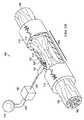
図1A−Bを参照すると、例えば、損傷した神経のような欠陥部を修復するために患者の身体の組織部位に減圧をかける減圧治療システム100が開示されている。損傷した神経は、挟まれ、部分的に断絶または切断されるか、あるいは疾患の結果として部分的に変質している。例えば、図1Aの損傷した神経は、患者の中枢神経系(CNS)に対して近位部104と遠位部106とを有する切断された神経102である。図1Bは減圧治療システムを示し、この場合の損傷した神経は挟まれた神経103であり、神経損傷部位108で損傷している。この場合、神経は挟まれ、部分的に切れているか部分的に変質しているが、完全に切断されてはいない。この神経は神経損傷部位108で分断していても、分断していなくてもよい。本明細書に用いられる用語「神経損傷部位」は、切断または部分的に切断された神経、変質または部分的に変質した神経、および圧迫または挟まれた神経を含むがそれらに限定されない任意の神経組織上または神経組織内に位置する創傷または欠陥部を言う。例えば、減圧組織治療は存在する神経組織の修復または再生を促進したり、移植または移された神経組織および/または細胞の成長を実現したりするのに用いることができる。 With reference to FIGS. 1A-B, a reduced
減圧治療システム100は、神経損傷部位108を示すために神経導管110の一部が取り除かれた神経損傷部位108で挟まれた神経103を囲む神経導管110を含む。神経導管110はほぼ管の形状をしており、神経損傷部位108および損傷していない近位部104と遠位部106の一部を塞ぐ。神経導管110は、外面113と内面112とを具え、内面112は神経損傷部位108の面との神経間隙114、すなわち神経導管110の内面112と神経損傷部位108の面との間の内腔空間を形成する。減圧治療システム100はまた、減圧と、第1の導管125を介して圧力源115に流体連結されたマニホルド120とを提供する減圧源115を含む。マニホルド120はマニホルドチャンバ121内に包含されており、マニホルドチャンバ121は当該マニホルドチャンバ121を神経導管110に固定するためにその一方の端部から延在するフランジ122を有する。マニホルドチャンバ121の他端は第1の導管125に接続されており、これによりマニホルド120と第1の導管125との流体接続が保持される。このマニホルドチャンバ121は、神経間隙114と第1の導管125間のマニホルド120の流体接続を保持するためにほぼ不浸透性で生体適合性の材料で製造してもよい。マニホルドチャンバ121は、マニホルド120が神経損傷部位108の面を囲む神経間隙114と流体連通するが神経間隙114の外側に位置するように、フランジ122により神経導管110に固定される。特定の態様において、フランジ122は接着剤で神経導管110に固定される。さらに、いくつかの応用例では、減圧治療が完了した後にフランジ122とマニホルドチャンバ121を神経導管110から取り除けるように、フランジ122は神経導管110に脱着可能に固定されてもよい。一実施例では、マニホルド120は神経導管110の壁内を通り神経間隙114に直接流体接触する。神経導管110が多孔性である別の実施例では、フランジ122は神経導管110の外面113に固定され、マニホルド120が外面113に近接配置されて神経導管110の多孔壁を介して神経間隙114と流体接続する。 The reduced
マニホルド120は神経損傷の種類や、どのようにマニホルドが神経損傷部位108周辺の神経間隙114と流体接続して配置されるのかによって、様々な形状をとることができる。マニホルド120はまたフランジ122に連結され、これが神経導管113の外面に連結されている。フランジ122は神経導管113の外面とマニホルド120の間に密封を形成して、神経導管110の外側に減圧がかからないようにしている。神経導管の管腔と神経間隙114はまた、組織の成長や修復のための構造を提供する足場材料(図示せず)を含んでもよい。減圧治療システム100はさらに、神経間隙114から出る血液または浸出液のような体液を集める導管125を介して減圧源115とマニホルド120とを流体連結するキャニスタ130を具える。一実施例において、減圧源115とキャニスタ130とは1つの収納構造内に一体形成されている。 The manifold 120 can take a variety of shapes depending on the type of nerve injury and how the manifold is placed in fluid connection with the
本明細書に用いられるように、用語「連結された」は別個の物体を介する直接連結または間接連結を含む。用語「連結された」はまた、同種の材料から形成された要素の各々により互いに連続する2以上の要素を包含する。また、用語「連結された」は化学的、機械的、熱的、あるいは電気的連結を含んでもよい。流体連結は流体が所定の部分または位置で連通していることを意味する。 As used herein, the term “coupled” includes direct or indirect coupling via separate objects. The term “coupled” also encompasses two or more elements that are contiguous with each other by elements formed from the same type of material. The term “coupled” may also include chemical, mechanical, thermal, or electrical coupling. Fluid connection means that fluid is in communication at a predetermined portion or location.
本明細書の内容において、用語「減圧」は一般に、治療が施される組織部位で雰囲気より低い圧力を言う。大抵の場合、この減圧は患者が置かれている位置の雰囲気圧より低くなる。用語「真空」および「負圧」は組織部位にかけられる圧力を記載するのに用いてもよいが、組織部位にかけられる実際の圧力は完全真空と一般に関連する圧力より著しく大きくてもよい。この学術用語と一致して、減圧または真空における増加は絶対圧力の相対的な減少を言い、減圧または負圧における減少は絶対圧力の相対的な増加を言う。用語「−△p」は減圧の変化を意味する。本明細書に用いられるように、より大きな(すなわち、より真空の)「−△p」は増加した負圧(すなわち、雰囲気圧から圧力のより大きな変化)を意味する。減圧治療は一般に−5mmHg乃至−500mmHg、より一般的には−5mmHg乃至−300mmHgで減圧をかけ、−50、−125または−175mmHgを含むがこれらに限定されない。減圧は特定の圧力レベルで一定であってもよいし、時間とともに変化してもよい。例えば、減圧は周期的にかけても、停止してもよく、時間とともに上昇または下降してもよい。 In the context of this specification, the term “reduced pressure” generally refers to a pressure below the atmosphere at the tissue site where the treatment is administered. In most cases, this reduced pressure will be lower than the ambient pressure where the patient is located. Although the terms “vacuum” and “negative pressure” may be used to describe the pressure applied to the tissue site, the actual pressure applied to the tissue site may be significantly greater than the pressure generally associated with a full vacuum. Consistent with this terminology, an increase in reduced pressure or vacuum refers to a relative decrease in absolute pressure, and a decrease in reduced pressure or negative pressure refers to a relative increase in absolute pressure. The term “−Δp” means a change in vacuum. As used herein, larger (ie, more vacuum) “−Δp” means increased negative pressure (ie, greater change in pressure from ambient pressure). Depressurization treatment is typically -5 mmHg to -500 mmHg, more typically -5 mmHg to -300 mmHg, including but not limited to -50, -125 or -175 mmHg. The reduced pressure may be constant at a specific pressure level or may change over time. For example, the reduced pressure may be periodically applied, stopped, or increased or decreased over time.
上述のように神経損傷部位は、図2に示される患者のCNSに対する近位部104と遠位部106とを有する、例えば、完全に切断された神経102を含む任意の神経組織上または神経組織内に配置された創傷または欠陥部であってもよい。切断された神経102は完全に切断された神経損傷部位108で損傷している。神経損傷部位108に減圧をかける他の減圧治療システム200は、減圧治療システム100と同様の要素を含み、同じ参照番号で示されている。この減圧治療システム200は、神経損傷部位108と切断された神経102の切断端部とを囲む神経導管110を含む。神経導管110の内面112は、神経損傷部位108内の切断された神経102の切断端部間、すなわち神経導管110の内面112と神経損傷部位108の表面との間の内腔空間に神経間隙114を形成する。減圧治療システム200はまた、第1の導管125および神経間隙114を介して減圧源115に流体連結されたマニホルド220を含む。マニホルド220はまた、フランジ222を有するマニホルドチャンバ221に部分的に収められており、これがマニホルドチャンバ221を神経導管110に固定するか、そうでなくてもマニホルドチャンバ121と同様にする。マニホルド120と異なり、マニホルド220は、神経間隙114内に延在するマニホルド凸部225を具える。このマニホルド220とマニホルド凸部225は、神経損傷の種類や、神経損傷部位108周辺の神経間隙114とどのようにマニホルドが配置され流体接触するかによって、様々な形状とすることができる。フランジ222は、神経導管113の外面とマニホルドチャンバ221との間を密閉し、減圧が神経導管110の外側にかからないようにしている。特定の態様において、フランジ222は接着剤で神経導管110に固定される。さらに、いくつかの応用例では、減圧治療が完了した後にフランジ222とマニホルドチャンバ221を取り除けるように、フランジ222は神経導管110に脱着可能に固定されてもよい。マニホルド凸部225は、当該マニホルド凸部が組織成長および修復のための構造体を提供する足場材料として作用するような材料で形成してもよい。減圧治療システム200はさらに、神経間隙114から流出した血液や浸出液などの体液を回収するために、減圧源115とマニホルド220の間に導管125を介して流体連結されたキャニスタ130を具えてもよい。一実施例では、減圧源115とキャニスタ130は単一ハウジング構造に一体化される。 As described above, the nerve injury site may be on any neural tissue or nerve tissue including, for example, a fully cut
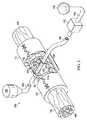
図3に、神経損傷部位108に減圧をかけるさらなる別の減圧治療システム300が示されている。完全に切断された神経102が、CNSに対して近位セグメント104および遠位セグメント106を有して示されている。この切断された神経102は神経損傷部位108にて損傷を受けており、完全に切断されている。減圧治療システム300は、神経損傷部位108および切断された神経102の切断端部を囲む神経導管110を具える。神経導管110の内面112は、神経損傷部位108内の切断神経102の切断端部間、すなわち神経導管110の内面112と神経損傷部位108の面との間の内腔空間に、神経間隙114を形成する。減圧治療システム300はまた、第1の導管125を介して減圧源115に、および神経間隙114に流体連結されたマニホルド320を具える。このマニホルド320はまた、それぞれマニホルドチャンバ321を神経導管110に固定するか、そうでなくてもマニホルドチャンバ121と221と同様のフランジ322を具えるマニホルドチャンバ321に部分的に収まっている。マニホルド220と異なり、マニホルド320は神経間隙114内を通って延在し、マニホルドチャンバ321とフランジ322により神経導管110の両側に固定される。マニホルド320は、神経損傷の種類や、どのようにマニホルドが神経損傷部位108周辺の神経間隙114と流体接続して配置されるのかによって、様々な形状をとることができる。フランジ322は、神経導管113の外面とマニホルドチャンバ321の間に密封を形成して、神経導管110の外側に減圧がかからないようにしている。特定の態様において、フランジ322は神経導管110に接着剤で固定される。いくつかの応用例では、減圧治療が完了した後にフランジ322とマニホルドチャンバ321を神経導管110から取り外せるように、フランジ322の一方または双方が神経導管110に脱着可能に固定される。マニホルド320は、当該マニホルドが組織の成長や修復のための構造体を提供する足場材料として機能するような材料で形成されてもよい。減圧治療システム300はさらに、神経間隙114から流出した血液や浸出液などの体液を回収するために、減圧源115とマニホルド320の間に導管125を介して流体連結されたキャニスタ130を具えてもよい。一実施例では、減圧源115とキャニスタ130は単一ハウジング構造に一体化される。減圧治療システム300はさらに、マニホルド320に第2の導管325を介して流体連結された流体源150を具えてもよい。したがって、特定の態様において、流体源150からマニホルド320および神経間隙114に流体が流れ、最終的にキャニスタ130に回収される。マニホルドを通る流体流は神経損傷部位108を横断して、マニホルドの詰まりを回避する補助となる。 FIG. 3 illustrates yet another reduced
上述のように、神経導管110は、一部が取り除かれて図1−3に示されているが、閉じた神経導管410として、神経損傷部位108を完全に囲むよう図4に示されている。マニホルド120、220、320が神経間隙114に挿入されるか、神経間隙114に近い神経導管110に取り付けられた後に、神経導管110は従来技術では周知の1以上の縫い目415または任意の他の締結装置を用いて密閉されてもよい。神経導管110は生体吸収性材料または生体不活性材料から形成されてもよい。神経導管に用いることができる材料は、ポリ乳酸(PLA)、ポリグリコール酸(PGA)、ポリラクチド−コ−グリコリド(PLGA)、ポリビニルピロリドン、ポリカプロラクトン、ポリカーボネート、ポリフマレート、カプロラクトン、ポリアミド、多糖類(アルギン酸塩(例えば、アルギン酸カルシウム)およびキトサンを含む)、ヒアルロン酸、ポリヒドロキシ酪酸、ポリヒドロキシバレレート、ポリジオキサノン、ポリオルソエステル、ポリエチレングリコール、ポロキサマ、ポリホスファゼン、ポリ酸無水物、ポリアミノ酸、ポリアセタール、ポリシアノアクリレート、ポリウレタン、ポリアクリル酸塩、エチレン酢酸ビニルポリマ、および他のアシル置換酢酸セルロース、およびその誘導体、ポリスチレン、ポリ塩化ビニル、ポリふっ化ビニル、ポリ(ビニルイミダゾール)、クロロスルホン化ポリオレフィン、ポリエチレンオキシド、ポリビニルアルコール、テフロン(登録商標)、およびナイロンを含むがこれらに限定されない。ある態様において、生物学的(例えば、精製した、または組み換え型の)材料は、フィブリン、フィブロネクチンまたはコラーゲン(例えば、DURAMATRIX(商標))を含むがこれらに限定されない神経導管を形成するのに用いられてもよい。 As described above,
神経導管110は、図3に示されているような近位の神経断端と遠位の神経断端との間の間隙を横切って適合した、ほぼ管状の完全な構造であってもよい。このようなほぼ管状の神経導管の例は神経ガイドとも呼ばれ、NEURAGEN(登録商標)およびNEUROFLEX(商標)コラーゲン導管を含むがこれらに限定されない。神経導管はまた、切断または損傷した(例えば、挟まれた)神経周りに配置されたラップから形成され、かつ縫合線のようなクロージャで密閉されてもよい。ラップ型の神経導管の具体例は、NEUROMEND(商標)およびNEURA WRAP(商標)コラーゲン導管を含むがこれらに限定されない。ある態様において、神経導管は損傷した組織を包む材料からできており、神経膠のような非神経細胞の浸潤を排除する。いくつかの実施例において、神経導管の材料は透過性であり、これにより流体およびタンパク因子が導管を通って拡散可能となる。例えば、神経導管の細孔は、導管内腔(例えば、約5μm乃至50μm、10μm乃至30μm、あるいは10μm乃至20μmの内径または平均内径を有する細孔)内に細胞が入るのを防ぐよう十分小さくてもよい。このように減圧が導管内にかけられると、流体およびタンパク質は勾配圧力により導管の内腔に引き入れられてもよい。導管の寸法が任意の具体的な神経の適用に調整されうることは当業者に理解されよう。一般に導管は約5、4、3、2.5または2mmといった約6.0mm以下の内径を含む。 The
図5を参照すると、減圧治療システム100、200、または300はさらに、第1の導管125に動作可能に接続され、マニホルド120、220、320にかけられる減圧を測る圧力センサ140を含む。このシステムはさらに、圧力センサ140と減圧源115とに電気的に接続された制御装置145含む。圧力センサ140は神経間隙114内の減圧を測り、第1の導管125が血液または他の体液で塞がれているかを示すことができる。圧力センサ140はまた、減圧源115により第1の導管125を通りマニホルド120、220、320に適用される減圧治療を調整する制御装置145にフィードバックを提供する。減圧治療システム100、200、または300はまた、第2の導管152を介して第1の導管125に流体連結され、制御装置145に動作可能に接続される流体供給源150を含む。流体供給源150は、抗菌剤、抗ウィルス剤、抗体、細胞増殖促進剤、灌流液、または他の化学的活性剤を含むがこれらに限定されない成長および/または治療剤を神経損傷部位108に送達するよう用いることができる。システム100、200、300はさらに第2の導管152に配置され、そこを通る流体の流れを制御する第1の弁154と、減圧供給源115と第1の導管125と第2の導管152との連結点との間の第1の導管125に配置され、減圧の流れを制御する第2の弁156とを含む。減圧治療システム300の場合、流体供給源150は、点線で示すように第2の導管325を介して神経組織損傷部位108でマニホルド320に直接連結される。制御装置145は、患者に施される特定療法に必要とされる場合に、第1、第2の弁154、156に動作可能に接続され、流体供給源150からマニホルド120、220、320各々への減圧および/または流体の送達を制御する。流体供給源150は上述のように液体を送達してもよいが、神経損傷部位108のその場での治癒を促進し、排出を実現するためにマニホルド120、220、320に空気を送達してもよい。 Referring to FIG. 5, the reduced
本明細書に用いられるように、用語「マニホルド」は減圧を組織部位に導いたり、流体を組織部位に送達したり、流体を組織部位から取り除いたりする際の助けとなる物質または構造を言う。マニホルドはマニホルド周辺組織の領域へ提供されたり、そこから取り除かれたりする流体の分配を向上させるために内部接続された複数の流れ経路または経路を含みうる。マニホルドの例は、流路、連続気泡発泡体のような細胞状発泡体、多孔性組織の集合体、および、流路を含んだり、含むよう硬化(cure)する液体、ゲルおよび発泡体を形成するよう配置された構造要素を有する装置を含むがこれらに限定されない。本発明によるマニホルドの詳細な記載およびそれら利用は以下に提供される。マニホルドが神経間隙114内へと延在するか、神経間隙114を通って延在する場合、以下に詳述する生体吸収性のマニホルド材料を用いてもよい。 As used herein, the term “manifold” refers to a substance or structure that assists in directing reduced pressure to a tissue site, delivering fluid to the tissue site, or removing fluid from the tissue site. The manifold may include a plurality of flow paths or paths interconnected to improve the distribution of fluid that is provided to or removed from the area surrounding the manifold. Examples of manifolds form channels, cellular foams such as open cell foams, aggregates of porous tissue, and liquids, gels and foams that contain or cure to contain channels Including but not limited to devices having structural elements arranged to do so. A detailed description of the manifolds according to the invention and their use is provided below. If the manifold extends into or through the
本明細書に用いられる用語「足場材料」は、細胞の成長および/または組織の形成のための構造マトリクスを提供する創傷または欠陥部に適用される物質または構造を言う。足場材料は三次元の多孔性構造であることが多い。足場材料は細胞成長を促進させる細胞、成長因子、細胞外マトリクスの成分、栄養素、インテグリン、または他の物質に注入されたり、それらで覆われたり、それらから形成されうる。足場材料はマトリクスを通る流れを導くことによりマニホルドの特徴を有するようになる。マニホルドは流れを足場材料や組織に伝達し、減圧治療のコンテクストにおいて、マニホルドは足場材料と流体連通している。本発明による足場材料の詳細な説明およびそれらの使用を以下に述べる。 The term “scaffold material” as used herein refers to a substance or structure applied to a wound or defect that provides a structural matrix for cell growth and / or tissue formation. Scaffold materials are often three-dimensional porous structures. The scaffold material can be injected into, covered with, or formed from cells, growth factors, extracellular matrix components, nutrients, integrins, or other substances that promote cell growth. The scaffold material becomes manifold features by directing flow through the matrix. The manifold communicates flow to the scaffold material and tissue, and in a reduced pressure treatment context, the manifold is in fluid communication with the scaffold material. A detailed description of the scaffold materials according to the present invention and their use is described below.
このようなものとして、本明細書に開示された発明は、微細、ナノスケール、またはメゾスコピックスケールでパターン化したタンパク質構成の制御を可能とする流体の流れの細胞レベルに基づくパターンを制御する方法および装置を開示し、細胞移動、細胞分化、および組織の機能的な再生に必要な作用のための構造化マニホルド、選択的に、足場材料の提供に従うものである。組織の修復および再生に関する従来技術の現状の消極的な性質と比べて、本明細書に開示された方法、足場材料、マニホルド、流れ源、およびシステムは積極的な構造を提供し、これによりタンパク質の内因性沈着と生化学的および物理学的手掛かりを有する暫定的マトリクスの編成とを促進し、足場材料または組織空間の細胞定着に導く。このように本発明は、導かれた流体の流れの活性力を有効に用いて、流体の影響下で生物学の必要性に基づいたマニホルドと足場材料とが設計された構造を提供することにより近年の技術を向上させる。流れベクトルと経路とはタンパク質の沈着と細胞の定着を促進するよう用いられる。本明細書に提供されるシステムは、機能的な組織の連続体を促進する足場材料または組織部位を通して、健康な組織端部から継ぎ目ない移行により暫定的マトリクスのネットワークの構築を促進するよう設計されている。 As such, the invention disclosed herein controls a pattern based on the cellular level of fluid flow that allows control of protein composition patterned on a fine, nanoscale, or mesoscopic scale, and The device is disclosed and follows the provision of a structured manifold, optionally a scaffold material, for the actions necessary for cell migration, cell differentiation, and functional regeneration of tissue. Compared to the current negative nature of the prior art with respect to tissue repair and regeneration, the methods, scaffold materials, manifolds, flow sources, and systems disclosed herein provide an aggressive structure and thereby protein Promotes the endogenous deposition of and the provisional matrix organization with biochemical and physical cues, leading to cell colonization of the scaffold material or tissue space. Thus, the present invention provides a structure in which manifolds and scaffolding materials are designed based on biological needs under the influence of fluids by effectively using the active force of the guided fluid flow. Improve recent technology. Flow vectors and pathways are used to promote protein deposition and cell colonization. The system provided herein is designed to facilitate the construction of a temporary matrix network through a seamless transition from a healthy tissue edge through a scaffold material or tissue site that promotes a functional tissue continuum. ing.
このように、本明細書に開示された装置、方法およびシステムは、機能の回復を促進するために移植された足場材料を通して、あるいは組織部位内で組織の再生を積極的に誘導する手段を提供する。この積極的な誘導は制御された流体の流れのメカニズムを通して起こり、この流れはそれ自体が自然な治癒プロセスの初期段階を開始させたり、増進させたりするように用いることができ、マニホルドは制御された流体の流れを作るのに必要な積極的な誘導を提供できる。特に、マニホルドが提供する制御された流れベクトルは、細胞やタンパク質の有向の流入を実現するよう用いることができる。組織部位や足場材料内に特定の流れの経路を作ることは、マニホルド、足場材料または組織空間内のコラーゲンやフィブリンのようなタンパク質のパターン化した沈着に導くことができる。暫定的マトリクス内に固定されたサイトカイン、成長因子、および細胞からの生物学的手掛かりは、暫定的マトリクスや細胞外マトリクスの物理学的手掛かりとともに作用し、治療の修復段階中、内因性細胞の続く移動をガイドする。これらの手掛かりは周囲の健康な組織から広がる痕跡または勾配として作用し、足場材料または組織空間を通り、編成された組織の再生のための連続したガイド経路を実現する。 Thus, the devices, methods and systems disclosed herein provide a means to actively induce tissue regeneration through implanted scaffold material or within tissue sites to facilitate functional recovery. To do. This positive induction occurs through a controlled fluid flow mechanism, which can itself be used to initiate or enhance the initial stages of the natural healing process, and the manifold is controlled. Can provide the positive guidance necessary to create a fluid flow. In particular, the controlled flow vector provided by the manifold can be used to achieve directed inflow of cells and proteins. Creating a specific flow path within a tissue site or scaffold material can lead to patterned deposition of a manifold, a scaffold material or a protein such as collagen or fibrin in the tissue space. Biological cues from cytokines, growth factors, and cells anchored in the temporary matrix act in conjunction with physical cues in the temporary and extracellular matrix, followed by endogenous cells during the repair phase of therapy Guide the movement. These cues act as traces or gradients extending from the surrounding healthy tissue and provide a continuous guide path for regeneration of the organized tissue through the scaffold material or tissue space.
このため、本開示は流体および勾配の流れの原理に基づいた明確な生物学的必要性のために設計された独自のマニホルド技術を提供する。ある態様において、発明は創傷治癒、流れ(または勾配)が活性化された再生医療への新しいアプローチに関する。基本的な形態において、このアプローチは、続く方向付けられた暫定的マトリクスの形成とともに、タンパク質の組織化された沈着および/またはサイトカインの空間的集合および成長因子のための組織空間内、外、またはそこを通る内因性または外因性流体の何れかの制御された動きのために勾配を形成する流れの源または発生器を含む。本明細書に規定された組織空間は、限定ではないが、神経組織の損傷部位を囲む領域を含む。 Thus, the present disclosure provides a unique manifold technology designed for a clear biological need based on fluid and gradient flow principles. In certain embodiments, the invention relates to a new approach to regenerative medicine in which wound healing, flow (or gradient) is activated. In its basic form, this approach can be followed by the formation of a directed tentative matrix, as well as organized deposition of proteins and / or spatial assembly of cytokines and tissue space for growth factors, outside, or It includes a flow source or generator that forms a gradient for controlled movement of either endogenous or exogenous fluid therethrough. The tissue space defined herein includes, but is not limited to, an area surrounding a nerve tissue injury site.
神経組織空間内、そこを通る、またはその外への流体の流れは、マニホルドおよび/または足場材料を含むシステムの追加の要素の包含を通して精密化され、提供される。システムの調整された要素は、タンパク質の制御された吸着、マトリクスの編成、および特定の細胞種の組織化された定着に影響を与え、提供できる寸法において、十分詳細に流れパラメータ、経路、およびパターンを生成するよう設計されている。システムの各要素は以下の通りである。 Fluid flow in, through or out of the neural tissue space is refined and provided through the inclusion of additional elements of the system including manifold and / or scaffold material. The tuned elements of the system affect the controlled adsorption of proteins, the organization of the matrix, and the organized colonization of specific cell types, with sufficiently detailed flow parameters, pathways, and patterns in dimensions that can be provided. Designed to produce Each element of the system is as follows.
流れ源または流れ発生器 流れは、機械的、化学的、および/または電気的潜在性における変化を導入する方法または装置により、神経組織空間内へ、そこを通して、またはその外に誘導される。これらの流れ発生器は、内因性または外因性流体の部位または貯留層から、流れ発生器またはその拡張要素(すなわち、マニホルドまたは足場材料)の設置位置への潜在性における勾配または変化の何れかを提供する。一実施例において、流れ源は減圧源を含む。本願によるシステムおよび装置はまた、マニホルドにかけられる負圧の適用および量を制御する弁または弁の配列を含んでもよい。ある態様において、本明細書に記載された神経導管および/またはマニホルドは圧力センサを含む。このように、いくつかの実施例において、源によりかけられた負圧の量は、マニホルドまたは神経導管、または組織の損傷部位で感知される負圧の量に基づいて制御される。The flow source or flow generator flow is directed into, through, or out of the neural tissue space by a method or device that introduces changes in mechanical, chemical, and / or electrical potential. These flow generators are either gradients or changes in the potential from the site or reservoir of endogenous or exogenous fluid to the location of the flow generator or its expansion element (ie manifold or scaffold material). provide. In one embodiment, the flow source includes a reduced pressure source. The system and apparatus according to the present application may also include a valve or valve arrangement that controls the application and amount of negative pressure applied to the manifold. In certain embodiments, the neural conduits and / or manifolds described herein include a pressure sensor. Thus, in some embodiments, the amount of negative pressure applied by the source is controlled based on the amount of negative pressure sensed at the manifold or nerve conduit, or at the site of tissue injury.
マニホルド 流れ発生器は流体の流れを促進させる駆動力である。マニホルドは流れ源または流れ発生器と組織空間との間の流れパターンを精密化する装置である。大規模レベルの流れは、マニホルド/足場材料、および最終的には組織空間内の小規模の流れ経路のための初期サイトを形成すべく、1または複数の選択的に配置された点への有向の局在性に用いられた特殊マニホルドにより精密化される。マニホルドはまた、外因性流体から流体を取り除くための導管として設けてもよいし、外因性流体を組織空間へ送達するための装置として設けてもよい。The manifold flow generator is a driving force that promotes fluid flow. The manifold is a device that refines the flow pattern between the flow source or flow generator and the tissue space. A large level of flow is present at one or more selectively placed points to form an initial site for the manifold / scaffold material and ultimately a small flow path within the tissue space. Refined by special manifold used for orientation localization. The manifold may also be provided as a conduit for removing fluid from the exogenous fluid or as a device for delivering the exogenous fluid to the tissue space.
マニホルドは一般に、機械的、化学的、電気的または類似の変更を流体の流れの変化に適用および伝達する際の助けとなる物理的物質または構造を言い、本明細書においては、タンパク質、細胞、および他の部分のような液体、ガス、および他の変形可能な物質の動きとして規定される。したがって、この物理的装置は、圧力、流体および上記規定されるように足場材料における流体の動きを伝達できる物質の出口または排出用の1または複数の点を含む。これは、細胞および/または治療部分のような外因性要因の、マニホルドに存在する内腔または複数の内腔を通り足場材料内への導入を含むが、これらに限定されない。さらに本明細書に用いられるように、マニホルドは流れの点源に戻る足場材料からの流体の侵入または導入のための1または複数の点を含む。 A manifold generally refers to a physical substance or structure that aids in applying and transmitting mechanical, chemical, electrical, or similar changes to fluid flow changes, and as used herein refers to proteins, cells, And movement of liquids, gases, and other deformable materials such as other parts. Thus, the physical device includes one or more points for outlet or discharge of a substance capable of transmitting pressure, fluid and fluid movement in the scaffold material as defined above. This includes, but is not limited to, the introduction of exogenous factors, such as cells and / or therapeutic moieties, into the scaffold material through the lumen or lumens present in the manifold. As further used herein, the manifold includes one or more points for fluid ingress or introduction from the scaffold material back to the flow point source.
マニホルドにより分配された流れは、外因性タンパク質、成長因子、サイトカイン、および細胞を組織化した方法で、ホスト内のそれらの常駐位置から組織空間または足場材料へと移動させることができる。経路に沿ったこれらの流れの確立は、ホストを足場材料に接続する界面の外因性ネットワークを作るタンパク質の沈着および暫定的マトリクスに導く。このマトリクスの拡大は、足場材料の設計を促進させる流れとともにマニホルドの流れの初期部位の選択的な配置を通して足場材料内に確立することができる。組織化されたタンパク質の沈着と暫定的マトリクスは、足場材料および組織空間を通して導かれた経路に沿って細胞の付着および移動を促進させる生化学的および物理的な枠組みを提供する。タンパク質、成長因子、および細胞の結果として生じた外因性ネットワークは、身体組織修復の続く段階、および再生メカニズムが構築される際の基礎を提供する。 The flow distributed by the manifold can be transferred from their resident location within the host to the tissue space or scaffold material in an organized manner with exogenous proteins, growth factors, cytokines, and cells. The establishment of these flows along the pathway leads to protein deposition and a provisional matrix that creates an exogenous network of interfaces that connect the host to the scaffold material. This matrix expansion can be established in the scaffold material through selective placement of the initial portion of the manifold flow along with a flow that facilitates the design of the scaffold material. Organized protein deposition and provisional matrices provide a biochemical and physical framework that promotes cell attachment and migration along pathways guided through the scaffold material and tissue space. The resulting exogenous network of proteins, growth factors, and cells provides the basis for the subsequent stages of body tissue repair and the mechanism by which regeneration is built.
マニホルドは配置されると、存在するなら、流れ発生源と足場材料とともに作用する。流れ発生源は、負圧の発生源、陽圧の発生源、および浸透流の発生源を含むがこれらに限定されない。マニホルドに確立された流れ勾配は、足場材料を通りさらに精密化され、流れ勾配を足場材料に送達して、特定の欠陥部に必要とされる足場材料を通して流れを最適化する。本明細書に開示された実施例の多くは、組織を再生に導くために物理的足場材料を通して選択的に圧力の変化を流体の制御された動きに伝達することができるマニホルドである。これらの実施例は一般に、特定の組織の再生における具体的な適用に特定されるが特定の組織に限定されない。 Once in place, the manifold works with the flow source and the scaffold material, if present. Flow sources include, but are not limited to, negative pressure sources, positive pressure sources, and osmotic flow sources. The flow gradient established in the manifold is further refined through the scaffold material, delivering the flow gradient to the scaffold material and optimizing the flow through the scaffold material required for the particular defect. Many of the embodiments disclosed herein are manifolds that can selectively transmit pressure changes to a controlled movement of fluid through a physical scaffold material to direct tissue to regeneration. These examples are generally specified for specific applications in the regeneration of specific tissues, but are not limited to specific tissues.
組織再生のために流れを誘発する目的を実現するために、前述の機械的、化学的または電気的な機動力は、1の勾配源から物理的な基質または足場材料に伝達されるべきであり、タンパク質沈着、マトリクス組織、細胞移動、および他の組織の再生関連の行為において細胞レベルの変化を誘発する。これらの変更は本質的に多変量であり、創傷部位または組織再生の所望の部位に適用されると、足場材料にかけられた圧力の物理的変化を誘発する機械的変化、タンパク質および/またはイオン濃度の勾配を誘発し、流れを誘発できる浸透勾配を生み出す化学的変化、あるいは点源からの電気信号の伝播を可能にする電流/イオン交換の勾配を生み出す電気的変化を含むことができる。しかしながら、適用者は勾配および流体の流れを通して組織の修復または成長をもたらす利益を誘発する任意の特定のメカニズムに縛られないと解すべきである。これらの勾配を組織に好適に伝達するために、物理的装置は流れの経路をその源から足場材料または組織部位、逆もまた同様、に導くことが必要とされる。 In order to achieve the purpose of inducing flow for tissue regeneration, the aforementioned mechanical, chemical or electrical mobility should be transferred from one gradient source to a physical substrate or scaffold material Induces changes at the cellular level in protein deposition, matrix tissue, cell migration, and other tissue regeneration-related actions. These changes are multivariate in nature and, when applied to the wound site or desired site of tissue regeneration, mechanical changes, protein and / or ion concentrations that induce a physical change in the pressure applied to the scaffold material A chemical change that creates a gradient of osmotic flow that can induce flow, or an electrical change that produces a current / ion exchange gradient that allows the propagation of electrical signals from a point source. However, it should be understood that the applicator is not bound by any particular mechanism that induces the benefits of tissue repair or growth through gradients and fluid flow. In order to suitably communicate these gradients to the tissue, the physical device is required to guide the flow path from its source to the scaffold material or tissue site, and vice versa.
いくつかの実施例において、マニホルドは足場材料の内容とほぼ同格または内容内の物理的構造を含み、機械的、化学的、電気的、または本質的に同様のものであろうとなかろうと、その点源から足場材料までこれらの変化を導く手段のために物理的パラメータの変更を伝播するよう作用する。足場材料に対するこのマニホルドの配置は、制御され、方向付けられた特定の組織種の再生を実現するのに決定的に重要となりうる。例えば、近位から遠位の神経残根まで最初に再生が無方向で生じる周辺神経では、より遠位端部に向かった神経導管の長さに沿ってマニホルドを配置し、その端部に向かって再生を導くのを助けることは重要である。しかしながら、その源に向かって神経再生に導くには、遠位の幹から派生する可溶性因子が重要であるよう示されてきたので、足場材料/導管の最も遠位の部分にマニホルドを配置しないこともまた重要である。 In some embodiments, the manifold includes a physical structure that is approximately equivalent to or within the content of the scaffold material, whether mechanical, chemical, electrical, or essentially similar. It acts to propagate changes in physical parameters because of the means to guide these changes from source to scaffold material. The placement of this manifold relative to the scaffold material can be critical in achieving controlled and directed regeneration of specific tissue types. For example, in a peripheral nerve where regeneration is initially undirected from the proximal to the distal nerve root, a manifold is placed along the length of the nerve conduit toward the distal end and toward that end. It is important to help guide regeneration. However, do not place a manifold in the most distal part of the scaffold material / conduit as soluble factors derived from the distal stem have been shown to be important to direct nerve regeneration towards its source Is also important.
マニホルドは生体吸収性材料または生体不活性材料から形成されてもよい。例は、医用グレードシリコーンポリマ、メタル、ポリ塩化ビニル(PVC)、およびポリウレタンのような非生体吸収性材料を含むが、これらに限定されない。コラーゲン、ポリ乳酸(PLA)、ポリグリコール酸(PGA)、ポリラクチド−コ−グリコリド(PLGA)、多糖類、ヒドロキシアパタイト、またはポリエチレングリコール、またはそれらの組み合わせのような生体吸収性ポリマを用いてもよい。いくつかのマニホルドがまた、非生体吸収性材料および生体吸収性材料の混合である。足場材料に用いられる一般的な材料はまた、マニホルドを構成するよう用いられてもよく、このような材料がさらに以下に詳細に述べられる。ある態様において、マニホルド材料は改良された生体吸収性のための高空隙の留分を含むよう構成されている。 The manifold may be formed from a bioabsorbable material or a bioinert material. Examples include, but are not limited to, non-bioabsorbable materials such as medical grade silicone polymers, metals, polyvinyl chloride (PVC), and polyurethane. Bioabsorbable polymers such as collagen, polylactic acid (PLA), polyglycolic acid (PGA), polylactide-co-glycolide (PLGA), polysaccharides, hydroxyapatite, or polyethylene glycol, or combinations thereof may be used. . Some manifolds are also a mixture of non-bioabsorbable and bioabsorbable materials. Common materials used for scaffold materials may also be used to construct the manifold, and such materials are described in further detail below. In certain embodiments, the manifold material is configured to include a high void fraction for improved bioabsorbability.
支持部 マニホルド支持構造は、許容される任意の生体適合材料で構成してもよい。支持構造は通常、不浸透性で、マニホルドを取り囲んでマニホルドの圧力を維持する。The support manifold support structure may be composed of any acceptable biocompatible material. The support structure is typically impermeable and surrounds the manifold to maintain manifold pressure.
フランジなどの支持部の一部は、マニホルドと神経導管を連結する。特定の態様において、フィブリン接着剤、シアノアクリレート、または他の生体由来の接着剤といった接着剤で、フランジが神経導管の外面に取り付けられる。支持部も、、化学的、熱的、浸透性、機械的(スナップ嵌めまたは他の締まり嵌め、螺合等)、磁気的、および静電機構などの接着剤以外の可逆的機構を介して、神経導管に連結されてもよい。このマニホルドは、(例えば治療完了後に)支持部を神経導管から脱着するために、結合機構の動作を逆にする薬剤を送達するのに用いられてもよい。例えば、静電結合は食塩水を導入すると解除され、接着剤を剥がすのに生体適合性の溶剤を用いてもよい。 A portion of the support, such as a flange, connects the manifold and the nerve conduit. In certain embodiments, the flange is attached to the outer surface of the nerve conduit with an adhesive, such as a fibrin adhesive, cyanoacrylate, or other biologically derived adhesive. The support can also be through reversible mechanisms other than adhesives, such as chemical, thermal, permeable, mechanical (snap or other interference fit, threaded, etc.), magnetic, and electrostatic mechanisms. It may be connected to a nerve conduit. This manifold may be used to deliver an agent that reverses the operation of the coupling mechanism to detach the support from the nerve conduit (eg, after treatment is complete). For example, electrostatic coupling is released when saline is introduced, and a biocompatible solvent may be used to remove the adhesive.
足場材料 生物学的および合成の足場材料はタンパク質沈着および組織の修復や再生のための細胞の内生をサポートする再生医療の分野に用いられる。足場材料工学における技術の現在の状況は、タンパク質沈着および細胞移動のための組織空間周辺の固有の特徴に依存している。発明に用いられる足場材料はマニホルドに連結され、組織部位に流体の流れの経路を導く物理的ガイダンスを提供し、接着性タンパク質および細胞のための経路を作り、これは組織空間内の組織の所定のパターンにおける暫定的マトリクスの確立に不可欠である。流体の流れの誘発が、勾配の誘発による組織の再生のための記載の方法および装置は、足場材料の設計への直接的な包含を有する。このコンテクスト内で、足場材料は流体源からマニホルド内の流れ開始点まで細胞レベルのパターンに組織空間内で流体の流れの経路を精密化するよう設ける。足場材料は組織部位内の流れ経路を精密化するためにマニホルドと関連して組み合わせられたマニホルドの性質を具体化する。ある態様において、足場材料は網状構造であり、改良された生体吸収性能のための高空隙分留を含む。Scaffold Materials Biological and synthetic scaffold materials are used in the field of regenerative medicine to support cell deposition for protein deposition and tissue repair and regeneration. The current state of the art in scaffold material engineering relies on unique features around the tissue space for protein deposition and cell migration. The scaffold material used in the invention is connected to a manifold and provides physical guidance to guide fluid flow pathways to the tissue site, creating pathways for adhesive proteins and cells, which are pre-determined of the tissue within the tissue space. It is essential to establish a temporary matrix in the pattern of The described method and apparatus for inducing fluid flow, tissue regeneration by inducing gradients, has a direct inclusion in the design of the scaffold material. Within this context, the scaffold material is provided to refine the fluid flow path in the tissue space in a cellular level pattern from the fluid source to the flow start point in the manifold. The scaffold material embodies the properties of the manifold combined with the manifold to refine the flow path within the tissue site. In certain embodiments, the scaffold material is a network structure and includes high void fractionation for improved bioabsorbable performance.
好適な足場材料の非限定的な例は、フィブリン、コラーゲンまたはフィブロネクチン、および合成または天然由来ポリマのような外因性マトリクスタンパク質を含み、ポリ乳酸(PLA)、ポリグリコール酸(PGA)、ポリラクチド−コ−グリコリド(PLGA)、ポリビニルピロリドン、ポリカプロラクトン、ポリカーボネート、ポリフマレート、カプロラクトン、ポリアミド、多糖類(アルギナート(例えば、カルシウムアルギナート)およびキトサンを含む)、ヒアルロン酸、ポリヒドロキシブチレート、ポリヒドロキシバレート、ポリジオキサノン、ポリオルソエステル、ポリエチレングリコール、ポロキサマ、ポリホスファゼン、ポリ酸無水物、ポリアミノ酸、ポリアセタール、ポリシアノアクリレート、ポリウレタン、ポリアクリル酸塩、エチレン酢酸ビニルポリマ、および他のアシル置換酢酸セルロース、およびその誘導体、ポリスチレン、ポリ塩化ビニル、ポリふっ化ビニル、ポリ(ビニルイミダゾール)、クロロスルホン化ポリオレフィン、ポリエチレンオキシド、ポリビニルアルコール、テフロン(登録商標)、およびナイロンのような生体吸収性材料または非吸収性材料を含む。足場材料はまた、ヒドロキシアパタイト、珊瑚アパタイト、リン酸カルシウム、硫酸カルシウム、炭酸カルシウム、または他の炭酸塩、バイオガラス、同種移植片、異種移植片、脱細胞化した組織、または上記の任意の合成物のようなセラミックを含んでもよい。具体的な実施例において、足場材料はコラーゲン、ポリ乳酸(PLA)、ポリグリコール酸(PGA)、ポリラクチド−コ−グリコリド(PLGA)、ポリウレタン、多糖類、ヒドロキシアパタイト、またはポリエチレングリコールを含む。さらに、足場材料は、足場材料の別個の領域あるいは複数の領域において、非共有結合または共有結合(例えば、ポリエチレンオキシド−ポリエチレングリコールブロックポリマ、またはターポリマのようなコポリマ)、またはそれらの組み合わせの何れかにおいて、任意の2、3以上の材料の組み合わせを含むことができる。好適なマトリクス材料は、例えば、Ma and Elisseeff, 2005, Salzman, 2004で議論されている。 Non-limiting examples of suitable scaffold materials include exogenous matrix proteins such as fibrin, collagen or fibronectin, and synthetic or naturally derived polymers, including polylactic acid (PLA), polyglycolic acid (PGA), polylactide-co -Glycolide (PLGA), polyvinylpyrrolidone, polycaprolactone, polycarbonate, polyfumarate, caprolactone, polyamide, polysaccharides (including alginate (eg calcium alginate) and chitosan), hyaluronic acid, polyhydroxybutyrate, polyhydroxyvalate, polydioxanone , Polyorthoesters, polyethylene glycol, poloxamers, polyphosphazenes, polyanhydrides, polyamino acids, polyacetals, polycyanoacrylates, polyurethanes, polyacrylates Rilates, ethylene vinyl acetate polymers, and other acyl-substituted cellulose acetates and their derivatives, polystyrene, polyvinyl chloride, polyvinyl fluoride, poly (vinyl imidazole), chlorosulfonated polyolefins, polyethylene oxide, polyvinyl alcohol, Teflon ( Registered trademark), and bioabsorbable or non-absorbable materials such as nylon. The scaffold material can also be hydroxyapatite, cocoon apatite, calcium phosphate, calcium sulfate, calcium carbonate, or other carbonates, bioglass, allograft, xenograft, decellularized tissue, or any composite of the above Such ceramics may be included. In specific examples, the scaffold material comprises collagen, polylactic acid (PLA), polyglycolic acid (PGA), polylactide-co-glycolide (PLGA), polyurethane, polysaccharide, hydroxyapatite, or polyethylene glycol. Further, the scaffold material can be either non-covalent or covalent (eg, a polyethylene oxide-polyethylene glycol block polymer, or a copolymer such as a terpolymer), or a combination thereof, in discrete regions or regions of the scaffold material. In any combination of two, three or more materials. Suitable matrix materials are discussed, for example, in Ma and Elisseeff, 2005, Salzman, 2004.
生物活性剤 ある態様において、本願による装置および方法は生物活性剤に関する。生物活性剤は、いくつかのケースにおいて、マニホルドまたは足場材料に直接組み込まれ(すなわち、生物活性マニホルドおよび/足場材料を生成する)。すなわち、コラーゲンまたはフィブリンのような組織を促進する薬剤が、マニホルドまたは足場材料上またはその中に直接組み込まれてもよい。同様に、異常な免疫反応を避ける必要がある適用例では、ラパマイシンのような(例えば、組織移植片)免疫調整剤がマニホルドまたは足場材料内に組み込まれてもよい。Bioactive agents In certain embodiments, the devices and methods according to the present application relate to bioactive agents. The bioactive agent is, in some cases, directly incorporated into the manifold or scaffold material (ie, producing a bioactive manifold and / or scaffold material). That is, agents that promote tissue such as collagen or fibrin may be incorporated directly onto or into the manifold or scaffold material. Similarly, in applications where an abnormal immune response needs to be avoided, immunomodulators such as rapamycin (eg, tissue graft) may be incorporated into the manifold or scaffold material.
さらなる態様において、可溶性生物活性剤は組織部位を通る流れによって、組織の損傷部位に導入可能である。例えば、マニホルドは流体源と流体連通してもよく、生体活性剤は流体源に導入されてもよく、これによりマニホルドおよび損傷した神経組織内に導入することができる。 In a further embodiment, the soluble bioactive agent can be introduced into the tissue injury site by flow through the tissue site. For example, the manifold may be in fluid communication with a fluid source and the bioactive agent may be introduced into the fluid source, thereby allowing it to be introduced into the manifold and damaged neural tissue.
様々なアプリケーション用の生物活性成長因子の非限定的な例は、成長ホルモン(GH)、骨形成タンパク質(BMP)、トランスフォーミング増殖因子−α(TGF−α)、TGF−β、繊維芽細胞(FGF)、顆粒球コロニー刺激因子(G−CSF)、顆粒球/マクロファージコロニー刺激因子(GM−CSF)、上皮細胞増殖因子(EGF)、血小板由来増殖因子(PDGF)、インスリン様成長因子(IGF)、血管内皮増殖因子(VEGF)、肝細胞増殖因子/細胞分散因子(HGF/SF)、インターロイキン、腫瘍壊死因子−α(TNF−α)または神経成長因子(NGF)である。 Non-limiting examples of bioactive growth factors for various applications include growth hormone (GH), bone morphogenetic protein (BMP), transforming growth factor-α (TGF-α), TGF-β, fibroblasts ( FGF), granulocyte colony stimulating factor (G-CSF), granulocyte / macrophage colony stimulating factor (GM-CSF), epidermal growth factor (EGF), platelet derived growth factor (PDGF), insulin-like growth factor (IGF) Vascular endothelial growth factor (VEGF), hepatocyte growth factor / cell dispersion factor (HGF / SF), interleukin, tumor necrosis factor-α (TNF-α) or nerve growth factor (NGF).
神経組織修復および再生工学 本明細書に記載された装置とシステムは、以下のものを含む様々なコンテクストにおいて、神経組織修復および工学に用いることができる。Neural tissue repair and regeneration engineering The devices and systems described herein can be used for neural tissue repair and engineering in a variety of contexts, including:
損傷組織の修復と再生 流れ発生器は、損傷した部位の損傷組織の再生または傷ついた機能に対応すべくマニホルドおよび/または足場材料と組み合わされてもよい。直接再生に導く。外因性の傷、手術、火傷、または他の原因(例えば、伝染または自己免疫性疾患)からの組織の損傷は、発明の方法、足場材料、マニホルド、流れの源、およびシステムを用いた再生に導くことができる。機能的な神経組織は再生へと導かれる。Damaged tissue repair and regeneration flow generators may be combined with manifolds and / or scaffold materials to accommodate regeneration or damaged function of damaged tissue at the damaged site. Direct to regeneration. Tissue damage from extrinsic wounds, surgery, burns, or other causes (eg, contagious or autoimmune disease) is subject to regeneration using the methods, scaffold materials, manifolds, flow sources, and systems of the invention Can lead. Functional nerve tissue is led to regeneration.
組織の疾患状況の進行の遅延 流れ発生器は、例えば、自己免疫性疾患に生じるような冒された神経組織の疾患の進行を遅らせるために、マニホルドおよび/または足場材料と組み合わされてもよい。A delayed flow generatorof progression of the disease state of the tissue may be combined with a manifold and / or scaffold material to slow the progression of disease of the affected neural tissue, such as occurs in autoimmune diseases.
組織の生存力の維持 流れ発生器は、例えば、生体内、生体外での足場材料または移植の準備、または移植のための外植された組織の生存能力を維持するために、マニホルドおよび/または足場材料と組み合わされてもよい。マニホルドと組み合わせされた流れ発生器は、養分を含んだ流れを組織に提供し、不要物を組織から取り除くよう制御するのに用いられる。Maintenance of tissue viability A flow generator may be used, for example, to maintain the viability of an explanted tissue for in vivo, ex vivo scaffolding material or transplantation, or explanted tissue for transplantation. It may be combined with a scaffold material. A flow generator in combination with a manifold is used to provide a flow containing nutrients to the tissue and to control removal of unwanted material from the tissue.
組織の拡大 流れ発生器は、存在する組織の拡大を促進させるために、マニホルドおよび/足場材料と組み合わされてもよい。発明の方法、足場材料、マニホルド、流れ源およびシステムは、さらなる組織の量が必要とされるか、望まれる場合に、この組織を成長に導くよう用いることができる。A tissue expansion flow generator may be combined with the manifold and / or scaffold material to promote the expansion of existing tissue. The methods, scaffold materials, manifolds, flow sources and systems of the invention can be used to guide this tissue to growth when additional amounts of tissue are needed or desired.
組織形成の加速 流れ発生器は、自然治癒反応内の組織形成率を加速させるために、マニホルド/足場材料と組み合わされてもよい。発明の方法、足場材料、マニホルド、流れ源、およびシステムは、暫定的マトリクスの形成を増大させ、その安定した配置を実現し、細胞の組織空間への補充を助けることにより細胞の成長を促進させるよう用いられてもよい。The tissue formation accelerated flow generator may be combined with a manifold / scaffold material to accelerate the rate of tissue formation within the spontaneous healing reaction. Inventive methods, scaffold materials, manifolds, flow sources, and systems increase the formation of a temporary matrix, achieve its stable placement, and promote cell growth by helping cells replenish the tissue space May be used.
特定の経路に沿った幹細胞の分化の刺激 流れ発生器は、幹細胞の分化を刺激するか、他の多能性の細胞を特別な系統を刺激するために、マニホルドおよび/または足場材料と組み合わされてもよい。発明の方法、足場材料、マニホルド、流れ源およびシステムは、多能性細胞を、組織空間での発育を助けるのに必要とされる特定の細胞系統内に導くよう用いられてもよい。Stimulatory flow generators forstem cell differentiation along specific pathways are combined with manifold and / or scaffold materials to stimulate stem cell differentiation or other pluripotent cells to stimulate special lineages May be. Inventive methods, scaffold materials, manifolds, flow sources and systems may be used to direct pluripotent cells into the specific cell lineage needed to aid in tissue space development.
タンパク質、マトリクス、細胞、または製薬の生体内環境への導入 流れ発生器は、外因性成長因子、タンパク質、細胞、または医薬品を組織空間に導入し、組織の修復、再生、および/または維持を促進させるために、マニホルドおよび/または足場材料と組み合わされてもよい。Introducing a protein, matrix, cell, or pharmaceutical into the in vivo environment A flow generatorintroduces exogenous growth factors, proteins, cells, or pharmaceuticals into the tissue space to facilitate tissue repair, regeneration, and / or maintenance May be combined with a manifold and / or scaffold material.
生体内への移植のための生体外におけるマトリクスの生成 流れ発生器は、後に生体内の移植に用いことができる生体外のマトリクスの形成を実現させるために、マニホルドおよび/または足場材料と組み合わされてもよい。Generating a flowmatrix in vitro for implantation in vivo A flow generator is combined with a manifold and / or scaffold material to achieve the formation of an in vitro matrix that can later be used for implantation in vivo. May be.
移植された組織の統合促進 流れ発生器は、移植された組織のホスト環境内への統合を促進させるために、マニホルドおよび/または足場材料と組み合わされてもよい。これは、自家移植片、同種移植片、または異種移植片移植に適用されうる。The transplanted tissue integration facilitating flow generator may be combined with a manifold and / or scaffold material to facilitate integration of the transplanted tissue into the host environment. This can be applied to autografts, allografts, or xenografts.
生体外のマトリクス(ECM)の沈着および配向の指示 流れ発生器は、細胞および組織により示される、導かれたECMの沈着および配向をガイドするために、マニホルドおよび/または足場材料と組み合わされてもよい。ECMの導かれた配向は、続く細胞層および組織の付着および定着を組織化および指示する際に影響を与える。In vitro matrix (ECM) deposition and orientation directed flow generators can be combined with manifold and / or scaffold materials to guide the guided ECM deposition and orientation exhibited by cells and tissues. Good. The guided orientation of the ECM affects in organizing and directing subsequent cell layer and tissue attachment and colonization.
参照
米国特許番号第4,787,906号
米国特許番号第6,103,255号
米国特許番号第6,135,116号
米国特許番号第6,365,146号
米国特許番号第6,695,823号
米国特許番号第6,696,575号
米国特許番号第6,767,334号
米国特許番号第6,814,079号
米国特許番号第6,856,821号
米国特許番号第6,936,037号
米国特許番号第6,951,553号
米国特許番号第6,994,702号
米国特許番号第7,004,915号
米国特許番号第7,070,584号
米国特許番号第7,077,832号
米国特許番号第7,108,683号
米国特許番号第7,160,553号
米国特許番号第7,186,244号
米国特許番号第7,214,202号
米国特許番号第7,279,612号
米国特許番号第7,316,672号
米国特許番号第7,346,945号
米国特許番号第7,351,250号
米国特許番号第7,384,786号
米国特許公開番号第2003/0225347号
米国特許公開番号第2005/0260189号
米国特許公開番号第2007/0123895号
米国特許公開番号第2008/0033324号
米国特許公開番号第2008/0208358号
米国仮特許出願番号第61/142,053号
米国仮特許出願番号第61/142,065号
Anderson et al., Tissue Eng., 13:2525-38,2007.
Brody et al., J. Biomed. Mater. Res. B: Appl. Biomater., 83:16-43, 2007.
Gemmiti et al., Tissue Eng., 12:469-79, 2006.
Lago et al., IEEE Trans. Biomed. Eng., 54:1129-37, 2007.
Ma et al., Scaffolding in Tissue Engineering, 2005.
Manwaring et al., Biomaterials, 22:3155-3168, 2001.
Manwaring et al., Biomaterials, 25:3631-3638, 2004.
Mercier et al., Biomaterials, 26:1945-1952, 2005.
Mikos et al., J. Biomed. Mater. Ref., 27:183-189, 2004
Norman et al., Ann Biomed End., 34:89-101, 2006.
PCT出願番号第WO00/38755A2
PCT出願番号第WO00/61206A1
PCT出願番号第WO03/018098A2
PCT出願番号第WO03/092620A2
PCT出願番号第WO04/060148A2
PCT出願番号第WO04/105576A2
PCT出願番号第WO05/009488A2
PCT出願番号第WO05/033273A2
PCT出願番号第WO06/004951
PCT出願番号第WO06/127853
PCT出願番号第WO07/067685A2
PCT出願番号第WO07/092397A2
PCT出願番号第WO07/106589A2
PCT出願番号第WO07/106590A2
PCT出願番号第WO07/106591A2
PCT出願番号第WO07/106592A2
PCT出願番号第WO07/106594A2
PCT出願番号第WO07/133555A2
PCT出願番号第WO07/133556A2
PCT出願番号第WO07/143060A2
PCT出願番号第WO07/196590
PCT出願番号第WO08/013896A2
PCT出願番号第WO08/036162A2
PCT出願番号第WO08/036359A2
PCT出願番号第WO08/036361A1
PCT出願番号第WO08/042481A2
PCT出願番号第WO08/091521A2
Pfister et al., Neurosurgery, 60:137-41, 2007.
Saltzman, Tissue Engineering: Engineering Principles for the Design of Replacement Organs and Tissues, 2004.
Sachlos et al., Cells and Mat., 5:29-40, 2003.
Segvich et al., J. Biomed. Mater. Res. B: Appl. Biomater., 84B:340-349, 2008.
Shimko et al., J Biomed. Mater. Res. B: Appl. Biomater., 73:315-24, 2005.
Takahashi et al., Cell, 126:663-76, 2006.
Tan et al., Bone, 41:745-751, 2007.
Tan et al., Biochem. Biophys. Tes. Comm., 369:1150-1154, 2008.
Walsh et al., Tissue Eng., 11:1085-1094, 2005.
Wen et al., Handbook of Nanostructured Biomaterials and Their Applications in Nanobiotechnology., 1-23, 2005.See U.S. Patent No. 4,787,906 Patent U.S. Patent No. 6,103,255 U.S. Patent No. 6,135,116 U.S. Patent No. 6,365,146 Patent U.S. Patent No. 6,695,823 US Patent No. 6,696,575 US Patent No. 6,767,334 US Patent No. 6,814,079 US Patent No. 6,856,821 US Patent No. 6,936,037 US Patent No. 6,951,553 US Patent No. 6,994,702 US Patent No. 7,004,915 US Patent No. 7,070,584 US Patent No. 7,077,832 United States Patent No. 7,108,683 United States Patent No. 7,160,553 United States Patent No. 7,186,244 United States Patent No. 7,214,202 United States US Patent No. 7,279,612 US Patent No. 7,316,672 US Patent No. 7,346,945 US Patent No. 7,351,250 US Patent No. 7,384,786 US Patent Publication Number 2003/0225347 United States Patent Publication Number 2005/0260189 United States Patent Publication Number 2007/0123895 United States Patent Publication Number 2008/0033324 United States Patent Publication Number 2008/0208358 United States Provisional Patent Application Number 61 / 142,053 US Provisional Patent Application No. 61 / 142,065
Anderson et al., Tissue Eng., 13: 2525-38, 2007.
Brody et al., J. Biomed. Mater. Res. B: Appl. Biomater., 83: 16-43, 2007.
Gemmiti et al., Tissue Eng., 12: 469-79, 2006.
Lago et al., IEEE Trans. Biomed. Eng., 54: 1129-37, 2007.
Ma et al., Scaffolding in Tissue Engineering, 2005.
Manwaring et al., Biomaterials, 22: 3155-3168, 2001.
Manwaring et al., Biomaterials, 25: 3631-3638, 2004.
Mercier et al., Biomaterials, 26: 1945-1952, 2005.
Mikos et al., J. Biomed. Mater. Ref., 27: 183-189, 2004
Norman et al., Ann Biomed End., 34: 89-101, 2006.
PCT application number WO00 / 38755A2
PCT application number WO00 / 61206A1
PCT application number WO03 / 018098A2
PCT Application No. WO03 / 092620A2
PCT application number WO04 / 060148A2
PCT application number WO04 / 105576A2
PCT Application No. WO05 / 009488A2
PCT application number WO05 / 033273A2
PCT application number WO06 / 004951
PCT application number WO06 / 127853
PCT application number WO07 / 067685A2
PCT application number WO07 / 092397A2
PCT Application No. WO07 / 106589A2
PCT Application No. WO07 / 106590A2
PCT application number WO07 / 106591A2
PCT Application No. WO07 / 106592A2
PCT Application No. WO07 / 106594A2
PCT Application No. WO07 / 133555A2
PCT Application No. WO07 / 133556A2
PCT Application No. WO07 / 143060A2
PCT application number WO07 / 196590
PCT application number WO08 / 013896A2
PCT application number WO08 / 036162A2
PCT application number WO08 / 036359A2
PCT Application No. WO08 / 036361A1
PCT application number WO08 / 042481A2
PCT Application No. WO08 / 091521A2
Pfister et al., Neurosurgery, 60: 137-41, 2007.
Saltzman, Tissue Engineering: Engineering Principles for the Design of Replacement Organs and Tissues, 2004.
Sachlos et al., Cells and Mat., 5: 29-40, 2003.
Segvich et al., J. Biomed. Mater. Res. B: Appl. Biomater., 84B: 340-349, 2008.
Shimko et al., J Biomed. Mater. Res. B: Appl. Biomater., 73: 315-24, 2005.
Takahashi et al., Cell, 126: 663-76, 2006.
Tan et al., Bone, 41: 745-751, 2007.
Tan et al., Biochem. Biophys. Tes. Comm., 369: 1150-1154, 2008.
Walsh et al., Tissue Eng., 11: 1085-1094, 2005.
Wen et al., Handbook of Nanostructured Biomaterials and Their Applications in Nanobiotechnology., 1-23, 2005.
本明細書に引用されたすべての文献はここに参照により組み込まれる。本明細書に記載された文献への言及は、単に著者によりなされた主張を要約することを意図しており、任意の文献が先行技術を構成することを容認しない。出願人は引用された文献の正確性および妥当性に異議を申し立てる権利を留保する。 All documents cited herein are hereby incorporated by reference. References to documents cited herein are intended merely to summarize the claims made by the author and do not permit any document to constitute prior art. Applicant reserves the right to challenge the accuracy and validity of the cited documents.
上記観点において、発明の利点が達成されて他の利点が得られる。上記方法および構成において、本願の範囲を逸脱することなく様々な変更がなされてもよく、上記に含まれ、添付の図面に示されるすべての事柄は例として説明されており限定を意図しないことを理解されたい。 In view of the above, the advantages of the invention are achieved and other advantages are obtained. Various changes may be made in the above methods and configurations without departing from the scope of the present application, and all matters contained in the above and shown in the accompanying drawings are described by way of example and are not intended to be limiting. I want you to understand.
Claims (56)
Translated fromJapanese外側壁と、前記組織部位を囲む内腔壁とを含む壁を有し、ほぼ管状の形状を有して、前記組織部位と前記内腔壁との間の内腔空間内に流体を含むための神経導管と、
1の側壁面と2の端部壁面とを含む面を有する概ね円筒状の本体であって、当該2の端部壁面のうちの第1の端部壁面は減圧を受ける円筒状本体と、前記第1の端部壁面以外の前記円筒状本体の面の第1の部分を含み前記内腔空間と流体接続する流体接触面と、前記第1の端部壁面および流体接触面以外の前記円筒状本体の第2の部分を含む支持面とを具えるマニホルドと、
前記支持面を囲む概ね管状の形状を有し、前記第1の端部壁面を前記減圧源へ連結する第1の端部と、前記マニホルドを前記神経導管へと前記内腔壁に対してほぼ半径方向に連結する第2の端部とを有する支持構造と、を具えることを特徴とする装置。A device that applies a reduced pressure from a reduced pressure source to a defective part at a nerve tissue site,
Having a wall including an outer wall and a lumen wall surrounding the tissue site, having a generally tubular shape, and including fluid in a lumen space between the tissue site and the lumen wall A nerve conduit of
A generally cylindrical main body having a surface including one side wall surface and two end wall surfaces, wherein the first end wall surface of the two end wall surfaces is subjected to decompression, A fluid contact surface including a first portion of the surface of the cylindrical body other than the first end wall surface and fluidly connected to the lumen space; and the cylindrical shape other than the first end wall surface and the fluid contact surface. A manifold comprising a support surface including a second portion of the body;
A first end that has a generally tubular shape surrounding the support surface and connects the first end wall to the reduced pressure source; and the manifold to the nerve conduit and substantially to the lumen wall A support structure having a second end coupled in a radial direction.
前記第2の端部壁面の近くの前記支持面を囲む概ね管状の形状を有し、前記第2の端部壁面を前記流体源に連結する第1の端部と、前記マニホルドを前記神経導管に連結する第2の端部とを具えることを特徴とする装置。2. The apparatus of claim 1, wherein the fluid contact surface is a portion of the wall surface that extends from a first side of the lumen wall through the lumen space to a second side of the lumen wall, and the fluid. A second end wall of the two end walls for receiving fluid from a source, the device further comprising:
A first end that has a generally tubular shape surrounding the support surface near the second end wall and connects the second end wall to the fluid source; and the manifold is connected to the neural conduit. And a second end connected to the device.
減圧を供給する減圧源と、
外側壁と、前記組織部位を囲む内腔壁とを含む壁を有し、ほぼ管状の形状を有して、前記組織部位と前記内腔壁との間の内腔空間内に流体を含むための神経導管と、
1の側壁面と2の端部壁面とを含む面を有し、当該2の端部壁面のうちの第1の端部壁面は前記減圧源に流体連結する概ね円筒状の本体と、前記第1の端部壁面以外の前記円筒状本体の面の第1の部分を含み前記内腔空間と流体接続する流体接触面と、前記第1の端部壁面および流体接触面以外の前記円筒状本体の第2の部分を含む支持面とを具えるマニホルドと、
前記支持面を囲む概ね管状の形状を有し、前記第1の端部壁面を前記減圧源へ連結する第1の端部と、前記マニホルドを前記神経導管へと前記内腔壁に対してほぼ半径方向に連結する第2の端部とを有する第1の支持構造と、を具えることを特徴とするシステム。A system for applying a decompression to a defect at a nerve tissue site, the system comprising:
A reduced pressure source for supplying reduced pressure;
Having a wall including an outer wall and a lumen wall surrounding the tissue site, having a generally tubular shape, and including fluid in a lumen space between the tissue site and the lumen wall A nerve conduit of
The first end wall surface of the two end wall surfaces is fluidly connected to the decompression source; and the first end wall surface includes a first side wall surface and a second end wall surface. A fluid contact surface including a first portion of the surface of the cylindrical body other than one end wall surface and fluidly connected to the lumen space; and the cylindrical body other than the first end wall surface and the fluid contact surface. A manifold comprising a support surface including a second portion of
A first end that has a generally tubular shape surrounding the support surface and connects the first end wall to the reduced pressure source; and the manifold to the nerve conduit and substantially to the lumen wall A first support structure having a second end coupled in a radial direction.
前記第2の端部壁面の近くの前記支持面を囲む概ね管状の形状を有し、前記第2の端部壁面を前記流体源に連結する第1の端部と、前記マニホルドを前記神経導管に連結する第2の端部とを具えることを特徴とするシステム。28. The system of claim 27, wherein the fluid contact surface is a portion of the wall surface that extends from a first side of the lumen wall through the lumen space to a second side of the lumen wall, and the fluid. A second end wall of the two end walls for receiving fluid from a source, the system further comprising:
A first end that has a generally tubular shape surrounding the support surface near the second end wall and connects the second end wall to the fluid source; and the manifold is connected to the neural conduit. And a second end coupled to the system.
外壁および内腔壁を含む壁を有する概ね管状の神経導管を移植するステップと、
前記内腔空間内に流体を含ませるために前記組織部位周辺の神経導管を密閉するステップと、
マニホルドと支持構造を前記組織部位に移植するステップであって、前記マニホルドは、1の側壁面と2の端部壁面とを含む面を有し、当該2の端部壁面のうちの第1の端部壁面は前記減圧源に流体連結する概ね円筒状の本体と、前記第1の端部壁面以外の前記円筒状本体の面の第1の部分を含み前記内腔空間と流体接続する流体接触面と、前記第1の端部壁面および流体接触面以外の前記円筒状本体の第2の部分を含む支持面とを具え、前記支持構造は、前記支持面を囲む概ね管状の形状を有し、前記第1の端部壁面を前記減圧源へ連結する第1の端部と、前記マニホルドを前記神経導管へと前記内腔壁に対してほぼ半径方向に連結する第2の端部とを有する、ステップと、
前記マニホルドを通して減圧を前記組織部位にかけるステップとを含むことを特徴とする方法。A method of applying a reduced pressure to a defect at a nerve tissue site, the method comprising:
Implanting a generally tubular nerve conduit having a wall including an outer wall and a lumen wall;
Sealing a nerve conduit around the tissue site to include fluid in the lumen space;
Implanting a manifold and support structure into the tissue site, wherein the manifold has a surface including one side wall surface and two end wall surfaces, and the first of the two end wall surfaces An end wall includes a generally cylindrical body fluidly coupled to the reduced pressure source and a fluid contact in fluid connection with the lumen space including a first portion of the surface of the cylindrical body other than the first end wall. And a support surface including a second portion of the cylindrical body other than the first end wall surface and the fluid contact surface, the support structure having a generally tubular shape surrounding the support surface. A first end connecting the first end wall to the reduced pressure source and a second end connecting the manifold to the nerve conduit and substantially radially to the lumen wall. Having a step;
Applying a reduced pressure to the tissue site through the manifold.
外壁および内腔壁を含む壁を有する概ね管状の神経導管を移植するステップと、
前記内腔空間内に流体を含ませるために前記組織部位周辺の神経導管を密閉するステップと、
マニホルドと支持構造を前記組織部位に移植するステップであって、前記マニホルドは、1の側壁面と2の端部壁面とを含む面を有し、当該2の端部壁面のうちの第1の端部壁面は前記減圧源に流体連結する概ね円筒状の本体と、前記第1の端部壁面以外の前記円筒状本体の面の第1の部分を含み前記内腔空間と流体接続する流体接触面と、前記第1の端部壁面および流体接触面以外の前記円筒状本体の第2の部分を含む支持面とを具え、前記支持構造は、前記支持面を囲む概ね管状の形状を有し、前記第1の端部壁面を前記減圧源へ連結する第1の端部と、前記マニホルドを前記神経導管へと前記内腔壁に対してほぼ半径方向に連結する第2の端部とを有する、ステップと、
前記マニホルドを通して減圧を前記組織部位にかけるステップとを含み、
この減圧をかけるステップが、前記欠陥部の修復と再生を促すことを特徴とする方法。A method of repairing or regenerating a defect at a nerve tissue site, the method comprising:
Implanting a generally tubular nerve conduit having a wall including an outer wall and a lumen wall;
Sealing a nerve conduit around the tissue site to include fluid in the lumen space;
Implanting a manifold and support structure into the tissue site, wherein the manifold has a surface including one side wall surface and two end wall surfaces, and the first of the two end wall surfaces An end wall includes a generally cylindrical body fluidly coupled to the reduced pressure source and a fluid contact in fluid connection with the lumen space including a first portion of the surface of the cylindrical body other than the first end wall. And a support surface including a second portion of the cylindrical body other than the first end wall surface and the fluid contact surface, the support structure having a generally tubular shape surrounding the support surface. A first end connecting the first end wall to the reduced pressure source and a second end connecting the manifold to the nerve conduit and substantially radially to the lumen wall. Having a step;
Applying reduced pressure to the tissue site through the manifold;
The method of applying a reduced pressure prompts repair and regeneration of the defective portion.
Applications Claiming Priority (9)
| Application Number | Priority Date | Filing Date | Title |
|---|---|---|---|
| US14205308P | 2008-12-31 | 2008-12-31 | |
| US14206508P | 2008-12-31 | 2008-12-31 | |
| US61/142,065 | 2008-12-31 | ||
| US61/142,053 | 2008-12-31 | ||
| US23469209P | 2009-08-18 | 2009-08-18 | |
| US61/234,692 | 2009-08-18 | ||
| US23877009P | 2009-09-01 | 2009-09-01 | |
| US61/238,770 | 2009-09-01 | ||
| PCT/US2009/069713WO2010078345A2 (en) | 2008-12-31 | 2009-12-29 | System for providing fluid flow to nerve tissues |
Publications (2)
| Publication Number | Publication Date |
|---|---|
| JP2012513846Atrue JP2012513846A (en) | 2012-06-21 |
| JP5553417B2 JP5553417B2 (en) | 2014-07-16 |
Family
ID=42285800
Family Applications (4)
| Application Number | Title | Priority Date | Filing Date |
|---|---|---|---|
| JP2011543718AExpired - Fee RelatedJP5611980B2 (en) | 2008-12-31 | 2009-12-29 | Tissue roll scaffold |
| JP2011543715AExpired - Fee RelatedJP5553417B2 (en) | 2008-12-31 | 2009-12-29 | System for providing fluid flow to neural tissue |
| JP2011543712AExpired - Fee RelatedJP5611979B2 (en) | 2008-12-31 | 2009-12-29 | System for providing fluid flow to neural tissue [cross-reference of related applications] |
| JP2011543716AExpired - Fee RelatedJP5472758B2 (en) | 2008-12-31 | 2009-12-29 | System for supplying fluid to neural tissue |
Family Applications Before (1)
| Application Number | Title | Priority Date | Filing Date |
|---|---|---|---|
| JP2011543718AExpired - Fee RelatedJP5611980B2 (en) | 2008-12-31 | 2009-12-29 | Tissue roll scaffold |
Family Applications After (2)
| Application Number | Title | Priority Date | Filing Date |
|---|---|---|---|
| JP2011543712AExpired - Fee RelatedJP5611979B2 (en) | 2008-12-31 | 2009-12-29 | System for providing fluid flow to neural tissue [cross-reference of related applications] |
| JP2011543716AExpired - Fee RelatedJP5472758B2 (en) | 2008-12-31 | 2009-12-29 | System for supplying fluid to neural tissue |
Country Status (12)
| Country | Link |
|---|---|
| US (11) | US8257372B2 (en) |
| EP (5) | EP2370118B1 (en) |
| JP (4) | JP5611980B2 (en) |
| KR (4) | KR20110102924A (en) |
| CN (4) | CN102264407B (en) |
| AU (4) | AU2009335114A1 (en) |
| CA (4) | CA2745462C (en) |
| MX (4) | MX2011007079A (en) |
| RU (4) | RU2011122956A (en) |
| SG (4) | SG172013A1 (en) |
| TW (4) | TW201029686A (en) |
| WO (4) | WO2010078345A2 (en) |
Families Citing this family (125)
| Publication number | Priority date | Publication date | Assignee | Title |
|---|---|---|---|---|
| JP4673305B2 (en)* | 2003-08-11 | 2011-04-20 | ウィルソン−クック・メディカル・インコーポレーテッド | Surgical graft |
| US9820888B2 (en) | 2006-09-26 | 2017-11-21 | Smith & Nephew, Inc. | Wound dressing |
| EP2148634A1 (en)* | 2007-05-15 | 2010-02-03 | Axongen Ab | Fibrin-based nerve repair conduit and method of producing the same |
| GB0804654D0 (en)* | 2008-03-13 | 2008-04-16 | Smith & Nephew | Vacuum closure device |
| CA2745462C (en)* | 2008-12-31 | 2014-07-15 | Kci Licensing, Inc. | Tissue roll scaffolds |
| GB0902368D0 (en) | 2009-02-13 | 2009-04-01 | Smith & Nephew | Wound packing |
| DE102009057962B4 (en)* | 2009-12-11 | 2012-09-20 | Karlsruher Institut für Technologie | Nerve prosthesis and method for making a nerve prosthesis |
| JP6013921B2 (en)* | 2010-02-23 | 2016-10-25 | エル−ヴイエイディー テクノロジー インコーポレイティッドL−Vad Technology,Inc. | Vacuum-assisted transcutaneous device |
| US8623047B2 (en)* | 2010-04-30 | 2014-01-07 | Kci Licensing, Inc. | System and method for sealing an incisional wound |
| KR101679426B1 (en)* | 2010-04-30 | 2016-11-25 | 에스케이이노베이션 주식회사 | A method of preparing high graded lube base oil using unconverted oil |
| US8758374B2 (en) | 2010-09-15 | 2014-06-24 | University Of Utah Research Foundation | Method for connecting nerves via a side-to-side epineurial window using artificial conduits |
| US20160346444A1 (en)* | 2010-09-24 | 2016-12-01 | Kci Licensing, Inc. | Cellular control and tissue regeneration systems and methods |
| US9408956B2 (en)* | 2010-09-24 | 2016-08-09 | Kci Licensing, Inc. | Cellular control and tissue regeneration systems and methods |
| US11214610B2 (en) | 2010-12-01 | 2022-01-04 | H. Lundbeck A/S | High-purity production of multi-subunit proteins such as antibodies in transformed microbes such as Pichia pastoris |
| US9884909B2 (en) | 2010-12-01 | 2018-02-06 | Alderbio Holdings Llc | Anti-NGF compositions and use thereof |
| EP2646468B1 (en) | 2010-12-01 | 2018-07-25 | AlderBio Holdings LLC | Anti-ngf compositions and use thereof |
| US9078878B2 (en) | 2010-12-01 | 2015-07-14 | Alderbio Holdings Llc | Anti-NGF antibodies that selectively inhibit the association of NGF with TrkA, without affecting the association of NGF with p75 |
| US9067988B2 (en) | 2010-12-01 | 2015-06-30 | Alderbio Holdings Llc | Methods of preventing or treating pain using anti-NGF antibodies |
| US9539324B2 (en) | 2010-12-01 | 2017-01-10 | Alderbio Holdings, Llc | Methods of preventing inflammation and treating pain using anti-NGF compositions |
| AU2012212070A1 (en) | 2011-02-04 | 2013-09-19 | University Of Massachusetts | Negative pressure wound closure device |
| US9421132B2 (en) | 2011-02-04 | 2016-08-23 | University Of Massachusetts | Negative pressure wound closure device |
| US9302034B2 (en) | 2011-04-04 | 2016-04-05 | Smith & Nephew, Inc. | Negative pressure wound therapy dressing |
| MX364446B (en) | 2011-04-15 | 2019-04-26 | Univ Massachusetts | Surgical cavity drainage and closure system. |
| EP3851131A1 (en) | 2011-05-31 | 2021-07-21 | LifeCell Corporation | Adipose tissue matrices |
| WO2013066619A1 (en)* | 2011-10-17 | 2013-05-10 | University Of Utah Research Foundation | Methods and devices for connecting nerves |
| US10842494B2 (en) | 2011-10-17 | 2020-11-24 | University Of Utah Research Foundation | Methods and devices for connecting nerves |
| JP6202686B2 (en)* | 2011-11-18 | 2017-09-27 | ケーシーアイ ライセンシング インコーポレイテッド | Tissue treatment system and method having a porous substrate having a contraction region and an expansion region |
| JP6250571B2 (en) | 2012-03-12 | 2017-12-20 | スミス アンド ネフュー ピーエルシーSmith & Nephew Public Limited Company | Pressure reducing apparatus and method |
| EP2852419B1 (en) | 2012-05-22 | 2019-11-20 | Smith & Nephew plc | Wound closure device |
| EP2852333B1 (en) | 2012-05-22 | 2021-12-15 | Smith & Nephew plc | Apparatuses for wound therapy |
| AU2013264937B2 (en) | 2012-05-24 | 2018-04-19 | Smith & Nephew Inc. | Devices and methods for treating and closing wounds with negative pressure |
| AU2013289045B2 (en) | 2012-07-13 | 2017-02-16 | Lifecell Corporation | Methods for improved treatment of adipose tissue |
| MX369689B (en) | 2012-07-16 | 2019-11-19 | Smith & Nephew Inc | Negative pressure wound closure device. |
| US9370536B2 (en) | 2012-09-26 | 2016-06-21 | Lifecell Corporation | Processed adipose tissue |
| TWI507181B (en)* | 2012-10-30 | 2015-11-11 | Univ Nat Cheng Kung | Intra-abdominal mechanical stimulation of systems for manufacturing high-strength tissue engineering materials |
| EP2916786B1 (en) | 2012-11-12 | 2018-03-21 | KCI Licensing, Inc. | Externally-applied wound dressings and closures |
| DE102012025124A1 (en) | 2012-12-21 | 2014-06-26 | Paul Hartmann Ag | Absorbent body for the therapeutic treatment of a wound by means of negative pressure |
| EP2953658B1 (en)* | 2013-02-06 | 2020-01-01 | LifeCell Corporation | Methods for localized modification of tissue products |
| US10085892B2 (en)* | 2013-03-07 | 2018-10-02 | Life Sciences Llc | Apparatus and method for wound infection prevention |
| US10124098B2 (en) | 2013-03-13 | 2018-11-13 | Smith & Nephew, Inc. | Negative pressure wound closure device and systems and methods of use in treating wounds with negative pressure |
| BR112015021123A2 (en) | 2013-03-14 | 2017-07-18 | Smith & Nephew | compressible wound fillers and systems and methods for use in treating negative pressure injuries |
| US9408756B2 (en) | 2013-03-15 | 2016-08-09 | Acclarent, Inc. | Nasal fluid management device |
| US9604041B2 (en) | 2013-03-15 | 2017-03-28 | Acclarent, Inc. | Nasal fluid management device |
| US9408955B2 (en) | 2013-03-15 | 2016-08-09 | Acclarent, Inc. | Nasal fluid management device |
| US20140276654A1 (en)* | 2013-03-15 | 2014-09-18 | Acclarent, Inc. | Nasal Suction Device |
| US10687983B2 (en)* | 2013-04-17 | 2020-06-23 | Mölnlycke Health Care Ab | Wound pad |
| CA2918157A1 (en) | 2013-07-16 | 2015-01-22 | Smith & Nephew Plc | Apparatus for wound therapy |
| CN106170275B (en) | 2013-10-21 | 2021-05-07 | 史密夫和内修有限公司 | Negative pressure wound closure device |
| EP3096725B1 (en) | 2014-01-21 | 2023-10-18 | Smith & Nephew plc | Wound treatment apparatuses |
| AU2015208299B2 (en) | 2014-01-21 | 2019-11-21 | Smith & Nephew Plc | Collapsible dressing for negative pressure wound treatment |
| CN105213076A (en)* | 2014-06-12 | 2016-01-06 | 微创心脉医疗科技(上海)有限公司 | A kind of artificial tumor neck and preparation method thereof |
| CN104127215B (en)* | 2014-07-18 | 2016-12-07 | 韦有芳 | Double-layer silica gel nerve conduit |
| DK3288508T3 (en) | 2015-04-27 | 2020-03-09 | Smith & Nephew | REDUCED PRESSURE DEVICES |
| CN107921237A (en) | 2015-04-27 | 2018-04-17 | 反射医学公司 | Sympathetic nerve cardiopulmonary neural modulation system and method |
| AU2016254119A1 (en) | 2015-04-29 | 2017-10-05 | Smith & Nephew Inc. | Negative pressure wound closure device |
| EP3714916A1 (en) | 2015-07-29 | 2020-09-30 | Innovative Therapies Inc. | Wound therapy device pressure monitoring and control system |
| US20180256402A1 (en)* | 2015-09-17 | 2018-09-13 | Hollister Incorporated | Scaffold-based wound care delivery system and method |
| US10814049B2 (en) | 2015-12-15 | 2020-10-27 | University Of Massachusetts | Negative pressure wound closure devices and methods |
| US11471586B2 (en) | 2015-12-15 | 2022-10-18 | University Of Massachusetts | Negative pressure wound closure devices and methods |
| US10575991B2 (en) | 2015-12-15 | 2020-03-03 | University Of Massachusetts | Negative pressure wound closure devices and methods |
| US11246879B2 (en) | 2016-02-09 | 2022-02-15 | Tulai Therapeutics, Inc. | Methods, agents, and devices for local neuromodulation of autonomic nerves |
| CN108778405B (en)* | 2016-02-26 | 2023-06-13 | 神经毫微股份公司 | Method for implanting cell aggregates and tissue fragments |
| EP3426206B1 (en) | 2016-03-07 | 2023-05-10 | Smith & Nephew plc | Wound treatment apparatuses and methods with negative pressure source integrated into wound dressing |
| CA3022184A1 (en) | 2016-04-26 | 2017-11-02 | Smith & Nephew Plc | Wound dressings and methods of use with integrated negative pressure source having a fluid ingress inhibition component |
| US11096831B2 (en) | 2016-05-03 | 2021-08-24 | Smith & Nephew Plc | Negative pressure wound therapy device activation and control |
| CA3038206A1 (en) | 2016-05-03 | 2017-11-09 | Smith & Nephew Plc | Optimizing power transfer to negative pressure sources in negative pressure therapy systems |
| WO2017191158A1 (en) | 2016-05-03 | 2017-11-09 | Smith & Nephew Plc | Systems and methods for driving negative pressure sources in negative pressure therapy systems |
| WO2017210109A1 (en) | 2016-06-03 | 2017-12-07 | Lifecell Corporation | Methods for localized modification of tissue products |
| EP3478287A4 (en) | 2016-06-29 | 2020-04-08 | Tulavi Therapeutics, Inc. | TREATMENT OF SEPSIS AND RELATED INFLAMMATION STATES BY LOCAL NEUROMODULATION OF THE AUTONOMOUS NERVOUS SYSTEM |
| EP3481446B1 (en) | 2016-07-05 | 2020-09-30 | LifeCell Corporation | Tissue matrices incorporating multiple tissue types |
| WO2018037075A1 (en) | 2016-08-25 | 2018-03-01 | Smith & Nephew Plc | Absorbent negative pressure wound therapy dressing |
| JP7038701B2 (en) | 2016-08-30 | 2022-03-18 | スミス アンド ネフュー ピーエルシー | System for applying decompression therapy |
| US11096832B2 (en) | 2016-09-27 | 2021-08-24 | Smith & Nephew Plc | Wound closure devices with dissolvable portions |
| EP3519001B1 (en) | 2016-09-30 | 2025-05-21 | Smith & Nephew plc | Negative pressure wound treatment apparatuses and methods with integrated electronics |
| WO2018081707A1 (en)* | 2016-10-28 | 2018-05-03 | The General Hospital Corporation | Systems and methods for assembling tissue grafts |
| CN110167495B (en) | 2016-11-02 | 2022-06-14 | 史密夫和内修有限公司 | Wound closure device |
| EP3551244A1 (en) | 2016-12-12 | 2019-10-16 | Smith & Nephew PLC | Pressure wound therapy status indication via external device |
| US11045583B2 (en) | 2016-12-22 | 2021-06-29 | Lifecell Corporation | Devices and methods for tissue cryomilling |
| CN108452377A (en)* | 2017-02-21 | 2018-08-28 | 北京中科再康生物技术有限公司 | Growth factor composite collagen timbering material and function collagen as tissue engineering scaffold for repairing nerve damage |
| EP3592312B1 (en) | 2017-03-08 | 2024-01-10 | Smith & Nephew plc | Negative pressure wound therapy device control in presence of fault condition |
| CN107126290B (en)* | 2017-04-26 | 2021-07-09 | 吴恩德 | An artificial optic nerve cannula for experimental animals |
| JP7121050B2 (en) | 2017-05-09 | 2022-08-17 | スミス アンド ネフュー ピーエルシー | Redundant control of negative pressure wound therapy systems |
| WO2018229009A1 (en) | 2017-06-13 | 2018-12-20 | Smith & Nephew Plc | Wound closure device and method of use |
| EP3638169B1 (en) | 2017-06-13 | 2024-11-13 | Smith & Nephew PLC | Collapsible structure and method of use |
| US11123476B2 (en) | 2017-06-14 | 2021-09-21 | Smith & Nephew, Inc. | Fluid removal management and control of wound closure in wound therapy |
| AU2018285239B2 (en) | 2017-06-14 | 2023-09-21 | Smith & Nephew Plc | Collapsible sheet for wound closure and method of use |
| WO2018231874A1 (en) | 2017-06-14 | 2018-12-20 | Smith & Nephew, Inc. | Control of wound closure and fluid removal management in wound therapy |
| WO2018229011A1 (en) | 2017-06-14 | 2018-12-20 | Smith & Nephew Plc | Collapsible structure for wound closure and method of use |
| US11633188B2 (en) | 2017-07-26 | 2023-04-25 | Neuraptive Therapeutics, Inc. | Device and method of creating a fluid containment field for administering therapeutics to a nerve |
| WO2019020544A1 (en) | 2017-07-27 | 2019-01-31 | Smith & Nephew Plc | Customizable wound closure device and method of use |
| US11590030B2 (en) | 2017-08-07 | 2023-02-28 | Smith & Nephew Plc | Wound closure device with protective layer and method of use |
| EP3675925A1 (en) | 2017-08-29 | 2020-07-08 | Smith & Nephew PLC | Systems and methods for monitoring wound closure |
| GB201718070D0 (en) | 2017-11-01 | 2017-12-13 | Smith & Nephew | Negative pressure wound treatment apparatuses and methods with integrated electronics |
| CA3074780A1 (en) | 2017-09-13 | 2019-03-21 | Smith & Nephew Plc | Negative pressure wound treatment apparatuses and methods with integrated electronics |
| JPWO2019054407A1 (en)* | 2017-09-15 | 2020-10-29 | 東洋紡株式会社 | Neuroprotective material |
| JP7297739B2 (en) | 2017-10-18 | 2023-06-26 | ライフセル コーポレーション | Adipose tissue products and manufacturing methods |
| US11123375B2 (en) | 2017-10-18 | 2021-09-21 | Lifecell Corporation | Methods of treating tissue voids following removal of implantable infusion ports using adipose tissue products |
| AU2018351314A1 (en) | 2017-10-19 | 2020-03-19 | Lifecell Corporation | Flowable acellular tissue matrix products and methods of production |
| US11246994B2 (en) | 2017-10-19 | 2022-02-15 | Lifecell Corporation | Methods for introduction of flowable acellular tissue matrix products into a hand |
| US11497653B2 (en) | 2017-11-01 | 2022-11-15 | Smith & Nephew Plc | Negative pressure wound treatment apparatuses and methods with integrated electronics |
| GB201718054D0 (en) | 2017-11-01 | 2017-12-13 | Smith & Nephew | Sterilization of integrated negative pressure wound treatment apparatuses and sterilization methods |
| GB201718072D0 (en) | 2017-11-01 | 2017-12-13 | Smith & Nephew | Negative pressure wound treatment apparatuses and methods with integrated electronics |
| KR102023574B1 (en)* | 2018-04-13 | 2019-09-24 | 한국과학기술연구원 | Peripheral Nerve Fixing Apparatus |
| US20210315587A1 (en) | 2018-07-02 | 2021-10-14 | Tulavi Therapeutics, Inc. | Methods and devices for in situ formed nerve cap with rapid release |
| AU2019299519B2 (en)* | 2018-07-02 | 2025-03-06 | Incept Llc | Methods and devices for in situ formed nerve cap |
| USD898925S1 (en) | 2018-09-13 | 2020-10-13 | Smith & Nephew Plc | Medical dressing |
| US20210348103A1 (en)* | 2018-09-21 | 2021-11-11 | Board Of Supervisors Of Louisiana State University And Agricultural And Mechanical College | Mesh rolled scaffold and advanced bioreactor |
| US10292709B1 (en)* | 2018-11-13 | 2019-05-21 | King Saud University | Device for sutureless repair of an injured nerve |
| WO2020170961A1 (en)* | 2019-02-22 | 2020-08-27 | 東レ株式会社 | Neuroregeneration inducing tube |
| GB201903774D0 (en) | 2019-03-20 | 2019-05-01 | Smith & Nephew | Negative pressure wound treatment apparatuses and methods with integrated electronics |
| EP3976127B1 (en) | 2019-05-30 | 2025-09-24 | LifeCell Corporation | Biologic breast implant |
| GB201907716D0 (en) | 2019-05-31 | 2019-07-17 | Smith & Nephew | Systems and methods for extending operational time of negative pressure wound treatment apparatuses |
| CN110227184B (en)* | 2019-07-16 | 2020-04-24 | 南通大学 | Differential tissue engineered nerves and applications |
| CN110403727B (en)* | 2019-08-01 | 2021-05-14 | 吉林大学 | A device for constructing a chronic sciatic nerve compression model |
| CA3163429A1 (en)* | 2020-01-13 | 2021-07-22 | Corinne Bright | Methods and devices for in situ formed nerve cap with rapid release |
| EP3919005A1 (en)* | 2020-06-05 | 2021-12-08 | Université de Strasbourg | Device for nerve repair |
| US20230256156A1 (en)* | 2020-08-14 | 2023-08-17 | Kci Manufacturing Unlimited Company | Decompression wrap |
| WO2022081856A1 (en)* | 2020-10-16 | 2022-04-21 | The Johns Hopkins University | Biodegradable nanofiber conical conduits for nerve repair |
| CN112535550B (en)* | 2020-12-25 | 2024-07-23 | 中国科学院重庆绿色智能技术研究院 | A nanostructured neural scaffold for repairing peripheral nerves |
| SE544490C2 (en)* | 2021-03-02 | 2022-06-21 | Carponovum Ab | A system for anastomosis comprising a pump providing negative pressure in proportion to a patient heart rate |
| US20240180737A1 (en)* | 2021-04-09 | 2024-06-06 | Purewick Corporation | Conduits including at least one conduit porous material |
| WO2023004814A1 (en)* | 2021-07-30 | 2023-02-02 | 中国人民解放军总医院第一医学中心 | In vivo microrobot for nerve traction |
| US20240341765A1 (en)* | 2021-08-02 | 2024-10-17 | Mary Hitchcock Memorial Hospital, For Itself And On Behalf Of Dartmouth-Hitchcock Clinic | Nerve coupler and method for use of the same |
| CN114732960B (en)* | 2022-03-30 | 2023-01-13 | 上海市第六人民医院 | Nerve conduit loaded with adipose-derived stem cells and preparation method thereof |
| CN115645610B (en)* | 2022-11-10 | 2023-09-15 | 深圳先进技术研究院 | Nerve conduits, preparation methods and applications |
Citations (7)
| Publication number | Priority date | Publication date | Assignee | Title |
|---|---|---|---|---|
| US3833002A (en)* | 1973-09-10 | 1974-09-03 | J Palma | Apparatus for aiding severed nerves to join |
| JPH11503048A (en)* | 1995-04-01 | 1999-03-23 | ギルテク リミテッド | Biodegradable device |
| JP2003019196A (en)* | 2001-07-09 | 2003-01-21 | Gunze Ltd | Neurotization tube |
| JP2005528167A (en)* | 2002-06-03 | 2005-09-22 | ウェイク フォーレスト ユニバーシティ ヘルス サイエンシーズ | Directed tissue growth using reduced pressure |
| JP2006326088A (en)* | 2005-05-27 | 2006-12-07 | Kawasumi Lab Inc | Nerve regeneration tube |
| US20070010831A1 (en)* | 2002-08-01 | 2007-01-11 | Romero-Ortega Mario I | Biomimetic biosynthetic nerve implant |
| US20070219471A1 (en)* | 2006-03-14 | 2007-09-20 | Johnson Royce W | System for percutaneously administering reduced pressure treatment using balloon dissection |
Family Cites Families (208)
| Publication number | Priority date | Publication date | Assignee | Title |
|---|---|---|---|---|
| US275409A (en)* | 1883-04-10 | Heating device | ||
| US1355846A (en) | 1920-02-06 | 1920-10-19 | David A Rannells | Medical appliance |
| US2547758A (en) | 1949-01-05 | 1951-04-03 | Wilmer B Keeling | Instrument for treating the male urethra |
| US2632443A (en) | 1949-04-18 | 1953-03-24 | Eleanor P Lesher | Surgical dressing |
| GB692578A (en) | 1949-09-13 | 1953-06-10 | Minnesota Mining & Mfg | Improvements in or relating to drape sheets for surgical use |
| US2682873A (en)* | 1952-07-30 | 1954-07-06 | Johnson & Johnson | General purpose protective dressing |
| NL189176B (en)* | 1956-07-13 | 1900-01-01 | Hisamitsu Pharmaceutical Co | PLASTER BASED ON A SYNTHETIC RUBBER. |
| US2969057A (en) | 1957-11-04 | 1961-01-24 | Brady Co W H | Nematodic swab |
| US3066672A (en) | 1960-09-27 | 1962-12-04 | Jr William H Crosby | Method and apparatus for serial sampling of intestinal juice |
| US3367332A (en)* | 1965-08-27 | 1968-02-06 | Gen Electric | Product and process for establishing a sterile area of skin |
| GB1168845A (en)* | 1966-12-23 | 1969-10-29 | Allen & Hanburys Ltd | Novel Fused-Ring Thiazine Compounds |
| US3520300A (en)* | 1967-03-15 | 1970-07-14 | Amp Inc | Surgical sponge and suction device |
| US3568675A (en) | 1968-08-30 | 1971-03-09 | Clyde B Harvey | Fistula and penetrating wound dressing |
| US3682180A (en)* | 1970-06-08 | 1972-08-08 | Coilform Co Inc | Drain clip for surgical drain |
| BE789293Q (en) | 1970-12-07 | 1973-01-15 | Parke Davis & Co | MEDICO-SURGICAL DRESSING FOR BURNS AND SIMILAR LESIONS |
| US3786817A (en)* | 1972-06-01 | 1974-01-22 | Palma J | Method and apparatus for aiding severed nerves to join |
| US3826254A (en) | 1973-02-26 | 1974-07-30 | Verco Ind | Needle or catheter retaining appliance |
| DE2527706A1 (en)* | 1975-06-21 | 1976-12-30 | Hanfried Dr Med Weigand | DEVICE FOR THE INTRODUCTION OF CONTRAST AGENTS INTO AN ARTIFICIAL INTESTINAL OUTLET |
| DE2640413C3 (en) | 1976-09-08 | 1980-03-27 | Richard Wolf Gmbh, 7134 Knittlingen | Catheter monitor |
| NL7710909A (en)* | 1976-10-08 | 1978-04-11 | Smith & Nephew | COMPOSITE STRAPS. |
| GB1562244A (en) | 1976-11-11 | 1980-03-05 | Lock P M | Wound dressing materials |
| US4080970A (en)* | 1976-11-17 | 1978-03-28 | Miller Thomas J | Post-operative combination dressing and internal drain tube with external shield and tube connector |
| US4139004A (en)* | 1977-02-17 | 1979-02-13 | Gonzalez Jr Harry | Bandage apparatus for treating burns |
| US4184510A (en)* | 1977-03-15 | 1980-01-22 | Fibra-Sonics, Inc. | Valued device for controlling vacuum in surgery |
| US4165748A (en)* | 1977-11-07 | 1979-08-28 | Johnson Melissa C | Catheter tube holder |
| US4245637A (en)* | 1978-07-10 | 1981-01-20 | Nichols Robert L | Shutoff valve sleeve |
| SE414994B (en) | 1978-11-28 | 1980-09-01 | Landstingens Inkopscentral | VENKATETERFORBAND |
| GB2047543B (en)* | 1978-12-06 | 1983-04-20 | Svedman Paul | Device for treating tissues for example skin |
| US4266545A (en) | 1979-04-06 | 1981-05-12 | Moss James P | Portable suction device for collecting fluids from a closed wound |
| US4284079A (en)* | 1979-06-28 | 1981-08-18 | Adair Edwin Lloyd | Method for applying a male incontinence device |
| US4261363A (en)* | 1979-11-09 | 1981-04-14 | C. R. Bard, Inc. | Retention clips for body fluid drains |
| US4569348A (en)* | 1980-02-22 | 1986-02-11 | Velcro Usa Inc. | Catheter tube holder strap |
| WO1981002516A1 (en) | 1980-03-11 | 1981-09-17 | E Schmid | Cushion for holding an element of grafted skin |
| US4297995A (en)* | 1980-06-03 | 1981-11-03 | Key Pharmaceuticals, Inc. | Bandage containing attachment post |
| US4333468A (en)* | 1980-08-18 | 1982-06-08 | Geist Robert W | Mesentery tube holder apparatus |
| US4465485A (en) | 1981-03-06 | 1984-08-14 | Becton, Dickinson And Company | Suction canister with unitary shut-off valve and filter features |
| US4392853A (en)* | 1981-03-16 | 1983-07-12 | Rudolph Muto | Sterile assembly for protecting and fastening an indwelling device |
| US4373519A (en)* | 1981-06-26 | 1983-02-15 | Minnesota Mining And Manufacturing Company | Composite wound dressing |
| US4392858A (en) | 1981-07-16 | 1983-07-12 | Sherwood Medical Company | Wound drainage device |
| US4419097A (en) | 1981-07-31 | 1983-12-06 | Rexar Industries, Inc. | Attachment for catheter tube |
| AU550575B2 (en) | 1981-08-07 | 1986-03-27 | Richard Christian Wright | Wound drainage device |
| SE429197B (en) | 1981-10-14 | 1983-08-22 | Frese Nielsen | SAR TREATMENT DEVICE |
| DE3146266A1 (en) | 1981-11-21 | 1983-06-01 | B. Braun Melsungen Ag, 3508 Melsungen | COMBINED DEVICE FOR A MEDICAL SUCTION DRAINAGE |
| US4551139A (en) | 1982-02-08 | 1985-11-05 | Marion Laboratories, Inc. | Method and apparatus for burn wound treatment |
| US4475909A (en) | 1982-05-06 | 1984-10-09 | Eisenberg Melvin I | Male urinary device and method for applying the device |
| EP0100148B1 (en) | 1982-07-06 | 1986-01-08 | Dow Corning Limited | Medical-surgical dressing and a process for the production thereof |
| NZ206837A (en) | 1983-01-27 | 1986-08-08 | Johnson & Johnson Prod Inc | Thin film adhesive dressing:backing material in three sections |
| US4548202A (en)* | 1983-06-20 | 1985-10-22 | Ethicon, Inc. | Mesh tissue fasteners |
| US4540412A (en)* | 1983-07-14 | 1985-09-10 | The Kendall Company | Device for moist heat therapy |
| US4543100A (en) | 1983-11-01 | 1985-09-24 | Brodsky Stuart A | Catheter and drain tube retainer |
| US4525374A (en)* | 1984-02-27 | 1985-06-25 | Manresa, Inc. | Treating hydrophobic filters to render them hydrophilic |
| US4778467A (en)* | 1984-04-25 | 1988-10-18 | The University Of Utah | Prostheses and methods for promoting nerve regeneration and for inhibiting the formation of neuromas |
| CA1286177C (en) | 1984-05-03 | 1991-07-16 | Smith And Nephew Associated Companies Plc | Adhesive wound dressing |
| US4897081A (en) | 1984-05-25 | 1990-01-30 | Thermedics Inc. | Percutaneous access device |
| US5215522A (en) | 1984-07-23 | 1993-06-01 | Ballard Medical Products | Single use medical aspirating device and method |
| GB8419745D0 (en) | 1984-08-02 | 1984-09-05 | Smith & Nephew Ass | Wound dressing |
| US4872450A (en) | 1984-08-17 | 1989-10-10 | Austad Eric D | Wound dressing and method of forming same |
| US4826494A (en) | 1984-11-09 | 1989-05-02 | Stryker Corporation | Vacuum wound drainage system |
| US4655754A (en)* | 1984-11-09 | 1987-04-07 | Stryker Corporation | Vacuum wound drainage system and lipids baffle therefor |
| US4605399A (en)* | 1984-12-04 | 1986-08-12 | Complex, Inc. | Transdermal infusion device |
| US4664862A (en)* | 1985-04-01 | 1987-05-12 | General Motors Corporation | Method of producing glass fiber mat reinforced plastic panels without the fiber readout defect |
| US5037397A (en) | 1985-05-03 | 1991-08-06 | Medical Distributors, Inc. | Universal clamp |
| US4871901A (en)* | 1985-06-25 | 1989-10-03 | Sanden Corporation | Control device for a coffee roaster |
| US4640688A (en) | 1985-08-23 | 1987-02-03 | Mentor Corporation | Urine collection catheter |
| US4710165A (en) | 1985-09-16 | 1987-12-01 | Mcneil Charles B | Wearable, variable rate suction/collection device |
| US4758220A (en) | 1985-09-26 | 1988-07-19 | Alcon Laboratories, Inc. | Surgical cassette proximity sensing and latching apparatus |
| US4733659A (en) | 1986-01-17 | 1988-03-29 | Seton Company | Foam bandage |
| EP0256060A1 (en) | 1986-01-31 | 1988-02-24 | OSMOND, Roger L. W. | Suction system for wound and gastro-intestinal drainage |
| US4838883A (en)* | 1986-03-07 | 1989-06-13 | Nissho Corporation | Urine-collecting device |
| JPS62281965A (en)* | 1986-05-29 | 1987-12-07 | テルモ株式会社 | Catheter and catheter fixing member |
| GB8621884D0 (en) | 1986-09-11 | 1986-10-15 | Bard Ltd | Catheter applicator |
| GB2195255B (en) | 1986-09-30 | 1991-05-01 | Vacutec Uk Limited | Apparatus for vacuum treatment of an epidermal surface |
| US4743232A (en)* | 1986-10-06 | 1988-05-10 | The Clinipad Corporation | Package assembly for plastic film bandage |
| DE3634569A1 (en) | 1986-10-10 | 1988-04-21 | Sachse Hans E | CONDOM CATHETER, A URINE TUBE CATHETER FOR PREVENTING RISING INFECTIONS |
| JPS63135179A (en) | 1986-11-26 | 1988-06-07 | 立花 俊郎 | Subcataneous drug administration set |
| GB8628564D0 (en) | 1986-11-28 | 1987-01-07 | Smiths Industries Plc | Anti-foaming agent suction apparatus |
| US4787906A (en) | 1987-03-02 | 1988-11-29 | Haris Andras G | Controlled tissue growth and graft containment |
| GB8706116D0 (en) | 1987-03-14 | 1987-04-15 | Smith & Nephew Ass | Adhesive dressings |
| US4787888A (en) | 1987-06-01 | 1988-11-29 | University Of Connecticut | Disposable piezoelectric polymer bandage for percutaneous delivery of drugs and method for such percutaneous delivery (a) |
| US4863449A (en)* | 1987-07-06 | 1989-09-05 | Hollister Incorporated | Adhesive-lined elastic condom cathether |
| US5176663A (en)* | 1987-12-02 | 1993-01-05 | Pal Svedman | Dressing having pad with compressibility limiting elements |
| US4906240A (en)* | 1988-02-01 | 1990-03-06 | Matrix Medica, Inc. | Adhesive-faced porous absorbent sheet and method of making same |
| US4985019A (en)* | 1988-03-11 | 1991-01-15 | Michelson Gary K | X-ray marker |
| GB8812803D0 (en) | 1988-05-28 | 1988-06-29 | Smiths Industries Plc | Medico-surgical containers |
| US4919654A (en) | 1988-08-03 | 1990-04-24 | Kalt Medical Corporation | IV clamp with membrane |
| US5000741A (en) | 1988-08-22 | 1991-03-19 | Kalt Medical Corporation | Transparent tracheostomy tube dressing |
| US5059596A (en) | 1989-01-16 | 1991-10-22 | Roussel Uclaf | Azabicyclo compounds |
| GB8906100D0 (en) | 1989-03-16 | 1989-04-26 | Smith & Nephew | Laminates |
| US5261893A (en) | 1989-04-03 | 1993-11-16 | Zamierowski David S | Fastening system and method |
| US5100396A (en) | 1989-04-03 | 1992-03-31 | Zamierowski David S | Fluidic connection system and method |
| US5527293A (en)* | 1989-04-03 | 1996-06-18 | Kinetic Concepts, Inc. | Fastening system and method |
| US4969880A (en) | 1989-04-03 | 1990-11-13 | Zamierowski David S | Wound dressing and treatment method |
| US5358494A (en) | 1989-07-11 | 1994-10-25 | Svedman Paul | Irrigation dressing |
| JP2719671B2 (en) | 1989-07-11 | 1998-02-25 | 日本ゼオン株式会社 | Wound dressing |
| US5232453A (en)* | 1989-07-14 | 1993-08-03 | E. R. Squibb & Sons, Inc. | Catheter holder |
| GB2235877A (en) | 1989-09-18 | 1991-03-20 | Antonio Talluri | Closed wound suction apparatus |
| US5134994A (en) | 1990-02-12 | 1992-08-04 | Say Sam L | Field aspirator in a soft pack with externally mounted container |
| US5092858A (en) | 1990-03-20 | 1992-03-03 | Becton, Dickinson And Company | Liquid gelling agent distributor device |
| US6102921A (en)* | 1990-12-21 | 2000-08-15 | University Of New Mexico | Nerve anastomosis sling and method |
| US5149331A (en)* | 1991-05-03 | 1992-09-22 | Ariel Ferdman | Method and device for wound closure |
| US5278100A (en)* | 1991-11-08 | 1994-01-11 | Micron Technology, Inc. | Chemical vapor deposition technique for depositing titanium silicide on semiconductor wafers |
| US5636643A (en) | 1991-11-14 | 1997-06-10 | Wake Forest University | Wound treatment employing reduced pressure |
| US5645081A (en) | 1991-11-14 | 1997-07-08 | Wake Forest University | Method of treating tissue damage and apparatus for same |
| US5279550A (en)* | 1991-12-19 | 1994-01-18 | Gish Biomedical, Inc. | Orthopedic autotransfusion system |
| US5167613A (en) | 1992-03-23 | 1992-12-01 | The Kendall Company | Composite vented wound dressing |
| FR2690617B1 (en)* | 1992-04-29 | 1994-06-24 | Cbh Textile | TRANSPARENT ADHESIVE DRESSING. |
| DE4306478A1 (en)* | 1993-03-02 | 1994-09-08 | Wolfgang Dr Wagner | Drainage device, in particular pleural drainage device, and drainage method |
| US5342376A (en) | 1993-05-03 | 1994-08-30 | Dermagraphics, Inc. | Inserting device for a barbed tissue connector |
| US6241747B1 (en) | 1993-05-03 | 2001-06-05 | Quill Medical, Inc. | Barbed Bodily tissue connector |
| US5344415A (en)* | 1993-06-15 | 1994-09-06 | Deroyal Industries, Inc. | Sterile system for dressing vascular access site |
| US5437651A (en)* | 1993-09-01 | 1995-08-01 | Research Medical, Inc. | Medical suction apparatus |
| US5549584A (en) | 1994-02-14 | 1996-08-27 | The Kendall Company | Apparatus for removing fluid from a wound |
| US5441482A (en)* | 1994-05-11 | 1995-08-15 | The Regents Of The University Of Minnesota | Jet driven surgical suction device and method of using |
| US5607388A (en)* | 1994-06-16 | 1997-03-04 | Hercules Incorporated | Multi-purpose wound dressing |
| US5556375A (en)* | 1994-06-16 | 1996-09-17 | Hercules Incorporated | Wound dressing having a fenestrated base layer |
| US5664270A (en) | 1994-07-19 | 1997-09-09 | Kinetic Concepts, Inc. | Patient interface system |
| DE69505545T2 (en) | 1994-08-22 | 1999-03-11 | Kinetic Concepts Inc | WOUND DRAINAGE DEVICE |
| DE29504378U1 (en) | 1995-03-15 | 1995-09-14 | MTG Medizinisch, technische Gerätebau GmbH, 66299 Friedrichsthal | Electronically controlled low-vacuum pump for chest and wound drainage |
| GB9523253D0 (en) | 1995-11-14 | 1996-01-17 | Mediscus Prod Ltd | Portable wound treatment apparatus |
| US7346945B2 (en) | 1996-11-18 | 2008-03-25 | Kci Licensing, Inc. | Bariatric treatment system and related methods |
| US5868764A (en)* | 1996-12-12 | 1999-02-09 | Cornell Research Foundation, Inc. | Perfusion and occlusion device and method |
| DE19722075C1 (en) | 1997-05-27 | 1998-10-01 | Wilhelm Dr Med Fleischmann | Medication supply to open wounds |
| US7214202B1 (en) | 1997-07-28 | 2007-05-08 | Kci Licensing, Inc. | Therapeutic apparatus for treating ulcers |
| US6135116A (en) | 1997-07-28 | 2000-10-24 | Kci Licensing, Inc. | Therapeutic method for treating ulcers |
| AU755496B2 (en) | 1997-09-12 | 2002-12-12 | Kci Licensing, Inc. | Surgical drape and suction head for wound treatment |
| GB9719520D0 (en) | 1997-09-12 | 1997-11-19 | Kci Medical Ltd | Surgical drape and suction heads for wound treatment |
| US6071267A (en) | 1998-02-06 | 2000-06-06 | Kinetic Concepts, Inc. | Medical patient fluid management interface system and method |
| US6488643B1 (en) | 1998-10-08 | 2002-12-03 | Kci Licensing, Inc. | Wound healing foot wrap |
| US6767334B1 (en) | 1998-12-23 | 2004-07-27 | Kci Licensing, Inc. | Method and apparatus for wound treatment |
| AU2713800A (en) | 1998-12-23 | 2000-07-31 | Kci Licensing, Inc. | Method and apparatus for wound treatment |
| US6287316B1 (en)* | 1999-03-26 | 2001-09-11 | Ethicon, Inc. | Knitted surgical mesh |
| US20070021697A1 (en) | 2004-07-26 | 2007-01-25 | Kci Licensing, Inc. | System and method for use of agent in combination with subatmospheric tissue treatment |
| US6994702B1 (en) | 1999-04-06 | 2006-02-07 | Kci Licensing, Inc. | Vacuum assisted closure pad with adaptation for phototherapy |
| US7799004B2 (en) | 2001-03-05 | 2010-09-21 | Kci Licensing, Inc. | Negative pressure wound treatment apparatus and infection identification system and method |
| US6856821B2 (en) | 2000-05-26 | 2005-02-15 | Kci Licensing, Inc. | System for combined transcutaneous blood gas monitoring and vacuum assisted wound closure |
| EP2255837B2 (en) | 1999-04-09 | 2020-07-29 | KCI Licensing, Inc. | Process for making a porous pad for use in a wound therapy device |
| US6695823B1 (en) | 1999-04-09 | 2004-02-24 | Kci Licensing, Inc. | Wound therapy device |
| US6103255A (en) | 1999-04-16 | 2000-08-15 | Rutgers, The State University | Porous polymer scaffolds for tissue engineering |
| GB9909301D0 (en) | 1999-04-22 | 1999-06-16 | Kci Medical Ltd | Wound treatment apparatus employing reduced pressure |
| US6365146B1 (en) | 1999-04-23 | 2002-04-02 | Rutgers, The State University Of New Jersey | Polymer encapsulation of hydrophobic materials |
| US6991643B2 (en)* | 2000-12-20 | 2006-01-31 | Usgi Medical Inc. | Multi-barbed device for retaining tissue in apposition and methods of use |
| AU2742401A (en) | 1999-12-30 | 2001-07-16 | Acrymed | Methods and compositions for improved delivery devices |
| ES2220734T3 (en) | 2000-02-24 | 2004-12-16 | Venetec International, Inc. | UNIVERSAL FIXING SYSTEM FOR CATETER. |
| US6962814B2 (en)* | 2000-08-16 | 2005-11-08 | Duke University | Decellularized tissue engineered constructs and tissues |
| US6685681B2 (en)* | 2000-11-29 | 2004-02-03 | Hill-Rom Services, Inc. | Vacuum therapy and cleansing dressing for wounds |
| US6696074B2 (en)* | 2000-12-04 | 2004-02-24 | Tei Biosciences, Inc. | Processing fetal or neo-natal tissue to produce a scaffold for tissue engineering |
| US7070584B2 (en) | 2001-02-20 | 2006-07-04 | Kci Licensing, Inc. | Biocompatible wound dressing |
| US7700819B2 (en)* | 2001-02-16 | 2010-04-20 | Kci Licensing, Inc. | Biocompatible wound dressing |
| US6540705B2 (en) | 2001-02-22 | 2003-04-01 | Core Products International, Inc. | Ankle brace providing upper and lower ankle adjustment |
| WO2002076288A2 (en) | 2001-03-27 | 2002-10-03 | Board Of Regents The University Of Texas System | Biodegradable, electrically conductiong polymer for tissue engineering applications |
| US7645269B2 (en) | 2001-04-30 | 2010-01-12 | Kci Licensing, Inc. | Gradient wound treatment system and method |
| US7108683B2 (en) | 2001-04-30 | 2006-09-19 | Kci Licensing, Inc | Wound therapy and tissue management system and method with fluid differentiation |
| US6846294B2 (en)* | 2001-05-10 | 2005-01-25 | Ppt Llc | External counterpulsation cardiac assist device |
| US7004915B2 (en) | 2001-08-24 | 2006-02-28 | Kci Licensing, Inc. | Negative pressure assisted tissue treatment system |
| US7465298B2 (en) | 2002-06-28 | 2008-12-16 | Mercator Medsystems, Inc. | Methods and systems for delivering liquid substances to tissues surrounding body lumens |
| JP2006510579A (en) | 2002-07-11 | 2006-03-30 | ターゲソン・エルエルシー | Microbubble composition and method for producing and using the same |
| US7351250B2 (en) | 2002-08-21 | 2008-04-01 | Kci Licensing, Inc. | Circumferential medical closure device and method |
| JP2005536275A (en)* | 2002-08-21 | 2005-12-02 | ヒル−ロム サービシズ,インコーポレイテッド | Wound packing to prevent wound closure |
| US7846141B2 (en) | 2002-09-03 | 2010-12-07 | Bluesky Medical Group Incorporated | Reduced pressure treatment system |
| US7666134B2 (en) | 2002-09-28 | 2010-02-23 | Kci Licensing, Inc. | System and method for transplantation of dermal tissue |
| GB0224986D0 (en) | 2002-10-28 | 2002-12-04 | Smith & Nephew | Apparatus |
| US6951553B2 (en) | 2002-12-31 | 2005-10-04 | Kci Licensing, Inc | Tissue closure treatment system and method with externally-applied patient interface |
| WO2004060148A2 (en) | 2002-12-31 | 2004-07-22 | Kci Licensing, Inc. | Tissue closure treatment system and method with externally-applied patient interface |
| US7976519B2 (en) | 2002-12-31 | 2011-07-12 | Kci Licensing, Inc. | Externally-applied patient interface system and method |
| US20040175690A1 (en) | 2003-03-03 | 2004-09-09 | Kci Licensing, Inc. | Tissue harvesting device and method |
| US20040186488A1 (en)* | 2003-03-17 | 2004-09-23 | Droese Karl William | Method of peripheral nerve reconstruction using a micro suction connector |
| US7169151B1 (en) | 2003-04-10 | 2007-01-30 | Kci Licensing, Inc. | Bone regeneration device for long bones, and method of use |
| EP1649008A2 (en) | 2003-07-16 | 2006-04-26 | Boston Scientific Limited | Aligned scaffolds for improved myocardial regeneration |
| CN1822874B (en) | 2003-07-22 | 2010-10-13 | 凯希特许有限公司 | Negative pressure wound treatment dressing |
| GB0325120D0 (en) | 2003-10-28 | 2003-12-03 | Smith & Nephew | Apparatus with actives |
| GB0325126D0 (en) | 2003-10-28 | 2003-12-03 | Smith & Nephew | Apparatus with heat |
| US20050171467A1 (en)* | 2004-01-30 | 2005-08-04 | Jaime Landman | Multiple function surgical device |
| WO2005077113A2 (en)* | 2004-02-10 | 2005-08-25 | Quantum Orthopedics | System and method for protecting neurovascular structures |
| US7909805B2 (en) | 2004-04-05 | 2011-03-22 | Bluesky Medical Group Incorporated | Flexible reduced pressure treatment appliance |
| US8529548B2 (en) | 2004-04-27 | 2013-09-10 | Smith & Nephew Plc | Wound treatment apparatus and method |
| BRPI0512831A (en) | 2004-06-30 | 2008-04-08 | Univ Illinois | smooth fabric construction, method for preparing a smooth fabric construction, use of smooth fabric construction, and composition |
| CN101137402B (en)* | 2004-10-20 | 2012-01-11 | 伊西康公司 | Reinforced absorbable multilayered fabric for use in medical devices and its preparation thereof |
| WO2006096791A2 (en) | 2005-03-07 | 2006-09-14 | Georgia Tech Research Corporation | Nanofilament scaffold for tissue regeneration |
| US20060283083A1 (en) | 2005-05-24 | 2006-12-21 | Weder Donald E | Preformed decorative wrapper and methods of use thereof |
| US7438705B2 (en)* | 2005-07-14 | 2008-10-21 | Boehringer Technologies, L.P. | System for treating a wound with suction and method detecting loss of suction |
| US20070027414A1 (en)* | 2005-07-28 | 2007-02-01 | Integra Lifesciences Corporation | Laminar construction negative pressure wound dressing including bioabsorbable material |
| US7655584B2 (en)* | 2005-07-29 | 2010-02-02 | Gore Enterprise Holdings, Inc. | Highly porous self-cohered web materials |
| DE102005054941A1 (en)* | 2005-11-17 | 2007-05-31 | Gelita Ag | nerve |
| UA91241C2 (en) | 2005-12-06 | 2010-07-12 | Кейсиай ЛАЙСЕНСИНГ, ИНК. | Wound treatment apparatus (variants), wound stasis and isolation apparatus, method of providing stasis and isolation to a wound |
| US8038595B2 (en)* | 2006-01-25 | 2011-10-18 | Beth Israel Deaconess Medical Center | Devices and methods for tissue transplant and regeneration |
| EP1986718B1 (en) | 2006-02-06 | 2015-10-28 | KCI Licensing, Inc. | Systems for improved connection to wound dressings in conjunction with reduced pressure wound treatment systems |
| US20080033324A1 (en)* | 2006-03-14 | 2008-02-07 | Cornet Douglas A | System for administering reduced pressure treatment having a manifold with a primary flow passage and a blockage prevention member |
| KR101164293B1 (en) | 2006-03-14 | 2012-07-09 | 케이씨아이 라이센싱 인코포레이티드 | System and method for purging a reduced pressure apparatus during the administration of reduced pressure treatment |
| US20070276195A1 (en) | 2006-05-12 | 2007-11-29 | Kci Licensing, Inc. | Systems and methods for wound area management |
| US20070276309A1 (en) | 2006-05-12 | 2007-11-29 | Kci Licensing, Inc. | Systems and methods for wound area management |
| US8079991B2 (en) | 2006-06-02 | 2011-12-20 | Kci Licensing Inc. | Wound suction peg apparatus |
| US20070292397A1 (en) | 2006-06-19 | 2007-12-20 | Mcnulty Amy K | Method for the detection and neutralization of bacteria |
| US9539373B2 (en)* | 2006-08-30 | 2017-01-10 | Southeastern Medical Technologies, Llc | Methods, compositions and apparatuses to treat wounds with pressures altered from atmospheric |
| US7876546B2 (en) | 2006-09-19 | 2011-01-25 | Kci Licensing Inc. | Component module for a reduced pressure treatment system |
| WO2008036361A2 (en) | 2006-09-19 | 2008-03-27 | Kci Licensing Inc. | Reduced pressure treatment system having blockage clearing and dual-zone pressure protection capabilities |
| ES2353827T3 (en)* | 2006-11-20 | 2011-03-07 | Septrx, Inc. | DEVICE FOR THE PREVENTION OF THE UNWANTED PASSAGE OF EMBOLS FROM A VENOUS BLOODY DEPOSIT TO AN ARTERIAL BLOODY DEPOSIT. |
| EP2263605A1 (en)* | 2006-11-20 | 2010-12-22 | SeptRx, Inc. | Device and method for preventing the undesired passage of emboli from a venous blood pool to an arterial blood pool |
| US8030534B2 (en)* | 2006-11-28 | 2011-10-04 | Boehringer Technologies, L.P. | Tunnel dressing for use with negative pressure wound therapy system |
| WO2008100446A2 (en) | 2007-02-09 | 2008-08-21 | Kci Licensing Inc. | Apparatus and method for administering reduced pressure treatment to a tissue site |
| US8057446B2 (en) | 2007-05-01 | 2011-11-15 | The Brigham And Women's Hospital, Inc. | Wound healing device |
| US9782300B2 (en)* | 2008-02-01 | 2017-10-10 | Kci Licensing, Inc. | Fiber-microsphere bioresorbable composite scaffold for wound healing |
| US8021347B2 (en) | 2008-07-21 | 2011-09-20 | Tyco Healthcare Group Lp | Thin film wound dressing |
| US20090234306A1 (en)* | 2008-03-13 | 2009-09-17 | Tyco Healthcare Group Lp | Vacuum wound therapy wound dressing with variable performance zones |
| US8007481B2 (en) | 2008-07-17 | 2011-08-30 | Tyco Healthcare Group Lp | Subatmospheric pressure mechanism for wound therapy system |
| CA2726997C (en)* | 2008-06-25 | 2014-04-15 | Kci Licensing, Inc. | Absorbable, reduced-pressure manifolds and systems |
| US8251979B2 (en) | 2009-05-11 | 2012-08-28 | Tyco Healthcare Group Lp | Orientation independent canister for a negative pressure wound therapy device |
| US8216198B2 (en) | 2009-01-09 | 2012-07-10 | Tyco Healthcare Group Lp | Canister for receiving wound exudate in a negative pressure therapy system |
| CA2745462C (en)* | 2008-12-31 | 2014-07-15 | Kci Licensing, Inc. | Tissue roll scaffolds |
- 2009
- 2009-12-29CACA2745462Apatent/CA2745462C/ennot_activeExpired - Fee Related
- 2009-12-29RURU2011122956/14Apatent/RU2011122956A/enunknown
- 2009-12-29MXMX2011007079Apatent/MX2011007079A/ennot_activeApplication Discontinuation
- 2009-12-29AUAU2009335114Apatent/AU2009335114A1/ennot_activeAbandoned
- 2009-12-29USUS12/648,458patent/US8257372B2/enactiveActive
- 2009-12-29KRKR1020117017774Apatent/KR20110102924A/ennot_activeCeased
- 2009-12-29USUS12/648,475patent/US9125766B2/ennot_activeExpired - Fee Related
- 2009-12-29EPEP09837119.8Apatent/EP2370118B1/ennot_activeNot-in-force
- 2009-12-29CNCN200980152944.8Apatent/CN102264407B/ennot_activeExpired - Fee Related
- 2009-12-29CACA2745676Apatent/CA2745676A1/ennot_activeAbandoned
- 2009-12-29SGSG2011041571Apatent/SG172013A1/enunknown
- 2009-12-29RURU2011122544/14Apatent/RU2011122544A/enunknown
- 2009-12-29SGSG2011041639Apatent/SG172019A1/enunknown
- 2009-12-29JPJP2011543718Apatent/JP5611980B2/ennot_activeExpired - Fee Related
- 2009-12-29EPEP09837120.6Apatent/EP2370142B1/enactiveActive
- 2009-12-29MXMX2011007081Apatent/MX2011007081A/ennot_activeApplication Discontinuation
- 2009-12-29USUS12/648,642patent/US8734474B2/enactiveActive
- 2009-12-29WOPCT/US2009/069713patent/WO2010078345A2/enactiveApplication Filing
- 2009-12-29AUAU2009335119Apatent/AU2009335119C1/ennot_activeCeased
- 2009-12-29JPJP2011543715Apatent/JP5553417B2/ennot_activeExpired - Fee Related
- 2009-12-29CNCN2009801529452Apatent/CN102264408B/ennot_activeExpired - Fee Related
- 2009-12-29CNCN2009801527211Apatent/CN102264405B/enactiveActive
- 2009-12-29WOPCT/US2009/069710patent/WO2010078342A2/enactiveApplication Filing
- 2009-12-29WOPCT/US2009/069718patent/WO2010078349A2/enactiveApplication Filing
- 2009-12-29WOPCT/US2009/069715patent/WO2010078347A2/enactiveApplication Filing
- 2009-12-29EPEP09837122.2Apatent/EP2370119B1/ennot_activeNot-in-force
- 2009-12-29EPEP09837117.2Apatent/EP2370117B1/enactiveActive
- 2009-12-29KRKR1020117017850Apatent/KR20110102929A/ennot_activeWithdrawn
- 2009-12-29SGSG2011041720Apatent/SG172025A1/enunknown
- 2009-12-29MXMX2011006992Apatent/MX2011006992A/ennot_activeApplication Discontinuation
- 2009-12-29RURU2011122549/14Apatent/RU2011122549A/enunknown
- 2009-12-29JPJP2011543712Apatent/JP5611979B2/ennot_activeExpired - Fee Related
- 2009-12-29AUAU2009335117Apatent/AU2009335117B2/ennot_activeCeased
- 2009-12-29SGSG2011041670Apatent/SG172021A1/enunknown
- 2009-12-29RURU2011122547/14Apatent/RU2011122547A/enunknown
- 2009-12-29EPEP14197314.9Apatent/EP2848268B1/ennot_activeNot-in-force
- 2009-12-29JPJP2011543716Apatent/JP5472758B2/ennot_activeExpired - Fee Related
- 2009-12-29USUS12/648,463patent/US9351882B2/enactiveActive
- 2009-12-29CACA2745903Apatent/CA2745903C/enactiveActive
- 2009-12-29CNCN2009801527616Apatent/CN102264431B/enactiveActive
- 2009-12-29KRKR1020117017858Apatent/KR20110102930A/ennot_activeCeased
- 2009-12-29CACA2745697Apatent/CA2745697C/ennot_activeExpired - Fee Related
- 2009-12-29MXMX2011006991Apatent/MX2011006991A/ennot_activeApplication Discontinuation
- 2009-12-29AUAU2009335121Apatent/AU2009335121B2/ennot_activeCeased
- 2009-12-29KRKR1020117017762Apatent/KR20110102921A/ennot_activeCeased
- 2009-12-30TWTW098146137Apatent/TW201029686A/enunknown
- 2009-12-30TWTW098146133Apatent/TW201029685A/enunknown
- 2009-12-30TWTW098146146Apatent/TW201029687A/enunknown
- 2009-12-30TWTW098146130Apatent/TW201028184A/enunknown
- 2012
- 2012-07-20USUS13/554,724patent/US8449562B2/ennot_activeExpired - Fee Related
- 2013
- 2013-04-25USUS13/870,059patent/US8992552B2/enactiveActive
- 2014
- 2014-04-14USUS14/252,664patent/US8998934B2/enactiveActive
- 2015
- 2015-07-28USUS14/811,355patent/US10155074B2/enactiveActive
- 2016
- 2016-04-20USUS15/134,161patent/US10493186B2/enactiveActive
- 2018
- 2018-11-09USUS16/186,097patent/US20190091387A1/ennot_activeAbandoned
- 2019
- 2019-11-01USUS16/671,460patent/US11357904B2/enactiveActive
Patent Citations (7)
| Publication number | Priority date | Publication date | Assignee | Title |
|---|---|---|---|---|
| US3833002A (en)* | 1973-09-10 | 1974-09-03 | J Palma | Apparatus for aiding severed nerves to join |
| JPH11503048A (en)* | 1995-04-01 | 1999-03-23 | ギルテク リミテッド | Biodegradable device |
| JP2003019196A (en)* | 2001-07-09 | 2003-01-21 | Gunze Ltd | Neurotization tube |
| JP2005528167A (en)* | 2002-06-03 | 2005-09-22 | ウェイク フォーレスト ユニバーシティ ヘルス サイエンシーズ | Directed tissue growth using reduced pressure |
| US20070010831A1 (en)* | 2002-08-01 | 2007-01-11 | Romero-Ortega Mario I | Biomimetic biosynthetic nerve implant |
| JP2006326088A (en)* | 2005-05-27 | 2006-12-07 | Kawasumi Lab Inc | Nerve regeneration tube |
| US20070219471A1 (en)* | 2006-03-14 | 2007-09-20 | Johnson Royce W | System for percutaneously administering reduced pressure treatment using balloon dissection |
Also Published As
Similar Documents
| Publication | Publication Date | Title |
|---|---|---|
| JP5553417B2 (en) | System for providing fluid flow to neural tissue | |
| AU2015230820B2 (en) | System for providing fluid flow to nerve tissues |
Legal Events
| Date | Code | Title | Description |
|---|---|---|---|
| A621 | Written request for application examination | Free format text:JAPANESE INTERMEDIATE CODE: A621 Effective date:20121005 | |
| A977 | Report on retrieval | Free format text:JAPANESE INTERMEDIATE CODE: A971007 Effective date:20130830 | |
| A131 | Notification of reasons for refusal | Free format text:JAPANESE INTERMEDIATE CODE: A131 Effective date:20130917 | |
| A521 | Request for written amendment filed | Free format text:JAPANESE INTERMEDIATE CODE: A523 Effective date:20131217 | |
| TRDD | Decision of grant or rejection written | ||
| A01 | Written decision to grant a patent or to grant a registration (utility model) | Free format text:JAPANESE INTERMEDIATE CODE: A01 Effective date:20140430 | |
| A61 | First payment of annual fees (during grant procedure) | Free format text:JAPANESE INTERMEDIATE CODE: A61 Effective date:20140523 | |
| R150 | Certificate of patent or registration of utility model | Ref document number:5553417 Country of ref document:JP Free format text:JAPANESE INTERMEDIATE CODE: R150 | |
| R250 | Receipt of annual fees | Free format text:JAPANESE INTERMEDIATE CODE: R250 | |
| R250 | Receipt of annual fees | Free format text:JAPANESE INTERMEDIATE CODE: R250 | |
| R250 | Receipt of annual fees | Free format text:JAPANESE INTERMEDIATE CODE: R250 | |
| LAPS | Cancellation because of no payment of annual fees |