JP2011523076A - Inverse data reconstruction for optimal time sampling of counts in physiological list mode nuclear medicine imaging - Google Patents
Inverse data reconstruction for optimal time sampling of counts in physiological list mode nuclear medicine imagingDownload PDFInfo
- Publication number
- JP2011523076A JP2011523076AJP2011513080AJP2011513080AJP2011523076AJP 2011523076 AJP2011523076 AJP 2011523076AJP 2011513080 AJP2011513080 AJP 2011513080AJP 2011513080 AJP2011513080 AJP 2011513080AJP 2011523076 AJP2011523076 AJP 2011523076A
- Authority
- JP
- Japan
- Prior art keywords
- image representation
- image
- merit
- event
- time
- Prior art date
- Legal status (The legal status is an assumption and is not a legal conclusion. Google has not performed a legal analysis and makes no representation as to the accuracy of the status listed.)
- Pending
Links
Images


Classifications
- A—HUMAN NECESSITIES
- A61—MEDICAL OR VETERINARY SCIENCE; HYGIENE
- A61B—DIAGNOSIS; SURGERY; IDENTIFICATION
- A61B6/00—Apparatus or devices for radiation diagnosis; Apparatus or devices for radiation diagnosis combined with radiation therapy equipment
- A61B6/50—Apparatus or devices for radiation diagnosis; Apparatus or devices for radiation diagnosis combined with radiation therapy equipment specially adapted for specific body parts; specially adapted for specific clinical applications
- A61B6/507—Apparatus or devices for radiation diagnosis; Apparatus or devices for radiation diagnosis combined with radiation therapy equipment specially adapted for specific body parts; specially adapted for specific clinical applications for determination of haemodynamic parameters, e.g. perfusion CT
- A—HUMAN NECESSITIES
- A61—MEDICAL OR VETERINARY SCIENCE; HYGIENE
- A61B—DIAGNOSIS; SURGERY; IDENTIFICATION
- A61B6/00—Apparatus or devices for radiation diagnosis; Apparatus or devices for radiation diagnosis combined with radiation therapy equipment
- A61B6/02—Arrangements for diagnosis sequentially in different planes; Stereoscopic radiation diagnosis
- A61B6/03—Computed tomography [CT]
- A61B6/037—Emission tomography
- A—HUMAN NECESSITIES
- A61—MEDICAL OR VETERINARY SCIENCE; HYGIENE
- A61B—DIAGNOSIS; SURGERY; IDENTIFICATION
- A61B6/00—Apparatus or devices for radiation diagnosis; Apparatus or devices for radiation diagnosis combined with radiation therapy equipment
- A61B6/48—Diagnostic techniques
- A61B6/486—Diagnostic techniques involving generating temporal series of image data
- G—PHYSICS
- G06—COMPUTING OR CALCULATING; COUNTING
- G06T—IMAGE DATA PROCESSING OR GENERATION, IN GENERAL
- G06T11/00—2D [Two Dimensional] image generation
- G06T11/003—Reconstruction from projections, e.g. tomography
- G06T11/006—Inverse problem, transformation from projection-space into object-space, e.g. transform methods, back-projection, algebraic methods
- G—PHYSICS
- G06—COMPUTING OR CALCULATING; COUNTING
- G06T—IMAGE DATA PROCESSING OR GENERATION, IN GENERAL
- G06T2211/00—Image generation
- G06T2211/40—Computed tomography
- G06T2211/412—Dynamic
Landscapes
- Health & Medical Sciences (AREA)
- Life Sciences & Earth Sciences (AREA)
- Engineering & Computer Science (AREA)
- Medical Informatics (AREA)
- Physics & Mathematics (AREA)
- Surgery (AREA)
- Veterinary Medicine (AREA)
- Nuclear Medicine, Radiotherapy & Molecular Imaging (AREA)
- Optics & Photonics (AREA)
- Pathology (AREA)
- Radiology & Medical Imaging (AREA)
- Biomedical Technology (AREA)
- Heart & Thoracic Surgery (AREA)
- Molecular Biology (AREA)
- Biophysics (AREA)
- Animal Behavior & Ethology (AREA)
- General Health & Medical Sciences (AREA)
- Public Health (AREA)
- High Energy & Nuclear Physics (AREA)
- General Physics & Mathematics (AREA)
- Theoretical Computer Science (AREA)
- Dentistry (AREA)
- Oral & Maxillofacial Surgery (AREA)
- Algebra (AREA)
- Mathematical Analysis (AREA)
- Mathematical Optimization (AREA)
- Mathematical Physics (AREA)
- Pure & Applied Mathematics (AREA)
- Nuclear Medicine (AREA)
- Apparatus For Radiation Diagnosis (AREA)
Abstract
Translated fromJapaneseDescription
Translated fromJapanese本出願は画像診断技術に関する。これはリストモードデータ収集を利用する核医学スキャナに特に応用され、それに特に関連して記載される。しかしながら、これは他のトレーサかん流検査にも応用され、前述の用途に必ずしも限定されないことが理解されるべきである。 The present application relates to diagnostic imaging techniques. This is particularly applicable to nuclear medicine scanners that utilize list mode data collection and will be described with particular reference thereto. However, it should be understood that this applies to other tracer perfusion tests and is not necessarily limited to the applications described above.
核医学ダイナミックイメージングで生理学的プロセスをとらえることは、特定のイメージングシステムに対するサンプリングの最適化と、トレーサ取り込みに関与する現象の理解を必要とする複雑なプロセスである。例えば、ルビジウム‐82(82Rb)注入とPETイメージングを用いる心かん流検査は、高中性子束、短半減期同位体(約90秒)、及び血流から82Rbを抽出することができる取り込みプロセスを、対象に応じて30秒という短い間、又は200秒という長い間、典型的な360秒スキャンにおいて動的にイメージングするという難題を兼ね備える。最初は血中に大量のトレーサがあるが、関心領域によって取り込まれる量は少ない。これは大量のデータを生じるがコントラストは不十分である。スキャンにおいて時間が経過するにつれて、より多くのトレーサが関心領域に達して取り込まれ、血中から除去されるが、同時に、トレーサの自然崩壊のために時間が経つにつれて信号強度が弱まる。これはよいコントラストをもたらすが、データ量が少ない。信号強度とコントラストを調和させるイメージングのための最適時間を見つけることは困難である。Capturing physiological processes in nuclear medicine dynamic imaging is a complex process that requires optimization of sampling for a particular imaging system and an understanding of the phenomena involved in tracer uptake. For example, cardiac perfusion studies using rubidium-82 (82 Rb) injection and PET imaging are a capture process that can extract82 Rb from high neutron flux, short half-life isotopes (about 90 seconds), and bloodstream With the challenge of dynamically imaging in a typical 360 second scan for as little as 30 seconds or as long as 200 seconds depending on the subject. Initially there is a large amount of tracer in the blood, but the amount taken up by the region of interest is small. This produces a large amount of data but the contrast is insufficient. As time passes in the scan, more tracers reach the region of interest and are taken out of the blood, but at the same time the signal intensity decreases over time due to the natural decay of the tracer. This provides good contrast but a small amount of data. Finding the optimal time for imaging that harmonizes signal strength and contrast is difficult.
再構成画像の品質は、再構成間隔の選択に左右される。90‐360秒といった長い再構成時間間隔を選ぶと、信号強度を最大化することができる、つまり、受信カウントを最大化することができる。これはコントラストを犠牲にする。例えば、心臓をイメージングする際、上記の選択された時間範囲は、心臓の組織によってまだ吸収されていない心室中の血液が依然としてアクティブであり得るため、低コントラストをもたらし得る。初期データは最高計数率を持つため、低計数率の後期データよりも優位に立つ傾向がある。他方で、300‐360秒の窓が選択される場合は、トレーサの大部分が関心のある組織によって吸収されているため、画像はよいコントラストを持つ可能性が最も高い。しかしながら、トレーサの大部分はその時までに崩壊しているため、かなりのノイズがある可能性があり、低イベント計数率につながる。 The quality of the reconstructed image depends on the selection of the reconstruction interval. Choosing a long reconstruction time interval, such as 90-360 seconds, can maximize the signal strength, i.e., maximize the reception count. This sacrifices contrast. For example, when imaging the heart, the selected time range described above can result in low contrast because blood in the ventricles that has not yet been absorbed by the heart tissue can still be active. Since the initial data has the highest count rate, it tends to be superior to the later data of the low count rate. On the other hand, if a 300-360 second window is selected, the image is most likely to have good contrast because most of the tracer is absorbed by the tissue of interest. However, since most of the tracers have collapsed by then, there can be considerable noise, leading to a low event count rate.
本出願は、画質を効率的に最適化するためにリストモードデータを活用することができる、新たな改良されたイベント処理法を提供する。 The present application provides a new and improved event processing method that can utilize list mode data to efficiently optimize image quality.
一態様によれば、画像診断装置が提供される。個別の検出器を含む検出器アレイは、患者内の放射能崩壊によって放出される光子を検出する。トリガプロセッサは受信された潜在的イベントにタイムスタンプを割り当てる。イベント検証プロセッサは受信された潜在的イベントに検証基準を適用する。再構成プロセッサは有効イベントをイメージング領域の画像表現に再構成する。性能指数アナライザは性能指数を決定するために再構成画像を解析する。 According to one aspect, an image diagnostic apparatus is provided. A detector array containing individual detectors detects photons emitted by radioactive decay in the patient. The trigger processor assigns a time stamp to the received potential event. The event validation processor applies validation criteria to the received potential events. The reconstruction processor reconstructs the valid event into an image representation of the imaging region. The figure of merit analyzer analyzes the reconstructed image to determine the figure of merit.
別の態様によれば、画像診断法が提供される。核崩壊イベントを示すデータ点のセットが収集され、データ点はデータ点が検出された時間に従ってソートされる。時間における基準点が選択される。基準点より前に現れているデータ点から画像表現が再構成される。画像表現に関連する性能指数が決定される。時間間隔が選択され、基準点から遡って適用され、新たな基準点を作る。 According to another aspect, a diagnostic imaging method is provided. A set of data points indicating a nuclear decay event is collected and the data points are sorted according to the time at which the data points were detected. A reference point in time is selected. The image representation is reconstructed from data points that appear before the reference point. A figure of merit associated with the image representation is determined. A time interval is selected and applied retroactively from the reference point to create a new reference point.
別の態様によれば、画像診断法が提供される。放射性崩壊において放出される光子が検出される。受信された潜在的イベントにタイムスタンプが割り当てられる。受信されたイベントに検証基準が適用される。有効イベントがイメージング領域の画像表現に再構成される。再構成画像は性能指数を決定するために解析される。 According to another aspect, a diagnostic imaging method is provided. The photons emitted in the radioactive decay are detected. A time stamp is assigned to the received potential event. Validation criteria are applied to received events. Valid events are reconstructed into an image representation of the imaging region. The reconstructed image is analyzed to determine the figure of merit.
1つの利点は、核トレーサかん流検査において信号強度と画像コントラストの最適な組み合わせを選択する能力にある。 One advantage resides in the ability to select the optimal combination of signal intensity and image contrast in nuclear tracer perfusion testing.
別の利点は、最高画質が達成されるまで、反復的に再構成画像にデータを追加又は再構成画像からデータを除去する能力にある。 Another advantage resides in the ability to repeatedly add data to or remove data from the reconstructed image until the highest image quality is achieved.
別の利点は、スキャナが互換性のあるデータ生成及びデータ形式(例えばリストモード)を持つならば、現在のスキャナに容易に組み込まれるということである。 Another advantage is that if the scanner has compatible data generation and data formats (eg list mode), it can be easily integrated into current scanners.
本発明のなおもさらなる利点は、以下の詳細な説明を読んで理解することで当業者に理解される。 Still further advantages of the present invention will be appreciated to those of ordinary skill in the art upon reading and understand the following detailed description.
本発明は、様々な構成部品と構成部品の配置、及び様々なステップとステップの配置で、具体化し得る。図面は好適な実施形態を例示する目的に過ぎず、本発明を限定するものと解釈されてはならない。 The present invention may be embodied in various components and arrangements of components, and in various steps and arrangements of steps. The drawings are only for purposes of illustrating the preferred embodiments and are not to be construed as limiting the invention.
図1を参照すると、画像診断装置10は筐体12と対象支持体14を含む。筐体12内には検出器アレイが囲まれている。検出器アレイは複数の個別の検出器素子16を含む。特定の一実施形態は陽電子放出断層撮影(PET)スキャナに関して記載されるが、本出願は他の医学的応用、例えば単光子放出コンピュータ断層撮影(SPECT)、及びx線天体物理学、ガンマ線望遠鏡、x線写真術、セキュリティ、及び工業用途においても有用であることが理解されるべきである。一般に、本出願は、x線、ガンマ線、又は高エネルギーで高空間分解能の荷電粒子のイメージングに応用される。該アレイは、検出器素子16がイメージング領域18に隣接して配置されるように配置される。検出器アレイは、検出器16のリング、多重リング、1つ以上の個別の平面又は円弧状パネルなどであることができる。陽電子放出断層撮影(PET)においては、ガンマ線のペアがイメージング領域において陽電子消滅イベントによって生成され、大体反対方向に進む。こうしたイベントは82Rbの核崩壊から生じ得る。これらのガンマ線はペアとして検出され、一方のガンマ線が検出器に達するまで他方よりも遠くまで進む場合、検出間にわずかな時間差を伴う(ナノ秒又はその何分の1かのオーダー)。従って、PETスキャナにおいて、検出器アレイは典型的にはイメージング領域を取り囲む。Referring to FIG. 1, the
PETスキャンが始まる前に、対象は放射性医薬品を注入される。ある一般的な検査において、放射性医薬品はタグ分子に結合した82Rbなどの放射性元素を含む。タグ分子はイメージングされるべき領域に関連し、生体プロセスを通してそこに集まる傾向がある。例えば急速に増殖している癌細胞は自身を複製するのに異常に高いエネルギーを消費する傾向がある。放射性医薬品は、細胞がエネルギーを生成するために典型的に代謝する、グルコースなどの分子、又はそのアナログに結合されることができ、これはこうした領域内で集まり、画像中に"ホットスポット"として現れる。心臓は比較的大量のエネルギーを消費するため、こうしたタグは心かん流イメージングにおいても有用である。他の技術は循環系を流れるタグ付き分子を観察する。こうした技術においては、体の組織によってすぐに吸収されない分子をタグ付けすることが有利である。Before the PET scan begins, the subject is injected with the radiopharmaceutical. In one common test, a radiopharmaceutical contains a radioactive element such as82 Rb bound to a tag molecule. Tag molecules are associated with the area to be imaged and tend to collect there through biological processes. For example, rapidly growing cancer cells tend to consume abnormally high energy to replicate themselves. Radiopharmaceuticals can be bound to molecules such as glucose, or analogs thereof, that cells typically metabolize to produce energy, which collect within these regions and serve as “hot spots” in the image. appear. Because the heart consumes a relatively large amount of energy, these tags are also useful in cardiac perfusion imaging. Other techniques observe tagged molecules flowing through the circulatory system. In such techniques, it is advantageous to tag molecules that are not readily absorbed by body tissue.
ガンマ線が検出器アレイに衝突するとき、時間信号が生成される。トリガプロセッサ20は、エネルギースパイク、例えばパルス下積分面積、放射性医薬品によって生成されるガンマ線のエネルギーの特性、について各検出器16を観察する。トリガプロセッサ20はクロック22をチェックし、検出されたガンマ線の各々に、立ち上がり受信時間スタンプでスタンプする。タイムスタンプ、エネルギー推定、及び位置推定は、イベントデータが有効であるかどうか、例えばイベントのペアが一致するかどうか、適切なエネルギーを持つかどうかなどを決定するために、イベント検証プロセッサ24によって最初に使用される。認められたペアはlines of response(LOR)を規定する。ガンマ線は光の速度で進むため、検出されたガンマ線が数ナノ秒よりも多く離れて到達する場合、これらはおそらく同じ消滅イベントによって生成されなかったと思われ、通常は切り捨てられる。time of flight PET(TOF‐PET)においては、実質的に同時の一致イベントにおけるわずかな違いが、LORに沿って消滅イベントの位置をさらに特定するために使用されるので、タイミングが特に重要である。イベントの時間分解能がより正確になるにつれて、イベントの位置がそのLORに沿って特定されることができる精度も正確になる。 A time signal is generated when gamma rays strike the detector array. The
LORはイベント記憶バッファ26に記憶される。一実施形態において、LORはリストモード形式で記憶される。つまり、イベントは周期的に挿入される時間表示とともに時間的順序で記憶される。あるいは、イベントは個別にタイムスタンプされることができる。再構成プロセッサ28は、フィルタ逆投影又は他の適切な再構成アルゴリズムを用いて、LORの全部又は一部を対象の画像表現へ再構成する。アナライザ29は性能指数又は画質の他の指標を決定するために再構成画像を解析する。アナライザ29は、画質が最適化されるまで、又は事前に選択されたレベルに達するまで、再構成されるLORの部分を規定する時間窓を調節するために、時間窓セレクタ31にインデックスを付ける。このプロセスは以下でより詳細に記載される。そして再構成はユーザに対して表示装置30上に表示され、印刷され、後で使用するために保存されることなどができる。 The LOR is stored in the
一実施形態において、イベントデータは"リストモード"形式で収集される。検出されたイベントの各々の関連特性をリストに記録することは、放出断層撮影用途において一般的な方法になっており、リストモードデータ収集及び記憶として知られている。リストモード再構成法は、ビン化(binned)又はヒストグラム化モード法とはいくつかの点で異なる。リストモードデータ収集は完全な空間分解能とともに極めて高い時間分解能をもたらし、収集後にフレーム期間が決定されることを可能にする。リストモード形式でデータを収集すると、フレームモード収集で実現可能な程度よりもはるかに効率的に、高い精度まで相互作用位置が記憶されることができる。ガントリ角度は所定フレームにビン化される必要がなく、実際の角度として記録されることができ、その結果連続収集に伴う角度ブレの影響を除去する。イベントのエネルギーを限られた数の所定窓のうちの1つに帰する代わりに、相互作用の実際のエネルギーが記録されることができる。このようにして次元を増加すると、データはリストに記憶され、ここで、ビンの代わりに、例えば受信時間などの複数の異なるパラメータによって並べられ、ソートされることができる。リストモードはまた、この情報が完全に利用可能になる前に、データの時間的フレーミング無しにゲート信号を記憶することもできる。その結果、記憶空間の大幅な増加無しに、リストモード収集で投影データを記録する忠実度が著しく増加する。別の利点は、イベントをその発生時間によって特定する能力であり、本出願にとって有利なことに、コントラストと信号強度の最適な組み合わせが観察される時間窓を選択することができることである。 In one embodiment, event data is collected in a “list mode” format. Recording the relevant characteristics of each detected event in a list has become a common practice in emission tomography applications and is known as list mode data collection and storage. The list mode reconstruction method differs from the binned or histogram mode method in several ways. List mode data collection provides very high temporal resolution as well as full spatial resolution, allowing the frame period to be determined after collection. Collecting data in list mode format can store interaction positions to a high degree of accuracy, much more efficiently than can be achieved with frame mode collection. The gantry angle does not need to be binned into a predetermined frame and can be recorded as the actual angle, thus eliminating the effects of angular blur associated with continuous collection. Instead of assigning the energy of the event to one of a limited number of predetermined windows, the actual energy of the interaction can be recorded. By increasing the dimensions in this way, the data is stored in a list, where instead of bins it can be ordered and sorted by a number of different parameters such as reception time. List mode can also store the gating signal without temporal framing of data before this information is fully available. As a result, the fidelity of recording projection data with list mode acquisition is significantly increased without significant increase in storage space. Another advantage is the ability to identify events by their time of occurrence and, advantageously for the present application, allows the selection of a time window over which the optimal combination of contrast and signal strength is observed.
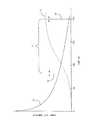
図2を参照すると、データ収集の期間内での再構成間隔すなわち窓33の賢明な選択により、再構成画像の最適品質が実現される。データ収集中、例えば360秒の間、計数率32は始めに最も高く、終りに近づくにつれて減少する。反対に、コントラスト34はスキャンの始めに最も低い。選択可能な時間窓33において収集されるLORが再構成される。窓の始め36及び/又は終り38は、コントラストと計数率のバランスを最適化するために選択的に調節される。勿論、重複する窓を含む、複数の窓が規定されることができ、各々に対して画像が再構成される。 Referring to FIG. 2, the optimal quality of the reconstructed image is achieved by judicious selection of the reconstruction interval or
ここで図3を参照すると、画像最適化を説明するフローチャートが提供される。まず、対象に放射性医薬品が注入される(40)。データが収集され(42)、スキャン中にタイムスタンプされる。データ収集の時間は、消滅イベントを生じるために使用される放射性物質によって異なることができる。82Rbの場合、スキャンは通常は約6分(360秒)続く。この期間において、物理学的に82Rbの元の量の1/16しか残らず、計数率は低すぎて診断上有用でなくなってしまっていることが予想される。Referring now to FIG. 3, a flowchart describing image optimization is provided. First, a radiopharmaceutical is injected into the subject (40). Data is collected (42) and time stamped during the scan. The time of data collection can vary depending on the radioactive material used to generate the extinction event.For 82 Rb, the scan typically lasts about 6 minutes (360 seconds). During this period, only 1/16 of the original amount of82 Rb remains physically, and the count rate is expected to be too low to be diagnostically useful.
データが収集された後、初期又は開始点、及び終末又は終了点によって規定される窓が選択される(44)。一実施形態において、終末又は終了点38は、スキャンの終わりから数秒であるように選択される。前述の通り、トレーサは関心のある組織にかん流するために十分な時間を与えられているので、このデータは最高コントラストの画像再構成をもたらす可能性が最も高い。スキャンの最後の最後からのデータを用いることは、普遍的に適用可能な方法であり、かん流に関して対象(人又は動物)の生理学についてほとんどわかっていないときには特に有用である。 After the data is collected, a window defined by an initial or starting point and an ending or ending point is selected (44). In one embodiment, the end or
より多くのことがわかる場合、より多くの点が選択されることができる。例えば、心臓イメージングに82Rbを用いる場合は、200秒が健康な人の被験者にとって十分な時間であるはずである。より一般的には、対象の生理学が、測定されるべき変数に関して安定状態にあるはずだということがわかっている時間がある場合、窓はその時間又はおよそその時間であるように選択されることができる。さらに、最終安定状態から中間状態を区別することができるパラメータがあってもよい。再度一実施例において、82Rb心臓イメージングにおいて、血流と比較した心筋活動の比率(例えば心室の空洞)はプロセスの指標となるはずである。If more is known, more points can be selected. For example, when using82 Rb for cardiac imaging, 200 seconds should be sufficient for a healthy human subject. More generally, if there is a time when it is known that the subject's physiology should be in a stable state with respect to the variable to be measured, the window is selected to be that time or approximately that time. Can do. Furthermore, there may be a parameter that can distinguish the intermediate state from the final stable state. Again in one example, in82 Rb cardiac imaging, the ratio of myocardial activity compared to blood flow (eg, ventricular cavity) should be indicative of the process.
次に、ベースライン画像が再構成される(46)。一実施形態において、ベースライン画像はスキャンの開始点から終了までのデータを用いて再構成される。このベースライン画像は高コントラストデータカウントを含むが、開始点が遅く設定される場合、カウントのボリュームは低くなり、低い信号対ノイズ比につながる。ベースライン画像は性能指数を決定するために評価される(48)。一実施形態において、性能指数はコントラスト対ノイズ比である。単位時間当たりの生のイベント数など、他の性能指数が勿論考慮される。 Next, the baseline image is reconstructed (46). In one embodiment, the baseline image is reconstructed using data from the start to the end of the scan. This baseline image contains a high contrast data count, but if the starting point is set late, the count volume will be low, leading to a low signal-to-noise ratio. The baseline image is evaluated (48) to determine a figure of merit. In one embodiment, the figure of merit is the contrast to noise ratio. Other performance indices such as the number of live events per unit time are of course considered.
ベースライン画像に対して特定の性能指数を考えると、開始点が調節され(50)、例えば特定の性能指数を改良する目的でカウント数を増加するために早い時間へ動かされる。窓の増加した領域におけるLORは、ベースライン画像を再構成するために使用されたLORに加えられ、更新画像が再構成される(52)。一旦更新画像が再構成されると、性能指数は新たな更新画像に対して再計算される(54)。図3の決定ブロック56として示されるように、性能指数が向上するか、又は検査のいくつかの選択された統計的範囲内で安定なままである場合、開始イベントはさらに調節され得(例えばわずかな期間後方へ動かす、二分探索又は選択された最適化アルゴリズムを用いる)、該プロセスが繰り返される。最良の性能指数を実現する画像は、さらなる解析58、表示、解釈のために使用され、又は将来の使用のために保持される。取り込み時間が重要な診断価値を持ち得るときは、開始時間と終了時間もまたユーザに表示される。 Given a particular figure of merit for the baseline image, the starting point is adjusted (50) and moved to an earlier time, for example, to increase the number of counts in order to improve the particular figure of merit. The LOR in the increased area of the window is added to the LOR used to reconstruct the baseline image, and the updated image is reconstructed (52). Once the updated image is reconstructed, the figure of merit is recalculated for the new updated image (54). If the figure of merit improves or remains stable within some selected statistical range of the test, as shown as
同じ方法で、窓の終了点が随意的に調節され得る。スキャンの終りにおけるデータは、発生するカウント数が少なくなるにつれて重要でなくなり得る。窓の終了点は時間的に後退させられ、更新画像が再構成される(52)。新たな性能指数は、向上しなくなるまで再度再計算される(54)。終了点の最適化は、開始点の最適化の前又は後のいずれかで起こることができる。上記の窓選択プロセス中に、窓33は、ユーザがそれを解析、定量化、又は診断に役立つために使用することができるように、ユーザに表示されることができる。最適化された窓33はまた、取り込み時間と効率が生体プロセスと機能を示すことができるように、コンピュータ支援診断におけるパラメータとして、システムによって使用されることもできる。 In the same way, the window end point can optionally be adjusted. Data at the end of the scan can become less important as the number of counts generated decreases. The window end is retracted in time and the updated image is reconstructed (52). The new figure of merit is recalculated until it does not improve (54). The end point optimization can occur either before or after the start point optimization. During the window selection process described above, the
一実施形態において、上記プロセスは自動化され、アナライザ29によって実行される。様々なレベルのユーザ入力が、決定を補助するために受け取られることができることが考慮される。例えば、開始及び/又は終了時間はユーザ入力60を用いてユーザによって設定されることができる。例えば、ユーザは図2に類似したディスプレイにおいて開始及び終了時間表示をドラッグすることができる。ユーザは、性能指数が特定画像においてその前のものに対して向上したかどうかを決定するよう指示され得る。アナライザ29は、どの画像が最良であるかを決定すると、その画像と、いくつかの前後の画像をユーザへレビューのために提示し得る。この実施形態において、該プロセスはまだ大部分が自動化されているが、補足するユーザ解析を伴う。望ましいユーザ入力とフィードバックのレベルは選択可能であり、自由自在にオン又はオフを切り替えられることができる。 In one embodiment, the process is automated and performed by
代替的な一実施形態において、データは非リストモード形式でコンパイルされることができる。この実施形態は、イベントが既にビン化された後でそれらを時間によってソートするという追加の問題を提示し、余分な処理能力と時間を必要とする。 In an alternative embodiment, the data can be compiled in non-list mode format. This embodiment presents the additional problem of sorting events by time after events are already binned, requiring extra processing power and time.
いくつかの放射性医薬品は異なる速度で体内の異なる組織タイプによって吸収される。従って、体の異なる部分に対する最適画像は異なる基準点を持ち得る。従って、複数の最適化画像が表示され得る。別の代替例として、取り込みと洗い出しを経時的に示す一連のシネ画像が表示されることができる。シネ画像の各々に対応する時間窓は同じように最適化されることができる。 Some radiopharmaceuticals are absorbed by different tissue types in the body at different rates. Thus, optimal images for different parts of the body can have different reference points. Accordingly, a plurality of optimized images can be displayed. As another alternative, a series of cine images showing uptake and washout over time can be displayed. The time window corresponding to each of the cine images can be optimized in the same way.
本発明は好適な実施形態を参照して記載されている。前述の詳細な説明を読んで理解することで修正と変更が想到され得る。本発明は、こうした修正と変更を、これらが添付の請求項又はその均等物の範囲内にある限り全て含むと解釈されることを目的とする。 The invention has been described with reference to the preferred embodiments. Modifications and changes may be conceived upon reading and understanding the foregoing detailed description. The present invention is intended to be construed as including all such modifications and changes as long as they fall within the scope of the appended claims or their equivalents.
Claims (16)
Translated fromJapaneseイメージング領域からイベントを受信するための検出器アレイと、
前記受信されたイベントにタイムスタンプを割り当てるためのトリガプロセッサと、
前記イベントの調節可能な部分を画像表現へ再構成する再構成プロセッサと、
画質を示すパラメータを決定するために、前記再構成された画像表現を解析し、かつ、検証された前記イベントの前記調節可能な部分を、前記決定されたパラメータに従って後続画像表現へ再構成するために調節する、アナライザと、
を有する、画像診断装置。A diagnostic imaging apparatus,
A detector array for receiving events from the imaging region;
A trigger processor for assigning a time stamp to the received event;
A reconstruction processor that reconstructs the adjustable portion of the event into an image representation;
Analyzing the reconstructed image representation to determine a parameter indicative of image quality, and reconstructing the adjustable portion of the verified event into a subsequent image representation according to the determined parameter To adjust the analyzer,
A diagnostic imaging apparatus.
再構成されるイベントを規定する時間窓を調節するために前記アナライザによって制御される時間窓セレクタと、
をさらに含む、請求項1又は2に記載の画像診断装置。An event storage buffer for storing the verified events in a list sortable according to the reception time of the event;
A time window selector controlled by the analyzer to adjust a time window defining reconstructed events;
The image diagnostic apparatus according to claim 1, further comprising:
受信された核崩壊イベントを示すデータ点のセットを収集するステップと、
前記受信されたイベントにタイムスタンプするステップと、
前記収集されたデータ点の調節可能な部分から画像表現を再構成するステップと、
画像表現の品質を示すパラメータの値を決定するために前記画像表現を解析するステップと、
後続画像表現を再構成する際に使用される、前記収集されたデータ点の前記調節可能な部分を調節するステップと、
を有する方法。A diagnostic imaging method,
Collecting a set of data points indicative of a received nuclear decay event;
Timestamping the received event;
Reconstructing an image representation from an adjustable portion of the collected data points;
Analyzing the image representation to determine a value of a parameter indicative of the quality of the image representation;
Adjusting the adjustable portion of the collected data points used in reconstructing a subsequent image representation;
Having a method.
前記後続画像が前記先行画像よりもよいことを示す、前記後続画像表現パラメータ及び前記先行画像表現パラメータの値に応じて、前記時間窓を調節し、別の画像表現を生成するステップをさらに含む、請求項9に記載の方法。Analyzing the subsequent image representation to determine a value of a parameter indicative of the quality of the second image representation;
Further comprising adjusting the time window to generate another image representation in response to the value of the subsequent image representation parameter and the preceding image representation parameter indicating that the subsequent image is better than the previous image. The method of claim 9.
前記後続画像が前記先行画像表現よりもよいことに応じて、前記スキャンの始めに向かって前記開始点を動かすステップをさらに含む、請求項10に記載の方法。The time window has a start point and an end point;
The method of claim 10, further comprising moving the starting point toward the beginning of the scan in response to the subsequent image being better than the preceding image representation.
Applications Claiming Priority (3)
| Application Number | Priority Date | Filing Date | Title |
|---|---|---|---|
| US6128908P | 2008-06-13 | 2008-06-13 | |
| US61/061,289 | 2008-06-13 | ||
| PCT/IB2009/052290WO2009150565A2 (en) | 2008-06-13 | 2009-05-29 | Reverse data reconstruction for optimal time sampling of counts in physiological list-mode nuclear imaging |
Related Child Applications (1)
| Application Number | Title | Priority Date | Filing Date |
|---|---|---|---|
| JP2014083474ADivisionJP5864658B2 (en) | 2008-06-13 | 2014-04-15 | Inverse data reconstruction for optimal time sampling of counts in physiological list mode nuclear medicine imaging |
Publications (1)
| Publication Number | Publication Date |
|---|---|
| JP2011523076Atrue JP2011523076A (en) | 2011-08-04 |
Family
ID=41417195
Family Applications (2)
| Application Number | Title | Priority Date | Filing Date |
|---|---|---|---|
| JP2011513080APendingJP2011523076A (en) | 2008-06-13 | 2009-05-29 | Inverse data reconstruction for optimal time sampling of counts in physiological list mode nuclear medicine imaging |
| JP2014083474AExpired - Fee RelatedJP5864658B2 (en) | 2008-06-13 | 2014-04-15 | Inverse data reconstruction for optimal time sampling of counts in physiological list mode nuclear medicine imaging |
Family Applications After (1)
| Application Number | Title | Priority Date | Filing Date |
|---|---|---|---|
| JP2014083474AExpired - Fee RelatedJP5864658B2 (en) | 2008-06-13 | 2014-04-15 | Inverse data reconstruction for optimal time sampling of counts in physiological list mode nuclear medicine imaging |
Country Status (6)
| Country | Link |
|---|---|
| US (1) | US8437836B2 (en) |
| EP (1) | EP2288291B1 (en) |
| JP (2) | JP2011523076A (en) |
| CN (1) | CN102098964B (en) |
| RU (1) | RU2517586C2 (en) |
| WO (1) | WO2009150565A2 (en) |
Cited By (1)
| Publication number | Priority date | Publication date | Assignee | Title |
|---|---|---|---|---|
| JP2021505259A (en)* | 2017-12-07 | 2021-02-18 | コーニンクレッカ フィリップス エヌ ヴェKoninklijke Philips N.V. | Device for presenting dark-field X-ray image information |
Families Citing this family (12)
| Publication number | Priority date | Publication date | Assignee | Title |
|---|---|---|---|---|
| GB2464212B (en)* | 2008-10-09 | 2013-03-06 | Siemens Medical Solutions | Methods and apparatus for analyzing medical imaging data |
| CN102028494B (en)* | 2011-01-24 | 2012-05-09 | 海纳医信(北京)软件科技有限责任公司 | Method and system for processing cerebral perfusion image sequence |
| JP6130840B2 (en) | 2011-10-04 | 2017-05-17 | コーニンクレッカ フィリップス エヌ ヴェKoninklijke Philips N.V. | Adaptive dual path target reconstruction and acquisition |
| US9299142B2 (en) | 2011-10-24 | 2016-03-29 | Koninklijke Philips N.V. | Perfusion imaging |
| WO2015040535A1 (en)* | 2013-09-17 | 2015-03-26 | Koninklijke Philips N.V. | A scatter reject method via energy calibration |
| US20150198684A1 (en)* | 2014-01-13 | 2015-07-16 | Tamer BASHA | System and Method For Flexible Automated Magnetic Resonance Imaging Reconstruction |
| US20150269753A1 (en)* | 2014-03-18 | 2015-09-24 | Siemens Medical Solutions Usa, Inc. | Multi-Output With Acquisition Replay System and Method |
| CN104598356B (en)* | 2014-12-23 | 2018-02-27 | 沈阳东软医疗系统有限公司 | A kind of event ordering method and device |
| CN104599299B (en) | 2014-12-24 | 2017-12-29 | 沈阳东软医疗系统有限公司 | A kind of method for reconstructing and device of CT images |
| CN105761293B (en)* | 2016-03-17 | 2019-01-08 | 上海联影医疗科技有限公司 | Medical imaging procedure and system |
| CN112512427B (en)* | 2018-08-07 | 2024-05-24 | 美国西门子医疗系统股份有限公司 | Adaptive Compton Camera for Medical Imaging |
| US11037338B2 (en)* | 2018-08-22 | 2021-06-15 | Nvidia Corporation | Reconstructing image data |
Citations (5)
| Publication number | Priority date | Publication date | Assignee | Title |
|---|---|---|---|---|
| US6255655B1 (en)* | 1997-01-08 | 2001-07-03 | Smv America, Inc. | Gamma camera for PET and SPECT studies |
| WO2007008528A2 (en)* | 2005-07-08 | 2007-01-18 | Wisconsin Alumni Research Foundation | Highly constrained image reconstruction method |
| WO2007100955A2 (en)* | 2006-02-28 | 2007-09-07 | Koninklijke Philips Electronics, N.V. | Local motion compensation based on list mode data |
| JP2008502409A (en)* | 2004-06-18 | 2008-01-31 | コーニンクレッカ フィリップス エレクトロニクス エヌ ヴィ | Artifact reduction |
| US20080073538A1 (en)* | 2006-09-21 | 2008-03-27 | A Hans Vija | Application-driven optimization of acquisition and reconstruction of SPECT/PET projection data |
Family Cites Families (30)
| Publication number | Priority date | Publication date | Assignee | Title |
|---|---|---|---|---|
| IL66127A (en)* | 1982-06-24 | 1987-11-30 | Israel State | Method and apparatus for measuring the index of refraction of fluids |
| US7209779B2 (en)* | 2001-07-17 | 2007-04-24 | Accuimage Diagnostics Corp. | Methods and software for retrospectively gating a set of images |
| US6664543B2 (en)* | 2002-01-28 | 2003-12-16 | Cti Pet Systems, Inc. | Continuous sampling and digital integration for PET scintillation |
| US7132664B1 (en)* | 2002-11-09 | 2006-11-07 | Crosetto Dario B | Method and apparatus for improving PET detectors |
| US7983735B2 (en)* | 2003-04-15 | 2011-07-19 | General Electric Company | Simulation of nuclear medical imaging |
| RU2233119C1 (en)* | 2003-06-24 | 2004-07-27 | Центральный научно-исследовательский рентгено-радиологический институт | Method for predicting cardiac sarcoidosis |
| EP1654704A2 (en)* | 2003-07-21 | 2006-05-10 | Paieon Inc. | Method and system for identifying an optimal image within a series of images that depict a moving organ |
| US20050123183A1 (en)* | 2003-09-02 | 2005-06-09 | Paul Schleyer | Data driven motion correction for nuclear imaging |
| US7045802B2 (en)* | 2003-11-26 | 2006-05-16 | General Electric Company | Method and apparatus for coincidence imaging digital triggering |
| US7968851B2 (en)* | 2004-01-13 | 2011-06-28 | Spectrum Dynamics Llc | Dynamic spect camera |
| WO2007054935A2 (en)* | 2005-11-09 | 2007-05-18 | Spectrum Dynamics Llc | Dynamic spect camera |
| US8586932B2 (en)* | 2004-11-09 | 2013-11-19 | Spectrum Dynamics Llc | System and method for radioactive emission measurement |
| WO2008010227A2 (en)* | 2006-07-19 | 2008-01-24 | Spectrum Dynamics Llc | Imaging protocols |
| WO2006051531A2 (en)* | 2004-11-09 | 2006-05-18 | Spectrum Dynamics Llc | Radioimaging |
| EP1778957A4 (en)* | 2004-06-01 | 2015-12-23 | Biosensors Int Group Ltd | OPTIMIZING THE MEASUREMENT OF RADIOACTIVE EMISSIONS IN SPECIFIC BODY STRUCTURES |
| JP4353040B2 (en)* | 2004-09-22 | 2009-10-28 | 株式会社島津製作所 | Positron CT system |
| US8423125B2 (en)* | 2004-11-09 | 2013-04-16 | Spectrum Dynamics Llc | Radioimaging |
| US20060239398A1 (en)* | 2005-03-07 | 2006-10-26 | Fused Multimodality Imaging, Ltd. | Breast diagnostic apparatus for fused SPECT, PET, x-ray CT, and optical surface imaging of breast cancer |
| WO2006114717A1 (en)* | 2005-04-27 | 2006-11-02 | Koninklijke Philips Electronics, N.V. | Ecg-gated temporal sampling in cardiac kinetic modeling |
| MX2007016203A (en) | 2005-06-29 | 2008-03-10 | Compumedics Ltd | Sensor assembly with conductive bridge. |
| US8644910B2 (en)* | 2005-07-19 | 2014-02-04 | Biosensors International Group, Ltd. | Imaging protocols |
| EP1908011B1 (en)* | 2005-07-19 | 2013-09-04 | Spectrum Dynamics LLC | Reconstruction stabilizer and active vision |
| RU2312602C2 (en)* | 2005-11-23 | 2007-12-20 | Государственное образовательное учреждение высшего профессионального образования "Московский государственный институт электронной техники" (технический университет) | Method for producing tomographic images |
| EP1971257B1 (en)* | 2005-12-28 | 2015-10-14 | Biosensors International Group, Ltd. | Gating with anatomically varying durations |
| GB0612234D0 (en) | 2006-06-20 | 2006-08-02 | Mallinckrodt Inc | Method of making and using rubidium-81-containing compositions |
| US8047715B2 (en)* | 2006-11-03 | 2011-11-01 | Koninklijke Philips Electronics N.V. | Multiple rotation C-arm |
| DE102007060689B4 (en)* | 2007-12-17 | 2017-07-13 | Siemens Healthcare Gmbh | Method for recording angiographic data sets and magnetic resonance system therefor |
| EP2286274B1 (en)* | 2008-05-28 | 2019-07-24 | Koninklijke Philips N.V. | Geometrical transformations preserving list-mode format |
| US7732774B2 (en)* | 2008-09-19 | 2010-06-08 | Jefferson Science Associates, Llc | High resolution PET breast imager with improved detection efficiency |
| US7858944B2 (en)* | 2009-01-23 | 2010-12-28 | Jefferson Science Associates, Llc | Dedicated mobile high resolution prostate PET imager with an insertable transrectal probe |
- 2009
- 2009-05-29JPJP2011513080Apatent/JP2011523076A/enactivePending
- 2009-05-29USUS12/995,685patent/US8437836B2/ennot_activeExpired - Fee Related
- 2009-05-29CNCN200980121926.3Apatent/CN102098964B/ennot_activeExpired - Fee Related
- 2009-05-29RURU2011100843/14Apatent/RU2517586C2/ennot_activeIP Right Cessation
- 2009-05-29WOPCT/IB2009/052290patent/WO2009150565A2/enactiveApplication Filing
- 2009-05-29EPEP09762092.6Apatent/EP2288291B1/ennot_activeNot-in-force
- 2014
- 2014-04-15JPJP2014083474Apatent/JP5864658B2/ennot_activeExpired - Fee Related
Patent Citations (5)
| Publication number | Priority date | Publication date | Assignee | Title |
|---|---|---|---|---|
| US6255655B1 (en)* | 1997-01-08 | 2001-07-03 | Smv America, Inc. | Gamma camera for PET and SPECT studies |
| JP2008502409A (en)* | 2004-06-18 | 2008-01-31 | コーニンクレッカ フィリップス エレクトロニクス エヌ ヴィ | Artifact reduction |
| WO2007008528A2 (en)* | 2005-07-08 | 2007-01-18 | Wisconsin Alumni Research Foundation | Highly constrained image reconstruction method |
| WO2007100955A2 (en)* | 2006-02-28 | 2007-09-07 | Koninklijke Philips Electronics, N.V. | Local motion compensation based on list mode data |
| US20080073538A1 (en)* | 2006-09-21 | 2008-03-27 | A Hans Vija | Application-driven optimization of acquisition and reconstruction of SPECT/PET projection data |
Cited By (3)
| Publication number | Priority date | Publication date | Assignee | Title |
|---|---|---|---|---|
| JP2021505259A (en)* | 2017-12-07 | 2021-02-18 | コーニンクレッカ フィリップス エヌ ヴェKoninklijke Philips N.V. | Device for presenting dark-field X-ray image information |
| JP7053834B2 (en) | 2017-12-07 | 2022-04-12 | コーニンクレッカ フィリップス エヌ ヴェ | Device for presenting dark-field X-ray image information |
| JP7053834B6 (en) | 2017-12-07 | 2022-06-02 | コーニンクレッカ フィリップス エヌ ヴェ | Device for presenting dark-field X-ray image information |
Also Published As
| Publication number | Publication date |
|---|---|
| JP5864658B2 (en) | 2016-02-17 |
| WO2009150565A3 (en) | 2010-06-24 |
| EP2288291A2 (en) | 2011-03-02 |
| CN102098964B (en) | 2015-04-08 |
| US20110105887A1 (en) | 2011-05-05 |
| EP2288291B1 (en) | 2018-11-14 |
| CN102098964A (en) | 2011-06-15 |
| JP2014149308A (en) | 2014-08-21 |
| RU2011100843A (en) | 2012-07-20 |
| RU2517586C2 (en) | 2014-05-27 |
| WO2009150565A2 (en) | 2009-12-17 |
| US8437836B2 (en) | 2013-05-07 |
Similar Documents
| Publication | Publication Date | Title |
|---|---|---|
| JP5864658B2 (en) | Inverse data reconstruction for optimal time sampling of counts in physiological list mode nuclear medicine imaging | |
| JP5860607B2 (en) | System and method for tomographic data collection and image reconstruction | |
| US7507968B2 (en) | Systems and methods for correcting a positron emission tomography emission image | |
| US6297506B1 (en) | System and method for reducing pile-up errors in multi-crystal gamma ray detector applications | |
| US9332952B2 (en) | Data-driven optimization of event acceptance/rejection logic | |
| US8204172B1 (en) | System and method of prior image constrained image reconstruction using short scan image data and objective function minimization | |
| US7907698B2 (en) | Gated CT with irregular sampling for slow CT acquisition | |
| US7840052B2 (en) | Restoration of the nuclear medicine 2D planar image by iterative constrained deconvolution | |
| JP7242708B2 (en) | Correction Method for Improving Quantification Accuracy in List-Mode Reconstruction | |
| JP2004530915A (en) | Dual isotope testing in nuclear medicine imaging | |
| CN103221841A (en) | PET calibrations with varying coincidence windows | |
| JP6811998B2 (en) | A PET device with a positron life measurement function and a method for measuring the positron life in the PET device. | |
| Casey et al. | Siemens biograph vision 600 | |
| CN101088028A (en) | Real-time list mode reconstruction | |
| US20070090300A1 (en) | Shifted transmission mock for nuclear medical imaging | |
| EP1631844B8 (en) | Generating detector efficiency estimates for a pet scanner | |
| GB2497834A (en) | Analyzing pet medical imaging data | |
| US20240193828A1 (en) | Systems and methods of list-mode image reconstruction in positron emission tomography (pet) systems | |
| Lagopati | Nuclear Medicine Imaging Essentials | |
| CN113647969A (en) | A kind of radiotracer composition analysis method and system | |
| Lodge | PET instrumentation and methodology | |
| Livieratos | cardiac pet: physics and Methodology | |
| Bal | Data-driven method for parametric imaging using dynamic cardiac single photon emission computed tomography | |
| Pointoin | Model-based randoms correction for 3D positron emission tomography |
Legal Events
| Date | Code | Title | Description |
|---|---|---|---|
| A621 | Written request for application examination | Free format text:JAPANESE INTERMEDIATE CODE: A621 Effective date:20120524 | |
| A131 | Notification of reasons for refusal | Free format text:JAPANESE INTERMEDIATE CODE: A131 Effective date:20130411 | |
| A601 | Written request for extension of time | Free format text:JAPANESE INTERMEDIATE CODE: A601 Effective date:20130708 | |
| A602 | Written permission of extension of time | Free format text:JAPANESE INTERMEDIATE CODE: A602 Effective date:20130716 | |
| A521 | Request for written amendment filed | Free format text:JAPANESE INTERMEDIATE CODE: A523 Effective date:20131001 | |
| A02 | Decision of refusal | Free format text:JAPANESE INTERMEDIATE CODE: A02 Effective date:20131217 |