JP2011514815A - In-vivo minimally invasive testing device including a metal guide - Google Patents
In-vivo minimally invasive testing device including a metal guideDownload PDFInfo
- Publication number
- JP2011514815A JP2011514815AJP2010547125AJP2010547125AJP2011514815AJP 2011514815 AJP2011514815 AJP 2011514815AJP 2010547125 AJP2010547125 AJP 2010547125AJP 2010547125 AJP2010547125 AJP 2010547125AJP 2011514815 AJP2011514815 AJP 2011514815A
- Authority
- JP
- Japan
- Prior art keywords
- tip
- metal guide
- substrate
- guide
- specific
- Prior art date
- Legal status (The legal status is an assumption and is not a legal conclusion. Google has not performed a legal analysis and makes no representation as to the accuracy of the status listed.)
- Granted
Links
Images


Classifications
- A—HUMAN NECESSITIES
- A61—MEDICAL OR VETERINARY SCIENCE; HYGIENE
- A61B—DIAGNOSIS; SURGERY; IDENTIFICATION
- A61B5/00—Measuring for diagnostic purposes; Identification of persons
- A—HUMAN NECESSITIES
- A61—MEDICAL OR VETERINARY SCIENCE; HYGIENE
- A61B—DIAGNOSIS; SURGERY; IDENTIFICATION
- A61B5/00—Measuring for diagnostic purposes; Identification of persons
- A61B5/145—Measuring characteristics of blood in vivo, e.g. gas concentration or pH-value ; Measuring characteristics of body fluids or tissues, e.g. interstitial fluid or cerebral tissue
- A61B5/14542—Measuring characteristics of blood in vivo, e.g. gas concentration or pH-value ; Measuring characteristics of body fluids or tissues, e.g. interstitial fluid or cerebral tissue for measuring blood gases
- A—HUMAN NECESSITIES
- A61—MEDICAL OR VETERINARY SCIENCE; HYGIENE
- A61B—DIAGNOSIS; SURGERY; IDENTIFICATION
- A61B5/00—Measuring for diagnostic purposes; Identification of persons
- A61B5/145—Measuring characteristics of blood in vivo, e.g. gas concentration or pH-value ; Measuring characteristics of body fluids or tissues, e.g. interstitial fluid or cerebral tissue
- A61B5/1468—Measuring characteristics of blood in vivo, e.g. gas concentration or pH-value ; Measuring characteristics of body fluids or tissues, e.g. interstitial fluid or cerebral tissue using chemical or electrochemical methods, e.g. by polarographic means
- A61B5/1473—Measuring characteristics of blood in vivo, e.g. gas concentration or pH-value ; Measuring characteristics of body fluids or tissues, e.g. interstitial fluid or cerebral tissue using chemical or electrochemical methods, e.g. by polarographic means invasive, e.g. introduced into the body by a catheter
- A—HUMAN NECESSITIES
- A61—MEDICAL OR VETERINARY SCIENCE; HYGIENE
- A61B—DIAGNOSIS; SURGERY; IDENTIFICATION
- A61B5/00—Measuring for diagnostic purposes; Identification of persons
- A61B5/40—Detecting, measuring or recording for evaluating the nervous system
- A61B5/4076—Diagnosing or monitoring particular conditions of the nervous system
- A—HUMAN NECESSITIES
- A61—MEDICAL OR VETERINARY SCIENCE; HYGIENE
- A61B—DIAGNOSIS; SURGERY; IDENTIFICATION
- A61B5/00—Measuring for diagnostic purposes; Identification of persons
- A61B5/68—Arrangements of detecting, measuring or recording means, e.g. sensors, in relation to patient
- A61B5/6846—Arrangements of detecting, measuring or recording means, e.g. sensors, in relation to patient specially adapted to be brought in contact with an internal body part, i.e. invasive
- A61B5/6847—Arrangements of detecting, measuring or recording means, e.g. sensors, in relation to patient specially adapted to be brought in contact with an internal body part, i.e. invasive mounted on an invasive device
- A61B5/6851—Guide wires
- A—HUMAN NECESSITIES
- A61—MEDICAL OR VETERINARY SCIENCE; HYGIENE
- A61B—DIAGNOSIS; SURGERY; IDENTIFICATION
- A61B2562/00—Details of sensors; Constructional details of sensor housings or probes; Accessories for sensors
- A61B2562/02—Details of sensors specially adapted for in-vivo measurements
- A—HUMAN NECESSITIES
- A61—MEDICAL OR VETERINARY SCIENCE; HYGIENE
- A61B—DIAGNOSIS; SURGERY; IDENTIFICATION
- A61B2562/00—Details of sensors; Constructional details of sensor housings or probes; Accessories for sensors
- A61B2562/04—Arrangements of multiple sensors of the same type
- A61B2562/043—Arrangements of multiple sensors of the same type in a linear array
Landscapes
- Health & Medical Sciences (AREA)
- Life Sciences & Earth Sciences (AREA)
- Physics & Mathematics (AREA)
- General Health & Medical Sciences (AREA)
- Veterinary Medicine (AREA)
- Engineering & Computer Science (AREA)
- Biomedical Technology (AREA)
- Heart & Thoracic Surgery (AREA)
- Medical Informatics (AREA)
- Molecular Biology (AREA)
- Surgery (AREA)
- Animal Behavior & Ethology (AREA)
- Biophysics (AREA)
- Public Health (AREA)
- Pathology (AREA)
- Optics & Photonics (AREA)
- Neurology (AREA)
- Chemical & Material Sciences (AREA)
- Chemical Kinetics & Catalysis (AREA)
- General Chemical & Material Sciences (AREA)
- Neurosurgery (AREA)
- Physiology (AREA)
- Investigating Or Analysing Biological Materials (AREA)
- Measurement Of The Respiration, Hearing Ability, Form, And Blood Characteristics Of Living Organisms (AREA)
- Endoscopes (AREA)
- Apparatus Associated With Microorganisms And Enzymes (AREA)
Abstract
Translated fromJapaneseDescription
Translated fromJapanese本発明は、経壁的検査デバイスの機能化、その機能化したデバイスを使用した基質の生体外分析の方法、および患者の癌、感染、炎症、神経変性疾患または移植片拒絶反応の診断のためのツールの製造のための上記デバイスの使用法に関する。 The present invention relates to the functionalization of a transmural testing device, a method for in vitro analysis of a substrate using the functionalized device, and diagnosis of cancer, infection, inflammation, neurodegenerative disease or graft rejection in a patient. The use of the above device for the manufacture of tools.
検査または生体内治療デバイスは、現在の技術で知られている。こうしたデバイスは、内視鏡タイプの剛体チューブ、または可撓性のチューブから構成されたカテーテルの形状をしており、特に自然の経路または管によって生体に挿入され、器官または特定の組織に到達させることができる。これらのデバイスにより、特に血餅をなくすこと、または光ファイバーと連動するときは、消化管としての系の状態、もしくは結腸としての器官の状態の視覚化および生体内管理が可能になる。次に、こうしたデバイスの使用により生じる患者の外傷は、最小限に抑えられているが、今も改善されつつある段階である。しかし、これらのデバイスで患者の器官または組織を分析することが常に可能なわけではない。血液循環もしくは自然の経路と比較すると前記組織または器官にアクセスしにくいこと、または前記器官もしくは組織の細胞の細かい分析に頼ることなしでは信頼できる診断の実行が難しいことから、このように不可能になることがある。このような場合には、一般に、現在の技術で今までは、特に細針(FNAすなわち<<細針吸引(Fine needle aspiration)>>:Engelsteinら、Br.J.Urol.、7:210〜213頁、1994年;Rodriguesら、J.Am.Acad.Dermatol.、42:735〜740頁、2000年;Arigaら、Am.J.Surg.、184:410〜413頁、2002年;Perez−Guillermoら、Diagn.Cytopathol.、32:315〜320頁、2005年;Fernandez−Esparrachら、Arch.Bronconeomol.、43:219〜224頁、2007年)によって、これらの組織もしくは器官の、またはより細かくそれらを構成する細胞の1つの形態を生体外で管理するために、前記組織または器官のフラグメントのサンプリング(生検)が使用されている。さらに、特定の数のマーカーの発現状態を分析することも可能であり、それらのマーカーの発現は、特異な病理学的状態(特に癌、炎症、感染、神経変性疾患または移植片拒絶反応)と関係している。 Testing or in-vivo treatment devices are known in the current art. These devices are in the form of an endoscope-type rigid tube, or a catheter constructed from a flexible tube, and are inserted into the body by a natural path or tube, especially to reach an organ or a specific tissue. be able to. These devices allow visualization and in vivo management of the state of the system as the digestive tract, or the state of the organ as the colon, especially when eliminating clots or working with optical fibers. Secondly, patient trauma resulting from the use of such devices has been minimized but is still being improved. However, it is not always possible to analyze patient organs or tissues with these devices. This is not possible because it is difficult to access the tissue or organ compared to the blood circulation or natural pathways, or it is difficult to perform a reliable diagnosis without resorting to detailed analysis of the cells of the organ or tissue. May be. In such cases, it is generally the case in the current art that, until now, in particular fine needles (FNA or << fine needle aspiration >>: Engelstein et al., Br. J. Urol., 7: 210. 213, 1994; Rodrigues et al., J. Am. Acad. Dermatol., 42: 735-740, 2000; Ariga et al., Am. J. Surg., 184: 410-413, 2002; Guillermo et al., Diagno. Cytopathol., 32: 315-320, 2005; Fernandez-Esparach et al., Arch.Bronconeomol., 43: 219-224, 2007), or more detailed of these tissues or organs. Sampling (biopsy) of tissue or organ fragments has been used to manage one form of the cells that make up them in vitro. In addition, it is possible to analyze the expression status of a certain number of markers, and the expression of those markers is dependent on the specific pathological condition (especially cancer, inflammation, infection, neurodegenerative disease or graft rejection). Involved.
しかし、これらの異なる方法は、生検のサンプリングまたは細胞の吸引によって外傷を伴い、これは、前記組織または器官に対して、したがって患者に対して重大な影響を与えることがある。それにより、患者の生体は、特に特定の器官(脳、膵臓、肝臓または肺)に関して、サンプリングに続く出血または瘢痕のせいで、非常に苦しめられることがある。 However, these different methods involve trauma by biopsy sampling or cell aspiration, which can have a significant impact on the tissue or organ and thus on the patient. Thereby, the patient's organism can be very plagued by bleeding or scarring following sampling, especially with respect to specific organs (brain, pancreas, liver or lung).
したがって、患者に与える外傷を限定しながら、特に、具体的には癌、炎症、感染または神経変性疾患など、異なる病理と連動するマーカーの発現と比較して、信頼できる正確な診断を実行できるようにする新しい検査の方法体系の特定が今なお必要とされている。 Therefore, it is possible to carry out a reliable and accurate diagnosis while limiting the trauma to the patient, especially compared to the expression of markers that are specifically linked to different pathologies, such as cancer, inflammation, infection or neurodegenerative diseases. There is still a need to identify new inspection methods.
EP1358481の特許に、分析または生体内の治療のデバイスが記載されており、そのデバイスは、(i)蛍光信号の分析以外のものによる基質の検査のマイクロ・システムと、(ii)一方の先端部に前記マイクロ・システムが固定され、もう一方の先端部は前記マイクロ・システムの制御が意図される、可撓性のバット(butt)と、(iii)前記可撓性のバットが内部を摺動できる内側開口部を有する医療用具と、(iv)基質のレベルで着脱可能な、マイクロ・システムの保護の摺動システムと、(v)前記マイクロ・システムのレベルの組織または細胞の引裂きシステムであって、最終的に、生物学的または化学的製品の局注のために、治療のための生検を実行するように、感覚受容体(触覚、視覚、物理化学または数値情報科学)による遠隔制御によってモニタリングするデバイスから選択される1つまたは複数のデバイスと連動する、引裂きシステムとを備える。 EP1354841 describes a device for analysis or treatment in vivo, the device comprising: (i) a microsystem for examination of the substrate by other than analysis of the fluorescence signal; and (ii) one tip The micro system is fixed to the other end, and the other tip is intended to control the micro system, and (iii) the flexible bat slides inside A medical device having an internal opening that can be made; (iv) a micro-system protective sliding system removable at the substrate level; and (v) a tissue or cell tearing system at the micro-system level. Sensory receptors (tactile, visual, physicochemical or numerical, so as to ultimately perform a biopsy for treatment, for local injection of biological or chemical products In conjunction with one or more devices selected from a device for monitoring by a remote control by multicast Science), and a tear system.
血管の侵襲および組織または細胞の引裂きを可能にするために、マイクロ・システムは、好ましくは遠位位置で、この機能を保証する、より剛体の別のシステムと連動する。 In order to allow vascular invasion and tissue or cell tearing, the micro system works in conjunction with another more rigid system that ensures this function, preferably at a distal location.
しかし、生体内検査デバイスの場合は、その検査デバイスは、より多くの場合、脈管内のまたは腔内の経路を使用して目標の器官または組織に向けられる。次いで、マイクロ・システムを引裂きシステムに結合すると、記載したデバイスの先端部で直径が無視できない程度増大する。そのとき、そのデバイスは、血液循環経路を変えずに同時に目標位置に正確に向けるには、使用が複雑過ぎることが明らかである。同時に、複数の要素を結合すると、器官または組織を穿孔可能にするのに必要な剛性を得ることに関して、デバイスの全体的な可撓性、したがって正確な案内を行うことに支障をきたす。 However, in the case of an in-vivo testing device, the testing device is more often directed to a target organ or tissue using an intravascular or intracavity route. Then, when the micro system is coupled to the tear system, the tip of the described device increases in diameter to a negligible extent. At that time, it is clear that the device is too complicated to use to accurately aim at the target location at the same time without changing the blood circulation path. At the same time, combining multiple elements interferes with the overall flexibility of the device, and therefore accurate guidance, with respect to obtaining the rigidity necessary to allow the organ or tissue to be pierced.
以下の重要な調査に続いて、発明者は現在、金属製ガイドを備えるデバイスを開発しようとしており、その金属製ガイドの一方の穿孔用先端部に、反応性基、特に、試験する基質の特異な抗体または抗体フラグメントが直接結合され、もう一方の先端部は、挿入位置から前記基質のミクロ分析および/またはマイクロ・サンプリングの位置への、前記ガイドの運転が意図される。前記ガイドを、着脱可能な保護システム、例えば、可撓性のカテーテル中に挿入することができ、そのとき、これらの組織、器官または細胞を構成する、試験する基質のミクロ分析および/またはマイクロ・サンプリングの位置に対して、前記ガイドの機能化された先端部を保護することが可能になる。 Following the following important investigations, the inventor is now trying to develop a device with a metal guide, with a specific group of reactive groups, particularly the substrate to be tested, on one piercing tip of the metal guide. The antibody or antibody fragment is directly bound and the other tip is intended to drive the guide from the insertion location to the location of micro-analysis and / or micro-sampling of the substrate. The guide can be inserted into a removable protection system, for example a flexible catheter, at which time the microanalysis and / or micro-analysis of the substrate to be tested constituting these tissues, organs or cells. It is possible to protect the functionalized tip of the guide against the sampling position.
本発明は、ガイド上にいくつかのスポット、例えば凹所を画定するように、ガイドの表面を構成することであり、それらの凹所に、反応性基が配置されて生化学的作用が起きる。電気化学的侵食およびレーザーが続くレーザー・リソグラフィによる、集束イオン・ビーム(FIBすなわち<<Focused Ion beam>>:Xieら、Nuclear Instruments & methods in Physics research Section B−beam Interactions with Materials and Atoms、211(3):363〜368頁、2003年)によるリソグラフィなど、異なる方法によって前記凹所を実現することができる。 The present invention is to configure the surface of the guide to define several spots on the guide, such as recesses, in which reactive groups are placed to cause biochemical action. . Focused ion beam (FIB or << Focused Ion beam >>: Xie et al., Nuclear Instruments & methods in Physics and Research Interactions, B-beam Interactions 211, Electrochemical Erosion and Laser Lithography) 3): 363-368, 2003), the recesses can be realized by different methods.
あるいは、前記ガイド上にいくつかのスポット、例えば少なくとも1つの溝を画定するように、金属製ガイドを構成することができ、それらの溝には、線形、円形、またはリボンの形態の、少なくとも1つの小型化したバイオチップが共同で連動し、そこに反応性基が配置されて生化学的反応が起きる。ガイドの表面構成によって形成されるスポット、例えば凹所は、ガイドの表面に起伏を形成しないが、溝または空所を形成する。したがって、ガイドの表面の構成によって形成されたスポットは、ガイドの直径を増大させることはない。 Alternatively, a metal guide can be configured to define a number of spots on the guide, for example at least one groove, wherein the grooves have at least one in the form of a linear, circular or ribbon. Two miniaturized biochips work together and a reactive group is placed there to cause a biochemical reaction. Spots formed by the surface configuration of the guide, such as recesses, do not form undulations on the surface of the guide, but form grooves or voids. Thus, the spot formed by the surface configuration of the guide does not increase the guide diameter.
有利なことに、ガイドの表面構成によって形成されたスポットは、ガイドの表面に位置し、これは、これらのスポットが流体、組織および細胞と直接接触することを意味する。 Advantageously, the spots formed by the surface configuration of the guide are located on the surface of the guide, meaning that these spots are in direct contact with fluids, tissues and cells.
さらに、前記デバイスを、前記金属製ガイドが内部を摺動できる内側開口部を有する医療用具中に、特に、経壁的な針穿刺、特に経皮的または経粘膜的な針穿刺、または脈管系のナビゲーションを含む内視鏡で挿入することができる。 Furthermore, the device can be used in a medical device having an inner opening in which the metallic guide can slide, in particular transmural needle puncture, in particular percutaneous or transmucosal needle puncture, or vascular. Can be inserted with an endoscope that includes system navigation.
さらに、前記デバイスは、光ファイバーと連動することができるか、または前記デバイスの金属製ガイドの代わりに光ファイバーを使用することができる。その光ファイバーの穿孔用先端部が金属製リングと連動し、そのリングに反応性基、特に試験する基質に特異な抗体または抗体フラグメントが直接結合され、したがって、診断に必要な、最終的には予後の評価に必要な要素の生体内の捕捉を考慮して、細かいインサイチュの視覚化が可能になる。光ファイバーの特性は、画像をマークし、インサイチュの視覚化からデバイスを設定するために使用される。 Furthermore, the device can work with an optical fiber, or an optical fiber can be used in place of the metal guide of the device. The piercing tip of the optical fiber is linked to a metal ring to which a reactive group, in particular an antibody or antibody fragment specific for the substrate being tested, is directly attached, and is therefore necessary for diagnosis and ultimately the prognosis Considering in-vivo capture of the elements required for evaluation of the in-vivo, it is possible to visualize in situ. The properties of the optical fiber are used to mark the image and set the device from in situ visualization.
本発明によるデバイスは、その単純化の故に、低侵襲性のままでありながら、可撓性または弾性が従来技術のデバイスと比較して改善される。そのため、腔内の経路、特に脈管内の経路による、または経壁的経路、特に経皮的または経粘膜的な経路による検査のために、デバイスをさらに効率的に使用することができる。さらに、本発明によるデバイスは、先端部が特異な試薬に結合した金属製ガイドを使用するので、機能化した先端部において、組織または器官を効率的に穿孔するのに十分な剛体である。最後に、本発明によるデバイスの先端部での直径は、血液循環または自然の空所中での単純化したナビゲーションを可能にし、具体的には、デバイスが穿孔しなければならない組織または器官のレベルで、外傷を最小限に抑えるのに十分に微弱なものである。 The device according to the invention, due to its simplicity, has improved flexibility or elasticity compared to prior art devices while remaining minimally invasive. As such, the device can be used more efficiently for inspection by intraluminal routes, particularly intravascular routes, or transmural routes, particularly percutaneous or transmucosal routes. Furthermore, the device according to the invention uses a metal guide whose tip is bound to a specific reagent, so that it is rigid enough to efficiently puncture tissue or organs at the functionalized tip. Finally, the diameter at the tip of the device according to the present invention allows for simplified navigation in the blood circulation or natural void, specifically the level of tissue or organ that the device must puncture It is weak enough to minimize trauma.
したがって、さらに、デバイスを引き抜いた後で、免疫酵素の定量として標準的な方法によって、または例えば中実のサポート[ELISA技術、タンパク質チップ(ESPINAら、J.Immunol.Methods、vol.290、121〜133頁、2004年)]上の免疫螢光法によって、本発明によるデバイスの先端部で、したがってミクロ分析および/またはマイクロ・サンプリング位置(器官または組織)のレベルで、(1つまたは複数の)前記特異な基質の存在および相対濃度を、生体外で特定することが可能である。 Thus, after further withdrawal of the device, it can be performed by standard methods for quantification of immunoenzymes, or for example by solid support [ELISA technology, protein chips (ESPINA et al., J. Immunol. Methods, vol. 290, 121- 133, 2004)] by immunofluorescence above, at the tip of the device according to the invention and thus at the level of microanalysis and / or microsampling location (organ or tissue). The presence and relative concentration of the specific substrate can be identified in vitro.
最後に、金属製ガイドの性質によって、そのデバイスは画像信号(動脈撮影、超音波検査、スキャン、MRIなど)が改良されている。この特性により、目標組織または器官への介入の時に、本発明によるデバイスの無線誘導を大幅に単純化することができる。細かいインサイチュの視覚化を可能にする画像をマークするために、前記デバイスを光ファイバーと結合することについても同様である。 Finally, due to the nature of the metal guide, the device has improved image signals (arterial imaging, ultrasonography, scanning, MRI, etc.). This property can greatly simplify the wireless guidance of the device according to the invention during intervention on the target tissue or organ. The same is true for combining the device with an optical fiber to mark an image that allows fine in situ visualization.
したがって、本発明の第1の目的は、前記基質の検査および/またはマイクロ・サンプリングの低侵襲性のシステムを備えることを特徴とする、基質の分析のためのデバイスであり、前記システムは、少なくとも1つの金属製ガイドから構成され、その金属製ガイドの先端部Eaには、少なくとも一連の凹所が設けられ、それらの凹所には、前記基質の特異な少なくとも1つの反応性基が直接結合され、前記機能化した先端部Eaは、穿孔し、もう一方の先端部Emは、前記金属製ガイドを運転し、最終的に吸引システムと連動することが意図される。 Accordingly, a first object of the present invention is a device for the analysis of a substrate, characterized in that it comprises a minimally invasive system for testing and / or micro-sampling of said substrate, said system comprising at least It is composed of one metal guide, and at least a series of recesses are provided at the tip Ea of the metal guide, and at least one specific reactive group of the substrate is directly bonded to these recesses. The functionalized tip Ea is perforated and the other tip Em is intended to drive the metal guide and ultimately to interlock with the suction system.
有利なことに、先端部Eaの表面を、少なくとも一連の凹所を画定するように構成することができ、それらの凹所に、前記基質の特異な少なくとも1つの反応性基が直接結合され、前記先端部Eaが穿孔する。 Advantageously, the surface of the tip Ea can be configured to define at least a series of recesses to which at least one specific reactive group of the substrate is directly bound, The tip Ea is perforated.
患者に生じる外傷が弱いので、本発明によるデバイスにより、いくつかのマイクロ分析またはマイクロ・サンプリング(例えば、前立腺内のステージで実行される分析および/またはサンプリング)を規則的な間隔で患者に実行することが可能になる。したがって、診断に加えて、前記分析および/または連続するサンプリングによって、患者の癌、炎症、感染、神経変性疾患、または器官の良好なグラフトの保持の進展をフォローすることが可能になる。 Since the trauma caused to the patient is weak, the device according to the invention performs several microanalysis or microsampling (eg analysis and / or sampling performed at a stage in the prostate) on the patient at regular intervals It becomes possible. Thus, in addition to diagnosis, the analysis and / or continuous sampling can follow the progress of a patient's cancer, inflammation, infection, neurodegenerative disease, or good organ retention.
有利には、機能化した先端部Eaは、長さ約0.5から2cmであり、少なくとも一連の1から25個の凹所、好ましくは2×25の凹所を有し、平均直径が約30から80μm、好ましくは約40から60μmであり、非常に好ましい直径が約50μmであり、深さが約20から30μm、好ましくは25μmであってよく、前記凹所は、互いに約60から120μm離間している。好ましくは、それらの凹所は壁が滑らかであるかまたは粗く、形状が卵形または円形であり、底部が平坦なまたはくぼんでいる。 Advantageously, the functionalized tip Ea is about 0.5 to 2 cm long and has at least a series of 1 to 25 recesses, preferably 2 × 25 recesses, with an average diameter of about 30 to 80 μm, preferably about 40 to 60 μm, a highly preferred diameter is about 50 μm, the depth may be about 20 to 30 μm, preferably 25 μm, and the recesses are about 60 to 120 μm apart from each other is doing. Preferably, these recesses have a smooth or rough wall, an oval or circular shape and a flat or hollow bottom.
有利には、機能化した先端部は、できるだけ上昇した、いくつかの凹所を呈することができる。それらのいくつかの凹所は、金属製ガイドが組織または器官を穿孔するのに十分剛体であるようなものである。この実施形態では、機能化した先端部の平均直径は、約0.3から3.5mm、好ましくは約0.35mmでよい。有利には、金属製ガイドは、凹所を形成するために、長さ0.5から2cm、好ましくは1cmに構成することができる。有利には、凹所の直径は、約30から80μm、好ましくは約30から60μm、例えば35μmでよい。有利には、凹所の深さは、約20から30μm、好ましくは約25μmでよい。凹所の間隔は、20から120μm、好ましくは25μmでよい。 Advantageously, the functionalized tip can exhibit several recesses, raised as much as possible. Some of these recesses are such that the metal guide is sufficiently rigid to puncture a tissue or organ. In this embodiment, the average diameter of the functionalized tip may be about 0.3 to 3.5 mm, preferably about 0.35 mm. Advantageously, the metal guide can be configured with a length of 0.5 to 2 cm, preferably 1 cm, to form a recess. Advantageously, the diameter of the recess may be about 30 to 80 μm, preferably about 30 to 60 μm, for example 35 μm. Advantageously, the depth of the recess may be about 20 to 30 μm, preferably about 25 μm. The spacing between the recesses may be 20 to 120 μm, preferably 25 μm.
有利には、機能化した端部Eaは、円筒形、平面、またはらせんの形態、または流体、組織および細胞と接触する凹所の表面積を増大するように修正した形態でよい。 Advantageously, the functionalized end Ea may be in the form of a cylinder, a plane, or a helix, or modified to increase the surface area of the recess in contact with fluid, tissue and cells.
本発明によるデバイスの特定の実施形態によれば、前記デバイスはさらに、機能化した先端部Eaにおいて着脱可能な保護システムを備える。 According to a particular embodiment of the device according to the invention, the device further comprises a protection system that is removable at the functionalized tip Ea.
本発明によるデバイスの特定の実施形態によれば、前記デバイスはさらに、前記少なくとも1つの金属製ガイドが内部を摺動できる内側開口部を有する医療用具を備える。 According to a particular embodiment of the device according to the invention, the device further comprises a medical device having an inner opening through which the at least one metal guide can slide.
ここに「金属製ガイド」とは、例えば、直径0.2から3.5mm、長さ5×10⁻²から2mの、中実で柔軟な金属製バットまたは中空の剛体金属製バットを意味し、その金属製バットは、血管、小型の空所中に、または器官もしくは組織を通して挿入することができ、それにより、挿入位置からインサイチュのミクロ分析、および/または、マイクロ・サンプリングの位置に向けることが可能になる。Here, the “metal guide” means, for example, a solid and soft metal bat or a hollow rigid metal bat having a diameter of 0.2 to 3.5 mm and a length of 5 × 10⁻² to 2 m. The metal bat can be inserted into a blood vessel, small cavity, or through an organ or tissue, thereby directing it from an insertion location to an in situ micro-analysis and / or micro-sampling location Is possible.
具体的には、中実で可撓性の金属製バットを光ファイバーから構成することができ、その光ファイバーの先端部Eaに、少なくとも一連の凹所を備える金属製リングが連動し、その金属製リングに、前記基質の特異な少なくとも1つの反応性基が直接結合され、前記機能化した先端部Eaが穿孔する。 Specifically, a solid and flexible metal bat can be composed of an optical fiber, and a metal ring having at least a series of recesses is interlocked with the tip Ea of the optical fiber. At least one specific reactive group of the substrate is directly bonded to the functionalized tip Ea.
有利には、前記金属製リングは、幅約0.5から2cmであり、少なくとも一連の1から25箇所の凹所、好ましくは2×25箇所の凹所を呈することができる。それらの凹所は、平均直径約30から80μm、好ましくは約30または40から60μm、さらに好ましくは約50μmまたは35μm、深さ約20から30μm、好ましくは25μmであり、前記凹所は、約20から120μm、例えば約60から120μm互いに離間している。好ましくは、それらの凹所は壁が滑らかであるかまたは粗く、形状が卵形または円形であり、底部が平坦なまたはくぼんでいる。 Advantageously, the metal ring is about 0.5 to 2 cm wide and can exhibit at least a series of 1 to 25 recesses, preferably 2 × 25 recesses. The recesses have an average diameter of about 30 to 80 μm, preferably about 30 or 40 to 60 μm, more preferably about 50 μm or 35 μm, and a depth of about 20 to 30 μm, preferably 25 μm. To 120 μm, for example about 60 to 120 μm apart. Preferably, these recesses have a smooth or rough wall, an oval or circular shape and a flat or hollow bottom.
「金属製ガイド」は、さらに、全てまたは一部が合金から構成された、中実または中空の、剛体または可撓性のバットを意味し、その合金の可撓性、剛性、酸化および免疫原性の特性は、生物、具体的には動物、さらに具体的にはヒトにおけるこうした使用に関して安定性がある。こうした生体適合性の合金は、一般的な知識によって当業者が容易に識別でき、それらには、とりわけ酸化されない鋼、チタン・ベースの合金、ニッケル、コバルト、またはそれらの混合物が含まれる。 “Metal guide” further means a solid or hollow, rigid or flexible bat, all or part of which is made of an alloy, the flexibility, rigidity, oxidation and immunogen of the alloy. Sexual properties are stable for such use in organisms, specifically animals, and more specifically humans. Such biocompatible alloys are readily discernable by those skilled in the art with general knowledge and include, among others, non-oxidized steel, titanium-based alloys, nickel, cobalt, or mixtures thereof.
本発明者は、チタン・ニッケル合金ベースのガイド(ニチノール合金)が、脈管内または腔内の経路で効率的に使用でき、また、外傷を最小限に抑えながら組織または器官を効率的に穿孔する先端部における全体的な可撓性および剛性の点で、何らかの特に興味深い特性を呈することを示そうとしている(前記組織または器官のレベルでの穿孔のサイズは、例えば0.05から0.5mm2程度、好ましくは0.07mm2程度である)。The inventor has found that titanium-nickel alloy based guides (Nitinol alloys) can be used efficiently in intravascular or intraluminal routes, and efficiently perforate tissue or organs with minimal trauma Trying to show some particularly interesting properties in terms of overall flexibility and stiffness at the tip (perforation size at the tissue or organ level is for example 0.05 to 0.5 mm2 Degree, preferably about 0.07 mm2 ).
有利には、金属製ガイドは、チタンおよびニッケル合金ベース、好ましくはニチノール・ベースのガイド(Euroflex協会によって販売されている金属製ガイド)である。 Advantageously, the metallic guide is a titanium and nickel alloy based guide, preferably a Nitinol based guide (a metallic guide sold by the Euroflex Association).
機能化した先端部Eaを除いて、前記金属製ガイドを、深さ約0.1から51μmの、親水性ポリマー、好ましくは、ヒドロゲルで、または多孔質の保護ポリマー層で被覆することができる。有利には、保護ポリマーは、フィルム状のパリレン、TiO2、またはOptoDex(登録商標)(Arrayon Biotechnology、Switzerland)、より好ましくはフィルム状のパリレンから構成される。With the exception of the functionalized tip Ea, the metallic guide can be coated with a hydrophilic polymer, preferably a hydrogel, or a porous protective polymer layer, about 0.1 to 51 μm deep. Advantageously, the protective polymer is composed of film-like parylene, TiO2 , or OptoDex® (Arrayon Biotechnology, Switzerland), more preferably film-like parylene.
金属製ガイドが内部に挿入される着脱可能な保護システムは、複数の形態、とりわけ単純に当業者によって決定することができる可撓性のカテーテルの形態、例えば脈管内、腔内、経壁的、とりわけ経皮的な経路のために使用可能な形態をとることができる。 The removable protection system into which the metal guide is inserted can be in several forms, in particular in the form of a flexible catheter that can be determined simply by a person skilled in the art, for example intravascular, intraluminal, transmural, In particular, it can take a form usable for a transdermal route.
前記着脱可能な保護システムを、とりわけ、心臓、脳、肺、膵臓、腎臓および肝臓の管に到達するように、脈管内の経路によって挿入することができる。 The removable protection system can be inserted by an intravascular route to reach, inter alia, the heart, brain, lung, pancreas, kidney and liver ducts.
腔内のアクセス、とりわけ内視鏡によって、経粘膜的経路による口腔、肛門、尿生殖器および呼吸器の経路、またはENTによって、あるいは、経皮的経路による皮膚のレベルでの穿刺によって、前記着脱可能な保護システムを挿入して、例えば乳腺、とりわけ腎臓に、関節中、脊柱管中、肝臓、肺または腎臓には腰椎穿刺または経壁的に、さらに、とりわけ消化管に対しては経粘膜的な経路によって到達させることができる。 Detachable by intracavitary access, especially by endoscopy, by oral, mucosal, genitourinary and respiratory routes by transmucosal route, or by ENT or by puncture at the skin level by percutaneous route Insertion of a protective system such as the mammary gland, especially the kidney, in the joint, spinal canal, liver, lung or kidney, lumbar puncture or transmural, and especially the gastrointestinal tract Can be reached by route.
そのため、本発明者は、本発明によるデバイスが、脈管内、腔内または経壁的経路によって、とりわけ通常使用される経粘膜的または経皮的に、通常到達が難しい組織または器官に到達可能であることを証明している。 For this reason, the inventor has found that the device according to the present invention can reach tissues or organs that are usually difficult to reach by intravascular, intracavitary or transmural routes, in particular transmucosally or transcutaneously. Prove that there is.
より一般的には、本発明者は、先端部が概して可撓性かつ剛性という特有の特徴により、本発明によるデバイスが、経壁的(経皮的、経粘膜的)、脈管内のまたは腔内の経路によって、(肝臓および膵臓を含む)口腔咽頭から直腸までの消化器系、(膀胱、腎臓、前立腺、精巣、卵巣および乳腺を含む)泌尿生殖器系、(肺を含む)気管気管支系、(耳および上咽頭を含む)ENT系、(滑液腔を含む)骨関節、内分泌系、脳神経系または外皮系の一部である何らかの器官および組織に到達およびそれを穿孔可能であり、次いで、それにより、必要な病理学上の診断の実現が可能であり、その後、(FNAとも称される)細針吸引による細胞のサンプリングを含む生検、のみならず、経肝的生検のような深い積極的な穿刺の実現が可能であることを証明している。 More generally, the inventor believes that due to the unique feature that the tip is generally flexible and rigid, the device according to the present invention can be transmural (percutaneous, transmucosal), intravascular or cavity. Depending on the internal route, the digestive system from the oropharynx to the rectum (including the liver and pancreas), the urogenital system (including the bladder, kidney, prostate, testis, ovary and mammary gland), the tracheobronchial system (including the lung), Can reach and perforate any organs and tissues that are part of the ENT system (including the ear and nasopharynx), bone joints (including the synovial cavity), endocrine system, cranial nervous system or integumental system, Thereby, it is possible to realize the necessary pathological diagnosis, after which biopsy including sampling of cells by fine needle aspiration (also called FNA), as well as transhepatic biopsy Deep and aggressive puncture is possible It has proved the door.
有利には、金属ガイドが内部に挿入される前記着脱可能な保護システムは、脈管内または腔内の送達のために適合化された可撓性のカテーテルの形態のものである。 Advantageously, said removable protection system into which a metal guide is inserted is in the form of a flexible catheter adapted for intravascular or intracavity delivery.
そのため、本発明によるデバイスは、具体的には、例えば、動脈および静脈、心血管、前立腺、乳腺、膵臓、腎臓、心筋、中枢神経系ならびにそれらの空所または導管、脳または肝臓において検査を実行し得るように形成される。 Thus, the device according to the invention specifically performs tests in, for example, arteries and veins, cardiovascular, prostate, mammary gland, pancreas, kidney, myocardium, central nervous system and their voids or ducts, brain or liver To be formed.
本発明によるデバイスの第1の特定の実施形態によれば、金属ガイドが内部に挿入される着脱可能な保護システムは、それ自体が内視鏡に挿入される。そのため、本発明によるデバイスは、具体的には、腔内の送達のために適合化される。 According to a first particular embodiment of the device according to the invention, the removable protection system into which the metal guide is inserted is itself inserted into the endoscope. As such, the device according to the invention is specifically adapted for intracavitary delivery.
そのため、本発明によるデバイスは、具体的には、例えば、気管気管支系(とりわけ、肺)、(肝臓および膵臓を含む)咽頭から直腸までの消化器系、泌尿生殖系(とりわけ、膀胱、腎臓、前立腺、精巣、卵巣および乳腺)、眼系(涙管)、耳鼻咽喉系(とりわけ、耳および鼻咽腔)、骨関節系または中枢神経系の、より具体的には脊柱内の送達による検査、または乳管内の送達によって乳腺の検査を実行し得るように形成される。 Therefore, the device according to the present invention specifically includes, for example, the tracheobronchial system (especially the lungs), the digestive system from the pharynx to the rectum (including the liver and pancreas), the urogenital system (especially the bladder, kidney, Examination of the prostate, testis, ovary and mammary gland), ophthalmic system (lacrimal duct), otolaryngological system (especially the ear and nasopharynx), osteoarticular system or central nervous system, more specifically by delivery within the spinal column, Alternatively, it is configured such that examination of the mammary gland can be performed by delivery within the duct.
本発明によるデバイスの第2の特定の実施形態によれば、経壁的吸引細針、より具体的には、経皮的または経粘膜的吸引針から構成される金属ガイドを、着脱可能な保護システム、例えば、可撓性のカテーテル中に挿入することができる。そのため、本発明によるデバイスは、具体的には、特定の経皮的または経粘膜的な送達のために適合化される。 According to a second particular embodiment of the device according to the invention, a removable guide for transmural aspiration needles, more specifically a metal guide consisting of a transcutaneous or transmucosal aspiration needle, is provided. It can be inserted into a system, for example a flexible catheter. As such, the device according to the present invention is specifically adapted for specific transdermal or transmucosal delivery.
そのため、本発明によるデバイスは、具体的には、例えば、外皮(皮膚、頭皮など)、乳房、腎臓、肺、肝臓、筋肉、骨筋肉または骨関節系、中枢もしくは末梢神経系または内分泌腺(より具体的には、甲状腺、副甲状腺、副腎、精巣、乳腺または卵巣)に関して検査を実行し得るように形成される。 Therefore, the device according to the invention specifically includes, for example, the outer skin (skin, scalp etc.), breast, kidney, lung, liver, muscle, bone muscle or osteoarticular system, central or peripheral nervous system or endocrine gland (more Specifically, it is formed so that a test can be performed on a thyroid gland, a parathyroid gland, an adrenal gland, a testis, a mammary gland, or an ovary.
本発明によるデバイスの機能化した先端部Eaに関しては、少なくとも一連の凹所を備え、それらの凹所に、試験する基質に特異な反応性基が直接結合される。 With regard to the functionalized tip Ea of the device according to the invention, it comprises at least a series of recesses into which reactive groups specific for the substrate to be tested are directly attached.
ここに「特異な反応性基」とは、例えば、検出する核酸配列に相補的な核酸配列[DNA(アンプリコン、遺伝子のフラグメント、EST、SNP)またはNRA]、および検出する抗体に特異な抗原、または検出する抗原に特異な抗体もしくは抗体フラグメント、好ましくは抗体または抗体フラグメントを意味する。 Here, the “specific reactive group” means, for example, a nucleic acid sequence complementary to the nucleic acid sequence to be detected [DNA (amplicon, gene fragment, EST, SNP) or NRA] and an antigen specific to the antibody to be detected. Or an antibody or antibody fragment specific for the antigen to be detected, preferably an antibody or antibody fragment.
有利なことに、前記特異な反応性基は、増大または縮小する範囲に従って本発明によるデバイスの機能化した先端部の微小な凹所に配置される。当業者は、一般的な知識およびルーチンの実験を使用して、基質に関する反応性基の親和性に応じて前記範囲を容易に判断することができる。例えば、反応性基の範囲は、試薬、より具体的には、基質に対する親和性を有する抗体、より具体的には10−9程度の抗原に関して、50から500μg/ml、好ましくは10から100μg/ml程度のものである。Advantageously, the unique reactive group is placed in a micro-recess in the functionalized tip of the device according to the invention according to the extent to which it increases or decreases. One of ordinary skill in the art can readily determine the range depending on the affinity of the reactive group for the substrate, using general knowledge and routine experimentation. For example, the range of reactive groups is 50 to 500 μg / ml, preferably 10 to 100 μg / ml, for reagents, more specifically antibodies having affinity for substrates, more specifically antigens on the order of 10−9. About ml.
「抗体」は、好ましくは、生体、哺乳動物、より具体的にはヒトの免疫グロブリン、より好ましくはIgGを意味する。 “Antibody” preferably means a biological, mammalian, more specifically human immunoglobulin, more preferably IgG.
「抗体フラグメント」は、抗原の特異な固定を維持できる抗体フラグメントを意味する。フラグメントFab、Fab’、F(ab’)2またはFvを、こうした抗体フラグメントの例として挙げることができる。“Antibody fragment” means an antibody fragment capable of maintaining a specific fixation of an antigen. Fragments Fab, Fab ′, F (ab ′)2 or Fv can be mentioned as examples of such antibody fragments.
試薬、より具体的にはタンパク質を金属サポート上に一旦結合する方法は、当業者によく知られている。アミノ酸の化学反応性が低いので、こうした方法は、通常、酸化の仕組みによって、またはリンク分子、最も多くの場合ポリマーの少なくとも1つの層で金属サポートを被覆することによる金属表面の活性化を必要とする(ポリマーは、例えばチオール基、カルボン酸、および/またはアミンを有することができる)。こうした方法の例として、自己組織化単分子膜(SAM、self−assembled monolayer)、より具体的にはアルカンチオール(WITTSTOCK and SCHUHMANN、Anal.Chem.、vol.69、5059〜5066頁、1997年;および国際特許出願WO03/006948参照)またはピロール(ビオチン化ピロールの電気化学的重合;DUPONT−FILLIARDら、Anal.Chim.Acta.、vol.449、45〜50頁、2001年)中に組織化された機能分子の金属サポート上への吸着を挙げることができる。 Methods for once binding reagents, more specifically proteins, onto metal supports are well known to those skilled in the art. Due to the low chemical reactivity of amino acids, such methods usually require activation of the metal surface by an oxidation mechanism or by coating the metal support with at least one layer of link molecules, most often polymers. (The polymer can have, for example, thiol groups, carboxylic acids, and / or amines). Examples of such methods include self-assembled monolayers (SAMs), more specifically alkanethiols (WITTSTOCK and SCHUHMANN, Anal. Chem., Vol. 69, 5059-5066, 1997; And International Patent Application WO 03/006948) or pyrrole (electrochemical polymerization of biotinylated pyrrole; DUPONT-FILLIARD et al., Anal. Chim. Acta., Vol. 449, 45-50, 2001). Adsorption of functional molecules on metal supports.
抗体または抗体フラグメントをこの機能分子の層に結合することは、(アルカンチオールの遊離チオール基の間のジスルフィド架橋のような)共有結合、または(ピロール/ビオチン複合体のポリマー層のビオチンと、ストレプタビジン/抗体複合体または抗体/ストレプタビジン・フラグメントのストレプタビジンとの間のストレプタビジン−ビオチン結合のような)非共有結合を意味する。 Coupling the antibody or antibody fragment to this functional molecule layer can be covalent (such as a disulfide bridge between the free thiol groups of the alkanethiol) or biotin (in the polymer layer of the pyrrole / biotin complex) and streptavidin. Means a non-covalent bond (such as a streptavidin-biotin bond between the / antibody complex or the antibody / streptavidin fragment streptavidin).
第1の好ましい実施形態によれば、本発明によるデバイスは、少なくとも金属ガイドを含み、その金属ガイドのEa先端部が、少なくとも1つの反応性基、好ましくは、癌、より具体的には乳癌、卵巣癌、前立腺癌、結腸癌、腹腔内癌、腎臓癌、肝臓癌、肺癌、膵臓癌、中枢もしくは末梢神経系または内分泌腺(より具体的には、甲状腺、精巣または卵巣)の癌のマーカー(抗原)に特異な抗体または抗体フラグメントに結合される。 According to a first preferred embodiment, the device according to the invention comprises at least a metal guide, wherein the Ea tip of the metal guide has at least one reactive group, preferably cancer, more particularly breast cancer, Ovarian cancer, prostate cancer, colon cancer, intraperitoneal cancer, kidney cancer, liver cancer, lung cancer, pancreatic cancer, central or peripheral nervous system or endocrine gland (more specifically, thyroid, testis or ovarian) cancer marker ( To an antibody or antibody fragment specific for the antigen.
乳癌のマーカーの例としては、マーカーCA15−3(癌関連抗原(Carcinoma−Associated Antigen)15−3;Duffy M.J.、Shering S.、Sherry F.、McDermott E.、O’Higgins N.、Int.J.Biol.Markers、2000年10月〜12月;15(4):330〜3頁)、CA27−29(癌関連抗原(Carcinoma−Associated Antigen)27−29;Kaohsiung J.、J.Med.Sci.、1999年9月;15(9):520〜8頁)、CEA(癌胎児抗原(Carcinoembryonic antigen);Soletormos G.、Nielsen D.、Schioler V.、Mouridsen H.、Dombernowsky P.、Eur.J.Cancer、2004年3月;40(4):481〜6頁)、TPA(組織ポリペプチド抗原(Tissue Polypeptide Antigen))、TPS(組織ポリペプチド特異抗原(Tissue Polypeptide Specific Antigen);Given M.、Scott M.、Mc Grath J.P.、Given H.F.、Breast、2000年10月;9(5):277〜80頁)、HER2(Fehm T.、Jager W.、Kramer S.、Sohn C.、Solomayer E.、Wallwiener D.、Gebauer G.、Anticancer Research、2004年5月〜6月;24(3b):1987〜92頁)、ER(エストロゲン受容体(Estrogen Receptor);Platet N.、Cathiard A.M.、Gleizes M.、Garcia M.、Crit.Rev.Oncol.Hematol.、2004年7月;51(1):55〜67頁)、PR(プロゲステロン受容体(Progesterone Receptor);Duffy M.J.、Clin.Chem.、2005年3月;51(3):494〜503頁.Epub2005年1月6日)、Ki−67(抗体Ki−67の細胞増殖関連抗原(cell proliferation−associated antigen of Antibody Ki−67);Schluter C.、Duchrow M.、Wohlenberg C.、Becker M.H.、Key G.、Flad H.D.、Gerdes J.、J.Cell.Biol.、1993年11月;123(3):513〜22頁)、およびUPA(ウロキナーゼプラスミノゲン活性化因子(Urokinase Plasminogen Activator);Duffy M.J.、Crit.Rev.Clin.Lab.Sci.、2001年6月;38(3):225〜62頁)を挙げることができる。 Examples of breast cancer markers include the marker CA15-3 (Carcinoma-Associated Antigen 15-3; Duffy MJ, Shering S., Sherry F., McDermott E., O'Higgins N., Int. J. Biol. Markers, October-December 2000; 15 (4): 330-3), CA27-29 (Carcinoma-Associated Antigen 27-29; Kaohsiung J., J. Am. Med. Sci., September 1999; 15 (9): 520-8), CEA (Carcinoembryonic antigen); Soletomos G., Nielsen D., Schiller V., oilsen H., Dmbernowsky P., Eur. J. Cancer, March 2004; 40 (4): 481-6), TPA (Tissue Polypeptide Antigen), TPS (Tissue Polypeptide Specific Antigen) (Tissue Polypeptide Specific Antigen); Given M., Scott M., Mc Grath JP, Given HF, Breast, October 2000; 9 (5): 277-80), HER2 (FemT Jager W., Kramer S., Sonn C., Solomayer E., Wallwiener D., Gebauer G., Anticancer Research, May 2004. June; 24 (3b): 1987-92), ER (Estrogen Receptor); Platet N., Catiad AM, Gleize M., Garcia M., Crit. Rev. Oncol. , July 2004; 51 (1): 55-67), PR (Progesterone Receptor); Duffy MJ, Clin. Chem., March 2005; 51 (3): 494- 503. Epub Jan. 6, 2005), Ki-67 (cell proliferation-associated antigen of Antibody Ki-67); Duchrow M .; Wohlenberg C .; Becker M .; H. , Key G. Flad H., et al. D. Gerdes J .; J. et al. Cell. Biol. 1993 November; 123 (3): 513-22), and UPA (Urokinase Plasminogen Activator); Duffy MJ, Crit. Rev. Clin. Lab. Sci., 2001. June; 38 (3): 225-62).
卵巣癌のマーカーの例としては、マーカーCA125(癌抗原 Carcinoma Antigen 125);Moss E.L.、Hollingworth J.、Reynolds T.M.、J.Clin.Pathol.、2005年3月;58(3):308〜12頁)、CA15−3、およびCEA(Valenzuela P.、Mateos S.、Tello E.、Lopez−Bueno M.J.、Garrido N.、Gaspar M.J.、Eur.J.Gyn.Oncol.、2003年;24(1):60〜2頁)を挙げることができる。 Examples of markers for ovarian cancer include the marker CA125 (cancer antigen Carcinoma Antigen 125); L. , Hollingworth J. et al. , Reynolds T .; M.M. J. et al. Clin. Pathol. 2005 (March 2005; 58 (3): 308-12), CA15-3, and CEA (Valenzuela P., Mateos S., Tello E., Lopez-Bueno M.J., Garrido N., Gaspar M.). J., Eur. J. Gyn. Oncol., 2003; 24 (1): 60-2 pages).
前立腺癌のマーカーの例としては、マーカーPSA(前立腺特異抗原(Prostate−Specific Antigen);Gray M.A.、Clin.Lab.、2005年;51(3−4):127〜33頁)、PSMA(前立腺特異的膜抗原(Prostate−Specific Membrane Antigen))、およびAR(アンドロゲン受容体(Androgen Receptor);Birtle A.J.、Freeman A.、Masters J.R.、Payne H.A.、Harland S.J.、BJU Int.、2005年8月;96(3):303〜7頁)を挙げることができる。 Examples of prostate cancer markers include marker PSA (Prostate-Specific Antigen; Gray MA, Clin. Lab., 2005; 51 (3-4): 127-33), PSMA. (Prostate-Specific Membrane Antigen), and AR (Androgen Receptor); Birtle AJ, Freeman A., Masters JR, Payne HA, Har. J., BJU Int., August 2005; 96 (3): 303-7).
結腸癌のマーカーの例としては、マーカーCEA(Duffy M.J.、Clin.Chem.、2001年4月;47(4):624〜30頁)、CA19−9(癌抗原19−9)、CA242(癌抗原242)、CA72−4(癌抗原72−4)、TPA、TPS(Duffy M.J.、van Dalen A.、Haglund C.、Hansson L.、Klapdor R.、Lamerz R.、Nilsson 0.、Sturgeon C.、Topolcan 0.、Eur.J.Cancer、2003年4月;39(6):718〜27頁)を挙げることができる。 Examples of markers for colon cancer include the marker CEA (Duffy MJ, Clin. Chem., April 2001; 47 (4): 624-30), CA19-9 (cancer antigen 19-9), CA242 (cancer antigen 242), CA72-4 (cancer antigen 72-4), TPA, TPS (Duffy MJ, van Dalen A., Haglund C., Hansson L., Klapdor R., Lamerz R.,
腹腔内癌のマーカーの例としては、マーカーCEAまたはCA19−9(Coban E.、Samur M.、Bozcuk H.、Ozdogan M.、Int.J.Biol.Markers、2003年7月〜9月;18(3):177〜81頁)を挙げることができる。 Examples of markers of intraperitoneal cancer include markers CEA or CA19-9 (Coban E., Samur M., Bozcuk H., Ozdogan M., Int. J. Biol. Markers, July-September 2003; 18 (3): pp. 177-81).
膵臓癌のマーカーの例としては、マーカーTA90−IC(90−kDa免疫原性腫瘍関連抗原(immunogenic Tumor−associated Antigen))、CA−19−9(Chung M.H.、Gupta R.K.、Bilchik AJ、Ye W、Yee R.、Morton D.L.、Curr.Surg.、2002年3月〜4月;59(2):194〜198頁)、TPS、HCGベータ(HCGベータ、ヒト絨毛膜性生殖腺刺激ホルモン・ベータ(Human Chorionic Gonadotropin beta))、CA72−4、CEA、CA19−9、CA242(Louhimo J.、Alfthan H.、Stenman U.H.、Haglund C.、Oncology、2004年;66(2):126〜31頁)を挙げることができる。 Examples of markers for pancreatic cancer include the marker TA90-IC (90-kDa immunogenic tumor-associated antigen), CA-19-9 (Chung MH, Gupta RK, Bilchik AJ, Ye W, Ye R., Morton DL, Curr. Surg., March-April 2002; 59 (2): 194-198), TPS, HCG beta (HCG beta, human villi Membrane gonadotropin beta), CA72-4, CEA, CA19-9, CA242 (Louhimo J., Alfthan H., Steman U.H., Haglund C., Oncology) 2004; 66 (2): 126-31, pp.) Can be mentioned.
肝臓癌のマーカーの例としては、アルファフェト・プロテイン・マーカーを挙げることができる。 An example of a liver cancer marker is an alpha fetoprotein marker.
肺癌のマーカーとしては、マーカーCyfraA41(サイトケラチン・フラグメント41(Cytokeratin fragment 41))、SCC(扁平上皮癌抗原(Squamous Cell Carcinoma antigen))、ACE(アンギオテンシン変換酵素(Angiotensin Converting Enzyme))、CA19−9、CA125、NSE(ニューロン特異性エノラーゼ(Neuron Spacific Enolase))、クロモグラニンA、CYFRA21−1(サイトケラチン・フラグメント21−1)、CA15−3を挙げることができる。 Markers of lung cancer include markers CyfraA41 (Cytokeratin fragment 41), SCC (Squamous Cell Carcinoma antigen), ACE (Angiotensin Converting Enzyme 9), and ACE (Angiotensin Converting Enzyme 9). , CA125, NSE (Neuron Specific Enolase), chromogranin A, CYFRA 21-1 (cytokeratin fragment 21-1), and CA15-3.
第2の好ましい実施形態によれば、本発明によるデバイスは、少なくとも1つの金属ガイドを含み、その金属ガイドのEa先端部が、少なくとも1つの試薬、好ましくは、炎症、より具体的には関節リウマチの特異のマーカーに特異の抗体または抗体フラグメントに結合される。 According to a second preferred embodiment, the device according to the invention comprises at least one metal guide, wherein the Ea tip of the metal guide has at least one reagent, preferably inflammation, more particularly rheumatoid arthritis. To a specific antibody or antibody fragment.
関節リウマチのマーカーの例として、より具体的には、IL−1β、IL−iRα、IL−2、IL−2R、IL−4、IL−5、IL−6、IL7、IL8、IL10、IL12p40P70、IL−13、IL−15、IL−17、le TNFα、IFNα、IFNγ、le GM−CSF、le MIP−1、IP−10、MIG、Eotaxine、RANTESおよびMCP−1(COCKRUMら、Lab Automation、BTi、2005年10月、19〜21頁)を挙げることができる。 As examples of markers of rheumatoid arthritis, more specifically, IL-1β, IL-iRα, IL-2, IL-2R, IL-4, IL-5, IL-6, IL7, IL8, IL10, IL12p40P70, IL-13, IL-15, IL-17, le TNFα, IFNα, IFNγ, le GM-CSF, le MIP-1, IP-10, MIG, Eotaxine, RANTES and MCP-1 (COCKRUM et al., Lab Automation, BTi , October 2005, pages 19 to 21).
第3の好ましい実施形態によれば、本発明によるデバイスは、少なくとも1つの金属ガイドを備え、その金属ガイドのEa先端部が、少なくとも1つの試薬、好ましくは感染、より具体的には、ウイルス、細菌、または寄生虫感染の特異なマーカーに特異な、好ましくは抗体または抗体フラグメントに結合される。 According to a third preferred embodiment, the device according to the invention comprises at least one metal guide, and the Ea tip of the metal guide has at least one reagent, preferably an infection, more particularly a virus, It is preferably bound to an antibody or antibody fragment specific for a specific marker of bacterial or parasitic infection.
多くの感染マーカーが当業者に知られており、これにより、所与の感染と関連する1つまたは複数の特異なマーカーを非常に簡単に特定することができる。 Many infection markers are known to those skilled in the art, which makes it very easy to identify one or more specific markers associated with a given infection.
第4の好ましい実施形態によれば、本発明によるデバイスは、少なくとも1つの金属ガイドを含み、それらの金属ガイドのEa先端部が、少なくとも試薬、好ましくは、移植片拒絶反応の特異なマーカーに特異な抗体または抗体フラグメントに結合される。 According to a fourth preferred embodiment, the device according to the invention comprises at least one metal guide, and the Ea tip of those metal guides is specific for at least a reagent, preferably a specific marker of graft rejection. Conjugated to any antibody or antibody fragment.
移植片拒絶反応の多数のマーカーが当業者に知られている。その例として、心臓移植の場合のMIP−1βおよびVE−カドヘリン(ROUSSOULIERESら、Circulation、vol.111(20)、2636〜2644頁、2005年)を挙げることができる。 Numerous markers of graft rejection are known to those skilled in the art. Examples thereof include MIP-1β and VE-cadherin in the case of heart transplantation (ROUSSOULIERES et al., Circulation, vol. 111 (20), 2636-2644, 2005).
上述した特異なマーカーに関しては、当業者は、一般的な知識を以て、過度な実験なしに、本発明によるデバイスで使用できる抗体または特異な抗体フラグメントを簡単に特定することができる。こうした抗体の例としては、TEBUまたはAXXORAで入手可能な抗体を挙げることができる。当業者はまた、よく知られた免疫処置方法を使用してこうした抗体を得ることもできる。 With respect to the specific markers described above, those skilled in the art can easily identify, with general knowledge, antibodies or specific antibody fragments that can be used in a device according to the present invention without undue experimentation. Examples of such antibodies include those available from TEBU or AXXORA. One skilled in the art can also obtain such antibodies using well-known immunization methods.
同様に、当業者は、本発明によるデバイスで使用できる、適応可能な特異な核酸を特定することができる。 Similarly, those skilled in the art can identify adaptable specific nucleic acids that can be used in the device according to the invention.
第5の好ましい実施形態によれば、本発明によるデバイスは、少なくとも1つの金属ガイドを含み、その金属ガイドのEa先端部が、網羅的なリストではないが、例えば、アルツハイマー病(MA)、パーキンソン病、筋萎縮性側索硬化症(SLA)などの神経変性の病理の特異なマーカーまたは1組の特異なマーカーに特異な、少なくとも1つの試薬、好ましくは抗体または抗体フラグメントに結合される。 According to a fifth preferred embodiment, the device according to the invention comprises at least one metal guide, and the Ea tip of the metal guide is not an exhaustive list, for example Alzheimer's disease (MA), Parkinson Bound to at least one reagent, preferably an antibody or antibody fragment, specific for a specific marker or set of specific markers of neurodegenerative pathology such as disease, amyotrophic lateral sclerosis (SLA).
いくつかのマーカーが知られており、こうした病理の研究のために当業者によって使用されている。その例として、心神喪失または心神喪失前の条件に関しては、総タンパク質Tau(MAPT微小管結合タンパク質Tau)、アミロイド・ペプチドABETA1−42、リン酸化タンパク質Tau(p.Tauリン酸化128)が、例えば、Waldemar G.、Dubois B.、Emre M.ら、Eur.J.Neurol.、2007年、14、1〜26頁;Dubois B.、Feldmann H.H.、Jacova C.、Dekosky S.T.ら、Lancet Neurol.2007年、6、734〜746頁;Krolak−Salmon P.ら「Vers un diagnostique biologique de la maladie d’Alzheimer et des syndromes apparentes」、La Revue de medecine interne(2008)、doi:10.1016/J. revmed.2008.01.029.に記載されている。これらのマーカーを、ELISA.INNOTEST B−Amyloid type (1−42),INNOTEST hTAUAg、INNOTEST PHOSPHOTAU(181p);Innogenetics、Ghent、Belgiumの抗原−抗体技術によって判断することができる。 Several markers are known and used by those skilled in the art for the study of these pathologies. For example, with regard to heart loss or conditions prior to heart loss, total protein Tau (MAPT microtubule binding protein Tau), amyloid peptide ABETA1-42, phosphorylated protein Tau (p. Tau phosphorylated 128), for example, Waldemar G. Dubois B. Emre M. et al. Et al., Eur. J. et al. Neurol. 2007, 14, 1-26; Dubois B. et al. Feldmann H .; H. Jacova C .; Dekosky S .; T.A. Et al., Lancet Neurol. 2007, 6, 734-746; Krollak-Salmon P.M. Et al., “Vers un diagnostic biologue de la maladie d'Alzheimer et des syndrome appareles”, La Revue de medecine interne (2008), 6: 01/01: 1. revmed. 2008.1.01.029. It is described in. These markers were labeled with ELISA. INNOTEST B-Amyloid type (1-42), INNOTEST hTAUAg, INNOTEST PHOSPHOTAU (181p); can be determined by the antigen-antibody technology of Innogenetics, Ghent, Belgium.
他のマーカーも、脳の悪化状態、より具体的には、例えば、参考文献、Lee J.M.ら、Clin.Chem.、2008年、54、1617〜1623頁に記載される、ビシニン様タンパク質(VLP6、またはVILIP−1、またはVSNL)の悪化状態を試験することを可能にする。本発明を実施しながらこれらのマーカーを使用することができる。 Other markers can also be a debilitating state of the brain, more specifically, see, for example, Reference J. Lee J. et al. M.M. Et al., Clin. Chem. 2008, 54, 1617-1623, which makes it possible to test the exacerbation of vicinin-like proteins (VLP6, or VILIP-1, or VSNL). These markers can be used while practicing the present invention.
パーキンソン病および多系統(シヌクレオパシー)萎縮症に関して、マーカー・アルファ−シヌクレイン(Mollenhauer B.、Cullen V.、Khan I.、Experimental Neurology、2008年、213、315〜325頁)を挙げることができる。 With regard to Parkinson's disease and multisystem atrophy, the marker alpha-synuclein (Mollenhauer B., Cullen V., Khan I., Experimental Neurology, 2008, 213, 315-325) can be mentioned.
最後に、本発明の範囲内で使用することができる、中枢神経系(SNC)に関する影響に特異でないマーカーが存在する。これらは、タンパク質GFAP、ミエリン、ニューロペプチドおよび神経伝達物質など、SNCから発生するタンパク質である。より具体的には、University of California、San Diego、Medicine & health/diseases、2009年2月による文書に記載されている、タンパク質BNDF(脳由来神経栄養因子(brain derived neurotrophic factor)も特に興味深い。さらに、免疫応答タンパク質、例えばIgG、アルブミン、補体タンパク質、反応性タンパク質C、ならびに例えば、トランスフェリン、ハプトグロビン、セルロプラスミン、リゾチーム、エノラーゼなどの炎症タンパク質を、本発明を実施するために使用することができる。 Finally, there are markers that are not specific for effects on the central nervous system (SNC) that can be used within the scope of the present invention. These are proteins originating from SNC, such as the proteins GFAP, myelin, neuropeptides and neurotransmitters. More specifically, the protein BNDF (also known as brain derived neurotrophic factor, particularly brain derived neurotrophic factor, particularly in brain-derived neurotrophic factor, as described in the document by University of California, San Diego, Medicine & health / disesees, February 2009. , Immune response proteins such as IgG, albumin, complement protein, reactive protein C, and inflammatory proteins such as transferrin, haptoglobin, ceruloplasmin, lysozyme, enolase can be used to practice the present invention. .
本発明のどの実施形態でも、いくつかの異なるマーカー、すなわち様々な病理のマーカーを、同じ金属ガイドの凹所に配置することができる。次いで、マーカーを、様々なマーカーが互いに作用しないようにいくつかの凹所に配置することができる。 In any embodiment of the invention, several different markers, i.e. markers of different pathologies, can be placed in the recesses of the same metal guide. The markers can then be placed in several recesses so that the various markers do not interact with each other.
有利なことに、本発明によるデバイス、または少なくとも、分析する基質に接触する、機能化した終端部分は、10−6程度の無菌保証レベル(SAL(Sterility Assurance Level))を示す。この無菌レベルに達するように様々な代替の溶液を考えることができる。検出する基質に特異な反応性基がないようにデバイスを滅菌し、次いで無菌条件下で反応性基を追加する可能性がある。反応性基を追加した後でデバイスを滅菌する別の可能性があり、これは滅菌技術の使用を必要とし、前記反応性基の活性を大幅には低下させない(例えば、エチレンオキシドまたは放射線を使用した滅菌)。Advantageously, the device according to the invention, or at least the functionalized terminal portion that contacts the substrate to be analyzed, exhibits a sterility assurance level (SAL) of the order of 10−6 . Various alternative solutions can be envisaged to reach this level of sterility. It is possible to sterilize the device so that there are no specific reactive groups on the substrate to be detected, and then add reactive groups under aseptic conditions. There is another possibility to sterilize the device after adding a reactive group, which requires the use of sterilization techniques and does not significantly reduce the activity of the reactive group (eg using ethylene oxide or radiation) Sterilization).
本発明の第2の目的は、組織または器官に存在する基質の生体外の検出方法であって、
a)本発明によるデバイスの機能化したEa先端部を、前記先端部が試験する前記組織または器官と接触しているときに、前記基質に特異な少なくとも1つの検出剤を含む溶液で培養するステップと、
b)前記基質を検出するステップとを含むことを特徴とする方法である。A second object of the present invention is a method for in vitro detection of a substrate present in a tissue or organ,
a) culturing the functionalized Ea tip of the device according to the invention in a solution comprising at least one detection agent specific for the substrate when the tip is in contact with the tissue or organ to be tested. When,
b) detecting the substrate.
培養ステップは、溶液中の検出剤、より具体的には抗体が、具体的には基質(マーカー、抗原、抗体など)、より具体的には、任意選択でデバイスの端部に存在する抗原に固定されるに十分な時間の間に実行することができる。当業者は、一般的な知識およびルーチンの実験により、溶液中の検出剤、より具体的には、基質の抗体、より具体的には抗原の、親和性に応じたこの培養時間を簡単に判断することができる。この培養時間はまた、培養中の溶液の温度に応じて変わる。例として、培養時間は、1分から2時間程度、好ましくは5分から1時間、具体的には好ましくは10から30分、温度は20℃(室温)から37℃である。 The culturing step involves the detection agent in solution, more specifically the antibody, specifically the substrate (marker, antigen, antibody, etc.), more specifically the antigen present at the end of the device, optionally. Can be performed for a time sufficient to be fixed. Those skilled in the art can easily determine, based on general knowledge and routine experiments, this incubation time depending on the affinity of the detection agent in solution, more specifically the antibody of the substrate, more specifically the antigen. can do. This incubation time also depends on the temperature of the solution during the cultivation. As an example, the culture time is about 1 minute to 2 hours, preferably 5 minutes to 1 hour, specifically preferably 10 to 30 minutes, and the temperature is 20 ° C. (room temperature) to 37 ° C.
有利には、溶液中の検出剤は、本発明によるデバイスの機能化した先端部に結合された特異な試薬とは異なる。 Advantageously, the detection agent in solution is different from the specific reagent bound to the functionalized tip of the device according to the invention.
好ましくは、検出剤は抗体である。 Preferably, the detection agent is an antibody.
有利には、溶液中の抗体、および本発明によるデバイスの機能化した先端部に結合した抗体は、それぞれ多クローン抗体であり、好ましくは、前記抗体は互いに同一である。 Advantageously, the antibody in solution and the antibody bound to the functionalized tip of the device according to the invention are each a polyclonal antibody, preferably said antibodies are identical to each other.
有利には、溶液中の抗体、および本発明によるデバイスの機能化した先端部に結合した抗体は、それぞれ単クローン抗体であり、好ましくは、前記抗体は互いに異なる。 Advantageously, the antibody in solution and the antibody bound to the functionalized tip of the device according to the invention are each a monoclonal antibody, preferably said antibodies are different from each other.
有利には、溶液中の抗体は、マークされ、より具体的には、酵素、例えばペルオキシダーゼまたはアルカリ性ホスファターゼに結合される。 Advantageously, the antibody in solution is marked and more specifically bound to an enzyme such as peroxidase or alkaline phosphatase.
本発明による方法の第1の特定の実施形態によれば、この方法はさらに、ステップa)とb)の間にはさまれる、ステップa)の検出剤に特異な少なくとも1つの検出剤を含む溶液中で前記先端部を培養するステップa’)を含む。 According to a first particular embodiment of the method according to the invention, the method further comprises at least one detection agent specific for the detection agent of step a) sandwiched between steps a) and b). Culturing the tip in solution a ′).
当業者は、本発明による方法に適合化された抗体を一般的な知識を以て簡単に特定することができる。その例として、この第2のステップで、具体的には、特定する基質(マーカー、抗原、抗体など)に対するマウス免疫グロブリンがステップa)で使用される場合に、抗体、具体的には、マウス免疫グロブリンを認識する抗体を使用することが可能である。 One skilled in the art can easily identify with general knowledge antibodies adapted to the method according to the invention. By way of example, in this second step, specifically when mouse immunoglobulins for the specified substrate (markers, antigens, antibodies, etc.) are used in step a), antibodies, specifically mice Antibodies that recognize immunoglobulins can be used.
本発明による方法の第2の特定の実施形態によれば、本発明による方法は、培養ステップa)、および場合によってはステップa’)の後に洗浄ステップを含み、この洗浄ステップにより具体的にはマーカー(抗原)に固定されていない抗体をなくすことが可能になる。 According to a second particular embodiment of the method according to the invention, the method according to the invention comprises a culturing step a) and optionally a washing step after step a ′), more specifically by this washing step. It is possible to eliminate antibodies that are not fixed to the marker (antigen).
こうした洗浄ステップのプロトコルもまた、一般的な知識に属し、ルーチンの実験によって容易に判断することができる。例えば、こうしたステップは、特異な抗原に関する溶液中の抗体の親和性に応じて、TRITON X100(登録商標)またはTWEEN 20(登録商標)など、ある程度重要な濃度の洗剤(0.05から1%)を含む溶液を用いて実行される。 These washing step protocols are also of general knowledge and can be readily determined by routine experimentation. For example, these steps may involve a somewhat important concentration of detergent (0.05 to 1%), such as TRITON X100® or TWEEN 20®, depending on the affinity of the antibody in solution for the specific antigen. It is carried out using a solution containing
検出ステップは、作用、より具体的にはステップa)、または任意選択でステップa’)で使用される抗体に結合した酵素の作用を証明することによって実行される。 The detection step is carried out by proving the action, more specifically the action of the enzyme bound to the antibody used in step a), or optionally in step a ').
この検出ステップのために使用されるプロトコルは、使用されるマーカー、より具体的には使用される酵素、例えば、ペルオキシダーゼおよびアルカリ性ホスファターゼに応じて変わるが、そのことは、当業者の一般的な知識に属することである。 The protocol used for this detection step will depend on the marker used, more specifically the enzymes used, eg peroxidase and alkaline phosphatase, which is a general knowledge of those skilled in the art. Belongs to.
この検出ステップにより、デバイスの機能化した先端部に固定された特異な基質の(例えば抗原の)量、最終的に、ミクロ分析および/またはマイクロ・サンプリングが実行された、器官または組織のレベルに存在する特異な基質の量を導き出すことが可能になる。 This detection step allows the amount of specific substrate (eg, antigen) immobilized on the functionalized tip of the device, and finally the level of the organ or tissue where microanalysis and / or microsampling has been performed. It is possible to derive the amount of specific substrate present.
最後に、本発明による方法を実行するために使用できる様々な反応性基は、当業者によく知られており、より具体的には、免疫酵素法または免疫螢光法の定量技術で、例えば中実のサポート上で使用される試薬を含む[ELISA技術、タンパク質チップ(上述のESPINAら、2004年)]。 Finally, the various reactive groups that can be used to carry out the method according to the invention are well known to the person skilled in the art and more specifically in the quantitative techniques of immunoenzymatic or immunofluorescent methods, for example Includes reagents used on solid support [ELISA technology, protein chip (ESPINA et al., 2004, supra)].
本発明の第3の目的は、患者の癌、炎症、感染、移植片拒絶反応または神経変性の病理を診断することが意図されたツールを製造するために、本発明によるデバイスを使用することである。 A third object of the present invention is to use a device according to the present invention to produce a tool intended to diagnose a patient's cancer, inflammation, infection, graft rejection or neurodegenerative pathology. is there.
本発明の特定の実施形態によれば、前記診断ツールは、少なくとも金属ガイドを含むことができ、その金属ガイドは可撓性のカテーテル中に挿入され、そのカテーテルは内視鏡中に挿入される。 According to certain embodiments of the invention, the diagnostic tool can include at least a metal guide, the metal guide being inserted into a flexible catheter, the catheter being inserted into an endoscope. .
本発明の別の特定の実施形態によれば、前記診断ツールは、経壁的吸引針、より具体的には経皮的または経粘膜的な吸引針にある、少なくとも1つの金属ガイドを含むことができ、それらの吸引針を、着脱可能な保護システム、例えば可撓性のカテーテル中に挿入できる。さらに、着脱可能な保護システムおよび金属ガイドは、前記ガイドの機能化したEa先端部がミクロ分析および/またはマイクロ・サンプリングの位置に接触可能になるように協働する。 According to another particular embodiment of the invention, said diagnostic tool comprises at least one metal guide on a transmural aspiration needle, more particularly a transcutaneous or transmucosal aspiration needle. The suction needles can be inserted into a removable protection system, for example a flexible catheter. Furthermore, the removable protection system and the metal guide cooperate so that the functionalized Ea tip of the guide can be contacted with the location of microanalysis and / or microsampling.
本発明のこれらの2つの特定の実施形態では、前記少なくとも1つの金属ガイドは、特定および配置の点から、光ファイバーの長さの少なくとも一部分と連動することができる。 In these two particular embodiments of the present invention, the at least one metal guide can be associated with at least a portion of the length of the optical fiber in terms of identification and placement.
有利には、前記診断用具は、腔内のルートを通して送り込まれる。 Advantageously, the diagnostic tool is fed through an intracavitary route.
したがって、前記ツールにより、(肝臓および膵臓を含む)咽頭から直腸までの消化器系で、(膀胱、尿道、腎臓、前立腺を含む)泌尿生殖系、(肺を含む)気管気管支系、(耳および鼻咽腔を含む)ORL系、ならびに(滑液腔を含む)骨関節系のミクロ分析および/またはマイクロ・サンプリングを実行することが可能になる。 Thus, the tool allows the digestive system from the pharynx to the rectum (including the liver and pancreas), the urogenital system (including the bladder, urethra, kidney, prostate), the tracheobronchial system (including the lungs), (the ear and It is possible to perform micro-analysis and / or micro-sampling of the ORL system (including the nasopharynx) and the osteoarticular system (including the synovial cavity).
好ましくは、前記診断ツールは、経壁的ルートによって、より具体的には、経粘膜的または経皮的ルートを通して送り込まれる。 Preferably, the diagnostic tool is delivered by a transmural route, more specifically through a transmucosal or percutaneous route.
こうした診断ツールにより、通常使用される腔内または脈管内のルートを通して到達させることが難しい組織または器官を分析することも可能になる。こうした診断ツールにより、皮膚、精巣、前立腺、卵巣、乳腺、また腎臓および肝臓、末梢神経系ならびに中枢神経系、より具体的には、脊柱内ルート、ならびに内分泌系(例えば甲状腺)のレベルで、経壁的ルートを使用してミクロ分析および/またはマイクロ・サンプリングを実行することも可能になる。 Such diagnostic tools also allow analysis of tissues or organs that are difficult to reach through commonly used intracavitary or intravascular routes. With these diagnostic tools, transcutaneous at the level of skin, testis, prostate, ovary, mammary gland, and kidney and liver, peripheral and central nervous system, more specifically, the intracranial route, and endocrine system (eg, thyroid). It is also possible to perform microanalysis and / or microsampling using a wall route.
本明細書の以下の実施例により本発明を例証することが可能であるが、これらの例は、あくまで非限定的な例として挙げられているものである。 The invention can be illustrated by the following examples herein, which are given as non-limiting examples only.
各添付図面は、使用する方法の感度および特異性の代表例を示すものである。 Each accompanying drawing shows a representative example of the sensitivity and specificity of the method used.
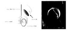
[実施例1] ニチノール・ベースの金属ガイドの用意、およびその作用
ニチノール・ベースの金属ガイド(Euroflex)の表面は、位置、例えば凹所を画定するように構成され、それらの凹所では、反応性基が配置され、生化学的作用が起きる(図1)。Example 1 Preparation of Nitinol-Based Metal Guide and Its Action The surface of the Nitinol-based metal guide (Euroflex) is configured to define a position, for example a recess, in which the reaction Sex groups are placed and biochemical effects occur (Figure 1).
前記「凹所」は、例えば、電気化学的エッチングおよびレーザー・アブレーションの前のレーザー・リソグラフィによる、集束イオン・ビーム・リソグラフィ(FIB、Xieら、Nuclear Instruments & Methods in Physics research Section B−beam Interactions with Materials and Atoms、211(3):363〜368頁、2003年)など、様々な方法を使用して得ることができる。 The “recess” is described in, for example, focused ion beam lithography (FIB, Xie et al., Nuclear Instruments & Methods in Physics Research B-beam Interactions by laser lithography prior to electrochemical etching and laser ablation. Materials and Atoms, 211 (3): 363-368, 2003).
FIB技術を使用して、機械がイオン・ビームを作り出し、そのイオン・ビームが、構成しなければならない表面に集束される。イオン・ビームの機械的な作用の下で、表面の材料の原子が、その表面から除かれる。適度な時間、エッチング・リーダー8μm3s−1、ビーム電流20nAで、FIB技術によって直径20μmの穴を形成することができる。図2に、直径5、20および40μm、深さ10および20μmの穴を示す。エッチング中に粉末材料を再堆積するので、穴の底部の表面は粗い。エッチング速度は、ビーム電流20nAで、直径40μmの円形領域で200nm min−1と測定された。その結果、エッチング速度は0.2μm3 nC−1(約5μm3 s−1)ということになり、これは、直径20μm、深さ20μmの穴を作製するための加工時間20分に相当する。表面粗さを改善するために、フッ素(XeF2)支援のフライス技術を用いた。次いで、非常に低い表面粗さが得られたが、XeF2がエッチング・ビームの軸に正確に沿っていなかったので、シャドーイング効果が確認された(図3)。Using FIB technology, the machine creates an ion beam that is focused on the surface that must be constructed. Under the mechanical action of the ion beam, surface material atoms are removed from the surface. A hole having a diameter of 20 μm can be formed by FIB technique with an etching leader of 8 μm3 s−1 and a beam current of 20 nA for an appropriate time. FIG. 2 shows holes with diameters of 5, 20 and 40 μm and depths of 10 and 20 μm. Since the powder material is redeposited during etching, the surface at the bottom of the hole is rough. The etching rate was measured to be 200 nm min−1 in a circular region with a diameter of 40 μm with a beam current of 20 nA. As a result, the etching rate is 0.2 μm3 nC−1 (about 5 μm3 s−1 ), which corresponds to a processing time of 20 minutes for producing a hole having a diameter of 20 μm and a depth of 20 μm. In order to improve the surface roughness, a fluorine (XeF2 ) assisted milling technique was used. A very low surface roughness was then obtained, but the shadowing effect was confirmed since XeF2 was not exactly along the axis of the etching beam (FIG. 3).
レーザー・リソグラフィ技術および電気化学的エッチングは、第1のステップで、表面をポリマー層で被覆することから構成される。第2のステップでは、ポリマー層が、レーザー・アブレーションを使用して機械加工される。第3のステップでは、その表面は、ポリマー層に設けられた開口部を通して、等方性電気化学的エッチングを使用してエッチングされる(図4)。図5に、ニチノール・ベースの金属ガイドの様々な構成試験の結果を示す。 Laser lithography techniques and electrochemical etching consist of coating the surface with a polymer layer in a first step. In the second step, the polymer layer is machined using laser ablation. In the third step, the surface is etched using an isotropic electrochemical etch through openings provided in the polymer layer (FIG. 4). FIG. 5 shows the results of various constitutive tests of Nitinol-based metal guides.
さらに、生体内で使用されるニチノール・ベースの金属ガイドは、通常、天然のNiTi酸化物層を生体適合性のTiO2層に置換する電気化学的ポリッシングによって加工される。穴を有する機械加工されたガイドは、穴の構成(図6)に対するこの方法の影響を推測するために、この方法を受けなければならない。In addition, nitinol-based metal guides used in vivo are typically processed by electrochemical polishing that replaces the native NiTi oxide layer with a biocompatible TiO2 layer. Machined guides with holes must undergo this method to infer the effect of this method on the hole configuration (FIG. 6).
ニチノール・ベースのガイドの表面上に空所を設けるための別の手法としては、レーザー・アブレーションを使用することが挙げられる。短レーザーの衝撃を利用すると、熱の発生によって周りの金属に影響を及ぼすことのない、金属の局所の蒸着が可能になる。記録された最も小さい寸法は20μm程度である。 Another technique for creating a void on the surface of a Nitinol-based guide is to use laser ablation. Utilizing the impact of a short laser allows the local deposition of metal without the heat generation affecting the surrounding metal. The smallest recorded dimension is about 20 μm.
本明細書で上述された3つの方法により凹所を作製することが可能である場合は、電気化学的エッチング方法が最良の結果を出した。 The electrochemical etching method gave the best results when it was possible to create the recesses by the three methods described herein above.
〔実施例2〕特定の器官への金属ガイドの生体内の挿入後の外傷
これらの実験の場合は、金属マイクロ・ガイド(MTI 0.012’’ Silver speed)を使用し、前記金属ガイドをマイクロ・カテーテル中に挿入した。[Example 2] Trauma after insertion of a metal guide into a specific organ in vivo In these experiments, a metal micro guide (MTI 0.012 "Silver speed) was used, -Inserted into the catheter.
そのデバイスを、脈管内のルートを通して(大腿動脈を介して)、吸引のレベルで、次いでスカルパのレベルから腎臓まで、全身麻酔下でピッグ中に導入した。このガイドを、動脈撮影による大腿動脈中で、前記デバイスを追従させて供給した。 The device was introduced into the pig under general anesthesia through the intravascular route (via the femoral artery) at the level of aspiration and then from the level of the scallop to the kidney. This guide was supplied by following the device in the femoral artery by arteriography.
デバイスは、腎臓の入口に配置されるときに、動脈内の侵襲によって腎臓中に導入される。組織中へのこうした穿通は、深さ数ミリメートルであり、前記デバイスを約10分間そこに維持した。 When placed at the entrance of the kidney, the device is introduced into the kidney by invasion within the artery. Such penetration into the tissue was a few millimeters deep and the device was maintained there for about 10 minutes.
最後に、デバイスを取り除いた。 Finally, the device was removed.
動物を安楽死させ、それらの腎臓を、本発明によるデバイスの穿通後の状況を推定するためにサンプリングした。 The animals were euthanized and their kidneys were sampled to estimate the situation after penetration of the device according to the invention.
その結果、腎臓に侵襲と関連する重大な出血がないことが示された。留意される最も重大な損傷は、侵襲位置のレベルの寸法3×1mmであった。 The results showed that the kidneys were free of significant bleeding associated with invasion. The most serious damage noted was the 3 × 1 mm dimension at the level of the invasive site.
したがって、本発明によるデバイスにより、非常にわずかな侵襲性でありながら器官へのアクセスを有することが可能になる。 Thus, the device according to the invention makes it possible to have access to organs with very little invasiveness.
〔実施例3〕肝臓へのデバイスの挿入後の微小外傷
金属マイクロ・ガイド(MTI 0.012’’ Silver speed)を使用した。その金属マイクロ・ガイドを、実施例1とは異なるファイバー・スコープ中に配置した。[Example 3] Microtrauma after insertion of device into liver A metal micro-guide (MTI 0.012 "Silver speed) was used. The metal micro guide was placed in a different fiber scope than Example 1.
そのデバイスを、動脈内ナビゲーションを通して(大腿動脈を介して)、吸引のレベルで、次いでスカルパのレベルから肝臓まで、全身麻酔下でピッグ中に導入した。この案内は、動脈撮影による大腿動脈中での前記デバイスの制御によって行った。 The device was introduced into the pig under general anesthesia through intra-arterial navigation (via the femoral artery) at the level of aspiration and then from the level of Scarpa to the liver. This guidance was performed by controlling the device in the femoral artery by arteriography.
デバイスを、肝臓の近くに配置するときにこの器官中に導入した。組織中へのこの穿通は、深さ数ミリメートルであり、前記デバイスを約10分間そこに維持した。 The device was introduced into this organ when placed near the liver. This penetration into the tissue was a few millimeters deep and the device was maintained there for about 10 minutes.
最後に、デバイスを取り除いた。 Finally, the device was removed.
前述のように、手術した肝臓のサンプリングにより、手術が器官に与える悪影響を推定することが可能になった。 As described above, sampling of the operated liver has made it possible to estimate the adverse effects of the operation on the organ.
肉眼で見えない損傷を、肝臓の表面に見ることができた。切開時に、被膜下面の寸法1.5×0.4cmおよび1.8×0.5cmの2つの実質内出血性の結節の存在を見ることができた。組織構造的に、他の顕著な異常なしに、洞様血管、門脈細静脈および中心小葉の静脈のうっ血を含む全ての面で肝臓の構造が維持される。 Damage that was not visible to the naked eye could be seen on the surface of the liver. At the time of the incision, it was possible to see the presence of two intraparenchymal nodules with dimensions 1.5 × 0.4 cm and 1.8 × 0.5 cm on the underside of the capsule. Histologically, the structure of the liver is maintained in all aspects including sinusoids, portal vein venules and central lobular vein congestion without any significant other abnormalities.
結論
結果として、出血性の損傷が最小限であることが示され、実質細胞の破壊なしに、毛細血管および中心小葉の静脈の単純なうっ血を有する、2つの軽微な微小損傷を見ることができた。Conclusions The results show that hemorrhagic damage is minimal, and two minor microinjuries with simple congestion of capillaries and central lobule veins can be seen without parenchymal cell destruction It was.
したがって、金属ガイドの利用により、軽微な外傷、場合よっては、生検の結果生じるより概して小さい外傷にすることが可能になる。 Thus, the use of a metal guide allows for minor trauma, and in general, less trauma that results from a biopsy.
〔実施例4〕中実のサポート上で抗原ACEの生体外の免疫捕捉および検出を可能にするデバイスの概念および生産のためのパラメータの考察
このデバイスは、抗原ACEを示すことを可能にするELISA技術の原理を使用する。この抗原上の異なるエピトープを認識する2つの単クローン抗体を、捕捉(AcM1)および抗原ACEの表出(AcM2)のために使用した。同じアイソトープ(IgG1)を有するこれらの単クローン抗体は、ビオチンに結合した単クローン抗体、およびストレプトアビジン・ペルオキシダーゼ複合体を使用して、ACE抗原の表出を行った(図7)。Example 4 Consideration of a Device Concept and Parameters for Production that Enables In Vitro Immune Capture and Detection of Antigen ACE on Solid Support This device is an ELISA that allows to display antigen ACE Use the principle of technology. Two monoclonal antibodies that recognize different epitopes on this antigen were used for capture (AcM1) and expression of antigen ACE (AcM2). These monoclonal antibodies with the same isotope (IgG1) performed ACE antigen expression using monoclonal antibodies conjugated to biotin and streptavidin peroxidase complex (FIG. 7).
2つのタイプのサポート、ELISA用プレートまたは剛体プラスチック・バットを使用した。 Two types of supports were used, ELISA plates or rigid plastic bats.
ELISA用プレート(Greiger)
炭酸塩/重炭酸塩の緩衝液で(1/5000および1/128000に)希釈した、100μlの抗原ACEに対する単クローン抗体(クローン5910またはクローン5905、マウスで産生されMedix Biochemicalによって市販されている)を各ウェルに配置し、プレートを37℃で1時間配置した。炭酸塩/重炭酸塩の対照溶液によって抗体を置換することによって1つの陰性対照が得られた。Plate for ELISA (Greiger)
Monoclonal antibody against 100 μl of antigen ACE (clone 5910 or clone 5905, produced in mice and marketed by Medix Biochemical) diluted in carbonate / bicarbonate buffer (1/5000 and 1/128000) Was placed in each well and the plate was placed at 37 ° C. for 1 hour. One negative control was obtained by replacing the antibody with a carbonate / bicarbonate control solution.
ウェル1箇所当たり250μlのPBSで3回洗浄した後で、プレートのフリー・サイトを、3%PBS−BSA(ウシ血清アルブミン)200mlで、37℃で2時間飽和した。 After washing 3 times with 250 μl PBS per well, the free sites on the plate were saturated with 200 ml of 3% PBS-BSA (bovine serum albumin) for 2 hours at 37 ° C.
次いで、0.5%PBS−Tween250μlでウェルを3回洗浄し、その後、PBS Tweenで1/10、1/100、1/1,000以内に希釈した抗原ACE陽性血清をウェル1箇所当たり100μl添加し、プレートを37℃で1時間培養した。 Next, the wells were washed 3 times with 250 μl of 0.5% PBS-Tween, and then 100 μl of antigen ACE positive serum diluted to within 1/10, 1/100, 1/1000 with PBS Tween was added per well. The plate was incubated at 37 ° C. for 1 hour.
ウェル1箇所当たり250μlのPBS−Tweenでの3回の洗浄を実行し、その後、PBS−Tweenで1/500以内にビオチン化した、抗原ACEに対する単クローン抗体(クローン5909、マウスで産生されMedix Biochemicalによって市販され、一定の親和性および認識したエピトープによって、以前の使用した捕捉抗体とは異なる)をウェル1箇所当たり100μl添加し、プレートをやはり37℃で1時間培養した。 Monoclonal antibody against the antigen ACE (clone 5909, Medix Biochemical produced in mice), washed 3 times with 250 μl PBS-Tween per well and then biotinylated within 1/500 with PBS-Tween (Depending on the constant affinity and recognized epitope, different from the previously used capture antibody) was added 100 μl per well and the plate was also incubated at 37 ° C. for 1 hour.
PBS−Tweenを使用して3回洗浄した後で、ペルオキシダーゼに結合した、1/2000以内に希釈した100μlのストレプトアビジン複合体を、各ウェルに添加し、37℃で1時間培養した。 After washing 3 times using PBS-Tween, 100 μl of streptavidin complex, diluted within 1/2000, bound to peroxidase, was added to each well and incubated at 37 ° C. for 1 hour.
PBS−Tweenで3回洗浄した後で、クエン酸−リン酸対照(pH5)に基質(H2O2)とクロモゲン(OPD、Sigma)の混合物をウェル1箇所当たり200μl添加することによって、表出が行われた。After washing three times with PBS-Tween, expression was achieved by adding 200 μl of a mixture of substrate (H2 O2 ) and chromogen (OPD, Sigma) per well to a citrate-phosphate control (pH 5). Was done.
同時に、抗原(ACE量5IU/ml未満の陰性対照)として「正常な」患者の血清を使用して、同じ作業を行った。 At the same time, the same work was performed using “normal” patient serum as the antigen (negative control with ACE amount less than 5 IU / ml).
そのとき、ウェル1箇所当たり1M硫酸を50μl添加することによって、反応を止めた。吸着は、プレート・リーダー上で492nmで読み取った(参照:ELX.800UV)。 At that time, the reaction was stopped by adding 50 μl of 1 M sulfuric acid per well. Adsorption was read at 492 nm on a plate reader (Ref: ELX.800 UV).
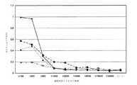
免疫捕捉のための単クローン抗体5910(1/500以内に、その後1/128000まで1/2ずつ段階的に希釈した)を使用して得られた結果、およびビオチン化単クローン抗体5909による表出を、抗原ACE陽性血清の場合を図8に、抗原ACE陰性血清の場合を図9に示す。 Results obtained using monoclonal antibody 5910 for immunocapture (within 1/500, then serially diluted 1/2 by 1/2 to 1/18000) and expression with biotinylated monoclonal antibody 5909 FIG. 8 shows the case of antigen ACE-positive serum, and FIG. 9 shows the case of antigen ACE-negative serum.
免疫捕捉のための単クローン抗体5905(1/100、1/200、1/500以内に、その後1/32000まで1/2ずつ段階的に希釈した)を使用して得られた結果、およびビオチン化単クローン抗体5909を使用した表出を、抗原ACE陽性血清の場合を図10に、抗原ACE陰性血清の場合を図11に示す。
図8から図11の見出し:
たて座標:492nmでの吸着(DO)
よこ座標:捕捉抗体(5910または5905)の希釈
◆ACE陽性血清の1/10以内の希釈液
■ACE陽性血清の1/100以内の希釈液
△ACE陽性血清の1/1000以内の希釈液
×=血清なしResults obtained using monoclonal antibody 5905 for immunocapture (within 1/100, 1/200, 1/500, and then serially diluted 1/2 to 1/32000), and biotin Expression using the monoclonal antibody 5909 is shown in FIG. 10 for the antigen ACE positive serum and FIG. 11 for the antigen ACE negative serum.
8 to 11 headings:
Vertical coordinate: Adsorption at 492 nm (DO)
Horizontal coordinate: Dilution of capture antibody (5910 or 5905) ◆ Dilution within 1/10 of ACE positive serum ■ Dilution within 1/100 of ACE positive serum ΔDilution within 1/1000 of ACE positive serum × = Without serum
結果として、捕捉単クローン抗体を1/500以内で使用したときに、1/10以内の抗原ACE陽性血清が0.5より高い吸着(DO)をもたらす(図8)ことが示される。同じ条件下で、抗原ACE陰性血清は0.15未満のDOをもたらす(図9)。 As a result, it is shown that when the captured monoclonal antibody is used within 1/500, antigen ACE-positive serum within 1/10 results in an adsorption (DO) higher than 0.5 (FIG. 8). Under the same conditions, antigen ACE-negative serum yields a DO of less than 0.15 (Figure 9).
しかし、捕捉単クローン抗体5910と検出単クローン抗体5909の結合(図8および図9)より、捕捉単クローン抗体5905と検出単クローン抗体5909の結合(図10および図11)でより良い結果が得られたことに留意されたい。実際のところ、1/10以内に希釈した抗原ACE陽性血清の場合にDOが1である(図10)ことを述べたが、抗原ACE陰性血清が同じ条件下で0.1のDOをもたらす(図11)。これらの結果を、様々な希釈液の捕捉単クローン抗体5910を使用して確認した(データは示さない)。 However, the binding of capture monoclonal antibody 5905 and detection monoclonal antibody 5909 (FIGS. 10 and 11) gives better results than the binding of capture monoclonal antibody 5910 and detection monoclonal antibody 5909 (FIGS. 8 and 9). Please note that In fact, it was stated that DO was 1 for antigen ACE positive sera diluted within 1/10 (FIG. 10), but antigen ACE negative sera yielded a DO of 0.1 under the same conditions ( FIG. 11). These results were confirmed using capture monoclonal antibody 5910 at various dilutions (data not shown).
従来の剛体のサポート
第1のステップでは、長さが2から3cm、直径が0.5から1mmのバットの形態の剛体プラスチック・サポートを作動させた。Conventional rigid support In the first step, a rigid plastic support in the form of a bat having a length of 2 to 3 cm and a diameter of 0.5 to 1 mm was activated.
第2のステップで、このように作動させたサポートを、1mlの溶血マイクロ・チューブ(Fisher)に配置し、抗原ACEに対する単クローン抗体(クローン5910、マウスで産生されMedix Biochemicalによって市販されている)で機能化し、37℃で1時間、炭酸塩/重炭酸塩(250μl/チューブ)対照で1/50、1/100、1/250、1/500以内に希釈した。単クローン抗体を炭酸塩/重炭酸塩対照に代えることによって陰性対照を行った。固定および洗浄の後に、+4℃の温度で1晩、3%PBS−BSA500μlで飽和が得られた。 In the second step, the support thus actuated is placed in a 1 ml hemolytic microtube (Fisher) and a monoclonal antibody against the antigen ACE (clone 5910, produced in mice and marketed by Medix Biochemical). And diluted to 1/50, 1/100, 1/250, 1/500 with carbonate / bicarbonate (250 μl / tube) control for 1 hour at 37 ° C. A negative control was performed by replacing the monoclonal antibody with a carbonate / bicarbonate control. After fixation and washing, saturation was obtained with 500 μl of 3% PBS-BSA overnight at a temperature of + 4 ° C.
次いで、1/10、1/100以内にPBSまたは「健康な」対象(抗原ACE陰性対照)からの血清で、37℃で1時間、同じ希釈液で希釈した抗原ACE陽性血清250μlでサポートを培養した。 The support was then cultured with serum from PBS or “healthy” subjects (antigen ACE negative control) within 1/10, 1/100, 250 μl of antigen ACE positive serum diluted with the same dilution at 37 ° C. for 1 hour. did.
1mg/ml以内の抗原ACEに対する単クローン抗体(クローン5909、マウスで産生されMedix Biochemicalによって市販されており、一定の親和性および承認されたエピトープにより精製したクローン5910とは異なる)を、0.1Mホウ酸塩の対照、pH8.8に対して4℃で1晩透析した。次いで、10mg/mlDMSOビオチン溶液を50μg/mgの抗体に添加した。室温で4時間培養した後で、攪拌下で、20μl/250μgの量のビオチンに1M塩化アンモニウムを添加し、得られた溶液を室温で再度1分培養した。反応が止まる際に、マークした抗体をPBSに対して24時間、+4℃で透析し、このマークした抗体を−20℃でアリコートとして維持した。 Monoclonal antibody against antigen ACE within 1 mg / ml (clone 5909, different from clone 5910, produced in mice and marketed by Medix Biochemical, purified with constant affinity and approved epitope) Dialyzed overnight at 4 ° C. against the borate control, pH 8.8. Then 10 mg / ml DMSO biotin solution was added to 50 μg / mg antibody. After culturing at room temperature for 4 hours, 1M ammonium chloride was added to biotin in an amount of 20 μl / 250 μg under stirring, and the resulting solution was incubated again at room temperature for 1 minute. When the reaction stopped, the marked antibody was dialyzed against PBS for 24 hours at + 4 ° C., and the marked antibody was maintained as an aliquot at −20 ° C.
PBS−Tweenで3回洗浄した後で、サポートを、250μlのビオチン化抗体5909で培養し、37℃で1時間、PBS−Tweenで1/500以内に希釈した。 After washing 3 times with PBS-Tween, the support was incubated with 250 μl of biotinylated antibody 5909 and diluted within 1/500 with PBS-Tween for 1 hour at 37 ° C.
ビオチンの検出(6−ビオチンアミドカプロイルアミドカプロン酸およびN−ヒドロキシこはく酸イミドのエステル、Sigma)を、37℃で1時間、PBSで1/2000以内に希釈したストレプトアビジン−ペルオキシダーゼ複合体(Amersham Biosciences)を使用して証明した。 Detection of biotin (6-biotinamidocaproylamidocaproic acid and ester of N-hydroxysuccinimide, Sigma) was diluted within 1/2000 with PBS for 1 hour at 37 ° C., streptavidin-peroxidase complex (Amersham) Biosciences).
クエン酸−リン酸対照(pH5)にチューブ当たり750μlの基質(H2O2)とクロモゲン(OPD、Sigma)の混合物を添加することによって、酵素の作用の表出を実行した。Expression of enzyme action was performed by adding 750 μl of a mixture of substrate (H2 O2 ) and chromogen (OPD, Sigma) per tube to a citrate-phosphate control (pH 5).
次いで、1M硫酸を添加することによって反応を止めた。吸着は492nmで読み取った。 The reaction was then stopped by adding 1M sulfuric acid. Adsorption was read at 492 nm.
プラスチック製バットのELISAの結果を図12に示す。
図12の見出し:
たて座標:492nmでの吸着(DO)
よこ座標:捕捉抗体5910の希釈
◆抗原ACEの1/10以内の希釈液
■抗原ACEの1/100以内の希釈液
△陰性対照1/10以内の希釈液
×陰性対照1/100以内の希釈液FIG. 12 shows the result of the ELISA of the plastic bat.
Figure 12 headings:
Vertical coordinate: Adsorption at 492 nm (DO)
Horizontal coordinate: Dilution of capture antibody 5910 ◆ Dilution within 1/10 of antigen ACE ■ Dilution within 1/100 of antigen ACE ΔDilution within 1/10 of negative control × Dilution within 1/100 of negative control
概して、結果は、抗原ACE陰性血清で得られたものより、抗原ACE陽性血清の場合にDOが7から10倍高いことを示す。 In general, the results show that DO is 7 to 10 times higher for antigen ACE positive sera than that obtained with antigen ACE negative sera.
1/50または1/100以内に希釈した捕捉単クローン抗体5910が固定された剛体プラスチック・サポートで最良の結果が得られた。 Best results were obtained with a rigid plastic support to which capture monoclonal antibody 5910 diluted within 1/50 or 1/100 was immobilized.
単クローン抗体5909の検出希釈液が1/500以内であるときに、検出した最良の抗原ACE濃度は、1/100、すなわち6IU/ml(「正常」と考えられる割合に近い:5IU/ml未満)以内に希釈した患者の血清に相当する。 When the detection dilution of monoclonal antibody 5909 is within 1/500, the best antigen ACE concentration detected is 1/100, ie 6 IU / ml (close to the rate considered “normal”: less than 5 IU / ml Corresponds to patient serum diluted within).
プラスチック・サポートを使用することにより、上述のプロトコルによる機能化された金属バット上の免疫捕捉プロセスの特異性および感度を確証することが可能になる。 By using a plastic support, it becomes possible to confirm the specificity and sensitivity of the immunocapture process on a functionalized metal bat according to the protocol described above.
結論
生体外免疫捕捉および表出技術による抗原ACEの検出のために得られた良好な結果は、「機能化したバット」デバイスの評価を確証し、そのため、生体外の表出の後の生体内での抗原ACEの捕捉が可能になる。Conclusion The good results obtained for the detection of antigen ACE by in vitro immune capture and expression techniques confirm the evaluation of the “functionalized vat” device, so that in vivo after in vitro expression It is possible to capture the antigen ACE.
〔実施例5〕例えば、イメージング技術、より具体的には放射線技術の制御下における、乳房腫瘍のマーカーACEの発現の特定
特許出願PCT WO03/006948に記載されているプロトコルによれば、第1のステップで、アルカンチオール層が、第1のステップで、ニチノール・ベースの金属ガイド(Euroflex)の端部のうち一方に吸着される。第2のステップでは、この層の遊離チオール官能基により、抗原ACEに対する単クローン抗体でジスルフィド架橋を形成することが可能になる。Example 5 Identification of the expression of a breast tumor marker ACE under the control of, for example, imaging technology, more specifically radiation technology According to the protocol described in patent application PCT WO 03/006948, the first In step, the alkanethiol layer is adsorbed on one of the ends of the nitinol-based metal guide (Euroflex) in the first step. In the second step, the free thiol functional groups in this layer make it possible to form disulfide bridges with monoclonal antibodies against the antigen ACE.
次いで、得られた金属製ガイドを、動物またはヒトで使用するように適合された生検針に導入する。 The resulting metal guide is then introduced into a biopsy needle adapted for use in animals or humans.
乳癌を患う患者から取り除いた後に、こうしたデバイスを使用して、手術する腫瘍(乳腺腫瘍)に即座の解剖病理学的検査を行う。代替の溶液によれば、医療倫理条件が存在するときは、乳癌を患う患者に局所または全身麻酔下で乳房に微小切開を行う。抗原ACEに対する抗体に結合した金属ガイドが挿入される針は、腫瘍中に導入されるか、または微小切開によって、次いで、画像、より具体的にはエコーグラフによって、ガイドの前進を追従しながら腫瘍まで案内される。 After removal from a patient suffering from breast cancer, such a device is used to perform an immediate anatomical pathological examination of the tumor to be operated on (mammary tumor). According to alternative solutions, a microincision is made in the breast under local or general anesthesia for patients suffering from breast cancer when medical ethical conditions exist. A needle into which a metal guide conjugated to an antibody against the antigen ACE is inserted is introduced into the tumor, or by microincision and then by an image, more specifically an echograph, following the advancement of the guide and the tumor Will be guided to.
次いで、前記低侵襲性の案内システムにより、抗原ACEに対する抗体に結合された金属ガイドの端部を取り出すことが可能になる。次いで、金属ガイドの端部を、腫瘍中に(穿孔によって)数ミリメートル程度の深さで導入する。任意選択で腫瘍によって発現される抗原ACEによって免疫捕捉が可能になる10分程度の待ち時間の後で、デバイスが取り除かれる。 The minimally invasive guidance system can then remove the end of the metal guide bound to the antibody against the antigen ACE. The end of the metal guide is then introduced into the tumor (by drilling) at a depth of the order of a few millimeters. The device is removed after a waiting time of about 10 minutes, optionally allowing immunocapture by the antigen ACE expressed by the tumor.
マイクロ・サンプリングは検体の生体内の免疫捕捉に限定され、生検を必要としない。 Microsampling is limited to in vivo immune capture of specimens and does not require biopsy.
最後に、デバイスを取り除き、次いで、マーカーACEのELISA定量法を、マーカーACEに対する単クローン抗体を有するデバイスの端部で実行し、そのマーカーACEは、抗原ACEに関する一定の親和性、および識別されたエピトープによって捕捉抗体と区別し、ビオチンに結合する。 Finally, the device was removed and then an ELISA quantification method of marker ACE was performed at the end of the device with a monoclonal antibody against marker ACE, which marker ACE was identified with a certain affinity for antigen ACE, and It is distinguished from the capture antibody by the epitope and binds to biotin.
ストレプトアビジン−ペルオキシダーゼ複合体を使用することによる酵素の作用の表出により、腫瘍によるマーカーACEの発現、およびそれに応じて患者に最良の治療を与えるために使用する治療を調整することが可能になる。 Expression of the action of the enzyme by using a streptavidin-peroxidase complex makes it possible to adjust the expression of the marker ACE by the tumor and the treatment used to give the patient the best treatment accordingly. .
〔実施例6〕皮膚癌
特許出願PCT WO03/006948に記載されているプロトコルによれば、アルカンチオールの層は、第1のステップで、ニチノール・ベースの金属ガイド(Euroflex)の端部に吸着されている。第2のステップでは、この層の遊離チオール官能基により、FAPマーカーに対する単クローン抗体(Fibroblast−activation protain;RETTIGら、Proc.Natl.Acad.Sci.USA、vol.85、3110頁、1988年)でのジスルフィド架橋の形成が可能になる。Example 6 Skin cancer According to the protocol described in patent application PCT WO03 / 006948, a layer of alkanethiol is adsorbed on the end of a Nitinol-based metal guide (Euroflex) in a first step. ing. In the second step, the free thiol functional group of this layer was used to generate a monoclonal antibody against the FAP marker (Fibroblast-activation protein; RETTIG et al., Proc. Natl. Acad. Sci. USA, vol. 85, 3110, 1988). Enables the formation of disulfide bridges at
次いで、従来のまたは即座の解剖病理学的検査のために、得られた金属ガイドを、医療倫理条件が存在するときは動物またはヒトの皮膚腫瘍のレベルで、またはそのアブレーションの後には皮膚癌を患う患者の皮膚腫瘍のレベルで導入する。 Then, for conventional or immediate anatomical pathology examination, the resulting metal guides should be tested for skin cancer at the level of animal or human skin tumors when medical ethical conditions exist, or after their ablation. Introduce at the level of skin tumors in affected patients.
マイクロ・サンプリングは生体内の免疫捕捉に限定され、特定の生検を必要としない。 Microsampling is limited to in vivo immune capture and does not require a specific biopsy.
最後に、デバイスを取り除き、ペルオキシダーゼに結合したマーカーFABに対する単クローン抗体で、デバイス端部にFABマーカーのELISA定量法を実行する。 Finally, the device is removed and an ELISA assay of the FAB marker is performed at the end of the device with a monoclonal antibody against the marker FAB conjugated to peroxidase.
ペルオキシダーゼの活性の表出により、腫瘍によるマーカーFAPの発現を導き出し、それに応じて、患者に最良の治療を施すために使用する治療法を調整することが可能になる。 The expression of peroxidase activity leads to the expression of the marker FAP by the tumor and it is possible to adjust the therapy used to give the patient the best treatment accordingly.
Claims (27)
Translated fromJapanese表面が、少なくとも一連の凹所を画定するように構成され、それらの凹所に、前記基質に特異な少なくとも1つの反応性基が直接結合され、穿孔するEa先端部と、
前記金属ガイドを操作することが意図された別のEm先端部と
を含むことを特徴とする、分析デバイス。A system for minimally invasive and / or micro-sampling testing of a substrate, said system comprising at least one metal guide,
An Ea tip that is configured to define at least a series of recesses in which at least one reactive group specific to the substrate is directly attached and pierced;
Analytical device, characterized in that it comprises another Em tip intended to manipulate the metal guide.
a)請求項1乃至15のいずれか一項に記載のデバイスの機能化されたEa先端部を、前記先端部が検査する前記組織または器官と接触しているときに、前記基質に特異な少なくとも1つの検出剤を含む溶液で培養するステップと、
b)前記基質を検出するステップと
を含むことを特徴とする、方法。A method for detecting a substrate present in a tissue or organ in vitro,
a) a functionalized Ea tip of the device according to any one of claims 1 to 15 when the tip is in contact with the tissue or organ to be examined, at least specific for the substrate Culturing in a solution containing one detection agent;
b) detecting the substrate.
Applications Claiming Priority (3)
| Application Number | Priority Date | Filing Date | Title |
|---|---|---|---|
| EP08290179.4 | 2008-02-26 | ||
| EP08290179AEP2095762B1 (en) | 2008-02-26 | 2008-02-26 | Device for in vivo micro-invasive investigation comprising a metal guide |
| PCT/EP2009/001308WO2009106295A1 (en) | 2008-02-26 | 2009-02-24 | In vivo micro-invasive investigation device including a metal guide |
Publications (2)
| Publication Number | Publication Date |
|---|---|
| JP2011514815Atrue JP2011514815A (en) | 2011-05-12 |
| JP5620277B2 JP5620277B2 (en) | 2014-11-05 |
Family
ID=39830196
Family Applications (1)
| Application Number | Title | Priority Date | Filing Date |
|---|---|---|---|
| JP2010547125AExpired - Fee RelatedJP5620277B2 (en) | 2008-02-26 | 2009-02-24 | In-vivo minimally invasive testing device including a metal guide |
Country Status (9)
| Country | Link |
|---|---|
| US (1) | US8969010B2 (en) |
| EP (1) | EP2095762B1 (en) |
| JP (1) | JP5620277B2 (en) |
| KR (1) | KR101624755B1 (en) |
| CN (2) | CN103932678B (en) |
| AT (1) | ATE508679T1 (en) |
| CA (1) | CA2716751C (en) |
| ES (1) | ES2369472T3 (en) |
| WO (1) | WO2009106295A1 (en) |
Families Citing this family (8)
| Publication number | Priority date | Publication date | Assignee | Title |
|---|---|---|---|---|
| JP3040061B2 (en) | 1995-03-28 | 2000-05-08 | 重秋 軍司 | Clothes rod hanging tool and clothesline |
| JP3024057B2 (en) | 1995-04-05 | 2000-03-21 | 功 清野 | Clothes dryer |
| ES2369472T3 (en)* | 2008-02-26 | 2011-12-01 | Biostems Ltd. | MICRO-INVASIVE LIVE RESEARCH DEVICE THAT INCLUDES A METAL GUIDE. |
| DE102013211837A1 (en)* | 2013-06-21 | 2014-12-24 | Gilupi Gmbh | Catheter with detection device for the real-time detection of a sample material |
| CN104970806B (en)* | 2015-07-12 | 2017-08-04 | 北京泱深生物信息技术有限公司 | A kind of smart machine for cancer return real-time dynamic monitoring |
| ES2722802B2 (en)* | 2018-02-14 | 2019-12-18 | Fund De Neurociencias | DEVICE FOR THE SELECTIVE ELIMINATION OF MOLECULES OF FABRICS OR FLOWS |
| CN110755116A (en)* | 2019-11-29 | 2020-02-07 | 南京大学 | A device for capturing fetal eukaryotic red blood cells in situ from the mother and its application |
| CN111956269B (en)* | 2020-08-20 | 2023-11-28 | 上海蓓蕊医疗科技有限公司 | Skin protector |
Citations (9)
| Publication number | Priority date | Publication date | Assignee | Title |
|---|---|---|---|---|
| JPS62218844A (en)* | 1986-02-27 | 1987-09-26 | イ−ライ・リリ−・アンド・カンパニ− | Optical fiber device |
| US20030049679A1 (en)* | 2000-03-17 | 2003-03-13 | Alain Pompidou | Device for in situ analysis and/or treatment consisting of a flexible rod and micro system fixed at one end of said flexible rod |
| US20040024333A1 (en)* | 2002-07-26 | 2004-02-05 | Stuart Brown | Tissue and fluid sampling device |
| US20040049679A1 (en)* | 2000-11-21 | 2004-03-11 | Claude Meggle | Authenticating method and device |
| JP2004523257A (en)* | 2000-11-09 | 2004-08-05 | サイセル・テクノロジーズ,インコーポレイテッド | Methods, circuits and compositions of matter for detecting biomolecular concentrates in vivo using fluorescent tags |
| JP2005512021A (en)* | 2001-07-09 | 2005-04-28 | アリゾナ ボード オブ リージェンツ | Affinity biosensor for monitoring biological processes |
| JP2005312757A (en)* | 2004-04-30 | 2005-11-10 | Takashi Yoshitake | Method of sampling intracerebral macromolecule |
| WO2006082344A1 (en)* | 2005-02-02 | 2006-08-10 | Commissariat A L'energie Atomique | Device for contact molecular sampling |
| WO2006081947A1 (en)* | 2005-02-03 | 2006-08-10 | Lts Lohmann Therapie-Systeme Ag | Biopsy needle for the histological examination of body tissue |
Family Cites Families (10)
| Publication number | Priority date | Publication date | Assignee | Title |
|---|---|---|---|---|
| DE3332924A1 (en) | 1983-09-13 | 1985-03-21 | Bayer Ag, 5090 Leverkusen | NOTCH IMPACT TOE, MELT-FREE-FLOWING POLYAMIDE MOLDS |
| US5938595A (en)* | 1996-05-24 | 1999-08-17 | The Regents Of The University Of California | Fiber optic D dimer biosensor |
| US7622294B2 (en)* | 1997-03-14 | 2009-11-24 | Trustees Of Tufts College | Methods for detecting target analytes and enzymatic reactions |
| US6689604B1 (en)* | 1998-03-20 | 2004-02-10 | National Research Council Of Canada | Lipopolysaccharide α-2,3 sialyltransferase of Campylobacter jejuni and its uses |
| US7025734B1 (en)* | 2001-09-28 | 2006-04-11 | Advanced Cardiovascular Systmes, Inc. | Guidewire with chemical sensing capabilities |
| US7259019B2 (en)* | 2002-03-11 | 2007-08-21 | Pawliszyn Janusz B | Multiple sampling device and method for investigating biological systems |
| KR20140134338A (en)* | 2003-09-11 | 2014-11-21 | 테라노스, 인코포레이티드 | Medical device for analyte monitoring and drug delivery |
| US20050153309A1 (en)* | 2003-12-22 | 2005-07-14 | David Hoon | Method and apparatus for in vivo surveillance of circulating biological components |
| US20070287991A1 (en)* | 2006-06-08 | 2007-12-13 | Mckay William F | Devices and methods for detection of markers of axial pain with or without radiculopathy |
| ES2369472T3 (en)* | 2008-02-26 | 2011-12-01 | Biostems Ltd. | MICRO-INVASIVE LIVE RESEARCH DEVICE THAT INCLUDES A METAL GUIDE. |
- 2008
- 2008-02-26ESES08290179Tpatent/ES2369472T3/enactiveActive
- 2008-02-26ATAT08290179Tpatent/ATE508679T1/ennot_activeIP Right Cessation
- 2008-02-26EPEP08290179Apatent/EP2095762B1/enactiveActive
- 2009
- 2009-02-24CNCN201410136896.6Apatent/CN103932678B/enactiveActive
- 2009-02-24USUS12/919,655patent/US8969010B2/enactiveActive
- 2009-02-24JPJP2010547125Apatent/JP5620277B2/ennot_activeExpired - Fee Related
- 2009-02-24CNCN200980112079.4Apatent/CN102014734B/enactiveActive
- 2009-02-24KRKR1020107021340Apatent/KR101624755B1/ennot_activeExpired - Fee Related
- 2009-02-24CACA2716751Apatent/CA2716751C/enactiveActive
- 2009-02-24WOPCT/EP2009/001308patent/WO2009106295A1/enactiveApplication Filing
Patent Citations (11)
| Publication number | Priority date | Publication date | Assignee | Title |
|---|---|---|---|---|
| JPS62218844A (en)* | 1986-02-27 | 1987-09-26 | イ−ライ・リリ−・アンド・カンパニ− | Optical fiber device |
| US20030049679A1 (en)* | 2000-03-17 | 2003-03-13 | Alain Pompidou | Device for in situ analysis and/or treatment consisting of a flexible rod and micro system fixed at one end of said flexible rod |
| JP2004523257A (en)* | 2000-11-09 | 2004-08-05 | サイセル・テクノロジーズ,インコーポレイテッド | Methods, circuits and compositions of matter for detecting biomolecular concentrates in vivo using fluorescent tags |
| US20040049679A1 (en)* | 2000-11-21 | 2004-03-11 | Claude Meggle | Authenticating method and device |
| JP2005512021A (en)* | 2001-07-09 | 2005-04-28 | アリゾナ ボード オブ リージェンツ | Affinity biosensor for monitoring biological processes |
| US20040024333A1 (en)* | 2002-07-26 | 2004-02-05 | Stuart Brown | Tissue and fluid sampling device |
| JP2005312757A (en)* | 2004-04-30 | 2005-11-10 | Takashi Yoshitake | Method of sampling intracerebral macromolecule |
| WO2006082344A1 (en)* | 2005-02-02 | 2006-08-10 | Commissariat A L'energie Atomique | Device for contact molecular sampling |
| JP2008528224A (en)* | 2005-02-02 | 2008-07-31 | コミツサリア タ レネルジー アトミーク | Contact molecular sampling device |
| WO2006081947A1 (en)* | 2005-02-03 | 2006-08-10 | Lts Lohmann Therapie-Systeme Ag | Biopsy needle for the histological examination of body tissue |
| JP2008528217A (en)* | 2005-02-03 | 2008-07-31 | エルテーエス ローマン テラピー−ジステーメ アーゲー | Biopsy needle for histological examination of body tissue |
Also Published As
| Publication number | Publication date |
|---|---|
| HK1155928A1 (en) | 2012-06-01 |
| CA2716751C (en) | 2016-03-08 |
| EP2095762A1 (en) | 2009-09-02 |
| ES2369472T3 (en) | 2011-12-01 |
| CA2716751A1 (en) | 2009-09-03 |
| KR101624755B1 (en) | 2016-05-26 |
| CN102014734B (en) | 2014-05-07 |
| US20110092783A1 (en) | 2011-04-21 |
| ATE508679T1 (en) | 2011-05-15 |
| CN103932678B (en) | 2017-10-24 |
| EP2095762B1 (en) | 2011-05-11 |
| WO2009106295A1 (en) | 2009-09-03 |
| KR20110005685A (en) | 2011-01-18 |
| US8969010B2 (en) | 2015-03-03 |
| CN103932678A (en) | 2014-07-23 |
| JP5620277B2 (en) | 2014-11-05 |
| CN102014734A (en) | 2011-04-13 |
Similar Documents
| Publication | Publication Date | Title |
|---|---|---|
| JP5620277B2 (en) | In-vivo minimally invasive testing device including a metal guide | |
| US10987037B2 (en) | Method and apparatus for in vivo surveillance of circulating biological components | |
| French et al. | Sensors for catheter applications | |
| US20050153379A1 (en) | Method and apparatus for in vivo collection of circulating biological components | |
| JP2014527866A (en) | Method for detecting portal vein and / or hepatic vein pressure and monitoring system for portal hypertension | |
| JP2001523492A (en) | Recommunication device, apparatus and method used therein | |
| JP2023522182A (en) | Vasculature visualization | |
| JP4533615B2 (en) | Puncture needle and ultrasonic endoscope system | |
| JP2019162416A (en) | Variable pitch flexible needle | |
| Sikkander et al. | A review of diagnostic nano stents: Part (I) | |
| JP2021532926A (en) | Intraluminal device with capacitive pressure sensor | |
| KR102077660B1 (en) | Ultrasound-guided direct needle engagement for flush-occluded superficial femoral artery, and using method thereof | |
| HK1195231B (en) | In vivo micro-invasive investigation device including a metal guide | |
| US20230320695A1 (en) | Catheter-type ultrasound endoscope and examination system including same | |
| HK1195231A (en) | In vivo micro-invasive investigation device including a metal guide | |
| HK1155928B (en) | In vivo micro-invasive investigation device including a metal guide | |
| JP6379363B1 (en) | Ultrasonic image display device | |
| WO2018116891A1 (en) | Image processing device, ultrasonic diagnostic system, method for operating image processing device, and program for operating image processing device | |
| Yoon et al. | Semi-implantable polyimide/PTFE needle-shaped biosensor for continuous glucose monitoring | |
| French et al. | Sensors for catheter applications | |
| WO2022243714A1 (en) | Depth-surface imaging device for registering ultrasound images to each other and to surface images by using surface information | |
| Gilbert et al. | Endoscopic ultrasound | |
| Feng et al. | PTA balloon catheter integrated electroactive polymer transducer for sensing vascular blockage and disturbing vessel plaques | |
| Tanaka et al. | Evaluation of narrow band imaging for peroral cholangiopancreatoscopy | |
| JPH04183477A (en) | Catheter and manufacture thereof |
Legal Events
| Date | Code | Title | Description |
|---|---|---|---|
| A621 | Written request for application examination | Free format text:JAPANESE INTERMEDIATE CODE: A621 Effective date:20120203 | |
| A131 | Notification of reasons for refusal | Free format text:JAPANESE INTERMEDIATE CODE: A131 Effective date:20131111 | |
| A131 | Notification of reasons for refusal | Free format text:JAPANESE INTERMEDIATE CODE: A131 Effective date:20140207 | |
| A521 | Request for written amendment filed | Free format text:JAPANESE INTERMEDIATE CODE: A523 Effective date:20140424 | |
| TRDD | Decision of grant or rejection written | ||
| A01 | Written decision to grant a patent or to grant a registration (utility model) | Free format text:JAPANESE INTERMEDIATE CODE: A01 Effective date:20140905 | |
| A61 | First payment of annual fees (during grant procedure) | Free format text:JAPANESE INTERMEDIATE CODE: A61 Effective date:20140918 | |
| R150 | Certificate of patent or registration of utility model | Ref document number:5620277 Country of ref document:JP Free format text:JAPANESE INTERMEDIATE CODE: R150 | |
| R250 | Receipt of annual fees | Free format text:JAPANESE INTERMEDIATE CODE: R250 | |
| R250 | Receipt of annual fees | Free format text:JAPANESE INTERMEDIATE CODE: R250 | |
| R250 | Receipt of annual fees | Free format text:JAPANESE INTERMEDIATE CODE: R250 | |
| R250 | Receipt of annual fees | Free format text:JAPANESE INTERMEDIATE CODE: R250 | |
| R250 | Receipt of annual fees | Free format text:JAPANESE INTERMEDIATE CODE: R250 | |
| R250 | Receipt of annual fees | Free format text:JAPANESE INTERMEDIATE CODE: R250 | |
| R250 | Receipt of annual fees | Free format text:JAPANESE INTERMEDIATE CODE: R250 | |
| LAPS | Cancellation because of no payment of annual fees |