JP2011113056A - Device for simulating operation using mirror - Google Patents
Device for simulating operation using mirrorDownload PDFInfo
- Publication number
- JP2011113056A JP2011113056AJP2009272137AJP2009272137AJP2011113056AJP 2011113056 AJP2011113056 AJP 2011113056AJP 2009272137 AJP2009272137 AJP 2009272137AJP 2009272137 AJP2009272137 AJP 2009272137AJP 2011113056 AJP2011113056 AJP 2011113056A
- Authority
- JP
- Japan
- Prior art keywords
- box
- model
- endoscopic
- simulation apparatus
- shaped body
- Prior art date
- Legal status (The legal status is an assumption and is not a legal conclusion. Google has not performed a legal analysis and makes no representation as to the accuracy of the status listed.)
- Granted
Links
- 238000004088simulationMethods0.000claimsabstractdescription47
- 125000006850spacer groupChemical group0.000claimsabstractdescription23
- 210000001015abdomenAnatomy0.000claimsabstractdescription15
- 210000003815abdominal wallAnatomy0.000claimsabstractdescription13
- 238000002674endoscopic surgeryMethods0.000claimsdescription33
- 210000000056organAnatomy0.000claimsdescription22
- 238000003780insertionMethods0.000claimsdescription10
- 230000037431insertionEffects0.000claimsdescription10
- 238000001356surgical procedureMethods0.000claimsdescription10
- 238000003384imaging methodMethods0.000claimsdescription9
- 230000001681protective effectEffects0.000claimsdescription6
- 238000012549trainingMethods0.000abstractdescription18
- 210000001835visceraAnatomy0.000abstractdescription6
- 210000002784stomachAnatomy0.000description30
- 238000002357laparoscopic surgeryMethods0.000description16
- 230000003187abdominal effectEffects0.000description15
- 238000000034methodMethods0.000description10
- 210000000683abdominal cavityAnatomy0.000description8
- 210000002747omentumAnatomy0.000description8
- 210000001198duodenumAnatomy0.000description7
- 239000000463materialSubstances0.000description5
- CURLTUGMZLYLDI-UHFFFAOYSA-NCarbon dioxideChemical compoundO=C=OCURLTUGMZLYLDI-UHFFFAOYSA-N0.000description4
- 230000000694effectsEffects0.000description3
- 239000013013elastic materialSubstances0.000description3
- 210000003734kidneyAnatomy0.000description3
- 210000000496pancreasAnatomy0.000description3
- 229910002092carbon dioxideInorganic materials0.000description2
- 239000001569carbon dioxideSubstances0.000description2
- 238000004891communicationMethods0.000description2
- 239000006260foamSubstances0.000description2
- 210000004185liverAnatomy0.000description2
- 238000000926separation methodMethods0.000description2
- 208000032544CicatrixDiseases0.000description1
- JOYRKODLDBILNP-UHFFFAOYSA-NEthyl urethaneChemical compoundCCOC(N)=OJOYRKODLDBILNP-UHFFFAOYSA-N0.000description1
- 206010028980NeoplasmDiseases0.000description1
- 208000005646PneumoperitoneumDiseases0.000description1
- 230000015572biosynthetic processEffects0.000description1
- 201000011510cancerDiseases0.000description1
- 238000011161developmentMethods0.000description1
- 230000018109developmental processEffects0.000description1
- 238000010586diagramMethods0.000description1
- 210000003238esophagusAnatomy0.000description1
- 238000002474experimental methodMethods0.000description1
- 238000013110gastrectomyMethods0.000description1
- 210000000569greater omentumAnatomy0.000description1
- 210000002429large intestineAnatomy0.000description1
- 210000004072lungAnatomy0.000description1
- 238000004519manufacturing processMethods0.000description1
- 238000012544monitoring processMethods0.000description1
- 210000004400mucous membraneAnatomy0.000description1
- 230000000474nursing effectEffects0.000description1
- 230000005305organ developmentEffects0.000description1
- 229920003023plasticPolymers0.000description1
- 230000002980postoperative effectEffects0.000description1
- 238000011084recoveryMethods0.000description1
- 231100000241scarToxicity0.000description1
- 230000037387scarsEffects0.000description1
- 238000012795verificationMethods0.000description1
Images


Landscapes
- Instructional Devices (AREA)
- Endoscopes (AREA)
Abstract
Description
Translated fromJapanese本発明は、鏡視下手術シミュレーション装置に関し、特に腹腔鏡手術における臓器展開や操作手順のシミュレーションに好適な鏡視下手術シミュレーション装置に関するものである。 The present invention relates to an endoscopic surgery simulation apparatus, and more particularly to an endoscopic surgery simulation apparatus suitable for organ development and operation procedure simulation in laparoscopic surgery.
鏡視下手術は、細長い内視鏡カメラを腹部や胸部に挿入し、モニター画面上に映し出された映像を見ながら行う外科手術である。従来の開腹手術に比べて切開部分が小さく、傷跡があまり目立たないことや術後の回復が早いなどの利点があること、被術者の心理的抵抗を軽減できること等から、近年多くの外科手術において採用されている。 Endoscopic surgery is a surgical operation performed by inserting an elongated endoscope camera into the abdomen or chest and watching the image displayed on the monitor screen. Many surgical operations in recent years have advantages such as a smaller incision, less noticeable scars and faster postoperative recovery, and reduced psychological resistance of the subject compared to conventional open surgery. Is adopted.
一方、鏡視下手術では、手術の術野が狭く、また立体的な映像が得られないことから、鏡視下手術の操作に精通した術者が手術を行う必要があり、手技の十分なトレーニングと術前の十分なシミュレーションが重要とされている。 On the other hand, in the case of endoscopic surgery, the surgical field is narrow and a three-dimensional image cannot be obtained. Therefore, it is necessary for an operator who is familiar with the operation of the endoscopic surgery to perform the operation, and the technique is sufficient. Training and sufficient preoperative simulation are considered important.
鏡視下手術のトレーニング装置としては、例えば、形成層を2つ以上の層で構成した擬似臓器を有する内視鏡用トレーニング装置(特許文献1)や、手術用器具を挿入可能なパネルを有するボディフォーム装置を備え、器具の内部動作のビデオ画像をカメラで取り込み、取り込んだ画像を出力モニタに表示させる手術トレーニングシミュレータ(特許文献2)が、提案されている。 As a training apparatus for endoscopic surgery, for example, an endoscope training apparatus (Patent Document 1) having a pseudo-organ having a formation layer composed of two or more layers, and a panel into which a surgical instrument can be inserted are provided. A surgical training simulator (Patent Document 2) that includes a body foam device, captures a video image of the internal operation of the instrument with a camera, and displays the captured image on an output monitor has been proposed.
特許文献1のトレーニング装置は、擬似臓器を2以上の層で構成することで、例えばがん細胞を含む粘膜のみを切除するトレーニングを行うことを主たる目的とする。また、特許文献2のトレーニングシミュレータは、カメラにより取り込んだ器具の内部動作のビデオ画像を出力モニタに表示させる際に3Dデータを生成するものであるが、内部シーンのグラフィック表現の生成に複雑なソフトを構築する必要がある。 The main purpose of the training device of Patent Document 1 is to perform training for excision of only a mucous membrane containing cancer cells, for example, by configuring a pseudo-organ with two or more layers. In addition, the training simulator of Patent Document 2 generates 3D data when displaying a video image of the internal operation of an instrument captured by a camera on an output monitor, but it is complicated software for generating a graphic representation of an internal scene. Need to build.
そこで、本発明は、簡易な構造により、実際の鏡視下手術と同様の操作のシミュレーションを実現できる鏡視下手術シミュレーション装置を提供することを目的とする。さらには、手術の助手に対するトレーニングや医学生等に対する教育指導にも優れる鏡視下手術シミュレーション装置を提供することを目的とする。 Therefore, an object of the present invention is to provide an endoscopic surgery simulation apparatus that can realize an operation simulation similar to that of an actual endoscopic surgery with a simple structure. It is another object of the present invention to provide an endoscopic surgery simulation apparatus that is excellent in training for a surgical assistant and teaching instruction for medical students.
前記課題を解決するために、本発明に係る鏡視下手術シミュレーション装置は、
鏡視下手術における人体の腹部をモデルとして大きさが決定され上面に開口部を備える箱状体と、
鏡視下手術における気腹状態の腹壁をモデルとして前記開口部を覆うように配置され、内部を透視可能でかつ鏡視下手術用器具を挿通可能な上部カバーと、
前記箱状体の内部に配置されるシミュレーション対象の臓器モデルと、
前記箱状体の内面と前記臓器モデルとの間に配置され、前記臓器モデルを前記箱状体の内部に立体的に配置するスペーサーと、
前記箱状体の内部に配置された臓器モデルを可動可能に支持する支持手段と、
前記箱状体の内部に配置された臓器モデルの手術シミュレート部位付近を撮像する撮像手段と、
前記撮像手段により撮像された映像を表示させるモニター手段と、
を有することを主要な特徴とする。In order to solve the above-described problem, an endoscopic surgery simulation apparatus according to the present invention includes:
A box-shaped body whose size is determined using the abdomen of the human body in a microscopic surgery and having an opening on the upper surface;
An upper cover that is arranged so as to cover the opening as a model of an abdominal wall in an insufflated state in an endoscopic operation, and is capable of seeing through the inside and allowing insertion of an endoscopic surgical instrument;
An organ model to be simulated arranged inside the box-shaped body;
A spacer that is arranged between the inner surface of the box-shaped body and the organ model, and that three-dimensionally arranges the organ model inside the box-shaped body;
Support means for movably supporting an organ model arranged inside the box-shaped body;
An imaging means for imaging the vicinity of a surgical simulation site of an organ model arranged inside the box-shaped body;
Monitor means for displaying the video imaged by the imaging means;
It has a main feature.
本発明に係る鏡視下手術シミュレーション装置は、前記上部カバーが、気腹状態の腹壁をモデルとして半楕円形状又は半円形状に上方に突出する形をなしていることを第2の特徴とする。 A second feature of the endoscopic surgery simulation apparatus according to the present invention is that the upper cover has a shape that protrudes upward in a semi-elliptical shape or a semi-circular shape using an abdominal abdominal wall as a model. .
本発明に係る鏡視下手術シミュレーション装置は、前記上部カバーが、気腹状態の腹壁に類似する弾性を有することを第3の特徴とする。 A third feature of the endoscopic surgery simulation apparatus according to the present invention is that the upper cover has elasticity similar to an abdominal wall in an insufflated state.
本発明に係る鏡視下手術シミュレーション装置は、前記上部カバーが、メッシュ状部材から構成されていることを第4の特徴とする。 A fourth feature of the endoscopic surgery simulation apparatus according to the present invention is that the upper cover is formed of a mesh-like member.
本発明に係る鏡視下手術シミュレーション装置は、前記上部カバーに設けられて鏡視下手術用器具を挿通可能な任意の隙間のいずれか1又は複数箇所に、鏡視下手術用器具の挿入位置の目印となる目印部材が装着可能であることを第5の特徴とする。 The endoscopic surgery simulation apparatus according to the present invention is an insertion position of the endoscopic surgery instrument in any one or a plurality of arbitrary gaps provided in the upper cover and capable of inserting the endoscopic surgery instrument. A fifth feature is that a mark member serving as a mark can be attached.
本発明に係る鏡視下手術シミュレーション装置は、前記撮像手段がカメラであり、実際の鏡視下手術における内視鏡カメラの映像を再現する位置および角度で、箱状体に取付けられていることを第6の特徴とする。 In the endoscopic surgery simulation apparatus according to the present invention, the imaging means is a camera, and is attached to the box-like body at a position and an angle for reproducing an image of an endoscopic camera in an actual endoscopic surgery. Is a sixth feature.
本発明に係る鏡視下手術シミュレーション装置は、前記上部カバーの上に保護カバーが装着可能とされていることを第7の特徴とする。 A seventh aspect of the endoscopic surgery simulation apparatus according to the present invention is that a protective cover can be mounted on the upper cover.
以上説明したように、本発明に係る鏡視下手術シミュレーション装置によると、シミュレート対象の臓器モデルを鏡視下手術における体内と類似の条件下に配置して、鏡視下手術における実際の操作に極めて近い形での臓器展開や操作手順をシミュレートし、手術の術者やその助手に対する効果的な技術トレーニングを行うことができる。 As described above, according to the endoscopic surgery simulation apparatus according to the present invention, an actual operation in the endoscopic surgery is performed by arranging the organ model to be simulated under conditions similar to those in the body in the endoscopic surgery. Simulate organ deployment and operation procedures in a form very close to that, and perform effective technical training for surgeons and their assistants.
また、臓器展開の操作中にその様子を、内部を透視可能でかつ鏡視下手術用器具を挿通可能な上部カバーを通して直接透き見することができるので、開腹手術と同様の立体感をつかみながらのトレーニングや、手術初心者や医学系学生等に対する教育指導、手術デモンストレーションの効果を高めることができる。 In addition, during the operation of organ deployment, the state can be seen directly through the upper cover that can be seen through and can be inserted through a surgical instrument under the microscope. Training, educational guidance for surgery beginners and medical students, and the effectiveness of surgery demonstrations can be enhanced.
さらに、臓器展開の操作の様子を立体的につかみながら、同時にモニターに映し出される平面的な映像と比較することもできるから、鏡視下手術の操作に対する理解がより深まり、トレーニング効果を格段に高めることができる。 In addition, it is possible to grasp the state of the operation of organ expansion three-dimensionally and compare it with a planar image displayed on the monitor at the same time, which deepens the understanding of the operation of the endoscopic operation and greatly enhances the training effect. be able to.
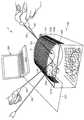
次に本発明の最良の実施形態を、図面を参照して説明する。図1において、符号1は腹腔鏡手術シミュレーション装置である。ここで、腹腔鏡手術は胸腔鏡手術等と並び鏡視下手術の一つである。 Next, the best mode of the present invention will be described with reference to the drawings. In FIG. 1, reference numeral 1 denotes a laparoscopic surgery simulation apparatus. Here, laparoscopic surgery is one of the endoscopic operations along with thoracoscopic surgery.
腹腔鏡手術シミュレーション装置(以下、シミュレーション装置という)1は、気腹状態の腹部を簡易に再現した簡易腹部モデル100と、カメラ200と、モニターとしてのパソコン300を備えている。 A laparoscopic surgery simulation apparatus (hereinafter referred to as a simulation apparatus) 1 includes a
簡易腹部モデル100は、上面に開口部101Aを備える箱状構造のボックス(箱状体)101を備えている。ボックス101は、透明なプラスチック素材からなり、気腹状態の腹部の腹腔内を想定して内部の大きさが決定されている(例えば、図1に示すボックス101の大きさは、およそ縦35mm×横25mm×高さ28mmである)。ここで、気腹状態とは、腹腔鏡手術において、体腔内へ炭酸ガス(CO2)を充填し、腹壁を気圧で挙上させた状態をいう。The
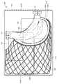
ボックス101の開口部101Aには、同開口部101Aを覆う弾性のあるメッシュカバー(上部カバー)102が、ボックス101の両側板101Bの上端位置から上方に湾曲して突出する半楕円形の形をなして、両側板101B間に複数の固定部材103により取付固定されている。メッシュカバー102は、気腹状態の腹壁をモデルとして再現するもので、弾性素材をマス目の径が約1cmとなるように縦横に格子状に配置して接合され、上方に湾曲する半楕円形状に張設することにより、気腹状態における腹壁の弾性力に近い弾性力が付与されている。 The opening 101A of the
固定部材103はボックス101の側板101Bに設けられたピン孔103aに対し側板101Bの外側から挿入される固定ピン103bと、側板101Bの内側から固定ピン103bの先端が螺合され連結される固定プレート103cとから構成されるが、かかる構造には限定されない。メッシュカバー102の一方の側端には固定ピン103bの位置に合わせてスリット104が設けられ、スリット104の奥部に固定ピン103bを位置させることにより、メッシュカバー102の一方の側部を側板101Bの内面に支持固定し、その一方でスリット104によりメッシュカバー102の一方の側部を側板101Bの内面から外してメッシュカバー102を容易に開くことが可能となっている。 The
図2は、メッシュカバー102を外した状態のボックス101の平面図を示している。ボックス101の内部にはシミュレーション対象の胃モデル105が立体的に配置されている。図2において、図の右側が胸部側、左側が腰部側である。胃モデル105は、人体の腹部腹腔内における胃の実際の立体的配置を再現する形で配置されている。 FIG. 2 shows a plan view of the
胃モデル105とボックス101の底板101Cとの間には、中央から腰部側寄りに背柱をモデルとする第1スペーサー106と、第1スペーサー106の両脇に左右の腎臓をモデルとする第2スペーサー107および第3スペーサー108が配置固定されている。これら第1〜第3スペーサー106〜108は、いずれも実際の背柱および肝臓を想定してこれに類似する硬質素材(例えばウレタン発泡体)から構成されている。胃モデル105が載置される部位には、腹腔内における背柱および左右の腎臓の実際の形状を再現するように傾斜面106A、107A、108Aが形成されている。 Between the
第1スペーサー106の胸部側寄りには膵臓をモデルとする第4スペーサー109が配置固定されている。この第4スペーサー109は実際の膵臓を想定してこれに類似する弾性素材(例えばスポンジ状素材)から構成されている。膵臓を想定した第4スペーサー109と右の腎臓を想定した第3スペーサー108との間には実際の腹腔を想定して窪み110が形成されている。 A
胃モデル105は、実際の胃臓器を想定して柔軟かつ弾性のある素材から構成され、ボックス101の内部において、胃モデル105の食道接続部105Aが前板101Dのやや右寄りに位置し、十二指腸接続部105Bが左の側板101Bの中央寄りに位置し、前記第4スペーサー109の上面から第3スペーサー108、第1スペーサー106、第2スペーサー107の上面にかけて立体的に配置されている。窪み110においては、スペーサーがないので、胃モデル105の当該部位は、その柔軟素材に従って、窪み110に落ち込む形状となっている。これによって、胃モデル105は、前述したように、人体の腹部の腹腔内における胃の実際の立体的配置を再現する形で配置されている。 The
胃モデル105は、底板101C上に固定された2つの支持プレート111,111間に、食道接続部105Aおよび十二指腸接続部105Bが架設される形で、可動可能に支持されている。胃モデル105の食道接続部105Aおよび十二指腸接続部105Bにはそれぞれ挟持部材112が装着されている。そして、各挟持部材112の支持孔112aに係止されたフック部材113,113が対応する支持プレート111,111の支持孔111aに係止されることにより、両支持プレート111,111間に胃モデル105が可動可能に支持されている。 The
2つの支持プレート111の各支持孔111aは、上下方向に等間隔で多数設けられている。これにより、フック部材113の係止位置を上下に変更することが可能であり、胃モデル105の食道接続部105Aおよび十二指腸接続部105Bの架設高さを、実際の食道接続高さおよび十二指腸接続高さに合わせて調整可能となっている。 A large number of
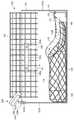
胃モデル105の外側面には、図3に示すように、食道接続部105Aの近くから十二指腸接続部105Bの近くにかけて、内臓器官の一種である大網をモデルとする網目状の大網モデル114が面ファスナー115を介して着脱可能に取り付けられている。面ファスナー115は、胃モデル105側の面ファスナー片115Aと大網モデル114側の面ファスナー片115Bから構成されている。大網モデル114は、柔軟性素材により構成され、胃モデル105の外側面から第1〜第3スペーサー106〜108の上面、さらにはボックス101の左右側板101B近傍の腰部側寄りと後板101Eの近傍まで広範囲に配置されている。 On the outer surface of the
図4は、ボックス101にメッシュカバー102およびカメラ200が取付けられた平面図を示している。同図では線図の煩雑さを避けるため大網モデル114が省略されている。図5は図4のA−A線矢視断面図、図6は図4のB−B線矢視断面図、7図は図4のC−C線矢視断面図である。図6および図7では分かりやすくするためメッシュカバー102を図4の左半分又は右半分のみ表わしている。カメラ200は、これらの図に示されるように、実際の腹腔鏡手術において外部から腹部の手術部位に挿入される内視鏡カメラの位置および角度を再現するように配置されている。 FIG. 4 is a plan view in which the
すなわち、カメラ200は、ボックス101の後板101Eの上端中央において、前面を斜め下向きの角度とし、前面のCCDレンズ部201が、ボックス101の底板103Cを見下ろす角度、すなわちボックス101内の胃モデル105の手術シミュレーション部位付近を向く角度で支持アタッチメント202により支持されている。支持アタッチメント202は、カメラ200を保持する保持台203と、ボックス101の後板101Eの上端中央に着脱可能に固定されたクリップ部材204とを備え、保持台203下面の支柱203Aがクリップ部材204上面のジョイント部204Aに対し前後左右に傾斜可能に支持されている。これにより、手術シミュレーション部位の位置に合わせてカメラ200の上下角度および左右の向きを調整可能である。 That is, the
かかるカメラ200によって、実際の腹腔鏡手術における映像をパソコン300の画面上に再現することができる。なお、符号205はカメラ本体201とパソコン300とをつなぐ通信ケーブルである。 With this
次に、上記構成のシミュレーション装置1を用いて、術者および助手を例にして、両者が腹腔鏡手術の臓器展開(視野展開)操作をシミュレートする方法について説明する。 Next, a method for simulating an organ deployment (field deployment) operation of laparoscopic surgery using the simulation apparatus 1 having the above configuration will be described, taking an operator and an assistant as an example.
図1は、上記構成のシミュレーション装置1を用いて両者が腹腔鏡手術の臓器展開操作をシミュレートしている様子を示している。実際の腹腔鏡手術においては、体腔内へ炭酸ガス(CO2)を充填して腹壁を気圧で挙上させて視野を確保し、細長い腹腔鏡カメラと細長い専用鉗子を腹腔内に挿入し、同器具を用いて臓器展開しながら、胃切除等の外科手術を行う。FIG. 1 shows a state in which the simulation apparatus 1 configured as described above is used to simulate an organ deployment operation for laparoscopic surgery. In actual laparoscopic surgery, the body cavity is filled with carbon dioxide (CO2 ) to raise the abdominal wall with atmospheric pressure to secure a field of view. Surgery such as gastrectomy is performed while organs are expanded using instruments.
本シミュレーション装置Sは、簡易腹部モデル100が最初から気腹状態に設定されている。そこで、簡易腹部モデル100の左右に立ち、気腹状態のメッシュカバー102の任意の隙間Sからそれぞれ2本の細長い専用鉗子(直径2〜10mm)150を腹腔内部を再現したボックス101内部に挿入する。なお、簡易腹部モデル100は、メッシュカバー102のあらゆる隙間Sから専用鉗子150を挿入可能である。 In the simulation apparatus S, the simple
図1に示すメッシュカバー102には、任意の隙間Sのうち、実際の腹腔鏡手術における腹部の器具挿入位置(切開位置)に相当する位置に目印となる挿入リング116が取付けられている。これにより、挿入リング116を目印として、実際の腹腔鏡手術と同じ位置および向きを再現し、専用鉗子150をボックス101内に容易に挿入できる。 In the
そして、簡易腹部モデル100の左右から、上部のメッシュカバー102を通して、ボックス101内の胃モデル105に対する専用鉗子150による展開操作および操作手順のシミュレートが可能である。一方の術者は展開操作および操作手順のデモンストレーションを行うことが可能であり、また、他方の助手は展開操作および操作手順のトレーニングを行うことが可能である。 Then, from the left and right sides of the simplified
胃モデル105は、腹部の腹腔内における胃の実際の立体的配置を再現する形で配置されるとともに、可動可能に支持されているので、実際の腹腔鏡手術を再現する展開操作が可能である。また、胃モデル105には面ファスナー115により大綱モデル114が取付けられているので、専用鉗子150を用いて、実際の腹腔鏡手術を再現する分離操作あるいは接合(縫合)操作が可能である。 The
胃モデル105の展開操作中、大網モデル114の分離・接合操作中は、メッシュカバー102を通してボックス101内の操作の様子を直接透き見することができるから、助手は回復手術と同様の立体感をつかみながら、展開操作方法や操作手順を十分に理解することができ、トレーニング効果を高めることができる。 During the operation of expanding the
メッシュカバー102は気腹状態の腹壁を再現する弾性力が付与され、専用鉗子150の操作時に気腹状態の腹壁と同様の抵抗を感じさせることができ、簡易なメッシュカバーでありながら、実際の腹壁と同様のリアリティーさをもたらすことができる。 The
また、胃モデル105の展開操作、大網モデル114の分離・接合操作は、ボックス101の後板101Eに実際の腹腔鏡手術における腹腔鏡カメラを再現するように支持されたカメラ200により撮影され、ケーブル204を介してパソコン300に画像データが送られ、パソコン300の画面上に同時に平面的な映像として映し出される。助手は、臓器展開の操作の様子を立体的につかみながら、同時にモニターに映し出される平面的な映像と比較することにより、トレーニング効果を格段に高めることができる。また、術者は、モニターに映し出される映像と実際の臓器展開の操作を比較しながらデモンストレーションできる。 Further, the deployment operation of the
本シミュレーション装置1を用いることによって、術者や助手に対するトレーニングだけでなく、腹腔鏡手術の未経験または初心者、例えば研修医、医学系学生、看護師、看護学生等に対し、腹腔鏡手術の手順を提示することが可能となり、これらの者は腹腔鏡手術の操作の様子を、ボックス101内を直接透し見して立体的に理解し、あるいは同時にパソコン300の画面上で狭い術野を見比べることによってより深く理解することができるようになり、専門的な医学教育や指導に有効に利用できる。 By using this simulation apparatus 1, not only training for surgeons and assistants but also inexperienced or beginners of laparoscopic surgery such as resident doctors, medical students, nurses, nursing students, etc. These can be presented, and these persons can understand the operation of laparoscopic surgery in a three-dimensional manner by directly looking inside the
また、臨床実験では、腹腔鏡手術の操作の検証や手術前のシミュレーションによる手術計画の立案、さらには手術手技の創意工夫、新しい手技の開発への利用も可能である。 In clinical experiments, it is also possible to verify the operation of laparoscopic surgery, create a surgical plan based on pre-operative simulation, and further use the ingenuity of surgical techniques and the development of new techniques.
簡易腹部モデル100は、箱状構造のボックス101に弾性のあるメッシュカバー102を取付けることにより気腹状態の腹部の腹腔を簡易に再現し、内部に胃モデル105および大網モデル114を可動可能に立体的に配置することにより腹部の腹腔内の臓器を簡易に再現することができた。これにより、従来に比べて腹部モデル、ひいてはシミュレーション装置を安価に製作可能である。 The simplified
図8は、本発明の他の実施形態を示すもので、オプションとして、メッシュカバー102の上に透き見を不能とする保護カバー117を装着可能としたものである。保護カバー117には図1におけるメッシュカバー102の挿入リング116の位置に合わせて専用鉗子150の挿入穴117aを設けるようにしている。 FIG. 8 shows another embodiment of the present invention. As an option, a
以上の実施形態においては、簡易腹部モデル100において、ボックス101内部に胃モデル105および大網モデル114を可動可能に立体的に配置するようにしたが、本発明の鏡視下手術シミュレーション装置1は、簡易腹部モデルにおいて、大腸モデルおよび周囲の臓器等を配置してのシミュレーションや、簡易胸部モデルにおいて、内部に左右の肺モデル等を配置してのシミュレーションに適用できることは言うまでもない。 In the above embodiment, in the simple
かくして、本発明の鏡視下手術シミュレーション装置1によれば、簡易な装置を用いて、実際の鏡視下手術と同様の操作のシミュレーションを実現できるとともに、手術の助手に対するトレーニングや医学生等に対する教育指導にも優れる装置を提供することができる。 Thus, according to the endoscopic surgery simulation apparatus 1 of the present invention, it is possible to realize a simulation of the same operation as an actual endoscopic surgery using a simple apparatus, and for training of surgical assistants, medical students, etc. A device excellent in educational guidance can be provided.
本発明に係る鏡視下手術シミュレーション装置は、鏡視下手術における術者や助手の操作トレーニング装置として、あるいは研修医等に対する医学教育用の装置として、さらには実際の鏡視下手術における操作の検証や手術前のシミュレーションによる手術計画の立案向け等に用いる装置として幅広く利用可能である。 The endoscopic surgery simulation apparatus according to the present invention is used as an operation training apparatus for an operator or assistant in an endoscopic operation, or as a medical education apparatus for a trainee or the like, and further for an operation in an actual endoscopic operation. It can be widely used as an apparatus used for planning an operation plan by verification and simulation before operation.
1 腹腔鏡手術シミュレーション装置(鏡視下手術シミュレーション装置)
100 簡易腹部モデル
101 ボックス(箱状体)
101A 開口部
101B 側板
101C 底板
101D 前板
101E 後板
102 メッシュカバー(上部カバー)
103 固定部材
103a ピン孔
103b 固定ピン
104 スリット
105 胃モデル(臓器モデル)
105A 食道接続部
105B 十二指腸接続部
106 第1スペーサー
107 第2スペーサー
108 第3スペーサー
106A,107A,108A 傾斜面
109 第4スペーサー
110 窪み
111 支持プレート
111a,112a 支持孔
112 挟持部材
113 フック部材
114 大綱モデル
115 面ファスナー
115A,115B 面ファスナー片
116 リング
117 保護カバー
117a 挿入穴
150 専用鉗子
200 カメラ
201 CCDレンズ部
202 支持アタッチメント
203 保持台
203A 支柱
204 クリップ部材
204A ジョイント部
205 通信ケーブル
300 モニター(モニター手段)
S 隙間
1 Laparoscopic surgery simulation device (mirror surgery simulation device)
100 Simple
103
105A
S clearance
Claims (7)
Translated fromJapanese鏡視下手術における気腹状態の腹壁をモデルとして前記開口部を覆うように配置され、内部を透視可能でかつ鏡視下手術用器具を挿通可能な上部カバーと、
前記箱状体の内部に配置されるシミュレーション対象の臓器モデルと、
前記箱状体の内面と前記臓器モデルとの間に配置され、前記臓器モデルを前記箱状体の内部に立体的に配置するスペーサーと、
前記箱状体の内部に配置された臓器モデルを可動可能に支持する支持手段と、
前記箱状体の内部に配置された臓器モデルの手術シミュレート部位付近を撮像する撮像手段と、
前記撮像手段により撮像された映像を表示させるモニター手段と、
を有することを特徴とする鏡視下手術シミュレーション装置。A box-shaped body whose size is determined using the abdomen of the human body in a microscopic surgery and having an opening on the upper surface;
An upper cover that is arranged so as to cover the opening as a model of an abdominal wall in an insufflated state in an endoscopic operation, and is capable of seeing through the inside and allowing insertion of an endoscopic surgical instrument;
An organ model to be simulated arranged inside the box-shaped body;
A spacer that is arranged between the inner surface of the box-shaped body and the organ model, and that three-dimensionally arranges the organ model inside the box-shaped body;
Support means for movably supporting an organ model arranged inside the box-shaped body;
An imaging means for imaging the vicinity of a surgical simulation site of an organ model arranged inside the box-shaped body;
Monitor means for displaying the video imaged by the imaging means;
A surgical operation simulation apparatus under the microscope.
Priority Applications (1)
| Application Number | Priority Date | Filing Date | Title |
|---|---|---|---|
| JP2009272137AJP5505927B2 (en) | 2009-11-30 | 2009-11-30 | Endoscopic surgery simulation device |
Applications Claiming Priority (1)
| Application Number | Priority Date | Filing Date | Title |
|---|---|---|---|
| JP2009272137AJP5505927B2 (en) | 2009-11-30 | 2009-11-30 | Endoscopic surgery simulation device |
Publications (2)
| Publication Number | Publication Date |
|---|---|
| JP2011113056Atrue JP2011113056A (en) | 2011-06-09 |
| JP5505927B2 JP5505927B2 (en) | 2014-05-28 |
Family
ID=44235396
Family Applications (1)
| Application Number | Title | Priority Date | Filing Date |
|---|---|---|---|
| JP2009272137AActiveJP5505927B2 (en) | 2009-11-30 | 2009-11-30 | Endoscopic surgery simulation device |
Country Status (1)
| Country | Link |
|---|---|
| JP (1) | JP5505927B2 (en) |
Cited By (40)
| Publication number | Priority date | Publication date | Assignee | Title |
|---|---|---|---|---|
| JP2013127496A (en)* | 2011-12-16 | 2013-06-27 | Tanac Co Ltd | Simulated internal organ installing base and surgical operation training device |
| KR101400442B1 (en) | 2012-11-05 | 2014-05-28 | 한국과학기술원 | Simulator for training needle interventional operation and interface apparatus for the same |
| WO2015151504A1 (en)* | 2014-03-31 | 2015-10-08 | 株式会社ファソテック | Peritoneal cavity simulator |
| KR20160034918A (en)* | 2013-07-24 | 2016-03-30 | 어플라이드 메디컬 리소시스 코포레이션 | First entry model |
| JP2017032814A (en)* | 2015-08-03 | 2017-02-09 | イービーエム株式会社 | Human body model unit for surgical training device |
| JP2017223850A (en)* | 2016-06-16 | 2017-12-21 | 国立大学法人山口大学 | Surgical operation training kit |
| JP2018112646A (en)* | 2017-01-11 | 2018-07-19 | 村上 貴志 | Surgery training system |
| JP2019086794A (en)* | 2013-03-01 | 2019-06-06 | アプライド メディカル リソーシーズ コーポレイション | Surgical simulation system and method |
| WO2020031311A1 (en)* | 2018-08-08 | 2020-02-13 | 株式会社ファソテック | Organ fixing tool for abdominal cavity simulator |
| EP3654316A1 (en)* | 2014-03-13 | 2020-05-20 | Applied Medical Resources Corporation | Advanced first entry model for surgical simulation |
| JP2020114439A (en)* | 2014-02-21 | 2020-07-30 | スリーディインテグレイテッド アーペーエス3Dintegrated Aps | Training method |
| CN111630580A (en)* | 2017-11-10 | 2020-09-04 | 沃图丽索吉公司 | System for simulating surgical procedures |
| US10847057B2 (en) | 2017-02-23 | 2020-11-24 | Applied Medical Resources Corporation | Synthetic tissue structures for electrosurgical training and simulation |
| JP2020190760A (en)* | 2020-08-27 | 2020-11-26 | 株式会社ファソテック | Organ fixture for abdominal cavity simulator |
| US10854112B2 (en) | 2010-10-01 | 2020-12-01 | Applied Medical Resources Corporation | Portable laparoscopic trainer |
| US10849688B2 (en) | 2016-03-02 | 2020-12-01 | Truinject Corp. | Sensory enhanced environments for injection aid and social training |
| US10896627B2 (en) | 2014-01-17 | 2021-01-19 | Truinjet Corp. | Injection site training system |
| US10902746B2 (en) | 2012-10-30 | 2021-01-26 | Truinject Corp. | System for cosmetic and therapeutic training |
| US11034831B2 (en) | 2015-05-14 | 2021-06-15 | Applied Medical Resources Corporation | Synthetic tissue structures for electrosurgical training and simulation |
| US11049418B2 (en) | 2013-06-18 | 2021-06-29 | Applied Medical Resources Corporation | Gallbladder model |
| US11100815B2 (en) | 2015-02-19 | 2021-08-24 | Applied Medical Resources Corporation | Simulated tissue structures and methods |
| US11120708B2 (en) | 2016-06-27 | 2021-09-14 | Applied Medical Resources Corporation | Simulated abdominal wall |
| US11158212B2 (en) | 2011-10-21 | 2021-10-26 | Applied Medical Resources Corporation | Simulated tissue structure for surgical training |
| CN114373348A (en)* | 2020-12-15 | 2022-04-19 | 西安赛德欧医疗研究院有限公司 | Simulation training box for full abdominal organ simulation surgery |
| US11361679B2 (en) | 2012-09-27 | 2022-06-14 | Applied Medical Resources Corporation | Surgical training model for laparoscopic procedures |
| US11403968B2 (en) | 2011-12-20 | 2022-08-02 | Applied Medical Resources Corporation | Advanced surgical simulation |
| US11450236B2 (en) | 2013-07-24 | 2022-09-20 | Applied Medical Resources Corporation | Advanced first entry model for surgical simulation |
| US11514819B2 (en) | 2012-09-26 | 2022-11-29 | Applied Medical Resources Corporation | Surgical training model for laparoscopic procedures |
| CN115485752A (en)* | 2021-01-18 | 2022-12-16 | 寿医疗株式会社 | Medical device operation practice device |
| WO2023003389A1 (en)* | 2021-07-21 | 2023-01-26 | (주)휴톰 | Apparatus and method for determining insertion position of trocar on three-dimensional virtual pneumoperitoneum model of patient |
| US11587466B2 (en) | 2015-07-16 | 2023-02-21 | Applied Medical Resources Corporation | Simulated dissectible tissue |
| US11710424B2 (en) | 2017-01-23 | 2023-07-25 | Truinject Corp. | Syringe dose and position measuring apparatus |
| US11721240B2 (en) | 2015-06-09 | 2023-08-08 | Applied Medical Resources Corporation | Hysterectomy model |
| US11721242B2 (en) | 2015-10-02 | 2023-08-08 | Applied Medical Resources Corporation | Hysterectomy model |
| US11887504B2 (en) | 2014-11-13 | 2024-01-30 | Applied Medical Resources Corporation | Simulated tissue models and methods |
| US11990055B2 (en) | 2012-09-27 | 2024-05-21 | Applied Medical Resources Corporation | Surgical training model for laparoscopic procedures |
| US12070581B2 (en) | 2015-10-20 | 2024-08-27 | Truinject Corp. | Injection system |
| US12217626B2 (en) | 2012-10-30 | 2025-02-04 | Truinject Corp. | Injection training apparatus using 3D position sensor |
| US12217625B2 (en) | 2015-11-20 | 2025-02-04 | Applied Medical Resources Corporation | Simulated dissectible tissue |
| US12243439B2 (en) | 2017-02-14 | 2025-03-04 | Applied Medical Resources Corporation | Laparoscopic training system |
Families Citing this family (1)
| Publication number | Priority date | Publication date | Assignee | Title |
|---|---|---|---|---|
| JP7358987B2 (en)* | 2017-11-28 | 2023-10-11 | ニプロ株式会社 | surgical practice model |
- 2009
- 2009-11-30JPJP2009272137Apatent/JP5505927B2/enactiveActive
Cited By (84)
| Publication number | Priority date | Publication date | Assignee | Title |
|---|---|---|---|---|
| US10854112B2 (en) | 2010-10-01 | 2020-12-01 | Applied Medical Resources Corporation | Portable laparoscopic trainer |
| US12154454B2 (en) | 2010-10-01 | 2024-11-26 | Applied Medical Resources Corporation | Portable laparoscopic trainer |
| US12014652B2 (en) | 2011-10-21 | 2024-06-18 | Applied Medical Resources Corporation | Simulated tissue structure for surgical training |
| US11158212B2 (en) | 2011-10-21 | 2021-10-26 | Applied Medical Resources Corporation | Simulated tissue structure for surgical training |
| JP2013127496A (en)* | 2011-12-16 | 2013-06-27 | Tanac Co Ltd | Simulated internal organ installing base and surgical operation training device |
| US11403968B2 (en) | 2011-12-20 | 2022-08-02 | Applied Medical Resources Corporation | Advanced surgical simulation |
| US11514819B2 (en) | 2012-09-26 | 2022-11-29 | Applied Medical Resources Corporation | Surgical training model for laparoscopic procedures |
| US11869378B2 (en) | 2012-09-27 | 2024-01-09 | Applied Medical Resources Corporation | Surgical training model for laparoscopic procedures |
| US11990055B2 (en) | 2012-09-27 | 2024-05-21 | Applied Medical Resources Corporation | Surgical training model for laparoscopic procedures |
| US11361679B2 (en) | 2012-09-27 | 2022-06-14 | Applied Medical Resources Corporation | Surgical training model for laparoscopic procedures |
| US10902746B2 (en) | 2012-10-30 | 2021-01-26 | Truinject Corp. | System for cosmetic and therapeutic training |
| US11854426B2 (en) | 2012-10-30 | 2023-12-26 | Truinject Corp. | System for cosmetic and therapeutic training |
| US12217626B2 (en) | 2012-10-30 | 2025-02-04 | Truinject Corp. | Injection training apparatus using 3D position sensor |
| US11403964B2 (en) | 2012-10-30 | 2022-08-02 | Truinject Corp. | System for cosmetic and therapeutic training |
| KR101400442B1 (en) | 2012-11-05 | 2014-05-28 | 한국과학기술원 | Simulator for training needle interventional operation and interface apparatus for the same |
| JP2023144020A (en)* | 2013-03-01 | 2023-10-06 | アプライド メディカル リソーシーズ コーポレイション | Surgical simulation systems and methods |
| JP2022089871A (en)* | 2013-03-01 | 2022-06-16 | アプライド メディカル リソーシーズ コーポレイション | Surgical simulation system and method |
| JP2019086794A (en)* | 2013-03-01 | 2019-06-06 | アプライド メディカル リソーシーズ コーポレイション | Surgical simulation system and method |
| JP7053515B2 (en) | 2013-03-01 | 2022-04-12 | アプライド メディカル リソーシーズ コーポレイション | Surgical simulation system and method |
| JP7514986B2 (en) | 2013-03-01 | 2024-07-11 | アプライド メディカル リソーシーズ コーポレイション | Surgery simulation system and method |
| US10991270B2 (en) | 2013-03-01 | 2021-04-27 | Applied Medical Resources Corporation | Advanced surgical simulation constructions and methods |
| US11735068B2 (en) | 2013-06-18 | 2023-08-22 | Applied Medical Resources Corporation | Gallbladder model |
| US11049418B2 (en) | 2013-06-18 | 2021-06-29 | Applied Medical Resources Corporation | Gallbladder model |
| US11854425B2 (en) | 2013-07-24 | 2023-12-26 | Applied Medical Resources Corporation | First entry model |
| KR102374993B1 (en) | 2013-07-24 | 2022-03-16 | 어플라이드 메디컬 리소시스 코포레이션 | First entry model |
| KR20210041118A (en)* | 2013-07-24 | 2021-04-14 | 어플라이드 메디컬 리소시스 코포레이션 | First entry model |
| KR102573569B1 (en) | 2013-07-24 | 2023-09-01 | 어플라이드 메디컬 리소시스 코포레이션 | First entry model |
| US12288476B2 (en) | 2013-07-24 | 2025-04-29 | Applied Medical Resources Corporation | Advanced first entry model for surgical simulation |
| JP2016532151A (en)* | 2013-07-24 | 2016-10-13 | アプライド メディカル リソーシーズ コーポレイション | First entry model |
| KR20160034918A (en)* | 2013-07-24 | 2016-03-30 | 어플라이드 메디컬 리소시스 코포레이션 | First entry model |
| KR102239454B1 (en)* | 2013-07-24 | 2021-04-13 | 어플라이드 메디컬 리소시스 코포레이션 | First entry model |
| US11450236B2 (en) | 2013-07-24 | 2022-09-20 | Applied Medical Resources Corporation | Advanced first entry model for surgical simulation |
| KR20220035286A (en)* | 2013-07-24 | 2022-03-21 | 어플라이드 메디컬 리소시스 코포레이션 | First entry model |
| US10896627B2 (en) | 2014-01-17 | 2021-01-19 | Truinjet Corp. | Injection site training system |
| JP2020114439A (en)* | 2014-02-21 | 2020-07-30 | スリーディインテグレイテッド アーペーエス3Dintegrated Aps | Training method |
| AU2022201612B2 (en)* | 2014-03-13 | 2024-01-11 | Applied Medical Resources Corporation | Advanced first entry model for surgical simulation |
| AU2019283949B2 (en)* | 2014-03-13 | 2021-12-09 | Applied Medical Resources Corporation | Advanced first entry model for surgical simulation |
| JP7617218B2 (en) | 2014-03-13 | 2025-01-17 | アプライド メディカル リソーシーズ コーポレイション | A new first-entry model for surgical imitation |
| JP7361172B2 (en) | 2014-03-13 | 2023-10-13 | アプライド メディカル リソーシーズ コーポレイション | New first entry model for surgical imitation |
| JP2023181181A (en)* | 2014-03-13 | 2023-12-21 | アプライド メディカル リソーシーズ コーポレイション | New first entry model for surgical imitation |
| KR20230170826A (en)* | 2014-03-13 | 2023-12-19 | 어플라이드 메디컬 리소시스 코포레이션 | Advanced first entry model for surgical simulation |
| EP3654316A1 (en)* | 2014-03-13 | 2020-05-20 | Applied Medical Resources Corporation | Advanced first entry model for surgical simulation |
| KR102784532B1 (en) | 2014-03-13 | 2025-03-21 | 어플라이드 메디컬 리소시스 코포레이션 | Advanced first entry model for surgical simulation |
| JP2021193455A (en)* | 2014-03-13 | 2021-12-23 | アプライド メディカル リソーシーズ コーポレイション | Advanced first entry model for surgical simulation |
| JP2022171681A (en)* | 2014-03-13 | 2022-11-11 | アプライド メディカル リソーシーズ コーポレイション | Advanced first entry model for surgical simulation |
| JP7129532B2 (en) | 2014-03-13 | 2022-09-01 | アプライド メディカル リソーシーズ コーポレイション | A new first entry model for surgical imitation |
| WO2015151504A1 (en)* | 2014-03-31 | 2015-10-08 | 株式会社ファソテック | Peritoneal cavity simulator |
| JPWO2015151504A1 (en)* | 2014-03-31 | 2017-04-13 | 株式会社ファソテック | Abdominal simulator |
| EP3128501A4 (en)* | 2014-03-31 | 2017-11-01 | Fasotec Co., Ltd. | Peritoneal cavity simulator |
| US10192465B2 (en) | 2014-03-31 | 2019-01-29 | Fasotec Co., Ltd. | Peritoneal cavity simulator |
| CN105324806A (en)* | 2014-03-31 | 2016-02-10 | 株式会社发索科技 | Peritoneal cavity simulator |
| US12211394B2 (en) | 2014-11-13 | 2025-01-28 | Applied Medical Resources Corporation | Simulated tissue models and methods |
| US11887504B2 (en) | 2014-11-13 | 2024-01-30 | Applied Medical Resources Corporation | Simulated tissue models and methods |
| US12131664B2 (en) | 2015-02-19 | 2024-10-29 | Applied Medical Resources Corporation | Simulated tissue structures and methods |
| US11100815B2 (en) | 2015-02-19 | 2021-08-24 | Applied Medical Resources Corporation | Simulated tissue structures and methods |
| US11034831B2 (en) | 2015-05-14 | 2021-06-15 | Applied Medical Resources Corporation | Synthetic tissue structures for electrosurgical training and simulation |
| US11721240B2 (en) | 2015-06-09 | 2023-08-08 | Applied Medical Resources Corporation | Hysterectomy model |
| US12175883B2 (en) | 2015-06-09 | 2024-12-24 | Applied Medical Resources Corporation | Hysterectomy model |
| US12087179B2 (en) | 2015-07-16 | 2024-09-10 | Applied Medical Resources Corporation | Simulated dissectible tissue |
| US11587466B2 (en) | 2015-07-16 | 2023-02-21 | Applied Medical Resources Corporation | Simulated dissectible tissue |
| JP2017032814A (en)* | 2015-08-03 | 2017-02-09 | イービーエム株式会社 | Human body model unit for surgical training device |
| US11721242B2 (en) | 2015-10-02 | 2023-08-08 | Applied Medical Resources Corporation | Hysterectomy model |
| US12243441B2 (en) | 2015-10-02 | 2025-03-04 | Applied Medical Resources Corporation | Hysterectomy model |
| US12070581B2 (en) | 2015-10-20 | 2024-08-27 | Truinject Corp. | Injection system |
| US12217625B2 (en) | 2015-11-20 | 2025-02-04 | Applied Medical Resources Corporation | Simulated dissectible tissue |
| US11730543B2 (en) | 2016-03-02 | 2023-08-22 | Truinject Corp. | Sensory enhanced environments for injection aid and social training |
| US10849688B2 (en) | 2016-03-02 | 2020-12-01 | Truinject Corp. | Sensory enhanced environments for injection aid and social training |
| JP2017223850A (en)* | 2016-06-16 | 2017-12-21 | 国立大学法人山口大学 | Surgical operation training kit |
| US11120708B2 (en) | 2016-06-27 | 2021-09-14 | Applied Medical Resources Corporation | Simulated abdominal wall |
| US11830378B2 (en) | 2016-06-27 | 2023-11-28 | Applied Medical Resources Corporation | Simulated abdominal wall |
| JP2018112646A (en)* | 2017-01-11 | 2018-07-19 | 村上 貴志 | Surgery training system |
| US11710424B2 (en) | 2017-01-23 | 2023-07-25 | Truinject Corp. | Syringe dose and position measuring apparatus |
| US12350472B2 (en) | 2017-01-23 | 2025-07-08 | Truinject Corp. | Syringe dose and position measuring apparatus |
| US12243439B2 (en) | 2017-02-14 | 2025-03-04 | Applied Medical Resources Corporation | Laparoscopic training system |
| US10847057B2 (en) | 2017-02-23 | 2020-11-24 | Applied Medical Resources Corporation | Synthetic tissue structures for electrosurgical training and simulation |
| CN111630580A (en)* | 2017-11-10 | 2020-09-04 | 沃图丽索吉公司 | System for simulating surgical procedures |
| CN111630580B (en)* | 2017-11-10 | 2022-12-30 | 沃图丽索吉公司 | System for simulating surgical procedures |
| WO2020031311A1 (en)* | 2018-08-08 | 2020-02-13 | 株式会社ファソテック | Organ fixing tool for abdominal cavity simulator |
| JPWO2020031311A1 (en)* | 2018-08-08 | 2020-08-20 | 株式会社ファソテック | Organ Fixator for Celiac Simulator |
| JP2020190760A (en)* | 2020-08-27 | 2020-11-26 | 株式会社ファソテック | Organ fixture for abdominal cavity simulator |
| CN114373348A (en)* | 2020-12-15 | 2022-04-19 | 西安赛德欧医疗研究院有限公司 | Simulation training box for full abdominal organ simulation surgery |
| CN115485752A (en)* | 2021-01-18 | 2022-12-16 | 寿医疗株式会社 | Medical device operation practice device |
| EP4368136A4 (en)* | 2021-07-21 | 2024-10-16 | Hutom Co., Ltd. | DEVICE AND METHOD FOR DETERMINING THE POSITION OF A TROCAR ON A THREE-DIMENSIONAL VIRTUAL PNEUMOPERITONEUM MODEL OF A PATIENT |
| WO2023003389A1 (en)* | 2021-07-21 | 2023-01-26 | (주)휴톰 | Apparatus and method for determining insertion position of trocar on three-dimensional virtual pneumoperitoneum model of patient |
Also Published As
| Publication number | Publication date |
|---|---|
| JP5505927B2 (en) | 2014-05-28 |
Similar Documents
| Publication | Publication Date | Title |
|---|---|---|
| JP5505927B2 (en) | Endoscopic surgery simulation device | |
| JP7104757B2 (en) | Portable laparoscopic trainer | |
| AU2018250511B2 (en) | Hernia model | |
| JP6315712B2 (en) | Surgical training model for transluminal laparoscopic procedures | |
| JP6245531B2 (en) | Surgical training model for laparoscopic procedures | |
| US8460002B2 (en) | Laparoscopic trainer and method of training | |
| Viglialoro et al. | Augmented reality to improve surgical simulation: Lessons learned towards the design of a hybrid laparoscopic simulator for cholecystectomy | |
| JP2011133830A (en) | Simulated operation tool device for setting trocar position | |
| KR20150064045A (en) | Surgical training model for laparoscopic procedures | |
| JP2015532454A (en) | Surgical training model for laparoscopic procedures | |
| US11488494B2 (en) | Laparoscopic instrument holder for surgical simulation and training | |
| JP2015502563A (en) | Simulated tissue structure for surgical training | |
| JP6521511B2 (en) | Surgical training device | |
| JP6862413B2 (en) | Appendectomy model | |
| JP2021503095A (en) | Hysterectomy model | |
| Muller-Wittig et al. | Enhanced training environment for minimally invasive surgery | |
| Guadagni et al. | Augmented Reality to Improve Surgical Simulation: Lessons Learned Towards the Design of a Hybrid Laparoscopic Simulator for Cholecystectomy |
Legal Events
| Date | Code | Title | Description |
|---|---|---|---|
| A621 | Written request for application examination | Free format text:JAPANESE INTERMEDIATE CODE: A621 Effective date:20121109 | |
| A977 | Report on retrieval | Free format text:JAPANESE INTERMEDIATE CODE: A971007 Effective date:20131128 | |
| TRDD | Decision of grant or rejection written | ||
| A01 | Written decision to grant a patent or to grant a registration (utility model) | Free format text:JAPANESE INTERMEDIATE CODE: A01 Effective date:20140310 | |
| A61 | First payment of annual fees (during grant procedure) | Free format text:JAPANESE INTERMEDIATE CODE: A61 Effective date:20140312 | |
| R150 | Certificate of patent or registration of utility model | Ref document number:5505927 Country of ref document:JP Free format text:JAPANESE INTERMEDIATE CODE: R150 | |
| R250 | Receipt of annual fees | Free format text:JAPANESE INTERMEDIATE CODE: R250 | |
| R250 | Receipt of annual fees | Free format text:JAPANESE INTERMEDIATE CODE: R250 | |
| R250 | Receipt of annual fees | Free format text:JAPANESE INTERMEDIATE CODE: R250 | |
| R250 | Receipt of annual fees | Free format text:JAPANESE INTERMEDIATE CODE: R250 | |
| R250 | Receipt of annual fees | Free format text:JAPANESE INTERMEDIATE CODE: R250 | |
| S111 | Request for change of ownership or part of ownership | Free format text:JAPANESE INTERMEDIATE CODE: R313113 | |
| R350 | Written notification of registration of transfer | Free format text:JAPANESE INTERMEDIATE CODE: R350 | |
| R250 | Receipt of annual fees | Free format text:JAPANESE INTERMEDIATE CODE: R250 |