JP2010504803A - Method and apparatus for indexing pulse amplitude - Google Patents
Method and apparatus for indexing pulse amplitudeDownload PDFInfo
- Publication number
- JP2010504803A JP2010504803AJP2009530316AJP2009530316AJP2010504803AJP 2010504803 AJP2010504803 AJP 2010504803AJP 2009530316 AJP2009530316 AJP 2009530316AJP 2009530316 AJP2009530316 AJP 2009530316AJP 2010504803 AJP2010504803 AJP 2010504803A
- Authority
- JP
- Japan
- Prior art keywords
- pulse
- pulse amplitude
- amplitude
- oxygen saturation
- monitored
- Prior art date
- Legal status (The legal status is an assumption and is not a legal conclusion. Google has not performed a legal analysis and makes no representation as to the accuracy of the status listed.)
- Pending
Links
- 238000000034methodMethods0.000titleclaimsabstractdescription37
- QVGXLLKOCUKJST-UHFFFAOYSA-Natomic oxygenChemical compound[O]QVGXLLKOCUKJST-UHFFFAOYSA-N0.000claimsabstractdescription54
- 239000001301oxygenSubstances0.000claimsabstractdescription54
- 229910052760oxygenInorganic materials0.000claimsabstractdescription54
- 239000008280bloodSubstances0.000claimsabstractdescription28
- 210000004369bloodAnatomy0.000claimsabstractdescription28
- 230000003287optical effectEffects0.000claimsdescription17
- 238000012544monitoring processMethods0.000claimsdescription6
- 230000005855radiationEffects0.000claimsdescription5
- 238000012545processingMethods0.000claimsdescription4
- 210000001367arteryAnatomy0.000claimsdescription3
- 238000010521absorption reactionMethods0.000abstractdescription10
- 230000001766physiological effectEffects0.000abstractdescription4
- 239000000306componentSubstances0.000description10
- 102000001554HemoglobinsHuman genes0.000description9
- 108010054147HemoglobinsProteins0.000description9
- 230000005540biological transmissionEffects0.000description7
- 230000008859changeEffects0.000description6
- 238000004364calculation methodMethods0.000description5
- 230000000694effectsEffects0.000description4
- 239000007789gasSubstances0.000description3
- 239000000463materialSubstances0.000description3
- 241001465754MetazoaSpecies0.000description2
- 238000012937correctionMethods0.000description2
- 238000010586diagramMethods0.000description2
- 230000001747exhibiting effectEffects0.000description2
- BCGWQEUPMDMJNV-UHFFFAOYSA-NimipramineChemical compoundC1CC2=CC=CC=C2N(CCCN(C)C)C2=CC=CC=C21BCGWQEUPMDMJNV-UHFFFAOYSA-N0.000description2
- 230000031700light absorptionEffects0.000description2
- 238000005259measurementMethods0.000description2
- 238000012986modificationMethods0.000description2
- 230000004048modificationEffects0.000description2
- 230000010349pulsationEffects0.000description2
- INGWEZCOABYORO-UHFFFAOYSA-N2-(furan-2-yl)-7-methyl-1h-1,8-naphthyridin-4-oneChemical compoundN=1C2=NC(C)=CC=C2C(O)=CC=1C1=CC=CO1INGWEZCOABYORO-UHFFFAOYSA-N0.000description1
- 241000282412HomoSpecies0.000description1
- 206010021143HypoxiaDiseases0.000description1
- 238000002835absorbanceMethods0.000description1
- 230000017531blood circulationEffects0.000description1
- 239000012503blood componentSubstances0.000description1
- 238000011088calibration curveMethods0.000description1
- 230000002612cardiopulmonary effectEffects0.000description1
- 230000005792cardiovascular activityEffects0.000description1
- 238000006243chemical reactionMethods0.000description1
- 238000007796conventional methodMethods0.000description1
- 238000013502data validationMethods0.000description1
- 230000001419dependent effectEffects0.000description1
- 238000013461designMethods0.000description1
- 230000009977dual effectEffects0.000description1
- 210000005069earsAnatomy0.000description1
- 210000003754fetusAnatomy0.000description1
- 210000003811fingerAnatomy0.000description1
- 230000001146hypoxic effectEffects0.000description1
- 230000006872improvementEffects0.000description1
- 238000007726management methodMethods0.000description1
- 239000000203mixtureSubstances0.000description1
- 230000002107myocardial effectEffects0.000description1
- 210000000492nasalseptumAnatomy0.000description1
- 238000010606normalizationMethods0.000description1
- 230000008569processEffects0.000description1
- 230000035485pulse pressureEffects0.000description1
- 210000002321radial arteryAnatomy0.000description1
- 238000011160researchMethods0.000description1
- 210000004761scalpAnatomy0.000description1
- 241000894007speciesSpecies0.000description1
- 238000010561standard procedureMethods0.000description1
- 230000001360synchronised effectEffects0.000description1
- 230000002861ventricularEffects0.000description1
Images


Classifications
- A—HUMAN NECESSITIES
- A61—MEDICAL OR VETERINARY SCIENCE; HYGIENE
- A61B—DIAGNOSIS; SURGERY; IDENTIFICATION
- A61B5/00—Measuring for diagnostic purposes; Identification of persons
- A61B5/145—Measuring characteristics of blood in vivo, e.g. gas concentration or pH-value ; Measuring characteristics of body fluids or tissues, e.g. interstitial fluid or cerebral tissue
- A61B5/1455—Measuring characteristics of blood in vivo, e.g. gas concentration or pH-value ; Measuring characteristics of body fluids or tissues, e.g. interstitial fluid or cerebral tissue using optical sensors, e.g. spectral photometrical oximeters
- A61B5/14551—Measuring characteristics of blood in vivo, e.g. gas concentration or pH-value ; Measuring characteristics of body fluids or tissues, e.g. interstitial fluid or cerebral tissue using optical sensors, e.g. spectral photometrical oximeters for measuring blood gases
- A—HUMAN NECESSITIES
- A61—MEDICAL OR VETERINARY SCIENCE; HYGIENE
- A61B—DIAGNOSIS; SURGERY; IDENTIFICATION
- A61B5/00—Measuring for diagnostic purposes; Identification of persons
- A61B5/145—Measuring characteristics of blood in vivo, e.g. gas concentration or pH-value ; Measuring characteristics of body fluids or tissues, e.g. interstitial fluid or cerebral tissue
- A61B5/1495—Calibrating or testing of in-vivo probes
- A—HUMAN NECESSITIES
- A61—MEDICAL OR VETERINARY SCIENCE; HYGIENE
- A61B—DIAGNOSIS; SURGERY; IDENTIFICATION
- A61B2560/00—Constructional details of operational features of apparatus; Accessories for medical measuring apparatus
- A61B2560/02—Operational features
- A61B2560/0223—Operational features of calibration, e.g. protocols for calibrating sensors
Landscapes
- Health & Medical Sciences (AREA)
- Life Sciences & Earth Sciences (AREA)
- Physics & Mathematics (AREA)
- Biomedical Technology (AREA)
- Medical Informatics (AREA)
- Biophysics (AREA)
- Pathology (AREA)
- Engineering & Computer Science (AREA)
- Veterinary Medicine (AREA)
- Heart & Thoracic Surgery (AREA)
- Optics & Photonics (AREA)
- Molecular Biology (AREA)
- Surgery (AREA)
- Animal Behavior & Ethology (AREA)
- General Health & Medical Sciences (AREA)
- Public Health (AREA)
- Spectroscopy & Molecular Physics (AREA)
- Measurement Of The Respiration, Hearing Ability, Form, And Blood Characteristics Of Living Organisms (AREA)
Abstract
Translated fromJapaneseDescription
Translated fromJapanese本発明はパルス酸素計に関する。特に、本発明はパルス酸素計のモニタリングの精度を改良する方法及び装置に関する。 The present invention relates to a pulse oximeter. In particular, the present invention relates to a method and apparatus for improving the accuracy of pulse oximeter monitoring.
パルス酸素計は患者の心肺機能に関する臨界的な情報を提供する。酸素計は、限定的ではないが、動脈血におけるヘモグリビンの血液酸素飽和度、新鮮な血液を供給する個々の血液脈動の容積、患者の各脈拍に対応する心拍数といった血液流の特性を連続してモニターするものである。このような装置は、特許文献1(米国特許第5,193,543号明細書)、特許文献2(米国特許第5,448,991号明細書)、特許文献3(米国特許第4,407,290号明細書)、特許文献4(米国特許第3,704,706号明細書)に説明されている。 The pulse oximeter provides critical information about the patient's cardiopulmonary function. An oximeter continuously measures blood flow characteristics such as, but not limited to, blood oxygen saturation of hemoglobin in arterial blood, the volume of individual blood pulsations that supply fresh blood, and the heart rate corresponding to each pulse of the patient. It is something to monitor. Such devices are disclosed in Patent Document 1 (US Pat. No. 5,193,543), Patent Document 2 (US Pat. No. 5,448,991), Patent Document 3 (US Pat. No. 4,407,290), Patent Document 4 (US Pat. (Patent No. 3,704,706).
当業者に明らかなように、パルス酸素計は、光を人間又は動物の組織(指、耳、鼻中隔又は頭皮といった組織)に通過させ、光電的に、組織への光の吸収を検知するものである。吸収光の量は、測定されている血液成分の量を計算するために使用される。 As will be apparent to those skilled in the art, a pulse oximeter allows light to pass through human or animal tissue (tissues such as fingers, ears, nasal septum, or scalp) and photoelectrically detects the absorption of light into the tissue. is there. The amount of absorbed light is used to calculate the amount of blood component being measured.
特に、赤色光で約650−670nmの範囲及び赤外線で約800−1000nmの範囲に個々の波長がある2つの光が典型的に組織を通過する。吸収の変化は、血液の飽和度が一定であると、光路中にある血液の量の変化(主に、動脈拍動に対応して動脈血の容積変化によるものと考えられる)により生ずる。さらに、酸素ヘモグロビンの吸収が2つの波長の光で異なることから、赤色光における吸収の変化の赤外線における吸収の変化の比が、酸素ヘモグロビンのパーセンテージを測定するために使用され得る。 In particular, two lights with individual wavelengths in the range of about 650-670 nm for red light and about 800-1000 nm for infrared light typically pass through the tissue. The change in absorption occurs due to a change in the amount of blood in the optical path (mainly due to a change in arterial volume corresponding to arterial pulsation) when the blood saturation is constant. Furthermore, since the absorption of oxygen hemoglobin is different for the two wavelengths of light, the ratio of the change in absorption in the infrared to the change in absorption in the red light can be used to determine the percentage of oxygen hemoglobin.
光の吸収を測定することにより生成される信号はAC及びDC部分からなる。AC部分は動脈血の容積の脈打つ変化による吸収の変化に対応するもので、DC部分は、組織、静脈血、毛細管血の吸収に主に対応する基礎的な光学的伝送に関するものである。信号のAC部分は、患者の血液ガス飽和度を表す波長の成分である。この成分は“プレチスモグラフ波又は波形”として参照されている(図1における曲線Pを参照)。 The signal generated by measuring the absorption of light consists of AC and DC parts. The AC portion corresponds to changes in absorption due to pulsating changes in arterial blood volume, and the DC portion relates to basic optical transmission that mainly corresponds to absorption of tissue, venous blood, and capillary blood. The AC portion of the signal is the component of the wavelength that represents the patient's blood gas saturation. This component is referred to as a “plethysmograph wave or waveform” (see curve P in FIG. 1).
脈打つ成分に起因する2つに波長の吸収の比は、得られた酸素計のデータを較正するために、既知の飽和度の値に関係づけられ得る。実際に、従来のパルス酸素計の方法は、AC成分の振幅、すなわちパルス振幅の対数の比を使用して、この比を決定する。飽和度の測定は、従来では、信号対ノイズ比を改善するために、プレチスモグラフ波の最大値及び最小値での振幅を使用して決定されていた。 The ratio of the absorption of the two wavelengths due to the pulsating component can be related to a known saturation value in order to calibrate the obtained oximeter data. In fact, conventional pulse oximeter methods use the ratio of the AC component amplitude, ie, the logarithm of the pulse amplitude, to determine this ratio. Saturation measurements have traditionally been determined using the amplitudes at the maximum and minimum values of the plethysmograph wave to improve the signal-to-noise ratio.
パルス酸素計に関連した困難性とは、基礎的な光学的伝送と比較してAC信号の相対的な強度が、二桁以上のオーダで患者間に違いが出ると観測されたことでる。たとえば、最大のパルス振幅が、異なる患者の間で、測定された基礎的な光学的伝送の0.1パーセントに満たないことろから10パーセントを超えるまで変動する。 The difficulty associated with pulse oximeters is that the relative strength of the AC signal compared to basic optical transmission has been observed to vary between patients on the order of two orders of magnitude or more. For example, the maximum pulse amplitude varies between different patients from less than 0.1 percent of the measured basic optical transmission to more than 10 percent.
上記パルス振幅の変動はセンサーやセンサー取り付けアタッチメントにもよる。しかし、変動の主要なものは、心筋収縮能や動脈血管の壁の伸張性により決定されるパルス圧の波の間、わずかな動脈の血管の伸長から生ずる。したがって、パルス振幅信号の相対的強度は患者の特性であり、酸素計のデータの収集の目的では、最適化されることの影響を受けない。 The fluctuation of the pulse amplitude depends on the sensor and the sensor mounting attachment. However, the main variability arises from slight arterial vessel stretching during the pulse pressure wave determined by myocardial contractility and arterial vessel wall stretchability. Thus, the relative intensity of the pulse amplitude signal is a patient characteristic and is not affected by being optimized for the purpose of collecting oximeter data.
下述するように、吸収の比に対応する対数の比は、パルス信号の振幅に非線形に依存する。したがって、患者間での相対的なパルス振幅の強度の幅広い変動は、血液の酸素飽和度の決定に際し、潜在的に不正確さが存在することを意味する。パルス酸素計の従来の方法には、パルス振幅の変動に起因する酸素計のデータにエラーが存在することを認めるものも、補償するものもない。 As described below, the logarithmic ratio corresponding to the absorption ratio depends nonlinearly on the amplitude of the pulse signal. Thus, a wide variation in relative pulse amplitude intensity between patients means that there is a potential inaccuracy in determining blood oxygen saturation. None of the conventional methods of pulse oximeters recognizes or compensates for the presence of errors in oximeter data due to variations in pulse amplitude.
本発明の目的は、パルス酸素計のデータの精度を改善するための方法及び装置を提供することである。 It is an object of the present invention to provide a method and apparatus for improving the accuracy of pulse oximeter data.
本発明の他の目的は、患者間でのパルス振幅の変動を補償する方法及び装置を提供することである。 Another object of the present invention is to provide a method and apparatus for compensating for variations in pulse amplitude between patients.
さらに、本発明の目的は、複数のパルス振幅での酸素飽和度に、対数の比を関連づける較正データを提供することである。 It is a further object of the present invention to provide calibration data relating the log ratio to oxygen saturation at multiple pulse amplitudes.
上記目的及び以下で明らかになる目的にしたがって、本発明は、患者の血液の生理学的特性をモニターする装置であって、第一及び第二の波長の光を放出する第一及び第二の放射エミッタと、患者の血液を通過し吸収された後の第一及び第二の波長の光を受光し、受信された第一及び第二の波長に対応する第一及び第二の強度信号を与える放射検出器と、第一及び第二の強度信号に関連したパルス振幅を決定し、該決定されたパルス振幅に第一及び第二の強度信号のモニターされた数学的な組み合わせをインデックス付けすることにより患者の血液の生理学的特性を計算するための制御器とを含み、モニターされた数学的な組み合わせが生理学的特性に関連し、第一及び第二の強度信号から計算される。 In accordance with the above objectives and objectives that will become apparent hereinafter, the present invention is an apparatus for monitoring physiological characteristics of a patient's blood, wherein the first and second radiations emit light of first and second wavelengths. An emitter and first and second wavelengths of light after passing through and absorbed by the patient's blood and receiving first and second intensity signals corresponding to the received first and second wavelengths Determining a pulse amplitude associated with the radiation detector and the first and second intensity signals and indexing the monitored mathematical combination of the first and second intensity signals to the determined pulse amplitude; And a controller for calculating a physiological characteristic of the patient's blood, wherein the monitored mathematical combination relates to the physiological characteristic and is calculated from the first and second intensity signals.
一実施例として、本発明の装置は動脈血の酸素飽和度を決定するように構成される。 As an example, the device of the present invention is configured to determine the oxygen saturation of arterial blood.
好適に、第一の波は約650−670nmの範囲にあり、第二の長が約800−1000nmの範囲にある。 Preferably, the first wave is in the range of about 650-670 nm and the second length is in the range of about 800-1000 nm.
一実施例として、数学的組み合わせは第一及び第二の強度信号の比である。好適に、その値は、特定のパルス振幅で酸素飽和度に前記比を関連づける、記憶された較正データセットにインデックス付けされる。また、好適に、記憶された複数の較正データセットが与えられるが、このデータセットは、約0.1パーセントから10パーセントの範囲にある複数のパルス振幅で酸素飽和度に前記比を関連づけるものである。他の実施例として、記憶された較正データセットは、約0.1パーセントから1パーセントの範囲にあるパルス振幅で複数のデータセットをさらに含む。 As an example, the mathematical combination is the ratio of the first and second intensity signals. Preferably, the value is indexed into a stored calibration data set that relates the ratio to oxygen saturation at a particular pulse amplitude. Also preferably, a plurality of stored calibration data sets are provided, which relate the ratio to oxygen saturation at a plurality of pulse amplitudes ranging from about 0.1 percent to 10 percent. is there. As another example, the stored calibration data set further includes a plurality of data sets with pulse amplitudes ranging from about 0.1 percent to 1 percent.
本発明の他の実施例として、制御器は第二の強度信号からパルス振幅を決定する。好適に、制御器は前記第二の強度信号中の最小の第一の振幅に関してパルス振幅を決定する。これに代えて、制御器は第二の強度信号中の最小の第一及び第二の振幅に関してパルス振幅を決定する。 As another embodiment of the invention, the controller determines the pulse amplitude from the second intensity signal. Preferably, the controller determines the pulse amplitude with respect to the minimum first amplitude in the second intensity signal. Alternatively, the controller determines the pulse amplitude with respect to the minimum first and second amplitudes in the second intensity signal.
本発明の一実施例として、インデックス付けは、あるパルス中の複数のパルス振幅でなされる。これに代えて、モニターされた数学的組み合わせは複数のパルスの平均から計算される。 In one embodiment of the invention, indexing is done with multiple pulse amplitudes in a pulse. Alternatively, the monitored mathematical combination is calculated from the average of multiple pulses.
好適な実施例として、モニターされた数学的組み合わせは最大の決定され、パルス振幅にインデックス付けされる。 As a preferred embodiment, the monitored mathematical combination is determined to the maximum and indexed to the pulse amplitude.
本発明はまた患者の血液の生理学的特性を反映する信号を処理する方法であって、(i)患者の組織領域に酸素計のセンサーを配置する工程と、(ii)実質的に赤色光の範囲にある第一の光及び実質的に赤外線の範囲にある第二の光を患者の組織領域に通過させる工程と、(iii)組織領域により吸収された第一及び第二の光を検出し、吸収された第一及び第二の光に対応する第一及び第二の強度信号を与える工程と、(iv)第一及び第二の強度信号に関連したパルス振幅を決定する工程と、(v)前記生理学的特性に関連したモニターされた第一及び第二の強度信号の数学的組み合わせを計算する工程と、(vi)モニターされた数学的組み合わせを決定されたパルス振幅にインデックス付けする工程とを含む。好適には、生理学的特性が動脈血の酸素飽和度である。 The present invention also provides a method of processing a signal that reflects a physiological characteristic of a patient's blood, comprising: (i) placing an oximeter sensor in the patient's tissue region; and (ii) a substantially red light source. Passing a first light in the range and a second light substantially in the infrared range through the patient's tissue region; and (iii) detecting the first and second light absorbed by the tissue region. Providing first and second intensity signals corresponding to the absorbed first and second light; and (iv) determining a pulse amplitude associated with the first and second intensity signals; v) calculating a mathematical combination of the monitored first and second intensity signals related to the physiological characteristic; and (vi) indexing the monitored mathematical combination to the determined pulse amplitude. Including. Preferably, the physiological property is arterial oxygen saturation.
本発明の一実施例として、モニターされた数学的組み合わせは第一及び第二の強度信号の比である。 In one embodiment of the invention, the monitored mathematical combination is the ratio of the first and second intensity signals.
好適に、インデックス付けする工程は、比を記憶された較正データセットに関連付けることからなる。本発明の一実施例として、約0.1パーセントから10パーセントの範囲にあるパルス振幅に対応する複数の記憶された較正データベースが使用される。他の実施例として、約0.1パーセントから1パーセントの範囲にあるパルス振幅に対応する記憶された複数の較正データセットが使用される。 Preferably, the indexing step comprises associating the ratio with a stored calibration data set. In one embodiment of the present invention, a plurality of stored calibration databases corresponding to pulse amplitudes in the range of about 0.1 percent to 10 percent are used. As another example, a plurality of stored calibration data sets corresponding to pulse amplitudes in the range of about 0.1 percent to 1 percent are used.
本発明の他の実施態様にしたがって、第二の光が赤外線波長の光であり、パルス振幅は第二の強度信号から決定される。一実施例として、パルス振幅は、第二の強度信号の第一の最小の振幅に関して決定される。他の実施例として、パルス振幅は、第二の強度信号の第一及び第二の最小の振幅に関して決定される。 According to another embodiment of the invention, the second light is infrared wavelength light and the pulse amplitude is determined from the second intensity signal. As an example, the pulse amplitude is determined with respect to the first minimum amplitude of the second intensity signal. As another example, the pulse amplitude is determined with respect to the first and second minimum amplitudes of the second intensity signal.
さらに、本発明の実施例として、インデックス付けする工程は、あるパルス内で複数回実行される。これに代え、モニターされた数学的組み合わせを計算する工程は、複数のパルスの平均を決定することを含む。 Further, as an embodiment of the present invention, the indexing step is performed multiple times within a pulse. Alternatively, calculating the monitored mathematical combination includes determining an average of a plurality of pulses.
さらに、本発明の実施例として、モニターされた数学的組み合わせをインデックス付けする工程は、最大の決定されたパルス振幅に値をインデックス付けすることを含む。 Further, as an embodiment of the invention, the step of indexing the monitored mathematical combination includes indexing the value to the maximum determined pulse amplitude.
他の実施例にしたがって、本発明は対象者の動脈の酸素飽和度を決定する方法であって、(i)複数の特定のパルス振幅のそれぞれで、2つの光学的な信号の数学的な組み合わせの関数として動脈の酸素飽和度を決定する工程と、(ii)動脈の酸素飽和度の関数を記憶する工程と、(iii)対象者から酸素計データを得る工程と、(iv)対象者のパルス振幅を決定する工程と、(v)酸素計のデータから得られた2つの光学的な信号の数学的な組み合わせをモニターする工程と、(vi)決定されたパルス振幅に最も近いパルス振幅を有する、記憶された動脈の酸素飽和度の関数を選択する工程と、(vii)モニターされた数学的な組み合わせ及び最も近いパルス振幅を有する、記憶された動脈の酸素飽和度の関数から対象者の動脈の酸素飽和度を決定する工程とを含む。 According to another embodiment, the present invention is a method for determining an arterial oxygen saturation of a subject, comprising: (i) a mathematical combination of two optical signals at each of a plurality of specific pulse amplitudes. Determining the arterial oxygen saturation as a function of: (ii) storing the arterial oxygen saturation function; (iii) obtaining oximeter data from the subject; and (iv) the subject's oxygen saturation data. Determining the pulse amplitude; (v) monitoring the mathematical combination of the two optical signals obtained from the oximeter data; and (vi) determining the pulse amplitude closest to the determined pulse amplitude. Selecting a memorized arterial oxygen saturation function, and (vii) the subject's oxygen saturation function from the memorized arterial oxygen saturation function having the monitored mathematical combination and the closest pulse amplitude. Determining the oxygen saturation of the artery.
本発明の他の実施例では、測定された数学的組み合わせにより密に対応するように、決定されたパルス振幅に最も近いパルス振幅を有する、記憶された動脈の酸素飽和度の関数を補間する工程をさらに含む。 In another embodiment of the invention, interpolating a stored arterial oxygen saturation function having a pulse amplitude closest to the determined pulse amplitude to more closely correspond to the measured mathematical combination. Further included.
さらに,特性および利点は下述する,添付図面(同じ要素には同じ符号が付されている)を参照してより具体的な本発明の好適な実施例の説明から明らかになろう。 Further characteristics and advantages will become apparent from the more specific description of the preferred embodiment of the invention with reference to the accompanying drawings, wherein like elements are given the same reference numerals, as described below.
本発明を詳細に記述する前に、本発明が特定の例示材料、方法、構成に限定されないことは理解されるべきである。しがたって、ここで記述されたものと均等な多くの材料及び方法は本発明の実施において使用できるが、好適な材料及び方法が下述される。 Before describing the present invention in detail, it is to be understood that the present invention is not limited to particular exemplary materials, methods, and configurations. Thus, although many materials and methods equivalent to those described herein can be used in the practice of the present invention, suitable materials and methods are described below.
ここで使用される用語が、本発明の特定の実施例の記述の目的のためのものであり、限定のためではないことは理解されよう。 It will be understood that the terminology used herein is for the purpose of describing particular embodiments of the invention and is not intended to be limiting.
別の定義がされない限り、ここで使用されたすべての技術的及び科学的用語は、当業者により通常理解される意味と同じである。 Unless defined otherwise, all technical and scientific terms used herein have the same meaning as commonly understood by one of ordinary skill in the art.
ここで引用したすべての(発行またはこれから発行される)刊行物、特許及び特許出願はここに参考文献として組み込まれる。 All (issued or forthcoming) publications, patents and patent applications cited herein are hereby incorporated by reference.
この明細書、特許請求の範囲での用語に関して、特に逆の指示がない限り、複数の場合を含むものである。 Regarding terms used in this specification and claims, a plurality of cases are included unless there is a contrary indication.
定義
ここで使用しれた用語、“信号”はアナログ形式の波形またはそのデジタル表示(生物学的又は生理学的センサーから収集されたもの)を意味し、これを含むものであるDefinitions The term “signal” as used herein means and includes an analog waveform or its digital representation (collected from a biological or physiological sensor).
ここで使用された用語、“データセット”は、特定のパルス振幅で、検出された光学信号の数学的な組み合わせと飽和度とを関連づけるデータを意味し、これを含むものである。たとえば、各データセットは、あるパルス振幅の対数の比と動脈血の酸素飽和度とを関係づける較正からなってもよい。好適に、下述するように、複数のデータセットが異なるパルス振幅のそれぞれに対して使用され、その結果、ある患者のパルス振幅と非常に整合するパルス振幅をもつデータセットが使用され得る。 As used herein, the term “data set” means and includes data relating a mathematical combination of detected optical signals and saturation at a particular pulse amplitude. For example, each data set may consist of a calibration relating the log ratio of a certain pulse amplitude to the arterial oxygen saturation. Preferably, as described below, multiple data sets are used for each of the different pulse amplitudes so that a data set with a pulse amplitude that closely matches the pulse amplitude of a patient can be used.
ここで使用された用語、“患者”及び“対象”は人間及び動物を意味し、これを含むものである。 As used herein, the terms “patient” and “subject” mean and include humans and animals.
上述したように、従来のパルス酸素計は、患者の間での、パルス振幅信号の相対的な強度における変化(非常に変化したもの)に対して報償することができなかった。したがって、ある患者に対する最大及び最小のパルス振幅の使用は典型的に最高の信号対ノイズ比を表すが、飽和度の計算は患者に依存する異なるパルス振幅で実行される。動脈血の酸素飽和度を決定するために使用された対数計算の比が重要なパルス振幅依存性を示すことから、従来技術のパルス酸素計は、このパルス振幅依存性に対応する誤りを有する。実際に、基礎的な酸素計信号の数学的な組み合わせに基づいたどの値も、パルス振幅依存性の影響を受けやすい。 As mentioned above, conventional pulse oximeters have not been able to compensate for changes in the relative intensity of pulse amplitude signals between patients (those that have changed significantly). Thus, while the use of maximum and minimum pulse amplitudes for a patient typically represents the highest signal-to-noise ratio, the saturation calculation is performed at different pulse amplitudes depending on the patient. Prior art pulse oximeters have errors that correspond to this pulse amplitude dependence, since the ratio of the logarithmic calculations used to determine the oxygen saturation of arterial blood exhibits significant pulse amplitude dependence. In fact, any value based on a mathematical combination of basic oximeter signals is susceptible to pulse amplitude dependence.
本発明は実質的に、従来のパルス酸素計システム、装置及び技術の精度を改善する。ここで詳述するように、生理学的特性を決定する方法及び装置は、組織による吸収がある2つの波長をもつ光の強度を検出し、パルス振幅を見積もり、見積もられたパルス振幅に計算される生理学的特性をインデックス付けすることからなる。 The present invention substantially improves the accuracy of conventional pulse oximeter systems, devices and techniques. As detailed herein, a method and apparatus for determining physiological characteristics detects the intensity of light having two wavelengths that are absorbed by tissue, estimates the pulse amplitude, and calculates it to the estimated pulse amplitude. Indexing physiological characteristics.
インデックス付け手段を設けることにより、本発明は、患者間でのパルス振幅の変動に関連した誤りを補償する。そこで、パルス振幅は正確に決定され、同じ患者内で又は異なる患者の間で対数の精度を実質的に改善する付加的な情報を与えるために使用される。したがって、パルス振幅のインデックス付けは、特定のパルス振幅をもつ患者から得られた対数の比を、所定のパルス振幅で対数の比を酸素飽和度に関連づける記憶された較正データにリンクする。より正確な酸素計の決定は、患者のパルス振幅と非常に整合するパルス振幅で較正データを使用することで行われる。本発明の一実施例では、パルス振幅のインデックス付けが、パルスの平均で実施される。より好適には、パルス振幅のインデックス付けは、最適な患者の管理に対してすべてのパルスの間、実施される。 By providing indexing means, the present invention compensates for errors associated with variations in pulse amplitude between patients. Thus, the pulse amplitude is accurately determined and used to provide additional information that substantially improves the logarithmic accuracy within the same patient or between different patients. Thus, pulse amplitude indexing links the log ratio obtained from a patient with a particular pulse amplitude to stored calibration data relating the log ratio to oxygen saturation at a given pulse amplitude. A more accurate oximeter determination is made by using calibration data with a pulse amplitude that closely matches the patient's pulse amplitude. In one embodiment of the invention, pulse amplitude indexing is performed on the average of the pulses. More preferably, pulse amplitude indexing is performed during all pulses for optimal patient management.
図1において、心電図(ECG)波形(“r”で示されている)の“r波”および関連したプレチスモグラフ波形(“p”で示されている)が示されている。当業者には明らかなように、ECG波形は、電気的な心臓の活動に対応する成分を含む複雑な波形からなる。QRS成分は心室の心収縮に関連する。 In FIG. 1, an electrocardiogram (ECG) waveform (indicated by “r”) “r wave” and an associated plethysmographic waveform (indicated by “p”) are shown. As will be apparent to those skilled in the art, an ECG waveform consists of a complex waveform that includes components corresponding to electrical heart activity. The QRS component is associated with ventricular systole.
QRS成分のr波部分は、典型的に急勾配の波形(最も高い振幅と傾斜部をもつ)であって、心臓血管の活動の始点を示すために使用される。動脈の血液のパルスは機械的に続き、体のどの部分でも、患者に対して一定に維持される決定可能な期間、電気的な心臓の活動のR波が分かる。たとえば、Goodlin等の“Systolic Time Intervals in the Fetus and Neonate, Obstetrics and Gynecoilogy”、第39巻、第2号(1972年2月)及び特許文献5を参照。 The r-wave portion of the QRS component is typically a steep waveform (with the highest amplitude and slope) and is used to indicate the beginning of cardiovascular activity. The pulse of arterial blood continues mechanically and every part of the body knows the R-wave of electrical heart activity for a determinable period that remains constant for the patient. See, for example, Goodlin et al., “Systolic Time Intervals in the Fetus and Neonate, Obstetrics and Gynecoilogy”, Vol. 39, No. 2 (February 1972) and
図2において、本発明にかかるパルス酸素計装置5の一実施例の略示図が示されている。上述のとおり、在来のパルス酸素計の方法及び装置は典型的に2つの光(第一の光が赤色光の範囲で、約650−670ナノメートルの範囲にある個々の波長をもち、第二の光が約800−1000ナノメートルの範囲にある個々の波長をもつ)を採用する。たとえば、適切な赤色LEDは約660ナノメートルの光を放出し、適切な赤外線LEDは約880ナノメートルの光を放出する。 FIG. 2 shows a schematic diagram of an embodiment of the
光は典型的に、エミッタ12、14から指4と通過するように向けられ、光検出器16(49mm2の面積をもつ光検知器のようなもの)により検出される。エミッタ12及び14は駆動回路18(制御信号回路20により制御される)により駆動される。検出器16は増幅回路22と通信する。一実施例にしたがって、LEDは一サイクル(一サイクルとは、赤色光がオンとなり、休止状態が続き、赤外線がオンとなり、そしてまた休止状態となるサイクル)当たり8kHzで駆動する。上記実施例では、全サイクル時間は125マイクロ秒で、LEDは一度に約41.254マイクロ秒の間作動する。Light is typically directed to pass through the
光検出器16は増幅回路22へ送信される出力信号を与える。増幅回路22からの増幅された信号は復調器24(制御信号回路20と同期される)に送信される。同業者には明らかなように、復調器24からの出力信号は、(i)背景信号、(ii)赤色光の範囲の信号及び(iii)赤外線の範囲の信号からなる時間分割された信号である。
復調器24(ほとんどのパルス酸素計システムにおいて採用されている)は、共通のモード信号を除去し、2つのチャネル(一方は赤色電圧(又は光学)信号を示し、他方は赤外線電圧(光学)信号を表す)に時間分割された信号を分離する。 A demodulator 24 (used in most pulse oximeter systems) removes the common mode signal and shows two channels (one showing a red voltage (or optical) signal and the other an infrared voltage (optical) signal. The time-divided signal is separated.
図2に示されているように、復調器24からの信号は、アナログ−デジタル変換器(ADC)26へ送信される。変換器26からの出力信号について、必要な計算が信号プロセッサ(DSP)28により行われ、その結果がディスプレー30へと送信される。一実施例では、ADC28はアナログ信号を、16ビットデジタル信号に8kHzで変換する。さらに、DSP28は好適に、他のソースから高周波数ノイズ低周波ノイズを除去するために、データを40Hzでフィルター処理する。また、好適に、DSPは、2kHzのレートで2つのデジタルデータストリームを与えるために、因子4で各データストリームをパース(parse)する。 As shown in FIG. 2, the signal from
在来のパルス酸素計要素の詳細及び関連した機能は、特許文献6(米国特許第4,934,372号)(この文献はここに参考文献として組み込まれる)に説明されている。 Details and related functions of conventional pulse oximeter elements are described in US Pat. No. 4,934,372, which is hereby incorporated by reference.
一実施例として、システムの電子系は、エミッタ12及び14は、より大きなDC信号上に乗ったAC信号(光プレチスモグラフパルス波形に対応する)を形成するために、可変な利得で駆動されるように構成されている。AC信号に対応する値の範囲は、1つのパルス波の間いろいろな点でのパルス振幅を表す。エミッタに供給される電流は、赤色光及び赤外線の信号の両方に対して、約1.25Vの一定なDC電流を形成するために、フィードバック駆動される。実際のDC値は連続して通報される。AC信号の大きさはDC信号に対して計算される。AC成分はADC26に与えられ、デジタルに変換される信号で、DC信号は“ゼロポイント”として処理される。これはDSP28の動的(デジタル)範囲により割られたADC26の電圧範囲の因子を生成する。当業者には分かるように、実際のAC電圧レベルは、デジタルACカウント(電圧変換因子倍、DC電圧にかけられる)をかけることにより計算される As one example, the system electronics allow
当業者には明らかなように、上記信号は任意ではあるが、信号の質を改善するために処理工程を受け得る。一例として、これら信号は、本件に関連した行われた米国特許出願第11/270,240号(2005年11月8日出願)(ここに参考文献として組み込まれる)に記述されているように処理され得る。この出願において、赤色光及び赤外線の信号は、ノイズに対応する残差(赤色光及び赤外線の信号の間の差異から差し引かれたもの)をかけることにより修正される。しかし、この工程は本発明を限定するものはなく、本発明を実施する上で必要というものでもない。 As will be apparent to those skilled in the art, the signal is optional but may be subjected to processing steps to improve the signal quality. As an example, these signals may be processed as described in co-pending US patent application Ser. No. 11 / 270,240 (filed Nov. 8, 2005), incorporated herein by reference. . In this application, the red and infrared signals are modified by multiplying the residual corresponding to the noise (subtracted from the difference between the red and infrared signals). However, this step does not limit the present invention and is not necessary for carrying out the present invention.
AC電流はまた、多くのパルスの平均を得ること、又はその見積もりを各パルス波信号の最大の大きさに制限することに関する処理を含む他の適切な方法により処理され得る。 The AC current can also be processed by other suitable methods, including processing related to obtaining an average of many pulses or limiting its estimate to the maximum magnitude of each pulse wave signal.
本発明にしたがって、赤色光と赤外線の信号の対するの比はパルスの振幅に対してインデックスが付けられる。パルス振幅の依存性を最小化することにより、血液の酸素飽和度の決定精度がより高められる。 In accordance with the present invention, the ratio of red light to infrared signal is indexed to the amplitude of the pulse. By minimizing the dependence of the pulse amplitude, the accuracy of determining the oxygen saturation level of blood is further increased.
この処理の詳細は、2つの独立したセンサーAおよびB(たとえば患者の各腕の人差し指に取り付けられるセンサー)を使用してパルス酸素計5から得られた典型的な信号データに関して下述される。図3及び図4はセンサーA及びBからの1つのパルスの間、収集されるデータを示す。センサーの両出力データは、mV単位で振幅AIRおよびAREDに変換される。示されたデータはまた、上記出願に記述されたように訂正された。好適に、データストリームの最大及び最小の振幅が比較器を使用して、50サンプルの連続した移動平均を行って、決定される。Details of this process are described below with respect to typical signal data obtained from the
本発明の一実施例にしたがって、パルス振幅(PA)(最大パルス振幅を含む)が最初に全ての時間点で、DC値に対して、時間点での振幅と基準の最小値と間の差異として決定される。たとえば、図3に示された最大振幅は、約時間点150で生じる。好適な実施例として、図3の信号では、基準の最小値は約時間点125で生じている最初の最小値である。他の実施例では、異なる基準最小値、たとえば第一及び第二の最小値から導出された最小値が形成されてもよい。図3に示されているように、第二の最小値は約時間点290で生じている。 According to one embodiment of the present invention, the pulse amplitude (PA) (including the maximum pulse amplitude) is initially the difference between the amplitude at the time point and the reference minimum value for the DC value at all time points. As determined. For example, the maximum amplitude shown in FIG. As a preferred embodiment, in the signal of FIG. 3, the reference minimum is the first minimum occurring at about time point 125. In other embodiments, different reference minimum values, eg, minimum values derived from the first and second minimum values, may be formed. As shown in FIG. 3, the second minimum occurs at about time point 290.
好適に、パルス振幅の最適な推定値(パーセント)は、赤外線が赤色光よりも、血液の酸素飽和度に依存しないため、赤外線から計算される。赤外線の振幅は、以下の実験的に決定された線形の訂正を行うことにより、選択されたパルス波の間、全ての時間点で修正される。
PA=100*(PAmax/1.25)+((PAmax/1.25)*(−5.58+(8.17*Sat))/100))
ここでSatは飽和度である(パーセント)。Preferably, an optimal estimate (percentage) of pulse amplitude is calculated from infrared because infrared is less dependent on blood oxygen saturation than red light. The infrared amplitude is corrected at all time points during the selected pulse wave by making the following experimentally determined linear correction.
PA = 100 * (PAmax / 1.25) + ((PAmax / 1.25) * (− 5.58+ (8.17 * Sat)) / 100))
Here,Sat is the degree of saturation (percent).
結果として、PA値(パーセント)は、ほとんどのパルスに対して、約0.1から10の範囲にある。当業者には分かるように、訂正のための因子は必要に応じて調節できる。 As a result, the PA value (percent) is in the range of about 0.1 to 10 for most pulses. As will be appreciated by those skilled in the art, the correction factors can be adjusted as needed.
上述のとおり、パルス振幅を決定することは、同じ患者又は異なる患者のモニターされた対数の比から正確な酸素計の決定を実質的に改善するための追加の情報を与える。同じ酸素飽和度において重要なパルス振幅依存性があるとき、対数の比の精度、赤外線信号に対する赤色光信号の比又は赤特定のDC信号に対して正規化した後の赤外線信号に対する赤色光信号の比のような基本的な酸素計信号の他の数学的な組み合わせは、それを全てのパルスに対する1つ以上のパルス振幅にインデックス付けることにより改良される。 As described above, determining the pulse amplitude provides additional information to substantially improve the accurate oximeter determination from the monitored log ratio of the same patient or different patients. When there is significant pulse amplitude dependence at the same oxygen saturation, the accuracy of the log ratio, the ratio of the red light signal to the infrared signal or the red light signal relative to the infrared signal after normalization to the red specific DC signal Other mathematical combinations of the basic oximeter signal, such as the ratio, are improved by indexing it to one or more pulse amplitudes for every pulse.
実際には、パルス振幅のインデックス付けは、0.1パーセントに満たないところから10パーセントを越えるところまでの大きな全期待範囲にわたって、所望のパルス振幅に対して、対数の比を飽和度に関連づける較正データセットを生成するために、広い飽和度の範囲にわたって、異なる患者からの異なるパルス振幅で、患者のデータを適用することにより達成される。一実施例では、0.1パーセントの解像力をもつ較正データセットが使用される。 In practice, pulse amplitude indexing is a calibration that relates the log ratio to saturation for the desired pulse amplitude over a large expected range from less than 0.1 percent to over 10 percent. This is accomplished by applying patient data with different pulse amplitudes from different patients over a wide saturation range to generate a data set. In one embodiment, a calibration data set with 0.1 percent resolution is used.
決定の精度は、インデックス付けに対してより高い解像度を与えるために、酸素計に記憶されたデータセットの数を増加させることにより改善され得る。 The accuracy of the decision can be improved by increasing the number of data sets stored in the oximeter to give a higher resolution for indexing.
上述のとおり、対数の比は酸素計の飽和度及び較正への変換のための有用なパラメータである。ベースラインの光学伝送(DC)に達するまで、LEDの強度を駆動する技術的な設計により、単純性はある程度達成される。AC/DCは伝送の比であるが、DC信号が一定となるように構成されていることから、対数の比は吸収度の比と同等である。 As mentioned above, the log ratio is a useful parameter for conversion to oximeter saturation and calibration. Some degree of simplicity is achieved by the technical design that drives the intensity of the LEDs until the baseline optical transmission (DC) is reached. AC / DC is a transmission ratio, but since the DC signal is configured to be constant, the logarithmic ratio is equivalent to the absorption ratio.
したがって、対数の比は、パルス酸素計に対して基準の飽和度(パーセント)に関連する基本的な測定パラメータである。それは振幅AIRおよびAREDから計算される。最初の工程は、赤色光信号AIRMin及び赤外線AREDMinの振幅(光学的な伝送と同等なものを表す)をゼロにすることである。この動作は、第一の最小値又は第一及び第二の最小値の平均のいずれかで行われる。比Rはゼロにした赤色光の振幅の対数の絶対値をゼロにした赤外線の振幅の対数の絶対値分で割ったものとして計算される。
R=|(log(ARed−ARedMin))|/|(log(AIR−AIRMin))|Thus, the log ratio is a fundamental measurement parameter related to the reference saturation (percentage) for a pulse oximeter. It is calculated from the amplitudes AIR and ARED . The first step is to zero the red light signal AIRMin and the infrared AREDMin amplitude (representing the equivalent of optical transmission). This operation is performed either on the first minimum value or on the average of the first and second minimum values. The ratio R is calculated as the absolute value of the logarithm of the amplitude of red light made zero divided by the absolute value of the logarithm of the amplitude of infrared light made zero.
R = | (log (ARed −ARedMin )) | / | (log (AIR −AIRMin )) |
生じた吸光度の対数の比は、在来の方法でCO−酸素計の基準に較正されたパルス振幅にインデックス付けされた対数の比を導くために、平均の間、より好適には、1つのパルスの間、パルス振幅の関数として分析される。 The log ratio of the resulting absorbance is more preferably one during the mean to derive the log ratio indexed to the pulse amplitude calibrated to the CO-oximeter reference in the conventional manner. During the pulse, it is analyzed as a function of the pulse amplitude.
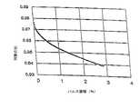
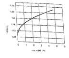
パルス振幅と対数の比の間の関係を示す典型的なデータは図5ないし図8に示されている。図5及び図6は、計算されたPAと、2つの独立したセンサーからの、既知の飽和度における1つの選択されたパルスの対数の比との関係を示す。このデータは、一定の飽和度にもかかわらず、異なるパルス振幅での変化する比の値により証拠付けられるように、対数の比に実質的に影響を与える。 Exemplary data showing the relationship between pulse amplitude and log ratio is shown in FIGS. Figures 5 and 6 show the relationship between the calculated PA and the log ratio of one selected pulse at a known saturation from two independent sensors. This data substantially affects the log ratio, as evidenced by varying ratio values at different pulse amplitudes, despite a certain degree of saturation.
図7及び図8は、同様の飽和度でパルス振幅の範囲の変化を示す二人の患者の間における対数の比を比較する。図7に示されたデータは、ベースラインの光学的伝送と比較して、0.1パーセントより小さい比較的弱いパルス振幅信号を示す患者から収集された。対照的に、図8のデータは、4パーセントを超える比較的強いパルス振幅信号を示す患者から収集された。図7及び図8の結果を比較すると、実質的に同等な飽和度であるにもかかわらず、より高いパルス振幅がより高い比と関連することは明らかである。 FIGS. 7 and 8 compare the log ratio between two patients exhibiting a change in the range of pulse amplitudes with similar saturation. The data shown in FIG. 7 was collected from a patient exhibiting a relatively weak pulse amplitude signal of less than 0.1 percent compared to baseline optical transmission. In contrast, the data in FIG. 8 was collected from a patient showing a relatively strong pulse amplitude signal greater than 4 percent. Comparing the results of FIGS. 7 and 8, it is clear that higher pulse amplitudes are associated with higher ratios, despite substantially equal saturation.
図7及び8に示されいるように84パーセントを越える高い飽和度では、パルス振幅と対数の比との間に正の関係があり、図5及び図6に示されているように低い飽和度では負の関係があることが分かった。効果は、飽和度が84パーセントからずれていくと増加する。明らかに、対数の比へのパルス振幅の影響に適応できないことが著しい不正確を示すことになる。 At high saturation above 84 percent as shown in FIGS. 7 and 8, there is a positive relationship between pulse amplitude and log ratio, and low saturation as shown in FIGS. Then we found that there was a negative relationship. The effect increases as the saturation deviates from 84 percent. Clearly, the inability to adapt to the effect of pulse amplitude on the log ratio would indicate significant inaccuracies.
本発明にしたがって、図5ないし図8に示されたようなデータが異なる飽和度及び異なるパルス振幅で個々の患者から収集される。この情報を使用して、患者からモニターされた対数の比は、パルス振幅依存性を補償するためにプレチスモグラフ波において、各時間点における計算されたPAにインデックス付けされる。このようにして、実質的により正確なパルス酸素計のデータが得られる。 In accordance with the present invention, data as shown in FIGS. 5-8 is collected from individual patients with different saturations and different pulse amplitudes. Using this information, the log ratio monitored from the patient is indexed to the calculated PA at each time point in the plethysmograph wave to compensate for the pulse amplitude dependence. In this way, substantially more accurate pulse oximeter data is obtained.
本発明の1つの実施例として、上述した計算はパルス振幅のインデックス付けで動脈血の酸素飽和度を決定する方法を提供するために使用される。特に、対数の比の関数の動脈血の酸素飽和度が、所望の範囲にわたって複数の特定のパルス振幅のそれぞれで決定される。酸素計のデータが患者から得られ、患者のパルス振幅が決定される。モニターされた患者の対数の比が得られたデータから計算される。患者のパルス振幅と非常に整合するパルス振幅での、記憶された動脈血の酸素飽和度の関数が選択される。つぎに、モニターされた対数の比は患者の動脈血の酸素飽和度を決定するために、選択された動脈血の酸素飽和度の関数とともに使用される。好適に、最も近いパルス振幅を有する記憶された動脈血の酸素飽和度は、モニターされた対数の比とより密に整合させるために補間され得る。 As one embodiment of the present invention, the calculations described above are used to provide a method for determining arterial oxygen saturation with pulse amplitude indexing. In particular, arterial oxygen saturation as a function of log ratio is determined at each of a plurality of specific pulse amplitudes over a desired range. Oximeter data is obtained from the patient and the patient's pulse amplitude is determined. The log ratio of the monitored patient is calculated from the data obtained. A function of the stored arterial oxygen saturation at a pulse amplitude that closely matches the patient's pulse amplitude is selected. The monitored log ratio is then used with a function of the selected arterial oxygen saturation to determine the arterial oxygen saturation of the patient. Preferably, the stored arterial oxygen saturation with the closest pulse amplitude can be interpolated to more closely match the monitored log ratio.
上述のとおり、低酸素ガスの混合物の吸入による脱飽和を受ける複数の患者からデータが収集される。対数の比の関数の動脈血の酸素飽和度は、データセットのアレーを生成するために、パルス振幅の範囲で集計される。したがって、そのアレーの各列は、既知の飽和度に対するパルス振幅のための対数の期待値を含む。 As described above, data is collected from multiple patients undergoing desaturation by inhalation of a mixture of hypoxic gases. The arterial oxygen saturation as a function of log ratio is aggregated over a range of pulse amplitudes to produce an array of data sets. Thus, each column of the array contains a logarithmic expectation for the pulse amplitude for a known saturation.
記憶された飽和度のデータは、動脈血の酸素飽和度を決定するために使用され、パルス酸素計のモニタリングが行われる。たとえば、モニターされたIR信号は実験的に導かれた式を使用して上述のとおりに訂正される。上記のとおり、あるパルスのパルス振幅は、赤外線の信号のAC成分のDC成分に対する電圧の比を決定することにより計算され得る。
電圧比=(電圧IR)/(電圧DC)The stored saturation data is used to determine arterial oxygen saturation and pulse oximeter monitoring is performed. For example, the monitored IR signal is corrected as described above using experimentally derived equations. As described above, the pulse amplitude of a pulse can be calculated by determining the ratio of the AC component to the DC component of the infrared signal.
Voltage ratio = (Voltage IR) / (Voltage DC)
飽和度因子がつぎの通りに計算される。
飽和度因子=−5.58+(8.17*飽和度)
飽和度の関数として表される。The saturation factor is calculated as follows:
Saturation factor = −5.58 + (8.17 * saturation)
Expressed as a function of saturation.
電圧比及び飽和度因子はパルス振幅を計算するために組み合わされる。
PA=((飽和度因子*電圧比/100)+電圧比)*100The voltage ratio and the saturation factor are combined to calculate the pulse amplitude.
PA = ((saturation factor * voltage ratio / 100) + voltage ratio) * 100
したがって、計算されたパルス振幅及びモニターされた対数の比は、最小自乗法推定法といった標準技術を使用して記憶された動脈血の飽和度データから飽和度の最もよく近似されたものを得るために使用され得る。好適にはアレーのデータセットはそのパルス振幅及び対数の比に対しても正確な飽和度の計算値を得るために補間される。 Thus, the calculated pulse amplitude and the ratio of the monitored logarithm are used to obtain the best approximation of saturation from arterial blood saturation data stored using standard techniques such as least squares estimation. Can be used. Preferably, the array data set is interpolated to obtain an accurate saturation calculation for its pulse amplitude and log ratio.
他の実施例として、赤色光の信号及び赤外線の信号の他の数学的組み合わせも対数の比に代えて使用できる。 As another example, other mathematical combinations of red and infrared signals can be used in place of the log ratio.
例
以下の例は、当業者が本発明をより明りょうに理解し、実施するためのものである。それらは本発明を制限するためのものではなく、単なる例示である。Examples The following examples are intended to enable those skilled in the art to more clearly understand and to practice the present invention. They are not intended to limit the invention but are merely exemplary.
例1
パルス振幅インデックス付き対数の比を使用して酸素計の決定とインデックス付けのない値との比較のために、八人の成人ボランティアに対して実施された。研究目的で、カテーテルが各対象者に橈骨動脈に配置された。Nellcor N-200パルス酸素計が標準装置として使用され、対象者を臨床的にモニターするために使用された。各対象者は、約70−100パーセントの範囲にある動脈血のヘモグロビンの酸素飽和度を生成するために、酸素濃度の変化する吸気が与えられた。Example 1
Pulse adult indexed log ratios were used on eight adult volunteers for comparison of oximeter determinations and unindexed values. For research purposes, a catheter was placed in the radial artery for each subject. A Nellcor N-200 pulse oximeter was used as a standard device and was used to monitor subjects clinically. Each subject was given an inspiration with varying oxygen concentrations to produce arterial hemoglobin oxygen saturation in the range of about 70-100 percent.
データの確認、二重のデータ、さらなる生理学的な情報のために、各患者の両手の指のそれぞれに、離れた位置に配置された2つの独立したセンサから実験的な酸素計の読み取りが行われた。各センサーからのデータは独立して処理された。 Experimental oximeter readings were taken from two separate sensors located on each finger of each patient's fingers for data validation, dual data, and additional physiological information. It was broken. Data from each sensor was processed independently.
血液のサンプルが動脈カテーテルから抽出され、同時に、酸素飽和度が読み取られ、すぐに分析された。データは収集され、波形が分析され、中間の工程が計算された。動脈血のサンプルは放射計により、2つの分離した血液−ガス分析計で分析された。ヘモグロビンの機能的な飽和度は、酸素ヘモグロビン/全ヘモグロビンとして計算された。すなわち、すべての非酸素ヘモグロビン種が全ヘモグロビンに含まれている。すべての飽和度でかつ全ての人体研究対象者に対して、アルゴリズムにしたがって計算された値に対する基準値は2つのCO−酸素計からの平均より読み取り値である。 A sample of blood was extracted from the arterial catheter and at the same time the oxygen saturation was read and analyzed immediately. Data was collected, waveforms were analyzed, and intermediate steps were calculated. Arterial blood samples were analyzed by radiometer with two separate blood-gas analyzers. The functional saturation of hemoglobin was calculated as oxygen hemoglobin / total hemoglobin. That is, all non-oxygen hemoglobin species are included in all hemoglobins. For all saturation subjects and for all human subjects, the reference value for the value calculated according to the algorithm is a reading from the average from two CO-oximeters.
上記研究結果が図9に示されている。対数の比がパルス振幅にインデックス付けされると、単に最大の振幅で決定されるよりも高い精度が得られることが、2つの独立したセンサーの使用により示された。これらの結果は、パルス振幅にインデックス付けを行うことにより、五倍まで精度を増加させることが可能であることを示す。振幅にインデックス付けすることにより得られた改善は、より高い飽和度及びより低い飽和度でもっとも大きく、84パーセント付近で最も小さい。 The study results are shown in FIG. It has been shown by the use of two independent sensors that the log ratio is indexed to the pulse amplitude to obtain a higher accuracy than simply determined at the maximum amplitude. These results show that it is possible to increase the accuracy up to 5 times by indexing the pulse amplitude. The improvement obtained by indexing the amplitude is greatest at higher and lower saturations and is smallest around 84 percent.
この例で、対数の比は1パーセントのパルス振幅にインデックス付けされる。しかし、当業者であれば、基準のデータが利用可能となるようにどの振幅も選択できる。好適実施例では、各パルスに対して利用可能な最も高いパルス振幅が、信号対ノイズ比を最大にするために使用される。最も高い振幅では、較正曲線はまた急勾配をもっており、その結果測定された飽和度のパラメータの解像度が改善される。 In this example, the log ratio is indexed to 1 percent pulse amplitude. However, one of ordinary skill in the art can select any amplitude such that reference data is available. In the preferred embodiment, the highest pulse amplitude available for each pulse is used to maximize the signal to noise ratio. At the highest amplitude, the calibration curve is also steep, resulting in improved resolution of the measured saturation parameter.
1パーセントのパルス振幅未満、実際上は0.1パーセント未満の解像度で、多くの飽和度及パルス振幅に対しインデックスデータをもつことが望ましい。 It is desirable to have index data for many saturations and pulse amplitudes with a resolution of less than 1 percent pulse amplitude, and practically less than 0.1 percent.
上記方法を採用することにより、最も正確なデータ点は、可能な限り高い精度を達成し、維持するために、パルス振幅により引き起こされる偏差を除去するために、選択されインデックス付けされる。 By employing the above method, the most accurate data points are selected and indexed to eliminate deviations caused by pulse amplitudes in order to achieve and maintain the highest possible accuracy.
さらに他の実施例として本発明の原理は、広範囲な他の生物学的及び肉体的な決定に適用することができる。たとえば、米国特許第6,480,729号明細書、米国特許第6,537,225号明細書、米国特許第6,594,511号明細書、米国特許第6,719,705号明細書、米国特許第6,819,950号明細書、米国特許第6,921,367号明細書、米国特許出願第10/912,721号(2004年8月4日出願)(これら文献は、ここの参考文献として組み込まれる)のそれぞれは生理学的特性を決定するための信号の取得に関するもので、本発明の方法及び装置とともの使用できる。 As yet another example, the principles of the present invention can be applied to a wide range of other biological and physical decisions. For example, U.S. Patent 6,480,729, U.S. Patent 6,537,225, U.S. Patent 6,594,511, U.S. Patent 6,719,705, U.S. Pat. US patent application Ser. No. 10 / 912,721 (filed Aug. 4, 2004), each of which is incorporated herein by reference, relates to the acquisition of signals for determining physiological properties, Can be used with the method and apparatus.
本発明の精神および範囲から逸脱することなく,当業者は本発明に種々の変更および改変を施して種々の用途および条件に適合させることができる。かかるものとして,これらの変更および改変は適切にかつ公平に請求の範囲の均等の範囲内に完全に入るものである。 Without departing from the spirit and scope of this invention, one of ordinary skill can make various changes and modifications to the invention to adapt it to various uses and conditions. As such, these changes and modifications are properly and fairly within the scope of the equivalent claims.
Claims (28)
Translated fromJapanese第一の波長をもつ第一の光を放出する第一の放射エミッタと、
第二の波長をもつ第二の光を放出する第二の放射エミッタと、
患者の血液を通過し吸収された後の前記第一及び第二の波長の前記第一及び第二の光を受光し、前記第一及び第二の光に対応する第一の強度信号及び第二の強度信号を与える放射検出器と、
前記第一及び第二の強度信号に関連したパルス振幅を決定し、該決定されたパルス振幅に前記第一及び第二の強度信号のモニターされた数学的な組み合わせをインデックス付けすることにより前記患者の血液の前記生理学的特性を計算するための制御器と、
を含み、
前記モニターされた数学的な組み合わせが前記生理学的特性に関連し、前記第一及び第二の強度信号から計算される、
ことを特徴とする装置。A device for monitoring physiological characteristics of a patient's blood,
A first radiation emitter that emits a first light having a first wavelength;
A second radiation emitter that emits a second light having a second wavelength;
Receiving the first and second lights of the first and second wavelengths after passing through the patient's blood and being absorbed, and a first intensity signal corresponding to the first and second lights and a first A radiation detector that provides a second intensity signal;
Determining the pulse amplitude associated with the first and second intensity signals and indexing the monitored mathematical combination of the first and second intensity signals to the determined pulse amplitude. A controller for calculating said physiological characteristics of the blood of
Including
The monitored mathematical combination relates to the physiological characteristic and is calculated from the first and second intensity signals;
A device characterized by that.
前記患者の組織領域に酸素計のセンサーを配置する工程と、
実質的に赤色光の範囲にある第一の光及び実質的に赤外線の範囲にある第二の光を前記患者の組織領域に通過させる工程と、
前記第一及び第二の強度信号に関連したパルス振幅を決定する工程と、
前記第一及び第二の強度信号から、前記生理学的特性に関連したモニターされた数学的組み合わせを計算する工程と、
前記モニターされた数学的組み合わせを前記決定されたパルス振幅にインデックス付けする工程と、
を含む方法。A method of processing a signal that reflects a physiological characteristic of a patient's blood, comprising:
Placing an oximeter sensor in the tissue region of the patient;
Passing a first light substantially in the red light range and a second light substantially in the infrared range through the tissue region of the patient;
Determining a pulse amplitude associated with the first and second intensity signals;
Calculating a monitored mathematical combination associated with the physiological characteristic from the first and second intensity signals;
Indexing the monitored mathematical combination to the determined pulse amplitude;
Including methods.
複数の特定のパルス振幅のそれぞれで、2つの光学的な信号の数学的な組み合わせの関数として動脈の酸素飽和度を決定する工程と、
前記動脈の酸素飽和度の関数を記憶する工程と、
対象者から酸素計データを得る工程と、
前記対象者のパルス振幅を決定する工程と、
前記酸素計のデータから得られた2つの光学的な信号の数学的な組み合わせをモニターする工程と、
前記決定されたパルス振幅に最も近いパルス振幅を有する、記憶された動脈の酸素飽和度の関数を選択する工程と、
前記モニターされた数学的な組み合わせ及び最も近いパルス振幅を有する、記憶された動脈の酸素飽和度の関数から前記対象者の動脈の酸素飽和度を決定する工程と、
を含む方法。A method for determining oxygen saturation in a subject's artery,
Determining arterial oxygen saturation as a function of a mathematical combination of two optical signals at each of a plurality of specific pulse amplitudes;
Storing a function of the oxygen saturation of the artery;
Obtaining oximeter data from the subject;
Determining the subject's pulse amplitude;
Monitoring a mathematical combination of two optical signals obtained from the oximeter data;
Selecting a stored arterial oxygen saturation function having a pulse amplitude closest to the determined pulse amplitude;
Determining the subject's arterial oxygen saturation from the stored arterial oxygen saturation function having the monitored mathematical combination and the closest pulse amplitude;
Including methods.
The method further comprises interpolating a stored arterial oxygen saturation function having a pulse amplitude closest to the determined pulse amplitude to more closely correspond to the measured mathematical combination. 28. The method according to 27.
Applications Claiming Priority (1)
| Application Number | Priority Date | Filing Date | Title |
|---|---|---|---|
| PCT/US2006/037914WO2008039195A1 (en) | 2006-09-27 | 2006-09-27 | Pulse amplitude indexing method and apparatus |
Publications (2)
| Publication Number | Publication Date |
|---|---|
| JP2010504803Atrue JP2010504803A (en) | 2010-02-18 |
| JP2010504803A5 JP2010504803A5 (en) | 2010-12-02 |
Family
ID=39230484
Family Applications (1)
| Application Number | Title | Priority Date | Filing Date |
|---|---|---|---|
| JP2009530316APendingJP2010504803A (en) | 2006-09-27 | 2006-09-27 | Method and apparatus for indexing pulse amplitude |
Country Status (3)
| Country | Link |
|---|---|
| EP (1) | EP2073694A4 (en) |
| JP (1) | JP2010504803A (en) |
| WO (1) | WO2008039195A1 (en) |
Families Citing this family (15)
| Publication number | Priority date | Publication date | Assignee | Title |
|---|---|---|---|---|
| US8229530B2 (en) | 2007-03-09 | 2012-07-24 | Nellcor Puritan Bennett Llc | System and method for detection of venous pulsation |
| US7541602B2 (en) | 2007-06-04 | 2009-06-02 | Or-Nim Medical Ltd. | System and method for noninvasively monitoring conditions of a subject |
| WO2009035669A1 (en) | 2007-09-13 | 2009-03-19 | The Curators Of The University Of Missouri | Optical device components |
| WO2009045492A1 (en) | 2007-10-04 | 2009-04-09 | The Curators Of The University Of Missouri | Optical device components |
| US7961305B2 (en) | 2007-10-23 | 2011-06-14 | The Curators Of The University Of Missouri | Optical device components |
| BRPI0909825B8 (en) | 2008-03-25 | 2021-06-22 | Univ Missouri | method and systems for non-invasive blood glucose detection using spectral data from one or more non-glucose components |
| US9560994B2 (en) | 2008-03-26 | 2017-02-07 | Covidien Lp | Pulse oximeter with adaptive power conservation |
| EP2299900B1 (en) | 2008-05-22 | 2017-06-07 | St. Louis Medical Devices, Inc. | Method and system for non-invasive optical blood glucose detection utilizing spectral data analysis |
| US8336391B2 (en) | 2008-07-06 | 2012-12-25 | Or-Nim Medical Ltd. | Method and system for non-invasively monitoring fluid flow in a subject |
| US9027412B2 (en) | 2008-07-06 | 2015-05-12 | Or-Nim Medical Ltd. | Method and system for non-invasively monitoring fluid flow in a subject |
| EP2339957A1 (en)* | 2008-07-06 | 2011-07-06 | Or-Nim Medical Ltd. | Method and system for non-invasively monitoring fluid flow in a subject |
| EP2413784B1 (en) | 2009-04-01 | 2024-07-17 | St. Louis Medical Devices, Inc. | Optical spectroscopy device for non-invasive blood glucose detection and associated method of use |
| US8509869B2 (en) | 2009-05-15 | 2013-08-13 | Covidien Lp | Method and apparatus for detecting and analyzing variations in a physiologic parameter |
| US9066660B2 (en) | 2009-09-29 | 2015-06-30 | Nellcor Puritan Bennett Ireland | Systems and methods for high-pass filtering a photoplethysmograph signal |
| AU2016283095C1 (en) | 2015-06-25 | 2020-07-23 | Fresenius Medical Care Holdings, Inc. | Direct light differential measurement system |
Family Cites Families (4)
| Publication number | Priority date | Publication date | Assignee | Title |
|---|---|---|---|---|
| EP1198196A4 (en)* | 1999-07-14 | 2007-05-02 | Providence Health Sys Oregon | METHOD AND DEVICE FOR PULSE OXIMETRY WITH ADAPTIVE CALIBRATION |
| US6882874B2 (en)* | 2002-02-15 | 2005-04-19 | Datex-Ohmeda, Inc. | Compensation of human variability in pulse oximetry |
| US6711425B1 (en)* | 2002-05-28 | 2004-03-23 | Ob Scientific, Inc. | Pulse oximeter with calibration stabilization |
| US7194293B2 (en)* | 2004-03-08 | 2007-03-20 | Nellcor Puritan Bennett Incorporated | Selection of ensemble averaging weights for a pulse oximeter based on signal quality metrics |
- 2006
- 2006-09-27JPJP2009530316Apatent/JP2010504803A/enactivePending
- 2006-09-27WOPCT/US2006/037914patent/WO2008039195A1/enactiveApplication Filing
- 2006-09-27EPEP06804239Apatent/EP2073694A4/ennot_activeWithdrawn
Also Published As
| Publication number | Publication date |
|---|---|
| EP2073694A1 (en) | 2009-07-01 |
| EP2073694A4 (en) | 2010-12-08 |
| WO2008039195A1 (en) | 2008-04-03 |
Similar Documents
| Publication | Publication Date | Title |
|---|---|---|
| US7184809B1 (en) | Pulse amplitude indexing method and apparatus | |
| JP2010504803A (en) | Method and apparatus for indexing pulse amplitude | |
| US7215987B1 (en) | Method and apparatus for processing signals reflecting physiological characteristics | |
| US20070260132A1 (en) | Method and apparatus for processing signals reflecting physiological characteristics from multiple sensors | |
| US7392075B2 (en) | Method for enhancing pulse oximetry calculations in the presence of correlated artifacts | |
| US7254431B2 (en) | Physiological parameter tracking system | |
| US5766127A (en) | Method and apparatus for improved photoplethysmographic perfusion-index monitoring | |
| JP3590409B2 (en) | Self-luminous non-invasive infrared spectrophotometer with temperature compensation | |
| JP2602321B2 (en) | Method and apparatus for calculating arterial oxygen saturation based on plethysmograph including transient state | |
| US6896661B2 (en) | Monitoring physiological parameters based on variations in a photoplethysmographic baseline signal | |
| US4863265A (en) | Apparatus and method for measuring blood constituents | |
| JP3590408B2 (en) | Self-luminous non-invasive infrared spectrophotometer | |
| US6178343B1 (en) | Pulse rate and heart rate coincidence detection for pulse oximetry | |
| US5193543A (en) | Method and apparatus for measuring arterial blood constituents | |
| JP2003505115A (en) | Digital oximeter and method for calculating oxygenation level | |
| JPH0549624A (en) | Method and device for monitoring saturation degree of blood oxygen | |
| US20080221462A1 (en) | Detection of oximetry sensor sites based on waveform characteristics | |
| CN209899402U (en) | Reflection type oximeter | |
| CN109044366B (en) | Method for detecting glycosylated hemoglobin and blood oxygen saturation and optical fingertip detector | |
| JP3107630B2 (en) | Pulse oximeter | |
| JP2693958B2 (en) | Oximeter and method for measuring blood components in arteries | |
| JP6653787B2 (en) | Optical vital sign sensor | |
| EP2073695A1 (en) | Method and apparatus for processing signals reflecting physiological characteristics | |
| WO2009088799A1 (en) | Method and apparatus for assessing contact of a sensor with arterialized tissue | |
| CN211049359U (en) | Blood oxygen measuring device and system |
Legal Events
| Date | Code | Title | Description |
|---|---|---|---|
| A072 | Dismissal of procedure [no reply to invitation to correct request for examination] | Free format text:JAPANESE INTERMEDIATE CODE: A073 Effective date:20110131 |