JP2010503496A - Apparatus and method for destroying cancer cells - Google Patents
Apparatus and method for destroying cancer cellsDownload PDFInfo
- Publication number
- JP2010503496A JP2010503496AJP2009528510AJP2009528510AJP2010503496AJP 2010503496 AJP2010503496 AJP 2010503496AJP 2009528510 AJP2009528510 AJP 2009528510AJP 2009528510 AJP2009528510 AJP 2009528510AJP 2010503496 AJP2010503496 AJP 2010503496A
- Authority
- JP
- Japan
- Prior art keywords
- electrode
- target tissue
- cancer cells
- ablation
- electrodes
- Prior art date
- Legal status (The legal status is an assumption and is not a legal conclusion. Google has not performed a legal analysis and makes no representation as to the accuracy of the status listed.)
- Granted
Links
- 206010028980NeoplasmDiseases0.000titleclaimsabstractdescription157
- 238000000034methodMethods0.000titleclaimsabstractdescription104
- 201000011510cancerDiseases0.000titleclaimsabstractdescription94
- 230000005684electric fieldEffects0.000claimsabstractdescription86
- 210000004027cellAnatomy0.000claimsdescription138
- 238000002679ablationMethods0.000claimsdescription117
- 238000012384transportation and deliveryMethods0.000claimsdescription48
- 238000011282treatmentMethods0.000claimsdescription47
- 239000000523sampleSubstances0.000claimsdescription31
- 230000006378damageEffects0.000claimsdescription21
- 238000003384imaging methodMethods0.000claimsdescription14
- 210000000170cell membraneAnatomy0.000claimsdescription8
- 229910052751metalInorganic materials0.000claimsdescription7
- 239000002184metalSubstances0.000claimsdescription7
- 238000002271resectionMethods0.000claimsdescription6
- 230000008859changeEffects0.000claimsdescription5
- 230000006369cell cycle progressionEffects0.000claimsdescription3
- 230000001225therapeutic effectEffects0.000claimsdescription3
- 230000008520organizationEffects0.000claims2
- 238000005339levitationMethods0.000claims1
- 230000008600mitotic progressionEffects0.000claims1
- 210000001519tissueAnatomy0.000description207
- 230000008569processEffects0.000description23
- 230000000694effectsEffects0.000description14
- 239000012530fluidSubstances0.000description9
- 230000032823cell divisionEffects0.000description8
- 241000700159RattusSpecies0.000description7
- 230000012010growthEffects0.000description7
- 239000000463materialSubstances0.000description7
- 238000013461designMethods0.000description6
- 230000008901benefitEffects0.000description5
- 238000004520electroporationMethods0.000description5
- 230000007246mechanismEffects0.000description5
- 238000007674radiofrequency ablationMethods0.000description5
- 230000004083survival effectEffects0.000description5
- 206010020843HyperthermiaDiseases0.000description4
- 230000009471actionEffects0.000description4
- 238000004458analytical methodMethods0.000description4
- 230000015556catabolic processEffects0.000description4
- 238000002591computed tomographyMethods0.000description4
- 238000007667floatingMethods0.000description4
- 230000006870functionEffects0.000description4
- 230000036031hyperthermiaEffects0.000description4
- 230000011278mitosisEffects0.000description4
- 239000007787solidSubstances0.000description4
- 210000004881tumor cellAnatomy0.000description4
- 208000026310Breast neoplasmDiseases0.000description3
- 241001465754MetazoaSpecies0.000description3
- 238000011298ablation treatmentMethods0.000description3
- 238000001514detection methodMethods0.000description3
- 238000012377drug deliveryMethods0.000description3
- 150000002739metalsChemical class0.000description3
- 238000009206nuclear medicineMethods0.000description3
- 230000035479physiological effects, processes and functionsEffects0.000description3
- 238000012545processingMethods0.000description3
- 230000002062proliferating effectEffects0.000description3
- 206010006187Breast cancerDiseases0.000description2
- 230000006907apoptotic processEffects0.000description2
- 230000036760body temperatureEffects0.000description2
- 230000022131cell cycleEffects0.000description2
- 239000004020conductorSubstances0.000description2
- 230000009089cytolysisEffects0.000description2
- 230000001086cytosolic effectEffects0.000description2
- 238000004925denaturationMethods0.000description2
- 230000036425denaturationEffects0.000description2
- 229910003460diamondInorganic materials0.000description2
- 239000010432diamondSubstances0.000description2
- 238000010438heat treatmentMethods0.000description2
- 230000002977hyperthermial effectEffects0.000description2
- 238000003780insertionMethods0.000description2
- 230000037431insertionEffects0.000description2
- 230000001788irregularEffects0.000description2
- 230000002427irreversible effectEffects0.000description2
- 231100000518lethalToxicity0.000description2
- 230000001665lethal effectEffects0.000description2
- 230000000670limiting effectEffects0.000description2
- 239000007788liquidSubstances0.000description2
- 238000002595magnetic resonance imagingMethods0.000description2
- 230000001404mediated effectEffects0.000description2
- 239000012528membraneSubstances0.000description2
- 230000031864metaphaseEffects0.000description2
- 239000000203mixtureSubstances0.000description2
- 230000004048modificationEffects0.000description2
- 238000012986modificationMethods0.000description2
- 230000001338necrotic effectEffects0.000description2
- HLXZNVUGXRDIFK-UHFFFAOYSA-Nnickel titaniumChemical compound[Ti].[Ti].[Ti].[Ti].[Ti].[Ti].[Ti].[Ti].[Ti].[Ti].[Ti].[Ni].[Ni].[Ni].[Ni].[Ni].[Ni].[Ni].[Ni].[Ni].[Ni].[Ni].[Ni].[Ni].[Ni]HLXZNVUGXRDIFK-UHFFFAOYSA-N0.000description2
- 229910001000nickel titaniumInorganic materials0.000description2
- 238000013421nuclear magnetic resonance imagingMethods0.000description2
- 230000003287optical effectEffects0.000description2
- BASFCYQUMIYNBI-UHFFFAOYSA-NplatinumChemical compound[Pt]BASFCYQUMIYNBI-UHFFFAOYSA-N0.000description2
- 102000004169proteins and genesHuman genes0.000description2
- 108090000623proteins and genesProteins0.000description2
- 238000011084recoveryMethods0.000description2
- 229910001220stainless steelInorganic materials0.000description2
- 239000010935stainless steelSubstances0.000description2
- 238000012360testing methodMethods0.000description2
- 231100000419toxicityToxicity0.000description2
- 230000001988toxicityEffects0.000description2
- 238000002604ultrasonographyMethods0.000description2
- RYGMFSIKBFXOCR-UHFFFAOYSA-NCopperChemical compound[Cu]RYGMFSIKBFXOCR-UHFFFAOYSA-N0.000description1
- 102000004190EnzymesHuman genes0.000description1
- 108090000790EnzymesProteins0.000description1
- 206010037660PyrexiaDiseases0.000description1
- BQCADISMDOOEFD-UHFFFAOYSA-NSilverChemical compound[Ag]BQCADISMDOOEFD-UHFFFAOYSA-N0.000description1
- 229910000831SteelInorganic materials0.000description1
- 210000001015abdomenAnatomy0.000description1
- 230000002159abnormal effectEffects0.000description1
- 239000000853adhesiveSubstances0.000description1
- 230000001070adhesive effectEffects0.000description1
- 230000002411adverseEffects0.000description1
- 238000013019agitationMethods0.000description1
- 238000010171animal modelMethods0.000description1
- 238000003491arrayMethods0.000description1
- 230000006399behaviorEffects0.000description1
- 230000009286beneficial effectEffects0.000description1
- 210000005013brain tissueAnatomy0.000description1
- 210000000481breastAnatomy0.000description1
- 210000000621bronchiAnatomy0.000description1
- 230000030833cell deathEffects0.000description1
- 230000010261cell growthEffects0.000description1
- 230000004663cell proliferationEffects0.000description1
- 230000001413cellular effectEffects0.000description1
- 230000033077cellular processEffects0.000description1
- 210000001072colonAnatomy0.000description1
- 229910052802copperInorganic materials0.000description1
- 239000010949copperSubstances0.000description1
- 238000012937correctionMethods0.000description1
- 230000021953cytokinesisEffects0.000description1
- 210000000805cytoplasmAnatomy0.000description1
- 230000000593degrading effectEffects0.000description1
- 230000002939deleterious effectEffects0.000description1
- 238000010586diagramMethods0.000description1
- 230000035622drinkingEffects0.000description1
- 239000007772electrode materialSubstances0.000description1
- 238000005516engineering processMethods0.000description1
- 230000001747exhibiting effectEffects0.000description1
- 238000002474experimental methodMethods0.000description1
- 238000000605extractionMethods0.000description1
- 230000008713feedback mechanismEffects0.000description1
- PCHJSUWPFVWCPO-UHFFFAOYSA-NgoldChemical compound[Au]PCHJSUWPFVWCPO-UHFFFAOYSA-N0.000description1
- 229910052737goldInorganic materials0.000description1
- 239000010931goldSubstances0.000description1
- 230000007773growth patternEffects0.000description1
- 230000023597hemostasisEffects0.000description1
- 208000021760high feverDiseases0.000description1
- 230000028993immune responseEffects0.000description1
- 230000002147killing effectEffects0.000description1
- 230000003902lesionEffects0.000description1
- 210000004185liverAnatomy0.000description1
- 230000007774longtermEffects0.000description1
- 210000004072lungAnatomy0.000description1
- 210000001365lymphatic vesselAnatomy0.000description1
- 210000003712lysosomeAnatomy0.000description1
- 230000001868lysosomic effectEffects0.000description1
- 230000003211malignant effectEffects0.000description1
- 238000005259measurementMethods0.000description1
- 230000010534mechanism of actionEffects0.000description1
- 230000029115microtubule polymerizationEffects0.000description1
- 230000000394mitotic effectEffects0.000description1
- 210000003205muscleAnatomy0.000description1
- 230000017074necrotic cell deathEffects0.000description1
- 210000005170neoplastic cellAnatomy0.000description1
- 210000003463organelleAnatomy0.000description1
- 230000003204osmotic effectEffects0.000description1
- 210000004923pancreatic tissueAnatomy0.000description1
- 230000036961partial effectEffects0.000description1
- 230000002093peripheral effectEffects0.000description1
- 229910052697platinumInorganic materials0.000description1
- 229920000642polymerPolymers0.000description1
- 238000003825pressingMethods0.000description1
- 230000002250progressing effectEffects0.000description1
- 230000035755proliferationEffects0.000description1
- 210000002307prostateAnatomy0.000description1
- 210000000664rectumAnatomy0.000description1
- 230000002829reductive effectEffects0.000description1
- 229910052709silverInorganic materials0.000description1
- 239000004332silverSubstances0.000description1
- 230000020347spindle assemblyEffects0.000description1
- 230000000087stabilizing effectEffects0.000description1
- 239000010959steelSubstances0.000description1
- 230000000638stimulationEffects0.000description1
- 238000007920subcutaneous administrationMethods0.000description1
- 238000012385systemic deliveryMethods0.000description1
- 231100000057systemic toxicityToxicity0.000description1
- 238000002560therapeutic procedureMethods0.000description1
- 231100000732tissue residueToxicity0.000description1
- 230000001960triggered effectEffects0.000description1
- 230000004614tumor growthEffects0.000description1
- 238000011179visual inspectionMethods0.000description1
- 238000005303weighingMethods0.000description1
Images


Classifications
- A—HUMAN NECESSITIES
- A61—MEDICAL OR VETERINARY SCIENCE; HYGIENE
- A61B—DIAGNOSIS; SURGERY; IDENTIFICATION
- A61B18/00—Surgical instruments, devices or methods for transferring non-mechanical forms of energy to or from the body
- A61B18/04—Surgical instruments, devices or methods for transferring non-mechanical forms of energy to or from the body by heating
- A61B18/12—Surgical instruments, devices or methods for transferring non-mechanical forms of energy to or from the body by heating by passing a current through the tissue to be heated, e.g. high-frequency current
- A61B18/14—Probes or electrodes therefor
- A61B18/1477—Needle-like probes
- A—HUMAN NECESSITIES
- A61—MEDICAL OR VETERINARY SCIENCE; HYGIENE
- A61B—DIAGNOSIS; SURGERY; IDENTIFICATION
- A61B18/00—Surgical instruments, devices or methods for transferring non-mechanical forms of energy to or from the body
- A61B18/04—Surgical instruments, devices or methods for transferring non-mechanical forms of energy to or from the body by heating
- A61B18/12—Surgical instruments, devices or methods for transferring non-mechanical forms of energy to or from the body by heating by passing a current through the tissue to be heated, e.g. high-frequency current
- A61B18/14—Probes or electrodes therefor
- A61B18/1492—Probes or electrodes therefor having a flexible, catheter-like structure, e.g. for heart ablation
- A—HUMAN NECESSITIES
- A61—MEDICAL OR VETERINARY SCIENCE; HYGIENE
- A61N—ELECTROTHERAPY; MAGNETOTHERAPY; RADIATION THERAPY; ULTRASOUND THERAPY
- A61N1/00—Electrotherapy; Circuits therefor
- A61N1/02—Details
- A61N1/04—Electrodes
- A61N1/06—Electrodes for high-frequency therapy
- A—HUMAN NECESSITIES
- A61—MEDICAL OR VETERINARY SCIENCE; HYGIENE
- A61B—DIAGNOSIS; SURGERY; IDENTIFICATION
- A61B18/00—Surgical instruments, devices or methods for transferring non-mechanical forms of energy to or from the body
- A61B2018/00571—Surgical instruments, devices or methods for transferring non-mechanical forms of energy to or from the body for achieving a particular surgical effect
- A61B2018/00577—Ablation
- A—HUMAN NECESSITIES
- A61—MEDICAL OR VETERINARY SCIENCE; HYGIENE
- A61B—DIAGNOSIS; SURGERY; IDENTIFICATION
- A61B18/00—Surgical instruments, devices or methods for transferring non-mechanical forms of energy to or from the body
- A61B18/04—Surgical instruments, devices or methods for transferring non-mechanical forms of energy to or from the body by heating
- A61B18/12—Surgical instruments, devices or methods for transferring non-mechanical forms of energy to or from the body by heating by passing a current through the tissue to be heated, e.g. high-frequency current
- A61B18/14—Probes or electrodes therefor
- A61B2018/1405—Electrodes having a specific shape
- A61B2018/1407—Loop
- A—HUMAN NECESSITIES
- A61—MEDICAL OR VETERINARY SCIENCE; HYGIENE
- A61B—DIAGNOSIS; SURGERY; IDENTIFICATION
- A61B18/00—Surgical instruments, devices or methods for transferring non-mechanical forms of energy to or from the body
- A61B18/04—Surgical instruments, devices or methods for transferring non-mechanical forms of energy to or from the body by heating
- A61B18/12—Surgical instruments, devices or methods for transferring non-mechanical forms of energy to or from the body by heating by passing a current through the tissue to be heated, e.g. high-frequency current
- A61B18/14—Probes or electrodes therefor
- A61B2018/1405—Electrodes having a specific shape
- A61B2018/1425—Needle
- A61B2018/143—Needle multiple needles
- A—HUMAN NECESSITIES
- A61—MEDICAL OR VETERINARY SCIENCE; HYGIENE
- A61B—DIAGNOSIS; SURGERY; IDENTIFICATION
- A61B18/00—Surgical instruments, devices or methods for transferring non-mechanical forms of energy to or from the body
- A61B18/04—Surgical instruments, devices or methods for transferring non-mechanical forms of energy to or from the body by heating
- A61B18/12—Surgical instruments, devices or methods for transferring non-mechanical forms of energy to or from the body by heating by passing a current through the tissue to be heated, e.g. high-frequency current
- A61B18/14—Probes or electrodes therefor
- A61B2018/1475—Electrodes retractable in or deployable from a housing
Landscapes
- Health & Medical Sciences (AREA)
- Engineering & Computer Science (AREA)
- Life Sciences & Earth Sciences (AREA)
- Surgery (AREA)
- Animal Behavior & Ethology (AREA)
- Veterinary Medicine (AREA)
- Nuclear Medicine, Radiotherapy & Molecular Imaging (AREA)
- Public Health (AREA)
- Biomedical Technology (AREA)
- General Health & Medical Sciences (AREA)
- Otolaryngology (AREA)
- Molecular Biology (AREA)
- Medical Informatics (AREA)
- Heart & Thoracic Surgery (AREA)
- Physics & Mathematics (AREA)
- Plasma & Fusion (AREA)
- Radiology & Medical Imaging (AREA)
- Cardiology (AREA)
- Surgical Instruments (AREA)
- Electrotherapy Devices (AREA)
- Medicines Containing Material From Animals Or Micro-Organisms (AREA)
- Medicines That Contain Protein Lipid Enzymes And Other Medicines (AREA)
- Pharmaceuticals Containing Other Organic And Inorganic Compounds (AREA)
Abstract
Translated fromJapaneseDescription
Translated fromJapanese(関連出願の相互参照)
本出願は、米国特許法第119条(e)に基づき、2006年9月14日に出願された米国仮特許出願第60/825,660号(代理人整理番号第026533−000100US号)、及び2006年10月30日に出願された米国仮特許出願第60/863,484号(代理人整理番号第026533−000200US号)の利益を主張し、これらの全開示事項は引用により本明細書に組み入れられる。(Cross-reference of related applications)
This application is based on United States Patent Act 119 (e), US Provisional Patent Application No. 60 / 825,660 (Attorney Docket No. 0265533-000100US) filed on September 14, 2006, and Claims the benefit of US Provisional Patent Application No. 60 / 863,484 (Attorney Docket No. 0265533-000200US) filed October 30, 2006, the entire disclosure of which is incorporated herein by reference. Be incorporated.
(技術分野)
本発明は、一般に、組織領域への電場のデリバリ(送達)に関する。更に詳細には、本発明は、電場デリバリ、及び癌細胞及び固形腫瘍の選択的切除を含む標的組織領域の非熱切除に関する。(Technical field)
The present invention relates generally to the delivery of electric fields to a tissue region. More particularly, the present invention relates to electric field delivery and non-thermal ablation of target tissue regions including selective excision of cancer cells and solid tumors.
現在の組織切除技術は、望ましくない組織又は病変の除去、止血、又は組織の切断を行う手段として、患者(例えばヒト、動物等)の組織に高周波数の高熱を誘起する電流に依存する。腫瘍組織を熱誘起死滅及び/又は除去することにより癌を治療するためのツールとして高熱切除の分野に関心が高まっており、活発に取り組まれている。 Current tissue ablation techniques rely on currents that induce high frequency high heat in the tissue of patients (eg, humans, animals, etc.) as a means of removing unwanted tissue or lesions, hemostasis, or cutting tissue. There is increasing interest in the field of hyperthermia as a tool for treating cancer by heat-induced killing and / or removal of tumor tissue and is actively addressed.
高熱腫瘍切除技術では、高周波数RF(例えば「RF熱切除」)又はマイクロ波源を用いて組織を加熱すると、標的組織に組織学的損傷が生じる。RF熱切除技術、例えば、約500kHz及びそれ以上を含む高周波を用いて、位置決めされた電極の周りの組織にイオン攪拌及び摩擦(例えば抵抗)加熱を引き起こす。組織への致死損傷(例えば、組織タンパク質の変性)は、約47°Cを超える温度で起こるが、RF熱切除の電極の近くで発生する熱は、約100°Cまで又はそれを超える温度に達する可能性がある。 In hyperthermic tumor excision techniques, heating tissue using a high frequency RF (eg, “RF thermal ablation”) or microwave source causes histological damage to the target tissue. RF thermal ablation techniques, such as high frequencies including about 500 kHz and above, are used to cause ion agitation and friction (eg, resistance) heating in the tissue around the positioned electrode. While lethal damage to tissue (eg, tissue protein denaturation) occurs at temperatures above about 47 ° C., heat generated near the electrode for RF thermal ablation can reach temperatures up to or above about 100 ° C. May reach.
高熱切除又は熱誘起腫瘍組織破壊に基づく多数の異なる癌切除法及び装置が提案されている。このような1つの実施例には、容積測定組織切除のための装置を教示する米国特許第5,827,276号(特許文献1)が含まれる。該装置は、カテーテルを通って支持された複数のワイヤを有するプローブを含み、近位端が発生器のアクティブ端子に接続され、遠位端がカテーテルの遠位端から突出している。教示事項には、単回の展開で大容積の熱的に切除された組織を生成することになる方法及び経皮的手順で展開可能なプローブが含まれる。 A number of different cancer resection methods and devices based on hyperthermia or heat-induced tumor tissue destruction have been proposed. One such example includes US Pat. No. 5,827,276, which teaches an apparatus for volumetric tissue ablation. The device includes a probe having a plurality of wires supported through a catheter, with a proximal end connected to the active terminal of the generator and a distal end protruding from the distal end of the catheter. The teachings include methods that will produce large volumes of thermally ablated tissue in a single deployment and probes that can be deployed in a percutaneous procedure.
米国特許第5,935,123号(特許文献2)は、カテーテル管腔を備えるカテーテルを含むRF治療装置を教示する。取り外し可能針電極は、カテーテルに固定関係でカテーテル管腔内に位置決めされる。治療装置は、限定ではないが腫瘍を含む選択した組織塊を切除するか、或いは高熱により塊を治療するのに使用するものとして教示されている。腫瘍部位は、RFエネルギーを制御してデリバリすることにより選択的に高熱療法又は切除で治療される。 U.S. Pat. No. 5,935,123 teaches an RF therapy device that includes a catheter with a catheter lumen. The removable needle electrode is positioned within the catheter lumen in a fixed relationship with the catheter. The treatment device is taught for use in excising selected tissue masses, including but not limited to tumors, or treating masses with high fever. The tumor site is selectively treated with hyperthermia or excision by controlling and delivering RF energy.
高熱又は熱誘起癌組織破壊を用いる多数の他の方法及び装置が教示されている。しかしながら、RF誘起高熱切除の重大な限界は、周囲の健康な非標的組織に対して組織学的損傷及び破壊を制限しながら、熱誘起損傷を標的癌組織に限局化するのが困難であることである。
従って、健康組織への損傷を最低限にしながら、癌細胞を選択的に破壊する侵襲性が最低限の切除技術が必要とされている。A number of other methods and devices using high heat or heat induced cancer tissue destruction are taught. However, a significant limitation of RF-induced hyperthermia is that it is difficult to localize heat-induced damage to the target cancer tissue while limiting histological damage and destruction to the surrounding healthy non-target tissue. It is.
Therefore, there is a need for a minimally invasive excision technique that selectively destroys cancer cells while minimizing damage to healthy tissue.
本発明は、選択的癌細胞破壊及び非熱組織切除のために低強度電場を印加するための装置及び関連する方法を提供する。本発明の装置は、一般に、1つの電極又は複数の電極を標的組織領域に導入して電場を標的組織領域に印加するように設計されることになる。1つの電極又は複数の電極は、典型的には、例えば、標的組織又は該標的組織領域内に位置決めされた電極内の位置から半径方向に複数の方向で外向きに電場が放射される場合を含む、印加電場が標的組織領域全体にわたって放射するように位置決めされる。付加的に、標的組織領域に印加されるエネルギーは、電気的に発生する熱が最小になり且つ組織温度の上昇を避けることができるように選択することができる。特定の実施形態では、印加電場は、一般に、標的細胞を低出力又は非熱切除するのに十分な低強度(例えば約50V/cm未満)且つ中間周波数(例えば、約50kHzと300kHzとの間)の交流電場である。本発明の電極位置決め及び電場印加(例えば、低出力/非熱切除電場)により、熱作用を切除プロセスの因子とすることなく癌細胞を切除するのに驚くほど効果的であることが実際に示された。更に、本発明による切除プロセスは、主に、異常増殖細胞又は無秩序な成長を示す細胞(例えば癌細胞)で行う。従って、本発明は、正常細胞又は組織を実質的に無傷で残しながら、癌細胞を最小の侵襲性で選択的に切除又は破壊する付加的な利点を提供する。 The present invention provides an apparatus and associated method for applying a low intensity electric field for selective cancer cell destruction and non-thermal tissue ablation. The devices of the present invention will generally be designed to introduce an electrode or electrodes into the target tissue region and apply an electric field to the target tissue region. One electrode or multiple electrodes are typically used when, for example, an electric field is radiated outwardly in a plurality of directions radially from a position within the target tissue or electrode positioned within the target tissue region. Including, the applied electric field is positioned to radiate across the target tissue region. Additionally, the energy applied to the target tissue region can be selected so that the heat generated electrically is minimized and an increase in tissue temperature can be avoided. In certain embodiments, the applied electric field is generally low intensity (eg, less than about 50 V / cm) and intermediate frequency (eg, between about 50 kHz and 300 kHz) sufficient to low power or non-thermal ablation of target cells. AC electric field. The electrode positioning and electric field application (e.g., low power / non-thermal ablation electric field) of the present invention has indeed shown to be surprisingly effective in resecting cancer cells without the thermal action being a factor in the ablation process. It was done. Furthermore, the excision process according to the present invention is mainly performed on abnormally proliferating cells or cells that exhibit unregulated growth (eg cancer cells). Thus, the present invention provides the additional advantage of selectively excising or destroying cancer cells with minimal invasiveness while leaving normal cells or tissue substantially intact.
従って、1つの態様では、本発明は、電場を組織にデリバリする方法を含む。このような方法は、癌細胞を含む標的組織領域内に電極を位置決めする段階と、交流電場を標的組織に印加して、電極の周りの標的組織領域の癌細胞を非熱的に切除する段階とを含む。 Accordingly, in one aspect, the present invention includes a method of delivering an electric field to tissue. Such a method includes positioning an electrode within a target tissue region containing cancer cells and applying an alternating electric field to the target tissue to non-thermally ablate cancer cells in the target tissue region around the electrode. Including.
1つの実施形態では、標的組織領域は、組織の塊又は固形部分を含む。典型的には、標的組織領域は、例えば、固形腫瘍を含む標的組織領域を有する癌細胞を含む。本発明の方法を受ける組織の容積は変わることができ、少なくとも部分的に癌細胞の塊の大きさに基づくことになる。標的組織領域の周囲寸法は、規則的(例えば、球形、楕円形、その他)とすることもでき、又は不規則であってもよい。標的組織領域は、超音波、コンピュータ断層撮影法(CT)走査、X線イメージング、核医学イメージング、核磁気共鳴画像法(MRI)、電磁波イメージング等のような従来のイメージング方法を用いて識別及び/又は特徴付けることができる。付加的に、種々のイメージングシステムは、患者の組織内或いは標的組織領域又はその内部に本発明の装置又は電極を配置及び/又は位置決めするのに用いることができる。 In one embodiment, the target tissue region comprises a tissue mass or solid portion. Typically, the target tissue region includes, for example, cancer cells having a target tissue region that includes a solid tumor. The volume of tissue undergoing the method of the invention can vary and will be based at least in part on the size of the mass of cancer cells. The perimeter dimensions of the target tissue region can be regular (eg, spherical, elliptical, etc.) or can be irregular. Target tissue regions are identified and / or used using conventional imaging methods such as ultrasound, computed tomography (CT) scanning, X-ray imaging, nuclear medicine imaging, nuclear magnetic resonance imaging (MRI), electromagnetic imaging, and the like. Or can be characterized. Additionally, various imaging systems can be used to place and / or position the device or electrode of the present invention within or within a patient tissue or a target tissue region.
上に記載したように、電極は、標的組織領域内に位置決めされ、交流電場が印加される。本発明による切除技術は、幾つかの実施形態では、局所組織温度を上昇させず、且つエネルギー印加による熱作用を組織切除を行う手段とすることなく達成することができる。典型的には、印加電場は、低強度の中間周波数交流電流を含む。1つの実施形態では、例えば、電流は、約50V/cm未満の電圧場を生成する。別の実施形態では、電流は、約50kHzと約300kHzとの周波数を含む。電圧場及び/又は印加電流の周波数は、エネルギー印加の間一定に保持することもでき、又は変えることもできる。電極は、標的組織内部から電場印加が行われるように標的組織領域内に位置決めすることができる。1つの実施形態では、電極は、標的組織領域(例えば腫瘍)内に位置決めされ、印加電流により、電極から半径方向外向きに延びる電場が形成される。特定の実施形態では、このような位置決めは、例えば、標的組織領域内の細胞を分裂/増殖する向きを含む腫瘍生理機能を利用して、電極により与えられる電場が分裂癌細胞の分裂軸と確実に実質的に位置合わせされるようにすることができる。 As described above, the electrodes are positioned in the target tissue region and an alternating electric field is applied. The ablation technique according to the present invention can be achieved in some embodiments without increasing the local tissue temperature and without applying the thermal effect of applying energy as a means for tissue ablation. Typically, the applied electric field includes low intensity intermediate frequency alternating current. In one embodiment, for example, the current generates a voltage field of less than about 50 V / cm. In another embodiment, the current includes a frequency of about 50 kHz and about 300 kHz. The frequency of the voltage field and / or the applied current can be kept constant during energy application or can be varied. The electrode can be positioned in the target tissue region such that an electric field is applied from within the target tissue. In one embodiment, the electrode is positioned within a target tissue region (eg, a tumor) and the applied current creates an electric field that extends radially outward from the electrode. In certain embodiments, such positioning utilizes, for example, tumor physiology, including the orientation to divide / proliferate cells in the target tissue region, to ensure that the electric field provided by the electrodes is aligned with the division axis of dividing cancer cells. Can be substantially aligned.
従って、別の態様では、本発明は、電場を組織にデリバリする方法を含み、本方法は、複数の電極を癌細胞を有する標的組織内に位置決めする段階を含む。この複数の電極は、第1の電極及び複数の第2の電極を含み、切除容積が少なくとも部分的に定められる。本方法は、交流電流を容積に印加し、容積内から半径方向外向きに延びる電場を提供し、標的組織の癌細胞を選択的に破壊するようにする段階を更に含む。 Accordingly, in another aspect, the invention includes a method of delivering an electric field to tissue, the method including positioning a plurality of electrodes within a target tissue having cancer cells. The plurality of electrodes includes a first electrode and a plurality of second electrodes, and an ablation volume is at least partially defined. The method further includes applying an alternating current to the volume to provide an electric field extending radially outward from within the volume to selectively destroy cancer cells of the target tissue.
本発明は、種々の電極組成、構成、幾何学的形状等を含むことができる。特定の実施形態では、電極は、例えば、標的組織領域内を進むときに組織を穿通する組織穿刺又は尖形遠位端を有する小直径の金属ワイヤを含む組織穿通電極を含むことができる。電極は、非絶縁とすることもでき、絶縁部分を含むこともできる。1つの実施形態では、電極の非絶縁部分は、周囲組織に電流を供給するための電場デリバリ表面を提供する。電極は、例えば、硬化又はより稠密組織を含む組織を通して更に容易に進められるように実質的に硬質とすることもでき、望ましい用途に応じてより可撓性とすることもできる。1つの実施形態では、電極は、針又は針様電極又はほぼ直線部分を有する電極を含む。別の実施形態では、電極は、湾曲しており、湾曲部分又は曲率半径を備えた部分を有することができる。電極組成は変えることができ、特定の実施形態では、形状記憶金属(例えば、市販の形状記憶金属、ニチノール(商標)等)又はスプラング鋼を含むことができる。適切な電極材料には、例えば、ステンレス鋼、プラチナ、金、銀、銅及び他の導電性の材料、金属、ポリマー等を含むことができる。特定の実施形態では、電極は、カテーテル及び/又はマイクロカテーテル又は電極を組織内に導入するための他の部材の管腔内に位置決めし、そこから展開可能とすることができる。 The present invention can include various electrode compositions, configurations, geometric shapes, and the like. In certain embodiments, the electrode can include, for example, a tissue piercing electrode that includes a small diameter metal wire having a tissue puncture or pointed distal end that penetrates tissue as it travels within a target tissue region. The electrode can be non-insulated or can include an insulating portion. In one embodiment, the non-insulated portion of the electrode provides an electric field delivery surface for supplying current to surrounding tissue. The electrode can be substantially rigid so that it can be more easily advanced through tissues including, for example, hardened or more dense tissues, and can be more flexible depending on the desired application. In one embodiment, the electrode comprises a needle or needle-like electrode or an electrode having a generally straight portion. In another embodiment, the electrode is curved and can have a curved portion or a portion with a radius of curvature. The electrode composition can vary and in certain embodiments can include shape memory metals (eg, commercially available shape memory metals, Nitinol ™, etc.) or sprung steel. Suitable electrode materials can include, for example, stainless steel, platinum, gold, silver, copper and other conductive materials, metals, polymers, and the like. In certain embodiments, the electrodes can be positioned and deployable from within the lumen of a catheter and / or microcatheter or other member for introducing the electrode into tissue.
1つの実施形態では、本発明は、標的組織領域内に位置決めされる或いは位置決め可能な複数の電極を含む。この複数の電極は、アレイを形成し、例えば、カテーテル管腔から展開可能である。1つの実施形態では、複数の電極には、実質的に切除容積を定める1つ又はそれ以上の外側又は第2の電極と、第1の又は中心に位置決めされた電極とを含み、第1の電極は第2の電極から間隔を置いて配置され、第2の電極により定められる切除容積内に配置される。電極は、単極モード又は双極モードで作動することができる。装置は、電極間で極性がシフトするように構成することができる。例えば、電極が双極性モードで用いられて電流が印加される1つの実施形態では、第1の又は中心に位置決めされた電極から実質的に切除容積を定める周囲に位置決めされた又は第2の電極に向かって半径方向外向きに延びる電場を生成することができる。 In one embodiment, the present invention includes a plurality of electrodes positioned or positionable within a target tissue region. The plurality of electrodes form an array and can be deployed from, for example, a catheter lumen. In one embodiment, the plurality of electrodes includes one or more outer or second electrodes that substantially define an ablation volume, and first or centrally positioned electrodes, The electrode is spaced from the second electrode and is disposed within the ablation volume defined by the second electrode. The electrodes can operate in monopolar mode or bipolar mode. The device can be configured such that the polarity shifts between the electrodes. For example, in one embodiment in which the electrode is used in bipolar mode and current is applied, a circumferentially positioned or second electrode that substantially defines an ablation volume from the first or centrally positioned electrode. An electric field can be generated that extends radially outward toward.
別の実施形態では、本発明は、フィードバックを生成し及び/又は切除プロセスを制御する1つ又はそれ以上のセンサ機構を利用することができる。センサ機構は、温度、電流、電圧、インピーダンス、pH等のようなパラメータを検出して測定するセンサ又は検出装置を含むことができる。本発明の特定の実施形態は、少なくとも部分的に検出特性又は検出特性の変化に基づき、印加電流を修正する段階を含むことができる。1つの実施形態では、例えば、印加電流の修正は、測定温度、インピーダンス等に応じて行うことができる。修正は、例えば、印加電流の電圧、周波数等を修正する段階、及び/又は、例えば、切除プロセス又はその段階が完了したと判断された場合に電流の印加を中止する段階を含むことができる。 In another embodiment, the present invention may utilize one or more sensor mechanisms that generate feedback and / or control the ablation process. The sensor mechanism can include a sensor or detection device that detects and measures parameters such as temperature, current, voltage, impedance, pH, and the like. Certain embodiments of the invention may include modifying the applied current based at least in part on the detection characteristic or a change in the detection characteristic. In one embodiment, for example, the correction of the applied current can be made according to the measured temperature, impedance, etc. The modification can include, for example, modifying the voltage, frequency, etc. of the applied current, and / or, for example, discontinuing application of the current if it is determined that the ablation process or that stage has been completed.
本発明の更に別の態様では、非熱組織切除のシステムが提供される。本システムは、1つ又はそれ以上の電極を有する組織切除プローブを含み、電極は、癌細胞を含む標的組織領域内に位置決め可能である。本システムは、標的組織領域の癌細胞を非熱的に切除する電流(例えば交流)を供給するエネルギー源を更に含む。 In yet another aspect of the invention, a non-thermal tissue ablation system is provided. The system includes a tissue ablation probe having one or more electrodes, the electrodes being positionable within a target tissue region containing cancer cells. The system further includes an energy source that provides an electrical current (eg, alternating current) that non-thermally ablate cancer cells in the target tissue region.
別の態様では、本発明は、癌細胞を選択的に破壊するためのシステムを提供する。本システムは、標的組織内に位置決め可能な複数の電極を含むプローブを含む。複数の電極は、第1の電極及び1つ又はそれ以上の第2の電極を含み、切除容積は、第2の電極により少なくとも部分的に定めることができる。第1の電極は、切除容積内に位置決めすることができる。本システムは、交流電流、及び標的組織内の癌細胞を選択的に破壊するように第1の電極から容積を通って半径方向に延びる1つ又はそれ以上の電場を生成するためのエネルギー源を更に含む。 In another aspect, the present invention provides a system for selectively destroying cancer cells. The system includes a probe that includes a plurality of electrodes that are positionable within a target tissue. The plurality of electrodes includes a first electrode and one or more second electrodes, and the ablation volume can be at least partially defined by the second electrode. The first electrode can be positioned within the ablation volume. The system includes an alternating current and an energy source for generating one or more electric fields extending radially from the first electrode through the volume to selectively destroy cancer cells in the target tissue. In addition.
本発明の性質及び利点をより完全に理解するために、以下の詳細な説明及び添付図面について説明する。本発明の他の態様、目的及び利点は、図面及び以下の詳細な説明から明らかになるであろう。 For a more complete understanding of the nature and advantages of the present invention, reference is made to the following detailed description and accompanying drawings. Other aspects, objects and advantages of the invention will be apparent from the drawings and detailed description that follows.
本発明は、低出力又は非熱組織切除のためのシステム及び装置、並びに関連する方法を提供する。本発明によれば、電極又は複数の電極を標的組織領域に導入し、標的組織領域に電場を印加することができる。標的組織領域に印加されるエネルギーは、電気的に生成される熱が最小限であり、組織温度の上昇を回避することができるように選択し、これによって標的細胞を低出力又は非熱切除することができる。本発明の装置及び方法は、熱作用を切除プロセスの因子とすることなく癌細胞を切除するのに効果的であることが実証されており、切除は、主に、無秩序な成長を示す異常増殖の1つ又は複数の細胞(例えば癌細胞)の間で行われる。従って、本発明は、正常細胞又は組織を実質的に無傷で残しながら、癌細胞を侵襲性を最小にして選択的に切除又は破壊するのに有利である。 The present invention provides systems and devices for low power or non-thermal tissue ablation and related methods. According to the present invention, an electrode or a plurality of electrodes can be introduced into a target tissue region, and an electric field can be applied to the target tissue region. The energy applied to the target tissue region is selected so that the heat generated electrically is minimal and can avoid an increase in tissue temperature, thereby low power or non-thermal ablation of the target cells. be able to. The devices and methods of the present invention have been demonstrated to be effective in ablating cancer cells without the thermal action being a factor in the ablation process, and ablation is primarily an abnormal growth that exhibits disordered growth. Between one or more cells (eg, cancer cells). Thus, the present invention is advantageous for selectively ablating or destroying cancer cells with minimal invasiveness while leaving normal cells or tissue substantially intact.
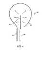
図1を参照すると、本発明の実施形態による装置が記載される。装置10は、遠位部分14及び近位部分16を有するデリバリ部材12を含む。装置10は、デリバリ部材12に結合(例えば取り外し可能に結合)することができる装置の近位部分18を更に含む。付加的に、装置10は、エネルギー源(図示せず)に電気的に結合される導電ケーブル20を含むことができる。装置は、デリバリ部材12の遠位部分14に複数の電極22を含む。電極22は、例えば、デリバリ部材12の遠位端部に位置決め又は固定することもでき、デリバリ部材12の管腔から位置決め可能及び展開可能であり、デリバリ部材12の遠位端から伸縮自在とすることもできる。電極22は、デリバリ部材12の管腔内に電極22を位置決めすることができる非展開状態と、デリバリ部材12の遠位端から進んだときの展開状態とを含むことができる。電極22は、遠位端から進めて伸長され、実質的に切除容積を定める展開状態にされる。 Referring to FIG. 1, an apparatus according to an embodiment of the present invention is described.
標的組織領域は、本発明の組織切除法が望ましいか又は有益である身体のどこにでも配置される可能性がある。標的組織は、どのような特定の種類にも限定されず、非限定的な実施例は、例えば、乳房組織、前立腺組織、肝臓、肺、脳組織、筋肉、リンパ管、膵臓組織、結腸、直腸、気管支等を含むことができる。標的組織領域は、典型的には、組織の塊又は固形部分を含むことになる。典型的には、標的組織領域は癌細胞を含み、これには、例えば固形腫瘍を含む標的組織領域が含まれる。本明細書で用いる用語「癌細胞」とは、一般に、例えば、前癌状態の細胞又は癌細胞(例えば、上皮性悪性腫瘍細胞又は非上皮性悪性腫瘍細胞)のような新生細胞を含む無秩序な成長を示すか又はその傾向があり、本明細書に記載する切除方法に適しているあらゆる細胞をいう。本発明の方法を受けることになる組織の容積は、例えば、癌細胞の塊の大きさ及び/又は形状、並びに他の因子に応じて変えることができる。標的組織領域の周囲寸法は、規則的(例えば、球形、楕円形、その他)とすることもでき、不規則とすることもできる。 The target tissue region can be located anywhere in the body where the tissue excision method of the present invention is desirable or beneficial. The target tissue is not limited to any particular type and non-limiting examples include, for example, breast tissue, prostate tissue, liver, lung, brain tissue, muscle, lymphatic vessel, pancreatic tissue, colon, rectum Can include bronchi and the like. The target tissue region will typically include a mass or solid portion of tissue. Typically, the target tissue region includes cancer cells, including, for example, a target tissue region that includes a solid tumor. As used herein, the term “cancer cell” generally refers to a disordered cell that includes neoplastic cells such as, for example, precancerous cells or cancer cells (eg, epithelial or non-epithelial malignant cells). Any cell that exhibits or is prone to growth and is suitable for the ablation methods described herein. The volume of tissue to be subjected to the method of the present invention can vary depending on, for example, the size and / or shape of the cancer cell mass and other factors. The perimeter dimensions of the target tissue region can be regular (eg, spherical, elliptical, etc.) or irregular.
本発明の方法及びシステムには、イメージングシステム及び装置を含むことができる。例えば、標的組織領域は、超音波、コンピュータ断層撮影法(CT)走査、X線イメージング、核医学イメージング、核磁気共鳴画像法(MRI)、電磁波イメージング等のような従来のイメージング方法を用いて識別及び/又は特徴付けることができる。幾つかの実施形態では、エネルギー印加並びに電極形状及び/又は幾何学的配置のような切除パラメータを選択する際に、イメージング手法を用いて識別されたものを含む腫瘍の特徴を用いることもできる。加えて、装置及び/又は電極を患者の組織に位置決め及び配置するために、これら又は他の公知のイメージングシステムを用いることもできる。 The methods and systems of the present invention can include imaging systems and devices. For example, target tissue regions are identified using conventional imaging methods such as ultrasound, computed tomography (CT) scanning, X-ray imaging, nuclear medicine imaging, nuclear magnetic resonance imaging (MRI), electromagnetic wave imaging, etc. And / or can be characterized. In some embodiments, tumor features, including those identified using imaging techniques, may be used in selecting ablation parameters such as energy application and electrode shape and / or geometry. In addition, these or other known imaging systems can be used to position and position the device and / or electrodes in the patient's tissue.
上述のように、電極は、標的組織領域内に位置決めされ、印加電場は、標的細胞を低出力又は非熱切除するのに十分である。本明細書で用いる用語「非熱切除」とは、一般に、電場を印加することにより組織又は組織の細胞の機能を除去又は破壊することを含む本発明の技術を指し、この場合、エネルギー印加/デリバリプロセスは、局所組織温度を実質的に上昇させることなく、且つエネルギー印加の熱作用が組織切除を行う有意な又は主要な手段ではなく行われる。多くの実施形態では、局所組織温度の上昇も回避することができ、その結果、標的組織領域では温度の上昇は検出されない。しかしながら、幾つかの実施形態では、標的組織領域に僅かな温度変化/上昇が起こることがあるが、典型的には、体温よりも数°Cを超えて高くはなく(例えば、体温よりも高いのが約10°C未満、典型的には約2度を超えない)、熱作用が組織切除を行う主要な手段ではない(例えば、有意な熱を介した致死的タンパク質変性が起こらない)。典型的には、印加電場は、低強度中間周波数交流電流を含む。本発明に従って利用される中間周波数は、例えば、典型的には電極の周りの組織を摩擦/抵抗加熱するのに必要なものより小さいことになる(例えば、約400kHz未満、好ましくは約300kHz又はそれ未満)。1つの実施形態では、例えば、電流は、約50V/cm未満の電圧場を生成する。別の実施形態では、電流は、約50kHzと約300kHzとの間の周波数を含む。 As described above, the electrodes are positioned within the target tissue region and the applied electric field is sufficient to low power or non-thermal ablation of the target cells. As used herein, the term “non-thermal ablation” generally refers to the technique of the present invention that includes removing or destroying the function of a tissue or tissue cells by applying an electric field, where energy application / The delivery process is performed without substantially increasing the local tissue temperature and the thermal effect of energy application is not a significant or primary means of tissue removal. In many embodiments, an increase in local tissue temperature can also be avoided, so that no increase in temperature is detected in the target tissue region. However, in some embodiments, slight temperature changes / rises may occur in the target tissue region, but typically are not more than a few degrees above body temperature (eg, higher than body temperature). Thermal action is not the primary means of performing tissue excision (eg, no significant heat-induced lethal protein denaturation occurs) that is less than about 10 ° C., typically no more than about 2 degrees. Typically, the applied electric field includes a low intensity intermediate frequency alternating current. The intermediate frequency utilized in accordance with the present invention will be, for example, typically less than that required to friction / resistance heat the tissue around the electrode (eg, less than about 400 kHz, preferably about 300 kHz or less. Less than). In one embodiment, for example, the current generates a voltage field of less than about 50 V / cm. In another embodiment, the current includes a frequency between about 50 kHz and about 300 kHz.
電圧場及び/又は印加電流の周波数及び/又は振幅は、エネルギー印加の間に一定に保持することができ、或いは変化させてもよい。幾つかの実施形態では、例えば、最適な切除電圧/周波数/電流が標的組織領域に確実に印加されるようにするために、所与の範囲にわたって「走査」することにより非一定の又は変動する電圧及び/又は周波数及び/又は電流を供給することが望ましい場合がある。別の実施形態では、エネルギーを印加する前に、特定の電圧及び/又は周波数及び/又は電流を選択することができる。更に、電流の印加が標的組織内部から行われて、且つ切除が標的組織の内側から外側になされるように、電極は、標的組織領域内部に位置決めすることができる。1つの実施形態では、電極は、標的組織領域(例えば腫瘍)内に位置決めされ、印加電流は、電極から半径方向外向きに延びる電場を生成する。特定の実施形態では、このような位置決めは、例えば、標的組織領域内の分裂/増殖細胞の向きを含む腫瘍生理機能を利用して、電極により生成される電場が分裂癌細胞の分裂軸線と実質的に位置合わせされるのを確実にすることができる。 The frequency and / or amplitude of the voltage field and / or applied current can be kept constant during energy application or may be varied. In some embodiments, for example, non-constant or fluctuating by “scanning” over a given range to ensure that the optimal ablation voltage / frequency / current is applied to the target tissue region. It may be desirable to supply voltage and / or frequency and / or current. In another embodiment, a specific voltage and / or frequency and / or current can be selected before applying energy. Furthermore, the electrodes can be positioned within the target tissue region so that the application of current is from within the target tissue and the ablation is made from the inside to the outside of the target tissue. In one embodiment, the electrode is positioned in a target tissue region (eg, a tumor) and the applied current generates an electric field that extends radially outward from the electrode. In certain embodiments, such positioning utilizes, for example, tumor physiology, including orientation of dividing / proliferating cells within the target tissue region, so that the electric field generated by the electrode is substantially in line with the dividing axis of dividing cancer cells. Can be reliably aligned.
図2A〜図2Cは、本発明の別の実施形態による複数の電極を有する装置を示す。図示するように、装置30は、装置の遠位部分から延びる複数の電極を含む。図2Aは、複数の電極を有する装置の斜視図(3次元側面図)を示す。図2Bは、電極配列を示す装置の上面図を示す。複数の電極は、中心に位置決めされた電極32と、中心電極32から横方向に間隔を置いて配置された外側電極34、36、38とを含む。図示した電極は、ほぼ直線の針様部分又は針電極を含む。電極は、装置の遠位部分から延びて、装置30の長手方向軸線と実質的に平行な向きにされる。加えて、各電極は、複数の電極の他の電極と実質的に平行である。複数の電極によって実質的に切除容積が定められ、外側電極34、36、38が切除容積の周囲を実質的に定め、電極32は、定められた周囲内に、又はほぼその中心点に位置決めされる。電極の各々は、切除プロセスにおいて異なる役割を果たすことができる。例えば、装置の異なる電極間で極性の変化及び/又は極性のシフトが存在する場合がある。本発明の他の装置と同様に、電極は、電気的に独立し、電気的に個別にアドレス指定可能とすることができ、例えば、2つ又はそれ以上の電極を1つのユニットとして効果的に機能するように電気的に接続することができる。1つの実施形態では、例えば、外側電極34、36、38を電気的に接続し、作動時には、内側電極32と異なる極性を含むことができる。図2Cに示すように、装置の電極32及び34、36は、反対の電荷(例えば、双極性)を含むことができる。このような場合には、印加電流は、矢印で示すように、周囲に位置決めされた又は外側にある電極34、36に向かって中心電極32から半径方向外向きに延びる電場を生成することができる。 2A-2C illustrate a device having a plurality of electrodes according to another embodiment of the present invention. As shown, the
幾つかの実施形態では、本発明の装置及び/又はシステムは、電気的に浮遊のシステム又は接地無しで作動するように設計されたシステムを含む。場合によっては、このように電気的に浮遊する電極構成により、正確な又は制御可能な電場印加及び/又はデリバリが可能になることが観察された。特定の実施形態によるシステムの低出力要件によって、例えば、熱RF又はマイクロ波切除、或いは高出力エネルギーデリバリ及び対応する電源が必要となる高電圧不可逆電気穿孔のような公知の技術と比較して、上述のように電気的に浮遊する装置及びシステムの構成における設計の選択肢(例えばバッテリ駆動)をより多くすることができる。 In some embodiments, the apparatus and / or system of the present invention includes an electrically floating system or a system designed to operate without grounding. In some cases, it has been observed that this electrically floating electrode configuration allows for accurate or controllable electric field application and / or delivery. Compared to known techniques such as thermal RF or microwave ablation, or high voltage irreversible electroporation where a high power energy delivery and corresponding power supply is required due to the low power requirements of the system according to certain embodiments, As described above, more design options (e.g., battery powered) can be provided in the configuration of electrically floating devices and systems.
本発明の装置の別の実施形態を図3A及び図3Bを参照して説明する。装置40は、装置40の遠位端42にあるか又はこれから延びる複数の電極を含む。複数の電極は、外側に位置決めされた電極44を含み、これらは、湾曲し且つ実質的に切除容積を定める。電極46は、外側電極44により定められた容積内に位置決めされ、電極44と間隔を置いて配置される。中心電極46は、ほぼ直線状で、装置40の長手方向軸線に平行に示されているが、他の構成も利用可能である。図3Bは、外側電極44により定められる周囲内の標的組織48を示しており、電流が標的組織48に印加され、湾曲電極44により定められる長方形又は楕円の切除容積を示している。従って、固形腫瘍のような標的組織領域48は基本的に、外側電極44により定められる容積内に包み込むことができる。矢印は、電極46から半径方向外向きに複数の方向に延びる電場を示す。 Another embodiment of the apparatus of the present invention is described with reference to FIGS. 3A and 3B. The
本発明の別の実施形態による装置の電極を図4を参照しながら説明する。装置50は、マイクロカテーテル54から伸縮自在なほぼ直線状の電極52と、湾曲部分を有し且つマイクロカテーテル58から伸縮自在な電極56とを含む。マイクロカテーテル58及び54は、デリバリカテーテルの管腔のような単一のデリバリ部材内に含めることもでき、或いは、例えば標的組織に個々に接近して方向を定めるように独立して配列することもできる。1つの外側電極が示されているが(例えば電極56)、他の実施形態(下を参照)に示すように複数の外側又は第2の電極を設けることもできる。 An electrode of a device according to another embodiment of the present invention will be described with reference to FIG.
装置は、マイクロカテーテルから各々展開可能又は伸縮可能な複数の電極を含むことができ、各マイクロカテーテル/電極組立体は、任意選択的に、図5A及び図5Bに示されるように大きなデリバリ部材の中心管腔内に位置決めされる。装置60は、管腔64を備えるデリバリ部材62と、管腔内に位置決めされたマイクロカテーテル66、68、70、72とを含む。図5Bは、デリバリ部材60の管腔62内に位置決めされたマイクロカテーテル60、68、70、72を備えた装置の上面図を示す。各々が湾曲部分を有する電極74、76、78は、マイクロカテーテル68、70、72から展開可能であり、展開状態では、実質的に切除容積を定める。電極80は、マイクロカテーテル66から展開可能であり、実質的に電極74、76、78により定められる切除容積内に位置決めされる。 The device can include a plurality of electrodes each deployable or telescopic from the microcatheter, each microcatheter / electrode assembly optionally including a large delivery member as shown in FIGS. 5A and 5B. Positioned in the central lumen.
使用時には、図6に示すように、本発明の装置82は、患者の組織84を通して進めることができ、装置82の電極86は、標的組織領域88(例えば腫瘍)内に位置決めすることができる。電極が標的組織領域88内に位置決めされると、標的組織領域88に電流が供給される。電極86が標的組織領域88内に位置決めされると、印加電流は、複数の方向で外向きに放射する電場を生成することができる。本発明のシステム又は装置は、単極モード又は双極モードで作動することができる。単極作動の1つの実施形態では、例えば、患者を導電性パッド又はプレート(例えば金属プレート)上に位置決めすることなどにより、患者の身体の外側に第2の電極を配置することができ、更に、患者の皮膚と第2の電極との間に配置した導電性ゲル又は接着剤のような導電性材料を利用することができる。双極モードの実施形態では、実質的に切除容積を定める外側電極は、戻り電極として機能し、又は切除容積内に位置決めされた電極で回路を完成することができ、印加電流が外側電極と切除容積内に位置決めされた電極との間に位置する標的領域の組織を通って流れるようにする。図7は、本発明の別の実施形態による本発明の装置の利用を示す。上に記載するように、装置90は、標的組織領域94に近接して位置決めされた患者の組織及びデリバリ部材92を通って進められる。デリバリ部材92が位置決めされると、複数の電極96、98、100をデリバリ部材92から展開することができる。外側電極96、98は、標的組織領域94の周囲内又はその周り、例えば、ほぼ標的組織領域の縁(例えば腫瘍縁)に展開され、切除容積又は標的領域を実質的に定める。内側電極100は、切除容積内に位置決めされる。 In use, as shown in FIG. 6, the
本発明は、標的組織に接近又は方向を定め、記載した切除治療をデリバリするための電極/プローブを位置決めする種々の手段を含むことができる。典型的には、本発明の装置の位置決めには、例えば、他の種類の組織切除(例えば、熱RF切除、マイクロ波切除、高電圧電気穿孔等)と共に一般的に用いられる接近技術を含む、侵襲性が最小の接近及び位置決め技術が含まれることになる。例えば、本発明の装置は、皮膚を通して経皮的に導入され、組織を通して進めて、標的組織にて位置決めすることができる。しかしながら、標的組織に方向を定めて装置を位置決め段階は、従来の外科的技術又は腹腔鏡技術と併せて行うこともできる。 The present invention can include various means for positioning or directing the electrodes / probes for approaching or directing the target tissue and delivering the described ablation treatment. Typically, positioning of the device of the present invention includes, for example, access techniques commonly used with other types of tissue ablation (eg, thermal RF ablation, microwave ablation, high voltage electroporation, etc.), Access and positioning techniques that are minimally invasive will be included. For example, the device of the present invention can be introduced percutaneously through the skin, advanced through the tissue, and positioned at the target tissue. However, the step of positioning the device relative to the target tissue can also be performed in conjunction with conventional surgical or laparoscopic techniques.
上述のように、本発明の特定の実施形態は、標的組織領域内に電極を位置決めし、交流電流を印加する段階を含み、この印加電流が、位置決めした電極から外向きに放射する電場を生成する。このように電場を印加すると、熱切除作用が無く低出力切除を介して癌細胞を破裂及び破壊するのに極めて有効であることが見出された。特定の実施形態では、本発明に従って癌細胞を破裂させ、その結果として得られる切除は、本発明の装置の電極により生成される電場が分裂癌細胞又は複数の細胞の分裂軸線と実質的に位置合わせされる場合に、更に効果的に行われた。図8Aは、癌腫瘍又は癌細胞の固形塊の成長パターン及び生理学についての簡易形態を示しており、領域の中心から外向きに分裂する癌細胞による腫瘍の成長を示す。矢印は、中心から外向きに分裂する癌細胞の分裂軸線を示す。図8Bは、図8Aの腫瘍の分裂細胞に焦点を当てた簡略図を示しており、細胞分裂の軸線の概念を更に示す。図示した分裂又は増殖癌細胞(有糸分裂の中期段階を示す)は、中期板軸線112に実質的に直交する細胞分裂110の軸を含み、細胞は、実質的に板軸線112に沿って分裂し、細胞増殖及び成長は、細胞分裂軸線110に沿って起こる。従って、本発明の特定の実施形態では、組織領域内、例えば腫瘍又は癌細胞の塊の中心領域に近接した電極の位置決め及び/又は装置の電極の構成及び配列は、電場がほぼ中心領域から外向きに放射され、且つ成長腫瘍細胞の分裂軸線と実質的に位置合わせされるように選択することができる。 As described above, certain embodiments of the present invention include positioning an electrode within a target tissue region and applying an alternating current that generates an electric field that radiates outward from the positioned electrode. To do. It has been found that application of an electric field in this way is extremely effective in rupturing and destroying cancer cells via low power ablation without thermal excision. In certain embodiments, the cancer cells are ruptured according to the present invention and the resulting ablation is such that the electric field generated by the electrode of the device of the present invention is substantially located at the dividing axis of the dividing cancer cell or cells. When combined, it was done more effectively. FIG. 8A shows a simplified form of growth pattern and physiology of a cancer tumor or solid mass of cancer cells, showing the growth of the tumor by cancer cells dividing outward from the center of the region. The arrow indicates the division axis of cancer cells that divide outward from the center. FIG. 8B shows a simplified diagram focusing on the dividing cells of the tumor of FIG. 8A, further illustrating the concept of a cell division axis. The illustrated mitotic or proliferating cancer cell (representing a metaphase stage of mitosis) includes an axis of
更に、記載したように電場を印加すると、無秩序な成長及び増殖を示さない正常細胞に対しては殆ど又は全く影響を及ぼさないようにしながら、分裂癌細胞を選択的に破裂及び破壊するのに特に効果的であることが観察された。特定の理論には縛られないが、記載したような電場印加は、特に、細胞分裂過程(例えば有糸分裂)又は細胞周期の進行、もしくはその段階又は過程(例えば、紡錘体形成、微小管重合、細胞質小器官機能又は配列、細胞質分裂、細胞浸透圧平衡等)を乱し、従って、より詳細には、無秩序な成長を示し(例えば癌細胞)、細胞周期により急速に進行する細胞に作用を及ぼす。 In addition, applying an electric field as described is particularly useful for selectively rupturing and destroying dividing cancer cells while having little or no effect on normal cells that do not exhibit disordered growth and proliferation. It was observed to be effective. Without being bound by a particular theory, the application of an electric field as described is particularly relevant to cell division processes (eg mitosis) or cell cycle progression, or stages or processes thereof (eg spindle formation, microtubule polymerization). , Disrupting cytoplasmic organelle function or sequence, cytokinesis, cell osmotic balance, etc.), and more particularly, exhibiting disordered growth (eg cancer cells) and acting on cells that progress rapidly through the cell cycle Effect.
本発明によれば、標的組織領域は、全体的又は部分的に切除することができる。一般に、標的領域又は腫瘍はできるだけ多く切除することが望ましいが、幾つかの実施形態では、方法は、標的領域の一部又はその全体未満の切除を含むことができることは理解される。場合によっては、最終的に腫瘍又は癌組織領域全体を破壊又は死滅させるために、部分的に腫瘍切除で十分である可能性がある。 According to the present invention, the target tissue region can be excised in whole or in part. In general, it is desirable to excise as much of the target area or tumor as possible, but it is understood that in some embodiments, the method can include excision of a portion of the target area or less than the entirety thereof. In some cases, partial tumor resection may be sufficient to ultimately destroy or kill the entire tumor or cancer tissue area.
図9A〜図9Dを参照しながら、本発明の実施形態による装置(例えば、図2A〜図2Cの装置)の利用を説明する。装置120は、実質的に切除容積を定める外側電極122、124、126を含む複数の電極と、少なくとも1つの内側電極128とを含む。装置は、腫瘍又はその一部を含む標的組織領域に位置決めすることができる。腫瘍130は、実質的に切除容積内に位置決めされて示されており、内側電極128は、腫瘍の中心をほぼ通って位置決めされ、外側電極122、124、126は、内側電極128から横方向に間隔を置いて配置されて、ほぼ腫瘍縁に或いは僅かに内部又は外部に位置決めされる。図9Aは、腫瘍130及び位置決めされた電極122、124、126、128の上部断面図を示し、図9Bは、その側面図を示す。図9Cの矢印で示される電場は、位置決めされた電極及び電流の印加により生成される。ここに見られるように、図9〜図9Cに示される平行な直線状針電極構成では、切除容積の長さに沿った電場は、装置の長手方向軸線に直交する方向に向けられる。中心電極128から外側電極122、124、126に向かって流れる電流は、腫瘍細胞の多く、特に領域132の細胞では腫瘍中心から外向きの方向(例えば図8A及び図8B参照)に分裂する細胞分裂の方向と実質的に位置合わせした電場を生成する。矢印は説明の目的で示しており、本発明の実施形態は、どのような特定の電流及び/又は電場方向にも限定されず、具体的に示された方向以外及び/又はこれに加えた方向も含むことができる点は理解されるであろう。腫瘍は、腫瘍細胞分裂の方向が電場とより近接して位置合わせされると考えられる領域132を含む。図示の構成では、腫瘍は、腫瘍の対向する端部において、生成された電場と位置合わせされていない細胞分裂軸線を有する細胞のより多くの割合を含むことができる、すなわち換言すれば、電場に対してある角度を有し且つ領域132の更に大きな割合の細胞が切除されてもエネルギー印加後に生存したままとすることができる領域134、136を含むことができる。しかしながら、1つの実施例では、このように腫瘍切除を用いて領域132の組織/細胞が切除されると、その後、物質は、治療部位から除去され(例えば圧力を印加することにより絞り出される)及び/又は周囲組織によって吸収されて、領域134及び136が内向きに潰れて平坦な「パンケーキ様」組織残留物(図9D)を形成し、エネルギー印加後に最終的には死滅するのが観察された。注目すべきことには、本発明の記載した切除技術を受けた多数の実験(例えば動物)モデルは、検出可能な腫瘍が完全に寛解していることを示した。これらの結果は、効果的に腫瘍組織を切除する本発明の方法は、腫瘍組織全体より少なく切除された場合でも固形腫瘍を破壊することができることを示し、電場が癌細胞の細胞分裂の方向に位置合わせする場合に組織切除が改良されることを示した。 With reference to FIGS. 9A-9D, the use of an apparatus according to an embodiment of the present invention (eg, the apparatus of FIGS. 2A-2C) will be described. The
図10には、本発明の装置の別の実施形態が示されている。上述のように、装置構成及び電極配列は、電場が、標的組織領域のほぼ中心から外向きに放射し、成長する腫瘍の特定の細胞の分裂軸線と実質的に位置合わせされるように選択することができる。電気エネルギーの印加及び電場と成長する腫瘍の分裂軸線との位置合わせをより最適にすることは、電極を標的領域に位置決めすること、並びに電極構成及び/又は装置の幾何学的配置を選択することの両方により達成することができる。1つの実施形態では、例えば、装置は、内側電極140と、湾曲する複数の外側電極142、144とを含むことができる。内側電極140は更に、湾曲又は非直線遠位部分を含むことができる。電極が湾曲部を有することは、装置又は内側電極の長手方向軸線に直交する方向以外の方向を含む複数の方向に放射する印加電場を選択するのに役立つことができる。外側湾曲電極は、実質的に切除容積を定め、内側電極は、切除容積内に位置決めされる。矢印は、中心から複数の方向で且つ実質的に標的組織領域の分裂癌細胞と一致して放射する電場を示す。場合によっては、この構成により生成される電場は、例えば、図9A〜図9Dに示される直線状針電極構成に比較して、標的組織領域の癌細胞の多くの部分と位置合わせすることができる。 FIG. 10 shows another embodiment of the apparatus of the present invention. As described above, the device configuration and electrode arrangement are selected such that the electric field radiates outward from approximately the center of the target tissue region and is substantially aligned with the division axis of the particular cell of the growing tumor. be able to. More optimal application of electrical energy and alignment of the electric field with the dividing axis of the growing tumor can position the electrode in the target area and select the electrode configuration and / or device geometry Can be achieved by both. In one embodiment, for example, the device can include an
切除プロセスが開始されると、電場強度は、内側又は中心電極にて、或いは中心電極の周り及びこれに近接した組織内で最大になる。切除プロセスが進行すると、内側電極に近接する癌細胞が、最初に破壊又は切除されるのが観察される。切除細胞は、効果的に「液化」するか、或いは低インピーダンスの液体様材料の性質を呈する。「液化」するという用語は、本明細書では便宜上及び説明の目的で用いられ、切除又は細胞死のどのような特定の機序を必ずしも暗示するものではなく、細胞小疱形成、アポトーシス、溶解、又は何らかの他の細胞プロセス、及び/又はその何らかの組み合わせを含むことができる。細胞破壊の別の起こり得る原因には、例えば、1つ又はそれ以上の細胞膜の絶縁破壊(例えば以下を参照)を含む、細胞膜の完全性の破裂を含むことができる。液体様材料が中心電極を囲み、高電場強度切除領域を効果的に拡大し、最高電場強度切除領域は、液体様材料の外周にある。従って、液体様材料は、「仮想電極」になるといわれる。切除プロセスが進行すると、液体様材料の外周又は「仮想電極」が拡大し、本質的に標的組織領域を内側から切除する。幾つかの実施形態では、標的組織領域は、切除プロセス後にはより曲がりやすく柔軟になるか、又はマッシュ状になることが観察された。切除された液体様腫瘍組織は、最終的には治療部位から除去され、及び/又は周囲組織に吸収されて、もはや検出可能でなくなった。 When the ablation process is initiated, the electric field strength is maximized at the inner or central electrode, or around and near the central electrode. As the ablation process proceeds, it is observed that cancer cells proximate the inner electrode are first destroyed or ablated. The excised cells effectively “liquefy” or exhibit the properties of a low impedance liquid-like material. The term “liquefy” is used herein for convenience and illustrative purposes and does not necessarily imply any specific mechanism of excision or cell death, but cell blebbing, apoptosis, lysis, Or some other cellular process, and / or some combination thereof. Another possible cause of cell disruption can include rupture of cell membrane integrity, including, for example, one or more cell membrane breakdowns (see, eg, below). A liquid-like material surrounds the central electrode and effectively enlarges the high field strength ablation region, with the highest field strength ablation region being at the outer periphery of the liquid-like material. Therefore, the liquid-like material is said to be a “virtual electrode”. As the ablation process proceeds, the outer circumference or “virtual electrode” of the liquid-like material expands, essentially ablating the target tissue region from the inside. In some embodiments, the target tissue region has been observed to be more flexible and pliable or mash after the ablation process. The resected fluid-like tumor tissue was eventually removed from the treatment site and / or absorbed into the surrounding tissue and was no longer detectable.
仮想電極効果を標的組織領域に位置決めされた電極の断面図を示す図11A〜図11Cを参照しながら例証する。外側電極150、152、154は、腫瘍156のほぼ縁又は外周に位置決めされ、内側電極158は、外側電極150、152、154により定められる容積のほぼ中心点に位置決めされる。切除は、T1すなわち切除プロセスの開始時(図11A)、液体様組織領域160の拡張で切除が開始された後T2(図11B)、液体様組織領域162が内側電極158から外側電極150、152、154に向かって更に外向きに拡張するその後の時間T3(図11C)で示される。 The virtual electrode effect is illustrated with reference to FIGS. 11A-11C showing cross-sectional views of electrodes positioned in a target tissue region. The
切除プロセスは、その進行を含めて、切除組織のインピーダンスの関連する変化を検出することにより監視することができる。切除された液体様組織の外側周囲が、切除容積を定める外側電極に到達すると、インピーダンスが安定又は横ばいになる。従って、切除プロセスの進行は、インピーダンスの変化を測定することにより監視することができ、インピーダンスの変化が観察されなくなると電場の印加は解除した。 The ablation process can be monitored by detecting associated changes in the impedance of the ablated tissue, including its progress. When the outer perimeter of the resected liquid-like tissue reaches the outer electrode that defines the ablation volume, the impedance becomes stable or level. Thus, the progress of the ablation process can be monitored by measuring the change in impedance, and the application of the electric field was released when the change in impedance was no longer observed.
また、フィードバック測定値を用いて、標的癌細胞の切除が非熱切除により確実に行われるようにすることもできる。特定の実施形態では、高熱作用又は熱切除を引き起こすことなく内側電極でできる限り大きな電場強度を発生させることが望ましいとすることができる。熱切除では、上述の「液化」作用なしに周囲細胞の破壊が引き起こされるので、特定の高熱作用は観察可能であり、本発明の望ましい非熱切除とは区別可能になる。例えば、細胞破壊が熱切除プロセスにより引き起こされる場合には、熱作用により炭化又は壊死した細胞のインピーダンスは通常増大するので、治療組織のインピーダンスが減少しない可能性がある。1つの実施形態では、本発明による非熱切除は、標的組織領域内(例えば、内側電極近傍)に熱電対のようなセンサを配置する段階と、標的細胞に熱作用を引き起こすことになる強度を下回る印加電場強度を選択する段階とを含むことができる。 The feedback measurement can also be used to ensure that target cancer cells are resected by non-thermal ablation. In certain embodiments, it may be desirable to generate as much electric field strength as possible at the inner electrode without causing high heat effects or thermal ablation. Because thermal ablation causes destruction of surrounding cells without the “liquefaction” effect described above, certain hyperthermic effects are observable and distinguishable from the desired non-thermal ablation of the present invention. For example, if cell destruction is caused by a thermal ablation process, the impedance of cells that have charred or necrotized due to thermal effects usually increases, so the impedance of the treated tissue may not decrease. In one embodiment, non-thermal ablation according to the present invention may include placing a sensor, such as a thermocouple, within the target tissue region (eg, near the inner electrode) and an intensity that will cause a thermal effect on the target cell. Selecting a lower applied electric field strength.
上に述べたように、場合によっては、標的組織領域内の内側電極の位置から出る電場強度を高めることが望ましいことがある。本発明の1つの実施形態では、電場強度は、標的組織領域内に配置された内側電極の表面積を増大させることにより高めることができる。増大させた電極の表面積の種々の実施形態が、図12A〜図12Fに示されているが、他の構成も利用可能になる。1つの実施形態では、電極は、追加として、円形パターン(図12A)、コークスクリュー(図12B)、又は単純なコイル(図12C)の形状にすることができるコイル遠位部分を含む。別の実施形態では、電極遠位端に小さなワイヤメッシュを含み、標的組織領域内に配置されたときに拡張させることができる(図12D)。別の実施形態では、電極は、遠位端がアレイ状に拡張された複数の小さなワイヤを含む、「リッツ」ワイヤ型電極(図12E)を含むことができる。別の実施形態では、遠位部分は、底部と底部とを積み重ねた2つの円錐に似た形状、すなわち側面から見てダイアモンド形の形状(図12F)を含むことができる。二重円錐/ダイアモンド端部の尖った対向する遠位及び近位部分により、組織での電極の挿入及び後退を容易にすることができる。多数の他の構成が利用可能であり、例えば、リング、球、コークスクリュー、螺旋、同心螺旋、又はこれらの複数形、針アレイ、管体を排除して小さな容器内に不規則に積み重なったストリングに類似したワイヤの小球を形成する、ある長さの非弾性ばね様ワイヤ等を含むことができる。 As noted above, in some cases it may be desirable to increase the electric field strength emanating from the location of the inner electrode within the target tissue region. In one embodiment of the invention, the electric field strength can be increased by increasing the surface area of the inner electrode disposed within the target tissue region. Various embodiments of increased electrode surface area are shown in FIGS. 12A-12F, although other configurations may be utilized. In one embodiment, the electrode additionally includes a coil distal portion that can be in the shape of a circular pattern (FIG. 12A), a corkscrew (FIG. 12B), or a simple coil (FIG. 12C). In another embodiment, the electrode distal end can include a small wire mesh that can be expanded when placed in the target tissue region (FIG. 12D). In another embodiment, the electrode can include a “Litz” wire-type electrode (FIG. 12E) that includes a plurality of small wires with distal ends expanded in an array. In another embodiment, the distal portion may include a shape resembling two cones stacked on the bottom and bottom, i.e., a diamond shape when viewed from the side (Fig. 12F). The pointed opposing distal and proximal portions of the double cone / diamond end can facilitate the insertion and withdrawal of the electrode in the tissue. Numerous other configurations are available, for example, rings, spheres, corkscrews, spirals, concentric spirals, or their plurals, needle arrays, strings that are randomly stacked in small containers It may include a length of inelastic spring-like wire or the like that forms a similar wire sphere.
図13には、本発明の装置の別の実施形態が示される。装置は、組織穿刺遠位部分172を備えたデリバリ部材170を含む。デリバリ部材は、内腔と、本体の開口部174と、遠位端の開口部176とを含む。部材の管腔内には複数の電極が位置決め可能である。展開された状態では、外側電極178は、部材170の遠位端の開口部176から延びて傘様方向に反転する。展開された外側電極178は、実質的に切除容積を定める。本体の開口部174から延びる電極180は、外側電極178から間隔を置いて配置され、切除容積内に位置決めされる。 FIG. 13 shows another embodiment of the apparatus of the present invention. The device includes a
図14は、図13に示すものと同様の装置を示す。図14を参照すると、装置は、遠位部分、本体上の開口部192、及び遠位端の開口部194を備えるデリバリ部材190を含む。外側電極196は、本体開口部192から遠位方向に展開され、展開されて遠位端開口部194から延びる電極198を囲む容積を定める。 FIG. 14 shows an apparatus similar to that shown in FIG. Referring to FIG. 14, the apparatus includes a
本発明の装置の別の実施形態を図15を参照しながら説明する。装置は、複数の電極を含み、各電極はマイクロカテーテル内に位置決めされ、各マイクロカテーテルはデリバリ部材の管腔内に位置決めされる。デリバリ部材又はプローブ300は、患者の組織に更に容易に挿入されるように、尖った鋭い組織穿刺端部を含むことができる。同様に、マイクロカテーテルは、尖った又は鋭い組織穿刺端部を含むことができる。使用時には、デリバリ部材300が、患者の組織を通って進み、遠位端が標的組織領域(例えば腫瘍)に近接して位置決めされ、マイクロカテーテルがデリバリ部材から展開される。フェーズ1展開において示されるように、マイクロカテーテル310は、デリバリ部材の遠位端から標的組織領域内に遠位方向に進められ、そこで、マイクロカテーテルの電極320を展開することができる。マイクロカテーテル330もまた、電極340に向けてデリバリ部材300から展開される。フェーズ2展開では、電極340は、標的組織領域の外周(例えば腫瘍縁)の周りなど、マイクロカテーテル330が向く方向に展開される。マイクロカテーテル及びそこに位置決め可能な電極の両方は、ニチノールのような形状記憶金属で作られ、展開時に予め設定された構成をとるようにすることができる。他の使用フェーズも含むことができる。 Another embodiment of the apparatus of the present invention will be described with reference to FIG. The device includes a plurality of electrodes, each electrode positioned within the microcatheter, and each microcatheter positioned within the lumen of the delivery member. The delivery member or probe 300 can include a sharp pointed tissue piercing end for easier insertion into the patient's tissue. Similarly, the microcatheter can include a sharp or sharp tissue piercing end. In use,
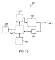
本発明の実施形態によるシステムを図16を参照しながら説明する。システム200は、患者にエネルギーをデリバリするための本発明のあらゆる装置を組み込むように含むことができ、駆動ユニット220、次いで本発明の装置の電極にエネルギーを供給するパワーユニット210を含む。本システムの構成要素は或いは構成要素の組み合わせは、本発明のシステムのためのエネルギー源を個々に又は集合的に含むことができる。パワーユニット210は、本発明の装置を作動させ、本明細書に記載するように標的組織に電流を印加するのに用いられるあらゆる電力生成手段を含むことができる。パワーユニット210は、例えば、1つ又はそれ以上の発電機、バッテリ(例えば、携帯型バッテリユニット)等を含むことができる。本発明のシステムの利点の1つは、切除プロセスに必要とされる電力が小さいことである。従って、1つの実施形態では、本発明のシステムは、携帯型及び/又はバッテリ駆動型装置を含むことができる。フィードバックユニット230は、
標的組織領域の組織の電場デリバリパラメータ及び/又は特徴、及び限定ではないが、電流、電圧、インピーダンス、温度、pH等を含む、測定されるパラメータ及び/又は特徴を測定する。システムには、1つ又はそれ以上のセンサ(例えば、温度センサ、インピーダンスセンサ、熱電対、その他)を含むことができ、これらは、装置又はシステムと結合することができ、及び/又は患者の組織又はその内部に別個に位置決めすることもできる。これらのセンサ及び/又はフィードバックユニット230を用いて、組織へのエネルギーのデリバリを監視又は制御することができる。パワーユニット210及び/又はシステムの他の構成要素は、制御ユニット240により駆動することができ、該制御ユニットは、例えば技術者又は医師による入力及び/又は制御のためのユーザインタフェース250と結合することができる。制御ユニット240及びシステム200は、イメージングシステム260(上記参照)と結合し、標的組織領域及び/又は場所を位置特定及び/又は特徴付け、或いは使用中の装置を位置決めするようにすることができる。A system according to an embodiment of the present invention will be described with reference to FIG. The
Measure measured parameters and / or characteristics, including, but not limited to, current, voltage, impedance, temperature, pH, etc., of the electric field delivery parameters and / or characteristics of the tissue in the target tissue region. The system can include one or more sensors (eg, temperature sensors, impedance sensors, thermocouples, etc.), which can be coupled to the device or system and / or patient tissue. Alternatively, it can be positioned separately within it. These sensors and / or
制御ユニットは、例えば、1つ又はそれ以上の処理構造を有する多様な専用又は市販のコンピュータ又はシステム、及びパーソナルコンピュータ等のコンピュータを含むことができ、このようなシステムは、本明細書に記載した方法ステップの何れか(又はその組み合わせ)を実装するように構成されたデータ処理ハードウェア及び/又はソフトウェアを含む場合が多い。何れのソフトウェアも典型的には、メモリ、或いは光、電気、又は無線遠隔測定信号を再生するデジタル又は光学媒体等の有形媒体内に具現化されたプログラム命令の機械可読コードを含むことになり、これらの構造の1つ又はそれ以上を更に用いて、あらゆる種々の分散又は集中信号処理アーキテクチャにおけるシステムの構成要素間でデータ及び情報を送信することができる。 The control unit can include, for example, a variety of dedicated or commercially available computers or systems having one or more processing structures, and computers such as personal computers, such systems described herein. Often includes data processing hardware and / or software configured to implement any (or combination) of method steps. Any software will typically include machine-readable code of program instructions embodied in memory or tangible media such as digital or optical media that reproduces optical, electrical or wireless telemetry signals; One or more of these structures can further be used to transmit data and information between the components of the system in any of a variety of distributed or centralized signal processing architectures.
コントローラを含むシステムの構成要素を用いて、標的組織にデリバリされる電力又は電気エネルギーの量を制御することができる。エネルギーは、プログラムされた又は予め設定された量で供給することができ、或いは、初期設定で開始して、エネルギーデリバリ及び切除プロセス中に電場に対して修正することもできる。1つの実施形態では、例えば、システムは、印加電圧及び周波数のような電場パラメータが予め設定された範囲にわたるデリバリを含む「走査モード」でエネルギーをデリバリすることができる。フィードバック機構を用いて走査モードで電場デリバリを監視し、標的にされている組織を切除するのに最適なデリバリ範囲パラメータを選択することができる。 System components, including a controller, can be used to control the amount of power or electrical energy delivered to the target tissue. The energy can be supplied in a programmed or preset amount, or it can be started with an initial setting and modified for the electric field during the energy delivery and ablation process. In one embodiment, for example, the system can deliver energy in a “scan mode” that includes delivery over a pre-set range of electric field parameters such as applied voltage and frequency. A feedback mechanism can be used to monitor the electric field delivery in scan mode and select the optimal delivery range parameters for ablating the targeted tissue.
本発明の方法及び技術は、単一の装置又は複数の装置を用いることができる。1つの実施形態では、例えば、本発明の装置(例えば、図2A〜図2Cに示されるような装置)は、上に記載するように標的組織領域内に位置決めすることができる。次に、第2の装置を標的組織領域内又は同じ腫瘍の一部又は別個の腫瘍の別の標的組織領域に位置決めすることができる。1つの実施形態では、例えば、第1の装置を標的組織領域内に位置決めし、第2の装置を第1の装置に対してある角度(例えば90度の角度)で標的組織領域内に位置決めすることができる。加えて、同じ装置を別個の時点で異なる向き及び/又は場所に位置決めすることもできる。 The methods and techniques of the present invention can use a single device or multiple devices. In one embodiment, for example, a device of the present invention (eg, a device as shown in FIGS. 2A-2C) can be positioned within a target tissue region as described above. The second device can then be positioned within the target tissue region or part of the same tumor or another target tissue region of a separate tumor. In one embodiment, for example, the first device is positioned within the target tissue region, and the second device is positioned within the target tissue region at an angle (eg, 90 degrees) with respect to the first device. be able to. In addition, the same device can be positioned in different orientations and / or locations at different times.
本発明のシステム及び装置は、必須ではないが、薬物デリバリ、局所又は全身デリバリ、放射線医学又は核医学システム等のような他のシステム、切除システム、癌治療システムと連動して用いることができる。同様に、装置は、薬物デリバリ針、電極等を含む薬物デリバリシステムのような他のシステムの構成要素及び/又は態様を組み入れるように修正することもできる。 The systems and devices of the present invention are not required, but can be used in conjunction with other systems such as drug delivery, local or systemic delivery, radiology or nuclear medicine systems, ablation systems, cancer treatment systems. Similarly, the device can be modified to incorporate other system components and / or aspects, such as drug delivery systems including drug delivery needles, electrodes, and the like.
場合によっては、本明細書に記載した切除プロセスのあるステージで標的組織領域から切除組織を除去することが望ましい場合がある。例えば、場合によっては、切除組織の除去により被験者の治療及び/又は回復を改善し、場合によっては、本発明の切除プロセスに伴うストレス及び/又は毒性(例えば、局所組織毒性、全身毒性等)を低減できることが観察されている。 In some cases, it may be desirable to remove ablated tissue from the target tissue region at some stage of the ablation process described herein. For example, in some cases, removal of the resected tissue improves the treatment and / or recovery of the subject, and in some cases, stress and / or toxicity (eg, local tissue toxicity, systemic toxicity, etc.) associated with the ablation process of the present invention. It has been observed that it can be reduced.
切除組織を除去するのに種々の装置及び方法を用いることができる。場合によっては、上に記載するように、切除組織は、効果的に「液化」するか、或いは液体様材料の特性を呈することができる。次いで、液体切除組織は、排液するか標的組織領域から除去することができる。1つの実施形態では、切除組織の除去は、例えば、例えば装置/電極を標的組織領域に導入する入口穴を含む組織内の穴又は穿孔から漏出させることによって、(例えば、標的組織領域又はそれに近接する組織に力又は圧力を印加するか印加せずに)切除組織を標的組織領域から漏出又は滲出させるように単純なものとすることができる。他の実施形態では、切除組織の除去は、より慎重に又はより制御したものにすることができる。除去は、シリンジ又は他の液体除去装置のような切除装置とは別個のデバイス又は装置を用いて達成することもでき、或いは、組織除去のために更に構成された切除装置を用いて達成することもできる。 Various devices and methods can be used to remove the ablated tissue. In some cases, as described above, the ablated tissue can effectively “liquefy” or exhibit the properties of a liquid-like material. The liquid ablated tissue can then be drained or removed from the target tissue region. In one embodiment, removal of the ablated tissue is performed (eg, at or near the target tissue region by leaking from a hole or perforation in the tissue including, for example, an entry hole that introduces the device / electrode into the target tissue region. It can be as simple as leaking or exuding the ablated tissue from the target tissue region (with or without application of force or pressure to the tissue to be applied). In other embodiments, removal of the ablated tissue can be made more carefully or more controlled. Removal can be accomplished using a device or apparatus that is separate from the ablation device, such as a syringe or other liquid removal device, or can be accomplished using an ablation device further configured for tissue removal. You can also.
本発明の実施形態は、上述のように癌細胞の非熱切除及び破壊に用いることに関して検討したが、場合によっては、システム及びプローブは、熱RF切除、マイクロ波切除、高圧直流電流を介する不可逆的電気穿孔法等、他の種類の組織切除に十分なエネルギーをデリバリするように構成することができる。例えば、本発明のシステムは、組織切除の1つ又はそれ以上の何れかの種類に適切なエネルギーをデリバリするように構成されたパワーユニットを含むことができる。実際、特定のプローブ構成は、例えば熱RF切除のデリバリの改善等を含む種々の種類の組織切除のデリバリを改善することができる設計(例えば、電極配列)を有する。本発明の方法による治療は、所与の治療に対する組織切除の1つ又はそれ以上の種類のデリバリを含むことができる。場合によっては、例えば、治療は、非熱組織切除がデリバリされるモードのような1つ又はそれ以上の切除デリバリモードを含むことができ、これは、熱RF組織切除のような別の切除モードの前又は後に置くことができる。例えば、1つの実施形態では、治療は、非熱組織切除の後にエネルギーを短時間印加するか、又はエネルギーパルスをデリバリして熱媒介作用を生じること、例えばプローブの1つ又はそれ以上の構成要素の「滅菌」を助けることを含み、侵入した軌跡を通って標的組織から抜き取り、プローブを抜き取る間に潜在的に生存可能なあらゆる癌細胞を組織内に持ち込むリスクを低減するようにすることができる。 While embodiments of the present invention have been discussed for use in non-thermal ablation and destruction of cancer cells as described above, in some cases, the system and probe are irreversible via thermal RF ablation, microwave ablation, high voltage direct current It can be configured to deliver sufficient energy for other types of tissue excision, such as mechanical electroporation. For example, the system of the present invention can include a power unit configured to deliver energy suitable for any one or more types of tissue ablation. Indeed, certain probe configurations have designs (eg, electrode arrangements) that can improve delivery of various types of tissue ablation, including improved delivery of thermal RF ablation, for example. Treatment according to the methods of the invention can include one or more types of delivery of tissue excision for a given treatment. In some cases, for example, the treatment can include one or more ablation delivery modes, such as a mode in which non-thermal tissue ablation is delivered, which is another ablation mode such as thermal RF tissue ablation. Can be placed before or after. For example, in one embodiment, the treatment applies a short period of energy after non-thermal tissue ablation or delivers an energy pulse to produce a heat-mediated effect, eg, one or more components of the probe Can be extracted from the target tissue through the invading trajectory, reducing the risk of bringing any potentially viable cancer cells into the tissue during extraction .
幾つかの実施形態では、本発明のシステムは、エネルギーデリバリプロセス中にプローブ及び他の構成要素を位置決め及び/又は安定化する特定の構成要素及び態様を更に含むことができる。例えば、エネルギー印加のような治療フェーズが数分間を超えることが予測される場合には、使用者(例えば外科医)がプローブを手で持つことを特に必要とせずにプローブを望ましい位置/場所に維持するように、位置決め又は安定化構造を含むことが望ましい場合がある。従って、システムは、プローブの位置決めを維持するためのハーネス、ベルト、クランプ、又は他の構造を含むことができる。システムは、治療中の患者の移動(例えば、シフト、歩行等)を可能にするように着装携行用途向けに設計することができる。実際に、低出力要件及び対応する設計オプション(例えば、バッテリ駆動システム)により、本システムを着装携行式システムとして用いるのに特に適切にすることができる。 In some embodiments, the system of the present invention can further include specific components and aspects that position and / or stabilize probes and other components during the energy delivery process. For example, if a treatment phase, such as energy application, is expected to exceed several minutes, the user (eg, surgeon) maintains the probe in the desired location / location without requiring the user to hold the probe by hand. As such, it may be desirable to include a positioning or stabilizing structure. Thus, the system can include a harness, belt, clamp, or other structure to maintain probe positioning. The system can be designed for wearable carrying applications to allow patient movement (eg, shifting, walking, etc.) during treatment. Indeed, low power requirements and corresponding design options (eg, battery powered systems) can make the system particularly suitable for use as a wearable portable system.
特定の実施形態では、本発明は、本明細書に記載した態様又は技術を公知の又は商業的に利用可能な構成要素と併せて使用して、癌細胞を破壊するシステム及び方法を改良することを含むことができる。例えば、熱RF切除、マイクロ波切除、高電圧電気穿孔等のような技術に一般的に使用されるものを含む、切除プローブ又は電極構成を有する特定の利用可能なシステムを修正して、本発明の非熱切除技術に用いることができる。1つの実施例では、本発明の方法は、「LeVeenプローブ」(例えば、米国特許第5,855,576号(特許文献3)参照)のような熱RF切除で一般的に使用されるプローブを用いた癌細胞の非熱切除を含む。図17を参照すると、非熱組織切除を行うためのプローブを用いた本発明の実施形態が記載されている。270は、プローブ272の遠位部分が標的部位又は領域(「TR」)或いはその内部に位置決めされるように、例えば経皮的に(皮膚「S」を通して)導入することができる。個々の電極274は、プローブ272の遠位部分から遠心方向に延びるように示される。電極274は、図17に示すように、最初に互いに半径方向外向きに発散し、最終的に外転して近位方向に戻るように進めることができる。電極274は、例えば、ケーブル278を介してパワーユニット280又は電源に電気的に接続することができ、図示のように、単極モードで作動することができる。電流は、標的組織領域内の癌細胞を非熱的に切除又は破壊するのに十分なレベル及び期間でパワーユニット280から印加することができる。 In certain embodiments, the present invention uses the aspects or techniques described herein in conjunction with known or commercially available components to improve systems and methods for destroying cancer cells. Can be included. For example, the present invention may be modified to modify certain available systems having an ablation probe or electrode configuration, including those commonly used in techniques such as thermal RF ablation, microwave ablation, high voltage electroporation, etc. It can be used for non-thermal ablation techniques. In one embodiment, the method of the present invention uses a probe commonly used in thermal RF ablation, such as a “LeVeen probe” (see, eg, US Pat. No. 5,855,576). Includes non-thermal excision of the cancer cells used. Referring to FIG. 17, an embodiment of the present invention using a probe for performing non-thermal tissue ablation is described. 270 can be introduced, for example, percutaneously (through skin “S”) such that the distal portion of
次の実施例は、限定ではなく本発明を例証することを意図している。
[実験例]
一連の試験は、乳癌腫瘍モデルの治療を含んでいた。1つの実施例では、試験は、体重230〜250グラムのメスFisher−344ラット(Charles River)に行った。最初に、ラット乳癌細胞(MTLn−3)を培養液中で成長させ、培養液の細胞を動物の腹部に埋め込むことにより皮下腫瘍を生成した。腫瘍の直径がほぼ1cm又はそれ以上まで成長すると、切除治療を施した。ラットの18の固形乳房腫瘍を治療した。The following examples are intended to illustrate the invention rather than to limit it.
[Experimental example]
A series of trials included treatment of a breast cancer tumor model. In one example, the test was performed on female Fisher-344 rats (Charles River) weighing 230-250 grams. Initially, rat breast cancer cells (MTLn-3) were grown in culture and subcutaneous cells were generated by implanting the cells of the culture in the abdomen of the animal. When the tumor diameter grew to approximately 1 cm or more, resection treatment was given. Rats were treated for 18 solid breast tumors.
切除治療には、ステンレス鋼針電極アレイを含む切除プローブを経皮的に皮膚を通して直接腫瘍内に挿入する段階と、次いで、腫瘍治療電場を印加する段階とを含んでいた。針アレイは、ほぼ等しい間隔を置いて配置された3つの平行な外側針で囲まれた中心針を含んでいた。針の長さ及び配列を含むプローブ設計は、治療されている腫瘍のほぼ直径を横切るように選択し、針の長さは約1cmであった。外側針は、中心針から約0.5cmの間隔を置いて配置した。中心針は、腫瘍治療電圧で作動され、周囲針は、電流のリターンパスを形成した。プローブが、上述のような「双極モード」で動作する場合にはアースは設けず(例えば、電気的に「浮遊」システム)、装置はバッテリ電源を含んでいた。 The ablation treatment involved inserting an ablation probe containing a stainless steel needle electrode array percutaneously directly through the skin and into the tumor, and then applying a tumor treatment electric field. The needle array included a central needle surrounded by three parallel outer needles spaced approximately equally spaced. The probe design, including needle length and sequence, was chosen to cross approximately the diameter of the tumor being treated, and the needle length was about 1 cm. The outer needle was placed approximately 0.5 cm from the center needle. The central needle was actuated with the tumor treatment voltage and the peripheral needle formed a current return path. When the probe was operated in “bipolar mode” as described above, no ground was provided (eg, an electrically “floating” system) and the device included a battery power source.
上述のプローブ及び針構成を用いると、治療電場は、ほぼ直径1cmで長さ1cmのシリンダ内に含まれ、その容積は、実質的に腫瘍を占有するものであった。印加AC電圧は、約20〜30mA(起動電流)で約10〜12ボルトピークであった。周波数は約98Khzであった。 With the probe and needle configuration described above, the therapeutic electric field was contained within a cylinder approximately 1 cm in diameter and 1 cm in length, and its volume substantially occupied the tumor. The applied AC voltage was about 10-12 volts peak at about 20-30 mA (starting current). The frequency was about 98 Khz.
治療中に腫瘍に入る全電流を測定するためにフィードバック回路を含んだ。予備実験からの仮説の1つは、腫瘍細胞が印加電場により破壊され始めると(例えば、溶解、砕解、細胞質流出等)、例えば破壊細胞の内容物(例えば細胞質等)の方が実質的に無傷の細胞よりも導電性がより大きいことに起因して腫瘍領域内のコンダクタンスが増大するので、電流が実際に増大することになるというものであった。この結果から、回路が仮説のように働くことが示された。各症例で治療開始後に電流が増大し始め、これは、治療が効果的に起こっていることを示している。また、観察された電流の増大は、このプロセスが実質的に終了段階に達したときの指標となる。例えば、電流がこれ以上増大することが観察されない場合には、治療を中止することができることを示していた。この電流の横這い状態は、ほぼ40mAで起こることが観察され、典型的には、治療に入ってほぼ90分以内であった。 A feedback circuit was included to measure the total current entering the tumor during treatment. One hypothesis from preliminary experiments is that when tumor cells begin to be destroyed by an applied electric field (eg, lysis, disintegration, cytoplasmic outflow, etc.), for example, the contents of the destroyed cells (eg, cytoplasm, etc.) are substantially more The current would actually increase because the conductance in the tumor area increased due to its greater conductivity than the intact cells. This result shows that the circuit works like a hypothesis. In each case, the current began to increase after the start of treatment, indicating that treatment is taking place effectively. Also, the observed increase in current is an indication when the process has substantially reached the end stage. For example, if no further increase in current was observed, it indicated that treatment could be discontinued. This current leveling was observed to occur at approximately 40 mA, typically within approximately 90 minutes of entering treatment.
腫瘍は、電力を印加したほぼ直後に細胞破壊を示し始めた。有意な破壊は、治療に入って30分以内に起こった。場合によっては、治療は、30分後に終了したが、腫瘍は殆ど検出不可能であった。他の治療群では、治療は、約90分又は3時間以内に完了した。何れの場合でも、治療した腫瘍は、目視検査ではほとんど検出不可能であった。腫瘍破壊が電場を印加した結果であることを確認するために、対照を含んだ。1群では、対照として電力を印加せずに針を挿入した。対照では、連続的な腫瘍の成長が観察された。 The tumor began to show cell destruction almost immediately after power was applied. Significant destruction occurred within 30 minutes of entering treatment. In some cases, treatment ended after 30 minutes, but the tumor was almost undetectable. In other treatment groups, treatment was completed within about 90 minutes or 3 hours. In either case, the treated tumor was almost undetectable by visual inspection. A control was included to confirm that the tumor destruction was the result of applying an electric field. In one group, a needle was inserted without applying power as a control. In the control, continuous tumor growth was observed.
治療したラットの群は、長期生存度試験のために選択し、治療後に屠殺しなかった。生存率は、治療日から12か月を超えた時点で確認したが、これは、何も治療を受けなかった場合の動物モデルの予想生存率(例えば約3週間)の17倍より大きかった。これらのラットは、現在まだ生きている。生存率観察及び組織学的分析(例えば以下を参照)により、上述のように治療を行った標的組織内の癌細胞の実質的に全てではなくても大部分が破壊されたことが示された。 A group of treated rats was selected for a long-term survival study and was not sacrificed after treatment. Survival was confirmed at more than 12 months from the date of treatment, which was greater than 17 times the expected survival rate of the animal model (eg about 3 weeks) when no treatment was received. These rats are still alive today. Survival observation and histological analysis (see eg, below) showed that most if not all of the cancer cells in the target tissue treated as described above were destroyed. .
上述のように、治療によって典型的には、癌細胞/組織が幾分液化した。治療後、治療を受けた標的組織領域透明〜ピンクがかった流体が存在することが観察され、これは、針進入場所から漏出し、治療領域が圧縮されるか圧力が印加されると更に排出される可能性がある。組織学的分析は、治療後に液体及び腫瘍領域に対して実施した。組織学的分析では、流体が、主に破壊された癌細胞からなり、破壊された細胞は実質的に壊死して生存不可能であるようにみえることが示された。pH試験では、除去した流体が典型的な生理学的流体よりも高度にアルカリ性(例えば、約7.8又はそれ以上のpH)であることが示された。観察された治療領域は、約80〜90%が壊死又は生存不可能癌細胞であった。壊死していない治療領域(例えば、典型的には標的組織の約10〜20%)は、健康な非癌性組織領域の一部であるように見えた。従って、この結果は、健康な細胞を実質的に無傷又は生存可能のまま残しながら、癌細胞が選択的に破壊されていることを示していた。 As noted above, treatment typically resulted in some liquefaction of the cancer cells / tissue. After treatment, it is observed that there is a clear to pinkish fluid in the treated target tissue area, which leaks from the needle entry location and is further expelled when the treatment area is compressed or pressure is applied. There is a possibility. Histological analysis was performed on fluid and tumor area after treatment. Histological analysis showed that the fluid consisted mainly of destroyed cancer cells, and the destroyed cells appeared to be substantially necrotic and non-viable. A pH test showed that the removed fluid was highly alkaline (eg, a pH of about 7.8 or higher) than typical physiological fluids. The observed therapeutic area was about 80-90% necrotic or non-viable cancer cells. Non-necrotic treatment areas (eg, typically about 10-20% of the target tissue) appeared to be part of healthy non-cancerous tissue areas. Thus, this result indicated that the cancer cells were selectively destroyed while leaving healthy cells substantially intact or viable.
治療を観察しても、有意な副作用はないようであった。更に分析するために屠殺しなかった生存被検体は、治療後に、衰弱もせず、有害な副作用(生存度、挙動、その他)の顕著な徴候も示さなかった。被験体には、エネルギーを印加したことによる重大な苦痛又は不快、又は治療領域の刺激は観察されなかった。治療中、ラットは、麻酔されていないにも関わらず快適のようであり、すなわち、食べて飲んで眠っている。 Observation of treatment did not appear to have significant side effects. Surviving subjects that were not sacrificed for further analysis were not debilitating after treatment and did not show significant signs of adverse side effects (survival, behavior, etc.). Subjects did not observe significant pain or discomfort due to the application of energy or stimulation of the treatment area. During treatment, the rat appears to be comfortable despite being unanesthetized, i.e. eating, drinking and sleeping.
唯一観察された可能性のある副作用は、治療後に流体又は切除組織が除去されていないことによるものであるようであった。治療後に治療領域から流体が除去されない場合には、被検体は、数時間嗜眠性であるようであり。切除治療後に流体を除去したラット被検体に比較して回復時間が延びた。 The only side effect that could be observed appeared to be due to fluid or ablated tissue not being removed after treatment. If fluid is not removed from the treatment area after treatment, the subject appears to be drowsy for several hours. Recovery time was extended compared to rat subjects with fluid removed after ablation treatment.
上述のように、記載したような治療では、定められた標的組織領域内の癌細胞が選択的に破壊されることが観察された。どのような特定の理論にも縛られないが、記載した治療が選択性を有することは、1つ又はそれ以上の理由により説明することができる。選択性が観察される理由の1つは切除プローブの設計であるようであり、すなわち、治療は、電極の位置決めにより定められる治療容積に実質的に限定されていた。デリバリエネルギーを受け取るようである唯一の組織は、プローブの外側電極内にあり、これは、電場がほぼ独占的に覆う領域である。電場は、外側電極により定められた容積の外部には延びないようである。 As mentioned above, it has been observed that the treatment as described selectively destroys cancer cells within a defined target tissue region. Without being bound to any particular theory, the selectivity of the described treatment can be explained for one or more reasons. One of the reasons why selectivity is observed seems to be the design of the ablation probe, i.e., treatment was substantially limited to the treatment volume defined by electrode positioning. The only tissue that appears to receive delivery energy is in the outer electrode of the probe, which is the area that the electric field covers almost exclusively. The electric field does not seem to extend outside the volume defined by the outer electrode.
第2に、選択性は、細胞破壊の機序に特有のものである可能性がある。細胞切除は、公知の高周波RF熱切除又はマイクロ波切除技術で行われる主として熱を介した切除から区別され、既に他の箇所で記載したように、高電圧直流電流を印加することによる高電圧不可逆電気穿孔による作用でもない。これらの既に教示した方法の全ては、設計により、癌と同様に正常組織も破壊する。本発明の技術は、熱又は高電圧切除の範囲に含まれない電圧、電力及び周波数範囲を利用する。 Second, selectivity may be unique to the mechanism of cell destruction. Cell ablation is distinguished from primarily heat-mediated ablation performed with known high frequency RF thermal ablation or microwave ablation techniques, and as already described elsewhere, high voltage irreversibility by applying high voltage direct current. It is not an effect of electroporation. All of these already taught methods, by design, destroy normal tissue as well as cancer. The technique of the present invention utilizes voltage, power and frequency ranges that are not included in the range of thermal or high voltage ablation.
更に、どのような特定の理論にも縛られないが、本技術の付加的な細胞レベルの作用により、非癌細胞に比較して癌細胞の選択的な破壊が引き起こされる可能性がある。本明細書に記載したようにエネルギーを印加すると、細胞膜の完全性の崩壊を仲介するようである。細胞膜が崩壊され及び/又は癌細胞が破壊される可能性のある理由の1つは、印加電場により細胞周期の進行及び細胞有糸分裂が破綻することを含むことができ、この破綻により、本明細書で観察されたように細胞破壊(例えば、壊死、アポトーシス、崩壊)がトリガされる。腫瘍内の癌細胞は、活発に分裂して増殖し、これによって、速度が数桁遅い非癌性又は健常細胞に比較して、遥かに高速に細胞周期/有糸分裂を通して活発に進行するので、記載したようなこのエネルギー印加は、癌細胞に対して選択性とすることができる。 Further, without being bound by any particular theory, the additional cellular level effects of the present technology may cause selective destruction of cancer cells relative to non-cancer cells. Application of energy as described herein appears to mediate disruption of cell membrane integrity. One of the reasons that cell membranes can be disrupted and / or cancer cells can be destroyed can include cell cycle progression and cell mitosis disrupted by an applied electric field, Cell destruction (eg, necrosis, apoptosis, decay) is triggered as observed in the specification. Cancer cells within a tumor actively divide and proliferate, thereby actively progressing through the cell cycle / mitosis much faster than non-cancerous or healthy cells, several orders of magnitude slower This energy application as described can be selective for cancer cells.
細胞破壊の別の可能性のある原因には、細胞膜の絶縁破壊を含むことができる。細胞膜は、これを上回ると細胞が破壊される絶縁破壊閾値を有することが知られている。正常細胞は典型的には、癌細胞よりも高い閾値を有する。従って、印加エネルギーが癌細胞の絶縁破壊閾値を超えるが正常/健常細胞の閾値を下回る場合には、正常細胞を害することなく癌細胞膜を選択的に破壊することが可能である。本明細書に記載した治療によって生じる膜完全性の破壊は、細胞外膜及び細胞内膜の両方に起こることができ、例えば、リソゾーム含有要素(例えば、分解酵素等)の破裂を引き起こし、これが更に細胞破壊を引き起こす。細胞の破裂及び細胞内容物の溢流は、細胞の近傍に有害な作用を及ぼす可能性があり、細胞破壊の一種のカスケードとなる。また、治療は、治療領域を「清浄化」することができ、更に、破壊又は除去されていないあらゆる残存/生存可能癌細胞を破壊することができる免疫応答も刺激することができる。他の崩壊及び/又は作用機序も起こる可能性がある。どのような特定の作用機序にも関係なく、細胞の崩壊が上述のように起こる場合には、結果として得られる流体は、場合によっては、一種の仮想電極として更に作用するようであり、電極を大きくすると直径が大きくなり、最終的には、全標的組織領域を実質的に覆うようになる。 Another possible cause of cell disruption can include cell membrane breakdown. It is known that the cell membrane has a dielectric breakdown threshold above which cells are destroyed. Normal cells typically have a higher threshold than cancer cells. Therefore, when the applied energy exceeds the dielectric breakdown threshold of cancer cells but falls below the threshold of normal / healthy cells, it is possible to selectively destroy the cancer cell membrane without harming normal cells. The disruption of membrane integrity caused by the treatments described herein can occur in both the outer and inner membranes, for example, causing rupture of lysosome-containing elements (eg, degrading enzymes, etc.), which further Causes cell destruction. Cell rupture and overflow of cell contents can have deleterious effects in the vicinity of the cell and form a kind of cascade of cell destruction. The treatment can also “clean” the treatment area and also stimulate an immune response that can destroy any remaining / viable cancer cells that have not been destroyed or removed. Other disruptions and / or mechanisms of action may also occur. Regardless of any particular mechanism of action, if cell disruption occurs as described above, the resulting fluid may in some cases further act as a kind of virtual electrode, Increases the diameter and eventually substantially covers the entire target tissue region.
本明細書に記載した実施例及び実施形態は、例証を目的としたものであり、これに照らして当業者であれば種々の変更形態又は変形形態が想起され、本出願の精神及び範囲並びに添付の請求項の範囲に含まれることになる点は理解されたい。多数の異なる組み合わせが可能であるが、このような組み合わせは本発明の一部とみなされる。 The examples and embodiments described herein are for illustrative purposes, and various modifications or variations will occur to those skilled in the art in light of this, and the spirit and scope of this application as well as the attached It should be understood that it will fall within the scope of the following claims. Many different combinations are possible, but such combinations are considered part of this invention.
30 装置
32 中心電極
34 外側電極
36 外側電極
38 外側電極30
Claims (56)
Translated fromJapanese癌細胞を含む標的組織領域内に電極を位置決めする段階と、
交流電流を標的組織に印加して、電極の周りの標的組織領域の癌細胞を非熱的に切除する段階と、
を含む方法。A method for delivering an electric field to an organization,
Positioning an electrode within a target tissue region containing cancer cells;
Applying an alternating current to the target tissue to non-thermally ablate the cancer cells in the target tissue region around the electrode;
Including methods.
請求項1に記載の方法。The positioning step includes identifying a central region of the target tissue region and disposing the electrode at or near the identified central region;
The method of claim 1.
請求項1に記載の方法。The non-thermal ablation includes low power ablation,
The method of claim 1.
請求項1に記載の方法。The current comprises a low current intermediate frequency current;
The method of claim 1.
請求項1に記載の方法。The current generates a voltage field of less than about 50 V / cm;
The method of claim 1.
請求項1に記載の方法。The current comprises a frequency between about 50 kHz and about 300 kHz;
The method of claim 1.
請求項1に記載の方法。The electrode generates an electric field extending in a plurality of different directions radially outward from the electrode;
The method of claim 1.
請求項1に記載の方法。Positioning a plurality of second electrodes within the target tissue region to define an ablation volume;
The method of claim 1.
請求項1に記載の方法。The electrode operates in monopolar or bipolar mode;
The method of claim 1.
請求項1に記載の方法。The target tissue region comprises a tumor;
The method of claim 1.
請求項1に記載の方法。Further detecting a tissue characteristic of the target tissue region including temperature or impedance;
The method of claim 1.
請求項11に記載の方法。Further modifying the applied current based at least in part on the detected feature change;
The method of claim 11.
第1の電極及び複数の第2の電極を備えた複数の電極を、前記第2の電極が少なくとも部分的に切除容積を定め、前記第1の電極が前記容積内に位置決めされるようにする段階と、
交流電流を前記容積に印加して、前記第1の電極から半径方向外向きに延びる電場を生成し、前記標的組織内の癌細胞を選択的に破壊するようにする段階と、
を含む方法。A method of delivering an electric field to an organization,
A plurality of electrodes comprising a first electrode and a plurality of second electrodes, wherein the second electrode at least partially defines an ablation volume and the first electrode is positioned within the volume. Stages,
Applying an alternating current to the volume to generate an electric field extending radially outward from the first electrode to selectively destroy cancer cells in the target tissue;
Including methods.
請求項13に記載の方法。The current generates a voltage field of less than about 50 V / cm and a frequency between about 50 kHz and about 300 kHz;
The method of claim 13.
請求項13に記載の方法。The first electrode is positioned substantially in the center of the ablation volume;
The method of claim 13.
請求項13に記載の方法。The applied electric field enables low power non-thermal tissue ablation;
The method of claim 13.
請求項13に記載の方法。The applied voltage field is substantially aligned with a dividing axis of a dividing cancer cell in the target tissue region;
The method of claim 13.
請求項13に記載の方法。The applied electric field disrupts mitosis or cell cycle progression that divides cancer cells;
The method of claim 13.
請求項13に記載の方法。The applied electric field disrupts cell membrane integrity of the cancer cells;
The method of claim 13.
請求項13に記載の方法。The applied electric field liquefies cancer cells in the target tissue region;
The method of claim 13.
請求項13に記載の方法。The cancer cells are substantially disposed within the ablated volume;
The method of claim 13.
請求項13に記載の方法。The target tissue region comprises a tumor;
The method of claim 13.
前記標的組織に電流を印加する段階と、
前記標的組織の癌細胞の破壊を示す、前記標的組織を通る電流のインピーダンスの低下を検出する段階と、
を含む方法。A method of destroying cancer cells in a target tissue,
Applying an electric current to the target tissue;
Detecting a decrease in impedance of current through the target tissue indicative of destruction of cancer cells in the target tissue;
Including methods.
請求項23に記載の方法。The drop in impedance is detected during current application;
24. The method of claim 23.
請求項23に記載の方法。The applied current liquefies cancer cells of the target tissue region;
24. The method of claim 23.
請求項25に記載の方法。Said liquefaction comprises rupturing cancer cells,
26. The method of claim 25.
請求項23に記載の方法。The electric field strength increases as it proceeds from the outer part to the inner part of the target tissue.
24. The method of claim 23.
癌細胞を含む標的組織領域内に位置決め可能な複数の電極を含み、該複数の電極が第1の電極及び複数の第2の電極を有するプローブと、
前記第2の電極により少なくとも部分的に定められる切除容積と、
を備え、
前記第1の電極が、前記切除容積内に位置決め可能であり、前記第2の電極から間隔を置いて配置されて、前記第1の電極から半径方向外向きに複数の異なる方向に前記容積を通って延びる1つ又はそれ以上の電場を形成するようにする、
ことを特徴とする装置。A device for delivering an electric field to a tissue to destroy cancer cells,
A probe comprising a plurality of electrodes positionable within a target tissue region containing cancer cells, the plurality of electrodes having a first electrode and a plurality of second electrodes;
An ablation volume defined at least in part by the second electrode;
With
The first electrode is positionable within the ablation volume and is spaced from the second electrode to radiate the volume in a plurality of different directions radially outward from the first electrode. To form one or more electric fields extending therethrough,
A device characterized by that.
請求項28に記載の装置。The electrode includes an insulating portion and / or a non-insulating portion;
30. The apparatus of claim 28.
請求項29に記載の装置。The non-insulating portion of the electrode includes a current delivery surface;
30. Apparatus according to claim 29.
請求項28に記載の装置。The electrode is positionable such that the applied electric field is substantially aligned with the dividing axis of a dividing cancer cell within the ablation volume;
30. The apparatus of claim 28.
請求項28に記載の装置。The second electrode includes a plurality of electrically connected electrode elements;
30. The apparatus of claim 28.
請求項28に記載の装置。The first electrode is positioned substantially in the center of the ablation volume;
30. The apparatus of claim 28.
請求項28に記載の装置。The device includes an electrical levitation system;
30. The apparatus of claim 28.
請求項28に記載の装置。The electrode comprises a shape memory metal;
30. The apparatus of claim 28.
請求項28に記載の装置。A delivery member having a distal portion with a lumen and capable of positioning the plurality of electrodes within the lumen;
30. The apparatus of claim 28.
請求項36に記載の装置。The plurality of electrodes includes a non-deployed state in which the electrodes are positioned within a lumen of the delivery member and a deployed state in which the electrodes are advanced from a distal portion of the delivery member.
37. The device according to claim 36.
請求項37に記載の装置。The ablation volume is defined by the deployed electrode;
38. The device according to claim 37.
請求項28に記載の装置。The probe includes a tissue puncture distal portion for percutaneous access to the target tissue;
30. The apparatus of claim 28.
請求項28に記載の装置。The second electrode includes a plurality of linear needle electrodes spaced equidistant from the central first needle electrode to define a cylindrical ablation volume;
30. The apparatus of claim 28.
請求項28に記載の装置。The second electrode is curved to define a substantially spherical ablation volume;
30. The apparatus of claim 28.
癌細胞を含む標的組織領域内に位置決め可能な1つ又はそれ以上の電極を有する組織切除プローブと、
前記標的組織領域の癌細胞を非熱的に切除するための交流電流を供給するよう前記プローブに結合されるエネルギー源と、
を備えるシステム。A system for non-thermal tissue resection,
A tissue ablation probe having one or more electrodes positionable within a target tissue region containing cancer cells;
An energy source coupled to the probe to provide an alternating current for non-thermally ablating cancer cells in the target tissue region;
A system comprising:
請求項42に記載のシステム。The energy source is powered by a battery;
43. The system of claim 42.
請求項42に記載のシステム。The electric field applied by the electrode is substantially aligned with the division axis of the dividing cancer cell of the target tissue;
43. The system of claim 42.
請求項42に記載のシステム。A feedback unit for detecting tissue characteristics of the target tissue region;
43. The system of claim 42.
請求項45に記載のシステム。The characteristics include impedance or temperature;
46. The system of claim 45.
請求項42に記載のシステム。Further comprising an imaging system;
43. The system of claim 42.
請求項42に記載のシステム。Further comprising a computer configured to output to the energy source a signal that provides a low voltage, intermediate frequency current to the target tissue.
43. The system of claim 42.
請求項42に記載のシステム。Comprising a positioning system for holding the probe in a therapeutic position during energy delivery to the target tissue;
43. The system of claim 42.
第1の電極及び1つ又はそれ以上の第2の電極を含む標的組織内に位置決め可能な複数の電極と、前記第2の電極により少なくとも部分的に定めることが可能であり且つ前記第1の電極が内部に位置決め可能な切除容積とを有するプローブと、
交流電流と、前記第1の電極から前記容積を通って半径方向に延びる1つ又はそれ以上の電場とを供給し、前記標的組織内の癌細胞を選択的に破壊するようにする、前記プローブに結合されたエネルギー源と、
を備えるシステム。A system for selectively destroying cancer cells,
A plurality of electrodes positionable within a target tissue including a first electrode and one or more second electrodes, and can be defined at least in part by the second electrode and the first A probe having an ablation volume within which an electrode can be positioned;
Providing said alternating current and one or more electric fields extending radially from said first electrode through said volume to selectively destroy cancer cells in said target tissue An energy source coupled to the
A system comprising:
請求項50に記載のシステム。The energy source is powered by a battery;
51. The system according to claim 50.
請求項50に記載のシステム。The applied electric field enables low power non-thermal tissue ablation;
51. The system according to claim 50.
請求項50に記載のシステム。The applied electric field is substantially aligned with the dividing axis of the dividing cancer cell of the target tissue region;
51. The system according to claim 50.
請求項50に記載のシステム。A computer coupled to the energy source to output a signal of a selected treatment current to be applied to the target tissue;
51. The system according to claim 50.
請求項54に記載のシステム。A user interface coupled to the computer;
55. The system of claim 54.
請求項50に記載のシステム。The system comprises a wearable portable system;
51. The system according to claim 50.
Applications Claiming Priority (5)
| Application Number | Priority Date | Filing Date | Title |
|---|---|---|---|
| US82566006P | 2006-09-14 | 2006-09-14 | |
| US60/825,660 | 2006-09-14 | ||
| US86348406P | 2006-10-30 | 2006-10-30 | |
| US60/863,484 | 2006-10-30 | ||
| PCT/US2007/078562WO2008034103A2 (en) | 2006-09-14 | 2007-09-14 | Device and method for destruction of cancer cells |
Publications (3)
| Publication Number | Publication Date |
|---|---|
| JP2010503496Atrue JP2010503496A (en) | 2010-02-04 |
| JP2010503496A5 JP2010503496A5 (en) | 2010-11-04 |
| JP5457183B2 JP5457183B2 (en) | 2014-04-02 |
Family
ID=39184631
Family Applications (1)
| Application Number | Title | Priority Date | Filing Date |
|---|---|---|---|
| JP2009528510AExpired - Fee RelatedJP5457183B2 (en) | 2006-09-14 | 2007-09-14 | Apparatus and method for destroying cancer cells |
Country Status (6)
| Country | Link |
|---|---|
| US (6) | US7680543B2 (en) |
| EP (1) | EP2068739A4 (en) |
| JP (1) | JP5457183B2 (en) |
| CN (1) | CN101553180B (en) |
| CA (1) | CA2663596A1 (en) |
| WO (3) | WO2008034107A2 (en) |
Cited By (51)
| Publication number | Priority date | Publication date | Assignee | Title |
|---|---|---|---|---|
| JP2012515018A (en)* | 2009-01-12 | 2012-07-05 | エシコン・エンド−サージェリィ・インコーポレイテッド | Electrical ablation device |
| WO2013105528A1 (en)* | 2012-01-12 | 2013-07-18 | テルモ株式会社 | Body tissue cauterizing system |
| US9078662B2 (en) | 2012-07-03 | 2015-07-14 | Ethicon Endo-Surgery, Inc. | Endoscopic cap electrode and method for using the same |
| US9277957B2 (en) | 2012-08-15 | 2016-03-08 | Ethicon Endo-Surgery, Inc. | Electrosurgical devices and methods |
| US9375268B2 (en) | 2007-02-15 | 2016-06-28 | Ethicon Endo-Surgery, Inc. | Electroporation ablation apparatus, system, and method |
| JP2016538961A (en)* | 2013-12-05 | 2016-12-15 | アールエフイーエムビー ホールディングス リミテッド ライアビリティ カンパニー | Cancer immunotherapy by high-frequency electrical membrane destruction (RF-EMB) |
| US9545290B2 (en) | 2012-07-30 | 2017-01-17 | Ethicon Endo-Surgery, Inc. | Needle probe guide |
| US9572623B2 (en) | 2012-08-02 | 2017-02-21 | Ethicon Endo-Surgery, Inc. | Reusable electrode and disposable sheath |
| JP2017518805A (en)* | 2014-05-12 | 2017-07-13 | バージニア テック インテレクチュアル プロパティース インコーポレイテッド | Selective modulation of intracellular effects of cells using pulsed electric fields |
| US9883910B2 (en) | 2011-03-17 | 2018-02-06 | Eticon Endo-Surgery, Inc. | Hand held surgical device for manipulating an internal magnet assembly within a patient |
| JP2018510677A (en)* | 2015-01-30 | 2018-04-19 | アールエフイーエムビー ホールディングス リミテッド ライアビリティ カンパニー | System and method for ablating soft tissue |
| KR20180107168A (en)* | 2016-02-29 | 2018-10-01 | 펄스 바이오사이언스, 인크. | High-Voltage Analog Circuit Pulser with Feedback Control |
| US10098691B2 (en) | 2009-12-18 | 2018-10-16 | Ethicon Endo-Surgery, Inc. | Surgical instrument comprising an electrode |
| US10098527B2 (en) | 2013-02-27 | 2018-10-16 | Ethidcon Endo-Surgery, Inc. | System for performing a minimally invasive surgical procedure |
| US10105141B2 (en) | 2008-07-14 | 2018-10-23 | Ethicon Endo-Surgery, Inc. | Tissue apposition clip application methods |
| US10206709B2 (en) | 2012-05-14 | 2019-02-19 | Ethicon Llc | Apparatus for introducing an object into a patient |
| US10258406B2 (en) | 2011-02-28 | 2019-04-16 | Ethicon Llc | Electrical ablation devices and methods |
| US10272178B2 (en) | 2008-04-29 | 2019-04-30 | Virginia Tech Intellectual Properties Inc. | Methods for blood-brain barrier disruption using electrical energy |
| US10278761B2 (en) | 2011-02-28 | 2019-05-07 | Ethicon Llc | Electrical ablation devices and methods |
| US10286108B2 (en) | 2008-04-29 | 2019-05-14 | Virginia Tech Intellectual Properties, Inc. | Irreversible electroporation to create tissue scaffolds |
| US10292755B2 (en) | 2009-04-09 | 2019-05-21 | Virginia Tech Intellectual Properties, Inc. | High frequency electroporation for cancer therapy |
| US10314649B2 (en) | 2012-08-02 | 2019-06-11 | Ethicon Endo-Surgery, Inc. | Flexible expandable electrode and method of intraluminal delivery of pulsed power |
| US10314603B2 (en) | 2008-11-25 | 2019-06-11 | Ethicon Llc | Rotational coupling device for surgical instrument with flexible actuators |
| US10470822B2 (en) | 2008-04-29 | 2019-11-12 | Virginia Tech Intellectual Properties, Inc. | System and method for estimating a treatment volume for administering electrical-energy based therapies |
| US10537379B2 (en) | 2008-04-29 | 2020-01-21 | Virginia Tech Intellectual Properties, Inc. | Irreversible electroporation using tissue vasculature to treat aberrant cell masses or create tissue scaffolds |
| US10694972B2 (en) | 2014-12-15 | 2020-06-30 | Virginia Tech Intellectual Properties, Inc. | Devices, systems, and methods for real-time monitoring of electrophysical effects during tissue treatment |
| US10702326B2 (en) | 2011-07-15 | 2020-07-07 | Virginia Tech Intellectual Properties, Inc. | Device and method for electroporation based treatment of stenosis of a tubular body part |
| US10828085B2 (en) | 2008-04-29 | 2020-11-10 | Virginia Tech Intellectual Properties, Inc. | Immunotherapeutic methods using irreversible electroporation |
| US10959772B2 (en) | 2008-04-29 | 2021-03-30 | Virginia Tech Intellectual Properties, Inc. | Blood-brain barrier disruption using electrical energy |
| JP2021053390A (en)* | 2019-09-26 | 2021-04-08 | ジャイラス・エーシーエムアイ・インコーポレーテッド | Apparatuses, systems, and methods for conveying implements through narrow passage in body |
| US11254926B2 (en) | 2008-04-29 | 2022-02-22 | Virginia Tech Intellectual Properties, Inc. | Devices and methods for high frequency electroporation |
| US11272979B2 (en) | 2008-04-29 | 2022-03-15 | Virginia Tech Intellectual Properties, Inc. | System and method for estimating tissue heating of a target ablation zone for electrical-energy based therapies |
| US11311329B2 (en) | 2018-03-13 | 2022-04-26 | Virginia Tech Intellectual Properties, Inc. | Treatment planning for immunotherapy based treatments using non-thermal ablation techniques |
| US11382681B2 (en) | 2009-04-09 | 2022-07-12 | Virginia Tech Intellectual Properties, Inc. | Device and methods for delivery of high frequency electrical pulses for non-thermal ablation |
| WO2022176202A1 (en)* | 2021-02-22 | 2022-08-25 | 日本ライフライン株式会社 | Power supply device and ablation system |
| US11453873B2 (en) | 2008-04-29 | 2022-09-27 | Virginia Tech Intellectual Properties, Inc. | Methods for delivery of biphasic electrical pulses for non-thermal ablation |
| US11607537B2 (en) | 2017-12-05 | 2023-03-21 | Virginia Tech Intellectual Properties, Inc. | Method for treating neurological disorders, including tumors, with electroporation |
| US11638603B2 (en) | 2009-04-09 | 2023-05-02 | Virginia Tech Intellectual Properties, Inc. | Selective modulation of intracellular effects of cells using pulsed electric fields |
| US11707629B2 (en) | 2009-05-28 | 2023-07-25 | Angiodynamics, Inc. | System and method for synchronizing energy delivery to the cardiac rhythm |
| US11723710B2 (en) | 2016-11-17 | 2023-08-15 | Angiodynamics, Inc. | Techniques for irreversible electroporation using a single-pole tine-style internal device communicating with an external surface electrode |
| US11779395B2 (en) | 2011-09-28 | 2023-10-10 | Angiodynamics, Inc. | Multiple treatment zone ablation probe |
| US11890046B2 (en) | 2008-04-29 | 2024-02-06 | Virginia Tech Intellectual Properties, Inc. | System and method for ablating a tissue site by electroporation with real-time monitoring of treatment progress |
| US11925405B2 (en) | 2018-03-13 | 2024-03-12 | Virginia Tech Intellectual Properties, Inc. | Treatment planning system for immunotherapy enhancement via non-thermal ablation |
| US11931096B2 (en) | 2010-10-13 | 2024-03-19 | Angiodynamics, Inc. | System and method for electrically ablating tissue of a patient |
| US11950835B2 (en) | 2019-06-28 | 2024-04-09 | Virginia Tech Intellectual Properties, Inc. | Cycled pulsing to mitigate thermal damage for multi-electrode irreversible electroporation therapy |
| US11957405B2 (en) | 2013-06-13 | 2024-04-16 | Angiodynamics, Inc. | Methods of sterilization and treating infection using irreversible electroporation |
| US12102376B2 (en) | 2012-02-08 | 2024-10-01 | Angiodynamics, Inc. | System and method for increasing a target zone for electrical ablation |
| US12114911B2 (en) | 2014-08-28 | 2024-10-15 | Angiodynamics, Inc. | System and method for ablating a tissue site by electroporation with real-time pulse monitoring |
| US12201349B2 (en) | 2009-04-03 | 2025-01-21 | Angiodynamics, Inc. | Congestive obstruction pulmonary disease (COPD) |
| US12214189B2 (en) | 2019-07-24 | 2025-02-04 | Virginia Tech Intellectual Properties, Inc. | Fourier analysis spectroscopy for monitoring tissue impedance changes and treatment outcome during electroporation-based-therapies |
| US12390262B2 (en) | 2018-03-13 | 2025-08-19 | Virginia Tech Intellectual Properties, Inc. | Treatment planning system for immunotherapy enhancement via non-thermal ablation |
Families Citing this family (159)
| Publication number | Priority date | Publication date | Assignee | Title |
|---|---|---|---|---|
| US20040215235A1 (en) | 1999-11-16 | 2004-10-28 | Barrx, Inc. | Methods and systems for determining physiologic characteristics for treatment of the esophagus |
| EP1515775A4 (en) | 2002-05-07 | 2010-03-03 | Oncostim Inc | Method and device for treating concer with electrical therapy in conjunction with chemotherapeutic agents and radiation therapy |
| US9655676B2 (en) | 2003-05-16 | 2017-05-23 | Trod Medical | Method of percutaneous localized or focal treatment of prostate lesions using radio frequency |
| US8048067B2 (en) | 2003-12-24 | 2011-11-01 | The Regents Of The University Of California | Tissue ablation with irreversible electroporation |
| WO2007100727A2 (en)* | 2006-02-24 | 2007-09-07 | Eastern Virginia Medical School | Nanosecond pulsed electric fields cause melanomas to self-destruct |
| EP2012695B1 (en)* | 2006-03-31 | 2015-07-22 | Cook Medical Technologies LLC | Electrosurgical cutting device |
| EP2068739A4 (en) | 2006-09-14 | 2013-01-23 | Lazure Technologies Llc | Device and method for destruction of cancer cells |
| US8641711B2 (en) | 2007-05-04 | 2014-02-04 | Covidien Lp | Method and apparatus for gastrointestinal tract ablation for treatment of obesity |
| US8784338B2 (en) | 2007-06-22 | 2014-07-22 | Covidien Lp | Electrical means to normalize ablational energy transmission to a luminal tissue surface of varying size |
| WO2009009443A1 (en) | 2007-07-06 | 2009-01-15 | Barrx Medical, Inc. | Method and apparatus for gastrointestinal tract ablation to achieve loss of persistent and/or recurrent excess body weight following a weight-loss operation |
| CN102688092B (en) | 2007-07-06 | 2015-04-22 | 柯惠有限合伙公司 | Ablation in the gastrointestinal tract to achieve hemostasis and eradicate lesions with a propensity for bleeding |
| US8646460B2 (en)* | 2007-07-30 | 2014-02-11 | Covidien Lp | Cleaning device and methods |
| US8273012B2 (en) | 2007-07-30 | 2012-09-25 | Tyco Healthcare Group, Lp | Cleaning device and methods |
| US8579897B2 (en)* | 2007-11-21 | 2013-11-12 | Ethicon Endo-Surgery, Inc. | Bipolar forceps |
| US8568410B2 (en) | 2007-08-31 | 2013-10-29 | Ethicon Endo-Surgery, Inc. | Electrical ablation surgical instruments |
| US8262655B2 (en) | 2007-11-21 | 2012-09-11 | Ethicon Endo-Surgery, Inc. | Bipolar forceps |
| US20090062788A1 (en)* | 2007-08-31 | 2009-03-05 | Long Gary L | Electrical ablation surgical instruments |
| US20090076502A1 (en)* | 2007-09-14 | 2009-03-19 | Lazure Technologies, Llc. | Prostate cancer ablation |
| US20090076500A1 (en)* | 2007-09-14 | 2009-03-19 | Lazure Technologies, Llc | Multi-tine probe and treatment by activation of opposing tines |
| US20090112059A1 (en) | 2007-10-31 | 2009-04-30 | Nobis Rudolph H | Apparatus and methods for closing a gastrotomy |
| US8480657B2 (en)* | 2007-10-31 | 2013-07-09 | Ethicon Endo-Surgery, Inc. | Detachable distal overtube section and methods for forming a sealable opening in the wall of an organ |
| US8262680B2 (en)* | 2008-03-10 | 2012-09-11 | Ethicon Endo-Surgery, Inc. | Anastomotic device |
| US20240008911A1 (en)* | 2008-04-29 | 2024-01-11 | Virginia Tech Intellectual Properties, Inc. | Immunotherapeutic methods using electroporation |
| US10117707B2 (en) | 2008-04-29 | 2018-11-06 | Virginia Tech Intellectual Properties, Inc. | System and method for estimating tissue heating of a target ablation zone for electrical-energy based therapies |
| US8926606B2 (en)* | 2009-04-09 | 2015-01-06 | Virginia Tech Intellectual Properties, Inc. | Integration of very short electric pulses for minimally to noninvasive electroporation |
| WO2009137800A2 (en)* | 2008-05-09 | 2009-11-12 | Angiodynamics, Inc. | Electroporation device and method |
| US9220892B2 (en)* | 2008-05-16 | 2015-12-29 | Trod Medical | Percutaneous and laparoscopic surgical instrument |
| US8679003B2 (en) | 2008-05-30 | 2014-03-25 | Ethicon Endo-Surgery, Inc. | Surgical device and endoscope including same |
| US8317806B2 (en) | 2008-05-30 | 2012-11-27 | Ethicon Endo-Surgery, Inc. | Endoscopic suturing tension controlling and indication devices |
| US8771260B2 (en) | 2008-05-30 | 2014-07-08 | Ethicon Endo-Surgery, Inc. | Actuating and articulating surgical device |
| US8403926B2 (en) | 2008-06-05 | 2013-03-26 | Ethicon Endo-Surgery, Inc. | Manually articulating devices |
| US8361112B2 (en) | 2008-06-27 | 2013-01-29 | Ethicon Endo-Surgery, Inc. | Surgical suture arrangement |
| US20100010294A1 (en)* | 2008-07-10 | 2010-01-14 | Ethicon Endo-Surgery, Inc. | Temporarily positionable medical devices |
| US8262563B2 (en) | 2008-07-14 | 2012-09-11 | Ethicon Endo-Surgery, Inc. | Endoscopic translumenal articulatable steerable overtube |
| US8211125B2 (en)* | 2008-08-15 | 2012-07-03 | Ethicon Endo-Surgery, Inc. | Sterile appliance delivery device for endoscopic procedures |
| US9597145B2 (en)* | 2008-08-20 | 2017-03-21 | Prostacare Pty Ltd | Non-thermal ablation system for treating tissue |
| ES2908706T3 (en)* | 2008-08-20 | 2022-05-03 | Prostacare Pty Ltd | System including a catheter and deployable electrodes for non-thermal tissue ablation |
| US8529563B2 (en)* | 2008-08-25 | 2013-09-10 | Ethicon Endo-Surgery, Inc. | Electrical ablation devices |
| US8241204B2 (en) | 2008-08-29 | 2012-08-14 | Ethicon Endo-Surgery, Inc. | Articulating end cap |
| US8480689B2 (en)* | 2008-09-02 | 2013-07-09 | Ethicon Endo-Surgery, Inc. | Suturing device |
| US8409200B2 (en) | 2008-09-03 | 2013-04-02 | Ethicon Endo-Surgery, Inc. | Surgical grasping device |
| US8114119B2 (en) | 2008-09-09 | 2012-02-14 | Ethicon Endo-Surgery, Inc. | Surgical grasping device |
| US20100100093A1 (en)* | 2008-09-16 | 2010-04-22 | Lazure Technologies, Llc. | System and method for controlled tissue heating for destruction of cancerous cells |
| US8337394B2 (en) | 2008-10-01 | 2012-12-25 | Ethicon Endo-Surgery, Inc. | Overtube with expandable tip |
| US20100106063A1 (en)* | 2008-10-29 | 2010-04-29 | Cabochon Aesthetics, Inc. | Ultrasound Enhancing Target for Treating Subcutaneous Tissue |
| US20100152725A1 (en)* | 2008-12-12 | 2010-06-17 | Angiodynamics, Inc. | Method and system for tissue treatment utilizing irreversible electroporation and thermal track coagulation |
| US20100168568A1 (en)* | 2008-12-30 | 2010-07-01 | St. Jude Medical, Atrial Fibrillation Division Inc. | Combined Diagnostic and Therapeutic Device Using Aligned Energy Beams |
| US8828031B2 (en) | 2009-01-12 | 2014-09-09 | Ethicon Endo-Surgery, Inc. | Apparatus for forming an anastomosis |
| US9226772B2 (en) | 2009-01-30 | 2016-01-05 | Ethicon Endo-Surgery, Inc. | Surgical device |
| US8252057B2 (en) | 2009-01-30 | 2012-08-28 | Ethicon Endo-Surgery, Inc. | Surgical access device |
| US8231603B2 (en)* | 2009-02-10 | 2012-07-31 | Angiodynamics, Inc. | Irreversible electroporation and tissue regeneration |
| US20110098704A1 (en) | 2009-10-28 | 2011-04-28 | Ethicon Endo-Surgery, Inc. | Electrical ablation devices |
| US8608652B2 (en) | 2009-11-05 | 2013-12-17 | Ethicon Endo-Surgery, Inc. | Vaginal entry surgical devices, kit, system, and method |
| US20110112434A1 (en)* | 2009-11-06 | 2011-05-12 | Ethicon Endo-Surgery, Inc. | Kits and procedures for natural orifice translumenal endoscopic surgery |
| US8496574B2 (en) | 2009-12-17 | 2013-07-30 | Ethicon Endo-Surgery, Inc. | Selectively positionable camera for surgical guide tube assembly |
| US8353487B2 (en)* | 2009-12-17 | 2013-01-15 | Ethicon Endo-Surgery, Inc. | User interface support devices for endoscopic surgical instruments |
| US8506564B2 (en) | 2009-12-18 | 2013-08-13 | Ethicon Endo-Surgery, Inc. | Surgical instrument comprising an electrode |
| US20110190764A1 (en)* | 2010-01-29 | 2011-08-04 | Ethicon Endo-Surgery, Inc. | Surgical instrument comprising an electrode |
| US9005198B2 (en)* | 2010-01-29 | 2015-04-14 | Ethicon Endo-Surgery, Inc. | Surgical instrument comprising an electrode |
| WO2011103133A2 (en)* | 2010-02-16 | 2011-08-25 | Angiodynamics, Inc. | Dual bracketed energy delivery probe and method of use |
| US20110288545A1 (en)* | 2010-04-22 | 2011-11-24 | Old Dominion University Research Foundation | Method and Device for Ablation of Cancer and Resistance to New Cancer Growth |
| US9526911B1 (en)* | 2010-04-27 | 2016-12-27 | Lazure Scientific, Inc. | Immune mediated cancer cell destruction, systems and methods |
| WO2012068580A1 (en)* | 2010-11-19 | 2012-05-24 | Intermountain Invention Management, Llc | Devices for killing tumor cells and related systems and methods |
| US10092291B2 (en) | 2011-01-25 | 2018-10-09 | Ethicon Endo-Surgery, Inc. | Surgical instrument with selectively rigidizable features |
| US9314620B2 (en) | 2011-02-28 | 2016-04-19 | Ethicon Endo-Surgery, Inc. | Electrical ablation devices and methods |
| US10278774B2 (en) | 2011-03-18 | 2019-05-07 | Covidien Lp | Selectively expandable operative element support structure and methods of use |
| EP2792322B1 (en) | 2011-08-25 | 2017-10-04 | Covidien LP | Systems and devices for treatment of luminal tissue |
| AU2012323882A1 (en) | 2011-10-15 | 2014-05-15 | Diros Technology Inc. | Method and apparatus for precisely controlling the size and shape of radiofrequency ablations |
| US8986199B2 (en) | 2012-02-17 | 2015-03-24 | Ethicon Endo-Surgery, Inc. | Apparatus and methods for cleaning the lens of an endoscope |
| US9314299B2 (en) | 2012-03-21 | 2016-04-19 | Biosense Webster (Israel) Ltd. | Flower catheter for mapping and ablating veinous and other tubular locations |
| JP6271545B2 (en) | 2012-07-23 | 2018-01-31 | コーニンクレッカ フィリップス エヌ ヴェKoninklijke Philips N.V. | Catheter for combined treatment of internal tissue radiation therapy and electrochemotherapy |
| US9993295B2 (en) | 2012-08-07 | 2018-06-12 | Covidien Lp | Microwave ablation catheter and method of utilizing the same |
| US9743975B2 (en) | 2012-10-02 | 2017-08-29 | Covidien Lp | Thermal ablation probe for a medical device |
| US9888956B2 (en) | 2013-01-22 | 2018-02-13 | Angiodynamics, Inc. | Integrated pump and generator device and method of use |
| US20140296843A1 (en) | 2013-03-27 | 2014-10-02 | Bnr Technology Development, Llc | Apparatus and method for treating cancer cells and bacteria in mammals including humans |
| WO2014197240A2 (en) | 2013-06-03 | 2014-12-11 | Nanoblate Corporation | Methods and devices for stimulating an immune response using nanosecond pulsed electric fields |
| US10154869B2 (en)* | 2013-08-02 | 2018-12-18 | Gary M. Onik | System and method for creating radio-frequency energy electrical membrane breakdown for tissue ablation |
| EP3030185B1 (en) | 2013-08-06 | 2023-05-10 | Memorial Sloan Kettering Cancer Center | System and computer-accessible medium for in-vivo tissue ablation and/or damage |
| US10390874B2 (en) | 2013-11-14 | 2019-08-27 | Rm2 Technology Llc | Methods, systems, and apparatuses for tissue ablation using electrolysis and permeabilization |
| CN103892920A (en)* | 2014-04-03 | 2014-07-02 | 张道福 | Neurosurgical procedure guiding tool |
| EP3174485B1 (en)* | 2014-07-28 | 2021-10-27 | Boston Scientific Scimed, Inc. | Multiple lead electrode probe for controlled tissue ablation |
| GB201414529D0 (en)* | 2014-08-15 | 2014-10-01 | Asalus Medical Instr Ltd | A surgical instrument |
| WO2016089781A1 (en) | 2014-12-01 | 2016-06-09 | Electroblate, Inc. | Nanoelectroablation control and vaccination |
| US10299858B2 (en) | 2014-12-23 | 2019-05-28 | Cook Medical Technologies Llc | Variable thickness electrosurgical snare |
| CA2975938A1 (en)* | 2015-02-04 | 2016-08-11 | Rfemb Holdings, Llc | Radio-frequency electrical membrane breakdown for reducing restenosis |
| CA2975931A1 (en)* | 2015-02-04 | 2016-08-11 | Rfemb Holdings, Llc | Radio-frequency electrical membrane breakdown for the treatment of adipose tissue and removal of unwanted body fat |
| CA2975926A1 (en)* | 2015-02-04 | 2016-08-11 | Rfemb Holdings, Llc | Radio-frequency electrical membrane breakdown for the treatment of benign prostatic hyperplasia |
| WO2016127162A1 (en)* | 2015-02-06 | 2016-08-11 | Rfemb Holdings, Llc | Radio-frequency electrical membrane breakdown for the treatment of cardiac rhythm disorders and for renal neuromodulation |
| US10806512B2 (en) | 2015-03-26 | 2020-10-20 | Garner B. Meads, JR. | Nasal coagulation suction device and methods |
| US9775673B2 (en)* | 2015-03-26 | 2017-10-03 | Garner B. Meads, JR. | Nasal coagulation suction device and methods |
| US10939949B2 (en)* | 2015-05-01 | 2021-03-09 | Inter Science Gmbh | Methods, systems, and apparatuses for tissue ablation using pulse shape designs |
| CN106308929A (en)* | 2015-06-30 | 2017-01-11 | 四川锦江电子科技有限公司 | Ablation device capable of achieving discharge between electrodes |
| JP7495778B2 (en) | 2015-10-07 | 2024-06-05 | メイヨ・ファウンデーション・フォー・メディカル・エデュケーション・アンド・リサーチ | Electroporation for the treatment of obesity or diabetes |
| US11612426B2 (en) | 2016-01-15 | 2023-03-28 | Immunsys, Inc. | Immunologic treatment of cancer |
| US10874451B2 (en) | 2016-02-29 | 2020-12-29 | Pulse Biosciences, Inc. | High-voltage analog circuit pulser and pulse generator discharge circuit |
| WO2017200954A1 (en)* | 2016-05-16 | 2017-11-23 | Pulse Biosciences, Inc. | Pulse applicator |
| US12403305B2 (en) | 2016-06-27 | 2025-09-02 | Galvanize Therapeutics, Inc. | Immunostimulation in the treatment of viral infection |
| WO2018005511A1 (en) | 2016-06-27 | 2018-01-04 | Gala Therapeutics, Inc. | Generator and a catheter with an electrode and a method for treating a lung passageway |
| CN106236248B (en)* | 2016-08-30 | 2019-11-12 | 苏州涵轩信息科技有限公司 | A kind of radio frequency ablation device, system and method |
| US10543357B2 (en) | 2016-09-19 | 2020-01-28 | Pulse Biosciences, Inc. | High voltage connectors for pulse generators |
| WO2018061124A1 (en)* | 2016-09-28 | 2018-04-05 | オリンパス株式会社 | Treatment tool |
| US10946193B2 (en) | 2017-02-28 | 2021-03-16 | Pulse Biosciences, Inc. | Pulse generator with independent panel triggering |
| CN107260300A (en)* | 2017-07-20 | 2017-10-20 | 常州朗合医疗器械有限公司 | Radio frequency ablation catheter and system |
| US10857347B2 (en) | 2017-09-19 | 2020-12-08 | Pulse Biosciences, Inc. | Treatment instrument and high-voltage connectors for robotic surgical system |
| US11648047B2 (en) | 2017-10-06 | 2023-05-16 | Vive Scientific, Llc | System and method to treat obstructive sleep apnea |
| US11338135B2 (en) | 2017-10-23 | 2022-05-24 | Cardiac Pacemakers, Inc. | Medical devices for cancer therapy with electric field shaping elements |
| US20190117971A1 (en)* | 2017-10-23 | 2019-04-25 | Cardiac Pacemakers, Inc. | Volume-filling leads for treatment of cancer with electric fields |
| US12403306B2 (en) | 2017-10-23 | 2025-09-02 | Cardiac Pacemakers, Inc. | Electric field shaping leads for treatment of cancer |
| US11045648B2 (en) | 2017-11-14 | 2021-06-29 | Boston Scientific Scimed, Inc. | Irreversible electroporation through a combination of substance injection and electrical field application |
| WO2019104326A1 (en) | 2017-11-27 | 2019-05-31 | Prostacare Pty Ltd | An apparatus and a method for the treatment of a prostatic disease |
| DK3731771T3 (en) | 2017-12-26 | 2023-08-28 | Galvanize Therapeutics Inc | V462033DK00 |
| CN111615371B (en)* | 2018-01-23 | 2024-02-27 | 波士顿科学国际有限公司 | Enhanced needle array and treatment for tumor ablation |
| EP3749191A1 (en)* | 2018-02-06 | 2020-12-16 | Biosense Webster (Israel) Ltd | Medical probe with staggered microelectrode configuration |
| WO2019168949A1 (en) | 2018-02-28 | 2019-09-06 | Prostacare Pty Ltd | System for managing high impedance changes in a non-thermal ablation system for bph |
| RU2739254C2 (en)* | 2018-07-17 | 2020-12-22 | Николай Васильевич Цугленок | Method for initiation of tumor cell death by succinic and 3-aminophthalic acids and hf- and uhf-energy wave radiation |
| RU2723883C2 (en)* | 2018-07-17 | 2020-06-18 | Николай Васильевич Цугленок | Method for initiating death of tumour cells with ascorbic acid and hydroside with 3-aminophthalic acid and hf- and microwave energy wave radiation |
| WO2020051241A1 (en) | 2018-09-04 | 2020-03-12 | Inter Science Gmbh | Methods, systems, and apparatuses for tissue ablation using a modulated exponential decay pulse |
| US20200100834A1 (en)* | 2018-09-27 | 2020-04-02 | Covidien Lp | Energy-based tissue specimen removal |
| US11135004B2 (en)* | 2018-12-24 | 2021-10-05 | Industrial Technology Research Institute | Ablation device |
| AU2020212654B2 (en)* | 2019-01-23 | 2025-08-28 | Ablation Gen 2 Pty Ltd | Devices and methods for ablating tissue |
| US11571569B2 (en) | 2019-02-15 | 2023-02-07 | Pulse Biosciences, Inc. | High-voltage catheters for sub-microsecond pulsing |
| JP7401555B2 (en) | 2019-03-15 | 2023-12-19 | ボストン サイエンティフィック サイムド,インコーポレイテッド | Time-multiplexed waveforms for selective cell ablation |
| CA3137082A1 (en)* | 2019-04-18 | 2020-10-22 | Galary, Inc. | Devices, systems and methods for the treatment of abnormal tissue |
| WO2020219337A1 (en) | 2019-04-22 | 2020-10-29 | Boston Scientific Scimed, Inc. | Systems for administering electrical stimulation to treat cancer |
| CN113727753B (en) | 2019-04-22 | 2024-09-13 | 波士顿科学国际有限公司 | Electrical stimulation devices for cancer treatment |
| WO2020219339A1 (en) | 2019-04-22 | 2020-10-29 | Boston Scientific Scimed, Inc. | Combination electrical and chemotherapeutic treatment of cancer |
| US11850422B2 (en) | 2019-04-23 | 2023-12-26 | Boston Scientific Scimed, Inc. | Electrodes for electrical stimulation to treat cancer |
| US11607542B2 (en) | 2019-04-23 | 2023-03-21 | Boston Scientific Scimed, Inc. | Electrical stimulation for cancer treatment with internal and external electrodes |
| US11712561B2 (en) | 2019-04-23 | 2023-08-01 | Boston Scientific Scimed, Inc. | Electrical stimulation with thermal treatment or thermal monitoring |
| CN118648971A (en) | 2019-05-09 | 2024-09-17 | 捷锐士阿希迈公司(以奥林巴斯美国外科技术名义) | Surgical System |
| US10667855B1 (en) | 2019-05-10 | 2020-06-02 | Trod Medical Us, Llc | Dual coil ablation devices |
| CN110151300A (en)* | 2019-05-17 | 2019-08-23 | 杭州睿笛生物科技有限公司 | An electric pulse ablation device capable of synergistic administration |
| CN110151306A (en)* | 2019-05-17 | 2019-08-23 | 杭州睿笛生物科技有限公司 | An electric pulse parallel electrode |
| EP3975899A4 (en) | 2019-05-29 | 2023-07-12 | Sirona Medical Technologies, Inc. | Cardiac electrical mapping and ablation |
| CN110755148B (en)* | 2019-09-20 | 2020-09-08 | 姚陈果 | Pulsed electric field tumor ablation parameter optimization system |
| CN114945338A (en) | 2019-10-15 | 2022-08-26 | 波士顿科学国际有限公司 | Control system and user interface for an ablation system |
| EP4048179A1 (en) | 2019-10-21 | 2022-08-31 | Endogenex, Inc. | Devices, systems, and methods for pulsed electric field treatment of the duodenum |
| CN110840552B (en)* | 2019-12-10 | 2025-01-28 | 杭州睿笛生物科技有限公司 | An electric pulse ablation system for treating atrial fibrillation and a method of using the same |
| CN110960314B (en)* | 2019-12-31 | 2021-06-15 | 杭州堃博生物科技有限公司 | Detection mechanism, radiofrequency ablation catheter and radiofrequency ablation system |
| CN115515674A (en) | 2020-02-24 | 2022-12-23 | 波士顿科学国际有限公司 | Systems and methods for treating pancreatic cancer |
| US11992260B2 (en) | 2020-03-31 | 2024-05-28 | Boston Scientific Scimed, Inc. | Smart probe identification for ablation modalities |
| WO2021216750A1 (en)* | 2020-04-22 | 2021-10-28 | Ne Scientific, Llc | Interactive multi-probe ablation guidance system and method |
| US12171484B2 (en)* | 2020-05-27 | 2024-12-24 | Medtronic, Inc. | Safety default return path for electric field therapy |
| CN111772782A (en)* | 2020-08-10 | 2020-10-16 | 湖南菁益医疗科技有限公司 | An electrode and ablation device |
| US12414684B2 (en) | 2020-08-19 | 2025-09-16 | Acclarent, Inc. | ENT instrument with deformable guide having translatable imaging feature |
| US12016612B2 (en) | 2020-08-19 | 2024-06-25 | Acclarent, Inc. | ENT ablation instrument with electrode loop |
| EP4420715A3 (en)* | 2020-09-30 | 2024-12-04 | Novocure GmbH | Implantable arrays for providing tumor treating fields |
| TWI869971B (en)* | 2020-10-16 | 2025-01-11 | 瑞士商諾沃庫勒有限責任公司 | Methods for reducing viability of cancer cells by activation of the sting pathway with ttfields |
| TWI815047B (en)* | 2020-10-16 | 2023-09-11 | 瑞士商諾沃庫勒有限責任公司 | Methods for reducing viability of cancer cells by activation of the sting pathway with ttfields |
| CN112641503A (en)* | 2020-12-25 | 2021-04-13 | 山西白求恩医院(山西医学科学院) | Tumor radio frequency ablation device |
| CN112933407B (en)* | 2021-01-28 | 2024-07-23 | 杭州维纳安可医疗科技有限责任公司 | Electrode structure, electrode patch, and cell division suppression device |
| CR20230475A (en) | 2021-04-07 | 2024-01-19 | Btl Medical Dev A S | Pulsed field ablation device and method |
| EP4366637B1 (en) | 2021-07-06 | 2025-06-25 | BTL Medical Development A.S. | Pulsed field ablation device |
| CN114652434B (en)* | 2022-02-16 | 2023-09-26 | 电子科技大学 | A multi-window microwave ablation method and device based on MEMS microneedle beam |
| CN115177357A (en)* | 2022-07-12 | 2022-10-14 | 深圳迈微医疗科技有限公司 | Bimodal tissue ablation device |
| IL319700A (en) | 2022-10-05 | 2025-05-01 | Btl Medical Dev A S | Pulsed field ablation device and method |
| CN115779273A (en)* | 2022-11-03 | 2023-03-14 | 赛福凯尔(绍兴)医疗科技有限公司 | Electric field energy focusing transmitting device and method |
| CN116636922B (en)* | 2023-05-12 | 2024-05-03 | 心诺普医疗技术(北京)有限公司 | Magnetic induction thermal ablation balloon catheter and ablation method |
| CN118058828B (en)* | 2024-04-17 | 2024-06-18 | 天津市鹰泰利安康医疗科技有限责任公司 | Bimodal composite pancreatic cancer tumor ablation method and bimodal composite pancreatic cancer tumor ablation system |
Citations (3)
| Publication number | Priority date | Publication date | Assignee | Title |
|---|---|---|---|---|
| JP2002524130A (en)* | 1998-09-04 | 2002-08-06 | リタ メディカル システムズ インコーポレイテッド | Electrosurgical instruments for necrosis induction |
| JP2002253569A (en)* | 2001-02-28 | 2002-09-10 | Mizuho Co Ltd | Portable electric operation appliance, portable electric operation appliance system having the same, and control method for portable electric operation appliance |
| WO2006036706A1 (en)* | 2004-09-24 | 2006-04-06 | The Board Of Trustees Of The Leland Stanford Junior University | Methods and devices for the non-thermal, electrically-induced closure of blood vessels |
Family Cites Families (95)
| Publication number | Priority date | Publication date | Assignee | Title |
|---|---|---|---|---|
| US3991770A (en)* | 1974-01-24 | 1976-11-16 | Leveen Harry H | Method for treating benign and malignant tumors utilizing radio frequency, electromagnetic radiation |
| US4016886A (en)* | 1974-11-26 | 1977-04-12 | The United States Of America As Represented By The United States Energy Research And Development Administration | Method for localizing heating in tumor tissue |
| US4346715A (en)* | 1978-07-12 | 1982-08-31 | The United States Of America As Represented By The Administrator Of The National Aeronautics And Space Administration | Hyperthermia heating apparatus |
| US4448198A (en)* | 1979-06-19 | 1984-05-15 | Bsd Medical Corporation | Invasive hyperthermia apparatus and method |
| US5370675A (en)* | 1992-08-12 | 1994-12-06 | Vidamed, Inc. | Medical probe device and method |
| CA1244889A (en)* | 1983-01-24 | 1988-11-15 | Kureha Chemical Ind Co Ltd | Device for hyperthermia |
| EP0165993A1 (en)* | 1983-12-27 | 1986-01-02 | The Board Of Trustees Of The Leland Stanford Junior University | Catheter for treatment of tumors and method for using same |
| US4723161A (en)* | 1985-03-20 | 1988-02-02 | Nec Corporation | Method and arrangement of coding digital image signals utilizing interframe correlation |
| FR2582947B1 (en) | 1985-06-07 | 1988-05-13 | Cgr Mev | HYPERTHERMIA TREATMENT DEVICE |
| US4860752A (en)* | 1988-02-18 | 1989-08-29 | Bsd Medical Corporation | Invasive microwave array with destructive and coherent phase |
| US7549424B2 (en)* | 1991-10-18 | 2009-06-23 | Pro Surg, Inc. | Method and apparatus for tissue treatment with laser and electromagnetic radiation |
| US5681282A (en)* | 1992-01-07 | 1997-10-28 | Arthrocare Corporation | Methods and apparatus for ablation of luminal tissues |
| US6296638B1 (en)* | 1993-05-10 | 2001-10-02 | Arthrocare Corporation | Systems for tissue ablation and aspiration |
| US5370677A (en)* | 1992-03-06 | 1994-12-06 | Urologix, Inc. | Gamma matched, helical dipole microwave antenna with tubular-shaped capacitor |
| US5277201A (en)* | 1992-05-01 | 1994-01-11 | Vesta Medical, Inc. | Endometrial ablation apparatus and method |
| US6749604B1 (en)* | 1993-05-10 | 2004-06-15 | Arthrocare Corporation | Electrosurgical instrument with axially-spaced electrodes |
| JP3587848B2 (en)* | 1993-06-10 | 2004-11-10 | ミア エイ イムラン | Transurethral RF resection device |
| US5807395A (en)* | 1993-08-27 | 1998-09-15 | Medtronic, Inc. | Method and apparatus for RF ablation and hyperthermia |
| US5472441A (en)* | 1993-11-08 | 1995-12-05 | Zomed International | Device for treating cancer and non-malignant tumors and methods |
| US5536267A (en)* | 1993-11-08 | 1996-07-16 | Zomed International | Multiple electrode ablation apparatus |
| US5599346A (en) | 1993-11-08 | 1997-02-04 | Zomed International, Inc. | RF treatment system |
| US6071280A (en)* | 1993-11-08 | 2000-06-06 | Rita Medical Systems, Inc. | Multiple electrode ablation apparatus |
| US6056744A (en)* | 1994-06-24 | 2000-05-02 | Conway Stuart Medical, Inc. | Sphincter treatment apparatus |
| US5630426A (en)* | 1995-03-03 | 1997-05-20 | Neovision Corporation | Apparatus and method for characterization and treatment of tumors |
| US5868740A (en)* | 1995-03-24 | 1999-02-09 | Board Of Regents-Univ Of Nebraska | Method for volumetric tissue ablation |
| US6149620A (en)* | 1995-11-22 | 2000-11-21 | Arthrocare Corporation | System and methods for electrosurgical tissue treatment in the presence of electrically conductive fluid |
| US6090105A (en)* | 1995-08-15 | 2000-07-18 | Rita Medical Systems, Inc. | Multiple electrode ablation apparatus and method |
| US6080150A (en)* | 1995-08-15 | 2000-06-27 | Rita Medical Systems, Inc. | Cell necrosis apparatus |
| US5863290A (en)* | 1995-08-15 | 1999-01-26 | Rita Medical Systems | Multiple antenna ablation apparatus and method |
| US5810804A (en)* | 1995-08-15 | 1998-09-22 | Rita Medical Systems | Multiple antenna ablation apparatus and method with cooling element |
| US6246912B1 (en)* | 1996-06-27 | 2001-06-12 | Sherwood Services Ag | Modulated high frequency tissue modification |
| US5993447A (en)* | 1996-08-16 | 1999-11-30 | United States Surgical | Apparatus for thermal treatment of tissue |
| ES2353846T3 (en)* | 1997-04-11 | 2011-03-07 | United States Surgical Corporation | APPLIANCE FOR RF ABLATION AND CONTROLLER OF THE SAME. |
| US5792140A (en)* | 1997-05-15 | 1998-08-11 | Irvine Biomedical, Inc. | Catheter having cooled multiple-needle electrode |
| US6050992A (en)* | 1997-05-19 | 2000-04-18 | Radiotherapeutics Corporation | Apparatus and method for treating tissue with multiple electrodes |
| US6652515B1 (en)* | 1997-07-08 | 2003-11-25 | Atrionix, Inc. | Tissue ablation device assembly and method for electrically isolating a pulmonary vein ostium from an atrial wall |
| US6440127B2 (en)* | 1998-02-11 | 2002-08-27 | Cosman Company, Inc. | Method for performing intraurethral radio-frequency urethral enlargement |
| US6517534B1 (en)* | 1998-02-11 | 2003-02-11 | Cosman Company, Inc. | Peri-urethral ablation |
| US5968041A (en)* | 1998-04-02 | 1999-10-19 | Vida Care, Inc. | Directable thermal energy delivery apparatus |
| US7198635B2 (en)* | 2000-10-17 | 2007-04-03 | Asthmatx, Inc. | Modification of airways by application of energy |
| US6136020A (en)* | 1998-06-26 | 2000-10-24 | Faour; Ali M. | Treatment for prostatitis and apparatus therefor |
| US6537272B2 (en)* | 1998-07-07 | 2003-03-25 | Medtronic, Inc. | Apparatus and method for creating, maintaining, and controlling a virtual electrode used for the ablation of tissue |
| US6212433B1 (en)* | 1998-07-28 | 2001-04-03 | Radiotherapeutics Corporation | Method for treating tumors near the surface of an organ |
| US6148236A (en)* | 1998-11-04 | 2000-11-14 | Urologix, Inc. | Cancer treatment system employing supplemented thermal therapy |
| US6666875B1 (en)* | 1999-03-05 | 2003-12-23 | Olympus Optical Co., Ltd. | Surgical apparatus permitting recharge of battery-driven surgical instrument in noncontact state |
| US6738663B2 (en)* | 1999-04-09 | 2004-05-18 | Oncostim, A Minnesota Corporation | Implantable device and method for the electrical treatment of cancer |
| US6939346B2 (en)* | 1999-04-21 | 2005-09-06 | Oratec Interventions, Inc. | Method and apparatus for controlling a temperature-controlled probe |
| US7053063B2 (en)* | 1999-07-21 | 2006-05-30 | The Regents Of The University Of California | Controlled electroporation and mass transfer across cell membranes in tissue |
| EP1228370A2 (en)* | 1999-11-10 | 2002-08-07 | Structural Bioinformatics Inc. | Computationally derived protein structures in pharmacogenomics |
| US6853864B2 (en) | 2000-02-02 | 2005-02-08 | Catholic University Of America, The | Use of electromagnetic fields in cancer and other therapies |
| US7016725B2 (en)* | 2001-11-06 | 2006-03-21 | Standen Ltd. | Method and apparatus for destroying dividing cells |
| CN1416466A (en)* | 2000-02-17 | 2003-05-07 | 约朗姆·帕尔蒂 | Method and apparatus for destroying dividing cells |
| US6868289B2 (en)* | 2002-10-02 | 2005-03-15 | Standen Ltd. | Apparatus for treating a tumor or the like and articles incorporating the apparatus for treatment of the tumor |
| US7089054B2 (en)* | 2002-10-02 | 2006-08-08 | Standen Ltd. | Apparatus and method for treating a tumor or the like |
| US7136699B2 (en)* | 2002-10-02 | 2006-11-14 | Standen, Ltd. | Apparatus for destroying dividing cells |
| US7146210B2 (en)* | 2000-02-17 | 2006-12-05 | Standen Ltd. | Apparatus and method for optimizing tumor treatment efficiency by electric fields |
| US6944504B1 (en)* | 2000-02-23 | 2005-09-13 | The United States Of America As Represented By The Administrator Of The National Aeronautics And Space Administration | Microwave medical treatment apparatus and method |
| JP4723156B2 (en)* | 2000-03-31 | 2011-07-13 | アンジオ ダイナミクス インコーポレイテッド | Tissue biopsy and treatment equipment |
| US6477426B1 (en)* | 2000-06-20 | 2002-11-05 | Celsion Corporation | System and method for heating the prostate gland to treat and prevent the growth and spread of prostate tumors |
| JP2004520865A (en) | 2000-07-25 | 2004-07-15 | リタ メディカル システムズ インコーポレイテッド | Apparatus for tumor detection and treatment using localized impedance measurements |
| US6892099B2 (en)* | 2001-02-08 | 2005-05-10 | Minnesota Medical Physics, Llc | Apparatus and method for reducing subcutaneous fat deposits, virtual face lift and body sculpturing by electroporation |
| US8251986B2 (en)* | 2000-08-17 | 2012-08-28 | Angiodynamics, Inc. | Method of destroying tissue cells by eletroporation |
| US6682555B2 (en)* | 2000-11-13 | 2004-01-27 | Wit Ip Corporation | Methods for treating the prostate and inhibiting obstruction of the prostatic urethra using biodegradable stents |
| US6866624B2 (en)* | 2000-12-08 | 2005-03-15 | Medtronic Ave,Inc. | Apparatus and method for treatment of malignant tumors |
| US20020087151A1 (en)* | 2000-12-29 | 2002-07-04 | Afx, Inc. | Tissue ablation apparatus with a sliding ablation instrument and method |
| US6743226B2 (en)* | 2001-02-09 | 2004-06-01 | Cosman Company, Inc. | Adjustable trans-urethral radio-frequency ablation |
| US7160296B2 (en)* | 2001-05-10 | 2007-01-09 | Rita Medical Systems, Inc. | Tissue ablation apparatus and method |
| US7135029B2 (en)* | 2001-06-29 | 2006-11-14 | Makin Inder Raj S | Ultrasonic surgical instrument for intracorporeal sonodynamic therapy |
| US6994706B2 (en)* | 2001-08-13 | 2006-02-07 | Minnesota Medical Physics, Llc | Apparatus and method for treatment of benign prostatic hyperplasia |
| US6835864B2 (en)* | 2001-09-05 | 2004-12-28 | Aaron John Luck | Method and device for treating minor wounds |
| AU2002336575A1 (en)* | 2001-09-14 | 2003-04-01 | Arthrocare Corporation | Electrosurgical apparatus and methods for tissue treatment and removal |
| JP4450622B2 (en)* | 2001-09-28 | 2010-04-14 | アンジオ ダイナミクス インコーポレイテッド | Impedance-controlled tissue peeling device and method |
| AU2002357166A1 (en)* | 2001-12-12 | 2003-06-23 | Tissuelink Medical, Inc. | Fluid-assisted medical devices, systems and methods |
| US6993394B2 (en)* | 2002-01-18 | 2006-01-31 | Calfacion Corporation | System method and apparatus for localized heating of tissue |
| US6850804B2 (en)* | 2002-01-18 | 2005-02-01 | Calfacior Corporation | System method and apparatus for localized heating of tissue |
| US6895282B2 (en)* | 2002-10-04 | 2005-05-17 | Boston Scientific Scimed, Inc. | Induction heating for the delivery of thermal therapy |
| EP1569570B1 (en)* | 2002-11-27 | 2006-11-15 | Medical Device Innovations Limited | Tissue ablation apparatus |
| US7238182B2 (en)* | 2003-04-25 | 2007-07-03 | Medtronic, Inc. | Device and method for transurethral prostate treatment |
| US6958064B2 (en)* | 2003-11-14 | 2005-10-25 | Boston Scientific Scimed, Inc. | Systems and methods for performing simultaneous ablation |
| US8048067B2 (en)* | 2003-12-24 | 2011-11-01 | The Regents Of The University Of California | Tissue ablation with irreversible electroporation |
| US7491200B2 (en)* | 2004-03-26 | 2009-02-17 | Arthrocare Corporation | Method for treating obstructive sleep disorder includes removing tissue from base of tongue |
| US20050222646A1 (en)* | 2004-04-06 | 2005-10-06 | Kai Kroll | Method and device for treating cancer with modified output electrical therapy |
| CN1976738B (en)* | 2004-04-23 | 2010-09-01 | 诺沃库勒有限公司 | Treating a tumor or the like with an electric field |
| US20050261672A1 (en)* | 2004-05-18 | 2005-11-24 | Mark Deem | Systems and methods for selective denervation of heart dysrhythmias |
| US7261710B2 (en)* | 2004-10-13 | 2007-08-28 | Medtronic, Inc. | Transurethral needle ablation system |
| CA2590342C (en)* | 2004-12-07 | 2016-09-20 | Standen Ltd. | Electrodes for applying an electric field in-vivo over an extended period of time |
| CN101124011B (en)* | 2004-12-27 | 2011-08-31 | 斯坦顿有限公司 | Using electric fields to treat tumors from different directions, etc. |
| US8211104B2 (en)* | 2005-01-06 | 2012-07-03 | Boston Scientific Scimed, Inc. | Co-access bipolar ablation probe |
| US20060264752A1 (en)* | 2005-04-27 | 2006-11-23 | The Regents Of The University Of California | Electroporation controlled with real time imaging |
| WO2006131817A1 (en)* | 2005-06-08 | 2006-12-14 | Standen, Ltd. | Treating cancer with electric fields that are guided to desired locations within a body |
| US20060293730A1 (en)* | 2005-06-24 | 2006-12-28 | Boris Rubinsky | Methods and systems for treating restenosis sites using electroporation |
| CN112402798B (en)* | 2005-10-03 | 2021-11-16 | 诺沃库勒有限责任公司 | Optimizing electric field characteristics to increase the effect of an electric field on proliferating cells |
| US20070135879A1 (en)* | 2005-12-08 | 2007-06-14 | Mcintyre Jon T | Cylindrical device for delivering energy to tissue |
| US20080033422A1 (en)* | 2006-08-04 | 2008-02-07 | Turner Paul F | Microwave applicator with margin temperature sensing element |
| EP2068739A4 (en) | 2006-09-14 | 2013-01-23 | Lazure Technologies Llc | Device and method for destruction of cancer cells |
- 2007
- 2007-09-14EPEP07842548Apatent/EP2068739A4/ennot_activeWithdrawn
- 2007-09-14USUS11/855,977patent/US7680543B2/ennot_activeExpired - Fee Related
- 2007-09-14WOPCT/US2007/078567patent/WO2008034107A2/enactiveApplication Filing
- 2007-09-14USUS11/855,940patent/US8109926B2/ennot_activeExpired - Fee Related
- 2007-09-14USUS11/855,956patent/US7722606B2/ennot_activeExpired - Fee Related
- 2007-09-14CNCN200780034204.5Apatent/CN101553180B/ennot_activeExpired - Fee Related
- 2007-09-14CACA002663596Apatent/CA2663596A1/ennot_activeAbandoned
- 2007-09-14JPJP2009528510Apatent/JP5457183B2/ennot_activeExpired - Fee Related
- 2007-09-14WOPCT/US2007/078559patent/WO2008034100A2/enactiveApplication Filing
- 2007-09-14WOPCT/US2007/078562patent/WO2008034103A2/enactiveApplication Filing
- 2010
- 2010-04-13USUS12/759,611patent/US8915911B2/ennot_activeExpired - Fee Related
- 2012
- 2012-01-05USUS13/344,373patent/US9308039B2/ennot_activeExpired - Fee Related
- 2014
- 2014-11-06USUS14/535,166patent/US20150066024A1/ennot_activeAbandoned
Patent Citations (3)
| Publication number | Priority date | Publication date | Assignee | Title |
|---|---|---|---|---|
| JP2002524130A (en)* | 1998-09-04 | 2002-08-06 | リタ メディカル システムズ インコーポレイテッド | Electrosurgical instruments for necrosis induction |
| JP2002253569A (en)* | 2001-02-28 | 2002-09-10 | Mizuho Co Ltd | Portable electric operation appliance, portable electric operation appliance system having the same, and control method for portable electric operation appliance |
| WO2006036706A1 (en)* | 2004-09-24 | 2006-04-06 | The Board Of Trustees Of The Leland Stanford Junior University | Methods and devices for the non-thermal, electrically-induced closure of blood vessels |
Cited By (85)
| Publication number | Priority date | Publication date | Assignee | Title |
|---|---|---|---|---|
| US9375268B2 (en) | 2007-02-15 | 2016-06-28 | Ethicon Endo-Surgery, Inc. | Electroporation ablation apparatus, system, and method |
| US10478248B2 (en) | 2007-02-15 | 2019-11-19 | Ethicon Llc | Electroporation ablation apparatus, system, and method |
| US11737810B2 (en) | 2008-04-29 | 2023-08-29 | Virginia Tech Intellectual Properties, Inc. | Immunotherapeutic methods using electroporation |
| US11974800B2 (en) | 2008-04-29 | 2024-05-07 | Virginia Tech Intellectual Properties, Inc. | Irreversible electroporation using tissue vasculature to treat aberrant cell masses or create tissue scaffolds |
| US11655466B2 (en) | 2008-04-29 | 2023-05-23 | Virginia Tech Intellectual Properties, Inc. | Methods of reducing adverse effects of non-thermal ablation |
| US10286108B2 (en) | 2008-04-29 | 2019-05-14 | Virginia Tech Intellectual Properties, Inc. | Irreversible electroporation to create tissue scaffolds |
| US11890046B2 (en) | 2008-04-29 | 2024-02-06 | Virginia Tech Intellectual Properties, Inc. | System and method for ablating a tissue site by electroporation with real-time monitoring of treatment progress |
| US11607271B2 (en) | 2008-04-29 | 2023-03-21 | Virginia Tech Intellectual Properties, Inc. | System and method for estimating a treatment volume for administering electrical-energy based therapies |
| US11453873B2 (en) | 2008-04-29 | 2022-09-27 | Virginia Tech Intellectual Properties, Inc. | Methods for delivery of biphasic electrical pulses for non-thermal ablation |
| US11952568B2 (en) | 2008-04-29 | 2024-04-09 | Virginia Tech Intellectual Properties, Inc. | Device and methods for delivery of biphasic electrical pulses for non-thermal ablation |
| US10537379B2 (en) | 2008-04-29 | 2020-01-21 | Virginia Tech Intellectual Properties, Inc. | Irreversible electroporation using tissue vasculature to treat aberrant cell masses or create tissue scaffolds |
| US11272979B2 (en) | 2008-04-29 | 2022-03-15 | Virginia Tech Intellectual Properties, Inc. | System and method for estimating tissue heating of a target ablation zone for electrical-energy based therapies |
| US11254926B2 (en) | 2008-04-29 | 2022-02-22 | Virginia Tech Intellectual Properties, Inc. | Devices and methods for high frequency electroporation |
| US12173280B2 (en) | 2008-04-29 | 2024-12-24 | Virginia Tech Intellectual Properties, Inc. | Methods of reducing adverse effects of non-thermal ablation |
| US10959772B2 (en) | 2008-04-29 | 2021-03-30 | Virginia Tech Intellectual Properties, Inc. | Blood-brain barrier disruption using electrical energy |
| US12390268B2 (en) | 2008-04-29 | 2025-08-19 | Virginia Tech Intellectual Properties, Inc. | System and method for estimating tissue heating of a target ablation zone for electrical-energy based therapies |
| US10272178B2 (en) | 2008-04-29 | 2019-04-30 | Virginia Tech Intellectual Properties Inc. | Methods for blood-brain barrier disruption using electrical energy |
| US10470822B2 (en) | 2008-04-29 | 2019-11-12 | Virginia Tech Intellectual Properties, Inc. | System and method for estimating a treatment volume for administering electrical-energy based therapies |
| US10828085B2 (en) | 2008-04-29 | 2020-11-10 | Virginia Tech Intellectual Properties, Inc. | Immunotherapeutic methods using irreversible electroporation |
| US12059197B2 (en) | 2008-04-29 | 2024-08-13 | Virginia Tech Intellectual Properties, Inc. | Blood-brain barrier disruption using reversible or irreversible electroporation |
| US10828086B2 (en) | 2008-04-29 | 2020-11-10 | Virginia Tech Intellectual Properties, Inc. | Immunotherapeutic methods using irreversible electroporation |
| US10105141B2 (en) | 2008-07-14 | 2018-10-23 | Ethicon Endo-Surgery, Inc. | Tissue apposition clip application methods |
| US10314603B2 (en) | 2008-11-25 | 2019-06-11 | Ethicon Llc | Rotational coupling device for surgical instrument with flexible actuators |
| JP2012515018A (en)* | 2009-01-12 | 2012-07-05 | エシコン・エンド−サージェリィ・インコーポレイテッド | Electrical ablation device |
| US10004558B2 (en) | 2009-01-12 | 2018-06-26 | Ethicon Endo-Surgery, Inc. | Electrical ablation devices |
| US9011431B2 (en) | 2009-01-12 | 2015-04-21 | Ethicon Endo-Surgery, Inc. | Electrical ablation devices |
| US12201349B2 (en) | 2009-04-03 | 2025-01-21 | Angiodynamics, Inc. | Congestive obstruction pulmonary disease (COPD) |
| US10292755B2 (en) | 2009-04-09 | 2019-05-21 | Virginia Tech Intellectual Properties, Inc. | High frequency electroporation for cancer therapy |
| US11382681B2 (en) | 2009-04-09 | 2022-07-12 | Virginia Tech Intellectual Properties, Inc. | Device and methods for delivery of high frequency electrical pulses for non-thermal ablation |
| US10448989B2 (en) | 2009-04-09 | 2019-10-22 | Virginia Tech Intellectual Properties, Inc. | High-frequency electroporation for cancer therapy |
| US11638603B2 (en) | 2009-04-09 | 2023-05-02 | Virginia Tech Intellectual Properties, Inc. | Selective modulation of intracellular effects of cells using pulsed electric fields |
| US11707629B2 (en) | 2009-05-28 | 2023-07-25 | Angiodynamics, Inc. | System and method for synchronizing energy delivery to the cardiac rhythm |
| US10098691B2 (en) | 2009-12-18 | 2018-10-16 | Ethicon Endo-Surgery, Inc. | Surgical instrument comprising an electrode |
| US11931096B2 (en) | 2010-10-13 | 2024-03-19 | Angiodynamics, Inc. | System and method for electrically ablating tissue of a patient |
| US10278761B2 (en) | 2011-02-28 | 2019-05-07 | Ethicon Llc | Electrical ablation devices and methods |
| US10258406B2 (en) | 2011-02-28 | 2019-04-16 | Ethicon Llc | Electrical ablation devices and methods |
| US9883910B2 (en) | 2011-03-17 | 2018-02-06 | Eticon Endo-Surgery, Inc. | Hand held surgical device for manipulating an internal magnet assembly within a patient |
| US10702326B2 (en) | 2011-07-15 | 2020-07-07 | Virginia Tech Intellectual Properties, Inc. | Device and method for electroporation based treatment of stenosis of a tubular body part |
| US12232792B2 (en) | 2011-07-15 | 2025-02-25 | Virginia Tech Intellectual Properties, Inc. | Device and method for electroporation based treatment |
| US11779395B2 (en) | 2011-09-28 | 2023-10-10 | Angiodynamics, Inc. | Multiple treatment zone ablation probe |
| WO2013105528A1 (en)* | 2012-01-12 | 2013-07-18 | テルモ株式会社 | Body tissue cauterizing system |
| US12102376B2 (en) | 2012-02-08 | 2024-10-01 | Angiodynamics, Inc. | System and method for increasing a target zone for electrical ablation |
| US10206709B2 (en) | 2012-05-14 | 2019-02-19 | Ethicon Llc | Apparatus for introducing an object into a patient |
| US9788888B2 (en) | 2012-07-03 | 2017-10-17 | Ethicon Endo-Surgery, Inc. | Endoscopic cap electrode and method for using the same |
| US9078662B2 (en) | 2012-07-03 | 2015-07-14 | Ethicon Endo-Surgery, Inc. | Endoscopic cap electrode and method for using the same |
| US9545290B2 (en) | 2012-07-30 | 2017-01-17 | Ethicon Endo-Surgery, Inc. | Needle probe guide |
| US10492880B2 (en) | 2012-07-30 | 2019-12-03 | Ethicon Llc | Needle probe guide |
| US10314649B2 (en) | 2012-08-02 | 2019-06-11 | Ethicon Endo-Surgery, Inc. | Flexible expandable electrode and method of intraluminal delivery of pulsed power |
| US9572623B2 (en) | 2012-08-02 | 2017-02-21 | Ethicon Endo-Surgery, Inc. | Reusable electrode and disposable sheath |
| US10342598B2 (en) | 2012-08-15 | 2019-07-09 | Ethicon Llc | Electrosurgical system for delivering a biphasic waveform |
| US9277957B2 (en) | 2012-08-15 | 2016-03-08 | Ethicon Endo-Surgery, Inc. | Electrosurgical devices and methods |
| US9788885B2 (en) | 2012-08-15 | 2017-10-17 | Ethicon Endo-Surgery, Inc. | Electrosurgical system energy source |
| US10098527B2 (en) | 2013-02-27 | 2018-10-16 | Ethidcon Endo-Surgery, Inc. | System for performing a minimally invasive surgical procedure |
| US11957405B2 (en) | 2013-06-13 | 2024-04-16 | Angiodynamics, Inc. | Methods of sterilization and treating infection using irreversible electroporation |
| JP2016538961A (en)* | 2013-12-05 | 2016-12-15 | アールエフイーエムビー ホールディングス リミテッド ライアビリティ カンパニー | Cancer immunotherapy by high-frequency electrical membrane destruction (RF-EMB) |
| JP2017518805A (en)* | 2014-05-12 | 2017-07-13 | バージニア テック インテレクチュアル プロパティース インコーポレイテッド | Selective modulation of intracellular effects of cells using pulsed electric fields |
| US10471254B2 (en) | 2014-05-12 | 2019-11-12 | Virginia Tech Intellectual Properties, Inc. | Selective modulation of intracellular effects of cells using pulsed electric fields |
| US11406820B2 (en) | 2014-05-12 | 2022-08-09 | Virginia Tech Intellectual Properties, Inc. | Selective modulation of intracellular effects of cells using pulsed electric fields |
| JP7051188B2 (en) | 2014-05-12 | 2022-04-11 | バージニア テック インテレクチュアル プロパティース インコーポレイテッド | Selective regulation of intracellular effect of cells using pulsed electric field |
| JP2019193668A (en)* | 2014-05-12 | 2019-11-07 | バージニア テック インテレクチュアル プロパティース インコーポレイテッド | Selective modulation of intracellular effects of cells using pulse electric fields |
| US12114911B2 (en) | 2014-08-28 | 2024-10-15 | Angiodynamics, Inc. | System and method for ablating a tissue site by electroporation with real-time pulse monitoring |
| US11903690B2 (en) | 2014-12-15 | 2024-02-20 | Virginia Tech Intellectual Properties, Inc. | Devices, systems, and methods for real-time monitoring of electrophysical effects during tissue treatment |
| US10694972B2 (en) | 2014-12-15 | 2020-06-30 | Virginia Tech Intellectual Properties, Inc. | Devices, systems, and methods for real-time monitoring of electrophysical effects during tissue treatment |
| JP2018510677A (en)* | 2015-01-30 | 2018-04-19 | アールエフイーエムビー ホールディングス リミテッド ライアビリティ カンパニー | System and method for ablating soft tissue |
| KR102215691B1 (en)* | 2016-02-29 | 2021-02-15 | 펄스 바이오사이언스, 인크. | High voltage analog circuit pulser with feedback control |
| JP2019510597A (en)* | 2016-02-29 | 2019-04-18 | パルス バイオサイエンセズ, インコーポレイテッドPulse Biosciences, Inc. | High voltage analog circuit pulser with feedback control |
| CN108702145B (en)* | 2016-02-29 | 2023-08-01 | 脉冲生物科学有限公司 | High voltage analog circuit pulse generator with feedback control |
| US11696800B2 (en) | 2016-02-29 | 2023-07-11 | Pulse Biosciences, Inc. | High-voltage analog circuit pulser |
| KR20180107168A (en)* | 2016-02-29 | 2018-10-01 | 펄스 바이오사이언스, 인크. | High-Voltage Analog Circuit Pulser with Feedback Control |
| CN108702145A (en)* | 2016-02-29 | 2018-10-23 | 脉冲生物科学有限公司 | High Voltage Analog Circuit Pulse Generator with Feedback Control |
| JP2020127799A (en)* | 2016-02-29 | 2020-08-27 | パルス バイオサイエンセズ, インコーポレイテッドPulse Biosciences, Inc. | High-voltage analog circuit pulser with feedback control |
| KR20210018539A (en)* | 2016-02-29 | 2021-02-17 | 펄스 바이오사이언스, 인크. | High-voltage analog circuit pulser with feedback control |
| US12082878B2 (en) | 2016-02-29 | 2024-09-10 | Pulse Biosciences, Inc. | High-voltage circuit pulser |
| JP2021177775A (en)* | 2016-02-29 | 2021-11-18 | パルス バイオサイエンセズ, インコーポレイテッドPulse Biosciences, Inc. | High voltage analog circuit pulsar with feedback control |
| KR102305635B1 (en) | 2016-02-29 | 2021-09-27 | 펄스 바이오사이언스, 인크. | High-voltage analog circuit pulser with feedback control |
| US11723710B2 (en) | 2016-11-17 | 2023-08-15 | Angiodynamics, Inc. | Techniques for irreversible electroporation using a single-pole tine-style internal device communicating with an external surface electrode |
| US11607537B2 (en) | 2017-12-05 | 2023-03-21 | Virginia Tech Intellectual Properties, Inc. | Method for treating neurological disorders, including tumors, with electroporation |
| US11311329B2 (en) | 2018-03-13 | 2022-04-26 | Virginia Tech Intellectual Properties, Inc. | Treatment planning for immunotherapy based treatments using non-thermal ablation techniques |
| US12390262B2 (en) | 2018-03-13 | 2025-08-19 | Virginia Tech Intellectual Properties, Inc. | Treatment planning system for immunotherapy enhancement via non-thermal ablation |
| US11925405B2 (en) | 2018-03-13 | 2024-03-12 | Virginia Tech Intellectual Properties, Inc. | Treatment planning system for immunotherapy enhancement via non-thermal ablation |
| US11950835B2 (en) | 2019-06-28 | 2024-04-09 | Virginia Tech Intellectual Properties, Inc. | Cycled pulsing to mitigate thermal damage for multi-electrode irreversible electroporation therapy |
| US12214189B2 (en) | 2019-07-24 | 2025-02-04 | Virginia Tech Intellectual Properties, Inc. | Fourier analysis spectroscopy for monitoring tissue impedance changes and treatment outcome during electroporation-based-therapies |
| JP7555773B2 (en) | 2019-09-26 | 2024-09-25 | ジャイラス エーシーエムアイ インク ディー/ビー/エー オリンパス サージカル テクノロジーズ アメリカ | DEVICES, SYSTEMS AND METHODS FOR DELIVERING DEVICES THROUGH NARROW BODY CONVENTIONS - Patent application |
| JP2021053390A (en)* | 2019-09-26 | 2021-04-08 | ジャイラス・エーシーエムアイ・インコーポレーテッド | Apparatuses, systems, and methods for conveying implements through narrow passage in body |
| WO2022176202A1 (en)* | 2021-02-22 | 2022-08-25 | 日本ライフライン株式会社 | Power supply device and ablation system |
Also Published As
| Publication number | Publication date |
|---|---|
| JP5457183B2 (en) | 2014-04-02 |
| CN101553180A (en) | 2009-10-07 |
| US20080071265A1 (en) | 2008-03-20 |
| WO2008034100A2 (en) | 2008-03-20 |
| WO2008034107A3 (en) | 2008-11-27 |
| US20080071264A1 (en) | 2008-03-20 |
| WO2008034103A2 (en) | 2008-03-20 |
| US20120232550A1 (en) | 2012-09-13 |
| WO2008034100A3 (en) | 2008-07-24 |
| US20110015630A1 (en) | 2011-01-20 |
| US20150066024A1 (en) | 2015-03-05 |
| US7680543B2 (en) | 2010-03-16 |
| EP2068739A2 (en) | 2009-06-17 |
| CA2663596A1 (en) | 2008-03-20 |
| EP2068739A4 (en) | 2013-01-23 |
| HK1138166A1 (en) | 2010-08-20 |
| WO2008034103A3 (en) | 2008-11-20 |
| US7722606B2 (en) | 2010-05-25 |
| US20080071262A1 (en) | 2008-03-20 |
| US8109926B2 (en) | 2012-02-07 |
| US8915911B2 (en) | 2014-12-23 |
| US9308039B2 (en) | 2016-04-12 |
| CN101553180B (en) | 2013-08-07 |
| WO2008034107A2 (en) | 2008-03-20 |
Similar Documents
| Publication | Publication Date | Title |
|---|---|---|
| JP5457183B2 (en) | Apparatus and method for destroying cancer cells | |
| US20170258518A1 (en) | Multi-tine probe and treatment by activation of opposing tines | |
| US9775671B2 (en) | Multi-layer electrode ablation probe and related methods | |
| EP1301138B1 (en) | Tumor ablation needle | |
| CN107635474B (en) | Methods and systems for manipulating ovarian tissue | |
| CN111615371A (en) | Enhanced needle arrays and treatments for tumor ablation | |
| US20100168734A1 (en) | Ablation device with compression balloon | |
| WO2007059769A1 (en) | Method and apparatus for substantial and uniform ablation about a linear bipolar array of electrodes | |
| US6979330B2 (en) | System for indirectly ablating tissue using implanted electrode devices | |
| US20160184006A1 (en) | Ablation probe with deployable electrodes | |
| WO2013076439A1 (en) | Apparatus for destroying solid tumours in- situ | |
| HK1138166B (en) | Device for destruction of cancer cells |
Legal Events
| Date | Code | Title | Description |
|---|---|---|---|
| A521 | Request for written amendment filed | Free format text:JAPANESE INTERMEDIATE CODE: A523 Effective date:20100914 | |
| A621 | Written request for application examination | Free format text:JAPANESE INTERMEDIATE CODE: A621 Effective date:20100914 | |
| A977 | Report on retrieval | Free format text:JAPANESE INTERMEDIATE CODE: A971007 Effective date:20120629 | |
| A131 | Notification of reasons for refusal | Free format text:JAPANESE INTERMEDIATE CODE: A131 Effective date:20120703 | |
| A601 | Written request for extension of time | Free format text:JAPANESE INTERMEDIATE CODE: A601 Effective date:20121003 | |
| A602 | Written permission of extension of time | Free format text:JAPANESE INTERMEDIATE CODE: A602 Effective date:20121011 | |
| A521 | Request for written amendment filed | Free format text:JAPANESE INTERMEDIATE CODE: A523 Effective date:20121105 | |
| A131 | Notification of reasons for refusal | Free format text:JAPANESE INTERMEDIATE CODE: A131 Effective date:20130402 | |
| A521 | Request for written amendment filed | Free format text:JAPANESE INTERMEDIATE CODE: A523 Effective date:20130702 | |
| TRDD | Decision of grant or rejection written | ||
| A01 | Written decision to grant a patent or to grant a registration (utility model) | Free format text:JAPANESE INTERMEDIATE CODE: A01 Effective date:20131210 | |
| A61 | First payment of annual fees (during grant procedure) | Free format text:JAPANESE INTERMEDIATE CODE: A61 Effective date:20140109 | |
| R150 | Certificate of patent or registration of utility model | Ref document number:5457183 Country of ref document:JP Free format text:JAPANESE INTERMEDIATE CODE: R150 Free format text:JAPANESE INTERMEDIATE CODE: R150 | |
| R250 | Receipt of annual fees | Free format text:JAPANESE INTERMEDIATE CODE: R250 | |
| R250 | Receipt of annual fees | Free format text:JAPANESE INTERMEDIATE CODE: R250 | |
| R250 | Receipt of annual fees | Free format text:JAPANESE INTERMEDIATE CODE: R250 | |
| R250 | Receipt of annual fees | Free format text:JAPANESE INTERMEDIATE CODE: R250 | |
| LAPS | Cancellation because of no payment of annual fees |