JP2010500103A - Catheters and arrays for anti-cancer treatment - Google Patents
Catheters and arrays for anti-cancer treatmentDownload PDFInfo
- Publication number
- JP2010500103A JP2010500103AJP2009523765AJP2009523765AJP2010500103AJP 2010500103 AJP2010500103 AJP 2010500103AJP 2009523765 AJP2009523765 AJP 2009523765AJP 2009523765 AJP2009523765 AJP 2009523765AJP 2010500103 AJP2010500103 AJP 2010500103A
- Authority
- JP
- Japan
- Prior art keywords
- catheter
- tissue
- catheters
- guide
- array
- Prior art date
- Legal status (The legal status is an assumption and is not a legal conclusion. Google has not performed a legal analysis and makes no representation as to the accuracy of the status listed.)
- Withdrawn
Links
- 238000003491arrayMethods0.000titleclaimsdescription8
- 238000011394anticancer treatmentMethods0.000titledescription2
- 206010028980NeoplasmDiseases0.000claimsabstractdescription104
- 238000000034methodMethods0.000claimsabstractdescription64
- 239000012867bioactive agentSubstances0.000claimsabstractdescription55
- 239000007788liquidSubstances0.000claimsabstractdescription53
- 239000003814drugSubstances0.000claimsabstractdescription32
- 239000002246antineoplastic agentSubstances0.000claimsabstractdescription16
- 239000003795chemical substances by applicationSubstances0.000claimsabstractdescription16
- 230000002159abnormal effectEffects0.000claimsabstractdescription12
- 229940127089cytotoxic agentDrugs0.000claimsabstractdescription12
- 238000003780insertionMethods0.000claimsdescription27
- 230000037431insertionEffects0.000claimsdescription27
- 229940079593drugDrugs0.000claimsdescription21
- 208000003174Brain NeoplasmsDiseases0.000claimsdescription18
- 239000000523sampleSubstances0.000claimsdescription13
- 230000002285radioactive effectEffects0.000claimsdescription10
- 239000006193liquid solutionSubstances0.000claimsdescription9
- 239000002777nucleosideSubstances0.000claimsdescription9
- 150000003833nucleoside derivativesChemical class0.000claimsdescription9
- 239000011800void materialSubstances0.000claimsdescription9
- IQFYYKKMVGJFEH-XLPZGREQSA-NThymidineChemical compoundO=C1NC(=O)C(C)=CN1[C@@H]1O[C@H](CO)[C@@H](O)C1IQFYYKKMVGJFEH-XLPZGREQSA-N0.000claimsdescription8
- 230000005865ionizing radiationEffects0.000claimsdescription7
- 230000033001locomotionEffects0.000claimsdescription7
- 230000000149penetrating effectEffects0.000claimsdescription7
- 206010061535Ovarian neoplasmDiseases0.000claimsdescription5
- 238000010348incorporationMethods0.000claimsdescription5
- 230000000903blocking effectEffects0.000claimsdescription4
- DWRXFEITVBNRMK-UHFFFAOYSA-NBeta-D-1-ArabinofuranosylthymineNatural productsO=C1NC(=O)C(C)=CN1C1C(O)C(O)C(CO)O1DWRXFEITVBNRMK-UHFFFAOYSA-N0.000claimsdescription3
- IQFYYKKMVGJFEH-UHFFFAOYSA-Nbeta-L-thymidineNatural productsO=C1NC(=O)C(C)=CN1C1OC(CO)C(O)C1IQFYYKKMVGJFEH-UHFFFAOYSA-N0.000claimsdescription3
- 229940002612prodrugDrugs0.000claimsdescription3
- 239000000651prodrugSubstances0.000claimsdescription3
- 229940104230thymidineDrugs0.000claimsdescription3
- 230000000451tissue damageEffects0.000claimsdescription3
- 231100000827tissue damageToxicity0.000claimsdescription3
- 206010018338GliomaDiseases0.000claimsdescription2
- 210000003484anatomyAnatomy0.000claimsdescription2
- 230000005740tumor formationEffects0.000claimsdescription2
- 239000012857radioactive materialSubstances0.000claims5
- XQFRJNBWHJMXHO-RRKCRQDMSA-NIDURChemical compoundC1[C@H](O)[C@@H](CO)O[C@H]1N1C(=O)NC(=O)C(I)=C1XQFRJNBWHJMXHO-RRKCRQDMSA-N0.000claims2
- FAPWRFPIFSIZLT-UHFFFAOYSA-MSodium chlorideChemical compound[Na+].[Cl-]FAPWRFPIFSIZLT-UHFFFAOYSA-M0.000claims1
- 208000029824high grade gliomaDiseases0.000claims1
- XMBWDFGMSWQBCA-UHFFFAOYSA-Nhydrogen iodideChemical compoundIXMBWDFGMSWQBCA-UHFFFAOYSA-N0.000claims1
- 230000002401inhibitory effectEffects0.000claims1
- 201000011614malignant gliomaDiseases0.000claims1
- 230000004060metabolic processEffects0.000claims1
- 230000003252repetitive effectEffects0.000claims1
- 239000011780sodium chlorideSubstances0.000claims1
- 230000001052transient effectEffects0.000claims1
- 238000011282treatmentMethods0.000abstractdescription83
- 238000009434installationMethods0.000abstractdescription26
- 208000005017glioblastomaDiseases0.000abstractdescription14
- 201000010915Glioblastoma multiformeDiseases0.000abstractdescription13
- 238000001959radiotherapyMethods0.000abstractdescription10
- 229940124597therapeutic agentDrugs0.000abstractdescription8
- 108091034117OligonucleotideProteins0.000abstractdescription6
- 108020004459Small interfering RNAProteins0.000abstractdescription6
- 108090000623proteins and genesProteins0.000abstractdescription5
- 102000004169proteins and genesHuman genes0.000abstractdescription5
- 230000003439radiotherapeutic effectEffects0.000abstract1
- 210000001519tissueAnatomy0.000description188
- 210000004027cellAnatomy0.000description47
- 239000012530fluidSubstances0.000description35
- 239000000243solutionSubstances0.000description28
- 238000013461designMethods0.000description25
- 238000001356surgical procedureMethods0.000description22
- 230000006378damageEffects0.000description19
- 210000004556brainAnatomy0.000description18
- 201000011510cancerDiseases0.000description17
- 108020004414DNAProteins0.000description15
- 238000012377drug deliveryMethods0.000description14
- 239000005549deoxyribonucleosideSubstances0.000description13
- 230000008676importEffects0.000description13
- 210000004881tumor cellAnatomy0.000description12
- 238000002271resectionMethods0.000description11
- 238000001802infusionMethods0.000description10
- 230000001965increasing effectEffects0.000description9
- 239000000463materialSubstances0.000description9
- 230000005855radiationEffects0.000description9
- DRTQHJPVMGBUCF-XVFCMESISA-NUridineChemical compoundO[C@@H]1[C@H](O)[C@@H](CO)O[C@H]1N1C(=O)NC(=O)C=C1DRTQHJPVMGBUCF-XVFCMESISA-N0.000description8
- 238000002512chemotherapyMethods0.000description8
- 230000035515penetrationEffects0.000description8
- 230000001225therapeutic effectEffects0.000description8
- 239000013598vectorSubstances0.000description8
- 210000005013brain tissueAnatomy0.000description7
- 150000001875compoundsChemical class0.000description7
- 239000002245particleSubstances0.000description7
- 238000013459approachMethods0.000description6
- 230000006870functionEffects0.000description6
- 239000007787solidSubstances0.000description6
- 239000000126substanceSubstances0.000description6
- DLGOEMSEDOSKAD-UHFFFAOYSA-NCarmustineChemical compoundClCCNC(=O)N(N=O)CCClDLGOEMSEDOSKAD-UHFFFAOYSA-N0.000description5
- 230000006820DNA synthesisEffects0.000description5
- DRTQHJPVMGBUCF-PSQAKQOGSA-Nbeta-L-uridineNatural productsO[C@H]1[C@@H](O)[C@H](CO)O[C@@H]1N1C(=O)NC(=O)C=C1DRTQHJPVMGBUCF-PSQAKQOGSA-N0.000description5
- 238000009792diffusion processMethods0.000description5
- 239000004055small Interfering RNASubstances0.000description5
- DRTQHJPVMGBUCF-UHFFFAOYSA-Nuracil arabinosideNatural productsOC1C(O)C(CO)OC1N1C(=O)NC(=O)C=C1DRTQHJPVMGBUCF-UHFFFAOYSA-N0.000description5
- 229940045145uridineDrugs0.000description5
- 238000005452bendingMethods0.000description4
- 229960005243carmustineDrugs0.000description4
- 230000007246mechanismEffects0.000description4
- 210000000056organAnatomy0.000description4
- 239000002831pharmacologic agentSubstances0.000description4
- 108090000765processed proteins & peptidesProteins0.000description4
- 230000002459sustained effectEffects0.000description4
- 230000008685targetingEffects0.000description4
- 235000012431wafersNutrition0.000description4
- 108091023037AptamerProteins0.000description3
- 230000018199S phaseEffects0.000description3
- 238000002679ablationMethods0.000description3
- 208000037844advanced solid tumorDiseases0.000description3
- 230000008901benefitEffects0.000description3
- 230000000975bioactive effectEffects0.000description3
- 230000015572biosynthetic processEffects0.000description3
- 201000010099diseaseDiseases0.000description3
- 208000037265diseases, disorders, signs and symptomsDiseases0.000description3
- 239000006185dispersionSubstances0.000description3
- 230000000694effectsEffects0.000description3
- 230000002068genetic effectEffects0.000description3
- 238000002347injectionMethods0.000description3
- 239000007924injectionSubstances0.000description3
- 125000003729nucleotide groupChemical group0.000description3
- 210000004303peritoneumAnatomy0.000description3
- 238000002203pretreatmentMethods0.000description3
- 230000008569processEffects0.000description3
- 210000000130stem cellAnatomy0.000description3
- 230000004083survival effectEffects0.000description3
- 230000009885systemic effectEffects0.000description3
- 231100000419toxicityToxicity0.000description3
- 230000001988toxicityEffects0.000description3
- WKBOTKDWSSQWDR-UHFFFAOYSA-NBromine atomChemical compound[Br]WKBOTKDWSSQWDR-UHFFFAOYSA-N0.000description2
- 102000053602DNAHuman genes0.000description2
- 102000016928DNA-directed DNA polymeraseHuman genes0.000description2
- 108010014303DNA-directed DNA polymeraseProteins0.000description2
- 206010033128Ovarian cancerDiseases0.000description2
- 208000007660Residual NeoplasmDiseases0.000description2
- 238000009098adjuvant therapyMethods0.000description2
- 239000000074antisense oligonucleotideSubstances0.000description2
- 238000012230antisense oligonucleotidesMethods0.000description2
- 210000004204blood vesselAnatomy0.000description2
- GDTBXPJZTBHREO-UHFFFAOYSA-NbromineSubstancesBrBrGDTBXPJZTBHREO-UHFFFAOYSA-N0.000description2
- 229910052794bromiumInorganic materials0.000description2
- 230000022131cell cycleEffects0.000description2
- 230000022534cell killingEffects0.000description2
- 210000000349chromosomeAnatomy0.000description2
- 238000002591computed tomographyMethods0.000description2
- 231100000433cytotoxicToxicity0.000description2
- 230000001472cytotoxic effectEffects0.000description2
- 230000007423decreaseEffects0.000description2
- 230000001066destructive effectEffects0.000description2
- 238000009826distributionMethods0.000description2
- 239000007789gasSubstances0.000description2
- 230000002706hydrostatic effectEffects0.000description2
- 238000003384imaging methodMethods0.000description2
- PNDPGZBMCMUPRI-UHFFFAOYSA-NiodineChemical compoundIIPNDPGZBMCMUPRI-UHFFFAOYSA-N0.000description2
- 125000002346iodo groupChemical groupI*0.000description2
- 238000002595magnetic resonance imagingMethods0.000description2
- 230000003211malignant effectEffects0.000description2
- 230000010534mechanism of actionEffects0.000description2
- 210000004379membraneAnatomy0.000description2
- 239000012528membraneSubstances0.000description2
- 230000002503metabolic effectEffects0.000description2
- 229910052751metalInorganic materials0.000description2
- 239000002184metalSubstances0.000description2
- 210000005036nerveAnatomy0.000description2
- 230000008520organizationEffects0.000description2
- 230000037361pathwayEffects0.000description2
- 210000003200peritoneal cavityAnatomy0.000description2
- 230000000144pharmacologic effectEffects0.000description2
- 102000004196processed proteins & peptidesHuman genes0.000description2
- 238000011160researchMethods0.000description2
- 229940126586small molecule drugDrugs0.000description2
- 150000003384small moleculesChemical class0.000description2
- 239000007779soft materialSubstances0.000description2
- 238000002560therapeutic procedureMethods0.000description2
- 210000005166vasculatureAnatomy0.000description2
- 230000004580weight lossEffects0.000description2
- GBYYTNDVCDADIY-HCWSKCQFSA-N1-[(2s,3r,4s,5r)-3,4-dihydroxy-5-(hydroxymethyl)-2-iodooxolan-2-yl]pyrimidine-2,4-dioneChemical compoundO[C@@H]1[C@H](O)[C@@H](CO)O[C@@]1(I)N1C(=O)NC(=O)C=C1GBYYTNDVCDADIY-HCWSKCQFSA-N0.000description1
- MXHRCPNRJAMMIM-SHYZEUOFSA-N2'-deoxyuridineChemical compoundC1[C@H](O)[C@@H](CO)O[C@H]1N1C(=O)NC(=O)C=C1MXHRCPNRJAMMIM-SHYZEUOFSA-N0.000description1
- ASJSAQIRZKANQN-CRCLSJGQSA-N2-deoxy-D-riboseChemical compoundOC[C@@H](O)[C@@H](O)CC=OASJSAQIRZKANQN-CRCLSJGQSA-N0.000description1
- FHIDNBAQOFJWCA-UAKXSSHOSA-N5-fluorouridineChemical compoundO[C@@H]1[C@H](O)[C@@H](CO)O[C@H]1N1C(=O)NC(=O)C(F)=C1FHIDNBAQOFJWCA-UAKXSSHOSA-N0.000description1
- 241000217377Amblema plicataSpecies0.000description1
- 108020000948Antisense OligonucleotidesProteins0.000description1
- 229940123587Cell cycle inhibitorDrugs0.000description1
- 208000037088Chromosome BreakageDiseases0.000description1
- HMFHBZSHGGEWLO-SOOFDHNKSA-ND-ribofuranoseChemical groupOC[C@H]1OC(O)[C@H](O)[C@@H]1OHMFHBZSHGGEWLO-SOOFDHNKSA-N0.000description1
- 238000000018DNA microarrayMethods0.000description1
- 108090000371EsterasesProteins0.000description1
- 206010016654FibrosisDiseases0.000description1
- 229940121710HMGCoA reductase inhibitorDrugs0.000description1
- 206010021143HypoxiaDiseases0.000description1
- 208000035346Margins of ExcisionDiseases0.000description1
- 101710163270NucleaseProteins0.000description1
- 229910019142PO4Inorganic materials0.000description1
- 206010060862Prostate cancerDiseases0.000description1
- 208000000236Prostatic NeoplasmsDiseases0.000description1
- PYMYPHUHKUWMLA-LMVFSUKVSA-NRiboseNatural productsOC[C@@H](O)[C@@H](O)[C@@H](O)C=OPYMYPHUHKUWMLA-LMVFSUKVSA-N0.000description1
- BPEGJWRSRHCHSN-UHFFFAOYSA-NTemozolomideChemical compoundO=C1N(C)N=NC2=C(C(N)=O)N=CN21BPEGJWRSRHCHSN-UHFFFAOYSA-N0.000description1
- YZCKVEUIGOORGS-NJFSPNSNSA-NTritiumChemical compound[3H]YZCKVEUIGOORGS-NJFSPNSNSA-N0.000description1
- -1[125I] iodooxyuridineChemical compound0.000description1
- JLCPHMBAVCMARE-UHFFFAOYSA-N[3-[[3-[[3-[[3-[[3-[[3-[[3-[[3-[[3-[[3-[[3-[[5-(2-amino-6-oxo-1H-purin-9-yl)-3-[[3-[[3-[[3-[[3-[[3-[[5-(2-amino-6-oxo-1H-purin-9-yl)-3-[[5-(2-amino-6-oxo-1H-purin-9-yl)-3-hydroxyoxolan-2-yl]methoxy-hydroxyphosphoryl]oxyoxolan-2-yl]methoxy-hydroxyphosphoryl]oxy-5-(5-methyl-2,4-dioxopyrimidin-1-yl)oxolan-2-yl]methoxy-hydroxyphosphoryl]oxy-5-(6-aminopurin-9-yl)oxolan-2-yl]methoxy-hydroxyphosphoryl]oxy-5-(6-aminopurin-9-yl)oxolan-2-yl]methoxy-hydroxyphosphoryl]oxy-5-(6-aminopurin-9-yl)oxolan-2-yl]methoxy-hydroxyphosphoryl]oxy-5-(6-aminopurin-9-yl)oxolan-2-yl]methoxy-hydroxyphosphoryl]oxyoxolan-2-yl]methoxy-hydroxyphosphoryl]oxy-5-(5-methyl-2,4-dioxopyrimidin-1-yl)oxolan-2-yl]methoxy-hydroxyphosphoryl]oxy-5-(4-amino-2-oxopyrimidin-1-yl)oxolan-2-yl]methoxy-hydroxyphosphoryl]oxy-5-(5-methyl-2,4-dioxopyrimidin-1-yl)oxolan-2-yl]methoxy-hydroxyphosphoryl]oxy-5-(5-methyl-2,4-dioxopyrimidin-1-yl)oxolan-2-yl]methoxy-hydroxyphosphoryl]oxy-5-(6-aminopurin-9-yl)oxolan-2-yl]methoxy-hydroxyphosphoryl]oxy-5-(6-aminopurin-9-yl)oxolan-2-yl]methoxy-hydroxyphosphoryl]oxy-5-(4-amino-2-oxopyrimidin-1-yl)oxolan-2-yl]methoxy-hydroxyphosphoryl]oxy-5-(4-amino-2-oxopyrimidin-1-yl)oxolan-2-yl]methoxy-hydroxyphosphoryl]oxy-5-(4-amino-2-oxopyrimidin-1-yl)oxolan-2-yl]methoxy-hydroxyphosphoryl]oxy-5-(6-aminopurin-9-yl)oxolan-2-yl]methoxy-hydroxyphosphoryl]oxy-5-(4-amino-2-oxopyrimidin-1-yl)oxolan-2-yl]methyl [5-(6-aminopurin-9-yl)-2-(hydroxymethyl)oxolan-3-yl] hydrogen phosphatePolymersCc1cn(C2CC(OP(O)(=O)OCC3OC(CC3OP(O)(=O)OCC3OC(CC3O)n3cnc4c3nc(N)[nH]c4=O)n3cnc4c3nc(N)[nH]c4=O)C(COP(O)(=O)OC3CC(OC3COP(O)(=O)OC3CC(OC3COP(O)(=O)OC3CC(OC3COP(O)(=O)OC3CC(OC3COP(O)(=O)OC3CC(OC3COP(O)(=O)OC3CC(OC3COP(O)(=O)OC3CC(OC3COP(O)(=O)OC3CC(OC3COP(O)(=O)OC3CC(OC3COP(O)(=O)OC3CC(OC3COP(O)(=O)OC3CC(OC3COP(O)(=O)OC3CC(OC3COP(O)(=O)OC3CC(OC3COP(O)(=O)OC3CC(OC3COP(O)(=O)OC3CC(OC3COP(O)(=O)OC3CC(OC3COP(O)(=O)OC3CC(OC3CO)n3cnc4c(N)ncnc34)n3ccc(N)nc3=O)n3cnc4c(N)ncnc34)n3ccc(N)nc3=O)n3ccc(N)nc3=O)n3ccc(N)nc3=O)n3cnc4c(N)ncnc34)n3cnc4c(N)ncnc34)n3cc(C)c(=O)[nH]c3=O)n3cc(C)c(=O)[nH]c3=O)n3ccc(N)nc3=O)n3cc(C)c(=O)[nH]c3=O)n3cnc4c3nc(N)[nH]c4=O)n3cnc4c(N)ncnc34)n3cnc4c(N)ncnc34)n3cnc4c(N)ncnc34)n3cnc4c(N)ncnc34)O2)c(=O)[nH]c1=OJLCPHMBAVCMARE-UHFFFAOYSA-N0.000description1
- 230000035508accumulationEffects0.000description1
- 238000009825accumulationMethods0.000description1
- 239000013543active substanceSubstances0.000description1
- HMFHBZSHGGEWLO-UHFFFAOYSA-Nalpha-D-Furanose-RiboseNatural productsOCC1OC(O)C(O)C1OHMFHBZSHGGEWLO-UHFFFAOYSA-N0.000description1
- 238000004458analytical methodMethods0.000description1
- 230000003466anti-cipated effectEffects0.000description1
- 230000000692anti-sense effectEffects0.000description1
- QVGXLLKOCUKJST-UHFFFAOYSA-Natomic oxygenChemical compound[O]QVGXLLKOCUKJST-UHFFFAOYSA-N0.000description1
- 229910052797bismuthInorganic materials0.000description1
- 238000002725brachytherapyMethods0.000description1
- 230000003925brain functionEffects0.000description1
- 230000000711cancerogenic effectEffects0.000description1
- 125000002915carbonyl groupChemical group[*:2]C([*:1])=O0.000description1
- 231100000315carcinogenicToxicity0.000description1
- 230000032823cell divisionEffects0.000description1
- 108091092356cellular DNAProteins0.000description1
- 210000003850cellular structureAnatomy0.000description1
- 230000004700cellular uptakeEffects0.000description1
- 210000003169central nervous systemAnatomy0.000description1
- 201000007455central nervous system cancerDiseases0.000description1
- 208000025997central nervous system neoplasmDiseases0.000description1
- 238000007385chemical modificationMethods0.000description1
- 238000006243chemical reactionMethods0.000description1
- 230000000973chemotherapeutic effectEffects0.000description1
- 229940044683chemotherapy drugDrugs0.000description1
- 238000002648combination therapyMethods0.000description1
- 230000008602contractionEffects0.000description1
- 230000008878couplingEffects0.000description1
- 238000010168coupling processMethods0.000description1
- 238000005859coupling reactionMethods0.000description1
- 238000011461current therapyMethods0.000description1
- 238000005520cutting processMethods0.000description1
- MXHRCPNRJAMMIM-UHFFFAOYSA-NdesoxyuridineNatural productsC1C(O)C(CO)OC1N1C(=O)NC(=O)C=C1MXHRCPNRJAMMIM-UHFFFAOYSA-N0.000description1
- 238000011161developmentMethods0.000description1
- 238000010586diagramMethods0.000description1
- 238000006073displacement reactionMethods0.000description1
- 238000004090dissolutionMethods0.000description1
- 230000005264electron captureEffects0.000description1
- 239000000839emulsionSubstances0.000description1
- 230000002708enhancing effectEffects0.000description1
- 239000000835fiberSubstances0.000description1
- 230000004761fibrosisEffects0.000description1
- 230000009969flowable effectEffects0.000description1
- ODKNJVUHOIMIIZ-RRKCRQDMSA-NfloxuridineChemical compoundC1[C@H](O)[C@@H](CO)O[C@H]1N1C(=O)NC(=O)C(F)=C1ODKNJVUHOIMIIZ-RRKCRQDMSA-N0.000description1
- 230000004907fluxEffects0.000description1
- 239000000499gelSubstances0.000description1
- 230000014509gene expressionEffects0.000description1
- 125000002887hydroxy groupChemical group[H]O*0.000description1
- 230000001146hypoxic effectEffects0.000description1
- 238000002513implantationMethods0.000description1
- 238000000338in vitroMethods0.000description1
- 238000007914intraventricular administrationMethods0.000description1
- 230000009545invasionEffects0.000description1
- 230000001788irregularEffects0.000description1
- 230000002045lasting effectEffects0.000description1
- 239000002502liposomeSubstances0.000description1
- 230000004807localizationEffects0.000description1
- 229920002521macromoleculePolymers0.000description1
- 238000004519manufacturing processMethods0.000description1
- 239000011159matrix materialSubstances0.000description1
- 239000000693micelleSubstances0.000description1
- 239000000203mixtureSubstances0.000description1
- 238000012986modificationMethods0.000description1
- 230000004048modificationEffects0.000description1
- 238000012544monitoring processMethods0.000description1
- 210000003739neckAnatomy0.000description1
- 239000002547new drugSubstances0.000description1
- 238000009377nuclear transmutationMethods0.000description1
- 239000002773nucleotideSubstances0.000description1
- 230000003204osmotic effectEffects0.000description1
- 239000001301oxygenSubstances0.000description1
- 229910052760oxygenInorganic materials0.000description1
- 239000002907paramagnetic materialSubstances0.000description1
- 239000000825pharmaceutical preparationSubstances0.000description1
- 229940127557pharmaceutical productDrugs0.000description1
- 239000003186pharmaceutical solutionSubstances0.000description1
- NBIIXXVUZAFLBC-UHFFFAOYSA-KphosphateChemical compound[O-]P([O-])([O-])=ONBIIXXVUZAFLBC-UHFFFAOYSA-K0.000description1
- 239000010452phosphateSubstances0.000description1
- 229920000642polymerPolymers0.000description1
- 239000002861polymer materialSubstances0.000description1
- 238000002600positron emission tomographyMethods0.000description1
- 230000002980postoperative effectEffects0.000description1
- 239000002243precursorSubstances0.000description1
- 238000004393prognosisMethods0.000description1
- 230000000750progressive effectEffects0.000description1
- 230000002062proliferating effectEffects0.000description1
- 238000000163radioactive labellingMethods0.000description1
- 239000000941radioactive substanceSubstances0.000description1
- 239000012217radiopharmaceuticalSubstances0.000description1
- 229940121896radiopharmaceuticalDrugs0.000description1
- 230000002799radiopharmaceutical effectEffects0.000description1
- 230000001105regulatory effectEffects0.000description1
- 230000008439repair processEffects0.000description1
- 238000012552reviewMethods0.000description1
- 125000000548ribosyl groupChemical groupC1([C@H](O)[C@H](O)[C@H](O1)CO)*0.000description1
- 230000037390scarringEffects0.000description1
- 238000007789sealingMethods0.000description1
- 230000009528severe injuryEffects0.000description1
- 231100000445severe late toxicityToxicity0.000description1
- 238000004904shorteningMethods0.000description1
- 238000003860storageMethods0.000description1
- 210000000701subdural spaceAnatomy0.000description1
- 239000013589supplementSubstances0.000description1
- 239000000725suspensionSubstances0.000description1
- 238000003786synthesis reactionMethods0.000description1
- 238000011521systemic chemotherapyMethods0.000description1
- 238000002626targeted therapyMethods0.000description1
- 229960004964temozolomideDrugs0.000description1
- 238000012876topographyMethods0.000description1
- 229910052722tritiumInorganic materials0.000description1
- 230000004614tumor growthEffects0.000description1
- 238000009736wettingMethods0.000description1
Images


Classifications
- A—HUMAN NECESSITIES
- A61—MEDICAL OR VETERINARY SCIENCE; HYGIENE
- A61M—DEVICES FOR INTRODUCING MEDIA INTO, OR ONTO, THE BODY; DEVICES FOR TRANSDUCING BODY MEDIA OR FOR TAKING MEDIA FROM THE BODY; DEVICES FOR PRODUCING OR ENDING SLEEP OR STUPOR
- A61M25/00—Catheters; Hollow probes
- A61M25/01—Introducing, guiding, advancing, emplacing or holding catheters
- A61M25/06—Body-piercing guide needles or the like
- A61M25/0662—Guide tubes
- A—HUMAN NECESSITIES
- A61—MEDICAL OR VETERINARY SCIENCE; HYGIENE
- A61M—DEVICES FOR INTRODUCING MEDIA INTO, OR ONTO, THE BODY; DEVICES FOR TRANSDUCING BODY MEDIA OR FOR TAKING MEDIA FROM THE BODY; DEVICES FOR PRODUCING OR ENDING SLEEP OR STUPOR
- A61M25/00—Catheters; Hollow probes
- A61M25/0021—Catheters; Hollow probes characterised by the form of the tubing
- A61M25/0023—Catheters; Hollow probes characterised by the form of the tubing by the form of the lumen, e.g. cross-section, variable diameter
- A61M25/0026—Multi-lumen catheters with stationary elements
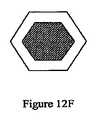
- A61M25/0032—Multi-lumen catheters with stationary elements characterized by at least one unconventionally shaped lumen, e.g. polygons, ellipsoids, wedges or shapes comprising concave and convex parts
- A—HUMAN NECESSITIES
- A61—MEDICAL OR VETERINARY SCIENCE; HYGIENE
- A61M—DEVICES FOR INTRODUCING MEDIA INTO, OR ONTO, THE BODY; DEVICES FOR TRANSDUCING BODY MEDIA OR FOR TAKING MEDIA FROM THE BODY; DEVICES FOR PRODUCING OR ENDING SLEEP OR STUPOR
- A61M25/00—Catheters; Hollow probes
- A61M25/0067—Catheters; Hollow probes characterised by the distal end, e.g. tips
- A61M25/0068—Static characteristics of the catheter tip, e.g. shape, atraumatic tip, curved tip or tip structure
- A61M25/007—Side holes, e.g. their profiles or arrangements; Provisions to keep side holes unblocked
- A—HUMAN NECESSITIES
- A61—MEDICAL OR VETERINARY SCIENCE; HYGIENE
- A61M—DEVICES FOR INTRODUCING MEDIA INTO, OR ONTO, THE BODY; DEVICES FOR TRANSDUCING BODY MEDIA OR FOR TAKING MEDIA FROM THE BODY; DEVICES FOR PRODUCING OR ENDING SLEEP OR STUPOR
- A61M25/00—Catheters; Hollow probes
- A61M25/0067—Catheters; Hollow probes characterised by the distal end, e.g. tips
- A61M25/0082—Catheter tip comprising a tool
- A61M25/0084—Catheter tip comprising a tool being one or more injection needles
- A—HUMAN NECESSITIES
- A61—MEDICAL OR VETERINARY SCIENCE; HYGIENE
- A61M—DEVICES FOR INTRODUCING MEDIA INTO, OR ONTO, THE BODY; DEVICES FOR TRANSDUCING BODY MEDIA OR FOR TAKING MEDIA FROM THE BODY; DEVICES FOR PRODUCING OR ENDING SLEEP OR STUPOR
- A61M25/00—Catheters; Hollow probes
- A61M25/0021—Catheters; Hollow probes characterised by the form of the tubing
- A61M25/0023—Catheters; Hollow probes characterised by the form of the tubing by the form of the lumen, e.g. cross-section, variable diameter
- A61M25/0026—Multi-lumen catheters with stationary elements
- A61M2025/0036—Multi-lumen catheters with stationary elements with more than four lumina
- A—HUMAN NECESSITIES
- A61—MEDICAL OR VETERINARY SCIENCE; HYGIENE
- A61M—DEVICES FOR INTRODUCING MEDIA INTO, OR ONTO, THE BODY; DEVICES FOR TRANSDUCING BODY MEDIA OR FOR TAKING MEDIA FROM THE BODY; DEVICES FOR PRODUCING OR ENDING SLEEP OR STUPOR
- A61M25/00—Catheters; Hollow probes
- A61M25/0067—Catheters; Hollow probes characterised by the distal end, e.g. tips
- A61M25/008—Strength or flexibility characteristics of the catheter tip
- A61M2025/0081—Soft tip
- A—HUMAN NECESSITIES
- A61—MEDICAL OR VETERINARY SCIENCE; HYGIENE
- A61M—DEVICES FOR INTRODUCING MEDIA INTO, OR ONTO, THE BODY; DEVICES FOR TRANSDUCING BODY MEDIA OR FOR TAKING MEDIA FROM THE BODY; DEVICES FOR PRODUCING OR ENDING SLEEP OR STUPOR
- A61M25/00—Catheters; Hollow probes
- A61M25/01—Introducing, guiding, advancing, emplacing or holding catheters
- A61M2025/0175—Introducing, guiding, advancing, emplacing or holding catheters having telescopic features, interengaging nestable members movable in relations to one another
- A—HUMAN NECESSITIES
- A61—MEDICAL OR VETERINARY SCIENCE; HYGIENE
- A61M—DEVICES FOR INTRODUCING MEDIA INTO, OR ONTO, THE BODY; DEVICES FOR TRANSDUCING BODY MEDIA OR FOR TAKING MEDIA FROM THE BODY; DEVICES FOR PRODUCING OR ENDING SLEEP OR STUPOR
- A61M25/00—Catheters; Hollow probes
- A61M25/0067—Catheters; Hollow probes characterised by the distal end, e.g. tips
- A61M25/0074—Dynamic characteristics of the catheter tip, e.g. openable, closable, expandable or deformable
- A—HUMAN NECESSITIES
- A61—MEDICAL OR VETERINARY SCIENCE; HYGIENE
- A61M—DEVICES FOR INTRODUCING MEDIA INTO, OR ONTO, THE BODY; DEVICES FOR TRANSDUCING BODY MEDIA OR FOR TAKING MEDIA FROM THE BODY; DEVICES FOR PRODUCING OR ENDING SLEEP OR STUPOR
- A61M25/00—Catheters; Hollow probes
- A61M25/0067—Catheters; Hollow probes characterised by the distal end, e.g. tips
- A61M25/008—Strength or flexibility characteristics of the catheter tip
Landscapes
- Health & Medical Sciences (AREA)
- Life Sciences & Earth Sciences (AREA)
- Biophysics (AREA)
- Pulmonology (AREA)
- Engineering & Computer Science (AREA)
- Anesthesiology (AREA)
- Biomedical Technology (AREA)
- Heart & Thoracic Surgery (AREA)
- Hematology (AREA)
- Animal Behavior & Ethology (AREA)
- General Health & Medical Sciences (AREA)
- Public Health (AREA)
- Veterinary Medicine (AREA)
- Physics & Mathematics (AREA)
- Geometry (AREA)
- Medicines That Contain Protein Lipid Enzymes And Other Medicines (AREA)
- Infusion, Injection, And Reservoir Apparatuses (AREA)
Abstract
Translated fromJapaneseDescription
Translated fromJapanese 関連出願に対する優先権の主張
本願は、2006年8月8日に出願された米国特許出願第60/821,775号および2007年3月20日に出願された米国特許出願第60/895,916号の優先権を主張し、これらの出願は、その全体が本明細書中に参考として援用される。Claims of priority over related applications This application is based on US patent application Ser. No. 60 / 821,775 filed Aug. 8, 2006 and US Patent Application No. 60 / 895,916 filed Mar. 20, 2007. These applications are hereby incorporated by reference in their entirety.
発明の背景
早期の固形腫瘍のような、腫瘍形成の治療では、放射線を伴う外科的切除またはアブレーションは、しばしば治療に良好な結果をもたらす。しかしながら、これは、後期まで進行してしまった大部分の固形腫瘍には当てはまらない。局所進行性または局所侵襲性の固形腫瘍は、腫瘍が生じた部位の周囲の他の健常な組織内に広範囲に侵入または浸潤した原発性癌である。局所進行性の腫瘍は、体全体の組織に発生する場合があるが、早期の腫瘍とは異なり、放射線治療を使用した完全な外科的切除または完全なアブレーションでは対応できない。腫瘍の進行による周囲組織の湿潤のため、全ての癌性細胞を除去するように機能し得る、いかなる外科的処置も、癌が生じた組織を不具にするか、または破壊する可能性がある。同様に、手術後に残された癌性細胞の根絶を目的とした放射線治療は、しばしば、対象とする治療野内およびその周囲に重度かつ回復困難な損傷をもたらす。しばしば、手術は、手術によって除去することができない悪性細胞を除去することを目的とした、放射線療法、化学療法、またはアジュバント療法の組み合わせと併用される。しかしながら、腫瘍が生じた部位の周囲の他の健常な組織内に腫瘍が浸潤すると、手術と放射線療法とを含む、または手術と放射線療法と化学療法とを含む併用療法であっても、治療野内の細胞に重度の損傷を生じさせずに、腫瘍細胞を根絶することができない。BACKGROUND OF THE INVENTION In the treatment of tumor formation, such as early solid tumors, surgical excision or ablation with radiation often gives good results for treatment. However, this is not the case for most solid tumors that have progressed to late stages. A locally advanced or locally invasive solid tumor is a primary cancer that has invaded or invaded extensively in other healthy tissue around the site where the tumor originated. Locally advanced tumors may occur in tissues throughout the body, but unlike early tumors, they cannot be addressed with complete surgical resection or complete ablation using radiation therapy. Any surgical procedure that can function to remove all cancerous cells due to the wetting of the surrounding tissue due to the progression of the tumor can disable or destroy the cancerous tissue. Similarly, radiation therapy aimed at eradicating cancerous cells left after surgery often results in severe and irreparable damage in and around the targeted treatment field. Often surgery is combined with a combination of radiation therapy, chemotherapy, or adjuvant therapy aimed at removing malignant cells that cannot be removed by surgery. However, if the tumor infiltrates into other healthy tissue around the site where the tumor originated, it may be in the treatment field, even if it includes surgery and radiation therapy, or a combination therapy that includes surgery, radiation therapy, and chemotherapy. The tumor cells cannot be eradicated without causing severe damage to the cells.
局所進行性の腫瘍を伴う場合、手術は、「減量」と称される全摘出のために行われるが、現在の手術には、個々の細胞を除去したり、顕微鏡によって細胞を処置したり、または腫瘍切除部位を囲む正常組織から腫瘍関連の脈管系を除去するためのツールが存在しない。このような手術において切除される周囲組織の量を最小限に抑えることがしばしば重要である。例えば、中枢神経系の腫瘍の場合、組織が失われることによって、通常の脳機能が大幅に損なわれる場合がある。したがって、そのような場合、周囲組織内に残った癌性細胞を殺傷するために、手術は、しばしば放射線療法および/または化学療法を伴う。化学療法は、局所的または全身的投与経路によって残留腫瘍細胞に送達することができる。外科的切除の程度を制限して、また、補助療法に依存して残留癌細胞を除去することによって、器官の機能を保持することができる。 When accompanied by locally advanced tumors, surgery is done for a total removal called “weight loss”, but current surgery removes individual cells, treats cells with a microscope, Or there is no tool to remove the tumor-related vasculature from the normal tissue surrounding the tumor resection site. It is often important to minimize the amount of surrounding tissue excised in such surgery. For example, in the case of central nervous system tumors, loss of tissue can significantly impair normal brain function. Thus, in such cases, surgery often involves radiation therapy and / or chemotherapy to kill cancerous cells remaining in the surrounding tissue. Chemotherapy can be delivered to residual tumor cells by local or systemic routes of administration. Organ function can be preserved by limiting the extent of surgical resection and by removing residual cancer cells depending on adjuvant therapy.
電離放射線ビーム(X線、γ線、または高エネルギβ粒子)を使用した従来の放射線療法は、抗癌治療法として確立されてはいるが、癌が局所的に進行した大部分の患者においては治癒的ではない。別の形態の放射線治療は、小線源療法であるが、該療法では、例えば脳または前立腺の癌の治療において、腫瘍部位に隣接する組織内に、γ線または高エネルギβ粒子を放射する密封放射性線源を据え付ける。例えば、特許文献1、特許文献2、および特許文献3を参照のこと。 Conventional radiation therapy using ionizing radiation beams (X-rays, γ-rays, or high-energy β particles) has been established as an anticancer treatment, but in most patients with locally advanced cancer. Not curative. Another form of radiation therapy is brachytherapy, in which, for example, in the treatment of brain or prostate cancer, a sealed that emits gamma rays or high energy beta particles in the tissue adjacent to the tumor site. Install radioactive sources. For example, see
局所進行性の固形腫瘍を有する約3分の1の患者は、薬剤を添加しても再発している(非特許文献1)。電離放射線は、高エネルギ放射線を放射するビームからであっても、同位体埋め込みからであっても、腫瘍細胞を除去し、一方で、治療野内の正常細胞を節約するのに必要な特異性に欠けている。したがって、正常組織への副次的損害を回避することができない。従来の放射線治療には、複数のさらなる限界がある。X線は、断続的な計画によって投与されるが、通常は週5日であり、それによって、癌細胞がそれらのDNAを修復して、治療の合間に腫瘍を再生息させる機会を提供する。電離放射線は、腫瘍細胞を除去するのに十分な酸素を組織内に必要とするが、大部分の固形腫瘍は、比較的低酸素であるので、放射線には本質的に耐性を示す。加えて、放射線の総生涯投与量は、重度の遅発性毒性の危険性によって制限される。したがって、一部の例外を除いて、通常は6週間まで続けられる単一の治療コースだけしか腫瘍に投与することができない。最後に、電離放射線は、特に化学療法薬剤と組み合わせて使用したときに、それ自体が発癌性を示す。 About one third of patients with locally advanced solid tumors relapse even when drugs are added (Non-patent Document 1). Ionizing radiation removes tumor cells, whether from beams emitting high-energy radiation or from isotope implantation, while at the specificity required to conserve normal cells in the treatment field. Missing. Therefore, collateral damage to normal tissues cannot be avoided. Conventional radiation therapy has several additional limitations. X-rays are administered on an intermittent schedule, but are usually 5 days a week, thereby providing the opportunity for cancer cells to repair their DNA and regenerate the tumor between treatments. Ionizing radiation requires sufficient oxygen in the tissue to remove tumor cells, but most solid tumors are relatively hypoxic and thus inherently resistant to radiation. In addition, the total lifetime dose of radiation is limited by the risk of severe late toxicity. Thus, with some exceptions, only a single course of treatment, usually lasting up to 6 weeks, can be administered to the tumor. Finally, ionizing radiation is itself carcinogenic, especially when used in combination with chemotherapeutic drugs.
化学療法剤は、体全体に分配されて、悪性細胞とともに正常細胞にも影響を及ぼすので、大部分のタイプの化学療法も、腫瘍特異性に欠けるという欠点があり、また、正常組織に副次的損害も生じさせる。多くの全身化学療法剤は、DNA合成および細胞分裂を受ける細胞に作用するので、標的癌細胞に加えて、体全体の多くの細胞集団にも影響を与え得る。 Because chemotherapeutic agents are distributed throughout the body and affect normal cells as well as malignant cells, most types of chemotherapy have the disadvantage of lacking tumor specificity and are secondary to normal tissues. Damages. Many systemic chemotherapeutic agents act on cells undergoing DNA synthesis and cell division, and thus can affect many cell populations throughout the body in addition to target cancer cells.
現在の治療法の欠点は、例えば、最も一般的な悪性脳腫瘍を構成する高侵襲的なタイプの癌である、多形膠芽腫(glioblastoma multiforme:GBM)のような、特定のタイプの癌に関して特に顕著である。実際に、約35年にわたって、数百もの実験的治療と、数千もの臨床試験に参加したGBM患者とを伴う調査を行ったが、新しく診断されたGBMの患者の予後は最悪である。最近の調査では、GBMと診断された後の生存率は、6ヶ月でわずか42%、1年では18%、2年では3%である(非特許文献2)。 The shortcomings of current therapies are for certain types of cancer, such as glioblastoma multiforme (GBM), which is the highly invasive type of cancer that constitutes the most common malignant brain tumor This is particularly noticeable. In fact, over a period of about 35 years, studies with hundreds of experimental treatments and GBM patients who participated in thousands of clinical trials have been conducted, but the prognosis for patients with newly diagnosed GBM is the worst. In a recent survey, the survival rate after being diagnosed with GBM is only 42% at 6 months, 18% at 1 year, and 3% at 2 years (Non-patent Document 2).
新しく診断されたGBMに対する現在の好適な治療では、外科的切除の後に、電離放射線と経口テモゾロマイド(放射線のコースの間と後に投与される化学療法剤)とのコースが行われる。現在利用可能な最良のこの治療を受けた患者における平均生存期間の延長は、手術および放射線治療だけのものを約2〜3ヶ月上回るだけである。 The current preferred treatment for newly diagnosed GBM involves a course of ionizing radiation and oral temozolomide (a chemotherapeutic agent administered during and after the course of radiation) after surgical resection. The increase in mean survival in patients receiving this best treatment currently available is only about 2-3 months above that of surgery and radiation alone.
近年、腫瘍部位で化学療法剤の有効濃度を増加させる手法が開発された。GBMの治療では、組織内または局所的化学療法が用いられ、わずかながら成功している。カルムスチン(化学療法剤)を含有したウエハを、腫瘍の外科的除去によって生じた空洞に挿入する。ウエハは、脳腫瘍のすぐ近くの脳組織内にカルムスチンを放出する。この治療では、手術および放射ビーム治療によってさらに治療した患者において、11.6ヶ月〜13.9ヶ月の平均生存期間の増加が示された(非特許文献3)。組織内化学療法は、外科的切除の後に再発するGBM腫瘍のうちの90%超が、2cm以内の外科的切除縁に局所化されるので、GBMの治療に特に適した治療となり得る(非特許文献4)。 In recent years, techniques have been developed to increase the effective concentration of chemotherapeutic agents at the tumor site. In treating GBM, intra- or local chemotherapy has been used with little success. A wafer containing carmustine (chemotherapeutic agent) is inserted into the cavity created by surgical removal of the tumor. The wafer releases carmustine into the brain tissue immediately adjacent to the brain tumor. This treatment showed an increase in mean survival of 11.6 to 13.9 months in patients who were further treated with surgery and radiation beam therapy (Non-Patent Document 3). Tissue chemotherapy can be a particularly suitable treatment for the treatment of GBM because more than 90% of GBM tumors that recur after surgical resection are localized to the surgical margin within 2 cm (non-patented) Reference 4).
したがって、(生化学的標的化とは別の)物理的手法による化学療法剤の濃度の局所化は、カルムスチンウエハによる有望な結果に示されるように、全身化学療法と比較して、特定の利点を提供すると思われる。しかしながら、大部分の化学物質は、脳組織または他のタイプの固形組織内に全く拡散しないので、課題は大きい。 Therefore, localization of chemotherapeutic agent concentrations by physical means (apart from biochemical targeting) is more specific than systemic chemotherapy, as shown by promising results with carmustine wafers. It seems to provide benefits. However, the challenge is significant because most chemicals do not diffuse at all into brain tissue or other types of solid tissue.
化学療法剤の物理的な局所的送達における別の開発は、対流増加送達である。この手法では、脳腫瘍に適用すると、流体は、循環系を通らずに、脳内の部位に直接送達される。流体は、液体が組織の隙間を通って移動してあらゆる溶解材料を担送するように、持続的圧力の下で適用される。例えば、非特許文献5;Laske,D W.et al.,“Convention−enhanced drug delivery”、特許文献4(1998年2月24日);および非特許文献6を参照のこと。したがって、対流増加送達は、生物活性剤を固形組織内に送達することができる有効距離を増加させるように機能する。 Another development in physical local delivery of chemotherapeutic agents is convection enhanced delivery. In this approach, when applied to a brain tumor, fluid is delivered directly to a site in the brain without going through the circulatory system. The fluid is applied under sustained pressure so that the liquid moves through the tissue gap and carries any dissolved material. For example, Non-Patent Document 5; Laske, D.W. et al. , "Convention-enhanced drug delivery", Patent Document 4 (February 24, 1998); and Non-Patent
対流増加送達は、通常、腫瘍除去時に生じた外科的空洞を囲む脳組織内に個々に据え付けられる、3〜5つのカテーテルの使用を伴う。カテーテルは、脳腫瘍の空洞内からではなく、脳の外側表面上の複数の起始点から挿入される。ポンプによって治療流体がカテーテル内に送られるので、カテーテル先端部からバルク流が生じる。このタイプの薬物送達に関連する最大の課題のうちの1つは、カテーテル先端部の最適位置を決定することである。カテーテル先端部の最適な位置決めは、広範囲かつ不規則に形成され得る全ての対象とする治療野への注入剤のアクセスを確保するためだけでなく、脳の無関係な領域の暴露を最小化するためにも重要である。他の課題は、少数のカテーテル先端部を使用して、治療野の十分な適用範囲を提供すること、カテーテル周辺および脳表面の裏側への注入剤の逆流を回避すること、および脳の脳室内および他の解剖学的部位内への注入剤の漏出を防ぐことである。 Convection-enhanced delivery usually involves the use of 3-5 catheters that are individually placed in the brain tissue surrounding the surgical cavity created during tumor removal. The catheter is inserted from multiple origins on the outer surface of the brain, not from within the brain tumor cavity. As the treatment fluid is pumped into the catheter by the pump, a bulk flow is generated from the catheter tip. One of the biggest challenges associated with this type of drug delivery is determining the optimal position of the catheter tip. Optimal positioning of the catheter tip not only ensures access of the infusate to all targeted treatment areas that can be extensively and irregularly formed, but also minimizes exposure of unrelated areas of the brain Also important. Other challenges include using a small number of catheter tips to provide adequate coverage of the treatment field, avoiding backflow of infusate around the catheter and back of the brain surface, and intraventricular of the brain And to prevent leakage of the infusate into other anatomical sites.
局所進行性の固形腫瘍の有効な治療には、GBMを含め、改善された薬物送達の方法だけでなく、癌細胞を除去すると同時に、癌細胞に侵入された正常細胞を排除することができる治療剤が必要である。この点に関して、遺伝子発現の研究によって明らかになった主な問題は、腫瘍が、これまでの予想をはるかに超えて遺伝的および代謝的に異質であることである。腫瘍は、共通の器官または起源の組織にかかわらず、また、顕微鏡では外観が非常に類似しているにもかかわらず、遺伝的および代謝的に異質である。これは、GBM、および中枢神経系内に発生する他の悪性神経膠腫に特に当てはまる。例えば、非特許文献7を参照のこと。腫瘍異質性を考慮すると、生化学標的化、すなわち、各腫瘍タイプを特異的に標的とする薬剤の検索は、困難な課題である。 Effective treatment of locally advanced solid tumors includes not only improved drug delivery methods, including GBM, but also treatments that can remove cancer cells while simultaneously eliminating normal cells invaded by cancer cells An agent is needed. In this regard, the main problem revealed by gene expression studies is that tumors are genetically and metabolically heterogeneous far beyond previous expectations. Tumors are genetically and metabolically heterogeneous, regardless of common organs or tissues of origin and despite their very similar appearance under a microscope. This is especially true for GBM and other malignant gliomas that occur in the central nervous system. For example, see
次の4項には、新しくかつ有効な治療が必要である。(a)治療野内の腫瘍幹細胞の亜集団を含む腫瘍細胞の除去、(b)広範囲の遺伝および代謝プロファイルを有する腫瘍細胞の除去、(c)化学療法および電離放射線に対して固有の耐性を有する腫瘍幹細胞の除去、(d)正常細胞および正常組織に対する毒性の最小化または回避。この課題に対する1つの手法は、多くの異なるタイプの癌細胞を殺傷することができる、薬剤の物理的な局所的送達と同時に、治療野内の正常な細胞に対する毒性を最小限にするか、または無くすものである。この手法は、標的療法とは異なり、各腫瘍を、その異なる遺伝および代謝プロファイルに従って治療するのに異なる薬物の機構が必要となり得る。 The next four items require new and effective treatment. (A) removal of tumor cells, including a subpopulation of tumor stem cells within the treatment field, (b) removal of tumor cells with a wide range of genetic and metabolic profiles, (c) inherent resistance to chemotherapy and ionizing radiation Removal of tumor stem cells, (d) minimization or avoidance of toxicity to normal cells and tissues. One approach to this challenge is to minimize or eliminate toxicity to normal cells in the treatment field, as well as physical local delivery of drugs that can kill many different types of cancer cells. Is. This approach, unlike targeted therapies, may require different drug mechanisms to treat each tumor according to its different genetic and metabolic profiles.
かなり関心が高まっている一意的な細胞殺傷の機構は、オージェ電子の放出である。これらの電子は、電子捕獲および内部変換によって減少する放射性核種によって放射される。放射性核種を放射するオージェの例には、123ヨウ素、125ヨウ素、77臭素、および80m臭素が挙げられる。オージェ電子は、トリチウムによって放射されるβ粒子のエネルギよりもさらに低いエネルギを有する。いくつかのオージェ放射体が、それぞれ核変換を有する複数の電子を放出するので、この効果は増幅される。低エネルギのオージェ電子によって、細胞内の粒子の経路長が極めて短くなり、副次的損害を最小限に抑えることから、非常に望ましい。A unique cell killing mechanism of considerable interest is the release of Auger electrons. These electrons are emitted by radionuclides that are reduced by electron capture and internal conversion. Examples of Augers that emit radionuclides include123 iodine,125 iodine,77 bromine, and80 m bromine. Auger electrons have an energy that is even lower than the energy of β particles emitted by tritium. This effect is amplified because several Auger emitters emit multiple electrons, each with a transmutation. Low energy Auger electrons are highly desirable because the path length of the particles in the cell is extremely short, minimizing collateral damage.
125Iを組み込んだ分子化合物の1つは、チミジン類似体である[125I]−ヨードウリジン−デオキシリボシド(125IUDR)である。125IUDRは、DNAポリメラーゼによってチミジンとして認識されるので、DNA合成時に、染色体内に組み込まれる。DNAに組み込まれると、オージェ電子は、それらの非常に短い経路長によって、2重鎖DNAの化学骨格にアクセスする。125I原子が崩壊するとき、オージェ電子によって、標的細胞内に修復不能な染色体の破壊が生じるが、標的細胞のすぐ近くの細胞に対する影響は最小限に抑えられる。125IUDRおよび関連化合物は、DNAを形成する細胞を破壊するが、他の細胞にはほとんど影響を及ぼさない。125IUDRが一意的な細胞殺傷能力を有するという認識にもかかわらず、また、125IUDRを腫瘍内に直接導入するという概念(例えば、Kassis et al.,“Treatment of tumors with 5−radioiodo−2’−deoxyuridine”、特許文献5を参照のこと)を含む、この作用機構の利用を対象とした長年にわたる研究にもかかわらず、これらの薬剤は、癌の治療には成功裏に適用されなかった。全身または局所的投与を用いた、125IUDRおよび関連する薬剤の固形腫瘍への送達は、極めて困難であることが判明した。One molecular compound that incorporates125 I is [125 I] -iodouridine-deoxyriboside (125 IUDR), a thymidine analog.Since 125 IUDR is recognized as thymidine by DNA polymerase, it is integrated into the chromosome during DNA synthesis. When incorporated into DNA, Auger electrons access the chemical backbone of double-stranded DNA through their very short path length.When the 125 I atom decays, Auger electrons cause irreparable chromosome breaks in the target cell, but the effects on the cells immediately adjacent to the target cell are minimized.125 IUDR and related compounds destroy cells that form DNA, but have little effect on other cells. Despite the recognition that125 IUDR has a unique cell killing capability, the concept of introducing125 IUDR directly into tumors (eg, Kassis et al., “Treatment of tumors with 5-radioido-2 ′) Despite many years of research aimed at the use of this mechanism of action, including “deoxyuridine” (see US Pat. No. 5,099,059), these drugs have not been successfully applied in the treatment of cancer. Delivery of125 IUDR and related drugs to solid tumors using systemic or local administration has proven extremely difficult.
DNAの合成中にDNA内にオージェ電子を放射するヌクレオチド類似体を組み込むことの有効性は、DNA合成に関与する標的細胞の割合を増加させることによって増大させることができる。この一般的手法は、多数の抗癌剤、特にS期(合成期)の細胞周期中に選択的に細胞に作用する細胞毒性薬物(すなわち、「S期の活性剤」)の効果を高めるために成功裏に使用された。例えば、非特許文献8を参照のこと。特定の薬物は、S期中に腫瘍細胞の進行を防ぐことができるので、標的細胞集団内の感受性細胞の分画を効率的に増加させる。この手法は、細胞周期抑制剤、5−フルオロウリジン2’デオキシリボヌクレオシドを使用した、5−[125I]−ヨードウリジン2’デオキシリボヌクレオシドのDNAへの取り込みおよび組み込みの増加に成功裏に使用された。例えば、非特許文献9;非特許文献10;および非特許文献11を参照のこと。The effectiveness of incorporating nucleotide analogues that emit Auger electrons into DNA during DNA synthesis can be increased by increasing the proportion of target cells involved in DNA synthesis. This general approach has been successful in enhancing the effectiveness of a number of anticancer agents, particularly cytotoxic drugs that selectively act on cells during the S (synthetic) cell cycle (ie, "S phase active agents"). Used on the back. For example, see
したがって、125IUDRおよび関連化合物の一意的な作用機構の活用を対象とした、新しい薬物送達装置および使用方法が必要である。新しい手法は、腫瘍を保持した幹細胞およびそれらの前駆体を含む、腫瘍細胞の循環を除去すると同時に、癌細胞に侵入された正常細胞を排除することを目的とした、125IUDR(および他の化合物)の固形腫瘍への送達が必要である。この必要性には、特に、しばしば不規則に形成される組織量の略均一な治療を提供するような方法で、当該の薬剤を腫瘍内に、および腫瘍細胞に侵入された正常細胞内に直接送達するための方法を含む。Therefore, there is a need for new drug delivery devices and methods of use that target the utilization of the unique mechanism of action of125 IUDR and related compounds. The new approach is aimed at eliminating tumor cell circulation, including tumor-bearing stem cells and their precursors, while simultaneously eliminating125 IUDR (and other compounds) that have invaded cancer cells. ) Delivery to solid tumors is required. This need is particularly relevant in such a way as to provide a substantially uniform treatment of the amount of tissue that is often irregularly formed, in such a way that the agent is directly injected into the tumor and into normal cells that have invaded the tumor cell. Including a method for delivery.
概要
本発明は、抗癌剤のような生物活性剤を、それを必要とする患者の脳組織のような標的組織に送達するための装置および方法を目的とする。本発明の一実施形態は、生物活性剤の液体溶液を患者の標的組織内に送達するためのカテーテルアレイシステムであって、各カテーテルが、直線または曲線の中空管を備え、該カテーテルを生体組織内に挿入し、しばらくの間、組織内に残留させて、生物活性剤の溶液を管を通じて組織内に送達するように適合された、複数の生体適合性カテーテルと、複数のカテーテルのそれぞれを誘導テンプレートに隣接した組織内に設置するように誘導して、組織内に空間的に画定されたカテーテルアレイを形成するように適合された、カテーテル誘導テンプレートと、マニホールドを介して複数のカテーテルのそれぞれに液体を送達するように適合された、加圧液体供給システムとを備え、各カテーテルは、組織内への挿入のための遠位部分と、溶液が中空管の内側から組織内へ通過することができる少なくとも1つのポートと、誘導テンプレートによる組織内への挿入を目的として適合された中間部分と、加圧液体供給システムのマニホールドへの接続に適合された基部とを備え、カテーテル誘導テンプレートは、複数のカテーテル誘導案内面のチャネルを備え、各誘導案内面のチャネルは、組織内へ挿入するために、このチャネルを通じて1つ以上のカテーテルの運動を誘導するように適合され、複数のカテーテルを挿入したときに、カテーテルが、空間的に画定されたカテーテルアレイを組織内に形成することができ、液体供給システムは、液体溶液に圧力を印加するように適合された加圧器と、液体を、加圧下で、複数のカテーテルのそれぞれの基部に送達するマニホールドとを備え、液体は、各カテーテルの中空管を通過して組織内に入ることができる、カテーテルアレイシステムを提供する。SUMMARY The present invention is directed to an apparatus and method for delivering a bioactive agent, such as an anti-cancer agent, to a target tissue, such as the brain tissue of a patient in need thereof. One embodiment of the present invention is a catheter array system for delivering a liquid solution of a bioactive agent into a target tissue of a patient, each catheter comprising a straight or curved hollow tube, A plurality of biocompatible catheters adapted to be inserted into the tissue and left in the tissue for a while to deliver a solution of the bioactive agent through the tube into the tissue, and each of the plurality of catheters Each of a plurality of catheters via a manifold and a catheter guide template adapted to be guided to be placed in tissue adjacent to the guide template to form a spatially defined catheter array in the tissue A pressurized liquid supply system adapted to deliver liquid to the catheter, each catheter having a distal portion for insertion into tissue and a solution Fits at least one port that can pass into the tissue from the inside of the hollow tube, an intermediate portion adapted for insertion into the tissue by the guide template, and connection to the manifold of the pressurized liquid supply system A catheter guide template comprising a plurality of catheter guide guide channel channels, each guide guide channel being configured to move one or more catheters through the channel for insertion into tissue. The catheter is capable of forming a spatially defined catheter array in tissue when inserted with a plurality of catheters adapted to guide, and the liquid supply system applies pressure to the liquid solution. And a manifold that delivers liquid under pressure to each base of a plurality of catheters. , The liquid may enter into the tissue through the hollow tube of each catheter, to provide a catheter array system.
本発明の実施形態は、カテーテルと、カテーテル誘導テンプレートと、加圧器およびマニホールドを含む液体供給システムとをさらに提供し、それぞれ、本発明のカテーテルアレイシステムの構成要素として使用するように適合される。 Embodiments of the present invention further provide a catheter, a catheter guide template, a liquid supply system including a pressurizer and a manifold, each adapted to be used as a component of the catheter array system of the present invention.
本発明の一実施形態は、カテーテルアレイを目的とし、該アレイは、それを必要とする患者の組織内に配置される。カテーテルアレイは、規則的であることが好ましく、カテーテルは、並列または放射状の三次元配列で配置される。カテーテルは、互いの間隔が十分狭いことが好ましく、それらの距離は、生物活性剤が組織を治療的に貫通することができる距離の約2倍程度である。アレイを構成するカテーテルは、総数のサブセットで、または同時に、個々に設置することができる。カテーテルアレイのサブセットは、異なる深さで、また、組織内の異なる空間的配列で据え付けることができる。カテーテル誘導テンプレートは、複数のカテーテルの挿入プロセス中に、空間的に画定されたカテーテルアレイを形成させるが、これは、逐次的に、同時に、複数のカテーテルのサブセットで行うことができる。 One embodiment of the present invention is directed to a catheter array that is placed in the tissue of a patient in need thereof. The catheter array is preferably regular, and the catheters are arranged in a parallel or radial three-dimensional array. The catheters are preferably sufficiently close to each other that their distance is on the order of about twice the distance that the bioactive agent can therapeutically penetrate the tissue. The catheters that make up the array can be placed individually in a subset of the total number, or simultaneously. Subsets of catheter arrays can be installed at different depths and at different spatial arrangements within the tissue. The catheter guide template forms a spatially defined catheter array during the insertion process of multiple catheters, which can be done sequentially and simultaneously with a subset of multiple catheters.
生物活性剤は、その溶液が本発明のカテーテルアレイシステムによって標的組織内に導入され、放射化学剤、化学療法剤、または他の小分子、抗体、タンパク質、ペプチド、オリゴヌクレオチドアプタマ、アンチセンスオリゴヌクレオチド、または低分子干渉RNA(siRNA)とすることができる。このような放射化学剤の1つは、123I−ヨードウリジンデオキシリボシドまたは125I−ヨードウリジンデオキシリボシド(123IUDRまたは125IUDR)のような、オージェ電子放射体を含み、放射性核種は、標的細胞内へ取り込むように適合された、化学物質内に組み込まれ、その場合、狭い範囲のオージェ電子は、それらが含まれた細胞内のDNAに破壊的な影響を直接及ぼすが、周辺の細胞への副次的損害は最小限に抑えられる。The bioactive agent is introduced into the target tissue by the catheter array system of the present invention and the radioactive agent is a radiochemical agent, chemotherapeutic agent, or other small molecule, antibody, protein, peptide, oligonucleotide aptamer, antisense oligo It can be a nucleotide or a small interfering RNA (siRNA). One such radiochemicalagents, 123 I-, such as iodo uridine deoxyriboside or125 I- iodo uridine deoxyriboside(123 IUDR or125 IUDR), include Auger electron emitters, radionuclides, target cells Incorporated into chemicals, adapted to be incorporated into, the narrow range of Auger electrons directly has a destructive effect on the DNA in the cells in which they are contained, but it does not affect surrounding cells Secondary damage is minimized.
本発明の実施形態はまた、本発明のカテーテルアレイシステムを使用した、生物活性剤の組織内への送達が医学的に指示された、異常状態の患者を治療する方法であって、誘導テンプレートが生物活性剤の組織内への送達の標的となる組織に直接隣接するように、カテーテル誘導テンプレートを、患者の標的組織内に、またはこれに隣接して設置し、ステップと、次いで、各カテーテルが、それぞれのチャネルによって、標的組織内の位置に向けられ、空間的に画定されたカテーテルアレイを形成するように、複数のカテーテルのそれぞれを誘導テンプレートを通じて挿入するステップと、加圧液体がカテーテルを通じて標的組織に送達されるように、液体供給システムを各カテーテルの基部に接続するステップと、その後、生物活性剤の溶液を含む液体を、液体供給システムから、複数のカテーテルを通じてポートを経由して標的組織内に供給するステップとを含む方法も目的とする。 An embodiment of the present invention is also a method of treating an abnormal patient who is medically instructed to deliver a bioactive agent into a tissue using the catheter array system of the present invention, wherein the guide template comprises: A catheter guide template is placed in or adjacent to the target tissue of the patient so that it is directly adjacent to the tissue targeted for delivery of the bioactive agent into the tissue. Inserting each of a plurality of catheters through a guide template so as to form a spatially defined catheter array directed to a location within the target tissue by each channel, and a pressurized liquid is targeted through the catheter Connecting a fluid supply system to the base of each catheter for delivery to the tissue, followed by dissolution of the bioactive agent. A liquid containing, from the liquid supply system, also aims method and supplying via port through a plurality of catheters into a target tissue.
カテーテルアレイシステムは、患者の組織内に、例えば脳腫瘍の除去によって残った空隙内に導入することができ、複数のカテーテルが、腫瘍切除部位を囲む組織内に侵入する。代替的に、カテーテルアレイシステムは、例えば特定の卵巣癌で生じるような、腫瘍プラーク内に導入することができる。 The catheter array system can be introduced into a patient's tissue, for example, into a void left by removal of a brain tumor, and multiple catheters penetrate into the tissue surrounding the tumor resection site. Alternatively, the catheter array system can be introduced into a tumor plaque, such as occurs in certain ovarian cancers.
システム全体を、患者の体内に完全に据え付けることができ、液体供給システムおよびマニホールド、ならびにカテーテル誘導テンプレートおよび複数のカテーテルは、患者の皮下に配置される。代替的に、液体供給システムは、少なくとも、患者の体外に配置することができる。 The entire system can be fully installed in the patient's body, with the fluid supply system and manifold, and the catheter guide template and multiple catheters placed under the patient's skin. Alternatively, the liquid supply system can be placed at least outside the patient's body.
適切な構造材料を用いて達成することができるように、カテーテル誘導テンプレートが生体組織と接触する範囲まで、少なくとも誘導テンプレートの表面が生体適合性であることが好ましい。同様に、液体供給システムが患者の体内に配置されるように適合された範囲まで、その外面を生体適合性とすることができる。 It is preferred that at least the surface of the guide template is biocompatible, at least to the extent that the catheter guide template contacts the living tissue, as can be achieved using a suitable structural material. Similarly, the outer surface can be biocompatible to the extent that the liquid supply system is adapted to be placed within the patient's body.
本発明の方法の一実施形態は、種々の圧力、流速、および投与の継続期間での、生物活性剤の溶液の投与を含むことができる。例えば、溶液は、種々の速度、および種々の期間で連続的、断続的に投与することができる。 One embodiment of the method of the invention can include administration of a solution of bioactive agent at various pressures, flow rates, and duration of administration. For example, the solution can be administered continuously, intermittently at various rates and for various periods of time.
好適な生物活性剤は放射性物質(radiological agent)であり、123Iまたは125Iのようなオージェ電子を放射する同位元素とすることができ、それが配置された組織に生じさせる損傷範囲が狭いので、健常な組織への望ましくない放射線損傷を制限する。オージェ電子を放射する同位元素は、標的組織内の癌性細胞の細胞構造体内に組み込まれるように適合された分子の一部とすることができ、例えば、ヌクレオチド類似体を放射性標識化して、本発明の方法の使用に適した生物活性構造を提供することができる。125I−ヨードウリジンデオキシリボシド(IUDR)は、一例である。A suitable bioactive agent is a radioactive agent, which can be an isotope that emits Auger electrons, such as123 I or125 I, since it has a small extent of damage to the tissue in which it is located. Limit unwanted radiation damage to healthy tissue. An isotope that emits Auger electrons can be part of a molecule adapted to be incorporated into the cellular structure of a cancerous cell in a target tissue, for example, by radiolabeling a nucleotide analog and Bioactive structures suitable for use in the inventive method can be provided.125 I-iodouridine deoxyriboside (IUDR) is an example.
発明の詳細な説明
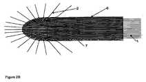
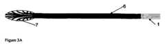
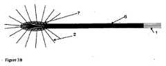
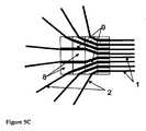
本発明の一実施形態は、生物活性剤の液体溶液を患者の標的組織内に送達するためのカテーテルアレイシステムであって、各カテーテルが、直線または曲線の中空管を備え、該カテーテルを生体組織内に挿入し、しばらくの間、組織内に残留させて、生物活性剤の溶液を管を通じて組織内に送達するように適合された、複数の生体適合性カテーテルと、複数のカテーテルのそれぞれを誘導テンプレートに隣接した組織内に設置するように誘導して、組織内に空間的に画定されたカテーテルアレイを形成するように適合された、カテーテル誘導テンプレートと、マニホールドを介して複数のカテーテルのそれぞれに液体を送達するように適合された、加圧液体供給システムとを備え、各カテーテルは、組織内への挿入のための遠位部分と、溶液が中空管の内側から組織内へ通過することができる少なくとも1つのポートと、誘導テンプレートによる組織内への挿入を目的として適合された中間部分と、加圧液体供給システムのマニホールドへの接続に適合された基部とを備え、カテーテル誘導テンプレートは、複数のカテーテル誘導案内面のチャネルを備え、各誘導案内面のチャネルは、組織内へ挿入するために、このチャネルを通じて1つ以上のカテーテルの運動を誘導するように適合され、複数のカテーテルを挿入したときに、カテーテルが、空間的に画定されたカテーテルアレイを組織内に形成することができ、液体供給システムは、液体溶液に圧力を印加するように適合された加圧器と、液体を、加圧下で、複数のカテーテルのそれぞれの基部に送達するマニホールドとを備え、液体は、各カテーテルの中空管を通過して組織内に入ることができる、カテーテルアレイシステムを目的とする。DETAILED DESCRIPTION OF THE INVENTION One embodiment of the present invention is a catheter array system for delivering a liquid solution of a bioactive agent into a target tissue of a patient, each catheter comprising a straight or curved hollow tube. A plurality of biocompatible catheters adapted to insert the catheter into biological tissue and remain in the tissue for a period of time to deliver a solution of the bioactive agent through the tube into the tissue; A catheter guide template adapted to guide each of the catheters to be placed in tissue adjacent to the guide template to form a spatially defined catheter array in the tissue, and via a manifold A pressurized liquid supply system adapted to deliver liquid to each of the plurality of catheters, each catheter for insertion into tissue A distal portion; at least one port through which solution can pass into the tissue from the inside of the hollow tube; an intermediate portion adapted for insertion into the tissue by a guide template; and a pressurized liquid supply system A catheter guide template comprising a plurality of catheter guide guide surface channels, each guide guide surface channel passing through the channel for insertion into tissue. The liquid supply system is adapted to induce movement of one or more catheters, and when a plurality of catheters are inserted, the catheters can form a spatially defined catheter array in the tissue. A pressurizer adapted to apply pressure to the solution and a fluid that delivers the liquid under pressure to each base of a plurality of catheters. And a hold, the liquid may enter into the tissue through the hollow tube of each catheter, an object catheter array system.
本発明の一実施形態は、複数のカテーテルと、当該カテーテルの固体組織内への、例えば脳組織内への据え付けを誘導するように適合されたカテーテル誘導テンプレートとを備えた、外科的に据え付けられる薬物送達装置に関する。複数のカテーテルは、カテーテル誘導テンプレートを用いて組織内に空間的に画定されたアレイを形成することを目的とし、これらを使用して、局所侵襲性の増殖型腫瘍細胞によって浸潤されたような、腫瘍内または組織内に生物活性治療剤を直接送達する。生物活性剤には、これに限定されないが、放射性化合物、細胞毒性および他の小分子薬物、抗体、タンパク質、ペプチド、オリゴヌクレオチドアプタマ、アンチセンスオリゴヌクレオチド、およびsiRNAが挙げられる。本発明のカテーテルアレイシステムを使用して、異なるタイプの局所進行性の固形腫瘍を治療することができる。治療野は、腫瘍自体および/または腫瘍に隣接した組織を含むことができる。脳腫瘍がある患者のような特定の状況では、治療野は、術後の腫瘍切除空洞に隣接した組織内に位置する場合がある。このような組織は、腫瘍細胞および腫瘍関連の新生脈管系の増殖による進行性の浸潤を伴う、腫瘍再発の危険性がある場合がある。この状態では、治療野は、腫瘍に隣接した脳組織を含み、治療は、腫瘍再発の前および/または後に投与することができる。 One embodiment of the present invention is surgically installed with a plurality of catheters and a catheter guide template adapted to guide installation of the catheters into solid tissue, eg, brain tissue. The present invention relates to a drug delivery device. Multiple catheters are intended to form a spatially defined array in tissue using a catheter guide template, and these are used to infiltrate with locally invasive proliferating tumor cells, The bioactive therapeutic agent is delivered directly into the tumor or tissue. Bioactive agents include, but are not limited to, radioactive compounds, cytotoxic and other small molecule drugs, antibodies, proteins, peptides, oligonucleotide aptamers, antisense oligonucleotides, and siRNA. The catheter array system of the present invention can be used to treat different types of locally advanced solid tumors. The treatment field can include the tumor itself and / or the tissue adjacent to the tumor. In certain situations, such as patients with brain tumors, the treatment field may be located in tissue adjacent to the postoperative tumor resection cavity. Such tissue may be at risk of tumor recurrence with progressive invasion due to growth of tumor cells and tumor-associated neovasculature. In this condition, the treatment field includes brain tissue adjacent to the tumor, and the treatment can be administered before and / or after tumor recurrence.
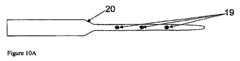
医薬品および放射化学薬品の局所的送達は、めったに実行されない。その1つの理由は、1つまたは複数のカテーテルを使用することで、主に拡散または低流速によって送達領域が非常に制限されるか、または対流(バルク流、高流速)によって送達領域は拡大するが、腫瘍内、およびその周辺における標的化が不正確になることである。単一のカテーテルによって生成される医薬品送達領域の範囲および形状は、器官内の組織の不均一性、組織内圧力の変化、毛細管密度の変化、不均等な瘢痕化、および/または疾病状態(例、腫瘍線維症)に関連する変動により、許容できない変動性を有し得る。加えて、標的領域自体は、非常に大きく、不規則な形状になり得る。 Local delivery of pharmaceuticals and radiochemicals is rarely performed. One reason is that the use of one or more catheters greatly limits the delivery area, mainly by diffusion or low flow rates, or enlarges the delivery area by convection (bulk flow, high flow rate). However, targeting within and around the tumor is inaccurate. The extent and shape of the drug delivery region produced by a single catheter can vary depending on the tissue heterogeneity within the organ, changes in tissue pressure, changes in capillary density, uneven scarring, and / or disease states (eg, , Tumor fibrosis) may have unacceptable variability. In addition, the target area itself can be very large and irregularly shaped.
局所的な薬物送達の固有の課題を解決する1つの方法は、各カテーテルが小さな領域への送達を担当する、複数のカテーテルを使用することである。複数のカテーテルは、その後、重なり合った領域に医薬品を送達して、異なる形状、サイズ、および密度の組織内における均一かつ効果的な標的化を提供することができる。当然、これは、複数のカテーテルを治療領域に個別に配置することによって行うことができる。しかしながら、カテーテルの個別の配置は時間のかかるプロセスであり、カテーテルの正確な相対的配置に固有の問題点がある。例えば、Bouvier G et.al.,“Direct delivery of medication into a brain tumor through multiple chronically implanted catheters”、Neurosurgery、20:286−291(1987)を参照のこと。 One way to solve the inherent challenges of local drug delivery is to use multiple catheters, each catheter responsible for delivery to a small area. Multiple catheters can then deliver pharmaceuticals to the overlapping area to provide uniform and effective targeting within different shapes, sizes, and densities of tissue. Of course, this can be done by placing multiple catheters individually in the treatment area. However, the individual placement of catheters is a time consuming process and has inherent problems with the correct relative placement of the catheters. For example, Bouvier G et. al. , "Direct delivery of meditation into a brain tum through multiple chromatic catalyzers", Neurosurgery, 20: 286-291 (1987).
本発明のカテーテルアレイシステムは、複数のカテーテルの治療野内への配置を誘導し、各カテーテルからの個々の薬物源が、重なり合った治療野を決定する。これらの装置は、カテーテルのネットワークまたはアレイを利用して、全ての治療野を抗腫瘍剤、放射性医薬品、または他の医薬品に暴露する。それぞれのカテーテルが、治療剤を治療野の1つの部分(サブ治療野とも称される)に送達するので、より均一な治療野が可能となる。重なり合ったサブ治療野は、完全でより均一な治療野を提供する。 The catheter array system of the present invention guides the placement of multiple catheters within a treatment field, and individual drug sources from each catheter determine overlapping treatment fields. These devices utilize a network or array of catheters to expose all therapeutic areas to anti-tumor agents, radiopharmaceuticals, or other pharmaceuticals. Each catheter delivers a therapeutic agent to a portion of the treatment field (also referred to as a sub-treatment field), thereby allowing a more uniform treatment field. Overlapping sub-treatment fields provide a complete and more uniform treatment field.
本発明の装置を使用して、個々に設置されたカテーテルによって達成できる時間枠よりもはるかに短い時間枠で、規則的または均等に離間したカテーテルの治療野内への配置を達成することができ、また、空間的精度が極めて高いので、脳のような患者の生体組織が暴露されたときの手術中に好都合となる。カテーテルの最適な位置決めは、全ての対象とする治療野への注入剤のアクセスを確保することだけでなく、脳の無関係な領域の暴露を最小化することにも重要である。 Using the device of the present invention, placement of regularly or evenly spaced catheters within the treatment field can be achieved in a much shorter time frame than can be achieved with individually installed catheters; Also, the spatial accuracy is extremely high, which is advantageous during surgery when the patient's living tissue, such as the brain, is exposed. Optimal positioning of the catheter is important not only to ensure access of the infusate to all targeted treatment areas, but also to minimize exposure of unrelated areas of the brain.
複数のカテーテルは、しばらくの間、組織内に残留するように適合される。これは、カテーテルを、単に注射針と同じように組織内に挿入し、材料を注入し、そして針を直ちに引き抜く、といった機能を行わないことを意味する。むしろ、標的細胞内でアレイを形成するカテーテルのそれぞれを、数時間、数日間、または数週間、その場所に残留させて、その間に、放射性物質のような生物活性剤を、比較的低速度で組織内に注入する。カテーテルは、ある程度の加圧下で、すなわち、溶液による組織の透過を高めるのに十分な加圧下で、生物活性剤の溶液を送達するように適合される。一般的に、組織を通過する液体の流れに対する抵抗が比較的高いので、絶対送達速度は、典型的な皮下注射針による注入と比較すると比較的低い。それぞれのカテーテルは、特定の状況で使用されている所望のレベルの特定の生物活性剤を、標的組織量に注入するのに十分な時間、組織内に残留する。 The plurality of catheters are adapted to remain in the tissue for a while. This means that the catheter does not simply perform the functions of inserting into the tissue, injecting material, and withdrawing the needle immediately, just like an injection needle. Rather, each of the catheters that form the array in the target cells are left in place for hours, days, or weeks, during which time bioactive agents, such as radioactive substances, are delivered at a relatively low rate. Inject into tissue. The catheter is adapted to deliver a solution of the bioactive agent under some pressure, ie, under sufficient pressure to enhance tissue penetration by the solution. In general, the absolute delivery rate is relatively low compared to infusion with a typical hypodermic needle because of the relatively high resistance to fluid flow through the tissue. Each catheter remains in the tissue for a time sufficient to infuse the target tissue volume with the desired level of the specific bioactive agent being used in the specific situation.
カテーテルは、カテーテルの進路からの、およびカテーテルの入口地点での組織内への注入剤の逆流を回避するように、また、治療野を越えて解剖学的空間内への、例えば脳腫瘍の場合は、脳室、軟髄膜、または硬膜下の空間への治療剤の導入を回避するように適合される。 The catheter should avoid backflow of infusate from the catheter path and into the tissue at the catheter entry point, and beyond the treatment field into the anatomical space, for example in the case of brain tumors Adapted to avoid introduction of therapeutic agents into the ventricles, leptomeninges, or subdural spaces.
アレイを形成するカテーテル間の間隔、アレイ内の互いのカテーテルの相対的配向、および標的組織に対するカテーテルアレイの配向を最適化して、治療期間中に、全ての標的組織を薬物含有液体に暴露することができる。カテーテルアレイは、装置の据え付け中の、治療期間中の、および装置の除去中の、治療野内およびその周囲の組織への損傷を最小限に抑えるように適合される。 Optimize the spacing between the catheters forming the array, the relative orientation of each other's catheters in the array, and the orientation of the catheter array relative to the target tissue to expose all target tissue to the drug-containing liquid during the treatment period Can do. The catheter array is adapted to minimize damage to the tissue in and around the treatment field during device installation, during treatment, and during device removal.
カテーテルアレイは、誘導テンプレートを使用して形成されて、空間的に画定されたアレイ内の組織内へのカテーテル先端部の据え付けを誘導する。誘導テンプレートは、各カテーテルのベクトルを決定して、治療野内へのカテーテルの貫通深さにわたる制御を提供する。種々の誘導テンプレートが提供されるが、それぞれ1つ以上の標的組織のタイプに対する適用に適している。特定の状況では、テンプレートは、カテーテルアレイの据え付け後、その場所に残留させることができる。他の場合では、テンプレートは、据え付け後に除去することができる。 The catheter array is formed using a guide template to guide installation of the catheter tip into the tissue in the spatially defined array. The guide template determines the vector for each catheter and provides control over the depth of penetration of the catheter into the treatment field. Various guide templates are provided, each suitable for application to one or more target tissue types. In certain situations, the template can remain in place after the catheter array is installed. In other cases, the template can be removed after installation.
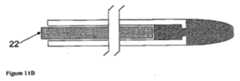
本願明細書のシステムは、複数のカテーテルの規則的なアレイを提供するように適合される。各カテーテルの寸法(長さ、内径、および外径)は、サブ治療野の深さおよび直径、アレイ内のカテーテルの密度、組織への損傷を最小限に抑えるという目的、最適な機械的強度、および据え付けの容易さを含む、複数の因子によって機能的に決定される。本発明のカテーテルアレイシステムを使用することで、脳腫瘍の空洞の内側部分に薬物送達カテーテルを据え付ける機会を提供し、それによって、脳腫瘍細胞を覆い隠していると最も思われる脳の領域に治療を集中させ(Hochberg,F.H.and Pruitt,A.、Neurology、30:907−911(1980))、一方で、その腫瘍を越えた領域に対する損傷を回避する。各カテーテルは、挿入中およびその後の、例えば腫瘍切除空洞内からの、神経および脈管構造への損傷を最小限に抑える機能がある。モジュール式のカテーテルアレイを使用することで、徐放的注入、および繰り返し注入を含む、種々のパルス投与、または一時的な投与計画を用いた、治療液体の治療野への送達という選択肢を提供する。 The system herein is adapted to provide a regular array of catheters. The dimensions (length, inner diameter, and outer diameter) of each catheter include the depth and diameter of the sub-treatment field, the density of the catheters in the array, the objective of minimizing tissue damage, optimal mechanical strength, And functionally determined by several factors, including ease of installation. The use of the catheter array system of the present invention provides an opportunity to place a drug delivery catheter in the inner part of a brain tumor cavity, thereby concentrating the treatment on the brain area most likely covering the brain tumor cells. (Hochberg, FH and Pruit, A., Neurology, 30: 907-911 (1980)) while avoiding damage to the area beyond the tumor. Each catheter is capable of minimizing damage to nerves and vasculature during and after insertion, for example from within the tumor excision cavity. Using a modular catheter array offers the option of delivering therapeutic fluid to the treatment field using a variety of pulsed or temporary dosing regimes, including sustained and repeated infusions .
標的組織内へのカテーテルの据え付け、および組織内でのカテーテルアレイの形成は、生体適合性表面を有することができるカテーテル誘導テンプレートを用いて達成される。誘導テンプレートは、互いに対して、また、カテーテルが据え付けられる組織に対して、規則的なアレイでのカテーテルの据え付けを誘導するように適合される。カテーテルのうちの少なくともいくつかは、据え付け前に基部に取り付けることができ、誘導テンプレートによって組織内に誘導することができる予備成形されたアレイを形成する。代替的に、カテーテルは、共通の基部に取り付けずに、誘導テンプレートの誘導下で据え付けることができる。カテーテルの誘導は、誘導テンプレート内のカテーテル誘導チャネルを用いて達成される。このチャネルは、据え付け中のカテーテルの位置を誘導する経路を提供し、据え付け中にそれぞれのチャネルを通じてカテーテルが相対的に運動できるように適合される。据え付け後にカテーテルを適所に固定できる機能があり、その場合、カテーテルの除去が望まれたときに解放することもできる。 The placement of the catheter within the target tissue and the formation of the catheter array within the tissue is accomplished using a catheter guide template that can have a biocompatible surface. The guide templates are adapted to guide the installation of the catheters in a regular array relative to each other and to the tissue in which the catheters are installed. At least some of the catheters can be attached to the base prior to installation to form a preformed array that can be guided into tissue by a guide template. Alternatively, the catheter can be installed under the guidance of a guide template without being attached to a common base. Catheter guidance is accomplished using a catheter guide channel in the guide template. This channel provides a path to guide the position of the catheter during installation and is adapted to allow relative movement of the catheter through each channel during installation. There is the ability to fix the catheter in place after installation, in which case it can also be released when removal of the catheter is desired.
誘導テンプレートは、カテーテルを据え付けた後に、カテーテルとともにその場所に残留させるか、または、誘導テンプレートは、カテーテルを据え付けた後に除去することができる。据え付け後、生物活性剤は、カテーテルから周囲組織内へ、ある期間にわたって放出され、生物活性剤は、患者の異常状態を治療する。カテーテルは、末期の固形腫瘍を含む器官のような、腫瘍の近傍にある組織内に据え付けられることが好ましい。一例は、脳腫瘍患者の脳である。カテーテルは、生物活性剤を放出し、該生物活性剤は、腫瘍に隣接するか、または腫瘍の外科的減量後に残存する空洞に隣接する、癌性細胞を含有している可能性のある組織内に集中され、組織全体に比較的均等に分配される。 The guide template can be left in place with the catheter after the catheter is installed, or the guide template can be removed after the catheter is installed. After installation, the bioactive agent is released from the catheter into the surrounding tissue over a period of time, and the bioactive agent treats the patient's abnormal condition. The catheter is preferably placed in tissue in the vicinity of the tumor, such as an organ containing a terminal solid tumor. An example is the brain of a brain tumor patient. The catheter releases a bioactive agent, which is adjacent to the tumor or adjacent to the cavity that remains after surgical weight loss of the tumor, in tissue that may contain cancerous cells. And is distributed relatively evenly throughout the organization.
卵巣癌のような特定のタイプの癌は、腹膜上に腫瘍プラークとして存在する。外科的切除は、プラークの数または位置によって、常に可能というわけではない。これらのプラークは「薄い」ので、表面に化学療法剤を適用することで、腫瘍組織を貫通してこれを破壊する。したがって、本発明の一実施形態は、これらの腫瘍の表面を治療し、その後、医薬品の腫瘍内への拡散を通じて、腫瘍全体を治療するように適合される。カテーテルアレイ装置は、多数のカテーテルを腫瘍内に配置するように設計される。アレイのサイズは、かなり大きくすることができ、大部分の腹膜空洞を包含することもできる。 Certain types of cancer, such as ovarian cancer, exist as tumor plaques on the peritoneum. Surgical excision is not always possible depending on the number or location of plaques. Because these plaques are “thin”, application of a chemotherapeutic agent to the surface penetrates and destroys the tumor tissue. Thus, one embodiment of the present invention is adapted to treat the surface of these tumors and then treat the entire tumor through diffusion of the drug into the tumor. The catheter array device is designed to place a number of catheters within a tumor. The size of the array can be quite large and can encompass most peritoneal cavities.
カテーテルアレイの据え付けは、脳腫瘍空洞内に位置決めすることができる誘導チャネルを有する誘導テンプレートによって、カテーテル誘導装置からカテーテルの出口の方向によって、および取り外し可能なカテーテル誘導ワイヤを用いて増加させることができるカテーテル自体の構造的な剛性によって、誘導される。したがって、本発明は、周囲の脳組織に対して種々の構成および配向で配列されるカテーテルアレイを形成するための方法を提供する。加えて、アレイは、多様な三次元形状およびサイズを有する治療野への治療化合物の送達を可能にする、モジュール式のアセンブリ機能を有する。カテーテルアレイが据え付けられると、複数のカテーテルに接続されたマニホールドを介して、治療液体を患部組織内に直接導入することができる。本願明細書に記載された装置のうちのいくつかは、治療の途中で、アレイ内の1つ以上のカテーテルの位置を変えることができるように適合される。 Catheter array mounting can be increased by a guide template having a guide channel that can be positioned within the brain tumor cavity, by the direction of the catheter exit from the catheter guide device, and by using a removable catheter guide wire It is induced by its structural rigidity. Accordingly, the present invention provides a method for forming a catheter array arranged in various configurations and orientations relative to surrounding brain tissue. In addition, the array has a modular assembly function that allows delivery of therapeutic compounds to treatment fields having a variety of three-dimensional shapes and sizes. Once the catheter array is installed, treatment fluid can be introduced directly into the affected tissue via a manifold connected to the plurality of catheters. Some of the devices described herein are adapted so that the position of one or more catheters in the array can be changed during the course of treatment.
加えて、該装置は、画像ベースの治療前の計画とともに使用することができる。本発明のシステムは、多種多様な三次元形状を有する治療野へのデジタル化された薬物送達を提供する、付属品類とともに使用することができる。この文脈では、デジタル化された薬物送達とは、カテーテルアレイが、コンピュータ断層撮影(CTスキャン)、磁気共鳴映像法(MRI)、陽電子放射断層撮影(PETスキャン)、PET−CT、または他の組織撮影技術を使用して得られた画像を用いてマップされた三次元治療野に適合した、三次元治療野を供給するように構成されることを意味する。治療野(標的組織)の三次元トポグラフィは、治療前に規定され、治療期間中に修正して、標的組織内の疾患の分布状態の変化に一致させることができる。カテーテルの挿入は、これらの同じ手段によって監視することができる。例えば、放射線不透過性または常磁性の物質を、カテーテルのうちの少なくともいくつかの、例えば先端部に含めて、外科的処置中のそれらの位置を可視化することができる。このように、標的組織の治療前のデジタルマップをオーバレイとして使用して、手術のリアルタイムでの監視中に、カテーテルを正確に配置することができる。 In addition, the device can be used with image-based pre-treatment planning. The system of the present invention can be used with accessories that provide digitized drug delivery to a treatment field having a wide variety of three-dimensional shapes. In this context, digitized drug delivery means that the catheter array is computed tomography (CT scan), magnetic resonance imaging (MRI), positron emission tomography (PET scan), PET-CT, or other tissue. It is meant to be configured to provide a three-dimensional treatment field that is adapted to the three-dimensional treatment field mapped using an image obtained using imaging techniques. The three-dimensional topography of the treatment field (target tissue) is defined prior to treatment and can be modified during the treatment period to match changes in the distribution of disease within the target tissue. The insertion of the catheter can be monitored by these same means. For example, radiopaque or paramagnetic materials can be included in at least some of the catheters, such as the tip, to visualize their position during the surgical procedure. In this way, the pre-treatment digital map of the target tissue can be used as an overlay to accurately position the catheter during real-time monitoring of the surgery.
代替的に、治療前の計画において、高周波放射プローブを使用して、脳内に物体を設置するための定位座標を決定することができ、これは、例えば手術前のMRIとともに使用して、脳の特定の領域内の物体の正確な設置を誘導することができる。本発明の一実施形態では、このタイプの高周波放射プローブ(RFプローブ)を使用して、手術中に、個々のカテーテル、カテーテルアレイ、またはカテーテル誘導テンプレートの設置を誘導することができる。これらのために、RFプローブは、カテーテル、カテーテルアレイ、またはカテーテル誘導テンプレートに、可逆的かつ物理的に関連付けることができる。カテーテル、カテーテルアレイ、またはカテーテル誘導テンプレートの最初の位置決めおよび/または最終的な設置は、定位座標を使用して誘導することができる。 Alternatively, in pre-treatment planning, radiofrequency emitting probes can be used to determine stereotaxic coordinates for placing objects in the brain, which can be used with, for example, pre-operative MRI, Accurate placement of objects within a particular area of In one embodiment of the invention, this type of radio frequency emitting probe (RF probe) can be used to guide the placement of individual catheters, catheter arrays, or catheter guide templates during surgery. For these, the RF probe can be reversibly and physically associated with a catheter, catheter array, or catheter guide template. Initial positioning and / or final placement of a catheter, catheter array, or catheter guide template can be guided using stereotactic coordinates.
カテーテルは、例えばRFプローブの取り付けおよび取り外しに適合させたクリップによって、RFプローブを取り付けるように適合させることができ、これは、カテーテルの組織内への挿入プロセス中に作動させることができる。RF放出の位置は、検出されて、カテーテルの正確な設置に必要とされる定位座標を提供する。次いで、RFプローブは、カテーテルから取り外すことができ、また任意選択で、これを使用してアレイの他のカテーテルを設置することができる。代替的に、RFプローブを使用して、予め組み立てられたカテーテルのマトリクスまたはアレイの組織内への設置を誘導することができる。代替的に、RFプローブは、カテーテル誘導テンプレートとともに使用することができ、カテーテルまたはカテーテルアレイを組織内に設置する前に、テンプレートの最適な位置決めを可能にする。RFプローブを使用して、カテーテルまたはカテーテルアレイのそれぞれの最適な挿入深さを決定することができる。代替的に、カテーテルまたはカテーテルアレイは、RFプローブの定位誘導に従ってカテーテル誘導テンプレートの位置が最適化された後に、組織内に設置することができる。 The catheter can be adapted to attach the RF probe, for example by a clip adapted for attachment and removal of the RF probe, which can be activated during the insertion process of the catheter into the tissue. The location of the RF emission is detected and provides the stereotaxic coordinates required for accurate placement of the catheter. The RF probe can then be removed from the catheter, and optionally used to place other catheters in the array. Alternatively, an RF probe can be used to guide placement of a pre-assembled catheter matrix or array into tissue. Alternatively, the RF probe can be used with a catheter guide template, allowing optimal positioning of the template prior to placement of the catheter or catheter array in tissue. An RF probe can be used to determine the optimal insertion depth for each of the catheters or catheter arrays. Alternatively, the catheter or catheter array can be placed in the tissue after the position of the catheter guide template is optimized according to the stereotactic guidance of the RF probe.
各カテーテルによって生成された流体流束により、本発明のカテーテルアレイシステム使用を使用することで、最小の逆流で、また、対象とする治療野を越えた解剖学的領域内へ薬物を送達する危険性が低減された、固体組織(例、脳)へのより制御された、予測可能な薬物送達を提供する。それぞれがサブ治療野を補うカテーテルアレイを使用することで、隣接するサブ治療野間の「分水界領域」を露出不足にする危険性を低減した、より予想通りに確実に薬物を分配する方法を提供する。これは、少数の比較的大きなカテーテルの、脳腫瘍切除空洞を囲む組織内への外科的配置に常に関連する当て推量を低減する。最後に、誘導テンプレートを使用したカテーテルアレイの形成は、脳腫瘍に加えて、多くのタイプの固体腫瘍での使用に適合され、また、全体的に比較的均一の濃度で、規定量の組織内を生物活性剤で満たすように医学的に指示された他の治療状況にも適合される。例えば、上述のように、腹膜上にプラークのある卵巣癌のような、腫瘍プラークを伴う異常状態は、比較的大きく、比較的平坦な組織表面を覆うように適合された本発明のカテーテルアレイを使用して治療することができ、複数のカテーテルは、切除した脳腫瘍の周囲の組織の治療時に据え付けることができるカテーテルの深さと比較して、比較的浅い深さまでプラークを貫通するように適合させることができる。 With the fluid flux generated by each catheter, using the catheter array system of the present invention, the risk of delivering drug into the anatomical region with minimal backflow and beyond the intended treatment field Provides more controlled and predictable drug delivery to solid tissues (eg, brain) with reduced sex. By using a catheter array that supplements each sub-treatment field, there is a more reliable way to distribute drugs as expected, reducing the risk of underexposure of the “watershed area” between adjacent sub-treatment fields. provide. This reduces the guesswork always associated with surgical placement of a small number of relatively large catheters within the tissue surrounding the brain tumor resection cavity. Finally, the formation of catheter arrays using guide templates is adapted for use with many types of solid tumors in addition to brain tumors, and within a defined amount of tissue at a relatively uniform concentration overall. It is also adapted to other treatment situations that have been medically instructed to be filled with bioactive agents. For example, as described above, abnormal conditions with tumor plaques, such as ovarian cancer with plaques on the peritoneum, can be achieved with a catheter array of the present invention adapted to cover a relatively large, relatively flat tissue surface. Multiple catheters can be adapted to penetrate the plaque to a relatively shallow depth compared to the depth of the catheter that can be installed during treatment of the tissue surrounding the resected brain tumor Can do.
「標的組織」とは、カテーテルが据え付けられる患部組織のことである。「治療野」は、全てのカテーテルアレイで治療される組織の三次元領域である。サブ治療野は、カテーテルアレイ内の単一のカテーテルによって補われる組織の三次元領域である。治療野および標的組織は、同じものとすることができる。 “Target tissue” refers to the affected tissue where the catheter is installed. A “treatment field” is a three-dimensional region of tissue that is treated with all catheter arrays. A sub-treatment field is a three-dimensional region of tissue that is supplemented by a single catheter within a catheter array. The treatment field and the target tissue can be the same.
「生物活性剤の溶液」は、疾患の治療に有用であると考えられる物質(治療剤)を含有するあらゆる流動可能な組成物である。溶液は、これに限定されないが、放射性化合物、小分子薬剤、抗体、タンパク質、ペプチド、オリゴヌクレオチドを含む、1つ以上の治療剤を含有することができる。治療剤は、治療液体中に溶解(溶液)または懸濁したもの(エマルジョン類、ミセラ、リポソーム、粒子等)とすることができる。本願明細書で使用する場合、生物活性物質の「溶液」という用語は、カテーテル経由の注入に好適な懸濁液または分散体も含む。組織に入った溶液は、「注入剤」と称する。 A “bioactive agent solution” is any flowable composition containing a substance (therapeutic agent) that is considered useful in the treatment of a disease. A solution can contain one or more therapeutic agents including, but not limited to, radioactive compounds, small molecule drugs, antibodies, proteins, peptides, oligonucleotides. The therapeutic agent can be dissolved (solution) or suspended (emulsions, micelles, liposomes, particles, etc.) in a therapeutic liquid. As used herein, the term “solution” of a bioactive substance also includes suspensions or dispersions suitable for infusion through a catheter. The solution that enters the tissue is referred to as the “injectant”.
「カテーテル」は、中空または管状構造であり、治療野に直接据え付けられる。生物活性剤の溶液は、カテーテルを介して標的組織(治療野)内に導入される。カテーテルは中空で、溶液が液体供給システムから組織内に流れるルーメンまたは中央チャネルを有する。カテーテルは、先端部を備え、また、1つ以上の開口部、アパーチャ、またはポートを、組織と直接接触するように適合されたカテーテルの先端部に、またはその比較的近くに、あるいは、あらゆる部分上に備える。カテーテルは、直線状または曲線状とすることができ、患者の固体組織内に据え付けるように適合される。カテーテルは、軸の外側に、1つまたは複数の厚いセグメント、リング、または突出部を備えて、カテーテルの進路周辺の逆流を低減し、したがって、組織への注入剤の取り込みを促進することができる。カテーテルは、丸みのある曲線的な先端部をさらに備えて、据え付け中の組織への損傷を最小限に抑えることができる。 A “catheter” is a hollow or tubular structure and is installed directly in a treatment field. A solution of the bioactive agent is introduced into the target tissue (treatment field) via the catheter. The catheter is hollow and has a lumen or central channel through which the solution flows from the liquid supply system into the tissue. The catheter includes a tip and one or more openings, apertures, or ports to or near the tip of the catheter adapted to be in direct contact with tissue or any portion thereof. Prepare for the top. The catheter can be straight or curved and is adapted to be placed within the solid tissue of a patient. The catheter can include one or more thick segments, rings, or protrusions on the outside of the shaft to reduce backflow around the catheter path and thus facilitate uptake of infusate into the tissue. . The catheter can further include a rounded curved tip to minimize damage to the tissue during installation.
カテーテルの基部は、医薬品または放射化学剤を含有する加圧液体源にマニホールドを介して接続される。カテーテルの基部は、カテーテルの遠位端に液体を送達する経路を提供し、該経路は、据え付け後、組織内に存在する。 The base of the catheter is connected via a manifold to a pressurized liquid source containing a pharmaceutical or radiochemical agent. The base of the catheter provides a path for delivering fluid to the distal end of the catheter, which path is present in the tissue after installation.
各カテーテルは、標的組織を貫通する先端部を有する。カテーテル先端部は、アパーチャまたはポート(開口端)を有するか、または塞ぐ(閉口端)ことができる。カテーテルの先端部およびその近傍部分は、溶液を放出するように適合されたポートも含むことができる。治療液体は、アパーチャまたは1つおよび/または複数のポートからルーメンを通じて治療野内に流れる。カテーテルは、1つ以上のアパーチャまたはポートを含むことができる。ポートは、先端部および/または側部を含む、カテーテル上の種々の場所に位置決めすることができる。 Each catheter has a tip that penetrates the target tissue. The catheter tip can have an aperture or port (open end) or can be closed (closed end). The distal end of the catheter and its vicinity can also include a port adapted to release the solution. The treatment fluid flows from the aperture or one and / or multiple ports through the lumen into the treatment field. The catheter can include one or more apertures or ports. The ports can be positioned at various locations on the catheter, including the tip and / or sides.
カテーテル先端部は、挿入中にカテーテル先端部が標的組織を貫通するときの、組織への損傷を最小限に抑えるように、カテーテル先端部バンパを備えることができる。カテーテル先端部バンパは、金属のような硬質の物質、または軟質ポリマー材料で構成することができる。バンパは、標的組織を切断せずに穿刺するように、丸い輪郭を有することができる。これらの特徴は、カテーテル先端部の経路内の血管および神経路への損傷の危険性を低減する。カテーテルは、アパーチャまたはポートから流れる治療流体の逆流を最小限に抑えるように、膨張部分、突出部を含むことができる。 The catheter tip can be provided with a catheter tip bumper to minimize damage to the tissue as the catheter tip penetrates the target tissue during insertion. The catheter tip bumper can be constructed of a hard material such as metal, or a soft polymer material. The bumper can have a round profile so as to puncture the target tissue without cutting. These features reduce the risk of damage to blood vessels and nerve tracts in the pathway of the catheter tip. The catheter can include an inflatable portion, a protrusion, to minimize the backflow of treatment fluid flowing from the aperture or port.
「カテーテルの進路」は、カテーテルが進行するときに組織内に形成されるチャネルである。カテーテルの進路は、据え付けの後にカテーテルを囲む。 A “catheter path” is a channel formed in tissue as the catheter progresses. The course of the catheter surrounds the catheter after installation.
カテーテルの「基部」は、マニホールドによって溶液源に接続される。カテーテル先端部は、治療野に直接入り、標的組織との接触を維持するが、カテーテルの基部は、標的組織に入らない。カテーテルの基部は、治療野の外側の組織と接触した状態になり得る。 The “base” of the catheter is connected to a solution source by a manifold. The catheter tip enters the treatment field directly and maintains contact with the target tissue, but the base of the catheter does not enter the target tissue. The base of the catheter can be in contact with tissue outside the treatment field.
「可撓性の継手」は、カテーテル管内に含めて、カテーテルが入る地点での標的組織への潜在的なトラクションを低減することができる。可撓性の継手は、カテーテル管システムまたはカテーテル内のどこかに含むことができる。「伸縮継手」は、その主直線軸に沿ってカテーテルを伸縮させる。 A “flexible joint” can be included in the catheter tube to reduce potential traction to the target tissue at the point where the catheter enters. The flexible fitting can be included anywhere within the catheter tube system or catheter. The “expansion joint” expands and contracts the catheter along its main linear axis.
「カテーテルアレイ」は、特定の構成で配列された2つ以上のカテーテルで構成される。カテーテルアレイは、カテーテルの並列または放射状(正または負)配列とすることができるが、下述するように、代替構造を有することもできる。最も単純なカテーテルアレイは、少なくとも2つのカテーテルを備えたブラシ状の構成を有する。 A “catheter array” consists of two or more catheters arranged in a particular configuration. The catheter array can be a side-by-side or radial (positive or negative) array of catheters, but can also have alternative structures, as described below. The simplest catheter array has a brush-like configuration with at least two catheters.
その誘導チャネルを有するカテーテル誘導テンプレートは、据え付け中に、各カテーテルを組織内の規定位置内に正確に誘導する。種々のカテーテル誘導テンプレートを以下に説明する。カテーテル誘導テンプレートは、(a)カテーテルアレイ内のカテーテル間に所定の間隔を提供し、(b)カテーテルが治療野に入るときに、互いにそれらの相対的配向を決定し、(c)標的組織に対するカテーテルの相対的配向(すなわちベクトル)を決定する。誘導テンプレートは、カテーテルを据え付けるためにカテーテルが挿入される、2つ以上のカテーテル誘導チャネルまたはカテーテル誘導管で構成される。カテーテルの「誘導チャネル」は、据え付け中にカテーテルが従う規定経路を提供し、また、カテーテルの据え付け中の、それぞれのチャネルを通じたカテーテルの相対的な運動を可能にするように適合される。据え付け中、カテーテル先端部は、カテーテル誘導装置の遠位端または輸出端から現れる。手術者は、カテーテル誘導テンプレートの近位端または輸入端で、カテーテルの据え付けを制御する。カテーテル誘導テンプレートの輸出および輸入方向(aspect)は、テンプレート装置のそれぞれのタイプごとに異なって設計することができる。カテーテル誘導テンプレートは、互いに対して、また、カテーテルが挿入される組織に対して、2つのカテーテルの相対的配向を決定する、少なくとも2つのカテーテル誘導案内面を提供する。カテーテルの数は多いほうが好ましく、例えば、誘導テンプレートは、約10、約20、または約30の個々のカテーテルの設置を提供することができる。 A catheter guide template with its guide channel guides each catheter accurately into a defined location in the tissue during installation. Various catheter guide templates are described below. The catheter guide template (a) provides a predetermined spacing between the catheters in the catheter array, (b) determines their relative orientation to each other as the catheters enter the treatment field, and (c) relative to the target tissue. Determine the relative orientation (ie, vector) of the catheter. A guide template is comprised of two or more catheter guide channels or catheter guide tubes into which a catheter is inserted to install the catheter. The “guide channel” of the catheter is adapted to provide a defined path that the catheter follows during installation and to allow relative movement of the catheter through the respective channel during installation of the catheter. During installation, the catheter tip emerges from the distal end or export end of the catheter guide device. The operator controls the placement of the catheter at the proximal or import end of the catheter guide template. The export and import directions of the catheter guide template can be designed differently for each type of template device. The catheter guide template provides at least two catheter guide guide surfaces that determine the relative orientation of the two catheters relative to each other and to the tissue into which the catheter is inserted. A higher number of catheters is preferred, for example, a guide template can provide for the placement of about 10, about 20, or about 30 individual catheters.
カテーテル誘導チャネルは、個々のカテーテルをカテーテル誘導装置から出口部位へ導くという目的に適った、直線状、曲線状、くの字状(すなわち湾曲状)、管状、または穴である。加えて、これらの流路、チャネルは、カテーテル誘導装置から出たときにカテーテルにベクトルを与える。 The catheter guide channel is a straight, curved, dogleg (ie, curved), tubular, or hole suitable for the purpose of guiding individual catheters from the catheter guide device to the exit site. In addition, these channels, channels, provide vectors to the catheter as it exits the catheter guide.
システムは、わずか2つから、数百もの個々のカテーテルを有する(好ましくは、5〜50)ことができる。カテーテルの基部の端部は、入口管システムに接続されたマニホールドに取り付けられ、治療流体は、加圧下で該システム内に導入される。テンプレートチャネルは、テンプレート(「カテーテルハブ」)の輸入方向上に位置決めされた、画定されたパターンで配列することができる。手術者は、カテーテルハブでカテーテル管を操作することによって、カテーテルの据え付けを制御することができる。据え付け後、カテーテルの輸入端は、カテーテルマニホールドに接続される。 The system can have as few as two to several hundred individual catheters (preferably 5-50). The proximal end of the catheter is attached to a manifold connected to an inlet tube system, and treatment fluid is introduced into the system under pressure. The template channels can be arranged in a defined pattern positioned in the import direction of the template (“catheter hub”). The operator can control the placement of the catheter by manipulating the catheter tube with the catheter hub. After installation, the import end of the catheter is connected to the catheter manifold.
カテーテルの基部または輸入部分は、マニホールドと称される共通チャンバに合流させることができる。装置は、カテーテルの輸入部分をマニホールドに接続する機構を提供することができる。マニホールドは、次いで入口管に接続することができ、治療液体をその管に導入することができる。入口管は、本体の外側、または本体の表面の下に終端させることができる。治療液体は、機械式ポンプ、浸透ポンプ、注射器、または静液圧を発生させることができるあらゆる装置を使用して、入口管システムに導入される。マニホールドは本体内部にあることが好ましいが、本体の外側にあってもよい。 The base or imported portion of the catheter can be merged into a common chamber called a manifold. The device can provide a mechanism for connecting the imported portion of the catheter to the manifold. The manifold can then be connected to an inlet tube and therapeutic fluid can be introduced into that tube. The inlet tube can terminate outside the body or below the surface of the body. The therapeutic fluid is introduced into the inlet tube system using a mechanical pump, osmotic pump, syringe, or any device capable of generating hydrostatic pressure. The manifold is preferably inside the body, but may be outside the body.
いくつかの実施形態では、カテーテル、またはカテーテルの輸入端に接続されたカテーテル管、あるいはその両方は、曲げやすい、または柔軟な材料で形成することができる。この場合、堅いが可撓性のあるカテーテル誘導ワイヤを使用して、据え付けを容易にすることができる。カテーテル誘導ワイヤは、カテーテルのルーメンに挿入される。カテーテル誘導ワイヤは、据え付けの後に除去するか、またはその場所に残留させることができる。 In some embodiments, the catheter, or the catheter tube connected to the import end of the catheter, or both, can be formed of a pliable or flexible material. In this case, a stiff but flexible catheter guide wire can be used to facilitate installation. The catheter guide wire is inserted into the lumen of the catheter. The catheter guide wire can be removed after installation or left in place.
カテーテル誘導テンプレートは、1つ以上の膨張可能バルーンまたは他の詰物構成要素を備えて、据え付けの後の装置の変位を最小限に抑えることができる。カテーテル誘導テンプレートのバルーンは、滑り嵌めを保持するように、カテーテル配置を保持するように、そして、装置の構成要素が組織の周囲上を運動することによって生じる潜在的なトラクションを低減するように適合される。いくつかの装置では、バルーンを使用して、カテーテルアレイを周囲組織に押し付けることができる。バルーンは、空気、流体、またはゲルで満たさすことができる。 The catheter guide template can include one or more inflatable balloons or other filling components to minimize displacement of the device after installation. The catheter guide template balloon is adapted to hold a sliding fit, to hold the catheter placement, and to reduce the potential traction caused by movement of the device components around the tissue. Is done. In some devices, a balloon can be used to press the catheter array against the surrounding tissue. The balloon can be filled with air, fluid, or gel.
カテーテルが標的組織を貫通しながら取り込む相対ベクトルには、種々の幾何学的変動がある。その1つは、全てが並列で、全てのカテーテルを同時に挿入することができるカテーテルのアレイである。もう1つは、カテーテル管またはカテーテル誘導チャネルによって決定された遠位のカテーテルが方向を有するものであり、その場合、カテーテルは、個々に、または少数の組で挿入することができる。誘導チャネルは、多種多様の方向を個々のカテーテルに割り当てる。しかしながら、チャネルの誘導によって設置した後のカテーテルの好適な配向は、組織内では並列または放射状パターンである。 There are various geometric variations in the relative vectors that the catheter captures as it penetrates the target tissue. One is an array of catheters that are all parallel and all catheters can be inserted simultaneously. The other is that the distal catheter, as determined by the catheter tube or catheter guide channel, has a direction, in which case the catheters can be inserted individually or in a small number of sets. Guide channels assign a wide variety of directions to individual catheters. However, the preferred orientation of the catheter after placement by channel guidance is a side-by-side or radial pattern within the tissue.
以下、本発明を図面を参照して説明するが、それらは非限定的な例として提供される。 The present invention will now be described with reference to the drawings, which are provided as non-limiting examples.
図1は、カテーテル(2)の並列アレイを示す図である。カテーテルシステム(1)の輸入(基部)端は、接続管を介して、医薬品を満たした液体(図示せず)を含む貯蔵容器に接続される。静液圧の下で、水剤は、液体がそこから組織内に放出されるカテーテル(2)に接続された基部セグメント(3)に送達される。この設計の利点は、据え付け部位へのアクセスが十分に大きければ、製造が容易で、使い易いことである。しかしながら、カテーテルが伸長位置に固定されるので、装置全体がかなり大きくなる場合がある。装置を据え付け部位から除去する可能性を考慮した場合、該装置は、堅固な輸入カテーテルを使用して(および輸入カテーテルを固体支持体上に取り付けることによって)、または代替的に、カテーテルアレイを適所に保持するように挿入され、好ましくは粘性のある液体(またはガス)で膨張するバルーンを使用することによって、適所に保持することができる。図1Cの代替設計でも、カテーテル誘導テンプレートまたはテンプレート誘導を使用して、(組織を貫通し終えるまで、カテーテルを並行に保つことによって)カテーテルの適切な配置を確実にする。 FIG. 1 shows a parallel array of catheters (2). The import (base) end of the catheter system (1) is connected via a connecting tube to a storage container containing a liquid (not shown) filled with a pharmaceutical product. Under hydrostatic pressure, the liquid medication is delivered to the base segment (3) connected to the catheter (2) from which liquid is released into the tissue. The advantage of this design is that it is easy to manufacture and easy to use if the access to the mounting site is large enough. However, because the catheter is fixed in the extended position, the entire device can be quite large. When considering the possibility of removing the device from the installation site, the device uses a rigid imported catheter (and by mounting the imported catheter on a solid support), or alternatively, places the catheter array in place. Can be held in place by using a balloon that is inserted to hold in, preferably inflated with a viscous liquid (or gas). The alternative design of FIG. 1C also uses a catheter guide template or template guide to ensure proper placement of the catheter (by keeping the catheter in parallel until it has penetrated the tissue).
図2の好適な実施形態では、堅固な外体の範囲でカテーテルチャネルを使用する。図2Aは、テンプレート本体(6)と、カテーテルチャネル(7)と、カテーテル(1)の輸入端とを示す、カテーテル誘導テンプレートの縦断面図である。図2Bは、遠位カテーテル(2)を伸ばした、カテーテル誘導テンプレートの縦断面図である。図2Cは、カテーテル(2)を伸ばした、カテーテル誘導テンプレートをその輸出(遠位)端から見た図である。堅固な外体(6)は、カテーテル誘導チャネル(7)の取り付け表面として機能し、また個々のカテーテルも覆う。カテーテル誘導チャネル(7)は、遠位カテーテル(2)をカテーテル誘導装置の表面上の適切な出口地点に導くように機能するだけでなく、治療する組織内へ入るベクトルを、カテーテルの遠位出口端に与えるようにも機能する。カテーテル(2)は、単にカテーテル(1)の輸入端をカテーテル誘導テンプレート内へ押すことによって伸ばされる。したがって、この設計の利点は、(a)遠位カテーテルを種々の深さに調整できること、および(b)遠位カテーテルは、装置が治療の最終的な位置に来るまで伸ばされず、組織を損傷する可能性を低減することである。カテーテルは可撓性のものとすることができるが、図2Dは、管内の円弧を最小化して、カテーテル誘導管内でのねじれを防ぎ、カテーテルの伸長および収縮を容易にした、カテーテル誘導チャネルの代替設計を示す図である。 In the preferred embodiment of FIG. 2, a catheter channel is used in the area of a rigid outer body. FIG. 2A is a longitudinal cross-sectional view of the catheter guide template showing the template body (6), the catheter channel (7), and the import end of the catheter (1). FIG. 2B is a longitudinal cross-sectional view of the catheter guide template with the distal catheter (2) extended. FIG. 2C is a view of the catheter guide template with its catheter (2) extended, as viewed from its export (distal) end. The rigid outer body (6) serves as the mounting surface for the catheter guide channel (7) and also covers the individual catheters. The catheter guide channel (7) not only functions to direct the distal catheter (2) to an appropriate exit point on the surface of the catheter guide device, but also allows the vector entering the tissue to be treated to pass through the distal exit of the catheter. Also works to give to the edge. The catheter (2) is stretched by simply pushing the import end of the catheter (1) into the catheter guide template. Thus, the advantages of this design are: (a) the distal catheter can be adjusted to various depths, and (b) the distal catheter is not stretched until the device is in the final position for treatment, damaging tissue To reduce the possibility. Although the catheter can be flexible, FIG. 2D is an alternative to a catheter guide channel that minimizes arcs in the tube to prevent twisting in the catheter guide tube and facilitates catheter expansion and contraction. It is a figure which shows a design.
図3Aは、図2Aに類似したカテーテル誘導テンプレートの縦断面図である。この場合、カテーテル誘導チャネル(7)の輸入端は、カテーテル誘導テンプレート全体のプロファイルをより薄くするように互いに束ねられる。この薄い設計は、より小さな空洞へのカテーテルの配置を支援し、また、カテーテル誘導テンプレートの柔軟な設計を補助する。これによって、「屈曲可能な」、または可撓性の送達システムを製作して、特殊または不規則な形状の腫瘍空洞に対応することができる。図3Bは、カテーテルの遠位端を伸ばした(2)カテーテル誘導テンプレートの縦断面図である。 FIG. 3A is a longitudinal cross-sectional view of a catheter guide template similar to FIG. 2A. In this case, the import ends of the catheter guide channel (7) are bundled together to make the overall profile of the catheter guide template thinner. This thin design supports placement of the catheter in a smaller cavity and assists in the flexible design of the catheter guide template. This allows a “bendable” or flexible delivery system to be fabricated to accommodate special or irregularly shaped tumor cavities. FIG. 3B is a longitudinal cross-sectional view of the catheter guide template with the distal end of the catheter extended (2).
図4Aは、図3Aの設計の可撓性に加えて、いずれかの設計におけるカテーテル誘導チャネルの半分だけを使用した一例である。これは、可変長で伸縮可能なカテーテル(2)を含む設計となる。図4Bは、本システムがどのように設置されて、脳(B)内の予め切除された腫瘍空洞(TC)の腫瘍再発(TR)を治療することができるのかを示す図である。図4Cは、テンプレート内にカテーテル誘導チャネルの屈曲可能なアセンブリを有することの有用性を説明する図である。実際には、カテーテルのうちの(これらの図に示された50%の代わりに)1つ〜2つ、10%、90%を、所要の治療野に対応するように種々のパターンで伸ばすことができる。 FIG. 4A is an example using only half of the catheter guide channel in either design in addition to the flexibility of the design of FIG. 3A. This is a design that includes a variable length telescopic catheter (2). FIG. 4B shows how the system can be installed to treat tumor recurrence (TR) of a previously excised tumor cavity (TC) in the brain (B). FIG. 4C illustrates the utility of having a bendable assembly of catheter guide channels within the template. In practice, one to two, 10%, and 90% of the catheters (instead of the 50% shown in these figures) are stretched in various patterns to accommodate the required treatment field. Can do.
カテーテル誘導チャネルの空間的配向は、カテーテル誘導装置内の各チャネルの固定三次元構成(例、直線状、曲線状、湾曲)によって確立される。カテーテル誘導チャネルの配向は、種々の構成をそれによって達成する、チャネルのモジュール式アセンブリによって確立することができる。例えば、カテーテル誘導チャネル(9)は、カテーテル誘導装置に組み立て入れられた円板(8)内に穿設または成形することができる。この円板は、孔が、カテーテルの各遠位端の出口の場所を決定し、また、装置および組織に対する方向ベクトルを決定するチャネルを形成するように組み入れられる。図5Aは、4つの円板(またはプレート)の設計の拡大図であり、図5Bは、同じ設計で、カテーテルの遠位端を伸ばした(2)図である。図5Cは、同じ設計の縦断面図である。カテーテルの方向ベクトルは、カテーテルが誘導チャネルを通って進むときに変化し、チャネルの輸入端で始まり、チャネルの輸出端で終端する。種々の角度または円弧の曲率は、角度を付けて(すなわち、図5に示されるような、くの字に)配列された一連の直線セグメントを使用して、または曲線状の設計(図示せず)を使用して、カテーテル誘導チャネル内に達成することができる。図5Dは、カテーテルを伸ばしていない、輸出(遠位)端から見たカテーテル誘導装置の図である。 The spatial orientation of the catheter guide channel is established by a fixed three-dimensional configuration (eg, straight, curved, curved) of each channel within the catheter guide device. The orientation of the catheter guide channel can be established by a modular assembly of channels, thereby achieving various configurations. For example, the catheter guide channel (9) can be drilled or molded into a disc (8) assembled in the catheter guide device. This disc is incorporated so that the holes form a channel that determines the location of the outlet of each distal end of the catheter and also determines the direction vector for the device and tissue. FIG. 5A is an enlarged view of a four disc (or plate) design, and FIG. 5B is a (2) view of the same design with the distal end of the catheter extended. FIG. 5C is a longitudinal section of the same design. The direction vector of the catheter changes as the catheter travels through the guide channel, starting at the import end of the channel and ending at the export end of the channel. Various angles or arc curvatures can be obtained using a series of straight line segments arranged at an angle (ie, in a U-shape as shown in FIG. 5), or a curved design (not shown). ) Can be achieved in the catheter guide channel. FIG. 5D is a view of the catheter guide device as seen from the export (distal) end without the catheter extended.
カテーテル誘導チャネルの設計の重要な特徴は、あらゆる所望の角度でカテーテルを据え付けるようにカスタマイズできることである。このカスタマイズには、これに限定されないが、交差(より適切な固定を与える)、カテーテルの組織への垂直な貫通(カテーテル先端部の貫通深さを最小化する)、カテーテルの組織への角度を付けた貫通(例えば、カテーテル誘導アセンブリから離れた組織部位に到達させる)、互いに平行なカテーテル等が挙げられる。カテーテルアレイの設計の同じく重要な特徴は、カテーテルを、異なる深さで組織内に挿入できることである。カテーテルは、異なる深さに挿入して、送達領域の重なり合いを補助するか、または変則的な腫瘍切除縁の有効な治療を補助することができる。各カテーテル誘導チャネルからは単一のカテーテルが出てくることが好ましいが、複数のカテーテルを単一のチャネルから伸ばすことができる。例えば、同じ誘導チャネルからの2つの別個のカテーテルが、異なる深さまで貫通することができるか、または、同じチャネルからの2つの別個のカテーテルが異なる固有の曲率を有することができ、それらが同じチャネルから出たものであっても、異なる場所で標的組織を貫通することができる。熟練手術者は、これらの全ての機能を使用することによって、異なる貫通深さ、異なる貫通ベクトル、および異なるカテーテル設計によってカスタマイズできる多種多様の構成で、カテーテルアレイを形成できるものと理解されよう。 An important feature of the catheter guide channel design is that it can be customized to mount the catheter at any desired angle. This customization includes, but is not limited to, crossing (giving better fixation), vertical penetration of the catheter into the tissue (minimizing the penetration depth of the catheter tip), angle of the catheter to the tissue. For example, attached penetrating (eg, reaching a tissue site away from the catheter guide assembly), parallel catheters, and the like. An equally important feature of the catheter array design is that the catheter can be inserted into the tissue at different depths. The catheter can be inserted at different depths to assist in the overlap of delivery regions or to assist in the effective treatment of an irregular tumor resection edge. Although it is preferred that a single catheter emerge from each catheter guide channel, multiple catheters can be extended from a single channel. For example, two separate catheters from the same guide channel can penetrate to different depths, or two separate catheters from the same channel can have different inherent curvatures, and they can be the same channel Even if it comes out of, it can penetrate the target tissue at different locations. It will be appreciated that a skilled operator can use all these features to form a catheter array in a wide variety of configurations that can be customized with different penetration depths, different penetration vectors, and different catheter designs.
伸張可能なカテーテル誘導テンプレートは、腫瘍の外科的切除の後に残された空洞を収容または「充填」するように調整することができる。手術は、容量、形状、および空洞の体表面からの深さが変化し得る空洞を残す。伸張可能な誘導テンプレートによって、多種多様な腫瘍空洞を治療することができる。図6は、カテーテル誘導チャネル(12)自体が可撓性であり、膜(10)に取り付けられた(または膨張装置自体に取り付けられた)、拡張可能な構造体を示す図である。この設計によって、収縮バージョンが可能となり、腫瘍空洞に容易に入った後に、流体、ゲル、またはガスを充填することによって、膨張させる(図6B)ことができる。「バルーン」(11)を封止することによって、カテーテル誘導テンプレートは、カテーテル(2)の遠位端を組織内に伸ばすことができ(図6C)、腫瘍空洞を塞ぐことができる。カテーテルおよび膨張装置の可撓性に応じて、カテーテル誘導装置を球形にする必要はなく、実際に、多種多様の形状を形成できることに留意されたい。 The extensible catheter guide template can be tailored to accommodate or “fill” the cavity left after surgical resection of the tumor. Surgery leaves a cavity that can vary in volume, shape, and depth of the cavity from the body surface. A wide variety of tumor cavities can be treated with extensible guide templates. FIG. 6 shows an expandable structure in which the catheter guide channel (12) itself is flexible and attached to the membrane (10) (or attached to the inflation device itself). This design allows for a contracted version that can be inflated by filling it with a fluid, gel, or gas after it easily enters the tumor cavity (FIG. 6B). By sealing the “balloon” (11), the catheter guide template can extend the distal end of the catheter (2) into the tissue (FIG. 6C) and plug the tumor cavity. It should be noted that depending on the flexibility of the catheter and dilator, the catheter guide need not be spherical, and in fact can have a wide variety of shapes.
代替の伸張可能なカテーテル誘導テンプレートは、屈曲に対してわずかな抵抗力があるカテーテル誘導チャネル(13)を使用する。したがって、図7Aで、カテーテル誘導チャネルが、弓状ロッド(15)を短くすることによって撓曲するときに、チャネルが、図7Bに示されるように曲がる。次いで、テンプレートが腫瘍空洞内に入った後に、遠位カテーテル(2)を伸ばすことができる(図7C)。 An alternative extensible catheter guide template uses a catheter guide channel (13) that is slightly resistant to bending. Thus, in FIG. 7A, when the catheter guide channel bends by shortening the arcuate rod (15), the channel bends as shown in FIG. 7B. The distal catheter (2) can then be extended after the template has entered the tumor cavity (FIG. 7C).
図8は、種々の寸法を有する、すなわち異なる領域および外周に適合するように組み立てることができるカテーテル誘導テンプレートを提供する、モジュール式の設計を示す図である。基本ユニットは、ストリップ(18)状に互いにリンクされた一片のカテーテル誘導孔である。ストリップまたはその部品は、それ自体を単純なテンプレートとして使用することができる。代替的に、これらのストリップの側部は、例えばスナップ、ベルクロ、ジプロックバッグに類似した格子組みストリップ等の連結機構によって、互いに取り付けることができる(図8Aおよび8Bを参照のこと)。また、これを使用して、例えばそれらをバレル状に連結することによって、異なるサイズまたは異なる外周のカテーテル誘導テンプレートを形成することができる。また、カテーテルオリフィスを、円筒形状を想定した螺旋形(図8C)に組み立てることも可能である。得られる円筒の径は、螺旋状に配列した隣接するストリップをいずれかの方向にスライドさせることによって、所望のサイズ(例、高さおよび/または外周)に調整することができる。モジュール式のカテーテル誘導テンプレートの単一のユニットを図8Dに示す。ユニットの外側(16)が示されている(16)。図8Dに示されるように、カテーテル誘導チャネルは、樽形のカテーテル誘導テンプレートの内側の誘導孔(17)に取り付けられる。カテーテルチャネルの輸入端は、それらが装置を出るときに束にすることができる。この設計は、可撓性だが堅い材料で作製して、カテーテルを伸ばす前に、腫瘍空洞内への最適な適合を提供する必要があるときに、組み立てられた装置の折り畳みまたは変形を容易にすることができることに留意されたい。 FIG. 8 illustrates a modular design that provides a catheter guide template having various dimensions, ie, can be assembled to fit different areas and perimeters. The basic unit is a piece of catheter guide hole linked together in the form of a strip (18). The strip or its parts can itself be used as a simple template. Alternatively, the sides of these strips can be attached to each other by a coupling mechanism such as, for example, a snap-on strip, a velcro, or a latticed strip similar to a ziplock bag (see FIGS. 8A and 8B). It can also be used to form catheter guide templates of different sizes or different perimeters, for example by connecting them in a barrel shape. It is also possible to assemble the catheter orifice into a spiral shape assuming a cylindrical shape (FIG. 8C). The resulting cylinder diameter can be adjusted to the desired size (eg, height and / or circumference) by sliding adjacent strips arranged in a spiral in either direction. A single unit of a modular catheter guide template is shown in FIG. 8D. The outside (16) of the unit is shown (16). As shown in FIG. 8D, the catheter guide channel is attached to a guide hole (17) inside the barrel-shaped catheter guide template. The import ends of the catheter channels can be bundled as they exit the device. This design is made of a flexible but stiff material to facilitate folding or deformation of the assembled device when it is necessary to provide an optimal fit into the tumor cavity before extending the catheter Note that you can.
カテーテルは、医薬品溶液の比較的均一な送達を提供する機能を考慮して設計される。図9A(19)のような単一のアパーチャまたはポートでは、均一の組織密度を想定した、略球状のパターンの薬物送達が見込まれる。複数のアパーチャまたはポートを有することで、球状ではなく、より卵形の分散パターンでの医薬品の分散が向上する(図9B、9C、9D、9F、9G)。 The catheter is designed with the ability to provide relatively uniform delivery of the pharmaceutical solution. With a single aperture or port as in FIG. 9A (19), a generally spherical pattern of drug delivery is anticipated assuming a uniform tissue density. Having multiple apertures or ports improves the dispersion of the drug in a more oval dispersion pattern rather than spherical (FIGS. 9B, 9C, 9D, 9F, 9G).
遠位カテーテルが治療部位内に貫通する間、組織への損傷を最小限に抑えることも重要である。第1の繰り返し時に、図9Hに示された曲線的なカテーテル先端部を使用して、挿入中の損傷を低減することができる。図9C、9D、9F、および9Gには、アパーチャまたはポートの無い丸いカテーテル先端部が示されているが、これらは挿入中の損傷量を低減することができる。挿入中の損傷を最低限に抑えるために、カテーテルの作製に使用される材料には、先端部に使用されるものとは異なる材料を使用することができる。したがって、図9Iは、貫通している組織に相互作用しない、またはこれに刺さらない軟質材料および/または曲げやすい材料で形成された、カテーテル先端部バンパを使用した設計を示す図である。加えて、管には標的組織を貫通するのに十分な剛性が必要であるが、可撓性の管を使用することで、血管および他の物体によって偏向されることによる損傷を低減するのを補助することができる。脳のような繊細な組織に使用するカテーテルは、軟質材料で構成することができ、一方で、繊維癌組織に使用するカテーテルは、可撓性があるが、機械的強度がある、生体適合性ポリマーまたは金属で構成することができる。 It is also important to minimize damage to the tissue while the distal catheter penetrates into the treatment site. During the first iteration, the curvilinear catheter tip shown in FIG. 9H can be used to reduce damage during insertion. 9C, 9D, 9F, and 9G show round catheter tips without apertures or ports, which can reduce the amount of damage during insertion. To minimize damage during insertion, the material used to make the catheter can be a different material than that used for the tip. Thus, FIG. 9I illustrates a design using a catheter tip bumper formed of a soft material and / or a pliable material that does not interact with or penetrate the penetrating tissue. In addition, although the tube must have sufficient rigidity to penetrate the target tissue, using a flexible tube can reduce damage from being deflected by blood vessels and other objects. Can assist. Catheters used for delicate tissues such as the brain can be composed of soft materials, while catheters used for fiber cancer tissues are flexible but mechanically strong, biocompatible It can be composed of polymer or metal.
カテーテルは、組織内のカテーテルによって形成された挿入孔、すなわち進路からの液体医薬品の逆流を最小限に抑えるか、または防ぐように設計された特徴を有することができる。カテーテルの伸長部分、および円錐形のカテーテルの使用は、逆流を防ぐ2つの方法である。図10Aには、遠位端よりも直径が大きく(20)、それによって、逆流を防ぐプラグとしての役割を果たす、カテーテルの近位端が示されている。図10Bでは、アパーチャまたはポート間で複数のカテーテルが膨張して、各薬物送達アパーチャまたはポートからの均一な送達を容易にする。図10Cは、逆流を防ぐ円錐形のカテーテル設計を示す図である。図10Dは、カテーテルの輸入端に印加されたねじり力を吸収し、したがって、組織内のカテーテル先端部のあらゆる運動を最小限に抑える、「可撓性の継手」(21)を含む膨張セグメントを備えたカテーテルを示す図である。 The catheter can have features designed to minimize or prevent backflow of liquid medication from the insertion hole, i.e., path, formed by the catheter in the tissue. The extension of the catheter and the use of a conical catheter are two ways to prevent backflow. FIG. 10A shows the proximal end of the catheter that is larger in diameter (20) than the distal end, thereby serving as a plug that prevents backflow. In FIG. 10B, multiple catheters are inflated between the apertures or ports to facilitate uniform delivery from each drug delivery aperture or port. FIG. 10C shows a conical catheter design that prevents backflow. FIG. 10D shows an inflatable segment that includes a “flexible fitting” (21) that absorbs the torsional force applied to the import end of the catheter and thus minimizes any movement of the catheter tip within the tissue. It is a figure which shows the provided catheter.
カテーテル誘導ワイヤを使用して、標的組織内へのカテーテルの貫通を容易にすることができる。誘導ワイヤ(22)は、それぞれのカテーテル内に挿入されて、設置している間の機械的強度を増加させる。図11Aは、先端部が丸いカテーテル内に配置された誘導ワイヤを示す図であり、図11Bでは、誘導ワイヤは、カテーテル先端部バンパを備えたカテーテルに挿入され、図11Cでは、誘導ワイヤは、アパーチャまたはポートをその末端部に備えたカテーテルで使用するように修正されている。いずれの場合においても、誘導ワイヤは、挿入後に除去するか、または誘導ワイヤの周囲に、水剤がアパーチャまたはポートに到達できる十分な隙間があれば、該ワイヤをその場所に残しておくことができる。 A catheter guide wire can be used to facilitate penetration of the catheter into the target tissue. A guide wire (22) is inserted into each catheter to increase the mechanical strength during installation. FIG. 11A shows a guide wire placed in a catheter with a rounded tip, in FIG. 11B, the guide wire is inserted into a catheter with a catheter tip bumper, and in FIG. 11C, the guide wire is It has been modified for use with a catheter with an aperture or port at its distal end. In either case, the guide wire may be removed after insertion, or if there is sufficient clearance around the guide wire to allow the liquid to reach the aperture or port, the wire may be left in place. it can.
組織内への挿入中のカテーテル先端部の構造的安定性を増加させる、1つのさらなる方法は、管の形状を改良するものである。図12Aは、円形の管設計の一例を示す図である。図12Bは、楕円の長軸における屈曲に対する抵抗力を増加させ、一方で、楕円の短軸においては比較的曲げやすくした楕円設計の図である。同様に、図12Cおよび12Dに示された四角形設計では、管のコーナ部と交差する平面における屈曲に対する抵抗力が増加する。これは、図12Fに示されるように、高レベルの、構造的剛性を増加させた星形の管とすることもできる。 One additional method of increasing the structural stability of the catheter tip during insertion into tissue is to improve the tube shape. FIG. 12A shows an example of a circular tube design. FIG. 12B is a diagram of an ellipse design that increases the resistance to bending at the major axis of the ellipse, while being relatively easy to bend at the minor axis of the ellipse. Similarly, the square design shown in FIGS. 12C and 12D increases the resistance to bending in a plane that intersects the corner of the tube. This can also be a star tube with a high level of increased structural rigidity, as shown in FIG. 12F.
図13Aは、カテーテルの長手方向軸に沿って配向された、隆線(23)およびチャネル(24)を備えたカテーテルを示す図である。この非限定的な例は、6つのチャネルと、6つの隆線とを有する。カテーテルポート(19)は、チャネルに開口する。6つ1組のポートが2組ある(図13では4つしか見えない)。このカテーテル設計によって、カテーテル入口を出た流体は、カテーテルの外側のチャネル内を長手方向に流れることができるようになり、それによって、カテーテルの長さ全体にピークの流体圧力を分配する。球形の遮断構造体(20)は、カテーテルの設置によって組織内に生じた進路を通じて、カテーテルによって組織外へ放出された液体の逆流を防ぐように適合される。図13Bは、図13Aに示されたカテーテルの断面を示す図であり、この断面は、ポート間のセグメントを通っており、カテーテルのルーメン(25)と、カテーテルの星形の外輪郭とを示している。図13では、2つのチャネル(24)と、3つの隆線(25)が見えている。図13Cは、図13Aに示されたカテーテルの別の断面を示す図であり、この断面は、ポート間のカテーテルの一部を切断したものである。アパーチャまたはポート(19)は、カテーテルのルーメン(25)と連続する。 FIG. 13A shows a catheter with ridges (23) and channels (24) oriented along the longitudinal axis of the catheter. This non-limiting example has 6 channels and 6 ridges. The catheter port (19) opens into the channel. There are two sets of six ports (only four are visible in FIG. 13). This catheter design allows fluid exiting the catheter inlet to flow longitudinally through the channel outside the catheter, thereby distributing peak fluid pressure throughout the length of the catheter. The spherical blocking structure (20) is adapted to prevent backflow of liquid released out of the tissue by the catheter through the path created in the tissue by placement of the catheter. FIG. 13B shows a cross section of the catheter shown in FIG. 13A, which passes through the segment between the ports and shows the catheter lumen (25) and the star outer contour of the catheter. ing. In FIG. 13, two channels (24) and three ridges (25) are visible. FIG. 13C shows another cross section of the catheter shown in FIG. 13A, which is a cut away portion of the catheter between the ports. The aperture or port (19) is continuous with the catheter lumen (25).
図14を参照すると、複数のカテーテルのそれぞれのための流れ調節装置の一実施形態が示されている。液体供給システム(図示せず)からの流入溶液(30)は、マニホールド(32)に流れ込み、そこからそれぞれのカテーテルの基部(34)に流れ込む。流れ調節装置(36)は、各カテーテル内の管のくびれた部分とすることができ、制御背圧を提供して、遠位端(38)のそれぞれから放出された流れを、カテーテルのそれらのポートと等しくする。したがって、異なる個々のカテーテルへの背圧が異なっていても、カテーテルからの流出(40)が略均一になる。 Referring to FIG. 14, one embodiment of a flow control device for each of a plurality of catheters is shown. Inflow solution (30) from a liquid supply system (not shown) flows into the manifold (32) and from there to the base (34) of the respective catheter. The flow control device (36) can be a constricted portion of the tube within each catheter, providing a controlled back pressure to allow the flow discharged from each of the distal ends (38) to flow through those of the catheter. Equal to the port. Thus, even though the back pressure to different individual catheters is different, the outflow (40) from the catheter is substantially uniform.
「流速または流量を制御するように適合された」とは、流れ調節装置によって、アレイの各カテーテルからの個々の流れを、流れ調節装置を用いずに組織内に据え付けられたときの流れから変更できることを意味する。例えば、流体は、アレイの全てのカテーテルに供給されるが、カテーテルのそれぞれが受ける背圧は、アレイを据え付けることができる組織の不均一性によって大きく異なる可能性がある。高い背圧を受けるカテーテルがあれば、実質的に背圧を受けないカテーテルもあり得る。そのような状況では、いかなる流れ調節装置も存在しない場合、大部分の流れは、最も低い背圧を受けるカテーテルの方に向かい、したがって、他のカテーテル内への溶液の流れ、およびそこから組織内への溶液の流れを減弱させる。このようにして、生物活性剤を含有する溶液が無駄になるか、いかなる治療的価値も無い空隙に集中してしまう可能性がある。流れ調節装置は、各カテーテルの内部管のくびれを通じて背圧を提供することによって、異常に低い背圧を受けるカテーテルを通じた流れを制限することができ、したがって、組織全体にわたる、生物活性剤の溶液のより適切な分散につながる。「流速または流量を均一にするように適合された」とは、カテーテルのそれぞれを通じた流れを、流れ調節装置の非存在下で生じた流れよりも均一にすることを意味する。一般的に、溶液が標的組織の全体にわたって均一に分配され、その結果、非常に低い背圧を受ける少数のカテーテルが流れの大部分を受けないように、流速または流量の均一化を試みることによって、カテーテルのそれぞれを通じた流速または流量を制御することが望ましい。 “Adapted to control flow rate or flow rate” means that the flow control device changes the individual flow from each catheter of the array from the flow when installed in the tissue without the flow control device. Means you can. For example, fluid is supplied to all catheters in the array, but the back pressure experienced by each of the catheters can vary greatly depending on the non-uniformity of the tissue in which the array can be installed. If there is a catheter that experiences high back pressure, there may be a catheter that does not experience substantial back pressure. In such situations, in the absence of any flow control device, the majority of the flow will be towards the catheter that is subjected to the lowest back pressure, and thus the flow of solution into the other catheter and from there into the tissue. Attenuate the flow of solution to In this way, the solution containing the bioactive agent can be wasted or concentrated in voids that have no therapeutic value. The flow control device can limit the flow through the catheter that is subjected to abnormally low back pressure by providing back pressure through the constriction of the inner tube of each catheter, and thus the bioactive agent solution throughout the tissue. Leads to a more appropriate distribution. “Adapted to make the flow rate or flow rate uniform” means that the flow through each of the catheters is more uniform than the flow produced in the absence of a flow control device. In general, by trying to equalize the flow rate or flow rate so that the solution is evenly distributed throughout the target tissue and, as a result, few catheters that experience very low back pressure receive the majority of the flow. It is desirable to control the flow rate or flow rate through each of the catheters.
あるカテーテルから空隙内への流れ、またはカテーテルの進路に沿った切除空洞内への逆流は、このように、より大きな流れを生成し、このカテーテルへの送達を不均衡なものとし、他のカテーテルへの流れを低減することが予想される。本発明のこの実施態様では、流れ調節装置は、マニホールドまたはポンプと、カテーテルポートまたはポートとの間に配置される。流れ調節装置は、カテーテル内部のルーメンの直径におけるくびれ、またはカテーテルとマニホールドと間の接合部のくびれである。流れの制御は、カテーテルのルーメン内に程度の異なるくびれを用いることによって、種々の程度に調整することができる。くびれが小さくなると圧力勾配が大きくなり、したがって、カテーテルの中の不均一な背圧の潜在的影響を最小限に抑えることが見込まれる。この流れ調節装置によって、マニホールド内に圧力の立ち上がりが生じ、その圧力がカテーテルのポート内の圧力よりも著しく高ければ、その結果は、局所的組織の背圧に関係なく、カテーテルのポートを通じて一定の流れとなる。個々の流れ調節装置は、個々のカテーテルからの流れを増減するように調整することもできる。例えば、調節可能なくびれによって、個々のカテーテルを、背圧の位置および差異に従って制御することもできる。代替的に、特定サイズの固定くびれを備えたカテーテルを、据え付け前に選択することができる。 Flow from one catheter into the cavity, or back flow into the ablation cavity along the course of the catheter, thus creates a larger flow and unbalances delivery to this catheter, It is expected to reduce the flow to In this embodiment of the invention, the flow control device is located between the manifold or pump and the catheter port or port. The flow control device is a constriction at the diameter of the lumen inside the catheter or a constriction at the junction between the catheter and the manifold. Flow control can be adjusted to varying degrees by using varying degrees of constriction in the lumen of the catheter. As the constriction decreases, the pressure gradient increases and therefore the potential impact of uneven back pressure in the catheter is expected to be minimized. If this flow control device causes a pressure build-up in the manifold and that pressure is significantly higher than the pressure in the catheter port, the result is constant throughout the catheter port, regardless of local tissue back pressure. Become a flow. Individual flow control devices can also be adjusted to increase or decrease the flow from individual catheters. For example, with adjustable necks, individual catheters can be controlled according to the location and differences in back pressure. Alternatively, a catheter with a specific size of a fixed waist can be selected prior to installation.
本発明の一実施形態は、本発明のカテーテルアレイシステムを使用した、生物活性剤の溶液の組織内への送達が医学的に指示された、異常状態の患者を治療する方法であって、カテーテル誘導テンプレートを、患者の標的組織内に、またはこれに隣接して設置し、誘導テンプレートが、生物活性剤の溶液の組織内への送達の標的となる組織に直接隣接するステップと、次いで、複数のカテーテルのそれぞれを、誘導テンプレートを通じて挿入し、各カテーテルが、それぞれの誘導案内面によって、標的組織内の位置に向けられ、空間的に画定されたカテーテルアレイを形成するステップと、液体供給システムを、各カテーテルの基部に接続し、加圧液体が、カテーテルを通じて標的組織に送達されるステップと、その後、生物活性剤の溶液を含む液体を、液体供給システムから、複数のカテーテルを通じてポートを経由して標的組織内に供給するステップとを含む方法に関する。 One embodiment of the present invention is a method of treating an abnormal patient who is medically instructed to deliver a solution of a bioactive agent into tissue using the catheter array system of the present invention. Placing the guide template in or adjacent to the target tissue of the patient, wherein the guide template is directly adjacent to the tissue targeted for delivery of the bioactive agent solution into the tissue; Inserting each of the catheters through a guide template, each catheter being directed by a respective guide guide surface to a location within the target tissue to form a spatially defined catheter array; and a liquid supply system, Connecting to the base of each catheter, the pressurized liquid being delivered to the target tissue through the catheter, and then the bioactive agent solution Free liquid from a liquid supply system, said method comprising the steps of providing via a port through a plurality of catheters into a target tissue.
本方法は、本発明のシステムの特定の実施形態に関して上述したように、例えばGBMのような脳腫瘍内の腫瘍切除部位を囲む組織の治療を含む。上述のように、残留癌性細胞を含有する可能性があり、末期の局所的な腫瘍から進行する腫瘍部位を囲む組織内に、画定された空間的なカテーテルのアレイを形成する本発明のシステムを使用することで、治療剤または薬剤の組み合わせを、組織の容量全体にわたって比較的均一なレベルで組織に送達するのに用いることができる。代替的に、本発明の方法は、手術が不要な、または手術の必要性が限られた、腫瘍の治療を含むことができる。例えば、特定の卵巣癌では、腫瘍プラークが腹膜の表面に形成される可能性がある。外科的切除は、プラークの数または位置によって、常に可能というわけではない。本発明の方法の一実施形態は、本発明のカテーテルアレイシステムを使用することができ、多数のカテーテルを腫瘍の領域内に配置するように適合させる。図15を参照すると、腫瘍プラークまたは複数のプラークの治療に使用することができる、カテーテルアレイシステムが示されている。薄い表面プラークに溶液を提供するのに深い貫通が必要でなく、マニホールド(42)は、1つの生物活性剤または複数の生物活性剤を、プラークの表面を浅く貫通させるか、またはこれを治療するように、および比較的広い表面積(場合により、腹膜空洞の大部分を含む)を覆うように適合された一組のカテーテル(44)に供給する。 The method includes treatment of tissue surrounding a tumor resection site in a brain tumor, such as a GBM, as described above with respect to certain embodiments of the system of the present invention. As described above, the system of the present invention forms a defined array of spatial catheters in tissue surrounding a tumor site that may contain residual cancerous cells and progress from a terminal local tumor Can be used to deliver a therapeutic agent or combination of agents to a tissue at a relatively uniform level throughout the volume of the tissue. Alternatively, the methods of the invention can include treatment of tumors that do not require surgery or have a limited need for surgery. For example, in certain ovarian cancers, tumor plaques can form on the surface of the peritoneum. Surgical excision is not always possible depending on the number or location of plaques. One embodiment of the method of the present invention can use the catheter array system of the present invention and is adapted to place multiple catheters within the area of the tumor. Referring to FIG. 15, a catheter array system is shown that can be used to treat a tumor plaque or multiple plaques. No deep penetration is required to provide a solution to the thin surface plaque, and the manifold (42) allows the bioactive agent or agents to penetrate or treat the surface of the plaque shallowly. And a set of catheters (44) adapted to cover a relatively large surface area (possibly including most of the peritoneal cavity).
流体薬理学的薬剤は、注入ポンプによって発生する流体圧力を一時的に増加させることによって、カテーテルから組織内に繰り返し、または断続的に放出することができる。流体圧力の増加は、瞬間的または短期間とすることができ、それによって、流体薬理学的薬剤の組織内への迅速な注入を生じさせる。代替的に、圧力勾配を、薬剤の送達の全体にわたってさらに持続するが、連続的に保持しないようにすることができ、それによって、流体薬理学的薬剤を組織内へ担送する1つ以上の流体波を生成することができる。いずれの場合も、流体の繰り返しまたは断続的放出の間隔を、短時間(例、1秒)とするか、またはより長く(例、数日)することができる。後者は、流体薬理学的薬剤の組織内へのパルス化した送達の例である。 The hydropharmacological agent can be repeatedly or intermittently released from the catheter into the tissue by temporarily increasing the fluid pressure generated by the infusion pump. The increase in fluid pressure can be instantaneous or short-lived, thereby causing rapid infusion of the fluid pharmacological agent into the tissue. Alternatively, the pressure gradient can be further sustained throughout the delivery of the drug, but not continuously maintained, thereby delivering one or more fluid pharmacological drugs into the tissue. Fluid waves can be generated. In either case, the interval between repeated or intermittent release of fluid can be short (eg, 1 second) or longer (eg, several days). The latter is an example of pulsed delivery of fluid pharmacological agents into tissue.
代替的に、流体薬理学的薬剤は、注入ポンプによって発生および保持された連続的な圧力勾配によって、カテーテルから組織内に連続的に放出することができる。後者の場合、圧力勾配は、薬剤の送達の全体にわたって保持され、それによって組織内に流体薬理学的薬剤の連続バルク流が生成される。流体圧力は、1つ以上のステップで増加させるか、注入期間の少なくとも一部を通じて連続的に増加させるか、または注入期間全体を通じて増加させることができる。 Alternatively, the hydropharmacological agent can be continuously released from the catheter into the tissue by a continuous pressure gradient generated and maintained by the infusion pump. In the latter case, the pressure gradient is maintained throughout drug delivery, thereby creating a continuous bulk flow of fluid pharmacological drug within the tissue. The fluid pressure can be increased in one or more steps, continuously increased through at least a portion of the infusion period, or increased throughout the infusion period.
本発明の他の実施形態によれば、流体薬理学的薬剤は、短時間の注入として、パルスとして、またはより持続させた注入として組織内に放出することができ、その後に、流体薬理学的薬剤を含有しない流体の注入を行うことができる。流体薬理学的薬剤を含有しない流体は、1つ以上の瞬間的な注入によって、流体運動の1つ以上の持続的な波によって、または一定の圧力勾配によって保持された連続的なバルク流によって、組織内に導入することができる。 According to other embodiments of the invention, the fluid pharmacological agent can be released into the tissue as a short infusion, as a pulse, or as a more sustained infusion, after which the fluid pharmacological agent Injection of a fluid containing no drug can be performed. Fluids that do not contain a hydropharmacological agent can be by one or more instantaneous injections, by one or more continuous waves of fluid motion, or by a continuous bulk flow held by a constant pressure gradient, Can be introduced into the organization.
本発明は、上述のカテーテル誘導装置を使用して送達される生物活性剤も記載する。生物活性剤は、放射化学剤、化学療法剤、または他の小分子、抗体、タンパク質、ペプチド、オリゴヌクレオチドアプタマ、アンチセンスオリゴヌクレオチド、または低分子干渉RNA(siRNA)とすることができる。 The present invention also describes a bioactive agent delivered using the catheter guide device described above. The bioactive agent can be a radiochemical, chemotherapeutic agent, or other small molecule, antibody, protein, peptide, oligonucleotide aptamer, antisense oligonucleotide, or small interfering RNA (siRNA).
本願明細書に記載された装置を使用して送達することができる放射化学剤の一例は、123I−ヨードウリジンデオキシリボシドまたは125I−ヨードウリジンデオキシリボシド(123IUDRまたは125IUDR)のような、オージェ電子放射体である。本例では、放射性123I−、または125I−原子は、化学物質(例、ウリジンデオキシリボヌクレオシド)内に組み込まれ、細胞が取り込んで、標的細胞内の新しく合成したDNA内に組み込まれるように適合される。本例では、標的細胞は、DNA合成に係わる治療野内のあらゆる細胞と定義される。短距離オージェ電子は、染色体に組み込まれると、最適に位置決めされて、DNAを含有する細胞内のDNAに破壊的な影響を直接及ぼし、周辺の細胞への副次的損害を最小限に抑える。Examples of radiochemical agents that can be delivered using the devices described herein include123 I-iodouridine deoxyriboside or125 I-iodouridine deoxyriboside (123 IUDR or125 IUDR), Auger electron emitter. In this example, radioactive123 I-, or125 I-atoms are incorporated into chemicals (eg, uridine deoxyribonucleosides) and adapted to be taken up by cells and incorporated into newly synthesized DNA in target cells. Is done. In this example, the target cell is defined as any cell in the therapeutic field involved in DNA synthesis. When short-range Auger electrons are integrated into a chromosome, they are optimally positioned and directly have a destructive effect on the DNA in cells that contain DNA, minimizing collateral damage to surrounding cells.
デオキシリボヌクレオシドを放射する多数のオージェ電子を使用することができ、これに限定されないが、5−[125I]−ヨードウリジン 2’デオキシリボヌクレオシド、5−[123I]−ヨードウリジン 2’デオキシリボヌクレオシド、5−[124I]−ヨードウリジン 2’デオキシリボヌクレオシド、5−[77Br]−ブロモウリジン 2’デオキシリボヌクレオシド、5−[80mBr]−ブロモウリジン 2’デオキシリボヌクレオシド、8−[125I]−ヨードウリジン 2’デオキシリボヌクレオシド、および5−[80mBr]−ブロモアデニン 2’デオキシリボヌクレオシドが挙げられる。加えて、デオキシリボヌクレオシドを放射するα粒子を使用することができ、これに限定されないが、5−[213Bi]−ビスマスウリジン 2’デオキシリボヌクレオシド、および5−[211At]−アスタチンウリジン 2’デオキシリボヌクレオシドが挙げられる。A number of Auger electrons that emit deoxyribonucleosides can be used, including but not limited to, 5- [125 I] -
加えて、上述のヌクレオシド類似体のあらゆるプロドラッグも、本願明細書に記載された装置を使用して送達することができるものと理解されよう。これは、ヌクレオシドのリボース部分上に5’および3’水酸基を伴うリン酸塩およびカルボニルエステル類の広範な選択肢を含む。例えば、米国特許第20050069495号(Baranowska−Kortylewicz et al.,Cancer specific radiolabeled conjugates regulated by the cell cycle for the treatment and diagcosis of cancer)を参照のこと。このようなプロドラッグは、ヌクレアーゼによって、多くの場合は遍在するエステラーゼによって加水分解され、それによって、当該ヌクレオシドの活性体を放出するが、該活性体は、細胞が取り込んだ後に再度リン酸化され、細胞DNADNAポリメラーゼによって認識され、その後新しく合成したDNA内に組み込まれる。上述の核種放射オージェまたはα粒子を含有するヌクレオシド類似体の種々の化学修飾体は、本願明細書に記載された装置を使用して送達できるものと理解されよう。例えば、3’デオキシリボースを含有するヌクレオシドは、連鎖停止前に、DNAの成長ストランドの末端位置に組み込むことができる。最後に、123IUDRまたは125IUDRのようなデオキシヌクレオシド類似体のリボースまたは塩基部分は、新しく合成したDNAへのそれらの組み込みを必然的に妨げることなく、多数の方法で修飾することができるものと理解されよう。In addition, it will be appreciated that any prodrug of the above-described nucleoside analogs can be delivered using the devices described herein. This includes a wide choice of phosphate and carbonyl esters with 5 'and 3' hydroxyl groups on the ribose moiety of the nucleoside. For example, see US 20050069495 (Baranowska-Kortylewicz et al., Cancer specific radiolabeled conjugated regulated by the cell cycle for the treatment). Such prodrugs are hydrolyzed by nucleases, often by ubiquitous esterases, thereby releasing the active form of the nucleoside, which is rephosphorylated after cellular uptake. It is recognized by cellular DNA DNA polymerase and then incorporated into newly synthesized DNA. It will be appreciated that various chemical modifications of the nucleoside analogs containing the nuclide emitting Auger or alpha particles described above can be delivered using the devices described herein. For example, a nucleoside containing 3 ′ deoxyribose can be incorporated into the terminal position of the growing strand of DNA prior to chain termination. Finally, the ribose or base moieties of deoxynucleoside analogues such as123 IUDR or125 IUDR can be modified in a number of ways without necessarily preventing their incorporation into newly synthesized DNA. It will be understood.
本願明細書に引用した全ての刊行物、特許、および特許文献は、個々に参照して援用されたかのように、参照することにより本願明細書に援用される。何らかの矛盾が生じた場合、あらゆる定義をその中に含む本開示を優先する。本発明は、種々の非限定的な例および実施形態を参照して説明した。しかしながら、本発明の精神と範囲を維持したまま、多数の変形および改良を行うことができるものと理解されたい。 All publications, patents, and patent documents cited herein are hereby incorporated by reference as if individually incorporated. In the event of any conflict, the present disclosure, including all definitions therein, will prevail. The invention has been described with reference to various non-limiting examples and embodiments. However, it should be understood that many variations and modifications may be made while maintaining the spirit and scope of the invention.
Claims (78)
Translated fromJapanese各カテーテルが、直線または曲線の中空管を備え、該カテーテルを生体組織内に挿入し、しばらくの間、前記組織内に残留させて、前記生物活性剤の前記溶液を前記管を通じて前記組織内に送達するように適合された、複数の生体適合性カテーテルと、
前記複数のカテーテルのそれぞれをテンプレートに隣接した組織内に設置するように誘導して、前記組織内に空間的に画定されたカテーテルアレイを形成するように適合された、カテーテル誘導テンプレートと、
マニホールドを介して前記複数のカテーテルのそれぞれに液体を送達するように適合された、加圧液体供給システムと、
を備え、
各カテーテルは、前記組織内への挿入のための遠位部分と、前記溶液が前記中空管の内部から前記組織内へ通過することができる少なくとも1つのポートと、前記誘導テンプレートによる前記組織内への挿入を目的として適合された中間部分と、前記加圧液体供給システムの前記マニホールドへの接続に適合された基部とを備え、
前記カテーテル誘導テンプレートは、複数のカテーテル誘導案内面のチャネルを備え、各誘導案内面のチャネルは、前記複数のカテーテルを挿入したときに、前記カテーテルが、前記空間的に画定されたカテーテルアレイを前記組織内に形成するように、前記組織内へ挿入するために前記チャネルを通じて1つ以上のカテーテルの運動を誘導するように適合され、
前記液体供給システムは、
前記液体溶液に圧力を印加するように適合された加圧器と、
前記液体が、各カテーテルの前記中空管を通過して前記組織内に入ることができるよう、前記液体を、加圧下で、前記複数のカテーテルのそれぞれの前記基部に送達するマニホールドとを備える、
カテーテルアレイシステム。A catheter array system for delivering a liquid solution of a bioactive agent into a target tissue of a patient comprising
Each catheter comprises a straight or curved hollow tube, which is inserted into the living tissue and left in the tissue for a while, and the solution of the bioactive agent is passed through the tube into the tissue. A plurality of biocompatible catheters adapted to be delivered to
A catheter guide template adapted to guide each of the plurality of catheters to be placed in tissue adjacent to the template to form a spatially defined catheter array in the tissue;
A pressurized liquid supply system adapted to deliver liquid to each of the plurality of catheters via a manifold;
With
Each catheter has a distal portion for insertion into the tissue, at least one port through which the solution can pass from the interior of the hollow tube into the tissue, and within the tissue by the guide template. An intermediate portion adapted for insertion into the base and a base adapted for connection to the manifold of the pressurized liquid supply system;
The catheter guide template comprises a plurality of catheter guide guide channel channels, each guide guide surface channel including the spatially defined catheter array when the plurality of catheters are inserted. Adapted to induce movement of one or more catheters through the channel for insertion into the tissue to form in the tissue;
The liquid supply system includes:
A pressurizer adapted to apply pressure to the liquid solution;
A manifold that delivers the liquid under pressure to each of the bases of the plurality of catheters so that the liquid can pass through the hollow tube of each catheter and enter the tissue.
Catheter array system.
前記テンプレートが、前記生物活性剤の前記溶液の前記組織内への送達の標的となる組織に隣接するように、前記カテーテル誘導テンプレートを、前記患者の前記標的組織内に、または前記標的組織に隣接して設置するステップと、
各カテーテルが、それぞれの誘導案内面によって、前記標的組織内の位置に向けられ、前記空間的に画定されたカテーテルアレイを形成するように、複数のカテーテルのそれぞれを、前記テンプレートを通じて挿入するステップと、
加圧液体を前記カテーテルを通じて前記標的組織に送達させることができるように、前記液体供給システムを各カテーテルの前記基部に接続するステップと、
加圧下で、前記生物活性剤の溶液を含む液体を、前記液体供給システムから、複数のカテーテルを通じて前記ポートを経由して前記標的組織内に供給するステップと、
を含む、方法。A method of treating a patient in an abnormal condition medically directed to deliver a solution of a bioactive agent into a tissue using the catheter array system of claim 1, comprising:
The catheter guide template is placed in or adjacent to the target tissue of the patient, such that the template is adjacent to the tissue targeted for delivery of the solution of the bioactive agent into the tissue. Step to install and
Inserting each of a plurality of catheters through the template such that each catheter is directed by a respective guide guide surface to a location within the target tissue to form the spatially defined catheter array; ,
Connecting the liquid supply system to the base of each catheter so that pressurized liquid can be delivered to the target tissue through the catheter;
Supplying, under pressure, a liquid containing a solution of the bioactive agent from the liquid supply system through a plurality of catheters and through the port into the target tissue;
Including a method.
Applications Claiming Priority (3)
| Application Number | Priority Date | Filing Date | Title |
|---|---|---|---|
| US82177506P | 2006-08-08 | 2006-08-08 | |
| US89591607P | 2007-03-20 | 2007-03-20 | |
| PCT/US2007/016701WO2008020967A2 (en) | 2006-08-08 | 2007-07-25 | Catheter and array for anticancer therapy |
Publications (1)
| Publication Number | Publication Date |
|---|---|
| JP2010500103Atrue JP2010500103A (en) | 2010-01-07 |
Family
ID=39082517
Family Applications (1)
| Application Number | Title | Priority Date | Filing Date |
|---|---|---|---|
| JP2009523765AWithdrawnJP2010500103A (en) | 2006-08-08 | 2007-07-25 | Catheters and arrays for anti-cancer treatment |
Country Status (4)
| Country | Link |
|---|---|
| US (1) | US20100280494A1 (en) |
| EP (1) | EP2068975A4 (en) |
| JP (1) | JP2010500103A (en) |
| WO (1) | WO2008020967A2 (en) |
Cited By (1)
| Publication number | Priority date | Publication date | Assignee | Title |
|---|---|---|---|---|
| JP2017518837A (en)* | 2014-06-24 | 2017-07-13 | プロセプト バイオロボティクス コーポレイション | Tissue sampling and cancer treatment method and apparatus |
Families Citing this family (21)
| Publication number | Priority date | Publication date | Assignee | Title |
|---|---|---|---|---|
| US20110135569A1 (en) | 2007-03-20 | 2011-06-09 | Peak Biosciences Inc. | Method for therapeutic administration of radionucleosides |
| US8470295B2 (en) | 2007-05-10 | 2013-06-25 | Peak Biosciences, Inc. | Methods of treatment of androgenic steroidal hormone dependent cancer with auger electron-emitting nucleoside analogs |
| US8834446B2 (en)* | 2008-06-12 | 2014-09-16 | DePuy Synthes Products, LLC | Pulsatile flux drug delivery |
| US8647311B2 (en)* | 2009-09-21 | 2014-02-11 | Translational Biologic Infusion Catheter, Llc | Biologics infusion system |
| US10155099B2 (en) | 2009-09-21 | 2018-12-18 | Cook Regentec Llc | Method for infusing stem cells |
| US10058675B2 (en) | 2009-09-21 | 2018-08-28 | Cook Regentec Llc | Infusion catheter tip for biologics with reinforced external balloon valve |
| EP2544753B1 (en) | 2010-03-10 | 2019-01-09 | The Cleveland Clinic Foundation | Catheter assembly |
| US8574218B2 (en) | 2010-05-10 | 2013-11-05 | Cook Medical Technologies Llc | Catheter, fluid conveying method, and percutaneous procedure |
| WO2012170331A1 (en) | 2011-06-06 | 2012-12-13 | The Cleveland Clinic Foundation | Catheter assembly |
| WO2013123309A1 (en) | 2012-02-15 | 2013-08-22 | The Cleveland Clinic Foundation | Catheter assembly and method of treating a vascular disease |
| US9381055B2 (en)* | 2013-03-13 | 2016-07-05 | Cryofocus Medtech (Shanghai) Co. Ltd. | Therapeutic cryoablation system |
| EP3157576B1 (en)* | 2014-06-20 | 2024-07-31 | Syddansk Universitet | Auger electron therapy for glioblastoma |
| US9968765B2 (en) | 2014-07-21 | 2018-05-15 | The Cleveland Clinic Foundation | Convection enhanced delivery device and system |
| US9907941B2 (en) | 2014-07-21 | 2018-03-06 | The Cleveland Clinic Foundation | Convection enhanced delivery device and system |
| US10376674B2 (en)* | 2014-09-15 | 2019-08-13 | Ethicon, Inc. | System and method for targeted delivery of therapeutic agents to tissue |
| EP3415106B1 (en) | 2017-06-14 | 2022-08-03 | IMEC vzw | Brain interaction apparatus, cranial anchor, and related systems |
| US20190314574A1 (en)* | 2018-04-13 | 2019-10-17 | Alcyone Lifesciences, Inc. | Devices and methods for percutaneous lung intratumoral therapy delivery |
| US12377242B2 (en)* | 2018-08-20 | 2025-08-05 | Memory Metal Holland Bv | Tubing for manufacturing expandable devices |
| WO2020232061A1 (en) | 2019-05-14 | 2020-11-19 | H. Lee Moffitt Cancer Center & Research Institute, Inc. | Modular infusion device and method of use |
| KR20230156328A (en) | 2021-03-11 | 2023-11-14 | 알파 타우 메디컬 리미티드 | Radiation therapy applicator |
| KR20240026919A (en) | 2021-06-30 | 2024-02-29 | 알파 타우 메디컬 리미티드 | Radiation therapy applicator with vertical or angled radial distribution |
Family Cites Families (50)
| Publication number | Priority date | Publication date | Assignee | Title |
|---|---|---|---|---|
| US2245350A (en)* | 1939-05-23 | 1941-06-10 | George R Marshall | Sacral foramina finder |
| JPS5423554A (en)* | 1977-07-22 | 1979-02-22 | Fuji Photo Optical Co Ltd | Image stabilizing optical device |
| US4657536A (en)* | 1979-04-13 | 1987-04-14 | Regents Of The University Of Minnesota | Check valve catheter |
| US5370675A (en)* | 1992-08-12 | 1994-12-06 | Vidamed, Inc. | Medical probe device and method |
| JP2738692B2 (en)* | 1988-01-29 | 1998-04-08 | 株式会社日立製作所 | Parallel compilation method |
| US4994033A (en)* | 1989-05-25 | 1991-02-19 | Schneider (Usa) Inc. | Intravascular drug delivery dilatation catheter |
| US5077034A (en)* | 1990-03-30 | 1991-12-31 | The President And Fellows Of Harvard College | Treatment of tumors with 5-radioiodo-2'-deoxyuridine |
| US6610841B1 (en)* | 1997-12-18 | 2003-08-26 | Gilead Sciences, Inc. | Nucleotide-based prodrugs |
| US5361752A (en)* | 1991-05-29 | 1994-11-08 | Origin Medsystems, Inc. | Retraction apparatus and methods for endoscopic surgery |
| US5211165A (en)* | 1991-09-03 | 1993-05-18 | General Electric Company | Tracking system to follow the position and orientation of a device with radiofrequency field gradients |
| US5221256A (en)* | 1992-02-10 | 1993-06-22 | Mahurkar Sakharam D | Multiple-lumen catheter |
| DE4235506A1 (en)* | 1992-10-21 | 1994-04-28 | Bavaria Med Tech | Drug injection catheter |
| WO1995003843A1 (en)* | 1993-07-30 | 1995-02-09 | The Regents Of The University Of California | Endocardial infusion catheter |
| AU7676894A (en)* | 1993-08-27 | 1995-03-21 | Government Of The United States Of America, As Represented By The Secretary Of The Department Of Health And Human Services, The | Convection-enhanced drug delivery |
| US5693029A (en)* | 1995-07-10 | 1997-12-02 | World Medical Manufacturing Corporation | Pro-cell intra-cavity therapeutic agent delivery device |
| AU6857796A (en)* | 1995-08-24 | 1997-03-19 | Interventional Innovations Corporation | X-ray catheter |
| US6283951B1 (en)* | 1996-10-11 | 2001-09-04 | Transvascular, Inc. | Systems and methods for delivering drugs to selected locations within the body |
| US5882332A (en)* | 1997-06-06 | 1999-03-16 | Wijay; Bandula | Drug infusion catheter and method |
| US6055453A (en)* | 1997-08-01 | 2000-04-25 | Genetronics, Inc. | Apparatus for addressing needle array electrodes for electroporation therapy |
| US6030358A (en)* | 1997-08-08 | 2000-02-29 | Odland; Rick Matthew | Microcatheter and method for site specific therapy |
| US20020128661A1 (en)* | 1998-02-24 | 2002-09-12 | Brock David L. | Surgical instrument |
| US6703050B1 (en)* | 1998-09-04 | 2004-03-09 | The Regents Of The University Of Michigan | Methods and compositions for the prevention or treatment of cancer |
| WO2000029049A1 (en)* | 1998-11-13 | 2000-05-25 | Elan Pharma International Limited | Drug delivery systems and methods |
| US6217554B1 (en)* | 1999-02-12 | 2001-04-17 | Pharmaspec Corporation | Methods and apparatus for delivering substances into extravascular tissue |
| US6689103B1 (en)* | 1999-05-07 | 2004-02-10 | Scimed Life System, Inc. | Injection array apparatus and method |
| US7160312B2 (en)* | 1999-06-25 | 2007-01-09 | Usgi Medical, Inc. | Implantable artificial partition and methods of use |
| US6283947B1 (en)* | 1999-07-13 | 2001-09-04 | Advanced Cardiovascular Systems, Inc. | Local drug delivery injection catheter |
| US6486146B1 (en)* | 2000-01-25 | 2002-11-26 | Mark Zamoyski | Cancer accelerants and phase synchronization methods |
| US6428504B1 (en)* | 2000-04-06 | 2002-08-06 | Varian Medical Systems, Inc. | Multipurpose template and needles for the delivery and monitoring of multiple minimally invasive therapies |
| US20050107738A1 (en)* | 2000-07-21 | 2005-05-19 | Slater Charles R. | Occludable intravascular catheter for drug delivery and method of using the same |
| EP1322653A2 (en)* | 2000-09-25 | 2003-07-02 | The Procter & Gamble Company | Metal complexes for use in medical and therapeutic applications |
| WO2002057447A2 (en)* | 2001-01-19 | 2002-07-25 | Genetica, Inc. | Methods and reagents for amplification and manipulation of vector and target nucleic acid sequences |
| US7179220B2 (en)* | 2001-02-07 | 2007-02-20 | Siemens Corporate Research, Inc. | Method for guiding flexible instrument procedures |
| US7087040B2 (en)* | 2001-02-28 | 2006-08-08 | Rex Medical, L.P. | Apparatus for delivering ablation fluid to treat lesions |
| US6554809B2 (en)* | 2001-08-02 | 2003-04-29 | Teodulo Aves | Epidural catheter needle |
| CA2466443A1 (en)* | 2001-11-09 | 2003-05-15 | Neopharm, Inc. | Selective treatment of il-13 expressing tumors |
| US20030171738A1 (en)* | 2002-03-06 | 2003-09-11 | Konieczynski David D. | Convection-enhanced drug delivery device and method of use |
| US8175711B2 (en)* | 2002-04-08 | 2012-05-08 | Ardian, Inc. | Methods for treating a condition or disease associated with cardio-renal function |
| ATE353235T1 (en)* | 2002-09-27 | 2007-02-15 | Nucletron Bv | DEVICE FOR RADIATION TREATMENT OF PROLIFERATIVE TISSUE BOUNDARY TO A BODY CAVITY |
| WO2004107953A2 (en)* | 2003-05-30 | 2004-12-16 | Innerspace Medical, Inc. | System and method for intracranial access and monitoring |
| US7220730B2 (en)* | 2003-09-25 | 2007-05-22 | Board Of Regents Of The University Of Nebraska | Cancer specific radiolabeled conjugates regulated by the cell cycle for the treatment and diagnosis of cancer |
| US7135136B2 (en)* | 2003-10-21 | 2006-11-14 | Acushnet Company | Golf ball manufacturing method |
| WO2005105200A1 (en)* | 2004-04-22 | 2005-11-10 | Medtronic, Inc. | Branching catheter systems with diagnostic components |
| US7559387B2 (en)* | 2004-12-20 | 2009-07-14 | Gm Global Technology Operations, Inc. | Deceleration rate based engine spin control and engine off functionality |
| US20070038181A1 (en)* | 2005-08-09 | 2007-02-15 | Alexander Melamud | Method, system and device for delivering a substance to tissue |
| US20080004630A1 (en)* | 2006-01-10 | 2008-01-03 | Madison Surgical Designs, Llc | Multiple-tract cannulas and related methods thereof |
| CN107126182B (en)* | 2007-01-19 | 2020-06-16 | 桑尼布鲁克健康科学中心 | Scanning mechanism for imaging probe |
| US20110135569A1 (en)* | 2007-03-20 | 2011-06-09 | Peak Biosciences Inc. | Method for therapeutic administration of radionucleosides |
| US8470295B2 (en)* | 2007-05-10 | 2013-06-25 | Peak Biosciences, Inc. | Methods of treatment of androgenic steroidal hormone dependent cancer with auger electron-emitting nucleoside analogs |
| US20100233668A1 (en)* | 2009-03-12 | 2010-09-16 | Kelsoe Jeffrey L | Dry erasable board in the form of a blister pack |
- 2007
- 2007-07-25JPJP2009523765Apatent/JP2010500103A/ennot_activeWithdrawn
- 2007-07-25USUS12/375,583patent/US20100280494A1/ennot_activeAbandoned
- 2007-07-25WOPCT/US2007/016701patent/WO2008020967A2/enactiveApplication Filing
- 2007-07-25EPEP07836227.4Apatent/EP2068975A4/ennot_activeWithdrawn
Cited By (3)
| Publication number | Priority date | Publication date | Assignee | Title |
|---|---|---|---|---|
| JP2017518837A (en)* | 2014-06-24 | 2017-07-13 | プロセプト バイオロボティクス コーポレイション | Tissue sampling and cancer treatment method and apparatus |
| US11065470B2 (en) | 2014-06-24 | 2021-07-20 | Procept Biorobotics Corporation | Tissue sampling and catheter selection cancer treatment methods |
| US12290703B2 (en) | 2014-06-24 | 2025-05-06 | Procept Biorobotics Corporation | Surgical tissue sampling and cancer detection |
Also Published As
| Publication number | Publication date |
|---|---|
| WO2008020967A2 (en) | 2008-02-21 |
| US20100280494A1 (en) | 2010-11-04 |
| EP2068975A4 (en) | 2013-10-30 |
| WO2008020967A3 (en) | 2008-11-20 |
| EP2068975A2 (en) | 2009-06-17 |
Similar Documents
| Publication | Publication Date | Title |
|---|---|---|
| JP2010500103A (en) | Catheters and arrays for anti-cancer treatment | |
| US8600479B2 (en) | Guidance and implantation of catheters | |
| Zhou et al. | Convection-enhanced delivery for diffuse intrinsic pontine glioma treatment | |
| Chua et al. | Emerging technologies for local cancer treatment | |
| US6575932B1 (en) | Adjustable multi-balloon local delivery device | |
| CA2639956C (en) | Catheter for reduced reflux in targeted tissue delivery of a therapeutic agent | |
| WO2008020931A2 (en) | Device for delivery of anti-cancer agents to tissue | |
| US11351399B2 (en) | Combined local delivery of therapeutic agents using interventional devices and radiation | |
| US10406335B2 (en) | Drug delivery systems and methods for treatment of prostate | |
| JP2002543868A (en) | Injection array device and method | |
| US11672956B2 (en) | Multi-purpose catheter for brachytherapy and intratumoral injection | |
| US8083720B2 (en) | Device and method for delivering therapeutic agents to an area of the body | |
| US20120259153A1 (en) | Delivery systems for brachytherapy, and associated methods | |
| WO2003002170A2 (en) | Minimally invasive drug delivery catheter | |
| US20040243101A1 (en) | Minimally invasive drug delivery catheter | |
| US20250032702A1 (en) | Modular Infusion Device and Method of Use | |
| WO2006137051A1 (en) | Device and method for in-tissue positioning of a substance | |
| US8944984B2 (en) | Radiation/drug delivery method and apparatus | |
| Hall | Convection-enhanced delivery: neurosurgical issues | |
| WO2014128875A1 (en) | Medical instrument | |
| NL2009715C2 (en) | An esophagus applicator for brachytherapy. | |
| George et al. | The Utility of Endoscopic Ultrasound–Guided Brachytherapy in Liver Metastasis: A Case Report and Review of the Literature | |
| US20140219916A1 (en) | Method and compound for treatment of cancer using phosphorous-32 labeled dna | |
| JP2025507661A (en) | Convection-enhanced diffuse alpha emitter radiation therapy | |
| Guthier et al. | Focal Radiosensitization of Brachytherapy: Determining the Optimal Design of Drug Eluting Implants |
Legal Events
| Date | Code | Title | Description |
|---|---|---|---|
| A300 | Application deemed to be withdrawn because no request for examination was validly filed | Free format text:JAPANESE INTERMEDIATE CODE: A300 Effective date:20101005 |