JP2010268821A - Medical image system, medical image management device, data processing method, and program - Google Patents
Medical image system, medical image management device, data processing method, and programDownload PDFInfo
- Publication number
- JP2010268821A JP2010268821AJP2007235559AJP2007235559AJP2010268821AJP 2010268821 AJP2010268821 AJP 2010268821AJP 2007235559 AJP2007235559 AJP 2007235559AJP 2007235559 AJP2007235559 AJP 2007235559AJP 2010268821 AJP2010268821 AJP 2010268821A
- Authority
- JP
- Japan
- Prior art keywords
- medical image
- test
- numerical
- interpretation
- information
- Prior art date
- Legal status (The legal status is an assumption and is not a legal conclusion. Google has not performed a legal analysis and makes no representation as to the accuracy of the status listed.)
- Pending
Links
Images


Classifications
- G—PHYSICS
- G06—COMPUTING OR CALCULATING; COUNTING
- G06Q—INFORMATION AND COMMUNICATION TECHNOLOGY [ICT] SPECIALLY ADAPTED FOR ADMINISTRATIVE, COMMERCIAL, FINANCIAL, MANAGERIAL OR SUPERVISORY PURPOSES; SYSTEMS OR METHODS SPECIALLY ADAPTED FOR ADMINISTRATIVE, COMMERCIAL, FINANCIAL, MANAGERIAL OR SUPERVISORY PURPOSES, NOT OTHERWISE PROVIDED FOR
- G06Q10/00—Administration; Management
- G06Q10/10—Office automation; Time management
- G—PHYSICS
- G06—COMPUTING OR CALCULATING; COUNTING
- G06T—IMAGE DATA PROCESSING OR GENERATION, IN GENERAL
- G06T7/00—Image analysis
- G06T7/0002—Inspection of images, e.g. flaw detection
- G06T7/0012—Biomedical image inspection
- G—PHYSICS
- G16—INFORMATION AND COMMUNICATION TECHNOLOGY [ICT] SPECIALLY ADAPTED FOR SPECIFIC APPLICATION FIELDS
- G16H—HEALTHCARE INFORMATICS, i.e. INFORMATION AND COMMUNICATION TECHNOLOGY [ICT] SPECIALLY ADAPTED FOR THE HANDLING OR PROCESSING OF MEDICAL OR HEALTHCARE DATA
- G16H15/00—ICT specially adapted for medical reports, e.g. generation or transmission thereof
- G—PHYSICS
- G16—INFORMATION AND COMMUNICATION TECHNOLOGY [ICT] SPECIALLY ADAPTED FOR SPECIFIC APPLICATION FIELDS
- G16H—HEALTHCARE INFORMATICS, i.e. INFORMATION AND COMMUNICATION TECHNOLOGY [ICT] SPECIALLY ADAPTED FOR THE HANDLING OR PROCESSING OF MEDICAL OR HEALTHCARE DATA
- G16H30/00—ICT specially adapted for the handling or processing of medical images
- G16H30/20—ICT specially adapted for the handling or processing of medical images for handling medical images, e.g. DICOM, HL7 or PACS
Landscapes
- Engineering & Computer Science (AREA)
- Health & Medical Sciences (AREA)
- Business, Economics & Management (AREA)
- General Health & Medical Sciences (AREA)
- Medical Informatics (AREA)
- Strategic Management (AREA)
- Entrepreneurship & Innovation (AREA)
- Human Resources & Organizations (AREA)
- Public Health (AREA)
- Primary Health Care (AREA)
- Epidemiology (AREA)
- Nuclear Medicine, Radiotherapy & Molecular Imaging (AREA)
- General Physics & Mathematics (AREA)
- Physics & Mathematics (AREA)
- Radiology & Medical Imaging (AREA)
- Theoretical Computer Science (AREA)
- Quality & Reliability (AREA)
- Marketing (AREA)
- Economics (AREA)
- Data Mining & Analysis (AREA)
- Operations Research (AREA)
- Tourism & Hospitality (AREA)
- General Business, Economics & Management (AREA)
- Computer Vision & Pattern Recognition (AREA)
- Medical Treatment And Welfare Office Work (AREA)
- Measuring And Recording Apparatus For Diagnosis (AREA)
Abstract
Description
Translated fromJapanese本発明は、医用画像システム、医用画像管理装置、データ処理方法及びプログラムに関する。 The present invention relates to a medical image system, a medical image management apparatus, a data processing method, and a program.
医療の分野において、患者を撮影した撮影画像(医用画像)はデジタル化して扱われている。具体的には、CR(Computed Radiography)装置やCT(Computed Tomography)装置、MR(Magnetic Resonance)装置等の医用撮影装置(以下、「モダリティ」と称す)を用いて、デジタル画像データを生成する。 In the medical field, a photographed image (medical image) obtained by photographing a patient is digitized and handled. Specifically, digital image data is generated using a medical imaging apparatus (hereinafter referred to as “modality”) such as a CR (Computed Radiography) apparatus, a CT (Computed Tomography) apparatus, or an MR (Magnetic Resonance) apparatus.
このデジタル画像データは、DICOM(Digital Imaging and Communication in Medicine)規格に準拠したデータ形式で、医用画像の画像データと当該画像データに関する付帯情報とから成る。以下、このデジタル画像データを医用画像データと称す。 This digital image data is in a data format conforming to the DICOM (Digital Imaging and Communication in Medicine) standard, and includes image data of a medical image and incidental information related to the image data. Hereinafter, this digital image data is referred to as medical image data.
そして生成された医用画像データは、PACS(Picture Archiving and Communication System for medical application)と呼ばれる画像サーバ(医用画像管理装置)で記憶管理されている。 The generated medical image data is stored and managed by an image server (medical image management apparatus) called PACS (Picture Archiving and Communication System for medical application).
また、近年、医用画像のデジタル化に伴い、医用画像の心胸比の計測や脊柱の側弯度の計測等のような、幾何学的な画像計測が自動化されて行われている。 In recent years, along with the digitization of medical images, geometrical image measurement such as measurement of the cardiothoracic ratio of the medical image and measurement of the lateral spine degree of the medical image has been automated.
例えば、心胸比や脊柱の側弯度等、画像を幾何学的に計測するための計測処理装置において、自動計測を効率よく行うために、同一被写体・同一部位を表す複数の画像のうち、過去の画像に対して設定された計測点の位置情報と計測対象画像の画像データとに基づいて、計測対象画像を幾何学的に計測するための計測点を自動的に設定するものが提案されている(特許文献1参照)。
一方、医用画像のデジタル化に伴い、読影医が読影を行う1日当たりの医用画像の数は増大の一途を辿っている。そのため、読影医が読影対象となる画像の特定領域(例えば、胸部や腹部)に対して、特定の検査目的の読影(例えば、心不全を予防するための心胸比の読影)を行う場合、関心のない領域に対しては目が届きにくく、重篤な疾患の兆候を見落としてしまう恐れがある。 On the other hand, with the digitization of medical images, the number of medical images per day that an interpreting doctor interprets has been increasing. Therefore, when an interpreting physician performs interpretation for a specific examination purpose (for example, interpretation of a cardiothoracic ratio to prevent heart failure) on a specific region (for example, chest or abdomen) of an image to be interpreted, Areas that are not visible are difficult to reach and may overlook signs of serious disease.
本発明は、上述したような課題に鑑みて為されたものであり、その目的とするところは、読影医が特定の検査目的の読影を行う場合に、当該検査目的外の疾患の見落としを防ぎ得る医用画像システム、医用画像管理装置、データ処理方法及びプログラムを提供することである。 The present invention has been made in view of the above-described problems, and the purpose of the present invention is to prevent oversight of diseases outside the examination purpose when the interpreting doctor performs interpretation for a particular examination purpose. To provide a medical image system, a medical image management apparatus, a data processing method, and a program.
上記の課題を解決するために、請求項1に記載の医用画像システムは、
それぞれ通信手段を有する医用画像管理装置と医用画像表示装置とが通信ネットワークを介して互いに通信可能に接続されており、
前記医用画像管理装置は、
患者の検査部位の撮影画像のうち検査目的外の疾患に関する検査数値を自動計測しその検査数値レポートを生成して、前記撮影画像と前記検査数値レポートとを互いに関連付け、且つそれぞれ独立したデータで前記通信手段に送信させる制御手段と、を備え、
前記医用画像表示装置は、
前記通信手段を介して受信した撮影画像及び検査数値レポートを表示画面上に表示させる表示制御手段を備える。In order to solve the above problem, a medical image system according to claim 1 is provided.
A medical image management device and a medical image display device each having communication means are connected to each other via a communication network so that they can communicate with each other.
The medical image management apparatus includes:
A test numerical value related to a disease other than the test purpose is automatically measured among the captured images of the patient's test site, and a test numerical report is generated. The captured image and the test numerical report are associated with each other, and the independent data Control means for transmitting to the communication means,
The medical image display device includes:
Display control means for displaying the photographed image and the inspection numerical report received via the communication means on a display screen is provided.
請求項2に記載の発明は、請求項1に記載の発明において、
前記制御手段は、前記撮影画像と前記検査数値レポートとを、各々に付帯する付帯情報に基づいて関連付ける。The invention according to claim 2 is the invention according to claim 1,
The control means associates the captured image with the examination numerical report based on supplementary information attached to each.
請求項3に記載の発明は、請求項1又は2に記載の発明において、
前記制御手段は、撮影画像の撮影条件に基づいて、自動計測を行う検査の種類を決定する。The invention according to claim 3 is the invention according to claim 1 or 2,
The said control means determines the kind of test | inspection which performs an automatic measurement based on the imaging conditions of a picked-up image.
請求項4に記載の発明は、請求項1〜3の何れか一項に記載の発明において、
前記表示制御手段は、前記検査数値が異常値の場合、その旨の警告を表示画面上に表示させる。The invention according to claim 4 is the invention according to any one of claims 1 to 3,
When the inspection numerical value is an abnormal value, the display control means displays a warning to that effect on the display screen.
請求項5に記載の医用画像管理装置は、
外部機器との間でデータの送受信を行う通信手段と、
患者の検査部位の撮影画像のうち検査目的外の疾患に関する検査数値を自動計測しその検査数値レポートを生成して、前記撮影画像と前記検査数値レポートとを互いに関連付け、且つそれぞれ独立したデータで前記通信手段に送信させる制御手段と、
を備える。The medical image management apparatus according to claim 5,
A communication means for transmitting / receiving data to / from an external device;
A test numerical value related to a disease other than the test purpose is automatically measured among the captured images of the patient's test site, and a test numerical report is generated. The captured image and the test numerical report are associated with each other, and the independent data Control means for transmitting to the communication means;
Is provided.
請求項6に記載の発明は、請求項5に記載の発明において、
前記制御手段は、前記撮影画像と前記検査数値レポートとを、各々に付帯する付帯情報に基づいて関連付ける。The invention according to claim 6 is the invention according to claim 5,
The control means associates the captured image with the examination numerical report based on supplementary information attached to each.
請求項7に記載の発明は、請求項5又は6に記載の発明において、
前記制御手段は、撮影画像の撮影条件に基づいて、自動計測を行う検査の種類を決定する。The invention according to claim 7 is the invention according to claim 5 or 6,
The said control means determines the kind of test | inspection which performs an automatic measurement based on the imaging conditions of a picked-up image.
請求項8に記載のデータ処理方法は、
患者の検査部位の撮影画像のうち検査目的外の疾患に関する検査数値を自動計測しその検査数値レポートを生成して、前記撮影画像と前記検査数値レポートとを互いに関連付け、且つそれぞれ独立したデータで外部機器に送信する。The data processing method according to claim 8 comprises:
A test numerical value relating to a disease other than the test purpose is automatically measured among the captured images of the examination region of the patient, and a test numerical report is generated. The captured image and the numerical test report are associated with each other, and the external data is independent from each other. Send to device.
請求項9に記載のプログラムは、
コンピュータを、
外部機器との間でデータの送受信を行う通信手段、
患者の検査部位の撮影画像のうち検査目的外の疾患に関する検査数値を自動計測しその検査数値レポートを生成して、前記撮影画像と前記検査数値レポートとを互いに関連付け、且つそれぞれ独立したデータで前記通信手段に送信させる制御手段、
として機能させる。The program according to claim 9 is:
Computer
A communication means for transmitting / receiving data to / from an external device,
A test numerical value related to a disease other than the test purpose is automatically measured among the captured images of the patient's test site, and a test numerical report is generated. The captured image and the test numerical report are associated with each other, and the independent data Control means for transmitting to the communication means;
To function as.
本発明によれば、検査目的部位の撮影画像と共に、当該撮影画像の検査目的外の疾患に関する検査数値を自動計測してその検査数値レポートを出力することができるので、検査目的外の疾患の発見又は見落としの抑制に資する。 According to the present invention, it is possible to automatically measure a test numerical value related to a disease outside the inspection purpose of the captured image together with a captured image of the inspection target region, and to output the inspection numerical value report. Or it helps to suppress oversight.
以下、図面を参照して、本発明に係る医用画像システムの一実施形態について説明する。 Hereinafter, an embodiment of a medical image system according to the present invention will be described with reference to the drawings.
[医用画像システムのシステム構成]
図1に、医用画像システム100のシステム構成を示す。図1に示すように、医用画像システム100は、RIS(Radiological Information System:放射線情報システム)10と、モダリティ20と、PACS30と、読影端末40とから構成されており、各装置は通信ネットワークNを介して、データ通信可能に接続されている。[System configuration of medical imaging system]
FIG. 1 shows a system configuration of the
RIS10は、制御部や記憶部、操作部、表示部、通信部等を有するコンピュータにより構成され、放射線科部門内における診療予約、診断結果のレポート、実績管理、材料在庫管理等の情報管理を行う。RIS10は、図示しない電子カルテ端末等により入力された検査オーダの登録を受け付ける。そして、その登録された検査オーダによって、検査オーダ情報を生成し、モダリティ20に送信する。 The RIS 10 is configured by a computer having a control unit, a storage unit, an operation unit, a display unit, a communication unit, and the like, and performs information management such as medical appointment reservation, diagnosis result report, results management, and material inventory management in the radiology department. . The RIS 10 accepts registration of an inspection order input from an electronic medical record terminal (not shown) or the like. Then, based on the registered inspection order, inspection order information is generated and transmitted to the
検査オーダ情報とは撮影や診断の検査オーダの内容を示すデータであり、患者情報、検査情報及びシリーズ情報を含む。患者情報は、撮影を行う患者の受付番号や氏名、患者ID、性別等の患者毎に設定されたデータである。検査情報は、医師がオーダした検査を識別する検査ID、手技、検査部位、撮影方向及び体位といった検査条件を示すデータである。シリーズ情報は、一つの検査の中で生成されるモダリティ20毎の一連の医用画像の単位(シリーズ)を示すシリーズUIDやモダリティ20の種別(モダリティ種別)を含むデータである。 The examination order information is data indicating the contents of the examination order for imaging and diagnosis, and includes patient information, examination information, and series information. The patient information is data set for each patient, such as the reception number, name, patient ID, and sex of the patient to be photographed. The examination information is data indicating examination conditions such as an examination ID for identifying an examination ordered by a doctor, a procedure, an examination site, an imaging direction, and a body position. The series information is data including a series UID that indicates a unit (series) of a series of medical images for each
モダリティ20は、患者を撮影し、撮影画像(医用画像)の画像データを生成する医用撮影装置である。モダリティ20としては、CR装置、CT装置、MRI装置、内視鏡装置、超音波診断装置等、様々な種類の医用画像を撮影するモダリティが適用可能である。 The
モダリティ20は、RIS10から受信した検査オーダ情報に基づいて、生成した医用画像の画像データに関する各種情報(患者情報、検査情報及びシリーズ情報等)を有する付帯情報を生成する。そして、当該画像データに当該付帯情報を付加し、DICOM規格に則った医用画像データを生成する。 Based on the examination order information received from the
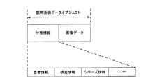
図2に、医用画像1枚の画像データに対応する医用画像データ(以下、医用画像データオブジェクトと称す。)のデータ構成を示す。医用画像データオブジェクトは、前述の通り、医用画像1枚の画像データと当該画像データに関する付帯情報とから成る。そして、付帯情報は、患者情報、検査情報及びシリーズ情報等を含む。 FIG. 2 shows a data structure of medical image data (hereinafter referred to as a medical image data object) corresponding to image data of one medical image. As described above, the medical image data object includes image data of one medical image and incidental information related to the image data. The incidental information includes patient information, examination information, series information, and the like.
モダリティ20は、医用画像データをPACS30に送信する。 The
PACS30は、医用画像データ、数値レポート情報を保存し、管理する医用画像管理装置である。PACS30は、読影端末40から送信される読影用データ取得要求に応じて、該当する医用画像データ、数値レポート情報を当該読影端末40に送信する。 The
PACS30は、モダリティ20から受信した医用画像データに含まれる画像データに対して自動計測処理を行い、医用画像に関する検査数値レポートの情報である数値レポート実データを生成する。そして、医用画像データに含まれる付帯情報等に基づいて、生成した数値レポート実データに関する各種情報(患者情報、検査情報及びシリーズ情報等)を有する付帯情報を生成する。そして、当該数値レポート実データに生成した付帯情報を付加し、DICOM規格に則った数値レポート情報を生成する。また、PACS30は、医用画像データと数値レポート情報とを各々の付帯情報を用いて関連付ける。そして、当該数値レポートを保存する。尚、自動計測処理、数値レポート実データについての詳細は後述する。 The
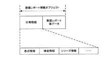
図3に、1回の自動計測処理における検査数値レポートの情報(数値レポート実データ)に対応する数値レポート情報(以下、数値レポート情報オブジェクトと称す。)のデータ構成を示す。数値レポート情報オブジェクトは、前述の通り、1回の自動計測処理における検査数値レポートの情報(数値レポート実データ)と当該数値レポート実データに関する付帯情報とから成る。そして、付帯情報は、患者情報、検査情報及びシリーズ情報等を含む。 FIG. 3 shows a data structure of numerical report information (hereinafter referred to as a numerical report information object) corresponding to inspection numerical report information (numerical report actual data) in one automatic measurement process. As described above, the numerical report information object includes inspection numerical report information (numerical report actual data) in one automatic measurement process and incidental information regarding the numerical report actual data. The incidental information includes patient information, examination information, series information, and the like.
読影端末40は、受信した医用画像データや数値レポート情報に基づいて、医用画像や当該医用画像に関する検査数値レポート等の再生表示を行う医用画像表示装置である。読影端末40は、読影用データ取得要求をPACS30に送信し、PACS30に保存されている医用画像データや数値レポート情報等を取得する。 The
[モダリティの機能的構成]
図4に、モダリティ20の機能構成を示す。図4に示すように、モダリティ20は、制御部21、操作部22、表示部23、通信部24、ROM(Read Only Memory)25、記憶部26、撮影部27等を備えて構成され、各部はバス28により接続されている。[Functional configuration of modality]
FIG. 4 shows a functional configuration of the
制御部21は、CPU(Central Processing Unit)、RAM(Random Access Memory)等から構成される。制御部21は、ROM25に記憶されている各種処理プログラムを読み出し、RAM内に形成されたワークエリアに展開し、当該プログラムに従ってモダリティ20の各部を統括的に制御する。 The
操作部22は、各種設定キー、操作指示キー等を備え、キー操作により入力された指示信号(操作信号)を制御部21に出力する。 The
表示部23は、LCD(Liquid Crystal Display)により構成され、制御部21から入力される表示信号の指示に従って、操作部22からの入力指示やデータ等を表示する。 The
通信部24は、LAN(Local Area Network)アダプタ、ルータ、TA(Terminal Adapter)等を備え、通信ネットワークNを介して接続されたRIS10、PACS30、読影端末40等の外部機器との間でデータの送受信を行う。 The
ROM25は、不揮発性の半導体メモリ等により構成され、制御部21で実行される各種処理プログラム、各種データ等を記憶する。これらの各種プログラムは、読み取り可能なプログラムコードの形態で格納され、制御部21は、当該プログラムコードに従った動作を逐次実行する。尚、ROM25は、マスクROM、PROMの何れの形態であっても良い。 The
記憶部26は、ハードディスク等の記憶装置であり、医用画像データ等を記憶する。 The
撮影部27は、撮影技師の操作部22からの指示により、患者を撮影して医用画像の画像データを生成する。 The
制御部21は、通信部24を介してRIS10から受信した検査オーダ情報に基づいて、撮影部27により生成された画像データに関する付帯情報を生成する。そして、制御部21は、当該画像データに付帯情報を付加して医用画像データを生成し、記憶部26に記憶させる。そして、制御部21は、記憶部26から医用画像データを読み出して、PACS30に送信する。 The
[PACSの機能的構成]
図5に、PACS30の機能構成を示す。図5に示すように、PACS30は、制御部31、操作部32、表示部33、通信部34、ROM35、記憶部36等を備えて構成され、各部はバス37により接続されている。[Functional structure of PACS]
FIG. 5 shows a functional configuration of the
制御部31は、CPU、RAM等から構成される。制御部31は、ROM35に記憶されている各種処理プログラムを読み出し、RAM内に形成されたワークエリアに展開し、当該プログラムに従ってPACS30の各部を統括的に制御する。 The
操作部32は、カーソルキー、数字入力キー、及び各種機能キー等を備えたキーボードと、マウスなどのポインティングデバイスを備えて構成され、キーボードに対するキー操作やマウス操作により入力された指示信号(操作信号)を制御部31に出力する。 The
表示部33は、LCD(Liquid Crystal Display)により構成され、制御部31から入力される表示信号の指示に従って、操作部32からの入力指示やデータ等を表示する。 The
通信部34は、LAN(Local Area Network)アダプタ、ルータ、TA(Terminal Adapter)等を備え、通信ネットワークNを介して接続されたRIS10、モダリティ20、読影端末40等の外部機器との間でデータの送受信を行う。 The
ROM35は、不揮発性の半導体メモリ等により構成され、制御部31で実行される各種処理プログラム、各種データ等を記憶する。これらの各種プログラムは、読み取り可能なプログラムコードの形態で格納され、制御部31は、当該プログラムコードに従った動作を逐次実行する。尚、ROM35は、マスクROM、PROMの何れの形態であっても良い。 The
記憶部36は、ハードディスク等の記憶装置であり、医用画像データ、数値レポート情報等を蓄積記憶する。また、記憶部36は、自動計測種類テーブル361を記憶する。自動計測種類テーブル361についての詳細は後述する。 The
制御部31は、通信部34を介してモダリティ20から受信した医用画像データを記憶部36に記憶させる。 The
また、制御部31は、モダリティ20から受信した医用画像データに含まれる画像データに対して自動計測処理を行い、数値レポート実データを生成する。そして、制御部31は、医用画像データに含まれる付帯情報等に基づいて、生成した数値レポート実データに関する付帯情報を生成する。 In addition, the
ここで、制御部31は、数値レポート実データに関する付帯情報を生成する際、当該数値レポート実データに関する付帯情報と、自動計測処理を行った画像データを含む医用画像データの付帯情報とを関連付ける。 Here, when generating the supplementary information regarding the numerical report actual data, the
具体的に、付帯情報の関連付けは、それぞれの付帯情報に含まれる患者情報中の各種情報(患者の受付番号、氏名、患者ID等)の値及び検査情報中の各種情報(検査ID、検査手技、検査部位、撮影方向等)の値を同値とすることにより行われる。 Specifically, the association of the incidental information is based on the values of various information (patient receipt number, name, patient ID, etc.) in the patient information included in each incidental information and various information (examination ID, examination technique) in the examination information. , Inspection site, imaging direction, and the like).
または、付帯情報の関連付けは、医用画像データの付帯情報に画像UIDを持たせ、更に数値レポート実データに関する付帯情報にも同様の画像UIDを持たせて、これらの画像UIDを同値とすることにより行われるとしてもよい。 Or, associating the supplementary information, the supplementary information of the medical image data has an image UID, and the supplementary information related to the numerical report actual data has the same image UID, and these image UIDs have the same value. It may be done.
そして、制御部31は、生成した付帯情報を数値レポート実データに付加して数値レポート情報を生成し、記憶部36に記憶させる。 Then, the
前記付帯情報の関連付けを行うことで、医用画像データと数値レポート情報とが関連付けられることになる。 By associating the incidental information, the medical image data and the numerical report information are associated with each other.
尚、医用画像データと数値レポート情報との関連付けは、1対1、複数対複数、複数対1、1対複数のように、どのように関連付けてもよい。 The medical image data and the numerical report information may be associated in any manner such as one-to-one, plural-to-multiple, plural-to-one, and one-to-multiple.
例えば、ある患者に対する同一検査内で撮影された複数枚の医用画像に対して自動計測処理を行い、複数対1に医用画像データと数値レポート情報とを関連付ける。 For example, automatic measurement processing is performed on a plurality of medical images photographed within the same examination for a certain patient, and medical image data and numerical report information are associated with a plurality of one-to-one.
また、ある患者に対する同一検査内で撮影された複数枚の医用画像に対して、複数の検査種類の自動計測処理を行い、複数対複数に医用画像データと数値レポート情報とを関連付けてもよい。 In addition, automatic measurement processing of a plurality of examination types may be performed on a plurality of medical images taken within the same examination for a certain patient, and medical image data and numerical report information may be associated with a plurality of pairs.
また、1枚の医用画像に対して自動計測処理を行い、1対1に医用画像データと数値レポート情報とを関連付けてもよい。 Alternatively, automatic measurement processing may be performed on one medical image, and medical image data and numerical report information may be associated one-on-one.
制御部31は、通信部34を介して読影端末40から読影用データ取得要求を受信すると、該当する医用画像データ、数値レポート情報を、通信部34を介して読影端末40に送信する。 When receiving the interpretation data acquisition request from the
ここで、読影用データ取得要求には、患者ID、検査IDが含まれる。制御部31は、通信部34を介して読影端末40から読影用データ取得要求を受信すると、当該読影用データ取得要求に含まれる患者ID、検査IDを読み出す。そして、制御部31は、当該読み出した患者ID及び検査IDと同じ値を持つ医用画像データを、記憶部36において検索する。また、同様に、当該読み出した患者ID及び検査IDと同じ値を持つ数値レポート情報を検索する。検索の結果、該当する医用画像データがあった場合、当該医用画像データを記憶部36から読み出し、通信部34を介して読影端末40に送信する。同様に、検索の結果、該当する数値レポート情報があった場合、当該数値レポート情報を読み出し、通信部34を介して読影端末40に送信する。 Here, the interpretation data acquisition request includes a patient ID and an examination ID. When receiving the interpretation data acquisition request from the
[自動計測処理]
ここで、制御部31が行う自動計測処理について説明する。自動計測処理とは、画像データを幾何学的に計測する処理である。例えば、自動計測処理の種類として、心胸比自動計測処理、コブ角度自動計測処理、内臓脂肪自動計測処理等がある。[Automatic measurement processing]
Here, the automatic measurement process performed by the
心胸比自動計測処理とは、パターンマッチングやエッジ探索等を用いて医用画像の各部を識別し、自動で胸郭と心臓の各幅を計測して、心胸比(心胸比=心臓幅/胸郭幅×100)を算出する処理である。心胸比は通常50%以下であるが、50%を超えると心肥大と診断される。 The cardiothoracic ratio automatic measurement process identifies each part of the medical image using pattern matching, edge search, etc., and automatically measures the width of the rib cage and the heart to calculate the cardiothoracic ratio (cardiothoracic ratio = heart width / thoracic width x 100). The cardiothoracic ratio is usually 50% or less, but if it exceeds 50%, cardiac hypertrophy is diagnosed.
コブ角度自動計測処理とは、パターンマッチングやエッジ探索等を用いて、医用画像の各部を識別し、脊椎において最も強く側弯を示している部位の上下端にある終椎(傾斜角の最も大きな椎体)の上位終椎の上面と下位終椎の下面のなす角、すなわち、コブ角度を自動で計測する処理である。一般的にコブ角度が25度を超えると要治療と診断される。 Cobb angle automatic measurement processing uses pattern matching, edge search, etc. to identify each part of the medical image, and the final vertebrae at the upper and lower ends of the part showing the scoliosis most strongly in the spine (the largest inclination angle) This is a process of automatically measuring the angle formed by the upper surface of the upper terminal vertebra and the lower surface of the lower terminal vertebra, that is, the bump angle. Generally, when the angle of the head exceeds 25 degrees, it is diagnosed as requiring treatment.
内臓脂肪面積自動計測処理とは、パターンマッチングやエッジ探索等を用いて、医用画像の各部や脂肪箇所を識別し、内臓脂肪の面積を計測する処理である。 The visceral fat area automatic measurement process is a process of measuring each visceral fat area by identifying each part or fat part of a medical image using pattern matching, edge search, or the like.
制御部31は、自動計測処理対象となる医用画像データの付帯情報を解析する。具体的に、付帯情報に含まれる検査情報の「手技」、「検査部位」、「撮影方向」、「体位」、付帯情報に含まれるシリーズ情報の「モダリティ種別」を読み出し、自動計測種類テーブル261を参照し、自動計測処理の種類(自動計測を行う検査の種類)を決定する。 The
ここで、図6に自動計測種類テーブル361のデータ構成を示す。図6に示すように、自動計測種類テーブル361は、「モダリティ種別」、「手技」、「検査部位」、「体位」、「撮影方向」、「自動計測処理」の項目から成る。「モダリティ種別」は、医用画像データの付帯情報に含まれるシリーズ情報の「モダリティ種別」に対応する項目である。また、「手技」、「検査部位」、「体位」、「撮影方向」は、それぞれ、医用画像データの付帯情報に含まれる検査情報の「手技」、「検査部位」、「体位」、「撮影方向」に対応する項目である。また、「自動計測処理」は、自動計測処理の種類を示す項目である。 Here, the data structure of the automatic measurement type table 361 is shown in FIG. As shown in FIG. 6, the automatic measurement type table 361 includes items of “modality type”, “procedure”, “examination part”, “body position”, “imaging direction”, and “automatic measurement process”. The “modality type” is an item corresponding to the “modality type” of the series information included in the incidental information of the medical image data. “Procedure”, “Examination site”, “Position”, and “Imaging direction” are the “Procedure”, “Examination site”, “Position”, and “Imaging” of the examination information included in the supplementary information of the medical image data, respectively. This item corresponds to “Direction”. “Automatic measurement processing” is an item indicating the type of automatic measurement processing.
例えば、自動計測種類テーブル361が図6のような値であり、制御部31が医用画像データの付帯情報から読み出した「モダリティ種別」、「手技」、「検査部位」、「体位」、「撮影方向」の値がそれぞれ、「CR」、「単純」、「胸部」、「立位」、「正面」である場合、制御部31は、自動計測処理として「心胸比自動計測処理」を選択する。 For example, the automatic measurement type table 361 has values as shown in FIG. 6, and the “Modality type”, “Procedure”, “Examination site”, “Position”, “Imaging” read from the supplementary information of the medical image data by the
図7に、数値レポート情報に含まれる数値レポート実データのデータ構成を示す。数値レポート実データは、「作成日」、「作成者」、「タイトル」、「見出し」、「値」の項目から成る。「作成日」は、数値レポート実データが生成された日にちを示す項目である。また、「作成者」は、数値レポート実データを生成した装置名を示す項目である。「タイトル」は、自動計測処理により生成された検査数値レポートの種類を示す項目であり、自動計測処理の種類に1対1に対応している。具体的に、心胸比自動計測処理、コブ角度自動計測処理、内臓脂肪面積自動計測処理に対応するタイトルは、それぞれ「心胸比計測レポート」、「コブ角度計測レポート」、「内臓脂肪面積計測レポート」となる。また、「見出し」は、自動計測処理により計測された検査数値の名称を示す項目である。また、「値」は、自動計測処理に計測された検査数値を示す項目である。 FIG. 7 shows the data structure of the numerical report actual data included in the numerical report information. Numerical report actual data includes items of “creation date”, “creator”, “title”, “heading”, and “value”. The “creation date” is an item indicating the date on which the actual numerical report data is generated. The “creator” is an item indicating the name of the device that generated the actual numerical report data. “Title” is an item indicating the type of the inspection numerical report generated by the automatic measurement process, and corresponds to the type of the automatic measurement process on a one-to-one basis. Specifically, the titles corresponding to the cardiothoracic ratio automatic measurement process, the galling angle automatic measurement process, and the visceral fat area automatic measurement process are “cardiothoracic ratio measurement report”, “cobb angle measurement report”, and “visceral fat area measurement report”, respectively. It becomes. “Heading” is an item indicating the name of the inspection numerical value measured by the automatic measurement process. The “value” is an item indicating an inspection numerical value measured in the automatic measurement process.
例えば、数値レポート実データの項目「作成日」、「作成者」、「タイトル」、「見出し」、「値」の値は、それぞれ「2007/7/7」、「CS―X」、「心胸比計測レポート」、「心胸比値」、「45%」となる。 For example, the values of the items “creation date”, “creator”, “title”, “heading”, and “value” of the actual numerical data data are “2007/7/7”, “CS-X”, “psycho-thoracic” Ratio measurement report ”,“ cardiothoracic ratio value ”, and“ 45% ”.
[読影端末の機能的構成]
図8に、読影端末40の機能構成を示す。図8に示すように、読影端末40は、制御部41、操作部42、表示部43、通信部44、ROM45、記憶部46等を備えて構成され、各部はバス47により接続されている。[Functional structure of interpretation terminal]
FIG. 8 shows a functional configuration of the
制御部41は、CPU、RAM等から構成される。制御部41は、ROM45に記憶されている各種処理プログラムを読み出し、RAM内に形成されたワークエリアに展開し、当該プログラムに従って読影端末40の各部を統括的に制御する。 The
操作部42は、カーソルキー、数字入力キー、及び各種機能キー等を備えたキーボードと、マウスなどのポインティングデバイスを備えて構成され、キーボードに対するキー操作やマウス操作により入力された指示信号を制御部41に出力する。 The
表示部43は、LCD等の高精細モニタにより構成され、制御部41から入力される表示データに基づいて各種画面を表示する。特に、表示部43は、読影医が読影を行う際に医用画像を表示する。 The
通信部44は、LAN(Local Area Network)アダプタ、ルータ、TA(Terminal Adapter)等を備え、通信ネットワークNを介して接続されたRIS10、モダリティ20、PACS30等の外部機器との間でデータの送受信を行う。 The
ROM45は、不揮発性の半導体メモリ等により構成され、制御部41で実行される各種処理プログラム、各種データ等を記憶する。これらの各種プログラムは、読み取り可能なプログラムコードの形態で格納され、制御部41は、当該プログラムコードに従った動作を逐次実行する。尚、ROM45は、マスクROM、PROMの何れの形態であっても良い。 The
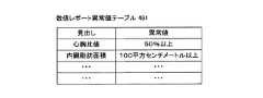
記憶部46は、ハードディスク等の記憶装置であり、各種データを記憶する。また、記憶部46は、数値レポート異常値テーブル461を記憶している。 The
図9に、数値レポート異常値テーブル461のデータ構成を示す。図9に示すように、数値レポート異常値テーブル461は、「見出し」、「異常値」の項目から成る。「見出し」は、数値レポート情報に含まれる数値レポート実データの「見出し」に対応する項目であり、自動計測処理により計測された検査数値の名称を示す。また、「異常値」は、自動計測処理により計測された検査数値の異常値又は範囲を示す項目である。例えば、心胸比値に対応する異常値は、「50パーセント以上」であり、内臓脂肪面積に対応する異常値は、「100平方センチメートル以上」である。 FIG. 9 shows the data structure of the numerical report abnormal value table 461. As shown in FIG. 9, the numerical report abnormal value table 461 includes items of “heading” and “abnormal value”. “Heading” is an item corresponding to “Heading” of actual numerical report data included in the numerical report information, and indicates the name of an inspection numerical value measured by automatic measurement processing. The “abnormal value” is an item indicating an abnormal value or a range of the inspection numerical value measured by the automatic measurement process. For example, the abnormal value corresponding to the cardiothoracic ratio value is “50 percent or more”, and the abnormal value corresponding to the visceral fat area is “100 square centimeters or more”.
尚、数値レポート異常値テーブル461のレコードの追加、各項目の値の変更は、操作部42におけるユーザ操作等により適宜変更可能である。 Note that the addition of a record in the numerical report abnormal value table 461 and the change of the value of each item can be appropriately changed by a user operation or the like in the
制御部41は、読影医による操作部42からの読影画面を表示させる旨の指示信号が入力されると、読影用データ取得要求を通信部44を介してPACS30に送信する。 When an instruction signal for displaying an interpretation screen from the
制御部41は、PACS30から医用画像データ、数値レポート情報を通信部44を介して受信すると、読影画面表示処理を行い、表示部43に読影画面を表示させる。読影医はこの読影画面を参照して読影を行う。 When receiving the medical image data and the numerical report information from the
[読影画面表示処理]
ここで、制御部41が行う読影画面表示処理について説明する。制御部41は、PACS30から受信した数値レポート情報に含まれる数値レポート実データの項目「見出し」と項目「値」の値を読み出す。[Interpretation screen display processing]
Here, the interpretation screen display process performed by the
制御部41は、数値レポート異常値テーブル461を参照して、該当する(「見出し」が一致する)レコードの「異常値」と数値レポート情報の「値」を比較し、数値レポート情報の「値」が異常値であるか否か(数値異常であるか否か)を判定する。 The
数値異常でない場合、制御部41は、読影画面の初期画面として通常読影画面431(図10参照)を表示部43に表示させる。また、数値異常である場合、制御部41は、読影画面の初期画面として注意喚起用読影画面432(図11参照)を表示部43に表示させる。 If the numerical value is not abnormal, the
例えば、図7のように心胸比自動計測処理の数値レポート情報の心胸比値が45%だった場合、図9に示す数値レポート異常値テーブル461によると、異常値70%以上に該当しないため、制御部41は、数値異常でないと判定し、通常読影画面431を表示する。 For example, when the cardiothoracic ratio value of the numerical report information of the cardiothoracic ratio automatic measurement process is 45% as shown in FIG. 7, according to the numerical report abnormal value table 461 shown in FIG. The
図10に、通常読影画面431の画面例を示す。図10に示すように、通常読影画面431には、医用画像と、医用画像に関する患者情報、検査情報等が表示される。また、当該医用画像の医用画像データに関連する数値レポート情報を示すボックスD1が閉じて表示される。ボックスD1が閉じた状態だと、数値レポート情報の「タイトル」がボックスD1上に表示される。また、ボックスD1が開いた状態だと、ボックスD1上に、数値レポート情報の「タイトル」、「見出し」、「値」の各値が表示される。ボックスD1はユーザ操作により開閉される。 FIG. 10 shows a screen example of the
図11に、注意喚起用読影画面432の画面例を示す。図11に示すように、注意喚起用読影画面432には、医用画像と、医用画像に関する患者情報、検査情報等が表示される。また、当該医用画像の医用画像データに関連する数値レポート情報を示すボックスD2が開いて表示される。ボックスD2が開いた情報だと、ボックスD2上に、数値レポート情報の「タイトル」、「見出し」、「値」の各値が表示される。また、ボックスD2が閉じた状態だと、数値レポート情報の「タイトル」がボックスD2上に表示される。ボックスD2はユーザ操作により開閉される。ボックスD2を始めから開いて表示することにより、読影医に対して、数値レポート情報の「値」が異常値であると警告する。 FIG. 11 shows a screen example of the
尚、制御部41がPACS30に送信した読影用データ取得要求に対して、該当する数値レポート情報がなく、医用画像データのみをPACS30から受信した場合、通常読影画面431のボックスD1、注意喚起用読影画面432のボックスD2は表示されない。 When there is no corresponding numerical report information and only the medical image data is received from the
尚、制御部41がPACS30に送信した読影用データ取得要求に対して、該当する数値レポート情報オブジェクトを複数受信した場合、通常読影画面431のボックスD1、注意喚起用読影画面432のボックスD2は複数表示される。 When a plurality of corresponding numerical report information objects are received in response to the interpretation data acquisition request transmitted from the
この場合、数値異常である数値レポート情報オブジェクトが一つもないと、通常読影像画面431が、数値異常である数値レポート情報オブジェクトが一つでもあると、注意喚起用読影画面432が表示される。更に、注意喚起用読影画面432において、数値異常である数レポート情報オブジェクトに対応するボックスD2のみが始めから開いて表示される。 In this case, if there is no numerical report information object that is numerically abnormal, the normal
[医用画像システムの動作]
次に、医用画像システム100における動作を説明する。図12は、モダリティ20、PACS30及び読影端末40において実行される処理を示すラダーチャートである。[Operation of medical imaging system]
Next, the operation in the
図12に示すように、モダリティ20は、患者を撮影し(ステップS1)、医用画像データを生成する(ステップS2)。そして、モダリティ20は、ステップS2において生成した医用画像データをPACS30に送信する(ステップS3)。PACS30は、モダリティ20から送信された医用画像データを保存する(ステップS4)。 As shown in FIG. 12, the
PACS30は、保存した医用画像データに含まれる画像データに対して自動計測処理を行い(ステップS5)、数値レポート情報を生成する(ステップS6)。この際、医用画像データと数値レポート情報とを付帯情報に基づいて関連付ける。そして生成した数値レポート情報を保存する(ステップS7)。 The
次に、読影端末40は、読影医のユーザ操作等に基づき、読影用データ取得要求をPACS30に送信する(ステップS8)。PACS30は、当該読影用データ取得要求を受信すると、該当する医用画像データを読影端末40に送信する(ステップS9)。また、該当する数値レポート情報を読影端末40に送信する(ステップS10)。 Next, the
そして、読影端末40は、PACS30から受信した医用画像データと数値レポート情報とに基づいて、読影画面表示処理を行う(ステップS11)。 The
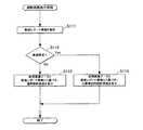
図13は、読影端末40が行う読影画面表示処理を示すフローチャートである。図13に示すように、読影端末40は、PACS30から受信した数値レポート情報を解析する(ステップS111)。そして、数値レポート異常値テーブル461を参照して数値レポート実データ中の項目「値」が異常値であるか否かを判定する(ステップS112)。 FIG. 13 is a flowchart showing an interpretation screen display process performed by the
判定の結果、異常値でない場合(ステップS112;No)、読影端末40は、医用画像データと数値レポート情報とに基づき、通常読影画面431を表示する(ステップS113)。また、判定の結果、異常値である場合(ステップS112;Yes)、読影端末40は、医用画像データと数値レポート情報とに基づき、注意喚起用読影画面432を表示する(ステップS114)。 As a result of the determination, if it is not an abnormal value (step S112; No), the
以上、本実施の形態によると、読影医による読影目的にかかわらず、PACS30は、医用画像データの付帯情報に基づいて自動計測処理の検査種類を決定し、決定した検査種類の自動計測処理を行って数値レポート情報を生成する。そして、読影端末40は、生成された数値レポート情報の値が異常値の場合、その旨の警告(注意喚起用読影画面432)を表示する。 As described above, according to the present embodiment, regardless of the purpose of interpretation by the interpreting physician, the
そのため、読影医による読影目的外(検査目的外)の疾患を見落とすことを防ぐことができる。具体的には、CR装置で撮影された胸部の医用画像を、心不全の予防のための心胸比の検査の目的以外で読影する場合でも、心胸比の異常を見落とすことを防ぐことができる。 Therefore, it is possible to prevent oversight of diseases that are not intended for interpretation by the interpreting physician (not intended for examination). Specifically, it is possible to prevent an abnormality in the cardiothoracic ratio from being overlooked even when the medical image of the chest imaged by the CR device is interpreted for purposes other than the purpose of examining the cardiothoracic ratio for the prevention of heart failure.
更に、数値レポート情報と医用画像データとが関連付けられているので、読影医は、異常値を示す医用画像を特定できる。 Furthermore, since the numerical report information and the medical image data are associated with each other, the interpretation doctor can specify a medical image indicating an abnormal value.
また、読影目的外の検査数値レポートを、アノテーションとして画像データに重ねることなく画像とは別に表示するため、読影目的外の検査数値レポートが医用画像上に表示されることがない。よって、読影医による医用画像の読影の妨げにならない。 Further, since the examination numerical report other than the interpretation purpose is displayed as an annotation separately from the image without being superimposed on the image data, the examination numerical report other than the interpretation purpose is not displayed on the medical image. Therefore, the interpretation of the medical image by the interpretation doctor is not hindered.
10 RIS
20 モダリティ
21 制御部
22 操作部
23 表示部
24 通信部
25 ROM
26 記憶部
27 撮影部
28 バス
30 PACS
31 制御部
32 操作部
33 表示部
34 通信部
35 ROM
36 記憶部
37 バス
40 読影端末
41 制御部
42 操作部
43 表示部
44 通信部
45 ROM
46 記憶部
47 バス
100 医用画像システム
361 自動計測種類テーブル
431 通常読影画面
432 注意喚起用読影画面
461 数値レポート異常値テーブル
N 通信ネットワーク10 RIS
20
26
31
36
46
Claims (9)
Translated fromJapanese前記医用画像管理装置は、
患者の検査部位の撮影画像のうち検査目的外の疾患に関する検査数値を自動計測しその検査数値レポートを生成して、前記撮影画像と前記検査数値レポートとを互いに関連付け、且つそれぞれ独立したデータで前記通信手段に送信させる制御手段を備え、
前記医用画像表示装置は、
前記通信手段を介して受信した撮影画像及び検査数値レポートを表示画面上に表示させる表示制御手段を備える、
医用画像システム。A medical image management device and a medical image display device each having communication means are connected to each other via a communication network so that they can communicate with each other.
The medical image management apparatus includes:
A test numerical value related to a disease other than the test purpose is automatically measured among the captured images of the patient's test site, and a test numerical report is generated. The captured image and the test numerical report are associated with each other, and the independent data Comprising control means for transmitting to the communication means;
The medical image display device includes:
Display control means for displaying the captured image and the inspection numerical report received via the communication means on a display screen;
Medical imaging system.
請求項1に記載の医用画像システム。The control means associates the photographed image and the examination numerical report based on incidental information attached to each of the images.
The medical image system according to claim 1.
請求項1又は2に記載の医用画像システム。The control means determines the type of inspection for automatic measurement based on the shooting conditions of the shot image.
The medical image system according to claim 1 or 2.
請求項1〜3の何れか一項に記載の医用画像システム。If the inspection numerical value is an abnormal value, the display control means displays a warning to that effect on the display screen.
The medical image system according to any one of claims 1 to 3.
患者の検査部位の撮影画像のうち検査目的外の疾患に関する検査数値を自動計測しその検査数値レポートを生成して、前記撮影画像と前記検査数値レポートとを互いに関連付け、且つそれぞれ独立したデータで前記通信手段に送信させる制御手段と、
を備える医用画像管理装置。A communication means for transmitting / receiving data to / from an external device;
A test numerical value related to a disease other than the test purpose is automatically measured among the captured images of the patient's test site, and a test numerical report is generated. The captured image and the test numerical report are associated with each other, and the independent data Control means for transmitting to the communication means;
A medical image management apparatus comprising:
請求項5に記載の医用画像管理装置。The control means associates the photographed image and the examination numerical report based on incidental information attached to each of the images.
The medical image management apparatus according to claim 5.
請求項5又は6に記載の医用画像管理装置。The control means determines the type of inspection for automatic measurement based on the shooting conditions of the shot image.
The medical image management apparatus according to claim 5 or 6.
外部機器との間でデータの送受信を行う通信手段、
患者の検査部位の撮影画像のうち検査目的外の疾患に関する検査数値を自動計測しその検査数値レポートを生成して、前記撮影画像と前記検査数値レポートとを互いに関連付け、且つそれぞれ独立したデータで前記通信手段に送信させる制御手段、
として機能させるためのプログラム。Computer
A communication means for transmitting / receiving data to / from an external device,
A test numerical value related to a disease other than the test purpose is automatically measured among the captured images of the patient's test site, and a test numerical report is generated. The captured image and the test numerical report are associated with each other, and the independent data Control means for transmitting to the communication means;
Program to function as.
Priority Applications (2)
| Application Number | Priority Date | Filing Date | Title |
|---|---|---|---|
| JP2007235559AJP2010268821A (en) | 2007-09-11 | 2007-09-11 | Medical image system, medical image management device, data processing method, and program |
| PCT/JP2008/065667WO2009034868A1 (en) | 2007-09-11 | 2008-09-01 | Medical image system, medical image management device, data processing method, and program |
Applications Claiming Priority (1)
| Application Number | Priority Date | Filing Date | Title |
|---|---|---|---|
| JP2007235559AJP2010268821A (en) | 2007-09-11 | 2007-09-11 | Medical image system, medical image management device, data processing method, and program |
Publications (1)
| Publication Number | Publication Date |
|---|---|
| JP2010268821Atrue JP2010268821A (en) | 2010-12-02 |
Family
ID=40451877
Family Applications (1)
| Application Number | Title | Priority Date | Filing Date |
|---|---|---|---|
| JP2007235559APendingJP2010268821A (en) | 2007-09-11 | 2007-09-11 | Medical image system, medical image management device, data processing method, and program |
Country Status (2)
| Country | Link |
|---|---|
| JP (1) | JP2010268821A (en) |
| WO (1) | WO2009034868A1 (en) |
Cited By (1)
| Publication number | Priority date | Publication date | Assignee | Title |
|---|---|---|---|---|
| JP2013180186A (en)* | 2012-03-05 | 2013-09-12 | Toshiba Corp | Medical report preparation system |
Families Citing this family (2)
| Publication number | Priority date | Publication date | Assignee | Title |
|---|---|---|---|---|
| JPWO2010109999A1 (en)* | 2009-03-26 | 2012-09-27 | コニカミノルタエムジー株式会社 | Report generation management device and program |
| JP7313888B2 (en)* | 2019-04-24 | 2023-07-25 | キヤノンメディカルシステムズ株式会社 | MEDICAL INFORMATION PROCESSING APPARATUS, MEDICAL INFORMATION PROGRAM AND METHOD |
Family Cites Families (1)
| Publication number | Priority date | Publication date | Assignee | Title |
|---|---|---|---|---|
| JP3083606B2 (en)* | 1990-11-22 | 2000-09-04 | 株式会社東芝 | Medical diagnosis support system |
- 2007
- 2007-09-11JPJP2007235559Apatent/JP2010268821A/enactivePending
- 2008
- 2008-09-01WOPCT/JP2008/065667patent/WO2009034868A1/enactiveApplication Filing
Cited By (1)
| Publication number | Priority date | Publication date | Assignee | Title |
|---|---|---|---|---|
| JP2013180186A (en)* | 2012-03-05 | 2013-09-12 | Toshiba Corp | Medical report preparation system |
Also Published As
| Publication number | Publication date |
|---|---|
| WO2009034868A1 (en) | 2009-03-19 |
Similar Documents
| Publication | Publication Date | Title |
|---|---|---|
| US20210158531A1 (en) | Patient Management Based On Anatomic Measurements | |
| US8934687B2 (en) | Image processing device, method and program including processing of tomographic images | |
| US20130216112A1 (en) | Structured, image-assisted finding generation | |
| WO2011083607A1 (en) | Medical-use information processing device and program | |
| JP2007307290A (en) | Medical image reading system | |
| CN110111876B (en) | Information processing apparatus and information processing method | |
| KR101050769B1 (en) | Medical Image Processing System and Processing Method | |
| US20090196479A1 (en) | Method and apparatus for computer-aided diagnosis filtered prioritized work item list | |
| US20200082931A1 (en) | Diagnostic support apparatus | |
| JP2010268821A (en) | Medical image system, medical image management device, data processing method, and program | |
| JP6353382B2 (en) | Feature quantity management device, its operating method and program, and feature quantity management system | |
| WO2008038581A1 (en) | Image compressing method, image compressing device, and medical network system | |
| JP2011128661A (en) | Regional medical cooperation system, registration terminal, and program | |
| JP2009066060A (en) | Medical image system, finding report generator, finding report generation method, and program | |
| JP2010268820A (en) | Medical image system, medical photographing device, data processing method, and program | |
| JP2010284175A (en) | Medical image system, data processor, data processing method, and program | |
| JP5605246B2 (en) | Abnormal shadow candidate detection system, server device, and program | |
| JP2010057726A (en) | Diagnostic imaging support system | |
| JP7505428B2 (en) | Medical radiation exposure dose management device and program | |
| JP2012130369A (en) | Image inspection device and image inspection system | |
| US20230223138A1 (en) | Medical information processing system, medical information processing method, and storage medium | |
| JP7428055B2 (en) | Diagnostic support system, diagnostic support device and program | |
| JP2009146340A (en) | Medical image system, examination order generation device and program | |
| JP2011189091A (en) | Image inspection device and image inspection system | |
| JP2009247431A (en) | Medical image system, image inspection apparatus and program |