JP2010207612A - Device for vulnerable plaque detection - Google Patents
Device for vulnerable plaque detectionDownload PDFInfo
- Publication number
- JP2010207612A JP2010207612AJP2010120418AJP2010120418AJP2010207612AJP 2010207612 AJP2010207612 AJP 2010207612AJP 2010120418 AJP2010120418 AJP 2010120418AJP 2010120418 AJP2010120418 AJP 2010120418AJP 2010207612 AJP2010207612 AJP 2010207612A
- Authority
- JP
- Japan
- Prior art keywords
- optical
- intravascular probe
- optical waveguide
- ultrasonic transducer
- intravascular
- Prior art date
- Legal status (The legal status is an assumption and is not a legal conclusion. Google has not performed a legal analysis and makes no representation as to the accuracy of the status listed.)
- Granted
Links
- 238000001514detection methodMethods0.000titledescription7
- 230000003287optical effectEffects0.000claimsabstractdescription118
- 239000000523sampleSubstances0.000claimsabstractdescription81
- 230000005855radiationEffects0.000claimsabstractdescription18
- 238000004891communicationMethods0.000claimsabstractdescription15
- 239000013307optical fiberSubstances0.000claimsdescription14
- 239000000463materialSubstances0.000claimsdescription6
- 230000001902propagating effectEffects0.000claims1
- 239000000835fiberSubstances0.000description16
- 210000001367arteryAnatomy0.000description9
- 210000004204blood vesselAnatomy0.000description8
- 238000002608intravascular ultrasoundMethods0.000description6
- 238000000034methodMethods0.000description6
- 150000002632lipidsChemical class0.000description5
- 239000008280bloodSubstances0.000description4
- 210000004369bloodAnatomy0.000description4
- 230000008878couplingEffects0.000description4
- 238000010168coupling processMethods0.000description4
- 238000005859coupling reactionMethods0.000description4
- 239000000203mixtureSubstances0.000description4
- 238000004566IR spectroscopyMethods0.000description3
- 238000003780insertionMethods0.000description3
- 230000037431insertionEffects0.000description3
- 230000003595spectral effectEffects0.000description3
- 238000002604ultrasonographyMethods0.000description3
- 239000002775capsuleSubstances0.000description2
- 230000007850degenerationEffects0.000description2
- 208000037265diseases, disorders, signs and symptomsDiseases0.000description2
- 208000035475disorderDiseases0.000description2
- 230000000877morphologic effectEffects0.000description2
- 239000000126substanceSubstances0.000description2
- 201000001320AtherosclerosisDiseases0.000description1
- FAPWRFPIFSIZLT-UHFFFAOYSA-MSodium chlorideChemical compound[Na+].[Cl-]FAPWRFPIFSIZLT-UHFFFAOYSA-M0.000description1
- 208000007536ThrombosisDiseases0.000description1
- 206010047163VasospasmDiseases0.000description1
- 230000001154acute effectEffects0.000description1
- 238000004458analytical methodMethods0.000description1
- 238000005452bendingMethods0.000description1
- 230000017531blood circulationEffects0.000description1
- 230000007423decreaseEffects0.000description1
- 230000001419dependent effectEffects0.000description1
- 238000003745diagnosisMethods0.000description1
- 239000012530fluidSubstances0.000description1
- 238000012844infrared spectroscopy analysisMethods0.000description1
- 208000028867ischemiaDiseases0.000description1
- 210000002540macrophageAnatomy0.000description1
- 230000014759maintenance of locationEffects0.000description1
- 238000012986modificationMethods0.000description1
- 230000004048modificationEffects0.000description1
- 238000007491morphometric analysisMethods0.000description1
- 208000010125myocardial infarctionDiseases0.000description1
- 229920000642polymerPolymers0.000description1
- 238000001055reflectance spectroscopyMethods0.000description1
- 239000011780sodium chlorideSubstances0.000description1
- 238000004611spectroscopical analysisMethods0.000description1
- 238000001228spectrumMethods0.000description1
- 238000012360testing methodMethods0.000description1
- 230000001225therapeutic effectEffects0.000description1
- 230000000472traumatic effectEffects0.000description1
- 230000002792vascularEffects0.000description1
- 208000019553vascular diseaseDiseases0.000description1
- 210000005166vasculatureAnatomy0.000description1
Images


Classifications
- A—HUMAN NECESSITIES
- A61—MEDICAL OR VETERINARY SCIENCE; HYGIENE
- A61B—DIAGNOSIS; SURGERY; IDENTIFICATION
- A61B5/00—Measuring for diagnostic purposes; Identification of persons
- A61B5/02—Detecting, measuring or recording for evaluating the cardiovascular system, e.g. pulse, heart rate, blood pressure or blood flow
- A61B5/02007—Evaluating blood vessel condition, e.g. elasticity, compliance
- A—HUMAN NECESSITIES
- A61—MEDICAL OR VETERINARY SCIENCE; HYGIENE
- A61B—DIAGNOSIS; SURGERY; IDENTIFICATION
- A61B5/00—Measuring for diagnostic purposes; Identification of persons
- A61B5/0059—Measuring for diagnostic purposes; Identification of persons using light, e.g. diagnosis by transillumination, diascopy, fluorescence
- A61B5/0082—Measuring for diagnostic purposes; Identification of persons using light, e.g. diagnosis by transillumination, diascopy, fluorescence adapted for particular medical purposes
- A61B5/0084—Measuring for diagnostic purposes; Identification of persons using light, e.g. diagnosis by transillumination, diascopy, fluorescence adapted for particular medical purposes for introduction into the body, e.g. by catheters
- A61B5/0086—Measuring for diagnostic purposes; Identification of persons using light, e.g. diagnosis by transillumination, diascopy, fluorescence adapted for particular medical purposes for introduction into the body, e.g. by catheters using infrared radiation
- A—HUMAN NECESSITIES
- A61—MEDICAL OR VETERINARY SCIENCE; HYGIENE
- A61B—DIAGNOSIS; SURGERY; IDENTIFICATION
- A61B8/00—Diagnosis using ultrasonic, sonic or infrasonic waves
- A61B8/12—Diagnosis using ultrasonic, sonic or infrasonic waves in body cavities or body tracts, e.g. by using catheters
- A—HUMAN NECESSITIES
- A61—MEDICAL OR VETERINARY SCIENCE; HYGIENE
- A61B—DIAGNOSIS; SURGERY; IDENTIFICATION
- A61B8/00—Diagnosis using ultrasonic, sonic or infrasonic waves
- A61B8/44—Constructional features of the ultrasonic, sonic or infrasonic diagnostic device
- A61B8/4444—Constructional features of the ultrasonic, sonic or infrasonic diagnostic device related to the probe
- A61B8/445—Details of catheter construction
- G—PHYSICS
- G01—MEASURING; TESTING
- G01S—RADIO DIRECTION-FINDING; RADIO NAVIGATION; DETERMINING DISTANCE OR VELOCITY BY USE OF RADIO WAVES; LOCATING OR PRESENCE-DETECTING BY USE OF THE REFLECTION OR RERADIATION OF RADIO WAVES; ANALOGOUS ARRANGEMENTS USING OTHER WAVES
- G01S15/00—Systems using the reflection or reradiation of acoustic waves, e.g. sonar systems
- G01S15/88—Sonar systems specially adapted for specific applications
- G01S15/89—Sonar systems specially adapted for specific applications for mapping or imaging
- G01S15/8906—Short-range imaging systems; Acoustic microscope systems using pulse-echo techniques
- G01S15/899—Combination of imaging systems with ancillary equipment
- A—HUMAN NECESSITIES
- A61—MEDICAL OR VETERINARY SCIENCE; HYGIENE
- A61B—DIAGNOSIS; SURGERY; IDENTIFICATION
- A61B5/00—Measuring for diagnostic purposes; Identification of persons
- A61B5/0059—Measuring for diagnostic purposes; Identification of persons using light, e.g. diagnosis by transillumination, diascopy, fluorescence
- A61B5/0075—Measuring for diagnostic purposes; Identification of persons using light, e.g. diagnosis by transillumination, diascopy, fluorescence by spectroscopy, i.e. measuring spectra, e.g. Raman spectroscopy, infrared absorption spectroscopy
Landscapes
- Health & Medical Sciences (AREA)
- Life Sciences & Earth Sciences (AREA)
- Physics & Mathematics (AREA)
- Engineering & Computer Science (AREA)
- Animal Behavior & Ethology (AREA)
- Veterinary Medicine (AREA)
- Biophysics (AREA)
- Pathology (AREA)
- Public Health (AREA)
- Biomedical Technology (AREA)
- Heart & Thoracic Surgery (AREA)
- Medical Informatics (AREA)
- Molecular Biology (AREA)
- Surgery (AREA)
- General Health & Medical Sciences (AREA)
- Radar, Positioning & Navigation (AREA)
- Remote Sensing (AREA)
- Nuclear Medicine, Radiotherapy & Molecular Imaging (AREA)
- Radiology & Medical Imaging (AREA)
- Acoustics & Sound (AREA)
- Vascular Medicine (AREA)
- Physiology (AREA)
- Cardiology (AREA)
- Computer Networks & Wireless Communication (AREA)
- General Physics & Mathematics (AREA)
- Ultra Sonic Daignosis Equipment (AREA)
- Measurement Of The Respiration, Hearing Ability, Form, And Blood Characteristics Of Living Organisms (AREA)
- Investigating Or Analysing Materials By Optical Means (AREA)
- Measuring Pulse, Heart Rate, Blood Pressure Or Blood Flow (AREA)
- Endoscopes (AREA)
Abstract
Translated fromJapaneseDescription
Translated fromJapanese本発明は管腔を診断するための装置に関し、特に、不安定プラークを検出するための装置に関する。 The present invention relates to an apparatus for diagnosing a lumen, and more particularly to an apparatus for detecting vulnerable plaque.
アテローム性動脈硬化症は、血管壁の変性を特徴とする血管疾患である。こうした変性は、患部血管の離散部位すなわちポケットで発生するとプラークと呼ばれる。ある種のプラークは、脳卒中又は心筋梗塞などの急性イベントに関係している。こうしたプラークは「不安定プラーク」と呼ばれる。不安定プラークは、典型的には、薄い線維性被膜により血液から分離した脂質を保持した貯留を含んでいる。線維性被膜は、管腔内の圧力上昇や血管痙攣に反応して破壊され、プラークの内部が血液流に曝されることがある。結果として生じる血栓は、虚血や塞栓の離脱に至ることがある。 Atherosclerosis is a vascular disease characterized by degeneration of the vascular wall. Such degeneration is called plaque when it occurs at discrete sites or pockets of the affected blood vessel. Certain types of plaques are associated with acute events such as stroke or myocardial infarction. Such plaques are called “unstable plaques”. Vulnerable plaque typically includes a reservoir that retains lipids separated from blood by a thin fibrous cap. Fibrous capsules can be destroyed in response to increased pressure in the lumen and vasospasm, and the plaque interior can be exposed to blood flow. The resulting thrombus can lead to ischemia and emboli withdrawal.
不安定プラークの位置を特定する一つの方法としては、赤外線で動脈壁を透視するものがある。これを行うためには、カテーテルを動脈の管腔に挿入する。こうしたカテーテルは、動脈壁上のある箇所を赤外線で照らすための送出ファイバを含んでいる。この光の一部は血液及び動脈壁を通過して、動脈壁内部の構造体に当たって散乱し、管腔に再び入る。こうして再び入った光はカテーテル内の収集ファイバにより収集可能であり、分光分析に供せられる。この種の拡散反射分光法を用いて、脂質内容などの不安定プラークに関連すると考えられる重要な成分を含んだ、動脈組織の化学組成を特定できる。 One method for identifying the location of unstable plaque is to see the artery wall through infrared rays. To do this, a catheter is inserted into the lumen of the artery. Such catheters include a delivery fiber for illuminating a location on the arterial wall with infrared light. Some of this light passes through the blood and the arterial wall, strikes the structures inside the arterial wall, scatters, and reenters the lumen. The light that reenters in this way can be collected by the collecting fiber in the catheter and is subjected to spectroscopic analysis. This type of diffuse reflectance spectroscopy can be used to identify the chemical composition of arterial tissue, including important components that are thought to be associated with vulnerable plaque, such as lipid content.
不安定プラークの位置を特定する別の方法としては、管腔を囲む動脈組織の形状を検出するため血管内超音波診断法(IVUS)を用いるものがある。この方法を行うには、カテーテルを動脈の管腔に挿入する。カテーテルは、超音波エネルギーを動脈壁に向けて送出する超音波トランスデューサを含んでいる。反射した超音波エネルギーは超音波トランスデューサにより受信され、動脈組織の形状を測量するのに利用される。動脈組織の形態に関するこの測量図は、不安定プラークに関連した線維性被膜を検出するのに利用できる。 Another method for locating vulnerable plaque is to use intravascular ultrasound (IVUS) to detect the shape of arterial tissue surrounding the lumen. To perform this method, a catheter is inserted into the lumen of the artery. The catheter includes an ultrasonic transducer that delivers ultrasonic energy toward the arterial wall. The reflected ultrasonic energy is received by the ultrasonic transducer and used to survey the shape of the arterial tissue. This survey map of arterial tissue morphology can be used to detect the fibrous capsule associated with vulnerable plaque.
本発明は、同一プローブ内で、赤外分光法と血管内超音波診断法という2つの検出様式を組み合わせると、不安定プラークなどの障害を検出するプローブの能力が向上するという認識に基づいている。 The present invention is based on the recognition that combining the two detection modes of infrared spectroscopy and intravascular ultrasound in the same probe improves the probe's ability to detect disorders such as unstable plaque. .
一様態では、本発明は、遠位部と近位部とを備えた外装を具備する脈管内プローブを含む。前記脈管内プローブは、前記外装に沿って延伸する第1光学導波路であって、前記遠位部と前記近位部との間で光放射を伝達するよう構成された第1光学導波路と、前記遠位部に設けられると共に、前記第1光学導波路と光学連通した第1ビーム方向転換器とを含む。更に、前記脈管内プローブは、前記第1光学導波路から光放射を受け取るよう構成された光学検出器と、前記遠位部に設けられた超音波トランスデューサとを含む。前記超音波トランスデューサは、超音波エネルギーを前記脈管内プローブと伝搬媒体との間に結合するよう構成されている。電線が前記外装に沿って延伸し、前記超音波トランスデューサと電気連通している。 In one aspect, the present invention includes an intravascular probe comprising a sheath with a distal portion and a proximal portion. The intravascular probe is a first optical waveguide extending along the sheath, the first optical waveguide configured to transmit light radiation between the distal portion and the proximal portion; A first beam redirector provided at the distal portion and in optical communication with the first optical waveguide. Further, the intravascular probe includes an optical detector configured to receive light radiation from the first optical waveguide, and an ultrasonic transducer provided at the distal portion. The ultrasonic transducer is configured to couple ultrasonic energy between the intravascular probe and a propagation medium. An electrical wire extends along the exterior and is in electrical communication with the ultrasonic transducer.
実施形態によっては、前記脈管内プローブは、前記外装に沿って延伸する第2光学導波路を含む。前記第2光学導波路は、前記遠位部と前記近位部との間に光放射を伝達するよう構成されている。又、この種の実施形態は、前記遠位部に設けられると共に、前記第2光学導波路と光学連通した第2ビーム方向転換器を含む。 In some embodiments, the intravascular probe includes a second optical waveguide that extends along the sheath. The second optical waveguide is configured to transmit light radiation between the distal portion and the proximal portion. This type of embodiment also includes a second beam redirector provided at the distal portion and in optical communication with the second optical waveguide.
幾つかの実施形態では、前記第2ビーム方向転換器は、前記第2光学導波路から該方向転換器に入射する軸方向ビームの光放射を、半径方向成分を備えた方向に沿って伝搬するビームに方向転換するよう構成されている。 In some embodiments, the second beam redirector propagates axial beam light radiation incident on the redirector from the second optical waveguide along a direction with a radial component. It is configured to turn to a beam.
別の実施形態では、前記脈管内プローブは、光放射を前記第2光学導波路に結合するよう構成された光学源を含む。 In another embodiment, the intravascular probe includes an optical source configured to couple optical radiation to the second optical waveguide.
別の様態では、本発明は、遠位部と近位部とを備えた外装を具備した脈管内プローブを含む。前記脈管内プローブは、前記外装に沿って延伸する第1光学導波路であって、前記遠位部と前記近位部との間で光放射を伝達するよう構成された第1光学導波路と、前記遠位部に設けられると共に、前記第1光学導波路と光学連通した第1ビーム方向転換器とを含む。前記脈管内プローブは、前記外装に沿って延伸する第2光学導波路であって、前記遠位部と前記近位部との間で光放射を伝達するよう構成された第2光学導波路と、前記遠位部に設けられると共に、前記第2光学導波路と光学連通した第2ビーム方向転換器とを更に含む。前記脈管内プローブは、前記遠位端に設けられた超音波トランスデューサも含む。前記超音波トランスデューサは、超音波エネルギーを前記脈管内プローブと伝搬媒体との間に結合するよう構成されている。電線が前記外装に沿って延伸し、前記超音波トランスデューサと電気連通している。光学導波路の一例は光ファイバである。 In another aspect, the invention includes an intravascular probe with a sheath having a distal portion and a proximal portion. The intravascular probe is a first optical waveguide extending along the sheath, the first optical waveguide configured to transmit light radiation between the distal portion and the proximal portion; A first beam redirector provided at the distal portion and in optical communication with the first optical waveguide. The intravascular probe is a second optical waveguide extending along the sheath, the second optical waveguide configured to transmit light radiation between the distal portion and the proximal portion; And a second beam redirector provided at the distal portion and in optical communication with the second optical waveguide. The intravascular probe also includes an ultrasonic transducer provided at the distal end. The ultrasonic transducer is configured to couple ultrasonic energy between the intravascular probe and a propagation medium. An electrical wire extends along the exterior and is in electrical communication with the ultrasonic transducer. An example of the optical waveguide is an optical fiber.
一実施形態では、前記脈管内プローブは、光放射を前記第1光学導波路から受け取るよう構成された光学検出器を更に含む。 In one embodiment, the intravascular probe further includes an optical detector configured to receive light radiation from the first optical waveguide.
別の実施形態では、前記脈管内プローブは、光放射を前記第1光学導波路に結合するよう構成された光学源を含む。前記光学源は赤外線を放射するよう構成できる。 In another embodiment, the intravascular probe includes an optical source configured to couple optical radiation into the first optical waveguide. The optical source can be configured to emit infrared radiation.
一実施形態では、前記第1ビーム方向転換器は光学反射体を含む。しかし、前記第1ビーム方向転換器は、プリズム又は前記第1光学導波路の遠位先端に設けた屈曲部を更に含むこともできる。 In one embodiment, the first beam redirector includes an optical reflector. However, the first beam redirector may further include a bent portion provided at a distal end of the prism or the first optical waveguide.
別の実施形態では、前記超音波トランスデューサは圧電トランスデューサを含む。 In another embodiment, the ultrasonic transducer includes a piezoelectric transducer.
別の実施形態では、前記外装は、赤外線に対して透明な材料を含む。 In another embodiment, the sheath includes a material that is transparent to infrared.
幾つかの実施形態では、前記第1ビーム方向転換器は前記超音波トランスデューサに強固に接続されている。他の実施形態では、前記第1ビーム方向転換器は前記超音波トランスデューサに柔軟に接続されている。 In some embodiments, the first beam redirector is rigidly connected to the ultrasonic transducer. In another embodiment, the first beam redirector is flexibly connected to the ultrasonic transducer.
幾つかの実施形態では、前記第1ビーム方向転換器は、前記外装の長手方向軸に対する第1軸方向位置から光を放射するよう構成される一方、前記超音波トランスデューサは、前記第1軸方向位置から超音波エネルギーを放射するよう構成されている。他の実施形態では、前記第1ビーム方向転換器は、前記外装の長手方向軸に対する第1軸方向位置から光を放射するよう構成される一方、前記超音波トランスデューサは、前記第1軸方向位置とは異なる第2軸方向位置から超音波エネルギーを放射するよう構成されている。 In some embodiments, the first beam redirector is configured to emit light from a first axial position relative to the longitudinal axis of the sheath, while the ultrasonic transducer is in the first axial direction. It is configured to emit ultrasonic energy from a location. In another embodiment, the first beam redirector is configured to emit light from a first axial position relative to a longitudinal axis of the sheath, while the ultrasonic transducer is configured to emit the first axial position. It is comprised so that ultrasonic energy may be radiated from the 2nd axial direction position different from.
幾つかの実施形態では、前記脈管内プローブは、前記第1光学導波路と前記電線とを囲む回転可能ケーブルを含み、該回転可能ケーブルは、前記第1ビーム方向転換器と前記超音波トランスデューサとを同軸で回転させるよう構成されている。他の実施形態では、前記脈管内プローブは、前記外装の長手方向軸周りに環状に配置された複数のビーム方向転換器と、前記複数のビーム方向転換器と光学連通した複数の光学導波路と、前記長手方向軸周りに環状に配置された複数の超音波トランスデューサとを含む。 In some embodiments, the intravascular probe includes a rotatable cable that surrounds the first optical waveguide and the wire, the rotatable cable including the first beam redirector, the ultrasonic transducer, and the like. Are configured to rotate coaxially. In another embodiment, the intravascular probe includes a plurality of beam redirectors arranged in an annular shape around a longitudinal axis of the sheath, and a plurality of optical waveguides in optical communication with the plurality of beam redirectors. And a plurality of ultrasonic transducers arranged in a ring around the longitudinal axis.
本明細書では、「赤外」という語句は、赤外、近赤外、中間赤外、遠赤外、又は極端遠赤外を意味する。 As used herein, the phrase “infrared” means infrared, near infrared, mid-infrared, far infrared, or extreme far infrared.
他に特に定義していない限り、本明細書で用いる科学技術用語は、本発明が属する分野の通常の技能を備えた当業者が一般に理解する意味と同一である。本明細書に記載したものと類似又は同等の方法及び材料を、本発明の実施又は試験に用いることができるが、適切な方法及び材料は後述する。本明細書で言及するすべての刊行物、特許出願、特許、及び他の引用文献は、その全体を引用して援用する。矛盾が生じた場合は、定義も含めて本明細書が優先する。更に、これら材料、方法、及び例は、例示的なものであって限定する意図はない。 Unless defined otherwise, scientific and technical terms used herein have the same meaning as commonly understood by one of ordinary skill in the art to which this invention belongs. Although methods and materials similar or equivalent to those described herein can be used in the practice or testing of the present invention, suitable methods and materials are described below. All publications, patent applications, patents, and other references mentioned herein are incorporated by reference in their entirety. In case of conflict, the present specification, including definitions, will control. In addition, the materials, methods, and examples are illustrative and not intended to be limiting.
本発明のその他の特徴及び利点は、次の詳細な説明及び特許請求の範囲から明らかとなるはずである。 Other features and advantages of the invention will be apparent from the following detailed description and from the claims.
プラークが破裂する脆弱性は、マクロファージの存在、局所的な温度上昇、及び薄い線維性被膜に覆われた脂質に富んだ貯留などの特徴の組合せを検出することで評価できる。検出様式によっては、これら特徴の何れか1つの検出にしか適さないものもある。 Plaque fragility can be assessed by detecting a combination of features such as macrophage presence, local temperature rise, and lipid-rich retention covered with a thin fibrous cap. Some detection modes are suitable for detecting only one of these features.
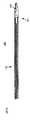
図1A及び1Bは、患者の動脈壁104中の不安定プラーク102を識別するための2つの検出様式を組み合わせる脈管内プローブ100の一実施形態を示す。赤外線分光法を用いて脂質内容を検出する化学分析と、血管内超音波診断法を用いて被膜厚さを検出する形態計測分析とを組み合わせることによって、何れか一方の検出様式だけを用いた場合より潜在的に不安定なプラークの識別における選択性が向上する。これら2つの検出様式は、血液を含有する環境においても高い選択性を実現できる。 FIGS. 1A and 1B illustrate one embodiment of an
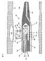
図1Aを参照すると、脈管内プローブ100はカテーテル112を含み、このカテーテル112はその遠位端111に案内ワイヤ管腔110を備える。図1Bを参照すると、案内ワイヤ管腔110内に通される案内ワイヤ108を用いて、脈管内プローブ100を動脈の管腔106に挿入できる。カテーテル112の外層は、赤外線を透過させる材料(例えばポリマー)で構成された外装114である。ハウジング116はカテーテル112の遠位端に位置しており、赤外線を送受信する光学台118と、超音波エネルギーを送受信する超音波トランスデューサ120とを含む。送出ファイバ122及び収集ファイバ123が、カテーテル112の遠位端と近位端との間に延在しており、これらファイバは光学台118に取り付けた遠位端を備えている。光源(図示しない)が、光を送出ファイバ122の近位端内に結合し、送出ミラー124が、送出ファイバ122の遠位端から放射された光125を動脈壁104に方向転換する。収集ミラー126は、動脈壁104内の様々な深さから散乱する光127を収集ファイバ123の遠位端内に方向転換する。送出ミラー124及び収集ミラー126に代えて、他のビーム方向転換器を用いてもよい(例えば、プリズムや光ファイバ先端に設けた屈曲部)。収集ファイバ123の近位端は、光学検出器(図示しない)と光学連通している。光学検出器が、収集ファイバ123内の光の強度を示す電気信号を発生する。この電気信号は、動脈壁104の組成を示す分光シグネチャーを含み、特に、この組成が不安定プラーク102に見られる脂質の存在と一致した組成かどうかを示す分光シグネチャーを含む。この電気信号の分光シグネチャーは、ハードウェア、ソフトウェア、又はそれらの組合せで実現されるスペクトル分析器(図示しない)を用いて分析できる。 Referring to FIG. 1A, the
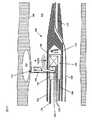
別法としては、図1Cに示した実現例における脈管内プローブ180では、単一の光ファイバ140を送出ファイバ122及び収集ファイバ123の代わりに用いることができる。管腔内壁104から直接的に散乱光を収集することにより、管腔106内の血液中を通過する光の伝搬に起因する散乱を回避できる。結果として、別個の収集ファイバ及び送出ファイバを提供する必要もなくなる。その代わり、単一のファイバ140を、非外傷性光カプラ142を用いた光の収集及び送出のために使用できる。図1Cを参照すると、非外傷性光カプラ142は、動脈壁104の接触域144上に静止している。図1Cに示したように配置すると、非外傷性光カプラ142は、ファイバ140を軸方向に伝わる光を接触域144に差し向ける。非外傷性光カプラ142から出た光は、動脈壁104を通過し、動脈壁104の裏側にあるプラーク102などの構造体を照らす。これら構造体は光の一部を散乱させ、接触域144に返す。すると、この光の一部は接触域26から動脈壁104を介して再出現する。外傷性光カプラ142はこの再出現した光を収集し、ファイバ140内に差し向ける。光ファイバ144の近位端は、光源と光学検出器との両方に(例えば、光サーキュレータを用いて)結合できる。 Alternatively, in the
超音波トランスデューサ120は光学台118に長手方向で隣接しており、超音波エネルギー130を動脈壁104に差し向け、動脈壁104から反射した超音波エネルギー132を受け取る。超音波トランスデューサ120は時分割多重化を用いており、送信する超音波エネルギー130と受信する超音波エネルギー132とを電線128に伝わる電気信号に結合できる。例えば、第1時間間隔において、電線128を伝わる電気信号が超音波トランスデューサ120を駆動して、それに対応した超音波信号を出力させることができる。そして第2時間間隔において、超音波信号が動脈壁から反射した後、超音波トランスデューサ120が電線128を伝わる電気信号を発生する。この電気信号は受信した超音波信号に対応する。受信した電気信号を用いて、動脈壁内部で検出されたプラーク102があれば、その被膜厚さを含んだ動脈壁の形状を復元できる。 The
外装114内部には、超音波トランスデューサ120を囲む食塩水又は他の流体などの伝搬媒体134が音響伝搬を向上させるために入れられている。又、伝搬媒体134は、光学台118から放射される赤外線に対して透明である。 Inside the
ハウジング116に取り付けられたトルクケーブル136が、光ファイバ122及び電線128を囲んでいる。モータ(図示しない)がトルクケーブル136を回転させることにより、ハウジング116も回転させる。この特徴によって、脈管内プローブ100は、光124と超音波エネルギーとで動脈壁104を円周方向に走査することが可能になる。 A
動作時には、脈管内プローブ100を、血管、典型的には動脈に案内ワイヤ108を用いて挿入する。一実施例では、脈管内プローブ100を非連続的な段階で挿入し、段階毎にプローブを完全に一回転させる。この場合、光学及び超音波データを非連続的な円形路に沿って収集できる。別法では、脈管内プローブ100を連続的に挿入し、この場合は軸方向移動と回転が同時進行する。この場合、光学及び超音波データは連続的な螺旋路に沿って収集される。いずれせよ、収集した光学データを用いて動脈壁104の3次元的スペクトル図表を生成でき、収集した超音波データを用いて、動脈壁104の3次元の形態学的図表を生成できる。次に、光学台118と超音波トランスデューサ120との相対位置に基づいて、光学データと超音波データとの対応付けを行う。収集したデータは実時間で使用可能であり、脈管内プローブ100が動脈を通過する時に、不安定プラークを診断したり、これら2つの検出様式により識別可能な特性を持つ他の障害を識別したりできる。脈管内プローブ100は、赤外分光法及び血管内超音波診断法という2つの診断様式に加え、随意選択で他の診断又は治療様式を実行するための構造体を含むこともできる。 In operation, the
図2は、脈管内プローブ200の第2実施形態の断面図であり、このプローブ200では、可とう性カップリング240が光学台218と超音波トランスデューサ220とを連結している。カテーテルを血管内に挿入する時は、剛性要素をなるべく短くしておくとカテーテルが血管の形状に対応しやすくなるので有利となりうる。脈管内プローブ200は、光学台218と超音波トランスデューサ220との間で屈曲するという利点を備えているので、このプローブ200は、血管系内の蛇行状経路を通り抜け可能となる。しかし、脈管内プローブ200から収集した光学及び超音波データは、脈管内プローブ100から収集した光学及び超音波データほどは互いに正確に対応しないこともある。一つの理由は、光学台218と超音波トランスデューサ220とが、第1実施形態の脈管内プローブ100の光学台及びトランスデューサに比べてより離間しているからである。従って、これらは異なる螺旋経路に沿ってデータを収集する。カテーテルの挿入速度が既知であれば、光学データと超音波データとの対応を決定する際にこの経路差を考慮できる。しかし、光学台218と超音波トランスデューサ220との可とう性カップリング240が配置されていることから、図1Aの実施形態に比べてこれが困難となる場合もある。 FIG. 2 is a cross-sectional view of a second embodiment of the
図3A及び3Bは、脈管内プローブ300が光学台318と超音波トランスデューサ320とを備えた第3実施形態の断面図を示す。光学台318及び超音波トランスデューサ320は横方向に互いに隣接しており、外装314の長手方向軸350に対して同じ軸方向位置からそれぞれ光と超音波エネルギーとを放射する。図3Aは、光学台318及び超音波トランスデューサ320の放射端の平面図を示す。図3Bは、光及び超音波エネルギーが同一の軸方向位置から放射されることを示した側面図であり、ハウジング316が同時に回転且つ移動されて、光及び超音波エネルギー350が概ね同一の螺旋経路を描く。これにより収集した光学データと超音波データとの対応付けが容易となる。光学データと超音波データとの時間オフセットは既知の回転速度から特定できる。 3A and 3B show cross-sectional views of a third embodiment in which the
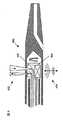
図4は第4実施形態の断面図を示し、ここで脈管内プローブ400は、図3A及び3Bに関連して説明したように、互いに隣接し且つ対向する光学台418と超音波トランスデューサ420とを備える。しかし、本実施形態では、光452は一方の側から放出され、超音波エネルギー454は反対側から放射される。この構成によれば、脈管内プローブ400の直径は、光学台418及び超音波トランスデューサ420の形状寸法によっては脈管内プローブ300の直径よりも小さくなる。直径が小さくなれば、脈管内プローブはより細い血管を通過できることがある。 FIG. 4 shows a cross-sectional view of the fourth embodiment, where the
図5は第5実施形態の断面図を示し、ここで脈管内プローブ500は、固定コア536と、放射状に配列した光カプラと、放射状に配列した超音波トランスデューサ520とを備える。固定コア536を備えた第5実施形態は、コアが回転する上述の実施形態よりも信頼性が高くなることがある。これは、第5実施形態にはトルクケーブルのような可動部分が無いからである。可動部分が無いことにより、外装514が万一破裂しても動脈壁が可動部分に接触しないので、脈管内プローブ500の安全性が向上する。 FIG. 5 shows a cross-sectional view of the fifth embodiment, wherein an
脈管内プローブ500は半径方向全てからデータを同時に収集できるので、診断速度が向上する。或いは、脈管内プローブ500は異なる時刻に異なる位置からデータを収集できるので、光が隣接する光ファイバから収集されたり、超音波エネルギーが隣接するトランスデューサから収集されたりすることによる漏話の可能性が減少する。スペクトル図表及び/又は形態学的図表の半径方向の分解能は、回転コアを備えた実施形態で作成される図表よりは低くなるが、解像度がどの程度異なるかは光ファイバ及び超音波トランスデューサの数に依存する。光ファイバ及び/又は超音波トランスデューサを多数配置すれば半径方向の解像度は向上するが、脈管内プローブ500が大きくなりすぎて、血管によっては挿入できないこともあり得る。 Since the
脈管内プローブ500は、同心状の案内ワイヤ管腔510を通過する案内ワイヤ508に沿って血管に挿入できる。同心状の案内ワイヤ管腔510を用いたカテーテル挿入には、軸心を逸れた遠位端案内ワイヤ管腔110の使用に比べて利点がある。一つの利点は、案内ワイヤ508が絡まる可能性が減少することである。別の利点としては、挿入時にユーザがワイヤに同軸の負荷をかけるため、同心状の案内ワイヤ管腔510ではトラッカビリティが向上する。又、同心状の案内ワイヤ管腔510を用いると、案内ワイヤ508が光ファイバ及び超音波トランスデューサの視野の外に位置することになる。 The
これら脈管内プローブは直径が十分小さいカテーテルを備えているため、プローブを細い血管にも挿入できる。図6A及び6Bは、回転コア(図1乃至4)及び固定コア(図5)を備えた実施形態のカテーテルの横断面図を比較したものである。 Since these intravascular probes have a sufficiently small diameter catheter, the probes can be inserted into thin blood vessels. 6A and 6B compare cross-sectional views of an embodiment of a catheter with a rotating core (FIGS. 1-4) and a stationary core (FIG. 5).
図6Aに示した回転コアカテーテル660は、赤外分光法用の光学信号を伝達する一対の光ファイバ622と、血管内超音波診断法用の電気信号を伝達する一対の電線628とを中空のトルクケーブル636内部に備えている。カテーテル660の外装614の直径は、トルクケーブル636の寸法により制限される。 The
図6Bに示した固定コアカテーテル670は、動脈壁の4つの四分円から、光学信号と血管内超音波診断法用の電気信号とをそれぞれ伝える四対の光ファイバ672と、四対の電線674とを備えている。トルクケーブルは不要だが、カテーテル670の外装676には、四分円それぞれに一対の光ファイバ672と一対の電線674とを収容するだけの直径が必要である。
他の実施形態The fixed
Other embodiments
本発明をその詳細な説明に関連して記載してきたが、上述の説明は例示を意図したものであり、本発明の範囲を限定するものではない。又、本発明の範囲は、添付した特許請求の範囲によって定義される。他の局面、利点、及び変更も次の特許請求の範囲に入る。
Although the present invention has been described in connection with a detailed description thereof, the above description is intended to be illustrative and not limiting the scope of the invention. The scope of the invention is also defined by the appended claims. Other aspects, advantages, and modifications are within the scope of the following claims.
Claims (20)
Translated fromJapanese遠位部と近位部とを備えた外装と、
前記外装に沿って延伸する第1光学導波路であって、前記遠位部と前記近位部との間で光放射を伝達するよう構成された第1光学導波路と、
前記遠位部に設けられると共に、前記第1光学導波路と光学連通した第1ビーム方向転換器と、
前記遠位部に設けられた光学台であって、内部に前記第1光学導波路の遠位端が配置された光学台と、
前記光学台上に設けられ、前記第1光学導波路と光学連通した第1ビーム方向転換器と、
前記外装に沿って延伸する第2光学導波路であって、前記遠位部と前記近位部との間で光放射を伝達するよう構成され、前記光学台内に配置された遠位端を有する、第2光学導波路と、
前記光学台上に設けられると共に、前記第2光学導波路と光学連通した第2ビーム方向転換器と、
前記遠位端に設けられ、かつ前記光学台に結合された超音波トランスデューサであって、超音波エネルギーを前記脈管内プローブと伝搬媒体との間で結合するよう構成された超音波トランスデューサと、
前記外装に沿って延伸すると共に、前記超音波トランスデューサと電気連通した電線とを含む、脈管内プローブ。An intravascular probe,
An exterior with a distal portion and a proximal portion;
A first optical waveguide extending along the sheath, the first optical waveguide configured to transmit light radiation between the distal portion and the proximal portion;
A first beam redirector provided at the distal portion and in optical communication with the first optical waveguide;
An optical bench provided at the distal portion, the optical bench having a distal end of the first optical waveguide disposed therein;
A first beam redirector provided on the optical bench and in optical communication with the first optical waveguide;
A second optical waveguide extending along the sheath, the second optical waveguide being configured to transmit light radiation between the distal portion and the proximal portion, and having a distal end disposed within the optical bench Having a second optical waveguide;
A second beam redirector provided on the optical bench and in optical communication with the second optical waveguide;
An ultrasonic transducer disposed at the distal end and coupled to the optical bench, the ultrasonic transducer configured to couple ultrasonic energy between the intravascular probe and a propagation medium;
An intravascular probe that extends along the exterior and includes an electrical wire in electrical communication with the ultrasonic transducer.
前記複数のビーム方向転換器と光学連通した複数の光学導波路と、
前記長手方向軸周りに環状に配置された複数の超音波トランスデューサとを更に含む、請求項1に記載の脈管内プローブ。A plurality of beam redirectors arranged annularly around the longitudinal axis of the sheath;
A plurality of optical waveguides in optical communication with the plurality of beam redirectors;
The intravascular probe according to claim 1, further comprising a plurality of ultrasonic transducers arranged annularly about the longitudinal axis.
Applications Claiming Priority (2)
| Application Number | Priority Date | Filing Date | Title |
|---|---|---|---|
| US10/668,012US6949072B2 (en) | 2003-09-22 | 2003-09-22 | Devices for vulnerable plaque detection |
| US10/668,012 | 2003-09-22 |
Related Parent Applications (1)
| Application Number | Title | Priority Date | Filing Date |
|---|---|---|---|
| JP2006528125ADivisionJP4972407B2 (en) | 2003-09-22 | 2004-09-21 | Device for detecting unstable plaque |
Related Child Applications (1)
| Application Number | Title | Priority Date | Filing Date |
|---|---|---|---|
| JP2013264566ADivisionJP5770259B2 (en) | 2003-09-22 | 2013-12-20 | Device for detecting unstable plaque |
Publications (2)
| Publication Number | Publication Date |
|---|---|
| JP2010207612Atrue JP2010207612A (en) | 2010-09-24 |
| JP5769931B2 JP5769931B2 (en) | 2015-08-26 |
Family
ID=34393402
Family Applications (3)
| Application Number | Title | Priority Date | Filing Date |
|---|---|---|---|
| JP2006528125AExpired - LifetimeJP4972407B2 (en) | 2003-09-22 | 2004-09-21 | Device for detecting unstable plaque |
| JP2010120418AExpired - LifetimeJP5769931B2 (en) | 2003-09-22 | 2010-05-26 | Device for detecting unstable plaque |
| JP2013264566AExpired - LifetimeJP5770259B2 (en) | 2003-09-22 | 2013-12-20 | Device for detecting unstable plaque |
Family Applications Before (1)
| Application Number | Title | Priority Date | Filing Date |
|---|---|---|---|
| JP2006528125AExpired - LifetimeJP4972407B2 (en) | 2003-09-22 | 2004-09-21 | Device for detecting unstable plaque |
Family Applications After (1)
| Application Number | Title | Priority Date | Filing Date |
|---|---|---|---|
| JP2013264566AExpired - LifetimeJP5770259B2 (en) | 2003-09-22 | 2013-12-20 | Device for detecting unstable plaque |
Country Status (4)
| Country | Link |
|---|---|
| US (1) | US6949072B2 (en) |
| EP (1) | EP1667576B1 (en) |
| JP (3) | JP4972407B2 (en) |
| WO (1) | WO2005030046A1 (en) |
Cited By (3)
| Publication number | Priority date | Publication date | Assignee | Title |
|---|---|---|---|---|
| WO2013145689A1 (en)* | 2012-03-28 | 2013-10-03 | テルモ株式会社 | Probe and diagnostic imaging device |
| CN108627574A (en)* | 2017-03-24 | 2018-10-09 | 空中客车运营简化股份公司 | For endoporus equipped with the ultrasonic probe and its method of adjustment for coupling support element |
| JP2020529232A (en)* | 2017-09-15 | 2020-10-08 | インフラレデックス, インコーポレイテッド | Blood vessel imaging method |
Families Citing this family (137)
| Publication number | Priority date | Publication date | Assignee | Title |
|---|---|---|---|---|
| US7231243B2 (en) | 2000-10-30 | 2007-06-12 | The General Hospital Corporation | Optical methods for tissue analysis |
| US9295391B1 (en) | 2000-11-10 | 2016-03-29 | The General Hospital Corporation | Spectrally encoded miniature endoscopic imaging probe |
| AT503309B1 (en) | 2001-05-01 | 2011-08-15 | Gen Hospital Corp | DEVICE FOR DETERMINING ATHEROSCLEROTIC BEARING BY MEASURING OPTICAL TISSUE PROPERTIES |
| US7355716B2 (en) | 2002-01-24 | 2008-04-08 | The General Hospital Corporation | Apparatus and method for ranging and noise reduction of low coherence interferometry LCI and optical coherence tomography OCT signals by parallel detection of spectral bands |
| US8054468B2 (en) | 2003-01-24 | 2011-11-08 | The General Hospital Corporation | Apparatus and method for ranging and noise reduction of low coherence interferometry LCI and optical coherence tomography OCT signals by parallel detection of spectral bands |
| EP2436307B1 (en) | 2003-03-31 | 2015-10-21 | The General Hospital Corporation | Speckle reduction in optical coherence tomography by path length encoded angular compounding |
| KR101386971B1 (en) | 2003-06-06 | 2014-04-18 | 더 제너럴 하스피탈 코포레이션 | Process and apparatus for a wavelength tunning source |
| EP2280256B1 (en) | 2003-10-27 | 2016-11-16 | The General Hospital Corporation | Method and apparatus for performing optical imaging using frequency-domain interferometry |
| JP4554967B2 (en)* | 2004-03-25 | 2010-09-29 | テルモ株式会社 | Ultrasonic catheter and diagnostic imaging apparatus |
| KR101239250B1 (en) | 2004-05-29 | 2013-03-05 | 더 제너럴 하스피탈 코포레이션 | Process, system and software arrangement for a chromatic dispersion compensation using reflective layers in optical coherence tomography (oct) imaging |
| JP2008502408A (en)* | 2004-06-17 | 2008-01-31 | コーニンクレッカ フィリップス エレクトロニクス エヌ ヴィ | Combined ultrasound imaging and spectroscopic molecular analysis apparatus and method |
| AU2005270037B2 (en) | 2004-07-02 | 2012-02-09 | The General Hospital Corporation | Endoscopic imaging probe comprising dual clad fibre |
| EP1782020B1 (en) | 2004-08-06 | 2012-10-03 | The General Hospital Corporation | Process, system and software arrangement for determining at least one location in a sample using an optical coherence tomography |
| EP2272421A1 (en) | 2004-08-24 | 2011-01-12 | The General Hospital Corporation | Method and apparatus for imaging of vessel segments |
| WO2006024014A2 (en) | 2004-08-24 | 2006-03-02 | The General Hospital Corporation | Process, system and software arrangement for measuring a mechanical strain and elastic properties of a sample |
| US7365859B2 (en) | 2004-09-10 | 2008-04-29 | The General Hospital Corporation | System and method for optical coherence imaging |
| KR101257100B1 (en) | 2004-09-29 | 2013-04-22 | 더 제너럴 하스피탈 코포레이션 | System and Method for Optical Coherence Imaging |
| WO2006058049A1 (en) | 2004-11-24 | 2006-06-01 | The General Hospital Corporation | Common-path interferometer for endoscopic oct |
| WO2006058346A1 (en) | 2004-11-29 | 2006-06-01 | The General Hospital Corporation | Arrangements, devices, endoscopes, catheters and methods for performing optical imaging by simultaneously illuminating and detecting multiple points on a sample |
| ES2337497T3 (en) | 2005-04-28 | 2010-04-26 | The General Hospital Corporation | EVALUATION OF CHARACTERISTICS OF THE IMAGE OF AN ANATOMICAL STRUCTURE IN IMAGES OF TOMOGRAPHY OF OPTICAL COHERENCE. |
| US9060689B2 (en) | 2005-06-01 | 2015-06-23 | The General Hospital Corporation | Apparatus, method and system for performing phase-resolved optical frequency domain imaging |
| EP2267404B1 (en) | 2005-08-09 | 2016-10-05 | The General Hospital Corporation | Apparatus and method for performing polarization-based quadrature demodulation in optical coherence tomography |
| US7843572B2 (en) | 2005-09-29 | 2010-11-30 | The General Hospital Corporation | Method and apparatus for optical imaging via spectral encoding |
| US20070270717A1 (en)* | 2005-09-30 | 2007-11-22 | Cornova, Inc. | Multi-faceted optical reflector |
| JP5129749B2 (en)* | 2005-09-30 | 2013-01-30 | コルノヴァ インク | System for probe inspection and treatment of body cavities |
| US7450241B2 (en)* | 2005-09-30 | 2008-11-11 | Infraredx, Inc. | Detecting vulnerable plaque |
| US7889348B2 (en) | 2005-10-14 | 2011-02-15 | The General Hospital Corporation | Arrangements and methods for facilitating photoluminescence imaging |
| US20070129625A1 (en)* | 2005-11-21 | 2007-06-07 | Boston Scientific Scimed Systems, Inc. | Systems and methods for detecting the presence of abnormalities in a medical image |
| EP1971848B1 (en) | 2006-01-10 | 2019-12-04 | The General Hospital Corporation | Systems and methods for generating data based on one or more spectrally-encoded endoscopy techniques |
| DK1973466T3 (en) | 2006-01-19 | 2021-02-01 | Massachusetts Gen Hospital | BALLOON IMAGING CATHETER |
| US8145018B2 (en) | 2006-01-19 | 2012-03-27 | The General Hospital Corporation | Apparatus for obtaining information for a structure using spectrally-encoded endoscopy techniques and methods for producing one or more optical arrangements |
| JP5680829B2 (en) | 2006-02-01 | 2015-03-04 | ザ ジェネラル ホスピタル コーポレイション | A device that irradiates a sample with multiple electromagnetic radiations |
| WO2007149602A2 (en) | 2006-02-01 | 2007-12-27 | The General Hospital Corporation | Methods and systems for providing electromagnetic radiation to at least one portion of a sample using conformal laser therapy procedures |
| US9777053B2 (en) | 2006-02-08 | 2017-10-03 | The General Hospital Corporation | Methods, arrangements and systems for obtaining information associated with an anatomical sample using optical microscopy |
| EP2982929A1 (en) | 2006-02-24 | 2016-02-10 | The General Hospital Corporation | Methods and systems for performing angle-resolved fourier-domain optical coherence tomography |
| US20070208257A1 (en)* | 2006-03-03 | 2007-09-06 | Furnish Simon M | Lateral Viewing Optical Catheters |
| WO2007103235A2 (en)* | 2006-03-03 | 2007-09-13 | Prescient Medical, Inc. | Optical imaging balloon catheters |
| JP5135324B2 (en) | 2006-04-05 | 2013-02-06 | ザ ジェネラル ホスピタル コーポレイション | Method, arrangement and system for polarization sensitive optical frequency domain imaging of samples |
| WO2007123518A1 (en)* | 2006-04-21 | 2007-11-01 | Cedars-Sinai Medical Center | Multiple imaging and/or spectroscopic modality probe |
| WO2007133961A2 (en) | 2006-05-10 | 2007-11-22 | The General Hospital Corporation | Processes, arrangements and systems for providing frequency domain imaging of a sample |
| US7782464B2 (en) | 2006-05-12 | 2010-08-24 | The General Hospital Corporation | Processes, arrangements and systems for providing a fiber layer thickness map based on optical coherence tomography images |
| US20070291275A1 (en)* | 2006-06-16 | 2007-12-20 | Prescient Medical, Inc. | Side-viewing optical acoustic sensors and their use in intravascular diagnostic probes |
| WO2008011163A2 (en)* | 2006-07-21 | 2008-01-24 | Prescient Medical, Inc. | Conformable tissue contact catheter |
| US7717244B2 (en)* | 2006-08-15 | 2010-05-18 | Sonnax Industries, Inc. | Replacement torque converter cover assembly |
| US7920271B2 (en) | 2006-08-25 | 2011-04-05 | The General Hospital Corporation | Apparatus and methods for enhancing optical coherence tomography imaging using volumetric filtering techniques |
| US8882674B2 (en)* | 2006-09-28 | 2014-11-11 | Research Foundation Of The City University Of New York | System and method for in vivo imaging of blood vessel walls to detect microcalcifications |
| WO2009089372A2 (en)* | 2008-01-08 | 2009-07-16 | Cornova, Inc. | Systems and methods for analysis and treatment of a body lumen |
| US8838213B2 (en)* | 2006-10-19 | 2014-09-16 | The General Hospital Corporation | Apparatus and method for obtaining and providing imaging information associated with at least one portion of a sample, and effecting such portion(s) |
| US20080097158A1 (en)* | 2006-10-20 | 2008-04-24 | Infraredx, Inc. | Noise Suppression System and Method in Catheter Pullback and Rotation System |
| US20080097223A1 (en)* | 2006-10-20 | 2008-04-24 | Infraredx, Inc. | Optical Catheter Carriage Interlock System and Method |
| US20080097408A1 (en)* | 2006-10-20 | 2008-04-24 | Infraredx, Inc. | Pullback Carriage Interlock System and Method for Catheter System |
| US20080097224A1 (en)* | 2006-10-20 | 2008-04-24 | Infraredx, Inc. | Manual and Motor Driven Optical Pullback and Rotation System and Method |
| WO2008051950A2 (en)* | 2006-10-23 | 2008-05-02 | Prescient Medical Inc. | Windowless fiber optic raman spectroscopy probes |
| CN107126182B (en)* | 2007-01-19 | 2020-06-16 | 桑尼布鲁克健康科学中心 | Scanning mechanism for imaging probe |
| US8460195B2 (en)* | 2007-01-19 | 2013-06-11 | Sunnybrook Health Sciences Centre | Scanning mechanisms for imaging probe |
| EP2104968A1 (en) | 2007-01-19 | 2009-09-30 | The General Hospital Corporation | Rotating disk reflection for fast wavelength scanning of dispersed broadband light |
| JP2008237235A (en)* | 2007-03-23 | 2008-10-09 | Olympus Medical Systems Corp | Endoscope and living body observation system |
| US9176319B2 (en) | 2007-03-23 | 2015-11-03 | The General Hospital Corporation | Methods, arrangements and apparatus for utilizing a wavelength-swept laser using angular scanning and dispersion procedures |
| US10534129B2 (en) | 2007-03-30 | 2020-01-14 | The General Hospital Corporation | System and method providing intracoronary laser speckle imaging for the detection of vulnerable plaque |
| US8045177B2 (en) | 2007-04-17 | 2011-10-25 | The General Hospital Corporation | Apparatus and methods for measuring vibrations using spectrally-encoded endoscopy |
| WO2008137637A2 (en) | 2007-05-04 | 2008-11-13 | The General Hospital Corporation | Methods, arrangements and systems for obtaining information associated with a sample using brillouin microscopy |
| US7952706B2 (en)* | 2007-05-17 | 2011-05-31 | Prescient Medical, Inc. | Multi-channel fiber optic spectroscopy systems employing integrated optics modules |
| EP2160217A1 (en) | 2007-06-08 | 2010-03-10 | Prescient Medical, Inc. | Optical catheter configurations combining raman spectroscopy with optical fiber-based low coherence reflectometry |
| WO2008157760A1 (en)* | 2007-06-21 | 2008-12-24 | Cornova, Inc. | Systems and methods for guiding the analysis and treatment of a body lumen |
| US20090024040A1 (en)* | 2007-07-20 | 2009-01-22 | Prescient Medical, Inc. | Wall-Contacting Intravascular Ultrasound Probe Catheters |
| US9375158B2 (en) | 2007-07-31 | 2016-06-28 | The General Hospital Corporation | Systems and methods for providing beam scan patterns for high speed doppler optical frequency domain imaging |
| WO2009029843A1 (en) | 2007-08-31 | 2009-03-05 | The General Hospital Corporation | System and method for self-interference fluoresence microscopy, and computer-accessible medium associated therewith |
| US20090076395A1 (en)* | 2007-09-19 | 2009-03-19 | Prescient Medical, Inc. | Optimized intravascular ultrasound probe catherers |
| US7933021B2 (en) | 2007-10-30 | 2011-04-26 | The General Hospital Corporation | System and method for cladding mode detection |
| WO2009089364A2 (en)* | 2008-01-08 | 2009-07-16 | Cornova, Inc. | Shaped fiber ends and methods of making same |
| JP5011147B2 (en)* | 2008-02-05 | 2012-08-29 | 国立大学法人山口大学 | Diagnostic system |
| US8016842B2 (en)* | 2008-03-25 | 2011-09-13 | Medtronic Vascular, Inc. | Methods for treating vulnerable plaque |
| US9713448B2 (en)* | 2008-04-03 | 2017-07-25 | Infraredx, Inc. | System and method for intravascular structural analysis compensation of chemical analysis modality |
| US20090259174A1 (en)* | 2008-04-15 | 2009-10-15 | Medtronic Vascular, Inc. | Methods and devices for treating vulnerable atherosclerotic plaque |
| US7898656B2 (en)* | 2008-04-30 | 2011-03-01 | The General Hospital Corporation | Apparatus and method for cross axis parallel spectroscopy |
| US8052605B2 (en)* | 2008-05-07 | 2011-11-08 | Infraredx | Multimodal catheter system and method for intravascular analysis |
| WO2009137608A2 (en)* | 2008-05-07 | 2009-11-12 | Infraredx, Inc. | Catheter with spinning ultrasound transceiver board |
| EP2274572A4 (en) | 2008-05-07 | 2013-08-28 | Gen Hospital Corp | SYSTEM, METHOD AND COMPUTER MEDIUM FOR MONITORING THE MOVEMENT OF VESSELS DURING A THREE-DIMENSIONAL MICROSCOPY EXAMINATION OF CORONARY ARTERIES |
| US8197413B2 (en)* | 2008-06-06 | 2012-06-12 | Boston Scientific Scimed, Inc. | Transducers, devices and systems containing the transducers, and methods of manufacture |
| JP5384860B2 (en)* | 2008-06-11 | 2014-01-08 | 富士フイルム株式会社 | Precision rotation transmission mechanism and optical scanning probe |
| US8861910B2 (en) | 2008-06-20 | 2014-10-14 | The General Hospital Corporation | Fused fiber optic coupler arrangement and method for use thereof |
| WO2010009136A2 (en) | 2008-07-14 | 2010-01-21 | The General Hospital Corporation | Apparatus and methods for color endoscopy |
| WO2010045437A2 (en)* | 2008-10-15 | 2010-04-22 | Cornova, Inc. | Systems and methods for analysis and treatment of an occluded body lumen |
| US20100113906A1 (en)* | 2008-11-06 | 2010-05-06 | Prescient Medical, Inc. | Hybrid basket catheters |
| US9795442B2 (en) | 2008-11-11 | 2017-10-24 | Shifamed Holdings, Llc | Ablation catheters |
| JP5731394B2 (en) | 2008-12-10 | 2015-06-10 | ザ ジェネラル ホスピタル コーポレイション | System, apparatus and method for extending imaging depth range of optical coherence tomography through optical subsampling |
| US20100179432A1 (en)* | 2009-01-09 | 2010-07-15 | Boston Scientific Scimed, Inc. | Systems and methods for making and using intravascular ultrasound systems with photo-acoustic imaging capabilities |
| US20100179434A1 (en)* | 2009-01-09 | 2010-07-15 | Boston Scientific Scimed, Inc. | Systems and methods for making and using intravascular ultrasound systems with photo-acoustic imaging capabilities |
| JP2012515576A (en) | 2009-01-20 | 2012-07-12 | ザ ジェネラル ホスピタル コーポレイション | Endoscopic biopsy device, system, and method |
| US8097864B2 (en) | 2009-01-26 | 2012-01-17 | The General Hospital Corporation | System, method and computer-accessible medium for providing wide-field superresolution microscopy |
| WO2010105197A2 (en) | 2009-03-12 | 2010-09-16 | The General Hospital Corporation | Non-contact optical system, computer-accessible medium and method for measuring at least one mechanical property of tissue using coherent speckle techniques(s) |
| EP2432542A4 (en)* | 2009-05-20 | 2013-07-03 | Cornova Inc | Systems and methods for analysis and treatment of a body lumen |
| JP5819823B2 (en) | 2009-07-14 | 2015-11-24 | ザ ジェネラル ホスピタル コーポレイション | Device for measuring the flow and pressure inside a blood vessel and method of operating the device |
| WO2011011269A2 (en) | 2009-07-23 | 2011-01-27 | Waters Kendall R | Endoventricular injection catheter system with integrated echocardiographic capabilities |
| US20120316419A1 (en) | 2010-02-18 | 2012-12-13 | Eric Chevalier | Multimodal catheter |
| KR20130004326A (en) | 2010-03-05 | 2013-01-09 | 더 제너럴 하스피탈 코포레이션 | Systems, methods and computer-accessible medium which provide microscopic images of at least one anatomical structure at a particular resolution |
| KR101108492B1 (en)* | 2010-04-19 | 2012-01-31 | 서울대학교병원 | Probe with small projector |
| US9069130B2 (en) | 2010-05-03 | 2015-06-30 | The General Hospital Corporation | Apparatus, method and system for generating optical radiation from biological gain media |
| US9655677B2 (en) | 2010-05-12 | 2017-05-23 | Shifamed Holdings, Llc | Ablation catheters including a balloon and electrodes |
| AU2011252976A1 (en) | 2010-05-12 | 2012-11-08 | Shifamed Holdings, Llc | Low profile electrode assembly |
| EP2575598A2 (en) | 2010-05-25 | 2013-04-10 | The General Hospital Corporation | Apparatus, systems, methods and computer-accessible medium for spectral analysis of optical coherence tomography images |
| EP2575597B1 (en) | 2010-05-25 | 2022-05-04 | The General Hospital Corporation | Apparatus for providing optical imaging of structures and compositions |
| JP6066901B2 (en) | 2010-06-03 | 2017-01-25 | ザ ジェネラル ホスピタル コーポレイション | Method for apparatus and device for imaging structures in or in one or more luminal organs |
| WO2012058381A2 (en) | 2010-10-27 | 2012-05-03 | The General Hospital Corporation | Apparatus, systems and methods for measuring blood pressure within at least one vessel |
| US9330092B2 (en) | 2011-07-19 | 2016-05-03 | The General Hospital Corporation | Systems, methods, apparatus and computer-accessible-medium for providing polarization-mode dispersion compensation in optical coherence tomography |
| EP3835718B1 (en) | 2011-08-25 | 2023-07-26 | The General Hospital Corporation | Apparatus for providing micro-optical coherence tomography inside a respiratory system |
| US8958867B2 (en) | 2011-08-29 | 2015-02-17 | Infraredx, Inc. | Detection of lipid core plaque cap thickness |
| JP2015502562A (en) | 2011-10-18 | 2015-01-22 | ザ ジェネラル ホスピタル コーポレイション | Apparatus and method for generating and / or providing recirculating optical delay |
| JP6038957B2 (en)* | 2012-02-14 | 2016-12-07 | セント・ジュード・メディカル・エイトリアル・フィブリレーション・ディヴィジョン・インコーポレーテッド | A system for evaluating the effects of ablation treatment of cardiac tissue using photoacoustic method |
| WO2013148306A1 (en) | 2012-03-30 | 2013-10-03 | The General Hospital Corporation | Imaging system, method and distal attachment for multidirectional field of view endoscopy |
| JP2015517387A (en) | 2012-05-21 | 2015-06-22 | ザ ジェネラル ホスピタル コーポレイション | Apparatus, device and method for capsule microscopy |
| US20140024931A1 (en)* | 2012-07-20 | 2014-01-23 | Lightlab Imaging, Inc. | Data Encoders for Medical Devices and Related Methods |
| EP2919658B1 (en) | 2012-11-19 | 2024-03-20 | Lightlab Imaging, Inc. | Interface devices, systems and methods for multimodal probes |
| US9968261B2 (en) | 2013-01-28 | 2018-05-15 | The General Hospital Corporation | Apparatus and method for providing diffuse spectroscopy co-registered with optical frequency domain imaging |
| WO2014120791A1 (en) | 2013-01-29 | 2014-08-07 | The General Hospital Corporation | Apparatus, systems and methods for providing information regarding the aortic valve |
| US11179028B2 (en) | 2013-02-01 | 2021-11-23 | The General Hospital Corporation | Objective lens arrangement for confocal endomicroscopy |
| US10478072B2 (en) | 2013-03-15 | 2019-11-19 | The General Hospital Corporation | Methods and system for characterizing an object |
| US10098694B2 (en) | 2013-04-08 | 2018-10-16 | Apama Medical, Inc. | Tissue ablation and monitoring thereof |
| US10349824B2 (en) | 2013-04-08 | 2019-07-16 | Apama Medical, Inc. | Tissue mapping and visualization systems |
| KR20150140760A (en) | 2013-04-08 | 2015-12-16 | 아파마 메디칼, 인크. | Cardiac ablation catheters and methods of use thereof |
| EP2997354A4 (en) | 2013-05-13 | 2017-01-18 | The General Hospital Corporation | Detecting self-interefering fluorescence phase and amplitude |
| EP3021735A4 (en) | 2013-07-19 | 2017-04-19 | The General Hospital Corporation | Determining eye motion by imaging retina. with feedback |
| WO2015009932A1 (en) | 2013-07-19 | 2015-01-22 | The General Hospital Corporation | Imaging apparatus and method which utilizes multidirectional field of view endoscopy |
| WO2015013651A2 (en) | 2013-07-26 | 2015-01-29 | The General Hospital Corporation | System, apparatus and method utilizing optical dispersion for fourier-domain optical coherence tomography |
| WO2015105870A1 (en) | 2014-01-08 | 2015-07-16 | The General Hospital Corporation | Method and apparatus for microscopic imaging |
| US10736494B2 (en) | 2014-01-31 | 2020-08-11 | The General Hospital Corporation | System and method for facilitating manual and/or automatic volumetric imaging with real-time tension or force feedback using a tethered imaging device |
| WO2015153982A1 (en) | 2014-04-04 | 2015-10-08 | The General Hospital Corporation | Apparatus and method for controlling propagation and/or transmission of electromagnetic radiation in flexible waveguide(s) |
| US10912462B2 (en) | 2014-07-25 | 2021-02-09 | The General Hospital Corporation | Apparatus, devices and methods for in vivo imaging and diagnosis |
| US10776654B2 (en) | 2015-03-10 | 2020-09-15 | Infraredx, Inc. | Assessment of lipid core plaque integrity |
| JP6559985B2 (en)* | 2015-03-20 | 2019-08-14 | テルモ株式会社 | Diagnostic imaging probe |
| EP4302713A3 (en) | 2015-11-16 | 2024-03-13 | Boston Scientific Scimed, Inc. | Energy delivery devices |
| KR102001980B1 (en)* | 2016-12-29 | 2019-07-19 | 울산과학기술원 | Photoacoustic and ultrasonic endoscopic mini-probe |
| MX2020004860A (en) | 2017-11-16 | 2020-08-13 | Colgate Palmolive Co | Ultrasonic system and method for detecting a biofilm on a tooth. |
| US11918761B2 (en) | 2018-09-14 | 2024-03-05 | Infraredx, Inc. | Intravascular imaging catheter system with force error detection and automatic remediation via pullback and rotation for translating and rotating a torque cable in a catheter |
| JP2022534042A (en)* | 2019-05-31 | 2022-07-27 | コーニンクレッカ フィリップス エヌ ヴェ | Intravascular optical device |
| CN113229854B (en)* | 2021-06-24 | 2022-03-08 | 哈尔滨医科大学 | Probe integrating optical coherence tomography imaging and intravascular ultrasound |
| WO2025041024A1 (en)* | 2023-08-21 | 2025-02-27 | Renaly Ltd | Ultrasound ablation of nerves |
Citations (9)
| Publication number | Priority date | Publication date | Assignee | Title |
|---|---|---|---|---|
| JPH0731617A (en)* | 1993-07-16 | 1995-02-03 | Toshiba Corp | Ultrasonic diagnostic equipment |
| JPH10511875A (en)* | 1995-01-03 | 1998-11-17 | ノン−インヴェイシヴ テクノロジイ,インク. | Optical coupler for in vivo examination of biological tissue |
| JPH1156752A (en)* | 1997-08-28 | 1999-03-02 | Olympus Optical Co Ltd | In-subject tomographic imaging system |
| JPH11151245A (en)* | 1997-11-19 | 1999-06-08 | Toshiba Corp | Ultrasonic probe and ultrasonic diagnostic device |
| US6016440A (en)* | 1996-07-29 | 2000-01-18 | Bruker Analytik Gmbh | Device for infrared (IR) spectroscopic investigations of internal surfaces of a body |
| JP2000511786A (en)* | 1995-09-20 | 2000-09-12 | テキサス・ハート・インスティチュート | Detection of temperature difference in pipe wall |
| JP2002200037A (en)* | 2000-10-31 | 2002-07-16 | Fuji Photo Film Co Ltd | Endoscope system |
| US20030199767A1 (en)* | 2002-04-19 | 2003-10-23 | Cespedes Eduardo Ignacio | Methods and apparatus for the identification and stabilization of vulnerable plaque |
| US6690958B1 (en)* | 2002-05-07 | 2004-02-10 | Nostix Llc | Ultrasound-guided near infrared spectrophotometer |
Family Cites Families (24)
| Publication number | Priority date | Publication date | Assignee | Title |
|---|---|---|---|---|
| US4375818A (en)* | 1979-03-12 | 1983-03-08 | Olympus Optical Company Ltd. | Ultrasonic diagnosis system assembled into endoscope |
| US5435805A (en)* | 1992-08-12 | 1995-07-25 | Vidamed, Inc. | Medical probe device with optical viewing capability |
| US5370675A (en)* | 1992-08-12 | 1994-12-06 | Vidamed, Inc. | Medical probe device and method |
| US4504727A (en)* | 1982-12-30 | 1985-03-12 | International Business Machines Corporation | Laser drilling system utilizing photoacoustic feedback |
| US4794931A (en) | 1986-02-28 | 1989-01-03 | Cardiovascular Imaging Systems, Inc. | Catheter apparatus, system and method for intravascular two-dimensional ultrasonography |
| NL8901084A (en)* | 1989-04-28 | 1990-11-16 | Du Med Bv | INTRA-LUMINAL DEVICE. |
| US5029588A (en) | 1989-06-15 | 1991-07-09 | Cardiovascular Imaging Systems, Inc. | Laser catheter with imaging capability |
| US5916210A (en)* | 1990-01-26 | 1999-06-29 | Intraluminal Therapeutics, Inc. | Catheter for laser treatment of atherosclerotic plaque and other tissue abnormalities |
| US5167233A (en)* | 1991-01-07 | 1992-12-01 | Endosonics Corporation | Dilating and imaging apparatus |
| US6134003A (en)* | 1991-04-29 | 2000-10-17 | Massachusetts Institute Of Technology | Method and apparatus for performing optical measurements using a fiber optic imaging guidewire, catheter or endoscope |
| WO1993021940A1 (en) | 1992-05-06 | 1993-11-11 | Immunomedics, Inc. | Intraoperative, intravascular and endoscopic tumor and lesion detection and therapy |
| US5576013A (en) | 1995-03-21 | 1996-11-19 | Eastern Virginia Medical School | Treating vascular and neoplastic tissues |
| US6008211A (en) | 1995-07-27 | 1999-12-28 | Pdt Pharmaceuticals, Inc. | Photoactivatable compounds comprising benzochlorin and furocoumarin |
| US6763261B2 (en) | 1995-09-20 | 2004-07-13 | Board Of Regents, The University Of Texas System | Method and apparatus for detecting vulnerable atherosclerotic plaque |
| US6615071B1 (en)* | 1995-09-20 | 2003-09-02 | Board Of Regents, The University Of Texas System | Method and apparatus for detecting vulnerable atherosclerotic plaque |
| US5725494A (en) | 1995-11-30 | 1998-03-10 | Pharmasonics, Inc. | Apparatus and methods for ultrasonically enhanced intraluminal therapy |
| US5728092A (en) | 1996-03-07 | 1998-03-17 | Miravant Systems, Inc. | Light delivery catheter |
| US6022309A (en)* | 1996-04-24 | 2000-02-08 | The Regents Of The University Of California | Opto-acoustic thrombolysis |
| US5924997A (en)* | 1996-07-29 | 1999-07-20 | Campbell; Thomas Henderson | Catheter and method for the thermal mapping of hot spots in vascular lesions of the human body |
| EP1043949A2 (en) | 1997-12-31 | 2000-10-18 | Pharmasonics, Inc. | Methods and systems for the inhibition of vascular hyperplasia |
| US6296619B1 (en) | 1998-12-30 | 2001-10-02 | Pharmasonics, Inc. | Therapeutic ultrasonic catheter for delivering a uniform energy dose |
| US6692430B2 (en)* | 2000-04-10 | 2004-02-17 | C2Cure Inc. | Intra vascular imaging apparatus |
| US6701181B2 (en)* | 2001-05-31 | 2004-03-02 | Infraredx, Inc. | Multi-path optical catheter |
| US20030236443A1 (en) | 2002-04-19 | 2003-12-25 | Cespedes Eduardo Ignacio | Methods and apparatus for the identification and stabilization of vulnerable plaque |
- 2003
- 2003-09-22USUS10/668,012patent/US6949072B2/ennot_activeExpired - Lifetime
- 2004
- 2004-09-21JPJP2006528125Apatent/JP4972407B2/ennot_activeExpired - Lifetime
- 2004-09-21WOPCT/US2004/031005patent/WO2005030046A1/enactiveApplication Filing
- 2004-09-21EPEP04784743.9Apatent/EP1667576B1/ennot_activeExpired - Lifetime
- 2010
- 2010-05-26JPJP2010120418Apatent/JP5769931B2/ennot_activeExpired - Lifetime
- 2013
- 2013-12-20JPJP2013264566Apatent/JP5770259B2/ennot_activeExpired - Lifetime
Patent Citations (9)
| Publication number | Priority date | Publication date | Assignee | Title |
|---|---|---|---|---|
| JPH0731617A (en)* | 1993-07-16 | 1995-02-03 | Toshiba Corp | Ultrasonic diagnostic equipment |
| JPH10511875A (en)* | 1995-01-03 | 1998-11-17 | ノン−インヴェイシヴ テクノロジイ,インク. | Optical coupler for in vivo examination of biological tissue |
| JP2000511786A (en)* | 1995-09-20 | 2000-09-12 | テキサス・ハート・インスティチュート | Detection of temperature difference in pipe wall |
| US6016440A (en)* | 1996-07-29 | 2000-01-18 | Bruker Analytik Gmbh | Device for infrared (IR) spectroscopic investigations of internal surfaces of a body |
| JPH1156752A (en)* | 1997-08-28 | 1999-03-02 | Olympus Optical Co Ltd | In-subject tomographic imaging system |
| JPH11151245A (en)* | 1997-11-19 | 1999-06-08 | Toshiba Corp | Ultrasonic probe and ultrasonic diagnostic device |
| JP2002200037A (en)* | 2000-10-31 | 2002-07-16 | Fuji Photo Film Co Ltd | Endoscope system |
| US20030199767A1 (en)* | 2002-04-19 | 2003-10-23 | Cespedes Eduardo Ignacio | Methods and apparatus for the identification and stabilization of vulnerable plaque |
| US6690958B1 (en)* | 2002-05-07 | 2004-02-10 | Nostix Llc | Ultrasound-guided near infrared spectrophotometer |
Cited By (6)
| Publication number | Priority date | Publication date | Assignee | Title |
|---|---|---|---|---|
| WO2013145689A1 (en)* | 2012-03-28 | 2013-10-03 | テルモ株式会社 | Probe and diagnostic imaging device |
| JPWO2013145689A1 (en)* | 2012-03-28 | 2015-12-10 | テルモ株式会社 | Probe and diagnostic imaging apparatus |
| CN108627574A (en)* | 2017-03-24 | 2018-10-09 | 空中客车运营简化股份公司 | For endoporus equipped with the ultrasonic probe and its method of adjustment for coupling support element |
| JP2020529232A (en)* | 2017-09-15 | 2020-10-08 | インフラレデックス, インコーポレイテッド | Blood vessel imaging method |
| JP7084468B2 (en) | 2017-09-15 | 2022-06-14 | インフラレデックス, インコーポレイテッド | How to operate the imaging catheter |
| US12076188B2 (en) | 2017-09-15 | 2024-09-03 | Infraredx, Inc. | Method of using an imaging catheter with a reinforced section |
Also Published As
| Publication number | Publication date |
|---|---|
| JP5769931B2 (en) | 2015-08-26 |
| EP1667576A1 (en) | 2006-06-14 |
| WO2005030046A1 (en) | 2005-04-07 |
| JP4972407B2 (en) | 2012-07-11 |
| EP1667576B1 (en) | 2014-03-05 |
| WO2005030046B1 (en) | 2005-06-02 |
| US6949072B2 (en) | 2005-09-27 |
| JP2014079638A (en) | 2014-05-08 |
| JP5770259B2 (en) | 2015-08-26 |
| US20050075574A1 (en) | 2005-04-07 |
| JP2007505723A (en) | 2007-03-15 |
Similar Documents
| Publication | Publication Date | Title |
|---|---|---|
| JP5769931B2 (en) | Device for detecting unstable plaque | |
| US8052605B2 (en) | Multimodal catheter system and method for intravascular analysis | |
| JP2011519687A (en) | Catheter with rotating ultrasonic transceiver board | |
| US7450241B2 (en) | Detecting vulnerable plaque | |
| US7447388B2 (en) | Systems and methods for minimally-invasive optical-acoustic imaging | |
| US9486143B2 (en) | Intravascular forward imaging device | |
| CN219963105U (en) | Medical device system and medical device | |
| US20130070255A1 (en) | Probe for optical tomograpic image measurement device and method for adjusting probe | |
| JP6674038B2 (en) | Integrated catheter device for cardiovascular diagnosis and image processing system using the same | |
| WO2006061829A1 (en) | Photoacoustic intravascular probe | |
| CN213309872U (en) | Optical and acoustic combined probe | |
| US20240426651A1 (en) | Fiber-optical sensor system for ultrasound sensing and imaging | |
| US20240423481A1 (en) | Fiber-optical sensor array for sensing and imaging | |
| US20140257077A1 (en) | Imaging devices and methods of use thereof |
Legal Events
| Date | Code | Title | Description |
|---|---|---|---|
| A621 | Written request for application examination | Free format text:JAPANESE INTERMEDIATE CODE: A621 Effective date:20100617 | |
| A131 | Notification of reasons for refusal | Free format text:JAPANESE INTERMEDIATE CODE: A131 Effective date:20121218 | |
| A601 | Written request for extension of time | Free format text:JAPANESE INTERMEDIATE CODE: A601 Effective date:20130318 | |
| A602 | Written permission of extension of time | Free format text:JAPANESE INTERMEDIATE CODE: A602 Effective date:20130322 | |
| A601 | Written request for extension of time | Free format text:JAPANESE INTERMEDIATE CODE: A601 Effective date:20130417 | |
| A602 | Written permission of extension of time | Free format text:JAPANESE INTERMEDIATE CODE: A602 Effective date:20130422 | |
| A601 | Written request for extension of time | Free format text:JAPANESE INTERMEDIATE CODE: A601 Effective date:20130517 | |
| A602 | Written permission of extension of time | Free format text:JAPANESE INTERMEDIATE CODE: A602 Effective date:20130522 | |
| A521 | Request for written amendment filed | Free format text:JAPANESE INTERMEDIATE CODE: A523 Effective date:20130618 | |
| A02 | Decision of refusal | Free format text:JAPANESE INTERMEDIATE CODE: A02 Effective date:20130820 | |
| A521 | Request for written amendment filed | Free format text:JAPANESE INTERMEDIATE CODE: A523 Effective date:20131220 | |
| A521 | Request for written amendment filed | Free format text:JAPANESE INTERMEDIATE CODE: A821 Effective date:20140203 | |
| A911 | Transfer to examiner for re-examination before appeal (zenchi) | Free format text:JAPANESE INTERMEDIATE CODE: A911 Effective date:20140317 | |
| A912 | Re-examination (zenchi) completed and case transferred to appeal board | Free format text:JAPANESE INTERMEDIATE CODE: A912 Effective date:20140425 | |
| A711 | Notification of change in applicant | Free format text:JAPANESE INTERMEDIATE CODE: A711 Effective date:20150302 | |
| A521 | Request for written amendment filed | Free format text:JAPANESE INTERMEDIATE CODE: A523 Effective date:20150430 | |
| A61 | First payment of annual fees (during grant procedure) | Free format text:JAPANESE INTERMEDIATE CODE: A61 Effective date:20150624 | |
| R150 | Certificate of patent or registration of utility model | Ref document number:5769931 Country of ref document:JP Free format text:JAPANESE INTERMEDIATE CODE: R150 | |
| S111 | Request for change of ownership or part of ownership | Free format text:JAPANESE INTERMEDIATE CODE: R313117 | |
| R350 | Written notification of registration of transfer | Free format text:JAPANESE INTERMEDIATE CODE: R350 | |
| S111 | Request for change of ownership or part of ownership | Free format text:JAPANESE INTERMEDIATE CODE: R313113 | |
| R350 | Written notification of registration of transfer | Free format text:JAPANESE INTERMEDIATE CODE: R350 | |
| R250 | Receipt of annual fees | Free format text:JAPANESE INTERMEDIATE CODE: R250 | |
| R250 | Receipt of annual fees | Free format text:JAPANESE INTERMEDIATE CODE: R250 | |
| R250 | Receipt of annual fees | Free format text:JAPANESE INTERMEDIATE CODE: R250 | |
| R250 | Receipt of annual fees | Free format text:JAPANESE INTERMEDIATE CODE: R250 | |
| R250 | Receipt of annual fees | Free format text:JAPANESE INTERMEDIATE CODE: R250 | |
| R250 | Receipt of annual fees | Free format text:JAPANESE INTERMEDIATE CODE: R250 | |
| R250 | Receipt of annual fees | Free format text:JAPANESE INTERMEDIATE CODE: R250 | |
| EXPY | Cancellation because of completion of term |