JP2009508544A - Stomach retention device and method - Google Patents
Stomach retention device and methodDownload PDFInfo
- Publication number
- JP2009508544A JP2009508544AJP2008529243AJP2008529243AJP2009508544AJP 2009508544 AJP2009508544 AJP 2009508544AJP 2008529243 AJP2008529243 AJP 2008529243AJP 2008529243 AJP2008529243 AJP 2008529243AJP 2009508544 AJP2009508544 AJP 2009508544A
- Authority
- JP
- Japan
- Prior art keywords
- stomach
- instrument
- component
- flow
- configuration
- Prior art date
- Legal status (The legal status is an assumption and is not a legal conclusion. Google has not performed a legal analysis and makes no representation as to the accuracy of the status listed.)
- Pending
Links
Images


Classifications
- A—HUMAN NECESSITIES
- A61—MEDICAL OR VETERINARY SCIENCE; HYGIENE
- A61B—DIAGNOSIS; SURGERY; IDENTIFICATION
- A61B17/00—Surgical instruments, devices or methods
- A61B17/12—Surgical instruments, devices or methods for ligaturing or otherwise compressing tubular parts of the body, e.g. blood vessels or umbilical cord
- A61B17/12022—Occluding by internal devices, e.g. balloons or releasable wires
- A61B17/12099—Occluding by internal devices, e.g. balloons or releasable wires characterised by the location of the occluder
- A—HUMAN NECESSITIES
- A61—MEDICAL OR VETERINARY SCIENCE; HYGIENE
- A61B—DIAGNOSIS; SURGERY; IDENTIFICATION
- A61B17/00—Surgical instruments, devices or methods
- A61B17/12—Surgical instruments, devices or methods for ligaturing or otherwise compressing tubular parts of the body, e.g. blood vessels or umbilical cord
- A61B17/12022—Occluding by internal devices, e.g. balloons or releasable wires
- A61B17/12131—Occluding by internal devices, e.g. balloons or releasable wires characterised by the type of occluding device
- A61B17/12136—Balloons
- A—HUMAN NECESSITIES
- A61—MEDICAL OR VETERINARY SCIENCE; HYGIENE
- A61B—DIAGNOSIS; SURGERY; IDENTIFICATION
- A61B17/00—Surgical instruments, devices or methods
- A61B17/12—Surgical instruments, devices or methods for ligaturing or otherwise compressing tubular parts of the body, e.g. blood vessels or umbilical cord
- A61B17/12022—Occluding by internal devices, e.g. balloons or releasable wires
- A61B17/12131—Occluding by internal devices, e.g. balloons or releasable wires characterised by the type of occluding device
- A61B17/12168—Occluding by internal devices, e.g. balloons or releasable wires characterised by the type of occluding device having a mesh structure
- A61B17/12172—Occluding by internal devices, e.g. balloons or releasable wires characterised by the type of occluding device having a mesh structure having a pre-set deployed three-dimensional shape
- A—HUMAN NECESSITIES
- A61—MEDICAL OR VETERINARY SCIENCE; HYGIENE
- A61B—DIAGNOSIS; SURGERY; IDENTIFICATION
- A61B17/00—Surgical instruments, devices or methods
- A61B17/12—Surgical instruments, devices or methods for ligaturing or otherwise compressing tubular parts of the body, e.g. blood vessels or umbilical cord
- A61B17/12022—Occluding by internal devices, e.g. balloons or releasable wires
- A61B17/12131—Occluding by internal devices, e.g. balloons or releasable wires characterised by the type of occluding device
- A61B17/12181—Occluding by internal devices, e.g. balloons or releasable wires characterised by the type of occluding device formed by fluidized, gelatinous or cellular remodelable materials, e.g. embolic liquids, foams or extracellular matrices
- A61B17/1219—Occluding by internal devices, e.g. balloons or releasable wires characterised by the type of occluding device formed by fluidized, gelatinous or cellular remodelable materials, e.g. embolic liquids, foams or extracellular matrices expandable in contact with liquids
- A—HUMAN NECESSITIES
- A61—MEDICAL OR VETERINARY SCIENCE; HYGIENE
- A61B—DIAGNOSIS; SURGERY; IDENTIFICATION
- A61B5/00—Measuring for diagnostic purposes; Identification of persons
- A61B5/145—Measuring characteristics of blood in vivo, e.g. gas concentration or pH-value ; Measuring characteristics of body fluids or tissues, e.g. interstitial fluid or cerebral tissue
- A61B5/14539—Measuring characteristics of blood in vivo, e.g. gas concentration or pH-value ; Measuring characteristics of body fluids or tissues, e.g. interstitial fluid or cerebral tissue for measuring pH
- A—HUMAN NECESSITIES
- A61—MEDICAL OR VETERINARY SCIENCE; HYGIENE
- A61F—FILTERS IMPLANTABLE INTO BLOOD VESSELS; PROSTHESES; DEVICES PROVIDING PATENCY TO, OR PREVENTING COLLAPSING OF, TUBULAR STRUCTURES OF THE BODY, e.g. STENTS; ORTHOPAEDIC, NURSING OR CONTRACEPTIVE DEVICES; FOMENTATION; TREATMENT OR PROTECTION OF EYES OR EARS; BANDAGES, DRESSINGS OR ABSORBENT PADS; FIRST-AID KITS
- A61F5/00—Orthopaedic methods or devices for non-surgical treatment of bones or joints; Nursing devices ; Anti-rape devices
- A61F5/0003—Apparatus for the treatment of obesity; Anti-eating devices
- A61F5/0013—Implantable devices or invasive measures
- A61F5/003—Implantable devices or invasive measures inflatable
- A—HUMAN NECESSITIES
- A61—MEDICAL OR VETERINARY SCIENCE; HYGIENE
- A61F—FILTERS IMPLANTABLE INTO BLOOD VESSELS; PROSTHESES; DEVICES PROVIDING PATENCY TO, OR PREVENTING COLLAPSING OF, TUBULAR STRUCTURES OF THE BODY, e.g. STENTS; ORTHOPAEDIC, NURSING OR CONTRACEPTIVE DEVICES; FOMENTATION; TREATMENT OR PROTECTION OF EYES OR EARS; BANDAGES, DRESSINGS OR ABSORBENT PADS; FIRST-AID KITS
- A61F5/00—Orthopaedic methods or devices for non-surgical treatment of bones or joints; Nursing devices ; Anti-rape devices
- A61F5/0003—Apparatus for the treatment of obesity; Anti-eating devices
- A61F5/0013—Implantable devices or invasive measures
- A61F5/0036—Intragastrical devices
- A—HUMAN NECESSITIES
- A61—MEDICAL OR VETERINARY SCIENCE; HYGIENE
- A61F—FILTERS IMPLANTABLE INTO BLOOD VESSELS; PROSTHESES; DEVICES PROVIDING PATENCY TO, OR PREVENTING COLLAPSING OF, TUBULAR STRUCTURES OF THE BODY, e.g. STENTS; ORTHOPAEDIC, NURSING OR CONTRACEPTIVE DEVICES; FOMENTATION; TREATMENT OR PROTECTION OF EYES OR EARS; BANDAGES, DRESSINGS OR ABSORBENT PADS; FIRST-AID KITS
- A61F5/00—Orthopaedic methods or devices for non-surgical treatment of bones or joints; Nursing devices ; Anti-rape devices
- A61F5/0003—Apparatus for the treatment of obesity; Anti-eating devices
- A61F5/0013—Implantable devices or invasive measures
- A61F5/0076—Implantable devices or invasive measures preventing normal digestion, e.g. Bariatric or gastric sleeves
- A61F5/0079—Pyloric or esophageal obstructions
- A—HUMAN NECESSITIES
- A61—MEDICAL OR VETERINARY SCIENCE; HYGIENE
- A61B—DIAGNOSIS; SURGERY; IDENTIFICATION
- A61B17/00—Surgical instruments, devices or methods
- A61B17/12—Surgical instruments, devices or methods for ligaturing or otherwise compressing tubular parts of the body, e.g. blood vessels or umbilical cord
- A61B17/12022—Occluding by internal devices, e.g. balloons or releasable wires
- A61B17/12131—Occluding by internal devices, e.g. balloons or releasable wires characterised by the type of occluding device
- A61B17/12168—Occluding by internal devices, e.g. balloons or releasable wires characterised by the type of occluding device having a mesh structure
- A61B17/12177—Occluding by internal devices, e.g. balloons or releasable wires characterised by the type of occluding device having a mesh structure comprising additional materials, e.g. thrombogenic, having filaments, having fibers or being coated
- A—HUMAN NECESSITIES
- A61—MEDICAL OR VETERINARY SCIENCE; HYGIENE
- A61B—DIAGNOSIS; SURGERY; IDENTIFICATION
- A61B17/00—Surgical instruments, devices or methods
- A61B2017/00743—Type of operation; Specification of treatment sites
- A61B2017/00818—Treatment of the gastro-intestinal system
- A—HUMAN NECESSITIES
- A61—MEDICAL OR VETERINARY SCIENCE; HYGIENE
- A61B—DIAGNOSIS; SURGERY; IDENTIFICATION
- A61B17/00—Surgical instruments, devices or methods
- A61B2017/00831—Material properties
- A61B2017/00867—Material properties shape memory effect
- A—HUMAN NECESSITIES
- A61—MEDICAL OR VETERINARY SCIENCE; HYGIENE
- A61B—DIAGNOSIS; SURGERY; IDENTIFICATION
- A61B17/00—Surgical instruments, devices or methods
- A61B2017/00831—Material properties
- A61B2017/00893—Material properties pharmaceutically effective
- A—HUMAN NECESSITIES
- A61—MEDICAL OR VETERINARY SCIENCE; HYGIENE
- A61B—DIAGNOSIS; SURGERY; IDENTIFICATION
- A61B17/00—Surgical instruments, devices or methods
- A61B17/12—Surgical instruments, devices or methods for ligaturing or otherwise compressing tubular parts of the body, e.g. blood vessels or umbilical cord
- A61B17/12022—Occluding by internal devices, e.g. balloons or releasable wires
- A61B2017/1205—Introduction devices
- A61B2017/12054—Details concerning the detachment of the occluding device from the introduction device
- A61B2017/12086—Details concerning the detachment of the occluding device from the introduction device magnetically detachable
- A—HUMAN NECESSITIES
- A61—MEDICAL OR VETERINARY SCIENCE; HYGIENE
- A61B—DIAGNOSIS; SURGERY; IDENTIFICATION
- A61B5/00—Measuring for diagnostic purposes; Identification of persons
- A61B5/03—Measuring fluid pressure within the body other than blood pressure, e.g. cerebral pressure ; Measuring pressure in body tissues or organs
- A61B5/036—Measuring fluid pressure within the body other than blood pressure, e.g. cerebral pressure ; Measuring pressure in body tissues or organs by means introduced into body tracts
- A—HUMAN NECESSITIES
- A61—MEDICAL OR VETERINARY SCIENCE; HYGIENE
- A61B—DIAGNOSIS; SURGERY; IDENTIFICATION
- A61B5/00—Measuring for diagnostic purposes; Identification of persons
- A61B5/07—Endoradiosondes
- A61B5/073—Intestinal transmitters
Landscapes
- Health & Medical Sciences (AREA)
- Life Sciences & Earth Sciences (AREA)
- Surgery (AREA)
- Public Health (AREA)
- Engineering & Computer Science (AREA)
- Biomedical Technology (AREA)
- Heart & Thoracic Surgery (AREA)
- Animal Behavior & Ethology (AREA)
- General Health & Medical Sciences (AREA)
- Veterinary Medicine (AREA)
- Vascular Medicine (AREA)
- Molecular Biology (AREA)
- Medical Informatics (AREA)
- Nuclear Medicine, Radiotherapy & Molecular Imaging (AREA)
- Reproductive Health (AREA)
- Nursing (AREA)
- Child & Adolescent Psychology (AREA)
- Obesity (AREA)
- Orthopedic Medicine & Surgery (AREA)
- Physics & Mathematics (AREA)
- Optics & Photonics (AREA)
- Biophysics (AREA)
- Pathology (AREA)
- Surgical Instruments (AREA)
- Media Introduction/Drainage Providing Device (AREA)
- Prostheses (AREA)
- Medicinal Preparation (AREA)
Abstract
Translated fromJapaneseDescription
Translated fromJapanese本出願は、2003年7月28日に出願された米国仮特許出願番号第60/490,421号の優先権を主張する、2003年9月24日に出願された米国特許出願番号第10/671,191号の一部継続出願である、2004年4月27日に出願された係属中の米国特許出願番号第10/833,950号の一部継続出願である。また、本出願は、2003年11月26日に出願された米国仮特許出願番号第60/525,105号の優先権を主張する。本段落で引用された全ての出願の完全な開示は、参照することにより本書に盛り込まれている。 This application claims priority from US Provisional Patent Application No. 60 / 490,421, filed July 28, 2003, which is filed on Sep. 24, 2003. No. 671,191, a continuation-in-part of pending US patent application Ser. No. 10 / 833,950 filed Apr. 27, 2004. This application also claims the priority of US Provisional Patent Application No. 60 / 525,105, filed Nov. 26, 2003. The complete disclosures of all applications cited in this paragraph are incorporated herein by reference.
本発明は、一般に、医療器具及び方法に関する。特に、本発明は、例えば肥満を治すために、幽門弁を部分的及び/又は断続的に閉塞して胃の空腹感を減らすための器具及び方法に関する。 The present invention relates generally to medical devices and methods. In particular, the present invention relates to devices and methods for partial and / or intermittent occlusion of the pyloric valve, for example to cure obesity, to reduce stomach hunger.
肥満は、アメリカ合衆国において蔓延する医学的な問題となっている。最近の政府の研究は、アメリカ人の40%が肥満(30を超える肥満度指数(BMI)と定義される)であり、大体20%が病的に太っているものと見積もっている。残念ながら、これらのパーセンテージが減少する兆候は無く、全てが今後数年間で増加するであろうと見込まれている。研究は、肥満を数え切れない程の健康上のリスクと関連付けており、これらのわずかな実例として、循環器疾患、癌、糖尿病、整形外科的負傷及び不快感、閉塞性睡眠時無呼吸、慢性疲労及びうつ病を含む。肥満を治すのための研究に何十億ドルを費やし、栄養及び運動の研究を行い、肥満について一般市民に教えているにもかかわらず、現在までの努力ではまだまだ有効でない。 Obesity is a prevalent medical problem in the United States. A recent government study estimates that 40% of Americans are obese (defined as body mass index (BMI) greater than 30) and roughly 20% are pathologically fat. Unfortunately, there are no signs of a decrease in these percentages, and all are expected to increase over the next few years. The study has associated obesity with countless health risks, and few examples of these include cardiovascular disease, cancer, diabetes, orthopedic injuries and discomfort, obstructive sleep apnea, chronic fatigue And depression. Despite spending billions of dollars in research to cure obesity, conducting nutrition and exercise research, and teaching the general public about obesity, efforts to date are still ineffective.
多くのアメリカ人は、ダイエット、運動さらには薬剤で肥満と闘おうとしているが、無駄になっている。ダイエット及び運動で体重を減らした大部分の人々は、短期間で体重がリバウンドする。有効な薬剤は、最近のフェン・フェン(Fen−Phen)ダイエット薬に関する恐怖からも明らかなように、深刻な副作用を有する可能性がある。ダイエット及び運動の難しさ、根本的且つ急激に変化するように思われる栄養に関する情報、効き目がなく深刻な副作用の原因になりそうなダイエット薬及びサプリメントに直面すると、多くの太り過ぎの人達は挫折して、肥満のままであることを決めてしまうか、又は、より急激な治療法の選択肢を追い求めることを選んでしまう。 Many Americans are trying to fight obesity with diet, exercise and even drugs, but they are wasted. Most people who lose weight through diet and exercise will rebound in a short period of time. Effective drugs may have serious side effects, as evidenced by the fear of recent Fen-Phen diet drugs. Many overweight people are frustrated in the face of dieting and exercise difficulties, nutritional information that seems to change radically and rapidly, and diet drugs and supplements that are ineffective and likely to cause serious side effects. Decide to remain obese, or choose to pursue more rapid treatment options.
より急激な選択肢は、一般に、胃のステープリング、他の外科的な胃の縮小方法、胃の外側周囲の締め付けバンドの設置、及び胃のバイパスといった、外科的処置を含む。最もよく知られた処置は、一つにはAl RokerやCarney Wilsonのような著名人の有名な経験により、Roux−En−Y胃のバイパスとして技術的に知られている胃のバイパス手術である。このような処置では、胃が実際にバイパスされ、非常に小さな胃のような袋が残り、少ない量の食事を取っただけで患者が満腹感を感じる。胃のバイパスは、非常に効果的なようであるが、1〜2%の死亡率、消化器の異常といった多くの合併症の可能性、及び最大6ヵ月の回復期間が伴っているため、非常にハイリスクな手術であると認知されている。また、他の外科的な代替例は、ハイリスクで効果がゆっくりとしており、又はこれら双方を伴う。 More rapid options generally include surgical procedures such as gastric stapling, other surgical gastric reduction methods, placement of a clamping band around the outer periphery of the stomach, and gastric bypass. The best known procedure is gastric bypass surgery, known in the art as the Roux-En-Y gastric bypass, due in part to the famous experience of celebrities such as Al Roker and Carney Wilson. . In such a procedure, the stomach is actually bypassed, a very small stomach-like bag remains, and the patient feels full after eating a small amount of food. Stomach bypass appears to be very effective, but it is very useful because it is associated with mortality of 1-2%, many complications such as gastrointestinal abnormalities, and a recovery period of up to 6 months. It is recognized as a high-risk operation. Also, other surgical alternatives are high risk, slow effect, or both.
ハイリスクの胃の外科的手術及び多くの肥満の人達による効果のないダイエット及び運動に起因して、減量及び肥満に取り組む多くの医療機器が開発されているが、これらは非常に多くの難点を有している。例えば、ある器具は、栄養分又は熱量を吸収せずに食べ物が通過するチューブ又はシュート(chute)を基本的に形成することによって、胃又は小腸の一部をバイパスすることを試みている。このような器具は、例えば、米国特許番号第5,820,584号及び米国特許出願公開番号第2003/0040804号及び第2003/0109931号に記載されている。残念なことに、これらは、患者における吸収の問題の原因となるよう構成されており、体内への熱量の摂取を減らすが、一般に消化器官の通過が速すぎて食べ物の「廃棄」をもたらして、非常に多くの胃腸症状をもたらしてしまう。 Many medical devices have been developed to address weight loss and obesity due to high-risk gastric surgery and ineffective diets and exercises by many obese people, but these present numerous challenges. Have. For example, some devices attempt to bypass a portion of the stomach or small intestine by essentially forming a tube or chute through which food passes without absorbing nutrients or heat. Such devices are described, for example, in U.S. Patent No. 5,820,584 and U.S. Patent Application Publication Nos. 2003/0040804 and 2003/0109931. Unfortunately, these are configured to cause absorption problems in patients and reduce the intake of heat into the body, but generally pass too quickly through the digestive tract resulting in food “waste” End up with so many gastrointestinal symptoms.
例えば、米国特許出願公開番号第2003/0093117号に記載されているような、別の方法は、胃に対する最小限に侵襲的な外科的処置を行うことを含んでおり、一般にその容積を減らすものである。このような方法に関する欠点は、それらがなお比較的に侵襲的で、一般に治すのが困難又は不可能である。 Another method, such as described in, for example, US Patent Application Publication No. 2003/0093117, involves performing a minimally invasive surgical procedure on the stomach, generally reducing its volume. It is. The disadvantages with such methods are that they are still relatively invasive and generally difficult or impossible to cure.
他の方法は、胃の中に場所を占めるバルーン及び他の器具を設置することを含んでおり、患者が少量の食べ物を食べた後に満腹感を感じる。例えば、このような器具のうちの一つが、米国特許出願公開番号第2003/0109935号に記載されている。しかしながら、場所を占める器具そのものは、他の治療法程効果的ではなく、現時点で多くの利用可能な器具は、潰れたり、胃を通過したり、腸のどこかに引っ掛かるといった、容認し難いほど深刻な危険性を有するため、深刻且つ死に至る可能性がある腸閉塞の原因となる。 Other methods include installing balloons and other devices that occupy space in the stomach, and the patient feels full after eating a small amount of food. For example, one such device is described in US Patent Application Publication No. 2003/0109935. However, the space occupying device itself is not as effective as other treatments, and many currently available devices are unacceptably crushed, passed through the stomach, or caught anywhere in the intestine. Serious dangers cause intestinal obstruction that can be serious and fatal.
肥満を治すために試みられているさらに別の方法は、食べ物が、胃の遠位端である幽門弁を通って胃を通過して十二指腸、すなわち小腸の最初の部分に入る速さを遅くすることを含んでいる。何人かの研究者が、例えば、胃の迷走神経が胃の運動性を減らして、9ヵ月間で20%以上の余分な体重を落とすことにつながる可能性を発見している。別の方法では、胃の迷走神経が肥満を治すのにも効果的である。しかしながら、これらの治療法は、侵襲的で、時として非可逆的な外科的処置を要し、他の重要な機能を行う迷走神経の能力に関して不都合な影響を有するようである。 Yet another method that has been attempted to cure obesity slows down the speed of food entering the duodenum, the first part of the small intestine, through the stomach through the pyloric valve, the distal end of the stomach Including that. Some researchers have discovered that, for example, the gastric vagus nerve can reduce gastric motility, leading to over 20% extra weight loss in 9 months. In another method, the gastric vagus nerve is also effective in curing obesity. However, these therapies are invasive, sometimes require irreversible surgical procedures, and appear to have adverse effects on the ability of the vagus nerve to perform other important functions.
他の方法は、幽門弁に又は幽門弁に直接隣接してインプラントを設置したり、又は充てん剤を注射することによって、胃の内容物の排出を遅くすることを試みている。このような方法は、例えば、米国特許番号第6,540,789号及び米国特許出願公開番号第2003/0153806及び第2003/0158601号に記載されている。一般に、このような方法は、効果的であるとは見られておらず、さらに、多くの場合非可逆的である。 Other methods attempt to slow gastric emptying by placing an implant at or immediately adjacent to the pyloric valve or by injecting a filler. Such methods are described, for example, in US Pat. No. 6,540,789 and US Patent Application Publication Nos. 2003/0153806 and 2003/0158601. In general, such methods have not been seen as effective and are often irreversible.
このため、肥満が固有且つ深刻な健康上の問題であるため、さらに、現在利用できる治療法の選択肢は多くの場合、効果がなく、危険性が伴い、又はこれら双方であって、肥満のための効果的で比較的非侵襲的な治療法の必要性がある。理想的には、このような治療法は、患者に使用及び配置するのが比較的容易で、副作用又は深刻な合併症の高い危険性なしに肥満を治し易くする。また、このような治療法は、理想的には、可逆的である。これらの目的の少なくともいくつかが、本発明に合致するであろう。 Because of this, obesity is an inherent and serious health problem, and in addition, currently available treatment options are often ineffective, risky, or both, because of obesity. There is a need for an effective and relatively non-invasive treatment. Ideally, such treatments are relatively easy to use and place on the patient and make it easier to cure obesity without the high risk of side effects or serious complications. Also, such treatment is ideally reversible. At least some of these objectives will be consistent with the present invention.
本発明は、幽門弁を閉塞又は閉鎖して体重を減らし、場合によっては、肥満を治療又は改善するための器具、方法及びシステムを提供する。器具は、一般に、胃の中に送出され、そこで器具が拡張又は膨張して幽門弁を部分的及び/又は断続的に閉塞又は閉鎖する。幽門弁を部分的又は断続的に閉塞又は閉鎖することによって、胃の内容物(すなわち、食べ物)が、胃の中に長く保たれるため、患者が素早く且つ長い間満腹感を感じることで、食べ物の摂取量が減って体重の減少に繋がる。 The present invention provides instruments, methods and systems for closing or closing the pyloric valve to reduce weight and, in some cases, treat or ameliorate obesity. The device is typically delivered into the stomach where the device expands or expands to partially and / or intermittently occlude or close the pyloric valve. By partially or intermittently occluding or closing the pyloric valve, the stomach contents (i.e. food) are kept longer in the stomach, so that the patient feels full and fast for a long time, Reduced food intake leads to weight loss.
器具は、一般に、胃の中に設置すると、幽門弁に自然に移動し弁に隣接する組織に接触して弁の開口部を閉塞するように構成される。器具の一方の部分は、器具が幽門弁を通過して腸の中に入ることが確実にできないよう構成される一方で、器具の他方の部分は、組織を傷付けずに幽門弁に隣接する胃の組織に接触するよう構成される。消化時及び胃の自然収縮時に、胃の内容物が小腸に向けてゆっくりとした通過の速さで通過できるように、器具が弁に接触しながら出入りする。多くの実施例では、食道を通って延びるカテーテル器具を介して、又は患者が器具を飲み込むことによって、器具を胃の中に導入してもよい。ある実施例では、食道を通して器具を回収又は取り外してもよく、多くの場合、送出のために使用したのと同じ器具を使用する。他の実施例では、閉塞器具が時間をかけて分解して消化器官を安全に通過するようにしてもよい。さらなる実施例では、器具を閉塞器具ではなく保持器具として構成し、胃の中で、胃容積変位部品、薬剤送出部品、又は胃酸分泌刺激薬といった機能部品を保持する一方で、流体が胃及び幽門を通過できる。 The instrument is generally configured to move naturally to the pyloric valve and contact tissue adjacent to the valve to occlude the valve opening when placed in the stomach. One part of the instrument is configured to ensure that the instrument cannot pass through the pyloric valve and into the intestine, while the other part of the instrument is a stomach adjacent to the pyloric valve without damaging the tissue. Configured to contact any tissue. During digestion and during spontaneous contraction of the stomach, the instrument enters and exits in contact with the valve so that the contents of the stomach can pass slowly toward the small intestine. In many embodiments, the device may be introduced into the stomach via a catheter device that extends through the esophagus or by the patient swallowing the device. In some embodiments, the instrument may be collected or removed through the esophagus, often using the same instrument used for delivery. In other embodiments, the occlusion device may degrade over time and pass safely through the digestive tract. In a further embodiment, the device is configured as a retention device rather than an occlusion device, and retains functional components in the stomach, such as gastric volume displacement components, drug delivery components, or gastric acid secretagogues, while fluid is in the stomach and pylorus. Can pass through.
本発明に係る一態様では、胃の幽門弁を閉塞するための器具が、第1の形態からより大きな第2の形態に胃の中で拡張するよう構成された拡張可能な支持部と、拡張可能な支持部に結合され幽門弁に隣接する胃の組織に接触するよう構成された従順な組織接触部とを有しており、幽門弁を少なくとも断続的に閉塞する。第2の形態では、支持部が、器具が幽門弁を通過するのを防ぐ。一般に、支持部及び組織接触部は、多くの様々な形態を有してよい。ある実施例では、2つの部分が、単一の押出部分であって、支持部が、組織接触部よりも大きな肉厚を有し、及び/又は、支持リング、格子、フレーム等といった1又はそれ以上の支持部材を収容する。他の実施例では、2つの部分が、別の部品を組み合わせたものでもよい。従順な組織接触部は、器具が接触する胃の組織が損傷(例えば侵食)するのを防止又は避けるように、全体として十分に従順である。 In one aspect according to the present invention, an instrument for occluding the pyloric valve of the stomach, the expandable support configured to expand in the stomach from a first configuration to a larger second configuration; And a compliant tissue contact portion coupled to the possible support and configured to contact stomach tissue adjacent to the pyloric valve, at least intermittently occluding the pyloric valve. In the second configuration, the support prevents the instrument from passing through the pyloric valve. In general, the support portion and the tissue contact portion may have many different forms. In certain embodiments, the two portions are a single extruded portion, the support has a greater wall thickness than the tissue contacting portion, and / or one or more such as a support ring, lattice, frame, etc. The above support member is accommodated. In other embodiments, the two parts may be a combination of different parts. A compliant tissue contact is generally sufficiently compliant to prevent or avoid damaging (eg, eroding) the stomach tissue with which the device contacts.
ある実施例では、全てではないが、拡張可能な支持部が自己拡張するため、少なくとも1の自己拡張材料を有する。例えば、自己拡張材料は、ニチノール、バネステンレス鋼又は他の形状記憶、超弾性又はバネ仕掛けの材料を含んでよいが、これらに限定されない。ある実施例では、自己拡張材料が、1又はそれ以上のリング、コイル、ケージ、支柱、骨格、バスケット、スポーク又は傘といった、少なくとも1の支持部材を有するが、これらに限定されない。このような支持部材を、一旦拡張すると、器具が潰れて腸の中に入らないように構成してよい。ある実施例では、支持部が、GORE−TEX(登録商標)、ポリテトラフルオロエチレン、シリコーン、ポリウレタン、又はポリエチレンといった、少なくとも1の材料に結合された1又はそれ以上の支持部材を有する。一方、組織接触部は、支持部から延びており、上記のような材料と同一又は異なる材料で作られる。 In some embodiments, but not all, the expandable support has at least one self-expanding material for self-expanding. For example, self-expanding materials may include, but are not limited to, nitinol, spring stainless steel or other shape memory, superelastic or spring loaded materials. In certain embodiments, the self-expanding material has at least one support member such as, but not limited to, one or more rings, coils, cages, struts, skeletons, baskets, spokes or umbrellas. Such a support member may be configured such that once expanded, the device will not collapse and enter the intestine. In some embodiments, the support has one or more support members bonded to at least one material, such as GORE-TEX®, polytetrafluoroethylene, silicone, polyurethane, or polyethylene. On the other hand, the tissue contact portion extends from the support portion and is made of the same or different material as described above.
代替的な実施例では、自己拡張材料が、拡張可能な支持部の中に配置された自己拡張発泡体と、その上可能性として組織接触部とを有する。例えば、発泡体が、ポリエチレン発泡体、ポリウレタン発泡体、シリコーン発泡体等を具えてよい。前述のような支持部材と同様に、拡張可能な発泡体により器具が幽門弁を通過しないようになる。任意に、ある実施例では、自己拡張材料が、胃の中に自然に存在する1又はそれ以上の物質と接触すると拡張する。 In an alternative embodiment, the self-expanding material has a self-expanding foam disposed within the expandable support and possibly a tissue contacting portion. For example, the foam may comprise a polyethylene foam, a polyurethane foam, a silicone foam, and the like. Similar to the support member as described above, the expandable foam prevents the instrument from passing through the pyloric valve. Optionally, in some embodiments, the self-expanding material expands upon contact with one or more substances that are naturally present in the stomach.
ある実施例では、支持部及び組織接触部が、GORE−TEX(登録商標)、ポリテトラフルオロエチレン、シリコーン、ポリウレタン、及びポリエチレンのうちの少なくとも1を具えており、支持部の肉厚が組織接触部の肉厚よりも大きい。また、このような器具では、支持部が、ニチノールリング等といった1又はそれ以上の支持部材を有する。ある実施例では、組織接触部が、弁の近くの組織に接触すると幽門弁のシールを一時的に形成するよう構成され、この組織接触部が、十分に従順であり接触する組織の損傷の原因とならないようにする。 In one embodiment, the support and tissue contacting portion comprise at least one of GORE-TEX®, polytetrafluoroethylene, silicone, polyurethane, and polyethylene, and the thickness of the support is tissue contacting. It is larger than the wall thickness. Moreover, in such an instrument, the support portion has one or more support members such as a nitinol ring. In one embodiment, the tissue contacting portion is configured to temporarily form a pyloric valve seal upon contact with tissue near the valve, the tissue contacting portion being sufficiently compliant and causing damage to the contacting tissue. Do not become.
様々な実施例において、閉塞器具が、適切な寸法、形態等を有してよい。ある実施例では、例えば、第2の形態における支持部が、2.5cmから15cmの間のより大きい断面形状を有する。支持部及び組織接触部は、ある実施例では、第2の形態において200ccよりも大きな組み合わせた容積を有する。このような組み合わせた容積は、ある実施例では、器具が(幽門弁閉塞器具とともに)肥満を治すためのスペースを占める器具として機能できるのに十分である。その寸法に加えて、器具の比重又は浮力が、幽門弁に接触及び幽門弁を閉塞するための性能を強化してもよい。ある実施例では、例えば、器具が0.25と4.0との間の比重を有する。ある実施例が、ガス又は流体を導入して器具の浮力を調整するための1又はそれ以上のチャンバを有するか、又は浮力を調整するための他の機構を有する。 In various embodiments, the closure device may have appropriate dimensions, configurations, etc. In certain embodiments, for example, the support in the second configuration has a larger cross-sectional shape between 2.5 cm and 15 cm. The support and the tissue contacting portion have a combined volume greater than 200 cc in the second configuration in one embodiment. Such a combined volume is sufficient in some embodiments to allow the instrument to function as an instrument that occupies space to cure obesity (along with the pyloric valve occlusion instrument). In addition to its dimensions, the specific gravity or buoyancy of the instrument may enhance the ability to contact and occlude the pyloric valve. In certain embodiments, for example, the instrument has a specific gravity between 0.25 and 4.0. Some embodiments have one or more chambers for introducing gas or fluid to adjust the buoyancy of the instrument, or other mechanisms for adjusting buoyancy.
上記のように、支持部及び組織接触部は、様々な実施例において適切な形状を有してよい。ある実施例では、例えば、器具が全体として、円形、楕円形、三角形、ダイアモンド形、矩形、四辺形、星形、これらの組み合わせ等の断面形状を有する。ある実施例では、例えば、器具が楕円形又は管状を有してよい。ある実施例では、器具が1又はそれ以上の開口部を具えた中空であり、胃の内容物が中空部を出入りすることで通過できる。別の実施例では、器具が、円錐の頂部に向いている組織接触部と、円錐の基部に向いている支持部とを具えた円錐形である。別の実施例では、カップ形であってもよい。以下に詳細に説明するように、多くの適切な代替例が様々な実施例において可能である。 As noted above, the support and tissue contacting portion may have a suitable shape in various embodiments. In some embodiments, for example, the instrument generally has a cross-sectional shape such as a circle, an ellipse, a triangle, a diamond, a rectangle, a quadrilateral, a star, combinations thereof, and the like. In certain embodiments, for example, the instrument may have an oval or tubular shape. In one embodiment, the device is hollow with one or more openings, allowing stomach contents to pass through the hollows. In another embodiment, the instrument is conical with a tissue contacting portion that faces the top of the cone and a support that faces the base of the cone. In another embodiment, it may be cup-shaped. As described in detail below, many suitable alternatives are possible in various embodiments.
また、器具のいくつかの実施例が、組織接触部から延びて幽門弁を少なくとも部分的に通過するよう適合した形状を有し、幽門弁の上に器具を位置決めするための位置決め部材を有する。ある実施例では、器具が、さらに、内側プラグと従順な外側シェルとを有する。このシェルは、位置決め部材の少なくとも部分に重なる第1の形態から、プラグの少なくとも部分に重なる第2の形態に移動可能である。このような実施例では、第2の形態においてプラグ及びシェルの第1の部分が支持部として機能し、第2の形態においてシェルの第2の部分が組織接触部として機能する。ある実施例では、第2の形態におけるシェルが、全体として円錐形である。外側シェルを適切な材料で作製してもよいが、ある実施例では、それは、GORE−TEX(登録商標)、ポリテトラフルオロエチレン、シリコーン、ポリウレタン又はポリエチレンといった材料を具えており、第1の部分の肉厚が第2の部分の肉厚よりも大きい。厚い方の第1の部分は、いくつかの支持機能を与える一方で、薄い方の第2の部分は、組織接触機能を与える。ある実施例では、細長いカテーテル器具の遠位端とともにシェルに力を加えることによって、外側シェルが第1の形態から第2の形態に移動可能である。また、いくつかの実施例では、内側のプラグが中実でよく、少なくとも10mmの大きな断面直径を有する。 Also, some embodiments of the instrument have a shape that extends from the tissue contact and is adapted to at least partially pass through the pyloric valve and has a positioning member for positioning the instrument on the pyloric valve. In certain embodiments, the instrument further includes an inner plug and a compliant outer shell. The shell is movable from a first configuration that overlaps at least a portion of the positioning member to a second configuration that overlaps at least a portion of the plug. In such an embodiment, in the second configuration, the first part of the plug and the shell functions as a support portion, and in the second configuration, the second portion of the shell functions as a tissue contact portion. In some embodiments, the shell in the second configuration is generally conical. The outer shell may be made of a suitable material, but in some embodiments it comprises a material such as GORE-TEX®, polytetrafluoroethylene, silicone, polyurethane or polyethylene, and the first part Is thicker than the thickness of the second portion. The thicker first part provides some support function, while the thinner second part provides tissue contact function. In certain embodiments, the outer shell can be moved from the first configuration to the second configuration by applying a force to the shell with the distal end of the elongated catheter device. Also, in some embodiments, the inner plug may be solid and have a large cross-sectional diameter of at least 10 mm.
位置決め部材を有するいくつかの実施例が、さらに、位置決め部材の遠位端に結合された保持部材を有しており、幽門弁と断続的に接触するように器具を保持する。ある実施例では、保持部材が第1の形態から第2の形態に自己拡張する。このような自己拡張保持部材は、様々な実施例において、胃の中又は十二指腸の中で拡張してよい。ある実施例では、保持部材及び閉塞部材が、位置決め部材を通して液通する。様々な実施例によれば、保持部材の断面直径を、第2の形態における支持部の断面直径よりも小さくするか又は大きくしてもよい。 Some embodiments having a locating member further include a retaining member coupled to the distal end of the locating member to hold the instrument in intermittent contact with the pyloric valve. In some embodiments, the retaining member self-expands from the first configuration to the second configuration. Such a self-expanding retention member may be expanded in the stomach or in the duodenum in various embodiments. In some embodiments, the retaining member and the closure member are fluidly passed through the positioning member. According to various embodiments, the cross-sectional diameter of the holding member may be smaller or larger than the cross-sectional diameter of the support in the second configuration.
様々な実施例では、保持部材が多くの様々な形態を有してよい。例えば、ある実施例では、保持部材が、取り外し器具を取り付けるため、胃から閉塞器具を取り外すために、少なくとも1の穴、リング、ループ又は他の面の形態を有する。ある実施例では、保持部材が、少なくとも1の放射線不透過性のマーカ又は物質を有して、器具を視覚化し易くする。ある実施例では、保持部材が幽門弁に対して遠位の腸に少なくとも1の治療又は診断薬を送出するよう構成される。例えば、保持部材が治療又は診断薬を運ぶ分解性材料を有してよい。代替的に、保持部材が、治療又は診断薬を放出可能に収容するための1又はそれ以上のハウジングを有してよい。他の実施例では、治療又は診断薬が、保持部材の少なくとも部分を覆うコーティングを具える。ある実施例では、保持部材が、幽門弁に対して遠位の腸を撮像するための画像装置を有する。また、保持部材が、化学的測定器具を有してよく、脂質、糖質、アルコール、薬剤、pHレベル、膵臓分泌物、胆汁分泌物及び/又は他の食事に関する又は生理的な化学物質のうちの少なくとも1の、小腸におけるレベルを測定する。 In various embodiments, the retaining member may have many different forms. For example, in certain embodiments, the retaining member has the form of at least one hole, ring, loop or other surface for attaching the removal device and for removing the occlusion device from the stomach. In certain embodiments, the retaining member has at least one radiopaque marker or substance to facilitate visualization of the instrument. In certain embodiments, the retention member is configured to deliver at least one therapeutic or diagnostic agent to the intestine distal to the pyloric valve. For example, the retaining member may have a degradable material that carries a therapeutic or diagnostic agent. Alternatively, the retaining member may have one or more housings for releasably containing a therapeutic or diagnostic agent. In other embodiments, the therapeutic or diagnostic agent comprises a coating that covers at least a portion of the retaining member. In one embodiment, the retaining member has an imaging device for imaging the intestine distal to the pyloric valve. In addition, the holding member may have a chemical measuring instrument, which may be a lipid, carbohydrate, alcohol, drug, pH level, pancreatic secretion, bile secretion and / or other dietary or physiological chemicals Measure at least one of the levels in the small intestine.
特定の寸法を有する保持部材及び/又は位置決め部材は、様々な実施例において有利であろう。例えば、ある実施例では、保持部材が、0.5cmと3.0cmとの間の断面直径を有する。ある実施例では、位置決め部材が、少なくとも3.0cmの長さを有する。ある実施例では、位置決め部材が2cm又はそれよりも小さい断面直径を有する。位置決め部材は、器具が幽門弁と接触するよう断続的に出入りできるよう構成された、円筒形等の一般的形状を有する。ある実施例では、位置決め部材が、幽門弁の中で第1の直径からより大きい第2の直径に自己拡張するよう構成される。ある実施例では、位置決め部材の遠位端に重りが付いている。 Holding members and / or positioning members having specific dimensions may be advantageous in various embodiments. For example, in certain embodiments, the retaining member has a cross-sectional diameter between 0.5 cm and 3.0 cm. In certain embodiments, the positioning member has a length of at least 3.0 cm. In some embodiments, the positioning member has a cross-sectional diameter of 2 cm or less. The positioning member has a general shape, such as a cylindrical shape, configured to allow the instrument to intermittently enter and exit to contact the pyloric valve. In certain embodiments, the positioning member is configured to self-expand from a first diameter to a larger second diameter in the pyloric valve. In some embodiments, a weight is attached to the distal end of the positioning member.
多くの実施例では、器具を、第1の形態の支持部とともに食道を通して胃の中に送出可能である。ある実施例では、例えば、生分解性のカバーが少なくとも支持部を覆うよう設けられており、カバーが、胃の中に送出するため第1の形態の支持部を拘束するよう構成され、胃の中で分解して拘束から支持部を解放するよう構成される。このようなカバーを有しているかどうかに拘わらず、ある実施例では、患者が第1の形態の支持部とともに器具を飲み込むよう構成される。このような飲み込み型の器具は、さらに、器具と取り外し可能に結合されて器具から患者の食道を通って患者の口に延びるよう構成された保持コードを有してよい。このコードは、器具が第1の形態から第2の形態に拡張するまで胃の中で器具を保持してよく、その後、コードは閉塞器具が幽門に接触できるように取り外される。任意に、器具が胃の中で適切に配置されていない場合、このコードが器具を取り除くようにしてもよい。いくつかのケースでは、コードを飲み込んで患者の胃の中で分解するようにしてもよい。 In many embodiments, the device can be delivered through the esophagus with the first form of support into the stomach. In certain embodiments, for example, a biodegradable cover is provided to cover at least the support, the cover being configured to constrain the first form of support for delivery into the stomach, and Configured to disassemble and release the support from the restraint. Regardless of having such a cover, in some embodiments, the patient is configured to swallow the device with the first form of support. Such a swallowable device may further include a retention cord that is removably coupled to the device and configured to extend from the device through the patient's esophagus to the patient's mouth. The cord may hold the device in the stomach until the device expands from the first configuration to the second configuration, after which the cord is removed so that the closure device can contact the pylorus. Optionally, if the device is not properly placed in the stomach, this cord may cause the device to be removed. In some cases, the cord may be swallowed and broken down in the patient's stomach.
他の実施例では、器具が、食道を通して胃に器具を送出するための、内視鏡、経口胃チューブ又は他の適切な細長い送出器具と取り外し可能に結合されていてよい。ある実施例では、器具を、口から食道を通って胃の中に延びるチューブの管腔を通して送出するよう構成する。任意に、支持部が、食道を介した器具の取り外しのために、第2の形態から第1の形態に潰れるようにしてもよい。代替的に、器具が、時間が経てば分解して幽門弁及び他の消化器官を通過するように、1又はそれ以上の生体分解性の材料を具えてもよい。このような生体分解性材料は、セルロース、ポリエチレングリコール、コラーゲン、ポリ乳酸及び/又は他のポリマーを含んでよいが、これらに限定されない。 In other embodiments, the device may be removably coupled with an endoscope, oral gastric tube or other suitable elongate delivery device for delivering the device through the esophagus to the stomach. In one embodiment, the device is configured to be delivered through the lumen of a tube that extends from the mouth, through the esophagus, and into the stomach. Optionally, the support may collapse from the second configuration to the first configuration for removal of the instrument through the esophagus. Alternatively, the instrument may comprise one or more biodegradable materials so that it degrades over time and passes through the pyloric valve and other digestive organs. Such biodegradable materials may include, but are not limited to, cellulose, polyethylene glycol, collagen, polylactic acid and / or other polymers.
器具全体として、様々な実施例において多くの様々な態様を有してよい。例えば、ある実施例では、支持部及び/又は組織接触部が、1又はそれ以上の放射線不透過物質、色素及び/又はマーカを有してよい。さらに、ある実施例が、胃の中で放出するための器具と放出可能に結合された1又はそれ以上の治療又は診断薬を有してよい。任意に、ある実施例が、胃、幽門弁、及び/又は幽門弁に対して遠位の腸を撮像するために閉塞器具に結合された画像装置を有する。ある実施例が、閉塞器具に結合された化学的な測定器具を有してよく、脂質、糖質、アルコール及び/又はこれらと同等なものの胃の中のレベルを測定する。ある実施例が、胃の中のスペースを占めて肥満を治療するための閉塞器具に結合された場所を占める部材を有してよい。ある実施例が、器具に結合されて胃の組織に取り外し可能に取り付けられた1又はそれ以上の電極を有してよい。このような実施例では、他の形態とともに、電極にエネルギを加えるための電源を器具の中に収容してもよい。電極が1又はそれ以上のコード又はヒモを介して器具に結合されていてもよい。 The entire device may have many different aspects in various embodiments. For example, in certain embodiments, the support and / or tissue contact may include one or more radiopaque materials, dyes, and / or markers. Further, certain embodiments may have one or more therapeutic or diagnostic agents releasably coupled with a device for release in the stomach. Optionally, some embodiments have an imaging device coupled to the occlusion device to image the stomach, pyloric valve, and / or intestine distal to the pyloric valve. Some embodiments may have a chemical measurement device coupled to an occlusive device to measure levels in the stomach of lipids, carbohydrates, alcohol and / or the like. Some embodiments may include a member that occupies a space in the stomach and occupies a location coupled to an occlusive device for treating obesity. Some embodiments may have one or more electrodes coupled to the device and removably attached to stomach tissue. In such embodiments, a power source for energizing the electrodes may be housed in the instrument, along with other configurations. The electrode may be coupled to the instrument via one or more cords or strings.
本発明の別の態様では、胃の幽門弁を閉塞するための器具が、第1の形態からより大きい第2の形態に胃の中で拡張するよう構成された閉塞部材と、この閉塞部材から延びる位置決め部材とを具える。上記のように、位置決め部材は、幽門弁を少なくとも部分的に通過するよう構成された形状を有しており、幽門弁の上に閉塞部材を位置決めする。ある実施例では、閉塞部材が第1の形態から第2の形態に自己拡張する。 In another aspect of the present invention, an instrument for occluding a gastric pyloric valve includes an occluding member configured to expand in the stomach from a first configuration to a larger second configuration, and from the occluding member A positioning member extending. As described above, the positioning member has a shape configured to at least partially pass through the pyloric valve and positions the occlusion member over the pyloric valve. In some embodiments, the closure member self-expands from the first configuration to the second configuration.
ある実施例では、閉塞部材が、内側プラグと従順な外側シェルとを具える。このシェルは、シェルが少なくとも位置決め部材の部分に重なる第1の形態から、シェルがプラグの少なくとも部分に重なる第2の形態に移動可能である。第2の形態におけるプラグ及びシェルの第1の部分は支持部として機能し、第2の形態におけるシェルの第2の部分は組織接触部として機能する。内側プラグ及び外側シェルは、上記のような何らかの形態を有してよい。また、本器具は、全体として、様々な実施例において上記のような何らかの形態を有してよい。例えば、ある実施例が、さらに、上記のような保持部材を有する。 In certain embodiments, the closure member includes an inner plug and a compliant outer shell. The shell is movable from a first configuration in which the shell overlaps at least a portion of the positioning member to a second configuration in which the shell overlaps at least a portion of the plug. The first part of the plug and shell in the second form functions as a support part, and the second part of the shell in the second form functions as a tissue contact part. The inner plug and outer shell may have some form as described above. Also, the device as a whole may have some form as described above in various embodiments. For example, an embodiment further includes a holding member as described above.
本発明の別の態様は、胃の幽門弁を閉塞するためのシステムが、幽門弁閉塞器具と食道を通して胃に幽門弁閉塞器具を送出するための送出器具とを有する。幽門弁閉塞器具は、第1の形態からより大きい第2に形態に胃の中で拡張するよう構成された拡張可能な支持部と、この器具が幽門弁を少なくとも断続的に閉塞するように、拡張可能な支持部に結合されて幽門弁に近接する胃の組織に接触するよう構成された従順な組織接触部とを有する。このような閉塞器具は、任意に、自己拡張してもよく、様々な実施例において上記のような他の形態を有してよい。 In another aspect of the invention, a system for occluding a pyloric valve of the stomach includes a pyloric valve occluding device and a delivery device for delivering the pyloric occlusive device through the esophagus to the stomach. The pyloric valve occlusion device includes an expandable support configured to expand in the stomach from a first configuration to a second, larger configuration, such that the device at least intermittently occludes the pyloric valve. A compliant tissue contacting portion coupled to the expandable support and configured to contact stomach tissue proximate to the pyloric valve. Such an occluding device may optionally be self-expanding and may have other configurations as described above in various embodiments.
ある実施例では、送出器具が細長い柔軟なカテーテルを具える。例えば、柔軟なカテーテルは、様々な実施例において、内視鏡、経口胃チューブ又はこれらと同様なものを具えてよい。ある実施例では、柔軟なカテーテルが、送出の際に中に閉塞器具を収容する管腔を規定する。さらに、このような柔軟なカテーテルが、任意に、送出の際に管腔の中に閉塞器具を解放可能に保持するための接続機構を有してよい。また、ある実施例では、送出器具は、食道を通して胃から閉塞器具を取り外すよう構成してもよい。代替的な実施例では、器具を内視鏡と一緒に潰れた状態で送出するようにしてもよい。 In certain embodiments, the delivery device comprises an elongated flexible catheter. For example, a flexible catheter may comprise an endoscope, an oral gastric tube, or the like in various embodiments. In one embodiment, a flexible catheter defines a lumen that houses an occlusive device during delivery. Further, such a flexible catheter may optionally have a connection mechanism for releasably holding the occlusive device within the lumen during delivery. In some embodiments, the delivery device may also be configured to remove the occlusion device from the stomach through the esophagus. In an alternative embodiment, the instrument may be delivered in a collapsed state with the endoscope.
代替的な実施例では、送出器具が、閉塞器具を収容するための患者が飲み込むことが可能な生体分解性のカプレットを具えており、生体分解性のカプレットは胃の中で分解する。これら又は他の実施例では、閉塞器具が、長い時間掛けて分解して幽門弁及び残りの消化器官を通過するように、1又はそれ以上の生体分解性の材料を具えてよい。このような生体分解材料は、セルロース、ポリエチレングリコール、コラーゲン、ポリ乳酸及び/又は他のポリマーを有してよいが、これらに限定されない。 In an alternative embodiment, the delivery device comprises a biodegradable caplet that can be swallowed by the patient to house the occlusion device, and the biodegradable caplet degrades in the stomach. In these or other embodiments, the occlusion device may comprise one or more biodegradable materials so that it will degrade over time and pass through the pyloric valve and the remaining digestive organs. Such biodegradable materials may include, but are not limited to, cellulose, polyethylene glycol, collagen, polylactic acid and / or other polymers.
ある実施例では、本システムが、さらに、胃の中のスペースを占めて肥満を治すための閉塞器具に結合された場所を占める部材を有する。 In certain embodiments, the system further comprises a member occupying a space in the stomach and coupled to an occlusive device for healing obesity.
本発明に係る別の態様では、胃の幽門弁を閉塞するための方法が、胃に食道を通して幽門弁閉塞器具を送出するステップと、第1の形態からより大きな第2の形態に閉塞器具が拡張できるよう胃の中に閉塞器具を解放するステップとを含む。上記のように、第2の形態の閉塞器具は、少なくとも断続的に幽門弁に接触又はこれを閉塞するよう構成される。 In another aspect of the present invention, a method for occluding a pyloric valve of the stomach delivers a pyloric valve occluding device through the esophagus to the stomach and the occluding device from a first configuration to a larger second configuration. Releasing an occlusive device into the stomach for expansion. As described above, the second form of the occlusion device is configured to contact or occlude the pyloric valve at least intermittently.
ある実施例では、閉塞器具を解放するステップが、第1の形態から第2の形態に器具が自己拡張できるよう拘束から器具を解放するステップを含む。例えば、閉塞器具を、食道を通して進める細長い柔軟なカテーテル、チューブ又はスコープを介して胃に送出してもよい。他の実施例では、閉塞器具を解放するステップが、患者が第1の形態で閉塞器具を摂取できることを含む。ある実施例では、例えば、飲み込むために器具を折り畳むか又は圧縮して、胃の中に達した時点で器具が開くか又は拡張するようにしてもよい。別の実施例では、器具を送出するステップが、患者が第1の形態の閉塞器具を収容する生体分解性カプセルを摂取できることを含んでおり、生体分解性材料カプセルは、胃の中で分解して第2の形態に拡張できる。 In certain embodiments, releasing the occlusion device includes releasing the device from restraint so that the device can self-expand from the first configuration to the second configuration. For example, an occlusion device may be delivered to the stomach via an elongate flexible catheter, tube or scope that is advanced through the esophagus. In other embodiments, releasing the occlusion device includes allowing the patient to take the occlusion device in a first configuration. In some embodiments, for example, the device may be folded or compressed for swallowing so that the device opens or expands when it reaches the stomach. In another embodiment, delivering the device includes allowing the patient to ingest a biodegradable capsule containing the first form of the occlusive device, wherein the biodegradable material capsule degrades in the stomach. Can be expanded to the second form.
ある方法は、自己拡張する閉塞器具を解放するステップを含んでいるが、他の実施例は、1又はそれ以上の送出器具を使用する拡張の動作を含んでもよい。例えば、ある実施例では、本方法が、さらに、閉塞器具を解放する前に胃の中で閉塞器具を膨張させるステップを含む。膨張可能な閉塞器具及びそれらの使用方法の詳細は、米国特許出願番号第10/671,191号に見られ、本願は、当該出願の一部継続出願であり、当該出願は参照することにより既に盛り込まれていた。代替的な実施例では、本方法が、さらに、第1の姿勢から第2の姿勢に閉塞器具の拡張可能なシェルを移動させて、第1の形態から第2の形態に閉塞器具を拡張させるステップを含んでよい。例えば、ある実施例では、カテーテル送出器具の遠位端を使用して、器具を送出させ易い姿勢から幽門弁を閉塞するための姿勢に、シェルを反転させてもよい。ある方法では、閉塞器具を解放する前後に閉塞器具の浮力を調整することもまた可能であり、胃の中で自然に移動させることができて幽門弁に接触してこれを閉塞する。浮力を調整するステップは、例えば、器具の1又はそれ以上のチャンバの中に流体又はガスを導入するステップを含む。 While some methods include releasing a self-expanding occlusion device, other embodiments may include the operation of expansion using one or more delivery devices. For example, in certain embodiments, the method further includes inflating the occlusive device in the stomach prior to releasing the occlusive device. Details of inflatable occlusion devices and methods of their use can be found in US patent application Ser. No. 10 / 671,191, which is a continuation-in-part of that application, which is already incorporated by reference. It was included. In an alternative embodiment, the method further moves the expandable shell of the closure device from the first position to the second position to expand the closure device from the first configuration to the second configuration. Steps may be included. For example, in one embodiment, the distal end of the catheter delivery device may be used to flip the shell from a posture that facilitates delivery of the device to a posture for closing the pyloric valve. In some methods, it is also possible to adjust the buoyancy of the closure device before and after releasing the closure device, allowing it to move naturally in the stomach and contacting the pyloric valve to occlude it. Adjusting the buoyancy includes, for example, introducing a fluid or gas into one or more chambers of the instrument.
全部ではないが、多くの実施例において、閉塞器具の送出は可逆的である。多くの様々な方法によって可逆性を実現してよい。ある実施例では、例えば、幽門弁を閉塞するための方法が、さらに、第2の形態から第1の形態に閉塞器具を潰すステップと、食道を通して胃から閉塞器具を取り外すステップとを含んでいる。代替的な実施例は、閉塞器具を粉々に切断するステップと、食道を通して胃から閉塞器具を取り除くステップとを含む。これらの実施例のいずれかにおいて、送出、解放、潰す及び取り除くステップを、あるケースでは、食道を通して進める1又はそれ以上の細長い柔軟性のあるカテーテル、チューブ又はスコープを介して行ってもよい。他の実施例では、胃の中で閉塞器具を解放するためのステップにより、器具が分解し始める。このような実施例では、時間をかけて分解して消化器官を安全に通過する器具によって、可逆性を実現する。 In many, if not all, delivery of the closure device is reversible. Reversibility may be achieved by many different methods. In certain embodiments, for example, a method for occluding a pyloric valve further includes collapsing the occlusive device from a second configuration to a first configuration and removing the occlusive device from the stomach through the esophagus. . Alternative embodiments include cutting the occlusive device into pieces and removing the occlusive device from the stomach through the esophagus. In any of these embodiments, the delivery, release, collapsing and removal steps may in some cases be performed via one or more elongated flexible catheters, tubes or scopes advanced through the esophagus. In other embodiments, the step for releasing the closure device in the stomach causes the device to begin to disassemble. In such an embodiment, reversibility is achieved by an instrument that breaks down over time and safely passes through the digestive tract.
ある実施例では、本方法が、さらに、少なくとも部分的に幽門弁を通って延びる部分を具えた器具を設けることによって幽門弁に少なくとも断続的に接触する閉塞又は閉鎖器具を保持するステップを有する。ある実施例では、閉塞又は閉鎖部材の第1の部分が、胃の中で第1の形態から第2の形態に拡張し、閉塞又は閉鎖部材の第2の部分が、第1の形態からより大きな第2の形態に拡張する前に、幽門弁を通って近接する腸の中に抜ける。任意に、架橋部分が、幽門弁を通って第1の部分と第2の部分との間を延びており、胃又は幽門弁の中で拡張する。架橋部分は、開口部の両端に第1及び第2の閉塞又は閉鎖部分を具えて、架橋部分が胃の開口部を通ることができる長さを有しているが、開口部そのものの通路よりもわずかに長く、架橋部分が通路の中の短い距離を長手方向に断続的に移動できるため、閉塞又は閉鎖部材がこの通路を断続的に閉塞又は閉鎖できる。 In certain embodiments, the method further comprises retaining an occlusion or closure device that at least intermittently contacts the pyloric valve by providing an instrument with a portion that extends at least partially through the pyloric valve. In certain embodiments, the first portion of the occlusion or closure member expands from the first configuration to the second configuration in the stomach, and the second portion of the occlusion or closure member is more from the first configuration. Before expanding into a large second form, it passes through the pyloric valve into the adjacent intestine. Optionally, a bridging portion extends between the first portion and the second portion through the pyloric valve and expands in the stomach or pyloric valve. The bridging portion has first and second occlusion or closure portions at both ends of the opening, and has a length that allows the bridging portion to pass through the stomach opening, but from the passage of the opening itself. Slightly longer and the bridging portion can move longitudinally intermittently over a short distance in the passage, so that the closure or closure member can intermittently close or close the passage.
本方法は、閉塞器具の少なくとも1の放射線不透過性のマーカ又は材料を視覚化するといった、追加的な態様を有してもよい。ある実施例では、複数の放射線不透過マーカを使用して、器具の方向を視覚化してもよい。あるケースでは、器具全体を放射線不透過性の材料で作製する。別の実施例では、本方法が、閉塞器具に結合された画像装置を使用して、1またはそれ以上の胃、幽門弁及び/又は腸の画像を取得するステップを有する。ある実施例では、第2の形態の閉塞部材が、より一層肥満を治すための場所を占める器具として機能するように十分に大きな大きさを有する。他の実施例では、本方法が、さらに、胃の中で閉塞部材に結合された場所を占める部材を拡張するステップを有しており、より一層肥満を治す。 The method may have additional aspects such as visualizing at least one radiopaque marker or material of the occlusion device. In some embodiments, multiple radiopaque markers may be used to visualize the orientation of the instrument. In some cases, the entire instrument is made of a radiopaque material. In another embodiment, the method comprises acquiring one or more images of the stomach, pyloric valve and / or intestine using an imaging device coupled to an occlusive device. In some embodiments, the second form of the occlusion member is sufficiently large to function as a device that occupies a place to cure even more obesity. In other embodiments, the method further includes expanding a member occupying a location in the stomach that is coupled to the occlusive member, further curing obesity.
本発明に係るさらに別の態様では、胃の幽門弁を閉塞するための方法が、食道を通って胃に延びる細長いカテーテル器具の管腔を通して第1の形態の幽門弁閉塞器具を通すステップと、カテーテル器具の遠位端から外に少なくとも部分的に閉塞器具を進めるステップと、カテーテル器具を使用して第1の形態からより大きい第2の形態に閉塞器具を拡張するステップとを含む。ある実施例では、例えば、閉塞器具を拡張するステップが、シェルをカバーする従順な器具の部分を、第1の潰れた形態から第2の拡張した形態に、カテーテル器具の遠位部を使用して反転させるステップを含む。任意に、本方法が、さらに、カテーテル器具から閉塞器具を解放するステップを含む。 In yet another aspect of the present invention, a method for occluding a pyloric valve of the stomach passes a first form of a pyloric valve occluding device through the lumen of an elongated catheter device that extends through the esophagus to the stomach; Advancing the occlusion device at least partially out of the distal end of the catheter device and expanding the occlusion device from the first configuration to the larger second configuration using the catheter device. In certain embodiments, for example, the step of expanding the occlusion device uses the distal portion of the catheter device from a first collapsed configuration to a second expanded configuration from a compliant device covering the shell. And inverting. Optionally, the method further comprises releasing the occlusion device from the catheter device.
本発明に係るこれら及び他の態様及び実施例を、図面を参照して以下に詳細に説明する。 These and other aspects and embodiments of the present invention are described in detail below with reference to the drawings.
本発明は、器具を対象としており、胃を保持し易くする方法が開示されている。 The present invention is directed to an instrument and discloses a method for facilitating retention of the stomach.
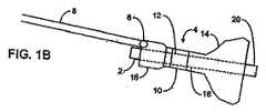
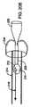
図1A−1Cは、それぞれ、幽門コーキング器具の1つのバリエーションにおける拡張を示す断面図であり、この器具は、胃の開口部、特に幽門弁を部分的及び/又は断続的に閉塞するよう構成されている。このような、特定のバリエーションにおいて、図1Aは、非拡張又は非膨張状態で幽門弁に送出及び/又は挿入する準備ができている器具4を示す。図1Bは、拡張状態における遠位の閉塞部材14を示す。使用時に、例えば、幽門部又は幽門部の向こう側に器具4を設置すると、遠位の閉塞部材14(又は「保持部材」)が、生体適合性のある様々な流体又はガス、例えば、食塩水、水、空気、窒素、等の流入によって、セルフシールの膨張ポート6に通じるチューブ8を介して膨張する。チューブ8は、カテーテル、内視鏡等といった、様々な送出チューブを有してよい。 1A-1C are cross-sectional views each showing an expansion in one variation of a pyloric caulking instrument that is configured to partially and / or intermittently occlude the stomach opening, particularly the pyloric valve. ing. In certain such variations, FIG. 1A shows the instrument 4 ready to be delivered and / or inserted into the pyloric valve in an unexpanded or non-expanded state. FIG. 1B shows the distal occlusion member 14 in the expanded state. In use, for example, when the instrument 4 is placed over the pylorus or beyond the pylorus, the distal occlusion member 14 (or “holding member”) is adapted to a variety of biocompatible fluids or gases such as saline. Inflated through the
遠位の閉塞部材14を、近位の閉塞部材16の材料に対してより簡単に膨張可能な材料で遠位の膨張部材14の膨張可能な部材を作製することによって、近位の閉塞部材16の膨張の前に膨張するよう構成してよい。閉塞部材14,16の作製時に使用する材料は、シリコーン、シリコーンエラストマー、ラテックス、ポリウレタン、PTFE、FEP、等といった、様々な材料を含んでよい。代替的に、一般に流体に接触することで膨張する発泡体又はヒドロゲルといった自己膨張材料を、閉塞部材14,16の中に使用してもよい。このような自己膨張材料を使用する場合、それらを閉塞部材14,16に配置してよく、食塩水といった流体を注入して材料を膨らませてもよい。様々な自己膨張材料を、近位の閉塞部材16よりもむしろ遠位の閉塞部材14に組み込んでもよく、膨張する材料によって与えられる半径方向の様々な圧力を受ける。 The
さらに、別の代替例では、膨張する何らかの骨格又は支持構造体を、各閉塞部材14,16の中に使用してもよい。このような骨格又は構造体を、形状記憶発泡体、形状記憶合金又はニチノールといった超弾性合金、又は形状記憶ポリマーで作製してもよい。骨格又は構造体を送出形態に圧縮して、自己膨張又は、例えば、電気、熱、RFエネルギ、等の活性化エネルギを供給することによって、所望の閉塞形状に拡張できるようにしてもよい。いずれのケースでも、遠位閉塞部材14を幽門弁の遠位に置き、その後で、その大きな形態に膨張又は拡張させてもよい。そして、図1Cに示すように、ポート6を介した注入によって近位の閉塞部材16が膨張又は拡張するポイントである幽門環に対して近位に引いてもよい。膨張又は拡張した双方の閉塞部材14,16とともに、双方を結合する架橋部材10を幽門に渡してもよい。架橋部材10は、幽門括約筋を大きく塞がない直径が最大8−10mmまでの1mm又はそれ以下といった様々な直径を有してよく、これにより、幽門括約筋、又は他の適した直径を一般に塞ぐ。 Furthermore, in another alternative, any expanding skeleton or support structure may be used in each
架橋部材10を、閉塞部材14,16が幽門弁に対するその位置を維持できるが、依然として部材14,16が移動できるのに十分な柔軟な長さを有するよう構成してもよい。近位の閉塞部材16は、幽門弁が完全閉塞した状態から、幽門弁を近位に移動させて遠位の閉塞部材14が部材16を移動させることができる程度まで、移動してよい。このような移動を、胃の管腔(胃)及び幽門弁を囲む筋肉の自然移動によって誘発させてもよい。このため、近位の閉塞部材16が近位に移動する場合、幽門弁をただ単に部分的に閉塞して、架橋部材10と弁との間の食べ物の断続的な通過が可能である。長い時間胃の中に食べ物を保持するため、患者があまり食べ物を取らないように、満腹感がすぐに始まって持続する。さらに、閉塞部材14,16の相対移動を可能とするため、架橋部材10は、閉塞部材14,16が幽門弁に対して近位又は遠位に移動するよう十分な公差を有した状態で幽門弁(又は胃の別の開口部)を通してその設置を可能にするのに十分な長さである。例えば、患者の幽門弁が約2cmの長さである場合には、架橋部材10は、例えば2cmよりも長く、好適には最大8cmの長さである。さらに、閉塞部材14,16が膨張又は拡張可能である一方で、架橋部材10そのものの直径が、膨張又は拡張するよう構成してもよい。 The bridging
好適には非常に目立つ目に見える色素又はマーカを、1又は双方の閉塞部材14,16に任意に注入して、安全対策としての役割を果たすようにしてもよい。代替的に、万一閉塞部材14,16が壊れた場合に閉塞部材14,16の色素又は断片が体を通過するときに目に見えるように、1又は双方の閉塞部材14,16を、任意に、非常に目立って見た目で組織と区別できる材料で作製してもよい。これにより、器具の破壊が起きたことが患者又は医師に示される。 Preferably, a very noticeable visible dye or marker may optionally be injected into one or both
別のバリエーションでは、器具をカバーする材料又は器具に組み込まれた材料に注入された持続放出性の薬剤を組み込んでもよい。任意の数のこれらの薬剤を、腸管への薬の放出によって又は患者との接触を介して、患者にゆっくりと注入してよい。代替的に、本器具が電気的な刺激技術を組み込んでよい。例えば、電気プローブを本器具の面から延ばして周囲の組織に挿入するか、あるいは、代わりに電極を器具の面の上に形成してよい。 In another variation, a sustained release drug injected into the material covering the device or the material incorporated into the device may be incorporated. Any number of these agents may be slowly infused into the patient by release of the drug into the intestinal tract or through contact with the patient. Alternatively, the device may incorporate electrical stimulation techniques. For example, an electrical probe may be extended from the surface of the instrument and inserted into the surrounding tissue, or alternatively an electrode may be formed on the surface of the instrument.
さらに別の代替例では、閉塞部材14,16を、1又は双方の部材14,16の上をカバーする侵食性又は生体分解性のカバーによって覆ってよい。このようなカバーを、1又は双方の部材14,16を拘束するよう構成してもよく、一旦本器具が胃の管腔に取り込まれ又は設置されると、周囲の流体に触れてカバーを自然に侵食することで、カバーされた閉塞部材が拡張又は膨張できる。別のバリエーションでは、近位及び遠位閉塞部材を、以下に詳細に説明するように、異なる速さ又は異なる環境でそれぞれ侵食するよう構成された異なる材料でそれぞれカバーしてもよい。 In yet another alternative, the
図1Aから図1Cに示す変形例では、器具4が、器具4を通して規定された任意の管腔18を有する。任意の管腔18は、入り口ポート2を通って管腔18に入り出口ポート20を取って管腔18を出る器具4を介して流体及び食べ物を通すことができる。管腔18を、器具4を通って少なくした量の食べ物を通すことができるよう構成してよく、このようなケースでは、図示する器具4を比較的短い架橋部材10で構成して、幽門に対する器具4の相対移動を抑制してもよい。このようなバリエーションでは、管腔18が、腸に胃の管腔74の内容物を活発に注ぎ込むか又は放出することができるよう構成される。このようなケースでは、器具4が幽門弁を閉塞しないよう移動できることが必要で無くなる。図示するように、任意のポンプ又はアクティブな絞り弁12を器具4に組み込んでよい。食べ物といった外からの物が胃の中にあることを検出したとき、又は内容物からの所定の圧力を検出したときに、ポンプ又は弁12が単に開くよう構成してよく、胃の内容物が管腔18及び弁12を通って通過できる。他の検出パラメータが、温度及びpHレベルを含んでよい。代替的に、ポンプ又は弁12を、ポンプ又は弁12、又は無線により患者又は医師によって外部から作動させることで自動的に作動するポンピング機構を介して、管腔18を通して胃の内容物をアクティブに注ぎ込むよう構成してよい。器具が弁12を具えて構成されたこのようなケースでは、バルブを一方向バルブとして構成してもよく、胃から腸管に流体及び食べ物が流れることができる。 In the variation shown in FIGS. 1A-1C, the instrument 4 has an
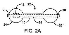
器具4は、近位の閉塞部材16の形状及び/又は全容積が、それが幽門弁を通って腸に入ってしまうのを防ぐのに十分であれば、どのような形状を有してもよい。図2Aから図2Dは、閉塞部材として使用できる、様々なバリエーションの形状の側面を示す。例えば、図2Aは、器具のバリエーション22の側面図を示しており、近位及び遠位閉塞部材24,26が、円形をした器具22によって規定される長手軸に沿った断面形状を有しており、球状の閉塞部材を形成する。近位及び遠位閉塞部材24,26を同じ大きさの直径で示しているが、所望の形状及び器具の構成に応じて直径を変えてもよい。例えば、近位閉塞部材24を、遠位閉塞部材26よりも大きな直径を有するよう構成してもよい。代替的に、あまり望ましくはないが、正反対の構成を有する器具を使用してもよい。さらに、所望の器具の構成に応じて、管腔28及びポンプ又は弁12を任意に含んでもよい。 The device 4 may have any shape as long as the shape and / or total volume of the
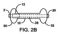
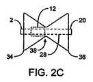
図2Bは、器具の別のバリエーションを示しており、近位及び遠位閉塞部材30,32が、楕円形の器具によって規定された長手軸に沿った断面形状を有し、楕円体を形成する。楕円形状の閉塞部材30,32の主軸は、好適には、このバリエーションでは器具の長手軸に対して垂直な方向を向いているが、様々な角度を向いていてもよい。図2Cは、近位及び遠位の閉塞部材34,36が三角形を成して円錐形の閉塞部材を形成するバリエーションを示す。このようなバリエーションでは、架橋部材38の長さが最小限であり、単に閉塞部材34,38が交わることで形成され、縊れ部を形成する。図2Dは、さらに別のバリエーションを示しており、近位及び遠位閉塞部材40,42をダイアモンド状に形成して、円錐形の閉塞部材のバリエーションを形成する。また、このようなバリエーションは、縊れ部44を形成する。 FIG. 2B shows another variation of the instrument where the proximal and
これらのバリエーションは、特定の形状を示しているが、これらは単に使用する様々なタイプの形状の説明を意図するものであり、限定することを意図するものではない。例えば、胃の開口部を閉塞して器具がそこを通って落ちるのを防ぐ働きをする長方形、正方形等といった任意の形状を使用してもよく、本開示の範囲内である。さらに、球状の遠位の閉塞部材及び円錐状の近位の閉塞部材を有する器具といった、一つの器具に対する閉塞部材として様々な形状の様々な組み合わせを使用してよい。 Although these variations show specific shapes, they are merely intended to illustrate the various types of shapes used and are not intended to be limiting. For example, any shape such as a rectangle, square, etc. that serves to occlude the stomach opening and prevent the device from falling therethrough may be used and is within the scope of the present disclosure. Further, various combinations of various shapes may be used as the occlusion member for a single instrument, such as an instrument having a spherical distal occlusion member and a conical proximal occlusion member.
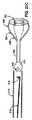
図3Aから図3Cは、幽門コーキング器具の別のバリエーションの断面図を示しており、本器具もまた、胃の開口部を断続的に閉塞するよう構成される。図1Aから図1Cに示す器具と同様に、このような特定のバリエーションは、器具46を通して規定される管腔の使用を省く。また、この器具46は、閉塞部材を拡張させるための上記の態様を含んでよい。例えば、拡張圧力を変える発泡体を使用して、例えば、食塩水又は水といった流体の器具46への注入時に、近位閉塞部材48の拡張に先立って、遠位閉塞部材50の拡張が確実に生じるようにしてもよい。器具46は、入り口ポート6を通って流入管8から流体が流入して、近位の閉塞部材48から遠位の閉塞部材50に中央部の管腔52を通って供給されるよう構成される。また、器具46を、以下に詳細に説明するように、図1Aから図2Cに示すような器具と同じ方法で設置してもよい。また、このようなバリエーションは、必要に応じて磁石が先端に付いた吸引カテーテルを単に設置することで器具46を取り外せるように、金属でできた膨張ポート6を組み込んでもよい。このカテーテルが適切に設置されると、吸引力を加えることによって器具が収縮し、幽門弁から器具46を簡単に取り外し易くなる。器具の膨張ポートの周りに金属リングが置かれると、先端に磁石の付いた吸引カテーテルを、患者の中を前に進めることができるか、又は経鼻胃チューブを使用して置くことができる。そして、センサが、磁石が金属リングに係合したことを表示すると、真空にして、感圧バリアが壊れるか又は単に真空力を加えることによって、器具全体が収縮する。このため、器具46を、内視鏡的又は経皮的な方法、例えば、oro−又はnano−gastric法によって、取り外すことができる。このようなバリエーションは、近位48及び遠位50の閉塞部材を接続する管腔52を有しており、このような管腔52を胃の中の空間を閉じないようにして、その代わりに、膨張液が液通するよう使用して閉塞部材48,50を膨張させてもよい。器具46の閉塞部材は、例えば、図1Aから2Dのような上記のような形状を有してよい。 3A-3C show cross-sectional views of another variation of the pyloric caulking device, which is also configured to intermittently occlude the stomach opening. Similar to the instrument shown in FIGS. 1A-1C, such a particular variation eliminates the use of a lumen defined through the
さらに、本器具の別のバリエーションを図4Aに示す。このようなバリエーションでは、器具54が、テーパの付いた架橋部材60を有している。架橋部材60を先細状にして、遠位閉塞部材58から近位閉塞部材56に向いたその長さ方向に沿って大きくしてもよい。テーパの付いた架橋部材60を、幽門弁を塞がないように器具54が移動し易いよう使用する。幽門弁が架橋部材60の周りで縮小すると、テーパが近位に器具を移動し易くする。閉塞部材56,58の大きさ及び形状のように、所望の結果に応じて、テーパの角度を変えてもよい。 Furthermore, another variation of the instrument is shown in FIG. 4A. In such variations, the instrument 54 has a tapered bridging
図4Bは、上記と同様な別のバリエーションを示す。このようなバリエーションでは、器具55が、架橋部材61を介して接続された円錐形の部材を有する閉塞部材57,59を有している。この架橋部材61は、器具55が幽門弁に対して相対移動できるのに互いに十分な距離で閉塞部材57,59を保持する長さを有している。器具55は、本書で開示した任意の方法を使用して閉塞部材57,59を膨張又は拡張させ、さらに器具55は、必要に応じて、中央管腔及びパッシブ又はアクティブバルブ又はポンピング機構を任意に組み込んでよい。 FIG. 4B shows another variation similar to the above. In such a variation, the
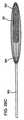
別の実施例では、遠位閉塞部材を完全に省略してもよい。例えば、図5Aは、代替的なバリエーション62の側面図を示しており、架橋部材66(又は「位置決め部材」)が、近位閉塞部材64からかなり長く、例えば、5cm又はそれよりも大きく延びている。架橋部材66を、幽門弁に隣接する近位の閉塞部材64によって所定の位置に保持する一方で、腸管の中、例えば、十二指腸に置いてもよい。幽門弁に対する近位の閉塞部材64の位置を、腸管壁を擦る架橋部材66によって発生する摩擦力によって維持してよい。閉塞部材64は、胃の収縮及び運動の際に、幽門弁を断続的に開放させる上記と同じような方法で作用してもよいが、架橋部材66の長さによって所定の位置に保持してもよい。架橋部材68の遠位端は、腸管の中で自由に動いてよいが、図5Bの器具70において示すように、それを腸壁に付けるための錘68又は多数のフック又は返し72によって任意に重くしてもよい。 In another embodiment, the distal occlusion member may be omitted entirely. For example, FIG. 5A shows a side view of an
さらに、本書に記載した器具の様々なバリエーションにおける特定の態様を様々に組み合わせて盛り込むことは、本開示の範囲内である。例えば、球状の近位の閉塞部材と円錐状の遠位の閉塞部材とを有する器具を使用してもよい。さらなる例として、本器具が様々な方法を組み込んで、近位の閉塞部材とは異なる方法で遠位の閉塞部材を膨張又は拡張させてもよい。さらに、本器具が一方の閉塞部材のみをカバーする生分解性を有し、器具に結合されたバルブ及び/又はポンプを器具の中に組み込んでもよく、さらには器具の長さにわたって規定された管腔を任意に有してもよい。これらの例は、本書に記載の様々なバリエーションの様々な態様を組み合わせることによって採用される様々な組み合わせの説明を単に意図するものであり、本発明の範囲内であることを意図するものである。 Further, it is within the scope of the present disclosure to incorporate various combinations of specific aspects of the various variations of the instrument described herein. For example, an instrument having a spherical proximal occlusion member and a conical distal occlusion member may be used. As a further example, the device may incorporate various methods to expand or expand the distal occlusion member in a different manner than the proximal occlusion member. In addition, the device may be biodegradable, covering only one occlusion member, and a valve and / or pump coupled to the device may be incorporated into the device, and further a tube defined over the length of the device. You may optionally have a cavity. These examples are merely intended to illustrate the various combinations employed by combining various aspects of the various variations described herein, and are intended to be within the scope of the present invention. .
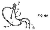
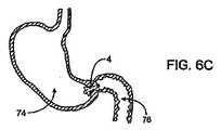
図6Aから図6Cは、胃の断面図であって、体内に摂取不可能な経鼻胃的(又は内視鏡的)な設置のための一つのバリエーション、すなわち器具4のアクティブなバリエーションを示す。器具4が食道78を通って送出される際に、それは、図6Aに示すように、任意のチューブ8によって位置決めされつつ、圧縮、収縮、又は縮小形態である。器具4を、閉塞部材によって幽門に架かるよう胃74及び十二指腸76のそれぞれに配置すると、図6Bに示すように、上記の方法を用いて器具4が膨張又は拡張する。そして、チューブ8を取り外して、図6Cに示すように、器具4を所定の位置に残す。 6A to 6C are cross-sectional views of the stomach, showing one variation for nasogastric (or endoscopic) placement that cannot be taken into the body, ie an active variation of the instrument 4. . When the device 4 is delivered through the
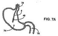
図7Aから図7Cは、胃の断面図であって、体内に摂取不可能な経鼻胃的(又は内視鏡的)な設置のための別のバリエーション、すなわち器具4のパッシブなバリエーションを示す。図7Aに示すように、圧縮、収縮、又は縮小形態にしながら、上記のように、器具46を食道78を通して進めてもよい。図7Bに示すように、器具46を、閉塞部材によって幽門に架かるよう胃74及び十二指腸76のそれぞれに配置すると、器具が膨張又は拡張して、図7Cに示すように、チューブ8を取り外して器具46を所定の位置に残す。 7A to 7C are cross-sectional views of the stomach showing another variation for nasogastric (or endoscopic) placement that cannot be taken into the body, ie a passive variation of the instrument 4. . As shown in FIG. 7A,
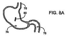
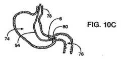
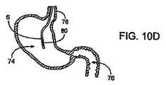
図8Aから図8Dは、胃の断面図であって、器具80のパッシブな(又は「自己拡張する」)実施例を配置するためのさらに別のバリエーションを示す。図8Aに示すように、器具80を単に摂取してもよい。胃74の中に入ると、胃液が、近位の閉塞部材82の膨張ポートを覆う、酸に鋭敏なコーティングを侵食する。コーティングが一旦分解すると、図8Bに示すように、近位の閉塞部材82を拡張又は膨張するよう構成する。拡張又は膨張が生じると、器具80が胃74の中に残り、図8Cに示すように、胃の自然収縮により縮小又は収縮状態で、最終的に遠位の閉塞部材84が十二指腸76を通る。遠位の閉塞部材84が十二指腸76に入ると、遠位の閉塞部材84を覆うアルカリに敏感なコーティングが侵食して、図8Dに示すように、器具が幽門弁に架かった状態で遠位の閉塞部材84の拡張又は膨張が生じる。遠位の閉塞部材84を覆うカバーを、それが十二指腸76に特有であるpHレベルが約6の酸性の環境に晒されるときのみ侵食するよう構成してもよい。取り外し易くするために、上記のように、所定の圧力レベルを受けると壊れるよう構成されたバリア88を具えた、中央の中空の管腔86によって2つの閉塞部材82,84を接続してもよい。従って、適切な圧力レベルを有する真空にすることでバリア88が壊れるよう構成して、器具80全体を収縮させてもよい。 FIGS. 8A-8D are cross-sectional views of the stomach and show yet another variation for placing a passive (or “self-expanding”) embodiment of the
図9Aから図9Dは、胃の断面図を示しており、経口器具90のパッシブなバリエーションの配置のためのさらに別のバリエーションを示す。このような代替的なバリエーションでは、器具90を経口摂取できる。図9Aに示すように、器具90が胃74の中に入ると、近位及び遠位閉塞部材82,92の双方が、それぞれ、図9B及び図9Cに示すように、膨張ポート又は器具90を覆う酸に敏感なコーティングの侵食で膨張するよう構成される。一旦膨張又は拡張すると、遠位閉塞部材92が、その小さな大きさ(拡張した幽門弁のほぼ直径の大きさである5−15mm)により最終的に通過する一方で、図9Dに示すように、近位の閉塞部材82が、例えば、15mmの直径又はそれよりも大きく、胃の幽門部の生理学的な制限による最大60mmの大きさの直径により胃74の中に残る。このように、ひとつの閉塞部材92を幽門弁を通過するのに十分小さく構成する一方で、近位の閉塞部材82を、胃74の中で膨張する双方の閉塞部材82,92で胃74の中に保持されるよう構成してもよい。閉塞部材の一方が、酸に敏感なコーティングでカバーされた膨張ポートを有する一方で、他方が腸に見られるpH(約6.0)で侵食する耐酸性のバー(bur)である。このように、本器具を摂取すると、閉塞部材の一方が、胃の中で器具を保持しながら拡張して、その後で胃の運動により収縮する残りの閉塞部材を腸の中に最終的に移動させる。第2の閉塞部材が腸管に接触すると、膨張ポートが腸の環境によって侵食されて、器具が幽門弁に架かりながら第2の部分がゆっくりと膨張する。 9A-9D show a cross-sectional view of the stomach, showing yet another variation for placement of a passive variation of the
多くの様々な代替例及びバリエーションを、説明したような自己拡張又は「パッシブな」幽門弁閉塞器具及び方法で採用してよい。いくつかの実施例では、患者が飲み込むようにするために、生分解性のコーティングを使用せずに、器具を折り畳み、圧縮し、又はそうでなければ、より小さな形態にしてもよい。食道を通って胃に入り次第、折り畳んだ器具を1又はそれ以上の形状記憶ニチノール支持リング又は他の自己拡張支持部材によって広げてもよい。飲み込む実施例では、器具が、この器具から食道に戻って患者の口に延びるヒモを有してよい。このようなヒモを胃の中で閉塞器具を保持するために伸びるまで使用することで、患者の胃及び/又は胃などに望みどおりに配置されない場合、閉塞器具を回収してもよい。ある実施例では、ヒモを飲み込んで胃の中で分解させてもよい。他の実施例では、飲み込んだ器具を幽門弁に接触させてもよいが、弁に架けるための架橋部材を有しなくてもよい。他のバリエーションが、様々な実施例に従って本発明の範囲内で考えられる。 Many different alternatives and variations may be employed with self-expanding or “passive” pyloric valve occlusion devices and methods as described. In some embodiments, the device may be folded, compressed, or otherwise smaller, without the use of a biodegradable coating, for the patient to swallow. As soon as it enters the stomach through the esophagus, the folded device may be expanded by one or more shape memory nitinol support rings or other self-expanding support members. In the swallowing embodiment, the device may have a string that extends from the device back to the esophagus and into the patient's mouth. Using such a string until stretched to hold the occlusive device in the stomach, the occlusive device may be retrieved if it is not placed as desired, such as in the patient's stomach and / or stomach. In some embodiments, the string may be swallowed and broken down in the stomach. In other embodiments, a swallowed device may be brought into contact with the pyloric valve but may not have a bridging member for hanging over the valve. Other variations are contemplated within the scope of the present invention according to various embodiments.
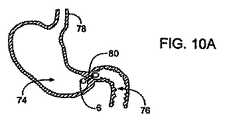
図10Aから図10Dは、器具80を取り外すためのひとつのバリエーションを示す胃74の断面図を示す(パッシブなバリエーションを示す)。器具80は、図10Aにおいて、胃74と十二指腸76との間に示されている。図10Bに示すように、先端に磁石の付いた吸引カテーテル又は内視鏡94を導入して、図10C又は図10Dに示すように、器具80を縮小又は除去してもよい。膨張ポート6がカテーテル94に接触する際に、カテーテル94が膨張ポート6に適切に接触しているかどうかを判断することを助けるものとして、先端が電気的に接触するよう構成してもよい。代替的に、器具80を内視鏡を通して取り外すか、又は時間とともに分解するよう構成して、最終的に腸に通してもよい。 10A through 10D show a cross-sectional view of the
他の実施例では、器具を収縮させるか又は潰すことによって閉塞器具を取り外し、カテーテル器具の管腔を通してそれを取り除いてもよい。ある実施例では、本器具を細かく切ってカテーテルの管腔を通して取り除いてもよい。さらに別の実施例では、本器具が時間とともに分解して幽門弁及び消化器官を安全に通過するようにしてもよい。本器具を除去又は通過させるためのあらゆる適切な代替例が、様々な実施例で可能である。 In other embodiments, the occlusion device may be removed by contracting or collapsing the device and removed through the lumen of the catheter device. In some embodiments, the device may be chopped and removed through the lumen of the catheter. In yet another embodiment, the device may break down over time and pass safely through the pyloric valve and digestive tract. Any suitable alternative for removing or passing the instrument is possible in various embodiments.
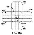
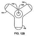
図11A及び11Bは、胃の中に単独で存在する本器具の代替的なバリエーションの平面図及び斜視図をそれぞれ示す。このような特定のバリエーションは、幽門を断続的に閉塞するよう構成された複数のプロング(prong)100,102,104,106,108,110を組み込んでいる。このようなバリエーションでは、拡張可能な材料96を適切に形成して、幽門を閉塞し易くする。収縮させることで本器具を幽門から取り出してもよいが、様々なプロングのうちの一つを通して再び挿入してもよい。本器具が、プロングの各組を通り幽門弁を完全に閉塞しないようにする複数の開口部98を規定してもよい。 11A and 11B show a top view and a perspective view, respectively, of an alternative variation of the device that is present alone in the stomach. Such a particular variation incorporates a plurality of
図12A及び12Bは、図11A及び図11Bに示す器具の別のバリエーションの側面図及び平面図をそれぞれ示す。このようなバリエーションでは、複数のプロング112,114,116,118のうちのいくつかを使用して、さらに、各プロングがそれを通る開口部120を規定してもよい。しかしながら、このようなバリエーションに示すように、各プロングを曲げ易くしてテーパを付けるか又は丸くし、周囲の組織を傷付けるのを防いでもよい。 12A and 12B show a side view and a plan view, respectively, of another variation of the instrument shown in FIGS. 11A and 11B. In such a variation, some of the plurality of
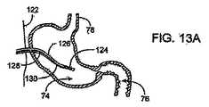
図13Aから図13Dは、本書に記載した器具の代替的な使用例の断面図を示す。このようなバリエーションでは、本器具を使用して、チューブを送る際に胃十二指腸逆流を防いでもよい。図示するように、器具124は、上記のバリエーションと同様であるが;しかしながら、このようなバリエーションでは、チューブを送るための器具124を通して規定される管腔132は、十二指腸76に置かれるよう構成された出口134を規定してもよい。また、器具124の近位部を、送りチューブ126及び膨張チューブ130に取り付けてもよい。送りチューブ126を使用して、十二指腸140に直接的に管腔132を通してチューブを送出する一方で、チューブを送る際に膨張チューブ130を使用して、膨張可能な幽門スパナ又は架橋部材136を膨張させ、送出した材料140の逆流を防いでもよい。また、器具124は、胃の内容物138を吸引するために用意される第3のチューブ128を組み込むことができ、送出した材料が肺に逆流するのを防いで胃74にかかる圧力を減らす。閉塞部材の近位部は、その膨張又は拡張状態を維持できるか又は時々圧力を減らして幽門弁への圧力を解放することができる。このようなバリエーションでは、経皮的な方法を示すが、経鼻胃的な方法又は他の方法も可能である。 Figures 13A through 13D show cross-sectional views of alternative uses of the instrument described herein. In such variations, the instrument may be used to prevent gastroduodenal reflux when feeding the tube. As shown, the
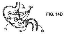
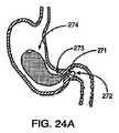
図14Aから図14Dは、本発明に係る器具のさらに別の代替的な使用の断面図を示す。図14Aから図14Cに示すように、器具90を、幽門弁を閉塞するよう置いてもよい。このうようなケースでは、摂取した器具90を示すが、器具90の設置が上記の方法のいずれかを介した形を取ってよい。図14Dに示すように、例えば、膨張可能な胃のバルーン、拡張可能な骨格といった追加的な1又はいくつかの胃の詰め物142、又は当技術分野で一般的に知られた、場所を占める任意の数の器具を使用してもよい。このようなバリエーションでは、器具90を置いてから胃の詰め物142を導入してもよい。器具90を使用して、胃の詰め物142が十分小さくなるまで胃の詰め物142が幽門弁を確実に通らないようにすることで、小腸が閉塞する付随的な危険性無しに分解不可能な物質を使用できる。 Figures 14A to 14D show cross-sectional views of yet another alternative use of the device according to the present invention. As shown in FIGS. 14A-14C,
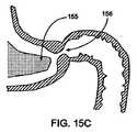
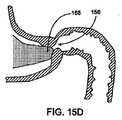
図15Aから図15Dは、幽門弁156を断続的に閉塞するためであって、このような実施例では胃の中のスペースに部分的に詰めるための器具150の別の実施例の使用を示す断面図である。図15Aは、非拡張又は非膨張状態で内視鏡、チューブ等といったカテーテル器具152を介した胃の中への送出及び/又は挿入の準備ができている器具150を示す。このような実施例では、本器具は、図15Bに示すように、器具150が胃の中で拡張する場合に拡張する拡張可能な発泡体154を有する。拡張可能な器具150及び/又は発泡体154のための無毒な適切な液体又はガスを膨張ポート158を通して導入してもよい。 FIGS. 15A-15D illustrate the use of another embodiment of the
適切な材料を使用して、器具150を形成してもよい。ある実施例では、例えば、器具150が、シリコーン、シリコーンエラストマー、ラテックス、ポリウレタン、PTFE、FEP及び/又はこれらと同等のもので作製した拡張可能なバルーンを具える。バルーンの内部の膨張可能な管腔に、無水架橋PEG、無水ヒドロゲル、又は他の膨潤性の塊、又は形状記憶発泡体、(ニチノールといった)形状記憶金属、又は形状記憶ポリマーといった形状記憶材料を具えたバットレス(buttressed)といった拡張可能な粘性材料を詰めてもよい。バットレス材料を、曲げることのできるバルーン材料の内部を含む、器具のどこかに置くことができる。代替的に、バルーンそのものの壁が、形状記憶材料を具えることができ、詰め物又はバットレスの必要性を無くす。自己拡張材料を使用する場合、それらをバルーンの内部に配置してもよく、バルーンに食塩水といった流体を注入して、材料を拡張させてもよい。 Appropriate materials may be used to form the
図15Bに示すように、器具150は、ある実施例では、近位部153と遠位部155とを有する。ある実施例では、近位部153が支持又は構造機能を有し、器具150が十分大きな断面直径を有して、器具150が幽門弁を確実に通過しないようにする。一般的に、遠位部155は、幽門弁156及び/又は幽門弁156に隣接する組織に接触するよう機能して、断続的及び/又は部分的に弁156を閉塞する。ある実施例では、遠位部155が幽門弁156の中、その周囲、又はその近傍の胃の組織に接触する場合に、組織を傷付けないように、遠位部155が従順な材料でできている。ある実施例では、近位部153及び遠位部155が同じ材料でできており、近位部153が、遠位部155と比較して多くの量の材料、大きな肉厚等を有する。 As shown in FIG. 15B, the
一般に、器具150は、図示するような不規則な楕円形、細長い球状、円錐状、ダイアモンド等といった多くの適切な形状を有してよい。ある実施例では、遠位部155が弁156に接触するよう合わせた状態で、器具150が幽門弁156に向かって自然に移動するように、形状を選択する。これら及び他の実施例では、器具の比重又は浮力を選択することによって、器具150が弁156にさらに移動し易くして、器具が胃の内容物を通過して弁156に向かって移動できるようにしてもよい。 In general, the
図15C及び図15Dは、幽門弁156と接触する器具150の遠位部155を示す。図示するように、遠位部155の形状を、弁156に接触するように移動するよう構成する。これは、一般に、胃の自然収縮の際に生じるため、幽門弁156の断続的な閉塞を与える。幽門弁156の断続的な閉塞により、胃の中の食べ物がより長く保持されるため、満腹感がより素早く開始してより長い間持続し、患者が食べ物をあまり食べなくなる。図15C及び図15dに示すような実施例では、遠位部155が弁156に接触すると、遠位部155が弁156を完全に閉塞する。代替的な実施例では、遠位部155が弁156を完全に閉塞しなくてもよく、幽門弁156を完全に縮小させた場合でさえも部分的に流れることができるよう構成された多くの様々な形態を有してよい。例えば、遠位部155が、(幽門に嵌合するよう構成された)突起部材等を具えた円錐形、楕円形、球状、ピラミッド形、管状、ディスク状といった形状を有してもよい。ある実施例では、遠位部155及び近位部153が、器具150の閉塞に拘わらず、いずれかの端部が幽門弁156を閉塞するように、同一又は、ほぼ同一の形状を有する。 15C and 15D show the
器具150は、多くの付加的な形態のうちのいずれかを有してよく、胃の中にそれを送出し易くし、幽門弁156を断続的に閉塞する機能を強化し、胃及び/又は胃などからそれを取り外し易くする。ある実施例では、例えば、器具150が、1又はそれ以上の放射線不透過性のマーカ、色素及び/又は材料を有しており、器具150を見易くする。また、器具150が、肉眼でそれを見易くする他のマーカ、色素、又は材料を有してもよく、器具150が体内で分解又は体内を通過するような実施例において、又は万一器具150が壊れたり又は破れたりした場合には安全機能として、好都合である。 The
ある実施例では、器具150が、胃又は幽門弁の向こう側の小腸に1又はそれ以上の薬剤を放出するための1又はそれ以上の機構を有してよい。例えば、持続放出性の薬剤を、器具150をカバーする物質、又は器具150を構成するために使用する物質と組み合わせ又はこれらの物質に注入してもよい。これらの薬剤は、多くの治療用又は診断用薬剤であってよく、腸管への薬の放出によって、又は患者との接触を通じて、患者にゆっくりと与える。他の実施例では、器具150が電気的な刺激技術を組み込んでもよい。例えば、電気プローブを器具150の面から延ばして周囲の組織に挿入するか、又は電極を器具150の面の上に形成してもよい。 In certain embodiments,
ある実施例では、器具150に侵食可能又は生分解性のカバーでカバーをして胃の中に送出してもよい。このようなカバーを、器具150を拘束するよう構成してもよく、一旦カバーが胃の管腔の中で物質に接触すると、カバー自然に壊れて分解することで、器具150を解放して器具150が拡張できる。ある実施例では、器具150を、胃の中で様々な速さ又は様々な化学的環境でそれぞれ侵食するよう構成された様々な材料でカバーをしてもよい。 In some embodiments,
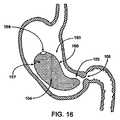
図16は、破裂が生じている図15Aから図15Dの器具150を示す。この図で示すように、器具150全体の形状を、拡張した発泡体154(又は他の実施例では、器具150の中又は器具150に設けられた他の骨格物質等)によって維持する。一般に、発泡体又は骨格物質は、胃の中でそれが分解するのを防ぐため耐酸性であることで、破裂が生じた後長時間にわたってそれが器具150を支持できる。代替的な実施例では、発泡体154又は他の骨格物質が、破裂の後でゆっくりと分解する一方で、検便の際に破裂を患者に報知するシグナル物質を放出する。そして、患者が医師に器具150が取り除かれたことを知らせる。 FIG. 16 shows the
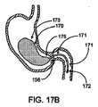
ここで、図17A及び図17Bを参照すると、幽門弁閉塞器具160の別の実施例が、膨張ポート168と、近位部163と、遠位部165と、位置決め部材161と、保持部材162とを有する。ある実施例では膨張を必要とし他の実施例では必要としないため、当然のことながら、膨張ポート168は任意である。位置決め部材161は、一般に、幽門弁156を断続的に閉塞するための場所に器具160を設置し易くする。保持部材162は、器具160の場所又は位置を保持し易くする。 Referring now to FIGS. 17A and 17B, another example of a pyloric
ある実施例では、位置決め部材161が中空であるため、流体及び又はガスが器具を通って通過でき、近位部163、遠位部165及び保持部材162が膨張できる。ある実施例では、位置決め部材161を比較的短くして、幽門156に対する遠位部165の移動を規制してもよい。他の実施例では、位置決め部材161をもっと長くして器具160をもっと移動させることができる。 In one embodiment, the positioning
ここで図17Bを参照すると、別の実施例では、近位部173及び遠位部175を有する器具170が、遠位端に膨張ポート172を有する位置決め部材171に結合されている。このような実施例では、器具170が非膨張状態で胃を通過し、位置決め部材171及びポート172を使用して器具170を膨張させ、その後、位置決め部材を飲み込んで位置決め部材が幽門弁156を通過し、小腸の初めの部分で安全に保持される。別の実施例では、器具を胃の中に置く一方で、口に向けて食道を上に延びる外すことのできるヒモを器具に取り付けてもよい。器具が適切に配置されていない場合、ヒモを使用して器具を取り外すことができ、又は代替的に、器具を胃の中の所定の位置に置いた時点でそれを器具から取り外すことができる。 Referring now to FIG. 17B, in another embodiment, an
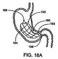
図18A及び図18Bに示すように、さらには前述のように、幽門弁を閉塞するための器具の様々な実施例が、多くの様々な拡張可能な支持機構を有する。前述のように、このような実施例は発泡体を含むが、自己拡張ケージ(cage)、コイル、格子、骨格等といった、他の支持構造及び材料を使用してもよい。また、図18Aでは、膨張ポート188とともに近位部183及び遠位部185を有する器具180が、拡張する骨格184をも有しており、骨格184を器具180の内面又は外面の壁と一体にするか、又は骨格184を壁の中に組み込んでもよい。このような拡張可能な骨格184をニチノールといった形状記憶又は超弾性材料で構成してもよい。骨格184を送出形態に圧縮した後に、自己拡張によって所望の閉塞形状に拡張できるか、又は電気エネルギ、熱、RFエネルギ等といった活性化エネルギを供給することによって拡張できる。別の実施例では、引っ張り器具で拡張形態に骨格を引くことによって骨格を配置してもよく、このような実施例では、骨格が歯止め機構を有してよく、骨格がその元々の形態に潰れるのを防ぐ。 As shown in FIGS. 18A and 18B, and as previously described, various embodiments of instruments for occluding the pyloric valve have many different expandable support mechanisms. As mentioned above, such embodiments include foam, but other support structures and materials may be used, such as self-expanding cages, coils, lattices, skeletons, and the like. Also in FIG. 18A,
図18Bに示す実施例では、器具190が、近位部193と、遠位部195と、膨張ポート198とを有する。このような実施例では、器具190の壁194が、形状記憶、超弾性又はそうでなければ自己拡張材料でできており、拘束から解放されると、小さな形態から大きな形態に拡張する。そして、壁194の材料が、その拡張形状を保持するため、器具190の形状を維持して器具が潰れるのを防ぐ。 In the embodiment shown in FIG. 18B,
図19A及び図19Bを参照すると、幽門弁閉塞器具200の別の実施例が、移動可能又は「反転した」外側シェル204と、内側コア202と、位置決め部材208と、穴212又は他の面形態を有する遠位保持部材210とを有する。図19Aに、幽門弁を断続的に閉塞するための拡張形態の器具200を示し、図19Bに、胃の中に送出するためのその潰れた形態を示す。シェル204は、組織接触/係合部205と支持部206とを有する。一般に、支持部206は、器具200が幽門を通過できないよう支持部206が器具200の断面直径を維持し易くするように、組織接触部205よりもより強固で/堅く、その一方で、組織接触部205は、顕著な損傷を引き起こさずにそれが胃の組織に接触できるように、より柔軟である。 Referring to FIGS. 19A and 19B, another embodiment of the pyloric
器具200の様々な部品を、上述の材料、又は現在知られあるいは今後発見される他の適した材料、といった適切な材料で構成してよい。ある実施例では、内側コア202が、シリコーンといった中実の材料であるが、他の実施例では、コア202が中空でもよい。コア202は、適切な大きさ、形状、断面直径等を有する。ある実施例では、コア202が、約5mmから約30mmの間、好適には約10mmの断面直径を有する。シェル204を、コア202と同じか又は異なる材料で作製してもよく、また、適切な大きさ、形状、断面直径等を有する。ある実施例では、シェル204の支持部206が、組織接触部205よりも厚い。他の実施例では、支持部206を組織接触部205とは異なる材料で作製してもよい。 The various parts of the
位置決め部材208は、内側コア202、シェル204又はこれら双方の延長線上にあってよく、又はその代わりに内側コア202及び/又は外側シェル204に結合された別の部品であってもよい。位置決め部材208は、それが幽門弁を通過できるよう、適切な長さ及び直径を有する。ある実施例では、その断面直径が、約1.0cm又はそれよりも小さく、その長さが約3.0cm又はそれよりも大きい。また、保持部材210は、適切な大きさ、形状又は形態を有しており、ある実施例では拡張可能で、またある実施例では自己拡張し、さらに他の実施例では全く拡張しないよう構成される。ある実施例では、保持部材210が、約30mm又はそれよりも小さい、好適には約25mm又はそれよりも小さい、さらに好適には約21mm又はそれよりも小さい、大きな断面直径を有する。保持部材210の穴212又は面形態は、器具200の送出、調整及び/又は回収のために、保持部材にアクチュエータ又は他の器具を取り付けることができるような形態を有する。位置決め部材208及び保持部材210の双方を、適切な材料で作製する。 The positioning
縮尺通りではないが、図19Bは、器具200が潰れた状態又は反転した状態を示す。このような形態では、シェル204が、送出チューブ又はカテーテルといったものを介した送出のために、より小さな断面直径に圧縮される。器具200を胃に送出した後にシェル204をその拡張状態に反転させ、その後で器具200が幽門弁を断続的に閉塞するよう作用する。 Although not to scale, FIG. 19B shows the
図20Aから図20Cは、図19A及び図19Bの器具200を胃に送出及び配置するための方法を示す。図20Aでは、器具200が、潰れた形態で送出チューブ214又はカテーテルの管腔の中に収容されている。図20Bでは、器具が送出チューブの中から部分的に外に出ることで、シェル204が少なくとも部分的に拡張できる。そして、保持部材210に穴216を通して掛けられたアクチュエータ216を使用して、シェル204が送出チューブ214の遠位端に重なるように、器具200を引き戻す。そして、送出チューブ204の遠位端を使用してシェル204に力を加えることで、図20Cに示すように、シェル204がその拡張状態に反転する。また、図20Cに示すように、アクチュエータ216が、保持部材210の穴212に取り付けるためのフック218を有してよい。シェル204をその拡張形態に移動させると、そのような形態にとどまるよう構成されているため、幽門弁への接触及び上記のような器具保持機能を与える。ある実施例では、送出チューブ214が、その遠位端又は遠位端の近くに、拡張可能なバルーン(図示せず)を有してもよい。このバルーンは、円周方向に膨張するドーナツ状でもよく、又は偏心した形状又は他の適切な形状を有してもよい。バルーンを膨張させて器具200を引っ張る際のストッパとして機能させてもよい。代替的に、バルーンを、器具200の下又は器具200の中で膨張させて、バルーンが膨張する際に器具200を反転させてもよい。 20A-20C illustrate a method for delivering and placing the
他の実施例では、他の適切な方法を使用して器具を送出及び/又は配置してもよい。例えば、ある実施例では、シェル204が、アクチュエータ216又は送出器具214の遠位端を使用せずに、その収縮/潰れた状態からその拡張状態に、「自己反転」してもよい。自己反転を、形状記憶又はバネ材料等で行うか、又は器具の剛性に偏りを作るようなシェルの形状によって行ってもよい。別の実施例では、器具200を、畳んだ状態で又はそうでなければ潰れた状態で又は溶解カプレットの中に収容して飲み込んでもよい。多くの様々な代替的な実施例が可能である。 In other embodiments, the device may be delivered and / or positioned using other suitable methods. For example, in certain embodiments, the
図21A及び図21Bは、器具の一つのバリエーションの導入及び拡張を示す。図21Aでは、バルーン251を非拡張又は非膨張状態で内視鏡検査用チューブ252によって導入する。バルーンの内部に形状記憶発泡体253がある。図21Bでは、バルーン251を完全に挿入して形状記憶発泡体253をその形状記憶に従って拡張させる。上述のように、バルーンをシリコン、シリコンエラストマー、ラテックス、ポリウレタン、PTFE、FEP、又は他の材料で作製することができる。バルーンの内部を、食塩水といった流体に触れると拡張する発泡体又はヒドロゲルといった自己拡張材料にすることができる。代替的に、バルーンを膨張ポート254を介して無害の流体又はガスで満たすことによって拡張できる。バルーンの遠位閉塞部255が幽門弁256を閉塞する。 21A and 21B show the introduction and expansion of one variation of the instrument. In FIG. 21A, the
図22A及び図22Bでは、幽門弁256に接触するよう移動する2つの連続的な姿勢における閉塞部255を示す。目に見える色素又はマーカ、好適には非常に目立つものを、安全対策としてバルーン251の中に注入できる。代替的に、バルーンそのものを非常に目立つ材料で作製することで、万が一バルーンが壊れた場合に、色素又はバルーンの破片が体内を通るときに目に見えるように、組織と視覚的に区別でき、壊れたことを患者又は医師に知らせる。 22A and 22B show the
また、バルーンを、侵食され得る又は生体分解性のカバーでカバーすることができ、バルーンが胃の管腔の中に摂取されるか又は置かれて、胃液がこのカバーを侵食することでバルーンが拡張又は膨張できるまで、カバーがバルーンを拘束する。また、バルーンを、異なる速さ又は異なる環境で侵食するよう構成された材料でカバーしてもよい。 The balloon can also be covered with an erodable or biodegradable cover, and the balloon is ingested or placed in the stomach lumen and gastric fluid erodes the cover so that the balloon is eroded. A cover restrains the balloon until it can be expanded or inflated. The balloon may also be covered with materials configured to erode at different speeds or in different environments.
図23では、バルーン253に破裂が生じるにもかかわらず、バルーンの中に保持される形状記憶発泡体又は拡張可能な内部の材料によって、バルーンの輪郭を保持する。発泡体又は材料は、好適には分解を防ぐよう耐酸性であり、破裂の後に長期間にわたってバルーンの壁を支持できる。代替的に、中に詰め物があるバルーンが、破裂の後で、検便時にその破裂を患者に知らせるためのシグナル材料を放出しながらゆっくりと分解する。 In FIG. 23, the balloon contour is retained by a shape memory foam or expandable internal material retained in the balloon, despite the
図24Aでは、幽門貫通ヒモ271がバルーンに取り付けられており、遠位閉塞部材272がヒモ271の反対側の端部に取り付けられている。ヒモ271は、幽門の近くのバルーン274の遠位端273を保持して、幽門を閉塞する位置にバルーンを向かわせる。比較的短いヒモ271が、幽門に対するバルーンの閉塞(遠位)端273の移動範囲を制限する。 In FIG. 24A, a pyloric
図24Bでは、バルーン281が、その遠位端に取り付けられたヒモ282と、ヒモ282の遠位端の膨張ポート284とを有する。このような器具を使用するために、膨張ポート284を飲み込まずにバルーン281を飲み込んで、患者の口の中に膨張ポートを残す。バルーンが膨張ポートを通して膨張すると、患者がヒモ282を簡単に摂取でき、幽門285の向こう側に最終的に移動して、幽門弁の領域でバルーンの遠位端283を保持し易くする。 In FIG. 24B,
さらなるバリエーションを図25A及び図25Bに示す。図25Aでは、バルーン291が、その形状を規定する内部の拡張可能なケージ(caging)292を収容している一方で、図25Bでは、バルーン293が、形状記憶材料でできた外側シェル294を含む。 Further variations are shown in FIGS. 25A and 25B. In FIG. 25A, the
器具のさらなるバリエーション及びその使用を図26A、図26B及び図26Cに示す。これらの図におけるこの器具は、機能部品302を中で保持する、弾力性のある格子又はケージといった形状記憶部品301である。その緩和した姿勢では、ここでは球状で示す(図26A)形状記憶部品301の容積は大きくて幽門を通過できないため、胃の中で保持されるが、開放構造であるため胃から幽門を通って腸に流れる消化物との緩衝を避ける。また、部品に長手方向の拡張力を加えて保持することによって、図26Cに示すように、形状記憶部品301を変形姿勢に細長くできる。このような変形姿勢では、器具を食道を通して胃の中に挿入できる。機能部品302は、緩和及び細長い双方の形態の形状記憶部品301の中に収まる。機能部品302は、薬ポンプ、胃酸分泌刺激薬、又は他の送出用又はそうでなければ治療用器具である。内部の延長ロッド304を収容する内視鏡器具303であって、内視鏡器具について一般的且つ知られた標準的な外部の手段(図示せず)によりロッドがこの器具に対して前後に移動できる器具によって、形状記憶部品301の操作を行う。内視鏡器具303及び延長ロッド304双方の遠位端に、鉗子、スネア(snare)、などといった、把持部品を取り付けてもよい。患者の胃の中に器具を挿入するために、医師が内視鏡器具の遠位端に器具を取り付けると、この器具の遠位端が形状記憶器具301の近位端に取り付けられ、内部のロッドの遠位端が機能部品302の近位端に取り付けられる。そして、図26Cに示すように、医師が細長い形状記憶部品301にロッド304を延ばすことで、胃の中に器具全体を挿入できるように、その直径を小さくする。一旦挿入されると、ロッド304が医師によって引っ込められ、図26Bのような中間形態を介して図26Aのような緩和形態になる。胃から器具を取り外すときは、逆の手順、すなわち、ロッド304が引っ込んだ内視鏡器具303を(食道を通して)胃の中に挿入し、挿入した後に把持部品を器具の部品に係合するよう操作することによってなされる。これらの部品が係合して、ロッドが拡張すると、形状記憶部品が拡張且つ変形することで、器具全体を胃から取り外すことができる。 Further variations of the instrument and its use are shown in FIGS. 26A, 26B and 26C. The instrument in these figures is a
図26A、図26B、図26Cに示す器具に関するさらなるバリエーションでは、形状記憶部品301を、形状記憶を具えていないか又は歪んだ後に完全に実現するのよりも小さい形状記憶を具えた、同じ形態の比較的柔軟なケージに代えてもよい。ケージの遠位端305を機能部品の遠位端306に接続する一方で、近位端307,308をそれぞれ、接続及び取り外し可能、すなわち、可逆的に接続可能にする。近位端307,308を接続すると、図26Aに示すように、ケージ301が図示する形態に固定されるため、その大きな直径により胃の中に保持可能である。近位端を取り外すと、ケージ301及び機能部品302が、取り外し可能、挿入可能、又は全体的に操作可能である。近位端の係脱を、鉗子、捻るための把持部品、又は切断部品といった内視鏡器具によって行うことができる。ある実施例では、取り外し可能且つ体外から監視可能なラベルを、機能器具302、ケージ301、又はこれら双方の近位端に付けて操作し易くする。 In a further variation on the device shown in FIGS. 26A, 26B, and 26C, the
さらなる実施例では、形状記憶部品又は柔軟なケージ301そのものが、すなわち、付加的な機能部品302無しで、場所を占める器具として有用であり、胃の中での容積を減らす。これは、胃の中で保持できる食べ物の量を減らすことによって、胃壁を通して吸収できる量を減らす減量手段としての機能を果たす。 In a further embodiment, the shape memory component or
本発明に係る好適な実施例を上述したが、様々な変更及び修正を行ってもよいことは、当業者にとって明らかである。添付の特許請求の範囲は、本発明の正確な精神及び範囲内に含まれる、このような変更及び修正の全てを意図するものである。 While the preferred embodiment of the present invention has been described above, it will be apparent to those skilled in the art that various changes and modifications may be made. The appended claims are intended to cover all such changes and modifications as fall within the true spirit and scope of the invention.
Claims (24)
Translated fromJapanese前記器具が、形状記憶部品が十分に拡張して前記形状記憶部品が幽門を通過しないような拡張形態から、患者の食道を通して前記形状記憶部品を挿入及び取り外しできる小径の形態に換えることが可能な貫流形状記憶部品と、
前記拡張形態及び前記小径の形態の双方の前記形状記憶部品の中に収容するのに十分小さな大きさの治療部品と、
患者の外部から前記拡張形態及び前記小径の形態との間で前記形状記憶部品を操作するための手段と、
を具えることを特徴とする器具。An instrument for holding a therapeutic component in a patient's stomach without obstructing fluid flow through the stomach,
The instrument can be changed from an expanded configuration in which the shape memory component is sufficiently expanded so that the shape memory component does not pass through the pylorus to a smaller diameter configuration in which the shape memory component can be inserted and removed through the patient's esophagus. A once-through shape memory component;
A treatment component sized sufficiently small to be housed in the shape memory component in both the expanded configuration and the small diameter configuration;
Means for manipulating the shape memory component between the expanded configuration and the small diameter configuration from outside the patient;
A device characterized by comprising.
前記器具が、幽門を通過できない十分大きな拡張形態から、患者の食道を通して貫流部品を挿入及び取り外しできる拡張形態に換えることが可能な貫流部品と、
前記拡張形態及び非拡張形態の前記貫流部品の中に収容するのに十分小さな大きさの治療部品と、
前記貫流部品が胃の中にあるときに前記拡張形態及び非拡張形態との間で前記貫流部品を操作するための手段と、
を具えることを特徴とする器具。An instrument for holding a therapeutic component in a patient's stomach without obstructing fluid flow through the stomach,
A flow-through component capable of changing from a sufficiently large expanded configuration in which the instrument cannot pass through the pylorus to an expanded configuration in which the flow-through component can be inserted and removed through the patient's esophagus;
A treatment component sized sufficiently small to be received in the flow-through component in the expanded and non-expanded configurations;
Means for manipulating the flow-through component between the expanded and non-expanded configurations when the flow-through component is in the stomach;
A device characterized by comprising.
これにより、前記貫流部品が前記拡張形態で固定されることを特徴とする請求項2に記載の器具。Furthermore, it comprises reversible attachment means for reversibly attaching the flow-through part to the treatment part,
Thereby, the said flow-through component is fixed in the said expansion | deployment form, The instrument of Claim 2 characterized by the above-mentioned.
前記器具が、形状記憶部品が十分に拡張して前記形状記憶部品が幽門を通過しないような拡張形態から、患者の食道を通して前記形状記憶部品を挿入及び取り外しできる小径の形態に換えることが可能な形状記憶部品と、
前記拡張形態及び前記小径の形態の双方の前記形状記憶部品の中に収容するのに十分小さな大きさの治療部品と、
患者の外部から前記拡張形態及び前記小径の形態との間で前記形状記憶部品を操作するための手段と、
を具えることを特徴とする器具。A device for holding a therapeutic component in a patient's stomach,
The instrument can be changed from an expanded configuration in which the shape memory component is sufficiently expanded so that the shape memory component does not pass through the pylorus to a smaller diameter configuration in which the shape memory component can be inserted and removed through the patient's esophagus. Shape memory parts,
A treatment component sized sufficiently small to be housed in the shape memory component in both the expanded configuration and the small diameter configuration;
Means for manipulating the shape memory component between the expanded configuration and the small diameter configuration from outside the patient;
A device characterized by comprising.
前記器具が、幽門を通過できない十分大きな拡張形態から、患者の食道を通して貫流部品を挿入及び取り外しできる非拡張形態に換えることが可能な膨らませることのできない部品と、
前記拡張形態及び前記非拡張形態の双方の前記貫流部品の中に収容するのに十分小さな大きさの治療部品と、
前記貫流部品が胃の中にあるときに前記拡張形態及び前記非拡張形態との間で前記貫流部品を操作するための手段と、
を具えることを特徴とする器具。A device for holding a therapeutic component in a patient's stomach,
A non-inflatable part that allows the device to change from a sufficiently large expanded configuration that cannot pass through the pylorus to a non-expanded configuration that allows the flow-through component to be inserted and removed through the patient's esophagus;
A treatment component sized small enough to be accommodated in the flow-through component in both the expanded configuration and the non-expanded configuration;
Means for manipulating the flow-through component between the expanded configuration and the non-expanded configuration when the flow-through component is in the stomach;
A device characterized by comprising.
これにより、前記器具が前記拡張形態で固定されることを特徴とする請求項6に記載の器具。Furthermore, it comprises reversible attachment means for reversibly attaching the flow-through part to the treatment part,
Accordingly, the instrument is fixed in the expanded configuration.
前記電極が、胃の組織、幽門又は十二指腸に接触することを特徴とする請求項5に記載の器具。And further comprising at least one electrode coupled to the device,
6. The device of claim 5, wherein the electrode contacts stomach tissue, pylorus or duodenum.
前記電極が、胃の組織、幽門又は十二指腸に接触することを特徴とする請求項6に記載の器具。And further comprising at least one electrode coupled to the device,
The device of claim 6, wherein the electrode contacts stomach tissue, pylorus or duodenum.
前記器具が、貫流部品が患者の食道を通って患者の胃の中に挿入されるよう断面が十分小さな非拡張形態から、胃の中に挿入されると前記貫流部品の断面が幽門を通過するには大き過ぎる拡張形態に換わることが可能な貫流部品と、
前記貫流部品が胃の中にあるときに前記非拡張形態から前記拡張形態に前記貫流部品を切り換えるための手段と、
を具えており、
前記非拡張形態から前記拡張形態への切り換えを、前記拡張形態に形状記憶材料又は内視鏡による固定によって行い、
取り外しのための前記拡張形態から前記非拡張形態への反転が、前記器具の内視鏡的な操作を要することを特徴とする器具。An instrument for reducing the effective volume of the stomach,
When the device is inserted into the stomach from an unexpanded configuration that is sufficiently small in cross-section such that the flow-through component is inserted through the patient's esophagus into the patient's stomach, the cross-section of the flow-through component passes through the pylorus. A flow-through component that can be replaced with an expanded form that is too large,
Means for switching the flow-through component from the non-expanded configuration to the expanded configuration when the flow-through component is in the stomach;
With
Switching from the non-expanded configuration to the expanded configuration is performed by fixing the expanded configuration with a shape memory material or an endoscope,
Reversing from the expanded configuration to the non-expanded configuration for removal requires an endoscopic operation of the device.
前記支持部材が、リング、コイル、ケージ(cage)、支柱(struts)、骨格(scaffolding)、バスケット、スポーク及び傘(umbrella)から成る群から選択されることを特徴とする請求項14に記載の器具。The self-expanding material comprises at least one support member for preventing the support part from being crushed;
15. The support member according to claim 14, wherein the support member is selected from the group consisting of a ring, a coil, a cage, struts, scaffolding, a basket, a spoke, and an umbrella. Instruments.
前記組織接触部が、前記組織への接触による前記組織の損傷を防止するよう十分に従順であることを特徴とする請求項14に記載の器具。The tissue contacting portion is configured to temporarily form a seal of the pyloric valve when touching tissue near the pyloric valve;
15. The device of claim 14, wherein the tissue contacting portion is sufficiently compliant to prevent damage to the tissue due to contact with the tissue.
Applications Claiming Priority (2)
| Application Number | Priority Date | Filing Date | Title |
|---|---|---|---|
| US11/215,430US20090259236A2 (en) | 2003-07-28 | 2005-08-29 | Gastric retaining devices and methods |
| PCT/US2006/033923WO2007027812A2 (en) | 2005-08-29 | 2006-08-29 | Gastric retaining devices and methods |
Publications (1)
| Publication Number | Publication Date |
|---|---|
| JP2009508544Atrue JP2009508544A (en) | 2009-03-05 |
Family
ID=37809481
Family Applications (1)
| Application Number | Title | Priority Date | Filing Date |
|---|---|---|---|
| JP2008529243APendingJP2009508544A (en) | 2005-08-29 | 2006-08-29 | Stomach retention device and method |
Country Status (7)
| Country | Link |
|---|---|
| US (5) | US20090259236A2 (en) |
| EP (1) | EP1919370A4 (en) |
| JP (1) | JP2009508544A (en) |
| AU (1) | AU2006284801B2 (en) |
| BR (1) | BRPI0617059A2 (en) |
| CA (1) | CA2620859C (en) |
| WO (1) | WO2007027812A2 (en) |
Cited By (4)
| Publication number | Priority date | Publication date | Assignee | Title |
|---|---|---|---|---|
| JP2010537790A (en)* | 2007-09-07 | 2010-12-09 | バロノバ, インコーポレイテッド | Apparatus and method of use for intermittent blockage of gastric opening |
| JP2012522595A (en)* | 2009-04-03 | 2012-09-27 | メタモディクス インコーポレイテッド | Modular gastrointestinal prosthesis |
| KR20150052009A (en)* | 2012-08-17 | 2015-05-13 | 심플 메디컬 이노베이션스 피티와이 리미티드 | Intragastric balloon |
| JP2021178060A (en)* | 2020-05-14 | 2021-11-18 | 勇 佐伯 | Esophageal atresia treatment tool |
Families Citing this family (208)
| Publication number | Priority date | Publication date | Assignee | Title |
|---|---|---|---|---|
| ES2435094T3 (en) | 2000-05-19 | 2013-12-18 | C.R. Bard, Inc. | Device and method of tissue capture and suturing |
| CA2417129A1 (en)* | 2000-08-11 | 2002-02-21 | Temple University Of The Commonwealth System Of Higher Education | Obesity controlling method |
| US7737109B2 (en)* | 2000-08-11 | 2010-06-15 | Temple University Of The Commonwealth System Of Higher Education | Obesity controlling method |
| US7097665B2 (en)* | 2003-01-16 | 2006-08-29 | Synecor, Llc | Positioning tools and methods for implanting medical devices |
| CN101810521B (en) | 2001-08-27 | 2015-05-13 | 辛尼科有限责任公司 | Satiation devices and methods |
| US6675809B2 (en) | 2001-08-27 | 2004-01-13 | Richard S. Stack | Satiation devices and methods |
| US7146984B2 (en)* | 2002-04-08 | 2006-12-12 | Synecor, Llc | Method and apparatus for modifying the exit orifice of a satiation pouch |
| US8845672B2 (en) | 2002-05-09 | 2014-09-30 | Reshape Medical, Inc. | Balloon system and methods for treating obesity |
| US7901419B2 (en)* | 2002-09-04 | 2011-03-08 | Allergan, Inc. | Telemetrically controlled band for regulating functioning of a body organ or duct, and methods of making, implantation and use |
| US7837669B2 (en)* | 2002-11-01 | 2010-11-23 | Valentx, Inc. | Devices and methods for endolumenal gastrointestinal bypass |
| US7794447B2 (en)* | 2002-11-01 | 2010-09-14 | Valentx, Inc. | Gastrointestinal sleeve device and methods for treatment of morbid obesity |
| US20090149871A9 (en)* | 2002-11-01 | 2009-06-11 | Jonathan Kagan | Devices and methods for treating morbid obesity |
| US8070743B2 (en)* | 2002-11-01 | 2011-12-06 | Valentx, Inc. | Devices and methods for attaching an endolumenal gastrointestinal implant |
| US20060015125A1 (en)* | 2004-05-07 | 2006-01-19 | Paul Swain | Devices and methods for gastric surgery |
| US7037344B2 (en)* | 2002-11-01 | 2006-05-02 | Valentx, Inc. | Apparatus and methods for treatment of morbid obesity |
| US9060844B2 (en) | 2002-11-01 | 2015-06-23 | Valentx, Inc. | Apparatus and methods for treatment of morbid obesity |
| US20040143342A1 (en) | 2003-01-16 | 2004-07-22 | Stack Richard S. | Satiation pouches and methods of use |
| US20090259236A2 (en) | 2003-07-28 | 2009-10-15 | Baronova, Inc. | Gastric retaining devices and methods |
| US8048169B2 (en)* | 2003-07-28 | 2011-11-01 | Baronova, Inc. | Pyloric valve obstructing devices and methods |
| US9700450B2 (en)* | 2003-07-28 | 2017-07-11 | Baronova, Inc. | Devices and methods for gastrointestinal stimulation |
| US8821521B2 (en)* | 2003-07-28 | 2014-09-02 | Baronova, Inc. | Gastro-intestinal device and method for treating addiction |
| US9498366B2 (en)* | 2003-07-28 | 2016-11-22 | Baronova, Inc. | Devices and methods for pyloric anchoring |
| US8206456B2 (en) | 2003-10-10 | 2012-06-26 | Barosense, Inc. | Restrictive and/or obstructive implant system for inducing weight loss |
| US20050247320A1 (en)* | 2003-10-10 | 2005-11-10 | Stack Richard S | Devices and methods for retaining a gastro-esophageal implant |
| US8147561B2 (en)* | 2004-02-26 | 2012-04-03 | Endosphere, Inc. | Methods and devices to curb appetite and/or reduce food intake |
| US7931693B2 (en)* | 2004-02-26 | 2011-04-26 | Endosphere, Inc. | Method and apparatus for reducing obesity |
| US8585771B2 (en)* | 2004-02-26 | 2013-11-19 | Endosphere, Inc. | Methods and devices to curb appetite and/or to reduce food intake |
| WO2005105003A1 (en) | 2004-04-26 | 2005-11-10 | Synecor, Llc | Restrictive and/or obstructive implant for inducing weight loss |
| US7955357B2 (en) | 2004-07-02 | 2011-06-07 | Ellipse Technologies, Inc. | Expandable rod system to treat scoliosis and method of using the same |
| AU2005319399B2 (en)* | 2004-12-21 | 2011-05-12 | Davol Inc. | Anastomotic outlet revision |
| US8403952B2 (en)* | 2004-12-27 | 2013-03-26 | Spatz-Fgia, Inc. | Floating gastrointestinal anchor |
| US9974680B2 (en) | 2004-12-27 | 2018-05-22 | Spatz Fgia, Inc. | System and methods for internalization of external components of adjustable intragastric balloon |
| WO2014082044A1 (en) | 2012-11-26 | 2014-05-30 | Spatz Fgia, Inc. | System and methods for internalization of components of an adjustable intragastric balloon |
| US20060142731A1 (en)* | 2004-12-27 | 2006-06-29 | Jeffrey Brooks | Floating gastro-intestinal anchor |
| US7771382B2 (en)* | 2005-01-19 | 2010-08-10 | Gi Dynamics, Inc. | Resistive anti-obesity devices |
| US9345604B2 (en) | 2005-05-02 | 2016-05-24 | Almuhannad Alfrhan | Percutaneous intragastric balloon device and method |
| US9055942B2 (en) | 2005-10-03 | 2015-06-16 | Boston Scienctific Scimed, Inc. | Endoscopic plication devices and methods |
| US20080190989A1 (en)* | 2005-10-03 | 2008-08-14 | Crews Samuel T | Endoscopic plication device and method |
| US20070100368A1 (en) | 2005-10-31 | 2007-05-03 | Quijano Rodolfo C | Intragastric space filler |
| AU2006315262A1 (en)* | 2005-11-10 | 2007-05-24 | Sentinel Group, Llc | Intragastric and transgastric device and method of visualization and therapeutic intervention |
| AU2007212404B2 (en) | 2006-02-03 | 2012-05-17 | Baronova, Inc. | Gastro-intestinal device and method for treating addiction |
| EP2004269B1 (en) | 2006-03-28 | 2016-08-10 | Spatz-Fgia Inc. | Floating gastrointestinal anchor |
| AU2007231510A1 (en)* | 2006-03-29 | 2007-10-04 | Electronic Dietary Foods Inc. | Ingestible implement for weight control |
| US9579227B2 (en) | 2006-03-29 | 2017-02-28 | Eat Little Inc. | Ingestible implement for weight control |
| US8187297B2 (en) | 2006-04-19 | 2012-05-29 | Vibsynt, Inc. | Devices and methods for treatment of obesity |
| US8398668B2 (en)* | 2006-04-19 | 2013-03-19 | Vibrynt, Inc. | Devices and methods for treatment of obesity |
| US7881797B2 (en)* | 2006-04-25 | 2011-02-01 | Valentx, Inc. | Methods and devices for gastrointestinal stimulation |
| US9060835B2 (en) | 2006-05-26 | 2015-06-23 | Endosphere, Inc. | Conformationally-stabilized intraluminal device for medical applications |
| US20070288033A1 (en)* | 2006-06-09 | 2007-12-13 | Allergan, Inc. | Intragastric balloon retrieval mechanisms |
| JP5191489B2 (en) | 2006-08-23 | 2013-05-08 | エスヴイアイピー 2 エルエルシー | Device and method for changing feeding behavior |
| JP5238702B2 (en) | 2006-09-02 | 2013-07-17 | シネコー・エルエルシー | Intestinal sleeve, installation system thereof, and method thereof |
| US20090125040A1 (en)* | 2006-09-13 | 2009-05-14 | Hambly Pablo R | Tissue acquisition devices and methods |
| US9314361B2 (en)* | 2006-09-15 | 2016-04-19 | Boston Scientific Scimed, Inc. | System and method for anchoring stomach implant |
| EP2080242A4 (en)* | 2006-09-25 | 2013-10-30 | Valentx Inc | Toposcopic access and delivery devices |
| US9326877B2 (en)* | 2006-09-29 | 2016-05-03 | Apollo Endosurgery, Inc. | Apparatus and method for intragastric balloon with in situ adjustment means |
| US20080086082A1 (en)* | 2006-10-06 | 2008-04-10 | Brooks Jeffrey S | Radiopaque marking to detect balloon deflation |
| US7862502B2 (en) | 2006-10-20 | 2011-01-04 | Ellipse Technologies, Inc. | Method and apparatus for adjusting a gastrointestinal restriction device |
| US8105392B2 (en)* | 2006-11-08 | 2012-01-31 | Boston Scientific Scimed, Inc. | Pyloric obesity valve |
| CA2672602C (en) | 2006-12-18 | 2014-05-20 | Electronic Dietary Foods Inc. | Device for delivery of a substance |
| US10238518B2 (en)* | 2007-02-27 | 2019-03-26 | Agt Inc. | Implantable weight control device |
| US20080208239A1 (en)* | 2007-02-27 | 2008-08-28 | Gary Annunziata | Method for treating obesity using an implantable weight loss device |
| WO2008112894A1 (en)* | 2007-03-15 | 2008-09-18 | Jiayi Li | Devices, systems, kits and methods for treatment of obesity |
| US8226602B2 (en)* | 2007-03-30 | 2012-07-24 | Reshape Medical, Inc. | Intragastric balloon system and therapeutic processes and products |
| US20080255601A1 (en) | 2007-04-13 | 2008-10-16 | Allergan, Inc. | Apparatus and method for remote deflation of intragastric balloon |
| WO2008132745A2 (en)* | 2007-04-30 | 2008-11-06 | Spatz Fgia, Inc. | Non-endoscopic insertion and removal of a device |
| US9364666B2 (en)* | 2007-05-07 | 2016-06-14 | Transtimulation Research, Inc. | Method of using a gastrointestinal stimulator device for digestive and eating disorders |
| CA2691269C (en)* | 2007-05-12 | 2016-04-12 | Barosense, Inc. | Devices and methods for stomach partitioning |
| WO2008149347A2 (en)* | 2007-06-04 | 2008-12-11 | Svip 8 Llc | Tissue anchorable devices |
| US20090012544A1 (en)* | 2007-06-08 | 2009-01-08 | Valen Tx, Inc. | Gastrointestinal bypass sleeve as an adjunct to bariatric surgery |
| WO2008154450A1 (en)* | 2007-06-08 | 2008-12-18 | Valentx, Inc. | Methods and devices for intragastric support of functional or prosthetic gastrointestinal devices |
| MX2009013568A (en)* | 2007-06-11 | 2010-04-21 | Valentx Inc | Endoscopic delivery devices and methods. |
| US8142469B2 (en) | 2007-06-25 | 2012-03-27 | Reshape Medical, Inc. | Gastric space filler device, delivery system, and related methods |
| WO2009005625A1 (en)* | 2007-07-03 | 2009-01-08 | Synecor, Llc | Satiation devices and methods for controlling obesity |
| US20110137227A1 (en)* | 2007-07-16 | 2011-06-09 | Mckinley James T | Methods and devices for delivering or delaying lipids within a duodenum |
| WO2009011881A1 (en) | 2007-07-18 | 2009-01-22 | Barosense, Inc. | Overtube introducer for use in endoscopic bariatric surgery |
| JP5581209B2 (en) | 2007-07-18 | 2014-08-27 | ボストン サイエンティフィック サイムド,インコーポレイテッド | Endoscopic implant system |
| US20090171383A1 (en) | 2007-12-31 | 2009-07-02 | David Cole | Gastric space occupier systems and methods of use |
| US8020741B2 (en) | 2008-03-18 | 2011-09-20 | Barosense, Inc. | Endoscopic stapling devices and methods |
| US11202707B2 (en) | 2008-03-25 | 2021-12-21 | Nuvasive Specialized Orthopedics, Inc. | Adjustable implant system |
| US20090259246A1 (en)* | 2008-04-14 | 2009-10-15 | Sherif Eskaros | Intragastric Volume-Occupying Device |
| EP2280670A4 (en)* | 2008-04-28 | 2012-08-01 | Eatlittle Inc | Bezoar-forming units for weight control |
| EP2303390A4 (en)* | 2008-06-03 | 2011-11-30 | Svip 8 Llc | Tissue-anchored devices |
| CA2731555C (en)* | 2008-08-03 | 2016-11-01 | Lunguard Ltd. | Enteral feeding device, system comprising it, methods and uses thereof |
| US8382756B2 (en) | 2008-11-10 | 2013-02-26 | Ellipse Technologies, Inc. | External adjustment device for distraction device |
| US7934631B2 (en) | 2008-11-10 | 2011-05-03 | Barosense, Inc. | Multi-fire stapling systems and methods for delivering arrays of staples |
| CN102481490A (en) | 2008-12-27 | 2012-05-30 | 约翰·汉考克 | High specific gravity intragastric device |
| US9174031B2 (en) | 2009-03-13 | 2015-11-03 | Reshape Medical, Inc. | Device and method for deflation and removal of implantable and inflatable devices |
| US8100932B2 (en) | 2009-03-31 | 2012-01-24 | Onciomed, Inc. | Method and apparatus for treating obesity and controlling weight gain using self-expanding intragastric devices |
| US8702641B2 (en) | 2009-04-03 | 2014-04-22 | Metamodix, Inc. | Gastrointestinal prostheses having partial bypass configurations |
| US9278019B2 (en) | 2009-04-03 | 2016-03-08 | Metamodix, Inc | Anchors and methods for intestinal bypass sleeves |
| JP5670424B2 (en) | 2009-04-03 | 2015-02-18 | リシェイプ メディカル, インコーポレイテッド | Improved gastric space filling and manufacturing method including in vitro testing |
| US9173760B2 (en) | 2009-04-03 | 2015-11-03 | Metamodix, Inc. | Delivery devices and methods for gastrointestinal implants |
| US9622792B2 (en) | 2009-04-29 | 2017-04-18 | Nuvasive Specialized Orthopedics, Inc. | Interspinous process device and method |
| JP2012525194A (en)* | 2009-04-30 | 2012-10-22 | エスヴィーアイピー 2 エルエルシー | Apparatus and methods for treating gastrointestinal and metabolic disorders |
| US20100276469A1 (en)* | 2009-05-01 | 2010-11-04 | Barosense, Inc. | Plication tagging device and method |
| US8961539B2 (en)* | 2009-05-04 | 2015-02-24 | Boston Scientific Scimed, Inc. | Endoscopic implant system and method |
| US20110066175A1 (en)* | 2009-05-07 | 2011-03-17 | Rainbow Medical Ltd. | Gastric anchor |
| US8414559B2 (en)* | 2009-05-07 | 2013-04-09 | Rainbow Medical Ltd. | Gastroretentive duodenal pill |
| US20100286628A1 (en)* | 2009-05-07 | 2010-11-11 | Rainbow Medical Ltd | Gastric anchor |
| US9295574B2 (en)* | 2009-07-01 | 2016-03-29 | E2, Llc | Systems and methods for treating obesity and type 2 diabetes |
| AU2010271294B2 (en) | 2009-07-10 | 2015-09-03 | Metamodix, Inc. | External anchoring configurations for modular gastrointestinal prostheses |
| WO2011011629A2 (en) | 2009-07-22 | 2011-01-27 | Reshape Medical, Inc. | Retrieval mechanisms for implantable medical devices |
| US9050174B2 (en) | 2009-07-23 | 2015-06-09 | Reshape Medical, Inc. | Deflation and removal of implantable medical devices |
| US9604038B2 (en) | 2009-07-23 | 2017-03-28 | Reshape Medical, Inc. | Inflation and deflation mechanisms for inflatable medical devices |
| WO2011038270A2 (en) | 2009-09-24 | 2011-03-31 | Reshape Medical, Inc. | Normalization and stabilization of balloon surfaces for deflation |
| EP2485643B1 (en)* | 2009-10-09 | 2020-02-26 | Flip Technologies Limited | A device for facilitating monitoring the cross-section of a gastric sleeve during formation thereof |
| WO2011045785A1 (en)* | 2009-10-13 | 2011-04-21 | Spatz Fgia, Inc. | Balloon hydraulic and gaseous expansion system |
| US9532892B2 (en) | 2009-10-21 | 2017-01-03 | Apollo Endosurgery, Inc. | Bariatric device and method for weight loss |
| US8636683B2 (en)* | 2010-01-14 | 2014-01-28 | Pavilion Medical Innovations, Llc | Systems and methods for bariatric therapy |
| EP2523630A4 (en)* | 2010-01-14 | 2014-01-08 | Pavilion Medical Innovations Llc | Systems and methods for bariatric therapy |
| EP2533846B1 (en) | 2010-02-08 | 2018-08-22 | ReShape Medical LLC | Materials and methods for improved intragastric balloon devices |
| WO2011097636A1 (en) | 2010-02-08 | 2011-08-11 | Reshape Medical, Inc. | Improved and enhanced aspiration processes and mechanisms for intragastric devices |
| US8608642B2 (en)* | 2010-02-25 | 2013-12-17 | Ethicon Endo-Surgery, Inc. | Methods and devices for treating morbid obesity using hydrogel |
| WO2011106637A1 (en) | 2010-02-25 | 2011-09-01 | Reshape Medical, Inc. | Improved and enhanced explant processes and mechanisms for intragastric devices |
| CA2793367C (en) | 2010-03-15 | 2018-01-02 | Allergan, Inc. | Bariatric device and method for weight loss |
| EP2555705A4 (en) | 2010-04-06 | 2014-01-15 | Reshape Medical Inc | Inflation devices for intragastric devices with improved attachment and detachment and associated systems and methods |
| US20110270025A1 (en) | 2010-04-30 | 2011-11-03 | Allergan, Inc. | Remotely powered remotely adjustable gastric band system |
| EP2571427B1 (en) | 2010-05-21 | 2017-07-19 | Boston Scientific Scimed, Inc. | Tissue-acquisition and fastening devices |
| US10010439B2 (en) | 2010-06-13 | 2018-07-03 | Synerz Medical, Inc. | Intragastric device for treating obesity |
| US9526648B2 (en) | 2010-06-13 | 2016-12-27 | Synerz Medical, Inc. | Intragastric device for treating obesity |
| US8628554B2 (en) | 2010-06-13 | 2014-01-14 | Virender K. Sharma | Intragastric device for treating obesity |
| US10420665B2 (en) | 2010-06-13 | 2019-09-24 | W. L. Gore & Associates, Inc. | Intragastric device for treating obesity |
| US9248043B2 (en) | 2010-06-30 | 2016-02-02 | Ellipse Technologies, Inc. | External adjustment device for distraction device |
| US9198791B2 (en) | 2010-07-22 | 2015-12-01 | Endobetix Ltd. | Pancreaticobiliary diversion device |
| CA2814502C (en)* | 2010-10-11 | 2018-07-24 | Allergan, Inc. | Re-shaping intragastric implants |
| ES2565348T3 (en) | 2010-10-18 | 2016-04-04 | Apollo Endosurgery, Inc. | Intragastric implant reagent devices |
| US9233016B2 (en) | 2010-10-18 | 2016-01-12 | Apollo Endosurgery, Inc. | Elevating stomach stimulation device |
| ES2566498T3 (en)* | 2010-10-18 | 2016-04-13 | Apollo Endosurgery, Inc. | Intragastric implants with duodenal anchors |
| US9463107B2 (en) | 2010-10-18 | 2016-10-11 | Apollo Endosurgery, Inc. | Variable size intragastric implant devices |
| US8870966B2 (en) | 2010-10-18 | 2014-10-28 | Apollo Endosurgery, Inc. | Intragastric balloon for treating obesity |
| US9095405B2 (en) | 2010-10-19 | 2015-08-04 | Apollo Endosurgery, Inc. | Space-filling intragastric implants with fluid flow |
| US8920447B2 (en) | 2010-10-19 | 2014-12-30 | Apollo Endosurgery, Inc. | Articulated gastric implant clip |
| US9198790B2 (en) | 2010-10-19 | 2015-12-01 | Apollo Endosurgery, Inc. | Upper stomach gastric implants |
| US8864840B2 (en) | 2010-10-19 | 2014-10-21 | Apollo Endosurgery, Inc. | Intragastric implants with collapsible frames |
| US9398969B2 (en) | 2010-10-19 | 2016-07-26 | Apollo Endosurgery, Inc. | Upper stomach gastric implants |
| WO2012054522A2 (en) | 2010-10-19 | 2012-04-26 | Allergan, Inc. | Anchored non-piercing duodenal sleeve and delivery systems |
| US9498365B2 (en) | 2010-10-19 | 2016-11-22 | Apollo Endosurgery, Inc. | Intragastric implants with multiple fluid chambers |
| US8685918B1 (en) | 2011-01-05 | 2014-04-01 | Marco Gasparotti | Weight loss regimen comprising enteral and oral feeding segments effective for the treatment of obesity |
| WO2012112396A2 (en) | 2011-02-14 | 2012-08-23 | Ellipse Technologies, Inc. | Device and method for treating fractured bones |
| US8888732B2 (en) | 2011-03-11 | 2014-11-18 | Apollo Endosurgery, Inc. | Intraluminal sleeve with active agents |
| US10743794B2 (en) | 2011-10-04 | 2020-08-18 | Nuvasive Specialized Orthopedics, Inc. | Devices and methods for non-invasive implant length sensing |
| US10016220B2 (en) | 2011-11-01 | 2018-07-10 | Nuvasive Specialized Orthopedics, Inc. | Adjustable magnetic devices and methods of using same |
| EP2775933B1 (en) | 2011-11-09 | 2016-12-28 | Easynotes Ltd. | Obstruction device |
| US9113868B2 (en) | 2011-12-15 | 2015-08-25 | Ethicon Endo-Surgery, Inc. | Devices and methods for endoluminal plication |
| US9173657B2 (en) | 2011-12-15 | 2015-11-03 | Ethicon Endo-Surgery, Inc. | Devices and methods for endoluminal plication |
| US9314362B2 (en) | 2012-01-08 | 2016-04-19 | Vibrynt, Inc. | Methods, instruments and devices for extragastric reduction of stomach volume |
| US8382775B1 (en) | 2012-01-08 | 2013-02-26 | Vibrynt, Inc. | Methods, instruments and devices for extragastric reduction of stomach volume |
| WO2013105961A1 (en)* | 2012-01-12 | 2013-07-18 | Empire Technology Development Llc | Methods and systems for detecting and removing metallic foreign bodies |
| US8992547B2 (en) | 2012-03-21 | 2015-03-31 | Ethicon Endo-Surgery, Inc. | Methods and devices for creating tissue plications |
| US20130324906A1 (en) | 2012-05-31 | 2013-12-05 | Valen Tx, Inc. | Devices and methods for gastrointestinal bypass |
| US9681975B2 (en) | 2012-05-31 | 2017-06-20 | Valentx, Inc. | Devices and methods for gastrointestinal bypass |
| US9050168B2 (en) | 2012-05-31 | 2015-06-09 | Valentx, Inc. | Devices and methods for gastrointestinal bypass |
| IN2015DN01104A (en) | 2012-07-13 | 2015-06-26 | Gi Dynamics Inc | |
| US10137023B2 (en) | 2012-09-14 | 2018-11-27 | Medibotics Llc | Colonnade (TM) expandable intragastric food flow lumen device |
| EP2911616B1 (en) | 2012-10-29 | 2020-10-07 | NuVasive Specialized Orthopedics, Inc. | Adjustable devices for treating arthritis of the knee |
| US9125660B2 (en) | 2013-04-14 | 2015-09-08 | Easynotes Ltd. | Inflation and deflation of obstruction device |
| WO2014113483A1 (en) | 2013-01-15 | 2014-07-24 | Metamodix, Inc. | System and method for affecting intestinal microbial flora |
| US9757264B2 (en) | 2013-03-13 | 2017-09-12 | Valentx, Inc. | Devices and methods for gastrointestinal bypass |
| AU2014235521B2 (en) | 2013-03-15 | 2018-01-04 | Baronova, Inc. | Locking gastric obstruction device and method of use |
| US20140276054A1 (en)* | 2013-03-15 | 2014-09-18 | Volcano Corporation | Piezoelectric-Actuated Fluid-Delivery Devices and Associated Systems and Methods |
| US10751094B2 (en) | 2013-10-10 | 2020-08-25 | Nuvasive Specialized Orthopedics, Inc. | Adjustable spinal implant |
| US10383756B2 (en)* | 2014-03-14 | 2019-08-20 | Boston Scientific Scimed, Inc. | Intragastric balloon delivery systems and methods |
| AU2015249283B2 (en)* | 2014-04-25 | 2019-07-18 | Flow Medtech, Llc | Left atrial appendage occlusion device |
| CN106456215B (en) | 2014-04-28 | 2020-04-10 | 诺威适骨科专科公司 | External adjustment device for adjusting a medical implant |
| CN106659562B (en) | 2014-04-30 | 2018-10-09 | 利恩医疗技术有限公司 | gastrointestinal device |
| AU2015274451C1 (en) | 2014-06-11 | 2019-09-12 | Massachusetts Institute Of Technology | Residence structures and related methods |
| US20170266112A1 (en) | 2014-06-11 | 2017-09-21 | Massachusetts Institute Of Technology | Residence structures and related methods |
| CA2999169A1 (en) | 2014-09-19 | 2016-03-24 | Flow Medtech, Inc. | Left atrial appendage occlusion device delivery system |
| WO2016077276A2 (en)* | 2014-11-10 | 2016-05-19 | University Of Florida Research Foundation, Inc. | Cycling balloon tamponade technology for managing esophageal varices |
| ES2908064T3 (en) | 2014-12-26 | 2022-04-27 | Nuvasive Specialized Orthopedics Inc | distraction systems |
| US10238427B2 (en) | 2015-02-19 | 2019-03-26 | Nuvasive Specialized Orthopedics, Inc. | Systems and methods for vertebral adjustment |
| BR112018007347A2 (en) | 2015-10-16 | 2018-10-23 | Nuvasive Specialized Orthopedics, Inc. | adjustable devices for the treatment of knee arthritis |
| US11576859B2 (en) | 2015-10-23 | 2023-02-14 | Lyndra Therapeutics, Inc. | Gastric residence systems for sustained release of therapeutic agents and methods of use thereof |
| FR3044216A1 (en)* | 2015-12-01 | 2017-06-02 | Life Partners Europe | INTRA-GASTRIC BALLOON WITH GASTRIC FLOW LIMITER, REALIZATION ASSEMBLY OF SUCH A BALLOON AND ASSOCIATED METHOD |
| CN108697649A (en) | 2015-12-08 | 2018-10-23 | 林德拉有限公司 | Geometric configuration for stomach resident system |
| CN108601611B (en) | 2015-12-10 | 2021-11-02 | 诺威适骨科专科公司 | External adjustment device for stretcher |
| WO2017124014A1 (en) | 2016-01-13 | 2017-07-20 | Agt Inc. | Implantable weight control device to promote early and prolonged satiety in a bariatric patient |
| BR112018015504A2 (en) | 2016-01-28 | 2018-12-18 | Nuvasive Specialized Orthopedics, Inc. | bone transport systems |
| US9622897B1 (en) | 2016-03-03 | 2017-04-18 | Metamodix, Inc. | Pyloric anchors and methods for intestinal bypass sleeves |
| US10779980B2 (en) | 2016-04-27 | 2020-09-22 | Synerz Medical, Inc. | Intragastric device for treating obesity |
| WO2017201424A1 (en) | 2016-05-19 | 2017-11-23 | Metamodix, Inc. | Pyloric anchor retrieval tools and methods |
| US20170333049A1 (en)* | 2016-05-23 | 2017-11-23 | Easynotes Ltd. | Pyloric obstruction device with space-occupying device |
| WO2017205844A2 (en) | 2016-05-27 | 2017-11-30 | Lyndra, Inc. | Materials architecture for gastric residence systems |
| AU2017336154B2 (en) | 2016-09-30 | 2023-11-09 | Lyndra Therapeutics, Inc. | Gastric residence systems for sustained delivery of adamantane-class drugs |
| WO2018136967A1 (en)* | 2017-01-23 | 2018-07-26 | Baronova, Inc. | Gastric obstruction device deployment assembly and methods of delivering and deploying a gastric obstruction device |
| BR112019016422A2 (en) | 2017-02-09 | 2020-04-07 | Spatz FGIA Ltd | check valve with gastrointestinal balloon docking station |
| US10799131B2 (en) | 2017-06-03 | 2020-10-13 | Sentinel Medical Technologies, LLC | Catheter for monitoring intrauterine pressure to protect the fallopian tubes |
| US11045143B2 (en) | 2017-06-03 | 2021-06-29 | Sentinel Medical Technologies, LLC | Catheter with connectable hub for monitoring pressure |
| US10813589B2 (en) | 2017-06-03 | 2020-10-27 | Sentinel Medical Technologies, LLC | Catheter for monitoring uterine contraction pressure |
| US11045128B2 (en) | 2017-06-03 | 2021-06-29 | Sentinel Medical Technologies, LLC | Catheter for monitoring intra-abdominal pressure |
| US11185245B2 (en) | 2017-06-03 | 2021-11-30 | Sentinel Medical Technologies, Llc. | Catheter for monitoring pressure for muscle compartment syndrome |
| JP7274428B2 (en) | 2017-06-09 | 2023-05-16 | リンドラ セラピューティクス, インコーポレイティド | Gastric retention system with controlled release film |
| US11166849B2 (en) | 2017-07-20 | 2021-11-09 | Shifamed Holdings, Llc | Adjustable flow glaucoma shunts and methods for making and using same |
| EP4218692A3 (en) | 2017-07-20 | 2023-09-06 | Shifamed Holdings, LLC | Adjustable flow glaucoma shunts and methods for making and using same |
| CN111867498A (en)* | 2018-03-29 | 2020-10-30 | 波士顿科学国际有限公司 | System and method for performing endoscopic surgery |
| WO2019232291A1 (en) | 2018-05-31 | 2019-12-05 | Massachusetts Institute Of Technology | Drug delivery articles for gram-level dosing |
| CN112930208A (en)* | 2018-06-01 | 2021-06-08 | 恩多Rx有限责任公司 | Dilation device and method of use thereof |
| US11672457B2 (en) | 2018-11-24 | 2023-06-13 | Sentinel Medical Technologies, Llc. | Catheter for monitoring pressure |
| CN113226225A (en) | 2018-12-21 | 2021-08-06 | 斯帕茨菲亚有限公司 | Valve with docking for gastrointestinal balloon |
| EP3911285A4 (en) | 2019-01-18 | 2022-10-19 | Shifamed Holdings, LLC | Adjustable flow glaucoma shunts and methods for making and using same |
| US11779263B2 (en) | 2019-02-08 | 2023-10-10 | Sentinel Medical Technologies, Llc. | Catheter for monitoring intra-abdominal pressure for assessing preeclampsia |
| US11730385B2 (en) | 2019-08-08 | 2023-08-22 | Sentinel Medical Technologies, LLC | Cable for use with pressure monitoring catheters |
| US11617543B2 (en) | 2019-12-30 | 2023-04-04 | Sentinel Medical Technologies, Llc. | Catheter for monitoring pressure |
| US11291585B2 (en) | 2020-02-14 | 2022-04-05 | Shifamed Holdings, Llc | Shunting systems with rotation-based flow control assemblies, and associated systems and methods |
| WO2021231411A1 (en) | 2020-05-12 | 2021-11-18 | Massachusetts Institute Of Technology | Gastroretentive articles for alcohol sensing |
| CN117769404A (en) | 2021-05-10 | 2024-03-26 | 波士顿科学国际有限公司 | Devices, systems, and methods for occluding an anatomic passageway |
| WO2024030402A1 (en)* | 2022-08-02 | 2024-02-08 | Boston Scientific Scimed, Inc. | Devices for occluding a body passage |
| WO2024030949A1 (en)* | 2022-08-03 | 2024-02-08 | Shifamed Holdings, Llc | Adjustable shunting systems including actuation assemblies and associated devices and methods |
Citations (1)
| Publication number | Priority date | Publication date | Assignee | Title |
|---|---|---|---|---|
| WO2005009288A2 (en)* | 2003-07-28 | 2005-02-03 | Polymorfix, Inc. | Pyloric valve obstructing devices and methods |
Family Cites Families (148)
| Publication number | Priority date | Publication date | Assignee | Title |
|---|---|---|---|---|
| US2499045A (en) | 1948-08-16 | 1950-02-28 | Walker Frank Ray | Rectal dilator and medicator |
| US3154077A (en) | 1962-06-04 | 1964-10-27 | Joseph P Cannon | Hemostatic device for anal surgery |
| US3915171A (en) | 1974-06-06 | 1975-10-28 | Dennis William Shermeta | Gastrostomy tube |
| US4057065A (en)* | 1976-06-21 | 1977-11-08 | Dow Corning Corporation | Percutaneous gastrointestinal tube |
| US4133315A (en)* | 1976-12-27 | 1979-01-09 | Berman Edward J | Method and apparatus for reducing obesity |
| US4315509A (en)* | 1977-01-10 | 1982-02-16 | Smit Julie A | Insertion and removal catheters and intestinal tubes for restricting absorption |
| US4341218A (en) | 1978-05-30 | 1982-07-27 | University Of California | Detachable balloon catheter |
| US4246893A (en)* | 1978-07-05 | 1981-01-27 | Daniel Berson | Inflatable gastric device for treating obesity |
| US4368739A (en)* | 1979-07-18 | 1983-01-18 | Nelson Jr Richard L | Long intestinal catheter |
| US4240412A (en) | 1979-08-29 | 1980-12-23 | Eugene James | Medical device |
| US4311146A (en) | 1980-05-08 | 1982-01-19 | Sorenson Research Co., Inc. | Detachable balloon catheter apparatus and method |
| US4368736A (en)* | 1980-11-17 | 1983-01-18 | Kaster Robert L | Anastomotic fitting |
| US4416267A (en)* | 1981-12-10 | 1983-11-22 | Garren Lloyd R | Method and apparatus for treating obesity |
| US4485805A (en) | 1982-08-24 | 1984-12-04 | Gunther Pacific Limited Of Hong Kong | Weight loss device and method |
| WO1988000027A1 (en) | 1986-07-09 | 1988-01-14 | Angelchik Jean P | Method for treatment of morbid obesity |
| US4517979A (en)* | 1983-07-14 | 1985-05-21 | Cordis Corporation | Detachable balloon catheter |
| US4648383A (en) | 1985-01-11 | 1987-03-10 | Angelchik Jean P | Peroral apparatus for morbid obesity treatment |
| US4686985A (en)* | 1985-05-15 | 1987-08-18 | Lottick Edward A | Anal dilator and occluder |
| US4598699A (en) | 1985-06-10 | 1986-07-08 | Garren Lloyd R | Endoscopic instrument for removing stomach insert |
| US4657020A (en)* | 1985-07-10 | 1987-04-14 | Jayco Pharmaceuticals | Method of using a foreign object protector hood |
| US4694827A (en)* | 1986-01-14 | 1987-09-22 | Weiner Brian C | Inflatable gastric device for treating obesity and method of using the same |
| GB8603099D0 (en)* | 1986-02-07 | 1986-03-12 | Blass K G | Gastrointestinal module |
| US4735214A (en)* | 1986-09-05 | 1988-04-05 | Berman Irwin R | Gastrointestinal diagnostic capsule and method of use |
| US5527336A (en)* | 1986-12-09 | 1996-06-18 | Boston Scientific Corporation | Flow obstruction treatment method |
| US4762128A (en) | 1986-12-09 | 1988-08-09 | Advanced Surgical Intervention, Inc. | Method and apparatus for treating hypertrophy of the prostate gland |
| US4836204A (en)* | 1987-07-06 | 1989-06-06 | Landymore Roderick W | Method for effecting closure of a perforation in the septum of the heart |
| DE3737753A1 (en) | 1987-11-06 | 1989-06-15 | Pfersee Chem Fab | AQUEOUS EQUIPMENT AND METHOD FOR SOFT HYDROPHOB / OLEOPHOB TREATMENT OF FIBER MATERIALS |
| JP2691937B2 (en)* | 1988-07-05 | 1997-12-17 | カンテニース,ジョゼ | Intragastric balloon |
| US4925446A (en) | 1988-07-06 | 1990-05-15 | Transpharm Group Inc. | Removable inflatable intragastrointestinal device for delivering beneficial agents |
| US4930496A (en)* | 1988-07-22 | 1990-06-05 | Vance Products, Inc. | Method and device for removing a stone from a ureter using extracorporeal shock wave lithotripsy |
| US5514091A (en)* | 1988-07-22 | 1996-05-07 | Yoon; Inbae | Expandable multifunctional manipulating instruments for various medical procedures |
| US4946440A (en)* | 1988-10-05 | 1990-08-07 | Hall John E | Evertible membrane catheter and method of use |
| US5011488A (en)* | 1988-12-07 | 1991-04-30 | Robert Ginsburg | Thrombus extraction system |
| FR2641692A1 (en)* | 1989-01-17 | 1990-07-20 | Nippon Zeon Co | Plug for closing an opening for a medical application, and device for the closure plug making use thereof |
| SE465017B (en)* | 1989-11-24 | 1991-07-15 | Lars Knutson | DEVICE FOR SEGMENTAL PERFUSION / ASPIRATION OF THE ENTREPRENEUR |
| US5674192A (en) | 1990-12-28 | 1997-10-07 | Boston Scientific Corporation | Drug delivery |
| US5331975A (en)* | 1990-03-02 | 1994-07-26 | Bonutti Peter M | Fluid operated retractors |
| IL94138A (en) | 1990-04-19 | 1997-03-18 | Instent Inc | Device for the treatment of constricted fluid conducting ducts |
| DE4012642A1 (en) | 1990-04-20 | 1991-10-24 | Wolf Gmbh Richard | Therapy or surgical instrument - has pre-shaped balloons of different sizes inflated via endoscope etc. |
| US5108420A (en)* | 1991-02-01 | 1992-04-28 | Temple University | Aperture occlusion device |
| US5234454A (en) | 1991-08-05 | 1993-08-10 | Akron City Hospital | Percutaneous intragastric balloon catheter and method for controlling body weight therewith |
| US5282829A (en)* | 1991-08-15 | 1994-02-01 | United States Surgical Corporation | Hollow body implants |
| IT1260485B (en)* | 1992-05-29 | 1996-04-09 | PROCEDURE AND DEVICE FOR THE TREATMENT OF THE OBESITY OF A PATIENT | |
| US5306300A (en)* | 1992-09-22 | 1994-04-26 | Berry H Lee | Tubular digestive screen |
| FR2706764B1 (en) | 1993-06-24 | 1995-08-04 | Synthelabo | |
| US5540701A (en) | 1994-05-20 | 1996-07-30 | Hugh Sharkey | Passive fixation anastomosis method and device |
| US5509888A (en)* | 1994-07-26 | 1996-04-23 | Conceptek Corporation | Controller valve device and method |
| US5545210A (en) | 1994-09-22 | 1996-08-13 | Advanced Coronary Technology, Inc. | Method of implanting a permanent shape memory alloy stent |
| US5634936A (en)* | 1995-02-06 | 1997-06-03 | Scimed Life Systems, Inc. | Device for closing a septal defect |
| US5676688A (en)* | 1995-02-06 | 1997-10-14 | Rtc, Inc. | Variably inflatable medical device |
| US5750585A (en)* | 1995-04-04 | 1998-05-12 | Purdue Research Foundation | Super absorbent hydrogel foams |
| US5601581A (en) | 1995-05-19 | 1997-02-11 | General Surgical Innovations, Inc. | Methods and devices for blood vessel harvesting |
| US6071300A (en)* | 1995-09-15 | 2000-06-06 | Sub-Q Inc. | Apparatus and method for percutaneous sealing of blood vessel punctures |
| US5707355A (en) | 1995-11-15 | 1998-01-13 | Zimmon Science Corporation | Apparatus and method for the treatment of esophageal varices and mucosal neoplasms |
| US6162201A (en) | 1995-12-01 | 2000-12-19 | Cohen; Kenneth L. | Internal urinary catheter |
| US5853422A (en)* | 1996-03-22 | 1998-12-29 | Scimed Life Systems, Inc. | Apparatus and method for closing a septal defect |
| US6702846B2 (en) | 1996-04-09 | 2004-03-09 | Endocare, Inc. | Urological stent therapy system and method |
| US5782916A (en)* | 1996-08-13 | 1998-07-21 | Galt Laboratories, Inc. | Device for maintaining urinary continence |
| WO1998010729A1 (en) | 1996-09-16 | 1998-03-19 | Exogen, Inc. | Cast punch |
| US5947991A (en) | 1997-01-07 | 1999-09-07 | Cowan; Robert K. | Single balloon device for cervix |
| US6159219A (en) | 1997-05-16 | 2000-12-12 | Scimed Life Systems, Inc | Stent retrieval device |
| US5820584A (en)* | 1997-08-28 | 1998-10-13 | Crabb; Jerry A. | Duodenal insert and method of use |
| FR2769491B1 (en)* | 1997-10-15 | 2000-01-07 | Patrick Sangouard | ADJUSTABLE ARTIFICIAL SPHINCTER WITH MAGNETIC CONTROL |
| JP2001522629A (en)* | 1997-11-07 | 2001-11-20 | サルヴィアック・リミテッド | Medical insertable occlusive device |
| US6254642B1 (en)* | 1997-12-09 | 2001-07-03 | Thomas V. Taylor | Perorally insertable gastroesophageal anti-reflux valve prosthesis and tool for implantation thereof |
| US5976174A (en)* | 1997-12-15 | 1999-11-02 | Ruiz; Carlos E. | Medical hole closure device and methods of use |
| US6067991A (en)* | 1998-08-13 | 2000-05-30 | Forsell; Peter | Mechanical food intake restriction device |
| US6152144A (en)* | 1998-11-06 | 2000-11-28 | Appriva Medical, Inc. | Method and device for left atrial appendage occlusion |
| US6427089B1 (en) | 1999-02-19 | 2002-07-30 | Edward W. Knowlton | Stomach treatment apparatus and method |
| USRE44297E1 (en) | 1999-06-18 | 2013-06-11 | Radi Medical Systems Ab | Tool, a sealing device, a system and a method for closing a wound |
| US6112703A (en)* | 1999-06-22 | 2000-09-05 | Jw Pet Company, Inc. | Shrouded chewable pet toys and method of making |
| US7160312B2 (en)* | 1999-06-25 | 2007-01-09 | Usgi Medical, Inc. | Implantable artificial partition and methods of use |
| US20050192629A1 (en)* | 1999-06-25 | 2005-09-01 | Usgi Medical Inc. | Methods and apparatus for creating and regulating a gastric stoma |
| US6994092B2 (en) | 1999-11-08 | 2006-02-07 | Ev3 Sunnyvale, Inc. | Device for containing embolic material in the LAA having a plurality of tissue retention structures |
| US6458153B1 (en) | 1999-12-31 | 2002-10-01 | Abps Venture One, Ltd. | Endoluminal cardiac and venous valve prostheses and methods of manufacture and delivery thereof |
| US6790218B2 (en) | 1999-12-23 | 2004-09-14 | Swaminathan Jayaraman | Occlusive coil manufacture and delivery |
| US6600953B2 (en)* | 2000-12-11 | 2003-07-29 | Impulse Dynamics N.V. | Acute and chronic electrical signal therapy for obesity |
| AU2001233098A1 (en) | 2000-01-27 | 2001-08-07 | Sterilis, Inc. | Cavity enlarger method and apparatus |
| MXPA00001922A (en) | 2000-02-24 | 2002-03-08 | De Hayos Garza Andres | Percutaneous intra-gastric balloon catheter for obesity treatment. |
| US6503264B1 (en) | 2000-03-03 | 2003-01-07 | Bioenterics Corporation | Endoscopic device for removing an intragastric balloon |
| JP3704609B2 (en)* | 2000-06-14 | 2005-10-12 | ピュア・グリーン株式会社 | Oily cosmetics and method for producing the same |
| US6540789B1 (en)* | 2000-06-15 | 2003-04-01 | Scimed Life Systems, Inc. | Method for treating morbid obesity |
| US6488962B1 (en) | 2000-06-20 | 2002-12-03 | Depomed, Inc. | Tablet shapes to enhance gastric retention of swellable controlled-release oral dosage forms |
| WO2002011696A2 (en)* | 2000-08-08 | 2002-02-14 | Ev & M | Active tissue augmentation materials and method |
| US7651696B2 (en) | 2000-08-25 | 2010-01-26 | Nexeon Medical Systems, Inc. | Implantable device for treating disease states and methods of using same |
| JP2002102260A (en)* | 2000-09-28 | 2002-04-09 | Nipro Corp | Shunt tube inside artery and its using method |
| US7011621B2 (en)* | 2000-09-29 | 2006-03-14 | Precision Medical Devices, Inc. | Body fluid flow control method and device |
| US6527701B1 (en)* | 2000-09-29 | 2003-03-04 | Precision Medical Devices, Inc. | Body fluid flow control device |
| US6579301B1 (en) | 2000-11-17 | 2003-06-17 | Syntheon, Llc | Intragastric balloon device adapted to be repeatedly varied in volume without external assistance |
| US6527739B1 (en)* | 2000-12-29 | 2003-03-04 | Advanced Cardiovascular Systems, Inc. | Spiraled balloon arrangement for treatment of a tortuous vessel |
| US6572627B2 (en) | 2001-01-08 | 2003-06-03 | Shlomo Gabbay | System to inhibit and/or control expansion of anatomical features |
| US6620122B2 (en) | 2001-04-26 | 2003-09-16 | Scimed Life Systems, Inc. | Gastric pseudocyst drainage and stent delivery system for use therein |
| US6535764B2 (en)* | 2001-05-01 | 2003-03-18 | Intrapace, Inc. | Gastric treatment and diagnosis device and method |
| JP4255286B2 (en)* | 2001-05-17 | 2009-04-15 | ウィルソン−クック メディカル インコーポレイテッド | Intragastric device for treating obesity |
| US6558400B2 (en)* | 2001-05-30 | 2003-05-06 | Satiety, Inc. | Obesity treatment tools and methods |
| US20020188354A1 (en) | 2001-06-12 | 2002-12-12 | Peghini Paolo Lino | Device to treat obesity by obstructing gastric outlet |
| US7160258B2 (en) | 2001-06-26 | 2007-01-09 | Entrack, Inc. | Capsule and method for treating or diagnosing the intestinal tract |
| US6951536B2 (en)* | 2001-07-30 | 2005-10-04 | Olympus Corporation | Capsule-type medical device and medical system |
| US6675809B2 (en)* | 2001-08-27 | 2004-01-13 | Richard S. Stack | Satiation devices and methods |
| US6845776B2 (en)* | 2001-08-27 | 2005-01-25 | Richard S. Stack | Satiation devices and methods |
| US20040117031A1 (en) | 2001-08-27 | 2004-06-17 | Stack Richard S. | Satiation devices and methods |
| US20060052821A1 (en) | 2001-09-06 | 2006-03-09 | Ovalis, Inc. | Systems and methods for treating septal defects |
| US6740121B2 (en)* | 2001-11-09 | 2004-05-25 | Boston Scientific Corporation | Intragastric stent for duodenum bypass |
| US6755869B2 (en)* | 2001-11-09 | 2004-06-29 | Boston Scientific Corporation | Intragastric prosthesis for the treatment of morbid obesity |
| US20030120328A1 (en)* | 2001-12-21 | 2003-06-26 | Transneuronix, Inc. | Medical implant device for electrostimulation using discrete micro-electrodes |
| US20030144708A1 (en)* | 2002-01-29 | 2003-07-31 | Starkebaum Warren L. | Methods and apparatus for retarding stomach emptying for treatment of eating disorders |
| US7146984B2 (en)* | 2002-04-08 | 2006-12-12 | Synecor, Llc | Method and apparatus for modifying the exit orifice of a satiation pouch |
| US7485141B2 (en)* | 2002-05-10 | 2009-02-03 | Cordis Corporation | Method of placing a tubular membrane on a structural frame |
| US6790214B2 (en) | 2002-05-17 | 2004-09-14 | Esophyx, Inc. | Transoral endoscopic gastroesophageal flap valve restoration device, assembly, system and method |
| JP4582518B2 (en) | 2002-09-18 | 2010-11-17 | エイエスエイピー ブリーズ アシスト プロプライエタリ リミテッド | Nasal dilator |
| US8070743B2 (en) | 2002-11-01 | 2011-12-06 | Valentx, Inc. | Devices and methods for attaching an endolumenal gastrointestinal implant |
| US7037344B2 (en)* | 2002-11-01 | 2006-05-02 | Valentx, Inc. | Apparatus and methods for treatment of morbid obesity |
| WO2004049982A2 (en)* | 2002-12-02 | 2004-06-17 | Gi Dynamics, Inc. | Bariatric sleeve |
| US7025791B2 (en)* | 2002-12-02 | 2006-04-11 | Gi Dynamics, Inc. | Bariatric sleeve |
| US7087072B2 (en)* | 2003-01-22 | 2006-08-08 | Cardia, Inc. | Articulated center post |
| US7444183B2 (en)* | 2003-02-03 | 2008-10-28 | Enteromedics, Inc. | Intraluminal electrode apparatus and method |
| US7291160B2 (en) | 2003-03-17 | 2007-11-06 | Delegge Rebecca | Intragastric catheter |
| US7186251B2 (en) | 2003-03-27 | 2007-03-06 | Cierra, Inc. | Energy based devices and methods for treatment of patent foramen ovale |
| US7611480B2 (en) | 2003-04-24 | 2009-11-03 | Levy Mark M | Gastrointestinal bioreactor |
| BR0302240B8 (en) | 2003-06-24 | 2013-02-19 | semi-stationary balloon in the gastric antrum with anchor rod for weight loss induction in humans. | |
| US8821521B2 (en)* | 2003-07-28 | 2014-09-02 | Baronova, Inc. | Gastro-intestinal device and method for treating addiction |
| US9700450B2 (en)* | 2003-07-28 | 2017-07-11 | Baronova, Inc. | Devices and methods for gastrointestinal stimulation |
| US9498366B2 (en)* | 2003-07-28 | 2016-11-22 | Baronova, Inc. | Devices and methods for pyloric anchoring |
| US6994095B2 (en)* | 2003-07-28 | 2006-02-07 | Medventure Associates Iv | Pyloric valve corking device and method |
| US20090259236A2 (en)* | 2003-07-28 | 2009-10-15 | Baronova, Inc. | Gastric retaining devices and methods |
| US7735493B2 (en) | 2003-08-15 | 2010-06-15 | Atritech, Inc. | System and method for delivering a left atrial appendage containment device |
| US20050192627A1 (en) | 2003-10-10 | 2005-09-01 | Whisenant Brian K. | Patent foramen ovale closure devices, delivery apparatus and related methods and systems |
| US20050247320A1 (en) | 2003-10-10 | 2005-11-10 | Stack Richard S | Devices and methods for retaining a gastro-esophageal implant |
| US7054690B2 (en)* | 2003-10-22 | 2006-05-30 | Intrapace, Inc. | Gastrointestinal stimulation device |
| US20050149142A1 (en)* | 2004-01-07 | 2005-07-07 | Starkebaum Warren L. | Gastric stimulation responsive to sensing feedback |
| US8147561B2 (en)* | 2004-02-26 | 2012-04-03 | Endosphere, Inc. | Methods and devices to curb appetite and/or reduce food intake |
| US20050245788A1 (en) | 2004-04-28 | 2005-11-03 | Medtronic, Inc. | Esophageal delivery system and method with position indexing |
| US7842053B2 (en) | 2004-05-06 | 2010-11-30 | Nmt Medical, Inc. | Double coil occluder |
| EP1748732A1 (en) | 2004-05-07 | 2007-02-07 | NMT Medical, Inc. | Catching mechanisms for tubular septal occluder |
| US7704268B2 (en) | 2004-05-07 | 2010-04-27 | Nmt Medical, Inc. | Closure device with hinges |
| US7803195B2 (en) | 2004-06-03 | 2010-09-28 | Mayo Foundation For Medical Education And Research | Obesity treatment and device |
| US20060259074A1 (en) | 2005-02-22 | 2006-11-16 | Brian Kelleher | Methods and devices for anchoring to soft tissue |
| AU2006244211B2 (en) | 2005-05-09 | 2011-03-10 | Cook Medical Technologies Llc | Intragastric device for treating obesity |
| CA2611746A1 (en) | 2005-06-10 | 2006-12-21 | Theranova Llc | Method and apparatus for monitoring implants |
| US8216266B2 (en)* | 2005-06-16 | 2012-07-10 | Hively Robert L | Gastric bariatric apparatus with selective inflation and safety features |
| US20070016262A1 (en)* | 2005-07-13 | 2007-01-18 | Betastim, Ltd. | Gi and pancreatic device for treating obesity and diabetes |
| US20070056591A1 (en) | 2005-09-15 | 2007-03-15 | Mcswain Hugh | Fallopian tube occlusion devices and methods |
| WO2007048437A1 (en) | 2005-10-29 | 2007-05-03 | Pnn Medical Sa | Stent with anchoring portion |
| WO2007078772A1 (en) | 2005-12-15 | 2007-07-12 | The Cleveland Clinic Foundation | Apparatus and method for treating a regurgitant valve |
| EP1968506B1 (en)* | 2005-12-22 | 2011-09-14 | Wilson-Cook Medical Inc. | Coiled intragastric member for treating obesity |
| AU2007212404B2 (en) | 2006-02-03 | 2012-05-17 | Baronova, Inc. | Gastro-intestinal device and method for treating addiction |
| US20090299486A1 (en) | 2006-06-29 | 2009-12-03 | Slimedics Ltd. | Gastrointestinal Prostheses |
| BRPI0815437A2 (en) | 2007-09-07 | 2015-02-18 | Baronova Inc | "DEVICE TO INTERMITTENTLY OBSERVE A GASTRIC OPENING" |
- 2005
- 2005-08-29USUS11/215,430patent/US20090259236A2/ennot_activeAbandoned
- 2006
- 2006-08-29JPJP2008529243Apatent/JP2009508544A/enactivePending
- 2006-08-29AUAU2006284801Apatent/AU2006284801B2/ennot_activeCeased
- 2006-08-29CACA2620859Apatent/CA2620859C/ennot_activeExpired - Fee Related
- 2006-08-29EPEP06802644.2Apatent/EP1919370A4/ennot_activeWithdrawn
- 2006-08-29WOPCT/US2006/033923patent/WO2007027812A2/enactiveApplication Filing
- 2006-08-29BRBRPI0617059-5Apatent/BRPI0617059A2/ennot_activeApplication Discontinuation
- 2009
- 2009-01-09USUS12/351,665patent/US9510834B2/ennot_activeExpired - Fee Related
- 2009-01-09USUS12/351,644patent/US9687243B2/ennot_activeExpired - Fee Related
- 2009-05-01USUS12/434,594patent/US9924948B2/ennot_activeExpired - Fee Related
- 2015
- 2015-02-10USUS14/618,868patent/US9931122B2/ennot_activeExpired - Lifetime
Patent Citations (1)
| Publication number | Priority date | Publication date | Assignee | Title |
|---|---|---|---|---|
| WO2005009288A2 (en)* | 2003-07-28 | 2005-02-03 | Polymorfix, Inc. | Pyloric valve obstructing devices and methods |
Cited By (7)
| Publication number | Priority date | Publication date | Assignee | Title |
|---|---|---|---|---|
| JP2010537790A (en)* | 2007-09-07 | 2010-12-09 | バロノバ, インコーポレイテッド | Apparatus and method of use for intermittent blockage of gastric opening |
| JP2012522595A (en)* | 2009-04-03 | 2012-09-27 | メタモディクス インコーポレイテッド | Modular gastrointestinal prosthesis |
| KR20150052009A (en)* | 2012-08-17 | 2015-05-13 | 심플 메디컬 이노베이션스 피티와이 리미티드 | Intragastric balloon |
| JP2015524715A (en)* | 2012-08-17 | 2015-08-27 | シンプル メディカル イノベーションズ ピーティーワイ リミテッド | Intragastric balloon |
| KR102238024B1 (en) | 2012-08-17 | 2021-04-08 | 심플 메디컬 이노베이션스 피티와이 리미티드 | Intragastric balloon |
| JP2021178060A (en)* | 2020-05-14 | 2021-11-18 | 勇 佐伯 | Esophageal atresia treatment tool |
| JP7565707B2 (en) | 2020-05-14 | 2024-10-11 | 勇 佐伯 | Esophageal atresia treatment device |
Also Published As
| Publication number | Publication date |
|---|---|
| BRPI0617059A2 (en) | 2011-07-12 |
| US9687243B2 (en) | 2017-06-27 |
| US9924948B2 (en) | 2018-03-27 |
| EP1919370A4 (en) | 2015-07-29 |
| CA2620859A1 (en) | 2007-03-08 |
| US20090216262A1 (en) | 2009-08-27 |
| US20090259236A2 (en) | 2009-10-15 |
| AU2006284801B2 (en) | 2011-08-04 |
| WO2007027812A3 (en) | 2009-04-23 |
| WO2007027812A2 (en) | 2007-03-08 |
| EP1919370A2 (en) | 2008-05-14 |
| US20090187201A1 (en) | 2009-07-23 |
| US9931122B2 (en) | 2018-04-03 |
| CA2620859C (en) | 2011-06-07 |
| AU2006284801A1 (en) | 2007-03-08 |
| US9510834B2 (en) | 2016-12-06 |
| US20060020278A1 (en) | 2006-01-26 |
| US20150150561A1 (en) | 2015-06-04 |
| US20090187200A1 (en) | 2009-07-23 |
Similar Documents
| Publication | Publication Date | Title |
|---|---|---|
| JP4741490B2 (en) | Pyloric valve occlusion device and method | |
| US9931122B2 (en) | Gastric retaining devices and methods | |
| JP5576427B2 (en) | Pylor mooring device and method | |
| AU2011239317B2 (en) | Gastric retaining devices and methods | |
| AU2012200476B2 (en) | Pyloric valve obstructing devices and methods | |
| AU2012201500A1 (en) | Devices and methods for pyloric anchoring |
Legal Events
| Date | Code | Title | Description |
|---|---|---|---|
| A621 | Written request for application examination | Free format text:JAPANESE INTERMEDIATE CODE: A621 Effective date:20090325 | |
| A521 | Request for written amendment filed | Free format text:JAPANESE INTERMEDIATE CODE: A523 Effective date:20090731 | |
| A521 | Request for written amendment filed | Free format text:JAPANESE INTERMEDIATE CODE: A821 Effective date:20090731 | |
| RD04 | Notification of resignation of power of attorney | Free format text:JAPANESE INTERMEDIATE CODE: A7424 Effective date:20100407 | |
| A131 | Notification of reasons for refusal | Free format text:JAPANESE INTERMEDIATE CODE: A131 Effective date:20110517 | |
| A601 | Written request for extension of time | Free format text:JAPANESE INTERMEDIATE CODE: A601 Effective date:20110809 | |
| A602 | Written permission of extension of time | Free format text:JAPANESE INTERMEDIATE CODE: A602 Effective date:20110816 | |
| A601 | Written request for extension of time | Free format text:JAPANESE INTERMEDIATE CODE: A601 Effective date:20110913 | |
| A602 | Written permission of extension of time | Free format text:JAPANESE INTERMEDIATE CODE: A602 Effective date:20110921 | |
| A601 | Written request for extension of time | Free format text:JAPANESE INTERMEDIATE CODE: A601 Effective date:20111005 | |
| A602 | Written permission of extension of time | Free format text:JAPANESE INTERMEDIATE CODE: A602 Effective date:20111013 | |
| A521 | Request for written amendment filed | Free format text:JAPANESE INTERMEDIATE CODE: A523 Effective date:20111110 | |
| A02 | Decision of refusal | Free format text:JAPANESE INTERMEDIATE CODE: A02 Effective date:20111206 |