JP2009500004A - Single molecule array for genetic and chemical analysis - Google Patents
Single molecule array for genetic and chemical analysisDownload PDFInfo
- Publication number
- JP2009500004A JP2009500004AJP2008516994AJP2008516994AJP2009500004AJP 2009500004 AJP2009500004 AJP 2009500004AJP 2008516994 AJP2008516994 AJP 2008516994AJP 2008516994 AJP2008516994 AJP 2008516994AJP 2009500004 AJP2009500004 AJP 2009500004A
- Authority
- JP
- Japan
- Prior art keywords
- array
- target
- polynucleotide
- dna
- oligonucleotide
- Prior art date
- Legal status (The legal status is an assumption and is not a legal conclusion. Google has not performed a legal analysis and makes no representation as to the accuracy of the status listed.)
- Granted
Links
Images


Classifications
- C—CHEMISTRY; METALLURGY
- C12—BIOCHEMISTRY; BEER; SPIRITS; WINE; VINEGAR; MICROBIOLOGY; ENZYMOLOGY; MUTATION OR GENETIC ENGINEERING
- C12Q—MEASURING OR TESTING PROCESSES INVOLVING ENZYMES, NUCLEIC ACIDS OR MICROORGANISMS; COMPOSITIONS OR TEST PAPERS THEREFOR; PROCESSES OF PREPARING SUCH COMPOSITIONS; CONDITION-RESPONSIVE CONTROL IN MICROBIOLOGICAL OR ENZYMOLOGICAL PROCESSES
- C12Q1/00—Measuring or testing processes involving enzymes, nucleic acids or microorganisms; Compositions therefor; Processes of preparing such compositions
- C12Q1/68—Measuring or testing processes involving enzymes, nucleic acids or microorganisms; Compositions therefor; Processes of preparing such compositions involving nucleic acids
- C12Q1/6869—Methods for sequencing
- C12Q1/6874—Methods for sequencing involving nucleic acid arrays, e.g. sequencing by hybridisation
- C—CHEMISTRY; METALLURGY
- C07—ORGANIC CHEMISTRY
- C07H—SUGARS; DERIVATIVES THEREOF; NUCLEOSIDES; NUCLEOTIDES; NUCLEIC ACIDS
- C07H21/00—Compounds containing two or more mononucleotide units having separate phosphate or polyphosphate groups linked by saccharide radicals of nucleoside groups, e.g. nucleic acids
- C07H21/04—Compounds containing two or more mononucleotide units having separate phosphate or polyphosphate groups linked by saccharide radicals of nucleoside groups, e.g. nucleic acids with deoxyribosyl as saccharide radical
- C—CHEMISTRY; METALLURGY
- C07—ORGANIC CHEMISTRY
- C07K—PEPTIDES
- C07K1/00—General methods for the preparation of peptides, i.e. processes for the organic chemical preparation of peptides or proteins of any length
- C07K1/04—General methods for the preparation of peptides, i.e. processes for the organic chemical preparation of peptides or proteins of any length on carriers
- C07K1/047—Simultaneous synthesis of different peptide species; Peptide libraries
- C—CHEMISTRY; METALLURGY
- C12—BIOCHEMISTRY; BEER; SPIRITS; WINE; VINEGAR; MICROBIOLOGY; ENZYMOLOGY; MUTATION OR GENETIC ENGINEERING
- C12Q—MEASURING OR TESTING PROCESSES INVOLVING ENZYMES, NUCLEIC ACIDS OR MICROORGANISMS; COMPOSITIONS OR TEST PAPERS THEREFOR; PROCESSES OF PREPARING SUCH COMPOSITIONS; CONDITION-RESPONSIVE CONTROL IN MICROBIOLOGICAL OR ENZYMOLOGICAL PROCESSES
- C12Q1/00—Measuring or testing processes involving enzymes, nucleic acids or microorganisms; Compositions therefor; Processes of preparing such compositions
- C12Q1/68—Measuring or testing processes involving enzymes, nucleic acids or microorganisms; Compositions therefor; Processes of preparing such compositions involving nucleic acids
- C12Q1/6806—Preparing nucleic acids for analysis, e.g. for polymerase chain reaction [PCR] assay
- C—CHEMISTRY; METALLURGY
- C12—BIOCHEMISTRY; BEER; SPIRITS; WINE; VINEGAR; MICROBIOLOGY; ENZYMOLOGY; MUTATION OR GENETIC ENGINEERING
- C12Q—MEASURING OR TESTING PROCESSES INVOLVING ENZYMES, NUCLEIC ACIDS OR MICROORGANISMS; COMPOSITIONS OR TEST PAPERS THEREFOR; PROCESSES OF PREPARING SUCH COMPOSITIONS; CONDITION-RESPONSIVE CONTROL IN MICROBIOLOGICAL OR ENZYMOLOGICAL PROCESSES
- C12Q1/00—Measuring or testing processes involving enzymes, nucleic acids or microorganisms; Compositions therefor; Processes of preparing such compositions
- C12Q1/68—Measuring or testing processes involving enzymes, nucleic acids or microorganisms; Compositions therefor; Processes of preparing such compositions involving nucleic acids
- C12Q1/6813—Hybridisation assays
- C12Q1/6816—Hybridisation assays characterised by the detection means
- C12Q1/682—Signal amplification
- C—CHEMISTRY; METALLURGY
- C12—BIOCHEMISTRY; BEER; SPIRITS; WINE; VINEGAR; MICROBIOLOGY; ENZYMOLOGY; MUTATION OR GENETIC ENGINEERING
- C12Q—MEASURING OR TESTING PROCESSES INVOLVING ENZYMES, NUCLEIC ACIDS OR MICROORGANISMS; COMPOSITIONS OR TEST PAPERS THEREFOR; PROCESSES OF PREPARING SUCH COMPOSITIONS; CONDITION-RESPONSIVE CONTROL IN MICROBIOLOGICAL OR ENZYMOLOGICAL PROCESSES
- C12Q1/00—Measuring or testing processes involving enzymes, nucleic acids or microorganisms; Compositions therefor; Processes of preparing such compositions
- C12Q1/68—Measuring or testing processes involving enzymes, nucleic acids or microorganisms; Compositions therefor; Processes of preparing such compositions involving nucleic acids
- C12Q1/6813—Hybridisation assays
- C12Q1/6834—Enzymatic or biochemical coupling of nucleic acids to a solid phase
- C12Q1/6837—Enzymatic or biochemical coupling of nucleic acids to a solid phase using probe arrays or probe chips
- C—CHEMISTRY; METALLURGY
- C12—BIOCHEMISTRY; BEER; SPIRITS; WINE; VINEGAR; MICROBIOLOGY; ENZYMOLOGY; MUTATION OR GENETIC ENGINEERING
- C12Q—MEASURING OR TESTING PROCESSES INVOLVING ENZYMES, NUCLEIC ACIDS OR MICROORGANISMS; COMPOSITIONS OR TEST PAPERS THEREFOR; PROCESSES OF PREPARING SUCH COMPOSITIONS; CONDITION-RESPONSIVE CONTROL IN MICROBIOLOGICAL OR ENZYMOLOGICAL PROCESSES
- C12Q1/00—Measuring or testing processes involving enzymes, nucleic acids or microorganisms; Compositions therefor; Processes of preparing such compositions
- C12Q1/68—Measuring or testing processes involving enzymes, nucleic acids or microorganisms; Compositions therefor; Processes of preparing such compositions involving nucleic acids
- C12Q1/6869—Methods for sequencing
- G—PHYSICS
- G01—MEASURING; TESTING
- G01N—INVESTIGATING OR ANALYSING MATERIALS BY DETERMINING THEIR CHEMICAL OR PHYSICAL PROPERTIES
- G01N15/00—Investigating characteristics of particles; Investigating permeability, pore-volume or surface-area of porous materials
- G01N15/10—Investigating individual particles
- G01N15/14—Optical investigation techniques, e.g. flow cytometry
- G01N15/1404—Handling flow, e.g. hydrodynamic focusing
- G—PHYSICS
- G01—MEASURING; TESTING
- G01N—INVESTIGATING OR ANALYSING MATERIALS BY DETERMINING THEIR CHEMICAL OR PHYSICAL PROPERTIES
- G01N15/00—Investigating characteristics of particles; Investigating permeability, pore-volume or surface-area of porous materials
- G01N15/10—Investigating individual particles
- G01N15/14—Optical investigation techniques, e.g. flow cytometry
- G01N15/1434—Optical arrangements
- C—CHEMISTRY; METALLURGY
- C12—BIOCHEMISTRY; BEER; SPIRITS; WINE; VINEGAR; MICROBIOLOGY; ENZYMOLOGY; MUTATION OR GENETIC ENGINEERING
- C12Q—MEASURING OR TESTING PROCESSES INVOLVING ENZYMES, NUCLEIC ACIDS OR MICROORGANISMS; COMPOSITIONS OR TEST PAPERS THEREFOR; PROCESSES OF PREPARING SUCH COMPOSITIONS; CONDITION-RESPONSIVE CONTROL IN MICROBIOLOGICAL OR ENZYMOLOGICAL PROCESSES
- C12Q2525/00—Reactions involving modified oligonucleotides, nucleic acids, or nucleotides
- C12Q2525/10—Modifications characterised by
- C12Q2525/151—Modifications characterised by repeat or repeated sequences, e.g. VNTR, microsatellite, concatemer
- C—CHEMISTRY; METALLURGY
- C12—BIOCHEMISTRY; BEER; SPIRITS; WINE; VINEGAR; MICROBIOLOGY; ENZYMOLOGY; MUTATION OR GENETIC ENGINEERING
- C12Q—MEASURING OR TESTING PROCESSES INVOLVING ENZYMES, NUCLEIC ACIDS OR MICROORGANISMS; COMPOSITIONS OR TEST PAPERS THEREFOR; PROCESSES OF PREPARING SUCH COMPOSITIONS; CONDITION-RESPONSIVE CONTROL IN MICROBIOLOGICAL OR ENZYMOLOGICAL PROCESSES
- C12Q2525/00—Reactions involving modified oligonucleotides, nucleic acids, or nucleotides
- C12Q2525/30—Oligonucleotides characterised by their secondary structure
- C12Q2525/313—Branched oligonucleotides
- C—CHEMISTRY; METALLURGY
- C12—BIOCHEMISTRY; BEER; SPIRITS; WINE; VINEGAR; MICROBIOLOGY; ENZYMOLOGY; MUTATION OR GENETIC ENGINEERING
- C12Q—MEASURING OR TESTING PROCESSES INVOLVING ENZYMES, NUCLEIC ACIDS OR MICROORGANISMS; COMPOSITIONS OR TEST PAPERS THEREFOR; PROCESSES OF PREPARING SUCH COMPOSITIONS; CONDITION-RESPONSIVE CONTROL IN MICROBIOLOGICAL OR ENZYMOLOGICAL PROCESSES
- C12Q2531/00—Reactions of nucleic acids characterised by
- C12Q2531/10—Reactions of nucleic acids characterised by the purpose being amplify/increase the copy number of target nucleic acid
- C12Q2531/125—Rolling circle
- C—CHEMISTRY; METALLURGY
- C12—BIOCHEMISTRY; BEER; SPIRITS; WINE; VINEGAR; MICROBIOLOGY; ENZYMOLOGY; MUTATION OR GENETIC ENGINEERING
- C12Q—MEASURING OR TESTING PROCESSES INVOLVING ENZYMES, NUCLEIC ACIDS OR MICROORGANISMS; COMPOSITIONS OR TEST PAPERS THEREFOR; PROCESSES OF PREPARING SUCH COMPOSITIONS; CONDITION-RESPONSIVE CONTROL IN MICROBIOLOGICAL OR ENZYMOLOGICAL PROCESSES
- C12Q2565/00—Nucleic acid analysis characterised by mode or means of detection
- C12Q2565/50—Detection characterised by immobilisation to a surface
- C12Q2565/513—Detection characterised by immobilisation to a surface characterised by the pattern of the arrayed oligonucleotides
- Y—GENERAL TAGGING OF NEW TECHNOLOGICAL DEVELOPMENTS; GENERAL TAGGING OF CROSS-SECTIONAL TECHNOLOGIES SPANNING OVER SEVERAL SECTIONS OF THE IPC; TECHNICAL SUBJECTS COVERED BY FORMER USPC CROSS-REFERENCE ART COLLECTIONS [XRACs] AND DIGESTS
- Y10—TECHNICAL SUBJECTS COVERED BY FORMER USPC
- Y10S—TECHNICAL SUBJECTS COVERED BY FORMER USPC CROSS-REFERENCE ART COLLECTIONS [XRACs] AND DIGESTS
- Y10S977/00—Nanotechnology
- Y10S977/70—Nanostructure
- Y10S977/778—Nanostructure within specified host or matrix material, e.g. nanocomposite films
- Y—GENERAL TAGGING OF NEW TECHNOLOGICAL DEVELOPMENTS; GENERAL TAGGING OF CROSS-SECTIONAL TECHNOLOGIES SPANNING OVER SEVERAL SECTIONS OF THE IPC; TECHNICAL SUBJECTS COVERED BY FORMER USPC CROSS-REFERENCE ART COLLECTIONS [XRACs] AND DIGESTS
- Y10—TECHNICAL SUBJECTS COVERED BY FORMER USPC
- Y10S—TECHNICAL SUBJECTS COVERED BY FORMER USPC CROSS-REFERENCE ART COLLECTIONS [XRACs] AND DIGESTS
- Y10S977/00—Nanotechnology
- Y10S977/70—Nanostructure
- Y10S977/788—Of specified organic or carbon-based composition
- Y10S977/789—Of specified organic or carbon-based composition in array format
- Y10S977/79—Of specified organic or carbon-based composition in array format with heterogeneous nanostructures
- Y10S977/791—Molecular array
- Y10S977/792—Nucleic acid array, e.g. human genome array
- Y—GENERAL TAGGING OF NEW TECHNOLOGICAL DEVELOPMENTS; GENERAL TAGGING OF CROSS-SECTIONAL TECHNOLOGIES SPANNING OVER SEVERAL SECTIONS OF THE IPC; TECHNICAL SUBJECTS COVERED BY FORMER USPC CROSS-REFERENCE ART COLLECTIONS [XRACs] AND DIGESTS
- Y10—TECHNICAL SUBJECTS COVERED BY FORMER USPC
- Y10S—TECHNICAL SUBJECTS COVERED BY FORMER USPC CROSS-REFERENCE ART COLLECTIONS [XRACs] AND DIGESTS
- Y10S977/00—Nanotechnology
- Y10S977/84—Manufacture, treatment, or detection of nanostructure
- Y10S977/88—Manufacture, treatment, or detection of nanostructure with arrangement, process, or apparatus for testing
- Y—GENERAL TAGGING OF NEW TECHNOLOGICAL DEVELOPMENTS; GENERAL TAGGING OF CROSS-SECTIONAL TECHNOLOGIES SPANNING OVER SEVERAL SECTIONS OF THE IPC; TECHNICAL SUBJECTS COVERED BY FORMER USPC CROSS-REFERENCE ART COLLECTIONS [XRACs] AND DIGESTS
- Y10—TECHNICAL SUBJECTS COVERED BY FORMER USPC
- Y10S—TECHNICAL SUBJECTS COVERED BY FORMER USPC CROSS-REFERENCE ART COLLECTIONS [XRACs] AND DIGESTS
- Y10S977/00—Nanotechnology
- Y10S977/84—Manufacture, treatment, or detection of nanostructure
- Y10S977/882—Assembling of separate components, e.g. by attaching
Landscapes
- Chemical & Material Sciences (AREA)
- Life Sciences & Earth Sciences (AREA)
- Organic Chemistry (AREA)
- Health & Medical Sciences (AREA)
- Proteomics, Peptides & Aminoacids (AREA)
- Engineering & Computer Science (AREA)
- Zoology (AREA)
- Wood Science & Technology (AREA)
- Biochemistry (AREA)
- Molecular Biology (AREA)
- General Health & Medical Sciences (AREA)
- Analytical Chemistry (AREA)
- Genetics & Genomics (AREA)
- Immunology (AREA)
- Biotechnology (AREA)
- Physics & Mathematics (AREA)
- Biophysics (AREA)
- General Engineering & Computer Science (AREA)
- Bioinformatics & Cheminformatics (AREA)
- Microbiology (AREA)
- Chemical Kinetics & Catalysis (AREA)
- Dispersion Chemistry (AREA)
- General Physics & Mathematics (AREA)
- Pathology (AREA)
- Medicinal Chemistry (AREA)
- Measuring Or Testing Involving Enzymes Or Micro-Organisms (AREA)
- Apparatus Associated With Microorganisms And Enzymes (AREA)
- Automatic Analysis And Handling Materials Therefor (AREA)
Abstract
Translated fromJapaneseDescription
Translated fromJapanese この発明は、米国国立衛生研究所によって交付された助成金番号1 U01 AI057315-01の下に政府の補助を受けてなされた。政府は本発明に一定の権利を有する。 This invention was made with government support under
本発明は、個別分子の集団をハイスループット解析するための方法および組成物に関し、より具体的には、単分子アレイの製作にかかわる方法および組成物、ならびにその応用、特に核酸シークエンシングおよび遺伝子解析におけるその応用に関する。 The present invention relates to methods and compositions for high-throughput analysis of populations of individual molecules, and more specifically, methods and compositions involved in the production of single molecule arrays and their applications, particularly nucleic acid sequencing and genetic analysis. Concerning its application in.
大規模分子解析は、人間においても、多くの経済的に重要な植物および動物においても、健康状態および疾患状態に関わる広範囲にわたるさまざまな生物学的現象を理解する上で、その中核をなしている(例えばCollinsら (2003), Nature, 422: 835-847;Hirschhornら (2005), Nature Reviews Genetics, 6: 95-108;National Cancer Institute, Report of Working Group on Biomedical Technology「Recommendation for a Human Cancer Genome Project」(2005年2月))。そのような解析の規模を拡大しコストを下げるには、小型化が極めて重要であることがわかっており、小型化への重要な方策として、プローブまたは分析物のマイクロアレイが使用されている。そのようなアレイは、現在利用することのできる(または現在出現しつつある)大規模遺伝子解析およびプロテオミクス技法(一塩基多型検出、コピー数算定、核酸シークエンシングなどのための技法を含む)の大半において、重要な役割を果たしている(例えばKennedyら (2003), Nature Biotechnology, 21: 1233-1237;Gundersonら (2005), Nature Genetics, 37: 549-554;PinkelおよびAlbertson (2005), Nature Genetics Supplement, 37: S11-S17;Leamonら (2003), Electrophoresis, 24: 3769-3777;Shendureら (2005), Science, 309: 1728-1732;Cowieら (2004), Human Mutation, 24: 261-271など)。しかし、そのような技法で現在使用されているマイクロアレイの規模は、例えば個人ゲノムシークエンシング、複雑な微生物群集の変化を個人または環境の健康状態の指標として使用するための環境シークエンシング(environmental sequencing)、ゲノム特徴を複雑な形質(例えば癌、糖尿病、心血管疾患などに対する罹病性)と関連づける研究などといった作業が実用的になるような、真に低コストな解析という目標を満たすために必要な規模には、今なお到達していない(例えばCollinsら(前掲);Hirschhornら(前掲);Tringeら (2005), Nature Reviews Genetics, 6: 805-814;Service (2006), Science, 311: 1544-1546)。 Large-scale molecular analysis is at the heart of understanding a wide variety of biological phenomena related to health and disease states, both in humans and in many economically important plants and animals (Eg Collins et al. (2003), Nature, 422: 835-847; Hirschhorn et al. (2005), Nature Reviews Genetics, 6: 95-108; National Cancer Institute, Report of Working Group on Biomedical Technology “Recommendation for a Human Cancer Genome Project "(February 2005)). Miniaturization has been found to be extremely important to increase the scale and cost of such analyses, and probes or analyte microarrays are used as an important strategy for miniaturization. Such arrays are currently available (or are emerging) for large-scale genetic analysis and proteomic techniques (including techniques for single nucleotide polymorphism detection, copy number counting, nucleic acid sequencing, etc.) In most cases it plays an important role (eg Kennedy et al. (2003), Nature Biotechnology, 21: 1233-1237; Gunderson et al. (2005), Nature Genetics, 37: 549-554; Pinkel and Albertson (2005), Nature Genetics Supplement, 37: S11-S17; Leamon et al. (2003), Electrophoresis, 24: 3769-3777; Shendure et al. (2005), Science, 309: 1728-1732; Cowie et al. (2004), Human Mutation, 24: 261-271 Such). However, the size of microarrays currently used in such techniques is, for example, individual genomic sequencing, environmental sequencing to use complex microbial community changes as indicators of individual or environmental health. , Scale needed to meet the goal of truly low-cost analysis, such as research that links genomic features to complex traits (eg, susceptibility to cancer, diabetes, cardiovascular disease, etc.) Have not yet been reached (eg Collins et al. (Supra); Hirschhorn et al. (Supra); Tringe et al. (2005), Nature Reviews Genetics, 6: 805-814; Service (2006), Science, 311: 1544- 1546).
アレイベースのDNAシークエンシングスキームにおいて解析規模を拡大することは、アレイの加工寸法が分子レベルまで減少するにつれて、とりわけに困難になる。というのも、大半のスキームは、高密度アレイを形成させるための手法を必要とするだけでなく、複雑な生化学的ステップの反復サイクルも必要とし、それらがアレイの完全性、シグナル生成、シグナル検出などの問題を複雑にするからである(例えばMetzker (2005), Genome Research, 15: 1767-1776;Shendureら (2004), Nature Reviews Genetics, 5: 335-344;Weiss (1999), Science, 283: 1676-1683)。いくつかのアプローチでは非増幅ターゲット配列の高密度アレイが使用されているが、「SBS(sequencing by synthesis)」ケミストリーを使用した場合、これらのアレイは深刻な信号対雑音比問題を起こす(例えばBalasubramanianらの米国特許第6,787,308号)。他のアプローチでは、ランダムに配置されたターゲット配列のin situ増幅を使用し、続いて「SBS」ケミストリーが適用されている。そのようなアプローチも、例えば(i)ターゲット配列クラスターサイズの著しい変動、(ii)ポリメラーゼによって行われる伸長ステップにおける位相の段階的喪失、(iii)読み取り長を抑制するシークエンシングサイクル効率の欠如など、さまざまな問題を引き起こしている(例えばKartalovら, Nucleic Acids Research, 32: 2873-2879 (2004);Mitraら, Anal. Biochem., 320: 55-65 (2003);Metzker(前掲))。 Increasing the scale of analysis in an array-based DNA sequencing scheme becomes particularly difficult as the processing dimensions of the array decrease to the molecular level. This is because most schemes not only require techniques to form high-density arrays, but also require repeated cycles of complex biochemical steps, which can include array integrity, signal generation, signal This complicates problems such as detection (eg Metzker (2005), Genome Research, 15: 1767-1776; Shendure et al. (2004), Nature Reviews Genetics, 5: 335-344; Weiss (1999), Science, 283: 1676-1683). Some approaches use high-density arrays of unamplified target sequences, but when using "Sequencing by synthesis" chemistry, these arrays cause severe signal-to-noise ratio problems (eg Balasubramanian US Pat. No. 6,787,308). Another approach uses in situ amplification of randomly placed target sequences followed by “SBS” chemistry. Such approaches also include, for example, (i) significant variation in target sequence cluster size, (ii) stepwise loss of phase in the extension step performed by the polymerase, (iii) lack of sequencing cycle efficiency to suppress read length, etc. It has caused various problems (eg Kartalov et al., Nucleic Acids Research, 32: 2873-2879 (2004); Mitra et al., Anal. Biochem., 320: 55-65 (2003); Metzker (supra)).
上記を考慮すると、多数の個別分子、例えば哺乳類サイズのゲノム全体を実質的にカバーするDNAフラグメントを、単一の解析作業で並列に、効率よく簡便に解析することを可能にするような分子アレイおよびアレイイング技法を利用することができれば、医学、生命科学、および農学分野にとって好都合だろう。
[発明の概要]In view of the above, a molecular array that allows a large number of individual molecules, such as DNA fragments that substantially cover the entire mammalian genome, to be analyzed efficiently and conveniently in parallel in a single analysis task And the availability of arraying techniques would be advantageous for the medical, life science, and agricultural fields.
[Summary of Invention]
一態様として、本発明は、高密度単分子アレイ、そのような組成物を作製し使用する方法、およびそのような方法を実行するためのキットを提供する。本発明の組成物は、一形態として、表面上に配置された複数の異なる単分子のランダムアレイであって、それらの単分子はそれぞれ、高分子構造および少なくとも一つの分析物を含み、各高分子構造が、表面上の一つ以上の官能基との結合を形成する能力を持つ複数の取付け官能基を含むようになっているアレイを包含する。ある態様では、分析物は高分子構造の一構成要素であり、別の態様では、分析物が高分子構造に、そのような構造上のユニークな官能基と分析物上の反応性基または取付け部分との間の結合によって取付けられる。もう一つの態様として、本発明の組成物は、表面上に配置された単分子のランダムアレイであって、それらの単分子はそれぞれ、少なくとも一つのターゲットポリヌクレオチドのコンカテマーを含み、かつそれぞれが、表面上の一つ以上の官能基とコンカテマー上の相補的官能基との間の結合によって、表面に取付けられるアレイを包含する。もう一つの形態として、本発明の組成物は、表面上に配置された単分子のランダムアレイであって、単分子がそれぞれ、少なくとも一つのターゲットポリヌクレオチドおよび少なくとも一つのアダプターオリゴヌクレオチドのコンカテマーを含み、かつそれぞれが、表面上の捕捉オリゴヌクレオチドとコンカテマー中の取付けオリゴヌクレオチドとの間の二重鎖の形成によって、そのような表面に取付けられるアレイを包含する。さらにもう一つの形態として、本発明の組成物は、表面上に配置された単分子のランダムアレイであって、各単分子はユニークな官能基および複数の相補的官能基を持つ二官能性高分子構造を含み、各単分子は、表面上の一つ以上の官能基と二官能性高分子構造上の相補的官能基との間の結合によって、表面に取付けられ、ユニークな官能基は、相補的官能基に対してオルソゴナルな化学反応性を持つと共に、分析物との共有結合を形成する能力を持つアレイを包含する。上記の組成物に関し、もう一つの態様では、そのような単分子が、所定の位置を持つ不連続な相隔たる領域上にランダムに分布して平面アレイ状に配置される。好ましくは、この態様においては、不連続な相隔たる領域がそれぞれ、単一の分子の捕捉しか許さない面積を持ち、かつそれぞれが、他の単分子を実質的に含まない領域間間隙によって取り囲まれる。 In one aspect, the present invention provides high density unimolecular arrays, methods of making and using such compositions, and kits for performing such methods. The composition of the invention, in one form, is a random array of a plurality of different single molecules disposed on a surface, each of the single molecules comprising a macromolecular structure and at least one analyte, It includes arrays in which the molecular structure includes a plurality of attachment functional groups capable of forming bonds with one or more functional groups on the surface. In some embodiments, the analyte is a component of a polymer structure, and in other embodiments, the analyte is attached to the polymer structure with a unique functional group on such structure and a reactive group or attachment on the analyte. Installed by coupling between the parts. In another embodiment, the composition of the invention is a random array of single molecules disposed on a surface, each single molecule comprising at least one concatamer of target polynucleotides, and each It includes an array attached to the surface by a bond between one or more functional groups on the surface and complementary functional groups on the concatemer. In another form, the composition of the present invention is a random array of single molecules disposed on a surface, each single molecule comprising at least one target polynucleotide and at least one adapter oligonucleotide concatemer. Each including an array attached to such a surface by the formation of a duplex between the capture oligonucleotide on the surface and the attachment oligonucleotide in the concatemer. In yet another form, the composition of the present invention is a random array of single molecules disposed on a surface, each single molecule having a unique functional group and a plurality of complementary functional groups. Each molecular molecule includes a molecular structure, and each single molecule is attached to the surface by a bond between one or more functional groups on the surface and complementary functional groups on the bifunctional polymer structure, and the unique functional group is Include arrays that have an orthogonal chemical reactivity to complementary functional groups and the ability to form covalent bonds with analytes. With respect to the above composition, in another embodiment, such single molecules are randomly distributed and arranged in a planar array on discontinuous spaced regions having predetermined positions. Preferably, in this embodiment, each discontinuous spaced region has an area that allows only single molecule capture, and each is surrounded by an inter-region gap that is substantially free of other single molecules. .
一態様として、本発明は、(a)表面を持つ支持体;および(b)表面に取付けられた複数のポリマー分子を含む、ポリマー分子のアレイであって、各ポリマー分子はランダムコイル状態を持ち、かつ一つ以上の線状ポリマーユニットの複数コピーの分岐構造または線状構造を含んでいて、ポリマー分子が、表面上のランダムコイルの投影像と実質的に等価な領域内で、表面に取付けられ、かつポリマー分子の少なくとも30パーセントを別々に検出することができるような密度で、ランダムに配置されるようなアレイを包含する。後により詳しく議論するように、ポリマー分子が線状である場合はいつでも、一実施形態として、上記投影像に関して「実質的に等価な」とは、そのような線状ポリマーの末端間距離の二乗平均平方根に等しい直径を持つ実質的に円形の領域を意味する。もう一つの実施形態として、線状ポリマーまたは分岐ポリマーに関して、「実質的に等価な」とは、そのポリマーの全長の半分以下、またはもう一つの実施形態として10分の1以下、またはもう一つの実施形態として100分の1以下である直径を持つ実質的に円形の領域を意味する。 In one aspect, the invention provides an array of polymer molecules comprising (a) a support having a surface; and (b) a plurality of polymer molecules attached to the surface, wherein each polymer molecule has a random coil state. And including multiple copies of branched or linear structures of one or more linear polymer units, where the polymer molecules are attached to the surface within an area substantially equivalent to a projected image of a random coil on the surface And an array that is randomly arranged at a density such that at least 30 percent of the polymer molecules can be detected separately. As will be discussed in more detail later, whenever a polymer molecule is linear, in one embodiment, “substantially equivalent” with respect to the projected image is the square of the end-to-end distance of such a linear polymer. It means a substantially circular area with a diameter equal to the mean square root. In another embodiment, with respect to a linear polymer or branched polymer, “substantially equivalent” means less than half of the total length of the polymer, or in another embodiment less than one tenth, or another As an embodiment, it means a substantially circular region with a diameter that is 1/100 or less.
もう一つの態様として、本発明は、(a)表面を持つ支持体;および(b)表面に取付けられた複数のポリヌクレオチド分子を含む、ポリヌクレオチド分子のアレイであって、各ポリヌクレオチド分子はランダムコイル状態を持ち、かつターゲット配列の複数コピーのコンカテマーを含み、ポリヌクレオチド分子が、表面上のランダムコイルの投影像に実質的に等価な領域内で、表面に取付けられ、かつポリヌクレオチド分子の少なくとも30パーセントが少なくとも50nmという最近接距離を持つような密度で、ランダムに配置されるようなアレイを包含する。 In another embodiment, the present invention provides an array of polynucleotide molecules comprising (a) a support having a surface; and (b) a plurality of polynucleotide molecules attached to the surface, wherein each polynucleotide molecule is A polynucleotide molecule having a random coil state and comprising multiple copies of a concatemer of a target sequence, wherein the polynucleotide molecule is attached to the surface within a region substantially equivalent to a projected image of the random coil on the surface, and of the polynucleotide molecule Includes arrays that are randomly arranged at a density such that at least 30 percent has a closest distance of at least 50 nm.
与えられたポリマー分子のアレイを作製する方法であって、各ポリマー分子はランダムコイル状態または類似の状態または他の三次元状態を持ち、かつ一つ以上の線状ポリマーユニットの複数コピーの分岐構造または線状構造を含み、既存のポリマー分子が、表面上のランダムコイルの投影像に実質的に等価な領域内で、またはそのポリマーの全長の半分以下、10分の1以下もしくは100分の1以下であるサイズを持つ領域内で、表面に取付けられ、かつポリマー分子の少なくとも20パーセントまたは少なくとも30パーセントを別々に検出することができるような密度で、ランダムに配置されるような方法。 A method for making a given array of polymer molecules, each polymer molecule having a random coil state or similar or other three-dimensional state, and a multi-copy branched structure of one or more linear polymer units Or containing a linear structure and existing polymer molecules within a region substantially equivalent to the projected image of a random coil on the surface, or less than half, less than one tenth or one hundredth of the total length of the polymer A method that is randomly arranged at a density such that at least 20 percent or at least 30 percent of the polymer molecules can be separately detected within a region having a size that is less than or equal to:
さらにもう一つの態様として、本発明は、(a)不連続な相隔たる領域(各不連続な相隔たる領域は、1μm2未満の面積を持ち、かつそこに取付けられた反応性官能基を含有する)の規則的アレイを有する平面状の表面を持つ支持体;および(b)表面に取付けられた複数の単分子(各単分子は、高分子構造と、取付け部分を持つ少なくとも一つの分析物とを含み、各高分子構造が、ユニークな官能基と、不連続な相隔たる領域の反応性官能基との結合を形成する能力を持つ複数の取付け官能基とを含み、分析物が、ユニークな官能基と分析物の取付け部分との間の結合によって高分子構造に取付けられるようになっている)を含む、単分子のアレイであって、複数の単分子が、不連続な相隔たる領域上に、その不連続な相隔たる領域の少なくとも過半数が単分子を一つだけ含有するように、ランダムに配置されるアレイを提供する。In yet another aspect, the present invention provides (a) discrete discrete regions (each discrete discrete region having an area of less than 1 μm2 and containing reactive functional groups attached thereto) A support having a planar surface with a regular array of; and (b) a plurality of single molecules attached to the surface, each single molecule having a polymeric structure and at least one analyte having an attachment portion Each macromolecular structure comprises a unique functional group and a plurality of attachment functional groups capable of forming bonds with reactive functional groups in discrete and spaced regions, and the analyte is unique A single molecule array comprising a plurality of unimolecular molecules separated from each other by a bond between a non-functional group and an attachment portion of an analyte. Above, at least in that discontinuous isolated area Majority to contain only one single molecule provides arrays are arranged randomly.
もう一つの態様として、本発明は、(a)捕捉オリゴヌクレオチドが取付けられている表面を持つ支持体;および(b)表面に取付けられた複数のポリヌクレオチド分子を含む、ポリヌクレオチド分子のアレイであって、各ポリヌクレオチド分子は、ターゲット配列およびアダプターオリゴヌクレオチドの複数コピーのコンカテマーを含み、ポリヌクレオチド分子は、捕捉オリゴヌクレオチドとアダプターオリゴヌクレオチドとの間に形成される一つ以上の複合体によって、表面に取付けられるようになっており、ポリヌクレオチド分子は、ポリヌクレオチド分子の少なくとも過半数が少なくとも50nmという最近接距離を持つような密度で、表面にランダムに配置されるアレイを提供する。この態様の一実施形態では、表面が、不連続な相隔たる領域のアレイを持つ平面状の表面であり、各不連続な相隔たる領域は、ポリヌクレオチド分子のサイズと等価なサイズを持ち、かつそこに取付けられた捕捉オリゴヌクレオチドを含有し、実質上全てのそのような領域にポリヌクレオチド分子が多くとも一つだけ取付けられている。 In another embodiment, the invention provides an array of polynucleotide molecules comprising: (a) a support having a surface to which capture oligonucleotides are attached; and (b) a plurality of polynucleotide molecules attached to the surface. Each polynucleotide molecule comprises a target sequence and multiple copies of a concatemer of adapter oligonucleotides, wherein the polynucleotide molecule is formed by one or more complexes formed between a capture oligonucleotide and an adapter oligonucleotide, Attached to the surface, the polynucleotide molecules provide an array that is randomly arranged on the surface at a density such that at least a majority of the polynucleotide molecules have a closest distance of at least 50 nm. In one embodiment of this aspect, the surface is a planar surface having an array of discontinuous spaced regions, each discontinuous spaced region having a size equivalent to the size of the polynucleotide molecule, and It contains a capture oligonucleotide attached thereto, and at most only one polynucleotide molecule is attached to virtually all such regions.
さらに本発明は、ポリヌクレオチド分子のアレイを作製する方法であって、以下のステップを含む方法を包含する:(a)ソースDNAからのDNAフラグメントおよびアダプターオリゴヌクレオチドのコンカテマーをそれぞれが含む、複数のポリヌクレオチド分子を生成させるステップ;および(b)捕捉オリゴヌクレオチドが取付けられている表面を持つ支持体上に、ポリヌクレオチド分子が、捕捉オリゴヌクレオチドとアダプターオリゴヌクレオチドとの間に形成される一つ以上の複合体によって表面に固定されるように、かつポリヌクレオチド分子が、ポリヌクレオチド分子の過半数が少なくとも50nmという最近接距離を持つような密度で、表面上にランダムに分布するように、複数のポリヌクレオチド分子を配置し、それによってポリヌクレオチド分子のアレイを形成させるステップ。 The present invention further includes a method of making an array of polynucleotide molecules comprising the following steps: (a) a plurality of concatamers each comprising a DNA fragment from a source DNA and an adapter oligonucleotide concatamer; Generating a polynucleotide molecule; and (b) one or more polynucleotide molecules are formed between the capture oligonucleotide and the adapter oligonucleotide on a support having a surface to which the capture oligonucleotide is attached. Multiple polynucleotides so that the polynucleotide molecules are randomly distributed on the surface at a density such that a majority of the polynucleotide molecules have a closest distance of at least 50 nm. Place the nucleotide molecule and thereby the polynucleo Step of forming an array of de molecule.
もう一つの態様として、本発明は、ターゲットポリヌクレオチドのヌクレオチド配列を決定する方法であって、(a)ターゲットポリヌクレオチドから複数のターゲットコンカテマーを生成させるステップ(各ターゲットコンカテマーは、ターゲットポリヌクレオチドのフラグメントのコピーを複数含み、上記複数のターゲットコンカテマーには、ターゲットポリヌクレオチドを実質的にカバーする数のフラグメントが含まれる);(b)ターゲットコンカテマーの少なくとも過半数が光学的に解像可能であるような密度で表面に固定されたターゲットコンカテマーのランダムアレイを形成させるステップ;(c)各ターゲットコンカテマー中の各フラグメントの少なくとも一部の配列を同定するステップ;および(d)コンカテマーのフラグメントの各部分の配列のアイデンティティからターゲットポリヌクレオチドのヌクレオチド配列を再構築するステップ、を含む方法を提供する。この態様の好ましい実施形態では、同定するステップが、(a)第1のプローブセットからの一つ以上のプローブを、その一つ以上のプローブとターゲットコンカテマー上の相補的配列との間の完全にマッチした二重鎖の形成を許す条件下で、ランダムアレイにハイブリダイズさせるステップ;(b)第2のプローブセットからの一つ以上のプローブを、その一つ以上のプローブとターゲットコンカテマー上の相補的配列との間の完全にマッチした二重鎖の形成を許す条件下で、ランダムアレイにハイブリダイズさせるステップ;(c)ターゲットコンカテマーに対して、隣接した部位でハイブリダイズした、第1セットおよび第2セットからのプローブを、連結するステップ;(d)連結した第1プローブおよび第2プローブの配列を同定するステップ;ならびに(e)ターゲットポリヌクレオチドの配列を、連結したプローブの配列のアイデンティティから決定することができるまで、ステップ(a)〜(d)を繰り返すステップ、を包含する。 In another embodiment, the present invention relates to a method for determining a nucleotide sequence of a target polynucleotide, the method comprising: (a) generating a plurality of target concatamers from the target polynucleotide (each target concatamer is a fragment of the target polynucleotide). Wherein the plurality of target concatamers includes a number of fragments that substantially cover the target polynucleotide); (b) at least a majority of the target concatamers are optically resolvable Forming a random array of target concatamers immobilized on the surface at density; (c) identifying the sequence of at least a portion of each fragment in each target concatamer; and (d) a fragment of the concatamer Reconstructing the nucleotide sequence of the target polynucleotide from the identity of the sequence of each portion of the target polynucleotide. In a preferred embodiment of this aspect, the identifying step comprises: (a) removing one or more probes from the first probe set completely between the one or more probes and a complementary sequence on the target concatemer. Hybridizing to a random array under conditions that allow formation of matched duplexes; (b) complementing one or more probes from a second probe set on the one or more probes and the target concatemer Hybridizing to a random array under conditions that allow the formation of perfectly matched duplexes with the target sequence; (c) a first set hybridized at adjacent sites to the target concatamer; and Ligating probes from the second set; (d) identifying the sequences of the ligated first and second probes; And (e) repeating steps (a) to (d) until the sequence of the target polynucleotide can be determined from the sequence identity of the linked probe.
もう一つの態様として、本発明は、本発明のランダムアレイを作製するためのキット、および本発明のランダムアレイの応用(特に一つ以上のターゲットポリヌクレオチドのハイスループット解析)を実行するためのキットを包含する。 In another embodiment, the present invention provides a kit for producing the random array of the present invention, and a kit for performing the application of the random array of the present invention (particularly, high-throughput analysis of one or more target polynucleotides). Is included.
本発明は、ターゲット分析物分子を組み込むことまたは取付けておくことができる線状および/または分岐ポリマー構造を含む単分子のアレイを提供することにより、マイクロアレイ分野に著しい進歩をもたらす。ある形態では、そのような単分子が、哺乳類サイズのゲノムの高効率高分解能解析(例えば、そのようなゲノムの全部または実質的部分の配列決定、複数のゲノムの選択した領域からのタグ付フラグメントの配列決定、遺伝子発現のデジタルリードアウト、およびコピー数パターン、メチル化パターン、染色体安定性、個体の遺伝的変異などのゲノムワイドな評価)を可能にする密度でアレイされたターゲットポリヌクレオチドのコンカテマーである。 The present invention provides a significant advancement in the microarray field by providing single molecule arrays comprising linear and / or branched polymer structures that can incorporate or be attached to target analyte molecules. In some forms, such single molecules can be used for high-efficiency, high-resolution analysis of mammalian-sized genomes (eg, sequencing all or a substantial portion of such genomes, tagged fragments from selected regions of multiple genomes). Concatamers of target polynucleotides arrayed at a density that enables sequencing, digital readout of gene expression, and genome-wide assessment of copy number patterns, methylation patterns, chromosome stability, individual genetic variation, etc. It is.
本発明の実施には、別段の表示がない限り、当業者の技量の範囲を超えない有機化学、ポリマー技術、分子生物学(組換え技法を含む)、細胞生物学、生化学、および免疫学の通常の技法および記述を使用する。そのような通常の技法には、ポリマーアレイ合成、ハイブリダイゼーション、ライゲーション、およびラベルを用いるハイブリダイゼーションの検出が含まれる。適切な技法の具体例は後述の実施例に見ることができる。ただし、他の等価な通常の技法も、もちろん使用することができる。そのような通常の技法および記述は、標準的実験マニュアル、例えば「Genome Analysis: A Laboratory Manual Series」(I〜IV巻)、「Using Antibodies: A Laboratory Manual」、「Cells: A Laboratory Manual」、「PCR Primer: A Laboratory Manual」および「Molecular Cloning: A Laboratory Manual」(いずれもCold Spring Harbor Laboratory Press刊)、Stryer, L. (1995)「Biochemistry」(第4版、Freeman、ニューヨーク)、Gait「Oligonucleotide Synthesis: A Practical Approach」(1984、IRL Press、ロンドン)、NelsonおよびCox (2000)「Lehninger, Principles of Biochemistry」(第3版、W.H.Freeman Pub.、ニューヨーク州ニューヨーク)、ならびにBergら(2002)「Biochemistry」(第5版、W.H.Freeman Pub.、ニューヨーク州ニューヨーク)などに見出すことができ、これらは全て、あらゆる目的で、参照によりそのまま本明細書に組み入れられる。 The practice of the present invention, unless otherwise indicated, organic chemistry, polymer technology, molecular biology (including recombinant techniques), cell biology, biochemistry, and immunology not exceeding the skill of the artisan. Use normal techniques and descriptions. Such conventional techniques include polymer array synthesis, hybridization, ligation, and detection of hybridization using a label. Examples of suitable techniques can be found in the examples below. However, other equivalent conventional techniques can of course be used. Such conventional techniques and descriptions are described in standard laboratory manuals such as `` Genome Analysis: A Laboratory Manual Series '' (Volumes I-IV), `` Using Antibodies: A Laboratory Manual '', `` Cells: A Laboratory Manual '', `` PCR Primer: A Laboratory Manual "and" Molecular Cloning: A Laboratory Manual "(both published by Cold Spring Harbor Laboratory Press), Stryer, L. (1995)" Biochemistry "(4th edition, Freeman, New York), Gait" Oligonucleotide Synthesis: A Practical Approach "(1984, IRL Press, London), Nelson and Cox (2000)" Lehninger, Principles of Biochemistry "(3rd edition, WHFreeman Pub., New York, NY), and Berg et al. (2002)" Biochemistry "(5th edition, WHFreeman Pub., New York, NY), etc., all of which are incorporated herein by reference for all purposes.
本発明は、分子(特にDNAフラグメント、例えばゲノムDNAフラグメント)の集団を大規模パラレル解析するためのランダム単分子アレイを提供する。一般に、本発明の単分子は、取付け部分および分析物部分を含む。取付け部分は、表面(特に、その表面または取付けられた分析物から生成するシグナルが濃縮されるように、表面上のコンパクトな面積または限られた面積)への多価取付けに対応する、高分子構造を含む。すなわち、高分子構造は表面のコンパクトかつ限定された領域を占有する。本発明の高分子構造はさまざまな方法で表面に結合させることができる。多価結合は共有結合でも非共有結合でもよい。非共有結合には、表面上の捕捉オリゴヌクレオチドと高分子構造中の相補的配列との間の二重鎖の形成、および引力的非共有結合相互作用、例えばファンデルワールス力、水素結合、イオン相互作用および疎水相互作用などによる表面への吸着が含まれる。多価共有結合は、後に詳述するように、高分子構造中の複数の相補的官能基と反応することができる反応性官能基を表面上に用意することによって、達成することができる。分析物部分は、ユニークな結合を使って高分子構造に取付けることができるか、または高分子構造と一体化して高分子構造の一部分を形成することができる。本発明の単分子は、通常は溶液から、支持材料の表面上にランダムに配置される。したがって、ある態様では、単分子が、ポアソン分布に非常に近い形で、表面上に均一に分布する。もう一つの態様では、単分子は、不連続な相隔たる領域を含有する表面上に配置され、単分子はそれらの領域に取付けられる。好ましくは、高分子構造、製造方法、およびそのような不連続な相隔たる領域の面積は、実質上全てのそのような領域が単分子を多くとも一つだけ含有するように選択される。好ましくは、本発明の単分子、特にコンカテマーは、表面上でほぼランダムコイルコンフィギュレーションをとり、不連続な相隔たる領域の面積に閉じこめられる。ある態様では、不連続な相隔たる領域が、規則的アレイ中に、直線的(rectilinear)パターン、六角形パターンなどに対応しうる所定の位置を持つ。そのような領域の規則的アレイは、解析中にアレイから収集されるシグナルの検出およびデータ解析にとって好都合である。また、不連続な相隔たる領域の限られた面積に閉じこめられた単分子は、(解析作業に蛍光プローブを使用する場合は特に)より濃縮されたシグナルまたはより強いシグナルを与え、その結果、信号対雑音値を高める。本発明の単分子は、不連続な相隔たる領域上に、ある与えられた領域が通常は同様の確からしさで異なる単分子のいずれかを受け取るように、ランダムに分布する。言い換えると、結果として生じるアレイは、製作直後は、空間的にアドレス可能(spatially addressable)ではないが、同定またはデコーディング作業を行うことによって、空間的にアドレス可能にすることができる。すなわち、単分子のアイデンティティは識別可能であるが、わかっているわけではない。後に詳述するように、いくつかの実施形態では、不連続な相隔たる領域のサブセットであって、例えば捕捉オリゴヌクレチドおよびアダプターオリゴヌクレオチドの相補的配列によって規定されるような、対応するサブセットからの単分子しか受け取らないものが存在する。 The present invention provides a random single molecule array for large-scale parallel analysis of a population of molecules (particularly DNA fragments, such as genomic DNA fragments). In general, a single molecule of the present invention includes an attachment portion and an analyte portion. The attachment portion is a macromolecule that supports multivalent attachment to a surface, particularly a compact or limited area on the surface so that the signal generated from that surface or attached analyte is concentrated. Includes structure. That is, the polymer structure occupies a compact and limited area of the surface. The polymeric structure of the present invention can be attached to the surface in a variety of ways. Multivalent bonds may be covalent or non-covalent. Non-covalent bonds include duplex formation between capture oligonucleotides on the surface and complementary sequences in the macromolecular structure, and attractive non-covalent interactions such as van der Waals forces, hydrogen bonds, ions Includes adsorption to surfaces such as by interaction and hydrophobic interaction. Multivalent covalent bonds can be achieved by providing on the surface reactive functional groups that can react with multiple complementary functional groups in the polymer structure, as described in detail below. The analyte portion can be attached to the polymer structure using a unique bond or can be integrated with the polymer structure to form part of the polymer structure. The single molecules of the present invention are randomly placed on the surface of the support material, usually from solution. Thus, in some embodiments, single molecules are uniformly distributed on the surface in a manner that is very close to a Poisson distribution. In another embodiment, the unimolecules are placed on a surface containing discontinuous spaced areas and the unimolecules are attached to those areas. Preferably, the polymer structure, the manufacturing method, and the area of such discrete regions are selected such that substantially all such regions contain at most one single molecule. Preferably, the single molecules of the present invention, in particular the concatamers, have a nearly random coil configuration on the surface and are confined to the area of discontinuous spaced regions. In some embodiments, the discontinuous spaced regions have a predetermined position in the regular array that can correspond to a rectilinear pattern, a hexagonal pattern, or the like. A regular array of such regions is advantageous for detection of signals collected from the array during analysis and data analysis. In addition, single molecules confined to a limited area of discontinuous isolated regions give a more concentrated or stronger signal (especially when using fluorescent probes for analysis work), resulting in signal Increase anti-noise value. The monomolecules of the present invention are randomly distributed on discontinuous spaced areas so that a given area usually receives any of the different monomolecules with a similar probability. In other words, the resulting array is not spatially addressable immediately after fabrication, but can be made spatially addressable by performing identification or decoding operations. That is, the identity of a single molecule is identifiable but not known. As will be described in more detail below, in some embodiments, a discontinuous, spaced subset of regions, for example, from a corresponding subset as defined by the complementary sequences of capture oligonucleotides and adapter oligonucleotides. There are things that only accept molecules.
本発明の高分子構造は、ポリマー(分岐ポリマーまたは線状ポリマー)を含み、合成物(例えば分岐DNA)でもよいし、天然源に由来するもの(例えば患者のゲノムDNAから得られる線状DNAフラグメント)であってもよい。通例、高分子構造は、合成物であっても、天然源に由来するものであって、両者の組み合わせであってもよい線状一本鎖DNAフラグメントのコンカテマーを含む。本明細書で使用する「ターゲット配列」という用語は、合成核酸または天然源(例えば患者標本など)由来の核酸を指す。通例、ターゲット配列は、本発明の方法によって(例えばRCRによって)生成させたコンカテマーの一部分であるが、他の構造、例えばデンドリマー、および他の分岐構造などの一部であってもよい。ターゲット配列が合成物であるか天然源に由来する場合、それらは通例、本発明の高分子構造または単分子を形成させる過程において、さまざまな方法によって複製される。そのような方法はコピー中にエラーを導入する場合があるが、それでもなお、それらは「ターゲット配列」という用語に包含されると理解される。 The polymer structure of the present invention includes a polymer (branched polymer or linear polymer), and may be a synthetic product (for example, branched DNA) or derived from a natural source (for example, a linear DNA fragment obtained from a patient's genomic DNA) ). Typically, the macromolecular structure comprises a concatemer of linear single-stranded DNA fragments that may be synthetic or derived from natural sources and may be a combination of both. As used herein, the term “target sequence” refers to a nucleic acid derived from a synthetic nucleic acid or a natural source such as a patient specimen. Typically, the target sequence is part of a concatamer generated by the methods of the invention (eg, by RCR), but may be part of other structures, such as dendrimers, and other branched structures. When target sequences are synthetic or derived from natural sources, they are typically replicated by a variety of methods in the process of forming the polymeric structure or single molecule of the present invention. While such methods may introduce errors during copying, they are nevertheless understood to be encompassed by the term “target sequence”.
高分子構造の個々の特徴または構成要素は、個々の実施形態におけるさまざまな設計目標を満たすように選択することができる。例えば、いくつかの実施形態では、例えばユニークな結合の一部として非可撓性分子スペーサーを設けることなどによって、分析物分子を表面からできる限り遠くに保っておくことが有利になりうる。もう一つの例として、一つの不連続な相隔たる領域に複数の高分子構造が取付けられるのを効果的に防ぐサイズを持つように、反応性官能基を選択することもできる。さらにもう一つの例として、例えば溶解度を高めること、水素結合による二次構造の形成を促進することなどとといった他のさまざまな目的で、高分子構造に他の官能基を設けることもできる。 Individual features or components of the polymeric structure can be selected to meet various design goals in individual embodiments. For example, in some embodiments it may be advantageous to keep analyte molecules as far as possible from the surface, such as by providing an inflexible molecular spacer as part of a unique binding. As another example, the reactive functional group can be selected to have a size that effectively prevents attachment of a plurality of polymer structures to one discrete, spaced-apart region. As yet another example, the polymer structure may be provided with other functional groups for various other purposes, such as increasing the solubility and promoting the formation of secondary structures by hydrogen bonding.
ある態様では、高分子構造が十分に大きく、そのサイズ、例えば通常の生理食塩溶液中で占める体積の長さ寸法(直径など)は、不連続な相隔たる領域のサイズとほぼ等価である。線状ポリヌクレオチドである高分子構造の場合、一態様として、サイズは数千ヌクレオチド(例えば10,000)から数十万ヌクレオチド(例えば10万〜20万)の範囲に及びうる。後に詳述するように、いくつかの実施形態では、環状DNAを生成させてから、それらをローリングサークル複製反応で複製して、環状DNAの相補鎖のコンカテマーを形成させることによって、そのような高分子構造が作製される。 In some embodiments, the polymer structure is sufficiently large and its size, eg, the length dimension (such as diameter) of the volume occupied in normal saline solution, is approximately equivalent to the size of the discontinuous isolated regions. For macromolecular structures that are linear polynucleotides, in one embodiment, the size can range from a few thousand nucleotides (eg 10,000) to a few hundred thousand nucleotides (eg 100,000 to 200,000). As described in more detail below, in some embodiments, such high peaks are generated by generating circular DNAs and then replicating them in a rolling circle replication reaction to form complementary concatamers of circular DNA. A molecular structure is created.
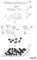
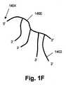
図1A〜1Gに模式的に示す実施形態で、上述の概念を、より詳しく説明する。これらの図面を説明した後、本発明の諸要素をさらに詳しく開示し、実施例を挙げる。上述のように、ある態様では、本発明の高分子構造が、ターゲット配列またはターゲットフラグメントのコンカテマーを含む一本鎖ポリヌクレオチドである。特に、そのようなポリヌクレオチドは、ターゲット配列およびアダプターオリゴヌクレオチドのコンカテマーであることができる。例えば、ソース核酸(1000)を処理(1001)して、好ましくは50〜600ヌクレオチド、より好ましくは300〜600ヌクレオチドの範囲の一本鎖フラグメント(1006)を形成させ、次にそれらをアダプターオリゴヌクレオチド(1004)に連結して、アダプター-フラグメントコンジュゲートの集団(1002)を形成させる。ソース核酸(1000)は、通常の技法を使って試料から抽出されたゲノムDNA、または通常の技法によって作成されたcDNAライブラリーもしくはゲノムライブラリー、または合成DNAなどであることができる。処置(1001)は普通、例えば化学的断片化、酵素的断片化、または機械的断片化などといった通常の技法による断片化と、それに続く変性による、一本鎖DNAフラグメントの作成を伴う。この例では、アダプターオリゴヌクレオチド(1004)を使って、図2Aに図解する方法により、DNAサークルの集団(1010)を形成させる(1008)。ある態様では、集団(1010)の各メンバーが、同一のプライマー結合部位を持つアダプターと、ソース核酸(1000)に由来するDNAフラグメントとを有する。アダプターは、他の機能的要素、例えば限定するわけではないが、タグ付け配列、取付け配列、パリンドローム配列、制限部位、官能化配列などを持っていてもよい。別の実施形態として、異なるプライマー結合部位を持つアダプターを用意することにより、DNAサークルのクラスを作り出してもよい。DNAサークル(1010)を形成させた後、プライマーおよびローリングサークル複製(RCR)試薬を加えることにより、通常のRCR反応で、アダプターオリゴヌクレオチドおよびDNAフラグメントの相補鎖のコンカテマー(1015)の集団(1012)を生成させる(1011)ことができ、次に、その集団を、通常の分離技法で単離することができる。あるいは、考えうる配列を全て含有する混合物から、またはサークルが合成物である場合には、サークル複製用の選択された配列を持つオリゴヌクレオチドの限られた混合物から、短いオリゴヌクレオチド(例えば6マー)の逐次ライゲーションによって、RCRを実行してもよい。また、ターゲット分子の始まりと終わりの両方に相補的なブリッジングテンプレートDNAの存在下でターゲットDNAのライゲーションを行うことによって、コンカテマーを生成させることもできる。異なるターゲットDNAの集団を、対応するブリッジングテンプレートの混合物によって、コンカテマーに変換することができる。次に、単離されたコンカテマー(1014)を、支持体表面(1018)上に配置して(1016)、単分子のランダムアレイを形成させる。取付けが不完全な単分子または先の製造ステップからくる他の試薬類であってその存在が望ましくないものまたは表面(1018)に非特異的に結合するものを除去するために、取付けには、さまざまなストリンジェンシーの洗浄ステップも含まれうる。コンカテマー(1020)は、共有結合および非共有結合を含むさまざまな技法によって、表面(1018)に固定することができる。一実施形態として、アダプターオリゴヌクレオチドのセグメント(例えばプライマー結合部位または他の要素)との複合体(例えば二本鎖二重鎖)を形成する捕捉オリゴヌクレオチドを、表面(1018)に取付けておくことができる。別の実施形態として、アダプターオリゴヌクレオチドとの三重鎖を形成するオリゴヌクレオチドクランプなどの構造を、捕捉オリゴヌクレチドが含んでもよい(Gryaznovらの米国特許第5,473,060号)。もう一つの実施形態として、例えばマイクロアレイにcDNAを取付けるために用いられる技法と同じ技法により、コンカテマー上の相補的官能基と反応して共有結合を形成する反応性官能基を、表面(1018)が持っていてもよい(例えばSmirnovら (2004), Genes, Chromosomes & Cancer, 40: 72-77;Beaucage (2001), Current Medicinal Chemistry, 8: 1213-1244;これらは参照により本明細書に組み入れられる)。疎水表面、例えば低濃度のさまざまな反応性官能基(例えば-OH基)を持つ清浄なガラス表面に、長いDNA分子(例えば数百ヌクレオチド以上)を効率よく取付けることもできる。DNAフラグメントのコンカテマーは、表面への配置後に、in situで、さらに増幅することができる。例えば配置後に、オリゴヌクレオチドのハイブリダイゼーションによってアダプター配列中の制限部位を再構成させることにより、コンカテマーを切断し、次に、そのフラグメントを後述のように環状化し、RCR反応によって、in situで増幅させる。 The above concept is explained in more detail in the embodiments schematically shown in FIGS. After describing these figures, the elements of the present invention are disclosed in more detail and examples are given. As described above, in some embodiments, the macromolecular structure of the invention is a single-stranded polynucleotide comprising a target sequence or target fragment concatamer. In particular, such polynucleotides can be concatamers of target sequences and adapter oligonucleotides. For example, the source nucleic acid (1000) is treated (1001) to form single-stranded fragments (1006), preferably in the range of 50-600 nucleotides, more preferably 300-600 nucleotides, which are then converted to adapter oligonucleotides Ligated to (1004) to form a population (1002) of adapter-fragment conjugates. The source nucleic acid (1000) can be genomic DNA extracted from a sample using conventional techniques, or a cDNA or genomic library created by conventional techniques, or synthetic DNA. Treatment (1001) usually involves fragmentation by conventional techniques such as chemical fragmentation, enzymatic fragmentation, or mechanical fragmentation followed by the generation of single stranded DNA fragments by subsequent denaturation. In this example, an adapter oligonucleotide (1004) is used to form a population of DNA circles (1010) by the method illustrated in FIG. 2A (1008). In some embodiments, each member of the population (1010) has an adapter with the same primer binding site and a DNA fragment derived from the source nucleic acid (1000). The adapter may have other functional elements such as, but not limited to, tagging sequences, attachment sequences, palindromic sequences, restriction sites, functionalization sequences, and the like. In another embodiment, a class of DNA circles may be created by providing adapters with different primer binding sites. After the formation of DNA circle (1010), a population of concatemers (1015) of complementary strands of adapter oligonucleotides and DNA fragments (1012) in a normal RCR reaction by adding primers and rolling circle replication (RCR) reagents Can then be generated (1011) and the population can then be isolated by conventional separation techniques. Alternatively, from a mixture containing all possible sequences, or from a limited mixture of oligonucleotides with a selected sequence for circle replication, if the circle is synthetic, a short oligonucleotide (eg 6-mer) RCR may be executed by sequential ligation. Concatemers can also be generated by ligation of target DNA in the presence of bridging template DNA complementary to both the beginning and end of the target molecule. Different populations of target DNA can be converted to concatamers by a mixture of corresponding bridging templates. The isolated concatamers (1014) are then placed (1016) on the support surface (1018) to form a random array of single molecules. To remove incompletely attached single molecules or other reagents from previous manufacturing steps that are undesirable for their presence or non-specifically bound to the surface (1018), Various stringency wash steps may also be included. The concatemer (1020) can be immobilized to the surface (1018) by a variety of techniques including covalent and non-covalent bonds. In one embodiment, capture oligonucleotides that form complexes (eg double stranded duplexes) with segments of adapter oligonucleotides (eg primer binding sites or other elements) are attached to the surface (1018). Can do. In another embodiment, the capture oligonucleotide may comprise a structure such as an oligonucleotide clamp that forms a triplex with the adapter oligonucleotide (Gryaznov et al., US Pat. No. 5,473,060). In another embodiment, the surface (1018) has a reactive functional group that reacts with a complementary functional group on the concatemer to form a covalent bond, for example by the same technique used to attach cDNA to a microarray. (Eg, Smirnov et al. (2004), Genes, Chromosomes & Cancer, 40: 72-77; Beaucage (2001), Current Medicinal Chemistry, 8: 1213-1244; these are incorporated herein by reference) ). Long DNA molecules (eg, several hundred nucleotides or more) can also be efficiently attached to hydrophobic surfaces, eg, clean glass surfaces with a low concentration of various reactive functional groups (eg, —OH groups). Concatemers of DNA fragments can be further amplified in situ after placement on the surface. For example, after placement, the concatemer is cleaved by reconstituting restriction sites in the adapter sequence by oligonucleotide hybridization, and then the fragment is circularized as described below and amplified in situ by an RCR reaction. .
図1Bに、単分子(例えば一本鎖ポリヌクレオチド)のランダムアレイの表面の一片(1102)を図解する。そのような分子は、通常の条件(例えばTE、SSC、SSPEなどの通常のDNAバッファー、室温)下で、約100〜300nmの直径を持つ溶液中の球状容積をほぼ満たすランダムコイルを形成し、その直径は、当業者には周知の形で、DNAのサイズおよびバッファー条件に依存する(例えばEdvinsson「On the size and shape of polymers and polymer complexes」Dissertation 696 (University of Uppsala, 2002))。ランダムコイルポリマー(例えば一本鎖DNA)のサイズの一尺度は、末端間距離の二乗平均平方根であり、これは大雑把に言ってランダムコイル状構造の直径の尺度である。本明細書において「ランダムコイル直径」というそのような直径は、例えばZetasizer Nano System(Malvern Instruments、英国)などの装置を使って、光散乱法で測定することができる。本発明の高分子構造の他のサイズ尺度には、分子量(例えばダルトンで表したもの)、および総ポリマー長(分岐ポリマーの場合、これはその分枝の全ての長さの和である)が含まれる。表面に取付けると、取付けケミストリー、結合の密度、表面の性質などに依存して、一本鎖ポリヌクレオチドは、平均して、ランダムコイルコンフィギュレーションのコンカテマーの直径にほぼ等しい直径(1110)を持つ破線の円(1108)によって規定される領域(1107)によって区切られる扁平なスフェロイド状容積を満たす。言い方を変えると、一態様として、高分子構造(例えばコンカテマーなど)は、表面(1102)上へのそのランダムコイル状態の投影像に実施的に等価な領域(例えば破線の円(1108)で図示される領域)内で、表面(1102)に取付けられる。高分子構造が占める面積は変動しうるので、いくつかの実施形態では、予想面積が、投影像(1108)の面積の2〜3倍から、そのような面積の小部分(例えば25〜50パーセント)までの範囲内に含まれうる。どこか他の項で言及するように、表面上にコンパクトな形態の高分子構造を保つことにより、高分子構造またはコンカテマーの構成要素に特異的なプローブ(例えば蛍光標識オリゴヌクレオチド)に、より強いシグナルを生じさせることが可能になる。領域(1107)の直径(1110)および単分子を含有する最近接領域までの距離(1106)は、アレイの製作において重要な二つの量である。表面上の単分子の近接性を測定するには、領域(1107)の中心間距離、領域(1007)のエッジ間距離など、さまざまな距離測定基準を使用することができる。ここでは、通例、中心間距離を使用する。本発明のアレイを製作する場合、これらのパラメータの選択は、一つには、解析工程で使用するシグナル生成系およびシグナル検出系に依存する。一般的には、分子の少なくとも20パーセント、または少なくとも30パーセント、または少なくとも40パーセント、または少なくとも過半数を、使用するシグナル生成系およびシグナル検出系で個別に解像することが可能になるような単分子の密度を選択する。ある態様では、単分子の少なくとも70パーセントを個別に解像することが可能になるような密度が選択される。ある態様では、走査型電子顕微鏡を、例えば金ナノ粒子ラベルを持つ分子特異的プローブと共に使用する場合(例えばNieら (2006), Anal. Chem., 78: 1528-1534;これは参照により本明細書に組み入れられる)はいつでも、単分子の少なくとも過半数が50nm以上の最近接距離を持つように、密度が選択され;またもう一つの態様では、単分子の少なくとも70パーセントは100nm以上の最近接距離を持つことが保証されるように、そのような密度が選択される。もう一つの態様では、光学顕微鏡を、例えば蛍光ラベルを持つ分子特異的プローブと共に使用する場合はいつでも、単分子の少なくとも過半数が200nm以上の最近接距離を持つように、密度が選択され;またもう一つの態様では、単分子の少なくとも70パーセントは200nm以上の最近接距離を持つことが保証されるように、そのような密度が選択される。さらにもう一つの態様では、光学顕微鏡を、例えば蛍光ラベルを持つ分子特異的プローブと共に使用する場合はいつでも、単分子の少なくとも過半数が300nm以上の最近接距離を持つように、密度が選択され;またもう一つの態様では、単分子の少なくとも70パーセントは300nm以上、または400nm以上、または500nm以上、または600nm以上、または700nm以上、または800nm以上の最近接距離を持つことが保証されるように、そのような密度が選択される。さらにもう一つの実施形態では、光学顕微鏡を使用する場合はいつでも、単分子の少なくとも過半数が、その顕微鏡の最小特徴解像力(minimal feature resolution power)の少なくとも2倍の最近接距離を持つように、密度が選択される。もう一つの態様では、別々に検出することができるポリマー分子の密度が、1μm2あたり少なくとも1000、または1μm2あたり少なくとも10,000、または1μm2あたり少なくとも100,000になるように、本発明のポリマー分子が表面上に配置される。FIG. 1B illustrates a piece of surface (1102) of a random array of single molecules (eg, single-stranded polynucleotides). Such molecules form a random coil that almost fills a spherical volume in a solution with a diameter of about 100-300 nm under normal conditions (e.g. a normal DNA buffer such as TE, SSC, SSPE, room temperature), Its diameter depends on the size and buffer conditions of the DNA in a manner well known to those skilled in the art (eg Edvinsson “On the size and shape of polymers and polymer complexes” Dissertation 696 (University of Uppsala, 2002)). One measure of the size of a random coil polymer (eg, single stranded DNA) is the root mean square of the end-to-end distance, which is roughly a measure of the diameter of the random coiled structure. Such a diameter, referred to herein as “random coil diameter”, can be measured by light scattering using a device such as, for example, the Zetasizer Nano System (Malvern Instruments, UK). Other size measures of the macromolecular structure of the present invention include molecular weight (eg expressed in daltons), and total polymer length (in the case of branched polymers, this is the sum of all the lengths of the branches). included. When attached to a surface, depending on the attachment chemistry, density of binding, surface nature, etc., the single-stranded polynucleotide on average is a dashed line with a diameter (1110) approximately equal to the diameter of the concatemer in the random coil configuration Fills a flat spheroid-like volume delimited by a region (1107) defined by a circle (1108). In other words, in one aspect, the macromolecular structure (eg, concatemer) is illustrated by a region (eg, dashed circle (1108)) that is practically equivalent to the projected image of its random coil state on the surface (1102). Attached to the surface (1102). Since the area occupied by the polymer structure can vary, in some embodiments, the expected area can vary from two to three times the area of the projected image (1108), from a small portion of such area (eg, 25-50 percent ). As noted elsewhere, by keeping a compact form of the polymer structure on the surface, it is stronger to probes specific to the polymer structure or concatamer components (eg, fluorescently labeled oligonucleotides) It is possible to generate a signal. The diameter (1110) of the region (1107) and the distance (1106) to the nearest region containing a single molecule are two important quantities in the fabrication of the array. To measure the proximity of single molecules on the surface, various distance metrics can be used, such as the distance between the centers of the regions (1107) and the distance between the edges of the regions (1007). Here, the center-to-center distance is usually used. When producing the array of the present invention, the selection of these parameters depends in part on the signal generation system and signal detection system used in the analysis process. In general, a single molecule that allows at least 20 percent, or at least 30 percent, or at least 40 percent, or at least a majority of the molecule to be resolved separately in the signal generation and detection systems used. Select the density. In certain embodiments, a density is selected that allows at least 70 percent of the single molecule to be resolved individually. In some embodiments, when a scanning electron microscope is used, for example, with a molecule specific probe having a gold nanoparticle label (eg, Nie et al. (2006), Anal. Chem., 78: 1528-1534; this is incorporated herein by reference). At any time) the density is selected such that at least a majority of the single molecules have a closest distance of 50 nm or more; and in another embodiment, at least 70 percent of the single molecules have a closest distance of 100 nm or more. Such a density is chosen such that it is guaranteed to have In another embodiment, whenever an optical microscope is used with, for example, a molecule-specific probe with a fluorescent label, the density is selected such that at least a majority of the single molecules have a closest distance of 200 nm or more; In one embodiment, such a density is selected to ensure that at least 70 percent of the single molecule has a closest distance of 200 nm or greater. In yet another embodiment, whenever an optical microscope is used with, for example, a molecule-specific probe with a fluorescent label, the density is selected such that at least a majority of the single molecules have a closest distance of 300 nm or more; In another embodiment, at least 70 percent of a single molecule is guaranteed to have a closest distance of 300 nm or more, or 400 nm or more, or 500 nm or more, or 600 nm or more, or 700 nm or more, or 800 nm or more. Such a density is selected. In yet another embodiment, whenever an optical microscope is used, the density is such that at least a majority of the single molecules have a closest distance that is at least twice the minimal feature resolution power of the microscope. Is selected. In another embodiment, the polymer molecules of the present invention have a surface density such that the density of polymer molecules that can be detected separately is at least 1000 per 1 μm2 , or at least 10,000 per 1 μm2 , or at least 100,000 per 1 μm2. Placed on top.
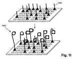
図1Cの特定実施形態を例示する本発明のもう一つの態様では、ランダムに配置される単分子の密度を、望ましい最近接距離が確保されるように選択する要件が、単分子を取付けるための実質上唯一の部位である不連続な相隔たる領域を表面上に設けることによって、取り除かれる。すなわち、そのような実施形態では、不連続な相隔たる領域の間にある表面上の領域(本明細書ではこれを「領域間部分」という)が、コンカテマーまたは他の高分子構造がそのような領域には結合しないという意味で、不活性である。いくつかの実施形態では、そのような領域間部分をブロッキング剤(例えばコンカテマーDNAとは無関係なDNA、他のポリマーなど)で処理することができる。図1Aの場合と同様に、ソース核酸(1000)を断片化し、環状化(1010)のためにアダプターを付加(1002)し、次に、RCRによってコンカテマーを形成させる(1012)。次に、表面(1120)の設計および製作によって決定される最近接距離(1124)をそれぞれに持つ不連続な相隔たる領域(1122)の規則的アレイを有する表面(1120)に、単離されたコンカテマー(1014)を適用する。後に詳述するように、捕捉オリゴヌクレオチドまたは反応性官能基で誘導体化するためのミクロン寸法およびサブミクロン寸法を持つ不連続な相隔たる領域(1122)のアレイは、電子線リソグラフィー、ナノインプリント技術、フォトリソグラフィーなどの、通常の半導体製作技法を使って製作することができる。一般に、単分子を表面(1120)に適用した時に実質上全ての領域(1122)が一つを超える単分子によって占有されないように、不連続な相隔たる領域(1122)の面積は、取付けケミストリー、使用する高分子構造などと共に、本発明の単分子のサイズに一致するように選択される。不連続な相隔たる領域一つにつき一つだけの単分子を持つ可能性は、反応性官能基または捕捉オリゴヌクレオチドの密度を、そのような部分が単分子上の各々の相補鎖よりも少なくなるように選択することによって、増加させることができる。そうすることで、単分子は、ある特定の不連続な相隔たる領域における表面への結合を全て「占有」することになり、その結果として、第2の単分子が同様に同じ領域に結合する機会を減少させる。特に、ある実施形態では、不連続な相隔たる領域中の捕捉オリゴヌクレオチドの実質上全てが、単一の高分子構造上のアダプターオリゴヌクレオチドにハイブリダイズする。ある態様では、不連続な相隔たる領域が、単分子の相補的官能基またはアダプターオリゴヌクレオチドの数の約10パーセント〜約50パーセントであるような数の反応性官能基または捕捉オリゴヌクレオチドを含有する。捕捉オリゴヌクレオチドの長さおよび配列は、幅広く変動することができ、周知の原理に従って選択することができる(例えばWetmur, Critical Reviews in Biochemistry and Molecular Biology, 26: 227-259 (1991);Hamesら編「Nucleic Acid Hybridization: A Practical Approach」(IRL Press、オックスフォード、1985)の第1章(BrittenおよびDavidson))。ある態様では、捕捉オリゴヌクレオチドの長さが6〜30ヌクレオチドの範囲にあり、もう一つの態様では、8〜30ヌクレオチド、または10〜24ヌクレオチドの範囲にある。捕捉オリゴヌクレオチドの長さは、(i)表面への高分子構造の効果的な結合をもたらし、その結果として、解析作業のステップ(例えば洗浄など)中に起こる高分子構造の損失が最小限に抑えられるように、かつ(ii)(分析物分子がコンカテマー中のDNAフラグメントである場合は特に)分析物分子における解析作業との干渉が回避されるように、選択される。(i)に関し、ある態様では、ストリンジェントな洗浄でも解離しないほど十分に安定な二重鎖が捕捉オリゴヌクレオチドとそれらの相補鎖との間に得られるように、配列および長さを選択する。(ii)に関し、DNAフラグメントがある特定の生物種に由来する場合は、データベースが利用可能であるなら、それを使って、そのDNAフラグメントと偽ハイブリッドまたは望ましくないハイブリッドを形成するかもしれない潜在的捕捉配列を選別することができる。捕捉オリゴヌクレオチドの配列を選択する際の他の因子は、プライマー、ハイブリダイゼーションプローブ、オリゴヌクレオチドタグなどを選択する際に考慮される因子と類似しており、それらについては、後に「定義」の項で言及する参考文献からもわかるように、十分な手引きがある。いくつかの実施形態では、不連続な相隔たる領域が2種類以上の捕捉オリゴヌクレオチドを含有し、異なる捕捉オリゴヌクレオチドのそれぞれが異なる長さおよび異なる配列を持つこともできる。不連続な相隔たる領域の規則的アレイを使用する実施形態の一側面では、最近接領域にある捕捉オリゴヌクレオチドの配列が異なる配列を持つように、捕捉オリゴヌクレオチドの配列を選択する。直線的アレイの場合は、配列タイプが交互する列によって、そのような構成が達成される。別の実施形態では、表面が、不連続な相隔たる領域のサブアレイを複数持ち、異なるサブアレイのそれぞれが、他のサブアレイとは異なる別個のヌクレオチド配列を持つ捕捉オリゴヌクレオチドを持っていてもよい。複数のサブアレイには、2個のサブアレイ、または4個以下のサブアレイ、または8個以下のサブアレイ、または16個以下のサブアレイ、または32個以下のサブアレイ、または64個以下のサブアレイが含まれうる。さらに別の実施形態では、表面が5000個以下のサブアレイを含みうる。ある態様では、表面基の望ましくない影響または捕捉オリゴヌクレオチドもしくは他の試薬との相互作用を最小限に抑えるために、マイクロアレイで行われるのと同様に、捕捉オリゴヌクレチドが、スペーサー分子(例えばポリエチレングリコールなどの不活性鎖)によって、アレイの表面に取付けられる。 In another aspect of the invention illustrating the particular embodiment of FIG. 1C, the requirement to select the density of randomly placed single molecules so that the desired closest distance is ensured is It is removed by providing discontinuous spaced areas on the surface that are essentially the only site. That is, in such an embodiment, a region on the surface between discontinuous spaced regions (referred to herein as an “interregional portion”) is a concatemer or other polymeric structure. Inactive in the sense that it does not bind to the region. In some embodiments, such interregional portions can be treated with a blocking agent (eg, DNA unrelated to concatamer DNA, other polymers, etc.). As in FIG. 1A, the source nucleic acid (1000) is fragmented, adapters are added (1002) for circularization (1010), and then concatemers are formed by RCR (1012). It was then isolated into a surface (1120) with a regular array of discontinuous spaced regions (1122) each having a closest distance (1124) determined by the design and fabrication of the surface (1120) Concatemer (1014) is applied. As detailed below, arrays of discontinuous spaced regions (1122) with micron and sub-micron dimensions for derivatization with capture oligonucleotides or reactive functional groups are available in electron beam lithography, nanoimprint technology, photo It can be fabricated using normal semiconductor fabrication techniques such as lithography. In general, the area of discontinuous spaced regions (1122) is the mounting chemistry, so that substantially all regions (1122) are not occupied by more than one single molecule when a single molecule is applied to the surface (1120). Along with the polymer structure used, etc., it is selected to match the size of the single molecule of the present invention. The possibility of having only one single molecule per discrete, distant region reduces the density of reactive functional groups or capture oligonucleotides, such portions being less than each complementary strand on the single molecule. Can be increased by selecting as follows. By doing so, the single molecule will “occupy” all of the binding to the surface in certain discrete discrete regions, and as a result, the second single molecule will bind to the same region as well. Reduce opportunities. In particular, in certain embodiments, substantially all of the capture oligonucleotides in the discrete, discrete regions hybridize to adapter oligonucleotides on a single macromolecular structure. In some embodiments, the discontinuous spaced regions contain a number of reactive functional groups or capture oligonucleotides that are from about 10 percent to about 50 percent of the number of single molecule complementary functional groups or adapter oligonucleotides. . The length and sequence of capture oligonucleotides can vary widely and can be selected according to well-known principles (eg Wetmur, Critical Reviews in Biochemistry and Molecular Biology, 26: 227-259 (1991); edited by Hames et al. “Nucleic Acid Hybridization: A Practical Approach” (IRL Press, Oxford, 1985), Chapter 1 (Britten and Davidson)). In some embodiments, the length of the capture oligonucleotide is in the range of 6-30 nucleotides, and in another embodiment, it is in the range of 8-30 nucleotides, or 10-24 nucleotides. The length of the capture oligonucleotide (i) results in effective binding of the polymer structure to the surface, resulting in minimal loss of polymer structure that occurs during analytical steps (eg, washing). It is selected to be suppressed and (ii) (especially when the analyte molecule is a DNA fragment in a concatamer) so as to avoid interference with analytical work on the analyte molecule. With regard to (i), in certain embodiments, the sequence and length are chosen so that a sufficiently stable duplex is obtained between the capture oligonucleotides and their complementary strands that does not dissociate upon stringent washing. With regard to (ii), if a DNA fragment is derived from a particular species, and if a database is available, it may be used to potentially form a false or undesirable hybrid with that DNA fragment. Capture sequences can be selected. Other factors in selecting the sequence of the capture oligonucleotide are similar to those considered when selecting primers, hybridization probes, oligonucleotide tags, etc., and are described in the “Definitions” section below. As you can see from the references mentioned in, there is enough guidance. In some embodiments, discontinuous spaced regions contain more than one type of capture oligonucleotide, and each of the different capture oligonucleotides can have a different length and a different sequence. In one aspect of an embodiment that uses a regular array of discrete spaced apart regions, the sequence of the capture oligonucleotides is selected such that the sequence of the capture oligonucleotides in the closest region has a different sequence. In the case of a linear array, such a configuration is achieved by columns with alternating arrangement types. In another embodiment, the surface may have multiple subarrays of discrete, spaced apart areas, each of the different subarrays having a capture oligonucleotide having a distinct nucleotide sequence different from the other subarrays. The plurality of subarrays may include 2 subarrays, or 4 or fewer subarrays, or 8 or fewer subarrays, or 16 or fewer subarrays, or 32 or fewer subarrays, or 64 or fewer subarrays. In yet another embodiment, the surface can contain up to 5000 subarrays. In some embodiments, the capture oligonucleotide is a spacer molecule (eg, polyethylene glycol, etc.), as is done in a microarray to minimize unwanted effects of surface groups or interaction with the capture oligonucleotide or other reagents. Attached to the surface of the array.
ある態様では、不連続な相隔たる領域(1122)の面積が1μm2未満であり;もう一つの態様では、不連続な相隔たる領域(1122)の面積が0.04μm2〜1μm2の範囲にあり;さらにもう一つの態様では、不連続な相隔たる領域(1122)の面積が0.2μm2〜1μm2である。もう一つの態様では、不連続な相隔たる領域の形状がほぼ円形または正方形であり、その結果、それらのサイズを単一の長さ寸法で示すことができる場合に、そのような領域のサイズが、125nm〜250nmの範囲、または200nm〜500nmの範囲にある。ある態様では、最も近接した領域(1122)の中心間距離が0.25μm〜20μmの範囲にあり;もう一つの態様では、そのような距離が1μm〜10μmの範囲、または50〜1000nmの範囲にある。ある態様では、領域(1120)を、表面(1018)上に、領域(1122)が所定の位置を持つような事実上任意のパターンで、すなわち、シグナル収集およびデータ解析機能の効率を高める任意の規則的アレイに、整列させることができる。そのようなパターンには、領域(1122)の同心円、らせんパターン、直線的パターン、六角形パターンなどが含まれるが、これらに限るわけではない。好ましくは、領域(1122)を直線的パターンまたは六角形パターンに整列させる。In some embodiments, the area of discrete spaced apart areas (1122) is located less than 1 [mu] m2; In another embodiment, the area of discrete spaced apart areas (1122) is in the range of 0.04μm2 ~1μm2 ; in yet another embodiment, the area of discrete spaced apart areas (1122) is 0.2μm2 ~1μm2. In another embodiment, the size of such discrete regions is such that the shape of the discontinuous spaced regions is approximately circular or square so that their size can be indicated by a single length dimension. , 125 nm to 250 nm, or 200 nm to 500 nm. In some embodiments, the center-to-center distance of the closest region (1122) is in the range of 0.25 μm to 20 μm; in another embodiment, such distance is in the range of 1 μm to 10 μm, or in the range of 50 to 1000 nm. . In some embodiments, the region (1120) is placed on the surface (1018) in virtually any pattern such that the region (1122) has a predetermined position, ie, any signal that increases the efficiency of signal collection and data analysis functions. It can be aligned in a regular array. Such patterns include, but are not limited to, concentric circles of region (1122), spiral patterns, linear patterns, hexagonal patterns, and the like. Preferably, region (1122) is aligned in a linear or hexagonal pattern.
図1Dに図解するように、一定の実施形態では、ソース核酸(1200)から製造されるDNAサークルが、アダプターオリゴヌクレオチドを含まなくてもよい。先と同様に、ソース核酸(1200)を断片化し、変性させること(1202)により、好ましくは約50〜600ヌクレオチドのサイズ範囲、より好ましくは約300〜約600ヌクレオチドのサイズ範囲の一本鎖フラグメントの集団(1204)を形成させ、次に、CircLigase(Epicentre Biotechnologies、ウィスコンシン州マディソン)などの環状化リガーゼを使った非テンプレート駆動型の反応で、それらを環状化する。DNAサークル(1206)の形成後に、選ばれた配列に結合するプライマーの混合物を用意することにより、コンカテマーを生成させる。プライマーの混合物は、DNAサークル(1206)の総数のサブセットだけがコンカテマーを生成するように選択することができる。コンカテマーを生成させた後(1208)、それらを単離し、表面(1210)に適用することにより、本発明のランダムアレイを形成させる。 As illustrated in FIG. 1D, in certain embodiments, a DNA circle produced from a source nucleic acid (1200) may not include an adapter oligonucleotide. As above, single stranded fragments, preferably in the size range of about 50-600 nucleotides, more preferably in the size range of about 300-600 nucleotides, by fragmenting and denaturing the source nucleic acid (1200) (1202). Are then circularized in a non-template driven reaction using a circularized ligase such as CircLigase (Epicentre Biotechnologies, Madison, Wis.). After formation of the DNA circle (1206), concatamers are generated by preparing a mixture of primers that bind to the selected sequence. The mixture of primers can be selected so that only a subset of the total number of DNA circles (1206) produces concatamers. After generating concatamers (1208), they are isolated and applied to the surface (1210) to form a random array of the present invention.
上述のように、本発明の単分子は取付け部分および分析物部分を含み、その取付け部分が、表面へのその単分子の多価取付けをもたらす高分子構造を含むようになっている。図1Eに図解するように、高分子構造は、反応中のDNAサークルが合成物であるRCR反応によって作製されるコンカテマーであってもよい。次に、コンカテマー上のユニークな官能基を使って、単分子の分析物部分を取付ける。事実上どんな配列の合成DNAサークルでも、周知の技法を使って、数百ヌクレオチド(例えば200ヌクレオチド)までのサイズであれば簡便に、また、難しくはなるが、何百ヌクレオチドもの(例えば500ヌクレオチドまでの)サイズでも、作成することができる(例えばKoolの米国特許第5,426,180号;Dolinnayaら (1993), Nucleic Acids Research, 21: 5403-5407;Rubinら (1995), Nucleic Acids Research, 23: 3547-3553など;これらは参照により本明細書に組み入れられる)。プライマー結合部位(1301)を含む合成DNAサークル(1300)を、PCR反応(1306)でプライマー(1302)と組み合わせることにより、コンカテマー(1308)を作成する。通例、この実施形態では、すべてのサークルが同じ配列を持つが、例えばコンカテマーのサブセットを、アレイの予め選択した領域に、相補的な取付け部分(例えばアダプター配列と捕捉オリゴヌクレオチド)を使って誘導するために、異なる配列を使用することもできる。分析物上の相補的官能基と反応して共有結合を形成する能力を持つ官能基(1304;「R」と呼ぶ)をその5'末端に持つプライマー(1302)を合成する。代表的官能基には、市販の化学薬品(例えばGlen Research)を使って取付けることができるアミノ基、スルフヒドリル基などがある。コンカテマー(1308)を表面(1310)に適用してアレイ(1314)を形成させた後、取付け部分を持つ分析物(1312)をアレイ(1310)に適用すると、ユニークな官能基R(1311)と取付け部分(1312)との反応により、コンカテマーとの結合が形成される。あるいは、アレイ(1310)への適用に先だって、取付け部分とユニークな官能基とが反応して結合を形成できるようにコンカテマー(1308)を分析物(1312)と混合した後、得られたコンジュゲートをアレイ(1310)に適用してもよい。コンカテマー(1308)と多くの分析物クラスとを連結するための適当な取付け部分およびユニークな官能基の選択については、文献に豊富な手引きがある。一態様として、タンパク質またはペプチド分析物をコンカテマーに連結するために、多くのホモ二官能性およびヘテロ二官能性試薬が市販され(例えばPierce)、参照により本明細書に組み入れられるHermanson「Bioconjugate Techniques」(Academic Press、ニューヨーク、1996)などの文献に開示されている。例えば、ユニークな官能基がアミノ基である場合はいつでも、N-スクシンイミジル3-(2-ピリジルジチオ)プロピオネート(SPDP)、スクシンイミジルオキシカルボニル-α-メチル-α-(2-ピリジルジチオ)トルエン(SMPT)、スクシンイミジル-4-(N-マレイミドメチル)シクロヘキサン-1-カルボキシレート(SMCC)、m-マレイミドベンゾイル-N-ヒドロキシスクシンイミドエステル(MBS)、N-スクシンイミジル(4-ヨードアセチル)アミノベンゾエート(SIAB)、スクシンイミジル6-((ヨードアセチル)アミノ)ヘキサノエート(SIAX)などの試薬を使って、コンカテマー(1308)を分析物上のスルフヒドリル基に連結することができる。分析物上の適切な相補的官能基には、アミノ基、スルフヒドリル基、カルボニル基などがあり、これらは分析物上に天然に存在する場合もあるし、適切なホモ二官能性またはヘテロ二官能性試薬との反応によって付加することもできる。分析物分子は、非共有結合、例えばビオチン-ストレプトアビジン結合、複合体(例えばコンカテマーに取付けられた第1オリゴヌクレオチドと分析物に取付けられたまたは分析物の一部を形成している相補的オリゴヌクレオチドとの二重鎖)の形成などの結合を使って、高分子構造に取付けることもできる。分析物には、核酸(例えばDNAまたはRNAフラグメント)、多糖、タンパク質などの生体分子が含まれる。 As described above, the unimolecule of the present invention includes an attachment portion and an analyte portion, such that the attachment portion includes a polymeric structure that provides for multivalent attachment of the unimolecule to a surface. As illustrated in FIG. 1E, the polymer structure may be a concatamer produced by an RCR reaction in which the DNA circle in the reaction is a synthetic product. Next, a single molecule analyte moiety is attached using a unique functional group on the concatemer. Synthetic DNA circles of virtually any sequence, using well-known techniques, can be conveniently and difficult to size up to several hundred nucleotides (eg up to 500 nucleotides), eg up to several hundred nucleotides (eg up to 200 nucleotides) (Eg, Kool US Pat. No. 5,426,180; Dolinnaya et al. (1993), Nucleic Acids Research, 21: 5403-5407; Rubin et al. (1995), Nucleic Acids Research, 23: 3547-). 3553, etc .; these are incorporated herein by reference). A concatemer (1308) is created by combining a synthetic DNA circle (1300) containing a primer binding site (1301) with a primer (1302) in a PCR reaction (1306). Typically, in this embodiment, all circles have the same sequence, but for example, a subset of concatamers is directed to a preselected region of the array using complementary attachment portions (eg, adapter sequences and capture oligonucleotides). For this reason, different sequences can be used. A primer (1302) is synthesized that has a functional group (1304; called “R”) at its 5 ′ end capable of reacting with a complementary functional group on the analyte to form a covalent bond. Exemplary functional groups include amino groups, sulfhydryl groups, and the like that can be attached using commercially available chemicals (eg, Glen Research). After applying concatamer (1308) to surface (1310) to form array (1314), an analyte (1312) with an attachment moiety is applied to array (1310) to create a unique functional group R (1311) The reaction with the attachment part (1312) forms a bond with the concatemer. Alternatively, prior to application to the array (1310), the concatemer (1308) is mixed with the analyte (1312) so that the attachment moiety and the unique functional group can react to form a bond, and then the resulting conjugate May be applied to the array (1310). There is a wealth of guidance in the literature on the selection of suitable attachments and unique functional groups for linking concatamers (1308) to many analyte classes. In one aspect, Hermanson “Bioconjugate Techniques”, many homobifunctional and heterobifunctional reagents are commercially available (eg, Pierce) and are incorporated herein by reference to link protein or peptide analytes to concatamers. (Academic Press, New York, 1996). For example, whenever the unique functional group is an amino group, N-succinimidyl 3- (2-pyridyldithio) propionate (SPDP), succinimidyloxycarbonyl-α-methyl-α- (2-pyridyldithio) Toluene (SMPT), succinimidyl-4- (N-maleimidomethyl) cyclohexane-1-carboxylate (SMCC), m-maleimidobenzoyl-N-hydroxysuccinimide ester (MBS), N-succinimidyl (4-iodoacetyl) aminobenzoate Concatemer (1308) can be linked to a sulfhydryl group on the analyte using reagents such as (SIAB), succinimidyl 6-((iodoacetyl) amino) hexanoate (SIAX). Suitable complementary functional groups on the analyte include amino groups, sulfhydryl groups, carbonyl groups, etc., which may be naturally present on the analyte, and may be suitable homobifunctional or heterobifunctional. It can also be added by reaction with a sex reagent. The analyte molecule can be a non-covalent bond, such as a biotin-streptavidin bond, a complex (eg, a first oligonucleotide attached to a concatamer and a complementary oligo attached to the analyte or forming part of the analyte. It can also be attached to macromolecular structures using bonds such as the formation of duplexes with nucleotides. Analytes include biomolecules such as nucleic acids (eg, DNA or RNA fragments), polysaccharides, proteins and the like.
上述のように、本発明の高分子構造は、線状ポリマー、例えばDNAフラグメントのコンカテマーに加えて、分岐ポリマーも含みうる。代表的な分岐ポリマー構造を図1Fおよび1Gに図解する。図1Fには、主鎖ポリヌクレオチド(1400)と複数の分枝ポリヌクレオチド(1402)とを含む分岐DNA構造が図解されている。この分岐DNA構造において、前記複数の分枝ポリヌクレオチドは、それぞれがその5'末端によって主鎖ポリヌクレオチド(1400)に接続されて、主鎖ポリヌクレオチド(1400)上の単一の5'末端(1404)以外は全て3'である末端を持つ櫛状の構造を形成しており、その単一の5'末端はユニークな官能基を持つように誘導体化される。後述するように、そのようなユニークな官能基は反応性化学基(例えば保護されたまたは保護されていないアミン、スルフヒドリルなど)であってもよいし、それに相補的な配列を持つオリゴヌクレオチドを有する分析物を捕捉するためのユニーク配列を有するオリゴヌクレオチドであってもよい。また、そのようなユニークな官能基は、例えばビオチンなどの捕捉部分であってもよい。そのような分岐DNA構造は、公知の技法を使って合成される(例えばGryaznovの米国特許第5,571,677号;Urdeaらの米国特許第5,124,246号;Seemanらの米国特許第6,255,469号など;これらは参照により本明細書に組み入れられる)。そのような高分子構造がポリヌクレオチドである場合はいつでも、その構成要素の配列を自己集合が容易になるように選択するか、あるいはそれらを専用の連結ケミストリー(例えば以下に開示するもの)を使って連結することができ、その場合は、他の因子(例えば、いくつかの実施形態では、自己アニーリングの回避、表面上の捕捉オリゴヌクレオチドへの容易な結合など)に基づいて、配列が選択される。図1Gには、デンドリマー構造が図解されている。このデンドリマー構造はオリゴヌクレオチド(1406)を含み、それが、それぞれに二つの官能基(1410;「R」で示す)を持つ複数の三価連結基(1408)で誘導体化される。さらに、それらの官能基により、追加ポリマー(1407)(例えばポリヌクレオチド)を取付けて、オリゴヌクレオチド(1406)への結合を形成させ、その結果として高分子構造(1409)を形成させることができる。そしてまた、それを多価リンカーで同様に誘導体化すれば、核酸デンドリマーを形成することができる。オリゴヌクレオチド用の三価リンカー(1408)は、Iyerらの米国特許第5,916,750号に開示されており、この特許は参照により本明細書に組み入れられる。図1Hに図解するように、そのようなデンドリマー構造または分岐構造(1411)が構築されたら、それらは、線状ポリヌクレオチドについて上述したように、アレイ(1420)に取付けることができ、その後、分析物(1430)を、ユニークな官能基(1410)によって取付けることができる。場合によっては、未反応のユニークな官能基(1422)を、通常の技法でキャッピングしてもよい。あるいは、デンドリマー構造または分岐構造(1411)を、まず分析物(1430)と、例えば溶解した状態で混合して、コンジュゲートを形成させ、次にそのコンジュゲートをアレイ(1420)上に配置する。図1Iに示すように、分析物が遊離の3'末端を持つポリヌクレオチド(1440)である場合は、その末端をin situ RCR反応で伸長させて、ターゲット配列またはさらなる付加のための他の配列のコンカテマーを形成させることができる。同様に、ポリヌクレオチド分析物は、通常の技法を使ったライゲーションによって伸長させることもできる。 As noted above, the polymeric structures of the present invention can include branched polymers in addition to linear polymers, such as concatemers of DNA fragments. A representative branched polymer structure is illustrated in FIGS. 1F and 1G. FIG. 1F illustrates a branched DNA structure comprising a backbone polynucleotide (1400) and a plurality of branched polynucleotides (1402). In this branched DNA structure, each of the plurality of branched polynucleotides is connected to the main polynucleotide (1400) by its 5 ′ end to form a single 5 ′ end ( All except 1404) form a comb-like structure with a 3 ′ end, and the single 5 ′ end is derivatized with a unique functional group. As described below, such unique functional groups may be reactive chemical groups (eg, protected or unprotected amines, sulfhydryls, etc.) and have oligonucleotides with complementary sequences thereto. It may be an oligonucleotide having a unique sequence for capturing an analyte. Such a unique functional group may also be a capture moiety such as biotin. Such branched DNA structures are synthesized using known techniques (eg, Gryaznov US Pat. No. 5,571,677; Urdea et al US Pat. No. 5,124,246; Seeman et al US Pat. No. 6,255,469, etc .; these are by reference) Incorporated herein). Whenever such a macromolecular structure is a polynucleotide, choose the sequence of its components to facilitate self-assembly, or use them with dedicated linking chemistry (eg those disclosed below) In which case the sequence is selected based on other factors (eg, in some embodiments, avoidance of self-annealing, easy binding to capture oligonucleotides on the surface, etc.) The FIG. 1G illustrates the dendrimer structure. This dendrimer structure comprises an oligonucleotide (1406), which is derivatized with a plurality of trivalent linking groups (1408), each with two functional groups (1410; indicated by “R”). In addition, these functional groups can attach additional polymers (1407) (eg, polynucleotides) to form bonds to oligonucleotides (1406), resulting in the formation of macromolecular structures (1409). And if it is similarly derivatized with a multivalent linker, a nucleic acid dendrimer can be formed. A trivalent linker (1408) for oligonucleotides is disclosed in US Pat. No. 5,916,750 to Iyer et al., Which is hereby incorporated by reference. Once such dendrimer structures or branched structures (1411) have been constructed, as illustrated in FIG. 1H, they can be attached to the array (1420) as described above for linear polynucleotides and then analyzed. The object (1430) can be attached by a unique functional group (1410). In some cases, the unreacted unique functional group (1422) may be capped by conventional techniques. Alternatively, the dendrimer structure or branched structure (1411) is first mixed with the analyte (1430), eg, in a dissolved state, to form a conjugate, which is then placed on the array (1420). As shown in FIG. 1I, if the analyte is a polynucleotide with a free 3 ′ end (1440), its end can be extended with an in situ RCR reaction to target sequences or other sequences for further addition. Of concatamers can be formed. Similarly, a polynucleotide analyte can be extended by ligation using conventional techniques.
ソース核酸およびターゲット配列の環状化
本発明の一態様では、高分子構造が、試料(例えば患者、経済的に重要な生物などから得られるゲノムDNAまたはcDNA)から抽出または誘導されるポリヌクレオチド分析物(すなわちターゲット配列)のコンカテマーを含む。そのような単分子を含む本発明のランダムアレイは、配列決定、SNP測定、アレルの定量、コピー数測定などを含むゲノムワイドな解析を行うのに役立つ。哺乳類サイズのゲノムの場合、好ましくは断片化を少なくとも二段階で行い、第1段階では、約100キロ塩基(Kb)〜約250キロ塩基のサイズ範囲にあるフラグメントの集団を生成させ、各100〜250Kbフラグメントに個別に適用される第2段階では、約50〜600ヌクレオチド、より好ましくは約300〜600ヌクレオチドのサイズ範囲にある、ランダムアレイ用のコンカテマーを生成させるためのフラグメントを生成させる。本発明のいくつかの態様では、断片化の第1段階を、そのようなフラグメントの予め決定されたサブセット(例えばシグナル伝達系路のタンパク質をコードする遺伝子を含有するフラグメントなど)を選択するために使用することもできる。本発明のアレイを構築するのに必要なゲノムDNAの量は広く変動しうる。ある態様では、哺乳類サイズのゲノムに関して、少なくとも10ゲノム相当のDNAからフラグメントを生成させ;もう一つの態様では、少なくとも30ゲノム相当のDNAからフラグメントを生成させ;もう一つの態様では、少なくとも60ゲノム相当のDNAからフラグメントを生成させる。Circularization of Source Nucleic Acid and Target Sequence In one aspectof the present invention, a polynucleotide analyte from which a macromolecular structure is extracted or derived from a sample (eg, genomic DNA or cDNA obtained from a patient, an economically important organism, etc.) (Ie target sequence) concatamers. The random arrays of the present invention comprising such single molecules are useful for genome-wide analysis including sequencing, SNP measurements, allele quantification, copy number measurements and the like. For mammalian-sized genomes, fragmentation is preferably performed in at least two stages, with the first stage generating a population of fragments ranging in size from about 100 kilobases (Kb) to about 250 kilobases, In the second step, applied individually to the 250 Kb fragment, fragments are generated to generate concatamers for random arrays in the size range of about 50-600 nucleotides, more preferably about 300-600 nucleotides. In some embodiments of the invention, the first stage of fragmentation is used to select a predetermined subset of such fragments, such as a fragment containing a gene encoding a protein of a signal transduction pathway. It can also be used. The amount of genomic DNA required to construct the arrays of the present invention can vary widely. In one embodiment, for a mammal-sized genome, fragments are generated from DNA equivalent to at least 10 genomes; in another embodiment, fragments are generated from DNA equivalent to at least 30 genomes; in another embodiment, equivalent to at least 60 genomes Generate fragments from the DNA.
ゲノムDNAは、例えばSambrookら(前掲)1999;「Current Protocols in Molecular Biology」Ausubelら編(John Wiley and Sons, Inc.、ニューヨーク、1999)などに開示されているような通常の技法を使って取得される。ゲノムDNAの単離にとって重要な因子には、以下に挙げるものが含まれる:1)DNAがDNAプロセシング酵素を含まず、塩類の混入がないこと;2)ゲノム全体を等しく表していること;および3)DNAフラグメントの長さが約5,000〜100,000bpであること。多くの場合、抽出されたDNAは消化する必要がない。なぜなら、溶解および抽出中に生じる剪断力が望ましい範囲のフラグメントを生成させるからである。もう一つの実施形態として、制限エンドヌクレアーゼを使った酵素的断片化によって、さらに短いフラグメント(1〜5kb)を生成させることができる。ある実施形態では、10〜100ゲノム相当のDNAにより、フラグメントの集団がゲノム全体をカバーすることを保証する。少量のDNAしか利用できず、例えば容器の壁などへの非特異的結合による損失の危険がある場合はいつでも、担体DNA(例えば無関係な環状合成二本鎖DNA)を用意し、それを試料DNAと混合して使用することが有利なこともある。 Genomic DNA is obtained using conventional techniques such as those disclosed in Sambrook et al. (Supra) 1999; “Current Protocols in Molecular Biology” Ausubel et al. (John Wiley and Sons, Inc., New York, 1999). Is done. Factors important for genomic DNA isolation include: 1) the DNA does not contain DNA processing enzymes and is free of salt; 2) represents the entire genome equally; and 3) The length of the DNA fragment is about 5,000 to 100,000 bp. In many cases, the extracted DNA does not need to be digested. This is because the shear forces generated during dissolution and extraction produce fragments in the desired range. In another embodiment, even shorter fragments (1-5 kb) can be generated by enzymatic fragmentation using restriction endonucleases. In certain embodiments, DNA equivalent to 10-100 genomes ensures that the population of fragments covers the entire genome. Whenever a small amount of DNA is available and there is a risk of loss due to non-specific binding to the wall of the container, for example, prepare a carrier DNA (eg an irrelevant circular synthetic double-stranded DNA) and use it as the sample DNA It may be advantageous to use it in a mixture.
フラグメントを生成させる場合、どちらの段階においても、フラグメントはゲノム全体に由来してもよいし、ゲノムの選ばれたサブセットに由来してもよい。ゲノムのサブセットからのフラグメントの単離または濃縮には、参照により本明細書に組み入れられる以下の文献によって例示されるように、多くの技法を利用することができる:Kandpalら (1990), Nucleic Acids Research, 18: 1789-1795;Callowらの米国特許出願公開第2005/0019776号;Zabeauらの米国特許第6,045,994号;Deugauらの米国特許第5,508,169号;Sibsonの米国特許第5,728,524号;Guilfoyleらの米国特許第5,994,068号;Jonesらの米国特許出願公開第2005/0142577号;Gullbergらの米国特許出願公開第2005/0037356号;Matsuzakiらの米国特許出願公開第2004/0067493号など。 When generating fragments, at either stage, the fragments may be derived from the entire genome or from a selected subset of the genome. Numerous techniques are available for isolation or enrichment of fragments from a subset of the genome, as exemplified by the following literature, which is incorporated herein by reference: Kandpal et al. (1990), Nucleic Acids Research, 18: 1789-1795; US Patent Application Publication No. 2005/0019776 to Callow et al. US Pat. No. 6,045,994 to Zabeau et al. US Pat. No. 5,508,169 to Deugau et al. US Pat. No. 5,728,524 to Sibson et al. US Patent No. 5,994,068; Jones et al. US Patent Application Publication No. 2005/0142577; Gullberg et al. US Patent Application Publication No. 2005/0037356; Matsuzaki et al. US Patent Application Publication No. 2004/0067493.
哺乳類サイズのゲノムの場合、ゲノムDNAの最初の断片化は、一つ以上の「稀な」切断制限エンドヌクレアーゼ(例えばNot I、Asc I、Bae I、CspC I、Pac I、Fse I、Sap I、Sfi I、Psr Iなど)を使った消化によって達成することができる。その結果生じたフラグメントは、直接使用することもできるし、あるいは既に配列決定されているゲノムの場合には、特異的フラグメントをそのような消化DNAから単離した後、図2Bに図解するような加工に使用してもよい。ゲノムDNA(230)を稀な切断制限エンドヌクレアーゼで消化(232)してフラグメント(234)を生成させた後、そのフラグメント(234)をさらに、5'一本鎖エキソヌクレアーゼ(例えばλエキソヌクレアーゼ)で短時間(すなわち反応を完了させない)消化して、フラグメントの末端にある制限部位配列に隣接する配列(237)を露出させる。そのような露出させた配列は、各フラグメントにユニークであるだろう。したがって、所望のフラグメントの末端に特異的なビオチン化プライマー(241)を、単離用の捕捉オリゴヌクレオチドにアニールさせるか、あるいは、そのようなフラグメントを、捕捉部分(例えばビオチン)を持つプライマーにアニールさせ、鎖置換活性を持たないDNAポリメラーゼ(例えばTaqポリメラーゼStoffelフラグメント)で伸長させることができる。そのような伸長後は、プライマー(241)の3'末端がフラグメントの上側の鎖(top strand)(242)に接し、それらを連結することで、連続した鎖を形成させることができるようになる。鎖置換活性を持ち、上側の鎖を合成によって置き換えるDNAポリメラーゼを使って、後者のアプローチを実行することもできる。どちらのアプローチでも、次に、ビオチン化されたフラグメントを、ストレプトアビジンで誘導体化された固形支持体(239)を使って単離(240)することができる。 For mammalian-sized genomes, the initial fragmentation of genomic DNA involves one or more “rare” cleavage restriction endonucleases (eg, Not I, Asc I, Bae I, CspC I, Pac I, Fse I, Sap I , Sfi I, Psr I, etc.). The resulting fragment can be used directly or, in the case of an already sequenced genome, after isolating specific fragments from such digested DNA, as illustrated in FIG. 2B. It may be used for processing. After digesting the genomic DNA (230) with a rare cleavage restriction endonuclease (232) to generate a fragment (234), the fragment (234) is further converted to a 5 ′ single-stranded exonuclease (eg, λ exonuclease) Digest for a short time (ie, do not complete the reaction) to expose the sequence (237) adjacent to the restriction site sequence at the end of the fragment. Such exposed sequences will be unique to each fragment. Thus, a biotinylated primer (241) specific for the end of the desired fragment is annealed to a capture oligonucleotide for isolation, or such a fragment is annealed to a primer with a capture moiety (eg, biotin). And can be extended with a DNA polymerase having no strand displacement activity (eg, Taq polymerase Stoffel fragment). After such extension, the 3 'end of primer (241) touches the top strand (242) of the fragment and joins them to form a continuous strand. . The latter approach can also be performed using a DNA polymerase that has strand displacement activity and replaces the upper strand by synthesis. With either approach, the biotinylated fragment can then be isolated (240) using a solid support (239) derivatized with streptavidin.
もう一つの態様では、ゲノムDNAテンプレートからのプライマー伸長を使って、ゲノムの関心領域を取り囲む10キロ塩基を超える選ばれた配列の線形増幅物を生成させる。例えば、所定サイズのターゲットの集団を作り出すために、フォワードプライマーを使って20サイクルの線形増幅を行った後、リバースプライマーを使った20サイクルを行う。2番目のプライマーを適用する前に、長いDNAを精製するための標準的カラムを使って最初のプライマーを除去するか、数個のウラシル塩基が組み込まれている場合には、最初のプライマーを分解する。フォワード鎖よりも多数のリバース鎖が生成して、二本鎖分子と一本鎖のリバース鎖の集団が得られる。リバースプライマーをビオチン化すれば、ストレプトアビジンビーズに捕捉することができ、これを加熱すると、捕捉されたホモ二重鎖を融解することができる。取付けられた分子は全て一本鎖で、元のゲノムDNAの一方の鎖を表すことになる。 In another embodiment, primer extension from a genomic DNA template is used to generate a linear amplification of a selected sequence that exceeds 10 kilobases surrounding the region of interest of the genome. For example, in order to create a population of targets of a predetermined size, 20 cycles using a reverse primer are performed after 20 cycles of linear amplification using a forward primer. Prior to applying the second primer, remove the first primer using a standard column to purify long DNA, or degrade the first primer if several uracil bases are incorporated To do. A larger number of reverse strands are generated than the forward strand, resulting in a population of double-stranded molecules and single-stranded reverse strands. If the reverse primer is biotinylated, it can be captured on streptavidin beads, and if this is heated, the captured homoduplex can be melted. All attached molecules are single stranded and represent one strand of the original genomic DNA.
生成した産物は、0.2〜2kbのサイズ、より好ましくは0.3〜0.6kbのサイズ(固形支持体からそれらを効果的に放出させる)に断片化し、RCR反応のために環状化することができる。図2Aに図解する環状化の一方法では、ゲノムDNA(200)を断片化し、変性した(202)後に、一本鎖DNAフラグメント(204)を、まずターミナルトランスフェラーゼで処理(206)することによって、3'末端にポリdAテール(208)を取付ける。次に、一方の末端がポリdAテールに相補的であり、かつ他方の末端が縮重ヌクレオチドのセグメントのおかげで任意の配列に相補的であるブリッジングオリゴヌクレオチド(210)を利用して、遊離末端のライゲーション(212)を分子内で行う。ブリッジングオリゴヌクレオチド(210)の二重鎖領域(214)は、少なくともRCR用のプライマー結合部位を含有し、いくつかの実施形態では、捕捉オリゴヌクレオチドに対する相補鎖を与える配列(これは、プライマー結合部位配列と同じであっても異なってもよく、あるいはプライマー結合部位配列とオーバーラップしてもよい)を含有する。捕捉オリゴヌクレオチドの長さは広く変動しうる。ある態様では、捕捉オリゴヌクレオチドと、ブリッジングオリゴヌクレオチド中のそれらの相補鎖とが、10〜100ヌクレオチドの範囲(より好ましくは10〜40ヌクレオチドの範囲)の長さを持つ。いくつかの実施形態では、二重鎖領域(214)が、例えばオリゴヌクレオチドタグ(例えばその関連DNAフラグメントの由来源となったソース核酸を同定するためタグ)などの追加要素を含有してもよい。すなわち、いくつかの実施形態では、異なるソース核酸からサークルまたはアダプターライゲーションまたはコンカテマーを別々に製造し、その際にユニークなタグを含有するブリッジングアダプターを使用しておいて、その後に、それらを混合して、ランダムアレイを作成するためのコンカテマー製造または表面への適用に使用することができる。そのようなランダムアレイでは、コンカテマー中の対応タグ配列に相補的な標識タグをハイブリダイズさせることによって、またはアダプター全体もしくはアダプターのタグ領域を配列決定することによって、関連フラグメントを同定することができる。環状産物(218)は、通常の精製カラム、または一つ以上の適当なエキソヌクレアーゼによる非環状DNAの消化、またはその両者によって、簡便に単離することができる。 The resulting product can be fragmented to a size of 0.2-2 kb, more preferably 0.3-0.6 kb (effectively releasing them from the solid support) and circularized for the RCR reaction. In one method of circularization illustrated in FIG. 2A, after genomic DNA (200) is fragmented and denatured (202), the single-stranded DNA fragment (204) is first treated (206) with terminal transferase. Install the poly dA tail (208) at the 3 'end. Next, a bridging oligonucleotide (210) is used that is complementary at one end to a poly dA tail and the other end is complementary to any sequence thanks to a segment of degenerate nucleotides. Terminal ligation (212) is performed intramolecularly. The duplex region (214) of the bridging oligonucleotide (210) contains at least a primer binding site for RCR, and in some embodiments a sequence that provides a complementary strand to the capture oligonucleotide (this is primer binding) It may be the same as or different from the site sequence, or may overlap with the primer binding site sequence). The length of the capture oligonucleotide can vary widely. In certain embodiments, the capture oligonucleotides and their complementary strands in the bridging oligonucleotide have a length in the range of 10-100 nucleotides (more preferably in the range of 10-40 nucleotides). In some embodiments, the duplex region (214) may contain additional elements such as, for example, an oligonucleotide tag (eg, a tag to identify the source nucleic acid from which the associated DNA fragment originated). . That is, in some embodiments, circles or adapter ligations or concatamers are produced separately from different source nucleic acids, using a bridging adapter containing a unique tag, and then mixing them. And can be used for concatemer production or surface application to create random arrays. In such random arrays, relevant fragments can be identified by hybridizing labeled tags complementary to the corresponding tag sequences in the concatamers, or by sequencing the entire adapter or the tag region of the adapter. The circular product (218) can be conveniently isolated by conventional purification columns, digestion of non-circular DNA with one or more suitable exonucleases, or both.
上述のように、望ましいサイズ範囲(例えば50〜600ヌクレオチド)のDNAフラグメントは、テンプレートを必要とせずに一本鎖DNAを環状化する一本鎖DNAリガーゼとして、CircLigaseなどの環状化酵素を使って、環状化することもできる。CircLigaseは、製造者の指示(Epicentre、ウィスコンシン州マディソン)に従って使用される。DNAフラグメントと一つ以上のアダプターとを含む一本鎖DNAサークルを形成させるための好ましいプロトコールは、以下に詳述するように、T4リガーゼなどの標準的リガーゼを使ってアダプターをDNAフラグメントの一端に連結し、次にCircLigaseを使ってサークルを閉じることである。 As mentioned above, DNA fragments in the desired size range (eg 50-600 nucleotides) can be obtained using a circularizing enzyme such as CircLigase as a single-stranded DNA ligase that circularizes single-stranded DNA without the need for a template. It can also be circularized. CircLigase is used according to the manufacturer's instructions (Epicentre, Madison, Wis.). A preferred protocol for forming a single-stranded DNA circle containing a DNA fragment and one or more adapters is as follows, using a standard ligase such as T4 ligase to attach the adapter to one end of the DNA fragment. Connect and then use CircleLigase to close the circle.
アダプターオリゴヌクレオチドとターゲット配列とを含むDNAサークルを、T4リガーゼを使って生成させるための代表的プロトコール。ターゲット配列は合成オリゴT1N(配列:5'-NNNNNNNNGCATANCACGANGTCATNATCGTNCAAACGTCAGTCCANGAATCNAGATCCACTTAGANTGNCGNNNNNNNN-3')(配列番号1)である。アダプターは二つの別々のオリゴで構成される。T1Nの5'末端につながるアダプターオリゴはBR2-ad(配列:5'-TATCATCTGGATGTTAGGAAGACAAAAGGAAGCTGAGGACATTAACGGAC-3')(配列番号2)であり、T1Nの3'末端につながるアダプターオリゴはUR3-ext(配列:5'-ACCTTCAGACCAGAT-3')(配列番号3)である。UR3-extは、第2アダプターの挿入に備えてDNAサークルを線状化するための手段になるように、IIs型制限酵素部位(Acu I:CTTCAG)を含有する。BR2-adをBR2-temp(配列5'-NNNNNNNGTCCGTTAATGTCCTCAG-3')(配列番号4)にアニールさせて、二本鎖アダプターであるBR2アダプターを形成させる。UR3-extをビオチン化UR3-temp(配列5'-[ビオチン]ATCTGGTCTGAAGGTNNNNNNN-3')(配列番号5)にアニールさせて、二本鎖アダプターであるUR3アダプターを形成させる。1pmolのターゲットT1Nを、25pmolのBR2アダプターおよび10pmolのUR3アダプターに、50mMトリス-Cl(pH7.8)、10%PEG、1mM ATP、50mg/L BSA、10mM MgCl2、0.3単位/μlのT4 DNAリガーゼ(Epicentre Biotechnologies、ウィスコンシン州)および10mM DTTを含有する、最終体積10μlの単一のライゲーション反応で、連結する。ライゲーション反応を、15℃で11分間、37℃で1分間を18回繰り返すという温度サイクリングプログラムで、インキュベートする。70℃で10分間加熱することによって反応を停止させる。連結した産物をストレプトアビジン磁気ビーズ(New England Biolabs、マサチューセッツ州)で捕捉することにより、過剰のBR2アダプターを除去する。3.3μlの4×結合バッファー(2M NaCl、80mMトリスHCl pH7.5)をライゲーション反応に加え、次にそれを、1×結合バッファー(0.5M NaCl、20mMトリスHCl pH7.5)中のストレプトアビジン磁気ビーズ15μgと混合する。室温で15分間インキュベートした後、ビーズを4体積の低塩濃度バッファー(0.15M NaCl、20mMトリスHCl pH7.5)で2回洗浄する。溶出バッファー(10mMトリスHCl pH7.5)を70度に予熱し、そのうちの10μlをビーズに70℃で5分間加える。磁気分離後に、上清を一次精製試料としてとっておく。上記と同様にBR2-adに対して逆相補的であるビオチン化オリゴBR-rc-bio(配列:5'-[ビオチン]CTTTTGTCTTCCTAACATCC-3')(配列番号6)を予め結合しておいた磁気ビーズを使って過剰のUR3アダプターを除去することにより、この試料をさらに精製する。最終精製試料中のアダプター-ターゲット連結産物の濃度は、尿素ポリアクリルアミドゲル電気泳動分析によって見積られる。環状化は、1mM ATPおよび供給者が提供する標準バッファー中、0.2単位/μlのT4ポリヌクレオチドキナーゼ(Epicentre Biotechnologies)を使って、ライゲーション産物をリン酸化することによって行われ、0.3単位/μlのT4 DNAリガーゼ(Epicentre Biotechnologies)および1mM ATPを使って、10倍モル過剰のスプリント(splint)オリゴUR3-closing-88(配列5'-AGATGATAATCTGGTC-3')(配列番号7)で環状化される。環状化された産物は、以下に説明するRCR反応を行うことによって確認される。A typical protocol for generating DNA circles containing adapter oligonucleotides and target sequences using T4 ligase. The target sequence is a synthetic oligo T1N (sequence: 5′-NNNNNNNNGCATANCACGANGTCATNATCGTNCAAACGTCAGTCCANGAATCNAGATCCACTTAGANTGNCGNNNNNNNN-3 ′) (SEQ ID NO: 1). The adapter consists of two separate oligos. The adapter oligo connected to the 5 'end of T1N is BR2-ad (sequence: 5'-TATCATCTGGATGTTAGGAAGACAAAAGGAAGCTGAGGACATTAACGGAC-3') (SEQ ID NO: 2), and the adapter oligo connected to the 3 'end of T1N is UR3-ext (sequence: 5' -ACCTTCAGACCAGAT-3 ') (SEQ ID NO: 3). UR3-ext contains a type IIs restriction enzyme site (Acu I: CTTCAG) to provide a means for linearizing the DNA circle in preparation for the insertion of the second adapter. BR2-ad is annealed to BR2-temp (
ローリングサークル複製によるポリヌクレオチドコンカテマーの生成
本発明の一態様では、単分子が、通常のローリングサークル複製(RCR)反応で作成されたポリヌクレオチド(通例、ポリヌクレオチド分析物、すなわちターゲット配列)のコンカテマーを含む。RCR反応の条件および試薬を選択するための手引きは、参照により本明細書に組み入れられる以下の文献からもわかるように、当業者が利用できる数多くの文献に見出すことができる:Koolの米国特許第5,426,180号;Lizardiの米国特許第5,854,033号および同第6,143,495号;Landegrenの米国特許第5,871,921号など。一般に、RCR反応の構成要素は、一本鎖DNAサークル、DNAサークルにアニールする一つ以上のプライマー、DNAサークルにアニールさせたプライマーの3'末端を伸長するための鎖置換活性を持つDNAポリメラーゼ、ヌクレオシド三リン酸、および通常のポリメラーゼ反応バッファーが含まれる。そのような構成要素を、プライマーがDNAサークルにアニールし、DNAポリメラーゼによって伸長されて、DNAサークル相補鎖のコンカテマーを形成することができるような条件下で混合する。代表的なRCR反応プロトコールは以下のとおりである。50μLの反応混合物に以下の成分を集める:2〜50pmolの環状DNA、0.5単位/μLのファージφ29 DNAポリメラーゼ、0.2μg/μLのBSA、3mM dNTP、1×φ29 DNAポリメラーゼ反応バッファー(Amersham)。RCR反応を30℃で12時間行う。いくつかの実施形態では、からみ合いおよび他の分子間相互作用を避けるために、ポリメラーゼ反応における環状DNAの濃度を、低く(1mlあたり約100億〜1000億サークル、すなわち1ピコリットルあたり10〜100サークル)選択することができる。Generating Polynucleotide Concatemers by Rolling Circle Replication In one aspect of the present invention, a single molecule comprises a concatamer of a polynucleotide (usually a polynucleotide analyte, ie, a target sequence) created by a normal rolling circle replication (RCR) reaction. Including. Guidance for selecting conditions and reagents for the RCR reaction can be found in a number of documents available to those skilled in the art, as can be seen from the following documents, which are incorporated herein by reference: US Pat. Nos. 5,426,180; Lizardi US Pat. Nos. 5,854,033 and 6,143,495; Landegren US Pat. No. 5,871,921. In general, the components of the RCR reaction consist of a single-stranded DNA circle, one or more primers that anneal to the DNA circle, a DNA polymerase that has strand displacement activity to extend the 3 ′ end of the primer annealed to the DNA circle, Nucleoside triphosphates and normal polymerase reaction buffer are included. Such components are mixed under conditions such that the primer anneals to the DNA circle and can be extended by DNA polymerase to form a concatamer of DNA circle complementary strands. A typical RCR reaction protocol is as follows. Collect the following components in a 50 μL reaction mixture: 2-50 pmol circular DNA, 0.5 units / μL phage φ29 DNA polymerase, 0.2 μg / μL BSA, 3 mM dNTP, 1 × φ29 DNA polymerase reaction buffer (Amersham). RCR reaction is performed at 30 ° C. for 12 hours. In some embodiments, to avoid entanglement and other intermolecular interactions, the concentration of circular DNA in the polymerase reaction is reduced (approximately 10 to 100 billion circles per ml, ie 10 to 100 per picoliter). Circle) can be selected.
好ましくは、RCRによって作成されるコンカテマーは、ほぼ均一なサイズである。したがって、いくつかの実施形態では、本発明のアレイを作製する方法が、コンカテマーをサイズ選択するステップを含みうる。例えばある態様では、集団として分子量の変動係数が約30%未満であるコンカテマーを選択し、もう一つの実施形態では約20%未満であるコンカテマーを選択する。ある態様では、極めて長いコンカテマー(例えば、ポリメラーゼによってより高速に合成されるDNAサークルが生成するもの)の存在を減少させるために、RCR反応混合物に低濃度の連鎖停止剤(例えばddNTP類)を加えることによって、サイズの均一性が、さらに改善される。ある実施形態では、50〜250Kbの範囲、または50〜100Kbの範囲の予想コンカテマーサイズをもたらすような濃度のddNTP類を使用する。もう一つの態様として、通常の分離技法、例えばサイズ排除クロマトグラフィー、膜濾過などを使って、特定サイズ範囲のコンカテマーを濃縮してもよい。 Preferably, the concatamers produced by RCR are approximately uniform in size. Thus, in some embodiments, a method of making an array of the invention can include the step of size selecting concatamers. For example, in one embodiment, concatamers having a coefficient of variation of molecular weight of less than about 30% are selected as a population, and in another embodiment, concatamers that are less than about 20% are selected. In some embodiments, low concentrations of chain terminators (eg, ddNTPs) are added to the RCR reaction mixture to reduce the presence of very long concatamers (eg, those that produce DNA circles that are synthesized faster by the polymerase). This further improves the size uniformity. In certain embodiments, concentrations of ddNTPs are used that result in an expected concatamer size in the range of 50-250 Kb, or in the range of 50-100 Kb. In another embodiment, a specific size range of concatamers may be concentrated using conventional separation techniques such as size exclusion chromatography, membrane filtration, and the like.
分岐ポリマーおよびDNAアセンブリを含む高分子構造の生成
本発明の一態様では、高分子構造が、少なくとも一つのユニークな官能基(ポリヌクレオチドの場合、これは通例、5'または3'末端の官能基である)と、固形支持体の表面の反応性官能基と特異的に反応する能力を持つ複数の相補的官能基とを持つポリマーを含む。分岐ポリマー(特に分岐ポリヌクレオチド)を含む高分子構造は、Gryaznov(前掲)、Urdea(前掲)などの文献に開示されているように、さまざまな方法で合成することができる。一態様として、本発明の分岐ポリマーには、櫛型分岐ポリマーが包含され、これは、一つ以上の分岐点が内部モノマーおよび/または連結部分に置かれた線状ポリマーユニットを含む。本発明の分岐ポリマーにはフォーク型分岐ポリマーも包含され、これは、一つまたは二つの分岐点が末端モノマーおよび/または連結部分に置かれた線状ポリマーユニットを含む。本発明の高分子構造には、一つ以上の二重鎖または三重鎖によって一つに結合された線状および/または分岐ポリヌクレオチドのアセンブリも包含される。そのようなアセンブリは、例えばGoodmanら, Science, 310: 1661-1665 (2005);Biracら, J. Mol. Graph Model, (April 18, 2006);Seemanらの米国特許第6,255,469号など(これらは参照により本明細書に組み入れられる)などによって開示されているように、構成要素線状ポリヌクレオチドから自己集合させうる。ある態様では、本発明の線状ポリマーユニットが、「-(M-L)n-」の形式を持つ(式中、Lはリンカー部分であり、Mは、不活性非立体障害スペーサー部分として役立つことから、他の構成要素を取付けるための分岐点として、あるいはラベルを取付けるための部位;例えばUrdeaらの米国特許第5,124,246号またはWangらの米国特許第4,925,785号に記述されているようにアンプリファイヤ(amplifier)鎖またはアンプリファイヤ構造にハイブリダイズまたは結合させるオリゴヌクレオチドまたは他の結合ポリマーを取付けるための部位;例えばWhiteleyらの米国特許第4,883,750号に記述されているように「フック(hook)」を取付けるための部位;あるいは溶解度に影響を与え、二重鎖および/または三重鎖形成を促進するための他の基(例えばインターカレーター、アルキル化剤など)を取付けるための部位として役立ちうる反応性官能基を提供することまで、さまざまな機能を提供するために、広範囲にわたるさまざまな化学構造から選択されうるモノマーである)。以下の参考文献には、本発明における使用に適したいくつかのホスホロアミダイトおよび/またはハイドロゲンホスホネートモノマーが開示されており、それらの合成およびオリゴヌクレオチドへの組み込みに関する手引きが記載されている:Newtonら, Nucleic Acids Research, 21:1155-1162 (1993);Griffinら, J. Am. Chem. Soc., 114:7976-7982 (1992);Jaschkeら, Tetrahedron Letters, 34:301-304 (1992);Maらの国際出願PCT/CA92/00423;Zonらの国際出願PCT/US90/06630;Durandら, Nucleic Acids Research, 18:6353-6359 (1990);Salunkheら, J. Am. Chem. Soc., 114:8768-8772 (1992);Urdeaらの米国特許第5,093,232号;Ruthの米国特許第4,948,882号;Cruickshankの米国特許第5,091,519号;Haralambidisら, Nucleic Acids Research, 15:4857-4876 (1987)など。より具体的には、Mは、1〜20個の炭素原子と酸素、窒素、および硫黄からなる群より選択される0〜10個のヘテロ原子とを含有する直鎖状、環状、または分岐状の有機分子構造である。好ましくは、Mは、1〜16個の炭素原子を含有するアルキル、アルコキシ、アルケニル、またはアリール;3〜8個の炭素原子と、酸素、窒素、および硫黄からなる群より選択される1〜3個のヘテロ原子とを持つ複素環;グリコシル;またはヌクレオシジルである。より好ましくは、Mは、1〜8個の炭素原子を含有するアルキル、アルコキシ、アルケニル、またはアリール;グリコシル;またはヌクレオシジルである。好ましくは、Lは、リン(V)連結基であり、これは、ホスホジエステル、ホスホトリエステル、メチルまたはエチルホスホネート、ホスホロチオエート、ホスホロジチオエート、ホスホロアミデートなどであることができる。一般に、ホスホロアミダイトまたはハイドロゲンホスホネート前駆体から誘導される結合は好ましいので、本発明の線状ポリマーユニットは、例えばApplied Biosystems, Inc.(カリフォルニア州フォスターシティー)モデル394などの市販自動DNA合成装置を使って、簡便に合成することができる。nは、MおよびLの性質に依存してかなり変化しうる。通例、nは約3から約100まで変化する。Mがヌクレオシドもしくはその類似体またはヌクレオシドサイズのモノマーであり、Lがリン(V)結合である場合、nは約12から約100まで変化する。好ましくは、Mがヌクレオシドもしくはその類似体またはヌクレオシドサイズのモノマーであり、Lがリン(V)結合である場合、nは約12〜約40まで変化する。ポリマーユニットは、それらの間に一つ以上の共有結合架橋を形成させることによって、アセンブルすることができる。ある態様では、一つ以上の構成要素上にあるチオール、ホスホロチオエート、またはホスホロジチオエート基を、一つ以上の他の構成要素上にあるハロアシル-またはハロアルキル-アミノ基と反応させて、一つ以上のチオ-もしくはジチオ-ホスホリルアシルまたはチオ-もしくはジチオ-ホスホリルアルキル架橋を形成させることにより、架橋が形成される。一般に、そのような架橋は、以下に挙げる形式の一つを持つ:-NHRSP(=Z)(O-)-- または-NHRS-(式中、Rはアルキルまたはアシルであり、Zは硫黄または酸素である)。アセンブリ反応には、その実施形態に依存して2〜20個の構成要素が関与しうるが、好ましくは2〜8個の構成要素が関与し、より好ましくは2〜4個の構成要素が関与する。好ましくは、上記ハロアシルアミノまたはハロアルキルアミノ基はハロアセチルアミノ基であり、より好ましくはハロアセチルアミノ基がブロモアセチルアミノ基である。ハロアシル-またはハロアルキル-アミノ基のアシル部分またはアルキル部分は、1〜12個の炭素原子を含有し、より好ましくは、そのような部分が1〜8個の炭素原子を含有する。反応は広範囲にわたるさまざまな溶媒系で行うことができるが、一般的には、アセンブリ反応は液体水性条件下で行われるか、例えば液体水性反応混合物の温度を低下させることによって得られる氷中に凍結した状態で行われる。もう一つの選択肢として、DMSO/H2Oにおけるチオホスホリルアセチルアミノ架橋の形成が、Thuongら, Tetrahedron Letters, 28:4157-4160 (1987)およびFrancoisら, Proc. Natl. Acad. Sci., 86:9702-9706 (1989)によって報告されている。典型的な水性条件には、25mM NaClおよび15mMリン酸緩衝液(pH7.0)中、4μMの反応物が含まれる。チオ-もしくはジチオ-ホスホリルアシル-またはチオ-もしくはジチオ-ホスホリルアルキル-アミノ架橋は好ましい。なぜなら、それらは、硝酸銀、ヨウ化カリウムなどの酸化剤によって、容易にかつ選択的に切断することができるからである。好ましくは、架橋のおよそ百倍モル過剰に相当する濃度のヨウ化カリウムKI3を使って、架橋を切断する。通例、KI3は約0.1Mの濃度で使用される。これらの架橋の切断が容易であることは、複雑な高分子構造の合成において、著しい利点である。というのも、それは、最終産物を解析して最終産物の構造が正しいことを確認するための簡便な方法になるからである。3'-ハロアシルまたはハロアルキル-アミノ(この例ではハロアセチルアミノ)誘導体化オリゴヌクレオチド1を、5'-ホスホロチオエート誘導体化オリゴヌクレオチド2と、以下のスキームに従って反応させる:
5'-BBB ... B-NHC(=O)CH2X+ (1)
S-P(=O)(O-)-BBB ... B-3'→ (2)
5'-BBB ... B-NHC(=O)CH2SP(=O)(O-)O-BBB ... B-3'
[式中、Xはハロであり、Bはヌクレオチドである]。ヌクレオチドが、より一般的な上述のポリマーユニット(M-L)nの代表例に過ぎないことは、言うまでもない。化合物1は、N,N-ジメチルホルムアミド(DMF)中のN-スクシンイミジルハロアセテートを、ホウ酸ナトリウムバッファー中の3'-アミノデオキシリボヌクレオチド前駆体と、室温で反応させることによって製造することができる。約35分後に、その混合物を(例えばH2Oで)希釈し、脱塩し、(例えば逆相HPLCによって)精製する。Y-アミノデオキシリボヌクレオチド前駆体は、GryaznovおよびLetsinger, Nucleic Acids Research, 20:3403-3409 (1992)に記述されているように製造することができる。簡単に述べると、脱保護後に、標準的なスクシニル結合によって支持体に連結されているデオキシチミジンの5'ヒドロキシルを、ジクロロメタン/ジイソプロピルエチルアミンなどの適当な溶媒中で、クロロ-(ジイソプロピルエチルアミノ)-メトキシホスフィンとの反応により、ホスフィチル化する。テトラゾールで活性化した後、その5'-ホスフィチル化チミジンを5'-トリチル-O-3'-アミノ-3'-デオキシヌクレオシドと反応させて、ヌクレオシド部分がホスホロアミデート結合によって共有結合的に接合されているヌクレオシド-チミジン二量体を形成させる。オリゴヌクレオチドの残りの部分は、標準的なホスホロアミダイトケミストリーによって合成される。スクシニル結合を切断した後、酸処理(例えば80%酢酸水溶液、室温で18〜20時間)によってホスホロアミデート結合を切断することにより、3'末端アミノ基を持つオリゴヌクレチドを生成させる。5'-モノホスホロチオエートオリゴヌクレオチド2は、以下のように形成される:5'モノホスフェートを、オリゴヌクレチドの5'末端に、化学的にまたはキナーゼを使って酵素的に取付ける(例えばSambrookら「Molecular Cloning: A Laboratory Manual」第2版(Cold Spring Harbor Laboratory、ニューヨーク、1989))。好ましくは、オリゴヌクレオチド合成の最終ステップとして、Eckstein編「Oligonucleotides and Analogues」(IRL Press、オックスフォード、1991)の第12章にThuongおよびAssclineが記載している、またはTetrahedron Lett., 27:4705 (1986)にHornおよびUrdeaが記載している、化学的リン酸化により(例えばClontech Labortories(カリフォルニア州パロアルト)の5'Phosphate-ON(商標)などといった市販の試薬を使って)モノホスフェートを付加する。次に、その5'-モノホスフェートを、通常の硫化剤、例えばS8の5%ピフィジン(pyfidine)/CS2溶液による処理(1:1、v/v、室温で45分間)、または米国特許第5,003,097号、同第5,151,510号、もしくは同第5,166,387号に記載されている硫化剤による処理を使って硫化する。モノホスホロジチオエートも類似の手法で製造される(例えばFroehlerらの欧州特許公開0 360 609 A2;Caruthersらの国際出願PCT/US89/02293など)。上記と同様に、5'-ハロアセチルアミノ誘導体化オリゴヌクレオチド3を、以下のスキームに従って、3'-モノホスホロチオエートオリゴヌクレオチド4と反応させる:
3'-BBB ... B-NHC(=O)CH2X+ (3)
S-P(=O)(O-)O-BBB ... B-5'→ (4)
3'-BBB ... B-NHC(=O)CH2SP(=O)(O-)-BBB ... B-5'
[式中、jマーおよびkマーのヌクレオチドモノマーが反対向きである点以外、記号は上記と同意義である]。この場合、化合物3は、3'-アミノオリゴヌクレオチドについて上述したように、N,N-ジメチルホルムアミド(DMF)中のN-スクシンイミジルハロアセテートを、ホウ酸ナトリウムバッファー中の5'-アミノデオキシリボヌクレオチド前駆体と室温で反応させることによって、製造することができる。5'-アミノデオキシヌクレオシドは、Glinskiら, J. Chem. Soc. Chem. Comm., 915-916 (1970);Millerら, J. Org. Chem. 29:1772 (1964);Ozolsら, Synthesis, 7:557-559 (1980);およびAzhayevら, Nucleic Acids Research, 6:625-643 (1979)(これらは参照により本明細書に組み入れられる)に従って製造される。3'-モノホスホロチオエートオリゴヌクレオチド4は、ThuongおよびAsscline(前掲)が記述しているように、製造することができる。オリゴヌクレオチド1と4および2と3を反応させて、それぞれ二つの5'末端または二つの3'末端を持つポリマーユニットを形成させてもよい。Generation of a polymeric structure comprising a branched polymer and a DNA assembly In one aspect of the invention, the polymeric structure comprises at least one unique functional group (in the case of polynucleotides, this is typically a functional group at the 5 'or 3' end And a plurality of complementary functional groups capable of reacting specifically with reactive functional groups on the surface of the solid support. Macromolecular structures containing branched polymers (particularly branched polynucleotides) can be synthesized in various ways, as disclosed in documents such as Gryaznov (supra), Urdea (supra). In one embodiment, the branched polymers of the present invention include comb-branched polymers, which comprise linear polymer units with one or more branch points located on internal monomers and / or linking moieties. The branched polymers of the present invention also include fork-type branched polymers, which contain linear polymer units with one or two branch points located at the terminal monomer and / or linking moiety. The polymeric structures of the present invention also include assemblies of linear and / or branched polynucleotides joined together by one or more duplexes or triplexes. Such assemblies include, for example, Goodman et al., Science, 310: 1661-1665 (2005); Birac et al., J. Mol. Graph Model, (April 18, 2006); Seeman et al. US Pat. No. 6,255,469, etc. Can be self-assembled from component linear polynucleotides, as disclosed by, for example, incorporated herein by reference. In some embodiments, the linear polymer unit of the present invention has the form "-(ML)n- ", where L is a linker moiety and M serves as an inert non-sterically hindered spacer moiety. As a branch point for attaching other components, or a site for attaching a label; for example an amplifier as described in Urdea et al. US Pat. No. 5,124,246 or Wang et al. US Pat. No. 4,925,785. ) A site for attaching an oligonucleotide or other binding polymer that hybridizes or binds to a strand or amplifier structure; for example, to attach a “hook” as described in US Pat. No. 4,883,750 to Whiteley et al. Or other groups to affect solubility and promote duplex and / or triplex formation (eg, intercalators, Until providing a reactive functional group that can serve as a site for mounting the alkylating agent, etc.), to provide various functions, a monomer can be selected from a variety of chemical structures extensive). The following references disclose several phosphoramidite and / or hydrogen phosphonate monomers suitable for use in the present invention and provide guidance on their synthesis and incorporation into oligonucleotides: Newton Nucleic Acids Research, 21: 1155-1162 (1993); Griffin et al., J. Am. Chem. Soc., 114: 7976-7982 (1992); Jaschke et al., Tetrahedron Letters, 34: 301-304 (1992). Ma et al. International Application PCT / CA92 / 00423; Zon et al. International Application PCT / US90 / 06630; Durand et al., Nucleic Acids Research, 18: 6353-6359 (1990); Salunkhe et al., J. Am. Chem. Soc. , 114: 8768-8772 (1992); Urdea et al. US Pat. No. 5,093,232; Ruth US Pat. No. 4,948,882; Cruickshank US Pat. No. 5,091,519; Haralambidis et al., Nucleic Acids Research, 15: 4857-4876 (1987) Such. More specifically, M is a linear, cyclic, or branched chain containing 1-20 carbon atoms and 0-10 heteroatoms selected from the group consisting of oxygen, nitrogen, and sulfur. The organic molecular structure of Preferably, M is alkyl, alkoxy, alkenyl, or aryl containing 1-16 carbon atoms; 1-3 selected from the group consisting of 3-8 carbon atoms and oxygen, nitrogen, and sulfur Heterocycle with 1 heteroatom; glycosyl; or nucleosidyl. More preferably, M is alkyl, alkoxy, alkenyl, or aryl containing 1-8 carbon atoms; glycosyl; or nucleosidyl. Preferably, L is a phosphorus (V) linking group, which can be a phosphodiester, phosphotriester, methyl or ethyl phosphonate, phosphorothioate, phosphorodithioate, phosphoramidate, and the like. In general, linkages derived from phosphoramidites or hydrogen phosphonate precursors are preferred, so that the linear polymer unit of the present invention can be obtained using a commercially available automated DNA synthesizer such as Applied Biosystems, Inc. (Foster City, Calif.) Model 394. And can be synthesized easily. n can vary considerably depending on the nature of M and L. Typically n varies from about 3 to about 100. When M is a nucleoside or analog or nucleoside size monomer and L is a phosphorus (V) bond, n varies from about 12 to about 100. Preferably, n varies from about 12 to about 40 when M is a nucleoside or analog or nucleoside size monomer and L is a phosphorus (V) bond. Polymer units can be assembled by forming one or more covalent bridges between them. In certain embodiments, a thiol, phosphorothioate, or phosphorodithioate group on one or more components is reacted with a haloacyl- or haloalkyl-amino group on one or more other components to produce one By forming the above thio- or dithio-phosphoryl acyl or thio- or dithio-phosphorylalkyl bridge, a bridge is formed. In general, such bridges have one of the following forms: -NHRSP (= Z) (O- )- or -NHRS-, wherein R is alkyl or acyl and Z is sulfur or Oxygen). Depending on the embodiment, the assembly reaction can involve 2 to 20 components, but preferably 2 to 8 components, more preferably 2 to 4 components. To do. Preferably, the haloacylamino or haloalkylamino group is a haloacetylamino group, more preferably the haloacetylamino group is a bromoacetylamino group. The acyl or alkyl part of the haloacyl- or haloalkyl-amino group contains 1 to 12 carbon atoms, more preferably such part contains 1 to 8 carbon atoms. The reaction can be performed in a wide variety of solvent systems, but in general the assembly reaction is performed under liquid aqueous conditions or frozen in ice, for example, obtained by lowering the temperature of the liquid aqueous reaction mixture. It is done in the state. Another option is to form thiophosphorylacetylamino bridges in DMSO / H2 O by Thuong et al., Tetrahedron Letters, 28: 4157-4160 (1987) and Francois et al., Proc. Natl. Acad. Sci., 86: 9702-9706 (1989). Typical aqueous conditions include 4 μM reaction in 25 mM NaCl and 15 mM phosphate buffer (pH 7.0). Thio- or dithio-phosphoryl acyl- or thio- or dithio-phosphorylalkyl-amino bridges are preferred. This is because they can be easily and selectively cleaved by an oxidizing agent such as silver nitrate or potassium iodide. Preferably, the bridge is cleaved with a concentration of potassium iodide KI3 corresponding to a molar excess of approximately one hundred times the bridge. Typically, KI3 is used at a concentration of about 0.1M. The ease of breaking these bridges is a significant advantage in the synthesis of complex polymer structures. This is because it provides a convenient way to analyze the final product and confirm that the structure of the final product is correct. 3′-haloacyl or haloalkyl-amino (in this example haloacetylamino)
5'-BBB ... B-NHC (= O) CH2 X + (1)
S- P (= O) (O-)-BBB ... B-3 '→ (2)
5'-BBB ... B-NHC (= O) CH2 SP (= O) (O-) O-BBB ... B-3 '
[Wherein X is halo and B is a nucleotide]. It goes without saying that nucleotides are only representative of the more general polymer unit (ML)n described above.
3'-BBB ... B-NHC (= O) CH2 X + (3)
SP (= O) (O - ) O-BBB ... B-5 '→ (4)
3'-BBB ... B-NHC ( = O) CH 2 SP (= O) (O -) -BBB ... B-5 '
[Wherein the symbols are as defined above except that the j- and k-mer nucleotide monomers are in opposite orientations]. In this case,
分枝を取付けるための反応性官能基は、さまざまな部位に導入することができる。好ましくは、ポリマーユニットまたはループ上の選ばれたモノマーまたは連結部分に、アミノ官能基を導入し、次にそれを、上述のようにハロアセチルアミノ基に変換する。ヌクレオシドモノマーのアミノ誘導体化塩基は、Urdeaらの米国特許第5,093,232号;Ruthの米国特許第4,948,882号;Haralambidisら, Nucleic Acids Research, 15:4857-4876 (1987)などに教示されているように、導入することができる。Clontech Laboratories(カリフォルニア州パロアルト)からAminomodifier II(商標)として市販されている保護ヒドロキシアミンホスホロアミダイトによって、アミノ官能基を導入することもできる。好ましくは、例えばAgrawalら, Nucleic Acids Research, 18:5419-5423 (1990);およびJagerら, Biochemistry, 27:7237-7246 (1988)に教示されているように、I2およびアルキルジアミンを使ったホスファイト結合の酸化によって、誘導体化されたホスホロアミデート結合を生成させることにより、アミノ官能基を導入する。一般に、上記の手法については、ハロアシル-またはハロアルキル-アミノ誘導体化ポリマーユニットを、ホスホロチオエート誘導体化ポリマーユニットとは別個に製造することが好ましく、そうしない場合は、ホスホロチオエート部分に保護基が必要である。Reactive functional groups for attaching branches can be introduced at various sites. Preferably, an amino functional group is introduced into a selected monomer or linking moiety on the polymer unit or loop, which is then converted to a haloacetylamino group as described above. Amino-derivatized bases of nucleoside monomers are taught in Urdea et al., US Pat. No. 5,093,232; Ruth, US Pat. No. 4,948,882; Haralambidis et al., Nucleic Acids Research, 15: 4857-4876 (1987), etc. Can be introduced. Amino functional groups can also be introduced by protected hydroxyamine phosphoramidites commercially available as Aminomodifier II ™ from Clontech Laboratories (Palo Alto, Calif.). Preferably, I2 and alkyl diamines were used, as taught for example in Agrawal et al., Nucleic Acids Research, 18: 5419-5423 (1990); and Jager et al., Biochemistry, 27: 7237-7246 (1988). An amino functional group is introduced by the formation of a derivatized phosphoramidate bond by oxidation of the phosphite bond. In general, for the above approach, it is preferred to prepare the haloacyl- or haloalkyl-amino derivatized polymer unit separately from the phosphorothioate derivatized polymer unit, otherwise a protecting group is required on the phosphorothioate moiety.
ランダムアレイを構築するための固相表面
本発明では広範囲にわたるさまざまな支持体を使用することができる。ある態様では、支持体が、表面(好ましくは、調査対象の単分子を同じ平面に置くことができるように、実質的に平面状である表面)を持つ堅い固体である。平面状という特徴は、検出光学機器による効率の良いシグナル収集を可能にする。もう一つの態様では、本発明の固形支持体が無孔性である(特に、単分子のランダムアレイが、所要体積の小さいハイブリダイゼーション反応によって解析される場合)。適切な固形支持体材料として、例えばガラス、ポリアクリルアミド被覆ガラス、セラミックス、シリカ、ケイ素、石英、種々のプラスチックなどの材料が挙げられる。一態様として、平面状の表面の面積は0.5〜4cm2の範囲にあることができる。ある態様では、固形支持体が、均一にシラン処理された表面を持つ顕微鏡スライドなどのガラスまたは石英である。これは、例えば酸処理後に、3-グリシドキシプロピルトリメトキシシラン、N,N-ジイソプロピルエチルアミン、および無水キシレン(8:1:24 v/v)の溶液に、80℃で浸漬することなど、通常のプロトコールを使って達成することができる(例えばBeattieら (1995), Molecular Biotechnology, 4: 213)。そのような表面は、例えば表面への適用に先だって捕捉オリゴヌクレオチドに3'または5'トリエチレングリコールホスホリルスペーサーを設けること(これにより、エポキシシラン化された表面が形成される)などによって(Beattieら(前掲)参照)、捕捉オリゴヌクレオチドの末端取付けが可能になるように、たやすく処理される。Beaucage(前掲)の開示からもわかるように、ガラスおよび他の表面に反応性官能基を付加するには、他にも多くのプロトコールを使用することができる。A wide variety of supports can be used in the present inventionfor solid phase surfaces to construct random arrays . In some embodiments, the support is a rigid solid with a surface (preferably a surface that is substantially planar so that the single molecule under investigation can be placed in the same plane). The planar feature allows efficient signal collection by the detection optics. In another embodiment, the solid support of the present invention is nonporous (especially when a random array of single molecules is analyzed by a hybridization reaction with a small required volume). Suitable solid support materials include, for example, materials such as glass, polyacrylamide coated glass, ceramics, silica, silicon, quartz, various plastics and the like. In one embodiment, the area of the planar surface can be in the range of 0.5-4 cm2 . In some embodiments, the solid support is glass or quartz, such as a microscope slide with a uniformly silanized surface. For example, after acid treatment, it is immersed in a solution of 3-glycidoxypropyltrimethoxysilane, N, N-diisopropylethylamine, and anhydrous xylene (8: 1: 24 v / v) at 80 ° C. This can be accomplished using routine protocols (eg Beattie et al. (1995), Molecular Biotechnology, 4: 213). Such a surface can be obtained, for example, by providing a capture oligonucleotide with a 3 ′ or 5 ′ triethylene glycol phosphoryl spacer prior to application to the surface (this creates an epoxy silanized surface) (Beattie et al. (See above)), which is easily processed to allow end attachment of the capture oligonucleotide. Many other protocols can be used to add reactive functional groups to glass and other surfaces, as can be seen from the disclosure of Beaucage (supra).
酵素処理が必要でない場合はいつでも、捕捉オリゴヌクレオチドは、好ましい性質(例えば増加した二重鎖安定性)を付与する非天然ヌクレオシド単位および/または結合を含むことができる。そのような化合物として、ペプチド核酸(PNA)、ロックト核酸(LNA)、オリゴヌクレオチドN3'→P5'ホスホロアミデート、オリゴ-2'-O-アルキルリボヌクレオチドなどが挙げられるが、これらに限るわけではない。 Whenever enzyme treatment is not required, the capture oligonucleotide can contain non-natural nucleoside units and / or linkages that confer favorable properties (eg, increased duplex stability). Such compounds include, but are not limited to, peptide nucleic acids (PNA), locked nucleic acids (LNA), oligonucleotides N3 '→ P5' phosphoramidates, oligo-2'-O-alkyl ribonucleotides, etc. is not.
不連続な相隔たる領域のパターンが要求される本発明の実施形態では、フォトリソグラフィー、電子線リソグラフィー、ナノインプリントリソグラフィー、およびナノプリンティングを使って、そのようなパターンを広範囲にわたるさまざまな表面上に生成させることができる(例えばPirrungらの米国特許第5,143,854号;Fodorらの米国特許第5,774,305号;Guo, (2004) Journal of Physics D: Applied Physics, 37: R123-141;これらは参照により本明細書に組み入れられる)。 In embodiments of the present invention where discontinuous spaced-apart patterns are required, such patterns can be generated on a wide variety of surfaces using photolithography, electron beam lithography, nanoimprint lithography, and nanoprinting (Eg, Pirrung et al., US Pat. No. 5,143,854; Fodor et al., US Pat. No. 5,774,305; Guo, (2004) Journal of Physics D: Applied Physics, 37: R123-141; these are incorporated herein by reference. Incorporated).
ある態様では、複数の不連続な相隔たる領域を含有する表面を、フォトリソグラフィーによって製作する。市販の光学的に平坦な石英基板に、厚さ100〜500nmのフォトレジスト層をスピンコーティングする。次にそのフォトレジストを石英基板上に焼き締める。活性化すべき領域のパターンを持つレチクルの像を、フォトレジストの表面に、ステッパーを使って投影する。露光後に、フォトレジストを現像して、UV光源に曝された投影パターン部分を除去する。これは、プラズマエッチング(極めて微細な細部を生じることができる乾式現像技法)によって達成される。次に、残っているフォトレジストを強化するために、基板を焼き締める。焼き締め後、石英ウエハは官能化できる状態になっている。次に、そのウエハを、3-アミノプロピルジメチルエトキシシランの蒸着に付す。アミノ官能化モノマーの密度は、モノマーの濃度および基板のばく露時間を変えることにより、緻密に制御することができる。プラズマエッチング工程によって露出された石英の部分だけが、モノマーと反応し、モノマーを捕捉しうる。次に、アミノ官能化モノマーの単層を露出した石英にキュアするために、基板を再び焼き締める。焼き締め後は、残留フォトレジストをアセトンで除去することができる。レジストとシランとでは結合ケミストリーが異なるので、基板上のアミノシラン官能化部分は、アセトン洗浄でも、無傷のままであることができる。これらの部分は、それらをピリジンおよびN,N-ジメチルホルムアミドの溶液中、p-フェニレンジイソチオシアネートと反応させることによって、さらに官能化することができる。この場合、基板はアミノ修飾オリゴヌクレチドと反応する能力を持つ。あるいは、5'-カルボキシ修飾剤-c10リンカー(Glen Research)を持つオリゴヌクレオチドを製造することもできる。この技法では、オリゴヌクレオチドをアミン修飾支持体に直接取付けることができるので、追加の官能化ステップを回避することができる。 In some embodiments, a surface containing a plurality of discrete spaced apart regions is fabricated by photolithography. A commercially available optically flat quartz substrate is spin-coated with a 100-500 nm thick photoresist layer. The photoresist is then baked onto the quartz substrate. An image of the reticle having the pattern of the area to be activated is projected onto the surface of the photoresist using a stepper. After the exposure, the photoresist is developed to remove the projected pattern portion exposed to the UV light source. This is achieved by plasma etching, a dry development technique that can produce very fine details. The substrate is then baked to strengthen the remaining photoresist. After baking, the quartz wafer is ready for functionalization. The wafer is then subjected to vapor deposition of 3-aminopropyldimethylethoxysilane. The density of the amino functionalized monomer can be finely controlled by varying the monomer concentration and substrate exposure time. Only the portion of quartz exposed by the plasma etching process can react with the monomer and trap the monomer. The substrate is then baked again to cure the monolayer of amino functionalized monomer to the exposed quartz. After baking, the residual photoresist can be removed with acetone. Since the bond chemistry is different between the resist and the silane, the aminosilane functionalized portion on the substrate can remain intact even with acetone cleaning. These moieties can be further functionalized by reacting them with p-phenylene diisothiocyanate in a solution of pyridine and N, N-dimethylformamide. In this case, the substrate has the ability to react with the amino-modified oligonucleotide. Alternatively, oligonucleotides with 5′-carboxy modifier-c10 linker (Glen Research) can be produced. This technique allows the oligonucleotide to be attached directly to the amine modified support, thus avoiding additional functionalization steps.
もう一つの態様では、複数の不連続な相隔たる領域を含有する表面が、ナノインプリントリソグラフィー(NIL)によって製作される。DNAアレイを作成するために、一般に転写層と呼ばれるレジストの層を、石英基板にスピンコーティングする。次に、一般にインプリント層と呼ばれるもう一つのタイプのレジストを、転写層の上に塗布する。次に、マスターインプリントツールにより、インプリント層に陥凹を作る。インプリント層の総厚を、インプリントの低部が転写層に達するまで、プラズマエッチングで減少させる。転写層はインプリント層より除去しにくいので、大半が手つかずのまま残る。次にインプリント層および転写層を、加熱によって硬化させる。次に、インプリントの低部が石英に達するまで、基板をプラズマエッチャーに入れる。次に、上述のように蒸着によって基板を誘導体化する。 In another embodiment, a surface containing a plurality of discrete spaced apart regions is fabricated by nanoimprint lithography (NIL). To create a DNA array, a layer of resist, commonly referred to as a transfer layer, is spin coated onto a quartz substrate. Next, another type of resist, commonly referred to as an imprint layer, is applied over the transfer layer. Next, a recess is made in the imprint layer by a master imprint tool. The total thickness of the imprint layer is reduced by plasma etching until the bottom of the imprint reaches the transfer layer. Since the transfer layer is more difficult to remove than the imprint layer, most remains untouched. Next, the imprint layer and the transfer layer are cured by heating. Next, the substrate is placed in a plasma etcher until the lower part of the imprint reaches quartz. Next, the substrate is derivatized by vapor deposition as described above.
もう一つの態様では、複数の不連続な相隔たる領域を含有する表面を、ナノプリンティングによって製作する。この工程では、フォトリソグラフィー、インプリントリソグラフィー、または電子線リソグラフィーを使って、プリントヘッド上に要求されるフィーチャーのネガ像であるマスターモールドを作る。プリントヘッドは、通例、ポリジメチルシロキサン(PDMS)などの柔らかい可撓性ポリマーで作られている。この材料、または異なる性質を持つ材料の層を、石英基板上にスピンコーティングする。次に、モールドを使って、レジスト材料のトップ層に、制御された温度および圧力条件下で、フィーチャーを型押しする。次に、そのプリントヘッドをプラズマベースのエッチング工程に付して、プリントヘッドのアスペクト比を改善し、型押しされた材料の経時的弛緩によるプリントヘッドの歪みを除去する。均一に誘導体化された表面上にアミン修飾オリゴヌクレオチドのパターンを沈着させることにより、ナノプリンティングを使って、ランダムアレイ基板を作製する。これらのオリゴヌクレオチドは、RCR産物用の捕捉プローブとして役立つだろう。ナノプリンティングの潜在的利点の一つは、交互配置された異なる捕捉プローブのパターンをランダムアレイ支持体上にプリントできることである。これは、それぞれのヘッドが異なるパターンを持ち、全てのパターンが組み合わされて最終構造の支持体パターンを形成する、複数のプリントヘッドを使った逐次プリントによって達成されるだろう。そのような方法により、ランダムアレイ内でのDNA要素の位置符号化(positional encoding)が、いくらか可能になる。例えば、特定の配列を含有する対照コンカテマーを、ランダムアレイの全体にわたって規則的な間隔で結合させることができる。 In another embodiment, a surface containing a plurality of discontinuous spaced regions is fabricated by nanoprinting. In this process, photolithography, imprint lithography, or electron beam lithography is used to create a master mold that is a negative image of the required features on the printhead. The printhead is typically made of a soft flexible polymer such as polydimethylsiloxane (PDMS). A layer of this material, or a material with different properties, is spin coated onto a quartz substrate. The feature is then stamped onto the top layer of resist material using a mold under controlled temperature and pressure conditions. The printhead is then subjected to a plasma-based etching process to improve the printhead aspect ratio and to eliminate printhead distortion due to time-dependent relaxation of the embossed material. Nanoprinting is used to create a random array substrate by depositing a pattern of amine-modified oligonucleotides on a uniformly derivatized surface. These oligonucleotides will serve as capture probes for RCR products. One potential advantage of nanoprinting is that different capture probe patterns interleaved can be printed on a random array support. This would be accomplished by sequential printing using multiple printheads, where each head has a different pattern and all patterns are combined to form the final support pattern. Such a method allows some positional encoding of the DNA elements within the random array. For example, control concatamers containing a particular sequence can be bound at regular intervals throughout the random array.
さらにもう一つの態様では、コアおよび被覆材料を持つ約100億〜1億本の光ファイバーの束または束の束から製造されたプリンティングヘッドまたはインプリントマスターを使って、サブミクロンサイズの捕捉オリゴヌクレオチドスポットの高密度アレイを製造する。ファイバーを牽引し融合させることにより、類似するサイズまたは2〜5分の1もしくは2〜5倍のサイズを持つ被覆材料によって隔てられた約50〜1000nmのコアを持つユニークな材料が作成される。被覆材料のディファレンシャルエッチング(溶解)により、非常に数多くのナノサイズのポストを持つナノプリンティグヘッドが得られる。このプリンティングヘッドは、オリゴヌクレオチドまたは他の生物学的(タンパク質、オリゴペプチド、DNA、アプタマー)もしくは化学的化合物(例えばさまざまな活性基を持つシラン)を沈着させるために使用することができる。ある実施形態では、グラスファイバーツールを、パターニングされる支持体として使用して、オリゴヌクレオチドまたは他の生物学的もしくは化学的化合物を沈着させる。この場合は、エッチングで作り出されたポストだけが、沈着させる材料と接触するだろう。また、コアを通る光を導き、光誘発性化学反応がコアの先端表面だけで起こって、その結果として、エッチングの必要を排除することが可能になるように、溶融ファイバー束の平坦な切り口を使用することもできる。どちらの場合も、次に、同じ支持体を、オリゴヌクレオチドまたは他の反応物をタグ付けするために使用した蛍光ラベルをイメージングするための導光/集光装置として、使用することができる。この装置は、大きな視野を大きな開口数(潜在的に>1)と共に与える。活性材料またはオリゴヌクレオチドの沈着を行うスタンピングツールまたはプリンティングツールを使って、2〜100種類のオリゴヌクレオチドを、交互的パターンでプリントすることができる。この工程には、約50〜500nmへのプリントヘッドの正確な位置決めが要求される。このタイプのオリゴヌクレオチドアレイは、2〜100種類のDNA集団(例えば異なるソースDNA)を取付けるために使用することができる。また、DNA特異的アンカーまたはタグを使用することにより、サブ光分解能(sub-light resolution)スポットからのパラレル読出しにも、使用することができる。情報は、DNA特異的タグ(例えば16個のDNAに対する16個の特異的アンカー)によってアクセスすることができ、5〜6色の組み合わせにより、16回のライゲーションサイクルまたは1回のライゲーションサイクルと16回のデコーディングサイクルを使って、2塩基を読み取ることができる。このアレイ作製方法は、1フラグメントあたりに要求される情報が限られている(例えばサイクル数が少ない)場合には効率がよく、したがって、1サイクルあたりの情報量または1表面あたりのサイクル数が増加する。 In yet another embodiment, a submicron sized capture oligonucleotide spot using a printing head or imprint master made from a bundle or bundle of about 10 billion to 100 million optical fibers having a core and a coating material Of high density arrays. By pulling and fusing the fibers, a unique material with a core of about 50-1000 nm separated by a coating material of similar size or 2 to 1/5 or 2 to 5 times the size is created. Differential etching (dissolution) of the coating material results in a nanoprinting head with a very large number of nano-sized posts. This printing head can be used to deposit oligonucleotides or other biological (proteins, oligopeptides, DNA, aptamers) or chemical compounds (eg silanes with various active groups). In certain embodiments, a glass fiber tool is used as a patterned support to deposit oligonucleotides or other biological or chemical compounds. In this case, only the post created by etching will be in contact with the material to be deposited. It also guides the light through the core and results in a flat cut of the molten fiber bundle so that light-induced chemical reactions can occur only at the core tip surface, thus eliminating the need for etching. It can also be used. In either case, the same support can then be used as a light guide / collector for imaging the fluorescent label used to tag the oligonucleotide or other reactant. This device provides a large field of view with a large numerical aperture (potentially> 1). Using a stamping or printing tool that deposits active material or oligonucleotides, 2 to 100 oligonucleotides can be printed in an alternating pattern. This process requires accurate positioning of the printhead to about 50-500 nm. This type of oligonucleotide array can be used to attach 2 to 100 different DNA populations (eg, different source DNAs). It can also be used for parallel readout from sub-light resolution spots by using DNA specific anchors or tags. Information can be accessed by DNA-specific tags (for example, 16 specific anchors for 16 DNA), with a combination of 5-6 colors, 16 ligation cycles or 16 ligation cycles and 16 ligation cycles Two bases can be read using the decoding cycle. This array production method is efficient when the information required per fragment is limited (for example, with a small number of cycles), thus increasing the amount of information per cycle or the number of cycles per surface. To do.
ある実施形態では、「不活性」コンカテマーを使って、試験コンカテマーを取付けるための表面を準備する。表面をまず、2タイプの合成コンカテマー(一方は捕捉コンカテマーであり、他方はスペーサーコンカテマーである)上に存在する結合部位に相補的な捕捉オリゴヌクレオチドで覆う。スペーサーコンカテマーは、試験コンカテマーの製造に使用するアダプターに相補的なDNAセグメントを持たず、捕捉コンカテマーに対して約5〜50(好ましくは10)倍過剰に使用される。捕捉オリゴヌクレオチドを持つ表面を、スペーサーコンカテマーが捕捉コンカテマーに対して約10倍(または5〜50倍)過剰に使用されている合成コンカテマー(連鎖ライゲーション(chain ligation)またはRCRによって製造)の混合物で、「飽和」させる。スペーサーコンカテマーと捕捉コンカテマーの比が約10:1であるため、捕捉コンカテマーは大部分がスペーサーコンカテマーの海に浮かぶ個別の島になる。10:1という比により、二つの捕捉コンカテマーは平均すると二つのスペーサーコンカテマーで隔てられることになる。コンカテマーが約200nmの直径を持つとすると、二つの捕捉コンカテマーは約600nmの中心間間隔を持つことになる。次に、この表面を使って、捕捉コンカテマーの一領域(ただしスペーサーコンカテマー上には存在しない領域)に相補的な結合部位を持つ試験コンカテマーまたは他の分子構造を取付ける。一つの捕捉コンカテマースポットにつき試験コンカテマーが一つだけ取付けられることを保証するために、捕捉コンカテマーは、試験コンカテマー中の結合部位の数より少ないコピー数を持つように製造することができる。試験DNAは捕捉コンカテマーだけに結合することができるので、集塊を作らずに高い部位占有率を持つ試験コンカテマーのアレイを製造することができる。ランダムな取付けであるため、表面上のいくつかの部分にはコンカテマーが何も取付けられていないということもありうるが、遊離の捕捉オリゴヌクレオチドを持つこれらの部分は、試験コンカテマーを結合することができないだろう。というのも、試験コンカテマーは捕捉オリゴヌクレオチドに対する結合部位を持たないように設計されるからである。ここに記載する個々の試験コンカテマーのアレイは、格子パターンには整列されないだろう。秩序正しい格子パターンはデータ収集を単純化するはずである。なぜなら、必要なピクセル数が少なくなり、画像解析システムもそれほど精巧でなくてよいからである。 In one embodiment, an “inert” concatamer is used to prepare a surface for mounting a test concatamer. The surface is first covered with a capture oligonucleotide complementary to the binding site present on the two types of synthetic concatamers (one is a capture concatamer and the other is a spacer concatamer). The spacer concatamer does not have a DNA segment complementary to the adapter used to produce the test concatamer and is used in an excess of about 5-50 (preferably 10) fold over the capture concatamer. The surface with the capture oligonucleotide is a mixture of synthetic concatamers (manufactured by chain ligation or RCR) in which the spacer concatamers are used approximately 10-fold (or 5-50 fold) in excess of the capture concatamers, “Saturate”. Because the ratio of spacer concatamers to capture concatamers is approximately 10: 1, the capture concatamers are mostly separate islands floating in the ocean of spacer concatamers. The ratio of 10: 1 means that on average two capture concatamers are separated by two spacer concatamers. If the concatamer has a diameter of about 200 nm, the two capture concatamers will have a center-to-center spacing of about 600 nm. This surface is then used to attach a test concatamer or other molecular structure with a binding site complementary to a region of the capture concatamer (but not on the spacer concatamer). To ensure that only one test concatamer is attached per capture concatamer spot, the capture concatamers can be made to have a copy number that is less than the number of binding sites in the test concatamers. Since test DNA can only bind to capture concatamers, an array of test concatamers with high site occupancy can be produced without creating clumps. Because of the random attachment, some parts on the surface may not have any concatamers attached, but these parts with free capture oligonucleotides can bind the test concatamers. I can't. This is because the test concatamer is designed to have no binding site for the capture oligonucleotide. The array of individual test concatamers described here will not be aligned to the grid pattern. An ordered grid pattern should simplify data collection. This is because the number of required pixels is reduced and the image analysis system need not be so sophisticated.
一態様として、複数の本発明のアレイを単一の表面上に置くことができる。例えば、標準的な96または384ウェルプレート形式に合致するように、パターニングされたアレイ基板を作成することができる。作成フォーマットは、一片のガラスまたはプラスチックおよび他の光学適合材料上、9mmピッチの6mm×6mmアレイの8×12パターンまたは4.5mmピッチの3.33mm×3.33mmの16×24パターンであることができる。ある例では、各6mm×6mmアレイが、1マイクロメートルピッチの250〜500nm角の領域3600万個からなる。ユニットアレイ間で異なる反応が混合しないように、疎水性もしくは他の表面または物理的障壁を使用することができる。 In one embodiment, multiple arrays of the invention can be placed on a single surface. For example, a patterned array substrate can be made to conform to a standard 96 or 384 well plate format. The creation format can be an 8 × 12 pattern of 9 mm pitch 6 mm × 6 mm array or a 3.33 mm × 3.33 mm 16 × 24 pattern of 4.5 mm pitch on a piece of glass or plastic and other optically compatible material. In one example, each 6 mm x 6 mm array consists of 36 million 250-500 nm square regions with 1 micrometer pitch. Hydrophobic or other surfaces or physical barriers can be used so that different reactions do not mix between unit arrays.
一例として、ケイ素、石英、またはガラス表面上の二酸化ケイ素にリソグラフィーで規定した部位を、3-アミノプロピルジメチルエトキシシランなどのシラン処理剤でシラン処理した後、p-フェニレンジイソチオシアネートなどの誘導体化剤で誘導体化することにより、DNA試料用の結合部位(すなわち不連続な相隔たる領域)を調製する。例えば、結合部位は、フォトリソグラフィー、電子線直接描画、またはナノインプリントリソグラフィーによって作成された四角形、円形または規則的/不規則的多角形であることができる。結合部位間の領域における非特異的結合の最小化。結合部位を取り囲む区域の湿潤性(疎水性対親水性)および反応性を制御して、その区域(すなわち結合部位以外の場所)におけるDNA試料の結合を防ぐことができる。例えば、この区域は、表面に共有結合するヘキサメチルジシラザン(HMDS)などの薬剤を使って、疎水性になるように、したがってDNA試料の親水性結合には適さなくなるように、製造することができる。同様に、この区域をDNA試料に対して非反応性にするフッ素系炭素化合物などの化学薬剤で、この区域をコーティングしてもよい。 As an example, lithographically defined sites on silicon, quartz, or silicon dioxide on glass surfaces with a silane treating agent such as 3-aminopropyldimethylethoxysilane, followed by derivatization such as p-phenylene diisothiocyanate By derivatizing with an agent, a binding site for a DNA sample (i.e. discontinuous isolated areas) is prepared. For example, the binding sites can be squares, circles or regular / irregular polygons created by photolithography, electron beam direct writing, or nanoimprint lithography. Minimization of non-specific binding in the region between binding sites. The wettability (hydrophobic versus hydrophilic) and reactivity of the area surrounding the binding site can be controlled to prevent binding of the DNA sample in that area (ie, other than the binding site). For example, this area can be manufactured using a drug such as hexamethyldisilazane (HMDS) that is covalently bound to the surface so that it is hydrophobic and therefore not suitable for the hydrophilic binding of DNA samples. it can. Similarly, the area may be coated with a chemical agent such as a fluorinated carbon compound that renders the area non-reactive with the DNA sample.
先の段落に挙げた三つの表面製作工程では、以下の代表的ステップに従う。フォトリソグラフィーの場合:
1)ガラスウエハを洗浄する
2)HMDSで表面をプライミングする
3)フォトレジストに結合部位をパターニングする
4)HMDSを除去するために酸素で結合部位表面を反応性イオンエッチングする
5)0.3%3-アミノプロピルジメチルエトキシシランでシラン処理する
6)裁断中のウエハを保護するためにフォトレジストでコーティングする
7)ウエハをチップに裁断する
8)フォトレジストを剥がす
9)10%ピリジンおよび90%N,N-ジメチルホルムアミド(DMF)の溶液で、溶液1mlにつき2.25mgのp-フェニレンジイソチオシアネート(PDC)を使って、結合部位を2時間誘導体化した後、メタノール、アセトン、および水ですすぐ。In the three surface fabrication processes listed in the previous paragraph, the following representative steps are followed. For photolithography:
1) Clean glass wafer
2) Priming the surface with HMDS
3) Pattern the binding sites on the photoresist
4) Reactive ion etching of the binding site surface with oxygen to remove HMDS
5) Silane treatment with 0.3% 3-aminopropyldimethylethoxysilane
6) Coat with photoresist to protect the wafer being cut
7) Cut wafer into chips
8) Remove the photoresist
9) After derivatizing the binding site with a solution of 10% pyridine and 90% N, N-dimethylformamide (DMF) with 2.25 mg p-phenylene diisothiocyanate (PDC) per ml solution for 2 hours, Rinse with methanol, acetone, and water.
電子線直接描画による表面製作の場合:
1)ガラスウエハを洗浄する
2)HMDSで表面をプライミングする
3)電子線でPMMAに結合部位をパターニングする
4)HMDSを除去するために酸素で結合部位表面を反応性イオンエッチングする
5)0.3%3-アミノプロピルジメチルエトキシシランでシラン処理する
6)裁断中のウエハを保護するためにフォトレジストでコーティングする
7)ウエハをチップに裁断する
8)フォトレジストを剥がす
9)10%ピリジンおよび90%N,N-ジメチルホルムアミド(DMF)の溶液で、溶液1mlにつき2.25mgのp-フェニレンジイソチオシアネート(PDC)を使って、結合部位を2時間誘導体化した後、メタノール、アセトン、および水ですすぐ。For surface production by direct electron beam drawing:
1) Clean glass wafer
2) Priming the surface with HMDS
3) Pattern binding sites on PMMA with electron beam
4) Reactive ion etching of the binding site surface with oxygen to remove HMDS
5) Silane treatment with 0.3% 3-aminopropyldimethylethoxysilane
6) Coat with photoresist to protect the wafer being cut
7) Cut wafer into chips
8) Remove the photoresist
9) After derivatizing the binding site with a solution of 10% pyridine and 90% N, N-dimethylformamide (DMF) with 2.25 mg p-phenylene diisothiocyanate (PDC) per ml solution for 2 hours, Rinse with methanol, acetone, and water.
ナノインプリントリソグラフィーによる表面製作の場合:
1)ガラスウエハを洗浄する
2)HMDSの表面をプライミングする
3)転写層でウエハをコーティングする
4)ナノインプリントテンプレートおよび転写層の上にあるフォトポリマーを使って、コンタクトプリントパターニングする
5)パターンを転写層まで乾式エッチングする
6)HMDSを除去するために酸素で結合部位表面を反応性イオンエッチングする
7)0.3%3-アミノプロピルジメチルエトキシシランでシラン処理する
8)裁断中のウエハを保護するためにフォトレジストでコーティングする
9)ウエハをチップに裁断する
10)フォトレジストを剥がす
11)10%ピリジンおよび90%N,N-ジメチルホルムアミド(DMF)の溶液で、溶液1mlにつき2.25mgのp-フェニレンジイソチオシアネート(PDC)を使って、結合部位を2時間誘導体化した後、メタノール、アセトン、および水ですすぐ。For surface fabrication by nanoimprint lithography:
1) Clean glass wafer
2) Priming the surface of HMDS
3) Coating the wafer with a transfer layer
4) Contact print patterning using nanoimprint template and photopolymer on transfer layer
5) Dry-etch the pattern to the transfer layer
6) Reactive ion etching of the binding site surface with oxygen to remove HMDS
7) Silane treatment with 0.3% 3-aminopropyldimethylethoxysilane
8) Coat with photoresist to protect the wafer being cut
9) Cut wafer into chips
10) Remove the photoresist
11) After derivatization of the binding site with a solution of 10% pyridine and 90% N, N-dimethylformamide (DMF) with 2.25 mg p-phenylene diisothiocyanate (PDC) per ml solution for 2 hours, Rinse with methanol, acetone, and water.
上述のように、ガラス表面も、本発明のランダムアレイを構築するために使用することができる。例えば、適切なガラス表面を、顕微鏡カバースリップから構築することができる。顕微鏡カバースリップ(22mm角、厚さ約170μm)をテフロン(登録商標)製ラックに入れる。それらを95%エタノール/水中の3M KOHに、2分間浸漬する。次に、それらを水中ですすいだ後、アセトンですすぐ。これにより、表面汚染が取り除かれ、シラン処理の準備がガラスに施される。プラズマ洗浄はKOH洗浄に代わる一方法である。ガラスの代りに溶融シリカまたは溶融石英を使用することもできる。清浄な乾燥したカバースリップを、アセトン中の0.3%3-アミノプロピルジメチルエトキシシラン、0.3%水に浸漬する。それらを45分間反応させておく。次にそれらをアセトン中ですすぎ、100℃で1時間、キュアする。3-アミノプロピルジメチルエトキシシランはガラス表面上に単層を形成するので、3-アミノプロピルトリエトキシシランの代用品として使用することができる。単層表面の方が低いバックグラウンドをもたらす。シラン処理剤は蒸着法を使って適用することもできる。3-アミノプロピルトリエトキシシランは、液相で沈着させると、より多くのポリマー表面を形成する傾向がある。次に、アミノ修飾シランをチオシアネート基で終端させる。これは、10%ピリジンおよび90%N,N-ジメチルホルムアミド(DMF)の溶液中、溶液1mlにつき2.25mgのp-フェニレンジイソチオシアネート(PDC)を使って行われる。反応を2時間行った後、スライドをメタノール中で洗浄し、次にアセトン、および水ですすぐ。次に、カバースリップを乾燥すると、プローブを結合する準備が整う。シラン処理剤の末端にあるアミノ基を修飾するために使用することができる化学薬品は他にもある。例えば、グルタルアルデヒドを使って、シラン処理剤の末端にあるアミノ基を、アミノ修飾オリゴヌクレオチドにカップリングすることができるアルデヒド基に修飾することができる。100mM重炭酸ナトリウム水溶液中の10〜50μM捕捉オリゴヌクレオチド溶液を表面に適用することによって、捕捉オリゴヌクレオチドをカバースライドの表面に結合させる。溶液を乾燥させた後、水中で洗浄する。3-アミノ基をPDCで終端させることを避け、5'末端がカルボキシル基またはアルデヒド基で修飾されている捕捉オリゴヌクレオチドへの(3-アミノ末端の)直接コンジュゲーションを行うことが有益であるかもしれない。カルボキシル基の場合は、EDC(1-エチル-3-(3-ジメチルアミノプロピル)-カルボジイミド)を含有する溶液に、オリゴヌクレオチドを適用する。アルデヒド基の場合は、オリゴを5〜10分間濡れた状態に保ってから、表面を1%水素化ホウ素ナトリウムで処理する。 As mentioned above, glass surfaces can also be used to construct the random arrays of the present invention. For example, a suitable glass surface can be constructed from a microscope cover slip. A microscope cover slip (22 mm square, about 170 μm thick) is placed in a Teflon (registered trademark) rack. Soak them in 3M KOH in 95% ethanol / water for 2 minutes. Then rinse them in water and then rinse with acetone. This removes surface contamination and prepares the glass for silane treatment. Plasma cleaning is an alternative to KOH cleaning. Instead of glass, fused silica or fused quartz can also be used. A clean dry coverslip is immersed in 0.3% 3-aminopropyldimethylethoxysilane, 0.3% water in acetone. Let them react for 45 minutes. They are then rinsed in acetone and cured at 100 ° C. for 1 hour. Since 3-aminopropyldimethylethoxysilane forms a monolayer on the glass surface, it can be used as a substitute for 3-aminopropyltriethoxysilane. A monolayer surface provides a lower background. Silane treatment agents can also be applied using vapor deposition. 3-Aminopropyltriethoxysilane tends to form more polymer surfaces when deposited in the liquid phase. The amino modified silane is then terminated with a thiocyanate group. This is done with 2.25 mg of p-phenylene diisothiocyanate (PDC) per ml of solution in a solution of 10% pyridine and 90% N, N-dimethylformamide (DMF). After 2 hours of reaction, the slides are washed in methanol and then rinsed with acetone and water. The cover slip is then dried and the probe is ready to be joined. There are other chemicals that can be used to modify the amino group at the end of the silane treating agent. For example, glutaraldehyde can be used to modify the amino group at the end of the silane treating agent to an aldehyde group that can be coupled to an amino-modified oligonucleotide. The capture oligonucleotide is bound to the surface of the cover slide by applying a 10-50 μM capture oligonucleotide solution in 100 mM aqueous sodium bicarbonate solution to the surface. The solution is dried and then washed in water. It may be beneficial to avoid direct termination of the 3-amino group with PDC and to perform direct conjugation (at the 3-amino terminus) to a capture oligonucleotide whose 5 'end is modified with a carboxyl or aldehyde group unknown. In the case of a carboxyl group, the oligonucleotide is applied to a solution containing EDC (1-ethyl-3- (3-dimethylaminopropyl) -carbodiimide). In the case of aldehyde groups, the oligo is kept wet for 5-10 minutes before the surface is treated with 1% sodium borohydride.
本発明のもう一つの態様では、ナノメートルサイズのビーズを使って、ランダムアレイを製造する。コンカテマーを生成させるために使用するサークル中のアダプターオリゴヌクレオチドに相補的な短いオリゴヌクレオチド(例えば6〜30ヌクレオチド)で誘導体化されたサブミクロン(例えば20〜50nm範囲)のガラスタイプまたは他のタイプのビーズを使用する。ビーズ上のオリゴヌクレオチドの数および配列の長さは、溶解した状態でコンカテマーを弱く結合するように、制御することができる。ビーズの反応速度は、固相支持体のみの場合より、はるかに速いはずである。コンカテマーを結合した後、ビーズをアレイ基板の表面に沈降させる。アレイ基板は、表面への優先的結合と、それによる間隔の空いたコンカテマーのアレイの形成を可能する条件を選択することができるように、より長く、より安定で、より多くのオリゴヌクレオチドを持つ。ビーズが磁気ビーズである場合は、磁場を使ってそれらを表面に引張ることができ、それらを表面付近で移動させるために使用することもできる。あるいは、遠心分離を使ってビーズを表面上に濃縮することもできる。代表的なプロトコールは以下のとおりである:1.1μlにつき100万個のコンカテマーを含むコンカテマー溶液20μlの調製物を、約8塩基長(異なる条件下で6〜16塩基を使用しうる)の捕捉ヌクレオチド約500個を持つ2000万個のナノビーズと混合する。100nmナノビーズは約40,000nm2あり、4000個までの短いオリゴヌクレオチドを保持することができる。捕捉プローブの密度を制御するための一方法は、この場合であれば、同じ取付けケミストリーを持つ2〜4塩基長オリゴヌクレオチドを約8倍多く、捕捉プローブと混合することである。また、もっと小さいナノビーズ(20〜50nm)を使用することもできる。2.300を超えるコピーを持つコンカテマーが、有意な個数でナノビーズに付着するように、反応条件(温度、pH、塩濃度)を調節する。3.その反応を、同じストリンジェント条件下で、サイズが約200nmの活性部位1600万個を持つ4×4mmのパターニングされた表面を有する支持体に適用し、ナノビーズを基板表面上に自然にまたは強制的に沈降させて、大きいコンカテマーをナノビーズと共に連れてくる。ナノビーズ-コンカテマーが移動する必要がある最大距離は約1mmである。ビーズの垂直移動は、潜在的コンカテマー-コンカテマー遭遇の数を最小化する。反応溶液は、少しずつ(例えば5μlずつ4回)適用することができる。この場合、適用される溶液の厚さ(例えばナノビーズ最大移動距離)は、わずかに約250ミクロンである。4.反応のストリンジェンシーをさらに増加させることにより、コンカテマーをナノビーズから放出させ、それらを、長さ20〜50塩基の捕捉オリゴヌクレオチド約300個を持つ支持体上の活性部位に取付ける。5.ナノビーズに取付けたコンカテマーは、最初は主として、支持体上の活性部位の間に沈降するだろう。なぜなら、不活性表面は活性表面よりも25倍多いからである。ナノビーズ-コンカテマーを約1〜数ミクロン移動させるために、わずかな水平移動力(例えば基板傾斜などの力)をかけてもよい。In another embodiment of the invention, random arrays are fabricated using nanometer sized beads. Submicron (eg 20-50 nm range) glass type or other types derivatized with short oligonucleotides (eg 6-30 nucleotides) complementary to the adapter oligonucleotide in the circle used to generate the concatemer Use beads. The number of oligonucleotides on the beads and the length of the sequence can be controlled to bind concatamers weakly in the dissolved state. The reaction rate of the beads should be much faster than with solid support alone. After binding the concatemer, the beads are allowed to settle on the surface of the array substrate. The array substrate has longer, more stable and more oligonucleotides so that conditions can be selected that allow preferential binding to the surface and thereby formation of an array of spaced concatamers. . If the beads are magnetic beads, they can be pulled to the surface using a magnetic field and can be used to move them near the surface. Alternatively, the beads can be concentrated on the surface using centrifugation. A typical protocol is as follows: 1. Capture 20 μl of a concatamer solution containing 1 million concatamers per μl, approximately 8 bases in length (6-16 bases can be used under different conditions) Mix with 20 million nanobeads with about 500 nucleotides. 100 nm nanobeads are approximately 40,000 nm2 and can hold up to 4000 short oligonucleotides. One way to control the density of the capture probe is in this case to mix approximately 8 times more 2-4 base long oligonucleotides with the same attachment chemistry with the capture probe. Smaller nanobeads (20-50 nm) can also be used. 2. Adjust reaction conditions (temperature, pH, salt concentration) so that concatamers with more than 300 copies attach to nanobeads in significant numbers. 3. The reaction is applied to a support with a 4x4mm patterned surface with 16 million active sites approximately 200nm in size under the same stringent conditions, allowing nanobeads to be naturally or forced onto the substrate surface Settling in and bring a large concatamer with the nanobeads. The maximum distance that the nanobead-concatamer needs to travel is about 1 mm. The vertical movement of the beads minimizes the number of potential concatemer-concatamer encounters. The reaction solution can be applied in small portions (eg, 5 μl 4 times). In this case, the thickness of the applied solution (eg nanobead maximum travel distance) is only about 250 microns. Four. By further increasing the stringency of the reaction, the concatamers are released from the nanobeads and they are attached to the active site on the support with about 300 capture oligonucleotides of 20-50 bases in length. Five. Concatemers attached to the nanobeads will initially settle primarily between the active sites on the support. This is because the inert surface is 25 times more active than the active surface. In order to move the nanobead-concatamer about 1 to several microns, a slight horizontal movement force (eg, force such as substrate tilt) may be applied.
検出機器
上述のように、本発明に従って作製されたランダムアレイ上の単分子からはシグナルが生成し、そのシグナルは、走査電子顕微鏡、近接場走査型光学顕微鏡(NSOM)、全反射照明蛍光顕微鏡(TIRFM)など、いくつかの検出システムによって検出される。表面上のナノスケール構造を解析し検出することを目的とするそのような技法の応用については、以下に挙げる文献(これらは参照により本明細書に組み入れられる)からもわかるように、豊富な手引きが文献に見出される:Reimerら編「Scanning Electron Microscopy: Physics of Image Formation and Microanalysis」第2版(Springer, 1998);Nieら, Anal. Chem., 78: 1528-1534 (2006);Hechtら, Journal Chemical Physics, 112: 7761-7774 (2000);Zhuら編「Near-Field Optics: Principles and Applications」(World Scientific Publishing、シンガポール、1999);Drmanacの国際特許公開WO 2004/076683;Lehrら, Anal. Chem., 75: 2414-2420 (2003);Neuschaferら, Biosensors & Bioelectronics, 18: 489-497 (2003);Neuschaferらの米国特許第6,289,144号など。特に興味深いのは、例えばNeuschaferらの米国特許第6,289,144号;Lehrら(前掲);およびDrmanacの国際特許公開WO 2004/076683などに開示されているTIRFMである。一態様として、本発明のアレイと共に使用される機器は、三つの基本構成要素を含む:(i)プローブ、洗浄液などの試薬類を貯蔵し、アレイに移動させ、加工するためのフルイディクスシステム;(ii)アレイを保持または包含し、フロースルーおよび温度制御能を持つ、反応チャンバまたはフローセル;ならびに(iii)照明および検出システム。ある実施形態では、フローセルが、温度を約5〜95℃(より具体的には10〜85℃)の範囲に維持することができる温度制御サブシステムを持ち、毎秒約0.5〜2℃の速度で温度を変化させることができる。Asdetection devices described above, the signal generated from a single molecule of the random arrays made in accordance with the present invention, the signal is a scanning electron microscope, the near-field scanning optical microscope (NSOM), total internal reflection fluorescence microscope ( Detected by several detection systems, such as TIRFM). The application of such techniques aimed at analyzing and detecting nanoscale structures on the surface is extensive, as can be seen from the following references, which are hereby incorporated by reference: Is found in the literature: Reimer et al., “Scanning Electron Microscopy: Physics of Image Formation and Microanalysis”, 2nd edition (Springer, 1998); Nie et al., Anal. Chem., 78: 1528-1534 (2006); Hecht et al., Journal Chemical Physics, 112: 7761-7774 (2000); edited by Zhu et al. “Near-Field Optics: Principles and Applications” (World Scientific Publishing, Singapore, 1999); Drmanac International Patent Publication WO 2004/076683; Lehr et al., Anal Chem., 75: 2414-2420 (2003); Neuschafer et al., Biosensors & Bioelectronics, 18: 489-497 (2003); Neuschafer et al. US Pat. No. 6,289,144. Of particular interest are the TIRFMs disclosed, for example, in Neuschafer et al. US Pat. No. 6,289,144; Lehr et al. (Supra); and Drmanac International Patent Publication WO 2004/076683. In one embodiment, the instrument used with the array of the present invention includes three basic components: (i) a fluidic system for storing, transferring, and processing reagents such as probes, wash solutions, etc .; (ii) a reaction chamber or flow cell holding or containing the array and having flow-through and temperature control capabilities; and (iii) an illumination and detection system. In one embodiment, the flow cell has a temperature control subsystem that can maintain the temperature in the range of about 5 to 95 ° C. (more specifically 10 to 85 ° C.) and at a rate of about 0.5 to 2 ° C. per second. The temperature can be changed.
一態様として、本発明の高分子構造を結合するために誘導体化されている1インチ角で厚さ170マイクロメートルのカバースリップ用のフローセルを使用することができる。このセルは、二つの平面の間にガラスおよびガスケットをサンドイッチ状に挟むことによって、「アレイ」を封入する。一方の平面は、イメージングを可能とするのに十分なサイズの開口部と、カバースリップ用のインデキシングポケット(indexing pocket)を持つ。他方の平面は、ガスケット用のインデキシングポケット、流体ポート、および温度制御システムを持つ。一つの流体ポートは、流体をフローセルから「引く」または「押す」シリンジポンプに接続され、他のポートは、漏斗状の混合チャンバに接続される。また、そのチャンバには液面センサが装備される。溶液類は漏斗内に投入され、必要に応じて混合された後、フローセル内に引き込まれる。液面センサが、フローセルに接続された漏斗中の空気を読み取ったら、液体を漏斗まで戻すために、ポンプを既知量だけ逆転させる。これにより、空気がフローセルに入るのを防ぐ。サンドイッチ状に挟むことによって起こる流体流れ/毛細管効果を調整するために、カバースリップ表面を区分けして、ストリップに分割してもよい。そのような基板は、毛細管流動効果を排除することによって基板上をバッファーが一様に流れやすくなるように、「開放(open air)」/「オープンフェイス(open face)」チャンバに格納することができる。イメージングは、TIRFまたは落射照明およびZeiss axiovert 200上の1.3メガピクセルHamamatsu orca-er-agなどのシステムを使って、100倍対物レンズで達成することができる。この構成は、基板にランダムに結合したRCRコンカテマー(無秩序アレイ)をイメージングする。イメージング速度は、対物レンズ拡大倍率を下げること、格子パターニングされたアレイを使用すること、および各画像で収集されるデータのピクセル数を増やすことによって改善することができる。例えば、最大4台(またはそれ以上)のカメラを、好ましくは10〜16メガピクセルの範囲で使用することができる。最大四つ(またはそれ以上)の発光スペクトルにまたがってピクセルデータを収集するために、複数のバンドパスフィルタおよび二色性ミラーも使用することができる。倍率が低下した対物レンズの低下した集光度を補償するために、励起光源の出力を増加させることができる。試料をハイブリダイズ/反応させている間もイメージングシステムを遊ばせておかないように、各カメラに一つ以上のフローチャンバを使用することによって、スループットを増加させることができる。アレイのプロービングは不連続的に行うことができるので、二つ以上のイメージングシステムを使用することにより、一組のアレイからデータを収集して、アッセイ時間をさらに減少させることができる。 In one embodiment, a 1 inch square, 170 micrometer thick cover slip flow cell that has been derivatized to bind the polymeric structures of the present invention can be used. This cell encapsulates an “array” by sandwiching a glass and gasket between two planes. One plane has an opening that is large enough to allow imaging and an indexing pocket for the coverslip. The other plane has an indexing pocket for the gasket, a fluid port, and a temperature control system. One fluid port is connected to a syringe pump that “pulls” or “pushes” fluid from the flow cell, and the other port is connected to a funnel-shaped mixing chamber. The chamber is also equipped with a liquid level sensor. The solutions are put into a funnel, mixed as necessary, and then drawn into the flow cell. Once the liquid level sensor has read the air in the funnel connected to the flow cell, the pump is reversed by a known amount to return liquid to the funnel. This prevents air from entering the flow cell. In order to adjust the fluid flow / capillary effect caused by sandwiching, the coverslip surface may be segmented and divided into strips. Such a substrate can be stored in an “open air” / “open face” chamber so that the buffer can flow uniformly over the substrate by eliminating the capillary flow effect. it can. Imaging can be accomplished with a 100 × objective using systems such as TIRF or epi-illumination and a 1.3 megapixel Hamamatsu orca-er-ag on
イメージング工程中は、基板に焦点を合わせておかなければならない。焦点を保つ際に重要な因子には、基板の平坦性、焦平面に対する基板の直交性、および基板を歪ませうる機械的力などがある。基板の平坦性は十分に制御することができ、1/4波長より良好な平坦性を持つガラスプレートは容易に得られる。基板上の一様でない機械的力は、ハイブリダイゼーションチャンバの適正な設計によって、最小限に抑えることができる。焦平面に対する直交性は、よく調節された高精度ステージによって達成することができる。自動焦点ルーチンは一般に動作するのに余分な時間を要するので、それらは、必要な場合に限って、実行することが望ましい。各画像を取得した後は、その画像の焦点が合っているかどうかを決定するために、速いアルゴリズムを使って画像が解析されるだろう。画像の焦点が合っていない場合は、自動焦点ルーチンが動作するだろう。その場合は、次のイメージングサイクル時に、そのアレイのそのセクションに戻ったときに使用するために、対物レンズのZ位置情報が保存されるだろう。対物レンズのZ位置を基板上のさまざまな位置でマッピングすることにより、基板画像取得に必要な時間が短縮されるだろう。 During the imaging process, the substrate must be in focus. Factors important in maintaining focus include the flatness of the substrate, the orthogonality of the substrate to the focal plane, and the mechanical forces that can distort the substrate. The flatness of the substrate can be sufficiently controlled, and a glass plate having a flatness better than a quarter wavelength can be easily obtained. Uneven mechanical forces on the substrate can be minimized by proper design of the hybridization chamber. Orthogonality to the focal plane can be achieved by a well-tuned high precision stage. Since autofocus routines generally require extra time to operate, they are preferably performed only when necessary. After each image is acquired, the image will be analyzed using a fast algorithm to determine if the image is in focus. If the image is out of focus, the autofocus routine will work. In that case, the Z position information of the objective lens will be saved for use when returning to that section of the array during the next imaging cycle. Mapping the Z position of the objective lens at various positions on the substrate will reduce the time required for substrate image acquisition.
蛍光の基づくシグナルに適した照明および検出システムは、80ミリワット532nm固体レーザーに結合されたTIRFスライダを装備したZeiss Axiovert 200である。このスライダは、正しいTIRF照明角度で、対物レンズを通して基板を照明する。TIRFは、基板と光学的に結合されたプリズムを通して基板を照明することにより、対物レンズを利用せずに達成することもできる。基板上でTIRFを実行するために平面導波路も使用することができる。落射照明も使用することができる。光源はラスター、スプレッドビーム、コヒーレント、インコヒーレントであることができ、単スペクトル源またはマルチスペクトル源に由来することができる。 A suitable illumination and detection system for fluorescence-based signals is the
イメージングシステムの一実施形態は、1.25mmの視野を持つ20倍レンズを含み、検出は10メガピクセルカメラで達成される。そのようなシステムは、パターニングされたアレイに1ミクロンピッチで取付けられた約150万個のコンカテマーをイメージングする。この構成では、コンカテマー1個につき約6.4個のピクセルがある。コンカテマーあたりのピクセルの数は、対物レンズの視野を増減することによって調節することができる。例えば、1mmの視野は、コンカテマーあたり10ピクセルという値をもたらし、2mmの視野はコンカテマーあたり2.5ピクセルという値をもたらす。対物レンズの倍率およびNAに対して視野を調節することにより、光学機器および画像解析ソフトウェアによる解像がまだ可能な、最も低いコンカテマーあたりのピクセル数を得ることができる。 One embodiment of the imaging system includes a 20 × lens with a 1.25 mm field of view, and detection is achieved with a 10 megapixel camera. Such a system images about 1.5 million concatamers attached to the patterned array at a 1 micron pitch. In this configuration, there are approximately 6.4 pixels per concatemer. The number of pixels per concatemer can be adjusted by increasing or decreasing the field of view of the objective lens. For example, a 1 mm field of view results in a value of 10 pixels per concatamer and a 2 mm field of view results in a value of 2.5 pixels per concatamer. By adjusting the field of view with respect to the magnification and NA of the objective lens, the lowest number of pixels per concatamer that can still be resolved by optics and image analysis software can be obtained.
TIRF照明および落射照明には、どちらも、ほとんどどんな光源でも使用することができる。照明スキームの一つは、共通の単色照明光源セット(6〜8色におよそ4つのレーザー)をイメージャー間で共有することである。各イメージャーは、任意の与えられた時刻に、異なる波長でデータを収集し、光源は、光学スイッチングシステムによって、そのイメージャーに切り替えられる。そのような実施形態では、照明光源は、好ましくは、少なくとも6つ、より好ましくは8つの異なる波長を生じる。そのような光源には、気体レーザー、ファイバーカプラで結合された複数のダイオード励起固体レーザー、濾波されたキセノンアーク灯、波長可変レーザー、またはより新規なSpectralum Light Engine(近日中にTidal Photonicsから販売される)などがある。Spectralum Light Engineは、プリズムを使って、光をスペクトル的に分離する。スペクトルは、Texas Instruments Digital Light Processorに投射され、これは、そのスペクトルの任意の部分をファイバーまたは光コネクタに選択的に反射することができる。このシステムは、バルブの加齢に伴うまたはバルブ交換による強度の相違を自動的に補償するために、個々の波長を横断して出力を監視し、較正して、それらを一定に保つ能力を持つ。 Both TIRF and epi-illumination can use almost any light source. One illumination scheme is to share a common monochromatic illumination source set (approximately 4 lasers for 6-8 colors) between imagers. Each imager collects data at a different wavelength at any given time, and the light source is switched to that imager by an optical switching system. In such embodiments, the illumination source preferably produces at least 6, and more preferably 8 different wavelengths. Such light sources include gas lasers, multiple diode-pumped solid state lasers coupled with fiber couplers, filtered xenon arc lamps, tunable lasers, or newer Spectralum Light Engines (sold by Tidal Photonics in the near future). Etc.). The Spectralum Light Engine uses a prism to spectrally separate light. The spectrum is projected onto a Texas Instruments Digital Light Processor, which can selectively reflect any portion of the spectrum to a fiber or optical connector. This system has the ability to monitor and calibrate outputs across individual wavelengths to keep them constant to automatically compensate for intensity differences with bulb aging or due to bulb replacement .
以下の表は、考えうるレーザー、色素およびフィルタの例を表す。 The table below shows examples of possible lasers, dyes and filters.

24時間で1領域につき約350(1色につき約60)の画像により60億のコンカテマーをうまくスコアリングするには、各イメージャーについてパラレル画像取得、画像取得速度の増加、および視野の増加を組み合わせる必要があるだろう。加えて、イメージャーは6〜8色をサポートしうる。市販の顕微鏡は普通、約1mmの視野を20倍の倍率、0.8のNAでイメージングする。0.5ミクロンというコンカテマーピッチ案では、これは、1画像あたりほぼ400万のコンカテマーということになる。これは、1回のハイブリダイゼーションサイクルあたり60億のスポットに約1,500の画像、または350回のイメージングサイクルで50万の画像をもたらす。大規模シークエンシング作業では、各イメージャーが、好ましくは、16メガピクセルCCDへの300ミリ秒の露出時間として、1日あたり約200,000の画像を取得する。したがって、好ましい機器設計は、それぞれが4つのフローセルを担当する4つのイメージャーモジュールである(合計16のフローセル)。上述のイメージングスキームは、各イメージャーが1000万ピクセルのCCD検出器を持ち、ほぼ300ミリ秒の露出時間で使用されると仮定している。これは、6つの蛍光体ラベルについてデータを収集するための方法として、許容できるはずである。このイメージング技法の考えうる短所の一つは、他の蛍光体はイメージングされているのに、一定の蛍光体が光源によって意図せずに光退色するかもしれないことである。光力を低く維持し、露出時間を最小限に保つことにより、光退色が著しく減少するだろう。インテンシファイドCCD(ICCD)を使用することにより、標準CCDより何桁も低い照明強度および露出時間でほぼ同じ品質のデータを収集することができるだろう。ICCDは一般に1〜1.4メガピクセルの範囲で入手することができる。要求される露出時間がはるかに短いので、1メガピクセルICCDは、標準CCDが一つの画像を取得する時間で10以上の画像を取得することができる。高速フィルタホイールおよび高速フローセルステージと一緒に使用すれば、1メガピクセルICCDは、10メガピクセル標準CCDと同じ量のデータを収集することができるはずである。 To successfully score 6 billion concatamers with about 350 images per area (about 60 per color) in 24 hours, combine parallel image acquisition, increased image acquisition speed, and increased field of view for each imager There will be a need. In addition, the imager can support 6-8 colors. Commercially available microscopes typically image approximately 1 mm fields with 20x magnification and 0.8 NA. For the 0.5 micron concatamer pitch proposal, this would be nearly 4 million concatamers per image. This results in approximately 1,500 images in 6 billion spots per hybridization cycle, or 500,000 images in 350 imaging cycles. For large-scale sequencing operations, each imager acquires approximately 200,000 images per day, preferably as a 300 millisecond exposure time to a 16 megapixel CCD. Thus, the preferred instrument design is four imager modules, each responsible for four flow cells (a total of 16 flow cells). The imaging scheme described above assumes that each imager has a 10 million pixel CCD detector and is used with an exposure time of approximately 300 milliseconds. This should be acceptable as a method for collecting data for the six phosphor labels. One possible disadvantage of this imaging technique is that certain phosphors may be unintentionally photobleached by the light source while other phosphors are being imaged. By keeping the light power low and keeping the exposure time to a minimum, photobleaching will be significantly reduced. By using an intensified CCD (ICCD), it would be possible to collect data of approximately the same quality with illumination intensity and exposure times orders of magnitude lower than a standard CCD. ICCDs are generally available in the 1 to 1.4 megapixel range. Because the required exposure time is much shorter, a 1 megapixel ICCD can acquire more than 10 images in a time that a standard CCD acquires one image. When used with a fast filter wheel and a fast flow cell stage, a 1 megapixel ICCD should be able to collect the same amount of data as a 10 megapixel standard CCD.
より高い開口数でより大きな視野をイメージングする能力を持つ光学機器は、カスタムレンズアセンブリとして製作することができる。3mmの視野をNA>0.9でイメージングする能力を持つ20倍の光学機器を製作することができそうである。そのようなイメージングシステム二つを、高ピクセル数CCDまたはCCDモザイクアレイと組み合わせると、ほぼ14時間で、全8フローセルアッセイをイメージングできるはずである。上述のように、16フローセルを用いることによって、さらなるゲインを実現することができる。フローセルの数を二倍にすると、各視野あたりの画像数が減ることにより、イメージング時間が9時間に短縮されるだろう。 An optical instrument with the ability to image a larger field of view with a higher numerical aperture can be fabricated as a custom lens assembly. It is possible to produce a 20x optical instrument with the ability to image a 3mm field of view with NA> 0.9. When two such imaging systems are combined with a high pixel count CCD or CCD mosaic array, all eight flow cell assays should be able to be imaged in approximately 14 hours. As described above, further gains can be realized by using 16 flow cells. Doubling the number of flow cells will reduce the imaging time to 9 hours by reducing the number of images per field of view.
コンカテマーおよび他のランダムDNAアレイでの反応効率は、プローブ、アンカーまたはプライマーおよび酵素の効率的な使用に依存しうる。これは、DNAを反応体積の異なる部分から表面の近傍に持ってくるために、液体を混合する(例えばフロースルーチャンバで液体を前後にプールする)か、撹拌するか、水平もしくは垂直電場を印加することによって達成しうる。効率のよい低コストアッセイ反応のためのアプローチの一つは、反応混合物を液滴などの薄層またはサイズ/厚さが約1〜数ミクロン(ただし好ましくは10ミクロン未満)の層に適用することである。1×1ミクロンスポット部分に指定された1×1×1ミクロン体積では、1pmol/1μl(1μM濃度)で、約1000分子のプローブが1〜1000コピーのDNAに近接しているだろう。100〜300分子までのプローブを使用すれば、プローブ濃度を有意に低下させず、有意なシグナルを得るのに十分なプローブが反応することになるだろう。このアプローチは、プローブおよび酵素を除去および洗浄するために開け閉めしておける開放反応チャンバで使用することができる。 Reaction efficiency with concatamers and other random DNA arrays may depend on the efficient use of probes, anchors or primers and enzymes. This can be done by mixing liquids (eg pooling liquids back and forth in a flow-through chamber), stirring or applying a horizontal or vertical electric field to bring DNA from different parts of the reaction volume to the vicinity of the surface. Can be achieved. One approach for an efficient low-cost assay reaction is to apply the reaction mixture to a thin layer such as a droplet or a layer with a size / thickness of about 1 to several microns (preferably less than 10 microns) It is. In a 1 × 1 × 1 micron volume designated for a 1 × 1 micron spot portion, at 1 pmol / 1 μl (1 μM concentration), approximately 1000 molecules of probes will be in close proximity to 1-1000 copies of DNA. Using up to 100-300 probes would result in enough probes reacting to obtain a significant signal without significantly reducing the probe concentration. This approach can be used in an open reaction chamber that can be opened and closed to remove and wash probes and enzymes.
上述のように、複数のカメラと複数のフローセルを使うことによって、より高いスループットを達成することができる。1台のロボット液体処理ガントリーで例えば16のフローセルに対応することができる。また、システムの構成要素は全て、共通の温度制御システムおよび試薬セットを共有することができる。コンビナトリアルSBHシークエンシング作業の場合、ロボットは、フローセル漏斗に分配するために、プローブプールおよびライゲーションバッファーを準備をすることができる。専用シリンジポンプは、洗浄液およびハイブリダイゼーションバッファーを、各フローセルの漏斗ポートに直接分配することができる。各イメージャーは、2〜4フローセルのグループに対応することができる。フローセルの各グループは、研究用顕微鏡によく見られる自動プレートステージと同様のXYモーションプラットフォーム上に置くことができる。システム制御および全システム構成要素間の協調は、マスターコンピュータで動作しているソフトウェアによって行うことができる。制御ソフトウェアはアッセイサイクルを非同期的に実行することにより、アッセイ全体を通して各イメージャーを連続的に動作させることができる。フローセルは、一つの加熱器と一つの冷却器とを持つ温度制御システムに接続され、各フローセルまたは2〜4ブロックのセルを需要に応じて独立して加熱または冷却することを可能にする。各フローセル温度を監視することができ、フローセル温度が設定閾値未満に低下したら、バルブは熱水再循環側に開きうる。同様に、フローセルが設定閾値を上回ったら、バルブは冷水再循環側に開きうる。フローセルが設定温度範囲内にある場合は、どちらのバルブも開かない。熱および冷再循環水はアルミニウムフローセルボディを通過するが、アッセイバッファーおよび試薬類とは別々で隔離されたままである。 As described above, higher throughput can be achieved by using multiple cameras and multiple flow cells. One robot liquid processing gantry can handle, for example, 16 flow cells. Also, all system components can share a common temperature control system and reagent set. For combinatorial SBH sequencing operations, the robot can prepare the probe pool and ligation buffer for distribution to the flow cell funnel. A dedicated syringe pump can dispense wash solution and hybridization buffer directly to the funnel port of each flow cell. Each imager can correspond to a group of 2 to 4 flow cells. Each group of flow cells can be placed on an XY motion platform similar to the automated plate stage often found in research microscopes. System control and coordination between all system components can be performed by software running on the master computer. The control software can run each imager continuously throughout the assay by executing the assay cycle asynchronously. The flow cell is connected to a temperature control system with one heater and one cooler, allowing each flow cell or 2-4 blocks of cells to be heated or cooled independently on demand. Each flow cell temperature can be monitored and if the flow cell temperature falls below a set threshold, the valve can open to the hot water recirculation side. Similarly, if the flow cell exceeds a set threshold, the valve can open to the cold water recirculation side. Neither valve opens when the flow cell is within the set temperature range. Hot and cold recirculated water passes through the aluminum flow cell body, but remains separate and separate from the assay buffer and reagents.
ターゲット配列コンカテマーのランダムアレイの配列解析
上述のように、生体分子(例えばゲノムDNAフラグメントまたはcDNAフラグメント)のランダムアレイは、大規模配列決定のプラットフォーム、およびSAGE(serial analysis of gene expression)法やMPSS(masively parallel signature sequencing)法によってなされる測定と同様の方法で配列タグを計数することに基づくゲノムワイドな測定のプラットフォームになる(例えばVelculescuら, (1995), Science 270, 484-487;およびBrennerら (2000), Nature Biotechnology, 18: 630-634)。そのようなゲノムワイドな測定には、限定するわけではないが、例えば、当業者に知られる広範囲にわたるさまざまなアッセイによって行うことができるような、多形(ヌクレオチドの置換、欠失、および挿入、逆位など)の決定、メチル化パターン、コピー数パターンなどの決定が含まれる(例えばSyvanen (2005), Nature Genetics Supplement, 37: S5-S10;Gundersonら (2005), Nature Genetics, 37: 549-554;Fanら (2003), Cold Spring Harbor Symposia on Quantitative Biology, LXVIII: 69-78;および米国特許第4,883,750号;同第6,858,412号;同第5,871,921号;同第6,355,431号など;これらは参照により本明細書に組み入れられる)。Sequence analysis of random arrays of target sequence concatamers As mentioned above, random arrays of biomolecules (eg genomic DNA fragments or cDNA fragments) are large-scale sequencing platforms, and SAGE (serial analysis of gene expression) or MPSS (MPSS) Become a genome-wide measurement platform based on counting sequence tags in a manner similar to that made by masively parallel signature sequencing (eg Velculescu et al., (1995), Science 270, 484-487; and Brenner et al. (2000), Nature Biotechnology, 18: 630-634). Such genome-wide measurements include, but are not limited to, polymorphisms (nucleotide substitutions, deletions and insertions, such as can be performed by a wide variety of assays known to those skilled in the art, Such as inversion, methylation pattern, copy number pattern, etc. (eg Syvanen (2005), Nature Genetics Supplement, 37: S5-S10; Gunderson et al. (2005), Nature Genetics, 37: 549- 554; Fan et al. (2003), Cold Spring Harbor Symposia on Quantitative Biology, LXVIII: 69-78; and US Pat. No. 4,883,750; 6,858,412; 5,871,921; Incorporated in the description).
本発明のランダムアレイでは、例えば、限定するわけではないが、ハイブリダイゼーションに基づく方法、例えばDrmanacの米国特許第6,864,052号;同第6,309,824号;および同第6,401,267号;ならびにDrmanacらの米国特許出願公開第2005/0191656号(これらは参照により本明細書に組み入れられる)に開示されているもの、SBS法、例えばNyrenらの米国特許第6,210,891号;Ronaghiの米国特許第6,828,100号;Ronaghiら (1998), Science, 281: 363-365;Balasubramanianの米国特許第6,833,246号;Quakeの米国特許第6,911,345号;Liら, Proc. Natl. Acad. Sci., 100: 414-419 (2003)(これらは参照により本明細書に組み入れられる)、ならびにライゲーションに基づく方法、例えばShendureら (2005), Science, 309: 1728-1739(これは参照により本明細書に組み入れられる)など、さまざまなシークエンシング方法を使用することができる。一態様として、本発明に従ってターゲットポリヌクレオチドのヌクレオチド配列を決定する方法は、以下に挙げるステップを含む:(a)ターゲットポリヌクレオチドから複数のターゲットコンカテマーを生成させるステップ(各ターゲットコンカテマーはターゲットポリヌクレオチドのフラグメントのコピーを複数含み、複数のターゲットコンカテマーには、ターゲットポリヌクレオチドを実質的にカバーする数のフラグメントが含まれる);(b)ターゲットコンカテマーの少なくとも過半数が光学的に解像可能であるような密度で表面に固定されたターゲットコンカテマーのランダムアレイを形成させるステップ;(c)各ターゲットコンカテマー中の各フラグメントの少なくとも一部の配列を同定するステップ;および(d)コンカテマーのフラグメントの各部分の配列のアイデンティティからターゲットポリヌクレオチドのヌクレオチド配列を再構築するステップ。通例、「実質的ににカバーする」とは、解析されるDNAの量が、ターゲットポリヌクレオチドの少なくとも2コピーに相当するものを含有すること、もう一つの態様では少なくとも10コピー、もう一つの態様では少なくとも20コピー、もう一つの態様では少なくとも100コピーに相当するものを含有することを意味する。ターゲットポリヌクレオチドには、ゲノムDNAフラグメントおよびcDNAフラグメントなどのDNAフラグメント、ならびにRNAフラグメトを含めることができる。 In the random arrays of the present invention, for example, but not limited to, hybridization-based methods such as Drmanac US Pat. Nos. 6,864,052; 6,309,824; and 6,401,267; and Drmanac et al. 2005/0191656, which are incorporated herein by reference, SBS methods such as Nyren et al. US Pat. No. 6,210,891; Ronaghi US Pat. No. 6,828,100; Ronaghi et al. (1998) , Science, 281: 363-365; Balasubramanian US Pat. No. 6,833,246; Quake US Pat. No. 6,911,345; Li et al., Proc. Natl. Acad. Sci., 100: 414-419 (2003) (these are by reference) As well as ligation-based methods such as Shendure et al. (2005), Science, 309: 1728-1739, which is hereby incorporated by reference). Rukoto can. In one aspect, a method for determining the nucleotide sequence of a target polynucleotide according to the present invention comprises the following steps: (a) generating a plurality of target concatamers from the target polynucleotide (each target concatamer being a target polynucleotide Including multiple copies of the fragment, wherein the plurality of target concatemers includes a number of fragments that substantially cover the target polynucleotide); (b) such that at least a majority of the target concatemers are optically resolvable Forming a random array of target concatamers immobilized on the surface at density; (c) identifying the sequence of at least a portion of each fragment in each target concatamer; and (d) concatamers Reconstructing the nucleotide sequence of the target polynucleotide from the identity of the sequence of each part of the fragment. Typically, “substantially cover” means that the amount of DNA to be analyzed contains that corresponding to at least 2 copies of the target polynucleotide, in another embodiment at least 10 copies, another embodiment Means containing at least 20 copies, and in another embodiment at least 100 copies. Target polynucleotides can include DNA fragments, such as genomic DNA fragments and cDNA fragments, and RNA fragments.
一態様として、複数のDNAまたはRNAフラグメント中の配列を決定するために本発明と共に使用されるシークエンシング方法は、以下のステップを含む:(a)それぞれがDNAまたはRNAフラグメントのコンカテマーを含む複数のポリヌクレオチド分子を生成させるステップ;(b)ターゲットコンカテマーの少なくとも過半数が光学的に解像可能であるような密度で表面に固定されたポリヌクレオチド分子のランダムアレイを形成させるステップ;および(c)解像可能なポリヌクレオチド中の各DNAまたはRNAフラグメントの少なくとも一部の配列を、光学的に検出可能な反応物の少なくとも一つの化学反応を使って同定するステップ。ある実施形態では、そのような光学的に検出可能な反応物がオリゴヌクレオチドである。もう一つの実施形態では、そのような光学的に検出可能な反応物がヌクレオシド三リン酸、例えばコンカテマーにハイブリダイズしたオリゴヌクレオチドを伸長させるために使用することができる蛍光標識ヌクレオシド三リン酸である。もう一つの実施形態では、そのような光学的に検出可能な試薬が、コンカテマー上で隣接した二重鎖を形成する第1オリゴヌクレオチドと第2オリゴヌクレオチドとを連結することによって形成されるオリゴヌクレオチドである。もう一つの実施形態では、そのような化学反応が、例えばコンカテマーにハイブリダイズしたプライマーを伸長させることなどによる、DNAまたはRNAの合成である。さらにもう一つの実施形態では、上記の光学的に検出可能な反応物が、核酸結合性オリゴペプチドまたはポリペプチドまたはタンパク質である。 In one aspect, a sequencing method used with the present invention to determine a sequence in a plurality of DNA or RNA fragments comprises the following steps: (a) a plurality of concatamers each comprising a concatemer of DNA or RNA fragments Generating a polynucleotide molecule; (b) forming a random array of polynucleotide molecules immobilized on the surface at a density such that at least a majority of the target concatamers are optically resolvable; and (c) a solution. Identifying the sequence of at least a portion of each DNA or RNA fragment in the imageable polynucleotide using at least one chemical reaction of the optically detectable reactant. In certain embodiments, such optically detectable reactant is an oligonucleotide. In another embodiment, such an optically detectable reactant is a nucleoside triphosphate, such as a fluorescently labeled nucleoside triphosphate that can be used to extend an oligonucleotide hybridized to a concatamer. . In another embodiment, such an optically detectable reagent is an oligonucleotide formed by linking a first oligonucleotide and a second oligonucleotide that form adjacent duplexes on a concatemer. It is. In another embodiment, such a chemical reaction is the synthesis of DNA or RNA, such as by extending a primer hybridized to a concatamer. In yet another embodiment, the optically detectable reactant is a nucleic acid binding oligopeptide or polypeptide or protein.
ある態様では、ランダムアレイ上のコンカテマーのポリヌクレオチド分析物のパラレルシークエンシングが、Drmanacが前掲の特許に開示したコンビナトリアルSBH(cSBH)によって達成される。ある態様では、第1および第2のオリゴヌクレオチドプローブセット(各セットは、そのセット内の所定のプローブ長について考えうる全ての配列を持つオリゴヌクレオチドを含むメンバープローブを有する)を用意する。例えば、あるセットが長さ6のプローブを含有するとすると、そのセットは4096(=46)個のプローブを含有する。もう一つの態様では、第1および第2のオリゴヌクレドプローブセットが、選ばれたターゲットポリヌクレオチドセットを検出するために設計された選ばれたヌクレオチド配列を持つプローブを含む。配列は、一つのプローブまたはプローブプールをハイブリダイズさせ、第2のプローブまたは第2のプローブプールをハイブリダイズさせ、それぞれのターゲット配列上で完全にマッチした二重鎖を形成するプローブを連結し、連結されるプローブを同定して、ターゲット配列に関する配列情報を取得し、全てのプローブまたはプローブプールがハイブリダイズされるまでこれらのステップを繰返し、そのハイブリダイゼーションおよび同定ステップ中に蓄積される配列情報からターゲットのヌクレオチド配列を決定することによって決定される。In one embodiment, parallel sequencing of concatemer polynucleotide analytes on a random array is achieved by combinatorial SBH (cSBH) disclosed by Drmanac in the above-mentioned patent. In one embodiment, first and second oligonucleotide probe sets are provided, each set having member probes that include oligonucleotides having all possible sequences for a given probe length within the set. For example, if a set contains 6 probes of length, the set contains 4096 (= 46 ) probes. In another embodiment, the first and second oligonucleotide probe sets comprise probes having a selected nucleotide sequence designed to detect a selected target polynucleotide set. The sequence hybridizes one probe or probe pool, hybridizes a second probe or second probe pool, ligates probes that form a perfectly matched duplex on each target sequence, Identify the probe to be ligated to obtain sequence information about the target sequence, repeat these steps until all probes or probe pools are hybridized, and from the sequence information accumulated during the hybridization and identification steps Determined by determining the nucleotide sequence of the target.
シークエンシング作業のために、いくつかの実施形態では、米国特許第6,864,052号に開示されているように、上記のセットをサブセットに分割し、それらをプールして一緒に使用することができる。第1セットおよび第2セットからのプローブを、ターゲット配列に、一緒にまたは順番に、セット全体として、またはサブセットもしくはプールとして、ハイブリダイズさせることができる。第1セットまたは第2セット中のプローブの長さは、ある態様では5〜10ヌクレオチドの範囲、もう一つの態様では5〜7ヌクレオチドの範囲にあって、連結された時に、それらがそれぞれ10〜20および10〜14の範囲の長さを持つライゲーション産物を形成するようになっている。 For sequencing operations, in some embodiments, the above set can be divided into subsets and pooled together for use as disclosed in US Pat. No. 6,864,052. Probes from the first set and the second set can be hybridized to the target sequence, together or sequentially, as a whole set, or as a subset or pool. The length of the probes in the first or second set is in the range of 5 to 10 nucleotides in one embodiment and in the range of 5 to 7 nucleotides in another embodiment, and when ligated they are each 10 to Ligation products with lengths ranging from 20 and 10 to 14 are formed.
もう一つの態様として、そのような技法を使って、取付けられたDNAコンカテマーのそれぞれの配列アイデンティティを、「シグネチャー」アプローチで決定することもできる。取付けられたコンカテマーの約25〜50%(いくつかの応用例では10〜30%)が各プローブに対して完全マッチ配列を持つことになるように、約50〜100個あるいは200個のプローブを使用する。このタイプのデータにより、コンカテマー内の各増幅DNAフラグメントを、基準配列にマッピングすることが可能になる。例えば、そのような工程により、4色のラベリングスキームで16回のハイブリダイゼーション/ストリップオフサイクルを使って、64個の4マー(すなわちが考えうる全256個の4マーの25%)をスコアリングすることができる。コンカテマー中に増幅された60〜70塩基フラグメントでは、64プローブのうち約16プローブが陽性になるだろう。というのも、64塩基長の配列には64の考えうる4マー(すなわち全ての考えうる4マーの4分の1)が存在するからである。無関係な60〜70塩基フラグメントは、著しく異なる約16の陽性デコーディングプローブセットを持つことになる。64プローブのうちの16プローブの組み合わせが偶然に存在する確率は10億フラグメントごとに1であり、これは実際上、そのコンカテマーのユニークなシグネチャーになる。20サイクルで80プローブをスコアリングし、20の陽性プローブを生成させると、より一層ユニークであると思われるシグネチャーを生成し、偶然による出現率は10億の10億倍回に1回である。「シグネチャー」アプローチはcDNAライブラリーから新規遺伝子を選択するために使用されたことがある。シグネチャーアプローチの実行は、試験した全てのプローブについて、得られた強度を選別し、陽性プローブ閾値を満たすプローブを所定の(予想される)数まで選択することである。これらのプローブは、そのアレイ中に存在すると予想される全DNAフラグメントの配列にマッピングされるだろう(より長い基準配列のスライディングウィンドウを使用することができる)。選ばれた陽性プローブを全てまたは統計的に十分な数だけ持つ配列が、与えられたコンカテマー中のDNAフラグメントの配列として割り当てられる。もう一つのアプローチでは、前もって測定しておいた完全マッチおよびミスマッチハイブリダイゼーション/ライゲーション効率を使って、予想されるシグナルを、使用したプローブの全てについて定義することができる。この場合は、相関係数に似た尺度を算出することができる。 Alternatively, using such techniques, the sequence identity of each of the attached DNA concatamers can be determined with a “signature” approach. About 50-100 or 200 probes so that about 25-50% of the attached concatamers (10-30% in some applications) will have perfect match sequences for each probe use. This type of data allows each amplified DNA fragment in the concatemer to be mapped to a reference sequence. For example, such a process would score 64 4-mers (ie 25% of all 256 possible 4-mers) using 16 hybridization / strip-off cycles in a 4-color labeling scheme. can do. For a 60-70 base fragment amplified in concatamers, about 16 out of 64 probes will be positive. This is because there are 64 possible 4-mers (ie, a quarter of all possible 4-mers) in a 64 base long sequence. An irrelevant 60-70 base fragment will have about 16 positive decoding probe sets that differ significantly. The probability of a 16-probe combination out of 64 probes occurring by chance is 1 for every 1 billion fragments, which effectively becomes the unique signature of that concatamer. Scoring 80 probes in 20 cycles and generating 20 positive probes produces a signature that appears to be even more unique, with an accidental rate of 1 in 1 billion
4マーをスコアリングする好ましい方法は、プローブのペア、例えば:N(5-7)BBBをBN(7-9)と、連結することである(ここに、Bは所定の塩基であり、Nは縮重塩基である)。より長いDNAコンカテマープローブ上でシグネチャーを生成させるには、より多くのユニーク塩基が使用されるだろう。例えば、1000塩基長のフラグメントにおける25%の陽性率は、N(4-6)BBBBおよびBBN(6-8)によって達成されるだろう。より長いフラグメントが、約60〜80プローブという同じ数(4色を使って15〜20のライゲーションサイクル)を必要とすることに注意されたい。A preferred method for scoring a 4-mer is to link a pair of probes, eg: N(5-7) BBB, with BN(7-9) (where B is a given base and N Is a degenerate base). More unique bases will be used to generate signatures on longer DNA concatemer probes. For example, a positive rate of 25% in a 1000 base long fragment would be achieved by N(4-6) BBBB and BBN(6-8) . Note that longer fragments require the same number of about 60-80 probes (15-20 ligation cycles using 4 colors).
ある実施形態では、与えられた長さの全てのプローブ(例えば4096個のN2-4BBBBBBN2-4)または全てのライゲーションペアを使って、コンカテマー中のDNAの全配列を決定することができる。例えば、N(5-7)B3とBBN(6-8)の1024の組み合わせをスコアリングして(4色を使用すると256サイクル) 、約250塩基(好ましくは約100塩基)までのDNAフラグメントの配列を決定することができる。In certain embodiments, all probes of a given length (eg, 4096 N2-4 BBBBBBN2-4 ) or all ligation pairs can be used to determine the entire sequence of DNA in a concatamer. . For example, scoring 1024 combinations of N(5-7) B3 and BBN(6-8) ( 256 cycles using 4 colors) and DNA fragments up to about 250 bases (preferably about 100 bases) The sequence of can be determined.
多数のNを持つシークエンシングプローブのデコーディングは、効率の差を最小限に抑えるために、縮重塩基における配列のサブセットを複数合成することによって準備することができる。各サブセットを適正な濃度で混合物に加える。また、一部のサブセットは、他のサブセットよりも多くの縮重位置を持ちうる。例えば、セットN(5-7)BBBの64プローブのそれぞれは、4つの異なる合成で製造することができる。一つは、完全に縮重した通常の5〜7塩基であり;2番目はN0-3(A,T)5BBBであり;3番目はN0-2(A,T)(G,C)(A,T)(G,C)(A,T)BBBであり、4番目はN0-2(G,C)(A,T)(G,C)(A,T)(G,C)BBBである。The decoding of sequencing probes with a large number of N can be prepared by synthesizing multiple subsets of sequences in degenerate bases to minimize the difference in efficiency. Add each subset to the mixture at the appropriate concentration. Some subsets may also have more degenerate positions than other subsets. For example, each of the 64 probes of set N(5-7) BBB can be made with four different syntheses. One is a fully degenerate normal 5-7 base; the second is N0-3 (A, T) 5BBB; the third is N0-2 (A, T) (G, C) ( A, T) (G, C) (A, T) BBB, the fourth is N0-2 (G, C) (A, T) (G, C) (A, T) (G, C) BBB It is.
三つの特異的合成で得たオリゴヌクレオチド調製物を、実験的に決定した量で通常合成に加えて、BBB配列の前にATリッチ(例えばAATAT)または(AもしくはT)および(GもしくはC)交互配列(例えばACAGTもしくはGAGAC)を持つターゲット配列とのハイブリッド生成を増加させる。これらの配列はハイブリッド形成の効率が低いと予想される。1024のターゲット配列全てを、N0-3NNNNNBBBプローブとのハイブリッドを形成する効率について調べることができ、最も弱い結合を与えるタイプを、約1〜10の追加合成で製造し、基本プローブ調製物に加えることができる。Oligonucleotide preparations obtained in three specific syntheses are added to the normal synthesis in experimentally determined amounts, plus AT rich (eg AATAT) or (A or T) and (G or C) before the BBB sequence Increase hybridization with target sequences with alternating sequences (eg ACAGT or GAGAC). These sequences are expected to be less efficient for hybridization. All 1024 target sequences can be examined for the efficiency of hybridizing with the N0-3 NNNNNBBB probe, and the type that gives the weakest binding can be produced with about 1-10 additional syntheses to create a basic probe preparation. Can be added.
シグネチャーによるデコーディング:小数の異なる試料に対する小数のプローブ:20プローブ中5〜7陽性(4色を使って5サイクル)は、約1万〜10万の異なるフラグメントを識別する能力を持つ。
8〜20マーRCR産物のデコーディング:この応用例では、DNAコンカテマーの形をしたユニークな8〜20塩基認識配列のランダムな分布として、アレイを形成させる。8〜20塩基プローブ領域の配列を決定するために、プローブをデコードする必要がある。これを行うために少なくとも二つの選択肢を利用することができ、以下の例では、12マーについて、その工程を説明する。まず最初に、短いプローブのハイブリダイゼーション特異性および完全にマッチしたハイブリッドのライゲーション特異性を利用して、配列の半分を決定する。12マーに隣接する6〜10塩基は予め定義され、6マー〜10マーオリゴヌクレオチドのサポート(support)として機能する。この短い6マーは、その3'末端で4つの標識6マー〜10マーの一つに連結する。これらのデコーディングプローブは、4つのオリゴヌクレオチドプールからなり、各オリゴヌクレオチドは、4〜9個の縮重塩基と1個の所定塩基とからなる。また、このオリゴヌクレオチドは4つの蛍光ラベルの一つで標識されるだろう。したがって、4つの考えうる塩基A、C、G、またはTのそれぞれは、蛍光色素によって表されるだろう。例えば、これら5群の4つのオリゴヌクレオチドと1つのユニバーサルオリゴヌクレオチド(Us)を、ライゲーションアッセイで使用することにより、12マーの最初の5塩基を配列決定することができる:B=末端の特定色素またはタグと関連づけられた4塩基のそれぞれ:
UUUUUUUU.BNNNNNNN*
UUUUUUUU.NBNNNNNN
UUUUUUUU.NNBNNNNN
UUUUUUUU.NNNBNNNN
UUUUUUUU.NNNNBNNN。
6個以上の塩基は、追加のプローブプールを使って配列決定することができる。12マーの中央近くの位置における識別を改善するために、6マーオリゴヌクレオチドをさらに12マー配列中に置くことができる。これには、そのシフトに対応するために、非標識オリゴヌクレオチドの3'末端に縮重塩基を組み込む必要があるだろう。これは、12マーの6位および7位に関するデコーディングプローブの一例である。
UUUUUUNN.NNNBNNNN
UUUUUUNN.NNNNBNNN。Decoding by signature: a small number of probes for a small number of different samples: 5-7 positives out of 20 probes (5 cycles using 4 colors) has the ability to distinguish about 10,000 to 100,000 different fragments.
Decoding 8-20mer RCR products: In this application, the array is formed as a random distribution of unique 8-20 base recognition sequences in the form of DNA concatamers. In order to determine the sequence of the 8-20 base probe region, the probe must be decoded. There are at least two options available to do this, and the following example describes the process for a 12mer. First, half of the sequence is determined using the hybridization specificity of the short probe and the ligation specificity of the perfectly matched hybrid. The 6-10 bases adjacent to the 12mer are predefined and serve as support for the 6mer-10mer oligonucleotide. This short 6-mer is linked at its 3 'end to one of four labeled 6-mers. These decoding probes are composed of four oligonucleotide pools, and each oligonucleotide is composed of 4 to 9 degenerate bases and one predetermined base. The oligonucleotide will also be labeled with one of four fluorescent labels. Thus, each of the four possible bases A, C, G, or T will be represented by a fluorescent dye. For example, these 5 groups of 4 oligonucleotides and 1 universal oligonucleotide (Us) can be used in a ligation assay to sequence the first 5 bases of a 12mer: B = terminal specific dye Or each of the 4 bases associated with the tag:
UUUUUUUU.BNNNNNNN*
UUUUUUUU.NBNNNNNN
UUUUUUUU.NNBNNNNN
UUUUUUUU.NNNBNNNN
UUUUUUUU.NNNNBNNN.
Six or more bases can be sequenced using additional probe pools. In order to improve discrimination at a position near the center of the 12mer, a 6mer oligonucleotide can be further placed in the 12mer sequence. This would require the incorporation of a degenerate base at the 3 ′ end of the unlabeled oligonucleotide to accommodate that shift. This is an example of a decoding probe for
UUUUUUNN.NNNBNNNN
UUUUUUNN.NNNNBNNN.
同様にして、12マーの右側から6塩基を、固定されたオリゴヌクレオチドおよび5'標識プローブを使って、デコードすることができる。上述のシステムでは、12マーの片側の6塩基を定義するのに6サイクルが必要である。ライゲーション部位から離れた塩基の冗長サイクル解析により、これは7または8サイクルは増加しうる。したがって合計すると、12マーの完全な配列決定は、12〜16サイクルのライゲーションで達成できるだろう。タイプの異なる二つの検出プローブライブラリーを組み合わせることによる、アレイ化DNAの部分的なまたは完全な配列決定。このアプローチでは、一つのセットが一般タイプN3-8B4-6のプローブ(アンカー)を持ち、これがセットBN6-8、NBN5-7、N2BN4-6、およびN3BN3-5からの第1の2または3または4プローブ/プローブプールと連結される。主な要件は、第1セットからのプローブを第2セットからの2〜4またはそれ以上のプローブと共に数サイクルで試験して、より長い連続した配列、例えば5〜6+3〜4=8〜10を、ほんの3〜4サイクルで読み取ることである。一例として、この工程は以下のとおりである:
1)1〜4個の4マーまたはより多くの5マーアンカーをハイブリダイズして、1DNAあたり70〜80%の1または2アンカーを得る。プールからどのアンカーが陽性であるかを識別するための一方法は、異なるハイブリッド安定性を持つ(おそらくNの数も異なる)特異的プローブを混合することである。プールからどのアンカーがスポットにハイブリダイズされるかを決定するために、アンカーにタグ付けしてもよい。追加DNAセグメントとしてのタグは、検出方法としてのアジャスタブルディスプレイスメント(adjustable displacement)に、使用することができる。例えば、EEEEEEEENNNAAAAAおよびFFFFFFFFNNNCCCCCプローブは、ハイブリダイゼーションまたはハイブリダイゼーションおよびライゲーション後に、対応する二つのディスプレーサー(displacer):EEEEEEEENNNNNおよびFFFFFFFFNNNNNNNNによって、差別的に除去することができ、この場合は、2番目の方が効率的である。どのアンカーが陽性であるかを決定するためだけに、別個のサイクルを使用することができる。この目的には、複数の色で標識またはタグ付けされたアンカーを、非標識N7〜N10サポーターオリゴヌクレオチドに連結することができる。
2)4つの塩基に対応する4色を持つBNNNNNNNNプローブをハイブリダイズさせる;二つのアンカーが一つのDNA中で陽性である場合には、弁別的に洗浄して(またはタグに対する相補鎖で置換して)、スコアリングされた二つの塩基のどちらがどちらのアンカーに関連するかを読み取る。このようにして、二つの7〜10塩基配列を同時にスコアリングすることができる。Similarly, 6 bases from the right side of the 12mer can be decoded using immobilized oligonucleotides and 5 ′ labeled probes. In the system described above, 6 cycles are required to define 6 bases on one side of a 12mer. Depending on the redundant cycle analysis of the base away from the ligation site, this can be increased by 7 or 8 cycles. Therefore, in total, a complete 12-mer sequencing could be achieved with 12-16 cycles of ligation. Partial or complete sequencing of arrayed DNA by combining two different types of detection probe libraries. In this approach, one set has a general type N3-8 B4-6 probe (anchor), which is a set BN6-8 , NBN5-7 , N2 BN4-6 , and N3 BN3 Linked to the first 2 or 3 or 4 probes / probe pool from-5 . The main requirement is that the probes from the first set are tested in several cycles with 2-4 or more probes from the second set to produce a longer continuous sequence, eg 5-6 + 3-4 = 8-10. Read in just 3-4 cycles. As an example, this process is as follows:
1) Hybridize 1-4 4-mer or more 5-mer anchors to obtain 70-80% 1 or 2 anchors per DNA. One way to distinguish which anchor from the pool is positive is to mix specific probes with different hybrid stability (possibly with different numbers of N). Anchors may be tagged to determine which anchors from the pool are hybridized to the spot. Tags as additional DNA segments can be used for adjustable displacement as a detection method. For example, EEEEEEEENNNAAAAA and FFFFFFFFNNNCCCCC probes can be differentially removed after hybridization or hybridization and ligation by two corresponding displacers: EEEEEEEENNNNN and FFFFFFFFNNNNNNNN, in which case the second is It is efficient. A separate cycle can be used only to determine which anchor is positive. For this purpose, anchors labeled or tagged with multiple colors can be linked to unlabeled N7-N10 supporter oligonucleotides.
2) Hybridize BNNNNNNNN probes with 4 colors corresponding to 4 bases; if two anchors are positive in one DNA, wash differentially (or replace with complementary strand to tag) And which of the two scored bases is associated with which anchor. In this way, two 7-10 base sequences can be scored simultaneously.
2-4サイクルにおいてさらなる2-4塩基について4-6塩基のアンカーに伸長して、各アレイにつき16の異なるアンカー(4色を使用する場合、32〜64回の物理的サイクル)を実行して1フラグメントにつき約16の考えうる8マー(合計約100塩基)を決定する(それを基準配列にマッピングするには十分である(100マーが10個の8マーからなるセットを持つ確率は、1兆の1兆倍回に1回未満(10-28)である))。もう一つのアレイ中の同じフラグメント上で並列してスコアリングされた異なるアンカーからのデータを組み合わせることにより、そのフラグメントの配列、ひいてはゲノム全体の配列を、オーバーラップする7〜10マーから生成させることができる。Extend to 4-6 base anchors for an additional 2-4 bases in 2-4 cycles and run 16 different anchors for each array (32-64 physical cycles when using 4 colors) Determine about 16 possible 8 mers per fragment (about 100 bases total) (sufficient to map it to the reference sequence (the probability that a 100 mer has a set of 10 8 mers is 1 Less than 1 trillion times trillion times (10-28 ))). By combining data from different anchors scored in parallel on the same fragment in another array, the sequence of that fragment, and thus the entire genome, can be generated from overlapping 7-10mers Can do.
デコーディングプローブまたは配列決定プローブのより大きなマルチプレックスのための、DNAタグによるプローブのタグ付け。プローブを直接標識する代わりに、それらを、天然塩基または新しい合成塩基(isoGおよびisoCなど)で構成される異なるオリゴヌクオチド配列でタグ付けすることができる。タグは、異なるオリゴヌクレオチド長(約6〜24塩基)および/または配列(GC含量を含む)を使って、それらのアンチタグと極めて正確な結合効率を持つように設計することができる。例えば、4回の連続的サイクルまたは1回のハイブリダイゼーションサイクルとそれに続く弁別的洗浄で、特異的アンチタグを使って認識することができる、4つの異なるタグを設計することができる。弁別的洗浄では、初期シグナルが、各タグにつき、それぞれ95〜99%、30〜40%、10〜20%および0〜5%に低下する。この場合、異なるタグを持つプローブは同じドットにはまれにしかハイブリダイズしないと仮定すると、二つの画像を取得することにより、4つの測定が得られる。それらが連続的に(または2〜16の異なる色で標識して1回に2〜16が)デコードされるとしても、多くの異なるタグを持つことのもう一つの利点は、1回のアッセイ反応に多数の個別に認識できるプローブを使用できることである。このように、プローブが短いインキュベーションおよび除去反応でデコードされるのであれば、4〜64倍長いアッセイ時間(これは、より特異的なまたはより強いシグナルをもたらしうる)が可能だろう。 Tagging probes with DNA tags for larger multiplexes of decoding or sequencing probes. Instead of directly labeling the probes, they can be tagged with different oligonucleotide sequences composed of natural or new synthetic bases (such as isoG and isoC). Tags can be designed to have very precise binding efficiencies with their anti-tags using different oligonucleotide lengths (about 6-24 bases) and / or sequences (including GC content). For example, four different tags can be designed that can be recognized using specific anti-tags in four consecutive cycles or one hybridization cycle followed by a discriminative wash. In differential washing, the initial signal drops to 95-99%, 30-40%, 10-20%, and 0-5% for each tag, respectively. In this case, assuming that probes with different tags rarely hybridize to the same dot, four measurements are obtained by acquiring two images. Another advantage of having many different tags, even if they are sequentially decoded (or labeled with 2-16 different colors and 2-16 at a time) A large number of individually recognizable probes can be used. Thus, if the probe is decoded with a short incubation and removal reaction, an assay time of 4 to 64 times longer (which may result in a more specific or stronger signal) would be possible.
デコーディング工程には48〜96以上のデコーディングプローブが必要である。これらのプールは、それぞれが異なる蛍光スペクトルを持つ4つの蛍光体でそれらをエンコードすることにより、さらに12〜24以上のプールに組み合わされる。20倍対物レンズを使うと、各6mm×6mmアレイは、10メガピクセルカメラを使って完全にカバーするのに、約30画像を必要としうる。1マイクロメートルアレイ部分のそれぞれは、約8ピクセルによって読まれる。各画像は250ミリ秒で取得される(露光に150ミリ秒およびステージを移動させるのに100ミリ秒)。この速い取得を使うと、各アレイをイメージングするのに約7.5秒あるいは各基板上の96アレイの全セットをイメージングするのに12分を要するだろう。イメージングシステムの一実施形態において、この高い画像取得速度は、それぞれが異なる蛍光体の蛍光スペクトルをイメージングする4つの10メガピクセルカメラによって達成される。カメラは一連の二色性ビームスプリッターを通して顕微鏡に結合される。余分な時間を要する自動焦点ルーチンは、取得した画像の焦点が合っていない場合にのみ動作する。その場合は、次のイメージングサイクル時に、そのアレイのそのセクションに戻ったときに使用するために、Z軸位置情報が保存されるだろう。基板上の各位置について自動焦点位置をマッピングすることにより、画像取得に要する時間が劇的に短縮されるだろう。 The decoding process requires 48-96 or more decoding probes. These pools are further combined into 12-24 or more pools by encoding them with four fluorophores, each with a different fluorescence spectrum. With a 20x objective, each 6mm x 6mm array can require about 30 images to fully cover using a 10 megapixel camera. Each 1 micrometer array portion is read by approximately 8 pixels. Each image is acquired in 250 milliseconds (150 milliseconds for exposure and 100 milliseconds for moving the stage). With this fast acquisition, it would take about 7.5 seconds to image each array or 12 minutes to image the entire set of 96 arrays on each substrate. In one embodiment of the imaging system, this high image acquisition speed is achieved by four 10 megapixel cameras, each imaging the fluorescence spectrum of a different phosphor. The camera is coupled to the microscope through a series of dichroic beam splitters. The autofocus routine, which takes extra time, works only when the acquired image is out of focus. In that case, the Z-axis position information will be saved for use when returning to that section of the array during the next imaging cycle. Mapping the autofocus position for each position on the substrate will dramatically reduce the time required for image acquisition.
各アレイはデコードするのに約12〜24サイクルを要する。各サイクルは、ハイブリダイゼーション、洗浄、アレイイメージング、およびストリップオフステップからなる。これらのステップは、この順に、上記の例の場合、それぞれ5、2、12、および5分を要し、各サイクルにつき合計24分、あるいは作業がリニアに行われたとして、各アレイにつきほぼ5〜10時間を要しうる。各アレイをデコードする時間は、システムが絶えずイメージングできるようにすることによって、2分の1に短縮することができる。これを達成するために、各顕微鏡で二つの別個の基板のイメージングを互い違いに配置する。一つの基板を反応させている間に、他方の基板をイメージングする。 Each array takes about 12-24 cycles to decode. Each cycle consists of hybridization, washing, array imaging, and strip-off steps. These steps, in this order, took 5, 2, 12, and 5 minutes, respectively, for the above example, for a total of 24 minutes for each cycle, or approximately 5 for each array, assuming the work was performed linearly. Can take ~ 10 hours. The time to decode each array can be reduced by a factor of 2 by allowing the system to continuously image. To achieve this, imaging of two separate substrates in each microscope is staggered. While one substrate is reacting, the other substrate is imaged.
cSBHを使った代表的デコーディングサイクルには、以下のステップが含まれる:(i)アレイの温度をハイブリダイゼーション温度(通常は5〜25℃の範囲)に設定する;(ii)次に、ロボットピペッターを使って、少量のデコーディングプローブを適当な量のハイブリダイゼーションバッファーと予備混合する;(iii)混合した試薬をハイブリダイゼーションチャンバにピペッティングする;(iv)予定の時間、ハイブリダイズさせる;(v)ポンプ(シリンジなど)を使ってチャンバから試薬を排出する;(vi)非ハイブリッドのミスマッチを洗浄するためにバッファーを加える;(vii)チャンバ温度を適当な洗浄温度(約10〜40℃)に調節する;(viii)チャンバを排液する;(ix)イメージングを改善する必要がある場合は、洗浄バッファーを追加する;(x)各アレイを、好ましくは高ピクセル数の高感度ccdカメラ(群)と光学的に結合された中倍率(20倍)顕微鏡対物レンズを使って、イメージングする;プレートステージがチャンバ(あるいは投入ロートを含むフローセル)をオブジェクト(object)の上で動かすか、対物光学アセンブリがチャンバの下で動く;二色性ミラー/ビームスプリッターを使用する一定の光学的配置を使って、マルチスペクトル画像を同時に収集することにより、画像取得時間を短縮することができる;アレイは、アレイ/画像サイズ/ピクセル密度に応じて、セクションごとにイメージイングするか、全体的にイメージングすることができる;セクションは(活性部位作製時に)基板上に予めコードしておいた統計的に有意な空領域を使って画像を整列することによりアセンブルするか、マルチステップナノプリンティング技法を使って作製することができ、例えば特定の捕捉プローブを使って、部位(活性部位のグリッド)を、そのグリッド中に空領域を残してプリントし、次に、別個のプリントヘッドを使って、その領域に異なるパターンまたは捕捉プローブをプリントすることができる;(xi)チャンバを排液してプローブストリップバッファーで置き換え(または既に充填されているバッファーを使用)、チャンバをプローブストリップオフ温度(60〜90℃)に加熱する;ストリップオフステップでは、ストリップオフ温度を下げるために、高pHバッファーを使用することができる;指定した時間、待つ;(xii)バッファーを除去する;(xiii)次のデコーディングプローブプールをセットで使って、次のサイクルを開始する。 A typical decoding cycle using cSBH includes the following steps: (i) setting the temperature of the array to the hybridization temperature (usually in the range of 5-25 ° C.); (ii) then the robot Use a pipettor to premix a small amount of decoding probe with the appropriate amount of hybridization buffer; (iii) pipet the mixed reagents into the hybridization chamber; (iv) hybridize for a predetermined time; v) Use a pump (such as a syringe) to drain the reagent from the chamber; (vi) Add buffer to wash non-hybrid mismatches; (vii) Set the chamber temperature to an appropriate wash temperature (approximately 10-40 ° C) (Viii) drain the chamber; (ix) add wash buffer if imaging needs to be improved (X) Each array is imaged using a medium magnification (20x) microscope objective, preferably optically coupled to a high sensitivity ccd camera (s) with a high pixel count; Alternatively, the flow cell containing the input funnel) can be moved over the object or the objective optic assembly can be moved under the chamber; using a fixed optical arrangement using a dichroic mirror / beam splitter, multispectral images Image acquisition time can be reduced; the array can be imaged section by section or globally, depending on the array / image size / pixel density; Align images using statistically significant empty areas pre-coded on the substrate (when creating active sites) Or can be created using multi-step nanoprinting techniques, for example using a specific capture probe, printing the site (active site grid) leaving an empty area in the grid, then In addition, a separate print head can be used to print a different pattern or capture probe in that area; (xi) drain the chamber and replace it with probe strip buffer (or use an already filled buffer) Heat the chamber to the probe strip-off temperature (60-90 ° C.); the strip-off step can use a high pH buffer to lower the strip-off temperature; wait for a specified time; (xii) buffer (Xiii) Use the following decoding probe pool as a set To start the next cycle.
ラベルおよび本発明のアレイ上のポリヌクレオチドに対するプローブによるシグナル生成
本発明のオリゴヌクレオチドプローブは、放射性部分、蛍光部分、比色部分、化学発光部分などの直接的または間接的取付けなど、さまざまな方法で標識することができる。DNAを標識する方法およびDNAアダプターを構築する方法に関する包括的な総説は、本発明のオリゴヌクレオチドプローブの構築に応用できる手引きになる。そのような総説には、Kricka, Ann. Clin. Biochem., 39: 114-129 (2002);Schaferlingら, Anal. Bioanal. Chem., (April 12, 2006);Matthewsら,Anal. Biochem., Vol 169, 1-25 (1988);Haugland「Handbook of Fluorescent Probes and Research Chemicals」第10版(Invitrogen/Molecular Probes, Inc.、ユージーン、2006);KellerおよびManak「DNA Probes」第2版(Stockton Press、ニューヨーク、1993);ならびにEckstein編「Oligonucleotides and Analogues: A Practical Approach」(IRL Press、オックスフォード、1991);Wetmur, Critical Reviews in Biochemistry and Molecular Biology, 26: 227-259 (1991);Hermanson「Bioconjugate Techniques」(Academic Press、ニューヨーク、1996)など。本発明に応用できる多くのより具体的な方法が、以下の参考文献例に開示されている:Fungらの米国特許第4,757,141号;Hobbs, Jr.ら 米国特許第5,151,507号;Cruickshankの米国特許第5,091,519号;(レポーター基を取付けるための官能化オリゴヌクレオチドの合成);Jablonskiら, Nucleic Acids Research, 14: 6115-6128 (1986)(酵素-オリゴヌクレオチドコンジュゲート);Juら, Nature Medicine, 2: 246-249 (1996);Bawendiらの米国特許第6,326,144号(誘導体化蛍光ナノ結晶);Bruchezらの米国特許第6,274,323号(誘導体化蛍光ナノ結晶)など。Signal Generation by Labels and Probes to Polynucleotides on Arrays of the Invention Oligonucleotide probes of the invention can be used in a variety of ways, including direct or indirect attachment of radioactive moieties, fluorescent moieties, colorimetric moieties, chemiluminescent moieties, etc. Can be labeled. A comprehensive review on how to label DNA and how to construct DNA adapters can be applied to construct the oligonucleotide probes of the present invention. Such reviews include Kricka, Ann. Clin. Biochem., 39: 114-129 (2002); Schafferling et al., Anal. Bioanal. Chem., (April 12, 2006); Matthews et al. ,Anal. Biochem. Vol 169, 1-25 (1988); Haugland "Handbook of Fluorescent Probes and Research Chemicals" 10th edition (Invitrogen / Molecular Probes, Inc., Eugene, 2006); Keller and Manak "DNA Probes" 2nd edition (Stockton Press) , New York, 1993); and Eckstein, “Oligonucleotides and Analogues: A Practical Approach” (IRL Press, Oxford, 1991); Wetmur, Critical Reviews in Biochemistry and Molecular Biology, 26: 227-259 (1991); Hermanson “Bioconjugate Techniques” (Academic Press, New York, 1996). Many more specific methods applicable to the present invention are disclosed in the following reference examples: Fung et al. US Pat. No. 4,757,141; Hobbs, Jr. et al. US Pat. No. 5,151,507; Cruickshank US Pat. (Synthesis of functionalized oligonucleotides to attach reporter groups); Jablonski et al., Nucleic Acids Research, 14: 6115-6128 (1986) (enzyme-oligonucleotide conjugates); Ju et al., Nature Medicine, 2: 246-249 (1996); Bawendi et al. US Pat. No. 6,326,144 (derivatized fluorescent nanocrystals); Bruchez et al. US Pat. No. 6,274,323 (derivatized fluorescent nanocrystals).
ある態様では、一つ以上の蛍光色素、例えばMenchenらの米国特許第5,188,934号(4,7-ジクロロフルオレセイン色素);Begotらの米国特許第5,366,860号(スペクトル的に分割可能なローダミン色素);Leeらの米国特許第5,847,162号(4,7-ジクロロローダミン色素);Khannaらの米国特許第4,318,846号(エーテル置換フルオレセイン色素);Leeらの米国特許第5,800,996号(エネルギー移動色素);Leeらの米国特許第5,066,580号(キサンテン色素);Mathiesらの米国特許第5,688,648号(エネルギー移動色素)などを、オリゴヌクレオチドプローブのラベルとして使用する。標識は、以下に挙げる特許および特許出願(これらは参照により本明細書に組み入れられる)に開示されているように、量子ドットを使って行うこともできる:6,322,901;6,576,291;6,423,551;6,251,303;6,319,426;6,426,513;6,444,143;5,990,479;6,207,392;2002/0045045;2003/0017264など。本明細書で使用する「蛍光シグナル生成部分」という用語は、一つ以上の分子の蛍光吸収および/または蛍光放出特性によって情報を伝達するシグナリング手段を意味する。そのような蛍光特性には、蛍光強度、蛍光寿命、蛍光スペクトル特徴、エネルギー移動などが含まれる。 In certain embodiments, one or more fluorescent dyes, such as Menchen et al., US Pat. No. 5,188,934 (4,7-dichlorofluorescein dye); Begot et al., US Pat. No. 5,366,860 (spectrascopically resolvable rhodamine dye); US Pat. No. 5,847,162 (4,7-dichlororhodamine dye); Khanna et al. US Pat. No. 4,318,846 (ether substituted fluorescein dye); Lee et al. US Pat. No. 5,800,996 (energy transfer dye); Patent 5,066,580 (xanthene dye); Mathies et al. US Pat. No. 5,688,648 (energy transfer dye) and the like are used as labels for oligonucleotide probes. Labeling can also be done using quantum dots, as disclosed in the following patents and patent applications, which are incorporated herein by reference: 6,322,901; 6,576,291; 6,423,551; 6,251,303; 6,319,426; 6,426,513; 6,444,143; 5,990,479; 6,207,392; 2002/0045045; 2003/0017264 and the like. As used herein, the term “fluorescent signal generating moiety” means a signaling means that conveys information through the fluorescent absorption and / or emission characteristics of one or more molecules. Such fluorescence characteristics include fluorescence intensity, fluorescence lifetime, fluorescence spectrum characteristics, energy transfer, and the like.
標識オリゴヌクレオチドに容易に組み込まれる市販の蛍光ヌクレオチド類似体には、例えばCy3-dCTP、Cy3-dUTP、Cy5-dCTP、Cy5-dUTP(Amersham Biosciences、米国ニュージャージー州ピスカタウェイ)、フルオレセイン-12-dUTP、テトラメチルローダミン-6-dUTP、Texas Red(登録商標)-5-dUTP、Cascade Blue(登録商標)-7-dUTP、BODIPY(登録商標)FL-14-dUTP、BODIPY(登録商標)R-14-dUTP、BODIPY(登録商標)TR-14-dUTP、Rhodamine Green(商標)-5-dUTP、Oregon Green(登録商標)488-5-dUTP、Texas Red(登録商標)-12-dUTP、BODIPY(登録商標)630/650-14-dUTP、BODIPY(登録商標)650/665-14-dUTP、Alexa Fluor(登録商標)488-5-dUTP、Alexa Fluor(登録商標)532-5-dUTP、Alexa Fluor(登録商標)568-5-dUTP、Alexa Fluor(登録商標)594-5-dUTP、Alexa Fluor(登録商標)546-14-dUTP、フルオレセイン-12-UTP、テトラメチルローダミン-6-UTP、Texas Red(登録商標)-5-UTP、Cascade Blue(登録商標)-7-UTP、BODIPY(登録商標)FL-14-UTP、BODIPY(登録商標)TMR-14-UTP、BODIPY(登録商標)TR-14-UTP、Rhodamine Green(商標)-5-UTP、Alexa Fluor(登録商標)488-5-UTP、Alexa Fluor(登録商標)546-14-UTP(Molecular Probes, Inc.、米国オレゴン州ユージーン)などがある。合成後の取付けに利用することができる他の蛍光体として、なかんずくAlexa Fluor(登録商標)350、Alexa Fluor(登録商標)532、Alexa Fluor(登録商標)546、Alexa Fluor(登録商標)568、Alexa Fluor(登録商標)594、Alexa Fluor(登録商標)647、BODIPY 493/503、BODIPY FL、BODIPY R6G、BODIPY 530/550、BODIPY TMR、BODIPY 558/568、BODIPY 558/568、BODIPY 564/570、BODIPY 576/589、BODIPY 581/591、BODIPY 630/650、BODIPY 650/665、Cascade Blue、Cascade Yellow、ダンシル、リサミン(lissamine)ローダミンB、Marina Blue、Oregon Green 488、Oregon Green 514、Pacific Blue、ローダミン6G、ローダミングリーン、ローダミンレッド、テトラメチルローダミン、Texas Red(Molecular Probes, Inc.(米国オレゴン州ユージーン)から入手することができる)、ならびにCy2、Cy3.5、Cy5.5、およびCy7(Amersham Biosciences(ニュージャージー州ピスカタウェイ)など)が挙げられる。PerCP-Cy5.5、PE-Cy5、PE-Cy5.5、PE-Cy7、PE-Texas Red、およびAPC-Cy7などのFRETタンデム蛍光体も使用することができ、PE-Alexa色素(610、647、680)およびAPC-Alexa色素も使用することができる。ビオチンまたはその誘導体を、検出オリゴヌクレオチド上のラベルとして使用し、続いて、検出可能に標識されたアビジン/ストレプトアビジン誘導体(例えばフィコエリトリンコンジュゲートストレプトアビジン)または検出可能に標識された抗ビオチン抗体で結合することもできる。ジゴキシゲニンをラベルとして組み込み、続いて検出可能に標識された抗ジゴキシゲニン抗体(例えばフルオレセイン化抗ジゴキシゲニン)で結合することができる。アミノアリル-dUTP残基を検出オリゴヌクレオチドに組み込み、続いてN-ヒドロキシスクシンイミド(NHS)誘導体化蛍光色素(例えば上に列挙したもの)にカップリングすることもできる。一般に、検出可能に標識されたコンジュゲートパートナーを、検出が可能なように結合することができる限り、コンジュゲートペアのどのメンバーでも、検出オリゴヌクレオチドに組み込むことができる。本明細書で使用する抗体という用語は、任意のクラスの抗体分子、またはそのサブフラグメント(例えばFab)を指す。他の適切な検出オリゴヌクレオチド用ラベルとして、フルオレセイン(FAM)、ジゴキシゲニン、ジニトロフェノール(DNP)、ダンシル、ビオチン、ブロモデオキシウリジン(BrdU)、ヘキサヒスチジン(6×His)、リン-アミノ酸(例えばP-tyr、P-ser、P-thr)、または他の任意の適切なラベルを挙げることができる。ある実施形態では、以下に挙げるハプテン/抗体ペアを検出に使用する(この場合、抗体のそれぞれは検出可能なラベルで誘導体化される):ビオチン/α-ビオチン、ジゴキシゲニン/α-ジゴキシゲニン、ジニトロフェノール(DNP)/α-DNP、5-カルボキシフルオレセイン(FAM)/α-FAM。以下のスキームで説明するように、プローブを、とりわけハプテンで間接的に標識した後、そのハプテンを、例えばHoltkeらの米国特許第5,344,757号;同第5,702,888号;および同第5,354,657号;Huberらの米国特許第5,198,537号;Miyoshiの米国特許第4,849,336号;MisiuraおよびGaitのPCT公開WO 91/17160等に開示されているように、捕捉剤によって結合することができる。本発明と共に使用するために、多くの異なるハプテン-捕捉剤ペアを利用することができる。代表的なハプテンには、ビオチン、デス-ビオチンおよび他の誘導体、ジニトロフェノール、ダンシル、フルオレセイン、CY5、および他の色素、ジゴキシゲニンなどがある。ビオチンの場合、捕捉剤はアビジン、ストレプトアビジン、または抗体であることができる。抗体は、他のハプテンの捕捉剤として使用することができる(多くの色素-抗体ペアが市販されている;例えばMolecular Probes)。 Commercially available fluorescent nucleotide analogs that are readily incorporated into labeled oligonucleotides include, for example, Cy3-dCTP, Cy3-dUTP, Cy5-dCTP, Cy5-dUTP (Amersham Biosciences, Piscataway, NJ, USA), fluorescein-12-dUTP, tetra Methylrhodamine-6-dUTP, Texas Red®-5-dUTP, Cascade Blue®-7-dUTP, BODIPY® FL-14-dUTP, BODIPY® R-14-dUTP , BODIPY® TR-14-dUTP, Rhodamine Green®-5-dUTP, Oregon Green® 488-5-dUTP, Texas Red®-12-dUTP, BODIPY® 630 / 650-14-dUTP, BODIPY® 650 / 665-14-dUTP, Alexa Fluor® 488-5-dUTP, Alexa Fluor® 532-5-dUTP, Alexa Fluor® ) 568-5-dUTP, Alexa Fluor® 594-5-dUTP, Alexa Fluor® 546-14-dUTP, fluorescein-12-UTP, tetramethylrhodamine-6-UTP, Texas Red (registered trademark) ) -5-UTP, Cascade Blue (registered trademark) -7-U TP, BODIPY (R) FL-14-UTP, BODIPY (R) TMR-14-UTP, BODIPY (R) TR-14-UTP, Rhodamine Green (TM) -5-UTP, Alexa Fluor (R) 488-5-UTP, Alexa Fluor (registered trademark) 546-14-UTP (Molecular Probes, Inc., Eugene, Oreg., USA). Other phosphors that can be used for post-synthesis mounting include Alexa Fluor® 350, Alexa Fluor® 532, Alexa Fluor® 546, Alexa Fluor® 568, Alexa Fluor, among others. Fluor® 594, Alexa Fluor® 647, BODIPY 493/503, BODIPY FL, BODIPY R6G, BODIPY 530/550, BODIPY TMR, BODIPY 558/568, BODIPY 558/568, BODIPY 564/570, BODIPY 576/589, BODIPY 581/591, BODIPY 630/650, BODIPY 650/665, Cascade Blue, Cascade Yellow, Dansil, lissamine rhodamine B, Marina Blue, Oregon Green 488, Oregon Green 514, Pacific Blue, Rhodamine 6G Rhodamine Green, Rhodamine Red, Tetramethylrhodamine, Texas Red (available from Molecular Probes, Inc. (Eugene, Oreg.)), And Cy2, Cy3.5, Cy5.5, and Cy7 (Amersham Biosciences ( Piscataway, New Jersey)) It is below. FRET tandem phosphors such as PerCP-Cy5.5, PE-Cy5, PE-Cy5.5, PE-Cy7, PE-Texas Red, and APC-Cy7 can also be used, and PE-Alexa dyes (610, 647 680) and APC-Alexa dyes can also be used. Biotin or a derivative thereof is used as a label on the detection oligonucleotide, followed by binding with a detectably labeled avidin / streptavidin derivative (eg phycoerythrin-conjugated streptavidin) or a detectably labeled anti-biotin antibody You can also Digoxigenin can be incorporated as a label and subsequently bound with a detectably labeled anti-digoxigenin antibody (eg, fluoresceinated anti-digoxigenin). An aminoallyl-dUTP residue can also be incorporated into the detection oligonucleotide and subsequently coupled to an N-hydroxysuccinimide (NHS) derivatized fluorescent dye (such as those listed above). In general, any member of the conjugate pair can be incorporated into the detection oligonucleotide as long as the detectably labeled conjugate partner can be conjugated so that detection is possible. As used herein, the term antibody refers to any class of antibody molecule, or subfragments thereof (eg, Fab). Other suitable detection oligonucleotide labels include fluorescein (FAM), digoxigenin, dinitrophenol (DNP), dansyl, biotin, bromodeoxyuridine (BrdU), hexahistidine (6 × His), phosphorus-amino acids (eg, P- tyr, P-ser, P-thr), or any other suitable label. In certain embodiments, the following hapten / antibody pairs are used for detection (where each of the antibodies is derivatized with a detectable label): biotin / α-biotin, digoxigenin / α-digoxigenin, dinitrophenol (DNP) / α-DNP, 5-carboxyfluorescein (FAM) / α-FAM. As described in the scheme below, after indirect labeling of a probe, in particular with a hapten, the hapten is obtained, for example, by Holtke et al. US Pat. Nos. 5,344,757; 5,702,888; and 5,354,657; Huber et al. It can be bound by a scavenger as disclosed in US Pat. No. 5,198,537; Miyoshi US Pat. No. 4,849,336; Misiura and Gait PCT Publication WO 91/17160. Many different hapten-capture agent pairs can be utilized for use with the present invention. Exemplary haptens include biotin, des-biotin and other derivatives, dinitrophenol, dansyl, fluorescein, CY5, and other dyes, digoxigenin and the like. In the case of biotin, the capture agent can be avidin, streptavidin, or an antibody. Antibodies can be used as capture agents for other haptens (many dye-antibody pairs are commercially available; eg, Molecular Probes).
本発明のキット
本明細書に記載する方法の商業化においては、本発明のランダムアレイを構築し、それをさまざまな応用例に使用するための一定のキットが、とりわけ有用である。本発明のランダムアレイを応用するためのキットには、例えば、ターゲットポリヌクレオチドのヌクレオチド配列を決定するためのキット、基準DNA配列と試験DNA配列の間の相違の大規模同定を行うためのキット、エクソンをプロファイリングするためのキットなどがあるが、これらに限るわけではない。キットは、典型的には、表面を持つ少なくとも一つの支持体、および本発明のランダムアレイを構築するのに、またはそれを使った応用を行うのに、必要または有用な一つ以上の試薬を含む。そのような試薬には、限定するわけではないが、核酸プライマー、プローブ、アダプター、酵素などがあり、それぞれ、流通に適したパッケージ(例えば、限定するわけではないが、箱、封をした小袋、ブリスター包装およびカートンなど)中の容器(例えば、限定するわけではないが、バイアル、チューブまたはボトルなど)に入れられる。パッケージは典型的には、梱包されている材料の使用法を示すラベルまたは添付文書を含む。本明細書にいう「梱包材料」には、キット中の試薬を配布するための梱包に用いられる任意の物品(例えば、限定するわけではないが、容器、バイアル、チューブ、ボトル、小袋、ブリスター包装、ラベル、タグ、指示シートおよび添付文書など)を包含する。Kits of the Invention In the commercialization of the methods described herein, certain kits for constructing the random arrays of the invention and using them in various applications are particularly useful. Kits for applying the random array of the present invention include, for example, a kit for determining the nucleotide sequence of a target polynucleotide, a kit for performing large-scale identification of differences between a reference DNA sequence and a test DNA sequence, There are kits for profiling exons, but not limited to them. A kit typically contains at least one support having a surface and one or more reagents necessary or useful to construct a random array of the present invention or to perform an application thereof. Including. Such reagents include, but are not limited to, nucleic acid primers, probes, adapters, enzymes, etc., each suitable for distribution (eg, but not limited to, a box, a sealed sachet, In a container (eg, but not limited to a blister pack and carton). The package typically includes a label or package insert that indicates the usage of the material being packaged. As used herein, “packaging material” refers to any article used for packaging to distribute the reagents in the kit (eg, but not limited to containers, vials, tubes, bottles, sachets, blister packs). , Labels, tags, instruction sheets and package inserts).
一態様として、本発明は、ソース核酸から得られるDNAフラグメントのコンカテマーのランダムアレイを作製するためのキットであって、以下の構成要素を含むものを提供する:(i)表面を持つ支持体;および(ii)各DNAフラグメントに連結して、それと共にDNAサークル(各DNAサークルは、ローリングサークル複製反応によって複製されて、表面上にランダムに配置することができるコンカテマーを形成する能力を持つ)を形成させるための少なくとも一つのアダプターオリゴヌクレオチド。そのようなキットにおいて、表面は、不連続な相隔たる領域のアレイを持つ平面状の表面であることができ、その場合、各不連続な相隔たる領域は、前記コンカテマーのサイズと等価なサイズを持つ。不連続な相隔たる領域は、0.1〜20μmの範囲の最近接距離を持つ規則的アレイを形成してもよい。不連続な相隔たる領域上のコンカテマーは、それらが光学的に解像可能であるような最近接距離を持ちうる。不連続な相隔たる領域には捕捉オリゴヌクレオチドが取付けられていてもよく、捕捉オリゴヌクレオチドとアダプターオリゴヌクレオチドの相補的領域との間の複合体の形成によってコンカテマーを不連続な相隔たる領域に取付けることができるように、アダプターオリゴヌクレオチドは、それぞれに、捕捉オリゴヌクレオチドに相補的な領域を持つことができる。いくつかの実施形態では、コンカテマーが前記不連続な相隔たる領域にランダムに分布し、最近接距離は0.3〜3μmの範囲にある。そのようなキットはさらに、(a)前記DNAフラグメントにホモポリマーテールを取付けて、前記アダプターオリゴヌクレオチドの第1末端に結合部位を提供するためのターミナルトランスフェラーゼ、(b)前記アダプターオリゴヌクレオチドの鎖を前記DNAフラグメントの末端に連結して前記DNAサークルを形成させるためのリガーゼ、(c)前記アダプターオリゴヌクレオチドの鎖の一領域にアニールさせるためのプライマー、および(d)鎖にアニールさせたプライマーをローリングサークル複製反応で伸長するためのDNAポリメラーゼを含みうる。上述のアダプターオリゴヌクレオチドは、4〜12の範囲の縮重塩基数を持つ第2の末端を持ちうる。 In one aspect, the invention provides a kit for making a random array of concatemers of DNA fragments obtained from a source nucleic acid, comprising the following components: (i) a support having a surface; And (ii) ligated to each DNA fragment and with it a DNA circle (each DNA circle is replicated by a rolling circle replication reaction and has the ability to form concatamers that can be randomly placed on the surface) At least one adapter oligonucleotide to form. In such a kit, the surface can be a planar surface with an array of discrete spaced regions, where each discrete spaced region has a size equivalent to the size of the concatamer. Have. The discontinuous spaced regions may form a regular array with closest distances in the range of 0.1-20 μm. Concatemers on discontinuous spaced areas can have a closest distance such that they are optically resolvable. A capture oligonucleotide may be attached to the discrete spaced areas, and the concatamers are attached to the discrete spaced areas by the formation of a complex between the capture oligonucleotide and the complementary region of the adapter oligonucleotide. The adapter oligonucleotides can each have a region that is complementary to the capture oligonucleotide. In some embodiments, concatamers are randomly distributed in the discontinuous spaced regions and the closest distance is in the range of 0.3-3 μm. Such a kit further comprises (a) a terminal transferase for attaching a homopolymer tail to the DNA fragment to provide a binding site at the first end of the adapter oligonucleotide, and (b) a strand of the adapter oligonucleotide. Rolling a ligase for ligation to the end of the DNA fragment to form the DNA circle, (c) a primer for annealing to a region of the adapter oligonucleotide strand, and (d) a primer annealed to the strand A DNA polymerase for extension in a circle replication reaction may be included. The adapter oligonucleotide described above can have a second end with a degenerate base number in the range of 4-12.
もう一つの態様として、本発明は、ターゲットポリヌクレオチドを配列決定するためのキットであって、以下の構成要素を含むものを提供する:(i)光学的に解像可能な不連続な相隔たる領域のアレイを有する平面状の表面を持つ支持体(各不連続な相隔たる領域は1μm2未満の面積を持つ);(ii)不連続な相隔たる領域上にランダムに配置された複数のコンカテマーにハイブリダイズさせるための第1のプローブセット(コンカテマーはそれぞれにターゲットポリヌクレオチドのDNAフラグメントのコピーを複数含有する);および(iii)複数のコンカテマーにハイブリダイズさせるための第2のプローブセット(第1セットからのプローブが第2セットからのプローブと隣接してハイブリダイズする場合はいつでも、それらのプローブが連結されるようになっている)。そのようなキットはさらに、リガーゼ、リガーゼバッファー、およびハイブリダイゼーションバッファーを含みうる。いくつかの実施形態では、不連続な相隔たる領域に捕捉オリゴヌクレオチドが取付けられていてもよく、コンカテマーはそれぞれに捕捉オリゴヌクレチドに相補的な領域を持っていて、前記コンカテマーが、捕捉オリゴヌクレオチドと前記コンカテマーの相補的領域との間の複合体の形成によって、不連続な相隔たる領域に取付けることができるようになっていてもよい。In another embodiment, the present invention provides a kit for sequencing a target polynucleotide comprising the following components: (i) optically resolvable discontinuous spacing A support having a planar surface with an array of regions (each discontinuous spaced region has an area of less than 1 μm2 ); (ii) a plurality of concatamers randomly arranged on the discontinuous spaced regions (Iii) a second probe set (hybrid) for hybridizing to a plurality of concatamers; and (iii) a second probe set (hybrids to hybridize to a plurality of concatamers). Whenever a probe from one set hybridizes adjacent to a probe from the second set, they are linked. It has become so). Such a kit can further include a ligase, a ligase buffer, and a hybridization buffer. In some embodiments, capture oligonucleotides may be attached to discrete, spaced apart regions, each concatamer has a region complementary to the capture oligonucleotide, and the concatamer comprises the capture oligonucleotide and the capture oligonucleotide. It may be possible to attach to discontinuous spaced areas by formation of a complex with the complementary regions of the concatemer.
さらにもう一つの態様として、本発明は、単分子アレイを構築するためのキットであって、以下の構成要素を含むものを提供する:(i)反応性官能基を持つ表面を有する支持体;および(ii)それぞれがユニークな官能基と複数の相補的官能基とを持つ複数の高分子構造(高分子構造は、表面上にランダムに取付けることができ、その取付けは、一つ以上の反応性官能基と一つ以上の相補的官能基との反応によって形成される一つ以上の結合によって形成され、ユニークな官能基は、分析物分子上の官能基と選択的に反応して、単分子アレイを形成させる能力を持つ)。そのようなキットのいくつかの実施形態では、表面が、前記反応性官能基を含有する不連続な相隔たる領域のアレイを有する平面状の表面であり、各不連続な相隔たる領域は1μm2未満の面積を持つ。さらなる実施形態では、不連続な相隔たる領域が、0.1〜20μmの範囲の最近接距離を持つ規則的アレイを形成する。さらなる実施形態では、不連続な相隔たる領域上のコンカテマーが、それらが光学的に解像可能であるような最近接距離を持つ。さらなる実施形態では、高分子構造が、一つ以上のDNAフラグメントのコンカテマーであり、ユニークな官能基がコンカテマーの3'末端または5'末端にある。In yet another embodiment, the present invention provides a kit for constructing a monomolecular array comprising the following components: (i) a support having a surface with a reactive functional group; And (ii) a plurality of polymer structures each having a unique functional group and a plurality of complementary functional groups (the polymer structure can be randomly attached on the surface, and the attachment is more than one reaction Formed by the reaction of one or more complementary functional groups with one or more complementary functional groups, the unique functional group reacts selectively with the functional group on the analyte molecule, With the ability to form molecular arrays). In some embodiments of such kits, the surface is a planar surface having an array of discontinuous spaced regions containing the reactive functional group, each discontinuous spaced region being 1 μm2. With an area less than. In a further embodiment, the discontinuous spaced areas form a regular array with closest distances in the range of 0.1-20 μm. In a further embodiment, concatamers on discontinuous spaced areas have a closest distance such that they are optically resolvable. In a further embodiment, the macromolecular structure is a concatemer of one or more DNA fragments and the unique functional group is at the 3 ′ end or 5 ′ end of the concatemer.
もう一つの態様として、本発明は、DNAフラグメントを環状化するためのキットであって、以下の構成要素を含むものを包含する:(a)一つ以上のDNAフラグメントに連結して、それと共にDNAサークルを形成させるための、少なくとも一つのアダプターオリゴヌクレオチド、(b)前記DNAフラグメントにホモポリマーテールを取付けて、前記アダプターオリゴヌクレオチドの第1末端に結合部位を提供するための、ターミナルトランスフェラーゼ、(c)前記アダプターオリゴヌクレオチドを前記DNAフラグメントの末端に連結して、前記DNAサークルを形成させるためのリガーゼ、(d)前記アダプターオリゴヌクレオチドの鎖の一領域にアニールさせるためのプライマー、および(e)鎖にアニールさせたプライマーをローリングサークル複製反応で伸長するためのDNAポリメラーゼ。そのようなキットの一実施形態では、上記のアダプターオリゴヌクレオチドが、4〜12の範囲の縮重塩基数を有する第2の末端を持ちうる。上記のキットはさらに、ターミナルトランスフェラーゼ用の反応バッファー、リガーゼ、およびDNAポリメラーゼを含みうる。さらにもう一つの態様として、本発明は、Circligase酵素(Epicentre Biotechnologies、ウィスコンシン州マディソン)を使ってDNAフラグメントを環状化するためのキットであって、体積排除(volume exclusion)ポリマーを含むものを包含する。もう一つの態様として、そのようなキットはさらに、以下の構成要素を含む:(a)pHを制御し、Circligaseに最適化された塩組成を提供するための反応バッファー、および(b)Circligase補因子。もう一つの態様では、そのようなキット用の反応バッファーが、0.5M MOPS(pH7.5)、0.1M KCl、50mM MgCl2、および10mM DTTを含む。もう一つの態様では、そのようなキットが、Circligase、例えば10〜100μLのCircligase溶液(濃度100単位/μL)を含む。代表的な体積排除ポリマーは米国特許第4,886,741号に開示されており、ポリエチレングリコール、ポリビニルピロリドン、デキストラン硫酸などのポリマーが挙げられる。ある態様では、ポリエチレングリコール(PEG)が50%PEG4000である。ある態様では、サークル形成用のキットが以下の構成要素を含む:In another embodiment, the present invention includes a kit for circularizing a DNA fragment comprising the following components: (a) linked to one or more DNA fragments and At least one adapter oligonucleotide for forming a DNA circle; (b) a terminal transferase for attaching a homopolymer tail to the DNA fragment to provide a binding site at the first end of the adapter oligonucleotide; c) a ligase for ligating the adapter oligonucleotide to the end of the DNA fragment to form the DNA circle; (d) a primer for annealing to a region of the adapter oligonucleotide strand; and (e) Extending primer annealed to strands by rolling circle replication reaction DNA polymerase to do. In one embodiment of such a kit, the adapter oligonucleotide can have a second end having a degenerate base number in the range of 4-12. The kit may further comprise a reaction buffer for terminal transferase, ligase, and DNA polymerase. In yet another embodiment, the present invention encompasses a kit for circularizing a DNA fragment using a Circligase enzyme (Epicentre Biotechnologies, Madison, Wis.) Comprising a volume exclusion polymer. . In another embodiment, such a kit further comprises the following components: (a) a reaction buffer for controlling pH and providing a salt composition optimized for Circligase, and (b) Circligase complement. factor. In another embodiment, the reaction buffer for such a kit comprises 0.5 M MOPS (pH 7.5), 0.1 M KCl, 50 mM MgCl2 , and 10 mM DTT. In another embodiment, such a kit comprises a Circligase, eg, 10-100 μL of a Circligase solution (concentration 100 units / μL). Representative volume exclusion polymers are disclosed in US Pat. No. 4,886,741, and include polymers such as polyethylene glycol, polyvinyl pyrrolidone, dextran sulfate. In some embodiments, the polyethylene glycol (PEG) is 50% PEG4000. In some embodiments, a kit for circle formation includes the following components:
1.DNA(5'-リン酸基および3'-ヒドロキシル基を持つssDNAテンプレート)の長さに応じてDNAを60〜96℃で加熱する。
2.2.2×反応混合物を60℃で約5〜10分間、予熱する。
3.DNAを96℃まで予熱した場合は、それを60℃に冷却する。
4.DNAとバッファーとを冷却せずに60℃で混合し、2〜3時間インキュベートする。
5.ライゲーション反応を停止するために酵素を熱失活させる。
1. Depending on the length of the DNA (ssDNA template with 5'-phosphate and 3'-hydroxyl groups), the DNA is heated at 60-96 ° C.
2. Preheat the 2.2 × reaction mixture at 60 ° C. for about 5-10 minutes.
3. If the DNA is preheated to 96 ° C, cool it to 60 ° C.
Four. Mix DNA and buffer without cooling at 60 ° C. and incubate for 2-3 hours.
Five. The enzyme is heat inactivated to stop the ligation reaction.
ミスマッチ酵素切断による大規模突然変異発見
本発明のアレイおよびシークエンシング方法は、ミスマッチ切断技法を用いる多形の大規模同定に使用することができる。突然変異検出のためのいくつかのアプローチでは、ヘテロ二重鎖を使用し、ミスマッチそのものを切断認識に利用する。ヒドロキシルアミンまたは四酸化オスミウムを使って修飾されたミスマッチにおけるピリミジンによる化学的切断は、切断されたフラグメントを放出させるための一つのアプローチになる。同様にして、酵素T7エンドヌクレアーゼIまたはT4エンドヌクレアーゼVIIも、酵素ミスマッチ切断(EMC)技法に使用されている(例えばYouilら, Proc. Natl. Acad. Sci., 92: 87-91 (1995);Mashalら, Nature Genetics, 9: 177-183 (1995);Babonら, Molecular Biotechnology, 23: 73-81 (2003);Ellisら, Nucleic Acids Research, 22: 2710-2711 (1994)など;これらは参照により本明細書に組み入れられる)。Cleavaseは、Third Wave Technologiesから商品化されている切断フラグメント長多形(cleavage fragments length polymorphism:CFLP)技法で使用される。一本鎖DNAをフォールドさせて二次構造をとらせると、DNAは、その鎖の塩基配列に依存する位置に、内部ヘアピンループを形成するだろう。Cleavaseは一本鎖DNAをそのループの5'側で切断するので、そのフラグメントをPAGEまたは同様のサイズ分割技法によって分離することができる。Mut SおよびMut Yなどのミスマッチ結合タンパク質も、その突然変異部位同定能力を、ヘテロ二重鎖の形成に依拠している。ミスマッチは通常、修復されるが、ゲル電気泳動における移動度シフトまたはエキソヌクレアーゼからの保護によってフラグメントを選択するために、これらの酵素の結合作用を使用することができる(例えばEllisら(前掲))。Large scale mutation discovery by mismatch enzyme cleavage The arrays and sequencing methods of the present invention can be used for large scale identification of polymorphs using mismatch cleavage techniques. Some approaches for mutation detection use heteroduplexes and utilize the mismatch itself for cleavage recognition. Chemical cleavage by pyrimidines in mismatches modified with hydroxylamine or osmium tetroxide is one approach to release the cleaved fragments. Similarly, the enzyme T7 endonuclease I or T4 endonuclease VII has also been used in enzyme mismatch cleavage (EMC) techniques (eg Youil et al., Proc. Natl. Acad. Sci., 92: 87-91 (1995) Mashal et al., Nature Genetics, 9: 177-183 (1995); Babon et al., Molecular Biotechnology, 23: 73-81 (2003); Ellis et al., Nucleic Acids Research, 22: 2710-2711 (1994), etc .; Incorporated herein by reference). Cleavase is used in the cleavage fragment length polymorphism (CFLP) technique commercialized by Third Wave Technologies. When single-stranded DNA is folded to assume a secondary structure, the DNA will form an internal hairpin loop at a position that depends on the base sequence of the strand. Cleavase cleaves single-stranded DNA at the 5 'end of the loop, so the fragments can be separated by PAGE or similar size-splitting techniques. Mismatch binding proteins such as Mut S and Mut Y also rely on heteroduplex formation for their ability to identify mutation sites. Mismatches are usually repaired, but the binding action of these enzymes can be used to select fragments by mobility shift in gel electrophoresis or protection from exonucleases (eg, Ellis et al., Supra). .
ヘテロ二重鎖形成のためのテンプレートは、ゲノムDNAからのプライマー伸長によって調製される。基準DNAの同じゲノム領域について、試験DNAの場合と同じ方法で、ただし別個の反応として、過剰の反対鎖を調製する。生成した試験DNA鎖をビオチン化し、ストレプトアビジン支持体に取付ける。加熱と相補鎖の除去によってホモ二重鎖の形成を防止する。ここで、基準調製物を一本鎖試験調製物と混合して、アニールさせることにより、ヘテロ二重鎖を生じさせる。このヘテロ二重鎖はいくつかのミスマッチを含有すると思われる。残存DNAを洗い流してから、ミスマッチエンドヌクレアーゼを加える。仮に1kbごとにミスマッチがあるとすると、10kbプライマー伸長物の場合は、これにより、約10個のフラグメントが生成すると予想される。切断後に、各フラグメントは各末端でアダプターを結合して、ミスマッチフラグメントサークル選択工程に移行することができる。大きいゲノムフラグメントから得られるミスマッチ切断されたDNAの捕捉。大きいゲノムフラグメントから上述のように調製された5〜10kbのゲノムフラグメントを、ターミナルトランスフェラーゼでビオチン化ジデオキシヌクレオチドを3'末端に付加することによってビオチン化し、過剰のビオチン化ヌクレオチドを濾過によって除去する。同じ配列領域をカバーする基準BACクローンを、同じ6塩基切断因子で消化して、試験DNAから生成するフラグメントと一致させる。ビオチン化ゲノムフラグメントを、BAC基準DNAの存在下で熱変性し、ゆっくりとアニールさせて、ビオチン化ヘテロ二重鎖を生成させる。基準BAC DNAは、ゲノムDNAに対して大過剰であるから、ビオチン化産物の大半がヘテロ二重鎖になる。次に、基準DNAを除去するために、ビオチン化DNAを表面に取付けることができる。残存DNAを洗い流してから、ミスマッチエンドヌクレアーゼを加える。切断後に、各フラグメントは各末端でアダプターを結合して、以下に述べるミスマッチサークル選択工程に移行することができる。(a)DNAをミスマッチの両側で切断する。(b)連結することができる5'オーバーハングを生成させる(4塩基認識部位を持つ適当な制限エンドヌクレアーゼによる消化で3'オーバーハングも生成させる)。(c)一端に活性オーバーハングを持っているアダプターを導入する。(d)アダプターを、生成した二つのフラグメントのそれぞれに連結する(上側の鎖の3'末端への配列の付加後、5'リン酸基から右側へのライゲーションのみ)。(e)その分子をリン酸化し、ブリッジングオリゴヌクレオチドを使って、一本鎖分子の二つの末端を連結する。(f)環状化後に、RCR反応でプライマーを伸長することによって、コンカテマーを生成させる。 Templates for heteroduplex formation are prepared by primer extension from genomic DNA. For the same genomic region of the reference DNA, an excess of the opposite strand is prepared in the same way as for the test DNA, but as a separate reaction. The resulting test DNA strand is biotinylated and attached to a streptavidin support. Homoduplex formation is prevented by heating and removal of the complementary strand. Here, the reference preparation is mixed with the single stranded test preparation and annealed to produce a heteroduplex. This heteroduplex appears to contain several mismatches. Wash off the remaining DNA and then add the mismatch endonuclease. If there is a mismatch every 1 kb, for a 10 kb primer extension this would be expected to produce about 10 fragments. After cleavage, each fragment can be ligated with an adapter at each end and transferred to a mismatched fragment circle selection step. Capture of mismatch-cleaved DNA from large genomic fragments. A 5-10 kb genomic fragment prepared as described above from a large genomic fragment is biotinylated by adding a biotinylated dideoxynucleotide to the 3 'end with terminal transferase and excess biotinylated nucleotides are removed by filtration. A reference BAC clone covering the same sequence region is digested with the same 6 base cleavage factor to match the fragment generated from the test DNA. Biotinylated genomic fragments are heat denatured in the presence of BAC reference DNA and slowly annealed to generate biotinylated heteroduplexes. Since the reference BAC DNA is in great excess relative to genomic DNA, most of the biotinylated product is heteroduplexed. The biotinylated DNA can then be attached to the surface to remove the reference DNA. Wash off the remaining DNA and then add the mismatch endonuclease. After cleavage, each fragment can be ligated with an adapter at each end and transferred to the mismatch circle selection step described below. (A) Cleave the DNA on both sides of the mismatch. (B) Generate a 5 'overhang that can be ligated (digestion with an appropriate restriction endonuclease with a 4-base recognition site also generates a 3' overhang). (C) Introduce an adapter with an active overhang at one end. (D) An adapter is ligated to each of the two fragments generated (only ligation from the 5 ′ phosphate group to the right after addition of the sequence to the 3 ′ end of the upper strand). (E) Phosphorylate the molecule and use a bridging oligonucleotide to join the two ends of the single stranded molecule. (f) After cyclization, concatamers are generated by extending the primer with an RCR reaction.
ミスマッチ切断産物からのサークル形成
方法I.上で生成させたヘテロ二重鎖は、図7および図8に図解するように、小さいDNAサークルの選択に使用することができる。図7に示すように、この工程では、試料のヘテロ二重鎖(700)をミスマッチ酵素で処理して、突然変異部(702)を取り囲む両方の鎖(704および706)が切断された産物を生じさせることにより、フラグメント(707)および(705)を得る。T7エンドヌクレアーゼIまたは同様の酵素は、突然変位部位の5'側を切断して、突然変異を取り囲む両方の鎖にさまざま長さの5'オーバーハングを生じさせる。次の段階は、切断産物を増幅およびシークエンシングに適した形で捕捉することである。アダプター(710)を、ミスマッチ切断によって生じたオーバーハングに連結するが(フラグメント(705)だけを図示してある)、オーバーハングの性質がわからないので、少なくとも三つのアダプターが必要であり、各アダプターは考えうる全ての末端に適応するように縮重塩基を使って合成される。バッファー交換および試料精製ならびに所望であれば表面における直接増幅のための捕捉が可能なように、環状化されない鎖の上に内部ビオチン(708)を持つアダプターを製造することができる。Circle formation methodfrom mismatch cleavage products The heteroduplex produced above can be used to select small DNA circles, as illustrated in FIGS. As shown in FIG. 7, in this step, the heteroduplex (700) of the sample was treated with a mismatch enzyme to produce a product in which both strands (704 and 706) surrounding the mutation site (702) were cleaved. This gives fragments (707) and (705). T7 endonuclease I or a similar enzyme suddenly cuts the 5 'side of the displacement site, resulting in various lengths of 5' overhangs on both strands surrounding the mutation. The next step is to capture the cleavage product in a form suitable for amplification and sequencing. Although the adapter (710) is ligated to the overhang caused by the mismatch cut (only fragment (705) is shown), the nature of the overhang is not known, so at least three adapters are required, and each adapter Synthesized using degenerate bases to accommodate all possible ends. Adapters with internal biotin (708) on non-circularized strands can be made to allow capture for buffer exchange and sample purification and, if desired, direct amplification on the surface.
突然変異間の介在配列は配列決定する必要がなく、システムの配列決定容量を低下させるので、ゲノム由来の試料を研究する際には、これを除去する。配列複雑度の削減は、酵素認識部位から離れた点でDNAを切断するIIs型酵素によって達成される。そうする場合、切断部位とその結果生じるオーバーハングは、全ての塩基変異の組み合わせになるだろう。使用することができる酵素として、MmeI(2塩基の3'オーバーハングを持つ20塩基)およびEco P15I(25塩基と2塩基の5'オーバーハング)が挙げられる。アダプターは約50bp長で、ローリングサークル増幅を開始するための配列を提供すると共に、サークル形成用のスタッファー(stuffer)配列ならびにIIs型制限エンドヌクレアーゼの認識部位(715)を提供する。アダプターをフラグメントに連結し終えたら、DNAをIIs型制限酵素で消化(720)して、アダプターに取付けられたままの突然変異部位を含有する配列20〜25塩基分を除いて、全て放出させる。 Intervening sequences between mutations do not need to be sequenced and reduce the sequencing capacity of the system and are removed when studying samples from the genome. Reduction of sequence complexity is achieved by type IIs enzymes that cleave DNA at points away from the enzyme recognition site. When doing so, the cleavage site and the resulting overhang will be a combination of all base mutations. Enzymes that can be used include MmeI (20 bases with 2
次に、過剰のフラグメントDNAを除去するために、アダプター付きのDNAフラグメントをストレプトアビジン支持体に取付ける。ミスマッチ切断末端に連結しなかった過剰のアダプターもストレプトアビジン固形支持体に結合するだろう。IIs型酵素によって生成した新しい縮重末端は、ここで、第2のアダプターに、第2アダプターの一方の鎖のリン酸化によって連結することができる。他方の鎖はリン酸化されず、3'末端がジデオキシヌクレオチドでブロックされる。形成された構造は基本的に、二つの異なるアダプターの間に捕捉された関心対象のゲノムフラグメントである。この構造からサークルを生成させるには、単に、例えば制限部位(722)および(724)での消化によって付着末端を形成させた後、分子内ライゲーションを行うことなどにより、分子の両端を一つにして連結するだけでよい。この事象は効率よく起こるはずだが、別の分子の末端がその分子の他方の末端で連結して、各ユニット分子の二量体分子またはそれ以上の多量体を生成させる可能性もある。これを最小限に抑えるための一方法は、分子内ライゲーションだけが有利になるように希釈条件下でライゲーションを行ってから、その後のステップのために試料を再濃縮することである。分子間ライゲーションを起こさずにサークル形成の効率を最大にするためのもう一つの戦略は、表面上の過剰なアダプターをブロックすることである。これは、ラムダエキソヌクレアーゼを使って下側の鎖を消化することによって、達成することができる。第2のアダプターが既に取付けられているなら、それは消化から保護されるだろう。なぜなら、利用できる5'リン酸基がないからである。第1アダプターだけを表面に取付けると、5'リン酸基が露出されて、アダプターの下側の鎖が分解を受ける。これにより、過剰の第1アダプターが表面から失われることになる。 The adapter-attached DNA fragment is then attached to a streptavidin support to remove excess fragment DNA. Excess adapter that was not ligated to the mismatch cleavage ends will also bind to the streptavidin solid support. The new degenerate end generated by the type IIs enzyme can now be linked to the second adapter by phosphorylation of one strand of the second adapter. The other strand is not phosphorylated and the 3 'end is blocked with dideoxynucleotides. The structure formed is basically a genomic fragment of interest captured between two different adapters. To generate a circle from this structure, the ends of the molecule are simply brought together by, for example, forming a sticky end by digestion at the restriction sites (722) and (724) followed by intramolecular ligation. Just connect them. This event should occur efficiently, but the end of another molecule may be joined at the other end of the molecule to produce a dimeric molecule or more multimer of each unit molecule. One way to minimize this is to perform ligation under dilution conditions so that only intramolecular ligation is advantageous, and then re-concentrate the sample for subsequent steps. Another strategy to maximize circle formation efficiency without causing intermolecular ligation is to block excess adapters on the surface. This can be achieved by digesting the lower strand with lambda exonuclease. If a second adapter is already installed, it will be protected from digestion. This is because there is no 5 'phosphate group available. When only the first adapter is attached to the surface, the 5 'phosphate group is exposed and the lower strand of the adapter undergoes degradation. This will cause excess first adapter to be lost from the surface.
ラムダエキソヌクレアーゼ処理後、第1アダプターの上側の鎖の5'末端を、第2アダプターの3'末端へのライゲーションに向けて準備する。これは、分子の再環状化がライゲーションと共に起こりうるように、アダプターに制限酵素部位を導入することによって達成することができる。環状分子中に捕捉されたDNAの増幅がローリングサークル増幅によって進行すると、サークルの長い線状コンカテマーコピーが形成される。伸長がビオチンの5'側で始まると、サークルおよび新たに合成された鎖が、溶液中に放出される。表面上の相補的オリゴヌクレオチドは凝集の原因となり、下流での応用にとって十分な結合をもたらす。一方の鎖は閉じたサークルであり、テンプレートとして働く。露出した3'末端を持つ他方の鎖は、開始プライマーとして働き、伸長される。 After lambda exonuclease treatment, the 5 ′ end of the upper strand of the first adapter is prepared for ligation to the 3 ′ end of the second adapter. This can be achieved by introducing restriction enzyme sites into the adapter so that molecular recirculation can occur with ligation. As the amplification of DNA trapped in the circular molecule proceeds by rolling circle amplification, a long linear concatamer copy of the circle is formed. As elongation begins on the 5 ′ side of biotin, the circle and newly synthesized strand are released into solution. Complementary oligonucleotides on the surface cause aggregation and provide sufficient binding for downstream applications. One chain is a closed circle that acts as a template. The other strand with an exposed 3 ′ end serves as an initiation primer and is extended.
方法II.図8に図解するこの方法は、上述の手法に似ていて、以下の点が変更されている:1)バッファー交換および試料精製のための捕捉が可能なように、環状化されない鎖の上に3'ビオチン(808)を持つアダプターを製造することができる。2)上述した10kbヘテロ二重鎖フラグメントの配列複雑度の削減を、例えば制限部位(810)(812)および(814)による4塩基切断制限酵素を使って行う。一つの反応に2個または3個の酵素を使用すると、ゲノムフラグメントサイズを約100塩基まで縮小することができるだろう。アダプター-DNAフラグメントは、過剰なフラグメントDNAを除去するために、ストレプトアビジン支持体に取付けることができる。ミスマッチ切断された末端に連結しなかった過剰なアダプターも、ストレプトアビジン固形支持体に結合するだろう。次に、ビオチン化およびリン酸化された鎖を、ラムダエキソヌクレアーゼで除去することができる。このラムダエキソヌクレアーゼは、5'末端から分解させるが、リン酸化されていない鎖は無傷のまま残す。ここで、この構造からサークルを生成させるには、分子の両端を一つにし、それらを連結して、サークルを形成させる必要がある。上述のようにブリッジングオリゴヌクレオチドを使ってサークルを形成させるために、いくつかのアプローチを利用することができる。ターミナルトランスフェクラーゼで3'末端にポリクレオチドを付加することにより、ブリッジオリゴヌクレオチド(818)の半分をそこにハイブリダイズさせるための配列を作りだすことができる(ポリAテール(816)として示す)。残りの半分はアダプター中の配列に結合することになる。あるいは、エキソヌクレアーゼによる一方の鎖の除去後にブリッジをそこにハイブリダイズさせるための配列になるようなアダプターを、エキソヌクレアーゼの添加前に、4塩基切断因子によって生成される末端に付加することもできる。この選択手法の重要な一面は、環状化および増幅のための鎖を選択できることである。これにより、元の突然変異を持つ鎖(5'オーバーハングに由来)だけが増幅され、アダプターに由来する鎖は増幅されないことが、保証される。もし3'陥凹鎖が増幅されると、アダプターからのミスマッチが、突然変異部位またはその近傍に、偽の塩基コールを生成しうる。環状分子中に捕捉されたDNAの増幅がローリングサークル増幅によって進行すると、サークルの線状コンカテマーコピーが形成される。 Method II. This method, illustrated in Figure 8, is similar to the technique described above, with the following changes: 1) On a non-circularized strand so that capture for buffer exchange and sample purification is possible. Adapters with 3 'biotin (808) can be produced. 2) Reduction of the sequence complexity of the 10 kb heteroduplex fragment described above is performed using, for example, a 4 base cleavage restriction enzyme with restriction sites (810) (812) and (814). Using 2 or 3 enzymes in one reaction would reduce the genome fragment size to about 100 bases. The adapter-DNA fragment can be attached to a streptavidin support to remove excess fragment DNA. Excess adapters that were not ligated to the mismatched ends will also bind to the streptavidin solid support. The biotinylated and phosphorylated strand can then be removed with lambda exonuclease. This lambda exonuclease degrades from the 5 'end, but leaves the unphosphorylated strand intact. Here, in order to generate a circle from this structure, it is necessary to form both ends of the molecule and connect them to form a circle. Several approaches are available to form circles using bridging oligonucleotides as described above. By adding a polynucleotide to the 3 'end with terminal transferase, a sequence can be created to hybridize half of the bridge oligonucleotide (818) to it (shown as poly A tail (816)). . The other half will bind to the sequence in the adapter. Alternatively, after removal of one strand by exonuclease, an adapter can be added to the end generated by the 4-base cleaving factor prior to the addition of exonuclease, resulting in a sequence for hybridizing the bridge thereto. . An important aspect of this selection technique is the ability to select strands for circularization and amplification. This ensures that only the strand with the original mutation (derived from the 5 ′ overhang) is amplified and the strand from the adapter is not amplified. If the 3 ′ recessed strand is amplified, mismatches from the adapter can generate spurious base calls at or near the mutation site. As amplification of the DNA captured in the circular molecule proceeds by rolling circle amplification, a linear concatamer copy of the circle is formed.
ミスマッチに由来するサークルの他の応用例.ミスマッチに由来する小さい環状DNA分子は、PCRなどの他の手段によって増幅することができる。一般的なプライマー結合部位をアダプター配列中に組み込むことができる。増幅した材料は、Sangerシークエンシングまたはアレイベースのシークエンシングなどといった方法による突然変異検出に使用することができる。 Other applications of circles derived from mismatches. Small circular DNA molecules derived from mismatches can be amplified by other means such as PCR. Common primer binding sites can be incorporated into the adapter sequence. The amplified material can be used for mutation detection by methods such as Sanger sequencing or array-based sequencing.
cDNAの無細胞クローン選択.伝統的なクローニング方法には、細菌がプラスミド複製による配列を排除する傾向を持つことや、個々のcDNA分子のクローンを作成するには時間の掛かる試薬集約的なプロトコールが必要であることなど、いくつかの短所がある。線状一本鎖は、閉じて環状型になっているDNA分子の増幅によって作製することができる。これらの大きなコンカテマー状の線状型は単分子から生じ、細菌コロニーを拾ってcDNA含有プラスミドを回収するのとほぼ同じ方法で単一の反応チャンバに分離すれば、効率の良い単離されたターゲットとしてPCRに役立ちうる。本発明者らは、このアプローチを、細胞に基づくクローン選択ステップを通す必要のないcDNAクローン選択手段として開発するつもりである。この手法の第1ステップでは、cDNAの3'末端のポリdT配列に結合させるためのポリdA配列を持つ、5'末端に対する遺伝子特異的オリゴヌクレオチドを連結する。このオリゴヌクレオチドは、T4 DNAリガーゼを使って2つの末端を連結し、サークルを形成させることを可能にするブリッジとして働く。
反応の第2ステップは、プライマーまたはブリッジングオリゴヌクレオチドを使って、φ29ポリメラーゼなどの鎖置換ポリメラーゼに、サークルのコンカテマーを生成させることである。次に、長い線状分子を希釈し、1536ウェルプレートに、単分子を含むウェルを選択することができるようにアレイする。ウェルの約10%が1分子を含有することを保証するために、約90%は分子を含まないものとして犠牲にする必要があるだろう。陽性であるウェルを検出するために、ターゲット中のユニバーサル配列を認識するデンドリマーをハイブリダイズさせて、ターゲット1分子あたり10K〜100Kの色素分子を生じさせる。過剰なデンドリマーを、ビオチン化捕捉オリゴへのハイブリダイゼーションによって除去する。ウェルを蛍光プレートリーダーで解析し、DNAの存在をスコアリングする。次に、陽性ウェルを再アレイすることにより、さらなる増幅のために、完備したウェルを持つプレートへとクローンを統合する。Cell-free clone selection of cDNA. There are a number of traditional cloning methods, including the tendency of bacteria to eliminate sequences due to plasmid replication and the need for time-consuming reagent-intensive protocols to clone individual cDNA molecules. There are disadvantages. A linear single strand can be produced by amplification of a closed and circular DNA molecule. These large concatemer-like linear forms originate from a single molecule and can be efficiently isolated targets if they are separated into a single reaction chamber in much the same way as picking up bacterial colonies and recovering cDNA-containing plasmids. As useful in PCR. We intend to develop this approach as a cDNA clone selection tool that does not need to go through a cell-based clone selection step. In the first step of this approach, a gene-specific oligonucleotide for the 5 ′ end that has a poly dA sequence for binding to the poly dT sequence at the 3 ′ end of the cDNA is ligated. This oligonucleotide serves as a bridge that allows the two ends to be joined using T4 DNA ligase to form a circle.
The second step of the reaction is to use a primer or bridging oligonucleotide to generate a circle concatamer with a strand displacement polymerase such as φ29 polymerase. The long linear molecules are then diluted and arrayed in a 1536 well plate so that wells containing single molecules can be selected. To ensure that about 10% of the wells contain one molecule, about 90% would need to be sacrificed as containing no molecules. To detect positive wells, a dendrimer that recognizes the universal sequence in the target is hybridized to yield 10K-100K dye molecules per target molecule. Excess dendrimer is removed by hybridization to a biotinylated capture oligo. The wells are analyzed with a fluorescent plate reader and scored for the presence of DNA. The clones are then integrated into plates with complete wells for further amplification by rearraying the positive wells.
スプライス変異体検出およびエクソンプロファイリング
ここに記載する工程は、何千もの遺伝子およびそれらのスプライス変異体の発現レベルを同定および定量するためのランダムDNAアレイと「スマート(smart)」プローブプールに基づく。真核生物では、一次転写物が転写複合体から現われるにつれて、スプライソソームが、一次転写物上のスプライス部位と相互作用して、イントロンを切り出す(例えばManiatisら, Nature, 418: 236-243 (2002))。しかし、スプライス部位配列を改変する突然変異またはスプライソソームとスプライス部位の相互作用に影響を及ぼす外部因子のせいで、選択的スプライス部位または潜在的スプライス部位が選択されて、異なるエクソンセットを持つmRNAによってコードされるタンパク質変異体の発現をもたらすということが起こりうる。大規模ESTシークエンシングプロジェクトで得られたcDNA配列の調査により、50%を超える遺伝子が既知のスプライス変異体を持つことが示された。マイクロアレイに基づくアプローチを使った最近の研究では、75%もの遺伝子が選択的スプライシングを受ると見積られた(例えばJohnsonら, Science, 302: 2141-2144 (2003))。Splice variant detection and exon profiling The steps described herein are based on random DNA arrays and “smart” probe pools to identify and quantify the expression levels of thousands of genes and their splice variants. In eukaryotes, as the primary transcript emerges from the transcription complex, the spliceosome interacts with a splice site on the primary transcript to excise an intron (eg Maniatis et al., Nature, 418: 236-243 (2002). )). However, due to mutations that alter the splice site sequence or external factors that affect the interaction between the spliceosome and the splice site, alternative splice sites or potential splice sites are selected by mRNA with different exon sets. It can happen that it leads to expression of the encoded protein variant. A survey of cDNA sequences obtained from a large EST sequencing project showed that over 50% of the genes had known splice variants. Recent studies using a microarray-based approach estimated that as many as 75% of the genes undergo alternative splicing (eg, Johnson et al., Science, 302: 2141-2144 (2003)).
選択的スプライシングによって生じるタンパク質の多様性は、高等真核生物における生物学的過程の複雑さの一因になっている可能性がある。また、このことから、変異型タンパク質の異常発現が疾患の病理発生の原因になりうることも考えられる。実際、選択的スプライシングは、成長ホルモン欠乏症、パーキンソン病、嚢胞性線維症および筋緊張性ジストロフィーのようなさまざまな疾患に関連することがわかっている(例えばGarcia-Blancoら, Nature Biotechnology, 22: 535-546 (2004))。新規なスプライス変異体を単離し、特徴づけるのは困難であるため、がんにおけるスプライス変異体の役割を暗示する証拠は、氷山の一角に過ぎないのかもしれない。mRNAのスプライシングパターンを迅速かつ確実に特徴づけることができるツールを入手することができれば、がんおよび疾患発生全般における選択的スプライシングの役割を解明するのに役立つだろう。 Protein diversity resulting from alternative splicing may contribute to the complexity of biological processes in higher eukaryotes. This also suggests that abnormal expression of mutant proteins can cause pathogenesis of the disease. Indeed, alternative splicing has been shown to be associated with various diseases such as growth hormone deficiency, Parkinson's disease, cystic fibrosis, and myotonic dystrophy (eg Garcia-Blanco et al., Nature Biotechnology, 22: 535 -546 (2004). Because it is difficult to isolate and characterize new splice variants, evidence suggesting the role of splice variants in cancer may be just the tip of the iceberg. The availability of tools that can quickly and reliably characterize mRNA splicing patterns will help elucidate the role of alternative splicing in cancer and disease development in general.
一態様として、本発明の方法は、以下に挙げるステップによるスプライス変異体の大規模測定を可能にする。(a)ターゲットmRNAまたは全mRNAから完全長第1鎖cDNAを調製する。(b)生成した完全長(または全)第1鎖cDNA分子を、アダプター配列の組み込みによって環状化する。(c)アダプター配列に相補的なプライマーを使って、cDNAサークルのローリングサークル複製(RCR)を行うことにより、最初のcDNAを100コピー以上含むコンカテマーを形成させる。(d)アダプター配列の一部に相補的な捕捉オリゴヌクレオチドで被覆されたガラス表面に、RCRで作製した「cDNAボール」を取付けることにより、ランダムアレイを調製する。高度なサブミクロンパターニングが施された表面の場合、1mm2に100万〜1000万のcDNAスポットを設けることができる。この取付けは分子過程であり、個々の「cDNAボール」またはコンカテマーをロボットでスポッティングする必要はないことに注意されたい。(e)試料生物/組織中の1000以上のターゲット遺伝子〜全ての既知遺伝子に含まれる10,000以上のエクソンを全て試験するために、4096の6マーおよび1024の標識5マーの既製ユニバーサルライブラリーから出発して、高度なコンピュータプログラムと簡単なロボットピペッターとを使って、約200の6マーおよび20の5マーの40〜80プールを作成する。(f)各サイクルのアレイ画像を生成させるための高感度10メガピクセルCCD検出器を持つ自動化された顕微鏡様の機器を使って、4〜8時間の工程において、40〜80サイクルで全てのプローブプールを、同じランダムアレイにハイブリダイズ/連結させる。(g)コンピュータプログラムを使って、スポットシグナル強度解析を行うことにより、どのcDNAがどのスポット上にあるかを同定すると共に、予想されるエクソンのいずれかが、解析した遺伝子のいずれかに、欠けているかどうかを同定する。アレイにおける出現数を計数することにより、各スプライス変異体について正確な発現レベルを得る。In one aspect, the method of the present invention allows for large scale measurement of splice variants by the steps listed below. (A) A full-length first strand cDNA is prepared from target mRNA or total mRNA. (B) The resulting full length (or full) first strand cDNA molecule is circularized by incorporation of an adapter sequence. (C) Using a primer complementary to the adapter sequence, rolling circle replication (RCR) of the cDNA circle is performed to form a concatamer containing 100 copies or more of the first cDNA. (D) A random array is prepared by attaching a “cDNA ball” made with RCR to a glass surface coated with a capture oligonucleotide complementary to a portion of the adapter sequence. In the case of a highly submicron patterned surface, 1 to 10 million cDNA spots can be provided on 1 mm2 . Note that this attachment is a molecular process and there is no need for robotic spotting of individual “cDNA balls” or concatamers. (E) Starting from an off-the-shelf universal library of 4096 6-mers and 1024 labeled 5-mers to test all 1000 exons contained in all known genes through 1000 or more target genes in the sample organism / tissue Then, using advanced computer programs and a simple robot pipetter, create a 40-80 pool of about 200 6mers and 20 5mers. (F) All probes in 40-80 cycles in a 4-8 hour process using an automated microscope-like instrument with a sensitive 10 megapixel CCD detector to generate array images for each cycle Pools are hybridized / linked to the same random array. (G) Perform a spot signal intensity analysis using a computer program to identify which cDNA is on which spot and any of the predicted exons are missing from any of the analyzed genes. Identify whether or not By counting the number of occurrences in the array, an accurate expression level is obtained for each splice variant.
標識mRNA/cDNAとハイブリダイズさせた現行の発現アレイを使って、ただ単に、転写された遺伝子の集団全体にわたる、エクソン使用の比率に関する情報、またはスプライシング事象の定量を得る代りに、このシステムでは、一つの転写物についてエクソンパターンの完全な解析が得られる。その感度の上限では、何百という他の細胞のうちの1細胞にしか存在しないmRNAタイプの単分子まで、詳細に解析することができる。これは、癌細胞の早期診断にユニークな可能性をもたらす。選択的cDNA調製物と、標準的な384ウェルフォーマットの「ランダムアレイのアレイ」と、ユニバーサルな短いプローブの「スマート」プールとの組み合わせは、例えば選択した試料中の少数遺伝子の深い解析、または多数の試料における、より全般的な解析、または小数試料中の多数の遺伝子の解析など、アッセイの設計に著しい自由度を与える。解析は、1)各特異的スプライス変異体の検出、2)野生型mRNAおよび選択的スプライシングを受けたmRNAの発現の定量を、同時にもたらす。ヘテロ接合性の喪失およびランダムアレイ上の単スポット上の同じ転写物における二つの遺伝子からのエクソンの二つのサブセットの存在により、遺伝子欠失および遺伝子転座の検出に基づいて、総体的な染色体変化をモニターするために使用することもできる。このアッセイの例外的な容量と情報量は、極めて少量のmRNAからの簡単な試料調製、全て既製の実証された試薬類(ユニバーサル標識および非標識プローブのライブラリーならびに最終的には全てのヒトおよびモデル生物遺伝子について開発されるであろうプライマー/アダプターを含む)に基づく完全に自動化されたアッセイと結びつけて考えられる。ここに提案するスプライス変異体プロファイリング工程は、個々の完全長cDNAクローンのハイスループットシークエンシングと等価であり、rSBHスループットは、4〜8時間のアッセイで100万cDNA分子のプロファイリングに達することができる。このシステムは、疾患の出現および進行中に起こるさまざまなスプライス変異体の発現レベルの変化をモニターするための強力なツールになるだろう。新規スプライス変異体の発見を可能にすること、または既知のスプライス変異体ががんの進行をモニターするための生体マーカーとして役立つことを実証することもできる。選択的スプライス変異体の役割および治療ターゲットとしてのそれらの考えうる用途をさらに理解するための手段を提供することもできる。この低コストなハイスループットアッセイの万能性および柔軟性は、がん研究および診断薬ならびに他の全ての生物医学分野において、大きな商業的機会をもたらす。この高容量システムはサービス提供ラボまたはサービス提供会社にとって理想的である。 Using the current expression array hybridized with labeled mRNA / cDNA, instead of simply obtaining information on the ratio of exon usage across the population of transcribed genes, or quantifying splicing events, A complete analysis of the exon pattern for one transcript is obtained. At the upper limit of sensitivity, it is possible to analyze in detail even a single molecule of mRNA type that exists only in one of hundreds of other cells. This offers unique possibilities for early diagnosis of cancer cells. The combination of a selective cDNA preparation, a “random array” in a standard 384-well format, and a “smart” pool of universal short probes, for example, for deep analysis of a small number of genes in a selected sample, or multiple This gives significant freedom to assay design, such as a more general analysis of a large number of samples, or analysis of a large number of genes in a small sample. The analysis simultaneously results in 1) detection of each specific splice variant, 2) quantification of the expression of wild-type mRNA and alternatively spliced mRNA. Global chromosome changes based on detection of gene deletions and gene translocations due to loss of heterozygosity and the presence of two subsets of exons from two genes in the same transcript on a single spot on a random array It can also be used to monitor. The exceptional volume and information content of this assay includes simple sample preparation from very small amounts of mRNA, all off-the-shelf proven reagents (universal labeled and unlabeled probe libraries and ultimately all human and In conjunction with a fully automated assay based on a primer / adapter that will be developed for a model organism gene. The proposed splice variant profiling step is equivalent to high-throughput sequencing of individual full-length cDNA clones, and rSBH throughput can reach profiling of 1 million cDNA molecules in a 4-8 hour assay. This system will be a powerful tool for monitoring changes in expression levels of various splice variants that occur during the emergence and progression of the disease. It can also enable the discovery of new splice variants or demonstrate that known splice variants serve as biomarkers for monitoring cancer progression. Means can also be provided to further understand the role of alternative splice variants and their possible use as therapeutic targets. The versatility and flexibility of this low-cost, high-throughput assay provides significant commercial opportunities in cancer research and diagnostics and all other biomedical fields. This high capacity system is ideal for service labs or service providers.
インビトロ転写用テンプレートの調製.エクソン配列を、プラスミドpBluescriptなどのベクターのマルチクローニング部位(MCS)にクローニングする。プローブプールの有用性を実証する目的には、連続した完全長配列をクローニングする必要はなく、適正なタンパク質コーディングフレームを維持する必要もない。1kbより短い遺伝子の場合は、完全長配列用の遺伝子特異的オリゴを使って、PCR産物をcDNAから生成させる。それより長い遺伝子の場合は、エクソンの連続するブロックに対応する約500bpを含むPCR産物を生成させ、pBluescriptのMCS中の適当なクローニング部位にクローニングすることによって、それらのフラグメントを順序づける。これは、選択的スプライス変異体をクローニングするためのアプローチでもある。というのも、望ましい変異体は、PCRに使用されるcDNAソース中に存在しないかもしれないからである。 Preparation of in vitro transcription template. The exon sequence is cloned into the multicloning site (MCS) of a vector such as plasmid pBluescript. For the purpose of demonstrating the usefulness of the probe pool, there is no need to clone consecutive full-length sequences and maintain a proper protein coding frame. For genes shorter than 1 kb, PCR products are generated from cDNA using gene-specific oligos for full-length sequences. For longer genes, a PCR product containing approximately 500 bp corresponding to a contiguous block of exons is generated and the fragments are ordered by cloning into the appropriate cloning site in the MCS of pBluescript. This is also an approach for cloning alternative splice variants. This is because the desired variant may not be present in the cDNA source used for PCR.
MCSの最後の部位を使って、細胞mRNAのポリAテールを真似るために、40個のAからなるストリングを挿入する。これは、ポリAテールが後述する試料調製ステップと干渉するかもしれないという可能性を考慮に入れるためだが、ポリdAテールは、上述のゲノムフラグメントの試料調製物に組み込まれるので、これが問題になるとは思われない。T7 RNAポリメラーゼを使って、ランオフ転写物を生成させ、生成したRNAを標準的な方法で精製する。 Using the last part of the MCS, a string of 40 A's is inserted to mimic the polyA tail of cellular mRNA. This is to take into account the possibility that the poly A tail may interfere with the sample preparation steps described below, but this is a problem because the poly dA tail is incorporated into the sample preparation of the genomic fragment described above. I don't think. T7 RNA polymerase is used to generate run-off transcripts, and the resulting RNA is purified using standard methods.
アレーイング用試料の調製。プローブプールは特定遺伝子用に設計されるので、それら特定遺伝子だけについて、cDNAを調製する。逆転写酵素反応をプライミングするために、遺伝子特異的プライマーを使用する。したがって1000の遺伝子には、1000のプライマーを使用する。逆転写のためのプラミング部位の位置は、注意深く選択する。というのも、>2kbのcDNAの合成が高い効率で起こると期待することは、合理的でないからである。最後のエクソンがコーディング配列の末端と長い3'非翻訳領域とからなることは、非常によくあることである。例えばCD44の場合、完全長mRNAは約5.7kbであるが、3'UTRは3kbから構成され、コーディング領域は2.2kbしかない。したがって、逆転写プライマー部位の論理的な位置は、通常は、コーディング配列の末端のすぐ下流である。一部のスプライス変異体では、選択的エクソンがしばしばブロックとして一つにクラスター化して、変動領域を生じる。テネイシンC変異体(8.5kb)の場合、最も一般的なアイソフォームは、8追加エクソンのブロックを持ち、この領域におけるエクソン使用には変動があることを示唆する証拠がある。したがって、テネイシンCの場合、プライマーはこの領域のすぐ下流に置かれるだろう。>2kbの長さを持つcDNAの合成に関わる問題ゆえに、長い遺伝子については、複数のプライマーを使ってエクソンを2kbのブロックに分割する必要があるかもしれない。 Preparation of samples for arraying. Since the probe pool is designed for specific genes, cDNA is prepared only for those specific genes. Gene-specific primers are used to prime the reverse transcriptase reaction. Therefore, 1000 primers are used for 1000 genes. The location of the plumbing site for reverse transcription is carefully selected. This is because it is not reasonable to expect synthesis of> 2 kb cDNA to occur with high efficiency. It is very common that the last exon consists of the end of the coding sequence and a long 3 'untranslated region. For example, in the case of CD44, the full-length mRNA is about 5.7 kb, but the 3′UTR is composed of 3 kb and the coding region is only 2.2 kb. Thus, the logical position of the reverse transcription primer site is usually immediately downstream of the end of the coding sequence. In some splice variants, alternative exons are often clustered together as a block, resulting in variable regions. In the case of tenascin-C mutant (8.5 kb), the most common isoform has a block of 8 additional exons, and there is evidence to suggest that there is variation in exon usage in this region. Thus, in the case of tenascin-C, the primer will be placed immediately downstream of this region. Due to problems with the synthesis of cDNAs> 2 kb in length, for long genes, it may be necessary to split the exon into 2 kb blocks using multiple primers.
逆転写反応は、市販のシステム、例えばInvitrogen(カリフォルニア州カールズバッド)のSuperScript IIIシステムや、Stratagene(カリフォルニア州ラホーヤ)のStrataScriptシステムを使って行うことができる。一本鎖cDNA分子を作製したら、残りの手順には、上述のように、アダプター配列の付加、分子の環状化およびRCRが含まれる。cDNAの5'末端は、基本的に、逆転写を開始させるために使用した遺伝子特異的プライマーが組み込まれたものである。逆転写プライミングオリゴの5'末端上に7塩基ユニバーサルタグを組み込むことにより、生成するcDNAは全て、同じ7塩基配列を5'末端に持つことになる。従って、縮重塩基を持つテンプレートオリゴヌクレオチドを使わなくても、アダプター配列とユニバーサルタグの両方に相補的な単一のテンプレートオリゴヌクレオチドを使って、ターゲット分子の全てにアダプターを連結することができる。通常は不明確なcDNAの3'末端(mRNAの5'末端)については、ゲノムフラグメントのランダム配列末端のように処理することができる。ポリAテールを付加する同様の方法が適用されるだろう。したがって、サークル閉環反応も、同じものを使用することができる。 The reverse transcription reaction can be performed using commercially available systems such as the SuperScript III system from Invitrogen (Carlsbad, Calif.) Or the StrataScript system from Stratagene (La Jolla, Calif.). Once the single-stranded cDNA molecule is made, the remaining procedures include the addition of adapter sequences, circularization of the molecule and RCR as described above. The 5 ′ end of the cDNA is basically the one that incorporates the gene-specific primer used to initiate reverse transcription. By incorporating a 7-base universal tag on the 5 'end of the reverse transcription priming oligo, all the resulting cDNAs will have the same 7-base sequence at the 5' end. Thus, an adapter can be linked to all of the target molecules using a single template oligonucleotide that is complementary to both the adapter sequence and the universal tag, without using a template oligonucleotide with a degenerate base. Normally, the 3 ′ end of cDNA that is unclear (5 ′ end of mRNA) can be treated like a random sequence end of a genomic fragment. A similar method of adding a poly A tail would apply. Therefore, the same circle ring closure reaction can be used.
逆転写酵素は、時期尚早に終結して、切断型cDNAを生成しやすい。著しく切断されたcDNAは、おそらく、遺伝子割当てに結びつくほど十分なプローブ結合部位を持たず、したがって解析されないだろう。完全長に近いが完全長ではないcDNA分子は、5'エクソンを欠くスプライス変異体として現われうる。そのような変異体を裏付ける補強証拠が配列データベースから得られないのであれば、それらは無視されうる。そのような問題を避ける方法は、完全長cDNA(または望ましい3'末端を持つもの)だけを、サークル閉環反応に適合するものとして選択することである。そうすれば、切断型分子は環状化されず、複製もされないだろう。まず、ライゲーションをブロックするために、ジデオキシ-シトシン残基を全てのcDNAの3'末端に付加することができ、次に所望の配列をターゲットとするミスマッチオリゴを使って、新しい3'末端を、T4エンドヌクレアーゼVIIを使って酵素ミスマッチ切断によって生成させることができる。新しい3'末端を持つcDNAは、続いてポリdAテールを付加し、環状化および複製の標準的プロトコールに付すことができる。 Reverse transcriptase is prematurely terminated and tends to produce a truncated cDNA. A severely cleaved cDNA probably does not have enough probe binding sites to lead to gene assignment and will therefore not be analyzed. A cDNA molecule close to full length but not full length can appear as a splice variant lacking a 5 ′ exon. If no supporting evidence is available from the sequence database to support such variants, they can be ignored. A way to avoid such problems is to select only full-length cDNAs (or those with the desired 3 'end) as compatible with the circle closure reaction. Then the truncated molecule will not be circularized and replicated. First, a dideoxy-cytosine residue can be added to the 3 ′ end of all cDNAs to block ligation, and then a new 3 ′ end is used using a mismatch oligo targeting the desired sequence. Can be generated by enzyme mismatch cleavage using T4 endonuclease VII. A cDNA with a new 3 'end can then be added with a poly dA tail and subjected to standard protocols for circularization and replication.
エクソンフラグメントのコンカテマーの複製およびアッセイは、上述のように、コンビナトリアルSBHを使って行うことができる。この技法で用いる5マーおよび6マープローブを選択するために、以下のステップのアルゴリズムを使用することができる。 Exon fragment concatemer replication and assay can be performed using combinatorial SBH, as described above. To select the 5-mer and 6-mer probes used in this technique, the following step algorithm can be used.
ステップ1:1000〜2000の最も短いエクソン(全部で約20〜50kb)を選択し、1024個の利用可能な標識5マーのそれぞれについて、マッチする配列を見つける。平均すると、各5マーは、20kbに20回出現するだろうが、一部は50回以上または100回以上出現しうる。最も頻度の高い5マーを選択することにより、最も多数の短いエクソンが、単一の標識プローブで検出されるだろう。目標は、1サイクルあたり約50〜100個の短いエクソン(500エクソンの10%〜20%)を検出することである。したがって、10個未満の標識プローブおよび50〜100個の非標識6マーで十分だろう。少数の標識プローブが好ましい。なぜなら、それにより、総蛍光バックグラウンドが最小限になるからである。 Step 1: Select the shortest exon of 1000-2000 (about 20-50 kb in total) and find a matching sequence for each of the 1024 available labeled 5mers. On average, each 5-mer will appear 20 times in 20 kb, but some may appear more than 50 times or more than 100 times. By selecting the most frequent 5-mer, the most numerous short exons will be detected with a single labeled probe. The goal is to detect about 50-100 short exons per cycle (10% to 20% of 500 exons). Thus, less than 10 labeled probes and 50-100 unlabeled 6-mers will suffice. A small number of labeled probes are preferred. Because it minimizes the total fluorescence background.
ステップ2.選択した10個の5マーに相補的な1000個の遺伝子全てにおける全ての部位に隣接する全ての6マーを見つける。平均して、各2kb遺伝子にそのような部位が20存在するだろう。部位の総数は約20,000になり、例えば各6マーは平均して5回出現するだろう。6マーをヒット頻度で分類する。最も頻繁なものは20ヒット以上を持ちうる。例えばそのような6マーは、10個の標識プローブと組み合わせることにより、20遺伝子を検出するだろう。したがって、500遺伝子のそれぞれについて一つのプローブペアを得るには、最低25個の6マープローブが必要だろう。現実的には、100〜200個の6マーが必要になりうる。 Step 2. Find all 6mers adjacent to all sites in all 1000 genes complementary to the 10 selected 5mers. On average, there will be 20 such sites in each 2 kb gene. The total number of sites will be about 20,000, for example each 6mer will appear on average 5 times. Sort 6-mer by hit frequency. The most frequent can have more than 20 hits. For example, such a 6mer will detect 20 genes by combining with 10 labeled probes. Thus, to obtain one probe pair for each of the 500 genes, a minimum of 25 6-mer probes will be required. Realistically, 100-200 6-mers may be required.
6マーおよび5マープローブの既製のライブライーを使用するコンビナトリアルSBHの利点ゆえに、確立されたピペッティングロボティクスを使って、1プールにつき約200プローブを含む40のプローブプールが容易に調製される。生成する情報は、1エクソンあたり3つを上回るプローブを持つことに等しいので、1セットの1000遺伝子に要求される30,000の長いエクソン特異的プローブが、8000の5マーおよび6マーの使用で効果的に置き換えられる。 Because of the advantages of combinatorial SBH using off-the-shelf libraries of 6-mer and 5-mer probes, 40 probe pools, including approximately 200 probes per pool, are easily prepared using established pipetting robotics. The information generated is equivalent to having more than 3 probes per exon, so the 30,000 long exon-specific probes required for a set of 1000 genes are effective with the use of 8000 5mers and 6mers. Is replaced by
エクソンプロファイリング.エクソンのプロファイリングは、遺伝子同定段階およびエクソン同定段階という二段階で行うことができる。遺伝子同定段階では、アレイ上の各コンカテマーを、ある特定遺伝子とユニークに結びつけることができる。理論的には、以下のスキームを使えば、1000個の遺伝子を同定するのに、10のプローブプールまたはハイブリダイゼーションサイクルで十分だろう。各遺伝子にユニークな二進コードを割り当てる。したがって、二進数字の数は、8遺伝子の場合は3数字、1024遺伝子の場合は10数字というように、遺伝子の総数に依存する。各プローブプールは、二進コードの数字に対応するように設計され、1遺伝子につき1ヒットだけで、それらの遺伝子の半分のユニークな組み合わせをヒットするようなプローブを含有するだろう。したがって、各ハイブリダイゼーションサイクルにつき、遺伝子のユニークな半分は、その数字に関して1を記録し、残りの半分は0を記録するだろう。10個のプローブプールを使った10回のハイブリダイゼーションサイクルにより、アレイ上のコンカテマーの全てに1000のユニークな遺伝子を割り当てるのに十分な、1024のユニークな二進コードが生成する。同定データに冗長性を与えるために、15〜20サイクルが使用されるだろう。20サイクルを使用した場合、100万のユニークな二進コードが得られ、失われたエクソンまたは遺伝子欠失によるシグナルの喪失を説明するのに十分な情報が存在するはずである。またこれは、平均で、各遺伝子につき10データポイント(それぞれ500データポイントの20サイクルは、合計10,000データポイントをあたえる)または1エクソンにつき1陽性プローブペアを持つことに等しくなるだろう。20サイクル後のこの時点で、このシステムは、アンプリオット(ampliot)に100万のユニークな遺伝子アイデンティティを割り当てる能力を持つ。したがって、アンプリオットの遺伝子アイデンティティを数えることにより、与えられた任意の試料中の全ての遺伝子の発現レベルを定量的に決定すること(ただしスプライス変異体のサブタイピングではない)ができる。 Exon profiling. Exon profiling can be performed in two stages, a gene identification stage and an exon identification stage. In the gene identification stage, each concatamer on the array can be uniquely associated with a particular gene. Theoretically, using the following scheme, 10 probe pools or hybridization cycles would be sufficient to identify 1000 genes. Assign a unique binary code to each gene. Thus, the number of binary digits depends on the total number of genes: 3 for 8 genes and 10 for 1024 genes. Each probe pool is designed to correspond to a binary code number and will contain probes that hit only one unique hit per gene and a unique combination of half of those genes. Thus, for each hybridization cycle, the unique half of the gene will record 1 for that number and the other half will record 0. Ten hybridization cycles using 10 probe pools generate 1024 unique binary codes, sufficient to assign 1000 unique genes to all of the concatemers on the array. 15-20 cycles will be used to provide redundancy to the identification data. If 20 cycles were used, one million unique binary codes would be obtained and there should be enough information to explain the loss of signal due to lost exons or gene deletions. This on average would be equivalent to having 10 data points for each gene (20 cycles of 500 data points each gives a total of 10,000 data points) or 1 positive probe pair per exon. At this point after 20 cycles, the system has the ability to assign 1 million unique genetic identities to the ampliot. Thus, by counting the gene identity of an amplicon, the expression level of all genes in any given sample can be quantitatively determined (but not spliced variant subtyping).
各アンプリオットを遺伝子割当てと結びつけた後、エクソン同定段階では、そのエクソンパターンがプロファイリングされる。エクソン同定段階では、遺伝子の全てまたは大半において、1遺伝子につき1つのエクソンが、1回のハイブリダイゼーションサイクルで試験される。ほとんどの場合、10〜20回のエクソン同定サイクルで十分なはずである。したがって、20回のエクソン同定サイクルの場合、各遺伝子中の10エクソンのそれぞれについて2プローブの情報が得られるだろう。20を超えるエクソンを持つ遺伝子の場合、1遺伝子につき2エクソンを同じサイクルでプローブすることができるような方法を開発することができる。一つの可能性は、色の異なる複数の蛍光体を使用することであり、もう一つの可能性は、異なるライゲーションプローブペアの差別的なハイブリッド安定性を利用することである。 After associating each amplification with a gene assignment, the exon pattern is profiled in the exon identification phase. In the exon identification phase, one exon per gene is tested in one hybridization cycle in all or most of the genes. In most cases, 10-20 exon identification cycles should be sufficient. Thus, for 20 exon identification cycles, 2 probes of information will be obtained for each of 10 exons in each gene. For genes with more than 20 exons, a method can be developed that allows two exons per gene to be probed in the same cycle. One possibility is to use multiple phosphors of different colors, and another possibility is to take advantage of the differential hybrid stability of different ligation probe pairs.
結論として、合計約40回のアッセイサイクルにより、各スポットにおける遺伝子アイデンティテイを取得し、10,000エクソンのそれぞれについて、選択的スプライシングまたは染色体欠失による失われたエクソンの正確な同定をもたらすのに十分な情報冗長性で、3つのマッチングプローブペアを提供するのに十分な情報が与えられるだろう。 In conclusion, a total of approximately 40 assay cycles provides gene identity at each spot, sufficient for each 10,000 exons to provide accurate identification of lost exons due to alternative splicing or chromosomal deletions. Information redundancy will provide enough information to provide three matching probe pairs.
ランダムアレイ支持体としてのガラスカバースリップ:
誘導体化プロトコール
この実施例では、DNAコンカテマーを配置するための支持体として使用されるガラスカバースリップを製造する。以下の材料を使用する:
Millipore DI水
2.5mLの3-アミノプロピルジメチルエトキシシラン(Gelest)
1.6グラムのp-フェニレンジイソチオシアネート(Acros Organics/fisher)
210グラムのKOH(VWR)
エタノール(VWR)
メタノール(VWR)
ピリジン(VWR)
N,N-ジメチルホルムアミド(VWR)
アセトン(VWR)
装置
100c乾燥器
磁気撹拌プレート
2インチ×0.5インチ磁気撹拌子 1
4リットルNuncビーカー 2
4インチ×8インチ×4インチ硝子容器 7
1リットルメスシリンダー
100mLメスシリンダー 1
実験用天秤 1
Metzlerはかり 1
大秤量皿 1
小秤量皿 1
厚手ニトリル手袋 1対
大ロート 1対
1mLピペットマン(フィルターチップ付き)
Nalgene撹拌子 1
気密容器(タッパーウェア) 1Glass coverslip as a random array support:
Derivatization Protocol In this example, a glass cover slip is prepared that is used as a support for placement of DNA concatamers. Use the following materials:
Millipore DI water
2.5mL 3-aminopropyldimethylethoxysilane (Gelest)
1.6 grams of p-phenylene diisothiocyanate (Acros Organics / fisher)
210 grams of KOH (VWR)
Ethanol (VWR)
Methanol (VWR)
Pyridine (VWR)
N, N-dimethylformamide (VWR)
Acetone (VWR)
apparatus
100c dryer magnetic stirring plate
2 inch x 0.5 inch
4 liter Nunc beaker 2
4 inch x 8 inch x 4 inch glass container 7
1 liter graduated cylinder
100mL graduated
Metzler scales 1
Large weighing
Small weighing
1mL pipetteman (with filter tip)
Airtight container (Tupperware) 1
大メスシリンダーを使ってエタノール950mlを計量し、4リットルNuncビーカーに加える。小メスシリンダーでDI水50mlを計量し、同じnuncビーカーに入れる。KOHペレット210グラムを実験用天秤の秤量皿に秤取る。ビーカーに撹拌子およびKOHペレットを加える。ビーカーを撹拌プレートに置き、KOHが完全に溶解するまで低速で撹拌する。KOHを溶解している間に、予洗したガラス容器6個を配置する。容器2〜5を上端から1/2インチまでDI水で満たす(約800ml)。容器6を上端から1/2インチまでアセトンで満たす。溶解したKOH溶液を容器1に上端から1/2インチまで注意深く注ぎ込む。ラックに収納したカバースリップを容器1に入れて3分間待ち、容器1からラックを取り出し、容器2〜5で、それぞれの容器にラックを最低15秒間は放置することによって洗浄する。ラックを容器6に短時間沈める。ラックを取りのけておき、容器1および2の溶液を、塩基性廃液容器に、大ロートと厚手ニトリル手袋を使って捨て、実験器具を清掃し、乾燥する。清浄な乾燥ガラス容器7個を配置する。容器1にアセトン775mlを加える。容器1にDI水2.5mlを加える。容器1をピペットチップで20秒間撹拌する。新しいピペットチップを使って、3-アミノプロピルジメチルエトキシシラン2.5mlを容器1に加える。ピペットチップを使って10秒間撹拌する。カバースリップのラック5個を全て、容器に浸漬する。容器1をポリプロピレン製のフタで覆う。45分間待つ。反応完了の15分前に、容器2〜4をアセトンで上端から1/2インチまで満たし、容器5を水で上端から1/2インチまで満たす。容器6をアセトンで上端から1/2インチまで満たす。反応が完了(45分間)したら、カバースリップラック1〜5を容器1から容器2に移し、15秒間待つ。これを容器6まで繰り返す。ラックを空の容器7に入れ、100c乾燥器に入れる。1時間待つ。ガラス容器7個を配置する。ラックを乾燥器から取り出した後、Meltzerはかりを使って、p-フェニレンジイソチオシアネート(PDC)1.6グラムを小秤量皿に秤取る。洗浄した1リットルメスシリンダーにジメチルホルムアミド720mlを注ぎ込み、ピリジンで800mlまで満たす。この溶液を、清浄な硝子容器に50%注ぎ込んだ後、それをシリンダーに戻すことによって混合する(1回繰返す)。この溶液で容器1を上端から1/2インチまで満たす。PDCを秤量皿から容器1に加える。撹拌子を使って容器を混合する。溶解しようとしないPDC塊を押し砕き、再び撹拌する。この時点でカバースリップラックはもう冷めているはずである。5つのラックすべてを容器1に入れる。ポリプロピレン製のフタで覆う。2時間待つ。反応完了の10分前に、容器2および3をメタノールで上端から1/2インチまで満たす。容器4および5をアセトンで上端から1/2インチまで満たす。容器6を65%アセトン35%水で上端から1/2インチまで満たす。容器7をアセトンで満たす。ラックを順次、全ての容器に移し、それぞれの移動の間、15秒間待つ。ラックを容器7から取り出し、容器1〜7の内容物を、有機廃液槽に捨てる。ラックを容器7に移し、乾燥器で15分間乾燥する。乾燥したラックを気密容器に入れる。この時点で取付けの準備ができている。 Using a large graduated cylinder, weigh 950 ml of ethanol and add to a 4 liter Nunc beaker. Weigh 50 ml of DI water with a small graduated cylinder and place in the same nunc beaker. Weigh 210 grams of KOH pellets into a weighing pan on a laboratory balance. Add stir bar and KOH pellets to beaker. Place beaker on stir plate and stir at low speed until KOH is completely dissolved.
大腸菌ゲノムDNAからのRCR産物の調製およびガラスカバースリップへの配置
大腸菌ゲノムDNA(32μg)(Sigma Chemical Co)をDnaseI(Epicentre)0.16Uを使って37℃で10分間断片化した後、95℃で10分間熱失活させた。アガロースゲル電気泳動で確認したところ、反応産物は平均サイズ200bpで分布していた。反応産物が所要のサイズ分布を満たさない場合は、新しい酵素を加えて、それらをさらに消化した。ゲノムDNAの最終濃度は200ng/μlだった。Preparation of RCR product from E. coli genomic DNA and placement on glass coverslips E. coli genomic DNA (32μg) (Sigma Chemical Co) was fragmented with DnaseI (Epicentre) 0.16U for 10 minutes at 37 ° C, then at 95 ° C Heat inactivated for 10 minutes. When confirmed by agarose gel electrophoresis, the reaction products were distributed with an average size of 200 bp. If the reaction products did not meet the required size distribution, new enzymes were added to further digest them. The final concentration of genomic DNA was 200 ng / μl.
Dnase消化したDNA(26ng/μl)を、New England Biolabs(NEB)のターミナルデオキシヌクレオチドトランスフェラーゼ(0.66U/μl)と、NEBが供給する反応バッファー中で反応させた。この反応はdATP(2mM)を含有し、37℃で30分間行った後、70℃で10分間熱失活させた。次に、DNA試料を95℃に5分間加熱してから、氷で急冷した。 Dnase digested DNA (26 ng / μl) was reacted with terminal deoxynucleotide transferase (0.66 U / μl) from New England Biolabs (NEB) in a reaction buffer supplied by NEB. This reaction contained dATP (2 mM), was performed at 37 ° C. for 30 minutes, and then heat-inactivated at 70 ° C. for 10 minutes. The DNA sample was then heated to 95 ° C. for 5 minutes and then quenched with ice.
次に、まず、65塩基オリゴヌクレオチド(TATCATCTACTGCACTGACCGGATGTTAGGAAGACAAAAGGAAGCTGAGGGTCACATTAACGGAC)(配列番号8)と第2のオリゴヌクレオチド(NNNNNNNGTCCGTTAATGTGAC3'2'3'ddC)(配列番号9)とのハイブリッドを65マーの3'末端で形成させること(この際、7つの「N」はオーバーハングを形成する)により、ゲノムDNAの5'末端に、合成DNAアダプターを連結した。短い方のオリゴは、ゲノムフラグメントの5'末端に65マーを連結するためのスプリントとして役立つだろう。このスプリント分子は、その5'末端が、ゲノムDNAの5'末端にあるさまざまな塩基にハイブリダイズする7つの縮重塩基からなっている。1200pmolのアダプターを1200pmolのスプリントとを、52μl中、95℃から室温まで1時間かけて、ゆっくりハイブリダイズさせることにより、アダプターハイブリッドを形成させた。 Next, a 65-base oligonucleotide (TATCATCTACTGCACTGACCGGATGTTAGGAAGACAAAAGGAAGCTGAGGGTCACATTAACGGAC) (SEQ ID NO: 8) and a second oligonucleotide (NNNNNNNGTCCGTTAATGTGAC3'2'3'ddC) (SEQ ID NO: 9) are first formed at the 3 'end of the 65-mer (7 “N” forms an overhang at this time), and a synthetic DNA adapter was ligated to the 5 ′ end of the genomic DNA. The shorter oligo will serve as a sprint to link the 65mer to the 5 'end of the genomic fragment. This splint molecule consists of seven degenerate bases that hybridize to the various bases at the 5 'end of the genomic DNA at the 5' end. Adapter hybrids were formed by slowly hybridizing 1200 pmol adapters with 1200 pmol splints in 52 μl from 95 ° C. to room temperature for 1 hour.
T4 DNAリガーゼ(0.3U/μl)を、ゲノムDNA(17ng/μl)およびアダプター-スプリント(0.5μM)と、NEBが供給する1×リガーゼ反応バッファー中で混合した。ライゲーションを15℃で30分間、20℃で30分間進行させた後、70℃で10分間失活させた。次に、第2のスプリント分子(AGATGATATTTTTTTT3'2'3'ddC)(配列番号10)(0.6μM)を反応に加え、その混合物にリガーゼバッファーおよびT4 DNAリガーゼ(0.3U/μl)を追加した。反応を15℃で30分間、次に20℃で30分間進行させてから、70℃で10分間失活させた。 T4 DNA ligase (0.3 U / μl) was mixed with genomic DNA (17 ng / μl) and adapter-sprint (0.5 μM) in 1 × ligase reaction buffer supplied by NEB. Ligation was allowed to proceed at 15 ° C. for 30 minutes, 20 ° C. for 30 minutes, and then inactivated at 70 ° C. for 10 minutes. Next, a second splint molecule (AGATGATATTTTTTTT3′2′3′ddC) (SEQ ID NO: 10) (0.6 μM) was added to the reaction, and ligase buffer and T4 DNA ligase (0.3 U / μl) were added to the mixture. The reaction was allowed to proceed at 15 ° C. for 30 minutes, then at 20 ° C. for 30 minutes, and then quenched at 70 ° C. for 10 minutes.
次に、ライゲーション混合物を、エキソヌクレアーゼI(NEB)(1U/μl)で、37℃で60分間処理した後、80℃で20分間失活させた。 Next, the ligation mixture was treated with exonuclease I (NEB) (1 U / μl) at 37 ° C. for 60 minutes and then inactivated at 80 ° C. for 20 minutes.
ローリングサークル複製は、NEBが供給する反応バッファー中、BSA(0.1μg/μl)、0.2mM各dNTP、開始プライマー(TCAGCTTCCTTTTGTCTTCCTAAC)(配列番号11)2fmol/μl、エキソヌクレアーゼで処理したゲノムDNAのライゲーション24pg/μl、およびφ29ポリメラーゼ(0.2U/μl)を使って行った。反応を30℃で1時間行った後、70℃で10分間熱失活させた。 Rolling circle replication was performed by ligation of genomic DNA treated with BSA (0.1 μg / μl), 0.2 mM each dNTP, starting primer (TCAGCTTCCTTTTGTCTTCCTAAC) (SEQ ID NO: 11) 2 fmol / μl, exonuclease, in the reaction buffer supplied by NEB. / μl, and φ29 polymerase (0.2 U / μl). The reaction was performed at 30 ° C. for 1 hour, and then heat-inactivated at 70 ° C. for 10 minutes.
カバースリップの表面にまずアミン修飾オリゴヌクレオチドを取付けることにより、カバースリップの表面にRCR反応産物を取付けた。0.1μM NaHCO3中で捕捉プローブ([AMINOC6][SP-C18][SP-C18]GGATGTTAGGAAGACAAAAGGAAGCTGAGG)(配列番号12)(50μM)をDITC誘導体化カバースリップに加え、40℃で約30分間乾燥させた。カバースリップをDDI水で15分間すすぎ、乾燥した。次に、RCR反応産物(4.5μl)を0.5μlの20×SSPEと混合し、スライドの中心に加えた。試料を風乾し、取付けられていない物質を3×SSPE中で10分間洗い落とした後、DDI水で短時間洗浄した。次に、スライドを乾燥してから、顕微鏡で組み立てた。取付けられたRCR産物を、アダプターの一領域に相補的な11マーTAMRA標識プローブをハイブリダイズさせることにより、可視化した。The RCR reaction product was attached to the surface of the coverslip by first attaching the amine-modified oligonucleotide to the surface of the coverslip. Capture probe ([AMINOC6] [SP-C18] [SP-C18] GGATGTTAGGAAGACAAAAGGAAGCTGAGG) (SEQ ID NO: 12) (50 μM) was added to the DITC derivatized coverslip in 0.1 μM NaHCO3 and dried at 40 ° C. for about 30 minutes . The coverslip was rinsed with DDI water for 15 minutes and dried. The RCR reaction product (4.5 μl) was then mixed with 0.5 μl of 20 × SSPE and added to the center of the slide. The sample was air-dried and the unattached material was washed off in 3 × SSPE for 10 minutes and then washed briefly with DDI water. The slides were then dried and assembled with a microscope. The attached RCR product was visualized by hybridizing an 11-mer TAMRA labeled probe complementary to a region of the adapter.
RCR反応産物を、一本鎖80マー合成DNAターゲット(NNNNNNNNGCATANCACGANGTCATNATCGTNCAAACGTCAGTCCANGAATCNAGATCCACTTAGANTAAAAAAAAAAAA)(配列番号13)から、上述のように、ただしTDTによるポリA付加を行わずに形成させた。このRCR反応は推定12.6fmol/μlのターゲット分子を含有した。反応産物(5μl)を、20μlの総反応体積で、SSPE(2×)およびSDS(0.3%)と混合した。0.1μM NaHCO3を使って50μMの溶液中で沈着させた捕捉プローブ([AMINOC6][SP-C18][SP-C18]GGATGTTAGGAAGACAAAAGGAAGCTGAGG)の列を表面上に乾燥させたカバースリップに、試料を適用して、湿潤チャンバ中に30分間放置した。次に、その溶液を3×SSPE中で10分間洗い落としてから、水中で短時間洗浄した。An RCR reaction product was formed from a single-stranded 80-mer synthetic DNA target (NNNNNNNNGCATANCACGANGTCATNATCGTNCAAACGTCAGTCCANGAATCNAGATCCACTTAGANTAAAAAAAAAAAA) (SEQ ID NO: 13) as described above, but without polyA addition by TDT. This RCR reaction contained an estimated 12.6 fmol / μl of target molecule. The reaction product (5 μl) was mixed with SSPE (2 ×) and SDS (0.3%) in a total reaction volume of 20 μl. Apply the sample to a coverslip dried on the surface with a row of capture probes ([AMINOC6] [SP-C18] [SP-C18] GGATGTTAGGAAGACAAAAGGAAGCTGAGG) deposited in a solution of 50 μM using 0.1 μM NaHCO3. And left in a humid chamber for 30 minutes. Next, the solution was washed off in 3 × SSPE for 10 minutes and then washed in water for a short time.
さまざまな反応構成要素を、それらがRCR産物形成に及ぼす影響について調べた。RCR反応にはφ29を0.2U/μlの最終濃度で加えるよりも、0.1U/μlの最終濃度で加えた方が、検出プローブハイブリダイゼーション後の強度が大きいRCR産物をより高い比率で生成することがわかった。開始プライマーは推定ターゲット濃度に対して10〜100倍のモル比で添加することが最適であることもわかった。伸長時間を長くすると、より強い蛍光シグナルが得られるが、より発散したコンカテマーを生じる傾向があった。現在の取付けプロトコールでは、2時間の伸長時間により、RCR産物の形態への有害な影響を最小限に抑えつつ、1時間のインキュベーションと比較して強化されたシグナルが得られた。 Various reaction components were investigated for their effect on RCR product formation. RCR reaction produces a higher ratio of RCR products with higher intensity after detection probe hybridization when φ29 is added at a final concentration of 0.1 U / μl than at a final concentration of 0.2 U / μl. I understood. It was also found that the starting primer was optimally added at a molar ratio of 10-100 times the estimated target concentration. Increasing the extension time resulted in a stronger fluorescent signal, but tended to produce more divergent concatamers. In the current mounting protocol, a 2 hour extension time resulted in an enhanced signal compared to a 1 hour incubation with minimal adverse effects on the morphology of the RCR product.
合成およびゲノムターゲットの推定濃度を、RCR反応中、0.1から0.25fmol/μlに下げることによって、RCR産物のさらなる最適化が達成された。これは、典型的には、取付けに方法1を使用することにより、明確でユニークなPCR産物を、顕微鏡スライドの表面にもたらす。より高濃度のターゲット(例えば>5fmol/μl)がRCR反応中に存在しうる合成ターゲットの場合は、RCR産物を方法2で取付けることができる。取付け方法1.RCR反応産物(4.5μl)を0.5μlの20×SSPEと混合し、スライドの中央に加えた。試料を風乾し、取付けられていない材料を3×SSPE中で10分間洗い落としてから、DDI水中で短時間洗浄した。次に、スライドを乾燥してから、顕微鏡上で組み立てた。取付けられたRCR産物を、アダプターの一領域に相補的な11マーTAMRA標識プローブをハイブリダイズさせることにより、可視化した。取付け方法2.RCR反応産物(1μl)を50μlの3×SSPEと混合し、捕捉プローブが取付けられているカバースリップの中央に加えた。SDS(0.3%)を添加すると、誘導体化された表面にではなく、捕捉プローブへの特異的な取付けが促進されることがわかった。試料を室温で30分間インキュベートし、取付けられていない材料を、3×SSPE中で10分間洗い落とした後、DDI水中で短時間洗い落とした。次に、スライドを乾燥してから、顕微鏡上で組み立てた。取付けられたRCR産物を、アダプターの一領域に相補的な11マーTAMRA標識プローブをハイブリダイズさせることにより、可視化した。上記のプロトコールにより、2〜4平方ミクロンあたり約1RCR産物というRCR産物密度が得られる。得られたカバースリップの代表的な画像を図3に示す。 Further optimization of the RCR product was achieved by reducing the estimated concentration of synthetic and genomic targets from 0.1 to 0.25 fmol / μl during the RCR reaction. This typically results in a clear and unique PCR product on the surface of the microscope slide by using
蛍光標識プローブを使ったRCR産物の識別
炭疽菌(Bacillus anthracis)およびペスト菌(Yersinia pestis)の診断領域に由来するPCR産物を一本鎖DNAに変換し、ユニバーサルアダプターに取付けた。次に、それら二つの試料を混合し、RCRを使って一緒に複製し、ガラス表面上にランダムアレイとして沈着させた。図4に図解するように、アンプリコン特異的プローブによる逐次的ハイブリダイゼーションにより、アレイ上の各スポットが上記二つの配列のどちらか一方にユニークに対応すること、およびそれらをそのプローブと特異的に結びつけうることが示された。この結果は、RCR反応によって生成する約100〜1000コピーのDNAフラグメントを持つサブミクロンサイズのDNAコンカテマー中に存在するDNAを同定する際の感受性および特異性を示している。炭疽菌由来の155bpアンプリコン配列およびペスト菌由来の275bpアンプリコン配列を、プライマーペアのうち一方のプライマーをリン酸化したPCRプライマーを使って、標準的なPCR技術で増幅した。リン酸化された鎖をラムダエキソヌクレアーゼで分解することにより、一本鎖型のPCR産物を生成させた。次に、一本鎖産物をユニバーサルプアダプターに連結することができるように、T4 DNAポリヌクレオチドキナーゼを使って、残った鎖の5'末端をリン酸化した。ターゲットの5'末端とユニバーサルアダプターの3'末端とに相補的なテンプレートオリゴヌクレオチドの助けを借りて、ユニバーサルアダプターをターゲット分子の5'末端に、T4 DNAリガーゼを使って連結した。次に、アダプターを連結したターゲットを、アダプターとターゲットの3'末端とに相補的な塩基を持つブリッジングオリゴヌクレオチドを使って環状化した。エキソヌクレアーゼIを使った処理により、線状DNA分子を除去した。一本鎖試料を混合し、ブリッジングオリゴヌクレオチドを開始プライマーとして、環状化したアダプター-ターゲット分子をφ29ポリマーゼで複製することにより、RCR産物(DNAコンカテマー)を生成させた。Identification of RCR products using fluorescently labeled probes PCR products derived from the diagnostic region of Bacillus anthracis and Yersinia pestis were converted to single-stranded DNA and attached to a universal adapter. The two samples were then mixed and replicated together using RCR and deposited as a random array on the glass surface. As illustrated in Figure 4, sequential hybridization with an amplicon-specific probe indicates that each spot on the array uniquely corresponds to one of the two sequences and that they are specifically associated with that probe. It was shown that it can be connected. This result demonstrates the sensitivity and specificity in identifying DNA present in submicron sized DNA concatamers with about 100-1000 copies of DNA fragments generated by the RCR reaction. A 155 bp amplicon sequence from Bacillus anthracis and a 275 bp amplicon sequence from Plasmodium pestis were amplified by standard PCR techniques using PCR primers that phosphorylated one of the primer pairs. A single-stranded PCR product was generated by degrading the phosphorylated strand with lambda exonuclease. Next, the 5 ′ end of the remaining strand was phosphorylated using T4 DNA polynucleotide kinase so that the single-stranded product could be ligated to the universal adapter. The universal adapter was ligated to the 5 ′ end of the target molecule using T4 DNA ligase with the help of a template oligonucleotide complementary to the 5 ′ end of the target and the 3 ′ end of the universal adapter. Next, the target to which the adapter was linked was circularized using a bridging oligonucleotide having a base complementary to the adapter and the 3 ′ end of the target. Linear DNA molecules were removed by treatment with exonuclease I. RCR products (DNA concatemers) were generated by mixing single-stranded samples and replicating the circularized adapter-target molecules with φ29 polymerase using the bridging oligonucleotide as the starting primer.
アミン修飾オリゴヌクレオチドの取付けに備えてカバースリップを準備するために、カバースリップをまずカリウム/エタノール溶液中で洗浄した後、すすいで、乾燥した。次に、それらを3-アミノプロピルジメチルエトキシシラン、アセトン、および水の溶液で45分間処理し、100℃の乾燥器で1時間キュアした。最終ステップとして、カバースリップをp-フェニレンジイソチオシアネート(PDC)、ピリジン、およびジメチルホルムアミドの溶液で、2時間処理した。捕捉オリゴヌクレオチド(配列5'-GGATGTTAGGAAGACAAAAGGAAGCTGAGG-3')(配列番号14)はユニバーサルアダプター配列に相補的であり、5'末端がアミン基および2つのC-18リンカーで修飾される。取付けのために、0.1M NaHCO3中10μMの捕捉オリゴ10μlを、誘導体化したカバースリップの中央にスポットし、70℃で10分間乾燥し、水ですすいだ。DNAコンカテマーのアレイを作るために、DNAコンカテマーを含有するRCR反応液を3×SSPEで10倍に希釈し、次に、そのうちの20μlを、カバースリープ表面上の固定化捕捉オリゴヌクレオチド全体に、水分飽和チャンバ中で30分間、沈着させた。次に、DNAコンカテマーを含むカバースリップを反応チャンバに組み入れ、2mLの3×SSPEですすいだ。炭疽菌およびペスト菌に由来するアレイ化ターゲットコンカテマー分子を、TAMRA標識オリゴマー:プローブBrPrb3(配列:5'-CATTAACGGAC-3'(配列番号15)、ユニバーサルアダプター配列に特異的に相補的)、プローブBa3(配列:5'-TGAGCGATTCG-3'(配列番号16)、Ba3アンプリコン配列に特異的に相補的)、プローブYp3(配列:5'-GGTGTCATGGA-3'、Yp3アンプリコン配列に特異的に相補的)で、順次プローブした。プローブは、室温の3×SSPE中、0.1μMの濃度で20分間、アレイにハイブリダイズさせた。過剰なプローブを2mlの3×SSPEで洗い落とした。画像をTIRF顕微鏡で撮影した。次に、1mlの3×SSPEを使って80℃で5分間、プローブを剥がすことにより、次のハイブリダイゼーションに備えて、アレイ化ターゲット分子を準備した。In order to prepare a coverslip for attachment of the amine-modified oligonucleotide, the coverslip was first washed in a potassium / ethanol solution, then rinsed and dried. They were then treated with a solution of 3-aminopropyldimethylethoxysilane, acetone, and water for 45 minutes and cured in a dryer at 100 ° C. for 1 hour. As a final step, the coverslips were treated with a solution of p-phenylene diisothiocyanate (PDC), pyridine, and dimethylformamide for 2 hours. The capture oligonucleotide (
図4に示すように、3つのプローブの逐次ハイブリダイゼーションから得えられる画像をオーバーレイすることにより、アダプタープローブとハイブリダイズしたアレイ化分子の大半は、アンプリコン1プローブ(例えば図4の「A」)またはアンプリコン2プローブ(例えば図4の「B」)のどちらか一方にだけハイブリダイズし、両方にハイブリダイズするものは極めて少ないことがわかる。この特異的ハイブリダイゼーションパターンは、アレイ上の各スポットが1タイプの配列(炭疽菌アンプリコンまたはペスト菌アンプリコンのどちらか)だけを含有することを実証している。 As shown in FIG. 4, by overlaying the images obtained from the sequential hybridization of the three probes, the majority of the arrayed molecules that hybridized with the adapter probe were amplicon 1 probes (eg, “A” in FIG. 4). ) Or amplicon 2 probe (eg, “B” in FIG. 4), and it is found that very few hybridize to both. This specific hybridization pattern demonstrates that each spot on the array contains only one type of sequence (either an anthrax or plague amplicon).
縮重塩基を含有する合成80マーオリゴヌクレオチドから作製されたアレイ化コンカテマーにおける塩基位置のデコーディング
縮重塩基を含有する合成オリゴヌクレオチドの個々の分子は4つの部分集団に分割することができ、それぞれは、その特定位置に、A、C、GまたはT塩基を持ちうる。この合成DNAから作製されるコンカテマーのアレイは、スポットの約25%が、それぞれの塩基を持ちうる。図5に示すように、それら4つの塩基に特異的なプローブペアの、4回にわたる連続的ハイブリダイゼーションおよびライゲーションにより、これらのコンカテマーの部分集団をうまく同定できるということを実証した。5'リン酸化3'TAMRA標識ペンタマーオリゴヌクレオチドを、4つのヘキサマーオリゴヌクレオチドの一つとペアにした。これら4つのライゲーションプローブペアのそれぞれは、A、C、GまたはT含有型ターゲットのいずれか一つにハイブリダイズするはずである。大半のターゲットについて3より高い識別スコアが得られたことから、ナノボールターゲット間の一塩基の相違を同定できることが実証された。識別スコアは、最も高いスポットスコアを、同じスポットの他の3つの塩基特異的シグナルの平均で割ったものである。アッセイ条件(バッファー組成、全ての構成要素の濃度、サイクル中の各ステップの時間および温度)を調節することにより、シグナル対バックグラウンドおよび完全マッチ対ミスマッチ比が高くなると期待される。これは、6マープローブのスポット型アレイで行った類似のライゲーションアッセイによって実証された。この場合、完全マッチ/バックグラウンド比は約50、平均完全マッ対/ミスマッチ比は30だった。これらの結果はさらに、連続的プローブサイクルの数を増やすか、1サイクルにつき異なる色素で標識された4つ以上のプローブを使用することによって、コンカテマー中に存在するDNAの部分配列または完全配列を決定できることも実証している。合成オリゴヌクレオチド(T1A:5'-NNNNNNNNGCATANCACGANGTCATNATCGTNCAAACGTCAGTCCANGAATCNAGATCCACTTAGANTAAAAAAAAAAAA-3')(配列番号13)は、位置32に縮重塩基を含有している。ユニバーサルアダプターをこのオリゴヌクレオチドに連結し、そのアダプター-T1A DNAを、上述のように環状化した。このターゲットでのローリングサークル複製(RCR)反応を使って作製したDNAコンカテマーを、ランダムアレイ上にアレイした。このランダムアレイ上の各スポットは、一分子のT1Aに由来するタンデムに複製されたコピーに相当するので、アレイされた特定スポット中のDNAは、T1Aの位置32に対応する位置にA、またはC、またはG、またはTのいずれか一つを含有するだろう。これらの部分集団を同定するために、4つの塩基のそれぞれに特異的な4つのライゲーションプローブセットを使用した。CAAACという配列を持つ、T1Aの位置33〜37に対応する5'リン酸化3'TAMRA標識ペンタマーオリゴヌクレオチド(プローブT1A9b)を、位置27〜32に対応する以下のヘキサマーオリゴヌクレオチドの一つとペアにした:ACTGTA(プローブT1A9a)、ACTGTC(プローブT1A10a)、ACTGTG(プローブT1A11a)、ACTGTT(プローブT1A12a)。これら4つのライゲーションプローブペアのそれぞれは、A、C、GまたはT含有型T1Aのいずれか一つにハイブリダイズするはずである。各ハイブリダイゼーションサイクルについて、T4 DNAリガーゼを含有するライゲーション/ハイブリダイゼーションバッファー中、20℃で5分間、プローブをアレイと共にインキュベートした。過剰のプローブを20℃で洗い落とし、TIRF顕微鏡で画像を撮影した。次のハイブリダイゼーションの準備をするために、結合しているプローブを剥がした。The individual molecules of a synthetic oligonucleotide containing a degenerate base at abase position in an arrayed concatemer made from a synthetic 80-mer oligonucleotide containing a degenerate base can be divided into four subpopulations, each May have an A, C, G or T base at that particular position. In an array of concatamers made from this synthetic DNA, approximately 25% of the spots can have their respective bases. As shown in FIG. 5, it has been demonstrated that four consecutive hybridizations and ligations of probe pairs specific for these four bases can successfully identify these concatemer subpopulations. A 5 ′ phosphorylated 3 ′ TAMRA labeled pentamer oligonucleotide was paired with one of four hexamer oligonucleotides. Each of these four ligation probe pairs should hybridize to any one of the A, C, G or T containing targets. A discrimination score higher than 3 was obtained for most targets, demonstrating that single-base differences between nanoball targets can be identified. The discrimination score is the highest spot score divided by the average of the other three base specific signals of the same spot. By adjusting the assay conditions (buffer composition, concentration of all components, time and temperature of each step in the cycle), it is expected that the signal to background and perfect match to mismatch ratios will be increased. This was demonstrated by a similar ligation assay performed on a 6-mer probe spot array. In this case, the perfect match / background ratio was about 50 and the average perfect map to mismatch ratio was 30. These results further determine the partial or complete sequence of DNA present in the concatemer by increasing the number of consecutive probe cycles or using four or more probes labeled with different dyes per cycle. It also demonstrates what can be done. The synthetic oligonucleotide (T1A: 5′-NNNNNNNNGCATANCACGANGTCATNATCGTNCAAACGTCAGTCCANGAATCNAGATCCACTTAGANTAAAAAAAAAAAA-3 ′) (SEQ ID NO: 13) contains a degenerate base at position 32. A universal adapter was ligated to the oligonucleotide and the adapter-T1A DNA was circularized as described above. DNA concatamers generated using a rolling circle replication (RCR) reaction with this target were arrayed on a random array. Since each spot on this random array corresponds to a tandem replicated copy derived from a single molecule of T1A, the DNA in the specific spot arrayed will be A or C at a position corresponding to position 32 of T1A. Or any one of G, or T. Four ligation probe sets specific for each of the four bases were used to identify these subpopulations. Pair the 5 'phosphorylated 3'TAMRA labeled pentamer oligonucleotide (probe T1A9b) corresponding to positions 33-37 of T1A with one of the following hexamer oligonucleotides corresponding to positions 27-32 with the sequence CAAAC ACTGTA (probe T1A9a), ACTGTC (probe T1A10a), ACTGTG (probe T1A11a), ACTGTT (probe T1A12a). Each of these four ligation probe pairs should hybridize to any one of A, C, G or T-containing T1A. For each hybridization cycle, the probe was incubated with the array for 5 minutes at 20 ° C. in a ligation / hybridization buffer containing T4 DNA ligase. Excess probe was washed off at 20 ° C. and images were taken with a TIRF microscope. The bound probe was peeled off in preparation for the next hybridization.
全てのスポットの位置を確定するためにアダプター特異的なプローブ(BrPrb3)をアレイにハイブリダイズさせた。次に、上記4つのライゲーションプローブペアを、0.4μMで、アレイに順次ハイブリダイズさせることにより、図5に4つのスポットについて図示するように、塩基を同定した。スポットの大半が4つのライゲーションプローブペアの一つだけと関連づけられること、したがってT1Aの位置32にある塩基の性質を特異的に決定できることは、明らかである。 An adapter specific probe (BrPrb3) was hybridized to the array to determine the location of all spots. Next, the above four ligation probe pairs were sequentially hybridized to the array at 0.4 μM to identify bases as illustrated for the four spots in FIG. It is clear that the majority of the spots are associated with only one of the four ligation probe pairs, and thus the nature of the base at position 32 of T1A can be specifically determined.
合成80マーオリゴヌクレオチドの末端にある二つの縮重塩基のデコーディング
上記の合成オリゴヌクレオチドは、ランダムなゲノムDNA末端を真似て、5'末端に8個の縮重塩基を含有している。このオリゴヌクレオチドから作製されるコンカテマーは、アダプター配列のすぐ隣に置かれたこれら8つの縮重塩基を持ちうる。既知のアダプター配列に隣接する未知の2塩基を配列決定することが実行可能であることを実証するために、アダプターの3'末端にハイブリダイズさせるための特異的配列を持つ12マーオリゴヌクレオチド(UK0-12配列5'-ACATTAACGGAC-3')(配列番号17)をアンカーとして使用し、BBNNNNNNの形をした16個のTAMRA標識オリゴヌクレオチドを、配列読み取りプローブとして使用した。各ハイブリダイゼーションサイクルにつき、0.2μMのUK0-12アンカープローブおよび0.4μMのBBNNNNNNプローブを、T4 DNAリガーゼを含有するライゲーション/ハイブリダイゼーションバッファー中、20℃で10分間、アレイと共にインキュベートした。過剰のプローブを20℃で洗い落とし、TIRF顕微鏡で画像を撮影した。次のハイブリダイゼーションの準備をするために、結合しているプローブを剥がした。BBNNNNNNプローブセットのサブセット(すなわちBBの代りにGA、GC、GGおよびGT)を使って、図6に示すように、これら4つのプローブの一つに特異的に結合するターゲットから作製されたコンカテマーアレイ上のスポットを、20を超える平均完全マッチ/ミスマッチ比で、同定することができた。Decoding of two degenerate bases at the end of a synthetic 80-mer oligonucleotide The synthetic oligonucleotide described above mimics a random genomic DNA end and contains 8 degenerate bases at the 5 'end. Concatemers made from this oligonucleotide can have these eight degenerate bases placed immediately next to the adapter sequence. To demonstrate that it is feasible to sequence two unknown bases adjacent to a known adapter sequence, a 12-mer oligonucleotide (UK0) with a specific sequence for hybridizing to the 3 ′ end of the adapter -12
定義
本明細書で使用する核酸化学、生化学、遺伝学、および分子生物学の用語は、その分野の標準的な専門書および教科書(例えばKornbergおよびBaker「DNA Replication」第2版(W.H. Freeman、ニューヨーク、1992);Lehninger「Biochemistry」第2版(Worth Publishers、ニューヨーク、1975);StrachanおよびRead「Human Molecular Genetics」第2版(Wiley-Liss、ニューヨーク、1999);Eckstein編「Oligonucleotides and Analogs: A Practical Approach」(Oxford University Press、ニューヨーク、1991);Gait編「Oligonucleotide Synthesis: A Practical Approach」(IRL Press、オックスフォード、1984)など)のものに従う。Definitions As used herein, the terms nucleic acid chemistry, biochemistry, genetics, and molecular biology refer to standard technical books and textbooks in the field (eg, Kornberg and Baker “DNA Replication” 2nd edition (WH Freeman, New York, 1992); Lehninger “Biochemistry” 2nd edition (Worth Publishers, New York, 1975); Strachan and Read “Human Molecular Genetics” 2nd edition (Wiley-Liss, New York, 1999); "Practical Approach" (Oxford University Press, New York, 1991); Gait edited "Oligonucleotide Synthesis: A Practical Approach" (IRL Press, Oxford, 1984), etc.).
「アンプリコン」は、ポリヌクレオチド増幅反応の産物を意味する。すなわち、これは、一つ以上の出発配列から複製されるポリヌクレオチド(通常は二本鎖)の集団である。一つ以上の出発配列は、同じ配列の一つ以上のコピーであってもよいし、異なる配列の混合物であってもよい。アンプリコンは、その産物が一つ以上のターゲット核酸の多数の複製物であるような種々の増幅反応によって生成されうる。一般に、アンプリコンを生成する増幅反応は、反応物(ヌクレオチドまたはオリゴヌクレオチド)の塩基対形成が、反応産物の生成に必要な相補鎖をテンプレートポリヌクレオチド中に持つという点で、「テンプレート駆動型(template-driven)」である。一態様として、テンプレート駆動型反応は、核酸ポリメラーゼによるプライマー伸長、または核酸リガーゼによるオリゴヌクレオチドライゲーションである。そのような反応には、参照により本明細書に組み入れられる以下の文献に開示されているポリメラーゼ連鎖反応(PCR)、線形ポリメラーゼ反応、NASBA(Nucleic Acid Sequence-Based Amplification)、ローリングサークル増幅などがあるが、これらに限るわけではない:Mullisらの米国特許第4,683,195号;同第4,965,188号;同第4,683,202号;同第4,800,159号(PCR);Gelfandらの米国特許第5,210,015号(「taqman」プローブを使ったリアルタイムPCR);Wittwerらの米国特許第6,174,670号;Kacianらの米国特許第5,399,491号(「NASBA」);Lizardiの米国特許第5,854,033号;青野らの特開平4-262799(ローリングサークル増幅)など。一態様として、本発明のアンプリコンは、PCRによって生成される。増幅反応の進行と同時に反応産物を検出することができるような検出ケミストリーを利用できるのであれば、増幅反応は「リアルタイム」増幅(例えばLeoneら, Nucleic Acids Research, 26: 2150-2155 (1998)などの参考文献に記述されている後述の「リアルタイムPCR」または「リアルタイムNASBA」)であってもよい。本明細書において「増幅する」という用語は、増幅反応を行うことを意味する。「反応混合物」は、反応を実行するのに必要な反応物(これには、例えば反応中にpHを選択したレベルに維持するための緩衝剤、塩類、補因子、補集剤などが含まれうるが、それらに限るわけではない)を全て含有する溶液を意味する。 “Amplicon” means the product of a polynucleotide amplification reaction. That is, it is a population of polynucleotides (usually double stranded) that are replicated from one or more starting sequences. The one or more starting sequences may be one or more copies of the same sequence or may be a mixture of different sequences. Amplicons can be generated by a variety of amplification reactions in which the product is a multiple copy of one or more target nucleic acids. In general, an amplification reaction that generates an amplicon is based on the fact that the base pairing of the reactant (nucleotide or oligonucleotide) has a complementary strand in the template polynucleotide that is necessary for the generation of the reaction product. template-driven). In one aspect, the template driven reaction is primer extension with nucleic acid polymerase or oligonucleotide ligation with nucleic acid ligase. Such reactions include polymerase chain reaction (PCR), linear polymerase reaction, NASBA (Nucleic Acid Sequence-Based Amplification), rolling circle amplification, etc., disclosed in the following references incorporated herein by reference: But not limited to: Mullis et al. US Pat. Nos. 4,683,195; 4,965,188; 4,683,202; 4,800,159 (PCR); Gelfand et al. US Pat. No. 5,210,015 (“taqman” probe) Wittwer et al. US Pat. No. 6,174,670; Kacian et al. US Pat. No. 5,399,491 (“NASBA”); Lizardi US Pat. No. 5,854,033; Aono et al. Such. In one aspect, the amplicons of the invention are generated by PCR. If a detection chemistry that can detect the reaction product at the same time as the amplification reaction progresses is available, the amplification reaction can be performed in “real-time” amplification (eg Leone et al., Nucleic Acids Research, 26: 2150-2155 (1998)). (See “Real-time PCR” or “Real-time NASBA”) described later in the above references). As used herein, the term “amplify” means performing an amplification reaction. A “reaction mixture” includes the reactants necessary to carry out the reaction (this includes, for example, buffers, salts, cofactors, collectors, etc., to maintain the pH at a selected level during the reaction). A solution containing all of, but not limited to).
「相補的または実質上相補的」とは、ヌクレオチド間または核酸間(例えば、二本鎖DNA分子の二つの鎖の間、またはオリゴヌクレオチドプライマーと一本鎖核酸上のプライマー結合部位との間など)で起こるハイブリダイゼーションもしくは塩基対形成または二重鎖の形成を指す。相補的ヌクレオチドは、一般に、AとT(もしくはAとU)またはCとGである。二つの一本鎖RNAまたはDNA分子は、適当なヌクレオチド挿入またはヌクレオチド欠失を加えて最適に整列して比較した時に、一方の鎖のヌクレオチドが他方の鎖のヌクレオチドの少なくとも約80%、通常は少なくとも90%〜95%、より好ましくは約98〜100%と対を形成するのであれば、実質上相補的であるという。あるいは、RNAまたはDNA鎖が選択的ハイブリダイゼーション条件下でその相補鎖にハイブリダイズする場合には、実質的相補性が存在する。典型的には、少なくとも14〜25ヌクレオチドのストレッチにわたって少なくとも約65%の相補性、好ましくは少なくとも約75%、より好ましくは少なくとも約90%の相補性がある場合に、選択的ハイブリダイゼーションが起こるだろう。参照により本明細書に組み入れられるM. Kanehisa, Nucleic Acids Res. 12:203 (1984)を参照されたい。 “Complementary or substantially complementary” means between nucleotides or between nucleic acids (eg, between two strands of a double-stranded DNA molecule, or between an oligonucleotide primer and a primer binding site on a single-stranded nucleic acid, etc. ), Or base pairing or duplex formation. Complementary nucleotides are generally A and T (or A and U) or C and G. Two single stranded RNA or DNA molecules have at least about 80% of the nucleotides in one strand, usually at least about 80% of the other strand when compared in optimal alignment with appropriate nucleotide insertions or deletions. Substantially complementary if paired with at least 90% -95%, more preferably about 98-100%. Alternatively, substantial complementarity exists when an RNA or DNA strand hybridizes to its complementary strand under selective hybridization conditions. Typically, selective hybridization will occur when there is at least about 65% complementarity, preferably at least about 75%, more preferably at least about 90% complementarity, over a stretch of at least 14-25 nucleotides. Let ’s go. See M. Kanehisa, Nucleic Acids Res. 12: 203 (1984), which is incorporated herein by reference.
「二重鎖」は、完全にまたは部分的に相補的であって、Watson-Crick型の塩基対形成を起こして安定な複合体を形成する、少なくとも二つのオリゴヌクレオチドおよび/またはポリヌクレオチドを意味する。「アニーリング」および「ハイブリダイゼーション」という用語は、安定な二重鎖の形成を表すために、可換的に使用される。二重鎖に関して「完全にマッチした」とは、二重鎖を構成しているポリヌクレオチド鎖またはオリゴヌクレオチド鎖が、各鎖中のすべてのヌクレオチドが他方の鎖中のヌクレオチドとWatson-Crick塩基対形成を起こすような形で、互いに二本鎖構造を形成することを意味する。「二重鎖」という用語は、使用しうるヌクレオシド類似体、例えばデオキシイノシン、2-アミノプリン塩基を持つヌクレオシド、PNAなどの対形成を包含する。2つのオリゴヌクレオチドまたはポリヌクレオチド間の二重鎖中の「ミスマッチ」とは、その二重鎖中の一対のヌクレオチドがWatson-Crick結合を起こせないことを意味する。 "Duplex" means at least two oligonucleotides and / or polynucleotides that are fully or partially complementary and that undergo Watson-Crick-type base pairing to form a stable complex. To do. The terms “annealing” and “hybridization” are used interchangeably to denote the formation of a stable duplex. A “perfect match” with respect to a duplex means that the polynucleotide or oligonucleotide strands that make up the duplex are Watson-Crick base pairs in which all the nucleotides in each strand are identical to the nucleotides in the other strand. It means forming a double-stranded structure with each other in such a way as to cause formation. The term “duplex” encompasses nucleoside analogs that can be used, eg, deoxyinosine, nucleoside with a 2-aminopurine base, PNA, and the like. A “mismatch” in a duplex between two oligonucleotides or polynucleotides means that a pair of nucleotides in the duplex cannot cause a Watson-Crick bond.
ゲノムまたはターゲットポリヌクレクレオチドに関して「遺伝子座」または「座」とは、ゲノムまたはターゲットポリヌクレオチドの連続した小領域またはセグメントを意味する。本明細書にいう遺伝子座または座は、ミトコンドリアDNAを含むゲノムにおけるヌクレオチド、遺伝子、もしくは遺伝子の一部の位置を指すか、またはそれがある遺伝子内にあるかどうか、もしくはある遺伝子と関連するかどうかとはかかわりなく、ゲノム配列の任意の連続部分を指しうる。一態様として、遺伝子座は、ミトコンドリアDNAを含むゲノム配列の任意の部分(単一のヌクレオチドから数百(例えば100〜300)ヌクレオチド長のセグメントまで)を指す。 A “locus” or “locus” with respect to a genome or target polynucleotide means a contiguous subregion or segment of the genome or target polynucleotide. As used herein, a locus or locus refers to the location of a nucleotide, gene, or part of a gene in a genome that includes mitochondrial DNA, or is it within or related to a gene Regardless, it can refer to any contiguous portion of the genomic sequence. In one aspect, a locus refers to any portion of a genomic sequence comprising mitochondrial DNA (from a single nucleotide to a segment that is several hundred (eg, 100-300) nucleotides long).
「遺伝子変異体」は、遺伝子座における一つ以上のヌクレオチドの置換、逆位、挿入、もしくは欠失、またはある遺伝子座から別の遺伝子座へのDNAの転座を意味する。一態様として、遺伝子変異体は、ある個体集団中に存在しうる、ある遺伝子座における選択的ヌクレオチド配列であって、その集団の他のメンバーと比較してヌクレオチド置換、挿入、および欠失を含むものを意味する。もう一つの態様として、ある遺伝子座における挿入または欠失は、もう一つの個体集団における同じ座と比較して、そのような座における1〜10ヌクレオチドの付加または不在を含む。 “Gene variant” means a substitution, inversion, insertion, or deletion of one or more nucleotides at a locus, or translocation of DNA from one locus to another. In one aspect, a genetic variant is a selective nucleotide sequence at a locus that may be present in a population of individuals, including nucleotide substitutions, insertions, and deletions compared to other members of the population. Means things. In another embodiment, an insertion or deletion at one locus includes the addition or absence of 1-10 nucleotides at such locus as compared to the same locus in another individual population.
「ハイブリダイゼーション」は、二つの一本鎖ポリヌクレオチドが非共有結合的に結合して安定な二本鎖ポリヌクレオチドを形成する過程を指す。「ハイブリダイゼーション」という用語は、三本鎖ハイブリダイゼーションも指しうる。その結果生じる(通常は)二本鎖のポリヌクレオチドが「ハイブリッド」または「二重鎖」である。「ハイブリダイゼーション条件」は、典型的には、約1M未満の塩濃度、より一般的には約500mM未満、約200mM未満の塩濃度を含むだろう。「ハイブリダイゼーションバッファー」は、5×SSPEなどの緩衝塩溶液である。ハイブリダイゼーション温度は、5℃という低温であってもよいが、典型的には22℃より高く、さらに典型的には約30℃より高く、好ましくは約37℃を超える。ハイブリダイゼーションは、通常はストリンジェントな条件、すなわちプローブがそのターゲットサブ配列にハイブリダイズするような条件で行われる。ストリンジェントな条件は配列に依存し、状況が異なればストリンジェントな条件も異なる。長いフラグメントほど、特異的ハイブリダイゼーションには、高いハイブリダイゼーション温度を必要とする。相補鎖の塩基組成および長さ、有機溶媒の存在、ならびに塩基ミスマッチの程度などといった他の因子もハイブリダイゼーションのストリンジェンシーに影響を及ぼしうるので、いずれか一つのみの絶対値よりもパラメータの組み合わせの方が重要である。一般に、ストリンジェントな条件は、所定のイオン強度およびpHにおけるその具体的配列についてのTmを約5℃下回るように選択される。代表的なストリンジェント条件には、少なくとも0.01M〜1M以下のNaイオン濃度(または他の塩)、pH7.0〜8.3、および少なくとも25℃の温度が含まれる。例えば、5×SSPE(750mM NaCl、50mMリン酸Na、5mM EDTA、pH7.4)および25〜30℃の温度という条件は、アレル特異的プローブハイブリダイゼーションに適している。ストリンジェントな条件については、例えばSambrook, FritscheおよびManiatis「Molecular Cloning A laboratory Manual」(第2版、Cold Spring Harbor Press (1989))およびAnderson「Nucleic Acid Hybridization」(第1版、BIOS Scientific Publishers Limited (1999))を参照されたい(これらの参考文献は、上述のあらゆる目的に関して、その全てが、参照により本明細書に組み入れられる)。「特異的に〜にハイブリダイズする」または「〜に特異的にハイブリダイズする」などの表現は、あるヌクレオチド配列または配列群が複雑な(例えば全細胞)DNAまたはRNA混合物中に存在する場合に、ある分子が、ストリンジェントな条件下で、その特定ヌクレオチド配列または配列群に、実質的に、またはもっぱらそれだけに、結合、二重鎖化、またはハイブリダイズすることを指す。 “Hybridization” refers to the process by which two single-stranded polynucleotides bind non-covalently to form a stable double-stranded polynucleotide. The term “hybridization” can also refer to triple-stranded hybridization. The resulting (usually) double stranded polynucleotide is a “hybrid” or “duplex”. “Hybridization conditions” will typically include salt concentrations of less than about 1M, more commonly less than about 500 mM, less than about 200 mM. “Hybridization buffer” is a buffered salt solution such as 5 × SSPE. Hybridization temperatures may be as low as 5 ° C, but typically are above 22 ° C, more typically above about 30 ° C, and preferably above about 37 ° C. Hybridization is usually performed under stringent conditions, that is, conditions under which the probe hybridizes to its target subsequence. Stringent conditions depend on the sequence, and stringent conditions differ in different circumstances. Longer fragments require higher hybridization temperatures for specific hybridization. Other factors such as the base composition and length of the complementary strand, the presence of organic solvents, and the degree of base mismatch can also affect the stringency of hybridization, so a combination of parameters rather than just one of the absolute values Is more important. Generally, stringent conditions are selected to be about 5 ° C. lower than the Tm for that specific sequence at a defined ionic strength and pH. Exemplary stringent conditions include a Na ion concentration (or other salt) of at least 0.01 M to 1 M or less, a pH of 7.0 to 8.3, and a temperature of at least 25 ° C. For example, conditions of 5 × SSPE (750 mM NaCl, 50 mM Na phosphate, 5 mM EDTA, pH 7.4) and a temperature of 25-30 ° C. are suitable for allele specific probe hybridization. For stringent conditions, see Sambrook, Fritsche and Maniatis “Molecular Cloning A laboratory Manual” (2nd edition, Cold Spring Harbor Press (1989)) and Anderson “Nucleic Acid Hybridization” (1st edition, BIOS Scientific Publishers Limited ( (1999)) (these references are hereby incorporated by reference in their entirety for all the above purposes). An expression such as “specifically hybridizes to” or “specifically hybridizes to” is used when a nucleotide sequence or group of sequences is present in a complex (eg, whole cell) DNA or RNA mixture. , Refers to binding, duplexing, or hybridizing, under stringent conditions, substantially or exclusively to its specific nucleotide sequence or groups of sequences.
「ライゲーション」は、テンプレート駆動型の反応において、二つ以上の核酸(例えばオリゴヌクレオチドおよび/またはポリヌクレオチド)の末端間に、共有結合を形成することを意味する。結合の性質は多種多様であることができ、ライゲーションは酵素的または化学的に行うことができる。本明細書にいうライゲーションは、通常は、酵素的に行われて、あるオリゴヌクレオチドの末端ヌクレオチドの5'炭素と、もう一つのオリゴヌクレオチドの3'炭素との間に、リン酸ジエステル結合を形成する。さまざまなテンプレート駆動型ライゲーション反応が、以下の参考文献に記述されており、これらは参照により本明細書に組み入れられる:Whitelyらの米国特許第4,883,750号;Letsingerらの米国特許第5,476,930号;Fungらの米国特許第5,593,826号;Koolの米国特許第5,426,180号;Landegrenらの米国特許第5,871,921号;XuおよびKool, Nucleic Acids Research, 27: 875-881 (1999);Higginsら, Methods in Enzymology, 68: 50-71 (1979);Englerら, The Enzymes, 15: 3-29 (1982);ならびにNamsaraevの米国特許出願公開第2004/0110213号。酵素的ライゲーションは、通常は、使用するそのリガーゼに必要な二価カチオン、補酵素などを全て含有する緩衝塩類溶液であるリガーゼバッファー中で行われる。 “Ligation” means the formation of a covalent bond between the ends of two or more nucleic acids (eg, oligonucleotides and / or polynucleotides) in a template-driven reaction. The nature of binding can vary widely and ligation can be performed enzymatically or chemically. Ligation as used herein is usually performed enzymatically to form a phosphodiester bond between the 5 ′ carbon of the terminal nucleotide of one oligonucleotide and the 3 ′ carbon of another oligonucleotide. To do. Various template-driven ligation reactions are described in the following references, which are hereby incorporated by reference: Whitely et al. US Pat. No. 4,883,750; Letsinger et al. US Pat. No. 5,476,930; Fung et al. US Pat. No. 5,593,826; Kool US Pat. No. 5,426,180; Landegren et al. US Pat. No. 5,871,921; Xu and Kool, Nucleic Acids Research, 27: 875-881 (1999); Higgins et al., Methods in Enzymology, 68: 50-71 (1979); Engler et al., The Enzymes, 15: 3-29 (1982); and Namsaraev US Patent Application Publication No. 2004/0110213. Enzymatic ligation is usually performed in a ligase buffer, which is a buffered salt solution containing all the divalent cations, coenzymes, etc. necessary for the ligase used.
「マイクロアレイ」または「アレイ」とは、核酸を含有する部位のアレイを保持する表面(通常は平面状または実質的に平面状の表面)を持つ固相支持体であって、アレイの各メンバー部位が、固定化されたオリゴヌクレオチドまたはポリヌクレオチドの同一コピーを含み、空間的に明確であり、アレイの他のメンバー部位とはオーバーラップしないようになっているもの、すなわち、部位が空間的に不連続であるものを指す。場合によっては、マイクロアレイの部位は、不連続であるだけでなく、相隔てられていてもよい。すなわち、異なる部位は境界を共有するのではなく、通常は核酸が結合していない部位間領域によって、隔てられる。空間的に明確なハイブリダイゼーション部位はさらに、その位置およびその固定化オリゴヌクレオチドのアイデンティティが既知であるか、または例えばその使用前に、予め決定されるという点で、「アドレス可能」であってもよい。いくつかの態様では、オリゴヌクレオチドまたはポリヌクレオチドが一本鎖であり、固相支持体に、通常は5'末端または3'末端によって共有結合される。別の態様では、オリゴヌクレオチドまたはポリヌクレオチドが、固相支持体に、例えばビオチン-ストレプトアビジン結合、共有結合された捕捉オリゴヌクレオチドへのハイブリダイゼーションなどによって、非共有結合的に取付けられる。従来のマイクロアレイ技術は、以下の参考文献に概説されている:Schena編「Microarrays: A Practical Approach」(IRL Press、オックスフォード、2000);Southern, Current Opin. Chem. Biol., 2: 404-410 (1998);Nature Genetics Supplement, 21: 1-60 (1999)。本明細書にいう「ランダムアレイ」または「ランダムマイクロアレイ」は、空間的にアドレスされない空間的に不連続なオリゴヌクレオチドまたはポリヌクレオチドの領域を持つマイクロアレイを指す。すなわち、取付けられたオリゴヌクレオチドまたはポリヌクレオチドのアイデンティティは、少なくとも最初はその位置から識別することができないが、アレイ上で特定の作業(例えば配列決定、デコーディングプローブのハイブリダイゼーションなど)を行うことによって決定することができる。ランダムマイクロアレイは、しばしば、マイクロビーズの平面的アレイから形成される(例えばBrennerら, Nature Biotechnology, 18: 630-634 (2000);Tulleyらの米国特許第6,133,043号;Stuelpnagelらの米国特許第6,396,995号;Cheeらの米国特許第6,544,732号など)。 A “microarray” or “array” is a solid support having a surface (usually a planar or substantially planar surface) that holds an array of sites containing nucleic acids, each member site of the array Contain the same copy of the immobilized oligonucleotide or polynucleotide, are spatially distinct and do not overlap with other member sites of the array, i.e. the site is spatially non-contiguous. Refers to something that is continuous. In some cases, the portions of the microarray are not only discontinuous, but may be spaced apart. That is, different sites do not share a boundary but are usually separated by an inter-site region to which no nucleic acid is bound. A spatially distinct hybridization site may also be “addressable” in that its location and the identity of its immobilized oligonucleotide are known or predetermined, eg, prior to its use. Good. In some embodiments, the oligonucleotide or polynucleotide is single stranded and is covalently linked to the solid support, usually by a 5 'end or a 3' end. In another embodiment, the oligonucleotide or polynucleotide is non-covalently attached to the solid support, for example by biotin-streptavidin binding, hybridization to a covalently bound capture oligonucleotide, and the like. Traditional microarray technology is outlined in the following references: “Microarrays: A Practical Approach” by Schena (IRL Press, Oxford, 2000); Southern, Current Opin. Chem. Biol., 2: 404-410 ( 1998); Nature Genetics Supplement, 21: 1-60 (1999). As used herein, “random array” or “random microarray” refers to a microarray having regions of spatially discontinuous oligonucleotides or polynucleotides that are not spatially addressed. That is, the identity of the attached oligonucleotide or polynucleotide is at least initially indistinguishable from its position, but by performing certain operations on the array (eg sequencing, hybridization of decoding probes, etc.) Can be determined. Random microarrays are often formed from planar arrays of microbeads (eg, Brenner et al., Nature Biotechnology, 18: 630-634 (2000); Tulley et al. US Pat. No. 6,133,043; Stuelpnagel et al. US Pat. No. 6,396,995). Cheee et al., US Pat. No. 6,544,732).
「ミスマッチ」は、Watson-Crick塩基対G-CおよびA-T以外の、塩基A、T(またはRNAの場合はU)、G、およびCの任意の二つの間の塩基対を意味する。考えうる8つのミスマッチは、A-A、T-T、G-G、C-C、T-G、C-A、T-C、およびA-Gである。 “Mismatch” means a base pair between any two of bases A, T (or U for RNA), G, and C other than Watson-Crick base pairs G-C and A-T. The eight possible mismatches are AA, TT, GG, CC, TG, CA, TC, and AG.
「突然変異」および「多形」は、ヌクレオチド配列が基準DNA配列または野生型配列または正常組織配列とは一つ以上の塩基の挿入および/または欠失によって異なっているDNA分子(例えば遺伝子)を表すために、通常はある程度可換的に用いられる。文脈によっては、突然変異が、ある生物にとって病的であるかどうかとはかかわりなく、任意の塩基変化であると理解されるのに対して、多形は、通常、直接的な病理学的帰結を伴わない塩基変化であると理解されるという点で、Cotton(「Mutation Detection」Oxford University Press、オックスフォード、1997)の用法に従う。 “Mutation” and “polymorphism” refers to a DNA molecule (eg, a gene) whose nucleotide sequence differs from a reference DNA sequence or wild-type sequence or normal tissue sequence by insertion and / or deletion of one or more bases. To represent, it is usually used interchangeably to some extent. In some contexts, mutations are understood to be arbitrary base changes, whether or not they are pathological to an organism, whereas polymorphs are usually a direct pathological consequence. It follows the usage of Cotton (“Mutation Detection” Oxford University Press, Oxford, 1997) in that it is understood to be a base change without an.
本明細書にいう「ヌクレオシド」には、2'-デオキシ型および2'-ヒドロキシル型を含む天然ヌクレオシド、例えばKornbergおよびBaker「DNA Replication」第2版(Freeman、サンフランシスコ、1992)に記述されているものが包含される。ヌクレオシドに関して、「類似体」には、例えばScheit「Nucleotide Analogs」(John Wiley、ニューヨーク、1980);UhlmanおよびPeyman, Chemical Reviews, 90: 543-584 (1990)などに記述されている修飾塩基部分および/または修飾糖部分を持つ合成ヌクレオシドが含まれる。ただし、それらは、特異的ハイブリダイゼーションが可能であるものとする。そのような類似体には、結合特性を強化する、複雑度を下げる、特異性を増加させるなどの目的で設計された合成ヌクレオシドが含まれる。強化されたハイブリダイゼーション特性またはヌクレアーゼ耐性を持つ類似体を含むポリヌクレオチドは、UhlmanおよびPeyman(前掲);Crookeら, Exp. Opin. Ther. Patents, 6: 855-870 (1996);Mesmaekerら, Current Opinion in Structual Biology, 5: 343-355 (1995)などに記述されている。二重鎖安定性を強化する能力を持つポリヌクレオチドの代表的タイプには、オリゴヌクレオチドN3'→P5'ホスホロアミデート(本明細書では「アミデート」という)、ペプチド核酸(本明細書では「PNA」という)、オリゴ-2'-O-アルキルリボヌクレオチド、C-5プロピニルピリミジンを含有するポリヌクレオチド、ロックト核酸(LNA)などの化合物が含まれる。そのようなオリゴヌクレオチドは市販されているか、あるいは文献記載の方法を使って合成することができる。 As used herein, “nucleosides” are described in natural nucleosides including 2′-deoxy and 2′-hydroxyl forms, such as Kornberg and Baker “DNA Replication”, 2nd edition (Freeman, San Francisco, 1992). Things are included. With respect to nucleosides, “analogs” include modified base moieties such as described in, for example, Scheit “Nucleotide Analogs” (John Wiley, New York, 1980); Uhlman and Peyman, Chemical Reviews, 90: 543-584 (1990), and the like. Synthetic nucleosides with modified sugar moieties are included. However, they shall be capable of specific hybridization. Such analogs include synthetic nucleosides designed to enhance binding properties, reduce complexity, increase specificity, and the like. Polynucleotides containing analogs with enhanced hybridization properties or nuclease resistance are described in Uhlman and Peyman (supra); Crooke et al., Exp. Opin. Ther. Patents, 6: 855-870 (1996); Mesmaeker et al., Current. Opinion in Structual Biology, 5: 343-355 (1995). Representative types of polynucleotides that have the ability to enhance duplex stability include oligonucleotides N3 ′ → P5 ′ phosphoramidates (referred to herein as “amidates”), peptide nucleic acids (referred to herein as “ PNA "), oligo-2'-O-alkyl ribonucleotides, polynucleotides containing C-5 propynyl pyrimidine, compounds such as locked nucleic acids (LNA). Such oligonucleotides are commercially available or can be synthesized using methods described in the literature.
「ポリメラーゼ連鎖反応」または「PCR」は、DNAの相補鎖の同時プライマー伸長によって特定のDNA配列をインビトロ増幅するための反応を意味する。言い換えると、PCRは、プライマー結合部位に挟まれたターゲット核酸のコピーまたは複製物を数多く作製するための反応であって、以下に挙げるステップの一回以上の繰返しを含む:(i)ターゲット核酸を変性するステップ、(ii)プライマー結合部位にプライマーをアニールさせるステップ、および(iii)ヌクレオシド三リン酸の存在下で核酸ポリメラーゼによってプライマーを伸長するステップ。通例、反応は、サーマルサイクラー装置中で、各ステップに最適化された異なる温度を循環する。具体的温度、各ステップにおける継続時間、ステップ間の変化速度は、例えば次に挙げる文献からもわかるように、当業者によく知られる多くの因子に依存する:McPhersonら編「PCR: A Practical Approach」および「PCR2: A Practical Approach」(IRL Press、オックスフォード、それぞれ1991および1995)。例えば、Taq DNAポリメラーゼを使用する通常のPCRでは、二本鎖ターゲット核酸を>90℃の温度で変性し、50〜75℃の範囲の温度でプライマーをアニールさせ、72〜78℃の範囲の温度でプライマーを伸長することができる。「PCR」という用語は、例えば、限定するわけではないが、RT-PCR、リアルタイムPCR、ネステッドPCR、定量PCR、マルチプレックスPCRなどといった、派生形の反応も包含する。反応液量は数百ナノリットル(例えば200nL)から数百μL(例えば200μL)までの範囲にわたる。「逆転写PCR」または「RT-PCR」は、ターゲットRNAを相補一本鎖DNAに変換する逆転写反応が先行して行われた後、増幅が行われるPCRである(例えばTecottらの米国特許第5,168,038号、この特許は参照により本明細書に組み入れられる)。「リアルタイムPCR」は、反応の進行と同時に反応産物(すなわちアンプリコン)の量がモニターされるPCRである。リアルタイムPCRには多くの形態があり、それらは主として、反応産物のモニタリングに使用される検出ケミストリーが異なる(例えばGelfandらの米国特許第5,210,015号(「taqman」);Wittwerらの米国特許第6,174,670号および同第6,569,627号(インターカレーティング色素);Tyagiらの米国特許第5,925,517号(分子ビーコン);これらの特許は参照により本明細書に組み入れられる)。リアルタイムPCR用の検出ケミストリーは、Mackayら, Nucleic Acids Research, 30: 1292-1305 (2002)に概説されており、この参考文献も参照により本明細書に組み入れられる。「ネステッドPCR」は、第1PCRのアンプリコンが、新しいプライマーセット(そのうち少なくとも一方は最初のアンプリコンの内部位置に結合するもの)を用いる第2PCR用の試料になるという、二段階PCRを意味する。ネステッド増幅反応に関して本明細書にいう「初期プライマー」とは、第1アンプリコンを生成させるために使用するプライマーを意味し、「二次プライマー」とは、第2(またはネステッド)アンプリコンを生成させるために使用する一つ以上のプライマーを意味する。「マルチプレックスPCR」は、複数のターゲット配列(または単一のターゲット配列と一つ以上の基準配列)が同じ反応混合物中で同時に行われるPCRを意味する(例えばBernardら, Anal. Biochem., 273: 221-228 (1999)(二色リアルタイムPCR))。通常は、増幅される各配列ごとに、異なるプライマーセットを使用する。「定量PCR」は、試料または標本における一つ以上の特定ターゲット配列の存在量を測定するために設計されたPCRを意味する。定量PCRには、そのようなターゲット配列の絶対的定量と相対的定量の両方が含まれる。定量的測定は、一つ以上の基準配列を使ってなされ、その基準配列はターゲット配列とは別個に、またはターゲット配列と一緒に、アッセイされうる。基準配列は、試料または標本にとって内在性または外因性であることができ、後者の場合は一つ以上の競合的テンプレートを含みうる。典型的な内在性基準配列には、以下に挙げる遺伝子の転写物のセグメントがある:β-アクチン、GAPDH、β2-ミクログロブリン、リボソームRNAなど。定量PCRの技法は、以下に挙げる参考文献(これらは参照により本明細書に組み入れられる)に例示されるように、当業者には周知である:Freemanら, Biotechniques, 26: 112-126 (1999);Becker-Andreら, Nucleic Acids Research, 17: 9437-9447 (1989);Zimmermanら, Biotechniques, 21: 268-279 (1996);Diviaccoら, Gene, 122: 3013-3020 (1992);Becker-Andreら, Nucleic Acids Research, 17: 9437-9446 (1989)など。“Polymerase chain reaction” or “PCR” means a reaction for in vitro amplification of a specific DNA sequence by simultaneous primer extension of complementary strands of DNA. In other words, PCR is a reaction for making many copies or replicas of a target nucleic acid sandwiched between primer binding sites, and includes one or more repetitions of the following steps: (i) Denaturing, (ii) annealing the primer to the primer binding site, and (iii) extending the primer with a nucleic acid polymerase in the presence of a nucleoside triphosphate. Typically, the reaction circulates in the thermal cycler apparatus at different temperatures optimized for each step. The specific temperature, the duration of each step, and the rate of change between steps depend on many factors well known to those skilled in the art, as can be seen, for example, from the following literature: “PCR: A Practical Approach” "And" PCR2: A Practical Approach "(IRL Press, Oxford, 1991 and 1995, respectively). For example, in normal PCR using Taq DNA polymerase, double-stranded target nucleic acids are denatured at temperatures> 90 ° C., primers are annealed at temperatures in the range 50-75 ° C., and temperatures in the range 72-78 ° C. The primer can be extended with The term “PCR” also includes derivative reactions such as, but not limited to, RT-PCR, real-time PCR, nested PCR, quantitative PCR, multiplex PCR, and the like. The reaction volume ranges from several hundred nanoliters (eg 200 nL) to several hundred μL (
「ポリヌクレオチド」または「オリゴヌクレオチド」は可換的に用いられ、それぞれに、ヌクレオチドモノマーの線状ポリマーを意味する。本明細書で使用するこれらの用語は二本鎖型も指しうる。ポリヌクレオチドおよびオリゴヌクレオチドを構成するモノマーは、モノマー間相互作用の規則的パターン(例えばWatson-Crick型の塩基対形成、塩基スタッキング、フーグスティーン型または逆フーグスティーン型の塩基対形成など)を介して天然ポリヌクレオチドに特異的に結合することにより、二重鎖型または三重鎖型を形成する能力を持つ。そのようなモノマーおよびそれらのヌクレオシド間結合は天然の結合であってよいし、その類似体(例えば天然類似体または非天然類似体)であってもよい。非天然類似体として、例えばPNA、ホスホロチオエートヌクレオシド間結合、蛍光体またはハプテンなどのラベルの取付けを可能にする連結基を含有する塩基を挙げることができる。オリゴヌクレオチドまたはポリヌクレオチドの使用が、酵素的処理、例えばポリメラーゼによる伸長、リガーゼによるライゲーションなどを必要とする場合はいつでも、そのような場合のオリゴヌクレオチドまたはポリヌクレオチドが、どの位置にも、または一部の位置には、ヌクレオシド間結合、糖部分、または塩基の一定の類似体を、そのような類似体が酵素反応に適合しない場合に、含有しないことは、当業者には理解されるだろう。ポリヌクレオチドは典型的には、数モノマー単位(例えば5〜40、この場合、それらは通常、「オリゴヌクレオチド」と呼ばれる)から、数千モノマー単位までのサイズを持つ。ポリヌクレオチドまたはオリゴヌクレオチドが文字(大文字または小文字)の配列(例えば「ATGCCTG」など)によって表される場合はいつでも、別段の表示がない限り、または文脈から明らかでない限り、ヌクレオチドは左から右に向かって5'→3'の順であること、そしてまた、「A」がデオキシアデノシンを表し、「C」がデオキシシチジンを表し、「G」がデオキシグアノシンを表し、「T」がチミジンを表し、「I」がデオキシイノシンを表し、「U」がウリジンを表すことは言うまでもない。別段の注記がない限り、用語法および原子番号付け規約は、StrachanおよびRead「Human Molecular Genetics 2」(Wiley-Liss、ニューヨーク、1999)に開示されているものに従う。通常、ポリヌクレオチドは、リン酸ジエステル結合によって連結された4つの天然ヌクレオシド(例えば、DNAの場合はデオキシアデノシン、デオキシシチジン、デオキシグアノシン、デオキシチミジン、RNAの場合はそのリボース相当物)を含む。しかしそれらは、非天然ヌクレオチド類似体(例えば修飾された塩基、糖、またはヌクレオシド間結合を含むもの)を含んでもよい。酵素が、その活性にとって、例えば一本鎖DNA、RNA/DNA二重鎖など、特定のオリゴヌクレオチド基質またはポリヌクレオチド基質要求性を持つことは、当業者には明白であるので、オリゴヌクレオチド基質またはポリヌクレオチド基質に関して適当な組成を選択することは、とりわけ専門書、例えばSambrookら「Molecular Cloning」第2版(Cold Spring Harbor Laboratory、ニューヨーク、1989)などの参考文献の手引きがあれば、当業者の知識で十分に対応することができる。 “Polynucleotide” or “oligonucleotide” are used interchangeably and each means a linear polymer of nucleotide monomers. As used herein, these terms can also refer to double-stranded forms. Monomers that make up polynucleotides and oligonucleotides have a regular pattern of inter-monomer interactions (eg Watson-Crick base pairing, base stacking, Hoogsteen or reverse Hoogsteen base pairing) It has the ability to form a double-stranded or triple-stranded form by specifically binding to a natural polynucleotide. Such monomers and their internucleoside linkages may be natural linkages or analogues thereof (eg, natural analogues or non-natural analogues). Non-natural analogs can include, for example, bases containing linking groups that allow attachment of labels such as PNA, phosphorothioate internucleoside linkages, fluorophores or haptens. Whenever the use of an oligonucleotide or polynucleotide requires enzymatic treatment, such as polymerase extension, ligase ligation, etc., the oligonucleotide or polynucleotide in such a case is in any position or part. It will be understood by those skilled in the art that this position does not contain certain analogs of internucleoside linkages, sugar moieties, or bases if such analogs are not compatible with the enzymatic reaction. Polynucleotides typically have a size from several monomer units (eg, 5-40, in which case they are usually referred to as “oligonucleotides”) to thousands of monomer units. Whenever a polynucleotide or oligonucleotide is represented by a letter (uppercase or lowercase) sequence (eg, “ATGCCTG”), the nucleotides must be left-to-right unless otherwise indicated or apparent from the context. 5 ′ → 3 ′ and “A” represents deoxyadenosine, “C” represents deoxycytidine, “G” represents deoxyguanosine, “T” represents thymidine, It goes without saying that “I” represents deoxyinosine and “U” represents uridine. Unless otherwise noted, terminology and atomic numbering conventions follow those disclosed in Strachan and Read “Human Molecular Genetics 2” (Wiley-Liss, New York, 1999). A polynucleotide typically comprises four natural nucleosides linked by phosphodiester bonds (eg, deoxyadenosine, deoxycytidine, deoxyguanosine, deoxythymidine in the case of DNA, and its ribose equivalent in the case of RNA). However, they may also include non-natural nucleotide analogs such as those containing modified bases, sugars, or internucleoside linkages. It will be apparent to those skilled in the art that the enzyme has a specific oligonucleotide substrate or polynucleotide substrate requirement for its activity, such as single stranded DNA, RNA / DNA duplex, etc. Choosing an appropriate composition for a polynucleotide substrate is particularly well known to those skilled in the art given the guidance of references such as specialist books such as Sambrook et al. “Molecular Cloning”, 2nd edition (Cold Spring Harbor Laboratory, New York, 1989). Knowledge can be enough.
「プライマー」は、ポリヌクレオチドテンプレートとの二重鎖を形成したときに、核酸合成の開始点としての役割を果たし、伸長された二重鎖が形成されるようにその3'末端からテンプレートに沿って伸長される能力を持つ、天然または合成のオリゴヌクレオチドを意味する。伸長過程で付加されるヌクレオチドの配列は、テンプレートポリヌクレオチドの配列によって決定される。通常、プライマーはDNAポリメラーゼによって伸長される。プライマーは通常、9〜40ヌクレオチドの範囲、または場合によって14〜36ヌクレオチドの範囲の長さを持つ。 A “primer” serves as the starting point for nucleic acid synthesis when it forms a duplex with a polynucleotide template, along the template from its 3 ′ end so that an extended duplex is formed. Means a natural or synthetic oligonucleotide with the ability to be extended. The sequence of nucleotides added during the extension process is determined by the sequence of the template polynucleotide. Usually, the primer is extended by a DNA polymerase. Primers usually have a length in the range of 9-40 nucleotides, or in some cases 14-36 nucleotides.
「リードアウト(readout)」は、測定および/または検出される一つまたは複数のパラメータであって、数字または値に変換することができるものを意味する。文脈により、リードアウトは、収集または記録されたそのようなデータの実際の数値表現を指しうる。例えば、マイクロアレイからの蛍光強度シグナルのリードアウトは、マイクロアレイの各ハイブリダイゼーション部位で生じるシグナルの位置および蛍光強度である。したがってそのようなリードアウトは、さまざまな方法で、例えばマイクロアレイの画像、数表などとして、記録または保存することができる。 “Leadout” means one or more parameters that are measured and / or detected and that can be converted to numbers or values. Depending on the context, a readout can refer to an actual numerical representation of such data collected or recorded. For example, the fluorescence intensity signal readout from the microarray is the location and fluorescence intensity of the signal occurring at each hybridization site of the microarray. Such readouts can therefore be recorded or stored in various ways, for example as microarray images, number tables, etc.
「固形支持体」「支持体」および「固相支持体」は可換的に使用され、剛体または半剛体表面を一つまたは複数持つ材料または材料群を指す。多くの実施形態では、固形支持体の少なくとも一つの表面が実質的に平坦であるだろうが、一部の実施形態では、異なる化合物のための合成領域を、例えばウェル、隆起領域、ピン、エッチングされたトレンチなどによって、物理的に分離することが望ましいこともあるだろう。他の実施形態によれば、固形支持体は、ビーズ、樹脂、ゲル、ミクロスフェア、または他の幾何学的構成をとるだろう。マイクロアレイは通常、少なくとも一つの平面的固相支持体、例えばガラス製顕微鏡スライドなどを含む。 “Solid support”, “support” and “solid support” are used interchangeably and refer to a material or group of materials having one or more rigid or semi-rigid surfaces. In many embodiments, at least one surface of the solid support will be substantially flat, but in some embodiments, synthetic regions for different compounds may be used, such as wells, raised regions, pins, etches, etc. It may be desirable to physically isolate, such as by a trench that has been made. According to other embodiments, the solid support will take beads, resins, gels, microspheres, or other geometric configurations. Microarrays typically include at least one planar solid support, such as a glass microscope slide.
DNAの「基準配列」または「基準集団」とは、基準DNA集団と試験DNA集団の相補鎖間で起こるヘテロ二重鎖の形成によって、DNAまたはRNAの試験集団(または「試験DNA配列」もしくは「試験DNA集団」)と比較される、個々のDNA配列またはDNA(またはそこから誘導されるRNA)の収集物を指す。完全にマッチしたヘテロ二重鎖が形成される場合は、基準集団と試験集団の各メンバーは同一であり、そうでない場合、それらは互いの変異体である。典型的には、基準集団のメンバーのヌクレオチド配列は既知であり、その配列は、典型的には、Genbank、Emblなどの配列データベースに記載されている。一態様として、DNAの基準集団は、既知細胞タイプまたは組織ソース由来のcDNAライブラリーまたはゲノムライブラリーを含みうる。例えば、DNAの基準集団は、健常個体の組織に由来するcDNAライブラリーまたはゲノムライブラリーを含み、DNAの試験集団は、罹患個体の同じ組織に由来するcDNAライブラリーまたはゲノムライブラリーを含みうる。DNAの基準集団は、個々のポリヌクレオチド、cDNA、遺伝子またはそのエクソンを組み合わせた収集物、例えば既知p53変異体の全てまたはサブセットをコードする遺伝子またはエクソン、シグナル伝達経路の遺伝子を含んでもよい。 A “reference sequence” or “reference population” of DNA refers to a test population of DNA or RNA (or “test DNA sequence” or “ A test DNA population ") refers to a collection of individual DNA sequences or DNA (or RNA derived therefrom) to be compared. If a perfectly matched heteroduplex is formed, each member of the reference and test populations is identical, otherwise they are variants of each other. Typically, the nucleotide sequence of a member of the reference population is known and the sequence is typically described in sequence databases such as Genbank, Embl. In one embodiment, the reference population of DNA can include cDNA libraries or genomic libraries from known cell types or tissue sources. For example, a reference population of DNA can include a cDNA library or genomic library derived from a tissue of a healthy individual, and a test population of DNA can include a cDNA library or genomic library derived from the same tissue of an affected individual. The reference population of DNA may include collections of individual polynucleotides, cDNAs, genes or exons thereof, such as genes or exons encoding all or a subset of known p53 variants, genes for signal transduction pathways.
ある分子の、もう一つの分子への結合(例えばプローブに対する標識ターゲット配列の結合)に関して「特異的」または「特異性」とは、それら二分子の間の認識、接触、および安定な複合体の形成と、それに加えて、その分子の他の分子との認識、接触、または複合体形成が実質的に少ないこととを意味する。一態様として、第2分子に対する第1分子の結合に関して「特異的」とは、第1分子が、ある反応または試料中の別の分子を認識し、その分子と複合体を形成する限りにおいて、それが第2分子との複合体を最も多数形成することを意味する。好ましくは、この最も多数とは、少なくとも50パーセントである。一般に、特異的結合事象に関与する分子は、その表面上または空洞中に、互いに結合する分子間の特異的認識をもたらす部分を持つ。特異的結合の例には、抗体-抗原相互作用、酵素-基質相互作用、ポリヌクレオチドおよび/またはオリゴヌクレオチド間の二重鎖または三重鎖の形成、受容体-リガンド相互作用などがある。特異性または特異的結合に関して本明細書にいう「接触」とは、例えばファンデルワールス力、水素結合、塩基スタッキング相互作用、イオン相互作用および疎水相互作用などの弱い非共有結合的化学相互作用がそれらの分子の相互作用を支配するのに十分なほど二つの分子が近くにあることを意味する。 “Specific” or “specificity” with respect to the binding of one molecule to another molecule (eg, binding of a labeled target sequence to a probe) refers to the recognition, contact, and stable complex between the two molecules. It means formation and, in addition, substantially less recognition, contact, or complex formation with other molecules of the molecule. In one aspect, “specific” with respect to the binding of a first molecule to a second molecule means that as long as the first molecule recognizes and forms a complex with that molecule in another reaction or sample. That means forming the largest number of complexes with the second molecule. Preferably, this most is at least 50 percent. In general, molecules involved in specific binding events have moieties on their surface or in cavities that provide specific recognition between molecules that bind to each other. Examples of specific binding include antibody-antigen interactions, enzyme-substrate interactions, duplex or triplex formation between polynucleotides and / or oligonucleotides, receptor-ligand interactions, and the like. “Contact” as used herein with respect to specificity or specific binding refers to weak non-covalent chemical interactions such as van der Waals forces, hydrogen bonds, base stacking interactions, ionic interactions and hydrophobic interactions. It means that two molecules are close enough to dominate their interaction.
本明細書で使用する「Tm」という用語は「融解温度」に関して用いられる。融解温度は、二本鎖核酸分子の集団の半分が一本鎖に解離した状態になる温度である。当技術分野では核酸のTmを算出するための等式がいくつかよく知られている。標準的な参考文献に示されているように、核酸が1M NaClの水性溶液中にある場合、Tm値の簡単な推定値は、式:Tm=81.5+0.41(%G+C)によって算出されうる(例えばAndersonおよびYoung「Quantitative Filter Hybridization, in Nucelic Acid Hybridization」(1985)を参照されたい)。他の参考文献(例えばAllawi, H.T.およびSantaLucia, J., Jr., Biochemistry 36, 10581-94 (1997))には、配列特徴だけでなく構造特徴および環境特徴を考慮してTmを算出する代替計算方法が記載されている。As used herein, the term “Tm ” is used in reference to “melting temperature”. The melting temperature is the temperature at which half of the population of double-stranded nucleic acid molecules is dissociated into single strands. Several equations for calculating the Tm of nucleic acids are well known in the art. As shown in standard references, if the nucleic acid is in an aqueous solution of 1M NaCl, a simple estimate of the Tm value can be calculated by the formula: Tm = 81.5 + 0.41 (% G + C) (See, eg, Anderson and Young “Quantitative Filter Hybridization, in Nucelic Acid Hybridization” (1985)). Other references (eg Allawi, HT and SantaLucia, J., Jr., Biochemistry 36, 10581-94 (1997)) calculate Tm considering not only sequence features but also structural and environmental features. An alternative calculation method is described.
「試料」は、通常、生物学的ソース、環境ソース、医学的ソース、または患者ソースから得られたある量の材料であって、ターゲット核酸の検出、測定、または標識が求められるものを意味する。一方において、これは、標本または培養物(例えば微生物学的培養物)を包含するものとする。他方において、これは、生物学的試料および環境試料をどちらも包含するものとする。試料は、合成起源の標本を含みうる。生物学的試料は、動物(ヒトを含む)の体液、固形物(例えば糞便)または組織、ならびに液状および固形の食品および飼料製品および成分、例えば乳製品、野菜、肉および肉副産物、および廃棄物でありうる。生物学的試料は、患者から採取された材料、例えば限定するわけではないが、培養物、血液、唾液、脳脊髄液、胸水、乳汁、リンパ液、喀痰、精液、針吸引物などを含みうる。生物学的試料は、さまざまな科の家畜、ならびに野生動物、例えば限定するわけではないが、有蹄類、熊、魚、齧歯類などの動物の全てから得ることができる。環境試料は、表層物質、土壌、水および産業試料などの環境材料、ならびに食品および乳製品を加工するための設備、器具、装置、用具、使い捨て品および非使い捨て品を含みうる。これらの例が本発明に適用可能な試料タイプを限定するとみなしてはならない。 “Sample” means a quantity of material, usually obtained from a biological, environmental, medical, or patient source, for which detection, measurement, or labeling of the target nucleic acid is sought. . On the one hand, this is intended to encompass specimens or cultures (eg microbiological cultures). On the other hand, this is intended to encompass both biological and environmental samples. The sample can include a specimen of synthetic origin. Biological samples include animal (including human) body fluids, solids (eg, feces) or tissues, and liquid and solid food and feed products and ingredients such as dairy products, vegetables, meat and meat by-products, and waste. It can be. A biological sample can include material collected from a patient, such as, but not limited to, culture, blood, saliva, cerebrospinal fluid, pleural effusion, milk, lymph, sputum, semen, needle aspirate, and the like. Biological samples can be obtained from livestock of various families, as well as all animals such as, but not limited to, ungulates, bears, fish, rodents. Environmental samples may include environmental materials such as surface material, soil, water and industrial samples, as well as equipment, instruments, devices, tools, disposables and non-disposables for processing food and dairy products. These examples should not be considered as limiting the sample types applicable to the present invention.
上記の教示内容は、本発明を例示するためのものであって、本発明の請求項の範囲がそれらの詳細によって限定されることはない。本発明の好ましい例示的実施形態を記述したが、そこに本発明から逸脱することなくさまざまな改変および変更を加えうることは当業者には明白であり、本発明の真の趣旨および範囲に含まれるそのような改変および変更は全て、本願請求項に包含されるものとする。 The above teachings are intended to be illustrative of the present invention and are not intended to limit the scope of the claims of the present invention by those details. While preferred exemplary embodiments of the invention have been described, it will be apparent to those skilled in the art that various modifications and changes can be made therein without departing from the invention and are within the true spirit and scope of the invention. All such modifications and changes that may be made are intended to be covered by the appended claims.
Claims (89)
Translated fromJapaneseその表面に取付けられた複数のポリマー分子
を含むポリマー分子のアレイであって、各ポリマー分子はランダムコイル状態を持ち、かつ一つ以上の線状ポリマーユニットの複数コピーの分岐構造または線状構造を含んでいて、ポリマー分子が、表面上のランダムコイルの投影像と実質的に等価な領域内で、表面に取付けられ、かつポリマー分子の少なくとも30パーセントを別々に検出することができるような密度で、ランダムに配置されるようなアレイ。A support having a surface and an array of polymer molecules comprising a plurality of polymer molecules attached to the surface, each polymer molecule having a random coil state and having multiple copies of one or more linear polymer units. Including a branched or linear structure, the polymer molecules are attached to the surface and detect at least 30 percent of the polymer molecules separately within an area substantially equivalent to the projected image of the random coil on the surface An array that is randomly arranged at such a density that it can.
その表面に取付けられた複数のポリヌクレオチド分子
を含むポリヌクレオチド分子のアレイであって、各ポリヌクレオチド分子はランダムコイル状態を持ち、かつターゲット配列の複数コピーのコンカテマーを含み、ポリヌクレオチド分子が、表面上のランダムコイルの投影像に実質的に等価な領域内で、表面に取付けられ、かつポリヌクレオチド分子の少なくとも30パーセントが少なくとも50nmという最近接距離を持つような密度で、ランダムに配置されるようなアレイ。A support having a surface; and an array of polynucleotide molecules comprising a plurality of polynucleotide molecules attached to the surface, each polynucleotide molecule having a random coil state and comprising multiple copies of a concatamer of a target sequence At a density such that the polynucleotide molecule is attached to the surface in a region substantially equivalent to a projected image of a random coil on the surface, and at least 30 percent of the polynucleotide molecule has a closest distance of at least 50 nm. An array that is randomly arranged.
その表面に取付けられた複数の単分子(各単分子は、高分子構造と、取付け部分を持つ少なくとも一つの分析物とを含み、各高分子構造が、ユニークな官能基と、不連続な相隔たる領域の反応性官能基との結合を形成する能力を持つ複数の取付け官能基とを含み、分析物が、ユニークな官能基と分析物の取付け部分との間の結合によって高分子構造に取付けられるようになっている)
を含む単分子のアレイであって、複数の単分子が、不連続な相隔たる領域上に、その不連続な相隔たる領域の少なくとも過半数が単分子を一つだけ含有するように、ランダムに配置されるアレイ。A support having a surface with a regular array of discrete spaced regions (each discrete spaced region having an area of less than 1 μm2 and containing reactive functional groups attached thereto); And a plurality of single molecules attached to the surface (each single molecule comprising a polymer structure and at least one analyte having an attachment moiety, each polymer structure comprising a unique functional group and a discontinuous A plurality of attachment functional groups capable of forming bonds with reactive functional groups in remote areas, and the analyte is made into a macromolecular structure by a bond between the unique functional group and the attachment portion of the analyte. Can be installed)
A plurality of single molecules are randomly arranged on a discontinuous spaced region so that at least a majority of the discontinuous spaced regions contain only one single molecule. Array.
その表面に取付けられた複数のポリヌクレオチド分子
を含むポリヌクレオチド分子のアレイであって、各ポリヌクレオチド分子は、ターゲット配列およびアダプターオリゴヌクレオチドの複数コピーのコンカテマーを含み、ポリヌクレオチド分子が、捕捉オリゴヌクレオチドとアダプターオリゴヌクレオチドとの間に形成される一つ以上の複合体によって、表面に取付けられるようになっており、ポリヌクレオチド分子の少なくとも過半数が少なくとも50nmという最近接距離を持つような密度で、ポリヌクレオチド分子が表面にランダムに配置されるアレイ。A support having a surface to which a capture oligonucleotide is attached; and an array of polynucleotide molecules comprising a plurality of polynucleotide molecules attached to the surface, each polynucleotide molecule comprising a target sequence and an adapter oligonucleotide Including multiple copies of concatamers, wherein the polynucleotide molecule is attached to the surface by one or more complexes formed between the capture oligonucleotide and the adapter oligonucleotide, wherein at least a majority of the polynucleotide molecules An array in which polynucleotide molecules are randomly arranged on the surface at a density such that has a closest distance of at least 50 nm.
ソースDNAからのDNAフラグメントおよびアダプターオリゴヌクレオチドのコンカテマーをそれぞれが含む、複数のポリヌクレオチド分子を生成させるステップ;
捕捉オリゴヌクレオチドが取付けられている表面を持つ支持体上に、ポリヌクレオチド分子が、捕捉オリゴヌクレオチドとアダプターオリゴヌクレオチドとの間に形成される一つ以上の複合体によって表面に固定されるように、かつポリヌクレオチド分子が、ポリヌクレオチド分子の過半数が少なくとも50nmという最近接距離を持つような密度で、表面上にランダムに分布するように、複数のポリヌクレオチド分子を配置し、それによってポリヌクレオチド分子のアレイを形成させるステップ
を含む方法。A method of making an array of polynucleotide molecules comprising:
Generating a plurality of polynucleotide molecules, each comprising a DNA fragment from a source DNA and a concatemer of adapter oligonucleotides;
On a support having a surface to which a capture oligonucleotide is attached, the polynucleotide molecule is immobilized on the surface by one or more complexes formed between the capture oligonucleotide and the adapter oligonucleotide, And arranging the plurality of polynucleotide molecules such that the polynucleotide molecules are randomly distributed on the surface at a density such that a majority of the polynucleotide molecules have a closest distance of at least 50 nm. Forming the array.
ソースDNAからのDNAフラグメントのコンカテマーをそれぞれが含む、複数のポリヌクレオチド分子を生成させるステップ;
反応性官能基が取付けられている表面を持つ支持体上に、ポリヌクレオチド分子が、反応性官能基とポリヌクレオチド分子上の相補的官能基との間に形成される一つ以上の結合によって表面に固定されるように、かつポリヌクレオチド分子が、ポリヌクレオチド分子の少なくとも過半数が少なくとも50nmという最近接距離を持つような密度で、表面上にランダムに分布するように、複数のポリヌクレオチド分子を配置し、それによってポリヌクレオチド分子のアレイを形成させるステップ
を含む方法。A method of making an array of polynucleotide molecules comprising:
Generating a plurality of polynucleotide molecules, each containing a concatemer of DNA fragments from the source DNA;
On a support having a surface to which a reactive functional group is attached, the polynucleotide molecule is surfaced by one or more bonds formed between the reactive functional group and a complementary functional group on the polynucleotide molecule. Arrange multiple polynucleotide molecules so that the polynucleotide molecules are randomly distributed on the surface with a density such that at least a majority of the polynucleotide molecules have a closest distance of at least 50 nm. And thereby forming an array of polynucleotide molecules.
(a)ターゲットポリヌクレオチドから複数のターゲットコンカテマーを生成させるステップ(各ターゲットコンカテマーは、ターゲットポリヌクレオチドのフラグメントのコピーを複数含み、上記複数のターゲットコンカテマーには、ターゲットポリヌクレオチドを実質的にカバーする数のフラグメントが含まれる);
(b)ターゲットコンカテマーの少なくとも過半数が光学的に解像可能であるような密度で表面に固定されたターゲットコンカテマーのランダムアレイを形成させるステップ;
(c)第1のプローブセットからの一つ以上のプローブを、その一つ以上のプローブとターゲットコンカテマー上の相補的配列との間の完全にマッチした二重鎖の形成を許す条件下で、ランダムアレイにハイブリダイズさせるステップ;
(d)第2のプローブセットからの一つ以上のプローブを、その一つ以上のプローブとターゲットコンカテマー上の相補的配列との間の完全にマッチした二重鎖の形成を許す条件下で、ランダムアレイにハイブリダイズさせるステップ;
(e)ターゲットコンカテマーに対して、隣接した部位でハイブリダイズした、第1セットおよび第2セットからのプローブを、連結するステップ;
(f)連結した第1プローブおよび第2プローブの配列を同定するステップ;ならびに
(g)ターゲットポリヌクレオチドの配列を、連結したプローブの配列のアイデンティティから決定することができるまで、ステップ(c)〜(f)を繰り返すステップ
を含む方法。A method for determining the nucleotide sequence of a target polynucleotide comprising:
(A) generating a plurality of target concatamers from a target polynucleotide (each target concatamer includes a plurality of copies of a fragment of the target polynucleotide, and the plurality of target concatamers substantially covers the target polynucleotide) Fragment));
(B) forming a random array of target concatamers immobilized on the surface at a density such that at least a majority of the target concatamers are optically resolvable;
(C) one or more probes from the first set of probes under conditions that allow formation of a perfectly matched duplex between the one or more probes and a complementary sequence on the target concatamer; Hybridizing to a random array;
(D) one or more probes from the second probe set under conditions that allow the formation of a perfectly matched duplex between the one or more probes and a complementary sequence on the target concatamer; Hybridizing to a random array;
(E) ligating probes from the first set and the second set, hybridized to the target concatamers at adjacent sites;
(F) identifying the sequences of the linked first and second probes; and (g) steps (c) to (c) until the sequence of the target polynucleotide can be determined from the identity of the sequence of the linked probes. A method comprising the step of repeating (f).
(a)光学的に解像可能な不連続な相隔たる領域のアレイを持つ平面状の表面に固定されたターゲットコンカテマーのランダムアレイを用意するステップ(各不連続な相隔たる領域は1μm2未満の面積を持ち、実質上全てのそのような領域に前記ターゲットコンカテマーが多くとも一つだけ取付けられており、各ターゲットコンカテマーはターゲットポリヌクレオチドのフラグメントのコピーを複数含み、いくつかの異なるターゲットコンカテマーが存在して、各々のフラグメントがターゲットポリペプチドを実質的にカバーするようになっている);
(b)第1プローブセットからの一つ以上のプローブを、その一つ以上のプローブとターゲットコンカテマー上の相補的配列との間の完全にマッチした二重鎖の形成を許す条件下で、ランダムアレイにハイブリダイズさせるステップ;
(c)第2のプローブセットからの一つ以上のプローブを、その一つ以上のプローブとターゲットコンカテマー上の相補的配列との間の完全にマッチした二重鎖の形成を許す条件下で、ランダムアレイにハイブリダイズさせるステップ;
(d)ターゲットコンカテマーに対して、隣接した部位でハイブリダイズした、第1セットおよび第2セットからのプローブを、連結するステップ;
(e)連結した第1プローブおよび第2プローブの配列を同定するステップ;ならびに
(f)ターゲットポリヌクレオチドの配列を、連結したプローブの配列のアイデンティティから決定することができるまで、ステップ(b)〜(e)を繰り返すステップ
を含む方法。A method for determining the nucleotide sequence of a target polynucleotide comprising:
(A) providing a random array of target concatamers fixed to a planar surface having an array of discrete, spaced apart areas that are optically resolvable (each discrete spaced area is less than 1 μm2 Has at least one target concatamer attached to virtually all such regions, each target concatamer containing multiple copies of a fragment of the target polynucleotide, and several different target concatamers Each fragment substantially covering the target polypeptide);
(B) randomize one or more probes from the first probe set under conditions that allow the formation of perfectly matched duplexes between the one or more probes and a complementary sequence on the target concatemer. Hybridizing to the array;
(C) one or more probes from the second probe set under conditions that allow formation of a perfectly matched duplex between the one or more probes and a complementary sequence on the target concatamer; Hybridizing to a random array;
(D) ligating probes from the first set and the second set, hybridized to the target concatamers at adjacent sites;
(E) identifying the sequences of the linked first and second probes; and (f) steps (b) to (b) until the sequence of the target polynucleotide can be determined from the identity of the sequence of the linked probes. A method comprising the step of repeating (e).
(a)ターゲットポリヌクレオチドから複数のターゲットコンカテマーを生成させるステップ(各ターゲットコンカテマーは、ターゲットポリヌクレオチドのフラグメントのコピーを複数含み、上記複数のターゲットコンカテマーには、ターゲットポリヌクレオチドを実質的にカバーする数のフラグメントが含まれる);
(b)ターゲットコンカテマーの少なくとも過半数が光学的に解像可能であるような密度で表面に固定されたターゲットコンカテマーのランダムアレイを形成させるステップ;
(c)各ターゲットコンカテマー中の各フラグメントの少なくとも一部の配列を同定するステップ;および
(d)コンカテマーのフラグメントの各部分の配列のアイデンティティからターゲットポリヌクレオチドのヌクレオチド配列を再構築するステップ
を含む方法。A method for determining the nucleotide sequence of a target polynucleotide comprising:
(A) generating a plurality of target concatamers from a target polynucleotide (each target concatamer includes a plurality of copies of a fragment of the target polynucleotide, and the plurality of target concatamers substantially covers the target polynucleotide) Fragment));
(B) forming a random array of target concatamers immobilized on the surface at a density such that at least a majority of the target concatamers are optically resolvable;
(C) identifying the sequence of at least a portion of each fragment in each target concatemer; and (d) reconstructing the nucleotide sequence of the target polynucleotide from the identity of the sequence of each portion of the concatemer fragment. .
表面を持つ支持体;
各DNAフラグメントに連結して、それと共にDNAサークル(各DNAサークルは、ローリングサークル複製反応によって複製されて、表面上にランダムに配置することができるコンカテマーを形成する能力を持つ)を形成させるための少なくとも一つのアダプターオリゴヌクレオチド
を含むキット。A kit for making a random array of concatemers of DNA fragments from a source nucleic acid,
A support having a surface;
To link to each DNA fragment and form a DNA circle with it (each DNA circle is replicated by a rolling circle replication reaction and has the ability to form concatamers that can be randomly placed on the surface) A kit comprising at least one adapter oligonucleotide.
光学的に解像可能な不連続な相隔たる領域のアレイを有する平面状の表面を持つ支持体(各不連続な相隔たる領域は1μm2未満の面積を持つ);
不連続な相隔たる領域上にランダムに配置された複数のコンカテマーにハイブリダイズさせるための第1のプローブセット(コンカテマーはそれぞれにターゲットポリヌクレオチドのDNAフラグメントのコピーを複数含有する);
複数のコンカテマーにハイブリダイズさせるための第2のプローブセット(第1セットからのプローブが第2セットからのプローブと隣接してハイブリダイズする場合はいつでも、それらのプローブが連結されるようになっている)
を含むキット。A kit for sequencing a target polynucleotide comprising:
A support having a planar surface with an array of optically resolvable discrete spaced regions (each discrete spaced region having an area of less than 1 μm2 );
A first set of probes for hybridizing to a plurality of randomly located concatamers on discontinuous spaced regions (each concatamer contains a plurality of copies of DNA fragments of the target polynucleotide);
A second set of probes to hybridize to multiple concatamers (whenever a probe from the first set hybridizes adjacent to a probe from the second set, the probes will be linked) Yes)
Including kit.
反応性官能基を持つ表面を有する支持体;および
それぞれがユニークな官能基と複数の相補的官能基とを持つ複数の高分子構造(高分子構造は、表面上にランダムに取付けることができ、その取付けは、一つ以上の反応性官能基と一つ以上の相補的官能基との反応によって形成される一つ以上の結合によって形成され、ユニークな官能基は、分析物分子上の官能基と選択的に反応して、単分子アレイを形成させる能力を持つ)
を含むキット。A kit for constructing a single molecule array,
A support having a surface with reactive functional groups; and a plurality of polymer structures each having a unique functional group and a plurality of complementary functional groups (the polymer structure can be randomly mounted on the surface; The attachment is formed by one or more bonds formed by the reaction of one or more reactive functional groups with one or more complementary functional groups, and the unique functional group is a functional group on the analyte molecule. With the ability to selectively react with and form single molecule arrays)
Including kit.
Applications Claiming Priority (7)
| Application Number | Priority Date | Filing Date | Title |
|---|---|---|---|
| US69077105P | 2005-06-15 | 2005-06-15 | |
| US60/690,771 | 2005-06-15 | ||
| US72511605P | 2005-10-07 | 2005-10-07 | |
| US60/725,116 | 2005-10-07 | ||
| US77641506P | 2006-02-24 | 2006-02-24 | |
| US60/776,415 | 2006-02-24 | ||
| PCT/US2006/022895WO2006138257A2 (en) | 2005-06-15 | 2006-06-13 | Single molecule arrays for genetic and chemical analysis |
Related Child Applications (1)
| Application Number | Title | Priority Date | Filing Date |
|---|---|---|---|
| JP2011132349ADivisionJP5965108B2 (en) | 2005-06-15 | 2011-06-14 | Single molecule array for genetic and chemical analysis |
Publications (2)
| Publication Number | Publication Date |
|---|---|
| JP2009500004Atrue JP2009500004A (en) | 2009-01-08 |
| JP5331476B2 JP5331476B2 (en) | 2013-10-30 |
Family
ID=37571035
Family Applications (4)
| Application Number | Title | Priority Date | Filing Date |
|---|---|---|---|
| JP2008516994AActiveJP5331476B2 (en) | 2005-06-15 | 2006-06-13 | Single molecule array for genetic and chemical analysis |
| JP2011132349AActiveJP5965108B2 (en) | 2005-06-15 | 2011-06-14 | Single molecule array for genetic and chemical analysis |
| JP2014030924APendingJP2014138597A (en) | 2005-06-15 | 2014-02-20 | Single molecule arrays for genetic and chemical analysis |
| JP2016082087APendingJP2016174608A (en) | 2005-06-15 | 2016-04-15 | Single molecule arrays for genetic and chemical analysis |
Family Applications After (3)
| Application Number | Title | Priority Date | Filing Date |
|---|---|---|---|
| JP2011132349AActiveJP5965108B2 (en) | 2005-06-15 | 2011-06-14 | Single molecule array for genetic and chemical analysis |
| JP2014030924APendingJP2014138597A (en) | 2005-06-15 | 2014-02-20 | Single molecule arrays for genetic and chemical analysis |
| JP2016082087APendingJP2016174608A (en) | 2005-06-15 | 2016-04-15 | Single molecule arrays for genetic and chemical analysis |
Country Status (11)
| Country | Link |
|---|---|
| US (29) | US8445194B2 (en) |
| EP (7) | EP2620510B2 (en) |
| JP (4) | JP5331476B2 (en) |
| CN (1) | CN101466847B (en) |
| AU (1) | AU2006259565B2 (en) |
| CA (2) | CA2611743C (en) |
| DK (4) | DK2620510T4 (en) |
| HK (1) | HK1209795A1 (en) |
| IL (2) | IL188142A (en) |
| SG (1) | SG162795A1 (en) |
| WO (2) | WO2006138257A2 (en) |
Cited By (16)
| Publication number | Priority date | Publication date | Assignee | Title |
|---|---|---|---|---|
| WO2013065499A1 (en)* | 2011-10-31 | 2013-05-10 | 株式会社 日立ハイテクノロジーズ | Nucleic acid amplification method, nucleic acid substrate, nucleic acid analysis method, and nucleic acid analysis device |
| JP2015042185A (en)* | 2010-04-30 | 2015-03-05 | コンプリート・ゲノミックス・インコーポレーテッド | Method and apparatus for accurate alignment and registration of DNA sequencing arrays |
| JP2015510766A (en)* | 2012-03-13 | 2015-04-13 | スウィフト バイオサイエンシーズ, インコーポレイテッド | Methods and compositions for size-controlled homopolymer tailing of substrate polynucleotides by nucleic acid polymerases |
| JP2015139373A (en)* | 2014-01-27 | 2015-08-03 | 株式会社日立ハイテクノロジーズ | Biomolecule analytical device, and biomolecule analyzer |
| JP2016027959A (en)* | 2012-06-08 | 2016-02-25 | イルミナ インコーポレイテッド | Polymer coatings |
| JPWO2016031247A1 (en)* | 2014-08-26 | 2017-06-08 | 国立研究開発法人科学技術振興機構 | Non-enzymatic nucleic acid chain binding method |
| JP2018508235A (en)* | 2015-02-17 | 2018-03-29 | コンプリート・ゲノミックス・インコーポレーテッド | DNA sequencing using controlled strand displacement |
| JP2019522999A (en)* | 2016-07-18 | 2019-08-22 | エフ.ホフマン−ラ ロシュ アーゲーF. Hoffmann−La Roche Aktiengesellschaft | Asymmetric templates and methods for nucleic acid sequencing |
| US10876153B2 (en) | 2016-03-18 | 2020-12-29 | Kabushiki Kaisha Toshiba | Nucleic acid detection method |
| JP2021526351A (en)* | 2018-04-02 | 2021-10-07 | プロジェニティ, インコーポレイテッド | Methods, systems, and compositions for counting nucleic acid molecules |
| JP2022025083A (en)* | 2016-08-15 | 2022-02-09 | アキュラーゲン ホールディングス リミテッド | Compositions and Methods for Detecting Rare Sequence Variants |
| JP2023547712A (en)* | 2020-11-09 | 2023-11-13 | ザ アッケルマンシア カンパニー | Composition containing Akkermansia muciniphila and green tea extract |
| US11859246B2 (en) | 2013-12-11 | 2024-01-02 | Accuragen Holdings Limited | Methods and compositions for enrichment of amplification products |
| JP2024056881A (en)* | 2018-04-04 | 2024-04-23 | ノーティラス・サブシディアリー・インコーポレイテッド | Methods for fabricating nanoarrays and microarrays |
| US12049665B2 (en) | 2018-06-12 | 2024-07-30 | Accuragen Holdings Limited | Methods and compositions for forming ligation products |
| US12163184B2 (en) | 2015-12-03 | 2024-12-10 | Accuragen Holdings Limited | Methods and compositions for forming ligation products |
Families Citing this family (653)
| Publication number | Priority date | Publication date | Assignee | Title |
|---|---|---|---|---|
| US7846733B2 (en) | 2000-06-26 | 2010-12-07 | Nugen Technologies, Inc. | Methods and compositions for transcription-based nucleic acid amplification |
| EP1427847B1 (en) | 2000-12-13 | 2010-07-28 | Nugen Technologies, Inc. | Methods and compositions for generation of multiple copies of nucleic acid sequences and methods of detection thereof |
| BR0205268A (en) | 2001-03-09 | 2004-11-30 | Nugen Technologies Inc | Processes and compositions for mRNA sequence mplification |
| CA2930400A1 (en) | 2003-02-26 | 2004-09-10 | Callida Genomics, Inc. | Apparatus with pixel alignment system for analyzing nucleic acid |
| CA2521084A1 (en) | 2003-04-14 | 2004-10-28 | Nugen Technologies, Inc. | Global amplification using a randomly primed composite primer |
| US8536661B1 (en) | 2004-06-25 | 2013-09-17 | University Of Hawaii | Biosensor chip sensor protection methods |
| EP2620510B2 (en) | 2005-06-15 | 2020-02-19 | Complete Genomics Inc. | Single molecule arrays for genetic and chemical analysis |
| US20090264299A1 (en)* | 2006-02-24 | 2009-10-22 | Complete Genomics, Inc. | High throughput genome sequencing on DNA arrays |
| EP2292788B1 (en) | 2005-06-23 | 2012-05-09 | Keygene N.V. | Strategies for high throughput identification and detection of polymorphisms |
| US9424392B2 (en) | 2005-11-26 | 2016-08-23 | Natera, Inc. | System and method for cleaning noisy genetic data from target individuals using genetic data from genetically related individuals |
| US11111543B2 (en) | 2005-07-29 | 2021-09-07 | Natera, Inc. | System and method for cleaning noisy genetic data and determining chromosome copy number |
| US11111544B2 (en) | 2005-07-29 | 2021-09-07 | Natera, Inc. | System and method for cleaning noisy genetic data and determining chromosome copy number |
| US7939258B2 (en) | 2005-09-07 | 2011-05-10 | Nugen Technologies, Inc. | Nucleic acid amplification procedure using RNA and DNA composite primers |
| US10316364B2 (en)* | 2005-09-29 | 2019-06-11 | Keygene N.V. | Method for identifying the source of an amplicon |
| DE602006011486D1 (en) | 2005-09-29 | 2010-02-11 | Keygene Nv | SCREENING MUTAGENIZED POPULATIONS WITH HIGH THROUGHPUT |
| US7960104B2 (en) | 2005-10-07 | 2011-06-14 | Callida Genomics, Inc. | Self-assembled single molecule arrays and uses thereof |
| CA2624896C (en)* | 2005-10-07 | 2017-11-07 | Callida Genomics, Inc. | Self-assembled single molecule arrays and uses thereof |
| WO2007133831A2 (en)* | 2006-02-24 | 2007-11-22 | Callida Genomics, Inc. | High throughput genome sequencing on dna arrays |
| WO2007120208A2 (en)* | 2005-11-14 | 2007-10-25 | President And Fellows Of Harvard College | Nanogrid rolling circle dna sequencing |
| ES2882401T3 (en) | 2005-12-22 | 2021-12-01 | Keygene Nv | High-throughput AFLP-based polymorphism detection method |
| CN1876840A (en)* | 2006-04-26 | 2006-12-13 | 东南大学 | Multi-copy monomolecular nucleic acid array chip |
| WO2008070352A2 (en)* | 2006-10-27 | 2008-06-12 | Complete Genomics, Inc. | Efficient arrays of amplified polynucleotides |
| WO2008070375A2 (en) | 2006-11-09 | 2008-06-12 | Complete Genomics, Inc. | Selection of dna adaptor orientation |
| US20090105961A1 (en)* | 2006-11-09 | 2009-04-23 | Complete Genomics, Inc. | Methods of nucleic acid identification in large-scale sequencing |
| US7629125B2 (en)* | 2006-11-16 | 2009-12-08 | General Electric Company | Sequential analysis of biological samples |
| US20080242560A1 (en)* | 2006-11-21 | 2008-10-02 | Gunderson Kevin L | Methods for generating amplified nucleic acid arrays |
| US9874501B2 (en) | 2006-11-24 | 2018-01-23 | Curiox Biosystems Pte Ltd. | Use of chemically patterned substrate for liquid handling, chemical and biological reactions |
| WO2008063135A1 (en) | 2006-11-24 | 2008-05-29 | Agency For Science, Technology And Research | Apparatus for processing a sample in a liquid droplet and method of using the same |
| CA2672315A1 (en) | 2006-12-14 | 2008-06-26 | Ion Torrent Systems Incorporated | Methods and apparatus for measuring analytes using large scale fet arrays |
| US11339430B2 (en) | 2007-07-10 | 2022-05-24 | Life Technologies Corporation | Methods and apparatus for measuring analytes using large scale FET arrays |
| US8349167B2 (en) | 2006-12-14 | 2013-01-08 | Life Technologies Corporation | Methods and apparatus for detecting molecular interactions using FET arrays |
| US8262900B2 (en) | 2006-12-14 | 2012-09-11 | Life Technologies Corporation | Methods and apparatus for measuring analytes using large scale FET arrays |
| US20230076823A1 (en)* | 2007-01-26 | 2023-03-09 | Illumina, Inc. | Independently removable nucleic acid sequencing system and method |
| EP2108042A4 (en) | 2007-02-02 | 2010-04-14 | California Inst Of Techn | SURFACE CHEMISTRY AND DEPOSITION TECHNIQUES |
| US20080194416A1 (en)* | 2007-02-08 | 2008-08-14 | Sigma Aldrich | Detection of mature small rna molecules |
| WO2008121384A1 (en)* | 2007-03-30 | 2008-10-09 | University Of Georgia Research Foundation, Inc. | Source tagging and normalization of dna for parallel dna sequencing, and direct measurement of mutation rates using the same |
| EP2164985A4 (en)* | 2007-06-01 | 2014-05-14 | 454 Life Sciences Corp | SYSTEM AND METHOD FOR IDENTIFYING INDIVIDUAL SAMPLES FROM A MULTIPLEX MIXTURE |
| WO2008157640A2 (en)* | 2007-06-18 | 2008-12-24 | Illumina, Inc. | Microfabrication methods for the optimal patterning of substrates |
| DE102007034967A1 (en) | 2007-07-26 | 2009-01-29 | Plansee Se | Fuel cell and process for its production |
| US8759077B2 (en) | 2007-08-28 | 2014-06-24 | Lightspeed Genomics, Inc. | Apparatus for selective excitation of microparticles |
| US8222040B2 (en)* | 2007-08-28 | 2012-07-17 | Lightspeed Genomics, Inc. | Nucleic acid sequencing by selective excitation of microparticles |
| US20090061424A1 (en)* | 2007-08-30 | 2009-03-05 | Sigma-Aldrich Company | Universal ligation array for analyzing gene expression or genomic variations |
| ITBO20070627A1 (en)* | 2007-09-14 | 2009-03-15 | Twof Inc | METHOD FOR THE PREPARATION OF MICROARRAY DNA WITH HIGH LINEAR DENSITY PROBES |
| WO2009045344A2 (en)* | 2007-09-28 | 2009-04-09 | Pacific Biosciences Of California, Inc. | Error-free amplification of dna for clonal sequencing |
| US8278047B2 (en) | 2007-10-01 | 2012-10-02 | Nabsys, Inc. | Biopolymer sequencing by hybridization of probes to form ternary complexes and variable range alignment |
| WO2009052214A2 (en) | 2007-10-15 | 2009-04-23 | Complete Genomics, Inc. | Sequence analysis using decorated nucleic acids |
| US7897344B2 (en)* | 2007-11-06 | 2011-03-01 | Complete Genomics, Inc. | Methods and oligonucleotide designs for insertion of multiple adaptors into library constructs |
| US20090263872A1 (en)* | 2008-01-23 | 2009-10-22 | Complete Genomics Inc. | Methods and compositions for preventing bias in amplification and sequencing reactions |
| US8617811B2 (en) | 2008-01-28 | 2013-12-31 | Complete Genomics, Inc. | Methods and compositions for efficient base calling in sequencing reactions |
| US8415099B2 (en)* | 2007-11-05 | 2013-04-09 | Complete Genomics, Inc. | Efficient base determination in sequencing reactions |
| US7901890B2 (en)* | 2007-11-05 | 2011-03-08 | Complete Genomics, Inc. | Methods and oligonucleotide designs for insertion of multiple adaptors employing selective methylation |
| US8518640B2 (en)* | 2007-10-29 | 2013-08-27 | Complete Genomics, Inc. | Nucleic acid sequencing and process |
| US9382585B2 (en) | 2007-10-30 | 2016-07-05 | Complete Genomics, Inc. | Apparatus for high throughput sequencing of nucleic acids |
| US7988918B2 (en) | 2007-11-01 | 2011-08-02 | Complete Genomics, Inc. | Structures for enhanced detection of fluorescence |
| US10725020B2 (en) | 2007-11-14 | 2020-07-28 | Curiox Biosystems Pte Ltd. | High throughput miniaturized assay system and methods |
| WO2013114217A1 (en)* | 2012-02-05 | 2013-08-08 | Curiox Biosystems Pte Ltd. | Array plates and methods for making and using same |
| WO2009073629A2 (en) | 2007-11-29 | 2009-06-11 | Complete Genomics, Inc. | Efficient shotgun sequencing methods |
| US9551026B2 (en) | 2007-12-03 | 2017-01-24 | Complete Genomincs, Inc. | Method for nucleic acid detection using voltage enhancement |
| WO2009076238A2 (en)* | 2007-12-05 | 2009-06-18 | Complete Genomics, Inc. | Efficient base determination in sequencing reactions |
| EP2610351B1 (en) | 2007-12-05 | 2015-07-08 | Complete Genomics, Inc. | Efficient base determination in sequencing reactions |
| US8592150B2 (en) | 2007-12-05 | 2013-11-26 | Complete Genomics, Inc. | Methods and compositions for long fragment read sequencing |
| US20090171640A1 (en)* | 2007-12-28 | 2009-07-02 | Microsoft Corporation | Population sequencing using short read technologies |
| US8034568B2 (en) | 2008-02-12 | 2011-10-11 | Nugen Technologies, Inc. | Isothermal nucleic acid amplification methods and compositions |
| US7846666B2 (en) | 2008-03-21 | 2010-12-07 | Nugen Technologies, Inc. | Methods of RNA amplification in the presence of DNA |
| WO2009132028A1 (en)* | 2008-04-21 | 2009-10-29 | Complete Genomics, Inc. | Array structures for nucleic acid detection |
| CN102076851A (en)* | 2008-05-02 | 2011-05-25 | Epi中心科技公司 | Selective 5' ligation tagging of rna |
| US8039817B2 (en) | 2008-05-05 | 2011-10-18 | Illumina, Inc. | Compensator for multiple surface imaging |
| TWI460602B (en)* | 2008-05-16 | 2014-11-11 | Counsyl Inc | Device for universal preconception screening |
| CN102203282B (en) | 2008-06-25 | 2014-04-30 | 生命技术公司 | Methods and apparatus for measuring analytes using large scale FET arrays |
| WO2010028140A2 (en) | 2008-09-03 | 2010-03-11 | Nabsys, Inc. | Use of longitudinally displaced nanoscale electrodes for voltage sensing of biomolecules and other analytes in fluidic channels |
| US8262879B2 (en) | 2008-09-03 | 2012-09-11 | Nabsys, Inc. | Devices and methods for determining the length of biopolymers and distances between probes bound thereto |
| US9650668B2 (en) | 2008-09-03 | 2017-05-16 | Nabsys 2.0 Llc | Use of longitudinally displaced nanoscale electrodes for voltage sensing of biomolecules and other analytes in fluidic channels |
| US8383345B2 (en) | 2008-09-12 | 2013-02-26 | University Of Washington | Sequence tag directed subassembly of short sequencing reads into long sequencing reads |
| US20100137143A1 (en) | 2008-10-22 | 2010-06-03 | Ion Torrent Systems Incorporated | Methods and apparatus for measuring analytes |
| US20100301398A1 (en) | 2009-05-29 | 2010-12-02 | Ion Torrent Systems Incorporated | Methods and apparatus for measuring analytes |
| US9080211B2 (en)* | 2008-10-24 | 2015-07-14 | Epicentre Technologies Corporation | Transposon end compositions and methods for modifying nucleic acids |
| ES2550237T3 (en) | 2008-10-24 | 2015-11-05 | Epicentre Technologies Corporation | Transposon end compositions and methods for modifying nucleic acids |
| US8748103B2 (en) | 2008-11-07 | 2014-06-10 | Sequenta, Inc. | Monitoring health and disease status using clonotype profiles |
| US8628927B2 (en) | 2008-11-07 | 2014-01-14 | Sequenta, Inc. | Monitoring health and disease status using clonotype profiles |
| US8691510B2 (en) | 2008-11-07 | 2014-04-08 | Sequenta, Inc. | Sequence analysis of complex amplicons |
| GB2467704B (en) | 2008-11-07 | 2011-08-24 | Mlc Dx Inc | A method for determining a profile of recombined DNA sequences in T-cells and/or B-cells |
| US9506119B2 (en) | 2008-11-07 | 2016-11-29 | Adaptive Biotechnologies Corp. | Method of sequence determination using sequence tags |
| US9528160B2 (en) | 2008-11-07 | 2016-12-27 | Adaptive Biotechnolgies Corp. | Rare clonotypes and uses thereof |
| US9365901B2 (en) | 2008-11-07 | 2016-06-14 | Adaptive Biotechnologies Corp. | Monitoring immunoglobulin heavy chain evolution in B-cell acute lymphoblastic leukemia |
| PL2387627T3 (en) | 2009-01-15 | 2016-10-31 | Adaptive immunity profiling and methods for generation of monoclonal antibodies | |
| EP2394164A4 (en)* | 2009-02-03 | 2014-01-08 | Complete Genomics Inc | Oligomer sequences mapping |
| WO2010091023A2 (en)* | 2009-02-03 | 2010-08-12 | Complete Genomics, Inc. | Indexing a reference sequence for oligomer sequence mapping |
| US8731843B2 (en)* | 2009-02-03 | 2014-05-20 | Complete Genomics, Inc. | Oligomer sequences mapping |
| US8455260B2 (en) | 2009-03-27 | 2013-06-04 | Massachusetts Institute Of Technology | Tagged-fragment map assembly |
| EP2430441B1 (en)* | 2009-04-29 | 2018-06-13 | Complete Genomics, Inc. | Method and system for calling variations in a sample polynucleotide sequence with respect to a reference polynucleotide sequence |
| US20130296189A1 (en)* | 2009-05-08 | 2013-11-07 | Suzhou Institute of Nano-tech and Nano-bionics, Chinese Academy of Sciences | Probes utilizing universal tags, a kit comprising the same and detection methods |
| US8246799B2 (en) | 2009-05-28 | 2012-08-21 | Nabsys, Inc. | Devices and methods for analyzing biomolecules and probes bound thereto |
| US8776573B2 (en) | 2009-05-29 | 2014-07-15 | Life Technologies Corporation | Methods and apparatus for measuring analytes |
| US8673627B2 (en) | 2009-05-29 | 2014-03-18 | Life Technologies Corporation | Apparatus and methods for performing electrochemical reactions |
| US20120261274A1 (en) | 2009-05-29 | 2012-10-18 | Life Technologies Corporation | Methods and apparatus for measuring analytes |
| DK2443236T3 (en) | 2009-06-15 | 2015-08-17 | Complete Genomics Inc | Methods and compositions for sequencing by long fragment reading |
| US9524369B2 (en)* | 2009-06-15 | 2016-12-20 | Complete Genomics, Inc. | Processing and analysis of complex nucleic acid sequence data |
| RU2539032C2 (en) | 2009-06-25 | 2015-01-10 | Фред Хатчинсон Кансэр Рисёч Сентер | Method for measuring artificial immunity |
| KR20110018763A (en)* | 2009-08-18 | 2011-02-24 | 삼성전자주식회사 | Method and apparatus for fixing a target material to a substrate |
| ES2562159T3 (en) | 2009-08-20 | 2016-03-02 | Population Genetics Technologies Ltd. | Compositions and methods for the rearrangement of molecular nucleic acid |
| US10385391B2 (en)* | 2009-09-22 | 2019-08-20 | President And Fellows Of Harvard College | Entangled mate sequencing |
| US20120185176A1 (en) | 2009-09-30 | 2012-07-19 | Natera, Inc. | Methods for Non-Invasive Prenatal Ploidy Calling |
| JP5901530B2 (en)* | 2009-11-10 | 2016-04-13 | ネステク ソシエテ アノニム | Cardiac aging biomarker and method of use |
| US9023769B2 (en) | 2009-11-30 | 2015-05-05 | Complete Genomics, Inc. | cDNA library for nucleic acid sequencing |
| WO2011085075A2 (en) | 2010-01-07 | 2011-07-14 | Gen9, Inc. | Assembly of high fidelity polynucleotides |
| EP2933629B1 (en) | 2010-02-19 | 2019-04-10 | Pacific Biosciences Of California, Inc. | System for measuring analytical reactions comprising a socket for an optode array chip |
| US8481903B2 (en) | 2010-03-06 | 2013-07-09 | Alexander Triener | Systems, methods, and apparatuses including a moveable optical component for detecting optical signals from a sample |
| US9465228B2 (en) | 2010-03-19 | 2016-10-11 | Optical Biosystems, Inc. | Illumination apparatus optimized for synthetic aperture optics imaging using minimum selective excitation patterns |
| US8502867B2 (en) | 2010-03-19 | 2013-08-06 | Lightspeed Genomics, Inc. | Synthetic aperture optics imaging method using minimum selective excitation patterns |
| WO2011123246A2 (en) | 2010-04-01 | 2011-10-06 | Illumina, Inc. | Solid-phase clonal amplification and related methods |
| HUE026666T2 (en) | 2010-04-05 | 2016-07-28 | Prognosys Biosciences Inc | Spatially encoded biological assays |
| US10787701B2 (en) | 2010-04-05 | 2020-09-29 | Prognosys Biosciences, Inc. | Spatially encoded biological assays |
| US20190300945A1 (en) | 2010-04-05 | 2019-10-03 | Prognosys Biosciences, Inc. | Spatially Encoded Biological Assays |
| US11339429B2 (en) | 2010-05-18 | 2022-05-24 | Natera, Inc. | Methods for non-invasive prenatal ploidy calling |
| US9677118B2 (en) | 2014-04-21 | 2017-06-13 | Natera, Inc. | Methods for simultaneous amplification of target loci |
| US10017812B2 (en) | 2010-05-18 | 2018-07-10 | Natera, Inc. | Methods for non-invasive prenatal ploidy calling |
| US12152275B2 (en) | 2010-05-18 | 2024-11-26 | Natera, Inc. | Methods for non-invasive prenatal ploidy calling |
| US11939634B2 (en) | 2010-05-18 | 2024-03-26 | Natera, Inc. | Methods for simultaneous amplification of target loci |
| US11322224B2 (en) | 2010-05-18 | 2022-05-03 | Natera, Inc. | Methods for non-invasive prenatal ploidy calling |
| US11332785B2 (en) | 2010-05-18 | 2022-05-17 | Natera, Inc. | Methods for non-invasive prenatal ploidy calling |
| EP2854057B1 (en) | 2010-05-18 | 2018-03-07 | Natera, Inc. | Methods for non-invasive pre-natal ploidy calling |
| US11408031B2 (en) | 2010-05-18 | 2022-08-09 | Natera, Inc. | Methods for non-invasive prenatal paternity testing |
| US12221653B2 (en) | 2010-05-18 | 2025-02-11 | Natera, Inc. | Methods for simultaneous amplification of target loci |
| US11326208B2 (en) | 2010-05-18 | 2022-05-10 | Natera, Inc. | Methods for nested PCR amplification of cell-free DNA |
| US11332793B2 (en) | 2010-05-18 | 2022-05-17 | Natera, Inc. | Methods for simultaneous amplification of target loci |
| US20190010543A1 (en)* | 2010-05-18 | 2019-01-10 | Natera, Inc. | Methods for simultaneous amplification of target loci |
| US10316362B2 (en) | 2010-05-18 | 2019-06-11 | Natera, Inc. | Methods for simultaneous amplification of target loci |
| AU2011226792A1 (en) | 2010-06-11 | 2012-01-12 | Life Technologies Corporation | Alternative nucleotide flows in sequencing-by-synthesis methods |
| US9353412B2 (en) | 2010-06-18 | 2016-05-31 | Illumina, Inc. | Conformational probes and methods for sequencing nucleic acids |
| JP2013540259A (en) | 2010-06-30 | 2013-10-31 | ライフ テクノロジーズ コーポレーション | Array column integrator |
| WO2012003363A1 (en) | 2010-06-30 | 2012-01-05 | Life Technologies Corporation | Ion-sensing charge-accumulation circuits and methods |
| TWI539172B (en) | 2010-06-30 | 2016-06-21 | 生命技術公司 | Methods and apparatus for testing isfet arrays |
| US11307166B2 (en) | 2010-07-01 | 2022-04-19 | Life Technologies Corporation | Column ADC |
| EP2589065B1 (en) | 2010-07-03 | 2015-08-19 | Life Technologies Corporation | Chemically sensitive sensor with lightly doped drains |
| CN103180459B (en)* | 2010-07-09 | 2016-10-19 | 赛尔冉迪思股份有限公司 | 3-D Sequencing Strategies for Genomic Regions of Interest |
| WO2012011877A2 (en) | 2010-07-23 | 2012-01-26 | Curiox Biosystems Pte Ltd | Apparatus and method for multiple reactions in small volumes |
| BR112013003898A2 (en)* | 2010-08-20 | 2016-06-07 | Univ California | manufacturing product, method for determining whether a compound, drug, small molecule or small molecule drug has any of, or a desired cellular effect or response (a) on a cell |
| US9671344B2 (en)* | 2010-08-31 | 2017-06-06 | Complete Genomics, Inc. | High-density biochemical array chips with asynchronous tracks for alignment correction by moiré averaging |
| US9880089B2 (en) | 2010-08-31 | 2018-01-30 | Complete Genomics, Inc. | High-density devices with synchronous tracks for quad-cell based alignment correction |
| FR2964391B1 (en)* | 2010-09-03 | 2014-04-11 | Centre Nat Rech Scient | BIOPUCES FOR ANALYSIS OF THE DYNAMICS OF NUCLEIC ACID MOLECULES |
| WO2012036679A1 (en) | 2010-09-15 | 2012-03-22 | Life Technologies Corporation | Methods and apparatus for measuring analytes |
| US8796036B2 (en) | 2010-09-24 | 2014-08-05 | Life Technologies Corporation | Method and system for delta double sampling |
| US8715933B2 (en) | 2010-09-27 | 2014-05-06 | Nabsys, Inc. | Assay methods using nicking endonucleases |
| US8725422B2 (en) | 2010-10-13 | 2014-05-13 | Complete Genomics, Inc. | Methods for estimating genome-wide copy number variations |
| WO2012058096A1 (en) | 2010-10-27 | 2012-05-03 | Illumina, Inc. | Microdevices and biosensor cartridges for biological or chemical analysis and systems and methods for the same |
| US9074251B2 (en) | 2011-02-10 | 2015-07-07 | Illumina, Inc. | Linking sequence reads using paired code tags |
| WO2012067911A1 (en) | 2010-11-16 | 2012-05-24 | Nabsys, Inc. | Methods for sequencing a biomolecule by detecting relative positions of hybridized probes |
| AU2011348100B2 (en) | 2010-12-22 | 2016-08-25 | Natera, Inc. | Methods for non-invasive prenatal paternity testing |
| WO2012118555A1 (en) | 2010-12-29 | 2012-09-07 | Life Technologies Corporation | Time-warped background signal for sequencing-by-synthesis operations |
| EP2658999B1 (en) | 2010-12-30 | 2019-03-13 | Life Technologies Corporation | Models for analyzing data from sequencing-by-synthesis operations |
| US20130060482A1 (en) | 2010-12-30 | 2013-03-07 | Life Technologies Corporation | Methods, systems, and computer readable media for making base calls in nucleic acid sequencing |
| WO2012092515A2 (en) | 2010-12-30 | 2012-07-05 | Life Technologies Corporation | Methods, systems, and computer readable media for nucleic acid sequencing |
| US8951781B2 (en) | 2011-01-10 | 2015-02-10 | Illumina, Inc. | Systems, methods, and apparatuses to image a sample for biological or chemical analysis |
| US10144950B2 (en) | 2011-01-31 | 2018-12-04 | Roche Sequencing Solutions, Inc. | Methods of identifying multiple epitopes in cells |
| CN103443338B (en) | 2011-02-02 | 2017-09-22 | 华盛顿大学商业化中心 | Massively parallel continguity mapping |
| US11274341B2 (en) | 2011-02-11 | 2022-03-15 | NABsys, 2.0 LLC | Assay methods using DNA binding proteins |
| EP2694675B1 (en) | 2011-04-08 | 2018-01-24 | Life Technologies Corporation | Phase-protecting reagent flow orderings for use in sequencing-by-synthesis |
| GB201106254D0 (en) | 2011-04-13 | 2011-05-25 | Frisen Jonas | Method and product |
| AU2012242525B2 (en)* | 2011-04-14 | 2015-09-17 | Complete Genomics, Inc. | Processing and analysis of complex nucleic acid sequence data |
| EP2718465B1 (en) | 2011-06-09 | 2022-04-13 | Illumina, Inc. | Method of making an analyte array |
| EP2745108B1 (en) | 2011-08-18 | 2019-06-26 | Life Technologies Corporation | Methods, systems, and computer readable media for making base calls in nucleic acid sequencing |
| US9679103B2 (en) | 2011-08-25 | 2017-06-13 | Complete Genomics, Inc. | Phasing of heterozygous loci to determine genomic haplotypes |
| US10704164B2 (en) | 2011-08-31 | 2020-07-07 | Life Technologies Corporation | Methods, systems, computer readable media, and kits for sample identification |
| US20150087537A1 (en) | 2011-08-31 | 2015-03-26 | Life Technologies Corporation | Methods, Systems, Computer Readable Media, and Kits for Sample Identification |
| EP2753715A4 (en)* | 2011-09-09 | 2015-05-20 | Univ Leland Stanford Junior | METHODS FOR OBTAINING A SEQUENCE |
| US10385475B2 (en) | 2011-09-12 | 2019-08-20 | Adaptive Biotechnologies Corp. | Random array sequencing of low-complexity libraries |
| ES2895184T3 (en) | 2011-09-23 | 2022-02-17 | Illumina Inc | Nucleic Acid Sequencing Compositions |
| US10378051B2 (en) | 2011-09-29 | 2019-08-13 | Illumina Cambridge Limited | Continuous extension and deblocking in reactions for nucleic acids synthesis and sequencing |
| JP2014531908A (en) | 2011-10-14 | 2014-12-04 | プレジデント アンド フェローズ オブ ハーバード カレッジ | Sequencing by structural assembly |
| HK1201565A1 (en) | 2011-10-21 | 2015-09-04 | Adaptive Biotechnologies Corporation | Quantification of adaptive immune cell genomes in a complex mixture of cells |
| CA2856163C (en) | 2011-10-28 | 2019-05-07 | Illumina, Inc. | Microarray fabrication system and method |
| US10837879B2 (en) | 2011-11-02 | 2020-11-17 | Complete Genomics, Inc. | Treatment for stabilizing nucleic acid arrays |
| US9970984B2 (en) | 2011-12-01 | 2018-05-15 | Life Technologies Corporation | Method and apparatus for identifying defects in a chemical sensor array |
| CA2858070C (en) | 2011-12-09 | 2018-07-10 | Adaptive Biotechnologies Corporation | Diagnosis of lymphoid malignancies and minimal residual disease detection |
| US9499865B2 (en) | 2011-12-13 | 2016-11-22 | Adaptive Biotechnologies Corp. | Detection and measurement of tissue-infiltrating lymphocytes |
| EP2605001A1 (en)* | 2011-12-15 | 2013-06-19 | Hain Lifescience GmbH | A device and method for optically measuring fluorescence of nucleic acids in test samples and use of the device and method |
| US9404147B2 (en)* | 2011-12-19 | 2016-08-02 | Gen-Probe Incorporated | Closed nucleic acid structures |
| EP4249605B1 (en) | 2011-12-22 | 2024-08-28 | President And Fellows Of Harvard College | Methods for analyte detection |
| ES2991004T3 (en) | 2011-12-22 | 2024-12-02 | Harvard College | Methods for the detection of analytes |
| US8821798B2 (en) | 2012-01-19 | 2014-09-02 | Life Technologies Corporation | Titanium nitride as sensing layer for microwell structure |
| US8747748B2 (en) | 2012-01-19 | 2014-06-10 | Life Technologies Corporation | Chemical sensor with conductive cup-shaped sensor surface |
| US9864846B2 (en) | 2012-01-31 | 2018-01-09 | Life Technologies Corporation | Methods and computer program products for compression of sequencing data |
| US9515676B2 (en) | 2012-01-31 | 2016-12-06 | Life Technologies Corporation | Methods and computer program products for compression of sequencing data |
| JP6012767B2 (en) | 2012-02-07 | 2016-10-25 | ヴィブラント ホールディングス リミテッド ライアビリティ カンパニー | Substrates, peptide arrays, and methods |
| US20130217023A1 (en)* | 2012-02-22 | 2013-08-22 | 454 Life Sciences Corporation | System And Method For Generation And Use Of Compact Clonally Amplified Products |
| WO2013134162A2 (en) | 2012-03-05 | 2013-09-12 | Sequenta, Inc. | Determining paired immune receptor chains from frequency matched subunits |
| EP2825310A2 (en) | 2012-03-16 | 2015-01-21 | Life Technologies Corporation | Systems and methods for containing biological samples |
| US9803239B2 (en) | 2012-03-29 | 2017-10-31 | Complete Genomics, Inc. | Flow cells for high density array chips |
| US20130261984A1 (en) | 2012-03-30 | 2013-10-03 | Illumina, Inc. | Methods and systems for determining fetal chromosomal abnormalities |
| IN2014DN07992A (en) | 2012-04-03 | 2015-05-01 | Illumina Inc | |
| US20130274148A1 (en) | 2012-04-11 | 2013-10-17 | Illumina, Inc. | Portable genetic detection and analysis system and method |
| HK1203220A1 (en) | 2012-05-04 | 2015-10-23 | 考利达基因组股份有限公司 | Methods for determining absolute genome-wide copy number variations of complex tumors |
| KR101621643B1 (en) | 2012-05-08 | 2016-05-16 | 어댑티브 바이오테크놀로지스 코포레이션 | Compositions and method for measuring and calibrating amplification bias in multiplexed pcr reactions |
| US9646132B2 (en) | 2012-05-11 | 2017-05-09 | Life Technologies Corporation | Models for analyzing data from sequencing-by-synthesis operations |
| US9052963B2 (en) | 2012-05-21 | 2015-06-09 | International Business Machines Corporation | Cloud computing data center machine monitor and control |
| US8786331B2 (en) | 2012-05-29 | 2014-07-22 | Life Technologies Corporation | System for reducing noise in a chemical sensor array |
| US9914967B2 (en) | 2012-06-05 | 2018-03-13 | President And Fellows Of Harvard College | Spatial sequencing of nucleic acids using DNA origami probes |
| US9488823B2 (en) | 2012-06-07 | 2016-11-08 | Complete Genomics, Inc. | Techniques for scanned illumination |
| US9628676B2 (en) | 2012-06-07 | 2017-04-18 | Complete Genomics, Inc. | Imaging systems with movable scan mirrors |
| US8895249B2 (en) | 2012-06-15 | 2014-11-25 | Illumina, Inc. | Kinetic exclusion amplification of nucleic acid libraries |
| CN107739754A (en)* | 2012-07-11 | 2018-02-27 | 北京贝瑞和康生物技术有限公司 | Segment DNA detection methods, segment DNA detection kits and its application |
| US9977861B2 (en) | 2012-07-18 | 2018-05-22 | Illumina Cambridge Limited | Methods and systems for determining haplotypes and phasing of haplotypes |
| CN113528634A (en) | 2012-08-14 | 2021-10-22 | 10X基因组学有限公司 | Microcapsule compositions and methods |
| US9951386B2 (en) | 2014-06-26 | 2018-04-24 | 10X Genomics, Inc. | Methods and systems for processing polynucleotides |
| US10273541B2 (en) | 2012-08-14 | 2019-04-30 | 10X Genomics, Inc. | Methods and systems for processing polynucleotides |
| US9701998B2 (en) | 2012-12-14 | 2017-07-11 | 10X Genomics, Inc. | Methods and systems for processing polynucleotides |
| US10323279B2 (en) | 2012-08-14 | 2019-06-18 | 10X Genomics, Inc. | Methods and systems for processing polynucleotides |
| US10752949B2 (en) | 2012-08-14 | 2020-08-25 | 10X Genomics, Inc. | Methods and systems for processing polynucleotides |
| US11591637B2 (en) | 2012-08-14 | 2023-02-28 | 10X Genomics, Inc. | Compositions and methods for sample processing |
| US10221442B2 (en) | 2012-08-14 | 2019-03-05 | 10X Genomics, Inc. | Compositions and methods for sample processing |
| US10400280B2 (en) | 2012-08-14 | 2019-09-03 | 10X Genomics, Inc. | Methods and systems for processing polynucleotides |
| NL2017959B1 (en) | 2016-12-08 | 2018-06-19 | Illumina Inc | Cartridge assembly |
| US20140100126A1 (en) | 2012-08-17 | 2014-04-10 | Natera, Inc. | Method for Non-Invasive Prenatal Testing Using Parental Mosaicism Data |
| WO2014031157A1 (en) | 2012-08-20 | 2014-02-27 | Illumina, Inc. | Method and system for fluorescence lifetime based sequencing |
| EP2901159B1 (en)* | 2012-09-28 | 2020-02-26 | Vibrant Holdings, LLC | Methods, systems, and arrays for biomolecular analysis |
| US10006909B2 (en) | 2012-09-28 | 2018-06-26 | Vibrant Holdings, Llc | Methods, systems, and arrays for biomolecular analysis |
| CA2886647A1 (en) | 2012-10-01 | 2014-04-10 | Adaptive Biotechnologies Corporation | Immunocompetence assessment by adaptive immune receptor diversity and clonality characterization |
| US10329608B2 (en) | 2012-10-10 | 2019-06-25 | Life Technologies Corporation | Methods, systems, and computer readable media for repeat sequencing |
| WO2014060483A1 (en) | 2012-10-17 | 2014-04-24 | Spatial Transcriptomics Ab | Methods and product for optimising localised or spatial detection of gene expression in a tissue sample |
| CN111793621B (en) | 2012-11-02 | 2024-12-10 | 生命技术公司 | Novel compositions, methods and kits for enhancing PCR specificity |
| US10286376B2 (en) | 2012-11-14 | 2019-05-14 | Vibrant Holdings, Llc | Substrates, systems, and methods for array synthesis and biomolecular analysis |
| CN105264532B (en) | 2012-11-19 | 2019-02-26 | 雅普顿生物系统公司 | Methods and kits for detection of multiple single-molecule target analytes |
| US10829816B2 (en) | 2012-11-19 | 2020-11-10 | Apton Biosystems, Inc. | Methods of analyte detection |
| EP2931919B1 (en) | 2012-12-14 | 2019-02-20 | 10X Genomics, Inc. | Methods and systems for processing polynucleotides |
| US10533221B2 (en) | 2012-12-14 | 2020-01-14 | 10X Genomics, Inc. | Methods and systems for processing polynucleotides |
| US9914966B1 (en) | 2012-12-20 | 2018-03-13 | Nabsys 2.0 Llc | Apparatus and methods for analysis of biomolecules using high frequency alternating current excitation |
| US9080968B2 (en) | 2013-01-04 | 2015-07-14 | Life Technologies Corporation | Methods and systems for point of use removal of sacrificial material |
| US9841398B2 (en) | 2013-01-08 | 2017-12-12 | Life Technologies Corporation | Methods for manufacturing well structures for low-noise chemical sensors |
| US9683230B2 (en) | 2013-01-09 | 2017-06-20 | Illumina Cambridge Limited | Sample preparation on a solid support |
| EP4414990A3 (en) | 2013-01-17 | 2024-11-06 | Personalis, Inc. | Methods and systems for genetic analysis |
| EP2956550B1 (en) | 2013-01-18 | 2020-04-08 | Nabsys 2.0 LLC | Enhanced probe binding |
| US8962366B2 (en) | 2013-01-28 | 2015-02-24 | Life Technologies Corporation | Self-aligned well structures for low-noise chemical sensors |
| US9411930B2 (en) | 2013-02-01 | 2016-08-09 | The Regents Of The University Of California | Methods for genome assembly and haplotype phasing |
| EP2951319B1 (en) | 2013-02-01 | 2021-03-10 | The Regents of the University of California | Methods for genome assembly and haplotype phasing |
| CN104969065A (en)* | 2013-02-07 | 2015-10-07 | 皇家飞利浦有限公司 | Processing of nucleotide sequences |
| AU2014214682B2 (en) | 2013-02-08 | 2018-07-26 | 10X Genomics, Inc. | Polynucleotide barcode generation |
| CA2901029C (en) | 2013-02-15 | 2020-06-16 | John J. Rajasekaran | Methods and compositions for amplified electrochemiluminescence detection |
| US9512422B2 (en)* | 2013-02-26 | 2016-12-06 | Illumina, Inc. | Gel patterned surfaces |
| EP3578666A1 (en) | 2013-03-12 | 2019-12-11 | President and Fellows of Harvard College | Method of generating a three-dimensional nucleic acid containing matrix |
| EP2970951B1 (en) | 2013-03-13 | 2019-02-20 | Illumina, Inc. | Methods for nucleic acid sequencing |
| US8963216B2 (en) | 2013-03-13 | 2015-02-24 | Life Technologies Corporation | Chemical sensor with sidewall spacer sensor surface |
| US8841217B1 (en) | 2013-03-13 | 2014-09-23 | Life Technologies Corporation | Chemical sensor with protruded sensor surface |
| US20160023208A1 (en) | 2013-03-13 | 2016-01-28 | Illumina, Inc. | Multilayer fluidic devices and methods for their fabrication |
| US20140296080A1 (en) | 2013-03-14 | 2014-10-02 | Life Technologies Corporation | Methods, Systems, and Computer Readable Media for Evaluating Variant Likelihood |
| WO2014151511A2 (en)* | 2013-03-15 | 2014-09-25 | Abbott Molecular Inc. | Systems and methods for detection of genomic copy number changes |
| US9328382B2 (en) | 2013-03-15 | 2016-05-03 | Complete Genomics, Inc. | Multiple tagging of individual long DNA fragments |
| EP2972279B1 (en) | 2013-03-15 | 2021-10-06 | Life Technologies Corporation | Chemical sensors with consistent sensor surface areas |
| US9116117B2 (en) | 2013-03-15 | 2015-08-25 | Life Technologies Corporation | Chemical sensor with sidewall sensor surface |
| EP2971105B1 (en)* | 2013-03-15 | 2018-08-01 | Adaptive Biotechnologies Corporation | Method of identifying adaptive immune receptor pairs |
| US9835585B2 (en) | 2013-03-15 | 2017-12-05 | Life Technologies Corporation | Chemical sensor with protruded sensor surface |
| JP6671274B2 (en) | 2013-03-15 | 2020-03-25 | ライフ テクノロジーズ コーポレーション | Chemical device with thin conductive element |
| EP2971071B1 (en) | 2013-03-15 | 2018-02-28 | Illumina, Inc. | Enzyme-linked nucleotides |
| EP2972280B1 (en) | 2013-03-15 | 2021-09-29 | Life Technologies Corporation | Chemical sensor with consistent sensor surface areas |
| SG10201708498VA (en) | 2013-04-17 | 2017-11-29 | Agency Science Tech & Res | Method for generating extended sequence reads |
| US20140336063A1 (en) | 2013-05-09 | 2014-11-13 | Life Technologies Corporation | Windowed Sequencing |
| EP4159243A1 (en) | 2013-06-04 | 2023-04-05 | President and Fellows of Harvard College | Rna-guided transcriptional regulation |
| US10458942B2 (en) | 2013-06-10 | 2019-10-29 | Life Technologies Corporation | Chemical sensor array having multiple sensors per well |
| CN105849275B (en) | 2013-06-25 | 2020-03-17 | 普罗格诺西斯生物科学公司 | Method and system for detecting spatial distribution of biological targets in a sample |
| US9708657B2 (en) | 2013-07-01 | 2017-07-18 | Adaptive Biotechnologies Corp. | Method for generating clonotype profiles using sequence tags |
| IL273519B2 (en) | 2013-07-01 | 2023-04-01 | Illumina Inc | Catalyst-free surface functionalization and polymer grafting |
| US9193999B2 (en) | 2013-07-03 | 2015-11-24 | Illumina, Inc. | Sequencing by orthogonal synthesis |
| US9557318B2 (en) | 2013-07-09 | 2017-01-31 | Curiox Biosystems Pte Ltd. | Array plates for washing samples |
| US9926597B2 (en) | 2013-07-26 | 2018-03-27 | Life Technologies Corporation | Control nucleic acid sequences for use in sequencing-by-synthesis and methods for designing the same |
| KR102423377B1 (en) | 2013-08-05 | 2022-07-25 | 트위스트 바이오사이언스 코포레이션 | De novo synthesized gene libraries |
| CN105408466B (en) | 2013-08-08 | 2018-04-17 | 伊鲁米那股份有限公司 | Fluidic system for reagent delivery to the flow cell |
| EP4306650A3 (en) | 2013-08-22 | 2024-03-27 | Pacific Biosciences of California, Inc. | Digital analysis of molecular analytes using electrical methods |
| WO2015027245A1 (en) | 2013-08-23 | 2015-02-26 | Complete Genomics, Inc. | Long fragment de novo assembly using short reads |
| AU2014312043A1 (en) | 2013-08-30 | 2016-02-25 | Illumina France | Manipulation of droplets on hydrophilic or variegated-hydrophilic surfaces |
| US10395758B2 (en) | 2013-08-30 | 2019-08-27 | 10X Genomics, Inc. | Sequencing methods |
| US9352315B2 (en) | 2013-09-27 | 2016-05-31 | Taiwan Semiconductor Manufacturing Company, Ltd. | Method to produce chemical pattern in micro-fluidic structure |
| US10577655B2 (en) | 2013-09-27 | 2020-03-03 | Natera, Inc. | Cell free DNA diagnostic testing standards |
| CN105683980B (en) | 2013-10-04 | 2018-08-24 | 生命科技股份有限公司 | The method and system of effect model stage by stage is established in using the sequencing for terminating chemical substance |
| EP3875601A1 (en) | 2013-10-17 | 2021-09-08 | Illumina, Inc. | Methods and compositions for preparing nucleic acid libraries |
| CA2931533C (en) | 2013-12-09 | 2023-08-08 | Illumina, Inc. | Methods and compositions for targeted nucleic acid sequencing |
| MX360883B (en) | 2013-12-10 | 2018-11-21 | Illumina Inc | BIOSENSORS FOR BIOLOGICAL OR CHEMICAL ANALYSIS AND METHODS FOR MANUFACTURING THEM. |
| US11286519B2 (en) | 2013-12-11 | 2022-03-29 | Accuragen Holdings Limited | Methods and compositions for enrichment of amplification products |
| KR102640585B1 (en) | 2013-12-11 | 2024-02-23 | 아큐라젠 홀딩스 리미티드 | Compositions and methods for detecting rare sequence variants |
| US9824068B2 (en) | 2013-12-16 | 2017-11-21 | 10X Genomics, Inc. | Methods and apparatus for sorting data |
| DK3083994T3 (en) | 2013-12-20 | 2021-09-13 | Illumina Inc | Preservation of genomic connectivity information in fragmented genomic DNA samples |
| AU2015227054A1 (en) | 2014-03-05 | 2016-09-22 | Adaptive Biotechnologies Corporation | Methods using randomer-containing synthetic molecules |
| JP2017508471A (en)* | 2014-03-28 | 2017-03-30 | ジーイー・ヘルスケア・バイオサイエンス・コーポレイション | Accurate detection of rare genetic mutations in next-generation sequencing |
| US10066265B2 (en) | 2014-04-01 | 2018-09-04 | Adaptive Biotechnologies Corp. | Determining antigen-specific t-cells |
| CN114534806B (en) | 2014-04-10 | 2024-03-29 | 10X基因组学有限公司 | Fluidic devices, systems and methods for packaging and partitioning reagents and uses thereof |
| ES2777529T3 (en) | 2014-04-17 | 2020-08-05 | Adaptive Biotechnologies Corp | Quantification of adaptive immune cell genomes in a complex mixture of cells |
| JP6659575B2 (en) | 2014-04-21 | 2020-03-04 | ナテラ, インコーポレイテッド | Mutation detection and chromosomal segment ploidy |
| EP3138088A4 (en)* | 2014-05-02 | 2017-12-06 | Synthetic Genomics, Inc. | Tamper-resistant assembly for securing valuable material |
| CN106536734B (en) | 2014-05-16 | 2020-12-22 | Illumina公司 | Nucleic acid synthesis technology |
| US20180173845A1 (en) | 2014-06-05 | 2018-06-21 | Natera, Inc. | Systems and Methods for Detection of Aneuploidy |
| US10829814B2 (en) | 2014-06-19 | 2020-11-10 | Illumina, Inc. | Methods and compositions for single cell genomics |
| KR102755843B1 (en) | 2014-06-26 | 2025-01-15 | 10엑스 제노믹스, 인크. | Methods of analyzing nucleic acids from individual cells or cell populations |
| AU2015279617A1 (en) | 2014-06-26 | 2017-01-12 | 10X Genomics, Inc. | Analysis of nucleic acid sequences |
| EP4235677A3 (en) | 2014-06-26 | 2023-11-22 | 10X Genomics, Inc. | Processes and systems for nucleic acid sequence assembly |
| US12312640B2 (en) | 2014-06-26 | 2025-05-27 | 10X Genomics, Inc. | Analysis of nucleic acid sequences |
| US10179932B2 (en) | 2014-07-11 | 2019-01-15 | President And Fellows Of Harvard College | Methods for high-throughput labelling and detection of biological features in situ using microscopy |
| JP6510029B2 (en) | 2014-07-15 | 2019-05-08 | イラミーナ インコーポレーテッド | Biochemically operated electronic devices |
| US20160032281A1 (en)* | 2014-07-31 | 2016-02-04 | Fei Company | Functionalized grids for locating and imaging biological specimens and methods of using the same |
| WO2016019360A1 (en) | 2014-08-01 | 2016-02-04 | Dovetail Genomics Llc | Tagging nucleic acids for sequence assembly |
| CN107076739B (en) | 2014-08-21 | 2018-12-25 | 伊卢米纳剑桥有限公司 | Reversible surface functionalization |
| CA2960840A1 (en) | 2014-09-18 | 2016-03-24 | Illumina, Inc. | Methods and systems for analyzing nucleic acid sequencing data |
| US10676787B2 (en) | 2014-10-13 | 2020-06-09 | Life Technologies Corporation | Methods, systems, and computer-readable media for accelerated base calling |
| US11873480B2 (en) | 2014-10-17 | 2024-01-16 | Illumina Cambridge Limited | Contiguity preserving transposition |
| CA2964472A1 (en) | 2014-10-29 | 2016-05-06 | 10X Genomics, Inc. | Methods and compositions for targeted nucleic acid sequencing |
| CA2966201A1 (en) | 2014-10-29 | 2016-05-06 | Adaptive Biotechnologies Corp. | Highly-multiplexed simultaneous detection of nucleic acids encoding paired adaptive immune receptor heterodimers from many samples |
| AU2015340767B2 (en) | 2014-10-31 | 2020-01-30 | Illumina Cambridge Limited | Novel polymers and DNA copolymer coatings |
| US9975122B2 (en) | 2014-11-05 | 2018-05-22 | 10X Genomics, Inc. | Instrument systems for integrated sample processing |
| WO2016073237A1 (en) | 2014-11-05 | 2016-05-12 | Illumina Cambridge Limited | Reducing dna damage during sample preparation and sequencing using siderophore chelators |
| US10246701B2 (en) | 2014-11-14 | 2019-04-02 | Adaptive Biotechnologies Corp. | Multiplexed digital quantitation of rearranged lymphoid receptors in a complex mixture |
| US10900065B2 (en) | 2014-11-14 | 2021-01-26 | University Of Washington | Methods and kits for labeling cellular molecules |
| US10233490B2 (en) | 2014-11-21 | 2019-03-19 | Metabiotech Corporation | Methods for assembling and reading nucleic acid sequences from mixed populations |
| WO2016078095A1 (en)* | 2014-11-21 | 2016-05-26 | 深圳华大基因科技有限公司 | Bubble-shaped connector element and method using bubble-shaped connector element to construct sequencing library |
| CA2965988A1 (en)* | 2014-11-21 | 2016-05-26 | Research Institute At Nationwide Children's Hospital | Parallel-processing systems and methods for highly scalable analysis of biological sequence data |
| EP3498866A1 (en) | 2014-11-25 | 2019-06-19 | Adaptive Biotechnologies Corp. | Characterization of adaptive immune response to vaccination or infection using immune repertoire sequencing |
| CN107223161B (en) | 2014-12-15 | 2022-03-22 | 亿明达股份有限公司 | Compositions and methods for single molecule placement on a substrate |
| KR102593647B1 (en) | 2014-12-18 | 2023-10-26 | 라이프 테크놀로지스 코포레이션 | High data rate integrated circuit with transmitter configuration |
| CN107407656B (en) | 2014-12-18 | 2020-04-07 | 生命科技公司 | Method and apparatus for measuring analytes using large scale FET arrays |
| US10077472B2 (en) | 2014-12-18 | 2018-09-18 | Life Technologies Corporation | High data rate integrated circuit with power management |
| US10221436B2 (en) | 2015-01-12 | 2019-03-05 | 10X Genomics, Inc. | Processes and systems for preparation of nucleic acid sequencing libraries and libraries prepared using same |
| US10650912B2 (en) | 2015-01-13 | 2020-05-12 | 10X Genomics, Inc. | Systems and methods for visualizing structural variation and phasing information |
| GB2548763B (en)* | 2015-02-02 | 2021-07-21 | Hitachi High Tech Corp | Multicolor fluorescence analysis device |
| US10669304B2 (en) | 2015-02-04 | 2020-06-02 | Twist Bioscience Corporation | Methods and devices for de novo oligonucleic acid assembly |
| CA2975855A1 (en) | 2015-02-04 | 2016-08-11 | Twist Bioscience Corporation | Compositions and methods for synthetic gene assembly |
| US10854315B2 (en) | 2015-02-09 | 2020-12-01 | 10X Genomics, Inc. | Systems and methods for determining structural variation and phasing using variant call data |
| CA2976902A1 (en) | 2015-02-17 | 2016-08-25 | Dovetail Genomics Llc | Nucleic acid sequence assembly |
| US10697000B2 (en) | 2015-02-24 | 2020-06-30 | 10X Genomics, Inc. | Partition processing methods and systems |
| EP3936619A1 (en) | 2015-02-24 | 2022-01-12 | 10X Genomics, Inc. | Methods for targeted nucleic acid sequence coverage |
| EP3591074B1 (en) | 2015-02-24 | 2024-10-23 | Adaptive Biotechnologies Corp. | Methods for diagnosing infectious disease and determining hla status using immune repertoire sequencing |
| ES2890776T3 (en) | 2015-03-13 | 2022-01-24 | Life Technologies Corp | Methods, compositions and kits for the capture, detection and quantification of small RNA |
| CN107810404B (en) | 2015-03-24 | 2020-11-10 | 伊鲁米那股份有限公司 | Methods, carrier assemblies and systems for imaging samples for biological or chemical analysis |
| GB2554572B (en) | 2015-03-26 | 2021-06-23 | Dovetail Genomics Llc | Physical linkage preservation in DNA storage |
| US11041202B2 (en) | 2015-04-01 | 2021-06-22 | Adaptive Biotechnologies Corporation | Method of identifying human compatible T cell receptors specific for an antigenic target |
| CA2982146A1 (en) | 2015-04-10 | 2016-10-13 | Spatial Transcriptomics Ab | Spatially distinguished, multiplex nucleic acid analysis of biological specimens |
| US9981239B2 (en) | 2015-04-21 | 2018-05-29 | Twist Bioscience Corporation | Devices and methods for oligonucleic acid library synthesis |
| KR102333255B1 (en) | 2015-05-11 | 2021-12-01 | 일루미나, 인코포레이티드 | Platform for discovery and analysis of therapeutic agents |
| DK3294906T3 (en) | 2015-05-11 | 2024-08-05 | Natera Inc | Methods for determining ploidy |
| CN114540475B (en) | 2015-05-14 | 2025-07-29 | 生命科技公司 | Barcode sequences and related systems and methods |
| ES2927121T3 (en) | 2015-05-29 | 2022-11-02 | Illumina Inc | Sample holder and assay system for performing designated reactions |
| US10545139B2 (en) | 2015-06-16 | 2020-01-28 | Curiox Biosystems Pte Ltd. | Methods and devices for performing biological assays using magnetic components |
| US10808282B2 (en) | 2015-07-07 | 2020-10-20 | Illumina, Inc. | Selective surface patterning via nanoimprinting |
| ES2945607T3 (en) | 2015-07-17 | 2023-07-04 | Illumina Inc | Polymer sheets for sequencing applications |
| CN107771223B (en) | 2015-07-30 | 2022-03-22 | 亿明达股份有限公司 | Orthogonal deblocking of nucleotides |
| EP3338096B1 (en) | 2015-08-24 | 2024-03-06 | Illumina, Inc. | In-line pressure accumulator and flow-control system for biological or chemical assays |
| EP3344389B1 (en) | 2015-09-02 | 2020-06-10 | Illumina Cambridge Limited | Method of fixing defects in a hydrophobic surface of a droplet actuator |
| CA2998169A1 (en) | 2015-09-18 | 2017-03-23 | Twist Bioscience Corporation | Oligonucleic acid variant libraries and synthesis thereof |
| KR102794025B1 (en) | 2015-09-22 | 2025-04-09 | 트위스트 바이오사이언스 코포레이션 | Flexible substrates for nucleic acid synthesis |
| MX2018004284A (en) | 2015-10-09 | 2018-05-16 | Accuragen Holdings Ltd | Methods and compositions for enrichment of amplification products. |
| JP7300831B2 (en) | 2015-10-19 | 2023-06-30 | ダブテイル ゲノミクス エルエルシー | Methods for Genome Assembly, Haplotype Phasing, and Target-Independent Nucleic Acid Detection |
| HK1252878A1 (en) | 2015-11-03 | 2019-06-06 | President And Fellows Of Harvard College | Method and apparatus for volumetric imaging of a three-dimensional nucleic acid containing matrix |
| WO2017087724A1 (en) | 2015-11-17 | 2017-05-26 | Omniome, Inc. | Methods for determining sequence profiles |
| US11371094B2 (en) | 2015-11-19 | 2022-06-28 | 10X Genomics, Inc. | Systems and methods for nucleic acid processing using degenerate nucleotides |
| EP3384077A4 (en) | 2015-12-01 | 2019-05-08 | Twist Bioscience Corporation | FUNCTIONALIZED SURFACES AND THEIR PREPARATION |
| PT3882357T (en) | 2015-12-04 | 2022-09-05 | 10X Genomics Inc | Methods and compositions for nucleic acid analysis |
| WO2017123533A1 (en) | 2016-01-11 | 2017-07-20 | Illumina, Inc. | Detection apparatus having a microfluorometer, a fluidic system, and a flow cell latch clamp module |
| EP3414341A4 (en) | 2016-02-11 | 2019-10-09 | 10X Genomics, Inc. | SYSTEMS, METHODS, AND MEDIA FOR ASSEMBLING NOVO OF GENOME SEQUENCE DATA OVERALL |
| US10975417B2 (en) | 2016-02-23 | 2021-04-13 | Dovetail Genomics, Llc | Generation of phased read-sets for genome assembly and haplotype phasing |
| EP3443119B8 (en) | 2016-04-15 | 2022-04-06 | Natera, Inc. | Methods for lung cancer detection |
| CA3022290A1 (en) | 2016-04-25 | 2017-11-02 | President And Fellows Of Harvard College | Hybridization chain reaction methods for in situ molecular detection |
| US10619205B2 (en) | 2016-05-06 | 2020-04-14 | Life Technologies Corporation | Combinatorial barcode sequences, and related systems and methods |
| WO2017197343A2 (en) | 2016-05-12 | 2017-11-16 | 10X Genomics, Inc. | Microfluidic on-chip filters |
| AU2017263810B2 (en) | 2016-05-13 | 2023-08-17 | Dovetail Genomics Llc | Recovering long-range linkage information from preserved samples |
| WO2017197338A1 (en) | 2016-05-13 | 2017-11-16 | 10X Genomics, Inc. | Microfluidic systems and methods of use |
| CN109511265B (en)* | 2016-05-16 | 2023-07-14 | 安可济控股有限公司 | A method for improving sequencing through strand identification |
| SG11201809376WA (en) | 2016-05-18 | 2018-12-28 | Illumina Inc | Self assembled patterning using patterned hydrophobic surfaces |
| CN109643578B (en) | 2016-06-01 | 2023-07-21 | 生命科技股份有限公司 | Methods and systems for designing gene combinations |
| SG11201901563UA (en) | 2016-08-22 | 2019-03-28 | Twist Bioscience Corp | De novo synthesized nucleic acid libraries |
| EP3507364A4 (en) | 2016-08-31 | 2020-05-20 | President and Fellows of Harvard College | METHOD FOR CREATING LIBRARIES OF NUCLEIC ACID SEQUENCES FOR DETECTION BY MEANS OF IN-SITU FLUORESCENCE SEQUENCING |
| CN109923216B (en) | 2016-08-31 | 2024-08-02 | 哈佛学院董事及会员团体 | Methods for combining detection of biomolecules into a single assay using fluorescent in situ sequencing |
| US10417457B2 (en) | 2016-09-21 | 2019-09-17 | Twist Bioscience Corporation | Nucleic acid based data storage |
| US10428325B1 (en) | 2016-09-21 | 2019-10-01 | Adaptive Biotechnologies Corporation | Identification of antigen-specific B cell receptors |
| WO2018064116A1 (en) | 2016-09-28 | 2018-04-05 | Illumina, Inc. | Methods and systems for data compression |
| WO2018067517A1 (en) | 2016-10-04 | 2018-04-12 | Natera, Inc. | Methods for characterizing copy number variation using proximity-litigation sequencing |
| IL290157B2 (en) | 2016-10-14 | 2023-09-01 | Illumina Inc | Cartridge assembly |
| WO2018081113A1 (en) | 2016-10-24 | 2018-05-03 | Sawaya Sterling | Concealing information present within nucleic acids |
| EP4123293B1 (en) | 2016-11-03 | 2025-09-17 | MGI Tech Co., Ltd. | Biosensor and method of manufacturing the same |
| SG10201912285UA (en) | 2016-11-16 | 2020-02-27 | Illumina Inc | Validation methods and systems for sequence variant calls |
| EP3548652B1 (en) | 2016-12-01 | 2024-04-03 | Nautilus Subsidiary, Inc. | Methods of assaying proteins |
| US10011870B2 (en) | 2016-12-07 | 2018-07-03 | Natera, Inc. | Compositions and methods for identifying nucleic acid molecules |
| EA201991262A1 (en) | 2016-12-16 | 2020-04-07 | Твист Байосайенс Корпорейшн | LIBRARIES OF OPTIONS OF IMMUNOLOGICAL SYNAPSIS AND THEIR SYNTHESIS |
| US10011872B1 (en) | 2016-12-22 | 2018-07-03 | 10X Genomics, Inc. | Methods and systems for processing polynucleotides |
| US10550429B2 (en) | 2016-12-22 | 2020-02-04 | 10X Genomics, Inc. | Methods and systems for processing polynucleotides |
| US10815525B2 (en) | 2016-12-22 | 2020-10-27 | 10X Genomics, Inc. | Methods and systems for processing polynucleotides |
| AU2018205472B2 (en) | 2017-01-04 | 2022-12-15 | Bgi Shenzhen | Nucleic acid sequencing using affinity reagents |
| GB201704754D0 (en) | 2017-01-05 | 2017-05-10 | Illumina Inc | Kinetic exclusion amplification of nucleic acid libraries |
| CN119040432A (en) | 2017-01-24 | 2024-11-29 | 瓦斯特根公司 | Method for constructing copies of nucleic acid molecules |
| US12264411B2 (en) | 2017-01-30 | 2025-04-01 | 10X Genomics, Inc. | Methods and systems for analysis |
| CN117512066A (en) | 2017-01-30 | 2024-02-06 | 10X基因组学有限公司 | Methods and systems for droplet-based single cell barcoding |
| US10995333B2 (en) | 2017-02-06 | 2021-05-04 | 10X Genomics, Inc. | Systems and methods for nucleic acid preparation |
| WO2018152162A1 (en) | 2017-02-15 | 2018-08-23 | Omniome, Inc. | Distinguishing sequences by detecting polymerase dissociation |
| CN110892485B (en) | 2017-02-22 | 2024-03-22 | 特韦斯特生物科学公司 | Nucleic acid-based data storage |
| CA3056388A1 (en) | 2017-03-15 | 2018-09-20 | Twist Bioscience Corporation | Variant libraries of the immunological synapse and synthesis thereof |
| CA3056765C (en)* | 2017-03-17 | 2024-04-02 | Apton Biosystems, Inc. | Sequencing and high resolution imaging |
| CN118099095A (en) | 2017-03-20 | 2024-05-28 | 深圳华大智造科技股份有限公司 | Biosensor for biological or chemical analysis and method for manufacturing the same |
| EP3607087A4 (en) | 2017-04-04 | 2020-12-30 | Omniome, Inc. | FLUIDIC DEVICE AND PROCEDURE FOR CHEMICAL AND BIOLOGICAL REACTIONS |
| CN110709979B (en) | 2017-04-05 | 2024-04-09 | 科睿思生物科技(私人)有限公司 | Method, device and apparatus for cleaning samples on an array plate |
| US10161003B2 (en) | 2017-04-25 | 2018-12-25 | Omniome, Inc. | Methods and apparatus that increase sequencing-by-binding efficiency |
| WO2018213774A1 (en) | 2017-05-19 | 2018-11-22 | 10X Genomics, Inc. | Systems and methods for analyzing datasets |
| US10844372B2 (en) | 2017-05-26 | 2020-11-24 | 10X Genomics, Inc. | Single cell analysis of transposase accessible chromatin |
| CN116064732A (en) | 2017-05-26 | 2023-05-05 | 10X基因组学有限公司 | Single-cell analysis of transposase-accessible chromatin |
| US10538808B2 (en) | 2017-05-26 | 2020-01-21 | Vibrant Holdings, Llc | Photoactive compounds and methods for biomolecule detection and sequencing |
| IL271205B2 (en) | 2017-06-12 | 2025-02-01 | Twist Bioscience Corp | Methods for seamless nucleic acid assembly |
| WO2018231864A1 (en) | 2017-06-12 | 2018-12-20 | Twist Bioscience Corporation | Methods for seamless nucleic acid assembly |
| JP6991504B2 (en)* | 2017-07-13 | 2022-01-12 | 国立研究開発法人産業技術総合研究所 | Target substance detection device and target substance detection method |
| US11241692B2 (en) | 2017-08-01 | 2022-02-08 | Mgi Tech Co., Ltd. | Gene sequencing reaction device, gene sequencing system, and gene sequencing reaction method |
| JP2021500531A (en) | 2017-08-15 | 2021-01-07 | オムニオム インコーポレイテッドOmniome, Inc. | Scanning equipment and methods useful for detecting chemical and biological specimens |
| EP3669018A4 (en) | 2017-08-18 | 2021-05-26 | Nautilus Biotechnology, Inc. | Methods of selecting binding reagents |
| US11125748B2 (en) | 2017-09-01 | 2021-09-21 | University Of British Columbia | Method for organizing individual molecules on a patterned substrate and structures assembled thereby |
| US11407837B2 (en) | 2017-09-11 | 2022-08-09 | Twist Bioscience Corporation | GPCR binding proteins and synthesis thereof |
| CN118045644A (en) | 2017-09-19 | 2024-05-17 | 深圳华大智造科技股份有限公司 | Wafer-scale sequencing flow cell manufacturing |
| JP2020534015A (en) | 2017-09-22 | 2020-11-26 | ユニヴァーシティ オブ ワシントン | In situ combinatorial labeling of cell molecules |
| GB2566986A (en) | 2017-09-29 | 2019-04-03 | Evonetix Ltd | Error detection during hybridisation of target double-stranded nucleic acid |
| US10837047B2 (en) | 2017-10-04 | 2020-11-17 | 10X Genomics, Inc. | Compositions, methods, and systems for bead formation using improved polymers |
| WO2019071471A1 (en) | 2017-10-11 | 2019-04-18 | 深圳华大智造科技有限公司 | Method for improving loading and stability of nucleic acid on solid support |
| AU2018350907B9 (en) | 2017-10-16 | 2021-09-30 | Illumina, Inc. | Deep learning-based aberrant splicing detection |
| MX2019014690A (en) | 2017-10-16 | 2020-02-07 | Illumina Inc | TECHNIQUES BASED ON DEEP LEARNING FOR THE TRAINING OF DEEP CONVOLUTIONAL NEURONAL NETWORKS. |
| EP3697932A1 (en) | 2017-10-19 | 2020-08-26 | Omniome, Inc. | Simultaneous background reduction and complex stabilization in binding assay workflows |
| KR102637566B1 (en) | 2017-10-20 | 2024-02-16 | 트위스트 바이오사이언스 코포레이션 | Heated nanowells for polynucleotide synthesis |
| WO2019084043A1 (en) | 2017-10-26 | 2019-05-02 | 10X Genomics, Inc. | Methods and systems for nuclecic acid preparation and chromatin analysis |
| CN114525273B (en) | 2017-10-27 | 2025-01-28 | 10X基因组学有限公司 | Methods and systems for sample preparation and analysis |
| EP3954782A1 (en) | 2017-11-15 | 2022-02-16 | 10X Genomics, Inc. | Functionalized gel beads |
| US10829815B2 (en) | 2017-11-17 | 2020-11-10 | 10X Genomics, Inc. | Methods and systems for associating physical and genetic properties of biological particles |
| US11254980B1 (en) | 2017-11-29 | 2022-02-22 | Adaptive Biotechnologies Corporation | Methods of profiling targeted polynucleotides while mitigating sequencing depth requirements |
| WO2019108851A1 (en) | 2017-11-30 | 2019-06-06 | 10X Genomics, Inc. | Systems and methods for nucleic acid preparation and analysis |
| CN118818037A (en) | 2017-12-12 | 2024-10-22 | 10X基因组学有限公司 | Systems and methods for single cell processing |
| CA3085933A1 (en) | 2017-12-14 | 2019-06-20 | Tai Diagnostics, Inc. | Assessing graft suitability for transplantation |
| WO2019126789A1 (en) | 2017-12-22 | 2019-06-27 | 10X Genomics, Inc. | Systems and methods for processing nucleic acid molecules from one or more cells |
| AU2018395430B2 (en) | 2017-12-29 | 2024-05-23 | Nautilus Subsidiary, Inc. | Decoding approaches for protein identification |
| AU2018353924A1 (en) | 2017-12-29 | 2019-07-18 | Clear Labs, Inc. | Automated priming and library loading device |
| SG11202006460SA (en) | 2018-01-04 | 2020-08-28 | Twist Bioscience Corp | Dna-based digital information storage |
| KR102588004B1 (en) | 2018-01-08 | 2023-10-11 | 일루미나, 인코포레이티드 | High-throughput sequencing with semiconductor-based detection |
| WO2019136388A1 (en) | 2018-01-08 | 2019-07-11 | Illumina, Inc. | Systems and devices for high-throughput sequencing with semiconductor-based detection |
| EP4592893A3 (en) | 2018-01-15 | 2025-08-20 | Illumina, Inc. | Deep learning-based variant classifier |
| CN115373127A (en) | 2018-01-30 | 2022-11-22 | 瑞巴斯生物系统 | Method and system for detecting particles on a target |
| EP3746566A1 (en) | 2018-01-31 | 2020-12-09 | Dovetail Genomics, LLC | Sample prep for dna linkage recovery |
| WO2019157529A1 (en) | 2018-02-12 | 2019-08-15 | 10X Genomics, Inc. | Methods characterizing multiple analytes from individual cells or cell populations |
| US11639928B2 (en) | 2018-02-22 | 2023-05-02 | 10X Genomics, Inc. | Methods and systems for characterizing analytes from individual cells or cell populations |
| WO2019169028A1 (en) | 2018-02-28 | 2019-09-06 | 10X Genomics, Inc. | Transcriptome sequencing through random ligation |
| US11203782B2 (en) | 2018-03-29 | 2021-12-21 | Accuragen Holdings Limited | Compositions and methods comprising asymmetric barcoding |
| CN112236218B (en) | 2018-04-02 | 2022-04-26 | 滴管公司 | System and method for continuous flow emulsion processing |
| SG11202009889VA (en) | 2018-04-06 | 2020-11-27 | 10X Genomics Inc | Systems and methods for quality control in single cell processing |
| US20190318806A1 (en) | 2018-04-12 | 2019-10-17 | Illumina, Inc. | Variant Classifier Based on Deep Neural Networks |
| NL2020861B1 (en) | 2018-04-12 | 2019-10-22 | Illumina Inc | Variant classifier based on deep neural networks |
| WO2019200228A1 (en) | 2018-04-14 | 2019-10-17 | Natera, Inc. | Methods for cancer detection and monitoring by means of personalized detection of circulating tumor dna |
| CN108444966A (en)* | 2018-04-16 | 2018-08-24 | 成都博奥晶芯生物科技有限公司 | A kind of concentration gradient fluorescent calibration piece of laser micro array chip scanner |
| CA3097583A1 (en) | 2018-04-19 | 2019-10-24 | Omniome, Inc. | Improving accuracy of base calls in nucleic acid sequencing methods |
| US10400272B1 (en) | 2018-04-26 | 2019-09-03 | Omniome, Inc. | Methods and compositions for stabilizing nucleic acid-nucleotide-polymerase complexes |
| CN112639094B (en) | 2018-05-08 | 2025-03-18 | 深圳华大智造科技股份有限公司 | Single-tube bead-based DNA co-barcoding for sequencing, haplotype typing, and assembly |
| EP3790985A4 (en) | 2018-05-09 | 2022-02-16 | Vibrant Holdings, LLC | METHOD OF SYNTHESIS OF A POLYNUCLEOTIDARRAY USING PHOTOACTIVATED AGENTS |
| WO2019217758A1 (en) | 2018-05-10 | 2019-11-14 | 10X Genomics, Inc. | Methods and systems for molecular library generation |
| CA3206377A1 (en) | 2018-05-17 | 2019-11-21 | Illumina, Inc. | High-throughput single-cell sequencing with reduced amplification bias |
| EP3814497A4 (en) | 2018-05-18 | 2022-03-02 | Twist Bioscience Corporation | POLYNUCLEOTIDES, REAGENTS, AND METHODS FOR NUCLEIC ACID HYBRIDIZATION |
| AU2019276719A1 (en) | 2018-05-31 | 2020-11-26 | Pacific Biosciences Of California, Inc. | Increased signal to noise in nucleic acid sequencing |
| KR102393414B1 (en) | 2018-06-04 | 2022-05-02 | 일루미나, 인코포레이티드 | High-throughput, high-volume single-cell transcriptome libraries and methods of making and using same |
| US11932899B2 (en) | 2018-06-07 | 2024-03-19 | 10X Genomics, Inc. | Methods and systems for characterizing nucleic acid molecules |
| US11703427B2 (en) | 2018-06-25 | 2023-07-18 | 10X Genomics, Inc. | Methods and systems for cell and bead processing |
| JP2020000197A (en)* | 2018-06-29 | 2020-01-09 | キヤノン株式会社 | Counting method, concentration measuring apparatus, and concentration measuring system |
| NL2021377B1 (en) | 2018-07-03 | 2020-01-08 | Illumina Inc | Interposer with first and second adhesive layers |
| US12234509B2 (en) | 2018-07-03 | 2025-02-25 | Natera, Inc. | Methods for detection of donor-derived cell-free DNA |
| CN112654713A (en)* | 2018-07-05 | 2021-04-13 | 安可济控股有限公司 | Compositions and methods for digital polymerase chain reaction |
| US12073922B2 (en) | 2018-07-11 | 2024-08-27 | Illumina, Inc. | Deep learning-based framework for identifying sequence patterns that cause sequence-specific errors (SSEs) |
| CA3107165A1 (en) | 2018-07-24 | 2020-01-30 | Omniome, Inc. | Serial formation of ternary complex species |
| US12188014B1 (en) | 2018-07-25 | 2025-01-07 | 10X Genomics, Inc. | Compositions and methods for nucleic acid processing using blocking agents |
| US20200032335A1 (en) | 2018-07-27 | 2020-01-30 | 10X Genomics, Inc. | Systems and methods for metabolome analysis |
| EP3836967A4 (en) | 2018-07-30 | 2022-06-15 | ReadCoor, LLC | Methods and systems for sample processing or analysis |
| SG11202101164TA (en) | 2018-08-03 | 2021-03-30 | 10X Genomics Inc | Methods and systems to minimize barcode exchange |
| US12065688B2 (en) | 2018-08-20 | 2024-08-20 | 10X Genomics, Inc. | Compositions and methods for cellular processing |
| WO2020041148A1 (en) | 2018-08-20 | 2020-02-27 | 10X Genomics, Inc. | Methods and systems for detection of protein-dna interactions using proximity ligation |
| US11519033B2 (en) | 2018-08-28 | 2022-12-06 | 10X Genomics, Inc. | Method for transposase-mediated spatial tagging and analyzing genomic DNA in a biological sample |
| KR20210047364A (en)* | 2018-09-19 | 2021-04-29 | 앱톤 바이오시스템즈, 인코포레이티드 | Densely packed analyte layer and detection method |
| WO2020076976A1 (en) | 2018-10-10 | 2020-04-16 | Readcoor, Inc. | Three-dimensional spatial molecular indexing |
| SG11201911777QA (en) | 2018-10-15 | 2020-05-28 | Illumina Inc | Deep learning-based techniques for pre-training deep convolutional neural networks |
| US20200149095A1 (en)* | 2018-11-14 | 2020-05-14 | Element Biosciences, Inc. | Low binding supports for improved solid-phase dna hybridization and amplification |
| US10876148B2 (en) | 2018-11-14 | 2020-12-29 | Element Biosciences, Inc. | De novo surface preparation and uses thereof |
| US10704094B1 (en) | 2018-11-14 | 2020-07-07 | Element Biosciences, Inc. | Multipart reagents having increased avidity for polymerase binding |
| US20240011086A1 (en) | 2018-11-15 | 2024-01-11 | Omniome, Inc. | Electronic detection of nucleic acid structure |
| WO2020102766A2 (en) | 2018-11-15 | 2020-05-22 | Element Biosciences, Inc. | Methods for generating circular nucleic acid molecules |
| TWI853851B (en) | 2018-11-15 | 2024-09-01 | 中國商深圳華大智造科技有限公司 | Microfluidic apparatus and method of making |
| CN113316638A (en) | 2018-11-20 | 2021-08-27 | 诺迪勒思生物科技公司 | Design and selection of affinity reagents |
| EP4362100A3 (en) | 2018-12-01 | 2024-07-10 | MGI Tech Co., Ltd. | Biosensor with improved light collection efficiency |
| CN113166805B (en) | 2018-12-04 | 2024-07-16 | 加利福尼亚太平洋生物科学股份有限公司 | Mixed-phase fluids for nucleic acid sequencing and other analytical assays |
| WO2020123319A2 (en) | 2018-12-10 | 2020-06-18 | 10X Genomics, Inc. | Methods of using master / copy arrays for spatial detection |
| US11459607B1 (en) | 2018-12-10 | 2022-10-04 | 10X Genomics, Inc. | Systems and methods for processing-nucleic acid molecules from a single cell using sequential co-partitioning and composite barcodes |
| AU2019402925B2 (en) | 2018-12-19 | 2025-09-25 | Illumina, Inc. | Methods for improving polynucleotide cluster clonality priority |
| US11041199B2 (en) | 2018-12-20 | 2021-06-22 | Omniome, Inc. | Temperature control for analysis of nucleic acids and other analytes |
| CN119708086A (en) | 2018-12-26 | 2025-03-28 | 特韦斯特生物科学公司 | Highly accurate de novo polynucleotide synthesis |
| US11649485B2 (en) | 2019-01-06 | 2023-05-16 | 10X Genomics, Inc. | Generating capture probes for spatial analysis |
| US11926867B2 (en) | 2019-01-06 | 2024-03-12 | 10X Genomics, Inc. | Generating capture probes for spatial analysis |
| US12169198B2 (en) | 2019-01-08 | 2024-12-17 | 10X Genomics, Inc. | Systems and methods for sample analysis |
| US11845983B1 (en) | 2019-01-09 | 2023-12-19 | 10X Genomics, Inc. | Methods and systems for multiplexing of droplet based assays |
| US20220195624A1 (en) | 2019-01-29 | 2022-06-23 | Mgi Tech Co., Ltd. | High coverage stlfr |
| WO2020167862A1 (en) | 2019-02-12 | 2020-08-20 | 10X Genomics, Inc. | Systems and methods for transfer of reagents between droplets |
| US12275993B2 (en) | 2019-02-12 | 2025-04-15 | 10X Genomics, Inc. | Analysis of nucleic acid sequences |
| US11467153B2 (en) | 2019-02-12 | 2022-10-11 | 10X Genomics, Inc. | Methods for processing nucleic acid molecules |
| US12305239B2 (en) | 2019-02-12 | 2025-05-20 | 10X Genomics, Inc. | Analysis of nucleic acid sequences |
| US11851683B1 (en) | 2019-02-12 | 2023-12-26 | 10X Genomics, Inc. | Methods and systems for selective analysis of cellular samples |
| EP3924505A1 (en) | 2019-02-12 | 2021-12-22 | 10X Genomics, Inc. | Methods for processing nucleic acid molecules |
| WO2020167866A1 (en) | 2019-02-12 | 2020-08-20 | 10X Genomics, Inc. | Systems and methods for transposon loading |
| US11499189B2 (en) | 2019-02-14 | 2022-11-15 | Pacific Biosciences Of California, Inc. | Mitigating adverse impacts of detection systems on nucleic acids and other biological analytes |
| EP3927467A4 (en) | 2019-02-20 | 2022-12-14 | Pacific Biosciences of California, Inc. | Scanning apparatus and methods for detecting chemical and biological analytes |
| US11655499B1 (en) | 2019-02-25 | 2023-05-23 | 10X Genomics, Inc. | Detection of sequence elements in nucleic acid molecules |
| US11492727B2 (en) | 2019-02-26 | 2022-11-08 | Twist Bioscience Corporation | Variant nucleic acid libraries for GLP1 receptor |
| CN113785057A (en) | 2019-02-26 | 2021-12-10 | 特韦斯特生物科学公司 | Variant nucleic acid libraries for antibody optimization |
| CN113302300A (en) | 2019-03-01 | 2021-08-24 | 伊路敏纳公司 | High throughput single cell nuclei and single cell libraries and methods of making and using the same |
| WO2020180813A1 (en) | 2019-03-06 | 2020-09-10 | Qiagen Sciences, Llc | Compositions and methods for adaptor design and nucleic acid library construction for rolony-based sequencing |
| EP3938537A1 (en) | 2019-03-11 | 2022-01-19 | 10X Genomics, Inc. | Systems and methods for processing optically tagged beads |
| WO2020191389A1 (en) | 2019-03-21 | 2020-09-24 | Illumina, Inc. | Training data generation for artificial intelligence-based sequencing |
| US11210554B2 (en) | 2019-03-21 | 2021-12-28 | Illumina, Inc. | Artificial intelligence-based generation of sequencing metadata |
| US11783917B2 (en) | 2019-03-21 | 2023-10-10 | Illumina, Inc. | Artificial intelligence-based base calling |
| NL2023316B1 (en) | 2019-03-21 | 2020-09-28 | Illumina Inc | Artificial intelligence-based sequencing |
| EP3947718A4 (en) | 2019-04-02 | 2022-12-21 | Enumera Molecular, Inc. | Methods, systems, and compositions for counting nucleic acid molecules |
| CA3135206A1 (en) | 2019-04-29 | 2020-11-05 | Pierre Indermuhle | Methods and systems for integrated on-chip single-molecule detection |
| CA3113272A1 (en) | 2019-04-29 | 2020-11-05 | Illumina, Inc. | Identification and analysis of microbial samples by rapid incubation and nucleic acid enrichment |
| WO2020232081A2 (en)* | 2019-05-13 | 2020-11-19 | Rapid Genomics Llc | Capture and analysis of target genomic regions |
| US11593649B2 (en) | 2019-05-16 | 2023-02-28 | Illumina, Inc. | Base calling using convolutions |
| AU2020276115A1 (en) | 2019-05-16 | 2021-01-07 | Illumina, Inc. | Systems and devices for characterization and performance analysis of pixel-based sequencing |
| US11423306B2 (en) | 2019-05-16 | 2022-08-23 | Illumina, Inc. | Systems and devices for characterization and performance analysis of pixel-based sequencing |
| EP3976820A1 (en) | 2019-05-30 | 2022-04-06 | 10X Genomics, Inc. | Methods of detecting spatial heterogeneity of a biological sample |
| US12305235B2 (en) | 2019-06-06 | 2025-05-20 | Natera, Inc. | Methods for detecting immune cell DNA and monitoring immune system |
| US11644406B2 (en) | 2019-06-11 | 2023-05-09 | Pacific Biosciences Of California, Inc. | Calibrated focus sensing |
| WO2020257612A1 (en) | 2019-06-21 | 2020-12-24 | Twist Bioscience Corporation | Barcode-based nucleic acid sequence assembly |
| US11841374B2 (en)* | 2019-06-27 | 2023-12-12 | Mjnn Llc | Laboratory automation system implementing efficient path for material and lab ware transfers |
| US11377655B2 (en) | 2019-07-16 | 2022-07-05 | Pacific Biosciences Of California, Inc. | Synthetic nucleic acids having non-natural structures |
| US10656368B1 (en) | 2019-07-24 | 2020-05-19 | Omniome, Inc. | Method and system for biological imaging using a wide field objective lens |
| US12235262B1 (en) | 2019-09-09 | 2025-02-25 | 10X Genomics, Inc. | Methods and systems for single cell protein analysis |
| EP4265628A3 (en) | 2019-09-10 | 2024-01-03 | Pacific Biosciences of California, Inc. | Reversible modification of nucleotides |
| WO2021050717A1 (en)* | 2019-09-10 | 2021-03-18 | The Regents Of The University Of California | Immune cell sequencing methods |
| WO2021061829A1 (en) | 2019-09-23 | 2021-04-01 | Twist Bioscience Corporation | Variant nucleic acid libraries for crth2 |
| WO2021061842A1 (en) | 2019-09-23 | 2021-04-01 | Twist Bioscience Corporation | Variant nucleic acid libraries for single domain antibodies |
| EP4045683B1 (en) | 2019-10-18 | 2025-02-19 | Pacific Biosciences of California, Inc. | Methods for capping nucleic acids |
| US12157124B2 (en) | 2019-11-06 | 2024-12-03 | 10X Genomics, Inc. | Imaging system hardware |
| WO2021091611A1 (en) | 2019-11-08 | 2021-05-14 | 10X Genomics, Inc. | Spatially-tagged analyte capture agents for analyte multiplexing |
| WO2021092433A2 (en) | 2019-11-08 | 2021-05-14 | 10X Genomics, Inc. | Enhancing specificity of analyte binding |
| US12163189B2 (en) | 2019-11-27 | 2024-12-10 | University Of Washington | Method for preparation and high-throughput microbial single-cell RNA sequencing of bacteria |
| WO2021119201A1 (en) | 2019-12-10 | 2021-06-17 | Enumerix, Inc. | Methods and systems for three-dimensional lightsheet imaging |
| MX2021011847A (en) | 2019-12-19 | 2021-11-17 | Illumina Inc | High-throughput single-cell libraries and methods of making and of using. |
| US20210189483A1 (en) | 2019-12-23 | 2021-06-24 | Mgi Tech Co. Ltd. | Controlled strand-displacement for paired end sequencing |
| WO2021133842A1 (en) | 2019-12-23 | 2021-07-01 | 10X Genomics, Inc. | Compositions and methods for using fixed biological samples in partition-based assays |
| AU2020412766C1 (en) | 2019-12-23 | 2025-01-02 | 10X Genomics, Inc. | Methods for spatial analysis using RNA-templated ligation |
| US12365942B2 (en) | 2020-01-13 | 2025-07-22 | 10X Genomics, Inc. | Methods of decreasing background on a spatial array |
| US12405264B2 (en) | 2020-01-17 | 2025-09-02 | 10X Genomics, Inc. | Electrophoretic system and method for analyte capture |
| US11732299B2 (en) | 2020-01-21 | 2023-08-22 | 10X Genomics, Inc. | Spatial assays with perturbed cells |
| US20210230681A1 (en) | 2020-01-24 | 2021-07-29 | 10X Genomics, Inc. | Methods for spatial analysis using proximity ligation |
| US11821035B1 (en) | 2020-01-29 | 2023-11-21 | 10X Genomics, Inc. | Compositions and methods of making gene expression libraries |
| US12076701B2 (en) | 2020-01-31 | 2024-09-03 | 10X Genomics, Inc. | Capturing oligonucleotides in spatial transcriptomics |
| US11898205B2 (en) | 2020-02-03 | 2024-02-13 | 10X Genomics, Inc. | Increasing capture efficiency of spatial assays |
| US12110541B2 (en) | 2020-02-03 | 2024-10-08 | 10X Genomics, Inc. | Methods for preparing high-resolution spatial arrays |
| CN115243792A (en) | 2020-02-04 | 2022-10-25 | 加利福尼亚太平洋生物科学股份有限公司 | Flow cell and methods of making and using the same |
| US11732300B2 (en) | 2020-02-05 | 2023-08-22 | 10X Genomics, Inc. | Increasing efficiency of spatial analysis in a biological sample |
| WO2021158925A1 (en) | 2020-02-07 | 2021-08-12 | 10X Genomics, Inc. | Quantitative and automated permeabilization performance evaluation for spatial transcriptomics |
| US11835462B2 (en) | 2020-02-11 | 2023-12-05 | 10X Genomics, Inc. | Methods and compositions for partitioning a biological sample |
| US12421558B2 (en) | 2020-02-13 | 2025-09-23 | 10X Genomics, Inc. | Systems and methods for joint interactive visualization of gene expression and DNA chromatin accessibility |
| US12281357B1 (en) | 2020-02-14 | 2025-04-22 | 10X Genomics, Inc. | In situ spatial barcoding |
| US12399123B1 (en) | 2020-02-14 | 2025-08-26 | 10X Genomics, Inc. | Spatial targeting of analytes |
| US12354008B2 (en) | 2020-02-20 | 2025-07-08 | Illumina, Inc. | Knowledge distillation and gradient pruning-based compression of artificial intelligence-based base caller |
| WO2021168353A2 (en) | 2020-02-20 | 2021-08-26 | Illumina, Inc. | Artificial intelligence-based many-to-many base calling |
| US11891654B2 (en) | 2020-02-24 | 2024-02-06 | 10X Genomics, Inc. | Methods of making gene expression libraries |
| US11926863B1 (en) | 2020-02-27 | 2024-03-12 | 10X Genomics, Inc. | Solid state single cell method for analyzing fixed biological cells |
| EP4114966B1 (en) | 2020-03-03 | 2025-02-26 | Pacific Biosciences Of California, Inc. | Methods and compositions for sequencing double stranded nucleic acids |
| US11768175B1 (en) | 2020-03-04 | 2023-09-26 | 10X Genomics, Inc. | Electrophoretic methods for spatial analysis |
| WO2021188717A1 (en)* | 2020-03-18 | 2021-09-23 | Apton Biosystems, Inc. | Systems and methods of detecting densely-packed analytes |
| EP4139052A4 (en)* | 2020-04-21 | 2023-10-18 | Roche Diagnostics GmbH | High-throughput nucleic acid sequencing with single-molecule sensor arrays |
| ES3014967T3 (en) | 2020-04-22 | 2025-04-28 | 10X Genomics Inc | Methods for spatial analysis using targeted rna depletion |
| US11188778B1 (en) | 2020-05-05 | 2021-11-30 | Illumina, Inc. | Equalization-based image processing and spatial crosstalk attenuator |
| US20230183798A1 (en) | 2020-05-05 | 2023-06-15 | Pacific Biosciences Of California, Inc. | Compositions and methods for modifying polymerase-nucleic acid complexes |
| US11851700B1 (en) | 2020-05-13 | 2023-12-26 | 10X Genomics, Inc. | Methods, kits, and compositions for processing extracellular molecules |
| WO2021236625A1 (en) | 2020-05-19 | 2021-11-25 | 10X Genomics, Inc. | Electrophoresis cassettes and instrumentation |
| GB2637892A (en)* | 2020-05-20 | 2025-08-06 | Element Biosciences Inc | Methods for paired-end sequencing library preparation |
| AU2021275906A1 (en) | 2020-05-22 | 2022-12-22 | 10X Genomics, Inc. | Spatial analysis to detect sequence variants |
| EP4414459B1 (en) | 2020-05-22 | 2025-09-03 | 10X Genomics, Inc. | Simultaneous spatio-temporal measurement of gene expression and cellular activity |
| WO2021242834A1 (en) | 2020-05-26 | 2021-12-02 | 10X Genomics, Inc. | Method for resetting an array |
| US12265079B1 (en) | 2020-06-02 | 2025-04-01 | 10X Genomics, Inc. | Systems and methods for detecting analytes from captured single biological particles |
| EP4158054B1 (en) | 2020-06-02 | 2025-04-16 | 10X Genomics, Inc. | Spatial transcriptomics for antigen-receptors |
| EP4025692A2 (en) | 2020-06-02 | 2022-07-13 | 10X Genomics, Inc. | Nucleic acid library methods |
| US12031177B1 (en) | 2020-06-04 | 2024-07-09 | 10X Genomics, Inc. | Methods of enhancing spatial resolution of transcripts |
| US20230193356A1 (en) | 2020-06-08 | 2023-06-22 | The Broad Institute, Inc. | Single cell combinatorial indexing from amplified nucleic acids |
| EP4162074B1 (en) | 2020-06-08 | 2024-04-24 | 10X Genomics, Inc. | Methods of determining a surgical margin and methods of use thereof |
| WO2021252617A1 (en) | 2020-06-09 | 2021-12-16 | Illumina, Inc. | Methods for increasing yield of sequencing libraries |
| AU2021288090A1 (en) | 2020-06-10 | 2023-01-19 | 10X Genomics, Inc. | Fluid delivery methods |
| US12435363B1 (en) | 2020-06-10 | 2025-10-07 | 10X Genomics, Inc. | Materials and methods for spatial transcriptomics |
| EP4446430A3 (en) | 2020-06-10 | 2024-12-18 | 10X Genomics, Inc. | Methods for determining a location of an analyte in a biological sample |
| AU2021294334A1 (en) | 2020-06-25 | 2023-02-02 | 10X Genomics, Inc. | Spatial analysis of DNA methylation |
| US11981960B1 (en) | 2020-07-06 | 2024-05-14 | 10X Genomics, Inc. | Spatial analysis utilizing degradable hydrogels |
| US11761038B1 (en) | 2020-07-06 | 2023-09-19 | 10X Genomics, Inc. | Methods for identifying a location of an RNA in a biological sample |
| US12209280B1 (en) | 2020-07-06 | 2025-01-28 | 10X Genomics, Inc. | Methods of identifying abundance and location of an analyte in a biological sample using second strand synthesis |
| GB202010691D0 (en)* | 2020-07-10 | 2020-08-26 | Vidya Holdings Ltd | Improvements in or relating to an apparatus for imaging |
| EP4182462A4 (en) | 2020-07-17 | 2024-09-25 | The Regents Of The University Of Michigan | MATERIALS AND METHODS FOR LOCALIZED DETECTION OF NUCLEIC ACIDS IN A TISSUE SAMPLE |
| US20220049303A1 (en) | 2020-08-17 | 2022-02-17 | Readcoor, Llc | Methods and systems for spatial mapping of genetic variants |
| US11981958B1 (en) | 2020-08-20 | 2024-05-14 | 10X Genomics, Inc. | Methods for spatial analysis using DNA capture |
| US12359193B2 (en) | 2022-03-04 | 2025-07-15 | Element Biosciences, Inc. | Single-stranded splint strands and methods of use |
| US12371743B2 (en) | 2022-03-04 | 2025-07-29 | Element Biosciences, Inc. | Double-stranded splint adaptors and methods of use |
| US11200446B1 (en) | 2020-08-31 | 2021-12-14 | Element Biosciences, Inc. | Single-pass primary analysis |
| US20230326065A1 (en)* | 2020-08-31 | 2023-10-12 | Element Biosciences, Inc. | Primary analysis in next generation sequencing |
| EP4491742A3 (en) | 2020-09-18 | 2025-05-21 | 10x Genomics, Inc. | Sample handling apparatus and image registration methods |
| US11926822B1 (en) | 2020-09-23 | 2024-03-12 | 10X Genomics, Inc. | Three-dimensional spatial analysis |
| US12084715B1 (en) | 2020-11-05 | 2024-09-10 | 10X Genomics, Inc. | Methods and systems for reducing artifactual antisense products |
| AU2021378290A1 (en) | 2020-11-11 | 2023-06-29 | Nautilus Subsidiary, Inc. | Affinity reagents having enhanced binding and detection characteristics |
| US11827935B1 (en) | 2020-11-19 | 2023-11-28 | 10X Genomics, Inc. | Methods for spatial analysis using rolling circle amplification and detection probes |
| AU2021409136A1 (en) | 2020-12-21 | 2023-06-29 | 10X Genomics, Inc. | Methods, compositions, and systems for capturing probes and/or barcodes |
| EP4274907A4 (en) | 2021-01-08 | 2025-01-15 | Cellanome, Inc. | Devices and methods for analyzing biological samples |
| CA3204784A1 (en)* | 2021-01-13 | 2022-07-21 | Alex Nemiroski | Surface structuring with colloidal assembly |
| US12398262B1 (en) | 2021-01-22 | 2025-08-26 | 10X Genomics, Inc. | Triblock copolymer-based cell stabilization and fixation system and methods of use thereof |
| WO2022178267A2 (en) | 2021-02-19 | 2022-08-25 | 10X Genomics, Inc. | Modular assay support devices |
| EP4298244A1 (en) | 2021-02-23 | 2024-01-03 | 10X Genomics, Inc. | Probe-based analysis of nucleic acids and proteins |
| EP4301499A4 (en) | 2021-03-05 | 2025-01-22 | Enumerix, Inc. | Systems and methods for generating droplets and performing digital analyses |
| US11505796B2 (en) | 2021-03-11 | 2022-11-22 | Nautilus Biotechnology, Inc. | Systems and methods for biomolecule retention |
| WO2022197754A1 (en) | 2021-03-16 | 2022-09-22 | Illumina Software, Inc. | Neural network parameter quantization for base calling |
| EP4524261A3 (en) | 2021-03-18 | 2025-05-14 | 10X Genomics, Inc. | Multiplex capture of gene and protein expression from a biological sample |
| CN117222747A (en) | 2021-03-22 | 2023-12-12 | 伊鲁米纳剑桥有限公司 | Methods for improving clonality of nucleic acid clusters |
| EP4305196B1 (en) | 2021-04-14 | 2025-04-02 | 10X Genomics, Inc. | Methods of measuring mislocalization of an analyte |
| US12217829B2 (en) | 2021-04-15 | 2025-02-04 | Illumina, Inc. | Artificial intelligence-based analysis of protein three-dimensional (3D) structures |
| US12176070B2 (en)* | 2021-04-19 | 2024-12-24 | University Of Utah Research Foundation | Systems and methods for facilitating rapid genome sequence analysis |
| EP4320271B1 (en) | 2021-05-06 | 2025-03-19 | 10X Genomics, Inc. | Methods for increasing resolution of spatial analysis |
| EP4347879B1 (en) | 2021-06-03 | 2025-02-19 | 10X Genomics, Inc. | Methods, compositions, kits, and systems for enhancing analyte capture for spatial analysis |
| WO2022261507A1 (en)* | 2021-06-11 | 2022-12-15 | Cellanome, Inc. | Systems and methods for analyzing biological samples |
| CN117677435A (en) | 2021-06-15 | 2024-03-08 | 伊鲁米纳公司 | Surface functionalization of non-aqueous gels for sequencing |
| US11427855B1 (en) | 2021-06-17 | 2022-08-30 | Element Biosciences, Inc. | Compositions and methods for pairwise sequencing |
| US11859241B2 (en) | 2021-06-17 | 2024-01-02 | Element Biosciences, Inc. | Compositions and methods for pairwise sequencing |
| WO2023278184A1 (en) | 2021-06-29 | 2023-01-05 | Illumina, Inc. | Methods and systems to correct crosstalk in illumination emitted from reaction sites |
| WO2023287617A1 (en) | 2021-07-13 | 2023-01-19 | Illumina, Inc. | Methods and systems for real time extraction of crosstalk in illumination emitted from reaction sites |
| US11455487B1 (en) | 2021-10-26 | 2022-09-27 | Illumina Software, Inc. | Intensity extraction and crosstalk attenuation using interpolation and adaptation for base calling |
| EP4374343A1 (en) | 2021-07-19 | 2024-05-29 | Illumina, Inc. | Intensity extraction with interpolation and adaptation for base calling |
| CN117813391A (en) | 2021-07-23 | 2024-04-02 | 因美纳有限公司 | Method for preparing substrate surface for DNA sequencing |
| EP4381095A1 (en)* | 2021-08-03 | 2024-06-12 | 10X Genomics, Inc. | Nucleic acid concatemers and methods for stabilizing and/or compacting the same |
| EP4196605B1 (en) | 2021-09-01 | 2024-12-04 | 10X Genomics, Inc. | Methods for blocking a capture probe on a spatial array |
| US12252745B2 (en) | 2021-09-02 | 2025-03-18 | Enumerix, Inc. | Detection and digital quantitation of multiple targets |
| EP4419705A1 (en) | 2021-10-20 | 2024-08-28 | Illumina, Inc. | Methods for capturing library dna for sequencing |
| EP4419707A1 (en) | 2021-11-10 | 2024-08-28 | 10X Genomics, Inc. | Methods, compositions, and kits for determining the location of an analyte in a biological sample |
| WO2023091592A1 (en)* | 2021-11-19 | 2023-05-25 | Dovetail Genomics, Llc | Dendrimers for genomic analysis methods and compositions |
| US20230167488A1 (en) | 2021-11-30 | 2023-06-01 | Nautilus Biotechnology, Inc. | Particle-based isolation of proteins and other analytes |
| EP4305195A2 (en) | 2021-12-01 | 2024-01-17 | 10X Genomics, Inc. | Methods, compositions, and systems for improved in situ detection of analytes and spatial analysis |
| JP2025500815A (en)* | 2021-12-10 | 2025-01-15 | エレメント バイオサイエンシズ,インコーポレイティド | Primary Analysis in Next Generation Sequencing |
| EP4453246A1 (en) | 2021-12-20 | 2024-10-30 | Enumerix, Inc. | Detection and digital quantitation of multiple targets |
| EP4441711A1 (en) | 2021-12-20 | 2024-10-09 | 10X Genomics, Inc. | Self-test for pathology/histology slide imaging device |
| EP4466373A1 (en) | 2022-01-20 | 2024-11-27 | Illumina, Inc. | Methods of detecting methylcytosine and hydroxymethylcytosine by sequencing |
| WO2023196572A1 (en) | 2022-04-07 | 2023-10-12 | Illumina Singapore Pte. Ltd. | Altered cytidine deaminases and methods of use |
| WO2024015962A1 (en) | 2022-07-15 | 2024-01-18 | Pacific Biosciences Of California, Inc. | Blocked asymmetric hairpin adaptors |
| WO2024030954A1 (en) | 2022-08-03 | 2024-02-08 | Nautilus Subsidiary, Inc. | Chemical modification of antibodies and functional fragments thereof |
| IL318892A (en) | 2022-08-15 | 2025-04-01 | Element Biosciences Inc | Methods for preparing nucleic acid nanostructures using compaction oligonucleotides |
| JP2025530259A (en) | 2022-09-12 | 2025-09-11 | エレメント バイオサイエンシズ,インコーポレイティド | Dual-stranded splint adapter with universal long splint strand and method of use |
| US20240133892A1 (en) | 2022-09-27 | 2024-04-25 | Nautilus Subsidiary, Inc. | Polypeptide capture, in situ fragmentation and identification |
| EP4594481A1 (en) | 2022-09-30 | 2025-08-06 | Illumina, Inc. | Helicase-cytidine deaminase complexes and methods of use |
| EP4594482A1 (en) | 2022-09-30 | 2025-08-06 | Illumina, Inc. | Cytidine deaminases and methods of use in mapping modified cytosine nucleotides |
| WO2024073043A1 (en) | 2022-09-30 | 2024-04-04 | Illumina, Inc. | Methods of using cpg binding proteins in mapping modified cytosine nucleotides |
| CN115409174B (en)* | 2022-11-01 | 2023-03-31 | 之江实验室 | A base sequence filtering method and device based on DRAM memory calculation |
| WO2024129969A1 (en) | 2022-12-14 | 2024-06-20 | Illumina, Inc. | Systems and methods for capture and enrichment of clustered beads on flow cell substrates |
| WO2024151373A1 (en) | 2023-01-12 | 2024-07-18 | Nautilus Subsidiary, Inc. | Characterization of glycans and glycoconjugates |
| US20240417720A1 (en) | 2023-06-14 | 2024-12-19 | Nautilus Subsidiary, Inc. | Making and using structured nucleic acid particles |
| WO2024263696A1 (en) | 2023-06-21 | 2024-12-26 | Nautilus Subsidiary, Inc. | Compositions and methods for improving affinity reagent avidity |
| WO2025010160A1 (en) | 2023-07-06 | 2025-01-09 | Pacific Biosciences Of California, Inc. | Methods and compositions for stabilizing concatemers |
| US20250066841A1 (en) | 2023-08-24 | 2025-02-27 | Nautilus Subsidiary, Inc. | Compositions and methods for detecting binding interactions under equilibrium or non-equilibrium conditions |
| WO2025072800A2 (en) | 2023-09-28 | 2025-04-03 | Illumina, Inc. | Altered cytidine deaminases and methods of use |
| WO2025081064A2 (en) | 2023-10-11 | 2025-04-17 | Illumina, Inc. | Thermophilic deaminase and methods for identifying modified cytosine |
| WO2025137222A1 (en) | 2023-12-19 | 2025-06-26 | Illumina, Inc. | Methylation detection assay |
| WO2025137268A1 (en) | 2023-12-20 | 2025-06-26 | Pacific Biosciences Of California, Inc. | Methods and compositions for reducing gc bias |
| WO2025199236A1 (en) | 2024-03-22 | 2025-09-25 | Nautilus Subsidiary, Inc. | Plasmonic particle systems for single-analyte assays |
Citations (5)
| Publication number | Priority date | Publication date | Assignee | Title |
|---|---|---|---|---|
| US6355419B1 (en)* | 1998-04-27 | 2002-03-12 | Hyseq, Inc. | Preparation of pools of nucleic acids based on representation in a sample |
| US20020076716A1 (en)* | 1998-04-09 | 2002-06-20 | Trustees Of Boston University | Nucleic acid arrays and methods of synthesis |
| WO2004076683A2 (en)* | 2003-02-26 | 2004-09-10 | Callida Genomics, Inc. | Random array dna analysis by hybridization |
| JP2005505748A (en)* | 2001-03-21 | 2005-02-24 | キュラジェン コーポレイション | Apparatus and method for nucleic acid sequencing |
| JP2005527242A (en)* | 2002-05-29 | 2005-09-15 | フィッシャー,アヒム | Method for parallel sequencing of nucleic acid mixtures by using a continuous flow system |
Family Cites Families (435)
| Publication number | Priority date | Publication date | Assignee | Title |
|---|---|---|---|---|
| US3356882A (en) | 1965-10-21 | 1967-12-05 | Ford Motor Co | Spark plug having the center electrode sheath with a nickel alloy |
| US3958144A (en) | 1973-10-01 | 1976-05-18 | Franks Harry E | Spark plug |
| JPS5180845A (en) | 1975-01-14 | 1976-07-15 | Teijin Ltd | 2*22 bisu * 3*55 jiburomu 44 * 22 hidorokishetokishi * fueniru * puropannoyojuhojiho |
| US4318846A (en) | 1979-09-07 | 1982-03-09 | Syva Company | Novel ether substituted fluorescein polyamino acid compounds as fluorescers and quenchers |
| US4469863A (en) | 1980-11-12 | 1984-09-04 | Ts O Paul O P | Nonionic nucleic acid alkyl and aryl phosphonates and processes for manufacture and use thereof |
| JPS5965108A (en) | 1982-10-06 | 1984-04-13 | Sumikin Kozai Kogyo Kk | Principal rafter-shaped steel dam and its construction |
| US4994373A (en)* | 1983-01-27 | 1991-02-19 | Enzo Biochem, Inc. | Method and structures employing chemically-labelled polynucleotide probes |
| US4605735A (en) | 1983-02-14 | 1986-08-12 | Wakunaga Seiyaku Kabushiki Kaisha | Oligonucleotide derivatives |
| US4948882A (en) | 1983-02-22 | 1990-08-14 | Syngene, Inc. | Single-stranded labelled oligonucleotides, reactive monomers and methods of synthesis |
| US4719179A (en) | 1984-11-30 | 1988-01-12 | Pharmacia P-L Biochemicals, Inc. | Six base oligonucleotide linkers and methods for their use |
| US4883750A (en)* | 1984-12-13 | 1989-11-28 | Applied Biosystems, Inc. | Detection of specific sequences in nucleic acids |
| US5034506A (en) | 1985-03-15 | 1991-07-23 | Anti-Gene Development Group | Uncharged morpholino-based polymers having achiral intersubunit linkages |
| US5235033A (en) | 1985-03-15 | 1993-08-10 | Anti-Gene Development Group | Alpha-morpholino ribonucleoside derivatives and polymers thereof |
| US4683195A (en) | 1986-01-30 | 1987-07-28 | Cetus Corporation | Process for amplifying, detecting, and/or-cloning nucleic acid sequences |
| US4683202A (en) | 1985-03-28 | 1987-07-28 | Cetus Corporation | Process for amplifying nucleic acid sequences |
| US4965188A (en) | 1986-08-22 | 1990-10-23 | Cetus Corporation | Process for amplifying, detecting, and/or cloning nucleic acid sequences using a thermostable enzyme |
| US4757141A (en) | 1985-08-26 | 1988-07-12 | Applied Biosystems, Incorporated | Amino-derivatized phosphite and phosphate linking agents, phosphoramidite precursors, and useful conjugates thereof |
| US5093232A (en) | 1985-12-11 | 1992-03-03 | Chiron Corporation | Nucleic acid probes |
| US4800159A (en) | 1986-02-07 | 1989-01-24 | Cetus Corporation | Process for amplifying, detecting, and/or cloning nucleic acid sequences |
| US4925785A (en) | 1986-03-07 | 1990-05-15 | Biotechnica Diagnostics, Inc. | Nucleic acid hybridization assays |
| US5091519A (en) | 1986-05-01 | 1992-02-25 | Amoco Corporation | Nucleotide compositions with linking groups |
| US5151507A (en) | 1986-07-02 | 1992-09-29 | E. I. Du Pont De Nemours And Company | Alkynylamino-nucleotides |
| US4725254A (en) | 1986-11-24 | 1988-02-16 | Allied Corporation | Method for manufacturing a center electrode for a spark plug |
| US6270961B1 (en)* | 1987-04-01 | 2001-08-07 | Hyseq, Inc. | Methods and apparatus for DNA sequencing and DNA identification |
| US5202231A (en) | 1987-04-01 | 1993-04-13 | Drmanac Radoje T | Method of sequencing of genomes by hybridization of oligonucleotide probes |
| US5525464A (en) | 1987-04-01 | 1996-06-11 | Hyseq, Inc. | Method of sequencing by hybridization of oligonucleotide probes |
| US4942124A (en) | 1987-08-11 | 1990-07-17 | President And Fellows Of Harvard College | Multiplex sequencing |
| US5124246A (en) | 1987-10-15 | 1992-06-23 | Chiron Corporation | Nucleic acid multimers and amplified nucleic acid hybridization assays using same |
| US4886741A (en) | 1987-12-09 | 1989-12-12 | Microprobe Corporation | Use of volume exclusion agents for the enhancement of in situ hybridization |
| US5354657A (en) | 1988-01-12 | 1994-10-11 | Boehringer Mannheim Gmbh | Process for the highly specific detection of nucleic acids in solid |
| DE3813278A1 (en) | 1988-01-12 | 1989-07-20 | Boehringer Mannheim Gmbh | METHOD FOR DETECTING NUCLEIC ACIDS |
| US5216141A (en) | 1988-06-06 | 1993-06-01 | Benner Steven A | Oligonucleotide analogs containing sulfur linkages |
| US5168038A (en) | 1988-06-17 | 1992-12-01 | The Board Of Trustees Of The Leland Stanford Junior University | In situ transcription in cells and tissues |
| US5066580A (en) | 1988-08-31 | 1991-11-19 | Becton Dickinson And Company | Xanthene dyes that emit to the red of fluorescein |
| GB8822228D0 (en) | 1988-09-21 | 1988-10-26 | Southern E M | Support-bound oligonucleotides |
| US5194599A (en) | 1988-09-23 | 1993-03-16 | Gilead Sciences, Inc. | Hydrogen phosphonodithioate compositions |
| DE3836656A1 (en) | 1988-10-27 | 1990-05-03 | Boehringer Mannheim Gmbh | NEW DIGOXIGENINE DERIVATIVES AND THEIR USE |
| US5091302A (en)* | 1989-04-27 | 1992-02-25 | The Blood Center Of Southeastern Wisconsin, Inc. | Polymorphism of human platelet membrane glycoprotein iiia and diagnostic and therapeutic applications thereof |
| US6346413B1 (en)* | 1989-06-07 | 2002-02-12 | Affymetrix, Inc. | Polymer arrays |
| US6379895B1 (en)* | 1989-06-07 | 2002-04-30 | Affymetrix, Inc. | Photolithographic and other means for manufacturing arrays |
| US5242974A (en) | 1991-11-22 | 1993-09-07 | Affymax Technologies N.V. | Polymer reversal on solid surfaces |
| US5800992A (en)* | 1989-06-07 | 1998-09-01 | Fodor; Stephen P.A. | Method of detecting nucleic acids |
| US5424186A (en) | 1989-06-07 | 1995-06-13 | Affymax Technologies N.V. | Very large scale immobilized polymer synthesis |
| US5143854A (en) | 1989-06-07 | 1992-09-01 | Affymax Technologies N.V. | Large scale photolithographic solid phase synthesis of polypeptides and receptor binding screening thereof |
| US5744101A (en)* | 1989-06-07 | 1998-04-28 | Affymax Technologies N.V. | Photolabile nucleoside protecting groups |
| US5527681A (en) | 1989-06-07 | 1996-06-18 | Affymax Technologies N.V. | Immobilized molecular synthesis of systematically substituted compounds |
| CA2020958C (en) | 1989-07-11 | 2005-01-11 | Daniel L. Kacian | Nucleic acid sequence amplification methods |
| DE3924454A1 (en) | 1989-07-24 | 1991-02-07 | Cornelis P Prof Dr Hollenberg | THE APPLICATION OF DNA AND DNA TECHNOLOGY FOR THE CONSTRUCTION OF NETWORKS FOR USE IN CHIP CONSTRUCTION AND CHIP PRODUCTION (DNA CHIPS) |
| JPH03101086A (en) | 1989-09-14 | 1991-04-25 | Ngk Spark Plug Co Ltd | Spark plug for internal combustion engine |
| US5366860A (en) | 1989-09-29 | 1994-11-22 | Applied Biosystems, Inc. | Spectrally resolvable rhodamine dyes for nucleic acid sequence determination |
| US5003097A (en) | 1989-10-02 | 1991-03-26 | The United States Of America As Represented By The Department Of Health And Human Services | Method for the sulfurization of phosphorous groups in compounds |
| US5252743A (en) | 1989-11-13 | 1993-10-12 | Affymax Technologies N.V. | Spatially-addressable immobilization of anti-ligands on surfaces |
| US5188934A (en) | 1989-11-14 | 1993-02-23 | Applied Biosystems, Inc. | 4,7-dichlorofluorescein dyes as molecular probes |
| US5166387A (en) | 1990-01-12 | 1992-11-24 | Applied Biosystems, Inc. | Method of synthesizing sulfurized oligonucleotide analogs with thiuram disulfides |
| US5427930A (en)* | 1990-01-26 | 1995-06-27 | Abbott Laboratories | Amplification of target nucleic acids using gap filling ligase chain reaction |
| CA2036946C (en) | 1990-04-06 | 2001-10-16 | Kenneth V. Deugau | Indexing linkers |
| US5151510A (en) | 1990-04-20 | 1992-09-29 | Applied Biosystems, Inc. | Method of synethesizing sulfurized oligonucleotide analogs |
| GB9009980D0 (en) | 1990-05-03 | 1990-06-27 | Amersham Int Plc | Phosphoramidite derivatives,their preparation and the use thereof in the incorporation of reporter groups on synthetic oligonucleotides |
| US5073562A (en) | 1990-05-10 | 1991-12-17 | G. D. Searle & Co. | Alkoxy-substituted dihydrobenzopyran-2-carboxylic acids and derivatives thereof |
| US5091320A (en)* | 1990-06-15 | 1992-02-25 | Bell Communications Research, Inc. | Ellipsometric control of material growth |
| AU649066B2 (en) | 1990-07-25 | 1994-05-12 | Syngene, Inc. | Circular extension for generating multiple nucleic acid complements |
| US5386023A (en) | 1990-07-27 | 1995-01-31 | Isis Pharmaceuticals | Backbone modified oligonucleotide analogs and preparation thereof through reductive coupling |
| US5602240A (en) | 1990-07-27 | 1997-02-11 | Ciba Geigy Ag. | Backbone modified oligonucleotide analogs |
| US5210015A (en) | 1990-08-06 | 1993-05-11 | Hoffman-La Roche Inc. | Homogeneous assay system using the nuclease activity of a nucleic acid polymerase |
| JP3080178B2 (en) | 1991-02-18 | 2000-08-21 | 東洋紡績株式会社 | Method for amplifying nucleic acid sequence and reagent kit therefor |
| US5426180A (en) | 1991-03-27 | 1995-06-20 | Research Corporation Technologies, Inc. | Methods of making single-stranded circular oligonucleotides |
| JP3085409B2 (en) | 1991-03-29 | 2000-09-11 | 東洋紡績株式会社 | Method for detecting target nucleic acid sequence and reagent kit therefor |
| US5474796A (en)* | 1991-09-04 | 1995-12-12 | Protogene Laboratories, Inc. | Method and apparatus for conducting an array of chemical reactions on a support surface |
| US6589726B1 (en)* | 1991-09-04 | 2003-07-08 | Metrigen, Inc. | Method and apparatus for in situ synthesis on a solid support |
| ATE148889T1 (en) | 1991-09-18 | 1997-02-15 | Affymax Tech Nv | METHOD FOR SYNTHESIS OF VARIOUS COLLECTIONS OF OLIGOMERS |
| AU672760B2 (en) | 1991-09-24 | 1996-10-17 | Keygene N.V. | Selective restriction fragment amplification: a general method for DNA fingerprinting |
| US5632957A (en)* | 1993-11-01 | 1997-05-27 | Nanogen | Molecular biological diagnostic systems including electrodes |
| US5981179A (en) | 1991-11-14 | 1999-11-09 | Digene Diagnostics, Inc. | Continuous amplification reaction |
| ATE241426T1 (en) | 1991-11-22 | 2003-06-15 | Affymetrix Inc A Delaware Corp | METHOD FOR PRODUCING POLYMER ARRAYS |
| US5550215A (en) | 1991-11-22 | 1996-08-27 | Holmes; Christopher P. | Polymer reversal on solid surfaces |
| US5324633A (en) | 1991-11-22 | 1994-06-28 | Affymax Technologies N.V. | Method and apparatus for measuring binding affinity |
| US5384261A (en) | 1991-11-22 | 1995-01-24 | Affymax Technologies N.V. | Very large scale immobilized polymer synthesis using mechanically directed flow paths |
| US5644048A (en) | 1992-01-10 | 1997-07-01 | Isis Pharmaceuticals, Inc. | Process for preparing phosphorothioate oligonucleotides |
| ATE278807T1 (en) | 1992-02-19 | 2004-10-15 | New York Health Res Inst | NEW ARRANGEMENTS OF OLIGONUCLEOTIDES AND THEIR USE FOR SORTING, ISOLATION, SEQUENCING AND MANIPULATION OF NUCLEIC ACIDS |
| GB9208733D0 (en) | 1992-04-22 | 1992-06-10 | Medical Res Council | Dna sequencing method |
| US5403708A (en)* | 1992-07-06 | 1995-04-04 | Brennan; Thomas M. | Methods and compositions for determining the sequence of nucleic acids |
| GB9214873D0 (en) | 1992-07-13 | 1992-08-26 | Medical Res Council | Process for categorising nucleotide sequence populations |
| WO1994003624A1 (en)* | 1992-08-04 | 1994-02-17 | Auerbach Jeffrey I | Methods for the isothermal amplification of nucleic acid molecules |
| US5834202A (en)* | 1992-08-04 | 1998-11-10 | Replicon, Inc. | Methods for the isothermal amplification of nucleic acid molecules |
| US6261808B1 (en)* | 1992-08-04 | 2001-07-17 | Replicon, Inc. | Amplification of nucleic acid molecules via circular replicons |
| US5583211A (en) | 1992-10-29 | 1996-12-10 | Beckman Instruments, Inc. | Surface activated organic polymers useful for location - specific attachment of nucleic acids, peptides, proteins and oligosaccharides |
| US5593826A (en) | 1993-03-22 | 1997-01-14 | Perkin-Elmer Corporation, Applied Biosystems, Inc. | Enzymatic ligation of 3'amino-substituted oligonucleotides |
| US5491074A (en) | 1993-04-01 | 1996-02-13 | Affymax Technologies Nv | Association peptides |
| WO1994024143A1 (en) | 1993-04-12 | 1994-10-27 | Northwestern University | Method of forming oligonucleotides |
| US6096880A (en)* | 1993-04-15 | 2000-08-01 | University Of Rochester | Circular DNA vectors for synthesis of RNA and DNA |
| US5714320A (en)* | 1993-04-15 | 1998-02-03 | University Of Rochester | Rolling circle synthesis of oligonucleotides and amplification of select randomized circular oligonucleotides |
| US6077668A (en)* | 1993-04-15 | 2000-06-20 | University Of Rochester | Highly sensitive multimeric nucleic acid probes |
| US5837832A (en)* | 1993-06-25 | 1998-11-17 | Affymetrix, Inc. | Arrays of nucleic acid probes on biological chips |
| US5858659A (en) | 1995-11-29 | 1999-01-12 | Affymetrix, Inc. | Polymorphism detection |
| US5473060A (en) | 1993-07-02 | 1995-12-05 | Lynx Therapeutics, Inc. | Oligonucleotide clamps having diagnostic applications |
| JPH09500378A (en)* | 1993-07-02 | 1997-01-14 | リンクス セラピューティクス,インコーポレイティド | Focused synthesis of branched and intricately linked polymeric structures |
| US6401267B1 (en) | 1993-09-27 | 2002-06-11 | Radoje Drmanac | Methods and compositions for efficient nucleic acid sequencing |
| EP1408122A3 (en) | 1993-09-27 | 2004-10-06 | Arch Development Corporation | Methods and compositions for efficient nucleic acid sequencing |
| US5472672A (en) | 1993-10-22 | 1995-12-05 | The Board Of Trustees Of The Leland Stanford Junior University | Apparatus and method for polymer synthesis using arrays |
| US6156501A (en) | 1993-10-26 | 2000-12-05 | Affymetrix, Inc. | Arrays of modified nucleic acid probes and methods of use |
| EP0730663B1 (en) | 1993-10-26 | 2003-09-24 | Affymetrix, Inc. | Arrays of nucleic acid probes on biological chips |
| US5429807A (en) | 1993-10-28 | 1995-07-04 | Beckman Instruments, Inc. | Method and apparatus for creating biopolymer arrays on a solid support surface |
| US5925517A (en) | 1993-11-12 | 1999-07-20 | The Public Health Research Institute Of The City Of New York, Inc. | Detectably labeled dual conformation oligonucleotide probes, assays and kits |
| GB2285942A (en) | 1994-01-25 | 1995-08-02 | Ford Motor Co | Forming an erosion resistant coating on an electrode |
| US5654419A (en) | 1994-02-01 | 1997-08-05 | The Regents Of The University Of California | Fluorescent labels and their use in separations |
| US6090555A (en) | 1997-12-11 | 2000-07-18 | Affymetrix, Inc. | Scanned image alignment systems and methods |
| US5578832A (en) | 1994-09-02 | 1996-11-26 | Affymetrix, Inc. | Method and apparatus for imaging a sample on a device |
| US5631734A (en) | 1994-02-10 | 1997-05-20 | Affymetrix, Inc. | Method and apparatus for detection of fluorescently labeled materials |
| SE9400522D0 (en) | 1994-02-16 | 1994-02-16 | Ulf Landegren | Method and reagent for detecting specific nucleotide sequences |
| US5637684A (en) | 1994-02-23 | 1997-06-10 | Isis Pharmaceuticals, Inc. | Phosphoramidate and phosphorothioamidate oligomeric compounds |
| DE69503126T2 (en) | 1994-05-05 | 1998-11-12 | Beckman Instruments Inc | REPETITIVE OLIGONUCLEOTIDE MATRIX |
| US5571639A (en) | 1994-05-24 | 1996-11-05 | Affymax Technologies N.V. | Computer-aided engineering system for design of sequence arrays and lithographic masks |
| US5807522A (en) | 1994-06-17 | 1998-09-15 | The Board Of Trustees Of The Leland Stanford Junior University | Methods for fabricating microarrays of biological samples |
| US7625697B2 (en) | 1994-06-17 | 2009-12-01 | The Board Of Trustees Of The Leland Stanford Junior University | Methods for constructing subarrays and subarrays made thereby |
| US5641658A (en)* | 1994-08-03 | 1997-06-24 | Mosaic Technologies, Inc. | Method for performing amplification of nucleic acid with two primers bound to a single solid support |
| US5710000A (en) | 1994-09-16 | 1998-01-20 | Affymetrix, Inc. | Capturing sequences adjacent to Type-IIs restriction sites for genomic library mapping |
| US6654505B2 (en)* | 1994-10-13 | 2003-11-25 | Lynx Therapeutics, Inc. | System and apparatus for sequential processing of analytes |
| US6013445A (en) | 1996-06-06 | 2000-01-11 | Lynx Therapeutics, Inc. | Massively parallel signature sequencing by ligation of encoded adaptors |
| US5604097A (en) | 1994-10-13 | 1997-02-18 | Spectragen, Inc. | Methods for sorting polynucleotides using oligonucleotide tags |
| US5556752A (en) | 1994-10-24 | 1996-09-17 | Affymetrix, Inc. | Surface-bound, unimolecular, double-stranded DNA |
| US5795716A (en) | 1994-10-21 | 1998-08-18 | Chee; Mark S. | Computer-aided visualization and analysis system for sequence evaluation |
| FR2726286B1 (en)* | 1994-10-28 | 1997-01-17 | Genset Sa | SOLID PHASE NUCLEIC ACID AMPLIFICATION PROCESS AND REAGENT KIT USEFUL FOR CARRYING OUT SAID PROCESS |
| US5744367A (en) | 1994-11-10 | 1998-04-28 | Igen International, Inc. | Magnetic particle based electrochemiluminescent detection apparatus and method |
| US5599695A (en) | 1995-02-27 | 1997-02-04 | Affymetrix, Inc. | Printing molecular library arrays using deprotection agents solely in the vapor phase |
| US5866337A (en)* | 1995-03-24 | 1999-02-02 | The Trustees Of Columbia University In The City Of New York | Method to detect mutations in a nucleic acid using a hybridization-ligation procedure |
| US5750341A (en) | 1995-04-17 | 1998-05-12 | Lynx Therapeutics, Inc. | DNA sequencing by parallel oligonucleotide extensions |
| US5624711A (en) | 1995-04-27 | 1997-04-29 | Affymax Technologies, N.V. | Derivatization of solid supports and methods for oligomer synthesis |
| US5648245A (en)* | 1995-05-09 | 1997-07-15 | Carnegie Institution Of Washington | Method for constructing an oligonucleotide concatamer library by rolling circle replication |
| BR9608503A (en) | 1995-05-12 | 1999-07-06 | Novarts Ag Novarits Sa Novarit | Sensor platform and method for the parallel detection of a plurality of analytes using evanescently excited luminescence |
| US5774305A (en) | 1995-06-07 | 1998-06-30 | Seagate Technology, Inc. | Head gimbal assembly to reduce slider distortion due to thermal stress |
| US5545531A (en) | 1995-06-07 | 1996-08-13 | Affymax Technologies N.V. | Methods for making a device for concurrently processing multiple biological chip assays |
| US5675209A (en) | 1995-06-19 | 1997-10-07 | Hoskins Manufacturing Company | Electrode material for a spark plug |
| US5968740A (en) | 1995-07-24 | 1999-10-19 | Affymetrix, Inc. | Method of Identifying a Base in a Nucleic Acid |
| US6132580A (en) | 1995-09-28 | 2000-10-17 | The Regents Of The University Of California | Miniature reaction chamber and devices incorporating same |
| US20020068357A1 (en) | 1995-09-28 | 2002-06-06 | Mathies Richard A. | Miniaturized integrated nucleic acid processing and analysis device and method |
| ATE496288T1 (en) | 1995-10-11 | 2011-02-15 | Luminex Corp | SIMULTANEOUS MULTIPLE ANALYSIS OF CLINICAL SAMPLES |
| US5658734A (en) | 1995-10-17 | 1997-08-19 | International Business Machines Corporation | Process for synthesizing chemical compounds |
| US5854033A (en) | 1995-11-21 | 1998-12-29 | Yale University | Rolling circle replication reporter systems |
| US6143495A (en) | 1995-11-21 | 2000-11-07 | Yale University | Unimolecular segment amplification and sequencing |
| EP0868530B1 (en)* | 1995-12-05 | 2003-04-23 | Jorn Erland Koch | A cascade nucleic acid amplification reaction |
| US6022963A (en) | 1995-12-15 | 2000-02-08 | Affymetrix, Inc. | Synthesis of oligonucleotide arrays using photocleavable protecting groups |
| US6660233B1 (en) | 1996-01-16 | 2003-12-09 | Beckman Coulter, Inc. | Analytical biochemistry system with robotically carried bioarray |
| DE69734237T2 (en)* | 1996-03-01 | 2006-07-06 | The University Court Of The University Of Dundee, Dundee | Experimental system for a drug trial |
| US6013440A (en)* | 1996-03-11 | 2000-01-11 | Affymetrix, Inc. | Nucleic acid affinity columns |
| US6458530B1 (en) | 1996-04-04 | 2002-10-01 | Affymetrix Inc. | Selecting tag nucleic acids |
| US5847162A (en) | 1996-06-27 | 1998-12-08 | The Perkin Elmer Corporation | 4, 7-Dichlororhodamine dyes |
| US5800996A (en) | 1996-05-03 | 1998-09-01 | The Perkin Elmer Corporation | Energy transfer dyes with enchanced fluorescence |
| US5851804A (en)* | 1996-05-06 | 1998-12-22 | Apollon, Inc. | Chimeric kanamycin resistance gene |
| US5981956A (en) | 1996-05-16 | 1999-11-09 | Affymetrix, Inc. | Systems and methods for detection of labeled materials |
| NZ333136A (en) | 1996-06-04 | 2000-03-27 | Univ Utah Res Found | Continuous monitoring of hybridization during PCR using fluorescence resonance energy transfer pairs |
| US5869245A (en)* | 1996-06-05 | 1999-02-09 | Fox Chase Cancer Center | Mismatch endonuclease and its use in identifying mutations in targeted polynucleotide strands |
| US6582921B2 (en) | 1996-07-29 | 2003-06-24 | Nanosphere, Inc. | Nanoparticles having oligonucleotides attached thereto and uses thereof |
| JP2002503334A (en) | 1996-09-04 | 2002-01-29 | テクニカル ユニバーシティ オブ デンマーク | Microflow system for particle separation and analysis |
| US5935793A (en)* | 1996-09-27 | 1999-08-10 | The Chinese University Of Hong Kong | Parallel polynucleotide sequencing method using tagged primers |
| GB9620209D0 (en) | 1996-09-27 | 1996-11-13 | Cemu Bioteknik Ab | Method of sequencing DNA |
| AU761161B2 (en) | 1996-11-06 | 2003-05-29 | Sequenom, Inc. | High density immobilization of nucleic acids |
| ATE259056T1 (en) | 1996-11-06 | 2004-02-15 | Sequenom Inc | METHOD FOR MASS SPECTROMETRY |
| JP3364230B2 (en) | 1996-11-14 | 2003-01-08 | アフィメトリックス,インコーポレイテッド | Chemical amplification for the synthesis of patterned arrays |
| US6054100A (en)* | 1996-11-18 | 2000-04-25 | Robbins Scientific Corporation | Apparatus for multi-well microscale synthesis |
| US5916750A (en) | 1997-01-08 | 1999-06-29 | Biogenex Laboratories | Multifunctional linking reagents for synthesis of branched oligomers |
| US6309824B1 (en) | 1997-01-16 | 2001-10-30 | Hyseq, Inc. | Methods for analyzing a target nucleic acid using immobilized heterogeneous mixtures of oligonucleotide probes |
| US20020042048A1 (en) | 1997-01-16 | 2002-04-11 | Radoje Drmanac | Methods and compositions for detection or quantification of nucleic acid species |
| US6297006B1 (en) | 1997-01-16 | 2001-10-02 | Hyseq, Inc. | Methods for sequencing repetitive sequences and for determining the order of sequence subfragments |
| US5994068A (en) | 1997-03-11 | 1999-11-30 | Wisconsin Alumni Research Foundation | Nucleic acid indexing |
| US7622294B2 (en) | 1997-03-14 | 2009-11-24 | Trustees Of Tufts College | Methods for detecting target analytes and enzymatic reactions |
| US6327410B1 (en)* | 1997-03-14 | 2001-12-04 | The Trustees Of Tufts College | Target analyte sensors utilizing Microspheres |
| AU6846798A (en) | 1997-04-01 | 1998-10-22 | Glaxo Group Limited | Method of nucleic acid sequencing |
| ATE545710T1 (en)† | 1997-04-01 | 2012-03-15 | Illumina Cambridge Ltd | METHOD FOR THE DUPLICATION OF NUCLEIC ACIDS |
| US5888737A (en)* | 1997-04-15 | 1999-03-30 | Lynx Therapeutics, Inc. | Adaptor-based sequence analysis |
| US20040229221A1 (en) | 1997-05-08 | 2004-11-18 | Trustees Of Columbia University In The City Of New York | Method to detect mutations in a nucleic acid using a hybridization-ligation procedure |
| US5919626A (en)† | 1997-06-06 | 1999-07-06 | Orchid Bio Computer, Inc. | Attachment of unmodified nucleic acids to silanized solid phase surfaces |
| ATE374815T1 (en) | 1997-07-07 | 2007-10-15 | Medical Res Council | METHOD OF INCREASE THE CONCENTRATION OF NUCLIC ACID MOLECULES |
| US6124120A (en) | 1997-10-08 | 2000-09-26 | Yale University | Multiple displacement amplification |
| US6485944B1 (en) | 1997-10-10 | 2002-11-26 | President And Fellows Of Harvard College | Replica amplification of nucleic acid arrays |
| AU737174B2 (en) | 1997-10-10 | 2001-08-09 | President & Fellows Of Harvard College | Replica amplification of nucleic acid arrays |
| US6511803B1 (en)* | 1997-10-10 | 2003-01-28 | President And Fellows Of Harvard College | Replica amplification of nucleic acid arrays |
| WO1999023254A1 (en) | 1997-10-31 | 1999-05-14 | Affymetrix, Inc. | Expression profiles in adult and fetal organs |
| US5866473A (en)* | 1997-10-31 | 1999-02-02 | Advanced Micro Devices, Inc. | Method of manufacturing a polysilicon gate having a dimension below the photolithography limitation |
| US6123819A (en) | 1997-11-12 | 2000-09-26 | Protiveris, Inc. | Nanoelectrode arrays |
| US6322901B1 (en) | 1997-11-13 | 2001-11-27 | Massachusetts Institute Of Technology | Highly luminescent color-selective nano-crystalline materials |
| US5990479A (en) | 1997-11-25 | 1999-11-23 | Regents Of The University Of California | Organo Luminescent semiconductor nanocrystal probes for biological applications and process for making and using such probes |
| US6207392B1 (en) | 1997-11-25 | 2001-03-27 | The Regents Of The University Of California | Semiconductor nanocrystal probes for biological applications and process for making and using such probes |
| US20030039967A1 (en)* | 1997-12-19 | 2003-02-27 | Kris Richard M. | High throughput assay system using mass spectrometry |
| US6087102A (en) | 1998-01-07 | 2000-07-11 | Clontech Laboratories, Inc. | Polymeric arrays and methods for their use in binding assays |
| US6428752B1 (en) | 1998-05-14 | 2002-08-06 | Affymetrix, Inc. | Cleaning deposit devices that form microarrays and the like |
| US6269846B1 (en) | 1998-01-13 | 2001-08-07 | Genetic Microsystems, Inc. | Depositing fluid specimens on substrates, resulting ordered arrays, techniques for deposition of arrays |
| US6287776B1 (en) | 1998-02-02 | 2001-09-11 | Signature Bioscience, Inc. | Method for detecting and classifying nucleic acid hybridization |
| US6309831B1 (en) | 1998-02-06 | 2001-10-30 | Affymetrix, Inc. | Method of manufacturing biological chips |
| US6136537A (en) | 1998-02-23 | 2000-10-24 | Macevicz; Stephen C. | Gene expression analysis |
| JP3944996B2 (en) | 1998-03-05 | 2007-07-18 | 株式会社日立製作所 | DNA probe array |
| US6174687B1 (en) | 1999-02-26 | 2001-01-16 | The Burnham Institute | Methods of identifying lung homing molecules using membrane dipeptidase |
| DE19812729A1 (en) | 1998-03-24 | 1999-09-30 | Bosch Gmbh Robert | Electric motor, in particular with a fan wheel to form an axial or radial fan |
| EP1985714B1 (en)* | 1998-03-25 | 2012-02-29 | Olink AB | Method and kit for detecting a target molecule employing at least two affinity probes and rolling circle replication of padlock probes |
| US5936324A (en) | 1998-03-30 | 1999-08-10 | Genetic Microsystems Inc. | Moving magnet scanner |
| US6004755A (en)* | 1998-04-07 | 1999-12-21 | Incyte Pharmaceuticals, Inc. | Quantitative microarray hybridizaton assays |
| JP4262799B2 (en) | 1998-04-16 | 2009-05-13 | 平田機工株式会社 | Raw tire supply method |
| US6270831B2 (en) | 1998-04-30 | 2001-08-07 | Medquest Products, Inc. | Method and apparatus for providing a conductive, amorphous non-stick coating |
| US6255469B1 (en)* | 1998-05-06 | 2001-07-03 | New York University | Periodic two and three dimensional nucleic acid structures |
| US6031078A (en) | 1998-06-16 | 2000-02-29 | Millennium Pharmaceuticals, Inc. | MTbx protein and nucleic acid molecules and uses therefor |
| US5980345A (en) | 1998-07-13 | 1999-11-09 | Alliedsignal Inc. | Spark plug electrode having iridium based sphere and method for manufacturing same |
| WO2000004193A1 (en)* | 1998-07-20 | 2000-01-27 | Yale University | Method for detecting nucleic acids using target-mediated ligation of bipartite primers |
| WO2002061127A2 (en)† | 2001-01-30 | 2002-08-08 | Solexa Ltd. | Arrayed polynucleotides and their use in genome analysis |
| IL141148A0 (en) | 1998-07-30 | 2002-02-10 | Solexa Ltd | Arrayed biomolecules and their use in sequencing |
| US20040106110A1 (en)* | 1998-07-30 | 2004-06-03 | Solexa, Ltd. | Preparation of polynucleotide arrays |
| US6787308B2 (en) | 1998-07-30 | 2004-09-07 | Solexa Ltd. | Arrayed biomolecules and their use in sequencing |
| GB0002310D0 (en) | 2000-02-01 | 2000-03-22 | Solexa Ltd | Polynucleotide sequencing |
| US6265021B1 (en) | 1998-07-31 | 2001-07-24 | International Business Machines Corporation | Nanoparticle structures utilizing synthetic DNA lattices |
| US6232067B1 (en)* | 1998-08-17 | 2001-05-15 | The Perkin-Elmer Corporation | Adapter directed expression analysis |
| US6046039A (en) | 1998-08-19 | 2000-04-04 | Battelle Memorial Institute | Methods for producing partially digested restriction DNA fragments and for producing a partially modified PCR product |
| US6653077B1 (en)* | 1998-09-04 | 2003-11-25 | Lynx Therapeutics, Inc. | Method of screening for genetic polymorphism |
| US6287824B1 (en)* | 1998-09-15 | 2001-09-11 | Yale University | Molecular cloning using rolling circle amplification |
| US6426513B1 (en) | 1998-09-18 | 2002-07-30 | Massachusetts Institute Of Technology | Water-soluble thiol-capped nanocrystals |
| US6235502B1 (en)* | 1998-09-18 | 2001-05-22 | Molecular Staging Inc. | Methods for selectively isolating DNA using rolling circle amplification |
| US20020019007A1 (en)* | 1998-09-18 | 2002-02-14 | Jensen Wayne A. | PCR methods and materials |
| US6326144B1 (en) | 1998-09-18 | 2001-12-04 | Massachusetts Institute Of Technology | Biological applications of quantum dots |
| US6251303B1 (en) | 1998-09-18 | 2001-06-26 | Massachusetts Institute Of Technology | Water-soluble fluorescent nanocrystals |
| AR021833A1 (en) | 1998-09-30 | 2002-08-07 | Applied Research Systems | METHODS OF AMPLIFICATION AND SEQUENCING OF NUCLEIC ACID |
| US6610492B1 (en) | 1998-10-01 | 2003-08-26 | Variagenics, Inc. | Base-modified nucleotides and cleavage of polynucleotides incorporating them |
| US6277628B1 (en) | 1998-10-02 | 2001-08-21 | Incyte Genomics, Inc. | Linear microarrays |
| US7272507B2 (en)* | 1999-10-26 | 2007-09-18 | Michael Paul Strathmann | Applications of parallel genomic analysis |
| US6534293B1 (en) | 1999-01-06 | 2003-03-18 | Cornell Research Foundation, Inc. | Accelerating identification of single nucleotide polymorphisms and alignment of clones in genomic sequencing |
| WO2000040758A2 (en) | 1999-01-06 | 2000-07-13 | Hyseq Inc. | Enhanced sequencing by hybridization using pools of probes |
| GB9901475D0 (en) | 1999-01-22 | 1999-03-17 | Pyrosequencing Ab | A method of DNA sequencing |
| US6514768B1 (en) | 1999-01-29 | 2003-02-04 | Surmodics, Inc. | Replicable probe array |
| AU3482200A (en)* | 1999-02-02 | 2000-08-25 | Bernhard Palsson | Methods for identifying drug targets based on genomic sequence data |
| ATE423845T1 (en) | 1999-03-18 | 2009-03-15 | Complete Genomics As | METHODS FOR CLONING AND PRODUCING CHAIN FRAGMENTS WITH READABLE CONTENT OF INFORMATION |
| CA2366289A1 (en) | 1999-03-25 | 2000-09-28 | Radoje T. Drmanac | Solution-based methods and materials for sequence analysis by hybridization |
| JP2002539849A (en) | 1999-03-26 | 2002-11-26 | ホワイトヘッド インスチチュート フォアー バイオメディカル リサーチ | Universal array |
| US6355431B1 (en) | 1999-04-20 | 2002-03-12 | Illumina, Inc. | Detection of nucleic acid amplification reactions using bead arrays |
| ATE553219T1 (en) | 1999-04-20 | 2012-04-15 | Illumina Inc | DETECTION OF NUCLEIC ACID REACTIONS ON BEAD ARRAYS |
| US6521428B1 (en)* | 1999-04-21 | 2003-02-18 | Genome Technologies, Llc | Shot-gun sequencing and amplification without cloning |
| JP2002544488A (en) | 1999-05-07 | 2002-12-24 | クアンタム ドット コーポレイション | Methods for detecting analytes using semiconductor nanocrystals |
| ATE338273T1 (en) | 1999-05-20 | 2006-09-15 | Illumina Inc | DEVICE FOR HOLDING AND PRESENTING AT LEAST ONE MICRO SPHERE MATRIX FOR SOLUTIONS AND/OR OPTICAL IMAGING SYSTEMS |
| EP1190100B1 (en)* | 1999-05-20 | 2012-07-25 | Illumina, Inc. | Combinatorial decoding of random nucleic acid arrays |
| US6544732B1 (en) | 1999-05-20 | 2003-04-08 | Illumina, Inc. | Encoding and decoding of array sensors utilizing nanocrystals |
| US6573369B2 (en)* | 1999-05-21 | 2003-06-03 | Bioforce Nanosciences, Inc. | Method and apparatus for solid state molecular analysis |
| US6326719B1 (en) | 1999-06-16 | 2001-12-04 | Alliedsignal Inc. | Spark plug shell having a bimetallic ground electrode spark plug incorporating the shell, and method of making same |
| US7501245B2 (en) | 1999-06-28 | 2009-03-10 | Helicos Biosciences Corp. | Methods and apparatuses for analyzing polynucleotide sequences |
| US6818395B1 (en) | 1999-06-28 | 2004-11-16 | California Institute Of Technology | Methods and apparatus for analyzing polynucleotide sequences |
| WO2001009384A2 (en) | 1999-07-29 | 2001-02-08 | Genzyme Corporation | Serial analysis of genetic alterations |
| US6440706B1 (en) | 1999-08-02 | 2002-08-27 | Johns Hopkins University | Digital amplification |
| US6472156B1 (en)* | 1999-08-30 | 2002-10-29 | The University Of Utah | Homogeneous multiplex hybridization analysis by color and Tm |
| US6274320B1 (en) | 1999-09-16 | 2001-08-14 | Curagen Corporation | Method of sequencing a nucleic acid |
| US7211390B2 (en) | 1999-09-16 | 2007-05-01 | 454 Life Sciences Corporation | Method of sequencing a nucleic acid |
| AU7537200A (en) | 1999-09-29 | 2001-04-30 | Solexa Ltd. | Polynucleotide sequencing |
| US6297016B1 (en) | 1999-10-08 | 2001-10-02 | Applera Corporation | Template-dependent ligation with PNA-DNA chimeric probes |
| EP1235932A2 (en)* | 1999-10-08 | 2002-09-04 | Protogene Laboratories, Inc. | Method and apparatus for performing large numbers of reactions using array assembly |
| US6287778B1 (en) | 1999-10-19 | 2001-09-11 | Affymetrix, Inc. | Allele detection using primer extension with sequence-coded identity tags |
| JP2003517589A (en) | 1999-11-02 | 2003-05-27 | セリーヌ フー, | Molecular microarrays and methods for their production and use |
| WO2001038580A2 (en)* | 1999-11-26 | 2001-05-31 | Curagen Corporation | Nucleic acid probe arrays |
| EP1238105A2 (en)* | 1999-12-02 | 2002-09-11 | Molecular Staging Inc. | Generation of single-strand circular dna from linear self-annealing segments |
| US20030186256A1 (en)† | 1999-12-23 | 2003-10-02 | Achim Fischer | Method for carrying out the parallel sequencing of a nucleic acid mixture on a surface |
| JP4860869B2 (en) | 1999-12-29 | 2012-01-25 | オックスフォード ジーン テクノロジー アイピー リミティド | Method for amplifying and detecting a plurality of polynucleotides on a solid support |
| GB0002389D0 (en) | 2000-02-02 | 2000-03-22 | Solexa Ltd | Molecular arrays |
| US6221603B1 (en)* | 2000-02-04 | 2001-04-24 | Molecular Dynamics, Inc. | Rolling circle amplification assay for nucleic acid analysis |
| US6913884B2 (en) | 2001-08-16 | 2005-07-05 | Illumina, Inc. | Compositions and methods for repetitive use of genomic DNA |
| CA2401962A1 (en)* | 2000-02-07 | 2001-08-09 | Illumina, Inc. | Nucleic acid detection methods using universal priming |
| DK1259643T3 (en)* | 2000-02-07 | 2009-02-23 | Illumina Inc | Method for Detecting Nucleic Acid Using Universal Priming |
| US6770441B2 (en) | 2000-02-10 | 2004-08-03 | Illumina, Inc. | Array compositions and methods of making same |
| AU2001241723A1 (en) | 2000-02-25 | 2001-09-03 | Affymetrix, Inc. | Methods for multi-stage solid phase amplification of nucleic acids |
| US20020004204A1 (en) | 2000-02-29 | 2002-01-10 | O'keefe Matthew T. | Microarray substrate with integrated photodetector and methods of use thereof |
| US6413722B1 (en)* | 2000-03-22 | 2002-07-02 | Incyte Genomics, Inc. | Polymer coated surfaces for microarray applications |
| EP2206791B1 (en)* | 2000-04-10 | 2016-07-13 | Taxon Biosciences, Inc. | Methods for the survey and genetic analysis of populations |
| WO2001083826A2 (en) | 2000-05-03 | 2001-11-08 | Massachusetts Institute Of Technology | Methods and reagents for assembling molecules on solid supports |
| AU2001274869A1 (en) | 2000-05-20 | 2001-12-03 | The Regents Of The University Of Michigan | Method of producing a dna library using positional amplification |
| WO2001090821A1 (en) | 2000-05-25 | 2001-11-29 | Fujitsu Limited | Toner and image forming method |
| DE10027651C2 (en) | 2000-06-03 | 2002-11-28 | Bosch Gmbh Robert | Electrode, method for its production and spark plug with such an electrode |
| AU2001268468A1 (en) | 2000-06-13 | 2001-12-24 | The Trustees Of Boston University | Use of nucleotide analogs in the analysis of oligonucleotide mixtures and in highly multiplexed nucleic acid sequencing |
| JP3930227B2 (en)* | 2000-06-14 | 2007-06-13 | ペンタックス株式会社 | Surveyor equipped with magnetic encoder and magnetic encoder |
| JP2002085097A (en) | 2000-09-12 | 2002-03-26 | Hitachi Ltd | DNA sequencing |
| US6649138B2 (en) | 2000-10-13 | 2003-11-18 | Quantum Dot Corporation | Surface-modified semiconductive and metallic nanoparticles having enhanced dispersibility in aqueous media |
| AU2002246612B2 (en) | 2000-10-24 | 2007-11-01 | The Board Of Trustees Of The Leland Stanford Junior University | Direct multiplex characterization of genomic DNA |
| US20020055102A1 (en)* | 2000-10-24 | 2002-05-09 | David Stern | Apparatus and method for scanning multiple arrays of biological probes |
| AR031640A1 (en) | 2000-12-08 | 2003-09-24 | Applied Research Systems | ISOTHERMAL AMPLIFICATION OF NUCLEIC ACIDS IN A SOLID SUPPORT |
| US6576291B2 (en) | 2000-12-08 | 2003-06-10 | Massachusetts Institute Of Technology | Preparation of nanocrystallites |
| US20020074920A1 (en) | 2000-12-15 | 2002-06-20 | Chiu Randolph Kwok-Kin | High efficiency, extended life spark plug having improved firing tips |
| US6783943B2 (en)* | 2000-12-20 | 2004-08-31 | The Regents Of The University Of California | Rolling circle amplification detection of RNA and DNA |
| US20020094536A1 (en)* | 2000-12-28 | 2002-07-18 | Cell Therapeutics, Inc. | Methods for making polynucleotide libraries, polynucleotide arrays, and cell libraries for high-throughput genomics analysis |
| WO2002061143A2 (en)* | 2001-01-31 | 2002-08-08 | Ambion, Inc. | Comparative analysis of nucleic acids using population tagging |
| EP2465943A3 (en)* | 2001-03-16 | 2012-10-03 | Kalim Mir | Linear polymer display |
| JP4171206B2 (en) | 2001-03-16 | 2008-10-22 | 株式会社デンソー | Spark plug and manufacturing method thereof |
| US6777187B2 (en) | 2001-05-02 | 2004-08-17 | Rubicon Genomics, Inc. | Genome walking by selective amplification of nick-translate DNA library and amplification from complex mixtures of templates |
| GB0114853D0 (en)* | 2001-06-18 | 2001-08-08 | Medical Res Council | Happier Mapping |
| US6815064B2 (en) | 2001-07-20 | 2004-11-09 | Quantum Dot Corporation | Luminescent nanoparticles and methods for their preparation |
| SG169225A1 (en) | 2001-07-25 | 2011-03-30 | Univ Princeton | Nanochannel arrays and their preparation and use for high throughput macromolecular analysis |
| US7297778B2 (en) | 2001-07-25 | 2007-11-20 | Affymetrix, Inc. | Complexity management of genomic DNA |
| EP1412527A2 (en) | 2001-07-27 | 2004-04-28 | McGill University | Isp-1 and ctb-1 genes and uses thereof |
| US6881579B2 (en) | 2001-07-30 | 2005-04-19 | Agilent Technologies, Inc. | Sample processing apparatus and methods |
| GB2378245A (en) | 2001-08-03 | 2003-02-05 | Mats Nilsson | Nucleic acid amplification method |
| WO2005040094A1 (en)* | 2003-10-24 | 2005-05-06 | Postech Foundation | Novel dendrimer compound and a biochip using the same |
| US9201067B2 (en)* | 2003-03-05 | 2015-12-01 | Posco | Size-controlled macromolecule |
| US6975943B2 (en) | 2001-09-24 | 2005-12-13 | Seqwright, Inc. | Clone-array pooled shotgun strategy for nucleic acid sequencing |
| US6617137B2 (en) | 2001-10-15 | 2003-09-09 | Molecular Staging Inc. | Method of amplifying whole genomes without subjecting the genome to denaturing conditions |
| GB0126887D0 (en) | 2001-11-08 | 2002-01-02 | Univ London | Method for producing and identifying soluble protein domains |
| ATE425263T1 (en) | 2001-11-19 | 2009-03-15 | Parallele Bioscience Inc | MULTIPLEX OLIGONUCLEOTIDE ADDITION AND TARGET AMPLIFICATION |
| GB2382137A (en) | 2001-11-20 | 2003-05-21 | Mats Gullberg | Nucleic acid enrichment |
| US7361310B1 (en)* | 2001-11-30 | 2008-04-22 | Northwestern University | Direct write nanolithographic deposition of nucleic acids from nanoscopic tips |
| US7011945B2 (en)* | 2001-12-21 | 2006-03-14 | Eastman Kodak Company | Random array of micro-spheres for the analysis of nucleic acids |
| US20040002090A1 (en) | 2002-03-05 | 2004-01-01 | Pascal Mayer | Methods for detecting genome-wide sequence variations associated with a phenotype |
| US20040005614A1 (en) | 2002-05-17 | 2004-01-08 | Nurith Kurn | Methods for fragmentation, labeling and immobilization of nucleic acids |
| AUPS298102A0 (en) | 2002-06-13 | 2002-07-04 | Nucleics Pty Ltd | Method for performing chemical reactions |
| US20050019776A1 (en)* | 2002-06-28 | 2005-01-27 | Callow Matthew James | Universal selective genome amplification and universal genotyping system |
| US7046376B2 (en) | 2002-07-05 | 2006-05-16 | Therma-Wave, Inc. | Overlay targets with isolated, critical-dimension features and apparatus to measure overlay |
| US6747331B2 (en)* | 2002-07-17 | 2004-06-08 | International Business Machines Corporation | Method and packaging structure for optimizing warpage of flip chip organic packages |
| TW200403052A (en) | 2002-07-17 | 2004-03-01 | Novartis Ag | Use of organic compounds |
| WO2004017376A2 (en)* | 2002-08-16 | 2004-02-26 | Miragene, Inc. | Integrated system for printing, processing, and imaging protein microarrays |
| ES2550513T3 (en)† | 2002-08-23 | 2015-11-10 | Illumina Cambridge Limited | Modified nucleotides for polynucleotide sequencing |
| AU2002348943A1 (en) | 2002-09-11 | 2004-04-30 | Moltech Invent S.A. | Non-carbon anodes for aluminium electrowinning and other oxidation resistant components with iron oxide-containing coatings |
| AU2003267583A1 (en) | 2002-09-19 | 2004-04-08 | The Chancellor, Master And Scholars Of The University Of Oxford | Molecular arrays and single molecule detection |
| US20040110213A1 (en) | 2002-09-30 | 2004-06-10 | Eugeni Namsaraev | Polynucleotide synthesis and labeling by kinetic sampling ligation |
| US7459273B2 (en) | 2002-10-04 | 2008-12-02 | Affymetrix, Inc. | Methods for genotyping selected polymorphism |
| DK2418287T3 (en) | 2002-10-11 | 2013-12-16 | Univ Erasmus | Nucleic acid amplification primers for PCR-based clonality studies of the TCR beta network |
| US20040120861A1 (en)* | 2002-10-11 | 2004-06-24 | Affymetrix, Inc. | System and method for high-throughput processing of biological probe arrays |
| US20040086892A1 (en) | 2002-11-06 | 2004-05-06 | Crothers Donald M. | Universal tag assay |
| WO2004083443A1 (en) | 2002-12-20 | 2004-09-30 | Caliper Life Sciences, Inc. | Single molecule amplification and detection of dna |
| US6977153B2 (en)* | 2002-12-31 | 2005-12-20 | Qiagen Gmbh | Rolling circle amplification of RNA |
| DE10300599A1 (en) | 2003-01-10 | 2004-07-22 | Jörg Dr. Sommer | Multi-hull ocean-going ship with movable float body wave power plant, converts float body movement relative to columns caused by wave motion to energy available on board, including for ship's drive |
| DE602004026033D1 (en)† | 2003-01-29 | 2010-04-29 | 454 Corp | SECONDARY SEQUENCING |
| US7575865B2 (en) | 2003-01-29 | 2009-08-18 | 454 Life Sciences Corporation | Methods of amplifying and sequencing nucleic acids |
| WO2004072294A2 (en) | 2003-02-12 | 2004-08-26 | Genizon Svenska Ab | Methods and means for nucleic acid sequencing |
| JP4773338B2 (en) | 2003-03-07 | 2011-09-14 | ルビコン ゲノミクス, インコーポレイテッド | Amplification and analysis of whole genome and whole transcriptome libraries generated by the DNA polymerization process |
| FR2852605B1 (en) | 2003-03-18 | 2012-11-30 | Commissariat Energie Atomique | PROCESS FOR PREPARING DNA FRAGMENTS AND ITS APPLICATIONS |
| JP2004286579A (en) | 2003-03-20 | 2004-10-14 | Kanagawa Acad Of Sci & Technol | DNA analysis array, DNA analysis system and analysis method using the same |
| US8067205B2 (en) | 2003-03-28 | 2011-11-29 | Japan As Represented By Director General Of National Rehabilitation Center For Persons With Disabilities | Method of synthesizing cDNA |
| US7745116B2 (en)* | 2003-04-08 | 2010-06-29 | Pacific Biosciences Of California, Inc. | Composition and method for nucleic acid sequencing |
| KR100523767B1 (en) | 2003-06-12 | 2005-10-26 | 한국과학기술원 | Method for fabricating a nanopattern using self-assembly of supramolecules and UV etching |
| US20050181394A1 (en) | 2003-06-20 | 2005-08-18 | Illumina, Inc. | Methods and compositions for whole genome amplification and genotyping |
| US20040259118A1 (en)* | 2003-06-23 | 2004-12-23 | Macevicz Stephen C. | Methods and compositions for nucleic acid sequence analysis |
| US7416911B2 (en) | 2003-06-24 | 2008-08-26 | California Institute Of Technology | Electrochemical method for attaching molecular and biomolecular structures to semiconductor microstructures and nanostructures |
| AU2003903295A0 (en) | 2003-06-30 | 2003-07-10 | Raustech Pty Ltd | Substrate for combinatorial chemistry |
| EP1644517A4 (en) | 2003-06-30 | 2010-04-21 | Raustech Pty Ltd | Micro and nano scale fabrication and manufacture by spatially selective deposition |
| US7326380B2 (en) | 2003-07-18 | 2008-02-05 | Northwestern University | Surface and site-specific polymerization by direct-write lithography |
| AU2003263632A1 (en)* | 2003-08-19 | 2005-03-07 | Posco | Novel dendrimer compound, a biochip using the same and a fabricating method thereof |
| GB0320337D0 (en) | 2003-08-29 | 2003-10-01 | Syrris Ltd | A microfluidic system |
| CA2536565A1 (en) | 2003-09-10 | 2005-05-12 | Althea Technologies, Inc. | Expression profiling using microarrays |
| US7781378B2 (en) | 2003-09-12 | 2010-08-24 | Cornell Research Foundation, Inc. | Protective coating for array material deposition |
| AT501110A1 (en)* | 2003-09-16 | 2006-06-15 | Upper Austrian Res Gmbh | ARRAYS TO BIND MOLECULES |
| US8222005B2 (en)* | 2003-09-17 | 2012-07-17 | Agency For Science, Technology And Research | Method for gene identification signature (GIS) analysis |
| EP1685380A2 (en) | 2003-09-18 | 2006-08-02 | Parallele Bioscience, Inc. | System and methods for enhancing signal-to-noise ratios of microarray-based measurements |
| GB0324456D0 (en) | 2003-10-20 | 2003-11-19 | Isis Innovation | Parallel DNA sequencing methods |
| WO2005039389A2 (en) | 2003-10-22 | 2005-05-06 | 454 Corporation | Sequence-based karyotyping |
| TWM245024U (en)* | 2003-10-30 | 2004-10-01 | Tranmax Machinery Co Ltd | Airtight structure for air passage of pneumatic tool |
| EP2202322A1 (en) | 2003-10-31 | 2010-06-30 | AB Advanced Genetic Analysis Corporation | Methods for producing a paired tag from a nucleic acid sequence and methods of use thereof |
| JP3796607B2 (en) | 2003-10-31 | 2006-07-12 | 株式会社リコー | Fixing device |
| US7148361B2 (en) | 2003-10-31 | 2006-12-12 | North Carolina State University | Synthesis of phosphono-substituted porphyrin compounds for attachment to metal oxide surfaces |
| US7169560B2 (en) | 2003-11-12 | 2007-01-30 | Helicos Biosciences Corporation | Short cycle methods for sequencing polynucleotides |
| US7854108B2 (en) | 2003-12-12 | 2010-12-21 | Vision Robotics Corporation | Agricultural robot system and method |
| US7972994B2 (en)* | 2003-12-17 | 2011-07-05 | Glaxosmithkline Llc | Methods for synthesis of encoded libraries |
| US20050136414A1 (en) | 2003-12-23 | 2005-06-23 | Kevin Gunderson | Methods and compositions for making locus-specific arrays |
| CA2552007A1 (en) | 2003-12-29 | 2005-07-21 | Nugen Technologies, Inc. | Methods for analysis of nucleic acid methylation status and methods for fragmentation, labeling and immobilization of nucleic acids |
| EP3175914A1 (en)* | 2004-01-07 | 2017-06-07 | Illumina Cambridge Limited | Improvements in or relating to molecular arrays |
| GB0400584D0 (en)* | 2004-01-12 | 2004-02-11 | Solexa Ltd | Nucleic acid chacterisation |
| ES2432040T3 (en) | 2004-01-28 | 2013-11-29 | 454 Life Sciences Corporation | Nucleic acid amplification with continuous flow emulsion |
| GB0402895D0 (en) | 2004-02-10 | 2004-03-17 | Solexa Ltd | Arrayed polynucleotides |
| DE602005020421D1 (en) | 2004-02-19 | 2010-05-20 | Helicos Biosciences Corp | METHOD FOR THE ANALYSIS OF POLYNUCLEOTIDE SEQUENCES |
| CA2557841A1 (en) | 2004-02-27 | 2005-09-09 | President And Fellows Of Harvard College | Polony fluorescent in situ sequencing beads |
| KR100552706B1 (en) | 2004-03-12 | 2006-02-20 | 삼성전자주식회사 | Nucleic Acid Amplification Method and Apparatus |
| US20050214840A1 (en) | 2004-03-23 | 2005-09-29 | Xiangning Chen | Restriction enzyme mediated method of multiplex genotyping |
| GB2413796B (en) | 2004-03-25 | 2006-03-29 | Global Genomics Ab | Methods and means for nucleic acid sequencing |
| ATE513925T1 (en) | 2004-04-09 | 2011-07-15 | Univ Boston | METHOD FOR DE NOVO DETECTION OF SEQUENCES IN NUCLEIC ACIDS: TARGET SEQUENCING BY FRAGMENTATION |
| US9302242B2 (en) | 2004-04-28 | 2016-04-05 | University Of Houston System | Preparation of nanometric arrays of biomolecules on oligo- or poly(ethylene glycol) films on silicon surfaces |
| US20050260609A1 (en) | 2004-05-24 | 2005-11-24 | Lapidus Stanley N | Methods and devices for sequencing nucleic acids |
| US20070117104A1 (en) | 2005-11-22 | 2007-05-24 | Buzby Philip R | Nucleotide analogs |
| JP2008512084A (en)* | 2004-05-25 | 2008-04-24 | ヘリコス バイオサイエンシーズ コーポレイション | Methods and devices for nucleic acid sequencing |
| US7565346B2 (en) | 2004-05-31 | 2009-07-21 | International Business Machines Corporation | System and method for sequence-based subspace pattern clustering |
| US20060024711A1 (en) | 2004-07-02 | 2006-02-02 | Helicos Biosciences Corporation | Methods for nucleic acid amplification and sequence determination |
| US7276720B2 (en) | 2004-07-19 | 2007-10-02 | Helicos Biosciences Corporation | Apparatus and methods for analyzing samples |
| US20060012793A1 (en) | 2004-07-19 | 2006-01-19 | Helicos Biosciences Corporation | Apparatus and methods for analyzing samples |
| WO2006073504A2 (en) | 2004-08-04 | 2006-07-13 | President And Fellows Of Harvard College | Wobble sequencing |
| US20060073506A1 (en) | 2004-09-17 | 2006-04-06 | Affymetrix, Inc. | Methods for identifying biological samples |
| WO2006035859A1 (en) | 2004-09-30 | 2006-04-06 | Japan Science And Technology Agency | Method of patterning self-organizing material, patterned substrate of self-organizing material and method of producing the same, and phosomask using patterned substrate of self-organizing material |
| GB0422551D0 (en) | 2004-10-11 | 2004-11-10 | Univ Liverpool | Labelling and sequencing of nucleic acids |
| US20060110764A1 (en) | 2004-10-25 | 2006-05-25 | Tom Tang | Large-scale parallelized DNA sequencing |
| EP1817572A2 (en) | 2004-11-16 | 2007-08-15 | Helicos Biosciences Corporation | An optical train and method for tirf single molecule detection and analysis |
| AU2005307814A1 (en) | 2004-11-17 | 2006-05-26 | The University Of Chicago | Histone deacetylase inhibitors and methods of use |
| WO2006061600A1 (en) | 2004-12-07 | 2006-06-15 | University Of Southampton | Particles for use as a solid support and process for their preparation |
| WO2006074351A2 (en) | 2005-01-05 | 2006-07-13 | Agencourt Personal Genomics | Reversible nucleotide terminators and uses thereof |
| EP2233581A1 (en) | 2005-02-01 | 2010-09-29 | AB Advanced Genetic Analysis Corporation | Nucleic acid sequencing by performing successive cycles of duplex extension |
| US7393665B2 (en) | 2005-02-10 | 2008-07-01 | Population Genetics Technologies Ltd | Methods and compositions for tagging and identifying polynucleotides |
| US8569042B2 (en) | 2005-02-23 | 2013-10-29 | Alcatel Lucent | DNA structures on ferroelectrics and semiconductors |
| US20060223122A1 (en) | 2005-03-08 | 2006-10-05 | Agnes Fogo | Classifying and predicting glomerulosclerosis using a proteomics approach |
| US8110196B2 (en) | 2005-04-29 | 2012-02-07 | Polytopas LLC | Methods and compositions for polytopic vaccination |
| US20060263789A1 (en)* | 2005-05-19 | 2006-11-23 | Robert Kincaid | Unique identifiers for indicating properties associated with entities to which they are attached, and methods for using |
| EP2620510B2 (en) | 2005-06-15 | 2020-02-19 | Complete Genomics Inc. | Single molecule arrays for genetic and chemical analysis |
| US20090264299A1 (en) | 2006-02-24 | 2009-10-22 | Complete Genomics, Inc. | High throughput genome sequencing on DNA arrays |
| EP1907591A2 (en) | 2005-07-28 | 2008-04-09 | Helicos Biosciences Corporation | Consecutive base single molecule sequencing |
| WO2007016509A1 (en)* | 2005-08-01 | 2007-02-08 | Kazuaki Uekawa | A system of sound representation and pronunciation techniques for english and other european languages |
| WO2007019479A2 (en) | 2005-08-04 | 2007-02-15 | Helicos Biosciences Corporation | Multi-channel flow cells |
| DK1915446T3 (en) | 2005-08-11 | 2017-09-11 | Synthetic Genomics Inc | IN VITRO RECOMBINATION PROCEDURE |
| US7666593B2 (en) | 2005-08-26 | 2010-02-23 | Helicos Biosciences Corporation | Single molecule sequencing of captured nucleic acids |
| DE602006011486D1 (en) | 2005-09-29 | 2010-02-11 | Keygene Nv | SCREENING MUTAGENIZED POPULATIONS WITH HIGH THROUGHPUT |
| AU2013202990B2 (en) | 2005-10-07 | 2015-08-20 | Complete Genomics, Inc. | High throughput genome sequencing on DNA arrays |
| US7960104B2 (en) | 2005-10-07 | 2011-06-14 | Callida Genomics, Inc. | Self-assembled single molecule arrays and uses thereof |
| WO2007133831A2 (en)* | 2006-02-24 | 2007-11-22 | Callida Genomics, Inc. | High throughput genome sequencing on dna arrays |
| CA2624896C (en) | 2005-10-07 | 2017-11-07 | Callida Genomics, Inc. | Self-assembled single molecule arrays and uses thereof |
| AU2012216376B2 (en) | 2005-10-07 | 2015-08-13 | Complete Genomics, Inc. | High throughput genome sequencing on DNA arrays |
| US9012193B2 (en) | 2005-10-10 | 2015-04-21 | The Regents Of The University Of California | Micro-bubble plate for patterning biological and non-biological materials |
| WO2007120208A2 (en)* | 2005-11-14 | 2007-10-25 | President And Fellows Of Harvard College | Nanogrid rolling circle dna sequencing |
| US7544473B2 (en)* | 2006-01-23 | 2009-06-09 | Population Genetics Technologies Ltd. | Nucleic acid analysis using sequence tokens |
| WO2007092538A2 (en)* | 2006-02-07 | 2007-08-16 | President And Fellows Of Harvard College | Methods for making nucleotide probes for sequencing and synthesis |
| DK1987159T4 (en) | 2006-02-08 | 2020-11-16 | Illumina Cambridge Ltd | PROCEDURE FOR SEQUENCE OF A POLYNUCLEOTID TEMPLATE |
| US8492098B2 (en)* | 2006-02-21 | 2013-07-23 | The Trustees Of Tufts College | Methods and arrays for target analyte detection and determination of reaction components that affect a reaction |
| CN101460633A (en)* | 2006-03-14 | 2009-06-17 | 基尼宗生物科学公司 | Methods and means for nucleic acid sequencing |
| DE102006015433A1 (en) | 2006-03-31 | 2007-10-04 | Reuver, Hermannus S.F. | Soil-processing of red mud comprises mixing the red mud with a soil substrate to form soil-mixture, spreading the mixture on a processing surface, adding soil processing worms into the mixture, and biologically processing by the worms |
| US7359610B2 (en) | 2006-04-03 | 2008-04-15 | Adc Telecommunications, Inc. | Cable manager including nestable radius limiter |
| US7569979B2 (en) | 2006-04-07 | 2009-08-04 | Federal-Mogul World Wide, Inc. | Spark plug having spark portion provided with a base material and a protective material |
| JP2009538123A (en) | 2006-04-19 | 2009-11-05 | アプライド バイオシステムズ, エルエルシー | Reagents, methods and libraries for gel-free bead-based sequencing |
| ATE540750T1 (en) | 2006-05-11 | 2012-01-15 | Raindance Technologies Inc | MICROFLUIDIC DEVICE AND METHOD |
| WO2008070352A2 (en)* | 2006-10-27 | 2008-06-12 | Complete Genomics, Inc. | Efficient arrays of amplified polynucleotides |
| WO2008070375A2 (en) | 2006-11-09 | 2008-06-12 | Complete Genomics, Inc. | Selection of dna adaptor orientation |
| US8725425B2 (en) | 2007-01-26 | 2014-05-13 | Illumina, Inc. | Image data efficient genetic sequencing method and system |
| CA2687171A1 (en) | 2007-06-08 | 2008-12-11 | Bio-Dixam, Llc | Amplification method of methylated or unmethylated nucleic acid |
| EP2188386A2 (en) | 2007-09-17 | 2010-05-26 | Universite De Strasbourg | Method for detecting or quantifying a truncating mutation |
| US8268564B2 (en) | 2007-09-26 | 2012-09-18 | President And Fellows Of Harvard College | Methods and applications for stitched DNA barcodes |
| US8518640B2 (en) | 2007-10-29 | 2013-08-27 | Complete Genomics, Inc. | Nucleic acid sequencing and process |
| US7897344B2 (en)* | 2007-11-06 | 2011-03-01 | Complete Genomics, Inc. | Methods and oligonucleotide designs for insertion of multiple adaptors into library constructs |
| US7901890B2 (en)* | 2007-11-05 | 2011-03-08 | Complete Genomics, Inc. | Methods and oligonucleotide designs for insertion of multiple adaptors employing selective methylation |
| US8592150B2 (en) | 2007-12-05 | 2013-11-26 | Complete Genomics, Inc. | Methods and compositions for long fragment read sequencing |
| JP2011509095A (en) | 2008-01-09 | 2011-03-24 | ライフ テクノロジーズ コーポレーション | Method for producing a library of paired tags for nucleic acid sequencing |
| US20090298131A1 (en) | 2008-02-19 | 2009-12-03 | Intelligent Biosystems, Inc. | Non-Emulsion Methods And Masked Biomolecules |
| EP2291533B2 (en) | 2008-07-02 | 2020-09-30 | Illumina Cambridge Limited | Using populations of beads for the fabrication of arrays on surfaces |
| MX336823B (en) | 2008-07-18 | 2016-02-03 | Ideapaint Inc | Ambient cure solvent-based coatings for writable-erasable surfaces. |
| US8407554B2 (en) | 2009-02-03 | 2013-03-26 | Complete Genomics, Inc. | Method and apparatus for quantification of DNA sequencing quality and construction of a characterizable model system using Reed-Solomon codes |
| US9524369B2 (en)* | 2009-06-15 | 2016-12-20 | Complete Genomics, Inc. | Processing and analysis of complex nucleic acid sequence data |
| CN102725424B (en) | 2010-01-25 | 2014-07-09 | Rd生物科技公司 | Self-folding amplification of target nucleic acids |
| EP2531608B1 (en) | 2010-02-05 | 2020-08-19 | Siemens Healthcare Diagnostics Inc. | Methods for increasing multiplex level by externalization of passive reference in polymerase chain reactions |
| PT2363502T (en) | 2010-03-04 | 2017-05-17 | Miacom Diagnostics Gmbh | Enhanced multiplex fish |
| US8481903B2 (en) | 2010-03-06 | 2013-07-09 | Alexander Triener | Systems, methods, and apparatuses including a moveable optical component for detecting optical signals from a sample |
| KR101808355B1 (en) | 2011-01-17 | 2017-12-12 | 파나소닉 아이피 매니지먼트 가부시키가이샤 | Imaging device |
| AU2012242525B2 (en)* | 2011-04-14 | 2015-09-17 | Complete Genomics, Inc. | Processing and analysis of complex nucleic acid sequence data |
| WO2012162161A1 (en) | 2011-05-20 | 2012-11-29 | Phthisis Diagnostics | Microsporidia detection system and method |
| CA2856163C (en) | 2011-10-28 | 2019-05-07 | Illumina, Inc. | Microarray fabrication system and method |
| US9006630B2 (en) | 2012-01-13 | 2015-04-14 | Altasens, Inc. | Quality of optically black reference pixels in CMOS iSoCs |
| ES2928691T3 (en) | 2012-05-21 | 2022-11-22 | Scripps Research Inst | Sample preparation methods |
| US9146248B2 (en) | 2013-03-14 | 2015-09-29 | Intelligent Bio-Systems, Inc. | Apparatus and methods for purging flow cells in nucleic acid sequencing instruments |
- 2006
- 2006-06-13EPEP12180882.8Apatent/EP2620510B2/enactiveActive
- 2006-06-13EPEP06760745.7Apatent/EP1907571B1/enactiveActive
- 2006-06-13DKDK12180882.8Tpatent/DK2620510T4/enactive
- 2006-06-13DKDK12150825.3Tpatent/DK2463386T3/enactive
- 2006-06-13EPEP12150825.3Apatent/EP2463386B1/enactiveActive
- 2006-06-13CNCN200680029826.4Apatent/CN101466847B/enactiveActive
- 2006-06-13EPEP20140198079patent/EP2865766A1/ennot_activeWithdrawn
- 2006-06-13USUS11/451,691patent/US8445194B2/enactiveActive
- 2006-06-13DKDK06784802.8Tpatent/DK1907583T4/enactive
- 2006-06-13EPEP06784802.8Apatent/EP1907583B2/enactiveActive
- 2006-06-13JPJP2008516994Apatent/JP5331476B2/enactiveActive
- 2006-06-13WOPCT/US2006/022895patent/WO2006138257A2/enactiveApplication Filing
- 2006-06-13AUAU2006259565Apatent/AU2006259565B2/enactiveActive
- 2006-06-13USUS11/451,692patent/US7709197B2/enactiveActive
- 2006-06-13DKDK06760745.7Tpatent/DK1907571T3/enactive
- 2006-06-13EPEP17167651.3Apatent/EP3257949A1/ennot_activeWithdrawn
- 2006-06-13EPEP18202997.5Apatent/EP3492602A1/ennot_activeWithdrawn
- 2006-06-13WOPCT/US2006/022950patent/WO2006138284A2/enactiveApplication Filing
- 2006-06-13CACA2611743Apatent/CA2611743C/enactiveActive
- 2006-06-13CACA2611671Apatent/CA2611671C/enactiveActive
- 2006-06-13SGSG201004212-5Apatent/SG162795A1/enunknown
- 2007
- 2007-10-31USUS11/982,467patent/US8445197B2/enactiveActive
- 2007-10-31USUS11/981,767patent/US8445196B2/enactiveActive
- 2007-10-31USUS11/981,607patent/US8133719B2/enactiveActive
- 2007-12-13ILIL188142Apatent/IL188142A/enactiveIP Right Grant
- 2008
- 2008-12-15USUS12/335,168patent/US7901891B2/enactiveActive
- 2010
- 2010-09-15USUS12/882,880patent/US20110071053A1/ennot_activeAbandoned
- 2011
- 2011-01-31USUS13/017,244patent/US8765379B2/enactiveActive
- 2011-06-14JPJP2011132349Apatent/JP5965108B2/enactiveActive
- 2013
- 2013-07-30USUS13/954,778patent/US8673562B2/enactiveActive
- 2013-08-08USUS13/962,893patent/US8765375B2/enactiveActive
- 2013-08-20USUS13/971,797patent/US10125392B2/enactiveActive
- 2013-08-20USUS13/971,806patent/US9637785B2/enactiveActive
- 2013-08-20USUS13/971,801patent/US9637784B2/enactiveActive
- 2013-08-23USUS13/975,234patent/US8771958B2/enactiveActive
- 2013-08-23USUS13/975,215patent/US8771957B2/enactiveActive
- 2013-08-23USUS13/975,223patent/US8765382B2/enactiveActive
- 2014
- 2014-02-20JPJP2014030924Apatent/JP2014138597A/enactivePending
- 2014-12-24USUS14/583,010patent/US20150159204A1/ennot_activeAbandoned
- 2015
- 2015-05-15USUS14/714,133patent/US9650673B2/enactiveActive
- 2015-10-08ILIL241986Apatent/IL241986A0/enunknown
- 2015-10-26HKHK15110524.7Apatent/HK1209795A1/enunknown
- 2016
- 2016-04-15JPJP2016082087Apatent/JP2016174608A/enactivePending
- 2017
- 2017-02-06USUS15/425,791patent/US9944984B2/enactiveActive
- 2017-02-25USUS15/442,659patent/US10351909B2/enactiveActive
- 2017-09-26USUS15/716,314patent/US20180051333A1/ennot_activeAbandoned
- 2019
- 2019-05-29USUS16/425,846patent/US20200115748A1/ennot_activeAbandoned
- 2019-12-30USUS16/730,829patent/US11414702B2/enactiveActive
- 2020
- 2020-08-14USUS16/994,343patent/US20200392574A1/ennot_activeAbandoned
- 2021
- 2021-11-09USUS17/522,708patent/US20220162694A1/ennot_activeAbandoned
- 2022
- 2022-07-14USUS17/864,916patent/US20220411866A1/enactivePending
- 2022-07-14USUS17/864,913patent/US12421544B2/enactiveActive
- 2024
- 2024-03-01USUS18/592,843patent/US12331353B2/enactiveActive
- 2024-03-01USUS18/592,885patent/US12331354B2/enactiveActive
Patent Citations (5)
| Publication number | Priority date | Publication date | Assignee | Title |
|---|---|---|---|---|
| US20020076716A1 (en)* | 1998-04-09 | 2002-06-20 | Trustees Of Boston University | Nucleic acid arrays and methods of synthesis |
| US6355419B1 (en)* | 1998-04-27 | 2002-03-12 | Hyseq, Inc. | Preparation of pools of nucleic acids based on representation in a sample |
| JP2005505748A (en)* | 2001-03-21 | 2005-02-24 | キュラジェン コーポレイション | Apparatus and method for nucleic acid sequencing |
| JP2005527242A (en)* | 2002-05-29 | 2005-09-15 | フィッシャー,アヒム | Method for parallel sequencing of nucleic acid mixtures by using a continuous flow system |
| WO2004076683A2 (en)* | 2003-02-26 | 2004-09-10 | Callida Genomics, Inc. | Random array dna analysis by hybridization |
Non-Patent Citations (1)
| Title |
|---|
| JPN6010071462; Lab. Invest., 81[8](2001) p.1079-1086* |
Cited By (26)
| Publication number | Priority date | Publication date | Assignee | Title |
|---|---|---|---|---|
| US9359641B2 (en) | 2010-04-30 | 2016-06-07 | Complete Genomics, Inc. | Method and system for accurate alignment and registration of array for DNA sequencing |
| JP2015042185A (en)* | 2010-04-30 | 2015-03-05 | コンプリート・ゲノミックス・インコーポレーテッド | Method and apparatus for accurate alignment and registration of DNA sequencing arrays |
| JPWO2013065499A1 (en)* | 2011-10-31 | 2015-04-02 | 株式会社日立ハイテクノロジーズ | Nucleic acid amplification method, nucleic acid substrate, nucleic acid analysis method and nucleic acid analyzer |
| WO2013065499A1 (en)* | 2011-10-31 | 2013-05-10 | 株式会社 日立ハイテクノロジーズ | Nucleic acid amplification method, nucleic acid substrate, nucleic acid analysis method, and nucleic acid analysis device |
| US9708649B2 (en) | 2011-10-31 | 2017-07-18 | Hitachi High-Technologies Corporation | Method and substrate for nucleic acid amplification, and method and apparatus for nucleic acid analysis |
| JP2015510766A (en)* | 2012-03-13 | 2015-04-13 | スウィフト バイオサイエンシーズ, インコーポレイテッド | Methods and compositions for size-controlled homopolymer tailing of substrate polynucleotides by nucleic acid polymerases |
| JP2016027959A (en)* | 2012-06-08 | 2016-02-25 | イルミナ インコーポレイテッド | Polymer coatings |
| US11859246B2 (en) | 2013-12-11 | 2024-01-02 | Accuragen Holdings Limited | Methods and compositions for enrichment of amplification products |
| JP2015139373A (en)* | 2014-01-27 | 2015-08-03 | 株式会社日立ハイテクノロジーズ | Biomolecule analytical device, and biomolecule analyzer |
| JPWO2016031247A1 (en)* | 2014-08-26 | 2017-06-08 | 国立研究開発法人科学技術振興機構 | Non-enzymatic nucleic acid chain binding method |
| US10570385B2 (en) | 2014-08-26 | 2020-02-25 | Japan Science And Technology Agency | Method for non-enzymatic combination of nucleic acid chains |
| JP2018508235A (en)* | 2015-02-17 | 2018-03-29 | コンプリート・ゲノミックス・インコーポレーテッド | DNA sequencing using controlled strand displacement |
| US11319588B2 (en) | 2015-02-17 | 2022-05-03 | Mgi Tech Co., Ltd. | DNA sequencing using controlled strand displacement |
| US12163184B2 (en) | 2015-12-03 | 2024-12-10 | Accuragen Holdings Limited | Methods and compositions for forming ligation products |
| US10876153B2 (en) | 2016-03-18 | 2020-12-29 | Kabushiki Kaisha Toshiba | Nucleic acid detection method |
| JP2019522999A (en)* | 2016-07-18 | 2019-08-22 | エフ.ホフマン−ラ ロシュ アーゲーF. Hoffmann−La Roche Aktiengesellschaft | Asymmetric templates and methods for nucleic acid sequencing |
| JP2022025083A (en)* | 2016-08-15 | 2022-02-09 | アキュラーゲン ホールディングス リミテッド | Compositions and Methods for Detecting Rare Sequence Variants |
| US11643683B2 (en) | 2016-08-15 | 2023-05-09 | Accuragen Holdings Limited | Compositions and methods for detecting rare sequence variants |
| JP7365382B2 (en) | 2016-08-15 | 2023-10-19 | アキュラーゲン ホールディングス リミテッド | Compositions and methods for detecting rare sequence variants |
| JP7348603B2 (en) | 2018-04-02 | 2023-09-21 | エニュメラ・モレキュラー・インコーポレイテッド | Methods, systems, and compositions for counting nucleic acid molecules |
| JP2023179416A (en)* | 2018-04-02 | 2023-12-19 | エニュメラ・モレキュラー・インコーポレイテッド | Method, system, and composition for counting nucleic acid molecules |
| JP2021526351A (en)* | 2018-04-02 | 2021-10-07 | プロジェニティ, インコーポレイテッド | Methods, systems, and compositions for counting nucleic acid molecules |
| JP7633717B2 (en) | 2018-04-02 | 2025-02-20 | エニュメラ・モレキュラー・インコーポレイテッド | Methods, systems and compositions for enumerating nucleic acid molecules |
| JP2024056881A (en)* | 2018-04-04 | 2024-04-23 | ノーティラス・サブシディアリー・インコーポレイテッド | Methods for fabricating nanoarrays and microarrays |
| US12049665B2 (en) | 2018-06-12 | 2024-07-30 | Accuragen Holdings Limited | Methods and compositions for forming ligation products |
| JP2023547712A (en)* | 2020-11-09 | 2023-11-13 | ザ アッケルマンシア カンパニー | Composition containing Akkermansia muciniphila and green tea extract |
Also Published As
Similar Documents
| Publication | Publication Date | Title |
|---|---|---|
| US12331354B2 (en) | DNA array | |
| EP2546360A1 (en) | Self-assembled single molecule arrays and uses thereof | |
| HK1187961B (en) | Single molecule arrays for genetic and chemical analysis | |
| HK1187961A (en) | Single molecule arrays for genetic and chemical analysis | |
| HK1176975A (en) | Self-assembled single molecule arrays and uses thereof |
Legal Events
| Date | Code | Title | Description |
|---|---|---|---|
| A131 | Notification of reasons for refusal | Free format text:JAPANESE INTERMEDIATE CODE: A131 Effective date:20101214 | |
| A601 | Written request for extension of time | Free format text:JAPANESE INTERMEDIATE CODE: A601 Effective date:20110311 | |
| A602 | Written permission of extension of time | Free format text:JAPANESE INTERMEDIATE CODE: A602 Effective date:20110318 | |
| A601 | Written request for extension of time | Free format text:JAPANESE INTERMEDIATE CODE: A601 Effective date:20110414 | |
| A602 | Written permission of extension of time | Free format text:JAPANESE INTERMEDIATE CODE: A602 Effective date:20110421 | |
| A601 | Written request for extension of time | Free format text:JAPANESE INTERMEDIATE CODE: A601 Effective date:20110516 | |
| A602 | Written permission of extension of time | Free format text:JAPANESE INTERMEDIATE CODE: A602 Effective date:20110523 | |
| A521 | Request for written amendment filed | Free format text:JAPANESE INTERMEDIATE CODE: A523 Effective date:20110614 | |
| A131 | Notification of reasons for refusal | Free format text:JAPANESE INTERMEDIATE CODE: A131 Effective date:20120313 | |
| A521 | Request for written amendment filed | Free format text:JAPANESE INTERMEDIATE CODE: A523 Effective date:20120612 | |
| A131 | Notification of reasons for refusal | Free format text:JAPANESE INTERMEDIATE CODE: A131 Effective date:20130319 | |
| A521 | Request for written amendment filed | Free format text:JAPANESE INTERMEDIATE CODE: A523 Effective date:20130613 | |
| TRDD | Decision of grant or rejection written | ||
| A01 | Written decision to grant a patent or to grant a registration (utility model) | Free format text:JAPANESE INTERMEDIATE CODE: A01 Effective date:20130709 | |
| A61 | First payment of annual fees (during grant procedure) | Free format text:JAPANESE INTERMEDIATE CODE: A61 Effective date:20130729 | |
| R150 | Certificate of patent or registration of utility model | Ref document number:5331476 Country of ref document:JP Free format text:JAPANESE INTERMEDIATE CODE: R150 Free format text:JAPANESE INTERMEDIATE CODE: R150 | |
| R250 | Receipt of annual fees | Free format text:JAPANESE INTERMEDIATE CODE: R250 | |
| S111 | Request for change of ownership or part of ownership | Free format text:JAPANESE INTERMEDIATE CODE: R313113 | |
| R350 | Written notification of registration of transfer | Free format text:JAPANESE INTERMEDIATE CODE: R350 | |
| R250 | Receipt of annual fees | Free format text:JAPANESE INTERMEDIATE CODE: R250 | |
| R250 | Receipt of annual fees | Free format text:JAPANESE INTERMEDIATE CODE: R250 | |
| R250 | Receipt of annual fees | Free format text:JAPANESE INTERMEDIATE CODE: R250 | |
| R250 | Receipt of annual fees | Free format text:JAPANESE INTERMEDIATE CODE: R250 | |
| R250 | Receipt of annual fees | Free format text:JAPANESE INTERMEDIATE CODE: R250 | |
| R250 | Receipt of annual fees | Free format text:JAPANESE INTERMEDIATE CODE: R250 | |
| R250 | Receipt of annual fees | Free format text:JAPANESE INTERMEDIATE CODE: R250 | |
| R250 | Receipt of annual fees | Free format text:JAPANESE INTERMEDIATE CODE: R250 | |
| R250 | Receipt of annual fees | Free format text:JAPANESE INTERMEDIATE CODE: R250 |