JP2008545402A - KCNN3 as a diagnostic and therapeutic target for neurodegenerative diseases - Google Patents
KCNN3 as a diagnostic and therapeutic target for neurodegenerative diseasesDownload PDFInfo
- Publication number
- JP2008545402A JP2008545402AJP2008512849AJP2008512849AJP2008545402AJP 2008545402 AJP2008545402 AJP 2008545402AJP 2008512849 AJP2008512849 AJP 2008512849AJP 2008512849 AJP2008512849 AJP 2008512849AJP 2008545402 AJP2008545402 AJP 2008545402A
- Authority
- JP
- Japan
- Prior art keywords
- kcnn3
- protein
- disease
- alzheimer
- gene encoding
- Prior art date
- Legal status (The legal status is an assumption and is not a legal conclusion. Google has not performed a legal analysis and makes no representation as to the accuracy of the status listed.)
- Withdrawn
Links
- 230000004770neurodegenerationEffects0.000titleclaimsabstractdescription70
- 208000015122neurodegenerative diseaseDiseases0.000titleclaimsabstractdescription70
- 101001026232Homo sapiens Small conductance calcium-activated potassium channel protein 3Proteins0.000titleclaimsdescription251
- 102100037442Small conductance calcium-activated potassium channel protein 3Human genes0.000titleclaimsdescription223
- 230000001225therapeutic effectEffects0.000titledescription19
- 108090000623proteins and genesProteins0.000claimsabstractdescription179
- 208000024827Alzheimer diseaseDiseases0.000claimsabstractdescription161
- 238000000034methodMethods0.000claimsabstractdescription89
- 102000004169proteins and genesHuman genes0.000claimsabstractdescription49
- 230000001965increasing effectEffects0.000claimsabstractdescription24
- 101150024043KCNN3 geneProteins0.000claimsabstractdescription21
- 238000012216screeningMethods0.000claimsabstractdescription21
- 230000002265preventionEffects0.000claimsabstractdescription7
- 230000014509gene expressionEffects0.000claimsdescription94
- 230000000694effectsEffects0.000claimsdescription87
- 238000013519translationMethods0.000claimsdescription65
- 150000001875compoundsChemical class0.000claimsdescription62
- 239000012634fragmentSubstances0.000claimsdescription60
- 208000037265diseases, disorders, signs and symptomsDiseases0.000claimsdescription55
- 201000010099diseaseDiseases0.000claimsdescription48
- 239000003795chemical substances by applicationSubstances0.000claimsdescription44
- 238000012360testing methodMethods0.000claimsdescription37
- 239000000126substanceSubstances0.000claimsdescription30
- 239000000556agonistSubstances0.000claimsdescription28
- 239000005557antagonistSubstances0.000claimsdescription28
- 125000003275alpha amino acid groupChemical group0.000claimsdescription26
- 239000003814drugSubstances0.000claimsdescription24
- 208000024891symptomDiseases0.000claimsdescription24
- 241001465754MetazoaSpecies0.000claimsdescription21
- 238000003556assayMethods0.000claimsdescription19
- 238000013518transcriptionMethods0.000claimsdescription19
- 230000035897transcriptionEffects0.000claimsdescription19
- 230000027455bindingEffects0.000claimsdescription17
- 239000003446ligandSubstances0.000claimsdescription14
- 230000008859changeEffects0.000claimsdescription12
- 230000001575pathological effectEffects0.000claimsdescription12
- 238000004519manufacturing processMethods0.000claimsdescription11
- 238000012544monitoring processMethods0.000claimsdescription11
- 229940124597therapeutic agentDrugs0.000claimsdescription11
- 238000011161developmentMethods0.000claimsdescription10
- 239000003153chemical reaction reagentSubstances0.000claimsdescription9
- 239000000203mixtureSubstances0.000claimsdescription9
- 238000010186stainingMethods0.000claimsdescription9
- 230000002163immunogenEffects0.000claimsdescription7
- 208000035475disorderDiseases0.000claimsdescription6
- 230000005764inhibitory processEffects0.000claimsdescription5
- 230000004048modificationEffects0.000claimsdescription4
- 238000012986modificationMethods0.000claimsdescription4
- 230000006378damageEffects0.000claimsdescription3
- 108091023040Transcription factorProteins0.000claimsdescription2
- 102000040945Transcription factorHuman genes0.000claimsdescription2
- 238000009472formulationMethods0.000claimsdescription2
- 230000003334potential effectEffects0.000claimsdescription2
- 230000022532regulation of transcription, DNA-dependentEffects0.000claimsdescription2
- 230000001747exhibiting effectEffects0.000claims1
- 238000012795verificationMethods0.000claims1
- 230000008482dysregulationEffects0.000abstractdescription7
- 210000004027cellAnatomy0.000description92
- 230000014616translationEffects0.000description54
- 235000018102proteinsNutrition0.000description44
- 210000004556brainAnatomy0.000description35
- 239000000523sampleSubstances0.000description34
- 239000002773nucleotideSubstances0.000description29
- 125000003729nucleotide groupChemical group0.000description29
- 239000002299complementary DNASubstances0.000description28
- 102000054738human KCNN3Human genes0.000description28
- 150000007523nucleic acidsChemical group0.000description24
- 239000013615primerSubstances0.000description24
- 238000004458analytical methodMethods0.000description22
- 210000001519tissueAnatomy0.000description22
- 108090000765processed proteins & peptidesProteins0.000description21
- 102000004257Potassium ChannelHuman genes0.000description20
- 108020004999messenger RNAProteins0.000description20
- 108020001213potassium channelProteins0.000description20
- 210000005013brain tissueAnatomy0.000description17
- 102000004196processed proteins & peptidesHuman genes0.000description17
- 230000002123temporal effectEffects0.000description17
- 108091006146ChannelsProteins0.000description16
- 210000002569neuronAnatomy0.000description16
- 229920001184polypeptidePolymers0.000description16
- 108091026890Coding regionProteins0.000description15
- 238000002474experimental methodMethods0.000description13
- 102000039446nucleic acidsHuman genes0.000description13
- 108020004707nucleic acidsProteins0.000description13
- 108091032973(ribonucleotides)n+mProteins0.000description12
- PGHMRUGBZOYCAA-ADZNBVRBSA-NionomycinChemical compoundO1[C@H](C[C@H](O)[C@H](C)[C@H](O)[C@H](C)/C=C/C[C@@H](C)C[C@@H](C)C(/O)=C/C(=O)[C@@H](C)C[C@@H](C)C[C@@H](CCC(O)=O)C)CC[C@@]1(C)[C@@H]1O[C@](C)([C@@H](C)O)CC1PGHMRUGBZOYCAA-ADZNBVRBSA-N0.000description12
- PGHMRUGBZOYCAA-UHFFFAOYSA-NionomycinNatural productsO1C(CC(O)C(C)C(O)C(C)C=CCC(C)CC(C)C(O)=CC(=O)C(C)CC(C)CC(CCC(O)=O)C)CCC1(C)C1OC(C)(C(C)O)CC1PGHMRUGBZOYCAA-UHFFFAOYSA-N0.000description12
- 238000007423screening assayMethods0.000description12
- 230000001054cortical effectEffects0.000description11
- 238000001514detection methodMethods0.000description11
- 108020004414DNAProteins0.000description10
- 210000005153frontal cortexAnatomy0.000description10
- 235000001014amino acidNutrition0.000description9
- 230000015572biosynthetic processEffects0.000description9
- 238000005516engineering processMethods0.000description9
- 210000002682neurofibrillary tangleAnatomy0.000description9
- FWBHETKCLVMNFS-UHFFFAOYSA-N4',6-Diamino-2-phenylindolChemical compoundC1=CC(C(=N)N)=CC=C1C1=CC2=CC=C(C(N)=N)C=C2N1FWBHETKCLVMNFS-UHFFFAOYSA-N0.000description8
- 208000037259Amyloid PlaqueDiseases0.000description8
- 101710126338ApaminProteins0.000description8
- 241000283707CapraSpecies0.000description8
- 108091028043Nucleic acid sequenceProteins0.000description8
- 238000011529RT qPCRMethods0.000description8
- 210000001130astrocyteAnatomy0.000description8
- 230000018109developmental processEffects0.000description8
- 239000003550markerSubstances0.000description8
- YVIIHEKJCKCXOB-STYWVVQQSA-Nmolport-023-276-178Chemical compoundC([C@H](NC(=O)[C@H](CCC(N)=O)NC(=O)[C@H](CCC(N)=O)NC(=O)[C@@H]1CSSC[C@H]2C(=O)N[C@@H](CCCCN)C(=O)N[C@@H](C)C(=O)N3CCC[C@H]3C(=O)N[C@@H](CCC(O)=O)C(=O)N[C@H](C(=O)N[C@@H](C)C(=O)N[C@H](C(N[C@@H](CSSC[C@H](N)C(=O)N[C@@H](CC(N)=O)C(=O)N2)C(=O)N[C@@H](C)C(=O)N[C@@H](CCCNC(N)=N)C(=O)N[C@@H](CCCNC(N)=N)C(=O)N1)=O)CC(C)C)[C@@H](C)O)C(N)=O)C1=CNC=N1YVIIHEKJCKCXOB-STYWVVQQSA-N0.000description8
- 210000004940nucleusAnatomy0.000description8
- 102000013455Amyloid beta-PeptidesHuman genes0.000description7
- 108010090849Amyloid beta-PeptidesProteins0.000description7
- 108091034117OligonucleotideProteins0.000description7
- 229940024606amino acidDrugs0.000description7
- 150000001413amino acidsChemical class0.000description7
- 230000004071biological effectEffects0.000description7
- 210000004978chinese hamster ovary cellAnatomy0.000description7
- 230000008021depositionEffects0.000description7
- 238000002493microarrayMethods0.000description7
- 230000008569processEffects0.000description7
- 238000003757reverse transcription PCRMethods0.000description7
- 108010068682CyclophilinsProteins0.000description6
- 102000001493CyclophilinsHuman genes0.000description6
- 102000004190EnzymesHuman genes0.000description6
- 108090000790EnzymesProteins0.000description6
- 101000974731Homo sapiens Small conductance calcium-activated potassium channel protein 1Proteins0.000description6
- 101001026230Homo sapiens Small conductance calcium-activated potassium channel protein 2Proteins0.000description6
- 241000699666Mus <mouse, genus>Species0.000description6
- 102100022747Small conductance calcium-activated potassium channel protein 1Human genes0.000description6
- 102100037446Small conductance calcium-activated potassium channel protein 2Human genes0.000description6
- 230000007792alzheimer disease pathologyEffects0.000description6
- 238000010171animal modelMethods0.000description6
- 230000000692anti-sense effectEffects0.000description6
- 210000001124body fluidAnatomy0.000description6
- 239000010839body fluidSubstances0.000description6
- 238000003745diagnosisMethods0.000description6
- 238000010790dilutionMethods0.000description6
- 239000012895dilutionSubstances0.000description6
- 238000009396hybridizationMethods0.000description6
- 238000005259measurementMethods0.000description6
- 230000035772mutationEffects0.000description6
- 230000002981neuropathic effectEffects0.000description6
- 239000012103Alexa Fluor 488Substances0.000description5
- 108020004705CodonProteins0.000description5
- 101001026236Homo sapiens Intermediate conductance calcium-activated potassium channel protein 4Proteins0.000description5
- 108700019146TransgenesProteins0.000description5
- DZHSAHHDTRWUTF-SIQRNXPUSA-Namyloid-beta polypeptide 42Chemical compoundC([C@@H](C(=O)N[C@@H](C)C(=O)N[C@@H](CCC(O)=O)C(=O)N[C@@H](CC(O)=O)C(=O)N[C@H](C(=O)NCC(=O)N[C@@H](CO)C(=O)N[C@@H](CC(N)=O)C(=O)N[C@@H](CCCCN)C(=O)NCC(=O)N[C@@H](C)C(=O)N[C@H](C(=O)N[C@@H]([C@@H](C)CC)C(=O)NCC(=O)N[C@@H](CC(C)C)C(=O)N[C@@H](CCSC)C(=O)N[C@@H](C(C)C)C(=O)NCC(=O)NCC(=O)N[C@@H](C(C)C)C(=O)N[C@@H](C(C)C)C(=O)N[C@@H]([C@@H](C)CC)C(=O)N[C@@H](C)C(O)=O)[C@@H](C)CC)C(C)C)NC(=O)[C@H](CC=1C=CC=CC=1)NC(=O)[C@@H](NC(=O)[C@H](CC(C)C)NC(=O)[C@H](CCCCN)NC(=O)[C@H](CCC(N)=O)NC(=O)[C@H](CC=1N=CNC=1)NC(=O)[C@H](CC=1N=CNC=1)NC(=O)[C@@H](NC(=O)[C@H](CCC(O)=O)NC(=O)[C@H](CC=1C=CC(O)=CC=1)NC(=O)CNC(=O)[C@H](CO)NC(=O)[C@H](CC(O)=O)NC(=O)[C@H](CC=1N=CNC=1)NC(=O)[C@H](CCCNC(N)=N)NC(=O)[C@H](CC=1C=CC=CC=1)NC(=O)[C@H](CCC(O)=O)NC(=O)[C@H](C)NC(=O)[C@@H](N)CC(O)=O)C(C)C)C(C)C)C1=CC=CC=C1DZHSAHHDTRWUTF-SIQRNXPUSA-N0.000description5
- 238000013459approachMethods0.000description5
- 230000001413cellular effectEffects0.000description5
- 230000007423decreaseEffects0.000description5
- 238000011156evaluationMethods0.000description5
- 239000007850fluorescent dyeSubstances0.000description5
- 230000006870functionEffects0.000description5
- 210000001320hippocampusAnatomy0.000description5
- 238000003018immunoassayMethods0.000description5
- 239000002502liposomeSubstances0.000description5
- 102000040430polynucleotideHuman genes0.000description5
- 108091033319polynucleotideProteins0.000description5
- 239000002157polynucleotideSubstances0.000description5
- 210000000130stem cellAnatomy0.000description5
- 238000001262western blotMethods0.000description5
- YBJHBAHKTGYVGT-ZKWXMUAHSA-N(+)-BiotinChemical compoundN1C(=O)N[C@@H]2[C@H](CCCCC(=O)O)SC[C@@H]21YBJHBAHKTGYVGT-ZKWXMUAHSA-N0.000description4
- 101710137189Amyloid-beta A4 proteinProteins0.000description4
- 101710151993Amyloid-beta precursor proteinProteins0.000description4
- 102100022704Amyloid-beta precursor proteinHuman genes0.000description4
- LFQSCWFLJHTTHZ-UHFFFAOYSA-NEthanolChemical compoundCCOLFQSCWFLJHTTHZ-UHFFFAOYSA-N0.000description4
- 102100037441Intermediate conductance calcium-activated potassium channel protein 4Human genes0.000description4
- 108090000862Ion ChannelsProteins0.000description4
- 102000004310Ion ChannelsHuman genes0.000description4
- 241000283973Oryctolagus cuniculusSpecies0.000description4
- 102100040283Peptidyl-prolyl cis-trans isomerase BHuman genes0.000description4
- 101710201179Small conductance calcium-activated potassium channel protein 3Proteins0.000description4
- 230000004913activationEffects0.000description4
- 239000012131assay bufferSubstances0.000description4
- 230000033228biological regulationEffects0.000description4
- 210000003169central nervous systemAnatomy0.000description4
- 210000000349chromosomeAnatomy0.000description4
- 208000010877cognitive diseaseDiseases0.000description4
- 239000003184complementary RNASubstances0.000description4
- 108010048032cyclophilin BProteins0.000description4
- 229940079593drugDrugs0.000description4
- 239000003937drug carrierSubstances0.000description4
- 239000000975dyeSubstances0.000description4
- 238000001415gene therapyMethods0.000description4
- 238000000338in vitroMethods0.000description4
- 230000004807localizationEffects0.000description4
- 239000012528membraneSubstances0.000description4
- 239000003068molecular probeSubstances0.000description4
- 230000007171neuropathologyEffects0.000description4
- 230000007170pathologyEffects0.000description4
- 108010044156peptidyl-prolyl cis-trans isomerase bProteins0.000description4
- 238000004393prognosisMethods0.000description4
- 238000000159protein binding assayMethods0.000description4
- 230000036390resting membrane potentialEffects0.000description4
- 210000003478temporal lobeAnatomy0.000description4
- 238000001890transfectionMethods0.000description4
- 238000012546transferMethods0.000description4
- RTHCYVBBDHJXIQ-MRXNPFEDSA-N(R)-fluoxetineChemical compoundO([C@H](CCNC)C=1C=CC=CC=1)C1=CC=C(C(F)(F)F)C=C1RTHCYVBBDHJXIQ-MRXNPFEDSA-N0.000description3
- FWMNVWWHGCHHJJ-SKKKGAJSSA-N4-amino-1-[(2r)-6-amino-2-[[(2r)-2-[[(2r)-2-[[(2r)-2-amino-3-phenylpropanoyl]amino]-3-phenylpropanoyl]amino]-4-methylpentanoyl]amino]hexanoyl]piperidine-4-carboxylic acidChemical compoundC([C@H](C(=O)N[C@H](CC(C)C)C(=O)N[C@H](CCCCN)C(=O)N1CCC(N)(CC1)C(O)=O)NC(=O)[C@H](N)CC=1C=CC=CC=1)C1=CC=CC=C1FWMNVWWHGCHHJJ-SKKKGAJSSA-N0.000description3
- 108700028369AllelesProteins0.000description3
- OYPRJOBELJOOCE-UHFFFAOYSA-NCalciumChemical compound[Ca]OYPRJOBELJOOCE-UHFFFAOYSA-N0.000description3
- 208000028698Cognitive impairmentDiseases0.000description3
- 206010012289DementiaDiseases0.000description3
- 101710193519Glial fibrillary acidic proteinProteins0.000description3
- 241000282412HomoSpecies0.000description3
- 101001092197Homo sapiens RNA binding protein fox-1 homolog 3Proteins0.000description3
- OKKJLVBELUTLKV-UHFFFAOYSA-NMethanolChemical compoundOCOKKJLVBELUTLKV-UHFFFAOYSA-N0.000description3
- NPYPAHLBTDXSSS-UHFFFAOYSA-NPotassium ionChemical compound[K+]NPYPAHLBTDXSSS-UHFFFAOYSA-N0.000description3
- 102100035530RNA binding protein fox-1 homolog 3Human genes0.000description3
- 238000010240RT-PCR analysisMethods0.000description3
- 230000000903blocking effectEffects0.000description3
- 239000000872bufferSubstances0.000description3
- 239000011575calciumSubstances0.000description3
- 229910052791calciumInorganic materials0.000description3
- 238000003759clinical diagnosisMethods0.000description3
- 230000003920cognitive functionEffects0.000description3
- 230000000295complement effectEffects0.000description3
- 238000012790confirmationMethods0.000description3
- 239000013068control sampleSubstances0.000description3
- 238000007405data analysisMethods0.000description3
- 238000009826distributionMethods0.000description3
- 230000005284excitationEffects0.000description3
- 229960002464fluoxetineDrugs0.000description3
- 210000001652frontal lobeAnatomy0.000description3
- 238000010166immunofluorescenceMethods0.000description3
- 238000010185immunofluorescence analysisMethods0.000description3
- 238000011534incubationMethods0.000description3
- 238000003780insertionMethods0.000description3
- 230000037431insertionEffects0.000description3
- 230000003834intracellular effectEffects0.000description3
- 210000001161mammalian embryoAnatomy0.000description3
- 238000001000micrographMethods0.000description3
- 238000002156mixingMethods0.000description3
- 230000001537neural effectEffects0.000description3
- 230000007137neurofibrillary pathologyEffects0.000description3
- 238000010606normalizationMethods0.000description3
- 239000013610patient sampleSubstances0.000description3
- 239000008194pharmaceutical compositionSubstances0.000description3
- 230000000144pharmacologic effectEffects0.000description3
- 238000002360preparation methodMethods0.000description3
- 230000000750progressive effectEffects0.000description3
- 230000001105regulatory effectEffects0.000description3
- 150000003384small moleculesChemical class0.000description3
- 230000000392somatic effectEffects0.000description3
- 238000007619statistical methodMethods0.000description3
- 238000006467substitution reactionMethods0.000description3
- 239000000758substrateSubstances0.000description3
- 238000003786synthesis reactionMethods0.000description3
- 238000002560therapeutic procedureMethods0.000description3
- 239000013598vectorSubstances0.000description3
- CSCPPACGZOOCGX-UHFFFAOYSA-NAcetoneChemical compoundCC(C)=OCSCPPACGZOOCGX-UHFFFAOYSA-N0.000description2
- 206010001497AgitationDiseases0.000description2
- 108020005544Antisense RNAProteins0.000description2
- 108090000994Catalytic RNAProteins0.000description2
- 102000053642Catalytic RNAHuman genes0.000description2
- 238000000018DNA microarrayMethods0.000description2
- 239000003155DNA primerSubstances0.000description2
- 238000002965ELISAMethods0.000description2
- 108700024394ExonProteins0.000description2
- 201000011240Frontotemporal dementiaDiseases0.000description2
- 102100039289Glial fibrillary acidic proteinHuman genes0.000description2
- TWRXJAOTZQYOKJ-UHFFFAOYSA-LMagnesium chlorideChemical compound[Mg+2].[Cl-].[Cl-]TWRXJAOTZQYOKJ-UHFFFAOYSA-L0.000description2
- 108010038807OligopeptidesProteins0.000description2
- 102000015636OligopeptidesHuman genes0.000description2
- 108010029485Protein IsoformsProteins0.000description2
- 102000001708Protein IsoformsHuman genes0.000description2
- 238000012228RNA interference-mediated gene silencingMethods0.000description2
- CGNLCCVKSWNSDG-UHFFFAOYSA-NSYBR Green IChemical compoundCN(C)CCCN(CCC)C1=CC(C=C2N(C3=CC=CC=C3S2)C)=C2C=CC=CC2=[N+]1C1=CC=CC=C1CGNLCCVKSWNSDG-UHFFFAOYSA-N0.000description2
- 102000002582Small-Conductance Calcium-Activated Potassium ChannelsHuman genes0.000description2
- 108010093479Small-Conductance Calcium-Activated Potassium ChannelsProteins0.000description2
- UIIMBOGNXHQVGW-UHFFFAOYSA-MSodium bicarbonateChemical compound[Na+].OC([O-])=OUIIMBOGNXHQVGW-UHFFFAOYSA-M0.000description2
- FAPWRFPIFSIZLT-UHFFFAOYSA-MSodium chlorideChemical compound[Na+].[Cl-]FAPWRFPIFSIZLT-UHFFFAOYSA-M0.000description2
- 239000013504Triton X-100Substances0.000description2
- 229920004890Triton X-100Polymers0.000description2
- JLCPHMBAVCMARE-UHFFFAOYSA-N[3-[[3-[[3-[[3-[[3-[[3-[[3-[[3-[[3-[[3-[[3-[[5-(2-amino-6-oxo-1H-purin-9-yl)-3-[[3-[[3-[[3-[[3-[[3-[[5-(2-amino-6-oxo-1H-purin-9-yl)-3-[[5-(2-amino-6-oxo-1H-purin-9-yl)-3-hydroxyoxolan-2-yl]methoxy-hydroxyphosphoryl]oxyoxolan-2-yl]methoxy-hydroxyphosphoryl]oxy-5-(5-methyl-2,4-dioxopyrimidin-1-yl)oxolan-2-yl]methoxy-hydroxyphosphoryl]oxy-5-(6-aminopurin-9-yl)oxolan-2-yl]methoxy-hydroxyphosphoryl]oxy-5-(6-aminopurin-9-yl)oxolan-2-yl]methoxy-hydroxyphosphoryl]oxy-5-(6-aminopurin-9-yl)oxolan-2-yl]methoxy-hydroxyphosphoryl]oxy-5-(6-aminopurin-9-yl)oxolan-2-yl]methoxy-hydroxyphosphoryl]oxyoxolan-2-yl]methoxy-hydroxyphosphoryl]oxy-5-(5-methyl-2,4-dioxopyrimidin-1-yl)oxolan-2-yl]methoxy-hydroxyphosphoryl]oxy-5-(4-amino-2-oxopyrimidin-1-yl)oxolan-2-yl]methoxy-hydroxyphosphoryl]oxy-5-(5-methyl-2,4-dioxopyrimidin-1-yl)oxolan-2-yl]methoxy-hydroxyphosphoryl]oxy-5-(5-methyl-2,4-dioxopyrimidin-1-yl)oxolan-2-yl]methoxy-hydroxyphosphoryl]oxy-5-(6-aminopurin-9-yl)oxolan-2-yl]methoxy-hydroxyphosphoryl]oxy-5-(6-aminopurin-9-yl)oxolan-2-yl]methoxy-hydroxyphosphoryl]oxy-5-(4-amino-2-oxopyrimidin-1-yl)oxolan-2-yl]methoxy-hydroxyphosphoryl]oxy-5-(4-amino-2-oxopyrimidin-1-yl)oxolan-2-yl]methoxy-hydroxyphosphoryl]oxy-5-(4-amino-2-oxopyrimidin-1-yl)oxolan-2-yl]methoxy-hydroxyphosphoryl]oxy-5-(6-aminopurin-9-yl)oxolan-2-yl]methoxy-hydroxyphosphoryl]oxy-5-(4-amino-2-oxopyrimidin-1-yl)oxolan-2-yl]methyl [5-(6-aminopurin-9-yl)-2-(hydroxymethyl)oxolan-3-yl] hydrogen phosphatePolymersCc1cn(C2CC(OP(O)(=O)OCC3OC(CC3OP(O)(=O)OCC3OC(CC3O)n3cnc4c3nc(N)[nH]c4=O)n3cnc4c3nc(N)[nH]c4=O)C(COP(O)(=O)OC3CC(OC3COP(O)(=O)OC3CC(OC3COP(O)(=O)OC3CC(OC3COP(O)(=O)OC3CC(OC3COP(O)(=O)OC3CC(OC3COP(O)(=O)OC3CC(OC3COP(O)(=O)OC3CC(OC3COP(O)(=O)OC3CC(OC3COP(O)(=O)OC3CC(OC3COP(O)(=O)OC3CC(OC3COP(O)(=O)OC3CC(OC3COP(O)(=O)OC3CC(OC3COP(O)(=O)OC3CC(OC3COP(O)(=O)OC3CC(OC3COP(O)(=O)OC3CC(OC3COP(O)(=O)OC3CC(OC3COP(O)(=O)OC3CC(OC3CO)n3cnc4c(N)ncnc34)n3ccc(N)nc3=O)n3cnc4c(N)ncnc34)n3ccc(N)nc3=O)n3ccc(N)nc3=O)n3ccc(N)nc3=O)n3cnc4c(N)ncnc34)n3cnc4c(N)ncnc34)n3cc(C)c(=O)[nH]c3=O)n3cc(C)c(=O)[nH]c3=O)n3ccc(N)nc3=O)n3cc(C)c(=O)[nH]c3=O)n3cnc4c3nc(N)[nH]c4=O)n3cnc4c(N)ncnc34)n3cnc4c(N)ncnc34)n3cnc4c(N)ncnc34)n3cnc4c(N)ncnc34)O2)c(=O)[nH]c1=OJLCPHMBAVCMARE-UHFFFAOYSA-N0.000description2
- 230000005856abnormalityEffects0.000description2
- 230000003321amplificationEffects0.000description2
- 230000000890antigenic effectEffects0.000description2
- 238000013528artificial neural networkMethods0.000description2
- 238000011948assay developmentMethods0.000description2
- 210000004227basal gangliaAnatomy0.000description2
- 238000002869basic local alignment search toolMethods0.000description2
- 239000003659bee venomSubstances0.000description2
- 229960002685biotinDrugs0.000description2
- 235000020958biotinNutrition0.000description2
- 239000011616biotinSubstances0.000description2
- 210000004958brain cellAnatomy0.000description2
- 230000015556catabolic processEffects0.000description2
- 210000000170cell membraneAnatomy0.000description2
- 210000001638cerebellumAnatomy0.000description2
- 230000034994deathEffects0.000description2
- 230000003412degenerative effectEffects0.000description2
- 238000006731degradation reactionMethods0.000description2
- 230000001066destructive effectEffects0.000description2
- 239000000032diagnostic agentSubstances0.000description2
- 229940039227diagnostic agentDrugs0.000description2
- 239000000539dimerSubstances0.000description2
- 208000037765diseases and disordersDiseases0.000description2
- 210000001671embryonic stem cellAnatomy0.000description2
- 239000003623enhancerSubstances0.000description2
- 238000010195expression analysisMethods0.000description2
- 239000013613expression plasmidSubstances0.000description2
- 239000013604expression vectorSubstances0.000description2
- 238000010304firingMethods0.000description2
- 230000009368gene silencing by RNAEffects0.000description2
- 238000002991immunohistochemical analysisMethods0.000description2
- 230000001939inductive effectEffects0.000description2
- 230000001404mediated effectEffects0.000description2
- 238000011880melting curve analysisMethods0.000description2
- 230000006386memory functionEffects0.000description2
- 238000000520microinjectionMethods0.000description2
- 230000010807negative regulation of bindingEffects0.000description2
- 210000005036nerveAnatomy0.000description2
- 210000001178neural stem cellAnatomy0.000description2
- 210000002241neuriteAnatomy0.000description2
- 238000003199nucleic acid amplification methodMethods0.000description2
- 210000002475olfactory pathwayAnatomy0.000description2
- 239000002751oligonucleotide probeSubstances0.000description2
- 239000002245particleSubstances0.000description2
- 230000008506pathogenesisEffects0.000description2
- 239000011591potassiumSubstances0.000description2
- 229910052700potassiumInorganic materials0.000description2
- 229910001414potassium ionInorganic materials0.000description2
- 230000000069prophylactic effectEffects0.000description2
- 230000002285radioactive effectEffects0.000description2
- 230000008439repair processEffects0.000description2
- 230000001177retroviral effectEffects0.000description2
- 108091092562ribozymeProteins0.000description2
- 238000002864sequence alignmentMethods0.000description2
- 210000002966serumAnatomy0.000description2
- 210000001082somatic cellAnatomy0.000description2
- 241000894007speciesSpecies0.000description2
- 230000000638stimulationEffects0.000description2
- 239000004094surface-active agentSubstances0.000description2
- 239000000725suspensionSubstances0.000description2
- 230000008685targetingEffects0.000description2
- 238000005382thermal cyclingMethods0.000description2
- 239000003053toxinSubstances0.000description2
- 231100000765toxinToxicity0.000description2
- 108700012359toxinsProteins0.000description2
- 230000009261transgenic effectEffects0.000description2
- 238000002054transplantationMethods0.000description2
- 238000010200validation analysisMethods0.000description2
- 239000013603viral vectorSubstances0.000description2
- BRZYSWJRSDMWLG-DJWUNRQOSA-N(2r,3r,4r,5r)-2-[(1s,2s,3r,4s,6r)-4,6-diamino-3-[(2s,3r,4r,5s,6r)-3-amino-4,5-dihydroxy-6-[(1r)-1-hydroxyethyl]oxan-2-yl]oxy-2-hydroxycyclohexyl]oxy-5-methyl-4-(methylamino)oxane-3,5-diolChemical compoundO1C[C@@](O)(C)[C@H](NC)[C@@H](O)[C@H]1O[C@@H]1[C@@H](O)[C@H](O[C@@H]2[C@@H]([C@@H](O)[C@H](O)[C@@H]([C@@H](C)O)O2)N)[C@@H](N)C[C@H]1NBRZYSWJRSDMWLG-DJWUNRQOSA-N0.000description1
- MLFFKNBNVRPTRW-KPNWGBFJSA-N(3S,8S,9S,10R,13R,14S,17R)-10,13-dimethyl-17-[(2R)-6-methylheptan-2-yl]-2,3,4,7,8,9,11,12,14,15,16,17-dodecahydro-1H-cyclopenta[a]phenanthren-3-ol hydrobromideChemical compoundBr.C1C=C2C[C@@H](O)CC[C@]2(C)[C@@H]2[C@@H]1[C@@H]1CC[C@H]([C@H](C)CCCC(C)C)[C@@]1(C)CC2MLFFKNBNVRPTRW-KPNWGBFJSA-N0.000description1
- 102000040650(ribonucleotides)n+mHuman genes0.000description1
- JKMHFZQWWAIEOD-UHFFFAOYSA-N2-[4-(2-hydroxyethyl)piperazin-1-yl]ethanesulfonic acidChemical compoundOCC[NH+]1CCN(CCS([O-])(=O)=O)CC1JKMHFZQWWAIEOD-UHFFFAOYSA-N0.000description1
- 208000006888AgnosiaDiseases0.000description1
- 241001047040AgnosiaSpecies0.000description1
- 101710095339Apolipoprotein EProteins0.000description1
- 108091023037AptamerProteins0.000description1
- 239000004475ArginineSubstances0.000description1
- DCXYFEDJOCDNAF-UHFFFAOYSA-NAsparagineNatural productsOC(=O)C(N)CC(N)=ODCXYFEDJOCDNAF-UHFFFAOYSA-N0.000description1
- 206010003694AtrophyDiseases0.000description1
- 208000020925Bipolar diseaseDiseases0.000description1
- BHPQYMZQTOCNFJ-UHFFFAOYSA-NCalcium cationChemical compound[Ca+2]BHPQYMZQTOCNFJ-UHFFFAOYSA-N0.000description1
- UXVMQQNJUSDDNG-UHFFFAOYSA-LCalcium chlorideChemical compound[Cl-].[Cl-].[Ca+2]UXVMQQNJUSDDNG-UHFFFAOYSA-L0.000description1
- CURLTUGMZLYLDI-UHFFFAOYSA-NCarbon dioxideChemical compoundO=C=OCURLTUGMZLYLDI-UHFFFAOYSA-N0.000description1
- 108020004635Complementary DNAProteins0.000description1
- 241000699802Cricetulus griseusSpecies0.000description1
- 238000000116DAPI stainingMethods0.000description1
- AHCYMLUZIRLXAA-SHYZEUOFSA-NDeoxyuridine 5'-triphosphateChemical compoundO1[C@H](COP(O)(=O)OP(O)(=O)OP(O)(O)=O)[C@@H](O)C[C@@H]1N1C(=O)NC(=O)C=C1AHCYMLUZIRLXAA-SHYZEUOFSA-N0.000description1
- 101100218845Escherichia coli (strain K12) bioH geneProteins0.000description1
- 108700039887Essential GenesProteins0.000description1
- 241000206602EukaryotaSpecies0.000description1
- 102000053171Glial Fibrillary AcidicHuman genes0.000description1
- 102100031181Glyceraldehyde-3-phosphate dehydrogenaseHuman genes0.000description1
- 239000007995HEPES bufferSubstances0.000description1
- 101100244966Homo sapiens PRKX geneProteins0.000description1
- 208000023105Huntington diseaseDiseases0.000description1
- 108010021625Immunoglobulin FragmentsProteins0.000description1
- 102000008394Immunoglobulin FragmentsHuman genes0.000description1
- 108091092195IntronProteins0.000description1
- 208000032382Ischaemic strokeDiseases0.000description1
- ODKSFYDXXFIFQN-BYPYZUCNSA-PL-argininium(2+)Chemical compoundNC(=[NH2+])NCCC[C@H]([NH3+])C(O)=OODKSFYDXXFIFQN-BYPYZUCNSA-P0.000description1
- DCXYFEDJOCDNAF-REOHCLBHSA-NL-asparagineChemical compoundOC(=O)[C@@H](N)CC(N)=ODCXYFEDJOCDNAF-REOHCLBHSA-N0.000description1
- ZDXPYRJPNDTMRX-VKHMYHEASA-NL-glutamineChemical compoundOC(=O)[C@@H](N)CCC(N)=OZDXPYRJPNDTMRX-VKHMYHEASA-N0.000description1
- ROHFNLRQFUQHCH-YFKPBYRVSA-NL-leucineChemical compoundCC(C)C[C@H](N)C(O)=OROHFNLRQFUQHCH-YFKPBYRVSA-N0.000description1
- KZSNJWFQEVHDMF-BYPYZUCNSA-NL-valineChemical compoundCC(C)[C@H](N)C(O)=OKZSNJWFQEVHDMF-BYPYZUCNSA-N0.000description1
- ROHFNLRQFUQHCH-UHFFFAOYSA-NLeucineNatural productsCC(C)CC(N)C(O)=OROHFNLRQFUQHCH-UHFFFAOYSA-N0.000description1
- 239000004472LysineSubstances0.000description1
- KDXKERNSBIXSRK-UHFFFAOYSA-NLysineNatural productsNCCCCC(N)C(O)=OKDXKERNSBIXSRK-UHFFFAOYSA-N0.000description1
- 108700011259MicroRNAsProteins0.000description1
- 208000026072Motor neurone diseaseDiseases0.000description1
- 102000007474Multiprotein ComplexesHuman genes0.000description1
- 108010085220Multiprotein ComplexesProteins0.000description1
- 241000699660Mus musculusSpecies0.000description1
- 241000699670Mus sp.Species0.000description1
- 208000028389Nerve injuryDiseases0.000description1
- 208000012902Nervous system diseaseDiseases0.000description1
- 208000025966Neurological diseaseDiseases0.000description1
- 238000000636Northern blottingMethods0.000description1
- 108091005461Nucleic proteinsChemical group0.000description1
- 108700026244Open Reading FramesProteins0.000description1
- 238000012408PCR amplificationMethods0.000description1
- 238000010222PCR analysisMethods0.000description1
- 229930040373ParaformaldehydeNatural products0.000description1
- 206010033799ParalysisDiseases0.000description1
- 208000018737Parkinson diseaseDiseases0.000description1
- 102000035195PeptidasesHuman genes0.000description1
- 108091005804PeptidasesProteins0.000description1
- 208000000609Pick Disease of the BrainDiseases0.000description1
- ZLMJMSJWJFRBEC-UHFFFAOYSA-NPotassiumChemical compound[K]ZLMJMSJWJFRBEC-UHFFFAOYSA-N0.000description1
- 102000012412Presenilin-1Human genes0.000description1
- 108010036933Presenilin-1Proteins0.000description1
- 102000012419Presenilin-2Human genes0.000description1
- 108010036908Presenilin-2Proteins0.000description1
- 102000015499PresenilinsHuman genes0.000description1
- 108010050254PresenilinsProteins0.000description1
- 208000024777Prion diseaseDiseases0.000description1
- 239000004365ProteaseSubstances0.000description1
- 108010076504Protein Sorting SignalsProteins0.000description1
- 238000002123RNA extractionMethods0.000description1
- 102000006382RibonucleasesHuman genes0.000description1
- 108010083644RibonucleasesProteins0.000description1
- 101150068851SK3 geneProteins0.000description1
- 108020004459Small interfering RNAProteins0.000description1
- 208000034972Sudden Infant DeathDiseases0.000description1
- 206010042440Sudden infant death syndromeDiseases0.000description1
- 108010006785Taq PolymeraseProteins0.000description1
- 206010052779Transplant rejectionsDiseases0.000description1
- KZSNJWFQEVHDMF-UHFFFAOYSA-NValineNatural productsCC(C)C(N)C(O)=OKZSNJWFQEVHDMF-UHFFFAOYSA-N0.000description1
- 241000700605VirusesSpecies0.000description1
- 230000002159abnormal effectEffects0.000description1
- 230000021736acetylationEffects0.000description1
- 238000006640acetylation reactionMethods0.000description1
- 230000009471actionEffects0.000description1
- 230000036982action potentialEffects0.000description1
- 230000003213activating effectEffects0.000description1
- 239000002671adjuvantSubstances0.000description1
- 210000004504adult stem cellAnatomy0.000description1
- 206010064930age-related macular degenerationDiseases0.000description1
- 230000032683agingEffects0.000description1
- 230000001270agonistic effectEffects0.000description1
- GZCGUPFRVQAUEE-SLPGGIOYSA-Naldehydo-D-glucoseChemical compoundOC[C@@H](O)[C@@H](O)[C@H](O)[C@@H](O)C=OGZCGUPFRVQAUEE-SLPGGIOYSA-N0.000description1
- VREFGVBLTWBCJP-UHFFFAOYSA-NalprazolamChemical compoundC12=CC(Cl)=CC=C2N2C(C)=NN=C2CN=C1C1=CC=CC=C1VREFGVBLTWBCJP-UHFFFAOYSA-N0.000description1
- 210000004727amygdalaAnatomy0.000description1
- 206010002026amyotrophic lateral sclerosisDiseases0.000description1
- 238000012442analytical experimentMethods0.000description1
- 230000003302anti-idiotypeEffects0.000description1
- 230000005875antibody responseEffects0.000description1
- 239000000427antigenSubstances0.000description1
- 108091007433antigensProteins0.000description1
- 102000036639antigensHuman genes0.000description1
- 239000000074antisense oligonucleotideSubstances0.000description1
- 238000012230antisense oligonucleotidesMethods0.000description1
- 201000007201aphasiaDiseases0.000description1
- ODKSFYDXXFIFQN-UHFFFAOYSA-NarginineNatural productsOC(=O)C(N)CCCNC(N)=NODKSFYDXXFIFQN-UHFFFAOYSA-N0.000description1
- 238000003491arrayMethods0.000description1
- 229960001230asparagineDrugs0.000description1
- 235000009582asparagineNutrition0.000description1
- 230000037444atrophyEffects0.000description1
- 238000011888autopsyMethods0.000description1
- 230000008901benefitEffects0.000description1
- 239000011230binding agentSubstances0.000description1
- 101150029327bioB geneProteins0.000description1
- 101150085692bioC geneProteins0.000description1
- 230000000975bioactive effectEffects0.000description1
- 210000002459blastocystAnatomy0.000description1
- 210000004369bloodAnatomy0.000description1
- 239000008280bloodSubstances0.000description1
- 210000001185bone marrowAnatomy0.000description1
- 210000000133brain stemAnatomy0.000description1
- 238000009395breedingMethods0.000description1
- 230000001488breeding effectEffects0.000description1
- 210000004899c-terminal regionAnatomy0.000description1
- 102100029402cAMP-dependent protein kinase catalytic subunit PRKXHuman genes0.000description1
- 238000010804cDNA synthesisMethods0.000description1
- 239000001110calcium chlorideSubstances0.000description1
- 235000011148calcium chlorideNutrition0.000description1
- 229910001628calcium chlorideInorganic materials0.000description1
- 230000009460calcium influxEffects0.000description1
- 229910001424calcium ionInorganic materials0.000description1
- 239000001506calcium phosphateSubstances0.000description1
- 229910000389calcium phosphateInorganic materials0.000description1
- 235000011010calcium phosphatesNutrition0.000description1
- 230000003185calcium uptakeEffects0.000description1
- 235000011089carbon dioxideNutrition0.000description1
- -1cationic lipidChemical class0.000description1
- 238000000423cell based assayMethods0.000description1
- 239000006143cell culture mediumSubstances0.000description1
- 210000003855cell nucleusAnatomy0.000description1
- 230000004663cell proliferationEffects0.000description1
- 210000003710cerebral cortexAnatomy0.000description1
- 210000001175cerebrospinal fluidAnatomy0.000description1
- 208000013677cerebrovascular dementiaDiseases0.000description1
- 238000003776cleavage reactionMethods0.000description1
- 238000010367cloningMethods0.000description1
- 230000008045co-localizationEffects0.000description1
- 238000000975co-precipitationMethods0.000description1
- 238000012875competitive assayMethods0.000description1
- 238000004590computer programMethods0.000description1
- 238000010276constructionMethods0.000description1
- 238000011109contaminationMethods0.000description1
- 230000000120cytopathologic effectEffects0.000description1
- 210000000805cytoplasmAnatomy0.000description1
- 230000003436cytoskeletal effectEffects0.000description1
- NHVNXKFIZYSCEB-XLPZGREQSA-NdTTPChemical compoundO=C1NC(=O)C(C)=CN1[C@@H]1O[C@H](COP(O)(=O)OP(O)(=O)OP(O)(O)=O)[C@@H](O)C1NHVNXKFIZYSCEB-XLPZGREQSA-N0.000description1
- 230000003247decreasing effectEffects0.000description1
- 230000002950deficientEffects0.000description1
- 230000007850degenerationEffects0.000description1
- 230000003111delayed effectEffects0.000description1
- 238000012217deletionMethods0.000description1
- 230000037430deletionEffects0.000description1
- 210000001947dentate gyrusAnatomy0.000description1
- 238000007435diagnostic evaluationMethods0.000description1
- 238000013154diagnostic monitoringMethods0.000description1
- 238000002405diagnostic procedureMethods0.000description1
- 238000010586diagramMethods0.000description1
- 230000004069differentiationEffects0.000description1
- 239000003085diluting agentSubstances0.000description1
- 239000012153distilled waterSubstances0.000description1
- 238000007876drug discoveryMethods0.000description1
- 239000003596drug targetSubstances0.000description1
- 238000001962electrophoresisMethods0.000description1
- 238000004520electroporationMethods0.000description1
- 210000003608feceAnatomy0.000description1
- 239000000835fiberSubstances0.000description1
- 238000002073fluorescence micrographMethods0.000description1
- 238000001506fluorescence spectroscopyMethods0.000description1
- 102000037865fusion proteinsHuman genes0.000description1
- 108020001507fusion proteinsProteins0.000description1
- ICLWTJIMXVISSR-UHFFFAOYSA-NgallamineChemical compoundCCN(CC)CCOC1=CC=CC(OCCN(CC)CC)=C1OCCN(CC)CCICLWTJIMXVISSR-UHFFFAOYSA-N0.000description1
- 229960003054gallamineDrugs0.000description1
- 238000010363gene targetingMethods0.000description1
- 230000009395genetic defectEffects0.000description1
- 210000004602germ cellAnatomy0.000description1
- 210000001280germinal centerAnatomy0.000description1
- 239000011521glassSubstances0.000description1
- 210000005046glial fibrillary acidic proteinAnatomy0.000description1
- ZDXPYRJPNDTMRX-UHFFFAOYSA-NglutamineNatural productsOC(=O)C(N)CCC(N)=OZDXPYRJPNDTMRX-UHFFFAOYSA-N0.000description1
- 108020004445glyceraldehyde-3-phosphate dehydrogenaseProteins0.000description1
- 230000013595glycosylationEffects0.000description1
- 238000006206glycosylation reactionMethods0.000description1
- 229910052737goldInorganic materials0.000description1
- 239000010931goldSubstances0.000description1
- 210000004884grey matterAnatomy0.000description1
- 230000012010growthEffects0.000description1
- 238000003306harvestingMethods0.000description1
- 230000036541healthEffects0.000description1
- 229940094991herring sperm dnaDrugs0.000description1
- 238000013537high throughput screeningMethods0.000description1
- 230000000971hippocampal effectEffects0.000description1
- 230000002102hyperpolarizationEffects0.000description1
- 238000005286illuminationMethods0.000description1
- 238000003384imaging methodMethods0.000description1
- 230000001900immune effectEffects0.000description1
- 238000012760immunocytochemical stainingMethods0.000description1
- 238000010820immunofluorescence microscopyMethods0.000description1
- 230000016784immunoglobulin productionEffects0.000description1
- 238000001114immunoprecipitationMethods0.000description1
- 230000001771impaired effectEffects0.000description1
- 238000001727in vivoMethods0.000description1
- 230000000415inactivating effectEffects0.000description1
- 239000003112inhibitorSubstances0.000description1
- 230000002401inhibitory effectEffects0.000description1
- 230000003914insulin secretionEffects0.000description1
- 229940125425inverse agonistDrugs0.000description1
- 238000011835investigationMethods0.000description1
- 230000002427irreversible effectEffects0.000description1
- 238000002955isolationMethods0.000description1
- 238000011813knockout mouse modelMethods0.000description1
- 238000002372labellingMethods0.000description1
- 238000012417linear regressionMethods0.000description1
- 230000029226lipidationEffects0.000description1
- 238000001638lipofectionMethods0.000description1
- 238000011068loading methodMethods0.000description1
- 208000002780macular degenerationDiseases0.000description1
- 229910001629magnesium chlorideInorganic materials0.000description1
- 239000000463materialSubstances0.000description1
- 230000007246mechanismEffects0.000description1
- 239000002609mediumSubstances0.000description1
- 238000002844meltingMethods0.000description1
- 230000008018meltingEffects0.000description1
- 206010027175memory impairmentDiseases0.000description1
- 230000003340mental effectEffects0.000description1
- QSHDDOUJBYECFT-UHFFFAOYSA-NmercuryChemical compound[Hg]QSHDDOUJBYECFT-UHFFFAOYSA-N0.000description1
- 229910052753mercuryInorganic materials0.000description1
- 210000002901mesenchymal stem cellAnatomy0.000description1
- 239000002679microRNASubstances0.000description1
- 238000012775microarray technologyMethods0.000description1
- 208000027061mild cognitive impairmentDiseases0.000description1
- 238000010369molecular cloningMethods0.000description1
- 230000002969morbidEffects0.000description1
- 208000005264motor neuron diseaseDiseases0.000description1
- 229940126619mouse monoclonal antibodyDrugs0.000description1
- 210000003097mucusAnatomy0.000description1
- 201000006417multiple sclerosisDiseases0.000description1
- 230000004118muscle contractionEffects0.000description1
- 201000003631narcolepsyDiseases0.000description1
- 230000008764nerve damageEffects0.000description1
- 230000036403neuro physiologyEffects0.000description1
- 230000003961neuronal insultEffects0.000description1
- 230000004112neuroprotectionEffects0.000description1
- 230000003557neuropsychological effectEffects0.000description1
- 238000006386neutralization reactionMethods0.000description1
- 238000012758nuclear stainingMethods0.000description1
- 229940046166oligodeoxynucleotideDrugs0.000description1
- 238000005457optimizationMethods0.000description1
- 210000000056organAnatomy0.000description1
- 210000003463organelleAnatomy0.000description1
- 230000003204osmotic effectEffects0.000description1
- 238000012261overproductionMethods0.000description1
- 210000004681ovumAnatomy0.000description1
- 229920002866paraformaldehydePolymers0.000description1
- 239000004031partial agonistSubstances0.000description1
- 230000036961partial effectEffects0.000description1
- 230000001717pathogenic effectEffects0.000description1
- 231100000915pathological changeToxicity0.000description1
- 230000036285pathological changeEffects0.000description1
- 230000007030peptide scissionEffects0.000description1
- 239000000546pharmaceutical excipientSubstances0.000description1
- 239000002953phosphate buffered salineSubstances0.000description1
- 230000026731phosphorylationEffects0.000description1
- 238000006366phosphorylation reactionMethods0.000description1
- 238000005375photometryMethods0.000description1
- 230000035790physiological processes and functionsEffects0.000description1
- 210000002381plasmaAnatomy0.000description1
- 239000013612plasmidSubstances0.000description1
- 108010040003polyglutamineProteins0.000description1
- 229920000155polyglutaminePolymers0.000description1
- 229920000642polymerPolymers0.000description1
- 238000003752polymerase chain reactionMethods0.000description1
- 102000054765polymorphisms of proteinsHuman genes0.000description1
- 230000003389potentiating effectEffects0.000description1
- 230000003449preventive effectEffects0.000description1
- 239000002987primer (paints)Substances0.000description1
- 230000037452primingEffects0.000description1
- 238000012545processingMethods0.000description1
- 208000037821progressive diseaseDiseases0.000description1
- 210000004129prosencephalonAnatomy0.000description1
- 230000004224protectionEffects0.000description1
- 238000003498protein arrayMethods0.000description1
- 239000003725proteoliposomeSubstances0.000description1
- 208000020016psychiatric diseaseDiseases0.000description1
- 230000010349pulsationEffects0.000description1
- 238000011002quantificationMethods0.000description1
- 239000000941radioactive substanceSubstances0.000description1
- 238000003127radioimmunoassayMethods0.000description1
- 239000011535reaction bufferSubstances0.000description1
- 238000003753real-time PCRMethods0.000description1
- 238000003259recombinant expressionMethods0.000description1
- 230000009467reductionEffects0.000description1
- 239000013643reference controlSubstances0.000description1
- 230000029865regulation of blood pressureEffects0.000description1
- 238000011160researchMethods0.000description1
- 238000002271resectionMethods0.000description1
- 230000029058respiratory gaseous exchangeEffects0.000description1
- 238000010839reverse transcriptionMethods0.000description1
- 210000003296salivaAnatomy0.000description1
- 238000005070samplingMethods0.000description1
- 230000007017scissionEffects0.000description1
- 239000002795scorpion venomSubstances0.000description1
- 238000005204segregationMethods0.000description1
- 238000000926separation methodMethods0.000description1
- 230000011664signalingEffects0.000description1
- 201000002859sleep apneaDiseases0.000description1
- 235000017557sodium bicarbonateNutrition0.000description1
- 229910000030sodium bicarbonateInorganic materials0.000description1
- 239000011780sodium chlorideSubstances0.000description1
- 239000007787solidSubstances0.000description1
- 239000000243solutionSubstances0.000description1
- 239000002904solventSubstances0.000description1
- 239000007858starting materialSubstances0.000description1
- 238000009168stem cell therapyMethods0.000description1
- 238000009580stem-cell therapyMethods0.000description1
- 230000004960subcellular localizationEffects0.000description1
- 210000004895subcellular structureAnatomy0.000description1
- YCUVUDODLRLVIC-VPHDGDOJSA-Nsudan black bChemical compoundC1=CC(=C23)NC(C)(C)NC2=CC=CC3=C1\N=N\C(C1=CC=CC=C11)=CC=C1\N=N\C1=CC=CC=C1YCUVUDODLRLVIC-VPHDGDOJSA-N0.000description1
- 230000001629suppressionEffects0.000description1
- 230000003956synaptic plasticityEffects0.000description1
- 102000013498tau ProteinsHuman genes0.000description1
- 108010026424tau ProteinsProteins0.000description1
- 210000001103thalamusAnatomy0.000description1
- 230000017423tissue regenerationEffects0.000description1
- 238000011200topical administrationMethods0.000description1
- 231100000331toxicToxicity0.000description1
- 230000002588toxic effectEffects0.000description1
- 238000011830transgenic mouse modelMethods0.000description1
- 108091005703transmembrane proteinsProteins0.000description1
- 102000035160transmembrane proteinsHuman genes0.000description1
- 230000032895transmembrane transportEffects0.000description1
- 230000000472traumatic effectEffects0.000description1
- QORWJWZARLRLPR-UHFFFAOYSA-Htricalcium bis(phosphate)Chemical compound[Ca+2].[Ca+2].[Ca+2].[O-]P([O-])([O-])=O.[O-]P([O-])([O-])=OQORWJWZARLRLPR-UHFFFAOYSA-H0.000description1
- JFJZZMVDLULRGK-URLMMPGGSA-OtubocurarineChemical compoundC([C@H]1[N+](C)(C)CCC=2C=C(C(=C(OC3=CC=C(C=C3)C[C@H]3C=4C=C(C(=CC=4CCN3C)OC)O3)C=21)O)OC)C1=CC=C(O)C3=C1JFJZZMVDLULRGK-URLMMPGGSA-O0.000description1
- 229960001844tubocurarineDrugs0.000description1
- HZWVJPDDZQOYGA-UHFFFAOYSA-Pucl 1684Chemical compoundC1C(C=2)=CC=CC=2C[N+](C2=CC=CC=C22)=CC=C2NCC(C=C2)=CC=C2CNC2=CC=[N+]1C1=CC=CC=C21HZWVJPDDZQOYGA-UHFFFAOYSA-P0.000description1
- 241001430294unidentified retrovirusSpecies0.000description1
- 210000002700urineAnatomy0.000description1
- 239000004474valineSubstances0.000description1
- 239000003981vehicleSubstances0.000description1
- 230000003612virological effectEffects0.000description1
- 238000005406washingMethods0.000description1
- XLYOFNOQVPJJNP-UHFFFAOYSA-NwaterChemical compoundOXLYOFNOQVPJJNP-UHFFFAOYSA-N0.000description1
- 210000004885white matterAnatomy0.000description1
Images


Classifications
- C—CHEMISTRY; METALLURGY
- C12—BIOCHEMISTRY; BEER; SPIRITS; WINE; VINEGAR; MICROBIOLOGY; ENZYMOLOGY; MUTATION OR GENETIC ENGINEERING
- C12Q—MEASURING OR TESTING PROCESSES INVOLVING ENZYMES, NUCLEIC ACIDS OR MICROORGANISMS; COMPOSITIONS OR TEST PAPERS THEREFOR; PROCESSES OF PREPARING SUCH COMPOSITIONS; CONDITION-RESPONSIVE CONTROL IN MICROBIOLOGICAL OR ENZYMOLOGICAL PROCESSES
- C12Q1/00—Measuring or testing processes involving enzymes, nucleic acids or microorganisms; Compositions therefor; Processes of preparing such compositions
- C12Q1/68—Measuring or testing processes involving enzymes, nucleic acids or microorganisms; Compositions therefor; Processes of preparing such compositions involving nucleic acids
- C12Q1/6876—Nucleic acid products used in the analysis of nucleic acids, e.g. primers or probes
- C12Q1/6883—Nucleic acid products used in the analysis of nucleic acids, e.g. primers or probes for diseases caused by alterations of genetic material
- A—HUMAN NECESSITIES
- A61—MEDICAL OR VETERINARY SCIENCE; HYGIENE
- A61P—SPECIFIC THERAPEUTIC ACTIVITY OF CHEMICAL COMPOUNDS OR MEDICINAL PREPARATIONS
- A61P21/00—Drugs for disorders of the muscular or neuromuscular system
- A61P21/04—Drugs for disorders of the muscular or neuromuscular system for myasthenia gravis
- A—HUMAN NECESSITIES
- A61—MEDICAL OR VETERINARY SCIENCE; HYGIENE
- A61P—SPECIFIC THERAPEUTIC ACTIVITY OF CHEMICAL COMPOUNDS OR MEDICINAL PREPARATIONS
- A61P25/00—Drugs for disorders of the nervous system
- A—HUMAN NECESSITIES
- A61—MEDICAL OR VETERINARY SCIENCE; HYGIENE
- A61P—SPECIFIC THERAPEUTIC ACTIVITY OF CHEMICAL COMPOUNDS OR MEDICINAL PREPARATIONS
- A61P25/00—Drugs for disorders of the nervous system
- A61P25/14—Drugs for disorders of the nervous system for treating abnormal movements, e.g. chorea, dyskinesia
- A61P25/16—Anti-Parkinson drugs
- A—HUMAN NECESSITIES
- A61—MEDICAL OR VETERINARY SCIENCE; HYGIENE
- A61P—SPECIFIC THERAPEUTIC ACTIVITY OF CHEMICAL COMPOUNDS OR MEDICINAL PREPARATIONS
- A61P25/00—Drugs for disorders of the nervous system
- A61P25/26—Psychostimulants, e.g. nicotine, cocaine
- A—HUMAN NECESSITIES
- A61—MEDICAL OR VETERINARY SCIENCE; HYGIENE
- A61P—SPECIFIC THERAPEUTIC ACTIVITY OF CHEMICAL COMPOUNDS OR MEDICINAL PREPARATIONS
- A61P25/00—Drugs for disorders of the nervous system
- A61P25/28—Drugs for disorders of the nervous system for treating neurodegenerative disorders of the central nervous system, e.g. nootropic agents, cognition enhancers, drugs for treating Alzheimer's disease or other forms of dementia
- A—HUMAN NECESSITIES
- A61—MEDICAL OR VETERINARY SCIENCE; HYGIENE
- A61P—SPECIFIC THERAPEUTIC ACTIVITY OF CHEMICAL COMPOUNDS OR MEDICINAL PREPARATIONS
- A61P27/00—Drugs for disorders of the senses
- A61P27/02—Ophthalmic agents
- A—HUMAN NECESSITIES
- A61—MEDICAL OR VETERINARY SCIENCE; HYGIENE
- A61P—SPECIFIC THERAPEUTIC ACTIVITY OF CHEMICAL COMPOUNDS OR MEDICINAL PREPARATIONS
- A61P43/00—Drugs for specific purposes, not provided for in groups A61P1/00-A61P41/00
- A—HUMAN NECESSITIES
- A61—MEDICAL OR VETERINARY SCIENCE; HYGIENE
- A61P—SPECIFIC THERAPEUTIC ACTIVITY OF CHEMICAL COMPOUNDS OR MEDICINAL PREPARATIONS
- A61P9/00—Drugs for disorders of the cardiovascular system
- G—PHYSICS
- G01—MEASURING; TESTING
- G01N—INVESTIGATING OR ANALYSING MATERIALS BY DETERMINING THEIR CHEMICAL OR PHYSICAL PROPERTIES
- G01N33/00—Investigating or analysing materials by specific methods not covered by groups G01N1/00 - G01N31/00
- G01N33/48—Biological material, e.g. blood, urine; Haemocytometers
- G01N33/50—Chemical analysis of biological material, e.g. blood, urine; Testing involving biospecific ligand binding methods; Immunological testing
- G01N33/68—Chemical analysis of biological material, e.g. blood, urine; Testing involving biospecific ligand binding methods; Immunological testing involving proteins, peptides or amino acids
- G01N33/6872—Intracellular protein regulatory factors and their receptors, e.g. including ion channels
- C—CHEMISTRY; METALLURGY
- C12—BIOCHEMISTRY; BEER; SPIRITS; WINE; VINEGAR; MICROBIOLOGY; ENZYMOLOGY; MUTATION OR GENETIC ENGINEERING
- C12Q—MEASURING OR TESTING PROCESSES INVOLVING ENZYMES, NUCLEIC ACIDS OR MICROORGANISMS; COMPOSITIONS OR TEST PAPERS THEREFOR; PROCESSES OF PREPARING SUCH COMPOSITIONS; CONDITION-RESPONSIVE CONTROL IN MICROBIOLOGICAL OR ENZYMOLOGICAL PROCESSES
- C12Q2600/00—Oligonucleotides characterized by their use
- C12Q2600/112—Disease subtyping, staging or classification
- C—CHEMISTRY; METALLURGY
- C12—BIOCHEMISTRY; BEER; SPIRITS; WINE; VINEGAR; MICROBIOLOGY; ENZYMOLOGY; MUTATION OR GENETIC ENGINEERING
- C12Q—MEASURING OR TESTING PROCESSES INVOLVING ENZYMES, NUCLEIC ACIDS OR MICROORGANISMS; COMPOSITIONS OR TEST PAPERS THEREFOR; PROCESSES OF PREPARING SUCH COMPOSITIONS; CONDITION-RESPONSIVE CONTROL IN MICROBIOLOGICAL OR ENZYMOLOGICAL PROCESSES
- C12Q2600/00—Oligonucleotides characterized by their use
- C12Q2600/136—Screening for pharmacological compounds
- C—CHEMISTRY; METALLURGY
- C12—BIOCHEMISTRY; BEER; SPIRITS; WINE; VINEGAR; MICROBIOLOGY; ENZYMOLOGY; MUTATION OR GENETIC ENGINEERING
- C12Q—MEASURING OR TESTING PROCESSES INVOLVING ENZYMES, NUCLEIC ACIDS OR MICROORGANISMS; COMPOSITIONS OR TEST PAPERS THEREFOR; PROCESSES OF PREPARING SUCH COMPOSITIONS; CONDITION-RESPONSIVE CONTROL IN MICROBIOLOGICAL OR ENZYMOLOGICAL PROCESSES
- C12Q2600/00—Oligonucleotides characterized by their use
- C12Q2600/158—Expression markers
- G—PHYSICS
- G01—MEASURING; TESTING
- G01N—INVESTIGATING OR ANALYSING MATERIALS BY DETERMINING THEIR CHEMICAL OR PHYSICAL PROPERTIES
- G01N2500/00—Screening for compounds of potential therapeutic value
- G01N2500/10—Screening for compounds of potential therapeutic value involving cells
- G—PHYSICS
- G01—MEASURING; TESTING
- G01N—INVESTIGATING OR ANALYSING MATERIALS BY DETERMINING THEIR CHEMICAL OR PHYSICAL PROPERTIES
- G01N2800/00—Detection or diagnosis of diseases
- G01N2800/28—Neurological disorders
- G01N2800/2814—Dementia; Cognitive disorders
- G01N2800/2821—Alzheimer
Landscapes
- Health & Medical Sciences (AREA)
- Life Sciences & Earth Sciences (AREA)
- Chemical & Material Sciences (AREA)
- Engineering & Computer Science (AREA)
- Organic Chemistry (AREA)
- Bioinformatics & Cheminformatics (AREA)
- General Health & Medical Sciences (AREA)
- Biomedical Technology (AREA)
- Medicinal Chemistry (AREA)
- Nuclear Medicine, Radiotherapy & Molecular Imaging (AREA)
- Pharmacology & Pharmacy (AREA)
- Animal Behavior & Ethology (AREA)
- General Chemical & Material Sciences (AREA)
- Chemical Kinetics & Catalysis (AREA)
- Public Health (AREA)
- Veterinary Medicine (AREA)
- Molecular Biology (AREA)
- Proteomics, Peptides & Aminoacids (AREA)
- Neurology (AREA)
- Neurosurgery (AREA)
- Immunology (AREA)
- Analytical Chemistry (AREA)
- Physics & Mathematics (AREA)
- Cell Biology (AREA)
- Urology & Nephrology (AREA)
- Genetics & Genomics (AREA)
- Biochemistry (AREA)
- Hematology (AREA)
- Zoology (AREA)
- Pathology (AREA)
- Biotechnology (AREA)
- Wood Science & Technology (AREA)
- Microbiology (AREA)
- Psychiatry (AREA)
- Biophysics (AREA)
- Psychology (AREA)
- General Physics & Mathematics (AREA)
- Food Science & Technology (AREA)
- General Engineering & Computer Science (AREA)
- Hospice & Palliative Care (AREA)
Abstract
Translated fromJapaneseDescription
Translated fromJapanese本発明は対象における神経変性疾患の進行の診断、予後診断及びモニター方法に関する。更に、神経変性疾患の治療抑制方法と調節剤のスクリーニング方法も提供する。本発明は更に医薬組成物、キット、及び組換え動物モデルも開示する。 The present invention relates to methods for diagnosis, prognosis and monitoring of progression of neurodegenerative diseases in a subject. Furthermore, a method for suppressing treatment of neurodegenerative diseases and a screening method for regulators are also provided. The present invention further discloses pharmaceutical compositions, kits, and recombinant animal models.
神経変性疾患、特にアルツハイマー病(AD)は患者の生活を著しく悪化させる。更に、これらの疾患は健康面、社会面及び経済面において多大な負担となる。ADは全痴呆患者の約70%を占める最も一般的な神経変性疾患であり、恐らく最も破壊的な加齢性神経変性疾患であり、65歳以上の人口の約10%を冒し、85歳以上では45%にも及ぶ(Vickersら,Progress in Neurobiology 2000,60:139−165;Walsh and Selkoe,Neuron 2004,44:181−193)。現在、米国、ヨーロッパ及び日本の患者数は1200万人と推定される。この状況は先進国の高齢者人口増加とともに不可避的に悪化すると思われる。AD個体の脳に生じる神経病理的特徴はアミロイドβ蛋白から構成される老人斑と、異常線維状構造物の出現及び神経原線維変化の形成を伴う顕著な細胞骨格変化である。 Neurodegenerative diseases, particularly Alzheimer's disease (AD), significantly exacerbate patient life. Furthermore, these diseases are a great burden on health, social and economic aspects. AD is the most common neurodegenerative disease, accounting for approximately 70% of all dementia patients, and perhaps the most destructive age-related neurodegenerative disease, affecting approximately 10% of the population over 65 years old and over 85 years old In 45% (Vickers et al., Progress in Neurobiology 2000, 60: 139-165; Walsh and Selkoe, Neuron 2004, 44: 181-193). Currently, the number of patients in the United States, Europe and Japan is estimated at 12 million. This situation will inevitably worsen with the increasing elderly population in developed countries. Neuropathological features that occur in the brain of AD individuals are senile plaques composed of amyloid β protein, and significant cytoskeletal changes accompanied by the appearance of abnormal fibrous structures and the formation of neurofibrillary tangles.
アミロイドβ蛋白はアミロイド前駆体蛋白(APP)が種々のプロテアーゼにより分解することにより生成する(Selkoe and Kopan,Annu Rev Neurosci 2003,26:565−597;Lingら,Int J Biochem Cell Biol 2003,35:1505−1535)。AD患者の脳には瀰漫性プラークと神経突起プラークの2種のプラークを検出することができる。これらのプラークは主に大脳皮質と海馬に検出される。脳における毒性Aβ沈着物の形成はAD過程の非常に初期に開始し、AD病理に至るその後の破壊的過程に重要な役割を果たすと言われている。ADの他の病理特徴は神経原線維変化(NFT)と神経突起異常(神経網スレッドと言う)である(Braak and Braak,J Neural Transm 1998,53:127−140)。NFTはニューロンの内側に発生し、タウ蛋白が化学的に変化し、線維が2本ずつ螺旋状に相互に捩れたものである。NFTの形成に伴い、ニューロン減少を観察することができる(Johnson and Jenkins,J Alzheimers Dis 1996,1:38−58;Johnson and Hartigan,J Alzheimers Dis 1999,1:329−351)。神経原線維変化の出現とその数の増加はADの臨床重篤度によく相関している(Schmittら,Neurology 2000,55:370−376)。ADは記憶形成の早期低下から最終的に高次認知機能の完全な喪失に至る進行性疾患である。認知障害としては特に記憶障害、失語症、失認及び実行機能低下が挙げられる。ADの病因の1つの特徴は特定脳領域及び神経細胞亜集団が選択的に変性プロセスを受け易い点である。具体的に言うと、下側頭葉領域と海馬は疾患の進行中の初期に重度損傷を受ける。他方、前頭葉皮質、後頭葉皮質及び小脳内のニューロンはほぼ無傷のままで神経変性から保護される(Terryら,Annals of Neurology 1981,10:184−92)。現在ADの治癒法はなく、ADの進行を止めるために有効な治療法もなく、死前にADを高い確率で診断するために有効な方法すら存在しない。個体にADを発症する素因を与える数種の危険因子が同定されており、そのうちで最もよく知られているのはアポリポ蛋白E遺伝子(ApoE)の3個の異なる既存対立遺伝子(ε2,3及び4)のうちのε4対立遺伝子である(Strittmatterら,Proc Natl Acad Sci USA 1993,90:1977−81;Roses,Ann NY Acad Sci 1998,855:738−43)。染色体21のアミロイド前駆体蛋白(APP)、染色体14のプレセニリン1、及び染色体1のプレセニリン2の遺伝子の遺伝的欠損に結びつけられている早発性ADの希少例はあるが、一般的な形態の遅発性散発性ADはまだ原因が分かっていない。神経変性障害の遅発性複合病因は治療薬及び診断薬の開発を阻む難問である。潜在的薬剤ターゲット及び診断マーカーのプールを拡大することが極めて重要である。 Amyloid β protein is produced by degradation of amyloid precursor protein (APP) by various proteases (Selkoe and Kopan, Annu Rev Neurosci 2003, 26: 565-597; Ling et al., Int J Biochem Cell Biol 2003, 35: 1505-1535). Two types of plaques can be detected in the AD patient's brain: diffuse plaques and neurite plaques. These plaques are mainly detected in the cerebral cortex and hippocampus. The formation of toxic Aβ deposits in the brain is said to play an important role in the subsequent destructive process that begins very early in the AD process and leads to AD pathology. Other pathological features of AD are neurofibrillary tangles (NFT) and neurite abnormalities (referred to as neural network threads) (Braak and Braak, J Neural Trans 1998, 53: 127-140). NFT occurs inside a neuron, tau protein is chemically changed, and two fibers are twisted together in a spiral. With the formation of NFT, neuronal loss can be observed (Johnson and Jenkins, J Alzheimers Dis 1996, 1: 38-58; Johnson and Hartigan, J Alzheimers Dis 1999, 1: 329-351). The appearance of neurofibrillary tangles and the increase in their number correlate well with the clinical severity of AD (Schmitt et al., Neurology 2000, 55: 370-376). AD is a progressive disease that leads from an early decline in memory formation to eventually complete loss of higher cognitive function. Cognitive impairments include memory impairment, aphasia, agnosia and impaired executive function. One feature of the pathogenesis of AD is that certain brain regions and neuronal subpopulations are susceptible to selective degenerative processes. Specifically, the lower temporal lobe area and the hippocampus are severely damaged early in the course of the disease. On the other hand, neurons in the frontal cortex, occipital cortex and cerebellum remain nearly intact and protected from neurodegeneration (Terry et al., Anals of Neurology 1981, 10: 184-92). There is currently no cure for AD, no effective treatment to stop AD progression, and no effective method to diagnose AD with high probability before death. Several risk factors have been identified that predispose individuals to developing AD, the most well-known of which are the three different existing alleles of the apolipoprotein E gene (ApoE) (ε2, 3 and 4) is the ε4 allele (Strittmatter et al., Proc Natl Acad Sci USA 1993, 90: 1977-81; Roses, Ann NY Acad Sci 1998, 855: 738-43). There are rare cases of early-onset AD associated with genetic defects in the genes for amyloid precursor protein (APP) on
従って、本発明の課題は神経疾患の病因を突き止め、特にこれらの疾患の診断と治療薬の開発に適した方法、材料、作用剤、組成物及び動物モデルを提供することである。この課題は独立クレームの構成要素により解決された。サブクレームは本発明の好ましい態様を定義する。 Accordingly, an object of the present invention is to determine the etiology of neurological diseases, and in particular to provide methods, materials, agents, compositions and animal models suitable for the diagnosis and development of therapeutic agents for these diseases. This problem has been solved by the components of the independent claims. The subclaims define preferred embodiments of the invention.
本発明はヒトアルツハイマー病脳サンプルにおけるカリウムイオンチャネル、小コンダクタンスカルシウム活性化カリウムチャネルKCNN3(別称SK3)をコードする遺伝子及びKCNN3の蛋白産物の調節異常差異的発現の検出に基づく。カリウムイオン(K+)チャネルは細胞興奮性(心臓拍動、筋肉収縮及び神経シグナル伝達)やインスリン分泌、細胞増殖、細胞容量調節等の多様な生理的プロセスに関与する膜貫通蛋白である。カリウムイオンチャネルのうちで小コンダクタンスカルシウム活性化カリウムチャネル(SKチャネル)はそれらの活性が細胞内カルシウムイオンにより調節されるという点でBKチャネル及びIKチャネルと共に特殊な特徴を示す。特にSKチャネルはニューロン内の作用電位に従う後過分極に重要な役割を果たす。従って、ニューロンの発火周波数の設定に非常に重要である。1996年に3種のSKチャネルがラット及びヒト脳からクローニングされ(Kohlerら,Science 1996,273:1709−1714)、Sk1〜3と命名された。その後、4番目のメンバーが数グループにより同定された(例えばIshiiら,Proc.Natl.Acad.Sci.USA 1997,94:11651)。The present invention is based on the detection of dysregulated differential expression of the potassium ion channel, the gene encoding the small conductance calcium activated potassium channel KCNN3 (also known as SK3) and the protein product of KCNN3 in human Alzheimer's disease brain samples. The potassium ion (K+ ) channel is a transmembrane protein involved in various physiological processes such as cell excitability (heart pulsation, muscle contraction and nerve signaling), insulin secretion, cell proliferation, cell volume regulation and the like. Among the potassium ion channels, small conductance calcium activated potassium channels (SK channels) exhibit special characteristics along with BK and IK channels in that their activity is regulated by intracellular calcium ions. In particular, SK channels play an important role in posthyperpolarization according to action potentials in neurons. Therefore, it is very important for setting the firing frequency of neurons. In 1996, three SK channels were cloned from rat and human brain (Kohler et al., Science 1996, 273: 1709-1714) and named Sk1-3. A fourth member was subsequently identified by several groups (eg, Ishii et al., Proc. Natl. Acad. Sci. USA 1997, 94: 11651).
SK1−4チャネル(別称KCNN1−4)は6個の膜貫通螺旋から構成され、主にホモマー複合体として活性であるが、ヘテロ四量体複合体の形成も示唆されている(Ishiiら1997,Proc.Natl.Acad.Sci.USA 94:11651)。KCNN3はこのチャネルが双極性障害に関与していると言われている2個のポリグルタミンリピートをもつ延長N末端をもつという点でKCNN1及びKCNN2と相違する(Chandyら,Molec.Psychiat.1998,3:32−37)。しかし、他の研究グループはこの知見を確認できなかった(例えばFrebourgら,Am.J.Hum.Genet.(suppl.)1998,63:A326限定;Austinら,Molec.Psychiat.1999,4:261−266;Wittekindtら,Neurogenetics 1998,1:259−265)。 The SK1-4 channel (also known as KCNN1-4) is composed of 6 transmembrane helices and is active primarily as a homomeric complex, but formation of heterotetrameric complexes has also been suggested (Ishii et al. 1997, Proc. Natl. Acad. Sci. USA 94: 11651). KCNN3 differs from KCNN1 and KCNN2 in that this channel has an extended N-terminus with two polyglutamine repeats that are said to be involved in bipolar disorder (Chandy et al., Molec. Psychiat. 1998, 3: 32-37). However, other research groups were unable to confirm this finding (eg, Frebourg et al., Am. J. Hum. Genet. (Suppl.) 1998, 63: A326 limited; Austin et al., Molec. Psychiat. 1999, 4: 261). -266; Witekindt et al., Neurogenetics 1998, 1: 259-265).
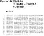
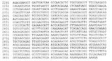
KCNN3のコーディング配列は736アミノ酸と分子量計算値82kDaをもつ蛋白質をコードする2211塩基対から構成される。この遺伝子は染色体1q22にマッピングされており、その8個のエキソンは約163kbpに及ぶ(Sunら,J.Hum.Genet.2001,46:463−470)。KCNN3はヒト及びマウス海馬で高度に発現され(Blankら,Nature Neurosci.6:2003,911−912;Tacconiら,Neuroscience 2001,102:209−215)、海馬ではその発現が加齢と共に増加すると報告されている(Blankら,Nature Neurosci.2003,6:911−912)。精密発現分析によると、KCNN3は特に海馬歯状回及びCA1−4領域と嗅内皮質で発現され、更に基底核、視床及び各種脳幹核でも発現されることが分かった(Sailerら,Mol.Cell.Neurosci.2004,26:458−469)。KCNN3は選択的にスプライスされ、KCNN1−3の機能を抑制するドミナントネガティブアイソフォームを生じることが報告されている(Kolski−Andreacoら,J.Biol.Chem.2004,279:6893−6904)。 The coding sequence of KCNN3 consists of 2211 base pairs encoding a protein having 736 amino acids and a calculated molecular weight of 82 kDa. This gene has been mapped to chromosome 1q22, and its 8 exons span approximately 163 kbp (Sun et al., J. Hum. Genet. 2001, 46: 463-470). KCNN3 is highly expressed in human and mouse hippocampus (Blank et al., Nature Neurosci. 6: 2003, 911-912; Tacconi et al., Neuroscience 2001, 102: 209-215) and reported that its expression increases with age in the hippocampus (Blank et al., Nature Neurosci. 2003, 6: 911-912). Precise expression analysis revealed that KCNN3 is expressed specifically in the hippocampal dentate gyrus, CA1-4 region and olfactory cortex, and is also expressed in the basal ganglia, thalamus and various brain stem nuclei (Sailer et al., Mol. Cell). Neurosci. 2004, 26: 458-469). KCNN3 has been reported to be alternatively spliced to produce a dominant negative isoform that represses the function of KCNN1-3 (Kolski-Andreaco et al., J. Biol. Chem. 2004, 279: 6893-6904).
KCNN3のその他の機能も報告されており、特に呼吸と分娩(Bondら,Science 2000,289:1942−1946)及び血圧調節(Taylorら,Circ.Res.2003,93:124−131)における役割が挙げられる。興味深いことに、KCNN3の加齢性発現上昇はアンチセンスオリゴヌクレオチド投与によるKCNN3−mRNAの選択的発現低下により克服できない多少の認知障害をマウスに生じたので、KCNN3の発現レベルはシナプス可塑性に結び付けられている(Blankら,Nature Neurosci.2003,6:911−912)。更に、KCNN3は膜興奮性の調節と中枢ニューロンの発火特性の決定に関与していることも示されている(Pedarzaniら,J.Biol.Chem.2001,276:9762−9769)。トランスジェニック動物が樹立されている(Bondら,Science 2000,289:1942−1946)。この場合、正常KCNN3プロモーター機能を維持しながら遺伝子スイッチの導入によりKCNN3遺伝子の発現を調節することはできない。著者らはKCNN3が例えば睡眠時無呼吸や乳幼児突然死のターゲットになると仮定している。 Other functions of KCNN3 have also been reported, particularly with a role in respiration and labor (Bond et al., Science 2000, 289: 1942-1946) and blood pressure regulation (Taylor et al., Circ. Res. 2003, 93: 124-131). Can be mentioned. Interestingly, the increased age-related expression of KCNN3 resulted in some cognitive impairment in mice that could not be overcome by the selective decrease of KCNN3-mRNA by antisense oligonucleotide administration, so the expression level of KCNN3 is linked to synaptic plasticity. (Blank et al., Nature Neurosci. 2003, 6: 911-912). Furthermore, KCNN3 has also been shown to be involved in the regulation of membrane excitability and in determining the firing properties of central neurons (Pedarzani et al., J. Biol. Chem. 2001, 276: 9762-9769). Transgenic animals have been established (Bond et al., Science 2000, 289: 1942-1946). In this case, the expression of the KCNN3 gene cannot be regulated by introducing a gene switch while maintaining the normal KCNN3 promoter function. The authors assume that KCNN3 is a target for sleep apnea and sudden infant death, for example.
SKチャネルの数種の遮断薬が記載されており、特に低ナノモル範囲でチャネルを遮断するハチ毒アパミンとサソリ毒スキラトキシンが挙げられる。しかし、これらの毒素は全SKチャネルを非特異的に遮断する。低分子量化合物がSKチャネル活性を妨害することも記載されているが、上記毒素に比較するとその効力は低い(例えばツボクラリン、UCL−1684、ガラミン)。 Several blockers of SK channels have been described, particularly bee venom apamin and scorpion venom squilatoxin that block the channel in the low nanomolar range. However, these toxins non-specifically block all SK channels. Although low molecular weight compounds have also been described to interfere with SK channel activity, they are less potent than the toxins (eg, tubocurarine, UCL-1684, gallamine).
本明細書と特許請求の範囲で使用する単数形はそうでないことが内容から明白である場合を除き、複数形も含む。例えば「細胞」は複数の細胞も意味し、他の用語についても同様である。 The singular forms used in the specification and claims include the plural unless the content clearly dictates otherwise. For example, “cell” means a plurality of cells, and the same applies to other terms.
本明細書と特許請求の範囲で使用する「及び/又は」なる用語はこの用語の前後の用語がそのいずれか一方又は両方であるとみなされることを意味する。例えば、「レベル及び/又は活性の測定」なる用語はレベルのみ又は活性のみ又はレベルと活性の両方を測定することを意味する。 The term “and / or” as used in the specification and claims means that the terms preceding and following the term are considered to be either or both. For example, the term “measurement of level and / or activity” means measuring level alone or activity alone or both level and activity.
本明細書で使用する「レベル」なる用語は転写産物(例えばmRNA)又は翻訳産物(例えば蛋白質又はポリペプチド)の量又は濃度の程度又は尺度を意味する。 As used herein, the term “level” refers to the degree or measure of the amount or concentration of a transcript (eg, mRNA) or translation product (eg, protein or polypeptide).
本明細書で使用する「活性」なる用語は転写産物もしくは翻訳産物が生物学的効果を生じる能力の尺度又は生物活性分子のレベルの尺度を意味する。 As used herein, the term “activity” refers to a measure of the ability of a transcript or translation product to produce a biological effect or a level of a bioactive molecule.
「活性」なる用語は更に、限定されないが配列番号1の新規カリウムチャネルポリペプチド等のカリウムチャネル又はカリウムチャネルサブユニットの結合、阻害、抑制、遮断、中和又は分離と、発現上昇又はカリウムチャネルサブユニットの活性化、アゴニスト作用、発現上昇に関する生物活性及び/又は薬理活性を意味する。「生物活性」としては限定されないが、カリウムイオン及び/又は膜貫通カリウムイオン流の膜貫通輸送及び/又はその調節が挙げられる。「薬理活性」としては限定されないが、カリウムチャネル又はカリウムチャネルサブユニットがリガンド、化合物、作用剤、モジュレーター及び/又は別のカリウムチャネルサブユニットと結合する能力が挙げられる。 The term “activity” further includes binding, inhibition, suppression, blocking, neutralization or separation of potassium channels or potassium channel subunits such as, but not limited to, the novel potassium channel polypeptide of SEQ ID NO: 1, and increased expression or potassium channel subunits. It means biological activity and / or pharmacological activity related to unit activation, agonist action, and increased expression. “Biological activity” includes, but is not limited to, transmembrane transport and / or modulation of potassium ion and / or transmembrane potassium ion flow. “Pharmacological activity” includes, but is not limited to, the ability of a potassium channel or potassium channel subunit to bind to a ligand, compound, agent, modulator and / or another potassium channel subunit.
本明細書で使用する「レベル」及び/又は「活性」なる用語は更に遺伝子発現レベル又は遺伝子活性を意味する。遺伝子発現は遺伝子に含まれる情報が転写及び翻訳により利用され、遺伝子産物が生産されることとして定義することができる。 As used herein, the term “level” and / or “activity” further refers to gene expression level or gene activity. Gene expression can be defined as the information contained in a gene being utilized by transcription and translation to produce a gene product.
「調節異常」とは遺伝子発現の発現上昇もしくは発現低下及び/又は遺伝子産物の安定性の増加もしくは低下を意味する。遺伝子産物はRNA又は蛋白質を含み、遺伝子発現の結果である。遺伝子産物の量を使用して遺伝子の活性の程度及びその遺伝子産物の安定性の程度を測定することができる。 “Dysregulation” means an increase or decrease in gene expression and / or an increase or decrease in the stability of a gene product. A gene product includes RNA or protein and is the result of gene expression. The amount of gene product can be used to measure the degree of gene activity and the degree of stability of the gene product.
本明細書と特許請求の範囲で使用する「遺伝子」なる用語はコーディング領域(エキソン)と非コーディング領域(例えばプロモーター又はエンハンサー等の非コーディング調節因子、イントロン、リーダー配列及びトレーラー配列)の両者を意味する。 As used herein and in the claims, the term “gene” refers to both coding regions (exons) and non-coding regions (eg, non-coding regulatory elements such as promoters or enhancers, introns, leader sequences and trailer sequences). To do.
「ORF」なる用語は「オープン・リーディング・フレーム(open reading frame)」の頭文字であり、少なくとも1個の読み枠に停止コドンをもたないため、潜在的にアミノ酸配列に翻訳することができる核酸配列を意味する。「調節因子」とは誘導型及び非誘導型プロモーター、エンハンサー、オペレーター、並びに遺伝子発現を誘導及び調節する他の因子を意味する。 The term “ORF” is an acronym for “open reading frame” and has no stop codon in at least one reading frame, so it can potentially be translated into an amino acid sequence. Means nucleic acid sequence. “Regulator” means inducible and non-inducible promoters, enhancers, operators, and other factors that induce and regulate gene expression.
本明細書で使用する「フラグメント」なる用語は例えば選択的スプライシング又は短縮又は他の方法で開裂した転写産物又は翻訳産物を意味する。 As used herein, the term “fragment” means, for example, a transcript or translation product that has been alternatively spliced or shortened or otherwise cleaved.
本明細書で使用する「誘導体」なる用語は突然変異もしくはRNA編集もしくは化学修飾もしくは他の方法で改変された転写産物又は突然変異もしくは化学修飾もしくは他の方法で改変された翻訳産物を意味する。分かりやすく言うならば、例えば誘導体転写産物とは一塩基又は多塩基欠失、挿入又は置換等の改変を核酸配列にもつ転写産物を意味する。例えば誘導体翻訳産物はリン酸化もしくはグリコシル化もしくはアセチル化もしくは脂質化の改変又はシグナルペプチド開裂もしくは他の型の成熟開裂の改変等の方法により作製することができる。これらの方法は翻訳後に実施することができる。 As used herein, the term “derivative” refers to a transcript that has been mutated or RNA edited or chemically modified or otherwise modified, or a translation product that has been mutated or chemically modified or otherwise modified. For example, a derivative transcript means a transcript having a modification in a nucleic acid sequence such as single base or polybase deletion, insertion or substitution. For example, derivative translation products can be made by methods such as modification of phosphorylation or glycosylation or acetylation or lipidation or modification of signal peptide cleavage or other types of mature cleavage. These methods can be performed after translation.
本明細書と特許請求の範囲で使用する「モジュレーター」なる用語は遺伝子又は遺伝子の転写産物又は遺伝子の翻訳産物のレベル及び/又は活性を変化又は変更させることが可能な分子を意味する。「モジュレーター」とはカリウムチャネルサブユニット又はカリウムチャネルの機能特性を亢進又は抑制し、従って、前記機能特性を「調節」し、カリウムチャネル又はカリウムチャネルサブユニットの結合、阻害、抑制、遮断、中和又は分離を「調節」し、活性化、アゴニスト作用及び発現上昇を「調節」することが可能な分子を意味する。「調節」は細胞の生物活性を変化させる能力という意味でも使用する。「モジュレーター」は遺伝子の転写産物又は翻訳産物の生物活性を変化又は変更させることができる。例えば前記調節としては、生物活性及び/又は薬理活性の増加もしくは低下、結合特性の変化、又は遺伝子の前記翻訳産物の生物学的、機能的もしくは免疫学的特性の他の任意変化もしくは変更が挙げられる。 As used herein and in the claims, the term “modulator” refers to a molecule capable of altering or altering the level and / or activity of a gene or gene transcript or gene translation product. A “modulator” enhances or suppresses the functional properties of a potassium channel subunit or potassium channel, and thus “modulates” the functional properties, and binds, inhibits, suppresses, blocks, neutralizes the potassium channel or potassium channel subunit. Alternatively, it refers to a molecule that can “modulate” segregation and “modulate” activation, agonistic action and increased expression. “Modulation” is also used to mean the ability to change the biological activity of a cell. A “modulator” can alter or alter the biological activity of a transcript or translation product of a gene. For example, the modulation includes an increase or decrease in biological and / or pharmacological activity, a change in binding properties, or any other change or change in the biological, functional or immunological properties of the translation product of a gene. It is done.
「作用剤」、「試薬」又は「化合物」なる用語は細胞、組織、体液又は任意生体系もしくは任意試験アッセイ系の状況内に正又は負の生物学的効果をもつ任意物質、化学薬品、組成物又は抽出物を意味する。例えば、ターゲットのアゴニスト、アンタゴニスト、部分アゴニスト又は逆アゴニストが挙げられる。このような作用剤、試薬又は化合物としては核酸、天然もしくは合成ペプチドもしくは蛋白複合体又は融合蛋白が挙げられる。更に、抗体、有機又は無機分子又は組成物、小分子、薬剤及び上記物質の任意のものの任意組合わせでもよい。これらは試験、診断又は治療目的に使用することができる。 The term “agent”, “reagent” or “compound” refers to any substance, chemical, composition that has a positive or negative biological effect within the context of a cell, tissue, body fluid or any biological system or any test assay system. Means product or extract. For example, a target agonist, antagonist, partial agonist or inverse agonist can be mentioned. Such agents, reagents or compounds include nucleic acids, natural or synthetic peptides or protein complexes or fusion proteins. Furthermore, any combination of antibodies, organic or inorganic molecules or compositions, small molecules, drugs and any of the above substances may be used. These can be used for testing, diagnostic or therapeutic purposes.
「オリゴヌクレオチドプライマー」又は「プライマー」なる用語は相補塩基対のハイブリド形成により所与ターゲットポリヌクレオチドとアニールすることができ、ポリメラーゼにより伸長することができる短い核酸配列を意味する。これらの配列は特定配列に対して特異的になるように選択してもよいし、ランダムに選択してもよく、例えば混合物中の可能な全配列をプライミングする。変発明で使用するプライマー長は10ヌクレオチド〜80ヌクレオチドとすることができる。「プローブ」は本明細書に記載及び開示する核酸配列の短い核酸配列又はその相補配列である。これらの配列は全長配列でもよいし、所与配列のフラグメント、誘導体、アイソフォーム又は変異体でもよい。「プローブ」とアッセイサンプルのハイブリダイゼーション複合体の同定により、このサンプル内の他の類似配列の存在の検出が可能になる。 The term “oligonucleotide primer” or “primer” refers to a short nucleic acid sequence that can anneal to a given target polynucleotide by hybridization of complementary base pairs and can be extended by a polymerase. These sequences may be selected to be specific for a particular sequence or may be selected randomly, eg, priming all possible sequences in the mixture. The primer length used in the modified invention can be 10 to 80 nucleotides. A “probe” is a short nucleic acid sequence of the nucleic acid sequences described and disclosed herein or a complementary sequence thereof. These sequences may be full-length sequences or fragments, derivatives, isoforms or variants of a given sequence. Identification of the hybridization complex between the “probe” and the assay sample allows detection of the presence of other similar sequences in the sample.
本明細書で使用する「ホモログ又はホモロジー」なる用語はヌクレオチド又はペプチド配列と別のヌクレオチド又はペプチド配列の近縁度を意味し、比較する前記配列間の一致度及び/又は類似度により決定される。当分野では、「一致度」及び「類似度」なる用語はクエリー配列と好ましくは同一型の他の配列(核酸又は蛋白質配列)の相互マッチングにより決定されるポリペプチド又はポリヌクレオチド配列近縁度を意味する。「一致度」及び「類似度」を計算及び決定するための好ましいコンピュータープログラムとしては限定されないが、GCG BLAST(Basic Local Alignment Search Tool)(Altschulら,J.Mol.Biol.1990,215:403−410;Altschulら,Nucleic Acids Res.1997,25:3389−3402;Devereuxら,Nucleic Acids Res.1984,12:387)、BLASTN 2.0(Gish W.,http://blast.wustl.edu,1996−2002)、FASTA(Pearson and Lipman,Proc.Natl.Acad.Sci.USA 1988,85:2444−2448)及び最長オーバーラップをもつ1対のコンティグを決定し、整列させるGCG GelMerge(Wilbur and Lipman,SIAM J.Appl.Math.1984,44:557−567;Needleman and Wunsch,J.Mol.Biol.1970,48:443−453)が挙げられる。 As used herein, the term “homolog or homology” refers to the closeness of a nucleotide or peptide sequence to another nucleotide or peptide sequence, determined by the degree of identity and / or similarity between the sequences being compared. . In the art, the terms “coincidence” and “similarity” refer to the closeness of a polypeptide or polynucleotide sequence determined by mutual matching of other sequences (nucleic acid or protein sequences), preferably of the same type with the query sequence. means. Preferred computer programs for calculating and determining “coincidence” and “similarity” include, but are not limited to, GCG BLAST (Basic Local Alignment Search Tool) (Altschul et al., J. Mol. Biol. 1990, 215: 403- 410; Altschul et al., Nucleic Acids Res. 1997, 25: 3389-3402; Devereux et al., Nucleic Acids Res. 1984, 12: 387), BLASTN 2.0 (Gish W., http://blast.wustl.edu. 1996-2002), FASTA (Pearson and Lipman, Proc. Natl. Acad. Sci. USA 1988, 85: 2444-24. 48) and GCG GelMerge (Wilbur and Lipman, SIAM J. Appl. Math. 1984, 44: 557-567; Needleman and Wunsch, J. Mol. Biol. 1970, 48: 443-453).
本明細書で使用する「変異体」なる用語は本発明に開示するポリペプチド及び蛋白質に関して、本発明の天然ポリペプチド又は蛋白質のN末端及び/又はC末端及び/又は天然アミノ酸配列内に1個以上のアミノ酸が付加され及び/又は置換され及び/又は欠失され及び/又は挿入されているが、その本質的特性を維持する任意ポリペプチド又は蛋白質を意味する。更に、「変異体」なる用語はポリペプチド又は蛋白質の任意短縮形態又は延長形態を含む。「変異体」はKCNN3蛋白質のアミノ酸配列、特に配列番号1に対して少なくとも約80%の配列一致度、より好ましくは少なくとも約90%の配列一致度、最も好ましくは少なくとも約95%の配列一致度をもつ配列も含む。「変異体」としては例えば高度保存領域に保存アミノ酸置換をもつ蛋白質が挙げられる。本発明の「蛋白質及びポリペプチド」は、KCNN3蛋白質のアミノ酸配列、特に配列番号1を含む蛋白質の変異体、フラグメント及び化学誘導体を含む。コドンが代替塩基配列により別のコドンで置換されているが、DNA配列により翻訳されたアミノ酸配列が変化していない配列変異が含まれる。当分野で公知のこの現象は特定アミノ酸を翻訳するコドンセットの冗長と呼ばれる。機能性に影響を与えないこのようなアミノ酸置換(例えばリジン→アルギニン、ロイシン→バリン、グルタミン→アスパラギン)が含まれる。自然界から単離可能であるか又は組換え及び/又は合成手段により生産可能な蛋白質及びポリペプチドが含まれる。天然蛋白質又はポリペプチドとは天然に存在する短縮又は分泌形態、天然に存在する変異体形態(例えばスプライス変異体)及び天然に存在する対立遺伝子変異体を意味する。本明細書で使用する「単離」なる用語はその天然環境から変化しているか及び/又は取出されており、即ちそれらが通常存在する細胞又は生体から単離されており、それらが自然界で結合していることが分かっている共存成分から分離されているか又は本質的に精製されている分子又は物質を意味する。この概念は更にこのような分子をコードする配列がその天然状態では結合していないポリヌクレオチドとこの配列を人為的に結合することができ、このような分子を組換え及び/又は合成手段により生産できることを意味し、「非天然」とも言う。前記目的のためにこれらの配列を当業者に公知の方法により生体又は非生体に導入することができるとしても、また、これらの配列が前記生体又は非生体に依然として存在しているとしても、これらの配列は単離されているとみなされる。本発明では、「危険」、「感受性」及び「素因」なる用語は同等であり、神経変性疾患、好ましくはアルツハイマー病を発症する確率に関して使用する。 As used herein, the term “variant” refers to one polypeptide within the N-terminal and / or C-terminal and / or natural amino acid sequence of the natural polypeptide or protein of the present invention with respect to the polypeptides and proteins disclosed in the present invention. It means any polypeptide or protein to which the above amino acids have been added and / or substituted and / or deleted and / or inserted, but maintain its essential properties. Furthermore, the term “variant” includes any shortened or extended form of a polypeptide or protein. A “variant” is an amino acid sequence of the KCNN3 protein, in particular at least about 80% sequence identity, more preferably at least about 90% sequence identity, most preferably at least about 95% sequence identity to SEQ ID NO: 1. Also includes arrays with. Examples of “mutants” include proteins having conservative amino acid substitutions in highly conserved regions. The “proteins and polypeptides” of the present invention include KCNN3 protein amino acid sequences, particularly protein variants, fragments and chemical derivatives comprising SEQ ID NO: 1. Codons are replaced with alternative codons by alternative codons, but sequence mutations that do not change the amino acid sequence translated by the DNA sequence are included. This phenomenon known in the art is called redundancy of codon sets that translate specific amino acids. Such amino acid substitutions that do not affect functionality (eg lysine → arginine, leucine → valine, glutamine → asparagine) are included. Proteins and polypeptides that can be isolated from nature or that can be produced by recombinant and / or synthetic means are included. Natural protein or polypeptide means naturally occurring truncated or secreted forms, naturally occurring variant forms (eg, splice variants) and naturally occurring allelic variants. As used herein, the term “isolated” has been altered and / or removed from its natural environment, ie it has been isolated from the cells or organism in which it normally resides and is bound in nature. Means a molecule or substance that has been separated or essentially purified from coexisting components known to be This concept also allows the sequence to be artificially linked to a polynucleotide to which the sequence encoding such a molecule is not bound in its native state, and such molecules are produced by recombinant and / or synthetic means. It means what can be done and is also called "non-natural". Even if these sequences can be introduced into a living body or non-living body by a method known to those skilled in the art for the above purpose, or even if these sequences are still present in the living body or non-living body, Sequences are considered isolated. In the present invention, the terms “danger”, “susceptibility” and “predisposition” are equivalent and are used with respect to the probability of developing a neurodegenerative disease, preferably Alzheimer's disease.
「AD」なる用語はアルツハイマー病を意味する。 The term “AD” means Alzheimer's disease.
本明細書で使用する「AD型神経病理」、「AD病理」なる用語は本明細書に記載し、最先端文献から広く公知の神経病理学的、神経生理学的、組織病理学的及び臨床的特徴、徴候及び症状を意味する(Iqbal,Swaab,Winblad and Wisniewski,Alzheimer’s Disease and Related Disorders(Etiology,Pathogenesis and Therapeutics),Wiley & Sons,New York,Weinheim,Toronto,1999;Scinto and Daffner,Early Diagnosis of Alzheimer’s Disease,Humana Press,Totowa,New Jersey,2000;Mayeux and Christen,Epidemiology of Alzheimer’s Disease:From Gene to Prevention,Springer Press,Berlin,Heidelberg,New York,1999;Younkin,Tanzi and Christen,Presenilins and Alzheimer’s Disease,Springer Press,Berlin,Heidelberg,New York,1998参照)。 As used herein, the terms “AD neuropathology” and “AD pathology” are described herein and are well known from the state of the art in neuropathology, neurophysiology, histopathology and clinical Means signs, symptoms and symptoms (Iqbal, Swab, Winblad and Wisniewski, Alzheimer's Dissease and Relied Disorders (Etiology, Pathogenesis and Therapeutics) Diagnosis of Alzheimer's Disease, Humana Press, Totowa, New Jersey , 2000; Mayeux and Christen, Epidemiology of Alzheimer's Disease: From Gene to Prevention, Springer Press, Berlin, Heidelberg, New York, 1999; Younkin, Tanzi and Christen, Presenilins and Alzheimer's Disease, Springer Press, Berlin, Heidelberg , New York, 1998).
「ブラークステージ(Braak stage)」又は「ブラークステージ分類」なる用語はBraak and Braakにより提案された基準による脳分類を意味する(Braak and Braak,Acta Neuropathology 1991,82:239−259;Braak and Braak,J Neural Transm 1998,53:127−140)。ADのブラークステージ分類は前脳の所定領域における神経原線維病理の程度と分布をランク付けし、ADの神経病理進行を6段階(ステージ0〜6)に分けている。これはADの死後神経病理段階分類において広く許容されている確立された方法である。精神状態及び認知機能/障害に関するAD患者の臨床症状と検死後に得られる対応するブラークステージの間には有意の相関があることが説得力をもって示されている(Bancherら,Neuroscience Letters 1993,162:179−182;Goldら,Acta Neuropathol 2000,99:579−582)。同様に、神経原線維変化とニューロン細胞病理の相関も認められており(Rosslerら,Acta Neuropathol 2002,103:363−369)、これらはどちらも認知機能を予測することが報告されている(Giannakopoulosら,Neurology 2003,60:1495−1500;Bennettら,Arch Neurol 2004,61:378−384)。更に、βアミロイドペプチド沈着から最終的に神経原線維変化の形成に至る発症カスケードも提示されており、こうして分子/細胞レベルにおける初期AD特異的イベントの先行が証明されている(Metsaarsら,Neurobiol Aging 2003,24:563−572)。 The term “Braak stage” or “Blaak stage classification” means a brain classification according to the criteria proposed by Braak and Braak (Braak and Braak, Acta Neuropathology 1991, 82: 239-259; Braak and Braak; J Neural Transm 1998, 53: 127-140). AD's plaque stage classification ranks the degree and distribution of neurofibrillary pathology in a predetermined region of the forebrain, and divides AD neuropathological progression into 6 stages (
従って、本発明では個体ドナーの臨床徴候/症状に依存しない(即ち精神障害、認知障害、他の神経精神病学的パラメーターの低下又はADの明白な臨床診断の報告の有無に依存しない)疾患進行の代替マーカーとしてブラークステージを使用する。即ち、ブラークステージ分類の基準となる神経原線維変化はその根底にある分子及び細胞病理メカニズム一般を反映し、従って、脳の病的(又は病前)状態を定義すると推測され、即ち、例えばブラークステージ1のドナーは定義によると、ステージ2(以上)のドナーよりも初期の分子/細胞発症段階にあり、従って、ブラークステージ1のドナーは例えばブラークステージ2以上のドナーと比較する場合の対照個体とみなすことができる。この点で、対照個体と疾患個体の区別は臨床診断に基づく「健常対照ドナー」と「AD患者」の区別と必ずしも同一である必要はなく、代替マーカーであるブラークステージにより推定及び分類した病的(又は病前)状態の推定相違を意味する。 Thus, the present invention does not depend on the individual donor's clinical signs / symptoms (ie, independent of the presence or absence of reports of mental disorders, cognitive impairment, reduction of other neuropsychological parameters or clear clinical diagnosis of AD). Use the plaque stage as an alternative marker. That is, the neurofibrillary tangles that serve as the basis for classification of the plaque stage reflect the underlying molecular and cytopathological mechanisms in general and are therefore speculated to define the pathological (or pre-morbid) state of the brain, ie,
「対照」から得られた値は「既知健常状態」を表す「参照値」であり、「AD患者」から得られた値は「既知疾患状態」を表す「参照値」である。本発明では、ブラークステージ0はアルツハイマー病の徴候及び症状をもつとみなされないヒトを表すことができ、ブラークステージ1〜4は既にADの臨床徴候及び症状があるか否かに応じて健常対照ヒト又はAD患者を表すことができる。ブラークステージの数値が高いほど、ADの徴候及び症状を示す可能性又はADの徴候及び症状を発症する危険が高くなる。神経病理評価即ちADの病理変化が痴呆の根本原因である確率の推定については、Braak H.(www.alzforum.org)によりアドバイスが与えられている。 The value obtained from “control” is a “reference value” representing “known healthy state”, and the value obtained from “AD patient” is a “reference value” representing “known disease state”. In the present invention,
本発明による神経変性疾患又は障害としては、アルツハイマー病、パーキンソン病、ハンチントン病、筋萎縮性側索硬化症、ピック病、前頭側頭型痴呆、進行性核性麻痺、大脳皮質基底核変性症、脳血管性痴呆、多発性全身萎縮症、嗜銀顆粒性痴呆及び他のタウパチー並びに軽度認知障害が挙げられる。神経変性プロセスに関する他の疾患としては、例えば虚血性脳卒中、加齢黄斑変性、ナルコレプシー、運動ニューロン疾患、プリオン疾患、外傷性神経損傷及び修復、並びに多発性硬化症が挙げられる。 Neurodegenerative diseases or disorders according to the present invention include Alzheimer's disease, Parkinson's disease, Huntington's disease, amyotrophic lateral sclerosis, Pick's disease, frontotemporal dementia, progressive nuclear paralysis, cortical basal ganglia degeneration, Examples include cerebrovascular dementia, multiple generalized atrophy, greasy granular dementia and other taupathies and mild cognitive impairment. Other diseases related to the neurodegenerative process include, for example, ischemic stroke, age-related macular degeneration, narcolepsy, motor neuron disease, prion disease, traumatic nerve injury and repair, and multiple sclerosis.
本発明は特定サンプル、AD患者の特定脳領域、各種ブラークステージのヒトの特定脳領域におけるカリウムチャネル、小コンダクタンスカルシウム活性化カリウムチャネルである小コンダクタンスカルシウム活性化カリウムチャネル蛋白3(別称KCNN3又はK3、別称SK3又はSKCa3)をコードする遺伝子及び前記遺伝子KCNN3(別称SK3)の蛋白産物の同定、差異的発現、差異的調節、調節異常を相互間比較及び/又は年齢一致対照ヒトとの比較により開示する。本発明はAD患者の下側頭葉皮質と前頭葉皮質ではKCNN3(SK3)mRNAレベルが増加し、発現が増加されるという点で、対照ヒトの各脳領域に比較してAD患者の脳ではKCNN3(SK3)の遺伝子発現が変化し、異常調節されることを開示する。更に、本発明はKCNN3(SK3)発現が各種ブラークステージで異なり、初期ブラークステージで既に発現レベル増加が開始しており、主に下側頭葉皮質で病理ブラークステージの進行に伴って漸進的に増加することを開示する。 The present invention relates to a specific sample, a specific brain region of an AD patient, a potassium channel in a human specific brain region of various plaque stages, a small conductance calcium activated potassium channel protein 3 (also known as KCNN3 or K3, Disclose the gene encoding another gene SK3 or SKCa3) and the protein product of the gene KCNN3 (also referred to as SK3), differential expression, differential regulation, dysregulation by mutual comparison and / or comparison with age-matched control humans . The present invention shows that KCNN3 (SK3) mRNA levels are increased and expression is increased in the inferior temporal cortex and frontal cortex of AD patients, and in the brains of AD patients, KCNN3 is increased compared to the control human brain regions. Disclose that (SK3) gene expression is altered and dysregulated. Furthermore, in the present invention, KCNN3 (SK3) expression is different at various plaque stages, and the expression level has already started to increase at the initial plaque stage, and gradually increases as the pathological plaque stage progresses mainly in the lower temporal cortex. Disclose the increase.
AD型病理の発症に平行するKCNN3(SK3)のこの調節異常はKCNN3とADの関係を明白に反映しており、疾患の過程における進行性病理イベントの指標である。AD患者と対照を比較した場合だけでなく各種ブラークステージ間で比較した場合にも観察されるKCNN3(SK3)遺伝子転写レベルの差異はKCNN3(SK3)蛋白質レベルに認められる実質的な差異により更に裏付けられる。対照とは対照的に、AD患者からの脳検体ではKCNN3(SK3)蛋白質は反応性星状細胞に高レベルで含まれており、蓄積し、皮質βアミロイド斑と同時沈着する。KCNN3(SK3)遺伝子発現のこの調節異常と、AD型病理の発症に平行する対応する遺伝子産物のレベル及び局在変化はKCNN3(SK3)とADの関係を明白に反映しており、疾患の過程における進行性病理イベントの指標である。KCNN3(SK3)遺伝子発現の調節異常及び対応する遺伝子産物のレベル及び局在変化と神経変性疾患、特にADの病理の関係を実証する実験はまだ記載されていない。同様に、KCNN3(SK3)遺伝子における突然変異が前記疾患と関連することも記載されていない。KCNN3(SK3)遺伝子をこのような疾患と関連付けることにより、特に前記疾患の診断及び治療の新規方法が得られる。更に、KCNN3をADの過程の初期に既に発生する病理イベントと関連付けることにより、脳の修復不能な損傷が生じる前に適用してAD病理の開始を予防する治療の可能性が得られる。従って、本発明は神経変性疾患、特にADの素因の診断評価、治療中のヒトの診断モニター、予後診断及び識別に有用である。 This dysregulation of KCNN3 (SK3) parallel to the onset of AD-type pathology clearly reflects the relationship between KCNN3 and AD and is an indicator of progressive pathological events in the course of the disease. Differences in KCNN3 (SK3) gene transcription levels observed not only when comparing AD patients and controls but also between various stages of plaque are further supported by the substantial differences observed in KCNN3 (SK3) protein levels It is done. In contrast to controls, in brain specimens from AD patients, KCNN3 (SK3) protein is present at high levels in reactive astrocytes, accumulates, and co-deposits with cortical β amyloid plaques. This dysregulation of KCNN3 (SK3) gene expression and the level and localization changes of the corresponding gene product parallel to the development of AD type pathology clearly reflect the relationship between KCNN3 (SK3) and AD, and the disease process Is an indicator of progressive pathological events in No experiments have yet been described that demonstrate the relationship between dysregulation of KCNN3 (SK3) gene expression and changes in the level and localization of the corresponding gene product and the pathology of neurodegenerative diseases, particularly AD. Similarly, it has not been described that mutations in the KCNN3 (SK3) gene are associated with the disease. By associating the KCNN3 (SK3) gene with such diseases, new methods of diagnosis and treatment of said diseases in particular are obtained. Furthermore, associating KCNN3 with a pathological event that already occurs early in the AD process provides the potential for treatment to be applied before irreparable damage to the brain occurs to prevent the onset of AD pathology. Thus, the present invention is useful for diagnostic evaluation of predisposition to neurodegenerative diseases, particularly AD, diagnostic monitoring of humans during treatment, prognosis and identification.
本発明はAD患者の特定脳領域におけるKCNN3(SK3)をコードする遺伝子及びその遺伝子産物の調節異常を開示する。下側頭葉、内側嗅領、海馬及び扁桃核内のニューロンはADで変性プロセスを受ける(Terryら,Annals of Neurology 1981,10:184−192)。これらの脳領域は主に学習及び記憶機能の処理に関与しており、ADでニューロン減少と変性に選択的感受性を示す。他方、前頭葉皮質、後頭葉皮質及び小脳内のニューロンはほぼ無傷のままであり、神経変性プロセスから保護される。本明細書に開示する実施例では、AD患者と年齢一致対照個体の前頭葉皮質(F)と下側頭葉皮質(T)に由来する脳組織を使用した。従って、KCNN3(SK3)遺伝子とその対応する転写及び/又は翻訳産物は原因としての役割を果たし、選択的ニューロン変性及び/又は神経保護に影響を与える。 The present invention discloses dysregulation of the gene encoding KCNN3 (SK3) and its gene product in specific brain regions of AD patients. Neurons in the inferior temporal lobe, medial olfactory tract, hippocampus and amygdala undergo a degenerative process in AD (Terry et al., Anals of Neurology 1981, 10: 184-192). These brain regions are primarily involved in the processing of learning and memory functions and are selectively sensitive to neuronal loss and degeneration in AD. On the other hand, neurons in the frontal cortex, occipital cortex and cerebellum remain nearly intact and protected from neurodegenerative processes. In the examples disclosed herein, brain tissue from the frontal cortex (F) and inferior temporal cortex (T) of AD patients and age-matched control individuals was used. Thus, the KCNN3 (SK3) gene and its corresponding transcription and / or translation products play a causative role and affect selective neuronal degeneration and / or neuroprotection.
1側面では、本発明は対象における神経変性疾患を診断もしくは予後診断する又は対象が前記疾患を発症する素因をもつか否かを判定する又は神経変性疾患をもつ対象に投与した治療薬の効果をモニターする方法に関する。本方法は前記対象から得られたサンプル中の(i)KCNN3蛋白質をコードする遺伝子の転写産物の及び/又は(ii)KCNN3蛋白質をコードする遺伝子の翻訳産物の及び/又は(iii)前記転写産物もしくは翻訳産物のフラグメントもしくは誘導体もしくは変異体のレベル、発現もしくは活性、又は前記レベル、発現及び前記活性の両者を測定する段階と、既知疾患状態(患者)を表す参照値及び/又は既知健常状態(対照)を表す参照値及び/又は既知ブラークステージを表す参照値と前記転写産物及び/又は前記翻訳産物の前記レベル、発現及び/又は前記活性を比較する段階と、前記レベル、発現及び/又は前記活性が既知健常状態を表す参照値に比較して変動、変化しているか否か、及び/又は既知疾患状態を表す参照値と同様もしくは同一であるか否か、及び/又は前記対象が神経変性疾患をもつこともしくは前記対象が前記疾患を発症する危険が増加していることの指標である既知ブラークステージを表す参照値と同様もしくは同一であるか否かを分析することにより、前記対象における前記神経変性疾患を診断もしくは予後診断する又は前記対象が前記神経変性疾患を発症する危険が増加しているか否かを判定する段階を含む。「対象における」なる用語は対象を冒す疾患に関する限りにおいて開示方法の結果を意味し、即ち、前記疾患は対象「における」。 In one aspect, the present invention diagnoses or prognoses a neurodegenerative disease in a subject, determines whether a subject has a predisposition to develop the disease, or effects of a therapeutic agent administered to a subject having a neurodegenerative disease. On how to monitor. The method comprises (i) a transcript of a gene encoding a KCNN3 protein and / or (ii) a translation product of a gene encoding a KCNN3 protein and / or (iii) the transcript in a sample obtained from the subject. Or measuring the level, expression or activity of a translation product fragment or derivative or variant, or both said level, expression and said activity, a reference value representing a known disease state (patient) and / or a known healthy state ( Comparing the level, expression and / or activity of the transcript and / or the translation product with a reference value representing a control) and / or a reference value representing a known plaque stage; Whether the activity is fluctuating or changing compared to a reference value representing a known healthy state and / or a reference value representing a known disease state Similar or identical and / or similar to a reference value representing a known plaque stage that is an indicator that the subject has a neurodegenerative disease or that the subject has an increased risk of developing the disease Or by diagnosing or prognosing the neurodegenerative disease in the subject or determining whether the subject is at increased risk of developing the neurodegenerative disease by analyzing whether or not they are the same Including. The term “in a subject” means the result of the disclosed method as far as the disease affecting the subject, ie the disease is “in” the subject.
別の1側面では、本発明は対象における神経変性疾患の進行をモニターする方法に関する。前記対象から得られたサンプル中の(i)KCNN3蛋白質をコードする遺伝子の転写産物、及び/又は(ii)KCNN3蛋白質をコードする遺伝子の翻訳産物、及び/又は(iii)前記転写産物もしくは翻訳産物のフラグメントもしくは誘導体もしくは変異体のレベル、発現もしくは活性、又は前記レベル、発現及び前記活性の両者を測定する。既知疾患状態又は既知健常状態又は既知ブラークステージを表す参照値と前記レベル、発現及び/又は前記活性を比較する。こうして、前記対象における前記神経変性疾患の進行をモニターする。 In another aspect, the invention relates to a method of monitoring the progression of a neurodegenerative disease in a subject. (I) a transcription product of a gene encoding a KCNN3 protein and / or (ii) a translation product of a gene encoding a KCNN3 protein in a sample obtained from the subject, and / or (iii) the transcription product or translation product. The level, expression or activity, or both the level, expression and activity are measured. The level, expression and / or activity is compared to a reference value representing a known disease state or a known healthy state or a known plaque stage. Thus, the progression of the neurodegenerative disease in the subject is monitored.
更に別の1側面では、本発明は神経変性疾患の治療の効果をモニターする治療評価方法に関し、本方法は前記疾患の治療中の対象から得られたサンプル中の(i)KCNN3蛋白質をコードする遺伝子の転写産物、及び/又は(ii)KCNN3蛋白質をコードする遺伝子の翻訳産物、及び/又は(iii)前記転写産物もしくは翻訳産物のフラグメントもしくは誘導体もしくは変異体のレベル、発現もしくは活性、又は前記レベル、発現及び前記活性の両者を測定する段階を含む。既知疾患状態又は既知健常状態又は既知ブラークステージを表す参照値と前記レベル、発現もしくは前記活性、又は前記レベル及び前記活性の両者を比較することにより、前記神経変性疾患の治療を評価する。 In yet another aspect, the present invention relates to a therapeutic evaluation method for monitoring the effect of treatment of a neurodegenerative disease, the method encoding (i) KCNN3 protein in a sample obtained from a subject being treated for said disease. The level, expression or activity of the transcription product of the gene and / or (ii) the translation product of the gene encoding the KCNN3 protein, and / or (iii) a fragment or derivative or variant of said transcription product or translation product, or said level Measuring both expression and said activity. The treatment of the neurodegenerative disease is evaluated by comparing the level, expression or activity, or both the level and the activity with a reference value representing a known disease state or a known healthy state or a known plaque stage.
好ましい1態様では、前記疾患の進行をモニターするために、一定期間にわたって前記対象から採取したサンプル系列中の(i)KCNN3蛋白質をコードする遺伝子の転写産物、及び/又は(ii)KCNN3蛋白質をコードする遺伝子の翻訳産物、及び/又は(iii)前記転写産物もしくは翻訳産物のフラグメントもしくは誘導体もしくは変異体のレベル、発現もしくは活性、又は前記レベル及び前記活性の両者を比較する。他の好ましい態様では、前記サンプル採取の1回以上の前に前記対象を治療する。更に別の好ましい態様では、前記対象の前記治療の前後に前記レベル及び/又は活性を測定する。 In a preferred embodiment, in order to monitor the progression of the disease, (i) a transcript of a gene encoding KCNN3 protein and / or (ii) a KCNN3 protein in a sample series collected from the subject over a period of time. Comparing the level, expression or activity of the translation product of the gene and / or (iii) a fragment or derivative or variant of the transcript or translation product, or both the level and the activity. In another preferred embodiment, the subject is treated one or more times before the sampling. In yet another preferred embodiment, the level and / or activity is measured before and after the treatment of the subject.
本発明の方法、キット、組換え動物、分子、アッセイ及び使用の好ましい1態様では、前記KCNN3遺伝子及び蛋白質(別称小コンダクタンスカルシウム活性化カリウムチャネル蛋白3、別称KCNN3又はK3、別称SK3又はSKCa3)は特に配列番号1の蛋白質(Genbank accesion number Q9UGI6)をコードするKCNN3遺伝子により表される。前記蛋白質のアミノ酸配列はGenbank accesion number AJ251016のcDNA配列(KCNN3,SK3,K3)に対応する配列番号5に対応するmRNA配列から推定される。本発明では、KCNN3はヒトKCNN3のコーディング配列(cds)を表す配列番号9の核酸配列も意味する。本発明では、前記配列は本明細書で使用する用語の意味で「単離」されている。更に、本発明では、前記KCNN3蛋白質をコードする遺伝子を一般にKCNN3遺伝子又はSK3遺伝子とも言い、簡単にKCNN3又はSK3とも言う。KCNN3又はSK3の蛋白質は一般にKCNN3蛋白質又はSK3蛋白質とも言う。 In a preferred embodiment of the method, kit, recombinant animal, molecule, assay and use of the present invention, the KCNN3 gene and protein (also known as small conductance calcium-activated
本発明の方法、キット、組換え動物、分子、アッセイ及び使用の別の好ましい1態様では、前記神経変性疾患又は障害はアルツハイマー病であり、前記対象はアルツハイマー病の徴候及び症状をもつ。 In another preferred embodiment of the methods, kits, recombinant animals, molecules, assays and uses of the invention, the neurodegenerative disease or disorder is Alzheimer's disease and the subject has signs and symptoms of Alzheimer's disease.
分析及び測定するサンプルは脳組織もしくは他の組織又は体細胞からなる群から選択することが好ましい。サンプルは更に脳髄液又は他の体液(例えば唾液、尿、糞便、血液、血清、血漿又は粘液)でもよい。本発明の神経変性疾患の診断、予後診断、進行モニター又は治療評価方法は体外で実施することができ、このような方法は対象もしくは患者又は対照ヒトから分離、採取又は単離したサンプル(例えば体液又は細胞)に実施することが好ましい。 The sample to be analyzed and measured is preferably selected from the group consisting of brain tissue or other tissues or somatic cells. The sample may also be cerebrospinal fluid or other body fluid (eg saliva, urine, feces, blood, serum, plasma or mucus). The diagnostic, prognostic, progress monitoring or therapeutic evaluation methods of neurodegenerative diseases of the present invention can be performed outside the body, and such methods can be performed separately from a subject or patient or control human (eg, body fluids). Or cell).
他の好ましい態様では、前記参照値は前記神経変性疾患をもたない対象から得られたサンプル(対照サンプル、対照、健常対照サンプル)又は神経変性疾患、特にアルツハイマー病をもつ対象から得られたサンプル(患者サンプル、患者、ADサンプル)又はADの徴候及び症状の有無を問わずに規定ブラークステージのヒトから得られたサンプル中の(i)KCNN3蛋白質をコードする遺伝子の転写産物、及び/又は(ii)KCNN3蛋白質をコードする遺伝子の翻訳産物、及び/又は(iii)前記転写産物もしくは翻訳産物のフラグメントもしくは誘導体もしくは変異体のレベル、発現もしくは活性、又は前記レベル及び前記活性の両者の値である。 In another preferred embodiment, said reference value is a sample obtained from a subject without said neurodegenerative disease (control sample, control, healthy control sample) or a sample obtained from a subject with neurodegenerative disease, in particular Alzheimer's disease. (Patient sample, patient, AD sample) or (i) a transcript of the gene encoding the KCNN3 protein in a sample obtained from a human at a defined plaque stage with or without AD signs and symptoms, and / or ( ii) the translation product of the gene encoding the KCNN3 protein, and / or (iii) the level, expression or activity of the transcript or translation product fragment or derivative or variant, or both values of the level and the activity .
好ましい態様では、前記対象から採取したサンプル細胞又は組織又は体液中のKCNN3蛋白質をコードする遺伝子の転写産物及び/又はKCNN3蛋白質をコードする遺伝子の翻訳産物及び/又はそのフラグメントもしくは誘導体もしくは変異体のレベル及び/又は活性及び/又は発現が既知健常状態(対照サンプル)を表す参照値に対して変化しているならば、神経変性疾患、特にADであると診断もしくは予後診断されるか又はADになる危険が増加していると判定される。別の好ましい1態様では、対象から得られたサンプル細胞又は組織又は体液中のKCNN3蛋白質をコードする遺伝子の転写産物及び/又はKCNN3蛋白質をコードする遺伝子の翻訳産物及び/又はそのフラグメントもしくは誘導体もしくは変異体のレベル及び/又は活性が神経変性疾患、特にアルツハイマー病の既知疾患状態(AD患者サンプル)を表す参照値に対して同一又は同様であるならば、前記神経変性疾患であると診断もしくは予後診断されるか又はその危険が増加していると判定される。 In a preferred embodiment, the level of the transcript of the gene encoding the KCNN3 protein and / or the translation product of the gene encoding the KCNN3 protein and / or the fragment or derivative or variant thereof in the sample cells or tissues or body fluids collected from the subject And / or if the activity and / or expression is altered relative to a reference value representing a known healthy state (control sample), it is diagnosed or prognostic or becomes AD with a neurodegenerative disease, in particular AD It is determined that the risk is increasing. In another preferred embodiment, a transcription product of a gene encoding KCNN3 protein and / or a translation product of a gene encoding KCNN3 protein and / or a fragment or derivative or mutation thereof in a sample cell or tissue or body fluid obtained from a subject If the body level and / or activity is the same or similar to a reference value that represents a known disease state (AD patient sample) of a neurodegenerative disease, particularly Alzheimer's disease, it is diagnosed or prognosticated as said neurodegenerative disease Or the risk is determined to have increased.
更に別の好ましい態様では、対象から得られたサンプル細胞又は組織又は体液中のKCNN3蛋白質をコードする遺伝子の転写産物及び/又はKCNN3蛋白質をコードする遺伝子の翻訳産物及び/又はそのフラグメントもしくは誘導体もしくは変異体のレベル及び/又は活性がADの徴候及び症状を発症する高い危険を示す既知ブラークステージを表す参照値に対して同一又は同様であるならば、ADであると診断もしくは予後診断されるか又はADになる危険が増加していると判定される。 In yet another preferred embodiment, a transcription product of a gene encoding KCNN3 protein and / or a translation product of a gene encoding KCNN3 protein and / or a fragment or derivative or mutation thereof in a sample cell or tissue or body fluid obtained from a subject If the body level and / or activity is the same or similar to a reference value that represents a known plaque stage that indicates a high risk of developing signs and symptoms of AD, or is diagnosed or prognosticated as AD It is determined that the risk of becoming AD is increasing.
KCNN3の前記転写産物及び/又は前記翻訳産物及び/又はそのフラグメントもしくは誘導体もしくは変異体の前記レベル変動変化、発現変化及び/又は前記活性変化は増加、発現上昇であることが好ましい。 It is preferable that the level variation change, expression change and / or activity change of the transcript and / or translation product and / or fragment or derivative or mutant thereof of KCNN3 is increased or increased.
好ましい態様では、転写産物のレベル及び/又はKCNN3蛋白質をコードする遺伝子の発現の測定はプライマーセットによる定量的PCR分析を使用し、対象のサンプルから抽出したRNAの逆転写により得られたcDNAに由来する前記遺伝子特異的配列を増幅することにより、対象から得られたサンプル中で実施される。本発明の実施例(vi)にプライマーセット(配列番号13,配列番号14)を示すが、本発明に開示する配列から作製した他のプライマーも使用することができる。前記遺伝子に特異的なプローブを使用したノーザンブロット又はリボヌクレアーゼ保護アッセイ(RPA)も適用できる。チップマイクロアレー技術により転写産物を測定すると更に好ましいと思われる。これらの技術は当業者に公知である(Sambrook and Russell,Molecular Cloning:A Laboratory Manual,Cold Spring Harbor Laboratory Press,Cold Spring Harbor,New York,2001;Schena M.,Microarray Biochip Technology,Eaton Publishing,Natick,MA,2000参照)。イムノアッセイの1例は特許出願WO02/14543に開示及び記載されているような酵素活性の検出及び測定である。 In a preferred embodiment, the measurement of transcript level and / or expression of the gene encoding the KCNN3 protein is derived from cDNA obtained by reverse transcription of RNA extracted from a sample of interest using quantitative PCR analysis with a primer set. In a sample obtained from a subject by amplifying said gene specific sequence. Although a primer set (SEQ ID NO: 13, SEQ ID NO: 14) is shown in Example (vi) of the present invention, other primers prepared from the sequences disclosed in the present invention can also be used. Northern blots or ribonuclease protection assays (RPA) using probes specific for the gene can also be applied. It would be more preferable to measure the transcripts by chip microarray technology. These techniques are well known to those skilled in the art (Sambrook and Russell, Molecular Cloning: A Laboratory Manual, Cold Spring Harbor Laboratory Press, Cold Spring Harbor, New York, NewMorch. MA, 2000). One example of an immunoassay is the detection and measurement of enzyme activity as disclosed and described in patent application WO 02/14543.
本発明は更に本発明に開示するような核酸配列又はそのフラグメントもしくは変異体に固有のプライマー及びプローブの構築及び使用に関する。オリゴヌクレオチドプライマー及び/又はプローブは蛍光、生物発光、磁気又は放射性物質で特異的に標識することができる。本発明は更に前記特異的オリゴヌクレオチドプライマーを適宜併用した前記核酸配列又はそのフラグメント及び変異体の検出及び生産に関する。前記プライマーセットを使用して当業者に周知の方法であるPCR分析を実施し、核酸を含むサンプルから前記遺伝子特異的核酸配列を増幅することができる。このようなサンプルは健常対象又は疾患対象又は規定ブラークステージの対象に由来する。増幅の結果として特異的核酸産物が生産されるか否かと、異なる長さのフラグメントを獲得できるか否かが神経変性疾患、特にアルツハイマー病の指標となる。従って、本発明は神経変性疾患、特にアルツハイマー病に関連し得る試験核酸配列を含む所与サンプル中の遺伝子突然変異及び一塩基多形の検出に有用な少なくとも10塩基長から全長コーディング配列及び遺伝子配列までの核酸配列、オリゴヌクレオチドプライマー及びプローブを提供する。この特徴は高速DNA塩基診断試験を開発するのに有利であり、このような試験はキットのフォーマットであることも好ましい。実施例1(vi)にKCNN3のプライマーを例示する。 The invention further relates to the construction and use of primers and probes specific to nucleic acid sequences as disclosed herein or fragments or variants thereof. Oligonucleotide primers and / or probes can be specifically labeled with fluorescent, bioluminescent, magnetic or radioactive substances. The present invention further relates to the detection and production of the nucleic acid sequence or a fragment and variant thereof appropriately combined with the specific oligonucleotide primer. The primer set can be used to perform PCR analysis, a method well known to those skilled in the art, to amplify the gene-specific nucleic acid sequence from a sample containing nucleic acid. Such samples are derived from healthy subjects or disease subjects or subjects with a defined plaque stage. Whether specific nucleic acid products are produced as a result of amplification and whether fragments of different lengths can be obtained are indicators of neurodegenerative diseases, particularly Alzheimer's disease. Accordingly, the present invention provides at least 10 base long to full length coding sequences and gene sequences useful for the detection of genetic mutations and single nucleotide polymorphisms in a given sample comprising test nucleic acid sequences that may be associated with neurodegenerative diseases, particularly Alzheimer's disease. Nucleic acid sequences, oligonucleotide primers and probes are provided. This feature is advantageous for developing fast DNA base diagnostic tests, and such tests are also preferably in kit format. Example 1 (vi) illustrates a primer for KCNN3.
更に、イムノアッセイ、活性アッセイ及び/又は結合アッセイを使用し、KCNN3蛋白質をコードする遺伝子の翻訳産物及び/又は前記翻訳産物のフラグメントもしくは誘導体もしくは変異体のレベル及び/又は活性及び/又は発現、及び/又は前記翻訳産物及び/又はそのフラグメントもしくは誘導体もしくは変異体の活性レベルを検出することができる。これらのアッセイは抗蛋白抗体又は抗蛋白抗体と結合する二次抗体に付けた酵素、クロモダイナミック、放射性、磁気又は蛍光ラベルを利用して前記蛋白分子と抗蛋白抗体の結合量を測定することができる。更に、他の高親和性リガンドも使用できる。使用可能なイムノアッセイとしては例えばELISA、ウェスタンブロット及び当業者に公知の他の技術が挙げられる(Harlow and Lane,Antibodies:A Laboratory Manual,Cold Spring Harbor Laboratory Press,Cold Spring Harbor,New York,1999及びEdwards R,Immunodiagnostics:A Practical Approach,Oxford University Press,Oxford;England,1999参照)。これらの全検出技術はマイクロアレー、蛋白アレー、抗体マイクロアレー、組織マイクロアレー、電子バイオチップ又は蛋白チップ技術のフォーマットで利用することもできる(Schena M.,Microarray Biochip Technology,Eaton Publishing,Natick,MA,2000参照)。 In addition, the level and / or activity and / or expression of the translation product of the gene encoding the KCNN3 protein and / or fragments or derivatives or variants of said translation product, using immunoassays, activity assays and / or binding assays, and / or Alternatively, the activity level of the translation product and / or fragment or derivative or variant thereof can be detected. These assays can measure the amount of binding between the protein molecule and the anti-protein antibody using an enzyme, chromodynamic, radioactive, magnetic or fluorescent label attached to the anti-protein antibody or the secondary antibody that binds to the anti-protein antibody. it can. In addition, other high affinity ligands can be used. Immunoassays that can be used include, for example, ELISA, Western blot, and other techniques known to those skilled in the art (Harlow and Lane, Antibodies: A Laboratory Manual, Cold Spring Laboratory Press, Cold Spring 99, Cold Spring 99, Cold Spring Harbor, 19 R, Immunodiagnostics: A Practical Approach, Oxford University Press, Oxford; England, 1999). All these detection techniques can also be used in the format of microarrays, protein arrays, antibody microarrays, tissue microarrays, electronic biochips or protein chip technologies (Schena M., Microarray Biochip Technology, Eaton Publishing, Natick, MA). , 2000).
別の側面では、本発明は対象における神経変性疾患、特にADを診断もしくは予後診断するか、又は対象が神経変性疾患、特にADを発症する性向もしくは素因の有無を判定するか、又は神経変性疾患、特にADをもつ対象に投与した治療薬の効果をモニターするためのキットに関し、前記キットは、
(a)(i)KCNN3蛋白質をコードする遺伝子の転写産物を選択的に検出する試薬、(ii)KCNN3蛋白質をコードする遺伝子の翻訳産物を選択的に検出する試薬から構成される群から選択される少なくとも1種の試薬と;
(b)−前記対象から得られたサンプル中のKCNN3蛋白質をコードする遺伝子の前記転写産物及び/又は前記翻訳産物及び/又はフラグメントもしくは誘導体もしくは変異体のレベルもしくは活性、又は前記レベル及び前記活性の両者及び/又は発現を測定し;
−既知疾患状態(患者)を表す参照値及び/又は既知健常状態(対照)を表す参照値及び/又は既知ブラークステージを表す参照値と前記転写産物及び/又は前記翻訳産物の前記レベル及び/又は前記活性及び/又は発現を比較し;
−前記レベル及び/又は前記活性及び/又は発現が既知健常状態を表す参照値に比較して変化しているか否か、及び/又は既知疾患状態を表す参照値もしくは既知ブラークステージを表す参照値と同様もしくは同一であるか否かを分析し;
−神経変性疾患、特にADを診断もしくは予後診断し又は前記対象が前記疾患を発症する性向もしくは素因の有無を判定し、前記転写産物及び/又は前記翻訳産物のレベル、発現もしくは活性、又は前記レベル及び前記活性の両者が既知健常状態(対照)を表す参照値に比較して変動もしくは変化しているか、及び/又は前記転写産物及び/又は前記翻訳産物のレベル、発現もしくは活性、又は前記レベル及び前記活性の両者が既知疾患状態(患者)、好ましくはADの疾患状態(AD患者)を表す参照値及び/又は既知ブラークステージを表す参照値と同様もしくは同一であるならば、神経変性疾患、特にADであると診断もしくは予後診断する、前記疾患を発症する性向もしくは素因が増加しており、ADの徴候及び症状を発症する危険が高いと判定することにより、神経変性疾患、特にADを診断もしくは予後診断する、又は対象が前記疾患を発症する性向もしくは素因の有無を判定する、又は治療効果をモニターするための説明書を含む。本発明のキットは神経変性疾患、特にADを発症する危険のある個体の識別に特に有用であると思われる。In another aspect, the invention diagnoses or prognoses a neurodegenerative disease, particularly AD, in a subject, or determines whether the subject has a propensity or predisposition to develop a neurodegenerative disease, particularly AD, or a neurodegenerative disease , Particularly for kits for monitoring the effects of therapeutic agents administered to subjects with AD,
(A) (i) a reagent that selectively detects a transcription product of a gene encoding KCNN3 protein, and (ii) a reagent that selectively detects a translation product of a gene encoding KCNN3 protein. At least one reagent;
(B)-the level or activity of the transcript and / or the translation product and / or fragment or derivative or variant of the gene encoding the KCNN3 protein in a sample obtained from the subject, or the level and the activity Measuring both and / or expression;
A reference value representing a known disease state (patient) and / or a reference value representing a known healthy state (control) and / or a reference value representing a known plaque stage and the level of the transcript and / or the translation product and / or Comparing the activity and / or expression;
-Whether the level and / or the activity and / or expression is altered compared to a reference value representing a known healthy state, and / or a reference value representing a known disease state or a reference value representing a known plaque stage; Analyze whether they are similar or identical;
-Diagnosing or prognosing neurodegenerative diseases, in particular AD, or determining the subject's propensity or predisposition to develop the disease, and the level, expression or activity of the transcript and / or translation product, or the level And both the activity is fluctuating or changing compared to a reference value representing a known healthy state (control), and / or the level of the transcript and / or the translation product, expression or activity, or the level and If both of the activities are similar or identical to a reference value representing a known disease state (patient), preferably a disease state of AD (AD patient) and / or a reference value representing a known plaque stage, particularly a neurodegenerative disease, Diagnosis or prognosis of AD, increased propensity or predisposition to develop the disease, risk of developing signs and symptoms of AD By determining a high, neurodegenerative diseases, especially diagnosis or prognosis of AD, or subject includes instructions for monitoring the presence or absence of determining, or therapeutic effect propensity or predisposition of developing said disease. The kit of the invention appears to be particularly useful for identifying individuals at risk for developing neurodegenerative diseases, particularly AD.
KCNN3蛋白質をコードする遺伝子の転写産物及び/又は翻訳産物を選択的に検出する試薬は各種長さの配列、配列フラグメント、抗体、アプタマー、siRNA、マイクロRNA及びリボザイムとすることができる。 Reagents for selectively detecting the transcription product and / or translation product of the gene encoding the KCNN3 protein can be various length sequences, sequence fragments, antibodies, aptamers, siRNA, microRNA, and ribozymes.
別の1側面では、本発明は対象における神経変性疾患、特にアルツハイマー病を診断及び予後診断する方法、対象が前記疾患を発症する性向もしくは素因の有無を判定する方法および神経変性疾患、特にADをもつ対象に投与した治療薬の効果をモニターする方法におけるキットの使用に関する。 In another aspect, the present invention relates to a method for diagnosing and prognosing a neurodegenerative disease, particularly Alzheimer's disease, in a subject, a method for determining the propensity or predisposition of a subject to develop the disease, and a neurodegenerative disease, particularly AD. It relates to the use of a kit in a method for monitoring the effects of a therapeutic agent administered to a subject.
従って、本発明のキットは疾患の過程で不可逆的損傷を受ける前に発症前の初期予防対策又は治療介入のために特定個体を標的とする手段として利用することができる。更に、好ましい態様では、本発明のキットは対象における神経変性疾患、特にADの進行をモニターすると共に、前記対象の前記疾患の治療処置の成否をモニターするために有用である。 Therefore, the kit of the present invention can be used as a means for targeting a specific individual for initial preventive measures or therapeutic intervention before onset before irreversible damage in the course of disease. Furthermore, in a preferred embodiment, the kit of the present invention is useful for monitoring the progress of neurodegenerative diseases, particularly AD, in a subject and monitoring the success or failure of the therapeutic treatment of the disease in the subject.
別の側面では、本発明は対象における神経変性疾患、特にADの治療又は予防方法として、前記治療を必要とする前記対象に、(i)KCNN3蛋白質をコードする遺伝子、及び/又は(ii)KCNN3蛋白質をコードする遺伝子の転写産物、及び/又は(iii)KCNN3蛋白質をコードする遺伝子の翻訳産物、及び/又は(iv)(i)〜(iii)のフラグメントもしくは誘導体もしくは変異体のレベルもしくは活性又は前記レベル及び前記活性の両者に直接又は間接的に作用する1種以上の作用剤、モジュレーター、選択的アンタゴニストもしくはアゴニスト又は抗体を治療又は予防のために有効な量及び処方で投与する段階を含む方法に関する。前記作用剤は小分子でもよいし、ペプチド、オリゴペプチド又はポリペプチドでもよい。前記ペプチド、オリゴペプチド又はポリペプチドとしてはKCNN3蛋白質をコードする遺伝子の翻訳産物のアミノ酸配列又はそのフラグメントもしくは誘導体もしくは変異体が挙げられる。本発明による神経変性疾患、特にADの治療又は予防剤はヌクレオチド、オリゴヌクレオチド又はポリヌクレオチドから構成されるものでもよい。前記オリゴヌクレオチド又はポリヌクレオチドはKCNN3蛋白質をコードする遺伝子のヌクレオチド配列をセンス方向又はアンチセンス方向のどちらに含むものでもよい。 In another aspect, the present invention provides a method for treating or preventing neurodegenerative diseases, particularly AD in a subject, to said subject in need of said treatment by (i) a gene encoding KCNN3 protein and / or (ii) KCNN3. Transcripts of genes encoding proteins and / or (iii) translation products of genes encoding KCNN3 proteins and / or (iv) the level or activity of fragments or derivatives or mutants of (i) to (iii) or Administering one or more agents, modulators, selective antagonists or agonists or antibodies that act directly or indirectly on both said level and said activity in an amount and formulation effective for treatment or prevention About. The agent may be a small molecule or a peptide, oligopeptide or polypeptide. Examples of the peptide, oligopeptide or polypeptide include an amino acid sequence of a translation product of a gene encoding KCNN3 protein, or a fragment, derivative or variant thereof. The therapeutic or prophylactic agent for neurodegenerative diseases, particularly AD according to the present invention may be composed of nucleotides, oligonucleotides or polynucleotides. The oligonucleotide or polynucleotide may contain a nucleotide sequence of a gene encoding KCNN3 protein in either the sense direction or the antisense direction.
好ましい態様では、前記方法は前記1種以上の作用剤を投与するために遺伝子療法及び/又はアンチセンス核酸技術のそれ自体公知の手法の適用を含む。一般に、遺伝子療法には突然変異遺伝子の分子置換、新規遺伝子の付加による治療蛋白質の合成、及び組換え発現法又は薬剤による内在細胞遺伝子発現の調節等の数種のアプローチがある。遺伝子導入技術は詳細に記載されており(例えばBehr,Acc Chem Res 1993,26:274−278及びMulligan,Science 1993,260:926−931参照)、細胞へのDNAの機械的マイクロインジェクション等の直接遺伝子導入技術に加え、生物ベクター(例えば組換えウイルス、特にレトロウイルス)又はモデルリポソームを利用する間接技術、あるいはポリカチオンとのDNA共沈によるトランスフェクション、化学的(溶剤、界面活性剤、ポリマー、酵素)又は物理的手段(機械的、浸透圧、熱、電気ショック)による細胞膜刺激に基づく技術等が挙げられる。中枢神経系への出生後遺伝子導入は詳細に記載されている(例えばWolff,Curr Opin Neurobiol 1993,3:743−748参照)。 In a preferred embodiment, the method comprises the application of per se known techniques of gene therapy and / or antisense nucleic acid technology to administer the one or more agents. In general, gene therapy includes several approaches such as molecular replacement of mutant genes, synthesis of therapeutic proteins by addition of new genes, and regulation of endogenous cell gene expression by recombinant expression methods or drugs. Gene transfer techniques are described in detail (see, eg, Behr, Acc Chem Res 1993, 26: 274-278 and Mulligan, Science 1993, 260: 926-931), such as direct mechanical microinjection of DNA into cells. In addition to gene transfer techniques, indirect techniques using biological vectors (eg recombinant viruses, in particular retroviruses) or model liposomes, transfection by DNA coprecipitation with polycations, chemical (solvents, surfactants, polymers, Enzymes) or techniques based on cell membrane stimulation by physical means (mechanical, osmotic pressure, heat, electric shock). Postnatal gene transfer into the central nervous system has been described in detail (see, eg, Wolff, Curr Opin Neurobiol 1993, 3: 743-748).
特に、本発明はアンチセンス核酸療法、即ちアンチセンス核酸又はその誘導体を所定の病態細胞に導入することにより不適切発現又は欠損遺伝子を発現を低下させることにより神経変性疾患を治療又は予防する方法に関する(例えばGillespie,DN&P 1992,5:389−395;Agrawal and Akhtar,Trends Biotechnol 1995,13:197−199;Crooke,Biotechnology 1992,10:882−6参照)。ハイブリド形成ストラテジー以外に、リボザイム(即ち酵素として作用するRNA分子)の利用により、疾患メッセージをもつRNAを破壊することも記載されている(例えばBarinaga,Science 1993,262:1512−1514参照)。好ましい態様では、治療する対象はヒトであり、治療用アンチセンス核酸又はその誘導体はKCNN3蛋白質をコードする遺伝子の転写産物に対する。対象の中枢神経系、好ましくは脳の細胞をこのように治療することが好ましい。細胞導入はアンチセンス核酸及びその誘導体をキャリヤー粒子に結合する方法や、上記技術等の公知ストラテジーにより実施することができる。標的治療用オリゴデオキシヌクレオチドを投与するためのストラテジーは当業者に公知である(例えばWickstrom,Trends Biotechnol 1992,10:281−287参照)。場合により、単なる局所投与により送達を実施することができる。アンチセンスRNAの細胞内発現を目的とする他のアプローチもある。このストラテジーでは、ターゲット核酸の1領域に相補的なRNAの合成を誘導する組換え遺伝子で細胞をex vivo形質転換する。細胞内で発現されるアンチセンスRNAの治療使用は手法的には遺伝子療法と同様である。RNA干渉(RNAi)として広く知られる2本鎖RNAの使用により遺伝子の細胞内発現を調節する方法が最近開発されたが、この方法も核酸治療に有効なアプローチであると思われる(Hannon,Nature 2002,418:244−251)。 In particular, the present invention relates to antisense nucleic acid therapy, that is, a method for treating or preventing neurodegenerative diseases by reducing inappropriate expression or defective gene expression by introducing antisense nucleic acid or a derivative thereof into a predetermined disease state cell. (See, eg, Gillespie, DN & P 1992, 5: 389-395; Agrawal and Akhtar, Trends Biotechnol 1995, 13: 197-199; Crooke, Biotechnology 1992, 10: 882-6). In addition to hybridization strategies, the use of ribozymes (ie, RNA molecules that act as enzymes) has also been described to destroy RNA with disease messages (see, eg, Barinaga, Science 1993, 262: 1512-1514). In a preferred embodiment, the subject to be treated is a human and the therapeutic antisense nucleic acid or derivative thereof is directed to the transcript of the gene encoding the KCNN3 protein. Preferably, the subject's central nervous system, preferably brain cells, are treated in this manner. Cell introduction can be carried out by a method of binding an antisense nucleic acid and a derivative thereof to carrier particles, or a known strategy such as the above technique. Strategies for administering targeted therapeutic oligodeoxynucleotides are known to those of skill in the art (see, eg, Wickstrom, Trends Biotechnol 1992, 10: 281-287). In some cases, delivery can be performed by simple topical administration. There are other approaches aimed at intracellular expression of antisense RNA. In this strategy, cells are transformed ex vivo with a recombinant gene that induces the synthesis of RNA complementary to a region of the target nucleic acid. The therapeutic use of antisense RNA expressed in cells is similar in technique to gene therapy. A method has recently been developed to regulate the intracellular expression of genes through the use of double-stranded RNA, commonly known as RNA interference (RNAi), which also appears to be an effective approach for nucleic acid therapy (Hannon, Nature). 2002, 418: 244-251).
他の好ましい態様では、前記方法は前記対象の中枢神経系、好ましくは脳にドナー細胞を移植する段階を含み、ドナー細胞は移植拒絶反応を最小限にするか又は低減するように処理することが好ましく、前記ドナー細胞は前記1種以上の作用剤をコードする少なくとも1個のトランスジーンの挿入により遺伝子改変されている。前記トランスジーンはウイルスベクター、特にレトロウイルスベクターに担持させる。トランスジーンはトランスジーンをコードするDNAの非ウイルス物理的トランスフェクション、特にマイクロインジェクションによりドナー細胞に挿入することができる。トランスジーンの挿入はエレクトロポレーション、化学的トランスフェクション、特にリン酸カルシウムトランスフェクション又はリポソームトランスフェクションにより実施することができる(Mc Celland and Pardee,Expression Genetics:Accelerated and High−Throughput Methods,Eaton Publishing,Natick,MA,1999参照)。 In another preferred embodiment, the method comprises the step of transplanting donor cells into the central nervous system, preferably the brain, of the subject, wherein the donor cells are treated to minimize or reduce transplant rejection. Preferably, the donor cell is genetically modified by insertion of at least one transgene encoding the one or more agents. The transgene is carried on a viral vector, particularly a retroviral vector. The transgene can be inserted into the donor cell by non-viral physical transfection of the DNA encoding the transgene, particularly microinjection. The transgene insertion can be performed by electroporation, chemical transfection, in particular calcium phosphate transfection or liposome transfection (Mc Celland and Pardee, Expression Genetics: Accelerated and High-Throughput Methods, Eaton Publishing MA, Eaton Publishing MA, , 1999).
好ましい態様では、神経変性疾患、特にADの前記治療及び予防剤は該当細胞を前記対象に導入する段階を含む方法により前記対象、特にヒトに投与することが可能な治療用蛋白質であり、前記該当細胞は前記治療用蛋白質をコードするDNAセグメントを挿入するようにin vitro処理されており、前記該当細胞は治療有効量の前記治療用蛋白質を前記対象でin vivo発現する。前記DNAセグメントはウイルスベクター、特にレトロウイルスベクターにより前記細胞にin vitro挿入することができる。 In a preferred embodiment, the therapeutic and prophylactic agent for neurodegenerative diseases, particularly AD, is a therapeutic protein that can be administered to the subject, particularly a human, by a method comprising introducing the relevant cells into the subject. The cells are treated in vitro to insert a DNA segment encoding the therapeutic protein, and the cells express a therapeutically effective amount of the therapeutic protein in vivo in the subject. The DNA segment can be inserted into the cells in vitro by viral vectors, particularly retroviral vectors.
本発明の治療方法は、胚性幹細胞又は胚性生殖細胞と成人神経幹細胞を従来記載されている細胞及び遺伝子治療法の任意のものと併用する治療クローニング、移植及び幹細胞療法の適用を含む。幹細胞は全能性でも多能性でもよい。更に臓器特異的でもよい。疾患及び/又は損傷脳細胞又は組織の修復ストラテジーは、(i)ドナー細胞を成人組織から採取する段階を含む。遺伝材料を除去した未受精卵にドナー細胞の核を移植する。体細胞核導入後の細胞の胚盤胞期から胚性幹細胞を単離する。次に、分化因子を使用して特定細胞型、好ましくは神経細胞に特異的に幹細胞を発生させる(Lanzaら,Nature Medicine 1999,9:975−977)。あるいは、前記ストラテジーは、(ii)中枢神経系又は骨髄(間葉系幹細胞)から単離した成人幹細胞をin vitro増殖とその後のグラフト及び移植のために精製する段階、あるいは、(iii)内在神経幹細胞を機能的ニューロンに増殖、移動及び分化させるように直接誘導する段階を含む(Peterson DA,Curr.Opin.Pharmacol.2002,2:34−42)。成人脳の胚中心はニューロン損傷又は異常を受けないので、損傷又は疾患脳組織の修復には成人神経幹細胞が非常に有望である(Colman A,Drug Discovery World 2001,7:66−71)。 The therapeutic methods of the present invention include therapeutic cloning, transplantation and application of stem cell therapy using embryonic stem cells or embryonic germ cells and adult neural stem cells in combination with any of the previously described cells and gene therapy methods. Stem cells can be totipotent or pluripotent. Furthermore, it may be organ specific. Disease and / or damaged brain cell or tissue repair strategies include (i) harvesting donor cells from adult tissue. Transplant donor cell nuclei into unfertilized eggs from which genetic material has been removed. An embryonic stem cell is isolated from the blastocyst stage of the cell after introduction of the somatic cell nucleus. Next, differentiation factors are used to generate stem cells specifically in specific cell types, preferably neurons (Lanza et al., Nature Medicine 1999, 9: 975-977). Alternatively, the strategy comprises (ii) purifying adult stem cells isolated from the central nervous system or bone marrow (mesenchymal stem cells) for in vitro growth and subsequent grafting and transplantation, or (iii) endogenous nerves Directly induce stem cells to proliferate, migrate and differentiate into functional neurons (Peterson DA, Curr. Opin. Pharmacol. 2002, 2: 34-42). Since the germinal center of the adult brain is not subject to neuronal damage or abnormalities, adult neural stem cells are very promising for the repair of damaged or diseased brain tissue (Colman A,
好ましい態様では、本発明の治療又は予防対象はヒト又は非ヒト実験動物(例えばマウス又はラット)、家畜又は非ヒト霊長類とすることができる。実験動物は神経変性障害の動物モデル(例えばAD型神経病理をもつトランスジェニックマウス及び/又はノックアウトマウス)とすることができる。 In a preferred embodiment, the subject of treatment or prevention of the present invention can be a human or non-human laboratory animal (eg mouse or rat), domestic animal or non-human primate. The experimental animal can be an animal model of a neurodegenerative disorder (eg, transgenic mice and / or knockout mice with AD type neuropathology).
別の好ましい1態様では、KCNN3サブユニットにより形成されるカリウムチャネルあるいはKCNN3及び/又はKCNN1及び/又はKCNN2及び/又はKCNN4サブユニットにより形成されるヘテロメリックカリウムチャネルに及ぼす化合物及び/又は作用剤及び/又はモジュレーターの効果を調査するための方法が提供される。この方法によると、適切な細胞(例えばCHO−K1細胞、COS−7細胞又はHEK293細胞)又は神経細胞株で単独で発現されるあるいはKCNN1及び/又はKCNN2及び/又はKCNN4カリウムチャネルサブユニットと同時発現されるKCNN3サブユニットにより媒介されるカリウム電流に及ぼす化合物及び/又は作用剤の電気生理的効果を試験する。前記試験を実施するためには、ヒト遺伝子産物KCNN3をコードするcDNAを適切な発現ベクターにクローニングする。KCNN3及び/又はKCNN1及び/又はKCNN2及び/又はKCNN4をコードするcDNAを別の適切な発現ベクターにクローニングする。好ましくはDMRIE−C(カチオン脂質1,2−ジミリスチルオキシプロピル−3−ジメチル−ヒドロキシエチルアンモニウムブロマイド−コレステロールのリポソーム製剤)等の試薬を使用して上記のような適切な細胞に前記プラスミドをトランスフェクトする。電圧クランプモードでパッチクランプ実験(Hamillら,Pflugers Arch.1981,391:85−100)を実施することができ、全細胞電流を記録し、得られたシグナルを増幅し、ディジタル化し、保存し、適切なソフトウェア、例えばPulse/Pulsefit(HEKA,Lambrecht,Germany)を使用して分析する。電流「ランダウン」又は「ランアップ」(Varnumら,Pro.Natl.Acad.Sci.USA 1993,90:11528−11532)が化合物及び/又は作用剤効果評価にはまだ強過ぎる場合には、穿孔パッチクランプ法を使用して前記カリウムチャネルの媒介電流の調査を実施し、不特定電流振幅変化を防ぐことができる(Dartら,J.Physiol.1995,483:29−39;Dinesh & Hablitz,Brain Res.1990,535:318−322)。単独で発現されるかあるいはKCNN1及び/又はKCNN2及び/又はKCNN4と同時発現されるKCNN3に及ぼす試験化合物の効果及び可逆性の調査の刺激プロトコールの1例を以下に記載する。例えば−60mVの保持電圧で細胞をクランプする。パルスサイクル時間は15秒間とすることができる。化合物及び/又は作用剤試験では、安定にトランスフェクトした細胞を例えば−50mVの保持電位から例えば−10又は−20mVずつ−160mVまで例えば100m秒間又は250m秒間又は500m秒間過分極した後に、例えば+90mVまで1秒間脱分極することができる。+90mVまでの試験パルスの終点の電流振幅を分析に使用する。この方法は単独で発現されるあるいはKCNN1及び/又はKCNN2及び/又はKCNN4と同時に発現されるKCNN3を開閉、活性化、不活化又はその生物物理学的特性を調節することが可能な化合物及び/又は作用剤を同定及び試験するのにも適している。高スループットスクリーニング技術(Netzerら,Curr Opin Drug Discov Devel 2003,4:462−469)で細胞株を使用することもできる。こうして同定及び試験したカリウムチャネル、特に小コンダクタンスカルシウム活性化カリウムチャネルのモジュレーターは学習及び記憶機能に潜在的に影響を与えるので、例えば神経変性疾患及びアルツハイマー病の治療アプローチに使用することができる。 In another preferred embodiment, the compounds and / or agents that affect the potassium channel formed by the KCNN3 subunit or the heteromeric potassium channel formed by the KCNN3 and / or KCNN1 and / or KCNN2 and / or KCNN4 subunits and / or Alternatively, a method for investigating the effect of a modulator is provided. According to this method, it is expressed alone in appropriate cells (eg CHO-K1 cells, COS-7 cells or HEK293 cells) or neuronal cell lines or co-expressed with KCNN1 and / or KCNN2 and / or KCNN4 potassium channel subunits The electrophysiological effects of compounds and / or agents on the potassium current mediated by the selected KCNN3 subunit are tested. To carry out the test, the cDNA encoding the human gene product KCNN3 is cloned into an appropriate expression vector. The cDNA encoding KCNN3 and / or KCNN1 and / or KCNN2 and / or KCNN4 is cloned into another suitable expression vector. Preferably, the plasmid is transferred to appropriate cells as described above using a reagent such as DMRIE-C (
別の1側面では、本発明は(i)KCNN3蛋白質をコードする遺伝子、及び/又は(ii)KCNN3蛋白質をコードする遺伝子の転写産物、及び/又は(iii)KCNN3蛋白質をコードする遺伝子の翻訳産物、及び/又は(iv)(i)〜(iii)のフラグメントもしくは誘導体もしくは変異体から構成される群から選択される少なくとも1種の物質の活性もしくはレベル又は前記活性及び前記レベルの両者、及び/又は発現の作用剤、選択的アンタゴニストもしくはアゴニスト又はモジュレーターに関し、前記作用剤、選択的アンタゴニストもしくはアゴニスト又はモジュレーターは神経変性疾患、特にADの治療に潜在的活性をもつ。 In another aspect, the present invention provides (i) a gene encoding a KCNN3 protein, and / or (ii) a transcription product of a gene encoding a KCNN3 protein, and / or (iii) a translation product of a gene encoding a KCNN3 protein. And / or (iv) the activity or level of at least one substance selected from the group consisting of fragments or derivatives or variants of (i) to (iii), or both said activity and said level, and / or Or with regard to an agent of expression, a selective antagonist or agonist or modulator, said agent, selective antagonist or agonist or modulator has potential activity in the treatment of neurodegenerative diseases, in particular AD.
別の側面では、本発明は神経変性疾患、特にADの治療又は予防用医薬の製造における、(i)KCNN3蛋白質をコードする遺伝子、及び/又は(ii)KCNN3蛋白質をコードする遺伝子の転写産物、及び/又は(iii)KCNN3蛋白質をコードする遺伝子の翻訳産物、及び/又は(iv)(i)〜(iii)のフラグメントもしくは誘導体もしくは変異体から構成される群から選択される少なくとも1種の物質の活性もしくはレベル又は前記活性及び前記レベルの両者、及び/又は発現の作用剤、抗体、選択的アンタゴニストもしくはアゴニスト又はモジュレーターの使用を提供する。前記抗体は特に配列番号1をもつKCNN3をコードする遺伝子の翻訳産物又はそのフラグメントもしくは誘導体もしくは変異体である免疫原に対して特異的に免疫反応性のものとすることができる。 In another aspect, the present invention relates to (i) a gene encoding KCNN3 protein and / or (ii) a transcript of a gene encoding KCNN3 protein in the manufacture of a medicament for the treatment or prevention of neurodegenerative diseases, particularly AD. And / or (iii) at least one substance selected from the group consisting of a translation product of a gene encoding a KCNN3 protein and / or a fragment, derivative or variant of (iv) (i) to (iii) Use of an agent, antibody, selective antagonist or agonist or modulator of the activity or level of or both of the activity and the level and / or expression. The antibody may be specifically immunoreactive with an immunogen which is a translation product of a gene encoding KCNN3 having SEQ ID NO: 1 or a fragment or derivative or variant thereof.
別の側面では、本発明は前記作用剤、抗体、選択的アンタゴニストもしくはアゴニスト又はモジュレーターと、好ましくは医薬キャリヤーを含有する医薬組成物に関する。前記キャリヤーとはモジュレーターと共に投与される希釈剤、アジュバント、賦形剤又はビヒクルを意味する。 In another aspect, the invention relates to a pharmaceutical composition comprising the agent, antibody, selective antagonist or agonist or modulator, and preferably a pharmaceutical carrier. The carrier means a diluent, adjuvant, excipient or vehicle administered with the modulator.
1側面では、本発明は治療又は予防有効量の前記医薬組成物を充填した1個以上の容器を含むキットも提供する。 In one aspect, the present invention also provides a kit comprising one or more containers filled with a therapeutically or prophylactically effective amount of the pharmaceutical composition.
別の1側面では、本発明は天然KCNN3遺伝子転写制御因子以外の転写因子の制御下に特に配列番号1をもつKCNN3蛋白質をコードする非天然遺伝子配列又はそのフラグメントもしくは誘導体もしくは変異体を含む組換え遺伝子改変非ヒト動物に関する。前記組換え非ヒト動物の作製は、(i)前記遺伝子配列と選択マーカー配列を含む遺伝子ターゲティング構築物を提供する段階と、(ii)前記ターゲティング構築物を非ヒト動物の幹細胞に導入する段階と、(iii)前記非ヒト動物幹細胞を非ヒト胚に導入する段階と、(iv)前記胚を偽妊娠非ヒト動物に移植する段階と、(v)前記胚を満期まで発生させる段階と、(vi)ゲノムの両方の対立遺伝子に前記遺伝子配列の変異を含む遺伝子改変非ヒト動物を同定する段階と、(vii)段階(vi)の遺伝子改変非ヒト動物を育種し、ゲノムに前記遺伝子配列の変異を含む遺伝子改変非ヒト動物を得る段階を含み、前記遺伝子の発現、異所発現、過少発現、非発現又は過剰発現、及び前記遺伝子配列の破壊又は改変の結果として、神経変性疾患、特にADの徴候及び症状を発症する素因を示す前記非ヒト動物が得られる。このような動物を作製及び構築するためのストラテジー及び技術は当業者に公知である(例えばCapecchi,Science 1989,244:1288−1292及びHoganら,Manipulating the Mouse Embryo:A Laboratory Manual,Cold Spring Harbor Laboratory Press,Cold Spring Harbor,New York,1994及びJackson and Abbott,Mouse Genetics and Transgenics:A Practical Approach,Oxford University Press,Oxford,England,1999参照)。 In another aspect, the invention provides a recombinant comprising a non-natural gene sequence encoding a KCNN3 protein having SEQ ID NO: 1 under the control of a transcription factor other than a natural KCNN3 gene transcription control factor, or a fragment or derivative or variant thereof. It relates to a genetically modified non-human animal. Production of the recombinant non-human animal comprises (i) providing a gene targeting construct comprising the gene sequence and a selectable marker sequence, and (ii) introducing the targeting construct into a non-human animal stem cell; iii) introducing the non-human animal stem cells into a non-human embryo; (iv) implanting the embryo into a pseudopregnant non-human animal; (v) generating the embryo to full term; (vi) Identifying a genetically modified non-human animal containing a mutation of said gene sequence in both alleles of the genome; and (vii) breeding the genetically modified non-human animal of step (vi), and Including the step of obtaining a genetically modified non-human animal comprising: Degenerative diseases, said non-human animal, particularly showing a predisposition to developing signs and symptoms of AD obtained. Strategies and techniques for producing and constructing such animals are known to those skilled in the art (eg, Capecchi, Science 1989, 244: 1288-1292 and Hogan et al. See Press, Cold Spring Harbor, New York, 1994, and Jackson and Abbott, Mouse Genetics and Transgenics: A Practical Approch, Oxford University, 1999.
神経変性疾患、特にアルツハイマー病を研究するための動物モデル、試験動物又は対照動物としてこのような遺伝子改変組換え非ヒト動物を利用することが好ましい。このような動物は神経変性疾患、特にアルツハイマー病を治療するための診断薬及び治療薬の開発において化合物、作用剤及びモジュレーターをスクリーニング、試験及び検証するために有用であると思われる。本発明ではスクリーニング法におけるこのような遺伝子改変動物の使用を開示する。 It is preferred to use such genetically modified recombinant non-human animals as animal models, test animals or control animals for studying neurodegenerative diseases, particularly Alzheimer's disease. Such animals would be useful for screening, testing and validating compounds, agents and modulators in the development of diagnostic and therapeutic agents for treating neurodegenerative diseases, particularly Alzheimer's disease. The present invention discloses the use of such genetically modified animals in screening methods.
別の1側面では、本発明は神経変性疾患、特にアルツハイマー病を治療するための診断薬及び治療薬の開発において化合物、作用剤及びモジュレーターをスクリーニング、試験及び検証するために、特に配列番号1をもつKCNN3蛋白質をコードする遺伝子配列又はそのフラグメントもしくは誘導体もしくは変異体を異所発現、過少発現、非発現もしくは過剰発現又は破壊又は別の方法で改変させた細胞を利用する。本発明ではスクリーニング法におけるこのような細胞の使用を開示する。 In another aspect, the present invention provides a method for screening, testing and validating compounds, agents and modulators in particular in the development of diagnostics and therapeutics for treating neurodegenerative diseases, particularly Alzheimer's disease. A cell in which the gene sequence encoding the KCNN3 protein possessed or a fragment or derivative or variant thereof is ectopically expressed, underexpressed, non-expressed or overexpressed or destroyed or otherwise modified is used. The present invention discloses the use of such cells in screening methods.
別の側面では、本発明は神経変性疾患、特にAD、又は関連疾患及び障害の治療用作用剤、モジュレーター又は選択的アンタゴニストもしくはアゴニストに関し、前記作用剤、モジュレーター又は選択的アンタゴニストもしくはアゴニストは(i)特に配列番号1をもつKCNN3蛋白質をコードする遺伝子、及び/又は(ii)特に配列番号1をもつKCNN3蛋白質をコードする遺伝子の転写産物、及び/又は(iii)特に配列番号1をもつKCNN3蛋白質をコードする遺伝子の翻訳産物、及び/又は(iv)(i)〜(iii)のフラグメントもしくは誘導体もしくは変異体から構成される群から選択される1種以上の物質の発現及び/又はレベル及び/又は活性を変化させることができる。このスクリーニング方法は、(a)細胞を試験化合物と接触させる段階と、(b)(i)〜(iv)に記載の1種以上の物質の活性及び/又はレベル、又は活性とレベルの両者、及び/又は発現を測定する段階と、(c)前記試験化合物と接触させていない対照細胞における前記物質の活性及び/又はレベル、又は活性とレベルの両者、及び/又は発現を測定する段階と、(d)段階(b)及び(c)の細胞における物質のレベル及び/又は活性及び/又は発現を比較し、接触させた細胞における前記物質の活性及び/又はレベル及び/又は発現が変化しているならば、試験化合物は神経変性疾患及び障害の治療用作用剤、モジュレーター又は選択的アンタゴニストもしくはアゴニストであると判定する段階を含む。 In another aspect, the present invention relates to agents, modulators or selective antagonists or agonists for the treatment of neurodegenerative diseases, particularly AD, or related diseases and disorders, wherein said agent, modulator or selective antagonist or agonist is (i) A gene encoding a KCNN3 protein having SEQ ID NO: 1 and / or (ii) a transcript of a gene encoding a KCNN3 protein having SEQ ID NO: 1 and / or (iii) a KCNN3 protein having SEQ ID NO: 1 The translation product of the encoding gene and / or the expression and / or level and / or level of one or more substances selected from the group consisting of fragments or derivatives or variants of (iv) (i) to (iii) The activity can be changed. This screening method comprises: (a) contacting a cell with a test compound; and (b) the activity and / or level of one or more substances described in (i) to (iv), or both activity and level, Measuring the expression and / or (c) measuring the activity and / or level, or both activity and level, and / or expression of the substance in a control cell not contacted with the test compound; (D) comparing the level and / or activity and / or expression of the substance in the cells of steps (b) and (c) and changing the activity and / or level and / or expression of said substance in the contacted cell; If so, the test compound comprises determining that it is an agent, modulator or selective antagonist or agonist for the treatment of neurodegenerative diseases and disorders.
別の1側面では、本発明は神経変性疾患、特にAD、又は関連疾患及び障害の治療用作用剤、モジュレーター又は選択的アンタゴニストもしくはアゴニストのスクリーニング方法に関し、前記作用剤、モジュレーター又は選択的アンタゴニストもしくはアゴニストは(i)特に配列番号1をもつKCNN3蛋白質をコードする遺伝子、及び/又は(ii)特に配列番号1をもつKCNN3蛋白質をコードする遺伝子の転写産物、及び/又は(iii)特に配列番号1をもつKCNN3蛋白質をコードする遺伝子の翻訳産物、及び/又は(iv)(i)〜(iii)のフラグメントもしくは誘導体もしくは変異体から構成される群から選択される1種以上の物質の発現及び/又はレベル及び/又は活性を変化させることができ、前記方法は、(a)神経変性疾患又は関連疾患もしくは障害の徴候及び症状を発症する素因をもつか又は既に発症している非ヒト試験動物(前記動物は本発明に開示するような動物モデルとすることができる)に試験化合物を投与する段階と、(b)(i)〜(iv)に記載の1種以上の物質の活性及び/又はレベル及び/又は発現を測定する段階と、(c)同様に神経変性疾患又は関連疾患もしくは障害の徴候及び症状を発症する素因をもつか又は既に発症しており、前記試験化合物を投与していない非ヒト対照動物における前記物質の活性及び/又はレベル及び/又は発現を測定する段階と、(d)段階(b)及び(c)の動物における物質のレベル及び/又は活性及び/又は発現を比較し、非ヒト試験動物における物質の活性及び/又はレベル及び/又は発現が変化しているならば、試験化合物は神経変性疾患及び障害の治療用作用剤、モジュレーター又は選択的アンタゴニストもしくはアゴニストであると判定する段階を含む。 In another aspect, the present invention relates to a method for screening agents, modulators or selective antagonists or agonists for the treatment of neurodegenerative diseases, particularly AD, or related diseases and disorders, said agents, modulators or selective antagonists or agonists (I) in particular a gene encoding a KCNN3 protein having SEQ ID NO: 1, and / or (ii) a transcript of a gene encoding a KCNN3 protein in particular SEQ ID NO: 1, and / or (iii) in particular SEQ ID NO: 1 Expression and / or expression of one or more substances selected from the group consisting of translation products of genes encoding KCNN3 protein and / or fragments, derivatives or variants of (iv) (i) to (iii) Levels and / or activities can be varied and the method comprises ( ) In non-human test animals that are predisposed to or have already developed signs and symptoms of a neurodegenerative disease or related disease or disorder (the animals can be animal models as disclosed in the present invention) Administering a test compound; (b) measuring the activity and / or level and / or expression of one or more substances described in (i) to (iv); and (c) a neurodegenerative disease as well. Or measuring the activity and / or level and / or expression of the substance in a non-human control animal that has or is predisposed to develop signs and symptoms of a related disease or disorder and has not been administered the test compound And (d) comparing the level and / or activity and / or expression of the substance in the animal of steps (b) and (c) to determine the activity and / or level and / or level of the substance in the non-human test animal. Includes if expression is altered, the test compound is a therapeutic agent for neurodegenerative diseases and disorders, the step of determining that the modulators or selective antagonists or agonists.
別の態様では、本発明は(i)上記スクリーニングアッセイの方法により神経変性疾患の作用剤、モジュレーター又は選択的アンタゴニストもしくはアゴニストを同定する段階と、(ii)前記作用剤、モジュレーター又は選択的アンタゴニストもしくはアゴニストを医薬キャリヤーと混合する段階を含む医薬の製造方法を提供する。但し、前記作用剤、モジュレーター又は選択的アンタゴニストもしくはアゴニストは他の型のスクリーニング方法及びアッセイにより同定可能な場合もある。 In another aspect, the invention provides (i) identifying an agent, modulator or selective antagonist or agonist of a neurodegenerative disease by the method of screening assay described above; and (ii) said agent, modulator or selective antagonist or A method for producing a medicament comprising the step of mixing an agonist with a pharmaceutical carrier is provided. However, the agent, modulator or selective antagonist or agonist may be identifiable by other types of screening methods and assays.
別の側面では、本発明は、リガンドと特に配列番号1をもつKCNN3蛋白質又はそのフラグメントもしくは誘導体もしくは変異体の結合阻害度又は結合増加度を測定するために及び/又は特に配列番号1をもつKCNN3蛋白質又はそのフラグメントもしくは誘導体もしくは変異体に対する前記化合物の結合度を測定するために、1種以上の化合物を試験するための、好ましくは複数の化合物を高スループットフォーマットでスクリーニングするためのアッセイを提供する。リガンドとKCNN3蛋白質又はそのフラグメントもしくは誘導体もしくは変異体の結合阻害度を測定するために、前記スクリーニングアッセイは、(i)前記KCNN3蛋白質又はそのフラグメントもしくは誘導体もしくは変異体の懸濁液を複数の容器に加える段階と、(ii)前記阻害についてスクリーニングしようとする化合物又は複数の化合物を前記複数の容器に加える段階と、(iii)検出可能なリガンド、好ましくは蛍光標識リガンドを前記容器に加える段階と、(iv)前記KCNN3蛋白質又は前記そのフラグメントもしくは誘導体もしくは変異体と、前記化合物又は複数化合物と、前記検出可能なリガンド、好ましくは蛍光標識リガンドを温置する段階と、(v)前記KCNN3蛋白質又は前記そのフラグメントもしくは誘導体もしくは変異体と結合した好ましくは蛍光の量を測定する段階と、(vi)前記化合物の1種以上による前記KCNN3蛋白質又は前記そのフラグメントもしくは誘導体もしくは変異体と前記リガンドの結合阻害度を測定する段階を含む。前記KCNN3翻訳産物又はそのフラグメントもしくは誘導体もしくは変異体を人工リポソームに再構成し、対応するプロテオリポソームを作製し、リガンドと前記KCNN3翻訳産物の結合阻害度を測定することが好ましいと思われる。KCNN3翻訳産物を界面活性剤からリポソームに再構成する方法は詳細に記載されている(Schwarzら,Biochemistry 1999,38:9456−9464;Krivosheev and Usanov,Biochemistry−Moscow 1997,62:1064−1073)。蛍光標識リガンドを使用する代わりに、所定側面では当業者に公知の他の任意の検出可能なラベル(例えば放射性ラベル)を使用し、相応に検出すると有利な場合がある。前記方法は新規化合物の同定に有用であることに加え、KCNN3蛋白質をコードする遺伝子の遺伝子産物又はそのフラグメントもしくは誘導体もしくは変異体とリガンドの結合を阻害する能力が改善又は他の方法で最適化されている化合物を評価するためにも有用であろう。蛍光結合アッセイの1例として、キャリヤー粒子の使用に基づくアッセイが特許出願WO00/52451に開示及び記載されている。別の例は特許WO02/01226に記載されている競合アッセイ法である。本発明のスクリーニングアッセイに好ましいシグナル検出方法は以下の特許出願:WO96/13744、WO98/16814、WO98/23942、WO99/17086、WO99/34195、WO00/66985、WO01/59436、WO01/59416に記載されている。 In another aspect, the invention provides for measuring the degree of binding inhibition or increased binding of a ligand and in particular a KCNN3 protein having SEQ ID NO: 1 or a fragment or derivative or variant thereof and / or KCNN3 having SEQ ID NO: 1 in particular. Provide an assay for testing one or more compounds, preferably for screening multiple compounds in a high-throughput format, to determine the degree of binding of said compound to a protein or fragment or derivative or variant thereof. . In order to determine the degree of inhibition of binding between the ligand and the KCNN3 protein or a fragment, derivative or variant thereof, the screening assay comprises (i) suspension of the KCNN3 protein or fragment, derivative or variant thereof in a plurality of containers. Adding (ii) adding the compound or compounds to be screened for said inhibition to said plurality of containers; (iii) adding a detectable ligand, preferably a fluorescently labeled ligand, to said containers; (Iv) incubating the KCNN3 protein or fragment or derivative or variant thereof, the compound or compounds, and the detectable ligand, preferably a fluorescently labeled ligand; (v) the KCNN3 protein or the That fragment or Preferably measuring the amount of fluorescence bound to the derivative or variant, and (vi) measuring the degree of inhibition of binding of the ligand to the KCNN3 protein or fragment or derivative or variant thereof by one or more of the compounds. Including stages. It would be preferable to reconstitute the KCNN3 translation product or a fragment or derivative or variant thereof into an artificial liposome to produce a corresponding proteoliposome and measure the binding inhibition degree between the ligand and the KCNN3 translation product. Methods for reconstituting KCNN3 translation products from surfactants into liposomes have been described in detail (Schwarz et al., Biochemistry 1999, 38: 9456-9464; Krivoshev and Usanov, Biochemistry-Moscow 1997, 62: 1064-1073). Instead of using a fluorescently labeled ligand, it may be advantageous in certain aspects to use any other detectable label known to those skilled in the art (eg, a radioactive label) and detect accordingly. In addition to being useful for the identification of novel compounds, the method is improved or otherwise optimized in its ability to inhibit the binding of the gene product of the gene encoding the KCNN3 protein or a fragment or derivative or variant thereof to the ligand. It may also be useful for evaluating compounds that are present. As an example of a fluorescent binding assay, an assay based on the use of carrier particles is disclosed and described in patent application WO 00/52451. Another example is the competitive assay described in patent WO02 / 01226. Preferred signal detection methods for the screening assays of the present invention are described in the following patent applications: WO96 / 13744, WO98 / 16814, WO98 / 23942, WO99 / 17086, WO99 / 34195, WO00 / 66985, WO01 / 59436, WO01 / 59416. ing.
別の1態様では、本発明は(i)上記阻害結合アッセイによりリガンドとKCNN3蛋白質をコードする遺伝子の遺伝子産物の結合の阻害剤として化合物を同定する段階と、(ii)前記化合物を医薬キャリヤーと混合する段階を含む医薬の製造方法を提供する。但し、前記化合物は他の型のスクリーニングアッセイにより同定可能な場合もある。 In another aspect, the invention provides (i) identifying a compound as an inhibitor of binding of a ligand to a gene product of a gene encoding a KCNN3 protein by the inhibitory binding assay, and (ii) said compound as a pharmaceutical carrier A method for producing a pharmaceutical comprising the step of mixing is provided. However, the compound may be identifiable by other types of screening assays.
特に配列番号1をもつKCNN3蛋白質又はそのフラグメントもしくは誘導体もしくは変異体に対する化合物の結合度を測定するために、1種以上の化合物を試験するための、好ましくは複数の化合物を高スループットフォーマットでスクリーニングするためのアッセイも提供され、前記スクリーニングアッセイは、(i)前記KCNN3蛋白質又はそのフラグメントもしくは誘導体もしくは変異体の懸濁液を複数の容器に加える段階と、(ii)前記結合についてスクリーニングしようとする検出可能な化合物、好ましくは蛍光標識化合物又は複数の検出可能な化合物、好ましくは蛍光標識化合物を前記複数の容器に加える段階と、(iii)前記KCNN3蛋白質又は前記そのフラグメントもしくは誘導体もしくは変異体と、前記検出可能な化合物、好ましくは蛍光標識化合物又は複数の検出可能な化合物、好ましくは蛍光標識化合物を温置する段階と、(iv)前記KCNN3蛋白質又は前記そのフラグメントもしくは誘導体もしくは変異体と結合した好ましくは蛍光の量を測定する段階と、(v)前記KCNN3蛋白質又は前記そのフラグメントもしくは誘導体もしくは変異体に対する前記化合物の1種以上の結合度を測定する段階を含む。この型のアッセイでは、蛍光ラベルを使用することが好ましいと思われる。但し、他の任意の型の検出可能なラベルも使用できる。更にこの型のアッセイでは、KCNN3翻訳産物又はそのフラグメントもしくは誘導体もしくは変異体を本発明に記載するような人工リポソームに再構成することが好ましいと思われる。前記アッセイ方法は新規化合物の同定に有用であることに加え、KCNN3蛋白質又はそのフラグメントもしくは誘導体もしくは変異体との結合能が改善又は他の方法で最適化されている化合物を評価するためにも有用であろう。 Particularly for testing one or more compounds, preferably screening a plurality of compounds in a high-throughput format, in order to determine the degree of binding of the compound to the KCNN3 protein having SEQ ID NO: 1 or fragments or derivatives or variants thereof There is also provided an assay for: (i) adding a suspension of said KCNN3 protein or fragment or derivative or variant thereof to a plurality of containers; and (ii) detection to be screened for said binding Adding a possible compound, preferably a fluorescently labeled compound or a plurality of detectable compounds, preferably a fluorescently labeled compound, to the plurality of containers; (iii) the KCNN3 protein or the fragment or derivative or variant thereof; Detectable A compound, preferably a fluorescently labeled compound or a plurality of detectable compounds, preferably a fluorescently labeled compound, and (iv) preferably fluorescently linked to the KCNN3 protein or a fragment or derivative or variant thereof. Measuring the amount; and (v) measuring the degree of binding of one or more of the compounds to the KCNN3 protein or the fragment or derivative or variant thereof. In this type of assay, it may be preferable to use fluorescent labels. However, any other type of detectable label can be used. Furthermore, in this type of assay, it may be preferable to reconstitute the KCNN3 translation product or fragments or derivatives or variants thereof into artificial liposomes as described in the present invention. In addition to being useful for the identification of novel compounds, the assay method is also useful for evaluating compounds that have improved ability to bind to KCNN3 protein or a fragment, derivative or variant thereof or have been otherwise optimized. Will.
別の1態様では、本発明は(i)上記結合アッセイによりKCNN3蛋白質をコードする遺伝子の遺伝子産物との結合剤としての化合物を同定する段階と、(ii)前記化合物を医薬キャリヤーと混合する段階を含む医薬の製造方法を提供する。但し、前記化合物は他の型のスクリーニングアッセイにより同定可能な場合もある。 In another aspect, the invention provides (i) identifying a compound as a binding agent with the gene product of the gene encoding KCNN3 protein by the binding assay, and (ii) mixing the compound with a pharmaceutical carrier. The manufacturing method of the pharmaceutical containing this is provided. However, the compound may be identifiable by other types of screening assays.
別の態様では、本発明は本発明のスクリーニングアッセイの方法のいずれかにより獲得可能な医薬を提供する。別の1態様では、本発明は本発明のスクリーニングアッセイの方法のいずれかにより得られた医薬を提供する。 In another aspect, the invention provides a medicament obtainable by any of the screening assay methods of the invention. In another aspect, the present invention provides a medicament obtained by any of the screening assay methods of the invention.
本発明の別の側面はKCNN3をコードする遺伝子の翻訳産物又はそのフラグメントもしくは誘導体もしくは変異体である蛋白分子に関し、および、神経変性疾患、特にアルツハイマー病の検出用診断ターゲットとしての特に配列番号1をもつ前記蛋白分子の使用に関する。 Another aspect of the present invention relates to a protein molecule that is a translation product of a gene encoding KCNN3, or a fragment or derivative or variant thereof, and particularly comprises SEQ ID NO: 1 as a diagnostic target for detection of neurodegenerative diseases, particularly Alzheimer's disease. The use of said protein molecule.
本発明は更に、KCNN3をコードする遺伝子の翻訳産物又はそのフラグメントもしくは誘導体もしくは変異体である蛋白分子に関し、および、神経変性疾患、特にアルツハイマー病を予防又は治療又は改善する作用剤、モジュレーター、選択的アンダコニスト、アゴニスト、試薬又は化合物のスクリーニングターゲットとしての特に配列番号1をもつ前記蛋白分子の使用に関する。 The present invention further relates to a protein molecule that is a translation product of a gene encoding KCNN3 or a fragment or derivative or variant thereof, and an agent, modulator, selective, which prevents or treats or improves neurodegenerative diseases, particularly Alzheimer's disease It relates in particular to the use of said protein molecule with SEQ ID NO: 1 as a screening target for an underconist, agonist, reagent or compound.
本発明は免疫原に対して特異的に免疫反応性の抗体に関し、前記免疫原は特に配列番号1をもつKCNN3蛋白質をコードするKCNN3遺伝子の翻訳産物又はそのフラグメントもしくは誘導体もしくは変異体である。この免疫原は前記遺伝子の翻訳産物の免疫原性又は抗原性エピトープ又は部分とすることができ、翻訳産物の前記免疫原性又は抗原性部分はポリペプチドであり、前記ポリペプチドは動物に抗体応答を誘発し、前記ポリペプチドは前記抗体と免疫特異的に結合する。抗体作製方法は当分野で周知である(Harlowら,Antibodies,A Laboratory Manual,Cold Spring Harbor Laboratory Press,Cold Spring Harbor,New York,1988参照)。本発明で使用する「抗体」なる用語は当分野で公知の全形態の抗体を包含し、例えばポリクローナル抗体、モノクローナル抗体、キメラ抗体、組換え抗体、抗イディオタイプ抗体、ヒト化抗体又は1本鎖抗体とそのフラグメントが挙げられる(Dubel and Breitling,Recombinant Antibodies,Wiley−Liss,New York,NY,1999参照)。本発明の抗体は例えば酵素イムノアッセイ(例えば酵素抗体法,ELISA)、ラジオイムノアッセイ、化学発光イムノアッセイ、ウェスタンブロット、免疫沈降法及び抗体マイクロアレー等の最先端技術(Harlow and Lane,Using Antibodies:A Laboratory Manual,Cold Spring Harbor Laboratory Press,Cold Spring Harbor,New York,1999及びEdwards R.,Immunodiagnostics:A Practical Approach,Oxford University Press,Oxford,England,1999参照)による各種診断及び治療法で有用である。これらの方法はKCNN3遺伝子の翻訳産物又はそのフラグメントもしくは誘導体もしくは変異体の検出に利用される。 The present invention relates to an antibody specifically immunoreactive with an immunogen, which is in particular a translation product of the KCNN3 gene encoding the KCNN3 protein having SEQ ID NO: 1, or a fragment, derivative or variant thereof. The immunogen can be an immunogenic or antigenic epitope or portion of a translation product of the gene, the immunogenic or antigenic portion of the translation product is a polypeptide, and the polypeptide is an antibody response to an animal. And the polypeptide binds immunospecifically to the antibody. Antibody production methods are well known in the art (see Harlow et al., Antibodies, A Laboratory Manual, Cold Spring Harbor Laboratory Press, Cold Spring Harbor, New York, 1988). The term “antibody” as used in the present invention encompasses all forms of antibodies known in the art, such as polyclonal antibodies, monoclonal antibodies, chimeric antibodies, recombinant antibodies, anti-idiotype antibodies, humanized antibodies or single chains. Examples include antibodies and fragments thereof (see Dubel and Breitling, Recombinant Antibodies, Wiley-Liss, New York, NY, 1999). The antibodies of the present invention are, for example, state-of-the-art technologies such as enzyme immunoassay (for example, enzyme antibody method, ELISA), radioimmunoassay, chemiluminescence immunoassay, Western blot, immunoprecipitation and antibody microarray (Harlow and Lane, Using Antibodies: A Laboratory Manual). , Cold Spring Harbor Laboratory Press, Cold Spring Harbor, New York, 1999 and Edwards R., Immunodiagnostics: A Practical Appropriate, Oxford. These methods are used to detect the translation product of the KCNN3 gene or a fragment or derivative or mutant thereof.
本発明の好ましい1態様では、対象から得られたサンプル中の細胞の病理状態を検出するために前記抗体を使用することができ、前記細胞を前記抗体で免疫細胞化学的に染色し、既知健常状態を示す細胞に比較して前記細胞中の染色度が変化しているか又は染色パターンが変化しているならば、前記細胞は病理状態にあると判定する。病理状態は神経変性疾患、特にADに関連することが好ましい。細胞の免疫細胞化学的染色は当業者に周知の多数の各種実験方法により実施することができる。しかし、自動抗体結合検出法を適用し、US6150173に記載の方法に従って細胞の染色度の測定、又は細胞の細胞染色パターンもしくは亜細胞染色パターンの測定、又は細胞表面上もしくは細胞内のオルガネラ及び他の亜細胞構造間の抗原の位置分布測定を実施することが好ましいと思われる。 In a preferred embodiment of the present invention, the antibody can be used to detect the pathological state of cells in a sample obtained from a subject, the cells are immunocytochemically stained with the antibody, If the staining degree in the cell is changed or the staining pattern is changed as compared with the cell showing the state, the cell is determined to be in a pathological state. The pathological condition is preferably associated with a neurodegenerative disease, particularly AD. Immunocytochemical staining of cells can be performed by a number of different experimental methods well known to those skilled in the art. However, applying an automated antibody binding detection method, measuring the degree of staining of cells according to the method described in US Pat. No. 6,150,173, measuring the cell staining pattern or subcellular staining pattern of cells, or organelles and other on the cell surface or in cells It may be preferable to perform a position distribution measurement of the antigen between the subcellular structures.
本発明の他の特徴及び利点は図面と実施例に関する以下の記載から自明である。これらの図面及び実施例は単なる例示であり、他の開示を限定するものではない。 Other features and advantages of the invention will be apparent from the following description of the drawings and examples. These drawings and examples are merely illustrative and do not limit other disclosures.
(図面)
図1はGeneChip分析により測定し比較した各種ブラークステージに対応する個体からのヒト脳組織サンプル中のKCNN3遺伝子由来mRNAのレベルの差異の識別を示す。各mRNA種のレベルはAD進行と定量的に相関するので、Braak and Braakによる脳組織サンプルの神経病理段階分類(ブラークステージ分類)により測定する場合にADの指標となることが明らかである。ブラークステージ0の5人の異なるドナー(C011,C012,C026,C027及びC032)、ブラークステージ1の7人の異なるドナー(C014,C028,C029,C030,C036,C038及びC039)、ブラークステージ2の5人の異なるドナー(C008,C031,C033,C034及びDE03)、ブラークステージ3の4人の異なるドナー(C025,DE07,DE11及びC057)及びブラークステージ4の4人の異なるドナー(P012,P046,P047及びP068)の各々の前頭葉皮質と下側頭葉皮質のcRNAプローブを夫々Affymetrix Human Genome U 133 Plus 2.0 Arrayの分析に付した。主に下側頭葉組織でブラークステージと共に漸進的に進行するKCNN3遺伝子の発現上昇を反映する明白な差異が認められる。(Drawing)
FIG. 1 shows the identification of the difference in the level of KCNN3 gene-derived mRNA in human brain tissue samples from individuals corresponding to various plaque stages measured and compared by GeneChip analysis. Since the level of each mRNA species correlates quantitatively with AD progression, it is clear that it becomes an index of AD when measured by neuropathological stage classification (Blake stage classification) of brain tissue samples by Braak and Braak. 5 different donors (C011, C012, C026, C027 and C032) at
図2は定量的RT−PCR分析により測定したADの指標となる各種ブラークステージに対応する個体からのヒト脳組織サンプル中のKCNN3遺伝子由来mRNAのレベルの差異を確認するためのデータを示す。GeneChip分析で使用したと同一ドナーの前頭葉皮質(Frontal)と下側頭葉皮質(Temporal)のcDNAを利用し、Roche Lightcycler高速熱サイクル技術を使用して定量的RT−PCRを実施した。データはその遺伝子発現レベルに有意差を示さなかった標準遺伝子であるシクロフィリンBの値に正規化した。最低レベルのブラークステージ0のサンプルと高レベルブラークステージ4に相当するサンプルを比較すると、KCNN3の遺伝子発現レベルの実質的な差異が明白に認められる。 FIG. 2 shows data for confirming the difference in the level of KCNN3 gene-derived mRNA in human brain tissue samples from individuals corresponding to various plaque stages serving as AD indices measured by quantitative RT-PCR analysis. Quantitative RT-PCR was performed using the Roche Lightcycler rapid thermal cycling technique utilizing the same donor frontal and inferior temporal cortex cDNAs used in the GeneChip analysis. The data was normalized to the value of cyclophilin B, which is a standard gene that did not show a significant difference in its gene expression level. A substantial difference in the gene expression level of KCNN3 is clearly seen when comparing the
図3は定量的RT−PCRと98%信頼性レベルの中央値の統計法を使用して測定したADの指標となる各種ブラークステージに対応する個体からのヒト脳組織サンプル中のKCNN3遺伝子由来mRNAの絶対レベルの分析を示す(Sachs L(1988)Statistische Methoden:Planung und Auswertung.Heidelberg New York,p.60)。ブラークステージ0〜2の対象を含む対照群を規定することによりデータを計算し、ブラークステージ3〜4を含む進行AD病理をもつ規定群について計算したデータと比較する。前頭葉皮質と下側頭葉皮質に発現上昇を反映する明白な差異が認められ、GeneChip分析結果が確証される。ブラークステージ0〜2とブラークステージ3〜4の下側頭葉皮質(T)を比較した場合に最も顕著な差異が明白に認められる。この差異は対照ヒトの下側頭葉皮質及び前頭葉皮質に対する進行AD病理をもつ個体の下側頭葉皮質及び前頭葉皮質におけるKCNN3の発現上昇と、進行AD病理をもつ個体の前頭葉皮質に対する下側頭葉皮質におけるKCNN3の発現上昇を反映している。 FIG. 3 shows KCNN3 gene-derived mRNA in human brain tissue samples from individuals corresponding to various plaque stages that are indicative of AD measured using quantitative RT-PCR and a statistical method of median 98% confidence level. (Sachs L (1988) Statistische Methoden: Plannund Austrian. Heidelberg New York, p. 60). Data is calculated by defining a control group that includes subjects at plaque stages 0-2 and compared to data calculated for a defined group with advanced AD pathology, including plaque stages 3-4. A clear difference reflecting increased expression in the frontal and inferior temporal cortex is observed, confirming the GeneChip analysis results. The most striking difference is clearly seen when comparing the inferior temporal cortex (T) of the plaque stages 0-2 and the plaque stages 3-4. This difference is due to the increased expression of KCNN3 in the inferior temporal cortex and frontal cortex of individuals with advanced AD pathology to the control human inferior temporal cortex and frontal cortex and to the inferior temporal cortex of individuals with advanced AD pathology This reflects the increased expression of KCNN3 in the leaf cortex.
図4AはヒトKCNN3蛋白質(スプライス変異体1,sv1)(UniProt primary accesion number Q9UGI6)のアミノ酸配列である配列番号1を開示する。このKCNN3蛋白質は736アミノ酸を含む。 FIG. 4A discloses SEQ ID NO: 1, which is the amino acid sequence of human KCNN3 protein (splice
図4BはヒトKCNN3蛋白質(スプライス変異体2,sv2)(UniProt primary accesion number Q5VT74)のアミノ酸配列である配列番号2を開示する。このKCNN3蛋白質は731アミノ酸を含む。 FIG. 4B discloses SEQ ID NO: 2, which is the amino acid sequence of human KCNN3 protein (splice
図4CはヒトKCNN3蛋白質(スプライス変異体3,sv3)(UniProt primary accesion number Q8WXG7)のアミノ酸配列である配列番号3を開示する。このKCNN3蛋白質は426アミノ酸を含む。 FIG. 4C discloses SEQ ID NO: 3, which is the amino acid sequence of human KCNN3 protein (splice
図4DはヒトKCNN3蛋白質(スプライス変異体4,sv4)(UniProt primary accesion number Q86VF9)のアミノ酸配列である配列番号4を開示する。このKCNN3蛋白質は418アミノ酸を含む。 FIG. 4D discloses SEQ ID NO: 4, which is the amino acid sequence of human KCNN3 protein (splice
図5AはKCNN3 sv1蛋白質をコードする3095ヌクレオチドのヒトKCNN3 cDNA(スプライス変異体1,sv1)(Genbank accesion number AJ251016)のヌクレオチド配列である配列番号5を示す。 FIG. 5A shows SEQ ID NO: 5, which is the nucleotide sequence of a 3095 nucleotide human KCNN3 cDNA (splice
図5BはKCNN3 sv2蛋白質をコードする2962ヌクレオチドのヒトKCNN3 cDNA(スプライス変異体2,sv2)(Ensembl transcript ID number ENST00000368469)のヌクレオチド配列である配列番号6を示す。 FIG. 5B shows SEQ ID NO: 6, which is the nucleotide sequence of a 2962 nucleotide human KCNN3 cDNA (splice
図5CはKCNN3 sv3蛋白質をコードする1966ヌクレオチドのヒトKCNN3 cDNA(スプライス変異体3,sv3)(Ensembl transcript ID number ENST00000361147)のヌクレオチド配列である配列番号7を示す。 FIG. 5C shows SEQ ID NO: 7, which is the nucleotide sequence of the 1966 nucleotide human KCNN3 cDNA (splice
図5DはKCNN3 sv4蛋白質をコードする1658ヌクレオチドのヒトKCNN3 cDNA(スプライス変異体4,sv4)(Ensembl transcript ID number ENST00000358505)のヌクレオチド配列である配列番号8を示す。 FIG. 5D shows SEQ ID NO: 8, which is the nucleotide sequence of the 1658 nucleotide human KCNN3 cDNA (splice
図6Aは配列番号5のヌクレオチド334〜2544からなる2211ヌクレオチドのヒトKCNN3 sv1のコーディング配列(cds)である配列番号9を示す。 FIG. 6A shows SEQ ID NO: 9, which is the 2211 nucleotide human KCNN3 sv1 coding sequence (cds) consisting of nucleotides 334 to 2544 of SEQ ID NO: 5.
図6Bは配列番号6のヌクレオチド317〜2512からなる2196ヌクレオチドのヒトKCNN3 sv2のコーディング配列(cds)である配列番号10を示す。 FIG. 6B shows SEQ ID NO: 10, which is the 2196 nucleotide human KCNN3 sv2 coding sequence (cds) consisting of nucleotides 317-2512 of SEQ ID NO: 6.
図6Cは配列番号7のヌクレオチド151〜1431からなる1281ヌクレオチドのヒトKCNN3 sv3のコーディング配列(cds)である配列番号11を示す。 FIG. 6C shows SEQ ID NO: 11, which is the coding sequence (cds) of 1281 nucleotide human KCNN3 sv3 consisting of
図6Dは配列番号8のヌクレオチド378〜1634からなる1257ヌクレオチドのヒトKCNN3 sv4のコーディング配列(cds)である配列番号12を示す。 FIG. 6D shows SEQ ID NO: 12, which is the 1257 nucleotide human KCNN3 sv4 coding sequence (cds) consisting of nucleotides 378-1634 of SEQ ID NO: 8.
図7は定量的RT−PCRによるKCNN3転写レベルプロファイリングに使用したプライマー(プライマーA,配列番号13及びプライマーB,配列番号14)と、配列番号5,KCNN3 cDNAの対応するクリッピングの配列アラインメントを示す。 FIG. 7 shows the sequence alignment of the corresponding clippings of the primers (primer A, SEQ ID NO: 13 and primer B, SEQ ID NO: 14) and SEQ ID NO: 5, KCNN3 cDNA used for KCNN3 transcription level profiling by quantitative RT-PCR.
図8はKCNN3 cDNA配列(配列番号5)、コーディング配列(配列番号9)及びKCNN3転写レベルプロファイリングに使用した両者のプライマー配列(プライマーA,配列番号13及びプライマーB,配列番号14)のアラインメントを模式的に示す。配列位置を右側に示す。 FIG. 8 schematically shows the alignment of the KCNN3 cDNA sequence (SEQ ID NO: 5), the coding sequence (SEQ ID NO: 9) and the primer sequences used for KCNN3 transcription level profiling (primer A, SEQ ID NO: 13 and primer B, SEQ ID NO: 14). Indicate. The sequence position is shown on the right.
図9はAD患者(ブラークステージ3から開始)からのヒト脳検体で観察されたKCNN3蛋白質と皮質βアミロイド斑の同時沈着を例示する。これに対して、AD徴候及び症状をもつと診断されず、神経病理学的にブラークステージ0〜2に分類された年齢一致対照からの脳検体にこのようなKCNN3蛋白質の沈着は観察されない。この典型例は、AD患者ではKCNN3蛋白質がアミロイド斑と同時沈着する(例えば矢印)が、対照では観察されないという一般知見を実証している。ブラークステージ0〜4のAD患者及び年齢一致対照からの新鮮凍結死後ヒト脳前頭葉(F,上段)及び下側頭葉(T,下段)皮質検体のアセトン固定クリオスタット切片の二重免疫蛍光顕微鏡写真(原倍率10倍)を示す。緑色シグナルはアフィニティー精製ポリクローナルウサギ抗KCNN3抗血清(Alomone Labs)と反応させ、AlexaFluor−488標識ヤギ抗ウサギIgG二次抗血清(Molecular Probes/Invitrogen)により検出したKCNN3特異的免疫反応性を表す。赤色シグナルはマウスモノクローナル抗NeuN抗体(Chemicon)と反応させた後にCy3標識ヤギ抗マウスIgG二次抗血清(Jackson/Dianova)で検出したニューロン特異的体細胞マーカー蛋白質NeuNを示す。核はDAPI(Sigma)により青色染色される。神経網の拡散緑色バックグラウンド染色を示す領域は皮質灰白質に相当し、白質は標識しないので、暗色で出現する。 FIG. 9 illustrates the simultaneous deposition of KCNN3 protein and cortical β amyloid plaques observed in human brain specimens from AD patients (starting from plaque stage 3). In contrast, such KCNN3 protein deposition is not observed in brain specimens from age-matched controls that are not diagnosed with AD signs and symptoms and that are neuropathologically classified as Bragg stages 0-2. This typical example demonstrates the general finding that in AD patients KCNN3 protein co-deposits with amyloid plaques (eg, arrows) but not in controls. Double immunofluorescence micrographs of acetone-fixed cryostat sections of freshly frozen postmortem human brain frontal lobe (F, upper) and inferior temporal (T, lower) cortical specimens from AD patients at plaque stage 0-4 and age-matched controls (
図10はAD患者(ブラークステージ4)からのヒト脳検体におけるKCNN3蛋白質と皮質βアミロイド斑の同時沈着を拡大図で例示する。これに対して、AD徴候及び症状をもつと診断されず、神経病理学的にブラークステージ0〜2に分類された年齢一致対照からの脳検体にこのようなKCNN3蛋白質の沈着は観察されない。これらの特徴的画像はKCNN3蛋白質が患者ではアミロイド斑と同時沈着するが、対照では同時沈着しないことを実証している。夫々ブラークステージ4及び0のAD患者及び年齢一致対照からの新鮮凍結死後ヒト脳前頭葉(F)及び下側頭葉(T)皮質検体のアセトン固定クリオスタット切片の二重免疫蛍光顕微鏡写真の高倍率拡大図(原倍率40倍)を示す。緑色シグナルはアフィニティー精製ポリクローナルウサギ抗KCNN3抗血清(Alomone Labs)と反応させ、AlexaFluor−488標識ヤギ抗ウサギIgG二次抗血清(Molecular Probes/Invitrogen)により検出した特異的KCNN3免疫反応性を表す。赤色シグナルはマウスモノクローナル抗NeuN抗体(Chemicon)と反応させた後にCy3標識ヤギ抗マウスIgG二次抗血清(Jackson/Dianova)で検出したニューロン特異的体細胞マーカー蛋白質NeuNを示す。核はDAPI(Sigma)により青色染色される。 FIG. 10 illustrates, in an enlarged view, the simultaneous deposition of KCNN3 protein and cortical β amyloid plaques in a human brain specimen from an AD patient (Blark stage 4). In contrast, such KCNN3 protein deposition is not observed in brain specimens from age-matched controls that are not diagnosed with AD signs and symptoms and that are neuropathologically classified as Bragg stages 0-2. These characteristic images demonstrate that KCNN3 protein is co-deposited with amyloid plaques in patients but not in controls. High magnification of double immunofluorescence micrographs of acetone-fixed cryostat sections of fresh frozen post-mortem human brain frontal lobe (F) and inferior temporal lobe (T) cortex specimens from AD patients and age-matched controls, respectively, in
図11はAD患者(ブラークステージ4)の皮質の反応性星状細胞が高レベルのKCNN3蛋白質を含有することを例示する。これに対して、AD徴候及び症状をもつと診断されず、神経病理学的にブラークステージ0に分類された年齢一致対照の皮質の星状細胞ではPRKX蛋白質を検出することができない。更に、この図もKCNN3蛋白質含有斑沈着がAD患者サンプルのみに存在し、対照には存在しないことを実証している。夫々ブラークステージ4及び0のAD患者及び年齢一致対照からの新鮮凍結死後ヒト脳前頭葉皮質検体のアセトン固定クリオスタット切片の二重免疫蛍光顕微鏡写真の高倍率拡大図(原倍率40倍)を示す。特異的KCNN3免疫反応性はアフィニティー精製ポリクローナルウサギ抗KCNN3抗血清(Alomone Labs)と反応させた後にAlexaFluor−488標識ヤギ抗ウサギIgG二次抗血清(Molecular Probes/Invitrogen)により検出し、グレースケール画像(各パネルの右上図)又は合併画像の緑色シグナル(各パネルの左下図)として可視化した。星状細胞特異的マーカー蛋白質GFAPはマウスモノクローナル抗GFAP抗体(Abeam)と反応させた後にCy3標識ヤギ抗マウスIgG二次抗血清(Jackson/Dianova)で検出し、グレースケール画像(各パネルの左上図)又は合併画像の赤色シグナル(各パネルの左下図)として可視化した。核はDAPI(Sigma)により青色染色される。右下図は対応する相コントラスト画像を示す。 FIG. 11 illustrates that reactive astrocytes in the cortex of AD patients (Blake stage 4) contain high levels of KCNN3 protein. On the other hand, PRKX protein cannot be detected in cortical astrocytes of age-matched controls that are not diagnosed as having AD signs and symptoms and that are neuropathologically classified as
図12はCHO細胞におけるKCNN3蛋白質産生のウェスタンブロット分析を示す。KCNN3のC末端にmycタグを付けて組織培養細胞に導入した。CMVプロモーターによりKCNN3の発現を誘導した。細胞を回収し、溶解させ、mycエピトープに対する抗体を3000倍の希釈倍率で使用してウェスタンブロット分析した。レーンAには約80kDaで泳動する強いバンドが出現する。対照CHO野生型細胞株では、同一分子量で泳動する対応するバンドは認められない(レーンB)。 FIG. 12 shows Western blot analysis of KCNN3 protein production in CHO cells. A myc tag was added to the C-terminus of KCNN3 and introduced into tissue culture cells. The expression of KCNN3 was induced by the CMV promoter. Cells were harvested, lysed and analyzed by Western blot using an antibody against the myc epitope at a dilution factor of 3000. In lane A, a strong band that migrates at about 80 kDa appears. In the control CHO wild type cell line, no corresponding band migrating at the same molecular weight is observed (lane B).
図13はCHO細胞におけるKCNN3発現の免疫蛍光分析を示す。KCNN3のC末端にmycタグを付けて組織培養細胞に導入した。CMVプロモーターの制御下でKCNN3を発現させた。KCNN3発現細胞をガラスカバーストリップに播種し、24時間温置後に細胞を免疫蛍光分析用にメタノールで固定した。mycエピトープに対する抗体を3000倍の希釈倍率で加えた後に抗myc抗体に対する蛍光標識抗体(1000倍)を加えて温置することによりKCNN3の発現を検出した。次に、細胞を顕微鏡スライドに載せ、蛍光顕微鏡で分析した。左図と中央図では細胞の細胞質と原形質膜にKCNN3の発現が認められる(膜局在を示す細胞の境界における発現を指す矢頭参照)。比較のために、蛍光を検出できないか又は有意低レベルでしか検出できず、従ってKCNN3が全く又は非常に低レベルでしか発現されない領域である細胞の核も矢印で示す(左右図)。左右図の青色はDAPI(1000倍)により染色された細胞の核を示す。 FIG. 13 shows immunofluorescence analysis of KCNN3 expression in CHO cells. A myc tag was added to the C-terminus of KCNN3 and introduced into tissue culture cells. KCNN3 was expressed under the control of the CMV promoter. KCNN3-expressing cells were seeded on glass cover strips, and after 24 hours of incubation, the cells were fixed with methanol for immunofluorescence analysis. The expression of KCNN3 was detected by adding an antibody against the myc epitope at a dilution factor of 3000 and then adding a fluorescently labeled antibody against the anti-myc antibody (1000 times) and incubating. The cells were then placed on a microscope slide and analyzed with a fluorescence microscope. In the left and center diagrams, KCNN3 expression is observed in the cytoplasm and plasma membrane of the cells (see arrowheads indicating expression at cell boundaries indicating membrane localization). For comparison, the nuclei of cells, which are regions where fluorescence can be detected only at significantly low levels and thus KCNN3 is expressed at all or very low levels, are also indicated by arrows (left and right figures). The blue color in the left and right views shows the nuclei of cells stained with DAPI (1000 times).
図14は細胞系におけるKCNN3イオンチャネル調節化合物のスクリーニングのためのアッセイ開発を要約する。CMVプロモーター下にKCNN3(CHO/KCNN3)を安定的に発現するCHO細胞をアパミン及びトリフルオロペラチンと共に30分間温置した後、イオンチャネルを活性化するイオノマイシン(1μM終濃度)を加えた。カルシウムが細胞に侵入すると、カリウムチャネルが活性化されるので、細胞の静止膜電位に作用し、蛍光色素により検出される。その後5分間に測定された最小蛍光値を実験開始時に記録された最大シグナルから差し引き、細胞と共に温置した基質の濃度に対してプロットした。基質の不在下及び最低濃度で得られた値は膜電位過分極を生じる完全活性カリウムチャネルを表す。同図はイオノマイシンの添加(赤矢印)後に蛍光が低下し、KCNN3が開いたことも示している。 FIG. 14 summarizes assay development for screening of KCNN3 ion channel modulating compounds in cell lines. CHO cells stably expressing KCNN3 (CHO / KCNN3) under the CMV promoter were incubated with apamin and trifluoroperatin for 30 minutes, and then ionomycin (1 μM final concentration) that activates ion channels was added. When calcium enters the cell, the potassium channel is activated, which acts on the resting membrane potential of the cell and is detected by a fluorescent dye. The minimum fluorescence value measured over 5 minutes was then subtracted from the maximum signal recorded at the start of the experiment and plotted against the concentration of substrate incubated with the cells. Values obtained in the absence of substrate and at the lowest concentration represent fully active potassium channels that produce membrane potential hyperpolarization. The figure also shows that after the addition of ionomycin (red arrow), the fluorescence decreased and KCNN3 opened.
図15はKCNN3アンタゴニストであるアパミン及びトリフルオロペラチンのIC50の決定を示す。アパミン及びトリフルオロペラチンを細胞と共に30分間温置した後、イオノマイシン(1μM終濃度)を加えた。その後の5分間の測定時間中の最小蛍光値を実験開始時に記録された最大シグナルから差し引き、基質の濃度に対してプロットした。IC50計算値はアパミン16nM、トリフルオロペラチン18μMであり、Terstappenら(Neuropharmacology 40,2001:772−783)で報告されているIC50値に合致する。CHO野生型細胞(CHO/−)の場合には、夫々イオノマイシンとアパミンを添加しても効果を認めることができなかった。FIG. 15 shows the IC50 determination of the KCNN3 antagonists apamin and trifluoroperatin. Apamin and trifluoroperatin were incubated with the cells for 30 minutes before ionomycin (1 μM final concentration) was added. The minimum fluorescence value during the subsequent 5 minute measurement period was subtracted from the maximum signal recorded at the start of the experiment and plotted against the substrate concentration. The calculated IC50 is apamin 16 nM, trifluoroperatin 18 μM, which is consistent with the IC50 value reported by Terstappen et al. (
図16は細胞KCNN3スクリーニングアッセイのZ’値評価を示す。イオノマイシン添加前と細胞をフルオキセチンと共に120秒間温置した後の蛍光強度の差を測定した。イオノマイシンによるカルシウム流入によりチャネルが開いた後、細胞の静止膜電位の過分極が生じる。他方、フルオキセチンの存在下では、イオンチャネルは遮断され、細胞へのカルシウム流入は蛍光強度の比較的小さい変化しか生じず、従って、静止膜電位の変化も小さい。上記実験でイオノマイシンとフルオキセチンの共存下における蛍光差の平均値は15912rfu(標準偏差3636rfu)であり、イオノマイシン単独の存在下における細胞蛍光の平均値は124086rfu(標準偏差12615rfu)である。Z’値を計算すると、0.55である。2回目の実験のZ’値は0.59であった。 FIG. 16 shows Z ′ value evaluation of the cellular KCNN3 screening assay. The difference in fluorescence intensity was measured before ionomycin addition and after the cells were incubated with fluoxetine for 120 seconds. After the channel is opened by calcium influx by ionomycin, the cell's resting membrane potential hyperpolarizes. On the other hand, in the presence of fluoxetine, the ion channel is blocked and calcium entry into the cell results in only a relatively small change in fluorescence intensity, and hence a small change in resting membrane potential. In the above experiment, the average value of the fluorescence difference in the presence of ionomycin and fluoxetine is 15912 rfu (
ヒト脳組織サンプルにおける差異的に発現されるアルツハイマー病関連遺伝子の同定及び確認
ADに関連する遺伝子の発現における特異的差異を識別するために、臨床的及び神経病理学的に十分に特性決定された個体からのヒト脳組織検体に由来する各種cRNAプローブを使用してGeneChipマイクロアレー(Affymetrix)分析を実施した。この技術は複数遺伝子の発現プロファイルを作成し、異なる組織サンプルに存在するmRNA集団を比較するために広く使用されている。本発明では、選択された死後脳組織検体(前頭葉及び下側頭葉皮質)に存在するmRNA集団を分析した。組織サンプルは健常対照個体(ブラーク0)からAD徴候及び症状をもつ個体(ブラーク4)までの全範囲を反映する各種ブラークステージに分類することができる個体に由来するものとした。遺伝子特異的オリゴヌクレオチドを使用するリアルタイム定量的PCRを利用して各遺伝子の差異的発現の確認を実施した。更に、免疫組織化学分析のために遺伝子産物特異的抗体を使用して蛋白質レベルで健常ステージと疾患ステージの特異的差異を分析した。これらの方法は疾患過程の初期に生じる病理イベントの指標である初期ブラークステージの発現レベルの差異を特異的に検出するように設計した。従って、差異的であると識別される前記遺伝子は事実上ADの発症に関与している。Identification and confirmation of differentially expressed Alzheimer's disease-related genes in human brain tissue samples Well-characterized clinically and neuropathologically to identify specific differences in the expression of genes associated with AD GeneChip microarray (Affymetrix) analysis was performed using various cRNA probes derived from human brain tissue specimens from individuals. This technique is widely used to create multi-gene expression profiles and compare mRNA populations present in different tissue samples. In the present invention, mRNA populations present in selected postmortem brain tissue samples (frontal lobe and inferior temporal cortex) were analyzed. Tissue samples were derived from individuals that could be classified into various plaque stages that reflected the entire range from healthy control individuals (Blaque 0) to individuals with AD signs and symptoms (Blaque 4). Confirmation of differential expression of each gene was performed using real-time quantitative PCR using gene-specific oligonucleotides. Furthermore, specific differences between the healthy and disease stages were analyzed at the protein level using gene product specific antibodies for immunohistochemical analysis. These methods were designed to specifically detect differences in the expression level of the early plaque stage, which is an indicator of pathological events occurring early in the disease process. Thus, the genes identified as differential are effectively involved in the development of AD.
(i)AD患者からの脳組織切除:
AD患者と年齢一致対照対象からの脳組織サンプルを採取した。死後6時間以内にサンプルをドライアイスで迅速に凍結させた。各組織からのサンプル切片をパラホルムアルデヒドで固定し、Braak and Braakによる神経原線維病理の各種段階でブラークステージ(0〜4)に神経病理学的に分類した。差異的発現分析用脳領域を同定し、RNA抽出を実施するまで−80℃で保存した。(I) Brain tissue resection from AD patients:
Brain tissue samples from AD patients and age-matched control subjects were collected. Samples were quickly frozen on dry ice within 6 hours of death. Sample sections from each tissue were fixed with paraformaldehyde and classified neuropathologically at the various stages of neurofibrillary pathology by Braak and Braak at the plaque stage (0-4). Brain areas for differential expression analysis were identified and stored at −80 ° C. until RNA extraction was performed.
(ii)全mRNAの単離:
RNeasyキット(Qiagen)を製造業者のプロトコールに従って使用することにより全RNAを凍結死後脳組織から抽出した。2100 Bioanalyzer(Agilent Technologies)を使用することによりEukaryote total RNA Nano LabChipシステムを利用して正確なRNA濃度とRNA品質を測定した。調製したRNAを更に品質試験するため、即ち部分分解の排除とDNA汚染を試験するために、特別に設計したイントロンGAPDHオリゴヌクレオチドと参照対照としてのゲノムDNAを使用し、製造業者により添付プロトコールに記載されているようにLightCycler技術(Roche)で融解曲線を作成した。(Ii) Isolation of total mRNA:
Total RNA was extracted from post-frozen brain tissue by using the RNeasy kit (Qiagen) according to the manufacturer's protocol. Accurate RNA concentration and RNA quality were measured using the Eukaryote total RNA Nano LabChip system by using 2100 Bioanalyzer (Agilent Technologies). In order to further test the quality of the prepared RNA, i.e. to eliminate partial degradation and to test for DNA contamination, use a specially designed intron GAPDH oligonucleotide and genomic DNA as a reference control, as described in the attached protocol by the manufacturer. Melting curves were generated with the LightCycler technique (Roche) as described.
(iii)プローブ合成:
ここでは、上記(ii)に記載したように抽出しておいた全RNAを出発材料として使用した。cDNAを作製するために、cDNA Synthesis Systemを製造業者のプロトコール(Roche)に従って実施した。インビトロ転写T7−Megascript−Kit(Ambion)を製造業者のプロトコールに従って利用してcDNAサンプルをcRNAに転写させ、ビオチン標識した。2100 Bioanalyzer(Agilent Technologies)を使用するmRNA Smear Nano LabChipシステムを利用してcRNA品質を測定した。光度分析(OD260/280nm)により正確なcRNA濃度を測定した。(Iii) Probe synthesis:
Here, total RNA that had been extracted as described in (ii) above was used as the starting material. To generate cDNA, a cDNA Synthesis System was performed according to the manufacturer's protocol (Roche). In vitro transcription T7-Megascript-Kit (Ambion) was used according to the manufacturer's protocol to transfer cDNA samples to cRNA and biotin-labeled. CRNA quality was measured using the mRNA Smale Nano LabChip system using 2100 Bioanalyzer (Agilent Technologies). The exact cRNA concentration was determined by photometric analysis (OD260/280 nm ).
(iv)GeneChipハイブリダイゼーション:
精製断片化ビオチン標識cRNAを市販スパイク対照(Affymetrix)bioB(1.5pM)、bioC(5pM)、bioD(25pM)及びcre(100pM)と共に各々60ng/μlの濃度でハイブリダイゼーションバッファー(0.1mg/mlニシン精子DNA,0.5mg/mlアセチル化BSA,1×MES)に再懸濁した後、5分間99℃で変性させた。次に、事前にハイブリド形成させておいた(1×MES)Human Genome U133 Plus 2.0 Array(Affymetrix)にプローブを1個ずつ適用した。45℃で60rpm下に一晩アレーハイブリダイゼーションを実施した。次に、GeneChip Operating System(GCOS)1.2(Affymetrix)による制御下に命令EukGe_WS2v4(Affymetrix)に従ってマイクロアレーの洗浄と染色を実施した。(Iv) GeneChip hybridization:
Purified fragmented biotin-labeled cRNA was added to hybridization buffer (0.1 mg / μl) at a concentration of 60 ng / μl each along with commercial spike control (Affymetrix) bioB (1.5 pM), bioC (5 pM), bioD (25 pM) and cre (100 pM). Resuspended in ml herring sperm DNA, 0.5 mg / ml acetylated BSA, 1 × MES) and denatured at 99 ° C. for 5 minutes. Next, probes were applied one by one to (1 × MES) Human Genome U133 Plus 2.0 Array (Affymetrix) that had been hybridized in advance. Array hybridization was performed overnight at 45 ° C. and 60 rpm. Next, the microarray was washed and stained according to the command EukGe_WS2v4 (Affymetrix) under the control of GeneChip Operating System (GCOS) 1.2 (Affymetrix).
(v)GeneChipデータ分析:
GCOS 1.2ソフトウェア(Affymetrix)による制御下にGeneScanner 3000(Affymetrix)を使用して蛍光生データを取得した。DecisionSite 8.0 for Functional Genomics(Spotfire)を使用して以下のようにデータ分析を実施した。即ちGCOS 1.2ソフトウェア(Affymetrix)により「present」のフラグを付けられたデータに生データを限定し;百分率値により生データの正規化を行い;Spotfireソフトウェアのプロファイル検索ツールを使用して差異的mRNA発現プロファイルの検出を実施した。KCNN3蛋白質をコードする遺伝子のこのようなGeneChipデータ分析の結果を図1に示す。(V) GeneChip data analysis:
Raw fluorescence data was acquired using a GeneScanner 3000 (Affymetrix) under the control of GCOS 1.2 software (Affymetrix). Data analysis was performed as follows using DecisionSite 8.0 for Functional Genomics (Spotfire). That is, restrict raw data to data that has been flagged as “present” by GCOS 1.2 software (Affymetrix); normalize raw data by percentage values; use Spotfire software's profile search tool to differentiate Detection of mRNA expression profile was performed. The results of such GeneChip data analysis of the gene encoding the KCNN3 protein are shown in FIG.
(vi)定量的RT−PCR:
LightCycler技術(Roche)を使用して差異的KCNN3遺伝子発現の積極的な確証を実施した。この技術はポリメラーゼ連鎖反応の高速熱サイクルと増幅中の蛍光シグナルのリアルタイム測定を特徴とするため、終点ではなく、動的な読取りを使用することによりRT−PCR産物の非常に正確な定量が可能になる。夫々AD患者及び年齢一致対照個体の前頭葉及び下側頭葉皮質に由来するKCNN3 cDNAの相対量をブラークステージ毎に4〜9種の多数の組織で測定した。(Vi) Quantitative RT-PCR:
Positive validation of differential KCNN3 gene expression was performed using the LightCycler technology (Roche). This technology features fast thermal cycling of the polymerase chain reaction and real-time measurement of the fluorescent signal during amplification, allowing very accurate quantification of RT-PCR products by using dynamic readings rather than endpoints become. The relative amounts of KCNN3 cDNA derived from the frontal and inferior temporal cortex of AD patients and age-matched control individuals, respectively, were measured in a large number of 4-9 tissues per plaque stage.
まず、標準曲線を作成し、KCNN3をコードする遺伝子の特異的プライマーであるプライマーA,配列番号13,5’−GGTGGAGAACAGAAATCCACG−3’(配列番号2のヌクレオチド2813−2833)及びプライマーB,配列番号14,3’−AACCAGTCCAGAAGAGGGGTC−5’(配列番号5の2895−2915)によるPCRの効率を測定した。LightCycler−FastStart DNA Master SYBR Green I mix(FastStart Taq DNAポリメラーゼ、反応バッファー、dTTPの代わりにdUTPを含むdNTPミックス、SYBR Green I色素、及び1mM MgCl2含有;Roche)、0.5μMプライマー、cDNA希釈系列(終濃度40、20、10、5、1及び0.5ngヒト脳全cDNA;Clontech)2μlに更に3mM MgCl2を加えて容量20μlとし、PCR増幅(95℃で1秒,56℃で5秒,及び72℃で5秒)を実施した。融解曲線分析の結果、約86℃に単一ピークが検出され、プライマーダイマーは認められなかった。2100 Bioanalyzer(Agilent Technologies)を使用してDNA 500 LabChipシステムによりqPCR産物の品質とサイズを測定した。サンプルの電気泳動図にはKCNN3蛋白質をコードする遺伝子の予想寸法103bpの単一ピークが観測された。First, a standard curve was prepared, primer A which is a specific primer of the gene encoding KCNN3, SEQ ID NO: 13, 5′-GGTGGAGAACAGAAAATCCACG-3 ′ (nucleotides 2813-2833 of SEQ ID NO: 2) and primer B, SEQ ID NO: 14 , 3′-AACCAGTCCCAGAAGAGGGGTC-5 ′ (2895-2915 of SEQ ID NO: 5) was measured. LightCycler-FastStart DNA Master SYBR Green I mix (FastStart Taq DNA polymerase, reaction buffer, dNTP mix containing dUTP instead of dTTP, SYBR Green I dye, and 1 mM MgCl2 diluted, RoM; (
MgCl2(3mMでなく1mM追加)以外は同様にqPCRプロトコールを適用し、特異的プライマー:配列番号15,5’−ACTGAAGCACTACGGGCCTG−3’及び配列番号16,5’−AGCCGTTGGTGTCTTTGCC−3’を使用してシクロフィリンBのPCR効率を測定した。融解曲線分析の結果、約87℃に単一ピークが検出され、プライマーダイマーは認められなかった。PCR産物のバイオアナライザー分析によると、予想寸法(62bp)の単一ピークが判明した。The qPCR protocol was similarly applied except for MgCl2 (addition of 1 mM instead of 3 mM), and using specific primers: SEQ ID NO: 15, 5′-ACTGAAGCACTACGGCCTG-3 ′ and SEQ ID NO: 16, 5′-AGCCGTTGGTGTCTTTGCCC-3 ′ The PCR efficiency of cyclophilin B was measured. As a result of melting curve analysis, a single peak was detected at about 87 ° C., and no primer dimer was observed. Bioanalyzer analysis of the PCR product revealed a single peak with the expected size (62 bp).
標準値を計算するために、まず使用したcDNA濃度の対数を夫々KCNN3とシクロフィリンBの閾値サイクル値Ctに対してプロットした。標準曲線(即ち線形回帰)の傾きと切片を計算した。第2段階では、対照とAD患者の前頭葉及び下側頭葉皮質からのmRNA発現を平行分析した。Ct値を測定し、対応する標準曲線:
10∧(Ct値−切片)/傾き[ng脳全cDNA]
を使用してng脳全cDNAに変換した。cDNA濃度計算値を各試験組織プローブについて平行分析したシクロフィリンBに正規化し、こうして得られた値を任意相対発現レベルとして定義する。KCNN3蛋白質をコードする遺伝子のこのような定量的RT−PCR分析の結果を図2に示す。To calculate the standard values were plotted first of cDNA concentration used logarithmically against the threshold cycle value Ct each KCNN3 and cyclophilin B. The slope and intercept of the standard curve (ie linear regression) was calculated. In the second stage, mRNA expression from control and AD patients' frontal and inferior temporal cortex was analyzed in parallel.Ct values are measured and the corresponding standard curve:
10∧ (Ct value−intercept) / slope [ng brain total cDNA]
Was converted to ng brain total cDNA. The calculated cDNA concentration is normalized to cyclophilin B analyzed in parallel for each test tissue probe, and the value thus obtained is defined as the arbitrary relative expression level. The results of such quantitative RT-PCR analysis of the gene encoding KCNN3 protein are shown in FIG.
(vii)各種ブラークステージのドナーグループを比較するmRNA発現の統計分析
この分析のために、異なる時点における異なる実験間のリアルタイム定量的PCR(Lightcycler法)の絶対値はキャリブレーターを使用せずに定量的比較に使用するために十分に一貫していることを証明した。100個を上回る組織のqPCR実験で正規化標準としてシクロフィリンを使用した。特に、シクロフィリンは正規化実験で最も一貫して発現されるハウスキーピング遺伝子であることが分かった。従って、シクロフィリンについて計算した値を使用することにより概念実証を実施した。(Vii) Statistical analysis of mRNA expression comparing donor groups at various plaque stages For this analysis, the absolute value of real-time quantitative PCR (Lightcycler method) between different experiments at different time points is quantitative without using a calibrator. Proven to be consistent enough to use for comparison. Cyclophilin was used as a normalization standard in qPCR experiments with more than 100 tissues. In particular, cyclophilin was found to be the most consistently expressed housekeeping gene in normalization experiments. Therefore, a proof of concept was performed by using the calculated value for cyclophilin.
第1の分析は3人の異なるドナーに由来する前頭葉皮質及び下側頭葉皮質組織のqPCR実験からのシクロフィリン値を使用した。全分析実験で各組織からの同一cDNA調製物を実施した。データ数が少ないため、この分析内では値の正規分布は得られなかった。従って、中央値とその98%信頼性レベルの方法を適用した。この分析の結果、絶対値の比較で中央値から8.7%の中央偏差と、相対比較で中央値から6.6%の中央偏差が判明した。 The first analysis used cyclophilin values from qPCR experiments of frontal cortex and inferior temporal cortex tissues from three different donors. The same cDNA preparation from each tissue was performed in all analytical experiments. Due to the small number of data, a normal distribution of values was not obtained within this analysis. Therefore, the method of median and its 98% confidence level was applied. As a result of this analysis, a median deviation of 8.7% from the median was found by comparison of absolute values, and a median deviation of 6.6% from the median by comparison.
第2の分析は2人の異なるドナーに各々由来する前頭葉皮質及び下側頭葉皮質組織のqPCR実験からのシクロフィリン値を使用したが、この場合には異なる時点からの異なるcDNA調製物を使用した。この分析の結果、絶対値の比較で中央値から29.2%の中央偏差と、相対比較で中央値から17.6%の中央偏差が判明した。この分析から、qPCR実験からの絶対値を使用できるが、中央値からの中央偏差を更に考慮すべきであるという結論に至った。 The second analysis used cyclophilin values from qPCR experiments of frontal cortex and inferior temporal cortex tissue from two different donors, respectively, in this case using different cDNA preparations from different time points. . As a result of this analysis, a median deviation of 29.2% from the median was found by comparison of absolute values, and a median deviation of 17.6% from the median by relative comparison. This analysis led to the conclusion that although absolute values from qPCR experiments can be used, the median deviation from the median should be further considered.
KCNN3の絶対値の詳細な分析を実施した。従って、シクロフィリンによる相対正規化後にKCNN3の絶対値を使用した。低レベルブラークステージ(ブラーク0〜ブラーク2)から構成されるグループと、高レベルブラークステージ(ブラーク3〜ブラーク4)から構成される別のグループについて中央値と98%信頼性レベルを計算した。この分析の目的はAD病の過程内でmRNA発現差の初期兆候を識別することであった。上記分析を図3に示す。 A detailed analysis of the absolute value of KCNN3 was performed. Therefore, the absolute value of KCNN3 was used after relative normalization with cyclophilin. The median and 98% confidence levels were calculated for a group consisting of low-level plaque stages (Blaque 0-Blaque 2) and another group consisting of high-level plaque stages (Blaque 3-Blaque 4). The purpose of this analysis was to identify early signs of differential mRNA expression within the course of AD disease. The above analysis is shown in FIG.
(viii)免疫組織化学分析による蛋白質レベルでのKCNN3遺伝子の差異的発現及びADとの関連の確認
ヒト脳におけるKCNN3を免疫蛍光染色し、AD組織を対照組織と比較するために、臨床的に診断され、Braak and Braakによる神経原線維病理の各種ステージ(本明細書では上記及び下記において簡単に「ブラークステージ」と言う)でADであると神経病理学的に確認された患者と、臨床的にも神経病理学的にもADの徴候を示さない年齢一致対照を含むドナーに由来する死後新鮮凍結前頭葉及び下側頭葉前脳検体をクリオスタット(Leica CM3050S)で14μm厚に切断した。組織切片を室温で風乾し、10分間アセトンで固定し、再び風乾した。PBSで洗浄後、切片をブロッキングバッファー(PBS中10%正常ウマ血清,0.2% Triton X−100)と共に30分間プレインキュベートした後、ブロッキングバッファーの20倍希釈液中でアフィニティー精製抗KCNN3ウサギポリクローナル抗体(APC−025,Alomone Labs,Jerusalem,Israel)と共に4℃で一晩温置した。0.1% Triton X−100/PBSで3回リンスした後、切片を1% BSA/PBSの1500倍希釈液中でAlexaFluor−488標識ヤギ抗ウサギIgG抗血清(Jackson/Dianova,Hamburg,Germany)と共に室温で2時間温置した後、再びPBSで洗浄した。夫々(i)ニューロン特異的体細胞マーカー蛋白質NeuN(Chemicon,Hampshire,UK;350倍希釈液)又は(ii)星状細胞特異的マーカー蛋白質グリア繊維性酸性蛋白質(GFAP,Abeam,Cambridge,UK;250倍希釈液)に対する別のマウスモノクローナル抗体と反応させた後にどちらの場合も二次Cy3標識ヤギ抗マウス抗体(Jackson/Dianova;800倍希釈液)を使用して上記のように(i)ニューロン細胞体又は(ii)星状細胞の同時染色を実施した。切片をPBS中5μM DAPIと共に3分間温置することにより核の染色を実施した。リポフスチン自家蛍光を阻止するために、切片を70%エタノール中、親油性黒色色素Sudan Black B(1%w/v)で5分間室温にて処理した後、70%エタノール、蒸留水及びPBSに順次浸した。ProLong−Gold退色防止マウント剤(Invitrogen/Molecular Probes,Karlsruhe,Germany)を使用して切片をカバースリップで覆った。水銀アークランプ付き正立顕微鏡(BX51,Olympus,Hamburg,Germany)を使用してエピ蛍光又は位相差照明条件下で顕微鏡画像を得た。適切なダイクロイックフィルター/ミラーセット(以下、「チャネル」と言う)を使用していずれかの蛍光色素(AlexaFluor−488,Cy3,DAPI)の特異的励起と前記抗体又は核DAPI染色による特異的標識に起因する発光蛍光の読取りを実施した。電荷結合ディスプレイカメラと適切な画像獲得及び処理ソフトウェア(ColorView−II and AnalySIS,Soft Imaging System,Olympus,Germany)を使用して顕微鏡画像をディジタルキャプチャーした。例えば3個までの異なるチャネルからのシグナルの同時局在を分析するために、異なるチャネルから得られた蛍光顕微鏡写真をオーバーレイし、RGBモードで上記免疫標識及び核(DAPI)の同時画像を作成した。(Viii) Differential expression of KCNN3 gene at the protein level by immunohistochemical analysis and confirmation of association with AD Clinical diagnosis to immunofluorescently stain KCNN3 in human brain and compare AD tissue to control tissue Patients who have been confirmed neuropathologically to be AD at various stages of neurofibrillary pathology by Braak and Braak (herein referred to simply as “Blaque stage” in the specification and below), and clinically In addition, fresh frozen postmortem frontal and inferior temporal frontal brain specimens from donors containing age-matched controls that showed no signs of AD neither neuropathologically were cut to 14 μm thickness with cryostat (Leica CM3050S). Tissue sections were air dried at room temperature, fixed with acetone for 10 minutes, and air dried again. After washing with PBS, the sections were preincubated with blocking buffer (10% normal horse serum in PBS, 0.2% Triton X-100) for 30 minutes and then affinity purified anti-KCNN3 rabbit polyclonal in 20-fold dilution of blocking buffer. Incubated overnight at 4 ° C. with antibody (APC-025, Alomone Labs, Jerusalem, Israel). After rinsing three times with 0.1% Triton X-100 / PBS, the sections were AlexaFluor-488-labeled goat anti-rabbit IgG antiserum (Jackson / Dianova, Hamburg, Germany) in a 1500-fold dilution of 1% BSA / PBS. The mixture was incubated at room temperature for 2 hours and washed again with PBS. Respectively (i) neuron-specific somatic marker protein NeuN (Chemicon, Hampshire, UK; 350-fold dilution) or (ii) astrocyte-specific marker protein glial fibrillary acidic protein (GFAP, Abbeam, Cambridge, UK; 250) (I) neuronal cells as described above using a secondary Cy3-labeled goat anti-mouse antibody (Jackson / Dianova; 800-fold dilution) in each case after reacting with another mouse monoclonal antibody Co-staining of the body or (ii) astrocytes was performed. Nuclear staining was performed by incubating sections with 5 μM DAPI in PBS for 3 minutes. To block lipofuscin autofluorescence, sections were treated with lipophilic black dye Sudan Black B (1% w / v) in 70% ethanol for 5 minutes at room temperature, then sequentially in 70% ethanol, distilled water and PBS. Soaked. Sections were covered with coverslips using ProLong-Gold antifade mounting agent (Invitrogen / Molecular Probes, Karlsruhe, Germany). Microscopic images were obtained under epifluorescence or phase contrast illumination conditions using an upright microscope with a mercury arc lamp (BX51, Olympus, Hamburg, Germany). Using a suitable dichroic filter / mirror set (hereinafter referred to as “channel”) for specific excitation of any fluorescent dye (AlexaFluor-488, Cy3, DAPI) and specific labeling by said antibody or nuclear DAPI staining The resulting emission fluorescence reading was performed. Microscopic images were digitally captured using a charge coupled display camera and appropriate image acquisition and processing software (ColorView-II and AnalySIS, Soft Imaging System, Olympus, Germany). For example, to analyze the co-localization of signals from up to three different channels, fluorescence micrographs obtained from different channels were overlaid to create a simultaneous image of the immunolabel and nucleus (DAPI) in RGB mode. .
培養系を使用したADにおけるKCNN3機能の分析
(i)KCNN3を安定に産生する細胞株の作製:
標準発現プラスミドを使用してCMVプロモーターの制御下のKCNN3遺伝子をクローニングし、CHO細胞に導入し、原則的に製造業者のプロトコール(Stratagene,Cat.No.217561)に従って抗生物質G418を添加後にクローン細胞株を単離した。間接免疫蛍光顕微鏡測定(図13)により各種細胞株におけるKCNN3蛋白質の産生とその局在を分析し、安定な産生を示す細胞株を選択した。次にこのCHO細胞株を使用し、天然細胞環境でイオンチャネルの活性を調査するためのアッセイを確立した。Analysis of KCNN3 function in AD using culture system (i) Production of cell line stably producing KCNN3:
The KCNN3 gene under the control of the CMV promoter was cloned using a standard expression plasmid, introduced into CHO cells, and clonal cells after addition of antibiotic G418 in principle according to the manufacturer's protocol (Stratagene, Cat. No. 217561) The strain was isolated. The production and localization of KCNN3 protein in various cell lines were analyzed by indirect immunofluorescence microscopy (FIG. 13), and cell lines showing stable production were selected. This CHO cell line was then used to establish an assay to investigate the activity of ion channels in the natural cell environment.
(ii)KCNN3モジュレーターの同定用細胞アッセイの開発:
基本的に、アッセイプロトコールはTerstappenら(Neuropharmacology 40:772−783,2001)により文献に要約されているように開発されている。このアッセイは膜電位感受性蛍光色素と、イオノマイシンの添加によるカルシウム感受性KCNN3−カリウムチャネルの活性化を利用する。カルシウムが細胞に侵入すると、KCNN3が活性化されるので、細胞の静止膜電位に作用し、蛍光色素により検出される。更に詳細に説明すると、KCNN3を発現するCHO細胞を透明底黒色384ウェルプレートに1.5×10⁴の密度で播種し、37℃の加湿インキュベーターで24時間培養する。5μM DiBAC4(3)を加えたアッセイバッファー(1mM KCl,2.3mM CaCl2,5mM NaHCO3,1mM MgCl2,154mM NaCl,5.5mM d(+)−グルコース,5mM HEPES,pH7.4)に細胞培養培地を交換する。30分間37℃で色素を細胞に負荷した後に、蛍光プレートリーダー(Flex−station,Molecular Devices)を使用して蛍光(励起488nm,発光538nm)を読取る。終濃度1μMのイオノマイシンの添加によりKCNN3の活性化を行う(図14及び15)。イオノマイシン添加の30分前に、標準遮断薬としてハチ毒アパミンとトリフルオロペラチンを細胞に添加しておく。5分間蛍光をモニターする。(Ii) Development of cellular assay for identification of KCNN3 modulator:
Basically, the assay protocol has been developed as summarized in the literature by Terstappen et al. (Neuropharmacology 40: 772-783, 2001). This assay utilizes membrane potential sensitive fluorescent dyes and activation of calcium sensitive KCNN3-potassium channels by the addition of ionomycin. When calcium enters the cell, KCNN3 is activated, so that it acts on the resting membrane potential of the cell and is detected by a fluorescent dye. More specifically, CHO cells expressing KCNN3 are seeded in a transparent bottom black 384 well plate at a density of 1.5 × 10⁴ and cultured in a humidified incubator at 37 ° C. for 24 hours. Cell culture medium was added to assay buffer (1 mM KCl, 2.3 mM CaCl2, 5 mM NaHCO3, 1 mM MgCl2, 154 mM NaCl, 5.5 mM d (+)-glucose, 5 mM HEPES, pH 7.4) supplemented with 5 μM DiBAC4 (3). Exchange. After loading the cells with the dye for 30 minutes at 37 ° C., the fluorescence (excitation 488 nm, emission 538 nm) is read using a fluorescence plate reader (Flex-station, Molecular Devices). Activation of KCNN3 is performed by adding ionomycin at a final concentration of 1 μM (FIGS. 14 and 15). Thirty minutes prior to the addition of ionomycin, bee venom apamin and trifluoroperatin are added to the cells as standard blockers. Monitor fluorescence for 5 minutes.
(iii)細胞KCNN3スクリーニングアッセイの最適化
スクリーニング目的でKCNN3アッセイを最適化するために、Z’値を評価した(図16)。24時間温置後に、CHO−KCNN3細胞を増殖させた384ウェルプレートから培地を除去した。アッセイバッファー中の色素溶液20μlを細胞にピペッティングした後、更にアッセイバッファー20μl単独又はアッセイバッファー20μl+フルオロキセチン100μMを加えた。37℃で30分間温置後にプレートを自動ピペッティング装置/蛍光リーダー(FlexStation,Molecular Devices)に移し、イオノマイシン10μlを終濃度500nMで加えた。次に蛍光を10秒おきに120秒間測定した。励起波長は485nmとし、発光波長は538nmとした(530nmカットオフフィルター)。(Iii) Optimization of cellular KCNN3 screening assay In order to optimize the KCNN3 assay for screening purposes, the Z 'value was evaluated (Figure 16). After incubation for 24 hours, the medium was removed from the 384 well plate on which CHO-KCNN3 cells were grown. After pipetting 20 μl of the dye solution in assay buffer onto the cells, 20 μl of assay buffer alone or 20 μl of assay buffer + 100 μM of fluoroxetine was further added. After incubation at 37 ° C. for 30 minutes, the plate was transferred to an automatic pipetting device / fluorescence reader (FlexStation, Molecular Devices) and 10 μl of ionomycin was added at a final concentration of 500 nM. The fluorescence was then measured every 10 seconds for 120 seconds. The excitation wavelength was 485 nm, and the emission wavelength was 538 nm (530 nm cut-off filter).
Claims (24)
Translated fromJapanese(a)前記対象から得られたサンプル中の(i)KCNN3蛋白質をコードする遺伝子の転写産物の及び/又は(ii)KCNN3蛋白質をコードする遺伝子の翻訳産物の及び/又は(iii)前記転写産物もしくは翻訳産物のフラグメントもしくは誘導体もしくは変異体のレベル及び/又は活性を測定する段階と、
(b)既知疾患状態を表す参照値及び/又は既知健常状態を表す参照値及び/又は既知ブラークステージを表す参照値と前記転写産物及び/又は前記翻訳産物の前記レベル及び/又は前記活性とを比較する段階と、
(c)前記レベル及び/又は前記活性が既知健常状態を表す参照値に比較して変化しているか否か、及び/又は既知疾患状態を表す参照値と同様もしくは同一であるか否か、あるいは前記対象がアルツハイマー病をもつこともしくは前記対象がアルツハイマー病を発症する危険が増加していることの指標である既知ブラークステージを表す参照値と同様もしくは同一であるか否かを分析する段階と
を含む前記方法。A method of diagnosing or prognosing Alzheimer's disease in a subject, determining whether a subject has a predisposition to develop Alzheimer's disease, or monitoring the effect of a therapeutic agent administered to a subject with Alzheimer's disease, comprising:
(A) in a sample obtained from said subject (i) a transcript of a gene encoding KCNN3 protein and / or (ii) a translation product of a gene encoding KCNN3 protein and / or (iii) said transcript Or measuring the level and / or activity of a fragment or derivative or variant of the translation product;
(B) a reference value representing a known disease state and / or a reference value representing a known healthy state and / or a reference value representing a known plaque stage and the level and / or activity of the transcript and / or translation product. Comparing, and
(C) whether the level and / or activity is altered compared to a reference value representing a known healthy state, and / or whether it is similar or identical to a reference value representing a known disease state, or Analyzing whether the subject has Alzheimer's disease or whether the subject is similar to or identical to a reference value representing a known plaque stage that is an indicator that the subject has an increased risk of developing Alzheimer's disease; Said method comprising.
前記キットは(i)KCNN3蛋白質をコードする遺伝子の転写産物及び/又は(ii)KCNN3蛋白質をコードする遺伝子の翻訳産物及び/又は(iii)前記転写産物もしくは翻訳産物のフラグメントもしくは誘導体もしくは変異体から構成される群から選択される少なくとも1種の試薬を含む
前記使用。Diagnosing or prognosing Alzheimer's disease in a subject by the method of claim 1, determining whether the subject has a predisposition to develop Alzheimer's disease, or monitoring the effect of a therapeutic agent administered to a subject having Alzheimer's disease Use of a kit to
The kit comprises (i) a transcript of a gene encoding a KCNN3 protein and / or (ii) a translation product of a gene encoding a KCNN3 protein and / or (iii) a fragment or derivative or variant of the transcript or translation product. Said use comprising at least one reagent selected from the group consisting of.
前記遺伝子配列の発現、破壊又は改変は、アルツハイマー病に関連する神経変性疾患の徴候を発症する素因を示す前記非ヒト動物をもたらす
前記遺伝子改変非ヒト動物。A genetically modified non-human animal comprising a non-natural gene sequence encoding a KCNN3 protein having the amino acid sequence of SEQ ID NO: 1 under the control of a transcription factor other than the natural KCNN3 gene transcription control factor, or a fragment or derivative or variant thereof,
The genetically modified non-human animal, wherein expression, destruction or modification of the gene sequence results in the non-human animal exhibiting a predisposition to developing symptoms of a neurodegenerative disease associated with Alzheimer's disease.
前記作用剤、モジュレーター又は選択的アンタゴニストもしくはアゴニストは、
(i)KCNN3蛋白質をコードする遺伝子、及び/又は
(ii)KCNN3蛋白質をコードする遺伝子の転写産物、及び/又は
(iii)KCNN3蛋白質をコードする遺伝子の翻訳産物、及び/又は
(iv)(i)〜(iii)のフラグメントもしくは誘導体もしくは変異体
から構成される群から選択される1種以上の物質の発現又はレベル又は活性を変化させることができ、
前記方法は、
(a)細胞を試験化合物と接触させる段階と;
(b)(i)〜(iv)に記載の1種以上の物質の活性及び/又はレベル及び/又は発現を測定する段階と;
(c)前記試験化合物と接触させていない対照細胞における(i)〜(iv)に記載の1種以上の物質の活性及び/又はレベル及び/又は発現を測定する段階と;
(d)段階(b)及び(c)の細胞における物質のレベル及び/又は活性及び/又は発現を比較し、接触させた細胞における物質の活性及び/又はレベル及び/又は発現が変化しているならば、試験化合物はアルツハイマー病又は関連疾患の治療用作用剤、モジュレーター又は選択的アンタゴニストもしくはアゴニストであると判定する段階と
を含む前記方法。A screening method for identifying therapeutic agents, modulators or selective antagonists or agonists for Alzheimer's disease or related diseases comprising:
Said agent, modulator or selective antagonist or agonist is:
(I) a gene encoding a KCNN3 protein, and / or (ii) a transcription product of a gene encoding a KCNN3 protein, and / or (iii) a translation product of a gene encoding a KCNN3 protein, and / or (iv) (i ) To (iii) can change the expression or level or activity of one or more substances selected from the group consisting of fragments or derivatives or variants,
The method
(A) contacting the cell with a test compound;
(B) measuring the activity and / or level and / or expression of one or more substances according to (i) to (iv);
(C) measuring the activity and / or level and / or expression of one or more substances according to (i) to (iv) in a control cell not contacted with the test compound;
(D) the level and / or activity and / or expression of the substance in the cells of steps (b) and (c) is compared, and the activity and / or level and / or expression of the substance in the contacted cell is altered And determining that the test compound is an agent, modulator or selective antagonist or agonist for the treatment of Alzheimer's disease or related diseases.
前記作用剤、モジュレーター又は選択的アンタゴニストもしくはアゴニストは、
(i)KCNN3蛋白質をコードする遺伝子、及び/又は
(ii)KCNN3蛋白質をコードする遺伝子の転写産物、及び/又は
(iii)KCNN3蛋白質をコードする遺伝子の翻訳産物、及び/又は
(iv)(i)〜(iii)のフラグメントもしくは誘導体もしくは変異体
から構成される群から選択される1種以上の物質の発現又はレベル又は活性を変化させることができ、
前記方法は、
(a)神経変性疾患又は関連疾患もしくは障害の徴候及び症状を発症する素因をもつ又は既に発症している非ヒト試験動物に試験化合物を投与する段階と;
(b)(i)〜(iv)に記載の1種以上の物質の活性及び/又はレベル及び/又は発現を測定する段階と;
(c)神経変性疾患又は関連疾患もしくは障害の徴候及び症状を発症する素因をもつ又は既に発症している、前記試験化合物を投与していない非ヒト対照動物における(i)〜(iv)に記載の1種以上の物質の活性及び/又はレベル及び/又は発現を測定する段階と;
(d)段階(b)及び(c)の動物における物質のレベル及び/又は活性及び/又は発現を比較し、非ヒト試験動物における物質の活性及び/又はレベル及び/又は発現が変化しているならば、試験化合物はアルツハイマー病又は関連疾患の治療用作用剤、モジュレーター又は選択的アンタゴニストもしくはアゴニストであると判定する段階と
を含む前記方法。A screening method for identifying therapeutic agents, modulators or selective antagonists or agonists for Alzheimer's disease or related diseases comprising:
Said agent, modulator or selective antagonist or agonist is:
(I) a gene encoding a KCNN3 protein, and / or (ii) a transcription product of a gene encoding a KCNN3 protein, and / or (iii) a translation product of a gene encoding a KCNN3 protein, and / or (iv) (i ) To (iii) can change the expression or level or activity of one or more substances selected from the group consisting of fragments or derivatives or variants,
The method
(A) administering a test compound to a non-human test animal that is predisposed to or has already developed signs and symptoms of a neurodegenerative disease or related disease or disorder;
(B) measuring the activity and / or level and / or expression of one or more substances according to (i) to (iv);
(C) Described in (i) to (iv) in a non-human control animal that is predisposed to, or has already developed, signs and symptoms of a neurodegenerative disease or related disease or disorder and not administered the test compound Measuring the activity and / or level and / or expression of one or more substances of;
(D) comparing the level and / or activity and / or expression of the substance in the animals of steps (b) and (c), the activity and / or level and / or expression of the substance in the non-human test animal being altered And determining that the test compound is an agent, modulator or selective antagonist or agonist for the treatment of Alzheimer's disease or related diseases.
(ii)KCNN3蛋白質をコードする遺伝子の転写産物、及び/又は
(iii)KCNN3蛋白質をコードする遺伝子の翻訳産物、及び/又は
(iv)(i)〜(iii)のフラグメントもしくは誘導体もしくは変異体
から構成される群から選択される1種以上の物質のレベル及び/又は活性及び/又は発現の作用剤、モジュレーター又は選択的アンタゴニストもしくはアゴニストであり、
前記作用剤、モジュレーター又は選択的アンタゴニストもしくはアゴニストはアルツハイマー病の治療の潜在活性を有する
前記作用剤、モジュレーター又は選択的アンタゴニストもしくはアゴニスト。(I) a gene encoding a KCNN3 protein, and / or (ii) a transcription product of a gene encoding a KCNN3 protein, and / or (iii) a translation product of a gene encoding a KCNN3 protein, and / or (iv) (i ) To (iii) an agent, modulator or selective antagonist or agonist of the level and / or activity and / or expression of one or more substances selected from the group consisting of fragments or derivatives or variants of
Said agent, modulator or selective antagonist or agonist is said agent, modulator or selective antagonist or agonist having potential activity for the treatment of Alzheimer's disease.
前記細胞を前記抗体で免疫細胞化学的に染色し、既知健常状態を示す細胞に比較して前記細胞中の染色度が変化又は染色パターンが変化した場合には、前記細胞はアルツハイマー病に関連する病理状態であると判定する
前記使用。An antibody that is specifically immunoreactive with an immunogen that is a translation product of a gene encoding KCNN3 or a fragment or derivative or variant thereof for detecting a pathological state of cells in a sample obtained from a subject Use,
The cells are associated with Alzheimer's disease when the cells are immunocytochemically stained with the antibody and the staining degree or staining pattern in the cells changes compared to cells that exhibit a known healthy state. Said use of determining a pathological condition.
Applications Claiming Priority (2)
| Application Number | Priority Date | Filing Date | Title |
|---|---|---|---|
| EP05104552 | 2005-05-27 | ||
| PCT/EP2006/062667WO2006125830A2 (en) | 2005-05-27 | 2006-05-29 | Kcnn3 as diagnostic and therapeutic target for neurodegenerative diseases |
Publications (1)
| Publication Number | Publication Date |
|---|---|
| JP2008545402Atrue JP2008545402A (en) | 2008-12-18 |
Family
ID=35457791
Family Applications (1)
| Application Number | Title | Priority Date | Filing Date |
|---|---|---|---|
| JP2008512849AWithdrawnJP2008545402A (en) | 2005-05-27 | 2006-05-29 | KCNN3 as a diagnostic and therapeutic target for neurodegenerative diseases |
Country Status (5)
| Country | Link |
|---|---|
| US (2) | US20090172827A1 (en) |
| EP (1) | EP1888783B1 (en) |
| JP (1) | JP2008545402A (en) |
| AT (1) | ATE530667T1 (en) |
| WO (1) | WO2006125830A2 (en) |
Cited By (1)
| Publication number | Priority date | Publication date | Assignee | Title |
|---|---|---|---|---|
| JP2022526646A (en)* | 2019-04-12 | 2022-05-25 | ウルトラジェニックス ファーマシューティカル インコーポレイテッド | Manipulated production cell lines and how to make and use them |
Families Citing this family (10)
| Publication number | Priority date | Publication date | Assignee | Title |
|---|---|---|---|---|
| DE10303974A1 (en) | 2003-01-31 | 2004-08-05 | Abbott Gmbh & Co. Kg | Amyloid β (1-42) oligomers, process for their preparation and their use |
| EP1954718B1 (en) | 2005-11-30 | 2014-09-03 | AbbVie Inc. | Anti-a globulomer antibodies, antigen-binding moieties thereof, corresponding hybridomas, nucleic acids, vectors, host cells, methods of producing said antibodies, compositions comprising said antibodies, uses of said antibodies and methods of using said antibodies |
| US8497072B2 (en) | 2005-11-30 | 2013-07-30 | Abbott Laboratories | Amyloid-beta globulomer antibodies |
| US8455626B2 (en) | 2006-11-30 | 2013-06-04 | Abbott Laboratories | Aβ conformer selective anti-aβ globulomer monoclonal antibodies |
| US8895004B2 (en) | 2007-02-27 | 2014-11-25 | AbbVie Deutschland GmbH & Co. KG | Method for the treatment of amyloidoses |
| FR2929292A1 (en) | 2008-03-28 | 2009-10-02 | Exonhit Therapeutics S A Sa | In vitro or ex vivo process, useful e.g. for detecting the presence of Alzheimer's disease or the risk of developing Alzheimer's disease in a mammal, comprises determination of the presence of alteration in one or more genes or RNA |
| HUP0900431A2 (en)* | 2009-07-10 | 2011-01-28 | Cryo Innovation Kft | Sample imaging system and pocedure for transmitting imager of cells or tissues located in breeder space towards a data processing device |
| US8987419B2 (en) | 2010-04-15 | 2015-03-24 | AbbVie Deutschland GmbH & Co. KG | Amyloid-beta binding proteins |
| EP3533803B1 (en) | 2010-08-14 | 2021-10-27 | AbbVie Inc. | Anti-amyloid-beta antibodies |
| WO2013052258A1 (en)* | 2011-10-04 | 2013-04-11 | Psychnostics, Llc | Methods for diagnosing and identifying modulators of membrane potentials in bipolar disorder and attention deficit hyperactivity disorder |
Family Cites Families (14)
| Publication number | Priority date | Publication date | Assignee | Title |
|---|---|---|---|---|
| DE4438391C2 (en) | 1994-10-27 | 1997-07-03 | Evotec Biosystems Gmbh | Device for determining substance-specific parameters of one or fewer molecules by means of correlation spectroscopy |
| DE19709348C2 (en) | 1996-05-29 | 1999-07-01 | Schubert Walter Dr Md | Automatic multi-epitope ligand mapping process |
| EP0836090A1 (en) | 1996-10-12 | 1998-04-15 | Evotec BioSystems GmbH | Method of analysis of samples by determination of the distribution of specific brightnesses of particles |
| DE19649048C1 (en) | 1996-11-27 | 1998-04-09 | Evotec Biosystems Gmbh | Particle identification method for enzyme-linked immunoassay using fast Fourier transform |
| AU742407B2 (en)* | 1997-07-15 | 2002-01-03 | Regents Of The University Of California, The | Isolated DNA encoding human calcium activated potassium channel |
| EP0945718A1 (en) | 1998-03-21 | 1999-09-29 | Evotec BioSystems AG | A method for characterizing samples by determination of a function of at least one specific property of units of said sample |
| WO1999034195A1 (en) | 1997-12-23 | 1999-07-08 | Evotec Biosystems Ag | Method for detecting reactions by means of coincidence analysis |
| DK1157267T3 (en) | 1999-03-03 | 2017-01-23 | Olympus Corp | Method for fluorescence binding assay |
| EP1175602B1 (en) | 1999-04-29 | 2003-03-19 | Evotec OAI AG | A method of characterizing fluorescent molecules or other particles using generating functions |
| WO2001059416A1 (en) | 2000-02-10 | 2001-08-16 | Evotec Oai Ag | Fluorescence intensity multiple distributions analysis: concurrent determination of diffusion times and molecular brightness |
| US6690463B2 (en) | 2000-02-10 | 2004-02-10 | Evotec Biosystems Ag | Fluorescence intensity and lifetime distribution analysis |
| DE10030798A1 (en) | 2000-06-29 | 2002-01-10 | Evotec Analytical Sys Gmbh | Competitive assay procedure |
| JP2005536224A (en)* | 2002-08-28 | 2005-12-02 | エヴォテック ニューロサイエンシス ゲゼルシャフト ミット ベシュレンクテル ハフツング | Diagnostic and therapeutic uses of foap-13 polynucleotides and polypeptides for neurodegenerative diseases |
| US20050010967A1 (en)* | 2003-02-28 | 2005-01-13 | The Regents Of The University Of California | SK3-1B GFP transgenic mouse model for spinocerebellar ataxia and hyperexcitable behavior |
- 2006
- 2006-05-29USUS11/920,734patent/US20090172827A1/ennot_activeAbandoned
- 2006-05-29WOPCT/EP2006/062667patent/WO2006125830A2/ennot_activeApplication Discontinuation
- 2006-05-29EPEP06763326Apatent/EP1888783B1/ennot_activeNot-in-force
- 2006-05-29JPJP2008512849Apatent/JP2008545402A/ennot_activeWithdrawn
- 2006-05-29ATAT06763326Tpatent/ATE530667T1/ennot_activeIP Right Cessation
- 2010
- 2010-06-02USUS12/792,611patent/US20110010780A1/ennot_activeAbandoned
Cited By (2)
| Publication number | Priority date | Publication date | Assignee | Title |
|---|---|---|---|---|
| JP2022526646A (en)* | 2019-04-12 | 2022-05-25 | ウルトラジェニックス ファーマシューティカル インコーポレイテッド | Manipulated production cell lines and how to make and use them |
| JP7560475B2 (en) | 2019-04-12 | 2024-10-02 | ウルトラジェニックス ファーマシューティカル インコーポレイテッド | Engineered production cell lines and methods of making and using same |
Also Published As
| Publication number | Publication date |
|---|---|
| EP1888783A2 (en) | 2008-02-20 |
| US20090172827A1 (en) | 2009-07-02 |
| ATE530667T1 (en) | 2011-11-15 |
| US20110010780A1 (en) | 2011-01-13 |
| WO2006125830A3 (en) | 2007-02-15 |
| WO2006125830A2 (en) | 2006-11-30 |
| EP1888783B1 (en) | 2011-10-26 |
Similar Documents
| Publication | Publication Date | Title |
|---|---|---|
| EP1888783B1 (en) | Kcnn3 as diagnostic and therapeutic target for alzheimer's disease | |
| US20080038730A1 (en) | Diagnostic and Therapeutic Use of Kcnj6 for Alzheimer's Disease | |
| EP1891241B1 (en) | Diagnostic and therapeutic target adarb2 proteins for neurodegenerative diseases | |
| EP1891234B1 (en) | Use of slc39a12 proteins as target in diagnosis and drug screening in alzheimer's disease | |
| US20080051334A1 (en) | Diagnostic and Therapeutic Use of Kcnc1 for Neurodegenerative Diseases | |
| EP1776591B1 (en) | Diagnostic and therapeutic use of a plasma membrane atpase | |
| US20060259990A1 (en) | Diagnostic and therapeutic use of tb2 gene and protein for neurodegenerative diseases | |
| JP4929277B2 (en) | Diagnostic and therapeutic target PRKX proteins for neurodegenerative diseases | |
| JP2005502368A (en) | Diagnostic and therapeutic use of F-box proteins for Alzheimer's disease and related neurodegenerative disorders | |
| US20090255003A1 (en) | Diagnostic and therapeutic target SLC39A11 proteins for neurodegenerative diseases | |
| US20060088827A1 (en) | Diagnostic and therapeutic use of a voltage-gated ion channel scn2a for neurodegenerative diseases | |
| US20070186290A1 (en) | Diagnostic and therapeutic use of the human hif3alpha gene and proteins for neurodegenerative diseases | |
| EP1721008A2 (en) | Diagnostic and therapeutic use of mal2 gene and protein for neurodegenerative diseases | |
| JP2007514406A (en) | Diagnostic and therapeutic uses of human DAX-1 gene and protein for neurodegenerative diseases | |
| JP2006518199A (en) | Diagnostic and therapeutic use of SCN2B protein for neurodegenerative diseases | |
| WO2005101014A2 (en) | Diagnostic and therapeutic use of kcnc1 for neurodegenerative diseases | |
| US20050106569A1 (en) | Diagnostic and therapeutic use of ma onconeuronal antigents for neurodegenerative diseases | |
| EP1499897A1 (en) | Diagnostic and therapeutic use of ensadin-0255 gene and protein for neurodegenerative diseases |
Legal Events
| Date | Code | Title | Description |
|---|---|---|---|
| A300 | Application deemed to be withdrawn because no request for examination was validly filed | Free format text:JAPANESE INTERMEDIATE CODE: A300 Effective date:20090804 |