JP2008279274A - Non-invasive physiological evaluation system and method - Google Patents
Non-invasive physiological evaluation system and methodDownload PDFInfo
- Publication number
- JP2008279274A JP2008279274AJP2008189627AJP2008189627AJP2008279274AJP 2008279274 AJP2008279274 AJP 2008279274AJP 2008189627 AJP2008189627 AJP 2008189627AJP 2008189627 AJP2008189627 AJP 2008189627AJP 2008279274 AJP2008279274 AJP 2008279274A
- Authority
- JP
- Japan
- Prior art keywords
- tissue
- acoustic
- displacement
- pressure
- brain
- Prior art date
- Legal status (The legal status is an assumption and is not a legal conclusion. Google has not performed a legal analysis and makes no representation as to the accuracy of the status listed.)
- Granted
Links
- 238000000034methodMethods0.000titledescription187
- 238000011156evaluationMethods0.000titledescription12
- 238000006073displacement reactionMethods0.000claimsabstractdescription205
- 238000007917intracranial administrationMethods0.000claimsabstractdescription195
- 238000002604ultrasonographyMethods0.000claimsabstractdescription111
- 230000005855radiationEffects0.000claimsabstractdescription62
- 210000005013brain tissueAnatomy0.000claimsabstractdescription38
- 230000006698inductionEffects0.000claimsabstract3
- 230000004872arterial blood pressureEffects0.000claimsdescription147
- 210000003169central nervous systemAnatomy0.000claimsdescription62
- 230000000747cardiac effectEffects0.000claimsdescription28
- 230000003788cerebral perfusionEffects0.000claimsdescription20
- 230000007613environmental effectEffects0.000claimsdescription19
- 238000004458analytical methodMethods0.000claimsdescription15
- 230000005540biological transmissionEffects0.000claimsdescription3
- 238000003860storageMethods0.000claimsdescription3
- 210000000653nervous systemAnatomy0.000abstract1
- 210000001519tissueAnatomy0.000description389
- 210000004556brainAnatomy0.000description148
- 208000002193PainDiseases0.000description54
- 230000036407painEffects0.000description54
- 238000005259measurementMethods0.000description49
- 230000017531blood circulationEffects0.000description44
- 230000036772blood pressureEffects0.000description39
- 239000000523sampleSubstances0.000description39
- 230000008859changeEffects0.000description35
- 230000006870functionEffects0.000description33
- 210000004204blood vesselAnatomy0.000description30
- 238000001514detection methodMethods0.000description28
- 238000012544monitoring processMethods0.000description28
- 230000008512biological responseEffects0.000description26
- 210000001175cerebrospinal fluidAnatomy0.000description26
- 238000003384imaging methodMethods0.000description23
- 210000003625skullAnatomy0.000description23
- 230000002490cerebral effectEffects0.000description21
- 238000009527percussionMethods0.000description20
- 210000004369bloodAnatomy0.000description19
- 239000008280bloodSubstances0.000description19
- 206010047163VasospasmDiseases0.000description18
- 230000033001locomotionEffects0.000description18
- 230000004044responseEffects0.000description17
- 210000005166vasculatureAnatomy0.000description17
- 238000009530blood pressure measurementMethods0.000description15
- 230000002792vascularEffects0.000description15
- 238000010586diagramMethods0.000description14
- 230000002159abnormal effectEffects0.000description13
- 208000015181infectious diseaseDiseases0.000description13
- 230000004807localizationEffects0.000description13
- 208000024891symptomDiseases0.000description13
- 206010028980NeoplasmDiseases0.000description12
- 210000000988bone and boneAnatomy0.000description12
- 238000003745diagnosisMethods0.000description12
- 210000003128headAnatomy0.000description12
- 230000000241respiratory effectEffects0.000description12
- 230000029058respiratory gaseous exchangeEffects0.000description12
- 208000008035Back PainDiseases0.000description11
- 208000006011StrokeDiseases0.000description11
- 230000000694effectsEffects0.000description11
- 238000005516engineering processMethods0.000description11
- 230000001965increasing effectEffects0.000description11
- 239000000463materialSubstances0.000description11
- 238000012360testing methodMethods0.000description11
- 230000007170pathologyEffects0.000description10
- 241000283690Bos taurusSpecies0.000description9
- 210000001367arteryAnatomy0.000description9
- 230000006378damageEffects0.000description9
- 206010022773Intracranial pressure increasedDiseases0.000description8
- 238000006243chemical reactionMethods0.000description8
- 230000007423decreaseEffects0.000description8
- 201000010099diseaseDiseases0.000description8
- 208000037265diseases, disorders, signs and symptomsDiseases0.000description8
- 230000010363phase shiftEffects0.000description8
- 238000012545processingMethods0.000description8
- 230000009885systemic effectEffects0.000description8
- 230000002123temporal effectEffects0.000description8
- 206010008111Cerebral haemorrhageDiseases0.000description7
- 208000008771LymphadenopathyDiseases0.000description7
- 206010030113OedemaDiseases0.000description7
- 206010047115VasculitisDiseases0.000description7
- 230000008901benefitEffects0.000description7
- 230000003727cerebral blood flowEffects0.000description7
- 230000000004hemodynamic effectEffects0.000description7
- 239000007788liquidSubstances0.000description7
- 208000018555lymphatic system diseaseDiseases0.000description7
- 230000007246mechanismEffects0.000description7
- 230000004962physiological conditionEffects0.000description7
- 230000002269spontaneous effectEffects0.000description7
- 238000012285ultrasound imagingMethods0.000description7
- 206010021143HypoxiaDiseases0.000description6
- 230000008321arterial blood flowEffects0.000description6
- 230000008602contractionEffects0.000description6
- 230000000875corresponding effectEffects0.000description6
- 210000001951dura materAnatomy0.000description6
- 239000012530fluidSubstances0.000description6
- 230000001939inductive effectEffects0.000description6
- 208000020658intracerebral hemorrhageDiseases0.000description6
- 201000006417multiple sclerosisDiseases0.000description6
- 210000003205muscleAnatomy0.000description6
- 210000001328optic nerveAnatomy0.000description6
- 230000008569processEffects0.000description6
- 208000024827Alzheimer diseaseDiseases0.000description5
- 206010003011AppendicitisDiseases0.000description5
- 206010015769Extradural haematomaDiseases0.000description5
- 208000008930Low Back PainDiseases0.000description5
- 208000002667Subdural HematomaDiseases0.000description5
- 230000006835compressionEffects0.000description5
- 238000007906compressionMethods0.000description5
- 230000010339dilationEffects0.000description5
- 210000001503jointAnatomy0.000description5
- 238000002595magnetic resonance imagingMethods0.000description5
- 230000010412perfusionEffects0.000description5
- 230000000737periodic effectEffects0.000description5
- 230000035790physiological processes and functionsEffects0.000description5
- 230000002829reductive effectEffects0.000description5
- 230000001360synchronised effectEffects0.000description5
- 241000193738Bacillus anthracisSpecies0.000description4
- 101100356682Caenorhabditis elegans rho-1 geneProteins0.000description4
- 206010017076FractureDiseases0.000description4
- 238000004497NIR spectroscopyMethods0.000description4
- 208000032851Subarachnoid HemorrhageDiseases0.000description4
- 208000022338anthrax infectionDiseases0.000description4
- 238000013459approachMethods0.000description4
- 230000006399behaviorEffects0.000description4
- 238000004364calculation methodMethods0.000description4
- 201000001352cholecystitisDiseases0.000description4
- 238000002059diagnostic imagingMethods0.000description4
- 238000002592echocardiographyMethods0.000description4
- 230000036541healthEffects0.000description4
- 230000007954hypoxiaEffects0.000description4
- 238000001727in vivoMethods0.000description4
- 208000014674injuryDiseases0.000description4
- 230000004410intraocular pressureEffects0.000description4
- 230000000302ischemic effectEffects0.000description4
- 238000012806monitoring deviceMethods0.000description4
- 230000003287optical effectEffects0.000description4
- 238000012216screeningMethods0.000description4
- 238000001228spectrumMethods0.000description4
- 238000012546transferMethods0.000description4
- 208000010392Bone FracturesDiseases0.000description3
- 241001269524DuraSpecies0.000description3
- 206010033645PancreatitisDiseases0.000description3
- 208000027418Wounds and injuryDiseases0.000description3
- 230000001133accelerationEffects0.000description3
- FPIPGXGPPPQFEQ-OVSJKPMPSA-Nall-trans-retinolChemical compoundOC\C=C(/C)\C=C\C=C(/C)\C=C\C1=C(C)CCCC1(C)CFPIPGXGPPPQFEQ-OVSJKPMPSA-N0.000description3
- 239000012620biological materialSubstances0.000description3
- 230000002596correlated effectEffects0.000description3
- 238000013500data storageMethods0.000description3
- 238000003748differential diagnosisMethods0.000description3
- 239000003814drugSubstances0.000description3
- 230000000763evoking effectEffects0.000description3
- 230000005284excitationEffects0.000description3
- 210000003414extremityAnatomy0.000description3
- 208000028867ischemiaDiseases0.000description3
- 229910052451lead zirconate titanateInorganic materials0.000description3
- 210000001165lymph nodeAnatomy0.000description3
- 238000007620mathematical functionMethods0.000description3
- 239000011159matrix materialSubstances0.000description3
- 210000001636ophthalmic arteryAnatomy0.000description3
- 238000012014optical coherence tomographyMethods0.000description3
- 230000008058pain sensationEffects0.000description3
- 210000000578peripheral nerveAnatomy0.000description3
- 230000001766physiological effectEffects0.000description3
- 230000001681protective effectEffects0.000description3
- 230000000541pulsatile effectEffects0.000description3
- 230000004043responsivenessEffects0.000description3
- 238000004088simulationMethods0.000description3
- 230000000638stimulationEffects0.000description3
- 238000001356surgical procedureMethods0.000description3
- 230000002861ventricularEffects0.000description3
- XLYOFNOQVPJJNP-UHFFFAOYSA-NwaterSubstancesOXLYOFNOQVPJJNP-UHFFFAOYSA-N0.000description3
- 206010011224CoughDiseases0.000description2
- 206010016717FistulaDiseases0.000description2
- 206010019196Head injuryDiseases0.000description2
- 208000029082Pelvic Inflammatory DiseaseDiseases0.000description2
- 206010035664PneumoniaDiseases0.000description2
- 206010047139VasoconstrictionDiseases0.000description2
- 230000005856abnormalityEffects0.000description2
- 238000010521absorption reactionMethods0.000description2
- 208000002552acute disseminated encephalomyelitisDiseases0.000description2
- 230000001154acute effectEffects0.000description2
- 210000000576arachnoidAnatomy0.000description2
- 238000003556assayMethods0.000description2
- 230000010455autoregulationEffects0.000description2
- 230000001042autoregulative effectEffects0.000description2
- 210000001841basilar arteryAnatomy0.000description2
- 230000036770blood supplyEffects0.000description2
- 210000001124body fluidAnatomy0.000description2
- 239000010839body fluidSubstances0.000description2
- 239000003795chemical substances by applicationSubstances0.000description2
- 210000000038chestAnatomy0.000description2
- 238000002591computed tomographyMethods0.000description2
- 230000001276controlling effectEffects0.000description2
- 238000013481data captureMethods0.000description2
- 230000003111delayed effectEffects0.000description2
- 238000002405diagnostic procedureMethods0.000description2
- 230000004882diastolic arterial blood pressureEffects0.000description2
- 230000035487diastolic blood pressureEffects0.000description2
- 239000002934diureticSubstances0.000description2
- 238000009552doppler ultrasonographyMethods0.000description2
- 229940079593drugDrugs0.000description2
- 230000002708enhancing effectEffects0.000description2
- 210000001508eyeAnatomy0.000description2
- 239000000835fiberSubstances0.000description2
- 230000003890fistulaEffects0.000description2
- 230000014509gene expressionEffects0.000description2
- 230000005484gravityEffects0.000description2
- 210000005003heart tissueAnatomy0.000description2
- 230000001969hypertrophic effectEffects0.000description2
- 230000001146hypoxic effectEffects0.000description2
- 230000001976improved effectEffects0.000description2
- 238000000338in vitroMethods0.000description2
- 238000011065in-situ storageMethods0.000description2
- 206010022000influenzaDiseases0.000description2
- 238000002347injectionMethods0.000description2
- 239000007924injectionSubstances0.000description2
- 238000009434installationMethods0.000description2
- 230000010354integrationEffects0.000description2
- 210000005171mammalian brainAnatomy0.000description2
- 238000013178mathematical modelMethods0.000description2
- 210000005015mediastinal lymph nodeAnatomy0.000description2
- 230000005486microgravityEffects0.000description2
- 210000003657middle cerebral arteryAnatomy0.000description2
- 210000005036nerveAnatomy0.000description2
- 230000010355oscillationEffects0.000description2
- 238000002559palpationMethods0.000description2
- 210000004197pelvisAnatomy0.000description2
- 230000035515penetrationEffects0.000description2
- 230000002093peripheral effectEffects0.000description2
- 230000006461physiological responseEffects0.000description2
- 229920002981polyvinylidene fluoridePolymers0.000description2
- 239000000047productSubstances0.000description2
- 230000001902propagating effectEffects0.000description2
- 230000010349pulsationEffects0.000description2
- 238000011084recoveryMethods0.000description2
- 230000035807sensationEffects0.000description2
- 206010041232sneezingDiseases0.000description2
- 210000004872soft tissueAnatomy0.000description2
- 239000007787solidSubstances0.000description2
- 239000000243solutionSubstances0.000description2
- 238000010183spectrum analysisMethods0.000description2
- 230000008093supporting effectEffects0.000description2
- 208000011580syndromic diseaseDiseases0.000description2
- 230000035488systolic blood pressureEffects0.000description2
- 210000000115thoracic cavityAnatomy0.000description2
- 238000003325tomographyMethods0.000description2
- 230000000472traumatic effectEffects0.000description2
- 230000001960triggered effectEffects0.000description2
- 230000025033vasoconstrictionEffects0.000description2
- 230000024883vasodilationEffects0.000description2
- 229940124549vasodilatorDrugs0.000description2
- 239000003071vasodilator agentSubstances0.000description2
- 230000000007visual effectEffects0.000description2
- 238000012800visualizationMethods0.000description2
- FPIPGXGPPPQFEQ-UHFFFAOYSA-N13-cis retinolNatural productsOCC=C(C)C=CC=C(C)C=CC1=C(C)CCCC1(C)CFPIPGXGPPPQFEQ-UHFFFAOYSA-N0.000description1
- 206010003162Arterial injuryDiseases0.000description1
- 201000001320AtherosclerosisDiseases0.000description1
- 208000004020Brain AbscessDiseases0.000description1
- 208000003174Brain NeoplasmsDiseases0.000description1
- 208000004434CalcinosisDiseases0.000description1
- 208000024172Cardiovascular diseaseDiseases0.000description1
- 206010010214Compression fractureDiseases0.000description1
- 208000001380Diabetic KetoacidosisDiseases0.000description1
- 208000018522Gastrointestinal diseaseDiseases0.000description1
- 206010018852HaematomaDiseases0.000description1
- 206010019233HeadachesDiseases0.000description1
- 241000282412HomoSpecies0.000description1
- 206010020880HypertrophyDiseases0.000description1
- 206010061218InflammationDiseases0.000description1
- 201000008450Intracranial aneurysmDiseases0.000description1
- 208000000913Kidney CalculiDiseases0.000description1
- 244000141359Malus pumilaSpecies0.000description1
- 241000124008MammaliaSpecies0.000description1
- 201000009906MeningitisDiseases0.000description1
- 208000019255Menstrual diseaseDiseases0.000description1
- 206010027439Metal poisoningDiseases0.000description1
- VVQNEPGJFQJSBK-UHFFFAOYSA-NMethyl methacrylateChemical compoundCOC(=O)C(C)=CVVQNEPGJFQJSBK-UHFFFAOYSA-N0.000description1
- 208000006670Multiple fracturesDiseases0.000description1
- 206010028813NauseaDiseases0.000description1
- 206010029148NephrolithiasisDiseases0.000description1
- 208000022873Ocular diseaseDiseases0.000description1
- 208000001132OsteoporosisDiseases0.000description1
- 206010033799ParalysisDiseases0.000description1
- 240000007643Phytolacca americanaSpecies0.000description1
- 235000009074Phytolacca americanaNutrition0.000description1
- 208000005374PoisoningDiseases0.000description1
- 208000003443UnconsciousnessDiseases0.000description1
- FPIPGXGPPPQFEQ-BOOMUCAASA-NVitamin ANatural productsOC/C=C(/C)\C=C\C=C(\C)/C=C/C1=C(C)CCCC1(C)CFPIPGXGPPPQFEQ-BOOMUCAASA-N0.000description1
- 206010047700VomitingDiseases0.000description1
- 210000001015abdomenAnatomy0.000description1
- 230000003187abdominal effectEffects0.000description1
- 238000009825accumulationMethods0.000description1
- 230000009471actionEffects0.000description1
- 230000006978adaptationEffects0.000description1
- 238000007792additionMethods0.000description1
- 235000019169all-trans-retinolNutrition0.000description1
- 239000011717all-trans-retinolSubstances0.000description1
- 210000003484anatomyAnatomy0.000description1
- 210000003423ankleAnatomy0.000description1
- 238000007486appendectomyMethods0.000description1
- 210000002565arterioleAnatomy0.000description1
- 230000004071biological effectEffects0.000description1
- 239000013060biological fluidSubstances0.000description1
- 230000008081blood perfusionEffects0.000description1
- 210000005252bulbus oculiAnatomy0.000description1
- 210000001217buttockAnatomy0.000description1
- 201000011510cancerDiseases0.000description1
- 210000000748cardiovascular systemAnatomy0.000description1
- 210000000845cartilageAnatomy0.000description1
- 230000015556catabolic processEffects0.000description1
- 210000004027cellAnatomy0.000description1
- 230000019522cellular metabolic processEffects0.000description1
- 239000000919ceramicSubstances0.000description1
- 238000002585cerebral angiographyMethods0.000description1
- 210000001627cerebral arteryAnatomy0.000description1
- 238000012512characterization methodMethods0.000description1
- 201000001883cholelithiasisDiseases0.000description1
- 210000002987choroid plexusAnatomy0.000description1
- 230000009693chronic damageEffects0.000description1
- 230000001684chronic effectEffects0.000description1
- 230000002057chronotropic effectEffects0.000description1
- 230000001427coherent effectEffects0.000description1
- 230000000052comparative effectEffects0.000description1
- 230000001447compensatory effectEffects0.000description1
- 239000002131composite materialSubstances0.000description1
- 150000001875compoundsChemical class0.000description1
- 238000013170computed tomography imagingMethods0.000description1
- 239000002872contrast mediaSubstances0.000description1
- 238000012937correctionMethods0.000description1
- 238000005314correlation functionMethods0.000description1
- 230000001054cortical effectEffects0.000description1
- 210000003792cranial nerveAnatomy0.000description1
- 125000004122cyclic groupChemical class0.000description1
- 201000003146cystitisDiseases0.000description1
- 230000003247decreasing effectEffects0.000description1
- 230000001066destructive effectEffects0.000description1
- 238000011161developmentMethods0.000description1
- 238000012631diagnostic techniqueMethods0.000description1
- 230000001882diuretic effectEffects0.000description1
- 229940030606diureticsDrugs0.000description1
- 230000002526effect on cardiovascular systemEffects0.000description1
- 206010014599encephalitisDiseases0.000description1
- 238000003891environmental analysisMethods0.000description1
- 238000011067equilibrationMethods0.000description1
- 230000001747exhibiting effectEffects0.000description1
- 238000002474experimental methodMethods0.000description1
- 238000002594fluoroscopyMethods0.000description1
- 208000001130gallstonesDiseases0.000description1
- 230000012010growthEffects0.000description1
- 238000007542hardness measurementMethods0.000description1
- 230000004886head movementEffects0.000description1
- 231100000869headacheToxicity0.000description1
- 230000003862health statusEffects0.000description1
- 208000003906hydrocephalusDiseases0.000description1
- 230000001771impaired effectEffects0.000description1
- 238000010874in vitro modelMethods0.000description1
- 230000002458infectious effectEffects0.000description1
- 230000002757inflammatory effectEffects0.000description1
- 230000004054inflammatory processEffects0.000description1
- 238000002329infrared spectrumMethods0.000description1
- 238000007689inspectionMethods0.000description1
- 230000003993interactionEffects0.000description1
- 230000002452interceptive effectEffects0.000description1
- 201000009941intracranial hypertensionDiseases0.000description1
- 201000007272intracranial sinus thrombosisDiseases0.000description1
- 238000001990intravenous administrationMethods0.000description1
- 230000001678irradiating effectEffects0.000description1
- 210000003127kneeAnatomy0.000description1
- 208000008127lead poisoningDiseases0.000description1
- HFGPZNIAWCZYJU-UHFFFAOYSA-Nlead zirconate titanateChemical compound[O-2].[O-2].[O-2].[O-2].[O-2].[Ti+4].[Zr+4].[Pb+2]HFGPZNIAWCZYJU-UHFFFAOYSA-N0.000description1
- 210000003041ligamentAnatomy0.000description1
- 230000000670limiting effectEffects0.000description1
- 238000012886linear functionMethods0.000description1
- 235000014666liquid concentrateNutrition0.000description1
- 238000002690local anesthesiaMethods0.000description1
- 210000003141lower extremityAnatomy0.000description1
- 208000010949lymph node diseaseDiseases0.000description1
- 238000013507mappingMethods0.000description1
- 210000001370mediastinumAnatomy0.000description1
- 210000004379membraneAnatomy0.000description1
- 239000012528membraneSubstances0.000description1
- 208000030159metabolic diseaseDiseases0.000description1
- 230000001394metastastic effectEffects0.000description1
- 206010061289metastatic neoplasmDiseases0.000description1
- 230000007570microbleedingEffects0.000description1
- 239000000203mixtureSubstances0.000description1
- 238000012986modificationMethods0.000description1
- 230000004048modificationEffects0.000description1
- 210000004165myocardiumAnatomy0.000description1
- 230000008693nauseaEffects0.000description1
- 210000004126nerve fiberAnatomy0.000description1
- 230000007971neurological deficitEffects0.000description1
- 230000003040nociceptive effectEffects0.000description1
- 238000013421nuclear magnetic resonance imagingMethods0.000description1
- 210000003733optic diskAnatomy0.000description1
- 210000000056organAnatomy0.000description1
- 210000004789organ systemAnatomy0.000description1
- 230000002611ovarianEffects0.000description1
- 230000001575pathological effectEffects0.000description1
- 230000008447perceptionEffects0.000description1
- 230000000144pharmacologic effectEffects0.000description1
- 230000000704physical effectEffects0.000description1
- 210000003446pia materAnatomy0.000description1
- 231100000572poisoningToxicity0.000description1
- 230000000607poisoning effectEffects0.000description1
- 229920000642polymerPolymers0.000description1
- 230000001144postural effectEffects0.000description1
- 238000003825pressingMethods0.000description1
- 230000000644propagated effectEffects0.000description1
- 210000002307prostateAnatomy0.000description1
- 210000001147pulmonary arteryAnatomy0.000description1
- 230000002685pulmonary effectEffects0.000description1
- 230000009257reactivityEffects0.000description1
- 230000003252repetitive effectEffects0.000description1
- 230000002207retinal effectEffects0.000description1
- 230000002441reversible effectEffects0.000description1
- 238000012552reviewMethods0.000description1
- 239000011435rockSubstances0.000description1
- 210000003131sacroiliac jointAnatomy0.000description1
- 238000005070samplingMethods0.000description1
- 230000001568sexual effectEffects0.000description1
- 230000035939shockEffects0.000description1
- 210000002832shoulderAnatomy0.000description1
- 230000016160smooth muscle contractionEffects0.000description1
- 230000003238somatosensory effectEffects0.000description1
- 201000008210space motion sicknessDiseases0.000description1
- 230000003595spectral effectEffects0.000description1
- 238000004611spectroscopical analysisMethods0.000description1
- 210000000278spinal cordAnatomy0.000description1
- 239000003381stabilizerSubstances0.000description1
- 238000010561standard procedureMethods0.000description1
- 150000003431steroidsChemical class0.000description1
- 210000002330subarachnoid spaceAnatomy0.000description1
- 239000000126substanceSubstances0.000description1
- 239000013589supplementSubstances0.000description1
- 230000004083survival effectEffects0.000description1
- 230000004873systolic arterial blood pressureEffects0.000description1
- 230000008685targetingEffects0.000description1
- 210000002435tendonAnatomy0.000description1
- 230000000451tissue damageEffects0.000description1
- 231100000827tissue damageToxicity0.000description1
- 230000001052transient effectEffects0.000description1
- 230000007704transitionEffects0.000description1
- 230000008736traumatic injuryEffects0.000description1
- 238000011144upstream manufacturingMethods0.000description1
- 230000006496vascular abnormalityEffects0.000description1
- 238000000827velocimetryMethods0.000description1
- 230000008320venous blood flowEffects0.000description1
- 238000009423ventilationMethods0.000description1
- 238000007794visualization techniqueMethods0.000description1
- 235000019155vitamin ANutrition0.000description1
- 239000011719vitamin ASubstances0.000description1
- 229940045997vitamin aDrugs0.000description1
- 230000008673vomitingEffects0.000description1
- 210000004885white matterAnatomy0.000description1
Images


Classifications
- A—HUMAN NECESSITIES
- A61—MEDICAL OR VETERINARY SCIENCE; HYGIENE
- A61B—DIAGNOSIS; SURGERY; IDENTIFICATION
- A61B5/00—Measuring for diagnostic purposes; Identification of persons
- A61B5/41—Detecting, measuring or recording for evaluating the immune or lymphatic systems
- A61B5/414—Evaluating particular organs or parts of the immune or lymphatic systems
- A61B5/415—Evaluating particular organs or parts of the immune or lymphatic systems the glands, e.g. tonsils, adenoids or thymus
- A—HUMAN NECESSITIES
- A61—MEDICAL OR VETERINARY SCIENCE; HYGIENE
- A61B—DIAGNOSIS; SURGERY; IDENTIFICATION
- A61B5/00—Measuring for diagnostic purposes; Identification of persons
- A61B5/0048—Detecting, measuring or recording by applying mechanical forces or stimuli
- A—HUMAN NECESSITIES
- A61—MEDICAL OR VETERINARY SCIENCE; HYGIENE
- A61B—DIAGNOSIS; SURGERY; IDENTIFICATION
- A61B5/00—Measuring for diagnostic purposes; Identification of persons
- A61B5/40—Detecting, measuring or recording for evaluating the nervous system
- A61B5/4058—Detecting, measuring or recording for evaluating the nervous system for evaluating the central nervous system
- A61B5/4064—Evaluating the brain
- A—HUMAN NECESSITIES
- A61—MEDICAL OR VETERINARY SCIENCE; HYGIENE
- A61B—DIAGNOSIS; SURGERY; IDENTIFICATION
- A61B5/00—Measuring for diagnostic purposes; Identification of persons
- A61B5/41—Detecting, measuring or recording for evaluating the immune or lymphatic systems
- A61B5/414—Evaluating particular organs or parts of the immune or lymphatic systems
- A61B5/418—Evaluating particular organs or parts of the immune or lymphatic systems lymph vessels, ducts or nodes
- A—HUMAN NECESSITIES
- A61—MEDICAL OR VETERINARY SCIENCE; HYGIENE
- A61B—DIAGNOSIS; SURGERY; IDENTIFICATION
- A61B5/00—Measuring for diagnostic purposes; Identification of persons
- A61B5/48—Other medical applications
- A61B5/4824—Touch or pain perception evaluation
- A—HUMAN NECESSITIES
- A61—MEDICAL OR VETERINARY SCIENCE; HYGIENE
- A61B—DIAGNOSIS; SURGERY; IDENTIFICATION
- A61B8/00—Diagnosis using ultrasonic, sonic or infrasonic waves
- A—HUMAN NECESSITIES
- A61—MEDICAL OR VETERINARY SCIENCE; HYGIENE
- A61B—DIAGNOSIS; SURGERY; IDENTIFICATION
- A61B8/00—Diagnosis using ultrasonic, sonic or infrasonic waves
- A61B8/08—Clinical applications
- A—HUMAN NECESSITIES
- A61—MEDICAL OR VETERINARY SCIENCE; HYGIENE
- A61B—DIAGNOSIS; SURGERY; IDENTIFICATION
- A61B8/00—Diagnosis using ultrasonic, sonic or infrasonic waves
- A61B8/08—Clinical applications
- A61B8/0808—Clinical applications for diagnosis of the brain
- A61B8/0816—Clinical applications for diagnosis of the brain using echo-encephalography
- A—HUMAN NECESSITIES
- A61—MEDICAL OR VETERINARY SCIENCE; HYGIENE
- A61B—DIAGNOSIS; SURGERY; IDENTIFICATION
- A61B8/00—Diagnosis using ultrasonic, sonic or infrasonic waves
- A61B8/48—Diagnostic techniques
- A61B8/485—Diagnostic techniques involving measuring strain or elastic properties
- A—HUMAN NECESSITIES
- A61—MEDICAL OR VETERINARY SCIENCE; HYGIENE
- A61B—DIAGNOSIS; SURGERY; IDENTIFICATION
- A61B5/00—Measuring for diagnostic purposes; Identification of persons
- A61B5/03—Measuring fluid pressure within the body other than blood pressure, e.g. cerebral pressure ; Measuring pressure in body tissues or organs
- A61B5/031—Intracranial pressure
- A—HUMAN NECESSITIES
- A61—MEDICAL OR VETERINARY SCIENCE; HYGIENE
- A61B—DIAGNOSIS; SURGERY; IDENTIFICATION
- A61B5/00—Measuring for diagnostic purposes; Identification of persons
- A61B5/40—Detecting, measuring or recording for evaluating the nervous system
- A61B5/4058—Detecting, measuring or recording for evaluating the nervous system for evaluating the central nervous system
- A—HUMAN NECESSITIES
- A61—MEDICAL OR VETERINARY SCIENCE; HYGIENE
- A61B—DIAGNOSIS; SURGERY; IDENTIFICATION
- A61B8/00—Diagnosis using ultrasonic, sonic or infrasonic waves
- A61B8/04—Measuring blood pressure
- A—HUMAN NECESSITIES
- A61—MEDICAL OR VETERINARY SCIENCE; HYGIENE
- A61B—DIAGNOSIS; SURGERY; IDENTIFICATION
- A61B8/00—Diagnosis using ultrasonic, sonic or infrasonic waves
- A61B8/48—Diagnostic techniques
- A61B8/488—Diagnostic techniques involving Doppler signals
Landscapes
- Health & Medical Sciences (AREA)
- Life Sciences & Earth Sciences (AREA)
- Animal Behavior & Ethology (AREA)
- Biomedical Technology (AREA)
- Physics & Mathematics (AREA)
- General Health & Medical Sciences (AREA)
- Public Health (AREA)
- Pathology (AREA)
- Engineering & Computer Science (AREA)
- Veterinary Medicine (AREA)
- Heart & Thoracic Surgery (AREA)
- Medical Informatics (AREA)
- Molecular Biology (AREA)
- Surgery (AREA)
- Biophysics (AREA)
- Nuclear Medicine, Radiotherapy & Molecular Imaging (AREA)
- Radiology & Medical Imaging (AREA)
- Neurology (AREA)
- Immunology (AREA)
- Vascular Medicine (AREA)
- Psychology (AREA)
- Neurosurgery (AREA)
- Physiology (AREA)
- Hospice & Palliative Care (AREA)
- Psychiatry (AREA)
- Endocrinology (AREA)
- Pain & Pain Management (AREA)
- Ultra Sonic Daignosis Equipment (AREA)
- Measuring And Recording Apparatus For Diagnosis (AREA)
- Pharmaceuticals Containing Other Organic And Inorganic Compounds (AREA)
Abstract
Translated fromJapaneseDescription
Translated fromJapanese 本発明の目的は、組織の外来性(誘発性)および/または内在性(内因性)の、変位お
よび/または圧縮を検出することにより、標的組織の医学的に関連する生理特性を評価す
ることである。他の目的は、ある種の生理特性を有する組織または集束超音波(音響探査
または触診)の適用に対するある種の生物学的な反応を生じる組織を空間的に局在定位す
ることである。したがって、本発明は、内因性および/または誘発性の組織変位および/
またはこれに関連する生物学的反応に関する、少なくとも1つのパラメーターを検出する
ことによる、組織の特性と生理学的な状態との非侵襲的な局在定位、評価および監視のた
めのシステムおよび方法に関する。It is an object of the present invention to evaluate medically relevant physiological properties of a target tissue by detecting exogenous (induced) and / or intrinsic (endogenous) displacement and / or compression of the tissue. It is. Another objective is to spatially localize tissues that have certain physiological characteristics or that produce certain biological responses to the application of focused ultrasound (acoustic exploration or palpation). Thus, the present invention provides for endogenous and / or induced tissue displacement and / or
Or, a system and method for non-invasive localization, assessment and monitoring of tissue properties and physiological conditions by detecting at least one parameter relating to a biological response associated therewith.
1の実施態様では、組織の音響特性は、内因性および/または誘発性の組織変位および
/またはこれに関連する生物学的反応に関係し、それゆえ、組織の特性および生理状態に
関係する。これらのシステムおよび方法は、中枢神経系(CNS)組織を評価するために
、特に有効である。本発明のシステムおよび方法の具体的な用途は、中枢神経系の急性、
慢性および外傷性の損傷または傷害と、頭蓋内圧(ICP)と、動脈血圧(ABP)と、
中枢神経系自己調節の状態または能力と、脳潅流圧(CPP)と、血管痙攣と、卒中と、
局所的な浮腫と、感染と、脈管炎との非侵襲的評価および監視と、アルツハイマー病と、
多発性硬化症と、虚血状態と、低酸素状態と、硬膜下、硬膜上およびクモ膜下の血腫と、
脳内出血、腫瘍その他の頭蓋内の塊状物等のような、組織特性の生理的変化により特徴づ
けられる疾患および病状の診断および監視とを含む。末梢神経組織、心臓組織その他の非
骨組織を含む他の組織タイプの内因性および/または誘発性の変位の検出も、中枢神経系
以外の生理状態の評価および監視のために利用される場合がある。In one embodiment, the acoustic properties of the tissue are related to intrinsic and / or induced tissue displacement and / or biological responses associated therewith, and are therefore related to tissue properties and physiological conditions. These systems and methods are particularly effective for assessing central nervous system (CNS) tissue. Specific applications of the systems and methods of the present invention include acute central nervous system,
Chronic and traumatic injury or injury, intracranial pressure (ICP), arterial blood pressure (ABP),
CNS self-regulation status or ability, cerebral perfusion pressure (CPP), vasospasm, stroke,
Non-invasive assessment and monitoring of local edema, infection and vasculitis, Alzheimer's disease,
Multiple sclerosis, ischemia, hypoxia, subdural, epidural and subarachnoid hematoma,
Diagnosis and monitoring of diseases and conditions characterized by physiological changes in tissue properties, such as intracerebral hemorrhage, tumors and other intracranial masses. Detection of intrinsic and / or induced displacement of other tissue types, including peripheral nervous tissue, heart tissue and other non-bone tissue, may also be used for the assessment and monitoring of physiological conditions other than the central nervous system. is there.
別の実施態様では、生理状態および/または生物学的反応を局在定位するための方法お
よびシステムが提供される。内部組織が、痛覚反応を生じさせるために、集束超音波の適
用により標的とされ、選択的に刺激される。音響ビームは標的を合わせて集束されるため
、全身化された痛みの部位の中で個々の部位を音響的に探査することにより、痛覚の発生
源が局在定位され同定される。集束された部位の標的音響探査は、超音波画像化法または
磁気共鳴コンピュータ断層撮影法(MRI)のような画像化技術を用いて補完され、ある
いは、可視化される場合がある。これらの痛みの局在定位技術は、脊椎と、その他の関節
と、さまざまな構造的に複雑な部位における痛みの発生源を局在定位し同定するため、お
よび、例えば、虫垂炎、胆嚢炎、骨盤の炎症、リンパ節症、末梢神経関連疾患等のような
内部の痛みの発生源を局在定位し同定するために有効である。In another embodiment, methods and systems are provided for localizing physiological states and / or biological responses. Internal tissue is targeted and selectively stimulated by the application of focused ultrasound to produce a nociceptive response. Since the acoustic beam is focused at the target, the source of pain sensation is localized and identified by acoustically probing individual sites within the generalized pain site. Target acoustic exploration of a focused site may be supplemented or visualized using imaging techniques such as ultrasound imaging or magnetic resonance computed tomography (MRI). These pain localization techniques are used to localize and identify the source of pain at the spine, other joints, and various structurally complex sites, and for example, appendicitis, cholecystitis, pelvis It is effective for localization and identification of internal pain sources such as inflammation, lymphadenopathy, peripheral nerve related diseases, etc.
さまざまな器官系および組織の特性を決定し特徴づけるための方法およびシステムが知
られている。非侵襲的で非外傷的な技術を用いる内部組織の特徴づけは、多くの領域で有
用である。さまざまな癌の非侵襲的な検出は、未だに問題があり、信頼性がないままであ
る。同様に、頭蓋内圧の非侵襲的な評価および監視も、かかる技術の開発に捧げられた努
力にもかかわらず、実用的な挑戦である。Methods and systems are known for determining and characterizing the characteristics of various organ systems and tissues. Internal tissue characterization using non-invasive and non-traumatic techniques is useful in many areas. Non-invasive detection of various cancers is still problematic and remains unreliable. Similarly, non-invasive assessment and monitoring of intracranial pressure is a practical challenge, despite efforts dedicated to the development of such technology.
超音波画像化法は、組織の特性に関する情報を提供することができる非侵襲的な診断の
一種である。医療用画像処理の分野では、超音波は、患者の内部の物体または構造の画像
を作成するためにさまざまなモードで利用されている。送信モードでは、超音波送信機が
物体の1の側面に配置され、前記超音波が、前記物体を通って、超音波受信機に送信され
る。各画素の明るさは前記受信機に到達した超音波の振幅に相関する(減衰モード)か、
あるいは、各画素の明るさは超音波が前記受信機に到達するのに要する時間に相関する(
飛行時間モード)かであるように画像が作成される場合がある。代替的に、受信機が送信
機と物体の同じ側面に位置する場合には、画像は、画素の明るさが反射した超音波の振幅
に相関する(反射または後方散乱またはエコーモード)ように作成される場合がある。ド
ップラーモードの動作では、組織(または物体)は、前記組織(または物体)から受信機
に反射した超音波の位相のシフトを測定することにより画像化される。Ultrasound imaging is a type of non-invasive diagnosis that can provide information about tissue properties. In the field of medical imaging, ultrasound is used in various modes to create images of objects or structures inside a patient. In the transmission mode, an ultrasonic transmitter is arranged on one side of the object, and the ultrasonic wave is transmitted through the object to the ultrasonic receiver. Whether the brightness of each pixel correlates with the amplitude of the ultrasonic wave reaching the receiver (attenuation mode),
Alternatively, the brightness of each pixel correlates with the time required for the ultrasound to reach the receiver (
The image may be created as if the time-of-flight mode. Alternatively, if the receiver is on the same side of the object as the transmitter, the image is created so that the pixel brightness correlates with the reflected ultrasound amplitude (reflected or backscattered or echo mode) May be. In Doppler mode operation, the tissue (or object) is imaged by measuring the phase shift of the ultrasound reflected from the tissue (or object) to the receiver.
医療用超音波トランスデューサは、電極により活性化される1または2以上のピエゾ電
気素子すなわち圧電素子から構成される。かかる圧電素子は、例えば、チタン酸ジルコン
酸鉛(PZT)、ポリビニリデンジフルオリド(PVDF)、PZTセラミック/ポリマ
ー複合材料等から構成される場合がある。電極が電源に接続され、電圧波形が適用される
と、圧電素子は、適用された電圧の周波数に対応する周波数でサイズが変化する。電圧波
形が適用されるとき、圧電素子は、接続されている媒体に励振波形に含まれる周波数の超
音波を発生する。逆に、超音波が圧電素子に当たるとき、圧電素子はその電極の間に対応
する電圧を発生する。多数の超音波トランスデューサの構成が当業者に知られている。Medical ultrasonic transducers are composed of one or more piezoelectric or piezoelectric elements activated by electrodes. Such piezoelectric elements may be composed of, for example, lead zirconate titanate (PZT), polyvinylidene difluoride (PVDF), PZT ceramic / polymer composite material, and the like. When the electrodes are connected to a power source and a voltage waveform is applied, the piezoelectric element changes size at a frequency corresponding to the frequency of the applied voltage. When the voltage waveform is applied, the piezoelectric element generates ultrasonic waves having a frequency included in the excitation waveform in the connected medium. Conversely, when ultrasonic waves strike a piezoelectric element, the piezoelectric element generates a corresponding voltage between its electrodes. Numerous ultrasonic transducer configurations are known to those skilled in the art.
画像処理に用いるときには、アレイ状に配置され、異なる電圧で駆動される複数の圧電
素子を有する超音波トランスデューサが提供される。適用された電圧の位相および振幅を
制御することにより、超音波は、合成され、所望のビーム方向に伝搬されて、前記ビーム
に沿って選択された一点に集束される合成超音波を発生させる。適用された電圧の位相お
よび振幅を制御することにより、前記ビームの集束する点は、被検者をスキャニングする
ために平面上を移動させることができる。かかる超音波画像化システムの多くが当業者に
周知である。When used for image processing, an ultrasonic transducer having a plurality of piezoelectric elements arranged in an array and driven by different voltages is provided. By controlling the phase and amplitude of the applied voltage, the ultrasound is synthesized and propagated in the desired beam direction to generate a synthesized ultrasound that is focused to a selected point along the beam. By controlling the phase and amplitude of the applied voltage, the focal point of the beam can be moved on a plane to scan the subject. Many such ultrasound imaging systems are well known to those skilled in the art.
音響放射力は、音波の伝搬路にある物体上でその音波によって作用する。超音波トラン
スデューサによって発生した音響放射力の利用は、組織の硬直性の測定との関係で提唱さ
れてきた。以下の非特許文献1を参照せよ。この刊行物は、集束した超音波放射パルスが
トランスデューサの焦点にある物体を変形させるために適用される実験を説明する。この
変形は、別のパルス−エコー超音波システムを用いて測定された。組織の硬直性の計測は
、前記音響力が連続的に適用されるときの物体の変形の量または速度または音響力が除去
された後の変形の緩和の速度にもとづいて実行される。
ファテミ(Fatemi)とグリーンリーフ(Greenleaf)は、干渉する超音
波ビームにより発生した局所的周期的放射力に対する物体の機械的反応をマッピングする
ために音響放出を利用する画像化技術を報告している。物体は、2つの異なる周波数の集
束された連続的な超音波ビームの交差する点が前記物体上の選択された点になるように配
置することにより、探査される。前記2つのビームが交差する領域における干渉は、超音
波エネルギー密度の変調を起こさせ、上記の選択された領域で前記物体に振動を発生させ
る。前記振動は測定可能な音場を発生する。著者は、超音波音響振動スペクトル法が、材
料の非破壊検査と、石灰化した動脈の画像化のような硬い組織封入体の医療画像化および
非侵襲的検出と、胸部の微小石灰化の検出と、硬い腫瘍の可視化と、異物の検出とのため
の潜在的な用途があると推測する。Fatemi and Greenleaf report imaging techniques that use acoustic emission to map the mechanical response of an object to local periodic radiation forces generated by interfering ultrasonic beams. . The object is probed by placing the intersecting point of two different frequency focused continuous ultrasound beams to be a selected point on the object. Interference in the region where the two beams intersect causes a modulation of the ultrasonic energy density and causes the object to vibrate in the selected region. The vibration generates a measurable sound field. The author found that ultrasonic acoustic vibration spectroscopy is a non-destructive examination of materials, medical imaging and non-invasive detection of hard tissue inclusions such as imaging of calcified arteries, and detection of chest microcalcifications We speculate that there are potential uses for the visualization of hard tumors and the detection of foreign bodies.
グリーンリーフらの以下の特許文献1および2は、所望の部位で交差するように複数の
高周波音響ビームを指向させることにより、標的部位で音響放射力を発生させるための方
法およびシステムを開示する。振幅可変放射力は、可変高周波音響ビームを用いることか
、あるいは、高周波音響ビームをベースバンドの低周波で振幅変調することかにより、発
生する場合がある。物体の機械特性または物体の存在は、適用された音響放射力によって
前記物体から発生した音波を解析することにより検出できる。前記物体のイメージは、高
周波音響ビームで前記物体をスキャニングし、それぞれのスキャニングされた位置で発生
した音波を解析することによって、作成できる。物体の機械的特徴は、高周波音響ビーム
の交差する部位で発生する動きを検出し、この動きをドップラー超音波核磁気共鳴画像化
法を用いて解析することによって評価することもできる。液温、密度および化学的組成の
ような液体(例えば、血液)の特徴の変化も、振動周波数信号の振幅の変化を評価するこ
とにより、検出できる。アテローム性動脈硬化症の検出、液体中の気泡の検出、血流中の
造影剤の濃度の測定、物体の位置の測定、物体の動きおよび速度の測定等を含む、さまざ
まな用途が言及されている。画像化システムも開示されている。
頭蓋内圧
正常で健康な哺乳類、特にヒトは、一般に一定の頭蓋内体積を有し、それゆえ、一般に一
定の頭蓋内圧を有する。さまざまな病状が頭蓋内体積の変化を生じさせ、その結果、頭蓋
内圧の変化を発生させる。頭蓋内圧の亢進は、頭蓋内圧が正常以上に上昇して、平均動脈
圧に近づくか、あるいは、等しくなるような条件を生じさせて、脳への血流を減らすこと
になる。亢進した頭蓋内圧は、脳への血流を減らすだけでなく、脳内の細胞の正常な代謝
にも影響を与える。Intracranial pressure Normal and healthy mammals, especially humans, generally have a constant intracranial volume and therefore generally have a constant intracranial pressure. Various medical conditions cause changes in intracranial volume, resulting in changes in intracranial pressure. The increase in intracranial pressure reduces the blood flow to the brain by creating a condition where the intracranial pressure rises above normal and approaches or equals the mean arterial pressure. Increased intracranial pressure not only reduces blood flow to the brain, but also affects the normal metabolism of cells in the brain.
最もありふれた頭蓋内圧亢進の原因は頭部の外傷である。頭蓋内圧亢進の他の原因は、
乳児揺さぶり症候群、硬膜外血腫、硬膜下血腫、脳出血、髄膜炎、脳炎、鉛中毒、ライエ
症候群、ビタミンA過剰症、糖尿病ケトアシドーシス、水中毒、脳腫瘍、頭蓋腔内のその
他の塊状物または血塊、脳膿瘍、卒中、ADEM(急性散在性脳脊髄炎)、代謝異常、水
頭症および硬膜静脈洞血栓症を含む。頭蓋内圧の変化、特に頭蓋内圧の亢進は、非常に重
症で、生命の危険がある場合もある。これらは、即時に治療し継続的に監視する必要があ
る。The most common cause of increased intracranial pressure is head trauma. Other causes of increased intracranial pressure are
Infant shaking syndrome, epidural hematoma, subdural hematoma, cerebral hemorrhage, meningitis, encephalitis, lead poisoning, Laie syndrome, vitamin A excess, diabetic ketoacidosis, water poisoning, brain tumor, other clumps in the cranial cavity Or clot, brain abscess, stroke, ADEM (acute disseminated encephalomyelitis), metabolic disorders, hydrocephalus and dural sinus thrombosis. Changes in intracranial pressure, especially increased intracranial pressure, are very severe and can be life-threatening. These need to be treated immediately and monitored continuously.
従来の頭蓋内圧監視装置は、硬膜外カテーテル、クモ膜下ボルト/スクリュー、脳室フ
ィステル形成術カテーテルおよび光ファイバーカテーテルを含む。これらの方法およびシ
ステムは全て侵襲的である。硬膜外カテーテルは、例えば、脳手術の間に挿入される。硬
膜外カテーテルは比較的感染のリスクが低く、頭部の動きにともなうトランスデューサの
調整の必要はないが、硬膜を介するため観測精度は低下し、CSFを排出することはでき
ない。クモ膜下ボルト/スクリュー法は、最小限の脳への穿通しか必要ではなく、比較的
感染リスクが低く、直接的な圧力の測定値を提供するが、無傷の頭蓋骨に穿通することが
必要であり、CSFをほとんど排出することはできない。脳室フィステル形成術カテーテ
ル法は、CSFの排出および試料採取を提供し、頭蓋内圧の直接的な測定を提供するが、
カニューレの軌跡に沿っての感染、脳内出血および水腫のリスクは著しく、頭部の動きに
ともなうトランスデューサの再定位が必要である。最後に、光ファイバーカテーテル法は
、カテーテルを脳室内またはクモ膜下腔内に設置できるため、多目的に使用でき、頭部の
動きに伴ってトランスデューサを調整する必要はないが、独立した監視システムを必要と
し、カテーテルが比較的脆弱である。これらの従来技術の全てが侵襲的な手順を必要とし
、これらのいずれも、定期的に頭蓋内圧を長期間監視するのにはあまり適さない。さらに
、これらの手順は、有資格者の神経外科医が勤務する病院でしか実施することはできない
。そのうえ、これらの従来技術の全てが、頭蓋内圧を局所的に測定するものであって、こ
の局所的な頭蓋内圧が脳全体の頭蓋内圧を反映すると仮定している。Conventional intracranial pressure monitoring devices include epidural catheters, subarachnoid bolts / screws, ventricular fistula catheters, and fiber optic catheters. All of these methods and systems are invasive. Epidural catheters are inserted, for example, during brain surgery. The epidural catheter has a relatively low risk of infection and does not require adjustment of the transducer with the movement of the head. However, since it passes through the dura mater, the observation accuracy is lowered and CSF cannot be discharged. The subarachnoid bolt / screw method requires minimal penetration into the brain, has a relatively low risk of infection and provides direct pressure measurements, but requires penetration into an intact skull. Yes, almost no CSF can be discharged. Ventricular fistula catheterization provides CSF drainage and sampling and provides a direct measurement of intracranial pressure,
The risk of infection along the cannula trajectory, intracerebral hemorrhage and edema is significant and requires reorientation of the transducer with head movement. Finally, fiber optic catheterization can be used for multiple purposes because the catheter can be placed in the ventricle or in the subarachnoid space and does not require adjustment of the transducer as the head moves, but requires an independent monitoring system And the catheter is relatively fragile. All of these prior arts require invasive procedures, none of which are well suited for regularly monitoring intracranial pressure for long periods of time. Furthermore, these procedures can only be performed in a hospital where a qualified neurosurgeon is employed. Moreover, all of these prior arts measure the intracranial pressure locally, and assume that this local intracranial pressure reflects the intracranial pressure of the entire brain.
さまざまな方法およびシステムが、頭蓋内圧を間接的におよび/または非侵襲的に測定
するために開発されてきた。これらの方法のいくつかは超音波技術に関する。例えば、以
下のラガウスカス(Ragauskas)らの特許文献5は、頭蓋内および頭蓋内の両方
で眼動脈内の血流の速度を検出する超音波ドップラー装置を使って頭蓋内圧を非侵襲的に
測定するための装置を開示する。血流が監視される眼は少し圧力をかけられるが、この圧
力は、眼動脈の頭蓋の内部と頭蓋の外部の血流測定値を平衡化するために十分である。か
かる平衡化が起こる圧力は、頭蓋内圧の受け入れ可能な指標であると開示されている。実
際には、与圧チャンバーが眼の周囲に密封され、前記チャンバーの圧力が眼動脈の頭蓋内
部分と頭蓋外部分の血流速度を平衡化するように制御される。
動脈血圧
動脈血圧は個体の健康状態の基本的で客観的な測定値である。実際、動脈血圧は、「生存
症状」と考えられており、医療と保健の全ての領域で決定的に重要である。動脈血圧の正
確な測定は、安定状態、切迫状態、緊急状態および手術状態での心臓血管系および血行力
学的な健康状態の決定を補助し、患者の健康を最大にするために適切な介入を示す。Arterial blood pressure Arterial blood pressure is a basic and objective measurement of an individual's health. In fact, arterial blood pressure is considered a “survival symptom” and is critical in all areas of medicine and health. Accurate measurement of arterial blood pressure assists in determining cardiovascular and hemodynamic health status in stable, imminent, emergency and surgical conditions and provides appropriate interventions to maximize patient health Show.
今日では、動脈血圧は、最も慣用的には、空気式プレチスモグラフィー、あるいは、コ
ロトコフ法としてしばしば説明される空気カフを使用して非侵襲的に測定される。この測
定モードは、実行するのは簡便で安価であるが、動脈血圧の最も正確な測定値を提供する
わけではなく、動脈壁の状態、患者のサイズ、患者の血行力学的状態および脈管平滑筋の
自立的緊張に由来するアーティファクトが起こりやすい。さらに、動脈血圧のカフ測定の
繰り返しは、その動脈壁の血管収縮により、誤って亢進した動脈血圧の測定値をもたらす
。これらの問題を克服するためと、連続した動脈血圧の測定値を提供するためとに、侵襲
的な動脈カテーテルが用いられる。かかるカテーテルは、非常に信頼性が高く、最も正確
な動脈血圧の測定値を提供するが、訓練された医療関係者、通常、内科医による取り付け
を必要とし、かさばり、精巧で、こわれやすく、無菌状態の器具の使用を必要とする。さ
らに、カテーテルを取り付けるときに、虚血事象につながる永久的な動脈の傷害のリスク
がある。その結果、これらの侵襲的な監視装置は、病院内の環境で、かつ、重篤な患者ま
たは手術中の患者についてのみ使用される。Today, arterial blood pressure is most commonly measured non-invasively using pneumatic plethysmography or an air cuff often described as the Korotkoff method. This mode of measurement is simple and inexpensive to perform, but does not provide the most accurate measurement of arterial blood pressure, and includes arterial wall status, patient size, patient hemodynamic status and vascular smoothness. Artifacts resulting from muscle self-reliance tend to occur. Furthermore, repeated arterial blood pressure cuff measurements result in erroneously increased arterial blood pressure measurements due to vascular contraction of the arterial wall. In order to overcome these problems and to provide continuous arterial blood pressure measurements, invasive arterial catheters are used. Such catheters are very reliable and provide the most accurate arterial blood pressure measurements, but require attachment by trained medical personnel, usually physicians, and are bulky, elaborate, fragile and sterile Requires the use of state appliances. Furthermore, when attaching a catheter, there is a risk of permanent arterial injury leading to an ischemic event. As a result, these invasive monitoring devices are used only in hospital environments and for serious patients or patients undergoing surgery.
ペナーズ(Penaz)に付与された以下の特許文献18の米国特許は、はじめに、プ
レチスモグラフィー式の脈管の体積用ゲージを備えた圧力カフで設置点を決定すること、
そして、動脈血圧を推測するために、測定された動脈の体積を一定に保つことによる、体
表から圧迫可能な動脈での自動的で非侵襲的な連続動脈血圧の決定方法を開示する。基本
的な血圧波動に重なる圧力の振動を起こす発生器と、血圧波動の振動の変化とが、一定の
動脈体積を保つために常時前記カフの圧力を調節する能動的なサーボ機構により監視され
ており、血圧波動の最高の高調波成分よりも高い血圧波動の振動周波数が、動脈血圧を決
定するために用いられる。
In order to estimate arterial blood pressure, a method for automatically and noninvasively determining continuous arterial blood pressure in an artery that can be compressed from the body surface by keeping the measured arterial volume constant is disclosed. Generators that generate pressure oscillations that overlap the basic blood pressure wave and changes in the vibration of the blood pressure wave are monitored by an active servo mechanism that constantly adjusts the cuff pressure to maintain a constant arterial volume. The vibration frequency of the blood pressure wave that is higher than the highest harmonic component of the blood pressure wave is used to determine the arterial blood pressure.
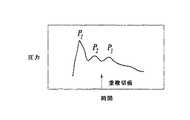
自己調節その他の脳の症状
頭蓋内圧、血圧および自己調節は、密接に関連している。経頭蓋ドップラー(TCD)信
号において観察されてきた、「A波」、「B波」、「C波」および「プラトー波」として
知られる、詳しく記述された周期的な現象が、例えば、動脈血圧および頭蓋内圧と関連す
る。Self-regulation and other brain symptoms Intracranial pressure, blood pressure and self-regulation are closely related. The well-described periodic phenomena known as “A wave”, “B wave”, “C wave” and “plateau wave” that have been observed in transcranial Doppler (TCD) signals include, for example, arterial blood pressure And associated with intracranial pressure.
中枢神経系(CNS)は、さまざまなタイプの組織および液体を含む。脳のような中枢
神経系組織に出入りする血流は、一般に拍動性があり、心臓周期のいずれかの時点での脳
内全血体積は、全身の血圧と、脳脈管系の保護的な自己調節機構とに関係づけられる。ミ
リメートル単位の直径を有する主要動脈から、ミクロン単位の直径を有する細動脈まで、
これらののさまざまな脳脈管系の生理的なスケールは、頭蓋内圧および自己調節に対して
、異なる時間スケールおよび異なる寄与レベルで反応する。脳脈管系の異なるクラスは、
脳の異なる変位特性に寄与するヤング係数のような、異なる物性を有する。脳組織が心臓
周期とともに拡張すると、脳脈管系は脳に流入する血量を調節し、同時に脳脊髄液は頭蓋
腔から出て脊髄領域に入っていき、これにより、相対的に一定の頭蓋内圧を維持する。血
液が脳から出て行くと、脳脊髄液は脊髄腔から頭蓋領域へ再流入する。The central nervous system (CNS) contains various types of tissues and fluids. Blood flow to and from central nervous system tissues such as the brain is generally pulsatile, and the whole blood volume in the brain at any point in the cardiac cycle is the blood pressure throughout the body and the protective protection of the cerebral vasculature. Related to self-regulation mechanisms. From major arteries with millimeter diameters to arterioles with micron diameters,
The physiological scales of these various cerebral vasculature respond to intracranial pressure and self-regulation at different time scales and different contribution levels. Different classes of cerebrovascular system
It has different physical properties such as Young's modulus that contributes to different displacement characteristics of the brain. As brain tissue expands with the cardiac cycle, the cerebral vasculature regulates the amount of blood that flows into the brain, and at the same time cerebrospinal fluid exits the cranial cavity and enters the spinal cord region, which causes a relatively constant cranial flow. Maintain internal pressure. As blood exits the brain, cerebrospinal fluid reflows from the spinal cavity into the skull region.
脳のこの周期的収縮拡張の間、脳への適当な血流が維持されなければならず、それゆえ
、脳脈管系は、動脈圧平均値のいかなる変化についても補償するために、脳脈管系の抵抗
を動的に調整する。脳は、実質的に一定速度の血流を受け入れるが、この速度は脳潅流圧
によって決定される。ここで広範囲の動脈圧平均値にわたって
脳潅流圧=動脈圧平均値−頭蓋内圧
である。このようにして、正常条件下では、脳およびその脈管系は、脳への適当な血流を
維持するために脳潅流圧を変化させることが可能である。これは自己調節の正常状態と呼
ばれている。脳への適当な血流を維持するための脳潅流圧を変化させる能力が失われると
き、自己調節が異常で、頭蓋内圧は動脈圧平均値に直接的に比例するようになる。During this cyclic contraction and dilation of the brain, proper blood flow to the brain must be maintained, and therefore the cerebral vasculature must compensate for any changes in the mean arterial pressure. Adjust pipe resistance dynamically. The brain accepts a substantially constant rate of blood flow, which is determined by cerebral perfusion pressure. Here, cerebral perfusion pressure = arterial pressure average value-intracranial pressure over a wide range of arterial pressure average values. In this way, under normal conditions, the brain and its vasculature can change the cerebral perfusion pressure to maintain proper blood flow to the brain. This is called the normal state of self-regulation. When the ability to change cerebral perfusion pressure to maintain adequate blood flow to the brain is lost, self-regulation is abnormal and intracranial pressure becomes directly proportional to the mean arterial pressure.
自己調節が「正常(intact)」か「異常(impaired)」かどうかの臨床
的な決定は、一般に、脳血流量(CBF)および動脈圧平均値を監視することによってな
される。脳血流量は、脳内の大きい血管の血流速度を測定するための経頭蓋ドップラー法
(TCD)を用いて監視され、動脈圧平均値は、標準的な技術のいずれかを用いて測定さ
れる場合がある。全身の血圧を変調−亢進または減退−させるような生理学的なチャレン
ジが、脳血流量を監視しながら患者に投与される。全身血圧は、例えば、個体の手足の圧
力を増大させる(例えば、手足のどれかに圧力カフを適用する)こと、全身血圧を変化さ
せる利尿剤その他の医薬を投与すること等によって変調させられる。全身血圧は、個体に
くしゃみまたはせきをさせることによっても変調させることができる。自己調節が正常の
とき、脳血流量は一般に広範囲の動脈圧平均値にわたって一定であるが、自己調節が異常
のとき、脳血流量は広範囲の動脈圧平均値にわたって増加または減少する。従来の臨床的
な自己調節の決定技術は不正確で負担が大きい。さらに、経頭蓋ドップラー法を用いる脳
血流量の測定は、患者および該患者の中枢神経系が静止していない場合があるのに、装置
の焦点を大きい脳血管に合わせてその状態を維持するために、熟練した超音波診断技師が
必要である。The clinical determination of whether self-regulation is “normal” or “impaired” is generally made by monitoring cerebral blood flow (CBF) and arterial pressure averages. Cerebral blood flow is monitored using the transcranial Doppler method (TCD) to measure the blood flow velocity of large blood vessels in the brain, and arterial pressure averages are measured using any of the standard techniques. There is a case. A physiological challenge, such as modulating or enhancing or reducing systemic blood pressure, is administered to the patient while monitoring cerebral blood flow. Systemic blood pressure is modulated, for example, by increasing the pressure of an individual's limbs (eg, applying a pressure cuff to any of the limbs), administering a diuretic or other medication that changes systemic blood pressure, and the like. Systemic blood pressure can also be modulated by causing an individual to sneeze or cough. When self-regulation is normal, cerebral blood flow is generally constant over a wide range of arterial pressure averages, but when self-regulation is abnormal, cerebral blood flow increases or decreases over a wide range of arterial pressure averages. Traditional clinical self-regulation determination techniques are inaccurate and burdensome. Furthermore, measurement of cerebral blood flow using the transcranial Doppler method is used to keep the device focused on a large cerebral blood vessel, even though the patient and the patient's central nervous system may not be stationary. In addition, a skilled ultrasonic diagnostic engineer is required.
同様に、卒中、局所的な浮腫、感染および/または血管炎を意味する場合がある、血管
痙攣のような症状の臨床的な決定は、経頭蓋ドップラー(TCD)法を用いて行われるの
が一般的である。血管痙攣は、脳脈管系が、患部の血管を通る血流が著しく減少するくら
い異常な収縮を起こすが、血流速度の測定値は実際には増加して、一時的な、そして、し
ばしば永久的な、神経性欠損(例えば、卒中)を引き起こす症状をいう。脳動脈瘤の破裂
に由来するクモ膜下出血の結果であることがしばしばである。従来の経頭蓋ドップラー超
音波診断法は、血管痙攣の程度を評価するために、大きな脳血管の流速を使用するが、そ
れは、より小さな血管は、正確に局在定位して経頭蓋ドップラーで超音波診断することが
不可能だからである。関心のある血管内の血流速度が一定値を超えるとき、血管痙攣が推
測される。実際には、経頭蓋ドップラー法は脳内のより小さな血管での血管痙攣を評価す
るのに十分な感度はないため、経頭蓋ドップラー法は頭蓋基部の大きな血管での血管痙攣
を評価することに限定されるのが一般的である。現在のところ、血管痙攣の存在を確認す
るための一般的な臨床診察は、従来の脳血管造影法を行うことである。これは、大がかり
で高価な手順である。したがって、本発明は、中枢神経系組織における、血管痙攣と、卒
中、局所的浮腫、感染および血管炎のようなその他の症状との状況における自己調節の状
態を評価および監視するためのシステムおよび方法にも向けられている。Similarly, clinical determination of symptoms such as vasospasm, which may mean stroke, local edema, infection and / or vasculitis, is made using the transcranial Doppler (TCD) method. It is common. Vasospasm is a phenomenon in which the cerebral vasculature contracts abnormally such that blood flow through the affected blood vessels is significantly reduced, but blood flow velocity measurements are actually increased, temporary, and often A condition that causes a permanent, neurological deficit (eg, stroke). Often results from subarachnoid hemorrhage resulting from rupture of a cerebral aneurysm. Traditional transcranial Doppler ultrasonography uses large cerebrovascular flow velocities to assess the degree of vasospasm, but it is more important for smaller vessels to be localized and localized with transcranial Doppler. This is because it is impossible to make an ultrasonic diagnosis. Vasospasm is inferred when the blood flow velocity in the blood vessel of interest exceeds a certain value. In fact, the transcranial Doppler method is not sensitive enough to assess vasospasm in smaller blood vessels in the brain, so transcranial Doppler method is used to assess vasospasm in large vessels at the base of the skull. Generally limited. At present, a common clinical examination to confirm the presence of vasospasm is to perform conventional cerebral angiography. This is a large and expensive procedure. Accordingly, the present invention provides a system and method for assessing and monitoring the state of self-regulation in the context of vasospasm and other symptoms such as stroke, local edema, infection and vasculitis in central nervous system tissue. It is also directed to.
痛みの発生源の局在定位および診断
痛みは多数の病状のよくある主症状であり、何かがおかしいという最初の警告になること
もしばしばである重要な役割を果たすが、極端に非特異的であることもある。痛みの特異
性および局在性を増強するための技術から利益が得られる複数のありふれた病状がある。
腰痛(low back pain、LBP)は、1つのありふれた病状の主要な例であ
る。腰痛の生涯発症率は60〜90%で、年間発症率は5%であると報告されている。国
立保健統計センターによると、毎年、プライマリー・ケアの医師を訪問する新患の14%
が腰痛のためであり、約1300万回の訪問が慢性腰痛に関係する。残念ながら、痛みの
正確な発生源の同定は困難である。複雑な構造のいくつかの構成部分は密接に関係してい
るが、ただ1つだけが発生源かもしれない。米国の労働力の半分が腰痛を訴えているが、
これらの患者の約20%だけが痛みの発生源を特定する診断につながっている。X線、コ
ンピュータ断層撮影法および磁気共鳴映像法は、腰痛患者の主要な画像診断テストであり
、これらは解剖学的な異常を鋭敏に映し出すが、解剖学的な知見と患者の症状との間の関
連性はせいぜい中くらいである。Localization of pain sources and diagnostic pain is a common main symptom of many medical conditions and plays an important role, often the first warning that something is wrong, but extremely nonspecific Sometimes it is. There are several common medical conditions that can benefit from techniques for enhancing the specificity and localization of pain.
Low back pain (LBP) is a major example of one common medical condition. It has been reported that the lifetime incidence of back pain is 60-90% and the annual incidence is 5%. According to the National Center for Health Statistics, 14% of new cases visit a primary care physician each year
Is due to back pain, and about 13 million visits are associated with chronic back pain. Unfortunately, identifying the exact source of pain is difficult. Several components of a complex structure are closely related, but only one may be the source. Half of the US workforce complains of back pain,
Only about 20% of these patients lead to diagnoses that identify the source of pain. X-rays, computed tomography, and magnetic resonance imaging are the main diagnostic imaging tests for patients with low back pain, which are a sharp reflection of anatomical abnormalities, but between the anatomical findings and the patient's symptoms. The relevance of is at best moderate.
近年、腰痛の専門家は、痛みの発生源を同定することを試みて、侵襲的で症状の発現を
促進するようなテストに頼り始めている。医師は、痛みを誘発するように椎間板造影法用
の注射針を椎間板に挿入し、局部麻酔およびステロイドの注射により痛みを誘発し、その
後痛みを取り除くために、関節面および仙腸骨関節の間に注射針を挿入する。これらのテ
ストは患者にとってしばしば不快で、感染および対比反応(contrast reac
tion)のリスクをともなう。In recent years, back pain specialists have begun to rely on tests that are invasive and promote the onset of symptoms, trying to identify the source of the pain. The doctor inserts a discography needle into the disc to induce pain, induces pain by local anesthesia and steroid injection, and then removes the pain between the joint surface and sacroiliac joint Insert the injection needle into. These tests are often uncomfortable for the patient and are associated with infection and contrast reactions.
risk).
高齢者においては、骨粗鬆症による圧迫骨折は非常に頻度が高い。発症率は年間700
,000骨折例で、毎年160,000回の医師への訪問と500万日以上の行動の制限
された日々とを生じさせている。最近まで、よい治療の選択肢はなかった。メチルメタク
リレートを椎体に経皮的に注入する椎体形成術は、これらの骨折の新規で有望な治療法で
ある。しかし、複数箇所の骨折をした患者では、痛い骨折箇所を同定することは困難な場
合がある。痛い骨折箇所を局在定位するための試みとして、打診、あるいは理学的検査と
、骨スキャニングと磁気共鳴画像化法とは、全て利用されてきたが、その成功の程度はさ
まざまであった。In elderly people, compression fractures due to osteoporosis are very frequent. Incidence rate is 700 per year
With 1,000,000 fracture cases, it produces 160,000 visits to physicians every year and a limited number of days of behavior over 5 million days. Until recently, there were no good treatment options. Vertebroplasty, in which methyl methacrylate is injected percutaneously into the vertebral body, is a new and promising treatment for these fractures. However, it may be difficult to identify a painful fracture site in a patient having multiple fractures. Percussion or physical examination, bone scanning, and magnetic resonance imaging have all been used as attempts to localize painful fracture sites, but the degree of success has varied.
腰痛は特異性の改良から利益が得られるありふれた痛みの病状であるが、他の病状も存
在する。虫垂炎の診断は困難で不正確である。CTおよび超音波のようなハイテク画像診
断法の使用にもかかわらず、JAMAの最近の総説は、虫垂摘出術での偽陽性の発生率に
変化がないことを証明した。さらに、手作業での腹部の触診が、特異性が低いけれども、
いまだに標準テストであって、その結果は一定しない。Low back pain is a common pain condition that can benefit from improved specificity, but there are other conditions. Diagnosis of appendicitis is difficult and inaccurate. Despite the use of high-tech imaging methods such as CT and ultrasound, JAMA's recent review has demonstrated no change in the incidence of false positives in appendectomy. In addition, manual palpation of the abdomen is less specific,
It is still a standard test and the results are not constant.
症状(symptons)は患者が自発的に訴えるものであるが、徴候(sign)は
診察する医師によって引き出される。上記の病状において、痛みの症状は、問題があると
いう信号を発するが、その問題の所在を特定することはないのがしばしばである。したが
って、腰痛その他の疾患、特に、炎症性の要素がある疾患(例えば、虫垂炎、胆嚢炎、膵
炎、骨盤の炎症疾患等)のケースでは、精度が高く、信頼性があり、非侵襲的なやり方で
、体内の複雑な構造の個々の構成部分(例えば、椎間板、椎体、椎弓板および脊椎関節突
起)を刺激して、痛みの正確な発生源を同定し、空間的に局在定位させる。それゆえ、本
発明の方法およびシステムは、さらに、痛みのような、生理状態および/または生物学的
な反応を局在定位させることにも向けられる。Symptoms are those that the patient complains spontaneously, but signs are drawn by the examining physician. In the above pathology, pain symptoms often signal that there is a problem but do not identify the location of the problem. Therefore, in the case of low back pain and other diseases, especially those with inflammatory components (eg, appendicitis, cholecystitis, pancreatitis, pelvic inflammatory diseases, etc.), it is accurate, reliable and non-invasive To stimulate individual components of complex structures in the body (eg, intervertebral discs, vertebral bodies, lamina, and spinal joint processes) to identify the exact source of pain and localize it spatially . Therefore, the methods and systems of the present invention are further directed to localizing physiological states and / or biological responses, such as pain.
発明の概要
本発明は、誘発性および/または内在性の組織変位および/または生物学的反応に関する
データにもとづいて、誘発性および/または内在性の組織変位の検出および生理学的な組
織特性の評価のための方法およびシステムを提供する。内部組織の生理特性は、本発明の
技術を用いて非侵襲的に評価される場合がある。例えば、心臓周期および/または呼吸周
期に由来するか、あるいは、明確な時間空間的な特徴を示しつつ、非侵襲的かつ非傷害的
なやり方で誘発された内在性の組織変位を経験するいずれかの組織は、本発明の方法およ
びシステムを用いて評価することができる。痛みのような、生理状態および/または生物
学的な反応は、本発明の方法およびシステムを用いて局在定位することができる。例えば
、組織変位および生物学的な反応は、音響放射力または温度変化または組織の空洞形成を
生じる、1または2以上の音響的なビームの適用によって誘発される。音響探査信号を標
的組織部位に適用すること、音響散乱データを取得することをともなう音響的な検出技術
が好ましいが、近赤外スペクトル(NIRS)法、磁気共鳴法、ハイドロホン等を含む、
代替的な検出技術を用いる場合がある。SUMMARY OF THE INVENTION The present invention is based on data relating to inducible and / or intrinsic tissue displacement and / or biological responses, and detection of inducible and / or intrinsic tissue displacement and assessment of physiological tissue properties. Methods and systems are provided. Physiological properties of internal tissues may be evaluated non-invasively using the techniques of the present invention. For example, either derived from the cardiac cycle and / or respiratory cycle, or experiencing intrinsic tissue displacement induced in a non-invasive and non-injury manner while exhibiting distinct temporal and spatial characteristics Can be assessed using the methods and systems of the present invention. Physiological conditions and / or biological responses, such as pain, can be localized using the methods and systems of the invention. For example, tissue displacement and biological responses are triggered by the application of one or more acoustic beams that result in acoustic radiation forces or temperature changes or tissue cavitation. An acoustic detection technique involving applying an acoustic exploration signal to a target tissue site and acquiring acoustic scattering data is preferred, including near infrared spectrum (NIRS), magnetic resonance, hydrophone, etc.
Alternative detection techniques may be used.
それゆえ、本発明の方法およびシステムは、さまざまな臨床的なパラメーターを評価し
、局在定位させ、監視するうえで、そして、さまざまな病状、反応および疾患の状態を診
断し、局在定位させ、監視するうえで有用である。これらの方法およびシステムは、例え
ば、非侵襲的に組織の硬直性およびコンプライアンスを検出するうえで、そして、組織の
硬直性およびコンプライアンスに関連する状態を評価するうえで有用である。前記方法お
よびシステムは、例えば、集束超音波を利用して非侵襲的に標的組織部位を探査して、損
傷を受けた組織または進行中の疾患の過程に関連する痛みのような組織の反応を局在定位
するうえでも有用である。集束超音波を適用することにより内部組織を標的として探査す
ることは、痛みの高感度の局在定位を提供し、虫垂炎、胆嚢炎、骨盤の炎症疾患、膵炎お
よびリンパ節症のような痛みを生じる多数の病状を診断するためと、脊椎その他の関節お
よびその他の体内の部位での痛みの発生源を局在定位し同定するためとに用いる場合があ
る。Therefore, the methods and systems of the present invention are useful in assessing, localizing and monitoring various clinical parameters and in diagnosing and localizing various medical conditions, reactions and disease states. Useful for monitoring. These methods and systems are useful, for example, in non-invasively detecting tissue stiffness and compliance, and in assessing conditions related to tissue stiffness and compliance. The methods and systems can, for example, use focused ultrasound to probe a target tissue site non-invasively to react to damaged tissue or painful tissue associated with an ongoing disease process. It is also useful for localization. Exploring targeted internal tissues by applying focused ultrasound provides sensitive localization of pain, and pain such as appendicitis, cholecystitis, pelvic inflammatory disease, pancreatitis and lymphadenopathy It may be used to diagnose a number of medical conditions that occur and to localize and identify the source of pain at the spine and other joints and other parts of the body.
したがって、本発明の1の実施態様では、方法およびシステムは、個々の痛みの発生源
である可能性がある部位を、集束的に、非侵襲的に、そして、安全に、刺激することによ
って、痛みを鑑別診断し局在定位するために、非侵襲的で集束超音波を利用する。組織の
標的音響探査は、標的組織部位に集束超音波パルスを適用することによって提供される。
例えば損傷している組織を含む標的部位に、適当な規模(magnitude)、周波数
、強度(intensity)および/またはパルス反復速度の音響(超音波)パルスを
適用することは被検者に痛覚を誘発するが、損傷していない組織部位に超音波ビームを適
用することは痛覚を生じない。痛みのレベルまたはタイプは、痛みの反応を誘発するため
に要する、集束音響ビームの規模、周波数、強度および/またはパルス反復速度に関係す
る場合がある。Thus, in one embodiment of the present invention, the method and system provides a focused, non-invasive and safe stimulation of sites that may be the source of individual pain. Non-invasive, focused ultrasound is used to differentially diagnose and localize pain. Target acoustic exploration of tissue is provided by applying focused ultrasound pulses to the target tissue site.
Applying acoustic (ultrasound) pulses of appropriate magnitude, frequency, intensity and / or pulse repetition rate to a target site, including, for example, damaged tissue, induces pain in the subject However, applying an ultrasonic beam to an intact tissue site does not cause pain. The level or type of pain may be related to the size, frequency, intensity and / or pulse repetition rate of the focused acoustic beam required to elicit a pain response.
超音波ビームの集束的な適用を用いて、本発明の方法およびシステムは、未分化の痛み
の全身的な部位の中で、痛みのような生物学的反応の発生源を局在定位するために用いら
れる。音響探査は、離散的な「ポーク(poke)」を生じる集束超音波の離散的な適用
をともなうか、あるいは、グリーンリーフ(前出)によって説明されたとおりの、組織の
振動を生じる音響エネルギーの適用をともなう場合がある。痛みの発生源を局在定位する
ために集束音響探査を利用する主な利点の1つは、痛みの鑑別診断が全く非侵襲的なやり
方で提供されることであるが、痛みのようなさまざまな生物学的反応を刺激する音響的な
技術の使用は、さまざまなタイプの診断および外科用の装置(例えば内視鏡等)のような
、侵襲的、半侵襲的、または、最小限に侵襲的装置および手順と連繋して使用される場合
がある。Using the focused application of an ultrasonic beam, the method and system of the present invention is used to localize the source of a painful biological response within a systemic site of undifferentiated pain. Used for. Acoustic exploration involves the discrete application of focused ultrasound that produces discrete “pokes”, or the acoustic energy that produces tissue vibrations as described by Greenleaf (supra). May have application. One of the main advantages of using focused acoustic exploration to localize pain sources is that differential diagnosis of pain is provided in a totally non-invasive manner, The use of acoustic techniques to stimulate various biological responses can be invasive, semi-invasive, or minimally invasive, such as various types of diagnostic and surgical devices (eg, endoscopes, etc.) May be used in conjunction with technical equipment and procedures.
以下に詳しく説明される集束音響探査技術は、超音波または磁気共鳴スキャニング診断
技術のような画像診断技術と組み合わされて、音響プローブの位置を特定し、痛み反応が
誘発されるとき、その痛みの発生源を特定することができる。被検者の意識があるときは
、該被検者の主観的な痛覚が上記の画像化技術と併用されて、全身的な痛みの部位の中を
音響プローブの焦点が移動するときに痛みの発生源を特定することができる。被検者の意
識がないとき、または、被検者の痛みがマヒまたはブロックされているときには、他の痛
みの生理的反応または指標が痛みの発生源を同定するために用いられる。超音波ビームの
集束的な標的への放射は、例えば、音響トランスデューサの位置および/または焦点を選
択的に変化させることにより達成され、集束的な音響ビームおよび前記反応の発生源の局
在定位は付属の画像化装置によって提供される。Focused acoustic exploration techniques, described in detail below, are combined with diagnostic imaging techniques such as ultrasound or magnetic resonance scanning diagnostic techniques to locate acoustic probes and, when a pain response is triggered, the pain The source can be identified. When the subject is conscious, the subject's subjective pain sensation is used in combination with the imaging technique described above to cause pain when the focal point of the acoustic probe moves through the site of general pain. The source can be identified. When the subject is unconscious, or when the subject's pain is paralyzed or blocked, other pain physiological responses or indicators are used to identify the source of the pain. Radiation of the ultrasound beam to the focused target is achieved, for example, by selectively changing the position and / or focus of the acoustic transducer, and the localized localization of the focused acoustic beam and the source of the reaction is Provided by an attached imaging device.
痛みのような生理状態および反応を局在定位するための組織部位の集束音響探査は、集
束音響ビームの適用および通過のために十分な音響的な窓が利用可能ないずれの組織部位
でも使用することができる。腹部および/または骨盤の領域での全身的で未分化の痛みの
局在定位は、虫垂炎、胆嚢炎、膵炎、痛みを特徴とする多数の胃腸疾患、胆石、腎臓結石
、膀胱炎およびさまざまな痛みのある膀胱の病状、月経障害、卵巣および子宮の病状等の
診断に供される。脊椎領域と、膝、足首、肩、臀部、仙腸骨その他の関節のような関節と
における、全身化された未分化の痛みは、本発明の集束音響探査技術を用いて局在定位さ
れ、痛みの発生源は、例えば、軟骨、筋肉、神経、靱帯、腱等のように同定される場合が
ある。音響的打診を誘発するために集束超音波を使用して、例えば、腰痛が椎間板に関係
するか、あるいは、関節面、椎体、神経、筋肉等に発するかであるとして局在定位および
同定される場合がある。末梢神経に関する痛みと、例えば、癌および感染に由来するリン
パ節症とが、診断および局在定位される場合もある。Focused acoustic exploration of a tissue site to localize painful physiological states and responses uses any tissue site where sufficient acoustic windows are available for application and passage of the focused acoustic beam be able to. Localization of systemic undifferentiated pain in the abdominal and / or pelvic areas includes appendicitis, cholecystitis, pancreatitis, numerous gastrointestinal diseases characterized by pain, gallstones, kidney stones, cystitis and various pains It is used for diagnosis of certain bladder conditions, menstrual disorders, ovarian and uterine conditions. Generalized undifferentiated pain in the spinal region and joints such as knees, ankles, shoulders, buttocks, sacroiliac and other joints is localized using the focused acoustic exploration technique of the present invention, Pain sources may be identified, for example, cartilage, muscle, nerves, ligaments, tendons and the like. Using focused ultrasound to induce acoustic percussion, for example, is localized and identified as if the back pain is related to the intervertebral disc or whether it originates in the articular surface, vertebral body, nerve, muscle, etc. There is a case. Pain associated with peripheral nerves and, for example, lymphadenopathy resulting from cancer and infection may be diagnosed and localized.
多くの状況では、組織部位はものすごい痛みはないが、肥大するか、それとも異常であ
る場合がある。音響探査は、肥大し、あるいは異常な組織部位の中に局在した痛みの部位
があるかどうかを同定し、それにより、陽性の診断を提供するためか、あるいは、少なく
ともある種の診断を排除するためかに用いられる場合がある。肥大した組織部位は、例え
ば、腫瘍、他の異常増殖、炎症を起こした組織等に由来する場合がある。癌性のリンパ節
は一般に痛まないが、炎症の二次性の肥大したリンパ節は痛むのが一般的である。したが
って、ここに説明する技術を用いる音響探査は、頭頚部に既知の原発腫瘍を有する患者に
おいて、リンパ節症が良性か転移性かの鑑別診断を提供する。本技術は、縦隔および骨盤
のような、他の解剖学的な部域での鑑別診断を提供するうえでも有用である。In many situations, the tissue site is not terribly painful, but may be enlarged or abnormal. Acoustic exploration identifies whether there is a site of pain that is hypertrophic or localized within an abnormal tissue site, thereby providing a positive diagnosis or at least eliminating some types of diagnosis May be used to do so. The enlarged tissue site may be derived, for example, from a tumor, other abnormal growth, inflamed tissue, or the like. Cancerous lymph nodes generally do not ache, but secondary, enlarged lymph nodes that are inflamed generally hurt. Thus, acoustic exploration using the techniques described herein provides a differential diagnosis of whether lymphadenopathy is benign or metastatic in patients with known primary tumors in the head and neck. The technique is also useful in providing differential diagnosis in other anatomical areas, such as the mediastinum and pelvis.
別の面では、本発明の方法およびシステムは、中枢神経系の組織特性と、頭蓋内圧を含
む関連する臨床的パラメーターとを非侵襲的に評価するために用いられ、代表的な実施態
様は、中枢神経系の組織特性と頭蓋内圧との非侵襲的評価との関係で説明される。本発明
の非侵襲的方法およびシステムは、動脈血圧および脳潅流圧とを評価するためと、急性、
慢性および外傷性の中枢神経系の損傷と、傷害、血管痙攣、卒中、局所的浮腫、感染血管
炎、硬膜下および硬膜上血腫、クモ膜下出血、虚血状態、多発性硬化症、アルツハイマー
病、低酸素状態、脳内出血、腫瘍その他の頭蓋内塊状物等のような中枢神経系の病状とを
、評価、診断、局在定位および監視するにも有用である。他の面において、本発明の方法
およびシステムは、心臓組織、末梢神経その他の非骨組織を含む、他の組織における異常
の評価、診断、局在定位および監視をするために用いられる一部のケースでは、評価は、
比較組織サンプルとの対比とは独立に行われるが、別のケースでは、評価は、さまざまな
標的組織部位の特性の比較により行われる。一部の実施態様では、測定された組織特性が
経験的に決定された基準と対比される。In another aspect, the methods and systems of the present invention are used to non-invasively assess central nervous system tissue properties and associated clinical parameters including intracranial pressure, and representative embodiments include: It is explained in relation to the non-invasive evaluation of tissue characteristics of the central nervous system and intracranial pressure. The non-invasive method and system of the present invention is useful for assessing arterial blood pressure and cerebral perfusion pressure, acute,
Chronic and traumatic central nervous system damage and injury, vasospasm, stroke, local edema, infectious vasculitis, subdural and epidural hematoma, subarachnoid hemorrhage, ischemic condition, multiple sclerosis, It is also useful for evaluation, diagnosis, localization and monitoring of central nervous system conditions such as Alzheimer's disease, hypoxia, intracerebral hemorrhage, tumors and other intracranial masses. In other aspects, the methods and systems of the present invention are used to assess, diagnose, localize and monitor abnormalities in other tissues, including heart tissue, peripheral nerves and other non-bone tissue. In the case, the evaluation is
While performed independently of contrast with the comparative tissue sample, in other cases, the assessment is performed by comparing the characteristics of various target tissue sites. In some embodiments, the measured tissue properties are compared to empirically determined criteria.
本発明の方法およびシステムの1つの面は、標的組織部位での内在性および/または誘
発性の組織変位か、関連する生物学的反応かに関する中枢神経系組織の特性との関係で、
頭蓋内圧を含むさまざまな臨床的パラメーターを評価し、監視することに関する。「正常
(normal)」な脳組織は、コンプライアンスおよび弾性がある。脳は脳脊髄液の中
にあり、閉鎖された頭蓋の円蓋によって保護されている。心臓周期の各サイクルごとに、
一塊りの動脈血が流入し、静脈血が脳実質から流出して、呼吸により変調されるやり方で
、心臓周期の間に脳組織の拡張と収縮とを起こさせる。心臓周期の中のいかなる時点にお
いても脳内の全血液体積は、全身の血圧と、脳の脈管系の保護的な自己調節機構との関数
である。脳の周期的な収縮および拡張の間、脳への血流が維持され、脳脈管系は、動脈圧
平均値の変化を補償するために、動的にその抵抗を調節する。One aspect of the methods and systems of the present invention is in relation to the properties of the central nervous system tissue whether it is an intrinsic and / or induced tissue displacement at the target tissue site or an associated biological response,
It relates to evaluating and monitoring various clinical parameters including intracranial pressure. “Normal” brain tissue is compliant and elastic. The brain is in the cerebrospinal fluid and protected by a closed cranial fornix. For each cycle of the heart cycle,
A lump of arterial blood flows in and venous blood flows out of the brain parenchyma, causing brain tissue to expand and contract during the cardiac cycle in a manner that is modulated by respiration. The total blood volume in the brain at any point in the cardiac cycle is a function of systemic blood pressure and the protective self-regulatory mechanism of the cerebral vasculature. During periodic contraction and dilation of the brain, blood flow to the brain is maintained, and the cerebral vasculature dynamically adjusts its resistance to compensate for changes in the mean arterial pressure.
頭蓋内圧および自己調節の状態は、入手可能な臨床的技術を用いて測定することが困難
で、監視することはさらに困難な、必須の臨床パラメーターである。図1Aは、伝統的な
侵襲的手段によって測定された、典型的な頭蓋内圧の波形を示す。この頭蓋内圧曲線は、
呼吸周期および動脈血圧の波形に重なる。図1Bは、図1Aのボックスに囲まれた波形の
拡大図を示し、心臓周期および自己調節システムの要素に由来する波形の標準的な形状を
示す。脳脊髄液圧の波形は、全身の血圧の波形と同様である。これは、「衝撃波」(P1
)、「動揺波」(P2)および「重拍波」(P3)という3つのほぼ一貫した成分を有す
る。P2とP3との間の重複切痕は、動脈波の重複切痕に対応する。Intracranial pressure and self-regulation status are essential clinical parameters that are difficult to measure and more difficult to monitor using available clinical techniques. FIG. 1A shows a typical intracranial pressure waveform measured by traditional invasive means. This intracranial pressure curve is
Overlapping respiratory cycle and arterial blood pressure waveforms. FIG. 1B shows a magnified view of the waveform enclosed in the box of FIG. 1A, showing the standard shape of the waveform derived from the cardiac cycle and the elements of the self-regulating system. The waveform of cerebrospinal fluid pressure is similar to the waveform of blood pressure throughout the body. This is the “shock wave” (P1
), “Rock wave” (P2) and “heavy beat wave” (P3). The overlapping notch between P2 and P3 corresponds to the overlapping notch of the arterial wave.
呼吸波は、胸内圧を反映して、中心静脈圧の変化に同調している。具体的には、吸気の
際、動脈血圧と同様に、胸部の内腔の体積が拡張するため、胸内圧は低下し、それゆえ、
頭蓋内圧は低下する。排気の際、胸内圧は胸部の内腔の体積が減少するため、動脈血圧が
増加し、それゆえ、頭蓋内圧が増加する。呼吸が機械的なベンチレータによって呼吸が介
助される被検者についても反対が正しい。機械的なベンチレータを用いる胸内圧の調節は
、ある程度までは、動脈血圧および頭蓋内圧を調節するために用いられる。正常には、心
拍の振幅は約1.1mmHgで、心臓および呼吸による変動の合計は約3.3mmHgで
ある。The respiratory wave is synchronized with changes in central venous pressure, reflecting intrathoracic pressure. Specifically, during inspiration, as with arterial blood pressure, the volume of the thoracic lumen expands, so the intrathoracic pressure decreases, and therefore
Intracranial pressure decreases. During evacuation, intrathoracic pressure decreases arterial blood pressure because the volume of the thoracic lumen decreases, thus increasing intracranial pressure. The opposite is also true for subjects whose breathing is assisted by a mechanical ventilator. Adjustment of intrathoracic pressure using a mechanical ventilator is used to a certain extent to adjust arterial blood pressure and intracranial pressure. Normally, the heartbeat amplitude is about 1.1 mmHg and the total heart and respiration variation is about 3.3 mmHg.
脳組織と、例えば、脳脊髄液、脳脊髄液または脳実質に隣接する組織、視神経のような
脳神経等を含む、他の中枢神経系組織とは、頭蓋内圧の評価に適した標的組織部位である
。中枢神経系組織に内在性の力が脳に加えられるとき、あるいは、中枢神経系組織に外来
の(誘発された)力が加えられるとき、頭蓋内圧の亢進は、呼吸、周期的な血流、補償的
な脳脊髄液および静脈の流出および脳脈管系の自己調節にもとづく変化の結果、脳その他
の中枢神経系組織において、相対的に硬直性を高くし、あるいはよりコンプライアンスが
少なくする。組織が圧縮されたり拡張したりするとき、あるいは、それぞれ血管収縮また
は血管拡張の際、血管の特性は変化する、すなわち、管壁が、より硬直したり、より柔軟
になったりして、例えば、脳脈管系の拍動の局所的徴候を生じる。Brain tissue and other central nervous system tissues, including cerebrospinal fluid, tissues adjacent to cerebrospinal fluid or brain parenchyma, cranial nerves such as the optic nerve, etc. are target tissue sites suitable for evaluating intracranial pressure. is there. When intrinsic forces are applied to the brain in the central nervous system tissue, or when extraneous (induced) forces are applied to the central nervous system tissue, increased intracranial pressure is associated with breathing, periodic blood flow, As a result of compensatory cerebrospinal fluid and venous outflows and changes based on cerebral vasculature self-regulation, the brain and other central nervous system tissues become relatively more rigid or less compliant. When the tissue is compressed or expanded, or upon vasoconstriction or vasodilation, respectively, the vascular properties change, i.e. the vessel wall becomes stiffer or more flexible, e.g. Causes local signs of pulsation of the cerebral vasculature.
発明者は、中枢神経系、特に、脳組織および視神経組織の硬直性は、内在性および/ま
たは誘発性の中枢神経系組織変位または関連する生物学的反応に関する音響特性の観察に
よって、評価することができることを確立した。関連する生物学的反応は、局所的潅流速
度、血流速度および電気生理学的活性の変化を含むが、これらに限定されない。組織の音
響特性と、組織の硬直性と、内在性および/または誘発性の組織変位と、関連する生物学
的反応とは、頭蓋内圧その他の中枢神経系の状態に経験的に関係がある。The inventor shall assess the stiffness of the central nervous system, in particular brain and optic nerve tissue, by observing acoustic properties related to intrinsic and / or induced central nervous system tissue displacement or related biological responses. Established that can be. Related biological responses include, but are not limited to, changes in local perfusion rate, blood flow rate and electrophysiological activity. Tissue acoustic properties, tissue stiffness, intrinsic and / or induced tissue displacement, and associated biological responses are empirically related to intracranial pressure and other conditions of the central nervous system.
組織の音響特性の評価は、本発明の方法およびシステムの好ましい実施態様であるが、
組織の硬直性またはコンプライアンスのような組織特性の評価のために用いられる内在性
および/または誘発性の組織変位および関連する生物学的反応に関するパラメーターは、
近赤外分光技術(NIRS)、光コヒーレンス断層撮影法(OCT)、磁気共鳴技術、陽
子射出断層撮影法(PET)、外部電気生理学的刺激法等のような非侵襲的光学検出技術
を含む、他の非侵襲的な技術を用いて測定される場合がある。携帯式の比較的安価な磁気
共鳴スキャナは、例えば、以下の非特許文献3に説明されている。組織変位および関連す
る生物学的反応のさまざまな時間空間的側面を測定するためにこれらの技術を利用するこ
とは、一般に知られている。
Parameters for endogenous and / or induced tissue displacement and associated biological responses used for assessment of tissue properties such as tissue stiffness or compliance are:
Including non-invasive optical detection techniques such as near infrared spectroscopy (NIRS), optical coherence tomography (OCT), magnetic resonance technology, proton emission tomography (PET), external electrophysiological stimulation, etc. It may be measured using other non-invasive techniques. A portable and relatively inexpensive magnetic resonance scanner is described, for example, in Non-Patent Document 3 below. It is generally known to utilize these techniques to measure various spatiotemporal aspects of tissue displacement and associated biological responses.
選択された標的組織部位からの音響散乱および/または放出データ、または、組織変位
、組織硬直性等のような派生的な(derivative)測定値は、有用な組織特性ま
たは頭蓋内圧のような臨床パラメーターに対して、経験的な関係式および/または数学的
なモデルを用いて関係づけられる。一般に、高い組織硬直性および/または低いコンプラ
イアンスは高い相対的頭蓋内圧を表し、低い組織硬直性および/または高いコンプライア
ンスは低い相対的頭蓋内圧を表す。同様に、組織硬直性と関係する音響散乱および/また
は放出の部域的な相違および/または変化は、血管痙攣か、虚血性または低酸素性の病状
か、腫瘍またはその他の塊状物か、アルツハイマー病、多発性硬化症等のようなさまざま
なの疾患状態の存在または進行かのような、局所的な病状を表す。平均動脈血圧および/
または連続動脈血圧の非侵襲的測定と、心臓周期および/または呼吸周期の軌跡とのよう
な、追加的なデータは、頭蓋内圧その他の臨床的なパラメーターか、組織の病状かを評価
するために、音響的なデータと併用される。Acoustic scattering and / or emission data from selected target tissue sites, or derivative measurements such as tissue displacement, tissue stiffness, etc. are useful tissue properties or clinical parameters such as intracranial pressure , Using empirical relational expressions and / or mathematical models. In general, high tissue stiffness and / or low compliance represents high relative intracranial pressure, and low tissue stiffness and / or high compliance represents low relative intracranial pressure. Similarly, regional differences and / or changes in acoustic scattering and / or release associated with tissue stiffness can be vasospasm, ischemic or hypoxic pathology, tumor or other mass, Alzheimer's Represents a local medical condition such as the presence or progression of various disease states such as disease, multiple sclerosis and the like. Mean arterial blood pressure and / or
Or additional data such as non-invasive measurements of continuous arterial blood pressure and cardiac and / or respiratory cycle trajectories to assess whether intracranial pressure or other clinical parameters or tissue pathology Used in conjunction with acoustic data.
「能動的」および「受動的」の両方のモードにおいて、異なる場所から照射されおよび
/または異なる時間に照射された、単一または複数の質問信号は、単一または複数の標的
組織部位を照射する場合がある。照射された標的組織の音響特性は、内在性および/また
は誘発性の組織変位か関連する生物学的反応かを評価するために、散乱データまたは放出
データを、同時におよび/または逐次的に獲得することにより評価される場合がある。一
部の実施態様では、内在性および/または誘発性の組織変位の測定値の絶対値が有用であ
る場合があるが、他の実施態様では、内在性および/または誘発性の組織変位の測定値は
、獲得されたデータと経験的に決定された基準値との対比、同一または相異なる時点で相
異なる標的組織部位から獲得されたデータとの対比および/または経時的に標的組織部位
から獲得されたデータとの対比によって、評価される。能動的および受動的なモードは、
個別に、あるいは、併用して、標的組織を評価するのに用いられる場合がある。In both “active” and “passive” modes, single or multiple interrogation signals irradiated from different locations and / or irradiated at different times illuminate single or multiple target tissue sites There is a case. Scattering or emission data is acquired simultaneously and / or sequentially in order to assess whether the acoustic properties of the irradiated target tissue are intrinsic and / or induced tissue displacement or related biological response May be evaluated. In some embodiments, absolute values of intrinsic and / or induced tissue displacement measurements may be useful, while in other embodiments, intrinsic and / or induced tissue displacement measurements. Values are obtained by comparing data acquired with empirically determined reference values, data acquired from different target tissue sites at the same or different time points, and / or acquired from target tissue sites over time. It is evaluated by comparison with the collected data. Active and passive modes are
It may be used individually or in combination to evaluate the target tissue.
組織標的部位は、体積が大きく、中枢神経系の組織特性の大まかな評価のために広い領
域に関するデータを提供する場合がある。しかし、本発明の方法およびシステムの利点の
1つは、高い空間的解像度で局所的な組織部位からのデータを提供するために、標的組織
部位の体積が小さく、空間的に分解されている場合があることである。このようにして、
組織特性の局所的な差異が、同定され、検査された組織内での空間的な位置と関係づけら
れる場合がある。1の実施態様によると、さまざまなサイズおよび/または位置の組織部
位が、同時にまたは逐次的に評価される。たいていの用途について、1mm3から100
cm3までの体積を有する標的組織部位に質問信号を発し、検出することができる、音響
発生源および/またはトランスデューサの使用が適当である。The tissue target site is large in volume and may provide data on a large area for a rough assessment of tissue properties of the central nervous system. However, one advantage of the method and system of the present invention is that the volume of the target tissue site is small and spatially resolved to provide data from a local tissue site with high spatial resolution. Is that there is. In this way
Local differences in tissue properties can be identified and related to the spatial location within the examined tissue. According to one embodiment, tissue sites of various sizes and / or locations are evaluated simultaneously or sequentially. For most applications, 1mm3 to 100
The use of acoustic sources and / or transducers that can issue and detect interrogation signals to target tissue sites having a volume of up to cm3 is suitable.
「能動的」および/または「受動的」モードでの組織の音響特性にもとづいて、頭蓋内
圧のような中枢神経系の組織特性を評価および/または監視するために、標的組織部位は
、脳組織か、視神経または視神経円板組織のような、他の中枢神経系組織かであることが
好ましい。心臓周期および呼吸周期の過程での音響散乱データおよび/または放出データ
の獲得および処理によって決定された、脳、視神経および視神経円板組織の硬直性および
/またはコンプライアンスは、頭蓋内圧と関係がある。一部の用途については、中枢神経
系の標的組織部位は、組織サンプルの均一性にもとづいて選択されるが、他の用途につい
ては、標的部位は、その標的部位の内部での組織タイプの既知の変化または予測された変
化にもとづいて選択される。In order to evaluate and / or monitor tissue properties of the central nervous system, such as intracranial pressure, based on the acoustic properties of the tissue in “active” and / or “passive” modes, Or other central nervous system tissue, such as optic nerve or optic disc tissue. The stiffness and / or compliance of brain, optic nerve and optic disc tissue, determined by acquisition and processing of acoustic scatter and / or emission data during the cardiac and respiratory cycles, is related to intracranial pressure. For some applications, the target tissue site in the central nervous system is selected based on the homogeneity of the tissue sample, while for other applications, the target site is a known tissue type within that target site. Selected based on changes or predicted changes.
受動的モードで動脈血圧データなしの中枢神経系特性の評価には、脳室以外の中枢神経
系標的部位が一般に好ましい。脳室、脈絡叢、脊柱等のような液体貯蔵部位またはその近
傍の中枢神経系の部位のような、脳室の標的組織部位は、臨床的に重要なパラメーターを
評価するために、動脈血圧データが、内在性および/または誘発性の組織変位に関する音
響データと併用されるとき、適当な標的組織部位である場合がある。また、能動的動作モ
ードまたは能動/受動併用動作モードにおいては、脳室の標的組織部位が適当である。1
または2以上の中枢神経系標的組織部位が、同時に、あるいは逐次的に、監視され、評価
に寄与する場合がある。For assessment of central nervous system characteristics without arterial blood pressure data in passive mode, central nervous system target sites other than the ventricles are generally preferred. Target tissue sites in the ventricles, such as fluid storage sites such as the ventricle, choroid plexus, spinal column, or nearby central nervous system sites, can be used to assess arterial blood pressure data to assess clinically important parameters. May be a suitable target tissue site when used in conjunction with acoustic data regarding intrinsic and / or induced tissue displacement. Further, in the active operation mode or the active / passive operation mode, the target tissue site of the ventricle is appropriate. 1
Alternatively, two or more central nervous system target tissue sites may be monitored simultaneously or sequentially to contribute to the evaluation.
頭蓋内圧または動脈血圧か、あるいはさまざまな組織特性かの局所的な差異は、内在性
および/または誘発性の組織変位か、あるいは関連する生物学的反応かに関する、音響散
乱データまたは音響放出データを、同時にまたは逐次的に獲得することにより評価される
場合がある。異なる頭蓋内圧または動脈血圧特性と異なる組織特性とを有する、組織部位
を局在定位する能力は、頭蓋内圧および動脈血圧の異常、血管痙攣を表す血管異常、卒中
、低酸素性または虚血性の病状、硬膜下および硬膜上血腫、脳内出血、感染、血管炎等を
局在定位するうえで有用である。異なる組織硬直特性を有する組織部位を局在定位する能
力は、「異常な」コンプライアンス特性を有する組織を局在定位し同定するうえで有用で
あり、アルツハイマー病、多発性硬化症、腫瘍その他の頭蓋内塊状物等のような病状を、
診断し監視するために用いられる場合がある。Local differences in intracranial pressure or arterial blood pressure, or various tissue characteristics can be attributed to acoustic scatter data or acoustic emission data relating to intrinsic and / or induced tissue displacement or related biological responses. May be assessed by acquiring simultaneously or sequentially. The ability to localize tissue sites with different intracranial pressure or arterial blood pressure characteristics and different tissue characteristics is the ability to localize intracranial pressure and arterial blood pressure, vascular abnormalities representing vasospasm, stroke, hypoxic or ischemic pathology It is useful for localization of subdural and epidural hematoma, intracerebral hemorrhage, infection, vasculitis and the like. The ability to localize tissue sites with different tissue stiffness characteristics is useful in localizing and identifying tissues with “abnormal” compliance characteristics, such as Alzheimer's disease, multiple sclerosis, tumors and other skulls. A medical condition such as an inner lump
May be used to diagnose and monitor.
本発明の「能動的」および/または「受動的」音響システムを用いる中枢神経系標的組
織部位の評価および監視は、脳脈管系の状態および病状の測定値を提供する。例えば、血
管痙攣は、大きい脳血管の流速を調べるための経頭蓋ドップラー超音波診断法を用いて評
価されるのが伝統的である。関心のある血管内での血流速度が一定の値を超えるとき、血
管痙攣が推測される。血管痙攣を起こす場合がある小さい脳血管は、経頭蓋ドップラー技
術を用いて正確に局在定位することはできないのが一般的である。中枢神経系(例えば脳
)の組織変位を評価するための本発明の方法およびシステムを用いて、選択された標的組
織部位の拍動性の変化が評価され、血管痙攣の病状にある組織を空間的に局在定位し同定
する場合がある。これらの方法およびシステムを用いて、血管痙攣は、頭蓋底部の大血管
だけでなく、脳全体で評価できる場合がある。同様に、超音波で測定され、ここに説明さ
れる技術を用いる、脳の中枢神経系組織特性の変化の評価は、卒中、局所的水腫、感染お
よび血管炎のような、さまざまな病理的状態の重篤度および進行の決定と監視とを可能に
する。Assessment and monitoring of the central nervous system target tissue site using the “active” and / or “passive” acoustic system of the present invention provides a measure of cerebral vascular system status and pathology. For example, vasospasm is traditionally evaluated using a transcranial Doppler ultrasonography to examine large cerebrovascular flow rates. Vasospasm is inferred when the blood flow velocity in the blood vessel of interest exceeds a certain value. Small cerebral blood vessels that can cause vasospasm generally cannot be localized correctly using transcranial Doppler techniques. Using the method and system of the present invention for assessing tissue displacement in the central nervous system (eg, the brain), pulsatile changes in selected target tissue sites are assessed to space tissue in the pathology of vasospasm May be localized and identified. Using these methods and systems, vasospasm may be assessed in the entire brain, not just the large blood vessels at the base of the skull. Similarly, assessment of changes in brain central nervous system tissue properties, measured with ultrasound and using the techniques described herein, can be used in various pathological conditions such as stroke, local edema, infection and vasculitis. Allows the determination and monitoring of the severity and progression of
さらに別の面では、本発明の方法およびシステムは、頭蓋内圧、動脈血圧、脳潅流圧そ
の他の中枢神経系組織特性の決定とともに、あるいは、前記決定とは切り離して、患者の
自己調節の状態を非侵襲的に決定するために用いる場合がある。ここに説明された、心臓
周期および/または呼吸周期の過程での内在性および/または誘発性の組織変位を評価す
るための本発明の非侵襲的な方法およびシステムは、患者の自己調節状態または能力を評
価するための従来のアプローチにおける、頭蓋内圧、脳潅流圧および/または自己調節の
評価のための従来の侵襲的な方法およびシステムの代わりとなる場合がある。前記内在性
および/または誘発性の組織変位のデータは、以下のより詳しく説明されるとおり、自己
調節状態または能力を評価するために、平均および/または連続動脈血圧に関するデータ
で補われる場合がある。そして、例えば、予測可能なやり方で動脈血圧を変化させる行動
を被検者にやらせることによるか、ベンチレータを用いて胸内圧を調節することによるか
、利尿剤および/または血管拡張剤または血管収縮剤のような、動脈の血流を変調させる
薬物を投与することによって、生じた動脈血圧の変調を起こすチャレンジが、自己調節を
評価するために、本発明の方法およびシステムと併用される場合がある。In yet another aspect, the methods and systems of the present invention can be used to determine a patient's self-regulatory status with or without determination of intracranial pressure, arterial blood pressure, cerebral perfusion pressure, and other central nervous system tissue characteristics. May be used to determine non-invasively. The non-invasive method and system of the present invention for assessing intrinsic and / or induced tissue displacements during the course of the cardiac cycle and / or the respiratory cycle described herein is a patient's self-regulating state or It may be an alternative to traditional invasive methods and systems for assessment of intracranial pressure, cerebral perfusion pressure and / or self-regulation in conventional approaches for assessing performance. The intrinsic and / or induced tissue displacement data may be supplemented with data on average and / or continuous arterial blood pressure to assess self-regulation status or ability, as described in more detail below. . And, for example, by causing the subject to perform behaviors that change arterial blood pressure in a predictable manner, or by adjusting intrathoracic pressure using a ventilator, diuretics and / or vasodilators or vasoconstriction Challenges that result in modulation of arterial blood pressure by administering drugs that modulate arterial blood flow, such as agents, may be combined with the methods and systems of the present invention to assess self-regulation. is there.
さらに別の面では、本発明の非侵襲的システムおよび方法は、経験的に確立した関係お
よび/または数学的モデルを用いて、断面の収縮および拡張の繰り返しその他の動脈また
は静脈の幾何学的特性または材料特性を測定するために、音響技術を用いて動脈および静
脈の血圧の測定値を提供する。別の面では、血圧は、心臓周期とともに血管が収縮し拡張
するのにともなって変位する、血管周辺の組織の収縮および拡張の繰り返しを測定するた
めに、音響技術を用いて決定される。音響検出技術を用いて決定される幾何学的特性は、
直径、断面積、アスペクト比、直径、速度等の変化の変化速度を含む。音響検出技術を用
いて決定される材料特性は、血管壁の硬直性または血管壁の近傍の組織の硬直性を含む。
血圧は、例えば、1または2以上の血管またはその近傍の標的組織部位から、能動的およ
び/または受動的モードで音響データを獲得することにより、評価される場合がある。中
枢神経系標的組織部位からの音響データが組織硬直性と関係づけられ、前記硬直性が頭蓋
内圧と関係づけられるのと同様に、音響データは血管壁または支持組織の硬直性と関連づ
けられ、前記硬直性は血圧と関係づけられる。動脈または静脈の血圧を決定するために適
当な標的組織は、いずれかの血管または周辺組織を含むものであってもよい。超音波散乱
データの検出は、例えば、同一の血管の中の同調的なドップラー血流測定値と関係づけら
れる場合がある。In yet another aspect, the non-invasive system and method of the present invention employs empirically established relationships and / or mathematical models to repeat cross-sectional contraction and dilation and other arterial or venous geometric characteristics. Or, to measure material properties, acoustic techniques are used to provide arterial and venous blood pressure measurements. In another aspect, blood pressure is determined using acoustic techniques to measure the repeated contraction and expansion of the tissue surrounding the blood vessel, which displaces as the blood vessel contracts and expands with the cardiac cycle. Geometric characteristics determined using acoustic detection techniques are
Includes change rate of change in diameter, cross-sectional area, aspect ratio, diameter, speed, etc. Material properties determined using acoustic detection techniques include vessel wall stiffness or tissue stiffness in the vicinity of the vessel wall.
Blood pressure may be assessed, for example, by acquiring acoustic data in active and / or passive mode from one or more blood vessels or nearby target tissue sites. Just as the acoustic data from the central nervous system target tissue site is related to tissue stiffness, and the stiffness is related to intracranial pressure, the acoustic data is related to the stiffness of the vessel wall or supporting tissue, Stiffness is related to blood pressure. Suitable target tissues for determining arterial or venous blood pressure may include any blood vessel or surrounding tissue. The detection of ultrasound scatter data may be associated with, for example, synchronized Doppler blood flow measurements within the same blood vessel.
従来の血圧装置により得られた血圧の測定値を用いる較正ステップが、血圧決定に取り
込まれる場合がある。血管壁の振動速度のような血管の拍動性の音響的な代理パラメータ
ーが、これらの定量の直接的な測定値の代わりとなる場合がある。この方法では、監視さ
れる血管の直径(またはその他の幾何学的特性)の自発的な変化が超音波を用いて評価さ
れ、この情報が、(例えば相関技術を用いて、)同一の血管内の同調的ドップラー血流計
測値と関係づけられる。血管の直径(またはその他の幾何学的特性)は、血液によって血
管壁に加えられる圧力の関数であり、血流速度はその血液が通過する血管の直径(または
半径)に依存するため、血圧は、ドップラーによって測定される流速から計算される。関
心のある血管の拍動性と、この部位の近位および遠位のドップラー流速とを同時に測定す
ることにより、連続的な血圧を決定することができる。A calibration step using blood pressure measurements obtained with a conventional blood pressure device may be incorporated into the blood pressure determination. Vascular pulsatile acoustic surrogate parameters, such as vessel wall vibration velocity, may be substituted for these quantitative measurements. In this method, spontaneous changes in the diameter (or other geometric characteristics) of the blood vessel being monitored are evaluated using ultrasound, and this information is used within the same blood vessel (eg, using correlation techniques). Associated with a tuned Doppler blood flow measurement. Blood vessel diameter (or other geometric characteristic) is a function of the pressure exerted by the blood on the vessel wall, and blood flow velocity depends on the diameter (or radius) of the blood vessel through which the blood passes, so blood pressure is Calculated from the flow rate measured by the Doppler. By simultaneously measuring the pulsatility of the vessel of interest and the Doppler flow rates proximal and distal to this site, continuous blood pressure can be determined.
以下に詳しく説明する1の実施態様では、超音波トランスデューサのような音響検出器
が、組織変位または関連する生物学的反応を表す、以下の1または2以上の動作モードで
の超音波信号を検出する。送信、反射、散乱、放出、後方散乱、エコー、ドップラー、カ
ラー・ドップラー、高調波画像法(harmonic、subharmonic or
superharmonic imaging)、aモード、mモードまたはbモード、
既知の周波数、強度およびパルス反復速度を有する超音波質問パルスが、所望の標的組織
部位に照射される。前記超音波質問パルスの周波数、強度およびパルス反復速度は、前記
質問パルスが望ましくない副作用を発生せず、例えば、血流および呼吸に由来する内在性
の組織変位と実質的に干渉しないように、選択される。送信された信号、信号反射、音響
放出、後方散乱のような散乱および/または前記質問パルスのエコーが、検出され、内在
性の組織変位および/または標的組織部位での組織特性を評価するのに用いられる。受動
的評価モードの好ましい実施態様では、音響検出器は、照射された質問信号の後方散乱を
検出するように実施される。音響検出器は、さらに、あるいは、代替的に、ドップラーモ
ードで作動され、検出器に反射した超音波の位相シフトを測定する場合がある。In one embodiment described in detail below, an acoustic detector, such as an ultrasonic transducer, detects an ultrasonic signal in one or more of the following modes of operation representing tissue displacement or an associated biological response: To do. Transmit, reflection, scatter, emission, backscatter, echo, Doppler, color Doppler, harmonic imaging (harmonic, subharmonic or
superharmonic imaging), a mode, m mode or b mode,
An ultrasonic interrogation pulse having a known frequency, intensity and pulse repetition rate is applied to the desired target tissue site. The frequency, intensity, and pulse repetition rate of the ultrasonic interrogation pulse are such that the interrogation pulse does not cause undesirable side effects, for example, does not substantially interfere with intrinsic tissue displacement resulting from blood flow and respiration. Selected. Transmitted signals, signal reflections, acoustic emissions, backscatter and other echoes and / or echoes of the interrogation pulse are detected to evaluate intrinsic tissue displacement and / or tissue properties at the target tissue site. Used. In a preferred embodiment of the passive evaluation mode, the acoustic detector is implemented to detect backscattering of the illuminated interrogation signal. The acoustic detector may additionally or alternatively be operated in Doppler mode to measure the phase shift of the ultrasound reflected back to the detector.
さまざまな技術が、内在性および/または誘発性の組織変位または関連する生物学的反
応に関する獲得された音響データを解析するために用いられる場合がある。例えば、相互
相関、自己相関、ウェーブレット解析、フーリエ解析、CWドップラー、重み付き差分絶
対値和(sum absolute difference)等のような、超音波画像法
との関連で開発され利用されている解析技術は、組織の変形のさまざまな特性を決定し、
組織の変形を組織特性と関係づけるために用いられる場合がある。偽ピーク補正技術は、
評価の精度を向上するために用いられる場合がある。さらに、心臓周期の中での大小の内
在性の脳組織の振動特性、すなわち、心臓周期の中または複数回の呼吸周期にわたる大小
の内在性振動の間の関係は、経験的に、頭蓋内圧その他の組織の特性および病状と関係づ
けられている。これらの決定は、動脈血圧および/または呼吸および/または外在性の組
織変位に関する追加の情報とともに、あるいは、かかる追加の情報なしで、実行される場
合がある。Various techniques may be used to analyze the acquired acoustic data regarding intrinsic and / or induced tissue displacement or associated biological responses. For example, analysis techniques developed and used in the context of ultrasound imaging, such as cross-correlation, autocorrelation, wavelet analysis, Fourier analysis, CW Doppler, weighted absolute sum of differences (sum absolute difference), etc. Determine the various characteristics of tissue deformation,
Sometimes used to relate tissue deformation to tissue properties. The false peak correction technology
It may be used to improve the accuracy of evaluation. In addition, the vibration characteristics of large and small intrinsic brain tissues during the cardiac cycle, i.e., the relationship between large and small intrinsic vibrations within the heart cycle or across multiple respiratory cycles, has been empirically Has been associated with tissue characteristics and pathologies. These determinations may be performed with or without additional information regarding arterial blood pressure and / or respiration and / or extrinsic tissue displacement.
本発明の方法およびシステムは、他のタイプの診断および監視システムに備わる、制御
と、データの保存および操作との特徴と同様の制御と、データの保存および操作との特徴
を取り込んでいることが好ましい。制御の特徴、データ保存の特徴、データ処理の特徴、
データ出力の特徴等のさまざまなタイプは、当業者に周知であり、本発明とともに使用す
るために適合される場合がある。The method and system of the present invention incorporates the control and data storage and manipulation features similar to those of the control and data storage and manipulation features of other types of diagnostic and monitoring systems. preferable. Control features, data storage features, data processing features,
Various types, such as data output features, are well known to those skilled in the art and may be adapted for use with the present invention.
本発明の方法およびシステムのさまざまな動作モードが、以下と、好ましい実施態様と
の説明において説明される。Various modes of operation of the method and system of the present invention are described below and in the description of the preferred embodiment.
「受動的」音響モード
「受動的」音響モードでは、本発明の方法およびシステムは、内在的(内発的)組織変位
に関するデータを獲得するために、超音波のような音響技術を利用する。例えば、超音波
後方散乱および/または放出のデータは、内在性組織変位と関係するが、この内在的組織
変位は、頭蓋内圧と、動脈血圧と、脳潅流圧と、血管痙攣、卒中、局所的水腫、感染およ
び血管炎と、アルツハイマー病、多発性硬化症、虚血状態、低酸素状態、硬膜下および硬
膜上血腫、クモ膜下出血、脳内出血、腫瘍その他の頭蓋内塊状物等とのような病状を表す
、さまざまな組織特性に関係づけられる。音響散乱測定は、また、中枢神経系組織の自己
調節状態および/または能力を評価するためにも用いられる場合がある。さらに、平均動
脈血圧および/または連続動脈血圧、血流等のような追加のデータが、これらの決定にお
いて使用される場合がある。“Passive” Acoustic Mode In “passive” acoustic mode, the methods and systems of the present invention utilize acoustic techniques, such as ultrasound, to obtain data regarding intrinsic (intrinsic) tissue displacement. For example, ultrasound backscatter and / or emission data is related to intrinsic tissue displacement, which is intracranial pressure, arterial blood pressure, cerebral perfusion pressure, vasospasm, stroke, local With edema, infection and vasculitis, Alzheimer's disease, multiple sclerosis, ischemia, hypoxia, subdural and epidural hematoma, subarachnoid hemorrhage, intracerebral hemorrhage, tumor and other intracranial masses It is related to various tissue characteristics that represent medical conditions such as Acoustic scatter measurements may also be used to assess the self-regulatory status and / or ability of central nervous system tissue. In addition, additional data such as mean arterial blood pressure and / or continuous arterial blood pressure, blood flow, etc. may be used in these determinations.
例えば、動脈血流および脳脊髄液供給の過程での内在性の変位が起こっている中枢神経
系の標的組織部位からの音響散乱の規模または振幅または位相は、例えばヤング率のよう
な中枢神経系組織の硬直性と直接関係があり、したがって、経験的に頭蓋内圧と関係があ
る。代替的に、あるいは、追加的に、1回の心臓周期か、1または2回以上の呼吸周期に
よって変調された1回の心臓周期かの間の、中枢神経系組織の大小の内在性の振動の間の
関係は、経験的に頭蓋内圧と関係づけられている。決定され、組織特性と関係がある追加
的な内在性の組織変位の特性は、以下のとおりである。心臓周期の中の最大振幅、前記最
大振幅と心臓周期の間のその後の振動の平均または偏差との比、変位の速度または加速度
のような、内在性の中枢神経系組織変位または緩和の全ての可能な変化速度等。動脈血圧
測定値および/または呼吸のデータのような、追加的なデータは、収集され、前記音響デ
ータとともに、頭蓋内圧、脳潅流圧、自己調節状態または能力等のさまざまな評価および
測定を行うために利用される場合がある。For example, the magnitude or amplitude or phase of acoustic scattering from a target tissue site in the central nervous system where intrinsic displacement occurs during arterial blood flow and cerebrospinal fluid supply is determined by the central nervous system, eg, Young's modulus It is directly related to tissue stiffness, and thus empirically related to intracranial pressure. Alternatively or additionally, large and small intrinsic vibrations of central nervous system tissue during a single cardiac cycle or a single cardiac cycle modulated by one or more respiratory cycles The relationship between is empirically related to intracranial pressure. Additional intrinsic tissue displacement characteristics that are determined and related to tissue characteristics are as follows: All of the endogenous central nervous system tissue displacement or relaxation, such as the maximum amplitude during the cardiac cycle, the ratio of the maximum amplitude to the average or deviation of subsequent vibrations during the cardiac cycle, the velocity or acceleration of the displacement Possible rate of change etc. Additional data, such as arterial blood pressure measurements and / or respiration data, is collected and together with the acoustic data to make various assessments and measurements such as intracranial pressure, cerebral perfusion pressure, autoregulation status or ability May be used.
第1の「能動的」音響探査モードまたは打診モード
第1の「能動的」モードでは、本発明の方法およびシステムは、集束超音波の適用により
、標的組織を刺激または探査するか、あるいは、標的組織部位に反応を誘発する。集束超
音波の適用に対する標的組織の反応は、変位または相対的な位置の変化か、痛みのような
感覚か、温度変化か、血流の変化か、その他の検出可能な反応かの場合がある。例えば、
標的組織部位を「打診」するために音響放射力を提供することは、1または2以上の音響
信号を照射することによって達成される場合がある。超音波のような非侵襲的技術と、近
赤外分光法および光コヒーレンス断層撮影法のような光学的技術と、磁気共鳴技術、外部
電気生理学的刺激、患者の反応等を含む他の技術とは、集束超音波の適用に対する少なく
とも1つの反応を評価するために用いられる。超音波画像法または磁気共鳴画像法のよう
な、可視化または画像化技術は、集束超音波パルスの狙いを定めるのを補助し、差次的に
反応する組織を局在定位するのを補助するために用いられる場合もある。First “Active” Acoustic Exploration Mode or Percussion Mode In the first “active” mode, the method and system of the present invention stimulates or explores the target tissue by applying focused ultrasound, or the target Induces a reaction at the tissue site. Target tissue response to focused ultrasound application can be displacement or relative position change, painful sensation, temperature change, blood flow change, or other detectable response . For example,
Providing an acoustic radiation force to “visit” a target tissue site may be accomplished by irradiating one or more acoustic signals. Non-invasive techniques such as ultrasound, optical techniques such as near infrared spectroscopy and optical coherence tomography, and other techniques including magnetic resonance techniques, external electrophysiological stimulation, patient response, etc. Is used to evaluate at least one response to the application of focused ultrasound. Visualization or imaging techniques, such as ultrasound imaging or magnetic resonance imaging, help to target focused ultrasound pulses and help localize differentially reacting tissues It may be used for
超音波のような音響的技術は、痛みのような生物学的反応を誘発し、生物学的材料を歪
みまたは変形させるために用いられる場合がある。例えば、以下の非特許文献4は、集束
超音波の短いパルスがヒトの組織の表層および深部のレセプター構造を刺激して、特に痛
覚を含む、異なる体性感覚の感知を誘発することを示した。
中枢神経系組織のような、生物学的材料は、超音波の一部を、前記材料の中を伝搬する
際に吸収する。以下の非特許文献5を参照せよ。さらに、脳脊髄液と脳組織との間のよう
な異なる組織タイプの間の境界には、超音波が界面を押すことを可能にする、「インピー
ダンス不適合」(すなわち、ある組織から別の組織への音響の密度および速度の積の間の
相違)が存在する。非特許文献6を参照せよ。非特許文献6に説明される放射力に起因す
る歪みは、硬膜と脳の間の距離より著しく小さい波長の超音波で脳脊髄液/脳の界面に発
生するか、硬膜と脳との間の距離より著しく大きい波長の超音波で骨/脳の有効な界面に
発生するかのいずれかの非特許文献5に説明される放射力より大きいと考えられる。放射
圧に対する前記2つの寄与についての方程式は、硬膜と脳との距離とに相当する音響の波
長について改変されることがある。
説明された実施態様では、特許請求の範囲を限定することなく、直前に説明したように
、ある種の単純化する仮定を設けた。非特許文献6に示された2つの組織の間での全圧力
(単位面積あたりの力)Pについての以下の方程式(69)を注記することは有用である
。In the described embodiment, without limiting the scope of the claims, certain simplifying assumptions were made, as just described. It is useful to note the following equation (69) for the total pressure (force per unit area) P between the two tissues shown in [6].
P=2(rho_1/rho_0)*K*<E>*(1+(rho_1*c_1)/(
rho_0*c_0))^(−2)
ここでrho_iは媒体(i)の密度で、c_iはその音速で、Kは媒体1の「非線形
性」パラメーターで、<E>は標的部位での超音波入射波にともなう時間平均エネルギー
密度で、関心のある界面での音響波の振幅がわかれば計算できる。本発明の目的には、媒
体「1」は脳で、媒体「0」は、脳脊髄液か骨かのいずれかである。P = 2 (rho_1 / rho_0)* K* <E>* (1+ (rho_1* c_1) / (
rho_0* c_0)) ^ (-2)
Where rho_i is the density of the medium (i), c_i is its speed of sound, K is the "non-linearity" parameter of medium 1, <E> is the time average energy density associated with the ultrasound incident wave at the target site, It can be calculated if the amplitude of the acoustic wave at the interface of interest is known. For purposes of the present invention, medium “1” is the brain and medium “0” is either cerebrospinal fluid or bone.
したがって、組織変位が誘発され、組織は音響的に打診または振動され、集束超音波の
適用により、変位その他の生物学的反応と、音響放出とを発生させる。音響放射力を用い
て、単一周波数の音響発生源は、脳組織のように少なくともある程度コンプライアンスが
ある材料を、伝搬中に前記発生源に対して単一方向に移動させるが、前記音響発生源から
の伝搬が停止するときには前記材料はそのもとあった場所に復帰する。反復パルスは組織
変位と緩和との一連の反復を誘発する。Thus, tissue displacement is induced, the tissue is acoustically percussed or vibrated, and the application of focused ultrasound generates displacement and other biological responses and acoustic emission. Using acoustic radiation force, a single frequency sound source moves at least some compliant material, such as brain tissue, in a single direction relative to the source during propagation, but the sound source When propagation from is stopped, the material returns to its original location. The repetitive pulse induces a series of repetitions of tissue displacement and relaxation.
中枢神経系組織の評価と頭蓋内圧の決定のために、例えば、1または2以上の音響トラ
ンスデューサが被検者の頭蓋と接触して、あるいはその近傍に配置される。以下に説明さ
れ、超音波技術を用いるのが好ましい、初期環境評価が、望ましい場合には、音響発生源
と標的組織部位との間の環境の特徴を評価するために実施され、前記標的組織に適用され
る音響力の規模が決定される。音響トランスデューサと、脳の表面、頭蓋の厚さ、硬膜の
厚さ、脳脊髄液を含むクモ膜の厚さ、さまざまな構造および組織の間のインピーダンス不
適合等のようなさまざまな構造的な指標が決定される場合がある。前記初期環境評価は、
さまざまな方法およびシステムのパラメーターを決定する。環境評価は、診断または監視
の手順の間中、間隔を置いて追加的に更新される場合もある。For evaluation of central nervous system tissue and determination of intracranial pressure, for example, one or more acoustic transducers are placed in contact with or near the subject's skull. An initial environmental assessment, described below and preferably using ultrasound technology, is performed to assess environmental characteristics between the sound source and the target tissue site, if desired, The magnitude of the applied acoustic force is determined. Various structural indicators such as acoustic transducer and brain surface, skull thickness, dura mater thickness, arachnoid thickness including cerebrospinal fluid, impedance mismatch between various structures and tissues, etc. May be determined. The initial environmental assessment is:
Determine various method and system parameters. Environmental assessments may be additionally updated at intervals throughout the diagnostic or monitoring procedure.
前記環境評価につづいて、音響力が音響トランスデューサによって予め定められた周波
数で適用され、脳の表面のような所望の場所の脳組織を変位させる。音響放射力を発生さ
せる超音波トランスデューサの焦点に依存して、変形は組織のいかなる所望の場所で起こ
させることができる。一部のシステムでは、焦点可変式の超音波トランスデューサが提供
され、複数の標的組織部位を用いて診断手順が実行される。頭蓋内圧の評価のための1の
実施態様によると、脳と脳脊髄液との間または脳と骨との間(適用される超音波の周波数
に依存する)のインピーダンス不適合により生じる放射圧によって誘発される組織変位を
最大にするため、超音波トランスデューサの焦点は、皮質表面の近傍または皮質表面の下
の短い距離に提供されることが好ましい。本発明の方法およびシステムは、ルデンコらに
よる非特許文献5に説明されたインピーダンス不適合より著しく大きいチューおよびアプ
フェルによる非特許文献6に説明されたインピーダンス不適合によって生じた放射力を必
要としないことを再度注記することは重要である。Following the environmental assessment, an acoustic force is applied at a predetermined frequency by an acoustic transducer to displace brain tissue at a desired location, such as the surface of the brain. Depending on the focal point of the ultrasound transducer that generates the acoustic radiation force, the deformation can occur at any desired location in the tissue. In some systems, a variable focus ultrasound transducer is provided and a diagnostic procedure is performed using multiple target tissue sites. According to one embodiment for the assessment of intracranial pressure, induced by radiation pressure caused by impedance mismatch between the brain and cerebrospinal fluid or between the brain and bone (depending on the frequency of the applied ultrasound) In order to maximize the tissue displacement that is done, the focal point of the ultrasonic transducer is preferably provided at a short distance near or below the cortical surface. Again, the method and system of the present invention does not require the radiation force caused by the impedance mismatch described in Chue and Apfer, which is significantly greater than the impedance mismatch described in Ludenco et al. It is important to note.
適用された音響放射力が中枢神経系組織に検出可能な変位を誘発するのに十分である、
すなわち、適用された超音波ビームが検出可能な生物学的反応を発生させるのに十分であ
り、医学的に望ましくないいかなる変化も調べられた組織に起こさない。例えば、適用さ
れた音響放射力は、組織を切断または損傷するのに十分な規模の剪断力を標的組織の近傍
の組織に発生させてはならない。さらに、適用された超音波は、調べられる組織の温度を
、受け入れがたい損傷を起こすくらい明らかに上昇させてはならないし、調べられる組織
に、大規模な、あるいは、有害な、空洞化その他の有害な機械的効果の原因を誘発しては
ならない。適当な超音波の照射量は、周知の技術を用いて決定される場合がある。例えば
、非特許文献7において、Fryらは、哺乳類の脳組織に構造的な変化を起こす超音波照
射量の閾値を研究して、非特許文献7の図1に、哺乳類(ネコ)の脳の白質に閾値の損傷
を発生させる、音響強度対単一パルス持続時間を示している。
That is, the applied ultrasound beam is sufficient to produce a detectable biological response and does not cause any medically undesirable changes in the examined tissue. For example, the applied acoustic radiation force should not generate a shear force on the tissue in the vicinity of the target tissue sufficient to cut or damage the tissue. In addition, the applied ultrasound should not increase the temperature of the tissue being examined clearly enough to cause unacceptable damage and may cause large or harmful cavitation or other damage to the tissue being examined. Do not induce causes of harmful mechanical effects. An appropriate ultrasonic dose may be determined using well-known techniques. For example, in Non-Patent Document 7, Fry et al. Studied the threshold of ultrasonic irradiation that causes structural changes in mammalian brain tissue, and in FIG. FIG. 6 shows acoustic intensity versus single pulse duration that causes threshold damage to white matter.
さらに、音響周波数は、頭蓋を貫通するのに十分低くなければならず、関心のある場所
の標的組織に測定可能な変形を発生させるのに十分高くなければならない。上記に概説し
たパラメーターの範囲内で、より高い周波数の音波は、より容易く集束されるので好まし
い。強度は、組織を変形させるのに十分強くなければならないが、調べられる組織に望ま
しくない変化を誘発するほど強くてはならない。パルスの長さは、比較的短いことが好ま
しいが、標的組織の計測可能な所望の変形および振動を発生させるのに十分な長さである
ことが好ましく、パルス反復周波数は、医学的に受け入れがたい変化を組織に誘発するこ
となく、組織に医学的に興味深い時間的な特徴を分離するのに十分に大きくなければなら
ない。Furthermore, the acoustic frequency must be low enough to penetrate the skull and high enough to cause a measurable deformation in the target tissue at the location of interest. Within the parameters outlined above, higher frequency sound waves are preferred because they are more easily focused. The strength must be strong enough to deform the tissue, but not so strong as to induce undesirable changes in the examined tissue. The length of the pulse is preferably relatively short, but is preferably long enough to produce the desired measurable deformation and vibration of the target tissue, and the pulse repetition frequency is medically acceptable. It must be large enough to segregate medically interesting temporal features in the tissue without inducing significant changes in the tissue.
一般に、組織変位または関連する生物学的反応に関する、少なくとも1つの音響特性が
、決定され、組織の特性と関係づけられ、究極的には、臨床的に重要なパラメーターと関
係づけられる。例えば、既知の音響力によって誘発された変位の規模または振幅は、中枢
神経系組織の弾性(あるいは、硬直性またはコンプライアンス、例えば、ヤング率)に直
接的に関係し、それゆえ、経験的に頭蓋内圧と関係づけられる。決定され、組織特性と関
係づけられる標的組織変位のその他の特性は、音響力の方向の最大振幅または音響力の方
向と垂直な最大振幅のような、振幅のさまざまな構成成分と、変位または緩和の速度また
は加速度のような、組織変位またはその後の緩和の全ての可能な変化速度と、変位の形状
のさまざまな構成成分の振幅または変化速度と、変位にともなう音響散乱信号のフーリエ
またはウェーブレット表示の変化と、音響放射力により発生した剪断波の特性と、誘発さ
れた2次高調波変形の特性と、これらに類するものとを含む。標的組織から返ってきたパ
ルスのエコーの時間変位も、変位振幅を表し、決定される場合がある。これらの特性は、
全て、「変位」の測定という。In general, at least one acoustic property related to tissue displacement or related biological response is determined and related to tissue properties and ultimately related to clinically important parameters. For example, the magnitude or amplitude of displacement induced by a known acoustic force is directly related to the elasticity (or stiffness or compliance, eg, Young's modulus) of the central nervous system tissue, and therefore empirically It is related to internal pressure. Other characteristics of the target tissue displacement that are determined and related to the tissue properties include various components of amplitude, such as maximum amplitude in the direction of the acoustic force or maximum amplitude perpendicular to the direction of the acoustic force, and displacement or relaxation. All possible rates of change of tissue displacement or subsequent relaxation, such as the velocity or acceleration of, the amplitude or rate of change of various components of the shape of the displacement, and the Fourier or wavelet representation of the acoustic scattering signal associated with the displacement Changes, characteristics of shear waves generated by acoustic radiation force, characteristics of induced second harmonic deformation, and the like. The temporal displacement of the echo of the pulse returned from the target tissue also represents the displacement amplitude and may be determined. These characteristics are
All are called “displacement” measurements.
第2の「能動的」音響探査モードまたは打診モード
第2の「能動的」動作モードでは、集束超音波の適用は、標的組織の振動を発生させ、前
記標的組織から放出された音響信号に関するデータが収集される。これらの信号は、ここ
では音響放出という。一般に、集束超音波の適用に関する本発明の方法およびシステムは
、標的組織の振動を発生させるのに用いられる場合があり、放出された音響信号は組織特
性および生理状態と関係する。Second “active” acoustic exploration mode or percussion mode In a second “active” mode of operation, the application of focused ultrasound generates vibrations of the target tissue and data relating to the acoustic signals emitted from the target tissue Are collected. These signals are referred to herein as acoustic emissions. In general, the method and system of the present invention for the application of focused ultrasound may be used to generate vibrations of a target tissue, and the emitted acoustic signal is related to tissue properties and physiological conditions.
1の実施態様では、本発明の方法およびシステムは、脳組織のような標的組織に振動放
射力を誘発するために、異なる周波数で駆動される少なくとも2つの音響トランスデュー
サを含む共焦点音響システムか、特定のパルス反復周波数(PRF)で駆動される単一の
音響トランスデューサを含む集束音響システムかを利用する。得られる振動の周波数は、
2つの共焦点音響ビームの重複部分で示される標的の場所での適用された周波数の差であ
るか、あるいは、単一のトランスデューサの場合ではパルス反復周波数である。集束超音
波を適用する間およびその後、標的組織はその固有特性と関係のある音響信号を放出する
。したがって、第2の能動的動作モードは、組織を特徴づけるために用いられる場合があ
る。診断用超音波技術は、周波数その他の放出音響信号の特性を測定するために用いられ
、前記放出音響信号は、経験的に組織特性と関係がある。In one embodiment, the method and system of the present invention comprises a confocal acoustic system comprising at least two acoustic transducers driven at different frequencies to induce vibrational radiation forces on a target tissue such as brain tissue It utilizes a focused acoustic system that includes a single acoustic transducer driven at a specific pulse repetition frequency (PRF). The frequency of vibration obtained is
It is the difference between the applied frequencies at the target location indicated by the overlap of the two confocal acoustic beams, or in the case of a single transducer, the pulse repetition frequency. During and after applying focused ultrasound, the target tissue emits an acoustic signal that is related to its intrinsic properties. Thus, the second active mode of operation may be used to characterize tissue. Diagnostic ultrasound techniques are used to measure frequencies and other characteristics of emitted acoustic signals, which are empirically related to tissue characteristics.
発明の詳細な説明
本発明の方法およびシステムはさまざまな異なる形式で具体化される場合があるが、図面
に示され、ここに説明される具体的な実施態様は、今回の開示が発明の原理の代表例と考
えられるべきであって、ここに提供される図面および説明に本発明を限定することを意図
するものではないという理解とともに提供される。特に、本発明の方法およびシステムの
好ましい実施態様は、脳組織および頭蓋内圧の評価と関連して説明されている。本発明の
方法およびシステムは、他の哺乳類の組織の標的、より広くは、他のタイプの材料の標的
に適用される場合があることを当業者は認知するであろう。DETAILED DESCRIPTION OF THE INVENTION Although the method and system of the present invention may be embodied in a variety of different forms, the specific embodiments shown in the drawings and described herein are illustrative of the principles of the invention disclosed herein. It is provided with the understanding that it is not intended to limit the invention to the drawings and description provided herein. In particular, preferred embodiments of the methods and systems of the present invention are described in connection with the assessment of brain tissue and intracranial pressure. Those skilled in the art will recognize that the methods and systems of the present invention may be applied to targets of other mammalian tissues, more broadly, targets of other types of materials.
内在性および/または誘発性の組織変位を示すデータを獲得するための本発明の複数の
代表的なシステムが以下に説明される。かかるシステムは商業的に入手可能な構成要素を
使用する場合があるが、獲得されたデータの処理と獲得されたデータに医学的な関連があ
る生理特性との対応づけとは、多数の生理パラメーターを非侵襲的に評価するための新規
なモダリティを提供する。獲得された音響散乱または放出データを用いて内在性および/
または誘発性の組織変位を検出し、前記音響散乱または放出データ、あるいは、変位デー
タを、頭蓋内圧、動脈血圧、自己調節状態および痛みの発生源のような臨床的に重要なパ
ラメーターと関係づけるための代表的なデータ処理技術も以下に説明される。これらの技
術は代表的なものであって、本発明の方法およびシステムは、これらの代表的な技術の利
用に限定することは意図されていない。Several exemplary systems of the present invention for obtaining data indicative of intrinsic and / or induced tissue displacement are described below. While such systems may use commercially available components, the processing of acquired data and the association of acquired data with physiological characteristics that are medically related to a number of physiological parameters Provides a new modality for non-invasive assessment. Using acquired acoustic scattering or emission data, intrinsic and / or
Or to detect induced tissue displacement and relate the acoustic scattering or emission data, or displacement data, to clinically important parameters such as intracranial pressure, arterial blood pressure, self-regulatory status and pain sources Representative data processing techniques are also described below. These techniques are exemplary and the methods and systems of the present invention are not intended to be limited to the use of these representative techniques.
(図示されていない)単純化されたシステムでは、単一の音響トランスデューサが、組
織評価に必要な質問信号を受動的モードで提供し、組織変位に必要な音響力を能動的モー
ドで提供し、さらに、内在性(受動的モード)または誘発性(能動的モード)の組織変位
を示す散乱質問信号の検出を提供する。例えば、商業的に入手可能な超音波トランスデュ
ーサは十分な帯域幅を有していて、単一のトランスデューサが、第1の周波数、第1のパ
ルス反復速度および第1の強度で作動するときには、内在性の組織変位を測定するための
質問信号を放出するために使用され、第2の周波数、第2のパルス反復速度および第2の
強度で作動するときには、組織の(外来性)変位または振動を誘発するために使用され、
例えば、第3の周波数または追加の周波数で作動するときには、内在性または誘発性の組
織変位または放出を評価し、あるいは、前記内在性または誘発性の組織変位に対する生物
学的反応を評価するために、前記組織から反射されるか、後方散乱されるか、エコーされ
るか、放出されるかした信号を検出するために使用される。複数の音響トランスデューサ
も使用される場合がある。別の実施態様では、1または2以上の診断用超音波プローブと
、1または2以上の変位超音波プローブとが単一の音響エレメントに具体化される場合が
ある。In a simplified system (not shown), a single acoustic transducer provides the interrogation signal required for tissue evaluation in a passive mode and the acoustic force required for tissue displacement in an active mode; Furthermore, it provides for the detection of scattered interrogation signals indicative of intrinsic (passive mode) or induced (active mode) tissue displacement. For example, commercially available ultrasonic transducers have sufficient bandwidth so that when a single transducer operates at a first frequency, a first pulse repetition rate and a first intensity, it is inherent. Used to emit interrogation signals for measuring sexual tissue displacement, and when operating at a second frequency, a second pulse repetition rate and a second intensity, the (exogenous) displacement or vibration of the tissue Used to trigger and
For example, when operating at a third or additional frequency, to assess endogenous or induced tissue displacement or release, or to assess a biological response to said intrinsic or induced tissue displacement , Used to detect signals reflected, backscattered, echoed or emitted from the tissue. Multiple acoustic transducers may also be used. In another embodiment, one or more diagnostic ultrasound probes and one or more displacement ultrasound probes may be embodied in a single acoustic element.
一般に、音響質問パルスは、音響打診パルスに比べて、より大きなピーク陽圧と、より
高い周波数とを有し、より短い。例えば、音響質問パルスは、典型的な周波数が0.5と
15MHzの間で、パルスあたり1〜50サイクルで、毎秒3〜10,000パルスから
なり、0.5W/cm2未満の時間平均強度である。音響打診信号は、例えば、周波数が
0.5から10MHzで、0.1〜100msの長い音のバーストからなり、毎秒1〜1
00パルスからなり、100〜1000W/cm2未満の時間平均強度であり、例えば、
より長いパルスはより低い強度を有する。打診され、あるいは、振動された組織からの音
響放出は、500Hzから10KHzの周波数範囲にあると予想される。In general, an acoustic interrogation pulse has a higher peak positive pressure, a higher frequency and is shorter than an acoustic percussion pulse. For example, an acoustic interrogation pulse consists of 3 to 10,000 pulses per second with a typical frequency between 0.5 and 15 MHz, 1 to 50 cycles per pulse, and a time average intensity of less than 0.5 W / cm2. It is. The acoustic percussion signal is, for example, a burst of long sounds of 0.1 to 100 ms with a frequency of 0.5 to 10 MHz and 1 to 1 per second.
Consisting of 00 pulses, with a time average intensity of less than 100-1000 W / cm2 , for example,
Longer pulses have lower intensity. The acoustic emission from the percussed or vibrated tissue is expected to be in the frequency range of 500 Hz to 10 KHz.
図2は、組織特性および頭蓋内圧のような用途のための内在性または誘発性の組織変位
の少なくとも1つを誘発および/または検出するための本発明のシステムを示す概念図で
ある。図2に示すとおり、本発明のシステムは、組織変位または放出を離れたところで非
侵襲的に評価するための音響発生源および受信機の複合機10を含む。内在性の組織変位
を評価するために受動的モードで使用するのに適当な1の実施態様では、音響発生源およ
び受信機の複合機10は、質問信号を発生させるための1または2以上の音響発生源22
を含む。誘発性の組織変位または放出を評価するための能動的モードでの使用に適当な別
の実施態様では、音響発生源および受信機の複合機10は、音響放射力を発生させるため
か、振動放射力を発生させるためか、音響放出を誘発するためかの1または2以上の音響
発生源12を含む。音響発生源12は、増幅器または電源14により駆動され、動作可能
に接続されており、増幅器または電源14は、1または2以上の機能発生器16に動作可
能に接続されており、機能発生器16はコントローラ20に動作可能に接続されている。
コントローラ20は、データ獲得、保存および解析の能力があることが好ましい。FIG. 2 is a conceptual diagram illustrating a system of the present invention for inducing and / or detecting at least one of intrinsic or induced tissue displacement for applications such as tissue characteristics and intracranial pressure. As shown in FIG. 2, the system of the present invention includes an acoustic source and
including. In another embodiment suitable for use in an active mode to evaluate evoked tissue displacement or release, the acoustic source and
The
コントローラ20、機能発生器16および増幅器14は、質問(受動的)モードまたは
音響放射力(能動的)モードで音響発生源12を駆動する。受動的モードでは、コントロ
ーラ30、機能発生器28および増幅器26が、望ましくない副作用を奏することなく、
かつ、著しい(外来性の)変位を生じることなく、中枢神経系のような組織標的32のた
めの質問信号を発生させるために、ダイプレクサー24を介して音響発生源22を所望の
周波数、強度およびパルス反復速度で駆動する。得られた散乱信号は、ダイプレクサー2
4を介してコントローラ30で受信される。能動的モードでは、コントローラ20、機能
発生器16および増幅器14が、望ましくない副作用を奏することなく、中枢神経系のよ
うな組織標的32に変位を発生させるために所望の周波数、強度およびパルス反復速度で
音響発生源12を駆動する。一部の実施態様では、コントローラ20および30は、互い
に連絡をとって、例えば、時間的にこれらの信号をインターリーブする。トランスデュー
サ22にもとづくシステムは、トランスデューサ12によって誘発された変位および/ま
たは放出を監視することができる。The
And, in order to generate an interrogation signal for a tissue target 32 such as the central nervous system without causing significant (exogenous) displacement, the
4 is received by the
作動音響パラメーターは、互いに関連し、適当な作動パラメーターは決まり切った実験
作業によって決定される場合がある。音響発生源またはトランスデューサの焦点は、前記
トランスデューサの機械的形状の結果、固定され調整不能の場合がある。代替的に、複数
のトランスデューサが提供され、焦点の変化および調整を可能にするように配置される場
合がある。音響発生源またはトランスデューサは、環状の形状であることが好ましく、好
ましい実施態様では、音響発生源12は、同心円状に配置された複数の環状のトランスデ
ューサを含む。音響発生源およびトランスデューサは、焦点が重なるか、あるいは、重な
らないように、互いに軸方向にまたは軸方向をはずすように配置される場合がある。Operating acoustic parameters are related to each other, and appropriate operating parameters may be determined by routine experimentation. The focal point of the sound source or transducer may be fixed and non-adjustable as a result of the transducer's mechanical shape. Alternatively, multiple transducers may be provided and arranged to allow focus change and adjustment. The sound source or transducer is preferably annular in shape, and in a preferred embodiment, the
第2の音響発生源13はダイプレクサー15に駆動され、かつ、動作可能に接続され、
ダイプレクサー15は増幅器または電源17に動作可能に接続され、増幅器または電源1
7は機能発生器19に動作可能に接続され、機能発生器19はコントローラ20および/
またはコントローラ30と連絡し、図2に示すとおりこれらが提供される場合がある。音
響発生源13は、音響発生源と標的組織との間の環境の特徴を評価するために用いられる
場合があり、前記標的組織の評価のために用いられるトランスデューサ12と関連するド
ライバおよびコントローラ要素とは独立に作動する場合もあり、トランスデューサ12と
協動して作動する場合もある。The second sound source 13 is driven by the diplexer 15 and is operably connected;
The diplexer 15 is operatively connected to an amplifier or
7 is operatively connected to a
Alternatively, the
図3は、本発明の組織評価の能動的モードでの使用に特に適合する音響発生源およびプ
ローブの複合機40の1の実施態様を示す。音響発生源およびプローブの複合機40は、
共焦点環状音響発生源42および44と、診断用超音波プローブ46とを含む。音響発生
源42および44を少し異なる周波数に設定することにより、場所48の脳表面近傍のよ
うな、脳内に示された共通の焦点にのみ著しい放射力を発生させ、組織を変形させる。単
一の音響発生源が使用されるか、複数の音響発生源が、周波数の違いがないように使用さ
れるとき、結果は、重複する焦点と一致する標的での脳の一方向的への変位であって、各
音響パルスの持続している間は、振動成分が無視できる。これらの状況下では、反復され
た単一周波数のパルスは、パルス反復周波数での定期的な組織のパルス振動を発生させる
。いずれの実施態様でも、音響放出は一時的に変形した組織から発生する場合があり、前
記放出はトランスデューサ46で監視され、組織特性または生理状態と関係づけられる。
代替的には、前記変位は、トランスデューサ46で監視され、組織特性または生理状態と
関係づけられる場合がある。FIG. 3 shows one embodiment of an acoustic source and probe combination 40 that is particularly adapted for use in the active mode of tissue assessment of the present invention. The sound source and probe compound machine 40 includes:
Confocal
Alternatively, the displacement may be monitored with
図3に示される音響発生源およびプローブの複合機40は、画像システムと併用して、
音響的に標的部位の組織を打診して、痛みのような集束超音波に対する組織の反応を局在
定位するために使用される場合もある。前記画像システムは、超音波か、磁気共鳴画像法
、コンピュータ断層撮影法、蛍光透視法等のような、他の組織画像化のモダリティかを利
用する場合がある。音響発生源を、例えば、超音波画像化能力を有するプローブ複合機と
ともに使用することは、標的部位の可視化を提供し、音響放射力の狙いを定めること、痛
みのような反応の局在定位をすることを補助する。痛みの反応は被検者により主観的に報
告される場合が一般的である。The acoustic source and probe combination machine 40 shown in FIG.
It may be used to acoustically perceive tissue at a target site and localize the tissue response to focused ultrasound such as pain. The imaging system may utilize ultrasound or other tissue imaging modalities such as magnetic resonance imaging, computed tomography, fluoroscopy, and the like. Using an acoustic source, for example, with a probe complex with ultrasonic imaging capabilities provides visualization of the target site, targeting acoustic radiation forces, and localization of painful reactions. To help. Pain responses are generally reported subjectively by the subject.
図4は、同心円状の環状のエレメントとして配置される、超音波トランスデューサ51
、52、53および54を含む、別の音響発生源およびプローブの複合機50を示す。そ
れぞれの環状の音響発生源は、他の音響発生源と協動して、選択された場所で組織を質問
および/または変位する単一の周波数発生源を表す。環状のトランスデューサの焦点は、
質問信号および放射力の焦点で、かつ、内在性の組織変位および/または誘発性の組織変
位の評価の場所および/または放出の評価の場所の場合がある。より多くの、あるいは、
より少ない超音波トランスデューサを使用する場合がある。環状トランスデューサが多い
ほど、質問信号または放射力が集束される場所についての制御および精度の程度を高める
のが一般的である。この環状トランスデューサの配置は、標的組織に振動放射力を発生さ
せるために、可変周波数モードで使用される場合がある。複数の音響発生源が使用される
とき、それぞれの音響発生源は、コントローラ、増幅器および機能発生器によって作動す
るが、別々の音響発生源の動作は集中制御システムを用いて制御することが可能である。
この音響システムは、さらに、さらなる超音波ビーム形成または電子制御を可能にするた
めに、トランスデューサの環状でない分布または軸方向でない分布を用いることにより、
一般化され、あるいは、特定の用途のために改変される場合がある。FIG. 4 shows an
, 52, 53 and 54, another acoustic source and probe complex 50 is shown. Each annular sound source represents a single frequency source that cooperates with other sound sources to interrogate and / or displace tissue at a selected location. The focal point of the annular transducer is
It may be the focal point of the interrogation signal and the radiation force and the location of intrinsic tissue displacement and / or induced tissue displacement assessment and / or the location of release assessment. More or
Less ultrasonic transducers may be used. In general, the more annular transducers, the greater the degree of control and accuracy over where the interrogation signal or radiation force is focused. This annular transducer arrangement may be used in a variable frequency mode to generate a vibrational radiation force on the target tissue. When multiple sound sources are used, each sound source is activated by a controller, amplifier and function generator, but the operation of the separate sound sources can be controlled using a centralized control system. is there.
The acoustic system further uses a non-annular or non-axial distribution of transducers to allow further ultrasonic beam forming or electronic control,
It may be generalized or modified for a specific application.
検出エレメント56は、音響発生源およびプローブの複合機50に提供され、少なくと
も1つの内在性および/または誘発性の組織変位を検出する。1の実施態様では、エレメ
ント56は、超音波パルスを組織変位の部位に向けて放出し、そのエコーを検出して組織
変位の規模その他を追跡する、診断用超音波プローブを含む。別の実施態様では、エレメ
ント56は、組織変位によって発生するドップラーシフトを検出する経頭蓋ドップラーの
ような超音波プローブを含む。さらに別に実施態様では、検出エレメント56は、音響放
射力が発生する組織によって放出された音波を検出するハイドロホンを含む。The detection element 56 is provided to the sound source and probe
商業的に入手可能な構成部分が本発明のシステムに使用される場合がある。特定の構成
部分の以下の説明は代表的なものであり、本発明のシステムは、これらの構成部分にいか
なる場合も限定されない。高集束度超音波トランスデューサはSonic Concep
ts(ソニック・コンセプツ、ワシントン州、Woodinville)から入手可能で
ある。マルチエレメントトランスデューサは研究者に使用されてきており、文献に説明さ
れている。高度集束超音波にもとづく外科用の複数の集束プローブのアプローチは、例え
ば、以下のチャウハン(Chauhan)らの非特許文献8に説明されている。例えば、
同軸に複数の環状エレメントが配置されたマルチエレメントトランスデューサが適当であ
る。かかるシステムは、商業的に入手可能な技術を用いて、Sonic Concept
s(ワシントン州、Woodinville)の様な商業的な提供者により組み立てられ
ている場合がある。ENIモデルA−150のような増幅器が適当で、商業的に入手可能
である。RitecのモデルREX−6のようなダイプレクサーが適当で、商業的に入手
可能である。HPのモデル33120Aのような機能発生器が適当で、商業的に入手可能
である。多くのタイプのコントローラが適当で、商業的に入手可能である。1の実施形態
では、DellのディメンジョンXPS PCは、データ取り込みのためにGageのモ
デルCS8500のA/Dコンバータを一体化し、データ取り込みおよび装置の制御のた
めにNational StandardのLabViewソフトウェアを利用する。一
部の実施態様では、ATLの経頭蓋ドップラープローブのモデルD2TCが検出に用いら
れている。
Available from ts (Sonic Concepts, Woodinville, WA). Multi-element transducers have been used by researchers and are described in the literature. The multiple focused probe approach for surgery based on highly focused ultrasound is described, for example, in Chauhan et al. For example,
A multi-element transducer in which a plurality of annular elements are arranged coaxially is suitable. Such a system uses Sonic Concept, using commercially available technology.
may be assembled by a commercial provider such as s (Woodinville, WA). Amplifiers such as ENI model A-150 are suitable and commercially available. A diplexer such as Ritec model REX-6 is suitable and commercially available. A function generator such as the HP model 33120A is suitable and commercially available. Many types of controllers are suitable and commercially available. In one embodiment, the Dell Dimension XPS PC integrates the Gage Model CS8500 A / D converter for data capture and utilizes National Standard LabView software for data capture and device control. In some embodiments, ATL transcranial Doppler probe model D2TC is used for detection.
動作においては、音響発生源/検出器の複合機は、音響発生源の焦点が標的組織の中に
音響的な焦点を提供するように調節できるように、表面の近傍に安定的に取り付けられる
か、あるいは、保持される。前記音響発生源/検出器の複合機は、単一の構成部分として
提供されることが好ましいが、別々の構成部分でも同様に使用できる。例えば、頭蓋内圧
の決定のための脳または視神経組織のような中枢神経系組織の解析には、前記音響発生源
/検出器の複合機は、スタビライザーの上に取り付けられるか、頭に取り付けることがで
きるヘルメット型の構造物のような構造物の中に取り付けられるかの場合がある。代替的
に、あるいは、追加的に、ゲルのような音響透過性の材料を含むアプリケータが、前記音
響発生源/検出器の複合機の表面と頭との間に配置される場合がある。痛みのような、集
束超音波探査に対する組織の反応の局在定位のためには、音響発生源/プローブの複合機
が、全身の場所の中でさまざまな標的組織部位の探査を容易にするために操作できるホル
ダーに提供される場合がある。音響プローブ装置の操作は手作業で達成されるか、電子操
作機構のような自動化機構を用いて達成されるかの場合がある。かかる機構は当業者に周
知である。In operation, the acoustic source / detector combination is stably mounted near the surface so that the focal point of the acoustic source can be adjusted to provide an acoustic focal point in the target tissue. Or held. The acoustic source / detector combination is preferably provided as a single component, although separate components can be used as well. For example, for analysis of central nervous system tissue such as brain or optic nerve tissue for determination of intracranial pressure, the acoustic source / detector combination can be mounted on a stabilizer or mounted on the head. It may be mounted in a structure such as a helmet-shaped structure that can be made. Alternatively or additionally, an applicator comprising a sound permeable material such as a gel may be placed between the surface of the sound source / detector complex and the head. For localization of tissue responses to focused ultrasound exploration, such as pain, an acoustic source / probe complex facilitates exploration of various target tissue sites within a whole body location May be provided with a holder that can be operated. The operation of the acoustic probe device may be accomplished manually or using an automated mechanism such as an electronic operating mechanism. Such mechanisms are well known to those skilled in the art.
脳組織を含むたいていの組織は、さまざまな細胞タイプおよび脈管系を含む。代表的な
標的組織がサンプル化されることを保証するために、標的組織の場所は、代表的なサンプ
ルを提供するために十分大きな体積でなければならない。もちろん、サンプル化する体積
についての要件は、組織のタイプおよび場所によって異なる。一般に、1mm3から約1
00cm3までの組織体積を有する標的部位が適当であり、約5cm3未満の組織体積を
有する標的組織が好ましい。頭蓋内圧、脈管系その他の中枢神経系の病状の評価には、一
般に均質な脈管系および組織タイプを有する中枢神経系組織標的が好ましい。Most tissues, including brain tissue, contain a variety of cell types and vascular systems. In order to ensure that a representative target tissue is sampled, the location of the target tissue must be large enough to provide a representative sample. Of course, the requirements for the volume to be sampled depend on the tissue type and location. Generally from 1 mm3 to about 1
Target site having a tissue volume of up to 00cm3 are suitable, the target tissue having a tissue volume of less than about 5 cm3 are preferred. For assessment of intracranial pressure, vascular system and other central nervous system pathologies, central nervous system tissue targets with generally homogeneous vascular and tissue types are preferred.
音響散乱データのような、内在性および/または誘発性の組織変位に関するデータは、
本発明の方法およびシステムによって処理され、頭蓋内圧、動脈血圧、自己調節状態その
他の病状または組織の状態のような、医学的に関連する生理特性と関係づけられる。さま
ざまなタイプの獲得されたデータにもとづいてさまざまな対応関係をつけるための代表的
なデータ処理技術は以下に説明される。これらのデータ処理技術は音響散乱データの取り
込みにもとづくが、これらの技術は、当業者に周知の改変を施して、近赤外分光学(NI
RS)のモダリティと、磁気共鳴モダリティとのような、他のモダリティで、適用される
場合がある。Data regarding intrinsic and / or induced tissue displacement, such as acoustic scattering data,
Processed by the method and system of the present invention and correlated with medically relevant physiological properties such as intracranial pressure, arterial blood pressure, autoregulatory status and other medical conditions or tissue conditions. Exemplary data processing techniques for establishing various correspondences based on various types of acquired data are described below. These data processing techniques are based on the acquisition of acoustic scattering data, but these techniques can be modified with near-infrared spectroscopy (NI), with modifications well known to those skilled in the art.
RS) modalities and other modalities such as magnetic resonance modalities may apply.
内在性組織変位(受動的モード)を用いる頭蓋内圧その他の中枢神経系組織特性
頭蓋内圧のような臨床的パラメーターを受動的作動モードでの内在性組織変位に関係づけ
るための複数の代替的な方法がある。これらの代表的な方法および技術は図示の目的のた
めだけに提供され、本発明の方法およびシステムはこれらの実施例に限定されない。Multiple alternative methods for relating clinical parameters such as intracranial pressure using intrinsic tissue displacement (passive mode) and other central nervous system tissue characteristics intracranial pressure to intrinsic tissue displacement in passive mode of operation There is. These representative methods and techniques are provided for illustrative purposes only, and the method and system of the present invention are not limited to these examples.
非侵襲的に測定された自発的組織変位と動脈血圧および頭蓋内圧との関係
1の方法は、中枢神経系標的組織部位からの音響散乱を解析することにより決定された、
(血流、脳脊髄液等に起因する)自発的(内在性)組織変位と、動脈血圧と、侵襲的に監
視された頭蓋内圧との間の関係を用いて、侵襲的または非侵襲的に測定された組織変位お
よび動脈血圧から頭蓋内圧を推定する。100kHz以上で作動する超音波プローブを用
いて、一定体積の組織が、特定の周波数および振幅を有する波形で超音波照射され、反射
超音波信号の時間または位相のシフトが内在性組織変位を計算するために用いられる。時
間または位相のシフトを組織変位に関係づける式は、
d=t*1500m/秒
である。ここで、dは組織変位、tは反射した信号の時間または位相のシフトで、150
0m/秒は脳内での推定音速である。
動脈圧平均値=(2*拡張期動脈血圧+収縮期動脈血圧)/3
で、Fは、指数、ベクトル、行列、積分等のような、いかなる関数であってもよいか、あ
るいは、単位脳潅流圧と経験的な関係であってもよいかであって、
d=F(脳潅流圧)であるとき、
頭蓋内圧=脳潅流圧−動脈圧平均値
であるから、F2=F−1として、
脳潅流圧=動脈圧平均値−頭蓋内圧=F2(d)
となる。F2は、さまざまな状況下のさまざまな患者からの測定値を得ることにより、経
験的に決定され、変位および動脈血圧の測定は、
頭蓋内圧=F2(d)−動脈圧平均値
として頭蓋内圧を計算するのに用いられる。The relationship 1 of non-invasively measured spontaneous tissue displacement and arterial blood pressure and intracranial pressure was determined by analyzing acoustic scattering from a central nervous system target tissue site,
Invasively or non-invasively using the relationship between spontaneous (intrinsic) tissue displacement (due to blood flow, cerebrospinal fluid, etc.), arterial blood pressure and invasively monitored intracranial pressure Intracranial pressure is estimated from the measured tissue displacement and arterial blood pressure. Using an ultrasound probe operating above 100 kHz, a fixed volume of tissue is sonicated with a waveform having a specific frequency and amplitude, and the time or phase shift of the reflected ultrasound signal calculates the intrinsic tissue displacement. Used for. The equation relating time or phase shift to tissue displacement is
d = t* 1500 m / sec. Where d is the tissue displacement, t is the time or phase shift of the reflected signal, 150
0 m / sec is the estimated sound speed in the brain.
Average arterial pressure = (2* diastolic arterial blood pressure + systolic arterial blood pressure) / 3
Where F can be any function, such as an exponent, vector, matrix, integral, etc., or can be an empirical relationship with unit brain perfusion pressure,
When d = F (brain perfusion pressure),
Since intracranial pressure = cerebral perfusion pressure-arterial pressure average value, F2 = F−1
Cerebral perfusion pressure = average arterial pressure−intracranial pressure = F2 (d)
It becomes. F2 is determined empirically by obtaining measurements from different patients under different circumstances, and displacement and arterial blood pressure measurements are
Intracranial pressure = F2 (d) —used to calculate intracranial pressure as an average arterial pressure.
頭蓋内圧と音響組織信号の振幅との関係
この方法は、中枢神経系組織部位から反射した音響信号の振幅と、動脈血圧と、侵襲的に
監視された頭蓋内圧との間に導かれた関係を用いて、非侵襲的に測定された音響信号と動
脈血圧とから頭蓋内圧を推定するものである。100kHz以上で作動する超音波プロー
ブを用いて、一定体積の組織が特定の周波数および振幅を有する波形で超音波照射され、
後方散乱の振幅が組織の反射/吸収の波形を発生させるのに用いられる。この新しい波形
αは、前記後方散乱の振幅を(ECGのトレースで測定された心臓周期のような)有限の
時間(epoch)にわたって積分し、前記時間の期間によって正規化することによって
作成することができる。前記後方散乱信号は動脈拍動波と関係があるため、αは、(上記
のとおり定義された)動脈圧平均値に対して正規化されて、波形βを発生することができ
る。この正規化された波形βと、侵襲的に測定された頭蓋内圧との関係は、後方散乱信号
と、動脈血圧と頭蓋内圧との同時計測を行って、Fは、いずれかの数学的関数か、単なる
経験的な関係かであるとして、
頭蓋内圧=F(β)
という式を解くことにより決定される。(さまざまな既知の条件下のさまざまな患者から
の複数の経験的な測定により)一旦Fが確立すると、非侵襲的な組織変位の決定と、非侵
襲的な動脈血圧の決定とにより、非侵襲的なβの決定が頭蓋内圧を計算するために用いら
れる。The relationship between intracranial pressure and the amplitude of the acoustic tissue signal. This method provides a guided relationship between the amplitude of the acoustic signal reflected from the central nervous system tissue site, the arterial blood pressure, and the invasively monitored intracranial pressure. It is used to estimate intracranial pressure from non-invasively measured acoustic signals and arterial blood pressure. Using an ultrasonic probe operating at 100 kHz or higher, a fixed volume of tissue is sonicated with a waveform having a specific frequency and amplitude,
The backscatter amplitude is used to generate a tissue reflection / absorption waveform. This new waveform α can be created by integrating the backscatter amplitude over a finite time (epoch) (such as a cardiac cycle measured in an ECG trace) and normalizing by the time period. it can. Since the backscatter signal is related to the arterial pulsating wave, α can be normalized to the arterial pressure average value (defined above) to produce the waveform β. The relationship between this normalized waveform β and the invasively measured intracranial pressure is the simultaneous measurement of the backscatter signal, arterial blood pressure and intracranial pressure, and F is any mathematical function As a mere empirical relationship
Intracranial pressure = F (β)
It is determined by solving the equation. Once F is established (by multiple empirical measurements from various patients under various known conditions), non-invasive tissue displacement determination and non-invasive arterial blood pressure determination are non-invasive. A typical β determination is used to calculate intracranial pressure.
ピーク後方散乱振幅と頭蓋内圧との関係
上記と同様のやり方で、一定の時間(例えば、心臓周期)にわたる後方散乱信号のピーク
振幅が、同一の時間にわたる動脈圧平均値によって正規化され、数値*を生成し、これが
頭蓋内圧の侵襲的な同時計測と関係づけられて、頭蓋内圧=F(*)の関係ができるが、
ここで、Fは数学的な関数か、*と頭蓋内圧との間の経験的な関係かである。Relationship between Peak Backscatter Amplitude and Intracranial Pressure In a manner similar to that described above, the peak amplitude of the backscatter signal over a period of time (eg, the cardiac cycle) is normalized by the arterial pressure average over the same time, and the numerical value* This is related to the invasive simultaneous measurement of intracranial pressure, and the relationship of intracranial pressure = F (* )
Where F is a mathematical function or an empirical relationship between* and intracranial pressure.
標準的な経頭蓋ドップラー(TCD)データを用いて頭蓋内圧および/または自己調節
状態を推定するために多くの試みがなされてきた。別の実施態様では、本発明の方法およ
びシステムは、標準的な経頭蓋ドップラー法の計測にもとづいた既存の非侵襲的な頭蓋内
圧のアッセイを用いて、中脳動脈の平均速度の非侵襲的計測を、血流、心臓周期および呼
吸によって生じた中枢神経系組織変位の非侵襲的計測で置換する。1つのかかる例が、以
下のシュミット(Schmidt)、B.らの非特許文献9にもとづいて提供される。以
下に説明する本発明の処理ステップは、非侵襲的に決定された変位および動脈血圧のデー
タのみを用いて頭蓋内圧を正確に予測する一連の式を生成するために、侵襲的な頭蓋内圧
と、侵襲的または非侵襲的な動脈血圧と、変位(またはこれに類するもの)との同時連続
測定を必要とする。
ステップ1
線形式系を用いて、動脈血圧と頭蓋内圧との間の重み関数が計算される。この式系の解は
、前記重み関数の係数を含むベクトルになる。この系をモデル化するために何個の係数を
選択してもよい。例えば、25個の係数が選択される。いずれかの特定の重み関数(f0
、f1、...、f24)について、時間配列における点kでの頭蓋内圧の値ICPkは
、時間k−24、k−23、...、k−1、kで記録された動脈血圧ABPk−24、
ABPk−23、...、ABPk−1、ABPkの値により以下の方程式
ICPk=f0*ABPk+f1*ABPk−1+...+f23*ABPk−23+f
24*ABPk−24
によって計算できる。Step 1
Using a linear system, a weight function between the arterial blood pressure and the intracranial pressure is calculated. The solution of this equation system is a vector including the coefficient of the weight function. Any number of coefficients may be selected to model this system. For example, 25 coefficients are selected. Any particular weight function (f0
, F1 ,. . . , F24 ), the value ICPk of the intracranial pressure at the point k in the time sequence is the time k-24, k-23,. . . , K-1, and arterial blood pressure ABPk-24 recorded atk ,
ABPk-23 ,. . . , ABPk−1 , ABPk , the following equation ICPk = f0* ABPk + f1* ABPk−1 +. . . + F23* ABPk-23 + f
24* ABPk-24
Can be calculated by
ステップ2
変位と動脈血圧の曲線との間の重み関数の係数が動き特性(movement char
acteristics)として用いられる。計算は、ステップ1に記載のものと同様で
、同時に実行される。これについても、何個の係数を用いてもよく、この実施例について
は6個が選択される。
The coefficient of the weight function between the displacement and the arterial blood pressure curve is the movement char.
used). The calculation is similar to that described in step 1 and is performed simultaneously. Again, any number of coefficients may be used, and six are selected for this embodiment.
ステップ3
ステップ2に記載の動き特性と、ステップ1に記載の25個の重み関数の係数との間の関
係は、近似的な線形関数(すなわち、行列AおよびベクトルB)によって記述され、この
関数は患者のデータの25個の重回帰分析の配列を通して計算される。Step 3
The relationship between the motion characteristics described in
ステップ1〜3が実行された後、非侵襲的な頭蓋内圧の決定は、以下のように行われる
。変位(またはこれに類するもの)と動脈血圧曲線とは、新たな患者(上記のシミュレー
ション関数の導出には用いられなかった患者)について非侵襲的に記録される間、動き特
性が10秒毎に計算され、前記シミュレーション関数に移送される。最後に、前記シミュ
レーション関数が、動脈血圧曲線をシミュレーションされた頭蓋内圧曲線に変換する。After steps 1-3 have been performed, the non-invasive intracranial pressure determination is performed as follows. Displacement (or similar) and arterial blood pressure curves are recorded non-invasively for new patients (patients that were not used to derive the above simulation functions) while the motion characteristics are every 10 seconds. Calculated and transferred to the simulation function. Finally, the simulation function transforms the arterial blood pressure curve into a simulated intracranial pressure curve.
「受動的」または「能動的」モードを用いる動脈血圧
本発明の方法およびシステムの別の面においては、血管の直径その他の幾何学的特性の内
在性および/または誘発性の変化か、血管周囲の組織の内在性または誘発性の変位の変化
かが、超音波を用いて監視および評価され、この情報が、同一の血管の中の同期ドップラ
ー流速計測値と関係づけられる。能動的モードでは、組織変位および関連する放出は、上
記のとおり、音響放射力の適用によって、血管または血管周囲の組織の中に誘発される場
合がある。同様に、受動的モードでは、血管またはその近傍での内在性組織変位は、さま
ざまな技術をもちいて検出される場合があり、超音波技術が好ましい。一部の実施態様で
は、所望の血管の場所を探し、それにより、前記血管またはその近傍に内在性および/ま
たは誘発性の変位を同定するための焦点を提供するように、ドップラー流速計測または超
音波検出技術を用いて、最初の評価が実行される。Arterial blood pressure using "passive" or "active" mode In another aspect of the methods and systems of the present invention, intrinsic and / or induced changes in vessel diameter or other geometric characteristics, or perivascular Changes in the intrinsic or induced displacement of the tissue are monitored and evaluated using ultrasound, and this information is correlated with synchronized Doppler flow measurements in the same vessel. In active mode, tissue displacement and associated release may be induced into the blood vessel or surrounding tissue by application of acoustic radiation force, as described above. Similarly, in passive mode, endogenous tissue displacement at or near a blood vessel may be detected using a variety of techniques, with ultrasound techniques being preferred. In some embodiments, Doppler velocimetry or ultrasonography is used to locate a desired vessel, thereby providing a focal point for identifying intrinsic and / or induced displacement in or near the vessel. An initial evaluation is performed using acoustic detection techniques.
前記血管の直径(またはその他の幾何学的特性)は血液によって前記血管の壁に加えら
れる圧力と関係して変化するため、そして、血流速度は血液が通過する血管の直径(また
は半径)に依存するため、血圧がドップラーによって計測された流速から計算できる。本
発明の方法およびシステムを用いて評価できる場合がある血管の幾何学的特性は、直径の
変化、断面積、アスペクト比、直径、速度および関連するパラメーターの変化速度を含む
。関心のある血管の拍動性と、この部位の近位および遠位のドップラー流速とを同時に計
測することにより、連続的な血圧が決定される。動脈血圧を評価するための具体的な方法
は以下に説明される。The diameter (or other geometric property) of the blood vessel changes in relation to the pressure exerted by the blood on the wall of the blood vessel, and the blood flow velocity depends on the diameter (or radius) of the blood vessel through which the blood passes. Because it depends, the blood pressure can be calculated from the flow rate measured by Doppler. Vascular geometric properties that may be evaluated using the methods and systems of the present invention include changes in diameter, cross-sectional area, aspect ratio, diameter, velocity, and the rate of change of related parameters. Continuous blood pressure is determined by simultaneously measuring the pulsatility of the vessel of interest and the Doppler flow rates proximal and distal to this site. A specific method for assessing arterial blood pressure is described below.
血圧は、能動的モードまたは受動的モードで、血管またはその近傍の標的組織部位の音
響特性を調べることにより、評価される場合がある。前記血管またはその近傍の標的組織
部位の音響特性は、組織の硬直性またはコンプライアンスと関係づけられ、中枢神経系に
おける組織の硬直性が頭蓋内圧と関係づけられるのとほぼ同じやり方で、これらは血圧と
関係づけられる。Blood pressure may be assessed by examining the acoustic properties of a blood vessel or nearby target tissue site in an active or passive mode. The acoustic characteristics of the target tissue site in or near the blood vessel are related to tissue stiffness or compliance, in much the same way that tissue stiffness in the central nervous system is related to intracranial pressure. Related.
ここで説明される受動的モードまたは能動的モードを用いて行った血圧測定は、既存の
侵襲的または非侵襲的な血圧監視装置の較正にも利用する場合がある。したがって、以下
の方法論、特に能動的音響モードを用いる血圧決定に関する方法論は、例えばMedwa
ve社(ミネソタ州、St.Paul)から入手可能な、既存の血圧監視装置と併用され
る場合がある。Blood pressure measurements made using the passive or active mode described herein may also be used to calibrate existing invasive or non-invasive blood pressure monitoring devices. Accordingly, the following methodologies, particularly those relating to blood pressure determination using active acoustic modes, are described in, for example, Medwa.
It may be used in conjunction with existing blood pressure monitoring devices available from ve (St. Paul, MN).
ドップラー流速で非侵襲的に計測された自発的な血管壁の変位と、動脈血圧との関係
この方法は、(血行力学的な状態に対する血圧および平滑筋の収縮反応による)自発的な
血管壁の変位と、関心のある血管の中の同期血流速度と、侵襲的に監視される動脈血圧と
の間に導かれる関係を利用して、非侵襲的に計測された血管壁の変位とドップラー流速と
から動脈血圧を推定する。超音波プローブを用いて、関心のある血管が特定の周波数およ
び振幅の波形で照射され、特定の反射または後方散乱またはエコーの信号の時間または位
相のシフトが、自発的な組織変位の計算に用いられる。Relationship between spontaneous vessel wall displacement measured non-invasively at Doppler flow rate and arterial blood pressure This method is based on spontaneous vessel wall displacement (due to blood pressure and smooth muscle contraction response to hemodynamic conditions). Noninvasively measured vessel wall displacement and Doppler flow rate using a relationship derived between displacement, synchronized blood flow velocity in the vessel of interest, and invasively monitored arterial blood pressure From this, the arterial blood pressure is estimated. Using an ultrasound probe, the vessel of interest is illuminated with a waveform of a specific frequency and amplitude, and the time or phase shift of a specific reflection or backscatter or echo signal is used to calculate spontaneous tissue displacement It is done.
時間または位相のシフトを組織変位と関係づける式は、
d=t*1500m/秒
であり、ここで、dは組織試料で、tは反射された信号の時間または位相のシフトで、1
500m/秒は組織内を通過する推定音速である。そこで、dと、同期的に測定された関
心のある血管の中のドップラー流速(i)と、侵襲的に測定された動脈血圧との間の関係
は、自発的な血管壁の変位と、流速と、動脈血圧ABPとを同時に計測して、式ABP=
F(d,i)を解くことにより決定されるが、ここで、Fは、指数、ベクトル、行列、積
分等のような、いかなる関数であってもよく、あるいは、単に経験的な関係性であっても
よい。(さまざまな状況の下にあるさまざまな患者からの複数の経験的な測定により)一
旦Fが確立すると、血管壁の変位と流速との非侵襲的な決定が、動脈血圧を計算するため
に用いられる。例えば動脈血圧を測定するために、カフ・プレチスモグラフィーを用いる
較正ステップが、連続的な非侵襲的動脈血圧計測の前に実施される場合がある。The equation relating time or phase shift to tissue displacement is
d = t* 1500 m / sec, where d is the tissue sample, t is the time or phase shift of the reflected signal, 1
500 m / sec is the estimated sound velocity that passes through the tissue. Thus, the relationship between d, the Doppler flow velocity (i) in the vessel of interest measured synchronously and the arterial blood pressure measured invasively is the displacement of the spontaneous vessel wall and the flow velocity. And arterial blood pressure ABP at the same time, the formula ABP =
Determined by solving for F (d, i), where F can be any function, such as exponent, vector, matrix, integral, etc., or simply in an empirical relationship. There may be. Once F is established (by multiple empirical measurements from different patients under different circumstances), a non-invasive determination of vessel wall displacement and flow velocity is used to calculate arterial blood pressure. It is done. For example, to measure arterial blood pressure, a calibration step using cuff plethysmography may be performed prior to continuous non-invasive arterial blood pressure measurement.
動脈血圧と、血管壁信号の振幅およびドップラー流速との関係
この方法は、反射された血管壁信号の振幅と、ドップラー流速と、侵襲的に監視された動
脈血圧との間に導かれた関係を用いて、非侵襲的に計測された血管壁信号およびドップラ
ー流速(i)から動脈血圧を推定するものである。超音波プローブを用いて、関心のある
特定の血管が特定の周波数および振幅の波形で照射され、後方散乱の振幅が、血管壁の反
射/吸収の波形を生成するのに用いられる。この新しい波形αは、(ECGトレースで測
定された心臓周期のような)有限の時間(epoch)にわたる後方散乱の振幅を積分し
て、これを前記時間の期間によって正規化することによって生成される。この導かれた波
形αと、侵襲的に計測された動脈血圧との間の関係は、つぎに、後方散乱信号と、ドップ
ラー流速と、動脈血圧とを同時に計測して、式
頭蓋内圧=F(α,i)を解くことにより決定されるが、ここで、Fはいかなる数学的関
数でもよく、あるいは、単に経験的な関係であってもよい。(さまざまな状況下でのさま
ざまな患者からの複数の経験的な測定により)一旦Fが確立すると、αの非侵襲的な決定
が動脈血圧ABPの計算に用いられる。動脈血圧を計測するためにカフ・プレチスモグラ
フィーを用いる較正ステップは、連続的で非侵襲的な動脈血圧の測定が行われる前に実施
される場合がある。Relationship between arterial blood pressure and vessel wall signal amplitude and Doppler flow rate This method provides a guided relationship between reflected vessel wall signal amplitude, Doppler flow rate and invasively monitored arterial blood pressure. It is used to estimate arterial blood pressure from a non-invasively measured blood vessel wall signal and Doppler flow velocity (i). Using an ultrasound probe, a particular vessel of interest is illuminated with a waveform of a particular frequency and amplitude, and the backscatter amplitude is used to generate a vessel wall reflection / absorption waveform. This new waveform α is generated by integrating the amplitude of the backscatter over a finite time (such as the cardiac cycle measured on the ECG trace) and normalizing it by the time period. . The relationship between this derived waveform α and the invasively measured arterial blood pressure is then determined by simultaneously measuring the backscatter signal, the Doppler flow rate, and the arterial blood pressure, and the expression of intracranial pressure = F ( is determined by solving for α, i), where F may be any mathematical function or simply an empirical relationship. Once F is established (by multiple empirical measurements from different patients under different circumstances), a non-invasive determination of α is used to calculate arterial blood pressure ABP. A calibration step using cuff plethysmography to measure arterial blood pressure may be performed before continuous, non-invasive arterial blood pressure measurements are made.
ピーク後方散乱振幅と動脈血圧との関係
上記と同様のやり方で、(例えば、心臓周期のような)一定の時間(epoch)にわた
る後方散乱信号のピーク振幅が、同一の時間にわたる後方散乱信号の基線値によって正規
化され、これが、ドップラー流速とともに、動脈血圧の非侵襲的な同時計測値と関係づけ
られる。動脈血圧を計測するためにカフ・プレチスモグラフィーを用いる較正ステップが
、連続的で非侵襲的な動脈血圧の測定が行われる前に実施される場合がある。Relationship between peak backscatter amplitude and arterial blood pressure In a similar manner as described above, the peak amplitude of the backscatter signal over a fixed time (epoch, such as the cardiac cycle) is the baseline of the backscatter signal over the same time. Normalized by the value, this is related to the non-invasive simultaneous measurement of arterial blood pressure along with the Doppler flow rate. A calibration step using cuff plethysmography to measure arterial blood pressure may be performed before continuous, non-invasive arterial blood pressure measurements are made.
誘発性組織変位(能動的)モードを用いる頭蓋内圧その他の中枢神経系の特性
例えば、誘発性の組織変位が評価される、第1および第2の能動的作動モードの両方にお
いて、初期環境評価は、音響発生源と標的組織との間の環境のさまざまなパラメーターを
決定して、適切な音響力が脳のような標的組織に適用されるようにするために実施される
のが一般的である。音響トランスデューサと、脳の表面、頭蓋の厚さ、硬膜、クモ膜、軟
膜および脳脊髄液の層の厚さ、さまざまな構造および組織の間のインピーダンス不適合等
のさまざまな構造上の特徴との距離のような環境的因子が決定される場合がある。前記初
期環境評価は、さまざまな方法およびシステムのパラメーターを決定する。環境評価は、
診断または監視の手順の間を通じて間隔を置いて更新されるのが好ましい。Intracranial pressure and other central nervous system characteristics using evoked tissue displacement (active) mode For example, in both the first and second active modes of operation where evoked tissue displacement is assessed, the initial environmental assessment is Typically performed to determine various parameters of the environment between the sound source and the target tissue so that the appropriate acoustic force is applied to the target tissue, such as the brain . Acoustic transducers and various structural features such as brain surface, skull thickness, dura mater, arachnoid membrane, pia mater and cerebrospinal fluid layer thickness, impedance mismatch between various structures and tissues Environmental factors such as distance may be determined. The initial environmental assessment determines various method and system parameters. Environmental assessment
It is preferably updated at intervals throughout the diagnostic or monitoring procedure.
音波が頭部の表面から脳まで伝搬するときに遭遇するさまざまな生物学的構造の間の距
離は、個人ごとに違いがある。したがって、環境的分析は、先験的に行われる疫学的な解
析を補充または厳密にするために、測定時に推奨される。例えば、高い周波数の超音波の
短いパルスは、音響発生源から、骨、液体、硬膜、脳組織等のように異なる特性を有する
生物学的構造の縁までの時間的距離を同定するために用いられる場合がある。それぞれの
タイプの生物学的構造における音速と、それぞれのパルスによって通過する時間的距離と
の知識を有するとき、各セクションの厚さが測定できる。所望の放射圧パルスを発生させ
る音響パルスの減衰は、各セクターを通過する距離と、各材料の減衰係数とに関係して変
化する。文献または疫学的研究から複数の層からなるシステムについての減衰の数値が与
えられるとき、別々のパルスがそれぞれの層に向けられて、生物学的構造の厚さと前記構
造の間のインピーダンス不適合とを遠隔的に決定する場合がある。The distance between the various biological structures encountered when sound waves propagate from the surface of the head to the brain varies from individual to individual. Environmental analysis is therefore recommended at the time of measurement in order to supplement or refine epidemiological analysis performed a priori. For example, short pulses of high frequency ultrasound are used to identify the temporal distance from the sound source to the edge of biological structures with different properties such as bone, fluid, dura mater, brain tissue, etc. May be used. With knowledge of the speed of sound in each type of biological structure and the temporal distance traveled by each pulse, the thickness of each section can be measured. The attenuation of the acoustic pulse that produces the desired radiation pressure pulse varies with the distance through each sector and the attenuation coefficient of each material. When the attenuation values for a multi-layer system are given from literature or epidemiological studies, separate pulses are directed to each layer to determine the thickness of the biological structure and the impedance mismatch between the structures. May be determined remotely.
「レイヤー・ストリッピング(layer stripping)」技術では、関心の
ある組織での減衰に関する情報が与えられるとき、隣接する組織間のインピーダンス(密
度時間音速)不適合の厳密な数値を決定するために、一連のパルスを照射する。代替的に
、関心のある組織の音速または密度の数値がこの過程で厳密になる場合がある。ストリッ
ピング法は、トランスデューサから、特徴がよくわかった共役媒体を通って、打診パルス
の波長に応じて、皮膚または皮膚/脂肪/筋肉複合体(打診パルスが比較的高い周波数の
ときは前者、比較的低い周波数のときは後者)に向けて既知の振幅および高い周波数を有
する第1の音響パルスを送ることによって達成される。測定可能な量の音響が音響受信機
に反射するが、その振幅は、十分に確立した方程式と既知の減衰値とを通じて、したがっ
て、共役媒体の密度と音速の積を通して、皮膚または皮膚/脂肪/筋肉複合体のインピー
ダンスと関係がある。つぎに、著しい反射が次の重要な層から生じるように最適化された
波長を有する、第2の音響パルスが照射される。その層のインピーダンスは上記のとおり
特徴づけられる場合がある。この過程は、例えば脳のインピーダンスを決定するまで、間
に入ったさまざまな層について繰り返される。打診トランスデューサから送られた音響と
の関連で脳の表面に受ける音響の振幅を特徴づけるのに必要な音響パラメーターの異なる
サブセットについて、よい推定値が入手可能である場合には、他の有用な音響パラメータ
ーについて相当するストリッピング法が同様に構築される。例えば、さまざまな生物学的
構造の間の距離とこれらの減衰係数とに関する、かかる経験的なデータは、本発明による
環境パラメーターを予測するために、そして、最終的には、脳の表面に到達してその表面
に変形を誘発する音響の量を決定するために、制御システムに利用され、一体化されてい
る場合がある。In the “layer stripping” technique, given information about the attenuation in the tissue of interest, a series of to determine the exact number of impedance (density time sound velocity) mismatches between adjacent tissues. Irradiate the pulse. Alternatively, the sound speed or density value of the tissue of interest may be exact during this process. The stripping method is based on the skin or skin / fat / muscle complex, depending on the wavelength of the percussion pulse, from the transducer through a well-characterized conjugate medium (the former when the percussion pulse is at a relatively high frequency, compared to This is achieved by sending a first acoustic pulse with a known amplitude and high frequency towards the latter when the frequency is low. Although a measurable amount of sound is reflected to the acoustic receiver, its amplitude is determined through the well-established equation and the known attenuation value, and thus through the product of the density of the conjugate medium and the speed of sound, through the skin or skin / fat / It is related to the impedance of the muscle complex. Next, a second acoustic pulse is applied that has a wavelength optimized so that significant reflection occurs from the next significant layer. The impedance of that layer may be characterized as described above. This process is repeated for the various layers in between, for example, until the brain impedance is determined. If a good estimate is available for different subsets of the acoustic parameters required to characterize the amplitude of the sound received on the surface of the brain in relation to the sound delivered from the percussion transducer, other useful acoustics are available. Corresponding stripping methods for parameters are similarly constructed. For example, such empirical data regarding distances between various biological structures and their attenuation coefficients is used to predict environmental parameters according to the present invention and ultimately reach the surface of the brain. And may be used and integrated in the control system to determine the amount of sound that induces deformation on the surface.
直前に説明したばかりの環境評価から利益を得る、代替的な実施態様では、既知の振幅
の音響パルス(A_0)が打診信号の周波数で脳の表面に向けて照射される場合がある。
間に入る組織層(皮膚または皮膚/脂肪/筋肉、骨、硬膜等)からこのパルスの複数の反
射があるが、脳から直接反射して最初に音響受信機に到達するパルスが、唯一ではなくて
も、最大のドップラーシフトを有する。診断用超音波装置が受信したこの最初のドップラ
ーシフトのあるパルスは、
振幅(A_1)=a2A0R
を有し、較正され、診断用パルスが間に入った環境を、脳の表面を往復して通って伝搬し
た結果として、照射された信号の振幅から「a」の係数の分だけ減少し、問題のパルスの
一部だけが、前記のストリッピング法によって決定された、脳と隣接する層との間のイン
ピーダンス不適合の既知の関数である、反射係数Rの脳の表面から反射してこないために
減少した。この情報とともに、脳の表面に受け取られた音響の振幅(aA0として定義さ
れたA_2)は、既知の量との関係で、
A_2=(A0A1)/R
として計算される。脳の表面に到達してその表面の変形を誘発する音響の量は、このよう
にして決定される。また、ドップラーシフトした信号を検出することにより、トランスデ
ューサに対する脳表面の位置が決定される。In an alternative embodiment that benefits from the environmental assessment just described, an acoustic pulse of known amplitude (A_0) may be directed toward the brain surface at the frequency of the percussion signal.
There are multiple reflections of this pulse from intervening tissue layers (skin or skin / fat / muscle, bone, dura, etc.), but the only pulse that directly reflects from the brain to reach the acoustic receiver first is Even without it, it has the largest Doppler shift. This first pulse with Doppler shift received by the diagnostic ultrasound system is
Amplitude (A_1) = a2 A0 R
Is reduced by the factor of “a” from the amplitude of the irradiated signal as a result of propagating back and forth through the surface of the brain through a calibrated environment with diagnostic pulses in between, Only part of the pulse in question does not reflect from the surface of the brain with a reflection coefficient R, which is a known function of impedance mismatch between the brain and adjacent layers, determined by the stripping method described above. Decreased. With this information, the amplitude of the sound received at the surface of the brain (A_2, defined as aA0 ), in relation to a known quantity,
A_2 = (A0 A1 ) / R
Is calculated as The amount of sound that reaches the surface of the brain and induces deformation of the surface is thus determined. Also, the position of the brain surface relative to the transducer is determined by detecting the Doppler shifted signal.
環境評価の後で、最初の動作モードにおいて、音響放射力が、標的組織の中の予め定め
られた空間的な位置に適用されて、前記標的組織の変形を発生させる。1または2以上の
音響発生源が使用される場合がある。単一の音響トランスデューサが音響発生源と検出器
との両方の役割を果たす場合がある。前記1または2以上の音響発生源は、所望の標的組
織の位置に対応する固定した、調節できない焦点を有する場合がある。代替的に、前記1
または2以上の音響発生源は、個別に、かつ、互いに対して、可変の焦点を有し、1また
は2以上の発生源の焦点が異なる標的組織の位置および被検者について調節される場合が
ある。第1のモードでの動作について、音響発生源は位相を揃えて作動して、それらの焦
点で放射力を発生させ、その結果は脳の一方向への変位で、装置のパルス反復周波数での
振動成分は無視できる。両方のモードでの動作については、音響発生源が位相および/ま
たは周波数で変調され、所望の標的組織の位置で組織の振動を発生させる。After environmental assessment, in a first mode of operation, acoustic radiation force is applied to a predetermined spatial location in the target tissue to cause deformation of the target tissue. One or more acoustic sources may be used. A single acoustic transducer may serve as both a sound source and a detector. The one or more acoustic sources may have a fixed, non-adjustable focus that corresponds to the desired target tissue location. Alternatively, the 1
Or two or more acoustic sources may have variable focus, individually and relative to each other, and the focus of one or more sources may be adjusted for different target tissue locations and subjects. is there. For operation in the first mode, the sound sources operate in phase to produce radiant forces at their focal points, the result being a unidirectional displacement of the brain at the pulse repetition frequency of the device. The vibration component can be ignored. For operation in both modes, the sound source is modulated in phase and / or frequency to generate tissue vibrations at the desired target tissue location.
放射力の適用および変形の間、あるいは、放射力の適用直後に、別の診断用探査パルス
が、変形を定量するために使用され、それにより、組織特性および/または頭蓋内圧に関
する情報を提供する。診断用超音波プローブは、例えば、標準のAまたはBまたはMモー
ドのいずれかで作動する。例えば、診断用超音波プローブは、標準のBモードの画像化モ
ードで作動するとき、変位を画像化し、あるいは、脳の表面から反射した診断用パルスの
帰還時間での動きとして変位を追跡する。図5A〜5Cの概念図は以下のことを示す。(
1)変形を起こす放射力の適用の前の変位していない脳の表面(図5A)、(2)最大振
幅Zを有する放射力の適用の結果生じた変位した脳の表面(図5B)、(3)音響発生源
の停止の後の変位していない状態への脳組織の緩和(図5C)。図5Bに示された一時的
に誘発された変位は、やがて変位前の状態(図5C)に緩和するときに音響を放出する。
ハイドロホンとして作動する適当な診断用超音波プローブは、放出された音響信号を受信
する場合があり、この放出された音響信号は、単独または変位のデータと併用されて、組
織の生理状態または病状を評価するために用いられる場合がある。Another diagnostic probe pulse is used to quantify the deformation during or immediately after the application and deformation of the radiation force, thereby providing information on tissue properties and / or intracranial pressure. . The diagnostic ultrasound probe operates, for example, in either standard A or B or M mode. For example, when a diagnostic ultrasound probe operates in a standard B-mode imaging mode, it images the displacement, or tracks the displacement as a movement in the feedback time of a diagnostic pulse reflected from the brain surface. The conceptual diagrams of FIGS. 5A to 5C show the following. (
1) undisplaced brain surface prior to application of deformation causing radiation force (FIG. 5A), (2) displaced brain surface resulting from application of radiation force with maximum amplitude Z (FIG. 5B), (3) Relaxation of brain tissue to an undisplaced state after the sound source is stopped (FIG. 5C). The temporarily induced displacement shown in FIG. 5B emits sound when it relaxes to the pre-displacement state (FIG. 5C).
A suitable diagnostic ultrasound probe that operates as a hydrophone may receive an emitted acoustic signal that can be used alone or in combination with displacement data to determine the physiological state or pathology of the tissue. May be used to evaluate
図5D〜Fの概念図は、時間との関係で変化する脳の表面から反射した音波を、本願の
頭蓋内圧を定量化する非侵襲的な手段の、最も単純な実施態様および最も経験的な実施態
様とともに示す。図5Dに示された、放射力が適用されずに変位のない組織の表面の状態
では、反射された音波は時間tに検出される。放射力が組織変位を誘発するために適用さ
れるときには、図5Eに示すとおり、反射した音波は時間Δtだけ遅延する。その後、組
織は原状の変位のない状態に緩和し、反射した音波は、図5Fに示すとおり、また、もと
の時間tに検出される。希望する場合には、脳脊髄液の音速を知って、これを空間的な変
位Zに翻訳することができる。ΔtかZかのいずれかを、純粋に経験的なやり方で、頭蓋
内圧と関係づける場合がある。5D-F show the simplest embodiment and the most empirical of the non-invasive means of quantifying the intracranial pressure of the present application of sound waves reflected from the surface of the brain that change in relation to time. It is shown with an embodiment. In the state of the tissue surface shown in FIG. 5D where no radiation force is applied and there is no displacement, the reflected sound wave is detected at time t. When radiation force is applied to induce tissue displacement, the reflected sound wave is delayed by time Δt, as shown in FIG. 5E. Thereafter, the tissue relaxes to the original undisplaced state, and the reflected sound wave is detected at the original time t as shown in FIG. 5F. If desired, the sound velocity of the cerebrospinal fluid can be known and translated into a spatial displacement Z. Either Δt or Z may be related to intracranial pressure in a purely empirical manner.
この情報を如何に用いて、第一原理により直接的に関係するやり方で非侵襲的に頭蓋内
圧を決定するか、という例として、以下の非特許文献10に記載のSadowskyの方
程式が以下の非特許文献11に引用されているので、参照せよ。
F=8*G*R*Z(1+(G/K)/(1+(G/3K)))^(−1)
ここで、Fは、剪断弾性係数Gおよび圧縮弾性係数Kの粘弾性固体の半径Rの表面の一部
に均一に加えられた力で、最大量Zの変形を前記粘弾性固体に起こす。かかる材料につい
て、G*2*(1+v)=Eとなり、ここで、「v」はポアソン比で、Eはヤング率であ
る。たいていの生物学的材料については、G/Kは非常に小さく、この方程式は、実際に
は、F=8*G*R*Zに還元される点に注意せよ。
F = 8* G* R* Z (1+ (G / K) / (1+ (G / 3K))) ^ (-1)
Here, F is a force uniformly applied to a part of the surface of the radius R of the viscoelastic solid having the shear elastic modulus G and the compressive elastic modulus K, and causes the maximum amount Z of deformation to the viscoelastic solid. For such materials, G* 2* (1 + v) = E, where “v” is the Poisson's ratio and E is the Young's modulus. Note that for most biological materials, G / K is very small and this equation is actually reduced to F = 8* G* R* Z.
先にチューおよびアプフェルにより引用された式は、半径Rで面積PI*R^2の断面
積の集束超音波により脳に加えられた全ての力を計算するのに用いられるが、この式は、
脳の剪断弾性率Gについての方程式を作成するためにFについての方程式に置換できる。
G=2(rho_1/rho_0)*K*<E>*(1+(rho_1*c_1)/(r
ho_0*c_0))^(−2)*(PI*R)/(8*Z)
Gは、剪断ストレスを支持する脳の能力の尺度を与える。上記のとおり、これは圧縮ス
トレスを支持する脳の能力と密接に関係がある。だから、脳におけるその数値は、直接的
に、たぶん線形的に、脳の頭蓋内圧と関係づけられる。したがって、上記の方程式を評価
することにより頭蓋内圧を推定し、経験的な手段により、Gを頭蓋内圧に関係づけること
ができる。The equation previously quoted by Chu and Apfer is used to calculate all the forces applied to the brain by focused ultrasound with a radius R and a cross-sectional area of area PI* R ^ 2, which is
An equation for F can be substituted to create an equation for the shear modulus G of the brain.
G = 2 (rho_1 / rho_0)* K* <E>* (1+ (rho_1* c_1) / (r
ho_0* c_0)) ^ (-2)* (PI* R) / (8* Z)
G gives a measure of the brain's ability to support shear stress. As mentioned above, this is closely related to the brain's ability to support compressive stress. So that figure in the brain is directly and possibly linearly related to the brain's intracranial pressure. Therefore, it is possible to estimate the intracranial pressure by evaluating the above equation and relate G to the intracranial pressure by empirical means.
経験的なステップを実行するための1つの方法は以下に説明される。患者の集団(罹患
しそうな年齢および人種)と、脳の平均密度(媒体1)と、脳脊髄液または骨(媒体2)
と、非線形パラメーターKとの関数として決定することができる。媒体1および2のイン
ピーダンスの比は、実際に決定することはなくても、上記の「レイヤー・ストリッピング
法」を用いて厳密にできる。入射音響ビームのエネルギー密度は、脳の表面での音響パル
スの振幅を測定することにより、上記のとおり計算できる。最後に、音響ビームは半径R
の円形の断面を有するように設計できる。前記音響ビームは、音響密度の少なくとも弱い
勾配を有する。したがって、音響ビームの不均一性が、前記音響ビームの「実効」半径、
すなわち、かなり大多数の放射圧が生じる半径を定義することによって考慮される。Rの
スケールでの骨表面の不規則性による、音響ビームの形状の散乱または変形の可能性も考
慮される。これらの効果は、骨の特性(厚さ、減衰、密度、音速等)の水平勾配がわかっ
ている頭蓋の周知の場所に超音波を適用することにより最小にできる。One method for performing the empirical steps is described below. Patient population (age and race likely), mean brain density (media 1), cerebrospinal fluid or bone (media 2)
And a function of the non-linear parameter K. The ratio of the impedances of
It can be designed to have a circular cross section. The acoustic beam has at least a weak gradient of acoustic density. Therefore, the non-uniformity of the acoustic beam is the “effective” radius of the acoustic beam,
That is, it is taken into account by defining a radius at which a significant majority of radiation pressure occurs. The possibility of scattering or deformation of the shape of the acoustic beam due to irregularities in the bone surface on the R scale is also considered. These effects can be minimized by applying ultrasound to a well-known location on the skull where the horizontal gradient of bone properties (thickness, attenuation, density, speed of sound, etc.) is known.
文献にあるパラメーターの数値と、以下の式とを用いて、脳の表面の歪みを推定するこ
とは可能である。It is possible to estimate the distortion of the surface of the brain using the numerical values of the parameters in the literature and the following equations.

ρiは約1kg/m3で、Gは約103〜104Paで、Ciは約1.5x103m/
sで、KはほぼSで、<Ei>=<Ii>/C0であり、ここで<Ii>は脳の表面に入
射する音響の時間平均空間ピーク強度である。この式は、以下の解に還元される。Zは、
R<Ii>x10−7mと、R<Ii>x10−6mとの間である。ρi is about 1 kg / m3 , G is about 103 to 104 Pa, and Ci is about 1.5 × 10³ m /
In s, K is approximately S and <Ei > = <Ii > / C0 , where <Ii > is the time-averaged spatial peak intensity of the sound incident on the surface of the brain. This equation is reduced to the following solution. Z is
It is between R <Ii > x10−7 m and R <Ii > x10−6 m.
公表された研究にもとづき、上記の方程式を評価するために必要な単位で表すと、<I
i>は102〜103w/cm2未満か、<Ii>は106〜107w/m2未満かであ
る。これらの強度を容易に達成することができる既存の装置にもとづいて、Rは10−3
mと10−2mとの間である。したがって、R=10−3mのとき、Zは約100ミクロ
ンから1ミリメートルであり、R=10−2mのとき、Zは1ミリメートルから1センチ
メートルである。これらの数値は、脳の表面での超音波の強度に最も直接的に依存して、
もっと小さい場合も、もっと大きい場合もある。例えば、以下の非特許文献12のナイテ
ンゲール(Nightengale)らは、約5mmのRについて1〜100w/cm2
のIiを用い、上記の基準を満たす組織およびファントム組織について、1〜100μm
の範囲の変位Zを観察している。
i > is less than 102 to 103 w / cm2 or <Ii > is less than 106 to 107 w / m2 . Based on existing equipment that can easily achieve these strengths, R is 10−3.
m and between 10−2 m. Thus, when R = 10−3 m, Z is about 100 microns to 1 millimeter, and when R = 10−2 m, Z is 1 millimeter to 1 centimeter. These numbers depend most directly on the intensity of the ultrasound at the surface of the brain,
It can be smaller or larger. For example, the following
1 to 100 μm for a tissue and a phantom tissue satisfying the above criteria using Ii
The displacement Z in the range is observed.
脳の歪みZが小さすぎて直接計測できない場合には、この変位にともなうドップラーシ
フトが計測され、経験的に、あるいは、第一原理により、前記歪みのサイズと関係づけら
れる場合がある。If the brain distortion Z is too small to be measured directly, the Doppler shift associated with this displacement is measured and may be empirically or related to the size of the distortion by the first principle.
本発明のこの実施態様は、較正された音響発生源の要件を緩和する場合には、原理的に
、少なくとも、頭蓋内圧の変化または頭蓋内圧の経時的変化を計算するために利用するこ
とができる。これらの頭蓋内圧の変化または頭蓋内圧の経時的変化は、医学的なコンプラ
イアンスと関係づけられる場合がある。This embodiment of the present invention can in principle be used to calculate at least the change in intracranial pressure or the change in intracranial pressure over time, in principle, if the requirements for calibrated sound sources are relaxed. . These changes in intracranial pressure or changes in intracranial pressure over time may be associated with medical compliance.
別の実施態様では、超音波パルスの高速連発を用いて、組織の自然緩和時間に対して短
い可変時間スケールの複数の変位または放出を発生させる場合がある。この実施態様では
、音響放射力の第1のパルスの適用が、よくわかった組織変位または放出を発生させ、前
記変位の間の予め定められと時間に、第2の音響力が適用され、前記変位の変位と、一部
のケースでは、付随する放出とを発生させる。放射圧が同じときに、第1および第2の変
位の振幅の比か、前記放出にともなう周波数の比が1に等しくない場合には、この実施態
様は有用である。これは、例えば、最初の変位が、(有用な場合には診断用音響後方散乱
の変化によって調べることができる)局所的な血液供給および/または(有用な場合には
従来の経頭蓋ドップラー技術によって調べることができる)血流速度を変化させる。変位
または放出のある一対が、互いに相違する量か、局所的な血液供給または速度を変化させ
ることができる量かは、頭蓋内圧とともに、少なくとも脳の局所的な医学的コンプライア
ンス(すなわち、頭蓋内圧の過剰な値なしに頭蓋内液の体積のさらなる変化を吸収する能
力)と関係するはずである。この技術の利点は、環境の較正が不要であることである。す
なわち、この技術を用いると、適用された放射圧を知る必要がない。そのかわり、必要な
のは、ユーザが、医学的に受け入れられ、計測可能な脳の変位を起こす一連の同一の超音
波パルスを発生できること、および、上記の関係が明確に定義された集団の成員のついて
しっかりと適用可能であることである。In another embodiment, rapid repetition of ultrasonic pulses may be used to generate multiple variable time scale displacements or releases with respect to the natural relaxation time of the tissue. In this embodiment, the application of the first pulse of acoustic radiation force produces a well-known tissue displacement or release, and a second acoustic force is applied at a predetermined time during the displacement, Displacement displacement and, in some cases, accompanying emissions are generated. This embodiment is useful when the ratio of the amplitudes of the first and second displacements or the ratio of the frequencies associated with the emission is not equal to 1 when the radiation pressure is the same. This may be due, for example, to local blood supply (which can be examined by changes in diagnostic acoustic backscatter if useful) and / or conventional transcranial Doppler techniques (if useful). Change blood flow velocity (which can be examined). Whether a pair of displacements or discharges is different from each other or can change the local blood supply or velocity, along with intracranial pressure, at least local medical compliance of the brain (ie, intracranial pressure It should be related to the ability to absorb further changes in the volume of intracranial fluid without excessive values). The advantage of this technique is that no environmental calibration is required. That is, with this technique, it is not necessary to know the applied radiation pressure. Instead, what is needed is that the user be able to generate a series of identical ultrasound pulses that cause medically acceptable and measurable brain displacement, and for a member of a well-defined population. It should be firmly applicable.
脳に適用される放射圧の量を知ることが要求されないが、有用性を測定できる特性があ
る医学的に受け入れられる変位または放出を発生させる別の実施態様では、例えば、心臓
周期の低い点と高い点とでの変位または放出の振幅、速度等の比を評価することができる
。潅流および/または血流速度の違いのため、この比は上記のように1に等しくはならず
、経験的な手段を用いて脳の局所的な医学的コンプライアンスの掲示液変化と頭蓋内圧と
のアッセイを可能にする。この実施態様が、血圧の平均および偏差の補助的な計測で補足
されることは有益である。Another embodiment that produces a medically acceptable displacement or release that is not required to know the amount of radiation pressure applied to the brain, but that has properties that can measure its usefulness, for example, at a low point in the cardiac cycle, The ratio of displacement or discharge amplitude, velocity, etc. at high points can be evaluated. Due to differences in perfusion and / or blood flow velocity, this ratio will not be equal to 1 as described above, and using empirical means, the change in brain local medical compliance bulletin changes and intracranial pressure Allows assay. Advantageously, this embodiment is supplemented with auxiliary measurements of blood pressure means and deviations.
較正された音響パルスを要しない別の実施態様では、有用性が計測できる特性を有する
、医学的に受け入れられる中枢神経系組織変位または中枢神経系組織からの音響放出を発
生させるのに十分な音響パルスが照射される。変位した組織の緩和(特に、その最大量か
、変位のスロープか、その他の変位の幾何学的特性からの緩和)の速度および/またはこ
れにともなう音響放出が、直接的に頭蓋内圧と関係づけられる場合がある。頭蓋内圧が高
いほど、音響放出の周波数および/または組織の硬直性が高く、もとどおりの幾何学的構
造に適合するのが迅速である。変形からの回復または音響放出の崩壊のうちのより高速で
あるいずれかか、超音波変位により誘発された一時的な生物学的効果からの回復の速度の
いずれかが、本発明の方法とともに利用される場合がある。組織が「線形」の粘弾性領域
に留まる場合には、これらの速度は、変位を発生させる放射圧の絶対値とは独立であるは
ずである。超音波によって永久的な変形をしていない組織は、線形の粘弾性領域に留まる
。In another embodiment that does not require a calibrated acoustic pulse, sufficient acoustics to generate medically acceptable central nervous system tissue displacement or acoustic emission from the central nervous system tissue that has measurable properties. A pulse is emitted. The rate of relaxation of the displaced tissue (especially its maximum amount, displacement slope or relaxation from other displacement geometric properties) and / or the accompanying acoustic emission is directly related to the intracranial pressure. May be. The higher the intracranial pressure, the higher the frequency of acoustic emission and / or tissue stiffness, and the quicker it will conform to the original geometry. Either the recovery from deformation or the decay of acoustic emission is faster, or the speed of recovery from transient biological effects induced by ultrasonic displacement is utilized with the method of the present invention. May be. If the tissue remains in a “linear” viscoelastic region, these velocities should be independent of the absolute value of the radiation pressure that causes the displacement. Tissue that is not permanently deformed by ultrasound remains in a linear viscoelastic region.
較正された音響パルスを要しない別の実施態様では、脳の表面のような、中枢神経系組
織の医学的に受け入れられる変位を発生させるのに十分な音響パルスが、マッドセンらの
米国特許第5,919,144号明細書に開示されたような直接手作業で適用された圧力
ではなく、非侵襲的に適用された音響放射圧として適用される。マッドセンらによって開
示された解析技術は、組織の硬直性、頭蓋内圧その他の関連する臨床的パラメーターを決
定するために使用される場合がある。In another embodiment that does not require calibrated acoustic pulses, sufficient acoustic pulses to generate a medically acceptable displacement of central nervous system tissue, such as the surface of the brain, are disclosed in US Pat. No. 5, Madsen et al. , 919, 144, but not as a direct manual applied pressure, but as a non-invasively applied acoustic radiation pressure. The analytical techniques disclosed by Madsen et al. May be used to determine tissue stiffness, intracranial pressure, and other relevant clinical parameters.
組織変位の「能動的モード」をともなう本発明の方法は、以下を含む。(1)任意的に
、音響発生源と所望の標的組織との間の組織の場所および特性を決定するための初期環境
評価を行うことにより、音響伝搬環境の特徴づけをおこなうこと。(2)所望の標的位置
で標的組織を変位させることが一般にしられた音響放射力を適用すること。(3)前記標
的組織変位か、前記放射力によって誘発された標的組織変位に対する生物学的反応かの少
なくとも1つを調べること。(4)前記標的組織変位または標的組織変位に対する生物学
的反応との関数として、頭蓋内圧、動脈血圧その他の組織特性を含む、ここで説明された
組織特性を評価すること。The method of the present invention with an “active mode” of tissue displacement includes: (1) Optionally characterize the acoustic propagation environment by performing an initial environmental assessment to determine the location and characteristics of the tissue between the acoustic source and the desired target tissue. (2) Apply an acoustic radiation force that is generally adapted to displace the target tissue at the desired target location. (3) examining at least one of the target tissue displacement or a biological response to the target tissue displacement induced by the radiation force; (4) Evaluating the tissue characteristics described herein, including intracranial pressure, arterial blood pressure, and other tissue characteristics as a function of the target tissue displacement or biological response to the target tissue displacement.
脳脊髄液と接する脳のように、音響的に圧縮可能な組織と接するか、あるいはその近傍
にある振動する物体は、前記音響的に圧縮可能な組織に、前記振動の周波数で、かつ、前
記振動の振幅と比例する振幅で、音響を放出する。第2の能動的な作動モードによると、
本発明の方法は以下を含む。(1)音響発生源と所望の標的組織との間の組織の場所およ
び特性を決定するための初期環境評価を行うことにより、音響伝搬環境の特徴づけをおこ
なうこと。(2)標的組織を所望の標的部位で振動させる1または2以上の音響発生源を
用いて、既知の音響放射力を適用すること。(3)前記振動させられた標的組織または前
記標的組織の近傍の液体からの音響放出の少なくとも1つを調べること。(4)前記音響
放出の少なくとも1つの関数として、頭蓋内圧、動脈血圧その他のここに説明された特性
を含む組織特性を決定すること。例えば、複数の音響発生源の周波数および/または位相
は、標的組織の所望の最大振動を発生させるように変調される場合があるが、それ自体が
、パルス反復周波数または打診周波数が一定の数値範囲をスキャニングされて決定される
。代替的には、1回または数回の打診が監視できる放出を誘発する場合がある。この実施
態様では、診断用のプローブが、振動する組織からの音響放出を検出するために用いられ
る。前記音響放出の振幅は、標的組織の組織の硬直性か、ヤング率または剪断弾性係数を
関係があるが、これは頭蓋内圧と経験的に関係がある。A vibrating object in contact with or in the vicinity of an acoustically compressible tissue, such as a brain in contact with cerebrospinal fluid, is applied to the acoustically compressible tissue at the frequency of the vibration, and Sound is emitted with an amplitude proportional to the amplitude of vibration. According to the second active mode of operation:
The method of the present invention includes: (1) Characterize the acoustic propagation environment by performing an initial environmental assessment to determine the location and characteristics of the tissue between the acoustic source and the desired target tissue. (2) applying a known acoustic radiation force using one or more acoustic sources that vibrate the target tissue at the desired target site; (3) examining at least one of acoustic emissions from the vibrated target tissue or liquid in the vicinity of the target tissue; (4) determining tissue characteristics, including intracranial pressure, arterial blood pressure, and other characteristics described herein as at least one function of the acoustic emission. For example, the frequency and / or phase of multiple sound sources may be modulated to produce the desired maximum vibration of the target tissue, but as such, is a numerical range where the pulse repetition frequency or percussion frequency is constant. Is determined by scanning. Alternatively, one or several visits may trigger a release that can be monitored. In this embodiment, a diagnostic probe is used to detect acoustic emission from vibrating tissue. The acoustic emission amplitude is related to the tissue stiffness of the target tissue, Young's modulus, or shear modulus, which is empirically related to intracranial pressure.
一般に、小さい単位音響放射力あたりの振幅変位と、小さいドップラー効果とは、より
硬直性が高く、コンプライアンスが小さい組織を表し、頭蓋内圧が決定されるところでは
、より高い頭蓋内圧を表す。相対的に大きい単位音響放射力あたりの振幅変位と、より高
いドップラー効果とは、より軟らかく、コンプライアンスが大きい組織を表し、頭蓋内圧
が決定されるところでは、より低い頭蓋内圧を表す。本発明の方法およびシステムを用い
て決定される頭蓋内圧と組織特性とは、例えば、頭蓋の厚さ、さまざまな組織特性および
条件、被検者の年齢、病状その他の特徴等と関係がある経験的な基準を比較される場合が
ある。In general, a small amplitude displacement per unit acoustic radiation force and a small Doppler effect represent a tissue that is more rigid and less compliant, and represents a higher intracranial pressure where the intracranial pressure is determined. A relatively large amplitude displacement per unit acoustic radiation force and a higher Doppler effect represent a softer, more compliant tissue and where the intracranial pressure is determined represents a lower intracranial pressure. Intracranial pressure and tissue characteristics determined using the methods and systems of the present invention are related to, for example, skull thickness, various tissue characteristics and conditions, subject age, medical condition, and other characteristics. May be compared.
組織の硬直性、特に脳組織の硬直性と、その表面の位置および形状は、心臓周期および
呼吸周期の両方とともに変化する。変形または振動を発生させる音響力がこれらの周期に
対して素早く適用される場合には、組織特性または頭蓋内圧の経時的変化が計測されるが
、これらは、医学的に興味深いか、頭蓋内圧の規模に関する情報を含む場合がある。患者
の動きもプローブの動きを発生させる場合があり、更新された環境評価を必要とする。1
の実施態様では、システムおよび環境のパラメーターは、患者の動きの効果を減らすため
に、患者の動きに対して素早く更新される。Tissue stiffness, particularly brain tissue stiffness, and the location and shape of its surface change with both the cardiac and respiratory cycles. When acoustic forces that generate deformation or vibration are applied quickly for these cycles, changes in tissue properties or intracranial pressure over time are measured, which are medically interesting or May contain information about scale. Patient movement can also cause probe movement and requires an updated environmental assessment. 1
In this embodiment, system and environmental parameters are quickly updated with respect to patient movement to reduce the effects of patient movement.
自己調節−受動的および/または能動的モード
患者の自己調節状態、あるいは、自己調節能力も、以下に詳しく説明するとおり、本発明
による内在性および/または誘発性の組織変位に関する音響データを用いて決定される場
合がある。頭蓋内圧と、自己調節状態、あるいは、自己調節能力とは密接に関係がある。
心臓周期のいずれかの時点での脳内の血液の全体積は、全身の血圧と、ミリメートルの単
位の直径を有する主要な動脈から、ミクロンの単位の直径を有する小動脈までの脳の脈管
系の保護的な自己調節機構との関数である。これらのさまざまな生理的スケールの脳脈管
系は、異なる時間スケールおよび異なるレベルの頭蓋内圧および自己調節の決定への寄与
で反応する。異なるクラスの脳脈管系は、ヤング率のような材料特性が異なり、これが、
脳の異なる変位特性に寄与する。Self-regulation—Passive and / or active mode patient self-regulation status, or self-regulation ability, is also described using acoustic data regarding intrinsic and / or induced tissue displacement according to the present invention, as described in detail below. May be determined. Intracranial pressure is closely related to self-regulation status or self-regulation ability.
The total volume of blood in the brain at any point in the cardiac cycle is the systemic blood pressure and the cerebral vessels from the main artery with a diameter in millimeters to the small artery with a diameter in microns It is a function of the system's protective self-regulation mechanism. These various physiological scales of the cerebral vasculature respond with contributions to different time scales and different levels of intracranial pressure and self-regulation decisions. Different classes of cerebral vasculature have different material properties such as Young's modulus,
Contributes to different displacement characteristics of the brain.
脳は実質的に一定速度の血流を受け入れるが、これは、脳潅流圧によって決定され、広
範囲の動脈圧平均値について、
脳潅流圧=動脈圧平均値−頭蓋内圧
の関係が成立する。このようにして、正常な条件下では、脳およびその脈管系は、脳への
適切な血流を維持するために、脳潅流圧を変化させることができる。これは、自己調節の
正常状態という。脳への適切な血流を維持するための脳潅流圧を変化させる能力が失われ
るとき、自己調節は異常で、頭蓋内圧は直接動脈圧平均値に比例するようになる。The brain accepts a substantially constant blood flow, which is determined by cerebral perfusion pressure, and for a wide range of arterial pressure averages,
The relationship of cerebral perfusion pressure = arterial pressure average value-intracranial pressure is established. In this way, under normal conditions, the brain and its vasculature can change cerebral perfusion pressure to maintain proper blood flow to the brain. This is called normal state of self-regulation. When the ability to change cerebral perfusion pressure to maintain proper blood flow to the brain is lost, self-regulation is abnormal and intracranial pressure becomes directly proportional to the mean arterial pressure.
1の実施態様では、連続的な動脈血圧および経頭蓋ドップラー流速とともに、内在性お
よび/または誘発性の組織変位または放出に関する連続的に獲得された非侵襲的中枢神経
系標的部位の音響データを用いて、脳の自己調節の状態が評価される。脳潅流圧は、変位
または放出のデータと、動脈血圧のデータとから決定される。具体的には、時間平均流速
(FVm)と脳潅流圧(Mx)との間の相関係数と、収縮期の流速と脳潅流圧(Sx)と
の間の相関係数とが、数分間の分計算され、それぞれの検査ごとに平均される。これらの
相関係数は、自己調節と結果とがわかっているさまざまな臨床的状況について決定される
。これから、MxとSxの数値のいかなるの組についての脳の自己調節の状態でも推定す
るために、回帰直線が決定される。以下の非特許文献13を参照せよ。
別の実施態様では、組織変位および/または放出に関する連続的に獲得された非侵襲的
な音響データが、連続的な動脈血圧の同時計測とともに、脳の自己調節の状態を決定する
ために用いられる。具体的には、圧力反応性指数(PRx)が、変位および/または放出
の数値の有限個の一連のサンプルと、数分間にわたって平均化された動脈血圧との間の移
動相関係数として計算される。したがって、動脈血圧の変化に対する脳脈管系の反応性(
自己調節)の連続指数が決定される。正のPRxは異常な自己調節を表し、悪い結果を予
想するが、負のPRxは正常な自己調節を表し、良い結果が予想される、以下の非特許文
献14を参照せよ。
A continuous index of self-regulation is determined. Positive PRx represents abnormal self-regulation and predicts bad results, while negative PRx represents normal self-regulation and good results are expected, see
別の実施態様では、組織変位および/または放出に関する同時に獲得された連続的で非
侵襲的な音響データと、連続的な侵襲的または非侵襲的な動脈血圧のデータとのスペクト
ル解析が、脳の自己調節の状態を決定するために用いられる。伝達関数(TFn)は、高
速フーリエ変換(FFT)スペクトルから変位および/または放出と動脈血圧の高調波ピ
ーク振幅の比として計算され、血管反応性の状態を区別する。伝達関数はさまざまな既知
の臨床的病状について計算され、このデータが自己調節の特定の状態に対応する伝達関数
についての数値を決定するために用いられる。これらの伝達関数の数値は、異常な自己調
節を、亢進した頭蓋内圧または活発な血管拡張にのみ関係する効果から区別することがで
きる。非特許文献15を参照せよ。
連続的な組織変位および/または放出と、侵襲的または非侵襲的連続動脈血圧とに関す
る音響データの同時獲得は、脳脈管系の拡張の基準として役立つ相関係数を計算するため
に用いられる。変位および/または音響放出(D)と動脈血圧とは250Hzの速度で同
時に獲得される。この2つの信号の正規化相関関数は、以下のとおり計算される。
r(t)=E[D(t),ABP(t)]/sqrt(E[D(t),D(t)]*E[
ABP(t),ABP(t)]
この関数の数値は、もとは、2つの関数についての相関係数である。−1と1との間で
変化する2つの信号の間の類似性の解析的測定値である。前記2つの信号が比例するとき
は、前記信号は強く相関しており、数値は−1か1かのいずれかに近づき、自己調節が異
常であることを表す。相関係数が−0.70と0.70との間のときは、信号は類似では
なく、自己調節は正常であるらしい。以下の非特許文献16を参照せよ。
r (t) = E [D (t), ABP (t)] / sqrt (E [D (t), D (t)]* E [
ABP (t), ABP (t)]
The numerical value of this function is originally a correlation coefficient for the two functions. An analytical measure of the similarity between two signals that vary between -1 and 1. When the two signals are proportional, the signals are strongly correlated and the numerical value approaches either -1 or 1, indicating that self-regulation is abnormal. When the correlation coefficient is between -0.70 and 0.70, the signals are not similar and the self-regulation seems to be normal. See
頭蓋内圧および/または自己調節の状態を正確に決定するために、血行力学的システム
および/または脳脊髄システムが、既知の頭蓋内圧の変動を起こすか、自己調節にチャレ
ンジするために、一定期間攪乱される必要がある場合がある。生理的なチャレンジをとも
なう複数の代表的なタイプの攪乱は以下に説明される。In order to accurately determine the status of intracranial pressure and / or self-regulation, the hemodynamic system and / or cerebrospinal system is perturbed for a period of time to cause known intracranial pressure fluctuations or to challenge self-regulation. May need to be done. Several representative types of disturbances with physiological challenges are described below.
1)自己調節の評価のための血行力学的システムの機械的攪乱は、大きな真空式または
水力式血圧カフを下肢の周りに取り付けて、静脈血の心臓への帰還を増大させるために膨
張させ、これにより血管の血液の体積を増大させ、脳へ流入する血流を増大させることに
つながることをともなう。自己調節の状態は、ドップラー情報の解析によって評価される
。脳に流入する血流を増大させる他の手段は、患者を重力服の中に入れること、挿管され
た患者のための機械式ベンチレータの換気パラメーターを変更すること、および、動脈の
血流の末梢への流入を制限することを含む。1) The mechanical disturbance of the hemodynamic system for self-regulation assessment is inflated to attach a large vacuum or hydraulic blood pressure cuff around the lower limb to increase return of venous blood to the heart, This increases the volume of blood in the blood vessels and leads to an increase in blood flow flowing into the brain. The state of self-regulation is evaluated by analysis of Doppler information. Other means of increasing blood flow into the brain include placing the patient in a gravity garment, changing mechanical ventilator ventilation parameters for the intubated patient, and peripheral arterial blood flow. Including restricting inflow to
2)自己調節の評価のための血行力学的システムの薬理学的攪乱。自己調節が正常のと
き、脳は、この減少した血流に対して、血流の方向を変えたり、抵抗を変化させたりする
ことにより反応して、十分な潅流を受けることを保証する。代替的に、静脈内の液体濃縮
塊が、一時的に血液の体積を増大されて、脳に流入するように、投与される。自己調節が
正常のとき、脳は適切に反応する。血液の体積と流れを変化させるためのその他の手段は
、昇圧剤、血管拡張剤、変時性および収縮性の薬剤の使用を含む。2) Pharmacological disruption of the hemodynamic system for evaluation of self-regulation. When self-regulation is normal, the brain responds to this reduced blood flow by changing the direction of the blood flow or changing resistance, ensuring that it receives adequate perfusion. Alternatively, an intravenous liquid concentrate is administered such that the blood volume is temporarily increased and enters the brain. When self-regulation is normal, the brain responds appropriately. Other means for altering blood volume and flow include the use of vasopressors, vasodilators, chronotropic and contractile agents.
3)頭蓋内圧を変化させる患者の体位の変化(トレンデレンブルグ体位に対する逆トレ
ンデレンブルグ体位)と、咳、くしゃみ等のような、頭蓋内圧を変化させる患者の平衡状
態の変化。3) Changes in the patient's position that changes the intracranial pressure (reverse Trendelenburg position relative to the Trendelenburg position) and changes in the patient's equilibrium state that changes the intracranial pressure, such as coughing and sneezing.
4)胸内圧を変化させる機械式ベンチレータの入出力の変調。 4) Modulation of input / output of mechanical ventilator that changes intrathoracic pressure.
たいていの状況下では、正常な自己調節および正常な頭蓋内圧の患者は、頭を下にした
り、頭を上にしたりするのを含めて、頭部の体位のいかなる変化にも寛容である。完全に
正常で健康な人間であっても、かかる体位の変化にともなう一時的な頭蓋内圧の変化は数
秒以内に起こるが、身体は補償して頭蓋内圧は正常に戻る。体位の変化は、例えば、非侵
襲的に頭蓋内圧および自己調節を決定するために用いられる方法を較正またはリセットす
るために、頭蓋内圧または自己調節の既知の変化を起こすうえで必要である。Under most circumstances, patients with normal self-regulation and normal intracranial pressure are tolerant of any change in head position, including head down or head up. Even in a completely normal and healthy person, the temporary change in intracranial pressure associated with such a change in position occurs within seconds, but the body compensates and the intracranial pressure returns to normal. Postural changes are necessary to cause known changes in intracranial pressure or self-regulation, for example, to calibrate or reset methods used to determine intracranial pressure and self-regulation non-invasively.
本発明のシステムおよび方法の特定の用途は、頭蓋内圧の非侵襲的評価および監視に関
して詳しく説明したが、同様の方法およびシステムは、アルツハイマー病、多発性硬化症
、虚血状態、低酸素状態、硬膜下および硬膜上血腫、クモ膜下出血、脳内出血、腫瘍その
他の頭蓋外の塊状物等のような、組織特性の生理的変化を特徴とする疾患および病状の診
断および監視のために用いられる場合がある。その他の過程のステップは、標的組織内の
複数の予め定められた位置での組織特性の評価を、経験的に決定されたデータか、他の組
織タイプまたは位置からの相当する組織特性のデータかに対する、異なる位置での組織特
性と比較することを含む場合がある。Although specific applications of the systems and methods of the present invention have been described in detail with respect to non-invasive assessment and monitoring of intracranial pressure, similar methods and systems are described in Alzheimer's disease, multiple sclerosis, ischemia, hypoxia, For diagnosis and monitoring of diseases and conditions characterized by physiological changes in tissue properties such as subdural and epidural hematoma, subarachnoid hemorrhage, intracerebral hemorrhage, tumors and other extracranial masses May be used. Other process steps include assessing tissue characteristics at multiple predetermined locations within the target tissue, either empirically determined data or corresponding tissue property data from other tissue types or locations. Comparison with tissue properties at different locations.
本発明の方法およびシステムは、救急車、救急外来、集中治療室等のような緊急医療の
場面と、外科の場面と、入院および外来治療の場面と、住宅、航空機、列車、船舶、公共
の場所等を含む、さまざまな場面で用いられる場合がある。用いられる技術は非侵襲的で
、不可逆的に標的組織を損傷することはない。したがって、これらは、望ましくない副作
用を起こさずに必要に応じていくらでも頻繁に用いることができる。本発明の方法および
システムは、患者の参加を必要としないので、無能力な患者でもこれらのシステムの利点
を享受できる。頭蓋内圧を含む組織特性を評価するための方法およびシステムは、組織特
性または頭蓋内圧を監視するために連続的に、あるいは、間欠的に使用できる。The method and system of the present invention can be used for emergency medical scenes such as ambulances, emergency outpatients, intensive care units, etc., surgical scenes, hospitalization and outpatient care scenes, houses, aircraft, trains, ships, public places. It may be used in various situations, including The technique used is non-invasive and does not irreversibly damage the target tissue. They can therefore be used as often as needed without causing undesirable side effects. Since the methods and systems of the present invention do not require patient participation, incompetent patients can also benefit from these systems. Methods and systems for assessing tissue properties, including intracranial pressure, can be used continuously or intermittently to monitor tissue properties or intracranial pressure.
特許文献および非特許文献を含む、ここに説明された全ての刊行物は、引用によりそれ
らの全体がここに取り込まれる。All publications described herein, including patent and non-patent literature, are hereby incorporated by reference in their entirety.
以下の実施例は、例示のために提供され、いかなる方法でも本発明の限定することを意
図するものではない。実施例1および2を支持するデータは、トランスデューサ13と、
これを支持する電子ダイプレクサー15、増幅器17および機能発生器19を除く図2の
装置を具体化した装置を用いて収集された。The following examples are provided for the purpose of illustration and are not intended to limit the invention in any way. Data supporting Examples 1 and 2 are transducer 13 and
The data was collected using an apparatus embodying the apparatus of FIG. 2 except for the electronic diplexer 15, the
脳組織の内在性の変位(例えば、圧縮および膨張)と、それらのさまざまな音響散乱特
性とは、標準的なドップラー(TDC)トランスデューサと、既製品のデータ獲得システ
ムと、新規の脳からの音響後方散乱信号の解析とを用いて直接計測できることを、試験管
内で(図6A)および生体内で(図6B〜D)で示し、以下の詳しく説明した。Intrinsic displacements of brain tissue (eg, compression and expansion) and their various acoustic scattering properties include standard Doppler (TDC) transducers, off-the-shelf data acquisition systems, and new brain acoustics. The fact that it can be directly measured using the analysis of the backscattered signal is shown in a test tube (FIG. 6A) and in vivo (FIGS. 6B-D) and described in detail below.
音響技術を用いて頭蓋内圧の変化を調べるための試験管内のモデルが、水密で、視覚お
よび音響的に透過性の、脳に加えられる圧力を変化するためのハンドポンプに取り付けら
れたボトルに入った液体に浸された新鮮なウシの脳を用いて構築された。音響トランスデ
ューサ(ATL/Philips Medical Systems、ワシントン州、B
othell)と、前記ボトルとは、質問トランスデューサの焦点が脳の中にあって脳の
縁の近傍であるように、水中に設置された。その増幅器が200mVで駆動されるトラン
スデューサと、LeCroy Waverunnerのオシロスコープとを用いて、質問
信号発信器によって発生した脳から後方散乱した音波波形が収集されたが、その波形は、
到達時間の変化により計測したところ、ハンドポンプの目盛りによって決定された試験管
内のウシの脳に加えられる圧力の増加に対する関数としてのウシの脳の変位の増加は、直
線的な関係にあること(図6A)が示された。これは予測された結果であった。閉空間に
おける液体(脳脊髄液)の体積を増加した結果として、脳に加えられる圧力(頭蓋内圧)
が亢進すると、脳は容器から遠ざかることが予想される。An in-vitro model for examining changes in intracranial pressure using acoustic technology is contained in a bottle that is watertight, visually and acoustically permeable, and attached to a hand pump for changing the pressure applied to the brain. It was constructed using fresh bovine brain soaked in liquid. Acoustic transducers (ATL / Philips Medical Systems, Washington, B
othell) and the bottle were placed in water so that the interrogation transducer was in focus in the brain and near the edge of the brain. Using a transducer whose amplifier is driven at 200 mV and a LeCroy Waverunner oscilloscope, the backscattered sound wave waveform generated by the interrogation signal generator was collected.
The increase in the displacement of the bovine brain as a function of the increase in pressure applied to the bovine brain in the test tube as determined by the change in arrival time is linearly related ( FIG. 6A) was shown. This was the expected result. Pressure applied to the brain (intracranial pressure) as a result of increasing the volume of fluid (cerebrospinal fluid) in a closed space
As it increases, the brain is expected to move away from the container.
図6B〜Dに示された変位(圧縮および膨張)の波形は、自然位での(in situ
)ヒトの脳組織の内在性変位に関連する音響散乱信号を計測するための超音波技術を用い
て作成された。音響トランスデューサ(ATL/Philips Medical Sy
stem、ワシントン州、Bothell)が、毎秒10〜103個の音響パルスで、3
〜15サイクルを含む2.25MHzの超音波で、ピーク陰圧が2MPaまたは20ba
r未満の音響質問信号で標的中枢神経系組織を照射するのに用いられた。LeCroy
Waverunnerのオシロスコープを用いて、前記質問信号発信器によって発生した
脳からの後方散乱した音波波形が収集され、組織変位が計算された。The displacement (compression and expansion) waveforms shown in FIGS. 6B-D are in situ (in situ).
) Created using ultrasound technology to measure acoustic scatter signals related to intrinsic displacement of human brain tissue. Acoustic transducer (ATL / Philips Medical Sy
Stem, Bothell, Washington) with 10-103 acoustic pulses per second
2.25 MHz ultrasound including -15 cycles, peak negative pressure of 2 MPa or 20 ba
Used to illuminate target central nervous system tissue with acoustic interrogation signals less than r. LeCroy
Using a Waverunner oscilloscope, the backscattered sound wave waveform from the brain generated by the interrogation signal transmitter was collected and the tissue displacement was calculated.
この計算は、対になった受信信号の正規化相関を用いて行われた。脳内での音速の推定
値と、時間的変位の計算値とが与えられると、ある瞬間の組織の空間的変位が計算できる
。前記空間的変位を経時的に追跡することは、診断用超音波により非侵襲的に探査された
脳組織変位の直接的な計測値を提供する。この計算は、質問信号が送信され、後方散乱信
号が受信された時間を記録する基準質問信号に、後方散乱信号を相関させることによって
も実行できる。関心のある領域からの後方散乱の振幅の変化も、頭蓋内圧波形を決定する
ために監視される。例えば、発明者たちは、約5から10msの短い時間間隔にわたる関
心のある領域の音響後方散乱信号を積分して、その積分をその時間間隔で正規化すること
によって、図1Aの信号の重要な特徴を有する時系列が得られることをみつけた。特に、
計測された脳の変位の小さい体積について、その後の変位から由来する信号または後方散
乱の正規化された積分に続いて由来した信号は、被検者の中脳動脈の平均速度の経時的変
化に一致するように見える。This calculation was performed using the normalized correlation of the paired received signals. Given an estimated value of sound velocity in the brain and a calculated value of temporal displacement, the spatial displacement of the tissue at a certain moment can be calculated. Tracking the spatial displacement over time provides a direct measure of brain tissue displacement probed non-invasively by diagnostic ultrasound. This calculation can also be performed by correlating the backscatter signal to a reference query signal that records the time at which the interrogation signal was transmitted and the backscatter signal was received. Changes in the backscatter amplitude from the region of interest are also monitored to determine the intracranial pressure waveform. For example, the inventors integrate the acoustic backscatter signal of the region of interest over a short time interval of about 5 to 10 ms and normalize the integration over that time interval, thereby enabling the signal of FIG. We have found that a time series with characteristics can be obtained. In particular,
For a small volume of measured brain displacement, the signal derived from subsequent displacements or the signal derived following the normalized integration of backscattering is indicative of changes in the average velocity of the subject's middle cerebral artery over time. Looks like a match.
図6B〜Dは、上記のとおり説明された本発明による超音波技術を用いて自然位で計測
された、ヒトの脳の特性の経時的変化を示す。息をとめたり、くしゃみをしたり等のよう
なある種の生理的な行動は、一時的に頭蓋内圧を亢進または減退させることが知られてい
る。6B-D show changes in human brain characteristics over time, measured in the natural position using the ultrasound technique according to the present invention described above. Certain physiological actions, such as breathing and sneezing, are known to temporarily increase or decrease intracranial pressure.
図6Bは、被検者の人間が息を止めているときの音響後方散乱の正規化された振幅の変
化を示す。図6Cは、相関技術にもとづいて、15サイクルの超音波のパルスを用いて、
被検者が息を止めている間のヒトの脳の変位を示す。特に、図6Cは、脳内の血液の体積
の蓄積のために脳に加えられる圧力が亢進したためにトランスデューサに向かって全体と
して増大した脳の変位を、図1Bで見られたのと同様の心臓で誘発された脳の変位信号と
ともに示す。FIG. 6B shows the change in normalized amplitude of acoustic backscatter when the subject's person is holding his breath. FIG. 6C shows a 15-cycle ultrasonic pulse based on correlation techniques.
Indicates the displacement of the human brain while the subject holds his breath. In particular, FIG. 6C shows the same displacement of the brain as seen in FIG. 1B, which generally increased toward the transducer due to increased pressure applied to the brain due to the accumulation of blood volume in the brain. It is shown with the displacement signal of the brain induced by.
図6Bは、図6Cと同じ種類の受信信号特性を示すが、ここでは、5サイクルのパルス
を使ったが、データを上記のとおり音響後方散乱信号にわたって積分することによって解
析した。図6Cと同様に、両方の波形とも被検者が息を止めていた10秒間にわたって変
化したが、これは被検者が息を止めるときの頭蓋内圧の既知の一時的変化とつじつまが合
う。脈管パルスと自己調節波形とは改変された形で図1Bと同様に図6Cにも存在してい
る。図6Bおよび6Cの時系列は、患者の中脳動脈で見られた速度パターンと同様に見え
る(データは示されない)。したがって、この計測は、主要な脳の動脈と、残りの脳脈管
系からの寄与による追加とに対して反応する脳実質の圧縮および膨張の正確な描写である
。FIG. 6B shows the same type of received signal characteristics as FIG. 6C, but here 5 cycles of pulses were used, but the data were analyzed by integrating over the acoustic backscatter signal as described above. Similar to FIG. 6C, both waveforms changed over a 10 second period when the subject held their breath, consistent with a known temporary change in intracranial pressure when the subject held their breath. Vascular pulses and self-regulating waveforms are present in a modified form in FIG. 6C as well as in FIG. 1B. The time series of FIGS. 6B and 6C look similar to the velocity pattern seen in the patient's middle cerebral artery (data not shown). This measurement is therefore an accurate depiction of the compression and expansion of the brain parenchyma in response to the major brain arteries and additions from the rest of the cerebral vasculature.
図6Dは、被検者が、まず、2〜3秒間息を止め、その後、息を吸うときの、表面近傍
の脳の変位の変化の例を示す。呼吸および呼吸周期の変化は、一時的に頭蓋内圧を変化さ
せることが知られている。最初、脳表面のトランスデューサに向かう変位の総量は増加し
た。吸気の際、脳組織は、数回の心臓周期の間、トランスデューサから遠ざかる方向に移
動した。観察された変位は、被検者が息を止め(一時的な血液体積および頭蓋内圧が増加
)、その後、息を吸う(一時的な血液体積および頭蓋内圧の減少)ときに予想される、頭
蓋内圧の一時的変化とつじつまが合っている。FIG. 6D shows an example of changes in the displacement of the brain near the surface when the subject first breathes for 2 to 3 seconds and then inhales. Changes in respiration and respiratory cycle are known to temporarily change intracranial pressure. Initially, the total amount of displacement toward the transducer on the brain surface increased. During inspiration, the brain tissue moved away from the transducer during several cardiac cycles. The observed displacement is expected to occur when the subject stops breathing (temporary blood volume and intracranial pressure increases) and then breathes (temporary blood volume and intracranial pressure decreases). This is consistent with a temporary change in internal pressure.
発明者の計測は、脳組織の小さな体積(1.0cm3のオーダー)について行われた。
発明者は、比較的大きな体積(10cm3のオーダー)の脳組織は、図1Aの頭蓋内圧の
トレースと同じに見える信号を発生する、と予測する。この信号は、直接、または、動脈
血圧データとともに、上記のとおり、頭蓋内圧および/または自己調節状態を評価するた
めに用いられる。大きい体積の脳組織にわたる音響後方散乱信号の寄与は、複数の脳血管
によって生じる脳組織変位(膨張および圧縮)の結果であり、この個別の内在性振動は、
侵襲的に観察されたとおり、互いに強め合う主要なもの(重複切痕等)を除き、打ち消し
あう。The inventor's measurements were made on a small volume of brain tissue (on the order of 1.0 cm3 ).
The inventor predicts that a relatively large volume (on the order of 10 cm3 ) of brain tissue produces a signal that looks the same as the intracranial pressure trace of FIG. 1A. This signal is used directly or in conjunction with arterial blood pressure data to assess intracranial pressure and / or self-regulatory status as described above. The contribution of the acoustic backscatter signal over a large volume of brain tissue is the result of brain tissue displacement (dilation and compression) caused by multiple cerebral blood vessels, and this individual intrinsic vibration is
As observed invasively, cancel each other, except for the main ones that reinforce each other (overlapping notches, etc.).
発明者は、上記と同様のウシの脳のモデルを用いて、試験管内で、広範囲の音響強度で
の打診超音波パルスが、大きな組織損傷を起こさずに、脳の変位を増大させることを示し
た。The inventor has used a similar bovine brain model as described above to show that percussion ultrasound pulses with a wide range of acoustic intensities in the test tube increase brain displacement without causing significant tissue damage. It was.
新鮮なウシの脳が、水密で視覚および音響的に透過性のハンドポンプに取り付けられた
、ボトルの中の液体に浸された。ATL音響トランスデューサ(ATL−Philips
Medical Systems、ワシントン州、Bothell)と前記ボトルとは
、音響打診および質問トランスデューサの焦点が脳の内部で脳の縁の近傍にくるように、
水中に設置された。発明者は、LeCroy Waverunnerのオシロスコープを
用いて、脳から後方散乱した質問音波波形を収集した。試験官内でウシの脳を打診および
質問するために、質問パルスは、図6Aについて説明したとおり照射されたが、打診パル
スは、30,000〜50,000サイクルを含む1Hzのパルス反復周波数を有し、5
00W/cm2未満の時間平均強度があった。Fresh bovine brains were immersed in the liquid in a bottle attached to a watertight, visually and acoustically permeable hand pump. ATL acoustic transducer (ATL-Philips
Medical Systems, Bothell, WA) and the bottle, so that the focal point of the acoustic percussion and interrogation transducer is within the brain, near the edge of the brain,
Installed in the water. The inventor collected interrogation sound wave waveforms backscattered from the brain using a LeCroy Waverunner oscilloscope. To perceive and interrogate the bovine brain within the examiner, the interrogation pulse was irradiated as described for FIG. 6A, but the percussion pulse had a pulse repetition frequency of 1 Hz including 30,000 to 50,000 cycles. Have 5
There was a time average intensity of less than 00 W / cm2 .
図7に示すとおり、常圧(0mmHg)で超音波の音響力が(mVで与えられる駆動電
圧に比例して)増大するとき、ミクロンで与えられる前記ウシの脳の計測された変位も増
大する。発明者は、試験管内の上記の実験的なウシの脳のモデルで、同一の超音波打診パ
ルスによる脳の変位は、脳に加えられる圧力が0から55mmHgまで増大するとき、3
00μmから210μmまで減少することを示した。したがって、同一の音響力が超音波
で適用されるとき、脳と、試験管内の組織変位とは、予想どおり、頭蓋内圧と負の相関が
ある。非侵襲的な超音波にもとづく脳組織の超音波打診の計測は、血圧測定を要しないで
、ヒトの頭蓋内圧を安全に直接計測することができるが、それは、この方法によって、脳
に既知の(超音波)力が加えられるからである。代替的に、安全であると決定することが
容易ではない値の強度未満の強度の集束超音波ビームを用いて、既知の力で脳組織を探査
または打診することは、既知の圧縮力に曝されるときに脳組織が起こす変位の量を較正す
ることにより、頭蓋内圧決定の受動的方法を補完するデータを作る。As shown in FIG. 7, when the ultrasonic acoustic force increases at normal pressure (0 mmHg) (in proportion to the drive voltage given in mV), the measured displacement of the bovine brain given in microns also increases. . The inventor has shown that in the above experimental bovine brain model in vitro, the displacement of the brain by the same ultrasound percussion pulse is 3 when the pressure applied to the brain increases from 0 to 55 mmHg.
It was shown to decrease from 00 μm to 210 μm. Therefore, when the same acoustic force is applied with ultrasound, the brain and the tissue displacement in the test tube negatively correlate with the intracranial pressure, as expected. The measurement of ultrasonic percussion of brain tissue based on noninvasive ultrasound can safely measure human intracranial pressure directly without requiring blood pressure measurement, which is known to the brain by this method. This is because (ultrasonic) force is applied. Alternatively, exploring or interrogating brain tissue with a known force using a focused ultrasound beam with an intensity less than that of an intensity that is not easily determined to be safe is exposed to a known compressive force. By calibrating the amount of displacement that brain tissue causes when done, it creates data that complements the passive method of determining intracranial pressure.
既存の経頭蓋ドップラー装置とコントローラは、本発明の方法およびシステムによる組
織変位に関する生データを処理するために改変される場合がある。ドップラー情報のよう
なデータは超音波トランスデューサ/受信機により獲得されるので、このデータは、従来
、血管壁の動き、組織変位、脳脊髄液の攪乱等によって説明できる信号の部分を除去する
ために設計された一組のフィルターを通され、血流で説明できる信号の部分のみがその後
の経頭蓋ドップラー解析のために残される。本出願については、経頭蓋ドップラー装置に
よって獲得された、血流とともに、血管壁の動き、脳組織変位および脳脊髄液の攪乱によ
り説明できる信号の部分を含む、フィルターを通さない信号が、本発明の方法によって頭
蓋内圧のような脳脊髄液組織特性を評価するために用いられる。Existing transcranial Doppler devices and controllers may be modified to process raw data regarding tissue displacement according to the methods and systems of the present invention. Since data such as Doppler information is acquired by the ultrasound transducer / receiver, this data is conventionally used to remove portions of the signal that can be explained by vessel wall motion, tissue displacement, cerebrospinal fluid disturbances, etc. Only the portion of the signal that can be passed through the designed set of filters and explained in the bloodstream is left for subsequent transcranial Doppler analysis. For this application, an unfiltered signal obtained by a transcranial Doppler device, including the portion of the signal that can be explained by blood vessel movement, brain tissue displacement, and cerebrospinal fluid perturbation, along with blood flow, is the present invention. Is used to evaluate cerebrospinal fluid tissue properties such as intracranial pressure.
経頭蓋ドップラーまたは同様の装置の超音波トランスデューサ/受信機によって獲得さ
れた、フィルターを通さないデータは、単独で、あるいは、動脈血圧に関するデータと併
用して、頭蓋内圧、自己調節状態等を評価するための上記の方法によって、処理される。
解析は、ドップラー情報を動脈血圧情報に相関させること、ドップラーおよび動脈血圧の
波形のフーリエ解析を実行すること、上記のドップラーおよび動脈血圧の情報を組み合わ
せて頭蓋内圧および自己調節状態を決定することを含む。Unfiltered data acquired by transcranial Doppler or similar device ultrasound transducer / receiver, alone or in combination with data on arterial blood pressure, assess intracranial pressure, self-regulation status, etc. Processed by the above method for.
Analysis involves correlating Doppler information with arterial blood pressure information, performing Fourier analysis of Doppler and arterial blood pressure waveforms, and combining the above Doppler and arterial blood pressure information to determine intracranial pressure and self-regulation status. Including.
本発明の方法およびシステムと、既存の技術とを用いて非侵襲的な頭蓋内圧のモニター
を構築するために、連続的な動脈血圧のモニターが用いられる。非侵襲的に作動する適当
な動脈血圧モニターは、例えば、Medwave社(ミネソタ州、St.Paul)から
Vasotracという商品名で入手可能である。侵襲的な動脈血圧監視システムは、S
paceLabs Medical社(米国、ワシントン州、Redmond)から入手
可能である。これらのシステム、または、同様のシステムは、動脈血圧の情報を、動脈血
圧データを経頭蓋ドップラーデータとともに解析して頭蓋内圧を(上記のように)生成す
る、他のプロセッサーに提供するために改変される。A continuous arterial blood pressure monitor is used to construct a non-invasive intracranial pressure monitor using the method and system of the present invention and existing techniques. A suitable arterial blood pressure monitor that operates non-invasively is available, for example, from Medwave (St. Paul, MN) under the trade name Vasotrac. The invasive arterial blood pressure monitoring system is S
available from paceLabs Medical, Inc. (Redmond, WA, USA). These or similar systems are modified to provide information on arterial blood pressure to other processors that analyze arterial blood pressure data along with transcranial Doppler data to generate intracranial pressure (as described above). Is done.
代替的に、本発明の方法およびシステムと、既存の技術とを用いる、非侵襲的な頭蓋内
圧モニターは、生のドップラーデータの処理と、頭蓋内圧を(上記のように)生成するた
めの動脈血圧との相関とを提供するために経頭蓋ドップラー装置を改変することにより組
み立てられる場合がある。適当な商業的な経頭蓋ドップラー装置は、例えば、Spenc
er Technologies(ワシントン州、シアトル)から、TCD100Mとい
う商品名で入手可能である。Alternatively, a non-invasive intracranial pressure monitor, using the method and system of the present invention and existing techniques, can process raw Doppler data and an artery to generate intracranial pressure (as described above). It may be assembled by modifying a transcranial Doppler device to provide a correlation with blood pressure. Suitable commercial transcranial Doppler devices are, for example, Spence
er Technologies (Seattle, Washington) is available under the trade name TCD100M.
代替的に、上記の2つの構成部分と、プロセッサーを組み合わせる一体化ユニットが、
商業的に入手可能な構成部分を用いて組み立てられ、したがって、動脈血圧、頭蓋内圧お
よび自己調節の同時表示を提供する場合がある。図8は、頭蓋内圧、動脈血圧および自己
調節状態を監視するための装置の出力表示のサンプルを示す。頭蓋内圧出力は経時的なm
mHgで表現され、動脈血圧出力は、収縮期血圧、拡張期血圧および動脈圧平均値の内訳
を含む場合があり、自己調節は経時的な頭蓋内圧および動脈血圧に比例する相関係数Rと
して表現される。パラメーターのそれぞれについて、状況表示(正常、異常等)が提供さ
れる場合があり、アラームが提供され、パラメーターが予め定められた閾値を超えるか、
それ以下に下がるときに作動するように設定される場合がある。Alternatively, an integrated unit that combines the above two components and the processor,
It may be assembled with commercially available components and thus provide a simultaneous display of arterial blood pressure, intracranial pressure and self-regulation. FIG. 8 shows a sample output display of a device for monitoring intracranial pressure, arterial blood pressure, and self-regulation status. Intracranial pressure output over time
Expressed in mHg, arterial blood pressure output may include a breakdown of systolic blood pressure, diastolic blood pressure, and arterial pressure average, and self-regulation is expressed as a correlation coefficient R proportional to intracranial pressure and arterial blood pressure over time Is done. For each parameter, status indications (normal, abnormal, etc.) may be provided, an alarm is provided, and the parameter exceeds a predetermined threshold,
It may be set to operate when it falls below that.
図8A〜8Cは、頭蓋内圧(図8A)と、動脈血圧(図8B)と、自己調節状況(図8
C)とについての代表的な出力表示を示す。図8Aは、15mmHgの平均頭蓋内圧を示
すが、これは正常範囲内である。図8Bは123mmHgの収縮期圧と、75mmHgの
拡張期圧と、91mmHgの平均圧とを示すが、これは正常範囲内である。図8Cは0.
3の相関値Rを示すが、これは正常範囲内である。出力表示は、図に示すように、収集し
たデータのグラフを示す場合があるが、追加的に、あるいは、代替的に、正常か異常かの
状態を示したり、頭蓋内圧、動脈血圧、自己調節状態の数値が予め定められた閾値範囲外
のときに作動する視認または聴取可能なアラームが提供される場合もある。8A-8C show intracranial pressure (FIG. 8A), arterial blood pressure (FIG. 8B), and self-regulation status (FIG. 8).
A representative output display for C) is shown. FIG. 8A shows an average intracranial pressure of 15 mmHg, which is within the normal range. FIG. 8B shows a systolic pressure of 123 mmHg, a diastolic pressure of 75 mmHg, and an average pressure of 91 mmHg, which are within the normal range. FIG.
A correlation value R of 3 is shown, which is within the normal range. The output display may show a graph of the collected data, as shown in the figure, but additionally or alternatively it shows normal or abnormal status, intracranial pressure, arterial blood pressure, self-regulation A visual or audible alarm may be provided that activates when the status value is outside a predetermined threshold range.
図8A〜8Cに示された代表的な出力表示は頭蓋内圧、動脈血圧および自己調節状態に
関する出力データを含むが、これらの出力のいずれか1つか、これらの出力の組み合わせ
のいずれかが、さまざまな装置で表示される場合があり、表示は代替的な情報を用いて他
の形式をとる場合もあることは理解されるであろう。本発明の方法およびシステムは、さ
らに、他の検出および監視のシステムと組み合わされて、追加の出力データを提供する場
合がある。The representative output displays shown in FIGS. 8A-8C include output data relating to intracranial pressure, arterial blood pressure, and self-regulation status, but any one of these outputs or any combination of these outputs may vary. It will be appreciated that the display may be displayed on various devices and the display may take other forms using alternative information. The method and system of the present invention may further be combined with other detection and monitoring systems to provide additional output data.
リンパ節症のような、肥大し、あるいは、痛みがある、組織部位をともなう炭疽菌感染
その他の感染症について患者集団をスクリーニングするための方法が提供される。吸入炭
疽にともなう高い死亡率は、部分的には、診断の遅れの結果である。吸入炭疽の症状は、
さまざまなインフルエンザおよび肺炎の症状のような、他の呼吸器の病状にともなう症状
と類似する。しかし、吸入炭疽は、インフルエンザおよび肺炎とは異なり、縦隔リンパ節
の肥大が特徴である。肥大は、一般に、重篤で、相対的に発病が早い。グリンバーグ(G
rinberg)ら、「吸入炭疽の定量的病理学I:定量的な顕微鏡観察結果」、Mod
Pathol、14巻、5号、482−95頁、2001年、5月。炭疽感染にともな
うリンパ節症は痛みがあると信じられている。そこで、痛みの音響的刺激が、炭疽菌感染
の初期の指標として用いられる場合がある。Methods are provided for screening a patient population for hypertrophic or painful, anthrax infection with tissue sites or other infections such as lymphadenopathy. The high mortality associated with inhaled anthrax is partly the result of a delayed diagnosis. The symptoms of inhaled anthrax are
Similar to symptoms associated with other respiratory conditions, such as various influenza and pneumonia symptoms. However, inhaled anthrax, unlike influenza and pneumonia, is characterized by mediastinal lymph node enlargement. Hypertrophy is generally severe and relatively early onset. Grinberg (G
rinberg) et al., “Quantitative pathology of inhaled anthrax I: quantitative microscopic observations”, Mod.
Pathol, Vol. 14, No. 5, 482-95, May 2001. Lymph node disease associated with anthrax infection is believed to be painful. Thus, an acoustic stimulus of pain may be used as an early indicator of anthrax infection.
上記の音響打診技術を用いて、患者がスクリーニングされて、痛みがあるリンパ節症で
あるがどうかが決定される。さまざまなリンパ節が標的となり、打診されて、そのリンパ
節が痛みの発生源かどうか決定される。特に、縦隔リンパ節は、痛むかどうか決定するた
めに、標的にされ、打診される。そうである場合には、炭疽感染にともなうリンパ節症を
確認するための追加の診断スクリーニングが実施され、治療が開始される場合がある。こ
のスクリーニング技術は、どの患者がさらなる診断手順の対象になるべきか、あるいは、
どの患者が先に治療されるべきかを同定するための患者集団の迅速なスクリーニングに特
に有用である。Using the acoustic percussion technique described above, patients are screened to determine if they have painful lymphadenopathy. Various lymph nodes are targeted and consulted to determine if they are the source of pain. In particular, the mediastinal lymph node is targeted and consulted to determine if it hurts. If so, additional diagnostic screening to confirm lymphadenopathy associated with anthrax infection may be performed and treatment may begin. This screening technique can determine which patients should be subject to further diagnostic procedures, or
It is particularly useful for rapid screening of patient populations to identify which patients should be treated first.
10 音響発生源および受信機の複合機
12、13、22 音響発生源
14、17、26 増幅器
15、24 ダイプレクサー
16、19、28 機能発生器
20、30 コントローラ
32 組織標的
40 音響発生源およびプローブの複合機
42、44 音響発生源
46 超音波プローブ
48 場所
50 音響発生源およびプローブの複合機
51、52、53、54 超音波トランスデューサ
56 検出エレメントDESCRIPTION OF
Claims (10)
Translated fromJapanese波検出器は動作可能に電源に接続され、該電源は動作可能に機能発生器に接続され、該機
能発生器は動作可能にコントローラに接続され、該コントローラは、データ獲得、保存及
び解析の能力を有し、前記コントローラは、中枢神経系標的組織部位で脳組織内の検出可
能な組織変位又は検出可能な組織振動を誘発するのに十分な音響放射力を非侵襲的に適用
する複数の超音波パルスの適用を制御する能力と、組織変位又は組織振動の誘発後に、前
記組織変位又は組織振動と関係づけられる前記組織部位からの音響データの取得を制御す
る能力と、前記組織変位又は組織振動に基づいて脳組織の頭蓋内圧(ICP)を計算する
能力とを有し、前記コントローラは、頭蓋内圧を表示するための表示装置に動作可能に接
続される、システム。An ultrasonic source and an ultrasonic detector, wherein the ultrasonic source and the ultrasonic detector are operably connected to a power source, the power source is operably connected to a function generator; The function generator is operably connected to a controller that has data acquisition, storage, and analysis capabilities that can be detected by detecting tissue displacements in brain tissue at a central nervous system target tissue site or The ability to control the application of multiple ultrasonic pulses to non-invasively apply an acoustic radiation force sufficient to induce detectable tissue vibration, and said tissue displacement or tissue vibration after induction of tissue displacement or tissue vibration; The ability to control the acquisition of acoustic data from the tissue site associated with and the ability to calculate the intracranial pressure (ICP) of brain tissue based on the tissue displacement or tissue vibration, The system, which is operatively connected to a display device for displaying the intracranial pressure.
記載のシステム。The system of claim 1, wherein the ultrasound source and the ultrasound detector are provided as a transcranial Doppler device.
に記載のシステム。The display device provides information regarding intracranial pressure, arterial blood pressure, and self-regulation.
The system described in.
、規模、振幅及び位相のうちの少なくとも1つである、請求項1に記載のシステム。The system of claim 1, wherein the acquired acoustic data related to the tissue displacement or tissue vibration is at least one of magnitude, amplitude and phase of acoustic scattering.
乱モード、放出モード、エコーモード、ドップラーモード、カラードップラーモード、ハ
ーモニック又はサブハーモニックモード、aモード、bモード、mモードのうち、少なく
とも1つのモードで作動する、請求項1に記載のシステム。The ultrasonic generation source and the ultrasonic detector include a transmission mode, a reflection mode, a scattering mode, a backscattering mode, an emission mode, an echo mode, a Doppler mode, a color Doppler mode, a harmonic or sub-harmonic mode, an a mode, a b mode, The system of claim 1, wherein the system operates in at least one of the m modes.
ちの少なくとも1つを計算する能力を有する、請求項1に記載のシステム。The system of claim 1, wherein the controller is capable of calculating at least one of cerebral perfusion pressure and arterial blood pressure based on the tissue displacement or tissue vibration.
比較する、請求項1に記載のシステム。The system of claim 1, wherein the controller compares the induced tissue displacement or tissue vibration to empirically determined criteria.
第2の組織変位又は組織振動を非侵襲的に誘発するべく、音響放射力を適用するために第
2の複数の超音波パルスの適用を制御する能力と、第2の標的組織部位での前記組織変位
又は組織振動に関係付けられる組織変位又は組織振動の誘発の後で、前記第2の標的組織
部位からのデータの獲得を制御する能力とを有する、請求項1に記載のシステム。The controller is configured to apply a second acoustic radiation force to non-invasively induce a second tissue displacement or tissue vibration at a second target tissue site that is different from the first central nervous system target tissue site. The ability to control the application of a plurality of ultrasonic pulses of the second target tissue site after induction of tissue displacement or tissue vibration related to the tissue displacement or tissue vibration at the second target tissue site The system of claim 1 having the ability to control the acquisition of data from.
の評価を行い、該環境の評価に基づいて、前記組織変位又は組織振動を誘発するために前
記組織に適用される音響放射力の規模を決定する環境評価を実施する能力を有する、請求
項1に記載のシステム。The controller evaluates the acoustic characteristics of the environment between the ultrasound source and the target tissue site, and based on the assessment of the environment, induces the tissue displacement or tissue vibration in the tissue. The system of claim 1, having the ability to perform an environmental assessment that determines the magnitude of the applied acoustic radiation force.
標的組織部位での内在性組織変位に関するデータを獲得する能力を有する、請求項1に記
載のシステム。The system of claim 1, wherein the controller is capable of acquiring data regarding endogenous tissue displacement at the central nervous system target tissue site at multiple time points over at least one cardiac cycle.
Applications Claiming Priority (1)
| Application Number | Priority Date | Filing Date | Title |
|---|---|---|---|
| US25395900P | 2000-11-28 | 2000-11-28 |
Related Parent Applications (1)
| Application Number | Title | Priority Date | Filing Date |
|---|---|---|---|
| JP2002545551ADivisionJP2004520870A (en) | 2000-11-28 | 2001-11-28 | Non-invasive physiological evaluation system and method |
Publications (2)
| Publication Number | Publication Date |
|---|---|
| JP2008279274Atrue JP2008279274A (en) | 2008-11-20 |
| JP4635118B2 JP4635118B2 (en) | 2011-02-16 |
Family
ID=22962353
Family Applications (2)
| Application Number | Title | Priority Date | Filing Date |
|---|---|---|---|
| JP2002545551APendingJP2004520870A (en) | 2000-11-28 | 2001-11-28 | Non-invasive physiological evaluation system and method |
| JP2008189627AExpired - Fee RelatedJP4635118B2 (en) | 2000-11-28 | 2008-07-23 | Non-invasive physiological evaluation system and method |
Family Applications Before (1)
| Application Number | Title | Priority Date | Filing Date |
|---|---|---|---|
| JP2002545551APendingJP2004520870A (en) | 2000-11-28 | 2001-11-28 | Non-invasive physiological evaluation system and method |
Country Status (6)
| Country | Link |
|---|---|
| US (1) | US6875176B2 (en) |
| EP (1) | EP1345527A4 (en) |
| JP (2) | JP2004520870A (en) |
| AU (1) | AU2002239360A1 (en) |
| CA (1) | CA2428872C (en) |
| WO (1) | WO2002043564A2 (en) |
Cited By (26)
| Publication number | Priority date | Publication date | Assignee | Title |
|---|---|---|---|---|
| JP2012170823A (en)* | 2011-02-17 | 2012-09-10 | Siemens Medical Solutions Usa Inc | Viscoelasticity measurement using amplitude and phase-modulated ultrasonic wave |
| KR101211672B1 (en) | 2010-12-10 | 2012-12-12 | 한국철도기술연구원 | sneeze Simulator and Simulation Method |
| JP2014145766A (en)* | 2013-01-25 | 2014-08-14 | General Electric Co <Ge> | Ultrasonic holography imaging system and method |
| JP2015508012A (en)* | 2012-02-21 | 2015-03-16 | マウイ イマギング,インコーポレーテッド | Determination of material hardness using multiple aperture ultrasound. |
| JP2015533526A (en)* | 2012-08-22 | 2015-11-26 | メドトロニック,インコーポレイテッド | Ultrasound diagnosis and therapy management system and related methods |
| JP2016504088A (en)* | 2012-12-18 | 2016-02-12 | オル−ニム メディカル エルティーディー.Or−Nim Medical Ltd. | System and method for monitoring blood flow in a region of interest within a patient's body |
| JP2016522040A (en)* | 2013-05-22 | 2016-07-28 | ネモデバイシズ アクチェンゲゼルシャフトNemodevices Ag | Measurement system and method for measuring parameters in human tissue |
| US9668714B2 (en) | 2010-04-14 | 2017-06-06 | Maui Imaging, Inc. | Systems and methods for improving ultrasound image quality by applying weighting factors |
| US9788813B2 (en) | 2010-10-13 | 2017-10-17 | Maui Imaging, Inc. | Multiple aperture probe internal apparatus and cable assemblies |
| US9883848B2 (en) | 2013-09-13 | 2018-02-06 | Maui Imaging, Inc. | Ultrasound imaging using apparent point-source transmit transducer |
| US9986975B2 (en) | 2006-09-14 | 2018-06-05 | Maui Imaging, Inc. | Point source transmission and speed-of-sound correction using multi-aperture ultrasound imaging |
| US9986969B2 (en) | 2012-09-06 | 2018-06-05 | Maui Imaging, Inc. | Ultrasound imaging system memory architecture |
| US10022089B2 (en) | 2012-11-06 | 2018-07-17 | Luciole Medical AG | Measuring device for determining cerebral parameters |
| US10064605B2 (en) | 2012-08-10 | 2018-09-04 | Maui Imaging, Inc. | Calibration of multiple aperture ultrasound probes |
| US10130333B2 (en) | 2006-10-25 | 2018-11-20 | Maui Imaging, Inc. | Method and apparatus to produce ultrasonic images using multiple apertures |
| US10206662B2 (en) | 2009-04-14 | 2019-02-19 | Maui Imaging, Inc. | Calibration of ultrasound probes |
| US10226234B2 (en) | 2011-12-01 | 2019-03-12 | Maui Imaging, Inc. | Motion detection using ping-based and multiple aperture doppler ultrasound |
| US10267913B2 (en) | 2013-03-13 | 2019-04-23 | Maui Imaging, Inc. | Alignment of ultrasound transducer arrays and multiple aperture probe assembly |
| US10401493B2 (en) | 2014-08-18 | 2019-09-03 | Maui Imaging, Inc. | Network-based ultrasound imaging system |
| JP2019530511A (en)* | 2016-10-04 | 2019-10-24 | ザ リージェンツ オブ ザ ユニバーシティ オブ カリフォルニア | Multi-frequency harmonic acousticography for target identification and boundary detection |
| US10617384B2 (en) | 2011-12-29 | 2020-04-14 | Maui Imaging, Inc. | M-mode ultrasound imaging of arbitrary paths |
| US10835208B2 (en) | 2010-04-14 | 2020-11-17 | Maui Imaging, Inc. | Concave ultrasound transducers and 3D arrays |
| US10856846B2 (en) | 2016-01-27 | 2020-12-08 | Maui Imaging, Inc. | Ultrasound imaging with sparse array probes |
| US20230072007A1 (en)* | 2021-08-26 | 2023-03-09 | Arkray, Inc. | Particle recovery device and particle recovery method |
| US12167209B2 (en) | 2012-09-06 | 2024-12-10 | Maui Imaging, Inc. | Ultrasound imaging system memory architecture |
| US12190627B2 (en) | 2015-03-30 | 2025-01-07 | Maui Imaging, Inc. | Ultrasound imaging systems and methods for detecting object motion |
Families Citing this family (351)
| Publication number | Priority date | Publication date | Assignee | Title |
|---|---|---|---|---|
| ZA985834B (en)* | 1997-07-21 | 1999-01-14 | Henkel Corp | Method for reinforcing structural members |
| US6050943A (en) | 1997-10-14 | 2000-04-18 | Guided Therapy Systems, Inc. | Imaging, therapy, and temperature monitoring ultrasonic system |
| US7520856B2 (en)* | 1999-09-17 | 2009-04-21 | University Of Washington | Image guided high intensity focused ultrasound device for therapy in obstetrics and gynecology |
| US7510536B2 (en)* | 1999-09-17 | 2009-03-31 | University Of Washington | Ultrasound guided high intensity focused ultrasound treatment of nerves |
| JP2003513691A (en) | 1999-10-25 | 2003-04-15 | シーラス、コーポレイション | Use of focused ultrasound to seal blood vessels |
| US6626855B1 (en)* | 1999-11-26 | 2003-09-30 | Therus Corpoation | Controlled high efficiency lesion formation using high intensity ultrasound |
| US6554774B1 (en)* | 2000-03-23 | 2003-04-29 | Tensys Medical, Inc. | Method and apparatus for assessing hemodynamic properties within the circulatory system of a living subject |
| US7104958B2 (en)* | 2001-10-01 | 2006-09-12 | New Health Sciences, Inc. | Systems and methods for investigating intracranial pressure |
| US20070016046A1 (en)* | 2000-09-29 | 2007-01-18 | New Health Sciences, Inc. | Systems and methods for using dynamic vascular assessment to distinguish among vascular states and for investigating intracranial pressure |
| US7547283B2 (en)* | 2000-11-28 | 2009-06-16 | Physiosonics, Inc. | Methods for determining intracranial pressure non-invasively |
| US7022077B2 (en)* | 2000-11-28 | 2006-04-04 | Allez Physionix Ltd. | Systems and methods for making noninvasive assessments of cardiac tissue and parameters |
| US20060079773A1 (en)* | 2000-11-28 | 2006-04-13 | Allez Physionix Limited | Systems and methods for making non-invasive physiological assessments by detecting induced acoustic emissions |
| US20120108918A1 (en)* | 2008-09-19 | 2012-05-03 | Physiosonics, Inc. | Acoustic Palpation Using Non-Invasive Ultrasound Techniques for Identification of Target Sites and Assessment of Chronic Pain Disorders |
| US20100087728A1 (en)* | 2000-11-28 | 2010-04-08 | Physiosonics, Inc. | Acoustic palpation using non-invasive ultrasound techniques to identify and localize tissue eliciting biological responses |
| US7914453B2 (en) | 2000-12-28 | 2011-03-29 | Ardent Sound, Inc. | Visual imaging system for ultrasonic probe |
| US7727152B2 (en)* | 2001-02-28 | 2010-06-01 | The Research Foundation Of State University Of New York | Method and apparatus for scanning confocal acoustic diagnostic for bone quality |
| US6758815B2 (en)* | 2001-10-17 | 2004-07-06 | Richard Bruce Bernardi | Apparatus and method for indicating mechanical stiffness properties of body tissue |
| DE10158519B4 (en)* | 2001-11-29 | 2005-01-13 | Dornier Medtech Holding International Gmbh | Shock and shock wave therapy device |
| US8187197B2 (en) | 2002-01-15 | 2012-05-29 | Orsan Medical Technologies Ltd. | Cerebral perfusion monitor |
| US7998080B2 (en)* | 2002-01-15 | 2011-08-16 | Orsan Medical Technologies Ltd. | Method for monitoring blood flow to brain |
| US8211031B2 (en)* | 2002-01-15 | 2012-07-03 | Orsan Medical Technologies Ltd. | Non-invasive intracranial monitor |
| AU2003219843B2 (en)* | 2002-02-20 | 2009-04-23 | Medicis Technologies Corporation | Ultrasonic treatment and imaging of adipose tissue |
| IL148299A (en)* | 2002-02-21 | 2014-04-30 | Technion Res & Dev Foundation | Ultrasound cardiac stimulator |
| CA2421352A1 (en)* | 2002-03-08 | 2003-09-08 | Giovanni Battista Mancini (A.K.A. Mancini, G.B. John) | Vessel evaluation methods, apparatus, computer-readable media and signals |
| IL148791A0 (en)* | 2002-03-20 | 2002-09-12 | Yoni Iger | Method and apparatus for altering activity of tissue layers |
| CA2418229A1 (en)* | 2002-05-16 | 2003-11-16 | Rohan Dharmakumar | Microbubble construct for sensitivity enhanced mr manometry |
| US20090062644A1 (en)* | 2002-06-07 | 2009-03-05 | Mcmorrow Gerald | System and method for ultrasound harmonic imaging |
| US20060100489A1 (en)* | 2002-06-25 | 2006-05-11 | Glucon, Inc. | Method and apparatus for determining tissue viability |
| WO2004002305A2 (en)* | 2002-07-01 | 2004-01-08 | Allez Physionix | Systems and methods for making noninvasive assessments of cardiac tissue and parameters |
| EP1538985A2 (en)* | 2002-07-19 | 2005-06-15 | Alfred E. Mann Institute for Biomedical Engineering at the University of Southern California | Optimization of excitation waveform for nonlinear transmit-receive systems |
| DE10234144A1 (en)* | 2002-07-26 | 2004-02-05 | Dornier Medtech Gmbh | lithotripter |
| US8055330B2 (en)* | 2002-08-28 | 2011-11-08 | Noam Egozi | Sensing gas bubbles in a living body |
| US20040064050A1 (en)* | 2002-09-20 | 2004-04-01 | Jun Liu | System and method for screening tissue |
| ATE479343T1 (en) | 2002-10-01 | 2010-09-15 | Nellcor Puritan Bennett Inc | USE OF A HEADBAND FOR VOLTAGE DISPLAY AND SYSTEM OF OXYMETER AND HEADBAND |
| JP4269623B2 (en)* | 2002-10-07 | 2009-05-27 | 株式会社 東北テクノアーチ | Blood flow visualization diagnostic device |
| DE60312200T2 (en)* | 2002-10-30 | 2007-11-08 | Dpcom As | METHOD FOR THE ANALYSIS OF SINGLE PULSE PRESSURE WAVES |
| US7901355B2 (en)* | 2003-01-23 | 2011-03-08 | L'oreal | Skin analysis apparatus including an ultrasound probe |
| EP1529491A4 (en)* | 2003-04-03 | 2009-05-27 | Panasonic Corp | ULTRASOUND DEVICE AND ITS CONTROL METHOD |
| US7713201B2 (en)* | 2003-04-09 | 2010-05-11 | Mayo Foundation For Medical Education | Method and apparatus for shear property characterization from resonance induced by oscillatory radiation force |
| US7344509B2 (en) | 2003-04-17 | 2008-03-18 | Kullervo Hynynen | Shear mode therapeutic ultrasound |
| US7175599B2 (en) | 2003-04-17 | 2007-02-13 | Brigham And Women's Hospital, Inc. | Shear mode diagnostic ultrasound |
| US6987481B2 (en) | 2003-04-25 | 2006-01-17 | Vega Grieshaber Kg | Radar filling level measurement using circularly polarized waves |
| US7122007B2 (en)* | 2003-05-12 | 2006-10-17 | Caritas St. Elizabeth Medical Center Of Boston, Inc. | Methods of and systems and devices for assessing intracranial pressure non-invasively |
| CN101150989B (en)* | 2003-06-03 | 2012-10-10 | 菲西奥松尼克斯公司 | Non-invasive determination of intracranial pressure by acoustic transducer |
| EP1486169A3 (en)* | 2003-06-13 | 2005-01-12 | Brensing, Andreas | Device for the acoustic examination of the heart of a patient |
| US7047056B2 (en) | 2003-06-25 | 2006-05-16 | Nellcor Puritan Bennett Incorporated | Hat-based oximeter sensor |
| AR047692A1 (en)* | 2003-07-10 | 2006-02-08 | Epix Medical Inc | IMAGES OF STATIONARY WHITE |
| WO2005009244A1 (en)* | 2003-07-24 | 2005-02-03 | HER MAJESTY THE QUEEN IN RIGHT OF CANADA asrepres ented by THE MINISTER OF NATIONAL DEFENSE | Non-invasive monitoring of intracranial dynamic effects and brain density fluctuations |
| PL1663394T3 (en)* | 2003-09-08 | 2014-10-31 | Univ Arkansas | Ultrasound apparatus for augmented clot lysis |
| US8412297B2 (en) | 2003-10-01 | 2013-04-02 | Covidien Lp | Forehead sensor placement |
| US7753847B2 (en)* | 2003-10-03 | 2010-07-13 | Mayo Foundation For Medical Education And Research | Ultrasound vibrometry |
| US7803116B2 (en) | 2003-10-03 | 2010-09-28 | University of Washington through its Center for Commericalization | Transcutaneous localization of arterial bleeding by two-dimensional ultrasonic imaging of tissue vibrations |
| JP4079227B2 (en)* | 2003-10-29 | 2008-04-23 | 独立行政法人産業技術総合研究所 | Subcutaneous fat measurement device using ultrasonic signals |
| US7437194B2 (en)* | 2003-10-31 | 2008-10-14 | Medtronic, Inc. | Stimulating the prostate gland |
| US20060020215A1 (en)* | 2003-11-06 | 2006-01-26 | Callahan Alfred S Iii | Systems and methods for health screening for vascular disease |
| US20110040171A1 (en)* | 2003-12-16 | 2011-02-17 | University Of Washington | Image guided high intensity focused ultrasound treatment of nerves |
| US20050165096A1 (en)* | 2004-01-24 | 2005-07-28 | Doohi Lee | Pain management system |
| US20080214951A1 (en)* | 2004-02-03 | 2008-09-04 | Neuro Diagnostic Devices, Inc. | Cerebrospinal Fluid Evaluation Systems |
| US7520862B2 (en)* | 2004-02-03 | 2009-04-21 | Neuro Diagnostic Devices, Inc. | Cerebral spinal fluid shunt evaluation system |
| US7662114B2 (en)* | 2004-03-02 | 2010-02-16 | Focus Surgery, Inc. | Ultrasound phased arrays |
| US7108424B2 (en)* | 2004-03-11 | 2006-09-19 | Agilent Technologies, Inc. | Method and apparatus for calibration of indirect measurement systems |
| US7491169B2 (en)* | 2004-03-22 | 2009-02-17 | The United States Of America As Represented By The Administrator Of The National Aeronautics And Space Administration | Ultrasonic apparatus and method to assess compartment syndrome |
| US7042218B2 (en)* | 2004-05-06 | 2006-05-09 | General Electric Company | System and method for reducing auditory perception of noise associated with a medical imaging process |
| WO2005107601A2 (en)* | 2004-05-06 | 2005-11-17 | Focus Surgery, Inc. | Method and apparatus for the selective treatment of tissue |
| US8235909B2 (en) | 2004-05-12 | 2012-08-07 | Guided Therapy Systems, L.L.C. | Method and system for controlled scanning, imaging and/or therapy |
| JP4465535B2 (en)* | 2004-06-09 | 2010-05-19 | 株式会社日立メディコ | Elastic image display method and ultrasonic diagnostic apparatus |
| US8744564B2 (en) | 2004-06-18 | 2014-06-03 | Impedimed Limited | Oedema detection |
| WO2005122907A1 (en)* | 2004-06-22 | 2005-12-29 | Hitachi Medical Corporation | Ultrasonograph and elasticity image display method |
| US7635338B2 (en)* | 2004-07-21 | 2009-12-22 | Sensometrics As | Processing of continuous pressure-related signals derivable from a human or animal body or body cavity: methods, devices and systems |
| US7573627B2 (en)* | 2004-07-27 | 2009-08-11 | The George Washington University | Amplified bimorph scanning mirror, optical system and method of scanning |
| US7381186B2 (en)* | 2004-08-02 | 2008-06-03 | The United States Of America As Represented By The Administrator Of The National Aeronautics And Space Administration | Method and apparatus to assess compartment syndrome |
| CN101090670B (en)* | 2004-08-17 | 2010-05-26 | 特赫尼恩研究与发展基金有限公司 | Ultrasound imaging-guided tissue destruction systems and methods |
| WO2006024014A2 (en)* | 2004-08-24 | 2006-03-02 | The General Hospital Corporation | Process, system and software arrangement for measuring a mechanical strain and elastic properties of a sample |
| US8409099B2 (en)* | 2004-08-26 | 2013-04-02 | Insightec Ltd. | Focused ultrasound system for surrounding a body tissue mass and treatment method |
| US9066679B2 (en)* | 2004-08-31 | 2015-06-30 | University Of Washington | Ultrasonic technique for assessing wall vibrations in stenosed blood vessels |
| EP1784130A4 (en)* | 2004-08-31 | 2014-11-19 | Univ Washington | ULTRASONIC TECHNIQUE FOR EVALUATING VIBRATIONS OF WALLS IN STENOSED BLOOD VESSELS |
| US9011336B2 (en) | 2004-09-16 | 2015-04-21 | Guided Therapy Systems, Llc | Method and system for combined energy therapy profile |
| US7824348B2 (en) | 2004-09-16 | 2010-11-02 | Guided Therapy Systems, L.L.C. | System and method for variable depth ultrasound treatment |
| US7393325B2 (en) | 2004-09-16 | 2008-07-01 | Guided Therapy Systems, L.L.C. | Method and system for ultrasound treatment with a multi-directional transducer |
| EP1788950A4 (en)* | 2004-09-16 | 2009-12-23 | Univ Washington | ACOUSTIC COUPLER WITH AN INDEPENDENT WATER CUSHION WITH CIRCULATION FOR COOLING A CONVERTER |
| US10864385B2 (en) | 2004-09-24 | 2020-12-15 | Guided Therapy Systems, Llc | Rejuvenating skin by heating tissue for cosmetic treatment of the face and body |
| US8535228B2 (en) | 2004-10-06 | 2013-09-17 | Guided Therapy Systems, Llc | Method and system for noninvasive face lifts and deep tissue tightening |
| US20120165848A1 (en) | 2010-08-02 | 2012-06-28 | Guided Therapy Systems, Llc | System and method for treating cartilage |
| US8444562B2 (en) | 2004-10-06 | 2013-05-21 | Guided Therapy Systems, Llc | System and method for treating muscle, tendon, ligament and cartilage tissue |
| US20060111744A1 (en) | 2004-10-13 | 2006-05-25 | Guided Therapy Systems, L.L.C. | Method and system for treatment of sweat glands |
| US9827449B2 (en) | 2004-10-06 | 2017-11-28 | Guided Therapy Systems, L.L.C. | Systems for treating skin laxity |
| US11235179B2 (en) | 2004-10-06 | 2022-02-01 | Guided Therapy Systems, Llc | Energy based skin gland treatment |
| US8690779B2 (en) | 2004-10-06 | 2014-04-08 | Guided Therapy Systems, Llc | Noninvasive aesthetic treatment for tightening tissue |
| US11883688B2 (en) | 2004-10-06 | 2024-01-30 | Guided Therapy Systems, Llc | Energy based fat reduction |
| US8133180B2 (en) | 2004-10-06 | 2012-03-13 | Guided Therapy Systems, L.L.C. | Method and system for treating cellulite |
| JP2008522642A (en) | 2004-10-06 | 2008-07-03 | ガイデッド セラピー システムズ, エル.エル.シー. | Method and system for beauty enhancement |
| US7758524B2 (en) | 2004-10-06 | 2010-07-20 | Guided Therapy Systems, L.L.C. | Method and system for ultra-high frequency ultrasound treatment |
| JP5094402B2 (en) | 2004-10-06 | 2012-12-12 | ガイデッド セラピー システムズ, エル.エル.シー. | Method and system for ultrasonic tissue processing |
| US9694212B2 (en) | 2004-10-06 | 2017-07-04 | Guided Therapy Systems, Llc | Method and system for ultrasound treatment of skin |
| US11724133B2 (en) | 2004-10-07 | 2023-08-15 | Guided Therapy Systems, Llc | Ultrasound probe for treatment of skin |
| US11207548B2 (en) | 2004-10-07 | 2021-12-28 | Guided Therapy Systems, L.L.C. | Ultrasound probe for treating skin laxity |
| WO2006044997A2 (en)* | 2004-10-15 | 2006-04-27 | The Trustees Of Columbia University In The City Of New York | System and method for localized measurement and imaging of viscosity of tissues |
| WO2006044996A2 (en)* | 2004-10-15 | 2006-04-27 | The Trustees Of Columbia University In The City Of New York | System and method for automated boundary detection of body structures |
| US8062224B2 (en)* | 2004-10-28 | 2011-11-22 | Uab Vittamed | Method and apparatus for non-invasive continuous monitoring of cerebrovascular autoregulation state |
| US20060116579A1 (en)* | 2004-11-29 | 2006-06-01 | Pai-Chi Li | Ultrasound imaging apparatus and method thereof |
| US20060122513A1 (en)* | 2004-12-06 | 2006-06-08 | Taylor William G | Doppler helmet |
| US20070232918A1 (en)* | 2004-12-06 | 2007-10-04 | William Taylor | Doppler helmet |
| ATE457722T1 (en)* | 2004-12-15 | 2010-03-15 | Dornier Medtech Systems Gmbh | IMPROVED CELL THERAPY AND TISSUE REGENERATION USING SHOCK WAVES IN PATIENTS WITH CARDIOVASCULAR AND NEUROLOGICAL DISEASES |
| JP2006230504A (en)* | 2005-02-22 | 2006-09-07 | Micro-Star Internatl Co Ltd | Method for measuring intracranial pressure and system |
| US20060195035A1 (en)* | 2005-02-28 | 2006-08-31 | Dehchuan Sun | Non-invasive radial artery blood pressure waveform measuring apparatus system and uses thereof |
| US20060241438A1 (en) | 2005-03-03 | 2006-10-26 | Chung-Yuo Wu | Method and related system for measuring intracranial pressure |
| US7520860B2 (en)* | 2005-04-13 | 2009-04-21 | Marie G. Johnson | Detection of coronary artery disease using an electronic stethoscope |
| CA2604649C (en) | 2005-04-14 | 2015-01-06 | Verasonics, Inc. | Ultrasound imaging system with pixel oriented processing |
| WO2006116480A2 (en) | 2005-04-25 | 2006-11-02 | Guided Therapy Systems, L.L.C. | Method and system for enhancing computer peripheral saftey |
| US20070016038A1 (en)* | 2005-05-04 | 2007-01-18 | Lynch John E | Ultrasonic method to determine bone parameters |
| WO2006121851A2 (en)* | 2005-05-05 | 2006-11-16 | Volcano Corporation | Capacitive microfabricated ultrasound transducer-based intravascular ultrasound probes |
| WO2006121972A2 (en)* | 2005-05-06 | 2006-11-16 | Stereoraxis, Inc. | Preoperative and intra-operative imaging-based procedure workflow with complexity scoring |
| US10687785B2 (en) | 2005-05-12 | 2020-06-23 | The Trustees Of Columbia Univeristy In The City Of New York | System and method for electromechanical activation of arrhythmias |
| WO2006124603A2 (en)* | 2005-05-12 | 2006-11-23 | The Trustees Of Columbia University In The City Of New York | System and method for electromechanical wave imaging of body structures |
| WO2006126703A2 (en) | 2005-05-24 | 2006-11-30 | Casio Computer Co., Ltd. | Display apparatus and drive control method thereof |
| US8038631B1 (en)* | 2005-06-01 | 2011-10-18 | Sanghvi Narendra T | Laparoscopic HIFU probe |
| US20110054343A1 (en) | 2005-07-01 | 2011-03-03 | Impedimed Limited | Monitoring system |
| EP2460468A1 (en) | 2005-07-01 | 2012-06-06 | Impedimed Limited | Monitoring system |
| US20070038096A1 (en)* | 2005-07-06 | 2007-02-15 | Ralf Seip | Method of optimizing an ultrasound transducer |
| US20070010805A1 (en)* | 2005-07-08 | 2007-01-11 | Fedewa Russell J | Method and apparatus for the treatment of tissue |
| WO2007009118A2 (en)* | 2005-07-13 | 2007-01-18 | Acoustx Corporation | Systems and methods for performing acoustic hemostasis of deep bleeding trauma in limbs |
| US20070083100A1 (en)* | 2005-07-20 | 2007-04-12 | Sebastian Schulz-Stubner | Ventriculostomy Catheter with In Situ Ultrasound Capability |
| US20070043290A1 (en)* | 2005-08-03 | 2007-02-22 | Goepp Julius G | Method and apparatus for the detection of a bone fracture |
| DE102005037043C5 (en)* | 2005-08-05 | 2017-12-14 | Dornier Medtech Systems Gmbh | Shock wave therapy device with image acquisition |
| EP1921976A2 (en) | 2005-08-12 | 2008-05-21 | University of Washington | Method and apparatus for preparing organs and tissues for laparoscopic surgery |
| WO2007035529A2 (en)* | 2005-09-16 | 2007-03-29 | University Of Washington | Thin-profile therapeutic ultrasound applicators |
| EP1937151A4 (en)* | 2005-09-19 | 2011-07-06 | Univ Columbia | SYSTEMS AND METHOD FOR OPENING THE BLOOD-BRAIN BARRIER OF A PERSON WITH ULTRASOUND |
| US8016757B2 (en)* | 2005-09-30 | 2011-09-13 | University Of Washington | Non-invasive temperature estimation technique for HIFU therapy monitoring using backscattered ultrasound |
| US20070213616A1 (en) | 2005-10-20 | 2007-09-13 | Thomas Anderson | Systems and methods for arteriotomy localization |
| JP5087007B2 (en) | 2005-11-23 | 2012-11-28 | インサイテック・リミテッド | Hierarchical switching ultra high density ultrasonic array |
| CN1977770B (en) | 2005-12-01 | 2011-08-10 | Ge医疗系统环球技术有限公司 | Method and device for computing cerebral hemorrhage 3D volume |
| IL185609A0 (en)* | 2007-08-30 | 2008-01-06 | Dan Furman | Multi function senssor |
| US20090221916A1 (en)* | 2005-12-09 | 2009-09-03 | The Trustees Of Columbia University In The City Of New York | Systems and Methods for Elastography Imaging |
| CA2633031A1 (en)* | 2005-12-14 | 2007-06-21 | Teijin Pharma Limited | Medical ultrasound device having irradiation position checking function |
| WO2007087522A2 (en)* | 2006-01-23 | 2007-08-02 | Karen Nussbaumer | Methods and apparatus for diagnosing and treating aneurysms |
| GB0602110D0 (en)* | 2006-02-02 | 2006-03-15 | Syme Paul D | Use of ultrasound in the diagnosis and treatment of multiple sclerosis |
| US20080004527A1 (en)* | 2006-04-05 | 2008-01-03 | Coleman D Jackson | High-resolution ultrasound spectral and wavelet analysis of vascular tissue |
| WO2007117570A2 (en)* | 2006-04-05 | 2007-10-18 | University Of California | Data mining system for noninvasive intracranial pressure assessment |
| WO2008006238A1 (en)* | 2006-06-13 | 2008-01-17 | Ge Medical Systems Global Technology Company, Llc. | Method and apparatus for cerebral haemorrhage segmentation |
| GB0613739D0 (en)* | 2006-07-11 | 2006-08-23 | Univ Belfast | Improvements relating to the assessment of blood velocity signals |
| US20080039724A1 (en)* | 2006-08-10 | 2008-02-14 | Ralf Seip | Ultrasound transducer with improved imaging |
| WO2008027520A2 (en)* | 2006-08-30 | 2008-03-06 | The Trustees Of Columbia University In The City Of New York | Systems and methods for composite elastography and wave imaging |
| US7889912B2 (en)* | 2006-09-15 | 2011-02-15 | The General Electric Company | Method for real-time tracking of cardiac structures in 3D echocardiography |
| US9566454B2 (en) | 2006-09-18 | 2017-02-14 | Guided Therapy Systems, Llc | Method and sysem for non-ablative acne treatment and prevention |
| US7559905B2 (en)* | 2006-09-21 | 2009-07-14 | Focus Surgery, Inc. | HIFU probe for treating tissue with in-line degassing of fluid |
| KR101411099B1 (en)* | 2006-10-25 | 2014-06-27 | 수퍼 소닉 이매진 | Method for generating mechanical waves by creating an interfacial acoustic radiation force |
| US20100049052A1 (en) | 2006-10-26 | 2010-02-25 | Cardiogal Ltd. | Non-invasive cardiac parameter measurement |
| AU2007327573B2 (en)* | 2006-11-30 | 2013-07-18 | Impedimed Limited | Measurement apparatus |
| US8556811B2 (en)* | 2007-02-02 | 2013-10-15 | Johns Hopkins University | Method and system for determining a cerebrovascular autoregulation state of a patient |
| JP4896763B2 (en)* | 2007-02-19 | 2012-03-14 | 株式会社東芝 | Respiratory suppression member and magnetic resonance imaging apparatus |
| EP2146628B1 (en)* | 2007-04-18 | 2011-11-09 | Jäger, Robert | Device for detecting and analyzing vital parameters of the body, such as particularly the pulse and respiration |
| WO2008128281A1 (en) | 2007-04-20 | 2008-10-30 | Impedimed Limited | Monitoring system and probe |
| US9005126B2 (en)* | 2007-05-03 | 2015-04-14 | University Of Washington | Ultrasonic tissue displacement/strain imaging of brain function |
| US20150174388A1 (en) | 2007-05-07 | 2015-06-25 | Guided Therapy Systems, Llc | Methods and Systems for Ultrasound Assisted Delivery of a Medicant to Tissue |
| JP2010526589A (en) | 2007-05-07 | 2010-08-05 | ガイデッド セラピー システムズ, エル.エル.シー. | Method and system for modulating a mediant using acoustic energy |
| DK2152167T3 (en) | 2007-05-07 | 2018-12-10 | Guided Therapy Systems Llc | Methods and systems for coupling and focusing acoustic energy using a coupling element |
| WO2008154504A2 (en) | 2007-06-08 | 2008-12-18 | William Marsh Rice University | System and method for intra-body communication |
| CA2690057A1 (en)* | 2007-07-11 | 2009-01-15 | W.R. Grace & Co.-Conn. | Waterproof membrane |
| CA2689545A1 (en)* | 2007-07-11 | 2009-01-15 | W.R. Grace & Co.-Conn. | Skid resistant roofing underlayment |
| US7999945B2 (en)* | 2007-07-18 | 2011-08-16 | The George Washington University | Optical coherence tomography / acoustic radiation force imaging probe |
| US8235902B2 (en)* | 2007-09-11 | 2012-08-07 | Focus Surgery, Inc. | System and method for tissue change monitoring during HIFU treatment |
| FR2923372B1 (en)* | 2007-11-08 | 2010-10-29 | Theraclion | DEVICE AND METHOD FOR NON-INVASIVE REPORTING OF A STRUCTURE SUCH AS A NERVE. |
| US7938780B2 (en)* | 2008-01-31 | 2011-05-10 | Uab Vittamed Technologijos | Ultrasonic method and apparatus for measuring intracranial contents volume change |
| AU2008207672B2 (en) | 2008-02-15 | 2013-10-31 | Impedimed Limited | Impedance Analysis |
| US8275201B2 (en) | 2008-03-06 | 2012-09-25 | Tyco Healthcare Group Lp | Image enhancement and application functionality for medical and other uses |
| US20090234245A1 (en)* | 2008-03-17 | 2009-09-17 | O2 Medtech, Inc. | Non-invasive monitoring of intracranial pressure |
| US20090238404A1 (en)* | 2008-03-18 | 2009-09-24 | Fredrik Orderud | Methods for using deformable models for tracking structures in volumetric data |
| WO2009124297A1 (en)* | 2008-04-03 | 2009-10-08 | Kai Sensors, Inc. | Non-contact physiologic motion sensors and methods for use |
| US20100152600A1 (en)* | 2008-04-03 | 2010-06-17 | Kai Sensors, Inc. | Non-contact physiologic motion sensors and methods for use |
| US8926515B2 (en)* | 2008-05-15 | 2015-01-06 | Uab Vittamed | Method and apparatus for continuously monitoring intracranial pressure |
| EP2281192A4 (en)* | 2008-05-16 | 2014-02-05 | Univ Drexel | SYSTEM AND METHOD FOR EVALUATING TISSUE |
| US20090292198A1 (en)* | 2008-05-23 | 2009-11-26 | Svein Kleiven | Non-invasive brain injury evaluation |
| US12102473B2 (en) | 2008-06-06 | 2024-10-01 | Ulthera, Inc. | Systems for ultrasound treatment |
| KR20110091832A (en) | 2008-06-06 | 2011-08-12 | 얼테라, 인크 | Tissue Imaging and Treatment Systems |
| KR20110036820A (en)* | 2008-07-30 | 2011-04-11 | 라파포트, 아써 | How to measure elasticity in the skull |
| WO2010014977A1 (en)* | 2008-08-01 | 2010-02-04 | The Trustees Of Columbia University In The City Of New York | Systems and methods for matching and imaging tissue characteristics |
| WO2010018525A1 (en)* | 2008-08-15 | 2010-02-18 | Koninklijke Philips Electronics N.V. | Transducer arrangement and method for acquiring sono-elastographical data and ultrasonic data of a material |
| JP5527953B2 (en)* | 2008-08-20 | 2014-06-25 | 株式会社ユネクス | Vasodilation reaction image display device |
| US8366627B2 (en) | 2008-09-10 | 2013-02-05 | Massachusetts Institute Of Technology | Systems, devices and methods for noninvasive or minimally-invasive estimation of intracranial pressure and cerebrovascular autoregulation |
| WO2010030819A1 (en) | 2008-09-10 | 2010-03-18 | The Trustees Of Columbia University In The City Of New York | Systems and methods for opening a tissue |
| US9364194B2 (en) | 2008-09-18 | 2016-06-14 | General Electric Company | Systems and methods for detecting regions of altered stiffness |
| WO2010033875A1 (en)* | 2008-09-19 | 2010-03-25 | Physiosonics, Inc. | Acoustic palpation using non-invasive ultrasound techniques to identify and localize tissue eliciting biological responses |
| DK2346395T3 (en)* | 2008-09-22 | 2018-06-06 | Cheetah Medical Inc | SYSTEM AND PROCEDURE FOR DETERMINING BLOOD FLOW |
| US8364220B2 (en) | 2008-09-25 | 2013-01-29 | Covidien Lp | Medical sensor and technique for using the same |
| US8257274B2 (en) | 2008-09-25 | 2012-09-04 | Nellcor Puritan Bennett Llc | Medical sensor and technique for using the same |
| US8097926B2 (en) | 2008-10-07 | 2012-01-17 | Mc10, Inc. | Systems, methods, and devices having stretchable integrated circuitry for sensing and delivering therapy |
| JP5646492B2 (en) | 2008-10-07 | 2014-12-24 | エムシー10 インコーポレイテッドMc10,Inc. | Stretchable integrated circuit and device with sensor array |
| CN102238906B (en)* | 2008-10-07 | 2013-11-06 | 奥森医疗科技有限公司 | Diagnosis of acute strokes |
| US8886334B2 (en) | 2008-10-07 | 2014-11-11 | Mc10, Inc. | Systems, methods, and devices using stretchable or flexible electronics for medical applications |
| US9078592B2 (en)* | 2008-10-27 | 2015-07-14 | Wisconsin Alumni Research Foundation | Ultrasonic strain imaging device with selectable cost-function |
| KR101027599B1 (en)* | 2008-11-18 | 2011-04-06 | (주)메디슨 | Ultrasound diagnostic system and method for providing high frame rate elastic ultrasound images |
| US8425424B2 (en) | 2008-11-19 | 2013-04-23 | Inightee Ltd. | Closed-loop clot lysis |
| US20100160781A1 (en)* | 2008-12-09 | 2010-06-24 | University Of Washington | Doppler and image guided device for negative feedback phased array hifu treatment of vascularized lesions |
| CA2748362A1 (en) | 2008-12-24 | 2010-07-01 | Michael H. Slayton | Methods and systems for fat reduction and/or cellulite treatment |
| US8764672B2 (en)* | 2009-02-17 | 2014-07-01 | Preston K. Manwaring | System, method and device for monitoring the condition of an internal organ |
| US8515515B2 (en) | 2009-03-25 | 2013-08-20 | Covidien Lp | Medical sensor with compressible light barrier and technique for using the same |
| US8781548B2 (en) | 2009-03-31 | 2014-07-15 | Covidien Lp | Medical sensor with flexible components and technique for using the same |
| US8617073B2 (en)* | 2009-04-17 | 2013-12-31 | Insightec Ltd. | Focusing ultrasound into the brain through the skull by utilizing both longitudinal and shear waves |
| WO2011024074A2 (en) | 2009-08-26 | 2011-03-03 | Insightec Ltd. | Asymmetric phased-array ultrasound transducer |
| WO2011025893A1 (en) | 2009-08-28 | 2011-03-03 | The Trustees Of Columbia University In The City Of New York | Systems, methods, and devices for production of gas-filled microbubbles |
| US8617892B2 (en) | 2009-09-01 | 2013-12-31 | The Trustees Of Columbia University In The City Of New York | Microbubble devices, methods and systems |
| EP2480144B1 (en) | 2009-09-21 | 2024-03-06 | The Trustees of Columbia University in the City of New York | Systems for opening of a tissue barrier |
| WO2011041727A1 (en) | 2009-10-01 | 2011-04-07 | Mc10, Inc. | Protective cases with integrated electronics |
| US9119951B2 (en) | 2009-10-12 | 2015-09-01 | Kona Medical, Inc. | Energetic modulation of nerves |
| US8469904B2 (en) | 2009-10-12 | 2013-06-25 | Kona Medical, Inc. | Energetic modulation of nerves |
| US20110118600A1 (en) | 2009-11-16 | 2011-05-19 | Michael Gertner | External Autonomic Modulation |
| US20110092880A1 (en) | 2009-10-12 | 2011-04-21 | Michael Gertner | Energetic modulation of nerves |
| US20160059044A1 (en) | 2009-10-12 | 2016-03-03 | Kona Medical, Inc. | Energy delivery to intraparenchymal regions of the kidney to treat hypertension |
| US8986231B2 (en) | 2009-10-12 | 2015-03-24 | Kona Medical, Inc. | Energetic modulation of nerves |
| US8986211B2 (en) | 2009-10-12 | 2015-03-24 | Kona Medical, Inc. | Energetic modulation of nerves |
| US11998266B2 (en) | 2009-10-12 | 2024-06-04 | Otsuka Medical Devices Co., Ltd | Intravascular energy delivery |
| US8517962B2 (en) | 2009-10-12 | 2013-08-27 | Kona Medical, Inc. | Energetic modulation of nerves |
| US8295912B2 (en) | 2009-10-12 | 2012-10-23 | Kona Medical, Inc. | Method and system to inhibit a function of a nerve traveling with an artery |
| US9174065B2 (en) | 2009-10-12 | 2015-11-03 | Kona Medical, Inc. | Energetic modulation of nerves |
| EP2489034B1 (en)* | 2009-10-14 | 2016-11-30 | Insightec Ltd. | Mapping ultrasound transducers |
| US9615767B2 (en) | 2009-10-26 | 2017-04-11 | Impedimed Limited | Fluid level indicator determination |
| US8597192B2 (en)* | 2009-10-30 | 2013-12-03 | Warsaw Orthopedic, Inc. | Ultrasonic devices and methods to diagnose pain generators |
| CA2778770A1 (en) | 2009-11-18 | 2011-05-26 | Chung Shing Fan | Signal distribution for patient-electrode measurements |
| US8715186B2 (en) | 2009-11-24 | 2014-05-06 | Guided Therapy Systems, Llc | Methods and systems for generating thermal bubbles for improved ultrasound imaging and therapy |
| US9398861B2 (en)* | 2009-12-04 | 2016-07-26 | Third Eye Diagnostics, Inc. | Methods and devices for assessing intracranial pressure |
| US20110137210A1 (en)* | 2009-12-08 | 2011-06-09 | Johnson Marie A | Systems and methods for detecting cardiovascular disease |
| WO2011075557A1 (en) | 2009-12-16 | 2011-06-23 | The Trustees Of Columbia University In The City Of New York | Methods, devices, and systems for on-demand ultrasound-triggered drug delivery |
| US20110184284A1 (en)* | 2010-01-28 | 2011-07-28 | Warsaw Orthopedic, Inc. | Non-invasive devices and methods to diagnose pain generators |
| US9585638B2 (en) | 2010-03-06 | 2017-03-07 | Russell Dewey | System for classifying palpable soft tissue masses using A-mode echographs |
| US20110230793A1 (en)* | 2010-03-16 | 2011-09-22 | Sonorhythm Llc | Treatment of Alopecia with ultrasound |
| US9852727B2 (en) | 2010-04-28 | 2017-12-26 | Insightec, Ltd. | Multi-segment ultrasound transducers |
| US8932237B2 (en) | 2010-04-28 | 2015-01-13 | Insightec, Ltd. | Efficient ultrasound focusing |
| US9585631B2 (en) | 2010-06-01 | 2017-03-07 | The Trustees Of Columbia University In The City Of New York | Devices, methods, and systems for measuring elastic properties of biological tissues using acoustic force |
| WO2012009350A1 (en)* | 2010-07-12 | 2012-01-19 | Yale University | Apparatus, systems and methods analyzing pressure and volume waveforms in the vasculature |
| US9504446B2 (en) | 2010-08-02 | 2016-11-29 | Guided Therapy Systems, Llc | Systems and methods for coupling an ultrasound source to tissue |
| WO2012019172A1 (en) | 2010-08-06 | 2012-02-09 | The Trustees Of Columbia University In The City Of New York | Medical imaging contrast devices, methods, and systems |
| WO2012022458A1 (en)* | 2010-08-17 | 2012-02-23 | Universite Paris-Sud Xi | Apparatus and method for generating mechanical waves into living bodies, system and method for mapping an organ or tissue and system and method for characterising the mechanical properties of said organ or tissue |
| US8892184B2 (en) | 2010-10-18 | 2014-11-18 | Siemens Medical Solutions Usa, Inc. | Systems and methods for reducing interference in a dual modality imaging system |
| US20130315462A1 (en)* | 2010-10-26 | 2013-11-28 | Christian Wachinger | Use of a Two-Dimensional Analytical Signal in Sonography |
| JP5653882B2 (en)* | 2010-10-27 | 2015-01-14 | 富士フイルム株式会社 | Photoacoustic imaging apparatus and method of operating the same |
| US8857438B2 (en) | 2010-11-08 | 2014-10-14 | Ulthera, Inc. | Devices and methods for acoustic shielding |
| CN102834059B (en)* | 2010-11-11 | 2013-12-04 | 奥林巴斯医疗株式会社 | Ultrasound observation device, method of operation of ultrasound observation device, and program for operation of ultrasound observation device |
| TW201225914A (en)* | 2010-12-23 | 2012-07-01 | Ind Tech Res Inst | Intestinal peristalsis detecting device and system and method thereof |
| US20120203121A1 (en) | 2011-02-09 | 2012-08-09 | Opher Kinrot | Devices and methods for monitoring cerebral hemodynamic characteristics |
| WO2013105987A2 (en) | 2011-02-15 | 2013-07-18 | Hemosonics, Llc | Characterization of blood hemostasis and oxygen transport parameters |
| EP2677961B1 (en) | 2011-02-24 | 2024-12-11 | Eximo Medical Ltd. | Hybrid catheter for vascular intervention |
| US9320491B2 (en) | 2011-04-18 | 2016-04-26 | The Trustees Of Columbia University In The City Of New York | Ultrasound devices methods and systems |
| WO2012162664A1 (en) | 2011-05-26 | 2012-11-29 | The Trustees Of Columbia University In The City Of New York | Systems and methods for opening of a tissue barrier in primates |
| JP2014523633A (en) | 2011-05-27 | 2014-09-11 | エムシー10 インコーポレイテッド | Electronic, optical and / or mechanical devices and systems and methods of manufacturing these devices and systems |
| US20130012816A1 (en) | 2011-07-10 | 2013-01-10 | Guided Therapy Systems, Llc | Methods and systems for controlling acoustic energy deposition into a medium |
| WO2013012641A1 (en) | 2011-07-11 | 2013-01-24 | Guided Therapy Systems, Llc | Systems and methods for coupling an ultrasound source to tissue |
| JP6129838B2 (en)* | 2011-09-01 | 2017-05-17 | エムシー10 インコーポレイテッドMc10,Inc. | Electronic device that detects the condition of the tissue |
| EP3689250B1 (en) | 2011-10-17 | 2022-12-07 | BFLY Operations, Inc. | Transmissive imaging and related apparatus and methods |
| JP2013094223A (en)* | 2011-10-28 | 2013-05-20 | Ge Medical Systems Global Technology Co Llc | Ultrasonic diagnostic apparatus |
| EP2599444B1 (en)* | 2011-12-01 | 2017-08-02 | Theraclion SA | Method and device for determining the elastic modulus of a biological tissue |
| KR20130080640A (en)* | 2012-01-05 | 2013-07-15 | 삼성메디슨 주식회사 | Method and apparatus for providing ultrasound images |
| US9263663B2 (en) | 2012-04-13 | 2016-02-16 | Ardent Sound, Inc. | Method of making thick film transducer arrays |
| JP5900129B2 (en)* | 2012-04-18 | 2016-04-06 | セイコーエプソン株式会社 | Blood pressure estimation parameter calibration method, blood pressure measurement method, and blood pressure measurement device |
| EP2861132B1 (en) | 2012-06-13 | 2020-11-18 | Dean Nahman | Devices for detection of internal bleeding and hematoma |
| US9060745B2 (en) | 2012-08-22 | 2015-06-23 | Covidien Lp | System and method for detecting fluid responsiveness of a patient |
| US8731649B2 (en) | 2012-08-30 | 2014-05-20 | Covidien Lp | Systems and methods for analyzing changes in cardiac output |
| US9357937B2 (en) | 2012-09-06 | 2016-06-07 | Covidien Lp | System and method for determining stroke volume of an individual |
| US9241646B2 (en) | 2012-09-11 | 2016-01-26 | Covidien Lp | System and method for determining stroke volume of a patient |
| US20140081152A1 (en) | 2012-09-14 | 2014-03-20 | Nellcor Puritan Bennett Llc | System and method for determining stability of cardiac output |
| US9510802B2 (en) | 2012-09-21 | 2016-12-06 | Guided Therapy Systems, Llc | Reflective ultrasound technology for dermatological treatments |
| US9171794B2 (en) | 2012-10-09 | 2015-10-27 | Mc10, Inc. | Embedding thin chips in polymer |
| WO2014059170A1 (en) | 2012-10-10 | 2014-04-17 | The Trustees Of Columbia University In The City Of New York | Systems and methods for mechanical mapping of cardiac rhythm |
| WO2014074901A1 (en)* | 2012-11-08 | 2014-05-15 | Thai Le | Improved blood pressure monitor and method |
| US8977348B2 (en) | 2012-12-21 | 2015-03-10 | Covidien Lp | Systems and methods for determining cardiac output |
| US20200397312A1 (en)* | 2013-01-31 | 2020-12-24 | Eximo Medical Ltd | System and methods for lesion characterization in blood vessels |
| JP6338603B2 (en)* | 2013-03-05 | 2018-06-06 | コーニンクレッカ フィリップス エヌ ヴェKoninklijke Philips N.V. | Scan for identification and separation of stationary fluid pools |
| CN105339045B (en)* | 2013-03-06 | 2019-03-01 | 因赛泰克有限公司 | The frequency optimization of ultrasonic therapy |
| CN104027893B (en) | 2013-03-08 | 2021-08-31 | 奥赛拉公司 | Apparatus and method for multifocal ultrasound therapy |
| WO2014160454A1 (en)* | 2013-03-13 | 2014-10-02 | Riverside Research Institute | Methods for diagnosing vitreo-retinal disease |
| WO2014146022A2 (en) | 2013-03-15 | 2014-09-18 | Guided Therapy Systems Llc | Ultrasound treatment device and methods of use |
| US9667889B2 (en) | 2013-04-03 | 2017-05-30 | Butterfly Network, Inc. | Portable electronic devices with integrated imaging capabilities |
| US9247921B2 (en) | 2013-06-07 | 2016-02-02 | The Trustees Of Columbia University In The City Of New York | Systems and methods of high frame rate streaming for treatment monitoring |
| WO2014210131A1 (en)* | 2013-06-25 | 2014-12-31 | The General Hospital Corporation | System and method for non-invasive, intracranial brian motion monitoring |
| US10154826B2 (en) | 2013-07-17 | 2018-12-18 | Tissue Differentiation Intelligence, Llc | Device and method for identifying anatomical structures |
| US10716536B2 (en) | 2013-07-17 | 2020-07-21 | Tissue Differentiation Intelligence, Llc | Identifying anatomical structures |
| US10322178B2 (en) | 2013-08-09 | 2019-06-18 | The Trustees Of Columbia University In The City Of New York | Systems and methods for targeted drug delivery |
| US10028723B2 (en) | 2013-09-03 | 2018-07-24 | The Trustees Of Columbia University In The City Of New York | Systems and methods for real-time, transcranial monitoring of blood-brain barrier opening |
| WO2015160708A1 (en) | 2014-04-18 | 2015-10-22 | Ulthera, Inc. | Band transducer ultrasound therapy |
| US20170079718A1 (en) | 2014-05-18 | 2017-03-23 | Eximo Medical Ltd. | System for tissue ablation using pulsed laser |
| EP4260803A3 (en) | 2014-06-11 | 2024-01-17 | Nihon Kohden Corporation | Apparatus for detecting increase in intracranial pressure |
| US10925579B2 (en) | 2014-11-05 | 2021-02-23 | Otsuka Medical Devices Co., Ltd. | Systems and methods for real-time tracking of a target tissue using imaging before and during therapy delivery |
| US10376166B2 (en)* | 2015-01-19 | 2019-08-13 | Statumanu Icp Aps | Method and apparatus for non-invasive assessment of intracranial pressure |
| US10159439B2 (en) | 2015-01-22 | 2018-12-25 | Elwha Llc | Devices and methods for remote hydration measurement |
| US10130301B2 (en) | 2015-01-22 | 2018-11-20 | Elwha Llc | Devices and methods for remote hydration measurement |
| US10117642B2 (en)* | 2015-02-24 | 2018-11-06 | Fujifilm Sonosite, Inc. | Ultrasound transducer with sealed, active cooling |
| JP6689285B2 (en) | 2015-02-26 | 2020-04-28 | コーニンクレッカ フィリップス エヌ ヴェKoninklijke Philips N.V. | Processing device, system, and method for processing accelerometer signals for use in monitoring a subject's vital signs |
| US9726647B2 (en) | 2015-03-17 | 2017-08-08 | Hemosonics, Llc | Determining mechanical properties via ultrasound-induced resonance |
| JP6786517B2 (en)* | 2015-04-09 | 2020-11-18 | ザ ジェネラル ホスピタル コーポレイション | A system for non-invasively monitoring intracranial pressure and how it operates |
| IN2015DE01455A (en)* | 2015-05-22 | 2015-06-26 | Hcl Technologies Ltd | |
| CA2987134A1 (en) | 2015-05-29 | 2016-12-08 | Sintef Tto As | A method for detecting pulsatile dynamics of the optic nerve sheath, diagnostic methods, medical uses, non-invasive markers, systems and transducer devices. |
| CN107635472A (en) | 2015-06-19 | 2018-01-26 | 神经系统分析公司 | Transcranial doppler detector |
| US10548528B2 (en)* | 2015-08-07 | 2020-02-04 | Ryan James Appleby | Smartphone device for body analysis |
| WO2017027789A1 (en)* | 2015-08-12 | 2017-02-16 | Sonectics Ultrasound, Inc. | Method and system for measuring pressure using ultrasound |
| EP3352675B1 (en)* | 2015-09-24 | 2024-07-31 | Nihon Kohden Corporation | Apparatus for detecting increase in brain swelling and/or shifting |
| CN108778140A (en) | 2016-01-05 | 2018-11-09 | 神经系统分析公司 | Systems and methods for determining clinical indications |
| WO2017120361A1 (en) | 2016-01-05 | 2017-07-13 | Neural Analytics, Inc. | Integrated probe structure |
| US11589836B2 (en) | 2016-01-05 | 2023-02-28 | Novasignal Corp. | Systems and methods for detecting neurological conditions |
| ES2939604T3 (en) | 2016-01-18 | 2023-04-25 | Ulthera Inc | Compact ultrasonic device having an annular ultrasonic array peripherally electrically connected to a flexible printed circuit board |
| EP3426159B1 (en)* | 2016-03-07 | 2021-05-26 | Avaz Surgical LLC | Identifying anatomical structures |
| EP3223181B1 (en) | 2016-03-24 | 2019-12-18 | Sofradim Production | System and method of generating a model and simulating an effect on a surgical repair site |
| AU2017243893A1 (en)* | 2016-03-29 | 2018-11-15 | The Governors Of The University Of Alberta | Spinal assessment system |
| CN109414292A (en) | 2016-05-05 | 2019-03-01 | 爱克斯莫医疗有限公司 | Device and method for cutting off and/or melting unwanted tissue |
| US11986341B1 (en) | 2016-05-26 | 2024-05-21 | Tissue Differentiation Intelligence, Llc | Methods for accessing spinal column using B-mode imaging to determine a trajectory without penetrating the the patient's anatomy |
| EP3252737A1 (en) | 2016-06-03 | 2017-12-06 | Sofradim Production | Abdominal model for laparoscopic abdominal wall repair/reconstruction simulation |
| EP3473164B1 (en) | 2016-06-17 | 2025-04-09 | Ichikawa Electric Co., Ltd. | Intracranial pressure estimating method and intracranial pressure estimating device |
| US11701086B1 (en) | 2016-06-21 | 2023-07-18 | Tissue Differentiation Intelligence, Llc | Methods and systems for improved nerve detection |
| KR101839758B1 (en) | 2016-06-29 | 2018-03-19 | 울산대학교 산학협력단 | Apparatus for measuring the blood circulation disorders and method thereof |
| US12350526B2 (en) | 2016-07-17 | 2025-07-08 | Nina Medical Ltd | Doppler guided ultrasound therapy |
| PL3981466T3 (en) | 2016-08-16 | 2023-11-20 | Ulthera, Inc. | Systems and methods for cosmetic ultrasound treatment of skin |
| CN106236138A (en)* | 2016-08-19 | 2016-12-21 | 西南医科大学 | Intracranial pressure noninvasive monitor based on R wave |
| JP7157051B2 (en)* | 2016-10-10 | 2022-10-19 | コーニンクレッカ フィリップス エヌ ヴェ | Apparatus and method for determining calibration parameters for blood pressure measuring device |
| GB201617255D0 (en)* | 2016-10-11 | 2016-11-23 | Oxford University Innovation Limited | Modular ultrasound apparatus and methods |
| EP3600062B1 (en) | 2017-03-24 | 2025-07-16 | Burl Concepts, Inc. | Portable ultrasound device |
| CN108652632B (en)* | 2017-03-31 | 2023-07-14 | 中国人民解放军总医院 | Forced oscillation pulmonary function test device and method |
| WO2018212115A1 (en)* | 2017-05-15 | 2018-11-22 | 公立大学法人大阪市立大学 | Device and method for tomographic visualization of tissue viscoelasticity |
| WO2018217499A1 (en) | 2017-05-24 | 2018-11-29 | Covidien Lp | Determining a limit of autoregulation |
| BR102017023879A2 (en)* | 2017-11-06 | 2019-06-04 | Braincare Desenvolvimento E Inovação Tecnológica S.A. | SYSTEM AND METHOD OF MONITORING AND INTRACRANIAL PRESSURE MANAGEMENT WITHOUT INVASIVE WIRE |
| CN111970972B (en)* | 2018-01-24 | 2024-09-10 | 尼娜医疗有限公司 | Acoustic wave field mapped with ultrasound particle velocity estimator |
| TWI797235B (en) | 2018-01-26 | 2023-04-01 | 美商奧賽拉公司 | Systems and methods for simultaneous multi-focus ultrasound therapy in multiple dimensions |
| US11944849B2 (en) | 2018-02-20 | 2024-04-02 | Ulthera, Inc. | Systems and methods for combined cosmetic treatment of cellulite with ultrasound |
| US11026586B2 (en) | 2018-04-25 | 2021-06-08 | Covidien Lp | Determining changes to autoregulation |
| US10660530B2 (en) | 2018-04-25 | 2020-05-26 | Covidien Lp | Determining changes to autoregulation |
| US10610164B2 (en) | 2018-04-25 | 2020-04-07 | Covidien Lp | Determining changes to autoregulation |
| US10674964B2 (en) | 2018-04-25 | 2020-06-09 | Covidien Lp | Determining changes to autoregulation |
| ES3015552T3 (en) | 2018-07-16 | 2025-05-06 | Bbi Medical Innovations Llc | Perfusion and oxygenation measurement |
| WO2020028193A1 (en) | 2018-07-30 | 2020-02-06 | Hi Llc | Non-invasive systems and methods for detecting mental impairment |
| EP3840659A1 (en) | 2018-08-21 | 2021-06-30 | California Institute of Technology | A wireless ultrasound monitoring device |
| WO2020131148A1 (en) | 2018-12-21 | 2020-06-25 | Hi Llc | Biofeedback for awareness and modulation of mental state using a non-invasive brain interface system and method |
| US20210321886A1 (en)* | 2018-12-29 | 2021-10-21 | Shenzhen Mindray Bio-Medical Electronics Co., Ltd. | Portable monitoring apparatus, monitoring device, monitoring system and patient status monitoring method |
| AU2020208610B2 (en)* | 2019-01-17 | 2025-01-02 | Grant Hocking | A method to quantify the hemodynamic and vascular properties in vivo from arterial waveform measurements |
| EP3911229A4 (en)* | 2019-01-17 | 2022-10-05 | Grant Hocking | A method to quantify the hemodynamic and vascular properties in vivo from arterial waveform measurements |
| CA3134074A1 (en) | 2019-04-03 | 2020-10-08 | Sonovum Gmbh | Method and device for a non-invasive determination and/or monitoring of intracranial compliance |
| CN113677259A (en) | 2019-04-04 | 2021-11-19 | Hi有限责任公司 | Modulating a user's mental state using a noninvasive brain interface system and method |
| JP7259081B2 (en)* | 2019-04-10 | 2023-04-17 | コーニンクレッカ フィリップス エヌ ヴェ | Method and apparatus for estimating intracranial blood pressure |
| AU2020261944A1 (en) | 2019-04-26 | 2021-11-18 | Hi Llc | Non-invasive system and method for product formulation assessment based on product-elicited brain state measurements |
| AU2020290891A1 (en) | 2019-06-11 | 2021-12-23 | Hi Llc | Non-invasive systems and methods for the detection and modulation of a user's mental state through awareness of priming effects |
| US12377293B2 (en) | 2019-07-15 | 2025-08-05 | Ulthera, Inc. | Systems and methods for measuring elasticity with imaging of ultrasound multi-focus shearwaves in multiple dimensions |
| US11633630B2 (en) | 2020-01-23 | 2023-04-25 | General Electric Company | Noninvasive tissue displacement control and monitoring for neuromodulation |
| US12102435B2 (en) | 2020-02-19 | 2024-10-01 | OptoVibronex, LLC | Systems, devices, and methods for non-invasive and non-destructive imaging and measurement of tissue and material mechanical properties |
| WO2021178298A1 (en) | 2020-03-04 | 2021-09-10 | Hi Llc | Systems and methods for training and using a neurome that emulates the brain of a user |
| US12376904B1 (en) | 2020-09-08 | 2025-08-05 | Angiodynamics, Inc. | Dynamic laser stabilization and calibration system |
| BR102020021338A2 (en)* | 2020-10-19 | 2022-05-03 | Braincare Desenvolvimento E Inovacao Tecnologica S A | Device and method for detecting and monitoring intracranial pressure non-invasively |
| CN112155547B (en)* | 2020-10-19 | 2023-05-23 | 广东工业大学 | Biological tissue recognition system |
| CN112790746A (en)* | 2021-01-27 | 2021-05-14 | 四川省肿瘤医院 | A system and method for measuring continuous blood pressure and peripheral perfusion pressure of arterial outer wall |
| JP7077433B1 (en)* | 2021-02-10 | 2022-05-30 | ジーイー・プレシジョン・ヘルスケア・エルエルシー | Control program for ultrasonic diagnostic equipment, ultrasonic diagnostic system and ultrasonic diagnostic equipment |
| US20230109678A1 (en)* | 2021-10-13 | 2023-04-13 | Kaunas University Of Technology | Method and apparatus for human brain neuroprotection during surgery |
| US12038322B2 (en) | 2022-06-21 | 2024-07-16 | Eximo Medical Ltd. | Devices and methods for testing ablation systems |
| JP2024048667A (en)* | 2022-09-28 | 2024-04-09 | 富士フイルム株式会社 | ULTRASONIC DIAGNOSTIC APPARATUS AND METHOD FOR CONTROLLING ULTRASONIC DIAGNOSTIC APPARATUS |
| KR20250119561A (en)* | 2022-11-14 | 2025-08-07 | 에스퍼토 메디컬 인코포레이티드 | Blood pressure measurement using only acoustic energy without imaging |
| CN117349452B (en)* | 2023-12-04 | 2024-02-09 | 长春中医药大学 | An information service system for traditional Chinese medicine drug retrieval |
| CN119564326B (en)* | 2025-02-10 | 2025-06-10 | 四川锦江电子医疗器械科技股份有限公司 | Pulsed electric field ablation control device |
Citations (4)
| Publication number | Priority date | Publication date | Assignee | Title |
|---|---|---|---|---|
| JPS6382623A (en)* | 1986-09-27 | 1988-04-13 | 日立建機株式会社 | Apparatus for recording intracranial pressure |
| JPH06500029A (en)* | 1990-07-31 | 1994-01-06 | メディス インコーポレイテッド | Method and device for measuring intracranial pressure |
| JPH10104146A (en)* | 1996-09-30 | 1998-04-24 | Olympus Optical Co Ltd | Hardness measuring apparatus |
| WO2000068647A2 (en)* | 1999-05-10 | 2000-11-16 | Inta-Medics Ltd. | Noninvasive monitoring of intracranial pressure |
Family Cites Families (49)
| Publication number | Priority date | Publication date | Assignee | Title |
|---|---|---|---|---|
| CA973632A (en) | 1973-05-29 | 1975-08-26 | Arthur C. Hudson | Echoencephalograph |
| DE2414218C3 (en) | 1974-03-25 | 1979-10-04 | Siemens Ag, 1000 Berlin Und 8000 Muenchen | Device for ultrasonic echo encephalography |
| US4646754A (en) | 1985-02-19 | 1987-03-03 | Seale Joseph B | Non-invasive determination of mechanical characteristics in the body |
| US4771792A (en) | 1985-02-19 | 1988-09-20 | Seale Joseph B | Non-invasive determination of mechanical characteristics in the body |
| US5074310A (en) | 1990-07-31 | 1991-12-24 | Mick Edwin C | Method and apparatus for the measurement of intracranial pressure |
| US5117835A (en) | 1990-07-31 | 1992-06-02 | Mick Edwin C | Method and apparatus for the measurement of intracranial pressure |
| US5241964A (en) | 1990-10-31 | 1993-09-07 | Medwave, Incorporated | Noninvasive, non-occlusive method and apparatus which provides a continuous indication of arterial pressure and a beat-by-beat characterization of the arterial system |
| US5086775A (en) | 1990-11-02 | 1992-02-11 | University Of Rochester | Method and apparatus for using Doppler modulation parameters for estimation of vibration amplitude |
| US5099848A (en) | 1990-11-02 | 1992-03-31 | University Of Rochester | Method and apparatus for breast imaging and tumor detection using modal vibration analysis |
| US5836894A (en) | 1992-12-21 | 1998-11-17 | Artann Laboratories | Apparatus for measuring mechanical parameters of the prostate and for imaging the prostate using such parameters |
| US5678565A (en) | 1992-12-21 | 1997-10-21 | Artann Corporation | Ultrasonic elasticity imaging method and device |
| US5524636A (en) | 1992-12-21 | 1996-06-11 | Artann Corporation Dba Artann Laboratories | Method and apparatus for elasticity imaging |
| JP3453415B2 (en) | 1992-12-22 | 2003-10-06 | コーニンクレッカ フィリップス エレクトロニクス エヌ ヴィ | Apparatus and method for measuring elasticity of artery by ultrasonic echography |
| US5388583A (en) | 1993-09-01 | 1995-02-14 | Uab Vittamed | Method and apparatus for non-invasively deriving and indicating of dynamic characteristics of the human and animal intracranial media |
| US5579774A (en) | 1994-03-07 | 1996-12-03 | Camino Neurocare, Inc. | Method and apparatus for monitoring local cerebral physiology |
| US5590649A (en) | 1994-04-15 | 1997-01-07 | Vital Insite, Inc. | Apparatus and method for measuring an induced perturbation to determine blood pressure |
| US5685313A (en) | 1994-05-31 | 1997-11-11 | Brain Monitor Ltd. | Tissue monitor |
| US5617873A (en) | 1994-08-25 | 1997-04-08 | The United States Of America As Represented By The Administrator, Of The National Aeronautics And Space Administration | Non-invasive method and apparatus for monitoring intracranial pressure and pressure volume index in humans |
| JP2883290B2 (en) | 1995-04-10 | 1999-04-19 | アロカ株式会社 | Ultrasonic bone evaluation device |
| US6117089A (en) | 1995-04-25 | 2000-09-12 | The Regents Of The University Of California | Method for noninvasive intracranial pressure measurement |
| US6020675A (en) | 1995-09-13 | 2000-02-01 | Kabushiki Kaisha Toshiba | Ultrasonic probe |
| US5606971A (en) | 1995-11-13 | 1997-03-04 | Artann Corporation, A Nj Corp. | Method and device for shear wave elasticity imaging |
| US5810731A (en) | 1995-11-13 | 1998-09-22 | Artann Laboratories | Method and apparatus for elasticity imaging using remotely induced shear wave |
| US5806521A (en) | 1996-03-26 | 1998-09-15 | Sandia Corporation | Composite ultrasound imaging apparatus and method |
| US5903516A (en) | 1996-05-08 | 1999-05-11 | Mayo Foundation For Medical Education And Research | Acoustic force generator for detection, imaging and information transmission using the beat signal of multiple intersecting sonic beams |
| US5749364A (en) | 1996-06-21 | 1998-05-12 | Acuson Corporation | Method and apparatus for mapping pressure and tissue properties |
| KR0180057B1 (en)* | 1996-07-08 | 1999-04-01 | 이민화 | 3D image acquisition device of ultrasonic system |
| US6066097A (en) | 1997-10-22 | 2000-05-23 | Florida Atlantic University | Two dimensional ultrasonic scanning system and method |
| IL119623A0 (en) | 1996-11-15 | 1997-02-18 | Inta Medics Ltd | Non-invasive real time diagnosis of migraine |
| US5921928A (en) | 1996-12-05 | 1999-07-13 | Mayo Foundation For Medical Education And Research | Acoustic force generation by amplitude modulating a sonic beam |
| WO1998034536A1 (en) | 1997-02-12 | 1998-08-13 | California Institute Of Technology | Non-invasive measurement of intracranial pressure |
| US5919144A (en) | 1997-05-06 | 1999-07-06 | Active Signal Technologies, Inc. | Apparatus and method for measurement of intracranial pressure with lower frequencies of acoustic signal |
| WO1998055025A1 (en)* | 1997-06-02 | 1998-12-10 | Duke University | Kinetic acoustic ocular examination apparatus and method |
| US5873840A (en) | 1997-08-21 | 1999-02-23 | Neff; Samuel R. | Intracranial pressure monitoring system |
| US5951477A (en) | 1997-09-11 | 1999-09-14 | Uab Vittamed | Method and apparatus for determining the pressure inside the brain |
| US6099471A (en)* | 1997-10-07 | 2000-08-08 | General Electric Company | Method and apparatus for real-time calculation and display of strain in ultrasound imaging |
| US5951476A (en) | 1997-11-14 | 1999-09-14 | Beach; Kirk Watson | Method for detecting brain microhemorrhage |
| US6113559A (en)* | 1997-12-29 | 2000-09-05 | Klopotek; Peter J. | Method and apparatus for therapeutic treatment of skin with ultrasound |
| JP2002501965A (en)* | 1998-01-29 | 2002-01-22 | キャボット コーポレイション | Dispersion purification method and inkjet ink manufacturing method |
| US6086533A (en) | 1998-06-12 | 2000-07-11 | Children's Medical Center Corporation | Non-invasive in vivo pressure measurement |
| US6042556A (en) | 1998-09-04 | 2000-03-28 | University Of Washington | Method for determining phase advancement of transducer elements in high intensity focused ultrasound |
| US6110114A (en) | 1998-09-30 | 2000-08-29 | Siemens Medical Systems, Inc. | Flexible beam sequencing for 3-dimensional ultrasound imaging |
| US6042545A (en) | 1998-11-25 | 2000-03-28 | Acuson Corporation | Medical diagnostic ultrasound system and method for transform ultrasound processing |
| IL128904A0 (en) | 1999-03-09 | 2000-02-17 | Inta Medics Ltd | Ultrasound pulsatility imaging |
| US6352507B1 (en) | 1999-08-23 | 2002-03-05 | G.E. Vingmed Ultrasound As | Method and apparatus for providing real-time calculation and display of tissue deformation in ultrasound imaging |
| US6527717B1 (en) | 2000-03-10 | 2003-03-04 | Acuson Corporation | Tissue motion analysis medical diagnostic ultrasound system and method |
| US6328694B1 (en)* | 2000-05-26 | 2001-12-11 | Inta-Medics, Ltd | Ultrasound apparatus and method for tissue resonance analysis |
| US7104958B2 (en) | 2001-10-01 | 2006-09-12 | New Health Sciences, Inc. | Systems and methods for investigating intracranial pressure |
| US6699193B2 (en) | 2000-09-29 | 2004-03-02 | New Health Sciences, Inc. | Decision support systems and methods for assessing vascular health |
- 2001
- 2001-11-28AUAU2002239360Apatent/AU2002239360A1/ennot_activeAbandoned
- 2001-11-28EPEP01987113Apatent/EP1345527A4/ennot_activeWithdrawn
- 2001-11-28WOPCT/US2001/044433patent/WO2002043564A2/enactiveApplication Filing
- 2001-11-28USUS09/995,897patent/US6875176B2/ennot_activeExpired - Lifetime
- 2001-11-28CACA2428872Apatent/CA2428872C/ennot_activeExpired - Fee Related
- 2001-11-28JPJP2002545551Apatent/JP2004520870A/enactivePending
- 2008
- 2008-07-23JPJP2008189627Apatent/JP4635118B2/ennot_activeExpired - Fee Related
Patent Citations (4)
| Publication number | Priority date | Publication date | Assignee | Title |
|---|---|---|---|---|
| JPS6382623A (en)* | 1986-09-27 | 1988-04-13 | 日立建機株式会社 | Apparatus for recording intracranial pressure |
| JPH06500029A (en)* | 1990-07-31 | 1994-01-06 | メディス インコーポレイテッド | Method and device for measuring intracranial pressure |
| JPH10104146A (en)* | 1996-09-30 | 1998-04-24 | Olympus Optical Co Ltd | Hardness measuring apparatus |
| WO2000068647A2 (en)* | 1999-05-10 | 2000-11-16 | Inta-Medics Ltd. | Noninvasive monitoring of intracranial pressure |
Cited By (46)
| Publication number | Priority date | Publication date | Assignee | Title |
|---|---|---|---|---|
| US9986975B2 (en) | 2006-09-14 | 2018-06-05 | Maui Imaging, Inc. | Point source transmission and speed-of-sound correction using multi-aperture ultrasound imaging |
| US10130333B2 (en) | 2006-10-25 | 2018-11-20 | Maui Imaging, Inc. | Method and apparatus to produce ultrasonic images using multiple apertures |
| US10675000B2 (en) | 2007-10-01 | 2020-06-09 | Maui Imaging, Inc. | Determining material stiffness using multiple aperture ultrasound |
| US11051791B2 (en)* | 2009-04-14 | 2021-07-06 | Maui Imaging, Inc. | Calibration of ultrasound probes |
| US10206662B2 (en) | 2009-04-14 | 2019-02-19 | Maui Imaging, Inc. | Calibration of ultrasound probes |
| US11998395B2 (en) | 2010-02-18 | 2024-06-04 | Maui Imaging, Inc. | Point source transmission and speed-of-sound correction using multi-aperture ultrasound imaging |
| US11172911B2 (en) | 2010-04-14 | 2021-11-16 | Maui Imaging, Inc. | Systems and methods for improving ultrasound image quality by applying weighting factors |
| US9668714B2 (en) | 2010-04-14 | 2017-06-06 | Maui Imaging, Inc. | Systems and methods for improving ultrasound image quality by applying weighting factors |
| US10835208B2 (en) | 2010-04-14 | 2020-11-17 | Maui Imaging, Inc. | Concave ultrasound transducers and 3D arrays |
| US9788813B2 (en) | 2010-10-13 | 2017-10-17 | Maui Imaging, Inc. | Multiple aperture probe internal apparatus and cable assemblies |
| US12350101B2 (en) | 2010-10-13 | 2025-07-08 | Maui Imaging, Inc. | Concave ultrasound transducers and 3D arrays |
| KR101211672B1 (en) | 2010-12-10 | 2012-12-12 | 한국철도기술연구원 | sneeze Simulator and Simulation Method |
| JP2012170823A (en)* | 2011-02-17 | 2012-09-10 | Siemens Medical Solutions Usa Inc | Viscoelasticity measurement using amplitude and phase-modulated ultrasonic wave |
| US10226234B2 (en) | 2011-12-01 | 2019-03-12 | Maui Imaging, Inc. | Motion detection using ping-based and multiple aperture doppler ultrasound |
| US10617384B2 (en) | 2011-12-29 | 2020-04-14 | Maui Imaging, Inc. | M-mode ultrasound imaging of arbitrary paths |
| US12343210B2 (en) | 2012-02-21 | 2025-07-01 | Maui Imaging, Inc. | Determining material stiffness using multiple aperture ultrasound |
| JP2015508012A (en)* | 2012-02-21 | 2015-03-16 | マウイ イマギング,インコーポレーテッド | Determination of material hardness using multiple aperture ultrasound. |
| US12186133B2 (en) | 2012-03-26 | 2025-01-07 | Maui Imaging, Inc. | Systems and methods for improving ultrasound image quality by applying weighting factors |
| US11253233B2 (en) | 2012-08-10 | 2022-02-22 | Maui Imaging, Inc. | Calibration of multiple aperture ultrasound probes |
| US10064605B2 (en) | 2012-08-10 | 2018-09-04 | Maui Imaging, Inc. | Calibration of multiple aperture ultrasound probes |
| US12171621B2 (en) | 2012-08-10 | 2024-12-24 | Maui Imaging, Inc. | Calibration of multiple aperture ultrasound probes |
| JP2015533526A (en)* | 2012-08-22 | 2015-11-26 | メドトロニック,インコーポレイテッド | Ultrasound diagnosis and therapy management system and related methods |
| US11167154B2 (en) | 2012-08-22 | 2021-11-09 | Medtronic, Inc. | Ultrasound diagnostic and therapy management system and associated method |
| US12167209B2 (en) | 2012-09-06 | 2024-12-10 | Maui Imaging, Inc. | Ultrasound imaging system memory architecture |
| US9986969B2 (en) | 2012-09-06 | 2018-06-05 | Maui Imaging, Inc. | Ultrasound imaging system memory architecture |
| US10022089B2 (en) | 2012-11-06 | 2018-07-17 | Luciole Medical AG | Measuring device for determining cerebral parameters |
| JP2016504088A (en)* | 2012-12-18 | 2016-02-12 | オル−ニム メディカル エルティーディー.Or−Nim Medical Ltd. | System and method for monitoring blood flow in a region of interest within a patient's body |
| US10775743B2 (en) | 2013-01-25 | 2020-09-15 | General Electric Company | Ultrasonic holography imaging system and method |
| JP2014145766A (en)* | 2013-01-25 | 2014-08-14 | General Electric Co <Ge> | Ultrasonic holography imaging system and method |
| US10267913B2 (en) | 2013-03-13 | 2019-04-23 | Maui Imaging, Inc. | Alignment of ultrasound transducer arrays and multiple aperture probe assembly |
| US10682100B2 (en) | 2013-05-22 | 2020-06-16 | Luciole Medical AG | Measurement system and method for measuring parameters in a body tissue |
| JP2016522040A (en)* | 2013-05-22 | 2016-07-28 | ネモデバイシズ アクチェンゲゼルシャフトNemodevices Ag | Measurement system and method for measuring parameters in human tissue |
| US12426855B2 (en) | 2013-09-13 | 2025-09-30 | Maui Imaging, Inc. | Ultrasound imaging using apparent point-source transmit transducer |
| US10653392B2 (en) | 2013-09-13 | 2020-05-19 | Maui Imaging, Inc. | Ultrasound imaging using apparent point-source transmit transducer |
| US9883848B2 (en) | 2013-09-13 | 2018-02-06 | Maui Imaging, Inc. | Ultrasound imaging using apparent point-source transmit transducer |
| US12204023B2 (en) | 2014-08-18 | 2025-01-21 | Maui Imaging, Inc. | Network-based ultrasound imaging system |
| US10401493B2 (en) | 2014-08-18 | 2019-09-03 | Maui Imaging, Inc. | Network-based ultrasound imaging system |
| US12190627B2 (en) | 2015-03-30 | 2025-01-07 | Maui Imaging, Inc. | Ultrasound imaging systems and methods for detecting object motion |
| US10856846B2 (en) | 2016-01-27 | 2020-12-08 | Maui Imaging, Inc. | Ultrasound imaging with sparse array probes |
| US12048587B2 (en) | 2016-01-27 | 2024-07-30 | Maui Imaging, Inc. | Ultrasound imaging with sparse array probes |
| EP3523054A4 (en)* | 2016-10-04 | 2020-06-03 | The Regents of The University of California | Multi-frequency harmonic acoustography for target identification and border detection |
| US11857373B2 (en) | 2016-10-04 | 2024-01-02 | The Regents Of The University Of California | Multi-frequency harmonic acoustography for target identification and border detection |
| JP7079244B2 (en) | 2016-10-04 | 2022-06-01 | ザ リージェンツ オブ ザ ユニバーシティ オブ カリフォルニア | Multi-frequency harmonic acoustics for target identification and boundary detection |
| JP2019530511A (en)* | 2016-10-04 | 2019-10-24 | ザ リージェンツ オブ ザ ユニバーシティ オブ カリフォルニア | Multi-frequency harmonic acousticography for target identification and boundary detection |
| US12253450B2 (en)* | 2021-08-26 | 2025-03-18 | Arkray, Inc. | Particle recovery device and particle recovery method |
| US20230072007A1 (en)* | 2021-08-26 | 2023-03-09 | Arkray, Inc. | Particle recovery device and particle recovery method |
Also Published As
| Publication number | Publication date |
|---|---|
| US6875176B2 (en) | 2005-04-05 |
| US20020095087A1 (en) | 2002-07-18 |
| EP1345527A4 (en) | 2007-09-19 |
| CA2428872C (en) | 2013-01-08 |
| CA2428872A1 (en) | 2002-06-06 |
| WO2002043564A2 (en) | 2002-06-06 |
| WO2002043564A9 (en) | 2003-08-07 |
| WO2002043564A3 (en) | 2003-01-30 |
| EP1345527A2 (en) | 2003-09-24 |
| JP4635118B2 (en) | 2011-02-16 |
| AU2002239360A1 (en) | 2002-06-11 |
| JP2004520870A (en) | 2004-07-15 |
Similar Documents
| Publication | Publication Date | Title |
|---|---|---|
| JP4635118B2 (en) | Non-invasive physiological evaluation system and method | |
| US20060079773A1 (en) | Systems and methods for making non-invasive physiological assessments by detecting induced acoustic emissions | |
| US7815574B2 (en) | Systems and methods for determining blood pressure | |
| US7547283B2 (en) | Methods for determining intracranial pressure non-invasively | |
| CN101150989B (en) | Non-invasive determination of intracranial pressure by acoustic transducer | |
| JP2005532097A (en) | Non-invasive evaluation system and method for cardiac tissue and parameters | |
| CN113645906B (en) | Method and apparatus for non-invasive determination and/or monitoring of intracranial compliance | |
| US11058313B2 (en) | Method and apparatus for noninvasive absolute (mean) intracranial pressure (A-ICP) measurement and/or monitoring | |
| US12426849B2 (en) | Method and apparatus for noninvasive absolute (mean) intracranial pressure (A-ICP) measurement and/or monitoring |
Legal Events
| Date | Code | Title | Description |
|---|---|---|---|
| A01 | Written decision to grant a patent or to grant a registration (utility model) | Free format text:JAPANESE INTERMEDIATE CODE: A01 Effective date:20100413 | |
| A61 | First payment of annual fees (during grant procedure) | Free format text:JAPANESE INTERMEDIATE CODE: A61 Effective date:20100426 | |
| FPAY | Renewal fee payment (event date is renewal date of database) | Free format text:PAYMENT UNTIL: 20131203 Year of fee payment:3 | |
| R150 | Certificate of patent or registration of utility model | Free format text:JAPANESE INTERMEDIATE CODE: R150 | |
| R250 | Receipt of annual fees | Free format text:JAPANESE INTERMEDIATE CODE: R250 | |
| R250 | Receipt of annual fees | Free format text:JAPANESE INTERMEDIATE CODE: R250 | |
| R250 | Receipt of annual fees | Free format text:JAPANESE INTERMEDIATE CODE: R250 | |
| LAPS | Cancellation because of no payment of annual fees |