JP2007524449A - Superelastic coiled stent - Google Patents
Superelastic coiled stentDownload PDFInfo
- Publication number
- JP2007524449A JP2007524449AJP2006517449AJP2006517449AJP2007524449AJP 2007524449 AJP2007524449 AJP 2007524449AJP 2006517449 AJP2006517449 AJP 2006517449AJP 2006517449 AJP2006517449 AJP 2006517449AJP 2007524449 AJP2007524449 AJP 2007524449A
- Authority
- JP
- Japan
- Prior art keywords
- stent
- coiled stent
- aneurysm
- coiled
- polymer coating
- Prior art date
- Legal status (The legal status is an assumption and is not a legal conclusion. Google has not performed a legal analysis and makes no representation as to the accuracy of the status listed.)
- Pending
Links
- 206010002329AneurysmDiseases0.000claimsabstractdescription121
- 201000008450Intracranial aneurysmDiseases0.000claimsabstractdescription37
- 210000004204blood vesselAnatomy0.000claimsabstractdescription35
- 238000000034methodMethods0.000claimsabstractdescription31
- 229920000642polymerPolymers0.000claimsdescription69
- 238000000576coating methodMethods0.000claimsdescription65
- 239000011248coating agentSubstances0.000claimsdescription64
- 239000003814drugSubstances0.000claimsdescription27
- 239000000463materialSubstances0.000claimsdescription26
- 229940079593drugDrugs0.000claimsdescription21
- 229910052751metalInorganic materials0.000claimsdescription21
- 239000002184metalSubstances0.000claimsdescription21
- 239000012790adhesive layerSubstances0.000claimsdescription16
- 238000007917intracranial administrationMethods0.000claimsdescription16
- BASFCYQUMIYNBI-UHFFFAOYSA-NplatinumChemical compound[Pt]BASFCYQUMIYNBI-UHFFFAOYSA-N0.000claimsdescription15
- 229910045601alloyInorganic materials0.000claimsdescription13
- 239000000956alloySubstances0.000claimsdescription13
- 210000005166vasculatureAnatomy0.000claimsdescription12
- 150000001875compoundsChemical class0.000claimsdescription11
- 230000001225therapeutic effectEffects0.000claimsdescription11
- 230000004323axial lengthEffects0.000claimsdescription9
- 229910052697platinumInorganic materials0.000claimsdescription7
- HLXZNVUGXRDIFK-UHFFFAOYSA-Nnickel titaniumChemical compound[Ti].[Ti].[Ti].[Ti].[Ti].[Ti].[Ti].[Ti].[Ti].[Ti].[Ti].[Ni].[Ni].[Ni].[Ni].[Ni].[Ni].[Ni].[Ni].[Ni].[Ni].[Ni].[Ni].[Ni].[Ni]HLXZNVUGXRDIFK-UHFFFAOYSA-N0.000claimsdescription6
- 229910001000nickel titaniumInorganic materials0.000claimsdescription6
- 239000012781shape memory materialSubstances0.000claimsdescription6
- 229920000249biocompatible polymerPolymers0.000claimsdescription5
- VYZAMTAEIAYCRO-UHFFFAOYSA-NChromiumChemical compound[Cr]VYZAMTAEIAYCRO-UHFFFAOYSA-N0.000claimsdescription4
- 229910001128Sn alloyInorganic materials0.000claimsdescription4
- RTAQQCXQSZGOHL-UHFFFAOYSA-NTitaniumChemical compound[Ti]RTAQQCXQSZGOHL-UHFFFAOYSA-N0.000claimsdescription4
- 229910052804chromiumInorganic materials0.000claimsdescription4
- 239000011651chromiumSubstances0.000claimsdescription4
- CLDVQCMGOSGNIW-UHFFFAOYSA-Nnickel tinChemical compound[Ni].[Sn]CLDVQCMGOSGNIW-UHFFFAOYSA-N0.000claimsdescription4
- 239000010935stainless steelSubstances0.000claimsdescription4
- 229910001220stainless steelInorganic materials0.000claimsdescription4
- 229910052715tantalumInorganic materials0.000claimsdescription4
- GUVRBAGPIYLISA-UHFFFAOYSA-Ntantalum atomChemical compound[Ta]GUVRBAGPIYLISA-UHFFFAOYSA-N0.000claimsdescription4
- 239000010936titaniumSubstances0.000claimsdescription4
- 229910052719titaniumInorganic materials0.000claimsdescription4
- 210000001367arteryAnatomy0.000abstractdescription7
- 239000012867bioactive agentSubstances0.000description23
- 238000011282treatmentMethods0.000description11
- 238000001356surgical procedureMethods0.000description8
- 102000008186CollagenHuman genes0.000description7
- 108010035532CollagenProteins0.000description7
- 210000004556brainAnatomy0.000description7
- 229920001436collagenPolymers0.000description7
- 230000007704transitionEffects0.000description7
- 239000010410layerSubstances0.000description6
- 208000037803restenosisDiseases0.000description6
- QFJCIRLUMZQUOT-HPLJOQBZSA-NsirolimusChemical compoundC1C[C@@H](O)[C@H](OC)C[C@@H]1C[C@@H](C)[C@H]1OC(=O)[C@@H]2CCCCN2C(=O)C(=O)[C@](O)(O2)[C@H](C)CC[C@H]2C[C@H](OC)/C(C)=C/C=C/C=C/[C@@H](C)C[C@@H](C)C(=O)[C@H](OC)[C@H](O)/C(C)=C/[C@@H](C)C(=O)C1QFJCIRLUMZQUOT-HPLJOQBZSA-N0.000description6
- 238000004804windingMethods0.000description6
- 208000037265diseases, disorders, signs and symptomsDiseases0.000description5
- 108090000623proteins and genesProteins0.000description5
- 102000004169proteins and genesHuman genes0.000description5
- 229910001285shape-memory alloyInorganic materials0.000description5
- 230000002792vascularEffects0.000description5
- 208000007536ThrombosisDiseases0.000description4
- 239000003146anticoagulant agentSubstances0.000description4
- 230000015572biosynthetic processEffects0.000description4
- 230000036760body temperatureEffects0.000description4
- 201000010099diseaseDiseases0.000description4
- 230000010102embolizationEffects0.000description4
- 238000005755formation reactionMethods0.000description4
- 239000008177pharmaceutical agentSubstances0.000description4
- 239000002861polymer materialSubstances0.000description4
- 210000003625skullAnatomy0.000description4
- 229940124597therapeutic agentDrugs0.000description4
- 229940127219anticoagulant drugDrugs0.000description3
- 238000005452bendingMethods0.000description3
- 230000017531blood circulationEffects0.000description3
- 150000001720carbohydratesChemical class0.000description3
- 239000003795chemical substances by applicationSubstances0.000description3
- 238000004090dissolutionMethods0.000description3
- 239000012530fluidSubstances0.000description3
- 230000006870functionEffects0.000description3
- 239000000411inducerSubstances0.000description3
- ZAHRKKWIAAJSAO-UHFFFAOYSA-NrapamycinNatural productsCOCC(O)C(=C/C(C)C(=O)CC(OC(=O)C1CCCCN1C(=O)C(=O)C2(O)OC(CC(OC)C(=CC=CC=CC(C)CC(C)C(=O)C)C)CCC2C)C(C)CC3CCC(O)C(C3)OC)CZAHRKKWIAAJSAO-UHFFFAOYSA-N0.000description3
- 229960002930sirolimusDrugs0.000description3
- 108020004511Recombinant DNAProteins0.000description2
- 239000013543active substanceSubstances0.000description2
- 239000003242anti bacterial agentSubstances0.000description2
- 239000002260anti-inflammatory agentSubstances0.000description2
- 229940124599anti-inflammatory drugDrugs0.000description2
- 230000001028anti-proliverative effectEffects0.000description2
- 230000000692anti-sense effectEffects0.000description2
- 239000002246antineoplastic agentSubstances0.000description2
- 229940127218antiplatelet drugDrugs0.000description2
- 229940127217antithrombotic drugDrugs0.000description2
- 229910001566austeniteInorganic materials0.000description2
- 230000004888barrier functionEffects0.000description2
- 229920002988biodegradable polymerPolymers0.000description2
- 239000004621biodegradable polymerSubstances0.000description2
- 230000000740bleeding effectEffects0.000description2
- 230000023555blood coagulationEffects0.000description2
- 230000001680brushing effectEffects0.000description2
- 230000002490cerebral effectEffects0.000description2
- ZYGHJZDHTFUPRJ-UHFFFAOYSA-NcoumarinChemical compoundC1=CC=C2OC(=O)C=CC2=C1ZYGHJZDHTFUPRJ-UHFFFAOYSA-N0.000description2
- 230000007423decreaseEffects0.000description2
- UREBDLICKHMUKA-CXSFZGCWSA-NdexamethasoneChemical compoundC1CC2=CC(=O)C=C[C@]2(C)[C@]2(F)[C@@H]1[C@@H]1C[C@@H](C)[C@@](C(=O)CO)(O)[C@@]1(C)C[C@@H]2OUREBDLICKHMUKA-CXSFZGCWSA-N0.000description2
- 229960003957dexamethasoneDrugs0.000description2
- 238000010586diagramMethods0.000description2
- 238000007598dipping methodMethods0.000description2
- 238000010828elutionMethods0.000description2
- 238000005516engineering processMethods0.000description2
- 238000001415gene therapyMethods0.000description2
- 238000003384imaging methodMethods0.000description2
- 238000010422paintingMethods0.000description2
- 229920001606poly(lactic acid-co-glycolic acid)Polymers0.000description2
- 229920001610polycaprolactonePolymers0.000description2
- 239000004632polycaprolactoneSubstances0.000description2
- 229920002635polyurethanePolymers0.000description2
- 239000004814polyurethaneSubstances0.000description2
- 238000005507sprayingMethods0.000description2
- 150000003431steroidsChemical class0.000description2
- 238000002560therapeutic procedureMethods0.000description2
- 210000001519tissueAnatomy0.000description2
- 208000000044AmnesiaDiseases0.000description1
- 208000031104Arterial Occlusive diseaseDiseases0.000description1
- 206010003210ArteriosclerosisDiseases0.000description1
- 201000000054Coronary RestenosisDiseases0.000description1
- 206010056489Coronary artery restenosisDiseases0.000description1
- 108020004414DNAProteins0.000description1
- 238000012276Endovascular treatmentMethods0.000description1
- 239000004593EpoxySubstances0.000description1
- VGGSQFUCUMXWEO-UHFFFAOYSA-NEtheneChemical compoundC=CVGGSQFUCUMXWEO-UHFFFAOYSA-N0.000description1
- 239000005977EthyleneSubstances0.000description1
- HTTJABKRGRZYRN-UHFFFAOYSA-NHeparinChemical compoundOC1C(NC(=O)C)C(O)OC(COS(O)(=O)=O)C1OC1C(OS(O)(=O)=O)C(O)C(OC2C(C(OS(O)(=O)=O)C(OC3C(C(O)C(O)C(O3)C(O)=O)OS(O)(=O)=O)C(CO)O2)NS(O)(=O)=O)C(C(O)=O)O1HTTJABKRGRZYRN-UHFFFAOYSA-N0.000description1
- 206010061218InflammationDiseases0.000description1
- 208000026139Memory diseaseDiseases0.000description1
- 206010028980NeoplasmDiseases0.000description1
- 206010033799ParalysisDiseases0.000description1
- 208000031481Pathologic ConstrictionDiseases0.000description1
- 239000004642PolyimideSubstances0.000description1
- 206010053648Vascular occlusionDiseases0.000description1
- XTXRWKRVRITETP-UHFFFAOYSA-NVinyl acetateChemical compoundCC(=O)OC=CXTXRWKRVRITETP-UHFFFAOYSA-N0.000description1
- 230000002159abnormal effectEffects0.000description1
- 239000002998adhesive polymerSubstances0.000description1
- 238000002583angiographyMethods0.000description1
- 229940121363anti-inflammatory agentDrugs0.000description1
- 229940088710antibiotic agentDrugs0.000description1
- 229940041181antineoplastic drugDrugs0.000description1
- 210000000709aortaAnatomy0.000description1
- 238000013459approachMethods0.000description1
- 208000021328arterial occlusionDiseases0.000description1
- 208000011775arteriosclerosis diseaseDiseases0.000description1
- 230000006399behaviorEffects0.000description1
- 230000000975bioactive effectEffects0.000description1
- 230000003115biocidal effectEffects0.000description1
- 239000008280bloodSubstances0.000description1
- 210000004369bloodAnatomy0.000description1
- 210000005013brain tissueAnatomy0.000description1
- 201000011510cancerDiseases0.000description1
- 230000005792cardiovascular activityEffects0.000description1
- 229920001577copolymerPolymers0.000description1
- 229960000956coumarinDrugs0.000description1
- 235000001671coumarinNutrition0.000description1
- 230000006378damageEffects0.000description1
- 230000006735deficitEffects0.000description1
- 238000009792diffusion processMethods0.000description1
- 208000035475disorderDiseases0.000description1
- 238000001647drug administrationMethods0.000description1
- 230000002526effect on cardiovascular systemEffects0.000description1
- 230000003073embolic effectEffects0.000description1
- 238000012282endovascular techniqueMethods0.000description1
- -1for examplePolymers0.000description1
- 230000014509gene expressionEffects0.000description1
- 230000002068genetic effectEffects0.000description1
- 229960002897heparinDrugs0.000description1
- 229920000669heparinPolymers0.000description1
- 210000002767hepatic arteryAnatomy0.000description1
- 206010020718hyperplasiaDiseases0.000description1
- 239000007943implantSubstances0.000description1
- 238000002513implantationMethods0.000description1
- 208000015181infectious diseaseDiseases0.000description1
- 230000004054inflammatory processEffects0.000description1
- 238000003780insertionMethods0.000description1
- 230000037431insertionEffects0.000description1
- 210000002414legAnatomy0.000description1
- 229910000734martensiteInorganic materials0.000description1
- 239000011159matrix materialSubstances0.000description1
- 230000007246mechanismEffects0.000description1
- 230000006984memory degenerationEffects0.000description1
- 208000023060memory lossDiseases0.000description1
- 150000002739metalsChemical class0.000description1
- 230000000813microbial effectEffects0.000description1
- 239000000203mixtureSubstances0.000description1
- 238000012986modificationMethods0.000description1
- 230000004048modificationEffects0.000description1
- 230000004112neuroprotectionEffects0.000description1
- RVTZCBVAJQQJTK-UHFFFAOYSA-Noxygen(2-);zirconium(4+)Chemical compound[O-2].[O-2].[Zr+4]RVTZCBVAJQQJTK-UHFFFAOYSA-N0.000description1
- 125000000951phenoxy groupChemical group[H]C1=C([H])C([H])=C(O*)C([H])=C1[H]0.000description1
- 239000000106platelet aggregation inhibitorSubstances0.000description1
- 229920000052poly(p-xylylene)Polymers0.000description1
- 229920002643polyglutamic acidPolymers0.000description1
- 229920001721polyimidePolymers0.000description1
- 229920001296polysiloxanePolymers0.000description1
- 230000008569processEffects0.000description1
- 230000002787reinforcementEffects0.000description1
- 231100000241scarToxicity0.000description1
- 150000003384small moleculesChemical class0.000description1
- 239000002904solventSubstances0.000description1
- 230000007480spreadingEffects0.000description1
- 230000036262stenosisEffects0.000description1
- 208000037804stenosisDiseases0.000description1
- 239000000126substanceSubstances0.000description1
- 230000008961swellingEffects0.000description1
- 229940126585therapeutic drugDrugs0.000description1
- 238000012546transferMethods0.000description1
- 210000000689upper legAnatomy0.000description1
- 208000021331vascular occlusion diseaseDiseases0.000description1
- 238000007631vascular surgeryMethods0.000description1
- 210000003462veinAnatomy0.000description1
- 230000003313weakening effectEffects0.000description1
Images


Classifications
- A—HUMAN NECESSITIES
- A61—MEDICAL OR VETERINARY SCIENCE; HYGIENE
- A61F—FILTERS IMPLANTABLE INTO BLOOD VESSELS; PROSTHESES; DEVICES PROVIDING PATENCY TO, OR PREVENTING COLLAPSING OF, TUBULAR STRUCTURES OF THE BODY, e.g. STENTS; ORTHOPAEDIC, NURSING OR CONTRACEPTIVE DEVICES; FOMENTATION; TREATMENT OR PROTECTION OF EYES OR EARS; BANDAGES, DRESSINGS OR ABSORBENT PADS; FIRST-AID KITS
- A61F2/00—Filters implantable into blood vessels; Prostheses, i.e. artificial substitutes or replacements for parts of the body; Appliances for connecting them with the body; Devices providing patency to, or preventing collapsing of, tubular structures of the body, e.g. stents
- A61F2/82—Devices providing patency to, or preventing collapsing of, tubular structures of the body, e.g. stents
- A61F2/86—Stents in a form characterised by the wire-like elements; Stents in the form characterised by a net-like or mesh-like structure
- A61F2/88—Stents in a form characterised by the wire-like elements; Stents in the form characterised by a net-like or mesh-like structure the wire-like elements formed as helical or spiral coils
- A—HUMAN NECESSITIES
- A61—MEDICAL OR VETERINARY SCIENCE; HYGIENE
- A61F—FILTERS IMPLANTABLE INTO BLOOD VESSELS; PROSTHESES; DEVICES PROVIDING PATENCY TO, OR PREVENTING COLLAPSING OF, TUBULAR STRUCTURES OF THE BODY, e.g. STENTS; ORTHOPAEDIC, NURSING OR CONTRACEPTIVE DEVICES; FOMENTATION; TREATMENT OR PROTECTION OF EYES OR EARS; BANDAGES, DRESSINGS OR ABSORBENT PADS; FIRST-AID KITS
- A61F2/00—Filters implantable into blood vessels; Prostheses, i.e. artificial substitutes or replacements for parts of the body; Appliances for connecting them with the body; Devices providing patency to, or preventing collapsing of, tubular structures of the body, e.g. stents
- A61F2/0077—Special surfaces of prostheses, e.g. for improving ingrowth
- A—HUMAN NECESSITIES
- A61—MEDICAL OR VETERINARY SCIENCE; HYGIENE
- A61F—FILTERS IMPLANTABLE INTO BLOOD VESSELS; PROSTHESES; DEVICES PROVIDING PATENCY TO, OR PREVENTING COLLAPSING OF, TUBULAR STRUCTURES OF THE BODY, e.g. STENTS; ORTHOPAEDIC, NURSING OR CONTRACEPTIVE DEVICES; FOMENTATION; TREATMENT OR PROTECTION OF EYES OR EARS; BANDAGES, DRESSINGS OR ABSORBENT PADS; FIRST-AID KITS
- A61F2/00—Filters implantable into blood vessels; Prostheses, i.e. artificial substitutes or replacements for parts of the body; Appliances for connecting them with the body; Devices providing patency to, or preventing collapsing of, tubular structures of the body, e.g. stents
- A61F2/95—Instruments specially adapted for placement or removal of stents or stent-grafts
- A—HUMAN NECESSITIES
- A61—MEDICAL OR VETERINARY SCIENCE; HYGIENE
- A61F—FILTERS IMPLANTABLE INTO BLOOD VESSELS; PROSTHESES; DEVICES PROVIDING PATENCY TO, OR PREVENTING COLLAPSING OF, TUBULAR STRUCTURES OF THE BODY, e.g. STENTS; ORTHOPAEDIC, NURSING OR CONTRACEPTIVE DEVICES; FOMENTATION; TREATMENT OR PROTECTION OF EYES OR EARS; BANDAGES, DRESSINGS OR ABSORBENT PADS; FIRST-AID KITS
- A61F2/00—Filters implantable into blood vessels; Prostheses, i.e. artificial substitutes or replacements for parts of the body; Appliances for connecting them with the body; Devices providing patency to, or preventing collapsing of, tubular structures of the body, e.g. stents
- A61F2/02—Prostheses implantable into the body
- A61F2/30—Joints
- A61F2002/30001—Additional features of subject-matter classified in A61F2/28, A61F2/30 and subgroups thereof
- A61F2002/30316—The prosthesis having different structural features at different locations within the same prosthesis; Connections between prosthetic parts; Special structural features of bone or joint prostheses not otherwise provided for
- A61F2002/30317—The prosthesis having different structural features at different locations within the same prosthesis
- A61F2002/30322—The prosthesis having different structural features at different locations within the same prosthesis differing in surface structures
- A—HUMAN NECESSITIES
- A61—MEDICAL OR VETERINARY SCIENCE; HYGIENE
- A61F—FILTERS IMPLANTABLE INTO BLOOD VESSELS; PROSTHESES; DEVICES PROVIDING PATENCY TO, OR PREVENTING COLLAPSING OF, TUBULAR STRUCTURES OF THE BODY, e.g. STENTS; ORTHOPAEDIC, NURSING OR CONTRACEPTIVE DEVICES; FOMENTATION; TREATMENT OR PROTECTION OF EYES OR EARS; BANDAGES, DRESSINGS OR ABSORBENT PADS; FIRST-AID KITS
- A61F2250/00—Special features of prostheses classified in groups A61F2/00 - A61F2/26 or A61F2/82 or A61F9/00 or A61F11/00 or subgroups thereof
- A61F2250/0014—Special features of prostheses classified in groups A61F2/00 - A61F2/26 or A61F2/82 or A61F9/00 or A61F11/00 or subgroups thereof having different values of a given property or geometrical feature, e.g. mechanical property or material property, at different locations within the same prosthesis
- A61F2250/0026—Special features of prostheses classified in groups A61F2/00 - A61F2/26 or A61F2/82 or A61F9/00 or A61F11/00 or subgroups thereof having different values of a given property or geometrical feature, e.g. mechanical property or material property, at different locations within the same prosthesis differing in surface structures
- A—HUMAN NECESSITIES
- A61—MEDICAL OR VETERINARY SCIENCE; HYGIENE
- A61F—FILTERS IMPLANTABLE INTO BLOOD VESSELS; PROSTHESES; DEVICES PROVIDING PATENCY TO, OR PREVENTING COLLAPSING OF, TUBULAR STRUCTURES OF THE BODY, e.g. STENTS; ORTHOPAEDIC, NURSING OR CONTRACEPTIVE DEVICES; FOMENTATION; TREATMENT OR PROTECTION OF EYES OR EARS; BANDAGES, DRESSINGS OR ABSORBENT PADS; FIRST-AID KITS
- A61F2250/00—Special features of prostheses classified in groups A61F2/00 - A61F2/26 or A61F2/82 or A61F9/00 or A61F11/00 or subgroups thereof
- A61F2250/0014—Special features of prostheses classified in groups A61F2/00 - A61F2/26 or A61F2/82 or A61F9/00 or A61F11/00 or subgroups thereof having different values of a given property or geometrical feature, e.g. mechanical property or material property, at different locations within the same prosthesis
- A61F2250/0039—Special features of prostheses classified in groups A61F2/00 - A61F2/26 or A61F2/82 or A61F9/00 or A61F11/00 or subgroups thereof having different values of a given property or geometrical feature, e.g. mechanical property or material property, at different locations within the same prosthesis differing in diameter
- A—HUMAN NECESSITIES
- A61—MEDICAL OR VETERINARY SCIENCE; HYGIENE
- A61F—FILTERS IMPLANTABLE INTO BLOOD VESSELS; PROSTHESES; DEVICES PROVIDING PATENCY TO, OR PREVENTING COLLAPSING OF, TUBULAR STRUCTURES OF THE BODY, e.g. STENTS; ORTHOPAEDIC, NURSING OR CONTRACEPTIVE DEVICES; FOMENTATION; TREATMENT OR PROTECTION OF EYES OR EARS; BANDAGES, DRESSINGS OR ABSORBENT PADS; FIRST-AID KITS
- A61F2250/00—Special features of prostheses classified in groups A61F2/00 - A61F2/26 or A61F2/82 or A61F9/00 or A61F11/00 or subgroups thereof
- A61F2250/0058—Additional features; Implant or prostheses properties not otherwise provided for
- A61F2250/0067—Means for introducing or releasing pharmaceutical products into the body
Landscapes
- Health & Medical Sciences (AREA)
- Engineering & Computer Science (AREA)
- Biomedical Technology (AREA)
- Cardiology (AREA)
- Oral & Maxillofacial Surgery (AREA)
- Transplantation (AREA)
- Heart & Thoracic Surgery (AREA)
- Vascular Medicine (AREA)
- Life Sciences & Earth Sciences (AREA)
- Animal Behavior & Ethology (AREA)
- General Health & Medical Sciences (AREA)
- Public Health (AREA)
- Veterinary Medicine (AREA)
- Surgical Instruments (AREA)
- Media Introduction/Drainage Providing Device (AREA)
- Prostheses (AREA)
Abstract
Translated fromJapaneseDescription
Translated fromJapanese本発明は、一般的に、生物医学的ステントに関する。より詳細には、本発明は、頭蓋内動脈瘤を治療するための超弾性コイル状ステントに関する。 The present invention relates generally to biomedical stents. More particularly, the present invention relates to a superelastic coiled stent for treating an intracranial aneurysm.
脳内及び身体の他の部位の動脈瘤は、通常、動脈が弱くなることにより、異常な膨張を動脈壁に発現させて発生する。脳動脈瘤は、動脈又は静脈の壁の衰弱によって起こり、脳の基底部にある大動脈の接合部で頻繁に起こる。動脈瘤が破裂すると、出血と、麻痺、言語障害、記憶喪失、又は更に死亡のような脳卒中に似た問題とを引き起こす場合がある。脳動脈瘤からの出血は、多くの場合に命に関わるものであり、生き延びた者も身体が不自由になることが多い。 Aneurysms in the brain and other parts of the body usually occur due to abnormal swelling in the arterial wall due to weakening of the arteries. Cerebral aneurysms occur due to weakness of the walls of the arteries or veins and frequently occur at the junction of the aorta at the base of the brain. An aneurysm rupture can cause bleeding and problems similar to a stroke, such as paralysis, speech impairment, memory loss, or even death. Bleeding from cerebral aneurysms is life-threatening in many cases, and those who survive are often disabled.
脳動脈瘤の治療の第一の目的は、血管内壁が更に腫大すること及び将来破裂することを防ぐことである。動脈瘤は、外科的技術を用いて血管の外部から又は血管内技術を用いて血管の内部から治療することができる。従来は、外科医は、頭蓋骨を切開し、外科用クリップを血管付近の動脈瘤嚢のネック部又は口に配置して動脈瘤を排除するか又は閉鎖することにより、破裂又は未破裂の脳動脈瘤を治療してきた。この手法では、罹患した動脈を露出させて動脈瘤を直接目に見えるようにする必要がある。微小神経外科技術の出現と、一時的クリッピング及び神経保護のような脳血管手術の進歩は、動脈瘤手術の適用性を拡大し、手術結果を向上させた。これらの進歩にも関わらず、外科的治療が困難である動脈瘤が依然として存在する。 The primary purpose of treating cerebral aneurysms is to prevent further enlargement and future rupture of the inner vascular wall. Aneurysms can be treated from the outside of a blood vessel using surgical techniques or from the inside of a blood vessel using endovascular techniques. Traditionally, a surgeon incises a skull and places a surgical clip in the neck or mouth of the aneurysm sac near the blood vessel to eliminate or close the aneurysm, thereby rupturing or unruptured cerebral aneurysm Have been treated. This approach requires the affected artery to be exposed so that the aneurysm is directly visible. The advent of microneurosurgical techniques and advances in cerebrovascular surgery such as temporary clipping and neuroprotection have expanded the applicability of aneurysm surgery and improved surgical results. Despite these advances, there are still aneurysms that are difficult to treat surgically.
外科手術とは対照的に、動脈瘤の血管内治療は、動脈造影中に使用されるのと同様のカテーテルを用いて行うことができる。これは、カテーテルを通じて動脈瘤の空洞に物質を詰め込み、これによって動脈血がその中に流れるのを阻止する塞栓術と呼ばれる技術である。動脈瘤塞栓術に使用される物質は、直径約100ミクロンの固有の螺旋を記憶した柔軟で可撓性のプラチナコイルを含む。この治療は、動脈瘤内に置かれたマイクロカテーテルを通じて動脈瘤の内腔内にプラチナコイルを肝動脈で送出することを伴っている。動脈瘤に入った状態で、コイル状ワイヤの端部をカテーテルから分離するために電流を使用し、所定位置にこのコイルを置くことができる。この処置は、動脈瘤の大部分をコイルで満すことによって血流が動脈瘤を迂回するまで繰り返される。コイルの目的は、コイルの周りに血栓又は血塊が短時間で形成するように促進することである。
従来の血管閉塞コイルは、伸縮抵抗性ワイヤを2つの末端キャップ間の主コイル内に取り付けたプラチナワイヤの螺旋に巻いたコイルから作ることができる。残念ながら、このような構成は、コイルの間隔及びコイルの曲げ部の半径に依存する比較的複雑で非線形の曲げ特性を有する。In contrast to surgery, endovascular treatment of aneurysms can be performed using a catheter similar to that used during arteriography. This is a technique called embolization in which an aneurysm cavity is filled with a substance through a catheter, thereby preventing arterial blood from flowing into it. Materials used for aneurysm embolization include a soft and flexible platinum coil that stores a unique helix of about 100 microns in diameter. This treatment involves delivering a platinum coil in the hepatic artery into the aneurysm lumen through a microcatheter placed in the aneurysm. Once in the aneurysm, current can be used to separate the end of the coiled wire from the catheter and the coil can be placed in place. This procedure is repeated until blood flow bypasses the aneurysm by filling the majority of the aneurysm with the coil. The purpose of the coil is to facilitate the formation of a thrombus or blood clot around the coil in a short time.
A conventional vaso-occlusive coil can be made from a coil wound on a spiral of platinum wire with a stretch resistant wire attached in the main coil between the two end caps. Unfortunately, such a configuration has relatively complex and non-linear bending characteristics that depend on coil spacing and coil bend radius.
Villar他は、2001年9月11日に付与された「装着されたポリマー材料を有する血管閉塞装置」という名称の米国特許第6,287,318号で螺旋状コイル器具を説明している。この装置は、螺旋に巻いた金属コアと、組紐に編まれた異なる血栓形成用の2つのポリマー結合部とを含む。発明者は、コイルを線維状付加物と結合させることにより、血管系に新毛細血管を出現させることができると示唆している。
動脈瘤の治療に使用されるコイルに、生体活性薬剤のコーティングが加えられてきた。生体活性薬剤又はコラーゲン物質で被覆したコイル状血管閉塞装置は、2001年2月13日に付与された「血管閉塞装置のための生体活性コーティング」という名称の米国特許第6,187,024号でBoock他により開示されている。Villar et al. Describe a helical coil device in US Pat. No. 6,287,318 entitled “Vessel Occlusion Device With Attached Polymer Material”, issued September 11, 2001. The device includes a spirally wound metal core and two polymer joints for different thrombus formations knitted into a braid. The inventor has suggested that new capillaries can appear in the vasculature by combining the coil with a fibrous adduct.
Bioactive agent coatings have been added to coils used to treat aneurysms. A coiled vaso-occlusion device coated with a bioactive agent or collagen material is described in US Pat. No. 6,187,024 entitled “Bioactive Coating for Vascular Occlusion Devices” granted February 13, 2001. Book et al.
コイル技術は頭蓋内動脈瘤を有する患者の大多数に好ましい治療法であると医療専門家は推察している。医療臨床試験は、コイル技術が死亡及び身体障害を外科手術に比べてほぼ4分の1に低減することができることを示している。動脈瘤のこの治療は、他の処置よりも侵襲性が少なく、より早く回復して入院期間を短くすることができる。しかし、コイルは、遠位の蛇行血管系へ送出するのは困難である場合があり、又は、動脈瘤を完全に又は部分的に削除しない場合もある。動脈瘤を塞ぐために使用されるコイルは、それらが埋め込まれる定位置に留まるということが重要である。しかし、配置後にコイルが移動するということは、これらのコイルに起こりうる問題である。頭蓋内血管系内に固定された塞栓用コイルを使用する方法は、2000年10月3日に付与された「自己固定式コイルを使用した頭蓋内血管塞栓治療法」という名称の米国特許第6,126,672号にBerryman他により開示されている。 Medical experts speculate that coil technology is the preferred treatment for the majority of patients with intracranial aneurysms. Medical clinical trials have shown that coil technology can reduce mortality and disability by almost a quarter compared to surgery. This treatment of aneurysms is less invasive than other procedures and can recover faster and shorten hospital stays. However, the coil may be difficult to deliver to the distal tortuous vasculature or may not completely or partially remove the aneurysm. It is important that the coils used to close the aneurysm remain in place where they are implanted. However, the fact that the coils move after placement is a potential problem for these coils. A method of using an embolic coil fixed in the intracranial vasculature is disclosed in US Pat. No. 6, entitled “Intracranial Vascular Embolization Therapy Using Self-Fixing Coil” granted October 3, 2000. 126,672, by Berryman et al.
頭蓋底にある動脈瘤又は非常に大きい動脈瘤のような一部の動脈瘤は、外科的処置もプラチナコイルを用いた塞栓術も適さない。いくつかの取外し可能なバルーンを動脈瘤付近に配置することが可能な場合には、大きな動脈瘤の閉鎖が最小の危険性で達成されることもある。「米国連邦医薬品局(FDA)」は、1998年に取外し可能なシリコーンバルーンの使用を認可した。このバルーン処置は、問題の動脈を永久的に閉鎖することを伴い、患者が動脈の閉塞に耐えることができる場合に使用することができる。動脈の閉塞が必要な血液の流れを妨げると考えられる時には、血管を閉鎖する前にバイパス処置が必要になる場合がある。 Some aneurysms, such as those at the base of the skull or very large aneurysms, are not suitable for surgical procedures or embolization with platinum coils. If several removable balloons can be placed near the aneurysm, large aneurysm closure may be achieved with minimal risk. The “Federal Drug Administration (FDA)” approved the use of removable silicone balloons in 1998. This balloon procedure involves permanently closing the artery in question and can be used when the patient can withstand the occlusion of the artery. When arterial occlusion is thought to impede the necessary blood flow, a bypass procedure may be required before the vessel is closed.
動脈瘤に使用されるように提案された他の種類の装置には、可撓性パッチ又はアンカーの付いた球根状装置がある。血管を通って動脈瘤の口まで送出される可撓性材料を使用する動脈瘤パッチは、2002年6月25日に付与された「二次元形状記憶合金用の分散活性剤を含む動脈瘤パッチ」という名称の米国特許第6,409,749号にMaynardにより開示されている。このパッチは、小嚢状動脈瘤又は漿果状動脈瘤とも呼ばれる嚢状動脈瘤に付着させるのに有効であろう。一般的に、嚢状及び横の動脈瘤は、多くの場合に親血管の湾曲沿いの分岐点の区域で、血管の一方の側にのみ膨らみ、その湾曲がなければ流れが進むであろう方向に向いている。動脈瘤パッチは、動脈瘤の異常な形状の凹部の場合、又は血管周囲全体を伴う傾向があり、そうでなければほぼ円筒形の血管のセグメントを外側に膨らませる非嚢状又は軸状の動脈瘤の場合にはあまり有効ではないであろう。 Another type of device that has been proposed for use in aneurysms is a bulbous device with a flexible patch or anchor. An aneurysm patch using a flexible material that is delivered through a blood vessel to the aneurysm's mouth was granted on June 25, 2002, an aneurysm patch comprising a dispersive active agent for a two-dimensional shape memory alloy. Is disclosed by Maynard in US Pat. No. 6,409,749. This patch would be effective to attach to a saccular aneurysm, also called a vesicular or berry aneurysm. In general, sac and lateral aneurysms often swell only on one side of the vessel, in the area of the bifurcation along the curvature of the parent vessel, and in which direction the flow would otherwise proceed Suitable for. Aneurysm patches are non-saccular or axial arteries that tend to involve an abnormally shaped recess in the aneurysm, or the entire circumference of the blood vessel, otherwise inflating the generally cylindrical vessel segment outward It may not be very effective in the case of an aneurysm.
球根状の本体部分及びアンカーを有する動脈瘤閉塞装置は、2001年1月2日に付与された「動脈瘤を塞ぐ方法及び器具」という名称の米国特許第6,168,622号にMazzocchiにより開示されている。この装置の弾力性本体及びアンカーは、拡大した本体と、本体を頭蓋内血管以外の身体の他の部分で血管の壁に結合する狭まったネック部とで機能することができるが、繊細な脳組織及び蛇行性脳血管を通る送出に対しては適さない場合がある。 An aneurysm occlusion device having a bulbous body portion and an anchor is disclosed by Mazzocchi in US Pat. No. 6,168,622 entitled “Method and Instrument for Closing an Aneurysm” granted January 2, 2001. Has been. The elastic body and anchor of this device can function with an enlarged body and a narrow neck that joins the body to the wall of the blood vessel at other parts of the body other than the intracranial blood vessels, but the delicate brain It may not be suitable for delivery through tissue and tortuous cerebral vessels.
ステントは、脳の血管手術に使用するために提案された閉塞装置のうちの1つである。従来のステントは、血管の開通性を回復して維持するために折り畳んだ状態で血管内に埋め込まれ、血管内の望ましい位置に配置される時に広げられる。それらは、一般的に、バルーンカテーテルのバルーン部分上にステントを取り付け、体腔内にステントを位置決めし、バルーンを膨張させてステントを広げることにより展開される。バルーンは、その後、ステントを所定位置に残して収縮させて取り除かれる。しかし、バルーンカテーテルの配置、膨張、及び収縮は、ステントの埋め込みを超えて繊細な頭蓋内の血管系に対する危険性を伴う複雑な処置であり、従って、折り畳んだステントを治療される部位に置き、バルーンを使用することなくステントを展開するより簡単なステントシステムを提供することが望ましいと考えられる。また、従来のステントは、繊細な脳の血管系に比べて嵩があり固いものである。 Stents are one of the proposed occluding devices for use in cerebral vascular surgery. Conventional stents are embedded in a vessel in a folded state to restore and maintain vessel patency and are expanded when placed in a desired location within the vessel. They are generally deployed by mounting a stent on the balloon portion of a balloon catheter, positioning the stent within a body cavity, expanding the balloon and expanding the stent. The balloon is then removed by deflation leaving the stent in place. However, placement, inflation, and deflation of the balloon catheter is a complex procedure that goes beyond stent implantation with a risk to the delicate intracranial vasculature, thus placing the folded stent at the site to be treated, It would be desirable to provide a simpler stent system that deploys a stent without the use of a balloon. Also, conventional stents are bulky and stiff compared to the delicate brain vasculature.
ステントは、一般的に、血管の補強構造として機能するのに必要な強度をもたらすために金属構造を有するが、通常は、ステントで治療される位置で血管の限局治療的薬物処置を送り出すものではない。治療薬を運んで放出することができるポリマー材料から形成されたステントは、ポリマー材料の薬物充填がポリマー材料の構造的及び機械的特性に有意な影響を与える可能性があるために、特にポリマー材料に薬物が充填されている時には、ステントに対する適切な構造及び機械的要件を与えない場合がある。ステントで治療される位置で血管の限局治療的処置を提供することができることが多くの場合に望ましいために、このようなポリマー材料をステント構造と結合させ、治療される血管系の特定の部位に治療薬又は他の生体活性薬剤を担持して送出する機能を有するステントを提供することが望ましいであろう。 Stents generally have a metallic structure to provide the strength necessary to function as a vascular reinforcement structure, but usually do not deliver a localized therapeutic drug treatment of the blood vessel at the location treated with the stent. Absent. Stents formed from polymeric materials that can carry and release therapeutic agents are particularly sensitive to polymer materials because drug loading of the polymeric material can significantly affect the structural and mechanical properties of the polymeric material. When loaded with drug, it may not provide the proper structural and mechanical requirements for the stent. Because it is often desirable to be able to provide a localized treatment of a blood vessel at a location that is treated with a stent, such a polymeric material is coupled with the stent structure to provide a specific site of the vascular system to be treated. It would be desirable to provide a stent that has the ability to carry and deliver a therapeutic agent or other bioactive agent.
マイクロケーブルの内部構造を備えたコラーゲンのチューブから形成されたコラーゲン被覆超弾性ステントは、2002年12月24日に付与された「被覆超弾性ステント」という名称の米国特許第6,497,671号においてFerrera及びWilsonにより開示されている。螺旋形状ステントは、リボンを形成する、コラーゲンチューブ内に配置された形状記憶材料の1つ又はそれよりも多い可撓性ストランドを使用する。マイクロコイル状ステントは、脳動脈瘤の場合に使用され、動脈瘤内に置かれたマイクロカテーテルを通じて動脈瘤の内腔の中に入ることができる。この処置は、上述のマイクロコイルを使用する動脈瘤治療と同様に、動脈瘤をステントのコイルで充填するものである。 A collagen-coated superelastic stent formed from a collagen tube with a microcable internal structure is disclosed in US Pat. No. 6,497,671, entitled “Coated Superelastic Stent”, issued December 24, 2002. In Ferrera and Wilson. Spiral shaped stents use one or more flexible strands of shape memory material disposed within a collagen tube that form a ribbon. Microcoiled stents are used in the case of cerebral aneurysms and can enter the lumen of the aneurysm through a microcatheter placed within the aneurysm. In this procedure, the aneurysm is filled with the coil of the stent, similar to the aneurysm treatment using the microcoil described above.
別のコイル状ステントは、1996年5月7日に付与された「分かれるコイル状ステント」という名称の米国特許第5,514,176号においてBoselyにより開示されている。このコイル状ステントは、上述のステントとは違い、カテーテルにより展開されるよりもむしろ外科的に配置され、体内に単に一時的に置かれるように設計されている。それは、恒久的装置が好ましい脳動脈瘤には恐らくあまり望ましい閉塞装置ではないが、それは、緊密に巻かれたコイルのループが実質的に無孔になる可能性があることを示している。
血管閉塞装置及びステントは、血管系の重要な治療を提供してきた。しかし、血管を破るか又は壊れやすい脳組織を損傷する可能性の少ない頭蓋内動脈瘤を塞ぐためのより簡単な処置を提供し、死に至る場合もある小さい動脈瘤の有効な治療を提供し、かつ他のコイル状処置と併用することができるステントシステム及び方法を提供することが望ましいと考えられる。Another coiled stent is disclosed by Bosely in US Pat. No. 5,514,176 entitled “Divided Coiled Stent” granted May 7, 1996. This coiled stent, unlike the stent described above, is designed to be placed surgically rather than deployed by a catheter and simply placed temporarily in the body. It is likely that a permanent device is probably not a desirable occlusion device for a preferred cerebral aneurysm, but it shows that a tightly wound coil loop can become substantially non-porous.
Vaso-occlusive devices and stents have provided important treatments for the vasculature. However, it provides a simpler procedure to block an intracranial aneurysm that is less likely to break blood vessels or damage fragile brain tissue, and provide an effective treatment for small aneurysms that can be fatal, It would be desirable to provide a stent system and method that can be used in conjunction with other coiled procedures.
本発明の1つの態様は、ステント受けチューブを備えたカテーテルとコイル状ステントとを含む動脈瘤を治療するためのシステムを提供する。コイル状ステントは、ステント受けチューブに軸線方向に受け取られ、カテーテルによって血管を通り動脈瘤まで送出される。コイル状ステントは、動脈瘤に展開される時に螺旋バネを形成する。
本発明の別の態様は、ステント受けチューブ内で軸線方向に伸長される時にステント受けチューブ内に同軸的に位置し、かつステント受けチューブから送出されて動脈瘤に隣接して展開される時に反跳するコイル状ステントフレームワークを含む、動脈瘤を治療するためのコイル状ステントである。One aspect of the present invention provides a system for treating an aneurysm comprising a catheter with a stent-receiving tube and a coiled stent. The coiled stent is received axially in the stent receiving tube and delivered by the catheter through the blood vessel to the aneurysm. A coiled stent forms a helical spring when deployed in an aneurysm.
Another aspect of the present invention is that it is coaxially positioned within the stent receiver tube when axially extended within the stent receiver tube and counteracted when delivered from the stent receiver tube and deployed adjacent to the aneurysm. A coiled stent for treating an aneurysm comprising a springing coiled stent framework.
本発明の別の態様は、頭蓋内動脈瘤を治療する方法である。軸線方向に受け取られたコイル状ステントを含むカテーテルは、体内の血管に挿入される。この軸線方向に受け取られたコイル状ステントは、頭蓋内動脈瘤に隣接して位置決めされ、カテーテルから分離される。反跳したステントの少なくとも一部分は、コイル状ステントが展開される時に頭蓋内動脈瘤の動脈瘤ネック部を塞ぐものである。
本発明は、様々な実施形態の添付図面及び以下に与える詳細説明によって解説される。図面は、本発明を特定的な実施形態に限定するように取るべきではなく、説明と理解のためのものである。詳細説明及び図面は、限定的ではなく本発明を単に例示するものであり、本発明の範囲は、特許請求の範囲及びその均等物によって規定されている。本発明の上述の態様及び他の付随する利点は、添付図面と共に詳細説明によって更に容易に認められであろう。Another aspect of the invention is a method of treating an intracranial aneurysm. A catheter containing a coiled stent received axially is inserted into a blood vessel in the body. The axially received coiled stent is positioned adjacent to the intracranial aneurysm and separated from the catheter. At least a portion of the recoiled stent occludes the aneurysm neck of the intracranial aneurysm when the coiled stent is deployed.
The present invention is illustrated by the accompanying drawings of various embodiments and the detailed description given below. The drawings should not be taken to limit the invention to the specific embodiments, but are for explanation and understanding. The detailed description and drawings are merely illustrative of the invention rather than limiting, the scope of the invention being defined by the appended claims and equivalents thereof. The foregoing aspects of the invention and other attendant advantages will be more readily appreciated by the detailed description in conjunction with the accompanying drawings.
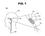
図1は、本発明の一実施形態に従って頭蓋内動脈瘤に展開されたコイル状ステントの説明図を100で示している。コイル状ステント120は、カテーテル140によって頭蓋内動脈瘤110に隣接して展開される。
コイル状ステント120は、頭蓋内動脈瘤110の動脈瘤ネック部112を塞ぐように位置決めすることができる。動脈瘤ネック部112は、動脈瘤嚢114が頭蓋内血管116に隣接している領域である動脈瘤口とも呼ばれる。動脈瘤ネック部112は、広い開口部、狭い開口部、又は不規則な形状の開口部を有することができ、動脈瘤の口と呼ばれることもある。動脈瘤の閉塞は、頭蓋内動脈瘤110と頭蓋内血管116の間の血管内流体の移動を妨げるか又は制限するものである。頭蓋内血管116は、頭蓋118の頭蓋内血管系内の多くの繊細な血管系内血管の1つとすることができる。FIG. 1 illustrates at 100 an illustration of a coiled stent deployed in an intracranial aneurysm according to one embodiment of the present invention.
The
頭蓋内動脈瘤110は、軸線方向動脈瘤、横動脈瘤、漿果状動脈瘤とも呼ばれる嚢状動脈瘤、又は頭蓋118内に形成されるあらゆる動脈瘤とすることができる。本発明は、頭蓋内動脈瘤のための治療方法として表すが、体内の他の適切な位置で動脈瘤を治療するために、またコイル状ステントを使用する他の処置のために利用することもできる。
図2は、本発明の一実施形態に従って螺旋バネとして形成されたコイル状ステントの斜視図を200で示している。コイル状ステント220は、ステントの隣接する巻きの間の中心間の間隔dによって中心軸に関して螺旋状に巻いている。The
FIG. 2 shows a perspective view at 200 of a coiled stent formed as a helical spring according to one embodiment of the present invention. The
一実施形態では、隣接する巻き間の間隔d、ステントフレームワークを形成するワイヤの直径、及びコイル状ステント220の外径及び内径は、コイル状ステント220の軸線方向長さに沿って名目上は一定である。コイル状ステント220の形状は、コイルの隣接する巻き間に小さな間隔ができるように設定することができる。螺旋バネのコンプライアンスは、ワイヤの直径、巻線の数、材料組成、並びにコイル状ステント220の外径及び内径によって部分的には判断される。コイル状ステント220は、コイル状ステントが動脈瘤に展開される時に螺旋バネを形成するように製造することができる。一例では、ステントフレームワークのワイヤの直径は、0.010インチよりも小さい。別の例では、ステントフレームワークの直径は、名目上0.001インチである。コイル状ステントは、例えば、1から5ミリメートルの間の外径と、数ミリメートルから30ミリメートル又はそれよりも大きい長さとを有することができる。 In one embodiment, the spacing d between adjacent turns, the diameter of the wires forming the stent framework, and the outer and inner diameters of the
コイル状ステント220は、コイル状ステントフレームワークを含む。コイル状ステントフレームワークは、ステント受けチューブ(図示せず)内で軸線方向に伸長された時にステント受けチューブ内で同軸状に位置することができ、ステント受けチューブから送出されて動脈瘤に隣接して展開される時に反跳することができる。例えば、コイル状ステント220は、カテーテルのステント受けチューブ内に置かれた時に軸線方向に延ばされ、後の展開のためにワイヤの直径よりも僅かに大きい内径を有する薄肉チューブ内に収容される。別の例では、コイル状ステント220は、ステント受けチューブ内に押し込まれて薄い覆いで覆われ、覆いが引っ込んでステントの展開を可能にする。
他の実施形態では、コイル状ステント220の内径及び外径は、血管に沿った外向きの圧力を最小にしながら血管内のステントを保護するのに役立つように、かつ動脈瘤を塞ぐのに役立つように、ステントの軸線方向長さに沿って変化することもできる。ステントフレームワークのワイヤの直径及び断面形状は、コイルの巻きに沿って変化してもよく、単位長さ当たりの巻きの密度は、コイル状ステント220の調整された剛性をもたらすために、並びに少なくともコイル状ステント220の一部分に沿って動脈瘤頚部又は動脈瘤嚢の口を塞ぐ助けになるより緊密なウェブを形成するために軸線方向長さに沿って変化することができる。 In other embodiments, the inner and outer diameters of the
コイル状ステント220は、金属基部を含むことができる。金属基部は、ニチノール、ニッケル−スズ合金、形状記憶材料のような超弾性金属、及び他の超弾性材料を含むことができる。これらの材料は、特に、望ましい最終的な形に形成することができ、延伸、引っ張り、又は曲げによって大きく変形し、もはや負荷がないか又は拘束されない時に望ましい最終形態に戻ることが可能であるために好ましいものである。超弾性材料の最大8%又はそれを超える弾性歪み限度のために、ステントを永久的又は可塑的に変形させることなく超弾性材料を大きく歪ませることができる。 The
例えば、緊密に巻かれた螺旋コイルに形成された超弾性材料の小直径のワイヤは、引っ張ることによりそれを軸線方向に延ばして直線的なワイヤに大きく変形させ、その後送出された後に緊密に巻かれた構成に戻すことができる。脳の繊細な血管系は、体内の他の血管よりも比較的小さな直径を有するために、コイル状ステントの直径、ステントフレームワークのワイヤの直径、及び展開機構もまた小さくすべきである。 For example, a small diameter wire of superelastic material formed in a tightly wound spiral coil can be stretched axially by pulling to deform it into a straight wire and then tightly wound after being delivered. You can return to the configured configuration. Since the delicate vasculature of the brain has a relatively smaller diameter than other blood vessels in the body, the diameter of the coiled stent, the diameter of the stent framework wire, and the deployment mechanism should also be small.
コイル状ステントが軸線方向に延ばされる時に、ステントフレームワークのワイヤ内で生じる曲げ歪みは、ワイヤの直径が小さくかつコイル状ステントの直径が十分に大きい時には恐らくは小さいために、ステントフレームワークのワイヤは、超弾性でない材料から形成することができる。コイル状ステントのステントフレームワークのワイヤは、例えば、ステンレス鋼、ニチノール、タンタル、「MP35N」合金、プラチナ、チタン、クロムベース合金、適切な生体適合性合金、適切な生体適合性ポリマー、又はその組合せのような金属基部を含むことができる。 Because the bending strain that occurs in the stent framework wire when the coiled stent is axially extended is probably small when the wire diameter is small and the coiled stent diameter is large enough, the stent framework wire It can be formed from a material that is not superelastic. The stent framework wire of the coiled stent can be, for example, stainless steel, nitinol, tantalum, “MP35N” alloy, platinum, titanium, chromium-based alloy, suitable biocompatible alloy, suitable biocompatible polymer, or combinations thereof A metal base can be included.
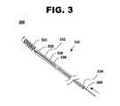
図3は、本発明の一実施形態に従って動脈瘤を治療するシステムの説明図を300で示している。動脈瘤治療システム300は、ステント受けチューブ330を備えたカテーテル340と、ステント受けチューブ330内に軸線方向に受け取られたコイル状ステント320とを含む。コイル状ステント320は、カテーテルにより体内の血管を通って動脈瘤へ送出される。通常、カテーテルは、例えば、大腿上方の小さな切開から挿入され、血流を通って脳内の動脈瘤の部位まで導かれる。カテーテル340は、本明細書では明確にするために示していないガイドワイヤのようなカテーテル340を導くための他の構造体を含むこともできる。 FIG. 3 illustrates at 300 an illustration of a system for treating an aneurysm according to one embodiment of the present invention. The
ステント受けチューブ330は、遠位端332及び近位端334を有する。ステント受けチューブ330は、外径336及び内径338を有する。外径336及び内径338は、軸線方向に受け取られたコイル状ステント320を収容する大きさにすることができる。ステント受けチューブ330は、ガイドワイヤと軸線方向に受け取られたコイル状ステントを位置決めして展開するための他の装置とを収容するカテーテルの一部を含む。
ステント受けチューブ330は、内径が軸線方向に広げられたコイル状ステント320とコイル状ステントフレームワークのあらゆるコーティングとを受け入れる大きさにされた薄肉チューブを含むことができる。別の実施形態では、ステント受けチューブ330は、軸線方向に受け取られたコイル状ステント320を取り囲む送出覆いを含み、これを引き裂くか又は引っ込めてステントの展開を可能にする。送出覆いは、例えば、コイルのワイヤの直径よりも僅かに大きい内径を有することができる。ステント受けチューブと軸線方向に受け取られたコイル状ステントとを備えたカテーテルは、非常に目立たない可撓性の送出可能なインプラントを提供する。
ステント受けチューブ330を備えたカテーテル340は、コイル状ステント320を動脈瘤に隣接して位置決めするために足、胸、首、又は頭にある1つ又はそれよりも多い血管を通って縫うようにして進む遠位端332によって血管内に挿入される。一実施形態では、軸線方向に受け取られたコイル状ステント320をステント受けチューブ330に通してステント受けチューブ330の遠位端332の外へ移動させるために、プッシュワイヤ350がカテーテルに挿入される。軸線方向に受け取られたコイル状ステント320がステント受けチューブ330から展開されると、コイル状ステント320は、所定のコイル形状に反跳して戻る。所定のコイル形状は、例えば、軸線方向長さに沿って一定の外径を有する螺旋コイルとすることができる。コイル状ステント320は、コイル状ステント320が動脈瘤に展開されると螺旋バネを形成する。所定のコイル形状は、例えば、動脈瘤に送出されると軸線方向長さに沿って1つ又はそれよりも多いテーパ又は起伏を有する螺旋コイルとすることができる。
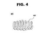
図4は、本発明の一実施形態に従って少なくとも1つのテーパ又は起伏を有する螺旋バネとして形成されたコイル状ステントの斜視図を400で示している。コイル状ステント420は、動脈瘤に展開された時に螺旋バネの軸線方向長さに沿って少なくとも1つのテーパ又は起伏を有する螺旋バネを形成する。この実施形態では、コイル状ステント420の長さの中ほどで直線状テーパが始まり、コイル状ステント420の各端部に向けて直径を一様な量で低減する。直線状テーパは、頭蓋内血管にコイル状ステント420を展開して固定することを助けることができる。いくつかの場合には、コイル状ステント420の拡大した中央部分は、動脈瘤嚢内に一部侵入し、嚢のネック部又は開口部を塞ぎ、血流のような心臓血管活動からの運動又は脈動の結果として頭蓋内血管を上下に動かすことからコイル状ステント420を固定することができる。
コイル状ステント420は、直線又は曲線状テーパ、一端から始まり他端で終わる単一テーパ、両端部が大きい逆テーパ、又はその組合せを有することができる。テーパ及び起伏は、起伏が最大になる所でコイルの巻き密度を増大又は減少させ、望ましい閉塞、展開、及びコンプライアンス特性を与えるように調整することができる。FIG. 4 shows a perspective view at 400 of a coiled stent formed as a helical spring having at least one taper or undulation in accordance with one embodiment of the present invention. The
The
図5は、本発明の別の実施形態に従って少なくとも1つのテーパ又は起伏を有する螺旋バネとして形成されたコイル状ステントの断面図を500で示している。コイル状ステント520は、動脈瘤に展開された時に螺旋バネの軸線方向長さに沿って少なくとも1つのテーパ又は起伏を有する螺旋バネを形成する。この実施形態では、コイル状ステント520は、名目上一様な直径を有する中央区域522と、中央区域522からコイル状ステントの直径が一様な量で減少する直線状テーパを備えたコイル状ステント520の各端に向けて延びる2つの端部区域524及び526とを含む。中央区域522の一定の直径は、展開された時に動脈瘤嚢のネック部又は開口部を塞ぐことを助け、動脈瘤嚢を出入りする血管内流体の流れを妨げるか又は制限し、それによって動脈瘤壁に対するこのような圧力を制限して動脈瘤嚢が更に大きくなるか又は動脈瘤壁が破裂する可能性を低減する。直線状テーパは、頭蓋内血管にコイル状ステント520を展開して固定することを助けることができる。 FIG. 5 shows a cross-sectional view at 500 of a coiled stent formed as a helical spring having at least one taper or undulation in accordance with another embodiment of the present invention.
コイル状ステント520の各端部区域524及び526のテーパは、直線又は曲線状とすることができる。単一テーパを一端で又は他端で使用することができ、両端が大きくなる逆テーパを使用することもできる。動脈瘤の口又はネック部の閉塞性を増大させるために、コイル状ステント520の長さに沿った起伏及び他の波状変形を含むことができる。テーパ及び起伏は、例えば、動脈瘤に隣接するコイル状ステント520の両側を通る血管内の流体の移送を制限するための部分的なウェブを形成するために、直径が名目上一様である中央区域522でコイルの巻き密度を増大させることができる。コイル状ステント520の中央区域におけるコイルの巻きの密度は、隣接するコイルの巻きが少なくともコイル状ステント520の一部で連続的なウェブを形成するように緊密に巻かれてもよい。ステントフレームワークの直径及び断面形状は、例えば、ウェブを形成するためにより大きい直径のワイヤ又は矩形の巻き断面に中央区域で変えることができる。また、コイル状ステントの一部に沿った巻きにポリマーコーティング又は他の物質を付加することも、部分的又は連続的ウェブの形成を助けることができる。 The taper of each
図6は、本発明の一実施形態に従って接着層及びポリマーコーティングをコイル状ステントの一部分に沿って周囲に配置したコイル状ステントの断面図を600で示している。コイル状ステント620は、コイル状ステントフレームワーク622と、コイル状ステント620の少なくとも一部分上に周方向に配置されたポリマーコーティング624とを含む。接着層626は、コイル状ステントフレームワーク622とポリマーコーティング624の間に位置決めすることができる。 FIG. 6 shows a cross-sectional view at 600 of a coiled stent with an adhesive layer and polymer coating disposed around a portion of the coiled stent according to one embodiment of the present invention. The
ポリマーコーティング624は、コイル状ステント620が展開された時にコイル状ステント620の長さの少なくとも一部分に沿って配置され、それによって配置したポリマーコーティング624は、コイル状ステント620の少なくとも一部分に沿って部分的又は連続的ウェブを形成することができる。ポリマーコーティング624の層の厚さ及び数は、ポリマーコーティング624とコイル状ステントフレームワーク622とが結合して部分的又は連続的ウェブを形成するように、コイル状ステント620の隣接する巻き間の間隔に基づいて判断することができる。部分的又は連続的ウェブは、動脈瘤壁が破損する可能性を低減しながら動脈瘤を塞ぐために、動脈瘤の開口部又はネック部に隣接して位置決めすることができる。ポリマーコーティング624は、部分的には、伸長したステントとコイル状ステント620を包み込む送出チューブ又はカテーテルとの間の摩擦係数を制御するように選択することができる。 The
ポリマーコーティング624は、コイル状ステント620を被覆するためにかつ体内に展開するために適切な1つ又はそれよりも多いポリマー材料を含むことができる。ポリマーコーティング624は、生体分解性ポリマー又は生体安定性ポリマーを含むことができる。ポリマーコーティング624は、例えば、ポリカプロラクトン(PCL)、ポリグリコリド(PGA)、又はポリ(ラクチド−コ−グリコリド)(PLGA)のような生体分解性ポリマー、又は、シリコーン−ウレタンコポリマー、ポリウレタン、又はエチレン酢酸ビニル(EVA)のような生体安定性ポリマーを含むことができる。 The
ポリマーコーティング624は、例えば、動脈瘤に展開された時に治療効果を与えるための1つ又はそれよりも多い生体活性薬剤を含有する薬剤ポリマーを含むことができる。ポリマーコーティング624は、薬剤や生体活性薬剤のような1つ又はそれよりも多い治療化合物を含むことができる。ポリマーコーティング624は、例えば、1つ又はそれよりも多い生体活性薬剤が相互に分散されたポリマーマトリックスを含有することができる。ポリマーコーティング624の1つ又はそれよりも多い層は、コイル状ステントフレームワーク622上に配置することができる。 The
治療化合物は、冠状動脈再狭窄、心臓血管再狭窄、血管造影再狭窄、動脈硬化、過形成、並びに他の病気及び状態を含む1つ又はそれよりも多い状態を治療又は予防するものである。例えば、治療化合物は、血管の再狭窄、すなわち、ステントが置かれた体腔の直径が狭くなること又は狭窄に相当する状態を抑制するか又は予防するために、ポリマーコーティング624の中に組み込むことができる。一実施形態では、生体活性薬剤は、再狭窄防止薬を含む。別の実施形態では、治療化合物は、アンチセンス薬、抗腫瘍薬、抗増殖薬、抗血栓薬、抗凝固剤、抗血小板薬、抗生物質、抗炎症薬、ステロイド、遺伝子治療薬、治療剤、有機薬剤、医薬品、組み換えDNA製品、組み換えRNA製品、コラーゲン、コラーゲン誘導剤、プロテイン、プロテイン類似化合物、サッカリド、又はサッカリド誘導剤のような生体活性薬剤を含む。別の実施形態では、ポリマーコーティング624は、医薬剤の組合せを含む。 Therapeutic compounds are those that treat or prevent one or more conditions, including coronary restenosis, cardiovascular restenosis, angiographic restenosis, arteriosclerosis, hyperplasia, and other diseases and conditions. For example, the therapeutic compound may be incorporated into the
いくつかの医薬剤は、ポリマーコーティング624に使用される可能性を有する。例えば、ラパマイシン、ラパマイシン類似化合物、又はラパマイシン誘導剤のような再狭窄防止薬は、体腔の狭まり及び閉塞の再発を防止又は低減する。アンチセンス薬は、遺伝子レベルで作用して、病気を引き起こすプロテインが生成される過程を妨げる。抗腫瘍薬は、一般的に、ステントの周辺で癌細胞の増殖及び広がりを防止し、鎮静化し、又は阻止するために使用される。抗増殖薬は、狙った細胞又は細胞型が成長するのを防止するか又は阻止することができる。抗血栓薬は、血栓の形成を積極的に遅らせるものである。抗凝固剤は、多くの場合、ヘパリン及びクマリンのような化合物を使用し、抗凝固治療で血液の凝固を遅らせるか又は防止する。抗血小板薬は、血小板に作用するように使用することができ、血小板の血液凝固の機能を抑制する。抗生物質は、微生物の成長を抑制するために及び病気や感染と闘うために頻繁に使用される。デキサメタゾンのような抗炎症薬は、ステント周辺の炎症を弱めるか又は軽減するために使用することができる。埋め込まれたステントに近接する瘢痕組織を軽減するために、時にはステロイドが使用される。遺伝子療法薬は、病気を治療し、治癒し、又は究極的には予防するために人の遺伝子の発現を変更することができる場合がある。 Some pharmaceutical agents have the potential to be used for
本質的に、生体活性薬剤とは、病気又は不調を予防又は治療するあらゆる治療剤である。有機薬剤は、あらゆる小分子の治療物質である。医薬品は、治療効果のあるあらゆる化合物である。組み換えDNA製品又は組み換えRNA製品は、修正したDNA又はRNA遺伝物質を含む。医薬的価値のある生体活性薬剤はまた、コラーゲン、及び他のプロテイン、サッカリド、及びそれらの誘導体を含むこともできる。 In essence, a bioactive agent is any therapeutic agent that prevents or treats a disease or disorder. Organic drugs are all small molecule therapeutics. A pharmaceutical is any compound that has a therapeutic effect. A recombinant DNA product or a recombinant RNA product contains modified DNA or RNA genetic material. Pharmaceutically active bioactive agents can also include collagen, and other proteins, saccharides, and derivatives thereof.
ポリマーコーティング624は、少なくとも1つの生体活性薬剤を溶出する。ポリマーコーティング624は、複数の生体活性薬剤を含んで溶出することができる。ポリマーコーティング624は、主として拡散過程によって1つ又はそれよりも多い生体活性薬剤の溶出を制御するように調整することができる。いくつかの場合には、ポリマーコーティングの一部分が体内に吸収されてコーティング内から生体活性薬剤を放出する。
薬剤ポリマーをポリマーコーティング624に組み込むことにより、例えば、手術の24時間以内に薬理活性の薬剤又は生体活性薬剤を迅速に送出し、第2の生体活性薬剤を次の3ヶ月から6ヶ月後にゆっくりと定常的に送出することが可能になる。The
By incorporating the drug polymer into the
ポリマーコーティング624は、複数の薬剤を含むことができ、各薬剤は、所定の溶出速度を有する。一実施形態では、第1の生体活性薬剤は、ステントフレームワークに隣接して集まり、第2の生体活性薬剤は、ポリマーコーティング624の外面に隣接して集まる。例えば、第1の生体活性薬剤は、ラパマイシン、ラパマイシン類似化合物、又はラパマイシン誘導剤のような再狭窄防止剤を含むことができる。第2の生体活性薬剤は、デキサメタゾンのような抗炎症剤を含むことができる。 The
接着層626は、ポリマーコーティング624とコイル状ステントフレームワーク622の間に置いて、ポリマーコーティングの接着性及び耐久性を向上させることができる。接着層626は、ポリマー材料又は下に重なるステントフレームワーク、特にコイル状ステント620の金属基部に良好に接着するあらゆる材料とすることができる。接着層626は、コイル状ステントフレームワーク622に良好に接着し、ポリマーコーティング624のような別のポリマー材料を容易に被覆することができるように選択される。接着層626は、パリレン、ポリウレタン、フェノキシ、エポキシ、ポリイミド、又はペラセンのようなあらゆる適切な接着層とすることができる。 An
図7は、本発明の一実施形態に従って頭蓋内動脈瘤を治療する方法の流れ図を700で示している。頭蓋内動脈瘤の治療方法700は、形成し、位置決めし、展開し、並びに軸線方向動脈瘤、横動脈瘤、嚢状又は漿果状動脈瘤、及び頭蓋内血管系の他の動脈瘤を治療する段階と、体内の他の位置にある血管内の異なる種類の動脈瘤を治療する段階とを含む。
ブロック705に見られるように、コイル状ステントを形成する。コイル状ステントフレームワークは、金属基部を含むことができる。コイル状ステントは、ニチノール、ニッケル−スズ合金、又は形状記憶材料のような超弾性材料から形成することができる。コイル状ステントは、例えば、型の周りで巻かれ、その巻かれた形状を維持するためにマルテンサイト−オーステナイト相転移温度を超えて加熱処理されるか又は形状固定された小直径ニチノールワイヤの一片から形成することができる。コイル状ステントを加熱処理及び形状固定するためには、例えば、摂氏490度から摂氏525度及びそれよりも高い温度を使用することができる。FIG. 7 shows a flow diagram at 700 of a method of treating an intracranial aneurysm according to one embodiment of the present invention. Intracranial
As seen in
いくつかの形状記憶合金では、転移温度が体温よりも低く、更に室温よりも低い場合がある。これらの合金を使用しているコイル状ステントでは、コイルを冷却し、直線状にするか又は軸線方向に伸長させ、ステント受けチューブに挿入することができる。動脈瘤に送出されると、軸線方向に伸長したコイル状ステントは、転移温度を超えて温められて螺旋のコイル型に戻る。十分に堅固なステント受けチューブ又は送出カテーテル覆いに装着される場合は、軸線方向に伸長したコイル状ステントは冷却され、軸線方向に伸長され、ステント受けチューブに挿入され、そこでコイル状ステントは温まってオーステナイト相転移温度まで移行するが、ステント受けチューブから送出されるまで伸長した形状で維持される。 For some shape memory alloys, the transition temperature may be lower than body temperature and even lower than room temperature. In coiled stents using these alloys, the coil can be cooled and straightened or stretched axially and inserted into the stent receiving tube. When delivered to the aneurysm, the axially elongated coiled stent is warmed above the transition temperature and returns to the helical coil shape. When mounted on a sufficiently rigid stent receiving tube or delivery catheter wrap, the axially elongated coiled stent is cooled, axially stretched and inserted into the stent receiving tube where the coiled stent is warmed. It transitions to the austenite phase transition temperature but remains in an elongated shape until delivered from the stent receiving tube.
代替的に、コイル状ステントが展開されて体温まで温められた時に、室温及びそれ以下でのコイル状ステントの変形が直ちに回復するように、形状記憶合金の転移温度は、室温よりも高く標準体温よりも低くすることができる。別の実施形態では、形状記憶合金の転移温度は、標準体温よりも高く設定されている。高い弾性歪み限度のようなマルテンサイト相での形状記憶合金の超弾性特性により、コイル状ステントをカテーテル展開用の小直径の受入チューブの中に縦方向に拡張し、同時に展開されて受入チューブから分離されるとコイル状ステントを螺旋形状に容易に回復させることが可能である。送出カテーテル上の薄肉ステント受けチューブ又は覆いは、弾力的に伸長したコイル状ステントを展開されるまで拘束する。 Alternatively, when the coiled stent is deployed and warmed to body temperature, the transition temperature of the shape memory alloy is higher than room temperature and normal body temperature so that the deformation of the coiled stent at room temperature and below is immediately recovered. Can be lower. In another embodiment, the transition temperature of the shape memory alloy is set higher than the standard body temperature. Due to the superelastic properties of the shape memory alloy in the martensite phase, such as high elastic strain limits, the coiled stent is longitudinally expanded into a small diameter receiving tube for catheter deployment and simultaneously deployed and removed from the receiving tube. Once separated, the coiled stent can be easily restored to a helical shape. A thin stent receiving tube or covering on the delivery catheter restrains the resiliently elongated coiled stent until it is deployed.
十分に細いワイヤでは、コイル状ステントは、他の非超弾性の非形状記憶材料から作ることができる。コイル状ステントは、ステンレス鋼、ニチノール、タンタル、「MP35N」合金、プラチナ、チタン、クロム基合金、適切な生体適合性合金、適切な生体適合性ポリマー、又はその組合せのような他の材料から形成することができる。特に、コイル状ステントフレームワークが小直径のワイヤを含み、コイル状ステントの外径が過度に小さくない場合には、軸線方向に伸長された時に生じる歪みが、使用される材料の弾性歪み限度を超えないので、これらの材料は適切である。一実施形態では、これらの材料のうちの1つのワイヤが、型又は固定具の周りに巻かれてコイル状ステントの望ましい形状を作り、次に、この材料は熱処理されて材料内の応力を軽減するために焼き戻され、好ましい形状に固定する。別の実施形態では、これらの材料のうちの1つのワイヤは、小半径の固定具にわたって引っ張られ、次に、巻かれた形状を維持するために熱処理される。代替的に、ワイヤは焼き戻されて熱で柔らかくされ、ワイヤを望ましい形状に可塑的に変形させるために小直径の型の周りで巻かれる。ある一定の量の弾性反跳を許した後に、コイルの巻きは、次に、降伏応力を増大させて弾性挙動の範囲を向上させるために、スエージ加工又は冷間加工することができる。 With sufficiently thin wires, coiled stents can be made from other non-superelastic non-shape memory materials. Coiled stents are formed from other materials such as stainless steel, nitinol, tantalum, “MP35N” alloys, platinum, titanium, chromium-based alloys, suitable biocompatible alloys, suitable biocompatible polymers, or combinations thereof. can do. In particular, if the coiled stent framework includes small diameter wires and the outer diameter of the coiled stent is not excessively small, the strain that occurs when stretched in the axial direction will limit the elastic strain limit of the material used. These materials are suitable because they do not exceed. In one embodiment, one of these materials is wound around a mold or fixture to create the desired shape of the coiled stent, which is then heat treated to reduce stress in the material. To be tempered and fixed in a preferred shape. In another embodiment, one of these materials is pulled over a small radius fixture and then heat treated to maintain the rolled shape. Alternatively, the wire is tempered and softened with heat and wound around a small diameter mold to plastically deform the wire into the desired shape. After allowing a certain amount of elastic recoil, the coil turns can then be swaged or cold worked to increase the yield stress and improve the range of elastic behavior.
コイル状ステントは、コイルの隣接する巻きの間を小さい間隔にするように形状固定することができる。コイル状ステントは、一様の外径を有するか又は1つ又はそれよりも多いテーパ又は起伏を有する螺旋バネを形成するように形状固定することができる。
代替的に、コイル状ステントは、その長さに沿って可変直径を有し、その長さに沿って可変強度をもたらすようにワイヤから形成することができる。脳の繊細な血管系に展開されるコイル状ステントを形成するために、調整された半径方向強度を有する非常に細い直径のワイヤを使用することができる。別の実施形態では、コイル状ステントフレームワークは、細長い又は矩形の断面を含む。The coiled stent can be fixed in shape so that there is a small spacing between adjacent turns of the coil. The coiled stent can be fixed in shape to form a helical spring having a uniform outer diameter or having one or more tapers or undulations.
Alternatively, the coiled stent can be formed from wire to have a variable diameter along its length and provide variable strength along its length. To form a coiled stent that is deployed in the delicate vasculature of the brain, very thin diameter wires with adjusted radial strength can be used. In another embodiment, the coiled stent framework includes an elongated or rectangular cross section.
ブロック710に見られるように、接着層をコイル状ステントの金属基部に配置することができる。接着層は、コイルを巻く前に金属ワイヤ上に配置することができる。代替的に、接着層は、コイル状ステントが巻かれた後にコイル状ステントフレームワークの少なくとも一部分上に配置することができる。接着層は、浸漬け、塗装、刷毛塗り、又は吹き付けのようなあらゆる適切な技術を使用して付加され、次に、真空又は制御された環境において室温又は上昇させた温度で乾燥させることができる。接着層は、ポリマーコーティングを金属ステントフレームワークに接着するのを助けるために、コイル状ステントの金属基部とポリマーコーティングの間に位置決めすることができる。 As seen in
ポリマーコーティングは、ブロック715に見られるように、コイル状ステントの少なくとも一部上で周方向に配置することができる。コイル状ステントフレームワークを形成するワイヤは、例えば、その周囲の回りを生体適合性ポリマーで被覆することができる。反跳して展開された時に、コーティングは、コイル状ステントの隣接する巻回の間の空間を部分的又は完全に密封し、動脈瘤嚢への漏れを防ぐことができる。 The polymer coating can be circumferentially disposed on at least a portion of the coiled stent, as seen in
ポリマーコーティングは、浸漬け、塗装、刷毛塗り、又は吹き付けのようなあらゆる適切な技術で付加され、次に、あらゆる溶剤を取り除くことによって乾かされ、必要に応じて硬化させることができる。ポリマーコーティングは、コイル状ステントが軸線方向に広げられた時又は望ましい螺旋形状に形成されるか又は反跳した時に付加することもできる。ポリマーコーティングとコイル状ステントフレームワークとの接着性を向上させるために、コイル状ステントの少なくとも一部分に配置された接着層上にポリマーコーティングを付加することもできる。 The polymer coating can be applied by any suitable technique, such as dipping, painting, brushing, or spraying, and then dried by removing any solvent and allowed to cure as needed. The polymer coating can also be applied when the coiled stent is axially expanded or formed into the desired helical shape or recoils. In order to improve the adhesion between the polymer coating and the coiled stent framework, the polymer coating can also be applied over an adhesive layer disposed on at least a portion of the coiled stent.
コイル状ステントが展開された時に、配置されたポリマーコーティングがコイル状ステントの少なくとも一部分に沿って連続的又はほぼ連続的なウェブを形成するように、ポリマーコーティングは、1つ又はそれよりも多いポリマー層を使用して適切な厚さまで付加することができる。
コイル状ステントフレームワーク上に配置されたポリマーコーティングは、薬剤ポリマーを含むことができる。1つ又はそれよりも多い生体活性薬剤は、動脈瘤に展開された時の送出のために、ポリマーコーティングにわたって相互に分散させることができる。薬剤ポリマーの1つ又はそれよりも多い層は、ポリマーコーティング中に含まれる1つ又はそれよりも多い薬剤の溶出速度を制御するために、コイル状ステントフレームワークに付加することができる。医薬品の溶出速度を制御するために、薬剤ポリマー層の間又はポリマーコーティングの周囲上に1つ又はそれよりも多いバリア層を含めることができる。The polymer coating is one or more polymers so that when the coiled stent is deployed, the deployed polymer coating forms a continuous or nearly continuous web along at least a portion of the coiled stent. Layers can be used to add to the appropriate thickness.
The polymer coating disposed on the coiled stent framework can include a drug polymer. One or more bioactive agents can be dispersed with each other across the polymer coating for delivery when deployed in an aneurysm. One or more layers of drug polymer can be added to the coiled stent framework to control the dissolution rate of the one or more drugs contained in the polymer coating. One or more barrier layers may be included between the drug polymer layers or around the polymer coating to control the dissolution rate of the pharmaceutical agent.
コイル状ステントは、ブロック720に見られるようにステント受けチューブに挿入される。ステント受けチューブは、軸線方向に受け取られたコイル状ステントを位置決めして展開するためのガイドワイヤ及び他の装置を収容するカテーテルの一部を含む。ステント受けチューブは、軸線方向に広げられたコイル状ステント及びコイル状ステントフレームワークのあらゆるコーティングを受け入れる大きさの内径を備えた薄肉チューブを含むことができる。代替的に、ステント受けチューブは、軸線方向に受け取られたコイル状ステントを囲む送出覆いを含み、これを引き裂いて又は引っ込めてステントの展開を可能にする。送出覆いは、コイルのワイヤ直径よりも僅かに大きい内径を有することができる。コイル状ステントは、通常、ステント受けチューブに挿入する前に滅菌される。 The coiled stent is inserted into the stent receiving tube as seen in
ブロック725に見られるように、軸線方向に受け取られたコイル状ステントを備えたカテーテルは、体内の血管に挿入される。カテーテルは、例えば、頭、首、胸、及び脚にある1つ又はそれよりも多い血管を通して挿入することができる。この血管は、例えば、体内の頭蓋内血管系に位置していてもよい。軸線方向に受け取られたコイル状ステントを備えたカテーテルは、血管内処置のための適切な入口点を通って頭蓋内血管に挿入される。 As seen in
軸線方向に受け取られたコイル状ステントは、ブロック730に見られるように、頭蓋内動脈瘤に隣接して位置決めすることができる。軸線方向に受け取られたコイル状ステントは、頭及び首の中の時折曲がりくねった経路を通ってガイドワイヤでカテーテルの先端を物理的に操作することによって位置決めすることができる。X線及び蛍光透視撮像システムのようなあらゆる適切な撮像システムを利用してカテーテルの先端の位置を判断し、それを動脈瘤の方へ導くことができる。例えば、カテーテルの端部又はコイル状ステント沿いの1つ又はそれよりも多い位置に放射線不透過性マーカを装着させることができる。 The axially received coiled stent can be positioned adjacent to the intracranial aneurysm, as seen in
軸線方向に受け取られたコイル状ステントは、ブロック735に見られるように、カテーテルから分離することができる。一例では、軸線方向に受け取られたコイル状ステントは、送出覆いを軸線方向に受け取られたコイル状ステントの周囲から引っ込めてコイル状ステントを動脈瘤に隣接して展開することにより、カテーテルから分離される。別の例では、軸線方向に受け取られたコイル状ステントをステント受けチューブに通して移動させ、コイル状ステントを動脈瘤に隣接して展開するために、プッシュワイヤを使用して軸線方向に展開されたコイル状ステントをカテーテルから分離する。プッシュワイヤは、例えば、プッシュワイヤの近位端を握って軸線方向に受け取られたコイル状ステントをステント受けチューブの遠位端から優しく押し出すことにより操作し、コイル状ステントをステント受けチューブから分離することができる。軸線方向に受け取られたコイル状ステントがステント受けチューブの遠位端から現れると、それは、ステント受けチューブから送出されて動脈瘤に隣接して展開される時に螺旋形状に再形成するか又は反跳する。 The axially received coiled stent can be separated from the catheter as seen in
コイル状ステントがステント受けチューブから分離される時に、ステントは、ブロック740に見られるように反跳する。ステントは、1つ又はそれよりも多いテーパ又は起伏を含むことができる螺旋形状に反跳する。コイル状ステントが展開される時に、反跳したステントの少なくとも一部分は、動脈瘤のネック部又は頭蓋内動脈瘤の開口部を塞ぐ。動脈瘤の頚部は、例えば、コイル状ステントの一部分を動脈瘤に隣接して置くことにより、完全に又は部分的に塞ぐことができる。コイル状ステントは、動脈瘤に隣接した領域では緊密な間隔の巻きを含むことができる。コイル状ステントは、コイル状ステントが展開された時にコイル状ステントの少なくとも一部分に沿って連続的ウェブを形成するように、巻きの上にポリマーコーティングを含むことができる。 As the coiled stent is separated from the stent-receiving tube, the stent recoils as seen in
治療化合物は、ブロック745に見られるように、1つ又はそれよりも多い医薬剤又は生体活性薬剤をポリマーコーティング中に相互に分散させて頭蓋内の血管又は頭蓋内動脈瘤に送出することができる。1つ又はそれよりも多い相互に分散された治療化合物を有したポリマーコーティングは、コイル状ステントが展開される時に、コイル状ステントの少なくとも一部分上に周方向に配置することができる。治療化合物は、生体活性薬剤とポリマーコーティング及びその上に配置されたあらゆるバリアコーティングの溶出特性とに依存して、数分、数時間、数日、又は更に数ヶ月の期間にわたって動脈瘤又は血管又はその両方に送出することができる。 The therapeutic compound can be delivered to an intracranial blood vessel or intracranial aneurysm, as seen in
頭蓋内動脈瘤を治療する方法に限って説明したが、本方法は、軸線方向に受け取られたコイル状ステントを展開することができる身体の別の部分のあらゆる動脈瘤又は血管に適用することができる。小さな血管閉塞コイルを動脈瘤嚢内に置くことのような他の外科的処置は、コイル状ステントが、動脈瘤のネック部又は開口部を塞ぐ時にいずれの血管閉塞コイルをも動脈瘤嚢内に維持することを助けるように、軸線方向に受け取られたステントの前及びそれと組み合わせて送り出すことができる。本発明の一実施形態では、血管閉塞コイルをコイル状ステントフレームワークに取り付けて、血管閉塞コイルを動脈瘤嚢に保持して配置することを更に助けることができる。
本明細書で開示した本発明の実施形態は、現時点で好ましいと考えられるものであるが、本発明の精神及び範囲から逸脱することなく様々な変更及び修正を行うことができる。本発明の範囲は、特許請求の範囲に示されており、均等物の意味及び範囲に該当する全ての変更は、本発明の範囲に含まれるものとする。Although described only as a method of treating an intracranial aneurysm, the method may be applied to any aneurysm or blood vessel in another part of the body capable of deploying an axially received coiled stent. it can. Other surgical procedures such as placing a small vaso-occlusive coil in the aneurysm sac keep any vaso-occlusion coil in the aneurysm sac when the coiled stent occludes the neck or opening of the aneurysm To help, it can be delivered before and in combination with the axially received stent. In one embodiment of the present invention, a vaso-occlusive coil can be attached to the coiled stent framework to further assist in holding and placing the vaso-occlusive coil in the aneurysm sac.
While the embodiments of the invention disclosed herein are presently preferred, various changes and modifications can be made without departing from the spirit and scope of the invention. The scope of the present invention is indicated in the claims, and all changes that fall within the meaning and range of equivalents are intended to be included within the scope of the present invention.
100 コイル状ステントの説明
110 頭蓋内動脈瘤
112 動脈瘤ネック部
120 コイル状ステント
140 カテーテル100 Description of
Claims (27)
Translated fromJapaneseステント受けチューブを含むカテーテルと、
前記ステント受けチューブに軸線方向に受け取られ、前記カテーテルを通じて血管を通って動脈瘤まで送出されるコイル状ステントと、
を含むことを特徴とするシステム。A system for treating an aneurysm,
A catheter including a stent receiving tube;
A coiled stent received axially by the stent receiving tube and delivered through the catheter through a blood vessel to an aneurysm;
A system characterized by including.
を更に含むことを特徴とする請求項1に記載のシステム。A pushwire for moving the axially received coiled stent through the stent-receiving tube and deploying the coiled stent adjacent to the aneurysm;
The system of claim 1 further comprising:
を更に含むことを特徴とする請求項1に記載のシステム。A polymer coating circumferentially disposed on at least a portion of the coiled stent;
The system of claim 1 further comprising:
を更に含むことを特徴とする請求項10に記載のシステム。An adhesive layer positioned between the disposed polymer coating and the metal base of the coiled stent;
The system of claim 10 further comprising:
ステント受けチューブ内で軸線方向に伸長される時に該ステント受けチューブ内に同軸的に位置し、該ステント受けチューブから送出されて動脈瘤に隣接して展開される時に反跳するコイル状ステントフレームワーク、
を含むことを特徴とするコイル状ステント。A coiled stent for treating an aneurysm,
Coiled stent framework that is coaxially positioned within the stent receiving tube when axially extended within the stent receiving tube and rebounds when delivered from the stent receiving tube and deployed adjacent to the aneurysm ,
A coiled stent comprising:
を更に含むことを特徴とする請求項14に記載のコイル状ステント。A polymer coating circumferentially disposed on at least a portion of the coiled stent;
The coiled stent according to claim 14, further comprising:
を更に含むことを特徴とする請求項14に記載のコイル状ステント。An adhesive layer positioned between the disposed polymer coating and the metal base of the coiled stent;
The coiled stent according to claim 14, further comprising:
体内の血管の中に軸線方向に受け入れられたコイル状ステントを含むカテーテルを挿入し、
前記軸線方向に受け入れられたコイル状ステントを頭蓋内動脈瘤に隣接して位置決めし、
前記軸線方向に受け入れられたコイル状ステントをカテーテルから分離し、
前記軸線方向に受け入れられたコイル状ステントを螺旋形状に再形成し、再形成されたステントの少なくとも一部分は、再形成されたステントが展開するとき、頭蓋内動脈瘤の動脈瘤ネック部を閉塞する、ことを特徴とする方法。A method for treating an intracranial aneurysm comprising:
Inserting a catheter containing an axially received coiled stent into a blood vessel in the body,
Positioning the axially received coiled stent adjacent to the intracranial aneurysm;
Separating the axially received coiled stent from the catheter;
The axially received coiled stent is reshaped into a helical shape and at least a portion of the reshaped stent occludes the aneurysm neck of the intracranial aneurysm when the reshaped stent is deployed A method characterized by that.
Applications Claiming Priority (2)
| Application Number | Priority Date | Filing Date | Title |
|---|---|---|---|
| US10/463,124US20040260384A1 (en) | 2003-06-17 | 2003-06-17 | Superelastic coiled stent |
| PCT/US2004/019651WO2004112655A1 (en) | 2003-06-17 | 2004-06-17 | Superelastic coiled stent |
Publications (1)
| Publication Number | Publication Date |
|---|---|
| JP2007524449Atrue JP2007524449A (en) | 2007-08-30 |
Family
ID=33517043
Family Applications (1)
| Application Number | Title | Priority Date | Filing Date |
|---|---|---|---|
| JP2006517449APendingJP2007524449A (en) | 2003-06-17 | 2004-06-17 | Superelastic coiled stent |
Country Status (4)
| Country | Link |
|---|---|
| US (1) | US20040260384A1 (en) |
| EP (1) | EP1638482A1 (en) |
| JP (1) | JP2007524449A (en) |
| WO (1) | WO2004112655A1 (en) |
Cited By (1)
| Publication number | Priority date | Publication date | Assignee | Title |
|---|---|---|---|---|
| WO2014115306A1 (en)* | 2013-01-25 | 2014-07-31 | テルモ株式会社 | Stent delivery device |
Families Citing this family (83)
| Publication number | Priority date | Publication date | Assignee | Title |
|---|---|---|---|---|
| US7635387B2 (en) | 2001-11-01 | 2009-12-22 | Cardiac Dimensions, Inc. | Adjustable height focal tissue deflector |
| US6976995B2 (en) | 2002-01-30 | 2005-12-20 | Cardiac Dimensions, Inc. | Fixed length anchor and pull mitral valve device and method |
| US7837729B2 (en) | 2002-12-05 | 2010-11-23 | Cardiac Dimensions, Inc. | Percutaneous mitral valve annuloplasty delivery system |
| US7316708B2 (en) | 2002-12-05 | 2008-01-08 | Cardiac Dimensions, Inc. | Medical device delivery system |
| US7794496B2 (en) | 2003-12-19 | 2010-09-14 | Cardiac Dimensions, Inc. | Tissue shaping device with integral connector and crimp |
| US9526616B2 (en) | 2003-12-19 | 2016-12-27 | Cardiac Dimensions Pty. Ltd. | Mitral valve annuloplasty device with twisted anchor |
| US20060004438A1 (en)* | 2004-04-30 | 2006-01-05 | Novostent Corporation | Prosthesis, delivery system and method for neurovascular aneurysm repair |
| US20050283220A1 (en)* | 2004-06-22 | 2005-12-22 | Gobran Riad H | Blood flow diverters for the treatment of intracranial aneurysms |
| DE502004008712D1 (en) | 2004-09-22 | 2009-01-29 | Dendron Gmbh | MEDICAL IMPLANT |
| EP1855619A4 (en) | 2005-01-20 | 2013-10-02 | Cardiac Dimensions Inc | Tissue shaping device |
| US7601160B2 (en)* | 2005-02-04 | 2009-10-13 | Zuli Holdings, Ltd | Device and methods for non-surgical clipping of aneurysms |
| JP5523700B2 (en) | 2005-04-04 | 2014-06-18 | フレキシブル ステンティング ソリューションズ,インク. | Flexible stent |
| CA2609687C (en) | 2005-05-24 | 2014-04-22 | Inspire M.D Ltd. | Stent apparatuses for treatment via body lumens and methods of use |
| US20070060994A1 (en)* | 2005-09-12 | 2007-03-15 | Gobran Riad H | Blood flow diverters for the treatment of intracranial aneurysms |
| US8057495B2 (en)* | 2005-09-13 | 2011-11-15 | Cook Medical Technologies Llc | Aneurysm occlusion device |
| US7749265B2 (en)* | 2005-10-05 | 2010-07-06 | Kenergy, Inc. | Radio frequency antenna for a wireless intravascular medical device |
| US8956400B2 (en)* | 2005-10-14 | 2015-02-17 | Flexible Stenting Solutions, Inc. | Helical stent |
| US11285005B2 (en) | 2006-07-17 | 2022-03-29 | Cardiac Dimensions Pty. Ltd. | Mitral valve annuloplasty device with twisted anchor |
| US8801747B2 (en) | 2007-03-13 | 2014-08-12 | Covidien Lp | Implant, a mandrel, and a method of forming an implant |
| US7988723B2 (en) | 2007-08-02 | 2011-08-02 | Flexible Stenting Solutions, Inc. | Flexible stent |
| US7691125B2 (en)* | 2007-10-04 | 2010-04-06 | Wilson-Cook Medical Inc. | System and method for forming a stent of a desired length at an endoluminal site |
| WO2009076515A1 (en)* | 2007-12-11 | 2009-06-18 | Cornell University | Method and apparatus for sealing an opening in the side wall of a body lumen |
| US10028747B2 (en) | 2008-05-01 | 2018-07-24 | Aneuclose Llc | Coils with a series of proximally-and-distally-connected loops for occluding a cerebral aneurysm |
| US10716573B2 (en) | 2008-05-01 | 2020-07-21 | Aneuclose | Janjua aneurysm net with a resilient neck-bridging portion for occluding a cerebral aneurysm |
| US8006594B2 (en) | 2008-08-11 | 2011-08-30 | Cardiac Dimensions, Inc. | Catheter cutting tool |
| US9149376B2 (en) | 2008-10-06 | 2015-10-06 | Cordis Corporation | Reconstrainable stent delivery system |
| GB0900565D0 (en) | 2009-01-14 | 2009-02-11 | Isis Innovation | Stent |
| JP5750051B2 (en)* | 2009-01-22 | 2015-07-15 | コーネル ユニヴァーシティー | Method and apparatus for restricting flow through a lumen wall |
| US20100191168A1 (en) | 2009-01-29 | 2010-07-29 | Trustees Of Tufts College | Endovascular cerebrospinal fluid shunt |
| US20110038936A1 (en)* | 2009-08-17 | 2011-02-17 | Kimberly Ann Griswold | System and method for electrospun drug loaded biodegradable chemotherapy applications |
| US9358140B1 (en) | 2009-11-18 | 2016-06-07 | Aneuclose Llc | Stent with outer member to embolize an aneurysm |
| US9247942B2 (en) | 2010-06-29 | 2016-02-02 | Artventive Medical Group, Inc. | Reversible tubal contraceptive device |
| EP2588042A4 (en)* | 2010-06-29 | 2015-03-18 | Artventive Medical Group Inc | Reducing flow through a tubular structure |
| US9149277B2 (en) | 2010-10-18 | 2015-10-06 | Artventive Medical Group, Inc. | Expandable device delivery |
| WO2012135859A2 (en) | 2011-04-01 | 2012-10-04 | Cornell University | Method and apparatus for restricting flow through an opening in the side wall of a body lumen, and/or for reinforcing a weakness in the side wall of a body lumen, while still maintaining substantially normal flow through the body lumen |
| WO2012149167A2 (en) | 2011-04-26 | 2012-11-01 | Christopher Gerard Kunis | Method and device for treatment of hypertension and other maladies |
| KR101480514B1 (en)* | 2011-05-26 | 2015-01-09 | 재단법인 아산사회복지재단 | Stent for cerebral aneurysm coil embolization |
| US9433722B2 (en)* | 2011-08-09 | 2016-09-06 | Abbott Cardiovascular Systems Inc. | Vascular shield and delivery system |
| EP2763601B1 (en) | 2011-10-07 | 2020-03-25 | Cornell University | Apparatus for restricting flow through an opening in a body lumen while maintaining normal flow |
| US9011480B2 (en) | 2012-01-20 | 2015-04-21 | Covidien Lp | Aneurysm treatment coils |
| US9072624B2 (en) | 2012-02-23 | 2015-07-07 | Covidien Lp | Luminal stenting |
| US20130226278A1 (en) | 2012-02-23 | 2013-08-29 | Tyco Healthcare Group Lp | Methods and apparatus for luminal stenting |
| US9687245B2 (en) | 2012-03-23 | 2017-06-27 | Covidien Lp | Occlusive devices and methods of use |
| US9078659B2 (en) | 2012-04-23 | 2015-07-14 | Covidien Lp | Delivery system with hooks for resheathability |
| US9724222B2 (en) | 2012-07-20 | 2017-08-08 | Covidien Lp | Resheathable stent delivery system |
| EP2908880B1 (en)* | 2012-10-16 | 2018-12-05 | Paul A. Spence | Devices for facilitating flow from the heart to a blood pump |
| US20140163586A1 (en)* | 2012-12-11 | 2014-06-12 | Dolly Jeanne Holt | Tissue repair devices and methods |
| US9095344B2 (en) | 2013-02-05 | 2015-08-04 | Artventive Medical Group, Inc. | Methods and apparatuses for blood vessel occlusion |
| US8984733B2 (en) | 2013-02-05 | 2015-03-24 | Artventive Medical Group, Inc. | Bodily lumen occlusion |
| US9907684B2 (en) | 2013-05-08 | 2018-03-06 | Aneuclose Llc | Method of radially-asymmetric stent expansion |
| US9636116B2 (en) | 2013-06-14 | 2017-05-02 | Artventive Medical Group, Inc. | Implantable luminal devices |
| US10149968B2 (en) | 2013-06-14 | 2018-12-11 | Artventive Medical Group, Inc. | Catheter-assisted tumor treatment |
| US9737308B2 (en) | 2013-06-14 | 2017-08-22 | Artventive Medical Group, Inc. | Catheter-assisted tumor treatment |
| US9737306B2 (en) | 2013-06-14 | 2017-08-22 | Artventive Medical Group, Inc. | Implantable luminal devices |
| US10130500B2 (en) | 2013-07-25 | 2018-11-20 | Covidien Lp | Methods and apparatus for luminal stenting |
| US10045867B2 (en) | 2013-08-27 | 2018-08-14 | Covidien Lp | Delivery of medical devices |
| US9782186B2 (en) | 2013-08-27 | 2017-10-10 | Covidien Lp | Vascular intervention system |
| US9737696B2 (en) | 2014-01-15 | 2017-08-22 | Tufts Medical Center, Inc. | Endovascular cerebrospinal fluid shunt |
| JP6637430B2 (en) | 2014-01-15 | 2020-01-29 | タフツ メディカル センター, インク.Tufts Medical Center, Inc. | Intravascular cerebrospinal fluid shunt |
| US9713475B2 (en) | 2014-04-18 | 2017-07-25 | Covidien Lp | Embolic medical devices |
| US10363043B2 (en) | 2014-05-01 | 2019-07-30 | Artventive Medical Group, Inc. | Treatment of incompetent vessels |
| CN107148293B (en) | 2014-10-31 | 2020-08-11 | 西瑞维斯克有限责任公司 | Method and system for treating hydrocephalus |
| US20170042551A1 (en) | 2015-08-13 | 2017-02-16 | The Brain Protection Company PTY LTD | Implantable damping devices for treating dementia and associated systems and methods of use |
| US10272230B2 (en) | 2015-10-30 | 2019-04-30 | Cerevasc, Llc | Systems and methods for treating hydrocephalus |
| US10813644B2 (en) | 2016-04-01 | 2020-10-27 | Artventive Medical Group, Inc. | Occlusive implant and delivery system |
| WO2018071600A1 (en) | 2016-10-11 | 2018-04-19 | Cerevasc, Llc | Methods and systems for treating hydrocephalus |
| US10376396B2 (en) | 2017-01-19 | 2019-08-13 | Covidien Lp | Coupling units for medical device delivery systems |
| US10390953B2 (en) | 2017-03-08 | 2019-08-27 | Cardiac Dimensions Pty. Ltd. | Methods and devices for reducing paravalvular leakage |
| US11654011B2 (en)* | 2017-05-30 | 2023-05-23 | University Of Massachusetts | System and methods for treating neurovascular compression |
| AU2018359016B9 (en) | 2017-11-03 | 2022-09-01 | The Brain Protection Company PTY LTD | Implantable damping device for modifying blood flow characteristics |
| EP3762083B1 (en) | 2018-03-08 | 2025-02-12 | CereVasc, Inc. | Systems for minimally invasive drug delivery to a subarachnoid space |
| US11123209B2 (en) | 2018-04-12 | 2021-09-21 | Covidien Lp | Medical device delivery |
| US10786377B2 (en) | 2018-04-12 | 2020-09-29 | Covidien Lp | Medical device delivery |
| US11413176B2 (en) | 2018-04-12 | 2022-08-16 | Covidien Lp | Medical device delivery |
| US11071637B2 (en) | 2018-04-12 | 2021-07-27 | Covidien Lp | Medical device delivery |
| WO2020117562A1 (en) | 2018-12-04 | 2020-06-11 | The Brain Protection Company PTY LTD | Combinatorial therapies including implantable damping devices and therapeutic agents for treating a condition and associated systems and methods of use |
| US11413174B2 (en) | 2019-06-26 | 2022-08-16 | Covidien Lp | Core assembly for medical device delivery systems |
| EP3998999A4 (en)* | 2019-07-16 | 2023-08-16 | Microvention, Inc. | MEDICAL DEVICE WITH IMPROVED FORM PROPERTIES |
| JP2023554000A (en) | 2020-12-14 | 2023-12-26 | カーディアック・ディメンションズ・プロプライエタリー・リミテッド | Modular preloaded medical implants and delivery systems |
| US12042413B2 (en) | 2021-04-07 | 2024-07-23 | Covidien Lp | Delivery of medical devices |
| US12109137B2 (en) | 2021-07-30 | 2024-10-08 | Covidien Lp | Medical device delivery |
| US11944558B2 (en) | 2021-08-05 | 2024-04-02 | Covidien Lp | Medical device delivery devices, systems, and methods |
| WO2024023290A1 (en)* | 2022-07-29 | 2024-02-01 | Fvd (Financière Vendéenne De Développement) | Urological stent |
Citations (8)
| Publication number | Priority date | Publication date | Assignee | Title |
|---|---|---|---|---|
| JPH06507096A (en)* | 1991-04-26 | 1994-08-11 | ユナイテッド ステイツ サージカル コーポレーション | Removable and heat recoverable tissue support device |
| US5383928A (en)* | 1992-06-10 | 1995-01-24 | Emory University | Stent sheath for local drug delivery |
| JPH1071154A (en)* | 1996-07-26 | 1998-03-17 | Target Therapeutics Inc | Aneurysm closure device assembly |
| WO2000012147A1 (en)* | 1998-09-02 | 2000-03-09 | Scimed Life Systems, Inc. | Drug delivery device for stent |
| US6096034A (en)* | 1996-07-26 | 2000-08-01 | Target Therapeutics, Inc. | Aneurysm closure device assembly |
| WO2000062711A1 (en)* | 1999-04-15 | 2000-10-26 | Smart Therapeutics, Inc. | Intravascular stent and method of treating neurovascualr vessel lesion |
| JP2002500533A (en)* | 1997-05-22 | 2002-01-08 | ボストン サイエンティフィック リミテッド | Stent with variable expansion force |
| US6361558B1 (en)* | 1998-03-31 | 2002-03-26 | Cordis Neurovascular, Inc. | Stent aneurysm treatment system and method |
Family Cites Families (17)
| Publication number | Priority date | Publication date | Assignee | Title |
|---|---|---|---|---|
| BR9405969A (en)* | 1993-03-11 | 1996-02-06 | Medinol Ltd | Process of making a stent-type device from a wire stent-type device process to install a stent-type device and process of heating a nitinol-stent-type device |
| US5514176A (en)* | 1995-01-20 | 1996-05-07 | Vance Products Inc. | Pull apart coil stent |
| US6168622B1 (en)* | 1996-01-24 | 2001-01-02 | Microvena Corporation | Method and apparatus for occluding aneurysms |
| US5941249A (en)* | 1996-09-05 | 1999-08-24 | Maynard; Ronald S. | Distributed activator for a two-dimensional shape memory alloy |
| US6007573A (en)* | 1996-09-18 | 1999-12-28 | Microtherapeutics, Inc. | Intracranial stent and method of use |
| US5830230A (en)* | 1997-03-07 | 1998-11-03 | Micro Therapeutics, Inc. | Method of intracranial vascular embolotherapy using self anchoring coils |
| US6241691B1 (en)* | 1997-12-05 | 2001-06-05 | Micrus Corporation | Coated superelastic stent |
| US5935145A (en)* | 1998-02-13 | 1999-08-10 | Target Therapeutics, Inc. | Vaso-occlusive device with attached polymeric materials |
| AU756080B2 (en)* | 1998-06-04 | 2003-01-02 | New York University | Endovascular thin film devices and methods for treating and preventing stroke |
| US6093199A (en)* | 1998-08-05 | 2000-07-25 | Endovascular Technologies, Inc. | Intra-luminal device for treatment of body cavities and lumens and method of use |
| US6187024B1 (en)* | 1998-11-10 | 2001-02-13 | Target Therapeutics, Inc. | Bioactive coating for vaso-occlusive devices |
| US6338739B1 (en)* | 1999-12-22 | 2002-01-15 | Ethicon, Inc. | Biodegradable stent |
| US6974473B2 (en)* | 2000-06-30 | 2005-12-13 | Vascular Architects, Inc. | Function-enhanced thrombolytic AV fistula and method |
| US6585753B2 (en)* | 2001-03-28 | 2003-07-01 | Scimed Life Systems, Inc. | Expandable coil stent |
| US7247338B2 (en)* | 2001-05-16 | 2007-07-24 | Regents Of The University Of Minnesota | Coating medical devices |
| US6675809B2 (en)* | 2001-08-27 | 2004-01-13 | Richard S. Stack | Satiation devices and methods |
| US7122048B2 (en)* | 2002-05-03 | 2006-10-17 | Scimed Life Systems, Inc. | Hypotube endoluminal device |
- 2003
- 2003-06-17USUS10/463,124patent/US20040260384A1/ennot_activeAbandoned
- 2004
- 2004-06-17WOPCT/US2004/019651patent/WO2004112655A1/enactiveApplication Filing
- 2004-06-17EPEP04755681Apatent/EP1638482A1/ennot_activeWithdrawn
- 2004-06-17JPJP2006517449Apatent/JP2007524449A/enactivePending
Patent Citations (11)
| Publication number | Priority date | Publication date | Assignee | Title |
|---|---|---|---|---|
| JPH06507096A (en)* | 1991-04-26 | 1994-08-11 | ユナイテッド ステイツ サージカル コーポレーション | Removable and heat recoverable tissue support device |
| US5383928A (en)* | 1992-06-10 | 1995-01-24 | Emory University | Stent sheath for local drug delivery |
| JPH1071154A (en)* | 1996-07-26 | 1998-03-17 | Target Therapeutics Inc | Aneurysm closure device assembly |
| US5980514A (en)* | 1996-07-26 | 1999-11-09 | Target Therapeutics, Inc. | Aneurysm closure device assembly |
| US6096034A (en)* | 1996-07-26 | 2000-08-01 | Target Therapeutics, Inc. | Aneurysm closure device assembly |
| JP2002500533A (en)* | 1997-05-22 | 2002-01-08 | ボストン サイエンティフィック リミテッド | Stent with variable expansion force |
| US6361558B1 (en)* | 1998-03-31 | 2002-03-26 | Cordis Neurovascular, Inc. | Stent aneurysm treatment system and method |
| WO2000012147A1 (en)* | 1998-09-02 | 2000-03-09 | Scimed Life Systems, Inc. | Drug delivery device for stent |
| JP2002523186A (en)* | 1998-09-02 | 2002-07-30 | ボストン サイエンティフィック リミテッド | Drug delivery device for stent |
| WO2000062711A1 (en)* | 1999-04-15 | 2000-10-26 | Smart Therapeutics, Inc. | Intravascular stent and method of treating neurovascualr vessel lesion |
| JP2002541911A (en)* | 1999-04-15 | 2002-12-10 | スマート セラピューティクス, インコーポレイテッド | Endovascular stent and method for treating neurovascular trauma |
Cited By (1)
| Publication number | Priority date | Publication date | Assignee | Title |
|---|---|---|---|---|
| WO2014115306A1 (en)* | 2013-01-25 | 2014-07-31 | テルモ株式会社 | Stent delivery device |
Also Published As
| Publication number | Publication date |
|---|---|
| EP1638482A1 (en) | 2006-03-29 |
| WO2004112655A1 (en) | 2004-12-29 |
| US20040260384A1 (en) | 2004-12-23 |
Similar Documents
| Publication | Publication Date | Title |
|---|---|---|
| JP2007524449A (en) | Superelastic coiled stent | |
| US10952878B2 (en) | Methods and systems for increasing a density of a region of a vascular device | |
| US20220079738A1 (en) | Devices and methods for treating an aneurysm | |
| EP1339358B1 (en) | Stent devices with detachable distal and proximal wires | |
| CA2261044C (en) | Surgical implants and delivery systems therefor | |
| US6340368B1 (en) | Implantable device with radiopaque ends | |
| CA2455464C (en) | Aneurysm treatment device | |
| US9561122B2 (en) | Vascular device for aneurysm treatment and providing blood flow into a perforator vessel | |
| EP1520555B1 (en) | Balloonless catheter for inserting a stent | |
| JP2006521161A (en) | Implant delivery technology | |
| AU2002316320A1 (en) | Aneurysm treatment device | |
| JP2002065862A (en) | Channeled vascular stent apparatus and method | |
| JP2019531779A (en) | Neurovascular stent | |
| US20100234934A1 (en) | Balloon Deployable Coronary Stent | |
| JP2022553794A (en) | Stent with protruding features for anchoring | |
| US12396732B2 (en) | Expanding medical device with support | |
| JP2005342104A (en) | Stent assembly |
Legal Events
| Date | Code | Title | Description |
|---|---|---|---|
| A131 | Notification of reasons for refusal | Free format text:JAPANESE INTERMEDIATE CODE: A131 Effective date:20081208 | |
| A02 | Decision of refusal | Free format text:JAPANESE INTERMEDIATE CODE: A02 Effective date:20090511 |