JP2007515212A - Active sutures for therapeutic fluid delivery - Google Patents
Active sutures for therapeutic fluid deliveryDownload PDFInfo
- Publication number
- JP2007515212A JP2007515212AJP2006542777AJP2006542777AJP2007515212AJP 2007515212 AJP2007515212 AJP 2007515212AJP 2006542777 AJP2006542777 AJP 2006542777AJP 2006542777 AJP2006542777 AJP 2006542777AJP 2007515212 AJP2007515212 AJP 2007515212A
- Authority
- JP
- Japan
- Prior art keywords
- suture
- active
- braided
- tube
- braided suture
- Prior art date
- Legal status (The legal status is an assumption and is not a legal conclusion. Google has not performed a legal analysis and makes no representation as to the accuracy of the status listed.)
- Granted
Links
Images


Classifications
- A—HUMAN NECESSITIES
- A61—MEDICAL OR VETERINARY SCIENCE; HYGIENE
- A61B—DIAGNOSIS; SURGERY; IDENTIFICATION
- A61B17/00—Surgical instruments, devices or methods
- A61B17/04—Surgical instruments, devices or methods for suturing wounds; Holders or packages for needles or suture materials
- A61B17/06—Needles ; Sutures; Needle-suture combinations; Holders or packages for needles or suture materials
- A61B17/06166—Sutures
- A—HUMAN NECESSITIES
- A61—MEDICAL OR VETERINARY SCIENCE; HYGIENE
- A61B—DIAGNOSIS; SURGERY; IDENTIFICATION
- A61B17/00—Surgical instruments, devices or methods
- A61B17/064—Surgical staples, i.e. penetrating the tissue
- A—HUMAN NECESSITIES
- A61—MEDICAL OR VETERINARY SCIENCE; HYGIENE
- A61B—DIAGNOSIS; SURGERY; IDENTIFICATION
- A61B17/00—Surgical instruments, devices or methods
- A61B17/04—Surgical instruments, devices or methods for suturing wounds; Holders or packages for needles or suture materials
- A61B17/06—Needles ; Sutures; Needle-suture combinations; Holders or packages for needles or suture materials
- A—HUMAN NECESSITIES
- A61—MEDICAL OR VETERINARY SCIENCE; HYGIENE
- A61B—DIAGNOSIS; SURGERY; IDENTIFICATION
- A61B17/00—Surgical instruments, devices or methods
- A61B17/04—Surgical instruments, devices or methods for suturing wounds; Holders or packages for needles or suture materials
- A61B17/0401—Suture anchors, buttons or pledgets, i.e. means for attaching sutures to bone, cartilage or soft tissue; Instruments for applying or removing suture anchors
- A61B2017/0446—Means for attaching and blocking the suture in the suture anchor
- A61B2017/0458—Longitudinal through hole, e.g. suture blocked by a distal suture knot
- A—HUMAN NECESSITIES
- A61—MEDICAL OR VETERINARY SCIENCE; HYGIENE
- A61B—DIAGNOSIS; SURGERY; IDENTIFICATION
- A61B17/00—Surgical instruments, devices or methods
- A61B17/04—Surgical instruments, devices or methods for suturing wounds; Holders or packages for needles or suture materials
- A61B17/0401—Suture anchors, buttons or pledgets, i.e. means for attaching sutures to bone, cartilage or soft tissue; Instruments for applying or removing suture anchors
- A61B2017/0446—Means for attaching and blocking the suture in the suture anchor
- A61B2017/0461—Means for attaching and blocking the suture in the suture anchor with features cooperating with special features on the suture, e.g. protrusions on the suture
- A—HUMAN NECESSITIES
- A61—MEDICAL OR VETERINARY SCIENCE; HYGIENE
- A61B—DIAGNOSIS; SURGERY; IDENTIFICATION
- A61B17/00—Surgical instruments, devices or methods
- A61B17/04—Surgical instruments, devices or methods for suturing wounds; Holders or packages for needles or suture materials
- A61B17/0401—Suture anchors, buttons or pledgets, i.e. means for attaching sutures to bone, cartilage or soft tissue; Instruments for applying or removing suture anchors
- A61B2017/0464—Suture anchors, buttons or pledgets, i.e. means for attaching sutures to bone, cartilage or soft tissue; Instruments for applying or removing suture anchors for soft tissue
- A—HUMAN NECESSITIES
- A61—MEDICAL OR VETERINARY SCIENCE; HYGIENE
- A61B—DIAGNOSIS; SURGERY; IDENTIFICATION
- A61B17/00—Surgical instruments, devices or methods
- A61B17/04—Surgical instruments, devices or methods for suturing wounds; Holders or packages for needles or suture materials
- A61B17/06—Needles ; Sutures; Needle-suture combinations; Holders or packages for needles or suture materials
- A61B17/06166—Sutures
- A61B2017/06185—Sutures hollow or tubular
Landscapes
- Health & Medical Sciences (AREA)
- Life Sciences & Earth Sciences (AREA)
- Surgery (AREA)
- Molecular Biology (AREA)
- Engineering & Computer Science (AREA)
- Biomedical Technology (AREA)
- Heart & Thoracic Surgery (AREA)
- Medical Informatics (AREA)
- Nuclear Medicine, Radiotherapy & Molecular Imaging (AREA)
- Animal Behavior & Ethology (AREA)
- General Health & Medical Sciences (AREA)
- Public Health (AREA)
- Veterinary Medicine (AREA)
- Materials For Medical Uses (AREA)
- Surgical Instruments (AREA)
- Media Introduction/Drainage Providing Device (AREA)
Abstract
Translated fromJapaneseDescription
Translated fromJapanese〔発明の分野〕
本発明は手術創傷に治療液を送達し、時には必要に応じて縫合された創傷を保持するために使える装置に関する。特に、本発明は縫合部周囲の組織に治療液あるいは生物活性液を噴射するために使用され得る機能的な縫合糸に関する。更に言えば、本発明は一端がコネクターを介して治療液源に接続可能で、その全長の少なくとも一部に沿って治療液を流すことができる内部通路を有する編組縫合糸に関する。(Field of the Invention)
The present invention relates to a device that can be used to deliver therapeutic fluid to a surgical wound and sometimes hold a suture that is sutured as needed. In particular, the present invention relates to a functional suture that can be used to inject a therapeutic or bioactive fluid into the tissue surrounding the suture. More particularly, the present invention relates to a braided suture having an internal passage that can be connected at one end to a treatment fluid source via a connector and that allows treatment fluid to flow along at least a portion of its entire length.
〔発明の背景〕
手術による創傷の直近部位に治療液を送達できれば数々の利点が得られる。痛みの軽減、創傷治癒の促進および手術部位の感染症発生の減少は実現可能な利点のほんの一部である。しかし、長時間に亘って創傷部位に限定して直接治療液を送達するのを経済的に容易にする装置の形態も機能も明らかではない。外科処置後の患者への静脈内(IV)投薬は一般に行われていることである。医師は様々な薬剤を長時間に亘って患者の血流に直接静脈内(IV)投与することがある。薬剤の静脈内(IV)投与は、事実、薬の一部が創傷部位に到達する前に薬が全身に循環する、薬を送達する全身的方法である。この場合、薬剤が創傷部位に到達する前にその多くが身体の他の個所で代謝されてしまうことがあるので、薬効が出る量の薬を創傷部位に届かせるために、静脈内(IV)投与される薬剤の全体量を増やしたりあるいは濃度を上げたりすることがしばしば必要である。しかし、多くの場合、創傷部位で最大の効き目が得られる高濃度の薬剤は、身体内のいろいろな臓器で中毒性の副作用を引き起こすこともあるので静脈内(IV)投与が安全でない場合もある。他の薬剤、例えば局部麻酔薬は局部に投与された時のみ効き目が出るもので静脈(IV)投与法とは単純に適合しない。BACKGROUND OF THE INVENTION
Numerous advantages are gained if the therapeutic solution can be delivered to the immediate site of the surgical wound. Pain relief, accelerated wound healing and reduced infection incidence at the surgical site are just a few of the possible benefits. However, neither the form nor the function of the device that makes it economically easy to deliver the treatment solution directly to the wound site over a long period of time is unclear. Intravenous (IV) dosing to patients after surgery is common practice. Physicians may administer various drugs intravenously (IV) directly into the patient's bloodstream over an extended period of time. Intravenous (IV) administration of the drug is in fact a systemic method of delivering the drug, where the drug circulates throughout the body before a portion of the drug reaches the wound site. In this case, many of the drugs may be metabolized elsewhere in the body before they reach the wound site, so intravenous (IV) to deliver a medicinal amount of the drug to the wound site It is often necessary to increase the total amount or concentration of the drug administered. However, in many cases, high concentrations of drugs that provide maximum efficacy at the wound site may cause toxic side effects in various organs within the body and may be unsafe for intravenous (IV) administration. . Other drugs, such as local anesthetics, are effective only when administered locally and are not simply compatible with intravenous (IV) administration.
外科的処置前、処置中また処置後に手術による創傷およびその周囲への多重注射をして副作用および外科的措置に伴う合併症を防ぐことが試みられている。注射器および皮下注射針は局部への投薬手段ではあるが、注射により長時間連続して投薬するのは実際的ではない。実際、時間が経つと薬剤は散逸して治療効果をあげるのに必要な濃度を下回り、更に注射の処方が必要となる。さらに、手術による創傷が薬物治療の局部的な目標部位の場合には、所望の治療効果をあげるために創傷部位周辺への多重注射が必要なこともある。もし多重注射を繰り返し行わなければならないとすると、患者は不快と度重なる煩わしさを耐えなければならない。更に他の欠点としては、この方法だと医療サービス者は繰り返し行われる局部注射に貴重な時間を費やし注意を払わねばならない。 Attempts have been made to prevent side effects and complications associated with surgical procedures by performing multiple injections into and around the surgical wound before, during and after the surgical procedure. Syringes and hypodermic needles are means for local administration, but it is not practical to continuously administer by injection for a long time. In fact, over time, the drug will dissipate and fall below the concentration necessary to achieve a therapeutic effect, and an injection prescription will be required. Furthermore, if the surgical wound is the local target site for drug treatment, multiple injections around the wound site may be required to achieve the desired therapeutic effect. If multiple injections must be repeated, the patient must endure discomfort and annoyance. Yet another drawback is that this method requires healthcare providers to spend valuable time and attention on repeated local injections.
治療液を局部に継続して送達する静脈投与(IV)および注射の上述した欠点に対処するため、創傷部位で使用する特化された輸液カテーテルが多数開発されている。これらの特化された輸液カテーテルは、例えば米国特許第5,458,582号、第5,891,101号および第6,626,885号に記載のように、通常その全長にわたって多数の孔を有し、治療液を保有しそれを輸液カテーテルに送る液槽とポンプに接続されている。輸液カテーテル自体を直接手術創傷の中に配置しその周りの傷を塞いで所定の位置に保持することもある。しかし、この配置方法では病原菌が輸液カテーテルを通って手術創傷に入る可能性もあるので、感染のリスクと傷の治癒低下のリスクが大きくなることもある。さらに一般的には、輸液カテーテルは傷の近辺の皮膚と皮下組織を貫通し、カテーテルの先端は手術創傷内にそしてカテーテル本体は傷の周囲の健康な組織内に保持される。このような輸液カテーテルの埋め込みに通常はカニューレを使って皮膚と皮下組織を貫通し輸液カテーテルを創傷部位に導く必要があることに注目することが大切である。これらのカテーテルは創傷に治療液を継続的に送達する手段にはなるが、おおくの欠陥を伴う。米国特許第6,626,885号に記載のような装置の多くはカテーテルを所定の位置にしっかりと保持するためにカニューレの使用が必要でそのため手術創傷近辺にさらなる穿刺傷を作ってしまい、一方米国特許第5,891,101号および米国特許第5,458,582号に記載の他の装置では特別な縫合糸の使用あるいは縫合処置の変更が必要となる。そのようなものであっても、輸液カテーテルがしっかりと固定されないこともあり、患者が誤ってカテーテルを創傷部位から引き抜いてしまうことも珍しいことではない。あるいは、患者の不快を和らげたりカテーテルの引き抜きに伴う他の合併症を防いだりするために、米国特許第5,458,582号に記載してあるようなカテーテル装置は生体吸収性材料から作られている。しかし、生体吸収性材料のカテーテルを埋め込むには身体により吸収され代謝される材料の量を増やすことになり、一般的にこの生体負担をできるだけ小さくすることが望ましい。最後に、これらの特化したカテーテルとそれらの動作のために必要な補助的な輸液槽とポンプの使用により数百から数千ドルのかなりの費用が追加される。 A number of specialized infusion catheters for use at the wound site have been developed to address the above-mentioned drawbacks of intravenous (IV) and injection for continuous delivery of therapeutic fluids locally. These specialized infusion catheters typically have multiple holes throughout their length, as described, for example, in US Pat. Nos. 5,458,582, 5,891,101 and 6,626,885. It is connected to a pump and a reservoir that holds the therapeutic fluid and sends it to the infusion catheter. The infusion catheter itself may be placed directly into the surgical wound and the wound around it may be closed and held in place. However, this placement method may also increase the risk of infection and reduced wound healing because pathogens can enter surgical wounds through infusion catheters. More generally, infusion catheters penetrate the skin and subcutaneous tissue near the wound, the catheter tip is retained in the surgical wound and the catheter body is retained in healthy tissue surrounding the wound. It is important to note that such infusion catheters typically require the use of a cannula to penetrate the skin and subcutaneous tissue and guide the infusion catheter to the wound site. While these catheters provide a means of continually delivering therapeutic fluid to the wound, they are associated with a number of deficiencies. Many devices, such as those described in US Pat. No. 6,626,885, require the use of a cannula to hold the catheter securely in place, thereby creating additional puncture wounds near the surgical wound, Other devices described in US Pat. No. 5,891,101 and US Pat. No. 5,458,582 require the use of special sutures or modification of the suture procedure. Even in such a case, the infusion catheter may not be firmly fixed, and it is not uncommon for a patient to accidentally withdraw the catheter from the wound site. Alternatively, to alleviate patient discomfort and prevent other complications associated with catheter withdrawal, a catheter device such as that described in US Pat. No. 5,458,582 is made from a bioabsorbable material. ing. However, implanting a catheter of bioabsorbable material increases the amount of material absorbed and metabolized by the body, and it is generally desirable to minimize this burden on the body. Finally, the use of these specialized catheters and the auxiliary infusion tanks and pumps necessary for their operation add significant costs of hundreds to thousands of dollars.
局部へ継続して薬物送達するのに、また傷の縫合と局部への薬物送達の両方を随意に行うのに使用できる縫合糸ならば、前述の装置が対応できない要求に応えることができる。縫合糸は傷の周囲の組織に埋め込まれ、実際、その部位では局部的薬物送達による最も大きな効果が得られる。さらに、縫合糸は傷の縫合をすることもあるので、患者が耐えなければならない侵襲的処置の数は必ずしも増えない。さらに、縫合糸の一端に取り付けてある縫合針を利用して傷の周囲の組織を貫通して輸液縫合糸の配置を容易にできる。縫合糸の一端に結び目を作って傷に固定して誤って縫合糸が傷から外れないようにできる。さらに、縫合糸が示す可撓性は輸液カテーテルのそれよりもかなり大きく、従って縫合糸は複雑なパターンであるいは従来の輸液カテーテルでは到達が難しかった部位にも配置できる。もし縫合糸からの薬物送達が可能であれば多くの利益が得られかもしれないが、そのような装置の形状や機能ははっきりしていない。 Sutures that can be used for continued drug delivery to the local area, and optionally both wound stitching and local drug delivery, can meet the demands of the devices described above. The suture is embedded in the tissue surrounding the wound and, in fact, the site is most effective with local drug delivery. Furthermore, since sutures can suture wounds, the number of invasive procedures that a patient must withstand does not necessarily increase. Furthermore, the infusion suture can be easily arranged by penetrating the tissue around the wound using a suture needle attached to one end of the suture. A knot can be tied to one end of the suture and secured to the wound to prevent the suture from accidentally coming off the wound. In addition, the flexibility of sutures is much greater than that of infusion catheters, so sutures can be placed in complex patterns or at sites that are difficult to reach with conventional infusion catheters. Although many benefits may be obtained if drug delivery from a suture is possible, the shape and function of such a device is unclear.
中空単繊維の縫合糸の概念は米国特許第3,918,455号で初めて開示された。この特許は縫合糸の縫合針への取付けを容易にするため中空縫合糸の使用に着目したが、縫合するに際して中空縫合糸の管孔に治療液を充填して縫合糸材料の溶解を促進したりあるいは縫合糸をX線写真撮影法で観察したりできるようにすることも示唆している。さらに、中空縫合糸をそのように押出し且つ延伸して微孔質にできることを示唆している。このように微孔質にすると、中空縫合糸の壁を構成する重合体により縫合糸の管孔内の治療液が徐々に壁を通って周囲の組織の中に拡散できる。米国特許第5,984,933号には組織縫合用の装置が記載されている。この特許は主として内視鏡的縫合を容易にする方法および装置について述べているが、装置の縫合材は中空でも中空でなくてもよく、中空の場合には縫合糸の壁に小さい孔を形成して縫合糸の管孔内の薬剤を周囲の組織に浸出可能であることを示唆している。これらの特許は中空縫合糸を使って治療液をその管孔に収納し、ある実施例では治療液をゆっくりと放出できると示唆しているが、まだ対処がとられていない重大な欠点がいくつかある。第一に、単繊維縫合糸はきずの影響を受け易い。中空縫合糸の壁に細孔あるいは穿孔を形成すると、縫合糸の強度特性が大幅に低下ししっかりと創傷縫合ができなくなることがある。第二に、中空縫合糸内に収納できる治療薬の量が少ない。事実、大部分の中空縫合糸内に収容できる薬物含有溶液の量は最大0.005ミリリットルあるいはそれ以下であり、一方市販されている薬物含有溶液は1ミリリットルを超える量でないと薬効がない。例えば、マルカイン、リドカイン、ブピバカイン、メピバカインおよびプロカインなどの麻酔薬は緩衝液に入れて総量5乃至30ミリリットルとして切開傷(incision)あるいは創傷(wound)の周囲の組織に注射するのが普通であり、この5乃至30ミリリットルは米国特許第3,918,455号および第5,984,933号に開示された中空縫合糸に適用できる量の500乃至3000倍である。最後に、中空縫合糸を創傷の周囲の組織にいったん埋め込むと、薬物送達速度は治療液が多数の穿孔あるいは細孔を通じて浸出あるいは拡散する速度により決まる。薬物送達速度の能動的制御および連続的な薬物送達は不可能である。さらに、もし薬物の副作用の発生があると、縫合糸を創傷から切離して薬物送達を止めねばならない。 The concept of hollow monofilament suture was first disclosed in US Pat. No. 3,918,455. This patent focused on the use of hollow sutures to facilitate the attachment of the suture to the suture needle, but when sutured, the tube hole of the hollow suture was filled with a treatment solution to promote dissolution of the suture material. It also suggests that sutures can be observed by X-ray photography. It further suggests that hollow sutures can be so extruded and stretched to become microporous. When microporous as described above, the treatment liquid in the suture hole can gradually diffuse through the wall and into the surrounding tissue by the polymer constituting the wall of the hollow suture. U.S. Pat. No. 5,984,933 describes a tissue suturing device. Although this patent primarily describes a method and device that facilitates endoscopic suturing, the suture material of the device may or may not be hollow, in which case a small hole is formed in the suture wall. This suggests that the drug in the suture hole can be leached into the surrounding tissue. These patents use hollow sutures to contain the treatment fluid in its lumen and suggest that in some embodiments, the treatment solution can be released slowly, but there are some significant drawbacks that have not yet been addressed. There is. First, single fiber sutures are susceptible to flaws. If pores or perforations are formed in the wall of the hollow suture, the strength characteristics of the suture may be greatly reduced, and it may not be possible to firmly wound the suture. Second, the amount of therapeutic agent that can be stored in the hollow suture is small. In fact, the amount of drug-containing solution that can be accommodated in most hollow sutures is at most 0.005 milliliters or less, whereas commercially available drug-containing solutions have no medicinal effect unless they exceed 1 milliliter. For example, anesthetics such as marcaine, lidocaine, bupivacaine, mepivacaine and procaine are usually injected into the tissue surrounding the incision or wound as a total volume of 5 to 30 ml in buffer, This 5 to 30 milliliters is 500 to 3000 times the amount applicable to the hollow suture disclosed in US Pat. Nos. 3,918,455 and 5,984,933. Finally, once the hollow suture is implanted in the tissue surrounding the wound, the drug delivery rate is determined by the rate at which the treatment solution leaches or diffuses through a number of perforations or pores. Active control of drug delivery rate and continuous drug delivery are not possible. Furthermore, if drug side effects occur, the suture must be cut from the wound to stop drug delivery.
米国特許第4,159,720号には治療液を組織に流し込む手段が述べられている。好ましい実施例では体外に配置されて吸収性ガーゼに治療液を供給する治療液収納槽を具備している。吸収性ガーゼは縫合糸の製造に一般に使われている材料から作ってもよく、また切開傷の内部への配置あるいは創傷を取囲む組織内への配置を始めとしていろいろな方法で組織内に配置できる。この発明は毛管作用を利用して治療液の吸入を行いまたその送達速度を制御する。従って、医師が自分の判断で治療液送達速度を速めたりあるいは遅くしたりすることはできない。さらに、治療液の流入速度は使用したガーゼ材料の種類および配置されたガーゼの厚さと長さに依存する。縫合糸が治療液に濡れない材料から成るかまたはそのような材料で被覆されている場合には、ガーゼの吸収作用はおこらず従って装置は機能しないことに注目することも大切である。実際、送達する治療液でガーゼが濡れる場合でも、縫合糸内で発生する毛管作用の力による治療液送達速度は、静脈内(IV)投与、輸液カテーテルあるいは注射などの他の手段により達成可能な治療液送達速度より数桁低いと思われる。 U.S. Pat. No. 4,159,720 describes means for injecting a treatment solution into tissue. In a preferred embodiment, a treatment liquid storage tank is provided that is disposed outside the body and supplies the treatment liquid to the absorbent gauze. Absorbable gauze may be made from materials commonly used in the manufacture of sutures, and can be placed in tissue in a variety of ways, including placement within the incision wound or tissue surrounding the wound. it can. This invention uses capillary action to inhale therapeutic fluid and control its delivery rate. Therefore, the doctor cannot speed up or slow down the delivery rate of the treatment liquid at his / her discretion. Furthermore, the inflow rate of the treatment liquid depends on the type of gauze material used and the thickness and length of the gauze placed. It is also important to note that if the suture is made of or is coated with a material that does not wet the treatment solution, the gauze will not absorb and therefore the device will not function. In fact, even when the gauze gets wet with the delivered therapeutic fluid, the rate of therapeutic fluid delivery due to the capillary action forces generated within the suture can be achieved by other means such as intravenous (IV) administration, infusion catheter or injection. It appears to be several orders of magnitude lower than the therapeutic fluid delivery rate.
傷の縫合と薬物送達という多重機能を果たす縫合糸が望ましい場合もある。しかし、上述の従来例とは異なり、縫合糸は、1)縫合糸強度のような重要な性能特性を損なわず、2)マイクロリットルではなくてミリリットルのオーダーの薬効有効量の薬物含有溶液の送達が可能であり、3)高度な薬物送達速度制御を行いまた薬物投与を医師の裁量で開始したり停止したりできるようにし、4)手術後に起こるかもしれない予期せざる患者の症状に応じて選択される二種類以上の投薬手段となり、5)縫合糸材の成分および濡れ特性とは無関係に機能する、ことが必要である。 A suture that performs the multiple functions of wound suturing and drug delivery may be desirable. However, unlike the prior art described above, sutures do not detract from important performance characteristics such as 1) suture strength, and 2) deliver medicinal effective amounts of drug-containing solutions on the order of milliliters rather than microliters. 3) provide advanced drug delivery rate control and allow drug administration to be started and stopped at the physician's discretion, and 4) depending on unexpected patient symptoms that may occur after surgery. It is necessary to have two or more dosage means selected and 5) to function independently of the suture material components and wetting characteristics.
前述の傷の縫合および薬物送達という条件を満足する縫合糸が本明細書に述べられている。本明細書に開示する縫合糸は創傷の縫合および創傷部位への治療液注入のため多機能的に使用できるが、縫合糸を輸液装置としてのみ作用させる単純な使用も同様に可能でありまた有用であることに注目するのは大切である。縫合糸の構成部品にはIVあるいは注射器または輸液ポンプなどの治療液溜めを、全長の少なくとも一部に沿って治療液を流すことができる少なくとも一つの内部通路を有する編組縫合糸に連結するようにされたコネクターを含ませ得る。治療液溜めから治療液がコネクターを通って内部通路に流入しさらに編組縫合糸の多重繊維間の隙間に入り込む。編組縫合糸の完全性はこの装置の設計で損なわれておらずまた縫合糸の強度のような重大な性能特性は米国薬局法(USP)規格以上に維持している。縫合糸の治療液導通部材を外部治療液槽につなぐコネクターを使用することで、縫合糸を介して送達可能な治療液の量を薬の効き目がでる量に増やすことができる。さらに、治療液の供給を調整することにより、薬物送達速度が積極的に制御可能でありまた二種類以上の投薬を必要に応じて供給しうる。 Sutures satisfying the aforementioned wound suture and drug delivery requirements are described herein. The sutures disclosed herein can be used multifunctionally for suturing wounds and injecting therapeutic fluid into the wound site, but simple use where the sutures act only as an infusion device is equally possible and useful It is important to note that The suture component may be connected to a braided suture having at least one internal passage through which treatment fluid can flow along at least a portion of the length of the treatment fluid reservoir, such as IV or a syringe or infusion pump. Connector may be included. The treatment solution flows from the treatment solution reservoir through the connector into the internal passage and further enters the gap between the multiple fibers of the braided suture. The integrity of the braided suture is not compromised by the design of the device, and critical performance characteristics such as suture strength remain above the US Pharmacopoeia (USP) standard. By using the connector that connects the therapeutic liquid conducting member of the suture thread to the external therapeutic liquid tank, the amount of the therapeutic liquid that can be delivered via the suture thread can be increased to the amount that the drug can be effective. In addition, by adjusting the supply of therapeutic fluid, the drug delivery rate can be actively controlled and more than one dosage can be supplied as needed.
〔発明の概要〕
本明細書には能動縫合糸(active suture)であって、近位および遠位端と外径とを有する編組縫合糸と、前記編組縫合糸の少なくとも一部と同軸で近位および遠位端と前記編組縫合糸の前記外径よりも小さい直径を有する少なくとも一つの通路とを具備し、前記少なくとも一つの通路の遠位端は前記編組縫合糸の前記近位および遠位端の間に配置されている能動縫合糸が記述されている。[Summary of the Invention]
An active suture includes a braided suture having proximal and distal ends and an outer diameter, and a proximal and distal end coaxial with at least a portion of the braided suture. And at least one passage having a diameter smaller than the outer diameter of the braided suture, the distal end of the at least one passage being disposed between the proximal and distal ends of the braided suture Active sutures are described.
また、本明細書には能動縫合糸であって、外径を有する編組縫合糸と、前記編組縫合糸の少なくとも一部と同軸で前記編組縫合糸の前記外径よりも小さい外径と内径を有し一つあるいは複数の開口を有するチューブとを具備し、前記チューブの前記外径の前記チューブの前記内径に対する割合が1.7を超える能動縫合糸が記述されている。 The present specification also includes an active suture, a braided suture having an outer diameter, an outer diameter and an inner diameter that are coaxial with at least a part of the braided suture and smaller than the outer diameter of the braided suture. And an active suture having a ratio of the outer diameter of the tube to the inner diameter of the tube of greater than 1.7.
さらに、本発明には外径を有する第1編組縫合糸を具備し、前記第1編組縫合糸はその少なくとも一部と同軸の被覆繊維トウ(coated fiber tow)あるいは被覆編組縫合糸を内部に埋め込んでいて、前記被覆繊維トウあるいは前記被覆編組縫合糸は前記第1編組縫合糸の外径よりも小さい外径を有し、前記被覆繊維トウあるいは前記被覆編組縫合糸は一つあるいは複数の開口を持っている能動縫合糸が記述されている。 Furthermore, the present invention includes a first braided suture having an outer diameter, and the first braided suture has a coated fiber tow or a coated braided suture embedded therein at least partially. The coated fiber tow or the coated braided suture has an outer diameter smaller than the outer diameter of the first braided suture, and the coated fiber tow or the coated braided suture has one or a plurality of openings. The active suture it has is described.
編組縫合糸を使用して縫合されている創傷に治療液を投与する方法も記述されていて、この方法では、前記編組縫合糸は近位および遠位端と、外径と、前記編組縫合糸の少なくとも一部と同軸の少なくとも一つの通路であって、前記通路は近位および遠位端と遠位端に形成した開口と前記編組縫合糸の外径よりも小さい径を有し、前記少なくとも一つの通路の前記遠位端は前記編組縫合糸の近位および遠位端の間に配置されている通路と、さらに前記少なくとも一つの通路の前記近位端に取付けたコネクターとを具備し、前記少なくとも一つの通路の遠位端は前記創傷あるいはその近くに位置するようになっている。 A method of administering therapeutic fluid to a wound that is sutured using a braided suture is also described, wherein the braided suture includes proximal and distal ends, an outer diameter, and the braided suture. At least one passage coaxial with at least a portion of the opening, the passage having a proximal and distal end and an opening formed at the distal end and a diameter smaller than the outer diameter of the braided suture, The distal end of one passage comprises a passage disposed between the proximal and distal ends of the braided suture, and a connector attached to the proximal end of the at least one passage; The distal end of the at least one passage is adapted to be located at or near the wound.
さらに、本明細書には縫合糸・針アセンブリを使用して任意に創傷への治療液の投与と組み合わせて創傷縫合する方法が述べられており、前記縫合糸・針アセンブリは、近位および遠位端と外径と少なくとも一つの通路とを具備し、前記通路は前記編組縫合糸の少なくとも一部と同軸で、近位および遠位端と前記遠位端に位置する開口と前記編組縫合糸の外径よりも小さい外径とを有し、前記少なくとも一つの通路の遠位端は前記編組縫合糸の近位端および遠位端の間に配置されている、編組縫合糸と;前記編組縫合糸の遠位端に取付けた外科用針と;前記少なくとも一つの通路の近位端に取付けたコネクターと、を具備している。 Further described herein is a method of suturing a wound using a suture / needle assembly, optionally in combination with administration of a therapeutic fluid to the wound, wherein the suture / needle assembly is proximal and distal. A proximal end, an outer diameter, and at least one passage, the passage being coaxial with at least a portion of the braided suture, proximal and distal ends, an opening located at the distal end, and the braided suture A braided suture having an outer diameter less than the outer diameter of the braided suture, wherein the distal end of the at least one passage is disposed between a proximal end and a distal end of the braided suture; A surgical needle attached to the distal end of the suture; and a connector attached to the proximal end of the at least one passage.
〔発明の詳細な説明〕
本明細書に記述の発明は長時間に亘って連続的あるいは断続的に創傷直近に一種類あるいは複数種類の治療薬を送達するのに使用でき、また侵襲性装置または処置を付け加える必要がなく、また身体が代謝しなければならない物質の量を大幅に増やす必要もなく、さらには補助的な装置あるいは器具に費用をつぎ込む必要もなく随意に創傷縫合にも利用できる能動縫合糸である。この能動縫合糸は、従来の輸液カテーテルの場合には通常使われているカニューレおよびガイドワイヤーを必要としないで組織内に配置可能である。Detailed Description of the Invention
The invention described herein can be used to deliver one or more therapeutic agents in the immediate vicinity of the wound continuously or intermittently over a long period of time, without the need for additional invasive devices or procedures, It is also an active suture that can optionally be used for wound suturing without the need to significantly increase the amount of material that the body must metabolize, and without the expense of ancillary devices or instruments. This active suture can be placed in the tissue without the need for a cannula and guide wire normally used with conventional infusion catheters.
図1aに模式的に示す能動縫合糸10は、一つあるいは複数の内部通路12を有する編組縫合糸14を具備し、この内部通路は、編組縫合糸の少なくとも一部に治療液を導通し排出することができる。この能動縫合糸はその遠位端で縫合針16に接続できる。縫合糸の少なくとも一部内に位置する内部通路は縫合糸から延長でき、またコネクター18を前記内部通路の近位端に嵌め込んで外部治療液溜めと能動縫合糸内の内部通路12の間で治療液を連通させることができる。コネクター18は、注射器に限定されものではないがそれを始めとするいろいろな従来の治療液溜め(fluid reservoir)、あるいは静脈内(IV)薬物送達システムに連結した従来の医用配管、または米国特許第6,626,392号、米国特許第6,626,855号、米国特許第5,284,481号および米国特許第5,080,652号に記載してあるようないろいろな輸液ポンプを直接収容するようにしてもよい。米国特許第6,626,392号および米国特許第6,626,855号に記述されているように、エラストマー重合体から作った膨張可能な治療液溜め34をコネクター18とシリンジ継手20との間に直列に取り付けてもよい。注射器をシリンジ継手20に取り付けて治療液溜めを膨らますのに使ってもよい。ルアーロック(luer lock)、一方向バルブ、双方向バルブおよびT型継手に限定されものではないがそれらを始めとするいろいろな市販の継手が使用できる。能動縫合糸の特殊な治療液溜め、注射器あるいは治療液源への接続を制限する特別製の継手を市販の継手の代わりに使用してもよい。治療液を濾過したりあるいはその流れを制限または阻止したりする米国特許第6,626,855号に記載されているような他の付属構成部品は治療液源と一体としてもよい。例えば、米国特許第6,371,937号に述べられているように流量を計測する追加の装置は輸液ポンプを能動縫合糸に接続するのに使われる配管に組込んでもよい。創傷縫合処置の前、その間あるいはその後に、治療液を外部治療液源からコネクターと内部通路を通しそして編組縫合糸の編目から縫合糸の周囲の組織に送達できる。外部治療液源に加えられるあるいはそれによる圧力は、毛管または拡散現象により編組縫合糸内に発生する如何なる圧力を超え得る。さらに、外部治療液源に加えられるあるいはそれによる圧力を制御することにより治療液の供給を調節でき、また治療液送達速度を積極的に制御できる。 The
あるいは図1bに示すように、能動縫合糸10をその遠位端で縫合針16に接続してもよく、またコネクター18を内部通路12の近位端に嵌め込んで外部治療液タンクと能動縫合糸の内部通路12との間で治療液を連通させてもよい。コネクター18は、注射器、流体ポンプあるいは静脈内(IV)送達システムに限定しないがそれらを始めとする様々な従来の治療液溜めを直接収容するようにしてもよい。図1bに示すように、コネクターは装置の内部通路と編組縫合糸の両方の周りにはめ込むようにしてもよい。 Alternatively, as shown in FIG. 1b, the
能動縫合糸の重要な構成要素は治療液を編組縫合糸の編目に流すための内部通路である。図1aまたは1bにおける2−2線に沿って見た内部通路を含む横断面図を図2a、2b、3aおよび3bに模式的に示す。図2aに示すように、編組縫合糸14に組込まれたポリマーチューブ24の管腔12が内部通路として機能しうる。図2bに示すように、チューブ24に治療液を編組縫合糸14に流し込む通路としてスリットあるいは微細な開口15をその全長に亘って形成してもよい。編組縫合糸に組込まれる内部通路としてのチューブの断面は円形、長方形および三角形に限定しないがそれらを含むいろいろな形状でよい。同様に、治療液導通管腔も十字あるいは星形だけではなく円形、三角形、長方形など様々な形状を取りうる。あるいは図3aおよび3bに示すように、ポリマー被覆28で切れ目なく覆われたあるいは重合体チューブで囲まれて編組縫合糸14に同軸に埋め込まれた繊維トウ26または編組縫合糸の単繊維の間の編目13は内部通路として作用しうる。図3bに示すように、繊維トウのポリマー被覆した各繊維あるいはポリマー被覆の編組縫合糸を独立の治療液導通縫合糸として作用させてもよい。 An important component of the active suture is an internal passage for the treatment fluid to flow through the braided suture stitch. 2a, 2b, 3a and 3b schematically show cross-sectional views including the internal passages taken along line 2-2 in FIG. 1a or 1b. As shown in FIG. 2a, the
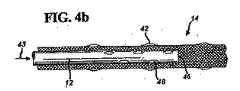
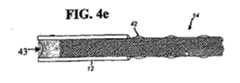
図4aに示す能動縫合糸の一部の長手方向断面図に示すように、内部通路12は編組縫合糸14内のコネクターと縫合針との間の位置で終わらせてもよい。この実施例では、治療液は図1におけるコネクター18を通り抜け、図4aの位置43に達する能動縫合糸の近位端内に流れ込み、更に内部通路12を通って通路46の開口端から編組縫合糸14の編目に流れ込む。治療液は編組縫合糸14の編目内に溜まり、最終的には表面42に達して周囲の組織に投与され得る。他の実施例では、治療液は内部通路の全長に沿って数箇所から放出されてもよい。図4bの長手方向断面図に示されるように、上述の位置43から治療液が流れ込む内部通路12は通路46の切断端からばかりでなく一つあるいは通路の全長に沿って複数の開口48を介して治療液を編組縫合糸に放出するようにしてもよい。通路の開口は実際上どのような幾何学形状でもよく、例えば円形、楕円形および長方形などだが、これらに限定されない。開口はそれぞれ異なる大きさでもよく、あるいは開口を一箇所に他の個所よりも稠密に設けて縫合糸に沿って場所毎にそれぞれ異なる治療薬送達速度としてもよい。他の実施例では、少なくとも一つの開口48を設けた内部通路を能動縫合糸の近位端から縫合針までの縫合糸の全長に亘って設けてもよい。図4cの能動縫合糸の一部の長手方向断面図に示すように、位置43で流れ込んだ治療液は一つあるいは能動縫合糸の全長に沿って複数の開口48から放出してもよい。図4bに示す実施例と同様に、開口はいろいろな幾何学形状を取り得、また縫合糸の全長に沿って様々な形態で分布できる。図4dに模式的に示す溝41のような内部通路に形成した切れ目のない開口を使って内部通路から編組縫合糸および創傷部位への治療液放出を容易にしてもよい。溝は、例えばチューブの全長に亘って直線状に設けてもよいし、あるいはチューブの全長に亘って螺旋状にしてもよい。この実施例では治療液は能動縫合糸の全長に沿っていずれかの位置から放出され得る。最後に、図4eに長手方向断面図で模式的に示すようにその全長の一部がチューブあるいはポリマー被覆で囲まれた編組縫合糸を使って治療液を図1aおよびbに示すコネクター18から編組縫合糸へ流すようにしてもよい。治療液導通部材の組合せを有する能動縫合糸を作成できることに注目するのも大切である。例えば、図1aに示すようにコネクター18と編組縫合糸の近位端との間の間隙を埋める治療液導通部材は微細径なチューブでよい。この微細径のチューブを、その全長に沿って多数の穿孔あるいは溝を有する編組縫合糸に埋め込まれている僅かに直径が大きいチューブに嵌合し取付けて能動縫合糸の内部通路を形成してもよい。 As shown in the longitudinal cross-sectional view of a portion of the active suture shown in FIG. 4a, the
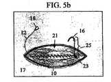
能動縫合糸はいろいろな形態で治療液を送達するように配置できる。最も簡単な方法では、創傷の隣接縫合(approximation)または縫合(closure)用の装置としてではなく、能動縫合糸を使って治療液を創傷部位に輸液できる。図5a、5bおよび5cに能動縫合糸10を輸液装置として配置する場合の一連の手順を模式的に示す。図5aに示すように、縫合針16を皮膚17および創傷に隣接する皮下組織に刺し込み、さらに切開傷部位21にまで届かせる。能動縫合糸は引っ張られて縫合針16により形成された孔を通り抜けて図5bに示すように切開傷内に配される。この段階では内部通路12の一部とコネクター18はまだ体外に留まっている。図5bに示すように、結び目を一つあるいは一連の結び目23を能動縫合糸の近位端に作って縫合糸を所定の位置に保持し装置が誤って抜出るのを防ぐことができる。縫合針を含む余分な縫合糸25は切取られ廃棄される。切開傷21は付属の縫合糸、ステープルあるいは皮膚癒合材を使って従来の手段で縫合される。図5cに示す最終段階では治療液が注射器22あるいは治療液溜めポンプ29を介して能動縫合糸に送られる。 Active sutures can be arranged to deliver treatment fluid in a variety of forms. In the simplest way, treatment fluid can be infused into the wound site using active sutures rather than as a device for adjacent wound closure or closure. 5a, 5b and 5c schematically show a series of procedures when the
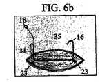
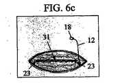
あるいは、能動縫合糸を創傷縫合用糸および輸液装置として配置してもよい。図6a、6b、6cおよび6dは図1aに示すタイプの能動縫合糸を創傷縫合および輸液用縫合糸として配置する場合の一連のステップを模式的に示す。第1のステップでは、一連の結び目23を能動縫合糸に内部通路12の遠位端と縫合針16の間で切開傷全体に亘って形成する。このステップは本質的に縫合糸を創傷縫合用部分33と輸液用部分31とに分割するものである。結び目と縫合針との間の縫合糸部分33を使って図6bに示すように連続縫合35で組織を縫い合わせる。縫合糸の輸液用部分31は図6cに示すように縫合35の線の上に配される。あるいは、輸液部分31は図6bで述べた創傷縫合ステップ時にその連続縫合の一つまたは複数の結節の下に固定してもよい。その後に切開傷を付属の縫合糸、ステープルおよび/または皮膚癒合材を使って従来の手段で縫合する。最終ステップ、図6dでは、治療液が注射器23あるいは治療液溜めポンプ29を介して装置に送られる。 Alternatively, active sutures may be placed as wound sutures and infusion devices. FIGS. 6a, 6b, 6c and 6d schematically illustrate a series of steps in placing an active suture of the type shown in FIG. 1a as a wound and infusion suture. In the first step, a series of
能動縫合糸は創傷縫合と治療液送達とのために様々な方法で配置できる。手術後の薬物送達を含む処置の場合には連続縫合と断続縫合の両方を用いることができる。皮内縫合のような他のタイプの連続縫合を用いてもよい。あるいは、一連の断続縫合により創傷を縫合してもよく、この創傷には一つまたは複数の縫目が能動縫合糸で形成される。標準の非能動縫合糸を能動縫合糸と共に使用して創傷縫合を促進できる。 Active sutures can be placed in a variety of ways for wound sutures and therapeutic fluid delivery. For procedures involving drug delivery after surgery, both continuous and interrupted sutures can be used. Other types of continuous sutures such as intradermal sutures may be used. Alternatively, the wound may be sutured by a series of interrupted sutures, where one or more stitches are formed with active sutures. Standard inactive sutures can be used with active sutures to facilitate wound suturing.
上述の配置方法に代わるものとして、能動縫合糸を切開傷部位に埋め込む代わりに切開傷周囲の組織に埋め込んでもよい。この場合能動縫合糸は手術前、手術中あるいは手術後の任意の時に図1aおよび1bに示す縫合針16を使って皮膚下に埋め込まれる。更に別な方法としては、その存在が周囲の組織に余計な傷を与えなければ、能動縫合糸を位置あるいは手術手技に関係なく治療液の送達を必要とするどのような組織にも埋め込むことができる。 As an alternative to the placement method described above, the active suture may be embedded in the tissue surrounding the incision wound instead of being embedded in the incision wound site. In this case, the active suture is implanted under the skin using the
前述したように創傷縫合後創傷に治療液を送達する方法に加えて、手術中、能動縫合糸の配置時に治療薬を送達する場合もあることに注目することは大切である。実際、或る場合には能動縫合糸にその配置前でも治療液を予め充填する、あるいは治療液で十分濡らすのが望ましい。さらに他の変形例では手術前または手術中にある種の治療液を送達した後、手術後に別の治療液を送達してもよい。 It is important to note that in addition to the method of delivering therapeutic fluid to the wound after wound closure as described above, the therapeutic agent may be delivered during surgery during placement of the active suture. In fact, in some cases it may be desirable to pre-fill the active suture with a treatment solution even prior to its placement or to wet it sufficiently with the treatment solution. In yet another variation, one treatment solution may be delivered before or during surgery, followed by another treatment solution after surgery.
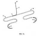
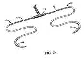
本発明はまた図7aおよび7bに模式的に示すように、二本の縫合針16と一つのコネクターを使って二腕縫合糸としても実施できる。これらの実施例において、外部治療液溜めから治療液を受けるようにしたコネクター18を能動縫合糸の中央部から延長するチューブ(図7a)あるいは能動縫合糸10自体(図7b)のいずれかに取付けて能動縫合糸の内部通路12と治療液を連通できるようにする。二腕縫合糸もいろいろな方法で配置できる。断続水平刺し縫い縫合に用いた二腕縫合糸10を模式的に図8aおよび8bに示す。 The present invention can also be implemented as a two-arm suture using two
治療液溜めポンプあるいは他の治療液供給手段を能動縫合糸に接続した場合には、能動縫合糸からの治療液放出速度は主として三つの要素、即ち、治療液の粘度、加えられた圧力および通路の設計、により決まる。円筒形パイプを流れる液流についてのハーゲン−ポアズイユ関係式を使って図2aおよび4aに示す通路を有する能動縫合糸を流れる治療液の体積流量を近似できる。
体積流量=(π×付与圧力×半径)/(8×液粘度×通路長)
ここで付与圧力は治療液源により加えられる圧力、半径は治療液が流れる内部通路の有効半径、通路長はコネクターから内部通路に設けられた開口の位置までの内部通路の有効長である。もし点滴静注(IV)を行うとすると、付与圧力は次式のように創傷部位からの点滴器(IV)の高さにより決めることができる。
付与圧力=液密度×重力定数×点滴器(IV)の患者からの高さWhen a therapeutic fluid reservoir pump or other therapeutic fluid supply means is connected to the active suture, the therapeutic fluid release rate from the active suture is primarily divided into three factors: therapeutic fluid viscosity, applied pressure and passageway. Depends on the design. The Hagen-Poiseuille relation for fluid flow through a cylindrical pipe can be used to approximate the volumetric flow rate of treatment fluid flowing through an active suture having the passage shown in FIGS. 2a and 4a.
Volume flow rate = (π × applied pressure × radius) / (8 × liquid viscosity × passage length)
Here, the applied pressure is the pressure applied by the treatment liquid source, the radius is the effective radius of the internal passage through which the treatment liquid flows, and the passage length is the effective length of the internal passage from the connector to the position of the opening provided in the internal passage. If intravenous drip (IV) is performed, the applied pressure can be determined by the height of the dropper (IV) from the wound site as follows:
Applied pressure = liquid density x gravity constant x height of dropper (IV) from patient
例えば、点滴輸液袋(IV bag)を創傷部位からほぼ1メートルの高さに置くと、ほぼ1.01×104パスカル(ほぼ0.1気圧(atm))の付与圧力で薬液が編組縫合糸に流れ込む。もし図1aにおけるエラストマーの膨張可能な治療液溜め34を使うと、薬液を編組縫合糸に流し込む付与圧力は1.01×105パスカル(1気圧)にもなり得る。最後に、通常IV供給システムと組み合わせて使われる流体ポンプは調節可能でいろいろな圧力および流量で治療液を編組縫合糸に供給できる。図9に、内径が夫々50、75および100μmであり、また編組縫合糸内のコネクターから0.2メートル未満の位置で終端している管腔を有する、図1a、2aおよび4aに示す実施例と同様な管状の内部通路を有する能動縫合糸における標準温度と標準圧力(STP)条件下での水の体積流量をハーゲン−ポアズイユ関係式を使って算定した値を示す。図9において実線は1.01×10⁴パスカル(0.1atm)の与圧での送達速度範囲を示す。エラストマーの膨張可能な液溜めポンプは通常1.01×10⁴〜1.01×10⁵パスカル(0.1〜1atm)程度の圧力を加える。図9における破線はほぼ1.01×10⁵パスカル(ほぼ1atm)の与圧での送達速度範囲を示す。管腔径と内部通路の長さがともに治療液の流速に大きく影響し、管腔の直径が小さくなるにつれ、また通路が長くなるにつれて速達速度が減少する。図9に示す値は結び目がない場合の薬物送達量の推定値であることに留意するのは大切である。縫合糸に結び目があると内部通路が曲がりくねって送達量が減少することになる。 For example, when an IV bag is placed at a height of approximately 1 meter from the wound site, the drug solution is applied to the braided suture with an applied pressure of approximately 1.01 × 10 4 Pascal (approximately 0.1 atm (atm)). Flows in. If the elastomeric
創傷縫合を容易にするため能動縫合糸に結び目を作るのが望ましい場合がある。多くの場合、図6a、6b、6cおよび6dに順次示した処置のような創傷縫合処置では能動縫合糸の内部通路を含む部分に結び目を作る必要がない。このように、この装置は創傷縫合用縫合糸としてまた治療液送達量の制御に悪影響を与えることなく治療液を注入する装置としても使用できる。しかし、能動縫合糸の内部通路を含む部分に作った結び目を使う必要がある処置の場合は、治療液が能動縫合糸の結び目がある場所を流れ通るために内部通路はそのままにしておかねばならない。もし被覆繊維トウあるいは被覆編組縫合糸の編目を図3aおよび3bに模式的に示したように能動縫合糸の内部通路として使うのならば、編目はそのままにしておく。しかし、もし被覆繊維トウあるいは被覆編組縫合糸の代わりにチューブを使って内部通路を形成するならば、結び目を作ると管腔が潰れて閉止することがある。管腔の閉止を防ぐためには管壁が十分厚いチューブを使う必要がある。結び目内部の管腔が潰れる可能性に影響がある変数には内部通路が埋め込まれている編組縫合糸の厚さと、チューブの剛性、チューブの強度および結び目を作る際に加えられた全張力が含まれる。こま結びあるいは外科結びのような外科的に許容できる結び目を作る能動縫合糸については、好ましくはチューブの内径(I.D.)に対する外径(O.D.)の割合が1.7を超え、現在縫合糸に使われているポリマー材の多くについてはI.D.に対するO.D.の割合が2.0を超えるのがより好ましい。 It may be desirable to tie a knot on the active suture to facilitate wound suturing. In many cases, wound suture procedures, such as those shown sequentially in FIGS. 6a, 6b, 6c and 6d, do not require knots in the portion containing the internal passage of the active suture. Thus, this device can be used as a suture for wound sutures and as a device for injecting a treatment solution without adversely affecting the control of the treatment solution delivery amount. However, for procedures that require the use of a knot made in the portion of the active suture that contains the internal passage, the internal passage must remain in order for the therapeutic fluid to flow through the active suture knot. . If the stitch of the coated fiber tow or the coated braided suture is used as an internal passage for the active suture as shown schematically in FIGS. 3a and 3b, the stitch is left as it is. However, if a tube is used instead of a coated fiber tow or a coated braided suture to form the internal passage, the knot may smash and close. To prevent the lumen from closing, it is necessary to use a tube with a sufficiently thick tube wall. Variables that affect the likelihood that the lumen inside the knot will collapse include the thickness of the braided suture in which the internal passage is embedded, the stiffness of the tube, the strength of the tube, and the total tension applied when making the knot. It is. For active sutures that make surgically acceptable knots such as top knots or surgical knots, the ratio of the outer diameter (OD) to the inner diameter (ID) of the tube is preferably greater than 1.7. For many of the polymer materials currently used for sutures, the ratio of OD to ID is more preferably 2.0.
能動縫合糸は例えば、能動縫合糸の内部通路として使う治療液導通部材の製作、治療液導通部材を編組縫合糸に組み込んで能動縫合糸を形成する工程、治療液導通部材あるいは能動縫合糸の近位端のコネクターへの取付け、そして能動縫合糸の遠位端の縫合針への取付けを含む工程により製造できる。寸法および形状が例えば図1aと1bに示す能動縫合糸に適合する細いチューブは従来のポリマー押出技術で製造可能である。これらのチューブは直接適当な寸法に押出してもよいしあるいは好ましい寸法よりも大きく押出してから従来のファイバー延伸技術で小さくしてもよい。図3aおよび3bに示すように被覆繊維トウあるいは被覆縫合糸を選んで能動縫合糸の治療液導通部材として使うのならば、能動縫合糸製造の第1ステップには編組縫合糸あるいは繊維トウに切れ目のないポリマー被覆を施す工程を含むことになる。ポリマー押出し機は、繊維トウまたは編組縫合糸が通り抜けできる金型を備え、それらが金型を通り抜けるにつれてポリマーフィルムに包み込まれる。このプロセスは針金を絶縁ポリマーでコーティングするためのワイヤーコーティングと同じ様なもので当該分野では周知である。チューブ、被覆繊維トウあるいは被覆編組縫合糸は製造後に処理して図4b、cおよびdに示すように孔または溝を形成できる。治療液導通部材のこれらの開口は機械的な方法で形成してもよいしあるいは精密レーザー機器で形成してもよい。幾つかの実施例では治療液導通部材の全長に亘って一連の開口を形成する工程は任意であることに注目するのは大切なことである。事実、図4aに示す実施例では治療液が単に通路の切断端から放出されるだけで、治療液導通部材の全長に亘って開口を形成する必要はない。チューブ、被覆繊維トウあるいは被覆編組縫合糸を形成した後、他の繊維の撚糸と編み合わせて図4a、4b、4cまたは4dに示す能動縫合糸を形成できる。これは、チューブ、被覆繊維トウあるいは被覆編組縫合糸を編組縫合糸の芯繊維に並んで通し、それにより編組縫合糸の織込まれた繊維でチューブ、被覆繊維トウあるいは被覆編組縫合糸を取囲むようにして行うことができる。チューブ、被覆繊維トウあるいは被覆編組縫合糸を編組縫合糸の芯繊維に巻きつけるブレーディング方法も考えられる。ブレーディング後、チューブ、被覆繊維トウあるいは被覆編組縫合糸の一部を取り除いて図4aおよび4bに示す実施例を作っても良い。これは、チューブ、被覆繊維トウあるいは被覆編組縫合糸を精密針ホールダーで掴みそして編組縫合糸を通してチューブ、被覆繊維トウあるいは被覆編組縫合糸の一部だけが編組縫合糸の内部に残るまで引き抜いて能動縫合糸を形成することで行える。あるいは、外径が編組縫合糸のそれよりも小さいポリマーチューブを編組縫合糸の近位端に圧入してもよい。このようにして、チューブの最も長くて数センチの部分を図4aに示すように編組縫合糸内にそれと同軸に配置でき、一方同じチューブの一部が図1aに示すように編組縫合糸の近位端から突出する。チューブが編組縫合糸から抜け落ちるのを防ぐため、少量の接着剤を編組縫合糸の近位端部に塗ってチューブを編組縫合糸の多数の繊維に固着してもよい。熱接着によりあるいは収縮性のあるポリマースリーブを使用してチューブを編組縫合糸の近位端に取付ける別の方法も考えられる。 The active suture includes, for example, the production of a treatment fluid conducting member used as an internal passage of the active suture, the process of incorporating the treatment fluid conducting member into the braided suture to form an active suture, the proximity of the treatment fluid conducting member or the active suture It can be manufactured by a process that includes attaching the distal end to the connector and attaching the active suture distal end to the suture needle. Thin tubes that are compatible in size and shape with, for example, the active suture shown in FIGS. 1a and 1b can be made by conventional polymer extrusion techniques. These tubes may be extruded directly to the appropriate dimensions, or extruded larger than the preferred dimensions and then reduced by conventional fiber drawing techniques. As shown in FIGS. 3a and 3b, if a coated fiber tow or coated suture is selected and used as a therapeutic fluid conducting member for an active suture, the first step in the production of active suture is a break in the braided suture or fiber tow. A step of applying a polymer coating free of The polymer extruder includes a mold through which fiber tows or braided sutures can pass and is wrapped in a polymer film as they pass through the mold. This process is similar to wire coating for coating a wire with an insulating polymer and is well known in the art. Tubes, coated fiber tows or coated braided sutures can be processed after manufacture to form holes or grooves as shown in FIGS. 4b, c and d. These openings of the treatment liquid conducting member may be formed by a mechanical method or may be formed by a precision laser device. It is important to note that in some embodiments, the process of forming a series of openings over the entire length of the treatment fluid conducting member is optional. In fact, in the embodiment shown in FIG. 4a, the treatment liquid is simply discharged from the cut end of the passage, and there is no need to form an opening over the entire length of the treatment liquid conducting member. After forming the tube, coated fiber tow or coated braided suture, the active suture shown in FIGS. 4a, 4b, 4c or 4d can be formed by interweaving with other fiber strands. This involves passing a tube, coated fiber tow or coated braided suture side by side with the core fiber of the braided suture, thereby surrounding the tube, coated fiber tow or coated braided suture with the woven fibers of the braided suture. Can be performed. A braiding method is also conceivable in which a tube, a coated fiber tow or a coated braided suture is wound around the core fiber of the braided suture. After braiding, the embodiment shown in FIGS. 4a and 4b may be made by removing a portion of the tube, coated fiber tow or coated braided suture. This is accomplished by grabbing the tube, coated fiber tow or coated braided suture with a precision needle holder and pulling through the braided suture until only a portion of the tube, coated fiber tow or coated braided suture remains inside the braided suture. This can be done by forming a suture. Alternatively, a polymer tube having an outer diameter smaller than that of the braided suture may be press fit into the proximal end of the braided suture. In this way, the longest and several centimeters of the tube can be placed coaxially with it in the braided suture as shown in FIG. 4a, while a portion of the same tube is close to the braided suture as shown in FIG. 1a. Projects from the edge. To prevent the tube from falling out of the braided suture, a small amount of adhesive may be applied to the proximal end of the braided suture to secure the tube to the multiple fibers of the braided suture. Other methods of attaching the tube to the proximal end of the braided suture by thermal bonding or using a shrinkable polymer sleeve are also contemplated.
能動縫合糸の構成成分は生体吸収性および非吸収性材料から作ることができる。本発明による縫合糸、チューブ、被覆繊維トウ、被覆編組縫合糸、接着剤およびコネクターは縫合糸の製造に通常使われるポリマー、例えば、ポリプロピレン、ポリエチレン、ポリアミド、ポリエチレンテレフタレート(PET)、ポリテトラフルオロエチレン(PTFE)、絹、ポリカプロラクトン、ポリジオキサノン、ポリグリコライド、ポリラクチド、あるいはポリカプロラクトン、ポリジオキサノン、ポリグリコライドまたはポリラクチドの配合物から製造できるが、これらの材料に限定されるものではない。さらに、コネクターは必ずしも患者の体内に埋め込まれないので、より多様なエンジニアリングポリマー材、例えば、溶媒を含まないポリ塩化ビニール、ポリウレタン、ポリエステル、ポリカーボネイト、ポリオレフィンおよびポリアミドなどで作れるが、これらの材料に限定されるものではない。 The active suture components can be made from bioabsorbable and non-absorbable materials. The sutures, tubes, coated fiber tows, coated braided sutures, adhesives and connectors according to the present invention are polymers commonly used in the manufacture of sutures such as polypropylene, polyethylene, polyamide, polyethylene terephthalate (PET), polytetrafluoroethylene. (PTFE), silk, polycaprolactone, polydioxanone, polyglycolide, polylactide, or blends of polycaprolactone, polydioxanone, polyglycolide or polylactide, but are not limited to these materials. In addition, since the connector is not necessarily implanted in the patient's body, it can be made from a wider variety of engineering polymer materials such as, but not limited to, polyvinyl chloride, polyurethane, polyester, polycarbonate, polyolefin and polyamide without solvent. Is not to be done.
上述のどの縫合糸にも使用できる流体は治療または生体活性薬あるいは液であり、以下に列挙するものが含まれるがそれらに限定されるものではない。即ち、2,4,4'−トリクロロ−2'ヒドロキシジフェニールエーテル、塩化ベンザルコニウム、スルファジアジン銀、ポビドンヨード、トリクロサン、ゲンタマイシン(gentamiacin)などの抗菌薬または抗生物質:セレコクシブ、ロフェコキシブ、アスピリン、サリチル酸、アセトミノフェン、インドメタシン(indomethicin)、スリンダク、トルメチン、ケトロラック、メフェナム酸(mefanamic acid)、イブプロフェン、ナプロキセン、フェニルブタゾン、スルフィンピラゾン、アパゾン、ピロキシカムなどのステロイド系あるいは非ステロイド系の抗炎症薬:チャネル拮抗剤(channel blocking agent)、マルカイン、リドカイン、ブピバカイン、メピバカイン、プロカイン、クロロプロカイン、ロピバカイン、テトラカイン、プリロカイン、レボブピビカイン(levobupivicaine)および局所麻酔薬にエピネフリンを配合したものなどの麻酔薬:モルヒネ、フェタニ−ル、コデインなどのオピオイド系鎮痛薬:ラパマイシンなどの抗増殖薬(anti-proliferatives):PDGFなどの成長因子:創傷治癒用の酸素を多量に含んだ薬液、ヒアルロン酸(hylauronic acid)、血管新成促進薬(angio-genesis promoting agent)、凝血促進因子(pro-coagulation factor)、抗凝血因子(anti-coagulation factor)、走化性薬(chemotactic agent)、アポトーシス促進薬(agents to promote apoptosis)、免疫調整薬、有糸分裂増殖薬(mitogenic agent)、ジフェンヒドラミン、クロルフェニラミン、ピリラミン、プロメタジン、メクリジン、テルフェナジン、アステミゾ−ル、フェキソフェニジン(fexofenidine)、ロラタジン(loratidine)、オーロチオグルコース、オーラノフィン、コルチゾール(ヒドロコルチゾン)、コルチゾン、フルドロコルチゾン、プレドニゾン、プレドニゾロン、6α−メチールプレドニゾン(6α-methylprednisone)、トリアムシノロン、ベタメタゾンおよびデキサメタゾンなどの瘢痕治療薬(scar treatment agent):トロンビン、トラネキサム酸、エピネフリンなどの止血薬:さらに、抗血栓症薬、溶液内の幹細胞などの生物学的薬剤、プロテインおよび酵素も能動縫合糸を通して送達できる。創傷部位の潅注も能動縫合糸をつうじて行える。 Fluids that can be used in any of the above sutures are therapeutic or bioactive agents or fluids, including but not limited to those listed below. That is, an antibacterial or antibiotic such as 2,4,4′-trichloro-2′hydroxydiphenyl ether, benzalkonium chloride, silver sulfadiazine, povidone iodine, triclosan, gentamiacin: celecoxib, rofecoxib, aspirin, salicylic acid, Steroidal or non-steroidal anti-inflammatory drugs such as acetminophen, indomethacin, sulindac, tolmetin, ketorolac, mefanamic acid, ibuprofen, naproxen, phenylbutazone, sulfinpyrazone, apazone, piroxicam: Channel blocking agent, marcaine, lidocaine, bupivacaine, mepivacaine, procaine, chloroprocaine, ropivacaine, tetracaine, prilocaine, levobupiviine (levobupivi) caine) and anesthetics such as local anesthetics containing epinephrine: morphine, fetanyl, codeine and other opioid analgesics: rapamycin and other anti-proliferatives: PDGF and other growth factors: wounds Medicinal solution containing a large amount of oxygen for healing, hylauronic acid, angio-genesis promoting agent, pro-coagulation factor, anti-coagulation factor ), Chemotactic agents, agents to promote apoptosis, immunomodulators, mitogenic agents, diphenhydramine, chlorpheniramine, pyrilamine, promethazine, meclizine, terfenadine, astemizo -Ru, fexofenidine, loratidine, aurothioglucose, auranofin, cortisol (hydrocord) Zon), cortisone, fludrocortisone, prednisone, prednisolone, 6α-methylprednisone, 6 am-methylprednisone, triamcinolone, betamethasone and dexamethasone scar treatment agents: hemostatic agents such as thrombin, tranexamic acid, epinephrine In addition, antithrombotic drugs, biological agents such as stem cells in solution, proteins and enzymes can also be delivered through active sutures. Irrigation of the wound site can also be performed through active sutures.
能動縫合糸を使用する別の方法および目的は創傷部位から体液を排出することである。能動縫合糸の近位端に繋いだ管をとおして減圧すると、体液が創傷部位から直接引き出されるので、創傷潅注処置のために体液を除去する新規な手段となる。あるいは、体液を創傷から引き出して分析を行い、創傷の状態を判断してもよい。例えば、採取した体液の化学的サインから創傷治癒の経過が分かり、あるいは微生物を検知すれば創傷内の感染についての早期診断が可能である。 Another method and purpose of using active sutures is to drain fluid from the wound site. Depressurization through a tube connected to the proximal end of the active suture draws fluid directly from the wound site, providing a new means of removing fluid for wound irrigation procedures. Alternatively, body fluid may be drawn from the wound and analyzed to determine the state of the wound. For example, the progress of wound healing can be known from the chemical signature of the collected body fluid, or early diagnosis of infection in the wound can be made by detecting microorganisms.
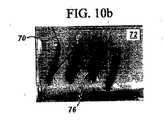
例1
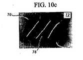
能動縫合糸の治療液を創傷周囲の組織に送達する能力を証明するために、図1bと4aに示すように編組縫合糸内で終端しているポリプロピレンチューブを含むPET編組縫合糸で生体外(in vitro)実験を行った。実験では能動縫合糸をゼラチンに多数回通した後に、青い顔料を含んだ水を能動縫合糸のゼラチンに埋め込んだ部分に流すIV送達システムに接続した。一連の時間経過後の映像を図10a、10bおよび10cに示す。実験開始時にとった図10aの映像はゼラチン72に埋め込まれた能動縫合糸70を示している。能動縫合糸74上の黒いところは内部通路の終端位置を示している。時間が経つにつれて顔料76が図10bに示すように能動縫合糸の周囲に広がっていっている。最終的には図10cに示すように液体は広がって創傷周囲の全領域を包み込んでしまう。Example 1
To demonstrate the ability to deliver active suture treatment fluid to the tissue surrounding the wound, in vitro with a PET braided suture comprising a polypropylene tube terminating in the braided suture as shown in FIGS. 1b and 4a ( In vitro experiments were performed. In the experiment, the active suture was passed through the gelatin multiple times and then connected to an IV delivery system in which water containing blue pigment was passed through the portion of the active suture embedded in the gelatin. Images after a series of times are shown in FIGS. 10a, 10b and 10c. The image in FIG. 10 a taken at the start of the experiment shows the
例2
能動縫合糸を創傷縫合と輸液の両方に使うのならば、内部通路を能動縫合糸に組込むことで縫合糸の引張り強さと結び目引張り強さを標準許容レベル未満に落としてはならない。ポリプロピレンチューブを芯繊維と並んで埋め込んだ米国薬局法(USP)規格サイズ0および2のPET編組縫合糸の結び目引張り強さを米国薬局法(USP)規格23により測定した。サイズ0の縫合糸には、外径がほぼ130マイクロメートルで内径が〜75マイクロメートルのチューブを内蔵し、サイズ2の縫合糸には外径がほぼ230マイクロメートルで内径が〜135マイクロメートルのチューブを内蔵していた。各テスト毎に少なくとも10サンプルをUSP仕様でテストした。芯にポリプロピレンチューブを内蔵したPET編組縫合糸の性能は、サイズ2および0の縫合糸の平均結び目引張り強さの値はそれぞれ6.12キログラム(13.5ポンド)と3.49キログラム(7.7ポンド)でUSP規格で設定された最低性能条件を簡単にクリアした。Example 2
If active sutures are used for both wound sutures and infusions, the suture and knot tensile strengths should not be reduced below standard acceptable levels by incorporating the internal passage into the active suture. The knot tensile strength of US pharmacy method (USP) standard size 0 and 2 PET braided sutures in which a polypropylene tube was embedded alongside the core fiber was measured according to US pharmacy method (USP) standard 23. The size 0 suture has a tube with an outer diameter of approximately 130 micrometers and an inner diameter of ~ 75 micrometers, while the size 2 suture has an outer diameter of approximately 230 micrometers and an inner diameter of ~ 135 micrometers. It had a built-in tube. At least 10 samples were tested to USP specifications for each test. The performance of PET braided sutures with a polypropylene tube in the core is that the average knot tensile strength values for size 2 and 0 sutures are 6.12 kilograms (13.5 pounds) and 3.49 kilograms (7. The minimum performance condition set by the USP standard at 7 pounds) was easily cleared.
例3
実験データによると、それぞれ外径が1.27×10-4メートル(.005インチ)から2.54×10-4メートル(.010インチ)、ヤング率0.1から3GPaの範囲、外径(O.D.s)が内径(I.D.s)の1.7倍未満の大きさのポリプロピレン製の押出しポリマーチューブは、それらが埋め込まれている編組縫合糸を外科処置で普通に使われるのと同じ様な形状のこま結びに結ぶと座屈して潰れてしまうことが示されている。ヤング率が0.1から3GPaの範囲で、内径に対する外径比が2.3を超えるポリエチレンとポリテトラフルオロエチレンを含むポリマーチューブを使って同様の実験を行ったところ、能動縫合糸のこま結び内部は完全に潰れず液体が結び目の部分を通って移送されうる。結び目を作る能動縫合糸では、内径に対する外径比が1.7を超えるのが好ましい。更に好ましくは、内径に対する外径比が2.0を超えることである。これらの実験において、チューブはポリエチレンテレフタレート(PET)ファイバー製でUSPサイズが2−0から5の間の編組縫合糸に埋め込んだ。結び目内の管腔が潰れてしまう可能性に影響する他の変数は内部通路が埋め込まれた編組縫合糸の厚さ、治療液導通チューブの強度および結び目を作るときに加えられた全張力である。Example 3
According to experimental data, the outer diameter ranges from 1.27 × 10 −4 meters (.005 inches) to 2.54 × 10 −4 meters (.010 inches), Young's modulus ranges from 0.1 to 3 GPa, Polypropylene extruded polymer tubes with ODs less than 1.7 times the inner diameter (IDs) are shaped like the ones commonly used in surgical procedures with braided sutures in which they are embedded. It is shown that buckling and crushing when tied to a knot. A similar experiment was conducted using a polymer tube containing polyethylene and polytetrafluoroethylene having a Young's modulus in the range of 0.1 to 3 GPa and an outer diameter ratio of more than 2.3. The interior does not collapse completely and liquid can be transferred through the knot. For active sutures that tie knots, the ratio of outer diameter to inner diameter is preferably greater than 1.7. More preferably, the outer diameter ratio with respect to the inner diameter exceeds 2.0. In these experiments, the tubes were embedded in braided sutures made of polyethylene terephthalate (PET) fibers and USP sizes between 2-0 and 5. Other variables that affect the likelihood that the lumen in the knot will collapse are the thickness of the braided suture in which the internal passage is embedded, the strength of the treatment fluid conducting tube, and the total tension applied when making the knot. .
〔実施の態様〕
(1) 能動縫合糸において、
近位端、遠位端および外径を有する編組縫合糸と、
前記編組縫合糸の少なくとも一部と同軸で、近位端、遠位端、および前記編組縫合糸の前記外径より小さい直径を有する、少なくとも一つの通路と、
を具備し、
前記少なくとも一つの通路の前記遠位端が前記編組縫合糸の前記近位端および遠位端の間に配されている、
能動縫合糸。
(2) 能動縫合糸において、
外径を有する編組縫合糸と、
前記編組縫合糸の少なくとも一部と同軸で、前記編組縫合糸の前記外径よりも小さい外径、内径、および一つあるいは複数の開口を有する、チューブと、
を具備し、
前記チューブの前記内径に対する前記チューブの前記外径の比が1.7を超える、
能動縫合糸。
(3) 能動縫合糸において、
第1編組縫合糸であって、外径と、前記第1編組縫合糸に埋め込まれ、かつ前記第1編組縫合糸の少なくとも一部と同軸の被覆繊維トウあるいは被覆編組縫合糸とを有し、前記被覆繊維トウあるいは被覆編組縫合糸は前記第1編組縫合糸の前記外径よりも小さい外径および一つあるいは複数の開口を有する、第1編組縫合糸、を具備する、能動縫合糸。
(4) 能動縫合糸・針アセンブリにおいて、
能動縫合糸であって、
近位端、遠位端および外径を有する編組縫合糸と、
近位端および遠位端を有し前記編組縫合糸の少なくとも一部と同軸のチューブと、を具備し、前記チューブは前記編組縫合糸の前記外径よりも小さい外径および一つあるいは複数の開口を有する、能動縫合糸と、
前記編組縫合糸の前記遠位端に取付けた外科用針と、
前記チューブの前記近位端に取付けたコネクターと、
を具備する、能動縫合糸・針アセンブリ。
(5) 能動縫合糸・針アセンブリにおいて、
能動縫合糸であって、
近位端、遠位端および外径を有する編組縫合糸と、
近位端および遠位端を有し前記編組縫合糸の少なくとも一部と同軸のチューブと、を具備し、前記チューブは前記編組縫合糸の前記外径よりも大きい内径を有する、能動縫合糸と、
前記編組縫合糸の前記遠位端に取付けた外科用針と、
前記チューブの前記近位端に取付けたコネクターと、
を具備する、能動縫合糸・針アセンブリ。
(6) 実施態様1に記載の能動縫合糸において、
前記少なくとも一つの通路はチューブの管腔である、能動縫合糸。
(7) 実施態様1に記載の能動縫合糸において、
前記少なくとも一つの通路は被覆繊維トウあるいは被覆編組縫合糸内にある、能動縫合糸。
(8) 実施態様6に記載の能動縫合糸において、
前記チューブは前記管腔を前記チューブの外表面に連通する一つあるいは複数の孔を有する、能動縫合糸。
(9) 実施態様7に記載の能動縫合糸において、
前記被覆繊維トウあるいは被覆編組縫合糸は前記少なくとも一つの通路を前記被覆繊維トウあるいは被覆編組縫合糸の外表面に連通する一つあるいは複数の孔を有する、能動縫合糸。
(10) 実施態様1ないし3のいずれか1項に記載の能動縫合糸において、
さらに、前記少なくとも一つの通路の前記近位端か、前記チューブの一端か、または前記被覆繊維トウあるいは被覆編組縫合糸の一端に配置されたコネクターを具備し、前記コネクターは皮下注射針、静脈内輸液システムあるいは液体ポンプに直接または間接的に取り付け可能である、能動縫合糸。
(11) 実施態様1ないし3のいずれか1項に記載の能動縫合糸において、
さらに、前記少なくとも一つの通路の前記近位端か、前記チューブの一端か、または前記被覆繊維トウあるいは被覆編組縫合糸の一端に配置された膨張可能な液溜めを具備する、能動縫合糸。
(12) 実施態様11に記載の能動縫合糸において、
さらに、前記膨張可能な液溜めの近位にあるコネクターを具備する、能動縫合糸。
(13) 縫合糸・針アセンブリを使用する創傷縫合方法において、
前記縫合糸・針アセンブリは
編組縫合糸であって、近位端、遠位端、外径、および前記編組縫合糸の少なくとも一部と同軸な少なくとも一つの通路を有し、前記通路は近位端、遠位端、および前記編組縫合糸の前記外径よりも小さい直径を有し、前記少なくとも一つの通路の前記遠位端は前記編組縫合糸の前記近位端および遠位端の間に配置されている、編組縫合糸と、
前記編組縫合糸の前記遠位端に取付けた外科用針と、
前記少なくとも一つの通路の前記近位端に取付けたコネクターと、を具備する、縫合糸・針アセンブリで、
前記方法は
前記少なくとも一つの通路の前記近位端に取付けた前記コネクターを液体を収納している液溜めに直接あるいは間接的に接続するステップと、
前記液体に圧力を加えて前記液体を前記コネクターおよび前記少なくとも一つの通路に注入するステップと、
前記縫合糸・針アセンブリを創傷の周りの組織中に埋め込み、前記少なくとも一つの通路の前記遠位端を前記創傷にあるいは前記創傷の近くに保持するステップと、
前記創傷を前記編組縫合糸で縫合するステップと、
を含む、方法。
(14) 縫合糸・針アセンブリを使用する創傷縫合方法において、
前記縫合糸・針アセンブリは
近位端、遠位端および外径を有する編組縫合糸と、
前記編組縫合糸の少なくとも一部と同軸であるチューブであって、近位端、遠位端、前記編組縫合糸の前記外径よりも小さい外径、内径、および一つあるいは複数の開口を有し、前記チューブの前記内径に対する前記チューブの前記外径の比が1.7を超える、チューブと、
前記編組縫合糸の前記遠位端に取付けた外科用針と、
前記チューブの前記近位端に取付けたコネクターと、
を具備する、縫合糸・針アセンブリで、
前記方法は
前記チューブの前記近位端に取付けたコネクターを液体を収納している液溜めに直接あるいは間接的に接続するステップと、
前記液体に圧力を加えて前記液体を前記コネクターと前記チューブとに注入するステップと、
縫合糸・針アセンブリを創傷の周りの組織中に埋め込み、前記一つあるいは複数の開口を前記創傷にあるいは前記創傷の近くに保持するステップと、
前記創傷を前記編組縫合糸で縫合するステップと、
を含む、方法。
(15) 縫合糸・針アセンブリを使用する創傷縫合方法において、
前記縫合糸・針アセンブリは
第1編組縫合糸であって、外径と、被覆繊維トウあるいは被覆編組縫合糸とを有し、前記被覆繊維トウあるいは被覆編組縫合糸は前記第1編組縫合糸の少なくとも一部と同軸で、かつ前記被覆繊維トウあるいは被覆編組縫合糸は近位端、遠位端、前記第1編組縫合糸の前記外径よりも小さい外径、および一つあるいは複数の開口を有する、第1編組縫合糸と、
前記編組縫合糸の前記遠位端に取付けた外科用針と、
前記チューブの前記近位端に取付けたコネクターと、を具備する、
縫合糸・針アセンブリで、
前記方法は
前記被覆繊維トウあるいは被覆編組縫合糸の前記近位端に取付けた前記コネクターを液体を収納している液溜めに直接あるいは間接的に接続するステップと、
前記液体に圧力を加えて前記液体を前記コネクターと前記被覆繊維トウあるいは被覆編組縫合糸とに注入するステップと、
前記縫合糸・針アセンブリを創傷の周りの組織中に埋め込み、前記被覆繊維トウあるいは被覆編組縫合糸の前記一つあるいは複数の開口を前記創傷にあるいは前記創傷の近くに保持するステップと、
前記創傷を前記第1編組縫合糸で縫合するステップと、
を含む、方法。
(16) 縫合糸・針アセンブリを使用する創傷縫合方法において、
前記縫合糸・針アセンブリは
近位端、遠位端および外径を有する編組縫合糸と、
前記編組縫合糸の少なくとも一部と同軸のチューブであって、近位端、遠位端、および前記編組縫合糸の前記外径よりも大きい内径を有し、前記チューブの前記遠位端は前記編組縫合糸の前記近位端および遠位端の間に配置されている、チューブと、
前記編組縫合糸の前記遠位端に取付けた外科用針と、
前記チューブの前記近位端に取付けたコネクターと、
を具備する、縫合糸・針アセンブリで、
前記方法は
前記チューブの前記近位端に取付けた前記コネクターを液体を収納している液溜めに直接あるいは間接的に接続するステップと、
前記液体に圧力を加えて前記液体を前記コネクターと前記チューブとに注入するステップと、
前記縫合糸・針アセンブリを創傷の周りの組織中に埋め込み、前記チューブの前記遠位端を前記創傷にあるいは前記創傷の近くに保持するステップと、
前記創傷を前記編組縫合糸で縫合するステップと、
を含む、方法。
(17) 液体を創傷に投与する方法において、
能動縫合糸・針アセンブリであって、
能動縫合糸であって、
近位端、遠位端および外径を有する編組縫合糸と、
(i)前記編組縫合糸の少なくとも一部と同軸で、かつ近位端、遠位端、および前記編組縫合糸の前記外径よりも小さい直径を有する少なくとも一つの通路であって、前記少なくとも一つの通路の前記遠位端は開口を有し、かつ前記編組縫合糸の前記近位端および遠位端の間に配置される、少なくとも一つの通路か、
(ii)前記編組縫合糸の少なくとも一部と同軸のチューブであって、前記チューブは近位端、遠位端、前記編組縫合糸の前記外径よりも小さい外径、および一つあるいは複数の開口を有する、チューブか、
(iii)前記編組縫合糸の少なくとも一部と同軸の被覆繊維トウあるいは被覆編組縫合糸であって、前記被覆繊維トウあるいは被覆編組縫合糸は近位端、遠位端、前記編組縫合糸の前記外径よりも小さい外径、および一つあるいは複数の開口を有する、被覆繊維トウあるいは被覆編組縫合糸か、または、
(iv)前記編組縫合糸の少なくとも一部と同軸のチューブであって、前記チューブは近位端、遠位端、および前記編組縫合糸の前記外径よりも大きい内径を有し、前記チューブの前記遠位端は前記編組縫合糸の前記近位端および遠位端との間に配置される、チューブと、を具備する、能動縫合糸と、
前記編組縫合糸の前記遠位端に取付けた外科用針と、
前記少なくとも一つの通路、前記チューブ、または前記被覆繊維トウあるいは被覆編組縫合糸の前記近位端に取付けたコネクターと、
を具備する、能動縫合糸・針アセンブリで、
前記方法は
前記能動縫合糸・針アセンブリを創傷の周りの組織中に埋め込み、前記少なくとも一つの通路あるいは前記(iv)のチューブの前記遠位端、または前記(ii)のチューブの一つあるいは複数の開口、または前記被覆繊維トウあるいは被覆編組縫合糸の前記一つあるいは複数の開口を前記創傷に、あるいは前記創傷に近接して、または前記創傷中に配置するステップと、
前記コネクターを液体を収納している液溜めに直接あるいは間接的に接続するステップと、
前記液体に圧力を加えて前記液体を前記コネクターと前記少なくとも一つの通路、前記チューブ、または前記被覆繊維トウあるいは被覆編組縫合糸とに注入するステップと、
前記液体を前記少なくとも一つの通路もしくは前記(iv)のチューブの前記遠位端の開口、または前記(ii)のチューブの前記一つあるいは複数の開口、または前記被覆繊維トウもしくは被覆編組縫合糸の前記一つあるいは複数の開口から前記編組縫合糸に注入し前記創傷にあるいは前記創傷の近くに流すステップと、
を含む、方法。
(18) 実施態様17に記載の方法において、
前記能動縫合糸・針アセンブリを前記組織に埋め込む前に前記創傷の組織の少なくとも一層を縫合するステップと、
前記能動縫合糸・針アセンブリを前記組織に埋め込んだ後、前記創傷の組織の残りの層を縫合するステップと、
前記コネクターを前記創傷の外部に残しておくステップと、
をさらに含む、方法。
(19) 実施態様17に記載の方法において、
前記少なくとも一つの通路の前記遠位端、または前記チューブの前記一つあるいは複数の開口、または前記被覆繊維トウあるいは被覆編組縫合糸の前記一つあるいは複数の開口を前記創傷に、あるいは前記創傷に近接して、または前記創傷中に配置した後、前記能動縫合糸に外科結びを作るステップをさらに含む、方法。Embodiment
(1) In active sutures,
A braided suture having a proximal end, a distal end and an outer diameter;
At least one passage coaxial with at least a portion of the braided suture and having a proximal end, a distal end, and a diameter smaller than the outer diameter of the braided suture;
Comprising
The distal end of the at least one passage is disposed between the proximal and distal ends of the braided suture;
Active suture.
(2) In active sutures,
A braided suture having an outer diameter;
A tube coaxial with at least a portion of the braided suture and having an outer diameter, an inner diameter, and one or more openings that are smaller than the outer diameter of the braided suture;
Comprising
The ratio of the outer diameter of the tube to the inner diameter of the tube is greater than 1.7;
Active suture.
(3) In active sutures,
A first braided suture having an outer diameter, a coated fiber tow or a coated braided suture embedded in the first braided suture and coaxial with at least a portion of the first braided suture; An active suture comprising: the coated fiber tow or the coated braided suture having a first braided suture having an outer diameter smaller than the outer diameter of the first braided suture and one or more openings.
(4) In the active suture and needle assembly,
An active suture,
A braided suture having a proximal end, a distal end and an outer diameter;
A tube having a proximal end and a distal end and coaxial with at least a portion of the braided suture, the tube having an outer diameter smaller than the outer diameter of the braided suture and one or more An active suture having an opening;
A surgical needle attached to the distal end of the braided suture;
A connector attached to the proximal end of the tube;
An active suture and needle assembly comprising:
(5) In the active suture and needle assembly,
An active suture,
A braided suture having a proximal end, a distal end and an outer diameter;
An active suture having a proximal end and a distal end and coaxial with at least a portion of the braided suture, the tube having an inner diameter greater than the outer diameter of the braided suture; ,
A surgical needle attached to the distal end of the braided suture;
A connector attached to the proximal end of the tube;
An active suture and needle assembly comprising:
(6) In the active suture thread according to embodiment 1,
An active suture, wherein the at least one passage is a lumen of a tube.
(7) In the active suture according to embodiment 1,
An active suture wherein the at least one passage is in a coated fiber tow or coated braided suture.
(8) In the active suture thread according to embodiment 6,
The tube has an active suture having one or more holes communicating the lumen with an outer surface of the tube.
(9) In the active suture thread according to embodiment 7,
The coated fiber tow or coated braided suture has an active suture having one or more holes communicating the at least one passage to the outer surface of the coated fiber tow or coated braided suture.
(10) In the active suture according to any one of the embodiments 1 to 3,
And a connector disposed at the proximal end of the at least one passageway, one end of the tube, or one end of the coated fiber tow or coated braided suture, the connector comprising a hypodermic needle, intravenous An active suture that can be attached directly or indirectly to an infusion system or fluid pump.
(11) In the active suture according to any one of the embodiments 1 to 3,
An active suture further comprising an inflatable reservoir disposed at the proximal end of the at least one passageway, one end of the tube, or one end of the coated fiber tow or coated braided suture.
(12) The active suture according to embodiment 11,
An active suture further comprising a connector proximate the inflatable reservoir.
(13) In a wound suture method using a suture / needle assembly,
The suture and needle assembly is a braided suture having a proximal end, a distal end, an outer diameter, and at least one passage coaxial with at least a portion of the braided suture, the passage being proximal An end, a distal end, and a diameter smaller than the outer diameter of the braided suture, wherein the distal end of the at least one passage is between the proximal end and the distal end of the braided suture A braided suture being disposed; and
A surgical needle attached to the distal end of the braided suture;
A suture and needle assembly comprising: a connector attached to the proximal end of the at least one passageway;
The method includes directly or indirectly connecting the connector attached to the proximal end of the at least one passageway to a liquid reservoir containing liquid;
Applying pressure to the liquid to inject the liquid into the connector and the at least one passageway;
Implanting the suture and needle assembly into tissue surrounding a wound and retaining the distal end of the at least one passageway at or near the wound;
Stitching the wound with the braided suture;
Including the method.
(14) In a wound suture method using a suture / needle assembly,
The suture and needle assembly includes a braided suture having a proximal end, a distal end and an outer diameter;
A tube that is coaxial with at least a portion of the braided suture having a proximal end, a distal end, an outer diameter that is smaller than the outer diameter of the braided suture, an inner diameter, and one or more openings. A tube having a ratio of the outer diameter of the tube to the inner diameter of the tube of greater than 1.7;
A surgical needle attached to the distal end of the braided suture;
A connector attached to the proximal end of the tube;
A suture and needle assembly comprising:
The method includes directly or indirectly connecting a connector attached to the proximal end of the tube to a reservoir containing liquid;
Applying pressure to the liquid to inject the liquid into the connector and the tube;
Implanting a suture and needle assembly in the tissue surrounding the wound and retaining the one or more openings in or near the wound;
Stitching the wound with the braided suture;
Including the method.
(15) In a wound suture method using a suture / needle assembly,
The suture / needle assembly is a first braided suture having an outer diameter and a coated fiber toe or a coated braided suture, and the coated fiber tow or the coated braided suture is the first braided suture of the first braided suture. The coated fiber tow or coated braided suture is at least partially coaxial and has a proximal end, a distal end, an outer diameter smaller than the outer diameter of the first braided suture, and one or more openings. Having a first braided suture;
A surgical needle attached to the distal end of the braided suture;
A connector attached to the proximal end of the tube;
With suture and needle assembly,
The method includes directly or indirectly connecting the connector attached to the proximal end of the coated fiber tow or coated braided suture to a liquid reservoir containing liquid;
Applying pressure to the liquid to inject the liquid into the connector and the coated fiber tow or coated braided suture;
Implanting the suture and needle assembly into tissue surrounding a wound and retaining the one or more openings of the coated fiber tow or coated braided suture in or near the wound;
Suturing the wound with the first braided suture;
Including the method.
(16) In a wound suturing method using a suture / needle assembly,
The suture and needle assembly includes a braided suture having a proximal end, a distal end and an outer diameter;
A tube coaxial with at least a portion of the braided suture having a proximal end, a distal end, and an inner diameter greater than the outer diameter of the braided suture, wherein the distal end of the tube is the A tube disposed between the proximal and distal ends of the braided suture;
A surgical needle attached to the distal end of the braided suture;
A connector attached to the proximal end of the tube;
A suture and needle assembly comprising:
The method includes directly or indirectly connecting the connector attached to the proximal end of the tube to a reservoir containing liquid;
Applying pressure to the liquid to inject the liquid into the connector and the tube;
Implanting the suture and needle assembly into tissue around a wound and holding the distal end of the tube at or near the wound;
Stitching the wound with the braided suture;
Including the method.
(17) In a method of administering a liquid to a wound,
An active suture and needle assembly comprising:
An active suture,
A braided suture having a proximal end, a distal end and an outer diameter;
(I) at least one passage coaxial with at least a portion of the braided suture and having a proximal end, a distal end, and a diameter smaller than the outer diameter of the braided suture; At least one passage, wherein the distal end of one passage has an opening and is disposed between the proximal and distal ends of the braided suture;
(Ii) a tube coaxial with at least a portion of the braided suture, the tube having a proximal end, a distal end, an outer diameter smaller than the outer diameter of the braided suture, and one or more Tube with opening, or
(Iii) A coated fiber tow or a coated braided suture coaxial with at least a part of the braided suture, wherein the coated fiber tow or the coated braided suture is a proximal end, a distal end, and the braided suture. A coated fiber tow or coated braided suture having an outer diameter smaller than the outer diameter and one or more openings, or
(Iv) a tube coaxial with at least a portion of the braided suture, the tube having a proximal end, a distal end, and an inner diameter greater than the outer diameter of the braided suture; An active suture comprising a tube, wherein the distal end is disposed between the proximal and distal ends of the braided suture;
A surgical needle attached to the distal end of the braided suture;
A connector attached to the proximal end of the at least one passage, the tube, or the coated fiber tow or coated braided suture;
An active suture and needle assembly comprising:
The method includes implanting the active suture and needle assembly into tissue around a wound, the distal end of the at least one passageway or the tube of (iv), or one or more of the tube of (ii). Placing the opening or the one or more openings of the coated fiber tow or braided suture into the wound, in proximity to or in the wound;
Directly or indirectly connecting the connector to a reservoir containing liquid;
Applying pressure to the liquid to inject the liquid into the connector and the at least one passage, the tube, or the coated fiber tow or coated braided suture;
The liquid is passed through the at least one passageway or the opening of the distal end of the tube of (iv), or the one or more openings of the tube of (ii), or the coated fiber tow or coated braided suture. Injecting into the braided suture from the one or more openings and flowing into or near the wound;
Including the method.
(18) In the method of
Suturing at least one layer of the wound tissue prior to implanting the active suture and needle assembly into the tissue;
Suturing the remaining layers of the wound tissue after implanting the active suture and needle assembly into the tissue;
Leaving the connector outside the wound;
Further comprising a method.
(19) In the method according to
The distal end of the at least one passage, or the one or more openings of the tube, or the one or more openings of the coated fiber tow or coated braided suture to the wound or to the wound A method further comprising tying a surgical knot to the active suture proximately or after placement in the wound.
Claims (10)
Translated fromJapanese近位端、遠位端、および外径を有する編組縫合糸と、
前記編組縫合糸の少なくとも一部と同軸で、近位端、遠位端、および前記編組縫合糸の前記外径より小さい直径を有する、少なくとも一つの通路と、
を具備し、
前記少なくとも一つの通路の前記遠位端が前記編組縫合糸の前記近位端および前記遠位端の間に配されている、 能動縫合糸。In active sutures,
A braided suture having a proximal end, a distal end, and an outer diameter;
At least one passage coaxial with at least a portion of the braided suture and having a proximal end, a distal end, and a diameter smaller than the outer diameter of the braided suture;
Comprising
An active suture, wherein the distal end of the at least one passage is disposed between the proximal end and the distal end of the braided suture.
外径を有する編組縫合糸と、
前記編組縫合糸の少なくとも一部と同軸で、前記編組縫合糸の前記外径よりも小さい外径、内径、および一つあるいは複数の開口を有する、チューブと、
を具備し、
前記チューブの前記内径に対する前記チューブの前記外径の比が1.7を超える、 能動縫合糸。In active sutures,
A braided suture having an outer diameter;
A tube coaxial with at least a portion of the braided suture and having an outer diameter, an inner diameter, and one or more openings that are smaller than the outer diameter of the braided suture;
Comprising
An active suture wherein the ratio of the outer diameter of the tube to the inner diameter of the tube is greater than 1.7.
第1編組縫合糸であって、外径と、前記第1編組縫合糸に埋め込まれ、かつ前記第1編組縫合糸の少なくとも一部と同軸の、被覆繊維トウあるいは被覆編組縫合糸とを有し、前記被覆繊維トウあるいは被覆編組縫合糸は、前記第1編組縫合糸の前記外径よりも小さい外径および一つあるいは複数の開口を有する、第1編組縫合糸、を具備する、能動縫合糸。In active sutures,
A first braided suture having an outer diameter and a coated fiber tow or coated braided suture embedded in the first braided suture and coaxial with at least a portion of the first braided suture The coated fiber tow or the coated braided suture comprises an active suture comprising a first braided suture having an outer diameter smaller than the outer diameter of the first braided suture and one or more openings. .
能動縫合糸であって、
近位端、遠位端および外径を有する編組縫合糸と、
近位端および遠位端を有し前記編組縫合糸の少なくとも一部と同軸のチューブと、を具備し、前記チューブは前記編組縫合糸の前記外径よりも小さい外径および一つあるいは複数の開口を有する、能動縫合糸と、
前記編組縫合糸の前記遠位端に取付けた外科用針と、
前記チューブの前記近位端に取付けたコネクターと、
を具備する、能動縫合糸・針アセンブリ。In an active suture and needle assembly,
An active suture,
A braided suture having a proximal end, a distal end and an outer diameter;
A tube having a proximal end and a distal end and coaxial with at least a portion of the braided suture, the tube having an outer diameter smaller than the outer diameter of the braided suture and one or more An active suture having an opening;
A surgical needle attached to the distal end of the braided suture;
A connector attached to the proximal end of the tube;
An active suture and needle assembly comprising:
能動縫合糸であって、
近位端、遠位端および外径を有する編組縫合糸と、
近位端および遠位端を有し前記編組縫合糸の少なくとも一部と同軸のチューブと、を具備し、前記チューブは前記編組縫合糸の前記外径よりも大きい内径を有する、能動縫合糸と、
前記編組縫合糸の前記遠位端に取付けた外科用針と、
前記チューブの前記近位端に取付けたコネクターと、
を具備する、能動縫合糸・針アセンブリ。In an active suture and needle assembly,
An active suture,
A braided suture having a proximal end, a distal end and an outer diameter;
An active suture having a proximal end and a distal end and coaxial with at least a portion of the braided suture, the tube having an inner diameter greater than the outer diameter of the braided suture; ,
A surgical needle attached to the distal end of the braided suture;
A connector attached to the proximal end of the tube;
An active suture and needle assembly comprising:
前記少なくとも一つの通路はチューブの管腔である、能動縫合糸。The active suture of claim 1, wherein
An active suture, wherein the at least one passage is a lumen of a tube.
前記少なくとも一つの通路は被覆繊維トウあるいは被覆編組縫合糸内にある、能動縫合糸。The active suture of claim 1, wherein
An active suture wherein the at least one passage is in a coated fiber tow or coated braided suture.
前記チューブは前記管腔を前記チューブの外表面に連通する一つあるいは複数の孔を有する、能動縫合糸。The active suture of claim 6,
The tube has an active suture having one or more holes communicating the lumen with an outer surface of the tube.
前記被覆繊維トウあるいは被覆編組縫合糸は前記少なくとも一つの通路を前記被覆繊維トウあるいは被覆編組縫合糸の外表面に連通する一つあるいは複数の孔を有する、能動縫合糸。The active suture of claim 7,
The coated fiber tow or coated braided suture has an active suture having one or more holes communicating the at least one passage to the outer surface of the coated fiber tow or coated braided suture.
さらに、前記少なくとも一つの通路の前記近位端か、前記チューブの一端か、または前記被覆繊維トウあるいは被覆編組縫合糸の一端に配置されたコネクターを具備し、前記コネクターは皮下注射針、静脈内輸液システムあるいは液体ポンプに直接または間接的に取り付け可能である、能動縫合糸。The active suture according to any one of claims 1 to 3,
And a connector disposed at the proximal end of the at least one passageway, one end of the tube, or one end of the coated fiber tow or coated braided suture, the connector comprising a hypodermic needle, intravenous An active suture that can be attached directly or indirectly to an infusion system or fluid pump.
Applications Claiming Priority (5)
| Application Number | Priority Date | Filing Date | Title |
|---|---|---|---|
| US10/727,367 | 2003-12-04 | ||
| US10/727,367US8257393B2 (en) | 2003-12-04 | 2003-12-04 | Active suture for the delivery of therapeutic fluids |
| US10/909,717 | 2004-08-02 | ||
| US10/909,717US7875055B2 (en) | 2003-12-04 | 2004-08-02 | Active suture for the delivery of therapeutic fluids |
| PCT/US2004/040488WO2005055836A2 (en) | 2003-12-04 | 2004-12-03 | Active suture for the delivery of therapeutic fluids |
Publications (2)
| Publication Number | Publication Date |
|---|---|
| JP2007515212Atrue JP2007515212A (en) | 2007-06-14 |
| JP5014798B2 JP5014798B2 (en) | 2012-08-29 |
Family
ID=34681729
Family Applications (1)
| Application Number | Title | Priority Date | Filing Date |
|---|---|---|---|
| JP2006542777AExpired - Fee RelatedJP5014798B2 (en) | 2003-12-04 | 2004-12-03 | Active sutures for therapeutic fluid delivery |
Country Status (7)
| Country | Link |
|---|---|
| US (1) | US8128656B2 (en) |
| EP (1) | EP1694216A2 (en) |
| JP (1) | JP5014798B2 (en) |
| KR (1) | KR101226543B1 (en) |
| AU (1) | AU2004296810B2 (en) |
| CA (1) | CA2546611A1 (en) |
| WO (1) | WO2005055836A2 (en) |
Cited By (5)
| Publication number | Priority date | Publication date | Assignee | Title |
|---|---|---|---|---|
| JP2006025867A (en)* | 2004-07-12 | 2006-02-02 | Shimane Univ | Surgical suture |
| JP2008142536A (en)* | 2006-12-05 | 2008-06-26 | Tyco Healthcare Group Lp | Knotless wound closure device |
| JP2014504894A (en)* | 2010-11-03 | 2014-02-27 | アンジオテック ファーマシューティカルズ, インコーポレイテッド | Indwelling suture material for eluting drug and method related thereto |
| JP2015511502A (en)* | 2012-03-01 | 2015-04-20 | シンセス・ゲーエムベーハーSynthes GmbH | Surgical suture with a soft core |
| US9757121B2 (en) | 2005-05-04 | 2017-09-12 | DePuy Synthes Products, Inc. | Joining element |
Families Citing this family (17)
| Publication number | Priority date | Publication date | Assignee | Title |
|---|---|---|---|---|
| CA2546611A1 (en) | 2003-12-04 | 2005-06-23 | Ethicon, Inc. | Active suture for the delivery of therapeutic fluids |
| US8257393B2 (en) | 2003-12-04 | 2012-09-04 | Ethicon, Inc. | Active suture for the delivery of therapeutic fluids |
| WO2006044494A2 (en)* | 2004-10-18 | 2006-04-27 | Tyco Healthcare Group, Lp | Adhesive suture structure and methods of using the same |
| US20070239206A1 (en)* | 2006-03-31 | 2007-10-11 | Shelton Frederick E Iv | Suture with adhesive/sealant delivery mechanism |
| US8808754B2 (en)* | 2006-06-29 | 2014-08-19 | Systagenix Wound Management (Us), Inc. | Methods for the treatment of wounds |
| US8226684B2 (en)* | 2008-12-22 | 2012-07-24 | Ethicon, Inc. | Surgical sutures having collapsible tissue anchoring protrusions and methods therefor |
| EP3085313B1 (en)* | 2009-06-29 | 2021-11-17 | Aesculap AG | Surgical thread comprising cells and method of manufacturing the thread |
| KR101248368B1 (en) | 2011-05-24 | 2013-04-01 | 서울대학교산학협력단 | A suture comprising drug-loaded polymer film and a preparation method thereof |
| US10278694B2 (en) | 2012-02-23 | 2019-05-07 | Northwestern University | Indirect attachment of a needle to a mesh suture |
| ES2655821T3 (en) | 2012-02-23 | 2018-02-21 | Northwestern University | Enhanced Suture |
| US20180200185A1 (en) | 2015-07-23 | 2018-07-19 | Novaflux, Inc | Implants and constructs including hollow fibers |
| KR101667511B1 (en)* | 2016-02-16 | 2016-10-18 | 윤지혜 | A medical thread and a medical thread kit |
| CN106108963B (en)* | 2016-08-08 | 2018-12-04 | 首都医科大学附属北京天坛医院 | Circular surgical suture |
| US11224421B2 (en) | 2018-11-28 | 2022-01-18 | Ethicon, Inc. | Systems, devices and methods of making surgical sutures having reformed, reduced diameter tips |
| USD903855S1 (en) | 2019-09-13 | 2020-12-01 | Ethicon, Inc. | Barbed microcatheter with spaced round openings |
| US11129968B2 (en) | 2019-09-13 | 2021-09-28 | Ethicon, Inc. | Methods of making and implanting barbed microcatheters having fluid egress openings for infusing therapeutic fluids |
| USD904603S1 (en) | 2019-09-13 | 2020-12-08 | Ethicon, Inc. | Barbed microcatheter with spaced elongated slits |
Citations (1)
| Publication number | Priority date | Publication date | Assignee | Title |
|---|---|---|---|---|
| FR2747908A1 (en)* | 1996-04-25 | 1997-10-31 | Chout Philippe | Surgical needle with hollow suture used for wound irrigation |
Family Cites Families (36)
| Publication number | Priority date | Publication date | Assignee | Title |
|---|---|---|---|---|
| US3212502A (en)* | 1963-01-25 | 1965-10-19 | Hu C Myers | Knotless adhesive impregnated sutures and method of use thereof |
| US3474703A (en)* | 1965-07-30 | 1969-10-28 | Little Inc A | Capillary devices |
| US4232673A (en)* | 1975-08-11 | 1980-11-11 | Louis Bucalo | Method for collecting and processing body fluids |
| US3821956A (en)* | 1973-08-14 | 1974-07-02 | D Gordhamer | Bovine teat dilator and medicament dispenser |
| US3918455A (en) | 1974-04-29 | 1975-11-11 | Albany Int Corp | Combined surgical suture and needle |
| GB1528955A (en)* | 1975-09-24 | 1978-10-18 | Loseff H | Treatment device for wounds |
| US4159720A (en)* | 1977-11-28 | 1979-07-03 | Burton Andrew F | Infusion of liquids into tissue |
| US4650473A (en)* | 1985-04-15 | 1987-03-17 | Warner-Lambert Company | Suturing saddle |
| US4673565A (en)* | 1985-05-03 | 1987-06-16 | E. I. Du Pont De Nemours And Company | Pharmaceutical compositions containing hollow fine tubular drug delivery systems |
| US4712553A (en)* | 1985-05-30 | 1987-12-15 | Cordis Corporation | Sutures having a porous surface |
| US5080652A (en) | 1989-10-31 | 1992-01-14 | Block Medical, Inc. | Infusion apparatus |
| US5100379A (en)* | 1990-11-19 | 1992-03-31 | The Kendall Company | Microcatheter having improved tensile strength |
| US5458582A (en) | 1992-06-15 | 1995-10-17 | Nakao; Naomi L. | Postoperative anesthetic delivery device and associated method for the postoperative treatment of pain |
| US5284481A (en) | 1992-12-02 | 1994-02-08 | Block Medical, Inc. | Compact collapsible infusion apparatus |
| US5538735A (en)* | 1993-02-19 | 1996-07-23 | Ahn; Sam S. | Method of making a drug delivery system using hollow fibers |
| ATE165231T1 (en)* | 1993-10-20 | 1998-05-15 | Schneider Europ Ag | ENDOPROSTHESIS |
| US5797886A (en)* | 1994-02-18 | 1998-08-25 | Merit Medical Systems, Inc. | Catheter apparatus with means for subcutaneous delivery of anesthetic agent or other fluid medicament |
| US6152141A (en)* | 1994-07-28 | 2000-11-28 | Heartport, Inc. | Method for delivery of therapeutic agents to the heart |
| US5665109A (en)* | 1994-12-29 | 1997-09-09 | Yoon; Inbae | Methods and apparatus for suturing tissue |
| US5681289A (en) | 1995-08-14 | 1997-10-28 | Medicinelodge Inc. | Chemical dispensing system |
| US5735829A (en)* | 1996-03-22 | 1998-04-07 | Cherian; George | Intercostal anesthetic infusion catheter |
| US6264500B1 (en)* | 1997-03-06 | 2001-07-24 | I-Pex Co., Ltd. | Electrical connector with cable guide slot |
| US5919473A (en)* | 1997-05-12 | 1999-07-06 | Elkhoury; George F. | Methods and devices for delivering opioid analgesics to wounds via a subdermal implant |
| JPH11302032A (en)* | 1998-04-17 | 1999-11-02 | Nippon Sheet Glass Co Ltd | Glass composition and substrate for information recording medium using same |
| US6193704B1 (en) | 1999-06-10 | 2001-02-27 | Thomas F. Winters | Site-specific postoperative pain relief system, fit and method |
| US6368315B1 (en) | 1999-06-23 | 2002-04-09 | Durect Corporation | Composite drug delivery catheter |
| US6350253B1 (en) | 1999-07-19 | 2002-02-26 | I-Flow Corporation | Catheter for uniform delivery of medication |
| US6626855B1 (en) | 1999-11-26 | 2003-09-30 | Therus Corpoation | Controlled high efficiency lesion formation using high intensity ultrasound |
| US6296659B1 (en)* | 2000-02-29 | 2001-10-02 | Opus Medical, Inc. | Single-tailed suturing method and apparatus |
| US6371937B1 (en) | 2000-03-27 | 2002-04-16 | I-Flow Corporation | Manometer infusion apparatus |
| JP3380885B2 (en) | 2000-05-25 | 2003-02-24 | 株式会社林工業所 | Winding method and winding device for motor stator |
| US6520949B2 (en)* | 2001-04-02 | 2003-02-18 | Martin St. Germain | Method and apparatus for administering fluid to animals subcutaneously |
| US6716225B2 (en)* | 2001-08-02 | 2004-04-06 | Collagen Matrix, Inc. | Implant devices for nerve repair |
| US6712794B2 (en) | 2001-08-21 | 2004-03-30 | Spinal Specialties, Inc. | Apparatus for delivering a viscous liquid to a surgical site |
| US8257393B2 (en)* | 2003-12-04 | 2012-09-04 | Ethicon, Inc. | Active suture for the delivery of therapeutic fluids |
| CA2546611A1 (en) | 2003-12-04 | 2005-06-23 | Ethicon, Inc. | Active suture for the delivery of therapeutic fluids |
- 2004
- 2004-12-03CACA002546611Apatent/CA2546611A1/ennot_activeAbandoned
- 2004-12-03KRKR1020067011036Apatent/KR101226543B1/ennot_activeExpired - Fee Related
- 2004-12-03EPEP04812911Apatent/EP1694216A2/ennot_activeWithdrawn
- 2004-12-03WOPCT/US2004/040488patent/WO2005055836A2/enactiveApplication Filing
- 2004-12-03AUAU2004296810Apatent/AU2004296810B2/ennot_activeCeased
- 2004-12-03JPJP2006542777Apatent/JP5014798B2/ennot_activeExpired - Fee Related
- 2005
- 2005-09-27USUS11/235,853patent/US8128656B2/enactiveActive
Patent Citations (1)
| Publication number | Priority date | Publication date | Assignee | Title |
|---|---|---|---|---|
| FR2747908A1 (en)* | 1996-04-25 | 1997-10-31 | Chout Philippe | Surgical needle with hollow suture used for wound irrigation |
Cited By (12)
| Publication number | Priority date | Publication date | Assignee | Title |
|---|---|---|---|---|
| JP2006025867A (en)* | 2004-07-12 | 2006-02-02 | Shimane Univ | Surgical suture |
| US9757121B2 (en) | 2005-05-04 | 2017-09-12 | DePuy Synthes Products, Inc. | Joining element |
| US9757120B2 (en) | 2005-05-04 | 2017-09-12 | DePuy Synthes Products, Inc. | Joining element |
| US10542972B2 (en) | 2005-05-04 | 2020-01-28 | DePuy Synthes Products, Inc. | Joining element |
| US11471154B2 (en) | 2005-05-04 | 2022-10-18 | DePuy Synthes Products, Inc. | Joining element |
| US12268384B2 (en) | 2005-05-04 | 2025-04-08 | DePuy Synthes Products, Inc. | Joining element |
| JP2008142536A (en)* | 2006-12-05 | 2008-06-26 | Tyco Healthcare Group Lp | Knotless wound closure device |
| JP2014504894A (en)* | 2010-11-03 | 2014-02-27 | アンジオテック ファーマシューティカルズ, インコーポレイテッド | Indwelling suture material for eluting drug and method related thereto |
| JP2016198527A (en)* | 2010-11-03 | 2016-12-01 | エシコン・エルエルシーEthicon LLC | Drug eluting indwelling suture material and method related thereto |
| US11007296B2 (en) | 2010-11-03 | 2021-05-18 | Ethicon, Inc. | Drug-eluting self-retaining sutures and methods relating thereto |
| JP2015511502A (en)* | 2012-03-01 | 2015-04-20 | シンセス・ゲーエムベーハーSynthes GmbH | Surgical suture with a soft core |
| US10660638B2 (en) | 2012-03-01 | 2020-05-26 | DePuy Synthes Products, Inc. | Surgical suture with soft core |
Also Published As
| Publication number | Publication date |
|---|---|
| CA2546611A1 (en) | 2005-06-23 |
| US8128656B2 (en) | 2012-03-06 |
| KR101226543B1 (en) | 2013-01-28 |
| AU2004296810B2 (en) | 2009-11-19 |
| JP5014798B2 (en) | 2012-08-29 |
| US20060030883A1 (en) | 2006-02-09 |
| WO2005055836A3 (en) | 2005-10-13 |
| WO2005055836A2 (en) | 2005-06-23 |
| KR20070092926A (en) | 2007-09-14 |
| AU2004296810A1 (en) | 2005-06-23 |
| EP1694216A2 (en) | 2006-08-30 |
Similar Documents
| Publication | Publication Date | Title |
|---|---|---|
| JP5014798B2 (en) | Active sutures for therapeutic fluid delivery | |
| US7875055B2 (en) | Active suture for the delivery of therapeutic fluids | |
| US7465291B2 (en) | Method of fluid delivery and catheters for use with same | |
| US7326196B2 (en) | Catheter assemblies for controlled movement of fluid | |
| US9440058B2 (en) | Device for enabling repeated access to a vessel | |
| JP2000512515A (en) | Devices, systems and methods for implanting filamentous and particulate matter into the body | |
| JP2009519743A (en) | Spine and epidural local anesthesia catheter | |
| US20060135946A1 (en) | Hemostasis cuff for catheter securement | |
| US20210016058A1 (en) | Indwelling venous cannula | |
| AU2020228971B2 (en) | Multi-headed catheter for fluid delivery | |
| EP1320401B1 (en) | Attachable catheter | |
| JP2020518328A (en) | Dural sealing system | |
| US20100106161A1 (en) | Surgical methods, devices, and kits | |
| WO2020176845A1 (en) | Tissue cuff | |
| CN111298267A (en) | Wound pain relief system | |
| CN215821106U (en) | A lumbar puncture needle for infants | |
| CN112618094B (en) | Jugular vein intubate device of dosing of cerebral infarction | |
| Cichocki Jr et al. | A bioabsorbable microcatheter for the continuous and direct treatment of surgical sites | |
| CN212214029U (en) | Packing wound pain relief device |
Legal Events
| Date | Code | Title | Description |
|---|---|---|---|
| A621 | Written request for application examination | Free format text:JAPANESE INTERMEDIATE CODE: A621 Effective date:20071129 | |
| RD04 | Notification of resignation of power of attorney | Free format text:JAPANESE INTERMEDIATE CODE: A7424 Effective date:20071203 | |
| RD04 | Notification of resignation of power of attorney | Free format text:JAPANESE INTERMEDIATE CODE: A7424 Effective date:20081001 | |
| A131 | Notification of reasons for refusal | Free format text:JAPANESE INTERMEDIATE CODE: A131 Effective date:20110405 | |
| A521 | Request for written amendment filed | Free format text:JAPANESE INTERMEDIATE CODE: A523 Effective date:20110704 | |
| TRDD | Decision of grant or rejection written | ||
| A01 | Written decision to grant a patent or to grant a registration (utility model) | Free format text:JAPANESE INTERMEDIATE CODE: A01 Effective date:20120508 | |
| A01 | Written decision to grant a patent or to grant a registration (utility model) | Free format text:JAPANESE INTERMEDIATE CODE: A01 | |
| A61 | First payment of annual fees (during grant procedure) | Free format text:JAPANESE INTERMEDIATE CODE: A61 Effective date:20120606 | |
| FPAY | Renewal fee payment (event date is renewal date of database) | Free format text:PAYMENT UNTIL: 20150615 Year of fee payment:3 | |
| R150 | Certificate of patent or registration of utility model | Ref document number:5014798 Country of ref document:JP Free format text:JAPANESE INTERMEDIATE CODE: R150 Free format text:JAPANESE INTERMEDIATE CODE: R150 | |
| R250 | Receipt of annual fees | Free format text:JAPANESE INTERMEDIATE CODE: R250 | |
| R250 | Receipt of annual fees | Free format text:JAPANESE INTERMEDIATE CODE: R250 | |
| R250 | Receipt of annual fees | Free format text:JAPANESE INTERMEDIATE CODE: R250 | |
| R250 | Receipt of annual fees | Free format text:JAPANESE INTERMEDIATE CODE: R250 | |
| R250 | Receipt of annual fees | Free format text:JAPANESE INTERMEDIATE CODE: R250 | |
| R250 | Receipt of annual fees | Free format text:JAPANESE INTERMEDIATE CODE: R250 | |
| R250 | Receipt of annual fees | Free format text:JAPANESE INTERMEDIATE CODE: R250 | |
| R250 | Receipt of annual fees | Free format text:JAPANESE INTERMEDIATE CODE: R250 | |
| LAPS | Cancellation because of no payment of annual fees |