JP2007512374A - Local intraosseous administration of osteogenic and antiresorptive agents and apparatus therefor - Google Patents
Local intraosseous administration of osteogenic and antiresorptive agents and apparatus thereforDownload PDFInfo
- Publication number
- JP2007512374A JP2007512374AJP2006541717AJP2006541717AJP2007512374AJP 2007512374 AJP2007512374 AJP 2007512374AJP 2006541717 AJP2006541717 AJP 2006541717AJP 2006541717 AJP2006541717 AJP 2006541717AJP 2007512374 AJP2007512374 AJP 2007512374A
- Authority
- JP
- Japan
- Prior art keywords
- kit
- bone
- implant
- agent
- osmotic pump
- Prior art date
- Legal status (The legal status is an assumption and is not a legal conclusion. Google has not performed a legal analysis and makes no representation as to the accuracy of the status listed.)
- Pending
Links
Images


Classifications
- A—HUMAN NECESSITIES
- A61—MEDICAL OR VETERINARY SCIENCE; HYGIENE
- A61K—PREPARATIONS FOR MEDICAL, DENTAL OR TOILETRY PURPOSES
- A61K33/00—Medicinal preparations containing inorganic active ingredients
- A61K33/42—Phosphorus; Compounds thereof
- A—HUMAN NECESSITIES
- A61—MEDICAL OR VETERINARY SCIENCE; HYGIENE
- A61B—DIAGNOSIS; SURGERY; IDENTIFICATION
- A61B17/00—Surgical instruments, devices or methods
- A61B17/34—Trocars; Puncturing needles
- A—HUMAN NECESSITIES
- A61—MEDICAL OR VETERINARY SCIENCE; HYGIENE
- A61B—DIAGNOSIS; SURGERY; IDENTIFICATION
- A61B17/00—Surgical instruments, devices or methods
- A61B17/34—Trocars; Puncturing needles
- A61B17/3472—Trocars; Puncturing needles for bones, e.g. intraosseus injections
- A—HUMAN NECESSITIES
- A61—MEDICAL OR VETERINARY SCIENCE; HYGIENE
- A61F—FILTERS IMPLANTABLE INTO BLOOD VESSELS; PROSTHESES; DEVICES PROVIDING PATENCY TO, OR PREVENTING COLLAPSING OF, TUBULAR STRUCTURES OF THE BODY, e.g. STENTS; ORTHOPAEDIC, NURSING OR CONTRACEPTIVE DEVICES; FOMENTATION; TREATMENT OR PROTECTION OF EYES OR EARS; BANDAGES, DRESSINGS OR ABSORBENT PADS; FIRST-AID KITS
- A61F2/00—Filters implantable into blood vessels; Prostheses, i.e. artificial substitutes or replacements for parts of the body; Appliances for connecting them with the body; Devices providing patency to, or preventing collapsing of, tubular structures of the body, e.g. stents
- A61F2/02—Prostheses implantable into the body
- A61F2/28—Bones
- A—HUMAN NECESSITIES
- A61—MEDICAL OR VETERINARY SCIENCE; HYGIENE
- A61F—FILTERS IMPLANTABLE INTO BLOOD VESSELS; PROSTHESES; DEVICES PROVIDING PATENCY TO, OR PREVENTING COLLAPSING OF, TUBULAR STRUCTURES OF THE BODY, e.g. STENTS; ORTHOPAEDIC, NURSING OR CONTRACEPTIVE DEVICES; FOMENTATION; TREATMENT OR PROTECTION OF EYES OR EARS; BANDAGES, DRESSINGS OR ABSORBENT PADS; FIRST-AID KITS
- A61F2/00—Filters implantable into blood vessels; Prostheses, i.e. artificial substitutes or replacements for parts of the body; Appliances for connecting them with the body; Devices providing patency to, or preventing collapsing of, tubular structures of the body, e.g. stents
- A61F2/02—Prostheses implantable into the body
- A61F2/30—Joints
- A—HUMAN NECESSITIES
- A61—MEDICAL OR VETERINARY SCIENCE; HYGIENE
- A61K—PREPARATIONS FOR MEDICAL, DENTAL OR TOILETRY PURPOSES
- A61K31/00—Medicinal preparations containing organic active ingredients
- A61K31/56—Compounds containing cyclopenta[a]hydrophenanthrene ring systems; Derivatives thereof, e.g. steroids
- A61K31/565—Compounds containing cyclopenta[a]hydrophenanthrene ring systems; Derivatives thereof, e.g. steroids not substituted in position 17 beta by a carbon atom, e.g. estrane, estradiol
- A61K31/566—Compounds containing cyclopenta[a]hydrophenanthrene ring systems; Derivatives thereof, e.g. steroids not substituted in position 17 beta by a carbon atom, e.g. estrane, estradiol having an oxo group in position 17, e.g. estrone
- A—HUMAN NECESSITIES
- A61—MEDICAL OR VETERINARY SCIENCE; HYGIENE
- A61K—PREPARATIONS FOR MEDICAL, DENTAL OR TOILETRY PURPOSES
- A61K38/00—Medicinal preparations containing peptides
- A61K38/16—Peptides having more than 20 amino acids; Gastrins; Somatostatins; Melanotropins; Derivatives thereof
- A61K38/17—Peptides having more than 20 amino acids; Gastrins; Somatostatins; Melanotropins; Derivatives thereof from animals; from humans
- A61K38/18—Growth factors; Growth regulators
- A61K38/1825—Fibroblast growth factor [FGF]
- A—HUMAN NECESSITIES
- A61—MEDICAL OR VETERINARY SCIENCE; HYGIENE
- A61K—PREPARATIONS FOR MEDICAL, DENTAL OR TOILETRY PURPOSES
- A61K38/00—Medicinal preparations containing peptides
- A61K38/16—Peptides having more than 20 amino acids; Gastrins; Somatostatins; Melanotropins; Derivatives thereof
- A61K38/17—Peptides having more than 20 amino acids; Gastrins; Somatostatins; Melanotropins; Derivatives thereof from animals; from humans
- A61K38/18—Growth factors; Growth regulators
- A61K38/1875—Bone morphogenic factor; Osteogenins; Osteogenic factor; Bone-inducing factor
- A—HUMAN NECESSITIES
- A61—MEDICAL OR VETERINARY SCIENCE; HYGIENE
- A61K—PREPARATIONS FOR MEDICAL, DENTAL OR TOILETRY PURPOSES
- A61K38/00—Medicinal preparations containing peptides
- A61K38/16—Peptides having more than 20 amino acids; Gastrins; Somatostatins; Melanotropins; Derivatives thereof
- A61K38/17—Peptides having more than 20 amino acids; Gastrins; Somatostatins; Melanotropins; Derivatives thereof from animals; from humans
- A61K38/19—Cytokines; Lymphokines; Interferons
- A61K38/20—Interleukins [IL]
- A—HUMAN NECESSITIES
- A61—MEDICAL OR VETERINARY SCIENCE; HYGIENE
- A61K—PREPARATIONS FOR MEDICAL, DENTAL OR TOILETRY PURPOSES
- A61K38/00—Medicinal preparations containing peptides
- A61K38/16—Peptides having more than 20 amino acids; Gastrins; Somatostatins; Melanotropins; Derivatives thereof
- A61K38/17—Peptides having more than 20 amino acids; Gastrins; Somatostatins; Melanotropins; Derivatives thereof from animals; from humans
- A61K38/39—Connective tissue peptides, e.g. collagen, elastin, laminin, fibronectin, vitronectin, cold insoluble globulin [CIG]
- A—HUMAN NECESSITIES
- A61—MEDICAL OR VETERINARY SCIENCE; HYGIENE
- A61K—PREPARATIONS FOR MEDICAL, DENTAL OR TOILETRY PURPOSES
- A61K39/00—Medicinal preparations containing antigens or antibodies
- A61K39/395—Antibodies; Immunoglobulins; Immune serum, e.g. antilymphocytic serum
- A—HUMAN NECESSITIES
- A61—MEDICAL OR VETERINARY SCIENCE; HYGIENE
- A61K—PREPARATIONS FOR MEDICAL, DENTAL OR TOILETRY PURPOSES
- A61K45/00—Medicinal preparations containing active ingredients not provided for in groups A61K31/00 - A61K41/00
- A61K45/06—Mixtures of active ingredients without chemical characterisation, e.g. antiphlogistics and cardiaca
- A—HUMAN NECESSITIES
- A61—MEDICAL OR VETERINARY SCIENCE; HYGIENE
- A61K—PREPARATIONS FOR MEDICAL, DENTAL OR TOILETRY PURPOSES
- A61K9/00—Medicinal preparations characterised by special physical form
- A61K9/0002—Galenical forms characterised by the drug release technique; Application systems commanded by energy
- A61K9/0004—Osmotic delivery systems; Sustained release driven by osmosis, thermal energy or gas
- A—HUMAN NECESSITIES
- A61—MEDICAL OR VETERINARY SCIENCE; HYGIENE
- A61P—SPECIFIC THERAPEUTIC ACTIVITY OF CHEMICAL COMPOUNDS OR MEDICINAL PREPARATIONS
- A61P19/00—Drugs for skeletal disorders
- A61P19/08—Drugs for skeletal disorders for bone diseases, e.g. rachitism, Paget's disease
- A—HUMAN NECESSITIES
- A61—MEDICAL OR VETERINARY SCIENCE; HYGIENE
- A61P—SPECIFIC THERAPEUTIC ACTIVITY OF CHEMICAL COMPOUNDS OR MEDICINAL PREPARATIONS
- A61P19/00—Drugs for skeletal disorders
- A61P19/08—Drugs for skeletal disorders for bone diseases, e.g. rachitism, Paget's disease
- A61P19/10—Drugs for skeletal disorders for bone diseases, e.g. rachitism, Paget's disease for osteoporosis
- A—HUMAN NECESSITIES
- A61—MEDICAL OR VETERINARY SCIENCE; HYGIENE
- A61P—SPECIFIC THERAPEUTIC ACTIVITY OF CHEMICAL COMPOUNDS OR MEDICINAL PREPARATIONS
- A61P43/00—Drugs for specific purposes, not provided for in groups A61P1/00-A61P41/00
- A—HUMAN NECESSITIES
- A61—MEDICAL OR VETERINARY SCIENCE; HYGIENE
- A61F—FILTERS IMPLANTABLE INTO BLOOD VESSELS; PROSTHESES; DEVICES PROVIDING PATENCY TO, OR PREVENTING COLLAPSING OF, TUBULAR STRUCTURES OF THE BODY, e.g. STENTS; ORTHOPAEDIC, NURSING OR CONTRACEPTIVE DEVICES; FOMENTATION; TREATMENT OR PROTECTION OF EYES OR EARS; BANDAGES, DRESSINGS OR ABSORBENT PADS; FIRST-AID KITS
- A61F2/00—Filters implantable into blood vessels; Prostheses, i.e. artificial substitutes or replacements for parts of the body; Appliances for connecting them with the body; Devices providing patency to, or preventing collapsing of, tubular structures of the body, e.g. stents
- A61F2/02—Prostheses implantable into the body
- A61F2/28—Bones
- A61F2002/2817—Bone stimulation by chemical reactions or by osteogenic or biological products for enhancing ossification, e.g. by bone morphogenetic or morphogenic proteins [BMP] or by transforming growth factors [TGF]
- A—HUMAN NECESSITIES
- A61—MEDICAL OR VETERINARY SCIENCE; HYGIENE
- A61F—FILTERS IMPLANTABLE INTO BLOOD VESSELS; PROSTHESES; DEVICES PROVIDING PATENCY TO, OR PREVENTING COLLAPSING OF, TUBULAR STRUCTURES OF THE BODY, e.g. STENTS; ORTHOPAEDIC, NURSING OR CONTRACEPTIVE DEVICES; FOMENTATION; TREATMENT OR PROTECTION OF EYES OR EARS; BANDAGES, DRESSINGS OR ABSORBENT PADS; FIRST-AID KITS
- A61F2/00—Filters implantable into blood vessels; Prostheses, i.e. artificial substitutes or replacements for parts of the body; Appliances for connecting them with the body; Devices providing patency to, or preventing collapsing of, tubular structures of the body, e.g. stents
- A61F2/02—Prostheses implantable into the body
- A61F2/30—Joints
- A61F2002/30001—Additional features of subject-matter classified in A61F2/28, A61F2/30 and subgroups thereof
- A61F2002/30667—Features concerning an interaction with the environment or a particular use of the prosthesis
- A61F2002/30677—Means for introducing or releasing pharmaceutical products, e.g. antibiotics, into the body
- A—HUMAN NECESSITIES
- A61—MEDICAL OR VETERINARY SCIENCE; HYGIENE
- A61M—DEVICES FOR INTRODUCING MEDIA INTO, OR ONTO, THE BODY; DEVICES FOR TRANSDUCING BODY MEDIA OR FOR TAKING MEDIA FROM THE BODY; DEVICES FOR PRODUCING OR ENDING SLEEP OR STUPOR
- A61M5/00—Devices for bringing media into the body in a subcutaneous, intra-vascular or intramuscular way; Accessories therefor, e.g. filling or cleaning devices, arm-rests
- A61M5/14—Infusion devices, e.g. infusing by gravity; Blood infusion; Accessories therefor
- A61M5/142—Pressure infusion, e.g. using pumps
- A61M5/145—Pressure infusion, e.g. using pumps using pressurised reservoirs, e.g. pressurised by means of pistons
- A61M2005/14513—Pressure infusion, e.g. using pumps using pressurised reservoirs, e.g. pressurised by means of pistons with secondary fluid driving or regulating the infusion
- A—HUMAN NECESSITIES
- A61—MEDICAL OR VETERINARY SCIENCE; HYGIENE
- A61M—DEVICES FOR INTRODUCING MEDIA INTO, OR ONTO, THE BODY; DEVICES FOR TRANSDUCING BODY MEDIA OR FOR TAKING MEDIA FROM THE BODY; DEVICES FOR PRODUCING OR ENDING SLEEP OR STUPOR
- A61M5/00—Devices for bringing media into the body in a subcutaneous, intra-vascular or intramuscular way; Accessories therefor, e.g. filling or cleaning devices, arm-rests
- A61M5/14—Infusion devices, e.g. infusing by gravity; Blood infusion; Accessories therefor
- A61M5/142—Pressure infusion, e.g. using pumps
- A61M5/14244—Pressure infusion, e.g. using pumps adapted to be carried by the patient, e.g. portable on the body
- A61M5/14276—Pressure infusion, e.g. using pumps adapted to be carried by the patient, e.g. portable on the body specially adapted for implantation
Landscapes
- Health & Medical Sciences (AREA)
- Life Sciences & Earth Sciences (AREA)
- Public Health (AREA)
- General Health & Medical Sciences (AREA)
- Animal Behavior & Ethology (AREA)
- Veterinary Medicine (AREA)
- Medicinal Chemistry (AREA)
- Chemical & Material Sciences (AREA)
- Pharmacology & Pharmacy (AREA)
- Engineering & Computer Science (AREA)
- Epidemiology (AREA)
- Bioinformatics & Cheminformatics (AREA)
- Immunology (AREA)
- Orthopedic Medicine & Surgery (AREA)
- Zoology (AREA)
- Gastroenterology & Hepatology (AREA)
- Proteomics, Peptides & Aminoacids (AREA)
- Biomedical Technology (AREA)
- Surgery (AREA)
- Nuclear Medicine, Radiotherapy & Molecular Imaging (AREA)
- Heart & Thoracic Surgery (AREA)
- Physical Education & Sports Medicine (AREA)
- Medical Informatics (AREA)
- Molecular Biology (AREA)
- Pathology (AREA)
- Chemical Kinetics & Catalysis (AREA)
- Organic Chemistry (AREA)
- General Chemical & Material Sciences (AREA)
- Rheumatology (AREA)
- Inorganic Chemistry (AREA)
- Microbiology (AREA)
- Mycology (AREA)
- Cardiology (AREA)
- Oral & Maxillofacial Surgery (AREA)
- Transplantation (AREA)
- Vascular Medicine (AREA)
- Medicines That Contain Protein Lipid Enzymes And Other Medicines (AREA)
- Pharmaceuticals Containing Other Organic And Inorganic Compounds (AREA)
- Acyclic And Carbocyclic Compounds In Medicinal Compositions (AREA)
- Medicinal Preparation (AREA)
Abstract
Translated fromJapaneseDescription
Translated fromJapanese〔関連出願〕
本出願は、2003年11月26日出願の米国特許出願第10/723,250号の係属出願である。上記出願の全教示内容は、参照により本明細書に組み込まれる。[Related applications]
This application is a pending application of US patent application Ser. No. 10 / 723,250 filed Nov. 26, 2003. The entire teachings of the above application are incorporated herein by reference.
〔発明の背景〕
骨粗鬆症は、骨が弱くなり、骨折する危険が増す疾患である。50歳以上のアメリカ女性は、その一生の間に骨折をする可能性が約50%あり、股関節、椎骨、または手首を骨折する可能性が約40%あるということが報告されている。閉経後の女性は、閉経後の最初の5から7年に、毎年、骨の質量の約1%から3%を失う。骨粗鬆症は、米国で1年に約150万件ある骨折の一因であると考えられている。この約150万件の骨折には、約70万件の脊柱骨折と約30万件の股関節骨折が含まれる。メイヨクリニック(the Mayo Clinic)によれば、股関節を骨折した50歳以上の人々の約25%が事故から1年以内に亡くなっている。骨粗鬆症の人が骨を折る恐れは、最初の骨折後に2倍になる。骨粗鬆症の人が二度目に椎骨を骨折する恐れは、最初の脊柱骨折の後に約4倍に増える。BACKGROUND OF THE INVENTION
Osteoporosis is a disease in which bones weaken and the risk of fractures increases. It has been reported that American women over the age of 50 have a 50% chance of having a fracture during their lifetime and a 40% chance of having a hip, vertebra, or wrist fracture. Postmenopausal women lose about 1% to 3% of bone mass annually in the first 5 to 7 years after menopause. Osteoporosis is believed to contribute to approximately 1.5 million fractures per year in the United States. The approximately 1.5 million fractures include approximately 700,000 spinal fractures and approximately 300,000 hip fractures. According to the Mayo Clinic, about 25% of people over the age of 50 who have broken hip joints died within a year of the accident. The risk of a person with osteoporosis breaking a bone doubles after the first fracture. The risk of osteoporotic fractures of the vertebra for the second time increases approximately four times after the first spinal fracture.
人骨は、硬い硬組織と、より軟らかい膠原組織とを含んでいる。これら組織の組み合わせが、骨に、構造的な耐荷重能力と、衝撃吸収能力との両方を与えている。しかし、骨が年を取るにつれて、骨のコラーゲン部分が徐々にミネラル化し、これにより、骨全体が脆くなる。この埋め合わせをするために、骨は、常に「リモデリング」プロセスを行っている。「リモデリング」では、古い、よりミネラル化した骨が、新しい、よりコラーゲン性の骨に置き換えられる。 The human bone contains hard hard tissue and softer collagenous tissue. The combination of these tissues provides the bone with both structural load carrying capacity and shock absorption capacity. However, as the bone ages, the collagen portion of the bone gradually mineralizes, thereby making the entire bone brittle. In order to make up for this, bones are constantly undergoing a “remodeling” process. “Remodeling” replaces old, more mineralized bone with new, more collagenous bone.
骨のリモデリングは、2つの相反するプロセス、すなわち、骨形成と骨吸収により行われる。骨形成は、骨芽細胞と呼ばれる骨形成細胞により主に行われ、一方、骨吸収は、破骨細胞と呼ばれる骨食(骨吸収)細胞により主に行われる。通常の望ましい状態では、骨形成の速さは、骨吸収の速さと基本的に等しく、この結果、体内の骨の質量が維持される。 Bone remodeling is performed by two opposing processes: bone formation and bone resorption. Bone formation is mainly performed by osteogenic cells called osteoblasts, while bone resorption is mainly performed by bone phagocytic (bone resorption) cells called osteoclasts. Under normal desirable conditions, the speed of bone formation is essentially equal to the speed of bone resorption, so that the mass of bone in the body is maintained.
骨粗鬆症は、骨吸収速度が骨形成速度を上回ったときに起こる。骨吸収の速さは、破骨細胞の局所的な生成量に主に依存する。 Osteoporosis occurs when the rate of bone resorption exceeds the rate of bone formation. The speed of bone resorption depends mainly on the local production of osteoclasts.
骨粗鬆症に対する現在の治療は、破骨細胞の活性を阻むことに集中している。特に、骨粗鬆症治療は、「吸収抑制剤」またはARAと呼ばれる薬を投与することに集中している。吸収抑制剤のもっとも一般的な種類には、エストロゲン(estrogen)、選択的エストロゲン受容体モジュレーター(SERMs)、ビフォスフォネート(biphosphonates)、カルシトニン(calcitonin)、オステオプロテグリン(osteoprotegrin)(OPG)、カセスピン(cathespin)Kおよびスタチン(statins)がある。最新の製品には、米国のFOSAMAX(登録商標)(アレンドロネート(alendronate))、ビフォスフォネートDIDRONEL(登録商標)(エチドロネート(etidronate))、およびACTONEL(登録商標)(リセドロネート(risedronate))がある。 Current treatments for osteoporosis focus on preventing osteoclast activity. In particular, osteoporosis treatment focuses on administering a drug called “resorptive inhibitor” or ARA. The most common types of absorption inhibitors include estrogens, selective estrogen receptor modulators (SERMs), biphosphonates, calcitonin, osteoprotegrin (OPG), There are cathespin K and statins. The latest products include US FOSAMAX® (alendronate), biphosphonate DIDRONEL® (etidronate), and ACTONEL® (risedronate) There is.
このような吸収抑制剤がもたらす明るい展望にも拘わらず、重要な問題がまだ残っている。第1に、多くの吸収抑制剤は、破骨細胞が完全に活動しなくなるように作用する。このために、骨形成と骨吸収の間の微妙なバランスが再び完全に崩れ、古い、非常にミネラル化している組織が骨内に残る。これには、骨密度(BMD)を増大させるという効果があるが、残った骨は脆く、微少な損傷を受けやすい。 Despite the bright prospects of such absorption inhibitors, important problems remain. First, many resorption inhibitors act so that osteoclasts are completely inactive. Because of this, the delicate balance between bone formation and bone resorption is completely broken again, leaving old, highly mineralized tissue in the bone. This has the effect of increasing bone density (BMD), but the remaining bone is brittle and susceptible to minor damage.
第2に、吸収抑制剤の多くは、口または静脈を介した手段により全身に投与される。このため、全身投与に伴う副作用が頻繁に見られる。例えば、ホルモン補充療法(「HRT」)における全身投与は、ガンのリスク増大を伴う。このような問題に対し、ビフォスフォネート(biphosphonates)などの一部の吸収抑制剤は骨組織に対して選択性を有するように改良されている。しかしながら、多くの場合、このような組織選択性のある薬が実際に骨に達する量は、しばしば100%未満である。 Second, many absorption inhibitors are administered systemically by means via the mouth or veins. For this reason, side effects associated with systemic administration are frequently seen. For example, systemic administration in hormone replacement therapy (“HRT”) is associated with an increased risk of cancer. In response to such problems, some absorption inhibitors such as biphosphonates have been improved to have selectivity for bone tissue. However, in many cases, the amount of such tissue selective drugs actually reaching the bone is often less than 100%.
最近、骨粗鬆症におけるエストロゲン(estrogen)と炎症促進性サイトカイン(pro-inflammatory cytokines)の役割がいっそう明確になってきた。例えば、閉経後の女性では、骨粗鬆症がエストロゲンの減少により起こると考えられている。エストロゲンは、炎症促進性サイトカインの生成を妨げると考えられているので、エストロゲンの量が激減すると、炎症促進性サイトカインが増え、結果として、破骨細胞の生成量が増え、骨吸収が増大すると考えられている。 Recently, the role of estrogens and pro-inflammatory cytokines in osteoporosis has become more clear. For example, it is believed that in postmenopausal women, osteoporosis is caused by estrogen loss. Estrogens are thought to interfere with the production of pro-inflammatory cytokines, so a drastic reduction in the amount of estrogen will increase pro-inflammatory cytokines, resulting in increased osteoclast production and increased bone resorption. It has been.
Pacifici R.の"Cytokines, estrogen, and postmenopausal osteoporosis--the second decade," Endocrinology, 139(6): 2659-2661 (1998)には、エストロゲンが、骨髄および骨細胞での炎症促進性サイトカインの生成を妨げることにより骨の消失を防ぐことが教示されている。Pacificiは、IL−1およびTNF−αが、もっとも多量に局所的に生成される骨吸収刺激物質であり、かつ、よく知られた骨形成阻害剤であることをさらに開示している。Pacificiは、エストロゲン調節サイトカイン(estrogen-regulated cytokines)のネットワークが骨代謝回転における変化およびエストロゲン不足で生じる骨損失の原因であるという仮説を裏付けるのに十分な証拠がいまやあり、また、ここ十年の間に、経口で有効な組織選択的サイトカイン阻害剤の開発が、閉経後の骨粗鬆症の防止および治療に対する新しい戦略につながりそうである、と結論づけている。Pacificiは経口投薬のみを開示しているので、Pacificiは選択的サイトカイン阻害剤の局所投薬は開示していない。 In Pacifici R. "Cytokines, estrogen, and postmenopausal osteoporosis--the second decade," Endocrinology, 139 (6): 2659-2661 (1998), estrogen produces pro-inflammatory cytokines in bone marrow and bone cells It is taught to prevent bone loss by preventing Pacifici further discloses that IL-1 and TNF-α are the most locally produced bone resorption stimulants and well known bone formation inhibitors. Pacifici now has enough evidence to support the hypothesis that the network of estrogen-regulated cytokines is responsible for changes in bone turnover and bone loss caused by estrogen deficiency, and for the last decade In the meantime, we conclude that the development of orally effective tissue-selective cytokine inhibitors is likely to lead to new strategies for the prevention and treatment of postmenopausal osteoporosis. Pacifici discloses only oral dosing, so Pacifici does not disclose local dosing of selective cytokine inhibitors.
Allali, F., et al., "Increase in bone mineral density of patients with spondyloarthropathy treated with anti-tumour necrosis factor alpha," Ann. Rheum. Dis., 62: 347-349 (2003)では、抗腫瘍壊死因子α(TNF−α)で治療した脊椎関節症(SpA)の患者の骨密度(BMD)の増大を報告している。Allaliの研究における患者は、輸液によりインフリキシマブ(infliximab)を受けている。Allaliは、SpAの患者のBMDに対する抗−TNF−αの利益が、骨細胞の脱共役効果によるものであると提唱している。Allaliは、選択的サイトカイン阻害剤の局所投与は開示していない。 Allali, F., et al., "Increase in bone mineral density of patients with spondyloarthropathy treated with anti-tumor necrosis factor alpha," Ann. Rheum. Dis., 62: 347-349 (2003) Reports increased bone density (BMD) in patients with spondyloarthropathy (SpA) treated with α (TNF-α). Patients in the Allali study are receiving infliximab by infusion. Allali proposes that the benefit of anti-TNF-α on BMD in SpA patients is due to the uncoupling effect of bone cells. Allali does not disclose local administration of selective cytokine inhibitors.
米国特許公開公報U.S.2003/0007972(以下「Tobinick I」という)は、治療上効果のある用量の特異性サイトカイン阻害剤を局所的に投与することにより、人間における骨移転を治療する方法を開示している。Tobinickは、腫瘍が骨に転移した部位近くの病変周囲または病変内部で使用するようにデザインした局所投与経路を開示しており、これには、皮下(subcutaneous)、筋内(intramuscular)、棘突起間(interspinous)、硬膜外(epidural)、硬膜上(peridural)、腸管外(parenteral)または脊椎周辺(perispinal)投与が含まれている。 U.S. Pat. S. 2003/0007972 (hereinafter “Tobinick I”) discloses a method of treating bone transfer in humans by locally administering a therapeutically effective dose of a specific cytokine inhibitor. Tobinick discloses a local route of administration designed to be used around or within a lesion near the site where the tumor metastasized to the bone, including subcutaneous, intramuscular, spinous processes Interspinous, epidural, epidural, parenteral or perispinal administration is included.
Tobinick, E.L.の"Targeted etanercept for treatment-refractory pain due to bone metastasis: two case reports," Clin. Ther., 25(8): 2279-88 (2003)(以下「Tobinick II」)は、標的SC注射(targeted SC injection)で送達したエタネルセプト(etanercept)が、骨転位を原因とする難治療性痛(treatment-refractory pain)を伴う特定患者に臨床的利益がありうることを開示している。 Tobinick, EL's "Targeted etanercept for treatment-refractory pain due to bone metastasis: two case reports," Clin. Ther., 25 (8): 2279-88 (2003) (hereinafter "Tobinick II") (etanercept) delivered with (targeted SC injection) discloses that certain patients with treatment-refractory pain due to bone dislocation may have clinical benefit.
Tobinickには、選択的サイトカイン阻害剤の骨内投与は開示されておらず、骨粗鬆症の骨を治療することも開示されていない。 Tobinick does not disclose intraosseous administration of selective cytokine inhibitors, nor does it disclose the treatment of osteoporotic bone.
つまり、脱共役再吸収骨のBMDを増やすために、高特異性サイトカイン拮抗剤(つまり、阻害剤)を骨内注入することを開示している従来の参考文献はない。 In other words, there is no conventional reference that discloses intraosseous injection of a highly specific cytokine antagonist (ie, inhibitor) to increase BMD in uncoupled resorbable bone.
吸収抑制剤に限界があるので、一部の研究者は、骨粗鬆症を治療する手段として、骨形成活性を高めることに注目した。例えば、副甲状線ホルモンの一種であるテリパラチド(teripartide)(hPTH1−34)については、骨形成速度を速めることが分かっており、骨粗鬆症の治療について承認されている。しかしながら、テリパラチドは、毎日、静脈注射で取らなければならない。さらに、Biskobing D.M.の"Novel therapies for osteoporosis," Expert Opinion Invest. Drugs, 12(4): 611-621 (2003)によれば、FDAは、大量のテリパラチドで治療したラットに骨肉腫が発生したことを考慮した長期安全性の観点から、治療を最大でも2年とすることを推奨している。これについては、Vahle J.L.らの"Skeletal changes in rats given daily subcutaneous injections of recombinant human parathyroid hormone (1-34) for 2 years and relevance to human safety," Toxicol Pathol., 30(3): 312-21 (2002)も参照のこと。 Due to the limitations of resorption inhibitors, some researchers have focused on increasing osteogenic activity as a means of treating osteoporosis. For example, teripartide (hPTH1-34), a type of parathyroid hormone, has been shown to increase the rate of bone formation and has been approved for the treatment of osteoporosis. However, teriparatide must be taken daily by intravenous injection. Furthermore, according to Biskobing DM's "Novel therapies for osteoporosis," Expert Opinion Invest. Drugs, 12 (4): 611-621 (2003), FDA caused osteosarcoma in rats treated with large amounts of teriparatide. From the viewpoint of long-term safety considering the above, it is recommended that the treatment be at most 2 years. For this, Vahle JL et al., “Skeletal changes in rats given daily subcutaneous injections of recombinant human parathyroid hormone (1-34) for 2 years and relevance to human safety,” Toxicol Pathol., 30 (3): 312-21 ( See also 2002).
他の研究者は、骨形成速度を上げる手段として、選択的成長因子を投与することを提案している。例えば、Rodan G.A.およびMartin T.J.の"Therapeutic approaches to bone diseases," Science, 289: 1508-1514(2000)(以下「Rodan」という)は、インシュリン様成長因子(IGF)、トランスフォーミング成長因子−β(TGF−β)、繊維芽細胞成長因子(FGF)および骨形成蛋白(BMP)のような成長因子が、骨疾患、特に重症の骨粗鬆症に対する潜在的な治療法として考慮できるようになったと提案している。Rodanは、将来の開発により、このような成長因子の生成物を骨芽細胞を対象に調節することによって、または、恐らく遺伝子治療によって、これらの成長因子を骨部位に限定することで、従来の難題を乗り越える道が開けるであろう、とさらに強調している。しかしながら、これらの成長因子の一部は、破骨細胞活性を過調節するという効果をも有することがある。 Other researchers have proposed administering selective growth factors as a means to increase the rate of bone formation. For example, Rodan GA and Martin TJ's “Therapeutic approaches to bone diseases,” Science, 289: 1508-1514 (2000) (hereinafter referred to as “Rodan”) are insulin-like growth factor (IGF), transforming growth factor-β ( Proposing that growth factors such as TGF-β), fibroblast growth factor (FGF) and bone morphogenetic protein (BMP) are now considered as potential treatments for bone diseases, particularly severe osteoporosis. Yes. In future developments, Rodan has traditionally restricted these growth factors to bone sites by modulating the products of these growth factors to osteoblasts, or perhaps by gene therapy. He further emphasizes that there will be a way to overcome the challenges. However, some of these growth factors may also have the effect of overregulating osteoclast activity.
その骨成長薬としての可能性から、多くの研究者が繊維芽細胞成長因子(FGF)を骨形成剤として利用することを研究してきた。 Because of its potential as a bone growth agent, many researchers have studied the use of fibroblast growth factor (FGF) as an osteogenic agent.
Nakamura K.らの"Local application of basic fibroblast growth factor into the bone increases bone mass at the applied site in rabbits," Arch. Orthop. Trauma Surg., 115 (6): 344-346 (1996)(以下「Nakamura」という)は、塩基性繊維芽細胞成長因子(bFGF)をウサギの腸骨に一回局所的に注入すると、骨が局所的に成長すると開示している。 Nakamura K. et al. “Local application of basic fibroblast growth factor into the bone increases bone mass at the applied site in rabbits,” Arch. Orthop. Trauma Surg., 115 (6): 344-346 (1996) (hereinafter “Nakamura ') Discloses that a single local injection of basic fibroblast growth factor (bFGF) into the iliac bone of a rabbit causes the bone to grow locally.
Lane N.E. et al., "Basic fibroblast growth factor forms new trabeculae that physically connect with pre-existing trabeculae, and this new bone is maintained with an anti-resorptive agent and enhanced with an anabolic agent in an osteopenic rat model," Osteoporosis Int'l., 14: 376-82 (2003)(以下「Lane」という)は、bFGFを全身投与すると、卵巣摘出(「OVX」)ラットの近位脛骨で骨の成長が誘発されると開示している。Laneは、bFGFに起因する骨の成長が、これらの卵巣摘出ラットにおいて、投与期間後に再吸収されるようであるとさらに報告している。最後に、Laneは、FGF後にhPTH(1−34)を全身投与することが、FGF投与に起因する骨成長を維持するのに効果的である報告している。 Lane NE et al., "Basic fibroblast growth factor forms new trabeculae that physically connect with pre-existing trabeculae, and this new bone is maintained with an anti-resorptive agent and enhanced with an anabolic agent in an osteopenic rat model," Osteoporosis Int 'l., 14: 376-82 (2003) (hereinafter “Lane”) discloses that systemic administration of bFGF induces bone growth in the proximal tibia of ovariectomized (“OVX”) rats. ing. Lane further reports that bFGF-induced bone growth appears to be resorbed after the dosing period in these ovariectomized rats. Finally, Lane reports that systemic administration of hPTH (1-34) after FGF is effective in maintaining bone growth resulting from FGF administration.
Goodman S. et al., "Effects of local infusion of TGFbeta on bone ingrowth in rabbit chambers," J.Biomed. Mat. Res. (Appl Biomater), 53: 475-479 (2000)は、ラビット小室(rabbit chambers)におけるTGF−Bを局所的に送達することを教示している。 Goodman S. et al., "Effects of local infusion of TGFbeta on bone ingrowth in rabbit chambers," J. Biomed. Mat. Res. (Appl Biomater), 53: 475-479 (2000) is a collection of rabbit chambers. ) To deliver locally TGF-B.
一部の研究者は、骨形成剤および吸収抑制剤を含む組み合わせ療法を支持してきた。例えば、Biskobingは、他の人がテリパラチドを吸収抑制剤とともに使用することを推奨していることにさらに触れていた。Rodan, "Therapeutic approaches to bone diseases," Science, 289: 1508-1514 (2000)は、例えば成長因子やホルモンを使った骨形成の促進は、骨吸収阻害剤を受けている患者に対する価値ある補助療法でありうるアプローチなのに、あまりにも着目されていないと結論している。 Some investigators have supported combination therapies that include osteogenic agents and resorption inhibitors. For example, Biskobing further noted that others recommend using teriparatide with absorption inhibitors. Rodan, "Therapeutic approaches to bone diseases," Science, 289: 1508-1514 (2000), for example, promoting bone formation using growth factors and hormones is a valuable adjuvant therapy for patients receiving bone resorption inhibitors. It concludes that it is an approach that could be, but not too much attention.
米国特許第6,554,830号(以下「Chappius」という)は、椎体内固定用の外科的アンカーであって、そこを通して骨セメントを送達するための複数の通路を有するものを開示している。具体的な骨接着用セメントは、ポリメチルメタクリレート(polymethylmethacrylate)およびクラニアル・プラスト(cranial plast)を含んでいるようである。 US Pat. No. 6,554,830 (hereinafter “Chappius”) discloses a surgical anchor for intrabody fusion having multiple passages for delivering bone cement therethrough. Yes. Specific bone cements appear to contain polymethylmethacrylate and cranial plast.
米国特許公開公報U.S.2002/0010471号(以下「Wironen」という)は、骨粗鬆症の骨に材料を注入する方法を開示している。具体的には、Wironenは、ねじ付きカテーテルおよび内部着脱可能トロカールを備えた骨に材料を注入する装置に向けられている。対象とする装置は、一端に取り付け手段、例えばルーアーロック取り付け具が、注射器を取り付けるためにさらに配置されていることがあり、これにより、あらゆる注入剤の注射器をルーアーロック取り付け具に取り付けることができ、その注入剤をカテーテルを通して骨髄腔に注射することができる。使用することができる注入剤の1つは、ミネラル化粒子(mineralized particles)(例えば、約100から1000ミクロン、例えば、500から850ミクロンの大きさのコルティコカンセロースチップ(corticocancellous chips)または「CCC」)、すりつぶした骨粉(例えば、約100から1000ミクロン、例えば、500から850ミクロン)、分解可能または分解不能なハイドロキシアパタイト(hydroxyapatite)、生物活性ガラスなどの生物活性セラミック、骨形成ペースト(osteogenic paste)、軟骨形成ペースト(chondrogenic paste)、キャリア関連成長因子(carrier associated Growth Factors)、キャリア関連ミネラル化粒子(carrier associated mineralized particles)、モルセライズド・スキン(morsellized skin)その他の組織、フィブリン・パウダー(Fibrin powder)、フィブリン/プラスミノゲン膠(Fibrin/plasminogen glue)、無機成分除去骨基質(Demineralized Bone Matrix) (DBM)/グリセロール(glycerol)、DBM/プルロニック(pleuronic)F127、DBM/CCC/F127、ポリエステル、ポリハイドロキシ化合物(polyhydroxy compounds)、ポリビニル化合物(polyvinyl compounds)、ポリアミノ化合物(polyamino compounds)、ポリカーボネイト化合物(polycarbonate compounds)、およびこれらの組成の1つ以上の混合物を含む組成である。Wironenは、このような骨ペースト合成物を用いて行った再建により、血管が発達したミネラル化組織の塊ができることをさらに教示している。分解不能ハイドロアパタイトを用いた場合、この塊は安定であり、骨粗鬆症患者で分解しない。Wironenは、吸収抑制材料は開示していない。 U.S. Pat. S. 2002/0010471 (hereinafter "Wironen") discloses a method of injecting material into osteoporotic bone. Specifically, Wironen is directed to a device for injecting material into bone with a threaded catheter and an internal removable trocar. The subject device may be further provided with attachment means at one end, for example a luer lock attachment, for attaching the syringe, thereby attaching any injectable syringe to the luer lock attachment. And the infusion can be injected through the catheter into the bone marrow cavity. One infusate that can be used is mineralized particles (eg, corticocancellous chips or “CCC” of the size of about 100 to 1000 microns, eg, 500 to 850 microns). ”), Ground bone meal (eg about 100 to 1000 microns, eg 500 to 850 microns), degradable or non-degradable hydroxyapatite, bioactive ceramics such as bioactive glass, osteogenic paste ), Chondrogenic pastes, carrier associated growth factors, carrier associated mineralized particles, morsellized skin and other tissues, fibrin powder ), Fibrin / Plasmino Fibrin / plasminogen glue, inorganic component-removed bone matrix (DBM) / glycerol, DBM / pleuronic F127, DBM / CCC / F127, polyester, polyhydroxy compounds ), Polyvinyl compounds, polyamino compounds, polycarbonate compounds, and mixtures of one or more of these compositions. Wironen further teaches that reconstructions made with such bone paste composites produce a mass of mineralized tissue with vascular development. When non-degradable hydroapatite is used, this mass is stable and does not degrade in osteoporotic patients. Wironen does not disclose absorption control materials.
したがって、骨粗鬆症および関連の疾患を治療する方法を改善する必要がある。 Therefore, there is a need for improved methods of treating osteoporosis and related diseases.
〔発明の概要〕
本発明は、骨粗鬆症を治療するための合成物、製剤、方法および装置を提供するものである。本願発明者は、a)骨治療薬の局所投与が望ましいこと、b)骨減少性または骨粗鬆症の骨において骨形成活動を維持することが望ましいこと、c)骨粗鬆症という疾患を患っている骨において、骨再吸収活性を骨粗鬆症前の水準にまで回復させることが望ましいこと、を理解した。[Summary of the Invention]
The present invention provides composites, formulations, methods and devices for treating osteoporosis. Inventors of the present application are: a) local administration of a bone therapeutic agent is desirable, b) it is desirable to maintain osteogenic activity in osteopenic or osteoporotic bone, c) in bone suffering from a disease called osteoporosis, It was understood that it would be desirable to restore bone resorption activity to pre-osteoporosis levels.
骨治療薬を局所的に投与することが望ましいのは、薬剤注射の局所性が、薬剤により標的骨の外部で無用の副作用が生じるというリスクを大きく軽減するからである。局所的な領域に送達を制限することにより、全身投与に普通使用するよりも、薬剤を高濃度で送達することも可能になり、これにより、薬剤の治療量の残留時間および有効性を増大させることができる。さらに、理論に捕らわれなければ、骨の皮質殻(cortical shell)が比較的高密度の構造を備えているので、このような骨の外側構成部分が薬剤の外への拡散を防ぐことができ、骨治療薬を適切に蓄積でき、これにより、標的骨における骨治療薬の半減期が長くなる。 It is desirable to administer the bone therapeutic agent locally because the locality of the drug injection greatly reduces the risk that the drug will cause unwanted side effects outside the target bone. By restricting delivery to a localized area, it is also possible to deliver the drug at a higher concentration than would normally be used for systemic administration, thereby increasing the residual time and effectiveness of the therapeutic amount of the drug be able to. Furthermore, if not bound by theory, the outer cortical shell of bone has a relatively high density structure, so that the outer component of such bone can prevent the diffusion of the drug out, Bone therapeutics can be properly stored, which increases the half-life of the bone therapeutic in the target bone.
骨形成剤の投与が望ましく、これは、骨減少性または骨粗鬆症の骨において骨折の危険が高く、その骨に骨形成剤を投入することにより、骨減少性または骨粗鬆症の骨の内部で新たに骨が成長するからである。このような骨の成長は、骨の強度を増大させ、これにより骨折の危険が減る。 It is desirable to administer osteogenic agents, which are at high risk of fractures in osteopenic or osteoporotic bone, and by introducing osteogenic agents into the bone, new bone within the osteopenic or osteoporotic bone. Because it grows. Such bone growth increases bone strength and thereby reduces the risk of fracture.
吸収抑制剤(ARA)の投与が望ましく、これは、吸収抑制剤が、骨形成剤(BFA)が使い果たされた後でさえも、骨粗鬆症を患っている骨での骨形成と骨再吸収の間の適切で望ましいバランスの回復を促進するからである。したがって、BFAによる骨成長が無期限に維持される。 It is desirable to administer an antiresorptive agent (ARA), since the antiresorptive agent is bone formation and bone resorption in bones suffering from osteoporosis even after the osteogenic agent (BFA) is exhausted. This is because it promotes the restoration of an appropriate and desirable balance between the two. Therefore, bone growth by BFA is maintained indefinitely.
よって、本発明のある態様では、本願発明者が患者の脱共役再吸収骨を治療上処置する方法を開発しており、この方法は、
a)骨形成剤を含む有効量の第1製剤を骨に局所的に投与する段階と、
b)吸収抑制剤を含む有効量の第2製剤を骨に局所的に投与する段階と、を含む。Thus, in one aspect of the invention, the inventor has developed a method for therapeutically treating a patient's uncoupled resorbable bone,
a) topically administering to the bone an effective amount of a first formulation comprising an osteogenic agent;
b) topically administering to the bone an effective amount of a second formulation comprising an absorption inhibitor.
本願発明者は、吸収抑制剤として、高特異性サイトカイン拮抗薬を局所的に骨内投与することに多くの利点があることを理解した。 The inventor of the present application has understood that there are many advantages in locally administering a highly specific cytokine antagonist as an absorption inhibitor.
第1に、多くのサイトカイン(選択的インターロイキンおよびTNF−αなど)が破骨細胞生成の増加を仲介するという役割を果たすことは知られているので、このようなタンパク質からなる拮抗剤または阻害剤を脱共役再吸収骨に直接注射すると、標的サイトカインがどのような、さらなる破骨細胞増加を含むことも防止される。基本的には、サイトカイン拮抗薬の骨内投与は、脱共役再吸収骨の骨再吸収過程の進行を抑え、より共役した、バランスのよい状態に戻す。好ましくは、本発明のこの態様は、脱共役再吸収骨を骨折する前に治療しようとする。 First, since many cytokines (such as selective interleukins and TNF-α) are known to play a role in mediating increased osteoclastogenesis, antagonists or inhibitors consisting of such proteins Injection of the agent directly into uncoupled resorbing bone prevents the target cytokine from containing any further osteoclast growth. Basically, intraosseous administration of a cytokine antagonist suppresses the progression of the bone resorption process of uncoupled resorbed bone and returns it to a more conjugated and balanced state. Preferably, this aspect of the invention seeks to treat the uncoupled resorbable bone prior to fracture.
第2に、高特異性サイトカイン拮抗剤(HSCA)、関心のある特定のサイトカインのみを抑制するので、HSCAは、その有効性を低下させることなく、骨に注射することもできる他の治療薬(骨成長薬、例えばFGFまたは間葉系幹細胞など)と組み合わせてもよい。 Secondly, because high specificity cytokine antagonists (HSCA), only the specific cytokines of interest, inhibit other therapeutic agents that can be injected into the bone without reducing its effectiveness (HSCA) It may be combined with a bone growth agent such as FGF or mesenchymal stem cells.
第3に、理論に捕らわれなければ、骨の皮質殻は、比較的高密度の構造を備えているので、骨のこの外側構成部分を介してHSCAを骨内投与すると、高特異性サイトカイン拮抗剤(HSCA)が適切に蓄積され、これにより、恐らく層板におけるその半減期が長くなる。 Thirdly, without being bound by theory, the cortical shell of bone has a relatively high density structure, so when HSCA is administered intraosseously through this outer component of bone, a highly specific cytokine antagonist (HSCA) accumulates appropriately, which probably increases its half-life in the laminate.
第4に、問題となるサイトカインの多くが、実は骨髄または骨細胞により分泌されていると考えられるので、高特異性拮抗剤の骨内投与は、問題となるサイトカインをその発生源において都合よく攻撃する。 Fourth, since many of the cytokines in question are thought to be actually secreted by bone marrow or bone cells, intraosseous administration of highly specific antagonists favors attack of the cytokine in question at its source. To do.
したがって、本発明の他の態様では、患者の骨粗鬆症の治療法が提供されており、この方法は、有効量の高特異性サイトカインを含む有効量の製剤を脱共役再吸収骨に局所投与することを含む。 Accordingly, in another aspect of the present invention, there is provided a method for treating osteoporosis in a patient, comprising locally administering an effective amount of a formulation comprising an effective amount of a highly specific cytokine to uncoupled resorbing bone. including.
したがって、本発明の他の態様では、骨粗鬆症の治療用キットが提供され、このキットは、
a)有効量の骨形成剤と、
b)有効量の高特異性サイトカイン拮抗剤と、を含む。Accordingly, in another aspect of the invention, a kit for the treatment of osteoporosis is provided, which kit comprises:
a) an effective amount of an osteogenic agent;
b) an effective amount of a highly specific cytokine antagonist.
本発明の上記および他の目的、特徴および利点は、添付図面に図示した本発明の好ましい実施形態についての、以下のより詳細な説明から明らかであろう。添付図面において、同様の参照符号は、異なる図面の全てにわたって同一の部分を示す。図面の縮尺は必ずしも一様ではなく、本発明の原理を説明するために誇張してある。
〔実施形態の詳細な説明〕
以下、本発明の好ましい実施形態の説明である。
本発明の目的に関しては、用語「阻害剤」および「拮抗剤」が相互に置き換え可能に使用されている。タンパク質は、合成段階、または翻訳段階で、シディング(shedding)によって、抗体によって、または可溶性受容体によって抑制することができる。用語「患者」は、脱共役再吸収骨(uncoupled resorbing bone)を有する人をいう。「骨減少性」の骨を有する患者は、骨密度が、患者の年齢および性別に対する平均骨密度(BMD)より小さい。「骨粗鬆症」の骨を有する患者は、骨密度が、その患者の年齢および性別に対する平均の2標準偏差以下よりも小さい。「局所」および「骨内」投与は、相互に置き換え可能に用いられている。「BF薬」または「BFA」は、骨形成剤である。「AR薬」または「ARA」は、吸収抑制剤である。「OP」は、骨粗鬆症の疾患をいう。The above and other objects, features and advantages of the present invention will be apparent from the following more detailed description of the preferred embodiment of the present invention as illustrated in the accompanying drawings. In the accompanying drawings, like reference numerals designate identical parts throughout the different views. The scale of the drawings is not necessarily uniform and is exaggerated to illustrate the principles of the invention.
[Detailed Description of Embodiment]
The following is a description of a preferred embodiment of the present invention.
For the purposes of the present invention, the terms “inhibitor” and “antagonist” are used interchangeably. Proteins can be suppressed by shedding, by antibodies, or by soluble receptors at the synthetic or translational stage. The term “patient” refers to a person who has uncoupled resorbing bone. Patients with “osteogenic” bone have a bone density that is less than the mean bone density (BMD) for the patient's age and gender. Patients with “osteoporotic” bones have a bone density that is less than 2 standard deviations of the average for the patient's age and gender. “Local” and “intraosseous” administration are used interchangeably. A “BF drug” or “BFA” is an osteogenic agent. “AR drugs” or “ARA” are absorption inhibitors. “OP” refers to a disease of osteoporosis.
本発明の目的に関しては、「骨内投与」は局所的な投与であり、限定はされないが、以下のものを含む:
a)比較的損なわれていない椎体のような脱共役再吸収骨の海面状部分に製剤を注入すること、
b)脱共役再吸収骨の皮質部分に製剤を注入すること、
c)骨の外壁部に取り付けたパッチ(patch)に製剤を設けること、
d)骨の外壁部の外側であるが、すぐ近くの位置にある蓄積部(depot)に製剤を設けること、
e)椎体の終盤の外側であるが、すぐ近くの位置にある蓄積部(depot)に製剤を設けること(「トランス−エンドプレート・アドミニストレーション(trans-endplate admisitration)」)
f)標的骨に実質的に注ぎ込む局所動脈に製剤を注入すること、
g)製剤をセメントと混ぜ、標的領域に注入すること、および
h)金属製または非金属性骨折固定装置/ポンプで製剤を標的組織に送達すること。For purposes of the present invention, “intraosseous administration” is topical administration, including but not limited to:
a) injecting the formulation into the sea surface of uncoupled resorbable bone, such as a relatively intact vertebral body,
b) injecting the formulation into the cortical portion of the uncoupled resorbing bone;
c) providing the formulation on a patch attached to the outer wall of the bone;
d) providing the formulation in a depot outside the outer wall of the bone, but in the immediate vicinity;
e) Placement of the preparation in a depot outside the end of the vertebral body but in the immediate vicinity ("trans-endplate administration")
f) injecting the formulation into a local artery that is substantially poured into the target bone;
g) Mixing the formulation with cement and injecting it into the target area; and h) Delivering the formulation to the target tissue with a metal or non-metallic fracture fixation device / pump.
他の投薬方法には、非経口、皮下、筋内、静脈、関節内、気管支内、腹内、嚢内、軟骨内、腔内性、腔内、小脳内、脳室内、結腸内、頚内、胃内、肝臓内、心筋内、骨内、骨盤内、心膜内、腹腔内、胸膜腔内、前立腺内、肺内、直腸内、腎内、網膜内、脊椎内、滑液包内、胸腔内、子宮内、膀胱内、ボーラス、膣、直腸、頬、舌下、鼻腔内および経皮がある。実施形態によっては、ARAを全身に投与する。 Other dosing methods include parenteral, subcutaneous, intramuscular, intravenous, intraarticular, intrabronchial, intraabdominal, intracapsular, intrachondral, intraluminal, intracavitary, intracerebellar, intraventricular, intracolonic, intracervical, Intragastric, intrahepatic, intramyocardial, intraosseous, intrapelvic, intrapericardial, intraperitoneal, intrapleural cavity, intraprostatic, intrapulmonary, intrarectal, intrarenal, intraretinal, intravertebral, intrasynovial, intrathoracic cavity There are internal, intrauterine, intravesical, bolus, vagina, rectum, cheek, sublingual, intranasal and transdermal. In some embodiments, ARA is administered systemically.
骨粗鬆症は連続プロセスであるので、治療薬を投与した骨は、数多くの段階のいずれでもありうる。一般に、骨は、脱共役再吸収骨とみなすべきである。本発明の目的に関しては、「脱共役再吸収骨」における骨リモデリング・プロセスでは、骨吸収が骨形成を上回り、これにより、骨減少症になり、そして、最終的に場合によっては骨粗鬆症の骨になる。したがって、骨は、無傷の骨かもしれないし、(椎体圧縮骨折のように)骨折しているかもしれない。骨粗鬆(骨密度(BMD)がその患者の年齢および性別での通常の骨密度の少なくとも2標準偏差以下であると定義されている)かもしれないし、骨減少症かもしれないし、あるいは、通常の骨密度(BMD)を有するかもしれない。場合によっては、脱共役が骨粗鬆症の骨を生成するのに十分な時間存在している。他の場合には、脱共役は、比較的短い時間だけ存在し、この結果、骨は骨減少症であるか、正常である。 Since osteoporosis is a continuous process, bone to which a therapeutic agent has been administered can be in any of a number of stages. In general, bone should be considered uncoupled resorbable bone. For the purposes of the present invention, in the bone remodeling process in “uncoupled resorbing bone”, bone resorption exceeds bone formation, thereby resulting in osteopenia and, ultimately, osteoporotic bone. become. Thus, the bone may be an intact bone or a fracture (like a vertebral body compression fracture). It may be osteoporosis (bone density (BMD) is defined to be at least 2 standard deviations below the normal bone density for the patient's age and gender), may be osteopenia, or usually May have a bone density (BMD) of. In some cases, uncoupling exists for a time sufficient to produce osteoporotic bone. In other cases, uncoupling exists only for a relatively short time, so that the bone is osteopenic or normal.
実施形態によっては、標的骨が基本的に健康な組織からなる。他の実施形態では、標的骨に腫瘍がある。 In some embodiments, the target bone consists essentially of healthy tissue. In other embodiments, the target bone has a tumor.
実施形態によっては、標的骨が無傷である。他の実施形態では、標的骨が骨折している。 In some embodiments, the target bone is intact. In other embodiments, the target bone is fractured.
患者は、第1種骨粗鬆症であるかもしれない。この場合、骨吸収速度が通常の値を上回り、このため、骨吸収が骨形成を上回る。それら一部の実施形態では、患者が閉経周辺期にあるかもしれない。それら一部の実施形態では、患者は閉経後かもしれない。閉経期の各ケースでは、患者はエストロゲン(estorogen)欠乏とみなされる。 The patient may have type 1 osteoporosis. In this case, the bone resorption rate exceeds the normal value, so that bone resorption exceeds bone formation. In some of these embodiments, the patient may be in the perimenopausal period. In some of these embodiments, the patient may be postmenopausal. In each menopause case, the patient is considered an estrogen deficiency.
患者は、第2種骨粗鬆症であるかもしれない。この場合、骨形成速度が通常の値を下回る。 The patient may have type 2 osteoporosis. In this case, the bone formation rate is lower than the normal value.
実施形態によっては、製剤を投与した骨が椎体である。実施形態によっては、椎体が頚椎体である。実施形態によっては、椎体が胸椎体である。実施形態によっては、椎体が腰椎体である。 In some embodiments, the bone to which the formulation is administered is a vertebral body. In some embodiments, the vertebral body is a cervical vertebral body. In some embodiments, the vertebral body is a thoracic vertebral body. In some embodiments, the vertebral body is a lumbar vertebral body.
椎体は、多くの場合にその前方部分が挫傷することにより欠損するので、骨成長が椎体の前方部分で確実に生じるようにすることが有利であろう。実施形態によっては、製剤が椎体の前半部に投与される。実施形態によっては、製剤が椎体の最前部の1/3に投与される。実施形態によっては、製剤が椎体の最前部の1/4に投与される。実施形態によっては、製剤が骨折している椎体に隣接する骨折していない椎体に投与される。 It is advantageous to ensure that bone growth occurs in the anterior portion of the vertebral body because the vertebral body is often lost by contusion of its anterior portion. In some embodiments, the formulation is administered to the anterior half of the vertebral body. In some embodiments, the formulation is administered in the first third of the vertebral body. In some embodiments, the formulation is administered in the foremost quarter of the vertebral body. In some embodiments, the formulation is administered to an unbroken vertebral body adjacent to the fractured vertebral body.
従来の椎骨形成術では、骨折している椎骨をポリメチルメタクリレート(PMMA)のような高剛性材料を用いて治療すると、多くの場合、隣接する損なわれていない椎体にかかる応力が増大し、多くの場合、結果としてそれら隣接段が骨折することが分かっている。したがって、実施形態によっては、製剤が補強した椎体に隣接する無傷の椎体に投与される。 In conventional vertebroplasty, treating a fractured vertebra with a rigid material such as polymethylmethacrylate (PMMA) often increases the stress on adjacent intact vertebral bodies, In many cases, it has been found that these adjacent steps result in fractures. Thus, in some embodiments, the formulation is administered to an intact vertebral body adjacent to the reinforced vertebral body.
椎体圧縮骨折部位を検査したところ、胸腰接合部の2つの特定の椎骨における骨折率が高いことが明らかになった。具体的には、文献には、T12またはL1椎骨の骨折が、全椎体圧縮骨折の約1/3から1/2の主な原因であると報告されている。したがって、実施形態によっては、T12およびL1椎骨からなる群から選択した椎骨に局所骨内投与が施される。実施形態によっては、T12およびL1椎骨の各々に、局所骨内投与が施される。 Examination of the vertebral body compression fracture site revealed a high fracture rate at the two specific vertebrae at the thoracolumbar junction. Specifically, the literature reports that T12 or L1 vertebral fractures are the main cause of about 1/3 to 1/2 of total vertebral body compression fractures. Thus, in some embodiments, local intraosseous administration is performed on a vertebra selected from the group consisting of T12 and L1 vertebrae. In some embodiments, each of the T12 and L1 vertebrae is subjected to local intraosseous administration.
実施形態によっては、T12およびL1椎骨のみに局所骨内投与が施される。このような実施形態では、骨折の危険がもっとも高い2つの椎骨に治療を施すという利点がある。この結果、人の背骨にある22個の椎骨の中の2つのみを治療することにより、椎体圧縮骨折の半分までをなくすことができた。 In some embodiments, only intra-osseous administration is performed on T12 and L1 vertebrae. Such an embodiment has the advantage of treating the two vertebrae with the highest risk of fracture. As a result, it was possible to eliminate up to half of the vertebral body compression fractures by treating only two of the 22 vertebrae on the human spine.
実施形態によっては、T6からL3までの各椎骨に骨内局所投与が施される。文献には、全椎体圧縮骨折の約90%が背骨のこの領域で生じると報告されている。結果として、人の背骨にある22個の椎骨の約半分だけを治療することにより、全椎体圧縮骨折の約90%をなくすことができた。 In some embodiments, intraosseous local administration is applied to each vertebra from T6 to L3. The literature reports that approximately 90% of all vertebral body compression fractures occur in this region of the spine. As a result, treating only about half of the 22 vertebrae in a person's spine could eliminate about 90% of all vertebral body compression fractures.
実施形態によっては、T4からL5までの各椎骨に骨内局所投与が施される。文献には、基本的に全椎体圧縮骨折が背骨のこの領域で生じると報告されている。 In some embodiments, intraosseous local administration is applied to each vertebra from T4 to L5. The literature reports that essentially all vertebral body compression fractures occur in this region of the spine.
実施形態によっては、製剤が投与される骨が大腿骨である。それら実施形態の一部では、製剤が大腿骨の頭部に投与される。それら実施形態の一部では、製剤が大腿骨の首部に投与される。 In some embodiments, the bone to which the formulation is administered is the femur. In some of these embodiments, the formulation is administered to the femoral head. In some of these embodiments, the formulation is administered to the neck of the femur.
実施形態によっては、製剤が無傷の股関節部(つまり、寛骨)に投与される。実施形態によっては、製剤が骨折している股関節部に投与される。実施形態によっては、骨折した股関節部に隣接する無傷の股関節部に製剤が投与される。 In some embodiments, the formulation is administered to the intact hip (ie, the hipbone). In some embodiments, the formulation is administered to the fractured hip. In some embodiments, the formulation is administered to an intact hip joint adjacent to the fractured hip joint.
実施形態によっては、標的組織が、足、足根関節、手首(例えば、好ましくは遠位橈骨)および脛骨(例えば、近位部分または遠位部分)からなる群から選択された人骨である。 In some embodiments, the target tissue is a human bone selected from the group consisting of a foot, a tarsal joint, a wrist (eg, preferably a distal radius) and a tibia (eg, a proximal or distal portion).
実施形態によっては、本発明の製剤が、骨の外側皮質壁を通して骨に直接投与される。ある実施形態では、直接投与に、BF薬および/またはAR薬を骨の海綿状部分に沈着させることが含まれる。この状態では、海綿状部分を囲む皮質壁の密な特質が、骨に入っているBF薬および/またはAR薬を維持するのに役立つ。ある実施形態では、直接投与に、BF薬および/またはAR薬を骨の皮質部分に沈着させることが含まれる。 In some embodiments, the formulations of the invention are administered directly to the bone through the outer cortical wall of the bone. In certain embodiments, direct administration includes depositing a BF drug and / or an AR drug on the spongy portion of the bone. In this state, the dense nature of the cortical wall surrounding the spongy portion helps maintain the BF and / or AR drugs that are in the bone. In certain embodiments, direct administration includes depositing BF and / or AR drugs in cortical portions of bone.
図1は、骨吸収が骨形成を上回るときの骨組織量の変化について発明者が理解しているところの図である。このような状態は、例えば、エストロゲン停止(estrogen withdrawal)後に生じうる。エストロゲン停止は、子宮摘出後または閉経後の女性に生じることがある。 FIG. 1 is a diagram where the inventor understands the change in the amount of bone tissue when bone resorption exceeds bone formation. Such a condition can occur, for example, after estrogen withdrawal. Estrogen arrest can occur in women after hysterectomy or after menopause.
図に示されているように、エストロゲン停止後すぐに、骨組織量が顕著に減少する。理論に捕らわれなければ、エストロゲン停止により、TNF−αのようなサイトカインが増加し、これにより、次に破骨細胞生成量が増大すると考えられる。破骨細胞生成量の増大により、骨リモルデリング・プロセスの脱共役が生じ、最終的に骨が消失する。骨の機能障害も伴う。骨機能のこのような低下は、IGF−1および/またはTGF−βの局所生成量の減少によるものかもしれない。 As shown in the figure, immediately after estrogen cessation, the amount of bone tissue is significantly reduced. Without being bound by theory, it is believed that estrogen arrest increases cytokines such as TNF-α, which in turn increases osteoclast production. Increased osteoclast production results in uncoupling of the bone remodeling process and eventually bone loss. Also associated with bone dysfunction. Such a decrease in bone function may be due to a decrease in local production of IGF-1 and / or TGF-β.
図2は、塩基性繊維芽細胞成長因子(bFGF)のような骨成長薬がエストロゲン停止後に投与された場合の骨組織量の変化に関して発明者が理解しているところの図である。図に示されているように、骨成長薬により、所定の数週間の間、骨の成長が効果的に生じる。理論に捕らわれなければ、BF薬の投与により、骨成長が増え、これにより、エストロゲン停止による骨吸収の増大を相殺し、最終的に骨が増えると考えられる。しかし、この数週間の短い期間の後、(消費または血管放出により)BF薬が組織から徐々に減少すると、骨リモデリング・プロセスがその本来の通常のバランス状態に戻る。さらに時間が経った後、BF薬が減り続けることで、骨リモデリング・プロセスが再吸収プロセスに戻り、骨が消失し続ける。簡単にいえば、骨成長剤を局所的に骨粗鬆症の骨に与えると、一時的に骨が増えるのみとなりうる。 FIG. 2 is a diagram that the inventor understands regarding changes in bone tissue mass when a bone growth agent such as basic fibroblast growth factor (bFGF) is administered after estrogen cessation. As shown in the figure, the bone growth drug effectively causes bone growth for a given number of weeks. Without being bound by theory, it is believed that the administration of BF drugs increases bone growth, thereby offsetting the increase in bone resorption due to estrogen arrest and eventually increasing bone. However, after this short period of weeks, as the BF drug gradually decreases from the tissue (due to consumption or vascular release), the bone remodeling process returns to its original normal balance. After more time, as the BF drug continues to decrease, the bone remodeling process returns to the resorption process and bones continue to disappear. Simply put, if a bone growth agent is given locally to osteoporotic bone, it can only temporarily increase bone.
図3は、骨形成剤の投与中止後に(抗TNF薬のような)吸収抑制剤を投与し続けた場合の骨組織量における変化について発明者が理解するところの図である。図に示されるように、AR薬は、最初に骨成長薬によって生じた骨成長を効果的に維持する。理論に捕らわれなければ、AR薬を投与し続けると、破骨細胞が少なくとも一部抑制され、これにより、骨リモデリング・プロセスが中立状態に維持され、そして、好ましくは、少なくともAR薬を投与している限りは、骨の状態が最終的に安定状態になると考えられる。 FIG. 3 is a diagram where the inventor understands the change in the amount of bone tissue when the resorption inhibitor (such as an anti-TNF drug) is continuously administered after the administration of the osteogenic agent is stopped. As shown in the figure, the AR drug effectively maintains the bone growth initially caused by the bone growth drug. Without being bound by theory, continuing to administer the AR drug will at least partially inhibit osteoclasts, thereby maintaining the bone remodeling process in a neutral state, and preferably administering at least the AR drug. As long as the bone is in a stable state.
実施形態によっては、少なくともAR薬が少なくとも間欠的に(より好ましくは連続的に)投与される。Laneのような一部の研究者によれば、単にBF薬を送達すると、短期間(例えば数週間)、骨組織が増えるだけである。その理由は、BF薬が使い果たされた後、標的骨がその脱共役状態に戻り、これにより、破骨細胞の活動が再び支配的になるからである。したがって、AR薬を複数回投与することが有益である。実施形態によっては、投与は、少なくとも1ヶ月にわたる。他の実施形態では、投与は少なくとも2ヶ月にわたり、例えば、少なくとも3ヶ月、または少なくとも6ヶ月、または少なくとも12ヶ月にわたる。 In some embodiments, at least the AR drug is administered at least intermittently (more preferably continuously). According to some researchers, such as Lane, delivering BF drugs only increases bone tissue for a short period of time (eg several weeks). The reason is that after the BF drug is used up, the target bone returns to its uncoupled state, which again makes osteoclast activity dominant again. Therefore, it is beneficial to administer AR drugs multiple times. In some embodiments, administration is for at least one month. In other embodiments, administration is for at least 2 months, such as at least 3 months, or at least 6 months, or at least 12 months.
ある実施形態では、少なくともAR薬が連続的に投与される。骨組織には非常に血管が発達している(そして、骨粗鬆症の組織ではさらに発達しているので)、間欠的に投与しただけでは、次の投与前にAR薬が使い果たされる恐れがある。このため、AR薬を連続投与するのがよい。実施形態によっては、連続投与が少なくとも1ヶ月にわたり、例えば、少なくとも2ヶ月、または少なくとも3ヶ月、または少なくとも6ヶ月、または少なくとも12ヶ月にわたる。 In certain embodiments, at least the AR drug is administered continuously. Bone tissue is highly vascularized (and is more developed in osteoporotic tissue), and if administered only intermittently, the AR drug may be used up before the next dose. For this reason, it is better to administer AR drugs continuously. In some embodiments, the continuous administration is for at least 1 month, such as at least 2 months, or at least 3 months, or at least 6 months, or at least 12 months.
BF薬およびAR薬は、同時に投与される。他の実施形態では、BF薬が最初に投与される。さらに別の実施形態では、BF薬の投与が中止された後に、AR薬が投与され続ける。実施形態によっては、AR薬が最初に投与される。 The BF drug and the AR drug are administered simultaneously. In other embodiments, the BF drug is administered first. In yet another embodiment, the AR drug continues to be administered after the administration of the BF drug is discontinued. In some embodiments, the AR drug is administered first.
実施形態によっては、BF薬が成長因子を含む。 In some embodiments, the BF drug includes a growth factor.
実施形態によっては、2種類のBF薬が例えば順次投与される。 In some embodiments, two types of BF drugs are administered sequentially, for example.
実施形態によっては、第1のBFは成長因子であり、第2のBF薬は同化剤である。前掲のLaneによれば、FGFの最初の投与により、スピンクルス(spinculues)が成長し、かつ、小柱接続性(trabeculae connectivity)が増大する一方で、hPTH(1−34)を後で投与すると骨の量が増える。 In some embodiments, the first BF is a growth factor and the second BF drug is an anabolic agent. According to Lane, supra, the first administration of FGF grows spinculues and increases trabeculae connectivity while hPTH (1-34) is administered later. The amount of bone increases.
実施形態によっては、2つの成長因子が例えば順次投与される。これらの実施形態の一部では、第1のBF薬は、血管形成成長因子(angiogenic growth factor)であり、第2のBF薬は、骨誘導成長因子(osteoinductive growth factor)である。参照によりその明細書が全てここに組み込まれる米国特許第5,270,300号(以下「Hunziker」という)によれば、これらの薬の順次投入には、骨の成長に重要な新生血管形成をまず準備するという利点がある。Hunzikerによれば、これらの因子の順次投入により、骨が非常によく成長する。好ましい血管形成成長因子には、FGF、PDGFおよびTGFがある。 In some embodiments, two growth factors are administered sequentially, for example. In some of these embodiments, the first BF agent is an angiogenic growth factor and the second BF agent is an osteoinductive growth factor. According to US Pat. No. 5,270,300 (hereinafter referred to as “Hunziker”), the entire specification of which is hereby incorporated by reference, the sequential introduction of these drugs results in neovascularization that is important for bone growth. There is an advantage of preparing first. According to Hunziker, bones grow very well with the sequential introduction of these factors. Preferred angiogenic growth factors include FGF, PDGF and TGF.
実施形態によっては、3つのBF薬が、例えば順次投与される。 In some embodiments, three BF drugs are administered sequentially, for example.
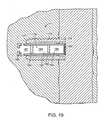
図4は、本発明の装置が埋め込まれた人の股関節部の断面である。図5は、本発明の装置が埋め込まれた人の股関節部の断面である。 FIG. 4 is a cross section of a human hip joint in which the device of the present invention is implanted. FIG. 5 is a cross section of a human hip joint in which the device of the present invention is implanted.
図6A−Fは、骨粗鬆症を治療するのに有効と考えられるいくつかの投与シナリオを示している。 6A-F illustrate several administration scenarios that may be effective in treating osteoporosis.
図6Aは、骨形成剤の初期の短期投与と、それに続くAR薬の長期投与を含む治療を開示している。この治療の基本原理は、患者に最初に骨形成剤を与えて、骨を成長させるというものである。しかし、骨成長は、多くの場合、約1ヶ月しか生じないので、BF薬は、最初の月の後に投与する必要はない。AR薬のその後の投与により、最初の月に成長した骨体の維持が保証される。 FIG. 6A discloses a treatment that includes an initial short-term administration of an osteogenic agent followed by a long-term administration of an AR drug. The basic principle of this treatment is that the patient is first given an osteogenic agent to grow bone. However, BF drugs do not need to be administered after the first month because bone growth often occurs only for about a month. Subsequent administration of the AR drug ensures maintenance of the bone that has grown in the first month.
図6Bは、最初に成長因子の短期投与があり、その後にAR薬および同化剤の投与が付随する治療を開示している。この治療の基本原理は、既存の小柱を物理的につなぐ、新しく作られた小柱形成細胞間橋(trabeculae-forming bridges)を患者に最初に与えることである。次にAR薬および同化剤(hPTH1−34など)を次に投与することにより、それぞれ、新しい骨の成長を維持することができ、さらに成長した骨が加わることができる。 FIG. 6B discloses a treatment that initially includes a short-term administration of a growth factor followed by administration of an AR drug and an anabolic agent. The basic principle of this treatment is to first give the patient newly created trabeculae-forming bridges that physically connect existing trabeculae. Subsequent administration of an AR drug and an anabolic agent (such as hPTH1-34) can then maintain new bone growth, respectively, and can add more grown bone.
図6Cは、最初にAR薬の短期投与があり、その後にBR薬の投与がある治療を開示している。この治療の基本原理は、骨粗鬆症により混乱していた骨リモデリングのバランスを最初に回復させることである。この点では、HSCAの投与が特に好ましい。バランスが回復した後に、骨成長薬が投与され、これにより、骨を脱共役させ、骨生成を生じさせる。 FIG. 6C discloses a treatment that initially has a short-term administration of an AR drug followed by administration of a BR drug. The basic principle of this treatment is to first restore the balance of bone remodeling that was confused by osteoporosis. In this regard, administration of HSCA is particularly preferred. After balance is restored, a bone growth agent is administered, thereby uncoupling the bone and causing bone formation.
図6Dは、図6Cの最初の段階をそのまま行うが、続いてAR薬の投与が加わる。この治療は、AR薬の長期投与を行わないと、BF薬の投与による骨形成が内在する骨粗鬆症のために再吸収されることを認めている。 FIG. 6D continues the first step of FIG. 6C, but is followed by administration of the AR drug. This treatment recognizes that without long-term administration of AR drug, bone formation by administration of BF drug is resorbed due to the underlying osteoporosis.
図6Eは、図6Dの基本原理に従っているが、AR薬を単純に連続投与する。このような治療投薬計画により、より簡単な送達装置が使用できるであろう。 FIG. 6E follows the basic principle of FIG. 6D, but simply administers the AR drug continuously. Such a therapeutic regimen would allow for a simpler delivery device to be used.
図6Fは、図6Eの基本原理に従っているが、さらにAR薬の連続投与を行う。このような治療投薬計画により、図7の装置のようないっそう簡単な送達装置を使用できるであろう。 FIG. 6F follows the basic principle of FIG. 6E, but additionally provides continuous administration of AR drug. Such a treatment regimen could allow for a simpler delivery device such as the device of FIG.
一般に、第1の製剤は、オプションとして、有効量の骨形成剤を含む。骨形成剤は、以下のものであってもよい:
a)成長因子(骨誘導成長因子または血管形成因子など)、
b)骨伝導能のあるもの(osteoconductive)(顆粒の多孔性マトリックスなど)、
c)骨形成性のあるもの(生育可能骨芽前駆細胞など)、または、
d)プラスミドDNA。In general, the first formulation optionally includes an effective amount of an osteogenic agent. The osteogenic agent may be:
a) growth factors (such as osteoinductive growth factors or angiogenic factors),
b) Osteoconductive (such as a porous matrix of granules),
c) those that are osteogenic (such as viable osteoprogenitor cells), or
d) Plasmid DNA.
実施形態によっては、製剤は、液状担体を含み、骨形成剤はその担体に溶解可能である。 In some embodiments, the formulation includes a liquid carrier, and the osteogenic agent can be dissolved in the carrier.
実施形態によっては、骨形成剤が成長因子である。本明細書で使用する限り、用語「成長因子」は、他の細胞、特に結合組織前駆細胞の成長または分化を調節するあらゆる細胞性生成物を包含する。本発明に従って使用することができる成長因子には、限定はされないが、酸性線維芽細胞成長因子および塩基性線維芽細胞成長因子(FGF−1およびFGF−2)およびFGF−4を含む線維芽細胞成長因子ファミリーのメンバー;PDGF−AB、PDGF−BBおよびPDGF−AAを含む血小板由来成長因子(PDGF)ファミリーのメンバー;EGF;VEGF;IGI−IおよびIIを含むインスリン様成長因子(IGF)ファミリーのメンバー;TGF−β1、2および3を含むTGF−βスーパーファミリー(superfamily);骨誘導因子(OIF)、 アンジオゲニン(angiogenin(s));エンドセリン(endothelins);肝細胞成長因子およびケラチノサイト成長因子;MP−52を含む骨形成蛋白(BMPs)BMP−1、BMP−3、BMP−2、OP−1,BMP−2A、BMP−2B、BMP−7およびBMP−14のメンバー;HBGF−1およびHBGF−2;成長分化因子(GDFs)、インディアンヘッジホッグ、ソニックヘッジホッグおよびデザートヘッジホッグを含む、蛋白のヘッジホッグファミリー(hedgehog family)のメンバー;ADMP−1;インターロイキン(IL)ファミリーの骨形成メンバー;GDF−5;および、CSF−1、G−CSF、およびGM−CSFを含むコロニー刺激因子(CSF)のメンバー;およびそれらのイソ型がある。 In some embodiments, the osteogenic agent is a growth factor. As used herein, the term “growth factor” encompasses any cellular product that regulates the growth or differentiation of other cells, particularly connective tissue progenitor cells. Growth factors that can be used in accordance with the present invention include, but are not limited to, fibroblasts including acidic fibroblast growth factor and basic fibroblast growth factor (FGF-1 and FGF-2) and FGF-4 Members of the growth factor family; members of the platelet-derived growth factor (PDGF) family including PDGF-AB, PDGF-BB and PDGF-AA; EGF; VEGF; insulin-like growth factor (IGF) family including IGI-I and II TGF-β superfamily including TGF-β1, 2 and 3; osteoinductive factor (OIF), angiogenin (s); endothelins; hepatocyte growth factor and keratinocyte growth factor; MP -52-containing bone morphogenetic proteins (BMPs) BMP-1, BMP-3, BMP- 2, members of OP-1, BMP-2A, BMP-2B, BMP-7 and BMP-14; HBGF-1 and HBGF-2; growth differentiation factors (GDFs), Indian hedgehog, sonic hedgehog and desert hedgehog A member of the hedgehog family of proteins, including: ADMP-1, an osteogenic member of the interleukin (IL) family; GDF-5; and CSF-1, G-CSF, and GM-CSF There are members of the colony stimulating factor (CSF); and their isoforms.
実施形態によっては、成長因子は、TGF−β、bFGF、およびIGF−1からなる群から選択される。これらの成長因子は、骨の再生を促進すると考えられている。実施形態によっては、成長因子がTGF−βである。より好ましくは、TGF−βは、約10ng/mlおよび約5000ng/mlの間、例えば、約50ng/mlおよび約500ng/mlの間、例えば、約100ng/mlおよび約300ng/mlの間の量で投与される。 In some embodiments, the growth factor is selected from the group consisting of TGF-β, bFGF, and IGF-1. These growth factors are thought to promote bone regeneration. In some embodiments, the growth factor is TGF-β. More preferably, the TGF-β is in an amount between about 10 ng / ml and about 5000 ng / ml, such as between about 50 ng / ml and about 500 ng / ml, such as between about 100 ng / ml and about 300 ng / ml. Is administered.
実施形態によっては、濃厚血小板が骨形成剤として与えられる。ある実施形態では、血小板から放出された成長因子の量が、その血小板を摂取した血液に見られた量の少なくとも2倍(例えば4倍)多い。実施形態によっては、濃厚血小板が自系(autologous)である。実施形態によっては、濃厚血小板が多血小板血漿(PRP)である。PRPは、骨の成長を再刺激することができる成長因子を含むことから、また、そのフィブリンマトリックス(fibrin matrix)が新しい組織の成長に適した足場を与えることから有益である。 In some embodiments, concentrated platelets are provided as an osteogenic agent. In certain embodiments, the amount of growth factor released from platelets is at least twice (eg, 4 times) greater than that found in blood that has ingested the platelets. In some embodiments, thick platelets are autologous. In some embodiments, the concentrated platelets are platelet rich plasma (PRP). PRP is beneficial because it contains growth factors that can re-stimulate bone growth and because the fibrin matrix provides a suitable scaffold for new tissue growth.
実施形態によっては、骨形成剤に有効量の骨形成蛋白(BMP)が含まれている。BMPは、骨芽細胞への間葉系幹細胞(MSCs)の分化およびその増殖を促進することにより骨形成をよく増大させる。 In some embodiments, the osteogenic agent includes an effective amount of bone morphogenetic protein (BMP). BMPs often increase bone formation by promoting the differentiation and proliferation of mesenchymal stem cells (MSCs) into osteoblasts.
実施形態によっては、約1ngおよび約10mgの間のBMPが標的骨に骨内投与される。実施形態によっては、約1マイクログラム(μg)および約1mgの間のBMPが標的骨に骨内投与される。 In some embodiments, between about 1 ng and about 10 mg of BMP is intraosseously administered into the target bone. In some embodiments, between about 1 microgram (μg) and about 1 mg of BMP is intraosseously administered to the target bone.
実施形態によっては、骨形成剤が有効量の繊維芽細胞成長因子(FGF)を含んでいる。FGFは、強力な分裂促進因子(mitogen)であり、かつ血管形成性があり、このため、間葉系幹細胞を標的領域に引きつける。さらに、FGFは、骨芽細胞を刺激して骨細胞に分化させると考えられている。 In some embodiments, the osteogenic agent includes an effective amount of fibroblast growth factor (FGF). FGF is a powerful mitogen and angiogenic, and therefore attracts mesenchymal stem cells to the target area. Furthermore, FGF is believed to stimulate osteoblasts to differentiate into bone cells.
実施形態によっては、FGFが酸性FGF(aFGF)である。 In some embodiments, the FGF is acidic FGF (aFGF).
実施形態によっては、FGFが塩基性FGF(bFGF)である。 In some embodiments, the FGF is basic FGF (bFGF).
実施形態によっては、約1マイクログラム(μg)および約10000μgの間のFGFが標的骨に骨内投与される。実施形態によっては、約10μgおよび約1000μgの間のFGFが標的骨に骨内投与される。実施形態によっては、約50μgおよび約600μgの間のFGFが標的骨に骨内投与される。 In some embodiments, between about 1 microgram (μg) and about 10000 μg of FGF are intraosseously administered into the target bone. In some embodiments, between about 10 μg and about 1000 μg of FGF are intraosseously administered into the target bone. In some embodiments, between about 50 μg and about 600 μg of FGF are intraosseously administered into the target bone.
実施形態によっては、約0.1および約4mg/kg/日の間のFGFが標的骨に骨内投与される。実施形態によっては、約1および約2mg/kg/日の間のFGFが標的骨に骨内投与される。 In some embodiments, between about 0.1 and about 4 mg / kg / day of FGF is intraosseously administered into the target bone. In some embodiments, between about 1 and about 2 mg / kg / day of FGF is intraosseously administered into the target bone.
実施形態によっては、FGFが約0.1mg/mlおよび約100mg/mlの間の濃度で標的骨に骨内投与される。実施形態によっては、FGFが約0.5mg/mlおよび約30mg/mlの間の濃度で標的骨に骨内投与される。実施形態によっては、FGFが約1mg/mlおよび約10mg/mlの間の濃度で標的骨に骨内投与される。 In some embodiments, FGF is intraosseously administered to the target bone at a concentration between about 0.1 mg / ml and about 100 mg / ml. In some embodiments, FGF is intraosseously administered to the target bone at a concentration between about 0.5 mg / ml and about 30 mg / ml. In some embodiments, FGF is intraosseously administered to the target bone at a concentration between about 1 mg / ml and about 10 mg / ml.
実施形態によっては、FGFが、約0.1mg/kgおよび約10mg/kgの間の局所組織濃度を与える量で標的骨に骨内投与される。 In some embodiments, FGF is intraosseously administered to the target bone in an amount that provides a local tissue concentration between about 0.1 mg / kg and about 10 mg / kg.
実施形態によっては、製剤がヒアルロン酸担体およびbFGFを含んでいる。実施形態によっては、米国特許第5,942,499号(以下「Orquest」という)に記載の製剤がFGF−含有製剤として選択される。 In some embodiments, the formulation includes a hyaluronic acid carrier and bFGF. In some embodiments, the formulation described in US Pat. No. 5,942,499 (hereinafter “Orquest”) is selected as the FGF-containing formulation.
実施形態によっては、骨形成剤が有効量のインシュリン様成長因子を含む。IGFは、マイトジェン(mitognenic)活性および/または細胞増殖を促進することにより骨形成をよく増大させる。 In some embodiments, the osteogenic agent comprises an effective amount of insulin-like growth factor. IGF well increases bone formation by promoting mitognenic activity and / or cell proliferation.
実施形態によっては、骨形成剤が有効量の副甲状腺ホルモン(PTH)を含む。理論に捕らわれなければ、PTHは、骨芽細胞の増殖を仲介することにより骨形成をよく増大させる。 In some embodiments, the osteogenic agent comprises an effective amount of parathyroid hormone (PTH). Without being bound by theory, PTH increases bone formation well by mediating osteoblast proliferation.
実施形態によっては、PTHが、米国特許第5,510,370号(以下「Hock」という)および第6,590,081号(以下「Zhang」という)、並びに米国特許公開公報2002/0107200号(以下「Chang」という)に教示されているようなフラグメントまたは変異体である。これら特許および公開公報の全内容は、その全体が本明細書に組み込まれる。ある実施形態では、PTHがPTH(1−34)(テリパラチド(teriparatide))、例えば、FORTEO(登録商標)(Eli Lilly and Company)である。実施形態によっては、BFAが、副甲状腺ホルモンムテイン(parathyroid hormone mutein)などの副甲状腺ホルモン誘導体(parathyroid hormone derivative)である。副甲状腺ムテインの例は、米国特許第5,856,138号(以下「Fukuda」という)で論じられている。この特許の全内容は、その全部が本明細書に取り込まれる。 In some embodiments, the PTH is disclosed in US Pat. Nos. 5,510,370 (hereinafter “Hock”) and 6,590,081 (hereinafter “Zhang”), and US Patent Publication 2002/0107200 ( Fragment or variant as taught in the following ("Chang"). The entire contents of these patents and publications are incorporated herein in their entirety. In certain embodiments, the PTH is PTH (1-34) (teriparatide), eg, FORTEO® (Eli Lilly and Company). In some embodiments, the BFA is a parathyroid hormone derivative, such as a parathyroid hormone mutein. Examples of parathyroid muteins are discussed in US Pat. No. 5,856,138 (hereinafter “Fukuda”). The entire contents of this patent are incorporated herein in their entirety.
実施形態によっては、骨形成剤が有効量のスタチン(statin)を含む。理論に捕らわれなければ、スタチンは、BMPの発現を向上させることにより骨形成をよく増大させる。 In some embodiments, the osteogenic agent comprises an effective amount of a statin. Without being bound by theory, statins increase bone formation well by improving BMP expression.
実施形態によっては、骨形成剤が多孔性マトリックスであり、好ましくは注射可能である。実施形態によっては、多孔性マトリクスがミネラルである。ある実施形態では、このミネラルがカルシウムおよびリンを含んでいる。実施形態によっては、このミネラルは、リン酸カルシウム、リン酸三カルシウム(tricalcium phosphate)およびハイドロキシアパタイト(hydroxyapatite)からなる群から選択される。ある実施形態では、マトリックスの平均穿孔が約20および約50μmの間であり、例えば、約50および約250μmの間である。本発明のさらに別の実施形態では、in situ穿孔(in situ porosity)が注入されたマトリックスで生成されて、注入された骨折安定化セメントに多孔性の足場を作る。標的組織にin situ穿孔(in situ porosity)が作られると、外科医は、穿孔に他の治療用化合物を注入することができ、これにより、周囲の組織を治療し、標的組織および注射可能セメントのリモデリング・プロセスを向上させる。 In some embodiments, the osteogenic agent is a porous matrix and is preferably injectable. In some embodiments, the porous matrix is a mineral. In some embodiments, the mineral includes calcium and phosphorus. In some embodiments, the mineral is selected from the group consisting of calcium phosphate, tricalcium phosphate, and hydroxyapatite. In certain embodiments, the average perforation of the matrix is between about 20 and about 50 μm, for example between about 50 and about 250 μm. In yet another embodiment of the invention, in situ porosity is generated in the injected matrix to create a porous scaffold in the injected fracture stabilizing cement. Once the in situ porosity has been created in the target tissue, the surgeon can inject other therapeutic compounds into the perforation, thereby treating the surrounding tissue and the target tissue and injectable cement. Improve the remodeling process.
実施形態によっては、ミネラルが顆粒形態で投与される。顆粒状ミネラルの投与は、ミネラル周りの骨成長の形成をオステオインテグレーション(osteointegration)が起きるように促進する。 In some embodiments, the mineral is administered in granular form. The administration of granular minerals promotes the formation of bone growth around the mineral so that osteointegration occurs.
実施形態によっては、ミネラルが固化可能なペースト形態で投与される。このような状態では、ペーストが体内で固化し、これにより、脆いOP体を治療後すぐに機械的に支持する。 In some embodiments, the mineral is administered in a paste form that can be solidified. In such a state, the paste solidifies in the body, thereby mechanically supporting the fragile OP body immediately after treatment.
他の実施形態では、治療薬が、注射可能で吸収性または非吸収性のセメントによって標的組織に送達される。治療薬は、生体吸収性マクロスフィア技術を利用して、最初に骨形成剤を放出し、次に吸収抑制剤を放出するように製剤にされる。セメントは、骨折した標的組織の苦痛を治療するのに必要な初期の安定性を与える。このような組織には、限定はされないが、股関節部、膝、椎体骨折および腸骨稜骨折がある。実施形態によっては、セメントが、リン酸カルシウム(calcium phosphate)、リン酸三カルシウム(tricalcium phosphate)およびハイドロキシアパタイト(hydroxyapatite)からなる群から選択される。他の実施形態では、セメントがいずれかの生体適合性ハードセメントであり、これには、PMMA、加工自原および同種移植骨(processed autogenous and allograft bone)がある。ハイドロキシルアパタイト(hydroxylapatite)は、その強度および生物学的プロフィールから好ましいセメントである。リン酸三カルシウムは、単独で使用してもよく、あるいは、特にセメントである程度の吸収が望まれている場合には、ハイドロキシアパタイト(hydroxylapatite)との組み合わせで使用してもよい。 In other embodiments, the therapeutic agent is delivered to the target tissue by an injectable, absorbable or non-absorbable cement. The therapeutic agent is formulated using bioabsorbable macrosphere technology to first release the osteogenic agent and then release the resorption inhibitor. The cement provides the initial stability necessary to treat the pain of the fractured target tissue. Such tissues include, but are not limited to, hips, knees, vertebral fractures and iliac crest fractures. In some embodiments, the cement is selected from the group consisting of calcium phosphate, tricalcium phosphate, and hydroxyapatite. In other embodiments, the cement is any biocompatible hard cement, including PMMA, processed autogenous and allograft bone. Hydroxylpatite is a preferred cement due to its strength and biological profile. Tricalcium phosphate may be used alone, or in combination with hydroxylapatite, particularly when some absorption is desired with cement.
実施形態によっては、多孔性マトリックスが再吸収可能な高分子材料を含む。 In some embodiments, the porous matrix includes a resorbable polymeric material.
実施形態によっては、骨形成剤が注入可能前駆体流体を含み、この流体は、生体内原位置でミネラル化コラーゲン組織(mineralized collagen composite)を作り出す。実施形態によっては、注入可能な前駆体流体は、以下のものを含む:
a)酸性可溶性第1種コラーゲン溶体(好ましくコラーゲン約1mg/mlおよび約7mg/mlの間)を含む第1製剤
b)カルシウムおよび燐酸塩が入っているリポソームを含む第2製剤。In some embodiments, the osteogenic agent includes an injectable precursor fluid that creates a mineralized collagen composite in situ. In some embodiments, the injectable precursor fluid includes:
a) a first formulation comprising an acidic soluble type 1 collagen solution (preferably between about 1 mg / ml and about 7 mg / ml of collagen) b) a second formulation comprising liposomes containing calcium and phosphate.
酸性可溶性コラーゲン溶体をカルシウムおよびリン酸塩入りリポソームと組み合わせると、リポソーム/コラーゲン前駆体流体ができ、これは、室温から37℃に加熱すると、ミネラル化したコラーゲンゲルを形成する。 When acidic soluble collagen solution is combined with calcium and phosphate containing liposomes, a liposome / collagen precursor fluid is formed, which forms a mineralized collagen gel when heated from room temperature to 37 ° C.
実施形態によっては、リポソームにジパルミトイルホスファチジルコリン(dipalmitoylphosphatidylcholine)(90モル%)およびジミリストイルホスファチジルコリン(dimyristoyl phosphatidylcholine)(10モル%)を入れる。このようなリポソームは、室温で安定であるが、35℃以上に加熱すると、リピド・チェーン・メルティング・トランジション(lipid chain melting transition)における捕らえていた塩を放出する結果、リン酸カルシウムミネラルを形成する。このような技術の一つがPederson, Biomaterials 24: 4881-4890 (2003)に開示されており、その仕様は、参照することによりその全部が本明細書に組み込まれる。 In some embodiments, the liposome contains dipalmitoylphosphatidylcholine (90 mol%) and dimyristoyl phosphatidylcholine (10 mol%). Such liposomes are stable at room temperature, but when heated to 35 ° C. or higher, they release salts captured in the lipid chain melting transition, resulting in the formation of calcium phosphate minerals. One such technique is disclosed in Pederson, Biomaterials 24: 4881-4890 (2003), the specification of which is hereby incorporated by reference in its entirety.
あるいは、コラーゲンの生体内原位置でのミネラル化は、他の種類の反応による温度上昇で行うこともできる。このような反応には、限定はされないが、化学、酵素、磁気、電気、光、または核によるものがある。それらの適切なソースには、光、化学反応、酵素制御反応、およびその材料に埋め込まれた電気ワイヤがある。電気ワイヤによるアプローチをさらに説明すると、ワイヤは、最初にその空間に埋め込み、加熱することでカルシウム沈殿物を作り、そして引き抜くことができる。実施形態によっては、ワイヤが、形状を作ることができるニチノールのような形状記憶ワイヤであってもよい。あるいは、昇温素子として伝導性ポリマーを選択することもできる。このポリマーは、コラーゲンを形成するために加熱され、次にその部位で分解、吸収される。これにより、ミネラル化したコラーゲンの近傍に、骨形成のための空間を与える。 Alternatively, the in-situ mineralization of collagen can also be performed by increasing the temperature due to other types of reactions. Such reactions include, but are not limited to, chemical, enzymatic, magnetic, electrical, light, or nuclear. Suitable sources include light, chemical reactions, enzyme-controlled reactions, and electrical wires embedded in the material. To further explain the electrical wire approach, the wire can be first embedded in the space and heated to create and precipitate a calcium precipitate. In some embodiments, the wire may be a shape memory wire such as Nitinol that can create a shape. Alternatively, a conductive polymer can be selected as the temperature raising element. This polymer is heated to form collagen and then decomposes and is absorbed at that site. This provides a space for bone formation in the vicinity of the mineralized collagen.
ある実施形態では、骨形成剤が複数の生存骨芽前駆細胞である。このような、骨に導入された生存細胞は、骨粗鬆症プロセスの間に骨が経験したあらゆる骨損失を少なくとも部分的に再建する能力がある。実施形態によっては、このような細胞が骨の海綿状部分に導入され、最終的に新しい海面質を生成する。他の実施形態では、このような細胞が皮質領域に導入され、新しい皮質骨を生成する。 In certain embodiments, the osteogenic agent is a plurality of viable osteoprogenitor cells. Such viable cells introduced into the bone are capable of at least partially reconstructing any bone loss experienced by the bone during the osteoporosis process. In some embodiments, such cells are introduced into the spongy portion of the bone, ultimately producing new sea surface quality. In other embodiments, such cells are introduced into the cortical region to produce new cortical bone.
実施形態によっては、このような細胞が他の一人の人間から得られ(同種移植片)、他の実施形態では、細胞が同一人から得られる(自己移植片)。実施形態によっては、細胞が骨組織から取られ、他の実施形態では、組織が非骨組織(例えば、間葉幹細胞、軟骨細胞、または線維芽細胞であることがある)から取られる。他の実施形態では、(膝、股関節部、肩、指、または耳などからの)自己移植片骨細胞が用いられることがある。 In some embodiments, such cells are obtained from another person (allograft), and in other embodiments, the cells are obtained from the same person (autograft). In some embodiments, the cells are taken from bone tissue, and in other embodiments, the tissue is taken from non-bone tissue (eg, can be mesenchymal stem cells, chondrocytes, or fibroblasts). In other embodiments, autograft bone cells (such as from the knee, hip, shoulder, finger, or ear) may be used.
ある実施形態では、生存細胞が補助治療薬または物質として選択される場合、生存細胞が間葉系幹細胞(MSCs)を含む。MSCは、脱共役吸収骨への投与に特に有利である。これは、MSCが、脱共役吸収骨の比較的厳しい環境をより容易に生き残ることができる、MSCが望ましい程度の柔軟性を有する、およびMSCが増殖して所望の細胞に分化できる、と考えられているからである。 In certain embodiments, when viable cells are selected as an adjuvant treatment or substance, the viable cells comprise mesenchymal stem cells (MSCs). MSC is particularly advantageous for administration to uncoupled resorbable bone. This may be because MSCs can more easily survive the harsh environment of uncoupled resorbed bone, MSCs have the desired degree of flexibility, and MSCs can proliferate and differentiate into the desired cells. Because.
実施形態によっては、間葉系幹細胞が、自系骨髄のような骨髄から得られる。他の実施形態では、間葉系幹細胞が脂肪組織、好ましくは自系脂肪組織から得られる。 In some embodiments, mesenchymal stem cells are obtained from bone marrow such as autologous bone marrow. In other embodiments, the mesenchymal stem cells are obtained from adipose tissue, preferably autologous adipose tissue.
実施形態によっては、骨に注入された間葉系幹細胞が非濃縮形態、例えば、新しい骨髄から用意される。他の実施形態では、間葉系幹細胞が濃縮形態で用意される。濃縮形態で用意された場合は、間葉系幹細胞が非培養であることがある。非培養、濃縮MSCは、遠心法、濾過、または免疫吸着によって容易に得られる。濾過を選択した場合、米国特許第6,049,026号(Muschler)に開示されている方法を使用することができる。米国特許第6,049,026号の明細書は、参照によりその全部が本明細書に組み込まれる。例えば、骨髄吸引懸濁液は、多孔性、生体適合性移植可能な基材に通して、組織前駆細胞の個数を高めた複合骨移植片にすることができる。実施形態によっては、MSCを濾過し、濃縮するのに用いるマトリックスもまた脱共役吸収骨に投与される。 In some embodiments, mesenchymal stem cells injected into bone are prepared from a non-enriched form, such as fresh bone marrow. In other embodiments, mesenchymal stem cells are provided in concentrated form. When prepared in a concentrated form, the mesenchymal stem cells may be uncultured. Uncultured, concentrated MSCs are readily obtained by centrifugation, filtration, or immunoadsorption. If filtration is selected, the method disclosed in US Pat. No. 6,049,026 (Muschler) can be used. The specification of US Pat. No. 6,049,026 is hereby incorporated by reference in its entirety. For example, the bone marrow aspirate suspension can be passed through a porous, biocompatible implantable substrate into a composite bone graft with an increased number of tissue progenitor cells. In some embodiments, a matrix used to filter and concentrate MSCs is also administered to uncoupled resorbable bone.
従って、本発明によれば、脱共役吸収骨を治療するためのキットが与えられ、このキットは以下のものを含む:
a)骨形成剤を含む第1製剤、
b)吸収抑制剤を含む第2製剤、および
c)生存細胞を含む第3製剤。Thus, according to the present invention, a kit for treating uncoupled resorbed bone is provided, which kit includes:
a) a first formulation comprising an osteogenic agent;
b) a second formulation comprising an absorption inhibitor; and c) a third formulation comprising viable cells.
実施形態によっては、骨細胞(同種同系ソースからであってもよいし、自系ソースからであってもよい)または間葉系幹細胞は、遺伝子組み替えを行って、本明細書で名を挙げた成長因子のリストから選択可能な、骨誘導能骨同化剤を生成してもよい。このような骨促進剤の生成は、骨成長につながる。 In some embodiments, bone cells (which may be from allogeneic or autologous sources) or mesenchymal stem cells have been genetically engineered and named herein. An osteoinductive osteoanabolic agent may be generated that can be selected from a list of growth factors. Generation of such a bone promoter leads to bone growth.
実施形態によっては、骨伝導能材料がカルシウムおよびリンを含む。実施形態によっては、骨伝導能材料がハイドロキシアパタイト(hydroxyapatite)を含む。実施形態によっては、骨伝導能材料がコラーゲンを含む。実施形態によっては、骨伝導能材料が粒子状形態をしている。 In some embodiments, the osteoconductive material includes calcium and phosphorus. In some embodiments, the osteoconductive material comprises hydroxyapatite. In some embodiments, the osteoconductive material comprises collagen. In some embodiments, the osteoconductive material is in particulate form.
実施形態によっては、第2製剤がHSCAを含む。実施形態によっては、第2製剤が薬剤ポンプを含む。実施形態によっては、徐放装置が生体吸収性材料を含む。キットは、有効量の成長因子をさらに含むことができる。実施形態によっては、各徐放装置がマイクロスフィアを含む。 In some embodiments, the second formulation comprises HSCA. In some embodiments, the second formulation includes a drug pump. In some embodiments, the sustained release device includes a bioabsorbable material. The kit can further comprise an effective amount of a growth factor. In some embodiments, each sustained release device includes a microsphere.
最近の研究で、プラスミドDNAがウィルスベクターを使用したときのように炎症反応を引き起こさないことが分かった。BMPのような遺伝子コード化骨(同化)剤は、脱共役吸収骨に注入した場合に有効でありうる。さらに、ここで提供したいずれかの成長因子または局所的な破骨細胞活動を制限する他の薬の過剰発現は、骨成長に対するよい影響を有するであろう。ある実施形態では、プラスミドに人のTGF−βまたはエリトロポイエチン(erytropoietin)(EPO)の遺伝子コードが入っている。 Recent studies have shown that plasmid DNA does not cause an inflammatory response as it does when using viral vectors. Gene-encoded bone (anabolic) agents such as BMP can be effective when injected into uncoupled resorption bone. Furthermore, overexpression of any growth factors provided herein or other drugs that limit local osteoclast activity would have a positive effect on bone growth. In some embodiments, the plasmid contains the human TGF-β or erytropoietin (EPO) genetic code.
したがって、実施形態によっては、補助治療薬が、生存細胞およびプラスミドDNAからなる群から選択される。 Thus, in some embodiments, the adjuvant treatment is selected from the group consisting of viable cells and plasmid DNA.
本発明は、エストロゲンを脱共役吸収骨に与えることにも向けられている。したがって、実施形態によっては、第2剤形が有効量のエストロゲンを吸収抑制剤として含んでいる。 The present invention is also directed to providing estrogen to uncoupled absorbed bone. Thus, in some embodiments, the second dosage form contains an effective amount of estrogen as an absorption inhibitor.
このようなエストロゲン分子は、TNF−αおよび所定のインターロイキンの様な炎症誘発性分子の生成を調節するように働く。 Such estrogen molecules serve to regulate the production of pro-inflammatory molecules such as TNF-α and certain interleukins.
エストロゲンが無くなることが、閉経後骨粗鬆症の主な原因であると考えられている。エストロゲンは、骨代謝回転を調節するために、骨芽細胞および破骨細胞のための高親和性受容体を介して作用する。この調整が閉経の間に失われると、骨吸収が増大する。したがって、閉経後の骨でエストロゲンの正常なレベルを回復させることが、破骨細胞のより正常なレベルの回復に役立つはずである。 Loss of estrogen is believed to be a major cause of postmenopausal osteoporosis. Estrogens act via high affinity receptors for osteoblasts and osteoclasts to regulate bone turnover. If this adjustment is lost during menopause, bone resorption increases. Thus, restoring normal levels of estrogen in postmenopausal bone should help restore more normal levels of osteoclasts.
したがって、本発明の他の実施形態によれば、OPを治療する方法が提供され、この方法には、エストロゲンを含む製剤の有効量を脱共役吸収骨に骨内投与することが含まれる。 Thus, according to another embodiment of the present invention, there is provided a method of treating OP, comprising intraosseously administering an effective amount of a formulation comprising estrogen to uncoupled resorbable bone.
実施形態によっては、第2製剤が有効量の選択的エストロゲン受容体モジュレーター(SERM)を含む。理論に捕らわれなければ、SERMが高い親和性をもってエストロゲン受容体に結びつくが、それはエストロゲンとは異なる方法で行い、TGF−βの増加を実現することにより骨の成長を調節しうると考えられる。 In some embodiments, the second formulation comprises an effective amount of a selective estrogen receptor modulator (SERM). Without being bound by theory, it is believed that SERMs bind to estrogen receptors with high affinity, but this can be done in a different way than estrogen and regulate bone growth by achieving an increase in TGF-β.
実施形態によっては、SERMがラロキシフェン(raloxifene)、タモキシフェン(tamoxifen)およびドロロキシフェン(droloxifene)からなる群から選択される。 In some embodiments, the SERM is selected from the group consisting of raloxifene, tamoxifen, and droloxifene.
ビスホスホン酸(biphosphonates)(BP)は、脱共役骨を治療するのに有効である。これは、ビスホスホン酸が骨のミネラル部分と結びついて、破骨細胞に吸収されるからである。いったん破骨細胞に入れば、ビスホスホン酸は、破骨細胞の活性および生存の両方に重要な酵素を抑制する。 Biphosphonates (BP) are effective in treating uncoupled bone. This is because bisphosphonic acid binds to the mineral part of bone and is absorbed by osteoclasts. Once in the osteoclast, bisphosphonic acid suppresses enzymes that are important for both osteoclast activity and survival.
実施形態によっては、BPは、アレンドロネート(alendronate)、クロドロネート(clodronate)、EB−1053、エチドロネート(etidronate)、イバンドロネート(ibandronate)、インカドロネート(incadronate)、 ネリドロネート(neridronate)、オルパドロネート(olpadronate)、パミドロネート(pamidronate)、リゼドロネート(risedronate)、チルドロネート(tiludronate)、YH−529およびゾレドロネート(zoledronate)からなる群から選択される。 In some embodiments, BP is alendronate, clodronate, EB-1053, etidronate, ibandronate, incadronate, neridronate, olpadronate ( selected from the group consisting of olpadronate, pamidronate, risedronate, tiludronate, YH-529 and zoledronate.
実施形態によっては、第2製剤が有効量のカルシトニン(calcitonin)を含む。理論に捕らわれなければ、カルシトニンは破骨細胞の中のGタンパク質共役型受容体と結びつき、カルシウムおよびサイクリックAMP経路の両方を介して破骨細胞を抑制すると考えられる。 In some embodiments, the second formulation comprises an effective amount of calcitonin. Without being bound by theory, it is believed that calcitonin binds to G protein-coupled receptors in osteoclasts and suppresses osteoclasts through both calcium and cyclic AMP pathways.
実施形態によっては、第2製剤が有効量のオステオプロテグリン(osteoprotegerin)(OPG)を含む。OPGは、腫瘍壊死因子上科の一つである。理論に捕らわれなければ、OPGは、破骨細胞の分化、発達に重要なタンパク質である破骨細胞分化因子(RANK-ligand)に結びつくと考えられる。 In some embodiments, the second formulation comprises an effective amount of osteoprotegerin (OPG). OPG is one of the tumor necrosis factor superfamily. Without being bound by theory, OPG is thought to be related to the osteoclast differentiation factor (RANK-ligand), which is a protein important for osteoclast differentiation and development.
さらに、抗カテプシン(anti-cathepsins)も本発明にしたがって使用することができる。このような酵素の抑制が骨組織の分解を抑制すると考えられる。好ましくは、拮抗剤がカセプシンB(cathepsin B)、カセプシンL(cathepsin L)およびカセプシンK(cathepsin K)からなる群から選択されたカセプシンを抑制する。 Furthermore, anti-cathepsins can also be used according to the present invention. It is thought that inhibition of such enzymes inhibits bone tissue degradation. Preferably, the antagonist inhibits cathepsin selected from the group consisting of cathepsin B, cathepsin L, and cathepsin K.
実施形態によっては、第2製剤が、有効量のカテプシンK抑制剤を含む。理論に捕らわれなければ、カセプシンKが骨吸収に必須の酵素であると考えられる。 In some embodiments, the second formulation comprises an effective amount of a cathepsin K inhibitor. Without being bound by theory, it is believed that cathepsin K is an essential enzyme for bone resorption.
第2製剤中の有効量の高特異性抗増殖吸収抑制剤の骨内投与がOPを有する患者の治療にも役立つとさらに考えられている。抗増殖剤は、炎症を起こした組織に影響を及ぼすことで炎症に効能があり、これにより、炎症促進性サイトカインの生成を制限すると考えられる。実施形態によっては、高特異性抗増殖剤が、a)ラパマイシン(rapamycin)、b)サイクリン依存性キナーゼ(cyclin dependent kinase)9(cdk)の抑制剤、およびC)ビタミンD類似体からなる群から選択される。ある実施形態では、ラパマイシンが選択されると、約0.5μg/kgおよび約50μg/kgの間の局所組織濃度を生成する用量が使用される。 It is further believed that intraosseous administration of an effective amount of a highly specific antiproliferative resorption inhibitor in the second formulation will also help treat patients with OP. Anti-proliferative agents are believed to be effective in inflammation by affecting inflamed tissues, thereby limiting the production of pro-inflammatory cytokines. In some embodiments, the highly specific anti-proliferative agent is from the group consisting of a) an inhibitor of rapamycin, b) an inhibitor of cyclin dependent kinase 9 (cdk), and C) a vitamin D analog. Selected. In certain embodiments, once rapamycin is selected, a dose is used that produces a local tissue concentration of between about 0.5 μg / kg and about 50 μg / kg.
したがって、本発明の他の実施形態によれば、OPを治療する方法であって、脱共役吸収骨に高特異性抗増殖剤を含む有効量の製剤を骨内投与することが含まれる方法が提供される。 Thus, according to another embodiment of the present invention, there is provided a method of treating OP, comprising intraosseous administration of an effective amount of a formulation comprising a highly specific antiproliferative agent to uncoupled resorbing bone. Provided.
ラパマイシンは、TOR(ラパマイシンの標的)タンパク質の下流シグナル伝達 (downstream signaling)の有効な抑制剤である。このため、ラパマイシンは、タンパク質合成およびタンパク質分解の間のバランスを調整する責任がある。OPは、骨の再生および吸収の間のバランスが失われることで広がると考えられている。TORタンパク質は、複数の代謝経路を調節するので、ラパマイシンは、サイクルのバランスを安定させることができる。ラパマイシンは、骨細胞の増殖およびその後の免疫反応にも直接影響を及ぼすことができる。ある実施形態では、約0.1μMから約10μM間での用量で与えられる。 Rapamycin is an effective inhibitor of downstream signaling of the TOR (Rapamycin target) protein. For this reason, rapamycin is responsible for adjusting the balance between protein synthesis and protein degradation. OP is thought to spread by losing the balance between bone regeneration and resorption. Since the TOR protein regulates multiple metabolic pathways, rapamycin can stabilize the cycle balance. Rapamycin can also directly affect bone cell proliferation and subsequent immune responses. In certain embodiments, the dose is given between about 0.1 μM and about 10 μM.
cdk抑制剤は、骨細胞の増殖およびその後の免疫反応に直接影響することができる。cdk抑制剤の例には、フラボピリドール(flavopiridol)、ロスコビチン(roscovitine)およびPCTの特許公報第WO02/057240号(Lin)に開示されている化合物がある。この特許公報の明細書は、参照により、本明細書にその全文が組み込まれる。ある実施形態では、cdk抑制剤が約1μMから約10μMの用量で与えられる。 A cdk inhibitor can directly affect bone cell proliferation and subsequent immune response. Examples of cdk inhibitors include flavopiridol, roscovitine and the compounds disclosed in PCT patent publication WO 02/057240 (Lin). The specification of this patent publication is incorporated herein by reference in its entirety. In certain embodiments, the cdk inhibitor is given at a dose of about 1 μM to about 10 μM.
実施形態によっては、ビタミンD類似体がVDRリガンドであり、好ましくは、有力な抗増殖剤である1アルファ25ジヒドロキシビタミン(dihydroxyritamin)D3である。 In some embodiments, the vitamin D analog is a VDR ligand, preferably 1
本発明は、骨の微視的環境にあるサイトカイン(例えば、炎症誘発性サイトカイン)を特に抑制することができる、少なくとも一つの高特異性サイトカイン拮抗剤(HSCA)または抑制剤を脱共役吸収骨に直接与えることに向けられている。ある実施形態では、HSCAが、骨または骨髄細胞によって放出された特定の炎症誘発性サイトカインの活動を抑制する。 The present invention provides uncoupled resorption bone with at least one highly specific cytokine antagonist (HSCA) or inhibitor capable of specifically suppressing cytokines (eg, pro-inflammatory cytokines) in the microscopic environment of bone. Directed to give directly. In certain embodiments, HSCA inhibits the activity of certain pro-inflammatory cytokines released by bone or bone marrow cells.
実施形態によっては、拮抗剤が、TNF−a、インターロイキン(好ましくは、IL−1、IL−6およびIL−8)、FAS、FASリガンド、およびIFN−ガンマからなる群から選択された炎症誘発性サイトカインを特に抑制することができる。このような特定の抑制剤には、米国特許公開公報U.S.2003/0039651号(以下「Olmarker」という)の5〜18ページで特定されているものがある。米国特許公開公報U.S.2003/0039651号の明細書は、参照することによりその全部が本明細書に組み込まれる。 In some embodiments, the antagonist is selected from the group consisting of TNF-a, interleukin (preferably IL-1, IL-6 and IL-8), FAS, FAS ligand, and IFN-gamma. Sex cytokines can be particularly suppressed. Such specific inhibitors include US Pat. S. Some are specified on pages 5 to 18 of 2003/0039651 (hereinafter referred to as “Olmarker”). U.S. Pat. S. The specification of 2003/0039651 is hereby incorporated in its entirety by reference.
実施形態によっては、HSCAがサイトカインをその生成を防止することで抑制する。実施形態によっては、HSCAが、膜結合サイトカインに結びつくことでサイトカインを抑制する。他の実施形態では、HSCAが、可溶性、例えば水溶性サイトカインに結びつくことによりサイトカインを抑制する。実施形態によっては、HSCA抑制剤が、膜結合サイトカインに結びつくことおよび可溶性サイトカインに結びつくことの両方でサイトカインを抑制する。実施形態によっては、HSCAが単一クローン抗体(mAb)である。mAbは、特に所定のターゲットタンパク質に結びつき、他のタンパク質には本質的に結びつかないので、mAbの使用は非常に望ましい。実施形態によっては、HSCAが標的サイトカインの天然受容体に結びつくことによりサイトカインを抑制する。 In some embodiments, HSCA suppresses cytokines by preventing their production. In some embodiments, HSCA suppresses cytokines by binding to membrane-bound cytokines. In other embodiments, HSCA suppresses cytokines by binding to soluble, eg, water soluble cytokines. In some embodiments, the HSCA inhibitor inhibits the cytokine both in association with membrane-bound cytokines and in association with soluble cytokines. In some embodiments, the HSCA is a monoclonal antibody (mAb). The use of mAbs is highly desirable because mAbs are particularly tied to a given target protein and essentially not to other proteins. In some embodiments, HSCA suppresses cytokines by binding to the natural receptor for the target cytokine.
実施形態によっては、HSCAがサイトカインの生成を防ぐことでサイトカインを抑制する。この一例は、p38マイトジェン活性化タンパク質(MAP)キナーゼの抑制剤である。実施形態によっては、TNF抑制剤が、膜結合TNFに結び付いて膜からのTNFの放出を防止するようにすることによりTNFを抑制する。他の実施形態では、TNF抑制剤が可溶性TNFに結び付くことによりTNFを抑制する。この一例は、エタネルセプト(etanercept)である。実施形態によっては、TNF抑制剤が、膜結合TNFに結び付くことと、可溶性TNFに結び付くこととの両方によりTNFを抑制する。この一例は、REMICADE(登録商標)インフリキシマブである。実施形態によっては、HSCAが標的サイトカインの天然受容体と結び付くことによりサイトカイン(例えばTNF−α)を抑制する。実施形態によっては、TNF−α抑制剤がTNF−α合成の抑制剤である。 In some embodiments, HSCA suppresses cytokines by preventing cytokine production. An example of this is an inhibitor of p38 mitogen activated protein (MAP) kinase. In some embodiments, the TNF inhibitor inhibits TNF by causing it to bind to membrane-bound TNF and prevent the release of TNF from the membrane. In other embodiments, the TNF inhibitor inhibits TNF by binding to soluble TNF. An example of this is etanercept. In some embodiments, the TNF inhibitor suppresses TNF by both binding to membrane bound TNF and binding to soluble TNF. An example of this is REMICADE® infliximab. In some embodiments, HSCA inhibits cytokines (eg, TNF-α) by binding to the natural receptor for the target cytokine. In some embodiments, the TNF-α inhibitor is an inhibitor of TNF-α synthesis.
実施形態によっては、吸収抑制剤が、非常に特殊な、腫瘍壊死因子(TNF)の拮抗剤である。このような拮抗剤は、非常に好ましい。なぜならば、文献によれば、このような拮抗剤の骨粗鬆症の骨への投与に、骨における破骨細胞濃度を基準(骨粗鬆症前の)レベルまで戻す効果があるからである。 In some embodiments, the absorption inhibitor is a very specific antagonist of tumor necrosis factor (TNF). Such antagonists are highly preferred. This is because, according to the literature, administration of such antagonists to osteoporotic bone has the effect of returning the osteoclast concentration in the bone to a reference (pre-osteoporosis) level.
具体的には、Kimble, R.B. et al., "Estrogen deficiency increases the ability of stromal cells to support murine osteoclastogenesis via an interleukin-1 and tumor necrosis factor-mediated stimulation of macrophage colony-stimulating factor production," J. Biol. Chem, 271 (46): 18890-7 (1996)、(以下「Kimble I」という)には、M−CSFおよび破骨細胞の両方の濃度が、有効量のIl−1/TNF−α抑制剤を投与した卵巣摘出ラットにおいて基本的に通常のレベルに戻ったと報告されている。Kimble, R.B. et al., "The functional block of TNF but not of IL-6 prevents bone loss in ovariectomized mice," J. Bone Min. Res., 12 (6) 935-941 (1997)、(以下「Kimble II」という)は、有効量のTNF−α抑制剤を投与された卵巣摘出マウスにおいて、破骨細胞濃度が基本的に通常のレベルに戻ることを報告しており、さらに、エストロゲン欠乏により骨が失われるメカニズムで中心的な役割を果たすエストロゲン調節サイトカインがIL−6ではなく、むしろTNFであると結論付けている。 Specifically, Kimble, RB et al., "Estrogen deficiency increases the ability of stromal cells to support murine osteoclastogenesis via an interleukin-1 and tumor necrosis factor-mediated stimulation of macrophage colony-stimulating factor production," J. Biol. Chem, 271 (46): 18890-7 (1996), (hereinafter referred to as “Kimble I”), both M-CSF and osteoclast concentrations are effective amounts of Il-1 / TNF-α inhibitors. Has been reported to return to normal levels in ovariectomized rats dosed with. Kimble, RB et al., "The functional block of TNF but not of IL-6 prevents bone loss in ovariectomized mice," J. Bone Min. Res., 12 (6) 935-941 (1997) (hereinafter "Kimble II ”) reported that the osteoclast concentration basically returned to normal levels in ovariectomized mice administered with an effective amount of a TNF-α inhibitor. We conclude that the estrogen-regulated cytokine that plays a central role in the lost mechanism is not TIL-6 but rather TNF.
よって、これらTNF拮抗剤は、破骨細胞の生成および機能を壊すのではなく、単に破骨細胞の水準をその通常水準に戻すという効果があるだけなので、非常に好ましい。 Thus, these TNF antagonists are highly preferred because they do not disrupt osteoclast production and function, but simply return the osteoclast level to its normal level.
好ましいTNF拮抗剤には、限定はされないが、以下のものがある: エタネルセプト(etanercept)(ENBREL(登録商標)、Amgen社製);インフリキシマブ(infliximab)(REMICADE(登録商標)、Johnson and Johnson社製);D2E7、人抗TNFモノクローナル抗体(ノール医薬品(Knoll Pharmaceuticals)、Abbott Laboratories製); CDP571(人抗TNFIgG4抗体)およびCDP870(抗TNFアルファ人化モノクローナル抗体フラグメント(an anti-TNF alpha humanized monoclonal antibody fragment)、いずれもCelltech社製;可溶性TNF受容体タイプI(Amgen社製); ペグ化された可溶性のTNF受容体タイプI(PEGsTNF−R1)(Amgen社製);およびオネルセプト(onercept)、組み換え型TNF結合タンパク質(r−TBP−1)(Serono社製)。 Preferred TNF antagonists include, but are not limited to: etanercept (ENBREL®, Amgen); infliximab (REMICADE®, Johnson and Johnson) ); D2E7, human anti-TNF monoclonal antibody (Knoll Pharmaceuticals, manufactured by Abbott Laboratories); CDP571 (human anti-TNF IgG4 antibody) and CDP870 (an anti-TNF alpha humanized monoclonal antibody fragment) ), All from Celltech; soluble TNF receptor type I (Amgen); pegylated soluble TNF receptor type I (PEGsTNF-R1) (Amgen); and onercept, recombinant TNF binding protein (r-TBP-1) (manufactured by Serono).
(オプションとして、本発明の少なくとも1つの抗体、その特定部分および/または変異体をさらに含む)本発明の組成、併用療法、同時投与、装置および/または方法に適するTNF拮抗剤には、限定はされないが、TNF化学的またはタンパク質拮抗剤(TNF chemical or protein antagonist)、抗TNF抗体、単一クローン性またはポリクローン性抗体またはフラグメント(TNF monoclonal or polyclonal antibody or fragment)、可溶性TNF受容体(例えば、p55、p70またはp85)またはフラグメント、その融解ポリペプチド(fusion polypeptides)、小分子TNF拮抗剤、例えばTNF結合タンパク質IまたはII(TBP−IまたはTBP−II)、ネレリモマブ(nerelimonmab)、REMICADE(登録商標)インフリキシマブ(infliximab)、エタネルセプト(etanercept)(ENBREL(登録商標))、アダリムマブ(adalimulab)(HUMIRA(登録商標))CDP−571、CDP−870、アフェリモマブ(afelimomab)、レナセプト(lenercept)など、それらの抗原結合フラグメント、および特にTNFに結び付く受容体分子;TNF合成、TNF放出または標的細胞への作用を防止および/または抑制する化合物、例えば、サリドマイド(thalidomide)、テニダプ(tenidap)、およびホスホジエステラーゼ(phosphodiesterase)抑制剤(例えば、ペントキシフィリン(pentoxifylline)およびロリプラム(rolipram))など;A2bアデノシン受容体アゴニスト(A2b adenosine receptor agonists)およびA2bアデノシン受容体エンハンサー(A2b adenosine receptor enhancers);TNF受容体シグナル伝達を防止および/または禁止する化合物、例えばマイトジェン活性タンパク質(mitogen activated protein)(MAP)キナーゼ抑制剤(kinase inhibitors);膜TNF開裂(membrane TNF cleavage)をブロックおよび/または抑制する化合物、例えば、メタロプロテナーゼ抑制剤(metalloproteinase inhibitors);TNF活動をブロックおよび/または抑制する化合物、例えば、アンギオテンシン変換酵素(ACE)抑制剤(例えばカプトプリル(captopril));TNF生成および/または合成をブロック/または抑制する化合物、例えば、MAPキナーゼ阻害剤など、がある。 TNF antagonists suitable for the compositions, combination therapies, co-administrations, devices and / or methods of the present invention (optionally further comprising at least one antibody of the invention, specific portions and / or variants thereof) include, but are not limited to, Not TNF chemical or protein antagonist, anti-TNF antibody, monoclonal or polyclonal antibody or fragment, soluble TNF receptor (eg, p55, p70 or p85) or fragments thereof, fusion polypeptides thereof, small molecule TNF antagonists such as TNF binding protein I or II (TBP-I or TBP-II), nerelimonmab, REMICADE® ) Infliximab, etanercept (ENBREL (registered) )), Adalimulab (HUMIRA®) CDP-571, CDP-870, afelimomab, lenercept, etc., antigen binding fragments thereof, and in particular receptor molecules associated with TNF; TNF Compounds that prevent and / or inhibit synthesis, TNF release or effects on target cells, such as thalidomide, tenidap, and phosphodiesterase inhibitors (eg, pentoxifylline and rolipram ( rolipram)), etc .; A2b adenosine receptor agonists and A2b adenosine receptor enhancers; compounds that prevent and / or inhibit TNF receptor signaling, such as mitogen activated proteins protein) (MAP) kinase inhibitors; compounds that block and / or inhibit membrane TNF cleavage, eg metalloproteinase inhibitors; compounds that block and / or inhibit TNF activity For example, angiotensin converting enzyme (ACE) inhibitors (eg, captopril); compounds that block / or inhibit TNF production and / or synthesis, eg, MAP kinase inhibitors.
ある実施形態では、TNA拮抗剤がサイクリン化合物である。サイクリン化合物は、非特異的方法でTNF−αを抑制する。TNF−αおよび他の類似の生物活性物質は、最初に非活性形態で作られ、そして細胞膜へ運ばれる。活性化すると、プロ−TNF−α(pro-TNF-a)の活性部分が分割され、放出される。このプロセスは、分断と呼ばれており、1つ以上の酵素で開始できる。これらの酵素の全ては、共通して金属イオンを有し、マトリックス・メタロプロテナーゼ(matrix metalloproteinases)(MMPs)と呼ばれている。サイクリン(cycine)化合物が金属イオンに結び付き、それによりMMPの活動を抑制し、続いてTNF−αおよび他の炎症誘発性サイトカインの放出を非特異的方法で抑制することは公知である。実施形態によっては、サイクリン化合物は、ドキシサイクリン(doxycycline)、ライムサイクリン(lymecycline)、オキシサイクリン(oxicycline)化合物、テトラサイクリン(tetracycline)、ミノサイクリン(minocycline)、化学修飾したテトラサイクリン(CMT)およびKB−R7785からなる群から選択されている。 In certain embodiments, the TNA antagonist is a cyclin compound. Cyclin compounds suppress TNF-α in a non-specific manner. TNF-α and other similar bioactive substances are first made in an inactive form and transported to the cell membrane. Upon activation, the active part of pro-TNF-α (pro-TNF-a) is split and released. This process is called fragmentation and can be initiated with one or more enzymes. All of these enzymes have a common metal ion and are called matrix metalloproteinases (MMPs). It is known that cycine compounds bind to metal ions, thereby inhibiting the activity of MMP and subsequently inhibiting the release of TNF-α and other pro-inflammatory cytokines in a non-specific manner. In some embodiments, the cyclin compound comprises doxycycline, lymecycline, oxycycline compound, tetracycline, minocycline, chemically modified tetracycline (CMT) and KB-R7785. Selected from the group.
本明細書で使用する限り、「腫瘍壊死因子抗体」、「TNF抗体」、「TNFα抗体」、またはフラグメント等は、in vitro、in situ、および/または好ましくはin vivo のTNFα活動を減少させる、ブロックする、抑制する、終わらせる、または干渉する。例えば本発明の適当なTNF人抗体は、TNFαと結合でき、抗TNF抗体、その抗原結合フラグメント、および、特にTNF−アルファ(TNFα)に結合する特定変異体またはそのドメインを含む。適切なTNF抗体またはフラグメントは、TNF、RNA、DNAまたはタンパク質合成、TNF放出、TNF受容体シグナル伝達、膜TNF開裂、TNF活動、TNF生成および/または合成を減少させる、ブロックする、終わらせる、干渉する、防ぐおよび/または抑制することもできる。 As used herein, a “tumor necrosis factor antibody”, “TNF antibody”, “TNFα antibody”, or fragment or the like decreases TNFα activity in vitro, in situ, and / or preferably in vivo. Block, suppress, end or interfere. For example, a suitable TNF human antibody of the present invention can bind to TNFα and includes an anti-TNF antibody, an antigen-binding fragment thereof, and a specific variant or domain thereof that specifically binds TNF-alpha (TNFα). Suitable TNF antibodies or fragments reduce, block, terminate, interfere with TNF, RNA, DNA or protein synthesis, TNF release, TNF receptor signaling, membrane TNF cleavage, TNF activity, TNF production and / or synthesis Can be prevented, prevented and / or suppressed.
キメラ抗体cA2は、A2と呼ばれる、高特異性中和マウス抗ヒトTNFαIgG1抗体の抗原結合可変部と、カッパ免疫グロブリンと呼ばれるヒトIgG1の定常部とを含む。ヒトIgG1Fc部は、同種異形抗体エフェクター機能を改善し、循環漿液半減期を増大させ、抗体の免疫原性を減少させる。キメラ抗体cA2のアビディティおよびエピトープ特異性は、マウスの抗体A2の可変部から導出される。具体的な実施形態では、マウス抗原A2の可変部をコード化する核酸のための好ましいソースは、A2ハイブリドーマ細胞系である。 The chimeric antibody cA2 comprises a highly specific neutralizing mouse anti-human TNFα IgG1 antibody antigen-binding variable region called A2, and a human IgG1 constant region called kappa immunoglobulin. The human IgG1 Fc portion improves allogeneic antibody effector function, increases circulating serous half-life, and reduces antibody immunogenicity. The avidity and epitope specificity of the chimeric antibody cA2 is derived from the variable region of the mouse antibody A2. In a specific embodiment, a preferred source for nucleic acids encoding the variable region of mouse antigen A2 is the A2 hybridoma cell line.
キメラA2(cA2)は、用量に依存した方法における天然および遺伝子組み替えヒトTNFαの両方の細胞毒効果を中和する。キメラ抗体cA2および遺伝子組み替えヒトTNFαの結合実験から、キメラ抗体cA2の特異性定数が1.04×10¹⁰M-1と計算された。モノクローナル抗体特異性および競合阻害による特異性を決定するための好ましい方法は、Harlow et al., Antibodies: A Laboratory Manual, Cold Spring Harbor Laboratory Press, Cold Spring Harbor, New York (1988); Colligan et al., eds.; Current Protocols in Immunology, (NY: Greene Publishing Assoc. and Wiley Interscience) (1992-2000); Kozbor et al., Immunol. Today, 4:72-79 (1983);およびAusubel et al., eds. Current Protocols in Molecular Biology, (NY: Wiley Interscience)(1987-2000); およびMuller, R., "Determination of affinity and specificity of anti-hapten antibodies by competitive radioimmunoassay," Meth. Enzymol., 92: 589-601 (1983)にある。これらは、参照することによりその全部が本明細書に組み込まれる。Chimera A2 (cA2) neutralizes the cytotoxic effects of both natural and recombinant human TNFα in a dose-dependent manner. From the binding experiment of the chimeric antibody cA2 and the recombinant human TNFα, the specificity constant of the chimeric antibody cA2 was calculated to be 1.04 × 10¹⁰ M−1 . A preferred method for determining monoclonal antibody specificity and specificity by competitive inhibition is Harlow et al., Antibodies: A Laboratory Manual, Cold Spring Harbor Laboratory Press, Cold Spring Harbor, New York (1988); Colligan et al. , eds .; Current Protocols in Immunology, (NY: Greene Publishing Assoc. and Wiley Interscience) (1992-2000); Kozbor et al., Immunol. Today, 4: 72-79 (1983); and Ausubel et al., eds. Current Protocols in Molecular Biology, (NY: Wiley Interscience) (1987-2000); and Muller, R., "Determination of affinity and specificity of anti-hapten antibodies by competitive radioimmunoassay," Meth. Enzymol., 92: 589 -601 (1983). All of which are incorporated herein by reference.
ある特別な実施形態では、マウスのモノクローナル抗体A2がc134Aと呼ばれる細胞株により作られる。キメラ抗体cA2は、c168Aと呼ばれる細胞株により作られる。cA2は、米国特許第6,284,471号(Le et al.)に詳しく記載されており、参照により、その全部が本明細書に組み込まれる。 In one particular embodiment, murine monoclonal antibody A2 is produced by a cell line called c134A. Chimeric antibody cA2 is made by a cell line called c168A. cA2 is described in detail in US Pat. No. 6,284,471 (Le et al.), which is hereby incorporated by reference in its entirety.
本発明で利用することができるモノクローナル抗TNF抗体のさらに別の例は、当該技術に記載されている(例えば、米国特許第5,231,024号;Moeller, A., et al., "Monoclonal antibodies to human tumor necrosis factor alpha: in vitro and in vivo application," A. et al., Cytokine 2 (3): 162-169 (1990);米国特許第6,277,969号;Rathjen et al., 国際公開第WO91/02078号(公開日1991年2月21日);Rubin et al., EPO特許公報第0 218 868号(公開日1987年4月22日);Yone et al., EPO特許公報第0 288 088号(公開日1988年10月26日);Liang, C.M., et al., "Production and characterization of monoclonal antibodies against recombinant human tumor necrosis factor/cachectin," Biochem. Biophys. Res. Comm., 137: 847-854 (1986);Meager, A., et al., "Preparation and characterization of monoclonal antibodies directed against antigenic determinants of recombinant human tumour necrosis factor (rTNF)," Hybridoma, 6: 305-311 (1987);Fendly, B.M., et al., "Murine monoclonal antibodies defining neutralizing epitopes on tumor necrosis factor," Hybridoma, 6: 359-369 (1987);Bringman, T.S., et al., "Monoclonal antibodies to human tumor necrosis factors alpha and beta: application for affinity purification, immunoassays, and as structural probes," Hybridoma, 6 : 489-507 (1987);およびHirai, M., et al., "Production and characterization of monoclonal antibodies to human tumor necrosis factors,” J. Immunol. Meth., 96: 57-62 (1987)を参照のこと。これらの参考文献は参照により本明細書に全て組み込まれる)。 Still other examples of monoclonal anti-TNF antibodies that can be utilized in the present invention are described in the art (eg, US Pat. No. 5,231,024; Moeller, A., et al., “Monoclonal antibodies to human tumor necrosis factor alpha: in vitro and in vivo application, "A. et al., Cytokine 2 (3): 162-169 (1990); US Pat. No. 6,277,969; Rathjen et al., International Publication No. WO 91/02078 (Publication Date: February 21, 1991); Rubin et al., EPO Patent Publication No. 0 218 868 (Publication Date: April 22, 1987); Yone et al., EPO Patent Publication No. 0 288 088 (published October 26, 1988); Liang, CM, et al., “Production and characterization of monoclonal antibodies against recombinant human tumor necrosis factor / cachectin,” Biochem. Biophys. Res. Comm., 137: 847-854 (1986); Meager, A., et al., "Preparation and characterization o f monoclonal antibodies directed against antigenic determinants of recombinant human tumour necrosis factor (rTNF), "Hybridoma, 6: 305-311 (1987); Fendly, BM, et al.," Murine monoclonal antibodies defining neutralizing epitopes on tumor necrosis factor, " Hybridoma, 6: 359-369 (1987); Bringman, TS, et al., "Monoclonal antibodies to human tumor necrosis factors alpha and beta: application for affinity purification, immunoassays, and as structural probes," Hybridoma, 6: 489- 507 (1987); and Hirai, M., et al., “Production and characterization of monoclonal antibodies to human tumor necrosis factors,” J. Immunol. Meth., 96: 57-62 (1987). These references are all incorporated herein by reference).
本発明において有用な好ましいTNF受容体分子には、高特異性をもってTNFαに結合するもの(例えば、Feldmann et al.,国際公開第WO92/07076号(公開日1992年4月30日);Schall, T.J. et al., "Molecular cloning and expression of a receptor for human tumor necrosis factor," Cell, 61: 361-370 (1990);およびLoetscher, H. et al., "Molecular cloning and expression of the human 55 kd tumor necrosis factor receptor," Cell, 61: 351-359 (1990)を参照。これらは参照により本明細書に全て組み込まれる。)があり、オプションとして、低免疫原性を有する。特に、55kDa(p55TNF−R)および75kDa(p75TNF−R)TNF細胞表面受容体が本発明において有用である。受容体の細胞外領域(ECD)またはその機能部(例えば、Corcoran, A.E. et al., "Characterization of ligand binding by the human p55 tumour-necrosis-factor receptor. Involvement of individual cysteine-rich repeats," Eur. J. Biochem., 223: 831-840 (1994)参照)を含むこれら受容体の切断型も本発明で有用である。ECDを含むTNF受容体の切断型は、尿および漿液において30kDaおよび40kDAのTNFα阻害性結合タンパク質として検出されている(Engelmann, H. et al., "Two tumor necrosis factor-binding proteins purified from human urine. Evidence for immunological cross-reactivity with cell surface tumor necrosis factor receptors," J. Biol. Chem., 265:, 1531-1536 (1990))。TNF受容体多量体分子およびTNF免疫受容体融合分子、および誘導体およびそのフラグメントまたは部分は、本発明の方法および組成において有用なTNF受容体分子のさらに別の例である。本発明で使用できるTNF受容体分子は、症状をよくまたは非常によく軽減し、かつ、低毒性で患者を長期間治療できる能力により特徴付けられる。低免疫原性および/または高特異性、並びに他の不明確な特性は、達成できる治療結果に寄与できる。 Preferred TNF receptor molecules useful in the present invention include those that bind TNFα with high specificity (eg Feldmann et al., International Publication No. WO 92/07076 (published 30 April 1992); Schall, TJ et al., "Molecular cloning and expression of a receptor for human tumor necrosis factor," Cell, 61: 361-370 (1990); and Loetscher, H. et al., "Molecular cloning and expression of the human 55 kd tumor necrosis factor receptor, "Cell, 61: 351-359 (1990). All of which are incorporated herein by reference), and optionally have low immunogenicity. In particular, 55 kDa (p55TNF-R) and 75 kDa (p75TNF-R) TNF cell surface receptors are useful in the present invention. The extracellular region (ECD) of the receptor or its functional part (e.g. Corcoran, AE et al., "Characterization of ligand binding by the human p55 tumour-necrosis-factor receptor. Involvement of individual cysteine-rich repeats," Eur. J. Biochem., 223: 831-840 (1994)) are also useful in the present invention. A truncated form of the TNF receptor, including ECD, has been detected as a 30 kDa and 40 kDa TNFα inhibitory binding protein in urine and serum (Engelmann, H. et al., “Two tumor necrosis factor-binding proteins purified from human urine Evidence for immunological cross-reactivity with cell surface tumor necrosis factor receptors, "J. Biol. Chem., 265 :, 1531-1536 (1990)). TNF receptor multimer molecules and TNF immunoreceptor fusion molecules, and derivatives and fragments or portions thereof are yet another example of TNF receptor molecules useful in the methods and compositions of the invention. TNF receptor molecules that can be used in the present invention are characterized by the ability to relieve symptoms well or very well and to treat patients for a long time with low toxicity. Low immunogenicity and / or high specificity, as well as other unclear characteristics, can contribute to the therapeutic results that can be achieved.
本発明で有用なTNF受容体多量体分子は、1つ以上のポリペプチドリンカー又は他の非ペプチドリンカー、例えばポリエチレングリコール(PEG)などを介して連結された2つ以上のTNF受容体のECDの全部又は機能部を含むことができる。多量体分子は、分泌タンパク質のシグナルペプチドをさらに含み、多量体分子を直接発現させることもできる。 The TNF receptor multimeric molecules useful in the present invention are all ECDs of two or more TNF receptors linked via one or more polypeptide linkers or other non-peptide linkers such as polyethylene glycol (PEG). Or a functional part can be included. The multimeric molecule further includes a signal peptide of a secreted protein, and the multimeric molecule can also be expressed directly.
本発明の方法および組成に有用なTNF免疫受容体分子は、1つ以上の免疫グロブリンの少なくとも一部と、1つ以上のTNF受容体の全てまたは機能部を含むことができる。これらの免疫受容体融合分子は、モノマー、またはヘテロもしくはホモ多量体としてアセンブルすることができる。免疫融合分子は、一価または多価であることもできる。このようなTNF免疫受容体融合分子の例は、TNF受容体/IgG融合タンパク質である。TNF免疫受容体融合分子およびその製造方法は、当該技術において説明されている(Lesslauer, W. et al., "Recombinant soluble tumor necrosis factor receptor proteins protect mice from lipopolysaccharide-induced lethality," Eur. J. Immunol, 21: 2883-2886 (1991);Ashkenazi, A., et al., "Protection against endotoxic shock by a tumor necrosis factor receptor immunoadhesin," Proc. Natl. Acad. Sci. USA, 88: 10535-10539 (1991);Peppel, K. et al., "A tumor necrosis factor (TNF) receptor-IgG heavy chain chimeric protein as a bivalent antagonist of TNF activity," J. Exp. Med., 174: 1483-1489 (1991);Kolls, J. et al., "Prolonged and effective blockade of tumor necrosis factor activity through adenovirus-mediated gene transfer," Proc. Natl. Acad. Sci. USA, 91: 215-219 (1994);Butler, D.M. et al., "TNF receptor fusion proteins are effective inhibitors of TNF-mediated cytotoxicity on human KYM-1D4 rhabdomyosarcoma cells," Cytokine, 6 (6): 616-623 (1994);Baker, D. et al., "Control of established experimental allergic encephalomyelitis by inhibition of tumor necrosis factor (TNF) activity within the central nervous system using monoclonal antibodies and TNF receptor-immunoglobulin fusion proteins," Eur. J. Immunol., 24: 2040-2048 (1994);およびBeutlerらの米国特許第5,447,851号、これら参考文献の各々は、参照により本明細書に全部組み込まれる)。免疫受容体融合分子の製造方法は、Caponらの米国特許第5,116,964号;Caponらの米国特許第5,225,538号;およびCapon, D.J. et al., "Designing CD4 immunoadhesins for AIDS therapy," Nature, 337: 525-531 (1989)にも見られ、これらの参考文献は、参照により本明細書に全部組み込まれる。 A TNF immunoreceptor molecule useful in the methods and compositions of the invention can comprise at least a portion of one or more immunoglobulins and all or a functional portion of one or more TNF receptors. These immunoreceptor fusion molecules can be assembled as monomers, or hetero- or homomultimers. The immunofusion molecule can also be monovalent or multivalent. An example of such a TNF immunoreceptor fusion molecule is a TNF receptor / IgG fusion protein. TNF immunoreceptor fusion molecules and methods for their production have been described in the art (Lesslauer, W. et al., “Recombinant soluble tumor necrosis factor receptor proteins protect mice from lipopolysaccharide-induced lethality,” Eur. J. Immunol. , 21: 2883-2886 (1991); Ashkenazi, A., et al., "Protection against endotoxic shock by a tumor necrosis factor receptor immunoadhesin," Proc. Natl. Acad. Sci. USA, 88: 10535-10539 (1991 Peppel, K. et al., "A tumor necrosis factor (TNF) receptor-IgG heavy chain chimeric protein as a bivalent antagonist of TNF activity," J. Exp. Med., 174: 1483-1489 (1991); Kolls, J. et al., "Prolonged and effective blockade of tumor necrosis factor activity through adenovirus-mediated gene transfer," Proc. Natl. Acad. Sci. USA, 91: 215-219 (1994); Butler, DM et al ., "TNF receptor fusion proteins are effective inhibitors of TNF-mediated cytotoxicity on human KYM-1D4 rhabdomyosarcoma cells," Cytokine, 6 (6): 616-623 (1994); Baker, D. et al., "Control of established experimental allergic encephalomyelitis by inhibition of tumor necrosis factor (TNF) activity within the central nervous system using monoclonal antibodies and TNF receptor-immunoglobulin fusion proteins," Eur. J. Immunol., 24: 2040 -2048 (1994); and US Pat. No. 5,447,851 to Beutler et al., Each of these references is hereby incorporated by reference in its entirety). Methods for producing immunoreceptor fusion molecules are described in Capon et al. US Pat. No. 5,116,964; Capon et al. US Pat. No. 5,225,538; and Capon, DJ et al., “Designing CD4 immunoadhesins for AIDS. therapy, "Nature, 337: 525-531 (1989), these references are fully incorporated herein by reference.
TNF受容体分子の機能上の等価物、誘導体、フラグメント、または領域とは、TNF受容体分子の一部、またはTNF受容体分子をコード化するTNF受容体分子核酸配列の部分を含むタンパク質またはペプチドをいう。TNF受容体分子核酸配列の部分は、本発明で使用できるTNF受容体分子に機能的に類似する(例えばTNFαに高い特異性を持って結合し、低免疫原性を有する)のに十分なサイズおよび配列からなる。TNF受容体分子の機能上の等価物は、本発明に使用できるTNF受容体分子に機能上類似する改良型TNF受容体分子をさらに含む(例えば、TNFαと高い特異性で結合し、低免疫原性を有する)。例えば、TNF受容体分子の機能上の等価物は、「サイレント」コドン(codon)または1つ以上のアミノ酸置換、欠失または付加を含むことができる(例えば、一つの酸性アミノ酸の別の酸性アミノ酸への置換;同一または異なる疎水性アミノ酸をコード化する一つのコドン(codon)の疎水性アミノ酸をコード化する他のコドンへの置換)。これについては、Ausubel et al., Current Protocols in Molecular Biology (NY: Greene Publishing Assoc. and Wiley-Interscience) (1987-2003)を参照のこと。 A functional equivalent, derivative, fragment, or region of a TNF receptor molecule is a protein or peptide comprising a portion of a TNF receptor molecule or a portion of a TNF receptor molecule nucleic acid sequence that encodes a TNF receptor molecule Say. A portion of the TNF receptor molecule nucleic acid sequence is of sufficient size to be functionally similar to a TNF receptor molecule that can be used in the present invention (eg, binds with high specificity to TNFα and has low immunogenicity). And an array. Functional equivalents of TNF receptor molecules further include improved TNF receptor molecules that are functionally similar to TNF receptor molecules that can be used in the present invention (eg, bind to TNFα with high specificity and low immunogenicity). Have sex). For example, a functional equivalent of a TNF receptor molecule can include a “silent” codon or one or more amino acid substitutions, deletions or additions (eg, another acidic amino acid of one acidic amino acid). Substitution of one codon encoding the same or different hydrophobic amino acid with another codon encoding a hydrophobic amino acid). See Ausubel et al., Current Protocols in Molecular Biology (NY: Greene Publishing Assoc. And Wiley-Interscience) (1987-2003).
実施形態によっては、TNF−αを抑制するモノクローナル抗体が、モノクローナルげっ歯類−ヒト抗体、げっ歯類抗体、ヒト抗体、またはそれらの任意の一部分または複数部分であり、免疫グロブリン可変部の抗原結合領域を少なくとも1つ有し、その抗体がTNFと結合するものからなる群から選択される。好ましくは、このモノクローナル抗体は、米国特許第6,277,969号に開示されている化合物の群から選択される。米国特許第6,277,969号の明細書は、参照により本明細書の全部組み込まれる。実施形態によっては、REMICADE(登録商標)インフリキシマブが、約0.4mg/mlおよび約4mg/mlの間のインフリキシマブ濃度を有する製剤で送達される。 In some embodiments, the monoclonal antibody that inhibits TNF-α is a monoclonal rodent-human antibody, a rodent antibody, a human antibody, or any portion or portions thereof, and the antigen binding of an immunoglobulin variable region It is selected from the group consisting of those having at least one region and whose antibody binds to TNF. Preferably, the monoclonal antibody is selected from the group of compounds disclosed in US Pat. No. 6,277,969. The specification of US Pat. No. 6,277,969 is hereby incorporated by reference in its entirety. In some embodiments, REMICADE® infliximab is delivered in a formulation having an infliximab concentration between about 0.4 mg / ml and about 4 mg / ml.
実施形態によっては、TNF−αの具体的な抑制剤は、p38MAPキナーゼの抑制剤、好ましくは、p38MAPキナーゼの小分子抑制剤である。p38MAPキナーゼを抑制は、TNF−αおよびIl−2の両方の生成をブロックすると考えられている。TNF−αおよびIl−2は、いずれも炎症誘発性サイトカインである。p38MAPキナーゼの小分子抑制剤は、非常に特異で強力である(〜nM)。理論に捕らわれなければ、p38の抑制は、TGFシグナル伝達も、TGF活性もブロックするはずがないと考えられる。さらに、p38抑制剤が一部のメタプロテイナーゼ、COX2およびNO合成酵素の誘発もブロックしうるとも考えられている。さらに、P38抑制剤が免疫細胞増殖に伴うインターロイキン、例えばIL−2などを抑制しないとも考えられている。 In some embodiments, the specific inhibitor of TNF-α is an inhibitor of p38 MAP kinase, preferably a small molecule inhibitor of p38 MAP kinase. Inhibition of p38 MAP kinase is believed to block the production of both TNF-α and Il-2. TNF-α and Il-2 are both pro-inflammatory cytokines. Small molecule inhibitors of p38 MAP kinase are very specific and potent (˜nM). Without being bound by theory, it is believed that p38 inhibition should not block TGF signaling or TGF activity. It is further believed that p38 inhibitors can also block the induction of some metaproteinases, COX2 and NO synthases. Furthermore, it is considered that P38 inhibitors do not inhibit interleukins such as IL-2 accompanying immune cell proliferation.
p38キナーゼの高特異性拮抗剤(HSA)を有効量、骨内に投薬することもまたOPを有する患者の治療に役立ちうる。p38キナーゼ部位は、TNF−α、IL−1およびCOX−2酵素の生成を調節すると考えられている。 Intraosseous administration of an effective amount of a highly specific antagonist of p38 kinase (HSA) can also help treat patients with OP. The p38 kinase site is thought to regulate the production of TNF-α, IL-1 and COX-2 enzymes.
したがって、本発明の他の実施形態によれば、OPの治療方法が提供され、この方法は、p38キナーゼの高特異性拮抗剤を含む製剤を有効量、OPの骨に局所的に骨内投与することが含まれる。 Thus, according to another embodiment of the present invention, there is provided a method of treating OP, which comprises intraosseously administering an effective amount of a formulation comprising a highly specific antagonist of p38 kinase to the bone of OP. For example.
好ましくは、それらは、約10nMから約10uMの用量で与えられる。いくつかのp38キナーゼの高特異性拮抗剤が、Zhang, C., "Mitogen-activated protein (MAP) kinase regulates production of tumor necrosis factor-alpha and release of arachidonic acid in mast cells. Indications of communication between p38 and p42 MAP kinases," J. Biol. Chem., 272 (20): 13397-402 (1997);Pargellis, C., "Inhibition of p38 MAP kinase by utilizing a novel allosteric binding site," Nature Structural Biology, 9 (4): 268-272 (2002);およびChae, H.J., "The p38 mitogen-activated protein kinase pathway regulates interleukin-6 synthesis in response to tumor necrosis factor in osteoblasts," Bone, 28(1) : 45-53 (2001)、および米国特許第6,541,477号(Goehring)および5,965,583号(Beers)に開示されている。これらの詳説は、参照によりその全部が本明細書の組み込まれる。好ましくは、p38キナーゼのHSAを製剤で投与して、約5μg/kgおよび約50μg/kgの間の局所組織濃度を作る。 Preferably they are given at a dose of about 10 nM to about 10 uM. Some highly specific antagonists of p38 kinase are Zhang, C., “Mitogen-activated protein (MAP) kinase regulates production of tumor necrosis factor-alpha and release of arachidonic acid in mast cells. Indications of communication between p38 and p42 MAP kinases, "J. Biol. Chem., 272 (20): 13397-402 (1997); Pargellis, C.," Inhibition of p38 MAP kinase by utilizing a novel allosteric binding site, "Nature Structural Biology, 9 ( 4): 268-272 (2002); and Chae, HJ, "The p38 mitogen-activated protein kinase pathway regulates interleukin-6 synthesis in response to tumor necrosis factor in osteoblasts," Bone, 28 (1): 45-53 ( 2001), and US Pat. Nos. 6,541,477 (Goehring) and 5,965,583 (Beers). These details are incorporated herein by reference in their entirety. Preferably, p38 kinase HSA is administered in a formulation to produce a local tissue concentration between about 5 μg / kg and about 50 μg / kg.
実施形態によっては、p38キナーゼ抑制剤が以下からなる群から選択される:
a)ダイアリル・イミジゾール;
b)N,N’−ダイアリル・ウレア(Bayer, Boehringer Ingelheim社およびVertex社開発);
c)N,N−ダイアリル・ウレア(Vertex社開発);
d)ベンゾフェノン(Leo Pharmaceuticals社開発);
e)ピラゾール・ケトン(Hoffman-LaRoche社開発);
f)インドール・アミド(GlaxoSmithKline社およびScios社開発);
g)ジアミド(AstraZeneca社開発);
h)キナゾリン(GlaxoSmithKline社開発);
i)ピリミド[4,5−d]ピリミジノン(GlaxoSmithKline社およびHoffman LaRoche社開発);および
j)ピリジアミノ−キナゾリン(Scios社開発)。In some embodiments, the p38 kinase inhibitor is selected from the group consisting of:
a) diallyl-imidizole;
b) N, N′-Dialyl urea (developed by Bayer, Boehringer Ingelheim and Vertex);
c) N, N-Dialyl Urea (developed by Vertex);
d) Benzophenone (developed by Leo Pharmaceuticals);
e) Pyrazole ketone (developed by Hoffman-LaRoche);
f) Indole amide (developed by GlaxoSmithKline and Scios);
g) Diamide (developed by AstraZeneca);
h) Quinazoline (developed by GlaxoSmithKline);
i) pyrimido [4,5-d] pyrimidinone (developed by GlaxoSmithKline and Hoffman LaRoche); and j) pyridiamino-quinazoline (developed by Scios).
この群のメンバーは、例えば、前述のZhang et al.、前述のPargellis et al.、前述のChae et al.、Cirillo, P.F. et al., "The non-diaryl heterocycle classes of p38 MAP kinase inhibitors," Current Topics in Medicinal Chemistry, 2: 1021-1035 (2002), Boehm et al., Exp. Opin, Ther. Patents, 10(1): 25-38 (2000)、およびLee, J.C. et al., "Inhibitionof p38 MAP kinase as a therapeutic strategy," Immunopharmacology, 47: 185-2001 (2000)に記載されている。 Members of this group include, for example, the aforementioned Zhang et al., The aforementioned Pargellis et al., The aforementioned Chae et al., Cirillo, PF et al., "The non-diaryl heterocycle classes of p38 MAP kinase inhibitors," Current Topics in Medicinal Chemistry, 2: 1021-1035 (2002), Boehm et al., Exp. Opin, Ther. Patents, 10 (1): 25-38 (2000), and Lee, JC et al., "Inhibitionof p38 MAP kinase as a therapeutic strategy, "Immunopharmacology, 47: 185-2001 (2000).
実施形態によっては、p38キナーゼ抑制剤が、SK&K 86002; SB 203580; L-167307; HEP 689; SB220025; VX-745; SU4984; RWJ 68354; ZM336372; PD098059; SB235699; およびSB220025からなる群から選択される。 In some embodiments, the p38 kinase inhibitor is selected from the group consisting of SK & K 86002; SB 203580; L-167307; HEP 689; SB220025; VX-745; SU4984; RWJ 68354; ZM336372; PD098059; SB235699; and SB220025 .
実施形態によっては、p38キナーゼ抑制剤が、1−アリール−2−ピリジニルヘテロ環として特徴付けられている。実施形態によっては、1−アリール−2ピリジニルヘテロ環が以下の群から選択される:
a)4,5置換イミダゾール(4,5 substituted imidazole);
b)1,4,5置換イミジゾール(1,4,5 substituted imidizole);
c)2,3,5置換イミジゾール(2,4,5 substituted imidizole);
d)1,2,4,5置換イミジゾール(1,2,4,5 substituted imdizole);および
e)ノン‐イミジゾール5員環ヘテロ環(non-imidizole 5-membered ring heterocycle)。In some embodiments, the p38 kinase inhibitor is characterized as a 1-aryl-2-pyridinyl heterocycle. In some embodiments, the 1-aryl-2pyridinyl heterocycle is selected from the following group:
a) 4,5 substituted imidazole;
b) 1,4,5 substituted imidizole;
c) 2,3,5 substituted imidizole;
d) 1,2,4,5 substituted imdizole; and e) non-imidizole 5-membered ring heterocycle.
実施形態によっては、p38キナーゼ抑制剤に少なくとも3つの環状基がある。 In some embodiments, the p38 kinase inhibitor has at least three cyclic groups.
実施形態によっては、p38キナーゼ抑制剤が、水に簡単に溶け、実質的に水に不溶な分子である分子からなる群から選択される。実施形態によっては、非常に特異な拮抗剤が実質的に水に不溶な分子であるp38キナーゼ抑制剤である。実質的に水に不溶であるp38抑制剤は、脱共役吸収骨に注入した場合と、骨に固体として残り、時間とともにわずかにしか溶けず、このため徐放となる点で有益なことがある。 In some embodiments, the p38 kinase inhibitor is selected from the group consisting of molecules that are readily soluble in water and substantially insoluble in water. In some embodiments, a highly specific antagonist is a p38 kinase inhibitor that is a substantially water insoluble molecule. A p38 inhibitor that is substantially insoluble in water may be beneficial when injected into uncoupled resorbed bone, and remains as a solid in the bone that dissolves only slightly over time, thus providing sustained release. .
実施形態によっては、HSCAがインターロイキンの特異な拮抗剤(つまり、抑制剤)である。好ましくは、標的インターロイキンは、IL−1、IL−2、IL−6、IL−8、IL−1βおよびIL−12からなる群から選択される。好ましくは、拮抗剤には、限定はされないが、キネレティグ(Kineretg)(組み換え型IL 1−RA、Amgen社製)、IL1−受容体タイプ2(Amgen社製)およびIL−1トラップ(IL-1 Trap)(Regeneron社製)が含まれる。 In some embodiments, HSCA is a specific antagonist (ie, inhibitor) of interleukins. Preferably, the target interleukin is selected from the group consisting of IL-1, IL-2, IL-6, IL-8, IL-1β and IL-12. Preferably, antagonists include, but are not limited to, Kineretg (recombinant IL1-RA, Amgen), IL1-receptor type 2 (Amgen) and IL-1 trap (IL-1 Trap) (Regeneron).
多くの炎症誘発性タンパク質が骨粗鬆症に関与していること、および本発明の拮抗剤が非常に特異であることが公知であるので、本発明の高特異性拮抗剤の少なくとも2つを直接骨に注射すると、所定の実施形態でさらに有益であろうとも考えられる。 Since it is known that many pro-inflammatory proteins are involved in osteoporosis and that the antagonists of the present invention are very specific, at least two of the highly specific antagonists of the present invention are directly applied to the bone. It is contemplated that injection may be further beneficial in certain embodiments.
したがって、本発明によれば、骨粗鬆症の骨を治療する方法が提供され、この方法には、炎症誘発性サイトカインの高特異性拮抗剤が少なくとも2つ含まれている製剤を骨に投与することが含まれ、その炎症誘発性サイトカインは、TNF−α、インターロイキン(好ましくは、IL−1、Ilー6およびIL−8)、FAS、FASリガンドおよびIFN−ガンマからなる群から選択されている。 Therefore, according to the present invention, there is provided a method for treating osteoporotic bone, which comprises administering to the bone a formulation comprising at least two highly specific antagonists of pro-inflammatory cytokines. And the pro-inflammatory cytokine is selected from the group consisting of TNF-α, interleukins (preferably IL-1, Il-6 and IL-8), FAS, FAS ligand and IFN-gamma.
ある実施形態では、少なくとも1つの物質がTNF−αの拮抗剤である。ある実施形態では、他方の物質がインターロイキンの拮抗剤である。 In certain embodiments, at least one substance is an antagonist of TNF-α. In certain embodiments, the other substance is an antagonist of interleukins.
本発明のBFAおよびARAは、個々の治療薬としても、他の治療薬と組み合わせても投与することができる。これらは、単独で投与することもでき、あるいは、選ばれた投与経路および標準的な薬務に基づいて選択した医薬品担体とともに投与することもできる。 The BFA and ARA of the present invention can be administered as individual therapeutic agents or in combination with other therapeutic agents. They can be administered alone or with a pharmaceutical carrier selected on the basis of the chosen route of administration and standard pharmaceutical practice.
投与される用量は、もちろん、公知の要因によって変わる。要因とは、特定の薬剤の薬力学的特性、ならびにその投与モードおよび経路;レシピエントの年齢、健康、および体重;症状の性質および程度、同時に行っている治療の種類、治療頻度、および望まれている効果などである。通常、活性成分の一日量は、体重1キログラム当たり約0.01から100ミリグラムでありうる。ある実施形態では、1日、1キログラム当たり、約1.0から5,好ましくは、約1から10ミリグラムを一日1から6回の用量に分けて、または徐放性形態で与えることが所望の結果を得るのに効果的である。 The dose administered will, of course, vary depending on known factors. Factors include the pharmacodynamic properties of a particular drug, and its mode of administration and route; the age, health, and weight of the recipient; the nature and extent of symptoms, the type of treatment being performed simultaneously, the frequency of treatment, and the desired Effects. Usually a daily dose of active ingredient can be about 0.01 to 100 milligrams per kilogram of body weight. In certain embodiments, it is desirable to provide about 1.0 to 5, preferably about 1 to 10 milligrams per kilogram per day, divided into 1 to 6 daily doses or in sustained release form. It is effective to obtain the result.
実施形態によっては、薬剤を約0.1から約100mg/kgまでの剤形で投与することができ、例えば、一日当たり約0.5、0.9、1.0、1.1、1.5、2、3、4、5、6、7、8、9、10、11、12、13、14、15、16、17、18、19、20、21、22、23、24、25、26、27、28、29、30、40、45、50、60、70、80、90または100mg/kgを1、2、3、4、5、6、7、8、9、10、11、12、13、14、15、16、17、18、19、20、21、22、23、24、25、26、27、28、29、30、31、32、33、34、35、36、37、38、39、または40日に少なくとも1回、あるいは、1、2、3、4、5、6、7、8、9、10、11、12、13、14、15、16、17、18、19または20週間に少なくとも1回、またはこれらの任意の組み合わせで、一回の用量でまたは用量を分けて、24、12、8、6、4、または2時間毎に、またはこれらの任意の組み合わせで投与することができる。ある実施形態では、薬剤が一月に3回投与され、例えば、最初の月に3回投与される。 In some embodiments, the drug can be administered in dosage forms from about 0.1 to about 100 mg / kg, for example, about 0.5, 0.9, 1.0, 1.1, 1. 5, 2, 3, 4, 5, 6, 7, 8, 9, 10, 11, 12, 13, 14, 15, 16, 17, 18, 19, 20, 21, 22, 23, 24, 25, 26, 27, 28, 29, 30, 40, 45, 50, 60, 70, 80, 90 or 100 mg /
いくつかの実施形態において、内部投与に適する剤形(組成)は、一般に、単位当たり約0.1ミリグラムから約500ミリグラムの活性成分を含む。これらの医薬品組成では、活性成分が、普通、組成の全重量を基準として0.5−95%の重量だけある。 In some embodiments, dosage forms (composition) suitable for internal administration generally comprise from about 0.1 milligram to about 500 milligrams of active ingredient per unit. In these pharmaceutical compositions, the active ingredient is usually only 0.5 to 95% by weight, based on the total weight of the composition.
実施形態によっては、薬剤は、薬剤的に容認可能な非経口賦形剤といっしょの溶液、懸濁液、エマルジョンまたは凍結乾燥パウダーとして処方することができる。このような賦形剤の例は、食塩水、リンゲル液、ブドウ糖溶液、および5%のヒト血清アルブミンである。リポソームや、固定油のような非水系賦形剤(nonaqueous vehicles)も使用することができる。賦形剤または凍結乾燥パウダーは、等張性を維持する添加剤(例えば塩化ナトリウム、マニトール)および化学的安定性を維持する添加剤(例えば、緩衝剤および防腐剤)を含むことができる。製剤は、一般的に用いる方法で滅菌される。適する医薬品担体は、当技術分野の標準参考書であるRemington’s Pharmaceutical Sciences, A. Osolの最新版に記載されている。 In some embodiments, the drug can be formulated as a solution, suspension, emulsion or lyophilized powder with a pharmaceutically acceptable parenteral excipient. Examples of such excipients are saline, Ringer's solution, dextrose solution, and 5% human serum albumin. Liposomes and nonaqueous vehicles such as fixed oils can also be used. Excipients or lyophilized powders can include additives that maintain isotonicity (eg, sodium chloride, mannitol) and additives that maintain chemical stability (eg, buffers and preservatives). The formulation is sterilized by commonly used methods. Suitable pharmaceutical carriers are described in the latest edition of Remington's Pharmaceutical Sciences, A. Osol, a standard reference text in the art.
骨粗鬆症(OP)は、多くの要因が関係する骨の進行性吸収を伴うので、多くの場合に、単一用量を与えたり、あるいは、数日の間にわたる投与計画をしたとしてもOPを取り扱うのには十分ではない。したがって、OPの長期薬物治療処置で、何回もの注射を要しないものが必要である。よって、ARおよび/またはBF剤が薬剤的に有効な量で可能な限り長く骨に残留することが望ましい。骨内のARおよび/またはBF剤の半減期は、多くの要因に依存し、この要因には、ARおよび/またはBF剤の大きさ、ならびに、その量が含まれる。一般に、ARおよび/またはBF剤の分子量が大きいほど、骨内に含まれたままでいる可能性が高い。 Osteoporosis (OP) involves progressive resorption of bone, which involves many factors, so in many cases it is treated with OP even if a single dose is given or if it is planned for several days. Is not enough. Therefore, there is a need for a long-term pharmacotherapy treatment for OP that does not require multiple injections. Therefore, it is desirable that the AR and / or BF agent remain in the bone as long as possible in a pharmaceutically effective amount. The half-life of AR and / or BF agents in bone depends on many factors, including the size and amount of AR and / or BF agents. In general, the higher the molecular weight of the AR and / or BF agent, the more likely it will remain contained within the bone.
骨内での半減期(滞留時間)が比較的短いARおよび/またはBF剤を選択した場合、比較的大きな用量のARおよび/またはBR剤を骨に投与することが望ましいであろう。このような状況では、ARおよび/またはBR剤の滞留時間により、長期間が経過するまで、ARおよび/またはBR剤が薬剤的に効果的な濃度よりも低下することはないであろう。 If an AR and / or BF agent with a relatively short half-life in bone (residence time) is selected, it may be desirable to administer a relatively large dose of AR and / or BR agent to the bone. In such situations, the residence time of the AR and / or BR agent will not cause the AR and / or BR agent to drop below a pharmaceutically effective concentration until a long period of time has elapsed.
製剤を骨に注射する場合には、送達する薬剤の量が約10ml以下であること、例えば、約5ml以下であること(つまり、最大約5ml)、例えば、約1と約3mlの間であるが望ましい。 When the formulation is injected into the bone, the amount of drug delivered is no more than about 10 ml, for example no more than about 5 ml (ie up to about 5 ml), eg between about 1 and about 3 ml Is desirable.
前述したように、ARおよび/またはBF剤の連続送達が非常に都合よいと考えられている。したがって、実施形態によっては、少なくともBFおよび/またはAR剤が徐放(つまり、送達)装置で与えられる。徐放装置は、長期間骨内に残り、中に入っているBFおよび/またはAR剤を周囲環境にゆっくり放出するようにしてある。このような送達モードは、BFおよび/またはAR剤が治療上有効な量で骨内に長期間残存することを可能にする。さらに1つ以上の他の治療薬も持続送達装置で送達することができる。 As mentioned above, continuous delivery of AR and / or BF agents is considered very convenient. Thus, in some embodiments, at least the BF and / or AR agent is provided in a sustained release (ie, delivery) device. The sustained release device is designed to remain in the bone for a long time and slowly release the contained BF and / or AR agent to the surrounding environment. Such a delivery mode allows the BF and / or AR agent to remain in the bone for an extended period of time in a therapeutically effective amount. Additionally, one or more other therapeutic agents can be delivered with a sustained delivery device.
実施形態によっては、BFおよび/またはAR剤が、持続送達装置から、その持続送達装置を介して(例えば、ポリマーまたはハイドロキシアパタイトのような多孔性セラミックを介して)拡散することにより主に放出される。他の実施形態では、BFおよび/またはAR剤が持続送達装置から、その持続送達装置の生分解(例えば、ポリマーまたはハイドロキシアパタイトのような多孔性セラミックの生分解)によって主に放出される。他の実施形態では、BFおよび/またはAR剤が持続送達装置から薬剤ポンプによるもののような対流によって主に放出される。 In some embodiments, the BF and / or AR agent is released primarily by diffusion from the sustained delivery device through the sustained delivery device (eg, through a porous ceramic such as a polymer or hydroxyapatite). The In other embodiments, the BF and / or AR agent is released from the sustained delivery device primarily by biodegradation of the sustained delivery device (eg, biodegradation of a porous ceramic such as a polymer or hydroxyapatite). In other embodiments, BF and / or AR agents are primarily released from the sustained delivery device by convection, such as by a drug pump.
実施形態によっては、徐放装置(つまり、持続伝達装置)は、生体吸収性材料を含み、この生体吸収性材料のゆっくりとした侵食によりBFおよび/またはAR剤が骨環境にゆっくりと放出される。実施形態によっては、徐放装置が生体吸収性ポリマーを含む。ある実施形態では、この生体吸収性ポリマーが少なくとも一月の、例えば、少なくとも二月の、例えば少なくとも六ヶ月の半減期を有する。 In some embodiments, the sustained release device (ie, sustained transmission device) includes a bioabsorbable material, and the slow erosion of the bioabsorbable material causes the BF and / or AR agent to be slowly released into the bone environment. . In some embodiments, the sustained release device includes a bioabsorbable polymer. In certain embodiments, the bioabsorbable polymer has a half-life of at least 1 month, such as at least 2 months, such as at least 6 months.
実施形態によっては、徐放装置が連続的に放出を行う。他の実施形態では、徐放装置が間欠的に放出を行う。他の実施形態では、徐放装置がバイオセンサーを備える。他の放出モードを用いることもできる。 In some embodiments, the sustained release device releases continuously. In other embodiments, the sustained release device releases intermittently. In other embodiments, the sustained release device comprises a biosensor. Other emission modes can also be used.
実施形態によっては、持続送達装置が複数の生体腐食性マクロスフィア(bioerodable macrospheres)を備える。実施形態によっては、BFおよび/またはAR剤が、好ましくは、カプセル内のゼラチン(または水もしくは溶媒)に入っていて、カプセルの外殻が腐食すると骨環境に放出される。装置は、いろいろな厚みの外殻を有する複数のカプセルを有し、これにより、外殻が順次分解することによりBFおよび/またはAR剤が周期的に放出されるようにすることができる。 In some embodiments, the sustained delivery device comprises a plurality of bioerodable macrospheres. In some embodiments, the BF and / or AR agent is preferably contained in gelatin (or water or solvent) within the capsule and released into the bone environment when the capsule shell erodes. The device has a plurality of capsules with outer shells of varying thickness, which allows the BF and / or AR agent to be released periodically as the outer shells are broken down sequentially.
実施形態によっては、持続送達装置が、複数の(例えば、少なくとも百個の)含水小室を備え、各小室にBFおよび/またはAR剤が入っている。各小室は、脂質二重層により画定されており、この脂質二重層は、自然に生じた脂質を合成により複製したものを含んでいる。薬剤の放出は、水性賦形剤、脂質の成分、および製造条件の少なくとも1つを変えることで制御できる。ある実施形態では、製剤には10%を越える脂質は含まれていない。実施形態によっては、Skyepharma PLC社(英国、ロンドン)製のDEPOFOAM(登録商標)技術が選択される。 In some embodiments, the sustained delivery device comprises a plurality (eg, at least a hundred) of water-containing chambers, each containing a BF and / or AR agent. Each chamber is defined by a lipid bilayer, which contains a synthetically replicated lipid that is produced naturally. Drug release can be controlled by varying at least one of the aqueous excipients, lipid components, and manufacturing conditions. In certain embodiments, the formulation does not contain more than 10% lipid. In some embodiments, DEPOFOAM® technology from Skyepharma PLC (London, UK) is selected.
実施形態によっては、持続送達装置に米国特許第5,270,300号(以下「Hunziker」という)に開示されている送達システムが含まれる。米国特許第5,270,300号の明細書は、参照によりその全体が本明細書に組み込まれる。 In some embodiments, a sustained delivery device includes the delivery system disclosed in US Pat. No. 5,270,300 (hereinafter “Hunziker”). The specification of US Pat. No. 5,270,300 is hereby incorporated by reference in its entirety.
実施形態によっては、持続送達装置が、公報番号第WO03/000190号に開示されているようなリポソーム送達システムを含む。リポソームは、小さな球であり、その壁部は水を含んだ脂質層である。形成されると、リポソームは水および存在しているあらゆる水溶性溶質を捕捉する。このような捕捉能力から、リポソームは、送達システムとして有用である。本発明の目的の場合、好ましい実施形態には、多層小胞、およびシパルミトイルホスファチジルコリンのような任意の自然発生リン脂質の利用が含まれる。 In some embodiments, the sustained delivery device comprises a liposome delivery system as disclosed in publication number WO03 / 000190. Liposomes are small spheres whose walls are lipid layers containing water. Once formed, the liposomes trap water and any water-soluble solutes present. Because of such capture capability, liposomes are useful as delivery systems. For the purposes of the present invention, preferred embodiments include the use of multilamellar vesicles and any naturally occurring phospholipid such as cypalmitoylphosphatidylcholine.
リポソームは、内部液状相を包む少なくとも1つの脂質二重層(液状コアまたは脂質二重層と他の脂質二重層との間に分散している液状相を包む脂質二重層)を有する小胞であってもよい。リポソームは、多層(MLVs)、単層(ULVs)および寡層(paucilamellar)(PLVs)小胞体のような様々な構造を有することであってもよい。この結果得られたリポソームの構造は、部分的に、疎水相を形成する材料の選択および温度や培養時間のような製造条件に依存する。 Liposomes are vesicles having at least one lipid bilayer that encloses an internal liquid phase (a liquid bilayer or a lipid bilayer that encloses a liquid phase dispersed between a lipid bilayer and another lipid bilayer). Also good. Liposomes may have various structures such as multilamellar (MLVs), unilamellar (ULVs) and paucilamellar (PLVs) vesicles. The resulting liposome structure depends, in part, on the choice of materials that form the hydrophobic phase and the production conditions such as temperature and incubation time.
リポソームによっては、少なくとも1つの両親媒性二重層形成物質を含む。その中に入っている治療物質は、脂質二重層に入っていてもよいし、リポソームの親水性区画に入っていてもよい。両親媒性二重層形成物質は、親水基と親油基の両方を含み、単独または他の脂質といっしょに、リポソームの二重層を形成することができる。脂質は、親油性の側鎖を一つまたは複数有することができ、この側差は、まったく飽和していることも、いないこともあり、また、枝分かれした構造であることも、まっすぐな構造であることもある。両親媒性二重層形成物質は、リン脂質またはセラミドであることもある。 Some liposomes contain at least one amphiphilic bilayer-forming substance. The therapeutic substance contained therein may be in the lipid bilayer or in the hydrophilic compartment of the liposome. Amphiphilic bilayer-forming substances contain both hydrophilic and lipophilic groups and can form liposome bilayers alone or together with other lipids. Lipids can have one or more lipophilic side chains, and this side difference may or may not be saturated at all, and may be branched or straight. Sometimes there are. The amphiphilic bilayer-forming substance may be a phospholipid or ceramide.
実施形態によっては、持続送達装置がコポリマー・ポリ−DL−ラクチド−コ−グリコライド(co-polymer poly-DL-lactide-co-glycolide)(PLG)を含む。好ましくは、製剤は、BFおよび/またはAR剤、コポリマーおよび溶媒を混ぜて液滴を形成し、次に溶媒を蒸発させてマイクロスフィア(microsphere)を形成することにより製造される。複数のマイクロスフィアは、次に生体適合性希釈剤と混ぜられる。好ましくは、BFおよび/またはAR剤は、コポリマーを通って拡散することによって、およびコポリマーの生分解によってコポリマーから放出される。本明細書の実施形態の一部では、Alkermes社(マサチューセッツ州ケンブリッジ)のPROLEASE(登録商標)技術が選択される。 In some embodiments, the sustained delivery device comprises a co-polymer poly-DL-lactide-co-glycolide (PLG). Preferably, the formulation is manufactured by mixing BF and / or AR agent, copolymer and solvent to form droplets and then evaporating the solvent to form microspheres. The plurality of microspheres is then mixed with a biocompatible diluent. Preferably, the BF and / or AR agent is released from the copolymer by diffusing through the copolymer and by biodegradation of the copolymer. In some of the embodiments herein, the PROLEASE® technology from Alkermes (Cambridge, Mass.) Is selected.
実施形態によっては、持続送達装置がヒドロゲル(hydrogel)を含む。ヒドロゲルは、BFおよび/またはAR剤をタイム−リリースマナー(time-release manner)でディスク環境(disk environment)へ送達することに使用することもできる。「ヒドロゲル」は、(天然または合成の)有機ポリマーを固化または凝固させて、水または他の溶媒の分子を捕捉してゲルを形成する三次元オープンラティス(open-lattice)構造を作るときに形成される物質である。凝固は、例えば、凝集、凝結、疎水的相互作用、またはクロスリンクによって生じることがある。本発明において利用したヒドロゲルは、迅速に凝固してBFおよび/またはAR剤を適用部位に保ち、これにより、骨から不用意に移動することがないようにする。ヒドロゲルも、ヒドロゲルに分散している細胞に対して、生体適合性があり、例えば、毒性は無い。 In some embodiments, the sustained delivery device comprises a hydrogel. Hydrogels can also be used to deliver BF and / or AR agents to a disk environment in a time-release manner. A “hydrogel” is formed when a (natural or synthetic) organic polymer is solidified or solidified to trap water or other solvent molecules to form a three-dimensional open-lattice structure. It is a substance to be Coagulation can occur, for example, by agglomeration, aggregation, hydrophobic interaction, or cross-linking. The hydrogel utilized in the present invention quickly solidifies to keep the BF and / or AR agent at the site of application, thereby preventing inadvertent migration from the bone. Hydrogels are also biocompatible with cells dispersed in the hydrogel, for example, not toxic.
「ヒドロゲルBFおよび/またはAR剤組成」は、ヒドロゲルが入っている所望の薬剤の懸濁液である。ヒドロゲルBFおよび/またはAR剤組成は、BFおよび/またはAR剤の、明確かつ正確に制御可能な密度で、一様な分布を形成する。さらに、ヒドロゲルは、非常に高い密度のBFおよび/またはAR剤を支持できる。 A “hydrogel BF and / or AR agent composition” is a suspension of the desired drug that contains the hydrogel. The hydrogel BF and / or AR agent composition forms a uniform distribution of BF and / or AR agent with a clear and precisely controllable density. Furthermore, the hydrogel can support very high density BF and / or AR agents.
本発明での使用に適したヒドロゲルには、水を含んだゲル、すなわち、親水性および水への不溶性を特徴とするポリマーがある。例えば、"Hydrogels", In Concise Encyclopedia of Polymer Science and Engineering, Mark et al., eds. (Wiley and Sons) pp. 458-459 (1990)を参照。この書の開示内容は、参照することによりその全部が本明細書に完全に組み込まれる。本発明によるこれらの使用はオプションとしてのものであるが、ヒドロゲルを含有するということは、多数の所望量に寄与する傾向があることから、非常に有益である。その親水性、水を含むという性質のために、ヒドロゲルは、
a)間葉系幹細胞のような生細胞を収容することができ、
b)骨の荷重負担能力を支援することができる。Hydrogels suitable for use in the present invention include water-containing gels, ie polymers characterized by hydrophilicity and insolubility in water. See, for example, “Hydrogels”, In Concise Encyclopedia of Polymer Science and Engineering, Mark et al., Eds. (Wiley and Sons) pp. 458-459 (1990). The disclosure of this document is fully incorporated herein by reference. Although their use according to the present invention is optional, the inclusion of a hydrogel is very beneficial as it tends to contribute to a large number of desired amounts. Due to its hydrophilic nature, including water, hydrogels
a) can accommodate living cells such as mesenchymal stem cells;
b) It can support the load bearing ability of bone.
ある実施形態では、ヒドロゲルが細かい粉状の合成ヒドロゲルである。このヒドロゲルは、以下のものの一つ以上を含むことがある:多糖(polysaccharides)、タンパク質(proteins)、ポリホスファゼン(polyphosphazenes)、ポリ(オキシエチレン)-ポリ(オキシプロピレン)ブロックポリマー(poly(oxyethylene)-poly(oxypropylene) block polymers)、エチレン・ジアミン(ethylene diamine)のポリ(オキシエチレン)-ポリ(オキシプロピレン)ブロックポリマー(poly(oxyethylene)-poly(oxypropylene) block polymers)、ポリ(アクリル酸)(poly (acrylic acids))、ポリ(メタクリル酸)(poly(methacrylic acids))、アクリル酸およびメタクリル酸のコポリマー、ポリ(ビニルアセテート)(poly(vinyl acetate))、およびスルホン酸塩ポリマー(sulfonated polymers)。 In some embodiments, the hydrogel is a fine powdered synthetic hydrogel. The hydrogel may contain one or more of the following: polysaccharides, proteins, polyphosphazenes, poly (oxyethylene) -poly (oxypropylene) block polymers (poly (oxyethylene) -poly (oxypropylene) block polymers), poly (oxyethylene) -poly (oxypropylene) block polymers of ethylene diamine, poly (acrylic acid) ( poly (acrylic acids)), poly (methacrylic acids), copolymers of acrylic and methacrylic acids, poly (vinyl acetate), and sulfonated polymers .
一般に、これらのポリマーは、水溶液、例えば水、または荷電側鎖(charged side groups)を有する水性アルコール溶液、またはその1価のイオン性塩に少なくとも一部が溶ける。カチオンと反応できる酸性側鎖のあるポリマーには多くの例があり、例えば、ポリ(ホスファゼン)(poly(phosphazenes))、ポリ(アクリル酸)(poly(acrylic acids))、およびポリ(メタクリル酸)(poly(methacrylic acids))がある。酸性基の例には、カルボン酸基(carboxylic acid groups)、スルホン酸基(sulfonic acid groups)、およびハロゲン化(好ましくはフッ化)アルコール基がある。アニオンと反応可能な塩基側鎖のあるポリマーの例には、ポリ(ビニルアミン)(poly(vinyl amines))、ポリ(ビニルピリジン)(poly(vinyl pyridine))およびポリ(ビニルイミダゾール)(poly(vinyl imidazole))がある。 In general, these polymers are at least partially soluble in aqueous solutions, such as water, or aqueous alcohol solutions with charged side groups, or monovalent ionic salts thereof. There are many examples of polymers with acidic side chains that can react with cations, such as poly (phosphazenes), poly (acrylic acids), and poly (methacrylic acid). (poly (methacrylic acids)). Examples of acidic groups include carboxylic acid groups, sulfonic acid groups, and halogenated (preferably fluorinated) alcohol groups. Examples of polymers with base side chains that can react with anions include poly (vinyl amines), poly (vinyl pyridine), and poly (vinyl imidazole). imidazole)).
実施形態によっては、持続送達装置が、PLA、PGA、PCLおよびその混合物からなる群から選択されたポリマーを含む。 In some embodiments, the sustained delivery device comprises a polymer selected from the group consisting of PLA, PGA, PCL and mixtures thereof.
持続送達媒体が本質的にデポー(depot)である場合、好ましくは、本発明の製剤を小口径の針を通して骨に注射する。実施形態によっては、この針の口径が22ゲージ以下であり、組織に損傷を与える可能性が軽減される。例えば、針は約24ゲージまたはそれより小さい口径を有することもあり、組織に損傷を与える可能性がさらに軽減される。 Where the sustained delivery vehicle is essentially a depot, the formulation of the present invention is preferably injected into the bone through a small diameter needle. In some embodiments, the diameter of the needle is 22 gauge or less, reducing the possibility of damaging tissue. For example, the needle may have a caliber of about 24 gauge or smaller, further reducing the possibility of damaging tissue.
したがって、本発明の別の態様によれば、骨粗鬆症の骨の治療用キットが提供され、このキットは、
a)骨形成剤を含む第1製剤と、
b)有効量の吸収抑制剤を含む第2製剤と、
c)第2製剤を骨に送達するように適合させた徐放装置と、
を備えている。Thus, according to another aspect of the present invention, there is provided a kit for treating osteoporotic bone, the kit comprising:
a) a first formulation comprising an osteogenic agent;
b) a second formulation comprising an effective amount of an absorption inhibitor;
c) a sustained release device adapted to deliver the second formulation to the bone;
It has.
したがって、本発明の他の実施形態では、
a)有効量の骨形成剤を含む第1製剤と、
b)第1の製剤を骨に送達するように適合させた第1徐放装置と、
c)有効量の吸収抑制剤を含む第2製剤と、
d)第2製剤を骨に送達するように適合させた第2徐放装置と、
を備えた治療用キットが提供される。Thus, in another embodiment of the invention,
a) a first formulation comprising an effective amount of an osteogenic agent;
b) a first sustained release device adapted to deliver the first formulation to the bone;
c) a second formulation comprising an effective amount of an absorption inhibitor;
d) a second sustained release device adapted to deliver the second formulation to the bone;
A therapeutic kit is provided.
実施形態によっては、骨形成剤が骨伝導能材料、タンパク同化剤、成長因子(BMPまたはFGFなど)である。実施形態によっては、第2徐放装置が薬剤ポンプを含む。実施形態によっては、第2徐放装置が生体吸収性材料を含む。キットは、有効量の成長因子をさらに網羅していてもよい。 In some embodiments, the osteogenic agent is an osteoconductive material, an anabolic agent, a growth factor (such as BMP or FGF). In some embodiments, the second sustained release device includes a drug pump. In some embodiments, the second sustained release device includes a bioabsorbable material. The kit may further cover an effective amount of a growth factor.
比較的半減期が長いBFおよび/またはAR剤を選択する場合、比較的少ない用量のBFおよび/またはAR剤を骨に投与できると考えることができる。このような条件では、BFおよび/またはAR剤がゆっくりと消耗して、長い期間が経過するまで、BFおよび/またはAR剤が骨内で治療上有効なレベルより下がることはない。 If a BF and / or AR agent with a relatively long half-life is selected, it can be considered that a relatively small dose of BF and / or AR agent can be administered to the bone. Under such conditions, the BF and / or AR agent will not be slowly depleted and the BF and / or AR agent will not fall below the therapeutically effective level in the bone until a long period of time has elapsed.
骨内でのBFおよび/またはAR剤の半減期が長い一部の実施形態では、投与する用量が非常に少ないことがある。 In some embodiments with a long half-life of BF and / or AR agents in bone, the dose administered may be very small.
例えば、(AR剤としてのTNF拮抗薬REMICADE(登録商標)インフリキシマブ(infliximab)の場合がそうであると考えられているように)約1〜10mg/kgまたは1〜10ppmの範囲内であるときにBFおよび/またはAR剤が有効であると考えられていれば、頚椎体の海綿状部分は容積が約3ml(または3ccまたは3g)であるので、薬剤の有効量を長続きさせるためには、たった約3〜30μgのHSCAを骨に投与する必要があるだけである。この経路により得られる量の少なさが、BFおよび/またはAR剤の有害な副作用の可能性を減らす。 For example, when in the range of about 1-10 mg / kg or 1-10 ppm (as is believed to be the case with the TNF antagonist REMICADE® infliximab as an AR agent) If the BF and / or AR agent is considered effective, the spongy part of the cervical vertebral body has a volume of about 3 ml (or 3 cc or 3 g), so only a long lasting effective amount of the drug is needed. Only about 3-30 μg HSCA need be administered to the bone. The small amount obtained by this route reduces the potential for adverse side effects of BF and / or AR agents.
例えば、臨床医が60mg/mlのREMICADE(登録商標)インフリキシマブを0.3ml、2.7ccの骨に投与し、これにより約6mg/mlまたは1000分の6の骨内インフリキシマブ濃度にしたと仮定する。理論に捕らわれなければ、インフリキシマブの骨内半減期が、全身に投与された場合と同じである(すなわち、約1週間)とすると、インフリキシマブの濃度は、約9週間の間、約10ppm以上を維持する。したがって、さらに用量が必要であれば、約2ヶ月後に、臨床医が第2の用量を与える必要があるだけである。 For example, suppose a clinician has administered 60 mg / ml REMICADE® infliximab to 0.3 ml, 2.7 cc of bone, resulting in an intrabone infliximab concentration of about 6 mg / ml or 6/1000. . Without being bound by theory, assuming that the intra-bone half-life of infliximab is the same as when administered systemically (ie, about 1 week), the concentration of infliximab remains above about 10 ppm for about 9 weeks To do. Thus, if more doses are needed, only after about 2 months the clinician needs to give a second dose.
したがって、実施形態によっては、BFおよび/またはAR剤が約1mg未満の用量で与えられ、例えば、最大約0.5mg、例えば、約0.5mg未満、例えば、約0.1mg未満、例えば、約0.01mg未満、例えば、約0.001mg未満の用量で与えられる。このような経路で得られるより少ない量が、BFおよび/またはAR剤の有害な副作用の可能性を減らす。好ましくは、このようなより少ない量で与えられるBFおよび/またはAR剤は、TNF拮抗薬であり、より好ましくは、REMICADE(登録商標)インフリキシマブである。実施形態によっては、破骨細胞の生成を減らすのに有効な量の製剤が投与される。実施形態によっては、標的骨の骨密度を維持するのに有効な量の製剤が投与される。実施形態によっては、標的骨の骨密度を増大させるのに有効な量の製剤が投与される。 Thus, in some embodiments, the BF and / or AR agent is given at a dose of less than about 1 mg, such as up to about 0.5 mg, such as less than about 0.5 mg, such as less than about 0.1 mg, such as It is given at a dose of less than 0.01 mg, for example less than about 0.001 mg. Lesser amounts obtained by such routes reduce the potential for adverse side effects of BF and / or AR agents. Preferably, the BF and / or AR agent given in such lower amounts is a TNF antagonist, more preferably REMICADE® infliximab. In some embodiments, an amount of the formulation effective to reduce osteoclast production is administered. In some embodiments, an amount of the formulation effective to maintain the bone density of the target bone is administered. In some embodiments, an amount of the formulation effective to increase the bone density of the target bone is administered.
本発明の一態様によれば、BF剤、AR剤および補足的な(例えば第3の)治療薬は、骨に局所的に投与される。1つ以上の補足的治療薬を投与することもある。例えば、第4、第5、第6の治療薬がありうる。 According to one aspect of the invention, the BF agent, the AR agent, and the supplemental (eg, third) therapeutic agent are administered locally to the bone. One or more supplemental therapeutic agents may be administered. For example, there can be fourth, fifth, and sixth therapeutic agents.
実施形態によっては、BF剤、AR剤および補足的な治療薬が同時に投与される。他の実施形態では、BF剤が最初に投与される。実施形態によっては、BF剤が枯渇した後にAF剤が投与される。 In some embodiments, the BF agent, the AR agent and the supplemental therapeutic agent are administered simultaneously. In other embodiments, the BF agent is administered first. In some embodiments, the AF agent is administered after the BF agent is depleted.
他の(補足的な)治療薬には、限定はされないが、ビタミンおよび他の栄養補給剤;ホルモン;グリコプロテイン(glycoproteins);フィブロネクチン(fibronectin);ペプチド(peptides)およびタンパク質;炭水化物(単純糖質および/または複合糖質の両方);プロテオグリカン(proteoglycans);オリゴヌクレオチド(oligonucleotides)(センス(sense)および/またはアンチセンス(antisense)DNAおよび/またはRNA);脱ミネラル化骨基質(demineralized bone matrix);抗体(例えば、感染性の病原体(infectious agents)、腫瘍(tumors)、薬剤またはホルモンに対するもの);遺伝子治療薬;および抗ガン剤がある。遺伝子改変細胞および/または他の細胞をこの発明のマトリックスに入れてもよい。必要であれば、鎮痛剤や麻薬のような物質を、骨に送達および放出するためにポリマーと混合してもよい。 Other (supplementary) therapeutics include, but are not limited to, vitamins and other nutritional supplements; hormones; glycoproteins; fibronectin; peptides and proteins; carbohydrates (simple carbohydrates) And / or complex carbohydrates); proteoglycans; oligonucleotides (sense and / or antisense DNA and / or RNA); demineralized bone matrix Antibodies (eg, against infectious agents, tumors, drugs or hormones); gene therapy agents; and anti-cancer agents. Genetically modified cells and / or other cells may be placed in the matrix of this invention. If necessary, substances such as analgesics and narcotics may be mixed with the polymer for delivery and release to the bone.
ガン患者に特に適する一部の実施形態では、抗ガン剤が局所的に投与される。 In some embodiments that are particularly suitable for cancer patients, the anti-cancer agent is administered locally.
骨折している患者に特に適する一部の実施形態では、感染予防のために抗生物質が局所的に投与される。 In some embodiments particularly suitable for patients with fractures, antibiotics are administered locally to prevent infection.
実施形態によっては、注射した材料を透視検査で監視できるように、製剤が放射線不透過性剤を含んでいる。 In some embodiments, the formulation includes a radiopaque agent so that the injected material can be monitored by fluoroscopy.
実施形態によっては、製剤が、生理食塩水のような適当な生体適合性溶媒を含んでいる。実施形態によっては、この溶媒が、米国特許第6,277,969号に開示されている溶媒から選択される。米国特許第6,277,969号の明細書は、参照により全部が本明細書に組み込まれる。実施形態によっては、溶媒が、好ましくは、ジメチル・スルホキシド(DMSO)およびエタノールからなる群から選択される。 In some embodiments, the formulation includes a suitable biocompatible solvent, such as saline. In some embodiments, the solvent is selected from the solvents disclosed in US Pat. No. 6,277,969. The specification of US Pat. No. 6,277,969 is hereby incorporated by reference in its entirety. In some embodiments, the solvent is preferably selected from the group consisting of dimethyl sulfoxide (DMSO) and ethanol.
注射をする前に、標的骨が実際に骨粗鬆症なのか、骨減少症なのかを確認するために診断検査を最初に行うことは、臨床医にとって有益であろう。これは、通常は、DEXA(2強度X線吸収測定)分析法によって行われる。 It may be beneficial for the clinician to first perform a diagnostic test to see if the target bone is actually osteoporosis or osteopenia before making the injection. This is usually done by DEXA (2 intensity X-ray absorption measurement) analysis.
実施形態によっては、送達後モニタリングまたはトラッキングを用いて骨密度および成長を評価する。 In some embodiments, post-delivery monitoring or tracking is used to assess bone density and growth.
NOシンターゼ酵素の高特異性拮抗薬の有効量の骨内投与もまた、OPを有する患者の治療に役立つであろうと考えられている。NOシンターゼ酵素が、NOの生成を規制すると考えられている。NOは、炎症誘発効果を有することが知られている。 It is believed that intraosseous administration of an effective amount of a highly specific antagonist of the NO synthase enzyme will also help treat patients with OP. The NO synthase enzyme is thought to regulate the production of NO. NO is known to have a pro-inflammatory effect.
したがって、本発明の他の実施形態によれば、OPの治療方法が提供され、この方法は、NOシンターゼの高特異性拮抗薬を含む製剤を有効量、脱共役再吸収骨に骨内投与することを含む。 Thus, according to another embodiment of the present invention, there is provided a method for treating OP, which comprises intraosseously administering an effective amount of a formulation comprising a highly specific antagonist of NO synthase to uncoupled resorbing bone. Including that.
NOシンターゼを含む高特異性拮抗薬の例には、N−イミノエチル−L−リジン(L−NIL)、およびNG−モノメチル−L−アルギニンがある。Examples of highly specific antagonists including NO synthase include N-iminoethyl-L-lysine (L-NIL), and NG -monomethyl-L-arginine.
実施形態によっては、NOシンターゼの高特異性拮抗薬を全身に投与してもよい。 In some embodiments, a highly specific antagonist of NO synthase may be administered systemically.
本発明は、非常に特異な抗アポトーシス(anti-apotosis)分子を脱共役再吸収骨に与えることにも向けられている。このような分子は、骨細胞アポトーシスから保護する役割を果たす。好ましい化合物には、EPO、エリスロポエチン擬態ペプチド(erythropoetin mimetic peptides)、EPOミメチボディ(mimetibodies)、IGF−I、IGF−II、およびカスパーゼ阻害剤がある。 The present invention is also directed to providing highly specific anti-apotosis molecules to uncoupled resorbable bone. Such molecules serve to protect against bone cell apoptosis. Preferred compounds include EPO, erythropoetin mimetic peptides, EPO mimetibodies, IGF-I, IGF-II, and caspase inhibitors.
したがって、本発明の他の実施形態によれば、OPを治療する方法が提供され、この方法には、高特異性抗アポトーシス剤を含む有効量の製剤を脱共役再吸収骨に骨内投与することが含まれる。 Thus, according to another embodiment of the present invention, there is provided a method of treating OP, wherein an effective amount of a formulation comprising a highly specific anti-apoptotic agent is intraosseously administered to uncoupled resorbable bone. It is included.
さらに、非ステロイド性抗炎症薬(NSAIDs)は、補足的な、例えば二次的な治療薬として選択することもできる。実施形態によっては、NSAIDがタンパク同化性であり、例えば、TOLMETIN(登録商標)(Ortho-MacNeilから入手可能)、SUPROL(登録商標)(Johnson & Johnsonから入手可能)およびチアプロフェン酸(Tiaprofenic acid)(Roussel Labsから入手可能)からなる群から選択される。好ましくは、タンパク質同化NSAIDは、初期局所組織濃度が約5μg/kgと約500μg/kgの間にするのに十分な用量で投与される。実施形態によっては、NSAIDがCOX経路およびLOX経路の両方の二重阻害薬であり、好ましくは、TEPOXALIN(登録商標)(Johnson & Johnsonから入手可能)である。 In addition, non-steroidal anti-inflammatory drugs (NSAIDs) can be selected as supplemental, eg, secondary therapeutic agents. In some embodiments, the NSAID is anabolic, eg, TOLMETIN® (available from Ortho-MacNeil), SUPROL® (available from Johnson & Johnson) and Tiaprofenic acid (Tiaprofenic acid) ( Selected from the group consisting of (available from Roussel Labs). Preferably, the anabolic NSAID is administered at a dose sufficient to bring the initial local tissue concentration between about 5 μg / kg and about 500 μg / kg. In some embodiments, the NSAID is a dual inhibitor of both the COX pathway and the LOX pathway, preferably TEPOXALIN® (available from Johnson & Johnson).
上述したように、骨粗鬆症の局所療法には、吸収抑制剤が血管が非常に発達した骨組織に持続的に存続することが必要である。したがって、吸収抑制剤を骨組織にゆっくりと、連続的に放出すると、吸収抑制剤が持続的に存続することが保証されるようである。 As mentioned above, local therapy for osteoporosis requires that the resorption suppressor persists continuously in bone tissue where blood vessels are highly developed. Thus, it appears that slow and continuous release of the absorption inhibitor into bone tissue ensures that the absorption inhibitor persists.
したがって、実施形態によっては、治療薬を骨に持続的に送達するための装置が提供され、この装置は、
a)吸収抑制剤を収容ための小室と、
b)その小室と流体が行き来できるように接続されている出口と、
c)その小室に収容されている有効量の吸収抑制剤と、
d)出口を介して小室から吸収抑制剤を放出する手段と、を備える。Thus, in some embodiments, a device for continuously delivering a therapeutic agent to bone is provided, the device comprising:
a) a chamber for containing an absorption inhibitor;
b) an outlet connected to allow the fluid to and from the chamber;
c) an effective amount of an absorption inhibitor contained in the chamber;
d) means for releasing the absorption inhibitor from the chamber through the outlet.
実施形態によっては、この装置が、小室に収容された有効量の吸収抑制剤を含む製剤(例えば、第1製剤)を含む。 In some embodiments, the device includes a formulation (eg, a first formulation) that includes an effective amount of an absorption inhibitor contained in a chamber.
次に図7を参照すると、治療薬を骨に持続的に送達するための浸透圧ポンプ型インプラント1が与えられている。この実施形態では、浸透圧ポンプ型インプラントが、
a)近位端部分13、遠位端部分15および貫通孔17を有する筒状部材11と、
b)筒状部材の近位端部分に配置された半透膜21と、
c)筒状部材の中に設けられ、近位室27および遠位室29を画定するピストン25と、
d)近位室に配置された浸透圧エンジン31と、
e)遠位室に配置された治療薬35と、を備え、
筒状部材が、骨に固定できるように適応させた外側表面、例えば、固定手段19(例えば、ねじ形状)を有する外側表面を有する。Referring now to FIG. 7, there is provided an osmotic pump implant 1 for continuously delivering a therapeutic agent to bone. In this embodiment, the osmotic pump implant is
a) a cylindrical member 11 having a proximal end portion 13, a distal end portion 15 and a through-hole 17, and
b) a semipermeable membrane 21 disposed at the proximal end portion of the tubular member;
c) a
d) an
e) a
The tubular member has an outer surface adapted to be fixed to the bone, for example an outer surface having a fixing means 19 (for example a screw shape).
実施形態によっては、筒状部材が、フックのある外側表面を有する。実施形態によっては、外側表面が、骨の成長を誘発するのに効果的な空隙率、例えば、孔の平均の大きさが約20μmと約500μmの間である空隙率などを有する。 In some embodiments, the tubular member has an outer surface with a hook. In some embodiments, the outer surface has a porosity that is effective to induce bone growth, such as a porosity with an average pore size between about 20 μm and about 500 μm.
図7に示した装置は、以下の原理で作用する。水が半透膜を浸潤し、浸透圧エンジンに吸収される。水が入ると、浸透圧エンジン用に選択した材料が膨張する。半透膜は固定されていて、ピストンが軸方向に可動であるので、浸透圧エンジンが膨張することにより生じた力がピストンを遠位へスライドさせる。この動きの結果、薬剤が遠位出口5から押し出される。実施形態によっては、装置の設計上の特徴が米国特許第5,728,396号(以下「Peery」という)から採用されている。米国特許第5,728,396号の明細書は、参照によりその全部が組み込まれる。 The apparatus shown in FIG. 7 operates on the following principle. Water infiltrates the semipermeable membrane and is absorbed by the osmotic engine. As water enters, the material selected for the osmotic engine expands. Since the semipermeable membrane is fixed and the piston is movable in the axial direction, the force generated by the expansion of the osmotic pressure engine slides the piston distally. As a result of this movement, the drug is pushed out of the distal outlet 5. In some embodiments, the design features of the device are taken from US Pat. No. 5,728,396 (hereinafter “Peery”). The specification of US Pat. No. 5,728,396 is incorporated by reference in its entirety.
実施形態によっては、図7に提供した治療薬が、吸収抑制(AR)剤である。実施形態によっては、少なくとも6ヶ月の間、予め定めた治療レベルの少なくとも70%の量でAR剤が与えられるように装置が合わせてある。実施形態によっては、与えられた治療薬が、成長因子(例えば、BMPまたはFGF)のような骨形成剤である。 In some embodiments, the therapeutic agent provided in FIG. 7 is an absorption inhibitor (AR) agent. In some embodiments, the device is adapted to provide the AR agent in an amount of at least 70% of a predetermined therapeutic level for at least 6 months. In some embodiments, the given therapeutic agent is an osteogenic agent such as a growth factor (eg, BMP or FGF).
多くの浸透圧エンジン型送達装置の大きな障害は、始動時間である。基本的には、治療薬が装置の遠位端から溶出する前に、浸透圧エンジンを整えなければならない。しかしながら、前述のLaneが、初期の骨成長過程において、吸収抑制剤が存在する必要がないことを証明したので、この装置が、その初期の期間にAR剤を与える必要はない。むしろ、この装置は、少なくとも15日のリードタイムの後にAR剤を送達し、なおかつ治療をすることであってもよい。 A major obstacle to many osmotic engine delivery devices is start-up time. Basically, the osmotic engine must be in place before the therapeutic agent elutes from the distal end of the device. However, since the aforementioned Lane has demonstrated that there is no need for a resorption inhibitor to be present in the initial bone growth process, the device does not need to provide an AR agent during that initial period. Rather, the device may deliver an AR agent and be treated after a lead time of at least 15 days.
骨は血管が非常に発達している組織であるので(特に多くの骨粗鬆症の患者ではそうである)、血管の存在によっても局所的に投与された骨形成剤(BF剤)が迅速に排出されることもありうる。例えば、BF剤のレベルが、局所投与してから約10〜15日以内に本質的にほとんどなくなってしまうと予想することは理にかなったことである。多くのBF剤の場合、有効量のBF剤を骨内により長い期間継続して与えることが有効であろうから、BF剤が無期限に連続的に存続することを保証する装置に対する要求があるようである。 Since bone is a highly vascularized tissue (especially in many osteoporotic patients), locally administered bone-forming agents (BF agents) are also rapidly expelled by the presence of blood vessels. It can happen. For example, it makes sense to expect that the level of BF agent will essentially disappear within about 10-15 days after topical administration. In the case of many BF agents, there will be a need for a device that guarantees that the BF agent will continue indefinitely, since it would be effective to continue to provide an effective amount of BF agent in the bone for a longer period of time. It seems.
次に図8を参照すると、2種類の治療薬を骨に持続的に送達するための浸透圧ポンプ型インプラントが与えられており、このインプラントは:
a)近位端部分63、遠位端部分65および貫通孔67を有する筒状部材61と、
b)筒状部材の近位端部分に配置された半透膜71と、
c)筒状部材の中に設けられていて、中間室77および遠位室79を画定する遠位ピストン75と、
d)筒状部材の中に設けられていて、中間室77および近位室83を画定する近位ピストン81と、
e)近位室に配置された浸透圧エンジン85と、
f)遠位室に配置された第1治療薬91(例えば、骨形成剤)と、
g)中間室に配置された第2治療薬95(例えば、吸収抑制剤)と、を備える。Referring now to FIG. 8, there is provided an osmotic pump implant for sustained delivery of two therapeutic agents to the bone, which implant:
a) a
b) a
c) a
d) a proximal piston 81 provided in the tubular member and defining an intermediate chamber 77 and a
e) an
f) a first therapeutic agent 91 (eg, an osteogenic agent) disposed in the distal chamber;
g) a second therapeutic agent 95 (for example, an absorption inhibitor) disposed in the intermediate chamber.
筒状部材は、骨に固定できるように適応させた外面、例えば、固定手段97(例えば、ねじ形状)を有する外面を有する。 The tubular member has an outer surface adapted to be fixed to the bone, for example an outer surface having a fixing means 97 (for example a screw shape).
実施形態によっては、筒状部材が、フックのある外面を有する。実施形態によっては、外面が、骨の成長を誘発するのに効果的な空隙率、平均的な孔の大きさが約20μmと約500μmの間であるような空隙率を有する。 In some embodiments, the tubular member has an outer surface with a hook. In some embodiments, the outer surface has a porosity that is effective to induce bone growth, such that the average pore size is between about 20 μm and about 500 μm.
図8の装置の主な作用形態は、2種類の治療薬が順に送達されることを除き、基本的に図7のそれと同様である。 The main mode of operation of the device of FIG. 8 is basically the same as that of FIG. 7 except that the two therapeutic agents are delivered in sequence.
実施形態によっては、筒状部材の側壁の遠位部分が、少なくとも一つの出口孔を有し(例えば図8、出口孔99参照)、遠位ピストンは、その長さが、遠位孔から側壁孔までの距離よりも短い大きさにしてある。使用時には、遠位ピストンが遠位の方へ移動し、BF剤を遠位孔および側壁孔の各々から押し出す。最終的に、遠位ピストンは、小室の遠位端に達し、そこに固定される。しかし、遠位ピストンの長さが、側壁孔を塞ぐようなものではないため、AR剤が依然として側壁孔から溶出することができる。したがって、近位ピストンがAR剤を側壁孔から押し出す。 In some embodiments, the distal portion of the tubular member side wall has at least one outlet hole (see, eg, FIG. 8, outlet hole 99), and the distal piston has a length from the distal hole to the side wall. The size is shorter than the distance to the hole. In use, the distal piston moves distally, pushing the BF agent out of each of the distal and sidewall holes. Eventually, the distal piston reaches the distal end of the chamber and is secured thereto. However, since the length of the distal piston is not such as to close the side wall hole, the AR agent can still elute from the side wall hole. Therefore, the proximal piston pushes the AR agent out of the side wall hole.
骨粗鬆症の治療に順次、連続してBF剤とAF剤を投与すると有利になるので、この実施形態は都合がよい。なぜなら、この実施形態では、順次、連続してBF剤およびAR剤を投与できるからである。 This embodiment is advantageous because it is advantageous to administer a BF agent and an AF agent sequentially and sequentially for the treatment of osteoporosis. This is because, in this embodiment, the BF agent and the AR agent can be administered sequentially in succession.
したがって、本発明の他の態様では、骨粗鬆症を治療するためのキットが提供され、このキットは:
a)骨アンカーであって:
i)少なくとも1つの出口孔を有する外面と、
ii)少なくとも1つの入口孔を有する遠位端部と、
iii)入口孔および出口孔と流れが行き来できるように接続されている貫通孔と、を備える骨アンカーと;
b)有効量の骨形成剤を含む第1製剤と、
c)有効量の吸収抑制剤を含む第2製剤と、を備える。Accordingly, in another aspect of the invention, a kit for treating osteoporosis is provided, which kit:
a) Bone anchor:
i) an outer surface having at least one outlet hole;
ii) a distal end having at least one inlet hole;
iii) a bone anchor comprising an inlet hole and an outlet hole, and a through hole connected so that flow can flow back and forth;
b) a first formulation comprising an effective amount of an osteogenic agent;
c) a second formulation comprising an effective amount of an absorption inhibitor.
図4の装置は、吸収抑制剤を少なくとも6ヶ月の間送達するのに有用であるが、AR剤を無限に送達するという要件には、図4の装置を他の装置に交換することが必要である。しかし、図4の装置の交換には、少なくとも2つの理由から問題があると考えられている。第1に、(例えば、ねじ形状を反対方向に回すことによる)装置の除去は、装置を囲んでいる骨にかなり損傷を与えうる。このような損傷により、最終的に2番目の装置を骨に挿入したときに骨と2番目の装置との間のはまり具合が緩くなりうる。第2に、装置が骨形成剤と吸収抑制剤を骨に投与しているので、ねじ形状部が骨と骨結合していて、除去が非常に困難となりうる。 The device of FIG. 4 is useful for delivering the absorption inhibitor for at least 6 months, but the requirement to deliver the AR agent indefinitely requires replacing the device of FIG. 4 with another device. It is. However, the replacement of the device of FIG. 4 is considered problematic for at least two reasons. First, removal of the device (eg, by turning the screw shape in the opposite direction) can significantly damage the bone surrounding the device. Such damage can loosen the fit between the bone and the second device when the second device is finally inserted into the bone. Second, since the device administers the bone-forming agent and the resorption inhibitor to the bone, the screw-shaped portion is bone-bonded to the bone and can be very difficult to remove.
したがって、薬剤ポンプが簡単に除去できる装置が必要である。 Therefore, there is a need for a device that can easily remove the drug pump.
例えば、次に図9を参照すると、治療薬を持続的に骨に送達する薬剤送達インプラント101があり、このインプラントは:
a)外面107、内室108および出口109を有する浸透圧ポンプ105、および
b)浸透圧ポンプを入れる凹部113および骨115に固定するための手段を有するキャリア111、を備える。For example, referring now to FIG. 9, there is a
a) an
本発明の薬剤送達インプラントは、周囲の骨を傷つけることなく、使い尽くした浸透圧ポンプを周期的に除去、交換することができるので、都合がよい。 The drug delivery implant of the present invention is advantageous because the exhausted osmotic pump can be periodically removed and replaced without damaging the surrounding bone.
実施形態によっては、浸透圧ポンプが:
a)近位端部123、遠位端部125および貫通孔127を有する筒状部材(つまり、チューブ)121と、
b)筒状部材の近位端部に配置された半透膜131と、
c)筒状部材に設けられており、近位室137および遠位室139を画定しているピストン135と、
d)近位室に配置された浸透圧エンジン141と、
e)遠位室に配置された治療薬143と、を備える。In some embodiments, the osmotic pump is:
a) a cylindrical member (ie, tube) 121 having a
b) a
c) a piston 135 provided on the tubular member and defining a
d) an osmotic engine 141 located in the proximal chamber;
e) a
実施形態によっては、キャリア111が筒状部材を備えており、この筒状部材は:
i)近位端部151と、
ii)遠位端部153と、
iii)内面159を画定する貫通孔155と、を備え、
外面にねじ形状部115があり、内面は、浸透圧ポンプの外面と取り外し可能に係合できるようになっている。In some embodiments, the carrier 111 includes a cylindrical member, which is:
i) a
ii) a
iii) a through
There is a thread-shaped
実施形態によっては、インプラントが、出口と流体が行き来できるように接続している貫通孔を有する。実施形態によっては、薬剤ポンプが、貫通孔内に配置された浸透圧ポンプを備えている。実施形態によっては、薬剤ポンプに剤形(例えば第1剤形)が入っており、この剤形は、有効量の骨形成剤および/または吸収抑制剤を含んでいる。実施形態によっては、薬剤ポンプが円筒状の外面を備え、キャリアが貫通孔を有し、円筒状の外面がその貫通孔にはまるようにしてある。 In some embodiments, the implant has a through hole that is connected to the outlet to allow fluid flow back and forth. In some embodiments, the drug pump includes an osmotic pump disposed in the through hole. In some embodiments, the drug pump contains a dosage form (eg, a first dosage form) that includes an effective amount of an osteogenic agent and / or a resorption inhibitor. In some embodiments, the drug pump has a cylindrical outer surface, the carrier has a through hole, and the cylindrical outer surface fits into the through hole.
使用時には、装置を骨に埋め込み、一番目の浸透圧ポンプを作動させ、治療上必要な量の薬剤を患者に与える。一番目の浸透圧ポンプを使い切った後に、一番目の浸透圧ポンプは除去し、二番目の新しい浸透圧ポンプと交換する。このプロセスは、無限に続けることができる。 In use, the device is implanted in the bone and the first osmotic pump is activated to provide the patient with the therapeutically required amount of drug. After using up the first osmotic pump, the first osmotic pump is removed and replaced with a second new osmotic pump. This process can continue indefinitely.
一部の脊髄領域では、問題のある椎間板がたびたび除去され、フュージョンケージ(fusion cage)またはモーションディスク(motion disk)に置き換えられる。いずれの場合でも、インプラントの利点の一つは、ディスクが変性した間に失われた隣り合う椎骨間のディスク高さが回復することである。しかし、その他にプロステーシスディスク(prosthetic disk)またはフュージョンディスク置換の候補である骨粗鬆症患者は、このような手術から除外されることがある。これは、重度の骨粗鬆症により、問題の椎間板に隣接する自然終板がインプラントに沈下し、これにより、1椎間板分の間隙の高さが減少することが心配されるからである。 In some spinal cord areas, the problematic intervertebral disc is often removed and replaced with a fusion cage or motion disk. In either case, one of the advantages of the implant is that the disc height between adjacent vertebrae lost while the disc has degenerated is restored. However, osteoporotic patients who are other candidates for prosthetic disk or fusion disk replacement may be excluded from such surgery. This is because severe osteoporosis causes the natural endplate adjacent to the disc in question to sink into the implant, thereby reducing the intervertebral disc gap height.
したがって、本発明の一態様では、本発明の装置が、交換の標的となっている椎間板に隣接する骨粗鬆症の椎体の少なくとも一方に(好ましくは両方に)挿入される。 Thus, in one aspect of the invention, the device of the invention is inserted into at least one (preferably both) of the osteoporotic vertebral bodies adjacent to the intervertebral disc being replaced.
したがって、本発明によれば、骨粗鬆症患者を治療する方法が提供され、この方法は:
a)i)上側椎体、ii)下側椎体、およびiii)その間に椎間板を含む機能的脊椎単位を有する骨粗鬆症患者を用意する段階と、
b)有効量の骨成長剤を少なくとも1つの椎体に送達するように適応させた装置を挿入する段階と、
c)椎間板の少なくとも一部を除去して1椎間板分の間隙を作る段階と、
d)脊髄インプラントをその1椎間板分の間隙に挿入する段階と、を含む。Thus, according to the present invention, there is provided a method of treating an osteoporotic patient, the method comprising:
providing an osteoporotic patient having a) i) an upper vertebral body, ii) a lower vertebral body, and iii) a functional spinal unit including an intervertebral disc therebetween.
b) inserting a device adapted to deliver an effective amount of bone growth agent to at least one vertebral body;
c) removing at least a portion of the intervertebral disc to create an intervertebral disc gap;
d) inserting a spinal implant into the intervertebral disc space.
実施形態によっては、本発明の装置が隣接する椎骨にディスク交換手術前に挿入される。実施形態によっては、本発明の装置が隣接する椎骨にディスク交換手術の約1ヶ月から約12ヶ月前に、例えば、約1ヶ月から約6ヶ月前に、例えば、約3ヶ月から約6ヶ月前に、挿入される。 In some embodiments, the device of the present invention is inserted into adjacent vertebrae prior to disk replacement surgery. In some embodiments, the device of the present invention may be applied to adjacent vertebrae from about 1 month to about 12 months before disk replacement surgery, such as from about 1 month to about 6 months, such as from about 3 months to about 6 months. Is inserted.
ある実施形態では、装置が、有効量の吸収抑制剤をも送達できるようにしてある。ある実施形態では、両方の椎骨体に一つまたは複数の薬剤を送達できるようにしてある。 In certain embodiments, the device is also capable of delivering an effective amount of an absorption inhibitor. In certain embodiments, one or more agents can be delivered to both vertebral bodies.
実施形態によっては、インプラントがフュージョンケージである。他の実施形態では、インプラントがモーションディスクである。モーションディスクは、好ましくは、クッションディスク(cushion disk)およびアーティキュレーティングディスク(articulating disc)からなる群から選択される。実施形態によっては、アーティキュレーティングディスクは:
a)i)椎体と合うように適応させた外面、および
ii)関節運動を支持するのに適する第1関節面を備えた内面、
を備えた第1プロステーシス椎骨終板(prosthetic vertebral endplate)と、
b)i)椎体と合うように適応させた外面、および
ii)関節運動を支持するのに適する第2関節面を備えた内面、
を備えた第2プロステーシス椎骨終板と、を備えている。In some embodiments, the implant is a fusion cage. In other embodiments, the implant is a motion disk. The motion disk is preferably selected from the group consisting of a cushion disk and an articulating disk. In some embodiments, the articulating disk is:
a) i) an outer surface adapted to fit with the vertebral body, and ii) an inner surface with a first articular surface suitable to support articulation,
A first prosthetic vertebral endplate comprising:
b) i) an outer surface adapted to fit with the vertebral body, and ii) an inner surface with a second articular surface suitable to support articulation,
A second prosthetic vertebral endplate.
実施形態によっては、モーションディスクが二部分設計となっている(プロステーシス終板の関節面が関節インターフェイスを形成するように適応させてある)。他の実施形態では、さらにコア部を有する三部分設計となっている(コア部の両関節面が、対応するプロステーシス終板の関節面とともに、2つの関節インターフェイスを形成するように適応させてある)。同様に、本発明のインプラントは、骨粗鬆症または骨減少症の股関節部または膝に、それをプロステーシス股関節部または膝と交換する前に埋め込むことができる。 In some embodiments, the motion disk has a two-part design (the joint surface of the prosthetic endplate is adapted to form a joint interface). In another embodiment, it has a three-part design with a further core part (both joint surfaces of the core part are adapted to form two joint interfaces with the joint surfaces of the corresponding prosthetic endplates) ). Similarly, an implant of the present invention can be implanted in an osteoporotic or osteopenic hip or knee prior to replacing it with a prosthetic hip or knee.
実施形態によっては、使い切った浸透圧ポンプを磁石が入っている装置で取り去ることができる。 In some embodiments, a depleted osmotic pump can be removed with a device containing a magnet.
(例えば、図10に示した)一実施形態では、モジュール式デザインと、2つの薬剤を送達する能力との両方を有する薬剤送達装置が提供されている。したがって、この装置は、それより前に示した図面の装置の利点を全て有し、別の利点も有する。 In one embodiment (eg, as shown in FIG. 10), a drug delivery device is provided that has both a modular design and the ability to deliver two drugs. Thus, this device has all the advantages of the device of the previous drawings and has other advantages.
次に図10を参照すると、少なくとも2つの治療薬を骨に持続的に送達するための浸透圧ポンプ171があり、この浸透圧ポンプは:
a)近位端部175、遠位端部177,および貫通孔179を有する筒状部材173と、
b)筒状部材の近位端部に配置された半透膜181と、
c)筒状部材に設けられており、中間室185と遠位室187を画定している遠位ピストン183と、
d)筒状部材に設けられており、中間室と近位室191を画定する近位ピストン189と、
e)近位室に配置された浸透圧エンジン193と、
f)遠位室に配置された第1治療薬195(好ましくは、骨形成剤)と、
g)中間室に配置された第2治療薬197(好ましくは、吸収抑制剤)と、を備える。Referring now to FIG. 10, there is an osmotic pump 171 for continuously delivering at least two therapeutic agents to the bone, which is:
a) a cylindrical member 173 having a proximal end 175, a distal end 177, and a through-hole 179;
b) a
c) a
d) a proximal piston 189 provided on the tubular member and defining an intermediate chamber and a proximal chamber 191;
e) an
f) a first therapeutic agent 195 (preferably an osteogenic agent) disposed in the distal chamber;
g) a second therapeutic agent 197 (preferably an absorption inhibitor) disposed in the intermediate chamber.
実施形態によっては、キャリアが筒状部材を備え、この筒状部材は:
i)近位端部と、
ii)遠位端部と、
iii)外面および内面を画定する貫通孔と、を備え、
外面にはねじ形状部があり、内面は、浸透圧ポンプの外側の面と取り外し可能に係合できるように適応されている。In some embodiments, the carrier comprises a cylindrical member, which is:
i) a proximal end;
ii) a distal end;
iii) a through hole defining an outer surface and an inner surface;
The outer surface has a threaded portion and the inner surface is adapted for releasable engagement with the outer surface of the osmotic pump.
他の実施形態では、製剤が、椎体の終板を通して骨に送達される。 In other embodiments, the formulation is delivered to the bone through the endplate of the vertebral body.
モジュール式薬剤送達装置が選択された場合、システムは、薬剤ポンプをキャリアに簡単に挿入でき、使用中に適所に維持でき、簡単に除去できるように設計しなければならない。 If a modular drug delivery device is selected, the system must be designed so that the drug pump can be easily inserted into the carrier, maintained in place during use, and easily removed.
(例えば図12に示すように)実施形態によっては、このような特性が、ゴム環199を薬剤ポンプの内腔の内環に設けることで達成される。 In some embodiments (eg, as shown in FIG. 12), such characteristics are achieved by providing a
薬剤ポンプを交換しなければならないとき、臨床医は、ポンプを見つけるという問題(ポンプの近位端は、皮膚表面下数センチに位置している)と、ポンプの上にある軟部組織を再び傷つけるという問題に直面する。したがって、実施形態によっては、ここで図11を参照すると、キャリアに近位筋肉横断チューブ200が設けられていて、このチューブがキャリアのねじ部分の近位に配置されていて、かつ、貫通孔と流体が行き来できるように接続されている。このチューブは、基本的に皮膚表面までずっと延びているので、臨床医は装置の位置を比較的簡単に特定できる。さらに、臨床医は、皮膚と標的骨の間にある軟部組織を再び傷つける必要ももはやない。実施形態によっては、チューブに孔202が穿孔されていて、浸透圧ポンプの半透膜に流体が届くようにしてもよい。 When the drug pump has to be replaced, the clinician finds the pump problem (the proximal end of the pump is located a few centimeters below the skin surface) and hurts the soft tissue above the pump again Face the problem. Thus, in some embodiments, referring now to FIG. 11, the carrier is provided with a proximal
次に図11を参照すると、いくつかの実施形態では、可撓性チューブの近位部分に放射線不透過性マーカー201(図14参照)が設けられ、X線透視検査により可撓性チューブがいっそう簡単に識別できるようにする。他の実施形態では、放射線不透過性マーカーがLEDに置き換えられる。 Referring now to FIG. 11, in some embodiments, a radiopaque marker 201 (see FIG. 14) is provided in the proximal portion of the flexible tube to further enhance the flexibility of the flexible tube. Make it easy to identify. In other embodiments, radiopaque markers are replaced with LEDs.
患者によっては、骨粗鬆症が甚だしく、ねじ形状式キャリアが骨の十分な足掛かりとならず、インプラントが不安定になる。したがって、実施形態によっては、キャリアがらせん体を備えている。 In some patients, osteoporosis is severe and the screw-shaped carrier does not provide a sufficient foothold for the bone, making the implant unstable. Thus, in some embodiments, the carrier comprises a helix.
したがって、本発明によれば、骨に治療薬を持続的に送達する薬剤送達装置が提供され、この装置は:
a)外面および出口を有する浸透圧ポンプと、
b)内側凹部を画定する実質的にらせん形のキャリアと、を備え、
浸透圧ポンプが内側凹部に入っている。Thus, in accordance with the present invention, a drug delivery device is provided that continuously delivers a therapeutic agent to bone, the device comprising:
a) an osmotic pump having an outer surface and an outlet;
b) a substantially helical carrier defining an inner recess,
The osmotic pump is in the inner recess.
次に図12を参照すると、薬剤ポンプをキャリアに容易に挿入できるように、また、ゴムの摩耗によって生じうる問題を軽減するように、薬剤ポンプの遠位端には、斜角をつけたノーズ部207または弾丸形状ノーズ部を設けてもよい。同様に、キャリアにも斜角を付けた遠位端部208を付けてもよい。 Referring now to FIG. 12, the distal end of the drug pump has a beveled nose so that the drug pump can be easily inserted into the carrier and to reduce problems that may be caused by rubber wear. A
薬剤ポンプが薬剤を分配した後は、薬剤ポンプを患者から取り出し、新しいものと取り替えなければならない。しかし、薬剤ポンプは、キャリアにしっかりと据えてあるので、取り出すのが難しいことがある。したがって、実施形態によっては、薬剤ポンプに除去手段が設けられている。実施形態によっては、そして、依然として図12を参照すると、除去手段は、薬剤ポンプの内側環部に設けられたねじ部209を備えている。実施形態によっては、使い尽くされた浸透圧ポンプがその内孔に取り外し用ねじ部209を有し(図12に示されているように)、相補的なねじを有する装置(不図示)によって除去することができる。除去の際、臨床医は、ドライバーをキャリアーチューブに挿入し、ポンプの近位半透膜を破り、これにより、ポンプ内腔へ入れるようにする。ドライバーには、薬剤ポンプの内側の面に配置されているねじ部とかみ合うねじ部がはめられている。続いてドライバーを回すと、ポンプがドライバーと係合する。最後に、臨床医が患者からドライバーと、ドライバーに係合しているポンプを取り去る。 After the drug pump dispenses the drug, the drug pump must be removed from the patient and replaced with a new one. However, the drug pump can be difficult to remove because it is firmly attached to the carrier. Therefore, in some embodiments, the drug pump is provided with a removing means. In some embodiments, and still referring to FIG. 12, the removal means comprises a threaded
他の実施形態では、除去手段がチューブの遠位端部に設けられた凹部を有することもある(モジュール式股関節部)。 In other embodiments, the removal means may have a recess provided at the distal end of the tube (a modular hip joint).
次に、図12および15を参照すると、実施形態によっては、ポンプは、近位に横向きに広がる隆起部203を有し、キャリアは、横向きの突起部205を有する。これらの特徴部は、合わせて、薬剤ポンプがキャリアの中に入りすぎないことを保証する止め部となる。交換用薬剤ポンプをキャリアに与えた場合、キャリアに対するポンプの最終的な位置が保証されると都合よい。したがって、実施形態によっては、薬剤ポンプに止め部が設けられている。実施形態によっては、この止め部が、ポンプの近位端部の外側面から放射方向に広がる縁を備える。交換用ポンプがキャリアの孔に挿入された場合、止め部は、環部の近位端に形成された横向き突起部に配置され、これにより、キャリア内の一定の位置が保証される。 Referring now to FIGS. 12 and 15, in some embodiments, the pump has a
実施形態によっては、浸透圧ポンプの胴部がチタン合金から作られる。 In some embodiments, the body of the osmotic pump is made from a titanium alloy.
実施形態によっては、キャリアがチタン合金、または、PEEKのような炭素繊維強化ポリマーから作られている。好ましくは、キャリアは、海綿状骨に比較的近い堅さの材料から作られている。好ましくは、キャリアは、堅さ(つまり弾性係数)が約0.1と約10GPaの間である材料から作られる。 In some embodiments, the carrier is made from a titanium alloy or a carbon fiber reinforced polymer such as PEEK. Preferably, the carrier is made of a material that is relatively close to the cancellous bone. Preferably, the carrier is made from a material that has a stiffness (ie, modulus of elasticity) between about 0.1 and about 10 GPa.
実施形態によっては、浸透圧ポンプの遠位端部にある出口孔は、TEFLON(登録商標)のようなこびりつかない材料でコーティングされている。TEFLON(登録商標)は、外側の骨組織から出口孔の中へと骨が成長することを防止すると考えられている。 In some embodiments, the outlet hole at the distal end of the osmotic pump is coated with a non-stick material such as TEFLON®. TEFLON® is believed to prevent bone growth from the outer bone tissue into the exit hole.
実施形態によっては、トリートメント(treatment)が、中空にした骨折固定装置に詰めることにより送達される。このような装置には、筒状骨ねじ、筒状ラグネジ、および骨折固定板がある。このような装置は、金属製でも、非金属性でもよく、吸収性でも、非吸収性でもよい。前述したように、これらの装置もまた2つの区画を有し、一つの区画に骨形成剤を詰め、2つ目の区画に吸収抑制剤を詰めてもよい。別の実施形態では、一つの区画に、生体吸収性マクロスフィアに入れられた骨形成剤および吸収抑制剤が詰められている。マクロスフィアの溶解は、骨形成剤が最初に放出され、続いて吸収抑制剤が放出されるように合わせてある。実施形態によっては、両方の薬剤が同時に放出されるようにマクロスフィアが作られている。 In some embodiments, the treatment is delivered by packing into a hollow fracture fixation device. Such devices include cylindrical bone screws, cylindrical lag screws, and fracture fixation plates. Such a device may be metallic or non-metallic, and may be absorbent or non-absorbable. As described above, these devices may also have two compartments, with one compartment filled with an osteogenic agent and the second compartment filled with a resorption inhibitor. In another embodiment, one compartment is packed with osteogenic and antiresorptive agents placed in a bioresorbable macrosphere. The dissolution of the macrosphere is tailored so that the osteogenic agent is released first, followed by the absorption inhibitor. In some embodiments, the macrosphere is made so that both drugs are released simultaneously.
次に図17aを参照すると、骨形成剤および吸収抑制剤を骨の骨折部位に送達するように適応させたカニューレ型ラグネジが提供されている。本発明のラグネジ201は:
a)内腔205および外面207を含む筒状部分203と、
b)外面の近位部分211に配置されたねじ部209と、
c)内腔と外面とを結ぶ複数の孔部217と、
d)外面の遠位部分215に配置された止め部213と、を備える。Referring now to FIG. 17a, a cannulated lag screw is provided that is adapted to deliver osteogenic and resorption inhibitors to a bone fracture site. The
a) a
b) a threaded
c) a plurality of
d) a
従来のラグネジと同様に、筒上部分のネジ付き遠位部分が骨折部を越えて進み、止め部が骨の表面に当たるようになると、ラグネジが万力のように作用して骨折部を締める。 Similar to the conventional lag screw, when the threaded distal portion of the upper tube portion extends beyond the fracture and the stop comes into contact with the bone surface, the lag screw acts like a vise to tighten the fracture.
ある実施形態では、図17aの内腔205には、(MP−52BMP14のような)骨形成剤が入っている複数の第1のマクロスフィアと、(インフリキシマブのような)吸収抑制剤が入っている複数の第2マクロスフィアとが充填されている。MP−52は、PCT公報第WO93/16099号(Neidhardt)に詳細に記載されており、その全内容は、参照により本明細書に全部組み込まれる。マクロスフィアは、最初の約1ヶ月で骨形成剤が放出され、その後に吸収抑制剤が放出されるように設計されている。 In some embodiments, the
図17bの実施形態では、(MP52のような)骨形成剤と、(インフリキシマブのような)吸収抑制剤が入っている浸透圧薬剤ポンプ231が内腔に入っている。ポンプは、最初の約1ヶ月で骨形成剤が放出され、その後に吸収抑制剤が放出されるように設計されている。 In the embodiment of FIG. 17b, an
本発明の装置は、骨粗鬆症の治療に非常に適しているが、骨にある癌腫瘍を含む他の病状を治療するのに使用することもできる。例えば、図7の装置は、単に吸収抑制剤を抗ガン剤に置き換えることで、骨にある腫瘍に抗ガン剤を局所的に送達するように適応させることができる。図8の二重送達装置は、癌により骨粗鬆症も同様に生じている場合に使用することができる。図9のモジュール式装置は、使い尽くした装置を新しい装置に交換することが必要である長期治療が患者にとってよいと考えられる場合に使用できる。 The device of the present invention is well suited for the treatment of osteoporosis, but can also be used to treat other medical conditions including cancerous tumors in the bone. For example, the device of FIG. 7 can be adapted to locally deliver an anti-cancer agent to a tumor in the bone simply by replacing the absorption inhibitor with an anti-cancer agent. The dual delivery device of FIG. 8 can be used when osteoporosis is also caused by cancer. The modular device of FIG. 9 can be used when a long-term treatment that requires replacing a depleted device with a new device is considered good for the patient.
実施形態によっては、装置の遠位端が、腫瘍のところ、または腫瘍の近くに配置される。他の実施形態では、装置の遠位端が、以前腫瘍が占めていた領域(つまり、空間)に、またはその近くに配置される。 In some embodiments, the distal end of the device is placed at or near the tumor. In other embodiments, the distal end of the device is placed in or near the area (ie, space) previously occupied by the tumor.
本願発明者は、OPを治療するより、より効果的に予防できると考えている。OP関連の合併症の多大な費用および罹患率を考えれば、予防がもっとも費用効果のよいアプローチであるであろう。骨格成熟前に可能な限り骨密度を高くすることが望ましいであろう。これは、リスクが高い患者に閉経期にエストロゲン注射を考慮することで達成できる。このような危険因子には、低い身長、座ったきりの生活、生涯にわたってカルシウムが不足している閉経後の白人女性がある。他の因子には、遺伝要因、アルコール依存、および、バイパラティロイディズム(byparathyroidism)がある。 The inventor believes that it can be prevented more effectively than treating OP. Prevention would be the most cost-effective approach given the tremendous cost and prevalence of OP-related complications. It would be desirable to increase bone density as much as possible before skeletal maturation. This can be achieved by considering estrogen injection at menopause in high-risk patients. These risk factors include post-menopausal white women with low stature, sitting alone, and calcium deficiencies throughout their lives. Other factors include genetic factors, alcohol dependence, and byparathyroidism.
[実証例]
[第1実施例]
この非限定的で預言的な実施例は、骨形成剤を含む第1製剤と、HSCA(吸収抑制剤)を含む第2製剤とを脱共役再吸収骨に如何にして骨内投与するかを説明するものである。[Demonstration example]
[First embodiment]
This non-limiting prophetic example shows how an intraosseous administration of a first formulation comprising an osteogenic agent and a second formulation comprising HSCA (an absorption inhibitor) to uncoupled resorbable bone. Explain.
最初に、臨床医が診断テストを用いて、患者の骨が骨粗鬆症であることを確認する。 First, the clinician uses diagnostic tests to confirm that the patient's bone is osteoporotic.
次に、臨床医が椎体の背側領域に(5mlのリドカインなどの)局所麻酔薬を与え、皮下の痛みを軽減する。 Next, the clinician gives a local anesthetic (such as 5 ml lidocaine) to the dorsal region of the vertebral body to relieve subcutaneous pain.
次に、臨床医は、OP骨の背側の患者の皮膚に、探り針が入っている比較的大きな(例えば18〜19ゲージの)針を使って穿孔し、針を皮下脂肪、背部仙腰靱帯および筋肉に通して、椎弓根の外縁まで進め、最終的には、脱共役再吸収骨の皮質壁に穿孔する。 The clinician then punctures the patient's skin behind the OP bone using a relatively large (eg, 18-19 gauge) needle with a probe, and the needle is subcutaneous fat, dorsal waist It passes through the ligaments and muscles to the outer edge of the pedicle, and finally drills into the cortical wall of the uncoupled resorbable bone.
次に、探り針が針からはずされる。 Next, the probe is removed from the needle.
次に、臨床医は、図9の薬剤送達装置を受け取る。この装置では、外側表面がより小さい浸透圧ポンプが、キャリアのより大きな内腔にはまるように適応されている。薬剤ポンプのこの外側表面は、直径が約4mmである。薬剤ポンプの胴部は、本発明の第1および第2の製剤を含む。 Next, the clinician receives the drug delivery device of FIG. In this device, an osmotic pump with a smaller outer surface is adapted to fit into the larger lumen of the carrier. This outer surface of the drug pump is about 4 mm in diameter. The barrel of the drug pump contains the first and second formulations of the present invention.
第1の製剤には、有効量のbFGF(塩基性繊維芽細胞成長因子)が入っており、第2の製剤には、REMICADE(登録商標)インフリキシマブが入っており、インフリキシマブ濃度が約0.4mg/mlと約4mg/mlの間である。 The first formulation contains an effective amount of bFGF (basic fibroblast growth factor), the second formulation contains REMICADE® infliximab, and the infliximab concentration is about 0.4 mg. / Ml and about 4 mg / ml.
次に、医師は装置をカニューレに同軸に通して、骨の皮質壁にねじ込む。水が装置の半透膜に入り、そのうちに、第1そしてその後に第2の製剤をOP骨へと排出する。 The physician then passes the device coaxially through the cannula and screws it into the cortical wall of the bone. Water enters the semi-permeable membrane of the device, during which time the first and then the second formulation is drained into the OP bone.
[第2実施例]
この非限定的で預言的な例は、HSCA(吸収抑制剤)および生理食塩水を含む製剤を脱共役再吸収骨にいかにして骨内投与するかを説明するものである。[Second Embodiment]
This non-limiting prophetic example illustrates how a formulation containing HSCA (an absorption inhibitor) and saline is administered intraosseously to uncoupled resorbable bone.
最初に、臨床医が診断テストを用いて、患者の骨に高いレベルの特定の炎症誘発性サイトカインがあることを確認する。 First, the clinician uses diagnostic tests to confirm that the patient's bone has a high level of certain pro-inflammatory cytokines.
次に、臨床医は、皮下の痛みを軽減するために、椎体の背側の領域に(5mlのリドカインなどの)局所麻酔薬をする。 The clinician then applies a local anesthetic (such as 5 ml lidocaine) to the dorsal area of the vertebral body to reduce the subcutaneous pain.
次に、臨床医は、骨の背部の患者の皮膚に探り針が入っている比較的大きな(例えば18〜19ゲージの)針を使って穿孔し、針を、皮下脂肪、背部仙腰靱帯および筋肉に通し、骨の外側端部まで進め、最終的に、脱共役再吸収骨に穿孔する。 The clinician then drills using a relatively large (e.g., 18-19 gauge) needle with a probe in the patient's skin behind the bone, and the needle is subcutaneous fat, dorsal sacral ligament and It is passed through the muscle, advanced to the outer edge of the bone, and finally drilled into the uncoupled resorbable bone.
次に、探り針が針からはずされる。 Next, the probe is removed from the needle.
次に、臨床医は、より大きなゲージの針にはまるように適応させてあるより小さいゲージの針を有する注射器を受け取る。この針は、通常、22または24ゲージの針である。注射器の胴部には、本発明の製剤が入っている。 The clinician then receives a syringe with a smaller gauge needle that is adapted to fit into a larger gauge needle. This needle is typically a 22 or 24 gauge needle. The barrel of the syringe contains the preparation of the present invention.
この製剤には、REMICADE(登録商標)インフリキシマブが入っており、インフリキシマブ濃度が約0.4mg/mlと約4mg/mlの間である。 This formulation contains REMICADE® infliximab with an infliximab concentration between about 0.4 mg / ml and about 4 mg / ml.
次に、臨床医は、小さい方の針を前進させて、大きい方の針に同軸に通し、大きい方の針の遠位端を通過させ、骨の皮質壁を通過させる。小さい方の針は、次に、海綿状部分の中心へとさらに前進させる。最終的に、臨床医は、注射器のプランジャーを押し込んで、約0.1と1mlの間の製剤をOP骨に注射する。 The clinician then advances the smaller needle coaxially through the larger needle, passes through the distal end of the larger needle, and passes through the cortical wall of the bone. The smaller needle is then further advanced to the center of the spongy portion. Finally, the clinician pushes the syringe plunger and injects between about 0.1 and 1 ml of formulation into the OP bone.
[第3実施例]
この非制限的で預言的な例は、ポリ−DL−ラクチド−コ−グリコライド(PLG)というコポリマーを含むデポー型徐放装置を製剤が含むことを除けば、第2実施例と同様である。製剤には、拮抗剤としてインフリキシマブが入っており、インフリキシマブ濃度は、約30mg/mlと約60mg/mlの間である。[Third embodiment]
This non-limiting prophetic example is similar to the second example, except that the formulation contains a depot-type sustained release device containing a copolymer poly-DL-lactide-co-glycolide (PLG). . The formulation contains infliximab as an antagonist and the infliximab concentration is between about 30 mg / ml and about 60 mg / ml.
[第4実施例]
この非制限的で預言的な実施例は、骨形成剤(BFA)および吸収抑制剤(ARA)を脱共役再吸収骨にいかにして骨内投与するかを説明するものである。[Fourth embodiment]
This non-limiting prophetic example illustrates how an osteogenic agent (BFA) and a resorption inhibitor (ARA) are administered intraosseously to uncoupled resorbable bone.
最初に、臨床医は、診断テストを用いて、患者の骨に高いレベルで特定の炎症誘発性サイトカインがあることを確認する。 Initially, the clinician uses diagnostic tests to confirm that the patient's bones have high levels of certain pro-inflammatory cytokines.
次に、臨床医は、椎体の背側の領域に(5mlリドカインなどの)局所麻酔薬を与えて皮下の痛みを軽減する。 The clinician then applies a local anesthetic (such as 5 ml lidocaine) to the dorsal area of the vertebral body to relieve subcutaneous pain.
次に、臨床医は、探り針が入っている比較的大きな(例えば、18〜19ゲージの)針を使って、骨の背側の患者の皮膚に穿孔し、針を、皮下脂肪、背部仙腰靱帯および筋肉に通し、骨の外縁まで前進させ、最終的に、脱共役骨の皮質壁に穿孔する。 The clinician then drills into the patient's skin on the dorsal side of the bone using a relatively large (eg, 18-19 gauge) needle containing a probe, and the needle is inserted into the subcutaneous fat, dorsal sac. Through the lumbar ligament and muscle, advance to the outer edge of the bone, and finally drill into the cortical wall of the uncoupled bone.
次に、探り針と針が薬7mm進められ、その後に取り去られ、これにより、筒状の凹部を骨に残す。 Next, the probe and needle are advanced 7 mm and then removed, thereby leaving a cylindrical recess in the bone.
次に、図16Aを参照すると、内側貫通孔と、複数の出口孔とを有するネジ付きキャリアがそのネジ形状部を筒状凹部にねじ込むことによりその凹部に挿入される。 Next, referring to FIG. 16A, a threaded carrier having an inner through hole and a plurality of outlet holes is inserted into the recess by screwing the threaded portion into the cylindrical recess.
次に図16Bを参照すると、カニューレが貫通孔に挿入される。 Referring now to FIG. 16B, a cannula is inserted into the through hole.
次に図16Cを参照すると、ハイドロキシアパタイトのような流動性の粒子状骨形成剤(BFA)がカニューレの近位端へ流し込まれる。BFAは、出口孔から出て、骨粗鬆症の骨に入る。 Referring now to FIG. 16C, a fluid particulate osteogenic agent (BFA) such as hydroxyapatite is poured into the proximal end of the cannula. BFA exits the exit hole and enters the osteoporotic bone.
次に図16Dを参照すると、カニューレが除去される。 Referring now to FIG. 16D, the cannula is removed.
次に図16Eを参照すると、浸透圧薬剤ポンプが、キャリアの貫通孔にぴったり合うように配置される。この薬剤ポンプには、(BMPまたはFGFのような)第2の骨形成剤を含み、遠位に配置されている第1製剤と、近位に配置された第2製剤が入っている。第2製剤には、REMICADE(登録商標)インフリキシマブのような吸収抑制剤(ARA)が入っており、インフリキシマブ濃度が約0.4mg/mlと約4mg/mlの間である。 Referring now to FIG. 16E, an osmotic drug pump is positioned to fit the carrier through-hole. The drug pump includes a first formulation disposed distally and a second formulation disposed proximally, including a second osteogenic agent (such as BMP or FGF). The second formulation contains an absorption inhibitor (ARA) such as REMICADE® infliximab with an infliximab concentration between about 0.4 mg / ml and about 4 mg / ml.
浸透圧ポンプの半透膜を水が浸透するにつれて、浸透圧エンジンが膨張し、各製剤を遠位の方へ押す。第1の骨形成剤は、遠位にある3つの孔の各々を通って出ていく。遠位ピストンが貫通孔の遠位端に達し、中央出口を塞ぐと、ARAが残りの2つの横孔を通って貫通孔から出ていく。 As water permeates through the semipermeable membrane of the osmotic pump, the osmotic engine expands and pushes each formulation distally. The first osteogenic agent exits through each of the three distal holes. When the distal piston reaches the distal end of the through hole and closes the central outlet, ARA exits the through hole through the remaining two lateral holes.
本発明をその好ましい実施形態を参照しながら具体的に示し、説明したが、添付の特許請求の範囲で網羅されている本発明の範囲から逸脱することなく、形状や詳細をいろいろと変更できることは、当業者には分かるであろう。 While the invention has been particularly shown and described with reference to preferred embodiments thereof, it is possible to make various changes in form and details without departing from the scope of the invention as covered by the appended claims. Those skilled in the art will understand.
〔実施の態様〕
(実施態様1) 患者の脱共役再吸収骨の治療処理方法において、
a)骨形成剤を含む有効量の第1製剤を骨に局所的に投与する段階と、
b)吸収抑制剤を含む有効量の第2製剤を骨に局所的に投与する段階と、
を含む方法。
(実施態様2) 実施態様1記載の方法において、
前記骨が無傷である、方法。
(実施態様3) 実施態様2記載の方法において、
前記骨形成剤を含む前記第1製剤の量が骨密度を増大させるのに有効である、方法。
(実施態様4) 実施態様3記載の方法において、
前記患者が閉経後である、方法。
(実施態様5) 実施態様4記載の方法において、
前記骨が椎体である、方法。Embodiment
(Embodiment 1) In a treatment method for uncoupled resorbable bone of a patient
a) topically administering to the bone an effective amount of a first formulation comprising an osteogenic agent;
b) topically administering to the bone an effective amount of a second formulation comprising an absorption inhibitor;
Including methods.
Embodiment 2 In the method of embodiment 1,
The method wherein the bone is intact.
Embodiment 3 In the method of embodiment 2,
A method wherein the amount of the first formulation comprising the osteogenic agent is effective to increase bone density.
The method wherein the patient is postmenopausal.
Embodiment 5 In the method of
The method wherein the bone is a vertebral body.
(実施態様6) 実施態様5記載の方法において、
前記吸収抑制剤が、TNF−αを抑制する高特異性サイトカイン拮抗剤という薬剤である、方法。
(実施態様7) 実施態様5記載の方法において、
前記吸収抑制剤がエストロゲンである、方法。
(実施態様8) 実施態様5記載の方法において、
前記骨は椎体であり、骨折した椎体に隣接している、方法。
(実施態様9) 実施態様1記載の方法において、
前記骨が骨粗鬆症である、方法。
(実施態様10) 実施態様1記載の方法において、
前記骨が寛骨である、方法。Embodiment 6 In the method of embodiment 5,
The method, wherein the absorption inhibitor is a drug called a highly specific cytokine antagonist that suppresses TNF-α.
Embodiment 7 In the method of embodiment 5,
A method wherein the absorption inhibitor is estrogen.
Embodiment 8 In the method of embodiment 5,
The method wherein the bone is a vertebral body and is adjacent to a fractured vertebral body.
Embodiment 9 In the method of embodiment 1,
The method wherein the bone is osteoporosis.
Embodiment 10 In the method of embodiment 1,
The method wherein the bone is hipbone.
(実施態様11)骨粗鬆症の骨を治療するためのキットにおいて、
a)骨伝導能材料を含む第1製剤と、
b)有効量の吸収抑制剤を含む第2製剤と、
c)前記第2製剤を骨の中に送達するように適応させた徐放装置と、
を含むキット。
(実施態様12) 実施態様11記載のキットにおいて、
前記骨伝導能材料がカルシウムとリンを含む、キット。
(実施態様13) 実施態様11記載のキットにおいて、
前記骨伝導能材料がハイドロキシアパタイトを含む、キット。
(実施態様14) 実施態様11記載のキットにおいて、
前記骨伝導能材料がコラーゲンを含む、キット。(Embodiment 11) In a kit for treating osteoporotic bone,
a) a first formulation comprising an osteoconductive material;
b) a second formulation comprising an effective amount of an absorption inhibitor;
c) a sustained release device adapted to deliver the second formulation into the bone;
Including kit.
Embodiment 12 In the kit according to Embodiment 11,
A kit wherein the osteoconductive material comprises calcium and phosphorus.
Embodiment 13 In the kit according to Embodiment 11,
The kit, wherein the osteoconductive material comprises hydroxyapatite.
Embodiment 14 In the kit according to Embodiment 11,
A kit wherein the osteoconductive material comprises collagen.
(実施態様15) 実施態様11記載のキットにおいて、
前記骨伝導能材料は、凝固可能なペースト形態であって生体内で固まることができ、前記骨粗鬆症の骨を処置後に機械的に支持する、キット。
(実施態様16) 実施態様11記載のキットにおいて、
前記第2製剤が高特異性サイトカイン拮抗剤を含む、キット。
(実施態様17) 実施態様11記載のキットにおいて、
前記徐放装置が薬剤ポンプを備えている、キット。
(実施態様18) 実施態様11記載のキットにおいて、
前記徐放装置が生体吸収材料を備えている、キット。
(実施態様19) 実施態様11記載のキットにおいて、
d)有効量の成長因子、をさらに備えている、キット。Embodiment 15 In the kit according to Embodiment 11,
The osteoconductive material is in the form of a coagulable paste, can be set in vivo, and mechanically supports the osteoporotic bone after treatment.
Embodiment 16 In the kit according to Embodiment 11,
The kit, wherein the second preparation comprises a highly specific cytokine antagonist.
Embodiment 17 In the kit according to Embodiment 11,
A kit, wherein the sustained release device comprises a drug pump.
Embodiment 18 In the kit according to Embodiment 11,
A kit, wherein the sustained release device comprises a bioabsorbable material.
d) The kit further comprising an effective amount of a growth factor.
(実施態様20) 実施態様11記載のキットにおいて、
前記徐放装置がマイクロスフィア(microspheres)を含む、キット。
(実施態様21) 患者の骨粗鬆症を治療する方法において、
有効量の高特異性サイトカイン拮抗剤を含む、有効量の製剤を脱共役再吸収骨に局所的に投与することを含む、方法。
(実施態様22) 実施態様21記載の方法において、
前記骨が無傷である、方法。
(実施態様23) 実施態様22記載の方法において、
前記量は、前記骨の骨密度を増大させるのに有効である、方法。Embodiment 20 In the kit according to Embodiment 11,
The kit, wherein the sustained release device comprises microspheres.
Embodiment 21 In a method of treating osteoporosis in a patient,
A method comprising locally administering an effective amount of a formulation comprising an effective amount of a highly specific cytokine antagonist to uncoupled resorbing bone.
Embodiment 22 In a method according to embodiment 21,
The method wherein the bone is intact.
Embodiment 23 In a method according to embodiment 22,
The method wherein the amount is effective to increase bone density of the bone.
(実施態様24) 実施態様23記載の方法において、
前記患者が閉経後である、方法。
(実施態様25) 実施態様24記載の方法において、
前記骨が椎体である、方法。
(実施態様26) 実施態様25記載の方法において、
前記高特異性サイトカイン拮抗剤がTNF−αを抑制する、方法。
(実施態様27) 実施態様25記載の方法において、
前記高特異性サイトカイン拮抗剤が少なくともある種のインターロイキンを抑制する、方法。
(実施態様28) 実施態様25記載の方法において、
前記骨が椎体であり、骨折している椎体に隣接している、方法。Embodiment 24 In the method of embodiment 23,
The method wherein the patient is postmenopausal.
The method wherein the bone is a vertebral body.
Embodiment 26 In the method of
The method wherein the highly specific cytokine antagonist inhibits TNF-α.
A method wherein the highly specific cytokine antagonist inhibits at least some interleukins.
Embodiment 28 In the method of
The method wherein the bone is a vertebral body and is adjacent to a fractured vertebral body.
(実施態様29) 実施態様21記載の方法において、
前記骨が骨粗鬆症である、方法。
(実施態様30) 実施態様21記載の方法において、
前記骨は、寛骨である、方法。
(実施態様31) 骨に治療薬を持続的に送達する浸透圧ポンプインプラントにおいて、
a)近位端部分、遠位端部分、および貫通孔を有する筒状部分と、
b)前記筒状部材の前記近位端部分に配置した半透膜と、
c)前記筒状部材内に設けられていて、近位室と遠位室とを画定しているピストンと、
d)前記近位室に配置された浸透圧エンジンと、
e)前記遠位室に設けられている治療薬と、
を備え、
前記筒状部材が前記骨に固定するように適応させてある外面を有する、
浸透圧ポンプインプラント。
The method wherein the bone is osteoporosis.
Embodiment 30 In the method of embodiment 21,
The method wherein the bone is hipbone.
a) a proximal end portion, a distal end portion, and a tubular portion having a through hole;
b) a semipermeable membrane disposed at the proximal end portion of the tubular member;
c) a piston provided within the tubular member and defining a proximal chamber and a distal chamber;
d) an osmotic engine disposed in the proximal chamber;
e) a therapeutic agent provided in the distal chamber;
With
The tubular member has an outer surface adapted to be secured to the bone;
Osmotic pump implant.
(実施態様32) 実施態様31記載の浸透圧ポンプインプラントにおいて、
前記外面がネジ形状部を有する、浸透圧ポンプインプラント。
(実施態様33) 実施態様31記載の浸透圧ポンプインプラントにおいて、
前記外面がフック部を有する、浸透圧ポンプインプラント。
(実施態様34) 実施態様31記載の浸透圧ポンプインプラントにおいて、
前記外面が骨の成長を誘発するのに有効な空隙率を有する、浸透圧ポンプインプラント。
(実施態様35) 実施態様31記載の浸透圧ポンプインプラントにおいて、
前記外面は、孔の平均的な大きさが20μmと500μmの間である空隙率を有する、浸透圧ポンプインプラント。Embodiment 32 In an osmotic pump implant according to
An osmotic pump implant wherein the outer surface has a threaded portion.
Embodiment 33 In an osmotic pump implant according to
An osmotic pump implant wherein the outer surface has a hook portion.
Embodiment 34 In an osmotic pump implant according to
An osmotic pump implant, wherein the outer surface has a porosity effective to induce bone growth.
The osmotic pump implant, wherein the outer surface has a porosity with an average pore size between 20 μm and 500 μm.
(実施態様36) 実施態様31記載の浸透圧ポンプインプラントにおいて、
前記治療薬が骨形成材である、浸透圧ポンプインプラント。
(実施態様37) 実施態様31記載の浸透圧ポンプインプラントにおいて、
前記外面がラグネジを形成するように適応させてある、浸透圧ポンプインプラント。
(実施態様38) 実施態様31記載の浸透圧ポンプインプラントにおいて、
前記治療薬が成長因子である、浸透圧ポンプインプラント。
(実施態様39) 実施態様31記載の浸透圧ポンプインプラントにおいて、
前記治療薬が抗生物質である、浸透圧ポンプインプラント。
(実施態様40) 実施態様31記載の浸透圧ポンプインプラントにおいて、
前記治療薬が吸収抑制剤である、浸透圧ポンプインプラント。Embodiment 36 The osmotic pump implant of
An osmotic pump implant wherein the therapeutic agent is a bone former.
Embodiment 37 In an osmotic pump implant according to
An osmotic pump implant wherein the outer surface is adapted to form a lag screw.
Embodiment 38 In an osmotic pump implant according to
An osmotic pump implant wherein the therapeutic agent is a growth factor.
Embodiment 39 In an osmotic pump implant according to
An osmotic pump implant wherein the therapeutic agent is an antibiotic.
Embodiment 40 In an osmotic pump implant according to
An osmotic pump implant wherein the therapeutic agent is an absorption inhibitor.
(実施態様41) 2種類の薬剤を持続的に送達するための浸透圧ポンプインプラントにおいて、
a)近位端部分、遠位端部分、および貫通孔を有する筒状部材と、
b)前記筒状部材の前記近位端部分に配置された半透膜と、
c)前記筒状部材に設けられ、中間室と遠位室を画定する遠位ピストンと、
d)前記筒状部材に設けられ、前記中間室と近位室とを画定する近位ピストンと、
e)前記近位室に配置された浸透圧エンジンと、
f)前記遠位室に配置された第1治療薬と、
g)前記中間室に配置された第2治療薬と、
を備える浸透圧ポンプインプラント。
(実施態様42) 実施態様41記載の浸透圧ポンプインプラントにおいて、
前記筒状部材が、前記骨に固定するように適応させた外面を有する、浸透圧ポンプインプラント。Embodiment 41 In an osmotic pump implant for the continuous delivery of two drugs,
a) a tubular member having a proximal end portion, a distal end portion, and a through hole;
b) a semipermeable membrane disposed at the proximal end portion of the tubular member;
c) a distal piston provided on the tubular member and defining an intermediate chamber and a distal chamber;
d) a proximal piston provided on the tubular member and defining the intermediate chamber and the proximal chamber;
e) an osmotic engine disposed in the proximal chamber;
f) a first therapeutic agent disposed in the distal chamber;
g) a second therapeutic agent disposed in the intermediate chamber;
An osmotic pump implant comprising:
Embodiment 42 In an osmotic pump implant according to embodiment 41,
An osmotic pump implant wherein the tubular member has an outer surface adapted to be secured to the bone.
(実施態様43) 実施態様41記載の浸透圧ポンプインプラントにおいて、
前記外面がネジ形状部を有する、浸透圧ポンプインプラント。
(実施態様44) 実施態様41記載の浸透圧ポンプインプラントにおいて、
前記外面が、骨の成長を誘発するのに有効な空隙率を有する、浸透圧ポンプインプラント。
(実施態様45) 実施態様41記載の浸透圧ポンプインプラントにおいて、
前記第1治療薬が骨形成剤である、浸透圧ポンプインプラント。
(実施態様46) 実施態様45記載の浸透圧ポンプインプラントにおいて、
前記骨形成剤が成長因子である、浸透圧ポンプインプラント。Embodiment 43 In an osmotic pump implant according to embodiment 41,
An osmotic pump implant wherein the outer surface has a threaded portion.
Embodiment 44 In an osmotic pump implant according to embodiment 41,
An osmotic pump implant, wherein the outer surface has a porosity effective to induce bone growth.
Embodiment 45 In an osmotic pump implant according to embodiment 41,
An osmotic pump implant wherein the first therapeutic agent is an osteogenic agent.
Embodiment 46 In an osmotic pump implant according to embodiment 45,
An osmotic pump implant wherein the osteogenic agent is a growth factor.
(実施態様47) 実施態様45記載の浸透圧ポンプインプラントにおいて、
前記骨形成剤がBMPである、浸透圧ポンプインプラント。
(実施態様48) 実施態様45記載の浸透圧ポンプインプラントにおいて、
前記骨形成剤がFGFである、浸透圧ポンプインプラント。
(実施態様49) 実施態様45記載の浸透圧ポンプインプラントにおいて、
前記第2治療薬が吸収抑制剤である、浸透圧ポンプインプラント。
(実施態様50) 治療薬を骨に持続的に送達するための装置において、
a)吸収抑制剤を収容するための小室と、
b)前記小室と流体が行き来できるように接続している出口と、
c)前記小室に収容されている有効量の吸収抑制剤と、
d)前記吸収抑制剤を前記小室から前記出口を通して放出する手段と、
を備える装置。Embodiment 47 In an osmotic pump implant according to embodiment 45,
An osmotic pump implant wherein the osteogenic agent is BMP.
Embodiment 48 In an osmotic pump implant according to embodiment 45,
An osmotic pump implant wherein the osteogenic agent is FGF.
Embodiment 49 In an osmotic pump implant according to embodiment 45,
An osmotic pump implant, wherein the second therapeutic agent is an absorption inhibitor.
Embodiment 50 In an apparatus for continuously delivering a therapeutic agent to bone,
a) a chamber for containing an absorption inhibitor;
b) an outlet connected to allow the fluid to and from the chamber;
c) an effective amount of an absorption inhibitor contained in the chamber;
d) means for releasing the absorption inhibitor from the chamber through the outlet;
A device comprising:
(実施態様51) 骨粗鬆症の骨を治療するためのキットにおいて、
a)有効量の骨形成剤を含む第1製剤と、
b)前記第1製剤を前記骨に送達するように適応させてある第1徐放装置と、
c)有効量の吸収抑制剤を含む第2製剤と、
d)前記第2製剤を前記骨に送達するように適応させてある第2徐放装置と、
を備えるキット。
(実施態様52) 実施態様51記載のキットにおいて、
前記骨形成剤が同化剤である、キット。
(実施態様53) 実施態様51記載のキットにおいて、
前記骨形成剤が成長因子である、キット。Embodiment 51 In a kit for treating osteoporotic bone,
a) a first formulation comprising an effective amount of an osteogenic agent;
b) a first sustained release device adapted to deliver the first formulation to the bone;
c) a second formulation comprising an effective amount of an absorption inhibitor;
d) a second sustained release device adapted to deliver the second formulation to the bone;
A kit comprising:
Embodiment 52 In the kit according to Embodiment 51,
A kit wherein the osteogenic agent is an anabolic agent.
Embodiment 53 In the kit according to Embodiment 51,
A kit wherein the osteogenic agent is a growth factor.
(実施態様54) 実施態様51記載のキットにおいて、
前記骨形成剤がBMPである、キット。
(実施態様55) 実施態様51記載のキットにおいて、
前記骨形成剤が抗生物質である、キット。
(実施態様56) 実施態様51記載のキットにおいて、
前記第2製剤が、高特異性サイトカイン拮抗剤を含む、キット。
(実施態様57) 実施態様51記載のキットにおいて、
前記第2徐放装置が薬剤ポンプを備えている、キット。
(実施態様58) 実施態様51記載のキットにおいて、
前記第2徐放装置が生体吸収性材料を含む、キット。Embodiment 54 In the kit according to embodiment 51,
A kit wherein the osteogenic agent is BMP.
A kit wherein the osteogenic agent is an antibiotic.
Embodiment 56 In the kit according to Embodiment 51,
The kit, wherein the second preparation comprises a highly specific cytokine antagonist.
Embodiment 57 In the kit according to Embodiment 51,
The kit, wherein the second sustained-release device comprises a drug pump.
Embodiment 58 In the kit according to embodiment 51,
The kit, wherein the second sustained release device comprises a bioabsorbable material.
(実施態様59) 実施態様11記載のキットにおいて、
d)有効量の成長因子、をさらに含む、キット。
(実施態様60) 骨粗鬆症の患者を治療する方法において、
a)i)上側椎体、ii)下側椎体、およびiii)前記上側椎体および前記下側椎体の間の椎間板を含む機能的脊椎単位を有する骨粗鬆症患者を用意する段階と、
b)有効量の骨成長剤を前記椎体の少なくとも1つに送達するように適応させた装置を挿入する段階と、
c)前記椎間板の少なくとも一部を除去して1椎間板分の間隙(a disc space)を作る段階と、
d)脊髄インプラントを前記1椎間板分の間隙に挿入する段階と、を含む方法。Embodiment 59 In the kit according to Embodiment 11,
d) A kit further comprising an effective amount of a growth factor.
Embodiment 60 In a method of treating a patient with osteoporosis,
providing an osteoporotic patient having a functional spinal unit comprising a) i) an upper vertebral body, ii) a lower vertebral body, and iii) an intervertebral disc between the upper and lower vertebral bodies;
b) inserting a device adapted to deliver an effective amount of bone growth agent to at least one of the vertebral bodies;
c) removing at least a portion of the intervertebral disc to create a disc space;
d) inserting a spinal implant into the intervertebral disc space.
(実施態様61) 骨粗鬆症の治療用キットにおいて、
a)有効量の骨形成剤と、
b)有効量の高特異性サイトカイン拮抗剤と、を備えるキット。
(実施態様62) 実施態様61記載のキットにおいて、
前記骨形成剤が同化剤である、キット。
(実施態様63) 実施態様61記載のキットにおいて、
前記骨形成剤がカルシウムとリンを含む、キット。
(実施態様64) 実施態様61記載のキットにおいて、
前記骨形成剤が注射可能な前駆体流体を含み、前記前駆体流体が生体内原位置でミネラル化コラーゲン複合体(mineralized collagen composite)を生成する、キット。
a) an effective amount of an osteogenic agent;
b) a kit comprising an effective amount of a highly specific cytokine antagonist.
Embodiment 62 In the kit according to
A kit wherein the osteogenic agent is an anabolic agent.
A kit wherein the osteogenic agent comprises calcium and phosphorus.
Embodiment 64 In the kit of
The kit, wherein the osteogenic agent comprises an injectable precursor fluid, the precursor fluid producing a mineralized collagen composite in situ.
(実施態様65) 実施態様61記載のキットにおいて、
前記骨形成剤がコラーゲンを含む、キット。
(実施態様66) 実施態様61記載のキットにおいて、
前記骨形成剤が粒子状形状をしている、キット。
(実施態様67) 実施態様61記載のキットにおいて、
前記骨形成剤が成長因子である、キット。
(実施態様68) 実施態様61記載のキットにおいて、
前記骨形成剤がBMPである、キット。
(実施態様69) 実施態様61記載のキットにおいて、
前記骨形成剤がFGFである、キット。
A kit wherein the osteogenic agent comprises collagen.
Embodiment 66 In the kit according to
A kit wherein the osteogenic agent has a particulate shape.
A kit wherein the osteogenic agent is a growth factor.
Embodiment 68 In the kit of
A kit wherein the osteogenic agent is BMP.
Embodiment 69 In the kit according to
A kit wherein the osteogenic agent is FGF.
(実施態様70) 患者の脱共役再吸収骨の治療処理方法において、
吸収抑制剤を含む有効量の製剤を骨に局所的に投与することを含み、前記骨に腫瘍がない、方法。
(実施態様71) 治療薬を骨に持続的に送達するための薬剤送達インプラントにおいて、
a)外面と出口を備えた薬剤ポンプと、
b)前記薬剤ポンプを入れるための凹部および前記骨に固定するための手段を備えたキャリアと、
を備えた薬剤送達インプラント。Embodiment 70 In a method of treating a patient's uncoupled resorbable bone,
A method comprising locally administering to a bone an effective amount of a formulation comprising an absorption inhibitor, wherein said bone is free of tumors.
a) a drug pump with an outer surface and an outlet;
b) a carrier with a recess for containing the drug pump and means for fixing to the bone;
A drug delivery implant comprising:
(実施態様72) 実施態様71記載の薬剤送達インプラントにおいて、
前記薬剤ポンプが、貫通孔を備えた可撓性筒状部材をさらに備え、前記貫通孔が前記出口と流体が行き来できるように接続されている、薬剤送達インプラント。
(実施態様73) 実施態様72記載の薬剤送達インプラントにおいて、
前記薬剤ポンプが貫通孔に配置された浸透圧エンジンを備える、薬剤送達インプラント。
(実施態様74) 実施態様71記載の薬剤送達インプラントにおいて、
前記薬剤ポンプには、有効量の骨形成剤を含む第1製剤が入っている、薬剤送達インプラント。
(実施態様75) 実施態様71記載の薬剤送達インプラントにおいて、
前記薬剤ポンプには、有効量の吸収抑制剤を含む第1製剤が入っている、薬剤送達インプラント。Embodiment 72 In a drug delivery implant according to
The drug delivery implant, wherein the drug pump further comprises a flexible tubular member with a through hole, the through hole being connected to allow fluid to flow back and forth with the outlet.
Embodiment 73 In a drug delivery implant according to embodiment 72,
A drug delivery implant, wherein the drug pump comprises an osmotic engine disposed in a through hole.
Embodiment 74 In a drug delivery implant according to
A drug delivery implant wherein the drug pump contains a first formulation comprising an effective amount of an osteogenic agent.
A drug delivery implant wherein the drug pump contains a first formulation comprising an effective amount of an absorption inhibitor.
(実施態様76) 実施態様71記載の薬剤送達インプラントにおいて、
前記キャリアには、放射線不透過性材料が含まれている、薬剤送達インプラント。
(実施態様77) 実施態様71記載の薬剤送達インプラントにおいて、
前記キャリアが、弾性係数が約0.1と約10gPaの間である材料から作られている、薬剤送達インプラント。
(実施態様78) 実施態様71記載の薬剤送達インプラントにおいて、
前記キャリアがネジ形状部を有する外面を有する、薬剤送達インプラント。
(実施態様79) 実施態様71記載の薬剤送達インプラントにおいて、
前記薬剤ポンプが円筒状外面を有し、前記キャリアが貫通孔を備えており、前記円筒状外面が前記貫通孔にはまるように適応されている、薬剤送達インプラント。Embodiment 76 In a drug delivery implant according to
A drug delivery implant, wherein the carrier includes a radiopaque material.
Embodiment 77 In a drug delivery implant according to
A drug delivery implant wherein the carrier is made of a material having an elastic modulus between about 0.1 and about 10 gPa.
Embodiment 78 In a drug delivery implant according to
A drug delivery implant wherein the carrier has an outer surface with a threaded portion.
A drug delivery implant, wherein the drug pump has a cylindrical outer surface, the carrier is provided with a through hole, and the cylindrical outer surface is adapted to fit into the through hole.
(実施態様80) 骨粗鬆症の治療用キットにおいて、
a)i)少なくとも1つの出口孔を有する外面と、
ii)少なくとも1つの入口孔を有する遠位端部分と、
iii)前記入口孔および前記出口孔と流体が行き来できるように接続している貫通孔と、を備える骨アンカーと、
b)有効量の骨形成剤を含む第1製剤と、
c)有効量の吸収抑制剤を含む第2製剤と、
を備えるキット。
(実施態様81) 実施態様80記載のキットにおいて、
前記骨形成剤は、同化剤である、キット。(Embodiment 80) In the osteoporosis treatment kit,
a) i) an outer surface having at least one outlet hole;
ii) a distal end portion having at least one inlet hole;
iii) a bone anchor comprising a through hole connected to the inlet hole and the outlet hole to allow fluid to flow back and forth;
b) a first formulation comprising an effective amount of an osteogenic agent;
c) a second formulation comprising an effective amount of an absorption inhibitor;
A kit comprising:
Embodiment 81 In the kit of embodiment 80,
The bone forming agent is a anabolic agent.
(実施態様82) 実施態様80記載のキットにおいて、
前記骨形成剤がカルシウムおよびリンを含む、キット。
(実施態様83) 実施態様80記載のキットにおいて、
前記骨形成剤がハイドロキシアパタイトを含む、キット。
(実施態様84) 実施態様80記載のキットにおいて、
前記骨形成剤がコラーゲンを含む、キット。
(実施態様85) 実施態様80記載のキットにおいて、
前記骨形成剤が粒子状形状をしている、キット。Embodiment 82 In the kit of embodiment 80,
A kit wherein the osteogenic agent comprises calcium and phosphorus.
A kit wherein the osteogenic agent comprises hydroxyapatite.
Embodiment 84 In the kit of embodiment 80,
A kit wherein the osteogenic agent comprises collagen.
A kit wherein the osteogenic agent has a particulate shape.
(実施態様86) 実施態様80記載のキットにおいて、
前記骨形成剤が成長因子である、キット。
(実施態様87) 実施態様80記載のキットにおいて、
前記骨形成剤がBMPである、キット。
(実施態様88) 実施態様80記載のキットにおいて、
前記骨形成剤がFGFである、キット。
(実施態様89) 患者の脱共役再吸収骨の治療処理方法において、
a)骨形成剤を含む有効量の第1製剤を前記骨に局所投与する段階と、
b)吸収抑制剤を含む有効量の第2製剤を前記骨に全身投与する段階と、を含む方法。Embodiment 86 In the kit of embodiment 80,
A kit wherein the osteogenic agent is a growth factor.
Embodiment 87 In the kit of embodiment 80,
A kit wherein the osteogenic agent is BMP.
Embodiment 88 In the kit of embodiment 80,
A kit wherein the osteogenic agent is FGF.
Embodiment 89 In a method of treating a patient's uncoupled resorbable bone,
a) topically administering to said bone an effective amount of a first formulation comprising an osteogenic agent;
b) systemically administering to the bone an effective amount of a second formulation comprising an absorption inhibitor.
本発明の上記および他の目的、特徴および利点は、添付図面に図示した本発明の好ましい実施形態についての、以下のより詳細な説明から明らかであろう。添付図面において、同様の参照符号は、異なる図面の全てにわたって同一の部分を示す。図面の縮尺は必ずしも一様ではなく、本発明の原理を説明するために誇張してある。 The above and other objects, features and advantages of the present invention will be apparent from the following more detailed description of the preferred embodiment of the present invention as illustrated in the accompanying drawings. In the accompanying drawings, like reference numerals designate identical parts throughout the different views. The scale of the drawings is not necessarily uniform and is exaggerated to illustrate the principles of the invention.
Claims (67)
Translated fromJapanesea)骨形成剤を含む有効量の第1製剤を骨に局所的に投与する段階と、
b)吸収抑制剤を含む有効量の第2製剤を骨に局所的に投与する段階と、
を含む方法。In a method of treating a patient's uncoupled resorbable bone
a) topically administering to the bone an effective amount of a first formulation comprising an osteogenic agent;
b) topically administering to the bone an effective amount of a second formulation comprising an absorption inhibitor;
Including methods.
a)骨伝導能材料を含む第1製剤と、
b)有効量の吸収抑制剤を含む第2製剤と、
c)前記第2製剤を骨の中に送達するように適応させた徐放装置と、
を含むキット。In a kit for treating osteoporotic bone,
a) a first formulation comprising an osteoconductive material;
b) a second formulation comprising an effective amount of an absorption inhibitor;
c) a sustained release device adapted to deliver the second formulation into the bone;
Including kit.
前記骨伝導能材料がカルシウムとリンを含む、キット。The kit according to claim 2,
A kit wherein the osteoconductive material comprises calcium and phosphorus.
前記骨伝導能材料がハイドロキシアパタイトを含む、キット。The kit according to claim 2,
The kit, wherein the osteoconductive material comprises hydroxyapatite.
前記骨伝導能材料がコラーゲンを含む、キット。The kit according to claim 2,
A kit wherein the osteoconductive material comprises collagen.
前記骨伝導能材料は、凝固可能なペースト形態であって生体内で固まることができ、前記骨粗鬆症の骨を処置後に機械的に支持する、キット。The kit according to claim 2,
The osteoconductive material is in the form of a coagulable paste, can be set in vivo, and mechanically supports the osteoporotic bone after treatment.
前記第2製剤が高特異性サイトカイン拮抗剤を含む、キット。The kit according to claim 2,
The kit, wherein the second preparation comprises a highly specific cytokine antagonist.
前記徐放装置が薬剤ポンプを備えている、キット。The kit according to claim 2,
A kit, wherein the sustained release device comprises a drug pump.
前記徐放装置が生体吸収材料を備えている、キット。The kit according to claim 2,
A kit, wherein the sustained release device comprises a bioabsorbable material.
d)有効量の成長因子、をさらに備えている、キット。The kit according to claim 2,
d) The kit further comprising an effective amount of a growth factor.
前記徐放装置がマイクロスフィアを含む、キット。The kit according to claim 2,
A kit wherein the sustained release device comprises a microsphere.
a)近位端部分、遠位端部分、および貫通孔を有する筒状部分と、
b)前記筒状部材の前記近位端部分に配置した半透膜と、
c)前記筒状部材内に設けられていて、近位室と遠位室とを画定しているピストンと、
d)前記近位室に配置された浸透圧エンジンと、
e)前記遠位室に設けられている治療薬と、
を備え、
前記筒状部材が前記骨に固定するように適応させてある外面を有する、
浸透圧ポンプインプラント。In an osmotic pump implant that delivers a therapeutic agent continuously to bone,
a) a proximal end portion, a distal end portion, and a tubular portion having a through hole;
b) a semipermeable membrane disposed at the proximal end portion of the tubular member;
c) a piston provided within the tubular member and defining a proximal chamber and a distal chamber;
d) an osmotic engine disposed in the proximal chamber;
e) a therapeutic agent provided in the distal chamber;
With
The tubular member has an outer surface adapted to be secured to the bone;
Osmotic pump implant.
前記外面がネジ形状部を有する、浸透圧ポンプインプラント。The osmotic pump implant of claim 12,
An osmotic pump implant wherein the outer surface has a threaded portion.
前記外面がフック部を有する、浸透圧ポンプインプラント。The osmotic pump implant of claim 12,
An osmotic pump implant wherein the outer surface has a hook portion.
前記外面が骨の成長を誘発するのに有効な空隙率を有する、浸透圧ポンプインプラント。The osmotic pump implant of claim 12,
An osmotic pump implant, wherein the outer surface has a porosity effective to induce bone growth.
前記外面は、孔の平均的な大きさが20μmと500μmの間である空隙率を有する、浸透圧ポンプインプラント。The osmotic pump implant of claim 12,
The osmotic pump implant, wherein the outer surface has a porosity with an average pore size between 20 μm and 500 μm.
前記治療薬が骨形成材である、浸透圧ポンプインプラント。The osmotic pump implant of claim 12,
An osmotic pump implant wherein the therapeutic agent is a bone former.
前記外面がラグネジを形成するように適応させてある、浸透圧ポンプインプラント。The osmotic pump implant of claim 12,
An osmotic pump implant wherein the outer surface is adapted to form a lag screw.
前記治療薬が成長因子である、浸透圧ポンプインプラント。The osmotic pump implant of claim 12,
An osmotic pump implant wherein the therapeutic agent is a growth factor.
前記治療薬が抗生物質である、浸透圧ポンプインプラント。The osmotic pump implant of claim 12,
An osmotic pump implant wherein the therapeutic agent is an antibiotic.
前記治療薬が吸収抑制剤である、浸透圧ポンプインプラント。The osmotic pump implant of claim 12,
An osmotic pump implant wherein the therapeutic agent is an absorption inhibitor.
a)近位端部分、遠位端部分、および貫通孔を有する筒状部材と、
b)前記筒状部材の前記近位端部分に配置された半透膜と、
c)前記筒状部材に設けられ、中間室と遠位室を画定する遠位ピストンと、
d)前記筒状部材に設けられ、前記中間室と近位室とを画定する近位ピストンと、
e)前記近位室に配置された浸透圧エンジンと、
f)前記遠位室に配置された第1治療薬と、
g)前記中間室に配置された第2治療薬と、
を備える浸透圧ポンプインプラント。In an osmotic pump implant for continuous delivery of two drugs,
a) a tubular member having a proximal end portion, a distal end portion, and a through hole;
b) a semipermeable membrane disposed at the proximal end portion of the tubular member;
c) a distal piston provided on the tubular member and defining an intermediate chamber and a distal chamber;
d) a proximal piston provided on the tubular member and defining the intermediate chamber and the proximal chamber;
e) an osmotic engine disposed in the proximal chamber;
f) a first therapeutic agent disposed in the distal chamber;
g) a second therapeutic agent disposed in the intermediate chamber;
An osmotic pump implant comprising:
前記筒状部材が、前記骨に固定するように適応させた外面を有する、浸透圧ポンプインプラント。The osmotic pump implant according to claim 22,
An osmotic pump implant wherein the tubular member has an outer surface adapted to be secured to the bone.
前記外面がネジ形状部を有する、浸透圧ポンプインプラント。The osmotic pump implant according to claim 22,
An osmotic pump implant wherein the outer surface has a threaded portion.
前記外面が、骨の成長を誘発するのに有効な空隙率を有する、浸透圧ポンプインプラント。The osmotic pump implant according to claim 22,
An osmotic pump implant, wherein the outer surface has a porosity effective to induce bone growth.
前記第1治療薬が骨形成剤である、浸透圧ポンプインプラント。The osmotic pump implant according to claim 22,
An osmotic pump implant wherein the first therapeutic agent is an osteogenic agent.
前記骨形成剤が成長因子である、浸透圧ポンプインプラント。The osmotic pump implant of claim 26.
An osmotic pump implant wherein the osteogenic agent is a growth factor.
前記骨形成剤がBMPである、浸透圧ポンプインプラント。The osmotic pump implant of claim 26.
An osmotic pump implant wherein the osteogenic agent is BMP.
前記骨形成剤がFGFである、浸透圧ポンプインプラント。The osmotic pump implant of claim 26.
An osmotic pump implant wherein the osteogenic agent is FGF.
前記第2治療薬が吸収抑制剤である、浸透圧ポンプインプラント。The osmotic pump implant of claim 26.
An osmotic pump implant, wherein the second therapeutic agent is an absorption inhibitor.
a)吸収抑制剤を収容するための小室と、
b)前記小室と流体が行き来できるように接続している出口と、
c)前記小室に収容されている有効量の吸収抑制剤と、
d)前記吸収抑制剤を前記小室から前記出口を通して放出する手段と、
を備える装置。In a device for continuously delivering a therapeutic agent to bone,
a) a chamber for containing an absorption inhibitor;
b) an outlet connected to allow the fluid to and from the chamber;
c) an effective amount of an absorption inhibitor contained in the chamber;
d) means for releasing the absorption inhibitor from the chamber through the outlet;
A device comprising:
a)有効量の骨形成剤を含む第1製剤と、
b)前記第1製剤を前記骨に送達するように適応させてある第1徐放装置と、
c)有効量の吸収抑制剤を含む第2製剤と、
d)前記第2製剤を前記骨に送達するように適応させてある第2徐放装置と、
を備えるキット。In a kit for treating osteoporotic bone,
a) a first formulation comprising an effective amount of an osteogenic agent;
b) a first sustained release device adapted to deliver the first formulation to the bone;
c) a second formulation comprising an effective amount of an absorption inhibitor;
d) a second sustained release device adapted to deliver the second formulation to the bone;
A kit comprising:
前記骨形成剤が同化剤である、キット。A kit according to claim 32,
A kit wherein the osteogenic agent is an anabolic agent.
前記骨形成剤が成長因子である、キット。A kit according to claim 32,
A kit wherein the osteogenic agent is a growth factor.
前記骨形成剤がBMPである、キット。A kit according to claim 32,
A kit wherein the osteogenic agent is BMP.
前記骨形成剤が抗生物質である、キット。A kit according to claim 32,
A kit wherein the osteogenic agent is an antibiotic.
前記第2製剤が、高特異性サイトカイン拮抗剤を含む、キット。A kit according to claim 32,
The kit, wherein the second preparation comprises a highly specific cytokine antagonist.
前記第2徐放装置が薬剤ポンプを備えている、キット。A kit according to claim 32,
The kit, wherein the second sustained-release device comprises a drug pump.
前記第2徐放装置が生体吸収性材料を含む、キット。A kit according to claim 32,
The kit, wherein the second sustained release device comprises a bioabsorbable material.
d)有効量の成長因子、をさらに含む、キット。The kit according to claim 2,
d) A kit further comprising an effective amount of a growth factor.
a)有効量の骨形成剤と、
b)有効量の高特異性サイトカイン拮抗剤と、を備えるキット。In the osteoporosis treatment kit,
a) an effective amount of an osteogenic agent;
b) a kit comprising an effective amount of a highly specific cytokine antagonist.
前記骨形成剤が同化剤である、キット。62. The kit of claim 61,
A kit wherein the osteogenic agent is an anabolic agent.
前記骨形成剤がカルシウムとリンを含む、キット。The kit of claim 41,
A kit wherein the osteogenic agent comprises calcium and phosphorus.
前記骨形成剤が注射可能な前駆体流体を含み、前記前駆体流体が生体内原位置でミネラル化コラーゲン複合体(mineralized collagen composite)を生成する、キット。The kit of claim 41,
The kit, wherein the osteogenic agent comprises an injectable precursor fluid, the precursor fluid producing a mineralized collagen composite in situ.
前記骨形成剤がコラーゲンを含む、キット。The kit of claim 41,
A kit wherein the osteogenic agent comprises collagen.
前記骨形成剤が粒子状形状をしている、キット。The kit of claim 41,
A kit wherein the osteogenic agent has a particulate shape.
前記骨形成剤が成長因子である、キット。The kit of claim 41,
A kit wherein the osteogenic agent is a growth factor.
前記骨形成剤がBMPである、キット。The kit of claim 41,
A kit wherein the osteogenic agent is BMP.
前記骨形成剤がFGFである、キット。The kit of claim 41,
A kit wherein the osteogenic agent is FGF.
a)外面と出口を備えた薬剤ポンプと、
b)前記薬剤ポンプを入れるための凹部および前記骨を固定するための手段を備えたキャリアと、
を備えた薬剤送達インプラント。In a drug delivery implant for sustained delivery of a therapeutic agent to bone,
a) a drug pump with an outer surface and an outlet;
b) a carrier with a recess for containing the drug pump and means for fixing the bone;
A drug delivery implant comprising:
前記薬剤ポンプが、貫通孔を備えた可撓性筒状部材をさらに備え、前記貫通孔が前記出口と流体が行き来できるように接続されている、薬剤送達インプラント。51. The drug delivery implant of claim 50.
The drug delivery implant, wherein the drug pump further comprises a flexible tubular member with a through hole, the through hole being connected to allow fluid to flow back and forth with the outlet.
前記薬剤ポンプが貫通孔に配置された浸透圧エンジンを備える、薬剤送達インプラント。52. The drug delivery implant of claim 51 wherein
A drug delivery implant, wherein the drug pump comprises an osmotic engine disposed in a through hole.
前記薬剤ポンプには、有効量の骨形成剤を含む第1製剤が入っている、薬剤送達インプラント。51. The drug delivery implant of claim 50.
A drug delivery implant wherein the drug pump contains a first formulation comprising an effective amount of an osteogenic agent.
前記薬剤ポンプには、有効量の吸収抑制剤を含む第1製剤が入っている、薬剤送達インプラント。51. The drug delivery implant of claim 50.
A drug delivery implant wherein the drug pump contains a first formulation comprising an effective amount of an absorption inhibitor.
前記キャリアには、放射線不透過性材料が含まれている、薬剤送達インプラント。51. The drug delivery implant of claim 50.
A drug delivery implant, wherein the carrier includes a radiopaque material.
前記キャリアが、弾性係数が約0.1と約10gPaの間である材料から作られている、薬剤送達インプラント。51. The drug delivery implant of claim 50.
A drug delivery implant wherein the carrier is made of a material having an elastic modulus between about 0.1 and about 10 gPa.
前記キャリアがネジ形状部を有する外面を有する、薬剤送達インプラント。51. The drug delivery implant of claim 50.
A drug delivery implant wherein the carrier has an outer surface with a threaded portion.
前記薬剤ポンプが円筒状外面を有し、前記キャリアが貫通孔を備えており、前記円筒状外面が前記貫通孔にはまるように適応されている、薬剤送達インプラント。51. The drug delivery implant of claim 50.
A drug delivery implant, wherein the drug pump has a cylindrical outer surface, the carrier is provided with a through hole, and the cylindrical outer surface is adapted to fit into the through hole.
a)i)少なくとも1つの出口孔を有する外面と、
ii)少なくとも1つの入口孔を有する遠位端部分と、
iii)前記入口孔および前記出口孔と流体が行き来できるように接続している貫通孔と、を備える骨アンカーと、
b)有効量の骨形成剤を含む第1製剤と、
c)有効量の吸収抑制剤を含む第2製剤と、
を備えるキット。In the osteoporosis treatment kit,
a) i) an outer surface having at least one outlet hole;
ii) a distal end portion having at least one inlet hole;
iii) a bone anchor comprising a through hole connected to the inlet hole and the outlet hole to allow fluid to flow back and forth;
b) a first formulation comprising an effective amount of an osteogenic agent;
c) a second formulation comprising an effective amount of an absorption inhibitor;
A kit comprising:
前記骨形成剤は、同化剤である、キット。The kit of claim 59,
The bone forming agent is a anabolic agent.
前記骨形成剤がカルシウムおよびリンを含む、キット。The kit of claim 59,
A kit wherein the osteogenic agent comprises calcium and phosphorus.
前記骨形成剤がハイドロキシアパタイトを含む、キット。The kit of claim 59,
A kit wherein the osteogenic agent comprises hydroxyapatite.
前記骨形成剤がコラーゲンを含む、キット。The kit of claim 59,
A kit wherein the osteogenic agent comprises collagen.
前記骨形成剤が粒子状形状をしている、キット。The kit of claim 59,
A kit wherein the osteogenic agent has a particulate shape.
前記骨形成剤が成長因子である、キット。The kit of claim 59,
A kit wherein the osteogenic agent is a growth factor.
前記骨形成剤がBMPである、キット。The kit of claim 59,
A kit wherein the osteogenic agent is BMP.
前記骨形成剤がFGFである、キット。
The kit of claim 59,
A kit wherein the osteogenic agent is FGF.
Applications Claiming Priority (2)
| Application Number | Priority Date | Filing Date | Title |
|---|---|---|---|
| US10/723,250US8895540B2 (en) | 2003-11-26 | 2003-11-26 | Local intraosseous administration of bone forming agents and anti-resorptive agents, and devices therefor |
| PCT/US2004/039589WO2005053795A2 (en) | 2003-11-26 | 2004-11-23 | Local intraosseous administration of bone forming agents and anti-resorptive agents, and devices therefor |
Publications (1)
| Publication Number | Publication Date |
|---|---|
| JP2007512374Atrue JP2007512374A (en) | 2007-05-17 |
Family
ID=34592215
Family Applications (1)
| Application Number | Title | Priority Date | Filing Date |
|---|---|---|---|
| JP2006541717APendingJP2007512374A (en) | 2003-11-26 | 2004-11-23 | Local intraosseous administration of osteogenic and antiresorptive agents and apparatus therefor |
Country Status (9)
| Country | Link |
|---|---|
| US (2) | US8895540B2 (en) |
| EP (1) | EP1687065A2 (en) |
| JP (1) | JP2007512374A (en) |
| KR (1) | KR20060132592A (en) |
| CN (1) | CN1905921A (en) |
| AU (1) | AU2004294967B2 (en) |
| BR (1) | BRPI0417006A (en) |
| CA (1) | CA2546710A1 (en) |
| WO (1) | WO2005053795A2 (en) |
Cited By (2)
| Publication number | Priority date | Publication date | Assignee | Title |
|---|---|---|---|---|
| JP2013505950A (en)* | 2009-09-24 | 2013-02-21 | ヴェンブー,ラジャン,ヴイ. | Compositions and methods for prevention and treatment of metabolic disorders |
| JP2016154935A (en)* | 2011-12-27 | 2016-09-01 | オステムインプラント カンパニー リミテッド | Dental implant with improved initial stability and method for manufacturing the same |
Families Citing this family (68)
| Publication number | Priority date | Publication date | Assignee | Title |
|---|---|---|---|---|
| US7115557B2 (en)* | 1998-09-25 | 2006-10-03 | Sciaticon Ab | Use of certain drugs for treating nerve root injury |
| JP2006513760A (en)* | 2003-02-13 | 2006-04-27 | ジンテーズ アクチエンゲゼルシャフト クール | Injectable bone replacement compound |
| US20040229878A1 (en)* | 2003-05-13 | 2004-11-18 | Depuy Spine, Inc. | Transdiscal administration of specific inhibitors of P38 kinase |
| US7429378B2 (en)* | 2003-05-13 | 2008-09-30 | Depuy Spine, Inc. | Transdiscal administration of high affinity anti-MMP inhibitors |
| US8273347B2 (en) | 2003-05-13 | 2012-09-25 | Depuy Spine, Inc. | Autologous treatment of degenerated disc with cells |
| US7344716B2 (en)* | 2003-05-13 | 2008-03-18 | Depuy Spine, Inc. | Transdiscal administration of specific inhibitors of pro-inflammatory cytokines |
| US7553827B2 (en)* | 2003-08-13 | 2009-06-30 | Depuy Spine, Inc. | Transdiscal administration of cycline compounds |
| US8361467B2 (en) | 2003-07-30 | 2013-01-29 | Depuy Spine, Inc. | Trans-capsular administration of high specificity cytokine inhibitors into orthopedic joints |
| US8895540B2 (en) | 2003-11-26 | 2014-11-25 | DePuy Synthes Products, LLC | Local intraosseous administration of bone forming agents and anti-resorptive agents, and devices therefor |
| JP2008507288A (en) | 2004-07-23 | 2008-03-13 | アクセルロン ファーマ インコーポレーテッド | ActRII receptor polypeptides, methods, and compositions |
| US20070016163A1 (en)* | 2005-06-28 | 2007-01-18 | Microchips, Inc. | Medical and dental implant devices for controlled drug delivery |
| CN103479994B (en) | 2005-11-23 | 2019-08-30 | 阿塞勒隆制药公司 | Activin-ActRIIa antagonist and its application for promoting bone growth |
| US8128933B2 (en) | 2005-11-23 | 2012-03-06 | Acceleron Pharma, Inc. | Method of promoting bone growth by an anti-activin B antibody |
| US9161967B2 (en)* | 2006-06-30 | 2015-10-20 | Biomimetic Therapeutics, Llc | Compositions and methods for treating the vertebral column |
| US20100093829A1 (en)* | 2006-07-07 | 2010-04-15 | Gorman James R | Methods for preventing, postponing or improving the outcome of spinal device and fusion procedures |
| US7723291B2 (en)* | 2006-07-27 | 2010-05-25 | Warsaw Orthopedic, Inc. | Release of BMP, bioactive agents and/or cells via a pump into a carrier matrix |
| US7671014B2 (en) | 2006-08-14 | 2010-03-02 | Warsaw Orthopedic, Inc. | Flowable carrier matrix and methods for delivering to a patient |
| WO2008035816A1 (en)* | 2006-09-21 | 2008-03-27 | Toshiki Oguro | Promoter of hard tissue regeneration |
| US8895016B2 (en) | 2006-12-18 | 2014-11-25 | Acceleron Pharma, Inc. | Antagonists of activin-actriia and uses for increasing red blood cell levels |
| ES2930183T3 (en) | 2006-12-29 | 2022-12-07 | Ossifi Mab Llc | Methods of altering bone growth by administration of the antagonist or agonist of Sost or Wise |
| CN101835485B (en) | 2007-02-01 | 2016-10-26 | 阿塞勒隆制药公司 | Activin-ACTRIIA antagonist and its use in treating or preventing breast cancer |
| TWI432449B (en) | 2007-02-02 | 2014-04-01 | Acceleron Pharma Inc | Variants derived from ActRIIB and their uses |
| KR20160129095A (en) | 2007-02-09 | 2016-11-08 | 악셀레론 파마 인코포레이티드 | Activin-actriia antagonists and uses for promoting bone growth in cancer patients |
| US20090005816A1 (en)* | 2007-06-26 | 2009-01-01 | Denardo Andrew J | Spinal rod, insertion device, and method of using |
| CN103877564A (en) | 2007-09-18 | 2014-06-25 | 阿塞勒隆制药公司 | Activin-actriia antagonists and uses for decreasing or inhibiting fsh secretion |
| US8986696B2 (en)* | 2007-12-21 | 2015-03-24 | Depuy Mitek, Inc. | Trans-capsular administration of p38 map kinase inhibitors into orthopedic joints |
| US20090162351A1 (en)* | 2007-12-21 | 2009-06-25 | Depuy Spine, Inc. | Transdiscal administration of inhibitors of p38 MAP kinase |
| US8088163B1 (en) | 2008-02-06 | 2012-01-03 | Kleiner Jeffrey B | Tools and methods for spinal fusion |
| KR20110014588A (en)* | 2008-04-14 | 2011-02-11 | 아도시아 | Bone-forming composition comprising growth factor / amphiphilic polymer complex, soluble cationic salts and organic support |
| US20210378834A1 (en) | 2008-05-22 | 2021-12-09 | Spinal Surgical Strategies, Inc., A Nevada Corporation D/B/A Kleiner Device Labs | Spinal fusion cage system with inserter |
| ES2808139T3 (en) | 2008-08-14 | 2021-02-25 | Acceleron Pharma Inc | GDF traps |
| US8216997B2 (en) | 2008-08-14 | 2012-07-10 | Acceleron Pharma, Inc. | Methods for increasing red blood cell levels and treating anemia using a combination of GDF traps and erythropoietin receptor activators |
| FR2948573B1 (en)* | 2009-07-31 | 2011-11-18 | Adocia | NEW FORM OF ADMINISTRATION OF OSTEOGENIC PROTEIN COMPLEXES |
| US8366748B2 (en) | 2008-12-05 | 2013-02-05 | Kleiner Jeffrey | Apparatus and method of spinal implant and fusion |
| AU2010204985A1 (en) | 2009-01-13 | 2011-08-04 | Acceleron Pharma Inc. | Methods for increasing adiponectin |
| US9247943B1 (en) | 2009-02-06 | 2016-02-02 | Kleiner Intellectual Property, Llc | Devices and methods for preparing an intervertebral workspace |
| CN107970445B (en)* | 2009-03-30 | 2021-09-07 | 阿塞勒隆制药公司 | Use of BMP-ALK3 antagonists and promoting bone growth |
| US8178488B2 (en) | 2009-06-08 | 2012-05-15 | Acceleron Pharma, Inc. | Methods for increasing thermogenic adipocytes |
| US8293881B2 (en) | 2009-06-12 | 2012-10-23 | Acceleron Pharma Inc. | Isolated nucleic acid encoding a truncated ActRIIB fusion protein |
| EP2475316B8 (en)* | 2009-09-10 | 2016-07-13 | Woodwelding AG | Device to be implanted in a human or animal body for signal delivery or acquisition within the body |
| US9060877B2 (en) | 2009-09-18 | 2015-06-23 | Spinal Surgical Strategies, Llc | Fusion cage with combined biological delivery system |
| US8906028B2 (en) | 2009-09-18 | 2014-12-09 | Spinal Surgical Strategies, Llc | Bone graft delivery device and method of using the same |
| US8685031B2 (en) | 2009-09-18 | 2014-04-01 | Spinal Surgical Strategies, Llc | Bone graft delivery system |
| US10245159B1 (en) | 2009-09-18 | 2019-04-02 | Spinal Surgical Strategies, Llc | Bone graft delivery system and method for using same |
| USD750249S1 (en) | 2014-10-20 | 2016-02-23 | Spinal Surgical Strategies, Llc | Expandable fusion cage |
| US10973656B2 (en) | 2009-09-18 | 2021-04-13 | Spinal Surgical Strategies, Inc. | Bone graft delivery system and method for using same |
| US20170238984A1 (en) | 2009-09-18 | 2017-08-24 | Spinal Surgical Strategies, Llc | Bone graft delivery device with positioning handle |
| US9629729B2 (en) | 2009-09-18 | 2017-04-25 | Spinal Surgical Strategies, Llc | Biological delivery system with adaptable fusion cage interface |
| USD723682S1 (en) | 2013-05-03 | 2015-03-03 | Spinal Surgical Strategies, Llc | Bone graft delivery tool |
| US9173694B2 (en) | 2009-09-18 | 2015-11-03 | Spinal Surgical Strategies, Llc | Fusion cage with combined biological delivery system |
| US9186193B2 (en) | 2009-09-18 | 2015-11-17 | Spinal Surgical Strategies, Llc | Fusion cage with combined biological delivery system |
| US9327015B2 (en)* | 2009-10-23 | 2016-05-03 | DePuy Synthes Products, Inc. | Methods and devices for correcting spinal deformity with pharmaceutical-eluting pedicle screws |
| AU2010322011B2 (en) | 2009-11-17 | 2016-03-31 | Acceleron Pharma Inc. | ActRIIB proteins and variants and uses therefore relating to utrophin induction for muscular dystrophy therapy |
| US8877221B2 (en)* | 2010-10-27 | 2014-11-04 | Warsaw Orthopedic, Inc. | Osteoconductive matrices comprising calcium phosphate particles and statins and methods of using the same |
| KR20130132824A (en) | 2010-11-08 | 2013-12-05 | 악셀레론 파마 인코포레이티드 | Actriia binding agents and uses thereof |
| NZ707477A (en) | 2012-11-02 | 2019-09-27 | Celgene Corp | Activin-actrii antagonists and uses for treating bone and other disorders |
| WO2014152629A1 (en)* | 2013-03-15 | 2014-09-25 | Miller Leonard B | Combination therapy to improve soft tissue healing, fat graft healing, endochondral bone healing and osteointegration |
| WO2014202837A1 (en)* | 2013-06-20 | 2014-12-24 | Tampereen Yliopisto | P38 mapk inhibitor-containing medium and method for osteogenic differentiation |
| CN103690273A (en)* | 2013-11-21 | 2014-04-02 | 中国人民解放军第四军医大学 | In vivo bioreactor |
| WO2015187994A1 (en)* | 2014-06-04 | 2015-12-10 | Cedars-Sinai Medical Center | Method for non surgical repair of vertebral compression fractures |
| BR122023023170A2 (en) | 2014-06-13 | 2024-02-20 | Acceleron Pharma Inc. | USE OF AN ACTRII ANTAGONIST IN THE TREATMENT OR PREVENTION OF SKIN ULCERS ASSOCIATED WITH BETA-THALASSEMIA |
| MA41052A (en) | 2014-10-09 | 2017-08-15 | Celgene Corp | TREATMENT OF CARDIOVASCULAR DISEASE USING ACTRII LIGAND TRAPS |
| SMT202300166T1 (en) | 2014-12-03 | 2023-07-20 | Celgene Corp | Activin-actrii antagonists and uses for treating myelodysplastic syndrome |
| US20160166284A1 (en)* | 2014-12-16 | 2016-06-16 | Oxford Performance Materials, Inc. | Methods and Systems For Local Administration of Therapeutics |
| USD797290S1 (en) | 2015-10-19 | 2017-09-12 | Spinal Surgical Strategies, Llc | Bone graft delivery tool |
| US20210177908A1 (en)* | 2018-06-28 | 2021-06-17 | Loma Linda University | Anabolic targeting stem cell gene therapy for osteoporosis |
| US11324806B2 (en)* | 2018-10-19 | 2022-05-10 | Warsaw Orthopedic, Inc. | Sustained delivery of a growth differentiation factor |
| US12310928B2 (en) | 2020-11-16 | 2025-05-27 | University Of Utah Research Foundation | Enthesis healing |
Family Cites Families (123)
| Publication number | Priority date | Publication date | Assignee | Title |
|---|---|---|---|---|
| US596583A (en)* | 1898-01-04 | Tablet | ||
| JPS556217B1 (en) | 1970-01-16 | 1980-02-14 | ||
| US3678158A (en) | 1971-05-11 | 1972-07-18 | Worthington Bio Chem Corp | Treatment of herniated intervertebral discs of mammals |
| GB1523965A (en) | 1976-03-19 | 1978-09-06 | Ici Ltd | Pharmaceutical compositions containing steroids |
| DK144800C (en) | 1980-04-21 | 1982-10-25 | Forenede Bryggerier As | PROCEDURE FOR THE EXTRACTION OF ENZYMES, PRIOR CU, ZN SUPEROXIDE DISMUTASE (SOD), CATALASE AND CARBONIC ACID ANHYDRASE, FROM BLOOD |
| US4435506A (en) | 1982-08-13 | 1984-03-06 | Miles Laboratories, Inc. | Isolation of superoxide dismutase |
| US4870163A (en) | 1985-08-29 | 1989-09-26 | New York Blood Center, Inc. | Preparation of pure human tumor necrosis factor and hybridomas producing monoclonal antibodies to human tumor necrosis factor |
| US4696816A (en) | 1985-11-07 | 1987-09-29 | Brown Mark D | Method for treating intervertebral disc displacement with enzymes |
| DE3631229A1 (en) | 1986-09-13 | 1988-03-24 | Basf Ag | MONOCLONAL ANTIBODIES AGAINST HUMAN TUMORNESCROSE FACTOR (TNF) AND THEIR USE |
| DE3888224T2 (en) | 1987-04-24 | 1994-07-21 | Teijin Ltd | Determination of tumor necrosis factor; monoclonal antibody and composition. |
| KR0148009B1 (en) | 1988-05-27 | 1998-08-01 | 그래고리 비. 아보트 | Interleukin-1 inhibitors |
| JPH03215430A (en) | 1990-01-19 | 1991-09-20 | Kita Kiyoshi | Anti-coagulant for articular cavity |
| US5116964A (en) | 1989-02-23 | 1992-05-26 | Genentech, Inc. | Hybrid immunoglobulins |
| US5225538A (en) | 1989-02-23 | 1993-07-06 | Genentech, Inc. | Lymphocyte homing receptor/immunoglobulin fusion proteins |
| US5194596A (en) | 1989-07-27 | 1993-03-16 | California Biotechnology Inc. | Production of vascular endothelial cell growth factor |
| DE69027121T3 (en) | 1989-08-07 | 2001-08-30 | Peptech Ltd., Dee Why | BINDING LANDING FOR TUMORNROCROSIS FACTOR |
| US5095037B1 (en) | 1989-12-21 | 1995-12-19 | Nissho Kk | Combined anti-inflammatory agent |
| US5031618A (en) | 1990-03-07 | 1991-07-16 | Medtronic, Inc. | Position-responsive neuro stimulator |
| GB9022648D0 (en) | 1990-10-18 | 1990-11-28 | Charing Cross Sunley Research | Polypeptide and its use |
| US5223248A (en) | 1991-02-11 | 1993-06-29 | The Research Foundation Of State University Of New York | Non-antibacterial tetracycline compositions possessing antiplaque properties |
| US6277969B1 (en) | 1991-03-18 | 2001-08-21 | New York University | Anti-TNF antibodies and peptides of human tumor necrosis factor |
| EP0610201B2 (en) | 1991-03-18 | 2007-09-26 | New York University | Monoclonal and chimeric antibodies specific for human tumor necrosis factor |
| US6284471B1 (en) | 1991-03-18 | 2001-09-04 | New York University Medical Center | Anti-TNFa antibodies and assays employing anti-TNFa antibodies |
| US5656272A (en) | 1991-03-18 | 1997-08-12 | New York University Medical Center | Methods of treating TNF-α-mediated Crohn's disease using chimeric anti-TNF antibodies |
| US5270300A (en) | 1991-09-06 | 1993-12-14 | Robert Francis Shaw | Methods and compositions for the treatment and repair of defects or lesions in cartilage or bone |
| US5656644A (en) | 1994-07-20 | 1997-08-12 | Smithkline Beecham Corporation | Pyridyl imidazoles |
| UA48105C2 (en) | 1992-02-12 | 2002-08-15 | Біофарм Гезельшафт Цур Біотехнологішен Ентвіклунг Вон Фармака Мбх | DNA FRAGMENT CODING THE PROTEIN OF THE FAMILY TGF-в (VERSIONS), RECOMBINANT DNA MOLECULE (VERSIONS), CELL-HOST (VERSIONS), METHOD FOR OBTAINING THE PROTEIN OF THE FAMILY TGF-в (VERSIONS), PHARMACEUTICAL COMPOSITION (VERSIONS), ANTIBODY OR AN ANTIBODY FRAGMENT (VERSIONS), CDNA MOLECULE (VERSIONS) |
| US5447851B1 (en) | 1992-04-02 | 1999-07-06 | Univ Texas System Board Of | Dna encoding a chimeric polypeptide comprising the extracellular domain of tnf receptor fused to igg vectors and host cells |
| US5258371A (en) | 1992-05-29 | 1993-11-02 | Kuraray Co., Ltd. | Method to reduce connective tissue destruction |
| US5368841A (en) | 1993-02-11 | 1994-11-29 | The General Hospital Corporation | Photodynamic therapy for the destruction of the synovium in the treatment of rheumatoid arthritis and the inflammatory arthritides |
| TW303299B (en) | 1993-07-22 | 1997-04-21 | Lilly Co Eli | |
| US5602156A (en) | 1993-09-17 | 1997-02-11 | The United States Of America As Represented By The Department Of Health And Human Services | Method for inhibiting metalloproteinase expression |
| WO1995013067A1 (en) | 1993-11-08 | 1995-05-18 | Smithkline Beecham Corporation | Oxazoles for treating cytokine mediated diseases |
| ATE182793T1 (en) | 1994-02-18 | 1999-08-15 | Immuno Ag | COMPOSITION AND METHOD FOR PREVENTING AND TREATING INFLAMMATION WITH IMMUNOGLOBIN A |
| US5800407A (en)* | 1995-12-21 | 1998-09-01 | Eldor; Joseph | Multiple hole epidural catheter |
| US20030207827A1 (en)* | 1995-12-22 | 2003-11-06 | William J. Boyle | Osteoprotegerin |
| ATE235224T1 (en) | 1996-02-02 | 2003-04-15 | Alza Corp | LEUPROLIDE DELAYED RELEASE IMPLANTABLE SYSTEM |
| JP4344404B2 (en) | 1996-02-09 | 2009-10-14 | アムジェン インコーポレイテッド | Composition comprising interleukin-1 inhibitor and controlled release polymer |
| US5842477A (en) | 1996-02-21 | 1998-12-01 | Advanced Tissue Sciences, Inc. | Method for repairing cartilage |
| NZ331238A (en)* | 1996-03-05 | 2000-05-26 | Orquest Inc | Method of promoting bone growth with hyaluronic acid and growth factors (bFGF) |
| US5824084A (en) | 1996-07-03 | 1998-10-20 | The Cleveland Clinic Foundation | Method of preparing a composite bone graft |
| US7069087B2 (en) | 2000-02-25 | 2006-06-27 | Oratec Interventions, Inc. | Apparatus and method for accessing and performing a function within an intervertebral disc |
| ATE406176T1 (en) | 1996-12-06 | 2008-09-15 | Amgen Inc | IL-1 INHIBITOR IN COMBINATION THERAPY FOR THE TREATMENT OF IL-1-MEDIATED DISEASES |
| US6294170B1 (en) | 1997-08-08 | 2001-09-25 | Amgen Inc. | Composition and method for treating inflammatory diseases |
| US6316408B1 (en) | 1997-04-16 | 2001-11-13 | Amgen Inc. | Methods of use for osetoprotegerin binding protein receptors |
| IL132318A0 (en) | 1997-04-24 | 2001-03-19 | Ortho Mcneil Pharm Inc | Substituted imidazoles useful in the treatment of inflammatory diseases |
| US5827886A (en) | 1997-05-07 | 1998-10-27 | Thione International, Inc. | Composition for relief of arthritis-induced symptoms |
| GB9713726D0 (en) | 1997-06-30 | 1997-09-03 | Ciba Geigy Ag | Organic compounds |
| WO1999009051A1 (en) | 1997-08-16 | 1999-02-25 | Orthogen Gentechnologie Gmbh | Method for the induction of proteins with therapeutic effect |
| US6770623B1 (en) | 1997-12-09 | 2004-08-03 | Eli Lilly And Company | Stabilized teriparatide solutions |
| US6590081B1 (en) | 1997-12-18 | 2003-07-08 | Eli Lilly And Company | Crystalline teriparatide |
| EP0950417A3 (en) | 1998-02-23 | 2000-02-23 | Pfizer Products Inc. | Treatment of skeletal disorders |
| AU2901199A (en) | 1998-03-13 | 1999-09-27 | Merck & Co., Inc. | Methods of inhibiting bone resorption |
| EP1067959A2 (en) | 1998-04-03 | 2001-01-17 | Chiron Corporation | Use of igf1 for treating articular cartilage disorders |
| US6228062B1 (en) | 1998-09-15 | 2001-05-08 | Becton, Dickinson And Company | One piece lock for splittable sheath |
| SE9803710L (en) | 1998-09-25 | 2000-03-26 | A & Science Invest Ab | Use of certain substances for the treatment of nerve root damage |
| US7115557B2 (en) | 1998-09-25 | 2006-10-03 | Sciaticon Ab | Use of certain drugs for treating nerve root injury |
| US7067144B2 (en) | 1998-10-20 | 2006-06-27 | Omeros Corporation | Compositions and methods for systemic inhibition of cartilage degradation |
| US20010006948A1 (en) | 1998-11-25 | 2001-07-05 | James D. Kang | Gene transfer to intervertebral disc cells |
| CA2355075C (en) | 1998-12-16 | 2009-12-08 | Aventis Pharma Limited | Heteroaryl-cyclic acetals |
| DE19903876B4 (en) | 1999-02-01 | 2006-09-28 | Orthogen Gentechnologie Gmbh | Process for the in vitro formation and accumulation of interleukin-1 receptor antagonists |
| US20030007972A1 (en) | 1999-02-24 | 2003-01-09 | Edward Tobinick | Cytokine antagonists and other biologics for the treatment of bone metastases |
| US6419944B2 (en) | 1999-02-24 | 2002-07-16 | Edward L. Tobinick | Cytokine antagonists for the treatment of localized disorders |
| US6537549B2 (en) | 1999-02-24 | 2003-03-25 | Edward L. Tobinick | Cytokine antagonists for the treatment of localized disorders |
| US6015557A (en) | 1999-02-24 | 2000-01-18 | Tobinick; Edward L. | Tumor necrosis factor antagonists for the treatment of neurological disorders |
| US6982089B2 (en) | 1999-02-24 | 2006-01-03 | Tact Ip, Llc | Cytokine antagonists for neurological and neuropsychiatric disorders |
| US6428576B1 (en)* | 1999-04-16 | 2002-08-06 | Endospine, Ltd. | System for repairing inter-vertebral discs |
| US6340369B1 (en) | 1999-08-13 | 2002-01-22 | Bret A. Ferree | Treating degenerative disc disease with harvested disc cells and analogues of the extracellular matrix |
| US6352557B1 (en) | 1999-08-13 | 2002-03-05 | Bret A. Ferree | Treating degenerative disc disease through transplantion of extracellular nucleus pulposus matrix and autograft nucleus pulposus cells |
| US6541477B2 (en) | 1999-08-27 | 2003-04-01 | Scios, Inc. | Inhibitors of p38-a kinase |
| US20020010471A1 (en) | 2000-02-04 | 2002-01-24 | Wironen John F. | Methods for injecting materials into bone |
| US6723335B1 (en) | 2000-04-07 | 2004-04-20 | Jeffrey William Moehlenbruck | Methods and compositions for treating intervertebral disc degeneration |
| US6554830B1 (en) | 2000-04-10 | 2003-04-29 | Sdgi Holdings, Inc. | Fenestrated surgical anchor and method |
| US6645485B2 (en) | 2000-05-10 | 2003-11-11 | Allan R. Dunn | Method of treating inflammation in the joints of a body |
| US6800298B1 (en) | 2000-05-11 | 2004-10-05 | Clemson University | Biological lubricant composition and method of applying lubricant composition |
| US20020032155A1 (en) | 2000-06-30 | 2002-03-14 | Ferree Bret A. | Method of treating disc herniation and disc degeneration with concentrated growth and differentiation factors |
| US20020026244A1 (en)* | 2000-08-30 | 2002-02-28 | Trieu Hai H. | Intervertebral disc nucleus implants and methods |
| US7998507B2 (en) | 2000-09-21 | 2011-08-16 | Elan Pharma International Ltd. | Nanoparticulate compositions of mitogen-activated protein (MAP) kinase inhibitors |
| US6756215B1 (en) | 2000-10-20 | 2004-06-29 | The United States Of America As Represented By The Secretary Of The Department Of Health & Human Services | Functionalized TGF-β fusion proteins |
| AR031880A1 (en) | 2000-11-03 | 2003-10-08 | Control Delivery Systems | IMPLANTABLE SUSTAINED RELEASE DEVICE FOR LOCALLY MANAGING A MEDICINAL PRODUCT |
| US6593310B1 (en)* | 2000-11-21 | 2003-07-15 | Arthropharm Pty. Ltd. | Treatment of osteoporosis |
| SK9062003A3 (en) | 2000-12-22 | 2004-04-06 | Ortho Mcneil Pharm Inc | Substituted triazole diamine derivatives as kinase inhibitors |
| US20020082697A1 (en) | 2000-12-22 | 2002-06-27 | Damien Christopher J. | Implantable osteogenic material |
| US20030069639A1 (en) | 2001-04-14 | 2003-04-10 | Tom Sander | Methods and compositions for repair or replacement of joints and soft tissues |
| CA2385745C (en) | 2001-06-08 | 2015-02-17 | Abbott Laboratories (Bermuda) Ltd. | Methods of administering anti-tnf.alpha. antibodies |
| US20050080113A1 (en) | 2001-06-11 | 2005-04-14 | Shigenori Ohkawa | Medicinal compositions |
| SE0102067D0 (en) | 2001-06-11 | 2001-06-11 | A & Science Invest Ab | Prevention of neovascularization of intervertebral discs and / or of tissues with local inflammation |
| US20050123593A1 (en) | 2001-06-25 | 2005-06-09 | Jonathan Thompson | Liposomal encapsulation of glycosaminoglycans for the treatment of arthritic joints |
| US20030008817A1 (en) | 2001-07-03 | 2003-01-09 | Tom Sander | Cross-link reversing agent |
| US20030220692A1 (en) | 2002-02-09 | 2003-11-27 | Shapiro Irving M. | Preparations of nucleus pulposus cells and methods for their generation, identification, and use |
| FR2836681B1 (en) | 2002-03-04 | 2004-06-18 | Centre Nat Rech Scient | MODIFIED PHOSPHOCALCIC COMPOUND, INJECTION COMPOSITION CONTAINING THE SAME |
| US20040029786A1 (en)* | 2002-05-08 | 2004-02-12 | Das Kiron M | Treatment of ulcerative colitis by introducing CEP antigen composition |
| US20040015202A1 (en) | 2002-06-14 | 2004-01-22 | Chandler Gilbert S. | Combination epidural infusion/stimulation method and system |
| PT1944322E (en) | 2002-07-19 | 2015-07-01 | Abbvie Biotechnology Ltd | Treatment of tnf alpha related disorders |
| US7674250B2 (en) | 2002-08-05 | 2010-03-09 | Boston Scientific Scimed, Inc. | Methods of delivering therapeutic agents |
| AR047712A1 (en) | 2002-09-07 | 2006-02-15 | Royal Veterinary College | TREATMENT METHOD OF A NATURAL SOFT SKELETTIC TISSUE INJURY MANAGING A COMPOSITION OF MESENQUIMATOSE MOTHER CELLS |
| WO2004033034A1 (en) | 2002-10-04 | 2004-04-22 | Microchips, Inc. | Medical device for neural stimulation and controlled drug delivery |
| US20060140914A1 (en) | 2002-10-31 | 2006-06-29 | The General Hospital Corporation | Repairing or replacing tissues or organs |
| US7135575B2 (en) | 2003-03-03 | 2006-11-14 | Array Biopharma, Inc. | P38 inhibitors and methods of use thereof |
| US20040193274A1 (en) | 2003-03-28 | 2004-09-30 | Trieu Hai H. | Materials and methods for augmenting and/or repairing intervertebral discs |
| US20040193270A1 (en) | 2003-03-31 | 2004-09-30 | Depuyacromed, Inc. | Implantable bone graft |
| MXPA05012275A (en) | 2003-05-13 | 2006-02-10 | Depuy Spine Inc | A method of treating degenerative disc disease. |
| US8273347B2 (en) | 2003-05-13 | 2012-09-25 | Depuy Spine, Inc. | Autologous treatment of degenerated disc with cells |
| US20040229878A1 (en) | 2003-05-13 | 2004-11-18 | Depuy Spine, Inc. | Transdiscal administration of specific inhibitors of P38 kinase |
| US7429378B2 (en) | 2003-05-13 | 2008-09-30 | Depuy Spine, Inc. | Transdiscal administration of high affinity anti-MMP inhibitors |
| US7553827B2 (en) | 2003-08-13 | 2009-06-30 | Depuy Spine, Inc. | Transdiscal administration of cycline compounds |
| US7344716B2 (en) | 2003-05-13 | 2008-03-18 | Depuy Spine, Inc. | Transdiscal administration of specific inhibitors of pro-inflammatory cytokines |
| AR044388A1 (en)* | 2003-05-20 | 2005-09-07 | Applied Molecular Evolution | CD20 UNION MOLECULES |
| US8361467B2 (en) | 2003-07-30 | 2013-01-29 | Depuy Spine, Inc. | Trans-capsular administration of high specificity cytokine inhibitors into orthopedic joints |
| US20050100538A1 (en) | 2003-07-31 | 2005-05-12 | Attawia Mohamed | Intradiscal injection of anti-oxidants |
| US7897384B2 (en)* | 2003-09-08 | 2011-03-01 | Ethicon, Inc. | Chondrocyte therapeutic delivery system |
| US20050208095A1 (en) | 2003-11-20 | 2005-09-22 | Angiotech International Ag | Polymer compositions and methods for their use |
| US8895540B2 (en) | 2003-11-26 | 2014-11-25 | DePuy Synthes Products, LLC | Local intraosseous administration of bone forming agents and anti-resorptive agents, and devices therefor |
| CN1287815C (en) | 2004-01-31 | 2006-12-06 | 周成刚 | Soft wound pill for treating protrusion of intervertebral disc and meniscus damage and its preparing method |
| CN1256102C (en) | 2004-05-01 | 2006-05-17 | 牛学哲 | Powder for treating hyperosteogeny and protrusion of lumber and spondylopathy intervertebral disc and its preparation |
| EP1750614B1 (en) | 2004-05-11 | 2017-09-27 | The General Hospital Corporation | Methods for making oxidation resistant polymeric material |
| US20060085009A1 (en)* | 2004-08-09 | 2006-04-20 | Csaba Truckai | Implants and methods for treating bone |
| US20060122150A1 (en) | 2004-09-30 | 2006-06-08 | Argentieri Dennis C | Pharmaceutical composition and method for treating a joint-capsule arthropathy |
| US20070299043A1 (en) | 2005-10-03 | 2007-12-27 | Hunter William L | Anti-scarring drug combinations and use thereof |
| US7741273B2 (en) | 2006-04-13 | 2010-06-22 | Warsaw Orthopedic, Inc. | Drug depot implant designs |
| US20100093829A1 (en) | 2006-07-07 | 2010-04-15 | Gorman James R | Methods for preventing, postponing or improving the outcome of spinal device and fusion procedures |
| US20090162351A1 (en) | 2007-12-21 | 2009-06-25 | Depuy Spine, Inc. | Transdiscal administration of inhibitors of p38 MAP kinase |
| US8986696B2 (en) | 2007-12-21 | 2015-03-24 | Depuy Mitek, Inc. | Trans-capsular administration of p38 map kinase inhibitors into orthopedic joints |
- 2003
- 2003-11-26USUS10/723,250patent/US8895540B2/ennot_activeCeased
- 2004
- 2004-11-23KRKR1020067010223Apatent/KR20060132592A/ennot_activeWithdrawn
- 2004-11-23CACA002546710Apatent/CA2546710A1/ennot_activeAbandoned
- 2004-11-23JPJP2006541717Apatent/JP2007512374A/enactivePending
- 2004-11-23BRBRPI0417006-7Apatent/BRPI0417006A/ennot_activeApplication Discontinuation
- 2004-11-23EPEP04812165Apatent/EP1687065A2/ennot_activeCeased
- 2004-11-23WOPCT/US2004/039589patent/WO2005053795A2/ennot_activeApplication Discontinuation
- 2004-11-23AUAU2004294967Apatent/AU2004294967B2/ennot_activeCeased
- 2004-11-23CNCNA200480040993XApatent/CN1905921A/enactivePending
- 2016
- 2016-11-21USUS15/357,619patent/USRE49219E1/enactiveActive
Cited By (2)
| Publication number | Priority date | Publication date | Assignee | Title |
|---|---|---|---|---|
| JP2013505950A (en)* | 2009-09-24 | 2013-02-21 | ヴェンブー,ラジャン,ヴイ. | Compositions and methods for prevention and treatment of metabolic disorders |
| JP2016154935A (en)* | 2011-12-27 | 2016-09-01 | オステムインプラント カンパニー リミテッド | Dental implant with improved initial stability and method for manufacturing the same |
Also Published As
| Publication number | Publication date |
|---|---|
| KR20060132592A (en) | 2006-12-21 |
| AU2004294967A1 (en) | 2005-06-16 |
| WO2005053795A2 (en) | 2005-06-16 |
| USRE49219E1 (en) | 2022-09-27 |
| US20050112091A1 (en) | 2005-05-26 |
| WO2005053795A3 (en) | 2006-04-06 |
| BRPI0417006A (en) | 2007-01-16 |
| AU2004294967B2 (en) | 2009-01-08 |
| US8895540B2 (en) | 2014-11-25 |
| EP1687065A2 (en) | 2006-08-09 |
| CA2546710A1 (en) | 2005-06-16 |
| CN1905921A (en) | 2007-01-31 |
Similar Documents
| Publication | Publication Date | Title |
|---|---|---|
| USRE49219E1 (en) | Local intraosseous administration of bone forming agents and anti-resorptive agents, and devices therefor | |
| US8728523B2 (en) | Transdiscal administration of specific inhibitors of pro-inflammatory cytokines | |
| JP5081228B2 (en) | Drug reservoir implant design and implantation method | |
| US20050025765A1 (en) | Trans-capsular administration of high specificity cytokine inhibitors into orthopedic joints | |
| US20040229878A1 (en) | Transdiscal administration of specific inhibitors of P38 kinase | |
| EP1631266B1 (en) | A method of treating degenerative disc disease | |
| MXPA06006007A (en) | Local intraosseous administration of bone forming agents and anti-resorptive agents, and devices therefor | |
| AU2007203631A1 (en) | Method and formulation of treating degenerative disc disease |
Legal Events
| Date | Code | Title | Description |
|---|---|---|---|
| A621 | Written request for application examination | Free format text:JAPANESE INTERMEDIATE CODE: A621 Effective date:20071120 | |
| RD04 | Notification of resignation of power of attorney | Free format text:JAPANESE INTERMEDIATE CODE: A7424 Effective date:20071203 | |
| RD04 | Notification of resignation of power of attorney | Free format text:JAPANESE INTERMEDIATE CODE: A7424 Effective date:20081001 | |
| A131 | Notification of reasons for refusal | Free format text:JAPANESE INTERMEDIATE CODE: A131 Effective date:20101116 | |
| A601 | Written request for extension of time | Free format text:JAPANESE INTERMEDIATE CODE: A601 Effective date:20110216 | |
| A521 | Request for written amendment filed | Free format text:JAPANESE INTERMEDIATE CODE: A523 Effective date:20110222 | |
| A602 | Written permission of extension of time | Free format text:JAPANESE INTERMEDIATE CODE: A602 Effective date:20110223 | |
| A131 | Notification of reasons for refusal | Free format text:JAPANESE INTERMEDIATE CODE: A131 Effective date:20110628 | |
| A601 | Written request for extension of time | Free format text:JAPANESE INTERMEDIATE CODE: A601 Effective date:20110928 | |
| A602 | Written permission of extension of time | Free format text:JAPANESE INTERMEDIATE CODE: A602 Effective date:20111005 | |
| A521 | Request for written amendment filed | Free format text:JAPANESE INTERMEDIATE CODE: A523 Effective date:20111025 | |
| A02 | Decision of refusal | Free format text:JAPANESE INTERMEDIATE CODE: A02 Effective date:20120327 |