Movatterモバイル変換


[0]ホーム

URL:


JP2007296382A - Insertion assisting tool - Google Patents

Insertion assisting tool
Download PDF

Info

Publication number
JP2007296382A
JP2007296382AJP2007198008AJP2007198008AJP2007296382AJP 2007296382 AJP2007296382 AJP 2007296382AJP 2007198008 AJP2007198008 AJP 2007198008AJP 2007198008 AJP2007198008 AJP 2007198008AJP 2007296382 AJP2007296382 AJP 2007296382A
Authority
JP
Japan
Prior art keywords
endoscope
stomach
flexible
lesioned part
organ
Prior art date
Legal status (The legal status is an assumption and is not a legal conclusion. Google has not performed a legal analysis and makes no representation as to the accuracy of the status listed.)
Pending
Application number
JP2007198008A
Other languages
Japanese (ja)
Other versions
JP2007296382A5 (en
Inventor
Masaaki Nakazawa
雅明 中沢
Toshio Nakamura
俊夫 中村
Hidenobu Kimura
英伸 木村
Tatsuya Furukawa
達也 古川
Takatsugu Yamatani
高嗣 山谷
Nobuyuki Matsuura
伸之 松浦
Hisao Yabe
久雄 矢部
Current Assignee (The listed assignees may be inaccurate. Google has not performed a legal analysis and makes no representation or warranty as to the accuracy of the list.)
Olympus Corp
Original Assignee
Olympus Corp
Priority date (The priority date is an assumption and is not a legal conclusion. Google has not performed a legal analysis and makes no representation as to the accuracy of the date listed.)
Filing date
Publication date
Application filed by Olympus CorpfiledCriticalOlympus Corp
Priority to JP2007198008ApriorityCriticalpatent/JP2007296382A/en
Publication of JP2007296382ApublicationCriticalpatent/JP2007296382A/en
Publication of JP2007296382A5publicationCriticalpatent/JP2007296382A5/ja
Pendinglegal-statusCriticalCurrent

Links

Images

Landscapes

Abstract

<P>PROBLEM TO BE SOLVED: To provide an insertion assisting tool capable of smoothly taking in and out an insertion part of an insertion tool insertable into internal organs. <P>SOLUTION: An insertion assisting tool body has a connecting part 128 connectable to an end part of an insertion pipe from the in vitro side in the insertion pipe 8 communicating the inside of the internal organs with the in vitro side, a through-hole communicating with the insertion pipe 8 via this connecting part 128 and having a diameter larger than an outer diameter of the insertion part 22a of the insertion tool 22, a seal member 124 sealing the atmosphere in the internal organs via the insertion pipe 8 by receiving the insertion part 22a in this through-hole 122, and an air introducing pipe 130 having one end communicating with the through-hole 122 and branching off from the through-hole 122 between the connecting part 128 and the seal member 124. Also, the tool body has an air sending pump 136 sending air to the air introducing pipe 130, and a pressure control valve 134 arranged between this air sending pump 136 and the air introducing pipe 130 and having an operation control mechanism for controlling operation of the air sending pump 136 by detecting pressure in the internal organs. <P>COPYRIGHT: (C)2008,JPO&INPIT

Description

Translated fromJapanese

この発明は、臓器内に経腹腔的または経皮的に挿入可能な挿入具の挿入を補助する挿入補助具に関する。  The present invention relates to an insertion aid for assisting insertion of an insertion tool that can be inserted transperitoneally or percutaneously into an organ.

特許文献1に開示されているように、胃ろう孔口金は、内視鏡の挿入部をこの口金の挿入口から挿入する場合、この挿入口の内部に設けられたバルーン部を膨張させて、内視鏡の挿入部に密着させながら体内に挿入される。このバルーン部は横断面がリング状に形成され、内視鏡の挿入部を手術中密閉し続け、臓器内部のシールを維持する。また、このバルーン部はシリコーンエラストマーとポリウレタンか、シリコーンエラストマーとポリエチレンで成形され、内視鏡の挿入部との摩擦が小さくなるように形成されている。
特開平9−122066号公報
As disclosed in Patent Document 1, when inserting the insertion portion of the endoscope from the insertion port of the mouthpiece, the gastric fistula mouthpiece expands the balloon portion provided inside the insertion port, It is inserted into the body in close contact with the insertion portion of the endoscope. The balloon section is formed in a ring shape in cross section, and keeps the insertion portion of the endoscope sealed during the operation to maintain a seal inside the organ. The balloon portion is formed of silicone elastomer and polyurethane, or silicone elastomer and polyethylene, and is formed so as to reduce friction with the insertion portion of the endoscope.
JP-A-9-1222066

しかしながら、臓器内を術中密閉するためにはバルーン部に大きな圧力を加えることが必要である。このバルーン部の膨張によって内視鏡の挿入部にも大きな圧力が加えられる。したがって、内視鏡の挿入部とバルーン部との間には大きな摩擦力が発生し、内視鏡の挿入部を出入させ難く、これを滑らかに動かすことができなくなるおそれがある。  However, it is necessary to apply a large pressure to the balloon in order to seal the inside of the organ intraoperatively. Due to the expansion of the balloon portion, a large pressure is also applied to the insertion portion of the endoscope. Therefore, a large frictional force is generated between the insertion portion of the endoscope and the balloon portion, and it is difficult to move the insertion portion of the endoscope in and out, and there is a possibility that it cannot be moved smoothly.

この発明はこのような課題を解決するためになされたもので、臓器内を密閉するとともに、例えば軟性内視鏡などの臓器内に挿入可能な挿入具の挿入部の出し入れをスムーズに行なうことができる挿入補助具を提供することを目的とする。  The present invention has been made to solve such a problem, and seals the inside of an organ and smoothly inserts and removes an insertion portion of an insertion tool that can be inserted into an organ such as a flexible endoscope. An object is to provide a possible insertion aid.

上記課題を解決するために、この発明の、臓器内に経腹腔的または経皮的に挿入可能な細長い挿入部を有する挿入具の挿入補助具は、臓器内に連通し、前記挿入部の外径よりも大径の貫通孔と、この貫通孔内で前記挿入部を受けて前記臓器内をシールするシール部材と、前記貫通孔に連通する流体導入管路とを備えた挿入補助具本体と、前記流体導入管路に流体を流す送流手段と、この送流手段から流される流体を前記挿入部と貫通孔との間に流してこれら挿入部と貫通孔との間の摩擦係数を下げる摩擦軽減手段とを設けたことを特徴とするものである。  In order to solve the above-mentioned problems, an insertion assisting tool for an insertion tool having an elongated insertion part that can be inserted transperitoneally or percutaneously into an organ according to the present invention communicates with the inside of the organ. An insertion assisting tool main body including a through hole having a diameter larger than the diameter, a seal member that receives the insertion portion in the through hole and seals the inside of the organ, and a fluid introduction conduit communicating with the through hole; And a flow feeding means for flowing a fluid through the fluid introduction pipe line, and a fluid flowing from the flow feeding means is caused to flow between the insertion portion and the through hole to reduce a friction coefficient between the insertion portion and the through hole. Friction reducing means is provided.

また、前記臓器内の圧力を検知する圧力検知手段と、この圧力検知手段で検知した圧力によって前記送流手段の作動を制御する作動制御手段とを有する圧力制御手段を、前記流体導入管路と前記送流手段との間にさらに備えていることが好適である。  Further, a pressure control means comprising a pressure detection means for detecting the pressure in the organ, and an operation control means for controlling the operation of the flow sending means by the pressure detected by the pressure detection means, and the fluid introduction conduit It is preferable to further include between the flow sending means.

また、前記挿入補助具本体と前記臓器との間に、さらに臓器内と体外とを経腹腔的または経皮的に接続する挿通管路を配設し、前記挿入補助具本体がこの挿通管路に接続されていることが好適である。  Further, an insertion conduit that connects the inside and outside of the organ transperitoneally or percutaneously is disposed between the insertion assisting device body and the organ, and the insertion assisting device body is connected to the insertion conduit. It is preferable that it is connected to.

この発明によれば、臓器内を密閉するとともに、例えば軟性内視鏡などの臓器内に挿入可能な挿入具の挿入部の出し入れをスムーズに行なうことができる挿入補助具を提供することができる。  According to the present invention, it is possible to provide an insertion aid that can seal the inside of an organ and smoothly insert and remove the insertion portion of the insertion tool that can be inserted into an organ such as a flexible endoscope.

以下、図面を参照しながらこの発明の好ましい実施の形態について説明する。  Hereinafter, preferred embodiments of the present invention will be described with reference to the drawings.

(第1の実施の形態)
まず、第1の実施の形態について図1ないし図3を用いて説明する。
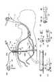
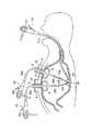
図1に示すように、この実施の形態にかかる治療装置は、2つの軟性内視鏡11,22を備えている。これら内視鏡11,22は、それぞれ体腔内に挿入可能な軟性の挿入部11a,22aを備えている。また、これら挿入部11a,22aの先端にはそれぞれ湾曲可能な湾曲部11b,22bを備えている。さらに、これら湾曲部11b,22bは、内視鏡11,22の本体にそれぞれ設けられた操作部11c,22cで操作される。これら内視鏡11,22の挿入部11a,22aには図示しないが、例えば鉗子チャンネルや送気・送水・吸引チャンネル、観察光学系などのうち、選択された内蔵物が配設されている。この実施の形態では、少なくとも1つの鉗子チャンネルと、観察光学系とが各軟性内視鏡11,22の挿入部11a,22aに設けられている。
(First embodiment)
First, a first embodiment will be described with reference to FIGS. 1 to 3.
As shown in FIG. 1, the treatment device according to this embodiment includes twoflexible endoscopes 11 and 22. Each of theseendoscopes 11 and 22 includesflexible insertion portions 11a and 22a that can be inserted into a body cavity. Moreover, thebending parts 11b and 22b which can be bent are provided at the front-end | tip of theseinsertion parts 11a and 22a, respectively. Further, these bendingportions 11b and 22b are operated byoperation portions 11c and 22c provided in the main bodies of theendoscopes 11 and 22, respectively. Although not shown in the drawings, theinsertion portions 11a and 22a of theendoscopes 11 and 22 are provided with a selected built-in object such as a forceps channel, an air / water / aspiration channel, and an observation optical system. In this embodiment, at least one forceps channel and an observation optical system are provided in theinsertion portions 11a and 22a of theflexible endoscopes 11 and 22, respectively.

これら軟性内視鏡11,22の挿入部11a,22aは例えば胃2などの臓器内に挿入される。一方の第1の軟性内視鏡11は経口的に胃2の内部に挿入され、他方の第2の軟性内視鏡22は胃2の内部と体外とを連通する胃ろう形成チューブ4を介して胃2の内部に挿入される。この胃ろう形成チューブ4はドーム部6とチューブ8とからなり、このチューブ8の一端がドーム部6に接続されている。また、この胃ろう形成チューブ4には皮膚表面でストッパー10が噛まされて、胃2の内部に落下しないように係止される。なお、これらドーム部6および湾曲チューブ8は例えば医療用の塩化ビニルやPTFEやシリコーンゴム材などで形成されていることが好適である。  Theinsertion portions 11a and 22a of theseflexible endoscopes 11 and 22 are inserted into an organ such as thestomach 2, for example. One firstflexible endoscope 11 is orally inserted into thestomach 2 and the other secondflexible endoscope 22 is passed through a gastricfistula formation tube 4 that communicates the inside of thestomach 2 with the outside of the body. Inserted into thestomach 2. The gastricfistula formation tube 4 includes adome portion 6 and atube 8, and one end of thetube 8 is connected to thedome portion 6. In addition, the gastricfistula formation tube 4 is engaged with astopper 10 on the skin surface so as not to fall into thestomach 2. Thedome 6 and thecurved tube 8 are preferably made of, for example, medical vinyl chloride, PTFE, silicone rubber material, or the like.

この胃ろう形成チューブ4は以下のようにして留置され、胃2の内部と体外とを接続する。
まず、図2の(A)に示すように、第1の軟性内視鏡11の挿入部11aを胃2の内部に経口的に挿入しておく。そして、胃ろう形成チューブ4の配置部位を選定するため、腹壁を指で押して、胃壁2aが粘膜下腫瘍のように内腔に隆起することを確認する。次に、第1の内視鏡11を用いてその内視鏡11の挿入部11a先端からの透過光を腹壁越しに確認し、この透過光が弱い場合には第1の内視鏡11の光量設定を行なう。そして、穿刺位置(胃ろう孔形成位置)を選定し、この場所をマジックペンなどでマークしておく。
その後、選定部位に外筒付き穿刺針(図示せず)を胃2の内部まで穿刺し、内筒である穿刺針を抜去する。そして、図2の(B)に示すように、外筒12内にループワイヤ13を挿入し、予め経口的に胃2の内部に挿入された第1の内視鏡11の鉗子チャンネルの1つに挿入されたポリペクトミー用スネア14で把持する。
図2の(C)に示すように、このスネア14でループワイヤ13を把持したまま第1の内視鏡11を口腔外に引き出す。このとき、外筒12から体腔外には、上述のループワイヤ13がさらに延びている。
そして、胃ろう形成チューブ4と口腔から引き出されたループワイヤ13とを結び、図2の(D)に示すように体表部側のループワイヤ13を引っ張って胃ろう形成チューブ4を経口的に胃2内にドーム部6を密着させ、チューブ8を体腔外に引き出した状態にする。
その後、図2の(E)に示すように、第1の内視鏡11を再び経口的に挿入し、胃ろう形成チューブ4のドーム部6が胃壁に食い込んでいないことを内視鏡11で観察して確認する。
そして、チューブ8を所望の位置で切断し、図1に示すように、ストッパー10を噛ませて、皮膚表面がストッパー10に対して水平になるように調整しておく。このストッパー10を噛ませたことによって、チューブ8の内径が変化するものではない。なお、胃ろう形成チューブ4を用いて胃2の内部に第2の内視鏡22や流体物などを導入しない場合、フィーディングアダプター(図示せず)などを体腔外側のチューブ8の他端部に嵌合させ、胃2の内部と外部とを遮断することが好適である。
The gastricfistula formation tube 4 is placed in the following manner to connect the inside of thestomach 2 to the outside of the body.
First, as shown in FIG. 2A, theinsertion portion 11a of the firstflexible endoscope 11 is orally inserted into thestomach 2. And in order to select the arrangement | positioning site | part of the gastricfistula formation tube 4, an abdominal wall is pushed with a finger | toe and it is confirmed that thestomach wall 2a protrudes in a lumen like a submucosa tumor. Next, using thefirst endoscope 11, the transmitted light from the distal end of theinsertion portion 11a of theendoscope 11 is confirmed through the abdominal wall, and when the transmitted light is weak, thefirst endoscope 11 Set the light intensity. Then, a puncture position (gastric fistula formation position) is selected, and this place is marked with a magic pen or the like.
Thereafter, a puncture needle with an outer cylinder (not shown) is punctured into thestomach 2 at the selected site, and the puncture needle as the inner cylinder is removed. Then, as shown in FIG. 2B, one of the forceps channels of thefirst endoscope 11 in which theloop wire 13 is inserted into theouter tube 12 and is orally inserted into thestomach 2 in advance. It is gripped by thepolypectomy snare 14 inserted in.
As shown in FIG. 2C, thefirst endoscope 11 is pulled out of the oral cavity while holding theloop wire 13 with thesnare 14. At this time, theloop wire 13 further extends from theouter cylinder 12 to the outside of the body cavity.
Then, the gastricfistula formation tube 4 is connected to theloop wire 13 drawn from the oral cavity, and theloop wire 13 on the body surface side is pulled as shown in FIG. Thedome 6 is brought into close contact with thestomach 2 and thetube 8 is pulled out of the body cavity.
Thereafter, as shown in FIG. 2E, thefirst endoscope 11 is orally inserted again, and theendoscope 11 confirms that thedome portion 6 of the gastricfistula formation tube 4 does not bite into the stomach wall. Observe and confirm.
And thetube 8 is cut | disconnected in a desired position, and as shown in FIG. 1, thestopper 10 is bitten and it adjusts so that the skin surface may become horizontal with respect to thestopper 10. FIG. The inside diameter of thetube 8 does not change by biting thestopper 10. When thesecond endoscope 22 or fluid is not introduced into thestomach 2 using the gastricfistula formation tube 4, a feeding adapter (not shown) or the like is used as the other end of thetube 8 outside the body cavity. It is preferable that the inside and outside of thestomach 2 are shut off by being fitted to each other.

胃2の内部の背中側の胃粘膜2aに病変部28が存在する場合、この病変部28はこれら2つの軟性内視鏡11,22によって以下のように処置される。
まず、胃ろう形成チューブ4を留置する際に第1の内視鏡11を用いて病変部28の位置や大きさを確認する。
次に、例えば胃ろう形成チューブ4を介して胃2の内部にエアーを送り、胃2の内部を膨らませる。
続いて、第1の軟性内視鏡11を経口的に胃2の内部に挿入する。また、この内視鏡11の挿入部11aの鉗子チャンネルの1つに例えば高周波スネア26を配置する。
そして、胃ろう形成チューブ4を介して経腹腔的または経皮的に胃2の内部に第2の軟性内視鏡22を挿入する。また、この内視鏡22の挿入部22aの鉗子チャンネルに把持鉗子24および図示しない注射針をそれぞれ挿入する。
その後、第1および第2の内視鏡11,22で病変部28を再確認する。そして、第2の内視鏡22に挿入された注射針で例えば生理食塩水を病変部28の組織下に局注し、病変部28を把持鉗子24で把持しやすいように盛り上げる。また、第1の内視鏡11に挿入された高周波スネア26をこの病変部28のまわりに配置する。続いて、第2の内視鏡22に挿入された把持鉗子24を用いてこの病変部28を把持した後、把持鉗子24を胃2の中心方向に引っ張って病変部28を持ち上げる。予め配置しておいた高周波スネア26で持ち上げられて隆起した病変部28を緊縛した後、高周波電流を流して切除する。
そして、このような切除処置が終了した後、切除した病変部28を把持したまま第2の軟性内視鏡22を生体内から抜去し、病変部28を回収して検査を行なう。
病変部28の検査の結果、この病変部28の深さや大きさ、浸潤度などによっては内視鏡による病変部28の切除のみで治療処置を完了する。
また、胃ろう形成チューブ4のストッパー10を外した後、第1の内視鏡11に配設したスネア26を用いてドーム部6を引っ掛けて保持し、経口的に取り除く。
When thelesioned part 28 exists in thegastric mucosa 2a on the back side inside thestomach 2, thelesioned part 28 is treated by the twoflexible endoscopes 11 and 22 as follows.
First, when the gastricfistula formation tube 4 is placed, the position and size of thelesioned part 28 are confirmed using thefirst endoscope 11.
Next, for example, air is sent into thestomach 2 through the gastricfistula formation tube 4 to inflate thestomach 2.
Subsequently, the firstflexible endoscope 11 is orally inserted into thestomach 2. Further, for example, a high-frequency snare 26 is disposed in one of the forceps channels of theinsertion portion 11a of theendoscope 11.
Then, the secondflexible endoscope 22 is inserted into thestomach 2 transperitoneally or percutaneously through the gastricfistula formation tube 4. In addition, the grasping forceps 24 and an injection needle (not shown) are inserted into the forceps channel of theinsertion portion 22a of theendoscope 22, respectively.
Thereafter, thelesioned part 28 is reconfirmed with the first andsecond endoscopes 11 and 22. Then, for example, physiological saline is locally injected under the tissue of thelesioned part 28 with an injection needle inserted into thesecond endoscope 22, and thelesioned part 28 is raised so as to be easily grasped by thegrasping forceps 24. Further, a high-frequency snare 26 inserted into thefirst endoscope 11 is arranged around thelesioned portion 28. Subsequently, after grasping thelesioned part 28 using the graspingforceps 24 inserted into thesecond endoscope 22, the graspingforceps 24 is pulled toward the center of thestomach 2 to lift thelesioned part 28. Thelesioned part 28 that is lifted and raised by the high-frequency snare 26 that has been arranged in advance is bound and then excised by flowing a high-frequency current.
After such excision treatment is completed, the secondflexible endoscope 22 is removed from the living body while holding the excisedlesioned part 28, and thelesioned part 28 is collected and examined.
As a result of the examination of thelesioned part 28, depending on the depth and size of thelesioned part 28, the degree of infiltration, etc., the therapeutic treatment is completed only by excision of thelesioned part 28 with an endoscope.
Further, after removing thestopper 10 of the gastricfistula formation tube 4, thedome portion 6 is hooked and held using thesnare 26 disposed in thefirst endoscope 11 and removed orally.

なお、この実施の形態では病変部28を第1の軟性内視鏡11に配設した高周波スネア26を用いて切除することを説明したが、切除処置具は高周波スネア26に限ることはない。例えば、図3に示す針状メス30を用いて病変部28を切除するようにしてもよい。この針状メス30を用いることによって、図1に示す高周波スネア26を用いて病変部28を切除する場合に比べて切除範囲を大きく採ることができる。
また、上述のように、第1および第2の軟性内視鏡11,22の挿入部22aに観察光学系を設けたが、第2の軟性内視鏡22の観察光学系が除かれて、第1の内視鏡11で第2の内視鏡22を誘導するようにしてもよい。
In this embodiment, it has been described that thelesioned portion 28 is excised using the high-frequency snare 26 disposed in the firstflexible endoscope 11, but the excision treatment tool is not limited to the high-frequency snare 26. For example, thelesioned part 28 may be excised using theneedle knife 30 shown in FIG. By using thisneedle scalpel 30, the excision range can be made larger than when thelesioned part 28 is excised using the high-frequency snare 26 shown in FIG.
Further, as described above, the observation optical system is provided in theinsertion portion 22a of the first and secondflexible endoscopes 11 and 22, but the observation optical system of the secondflexible endoscope 22 is removed, Thesecond endoscope 22 may be guided by thefirst endoscope 11.

したがって、この実施の形態について以下のことがいえる。
2つの軟性内視鏡を用いることによって、治療装置の操作性を向上させることができるので、胃2の内部の粘膜2aや胃壁などにある病変部28をより確実に切除することができる。また、このために、病変部28を取り残す可能性が低く、再発の可能性を低く抑えることができる。
また、軟性の内視鏡を用いることによって、背中側に病変部が存在する場合だけでなく、腹腔(胃ろう孔)近傍に病変部が存在する場合にもこの病変部28を同様に切除することができる。
Therefore, the following can be said with respect to this embodiment.
By using two flexible endoscopes, the operability of the treatment apparatus can be improved, so that thelesioned part 28 in themucous membrane 2a, stomach wall, etc. inside thestomach 2 can be excised more reliably. For this reason, the possibility of leaving thelesioned part 28 is low, and the possibility of recurrence can be kept low.
Further, by using a flexible endoscope, not only when a lesion is present on the back side, but also when a lesion is present near the abdominal cavity (gastric fistula), thelesion 28 is similarly excised. be able to.

病変部28の深さや大きさ、浸潤度などによって高周波スネア26や針状メス30などの切除処置具を選択して使用することによって、より確実に手技を行なうことができる。  By selecting and using a resection treatment tool such as the high-frequency snare 26 or theneedle scalpel 30 according to the depth, size, infiltration degree, and the like of thelesioned part 28, the procedure can be performed more reliably.

(第2の実施の形態)
次に、第2の実施の形態について図4を用いて説明する。この実施の形態は第1の実施の形態の変形例であり、同一の部材には同一の符号を付し、詳しい説明を省略する。
(Second Embodiment)
Next, a second embodiment will be described with reference to FIG. This embodiment is a modification of the first embodiment, and the same members are denoted by the same reference numerals and detailed description thereof is omitted.

図4の(A)に示すように、この実施の形態にかかる治療装置は、胃2の内部に第1および第2の軟性内視鏡11,22が挿入されている。なお、第2の軟性内視鏡22の挿入部22aからは観察光学系が除かれている。このため、この内視鏡22を用いて胃2の内部を観察することができない。したがって、第2の軟性内視鏡22は後述する処置具(例えば把持鉗子24)の胃2の内部への導入用具として用いられる。一方で、この軟性内視鏡22の挿入部22aは観察光学系を除くことによって、少なくともこの観察光学系が配設されている場合よりも細く形成される。  As shown in FIG. 4A, in the treatment apparatus according to this embodiment, first and secondflexible endoscopes 11 and 22 are inserted into thestomach 2. The observation optical system is removed from theinsertion portion 22a of the secondflexible endoscope 22. For this reason, the inside of thestomach 2 cannot be observed using theendoscope 22. Therefore, the secondflexible endoscope 22 is used as a tool for introducing a treatment tool (for example, grasping forceps 24) described later into thestomach 2. On the other hand, theinsertion portion 22a of theflexible endoscope 22 is formed to be narrower than at least when the observation optical system is provided by removing the observation optical system.

また、図4の(B)に示すように、第2の軟性内視鏡22の挿入部22aの先端近傍には、側方に突出した突出部32が設けられている。この突出部32には挿入部22aに沿った方向に孔34が設けられている。この孔34には、把持鉗子24の挿入部24aが挿入されている。  Further, as shown in FIG. 4B, a protrudingportion 32 protruding sideways is provided in the vicinity of the distal end of theinsertion portion 22a of the secondflexible endoscope 22. Theprotrusion 32 is provided with ahole 34 in a direction along theinsertion portion 22a. Theinsertion portion 24 a of the graspingforceps 24 is inserted into thehole 34.

胃2の内部の背中側の胃粘膜に病変部28が存在する場合、この病変部28はこれら2つの軟性内視鏡11,22によって以下のように処置される。
まず、胃ろう形成チューブ4を留置する際に第1の内視鏡11を用いて病変部28の大きさを確認する。
そして、例えば胃ろう形成チューブ4を介して胃2の内部にエアーを送り、胃2の内部を膨らませる。
次に、第1の軟性内視鏡11を経口的に胃2の内部に挿入する。また、この内視鏡11の挿入部11aの鉗子チャンネルの1つに例えば高周波スネア26を配置する。
そして、上述した孔34に把持鉗子24を挿入した状態で、胃ろう形成チューブ4を介して経腹腔的または経皮的に胃2の内部に第2の軟性内視鏡22を挿入する。
その後、第1の軟性内視鏡11で病変部28を再確認する。そして、第1の軟性内視鏡11を用いて第2の軟性内視鏡22に沿って配設された把持鉗子24を病変部28に誘導する。
次に、第1の軟性内視鏡11に配設された高周波スネア26の先端部を病変部28の周囲を取り囲むように配置する。そして、把持鉗子24で病変部28を把持して胃2の中心部方向に引っ張り、高周波スネア26を緊縛させて高周波電流を流して病変部28を切除する。
このような処置が終了した後、切除部分を把持したまま第2の軟性内視鏡22を生体内から抜去し、病変部28を回収する。
また、必要に応じて、胃ろう形成チューブ4のストッパー10を外した後、第1の内視鏡11に配設したスネア26を用いてドーム部6を引っ掛けて保持し、胃ろう形成チューブ4を経口的に取り除く。
When thelesioned part 28 exists in the gastric mucosa on the back side inside thestomach 2, thelesioned part 28 is treated by these twoflexible endoscopes 11 and 22 as follows.
First, when the gastricfistula formation tube 4 is placed, the size of thelesioned part 28 is confirmed using thefirst endoscope 11.
Then, for example, air is sent into thestomach 2 via the gastricfistula formation tube 4 to inflate thestomach 2.
Next, the firstflexible endoscope 11 is inserted into thestomach 2 orally. Further, for example, a high-frequency snare 26 is disposed in one of the forceps channels of theinsertion portion 11a of theendoscope 11.
Then, with the graspingforceps 24 inserted into thehole 34 described above, the secondflexible endoscope 22 is inserted into thestomach 2 through the gastricfistula formation tube 4 transperitoneally or percutaneously.
Thereafter, thelesioned part 28 is reconfirmed with the firstflexible endoscope 11. Then, using the firstflexible endoscope 11, the graspingforceps 24 disposed along the secondflexible endoscope 22 is guided to thelesioned part 28.
Next, the distal end portion of the high-frequency snare 26 disposed in the firstflexible endoscope 11 is disposed so as to surround thelesioned portion 28. Then, thelesioned part 28 is grasped by the graspingforceps 24 and pulled toward the central part of thestomach 2, the high-frequency snare 26 is bound, and a high-frequency current is applied to excise thelesioned part 28.
After such treatment is completed, the secondflexible endoscope 22 is removed from the living body while holding the excised part, and thelesioned part 28 is recovered.
Further, if necessary, after removing thestopper 10 of the gastricfistula formation tube 4, thedome portion 6 is hooked and held by using thesnare 26 disposed in thefirst endoscope 11, and the gastricfistula formation tube 4. Remove orally.

したがって、この実施の形態について以下のことがいえる。
第2の軟性内視鏡22から観察光学系を除くとともに、例えば把持鉗子24などの鉗子を挿入部22aの外側に配置したので、挿入部22a自体を細くすることができる。また、鉗子を外側に配置することによって、内視鏡22の鉗子チャンネルの大きさに関係なく、様々な種類、様々な大きさ(太さ)の鉗子を使用することができる。
Therefore, the following can be said with respect to this embodiment.
Since the observation optical system is removed from the secondflexible endoscope 22 and forceps such as the graspingforceps 24 are disposed outside theinsertion portion 22a, theinsertion portion 22a itself can be thinned. Further, by arranging the forceps outside, various types and sizes (thicknesses) of forceps can be used regardless of the size of the forceps channel of theendoscope 22.

なお、鉗子を挿入部22aに沿って配設する構成は、上述したものに限ることはなく、例えば以下のようなものを挙げることができる。
図4の(C)に示すように、上述の突出部32には例えば把持鉗子24が係止されるフック部36が設けられている。このフック部36に上述の把持鉗子24が係止され、同様に処置を行なってもよい。
また、図4の(D)に示すように、第2の軟性内視鏡22の先端部には筒状部材38が着脱可能に形成されている。この筒状部材38には側方に突出した突出部32が設けられている。この突出部32には把持鉗子24の挿入部24aに沿った方向に孔34が設けられている。この孔34には把持鉗子24の挿入部24aを挿入可能である。
また、図4の(E)に示すように、図4の(D)に示す筒状部材38に設けられた孔34には第2の軟性内視鏡22の挿入部22aの後方に向かってガイドチューブ42が延びている。このガイドチューブ42内に例えば把持鉗子24などの所望の処置具が挿通されてガイドチューブ42の先端部から突出される。
さらに、図4の(F)に示すように、第2の軟性内視鏡22とガイドチューブ42とがサージカルテープ(医療用テープ)44によって挿入部22aに沿って配設されている。図4の(E)と同様に、このガイドチューブ42内に例えば把持鉗子24などの所望の処置具が挿通されてガイドチューブ42の先端部から突出される。
In addition, the structure which arrange | positions forceps along theinsertion part 22a is not restricted to what was mentioned above, For example, the following can be mentioned.
As shown in FIG. 4C, theprotrusion 32 described above is provided with ahook portion 36 to which the graspingforceps 24 are locked, for example. The above-described graspingforceps 24 is locked to thehook portion 36, and treatment may be performed in the same manner.
Further, as shown in FIG. 4D, acylindrical member 38 is detachably formed at the distal end portion of the secondflexible endoscope 22. Thecylindrical member 38 is provided with a protrudingportion 32 protruding sideways. Theprotrusion 32 is provided with ahole 34 in a direction along theinsertion portion 24 a of the graspingforceps 24. Theinsertion portion 24 a of the graspingforceps 24 can be inserted into thehole 34.
As shown in FIG. 4E, thehole 34 provided in thetubular member 38 shown in FIG. 4D is inserted toward the rear of theinsertion portion 22a of the secondflexible endoscope 22. Aguide tube 42 extends. A desired treatment instrument such as the graspingforceps 24 is inserted into theguide tube 42 and protrudes from the distal end portion of theguide tube 42.
Further, as shown in FIG. 4F, the secondflexible endoscope 22 and theguide tube 42 are disposed along theinsertion portion 22a by a surgical tape (medical tape) 44. Similarly to (E) of FIG. 4, a desired treatment instrument such as the graspingforceps 24 is inserted into theguide tube 42 and protrudes from the distal end portion of theguide tube 42.

なお、第2の軟性内視鏡22の挿入部22aには、観察光学系が除かれて細く形成されていることを説明したが、もちろん通常の軟性内視鏡のように観察光学系が設けられて、2つの内視鏡11,22で病変部28を観察しながら処置を行なうことも好適である。または、観察光学系の代わりに鉗子チャンネルを除いても良い。また、第1の軟性内視鏡11によって胃2の内部にエアーを送っても良い。  Although it has been described that the insertionoptical part 22a of the secondflexible endoscope 22 is formed thin by removing the observation optical system, of course, the observation optical system is provided like a normal flexible endoscope. It is also preferable to perform the treatment while observing thelesioned part 28 with the twoendoscopes 11 and 22. Alternatively, the forceps channel may be removed instead of the observation optical system. In addition, air may be sent into thestomach 2 by the firstflexible endoscope 11.

(第3の実施の形態)
第3の実施の形態について図5を用いて説明する。この実施の形態は第1および第2の実施の形態の変形例であり、同一の部材には同一の符号を付し、詳しい説明を省略する。
(Third embodiment)
A third embodiment will be described with reference to FIG. This embodiment is a modification of the first and second embodiments, and the same members are denoted by the same reference numerals and detailed description thereof is omitted.

図5の(A)に示すように、この実施の形態にかかる治療装置は、胃2の内部に第1および第2の軟性内視鏡11,22が挿入されている。また、図5の(B)に示すように、第2の軟性内視鏡22の湾曲部22bの基端部近傍に4つの指標(識別ラベル)50が設けられている。これら指標50は第2の軟性内視鏡22の周方向を例えば上(U)方向50a、下(D)方向50b、右(R)方向(図示せず)、左(L)方向50cの4つの方向とすると、これら指標50がそれぞれ例えば赤、青、黄、緑などに色分けされて設けられている。このような指標50を第1の軟性内視鏡11を用いて観察することによって、第1の軟性内視鏡11の術者が第2の軟性内視鏡22の術者に向かって第2の軟性内視鏡22の湾曲部22bを動かしてほしい方向を容易に指示することができる。  As shown in FIG. 5A, in the treatment apparatus according to this embodiment, the first and secondflexible endoscopes 11 and 22 are inserted into thestomach 2. Further, as shown in FIG. 5B, four indicators (identification labels) 50 are provided in the vicinity of the proximal end portion of thecurved portion 22b of the secondflexible endoscope 22. These indicators 50 are, for example, four circumferential directions of the second flexible endoscope 22: an upward (U)direction 50a, a downward (D)direction 50b, a right (R) direction (not shown), and a left (L)direction 50c. Assuming that there are two directions, these indices 50 are provided in different colors, for example, red, blue, yellow, and green. By observing such an index 50 using the firstflexible endoscope 11, the operator of the firstflexible endoscope 11 is directed to the operator of the secondflexible endoscope 22. The direction in which the bendingportion 22b of theflexible endoscope 22 is desired to be moved can be easily indicated.

胃2の内部の背中側の胃粘膜に病変部28が存在する場合、この病変部28はこれら2つの軟性内視鏡11,22によって以下のように処置される。
まず、胃ろう形成チューブ4を留置する際に第1の内視鏡11を用いて病変部28の大きさを確認しておく。
そして、例えば胃ろう形成チューブ4を介して胃2の内部にエアーを送り、胃2の内部を膨らませる。
次に、第1の軟性内視鏡11を経口的に胃2の内部に挿入するとともに、第2の軟性内視鏡22を胃ろう形成チューブ4を介して胃2の内部に挿入する。
そして、第1の軟性内視鏡11で病変部28を再確認する。その後、第2の軟性内視鏡22の挿入部22aの先端部を図5の(B)に示すように、第1の内視鏡11に設けた観察光学系で映す。さらに、第2の軟性内視鏡22の湾曲部22bを指標50にしたがって病変部28に向けて移動や湾曲などさせてこの病変部28を把持する。すなわち、第1の内視鏡11を用いて第2の内視鏡を所望の位置に誘導する。
次に、第2の内視鏡22に設けた把持鉗子24で病変部28を把持し、胃2の中心部方向に引っ張った状態で、第1の内視鏡11に配設した高周波スネア(図示せず)などで病変部28を切除する。
When thelesioned part 28 exists in the gastric mucosa on the back side inside thestomach 2, thelesioned part 28 is treated by these twoflexible endoscopes 11 and 22 as follows.
First, when the gastricfistula formation tube 4 is placed, the size of thelesioned part 28 is confirmed using thefirst endoscope 11.
Then, for example, air is sent into thestomach 2 via the gastricfistula formation tube 4 to inflate thestomach 2.
Next, the firstflexible endoscope 11 is orally inserted into thestomach 2, and the secondflexible endoscope 22 is inserted into thestomach 2 through the gastricfistula formation tube 4.
Then, thelesioned part 28 is reconfirmed with the firstflexible endoscope 11. Thereafter, the distal end portion of theinsertion portion 22a of the secondflexible endoscope 22 is projected by an observation optical system provided in thefirst endoscope 11 as shown in FIG. Further, thecurved portion 22 b of the secondflexible endoscope 22 is moved or curved toward thelesioned portion 28 according to the index 50 to grip thelesioned portion 28. That is, the second endoscope is guided to a desired position using thefirst endoscope 11.
Next, thelesioned part 28 is grasped with the graspingforceps 24 provided on thesecond endoscope 22 and pulled toward the central part of thestomach 2, and the high-frequency snare (which is disposed on the first endoscope 11) Thelesioned part 28 is excised with a not-shown).

ところで、このような指標50は例えば湾曲部22bや挿入部22aなどの先端部に例えばプラス記号形状の溝、マイナス記号形状の溝、三角形状の溝などを同色に設けてもよい。また、色分けは上述したものに限ることはなく、第1の軟性内視鏡11の観察光学系を用いて認識しやすいものであれば、他の色分けをしても構わない。  By the way, such an index 50 may be provided with, for example, a plus symbol-shaped groove, a minus symbol-shaped groove, a triangular groove, or the like in the same color at the tip of thecurved portion 22b or theinsertion portion 22a. The color coding is not limited to that described above, and other color coding may be used as long as it is easy to recognize using the observation optical system of the firstflexible endoscope 11.

したがって、この実施の形態について以下のことがいえる。
第1の内視鏡11の術者が第2の内視鏡22の術者に対して第2の内視鏡22の操作指示を行なう場合、湾曲部22bを動かしてほしい方向を第1の内視鏡11の術者が容易に認識することができる。このため、第1の内視鏡11の術者と、第2の内視鏡22の術者との連携を取りやすくすることができる。すなわち、治療処置を円滑に進めることができ、処置時間の短縮を図ることができる。
Therefore, the following can be said with respect to this embodiment.
When the operator of thefirst endoscope 11 instructs the operator of thesecond endoscope 22 to operate thesecond endoscope 22, the direction in which the bendingportion 22b is desired to be moved is set to the first direction. The operator of theendoscope 11 can easily recognize. For this reason, it is possible to facilitate cooperation between the operator of thefirst endoscope 11 and the operator of thesecond endoscope 22. That is, the therapeutic treatment can be smoothly advanced, and the treatment time can be shortened.

(第4の実施の形態)
第4の実施の形態について図6および図7を用いて説明する。この実施の形態は第1の実施の形態の変形例であり、同一の部材には同一の符号を付し、詳しい説明を省略する。
(Fourth embodiment)
A fourth embodiment will be described with reference to FIGS. This embodiment is a modification of the first embodiment, and the same members are denoted by the same reference numerals and detailed description thereof is omitted.

図6に示すように、この実施の形態にかかる治療装置は、胃2の内部に第1および第2の軟性内視鏡11,22が挿入されている。  As shown in FIG. 6, in the treatment apparatus according to this embodiment, first and secondflexible endoscopes 11 and 22 are inserted into thestomach 2.

胃ろう形成チューブ4には手元口金60が取り付けられている。この手元口金60は図7に示すように、管状部62と、この管状部62よりも大径のフランジ部64とを備えている。管状部62は胃ろう形成チューブ4のチューブ8の開口端部に嵌合されている。また、フランジ部64にはばね66が配設されている。このばね66上にはプラスチック材や金属材など、剛性を有し、かつ、中心に第2の軟性内視鏡22の挿入部22aの外径よりも大径の円孔68を有する支持板70が配設されている。さらに、この支持板70の上には例えばゴム材などの弾性部材からなるシール部材72が配設されている。このシール部材72の中央部には第2の内視鏡22の挿入部22aの外径よりも小径の円孔やプラス記号形状、マイナス記号形状などの形状を有する貫通孔が形成されている。この貫通孔と支持板70に設けられた円孔68とは連通されている。さらに、フランジ部64の外周にはシール部材72を留める蓋74が螺合される螺合部76が形成されている。  Ahand cap 60 is attached to the gastricfistula formation tube 4. As shown in FIG. 7, thehand cap 60 includes atubular portion 62 and aflange portion 64 having a diameter larger than that of thetubular portion 62. Thetubular portion 62 is fitted to the open end of thetube 8 of the gastricfistula formation tube 4. Aspring 66 is disposed on theflange portion 64. On thisspring 66, asupport plate 70 having acircular hole 68 having rigidity, such as a plastic material or a metal material, and having a diameter larger than the outer diameter of theinsertion portion 22a of the secondflexible endoscope 22 at the center. Is arranged. Further, aseal member 72 made of an elastic member such as a rubber material is disposed on thesupport plate 70. A circular hole having a diameter smaller than the outer diameter of theinsertion portion 22a of thesecond endoscope 22 and a through hole having a shape such as a plus symbol shape or a minus symbol shape are formed in the central portion of theseal member 72. The through hole communicates with thecircular hole 68 provided in thesupport plate 70. Further, on the outer periphery of theflange portion 64, a screwingportion 76 to which alid 74 for fastening theseal member 72 is screwed is formed.

このような手元口金60に第2の内視鏡22の挿入部22aを挿入する。内視鏡22の挿入部22aにシール部材72が密着した状態で挿入部22aが胃2の内部に次第に挿入される。このとき、シール部材72はフランジ部64に配設されたばね66の付勢力に抗して支持板70を胃2の内部方向に向けて移動させる。内視鏡22の挿入部22aの挿入を留めると、支持板70がばね66の付勢力によって胃2の外部方向に移動されるとともに、シール部材72が同一方向に移動される。すなわち、内視鏡22の挿入部22aはシール部材72に密着した状態で、胃2の内部から外側に押出す方向に付勢されている。例えば病変部28を把持鉗子24で把持した場合、病変部28を胃2の外部方向に引っ張った状態になる。  Theinsertion portion 22a of thesecond endoscope 22 is inserted into such ahand cap 60. Theinsertion portion 22a is gradually inserted into thestomach 2 with theseal member 72 in close contact with theinsertion portion 22a of theendoscope 22. At this time, theseal member 72 moves thesupport plate 70 toward the inside of thestomach 2 against the urging force of thespring 66 disposed in theflange portion 64. When the insertion of theinsertion portion 22a of theendoscope 22 is stopped, thesupport plate 70 is moved to the outside of thestomach 2 by the biasing force of thespring 66, and theseal member 72 is moved in the same direction. That is, theinsertion portion 22a of theendoscope 22 is urged in a direction of pushing outward from the inside of thestomach 2 while being in close contact with theseal member 72. For example, when thelesioned part 28 is grasped by the graspingforceps 24, thelesioned part 28 is pulled outward from thestomach 2.

胃2の内部の背中側の胃粘膜に病変部28が存在する場合、この病変部28はこれら2つの軟性内視鏡11,22によって第1の実施の形態で説明した処置と同様に処置される。  When thelesioned part 28 exists in the gastric mucosa on the back side inside thestomach 2, thelesioned part 28 is treated in the same manner as the treatment described in the first embodiment by these twoflexible endoscopes 11 and 22. The

したがって、この実施の形態について以下のことがいえる。
第2の軟性内視鏡22をこの手元口金60を介して胃2の内部に挿入した場合、挿入部22aはシール部材72に密着されている。さらに、ばね66によって支持板70およびシール部材72が上方に付勢されている。このため、間接的に挿入部22aが上方に付勢され、病変部28を把持した状態で第2の内視鏡22を保持するだけで、病変部28が上側に引っ張られた状態にされる。
また、シール部材72は内視鏡22の挿入部22aが挿入された場合、その外径部に密着するので、胃2の内部を膨らませたエアーを体外に漏れ難くすることができる。
Therefore, the following can be said with respect to this embodiment.
When the secondflexible endoscope 22 is inserted into thestomach 2 through thehand cap 60, theinsertion portion 22 a is in close contact with theseal member 72. Further, thesupport plate 70 and theseal member 72 are urged upward by thespring 66. For this reason, theinsertion portion 22a is indirectly biased upward, and thelesioned portion 28 is pulled upward only by holding thesecond endoscope 22 while holding thelesioned portion 28. .
Further, when theinsertion portion 22a of theendoscope 22 is inserted, theseal member 72 is in close contact with the outer diameter portion thereof, so that the air inflating the inside of thestomach 2 can be made difficult to leak out of the body.

(第5の実施の形態)
第5の実施の形態について図8を用いて説明する。この実施の形態は第1の実施の形態の変形例であり、同一の部材には同一の符号を付し、詳しい説明を省略する。
(Fifth embodiment)
A fifth embodiment will be described with reference to FIG. This embodiment is a modification of the first embodiment, and the same members are denoted by the same reference numerals and detailed description thereof is omitted.

図8の(A)に示すように、この実施の形態にかかる治療装置は、胃2の内部に第1および第2の軟性内視鏡11,22が挿入されている。図8の(B)に示すように、第2の内視鏡22の挿入部22aの先端近傍には、保持部80aが側方に突出されて設けられている。第1の内視鏡11の挿入部11aの先端の鉗子チャンネルには把持鉗子82が配設されている。この把持鉗子82を操作して、第2の内視鏡22の保持部80aを把持することによって、第1の内視鏡11の操作部11cを操作して、第2の内視鏡22の挿入部22aを所望の位置に案内することができる。  As shown in FIG. 8A, in the treatment apparatus according to this embodiment, the first and secondflexible endoscopes 11 and 22 are inserted into thestomach 2. As shown in FIG. 8B, in the vicinity of the distal end of theinsertion portion 22a of thesecond endoscope 22, a holdingportion 80a is provided so as to protrude laterally. A graspingforceps 82 is disposed in the forceps channel at the distal end of theinsertion portion 11 a of thefirst endoscope 11. By operating the grippingforceps 82 and gripping the holdingportion 80a of thesecond endoscope 22, theoperation portion 11c of thefirst endoscope 11 is operated, and thesecond endoscope 22 is operated. Theinsertion part 22a can be guided to a desired position.

胃2の内部の背中側の胃粘膜に病変部28が存在する場合、この病変部28はこれら2つの軟性内視鏡11,22によって以下のように処置される。
まず、胃ろう形成チューブ4を留置する際に第1の内視鏡11を用いて病変部28の大きさを確認しておく。
そして、例えば胃ろう形成チューブ4を介して胃2の内部にエアーを送り、胃2の内部を膨らませる。
次に、第1の軟性内視鏡11を経口的に胃2の内部に挿入するとともに、第2の軟性内視鏡22を胃ろう形成チューブ4を介して胃2の内部に挿入する。
そして、第1の軟性内視鏡11で病変部28を再確認する。その後、第1の軟性内視鏡11を用いて第2の軟性内視鏡22の挿入部22aの先端部に設けた保持部80aを保持する。この状態で、さらに第1の軟性内視鏡11を操作して、第2の軟性内視鏡22の挿入部22aの先端部を病変部28に向けて誘導する。すなわち、この実施の形態では第2の内視鏡22から観察光学系が除かれていても構わない。
その後、例えば第1の内視鏡11に高周波スネア(図示せず)などを配置し、第2の内視鏡22に把持鉗子(図示せず)などを配置し、これら内視鏡11,22を協働させて、病変部28の切除を行なう。
When thelesioned part 28 exists in the gastric mucosa on the back side inside thestomach 2, thelesioned part 28 is treated by these twoflexible endoscopes 11 and 22 as follows.
First, when the gastricfistula formation tube 4 is placed, the size of thelesioned part 28 is confirmed using thefirst endoscope 11.
Then, for example, air is sent into thestomach 2 via the gastricfistula formation tube 4 to inflate thestomach 2.
Next, the firstflexible endoscope 11 is orally inserted into thestomach 2, and the secondflexible endoscope 22 is inserted into thestomach 2 through the gastricfistula formation tube 4.
Then, thelesioned part 28 is reconfirmed with the firstflexible endoscope 11. Thereafter, the firstflexible endoscope 11 is used to hold the holdingportion 80 a provided at the distal end portion of theinsertion portion 22 a of the secondflexible endoscope 22. In this state, the firstflexible endoscope 11 is further operated to guide the distal end portion of theinsertion portion 22a of the secondflexible endoscope 22 toward thelesioned portion 28. That is, in this embodiment, the observation optical system may be removed from thesecond endoscope 22.
Thereafter, for example, a high-frequency snare (not shown) or the like is arranged on thefirst endoscope 11, a grasping forceps (not shown) or the like is arranged on thesecond endoscope 22, and theseendoscopes 11 and 22 are arranged. Thelesioned part 28 is excised by cooperating with.

なお、第1の内視鏡11の把持鉗子82によって把持して所望の位置に案内する手段は第2の内視鏡22の挿入部22aの先端部に設けた保持部80aに限ることはなく、例えば図8の(C)に示すように、湾曲部22bに金属材製のブレード部80bなどを備え、このブレード部80bを第1の内視鏡11に設けた把持鉗子82やバスケット把持鉗子(図示せず)などを用いて把持して第2の内視鏡を所望の位置に誘導してもよい。このブレード部80bは胃ろう形成チューブ4を介して胃2の内部に挿入されるので、生体を傷つけることなく胃2の内部に挿入することができる。  Note that the means for grasping and guiding to the desired position by the graspingforceps 82 of thefirst endoscope 11 is not limited to the holdingportion 80a provided at the distal end portion of theinsertion portion 22a of thesecond endoscope 22. For example, as shown in FIG. 8C, the bendingportion 22b is provided with ablade portion 80b made of a metal material and the like, and theblade portion 80b is provided in thefirst endoscope 11 or the basket holding forceps. The second endoscope may be guided to a desired position by grasping it using (not shown) or the like. Since thisblade part 80b is inserted into thestomach 2 through the gastricfistula formation tube 4, it can be inserted into thestomach 2 without damaging the living body.

したがって、この実施の形態について以下のことがいえる。
病変部28が例えば腹腔側に存在している場合、第1の内視鏡11を操作して挿入部11aを移動させることによって、第2の内視鏡22も所望の位置に配設することができるので、術者の操作の手間を半減させることができる。
Therefore, the following can be said with respect to this embodiment.
When thelesioned part 28 exists on the abdominal cavity side, for example, thesecond endoscope 22 is also disposed at a desired position by operating thefirst endoscope 11 and moving theinsertion part 11a. Therefore, the operator's operation can be halved.

また、第2の内視鏡22から観察光学系が除かれている場合でも、第1の内視鏡11などを用いて病変部28に容易に誘導することができる。  Even when the observation optical system is removed from thesecond endoscope 22, it can be easily guided to thelesioned part 28 using thefirst endoscope 11 or the like.

(第6の実施の形態)
第6の実施の形態について図9を用いて説明する。この実施の形態は第1の実施の形態の変形例であり、同一の部材には同一の符号を付し、詳しい説明を省略する。
(Sixth embodiment)
A sixth embodiment will be described with reference to FIG. This embodiment is a modification of the first embodiment, and the same members are denoted by the same reference numerals and detailed description thereof is omitted.

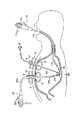
図9に示すように、この実施の形態にかかる治療装置は、胃2の内部に第1の軟性内視鏡11が挿入されている。また、胃ろう形成チューブ4には管状体84が挿入されている。この管状体84には例えば3つのルーメン(図示せず)が設けられている。このうち、2つのルーメン中には多関節アームを有する把持鉗子86が配設され、もう1つのルーメン中には第2の軟性内視鏡22が配設されている。この把持鉗子86はアーム部86aと、このアーム部86aを操作するアーム操作部86bと、先端に設けられたカップを開閉操作する開閉操作部86cとを備えている。また、第2の内視鏡22は観察光学系として使用される。このため、挿入部22aには、観察光学系のみが設けられていればよい。一方、第1の内視鏡11の挿入部11aには針状メス30が配設されている。  As shown in FIG. 9, in the treatment apparatus according to this embodiment, the firstflexible endoscope 11 is inserted into thestomach 2. Atubular body 84 is inserted into the gastricfistula formation tube 4. Thetubular body 84 is provided with, for example, three lumens (not shown). Among these, the grasping forceps 86 having a multi-joint arm is disposed in two lumens, and the secondflexible endoscope 22 is disposed in the other lumen. The grasping forceps 86 includes anarm portion 86a, anarm operation portion 86b for operating thearm portion 86a, and an opening /closing operation portion 86c for opening / closing a cup provided at the tip. Thesecond endoscope 22 is used as an observation optical system. For this reason, only the observation optical system should just be provided in theinsertion part 22a. On the other hand, aneedle scalpel 30 is disposed in theinsertion portion 11 a of thefirst endoscope 11.

胃2の内部の背中側の胃粘膜に病変部28が存在する場合、この病変部28はこれら2つの軟性内視鏡11,22などによって以下のように処置される。
まず、胃ろう形成チューブ4を留置する際に第1の内視鏡11を用いて病変部28の大きさを確認しておく。
そして、例えば胃ろう形成チューブ4を介して胃2の内部にエアーを送り、胃2の内部を膨らませる。
次に、第1の軟性内視鏡11を経口的に胃2の内部に挿入するとともに、管状体84を胃ろう形成チューブ4を介して胃2の内部に挿入する。
そして、第1の軟性内視鏡11で病変部28を再確認する。その後、管状体84に2つの把持鉗子86と第2の軟性内視鏡22とを管状体84内の図示しないルーメン内をそれぞれ挿通させる。第2の内視鏡22で病変部28を確認しながら、2つの把持鉗子86をアーム操作部86bおよび開閉操作部86cを操作して病変部28を把持して持ち上げる。
さらに、第1の内視鏡11に配設した針状メス30を用いて持ち上げた病変部28を切除する。
その後、把持鉗子86を用いて切除した病変部28を回収し、検査を行なう。
When thelesioned part 28 exists in the gastric mucosa on the back side inside thestomach 2, thelesioned part 28 is treated as follows by the twoflexible endoscopes 11 and 22.
First, when the gastricfistula formation tube 4 is placed, the size of thelesioned part 28 is confirmed using thefirst endoscope 11.
Then, for example, air is sent into thestomach 2 via the gastricfistula formation tube 4 to inflate thestomach 2.
Next, the firstflexible endoscope 11 is orally inserted into thestomach 2 and thetubular body 84 is inserted into thestomach 2 through the gastricfistula formation tube 4.
Then, thelesioned part 28 is reconfirmed with the firstflexible endoscope 11. Thereafter, the two grasping forceps 86 and the secondflexible endoscope 22 are inserted into thetubular body 84 through a lumen (not shown) in thetubular body 84. While confirming thelesioned part 28 with thesecond endoscope 22, the two grasping forceps 86 are operated by operating thearm operation part 86b and the opening /closing operation part 86c to grasp thelesioned part 28 and lift it.
Further, thelesioned part 28 lifted by using the needle-shapedknife 30 disposed in thefirst endoscope 11 is excised.
Thereafter, thelesioned part 28 excised using the grasping forceps 86 is collected and inspected.

したがって、この実施の形態について以下のことがいえる。
2つの把持鉗子86を1つの孔(胃ろう形成チューブ4)を介して挿入することによって、病変部28が広い場合に病変部28を広範囲に持ち上げることができるとともに、第1の内視鏡11を用いてこの病変部28を容易に切除することができる。
Therefore, the following can be said with respect to this embodiment.
By inserting the two grasping forceps 86 through one hole (gastrostomy tube 4), thelesioned part 28 can be lifted over a wide range when thelesioned part 28 is wide, and thefirst endoscope 11 Thelesioned part 28 can be easily excised using.

(第7の実施の形態)
第7の実施の形態について図10を用いて説明する。この実施の形態は第1の実施の形態の変形例であり、同一の部材には同一の符号を付し、詳しい説明を省略する。
(Seventh embodiment)
A seventh embodiment will be described with reference to FIG. This embodiment is a modification of the first embodiment, and the same members are denoted by the same reference numerals and detailed description thereof is omitted.

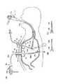
図10の(A)に示すように、この実施の形態にかかる治療装置は、胃2の内部に第1および第2の軟性内視鏡11,22が挿入されている。また、第2の軟性内視鏡22を胃2の内部に導く胃ろう形成チューブ4の上端部にはシール部材98が嵌合されている。このシール部材98は例えば2つの孔98a,98bを備えている。これら孔98a,98bには、それぞれ内視鏡22の挿入部22aと、硬性の把持鉗子100が挿通されている。このシール部材98には第2の内視鏡22の挿入部22aおよび把持鉗子100の挿入部が密着し、胃2の内部のエアーが体外に漏れ出し難く形成されている。  As shown in FIG. 10A, in the treatment apparatus according to this embodiment, the first and secondflexible endoscopes 11 and 22 are inserted into thestomach 2. Aseal member 98 is fitted to the upper end of the gastricfistula formation tube 4 that guides the secondflexible endoscope 22 into thestomach 2. Theseal member 98 includes, for example, twoholes 98a and 98b. Theinsertion portion 22a of theendoscope 22 and the rigid graspingforceps 100 are inserted into theholes 98a and 98b, respectively. Theinsertion portion 22a of thesecond endoscope 22 and the insertion portion of the graspingforceps 100 are in close contact with theseal member 98, so that the air inside thestomach 2 is difficult to leak out of the body.

また、図10の(B)に示すように、第1の内視鏡11の挿入部11aの中央部には対物レンズ90が配設されている。この対物レンズ90の側方には1対のライトガイド92が配設されている。さらに、この挿入部11aには送気・送水ノズル94が配設されている。そして、ライトガイド92と送気・送水ノズル94との間には処置具ガイド溝96がそれぞれ配設されている。また、これら処置具ガイド溝96は対物レンズ90に対して対称の位置にも設けられている。これら処置具ガイド溝96には、例えば高周波スネア26や針状メス30が配設されている。これら高周波スネア26や針状メス30は溝部開口部97から挿入部11aの先端方向に選択的に導出されて使用される。  As shown in FIG. 10B, anobjective lens 90 is disposed at the center of theinsertion portion 11a of thefirst endoscope 11. A pair of light guides 92 is disposed on the side of theobjective lens 90. Further, an air /water supply nozzle 94 is disposed in theinsertion portion 11a. A treatmentinstrument guide groove 96 is provided between thelight guide 92 and the air /water supply nozzle 94. The treatmentinstrument guide grooves 96 are also provided at symmetrical positions with respect to theobjective lens 90. In these treatmentinstrument guide grooves 96, for example, a high-frequency snare 26 and aneedle knife 30 are disposed. The high-frequency snare 26 and the needle-shapedknife 30 are selectively led out from thegroove opening 97 toward the distal end of theinsertion portion 11a.

胃2の内部の背中側の胃粘膜に病変部28が存在する場合、この病変部28はこれら2つの軟性内視鏡11,22によって以下のように処置される。
まず、例えば胃ろう形成チューブ4を介して胃2の内部にエアーを送り、胃2の内部を膨らませる。
次に、第1の軟性内視鏡11を経口的に胃2の内部に挿入するとともに、第2の軟性内視鏡22および硬性の把持鉗子100を胃ろう形成チューブ4を介して胃2の内部に挿入する。
そして、第1の軟性内視鏡11で病変部28を確認する。その後、第2の軟性内視鏡22で病変部28を確認しながらこの病変部28を把持するとともに、硬性の把持鉗子100を用いてこれを把持する。この病変部28を把持した状態でこれを持ち上げる。
次に、第1の軟性内視鏡11の処置具ガイド溝96に配設された高周波スネア26および針状メス30のいずれかを病変部28の大きさによって選択し、病変部28を切除する。
その後、第2の軟性内視鏡22に配設された把持鉗子24または硬性の把持鉗子100のいずれかを用いて切除した病変部28を回収し、検査を行なう。
When thelesioned part 28 exists in the gastric mucosa on the back side inside thestomach 2, thelesioned part 28 is treated by these twoflexible endoscopes 11 and 22 as follows.
First, for example, air is sent into thestomach 2 through the gastricfistula formation tube 4 to inflate thestomach 2.
Next, the firstflexible endoscope 11 is orally inserted into thestomach 2, and the secondflexible endoscope 22 and the rigid graspingforceps 100 are inserted into thestomach 2 through the gastricfistula formation tube 4. Insert inside.
Then, thelesioned part 28 is confirmed with the firstflexible endoscope 11. Thereafter, thelesioned part 28 is grasped while thelesioned part 28 is confirmed by the secondflexible endoscope 22 and is grasped by using the rigid graspingforceps 100. Thelesioned part 28 is lifted while being gripped.
Next, either thehigh frequency snare 26 or theneedle scalpel 30 disposed in the treatmentinstrument guide groove 96 of the firstflexible endoscope 11 is selected according to the size of thelesioned portion 28, and thelesioned portion 28 is excised. .
Thereafter, the excisedlesioned part 28 is collected using either the graspingforceps 24 or the rigid graspingforceps 100 disposed in the secondflexible endoscope 22 and inspected.

したがって、この実施の形態について以下のことがいえる。
第1の軟性内視鏡11の挿入部11aに沿って処置具ガイド溝96を設け、これら処置具ガイド溝96にいくつかの処置具を配設しておくと、病変部28の大きさなどによって、処置具を選択して使用することができる。
Therefore, the following can be said with respect to this embodiment.
When a treatmentinstrument guide groove 96 is provided along theinsertion portion 11a of the firstflexible endoscope 11, and several treatment instruments are arranged in the treatmentinstrument guide groove 96, the size of thelesioned part 28, etc. The treatment tool can be selected and used.

(第8の実施の形態)
第8の実施の形態について図11を用いて説明する。この実施の形態は第7の実施の形態の変形例であり、同一の部材には同一の符号を付し、詳しい説明を省略する。
(Eighth embodiment)
An eighth embodiment will be described with reference to FIG. This embodiment is a modification of the seventh embodiment, and the same members are denoted by the same reference numerals and detailed description thereof is omitted.

図11の(A)に示すように、この実施の形態にかかる治療装置は、胃2の内部に第1および第2の軟性内視鏡11,22が挿入されている。第1の内視鏡11の挿入部11aには針状メス30が配設されて操作可能となっている。  As shown in FIG. 11A, in the treatment apparatus according to this embodiment, the first and secondflexible endoscopes 11 and 22 are inserted into thestomach 2. Aneedle scalpel 30 is disposed in theinsertion portion 11a of thefirst endoscope 11 and can be operated.

また、第7の実施の形態と同様に、胃ろう形成チューブ4の上端部にはシール部材98が嵌合されている。このシール部材98は2つの孔98a,98bを備えている。各孔98a,98bには、内視鏡22の挿入部22aと、硬性の把持鉗子102が挿通されている。この硬性の把持鉗子102には開閉操作部102aと回動操作部102bとが配設され、図11の(B)に示すように、先端が開閉操作可能で、かつ、回動操作可能に形成されている。  Further, as in the seventh embodiment, aseal member 98 is fitted to the upper end portion of the gastricfistula formation tube 4. Theseal member 98 has twoholes 98a and 98b. Theinsertion portions 22a of theendoscope 22 and the rigid graspingforceps 102 are inserted into theholes 98a and 98b. This rigidgrasping forceps 102 is provided with an opening /closing operation portion 102a and arotation operation portion 102b, and as shown in FIG. 11B, the tip can be opened / closed and can be rotated. Has been.

胃2の内部の背中側の胃粘膜に病変部28が存在する場合、この病変部28はこれら2つの軟性内視鏡11,22によって以下のように処置される。
まず、例えば胃ろう形成チューブ4を介して胃2の内部にエアーを送り、胃2の内部を膨らませる。
次に、第1の軟性内視鏡11を経口的に胃2の内部に挿入するとともに、第2の軟性内視鏡22および硬性の把持鉗子102を胃ろう形成チューブ4を介して胃2の内部に挿入する。
そして、第2の内視鏡22で病変部28を確認してこれを把持鉗子24を用いて把持して病変部28を持ち上げる。また、硬性の把持鉗子102の開閉操作部102aと回動操作部102bとを操作して、第1の内視鏡11の挿入部11aの先端部を把持して、病変部28に向けて誘導する。
次に、第2の内視鏡22で持ち上げておいた病変部28を第1の内視鏡11に配設した針状メス30を用いて切除する。
その後、第2の内視鏡22の把持鉗子24で把持した病変部28を回収し、検査を行なう。
When thelesioned part 28 exists in the gastric mucosa on the back side inside thestomach 2, thelesioned part 28 is treated by these twoflexible endoscopes 11 and 22 as follows.
First, for example, air is sent into thestomach 2 through the gastricfistula formation tube 4 to inflate thestomach 2.
Next, the firstflexible endoscope 11 is orally inserted into thestomach 2, and the secondflexible endoscope 22 and the rigid graspingforceps 102 are inserted into thestomach 2 through the gastricfistula formation tube 4. Insert inside.
Then, thelesioned part 28 is confirmed with thesecond endoscope 22 and is gripped with the graspingforceps 24 to lift thelesioned part 28. In addition, the distal end portion of theinsertion portion 11a of thefirst endoscope 11 is grasped by operating the opening /closing operation portion 102a and therotation operation portion 102b of the rigid graspingforceps 102, and guided toward thelesioned portion 28. To do.
Next, thelesioned part 28 lifted by thesecond endoscope 22 is excised using aneedle scalpel 30 disposed in thefirst endoscope 11.
Thereafter, thelesioned part 28 grasped by the graspingforceps 24 of thesecond endoscope 22 is collected and inspected.

なお、この実施の形態では第1の内視鏡11を把持することを説明したが、第2の内視鏡22の挿入部22aの先端近傍を把持して、同様に所望の位置に誘導するようにしてもよい。  In this embodiment, it is explained that thefirst endoscope 11 is gripped, but the vicinity of the distal end of theinsertion portion 22a of thesecond endoscope 22 is gripped and similarly guided to a desired position. You may do it.

したがって、この実施の形態について以下のことがいえる。
硬性の把持鉗子102を用いて第1の内視鏡11を病変部28に向けて誘導することによって、第2の内視鏡22の術者が第1の内視鏡11の術者に指示を送りやすくすることができる。
Therefore, the following can be said with respect to this embodiment.
The operator of thesecond endoscope 22 instructs the operator of thefirst endoscope 11 by guiding thefirst endoscope 11 toward thelesioned portion 28 using the rigid graspingforceps 102. Can be sent easily.

(第9の実施の形態)
第9の実施の形態について図12ないし図14を用いて説明する。この実施の形態は第1の実施の形態の変形例であり、同一の部材には同一の符号を付し、詳しい説明を省略する。
(Ninth embodiment)
A ninth embodiment will be described with reference to FIGS. This embodiment is a modification of the first embodiment, and the same members are denoted by the same reference numerals and detailed description thereof is omitted.

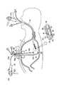
図12に示すように、この実施の形態にかかる治療装置は、胃2の内部に第1および第2の軟性内視鏡11,22が挿入されている。第2の内視鏡22の湾曲部22bはそれぞれ4つの方向(例えば上下、左右方向)に湾曲可能な2つの湾曲部22b’22b”が接続されて形成されている。また、この内視鏡22にはそれぞれの湾曲部22b’,22b”に対応した2つの操作部22c’,22c”が設けられている。挿入部22aの先端側の湾曲部22b”は操作部22c”によって操作され、後端側の湾曲部22b’は後端側の操作部22c’によって操作される。  As shown in FIG. 12, in the treatment apparatus according to this embodiment, first and secondflexible endoscopes 11 and 22 are inserted into thestomach 2. The bendingportion 22b of thesecond endoscope 22 is formed by connecting two bendingportions 22b′22b ″ that can be bent in four directions (for example, up and down and left and right directions). 22, twooperation portions 22c ′ and 22c ″ corresponding to therespective bending portions 22b ′ and 22b ″ are provided. The bendingportion 22b ″ on the distal end side of theinsertion portion 22a is operated by theoperation portion 22c ″. The rear endside bending portion 22b ′ is operated by the rear endside operation portion 22c ′.

胃2の内部の背中側の胃粘膜に病変部28が存在する場合、この病変部28はこれら2つの軟性内視鏡11,22によって以下のように処置される。
まず、胃ろう形成チューブ4を留置する際に第1の内視鏡11を用いて病変部28の位置や大きさなどを確認しておく。
次に、例えば胃ろう形成チューブ4を介して胃2の内部にエアーを送り、胃2の内部を膨らませる。
さらに、第1の軟性内視鏡11を経口的に胃2の内部に挿入するとともに、第2の軟性内視鏡22を胃ろう形成チューブ4を介して胃2の内部に挿入する。
そして、第1の軟性内視鏡11で病変部28を再確認する。その後、第2の軟性内視鏡22の操作部22c’,22c”をそれぞれ操作して湾曲部22b’,22b”を所望の方向に湾曲させて把持鉗子24を用いて病変部28を把持して持ち上げる。
続いて、第1の内視鏡11に配設した針状メス30を用い、持ち上げられた病変部28を切除する。
その後、把持鉗子24を用いて切除した病変部28を回収し、検査を行なう。
When thelesioned part 28 exists in the gastric mucosa on the back side inside thestomach 2, thelesioned part 28 is treated by these twoflexible endoscopes 11 and 22 as follows.
First, when the gastricfistula formation tube 4 is placed, the position and size of thelesioned part 28 are confirmed using thefirst endoscope 11.
Next, for example, air is sent into thestomach 2 through the gastricfistula formation tube 4 to inflate thestomach 2.
Furthermore, the firstflexible endoscope 11 is orally inserted into thestomach 2 and the secondflexible endoscope 22 is inserted into thestomach 2 through the gastricfistula formation tube 4.
Then, thelesioned part 28 is reconfirmed with the firstflexible endoscope 11. Thereafter, theoperation portions 22c ′ and 22c ″ of the secondflexible endoscope 22 are respectively operated to bend thecurved portions 22b ′ and 22b ″ in a desired direction, and thelesioned portion 28 is grasped using the graspingforceps 24. Lift up.
Subsequently, the raisedlesioned part 28 is excised using theneedle scalpel 30 disposed in thefirst endoscope 11.
Thereafter, thelesioned part 28 excised using the graspingforceps 24 is collected and inspected.

なお、この実施の形態では第2の軟性内視鏡22の湾曲部22bに4つの方向に湾曲可能な湾曲部22b’,22b”を設けたが、このような湾曲部22bに限ることはない。例えば、図13に示すように、第1の軟性内視鏡11の湾曲部11bにそれぞれ4つの方向に湾曲可能な湾曲部11b’,11b”を設けるとともに、内視鏡11の本体に2つの操作部11c’,11c”を設けてもよい。また、例えば、図14に示すように、第1および第2の内視鏡11,22の湾曲部11b,22bにそれぞれ4方向に湾曲可能な湾曲部11b’,11b”,22b,22b”を設け、内視鏡11,22の本体に2つの操作部11c’,11c”,22c’,22c”を設けてもよい。  In this embodiment, the bendingportion 22b of the secondflexible endoscope 22 is provided with the bendingportions 22b ′ and 22b ″ that can be bent in four directions. However, the bendingportion 22b is not limited thereto. For example, as shown in Fig. 13, the bendingportion 11b of the firstflexible endoscope 11 is provided with bendingportions 11b 'and 11b "that can be bent in four directions, respectively, and 2 in the main body of theendoscope 11. Twooperation portions 11c ′ and 11c ″ may be provided. For example, as shown in FIG. 14, the bendingportions 11b and 22b of the first andsecond endoscopes 11 and 22 can be bent in four directions, respectively. Bendingportions 11b ′, 11b ″, 22b, and 22b ″ may be provided, and twooperation portions 11c ′, 11c ″, 22c ′, and 22c ″ may be provided on the main bodies of theendoscopes 11 and 22.

また、内視鏡11,22の湾曲部11b,22bが湾曲可能な方向は4つの方向に限ることはなく、2つの方向や3つの方向などでも構わない。  The directions in which the bendingportions 11b and 22b of theendoscopes 11 and 22 can be bent are not limited to four directions, and may be two directions or three directions.

したがって、この実施の形態について以下のことがいえる。
この実施の形態では背中側の病変部28に対して処置を行なうことを説明したが、複数の湾曲部22b’,22b”を設けたことによって、処置を行ない難かった腹腔近傍の病変部28に対しても所望の処置を行ないやすくすることができる。
Therefore, the following can be said with respect to this embodiment.
In this embodiment, it has been described that the treatment is performed on thelesion 28 on the back side. However, by providing a plurality ofcurved portions 22b ′ and 22b ″, thelesion 28 near the abdominal cavity where the treatment is difficult to perform is provided. Also, it is possible to easily perform a desired treatment.

なお、この実施の形態では、第2の内視鏡22に2つの湾曲部22b’,22b”を設け、2つの操作部22c’,22c”でこれら湾曲部22b’,22b”を操作するようにしたことを説明したが、第1の内視鏡11も同様に連結された2つの湾曲部を有するとともに、これら湾曲部に対応した操作部が形成されていることが好適である。  In this embodiment, thesecond endoscope 22 is provided with two bendingportions 22b ′ and 22b ″, and the twooperation portions 22c ′ and 22c ″ operate these bendingportions 22b ′ and 22b ″. As described above, it is preferable that thefirst endoscope 11 also has two curved portions that are similarly connected, and an operation portion corresponding to these curved portions is formed.

(第10の実施の形態)
第10の実施の形態について図15を用いて説明する。この実施の形態は第1の実施の形態の変形例であり、同一の部材には同一の符号を付し、詳しい説明を省略する。
(Tenth embodiment)
A tenth embodiment will be described with reference to FIG. This embodiment is a modification of the first embodiment, and the same members are denoted by the same reference numerals and detailed description thereof is omitted.

図15の(A)に示すように、この実施の形態にかかる治療装置は、胃2の内部に第1および第2の軟性内視鏡11,22が挿入されている。第2の内視鏡22には、この内視鏡22の挿入部22aよりも大径でかつ、可撓性を有し、透明なオーバーチューブ104が配設されている。また、図15の(B)に示すように、このオーバーチューブ104の外径よりも胃ろう形成チューブ4の内径が大径に形成されている。このオーバーチューブ104内にはスコープ挿入ルーメン104aと処置具チャンネル104bとが設けられている。この処置具チャンネル104b内には高周波スネア106が配設されている。また、このオーバーチューブ104の先端には段差が設けられ、高周波スネア106のスネア保持溝106aが形成されている。  As shown in FIG. 15A, in the treatment apparatus according to this embodiment, the first and secondflexible endoscopes 11 and 22 are inserted into thestomach 2. Thesecond endoscope 22 is provided with atransparent overtube 104 having a diameter larger than that of theinsertion portion 22a of theendoscope 22 and having flexibility. Further, as shown in FIG. 15B, the inner diameter of the gastricfistula formation tube 4 is formed larger than the outer diameter of theovertube 104. In theovertube 104, ascope insertion lumen 104a and atreatment instrument channel 104b are provided. A high-frequency snare 106 is disposed in thetreatment instrument channel 104b. Further, a step is provided at the tip of theovertube 104, and asnare holding groove 106a of thehigh frequency snare 106 is formed.

そして、オーバーチューブ104の他端部(スコープ挿入ルーメン104aの上端部)にはシール部材108が配設されている。このシール部材108は内視鏡22の挿入部22aをスコープ挿入ルーメン104a内に挿通可能であるとともに、挿入部22aに密着している。また、このシール部材108の側方に開口を備えている。この開口には吸引チューブ110の一端部が接続され、この吸引チューブ110の他端部には吸引器112が設けられている。なお、高周波スネア106の操作部106bはオーバーチューブ104の側方から突出されている。  Aseal member 108 is disposed at the other end of the overtube 104 (the upper end of thescope insertion lumen 104a). Theseal member 108 can insert theinsertion portion 22a of theendoscope 22 into thescope insertion lumen 104a and is in close contact with theinsertion portion 22a. An opening is provided on the side of theseal member 108. One end of thesuction tube 110 is connected to the opening, and asuction device 112 is provided at the other end of thesuction tube 110. Note that theoperation portion 106 b of the high-frequency snare 106 protrudes from the side of theovertube 104.

胃2の内部の背中側の胃粘膜に病変部28が存在する場合、この病変部28はこれら2つの軟性内視鏡11,22によって以下のように処置される。
まず、例えば胃ろう形成チューブ4を介して胃2の内部にエアーを送り、胃2の内部を膨らませる。
次に、図15の(A)に示すように、第2の内視鏡22にオーバーチューブ104を被せた状態で胃ろう形成チューブ4内に挿入する。そして、第1および第2の内視鏡11,22で病変部28を観察しながらオーバーチューブ104の先端部を病変部28に対して近づける。
図15の(C)に示すように、オーバーチューブ104の先端部を胃粘膜2aに密着させるとともに、高周波スネア106を病変部28の周囲を取り囲む位置に配置する。そして、オーバーチューブ104内のスコープ挿入ルーメン104aに接続された吸引器112を働かせて病変部28を吸引する。吸引した状態で高周波スネア106を操作して病変部28を緊縛切除する。
その後、図15の(D)に示すように、第1の内視鏡11に設けた把持鉗子82を用いて病変部28を把持し、これを回収して検査を行なう。
When thelesioned part 28 exists in the gastric mucosa on the back side inside thestomach 2, thelesioned part 28 is treated by these twoflexible endoscopes 11 and 22 as follows.
First, for example, air is sent into thestomach 2 through the gastricfistula formation tube 4 to inflate thestomach 2.
Next, as shown in FIG. 15A, thesecond endoscope 22 is inserted into the gastricfistula formation tube 4 with theovertube 104 covered. Then, the distal end portion of theovertube 104 is brought closer to thelesioned portion 28 while observing thelesioned portion 28 with the first andsecond endoscopes 11 and 22.
As shown in FIG. 15C, the tip of theovertube 104 is brought into close contact with thegastric mucosa 2a, and the high-frequency snare 106 is disposed at a position surrounding thelesioned part 28. Theaspirator 112 connected to thescope insertion lumen 104a in theovertube 104 is operated to suck thelesioned part 28. In the sucked state, the high-frequency snare 106 is operated and thelesioned part 28 is tightly cut.
Thereafter, as shown in FIG. 15D, thelesioned part 28 is grasped using the graspingforceps 82 provided in thefirst endoscope 11, and this is collected and inspected.

したがって、この実施の形態について以下のことがいえる。
オーバーチューブ104を第2の内視鏡22の挿入部22aの周囲に設け、このオーバーチューブ104を用いて病変部28を隆起させるとともに切除を行なうことができるので、操作が容易である。
なお、この実施の形態では第1の内視鏡11を用いて病変部28を回収したが、オーバーチューブ104を用いて病変部28を回収してもよい。
Therefore, the following can be said with respect to this embodiment.
Since theovertube 104 is provided around theinsertion portion 22a of thesecond endoscope 22 and thelesioned portion 28 can be raised and excised using theovertube 104, the operation is easy.
In this embodiment, thelesioned part 28 is collected using thefirst endoscope 11, but thelesioned part 28 may be collected using theovertube 104.

(第11の実施の形態)
第11の実施の形態について図16を用いて説明する。この実施の形態は第1の実施の形態の変形例であり、同一の部材には同一の符号を付し、詳しい説明を省略する。
(Eleventh embodiment)
The eleventh embodiment will be described with reference to FIG. This embodiment is a modification of the first embodiment, and the same members are denoted by the same reference numerals and detailed description thereof is omitted.

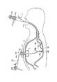
図16に示すように、この実施の形態にかかる治療装置は、胃2の内部に第1および第2の軟性内視鏡11,22が挿入されている。第2の内視鏡22を胃2の内部に挿入する場合、胃ろう形成チューブ4aを介して挿入される。この胃ろう形成チューブ4aはドーム部6と、湾曲チューブ8aとから形成されている。この湾曲チューブ8aは胃2の内部で任意の方向に湾曲されている。また、この湾曲チューブ8aの体外側の上端部には湾曲チューブ8aが湾曲した方向に指標114が取り付けられている。このため、湾曲チューブ8aの湾曲方向を術者が容易に認識することができる。  As shown in FIG. 16, in the treatment apparatus according to this embodiment, first and secondflexible endoscopes 11 and 22 are inserted into thestomach 2. When thesecond endoscope 22 is inserted into thestomach 2, it is inserted through the gastricfistula formation tube 4a. This gastricfistula formation tube 4a is formed of adome portion 6 and acurved tube 8a. Thecurved tube 8a is curved in an arbitrary direction inside thestomach 2. In addition, anindex 114 is attached to the upper end of thecurved tube 8a outside the body in the direction in which thecurved tube 8a is curved. For this reason, the surgeon can easily recognize the bending direction of the bendingtube 8a.

なお、この湾曲形状は図16に示す形状に限ることはない。この湾曲形状は熱成形などによって、術者が所望の形状に作成することが容易である。また、指標114はマーキングや切欠きなどからなることが好適である。  The curved shape is not limited to the shape shown in FIG. This curved shape can be easily formed into a desired shape by the operator by thermoforming or the like. In addition, it is preferable that theindex 114 is made of a marking or a notch.

この胃ろう形成チューブ4aは、第1の実施の形態で説明した胃ろう形成チューブ4と同様にして留置されるので、説明を省略する。
胃2の内部の腹腔側の胃粘膜に病変部28が存在する場合、この病変部28はこれら2つの軟性内視鏡11,22によって以下のように処置される。
まず、胃ろう形成チューブ4aを留置する際に第1の内視鏡11を用いて病変部28の大きさを確認しておく。
次に、例えば胃ろう形成チューブ4aを介して胃2の内部にエアーを送り、胃2の内部を膨らませる。
続いて、第1の軟性内視鏡11を経口的に胃2の内部に挿入するとともに、第2の軟性内視鏡22を胃ろう形成チューブ4aを介して胃2の内部に挿入する。
そして、第1の軟性内視鏡11で病変部28を再確認する。その後、指標114の位置を確認して、第2の軟性内視鏡22の挿入部22aを胃ろう形成チューブ4aを介して病変部28の方向に誘導し、湾曲部22bを病変部28に向けて湾曲させて把持鉗子24を用いて病変部28を把持して胃2の中心部に向けて持ち上げる。
さらに、第1の内視鏡11に配設した針状メス30を用い、持ち上げられた病変部28を切除する。
その後、把持鉗子24を用いて切除した病変部28を回収し、検査を行なう。
Since the gastricfistula formation tube 4a is placed in the same manner as the gastricfistula formation tube 4 described in the first embodiment, the description thereof is omitted.
When thelesioned part 28 exists in the gastric mucosa on the abdominal cavity inside thestomach 2, thelesioned part 28 is treated by these twoflexible endoscopes 11 and 22 as follows.
First, when the gastricfistula formation tube 4 a is placed, the size of thelesioned part 28 is confirmed using thefirst endoscope 11.
Next, air is sent into thestomach 2 through, for example, the gastricfistula formation tube 4 a to inflate thestomach 2.
Subsequently, the firstflexible endoscope 11 is orally inserted into thestomach 2 and the secondflexible endoscope 22 is inserted into thestomach 2 through the gastricfistula formation tube 4a.
Then, thelesioned part 28 is reconfirmed with the firstflexible endoscope 11. Thereafter, the position of theindex 114 is confirmed, theinsertion portion 22a of the secondflexible endoscope 22 is guided toward thelesioned portion 28 via the gastricfistula formation tube 4a, and thecurved portion 22b is directed toward thelesioned portion 28. Then, thelesioned part 28 is grasped using the graspingforceps 24 and lifted toward the center of thestomach 2.
Further, the raisedlesioned part 28 is excised using the needle-shapedknife 30 disposed in thefirst endoscope 11.
Thereafter, thelesioned part 28 excised using the graspingforceps 24 is collected and inspected.

したがって、この実施の形態について以下のことがいえる。
このような臓器の内部で湾曲した胃ろう形成チューブ4aを用いることによって、腹腔に近い位置の病変部28の処置を容易に行なうことができる。
また、胃ろう形成チューブ4aのチューブ8a上端部に指標114を設けたことによって、内視鏡22の挿入部22aが案内されている方向を瞬時に認識することができる。
Therefore, the following can be said with respect to this embodiment.
By using the gastricfistula formation tube 4a curved inside such an organ, it is possible to easily treat thelesioned portion 28 at a position close to the abdominal cavity.
Further, by providing theindex 114 at the upper end of thetube 8a of the gastricfistula formation tube 4a, the direction in which theinsertion portion 22a of theendoscope 22 is guided can be recognized instantly.

(第12の実施の形態)
第12の実施の形態について図17を用いて説明する。この実施の形態は第1の実施の形態の変形例であり、同一の部材には同一の符号を付し、詳しい説明を省略する。
(Twelfth embodiment)
A twelfth embodiment will be described with reference to FIG. This embodiment is a modification of the first embodiment, and the same members are denoted by the same reference numerals and detailed description thereof is omitted.

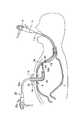
図17に示すように、この実施の形態にかかる治療装置は、胃2の内部に第1および第2の軟性内視鏡11,22が挿入されている。胃ろう形成チューブ4の中にはさらに湾曲した挿入補助具4bが配設されている。この挿入補助具4bは体外から直接胃ろう形成チューブ4内に挿入可能である。この挿入補助具4bの上端部にはシール部材118が配設され、補助具4bが胃2の内部に落下しないように形成されている。また、この挿入補助具4bは胃ろう形成チューブ4内を回転可能および摺動可能に配設されている。この挿入補助具4bは例えば金属パイプや固めの樹脂材(エラストマー)で形成されていることが好適である。また、この挿入補助具4bは半硬性で、術者が胃ろう形成チューブ4内に配設する前に所望の形状にプリフォームしてもよい。また、第11の実施の形態のように、湾曲した右方向にマーキングや切欠きなどを設けても良い。  As shown in FIG. 17, in the treatment apparatus according to this embodiment, first and secondflexible endoscopes 11 and 22 are inserted into thestomach 2. Further inserted in the gastricfistula formation tube 4 is acurved insertion aid 4b. Thisinsertion aid 4b can be inserted directly into the gastricfistula formation tube 4 from outside the body. A sealingmember 118 is disposed at the upper end of theinsertion assisting tool 4b so that the assistingtool 4b does not fall into thestomach 2. Theinsertion assisting tool 4b is disposed in the gastricfistula formation tube 4 so as to be rotatable and slidable. Theinsertion aid 4b is preferably formed of, for example, a metal pipe or a hard resin material (elastomer). Theinsertion assisting tool 4b is semi-rigid and may be preformed into a desired shape before being placed in the gastricfistula formation tube 4 by the operator. Further, as in the eleventh embodiment, a marking or a notch may be provided in the curved right direction.

胃2の内部の腹腔側の胃粘膜に病変部28が存在する場合、この病変部28はこれら2つの軟性内視鏡11,22によって以下のように処置される。
まず、胃ろう形成チューブ4を留置する際に第1の内視鏡11を用いて病変部28の大きさを確認しておく。
次に、例えば胃ろう形成チューブ4を介して胃2の内部にエアーを送り、胃2の内部を膨らませる。
続いて、第1の軟性内視鏡11を経口的に胃2の内部に挿入するとともに、第2の軟性内視鏡22を胃ろう形成チューブ4内に設けられた挿入補助具4bを介して胃2の内部に挿入する。
そして、第1の軟性内視鏡11で病変部28を再確認する。その後、第2の軟性内視鏡22の挿入部22aを胃ろう形成チューブ4aを介して病変部28の方向に誘導し、湾曲部22bを病変部28に向けて湾曲させて把持鉗子24を用いて病変部28を把持して持ち上げる。
さらに、第1の内視鏡11に配設した針状メス30を用い、持ち上げられた病変部28を切除する。
その後、把持鉗子24を用いて切除した病変部28を回収し、検査を行なう。
When thelesioned part 28 exists in the gastric mucosa on the abdominal cavity inside thestomach 2, thelesioned part 28 is treated by these twoflexible endoscopes 11 and 22 as follows.
First, when the gastricfistula formation tube 4 is placed, the size of thelesioned part 28 is confirmed using thefirst endoscope 11.
Next, for example, air is sent into thestomach 2 through the gastricfistula formation tube 4 to inflate thestomach 2.
Subsequently, the firstflexible endoscope 11 is orally inserted into thestomach 2, and the secondflexible endoscope 22 is inserted through theinsertion aid 4 b provided in the gastricfistula formation tube 4. Insert intostomach 2.
Then, thelesioned part 28 is reconfirmed with the firstflexible endoscope 11. Thereafter, theinsertion portion 22a of the secondflexible endoscope 22 is guided toward thelesioned portion 28 through the gastricfistula formation tube 4a, the bendingportion 22b is bent toward thelesioned portion 28, and the graspingforceps 24 is used. Then, thelesioned part 28 is grasped and lifted.
Further, the raisedlesioned part 28 is excised using the needle-shapedknife 30 disposed in thefirst endoscope 11.
Thereafter, thelesioned part 28 excised using the graspingforceps 24 is collected and inspected.

したがって、この実施の形態について以下のことがいえる。
このような臓器の内部で湾曲した挿入補助具4bを有する胃ろう形成チューブ4を用いることによって、腹腔に近い位置の病変部28の処置を容易に行なうことができる。
Therefore, the following can be said with respect to this embodiment.
By using the gastricfistula formation tube 4 having theinsertion assisting tool 4b curved inside such an organ, the treatment of thelesioned part 28 at a position close to the abdominal cavity can be easily performed.

(第13の実施の形態)
第13の実施の形態について図18および図19を用いて説明する。この実施の形態は第1の実施の形態の変形例であり、同一の部材には同一の符号を付し、詳しい説明を省略する。
(Thirteenth embodiment)
A thirteenth embodiment will be described with reference to FIGS. This embodiment is a modification of the first embodiment, and the same members are denoted by the same reference numerals and detailed description thereof is omitted.

図18に示すように、この実施の形態にかかる治療装置は、胃2の内部に第1および第2の軟性内視鏡11,22が挿入されている。胃ろう形成チューブ4にのチューブ8の上端部には、体外側から内視鏡22の挿入部22aの挿入補助具120が設けられている。この挿入補助具120は挿入部120aの挿通を滑らかにするために使用される。  As shown in FIG. 18, in the treatment apparatus according to this embodiment, first and secondflexible endoscopes 11 and 22 are inserted into thestomach 2. Aninsertion assisting tool 120 for theinsertion portion 22a of theendoscope 22 is provided from the outside of the body at the upper end portion of thetube 8 in the gastricfistula formation tube 4. Thisinsertion assisting tool 120 is used for smooth insertion of the insertion portion 120a.

図19の(A)に示すように、この挿入補助具120は内視鏡22の挿入部22aが挿通可能な貫通孔122を備えている。この貫通孔122の直径は内視鏡22の挿入部22aの直径よりもやや大きく形成され、挿入部22aを貫通孔122内で摺動可能に形成されている。この貫通孔122の上端部近傍には、例えばOリングなどからなるシール部材124が配設されている。このシール部材124は挿入補助具120の上端部から固定具126を介して固定されている。この固定具126は挿入補助具120の本体に螺着されている。さらに、この固定具126にはカニメ穴127を備えている。また、この挿入補助具120の下端部には胃ろう形成チューブ4のチューブ8の上端部に嵌合される接続部128を備えている。さらに、このシール部材124と接続部128との間の側方には、貫通孔122に接続された送気口金(エアー導入管路)130が設けられている。この送気口金130には、送気ホース132aの一端が接続され、この送気ホース132aの他端に過昇圧防止弁(圧力制御弁)134が設けられている。さらに、この過昇圧防止弁134の前方には、送気ホース132bを介してエアー送気可能な送気ポンプ(送流手段)136が配設されている。  As shown in FIG. 19A, theinsertion assisting tool 120 includes a throughhole 122 through which theinsertion portion 22a of theendoscope 22 can be inserted. The diameter of the throughhole 122 is slightly larger than the diameter of theinsertion portion 22a of theendoscope 22, and theinsertion portion 22a is formed to be slidable in the throughhole 122. In the vicinity of the upper end portion of the throughhole 122, aseal member 124 made of, for example, an O-ring is disposed. Theseal member 124 is fixed from the upper end portion of theinsertion assisting tool 120 via afixing tool 126. The fixingtool 126 is screwed to the main body of theinsertion assisting tool 120. Further, thefixture 126 is provided with acramp hole 127. In addition, a connectingportion 128 that is fitted to the upper end portion of thetube 8 of the gastricfistula formation tube 4 is provided at the lower end portion of theinsertion assisting tool 120. Further, an air supply base (air introduction conduit) 130 connected to the throughhole 122 is provided on the side between theseal member 124 and theconnection portion 128. One end of anair supply hose 132a is connected to theair supply base 130, and an excessive pressure increase prevention valve (pressure control valve) 134 is provided at the other end of theair supply hose 132a. Further, an air supply pump (flowing means) 136 capable of supplying air via anair supply hose 132b is disposed in front of the excessive pressureincrease prevention valve 134.

また、図19の(B)に示すように、過昇圧防止弁134には、弁134aと圧力検知機構134bと作動制御機構134cとを備えている。圧力検知機構134bは弁134aを通して胃2の内部の圧力と、送気ポンプ136から送気されるエアーの圧力とを例えば0.5秒/回などの所定のインターバルで検知する。また、作動制御機構134cは、圧力検知機構134bによって検知した圧力によって、弁134aの開閉および送気ポンプ136の作動を制御する。このため、胃2の内部が所定の圧力に達すると送気ポンプ136の作動が停止するとともに、弁134aが閉じる。一方、胃2の内部にエアーを送気する場合は胃2側の圧力を検知して、胃2の内部が所定の圧力に達していない場合は送気ポンプ136が作動するとともに弁134aが開いて胃2の内部にエアーが送気される。  Further, as shown in FIG. 19B, the excessive pressureincrease prevention valve 134 includes avalve 134a, apressure detection mechanism 134b, and anoperation control mechanism 134c. Thepressure detection mechanism 134b detects the pressure inside thestomach 2 through thevalve 134a and the pressure of the air supplied from theair supply pump 136 at predetermined intervals such as 0.5 seconds / time. Further, theoperation control mechanism 134c controls the opening / closing of thevalve 134a and the operation of theair supply pump 136 based on the pressure detected by thepressure detection mechanism 134b. For this reason, when the inside of thestomach 2 reaches a predetermined pressure, the operation of theair supply pump 136 is stopped and thevalve 134a is closed. On the other hand, when air is supplied into thestomach 2, the pressure on thestomach 2 side is detected. When the inside of thestomach 2 does not reach the predetermined pressure, theair supply pump 136 is activated and thevalve 134 a is opened. Then, air is sent into thestomach 2.

このような挿入補助具120を胃ろう形成チューブ4のチューブ8上端部に配設して、この挿入補助具120を介して内視鏡22の挿入部22aを胃2の内部に挿入して、これを摺動させる場合、以下のように作用される。
まず、送気ポンプ136からエアー138を送気し、送気ホース132b、過昇圧防止弁134、送気ホース132aおよび送気口金130を介して挿入補助具120の本体内に送気しながら挿入補助具120の貫通孔122に内視鏡22の挿入部22aを挿入する。この内視鏡22の挿入部22aの挿入によって、送気ポンプ136から送気されるエアーが胃2の内部に送気される。さらに挿入部22aを挿入すると、挿入補助具120の貫通孔122と内視鏡22の挿入部22aとの間に送気されたエアー138の流れ作用によって、貫通孔122と挿入部22aとの間の摩擦係数が減少して、内視鏡22の挿入部22aを貫通孔122内で滑らかに摺動させることができる。
Such aninsertion aid 120 is disposed at the upper end of thetube 8 of the gastricfistula formation tube 4, and theinsertion portion 22 a of theendoscope 22 is inserted into thestomach 2 through theinsertion aid 120. When this is slid, it operates as follows.
First,air 138 is supplied from theair supply pump 136 and inserted while supplying air into the main body of theinsertion aid 120 through theair supply hose 132b, theoverpressure prevention valve 134, theair supply hose 132a and theair supply base 130. Theinsertion portion 22 a of theendoscope 22 is inserted into the throughhole 122 of theauxiliary tool 120. By inserting theinsertion portion 22a of theendoscope 22, the air supplied from theair supply pump 136 is supplied into thestomach 2. When theinsertion portion 22a is further inserted, the flow of theair 138 supplied between the throughhole 122 of theinsertion assisting tool 120 and theinsertion portion 22a of theendoscope 22 causes a gap between the throughhole 122 and theinsertion portion 22a. Thus, theinsertion portion 22a of theendoscope 22 can be smoothly slid in the throughhole 122.

なお、胃2の内部が過昇圧防止弁134に設けられた圧力検知機構134bにより、所定の圧力に達したことを検知した場合、作動制御装置134cにこのデータが送られて送気ポンプ136の作動が停止し、弁134aが閉じる。  When it is detected that the inside of thestomach 2 has reached a predetermined pressure by thepressure detection mechanism 134b provided in theoverpressure prevention valve 134, this data is sent to theoperation control device 134c and theair supply pump 136 Operation stops andvalve 134a closes.

したがって、この挿入補助具120を使用すると、送気ポンプ136によってエアー138を貫通孔122を通して胃2の内部を所望の圧力にするまで送気するとともに、このエアー138の流れによって貫通孔122と内視鏡22の挿入部22aとの間に潤滑作用を備えている。  Therefore, when thisinsertion assisting tool 120 is used,air 138 is supplied through the through-hole 122 until the inside of thestomach 2 is brought to a desired pressure by theair supply pump 136, and theair 138 flows to the inside of the through-hole 122. A lubricating action is provided between theinsertion portion 22 a of theendoscope 22.

なお、この実施の形態にかかる挿入補助具120は、内視鏡22の挿入部22aの摺動を検知するセンサー(図示せず)が装着されていることが好適である。この挿入部22aの摺動がセンサーに検知されて送気ポンプ136が作動してエアーの送気開始および送気停止が自動的に制御され、かつ、胃2の内部が好適な圧力に保持されるように形成されていることが好適である。  Theinsertion assisting tool 120 according to this embodiment is preferably equipped with a sensor (not shown) that detects sliding of theinsertion portion 22a of theendoscope 22. The sliding of theinsertion portion 22a is detected by the sensor, and theair supply pump 136 is operated to automatically control the start and stop of the air supply, and the inside of thestomach 2 is maintained at a suitable pressure. It is suitable that it is formed.

胃2の内部の背中側の胃粘膜に病変部28が存在する場合、この病変部28はこれら2つの軟性内視鏡11,22によって以下のように処置される。
図18に示すように、胃ろう形成チューブ4の上端に挿入補助具120を嵌合させる。
次に、第1の軟性内視鏡11を経口的に胃2の内部に挿入する。また、送気ポンプ136を作動させて、挿入補助具120にエアー138を送気しながら第2の内視鏡22の挿入部22aを挿入補助具120の貫通孔122に挿入し、胃ろう形成チューブ4を介して挿入部22aを胃2の内部に挿入する。この状態で、胃2の内部にエアー138が送られ、膨らませられた状態にされる。
その後、第1および第2の内視鏡11,22で病変部28を再確認する。そして、第2の内視鏡22に挿入された把持鉗子24を用いてこの病変部28を把持した後、把持鉗子24を引っ張って病変部28を持ち上げる。続いて、この病変部28を針状メス30を用いて切除する。
そして、このような処置が終了した後、切除部分を把持したまま第2の軟性内視鏡22を生体内から抜去し、病変部28を回収して検査を行なう。
When thelesioned part 28 exists in the gastric mucosa on the back side inside thestomach 2, thelesioned part 28 is treated by these twoflexible endoscopes 11 and 22 as follows.
As shown in FIG. 18, theinsertion aid 120 is fitted to the upper end of the gastricfistula formation tube 4.
Next, the firstflexible endoscope 11 is inserted into thestomach 2 orally. Further, theair feeding pump 136 is operated to insert theinsertion portion 22a of thesecond endoscope 22 into the throughhole 122 of theinsertion assisting tool 120 while supplyingair 138 to theinsertion assisting tool 120, thereby forming a gastric fistula. Theinsertion portion 22 a is inserted into thestomach 2 through thetube 4. In this state,air 138 is sent into thestomach 2 and inflated.
Thereafter, thelesioned part 28 is reconfirmed with the first andsecond endoscopes 11 and 22. Then, after grasping thelesioned part 28 using the graspingforceps 24 inserted into thesecond endoscope 22, the graspingforceps 24 is pulled to lift thelesioned part 28. Subsequently, thelesioned part 28 is excised using aneedle knife 30.
Then, after such treatment is completed, the secondflexible endoscope 22 is removed from the living body while holding the excised part, and thelesioned part 28 is collected and examined.

したがって、この実施の形態について以下のことがいえる。  Therefore, the following can be said with respect to this embodiment.

この実施の形態で説明したように、挿入補助具120を胃ろう形成チューブ4に設けたことによって、手術時に胃2の内部を膨らませて所望の圧力に制御することができるとともに、術時の内視鏡22の挿入部22aの挿通(摺動)を滑らかにすることができる。  As described in this embodiment, by providing theinsertion assisting tool 120 in the gastricfistula formation tube 4, the inside of thestomach 2 can be inflated at the time of surgery and controlled to a desired pressure. The insertion (sliding) of theinsertion portion 22a of theendoscope 22 can be made smooth.

また、挿入部22aを貫通孔122に対してほぼ同一の径で、かつ、摺動可能に設けているので、内視鏡22の挿入部22aのふらつきを最小限に抑えることができる。  In addition, since theinsertion portion 22a is provided so as to be slidable with respect to the throughhole 122, the wobbling of theinsertion portion 22a of theendoscope 22 can be minimized.

(第14の実施の形態)
第14の実施の形態について図20および図21を用いて説明する。この実施の形態は第1の実施の形態の変形例であり、同一の部材には同一の符号を付し、詳しい説明を省略する。
(Fourteenth embodiment)
A fourteenth embodiment will be described with reference to FIGS. This embodiment is a modification of the first embodiment, and the same members are denoted by the same reference numerals and detailed description thereof is omitted.

図20に示すように、この実施の形態にかかる治療装置は、胃2の内部に第1および第2の軟性内視鏡11,22が挿入されている。さらに、この治療装置では、第1の実施の形態で説明した胃ろう形成チューブ4(第1の胃ろう形成チューブと称する)と同様に、さらに同一の部材からなる胃ろう形成チューブ4’(第2の胃ろう形成チューブと称する)が腹腔に留置されている。  As shown in FIG. 20, in the treatment apparatus according to this embodiment, first and secondflexible endoscopes 11 and 22 are inserted into thestomach 2. Further, in this treatment apparatus, the gastricfistula formation tube 4 ′ (the first gastricfistula formation tube 4 ′) composed of the same members as the gastric fistula formation tube 4 (referred to as the first gastric fistula formation tube) described in the first embodiment. 2) (referred to as gastrostomy tube).

第2の胃ろう形成チューブ4’には第3の軟性内視鏡33が配設されている。この第3の軟性内視鏡33は挿入部33aと、この挿入部33aの先端部に設けられた湾曲部33bと、挿入部33aの基端部に設けられた操作部33cとを備えている。また、この挿入部33aには少なくとも鉗子チャンネル(図示せず)が配設され、この鉗子チャンネルに把持鉗子140が配設されている。  A thirdflexible endoscope 33 is disposed on the second gastricfistula formation tube 4 ′. The thirdflexible endoscope 33 includes aninsertion portion 33a, a bendingportion 33b provided at the distal end portion of theinsertion portion 33a, and anoperation portion 33c provided at the proximal end portion of theinsertion portion 33a. . Theinsertion portion 33a is provided with at least a forceps channel (not shown), and a graspingforceps 140 is provided in the forceps channel.

胃2の内部の背中側の胃粘膜に病変部28が存在する場合、この病変部28はこれら3つの軟性内視鏡11,22,33によって以下のように処置される。
まず、第1の実施の形態で説明したように、第1および第2の胃ろう形成チューブ4,4’を留置する。そして、このときに第1の内視鏡11を用いて病変部28の大きさを確認する。
次に、例えば第1の胃ろう形成チューブ4を介して胃2の内部にエアーを送り、胃2の内部を膨らませる。
続いて、第1の軟性内視鏡11を経口的に胃2の内部に挿入する。また、この内視鏡11の挿入部11aの1つのチャンネルに例えば高周波スネア26を配置しておく。
そして、胃ろう形成チューブ4,4’を介して経腹腔的または経皮的に胃2の内部にそれぞれ第2および第3の軟性内視鏡22,33を挿入する。また、これら内視鏡22,33の挿入部22a,33aの1つのチャンネルにそれぞれ把持鉗子24,140を挿入しておく。
その後、第1ないし第3の内視鏡11,22,33で病変部28を再確認する。そして、第1の内視鏡11に挿入された高周波スネア26をこの病変部28のまわりに配置する。続いて、第2および第3の内視鏡22,33に挿入された把持鉗子24,140を用いてこの病変部28を把持した後、把持鉗子24,140を引っ張って病変部28を持ち上げる。予め配置しておいた高周波スネア26に高周波電流を流し、持ち上げられて隆起した病変部28を切除する。
そして、このような処置が終了した後、切除部分を例えば第2の軟性内視鏡22の把持鉗子24で把持したまま第2の軟性内視鏡22および第3の軟性内視鏡33を生体内から抜去し、病変部28を回収して検査を行なう。
When thelesioned part 28 is present in the gastric mucosa on the back side inside thestomach 2, thelesioned part 28 is treated by these threeflexible endoscopes 11, 22, 33 as follows.
First, as described in the first embodiment, the first and second gastricfistula formation tubes 4, 4 ′ are placed. At this time, the size of thelesioned part 28 is confirmed using thefirst endoscope 11.
Next, for example, air is sent into thestomach 2 through the first gastricfistula formation tube 4 to inflate thestomach 2.
Subsequently, the firstflexible endoscope 11 is orally inserted into thestomach 2. Further, for example, ahigh frequency snare 26 is disposed in one channel of theinsertion portion 11a of theendoscope 11.
Then, the second and thirdflexible endoscopes 22 and 33 are inserted into thestomach 2 transperitoneally or percutaneously through the gastricfistula formation tubes 4 and 4 ′. The graspingforceps 24 and 140 are inserted into one channel of theinsertion portions 22a and 33a of theendoscopes 22 and 33, respectively.
Thereafter, thelesioned part 28 is reconfirmed by the first tothird endoscopes 11, 22 and 33. Then, the high-frequency snare 26 inserted into thefirst endoscope 11 is arranged around thelesioned portion 28. Subsequently, after grasping thelesioned part 28 using the graspingforceps 24 and 140 inserted in the second andthird endoscopes 22 and 33, the graspingforceps 24 and 140 are pulled to lift thelesioned part 28. A high-frequency current is applied to the high-frequency snare 26 arranged in advance, and thelesioned part 28 that has been lifted and raised is excised.
Then, after such treatment is completed, the secondflexible endoscope 22 and the thirdflexible endoscope 33 are produced while the excised portion is held with the graspingforceps 24 of the secondflexible endoscope 22, for example. It is removed from the body, and thelesioned part 28 is collected and examined.

なお、この実施の形態では病変部28を高周波スネア26を用いて切除することを説明したが、切除処置具は高周波スネア26に限ることはない。例えば、図21に示す針状メス30を用いて病変部28を切除するようにしてもよい。この針状メス30を用いることによって、図20に示す高周波スネア26を用いて病変部28を切除する場合に比べて切除範囲を大きく採ることができる。  In this embodiment, it has been described that thelesioned part 28 is excised using the high-frequency snare 26, but the excision treatment tool is not limited to the high-frequency snare 26. For example, thelesioned part 28 may be excised using aneedle knife 30 shown in FIG. By using thisneedle scalpel 30, a larger excision range can be taken compared to the case where thelesioned part 28 is excised using the high-frequency snare 26 shown in FIG. 20.

また、第3の内視鏡33は軟性の内視鏡であることが必ずしも必要でなく、硬性の内視鏡を用いてもよい。  Further, thethird endoscope 33 is not necessarily a soft endoscope, and a rigid endoscope may be used.

また、第2および/もしくは第3の内視鏡22,33は観察光学系がそれぞれの挿入部22a,33aから除かれていても構わない。  Further, the second and / orthird endoscopes 22 and 33 may have the observation optical system removed from therespective insertion portions 22a and 33a.

したがって、この実施の形態について以下のことがいえる。
この実施の形態で説明したように、3つの軟性内視鏡11,22,33を用いることによって、治療装置の操作性が向上するので、胃2の内部の粘膜にある病変部28をより確実に切除することができる。また、このために、病変部28を取り残すことがなく、再発の可能性を低く抑えることができる。
特に、2つの内視鏡22,33を用いて病変部28を把持することができるので、より確実に処置を行なうことができる。
Therefore, the following can be said with respect to this embodiment.
As described in this embodiment, since the operability of the treatment apparatus is improved by using the threeflexible endoscopes 11, 22, and 33, thelesioned part 28 in the mucous membrane inside thestomach 2 can be more reliably detected. Can be excised. For this reason, thelesioned part 28 is not left, and the possibility of recurrence can be kept low.
In particular, since thelesioned part 28 can be grasped using the twoendoscopes 22 and 33, the treatment can be performed more reliably.

病変部28の大きさによって高周波スネア26や針状メス30などの切除処置具を選択して使用することによって、より確実に手技を行なうことができる。  By selecting and using a resection treatment tool such as the high-frequency snare 26 or theneedle scalpel 30 depending on the size of thelesioned part 28, the procedure can be performed more reliably.

(第15の実施の形態)
第15の実施の形態について図22を用いて説明する。この実施の形態は第14の実施の形態の変形例であり、同一の部材には同一の符号を付し、詳しい説明を省略する。
(Fifteenth embodiment)
A fifteenth embodiment will be described with reference to FIG. This embodiment is a modification of the fourteenth embodiment, and the same members are denoted by the same reference numerals and detailed description thereof is omitted.

図22に示すように、この実施の形態にかかる治療装置は、胃2の内部に第1ないし第3の軟性内視鏡11,22,33が挿入されている。第3の軟性内視鏡33の挿入部33aに設けられた鉗子チャンネルには高周波スネア142が配設されている。  As shown in FIG. 22, in the treatment apparatus according to this embodiment, first to thirdflexible endoscopes 11, 22 and 33 are inserted into thestomach 2. A high-frequency snare 142 is disposed in the forceps channel provided in theinsertion portion 33 a of the thirdflexible endoscope 33.

胃2の内部の背中側の胃粘膜に病変部28が存在する場合、この病変部28はこれら3つの軟性内視鏡11,22,33によって以下のように処置される。
まず、第1および第2の胃ろう形成チューブ4,4’を留置する際に第1の内視鏡11を用いて病変部28の大きさを確認しておく。
次に、例えば第1の胃ろう形成チューブ4を介して胃2の内部にエアーを送り、胃2の内部を膨らませる。
そして、第1の軟性内視鏡11を経口的に胃2の内部に挿入する。そして、胃ろう形成チューブ4,4’を介して経腹腔的または経皮的に胃2の内部にそれぞれ第2および第3の軟性内視鏡22,33を挿入する。また、第2の軟性内視鏡22の挿入部22aの1つのチャンネルには把持鉗子24を挿入しておく。第3の軟性内視鏡33の挿入部33aの1つのチャンネルには高周波スネア142を挿入しておく。
When thelesioned part 28 is present in the gastric mucosa on the back side inside thestomach 2, thelesioned part 28 is treated by these threeflexible endoscopes 11, 22, 33 as follows.
First, the size of thelesioned part 28 is confirmed using thefirst endoscope 11 when the first and second gastricfistula formation tubes 4 and 4 ′ are placed.
Next, for example, air is sent into thestomach 2 through the first gastricfistula formation tube 4 to inflate thestomach 2.
Then, the firstflexible endoscope 11 is inserted into thestomach 2 orally. Then, the second and thirdflexible endoscopes 22 and 33 are inserted into thestomach 2 transperitoneally or percutaneously through the gastricfistula formation tubes 4 and 4 ′. A graspingforceps 24 is inserted into one channel of theinsertion portion 22a of the secondflexible endoscope 22. Ahigh frequency snare 142 is inserted into one channel of theinsertion portion 33 a of the thirdflexible endoscope 33.

その後、第1ないし第3の内視鏡11,22,33で病変部28を再確認する。そして、第1の軟性内視鏡11でこれら第2および第3の軟性内視鏡22,33の湾曲部22b,33bや、これらの挿入部22a,33aに配設されたそれぞれ把持鉗子24および高周波スネア142の観察を行ないながら、第3の内視鏡33に挿入された高周波スネア142をこの病変部28のまわりに配置する。続いて、第2の内視鏡22に挿入された把持鉗子24を用いてこの病変部28を把持した後、把持鉗子24を引っ張って病変部28を持ち上げる。予め配置しておいた高周波スネア142に高周波電流を流し、持ち上げられて隆起した病変部28を切除する。
そして、このような処置が終了した後、切除部分を第2の軟性内視鏡22の把持鉗子24で把持したまま第2の軟性内視鏡22を生体内から抜去し、病変部28を回収して検査を行なう。
Thereafter, thelesioned part 28 is reconfirmed by the first tothird endoscopes 11, 22 and 33. In the firstflexible endoscope 11, the bendingforceps 24 and the bendingportions 22b and 33b of the second and thirdflexible endoscopes 22 and 33 and theinsertion portions 22a and 33a, respectively, While observing the high-frequency snare 142, the high-frequency snare 142 inserted into thethird endoscope 33 is arranged around thelesioned portion 28. Subsequently, after grasping thelesioned part 28 using the graspingforceps 24 inserted into thesecond endoscope 22, the graspingforceps 24 is pulled to lift thelesioned part 28. A high-frequency current is applied to the high-frequency snare 142 arranged in advance, and thelesioned part 28 that has been lifted and raised is excised.
Then, after such treatment is completed, the secondflexible endoscope 22 is removed from the living body while the excised portion is held by the graspingforceps 24 of the secondflexible endoscope 22, and thelesioned portion 28 is recovered. And inspect it.

したがって、この実施の形態について以下のことがいえる。
この実施の形態で説明したように、第1の内視鏡11で第2および第3の内視鏡22,33の動作を確認しながら第2および第3の内視鏡22,33を用いて病変部28の処置を行なうことができるので、より確実な手技を行なうことができる。
Therefore, the following can be said with respect to this embodiment.
As described in this embodiment, the first andsecond endoscopes 22 and 33 are used while confirming the operation of the second andthird endoscopes 22 and 33 with thefirst endoscope 11. Thus, thelesioned part 28 can be treated, and a more reliable procedure can be performed.

(第16の実施の形態)
第16の実施の形態について図23を用いて説明する。この実施の形態は第14の実施の形態の変形例であり、同一の部材には同一の符号を付し、詳しい説明を省略する。
(Sixteenth embodiment)
A sixteenth embodiment will be described with reference to FIG. This embodiment is a modification of the fourteenth embodiment, and the same members are denoted by the same reference numerals and detailed description thereof is omitted.

図23に示すように、この実施の形態にかかる治療装置は、胃2の内部に第1および第2の軟性内視鏡11,22が挿入されている。さらに、第2の胃ろう形成チューブ4’を介して、胃2の内部には例えば可撓性を有する把持鉗子144が挿入されている。  As shown in FIG. 23, in the treatment apparatus according to this embodiment, first and secondflexible endoscopes 11 and 22 are inserted into thestomach 2. Furthermore, for example, flexible graspingforceps 144 is inserted into thestomach 2 through the second gastricfistula formation tube 4 ′.

胃2の内部の背中側の胃粘膜に病変部28が存在する場合、この病変部28はこれら2つの軟性内視鏡11,22および把持鉗子144によって以下のように処置される。
まず、第1および第2の胃ろう形成チューブ4,4’を留置する際に第1の内視鏡11を用いて病変部28の大きさを確認しておく。
次に、例えば第1の胃ろう形成チューブ4を介して胃2の内部にエアーを送り、胃2の内部を膨らませる。
続いて、第1の軟性内視鏡11を経口的に胃2の内部に挿入する。また、この内視鏡11の挿入部11aの1つのチャンネルに例えば針状メス30を配置しておく。
そして、第1の胃ろう形成チューブ4を介して経腹腔的または経皮的に胃2の内部に第2の軟性内視鏡22を挿入する。また、この内視鏡22の挿入部22aの1つのチャンネルにそれぞれ把持鉗子24を挿入しておく。さらに、第2の胃ろう形成チューブ4’を介して経腹腔的または経皮的に胃2の内部に把持鉗子144を挿入する。この把持鉗子144の先端部近傍を第2の内視鏡22に配設された把持鉗子24で把持し、所望の位置に誘導する。
その後、第1および第2の内視鏡11,22で病変部28を再確認する。次に、第2の内視鏡22に配設された把持鉗子24および第2の胃ろう形成チューブ4’に配設された把持鉗子144を用いてこの病変部28を把持した後、把持鉗子24,144を引っ張って病変部28を持ち上げる。そして、針状メス30を用いて持ち上げられて隆起した病変部28を切除する。
さらに、このような処置が終了した後、切除部分を例えば把持鉗子144で把持したままこの把持鉗子144および第2の軟性内視鏡22を生体内から抜去し、病変部28を回収して検査を行なう。
When thelesioned part 28 exists in the gastric mucosa on the back side inside thestomach 2, thelesioned part 28 is treated by the twoflexible endoscopes 11 and 22 and the graspingforceps 144 as follows.
First, the size of thelesioned part 28 is confirmed using thefirst endoscope 11 when the first and second gastricfistula formation tubes 4 and 4 ′ are placed.
Next, for example, air is sent into thestomach 2 through the first gastricfistula formation tube 4 to inflate thestomach 2.
Subsequently, the firstflexible endoscope 11 is orally inserted into thestomach 2. Further, for example, aneedle knife 30 is disposed in one channel of theinsertion portion 11a of theendoscope 11.
Then, the secondflexible endoscope 22 is inserted into thestomach 2 transperitoneally or percutaneously through the first gastricfistula formation tube 4. Further, the graspingforceps 24 is inserted into one channel of theinsertion portion 22a of theendoscope 22, respectively. Further, the graspingforceps 144 is inserted into thestomach 2 transperitoneally or percutaneously through the second gastricfistula formation tube 4 ′. The vicinity of the distal end portion of the graspingforceps 144 is grasped by the graspingforceps 24 disposed in thesecond endoscope 22 and guided to a desired position.
Thereafter, thelesioned part 28 is reconfirmed with the first andsecond endoscopes 11 and 22. Next, after grasping thislesioned part 28 using the graspingforceps 24 disposed on thesecond endoscope 22 and the graspingforceps 144 disposed on the second gastricfistula formation tube 4 ′, the grasping forceps Thelesioned part 28 is lifted by pulling 24 and 144. Then, thelesioned part 28 lifted and raised by using theneedle knife 30 is excised.
Further, after such treatment is finished, the graspingforceps 144 and the secondflexible endoscope 22 are removed from the living body while the excised part is grasped by the graspingforceps 144, for example, and thelesioned part 28 is recovered and examined. To do.

なお、この実施の形態では、第2の胃ろう形成チューブ4’に可撓性を有する把持鉗子144を挿入して処置を行なうことを説明したが、硬性の把持鉗子などの硬性の処置具でも構わない。また、把持鉗子144に限ることはなく、高周波スネアや針状メスなどが配設されてもよい。  In this embodiment, it has been described that the treatment is performed by inserting the flexible graspingforceps 144 into the second gastricfistula formation tube 4 ′, but a rigid treatment instrument such as a rigid grasping forceps is also used. I do not care. Further, the grippingforceps 144 is not limited, and a high-frequency snare, a needle knife, or the like may be provided.

したがって、この実施の形態について以下のことがいえる。
把持鉗子144を胃2の内部に挿入する場合、軟性内視鏡などの鉗子チャンネルを介す必要がないので、径が大きな処置具や先端が大きな処置具など、内視鏡22の鉗子チャンネルに挿入することが困難な処置具を使用することができる。
Therefore, the following can be said with respect to this embodiment.
When the graspingforceps 144 is inserted into thestomach 2, there is no need to go through a forceps channel such as a flexible endoscope. Therefore, the forceps channel of theendoscope 22 such as a treatment tool having a large diameter or a treatment tool having a large tip is used. A treatment tool that is difficult to insert can be used.

(第17の実施の形態)
第17の実施の形態について図24を用いて説明する。この実施の形態は第16の実施の形態の変形例であり、同一の部材には同一の符号を付し、詳しい説明を省略する。
(Seventeenth embodiment)
The seventeenth embodiment will be described with reference to FIG. This embodiment is a modification of the sixteenth embodiment, and the same members are denoted by the same reference numerals and detailed description thereof is omitted.

図24の(A)に示すように、この実施の形態にかかる治療装置は、胃2の内部に第1および第2の軟性内視鏡11,22が挿入されている。さらに、この治療装置では、第2の胃ろう形成チューブ4’に可撓性を有する把持鉗子144が配設されている。この把持鉗子144は図24の(B)に示すように、挿入部144aの先端部近傍に長手方向に延びた板状のタブ146が配設されている。  As shown in FIG. 24A, in the treatment apparatus according to this embodiment, first and secondflexible endoscopes 11 and 22 are inserted into thestomach 2. Furthermore, in this treatment apparatus, a flexiblegrasping forceps 144 is disposed on the second gastricfistula formation tube 4 ′. As shown in FIG. 24B, the grippingforceps 144 is provided with a plate-like tab 146 extending in the longitudinal direction in the vicinity of the distal end portion of the insertion portion 144a.

胃2の内部の背中側の胃粘膜に病変部28が存在する場合、この病変部28はこれら2つの軟性内視鏡11,22および把持鉗子144によって以下のように処置される。
まず、第1および第2の胃ろう形成チューブ4,4’を留置する際に第1の内視鏡11を用いて病変部28の大きさを確認しておく。
次に、例えば第1の胃ろう形成チューブ4を介して胃2の内部にエアーを送り、胃2の内部を膨らませる。
続いて、第1の軟性内視鏡11を経口的に胃2の内部に挿入する。また、この内視鏡11の挿入部11aの1つのチャンネルに例えば把持鉗子82を配置しておく。
そして、第1の胃ろう形成チューブ4を介して経腹腔的または経皮的に胃2の内部に第2の軟性内視鏡22を挿入する。また、この内視鏡22の挿入部22aの1つのチャンネルにそれぞれ把持鉗子24を挿入しておく。さらに、第2の胃ろう形成チューブ4’を介して経腹腔的または経皮的に胃2の内部に把持鉗子144を挿入する。この把持鉗子144の先端部近傍に設けられたタブ146を第1の内視鏡11に配設された把持鉗子82で把持し、所望の位置に誘導する。
その後、第1および第2の内視鏡11,22で病変部28を再確認する。次に、第2の内視鏡22に配設された把持鉗子24および第2の胃ろう形成チューブ4’に配設された把持鉗子144を用いてこの病変部28を把持した後、把持鉗子24,144を引っ張って病変部28を持ち上げる。そして、第1の内視鏡11に例えば図示しない針状メスを配設し、この針状メスを用いて持ち上げられて隆起した病変部28を切除する。
さらに、このような処置が終了した後、切除部分を例えば把持鉗子144で把持したままこの把持鉗子144および第2の軟性内視鏡22を生体内から抜去し、病変部28を回収して検査を行なう。
When thelesioned part 28 exists in the gastric mucosa on the back side inside thestomach 2, thelesioned part 28 is treated by the twoflexible endoscopes 11 and 22 and the graspingforceps 144 as follows.
First, the size of thelesioned part 28 is confirmed using thefirst endoscope 11 when the first and second gastricfistula formation tubes 4 and 4 ′ are placed.
Next, for example, air is sent into thestomach 2 through the first gastricfistula formation tube 4 to inflate thestomach 2.
Subsequently, the firstflexible endoscope 11 is orally inserted into thestomach 2. Further, for example, a graspingforceps 82 is disposed in one channel of theinsertion portion 11a of theendoscope 11.
Then, the secondflexible endoscope 22 is inserted into thestomach 2 transperitoneally or percutaneously through the first gastricfistula formation tube 4. Further, the graspingforceps 24 is inserted into one channel of theinsertion portion 22a of theendoscope 22, respectively. Further, the graspingforceps 144 is inserted into thestomach 2 transperitoneally or percutaneously through the second gastricfistula formation tube 4 ′. Thetab 146 provided in the vicinity of the distal end portion of the graspingforceps 144 is grasped by the graspingforceps 82 disposed in thefirst endoscope 11 and guided to a desired position.
Thereafter, thelesioned part 28 is reconfirmed with the first andsecond endoscopes 11 and 22. Next, after grasping thislesioned part 28 using the graspingforceps 24 disposed on thesecond endoscope 22 and the graspingforceps 144 disposed on the second gastricfistula formation tube 4 ′, the grasping forceps Thelesioned part 28 is lifted by pulling 24 and 144. Then, for example, a needle scalpel (not shown) is disposed on thefirst endoscope 11, and thelesioned part 28 lifted and raised using the needle scalpel is excised.
Further, after such treatment is finished, the graspingforceps 144 and the secondflexible endoscope 22 are removed from the living body while the excised part is grasped by the graspingforceps 144, for example, and thelesioned part 28 is recovered and examined. To do.

なお、この実施の形態では、把持鉗子144の先端部近傍に板状のタブ146を有していることを説明したが、このようなタブ146に限ることはなく、例えば、図24の(C)に示すように、把持鉗子144の先端部近傍に円形状のフランジ部148を配設しても構わない。  In this embodiment, it has been described that the plate-like tab 146 is provided in the vicinity of the distal end portion of the graspingforceps 144. However, the present invention is not limited to such atab 146. For example, FIG. ), Acircular flange portion 148 may be disposed in the vicinity of the distal end portion of the graspingforceps 144.

したがって、この実施の形態について以下のことがいえる。
把持鉗子144を胃2の内部に挿入する場合、軟性内視鏡などの鉗子チャンネルを介す必要がないので、大きな処置具を使用することができる。また、病変部28に把持鉗子114を容易に近づけることができる。
Therefore, the following can be said with respect to this embodiment.
When inserting the graspingforceps 144 into thestomach 2, it is not necessary to go through a forceps channel such as a flexible endoscope, so that a large treatment tool can be used. In addition, the graspingforceps 114 can be easily brought close to thelesioned part 28.

(第18の実施の形態)
第18の実施の形態について図25を用いて説明する。この実施の形態は第16の実施の形態の変形例であり、同一の部材には同一の符号を付し、詳しい説明を省略する。
(Eighteenth embodiment)
The eighteenth embodiment will be described with reference to FIG. This embodiment is a modification of the sixteenth embodiment, and the same members are denoted by the same reference numerals and detailed description thereof is omitted.

図25の(A)に示すように、この実施の形態にかかる治療装置は、胃2の内部に第1および第2の軟性内視鏡11,22が挿入されている。さらに、この治療装置では、第2の胃ろう形成チューブ4’に可撓性を有する把持鉗子144が配設されている。  As shown in FIG. 25A, in the treatment apparatus according to this embodiment, the first and secondflexible endoscopes 11 and 22 are inserted into thestomach 2. Furthermore, in this treatment apparatus, a flexiblegrasping forceps 144 is disposed on the second gastricfistula formation tube 4 ′.

胃2の内部の背中側の胃粘膜に病変部28が存在する場合、この病変部28はこれら2つの軟性内視鏡11,22および把持鉗子144によって以下のように処置される。
まず、第1および第2の胃ろう形成チューブ4,4’を留置する際に第1の内視鏡11を用いて病変部28の大きさを確認しておく。
次に、例えば第1の胃ろう形成チューブ4を介して胃2の内部にエアーを送り、胃2の内部を膨らませる。
続いて、第1の軟性内視鏡11を経口的に胃2の内部に挿入する。また、この内視鏡11の挿入部11aの1つのチャンネルに例えば針状メス30を配置しておく。
そして、第1の胃ろう形成チューブ4を介して経腹腔的または経皮的に胃2の内部に第2の軟性内視鏡22を挿入する。また、この内視鏡22の挿入部22aの1つのチャンネルにそれぞれ把持鉗子24を挿入しておく。さらに、第2の胃ろう形成チューブ4’を介して経腹腔的または経皮的に胃2の内部に把持鉗子144を挿入する。この把持鉗子144の先端部近傍を第2の内視鏡22に配設された把持鉗子24で把持し、所望の位置に誘導する。
その後、第1および第2の内視鏡11,22で病変部28を再確認する。次に、第2の内視鏡22に配設された把持鉗子24および第2の胃ろう形成チューブ4’に配設された把持鉗子144を用いてこの病変部28を把持した後、把持鉗子24,144を引っ張って病変部28を持ち上げる。そして、この状態で、第2の胃ろう形成チューブ4’のチューブ8上端部を図25の(B)に示すようにクリップ150で留める。そして、針状メス30を用いて持ち上げられて隆起した病変部28を切除する。
さらに、このような処置が終了した後、切除部分を例えば把持鉗子144で把持したままこの把持鉗子144および第2の軟性内視鏡22を生体内から抜去し、病変部28を回収して検査を行なう。
When thelesioned part 28 exists in the gastric mucosa on the back side inside thestomach 2, thelesioned part 28 is treated by the twoflexible endoscopes 11 and 22 and the graspingforceps 144 as follows.
First, the size of thelesioned part 28 is confirmed using thefirst endoscope 11 when the first and second gastricfistula formation tubes 4 and 4 ′ are placed.
Next, for example, air is sent into thestomach 2 through the first gastricfistula formation tube 4 to inflate thestomach 2.
Subsequently, the firstflexible endoscope 11 is orally inserted into thestomach 2. Further, for example, aneedle knife 30 is disposed in one channel of theinsertion portion 11a of theendoscope 11.
Then, the secondflexible endoscope 22 is inserted into thestomach 2 transperitoneally or percutaneously through the first gastricfistula formation tube 4. Further, the graspingforceps 24 is inserted into one channel of theinsertion portion 22a of theendoscope 22, respectively. Further, the graspingforceps 144 is inserted into thestomach 2 transperitoneally or percutaneously through the second gastricfistula formation tube 4 ′. The vicinity of the distal end portion of the graspingforceps 144 is grasped by the graspingforceps 24 disposed in thesecond endoscope 22 and guided to a desired position.
Thereafter, thelesioned part 28 is reconfirmed with the first andsecond endoscopes 11 and 22. Next, after grasping thislesioned part 28 using the graspingforceps 24 disposed on thesecond endoscope 22 and the graspingforceps 144 disposed on the second gastricfistula formation tube 4 ′, the grasping forceps Thelesioned part 28 is lifted by pulling 24 and 144. In this state, the upper end portion of thetube 8 of the second gastricfistula formation tube 4 ′ is fastened with aclip 150 as shown in FIG. Then, thelesioned part 28 lifted and raised by using theneedle knife 30 is excised.
Further, after such treatment is finished, the graspingforceps 144 and the secondflexible endoscope 22 are removed from the living body while the excised part is grasped by the graspingforceps 144, for example, and thelesioned part 28 is recovered and examined. To do.

なお、この実施の形態で説明した把持鉗子144は、例えば所望の形状に予め湾曲して形成された硬性シースでも構わない。また、半硬性で、所望の形状にプリフォーム可能な処置具でも構わない。  Note that the graspingforceps 144 described in this embodiment may be a rigid sheath that is curved in a desired shape, for example. Alternatively, a treatment tool that is semi-rigid and can be preformed into a desired shape may be used.

したがって、この実施の形態について以下のことがいえる。
病変部28を把持鉗子144を用いて隆起させた状態で、把持鉗子144を留めることができるので、病変部28を切除する手技に第1および第2の内視鏡11,22のみを操作して行なうことができ、操作性の向上を図ることができる。
Therefore, the following can be said with respect to this embodiment.
Since the graspingforceps 144 can be fastened with thelesioned part 28 raised using the graspingforceps 144, only the first andsecond endoscopes 11 and 22 are operated for excision of thelesioned part 28. The operability can be improved.

(第19の実施の形態)
第19の実施の形態について図26を用いて説明する。この実施の形態は第7および第16の実施の形態の変形例であり、同一の部材には同一の符号を付し、詳しい説明を省略する。
(Nineteenth embodiment)
The nineteenth embodiment will be described with reference to FIG. This embodiment is a modification of the seventh and sixteenth embodiments, and the same members are denoted by the same reference numerals and detailed description thereof is omitted.

図26の(A)に示すように、この実施の形態にかかる治療装置は、胃2の内部に第1および第2の軟性内視鏡11,22が挿入されている。さらに、硬性の把持鉗子100が第2の胃ろう形成チューブ4’を介して胃2の内部に挿通されている。  As shown in FIG. 26A, in the treatment apparatus according to this embodiment, first and secondflexible endoscopes 11 and 22 are inserted into thestomach 2. Further, a rigidgrasping forceps 100 is inserted into thestomach 2 through the second gastricfistula formation tube 4 ′.

また、図26の(B)に示すように、第1の内視鏡11の挿入部11aの中央部には対物レンズ90が配設されている。この対物レンズ90の側方には1対のライトガイド92が配設されている。さらに、この挿入部11aには送気・送水ノズル94が配設されている。そして、ライトガイド92と送気・送水ノズル94との間には処置具ガイド溝96がそれぞれ配設されている。また、これら処置具ガイド溝96は対物レンズ90に対して対称の位置にも設けられている。これら処置具ガイド溝96には、例えば高周波スネア26や針状メス30が配設されている。  In addition, as shown in FIG. 26B, anobjective lens 90 is disposed at the center of theinsertion portion 11a of thefirst endoscope 11. A pair of light guides 92 is disposed on the side of theobjective lens 90. Further, an air /water supply nozzle 94 is disposed in theinsertion portion 11a. A treatmentinstrument guide groove 96 is provided between thelight guide 92 and the air /water supply nozzle 94. The treatmentinstrument guide grooves 96 are also provided at symmetrical positions with respect to theobjective lens 90. In these treatmentinstrument guide grooves 96, for example, a high-frequency snare 26 and aneedle knife 30 are disposed.

胃2の内部の背中側の胃粘膜に病変部28が存在する場合、この病変部28はこれら2つの軟性内視鏡11,22によって第7の実施の形態で説明した処置と同様に処置される。  When thelesioned part 28 exists in the gastric mucosa on the back side inside thestomach 2, thelesioned part 28 is treated in the same manner as the treatment described in the seventh embodiment by these twoflexible endoscopes 11 and 22. The

したがって、この実施の形態について以下のことがいえる。
第1の軟性内視鏡11の挿入部11aに沿って処置具ガイド溝96を設け、これら処置具ガイド溝96にいくつかの処置具を配設しておくと、病変部28の大きさなどによって、処置具を選択して使用することができる。
Therefore, the following can be said with respect to this embodiment.
When a treatmentinstrument guide groove 96 is provided along theinsertion portion 11a of the firstflexible endoscope 11, and several treatment instruments are arranged in the treatmentinstrument guide groove 96, the size of thelesioned part 28, etc. The treatment tool can be selected and used.

(第20の実施の形態)
第20の実施の形態について図27を用いて説明する。この実施の形態は第8および16の実施の形態の変形例であり、同一の部材には同一の符号を付し、詳しい説明を省略する。
(20th embodiment)
A twentieth embodiment will be described with reference to FIG. This embodiment is a modification of the eighth and sixteenth embodiments, and the same members are denoted by the same reference numerals and detailed description thereof is omitted.

図27の(A)に示すように、この実施の形態にかかる治療装置は、胃2の内部に第1および第2の軟性内視鏡11,22が挿入されている。第1の内視鏡11の挿入部11aからは針状メス30が突出されて操作可能となっている。  As shown in FIG. 27A, in the treatment apparatus according to this embodiment, first and secondflexible endoscopes 11 and 22 are inserted into thestomach 2. A needle-like knife 30 protrudes from theinsertion portion 11a of thefirst endoscope 11 and can be operated.

また、第2の胃ろう形成チューブ4’には硬性の把持鉗子102が挿通されている。この硬性の把持鉗子102には開閉操作部102aと回動操作部102bとが配設され、図27の(B)に示すように、先端が開閉操作可能で、かつ、回動操作可能に形成されている。  A rigid graspingforceps 102 is inserted through the second gastricfistula formation tube 4 ′. This rigidgrasping forceps 102 is provided with an opening /closing operation portion 102a and arotation operation portion 102b, and as shown in FIG. 27 (B), the tip can be opened / closed and can be rotated. Has been.

胃2の内部の背中側の胃粘膜に病変部28が存在する場合、この病変部28はこれら2つの軟性内視鏡11,22によって第8の実施の形態で説明した処置と同様に処置される。  When thelesioned part 28 exists in the gastric mucosa on the back side inside thestomach 2, thelesioned part 28 is treated in the same manner as the procedure described in the eighth embodiment by these twoflexible endoscopes 11 and 22. The

したがって、この実施の形態について以下のことがいえる。
硬性の把持鉗子102を用いて第1の内視鏡11を病変部28に向けて誘導することによって、第2の内視鏡22の術者が第1の内視鏡11の術者に指示を送りやすくすることができる。
Therefore, the following can be said with respect to this embodiment.
The operator of thesecond endoscope 22 instructs the operator of thefirst endoscope 11 by guiding thefirst endoscope 11 toward thelesioned portion 28 using the rigid graspingforceps 102. Can be sent easily.

(第21の実施の形態)
第21の実施の形態について図28を用いて説明する。この実施の形態は第16の実施の形態の変形例であり、同一の部材には同一の符号を付し、詳しい説明を省略する。
(Twenty-first embodiment)
A twenty-first embodiment will be described with reference to FIG. This embodiment is a modification of the sixteenth embodiment, and the same members are denoted by the same reference numerals and detailed description thereof is omitted.

図28の(A)に示すように、この実施の形態にかかる治療装置は、胃2の内部に第1および第2の軟性内視鏡11,22が挿入されている。さらに第2の軟性内視鏡22には、糸付クリップ152が配設されている。このクリップ152の糸は第2の胃ろう形成チューブ4’を介して体外に出される程度の長さを少なくとも備えている。また、第2の胃ろう形成チューブ4’には先端がフック状に形成された回収処置具154が配設されている。  As shown in FIG. 28A, in the treatment apparatus according to this embodiment, the first and secondflexible endoscopes 11 and 22 are inserted into thestomach 2. Furthermore, a threadedclip 152 is disposed on the secondflexible endoscope 22. The thread of theclip 152 has at least a length enough to be removed from the body via the second gastric fistula formation tube 4 '. Further, the second gastricfistula formation tube 4 ′ is provided with arecovery treatment instrument 154 whose tip is formed in a hook shape.

さらに、2つの胃ろう形成チューブ4,4’のチューブ8の上端部にはそれぞれ例えばシリコーンなどの弾性部材からなるシール部材156が装着されている。これらシール部材156は第2の内視鏡22の挿入部22aおよび回収処置具154の挿入部に密着し、胃2の内部の空気が体外に漏れ出さないように形成されている。これらシール部材156の中央部は例えば円孔状やプラス記号形状、マイナス記号形状などの孔が形成されている。  Further, a sealingmember 156 made of an elastic member such as silicone is attached to the upper ends of thetubes 8 of the two gastricfistula formation tubes 4 and 4 ′. Theseseal members 156 are formed in close contact with theinsertion portion 22a of thesecond endoscope 22 and the insertion portion of therecovery treatment instrument 154 so that the air inside thestomach 2 does not leak out of the body. For example, a hole having a circular hole shape, a plus symbol shape, a minus symbol shape, or the like is formed at the center of theseal member 156.

胃2の内部の背中側の胃粘膜に病変部28が存在する場合、この病変部28はこれら2つの軟性内視鏡11,22および回収処置具154によって以下のように処置される。
まず、例えば第1の胃ろう形成チューブ4を介して胃2の内部にエアーを送り、胃2の内部を膨らませる。
続いて、第1の軟性内視鏡11を経口的に胃2の内部に挿入する。また、この内視鏡11の挿入部11aの1つのチャンネルに例えば針状メス30を配置しておく。
そして、図28の(A)に示すように、第1の胃ろう形成チューブ4を介して経腹腔的または経皮的に胃2の内部に第2の軟性内視鏡22を挿入する。また、この内視鏡22の挿入部22aの1つのチャンネルにそれぞれ糸付クリップ152を挿入し、病変部28の近傍を把持させておく。さらに、第2の胃ろう形成チューブ4’を介して経腹腔的または経皮的に胃2の内部に回収処置具154を挿入する。この回収処置具154で糸付クリップ152の糸部を引っ掛けて、図28の(B)に示すように、第2の胃ろう形成チューブ4’の外側(体外)に引き抜く。
体外でこの糸付クリップ152の糸部を保持した状態で、第2の内視鏡22から把持鉗子24を出して病変部28の近傍を把持して引っ張り、隆起させる。そして、針状メス30を用いて持ち上げられて隆起した病変部28を切除する。
さらに、このような処置が終了した後、図28の(C)に示すように、切除部分を糸付クリップ152で把持したままこの糸付クリップ152を生体内から抜去し、病変部28を回収して検査を行なう。
When thelesioned part 28 exists in the gastric mucosa on the back side inside thestomach 2, thelesioned part 28 is treated by the twoflexible endoscopes 11 and 22 and therecovery treatment tool 154 as follows.
First, for example, air is sent into thestomach 2 via the first gastricfistula formation tube 4 to inflate thestomach 2.
Subsequently, the firstflexible endoscope 11 is orally inserted into thestomach 2. Further, for example, aneedle knife 30 is disposed in one channel of theinsertion portion 11a of theendoscope 11.
Then, as shown in FIG. 28A, the secondflexible endoscope 22 is inserted into thestomach 2 transperitoneally or percutaneously through the first gastricfistula formation tube 4. Further, a threadedclip 152 is inserted into each channel of theinsertion portion 22a of theendoscope 22 so that the vicinity of thelesioned portion 28 is gripped. Further, therecovery treatment instrument 154 is inserted into thestomach 2 transperitoneally or percutaneously through the second gastricfistula formation tube 4 ′. With thisrecovery treatment instrument 154, the thread portion of the threadedclip 152 is hooked and pulled out to the outside (outside the body) of the second gastricfistula formation tube 4 ′ as shown in FIG.
In a state where the thread portion of the threadedclip 152 is held outside the body, the graspingforceps 24 is taken out from thesecond endoscope 22 and the vicinity of thelesioned portion 28 is grasped and pulled and raised. Then, thelesioned part 28 lifted and raised by using theneedle knife 30 is excised.
Further, after such treatment is completed, as shown in FIG. 28C, the threadedclip 152 is removed from the living body while the cut portion is held by the threadedclip 152, and thelesioned part 28 is recovered. And inspect it.

一方、第1および第2の軟性内視鏡22は、さらに他の病変部(図示せず)を探し、同様な処置を行なう。例えば、回収処置具154を第2の胃ろう形成チューブ4’から挿入し、第2の内視鏡22に糸付クリップ152を挿入し、上述した処置と同様な処置を行なう。  On the other hand, the first and secondflexible endoscopes 22 search for another lesion (not shown) and perform the same treatment. For example, therecovery treatment instrument 154 is inserted from the second gastricfistula formation tube 4 ′, the threadedclip 152 is inserted into thesecond endoscope 22, and the same treatment as described above is performed.

したがって、この実施の形態について以下のことがいえる。
胃2の内部に病変部28が複数設けられている場合、病変部28を回収する際に内視鏡22を抜差しする必要がない治療装置を提供することができる。すなわち、切除片が大きい場合でも、内視鏡を体外に抜去する必要をなくすことができる。このため、処置を続けて行なうことができ、手技の時間を短縮することができる。
また、大きな病変部28を一括して切除する場合に、複数の糸付クリップ152を用いてこの病変部28を持ち上げて隆起させることができる。
さらに、胃2の内部は送気して膨らませた状態で処置することが好適であるが、シール部材156により、送気したエアーを体外に漏れ出し難くすることができる。
Therefore, the following can be said with respect to this embodiment.
When a plurality oflesions 28 are provided in thestomach 2, it is possible to provide a treatment apparatus that does not require the insertion and removal of theendoscope 22 when collecting thelesions 28. That is, even when the excision piece is large, it is not necessary to remove the endoscope from the body. For this reason, treatment can be performed continuously, and the time of the procedure can be shortened.
Moreover, when excising the largelesioned part 28 collectively, thislesioned part 28 can be lifted and raised using the someclip 152 with a thread | yarn.
Further, it is preferable to treat the inside of thestomach 2 in a state where it is inflated by supplying air, but the supplied air can be made difficult to leak out of the body by theseal member 156.

(第22の実施の形態)
第22の実施の形態について図29および図30を用いて説明する。この実施の形態は第1の実施の形態の変形例であり、同一の部材には同一の符号を付し、詳しい説明を省略する。
(Twenty-second embodiment)
A twenty-second embodiment will be described with reference to FIGS. 29 and 30. FIG. This embodiment is a modification of the first embodiment, and the same members are denoted by the same reference numerals and detailed description thereof is omitted.

図29に示すように、この実施の形態にかかる治療装置は胃2の内部に第1および第2の軟性内視鏡11,22が挿入されている。さらに、第2の胃ろう形成チューブ4’には例えば超音波凝固切開装置160、超音波吸引装置やカッティングスーチャーなどの超音波処置具が配設されている。以下、超音波凝固切開装置160を例にして説明する。  As shown in FIG. 29, in the treatment apparatus according to this embodiment, first and secondflexible endoscopes 11 and 22 are inserted into thestomach 2. Further, the second gastricfistula formation tube 4 ′ is provided with an ultrasonic treatment instrument such as an ultrasonic coagulation /cutting device 160, an ultrasonic suction device or a cutting suture. Hereinafter, the ultrasonic coagulation /cutting device 160 will be described as an example.

この超音波凝固切開装置160は体腔内に挿入可能な処置装置160aと、この処置装置160aの基端部で、かつ、体外に処置制御装置160bが配設されている。この処置制御装置160bは処置装置160aに超音波の発生、停止を操作することができる。また、処置装置160aの先端部は所望の角度に湾曲して形成されている。  The ultrasonic coagulation /cutting device 160 includes atreatment device 160a that can be inserted into a body cavity, and atreatment control device 160b disposed outside the body at the base end of thetreatment device 160a. Thetreatment control device 160b can operate thetreatment device 160a to generate and stop ultrasonic waves. Further, the distal end portion of thetreatment device 160a is formed to be bent at a desired angle.

胃2の内部の背中側の胃粘膜に病変部28が存在する場合、この病変部28はこれら2つの軟性内視鏡11,22および超音波凝固切開装置160によって以下のように処置される。
まず、例えば第1の胃ろう形成チューブ4を介して胃2の内部にエアーを送り、胃2の内部を膨らませる。
続いて、第1の軟性内視鏡11を経口的に胃2の内部に挿入する。この第1の内視鏡11は病変部28の観察用に用いられる。そして、第1の胃ろう形成チューブ4を介して経腹腔的または経皮的に胃2の内部に第2の軟性内視鏡22を挿入する。この内視鏡22の挿入部22aの1つのチャンネルに把持鉗子24を挿入し、病変部28の近傍を把持して隆起させておく。
第2の胃ろう形成チューブ4’を介して経腹腔的または経皮的に超音波凝固切開装置160の処置装置160aを挿入する。この処置装置160aの先端が湾曲しているので、隆起させた病変部28の下側に容易に入れることができる。この状態で、処置制御装置160bを駆動操作して、病変部28を切除する。
When thelesioned part 28 exists in the gastric mucosa on the back side inside thestomach 2, thelesioned part 28 is treated by the twoflexible endoscopes 11 and 22 and the ultrasonic coagulation /cutting device 160 as follows.
First, for example, air is sent into thestomach 2 via the first gastricfistula formation tube 4 to inflate thestomach 2.
Subsequently, the firstflexible endoscope 11 is orally inserted into thestomach 2. Thefirst endoscope 11 is used for observing thelesioned part 28. Then, the secondflexible endoscope 22 is inserted into thestomach 2 transperitoneally or percutaneously through the first gastricfistula formation tube 4. The graspingforceps 24 is inserted into one channel of theinsertion portion 22a of theendoscope 22, and the vicinity of thelesioned portion 28 is grasped and raised.
Thetreatment device 160a of the ultrasonic coagulation /cutting device 160 is inserted transperitoneally or percutaneously through the second gastricfistula formation tube 4 ′. Since the distal end of thetreatment device 160a is curved, thetreatment device 160a can be easily placed under the raisedlesioned portion 28. In this state, thetreatment control device 160b is driven and thelesioned part 28 is excised.

したがって、この実施の形態について以下のことがいえる。
処置装置160aの先端部を湾曲させて、隆起させた病変部28を容易に切除することができる。
Therefore, the following can be said with respect to this embodiment.
The distal end portion of thetreatment device 160a is bent, and the raisedlesioned portion 28 can be easily excised.

なお、この実施の形態にかかる第1および第2の軟性内視鏡11,22には図30に示すように、それぞれ内視鏡用のスタンド162,164が配設されている。これらスタンド162,164はそれぞれ脚部162a,164aが立設されている。これら脚部162a,164aの先端部には、これら脚部162a,164aに対して屈曲可能な屈曲アーム162b,164bの基端部が回動可能に枢支されている。さらに、屈曲アーム162b,164bの先端部には内視鏡本体保持部162c,164cが装着されている。第1の内視鏡11は、病変部28を観察可能な状態で所望の位置で内視鏡本体保持部162c,164cによって保持される。また、第2の内視鏡22は病変部28を把持した状態で内視鏡本体保持部162c,164cによって保持される。
このため、第1および第2の内視鏡11,22を所望の位置で保持することができるので、術者の操作を減らすことができる。また、2つの内視鏡11,22を所望の位置で保持することによって、例えば超音波凝固切開装置160の処置装置160aを第2の胃ろう形成チューブ4’から挿入して、1人で病変部28の処置を行なうことができる。
The first and secondflexible endoscopes 11 and 22 according to this embodiment are provided with endoscope stands 162 and 164, respectively, as shown in FIG. These stands 162 and 164 are respectively provided withleg portions 162a and 164a. The base ends of the bendingarms 162b and 164b that can be bent with respect to thelegs 162a and 164a are pivotally supported at the distal ends of thelegs 162a and 164a. Furthermore, endoscopebody holding portions 162c and 164c are attached to the distal ends of the bendingarms 162b and 164b. Thefirst endoscope 11 is held by the endoscope mainbody holding parts 162c and 164c at a desired position in a state where thelesioned part 28 can be observed. Thesecond endoscope 22 is held by the endoscope mainbody holding parts 162c and 164c in a state where thelesioned part 28 is held.
For this reason, since the first andsecond endoscopes 11 and 22 can be held at desired positions, the operation of the operator can be reduced. Further, by holding the twoendoscopes 11 and 22 at desired positions, for example, thetreatment device 160a of the ultrasonic coagulation /cutting device 160 is inserted from the second gastricfistula formation tube 4 ′, and the lesion is caused by one person. The treatment of thepart 28 can be performed.

また、このようなスタンド162,164は超音波凝固切開装置160などに設けてもよい。  Such stands 162 and 164 may be provided in the ultrasonic coagulation /cutting device 160 or the like.

(第23の実施の形態)
第23の実施の形態について図31を用いて説明する。この実施の形態は第22の実施の形態の変形例であり、同一の部材には同一の符号を付し、詳しい説明を省略する。
(Twenty-third embodiment)
A twenty-third embodiment will be described with reference to FIG. This embodiment is a modification of the twenty-second embodiment, and the same members are denoted by the same reference numerals and detailed description thereof is omitted.

図31に示すように、この実施の形態にかかる治療装置は胃2の内部に第1および第2の軟性内視鏡11,22が挿入されている。さらに、第2の胃ろう形成チューブ4’には吸引装置166が配設されている。この吸引装置166は第2の胃ろう形成チューブ4’を介して胃2の内部に挿入可能なドレーンチューブ166aを備えている。このドレーンチューブ166aの基端部には、吸引器166bの先端部が配設されている。吸引器166bを駆動操作することによって、胃2の内部の汚物などの貯留液168を吸引して体外に排出することができる。  As shown in FIG. 31, in the treatment apparatus according to this embodiment, first and secondflexible endoscopes 11 and 22 are inserted into thestomach 2. Further, asuction device 166 is disposed in the second gastricfistula formation tube 4 ′. Thesuction device 166 includes adrain tube 166a that can be inserted into thestomach 2 through the second gastric fistula formation tube 4 '. The distal end portion of thesuction device 166b is disposed at the proximal end portion of thedrain tube 166a. By operating theaspirator 166b, the stored liquid 168 such as filth inside thestomach 2 can be sucked and discharged out of the body.

胃2の内部の背中側の胃粘膜に病変部28が存在する場合、この病変部28はこれら2つの軟性内視鏡11,22および吸引装置166によって以下のように処置される。
まず、例えば第1の胃ろう形成チューブ4を介して胃2の内部にエアーを送り、胃2の内部を膨らませる。
続いて、第1の軟性内視鏡11を経口的に胃2の内部に挿入する。また、この内視鏡11の挿入部11aの1つのチャンネルに例えば針状メス30を配置しておく。
そして、図31に示すように、第1の胃ろう形成チューブ4を介して経腹腔的または経皮的に胃2の内部に第2の軟性内視鏡22を挿入する。また、この内視鏡22の挿入部22aの1つのチャンネルに把持鉗子24を挿入し、病変部28の近傍を把持する。次に、第2の胃ろう形成チューブ4’を介して吸引装置166のドレーンチューブ166aを挿入する。このドレーンチューブ166aを背中側の病変部28の近傍に貯留した汚物などの貯留液中に入れて、吸引器166bを駆動操作して、貯留液を体外に排出する。
その後、第2の内視鏡22の把持鉗子24で把持し、隆起させた病変部28を第1の内視鏡11の針状メス30を用いて切除する。
さらに、このような処置が終了した後、切除部分を把持鉗子24で把持したまま第2の内視鏡22を生体内から抜去し、病変部28を回収して検査を行なう。
When thelesioned part 28 is present in the gastric mucosa on the back side inside thestomach 2, thelesioned part 28 is treated by the twoflexible endoscopes 11 and 22 and thesuction device 166 as follows.
First, for example, air is sent into thestomach 2 via the first gastricfistula formation tube 4 to inflate thestomach 2.
Subsequently, the firstflexible endoscope 11 is orally inserted into thestomach 2. Further, for example, aneedle knife 30 is disposed in one channel of theinsertion portion 11a of theendoscope 11.
Then, as shown in FIG. 31, the secondflexible endoscope 22 is inserted into thestomach 2 through the first gastricfistula formation tube 4 transperitoneally or percutaneously. Further, the graspingforceps 24 is inserted into one channel of theinsertion portion 22 a of theendoscope 22 to grasp the vicinity of thelesioned portion 28. Next, thedrain tube 166a of thesuction device 166 is inserted through the second gastricfistula formation tube 4 ′. Thedrain tube 166a is placed in a storage liquid such as filth stored in the vicinity of thelesion 28 on the back side, and theaspirator 166b is driven to discharge the stored liquid outside the body.
Thereafter, thelesioned part 28 grasped and raised by the graspingforceps 24 of thesecond endoscope 22 is excised using theneedle scalpel 30 of thefirst endoscope 11.
Further, after such treatment is completed, thesecond endoscope 22 is removed from the living body while the excised portion is held by the holdingforceps 24, and thelesioned part 28 is collected and examined.

したがって、この実施の形態について以下のことがいえる。
臓器内に貯留した汚物等を除去してから病変部28の操作を行なっているので、第1および第2の内視鏡11,22で病変部28を確認しやすく、病変部28の取り残しの可能性を少なくすることができる。
Therefore, the following can be said with respect to this embodiment.
Since thelesioned part 28 is operated after removing the dirt and the like stored in the organ, thelesioned part 28 can be easily confirmed with the first andsecond endoscopes 11 and 22, and thelesioned part 28 is left behind. The possibility can be reduced.

なお、この実施の形態ではドレーンチューブ166aを第2の胃ろう形成チューブ4’を介して挿入することを説明したが、第1の胃ろう形成チューブ4から第2の内視鏡22とともに挿入してもよい。  In this embodiment, it has been described that thedrain tube 166a is inserted through the second gastric fistula formation tube 4 '. However, thedrain tube 166a is inserted from the first gastricfistula formation tube 4 together with thesecond endoscope 22. May be.

(第24の実施の形態)
第24の実施の形態について図32を用いて説明する。この実施の形態は第22の実施の形態の変形例であり、同一の部材には同一の符号を付し、詳しい説明を省略する。
(24th Embodiment)
The twenty-fourth embodiment will be described with reference to FIG. This embodiment is a modification of the twenty-second embodiment, and the same members are denoted by the same reference numerals and detailed description thereof is omitted.

図32の(A)に示すように、この実施の形態にかかる治療装置は胃2の内部に第1および第2の軟性内視鏡11,22が挿入されている。さらに、第2の胃ろう形成チューブ4’には複数(この実施の形態では5つ)のルーメンを有する処置用チューブ170が配設されている。これら5つのルーメンは送気口172、送水口174、吸引口176および2つの鉗子チャンネル178,179にそれぞれ形成されている。送気口172は処置用チューブ170の上端部にシリンジ送気口172aを備えている。また、送水口174および吸引口176は同様にそれぞれシリンジ送水口174a、吸引器管路176aを備えている。さらに、鉗子チャンネル178には把持鉗子144が配設されている。  As shown in FIG. 32A, in the treatment apparatus according to this embodiment, first and secondflexible endoscopes 11 and 22 are inserted into thestomach 2. Further, atreatment tube 170 having a plurality of (five in this embodiment) lumens is disposed on the second gastricfistula formation tube 4 ′. These five lumens are formed in theair supply port 172, thewater supply port 174, thesuction port 176, and the twoforceps channels 178 and 179, respectively. Theair supply port 172 includes a syringeair supply port 172 a at the upper end of thetreatment tube 170. Similarly, thewater supply port 174 and thesuction port 176 are respectively provided with a syringewater supply port 174a and asuction device conduit 176a. Further, a graspingforceps 144 is disposed in theforceps channel 178.

胃2の内部の背中側の胃粘膜に病変部28が存在する場合、この病変部28はこれら2つの軟性内視鏡11,22および把持鉗子144によって以下のように処置される。
まず、例えば第1の胃ろう形成チューブ4を介して胃2の内部にエアーを送り、胃2の内部を膨らませる。
続いて、第1の軟性内視鏡11を経口的に胃2の内部に挿入する。また、この内視鏡11の挿入部11aの1つのチャンネルに例えば針状メス30を配置しておく。
そして、図31に示すように、第1の胃ろう形成チューブ4を介して経腹腔的または経皮的に胃2の内部に第2の軟性内視鏡22を挿入する。また、この内視鏡22の挿入部22aの1つのチャンネルに把持鉗子24を挿入し、病変部28の近傍を把持させておく。次に、第2の胃ろう形成チューブ4’を介して処置用チューブ170を挿入する。この処置用チューブ170の鉗子チャンネル178に例えば把持鉗子144を配置しておく。
その後、把持鉗子24,144で把持して隆起させた病変部28を第1の内視鏡11の針状メス30を用いて切除する。
さらに、このような処置が終了した後、切除部分を把持鉗子24で把持したまま第2の内視鏡22を生体内から抜去し、病変部28を回収して検査を行なう。
続いて、シリンジ送気口172aからエアーを送気し、送気口172からこのエアーを切除部などに射出させる。また、シリンジ送水口174aから液体を送水し、送水口174からこの液体を切除部などに射出させる。さらに、切除部から出される切除片を吸引口176から吸引器管路176aを介して吸引して、胃2の内部の病変部28およびその近傍を洗浄する。
When thelesioned part 28 exists in the gastric mucosa on the back side inside thestomach 2, thelesioned part 28 is treated by the twoflexible endoscopes 11 and 22 and the graspingforceps 144 as follows.
First, for example, air is sent into thestomach 2 via the first gastricfistula formation tube 4 to inflate thestomach 2.
Subsequently, the firstflexible endoscope 11 is orally inserted into thestomach 2. Further, for example, aneedle knife 30 is disposed in one channel of theinsertion portion 11a of theendoscope 11.
Then, as shown in FIG. 31, the secondflexible endoscope 22 is inserted into thestomach 2 through the first gastricfistula formation tube 4 transperitoneally or percutaneously. In addition, the graspingforceps 24 is inserted into one channel of theinsertion portion 22a of theendoscope 22, and the vicinity of thelesioned portion 28 is grasped. Next, thetreatment tube 170 is inserted through the second gastricfistula formation tube 4 ′. For example, a graspingforceps 144 is disposed in theforceps channel 178 of thetreatment tube 170.
Thereafter, thelesioned part 28 grasped and raised by the graspingforceps 24 and 144 is excised using the needle-shapedknife 30 of thefirst endoscope 11.
Further, after such treatment is completed, thesecond endoscope 22 is removed from the living body while the excised portion is held by the holdingforceps 24, and thelesioned part 28 is collected and examined.
Subsequently, air is supplied from the syringeair supply port 172a, and this air is injected from theair supply port 172 to the excision part or the like. Further, the liquid is supplied from the syringewater supply port 174a, and this liquid is ejected from thewater supply port 174 to the excision part or the like. Further, the excised piece taken out from the excision part is aspirated from theaspiration port 176 through theaspirator duct 176a, and thelesioned part 28 in thestomach 2 and the vicinity thereof are washed.

したがって、この実施の形態について以下のことがいえる。
臓器内に貯留した汚物等を除去してから病変部28の操作を行なっているので、第1および第2の内視鏡11,22で病変部28を確認しやすく、病変部28の取り残しの可能性を少なくすることができる。
Therefore, the following can be said with respect to this embodiment.
Since thelesioned part 28 is operated after removing the dirt and the like stored in the organ, thelesioned part 28 can be easily confirmed with the first andsecond endoscopes 11 and 22, and thelesioned part 28 is left behind. The possibility can be reduced.

なお、第4、第7ないし第9、第12、第13、第19ないし第21の実施の形態では胃ろう形成チューブ4,4’のチューブ8の上端部に内視鏡22の挿入部22aをシールするシール部材を設けたが、これら実施の形態だけでなく、他の実施の形態にもこのようなシール部材が設けられていることが好適である。このようなシール部材を設けることによって、胃2の内部を膨らませたときに気体を胃2の外部に漏れ難くすることができ、胃2の内部を膨らませた状態を保持することができる。  In the fourth, seventh to ninth, twelfth, thirteenth, nineteenth to twenty-first embodiments, theinsertion portion 22a of theendoscope 22 is provided at the upper end of thetube 8 of the gastricfistula formation tubes 4, 4 ′. However, it is preferable that such a seal member is provided not only in these embodiments but also in other embodiments. By providing such a seal member, it is possible to make it difficult for gas to leak out of thestomach 2 when the inside of thestomach 2 is inflated, and the state in which the inside of thestomach 2 is inflated can be maintained.

これまで、いくつかの実施の形態について図面を参照しながら具体的に説明したが、この発明は、上述した実施の形態に限定されるものではなく、その要旨を逸脱しない範囲で行なわれるすべての実施を含む。  Although several embodiments have been specifically described so far with reference to the drawings, the present invention is not limited to the above-described embodiments, and all the embodiments performed without departing from the gist of the present invention are described. Including implementation.

上記説明によれば、下記の事項の発明が得られる。また、各項の組み合わせも可能である。  According to the above description, the following matters can be obtained. Combinations of the terms are also possible.

[付記]
(付記1) 臓器内の病変部を治療可能な治療装置であって、
前記臓器内に経口的に挿入可能な挿入部を有する第1の軟性内視鏡と、
前記臓器内に経腹腔的または経皮的に挿入可能な挿入部を有する第2の軟性内視鏡と、
を備え、
これら第1および第2の軟性内視鏡を協働させて前記病変部を治療可能とした。
[Appendix]
(Appendix 1) A therapeutic device capable of treating a lesion in an organ,
A first flexible endoscope having an insertion portion that can be orally inserted into the organ;
A second flexible endoscope having an insertion part that can be inserted transperitoneally or percutaneously into the organ;
With
The first and second flexible endoscopes were allowed to cooperate to treat the lesion.

(付記2) 付記項1に記載の治療装置であって、前記臓器に経腹腔的または経皮的に挿入可能な挿入部を有する少なくとも1つの第3の軟性内視鏡を備え、
前記第1ないし第3の軟性内視鏡を協働させて前記病変部を治療可能とした。
(Additional remark 2) It is the treatment apparatus of Additional remark 1, Comprising: At least 1 3rd flexible endoscope which has an insertion part which can be transperitoneally or percutaneously inserted in the said organ,
The first to third flexible endoscopes are allowed to cooperate to treat the lesion.

(付記3) 付記項1または付記項3に記載の治療装置であって、前記第2の軟性内視鏡の挿入部から観察光学系が除かれている。    (Additional remark 3) It is the treatment apparatus of Additional remark 1 or Additional remark 3, Comprising: The observation optical system is removed from the insertion part of the said 2nd flexible endoscope.

(付記4) 付記項1ないし付記項3のいずれか1に記載の治療装置であって、前記臓器は胃である。    (Supplementary note 4) The treatment apparatus according to any one of supplementary items 1 to 3, wherein the organ is a stomach.

(付記5) 付記項4に記載の治療装置であって、前記胃の内部と生体の外部とを経腹腔的または経皮的に連結する第1の胃ろう孔が形成されている。    (Additional remark 5) It is a treatment apparatus ofAdditional remark 4, Comprising: The 1st gastric fistula which connects the inside of the said stomach and the exterior of a biological body peritoneally or percutaneously is formed.

(付記6) 付記項5に記載の治療装置であって、前記第1の胃ろう孔には留置カテーテルが配設され、この留置カテーテルを介して前記第2の軟性内視鏡が胃の内部に挿入される。    (Additional remark 6) It is a treatment apparatus of Additional remark 5, Comprising: An indwelling catheter is arrange | positioned by the said 1st gastric fistula, The said 2nd flexible endoscope is the inside of a stomach through this indwelling catheter. Inserted into.

(付記7) 付記項6に記載の治療装置であって、前記留置カテーテルは塩化ビニル材もしくはPTFE系材もしくはシリコーン材からなる。    (Additional remark 7) It is a treatment apparatus ofAdditional remark 6, Comprising: The said indwelling catheter consists of a vinyl chloride material, a PTFE type material, or a silicone material.

(付記8) 付記項5ないし付記項7のいずれか1に記載の治療装置であって、前記第1の胃ろう孔を介して挿入可能な処置具を備えている。    (Additional remark 8) It is the treatment apparatus of any one of Additional remark 5 thru | or Additional remark 7, Comprising: The treatment tool which can be inserted through the said 1st gastric fistula is provided.

(付記9) 付記項8に記載の治療装置であって、前記第2の軟性内視鏡の挿入部から側方に突出して設けられ、前記処置具を保持する保持部を備えている。    (Additional remark 9) It is a treatment apparatus ofAdditional remark 8, Comprising: It is provided in the side protruding from the insertion part of the said 2nd flexible endoscope, and is provided with the holding | maintenance part which hold | maintains the said treatment tool.

(付記10) 付記項1に記載の治療装置であって、前記第2の軟性内視鏡の挿入部の先端部近傍の外表面には、前記第1の軟性内視鏡で観察可能でかつ、前記第2の軟性内視鏡の湾曲部を所望の方向に誘導可能な方向識別ラベルが設けられている。    (Additional remark 10) It is the treatment apparatus of Additional remark 1, Comprising: On the outer surface of the front-end | tip part vicinity of the insertion part of the said 2nd flexible endoscope, it can observe with the said 1st flexible endoscope, and A direction identification label that can guide the curved portion of the second flexible endoscope in a desired direction is provided.

(付記11) 付記項1もしくは付記項2に記載の治療装置であって、前記軟性内視鏡の挿入部に処置具を配設し、前記挿入部の先端から突出可能とし、この処置具の先端部が湾曲可能に形成されている。    (Additional remark 11) It is a treatment apparatus of Additional remark 1 orAdditional remark 2 WHEREIN: A treatment tool is arrange | positioned in the insertion part of the said flexible endoscope, and it can protrude from the front-end | tip of the said insertion part. The tip portion is formed to be bendable.

(付記12) 付記項11に記載の治療装置であって、前記処置具の挿入部の先端部近傍の外表面には、前記第1の軟性内視鏡で観察可能でかつ、前記第2の処置具の湾曲部を所望の方向に誘導可能な方向識別ラベルが設けられている。    (Additional remark 12) It is a treatment apparatus ofAdditional remark 11, Comprising: The outer surface of the front-end | tip part vicinity of the insertion part of the said treatment tool can be observed with a said 1st flexible endoscope, and said 2nd A direction identification label that can guide the curved portion of the treatment tool in a desired direction is provided.

(付記13) 付記項1ないし付記項12のいずれか1に記載の治療装置であって、前記胃の内部と生体の外部とを経腹腔的または経皮的に連通し、前記第1の胃ろう孔と異なる第2の胃ろう孔が形成されている。    (Supplementary note 13) The treatment apparatus according to any one of supplementary notes 1 to 12, wherein the first stomach is connected to the inside of the stomach and the outside of the living body transperitoneally or percutaneously. A second gastric fistula different from the fistula is formed.

(付記14) 付記項11ないし付記項13のいずれか1に記載の治療装置であって、前記第2の胃ろう孔を介して挿入可能な前記第3の軟性内視鏡および/もしくは前記処置具を備えている。    (Supplementary note 14) The treatment apparatus according to any one ofsupplementary note 11 tosupplementary claim 13, wherein the third flexible endoscope insertable through the second gastric fistula and / or the treatment It has tools.

(付記15) 付記項14に記載の治療装置であって、経腹腔的または経皮的に前記臓器内に挿入される前記軟性内視鏡および前記処置具の挿入部の先端近傍に前記第1の軟性内視鏡に配設される処置具で保持可能な少なくとも1つの把持部を備えている。    (Supplementary note 15) The treatment apparatus according toSupplementary note 14, wherein the first endoscope is inserted in a vicinity of a distal end of the insertion portion of the flexible endoscope and the treatment tool that are inserted transperitoneally or percutaneously into the organ. At least one gripper that can be held by a treatment instrument disposed in the flexible endoscope.

(付記16) 付記項14に記載の治療装置であって、前記処置具は少なくとも2つの多関節アームと、これら多関節アームの先端部に設けられた把持部と、これら多関節アームおよび把持部をそれぞれ操作する操作部とを備えている。    (Supplementary note 16) The treatment apparatus according tosupplementary note 14, wherein the treatment instrument includes at least two multi-joint arms, a grip portion provided at a tip portion of the multi-joint arm, and the multi-joint arm and the grip portion. And an operation unit for operating each of them.

(付記17) 付記項14に記載の治療装置であって、前記留置カテーテルの体外側端部に前記第2の軟性内視鏡の挿入部および/もしくは前記処置具の挿入口を有し、前記臓器の内部をシール可能なシール部材を備えている。    (Supplementary note 17) The treatment apparatus according toSupplementary note 14, wherein the indwelling catheter has an insertion portion of the second flexible endoscope and / or an insertion port of the treatment instrument at a body outer end portion, A seal member capable of sealing the inside of the organ is provided.

(付記18) 付記項16に記載の治療装置であって、前記留置カテーテルの体外側端部に前記軟性内視鏡および/もしくは前記処置具の導入管路に送気路が連通され、手元側にはシール部材を備えている挿入補助具を着脱自在に設けた。    (Supplementary note 18) The therapeutic apparatus according to supplementary note 16, wherein an air supply path is communicated to an outer body end portion of the indwelling catheter to the flexible endoscope and / or an introduction conduit of the treatment instrument, and the proximal side Is provided with a removable insertion tool provided with a seal member.

(付記19) 付記項6もしくは付記項18に記載の治療装置であって、前記留置カテーテルを介して前記臓器内に挿入され、前記第1の軟性内視鏡を保持可能な保持機構を備えている。    (Supplementary note 19) The treatment apparatus according tosupplementary note 6 or supplementary note 18, further comprising a holding mechanism that is inserted into the organ through the indwelling catheter and can hold the first flexible endoscope. Yes.

(付記20) 付記項1に記載の治療装置であって、前記第2の軟性内視鏡は4つの方向に湾曲可能な第1の湾曲部と、少なくとも2つの方向に湾曲可能な第2の湾曲部とを備えている。    (Supplementary note 20) The treatment apparatus according to Supplementary note 1, wherein the second flexible endoscope includes a first bending portion that can be bent in four directions and a second bending portion that can be bent in at least two directions. And a curved portion.

(付記21) 臓器内の病変部を治療可能な治療装置であって、
生体内の臓器に経口的に挿入可能な軟性の挿入部を有する第1の内視鏡と、
前記臓器に経腹腔的または経皮的に挿入可能な軟性の挿入部を有する少なくとも1つの第2の内視鏡と、
前記臓器に経腹腔的または経皮的に挿入可能な挿入部を有する少なくとも1つの挿入具と、
を備え、前記第1および第2の内視鏡と、前記挿入具とを協動させて前記臓器内の病変部を治療可能とした。
(Supplementary note 21) A therapeutic device capable of treating a lesion in an organ,
A first endoscope having a soft insertion portion that can be orally inserted into an organ in a living body;
At least one second endoscope having a soft insertion part that can be inserted transperitoneally or percutaneously into the organ;
At least one insertion tool having an insertion part that can be inserted transperitoneally or percutaneously into the organ;
The lesioned part in the organ can be treated by cooperating the first and second endoscopes and the insertion tool.

(付記22) 付記項22に記載の治療装置であって、前記挿入具は軟性の挿入部を有する内視鏡、軟性の挿入部を有する処置具もしくは硬性の挿入部を有する処置具のいずれかである。    (Additional remark 22) It is the treatment apparatus ofAdditional remark 22, Comprising: The said insertion tool is either the endoscope which has a soft insertion part, the treatment tool which has a soft insertion part, or the treatment tool which has a rigid insertion part. It is.

(付記23) 付記項21もしくは付記項22に記載の治療装置であって、前記臓器に経腹腔的または経皮的に挿入可能な前記内視鏡には、この内視鏡の挿入部に沿って前記病変部を処置可能な処置具が配設されている。    (Supplementary Note 23) The treatment apparatus according to Supplementary Note 21 orSupplementary Note 22, wherein the endoscope that can be inserted transperitoneally or percutaneously into the organ is provided along an insertion portion of the endoscope. A treatment instrument capable of treating the lesion is disposed.

(付記24) 臓器内に経腹腔的または経皮的に少なくとも内視鏡および処置具のいずれかを挿入可能な挿入補助具であって、
臓器内に連通され、前記挿入部の外径よりも大径の貫通孔と、この貫通孔内で前記挿入部を受けて前記臓器内の雰囲気をシールするシール部材と、前記貫通孔に連通するエアー導入管路とを備えた本体と、
前記エアー導入管路にエアーを送気する送気ポンプと、
を備え、前記送気ポンプから送気されるエアーが前記挿入部と貫通孔との間に流れてこれら挿入部と貫通孔との間の摩擦係数を下げるとともに、このエアーが臓器内に流入して、この臓器内を膨らませる。
(Supplementary Note 24) An insertion aid that can insert at least one of an endoscope and a treatment tool transperitoneally or percutaneously into an organ,
A through-hole communicating with the inside of the organ and having a diameter larger than the outer diameter of the insertion portion, a seal member that receives the insertion portion within the through-hole and seals the atmosphere in the organ, and communicates with the through-hole A main body having an air introduction conduit;
An air supply pump for supplying air to the air introduction conduit;
The air supplied from the air supply pump flows between the insertion portion and the through hole to lower the coefficient of friction between the insertion portion and the through hole, and the air flows into the organ. To inflate this organ.

(付記25) 付記項24に記載の治療装置であって、前記エアー導入管路と前記送気ポンプとの間に、前記臓器内の圧力を検知する圧力検知機構と、この検知圧力によって前記送気ポンプの作動を制御する作動制御機構とを有する圧力制御弁をさらに備えている。    (Supplementary note 25) The treatment apparatus according tosupplementary note 24, wherein a pressure detection mechanism that detects pressure in the organ between the air introduction conduit and the air supply pump, and the supply pressure by the detection pressure. And a pressure control valve having an operation control mechanism for controlling the operation of the air pump.

(付記26) 付記項24もしくは付記項25に記載の治療装置であって、前記本体と前記臓器との間には、さらに臓器内と体外とを経腹腔的または経皮的に接続する挿通管路を備え、前記本体がこの挿通管路に接続されている。    (Supplementary note 26) The treatment device according toSupplementary note 24 or Supplementary note 25, wherein the tube is further connected transperitoneally or percutaneously between the body and the organ. The main body is connected to this insertion pipe line.

(付記27) 生体の臓器内に存在する病変部の治療方法であって、
臓器内に挿入可能な第1の軟性内視鏡の挿入部を経口的に挿入し、
この臓器内に挿入可能な第2の軟性内視鏡の挿入部を経腹腔的または経皮的に挿入し、
これら第1および第2の軟性内視鏡を協働してこの臓器内の病変部を治療する。
(Supplementary note 27) A method for treating a lesion existing in an organ of a living body,
Orally inserting the insertion section of the first flexible endoscope that can be inserted into the organ;
Insert the insertion part of the second flexible endoscope that can be inserted into this organ transperitoneally or percutaneously,
The first and second flexible endoscopes cooperate to treat a lesion in the organ.

(付記28) 付記項27に記載の治療方法であって、前記臓器内にさらに第3の軟性内視鏡の挿入部を経腹腔的または経皮的に挿入する。    (Additional remark 28) It is a treatment method of Additional remark 27, Comprising: The insertion part of a 3rd flexible endoscope is further inserted transperitoneally or percutaneously into the said organ.

(付記29) 付記項27または付記項28に記載の治療方法であって、前記臓器内を内視鏡粘膜切除術(EMR:Endoscopic Mucosal Resection)で処置する。    (Supplementary note 29) The treatment method according to supplementary note 27 orsupplementary note 28, wherein the organ is treated by endoscopic mucosal resection (EMR).

(付記30) 付記項27ないし付記項29のいずれか1に記載の治療方法であって、前記第2の軟性内視鏡で前記病変部を持ち上げ、前記第1の軟性内視鏡でこの病変部を切除する。    (Appendix 30) The treatment method according to any one of appendices 27 to 29, wherein the lesion is lifted by the second flexible endoscope, and the lesion is observed by the first flexible endoscope. Excise the part.

(付記31) 付記項27ないし付記項29のいずれか1に記載の治療方法であって、前記第2および第3の軟性内視鏡で前記病変部を持ち上げ、前記第1の軟性内視鏡でこの病変部を切除する。    (Supplementary note 31) The treatment method according to any one of supplementary notes 27 to 29, wherein the lesioned part is lifted by the second and third flexible endoscopes, and the first flexible endoscope To remove the lesion.

(付記32) 付記項27ないし付記項29のいずれか1に記載の治療方法であって、挿入部から観察光学系が除かれた前記第2の軟性内視鏡を前記第1の軟性内視鏡で所望の位置に誘導する。    (Supplementary note 32) The treatment method according to any one of supplementary items 27 to 29, wherein the second flexible endoscope in which an observation optical system is removed from an insertion portion is the first flexible endoscope. Guide to the desired position with a mirror.

(付記33) 付記項27ないし付記項31のいずれか1に記載の治療方法であって、前記臓器は胃である。    (Supplementary note 33) The treatment method according to any one of supplementary items 27 to 31, wherein the organ is a stomach.

(付記34) 付記項33に記載の治療方法であって、前記第2の軟性内視鏡を前記胃と体外とを連結する第1の胃ろう孔を介して挿入する。    (Additional remark 34) It is a treatment method ofAdditional remark 33, Comprising: A said 2nd flexible endoscope is inserted through the 1st gastric fistula which connects the said stomach and the exterior.

(付記35) 付記項34に記載の治療方法であって、前記第1の胃ろう孔を前記第1の軟性内視鏡を用いた経皮内視鏡胃ろう造設術(PEG:Percutaneous Endoscopic Gastrostomy)で形成する。    (Supplementary note 35) The treatment method according tosupplementary note 34, wherein the first gastric fistula is percutaneous endoscopic gastrostomy using the first flexible endoscope (PEG). ).

(付記36) 付記項35に記載の治療方法であって、前記第1の胃ろう孔に留置カテーテルを設置し、この留置カテーテルを介して前記第2の軟性内視鏡を挿入する。    (Additional remark 36) It is a treatment method of Additional remark 35, Comprising: An indwelling catheter is installed in a said 1st gastric fistula, and a said 2nd flexible endoscope is inserted through this indwelling catheter.

(付記37) 付記項35に記載の治療方法であって、前記第1の胃ろう孔と異なる位置に設けられた第2の胃ろう孔を介して処置具を挿入する。    (Additional remark 37) It is the treatment method of Additional remark 35, Comprising: A treatment tool is inserted through the 2nd gastric fistula provided in the position different from the said 1st gastric fistula.

(付記38) 付記項27または付記項28に記載の治療方法であって、前記臓器内に経口的に挿入する第1の軟性内視鏡で前記病変部を観察し、
経腹腔的または経皮的に挿入する複数の軟性内視鏡で処置を行なう。
(Appendix 38) The treatment method according to appendix 27 orappendix 28, wherein the lesion is observed with a first flexible endoscope that is orally inserted into the organ.
Treatment is performed with a plurality of flexible endoscopes inserted transperitoneally or percutaneously.

(付記39) 臓器内に経腹腔的または経皮的に挿入可能な細長い挿入部を有する挿入具の挿入補助具であって、
臓器内に連通し、前記挿入部の外径よりも大径の貫通孔と、この貫通孔内で前記挿入部を受けて前記臓器内をシールするシール部材と、前記貫通孔に連通する流体導入管路とを備えた挿入補助具本体と、
前記流体導入管路に流体を流す送流手段と、
この送流手段から流される流体を前記挿入部と貫通孔との間に流してこれら挿入部と貫通孔との間の摩擦係数を下げる摩擦軽減手段とを設けた。
(Supplementary note 39) An insertion assisting tool for an insertion tool having an elongated insertion part that can be inserted transperitoneally or percutaneously into an organ,
A through-hole communicating with the inside of the organ and having a diameter larger than the outer diameter of the insertion portion, a seal member for receiving the insertion portion within the through-hole and sealing the inside of the organ, and a fluid introduction communicating with the through-hole An insertion assisting device body including a conduit,
A flow means for flowing a fluid through the fluid introduction line;
Friction reducing means for reducing the friction coefficient between the insertion portion and the through hole by flowing the fluid flowing from the flow feeding means between the insertion portion and the through hole is provided.

(付記40) 付記項39に記載の挿入補助具であって、前記臓器内の圧力を検知する圧力検知手段と、この圧力検知手段で検知した圧力によって前記送流手段の作動を制御する作動制御手段とを有する圧力制御手段を、前記流体導入管路と前記送流手段との間にさらに備えている。    (Additional remark 40) It is the insertion auxiliary tool of Additional remark 39, Comprising: The pressure detection means which detects the pressure in the said organ, The operation control which controls the action | operation of the said flow delivery means with the pressure detected by this pressure detection means Pressure control means having a means is further provided between the fluid introduction conduit and the flow sending means.

(付記41) 付記項39もしくは付記項40に記載の挿入補助具であって、前記挿入補助具本体と前記臓器との間に、さらに臓器内と体外とを経腹腔的または経皮的に接続する挿通管路を配設し、前記挿入補助具本体がこの挿通管路に接続されている。    (Appendix 41) The insertion assisting device according to appendix 39 or appendix 40, wherein the insertion assisting device main body and the organ are connected intraperitoneally or percutaneously through the organ. An insertion pipe line is disposed, and the insertion assisting tool main body is connected to the insertion pipe line.

第1の実施の形態にかかり、第1の軟性内視鏡を経口的に臓器に導入し、第2の軟性内視鏡を経腹腔的または経皮的に臓器に導入して、これら2つの軟性内視鏡を用いて病変部を処置する治療装置を示す概略的な断面図。According to the first embodiment, the first flexible endoscope is orally introduced into the organ, and the second flexible endoscope is introduced into the organ transperitoneally or percutaneously. FIG. 2 is a schematic cross-sectional view showing a treatment apparatus that treats a lesion using a flexible endoscope.胃ろう形成チューブを体壁に留置する手順を示す概略図。Schematic which shows the procedure indwelling a gastric fistula formation tube to a body wall.第1の実施の形態にかかる治療装置の変形例を示す概略的な断面図。FIG. 6 is a schematic cross-sectional view showing a modification of the treatment apparatus according to the first embodiment.第2の実施の形態にかかり、(A)は第1の軟性内視鏡を経口的に臓器に導入し、第2の軟性内視鏡を経腹腔的または経皮的に臓器に導入して、これら2つの軟性内視鏡を用いて病変部を処置する治療装置を示す概略的な断面図、(B)は第2の内視鏡の挿入部の先端部に突出部を設けるとともに、この挿入部に沿って孔が設けられて、この孔に把持鉗子が配設された状態を示す概略図、(C)は(B)に示す突出部にフック部を設けた状態を示す概略図、(D)は第2の内視鏡の先端部に嵌合可能な筒状部材を設けてこの筒状部材に(B)に示す突出部と同様な突出部を設けたことを示す概略図、(E)は(D)に示す孔にガイドチューブを配設した状態を示す概略図、(F)は第2の軟性内視鏡の挿入部に沿ってガイドチューブをサージカルテープを用いて配設した状態を示す概略図。According to the second embodiment, (A) introduces a first flexible endoscope into an organ orally, and introduces a second flexible endoscope into the organ transperitoneally or percutaneously. FIG. 5 is a schematic cross-sectional view showing a treatment apparatus for treating a lesion using these two flexible endoscopes. FIG. 5B is a plan view showing a protrusion at the distal end of the insertion portion of the second endoscope. A schematic view showing a state in which a hole is provided along the insertion portion, and gripping forceps are disposed in the hole, (C) is a schematic view showing a state in which a hook portion is provided in the protruding portion shown in (B), (D) is a schematic view showing that a cylindrical member that can be fitted to the distal end portion of the second endoscope is provided, and a protruding portion similar to the protruding portion shown in (B) is provided on this cylindrical member; (E) is a schematic view showing a state where a guide tube is disposed in the hole shown in (D), and (F) is a surge of the guide tube along the insertion portion of the second flexible endoscope. Schematic view showing a state in which disposed with Rutepu.第3の実施の形態にかかり、(A)は第1の軟性内視鏡を経口的に臓器に導入し、第2の軟性内視鏡を経腹腔的または経皮的に臓器に導入して、これら2つの軟性内視鏡を用いて病変部を処置する治療装置を示す概略的な断面図、(B)は第1の軟性内視鏡を用いて第2の軟性内視鏡の湾曲部の近傍を示す概略図。According to the third embodiment, (A) introduces the first flexible endoscope orally into the organ and introduces the second flexible endoscope transperitoneally or percutaneously into the organ. The schematic sectional drawing which shows the therapeutic apparatus which treats a lesioned part using these two flexible endoscopes, (B) is a curved part of the 2nd flexible endoscope using a 1st flexible endoscope Schematic which shows the vicinity.第4の実施の形態にかかり、第1の軟性内視鏡を経口的に臓器に導入し、第2の軟性内視鏡を経腹腔的または経皮的に臓器に導入して、これら2つの軟性内視鏡を用いて病変部を処置する治療装置を示す概略的な断面図。According to the fourth embodiment, the first flexible endoscope is orally introduced into the organ and the second flexible endoscope is transperitoneally or percutaneously introduced into the organ. FIG. 2 is a schematic cross-sectional view showing a treatment apparatus that treats a lesion using a flexible endoscope.図6に示す胃ろう形成チューブに嵌合された口金を示す断面図。Sectional drawing which shows the nozzle | cap | die fitted to the gastric fistula formation tube shown in FIG.第5の実施の形態にかかり、(A)は第1の軟性内視鏡を経口的に臓器に導入し、第2の軟性内視鏡を経腹腔的または経皮的に臓器に導入して、これら2つの軟性内視鏡を用いて病変部を処置する治療装置を示す概略的な断面図、(B)は第2の内視鏡の先端部に第1の軟性内視鏡の挿入部から突出させた把持鉗子を用いて保持可能な保持部を設けたことを示す概略図、(C)は第2の内視鏡の湾曲部にブレードを配設したことを示す概略図。According to the fifth embodiment, (A) introduces the first flexible endoscope orally into the organ and introduces the second flexible endoscope transperitoneally or percutaneously into the organ. FIG. 3 is a schematic cross-sectional view showing a therapeutic apparatus for treating a lesion using these two flexible endoscopes. FIG. 5B is an insertion portion of the first flexible endoscope at the distal end portion of the second endoscope. Schematic which shows having provided the holding | maintenance part which can be hold | maintained using the grasping forceps protruded from FIG., (C) is schematic which shows having arrange | positioned the braid | blade to the curved part of the 2nd endoscope.第6の実施の形態にかかり、第1の軟性内視鏡を経口的に臓器に導入し、第2の軟性内視鏡を経腹腔的または経皮的に臓器に導入して、これら2つの軟性内視鏡を用いて病変部を処置する治療装置を示す概略的な断面図。According to the sixth embodiment, the first flexible endoscope is orally introduced into the organ, and the second flexible endoscope is introduced into the organ transperitoneally or percutaneously. FIG. 2 is a schematic cross-sectional view showing a treatment apparatus that treats a lesion using a flexible endoscope.第7の実施の形態にかかり、(A)は第1の軟性内視鏡を経口的に臓器に導入し、第2の軟性内視鏡を経腹腔的または経皮的に臓器に導入して、これら2つの軟性内視鏡を用いて病変部を処置する治療装置を示す概略的な断面図、(B)は第1の軟性内視鏡の挿入部の先端部を示す概略図。According to the seventh embodiment, (A) introduces a first flexible endoscope into an organ orally and introduces a second flexible endoscope into the organ transperitoneally or percutaneously. The schematic sectional drawing which shows the therapeutic device which treats a lesioned part using these two flexible endoscopes, (B) is the schematic which shows the front-end | tip part of the insertion part of a 1st flexible endoscope.第8の実施の形態にかかり、(A)は第1の軟性内視鏡を経口的に臓器に導入し、第2の軟性内視鏡と硬性把持鉗子とを経腹腔的または経皮的に臓器に導入して、これら2つの軟性内視鏡と硬性把持鉗子とを用いて病変部を処置する治療装置を示す概略的な断面図、(B)は第1の軟性内視鏡を硬性把持鉗子で把持して湾曲部を所望の方向に導くことが可能であることを示す概略図。According to the eighth embodiment, (A) introduces the first flexible endoscope into an organ orally, and transperitoneally or percutaneously connects the second flexible endoscope and the rigid grasping forceps. Schematic cross-sectional view showing a treatment apparatus that is introduced into an organ and treats a lesion using these two flexible endoscopes and rigid grasping forceps, (B) is a rigid grasping of the first flexible endoscope Schematic which shows that it is possible to hold | grip with a forceps and to guide a curved part to a desired direction.第9の実施の形態にかかり、第1の軟性内視鏡を経口的に臓器に導入し、第2の軟性内視鏡を経腹腔的または経皮的に臓器に導入して、これら2つの軟性内視鏡を用いて病変部を処置する治療装置を示す概略的な断面図。According to the ninth embodiment, the first flexible endoscope is orally introduced into the organ, and the second flexible endoscope is introduced into the organ transperitoneally or percutaneously. FIG. 2 is a schematic cross-sectional view showing a treatment apparatus that treats a lesion using a flexible endoscope.第9の実施の形態にかかる治療装置の変形例を示す概略的な断面図。FIG. 10 is a schematic cross-sectional view showing a modified example of the treatment apparatus according to the ninth embodiment.第9の実施の形態にかかる治療装置の変形例を示す概略的な断面図。FIG. 10 is a schematic cross-sectional view showing a modified example of the treatment apparatus according to the ninth embodiment.第10の実施の形態にかかり、(A)は第2の軟性内視鏡にオーバーチューブを被せた状態を示す概略図、(B)は(A)に示すオーバーチューブの先端部の構成を示す概略図、(C)はオーバーチューブの中に病変部を吸引した状態を示す概略図、(D)は(A)ないし(C)に示す高周波スネアを用いて病変部を切除した後、第1の軟性内視鏡を用いて病変部を回収することを示す概略図。FIG. 10A is a schematic diagram illustrating a state in which the second flexible endoscope is covered with an overtube according to the tenth embodiment, and FIG. 5B is a diagram illustrating a configuration of a distal end portion of the overtube illustrated in FIG. Schematic, (C) is a schematic diagram showing a state where the lesioned part is sucked into the overtube, (D) is a first view after excising the lesioned part using the high frequency snare shown in (A) to (C). Schematic which shows collect | recovering a lesioned part using the flexible endoscope of FIG.第11の実施の形態にかかり、第1の軟性内視鏡を経口的に臓器に導入し、第2の軟性内視鏡を胃内で湾曲した胃ろう形成チューブを用いて経腹腔的または経皮的に臓器に導入して、これら2つの軟性内視鏡を用いて腹壁側に存在する病変部を処置する治療装置を示す概略的な断面図。According to the eleventh embodiment, the first flexible endoscope is orally introduced into an organ, and the second flexible endoscope is transperitoneally or transluminally using a gastric fistula formation tube curved in the stomach. FIG. 3 is a schematic cross-sectional view showing a therapeutic apparatus that is introduced into an organ cutaneously and treats a lesion existing on the abdominal wall side using these two flexible endoscopes.第12の実施の形態にかかり、第1の軟性内視鏡を経口的に臓器に導入し、第2の軟性内視鏡を胃内で湾曲した胃ろう形成チューブを用いて経腹腔的または経皮的に臓器に導入して、これら2つの軟性内視鏡を用いて腹壁側に存在する病変部を処置する治療装置を示す概略的な断面図。According to a twelfth embodiment, the first flexible endoscope is orally introduced into an organ, and the second flexible endoscope is transperitoneally or transluminally using a gastric fistula formation tube curved in the stomach. FIG. 3 is a schematic cross-sectional view showing a therapeutic apparatus that is introduced into an organ cutaneously and treats a lesion existing on the abdominal wall side using these two flexible endoscopes.第13の実施の形態にかかり、第1の軟性内視鏡を経口的に臓器に導入し、第2の軟性内視鏡を胃ろう形成チューブの上端に配設された挿入補助具を通して経腹腔的または経皮的に臓器に導入して、これら2つの軟性内視鏡を用いて病変部を処置する治療装置を示す概略的な断面図。According to the thirteenth embodiment, the first flexible endoscope is orally introduced into the organ, and the second flexible endoscope is transperitoneally passed through the insertion aid provided at the upper end of the gastric fistula formation tube. FIG. 3 is a schematic cross-sectional view showing a treatment apparatus that is introduced into an organ intentionally or percutaneously and treats a lesion using these two flexible endoscopes.(A)は図18に示す胃ろう形成チューブの上端部に配設された内視鏡の挿入補助具を示す概略的な断面図、(B)は(A)の過昇圧防止弁および送気ポンプの構成を示すブロック図。(A) is a schematic cross-sectional view showing an insertion assisting tool for an endoscope disposed at the upper end of the gastric fistula formation tube shown in FIG. 18, and (B) is an over-pressurization prevention valve and air supply of (A). The block diagram which shows the structure of a pump.第14の実施の形態にかかり、第1の軟性内視鏡を経口的に臓器に導入し、第2および第3の軟性内視鏡を経腹腔的または経皮的に臓器に導入して、これら3つの軟性内視鏡を用いて病変部を処置する治療装置を示す概略的な断面図。According to the fourteenth embodiment, the first flexible endoscope is orally introduced into the organ, and the second and third flexible endoscopes are transperitoneally or percutaneously introduced into the organ, The schematic sectional drawing which shows the therapeutic apparatus which treats a lesioned part using these three flexible endoscopes.第14の実施の形態にかかる治療装置の変形例を示す概略的な断面図。Schematic sectional drawing which shows the modification of the treatment apparatus concerning 14th Embodiment.第15の実施の形態にかかり、第1の軟性内視鏡を経口的に臓器内に導入し、第2および第3の軟性内視鏡を経腹腔的または経皮的に臓器に導入して、これら3つの軟性内視鏡を用いて病変部を処置する治療装置を示す概略的な断面図。According to the fifteenth embodiment, the first flexible endoscope is orally introduced into the organ, and the second and third flexible endoscopes are transperitoneally or percutaneously introduced into the organ. The schematic sectional drawing which shows the treatment apparatus which treats a lesioned part using these three flexible endoscopes.第16の実施の形態にかかり、第1の軟性内視鏡を経口的に臓器内に導入し、第2の軟性内視鏡を経腹腔的または経皮的に臓器に導入し、さらに処置具を他の腹壁から経腹腔的または経皮的に臓器内に導入して、これら2つの軟性内視鏡と1つの処置具を用いて病変部を処置する治療装置を示す概略的な断面図。According to the sixteenth embodiment, the first flexible endoscope is orally introduced into the organ, the second flexible endoscope is transperitoneally or percutaneously introduced into the organ, and the treatment tool FIG. 2 is a schematic cross-sectional view showing a therapeutic apparatus that introduces an intraperitoneally or percutaneously into an organ from another abdominal wall and treats a lesion using these two flexible endoscopes and one treatment tool.第17の実施の形態にかかり、(A)は経口的に臓器内に導入した第1の軟性内視鏡で経腹腔的または経皮的に臓器内に導入した処置具の先端部を把持し、さらに経腹腔的または経皮的に臓器内に導入した第2の軟性内視鏡と協働して臓器内を処置する治療装置を示す概略的な断面図、(B)は上述した処置具の先端に保持部を有することを示す概略図、(C)は上述した処置具の先端に鍔部材を有することを示す概略図。According to the seventeenth embodiment, (A) grasps the distal end portion of the treatment instrument introduced transperitoneally or percutaneously into the organ with the first flexible endoscope introduced orally into the organ. Furthermore, a schematic cross-sectional view showing a treatment apparatus for treating the inside of an organ in cooperation with a second flexible endoscope introduced transperitoneally or percutaneously into the organ, (B) is the treatment tool described above Schematic which shows having a holding | maintenance part at the front-end | tip of (C), Schematic which shows having a heel member at the front-end | tip of the treatment tool mentioned above.第18の実施の形態にかかり、(A)は第1の軟性内視鏡を経口的に臓器内に導入し、第2の軟性内視鏡を経腹腔的または経皮的に臓器に導入し、さらに処置具を他の腹壁から経腹腔的または経皮的に臓器内に導入して、処置具を用いて病変部を把持して持ち上げた状態を保持することができる治療装置を示す概略的な断面図、(B)は(A)の状態を保持するために胃ろう形成チューブの上端部に配設されるクリップを示す概略図。According to the eighteenth embodiment, (A) introduces the first flexible endoscope orally into the organ and introduces the second flexible endoscope transperitoneally or percutaneously into the organ. Furthermore, it schematically shows a treatment apparatus that can introduce a treatment tool into another organ transperitoneally or percutaneously from another abdominal wall, hold the lesioned part using the treatment tool, and hold the lifted state. FIG. 5B is a schematic view showing a clip disposed on the upper end portion of the gastric fistula formation tube in order to maintain the state of (A).第19の実施の形態にかかり、(A)は第1の軟性内視鏡を経口的に臓器に導入し、第2の軟性内視鏡を経腹腔的または経皮的に臓器に導入し、さらに処置具を他の腹壁から経腹腔的または経皮的に臓器内に導入して、病変部を処置する治療装置を示す概略的な断面図、(B)は第1の軟性内視鏡の挿入部の先端部を示す概略図。According to a nineteenth embodiment, (A) introduces the first flexible endoscope orally into the organ, and introduces the second flexible endoscope transperitoneally or percutaneously into the organ; Furthermore, a schematic cross-sectional view showing a treatment apparatus for treating a lesioned part by introducing a treatment tool from another abdominal wall transperitoneally or percutaneously into an organ, (B) is a diagram of the first flexible endoscope Schematic which shows the front-end | tip part of an insertion part.第20の実施の形態にかかり、(A)は第1の軟性内視鏡を経口的に臓器に導入し、第2の軟性内視鏡を経腹腔的または経皮的に臓器に導入し、さらに処置具を他の腹壁から経腹腔的または経皮的に臓器内に導入して、病変部を処置する治療装置を示す概略的な断面図、(B)は第1の軟性内視鏡を硬性把持鉗子で把持して湾曲部を所望の方向に導くことが可能であることを示す概略図。According to the twentieth embodiment, (A) introduces the first flexible endoscope orally into the organ, and introduces the second flexible endoscope transperitoneally or percutaneously into the organ; Furthermore, a schematic cross-sectional view showing a treatment device for treating a lesioned part by introducing a treatment tool into an organ transperitoneally or percutaneously from another abdominal wall, (B) shows a first flexible endoscope Schematic which shows that it is possible to hold | grip with a rigid grasping forceps and to guide a curved part to a desired direction.第21の実施の形態にかかり、(A)は第2の内視鏡に備えた糸付クリップを他の胃ろう形成チューブ内から導入した糸付クリップ回収用の回収処置具で引っ掛けた状態を示す概略図、(B)は糸付クリップを回収処置具が配設されていた胃ろう形成チューブから体外に取り出され、第2の軟性内視鏡から把持鉗子を出して、病変部を把持した状態を示す概略図、(C)は第1の軟性内視鏡を用いて病変部を切除して、糸付クリップを引いて切除片を体外に取り出す状態を示す概略図。According to the twenty-first embodiment, (A) shows a state in which a threaded clip provided in the second endoscope is hooked by a recovery treatment tool for recovering a clip with a thread introduced from another gastric fistula formation tube. Schematic shown, (B) is taken out of the body from a gastric fistula formation tube where the recovery treatment instrument was disposed, and the grasping forceps were taken out from the second flexible endoscope to grasp the lesioned part. The schematic diagram which shows a state, (C) is the schematic diagram which shows the state which cuts out a lesioned part using a 1st flexible endoscope, pulls out a clip with a thread | yarn, and takes out an excision piece outside the body.第22の実施の形態にかかり、第1の軟性内視鏡を経口的に臓器内に導入し、第2の軟性内視鏡を経腹腔的または経皮的に臓器に導入し、さらに超音波処置具を他の腹壁から経腹腔的または経皮的に臓器内に導入して、これら2つの軟性内視鏡と1つの超音波処置具を用いて病変部を処置する治療装置を示す概略的な断面図。According to the twenty-second embodiment, the first flexible endoscope is orally introduced into the organ, the second flexible endoscope is transperitoneally or percutaneously introduced into the organ, and ultrasound is further introduced. Schematic showing a therapeutic apparatus in which a treatment tool is introduced into an organ transperitoneally or percutaneously from another abdominal wall, and a lesion is treated using these two flexible endoscopes and one ultrasonic treatment tool Sectional view.第22の実施の形態にかかる治療装置の変形例を示す概略的な断面図。FIG. 38 is a schematic cross-sectional view showing a modified example of the treatment apparatus according to the twenty-second embodiment.第23の実施の形態にかかり、第1の軟性内視鏡を経口的に臓器内に導入し、第2の軟性内視鏡を経腹腔的または経皮的に臓器に導入し、さらに吸引装置を他の腹腔から経腹腔的または経皮的に臓器内に導入して、これら2つの軟性内視鏡と1つの吸引装置を用いて病変部を処置する治療装置を示す概略的な断面図。According to a twenty-third embodiment, the first flexible endoscope is orally introduced into the organ, the second flexible endoscope is transperitoneally or percutaneously introduced into the organ, and the suction device FIG. 6 is a schematic cross-sectional view showing a therapeutic apparatus in which a lesion is treated by introducing a transperitoneally or percutaneously into an organ from another abdominal cavity and using these two flexible endoscopes and one suction device.第24の実施の形態にかかり、(A)は第1の軟性内視鏡を経口的に臓器内に導入し、第2の軟性内視鏡を経腹腔的または経皮的に臓器に導入し、さらにマルチルーメン処置用チューブを他の腹壁から経腹腔的または経皮的に臓器内に導入して、これら2つの軟性内視鏡と1つのマルチルーメン処置用チューブを用いて病変部を処置する治療装置を示す概略的な断面図、(B)は(A)に示すマルチルーメン処置用チューブを矢印B方向から見た場合を示す概略図。According to a twenty-fourth embodiment, (A) introduces the first flexible endoscope orally into the organ and introduces the second flexible endoscope transperitoneally or percutaneously into the organ. Furthermore, a multi-lumen treatment tube is introduced into an organ transperitoneally or percutaneously from another abdominal wall, and a lesion is treated using these two flexible endoscopes and one multi-lumen treatment tube. The schematic sectional drawing which shows a treatment apparatus, (B) is the schematic which shows the case where the tube for multilumen treatment shown to (A) is seen from the arrow B direction.

符号の説明Explanation of symbols

2…臓器、4…胃ろう形成チューブ、22…第2の軟性内視鏡、22a…挿入部、22b…湾曲部、120…挿入補助具、120a…挿入部、122…貫通孔、124…シール部材、128…接続部、130…送気口金、132a,132b…送気ホース、134…過昇圧防止弁、136…送気ポンプ  DESCRIPTION OFSYMBOLS 2 ... Organ, 4 ... Gastrostomy tube, 22 ... 2nd flexible endoscope, 22a ... Insertion part, 22b ... Curve part, 120 ... Insertion aid, 120a ... Insertion part, 122 ... Through-hole, 124 ...Seal 128, connection part, 130, air supply cap, 132a, 132b, air supply hose, 134, overpressure prevention valve, 136, air supply pump

Claims (3)

Translated fromJapanese
臓器内に経腹腔的または経皮的に挿入可能な細長い挿入部を有する挿入具の挿入補助具であって、
臓器内に連通し、前記挿入部の外径よりも大径の貫通孔と、この貫通孔内で前記挿入部を受けて前記臓器内をシールするシール部材と、前記貫通孔に連通する流体導入管路とを備えた挿入補助具本体と、
前記流体導入管路に流体を流す送流手段と、
この送流手段から流される流体を前記挿入部と貫通孔との間に流してこれら挿入部と貫通孔との間の摩擦係数を下げる摩擦軽減手段とを設けたことを特徴とする挿入補助具。
An insertion aid for an insertion tool having an elongated insertion part that can be inserted transperitoneally or percutaneously into an organ,
A through-hole communicating with the inside of the organ and having a diameter larger than the outer diameter of the insertion portion, a seal member for receiving the insertion portion within the through-hole and sealing the inside of the organ, and a fluid introduction communicating with the through-hole An insertion assisting device body including a conduit,
A flow means for flowing a fluid through the fluid introduction line;
An insertion assisting device comprising: friction reducing means for reducing the coefficient of friction between the insertion portion and the through hole by flowing the fluid flowing from the flow feeding means between the insertion portion and the through hole. .
前記臓器内の圧力を検知する圧力検知手段と、この圧力検知手段で検知した圧力によって前記送流手段の作動を制御する作動制御手段とを有する圧力制御手段を、前記流体導入管路と前記送流手段との間にさらに具備したことを特徴とする請求項1に記載の挿入補助具。  A pressure control means comprising a pressure detection means for detecting the pressure in the organ and an operation control means for controlling the operation of the flow sending means by the pressure detected by the pressure detection means; The insertion aid according to claim 1, further comprising a flow means. 前記挿入補助具本体と前記臓器との間に、さらに臓器内と体外とを経腹腔的または経皮的に接続する挿通管路を配設し、前記挿入補助具本体がこの挿通管路に接続されていることを特徴とする請求項1に記載の挿入補助具。  Between the insertion assisting device main body and the organ, an insertion conduit that connects the inside and outside of the organ transperitoneally or percutaneously is disposed, and the insertion assisting device main body is connected to the insertion conduit. The insertion aid according to claim 1, wherein the insertion aid is provided.
JP2007198008A2007-07-302007-07-30Insertion assisting toolPendingJP2007296382A (en)

Priority Applications (1)

Application NumberPriority DateFiling DateTitle
JP2007198008AJP2007296382A (en)2007-07-302007-07-30Insertion assisting tool

Applications Claiming Priority (1)

Application NumberPriority DateFiling DateTitle
JP2007198008AJP2007296382A (en)2007-07-302007-07-30Insertion assisting tool

Related Parent Applications (1)

Application NumberTitlePriority DateFiling Date
JP2002005106ADivisionJP2003204920A (en)2002-01-112002-01-11Insertion assisting tool

Publications (2)

Publication NumberPublication Date
JP2007296382Atrue JP2007296382A (en)2007-11-15
JP2007296382A5 JP2007296382A5 (en)2007-12-27

Family

ID=38766331

Family Applications (1)

Application NumberTitlePriority DateFiling Date
JP2007198008APendingJP2007296382A (en)2007-07-302007-07-30Insertion assisting tool

Country Status (1)

CountryLink
JP (1)JP2007296382A (en)

Cited By (3)

* Cited by examiner, † Cited by third party
Publication numberPriority datePublication dateAssigneeTitle
JP2010273749A (en)*2009-05-272010-12-09Nippon Sherwood Medical Industries Ltd Endoscope bending tool and method of manufacturing the same
US10729505B2 (en)2016-06-232020-08-04Olymps CorporationMedical system
JP2020137891A (en)*2019-02-282020-09-03日本ゼオン株式会社 Endoscope overtube

Citations (13)

* Cited by examiner, † Cited by third party
Publication numberPriority datePublication dateAssigneeTitle
JPS6075025A (en)*1983-09-301985-04-27オリンパス光学工業株式会社Endoscope
JPH0630946A (en)*1992-07-171994-02-08Olympus Optical Co LtdGrip forceps
JPH0638923A (en)*1992-07-211994-02-15Olympus Optical Co LtdLeading attachment for endoscope
JPH06319687A (en)*1993-05-121994-11-22Asahi Optical Co Ltd Endoscope insertion part
JPH0838494A (en)*1994-07-281996-02-13Asahi Optical Co Ltd Endoscopic grasping forceps
JPH0866408A (en)*1994-08-291996-03-12Olympus Optical Co LtdGuide for fistulation
JPH0889504A (en)*1994-09-261996-04-09Olympus Optical Co LtdIntracoelom ultrasonic probe
JPH08103449A (en)*1994-10-041996-04-23Asahi Optical Co Ltd Endoscopic grasping forceps
JPH10272139A (en)*1997-01-311998-10-13Olympus Optical Co LtdGuide aid tool
JPH11299789A (en)*1998-04-231999-11-02Olympus Optical Co LtdUltrasonic probe in body cavity
JP2000126302A (en)*1998-08-172000-05-09Yutaka SuzukiGastrostomy, contamination preventive cover used for gastrostomy, and catheter kit for gastrostomy
JP2000262535A (en)*1999-03-182000-09-26Olympus Optical Co LtdOperative device
JP2003204920A (en)*2002-01-112003-07-22Olympus Optical Co LtdInsertion assisting tool

Patent Citations (13)

* Cited by examiner, † Cited by third party
Publication numberPriority datePublication dateAssigneeTitle
JPS6075025A (en)*1983-09-301985-04-27オリンパス光学工業株式会社Endoscope
JPH0630946A (en)*1992-07-171994-02-08Olympus Optical Co LtdGrip forceps
JPH0638923A (en)*1992-07-211994-02-15Olympus Optical Co LtdLeading attachment for endoscope
JPH06319687A (en)*1993-05-121994-11-22Asahi Optical Co Ltd Endoscope insertion part
JPH0838494A (en)*1994-07-281996-02-13Asahi Optical Co Ltd Endoscopic grasping forceps
JPH0866408A (en)*1994-08-291996-03-12Olympus Optical Co LtdGuide for fistulation
JPH0889504A (en)*1994-09-261996-04-09Olympus Optical Co LtdIntracoelom ultrasonic probe
JPH08103449A (en)*1994-10-041996-04-23Asahi Optical Co Ltd Endoscopic grasping forceps
JPH10272139A (en)*1997-01-311998-10-13Olympus Optical Co LtdGuide aid tool
JPH11299789A (en)*1998-04-231999-11-02Olympus Optical Co LtdUltrasonic probe in body cavity
JP2000126302A (en)*1998-08-172000-05-09Yutaka SuzukiGastrostomy, contamination preventive cover used for gastrostomy, and catheter kit for gastrostomy
JP2000262535A (en)*1999-03-182000-09-26Olympus Optical Co LtdOperative device
JP2003204920A (en)*2002-01-112003-07-22Olympus Optical Co LtdInsertion assisting tool

Cited By (4)

* Cited by examiner, † Cited by third party
Publication numberPriority datePublication dateAssigneeTitle
JP2010273749A (en)*2009-05-272010-12-09Nippon Sherwood Medical Industries Ltd Endoscope bending tool and method of manufacturing the same
US10729505B2 (en)2016-06-232020-08-04Olymps CorporationMedical system
JP2020137891A (en)*2019-02-282020-09-03日本ゼオン株式会社 Endoscope overtube
JP7421270B2 (en)2019-02-282024-01-24日本ゼオン株式会社 Overtube for endoscope

Similar Documents

PublicationPublication DateTitle
JP2003204920A (en)Insertion assisting tool
US20210022757A1 (en)Suction evacuation device
KR101326417B1 (en)Overtube for endoscope
JP4477382B2 (en) Endoscopic intraperitoneal treatment system
JP4481247B2 (en) Overtube and method of manufacturing overtube
US9241692B2 (en)Pressure/vacuum actuated catheter forceps
JP4674975B2 (en) Endoscope hood
JP3232938U (en) Tow device and tow ring for tow device
CN111629643B (en) Endoscope overtube and endoscope
JP2009523033A (en) Natural oral medical practice and puncture device
JP5124287B2 (en) Medical indwelling member
US12262902B2 (en)Suction evacuation device
JP2010508084A (en) Biopsy collection instrument
WO2009154192A1 (en)Lumenal wall puncturing overtube
KR101821893B1 (en)Removable surgical smoke suction unit for laparoscopic surgery instrument
JP2015083069A (en)Treatment instrument for endoscope
JP2015504726A (en) Tissue excision apparatus and related method of use
US20110105838A1 (en)Suction device for endoscopic instruments and method
CA2682293C (en)Endoscopic suction device for mucosectomy
JP2007296382A (en)Insertion assisting tool
JP6351122B2 (en) Endoscope system and endoscope overtube
JP2003204968A (en)Treatment instrument
JP2003204967A (en)Treatment instrument
JP2002125920A (en) Mantle tube insertion device
WO2019104682A1 (en)Water jet for tissue pulverizer

Legal Events

DateCodeTitleDescription
A621Written request for application examination

Free format text:JAPANESE INTERMEDIATE CODE: A621

Effective date:20070827

A521Written amendment

Free format text:JAPANESE INTERMEDIATE CODE: A523

Effective date:20071012

A131Notification of reasons for refusal

Free format text:JAPANESE INTERMEDIATE CODE: A131

Effective date:20101005

A02Decision of refusal

Free format text:JAPANESE INTERMEDIATE CODE: A02

Effective date:20110405


[8]ページ先頭

©2009-2025 Movatter.jp