JP2007278748A - Target substance detection element, detection material, and detection kit - Google Patents
Target substance detection element, detection material, and detection kitDownload PDFInfo
- Publication number
- JP2007278748A JP2007278748AJP2006102985AJP2006102985AJP2007278748AJP 2007278748 AJP2007278748 AJP 2007278748AJP 2006102985 AJP2006102985 AJP 2006102985AJP 2006102985 AJP2006102985 AJP 2006102985AJP 2007278748 AJP2007278748 AJP 2007278748A
- Authority
- JP
- Japan
- Prior art keywords
- target substance
- molecule
- magnetic
- peptide molecule
- detection
- Prior art date
- Legal status (The legal status is an assumption and is not a legal conclusion. Google has not performed a legal analysis and makes no representation as to the accuracy of the status listed.)
- Pending
Links
Images


Classifications
- G—PHYSICS
- G01—MEASURING; TESTING
- G01N—INVESTIGATING OR ANALYSING MATERIALS BY DETERMINING THEIR CHEMICAL OR PHYSICAL PROPERTIES
- G01N27/00—Investigating or analysing materials by the use of electric, electrochemical, or magnetic means
- G01N27/72—Investigating or analysing materials by the use of electric, electrochemical, or magnetic means by investigating magnetic variables
- G01N27/74—Investigating or analysing materials by the use of electric, electrochemical, or magnetic means by investigating magnetic variables of fluids
- G01N27/745—Investigating or analysing materials by the use of electric, electrochemical, or magnetic means by investigating magnetic variables of fluids for detecting magnetic beads used in biochemical assays
- G—PHYSICS
- G01—MEASURING; TESTING
- G01N—INVESTIGATING OR ANALYSING MATERIALS BY DETERMINING THEIR CHEMICAL OR PHYSICAL PROPERTIES
- G01N33/00—Investigating or analysing materials by specific methods not covered by groups G01N1/00 - G01N31/00
- G01N33/48—Biological material, e.g. blood, urine; Haemocytometers
- G01N33/50—Chemical analysis of biological material, e.g. blood, urine; Testing involving biospecific ligand binding methods; Immunological testing
- G01N33/53—Immunoassay; Biospecific binding assay; Materials therefor
- G01N33/531—Production of immunochemical test materials
- G01N33/532—Production of labelled immunochemicals
- G—PHYSICS
- G01—MEASURING; TESTING
- G01N—INVESTIGATING OR ANALYSING MATERIALS BY DETERMINING THEIR CHEMICAL OR PHYSICAL PROPERTIES
- G01N33/00—Investigating or analysing materials by specific methods not covered by groups G01N1/00 - G01N31/00
- G01N33/48—Biological material, e.g. blood, urine; Haemocytometers
- G01N33/50—Chemical analysis of biological material, e.g. blood, urine; Testing involving biospecific ligand binding methods; Immunological testing
- G01N33/58—Chemical analysis of biological material, e.g. blood, urine; Testing involving biospecific ligand binding methods; Immunological testing involving labelled substances
- G01N33/585—Chemical analysis of biological material, e.g. blood, urine; Testing involving biospecific ligand binding methods; Immunological testing involving labelled substances with a particulate label, e.g. coloured latex
Landscapes
- Health & Medical Sciences (AREA)
- Life Sciences & Earth Sciences (AREA)
- Immunology (AREA)
- Chemical & Material Sciences (AREA)
- Engineering & Computer Science (AREA)
- Molecular Biology (AREA)
- Hematology (AREA)
- Urology & Nephrology (AREA)
- Biomedical Technology (AREA)
- Biochemistry (AREA)
- General Health & Medical Sciences (AREA)
- General Physics & Mathematics (AREA)
- Physics & Mathematics (AREA)
- Pathology (AREA)
- Analytical Chemistry (AREA)
- Biotechnology (AREA)
- Cell Biology (AREA)
- Microbiology (AREA)
- Food Science & Technology (AREA)
- Medicinal Chemistry (AREA)
- Chemical Kinetics & Catalysis (AREA)
- Electrochemistry (AREA)
- Peptides Or Proteins (AREA)
- Investigating Or Analyzing Materials By The Use Of Magnetic Means (AREA)
Abstract
Translated fromJapaneseDescription
Translated fromJapanese本発明は、検体中の標的物質を、磁気的な手法に基づいて検出する検出素子、検出材料、及び検出キットに関するものである。 The present invention relates to a detection element, a detection material, and a detection kit that detect a target substance in a specimen based on a magnetic technique.
バイオセンサは生体や生体分子の持つ、優れた分子認識能を活用した計測デバイスである。生体内には、互いに親和性のある物質の組合せとして例えば酵素-基質、抗原-抗体、DNA-DNA等がある。バイオセンサはこれらの組合せの一方を基材に固定もしくは担持し、用いることによって、もう一方の物質を選択的に計測できるという原理を利用している。近年では、バイオセンサは医療分野のみならず、環境や食料品等への幅広い応用が期待され、その使用領域を広げるためにも、あらゆる場所に設置あるいは持ち運び可能な小型、軽量、高感度なバイオセンサが望まれている。 A biosensor is a measuring device that utilizes the excellent molecular recognition ability of living organisms and biomolecules. In vivo, there are, for example, enzyme-substrate, antigen-antibody, DNA-DNA and the like as combinations of substances having affinity for each other. Biosensors use the principle that one of these combinations can be fixed or supported on a substrate and used to selectively measure the other substance. In recent years, biosensors are expected to have a wide range of applications not only in the medical field, but also in the environment and foodstuffs. To expand the range of use, biosensors are small, light, and highly sensitive. A sensor is desired.
そして、現在、高感度センシング方式のひとつとして、磁気センサーを用い、表面近傍に位置する磁性標識の有無あるいは数量を検知する事により検体中の標的物質の有無あるいは濃度を検出する標的物質検出素子の研究が盛んに進められている。このような標的物質検出素子を用いた検出では、まずセンサー素子の表面に、標的物質の特定の領域(抗原抗体反応の場合はエピトープと呼ばれる)と特異的に結合することができる第一の標的物質捕捉分子(抗原抗体反応の場合は一次抗体と呼ばれる)を固定する。このセンサー素子を標的物質を含む検体と接触せしめることで、第一の標的物質捕捉分子に標的物質を結合させる。次に第一の標的物質捕捉分子が捕捉した同一の標的物質と異なる領域で特異的に結合することができる第二の標的物質捕捉分子(抗原抗体反応の場合は二次抗体と呼ばれる)により磁気ビーズのような磁性標識を修飾する。この磁性標識を、上述の標的物質が固定された標的物質検出素子と接触させる。これにより、第二の標的物質捕捉分子を介して磁性標識をセンサー素子表面に固定する。その結果、センサー素子表面に固定された磁性標識の数量を何らかの磁気的手法で測定する事で、目的とする標的物質の数量あるいは濃度を計算することが可能となる。 At present, as one of the high-sensitivity sensing methods, a magnetic sensor is used to detect the presence or concentration of a target substance in a sample by detecting the presence or quantity of a magnetic label located near the surface. Research is actively underway. In detection using such a target substance detection element, first, a first target that can specifically bind to a specific region of the target substance (referred to as an epitope in the case of an antigen-antibody reaction) on the surface of the sensor element. A substance-capturing molecule (called a primary antibody in the case of an antigen-antibody reaction) is immobilized. By bringing the sensor element into contact with the specimen containing the target substance, the target substance is bound to the first target substance capturing molecule. Next, the second target substance capture molecule (called a secondary antibody in the case of an antigen-antibody reaction) can bind magnetically in a different region from the same target substance captured by the first target substance capture molecule. Modify magnetic labels such as beads. This magnetic label is brought into contact with the target substance detection element on which the above-mentioned target substance is fixed. As a result, the magnetic label is immobilized on the surface of the sensor element via the second target substance capturing molecule. As a result, the quantity or concentration of the target substance can be calculated by measuring the quantity of the magnetic label fixed on the surface of the sensor element by some magnetic technique.
また、異なる方法として、予め標的物質を含む検体中に、磁性標識した第二の標的物質捕捉分子を加えて標的物質と第二の標的物質捕捉分子を接触せしめ、「標的物質−第二の標的物質捕捉分子」複合体を形成させる。その複合体をセンサー素子上に固定された第一の標的物質捕捉分子と接触させる事によって結果的に磁性標識をセンサー素子表面に固定する方法をとる事ができる。 Alternatively, as a different method, a magnetically labeled second target substance capture molecule is added to a sample containing the target substance in advance to bring the target substance and the second target substance capture molecule into contact with each other. A “substance capture molecule” complex is formed. By bringing the complex into contact with the first target substance-capturing molecule immobilized on the sensor element, a method of immobilizing the magnetic label on the sensor element surface can be taken.
このような磁気検出の手法を用いた標的物質検出素子として、以下の手法が提案されている。即ち、特開2001-033455号公報には、標識としての磁性体を抗原抗体反応により検体に結合させ、該標識を磁化した上で、磁気センサーとしてのSQUID(超電導量子干渉計)により該標識を検出する免疫検査方法が開示されている。 The following method has been proposed as a target substance detection element using such a magnetic detection method. That is, in JP-A-2001-033455, a magnetic substance as a label is bound to a specimen by an antigen-antibody reaction, the label is magnetized, and then the label is attached by a SQUID (superconducting quantum interferometer) as a magnetic sensor. An immunoassay method for detection is disclosed.
国際公開第03/067258号パンフレットには、結合した磁性分子により形成される磁場を検知するための検知素子が半導体ホール素子を含んでおり、特定された磁性分子の量に基づいて測定対象物の分析を行うことを特徴とするバイオセンサが開示されている。 In the pamphlet of International Publication No. 03/067258, a detection element for detecting a magnetic field formed by bonded magnetic molecules includes a semiconductor Hall element. Based on the amount of the specified magnetic molecules, A biosensor characterized by performing an analysis is disclosed.
また、米国特許5981297号明細書には、センサー素子上の第一の捕捉分子と標的分子を介した第二の捕捉分子に標識された磁性微粒子の磁気信号を、磁気抵抗効果素子を用いて検出する方法が開示されている。 さらに、特表2003−526104号公報には、磁気インピーダンス法を用いた磁性粒子を用いた生物学的および/または化学的混合物の分析の方法が開示されている。
これらの従来手法は、実際の標的物質の有無あるいは濃度を磁気標識により高感度に検出できる方法として非常に有用である。従来手法を例えばイムノアッセイ(抗原抗体検査)に用いた場合のセンサー素子近傍の概略を、図1を用いて説明する。図1において、センサー素子1上に施された機能性膜2表面に固定された第一の標的物質捕捉分子3、および磁性標識6に固定された第二の標的物質捕捉分子5としてイムノグロブリン(免疫グロブリン)G(以降IgGと記載する場合もある)を用いている。この場合、センサー素子表面と磁性標識6との間の距離が、最低でも「IgG−標的物質4−IgG」の分だけ離れる事となる。実際の磁気信号である、磁性標識6から生じる浮遊磁界Hrは、磁性粒子が大きさMで磁化されているとき、下方向成分Hzおよびセンサー素子膜面内方向成分Hxに分けると、一般に知られているように式1で表される。単位はエルステッド(Oe)である(図2参照)。
ここでμは透磁率、rは磁性粒子の中心からの距離、θは磁性膜の膜面垂直方向からの傾きである。即ち、磁性標識6とセンサー素子1との位置関係が図2に示すような構成の場合、これらの間の距離を短縮する事が信号強度の向上、ひいては標的物質検出素子の感度向上にとって非常に重要な課題である。 Here, μ is the magnetic permeability, r is the distance from the center of the magnetic particle, and θ is the inclination from the direction perpendicular to the film surface of the magnetic film. That is, in the case where the positional relationship between the
本発明の目的は、標的物質に結合した磁性標識を磁気センサーにより検知することで、標的物質の有無、あるいは数量を検出する標的物質検出において、検知時の磁性標識とセンサー素子との距離を従来より短縮することができる素子および標識材料を提供することにある。本発明の更なる目的は、かかる素子と標的材料を含んだキットを提供することにある。 An object of the present invention is to detect a magnetic label bound to a target substance with a magnetic sensor, thereby detecting the distance between the magnetic label and the sensor element at the time of detection in target substance detection for detecting the presence or quantity of the target substance. An object of the present invention is to provide an element and a labeling material that can be further shortened. A further object of the present invention is to provide a kit comprising such an element and a target material.
本発明者等は鋭意検討した結果、上記課題を解決することで現状開発されている磁気センサーの検出感度を向上させることができる素子および材料、さらにこれらを含んだキットを見出すに至った。 As a result of intensive studies, the present inventors have found elements and materials that can improve the detection sensitivity of magnetic sensors currently being developed by solving the above-mentioned problems, and kits including these.
即ち本発明の素子は、磁性標識と磁場を検知する素子とを組み合わせて、検体中の標的物質の有無あるいは濃度を検出する標的物質検出に用いる前記素子であって、
磁場を検知する部位と、前記標的物質を特異的に捕捉するペプチド分子と、該ペプチド分子を固定する部位とを有し、
前記ペプチド分子が、イムノグロブリンG分子より分子量が小さい事を特徴とする、標的物質検出用素子である。That is, the element of the present invention is the element used for target substance detection for detecting the presence or concentration of a target substance in a sample by combining a magnetic label and an element for detecting a magnetic field,
A site for detecting a magnetic field, a peptide molecule that specifically captures the target substance, and a site for immobilizing the peptide molecule,
The element for detecting a target substance, wherein the peptide molecule has a molecular weight smaller than that of an immunoglobulin G molecule.
また本発明の標識材料は、磁性標識と磁場を検知する素子とを組み合わせて、検体中の標的物質の有無あるいは濃度を検出する標的物質検出に用いる前記磁性標識としての材料であって、
磁性体を含む粒子と、前記標的物質を特異的に捕捉するペプチド分子とを有し、該ペプチド分子が前記粒子の表面に固定されており、
前記ペプチド分子が、イムノグロブリンG分子より分子量が小さい事を特徴とする、標的物質検出用材料である。The labeling material of the present invention is a material as the magnetic label used for target substance detection for detecting the presence or concentration of a target substance in a sample by combining a magnetic label and an element for detecting a magnetic field,
A particle comprising a magnetic substance and a peptide molecule that specifically captures the target substance, the peptide molecule being immobilized on the surface of the particle;
The peptide molecule is a material for detecting a target substance, wherein the molecular weight is smaller than that of an immunoglobulin G molecule.
さらに本発明のキットは、磁性標識と、該磁性標識の磁場を検知する素子とを組み合わせて、検体中の標的物質の有無あるいは濃度を検出する標的物質検出に用いるキットであって、
磁場を検知する部位と、前記標的物質を特異的に捕捉する第一のペプチド分子と、該第一のペプチド分子を固定する部位とを有する素子と、
磁性体を含む粒子と、前記標的物質を特異的に捕捉する第二のペプチド分子とを有し、該第二のペプチド分子が前記粒子に固定されている標識材料と、を含み、
前記第一のペプチド分子と前記第二のペプチド分子は、それぞれ前記標的物質の異なる領域を特異的に認識して結合し、
前記第一のペプチド分子および前記第二のぺプチド分子の少なくとも一方が、イムノグロブリンG分子より分子量が小さい事を特徴とする、標的物質検出用キットである。Furthermore, the kit of the present invention is a kit used for target substance detection for detecting the presence or concentration of a target substance in a sample by combining a magnetic label and an element for detecting the magnetic field of the magnetic label,
An element having a site for detecting a magnetic field, a first peptide molecule for specifically capturing the target substance, and a site for immobilizing the first peptide molecule;
A particle containing a magnetic substance, and a second peptide molecule that specifically captures the target substance, and a labeling material on which the second peptide molecule is fixed to the particle,
The first peptide molecule and the second peptide molecule each specifically recognize and bind to different regions of the target substance,
A kit for detecting a target substance, wherein at least one of the first peptide molecule and the second peptide molecule has a molecular weight smaller than that of an immunoglobulin G molecule.
本発明により、磁性標識と、磁場を検知する素子とを組み合わせて、検体中の標的物質の有無あるいは濃度を検出する標的物質検出に用いる高感度な標的物質検出素子、検出材料、及び検出キットを提供することが可能と成る。 According to the present invention, there is provided a highly sensitive target substance detection element, detection material, and detection kit for use in target substance detection for detecting the presence or concentration of a target substance in a specimen by combining a magnetic label and an element for detecting a magnetic field. It becomes possible to provide.
本発明にかかる標的物質検出用素子、標的物質検出用標識材料および標的物質検出用キットは、
磁性標識と、磁場を検知する素子とを組み合わせて、検体中の標的物質の有無あるいは濃度を検出する標的物質検出に用いられる。Target element detection element, target substance detection labeling material and target substance detection kit according to the present invention,
A combination of a magnetic label and an element that detects a magnetic field is used to detect a target substance that detects the presence or concentration of the target substance in a specimen.
本発明にかかる標的物質検出用素子は、磁場を検知する部位(以下、磁場検知部位とも記載する)と、標的物質と特異的に結合するペプチド分子と、このペプチド分子を固定する部位(以下、ペプチド固定部位とも記載する)とを少なくとも有している。本発明は、このペプチド分子がイムノグロブリンG分子より分子量が小さい事を特徴としている。この検出用素子の構成として、磁場検知部位の表面にペプチド分子を固定する場合は、磁場検知部位をペプチド固定部位とすることもできる。この検出用素子は、更に一または複数の基板を有していてもよく、基板上に磁場検知部位が配置されてもよいし、基板をペプチド固定部位としてもよい。磁場検知部位とペプチド分子は、同一基板上にあっても、異なる基板にあってもよい。また、本発明の標的物質検出用素子は、磁場検知部と独立して、ペプチド分子が固定された基板を有する構成であってもよく、検出時にペプチド分子が固定された基板と磁場検知部とを非接触に離して配置する検出装置に使用することができる。また、この素子は更に、ペプチド分子固定部位の表面に、ペプチド分子を固定させるために機能する膜(以下、機能性膜とも記載する)を有している構成であってもよい。 The element for detecting a target substance according to the present invention includes a site for detecting a magnetic field (hereinafter also referred to as a magnetic field detection site), a peptide molecule that specifically binds to the target substance, and a site for fixing the peptide molecule (hereinafter, It is also described as a peptide fixing site). The present invention is characterized in that the peptide molecule has a smaller molecular weight than the immunoglobulin G molecule. As a configuration of this detection element, when a peptide molecule is immobilized on the surface of the magnetic field detection site, the magnetic field detection site can be used as a peptide fixation site. This detection element may further have one or a plurality of substrates, a magnetic field detection site may be disposed on the substrate, or the substrate may be a peptide immobilization site. The magnetic field detection site and the peptide molecule may be on the same substrate or on different substrates. In addition, the target substance detection element of the present invention may be configured to have a substrate on which peptide molecules are fixed independently of the magnetic field detection unit. The substrate on which peptide molecules are fixed at the time of detection, the magnetic field detection unit, Can be used in a detection device that is arranged in a non-contact manner. In addition, this element may be configured to further have a film (hereinafter also referred to as a functional film) that functions to fix peptide molecules on the surface of the peptide molecule fixing site.
本発明にかかる標的物質検出用標識材料は、磁性体を含む粒子と、標的物質と特異的に結合するペプチド分子とを有し、このペプチド分子がこの磁性体を含む粒子の表面に固定されており、このペプチド分子がイムノグロブリンG分子より分子量が小さい事を特徴とする。また、この磁性体を含む粒子は、その表面にペプチド分子を固定させるために機能する膜(以下、機能性膜とも記載する)を有している構成であってもよい。 The labeling material for detecting a target substance according to the present invention has a particle containing a magnetic substance and a peptide molecule that specifically binds to the target substance, and the peptide molecule is immobilized on the surface of the particle containing the magnetic substance. The peptide molecule has a molecular weight smaller than that of the immunoglobulin G molecule. Moreover, the particle | grains containing this magnetic body may be the structure which has a film | membrane (henceforth a functional film | membrane) which functions in order to fix a peptide molecule on the surface.
また本発明にかかる標的物質検出用キットは、磁場を検知する部位と、標的物質と特異的に結合する第一のペプチド分子と、この第一のペプチド分子を固定する部位とを有する素子と、
磁性体を含む粒子と、標的物質と特異的に結合する第二のペプチド分子とを有し、この第二のペプチド分子がこの磁性体を含む粒子に固定されている材料と、を含み、
第一のペプチド分子と第二のペプチド分子は、それぞれ標的物質の互いに異なる領域に特異的に結合し、この第一のペプチド分子とこの第二のぺプチド分子の少なくとも一方は、イムノグロブリンG分子より分子量が小さい事を特徴とする。The kit for detecting a target substance according to the present invention includes a device having a site for detecting a magnetic field, a first peptide molecule that specifically binds to the target substance, and a site for fixing the first peptide molecule,
A particle having a magnetic substance and a second peptide molecule that specifically binds to a target substance, the second peptide molecule being fixed to the particle containing the magnetic substance, and
The first peptide molecule and the second peptide molecule specifically bind to different regions of the target substance, respectively, and at least one of the first peptide molecule and the second peptide molecule is an immunoglobulin G molecule. It is characterized by a lower molecular weight.
本発明における機能性膜とは、前記標的分子と特異的に結合するペプチド分子を固定するための膜であり、その機能は、以下の2点である。即ち、(ア)前記ペプチド分子を効率的に固定するための表面の提供と、(イ)前記標的分子以外の分子が非特異的に吸着しないような(特に磁性標識を有する第二のペプチド分子が直接素子上に吸着しないような)表面の提供である。前者(ア)の達成のためには、ペプチドを固定するための官能基(通常カルボキシル基、エポキシ基、アルデヒド基)を表面に有するような処置を施すか、本発明の一方法で記載されている金親和性Diabodyであれば、金を表面に有する必要がある。また、ペプチドを物理吸着により固定する場合であればある程度洗浄が施してあれば、特別な表面処理は必要ない場合もある。また、後者(イ)の達成のためには、通常ブロッキング剤として用いるカゼインやPEG(ポリエチレングリコール)、PC(ホスホチジルコリン)のような化合物で表面をコートすることが必要な場合もある。 The functional membrane in the present invention is a membrane for immobilizing peptide molecules that specifically bind to the target molecule, and has the following two functions. That is, (a) providing a surface for efficiently immobilizing the peptide molecule, and (a) a molecule other than the target molecule is not adsorbed nonspecifically (in particular, a second peptide molecule having a magnetic label Providing a surface that does not adsorb directly onto the device. In order to achieve the former (a), a treatment that has a functional group (usually a carboxyl group, an epoxy group, or an aldehyde group) for immobilizing a peptide on the surface is performed or described in one method of the present invention. If it is a gold affinity diabody, it is necessary to have gold on the surface. In addition, if the peptide is immobilized by physical adsorption, a special surface treatment may not be necessary if the peptide is washed to some extent. In order to achieve the latter (i), it may be necessary to coat the surface with a compound such as casein, PEG (polyethylene glycol) or PC (phosphotidylcholine) which is usually used as a blocking agent.
図1で示した従来手法では、磁性標識6はセンサー素子1上に機能性膜2を介して固定された第一の標的物質捕捉分子3と、標的物質4と、磁性標識で修飾されている第二の標的物質捕捉分子5の距離だけセンサー素子1上から離れる事となる。 In the conventional method shown in FIG. 1, the
本発明の要点は、従来法で捕捉分子として用いられているイムノグロブリンG分子の代りに、第一の標的物質捕捉分子、第二の標的物質捕捉分子の少なくとも一方として、イムノグロブリンG分子より分子量が小さいペプチド分子を用いる点にある。 The main point of the present invention is that, instead of the immunoglobulin G molecule used as a capture molecule in the conventional method, the molecular weight of the immunoglobulin G molecule is higher than that of the immunoglobulin G molecule as at least one of the first target substance capture molecule and the second target substance capture molecule. Is that a small peptide molecule is used.
非特許文献Chin. J. Traumatol. 8(5):277-82, 2005によれば、AFM観察によるIgGの大きさは 13.64 x 6.28 x 2.61nmであると報告されている。更に、非特許文献Ultramicroscopy. 105(1-4), 103-10, 2005には、IgGの断片であるFab分子に相当する分子量50,000のフラグメントのAFM観察により、平均の最大径7.56nmであることが報告されている。よって、標的物質捕捉分子であるペプチド分子の分子量がイムノグロブリンG分子より小さければ、実際にペプチド分子が短くなることが証明されている。 Non-patent document Chin. J. Traumatol. 8 (5): 277-82, 2005 reports that the IgG size by AFM observation is 13.64 × 6.28 × 2.61 nm. Furthermore, non-patent document Ultramicroscopy. 105 (1-4), 103-10, 2005 includes an average maximum diameter of 7.56 nm by AFM observation of a fragment having a molecular weight of 50,000 corresponding to a Fab molecule that is an IgG fragment. It has been reported. Therefore, it has been proved that the peptide molecule is actually shortened if the molecular weight of the peptide molecule as the target substance capturing molecule is smaller than that of the immunoglobulin G molecule.
以下に、本発明の好ましい実施の形態について、詳細に説明する。 Hereinafter, preferred embodiments of the present invention will be described in detail.
まず、本発明に係るペプチド分子について説明する。ペプチド分子は、2個以上のα―アミノ酸がアミド結合(すなわちペプチド結合)を形成してできる化合物であり、ポリペプチド及びタンパク質などが含まれる。本発明に係るペプチド分子は、標的物質のある領域を特異的に認識し、結合することができ、且つイムノグロブリンG分子より分子量が小さいペプチド分子である。ここで、イムノグロブリンG分子は、イムノグロブリンの5つのクラスの中で最も分子量の小さい分子であり、H鎖(分子量5万から7万)2本、L鎖(分子量2.3万)2本のセットで構成されるため、その分子量は約150,000で、140,000から170,000の分子量範囲に入る分子である。 First, the peptide molecule according to the present invention will be described. A peptide molecule is a compound formed by two or more α-amino acids forming an amide bond (that is, a peptide bond), and includes polypeptides and proteins. The peptide molecule according to the present invention is a peptide molecule that can specifically recognize and bind to a region of a target substance and has a molecular weight smaller than that of an immunoglobulin G molecule. Here, the immunoglobulin G molecule is a molecule having the smallest molecular weight among the five classes of immunoglobulins, two H chains (molecular weight 50,000 to 70,000) and two L chains (molecular weight 23,000). Thus, the molecular weight is about 150,000, and the molecules fall within the molecular weight range of 140,000 to 170,000.
本発明に係るペプチド分子として、例えば、抗体、受容体タンパク、酵素、又はこれらのペプチド分子の標的物質と結合する部位を含む分子であって、且つイムノグロブリンG分子より分子量が小さいペプチド分子が挙げられる。 Peptide molecules according to the present invention include, for example, antibodies, receptor proteins, enzymes, or molecules containing a site that binds to a target substance of these peptide molecules and having a molecular weight smaller than that of immunoglobulin G molecules. It is done.
抗体の標的物質と結合する部位を含む分子の例としては、前記イムノグロブリンG分子の一部を含んでいる分子が挙げられる。より具体的には、イムノグロブリンG分子のF(ab’)2、Fab’分子またはFab分子、並びにそれらの一部を含む分子が挙げられる。また、イムノグロブリンG分子の可変領域(Fv)、あるいはその一部を含む分子もそのようなペプチド分子として挙げられる。さらには、イムノグロブリンG分子の重鎖可変領域(VH)あるいはその一部を含む分子、並びにイムノグロブリンG分子の軽鎖可変領域(VL)あるいはその一部を含む分子もそのようなペプチド分子として挙げられる。 Examples of the molecule containing a site that binds to the target substance of the antibody include a molecule containing a part of the immunoglobulin G molecule. More specifically, F (ab ') 2, an Fab' molecule or an Fab molecule of an immunoglobulin G molecule, and a molecule containing a part thereof can be mentioned. In addition, a variable molecule (Fv) of an immunoglobulin G molecule or a molecule containing a part thereof is also exemplified as such a peptide molecule. Furthermore, a molecule containing the heavy chain variable region (VH) or a part thereof of an immunoglobulin G molecule, and a molecule containing the light chain variable region (VL) of an immunoglobulin G molecule or a part thereof are also considered as such peptide molecules. Can be mentioned.
イムノグロブリンG分子の可変領域(Fv)は、重鎖可変領域(VH)および軽鎖可変領域(VL)からなるため、イムノグロブリンG分子の可変領域の一部とは、重鎖可変領域(VH)あるいは軽鎖可変領域(VL)であってもよい。 Since the variable region (Fv) of an immunoglobulin G molecule consists of a heavy chain variable region (VH) and a light chain variable region (VL), a part of the variable region of the immunoglobulin G molecule is the heavy chain variable region (VH). Or a light chain variable region (VL).
更に、生体外分子進化的手法であるファージディスプレイ法等の手法を用いて、アミノ酸数5−30程度のペプチド鎖をライブラリーとし、前記標的分子に対し特異的親和性を有するペプチド分子を選択し、本発明の方法に用いることが可能である。 Furthermore, using a method such as phage display method which is an in vitro molecular evolution method, peptide chains having about 5-30 amino acids are used as a library, and peptide molecules having specific affinity for the target molecule are selected. It can be used in the method of the present invention.
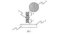
図3に、第一の捕捉分子となるペプチド分子、及び第二の捕捉分子となるペプチド分子として、イムノグロブリンG分子の可変領域(Fv)分子を用いた場合の概略図を示す。図中7が第一の標的物質捕捉分子となるイムノグロブリンG分子の可変領域(Fv)分子、図中8が第二の標的物質捕捉分子となるイムノグロブリンG分子の可変領域(Fv)分子を示している。 FIG. 3 shows a schematic diagram in the case of using a variable region (Fv) molecule of an immunoglobulin G molecule as a peptide molecule serving as a first capture molecule and a peptide molecule serving as a second capture molecule. In the figure, 7 is a variable region (Fv) molecule of an immunoglobulin G molecule that is a first target substance capturing molecule, and 8 is a variable region (Fv) molecule of an immunoglobulin G molecule that is a second target substance capturing molecule. Show.
イムノグロブリンG分子の可変領域(Fv)分子は、イムノグロブリンG分子の標的物質の特異的親和性または結合性を司るユニットであり、原理的にはイムノグロブリンG分子と同等の特異的親和性あるいは結合性を有している。一方、イムノグロブリンG分子の可変領域(Fv)分子の分子量はイムノグロブリンG分子の約1/6、分子の大きさはイムノグロブリンG分子が約15nmであるのに対しイムノグロブリンG分子の可変領域(Fv)分子はその1/5以下の2mから3nmの範囲にある。従って、センサー素子表面と磁性標識との間の距離は最大25nm程度短縮され、その分だけ磁性標識から得られる信号は大きくなる事となる。 The variable region (Fv) molecule of an immunoglobulin G molecule is a unit that controls the specific affinity or binding property of the target substance of the immunoglobulin G molecule, and in principle has a specific affinity equivalent to that of the immunoglobulin G molecule or Has binding properties. On the other hand, the molecular weight of the variable region (Fv) molecule of immunoglobulin G molecule is about 1/6 of that of immunoglobulin G molecule, and the size of immunoglobulin G molecule is about 15 nm, whereas the variable region of immunoglobulin G molecule. The (Fv) molecule is in the range of 2 to 3 nm, which is 1/5 or less. Therefore, the distance between the sensor element surface and the magnetic label is shortened by about 25 nm at the maximum, and the signal obtained from the magnetic label is increased accordingly.
これらの分子の組合せは、標的物質に対する捕捉分子の分子構造、生産性、固定のし易さ等により様々な様態をとり得る。例えば、図4では、第一の捕捉分子としてイムノグロブリンG分子の可変領域(Fv)分子7、第二の捕捉分子としてイムノグロブリンG分子5を用いた例を示す。また、図5では、第一の捕捉分子としてイムノグロブリンG分子3、第二の捕捉分子としてイムノグロブリンG分子のFab分子9を用いた例を示す。 The combination of these molecules can take various forms depending on the molecular structure of the capture molecule with respect to the target substance, productivity, ease of immobilization, and the like. For example, FIG. 4 shows an example in which an immunoglobulin G molecule variable region (Fv)
本発明にかかる素子、あるいは磁性標識へのペプチド分子の固定は、「磁性標識および素子のそれぞれに固定されたペプチド分子が標的物質を介して結合した状態」を検出時まで維持できるだけの強度および持続性を有していれば特に限定されない。そのような固定の種類として例えば、共有結合等の化学結合でもよいし、抗原−抗体間に働く分子間相互作用による固定でもよい。 The immobilization of the peptide molecule on the element or magnetic label according to the present invention is sufficient and strong enough to maintain the state in which the peptide molecule immobilized on each of the magnetic label and the element is bound via the target substance until the time of detection. If it has property, it will not specifically limit. As such a kind of immobilization, for example, a chemical bond such as a covalent bond may be used, or an immobilization by an intermolecular interaction acting between an antigen and an antibody may be used.
例えば、センサー素子の表面に位置する機能性膜を金の薄膜とする事により、国際公開第05/095461号パンフレットに開示されている、次のような構成の金結合性タンパク質を本発明にかかる第一の捕捉分子として用いることが出来る。即ち、第一の捕捉分子としては、以下の第一及び第二のドメインを含むことを特徴とする金結合性タンパク質が好適に利用できる(以下、金結合性Diabody(ダイアボディー)と記載する場合もある)。
(1)金に対する結合部位を有し、少なくともイムノグロブリンG軽鎖可変領域(VL)またはイムノグロブリンG重鎖可変領域(VH)の一部を含む第一のドメイン。
(2)標的物質に対する結合部位を有し、少なくともイムノグロブリンG重鎖可変領域(VH)またはイムノグロブリンG軽鎖可変領域(VL)の一部を含む第二のドメイン。
更に、磁性標識の表面の少なくとも一部を金とする事により、上述の構成の金結合性タンパク質を第二の捕捉分子としても用いる事ができる。For example, by making the functional film located on the surface of the sensor element a gold thin film, a gold-binding protein having the following constitution disclosed in WO05 / 095461 is applied to the present invention. It can be used as the first capture molecule. That is, as the first capture molecule, a gold-binding protein characterized by including the following first and second domains can be preferably used (hereinafter referred to as gold-binding diabody). There is also.)
(1) A first domain having a binding site for gold and containing at least a part of an immunoglobulin G light chain variable region (VL) or an immunoglobulin G heavy chain variable region (VH).
(2) A second domain having a binding site for a target substance and containing at least a part of an immunoglobulin G heavy chain variable region (VH) or an immunoglobulin G light chain variable region (VL).
Furthermore, by using at least a part of the surface of the magnetic label as gold, the gold-binding protein having the above-described configuration can be used as the second capture molecule.
この場合、上述の第一のドメインが、センサー素子、或いは磁性標識に固定する場合のアンカーとなり、簡便且つ捕捉分子の捕捉能を保持した状態での固定が可能となる。この場合の例の概略図を図6に示す。金薄膜10により表面を被覆されたセンサー素子上に、第一の捕捉分子として上述の構成の金結合性タンパク質12が固定されている。この第一の捕捉分子に結合した標的物質を挟んで、金薄膜11により被覆された磁性標識6上に固定された、第二の捕捉分子として上述の構成の金結合性タンパク質13が標的物質の別の領域に結合する形で固定されている。このような金結合性タンパク質の分子の大きさは5nmであり、磁性標識と素子との距離は、第一の捕捉分子、第二の捕捉分子ともに金結合性タンパク質を用いると、イムノグロブリンG分子の場合よりも最大20nm程度短縮され、結果的に信号強度が向上する。さらに、金結合性タンパク質は、化学修飾による素子および標識材料への固定化を要さないため、化学反応による標的物質結合部位へのダメージがない。このため、金結合性タンパク質は、標的物質との親和性又は結合性が損なわれない状態の捕捉分子として好適に用いられる。よって、本発明にかかる素子、及び材料の有するペプチド分子として、あるいは、本発明にかかるキットに含まれる第一のペプチド分子及び第二のペプチド分子の少なくとも一方として、金結合性Diabody(ダイアボディー)を用いることが好ましい。 In this case, the above-mentioned first domain serves as an anchor for immobilization to the sensor element or the magnetic label, and immobilization in a state where the capture ability of the capture molecule is retained is possible. A schematic diagram of an example in this case is shown in FIG. On the sensor element whose surface is coated with the gold
更に近年、様々な標的物質に対して特異的に結合する5から20残基程度のアミノ酸が連なったペプチドの研究開発が進んできている。このようなペプチド分子も同様に本発明に用いる事ができる。具体的にこのようなペプチドは、ファージディスプレイ法、リボゾームディスプレイ法のような「分子進化工学」的手法を用いる事により、目的の標的物質をターゲットにして取得することができる。上述のペプチドをファージディスプレイにより取得する場合、ペプチドライブラリ(ファージディスプレイの場合はファージライブラリ)が必要であるが、必要に応じて変異を導入する事により自家製として作製する事も可能である。さらにはNew England Biolabs(NEB)社製のペプチドリガンド検索用ファージディスプレイキットのようなキットを購入して取得する事もできる。 Furthermore, in recent years, research and development of peptides in which amino acids of about 5 to 20 residues that specifically bind to various target substances are continued. Such peptide molecules can also be used in the present invention. Specifically, such a peptide can be obtained by targeting a target target substance by using a “molecular evolution engineering” technique such as a phage display method or a ribosome display method. When acquiring the above-mentioned peptide by phage display, a peptide library (in the case of phage display, a phage library) is required, but it is also possible to make it homemade by introducing mutations as necessary. Furthermore, a kit such as a phage display kit for searching peptide ligands manufactured by New England Biolabs (NEB) can be purchased and obtained.
本発明に用いる磁性標識としての材料に含まれる磁性体を含む粒子には、通常用いられる常磁性、超常磁性を示す磁性微粒子、磁性ビーズを用いる事ができる。一般的には、フェライトやマグネタイトといった鉄酸化物の粒子とスチレン系、デキストラン系、アクリルアミド系等のポリマーとの混合物、あるいはこれらのポリマーによる被覆物が用いられる。このような磁性微粒子、磁性ビーズの大きさは、素子の形状、大きさ、或いは用途によって様々に選択する事が可能であるが、一般的に数十ナノメートルから数百マイクロメートルの直径を有するものが好適である。 As the particles containing the magnetic substance contained in the magnetic label material used in the present invention, commonly used paramagnetic and superparamagnetic magnetic fine particles and magnetic beads can be used. In general, a mixture of iron oxide particles such as ferrite and magnetite and a polymer such as styrene, dextran, and acrylamide, or a coating of these polymers is used. The size of such magnetic fine particles and magnetic beads can be variously selected according to the shape, size, or application of the device, but generally has a diameter of several tens of nanometers to several hundreds of micrometers. Those are preferred.
本発明の標的物質検出素子の検出方式は、磁界効果を利用する方式であれば如何なる方式でも用いることが可能であり、その中でも特に、磁気抵抗効果素子、ホール効果素子、磁気インピーダンス素子、および超電導量子干渉計素子が好適に用いることができる。これらの素子は、上述の標的物質検出用素子の構成における磁場を検知する部位に含まれる。本発明の素子、材料、及びキットが用いられる検出方式は、所謂磁気センサーに分類される方式であれば如何なる方式でもよく、その中でも特に磁気抵抗効果素子、ホール効果素子、磁気インピーダンス素子、および超電導量子干渉計素子を用いる検出方式のいずれか1つ以上の方式である事が望ましい。 As the detection method of the target substance detection element of the present invention, any system can be used as long as it uses a magnetic field effect, and among them, a magnetoresistance effect element, a Hall effect element, a magnetic impedance element, and a superconductivity A quantum interferometer element can be suitably used. These elements are included in a part for detecting a magnetic field in the configuration of the target substance detection element described above. The detection method in which the element, material, and kit of the present invention are used may be any method as long as it is classified as a so-called magnetic sensor, and among them, a magnetoresistive effect element, a Hall effect element, a magnetic impedance element, and a superconductivity It is desirable that one or more detection methods using a quantum interferometer element be used.
本発明の検出素子により検出可能な標的物質は、前記第一のペプチド分子が認識、結合する事ができる領域と、前記第二のペプチド分子が認識、結合する事ができる領域とを併せ持つ物質であれば如何なる物質をも対象にする事が可能である。 The target substance detectable by the detection element of the present invention is a substance having both a region where the first peptide molecule can be recognized and bound and a region where the second peptide molecule can be recognized and bound. Any substance can be targeted.
通常標的物質として適用される生体物質としては、核酸、タンパク質、糖鎖、脂質及びそれらの複合体から選択される生体物質が含まれ、更に詳しくは、核酸、タンパク質、糖鎖、脂質から選択される生体分子を含んでなるものである。具体的には、DNA、RNA、アプタマー、遺伝子、染色体、細胞膜、ウイルス、抗原、抗体、レクチン、ハプテン、ホルモン、レセプタ、酵素、ペプチド、スフィンゴ糖、スフィンゴ脂質の何れかから選択された物質を含むものであれば、如何なる物質にも本発明を適用することができる。更には、前記の「生体物質」を産生する細菌や細胞そのものも、本発明が対象とする「生体物質」として標的物質となり得る。 Biological substances that are usually applied as target substances include biological substances selected from nucleic acids, proteins, sugar chains, lipids and complexes thereof, and more specifically selected from nucleic acids, proteins, sugar chains, and lipids. A biomolecule. Specifically, it includes a substance selected from any of DNA, RNA, aptamer, gene, chromosome, cell membrane, virus, antigen, antibody, lectin, hapten, hormone, receptor, enzyme, peptide, sphingosaccharide, and sphingolipid. The present invention can be applied to any substance as long as it is anything. Furthermore, bacteria and cells themselves that produce the above-mentioned “biological substances” can also be target substances as “biological substances” targeted by the present invention.
その中でも特に、タンパク質(脂質タンパク、糖タンパク、タンパク質複合体およびタンパク質多量体など)の具体例として、いわゆる疾病マーカーが挙げられる。 Among them, in particular, so-called disease markers can be mentioned as specific examples of proteins (lipid proteins, glycoproteins, protein complexes, protein multimers, etc.).
疾患マーカーの例としては、胎児期に肝細胞で産生され胎児血中に存在する酸性糖蛋白であり、肝細胞癌(原発性肝癌)、肝芽腫、転移性肝癌およびヨークサック腫瘍のマーカーとなるα−フェトプロテイン(AFP);肝実質障害時に出現する異常プロトロンビンであり、肝細胞癌で特異的に出現することが確認されるPIVKA−II;免疫組織化学的に乳癌特異抗原である糖蛋白で、原発性進行乳癌、再発・転移乳癌のマーカーとなるBCA225;ヒト胎児の血清、腸および脳組織抽出液に発見された塩基性胎児蛋白であり、卵巣癌、睾丸腫瘍、前立腺癌、膵癌、胆道癌、肝細胞癌、腎臓癌、肺癌、胃癌、膀胱癌および大腸癌のマーカーである塩基性フェトプロテイン(BFP);進行乳癌、再発乳癌、原発性乳癌および卵巣癌のマーカーとなる糖鎖抗原であるCA15−3;膵癌、胆道癌、胃癌、肝癌、大腸癌および卵巣癌のマーカーとなる糖鎖抗原であるCA19−9;卵巣癌、乳癌、結腸・直腸癌、胃癌および膵癌のマーカーとなる糖鎖抗原であるCA72−4;卵巣癌(特に漿液性嚢胞腺癌)、子宮体部腺癌、卵管癌、子宮頸部腺癌、膵癌、肺癌および大腸癌のマーカーとなる糖鎖抗原であるCA125;上皮性卵巣癌、卵管癌、肺癌、肝細胞癌および膵癌のマーカーとなる糖蛋白であるCA130;卵巣癌(特に漿液性嚢胞腺癌)、子宮体部腺癌および子宮頸部腺癌のマーカーとなるコア蛋白抗原であるCA602;卵巣癌(特に粘液性嚢胞腺癌)、子宮頸部腺癌および子宮体部腺癌のマーカーとなる母核糖鎖関連抗原であるCA54/61(CA546);大腸癌、胃癌、直腸癌、胆道癌、膵癌、肺癌、乳癌、子宮癌および尿路系癌等の腫瘍関連のマーカー抗原として現在、癌診断の補助に最も広く利用されている癌胎児性抗原(CEA);膵癌、胆道癌、肝細胞癌、胃癌、卵巣癌および大腸癌のマーカーとなる糖鎖抗原であるDUPAN−2;膵臓に存在し、結合組織の弾性線維エラスチン(動脈壁や腱などを構成する)を特異的に加水分解する膵外分泌蛋白分解酵素であり、膵癌、膵嚢癌および胆道癌のマーカーとなるエラスターゼ1;ヒト癌患者の腹水や血清中に高濃度に存在する糖蛋白であり、肺癌、白血病、食道癌、膵癌、卵巣癌、腎癌、胆管癌、胃癌、膀胱癌、大腸癌、甲状腺癌および悪性リンパ腫のマーカーとなる免疫抑制酸性蛋白(IAP)、膵癌、胆道癌、乳癌、大腸癌、肝細胞癌、肺腺癌および胃癌のマーカーとなる糖鎖抗原であるNCC−ST−439;前立腺癌のマーカーとなる糖蛋白質であるγ−セミノプロテイン(γ−Sm);ヒト前立腺組織から抽出された糖蛋白であり、前立腺組織のみに存在し、それゆえ前立腺癌のマーカーとなる前立腺特異抗原(PSA);前立腺から分泌される酸性pH下でリン酸エステルを水解する酵素であり、前立腺癌の腫瘍マーカーとして用いられる前立腺酸性フォスファターゼ(PAP);神経組織及び神経内分泌細胞に特異的に存在する解糖系酵素であり、肺癌(特に肺小細胞癌)、神経芽細胞腫、神経系腫瘍、膵小島癌、食道小細胞癌、胃癌、腎臓癌および乳癌のマーカーとなる神経特異エノラーゼ(NSE);子宮頸部扁平上皮癌の肝転移巣から抽出・精製された蛋白質であり、子宮癌(頸部扁平上皮癌)、肺癌、食道癌、頭頸部癌および皮膚癌のマーカーとなる扁平上皮癌関連抗原(SCC抗原);肺腺癌、食道癌、胃癌、大腸癌、直腸癌、膵癌、卵巣癌および子宮癌のマーカーとなる糖鎖抗原であるシアリルLeX−i抗原(SLX);膵癌、胆道癌、肝癌、胃癌および大腸癌のマーカーとなる糖鎖抗原であるSPan−1;食道癌、胃癌、直腸・結腸癌、乳癌、肝細胞癌、胆道癌、膵癌、肺癌および子宮癌のマーカーであり、特に他の腫瘍マーカーと組み合わせて進行癌を推測し、再発予知・治療経過観察として有用である単鎖ポリペプチドである組織ポリペプタイド抗原(TPA);卵巣癌、転移性卵巣癌、胃癌、大腸癌、胆道系癌、膵癌および肺癌のマーカーとなる母核糖鎖抗原であるシアリルTn抗原(STN);肺の非小細胞癌、特に肺の扁平上皮癌の検出に有効な腫瘍マーカーであるシフラ(cytokeratin;CYFRA);胃液中に分泌される蛋白消化酵素であるペプシンの2種(PGI・PGII)の不活性型前駆体であり、胃潰瘍(特に低位胃潰瘍)、十二指腸潰瘍(特に再発、難治例)、ブルンネル腺腫、ゾーリンガーエリソン症候群および急性胃炎のマーカーとなるペプシノゲン(PG);組織障害や感染により、血漿中で変化する急性相反応蛋白であり、急性心筋梗塞等により心筋に壊死が起こると高値を示すC−反応性蛋白(CRP);組織障害や感染により、血漿中で変化する急性相反応蛋白である血清アミロイドA蛋白(SAA)、主に心筋や骨格筋に存在する分子量約17500のヘム蛋白であり、急性心筋梗塞、筋ジストロフィー、多発性筋炎および皮膚筋炎のマーカーとなるミオグロビン;骨格筋,心筋の可溶性分画を中心に存在し、細胞の損傷によって血液中に遊出する酵素であって、急性心筋梗塞、甲状腺機能低下症、進行性筋ジストロフィー症および多発性筋炎のマーカーとなるクレアチンキナーゼ(CK)(骨格筋由来のCK−MM型,脳,平滑筋由来のCK−BB型,心筋由来のCK−MB型の3種のアイソザイム及びミトコンドリア・アイソザイムや免疫グロブリンとの結合型CK(マクロCK));横紋筋の薄いフィラメント上でトロポニンI,Cとともにトロポニン複合体を形成し、筋収縮の調節に関与している分子量39,000の蛋白であり、横紋筋融解症、心筋炎、心筋梗塞および腎不全のマーカーとなるトロポニンT;骨格筋・心筋いずれの細胞にも含まれる蛋白であり、測定結果の上昇は骨格筋、心筋の障害や壊死を意味するため、急性心筋梗塞症、筋ジストロフィーおよび腎不全のマーカーとなる心室筋ミオシン軽鎖I;また、近年ストレスマーカーとして注目されてきているクロモグラニンA、チオレドキシン及び8−OhdG;等が挙げられる。 Examples of disease markers are acidic glycoproteins produced in hepatocytes in the fetal period and present in fetal blood, and markers for hepatocellular carcinoma (primary liver cancer), hepatoblastoma, metastatic liver cancer and Yorksack tumor Α-fetoprotein (AFP); PIVKA-II, an abnormal prothrombin that appears at the time of liver parenchymal disorder, which is confirmed to appear specifically in hepatocellular carcinoma; a glycoprotein that is a breast cancer-specific antigen immunohistochemically BCA225, a marker of primary advanced breast cancer, recurrence / metastasis breast cancer; basic fetal protein found in human fetal serum, intestinal and brain tissue extracts, ovarian cancer, testicular tumor, prostate cancer, pancreatic cancer, biliary tract Basic fetoprotein (BFP), a marker for cancer, hepatocellular carcinoma, kidney cancer, lung cancer, stomach cancer, bladder cancer and colon cancer; advanced breast cancer, recurrent breast cancer, primary breast cancer and ovarian cancer CA15-3 which is a sugar chain antigen serving as a car; CA19-9 which is a sugar chain antigen serving as a marker for pancreatic cancer, biliary tract cancer, stomach cancer, liver cancer, colon cancer and ovarian cancer; ovarian cancer, breast cancer, colorectal cancer, gastric cancer CA72-4 which is a sugar chain antigen serving as a marker for pancreatic cancer; markers for ovarian cancer (especially serous cystadenocarcinoma), endometrial adenocarcinoma, fallopian tube cancer, cervical adenocarcinoma, pancreatic cancer, lung cancer and colon cancer CA125 which is a sugar chain antigen, CA130 which is a glycoprotein which is a marker for epithelial ovarian cancer, fallopian tube cancer, lung cancer, hepatocellular carcinoma and pancreatic cancer; ovarian cancer (especially serous cystadenocarcinoma), endometrial gland CA602, a core protein antigen that is a marker for cancer and cervical adenocarcinoma; a mother-sugar-chain-related antigen that is a marker for ovarian cancer (especially mucinous cystadenocarcinoma), cervical adenocarcinoma and uterine adenocarcinoma CA54 / 61 (CA546); large intestine Carcinoembryonic antigen (CEA), which is currently the most widely used to assist cancer diagnosis, as a tumor-related marker antigen such as cancer, stomach cancer, rectal cancer, biliary tract cancer, pancreatic cancer, lung cancer, breast cancer, uterine cancer and urinary tract cancer ); DUPAN-2, a sugar chain antigen that serves as a marker for pancreatic cancer, biliary tract cancer, hepatocellular carcinoma, gastric cancer, ovarian cancer, and colon cancer; elastic fiber elastin of the connective tissue (arterial wall, tendon, etc.) Pancreatic exocrine proteolytic enzyme that specifically hydrolyzes elastase 1 as a marker for pancreatic cancer, pancreatic sac cancer, and biliary tract cancer; glycoprotein present in high concentrations in ascites and serum of human cancer patients, Lung cancer, leukemia, esophageal cancer, pancreatic cancer, ovarian cancer, renal cancer, bile duct cancer, gastric cancer, bladder cancer, colon cancer, thyroid cancer and immunosuppressive acidic protein (IAP) as a marker for malignant lymphoma, pancreatic cancer, biliary tract cancer, breast cancer, Colorectal cancer, hepatocellular carcinoma NCC-ST-439, a sugar chain antigen that serves as a marker for lung adenocarcinoma and gastric cancer; γ-seminoprotein (γ-Sm), a glycoprotein that serves as a marker for prostate cancer; glycoprotein extracted from human prostate tissue Prostate-specific antigen (PSA) that is present only in prostate tissue and is therefore a marker for prostate cancer; an enzyme that hydrolyzes phosphate esters under acidic pH secreted from the prostate, and as a tumor marker for prostate cancer Prostatic acid phosphatase (PAP) used; a glycolytic enzyme that exists specifically in nerve tissue and neuroendocrine cells, including lung cancer (particularly small cell lung cancer), neuroblastoma, nervous system tumor, pancreatic islet cancer, Nerve specific enolase (NSE), a marker for esophageal small cell carcinoma, gastric cancer, kidney cancer and breast cancer; a protein extracted and purified from liver metastases of cervical squamous cell carcinoma Yes, squamous cell carcinoma-associated antigen (SCC antigen) that is a marker for uterine cancer (cervical squamous cell carcinoma), lung cancer, esophageal cancer, head and neck cancer, and skin cancer; lung adenocarcinoma, esophageal cancer, stomach cancer, colon cancer, rectum Sialyl LeX-i antigen (SLX) which is a sugar chain antigen serving as a marker for cancer, pancreatic cancer, ovarian cancer and uterine cancer; SPan-1 which is a sugar chain antigen serving as a marker for pancreatic cancer, biliary tract cancer, liver cancer, stomach cancer and colon cancer ; Esophageal cancer, gastric cancer, colorectal cancer, breast cancer, hepatocellular carcinoma, biliary tract cancer, pancreatic cancer, lung cancer and uterine cancer marker, especially in combination with other tumor markers, predicting advanced cancer, progress of recurrence Tissue polypeptide antigen (TPA), a single-chain polypeptide that is useful for observation; sialyl, a nucleoside sugar chain antigen that serves as a marker for ovarian cancer, metastatic ovarian cancer, gastric cancer, colon cancer, biliary tract cancer, pancreatic cancer, and lung cancer Tn (STN); two types of cytokeratin (CYFRA), an effective tumor marker for detecting non-small cell lung cancer, especially squamous cell carcinoma of the lung; pepsin, a protein digestive enzyme secreted into gastric juice ( PGI / PGII), an inactive precursor, and a marker for gastric ulcer (especially low-grade gastric ulcer), duodenal ulcer (especially relapsed or refractory), Brunnell's adenoma, Zollinger-Ellison syndrome and acute gastritis; tissue C-reactive protein (CRP), which is an acute phase reaction protein that changes in plasma due to damage or infection, and shows a high value when necrosis occurs in the myocardium due to acute myocardial infarction, etc .; changes in plasma due to tissue damage or infection Serum amyloid A protein (SAA), which is an acute phase reaction protein, is a heme protein having a molecular weight of about 17500, which is mainly present in cardiac muscle and skeletal muscle. Myoglobin, a marker for myocardial infarction, muscular dystrophy, polymyositis and dermatomyositis; an enzyme that exists mainly in the soluble fraction of skeletal muscle and myocardium and is released into the blood by cell damage. Creatine kinase (CK) as a marker of hypothyroidism, progressive muscular dystrophy and polymyositis (CK-MM type derived from skeletal muscle, CK-BB type derived from brain, smooth muscle, CK-MB type derived from
以下、実施例を用いてさらに詳細に本発明を説明するが、本発明は、これらの実施例に限定されるものではなく、材料、組成条件、反応条件等、同様な機能、効果を有する検出素子、検出装置が得られる範囲で自由に変えることができる。
(実施例1)
本実施例では、標的物質のモデルタンパク質としてニワトリ卵白リゾチーム(以降HELと記載する場合もある)を用いる。また、検出素子に固定され、前記標的物質の特定の領域を特異的に認識し、結合することができる第一のペプチド分子として抗HEL抗体のscFv(single chain Fv)を用いる。scFvは、抗原を認識する最小単位である可変領域(Fv)を構成するVH及びVLをペプチドリンカーで結合した単鎖可変領域フラグメントである。磁性標識に固定され、前記標的物質の第一のペプチド分子結合領域と異なる領域を特異的に認識し、結合することができる第二のペプチド分子として抗HEL抗体のFab領域分子を用いる。さらに本実施例では、検出方法としてTMR(トンネル膜型磁気抵抗効果)手法を用いて本発明の効果の例を示す。Hereinafter, the present invention will be described in more detail with reference to examples. However, the present invention is not limited to these examples, and detection having similar functions and effects such as materials, composition conditions, reaction conditions, and the like. It can be freely changed as long as the element and the detection device can be obtained.
Example 1
In this example, chicken egg white lysozyme (hereinafter sometimes referred to as HEL) is used as a model protein of the target substance. In addition, an anti-HEL antibody scFv (single chain Fv) is used as a first peptide molecule that is fixed to the detection element and can specifically recognize and bind to a specific region of the target substance. scFv is a single-chain variable region fragment in which VH and VL constituting a variable region (Fv), which is the minimum unit for recognizing an antigen, are connected by a peptide linker. The Fab region molecule of the anti-HEL antibody is used as the second peptide molecule that is fixed to the magnetic label and can specifically recognize and bind to a region different from the first peptide molecule binding region of the target substance. Further, in this embodiment, an example of the effect of the present invention is shown by using a TMR (tunnel film type magnetoresistive effect) technique as a detection method.
まず、抗HEL抗体のscFv分子は以下のように作製する。
(1)発現ベクターによる形質転換
J. of Bio.chem.,2003,278,p8979−8987記載のHEL結合HyHEL10 scFv発現プラスミドを用いてBL21(DE3)コンピテントセル40μLを形質転換する。なお、HyHEL10 scFvのアミノ酸配列(配列番号1)および塩基配列(配列番号2)については配列表を参照のこと。First, the scFv molecule of the anti-HEL antibody is prepared as follows.
(1) Transformation with expression vector of Bio. chem. , 2003, 278, p8979-8987, 40 μL of BL21 (DE3) competent cells are transformed with the HEL-binding HyHEL10 scFv expression plasmid. For the amino acid sequence (SEQ ID NO: 1) and base sequence (SEQ ID NO: 2) of HyHEL10 scFv, see the sequence listing.
形質転換は、ヒートショックを氷中→42℃×90sec→氷中の条件でおこなう。ヒートショックにより形質転換した上記BL21溶液にLB培地750μLを加え、一時間37℃にて振盪培養を行なう。その後、6000rpm×5分間遠心を行い、培養上清650μLを廃棄し、残った培養上清と沈殿となった細胞画分を攪拌し、LB/amp.プレートに撒き、一晩37℃にて静置する。 Transformation is performed under conditions of heat shock in ice → 42 ° C. × 90 sec → ice. 750 μL of LB medium is added to the BL21 solution transformed by heat shock, and shaking culture is performed at 37 ° C. for 1 hour. Thereafter, centrifugation was performed at 6000 rpm for 5 minutes, 650 μL of the culture supernatant was discarded, the remaining culture supernatant and the precipitated cell fraction were stirred, and LB / amp. Spread on a plate and let stand at 37 ° C overnight.
(2)予備培養
プレート上のコロニーを無作為に選択し、3.0mL LB/amp.培地にて28℃にて一晩振盪培養を行なう。(2) Preculture A colony on the plate was randomly selected and 3.0 mL LB / amp. Shake culture overnight at 28 ° C. in the medium.
(3)本培養
上記予備培養溶液を2×YT培地 750MLに植え継ぎ、更に培養を28℃にて継続する。OD600が0.8を越えた時点で、終濃度が1mMとなるようにIPTGを加え、更に28℃にて終夜培養を行なう。(3) Main culture The above preculture solution is inoculated into 750 mL of 2 × YT medium, and the culture is further continued at 28 ° C. When the OD600 exceeds 0.8, IPTG is added so that the final concentration is 1 mM, and the culture is further performed overnight at 28 ° C.
(4)精製
目的のポリペプチド鎖を不溶性顆粒画分から以下の工程により精製する。
(A)不溶性顆粒の回収
上記(3)で得られた培養液を6000rpm×30minにて遠心し、沈殿を菌体画分として得る。得られた菌体をトリス溶液(20mM トリス/500mM NaCl)15mlに氷中にて懸濁する。得られた懸濁液をフレンチプレスにて破砕し、菌破砕液を得る。次に、菌破砕液を12,000rpm×15minで遠心を行い、上清を除き、沈殿を不溶性顆粒画分として得る。
(B)不溶性顆粒画分の可溶化
(A)で得られた不溶性画分を6M 塩酸グアニジン/トリス溶液 10mLを加えて、一晩浸漬する。次に、12,000rpm×10minで遠心し、上清を可溶化溶液として得る。
(C)金属キレートカラム
金属キレートカラム担体として、His−Bind(Novagen社製)を用いる。カラム調整やサンプル負荷、及び洗浄工程は、前記業者の推奨方法に準拠し、室温(20℃)にて行う。目的であるHisタグ融合のポリペプチドの溶出は60mMイミダゾール/Tris溶液にて行う。溶出液のSDS−PAGE(アクリルアミド15%)の結果、単一バンドであり、精製されていることを確認する。
(D)透析
上記溶出液に対して、外液を6M 塩酸グアニンジン/Tris溶液として4℃にて透析を行い、溶出液中のイミダゾールの除去を行い、目的のポリペプチド鎖を含む溶液を得る。
(E)リフォールディング
金結合性Fvと上記ペプチドを融合したscFv−Spのポリペプチド鎖溶液を以下の工程により別個に、脱塩酸グアニンジンを透析にて行いながらタンパク質のリフォールディングを行う。
a) 6M 塩酸グアニジン/Tris溶液を用い、それぞれのポリペプチド鎖のモル吸光係数とΔO.D.(280nm−320nm)値から濃度7.5μMのサンプル(希釈後体積10ml)を調整する。次にβ−メルカプトエタノール(還元剤)を終濃度375μM(タンパク濃度50倍)になるよう添加、室温、暗所で4時間還元を行う。このサンプル溶液を透析バック(MWCO:14,000)に入れ、透析用サンプルとする。
b)透析外液を6M塩酸グアニンジン/トリス溶液として、透析サンプルを浸漬し、緩やかに攪拌しながら6時間透析する。
c)外液の塩酸グアニジン濃度を3M、2Mと段階的に下げる。それぞれの外液濃度において、6時間透析する。
d)酸化型グルタチオン(GSSG)を終濃度375μM、L−Argを 終濃度0.4M)となるようにトリス溶液に加え、上記3)の2Mの透析外液を加え、塩酸グアニジン濃度が1Mとし、pHをNaOHで、pH8.0(4℃)に調整した溶液にて、12時間緩やかに攪拌しながら透析する。
e)上記d)と同様の作業にて塩酸グアニジン濃度0.5Mの含L−Arg トリス溶液を整し、更に12時間透析する。
f)最後にトリス溶液にて12時間透析する。上記で得られた溶液に対して、更に外液をリン酸バッファー(以下、PBS)に交換する。透析終了後、10000rpmで約20分遠心分離し凝集体と上清を分離する。得られる上清の吸光スペクトル測定し、280nmから算出される濃度は6.3μMである。(4) Purification The target polypeptide chain is purified from the insoluble granule fraction by the following steps.
(A) Recovery of insoluble granules The culture solution obtained in (3) above is centrifuged at 6000 rpm x 30 min to obtain a precipitate as a cell fraction. The obtained cells are suspended in 15 ml of Tris solution (20 mM Tris / 500 mM NaCl) in ice. The obtained suspension is crushed with a French press to obtain a bacterial crushing solution. Next, the bacterial disruption solution is centrifuged at 12,000 rpm × 15 min, the supernatant is removed, and the precipitate is obtained as an insoluble granule fraction.
(B) Solubilization of insoluble
(C) Metal chelate column As a metal chelate column carrier, His-Bind (manufactured by Novagen) is used. The column adjustment, sample loading, and washing step are performed at room temperature (20 ° C.) in accordance with the recommended method of the supplier. The target His tag fusion polypeptide is eluted with a 60 mM imidazole / Tris solution. As a result of SDS-PAGE (
(D) Dialysis The eluate is dialyzed at 4 ° C. using 6M guanidine hydrochloride / Tris solution as an external solution to remove imidazole in the eluate to obtain a solution containing the target polypeptide chain.
(E) Refolding A protein chain solution of scFv-Sp in which gold-binding Fv and the above peptide are fused is separately separated by the following steps while guanidine hydrochloride is dialyzed.
a) Using 6M guanidine hydrochloride / Tris solution, the molar extinction coefficient and ΔO. D. A sample with a concentration of 7.5 μM (volume after dilution: 10 ml) is prepared from the (280 nm-320 nm) value. Next, β-mercaptoethanol (reducing agent) is added to a final concentration of 375 μM (protein concentration 50 times), and reduction is performed at room temperature in the dark for 4 hours. This sample solution is put into a dialysis bag (MWCO: 14,000) to obtain a sample for dialysis.
b) The dialysis sample is immersed in 6M guanidine hydrochloride / Tris solution as an external solution for dialysis, and dialyzed for 6 hours with gentle stirring.
c) Decrease the concentration of guanidine hydrochloride in the external solution stepwise to 3M and 2M. Dialyse for 6 hours at each external concentration.
d) Add oxidized glutathione (GSSG) to the Tris solution to a final concentration of 375 μM and L-Arg to a final concentration of 0.4 M). Then, dialyze with a solution adjusted to pH 8.0 (4 ° C.) with NaOH for 12 hours with gentle stirring.
e) Prepare an L-Arg Tris solution containing 0.5 M guanidine hydrochloride in the same manner as in d) above, and dialyze for 12 hours.
f) Finally, dialyze against Tris solution for 12 hours. The external solution is further exchanged with a phosphate buffer (hereinafter referred to as PBS) for the solution obtained above. After completion of dialysis, the aggregate and supernatant are separated by centrifugation at 10,000 rpm for about 20 minutes. The absorbance spectrum of the obtained supernatant is measured, and the concentration calculated from 280 nm is 6.3 μM.
(F)ゲルろ過精製
Sephadex75(アマシャムバイオサイエンス社)を用いて、ゲルろ過による精製(バッファー条件:50mM Tris−HCl、200mM NaCl、1mMEDTA、pH8.0、流速:0.7mL/min)を4℃にて行う。得られる分画を濃縮後、SDS−PAGE(アクリルアミド17.5%)及びHRP融合抗His抗体を用いて、上述と同様のWestern Blottingを行う。これにより、目的のタンパク質の分画を特定し、単一バンドに精製される。その中で、約25kDaの単量体タンパク質であることを示唆するピークを分取し、HEL抗体のscFv分子とする。(F) Gel filtration purification Using Sephadex 75 (Amersham Bioscience), purification by gel filtration (buffer conditions: 50 mM Tris-HCl, 200 mM NaCl, 1 mM EDTA, pH 8.0, flow rate: 0.7 mL / min) was performed at 4 ° C. To do. After concentration of the obtained fraction, Western blotting similar to that described above is performed using SDS-PAGE (acrylamide 17.5%) and an HRP-fused anti-His antibody. Thereby, the fraction of the target protein is specified and purified to a single band. Among them, a peak suggesting that it is a monomer protein of about 25 kDa is collected and used as the scFv molecule of the HEL antibody.
また、抗HEL抗体のFab領域分子は、Rockland社製抗HELポリクローナル抗体を定法によりペプシン処理後に2- メルカプトエタノールアミンで還元し、アフィニティカラム、ゲルろ過カラムにより精製する事により得る。得られる抗HEL抗体のFab領域分子は、分子中のアミノ基を、磁性ビーズDynabeads M-270 Carboxylic Acid(Dynal社製、平均径2.8μm)のカルボキシル基にカルボジイミドによる活性化することにより結合させることで固定する。 The Fab region molecule of the anti-HEL antibody can be obtained by treating a Rockland anti-HEL polyclonal antibody with pepsin by a conventional method, reducing with 2-mercaptoethanolamine, and purifying with an affinity column or gel filtration column. The Fab region molecule of the resulting anti-HEL antibody binds the amino group in the molecule to the carboxyl group of the magnetic beads Dynabeads M-270 Carboxylic Acid (Dynal, average diameter 2.8 μm) by activating with carbodiimide. To fix.
次に検出デバイス及び磁気信号測定につき説明する。 Next, the detection device and magnetic signal measurement will be described.
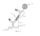
図7(a)は本発明を説明するための検出デバイスであるスピントンネル磁気抵抗効果素子(TMR素子)100の断面を模式的に示した図である。支持体101上にハフニウム膜102、マンガン−イリジウム合金膜103、鉄−コバルト合金膜104、ルテニウム膜105、鉄−コバルト合金膜106、マグネシウム酸化膜107、鉄−コバルト合金膜108、ニッケル−鉄合金膜109、プラチナ膜110、シリコン膜111を順次形成する。ハフニウム膜102は下部電極であり、より高い電気導電性が求められる場合は、支持体101とハフニウム膜102の間に、例えばプラチナなどの電気抵抗の小さな材料からなる膜を形成しても良い。マンガン−イリジウム合金膜103、鉄−コバルト合金膜104、ルテニウム膜105、鉄−コバルト合金膜106からなる多層膜は、外部磁界の印加によっても磁化方向が容易に変化しないピンド層112である。鉄−コバルト合金膜104と鉄−コバルト合金膜106は磁気的に強く結合しておりこれらの磁化は反平行を向いている。また、マンガン−イリジウム合金膜103は反強磁性膜であり、鉄−コバルト合金膜104と交換結合している。鉄−コバルト合金膜108とニッケル−鉄合金膜109は交換結合をしており、2つの層によって、外部磁界によって磁化方向が容易に変化するフリー層113である。マグネシウム酸化膜107はトンネル障壁層、プラチナ膜110は磁性膜の酸化を防止する保護層であり、かつ上部電極である。 FIG. 7A is a diagram schematically showing a cross section of a spin tunnel magnetoresistive element (TMR element) 100 which is a detection device for explaining the present invention.
シリコン膜111の表面に第一のペプチド分子171を担持するために、シリコン膜111の表面は、まず親水化処理が施された後、アミノシランカップリング剤処理される。さらに第一のペプチド分子171を固定化させるためのグルタルアルデヒド等架橋剤を用いて、前記アミノシランカップリング剤由来のアミノ基とペプチド鎖間を化学結合させ所望の抗原を補捉する第一のペプチド分子171が固定されている。 In order to carry the
この検出デバイスを用い、以下のプロトコールに従ってHELの検出を行う。検出デバイスには、HELを認識する第一のペプチド分子171が固定化されている。
(1)標的物質173であるHELを含むリン酸緩衝生理食塩水に上記検出デバイスを浸し、5分間インキュベートする。
(2)未反応のHELをリン酸緩衝生理食塩水で洗浄する。
(3)磁性粒子174により標識された第二のペプチド分子172を含むリン酸緩衝生理食塩水に工程(1)および(2)が終了した上記検出デバイスを浸し、5分間インキュベートする。
(4)未反応の該標識抗体をリン酸緩衝生理食塩水で洗浄する。ただし、磁性粒子174の平均直径は約400nmで超常磁性を示し、また、TMR素子100は上部から見た形状が200nm×400nmの長方形とする。Using this detection device, HEL is detected according to the following protocol. A
(1) The detection device is immersed in phosphate buffered saline containing HEL, which is the
(2) Unreacted HEL is washed with phosphate buffered saline.
(3) The detection device in which steps (1) and (2) are completed is immersed in a phosphate buffered saline containing the
(4) The unreacted labeled antibody is washed with phosphate buffered saline. However, the average diameter of the
図7(d)に本実施例で用いる検出回路を示す。TMR素子100には定電流源177が直列に接続されている。ただし、検出電流はTMR素子100のマグネシウム酸化膜107をトンネリングするように膜面垂直方向に流す。さらに、TMR素子100の他端はセンスアンプ178の入力端子に接続される。TMR素子100にかかる電圧と参照電圧に差が有る場合には、検出信号がセンスアンプ178から出力される。 FIG. 7 (d) shows a detection circuit used in this embodiment. A constant
TMR素子100の表面に「第一のペプチド分子ー標的物質ー第二のペプチド分子」を介して固定された磁性粒子174に下方向で600 Oeの大きさの外部磁界175を印加し、磁性粒子174の磁化を下方向に向ける。磁性粒子174からは浮遊磁界が発生し、この浮遊磁界の膜面内成分が、フリー層113に大きく作用して磁化が膜面内で回転する。もし、磁性粒子174がTMR素子100上に存在しなければ、浮遊磁界が発生せず、TMR素子100に膜面内方向の磁界が印加されない。つまり、標的物質173の有無によってTMR素子100の磁化状態が異なり、抵抗値が異なることから、標的物質の検出が行われる。 An external
上記検出を図7(b)に示す本実施例の構成と図7(c)に構成による対照例とで比較する。なお、本実施例では、「第一のペプチド分子として抗HEL抗体のscFv171b、第二のペプチド分子として抗HEL抗体のFab領域分子172b」が用いられている。対照例では、従来技術の構成である、「第一のペプチド分子として抗HELポリクローナル抗体171c、第二のペプチド分子として同じく抗HELポリクローナル抗体172c」が用いられている。 The above detection is compared between the configuration of the present embodiment shown in FIG. 7B and the control example according to the configuration shown in FIG. In the present example, “scFv171b of anti-HEL antibody as the first peptide molecule and Fab region molecule 172b of anti-HEL antibody as the second peptide molecule” are used. In the control example, “anti-HEL polyclonal antibody 171c as the first peptide molecule and anti-HEL polyclonal antibody 172c as the second peptide molecule”, which is a configuration of the prior art, is used.
この場合、対照例ではフリー層113表面から磁性粒子174までの平均距離は約35nmであるのに対し、本実施例では15nm程度となり、それに伴い、本実施例は対照例より強い電圧の変化が検出され、結果として検出感度が向上する。 In this case, in the control example, the average distance from the surface of the
本実施例においては膜面磁化膜からなるTMR素子を検出デバイスとして用いたが、検出デバイスはTMR素子に限られるものではなく、磁気センサーであればどのようなものでも使用可能である。また、磁気抵抗効果素子を用いる場合にも、TMR素子の他に、巨大磁気抵抗効果素子(GMR素子)でも本発明を達成可能である。GMR素子の構造はTMR素子と酷似しているが、トンネル障壁層が無く、ピンド層とフリー層の間に非磁性金属層が形成される。非磁性金属層は銅による形成が一般的である。また、磁気抵抗効果素子を構成する磁性膜においても、面内磁化膜ではなく垂直磁化膜でも構わない。図7(e)は垂直磁化膜によって構成されたGMR素子の膜断面を模式的に示した図である。支持体114上に第1の垂直磁化膜115、第1の高スピン分極率層116、非磁性膜117、第2の高スピン分極率層118、第2の垂直磁化膜119が順次成膜されるGMR素子130を形成する。第1の垂直磁化膜115および第1の高スピン分極率層116からなる2層交換結合膜は膜面垂直方向が磁化容易軸であるピンド層120であり磁化方向が固定される。そのため第1の垂直磁化膜115はテルビウム、鉄およびコバルトからなる合金膜によって形成し、そのテルビウムの組成は補償組成に近い21at%とする。また、第1の高スピン分極率層116および第2の高スピン分極率層117は鉄とコバルトからなる合金膜であり、Co組成を40at%とする。また、第2の垂直磁化膜119および第2の高スピン分極率層118の2層交換結合膜はフリー層121である。フリー層121はその磁化が膜面垂直方向に容易に傾くことが可能であるならば良く、必ずしも零磁場で磁化方向が膜面垂直方向に向いている必要は無い。もし、零磁場で磁化方向が膜面垂直となる構成とするならば、ピンド層120の保磁力よりも小さな保磁力とする。第2の垂直磁化膜119はガドリニウム、鉄およびコバルトからなる合金で形成する。そうすることによって、磁化方向の変化が容易になる。高スピン分極率層116および118は単独に存在する場合には面内磁化膜であるが、垂直磁化膜115および119と交換結合することによって、磁化方向を膜面垂直方向に向けることが容易となる。フリー層121とピンド層120の間の非磁性膜は銅によって形成する。また、第2の垂直磁化膜119の表面をシリコンからなる保護膜122で覆う。GMR素子130の両端にPtからなる電極123および124を形成する。GMR素子においては検出電流は素子の膜面垂直方向でも、膜面内方向でも、あるいは斜め方向でも構わない。検出の際には、上記TMR素子を用いた場合と同様に、検出電流を流しながら外部磁界を印加し、GMR素子130にかかる電圧を読み取ることで、標的物質173を検出する。 In this embodiment, a TMR element made of a film-surface magnetization film is used as a detection device. However, the detection device is not limited to a TMR element, and any magnetic sensor can be used. Also, when using a magnetoresistive effect element, the present invention can be achieved by a giant magnetoresistive effect element (GMR element) in addition to the TMR element. The structure of the GMR element is very similar to that of the TMR element, but there is no tunnel barrier layer, and a nonmagnetic metal layer is formed between the pinned layer and the free layer. The nonmagnetic metal layer is generally formed of copper. Also, the magnetic film constituting the magnetoresistive element may be a perpendicular magnetization film instead of an in-plane magnetization film. FIG. 7 (e) is a diagram schematically showing a film cross section of a GMR element constituted by a perpendicular magnetization film. On the
(実施例2)
本実施例では、標的物質のモデルタンパク質として実施例1と同様にHELを用いる。また、検出素子に固定され、前記標的物質の特定の領域を特異的に認識し、結合することができる第一のペプチド分子として抗HEL抗体のFab領域分子を用いる。磁性標識に固定され、前記標的物質の第一のペプチド分子結合領域と異なる領域を特異的に認識し、結合することができる第二のペプチド分子としてHEL及び金の双方を同時に認識することができるDiabodyを用いる。さらに本実施例では、検出方法としてGMR(巨大磁気抵抗効果)手法を用いて本発明の効果の例を示す。(Example 2)
In this example, HEL is used as a model protein of the target substance as in Example 1. Further, the Fab region molecule of the anti-HEL antibody is used as the first peptide molecule that is fixed to the detection element and can specifically recognize and bind to a specific region of the target substance. It is fixed to a magnetic label and can recognize both HEL and gold simultaneously as a second peptide molecule that can specifically recognize and bind to a region different from the first peptide molecule binding region of the target substance. Use Diabody. Further, in this embodiment, an example of the effect of the present invention is shown by using a GMR (giant magnetoresistive effect) technique as a detection method.
抗HEL抗体のFab領域分子は、実施例1に記載の方法と同様の方法で作製する。 HEL及び金の双方を同時に認識することができるDiabodyは以下のように作製する。
(1)発現ベクター作製
実施例1に記載のHEL結合HyHEL10 scFv発現プラスミド上のVLコードDNA配列の前に金結合性VHをコードするDNA(配列番号3及び4)及びリンカーとして(GGGGS)×3をコードするDNA断片を挿入する。挿入する金結合性VH及びリンカーコードDNAは従来公知のオーバーラップPCR法にて形成する。前記金結合性VHコードDNAの5’、3’末端にはNcoI切断部位をフレームワークがずれないように設計する。The Fab region molecule of the anti-HEL antibody is prepared by the same method as described in Example 1. Diabody that can recognize both HEL and gold simultaneously is prepared as follows.
(1) Preparation of expression vector DNA (SEQ ID NO: 3 and 4) encoding gold-binding VH before VL-encoding DNA sequence on HEL-binding HyHEL10 scFv expression plasmid described in Example 1 and (GGGGS) × 3 as a linker A DNA fragment encoding is inserted. The gold-binding VH and linker-encoding DNA to be inserted are formed by a conventionally known overlap PCR method. An NcoI cleavage site is designed at the 5 ′ and 3 ′ ends of the gold-binding VH-encoding DNA so that the framework does not shift.
前記発現プラミド及びPCR産物をNCO1(タカラバイオ)にて業者推奨方法にて制限酵素反応を行う。前記酵素反応溶液をアガロースゲル電気泳動を行う。 The expression plasmid and the PCR product are subjected to a restriction enzyme reaction with NCO1 (Takara Bio) according to the method recommended by the manufacturer. The enzyme reaction solution is subjected to agarose gel electrophoresis.
PCR産物の約0.4kbp及びプラスミド反応液の3.0kbpを切り出し、精製キット(Promega社製:商品名Wizard SV Gel and PCR Clean−Up System)にて精製する。次に、上記にて得られたDNA断片をT4−Ligase(Roche社製)にて業者推奨の方法にて2時間ライゲーションを行う。 About 0.4 kbp of the PCR product and 3.0 kbp of the plasmid reaction solution are excised and purified with a purification kit (Promega, trade name: Wizard SV Gel and PCR Clean-Up System). Next, the DNA fragment obtained above is ligated with T4-Ligase (Roche) for 2 hours by the method recommended by the manufacturer.
得られるライゲーション溶液を用いて、JM109コンピテントセル(Promega社)をヒートショック法(氷中→42℃×90sec→氷中)により形質転換を行う。ヒートショック後の溶液にLB培地(トリプトン10g、酵母エキス 5g、塩化ナトリウム 10g/L)750μLを添加し、37℃にて1時間振盪培養する。得られる培養液を6000rpmにて5分間遠心し、上清を700μL廃棄する。残りの培養液と沈殿を攪拌し、LB/アンピシリン(100μg/mL)寒天プレートに展開し、37℃にて16時間静置する。10個のコロニーを液体LB/アンピシリン培地にて一晩培養する。 Using the resulting ligation solution, JM109 competent cells (Promega) are transformed by the heat shock method (in ice → 42 ° C. × 90 sec → in ice). 750 μL of LB medium (tryptone 10 g, yeast extract 5 g, sodium chloride 10 g / L) is added to the solution after the heat shock, and cultured with shaking at 37 ° C. for 1 hour. The obtained culture solution is centrifuged at 6000 rpm for 5 minutes, and 700 μL of the supernatant is discarded. The remaining culture solution and the precipitate are stirred, spread on an LB / ampicillin (100 μg / mL) agar plate, and allowed to stand at 37 ° C. for 16 hours. Ten colonies are cultured overnight in liquid LB / ampicillin medium.
培養液を遠心(6000rpm×5min)で得られる沈殿(菌体)からMinipreps SV plus DNA Purification system(promega社)を用い、業者推奨の方法にてプラスミドを回収する。 From the precipitate (bacteria) obtained by centrifuging the culture solution (6000 rpm × 5 min), the plasmid is recovered by a method recommended by the manufacturer using Minipreps SV plus DNA Purification system (Promega).
得られた10個のプラスミドサンプルのDNAシークエンス実験の結果は、5/10で目的方向に金結合VHコードDNAが挿入されたプラスミドを得られることがわかる。このプラスミドをpUT−VHGHEL10とする。 As a result of the DNA sequencing experiment of the 10 plasmid samples obtained, it can be seen that a plasmid in which gold-binding VH-encoded DNA was inserted in the target direction was obtained at 5/10. This plasmid is designated as pUT-VHGHEL10.
(2)発現ベクターによる形質転換
(1)で得られる金結合性VH融合scFv発現プラスミドpUT−VHGHEL10をヒートショックにより形質転換した上記BL21溶液にLB培地750μLを加え、一時間37℃にて振盪培養を行なう。その後、6000rpm×5分間遠心を行い、培養上清650μLを廃棄し、残った培養上清と沈殿となった細胞画分を攪拌し、LB/amp.プレートに撒き、一晩37℃にて静置する。
(3)予備培養
プレート上のコロニーを無作為に選択し、3.0mL LB/amp.培地にて28℃にて一晩振盪培養を行なう。
(4)本培養
上記予備培養溶液を2×YT培地 750MLに植え継ぎ、更に培養を28℃にて継続する。OD600が0.8を越えた時点で、終濃度が1mMとなるようにIPTGを加え、更に28℃にて終夜培養を行なう。
(5)精製
目的のポリペプチド鎖を不溶性顆粒画分から以下の工程により精製する。
(A)不溶性顆粒の回収
上記(4)で得られた培養液を6000rpm×30minにて遠心し、沈殿を菌体画分として得る。得られた菌体をトリス溶液(20mM トリス/500mM NaCl)15mlに氷中にて懸濁する。得られた懸濁液をフレンチプレスにて破砕し、菌破砕液を得る。次に、菌破砕液を12,000rpm×15minで遠心を行い、上清を除き、沈殿を不溶性顆粒画分として得る。
(B)不溶性顆粒画分の可溶化
(A)で得られた不溶性画分を6M 塩酸グアニジン/トリス溶液 10mLを加えて、一晩浸漬する。次に、12,000rpm×10minで遠心し、上清を可溶化溶液として得る。
(C)金属キレートカラム
金属キレートカラム担体として、His−Bind(Novagen社製)を用いる。カラム調整やサンプル負荷、及び洗浄工程は、前記業者の推奨方法に準拠し、室温(20℃)にて行う。目的であるHisタグ融合のポリペプチドの溶出は60mMイミダゾール/Tris溶液にて行う。溶出液のSDS−PAGE(アクリルアミド15%)の結果、単一バンドであり、精製されていることを確認する。
(D)透析
上記溶出液に対して、外液を6M 塩酸グアニンジン/PBS溶液( 8.0g/L NaCl、 mM KCl 0.2g/L、Na2HPO4−12H2O 3.6g/L)、KH2PO4 0.2g/L)として4℃にて透析を行い、溶出液中のイミダゾールの除去を行い、目的のポリペプチド鎖を含む溶液を得る。(2) Transformation with expression vector 750 μL of LB medium was added to the BL21 solution transformed with the gold-binding VH fusion scFv expression plasmid pUT-VHGHEL10 obtained in (1) by heat shock, and cultured at 37 ° C. for 1 hour with shaking. To do. Thereafter, centrifugation was performed at 6000 rpm for 5 minutes, 650 μL of the culture supernatant was discarded, the remaining culture supernatant and the precipitated cell fraction were stirred, and LB / amp. Spread on a plate and let stand at 37 ° C overnight.
(3) Pre-culture Colonies on the plate were randomly selected and 3.0 mL LB / amp. Shake culture overnight at 28 ° C. in the medium.
(4) Main culture The above preculture solution is inoculated into 750 mL of 2 × YT medium, and the culture is further continued at 28 ° C. When the OD600 exceeds 0.8, IPTG is added so that the final concentration is 1 mM, and the culture is further performed overnight at 28 ° C.
(5) Purification The target polypeptide chain is purified from the insoluble granule fraction by the following steps.
(A) Recovery of insoluble granules The culture solution obtained in (4) above is centrifuged at 6000 rpm x 30 min to obtain a precipitate as a bacterial cell fraction. The obtained cells are suspended in 15 ml of Tris solution (20 mM Tris / 500 mM NaCl) in ice. The obtained suspension is crushed with a French press to obtain a bacterial crushing solution. Next, the bacterial disruption solution is centrifuged at 12,000 rpm × 15 min, the supernatant is removed, and the precipitate is obtained as an insoluble granule fraction.
(B) Solubilization of insoluble
(C) Metal chelate column As a metal chelate column carrier, His-Bind (manufactured by Novagen) is used. The column adjustment, sample loading, and washing step are performed at room temperature (20 ° C.) in accordance with the recommended method of the supplier. The target His tag fusion polypeptide is eluted with a 60 mM imidazole / Tris solution. As a result of SDS-PAGE (
(D) Dialysis For the eluate, the external solution was 6M guanidine hydrochloride / PBS solution (8.0 g / L NaCl, mM KCl 0.2 g / L, Na2HPO4-12H2O 3.6 g / L), KH2PO4 0.2 g. / L), and dialysis is performed at 4 ° C. to remove imidazole from the eluate to obtain a solution containing the desired polypeptide chain.
(E)リフォールディング
金結合性Fv融合HyHEL10scFvのポリペプチド鎖溶液を以下の工程により別個に、脱塩酸グアニンジンを透析にて行いながらタンパク質のリフォールディングを行う。
a) 6M 塩酸グアニジン/PBS溶液を用い、それぞれのポリペプチド鎖の盛る急行係数とΔO.D.(280nm−320nm)値から濃度7.5μMのサンプル(希釈後体積10ml)を調整する。このサンプル溶液を透析バック(MWCO:14,000)に入れ、透析用サンプルとする。
b)透析外液を6M塩酸グアニンジン/PBS溶液として、透析サンプルを浸漬し、緩やかに攪拌しながら6時間透析する。
c)外液の塩酸グアニジン濃度を3M、2Mと段階的に下げる。それぞれの外液濃度において、6時間透析する。
d)酸化型グルタチオン(GSSG)を終濃度375μM、L−Argを 終濃度0.4MとなるようにPBS溶液に加え、上記3)の2Mの透析外液を加え、塩酸グアニジン濃度が1Mとし、pHをNaOHで、pH8.0(4℃)に調整した溶液にて、12時間緩やかに攪拌しながら透析する。
e)上記d)と同様の作業にて塩酸グアニジン濃度0.5Mの含375μM GSSG/0.4M L−Arg PBS溶液を整し、更に12時間透析する。
f)更に外液を375μM GSSG/0.4M L−Arg PBS溶液にて12時間透析する。12時間毎に外液を180μM GSSG/0.2ML−Arg PBS溶液、90μM GSSG/0.1ML−Arg PBS溶液と変更する。透析終了後、10000rpmで約20分遠心分離し凝集体と上清を分離する。得られる上清の吸光スペクトル測定し、280nmから算出される濃度は6.7μMである。上記で得られた溶液に対して、週濃度が0.005%となるようにTween20を加える。(E) Refolding The protein chain is refolded while the polypeptide chain solution of gold-binding Fv-fused HyHEL10scFv is separately treated by dialysis with guanidine hydrochloride by the following steps.
a) Using 6M guanidine hydrochloride / PBS solution, the express coefficient and ΔO. D. A sample with a concentration of 7.5 μM (volume after dilution: 10 ml) is prepared from the (280 nm-320 nm) value. This sample solution is put into a dialysis bag (MWCO: 14,000) to obtain a sample for dialysis.
b) The dialysis sample is immersed in 6M guanidine hydrochloride / PBS solution as an external dialysis solution and dialyzed for 6 hours with gentle stirring.
c) Decrease the concentration of guanidine hydrochloride in the external solution stepwise to 3M and 2M. Dialyse for 6 hours at each external concentration.
d) Add oxidized glutathione (GSSG) to the PBS solution to a final concentration of 375 μM and L-Arg to a final concentration of 0.4 M, add the 2M dialysis external solution of 3) above to a guanidine hydrochloride concentration of 1M, Dialyze with a solution adjusted to pH 8.0 (4 ° C.) with NaOH for 12 hours with gentle stirring.
e) Prepare a 375 μM GSSG / 0.4M L-Arg PBS solution containing 0.5 M guanidine hydrochloride in the same manner as in d) above, and dialyze for another 12 hours.
f) Further, the external solution is dialyzed against a 375 μM GSSG / 0.4 M L-Arg PBS solution for 12 hours. The external solution is changed to a 180 μM GSSG / 0.2 ML-Arg PBS solution and a 90 μM GSSG / 0.1 ML-Arg PBS solution every 12 hours. After completion of dialysis, the aggregate and supernatant are separated by centrifugation at 10,000 rpm for about 20 minutes. The absorbance spectrum of the obtained supernatant is measured, and the concentration calculated from 280 nm is 6.7 μM. Tween 20 is added to the solution obtained above so that the weekly concentration is 0.005%.
(F)ゲルろ過精製
Superose12 PC 3.2/30(アマシャムバイオサイエンス社)を用いて、ゲルろ過による精製(バッファー条件:0.005%Tween/PBS、pH7.4、流速:0.7mL/min)を4℃にて行う。得られる分画を濃縮後、SDS−PAGE(アクリルアミド12.5%)及びHRP融合抗His抗体を用いて、上述と同様のwestern Blottingを行う。これにより、目的のタンパク質の分画を特定し、単一バンドに精製される。その中で、約39kDaの単量体タンパク質であることを示唆するピークを分取し、「HEL及び金の双方を同時に認識することができるDiabody」を得る。(F) Gel filtration purification Purification by gel filtration using Superose 12 PC 3.2 / 30 (Amersham Biosciences) (buffer conditions: 0.005% Tween / PBS, pH 7.4, flow rate: 0.7 mL / min) ) At 4 ° C. After concentrating the obtained fraction, the same blotting as described above is performed using SDS-PAGE (acrylamide 12.5%) and an HRP-fused anti-His antibody. Thereby, the fraction of the target protein is specified and purified to a single band. Among them, a peak suggesting that it is a monomeric protein of about 39 kDa is collected, and “Diabody capable of simultaneously recognizing both HEL and gold” is obtained.
本実施例で磁性標識として用いる金コロイド固定超常磁性微粒子の作製は、Anal. Chem. 2005, 77, 1031-1037 の方法に準じて以下のように行う。 Production of colloidal gold-fixed superparamagnetic fine particles used as a magnetic label in this example is performed as follows according to the method of Anal. Chem. 2005, 77, 1031-1037.
予め、5質量%のBangs Laboratories社製「BioMag, amine-terminated superparamagnetic microparticles」縣濁液0.5mLを、予め精製水で洗浄し、最終体積2.5mLとなるように調製する(以下磁性粒子溶液と記載する)。次に濃縮したBBI社製金コロイド(平均粒径約100nm)溶液1mLに、上記調製磁性粒子溶液0.25mLを加え、攪拌する。約2時間後、得られた粒子を磁石により収集し、水、エタノールで洗浄し、目的の金コロイド固定超常磁性微粒子を得る。 In advance, 0.5 mL of 5% by mass of “BioMag, amine-terminated superparamagnetic microparticles” manufactured by Bangs Laboratories is preliminarily washed with purified water to prepare a final volume of 2.5 mL (hereinafter referred to as magnetic particle solution). ). Next, 0.25 mL of the prepared magnetic particle solution is added to 1 mL of a concentrated gold colloid (average particle size: about 100 nm) solution manufactured by BBI, and stirred. After about 2 hours, the obtained particles are collected with a magnet and washed with water and ethanol to obtain target gold colloid-fixed superparamagnetic fine particles.
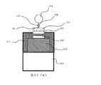
図8(a)は本発明を説明するための検出デバイスの断面を模式的に示した図である。本実施例では金属人工格子膜からなるGMR素子140を検出デバイスに用いる。支持体125上に下電極126としてプラチナ膜を成膜し、その上にコバルト薄膜と銅薄膜を交互に各10層ずつ積層した金属人工格子膜127を形成する。さらに保護膜128および上部電極129としてプラチナ膜、捕捉分子としての第一のペプチド分子固定膜131としてシリコン膜を順次形成する。 FIG. 8A is a diagram schematically showing a cross section of a detection device for explaining the present invention. In this embodiment, a
その後、実施例1と同様に「第一のペプチド分子161−標的物質−第二のペプチド分子162」の反応を介して磁性粒子164をGMR素子140表面に固定する。GMR素子140は短辺の長さが90nmで長辺の長さが180nmの長方形である。この場合、磁性膜の長さ方向が磁化容易軸となる。金属人工格子膜は、非磁性を介した強磁性膜の磁性体の磁化方向が、零磁場中で反平行になり、膜面内方向の磁界を印加すると、強磁性体の磁化方向が、平行に向くという性質を持っている。 Thereafter, similarly to Example 1, the
標的物質検出方法は、実施例1の場合と同様である。ただし、本実施例においては膜面内方向に外部磁界176を印加する。すなわち、GMR素子140表面に「第一のペプチド分子ー標的物質ー第二のペプチド分子」の反応を介して固定された磁性粒子164に右方向の外部磁界181を印加し、磁性粒子164の磁化を右方向に向ける。磁性粒子164からは浮遊磁界が発生し、フリー層240には外部磁界176(磁性粒子164の磁化方向)と反対方向の浮遊磁界が印加される。したがって、磁性粒子の有無によってGMR素子に印加される正味の磁界の大きさが異なり、この違いがGMR素子にかかる電圧の大きさの違いとして現れる。以上に説明した方法により被検体溶液中の標的物質173を検出することができる。 The target substance detection method is the same as in Example 1. However, in this embodiment, an external
上記検出を図8(b)に示す本実施例の構成と図8(c)に構成による対照例とで比較する。なお、本実施例では、「第一のペプチド分子として抗HEL抗体のFab領域分子161b、第二のペプチド分子としてHEL及び金の双方を同時に認識することができるDiabody 162b」が用いられている。対照例では、従来技術の構成である、「第一のペプチド分子として抗HELポリクローナル抗体161c、第二のペプチド分子として同じく抗HELポリクローナル抗体162c」が用いられている。 The above detection is compared between the configuration of this embodiment shown in FIG. 8B and the control example according to the configuration shown in FIG. In the present example, “Diabody 162b capable of simultaneously recognizing both HEL and gold as the first peptide molecule and Fab region molecule 161b of the anti-HEL antibody as the second peptide molecule” is used. In the control example, “anti-HEL polyclonal antibody 161c is used as the first peptide molecule and anti-HEL polyclonal antibody 162c is used as the second peptide molecule”, which is a configuration of the prior art.
本実施例における、HELを認識する第一のペプチド分子161、即ち、実施例では抗HEL抗体のFab領域分子161b、比較例では抗HELポリクローナル抗体161cの検出デバイス上への固定は、実施例1に記載の方法と同様の方法により行う。 In this example, the
一方、本実施例の磁性微粒子164は、金コロイド膜165で被覆されているため、第二のペプチド分子162の固定は以下の通り行う。 On the other hand, since the magnetic
本実施例:
第二のペプチド分子が「HEL及び金の双方を同時に認識することができるDiabody 162b」の場合は、特別な処理の必要が無い状態で金に固定されるため、以下の方法で固定が可能である。即ち、界面活性剤としてTween20を0.1%(v/v)含むリン酸緩衝液(以降PBST溶液と記載する場合もある)に磁性微粒子を分散させる。この分散液に、前記Diabodyが溶解したPBST溶液を加え、30℃で2時間インキュベートし、磁石により磁性微粒子を収集し、PBST溶液で洗浄することで「HEL及び金の双方を同時に認識することができるDiabody 162b」が固定された磁性微粒子を得る。Example:
If the second peptide molecule is “Diabody 162b, which can recognize both HEL and gold at the same time,” it is fixed to gold without the need for special treatment. is there. That is, magnetic fine particles are dispersed in a phosphate buffer solution (hereinafter sometimes referred to as PBST solution) containing 0.1% (v / v) Tween 20 as a surfactant. A PBST solution in which the diabody is dissolved is added to this dispersion, and the mixture is incubated at 30 ° C. for 2 hours. Magnetic particles are collected with a magnet and washed with the PBST solution, so that “both HEL and gold can be recognized simultaneously. Magnetic Diaphragm 162b "which can be obtained is obtained.
本実施例に対する比較例:
第二のペプチド分子が「抗HELポリクローナル抗体162c」の場合は以下のように固定を行う。即ち、Dithiobis(succinimidyl hexanoate)(同仁化学研究所社製)クロロホルム溶液に、前記磁性微粒子を加え、室温で1分反応させ、磁石により磁性微粒子を収集し、クロロホルムで洗浄後、窒素ガスにて乾燥させる。こうして得られた表面活性エステル化磁性微粒子に抗HEL抗体のFab領域分子をアミンカップリングさせ、表面に抗HEL抗体のFab領域分子が固定された磁性微粒子を得る。Comparative example for this example:
When the second peptide molecule is “anti-HEL polyclonal antibody 162c”, immobilization is performed as follows. That is, Dithiobis (succinimidyl hexanoate) (manufactured by Dojindo Laboratories) Add the magnetic fine particles to a chloroform solution, react at room temperature for 1 minute, collect magnetic fine particles with a magnet, wash with chloroform, and dry with nitrogen gas Let The surface active esterified magnetic fine particles thus obtained are amine-coupled with Fab region molecules of the anti-HEL antibody to obtain magnetic fine particles in which the Fab region molecules of the anti-HEL antibody are immobilized on the surface.
これらの検出デバイスを用い、実施例1と同様のプロトコールに従ってHELの検出を行う。これらの場合、磁性微粒子164の直径を400nmとすると、比較例ではGMR素子140表面から磁性粒子164までの平均距離は約55nmであるのに対し、本実施例では35nm程度となり、それに伴い、本実施例は対照例より強い電圧の変化が検出され、結果として検出感度が向上する。
(実施例3)
本実施例では、標的物質のモデルタンパク質として実施例1と同様にHELを用いる。また、検出素子に固定され、標的物質の特定の領域を特異的に認識し、結合することができる第一のペプチド分子としてHEL及び金の双方を同時に認識することができるDiabodyを用いる。また磁性標識に固定され、前記標的物質の第一のペプチド分子結合領域と異なる領域を特異的に認識し、結合することができる第二のペプチド分子として抗HELポリクローナル抗体を用いる。さらに本実施例では、検出方法として半導体ホール素子を用いた検出手法を用いて本発明の効果の例を示す。Using these detection devices, HEL is detected according to the same protocol as in Example 1. In these cases, when the diameter of the magnetic
(Example 3)
In this example, HEL is used as a model protein of the target substance as in Example 1. In addition, a Diabody that can be simultaneously recognized as both HEL and gold is used as the first peptide molecule that is fixed to the detection element and that can specifically recognize and bind to a specific region of the target substance. In addition, an anti-HEL polyclonal antibody is used as a second peptide molecule that is fixed to a magnetic label and can specifically recognize and bind to a region different from the first peptide molecule binding region of the target substance. Further, in this embodiment, an example of the effect of the present invention will be shown by using a detection method using a semiconductor Hall element as a detection method.
本実施例の第一のペプチド分子として使用するHEL及び金の双方を同時に認識することができるDiabodyは、実施例2と同様に作製、取得する。本実施例の第二のペプチド分子として使用する抗HELポリクローナル抗体は、Rockland社より入手する。素子の作製、測定手順は特WO03/067258に準じて行う。 Diabody capable of simultaneously recognizing both HEL and gold used as the first peptide molecule of this example is prepared and obtained in the same manner as in Example 2. The anti-HEL polyclonal antibody used as the second peptide molecule of this example is obtained from Rockland. The device fabrication and measurement procedures are performed according to Japanese Patent No. WO 03/067258.
図9(a)を用いて本発明のバイオセンサの検出原理を説明する。図9(a)は、センサチップ201の半導体ホール素子202近辺の断面を模式的に表したものである。半導体ホール素子202の表面に第一のペプチド分子181が固定されている。その第一のペプチド分子181に標的物質173が特異的に結合し、その標的物質173に、さらに、磁性粒子174が、前記磁性粒子174表面に固定された第二のペプチド分子182と前記標的物質173との特異的結合を介して結合している。 The detection principle of the biosensor of the present invention will be described with reference to FIG. FIG. 9A schematically shows a cross section of the
センサチップ201表面に対向する位置には、上部コイルCU(第1の磁場発生手段)203を配置する。上述のようにセンサチップ201表面に磁性粒子174が結合した状態で、上部コイルCU203に通電し磁場を発生させる。コイルの代わりに、例えば、永久磁石などを用いてもよい。図9(a)では磁束は、磁束方向を表す矢印200で示される方向に形成され、半導体ホール素子面に対して垂直であることが示されている。磁束は磁性粒子174により収束されるため、磁性粒子174が無い場合に比べて半導体ホール素子201での磁束密度は増加する。又、上部コイルCU203により磁場を印加するため、センサチップ201の表面から遠ざかるほど磁束密度が増加する。このため、センサチップ201表面に結合せずに浮遊している磁性粒子は上方に引き寄せられ半導体ホール素子202により検知する磁束密度には影響しない。半導体ホール素子202の出力電圧は磁束密度に比例するため、この出力電圧により磁性粒子174が半導体ホール素子202上に結合しているか否かを判定できる。 An upper coil CU (first magnetic field generating means) 203 is disposed at a position facing the surface of the
また、1つの半導体ホール素子2で複数の磁性分子の存在を検知する場合は、磁性粒子174により収束され増加する磁束密度の増分は、磁性粒子の数に依存する為、1つの半導体ホール素子202上に結合した磁性粒子174の数を検知することもできる。 When detecting the presence of a plurality of magnetic molecules with one
また、本実施の形態においては、センサチップ1の裏側にも、下部コイルCD(第2の磁場発生手段)204を配置する。この下部コイルCD204は、磁性粒子174の検知するためではなく、磁性粒子174をセンサチップ201表面に近づけるための磁場を発生するものである。永久磁石などを用いてもよい。下部コイルCD204は、センサチップ201への磁性粒子174の導入時に、通電して磁場を発生させるようにする。このセンサチップ201表面からの距離の増加に応じて磁束密度が減少するように形成された磁場により、磁性分子がセンサチップ201表面に引き寄せられるため、磁性粒子174がセンサチップ201表面に結合する時間が短縮される。 In the present embodiment, the lower coil CD (second magnetic field generating means) 204 is also arranged on the back side of the
この検出デバイスを用い、以下のプロトコールに従ってHELの検出を行う。検出デバイスには、HELを認識する第一のペプチド分子181が固定化されている。
(1)標的物質173であるHELを含むリン酸緩衝生理食塩水に上記検出デバイスを浸し、5分間インキュベートする。
(2)未反応のHELをリン酸緩衝生理食塩水で洗浄する。
(3)磁性粒子174により標識された第二のペプチド分子182を含むリン酸緩衝生理食塩水に工程(1)および(2)が終了した上記検出デバイスを浸し、5分間インキュベートする。
(4)未反応の該標識抗体をリン酸緩衝生理食塩水で洗浄する。Using this detection device, HEL is detected according to the following protocol. A
(1) The detection device is immersed in phosphate buffered saline containing HEL, which is the
(2) Unreacted HEL is washed with phosphate buffered saline.
(3) The detection device having completed steps (1) and (2) is immersed in a phosphate buffered saline containing the
(4) The unreacted labeled antibody is washed with phosphate buffered saline.
まず上記(1)に記載の状態で、下部コイルにより磁場を印加する。このセンサチップ表面からの距離の増加に応じて磁束密度が減少するように形成された磁場により、磁性粒子はセンサチップ表面に引き付けられて、センサチップ表面に結合する速度が高められる。 First, in the state described in (1) above, a magnetic field is applied by the lower coil. Due to the magnetic field formed so that the magnetic flux density decreases as the distance from the surface of the sensor chip increases, the magnetic particles are attracted to the surface of the sensor chip, and the speed of binding to the surface of the sensor chip is increased.
次に、磁性粒子のセンサチップ表面への結合が完了した状態で、下部コイルによる磁場をOFFし、センサチップに磁場を印加しない状態で、ホール素子の出力を取得する。 Next, in the state where the coupling of the magnetic particles to the sensor chip surface is completed, the magnetic field by the lower coil is turned off, and the output of the Hall element is acquired in the state where no magnetic field is applied to the sensor chip.
次に上部コイルにより磁場を印加し、ホール素子からの出力信号を取り出す。先に取得したゼロ磁場での出力と、本出力とを比較し、同様に取得した、磁性粒子を含まないセンサ素子の相対出力と比較することで、磁性粒子、即ち標的物質の濃度を検出する。 Next, a magnetic field is applied by the upper coil, and an output signal from the Hall element is taken out. Compare the output with the zero magnetic field acquired earlier with this output, and compare it with the relative output of the sensor element that does not contain magnetic particles, and detect the concentration of magnetic particles, that is, the target substance. .
上記検出を図9(b)に示す本実施例の構成と図9(c)に構成による対照例とで比較する。なお、本実施例では、「第一のペプチド分子としてHEL及び金の双方を同時に認識することができるDiabody 181b、第二のペプチド分子として抗HELポリクローナル抗体182b」が用いられている。対照例では、従来技術の構成である、「第一のペプチド分子として抗HELポリクローナル抗体181c、第二のペプチド分子として同じく抗HELポリクローナル抗体182c」が用いられている。 The above detection is compared between the configuration of the present embodiment shown in FIG. 9B and the control example according to the configuration in FIG. 9C. In this example, “Diabody 181b capable of simultaneously recognizing both HEL and gold as the first peptide molecule, and anti-HEL polyclonal antibody 182b as the second peptide molecule” are used. In the control example, “anti-HEL polyclonal antibody 181c as the first peptide molecule and anti-HEL polyclonal antibody 182c as the second peptide molecule”, which is a configuration of the prior art, is used.
図9(b)に示す本実施例の構成において、第一のペプチド分子として用いるHEL及び金の双方を同時に認識することができるDiabody 181bのセンサチップへの固定は次のように行う。即ち、センサチップ表面に金薄膜をスパッタ法或いは蒸着法により形成し、前記Diabodyが溶解したPBST溶液を加え、30℃で2時間インキュベートした後、同じくPBST溶液で洗浄することでセンサチップにDiabodyが固定される。 In the configuration of this embodiment shown in FIG. 9B, the fixation of Diabody 181b capable of simultaneously recognizing both HEL and gold used as the first peptide molecule to the sensor chip is performed as follows. That is, a gold thin film is formed on the surface of the sensor chip by sputtering or vapor deposition, a PBST solution in which the above diabody is dissolved is added, incubated at 30 ° C. for 2 hours, and then washed with the PBST solution. Fixed.
図9(c)に示す対照例の構成において、第一のペプチド分子として用いる抗HELポリクローナル抗体181cのセンサチップへの固定は、実施例と条件を合わせるために次の方法によって行った。すなわち、センサチップ表面に金薄膜をスパッタ法或いは蒸着法により形成し、Dithiobis(succinimidyl hexanoate)(同仁科学研究所社製)クロロホルム溶液で室温で1分反応させ、クロロホルムで洗浄後、窒素ガスにて乾燥させて得られた表面活性エステル化金表面センサチップに抗HELポリクローナル抗体をアミンカップリングさせて固定を行う。 In the configuration of the control example shown in FIG. 9C, the anti-HEL polyclonal antibody 181c used as the first peptide molecule was immobilized on the sensor chip by the following method in order to match the conditions with the examples. That is, a gold thin film is formed on the surface of the sensor chip by sputtering or vapor deposition, reacted with Dithiobis (succinimidyl hexanoate) (manufactured by Dojindo Laboratories) at room temperature for 1 minute, washed with chloroform, then nitrogen gas An anti-HEL polyclonal antibody is amine-coupled to the surface-active esterified gold surface sensor chip obtained by drying and fixed.
実施例及び対照例の、磁性微粒子への抗HELポリクローナル抗体の固定は、実施例1に記載の方法で行う。 Immobilization of the anti-HEL polyclonal antibody to the magnetic microparticles in Examples and Controls is performed by the method described in Example 1.
この場合、対照例に比べ実施例のほうが磁性微粒子とセンサチップとの距離が10nm短縮され、それに伴い、結果として検出感度が向上する。 In this case, the distance between the magnetic fine particles and the sensor chip is shortened by 10 nm in the embodiment as compared with the control example, and as a result, the detection sensitivity is improved.
(実施例4)
本実施例では、実施例1と同様の構成、即ち、標的物質のモデルタンパク質としてHELを用いる。また、検出素子に固定され、前記標的物質の特定の領域を特異的に認識し、結合することができる第一のペプチド分子として抗HEL抗体のscFvを用いる。また、磁性標識に固定され、前記標的物質の第一のペプチド分子結合領域と異なる領域を特異的に認識し、結合することができる第二のペプチド分子として抗HEL抗体のFab領域分子を用いる。本実施例では、検出方法として超電導量子干渉素子(SQUID)の手法を用いて本発明の効果の例を示す。各ペプチド分子のセンサ表面及び磁性微粒子への固定は実施例1に記載の方法と同様の方法により行う。Example 4
In this example, HEL is used as the model protein of the target substance, that is, the same configuration as in Example 1. In addition, scFv of an anti-HEL antibody is used as a first peptide molecule that is immobilized on a detection element and can specifically recognize and bind to a specific region of the target substance. Further, an Fab region molecule of an anti-HEL antibody is used as a second peptide molecule that is fixed to a magnetic label and can specifically recognize and bind to a region different from the first peptide molecule binding region of the target substance. In this embodiment, an example of the effect of the present invention will be described using a superconducting quantum interference device (SQUID) technique as a detection method. Immobilization of each peptide molecule on the sensor surface and magnetic fine particles is performed by the same method as described in Example 1.
素子の作製、測定手順は特開2005-291909号公報に記載の方法に準じて行う。装置の構成を図10(a)に示す。即ち、基板215上に固定された第一のペプチド分子191と、その標的物質173が特異的に結合し、更に前記標的物質173と特異的に結合し、磁性微粒子174に固定された第二のペプチド分子192を介して、磁性微粒子174が基板上に固定される。このように磁性微粒子が固定された基板215がSQUID装置上に配置される。装置外面は断熱体213により被覆され、基板の下部にサファイア窓211が配置される。前記サファイア窓211の下部にサファイア棒212上に設置されたSQUID素子210が配置される。このようなSQUID装置は、液体窒素容器214の上部に配置される。本構成の装置を用いて、特開2005-291909号公報に開示されている検出を行う。 The device fabrication and measurement procedures are performed according to the method described in JP-A-2005-291909. The configuration of the apparatus is shown in FIG. That is, the
上記検出を図10(b)に示す本実施例の構成と図10(c)に構成による対照例とで比較する。なお、本実施例では、「第一のペプチド分子として抗HEL抗体のscFv191b、第二のペプチド分子として抗HEL抗体のFab領域分子192b」が用いられている。対照例では、従来技術の構成である、「第一のペプチド分子として抗HELポリクローナル抗体191c、第二のペプチド分子として同じく抗HELポリクローナル抗体192c」が用いられている。 The above detection is compared between the configuration of this embodiment shown in FIG. 10B and the control example according to the configuration shown in FIG. In the present example, “scFv191b of anti-HEL antibody as the first peptide molecule and Fab region molecule 192b of anti-HEL antibody as the second peptide molecule” are used. In the control example, “anti-HEL polyclonal antibody 191c as the first peptide molecule and anti-HEL polyclonal antibody 192c as the second peptide molecule”, which is a configuration of the prior art, is used.
基板への第一のペプチド分子の固定、及び磁性微粒子への第二のペプチド分子の固定は実施例1に記載の方法と同様の方法で行う。また、「第一のペプチド分子−標的物質ー第二のペプチド分子」を介した磁性微粒子の固定も実施例1に記載の方法と同様の方法で行う。 The fixation of the first peptide molecule to the substrate and the fixation of the second peptide molecule to the magnetic microparticles are performed by the same method as described in Example 1. In addition, immobilization of magnetic fine particles via “first peptide molecule—target substance—second peptide molecule” is performed in the same manner as described in Example 1.
その結果、対照例に比べ実施例の方がSQUID素子と磁性微粒子の距離が約20nm短縮されているためそれに伴い、本実施例は対照例より強い電圧の変化が検出され、結果として検出感度が向上する。 As a result, since the distance between the SQUID element and the magnetic fine particles is shortened by about 20 nm in comparison with the control example, a strong voltage change is detected in this embodiment than in the control example. improves.
1 センサー素子
2 機能性膜
3 IgG分子(第一の標的物質捕捉分子)
4 標的物質
5 IgG分子(第二の標的物質捕捉分子)
6 磁性標識
7 IgG分子可変領域(Fv)分子(第一の標的物質捕捉分子)
8 IgG分子可変領域(Fv)分子(第二の標的物質捕捉分子)
9 IgG分子のFab分子(第二の標的物質捕捉分子)
10 金薄膜(機能性膜)
11 金薄膜(磁性標識体被覆膜)
12 金結合性Diabody(第一の標的物質捕捉分子)
13 金結合性Diabody(第二の標的物質捕捉分子)
100 スピントンネル磁気抵抗効果素子(TMR素子)
101 支持体
102 ハフニウム膜
103 マンガン−イリジウム合金膜
104 鉄−コバルト合金膜
105 ルテニウム膜
106 鉄−コバルト合金膜
107 マグネシウム酸化膜
108 鉄−コバルト合金膜
109 ニッケル−鉄合金膜
110 プラチナ膜
111 シリコン膜
112 ピンド層
113 フリー層
114 支持体
115 第1の垂直磁化膜
116 第1の高スピン分極率層
117 非磁性膜
118 第2の高スピン分極率層
119 第二の垂直磁化膜
120 ピント層
121 フリー層
122 保護膜
123 Pt(電極)
124 Pt(電極)
125 支持体
126 下部電極
127 金属人工格子膜
128 保護膜
129 上部電極
130 GMR素子
131 固定膜
140 GMR素子
141 TMR素子
151 センサチップ
153 センサチップ
154 GMR素子
161 第一のペプチド分子
161b 抗HEL抗体のFab領域分子(第一のペプチド分子)
161c 抗HELポリクローナル抗体(第一のペプチド分子)
162 第二のペプチド分子
162b Diabody(第二のペプチド分子)
162c 抗HELポリクローナル抗体(第二のペプチド分子)
164 磁性粒子
165 金コロイド膜
171 第一のペプチド分子
171b 抗HEL抗体のscFv(第一のペプチド分子)
171c 抗HELポリクローナル抗体(第一のペプチド分子)
172 第二のペプチド分子
172b 抗HEL抗体のFab領域分子(第二のペプチド分子)
172c 抗HELポリクローナル抗体(第二のペプチド分子)
173 標的物質
174 磁性粒子
175 外部磁界
176 外部磁界
177 定電流源
178 センスアンプ
181 第一のペプチド分子
181b Diabody(第一のペプチド分子)
181c 抗HELポリクローナル抗体(第二のペプチド分子)
182 第二のペプチド分子
182b 抗HELポリクローナル抗体(第一のペプチド分子)
182c 抗HELポリクローナル抗体(第二のペプチド分子)
191 第一のペプチド分子
191b 抗HEL抗体のscFv(第一のペプチド分子)
191c 抗HELポリクローナル抗体(第一のペプチド分子)
192 第二のペプチド分子
192b 抗HEL抗体のFab領域分子(第二のペプチド分子)
192c 抗HELポリクローナル抗体(第二のペプチド分子)
200 磁束方向
201 センサチップ
202 半導体ホール素子
203 上部コイルCU
204 下部コイルCD
210 SQUID素子
211 サファイア窓
212 サファイア棒
213 断熱体
214 液体窒素容器
215 基板
216 センサチップ1
4
6
8 IgG molecule variable region (Fv) molecule (second target substance capture molecule)
9 Fab molecule of IgG molecule (second target substance capture molecule)
10 Gold thin film (functional film)
11 Gold thin film (magnetic marker coating)
12 Gold-binding diabody (first target substance capturing molecule)
13 Gold-binding diabody (second target substance capturing molecule)
100 Spin tunnel magnetoresistive element (TMR element)
DESCRIPTION OF
124 Pt (electrode)
125
161c Anti-HEL polyclonal antibody (first peptide molecule)
162 Second peptide molecule 162b Diabody (second peptide molecule)
162c anti-HEL polyclonal antibody (second peptide molecule)
164
171c Anti-HEL polyclonal antibody (first peptide molecule)
172 Second peptide molecule 172b Anti-HEL antibody Fab region molecule (second peptide molecule)
172c anti-HEL polyclonal antibody (second peptide molecule)
173
181c anti-HEL polyclonal antibody (second peptide molecule)
182 Second peptide molecule 182b Anti-HEL polyclonal antibody (first peptide molecule)
182c anti-HEL polyclonal antibody (second peptide molecule)
191 First peptide molecule 191b scFv of anti-HEL antibody (first peptide molecule)
191c Anti-HEL polyclonal antibody (first peptide molecule)
192 Second peptide molecule 192b Anti-HEL antibody Fab region molecule (second peptide molecule)
192c anti-HEL polyclonal antibody (second peptide molecule)
200
204 Lower coil CD
210
Claims (6)
Translated fromJapanese磁場を検知する部位と、前記標的物質と特異的に結合するペプチド分子と、該ペプチド分子を固定する部位とを有し、
前記ペプチド分子が、イムノグロブリンG分子より分子量が小さい事を特徴とする、標的物質検出用素子。A combination of a magnetic label and an element for detecting a magnetic field, the element used for target substance detection for detecting the presence or concentration of a target substance in a specimen,
A site for detecting a magnetic field, a peptide molecule that specifically binds to the target substance, and a site for immobilizing the peptide molecule,
A target substance detection element, wherein the peptide molecule has a molecular weight smaller than that of an immunoglobulin G molecule.
磁性体を含む粒子と、前記標的物質と特異的に結合するペプチド分子とを有し、該ペプチド分子が前記粒子の表面に固定されており、
前記ペプチド分子が、イムノグロブリンG分子より分子量が小さい事を特徴とする、標的物質検出用材料。A material as the magnetic label used for target substance detection for detecting the presence or concentration of a target substance in a sample by combining a magnetic label and an element for detecting a magnetic field,
Having a particle containing a magnetic substance and a peptide molecule that specifically binds to the target substance, the peptide molecule being immobilized on the surface of the particle,
A material for detecting a target substance, wherein the peptide molecule has a molecular weight smaller than that of an immunoglobulin G molecule.
磁場を検知する部位と、前記標的物質と特異的に結合する第一のペプチド分子と、該第一のペプチド分子を固定する部位とを有する素子と、
磁性体を含む粒子と、前記標的物質と特異的に結合する第二のペプチド分子とを有し、該第二のペプチド分子が前記粒子に固定されている標識材料と、を含み、
前記第一のペプチド分子と前記第二のペプチド分子は、それぞれ前記標的物質の異なる領域に特異的に結合し、
前記第一のペプチド分子と前記第二のぺプチド分子の少なくとも一方は、イムノグロブリンG分子より分子量が小さい事を特徴とする、標的物質検出用キット。A kit used for detecting a target substance for detecting the presence or concentration of a target substance in a sample by combining a magnetic label and an element for detecting a magnetic field,
An element having a site for detecting a magnetic field, a first peptide molecule that specifically binds to the target substance, and a site for immobilizing the first peptide molecule;
A particle containing a magnetic substance, a second peptide molecule that specifically binds to the target substance, and a labeling material on which the second peptide molecule is fixed to the particle,
The first peptide molecule and the second peptide molecule each specifically bind to different regions of the target substance,
The target substance detection kit, wherein at least one of the first peptide molecule and the second peptide molecule has a molecular weight smaller than that of an immunoglobulin G molecule.
Priority Applications (2)
| Application Number | Priority Date | Filing Date | Title |
|---|---|---|---|
| JP2006102985AJP2007278748A (en) | 2006-04-04 | 2006-04-04 | Target substance detection element, detection material, and detection kit |
| US11/727,674US20070298510A1 (en) | 2006-04-04 | 2007-03-28 | Target substance-detecting element, detecting material, and detector kit |
Applications Claiming Priority (1)
| Application Number | Priority Date | Filing Date | Title |
|---|---|---|---|
| JP2006102985AJP2007278748A (en) | 2006-04-04 | 2006-04-04 | Target substance detection element, detection material, and detection kit |
Publications (1)
| Publication Number | Publication Date |
|---|---|
| JP2007278748Atrue JP2007278748A (en) | 2007-10-25 |
Family
ID=38680345
Family Applications (1)
| Application Number | Title | Priority Date | Filing Date |
|---|---|---|---|
| JP2006102985APendingJP2007278748A (en) | 2006-04-04 | 2006-04-04 | Target substance detection element, detection material, and detection kit |
Country Status (2)
| Country | Link |
|---|---|
| US (1) | US20070298510A1 (en) |
| JP (1) | JP2007278748A (en) |
Cited By (2)
| Publication number | Priority date | Publication date | Assignee | Title |
|---|---|---|---|---|
| WO2009107484A1 (en)* | 2008-02-26 | 2009-09-03 | Canon Kabushiki Kaisha | Method of detecting target substance and target-substance detection kit |
| JP2010286466A (en)* | 2008-10-24 | 2010-12-24 | Fujifilm Corp | Fragmented antibody-immobilized carrier and method for producing the same |
Families Citing this family (14)
| Publication number | Priority date | Publication date | Assignee | Title |
|---|---|---|---|---|
| KR100940138B1 (en)* | 2004-03-31 | 2010-02-03 | 캐논 가부시끼가이샤 | Gold-binding protein and use thereof |
| JP5142458B2 (en)* | 2005-06-30 | 2013-02-13 | キヤノン株式会社 | Target substance capture molecule, target substance capture element, target substance detection apparatus and kit using these, and target substance detection method |
| US20090130776A1 (en)* | 2005-09-01 | 2009-05-21 | Canon Kabushiki Kaisha | Binding protein molecule |
| JP5164411B2 (en)* | 2006-03-31 | 2013-03-21 | キヤノン株式会社 | Target substance detection method and target substance detection kit |
| JP4861739B2 (en)* | 2006-04-11 | 2012-01-25 | キヤノン株式会社 | Magnetic sensor, method for producing the sensor, target substance detection apparatus and biosensor kit using the sensor |
| JP5247106B2 (en)* | 2006-10-13 | 2013-07-24 | キヤノン株式会社 | Protein, protein immobilization method, structure, biosensor, nucleic acid, vector, and target substance detection kit |
| JP5300205B2 (en)* | 2007-03-22 | 2013-09-25 | キヤノン株式会社 | Target substance detection element, target substance detection method, and method for manufacturing target substance detection element |
| US20080241964A1 (en)* | 2007-03-27 | 2008-10-02 | Canon Kabushiki Kaisha | Material for improving sensitivity of magnetic sensor and method thereof |
| US8076161B2 (en)* | 2007-05-31 | 2011-12-13 | Canon Kabushiki Kaisha | Target substance detection kit and target substance detection method |
| US20080309323A1 (en)* | 2007-06-12 | 2008-12-18 | Canon Kabushiki Kaisha | Method for biochemical analysis |
| ES2608930T3 (en) | 2012-01-04 | 2017-04-17 | Magnomics, S.A. | Monolithic device that combines CMOS with magnetoresistive sensors |
| CN102662053B (en)* | 2012-04-19 | 2014-10-22 | 清华大学 | Immune biochip regeneration method and regenerated immune biochip |
| CN107796865B (en) | 2016-09-05 | 2021-05-25 | 财团法人工业技术研究院 | Biomolecular Magnetic Sensors |
| WO2022108579A1 (en)* | 2020-11-18 | 2022-05-27 | Hitachi High-Tech Corporation | Method, device, and program for analyzing inter-molecular interaction |
Citations (3)
| Publication number | Priority date | Publication date | Assignee | Title |
|---|---|---|---|---|
| JP2001033455A (en)* | 1999-07-21 | 2001-02-09 | Sumitomo Electric Ind Ltd | Immunoassay method and device using magnetic label |
| WO2005022154A1 (en)* | 2003-08-29 | 2005-03-10 | Asahi Kasei Kabushiki Kaisha | Biosensor and method of analyte measuring |
| JP2005189237A (en)* | 2003-12-05 | 2005-07-14 | Sekisui Chem Co Ltd | Optical measuring method |
Family Cites Families (42)
| Publication number | Priority date | Publication date | Assignee | Title |
|---|---|---|---|---|
| JP3086182B2 (en)* | 1995-12-19 | 2000-09-11 | ライト工業株式会社 | Soil purification method |
| JP3406926B2 (en)* | 1993-02-18 | 2003-05-19 | キヤノン株式会社 | Method for biodegrading trichlorethylene and method for biodegrading organic chlorine compounds using microorganisms |
| US6096530A (en)* | 1992-04-22 | 2000-08-01 | Canon Kabushiki Kaisha | Pseudomonas cepacia strain isolated from termite intestines that degrades trichlorethylene and furan compounds |
| AU690528B2 (en)* | 1992-12-04 | 1998-04-30 | Medical Research Council | Multivalent and multispecific binding proteins, their manufacture and use |
| US5411730A (en)* | 1993-07-20 | 1995-05-02 | Research Corporation Technologies, Inc. | Magnetic microparticles |
| EP0646642A3 (en)* | 1993-09-30 | 1995-08-16 | Canon Kk | Microorganism-holding carrier and method for remediation of soil employing the carrier. |
| DE69421254T2 (en)* | 1993-12-22 | 2000-04-20 | Canon Kk | Process for the degradation of an organochlorine compound and environmental remediation using microorganisms |
| DE69514871T2 (en)* | 1994-05-30 | 2000-06-29 | Canon Kk | Corynebacterium sp. J1, Process for biodegrading aromatic compounds and / or chlorinated organic compounds, and process for detoxifying the environment therewith |
| US5679568A (en)* | 1994-11-21 | 1997-10-21 | Canon Kabushiki Kaisha | Processes for decomposing a pollutant and remedying an environment using Renobacter sp. ferm BP-5353 having dehalogenase activity |
| JP3478619B2 (en)* | 1994-12-02 | 2003-12-15 | キヤノン株式会社 | Novel microorganism KB2 and biodegradation treatment method of aromatic compound and / or volatile organochlorine compound using the same |
| JP3108006B2 (en)* | 1995-02-28 | 2000-11-13 | キヤノン株式会社 | JM1 strain, environmental restoration method, organic compound decomposition method, microorganism acquisition method, microorganism detection method, and microorganism detection kit |
| JP3083077B2 (en)* | 1996-04-11 | 2000-09-04 | キヤノン株式会社 | Organic compound biodegradation method and environmental restoration method |
| JP3323746B2 (en)* | 1996-08-01 | 2002-09-09 | キヤノン株式会社 | Novel microorganisms, organic compound biodegradation method and environmental restoration method |
| JP3347596B2 (en)* | 1996-08-01 | 2002-11-20 | キヤノン株式会社 | A novel microorganism, a method for biodegrading aromatic compounds and / or organic chlorine compounds, and a method for purifying a medium |
| US5981297A (en)* | 1997-02-05 | 1999-11-09 | The United States Of America As Represented By The Secretary Of The Navy | Biosensor using magnetically-detected label |
| CA2229754C (en)* | 1997-02-18 | 2002-04-09 | Canon Kabushiki Kaisha | Method for culturing microorganism, method for biosynthesizing organic compound, method for maintaining microbial ability to decompose polluting substance, method for decomposing pollutant, and method for remedying environment |
| JP3673640B2 (en)* | 1997-05-15 | 2005-07-20 | キヤノン株式会社 | Method for purifying contaminated medium and purification device used therefor |
| US6864074B2 (en)* | 1998-10-30 | 2005-03-08 | Canon Kabushiki Kaisha | Dna fragment carrying toluene monooxygenase gene, recombinant plasmid, transformed microorganism, method for degrading chlorinated aliphatic hydrocarbon compounds and aromatic compounds, and method for environmental remediation |
| US6472191B1 (en)* | 1998-12-03 | 2002-10-29 | Canon Kabushiki Kaisha | Dna fragment carrying toluene monooxygenase gene, recombinant plasmid, transformed microorganism, method for degrading chlorinated aliphatic hydrocarbon compounds and aromatic compounds, and method for environmental remediation |
| JP3684150B2 (en)* | 1999-12-27 | 2005-08-17 | キヤノン株式会社 | Polyhydroxyalkanoate |
| KR100447966B1 (en)* | 2000-02-29 | 2004-09-13 | 캐논 가부시끼가이샤 | Polyhydroxyalkanoate containing 3-hydroxythienylalkanoic acid as monomer unit and method for producing the same |
| US6861550B2 (en)* | 2000-02-29 | 2005-03-01 | Canon Kabushiki Kaisha | Polyhydroxyalkanoate containing 3-hydroxybenzoylalkanoic acid as monomer unit, and method for producing the same |
| RU2166751C1 (en)* | 2000-03-09 | 2001-05-10 | Никитин Петр Иванович | Process of analysis of mixture of biologic and/or chemical components with use of magnetic particles and device for its implementation |
| JP2004503758A (en)* | 2000-06-14 | 2004-02-05 | ボード・オブ・リージェンツ,ザ・ユニヴァーシティ・オヴ・テキサス・システム | Particles designed to be dielectric |
| JP3768799B2 (en)* | 2000-10-30 | 2006-04-19 | キヤノン株式会社 | Process for producing polyhydroxyalkanoate from substituted fatty acid ester |
| JP3720779B2 (en)* | 2001-02-28 | 2005-11-30 | キヤノン株式会社 | NOVEL POLYHYDROXYALKANOATE TYPE POLYESTER HAVING VINYLPHENYL STRUCTURE IN SIDE CHAIN AND PROCESS FOR PRODUCING THE SAME |
| JP3748537B2 (en)* | 2001-03-01 | 2006-02-22 | キヤノン株式会社 | POLYHYDROXYALKANOATE AND PROCESS FOR PRODUCING THE SAME, AND ω- (2-THIENYLSULFANYL) ALKANOIC ACID AND PROCESS FOR PRODUCING THE SAME |
| KR100461511B1 (en)* | 2001-04-27 | 2004-12-14 | 캐논 가부시끼가이샤 | Novel polyhydroxyalkanoate, its production method, charge control agent containing the polyhydroxyalkanoate, toner binder and toner, and image forming method image forming apparatus using the toner |
| KR100528749B1 (en)* | 2001-04-27 | 2005-11-15 | 캐논 가부시끼가이샤 | Novel polyhydroxyalkanoates having in its side chain phenylsulfinyl structure and/or phenyl sulfonyl structure and production process therefor, charge control agent, toner binder and toner containing same, and image forming method and image forming apparatus using the toner |
| JP3496002B2 (en)* | 2001-04-27 | 2004-02-09 | キヤノン株式会社 | Binder resin containing novel polyhydroxyalkanoate, toner containing the binder resin; image forming method and image forming apparatus using the toner |
| JP3501771B2 (en)* | 2001-04-27 | 2004-03-02 | キヤノン株式会社 | Binder resin containing polyhydroxyalkanoate, toner containing the binder resin; image forming method and image forming apparatus using the toner |
| KR100487555B1 (en)* | 2001-04-27 | 2005-05-06 | 캐논 가부시끼가이샤 | Novel polyhydroxyalkanoate, producing method therefor, charge control agent containing such polyhydroxyalkanoate, toner contatining such charge control agent and image-forming method and image-forming apparatus utilizing such toner |
| US6869782B2 (en)* | 2001-07-10 | 2005-03-22 | Canon Kabushiki Kaisha | Polyhydroxyalkanoate that comprises unit having (methylsulfanyl) phenoxy structure in side chain thereof and process for producing the same |
| DE60315415T2 (en)* | 2002-01-29 | 2008-04-30 | Asahi Kasei Kabushiki Kaisha | BIOSENSOR, MAGNETIC MOLECULE MEASURING PROCESS AND MEASURING OBJECT MEASUREMENT PROCESS |
| JP4078247B2 (en)* | 2003-05-02 | 2008-04-23 | キヤノン株式会社 | Magnetic substance-biological substance complex type structure, peptide fragment having amino acid sequence capable of binding to magnetic substance and gene thereof, and method for producing magnetic substance-biological substance complex type structure |
| US7701138B2 (en)* | 2003-07-02 | 2010-04-20 | Canon Kabushiki Kaisha | Information acquisition method, information acquisition apparatus and disease diagnosis method |
| KR100940138B1 (en)* | 2004-03-31 | 2010-02-03 | 캐논 가부시끼가이샤 | Gold-binding protein and use thereof |
| JP2005315744A (en)* | 2004-04-28 | 2005-11-10 | Canon Inc | Detection element, detection apparatus, and detection kit including element and reagent |
| JP2005315678A (en)* | 2004-04-28 | 2005-11-10 | Canon Inc | Detection method, detection device, and detection kit |
| JP2006010534A (en)* | 2004-06-25 | 2006-01-12 | Canon Inc | Core / shell type magnetic particle sensor |
| US8945946B2 (en)* | 2006-03-31 | 2015-02-03 | Canon Kabushiki Kaisha | Sensor element and detection method of magnetic particles using this element, and detection method of target substance |
| JP4861739B2 (en)* | 2006-04-11 | 2012-01-25 | キヤノン株式会社 | Magnetic sensor, method for producing the sensor, target substance detection apparatus and biosensor kit using the sensor |
- 2006
- 2006-04-04JPJP2006102985Apatent/JP2007278748A/enactivePending
- 2007
- 2007-03-28USUS11/727,674patent/US20070298510A1/ennot_activeAbandoned
Patent Citations (3)
| Publication number | Priority date | Publication date | Assignee | Title |
|---|---|---|---|---|
| JP2001033455A (en)* | 1999-07-21 | 2001-02-09 | Sumitomo Electric Ind Ltd | Immunoassay method and device using magnetic label |
| WO2005022154A1 (en)* | 2003-08-29 | 2005-03-10 | Asahi Kasei Kabushiki Kaisha | Biosensor and method of analyte measuring |
| JP2005189237A (en)* | 2003-12-05 | 2005-07-14 | Sekisui Chem Co Ltd | Optical measuring method |
Cited By (4)
| Publication number | Priority date | Publication date | Assignee | Title |
|---|---|---|---|---|
| WO2009107484A1 (en)* | 2008-02-26 | 2009-09-03 | Canon Kabushiki Kaisha | Method of detecting target substance and target-substance detection kit |
| US8513024B2 (en) | 2008-02-26 | 2013-08-20 | Canon Kabushiki Kaisha | Method of detecting target substance and target-substance detection kit |
| JP2010286466A (en)* | 2008-10-24 | 2010-12-24 | Fujifilm Corp | Fragmented antibody-immobilized carrier and method for producing the same |
| US9487592B2 (en) | 2008-10-24 | 2016-11-08 | Fujifilm Corporation | Immobilization substrate and method for producing the same |
Also Published As
| Publication number | Publication date |
|---|---|
| US20070298510A1 (en) | 2007-12-27 |
Similar Documents
| Publication | Publication Date | Title |
|---|---|---|
| JP2007278748A (en) | Target substance detection element, detection material, and detection kit | |
| JP5300205B2 (en) | Target substance detection element, target substance detection method, and method for manufacturing target substance detection element | |
| Ha et al. | Recent advances incorporating superparamagnetic nanoparticles into immunoassays | |
| US20080241964A1 (en) | Material for improving sensitivity of magnetic sensor and method thereof | |
| JP3607297B2 (en) | Methods and compounds for the detection of analytes in magnetic relaxation measurements and their use | |
| US20080129283A1 (en) | Method for detecting target substance using magnetic body | |
| JP5524812B2 (en) | Interaction between colloidal immobilized species and species on non-colloidal structures | |
| JP5200003B2 (en) | Detection of target molecules in a sample using a magnetic field | |
| CN101441210B (en) | Nano magnetic particle chromatography test paper detection method | |
| CN104034881B (en) | The magnetic sensing recognition methods of the low abundance biomolecule of high flux multichannel | |
| WO2005029076A2 (en) | Nanoparticle conjugates and method of production thereof | |
| US6110660A (en) | Procedure for quantitative and qualitative determination of chemical substances, based on molecular recognition and measurement of magnetic permeability | |
| JP2008268186A (en) | Material and method for improving sensitivity of magnetic sensor | |
| Lee et al. | Anti-IgG-anchored liquid crystal microdroplets for label free detection of IgG | |
| IL298373A (en) | Use of magnetic nanoparticles for detection and quantification of analyte(s) | |
| Liu et al. | Manganese modified CdTe/CdS quantum dots as an immunoassay biosensor for the detection of Golgi protein-73 | |
| US20100159442A1 (en) | Magnetic recognition system | |
| JPH11508031A (en) | Methods and compounds for detection utilizing remanence and their use | |
| JP2005315678A (en) | Detection method, detection device, and detection kit | |
| JP2005315726A (en) | Detection reagent, detection apparatus, detection method and detection kit thereof | |
| US20220205997A1 (en) | A detection method for detecting an oxidized LDL/Beta2GPI complex and a detection kit therefor | |
| JP4402507B2 (en) | Detection device, detection method and detection kit | |
| Roychoudhury | Magnetic-based sensing | |
| JP2009103455A (en) | Target substance detection element, detection apparatus and detection method | |
| JP2007091736A (en) | Binding protein molecule |
Legal Events
| Date | Code | Title | Description |
|---|---|---|---|
| A621 | Written request for application examination | Free format text:JAPANESE INTERMEDIATE CODE: A621 Effective date:20090401 | |
| A977 | Report on retrieval | Free format text:JAPANESE INTERMEDIATE CODE: A971007 Effective date:20101217 | |
| A131 | Notification of reasons for refusal | Free format text:JAPANESE INTERMEDIATE CODE: A131 Effective date:20110104 | |
| A02 | Decision of refusal | Free format text:JAPANESE INTERMEDIATE CODE: A02 Effective date:20110426 |