JP2007152108A - Selectively exposable miniature probes with integrated sensor arrays for continuous in vivo diagnostics - Google Patents
Selectively exposable miniature probes with integrated sensor arrays for continuous in vivo diagnosticsDownload PDFInfo
- Publication number
- JP2007152108A JP2007152108AJP2006325927AJP2006325927AJP2007152108AJP 2007152108 AJP2007152108 AJP 2007152108AJP 2006325927 AJP2006325927 AJP 2006325927AJP 2006325927 AJP2006325927 AJP 2006325927AJP 2007152108 AJP2007152108 AJP 2007152108A
- Authority
- JP
- Japan
- Prior art keywords
- detection device
- medical detection
- sensor
- substance
- medical
- Prior art date
- Legal status (The legal status is an assumption and is not a legal conclusion. Google has not performed a legal analysis and makes no representation as to the accuracy of the status listed.)
- Abandoned
Links
Images

Classifications
- A—HUMAN NECESSITIES
- A61—MEDICAL OR VETERINARY SCIENCE; HYGIENE
- A61B—DIAGNOSIS; SURGERY; IDENTIFICATION
- A61B5/00—Measuring for diagnostic purposes; Identification of persons
- A61B5/145—Measuring characteristics of blood in vivo, e.g. gas concentration or pH-value ; Measuring characteristics of body fluids or tissues, e.g. interstitial fluid or cerebral tissue
- A61B5/1468—Measuring characteristics of blood in vivo, e.g. gas concentration or pH-value ; Measuring characteristics of body fluids or tissues, e.g. interstitial fluid or cerebral tissue using chemical or electrochemical methods, e.g. by polarographic means
- A61B5/1486—Measuring characteristics of blood in vivo, e.g. gas concentration or pH-value ; Measuring characteristics of body fluids or tissues, e.g. interstitial fluid or cerebral tissue using chemical or electrochemical methods, e.g. by polarographic means using enzyme electrodes, e.g. with immobilised oxidase
- A61B5/14865—Measuring characteristics of blood in vivo, e.g. gas concentration or pH-value ; Measuring characteristics of body fluids or tissues, e.g. interstitial fluid or cerebral tissue using chemical or electrochemical methods, e.g. by polarographic means using enzyme electrodes, e.g. with immobilised oxidase invasive, e.g. introduced into the body by a catheter or needle or using implanted sensors
- A—HUMAN NECESSITIES
- A61—MEDICAL OR VETERINARY SCIENCE; HYGIENE
- A61B—DIAGNOSIS; SURGERY; IDENTIFICATION
- A61B5/00—Measuring for diagnostic purposes; Identification of persons
- A61B5/145—Measuring characteristics of blood in vivo, e.g. gas concentration or pH-value ; Measuring characteristics of body fluids or tissues, e.g. interstitial fluid or cerebral tissue
- A61B5/14507—Measuring characteristics of blood in vivo, e.g. gas concentration or pH-value ; Measuring characteristics of body fluids or tissues, e.g. interstitial fluid or cerebral tissue specially adapted for measuring characteristics of body fluids other than blood
- A61B5/1451—Measuring characteristics of blood in vivo, e.g. gas concentration or pH-value ; Measuring characteristics of body fluids or tissues, e.g. interstitial fluid or cerebral tissue specially adapted for measuring characteristics of body fluids other than blood for interstitial fluid
- A61B5/14514—Measuring characteristics of blood in vivo, e.g. gas concentration or pH-value ; Measuring characteristics of body fluids or tissues, e.g. interstitial fluid or cerebral tissue specially adapted for measuring characteristics of body fluids other than blood for interstitial fluid using means for aiding extraction of interstitial fluid, e.g. microneedles or suction
- A—HUMAN NECESSITIES
- A61—MEDICAL OR VETERINARY SCIENCE; HYGIENE
- A61B—DIAGNOSIS; SURGERY; IDENTIFICATION
- A61B5/00—Measuring for diagnostic purposes; Identification of persons
- A61B5/145—Measuring characteristics of blood in vivo, e.g. gas concentration or pH-value ; Measuring characteristics of body fluids or tissues, e.g. interstitial fluid or cerebral tissue
- A61B5/14532—Measuring characteristics of blood in vivo, e.g. gas concentration or pH-value ; Measuring characteristics of body fluids or tissues, e.g. interstitial fluid or cerebral tissue for measuring glucose, e.g. by tissue impedance measurement
- A—HUMAN NECESSITIES
- A61—MEDICAL OR VETERINARY SCIENCE; HYGIENE
- A61B—DIAGNOSIS; SURGERY; IDENTIFICATION
- A61B5/00—Measuring for diagnostic purposes; Identification of persons
- A61B5/145—Measuring characteristics of blood in vivo, e.g. gas concentration or pH-value ; Measuring characteristics of body fluids or tissues, e.g. interstitial fluid or cerebral tissue
- A61B5/14546—Measuring characteristics of blood in vivo, e.g. gas concentration or pH-value ; Measuring characteristics of body fluids or tissues, e.g. interstitial fluid or cerebral tissue for measuring analytes not otherwise provided for, e.g. ions, cytochromes
- A—HUMAN NECESSITIES
- A61—MEDICAL OR VETERINARY SCIENCE; HYGIENE
- A61B—DIAGNOSIS; SURGERY; IDENTIFICATION
- A61B5/00—Measuring for diagnostic purposes; Identification of persons
- A61B5/68—Arrangements of detecting, measuring or recording means, e.g. sensors, in relation to patient
- A61B5/6846—Arrangements of detecting, measuring or recording means, e.g. sensors, in relation to patient specially adapted to be brought in contact with an internal body part, i.e. invasive
- A61B5/6847—Arrangements of detecting, measuring or recording means, e.g. sensors, in relation to patient specially adapted to be brought in contact with an internal body part, i.e. invasive mounted on an invasive device
- A61B5/685—Microneedles
- A—HUMAN NECESSITIES
- A61—MEDICAL OR VETERINARY SCIENCE; HYGIENE
- A61B—DIAGNOSIS; SURGERY; IDENTIFICATION
- A61B10/00—Instruments for taking body samples for diagnostic purposes; Other methods or instruments for diagnosis, e.g. for vaccination diagnosis, sex determination or ovulation-period determination; Throat striking implements
- A61B10/0045—Devices for taking samples of body liquids
- A61B2010/008—Interstitial fluid
Landscapes
- Health & Medical Sciences (AREA)
- Life Sciences & Earth Sciences (AREA)
- Physics & Mathematics (AREA)
- Surgery (AREA)
- General Health & Medical Sciences (AREA)
- Engineering & Computer Science (AREA)
- Biomedical Technology (AREA)
- Heart & Thoracic Surgery (AREA)
- Medical Informatics (AREA)
- Molecular Biology (AREA)
- Biophysics (AREA)
- Animal Behavior & Ethology (AREA)
- Pathology (AREA)
- Public Health (AREA)
- Veterinary Medicine (AREA)
- Optics & Photonics (AREA)
- Chemical & Material Sciences (AREA)
- Chemical Kinetics & Catalysis (AREA)
- General Chemical & Material Sciences (AREA)
- Emergency Medicine (AREA)
- Measurement Of The Respiration, Hearing Ability, Form, And Blood Characteristics Of Living Organisms (AREA)
- Electrotherapy Devices (AREA)
- External Artificial Organs (AREA)
Abstract
Description
Translated fromJapanese 〔発明の分野〕
本発明は、医療器具に関し、より具体的には、所望の使用前に、ホストによる汚染およびその他の相互作用からセンサーを防護するための保護被覆で覆われた一体化されたセンサーを含む少なくとも一つのミニチュアプローブ/ランセット(以下、微小突起と言う)を備えた医療用検出装置に関する。この保護被覆は、センサーの使用前にセンサーから選択的に除去される。本発明のいくつかの実施形態では、微小突起のいくつかが、感知する代わりに電気刺激のために使用し得る、センサーの代わりの電極を含み得る。本発明は、本発明の医療用検出装置を使用して選択された物質を検出する方法にも関する。(Field of the Invention)
The present invention relates to medical devices and, more particularly, includes at least one integrated sensor covered with a protective coating to protect the sensor from host contamination and other interactions prior to desired use. The present invention relates to a medical detection device including two miniature probes / lancets (hereinafter referred to as microprojections). This protective coating is selectively removed from the sensor prior to use of the sensor. In some embodiments of the invention, some of the microprojections may include electrodes instead of sensors that can be used for electrical stimulation instead of sensing. The invention also relates to a method of detecting a selected substance using the medical detection device of the invention.
〔発明の背景〕
生体内で生体分子またはその他の物質を連続的に感知する能力は、例えば、糖尿病および癌等の疾病の、診断、監視および治療にとって非常に好ましい。しかしながら、数十年の研究開発にも拘わらず、生体内バイオセンサーは、下記の四つの主要な技術的な課題のためにとらえどころのない状態のままである。
(1)センサーのドリフトを生じさせ、定期的な較正を必要とするようにする、体内に植え込まれたときのバイオセンサーの汚染。汚染は、典型的には、例えばタンパク質等の、検討対象物以外の生体分子がセンサー面に蓄積することやホストの免疫系によるインプラントのバイオカプセル化で生じる。
(2)「過酷な」生体内環境(例えば昇温状態および高イオン濃度)との相互作用の結果生じるセンサーの劣化および故障。
(3)植え込まれたデバイスに対するホストの免疫系の拒絶反応(Adverse response)(例えば炎症/感染症)。
(4)センサーおよび関連電子部品が、体内に難なく植え込みできるように十分小さくなければならないこと。BACKGROUND OF THE INVENTION
The ability to continuously sense biomolecules or other substances in vivo is highly desirable for diagnosis, monitoring and treatment of diseases such as diabetes and cancer. However, despite decades of research and development, in vivo biosensors remain elusive due to the following four major technical challenges.
(1) Biosensor contamination when implanted in the body, causing sensor drift and requiring periodic calibration. Contamination typically occurs due to biomolecules other than the analyte of interest, such as proteins, accumulating on the sensor surface or bioencapsulation of the implant by the host immune system.
(2) Sensor degradation and failure resulting from interaction with “harsh” in vivo environments (eg, elevated temperature and high ion concentration).
(3) Adverse response of the host immune system to the implanted device (eg inflammation / infection).
(4) Sensors and related electronic parts must be small enough to be easily implanted in the body.
糖尿病の治療のためにブドウ糖レベルを監視する場合の特定の問題に関しては、いくつかのアプローチが取られている。最も一般的に用いられるアプローチでは、血液の小サンプル(2,3μL)が体から抜き取られ、ブドウ糖濃度を電気化学的に測定するテスト片に塗布される。抜き取られた血液のブドウ糖濃度を測定するために光学的アプローチも使用し得る。より最近では、抜き取られた間質液(ISF)中のブドウ糖レベルを測定して、その結果を血液のブドウ糖濃度と関連付ける研究がなされた。ISFの長所は、角質層(すなわち皮膚の外層)の下の浅い所からサンプルを抜き取ることができ、このため、血液サンプルを抜き取るために使用される、ランセットで切開するデバイスにより生じる疼痛を減少させるか、あるいはなくすことができる点である。 Several approaches have been taken with respect to the specific problem of monitoring glucose levels for the treatment of diabetes. In the most commonly used approach, a small sample (2, 3 μL) of blood is drawn from the body and applied to a test strip that electrochemically measures glucose concentration. An optical approach may also be used to measure the glucose concentration of the drawn blood. More recently, studies have been conducted to measure glucose levels in extracted interstitial fluid (ISF) and relate the results to blood glucose concentrations. The advantage of ISF is that the sample can be withdrawn from the shallow area beneath the stratum corneum (ie, the outer layer of the skin), thus reducing the pain caused by the lancet incision device used to withdraw the blood sample. Or can be eliminated.
血液およびISF中のブドウ糖レベルの生体内測定のアプローチには、皮膚下に植え込まれた光学的センサー、組織または血管中に植え込まれた電気化学的センサーおよび、皮膚を通しての直接の近赤外または中赤外測定がある。精度の低い直接の近赤外測定以外では、既存のアプローチは全て、上記の四つの問題を抱えており、今のところ、長期間の連続的な監視には実際的ではない。取り替え前に約三日間稼働する、皮膚下に植え込まれた電気化学的連続ブドウ糖センサーについて、臨床試験が現在進行中である。 In vivo measurements of glucose levels in blood and ISF include optical sensors implanted under the skin, electrochemical sensors implanted in tissue or blood vessels, and direct near infrared through the skin. Or there is mid-infrared measurement. Except for direct near-infrared measurements with low accuracy, all existing approaches have the above four problems and are currently not practical for long-term continuous monitoring. Clinical trials are currently underway for an electrochemical continuous glucose sensor implanted under the skin that operates for about three days prior to replacement.
上記に鑑み、生体内バイオセンサーに対する主要な四つの技術的課題全部に対処する、生体分子等の物質を連続的に監視する新しいアプローチを提供することについてのニーズがなお存在する。 In view of the above, there is still a need to provide a new approach for continuously monitoring substances such as biomolecules that addresses all four major technical challenges for in vivo biosensors.
〔発明の概要〕
本発明は、ホストの内部で、疾病を診断、監視および/または治療するために使用し得る医療用検出装置であって、所望の物質を体内で連続的に感知することのできる医療用検出装置、ならびに、本発明の医療用検出装置を使用して選択された物質を感知すなわち検出する方法を提供するものである。本発明の装置および方法は、例えば、糖尿病、癌、うっ血性心不全、心臓血管疾患および神経疾患の容態を、診断および監視するために使用し得る。[Summary of the Invention]
The present invention relates to a medical detection device that can be used for diagnosing, monitoring and / or treating a disease inside a host, and capable of continuously sensing a desired substance in the body. And a method for sensing or detecting a selected substance using the medical detection device of the present invention. The devices and methods of the invention can be used, for example, to diagnose and monitor the condition of diabetes, cancer, congestive heart failure, cardiovascular disease and neurological disease.
具体的には、本発明により、生体内バイオセンサーに対する主要な四つの技術的課題全部に対処する、選択された物質を連続的に感知するための新しいアプローチが提供される。本発明の医療用検出装置は、一体化されたセンサーを含む少なくとも一つのミニチュア微小突起を備える。本発明の医療用検出装置には、そのようなミニチュア微小突起の配列であって、そのそれぞれが一体化された電気化学的センサーを含むことが好ましい。本発明の医療用検出装置内にあるそれぞれのセンサーは、例えば、ダッドーナ(Daddona)等に付与された米国特許第6,091,975号に記述されているように、個々に対処可能である。本文献は、参照によりその全体が本明細書に組み込まれる。 Specifically, the present invention provides a new approach for continuously sensing selected substances that addresses all four major technical challenges for in vivo biosensors. The medical detection device of the present invention includes at least one miniature microprojection including an integrated sensor. The medical detection device of the present invention preferably includes an electrochemical sensor that is an array of such miniature microprojections, each of which is integrated. Each sensor within the medical detection device of the present invention can be individually addressed as described, for example, in US Pat. No. 6,091,975 to Daddona et al. This document is incorporated herein by reference in its entirety.
本発明によれば、本発明の医療用検出装置が、(ヒトまたは動物の)皮膚に適用され、それぞれの微小突起が角質層中に延び、その間質液と接触する。本明細書では、用語「間質液」が、細胞外液の二つの成分のうちの一つを意味するように使用され、もう一方の成分は血漿である。間質液は、アミノ酸、糖類、脂肪酸、コエンザイム、ホルモン、神経伝達物質、塩類ならびに、細胞からの排出物(waster products)を含む水溶媒からなる。血漿および間質液は、場所を除いて本質的に同一である。血液の主要成分である血漿は、血管内皮の孔や細胞間の裂溝を通して間質液と自由に行き来する。血圧によりこのような開口部を通して血漿を間質に押し入れることにより、間質液が連続的に産生される。この孔はタンパク質や細胞を通すには小さすぎるが、他の溶質の大部分は通ることができる。その上、血液からの受動的拡散および能動的細胞輸送も、流体産生に役割を果たす。 According to the present invention, the medical detection device of the present invention is applied to the (human or animal) skin, and each microprojection extends into the stratum corneum and contacts the interstitial fluid. As used herein, the term “interstitial fluid” is used to mean one of two components of extracellular fluid, the other component being plasma. Interstitial fluid consists of amino acids, sugars, fatty acids, coenzymes, hormones, neurotransmitters, salts and aqueous solvents containing waste products from the cells. Plasma and interstitial fluid are essentially the same except for location. Plasma, which is the main component of blood, freely moves to and from interstitial fluid through pores of vascular endothelium and interstitial clefts. Interstitial fluid is produced continuously by pushing blood pressure through such openings with blood pressure into the interstitium. The pores are too small to pass proteins and cells, but most other solutes can pass through. In addition, passive diffusion from blood and active cell transport also play a role in fluid production.
本発明によれば、それぞれのセンサーが、個々のセンサーが使用されていないときに、ホストによる汚染および他の相互作用からセンサーを防護するための生体適合性を有する保護被覆でカバーされている。個々のセンサー/センサー類を作動させるには、この生体適合性を有する保護被覆を選択的に除去する。剥き出しになったセンサー/センサー類は、次いで、ある時間の間使用される。この時間はセンサーのドリフトによって左右される。本センサー/センサー類の安定した操作限界を超えた後は、新しいセンサー/センサー類を選択的に剥き出しにし、以降の測定のために使用する。全てのセンサーが使用され終わるまでこのプロセスを繰り返し、その時点で、使い終わった医療用検出装置を取り出し、新しい医療用検出装置を適用する。 In accordance with the present invention, each sensor is covered with a biocompatible protective coating to protect the sensor from host contamination and other interactions when the individual sensor is not in use. This biocompatible protective coating is selectively removed in order to activate individual sensors / sensors. The exposed sensors / sensors are then used for some time. This time depends on sensor drift. After the stable operating limit of this sensor / sensors is exceeded, new sensors / sensors are selectively exposed and used for subsequent measurements. This process is repeated until all sensors have been used, at which point the used medical detector is removed and a new medical detector is applied.
一般的には、本発明の医療用検出装置は、プレートであって、当該プレートに固定して取り付けられ当該プレートから延在する少なくとも一つの微小突起を有するプレートと、前記少なくとも一つの微小突起上の少なくとも一つの物質検出センサーとを備え、当該少なくとも一つの物質検出センサーが、物質を検出する前に選択的に除去される、生体適合性を有する保護被覆でカバーされており、前記少なくとも一つの微小突起が、前記少なくとも一つの物質検出センサーを、体表面の最外層の下で体液と接触するように位置づける長さを有する。 In general, the medical detection device of the present invention is a plate having at least one microprotrusion fixedly attached to the plate and extending from the plate, and on the at least one microprotrusion. At least one substance detection sensor, wherein the at least one substance detection sensor is covered with a biocompatible protective coating that is selectively removed before detecting the substance, The microprotrusions have a length that positions the at least one substance detection sensor in contact with the body fluid under the outermost layer of the body surface.
本発明のいくつかの実施形態では、微小突起のいくつかが、センサーの代わりに電極を含み得る。この電極は、感知ではなく電気刺激のために使用し得る。この実施形態により、血液中のブドウ糖レベルと間質液中のブドウ糖レベルとの間における遅れ時間(lag time)を減少させることができる電気刺激の使用を本医療器具に含めることができる。当業者に知られているように、この遅れ時間は、どれだけの量のインスリンを糖尿病患者が注射しなければならないかを正確に決定することを困難にし得る。電気刺激が血液灌流を局所的に増加させ、これによって、間質液のブドウ糖レベルを血液中のブドウ糖レベルにより近付け得ることがこれまでに知られている。 In some embodiments of the invention, some of the microprojections may include electrodes instead of sensors. This electrode can be used for electrical stimulation rather than sensing. According to this embodiment, the medical device can include the use of electrical stimulation that can reduce the lag time between the glucose level in the blood and the glucose level in the interstitial fluid. As is known to those skilled in the art, this lag time can make it difficult to accurately determine how much insulin a diabetic patient must inject. It has been previously known that electrical stimulation can locally increase blood perfusion, thereby allowing interstitial fluid glucose levels to be closer to glucose levels in the blood.
上述の医療用検出装置に加えて、本発明の方法には、
医療用検出装置を準備することであって、この医療用検出装置は、プレートであって、当該プレートに固定して取り付けられ当該プレートから延在する少なくとも一つの微小突起を有するプレート、および当該少なくとも一つの微小突起上の少なくとも一つの物質検出センサーを備え、当該少なくとも一つの物質検出センサーが、生体適合性を有する保護被覆でカバーされており、前記少なくとも一つの微小突起が、前記少なくとも一つの物質検出センサーを、体表面の最外層の下で体液と接触するように位置づける長さを持っている、医療用検出装置を準備することと、
前記少なくとも一つの物質検出センサーから生体適合性を有する前記保護被覆を選択的に除去して当該センサーを剥き出しにすることと、
物質を検出することと、
が含まれる。In addition to the medical detection device described above, the method of the present invention includes:
Providing a medical detection device comprising: a plate having at least one microprotrusion fixedly attached to and extending from the plate; and At least one substance detection sensor on one microprojection, wherein the at least one substance detection sensor is covered with a biocompatible protective coating, and the at least one microprojection is the at least one substance. Providing a medical detection device having a length to position the detection sensor in contact with bodily fluids under the outermost layer of the body surface;
Selectively removing the biocompatible protective coating from the at least one substance detection sensor to expose the sensor;
Detecting the substance,
Is included.
上述の通り、微小突起のいくつかはセンサーの代わりに電極を含み得る。これらの電極は、感知ではなく電気刺激のために使用し得る。この実施形態には、上記と同様の基本的な処理ステップが含まれる。 As described above, some of the microprojections can include electrodes instead of sensors. These electrodes can be used for electrical stimulation rather than sensing. This embodiment includes the same basic processing steps as described above.
上述の通り、本発明の医療用検出装置およびこの医療用検出装置を使用する方法の主要な利点は、センサーの作動前における、ホストからの汚染および他の相互作用を最小限にするためのセンサーの防護すなわち保護である。本発明アプローチのもう一つの利点は、センサーを工場で事前に較正でき、測定プロセス中の任意の時点における再較正が必要でないことである。本発明の更に他の利益は、個々の微小突起を非常に小さく(約100μm)作ることができるため、何百ものセンサーを一つの医療用検出装置として一体化でき、これにより、取り替え前に、物質、たとえばブドウ糖の監視を長期間行えることである。本発明の更なる利点は、より大きな精度で結果を得るために、複数のセンサーを同時に使用し得ることである。 As mentioned above, the main advantage of the medical detection device of the present invention and the method of using this medical detection device is that the sensor for minimizing contamination and other interactions from the host prior to operation of the sensor. Protection, ie protection. Another advantage of the present approach is that the sensor can be pre-calibrated at the factory and no recalibration is required at any point during the measurement process. Yet another benefit of the present invention is that individual microprojections can be made very small (about 100 μm), allowing hundreds of sensors to be integrated into a single medical detection device, thereby allowing for prior to replacement. The ability to monitor substances such as glucose for a long time. A further advantage of the present invention is that multiple sensors can be used simultaneously to obtain results with greater accuracy.
本発明のなお更なる利点は、それぞれのセンサーをカバーする保護被覆が、ホストの拒絶反応を回避する生体適合性を有する材料を含むという点である。ここに使用される用語「生体適合性を有する」は、有毒でも有害でもないことにより、また、免疫反応を起こさないことにより、生体の組織または生体有機体に対し適合性を有する材料を意味する。 A still further advantage of the present invention is that the protective coating covering each sensor comprises a biocompatible material that avoids host rejection. The term “biocompatible” as used herein refers to a material that is compatible with living tissues or organisms by not being toxic or harmful and by not causing an immune response. .
本発明のいくつかの実施形態では、疾病を治療し、異物反応を最小限にするため等のために使用し得る少なくとも一つの薬剤で微小突起を被覆することもできる。 In some embodiments of the invention, the microprojections can also be coated with at least one agent that can be used to treat disease, minimize foreign body reaction, and the like.
本発明の他の利益には、以下が含まれる:本発明の医療用検出装置が、適用および使用に便利であること;センサーおよびセンサーを剥き出しにするための手段が、完全に電気的に対応でき、着用するのに頑丈で、コンパクトで、快適であるシステムを開発し得ること;本発明の医療用検出装置内に無線通信チップを組み入れられること;センサーが、生体内での環境にさらされる間長期間稼働できるものである必要がないこと;本発明の医療用検出装置の形成に、簡略で低コストの大量製造アプローチを使用することができること;および、本発明の医療用検出装置が、閉ループ系を与える治療および治療用装置と一体化し得ること。 Other benefits of the present invention include: the medical detection device of the present invention is convenient for application and use; the sensor and the means for exposing the sensor are fully electrically compatible Be able to develop a system that is rugged, compact and comfortable to wear; can incorporate a wireless communication chip within the medical detection device of the present invention; and the sensor is exposed to the in vivo environment It is not necessary to be able to operate for a long period of time; a simple and low-cost mass production approach can be used to form the medical detection device of the present invention; and the medical detection device of the present invention comprises: Be able to be integrated with therapeutic and therapeutic devices that provide a closed loop system.
〔発明の詳細な説明〕
ここで、使用前には生体適合性を有する保護被覆で保護されているセンサーを含む少なくとも一つの微小突起を備える医療用検出装置および、本発明の医療用検出装置を使用して物質を検出する方法を提供する本発明を、以下の説明および本出願に付随する図面を参照して、より詳細に説明する。本出願の図面は、例示の目的のためのものであり、一定の縮尺で描かれていない点に留意されたい。Detailed Description of the Invention
Here, before use, a substance is detected by using the medical detection apparatus including at least one microprotrusion including a sensor protected by a biocompatible protective coating, and the medical detection apparatus of the present invention. The invention which provides the method will be described in more detail with reference to the following description and the drawings accompanying the application. It should be noted that the drawings in this application are for illustrative purposes and are not drawn to scale.
本出願に付随する図面を参照する前に、これらの図面は、複数の微小突起が存在し、それぞれの突起が、医療用検出装置の使用前は、生体適合性を有する保護被覆でカバーされているセンサーを含む本発明の一実施形態を例示するものである点に留意されたい。このような構造が図示されているが、本医療用検出装置が、当該微小突起の少なくとも一つを含む限り、本発明は、微小突起のいかなる具体的な数にも制限される訳ではない。 Before referring to the drawings accompanying this application, these drawings show that there are a plurality of microprojections, each of which is covered with a biocompatible protective coating prior to use of the medical detection device. It should be noted that one embodiment of the present invention including a sensor is illustrated. Although such a structure is illustrated, the present invention is not limited to any specific number of microprojections as long as the medical detection device includes at least one of the microprojections.
本明細書では、用語「検出する」は、物質の存在または量の検出または感知、および、物質の存在または量の監視を含む、広い意味で使用される。用語「物質(agent)」には、皮膚を通してサンプリングできる、生体分子、身体電解質、アルコール、不法薬物、医薬品等が含められる。物質検出に対する抵抗等の皮膚の主要な障壁特性は、最外層(すなわち角質層)にある。表皮の内部区域は、一般的に、表皮顆粒層、マルピーギ層(stratum malpighii)および表皮胚芽層として通常特定される三枚の層を含んでいる。表皮顆粒層、マルピーギ層および表皮胚芽層を通る物質の動きに対しては、本質的に、抵抗はないかほとんどない。本発明の装置は角質層を貫通して、患者の皮膚の最外層の下に位置するむき出しのセンサーで、物質をその場で検出するために使用される。 As used herein, the term “detect” is used in a broad sense, including detecting or sensing the presence or amount of a substance, and monitoring the presence or amount of a substance. The term “agent” includes biomolecules, body electrolytes, alcohol, illegal drugs, pharmaceuticals, etc. that can be sampled through the skin. The main barrier properties of the skin, such as resistance to substance detection, are in the outermost layer (ie stratum corneum). The inner area of the epidermis generally comprises three layers, usually designated as the epidermal granule layer, the stratum malpighii and the epidermal germ layer. There is essentially no or little resistance to the movement of material through the epidermal granule layer, the Marupigi layer and the epidermal germ layer. The device of the present invention is a bare sensor that penetrates the stratum corneum and lies below the outermost layer of the patient's skin and is used to detect substances in situ.
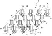
皮膚を貫通するために使用し得る、医療用検出装置の断片10を示す図1および図2を参照する。図1に示される装置断片10は、センサーのうちの少なくとも一つから生体適合性を有する保護被覆15を選択的に除去する前のものであり、図2の装置断片10は、センサーのうちの少なくとも一つから生体適合性を有する保護被覆15を除去した後のものである。図2において、参照符号14は、むき出しの物質検出センサーを示す。 Reference is made to FIGS. 1 and 2 showing a
本発明のいくつかの実施形態では、微小突起のいくつかが、センサーの代わりに電極(具体的には示されていない)を含み得、これが、感知ではなく、電気刺激のために使用され得る。この実施形態により、本医療器具に、血液中のブドウ糖レベルと間質液中のブドウ糖レベルとの間の遅れ時間(lag time)を減少させることができる電気刺激の使用を含めることができる。当業者に知られているように、遅れ時間は、糖尿病患者がどれだけの量のインスリンを注射すべきかを正確に決定することを困難にし得る。この点に関しては、電気刺激が血液灌流を局所的に増加させ、これによって、間質液のブドウ糖レベルを血液中のブドウ糖レベルにより近付け得ることがこれまでに知られている。 In some embodiments of the invention, some of the microprojections may include electrodes (not specifically shown) instead of sensors, which can be used for electrical stimulation rather than sensing . According to this embodiment, the medical device can include the use of electrical stimulation that can reduce the lag time between the glucose level in the blood and the glucose level in the interstitial fluid. As is known to those skilled in the art, lag time can make it difficult for a diabetic patient to accurately determine how much insulin to inject. In this regard, it has been known so far that electrical stimulation can locally increase blood perfusion, thereby allowing interstitial fluid glucose levels to be closer to glucose levels in the blood.
装置断片10は、(たとえば、ヒトまたはその他の動物の)皮膚の最外部の角質層を貫通するような寸法および形状の複数の微小突起12を備えている。微小突起12のそれぞれは、プレート5の表面から外に向かって延びている。図示されているように、それぞれの微小突起12は、それぞれの微小突起の先端にあるセンサーと接触する、一対の電導性のリード線16Aおよび16Bを備えている。図1では、センサーのそれぞれは、生体適合性を有する保護被覆15でカバーされている。図2では、生体適合性を有する保護被覆15が、センサーのうちの少なくとも一つから除去されており、所望の物質を検出するために使用できるむき出しのセンサー14を与えるようになっている。
装置断片10は、患者の身体内の選択されたサイトに、少なくとも一つのむき出しのセンサー14(図2参照)を経皮的に設置するようになっている。特に、装置断片10により、患者の病態を示す特定のパラメータを検出するために使用されるタイプの、柔軟な薄膜電気化学的センサーの配置が容易になる。患者の皮膚内にむき出しのセンサー14を配置することにより、吸収部材中に間質液を採集することに依存する代わりに、読み取り値をその場で得ることができる。このその場における検出は、測定の前に間質液を抜き取ることに依存する診断法と比較して読み取りにおける遅れ時間を最小限にする。好ましい一実施形態では、むき出しのセンサー14が、糖尿病患者のブドウ糖レベルを監視するように設計されている。
図1および図2に示される装置断片から生体適合性を有する保護被覆15を差し引いたものは、本技術分野で周知の技術を使用して作製される。例えば、ダッドーナ(Daddona)等に付与された米国特許第6,091,975号に記述された技術を用いて装置断片10を製造することができる。本開示の内容の全体は、既に、参照によりその全体が本明細書に組み込まれたものである。配列中のそれぞれのセンサーは、’975特許の開示に従って対処される。上記は、突起および突起上にあるセンサーを作製する方法の一例であり、他の、方法、センサーならび突起およびセンサーを作製する方法が可能である点が留意される。 The device piece shown in FIGS. 1 and 2 minus the biocompatible
本発明に従って、微小突起が角質層を通過するが、患者の神経末端には接触しないように、それぞれの微小突起12の大きさが適切に決められる。例えば、それぞれの微小突起12の先端にあるむき出しのセンサー14は、直径が約100μmであり、微小突起12は約150μmの全長を持つ。この構成で、むき出しのセンサー14は、患者の間質液中の物質の存在または量の変化に敏感に応答し、疼痛感や過度の出血を引き起こすことがない。微小突起上にある電導性のリード線のそれぞれは、ポリイミドフィルム等の選択された絶縁材料の層間に埋め込まれあるいは収納された薄膜導体を利用する薄膜マスク技術を使用して構成することができる。それぞれの微小突起12の遠位の先端にあるむき出しのセンサー14は、センサーが経皮的に置かれたときに患者の間質液と接触するために、患者の皮膚に挿入される。むき出しのセンサー14は、例えば、蒸着、電気メッキ、インクジェット方式のスポッティング、印刷、ピンスポッティング等の、本技術分野で周知の技術によって形成される。むき出しのセンサー14には、例えば、電気化学的電極センサー、蛍光に基づくセンサー、タンパク質、細胞またはDNAの結合に基づくセンサーまたは、これらの混合物や組合せ等の任意のバイオセンサーが含まれる。 In accordance with the present invention, each
一般的に、微小突起12は、(必須というわけではないが)、単一片の材料から形成され、体表面の少なくとも角質層に穴をあけるために十分な鋭利さと長さを有する。一実施形態では、微小突起12とプレート5とは、物質の通過に対し、本質的に不透過性または不透過性を有する。それぞれの微小突起12の幅は、任意の範囲でよい。通常、微小突起12の幅は、約25μm〜500μmの範囲にある。微小突起の長さは、透過する体表面の変動次第であり、本発明の特徴の一つは、センサーが表皮の最外層の下の物質を検出することであるため、微小突起の長さは角質層の生得の厚さに対応する。通常、微小突起12は、約20μm〜約400μmの長さである。微小突起12は、微小突起12を体表面中に押し入れるのに要求される挿入力を更に減少させるために、傾斜した(すなわち、角度を付けた)先端を持つことができる。それぞれの微小突起の先端を全て同じ角度にし、または、別々の角度にして、体表を貫通するようにし得る。あるいは、それぞれの微小突起12の先端を、例えば凸面形状や凹面形状を持つ、弧の形状(すなわちカーブした形状)にすることができる。 In general, the microprojections 12 (though not necessarily) are formed from a single piece of material and have sufficient sharpness and length to puncture at least the stratum corneum on the body surface. In one embodiment, the microprojections 12 and the
フォトエッチング加工で任意の微小突起配列のパターンを作製することができる。ステンレススチールまたはチタン等の金属の薄いプレート5に、皮膚を貫通する構造を有するパターンをフォトリソグラフィによりエッチングする。一般的に、プレート5上に、典型的には約7〜約100μmの厚さ、好ましくは約25〜約50μmの厚さ、を持つ、薄い積層ドライレジストまたはウエットレジストを塗布する。所望のパターンを有するマスクを用いて、このレジストを接触露光し(contact exposed)、続いて現像する。これらの操作をプリント回路基板の製造に用いるのとほぼ同様にして行う。プレート5は、次いで、酸性溶液でエッチングされる。微小突起が、平面を曲げて造られる前に、このシート上にセンサーが形成される。平面状の本シート上にセンサーを形成するのに、標準的なフォトリソグラフィと印刷技術を使用し得る。センサーのいくつかを、電気刺激の使用のための電極で置換する場合には、それらの電極は、センサーと同じ手順を用いて作製される。プレートにパターンがエッチングされた後、プレート5は、複数の開口部を有するダイ上に置かれる。まず、プレート5の任意の開口部とダイの開口部とに対応する複数の突起を持つパンチが、プレートおよびダイの上方に置かれる。最初、微小突起12は、プレート5の残余の部分と同一平面にある。次いで、パンチダイが開口部に押し込まれ、プレート5の平面に対しほぼ垂直になるように、微小突起12を下方に曲げる。完成された構造が微小突起12となる。 An arbitrary pattern of minute protrusions can be formed by photoetching. A pattern having a structure penetrating the skin is etched by photolithography on a
通常、微小突起12は、パンチで打ち出された後、プレート5の表面に対し約90度の角度を有するが、垂直な位置から、前方または後方へ、体表面への侵入と取り付けとを容易にする任意の角度にすることができる。更に、本装置の固定を更に強化するために、角度のついた微小突起12と共に、とげ状物、開口部等、その他の固定用要素を使用することができる。 Usually, the
プレート5および微小突起12は、硬質ポリマー、金属および金属合金等の、微小突起12を製造するために十分な強さと十分な製造能力とを持つ材料から作製することができる。金属および金属合金の例には、ステンレススチール、鉄、鋼、スズ、亜鉛、銀、プラチナ、アルミニウム、ジルコニウム、チタン、および、ニッケル、モリブデンまたはクロムを有するチタン合金が含まれるが、これらに限られるわけではない。プレート5および微小突起12のそれぞれは、不活性な性質と生体適合性とを与えるために、銀、金、プラチナ、イリジウム、チタン、ロジウムの、メッキまたは蒸着またはスパッタリングされた、生体適合性を有する金属の薄層を有し得る。ポリマーの例には、ポリスチレン、ポリメチルメタクリレート、ポリプロピレン、「ベークライト(BAKELITE)」、酢酸セルロース、エチルセルロース、スチレン/アクリロニトリルコポリマー、スチレン/ブタジエンコポリマー、アクリロニトリル/ブタジエン/スチレン(ABS)コポリマー、ポリ塩化ビニルならびに、ポリアクリレートおよびポリメタクリレート等のアクリル酸ポリマーが含まれるが、これらに限られるわけではない。 The
本発明のいくつかの実施形態では、戻りのリード線(return leads)の一つとしてプレート5自体を使用し得る。 In some embodiments of the invention, the
微小突起12およびセンサーの数は、本システムで所望される余剰分、検出される物質、使用されているセンサーのタイプ、本装置が機能することを意図されている時間の長さおよび、当業者にとって明らかなその他の因子との関係で変更し得る。 The number of
本発明にしたがって、また、センサーのうちの少なくとも一つを使用する前に、配列内のセンサーは、生体適合性を有する保護被覆15でカバーされている(図1参照)。本発明で使用し得る、適切な生体適合性を有する保護被覆15には、金属、(例えば、チタン(Ti)、ニチノール(NiTi)、金、プラチナおよびアルミニウム等)、および、ポリマー{例えば、シリコーンポリマー(すなわちオルガノシロキサン)、ポリウレタン、ポリアミド、パリレン、フルオロポリマー(例えばテフロン(登録商標)等)、ポリオレフィン(例えば、ポリエチレンおよびポリプロピレン等)、コラーゲン、キチン、アルジネート、ポリビニルピロリドン、ポリエチレングリコール、ポリエチレンオキシド、ポリビニルアルコール、ポリグリコール乳酸、ポリ乳酸、ポリカプロラクトン、ポリアミノ酸およびヒドロゲル(例えばカルボキシメチルセルロース)}が含まれるが、これらに限られるわけではない。本発明の一実施形態では、生体適合性を有する金属が、保護被覆15として使用される。本発明の生体適合性を有するポリマーのもう一つの実施形態では、特にシリコーンポリマーが、保護被覆として使用される。 In accordance with the present invention and prior to using at least one of the sensors, the sensors in the array are covered with a biocompatible protective coating 15 (see FIG. 1). Suitable biocompatible
保護被覆15は、例えば、スピンオンコーティング、ディップコーティング、スプレーコーティング、化学気相成長法、プラズマ助長化学気相成長法、メッキ、スパッタリング、蒸着およびその他類似の析出プロセスを含む、従来の析出プロセスを使用してセンサーに塗布される。使用される析出プロセスの如何に拘わらず、保護層15の厚さは、典型的には、約0.25〜約20μmであり、約2〜約5μmの厚さがより典型的である。 The
本発明のいくつかの実施形態では、微小突起12のいくつかは、特定の病状または疾病を治療するための薬剤で被覆することもできる。本発明で使用し得る薬剤のタイプには、抗生物質、抗ウイルス薬、シロリムス、抗凝血剤(例えばヘパリン)、大分子の生物薬剤、ワクチン、ホルモンおよびナトレコール(Natrecor)が含まれるが、これらに限られるわけではない。薬剤被覆は、薬剤を生体適合性のある材料中に取り込み、次いで、特定の微小突起12上にこの薬剤被覆を沈着させることによって形成される。いくつかの実施形態では、薬剤を、上述の保護被覆内に混入し得る。他の実施形態では、センサー上に保護被覆を形成する前または形成した後に、薬剤被覆を塗布し得る。薬剤は、突起をプレートに対し垂直に曲げた後、液体の薬剤組成物を突起上にディップコーティングまたはローリングすることで、塗布することができる。このようにして、特定の突起から保護層を除去するときに、薬剤を選択的に放出できる。または、薬剤を塗布した全ての微小突起から薬剤が一定の速度で溶出し得る。 In some embodiments of the invention, some of the
塗布の間、所望のセンサーから保護被覆15を選択的に除去することによって、特定のセンサーまたは複数のセンサーが剥き出しになる。電気刺激のための電極がセンサーの代わりに使用されるいくつかの塗布では、同じ手順が適用される。保護被覆15は、熱切除、エッチング、電気的に活性化された溶解または電気的に活性化された腐食によって除去し得る。サンティーニ・ジュニア(Santini, Jr.)等に付与された米国特許第6,551,838号、サンティーニ・ジュニア(Santini, Jr.)等に付与された米国特許第6,849,463号、および、シェパード・ジュニア(Sheppard, Jr.)等に付与された米国特許第6,773,429号に、本発明で使用し得る、電気的に活性化された溶解および電気的に活性化された腐食プロセスに関する詳細が記載されている。このようなものとして、前述の開示は、参照によりその内容の全体が本明細書に組み込まれる。いくつかの実施形態では、保護被覆15が、新しいセンサーを剥き出しにするためにスライドするか、または回転し得る、生体適合性を有する鞘(sheath)であり得る。 By selectively removing the
次いで、むき出しのセンサー14は、ある期間使用される。この期間はセンサーのドリフトによって左右される。センサーの安定操作の限界を超えた後、新しいセンサーが、上述のように選択的に剥き出しにされ、以降の測定のために使用される。全てのセンサーが使用され終わるまでこのプロセスが繰り返され、その時点で、使用済みの医療用検出装置が取り外され、新しい装置が適用される。 The
任意的に、プレート5の体接触側に塗布する接着剤、または装置断片10上のその他の固定用要素等の種々の手段により、装置断片10を患者の体表面に付着するようにし得る。更に、装置と皮膚との接触を維持するために時計バンドまたは伸縮性のある包帯を使用し得る。接着剤は、通常のユーザの活動の間装置10が体表面上の適切な場所にとどまり、かつ、所定の(例えば24時間の)着用期間後、妥当な取り外しができることを保証できるような十分な付着性を有していなければならない。使用前の接着剤の完全性維持のために適切な解除ライナー(release liner)(図示されず)を使用することが好ましい。使用に際しては、装置が皮膚に適用されるのに先だって、接着剤から解除ライナーが剥がされる。 Optionally, the
本発明の装置断片10は、電子的逆輸送(reverse electrotransport)(すなわちイオン導入および/または電気浸透)、浸透および受動拡散等の、流体を引きつける方法でも使用し得るが、これらに限られるわけではない。検出されるべき物質を運ぶ、身体からの流体(すなわち間質液または汗)を抜き出すために浸透装置を使用し得る。たとえば、米国特許第4,756,314号を参照すれば、本発明で使用できる浸透性の構成が開示されている。この浸透装置は、柔軟な接着剤のオーバーレイの手段により体表面に付着し得る。 The
装置断片10は、電子制御ユニット(図示せず)または外部センサー(図示せず)とインタフェースし得る。装置断片10には、エレクトロニクス制御装置を取り付け得る。あるいは、エレクトロニクス制御装置は、個別のユニットとし、あるいは二つのユニットの組み合わせとし得る。インタフェースは、典型的には、インタフェース器具と本発明に記載された装置断片10とを接続する金属トレースを使用して、プレート5の上部表面に沿ってなされる。あるいは、無線手段を使用して電子的インタフェースを達成し得る。
本発明の好ましい実施形態では、本発明の装置が、例えば糖尿病患者におけるブドウ糖等の身体パラメータを、定期的にまたは連続的に検出するために、長期間に渡って使用される。そのような読み取り値は、患者の血液ブドウ糖濃度を(すなわち、血液中のブドウ糖濃度を相関付ける適切なソフトウェアによって)監視することに役立ち、更に、患者へのインスリンの投与および/または食餌療法の適切な修正および/または運動の適切な修正等の治療方法の調節に使用し得る。本発明のいくつかの実施形態では、少なくとも一つの微小突起上にある被覆内にインスリンを含め得る。 In a preferred embodiment of the present invention, the device of the present invention is used over a long period of time to periodically or continuously detect a physical parameter such as glucose in a diabetic patient. Such readings are useful for monitoring the patient's blood glucose concentration (ie, by appropriate software that correlates the glucose concentration in the blood), and that the patient is administered insulin and / or dietary May be used to adjust treatment methods such as proper correction and / or appropriate correction of movement. In some embodiments of the invention, insulin may be included in a coating on at least one microprojection.
上述の発明装置および方法は、他の生体内での適用に容易に一体化し得る。これには、最小限に侵襲的な手順や内視鏡ベースのシステムのための、経皮診断、植え込まれた医療器具、カテーテルベースのシステムが含まれるが、これらに限られるわけではない。 The inventive devices and methods described above can be easily integrated into other in vivo applications. This includes, but is not limited to, percutaneous diagnostics, implanted medical devices, and catheter-based systems for minimally invasive procedures and endoscope-based systems.
これまで、特に、その好ましい実施形態に関連して、本発明を説明してきたが、当業者ならば、形態上および詳細の前述の変更を、本出願の精神と範囲とから逸脱することなくなし得ることを理解するであろう。それ故、本発明は、本明細書に記載および表示された形状および詳細そのものに限られるものでなく、添付の特許請求の範囲内に属することが意図されている。 While the invention has been described hereinabove, particularly with reference to preferred embodiments thereof, those skilled in the art will make the foregoing changes in form and detail without departing from the spirit and scope of the present application. You will understand that you get. Thus, the present invention is not intended to be limited to the precise forms and details described and shown herein, but is intended to fall within the scope of the appended claims.
〔実施の態様〕
本発明の好ましい実施態様は以下の通りである。Embodiment
Preferred embodiments of the present invention are as follows.
(1) 医療用検出装置において、
プレートであって、前記プレートに固定して取り付けられ前記プレートから延びる少なくとも一つの微小突起を有する、プレートと、
前記少なくとも一つの微小突起上の少なくとも一つの物質検出センサーであって、
前記少なくとも一つの物質検出センサーが、物質を検出する前に選択的に除去される、生体適合性を有する保護被覆でカバーされており、
前記少なくとも一つの微小突起が、前記少なくとも一つの物質検出センサーを、体表の最外層の下に体液と接触させて位置づける長さを有する、
少なくとも一つの物質検出センサーと、
を備える、医療用検出装置。
(2) 実施態様1に記載の医療用検出装置において、
剥き出しになった、他の物質検出センサー、
を更に備える、医療用検出装置。
(3) 実施態様1に記載の医療用検出装置において、
前記少なくとも一つの物質検出センサーが、前記プレートの表面に延びる一対の電導性リード線を有する、医療用検出装置。
(4) 実施態様1に記載の医療用検出装置において、
前記少なくとも一つの物質検出センサーが、バイオセンサーである、医療用検出装置。
(5) 実施態様4に記載の医療用検出装置において、
前記バイオセンサーが、電気化学的電極センサーを含む、医療用検出装置。
(6) 実施態様1に記載の医療用検出装置において、
前記物質検出センサーの少なくとも一つをカバーする被覆であって、病状を治療するための薬剤を含む、被覆、
を更に備える、医療用検出装置。
(7) 実施態様1に記載の医療用検出装置において、
前記プレートが、外部の電気的制御ユニットまたは外部の電気的制御センサーとインタフェースしている、医療用検出装置。
(8) 実施態様1に記載の医療用検出装置において、
前記生体適合性を有する保護被覆が、金属またはポリマーを含む、医療用検出装置。
(9) 実施態様8に記載の医療用検出装置において、
前記金属が、チタン(Ti)、ニチノール(NiTi)、金、プラチナ、および、アルミニウム、のうちの一つを含む、医療用検出装置。
(10) 実施態様8に記載の医療用検出装置において、
前記ポリマーが、シリコーンポリマー、ポリウレタン、ポリアミド、パリレン、フルオロポリマー、ポリオレフィン、コラーゲン、キチン、アルジネート、ポリビニルピロリドン、ポリエチレングリコール、ポリエチレンオキシド、ポリビニルアルコール、ポリグリコール乳酸、ポリ乳酸、ポリカプロラクトン、ポリアミノ酸、および、ヒドロゲル、のうちの一つを含む、医療用検出装置。
(11) 実施態様1に記載の医療用検出装置において、
前記体液が、間質液である、医療用検出装置。
(12) 実施態様1に記載の医療用検出装置において、
前記センサーのいくつかが、電気刺激のために使用される電極で置換されている、医療用検出装置。
(13) 実施態様1に記載の医療用検出装置において、
前記プレートが、戻りのリード線(return lead)として働く、医療用検出装置。
(14) 物質を検出する方法において、
医療用検出装置を準備することであって、
前記医療用検出装置は、
プレートであって、前記プレートに固定して取り付けられ前記プレートから延びる少なくとも一つの微小突起を有する、プレート、および、
前記少なくとも一つの微小突起上に設けられた少なくとも一つの物質検出センサー、
を備え、
前記少なくとも一つの物質検出センサーが、生体適合性を有する保護被覆でカバーされており、
前記少なくとも一つの微小突起が、前記少なくとも一つの物質検出センサーを、体表の最外層の下に体液と接触させて位置づける長さを有する、
医療用検出装置を準備することと、
前記少なくとも一つの物質検出センサーから前記生体適合性を有する保護被覆を選択的に除去して前記センサーを剥き出しにすることと、
物質を検出することと、
を含む、方法。
(15) 実施態様14に記載の方法において、
前記体液が、間質液である、方法。
(16) 実施態様14に記載の方法において、
前記物質が、生体分子、体電解質(body electrolyte)、アルコール、不法薬物、または医薬品である、方法。
(17) 実施態様14に記載の方法において、
前記物質が、ブドウ糖である、方法。
(18) 実施態様14に記載の方法において、
前記選択的に除去すること、および前記検出することを繰り返し、前記物質の長期にわたる定期的な検出、または連続的な検出をおこなうこと、
を更に含む、方法。
(19) 実施態様14に記載の方法において、
前記検出することが、ホストの疾病の、診断、治療、および監視の少なくとも一つのために使用される、方法。
(20) 実施態様19に記載の方法において、
前記疾病が、糖尿病、うっ血性心不全、心臓血管疾患、癌、および神経疾患のうちの一つである、方法。
(21) 実施態様14に記載の方法において、
前記微小突起のうちの少なくとも一つの上に位置する薬剤被覆、
を更に含む、方法。
(22) 実施態様14に記載の方法において、
前記センサーのいくつかが、電気刺激のために使用される電極で置換されている、方法。
(23) ブドウ糖の検出方法において、
医療用検出装置を準備することであって、
前記医療用検出装置は、
プレートであって、前記プレートに固定して取り付けられ前記プレートから延びる少なくとも一つの微小突起を有する、プレート、および、
前記少なくとも一つの微小突起上に設けられた少なくとも一つの電気化学的センサー、
を含み、
前記少なくとも一つの電気化学的センサーが、生体適合性を有する保護被覆でカバーされており、
前記少なくとも一つの微小突起が、前記少なくとも一つの電気化学的センサーを、皮膚の最外層の下に間質液と接触させて位置づける長さを有する、
医療用検出装置を準備することと、
前記少なくとも一つの電気化学的センサーから前記生体適合性を有する保護被覆を選択的に除去して前記センサーを剥き出しにすることと、
ブドウ糖を検出することであって、前記選択的に除去すること、および前記検出することを多数回繰り返して、ブドウ糖の長期にわたる定期的な検出または連続的な検出を行う、ブドウ糖を検出することと、 を含む、方法。
(24) 実施態様23に記載の方法において、
前記センサーのいくつかが、電気刺激のために使用される電極で置換されている、方法。(1) In a medical detection device,
A plate having at least one microprojection fixedly attached to and extending from the plate; and
At least one substance detection sensor on the at least one microprojection,
The at least one substance detection sensor is covered with a biocompatible protective coating that is selectively removed before detecting the substance;
The at least one microprojection has a length for positioning the at least one substance detection sensor in contact with a body fluid under the outermost layer of the body surface;
At least one substance detection sensor;
A medical detection apparatus comprising:
(2) In the medical detection device according to Embodiment 1,
The other substance detection sensor that was exposed,
A medical detection device further comprising:
(3) In the medical detection device according to Embodiment 1,
The medical detection apparatus, wherein the at least one substance detection sensor has a pair of conductive leads extending on a surface of the plate.
(4) In the medical detection device according to Embodiment 1,
A medical detection apparatus, wherein the at least one substance detection sensor is a biosensor.
(5) In the medical detection device according to Embodiment 4,
A medical detection device, wherein the biosensor includes an electrochemical electrode sensor.
(6) In the medical detection device according to Embodiment 1,
A coating covering at least one of said substance detection sensors, comprising a drug for treating a medical condition;
A medical detection device further comprising:
(7) In the medical detection device according to Embodiment 1,
A medical detection device, wherein the plate interfaces with an external electrical control unit or an external electrical control sensor.
(8) In the medical detection device according to Embodiment 1,
A medical detection device, wherein the biocompatible protective coating comprises a metal or a polymer.
(9) In the medical detection device according to embodiment 8,
The medical detection apparatus, wherein the metal includes one of titanium (Ti), nitinol (NiTi), gold, platinum, and aluminum.
(10) In the medical detection device according to embodiment 8,
The polymer is a silicone polymer, polyurethane, polyamide, parylene, fluoropolymer, polyolefin, collagen, chitin, alginate, polyvinyl pyrrolidone, polyethylene glycol, polyethylene oxide, polyvinyl alcohol, polyglycol lactic acid, polylactic acid, polycaprolactone, polyamino acid, and , A hydrogel, a medical detection device.
(11) In the medical detection device according to Embodiment 1,
A medical detection apparatus, wherein the body fluid is an interstitial fluid.
(12) In the medical detection device according to Embodiment 1,
A medical detection device, wherein some of said sensors are replaced with electrodes used for electrical stimulation.
(13) In the medical detection device according to Embodiment 1,
A medical detection device wherein the plate serves as a return lead.
(14) In a method for detecting a substance,
Preparing a medical detection device,
The medical detection device is:
A plate having at least one microprojection fixedly attached to and extending from the plate; and
At least one substance detection sensor provided on the at least one microprojection;
With
The at least one substance detection sensor is covered with a biocompatible protective coating;
The at least one microprojection has a length for positioning the at least one substance detection sensor in contact with a body fluid under the outermost layer of the body surface;
Preparing a medical detection device;
Selectively removing the biocompatible protective coating from the at least one substance detection sensor to expose the sensor;
Detecting the substance,
Including the method.
(15) In the method of
The method, wherein the body fluid is an interstitial fluid.
(16) In the method of
The method wherein the substance is a biomolecule, body electrolyte, alcohol, illegal drug, or pharmaceutical.
(17) In the method of
The method wherein the substance is glucose.
(18) In the method of
Repeating the selective removal and the detection to perform long-term periodic detection or continuous detection of the substance;
The method further comprising:
(19) In the method of
The method wherein said detecting is used for at least one of diagnosis, treatment and monitoring of a host disease.
(20) In the method of embodiment 19,
The method wherein the disease is one of diabetes, congestive heart failure, cardiovascular disease, cancer, and neurological disease.
(21) In the method of
A drug coating located on at least one of the microprojections;
The method further comprising:
(22) In the method of
A method wherein some of the sensors are replaced with electrodes used for electrical stimulation.
(23) In the method for detecting glucose,
Preparing a medical detection device,
The medical detection device is:
A plate having at least one microprojection fixedly attached to and extending from the plate; and
At least one electrochemical sensor provided on the at least one microprojection;
Including
The at least one electrochemical sensor is covered with a biocompatible protective coating;
The at least one microprojection has a length to position the at least one electrochemical sensor in contact with interstitial fluid under the outermost layer of the skin;
Preparing a medical detection device;
Selectively removing the biocompatible protective coating from the at least one electrochemical sensor to expose the sensor;
Detecting glucose, wherein said selective removal and said detection are repeated a number of times, with periodic or continuous detection of glucose over time. A method comprising:
(24) In the method of embodiment 23,
A method wherein some of the sensors are replaced with electrodes used for electrical stimulation.
Claims (24)
Translated fromJapaneseプレートであって、前記プレートに固定して取り付けられ前記プレートから延びる少なくとも一つの微小突起を有する、プレートと、
前記少なくとも一つの微小突起上に設けられた少なくとも一つの物質検出センサーであって、
前記少なくとも一つの物質検出センサーが、物質を検出する前に選択的に除去される、生体適合性を有する保護被覆でカバーされており、
前記少なくとも一つの微小突起が、前記少なくとも一つの物質検出センサーを、体表の最外層の下に体液と接触させて位置づける長さを有する、
少なくとも一つの物質検出センサーと、
を備える、医療用検出装置。In a medical detection device,
A plate having at least one microprojection fixedly attached to and extending from the plate; and
At least one substance detection sensor provided on the at least one microprojection,
The at least one substance detection sensor is covered with a biocompatible protective coating that is selectively removed before detecting the substance;
The at least one microprojection has a length for positioning the at least one substance detection sensor in contact with a body fluid under the outermost layer of the body surface;
At least one substance detection sensor;
A medical detection apparatus comprising:
剥き出しになった、他の物質検出センサー、
を更に備える、医療用検出装置。The medical detection device according to claim 1,
The other substance detection sensor that was exposed,
A medical detection device further comprising:
前記少なくとも一つの物質検出センサーが、前記プレートの表面に延びる一対の電導性リード線を有する、医療用検出装置。The medical detection device according to claim 1,
The medical detection apparatus, wherein the at least one substance detection sensor has a pair of conductive leads extending on a surface of the plate.
前記少なくとも一つの物質検出センサーが、バイオセンサーである、医療用検出装置。The medical detection device according to claim 1,
A medical detection apparatus, wherein the at least one substance detection sensor is a biosensor.
前記バイオセンサーが、電気化学的電極センサーを含む、医療用検出装置。The medical detection device according to claim 4, wherein
A medical detection device, wherein the biosensor includes an electrochemical electrode sensor.
前記物質検出センサーの少なくとも一つをカバーする被覆であって、病状を治療するための薬剤を含む、被覆、
を更に備える、医療用検出装置。The medical detection device according to claim 1,
A coating covering at least one of said substance detection sensors, comprising a drug for treating a medical condition;
A medical detection device further comprising:
前記プレートが、外部の電気的制御ユニットまたは外部の電気的制御センサーとインタフェースしている、医療用検出装置。The medical detection device according to claim 1,
A medical detection device, wherein the plate interfaces with an external electrical control unit or an external electrical control sensor.
前記生体適合性を有する保護被覆が、金属またはポリマーを含む、医療用検出装置。The medical detection device according to claim 1,
A medical detection device, wherein the biocompatible protective coating comprises a metal or a polymer.
前記金属が、チタン(Ti)、ニチノール(NiTi)、金、プラチナ、および、アルミニウム、のうちの一つを含む、医療用検出装置。The medical detection device according to claim 8,
The medical detection apparatus, wherein the metal includes one of titanium (Ti), nitinol (NiTi), gold, platinum, and aluminum.
前記ポリマーが、シリコーンポリマー、ポリウレタン、ポリアミド、パリレン、フルオロポリマー、ポリオレフィン、コラーゲン、キチン、アルジネート、ポリビニルピロリドン、ポリエチレングリコール、ポリエチレンオキシド、ポリビニルアルコール、ポリグリコール乳酸、ポリ乳酸、ポリカプロラクトン、ポリアミノ酸、および、ヒドロゲル、のうちの一つを含む、医療用検出装置。The medical detection device according to claim 8,
The polymer is a silicone polymer, polyurethane, polyamide, parylene, fluoropolymer, polyolefin, collagen, chitin, alginate, polyvinyl pyrrolidone, polyethylene glycol, polyethylene oxide, polyvinyl alcohol, polyglycol lactic acid, polylactic acid, polycaprolactone, polyamino acid, and , A hydrogel, a medical detection device.
前記体液が、間質液である、医療用検出装置。The medical detection device according to claim 1,
A medical detection apparatus, wherein the body fluid is an interstitial fluid.
前記センサーのいくつかが、電気刺激のために使用される電極で置換されている、医療用検出装置。The medical detection device according to claim 1,
A medical detection device, wherein some of said sensors are replaced with electrodes used for electrical stimulation.
前記プレートが、戻りのリード線(return lead)として働く、医療用検出装置。The medical detection device according to claim 1,
A medical detection device wherein the plate serves as a return lead.
医療用検出装置を準備することであって、
前記医療用検出装置は、
プレートであって、前記プレートに固定して取り付けられ前記プレートから延びる少なくとも一つの微小突起を有する、プレート、および、
前記少なくとも一つの微小突起上に設けられた少なくとも一つの物質検出センサー、
を備え、
前記少なくとも一つの物質検出センサーが、生体適合性を有する保護被覆でカバーされており、
前記少なくとも一つの微小突起が、前記少なくとも一つの物質検出センサーを、体表の最外層の下に体液と接触させて位置づける長さを有する、
医療用検出装置を準備することと、
前記少なくとも一つの物質検出センサーから前記生体適合性を有する保護被覆を選択的に除去して前記センサーを剥き出しにすることと、
物質を検出することと、
を含む、方法。In a method for detecting a substance,
Preparing a medical detection device,
The medical detection device is:
A plate having at least one microprojection fixedly attached to and extending from the plate; and
At least one substance detection sensor provided on the at least one microprojection;
With
The at least one substance detection sensor is covered with a biocompatible protective coating;
The at least one microprojection has a length for positioning the at least one substance detection sensor in contact with a body fluid under the outermost layer of the body surface;
Preparing a medical detection device;
Selectively removing the biocompatible protective coating from the at least one substance detection sensor to expose the sensor;
Detecting the substance,
Including the method.
前記体液が、間質液である、方法。15. The method of claim 14, wherein
The method, wherein the body fluid is an interstitial fluid.
前記物質が、生体分子、体電解質(body electrolyte)、アルコール、不法薬物、または医薬品である、方法。15. The method of claim 14, wherein
The method wherein the substance is a biomolecule, body electrolyte, alcohol, illegal drug, or pharmaceutical.
前記物質が、ブドウ糖である、方法。15. The method of claim 14, wherein
The method wherein the substance is glucose.
前記選択的に除去すること、および前記検出することを繰り返し、前記物質の長期にわたる定期的な検出、または連続的な検出をおこなうこと、
を更に含む、方法。15. The method of claim 14, wherein
Repeating the selective removal and the detection to perform long-term periodic detection or continuous detection of the substance;
The method further comprising:
前記検出することが、ホストの疾病の、診断、治療、および監視の少なくとも一つのために使用される、方法。15. The method of claim 14, wherein
The method wherein said detecting is used for at least one of diagnosis, treatment and monitoring of a host disease.
前記疾病が、糖尿病、うっ血性心不全、心臓血管疾患、癌、および神経疾患のうちの一つである、方法。The method of claim 19, wherein
The method wherein the disease is one of diabetes, congestive heart failure, cardiovascular disease, cancer, and neurological disease.
前記微小突起のうちの少なくとも一つの上に位置する薬剤被覆、
を更に含む、方法。15. The method of claim 14, wherein
A drug coating located on at least one of the microprojections;
The method further comprising:
前記センサーのいくつかが、電気刺激のために使用される電極で置換されている、方法。15. The method of claim 14, wherein
A method wherein some of the sensors are replaced with electrodes used for electrical stimulation.
医療用検出装置を準備することであって、
前記医療用検出装置は、
プレートであって、前記プレートに固定して取り付けられ前記プレートから延びる少なくとも一つの微小突起を有する、プレート、および、
前記少なくとも一つの微小突起上に設けられた少なくとも一つの電気化学的センサー、
を含み、
前記少なくとも一つの電気化学的センサーが、生体適合性を有する保護被覆でカバーされており、
前記少なくとも一つの微小突起が、前記少なくとも一つの電気化学的センサーを、皮膚の最外層の下に間質液と接触させて位置づける長さを有する、
医療用検出装置を準備することと、
前記少なくとも一つの電気化学的センサーから前記生体適合性を有する保護被覆を選択的に除去して前記センサーを剥き出しにすることと、
ブドウ糖を検出することであって、前記選択的に除去すること、および前記検出することを複数回繰り返して、ブドウ糖の長期にわたる定期的な検出または連続的な検出を行う、ブドウ糖を検出することと、
を含む、方法。In the method for detecting glucose,
Preparing a medical detection device,
The medical detection device is:
A plate having at least one microprojection fixedly attached to and extending from the plate; and
At least one electrochemical sensor provided on the at least one microprojection;
Including
The at least one electrochemical sensor is covered with a biocompatible protective coating;
The at least one microprojection has a length to position the at least one electrochemical sensor in contact with interstitial fluid under the outermost layer of the skin;
Preparing a medical detection device;
Selectively removing the biocompatible protective coating from the at least one electrochemical sensor to expose the sensor;
Detecting glucose, wherein the selective removal and the detection are repeated multiple times to perform periodic or continuous detection of glucose over a long period of time; ,
Including the method.
前記センサーのいくつかが、電気刺激のために使用される電極で置換されている、方法。24. The method of claim 23, wherein
A method wherein some of the sensors are replaced with electrodes used for electrical stimulation.
Applications Claiming Priority (1)
| Application Number | Priority Date | Filing Date | Title |
|---|---|---|---|
| US11/293,754US20070129620A1 (en) | 2005-12-02 | 2005-12-02 | Selectively exposable miniature probes with integrated sensor arrays for continuous in vivo diagnostics |
Publications (1)
| Publication Number | Publication Date |
|---|---|
| JP2007152108Atrue JP2007152108A (en) | 2007-06-21 |
Family
ID=37766010
Family Applications (1)
| Application Number | Title | Priority Date | Filing Date |
|---|---|---|---|
| JP2006325927AAbandonedJP2007152108A (en) | 2005-12-02 | 2006-12-01 | Selectively exposable miniature probes with integrated sensor arrays for continuous in vivo diagnostics |
Country Status (6)
| Country | Link |
|---|---|
| US (1) | US20070129620A1 (en) |
| EP (1) | EP1792565B1 (en) |
| JP (1) | JP2007152108A (en) |
| AT (1) | ATE411771T1 (en) |
| CA (1) | CA2569126A1 (en) |
| DE (1) | DE602006003295D1 (en) |
Cited By (1)
| Publication number | Priority date | Publication date | Assignee | Title |
|---|---|---|---|---|
| WO2022076681A1 (en)* | 2020-10-07 | 2022-04-14 | True Mobile Health Llc | Illicit substance use compliance system and method |
Families Citing this family (11)
| Publication number | Priority date | Publication date | Assignee | Title |
|---|---|---|---|---|
| KR101135624B1 (en)* | 2009-01-15 | 2012-04-17 | 한양대학교 산학협력단 | A biosensor coated with electroactive polymer layer for extension of biosensor life span |
| KR101065748B1 (en)* | 2009-03-17 | 2011-09-19 | 한양대학교 산학협력단 | Biosensor with Electrosensitive Polymer Layer showing Bending Behavior |
| US8647357B2 (en) | 2011-02-05 | 2014-02-11 | Birch Narrows Development Llc | Lancet device with flexible cover |
| SG194305A1 (en)* | 2012-04-16 | 2013-11-29 | Agency Science Tech & Res | Guide wire arrangement |
| US9237866B2 (en) | 2013-04-29 | 2016-01-19 | Birch Narrows Development, LLC | Blood glucose management |
| US12109032B1 (en) | 2017-03-11 | 2024-10-08 | Biolinq Incorporated | Methods for achieving an isolated electrical interface between an anterior surface of a microneedle structure and a posterior surface of a support structure |
| US11045142B1 (en) | 2017-04-29 | 2021-06-29 | Biolinq, Inc. | Heterogeneous integration of silicon-fabricated solid microneedle sensors and CMOS circuitry |
| US20200158679A1 (en)* | 2018-11-16 | 2020-05-21 | Medtronic Minimed, Inc. | Analyte sensor with extended lifetime |
| SE2251496A1 (en) | 2020-07-29 | 2022-12-20 | Biolinq Incorporated | Continuous analyte monitoring system with microneedle array |
| CA3184224A1 (en) | 2021-05-08 | 2022-11-17 | Joshua Ray Windmiller | Fault detection for microneedle array based continuous analyte monitoring device |
| US12336816B2 (en) | 2023-02-02 | 2025-06-24 | Biolinq Incorporated | Method for improved sensor sensitivity of a microneedle-based continuous analyte monitoring system |
Family Cites Families (7)
| Publication number | Priority date | Publication date | Assignee | Title |
|---|---|---|---|---|
| US5427096A (en)* | 1993-11-19 | 1995-06-27 | Cmc Assemblers, Inc. | Water-degradable electrode |
| IE72524B1 (en)* | 1994-11-04 | 1997-04-23 | Elan Med Tech | Analyte-controlled liquid delivery device and analyte monitor |
| US6091975A (en)* | 1998-04-01 | 2000-07-18 | Alza Corporation | Minimally invasive detecting device |
| ES2420279T3 (en)* | 2000-03-02 | 2013-08-23 | Microchips, Inc. | Microfabricated devices and methods for storage and selective exposure of chemicals |
| US6874621B2 (en)* | 2001-02-27 | 2005-04-05 | Koninklijke Philips Electronics N.V. | Method and package for increasing electrode shelf life |
| US7153265B2 (en)* | 2002-04-22 | 2006-12-26 | Medtronic Minimed, Inc. | Anti-inflammatory biosensor for reduced biofouling and enhanced sensor performance |
| US7577470B2 (en)* | 2003-11-13 | 2009-08-18 | Medtronic Minimed, Inc. | Long term analyte sensor array |
- 2005
- 2005-12-02USUS11/293,754patent/US20070129620A1/ennot_activeAbandoned
- 2006
- 2006-11-21ATAT06255949Tpatent/ATE411771T1/ennot_activeIP Right Cessation
- 2006-11-21DEDE602006003295Tpatent/DE602006003295D1/enactiveActive
- 2006-11-21EPEP06255949Apatent/EP1792565B1/enactiveActive
- 2006-11-28CACA002569126Apatent/CA2569126A1/ennot_activeAbandoned
- 2006-12-01JPJP2006325927Apatent/JP2007152108A/ennot_activeAbandoned
Cited By (1)
| Publication number | Priority date | Publication date | Assignee | Title |
|---|---|---|---|---|
| WO2022076681A1 (en)* | 2020-10-07 | 2022-04-14 | True Mobile Health Llc | Illicit substance use compliance system and method |
Also Published As
| Publication number | Publication date |
|---|---|
| EP1792565A1 (en) | 2007-06-06 |
| ATE411771T1 (en) | 2008-11-15 |
| US20070129620A1 (en) | 2007-06-07 |
| DE602006003295D1 (en) | 2008-12-04 |
| EP1792565B1 (en) | 2008-10-22 |
| CA2569126A1 (en) | 2007-06-02 |
Similar Documents
| Publication | Publication Date | Title |
|---|---|---|
| JP2007152108A (en) | Selectively exposable miniature probes with integrated sensor arrays for continuous in vivo diagnostics | |
| US6091975A (en) | Minimally invasive detecting device | |
| JP3963485B2 (en) | Minimal intrusion detection device | |
| JP5138819B2 (en) | Transcutaneous analyte sensor | |
| US8224414B2 (en) | System and method for analyte sampling and analysis with hydrogel | |
| JP3328290B2 (en) | Ion introduction sampling apparatus and method | |
| US7228162B2 (en) | Analyte sensor | |
| CA2584699C (en) | System and method for analyte sampling and analysis with hydrogel | |
| JP2003033336A (en) | Device and method for sampling and measuring biofluid component | |
| Friedel et al. | Continuous molecular monitoring of human dermal interstitial fluid with microneedle-enabled electrochemical aptamer sensors | |
| WO2004063718A2 (en) | Assembly of single use sensing elements | |
| Zhan et al. | A 3D-printed microneedle extraction system integrated with patterned electrodes for minimally invasive transdermal detection | |
| Vranić et al. | Microneedle-based sensor systems for real-time continuous transdermal monitoring of analytes in body fluids | |
| WO2018100176A1 (en) | Cartridge for biochemical sensor | |
| BR102022004984A2 (en) | GLUCOSE METER WITHOUT DRILLING BY REVERSE IONTOPHORESIS | |
| Vranić et al. | Microneedle-Based Sensor Systems | |
| WO2014108087A1 (en) | Portable monitoring system for dynamically and continuously measuring analyte in body liquid | |
| JP2004033374A (en) | In vivo information detection unit |
Legal Events
| Date | Code | Title | Description |
|---|---|---|---|
| RD04 | Notification of resignation of power of attorney | Free format text:JAPANESE INTERMEDIATE CODE: A7424 Effective date:20071130 | |
| RD04 | Notification of resignation of power of attorney | Free format text:JAPANESE INTERMEDIATE CODE: A7424 Effective date:20081017 | |
| A621 | Written request for application examination | Free format text:JAPANESE INTERMEDIATE CODE: A621 Effective date:20090918 | |
| A762 | Written abandonment of application | Free format text:JAPANESE INTERMEDIATE CODE: A762 Effective date:20091105 | |
| A521 | Request for written amendment filed | Free format text:JAPANESE INTERMEDIATE CODE: A821 Effective date:20091105 |