JP2007097653A - Endoscopic diagnosis system - Google Patents
Endoscopic diagnosis systemDownload PDFInfo
- Publication number
- JP2007097653A JP2007097653AJP2005288359AJP2005288359AJP2007097653AJP 2007097653 AJP2007097653 AJP 2007097653AJP 2005288359 AJP2005288359 AJP 2005288359AJP 2005288359 AJP2005288359 AJP 2005288359AJP 2007097653 AJP2007097653 AJP 2007097653A
- Authority
- JP
- Japan
- Prior art keywords
- unit
- instructor
- signal
- endoscopic
- operator
- Prior art date
- Legal status (The legal status is an assumption and is not a legal conclusion. Google has not performed a legal analysis and makes no representation as to the accuracy of the status listed.)
- Pending
Links
- 238000012327Endoscopic diagnosisMethods0.000titleclaimsabstractdescription24
- 238000004891communicationMethods0.000claimsabstractdescription12
- 230000005540biological transmissionEffects0.000claimsdescription20
- 238000003384imaging methodMethods0.000claimsdescription17
- 238000003745diagnosisMethods0.000claimsdescription13
- 238000001839endoscopyMethods0.000claims1
- 230000005236sound signalEffects0.000description38
- 238000000034methodMethods0.000description10
- 238000000926separation methodMethods0.000description7
- 238000006243chemical reactionMethods0.000description6
- 238000003780insertionMethods0.000description6
- 230000037431insertionEffects0.000description6
- XLYOFNOQVPJJNP-UHFFFAOYSA-NwaterSubstancesOXLYOFNOQVPJJNP-UHFFFAOYSA-N0.000description6
- 230000001276controlling effectEffects0.000description5
- 238000005452bendingMethods0.000description3
- 238000001514detection methodMethods0.000description3
- 238000010586diagramMethods0.000description3
- 230000010363phase shiftEffects0.000description3
- 230000003321amplificationEffects0.000description2
- 230000002596correlated effectEffects0.000description2
- 238000005286illuminationMethods0.000description2
- 238000003199nucleic acid amplification methodMethods0.000description2
- 238000005070samplingMethods0.000description2
- 238000004140cleaningMethods0.000description1
- 230000000875corresponding effectEffects0.000description1
- 238000005192partitionMethods0.000description1
- 230000000087stabilizing effectEffects0.000description1
Images


Classifications
- G—PHYSICS
- G16—INFORMATION AND COMMUNICATION TECHNOLOGY [ICT] SPECIALLY ADAPTED FOR SPECIFIC APPLICATION FIELDS
- G16H—HEALTHCARE INFORMATICS, i.e. INFORMATION AND COMMUNICATION TECHNOLOGY [ICT] SPECIALLY ADAPTED FOR THE HANDLING OR PROCESSING OF MEDICAL OR HEALTHCARE DATA
- G16H80/00—ICT specially adapted for facilitating communication between medical practitioners or patients, e.g. for collaborative diagnosis, therapy or health monitoring
- G—PHYSICS
- G16—INFORMATION AND COMMUNICATION TECHNOLOGY [ICT] SPECIALLY ADAPTED FOR SPECIFIC APPLICATION FIELDS
- G16H—HEALTHCARE INFORMATICS, i.e. INFORMATION AND COMMUNICATION TECHNOLOGY [ICT] SPECIALLY ADAPTED FOR THE HANDLING OR PROCESSING OF MEDICAL OR HEALTHCARE DATA
- G16H30/00—ICT specially adapted for the handling or processing of medical images
- G16H30/20—ICT specially adapted for the handling or processing of medical images for handling medical images, e.g. DICOM, HL7 or PACS
- G—PHYSICS
- G16—INFORMATION AND COMMUNICATION TECHNOLOGY [ICT] SPECIALLY ADAPTED FOR SPECIFIC APPLICATION FIELDS
- G16H—HEALTHCARE INFORMATICS, i.e. INFORMATION AND COMMUNICATION TECHNOLOGY [ICT] SPECIALLY ADAPTED FOR THE HANDLING OR PROCESSING OF MEDICAL OR HEALTHCARE DATA
- G16H50/00—ICT specially adapted for medical diagnosis, medical simulation or medical data mining; ICT specially adapted for detecting, monitoring or modelling epidemics or pandemics
- G16H50/20—ICT specially adapted for medical diagnosis, medical simulation or medical data mining; ICT specially adapted for detecting, monitoring or modelling epidemics or pandemics for computer-aided diagnosis, e.g. based on medical expert systems
Landscapes
- Health & Medical Sciences (AREA)
- Engineering & Computer Science (AREA)
- Medical Informatics (AREA)
- Public Health (AREA)
- Epidemiology (AREA)
- General Health & Medical Sciences (AREA)
- Primary Health Care (AREA)
- Biomedical Technology (AREA)
- Pathology (AREA)
- Radiology & Medical Imaging (AREA)
- Nuclear Medicine, Radiotherapy & Molecular Imaging (AREA)
- Data Mining & Analysis (AREA)
- Databases & Information Systems (AREA)
- Endoscopes (AREA)
- Closed-Circuit Television Systems (AREA)
- Instruments For Viewing The Inside Of Hollow Bodies (AREA)
Abstract
Description
Translated fromJapanese本発明は、遠隔地にある指導室にいる指導者の指示を受けて、検査室にいる術者が内視鏡診断を行う内視鏡診断システムに関する。 The present invention relates to an endoscopic diagnosis system in which an operator in an examination room performs an endoscopic diagnosis in response to an instruction from a leader in a guidance room at a remote place.
従来から、医療分野において、電子内視鏡を利用した医療診断が盛んに行われている。電子内視鏡の体腔内に挿入される挿入部先端には、CCDなどの撮像素子が内蔵されており、このCCDにより取得した撮像信号に対して、プロセッサ装置で信号処理を施すことで、モニタで体腔内の画像(内視鏡画像)を観察することができる。電子内視鏡やプロセッサ装置は、同時に複数の患者の内視鏡診断が可能なように、例えば病院内の一室をパーテーションなどで仕切った複数の検査室にそれぞれ設置されている。 Conventionally, medical diagnosis using an electronic endoscope has been actively performed in the medical field. An imaging element such as a CCD is built in the distal end of the insertion portion to be inserted into the body cavity of the electronic endoscope, and the image signal acquired by the CCD is subjected to signal processing by the processor device, thereby being monitored. With this, an image inside the body cavity (endoscopic image) can be observed. The electronic endoscope and the processor device are installed in, for example, a plurality of examination rooms in which a room in a hospital is partitioned by a partition or the like so that endoscopic diagnosis of a plurality of patients can be performed simultaneously.
上記のような電子内視鏡システムには、研修医などの経験の浅い医師が術者として担当した場合などに、内視鏡診断が的確に行われているか否かを判断するため、術者よりも経験豊富な医師が指導者として在室する指導室を用意し、指導者が術者に的確な指示を与えることができるようにした手術支援システムが提案されている(特許文献1、および2参照)。
特許文献1、および2に記載の技術では、検査室と指導室とをISDNなどの通信回線で接続し、この通信回線を介して内視鏡画像や指導者の指示を遣り取りしているため、各室間の配線に手間が掛かるという問題があった。 In the techniques described in
本発明は、上記課題を鑑みてなされたものであり、無線通信を利用して、検査室と指導室との間の配線をなくし、手間を掛けずに構築することができる内視鏡診断システムを提供することを目的とする。 The present invention has been made in view of the above problems, and is an endoscope diagnosis system that can be constructed without using a wireless communication and eliminating wiring between an examination room and an instruction room. The purpose is to provide.
上記目的を達成するために、本発明は、遠隔地にある指導室にいる指導者の指示を受けて、検査室にいる術者が内視鏡診断を行う内視鏡診断システムにおいて、体腔内の被観察体像を撮影して撮像信号を出力する撮像素子、前記撮像信号をデジタル化した画像信号に直交変調を施すとともに、前記術者から入力される前記指導者への問い合わせ情報を、位相をずらして前記画像信号に重畳し、RF信号を生成する変調部、およびRF信号を電波として送信する送信部を有する電子内視鏡と、電波を受信する受信部、前記RF信号から元の画像信号を復調する復調部、および前記画像信号から内視鏡画像を生成する画像信号処理部を有する術者用プロセッサ装置と、前記内視鏡画像を表示する術者用モニタと、前記問い合わせ情報を入力するための術者用情報入力装置と、前記指導者から入力される前記術者への指示情報を出力する術者用情報出力装置とから構成される術者用内視鏡診断ユニットを前記検査室に設置し、電波を受信する受信部、前記RF信号から元の画像信号および問い合わせ情報を復調する復調部、前記画像信号から内視鏡画像を生成する画像信号処理部、前記指示情報を変調してRF信号を生成する変調部、およびRF信号を電波として送信する送信部を有する指導者用プロセッサ装置と、前記内視鏡画像を表示する指導者用モニタと、前記指示情報を入力するための指導者用情報入力装置と、前記問い合わせ情報を出力する指導者用情報出力装置とから構成される指導者用内視鏡診断ユニットを前記指導室に設置し、前記術者用内視鏡診断ユニットおよび前記指導者用内視鏡診断ユニットは、前記問い合わせ情報および前記指示情報の送受信を半二重通信により行うことを特徴とする。 In order to achieve the above object, the present invention provides an endoscopic diagnosis system in which an operator in an examination room performs an endoscopic diagnosis in response to an instruction of a leader in a remote instruction room. An imaging device that captures an image of the object to be observed and outputs an imaging signal, and applies quadrature modulation to an image signal obtained by digitizing the imaging signal, and inquiries to the instructor input from the surgeon are based on phase information And an electronic endoscope having a modulation unit that generates an RF signal and a transmission unit that transmits the RF signal as a radio wave, a reception unit that receives the radio wave, and an original image from the RF signal A processor for a surgeon having a demodulator for demodulating a signal, and an image signal processor for generating an endoscopic image from the image signal; a surgeon monitor for displaying the endoscopic image; and the inquiry information. To enter An endoscopic diagnostic unit for a surgeon comprising the information input device for the surgeon and an information output device for the surgeon that outputs instruction information to the surgeon input from the instructor. A receiving unit for receiving radio waves, a demodulating unit for demodulating the original image signal and inquiry information from the RF signal, an image signal processing unit for generating an endoscopic image from the image signal, and modulating the instruction information A processor device for a leader having a modulation section for generating an RF signal and a transmission section for transmitting the RF signal as a radio wave, a monitor for a leader for displaying the endoscopic image, and a guidance for inputting the instruction information An instructor endoscope diagnostic unit comprising an information input device for an instructor and an information output device for an instructor that outputs the inquiry information is installed in the instruction room, and the endoscopic diagnosis unit for the operator and Above Guide's endoscopic diagnosis unit, and performs transmission and reception of the inquiry information and the instruction information by the half-duplex communication.
なお、前記術者用内視鏡診断ユニットを複数の検査室にそれぞれ設置し、ユニット毎に前記電波の送受信周波数帯域を個別のチャネルで割り振るとともに、前記指導者用内視鏡診断ユニットに、前記チャネルを選択的に切り替える切り替え操作手段を設けることが好ましい。 The endoscopic diagnostic unit for the operator is installed in each of a plurality of examination rooms, and the transmission / reception frequency band of the radio wave is allocated to each unit by an individual channel, and the endoscopic diagnostic unit for the instructor, It is preferable to provide switching operation means for selectively switching channels.
また、前記問い合わせ情報および前記指示情報は、音声、文字、若しくは各装置の動作を制御するための設定情報のうちの少なくともいずれか1つであることが好ましい。 The inquiry information and the instruction information are preferably at least one of voice, characters, or setting information for controlling the operation of each device.
本発明の内視鏡診断システムによれば、体腔内の被観察体像を撮影して撮像信号を出力する撮像素子、撮像信号をデジタル化した画像信号に直交変調を施すとともに、術者から入力される指導者への問い合わせ情報を、位相をずらして画像信号に重畳し、RF信号を生成する変調部、およびRF信号を電波として送信する送信部を有する電子内視鏡と、電波を受信する受信部、RF信号から元の画像信号を復調する復調部、および画像信号から内視鏡画像を生成する画像信号処理部を有する術者用プロセッサ装置と、内視鏡画像を表示する術者用モニタと、問い合わせ情報を入力するための術者用情報入力装置と、指導者から入力される術者への指示情報を出力する術者用情報出力装置とから構成される術者用内視鏡診断ユニットを検査室に設置し、電波を受信する受信部、RF信号から元の画像信号および問い合わせ情報を復調する復調部、画像信号から内視鏡画像を生成する画像信号処理部、指示情報を変調してRF信号を生成する変調部、およびRF信号を電波として送信する送信部を有する指導者用プロセッサ装置と、内視鏡画像を表示する指導者用モニタと、指示情報を入力するための指導者用情報入力装置と、問い合わせ情報を出力する指導者用情報出力装置とから構成される指導者用内視鏡診断ユニットを指導室に設置し、術者用内視鏡診断ユニットおよび指導者用内視鏡診断ユニットは、問い合わせ情報および指示情報の送受信を半二重通信により行うので、検査室と指導室との間の配線がなくなり、手間を掛けずにシステムを構築することができる。 According to the endoscopic diagnosis system of the present invention, an imaging device that captures an image of a body to be observed in a body cavity and outputs an imaging signal, orthogonally modulates an image signal obtained by digitizing the imaging signal, and inputs from an operator Inquiry information to the instructor to be superposed on the image signal with a phase shift, an electronic endoscope having a modulation unit that generates an RF signal, and a transmission unit that transmits the RF signal as a radio wave, and receives the radio wave A processor unit for an operator having a receiving unit, a demodulating unit for demodulating an original image signal from an RF signal, and an image signal processing unit for generating an endoscopic image from the image signal, and an operator for displaying an endoscopic image An endoscope for a surgeon comprising a monitor, a surgeon information input device for inputting inquiry information, and a surgeon information output device for outputting instruction information to the surgeon input from the instructor Diagnostic unit in the laboratory A receiving unit for receiving radio waves, a demodulating unit for demodulating the original image signal and inquiry information from the RF signal, an image signal processing unit for generating an endoscopic image from the image signal, and modulating the RF signal by modulating the instruction information An instructor processor device having a modulating unit for generating and a transmitting unit for transmitting RF signals as radio waves, an instructor monitor for displaying an endoscopic image, and an instructor information input device for inputting instruction information And an instructor's endoscopic diagnostic unit that includes an information output device for an instructor that outputs inquiry information in an instruction room, and an endoscopic diagnostic unit for an operator and an endoscopic diagnostic unit for an instructor Since the inquiry information and the instruction information are transmitted and received by half-duplex communication, the wiring between the examination room and the instruction room is eliminated, and a system can be constructed without trouble.
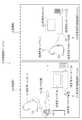
図1において、内視鏡診断システム2は、術者が患者に内視鏡診断を施す検査室10に設置された術者用内視鏡診断ユニット11と、術者の内視鏡診断を監督する指導者が在室する指導室12に設置された指導者用内視鏡診断ユニット13とからなる。 In FIG. 1, an
術者用内視鏡診断ユニット11は、電子内視鏡14と、内視鏡画像を生成する術者用プロセッサ装置15と、内視鏡画像を表示する術者用モニタ16と、術者の音声を取り込む術者用マイクロホン17aを備えた術者用ヘッドセット17と、指導者の音声を出力する術者用スピーカー18とから構成される。 The endoscopic diagnosis unit 11 for an operator includes an
一方、指導者用内視鏡診断ユニット13は、内視鏡画像を生成する指導者用プロセッサ装置19と、内視鏡画像を表示する指導者用モニタ20と、指導者の音声を取り込む指導者用マイクロホン21aを備えた指導者用ヘッドセット21と、術者の音声を出力する指導者用スピーカー22とから構成される。 On the other hand, the instructor endoscope diagnostic unit 13 includes an
図2において、電子内視鏡14は、体腔内に挿入される挿入部30と、挿入部30の基端部分に連設された操作部31とを備えている。挿入部30の先端に連設された先端部30aには、体腔内の被観察体像の像光を取り込むための対物レンズ32と、体腔内の被観察体像を撮影する撮像素子としてのCCD33、および照射レンズ34と体腔内照明用のLED光源(LED)35(ともに図3参照)が内蔵されている。 In FIG. 2, the
先端部30aの後方には、複数の湾曲駒を連結した湾曲部36が設けられている。この湾曲部36は、操作部31に設けられたアングルノブ31aが操作されて、挿入部30内に挿設されたワイヤが押し引きされることにより、上下左右方向に湾曲動作し、先端部30aが体腔内の所望の方向に向けられるようになっている。 A
操作部31の下方には、水が貯留される貯水タンク37と、エアーが貯留されるエアーボンベ38とが内蔵されたカートリッジ39が着脱自在に取り付けられている。これら貯水タンク37、エアーボンベ38に貯留された水、エアーは、操作部31の送水/送気ボタン31bの操作に連動して、電子内視鏡14内部に配設された送水パイプ、送気パイプを通って、先端部30aに形成された洗浄ノズル(図示せず)から対物レンズ32に向けて噴射される。これにより、対物レンズ32表面に付着した汚物などの除去や、体腔内への送気を行うことが可能となっている。ここで、カートリッジ39は、電子内視鏡14を使用する際に操作者の手の付け根が当接する位置に取り付けられており、電子内視鏡14の操作性を安定化させる役割も果たしている。なお、符号40は、処置具が挿通される鉗子口である。 A
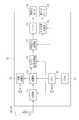
図3において、電子内視鏡14は、CPU50により全体の動作を統括的に制御される。CPU50には、電子内視鏡14の動作を制御するための各種プログラムやデータが記憶されたROM51が接続されている。CPU50は、ROM51から必要なプログラムやデータを読み出し、電子内視鏡14の動作制御を行う。 In FIG. 3, the entire operation of the
LED35には、駆動部52が接続されている。駆動部52は、CPU50の制御の下に、LED35をオン/オフ駆動させる。LED35から発せられた光は、照射レンズ34を介して体腔内の被観察体に照射される。なお、先端部30aではなく操作部31の内部にLED35を配し、ライトガイドで先端部30aに導光する構成としてもよい。 A
CCD33は、対物レンズ32から入射した体腔内の被観察体像の像光を撮像面に結像させ、各画素からこれに応じた撮像信号を出力する。AFE53は、CPU50の制御の下に、CCD33から入力された撮像信号に対して、相関二重サンプリング、増幅、およびA/D変換を施して、撮像信号をデジタルの画像信号に変換する。 The
術者用マイクロホン17aには、音声信号処理部54が接続されている。音声信号処理部54は、術者用マイクロホン17aから入力された術者の音声に対して、A/D変換、ノイズ除去などの各種処理を施し、デジタルの音声信号を出力する。 An audio
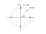
図4に模式的に示すように、変調部55は、AFE53から出力されたデジタルの画像信号に、4値PSK(QPSK)方式を用いてデジタル直交変調を施す(図中黒点で示す)とともに、音声信号処理部54から出力されたデジタルの音声信号を、45°位相をずらして画像信号に重畳し(図中白抜き点で示す)、RF信号を生成する。 As schematically shown in FIG. 4, the
図3に戻って、送信部56は、アンテナ57を介して、変調部55で生成されたRF信号を、電波58として術者用プロセッサ装置15および指導者用プロセッサ装置19に送信する。 Returning to FIG. 3, the
コネクタ59には、バッテリ60が接続されている。バッテリ60の電力は、CPU50により制御される電力供給部61から、電子内視鏡14の各部に供給される。なお、図2には示していないが、操作部31の後部には、バッテリ60を収納するバッテリ収納室が設けられており、コネクタ59はその内部に配されている。また、術者用ヘッドセット17が接続されるコネクタも設けられている。 A
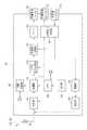
図5において、術者用プロセッサ装置15は、CPU70により全体の動作を統括的に制御される。CPU70には、術者用プロセッサ装置15の動作を制御するための各種プログラムやデータが記憶されたROM71が接続されている。CPU70は、このROM71から必要なプログラムやデータを読み出し、術者用プロセッサ装置15の動作制御を行う。 In FIG. 5, the operation of the
アンテナ72は、電子内視鏡14からの電波58、および指導者用プロセッサ装置19からの電波93(図6参照、詳しくは後述)を受信する。受信部73は、アンテナ72で受信された電波58、93、すなわちRF信号を増幅する。復調部74は、電波58で表されるRF信号に対して、例えばデジタル直交検波を施して、RF信号を電子内視鏡14で変調される前の画像信号に復調する。また、復調部74は、電波93で表されるRF信号を、指導者用プロセッサ装置19で変調される前の音声信号に復調する。 The
同期分離部75は、CPU70の制御の下に、復調部74で復調された画像信号から、振幅分離によって同期信号を分離し、 続いて周波数分離により水平同期信号と垂直同期信号とを分離する。ビデオ信号処理部76は、画像信号からデジタルのビデオ信号を生成する。画像信号処理部77は、ビデオ信号処理部76で生成されたビデオ信号に対して、マスク生成やキャラクタ情報付加などの各種画像処理を施す。バッファ78は、画像信号処理部77で各種画像処理が施され、術者用モニタ16に内視鏡画像として表示されるビデオ信号を一旦格納する。 Under the control of the
音声信号処理部79は、復調部74で復調された指導者の音声信号に対して、ノイズ除去、D/A変換などの各種処理を施し、術者用スピーカー18に出力する。 The audio
図6において、指導者用プロセッサ装置19は、CPU90により全体の動作を統括的に制御される。CPU90には、指導者用プロセッサ装置19の動作を制御するための各種プログラムやデータが記憶されたROM91が接続されている。CPU90は、ROM91から必要なプログラムやデータを読み出し、指導者用プロセッサ装置19の動作制御を行う。 In FIG. 6, the entire operation of the
アンテナ92は、電子内視鏡14からの電波58を受信するとともに、術者用プロセッサ装置15に電波93を送信する。受信部94は、アンテナ92で受信された電波58、すなわちRF信号を増幅する。復調部95は、電波58で表されるRF信号に対して、例えばデジタル直交検波を施して、RF信号を電子内視鏡14で変調される前の画像信号および音声信号に復調する。 The
指導者用マイクロホン21aには、音声信号処理部96が接続されている。音声信号処理部96は、指導者用マイクロホン21aから入力された指導者の音声に対して、A/D変換、ノイズ除去などの各種処理を施し、デジタルの音声信号を出力する。また、音声信号処理部96は、復調部95で復調された術者の音声信号に対して、ノイズ除去、D/A変換などの各種処理を施し、指導者用スピーカー22に出力する。 An audio
図7に模式的に示すように、変調部97は、電子内視鏡14の変調部55で画像信号をあてた箇所にダミーの信号をあてる(図中網目点で示す)とともに、音声信号処理部95から出力されたデジタルの音声信号を、45°位相をずらしてダミーの信号に重畳し(図中白抜き点で示す)、電波93の元となるRF信号を生成する。 As schematically shown in FIG. 7, the
図6に戻って、送信部98は、アンテナ92を介して、変調部97で生成されたRF信号を、電波93として術者用プロセッサ装置15に送信する。なお、同期分離部99〜バッファ102は、術者用プロセッサ装置15の同期分離部75〜バッファ78と同様の機能を有するため、説明を省略する。 Returning to FIG. 6, the
術者用内視鏡診断ユニット11および指導者用内視鏡診断ユニット13は、電波58、93による音声信号の送受信を半二重通信により行う。具体的には、術者用プロセッサ装置15のCPU70は、受信部73で受信されたRF信号が、画像信号および音声信号で構成されていた場合、つまり、術者から指導者への問い合わせが術者用マイクロホン17aに音声入力された場合、指導者用プロセッサ装置19から指導者の音声付きの電波93を受信しても、指導者の音声を術者用スピーカー18から出力しないように、音声信号処理部79の動作を制御する。あるいは、指導者用プロセッサ装置19からの電波93を受け付けないように、受信部73を制御する。 The endoscopic diagnostic unit 11 for surgeons and the endoscopic diagnostic unit 13 for instructors perform transmission / reception of audio signals by
また、指導者用プロセッサ装置19のCPU90は、送信部98から電波93を送信する場合、つまり、指導者から術者への指示が指導者用マイクロホン21aに音声入力された場合、電子内視鏡14から術者の音声付きの電波58を受信しても、術者の音声を指導者用スピーカー22から出力しないように、音声信号処理部96の動作を制御する。 The
上記のように構成された内視鏡診断システム2で体腔内を観察する際には、LED35をオンして、挿入部30を体腔内に挿入し、体腔内を照明しながら、CCD33による内視鏡画像を術者用モニタ16で観察する。When observing the inside of a body cavity with the endoscope
このとき、対物レンズ32から入射した体腔内の被観察体像の像光は、CCD33の撮像面に結像され、CCD33から撮像信号が出力される。CCD33から出力された撮像信号は、AFE53で相関二重サンプリング、増幅、およびA/D変換が施され、デジタルの画像信号に変換される。 At this time, the image light of the observed body image in the body cavity incident from the
AFE53から出力されたデジタルの画像信号は、変調部55でデジタル直交変調が施され、RF信号が生成される。生成されたRF信号は、送信部56で増幅され、アンテナ57から電波58として送信される。 The digital image signal output from the
一方、術者用プロセッサ装置15では、電子内視鏡14のアンテナ57から送信された電波58がアンテナ72で受信されると、この電波58、すなわちRF信号が受信部73で増幅される。復調部74では、受信部73で増幅されたRF信号にデジタル直交検波が施され、電子内視鏡14で変調される前の画像信号が復調される。 On the other hand, when the
復調部74で復調された画像信号は、CPU70の制御の下に、同期分離部75で同期分離が施され、ビデオ信号処理部76でデジタルのビデオ信号として出力される。ビデオ信号処理部76で出力されたビデオ信号は、画像信号処理部77で各種画像処理が施され、バッファ78に一旦格納されて、術者用モニタ16に内視鏡画像として表示される。なお、指導者用プロセッサ装置19においても、術者用プロセッサ装置15と同様に、電子内視鏡14からの電波58から内視鏡画像が生成され、生成された内視鏡画像が指導者用モニタ20に表示される。以上のようにして、電子内視鏡14と術者用プロセッサ装置15、指導者用プロセッサ装置19との間で、電波58により信号が送受信される。 The image signal demodulated by the
続いて、指導者の指示を仰ぐための問い合わせを、術者が術者用マイクロホン17aに音声入力した場合の処理について説明する。術者用マイクロホン17aに入力された術者の音声は、音声信号処理部54で各種処理が施され、デジタルの音声信号に変換される。音声信号処理部54でデジタル化された音声信号は、変調部55でデジタル直交変調が施された画像信号に位相をずらして重畳され、送信部56、アンテナ57を介して、電波58として術者用プロセッサ装置15および指導者用プロセッサ装置19に送信される。 Next, processing when the surgeon inputs a voice to the surgeon's
術者用プロセッサ装置15では、前述の処理により電波58から内視鏡画像が生成される。一方、指導者用プロセッサ装置19では、電波58がアンテナ92で受信されると、受信部94を経由して電波58で表されるRF信号が復調部95に入力され、復調部95で元の画像信号および音声信号に復調される。 In the
復調された画像信号は、術者用プロセッサ装置15の場合と同様の処理を経て、内視鏡画像となり、指導者用モニタ20に表示される。対して、復調された音声信号は、音声信号処理部96で各種処理が施され、指導者用スピーカー22に出力される。これにより、術者は、術者用マイクロホン17aを用いて、指導者に問い合わせをすることができ、指導者は、指導者用スピーカー22を介して、術者による問い合わせを聴取することができる。 The demodulated image signal undergoes the same processing as in the case of the
次に、内視鏡診断への指示を、指導者が指導者用マイクロホン21aに音声入力した場合の処理について説明する。指導者用マイクロホン21aに入力された術者の音声は、音声信号処理部96で各種処理が施され、デジタルの音声信号に変換される。音声信号処理部96でデジタル化された音声信号は、変調部97でデジタル直交変調が施されたダミーの信号に位相をずらして重畳され、送信部98、アンテナ92を介して、電波93として術者用プロセッサ装置15に送信される。 Next, processing when an instruction to the endoscope diagnosis is input by voice to the
術者用プロセッサ装置15では、電波93がアンテナ72で受信されると、受信部73を経由して電波93で表されるRF信号が復調部74に入力され、復調部74で元のダミー信号および音声信号に復調される。 In the
復調された音声信号は、音声信号処理部79で各種処理が施され、術者用スピーカー18に出力される。これにより、指導者は、指導者用マイクロホン21aを用いて、術者に指示を与えることができ、術者は、術者用スピーカー18を介して、指導者による指示を聴取することができる。 The demodulated audio signal is subjected to various processes by the audio
ここで、受信部73で受信されたRF信号が、画像信号および音声信号で構成されていた場合、術者用プロセッサ装置15では、指導者用プロセッサ装置19から指導者の音声付きの電波93を受信しても、指導者の音声が術者用スピーカー18から出力されないように、CPU70により音声信号処理部79の動作が制御される。あるいは、指導者用プロセッサ装置19からの電波93を受け付けないように、受信部73が制御される。 Here, when the RF signal received by the receiving
また、送信部98から電波93を送信する場合、指導者用プロセッサ装置19では、電子内視鏡14から術者の音声付きの電波58を受信しても、術者の音声が指導者用スピーカー22から出力されないように、CPU90により音声信号処理部96の動作が制御される。これにより、術者用内視鏡診断ユニット11と指導者用内視鏡診断ユニット13との間の、電波58、93による音声信号の送受信が半二重通信で行われる。 Further, when the radio wave 93 is transmitted from the
なお、上記実施形態では、術者用内視鏡診断ユニット11が1台の場合を例示して説明したが、図8に示すように、検査室10a〜10dに、術者用内視鏡診断ユニット11a〜11dをそれぞれ設置した内視鏡診断システム110についても、本発明は有効である。 In the above embodiment, the case where there is one endoscope diagnosis unit 11 for the operator has been described as an example. However, as shown in FIG. 8, the endoscope diagnosis for the operator is performed in the
図8において、内視鏡診断ユニット11a〜11dには、ユニット毎に電波58、93の送受信周波数帯域が個別のチャネル(Ch1〜4)で割り振られている。そして、Ch1〜4を選択的に切り替えるための切り替えダイヤル111が設けられた指導者用内視鏡診断ユニット112が指導室12に設置されている。この場合、切り替えダイヤル111で選択されたチャネルの内視鏡画像が指導者用モニタ20に表示され、術者と指導者との間の連絡は、選択されたチャネルのみで可能となる。また、このように複数台の術者用内視鏡診断ユニット11a〜11dを用いる場合、それぞれの術者用内視鏡診断ユニット11a〜11dと指導者用内視鏡診断ユニット112とは、問い合わせ情報および指示情報の送受信を同一のチャネルで位相をずらし、半二重通信で行うことにより、使用チャネルを単一にして送受信が可能となるため、割り当てられたチャネルでより多くの術者用内視鏡診断ユニットを使用可能な内視鏡診断システムを構築することができる。 In FIG. 8, the transmission / reception frequency bands of the
上記実施形態では、音声により術者と指導者との間の連絡を行っているが、キーボードなどの文字入力装置を各ユニット11、13に設け、文字入力装置で入力された文字を各モニタ16、20に表示するように構成してもよい。また、各装置の動作を制御するための設定情報(AFE53や画像信号処理部77の各種処理パラメータなど)を、音声や文字の代わりに送受信してもよい。 In the above embodiment, the operator and the instructor are communicated by voice. However, a character input device such as a keyboard is provided in each unit 11, 13, and characters input by the character input device are displayed on each
2、110 内視鏡診断システム
10 検査室
11 術者用内視鏡診断ユニット
12 指導室
13、112 指導者用内視鏡診断ユニット
14 電子内視鏡
15 術者用プロセッサ装置
16 術者用モニタ
17 術者用ヘッドセット
17a 術者用マイクロホン
18 術者用スピーカー
19 指導者用プロセッサ装置
20 指導者用モニタ
21 指導者用ヘッドセット
21a 指導者用マイクロホン
22 指導者用スピーカー
33 CCD
50 CPU
54 音声信号処理部
55 変調部
56 送信部
58 電波
70 CPU
73 受信部
74 復調部
77 画像信号処理部
79 音声信号処理部
90 CPU
93 電波
94 受信部
95 復調部
96 音声信号処理部
97 変調部
98 送信部
101 画像信号処理部
111 切り替えダイヤル
2,110 Endoscopic diagnosis system 10 Examination room 11 Endoscope diagnosis unit for
50 CPU
54 audio
73
93
Claims (3)
Translated fromJapanese体腔内の被観察体像を撮影して撮像信号を出力する撮像素子、前記撮像信号をデジタル化した画像信号に直交変調を施すとともに、前記術者から入力される前記指導者への問い合わせ情報を、位相をずらして前記画像信号に重畳し、RF信号を生成する変調部、およびRF信号を電波として送信する送信部を有する電子内視鏡と、
電波を受信する受信部、前記RF信号から元の画像信号を復調する復調部、および前記画像信号から内視鏡画像を生成する画像信号処理部を有する術者用プロセッサ装置と、
前記内視鏡画像を表示する術者用モニタと、
前記問い合わせ情報を入力するための術者用情報入力装置と、
前記指導者から入力される前記術者への指示情報を出力する術者用情報出力装置とから構成される術者用内視鏡診断ユニットを前記検査室に設置し、
電波を受信する受信部、前記RF信号から元の画像信号および問い合わせ情報を復調する復調部、前記画像信号から内視鏡画像を生成する画像信号処理部、前記指示情報を変調してRF信号を生成する変調部、およびRF信号を電波として送信する送信部を有する指導者用プロセッサ装置と、
前記内視鏡画像を表示する指導者用モニタと、
前記指示情報を入力するための指導者用情報入力装置と、
前記問い合わせ情報を出力する指導者用情報出力装置とから構成される指導者用内視鏡診断ユニットを前記指導室に設置し、
前記術者用内視鏡診断ユニットおよび前記指導者用内視鏡診断ユニットは、前記問い合わせ情報および前記指示情報の送受信を半二重通信により行うことを特徴とする内視鏡診断システム。In an endoscopic diagnosis system in which an operator in an examination room performs an endoscopic diagnosis in response to an instruction from a leader in a remote instruction room,
An image sensor that captures an image of an object to be observed in a body cavity and outputs an imaging signal, performs orthogonal modulation on an image signal obtained by digitizing the imaging signal, and receives inquiry information from the operator to the instructor An electronic endoscope having a modulation unit that shifts the phase and superimposes the image signal to generate an RF signal, and a transmission unit that transmits the RF signal as a radio wave;
A processor unit for an operator having a receiving unit that receives radio waves, a demodulating unit that demodulates an original image signal from the RF signal, and an image signal processing unit that generates an endoscopic image from the image signal;
A surgeon's monitor for displaying the endoscopic image;
A surgeon information input device for inputting the inquiry information;
An endoscopic diagnostic unit for a surgeon configured with an information output device for a surgeon that outputs instruction information to the surgeon input from the instructor is installed in the examination room,
A receiving unit that receives radio waves, a demodulating unit that demodulates an original image signal and inquiry information from the RF signal, an image signal processing unit that generates an endoscopic image from the image signal, and an RF signal that modulates the instruction information A processor unit for a leader having a modulation unit to generate and a transmission unit for transmitting an RF signal as a radio wave;
An instructor monitor for displaying the endoscopic image;
An information input device for a leader for inputting the instruction information;
An instructor endoscope diagnostic unit composed of an instructor information output device that outputs the inquiry information is installed in the instruction room,
The endoscopic diagnostic system for an operator and the endoscopic diagnostic unit for an instructor perform transmission / reception of the inquiry information and the instruction information by half-duplex communication.
前記指導者用内視鏡診断ユニットに、前記チャネルを選択的に切り替える切り替え操作手段を設けたことを特徴とする請求項1に記載の内視鏡診断システム。The endoscopic diagnostic unit for the operator is installed in each of a plurality of examination rooms, and the transmission / reception frequency band of the radio wave is allocated to each unit by an individual channel,
2. The endoscope diagnosis system according to claim 1, wherein a switching operation means for selectively switching the channel is provided in the instructor endoscope diagnosis unit.
The endoscopy according to claim 1 or 2, wherein the inquiry information and the instruction information are at least one of voice, characters, or setting information for controlling the operation of each device. Mirror diagnostic system.
Priority Applications (3)
| Application Number | Priority Date | Filing Date | Title |
|---|---|---|---|
| JP2005288359AJP2007097653A (en) | 2005-09-30 | 2005-09-30 | Endoscopic diagnosis system |
| US11/528,640US7758496B2 (en) | 2005-09-30 | 2006-09-28 | Diagnostic system using endoscope |
| EP06020529AEP1770568A1 (en) | 2005-09-30 | 2006-09-29 | Diagnostic system using an electronic endoscope |
Applications Claiming Priority (1)
| Application Number | Priority Date | Filing Date | Title |
|---|---|---|---|
| JP2005288359AJP2007097653A (en) | 2005-09-30 | 2005-09-30 | Endoscopic diagnosis system |
Publications (1)
| Publication Number | Publication Date |
|---|---|
| JP2007097653Atrue JP2007097653A (en) | 2007-04-19 |
Family
ID=37533544
Family Applications (1)
| Application Number | Title | Priority Date | Filing Date |
|---|---|---|---|
| JP2005288359APendingJP2007097653A (en) | 2005-09-30 | 2005-09-30 | Endoscopic diagnosis system |
Country Status (3)
| Country | Link |
|---|---|
| US (1) | US7758496B2 (en) |
| EP (1) | EP1770568A1 (en) |
| JP (1) | JP2007097653A (en) |
Cited By (3)
| Publication number | Priority date | Publication date | Assignee | Title |
|---|---|---|---|---|
| WO2010143349A1 (en)* | 2009-06-10 | 2010-12-16 | オリンパス株式会社 | Wireless endoscopic apparatus, receiving device thereof, and receiving method |
| JP2013180079A (en)* | 2012-03-02 | 2013-09-12 | Hoya Corp | Electronic endoscope system |
| JPWO2024090228A1 (en)* | 2022-10-26 | 2024-05-02 |
Families Citing this family (6)
| Publication number | Priority date | Publication date | Assignee | Title |
|---|---|---|---|---|
| JP4880398B2 (en)* | 2006-08-10 | 2012-02-22 | 富士フイルム株式会社 | Endoscopic diagnosis system |
| US11561762B2 (en)* | 2011-08-21 | 2023-01-24 | Asensus Surgical Europe S.A.R.L. | Vocally actuated surgical control system |
| US10866783B2 (en)* | 2011-08-21 | 2020-12-15 | Transenterix Europe S.A.R.L. | Vocally activated surgical control system |
| US10565977B1 (en) | 2018-08-20 | 2020-02-18 | Verb Surgical Inc. | Surgical tool having integrated microphones |
| WO2024181889A1 (en)* | 2023-03-01 | 2024-09-06 | Константин Болеславович ТУМИНАС | Optical sensor for monitoring the physiological state of a person |
| WO2025024751A1 (en)* | 2023-07-27 | 2025-01-30 | Boston Scientific Scimed, Inc. | Medical devices and related systems and methods for reducing signal distortion in image signals |
Citations (5)
| Publication number | Priority date | Publication date | Assignee | Title |
|---|---|---|---|---|
| JPS63233681A (en)* | 1987-03-05 | 1988-09-29 | ジェネラル インストラメント コーポレーション | Method and apparatus for supplying digital audio to the audio carrier of a standard television signal |
| JPH04323991A (en)* | 1991-04-23 | 1992-11-13 | Toshiba Corp | Video signal transmission system |
| JP2004321603A (en)* | 2003-04-25 | 2004-11-18 | Olympus Corp | Device, method and program for image display |
| JP2005111080A (en)* | 2003-10-09 | 2005-04-28 | Olympus Corp | Surgery support system |
| JP2005118232A (en)* | 2003-10-15 | 2005-05-12 | Olympus Corp | Surgery support system |
Family Cites Families (12)
| Publication number | Priority date | Publication date | Assignee | Title |
|---|---|---|---|---|
| JP2000271147A (en) | 1999-03-19 | 2000-10-03 | Olympus Optical Co Ltd | Remote surgery support system |
| US6602185B1 (en)* | 1999-02-18 | 2003-08-05 | Olympus Optical Co., Ltd. | Remote surgery support system |
| JP2002306509A (en)* | 2001-04-10 | 2002-10-22 | Olympus Optical Co Ltd | Remote operation supporting system |
| JP2003010112A (en)* | 2001-06-28 | 2003-01-14 | Olympus Optical Co Ltd | Endoscope system |
| JP2003070804A (en)* | 2001-09-05 | 2003-03-11 | Olympus Optical Co Ltd | Remote medical support system |
| JP2003135371A (en)* | 2001-10-31 | 2003-05-13 | Olympus Optical Co Ltd | Endoscopic system |
| JP2004181229A (en)* | 2002-11-20 | 2004-07-02 | Olympus Corp | System and method for supporting remote operation |
| US7158860B2 (en)* | 2003-02-24 | 2007-01-02 | Intouch Technologies, Inc. | Healthcare tele-robotic system which allows parallel remote station observation |
| US7523145B2 (en)* | 2004-04-22 | 2009-04-21 | Opentv, Inc. | System for managing data in a distributed computing system |
| JP2006000537A (en)* | 2004-06-21 | 2006-01-05 | Olympus Corp | Endoscope system |
| GB2419483B (en)* | 2004-09-17 | 2008-12-24 | Motorola Inc | Demodulator for use in wireless communucations and receiver, method and terminal using it |
| US7222000B2 (en)* | 2005-01-18 | 2007-05-22 | Intouch Technologies, Inc. | Mobile videoconferencing platform with automatic shut-off features |
- 2005
- 2005-09-30JPJP2005288359Apatent/JP2007097653A/enactivePending
- 2006
- 2006-09-28USUS11/528,640patent/US7758496B2/ennot_activeExpired - Fee Related
- 2006-09-29EPEP06020529Apatent/EP1770568A1/ennot_activeCeased
Patent Citations (5)
| Publication number | Priority date | Publication date | Assignee | Title |
|---|---|---|---|---|
| JPS63233681A (en)* | 1987-03-05 | 1988-09-29 | ジェネラル インストラメント コーポレーション | Method and apparatus for supplying digital audio to the audio carrier of a standard television signal |
| JPH04323991A (en)* | 1991-04-23 | 1992-11-13 | Toshiba Corp | Video signal transmission system |
| JP2004321603A (en)* | 2003-04-25 | 2004-11-18 | Olympus Corp | Device, method and program for image display |
| JP2005111080A (en)* | 2003-10-09 | 2005-04-28 | Olympus Corp | Surgery support system |
| JP2005118232A (en)* | 2003-10-15 | 2005-05-12 | Olympus Corp | Surgery support system |
Cited By (5)
| Publication number | Priority date | Publication date | Assignee | Title |
|---|---|---|---|---|
| WO2010143349A1 (en)* | 2009-06-10 | 2010-12-16 | オリンパス株式会社 | Wireless endoscopic apparatus, receiving device thereof, and receiving method |
| JP2013180079A (en)* | 2012-03-02 | 2013-09-12 | Hoya Corp | Electronic endoscope system |
| JPWO2024090228A1 (en)* | 2022-10-26 | 2024-05-02 | ||
| WO2024090228A1 (en)* | 2022-10-26 | 2024-05-02 | 伊知朗 竹政 | Information processing device, information processing system, and information processing program |
| JP7736943B2 (en) | 2022-10-26 | 2025-09-09 | 伊知朗 竹政 | Information processing device, information processing system, and information processing program |
Also Published As
| Publication number | Publication date |
|---|---|
| US7758496B2 (en) | 2010-07-20 |
| US20070078303A1 (en) | 2007-04-05 |
| EP1770568A1 (en) | 2007-04-04 |
Similar Documents
| Publication | Publication Date | Title |
|---|---|---|
| JP4880398B2 (en) | Endoscopic diagnosis system | |
| US20050149001A1 (en) | Operation support system and support method of operation support system | |
| JP4472728B2 (en) | Endoscope system | |
| US8764634B2 (en) | Imaging apparatus | |
| US7485115B2 (en) | Remote operation support system and method | |
| US7758496B2 (en) | Diagnostic system using endoscope | |
| JP2006218129A (en) | Surgery supporting system | |
| JP2006271697A (en) | Electronic endoscope | |
| JP2005169009A (en) | Endoscope system and endoscope | |
| JP4781764B2 (en) | Electronic endoscope system | |
| JP5080164B2 (en) | Endoscope observation device | |
| JP2005118232A (en) | Surgery support system | |
| JP5028002B2 (en) | Electronic endoscope system | |
| JP2016101377A (en) | Endoscope apparatus and endoscopic image transmission method | |
| US8294751B2 (en) | Electronic endoscope system | |
| JP2006271432A (en) | Electronic endoscopic apparatus | |
| JP2006280541A (en) | Electronic endoscope system | |
| JP2009039249A (en) | Endoscope system | |
| JP2000245738A (en) | Remote operation supporting system | |
| JP2005143918A (en) | Remote operation support system | |
| JP4229920B2 (en) | Air supply system | |
| JP2002152314A (en) | Transmitting/receiving method and ultrasound wave diagnosis system | |
| JP2001309884A (en) | Endoscope apparatus and video camera for endoscope | |
| JP2006271433A (en) | Electronic endoscope | |
| WO2023053662A1 (en) | Ultrasonic endoscope system and operating method for ultrasonic endoscope system |
Legal Events
| Date | Code | Title | Description |
|---|---|---|---|
| A621 | Written request for application examination | Free format text:JAPANESE INTERMEDIATE CODE: A621 Effective date:20080514 | |
| A711 | Notification of change in applicant | Free format text:JAPANESE INTERMEDIATE CODE: A711 Effective date:20090828 | |
| A131 | Notification of reasons for refusal | Free format text:JAPANESE INTERMEDIATE CODE: A131 Effective date:20110127 | |
| A977 | Report on retrieval | Free format text:JAPANESE INTERMEDIATE CODE: A971007 Effective date:20110127 | |
| A521 | Request for written amendment filed | Free format text:JAPANESE INTERMEDIATE CODE: A523 Effective date:20110318 | |
| A02 | Decision of refusal | Free format text:JAPANESE INTERMEDIATE CODE: A02 Effective date:20110511 |