JP2006507071A - Magnetic resonance method - Google Patents
Magnetic resonance methodDownload PDFInfo
- Publication number
- JP2006507071A JP2006507071AJP2004554814AJP2004554814AJP2006507071AJP 2006507071 AJP2006507071 AJP 2006507071AJP 2004554814 AJP2004554814 AJP 2004554814AJP 2004554814 AJP2004554814 AJP 2004554814AJP 2006507071 AJP2006507071 AJP 2006507071A
- Authority
- JP
- Japan
- Prior art keywords
- image
- magnetic resonance
- sense
- coil
- intrinsic
- Prior art date
- Legal status (The legal status is an assumption and is not a legal conclusion. Google has not performed a legal analysis and makes no representation as to the accuracy of the status listed.)
- Pending
Links
- 238000001646magnetic resonance methodMethods0.000titleclaims2
- 230000035945sensitivityEffects0.000claimsabstractdescription35
- 238000000034methodMethods0.000claimsabstractdescription29
- 238000003384imaging methodMethods0.000claimsabstractdescription17
- 238000002595magnetic resonance imagingMethods0.000claimsabstractdescription8
- 238000004590computer programMethods0.000claimsdescription5
- 238000001914filtrationMethods0.000claims2
- XLYOFNOQVPJJNP-UHFFFAOYSA-NwaterSubstancesOXLYOFNOQVPJJNP-UHFFFAOYSA-N0.000description6
- 230000005540biological transmissionEffects0.000description4
- 230000008901benefitEffects0.000description3
- 238000005259measurementMethods0.000description3
- 238000012545processingMethods0.000description3
- 210000001015abdomenAnatomy0.000description2
- 238000001514detection methodMethods0.000description2
- 239000011159matrix materialSubstances0.000description2
- 238000004804windingMethods0.000description2
- 230000003321amplificationEffects0.000description1
- 239000004020conductorSubstances0.000description1
- 238000012937correctionMethods0.000description1
- 238000013481data captureMethods0.000description1
- 238000013480data collectionMethods0.000description1
- 230000007423decreaseEffects0.000description1
- 230000001419dependent effectEffects0.000description1
- 238000010586diagramMethods0.000description1
- 230000000694effectsEffects0.000description1
- 238000002474experimental methodMethods0.000description1
- 238000001208nuclear magnetic resonance pulse sequenceMethods0.000description1
- 238000003199nucleic acid amplification methodMethods0.000description1
- 238000004088simulationMethods0.000description1
- 230000006641stabilisationEffects0.000description1
- 238000011105stabilizationMethods0.000description1
- 230000009466transformationEffects0.000description1
Images


Classifications
- G—PHYSICS
- G01—MEASURING; TESTING
- G01R—MEASURING ELECTRIC VARIABLES; MEASURING MAGNETIC VARIABLES
- G01R33/00—Arrangements or instruments for measuring magnetic variables
- G01R33/20—Arrangements or instruments for measuring magnetic variables involving magnetic resonance
- G01R33/44—Arrangements or instruments for measuring magnetic variables involving magnetic resonance using nuclear magnetic resonance [NMR]
- G01R33/48—NMR imaging systems
- G01R33/54—Signal processing systems, e.g. using pulse sequences ; Generation or control of pulse sequences; Operator console
- G01R33/56—Image enhancement or correction, e.g. subtraction or averaging techniques, e.g. improvement of signal-to-noise ratio and resolution
- G01R33/561—Image enhancement or correction, e.g. subtraction or averaging techniques, e.g. improvement of signal-to-noise ratio and resolution by reduction of the scanning time, i.e. fast acquiring systems, e.g. using echo-planar pulse sequences
- G01R33/5611—Parallel magnetic resonance imaging, e.g. sensitivity encoding [SENSE], simultaneous acquisition of spatial harmonics [SMASH], unaliasing by Fourier encoding of the overlaps using the temporal dimension [UNFOLD], k-t-broad-use linear acquisition speed-up technique [k-t-BLAST], k-t-SENSE
Landscapes
- Physics & Mathematics (AREA)
- Health & Medical Sciences (AREA)
- General Health & Medical Sciences (AREA)
- Nuclear Medicine, Radiotherapy & Molecular Imaging (AREA)
- Radiology & Medical Imaging (AREA)
- Engineering & Computer Science (AREA)
- Signal Processing (AREA)
- High Energy & Nuclear Physics (AREA)
- Condensed Matter Physics & Semiconductors (AREA)
- General Physics & Mathematics (AREA)
- Magnetic Resonance Imaging Apparatus (AREA)
Abstract
Translated fromJapaneseDescription
Translated fromJapanese 本発明は、安定した磁場内に配置された対象の撮像の磁気共鳴(MR)方法に関連し、この方法により以下の段階、即ち、
対象の一部内のスピンを励起する段階、
読み出し傾斜磁場及び他の傾斜磁場の印加によりk空間内の複数のラインを含む所定の軌跡に沿ってMR信号を測定する段階、
ナビゲータMR信号の測定のためにナビゲータ傾斜磁場を印加する段階が繰り返され、
前記方法はまた、測定されたMR信号を補正するよう測定されたナビゲータMR信号の位相及び大きさから位相補正を決定する段階と、補正されたMR信号から対象の一部の画像を決定する段階とを更に含む、方法に関連する。The present invention relates to a magnetic resonance (MR) method of imaging an object placed in a stable magnetic field, whereby the method comprises the following steps:
Exciting the spins in a part of the object,
Measuring MR signals along a predetermined trajectory including a plurality of lines in k-space by applying a readout gradient magnetic field and another gradient magnetic field;
The step of applying a navigator gradient magnetic field for navigator MR signal measurement is repeated,
The method also includes determining a phase correction from the phase and magnitude of the measured navigator MR signal to correct the measured MR signal, and determining a partial image of the object from the corrected MR signal. And relates to a method.
本発明はまた、かかる方法を実行するMR装置及びコンピュータプログラムプロダクトに関連する。 The invention also relates to an MR apparatus and a computer program product for performing such a method.
特許文献1には、水平磁場及び垂直磁場の両方のMRIシステムでNMR無線周波信号を受信するのに使用される内在的に減結合されたサンドウィッチ型ソレノイド・アレイ・コイルが記載されている。その最も基本的な構成では、アレイコイルは、2つの同軸RF受信コイルを含む。アレイの第1のコイルは、共通軸に沿って互いから離された2つのソレノイド(又はループ)部分を有する。2つの部分は、直列に電気的に接続されているが、各部分の導体は反対の方向に巻回されており、コイルを通る電流は各部分内に反対の極性の磁場を生成する。コイルアレイの第2のコイルは、第1のコイルの2つの別々のソレノイド部分の間の、組み合わされた反対の磁場がヌルとなるよう打ち消し合う領域に配置される(「挟み込まれている(サンドウィッチ)」)巻回配置及び幾何学的対称性により、アレイの受信コイルは互いから電磁的に「減結合」され、それらの感度をNMR信号を受信することに向けて維持する。多コイル・アレイ配置はまた、画像データ捕捉中の時間の犠牲なしに画像折り返し問題を防止するよう、より大きい視野(FOV)とより小さい視野(FOV)との間で選択を行うことを可能とする。また、等しくない構成要素コイル直径、等しくない構成要素コイル巻線、同軸でないコイル形態を含む多の実施例が開示される。 U.S. Pat. No. 6,057,051 describes an intrinsically decoupled sandwich solenoid array coil used to receive NMR radio frequency signals in both horizontal and vertical magnetic field MRI systems. In its most basic configuration, the array coil includes two coaxial RF receive coils. The first coil of the array has two solenoid (or loop) portions spaced from each other along a common axis. The two parts are electrically connected in series, but the conductors in each part are wound in opposite directions, and the current through the coil produces a magnetic field of opposite polarity in each part. The second coil of the coil array is located in the area between the two separate solenoid parts of the first coil where the combined opposite magnetic field cancels out ("sandwiched" (sandwich ) ") Due to the winding arrangement and geometric symmetry, the receive coils of the array are electromagnetically" decoupled "from each other, maintaining their sensitivity towards receiving NMR signals. The multi-coil array arrangement also allows selection between a larger field of view (FOV) and a smaller field of view (FOV) to prevent image folding problems without sacrificing time during image data capture. To do. A number of embodiments are also disclosed, including unequal component coil diameters, unequal component coil windings, and non-coaxial coil configurations.
この参考文献のコイル・アレイ配置では、FOVは、画像が小さい関心領域又は体積から得られる場合に折り返しアーティファクトを克服するために、アレイの幾つかのコイルからNMR信号を組み合わせることによって大きく、又は、単一のコイルのNMR信号のみを選択することによって小さく選択されうる。従って、FOVは、撮像対象のサイズに依存して選択されうる。 In the coil array arrangement of this reference, the FOV is increased by combining NMR signals from several coils of the array to overcome aliasing artifacts when the image is obtained from a small region of interest or volume, or Small selections can be made by selecting only a single coil NMR signal. Therefore, the FOV can be selected depending on the size of the imaging target.
更に、特許文献2では、位相エンコード及び読み出し方向の傾斜磁場が、患者の関心領域中で励起されたMR活性核を空間エンコードするために印加される磁気共鳴撮像方法が開示されている。読み出し方向に減少された数の読み出しがとられ、それによりエイリアシングのある減少された視野の画像が作成される。データの収集の時間が減少されるという利点とともに、完全な画像を生成するようエイリアシングされた画像を展開するために、これらのコイルに関する感度情報とともに、少なくとも2つのRF受信コイルが用いられる。感度情報は、画像情報が収集されるよりも低い解像度で収集される。コイルの感度を較正するのに用いられる参照データ中のより低い解像度の効果は、参照データ中のノイズを減少させることであり、従って目標となる展開されたSENSEデータの信号対雑音比は高められる。 Further,
FOVが撮像された対象よりも小さい場合、内在的な折り返しアーティファクトが生ずる。内在的な折り返しアーティファクトは、関心となる領域、即ち心臓、が対象のスライスよりも遙かに小さい心臓撮像に、又は、腕が折り曲げられる、腹部の撮像に、及び、大きいFOVの変形されたエッジが使用されない全身MR撮像に使用される。SENSE又はSMASHのような並列撮像法では、内在的な折り返しアーティファクトはコイル感度行列を決定させないため、位相エンコード方向上、対象の寸法よりも小さい視野を選択することは許されない。SENSEが用いられる場合、操作者は対象全体を囲む大きい視野を選択することが強制され、これはSENSE方法によって与えられる時間の減少を部分的に無駄なものとする。
本発明は、SENSE又はSMASHといった並列撮像技術の効率を更に高めることを目的とする。 The present invention aims to further increase the efficiency of parallel imaging techniques such as SENSE or SMASH.
本発明のこの目的は、請求項1に記載の方法によって達成される。本発明は更に、請求項4に記載の装置、並びに、請求項5に記載のコンピュータ・プログラム・プロダクトに関連する。 This object of the invention is achieved by a method according to claim 1. The invention further relates to an apparatus according to claim 4 and a computer program product according to claim 5.
本発明は、減少されたFOVが選択されるという主な利点を有する。内在的な折り返しアーティファクトが発生される結果として、参照画像の計算によって解決されうる。 The present invention has the main advantage that a reduced FOV is selected. As a result of the generation of intrinsic aliasing artifacts, it can be solved by calculating the reference image.
本発明の上述の及び他の利点は、従属項、及び、添付の図面を参照して本発明の典型的な実施例が記載されている以下の説明に開示されている。 The above and other advantages of the present invention are disclosed in the dependent claims and the following description in which exemplary embodiments of the invention are described with reference to the accompanying drawings.
磁気共鳴撮像法では、より短い期間内に許容可能な画像を得るという一般的な傾向がある。このために、近年、スイス連邦工科大学チューリッヒ校、生体工学及び医療情報学研究所(Institute of Biomedical Engineering and Medical Informatics)により、「SENSE」と称される感度エンコード法が開発された。SENSE法は、磁気共鳴装置のコイルによって検出されるように画像に対して直接作用するアルゴリズムに基づくものである。画像に対する位相エンコード・ステップの数は、R倍だけ減少され、その倍数だけ信号捕捉を加速させ、Rは1よりも大きい任意の数でありうる。即ち、(位相)エンコード・ステップの数は、完全な集合のエンコード・ステップに対して減少される。この完全な集合は、再構成されるMR画像の予め選択された空間解像度に対して十分なk空間内のMR信号をサンプリングするのに必要なエンコード・ステップを生じさせる。結果として得られる、多数のコイルからのエイリアシングされた画像は、単一のR回折り返された画像を発生するようSENSEアルゴリズムによって用いられる。SENSE方法に重要なものは、いわゆる感度マップとして配置されたコイルの感度の知識である。この方法を加速させるために、シングルコイル参照の「平方和」による、又は、ボディコイル参照による分割を通じて取得されうる生感度マップを使用することが提案される(例えば、K.Pruessmann外、ISMRM議事録、1998年、アブストラクト第579頁、799頁、803頁及び2087頁参照)。実際、SENSE法は、意図的にk空間をアンダーサンプリングすることにより、即ち捕捉すべき対象よりも小さい視野(FOV)を意図的に選択することにより、走査時間を減少させることを可能とする。このアンダーサンプリングから、異なったコイル感度パターンを有する一組の別個のコイルについての知識を用いて分解又は展開される、折り返しアーティファクトが得られる。アンダーサンプリングは、両方の位相エンコード方向のうちのいずれかでありうる。 Magnetic resonance imaging has a general tendency to obtain acceptable images within a shorter period of time. To this end, a sensitivity encoding method called “SENSE” has recently been developed by the Swiss Federal Institute of Technology, Zurich, Institute of Biomedical Engineering and Medical Informatics. The SENSE method is based on an algorithm that operates directly on the image as detected by the coils of the magnetic resonance apparatus. The number of phase encoding steps for an image is reduced by a factor of R, accelerating signal acquisition by that factor, and R can be any number greater than one. That is, the number of (phase) encoding steps is reduced relative to the complete set of encoding steps. This complete set yields the encoding steps necessary to sample MR signals in k-space sufficient for the preselected spatial resolution of the reconstructed MR image. The resulting aliased images from multiple coils are used by the SENSE algorithm to generate a single R-folded image. What is important for the SENSE method is knowledge of the sensitivity of the coils arranged as a so-called sensitivity map. In order to accelerate this method, it is proposed to use a raw sensitivity map that can be obtained by splitting with a single coil reference “sum of squares” or with a body coil reference (eg, K. Pruessmann et al., ISMRM proceedings. Vol., 1998, abstract pages 579, 799, 803 and 2087). In fact, the SENSE method makes it possible to reduce the scanning time by intentionally undersampling the k-space, ie by intentionally selecting a field of view (FOV) that is smaller than the object to be captured. From this undersampling, folding artifacts are obtained that are decomposed or expanded using knowledge of a set of separate coils having different coil sensitivity patterns. Undersampling can be in either of the phase encoding directions.
NMR撮像法では、内在的な折り返しアーティファクトの方法が用いられ、例えば、関心領域、即ち心臓が対象のスライスよりも遙かに小さい心臓撮像に、又は、腕が折り曲げられる、腹部の撮像に、及び、大きいFOVの変形されたエッジが使用されない全身MR撮像に使用される。SENSE又はSMASHのような並列撮像法では、内在的な折り返しアーティファクトはコイル感度行列を決定させないため、位相エンコード方向上、対象の寸法よりも小さい視野を選択することは許されない。SENSEが用いられる場合、操作者は対象全体を囲む大きい視野を選択することが強制され、これはSENSE方法によって与えられる時間の減少を部分的に無駄なものとする。この制限は、SENSEのような並列撮像法に用いられるときに数学に基づいて克服されることが不可能であると考えられる。 In NMR imaging, the method of intrinsic folding artifacts is used, for example, for imaging a region of interest, i.e. for heart imaging where the heart is much smaller than the slice of interest, or for imaging the abdomen where the arm is folded, and Used for whole body MR imaging, where large FOV deformed edges are not used. In parallel imaging methods such as SENSE or SMASH, the intrinsic aliasing artifact does not determine the coil sensitivity matrix, so it is not allowed to select a field of view smaller than the target dimension in the phase encoding direction. When SENSE is used, the operator is forced to select a large field of view that surrounds the entire object, which partially wastes the time reduction provided by the SENSE method. This limitation is considered impossible to overcome based on mathematics when used in parallel imaging methods such as SENSE.
現在のところ、SENSE測定は、基本的には以下のように与えられる。
1.患者の解剖学的細部なしに、SENSE法で用いられるコイルの各素子についての低解像度画像を得るための予備走査が行われる。
2.全ての素子のエイリアシングされた画像を生じさせるSENSE走査が行われる。
3.コイルの感度プロファイル及びSENSE走査画像は、実際の画像を再構成するようSENSEアルゴリズムによって使用される。ボディコイル参照スキャンもまた、規則かのために用いられる。通常は、特定のMR検査では全てのSENSE走査のために一回の予備走査で十分である。At present, the SENSE measurement is basically given as follows.
1. A pre-scan is performed to obtain a low resolution image for each element of the coil used in the SENSE method without the anatomical details of the patient.
2. A SENSE scan is performed that produces an aliased image of all elements.
3. The coil sensitivity profile and the SENSE scan image are used by the SENSE algorithm to reconstruct the actual image. A body coil reference scan is also used for rules. Normally, a single prescan is sufficient for all SENSE scans for a particular MR examination.
内在的な折り返しアーティファクトを可能とするために、ステップ2の後に1つの付加的なステップが追加されるべきである。 One additional step should be added after
2b.予備走査の画像から、内在的な折り返しを示すコイル素子の感度プロファイル及び内在的な折り返しを示す参照画像が計算される。この走査は、再構成処理の一部として、又は走査中に、数分の一秒でなされうる。続いて、これらの明示的に折り返された画像は、SENSEアルゴリズムで使用されうる。 2b. From the pre-scan image, a sensitivity profile of the coil element showing the intrinsic fold and a reference image showing the intrinsic fold are calculated. This scan can be done in a fraction of a second as part of the reconstruction process or during the scan. Subsequently, these explicitly folded images can be used in the SENSE algorithm.
この方法では、約30%の時間短縮を得ることができる。例として、幅40cmの対象を用いる。1mmの解像度では400のエンコード・ステップが必要である。関心領域がわずか20cmであり、内在的な折り返しアーティファクトが許されると、30cmの視野が選定されえ、300のエンコード・ステップが必要である。3倍の縮小率で必ず40cmの視野のSENSEのみを用いるとき、133のエンコードステップのみが必要である。更に、SENSE法で内在的な折り返しアーティファクトが許され、30cmの視野が用いられるとき、わずか100のエンコード・ステップが必要である。関心領域内のノイズは、3倍の減少の通常のSENSE法によって形成される画像のノイズに等しい。しかしながら、内在的な折り返しアーティファクトを有する提示される方法は、30%高速である。 With this method, a time reduction of about 30% can be obtained. As an example, a 40 cm wide object is used. For a 1 mm resolution, 400 encoding steps are required. If the region of interest is only 20 cm and intrinsic folding artifacts are allowed, a 30 cm field of view can be selected and 300 encoding steps are required. When using only SENSE with a 3x reduction ratio and a 40 cm field of view, only 133 encoding steps are required. Furthermore, when the SENSE method allows for intrinsic folding artifacts and only a 30 cm field of view is used, only 100 encoding steps are required. The noise in the region of interest is equal to the noise of the image formed by the normal SENSE method with a 3x reduction. However, the presented method with intrinsic aliasing artifacts is 30% faster.
SENSEが導入されたとき、SENSE法の数学的な原理に基づき、内在的な折り返しアーティファクトを有するいかなる組合せも不可能であると考えられた。しかしながら、実験により、実際は、SENSEの数学が依然として成り立つが、上述の方法は非常にうまくいくことが示されている。以下の例について考える。視野FOVは、3/4*対象(mFOV=0.75&対象)であると定義され、3倍のSENSE減少係数が、mFOVの減少された視野に適用され、即ち、SENSE折り返し距離Δx=1/3*mFOV=1/4*対象である。ここで、全てのコイル素子iのエイリアシングされた画像の中の画素mは、対象内の4つの位置から信号を導出し、SENSE減少係数3に内在的な折り返しアーティファクトを加えると、 When SENSE was introduced, it was considered impossible based on the mathematical principle of the SENSE method to have any combination with an intrinsic folding artifact. However, experiments have shown that, in fact, the SENSE mathematics still holds, but the above method works very well. Consider the following example. The field of view FOV is defined to be 3/4 * subject (mFOV = 0.75 & subject), and a 3 times SENSE reduction factor is applied to the field of view with reduced mFOV, ie, SENSE folding distance Δx = 1/3 * mFOV = 1/4 * Here, pixel m in the aliased image of all coil elements i derives a signal from four positions in the object and adds an intrinsic aliasing artifact to SENSE

上述のようなフォーカスされたSENSEの近似のために、8つの感度マップを得るために8つの素子のヘッドコイルを有するMRI装置が使用された。感度マップの解像度は、SENSE画像の解像度に等しい。参照として、16cm直径の水で充填されたファントムと、14×16cm2のFOVが用いられ、それにより画像中に内在的な折り返しアーティファクトがある。感度マップは、より大きな体積に亘って測定された。より小さいmFOVを有するSENSE再構成は、感度マップが上述のステップ2bのように人工的に後ろに折り返されているとき、図1aに示すようなアーティファクトを示す。14×16cm2FOV中の1つの素子の結果として得られる(絶対値)画像を、図1bに示す。素子は、右上に配置されている。後ろに折り返された感度マップが入力として用いられると、SENSE再構成は細かく作用する。再構成の後、図1cに示すように、内在的な折り返しアーティファクトのみが残される。通常は、感度マップの解像度は、実際のSENSE画像の解像度よりも小さいよう選択される。図1dに示すように、後ろへ折り返された鮮鋭なエッジは、これらのアーティファクトを生じさせうる。しかしながら、多くの場合、後ろへ折り返されたエッジにおける感度は低く(例えば心臓画像中)、アーティファクトははっきりしたものではなく、従って無視されうる。Due to the focused SENSE approximation as described above, an MRI apparatus with an 8 element head coil was used to obtain 8 sensitivity maps. The resolution of the sensitivity map is equal to the resolution of the SENSE image. As a reference, a phantom filled with 16 cm diameter water and a 14 × 16 cm2 FOV are used, so that there are intrinsic aliasing artifacts in the image. The sensitivity map was measured over a larger volume. A SENSE reconstruction with a smaller mFOV will show artifacts as shown in FIG. 1a when the sensitivity map is artificially folded back as in step 2b above. The resulting (absolute value) image of one element in a 14 × 16 cm2 FOV is shown in FIG. The element is arranged on the upper right. SENSE reconstruction works finely when a sensitivity map folded back is used as input. After reconstruction, only the intrinsic wrap artifact is left, as shown in FIG. 1c. Usually, the resolution of the sensitivity map is selected to be smaller than the resolution of the actual SENSE image. As shown in FIG. 1d, sharp edges that are folded back can cause these artifacts. In many cases, however, the sensitivity at the folded back edge is low (eg in the heart image) and the artifacts are not obvious and can therefore be ignored.
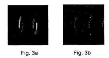
図2乃至図4は、ファントムが左から右(LR)方向に3倍のSENSE係数で測定される画像を示す。視野は、ファントムよりも小さいよう選定され、内在的な折り返しアーティファクトを生じさせる。SENSEを用いて展開するとき、感度推定値は、左側の一組の画像からわかるように重大なアーティファクトを生じさせる内在的な折り返しにより、誤っている。図2a中、等距離の水柱を伴うファントムが用いられ、図3a中、図2aと同じファントムからの、更に大きい水柱をファントムが側方にあり、図4a中、均質に充填された水のファントムが示される。これらの画像は、操作者があまりにも小さいFOVを選択したときに通常は何が生ずるかを示す。同じファントムの右側の一組の画像は、内在的な折り返しアルゴリズムを含むSENSEと共にとられる。内在的な折り返しアーティファクトは、対象のエッジの内在的な折り返しを除き消滅する。この急速な可能な例では、参照スキャンは、図1dのシミュレーションに示すのと同じリンギング・アーティファクトを生じさせる実際のSENSEスキャンよりも低い解像度でとられたものである。両方のスキャンの解像度及びリンギングフィルタの適用のよりよい一致は、画像を改善させる。この例では、エッジにおける全体感度は、この特定のコイルでは対象の中心の全体感度よりもはるかに大きく、強いエッジアーティファクトを生じさせる。意図されたコイル、即ちSENSE心臓コイル、については、エッジアーティファクトは、位相エンコード方向が前頭面のいずれかの場所に選定されると、前頭面内のエッジにおける感度が中心よりも低くなるため、かなり減少される。従って、図2b、図3b、及び図4b中の画像は正しく展開され、はっきりとした関心領域と、側に幾つかの折り返しアーティファクトを残す。 2 to 4 show images in which the phantom is measured from the left to the right (LR) direction with a triple SENSE coefficient. The field of view is chosen to be smaller than the phantom, creating an intrinsic aliasing artifact. When deploying with SENSE, the sensitivity estimate is incorrect due to an intrinsic wrap that causes significant artifacts as can be seen from the set of images on the left. In FIG. 2a, a phantom with equidistant water columns is used, in FIG. 3a, from the same phantom as in FIG. 2a, the larger water column is on the side, and in FIG. Is shown. These images show what normally happens when the operator selects a too small FOV. A set of images on the right side of the same phantom is taken with SENSE containing an intrinsic folding algorithm. Intrinsic aliasing artifacts disappear except for intrinsic aliasing of the subject edge. In this rapid possible example, the reference scan was taken at a lower resolution than the actual SENSE scan that produced the same ringing artifacts as shown in the simulation of FIG. 1d. A better match between the resolution of both scans and the application of the ringing filter improves the image. In this example, the overall sensitivity at the edge is much greater for this particular coil than the overall sensitivity at the center of the object, resulting in a strong edge artifact. For the intended coil, ie, the SENSE heart coil, the edge artifact is significant because the sensitivity at the edge in the frontal plane is lower than the center when the phase encoding direction is chosen anywhere on the frontal plane. Will be reduced. Thus, the images in FIGS. 2b, 3b, and 4b are correctly developed, leaving a clear region of interest and some folding artifacts on the side.
MR装置の実際的な実施例について図5に示し、図5は安定した磁場を発生する第1の磁石系2と、傾斜磁場コイル3として知られる手段であるX,Y,Z方向に傾斜を有する追加的な磁場を発生する手段とを含む。しかしながら、コイル3は、上述のように非常に非線形であり、磁場のパターン即ち「傾斜磁場」は、通常のMRシステムのようにX,Y,Zのうちの1つのみに向けられるのではない。図示の座標系のZ方向は、慣習通り磁石系2内の安定した磁場の方向に対応し、これは線形で線形でありさえすればよい。使用されるべき測定座標系x,y,zは、図2に示すX,Y,Z系と独立に選定されうる。傾斜磁場コイル3は、電源装置4によって給電される。RF送信コイル5はRF磁場を発生するのに役立ち、RF送信及び変調器6に接続される。受信コイルは、例えば人間又は動物の体である、検査されるべき対象7中にRF磁場を発生するよう磁気共鳴信号を受信するのに用いられる。このコイル5は、RF送信コイル5と同じコイルであってもよく、又は、多数の受信アンテナの配列(図示せず)であってもよい。コイル5は、非フェーズド・アレイ受信アンテナであり、これは多数の受信アンテナの配列とは異なる。更に、磁石系2は、検査されるべき体7の一部を収容するのに十分に大きい検査空間を囲む。RFコイル5は、この検査空間内で検査されるべき体7の一部の周り又はその上に配置される。RF送信コイル5は、送信/受信回路9を介して信号増幅及び復調ユニット10に接続される。制御ユニット11は、RFパルス及び傾斜磁場を含む特別なパルスシーケンスを発生するよう、RF送信及び変調器6と電源ユニット4とを制御する。制御ユニット11は、復調ユニット10から得られる位相及び振幅が処理ユニット12に印加されるMR信号の検出を制御する。制御ユニット11と、夫々の受信コイル3及び5は、下位繰り返し時間基準(一般的には、10ミリ秒未満)で検出経路間での切り換えを可能とする制御手段を具備する。これらの手段は、特に、アンテナの信頼性の高い位相動作を確実とする電流/電圧安定化ユニット、及び、コイルと処理ユニット12の間の信号経路中の1つ又はそれ以上のスイッチ及びアナログ・ディジタル変換器を含むことを意味する。処理ユニット12は、変換により画像を形成するよう提示された信号値を処理する。この画像は、例えばモニタ13により視覚化されうる。 FIG. 5 shows a practical embodiment of the MR apparatus. FIG. 5 shows a
Claims (5)
Translated fromJapanese前記視野は、エンコード方向上、対象の寸法よりも小さく設定され、前記受信アンテナの感度マップ及び内在折り返しアーティファクトを示す参照画像は前記MR画像を展開された画像へ再構成するのに用いられることを特徴とする、磁気共鳴撮像方法。Magnetic resonance, forming an image of interest from multiple signals captured by a receive antenna, given a sensitivity map for each receive antenna, and reducing the number of encoding steps applied to its complete set An imaging method,
The field of view is set to be smaller than the target dimension in the encoding direction, and the sensitivity map of the receiving antenna and the reference image showing the intrinsic folding artifact are used to reconstruct the MR image into a developed image. A magnetic resonance imaging method.
前記対象の一部内でスピンを励起する手段と、
複数の受信アンテナと、
読み出し傾斜磁場及び他の傾斜磁場の印加によりk空間内に複数のラインを含む所定の軌跡に沿ってMR信号を測定し、前記位相エンコードステップの数はその完全な集合に対して減少される、手段と、
各受信アンテナの夫々に対して感度マップを与える手段と、
前記視野を前記対象寸法よりも小さく設定する手段と、
前記受信アンテナの感度マップ及び内在折り返しアーティファクトを示す参照画像を用いることにより、前記MR画像を前記測定されたMR信号から折り返された画像へ再構成する手段とを有する、磁気共鳴撮像装置。A magnetic resonance apparatus for acquiring MR images from a plurality of signals,
Means for exciting spins within a portion of the object;
Multiple receive antennas;
MR signals are measured along a predetermined trajectory including a plurality of lines in k-space by applying a readout gradient and other gradient fields, and the number of phase encoding steps is reduced with respect to the complete set; Means,
Means for providing a sensitivity map for each of the receiving antennas;
Means for setting the field of view smaller than the target dimension;
A magnetic resonance imaging apparatus comprising: means for reconstructing the MR image from the measured MR signal into a folded image by using a reference image indicating a sensitivity map of the receiving antenna and an intrinsic folding artifact.
前記コンピュータに、
磁気共鳴撮像装置が複数の信号からMR画像を取得することの実行を制御させ、前記コンピュータプログラムは、
前記対象よりも小さい視野を設定し、
前記受信アンテナの感度マップ及び内在折り返しアーティファクトを示す参照画像を用いて前記MR画像を展開された画像へ再構成する
ための命令を有する、
コンピュータ読み取り可能なプログラム手段を有する、コンピュータプログラムプロダクト。A computer program product stored on a computer usable medium to form an image by a magnetic resonance method,
In the computer,
The magnetic resonance imaging apparatus controls execution of acquiring MR images from a plurality of signals, and the computer program includes:
Set a field of view smaller than the subject,
Instructions for reconstructing the MR image into a developed image using a reference image indicating a sensitivity map of the receiving antenna and an intrinsic folding artifact;
A computer program product comprising computer readable program means.
Applications Claiming Priority (2)
| Application Number | Priority Date | Filing Date | Title |
|---|---|---|---|
| EP02079908 | 2002-11-26 | ||
| PCT/IB2003/005290WO2004048992A1 (en) | 2002-11-26 | 2003-11-20 | Magnetic resonance method |
Publications (2)
| Publication Number | Publication Date |
|---|---|
| JP2006507071Atrue JP2006507071A (en) | 2006-03-02 |
| JP2006507071A5 JP2006507071A5 (en) | 2007-01-25 |
Family
ID=32338104
Family Applications (1)
| Application Number | Title | Priority Date | Filing Date |
|---|---|---|---|
| JP2004554814APendingJP2006507071A (en) | 2002-11-26 | 2003-11-20 | Magnetic resonance method |
Country Status (5)
| Country | Link |
|---|---|
| US (1) | US20060058629A1 (en) |
| EP (1) | EP1567881A1 (en) |
| JP (1) | JP2006507071A (en) |
| AU (1) | AU2003276621A1 (en) |
| WO (1) | WO2004048992A1 (en) |
Cited By (1)
| Publication number | Priority date | Publication date | Assignee | Title |
|---|---|---|---|---|
| JP2012521253A (en)* | 2009-03-25 | 2012-09-13 | コーニンクレッカ フィリップス エレクトロニクス エヌ ヴィ | MR imaging using parallel signal acquisition |
Families Citing this family (15)
| Publication number | Priority date | Publication date | Assignee | Title |
|---|---|---|---|---|
| JP4443079B2 (en)* | 2001-09-13 | 2010-03-31 | 株式会社日立メディコ | Magnetic resonance imaging apparatus and RF receiving coil for magnetic resonance imaging apparatus |
| US8659297B2 (en)* | 2012-02-27 | 2014-02-25 | Perinatronics Medical Systems, Inc. | Reducing noise in magnetic resonance imaging using conductive loops |
| US9780756B2 (en) | 2013-08-01 | 2017-10-03 | Qorvo Us, Inc. | Calibration for a tunable RF filter structure |
| US9899133B2 (en) | 2013-08-01 | 2018-02-20 | Qorvo Us, Inc. | Advanced 3D inductor structures with confined magnetic field |
| US9705478B2 (en) | 2013-08-01 | 2017-07-11 | Qorvo Us, Inc. | Weakly coupled tunable RF receiver architecture |
| US9774311B2 (en) | 2013-03-15 | 2017-09-26 | Qorvo Us, Inc. | Filtering characteristic adjustments of weakly coupled tunable RF filters |
| US9391565B2 (en) | 2013-03-15 | 2016-07-12 | TriQuint International PTE, Ltd. | Amplifier phase distortion correction based on amplitude distortion measurement |
| US9825656B2 (en) | 2013-08-01 | 2017-11-21 | Qorvo Us, Inc. | Weakly coupled tunable RF transmitter architecture |
| US12224096B2 (en) | 2013-03-15 | 2025-02-11 | Qorvo Us, Inc. | Advanced 3D inductor structures with confined magnetic field |
| US9866197B2 (en) | 2013-06-06 | 2018-01-09 | Qorvo Us, Inc. | Tunable RF filter based RF communications system |
| US9859863B2 (en) | 2013-03-15 | 2018-01-02 | Qorvo Us, Inc. | RF filter structure for antenna diversity and beam forming |
| US9871499B2 (en) | 2013-03-15 | 2018-01-16 | Qorvo Us, Inc. | Multi-band impedance tuners using weakly-coupled LC resonators |
| US9966981B2 (en) | 2013-06-06 | 2018-05-08 | Qorvo Us, Inc. | Passive acoustic resonator based RF receiver |
| US10796835B2 (en) | 2015-08-24 | 2020-10-06 | Qorvo Us, Inc. | Stacked laminate inductors for high module volume utilization and performance-cost-size-processing-time tradeoff |
| US11139238B2 (en) | 2016-12-07 | 2021-10-05 | Qorvo Us, Inc. | High Q factor inductor structure |
Family Cites Families (9)
| Publication number | Priority date | Publication date | Assignee | Title |
|---|---|---|---|---|
| DE19835451B4 (en)* | 1997-08-20 | 2005-03-24 | Siemens Ag | Method for a computer tomograph for post-processing a sectional image and computer tomograph operating according to this method |
| WO2000072034A1 (en)* | 1999-05-20 | 2000-11-30 | Koninklijke Philips Electronics N.V. | Magnetic resonance imaging method with sub-sampling |
| US6493572B1 (en)* | 1999-09-30 | 2002-12-10 | Toshiba America Mri, Inc. | Inherently de-coupled sandwiched solenoidal array coil |
| GB9926918D0 (en)* | 1999-11-15 | 2000-01-12 | Marconi Electronic Syst Ltd | Magnetic resonance imaging |
| GB9926923D0 (en)* | 1999-11-15 | 2000-01-12 | Marconi Electronic Syst Ltd | Magnetic resonance imaging |
| US6611143B2 (en)* | 2000-03-14 | 2003-08-26 | Kabushiki Kaisha Toshiba | MRI apparatus generating odd and/or even echo images with sensitivity distribution of coils |
| DE10015068C2 (en)* | 2000-03-25 | 2002-06-27 | Bruker Medical Gmbh | Method of generating magnetic resonance images |
| JP5085832B2 (en)* | 2000-07-31 | 2012-11-28 | コーニンクレッカ フィリップス エレクトロニクス エヌ ヴィ | Magnetic resonance imaging method using subsampled capture |
| GB0121744D0 (en)* | 2001-04-20 | 2001-10-31 | Marconi Medical Systems Uk Ltd | Magnetic resonance imaging |
- 2003
- 2003-11-20JPJP2004554814Apatent/JP2006507071A/enactivePending
- 2003-11-20USUS10/536,285patent/US20060058629A1/ennot_activeAbandoned
- 2003-11-20WOPCT/IB2003/005290patent/WO2004048992A1/enactiveApplication Filing
- 2003-11-20AUAU2003276621Apatent/AU2003276621A1/ennot_activeAbandoned
- 2003-11-20EPEP03811834Apatent/EP1567881A1/ennot_activeWithdrawn
Cited By (1)
| Publication number | Priority date | Publication date | Assignee | Title |
|---|---|---|---|---|
| JP2012521253A (en)* | 2009-03-25 | 2012-09-13 | コーニンクレッカ フィリップス エレクトロニクス エヌ ヴィ | MR imaging using parallel signal acquisition |
Also Published As
| Publication number | Publication date |
|---|---|
| EP1567881A1 (en) | 2005-08-31 |
| WO2004048992A1 (en) | 2004-06-10 |
| AU2003276621A1 (en) | 2004-06-18 |
| US20060058629A1 (en) | 2006-03-16 |
Similar Documents
| Publication | Publication Date | Title |
|---|---|---|
| JP6513398B2 (en) | MR image reconstruction using prior information constrained regularization | |
| JP6554729B2 (en) | Reduced field magnetic resonance imaging system and method | |
| US6448771B1 (en) | Magnetic resonance method for forming a fast dynamic image | |
| JP6333293B2 (en) | Metal resistant MR imaging | |
| JP4152381B2 (en) | Magnetic resonance imaging system | |
| EP1371327A1 (en) | Magnetic resonance imaging apparatus and method | |
| US9223001B2 (en) | MR imaging using navigators | |
| US7319324B2 (en) | MRI method and apparatus using PPA image reconstruction | |
| CN101529268B (en) | MRI RF encoding using multiple transmit coils | |
| US7498809B2 (en) | Magnetic resonance imaging device with multiple RF coils applying half-pulse waveforms for selective excitation of a local region | |
| JP2006507071A (en) | Magnetic resonance method | |
| JP4072879B2 (en) | Nuclear magnetic resonance imaging system | |
| US9170314B2 (en) | Method and magnetic resonance apparatus to acquire MR data in a predetermined three-dimensional volume segment of a subject | |
| JP4325791B2 (en) | Storage medium encoded with computer program code for image reconstruction and apparatus for image reconstruction | |
| JP4202855B2 (en) | Magnetic resonance imaging system | |
| JP4679158B2 (en) | Magnetic resonance imaging system | |
| US12078696B2 (en) | Silent calibration for magnetic resonance imaging | |
| US6919722B2 (en) | Image quality improvement for SENSE with low signal regions | |
| JP4283115B2 (en) | Diffusion-weighted parallel imaging method with phase correction based on navigator signal | |
| WO2007004123A2 (en) | Mri-navigator sequence with restored magnetization | |
| JP2008099717A (en) | Magnetic resonance imaging system | |
| JP4912156B2 (en) | Magnetic resonance imaging method and apparatus | |
| US11519990B1 (en) | Magnetic resonance imaging system and method |
Legal Events
| Date | Code | Title | Description |
|---|---|---|---|
| A621 | Written request for application examination | Free format text:JAPANESE INTERMEDIATE CODE: A621 Effective date:20061117 | |
| A521 | Request for written amendment filed | Free format text:JAPANESE INTERMEDIATE CODE: A523 Effective date:20061120 | |
| A131 | Notification of reasons for refusal | Free format text:JAPANESE INTERMEDIATE CODE: A131 Effective date:20090804 | |
| A02 | Decision of refusal | Free format text:JAPANESE INTERMEDIATE CODE: A02 Effective date:20100112 |