JP2006051249A - Trocar for endoscopic surgery - Google Patents
Trocar for endoscopic surgeryDownload PDFInfo
- Publication number
- JP2006051249A JP2006051249AJP2004236116AJP2004236116AJP2006051249AJP 2006051249 AJP2006051249 AJP 2006051249AJP 2004236116 AJP2004236116 AJP 2004236116AJP 2004236116 AJP2004236116 AJP 2004236116AJP 2006051249 AJP2006051249 AJP 2006051249A
- Authority
- JP
- Japan
- Prior art keywords
- radiation detector
- radiation
- trocar
- lymph node
- outer tube
- Prior art date
- Legal status (The legal status is an assumption and is not a legal conclusion. Google has not performed a legal analysis and makes no representation as to the accuracy of the status listed.)
- Pending
Links
- 238000002674endoscopic surgeryMethods0.000titleabstractdescription8
- 230000005855radiationEffects0.000claimsabstractdescription144
- 239000000463materialSubstances0.000claimsabstractdescription4
- 210000001165lymph nodeAnatomy0.000abstractdescription57
- 238000000034methodMethods0.000description18
- 206010027476MetastasesDiseases0.000description15
- 230000009401metastasisEffects0.000description15
- 210000004881tumor cellAnatomy0.000description15
- 239000012857radioactive materialSubstances0.000description12
- 238000002224dissectionMethods0.000description9
- 238000001514detection methodMethods0.000description7
- 238000003780insertionMethods0.000description6
- 230000037431insertionEffects0.000description6
- 239000000941radioactive substanceSubstances0.000description5
- 206010028980NeoplasmDiseases0.000description4
- 230000000694effectsEffects0.000description2
- 238000001356surgical procedureMethods0.000description2
- 238000010586diagramMethods0.000description1
- WABPQHHGFIMREM-UHFFFAOYSA-Nlead(0)Chemical compound[Pb]WABPQHHGFIMREM-UHFFFAOYSA-N0.000description1
- 210000001365lymphatic vesselAnatomy0.000description1
- 238000004519manufacturing processMethods0.000description1
Images

Landscapes
- Surgical Instruments (AREA)
- Nuclear Medicine (AREA)
- Measurement Of Radiation (AREA)
Abstract
Description
Translated fromJapanese本発明は、放射線検出器本体を内装し且つその電気的出力を外部に導出するようになされている放射線検出器を挿通し得る外套管を有する鏡視下手術用トラカールに関する。 The present invention relates to an endoscopic surgical trocar having a mantle tube in which a radiation detector body is built and a radiation detector adapted to lead out the electrical output to the outside can be inserted.
従来、腫瘍の細胞がリンパ節を介して転移することから、その転移を阻止すべく、その転移の危険性があるリンパ節を鏡視下で郭清(切除)する、という鏡視下リンパ節郭清手術が行われている。 Traditionally, tumor cells metastasize via lymph nodes, and in order to prevent the metastasis, lymph nodes that are at risk of metastasis are dissected (removed) under the microscope. Dissection surgery is being performed.
ところで、その鏡視下リンパ節郭清手術においては、その手術に先立ち、腫瘍の細胞が転移する危険性のあるリンパ節を探索同定する必要があるが、従来、そのリンパ節を探索同定するリンパ節探索同定方法として、腫瘍の生成部位またはその近傍に放射性同位元素を含む放射性物質を打ち込み、一方、それによってその放射性物質がリンパ管を通ってリンパ節に達することから、そのリンパ節に達した放射性物質から放射される放射線を検出すべく、図2Aに示されているような、把持部Gから延長している支持管Hの遊端に取付けられているとともに放射線検出器本体Mを内装し且つその電気的出力を支持管H及び把持部G内を通って延長している導線を介して外部に導出するようになされている放射線検出器Dを把持部G及び支持管Hを介して挿通し得る、図2Aに示されているような、ガス導入・器具挿入案内部Qから延長している外套管Bを有する鏡視下手術用トラカールTを、そのガス導入・器具挿入案内部Q側から外套管Bを通って体腔内にガスを導入して、鏡視下で用い、その鏡視下手術用トラカールTの外套管Bに、図2Bに示すように、放射線検出器Dを、それが把持部G及び支持管Hを介して外套管Bの先端から突出するように、ガス導入・器具挿入案内部Q側から挿通し、その状態で、その放射線検出器Dで放射線を検出するようにし、そして、そのとき、鏡視下手術用トラカールTの外套管Bの向きを、放射線検出器Dの向きとともに、その放射線検出器Dが放射線を最大検出値で検出するように変化させ、それによって、放射線検出器Dが放射線を最大検出値で検出するときの鏡視下手術用トラカールTの外套管Bの向きが放射線検出器Dの向きとともに決まれば、その鏡視下手術用トラカールTの外套管B乃至放射線検出器Dが向いている先に腫瘍の細胞が転移する危険性のあるリンパ節が存在するとして、腫瘍の細胞が転移する危険性のあるリンパ節を探索同定する、という方法が提案されている。 By the way, in the endoscopic lymph node dissection operation, it is necessary to search and identify a lymph node at risk of tumor cell metastasis prior to the operation. As a nodal search and identification method, a radioactive substance containing a radioisotope is implanted at or near the tumor generation site, and the radioactive substance reaches the lymph node through the lymphatic vessel, so that the lymph node is reached. In order to detect the radiation emitted from the radioactive substance, it is attached to the free end of the support tube H extending from the gripping portion G as shown in FIG. The radiation detector D is adapted to lead out the electrical output to the outside through a lead wire extending through the support tube H and the grip portion G, and the grip portion G and the support tube H. As shown in FIG. 2A, a gastroscopic instrument trocar T having a mantle tube B extending from the gas introducing / instrument insertion guide portion Q, as shown in FIG. Gas is introduced into the body cavity from the part Q side through the mantle tube B and used under the microscope, and the radiation detector D is applied to the mantle tube B of the mirror operative trocar T as shown in FIG. 2B. Is inserted from the gas introduction / instrument insertion guide portion Q side so that it protrudes from the tip of the outer tube B via the gripping portion G and the support tube H, and in that state, the radiation detector D emits radiation. Then, the direction of the outer tube B of the trocar T for surgical operation is changed together with the direction of the radiation detector D so that the radiation detector D detects the radiation with the maximum detection value. So that the radiation detector D optimizes the radiation. If the direction of the outer tube B of the operative trocar T for the microscopic surgery when the detection value is detected is determined together with the direction of the radiation detector D, the outer tube B to the radiation detector D of the trocar T for the surgical operation are directed. Since there is already a lymph node at risk of metastasis of tumor cells, a method of searching and identifying a lymph node at risk of metastasis of tumor cells has been proposed.
上述したリンパ節探索同定方法によれば、上述したところから明らかなように、上述した鏡視下リンパ節郭清手術に先立ち、腫瘍の細胞が転移する危険性のあるリンパ節を探索同定するのに適用することができる。 According to the lymph node search and identification method described above, as apparent from the above, prior to the above-mentioned endoscopic lymph node dissection operation, the lymph nodes at risk of tumor cell metastasis are searched and identified. Can be applied to.
しかしながら、上述したリンパ節探索同定方法の場合、それに用いる鏡視下手術用トラカールTが、上述したところから明らかなように、外套管Bをして、放射線検出器Dを外套管Bの先端から突出させて使用することを予定している長さを有するのを普通とし、すなわち、外套管B内に放射線検出器Dを配置して使用することを予定していない比較的短い長さを有するのを普通とし、このため、放射線検出器Dを鏡視下手術用トラカールTの外套管Bの先端から突出させた状態で、その放射線検出器Dで、リンパ節に達した放射性物質から放射される放射線を検出するのを余儀なくされ、よって、その検出時、その放射線検出器Dが、いま探索同定しようとしているリンパ節に達した放射性物質から放射される放射線以外の、いま探索同定しようとしているリンパ節以外のリンパ節に達した放射性物質から放射される放射線や、腫瘍の生成部位またはその近傍に打込まれた放射性物質もしくはその漏れから放射される放射線をも検出する。 However, in the case of the above-described lymph node search and identification method, the operative trocar T used for the operation is the mantle tube B and the radiation detector D is inserted from the distal end of the mantle tube B, as is apparent from the above. It is normal to have a length that is intended to be used in a protruding manner, that is, it has a relatively short length that is not intended to be used by placing the radiation detector D in the outer tube B. Therefore, in the state where the radiation detector D is projected from the distal end of the outer tube B of the trocar T for surgical operation, the radiation detector D emits the radioactive material that has reached the lymph node. Therefore, at the time of detection, the radiation detector D is now searching for identification other than the radiation emitted from the radioactive material that has reached the lymph node to be searched and identified. Yo and by radiation and emitted from the radioactive material has reached the lymph nodes other than the lymph nodes that were also detects the radiation emitted from the tumor-forming site or radioactive substance or a leak has been implanted in the vicinity of.
このようなことから、上述したリンパ節探索同定方法の場合、上述した鏡視下リンパ節郭清手術に先立ち、腫瘍の細胞が転移する危険性のあるリンパ節を可能な限り速やかに且つ可能な限り放射線を検出して高精度に探索同定しなければならない高い必要性があるにも拘らず、放射線の検出に用いる放射線検出器Dが内装している放射線検出器本体Mとして高い放射線指向性を有するものを用いるとしても、腫瘍の細胞が転移する危険性のあるリンパ節を、放射線の検出により可能な限り速やかに且つ可能な限り高精度に探索同定するのに、一定の限度を有していた。 For this reason, in the case of the above-mentioned lymph node search and identification method, it is possible to quickly and as quickly as possible lymph nodes at risk of tumor cell metastasis prior to the above-described endoscopic lymph node dissection operation. Despite the high necessity of detecting and identifying radiation with high accuracy as long as possible, the radiation detector main body M in which the radiation detector D used for radiation detection is built has high radiation directivity. However, there is a certain limit to search for and identify lymph nodes at risk of metastasis of tumor cells as quickly and as accurately as possible by detecting radiation. It was.
よって、本発明は、上述したリンパ節探索同定方法に用いて腫瘍の細胞が転移する危険性のあるリンパ節を上述した鏡視下リンパ節郭清手術に先立ち放射線の検出により探索同定する場合に、その探索同定を、その放射線の検出に用いる放射線検出器Dが内装している放射線検出器本体Mとして高い放射線指向性を有するものを用いなくても、極めて速やかに且つ極めて高精度に行うことができる、新規な鏡視下手術用トラカールを提案せんとするものである。 Therefore, the present invention uses the above-described lymph node search and identification method when searching and identifying a lymph node at risk of metastasis of tumor cells by detecting radiation prior to the above-mentioned endoscopic lymph node dissection operation. The search and identification can be performed very quickly and with high accuracy even without using a radiation detector body M having high radiation directivity as the radiation detector body M in which the radiation detector D used for detecting the radiation is incorporated. It is intended to propose a new endoscopic surgical trocar that can do this.
本発明による鏡視下手術用トラカールは、放射線検出器本体を内装し且つ電気的出力を外部に導出するようになされているそれ自体公知の種々の放射線検出器を挿通し得る外套管を有する、それ自体公知の種々の鏡視下手術用トラカールの構成を有する。 The endoscopic surgical trocar according to the present invention has a sheath tube through which various radiation detectors known per se can be inserted, which are equipped with a radiation detector body and are adapted to lead out the electrical output to the outside. It has various trocar trocar configurations known per se.
しかしながら、本発明による鏡視下手術用トラカールは、そのような構成を有する鏡視下手術用トラカールにおいて、その外套管が、その内に放射線検出器を配置して使用することを予定している長さを有し、且つ少なくともその先端から放射線検出器を外套管内に配置して使用するときのその放射線検出器の配置位置に対応する位置までの領域において、放射線遮蔽用材でなり、または外套管の外面及びまたは内面が、少なくともその先端から放射線検出器の上記の配置位置に対応する位置までの領域において、放射線遮蔽用層で覆われている。 However, the endoscopic surgical trocar according to the present invention is intended to be used in the endoscopic surgical trocar having such a configuration in which the outer tube is disposed with a radiation detector therein. A region having a length and at least from the tip of the radiation detector to a position corresponding to the position of the radiation detector when the radiation detector is disposed in the outer tube, The outer surface and / or the inner surface is covered with a radiation shielding layer at least in a region from the tip thereof to a position corresponding to the above-described arrangement position of the radiation detector.
本発明による鏡視下手術用トラカールによれば、それが、[背景技術]の項で図2を伴って上述した外套管Bを有する鏡視下手術用トラカールTに対応することは明らかであり、よって、[背景技術]の項で上述した鏡視下リンパ節郭清手術に先立ち、本発明による鏡視下手術用トラカールを、その外套管内に、放射線検出器を配置して、[背景技術]の項で上述したリンパ節探索同定方法に同様に用いて、放射線検出器で放射線を検出するようにすれば、そのリンパ節探索同定方法によって、[背景技術]の項で上述した従来の場合と同様に、腫瘍の細胞が転移する危険性のあるリンパ節を探索同定することができる。 According to the endoscopic surgical trocar according to the invention, it is clear that it corresponds to the endoscopic surgical trocar T having the mantle tube B described above with reference to FIG. Therefore, prior to the endoscopic lymph node dissection operation described above in the section of “Background Art”, the trocar for endoscopic surgery according to the present invention is disposed in the outer tube, and the radiation detector is disposed in the [Background Art]. In the same manner as in the lymph node search and identification method described in the above section, if the radiation is detected by the radiation detector, the conventional case described in the [Background Art] section can be obtained depending on the lymph node search and identification method. Similarly, lymph nodes at risk of tumor cell metastasis can be identified.
しかしながら、この場合のリンパ節探索同定方法に用いている本発明による鏡視下手術用トラカールが、その外套管をして、その内に放射線検出器を配置して使用することを予定している長さを有し、このため、放射線検出器を鏡視下手術用トラカールの外套管内に配置した状態で、その放射線検出器で、リンパ節に達した放射性物質から放射される放射線を検出することができ、しかも、この場合、鏡視下手術用トラカールの外套管が、少なくともその先端から放射線検出器を外套管内に配設して使用するときのその放射線検出器の配置位置に対応する位置までの領域において、放射線遮蔽用材でなり、または外套管の外面及びまたは内面が、少なくともその先端から放射線検出器の上記の配置位置に対応する位置までの領域において、放射線遮蔽用層で覆われている。 However, the endoscopic surgical trocar according to the present invention used in the lymph node search and identification method in this case is planned to be used as a mantle tube and a radiation detector disposed therein. The radiation detector has a length, and therefore the radiation detector is placed in the outer tube of the surgical trocar under the microscope, and the radiation detector detects the radiation emitted from the radioactive material that has reached the lymph nodes. Moreover, in this case, the outer tube of the trocar for surgical operation is at least from the tip to the position corresponding to the position of the radiation detector when the radiation detector is used in the outer tube. In this region, it is made of a radiation shielding material, or the outer surface and / or the inner surface of the outer tube is released at least in the region from the tip thereof to the position corresponding to the above-described position of the radiation detector. It is covered with a line shielding layer.
このため、放射線検出器によるリンパ節に達した放射性物質から放射される放射線の検出時、その放射線検出器がいま探索同定しようとしているリンパ節に達した放射性物質から放射される放射線以外の、いま探索同定しようとしているリンパ節以外のリンパ節に達した放射性物質から放射される放射線や、腫瘍の生成部位またはその近傍に打込まれた放射性物質もしくはその漏れから放射される放射線をも不必要に検出することを、有効、確実に回避させることができる。 For this reason, when detecting radiation radiated from the radioactive material that has reached the lymph node by the radiation detector, other than radiation radiated from the radioactive material that has reached the lymph node that the radiation detector is currently trying to identify. There is no need for radiation emitted from radioactive material that has reached lymph nodes other than the lymph node to be searched for, or radioactive material injected at or near the tumor site or radiation from its leakage. Detection can be effectively and reliably avoided.
よって、上述した本発明による鏡視下手術用トラカールをその外套管内に放射線検出器を配置して用いたリンパ節探索同定方法の場合、それに用いる放射線検出器が内装している放射線検出器本体として高い放射線指向性を有するものを用いなくても、腫瘍の細胞が転移する危険性のあるリンパ節を極めて速やかに且つ極めて高精度に探索同定することができる。 Therefore, in the case of the lymph node search and identification method using the above-described endoscopic surgical trocar according to the present invention in which the radiation detector is disposed in the outer tube, the radiation detector body incorporated in the radiation detector used therein is used. Even without using those having high radiation directivity, it is possible to search and identify lymph nodes at risk of metastasis of tumor cells very quickly and with very high accuracy.
以上のことから、本発明による鏡視下手術用トラカールによれば、その外套管内に放射線検出器を配置して、上述したリンパ節探索同定方法に用い、腫瘍の細胞が転移する危険性のあるリンパ節を上述した鏡視下リンパ節郭清手術に先立ち探索同定する場合に、その探索同定を、それに用いる放射線検出器が内装している放射線検出器本体として高い放射線指向性を有するものを用いなくても、腫瘍の細胞が転移する危険性のあるリンパ節を可能な限り速やかに且つ可能な限り高精度に探索同定しなければならない高い必要性に応え、極めて速やかに且つ極めて高精度に行うことができる、という大なる特徴を有する。 From the above, according to the trocar for endoscopic surgery according to the present invention, there is a risk that tumor cells may metastasize when a radiation detector is placed in the mantle and used in the above-described lymph node search and identification method. When the lymph node is searched and identified prior to the above-mentioned endoscopic lymph node dissection operation, the radiation detection body used for the search and identification is a radiation detector body that has high radiation directivity. Even without it, it responds to the high need to find and identify lymph nodes at risk of metastasis of tumor cells as quickly and as accurately as possible, and very quickly and with high accuracy It has the great feature of being able to.
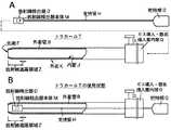
次に、図1を伴って、本発明による鏡視下手術用トラカールの実施例を述べよう。
図1において、図2との対応部分には同一符号を付して示す。Next, with reference to FIG. 1, an embodiment of the endoscopic surgical trocar according to the present invention will be described.
In FIG. 1, parts corresponding to those in FIG.
図1に示す本発明による鏡視下手術用トラカールの実施例は、図2を伴って上述したと同様の放射線検出器本体Mを内装し且つその電気的出力Eを外部に導出するようになされた放射線検出器Dを、図2を伴って上述したように挿通し得る、図2を伴って上述した外套管Bに対応した外套管Bを有する、図2を伴って上述した鏡視下手術用トラカールTに対応した鏡視下手術用トラカールTの構成を有する。 The embodiment of the trocar for a surgical operation according to the present invention shown in FIG. 1 is configured so that a radiation detector main body M similar to that described above with reference to FIG. 2 is installed and its electrical output E is led out. The above described radiation detector D has a mantle tube B corresponding to the mantle tube B described above with reference to FIG. 2 and can be inserted as described above with reference to FIG. The configuration of the trocar T for surgical operation corresponding to the trocar T for medical use is provided.
図1に示す本発明による鏡視下手術用トラカールの実施例は、図2を伴って上述した鏡視下手術用トラカールTに対応した構成を有する鏡視下手術用トラカールTにおいて、(A)その外套管Bが、その内に放射線検出器Dを配置して使用することを予定している長さを有し、且つ(B)(イ)外套管Bが、(a)その全域、または(b)先端Fから放射線検出器Dを配置して使用するときのその放射線検出器の配置位置に対応する位置を超えた位置までの領域、(c)先端Fから放射線検出器Dを配置して使用するときのその放射線検出器の配置位置に対応する位置までの領域など、少なくとも、先端Fから放射線検出器Dを配置して使用するときのその放射線検出器の配置位置に対応する位置までの図において放射線遮蔽領域Zとして示している領域において、Pb、Wなどの放射線遮蔽材(図示せず)でなり、または、(ロ)外套管Bの外面K及びまたは内面Jが、(a)その全域、(b)先端Fから放射線検出器Dを配置して使用するときのその放射線検出器の配置位置に対応する位置を超えた位置までの領域、(c)先端Fから放射線検出器Dを配置して使用するときのその放射線検出器の配置位置に対応する位置までの領域など、少なくとも、先端Fから放射線検出器Dを配置して使用するときのその放射線検出器の配置位置に対応する位置までの図において放射線遮蔽領域Zとして示している領域において、Pb、Wなどでなる放射線遮蔽層(図示せず)で覆われている。 The embodiment of the trocar for a surgical operation according to the present invention shown in FIG. 1 is a trocar for a surgical operation having a configuration corresponding to the trocar T for a surgical operation described above with reference to FIG. The outer tube B has a length that the radiation detector D is intended to be placed and used therein, and (B) (A) the outer tube B is (a) the entire region thereof, or (B) An area from the tip F to a position beyond the position corresponding to the arrangement position of the radiation detector D when the radiation detector D is used. (C) The radiation detector D is arranged from the tip F. At least from the tip F to a position corresponding to the position of the radiation detector when the radiation detector D is placed and used, such as an area up to a position corresponding to the position of the radiation detector when used. Shown as radiation shielding area Z Or (b) the outer surface K and / or the inner surface J of the outer tube B are (a) the entire region, (b) from the tip F. An area from the position corresponding to the arrangement position of the radiation detector when the radiation detector D is used to be used; and (c) the area when the radiation detector D is used from the tip F. Radiation shielding region in a view from at least the tip F to a position corresponding to the position of the radiation detector when the radiation detector D is disposed and used, such as an area up to a position corresponding to the position of the radiation detector. In the region indicated by Z, it is covered with a radiation shielding layer (not shown) made of Pb, W or the like.
以上が、本発明による鏡視下手術用トラカールの実施例の構成である。
このような構成を有する本発明による鏡視下手術用トラカールTの実施例によれば、それが、[背景技術]の項で図2を伴って上述した放射線検出器Dを挿通し得る外套管Bを有する鏡視下手術用トラカールTに対応していることは明らかであり、よって、[背景技術]の項で上述した鏡視下リンパ節郭清手術に先立ち、本発明による鏡視下手術用トラカールTの実施例を、図1Bに示すように、その外套管B内に、上述した放射線検出器Dを配置して、[背景技術]の項で上述したリンパ節探索同定方法に同様に用いて、放射線検出器Dで放射線を検出するようにすれば、そのリンパ節探索同定方法によって、[背景技術]の項で上述した従来の場合と同様に、腫瘍の細胞が転移する危険性のあるリンパ節を探索同定することができる。The above is the configuration of the embodiment of the trocar for surgical operation according to the present invention.
According to the embodiment of the trocar T for surgical operation according to the present invention having such a configuration, it is a mantle tube through which the radiation detector D described above with reference to FIG. It is clear that it corresponds to the trocar T for endoscopic surgery having B, and therefore, the endoscopic surgery according to the present invention prior to the endoscopic lymph node dissection described above in the section “Background Art”. As shown in FIG. 1B, the radiation detector D described above is disposed in the mantle tube B as shown in FIG. If the radiation detector D is used to detect the radiation, the lymph node search and identification method may cause a risk that the tumor cells may metastasize as in the conventional case described above in the section “Background Art”. One lymph node can be searched and identified.
しかしながら、この場合のリンパ節探索同定方法に用いている本発明による鏡視下手術用トラカールTの実施例が、その外套管Bをして、その内に放射線検出器Dを配置して使用することを予定している長さを有し、このため、放射線検出器Dを鏡視下手術用トラカールTの外套管B内に配置した状態で、その放射線検出器Dで、リンパ節に達した放射性物質から放射される放射線を検出することができ、しかも、この場合、鏡視下手術用トラカールTの外套管Bが、少なくともその先端Fから放射線検出器Dを配置して使用するときのその放射線検出器Dの挿入配置位置に対応する位置までの放射線遮蔽領域Zとして示している領域において、放射線遮蔽用材でなり、または外套管Bの外面K及びまたは内面Jが、少なくともその先端Fから放射線検出器Dが挿入配置されている状態で使用されるときのその放射線検出器の挿入配置位置に対応する位置までの放射線遮蔽領域Zとして示している領域において、放射線遮蔽用層で覆われている。 However, the embodiment of the endoscopic surgical trocar T according to the present invention used in the lymph node search and identification method in this case uses the mantle tube B and the radiation detector D disposed in the mantle tube B. Therefore, the radiation detector D was placed in the mantle tube B of the surgical trocar T under the microscope, and the lymph node was reached by the radiation detector D. The radiation emitted from the radioactive substance can be detected, and in this case, when the outer tube B of the surgical trocar T under the microscope is used by arranging the radiation detector D from at least the tip F thereof, In a region shown as a radiation shielding region Z up to a position corresponding to the insertion arrangement position of the radiation detector D, it is made of a radiation shielding material, or the outer surface K and / or the inner surface J of the mantle tube B is at least the tip F thereof. When the radiation detector D is used in a state where it is inserted and arranged, the region shown as the radiation shielding region Z up to a position corresponding to the insertion arrangement position of the radiation detector is covered with a radiation shielding layer. Yes.
このため、放射線検出器Dによるリンパ節に達した放射性物質から放射される放射線の検出時、その放射線検出器Dがいま探索同定しようとしているリンパ節に達した放射性物質から放射される放射線以外の、いま探索同定しようとしているリンパ節以外のリンパ節に達した放射性物質から放射される放射線や、腫瘍の生成部位またはその近傍に打込まれた放射性物質もしくはその漏れから放射される放射線をも不必要に検出することを有効、確実に回避させることができる。 Therefore, when the radiation detector D detects radiation radiated from the radioactive material that has reached the lymph node, the radiation detector D other than the radiation radiated from the radioactive material that has reached the lymph node that the radiation detector D is currently trying to identify. In addition, there is no radiation emitted from radioactive material that has reached lymph nodes other than the lymph node that is currently being identified, or radioactive material that is injected into or near the site of tumor production or radiation emitted from the leakage. It is possible to effectively and reliably avoid detection if necessary.
よって、上述した本発明による鏡視下手術用トラカールTの実施例をその外套管B内に放射線検出器Dを配置して用いたリンパ節探索同定方法の場合、それに用いる放射線検出器Dが内装している放射線検出器本体Mとして高い放射線指向性を有するものを用いなくても、腫瘍の細胞が転移する危険性のあるリンパ節を極めて速やかに且つ極めて高精度に探索同定することができる。 Therefore, in the case of the lymph node searching and identifying method using the above-described embodiment of the trocar T for endoscopic surgery according to the present invention in which the radiation detector D is arranged in the mantle tube B, the radiation detector D used therein is internally provided. Even if a radiation detector body M having high radiation directivity is not used, lymph nodes at risk of tumor cell metastasis can be searched and identified very quickly and with extremely high accuracy.
以上のことから、本発明による鏡視下手術用トラカールTの実施例によれば、その外套管B内に放射線検出器Dを配置して、上述したリンパ節探索同定方法に用い、腫瘍の細胞が転移する危険性のあるリンパ節を上述した鏡視下リンパ節郭清手術に先立ち探索同定する場合に、その探索同定を、それに用いる放射線検出器Dが内装している放射線検出器本体Mとして高い放射線指向性を有するものを用いなくても、腫瘍の細胞が転移する危険性のあるリンパ節を可能な限り速やかに且つ可能な限り高精度に探索同定しなければならない高い必要性に応え、極めて速やかに且つ極めて高精度に行うことができる、という大なる特徴を有する。 From the above, according to the embodiment of the trocar T for endoscopic surgery according to the present invention, the radiation detector D is arranged in the mantle tube B, and used for the above-described lymph node search and identification method. When the lymph node at risk of metastasis is searched and identified prior to the above-mentioned endoscopic lymph node dissection operation, the search and identification is performed as a radiation detector body M built in the radiation detector D used therein. To meet the high need to search and identify lymph nodes at risk of metastasis of tumor cells as quickly and as accurately as possible without using high radiation directivity, It has the great feature that it can be performed very quickly and with very high accuracy.
なお、上述においては、図2を伴って上述した鏡視下手術用トラカールTに対応した構成を有する鏡視下手術用トラカールに本発明を適用した場合を述べたに過ぎず、要は、放射線検出器本体を内装し且つその電気的出力を外部に導出するようになされている放射線検出器を挿通し得る外套管を有するそれ自体公知の種々の鏡視下手術用トラカールであれば、従って、図2を伴って上述した鏡視下手術用トラカールTにおいてガス導入・器具挿入案内部Qがガス導入部を有しない器具案内部から延長している外套管を有する鏡視下手術用トラカールであっても、それに、本発明を適用して、上述したと同様の作用・効果を得ることができることは明らかであろう。 In the above description, only the case where the present invention is applied to the mirror operation trocar having the configuration corresponding to the operation mirror trocar T described above with reference to FIG. 2 is described. Various arthroscopic surgical trocars known per se having a mantle tube which can be inserted through a radiation detector which is designed to house the detector body and to lead out its electrical output to the outside. In the trocar T for surgical operation described above with reference to FIG. 2, the gas introduction / instrument insertion guide portion Q is an endoscopic surgical trocar having an outer tube extending from an instrument guide portion having no gas introduction portion. However, it will be apparent that the present invention can be applied to obtain the same operations and effects as described above.
本発明による鏡視下手術用トラカールは、上述したリンパ節探索同定方法に用いる場合ばかりでなく、広く、体内の放射線源が存在している部位を探索同定する方法に用いて、上述したリンパ節探索同定方法の場合に準じた作用・効果を得ることができる。 The trocar for surgical operation according to the present invention is used not only in the above-described method for searching and identifying lymph nodes, but also widely used in the method for searching and identifying a site where a radiation source in the body exists. Actions and effects according to the search identification method can be obtained.
B 外套管
D 放射線検出器
G 把持部
H 支持管
M 放射線検出器本体
Q ガス導入・器具挿入案内部
T 鏡視下手術用トラカール
Z 放射線遮蔽領域B Outer tube D Radiation detector G Gripping part H Support tube M Radiation detector main body Q Gas introduction / instrument insertion guide part T Mirror for surgical operation Z Radiation shielding area
Claims (1)
Translated fromJapanese上記外套管が、その内に上記放射線検出器を配置して使用することを予定している長さを有し、且つ少なくともその先端から上記放射線検出器を上記外套管内に配置して使用するときのその放射線検出器の配置位置に対応する位置までの領域において、放射線遮蔽用材でなり、または上記外套管の外面及びまたは内面が、少なくともその先端から上記放射線検出器の上記配置位置に対応する位置までの領域において、放射線遮蔽用層で覆われていることを特徴とする鏡視下手術用トラカール。
In an endoscopic surgical trocar having a mantle tube that has a radiation detector body and that can be inserted through the radiation detector that is adapted to lead out its electrical output to the outside,
When the outer tube has a length that the radiation detector is scheduled to be used in the outer tube, and at least the distal end of the outer tube is used in the outer tube. In the region up to the position corresponding to the arrangement position of the radiation detector, the position is made of a radiation shielding material, or the outer surface and / or the inner surface of the outer tube corresponds to the arrangement position of the radiation detector at least from the tip A trocar for surgical operation characterized by being covered with a radiation shielding layer in the region up to.
Priority Applications (1)
| Application Number | Priority Date | Filing Date | Title |
|---|---|---|---|
| JP2004236116AJP2006051249A (en) | 2004-08-13 | 2004-08-13 | Trocar for endoscopic surgery |
Applications Claiming Priority (1)
| Application Number | Priority Date | Filing Date | Title |
|---|---|---|---|
| JP2004236116AJP2006051249A (en) | 2004-08-13 | 2004-08-13 | Trocar for endoscopic surgery |
Publications (1)
| Publication Number | Publication Date |
|---|---|
| JP2006051249Atrue JP2006051249A (en) | 2006-02-23 |
Family
ID=36029083
Family Applications (1)
| Application Number | Title | Priority Date | Filing Date |
|---|---|---|---|
| JP2004236116APendingJP2006051249A (en) | 2004-08-13 | 2004-08-13 | Trocar for endoscopic surgery |
Country Status (1)
| Country | Link |
|---|---|
| JP (1) | JP2006051249A (en) |
Cited By (1)
| Publication number | Priority date | Publication date | Assignee | Title |
|---|---|---|---|---|
| WO2020093239A1 (en)* | 2018-11-06 | 2020-05-14 | Shenzhen Xpectvision Technology Co., Ltd. | Apparatus for imaging the prostate |
Citations (4)
| Publication number | Priority date | Publication date | Assignee | Title |
|---|---|---|---|---|
| JPH07250810A (en)* | 1992-02-14 | 1995-10-03 | Ethicon Inc | Trocar with transparent cannula and how to use |
| JPH09503953A (en)* | 1994-05-06 | 1997-04-22 | アーティン エム テルナミアン | Laparoscopic instrument for penetrating body tissue |
| JP2003111761A (en)* | 2001-10-05 | 2003-04-15 | Olympus Optical Co Ltd | Apparatus for detecting radiation |
| JP2003116786A (en)* | 2001-10-17 | 2003-04-22 | Olympus Optical Co Ltd | Endoscope device |
- 2004
- 2004-08-13JPJP2004236116Apatent/JP2006051249A/enactivePending
Patent Citations (4)
| Publication number | Priority date | Publication date | Assignee | Title |
|---|---|---|---|---|
| JPH07250810A (en)* | 1992-02-14 | 1995-10-03 | Ethicon Inc | Trocar with transparent cannula and how to use |
| JPH09503953A (en)* | 1994-05-06 | 1997-04-22 | アーティン エム テルナミアン | Laparoscopic instrument for penetrating body tissue |
| JP2003111761A (en)* | 2001-10-05 | 2003-04-15 | Olympus Optical Co Ltd | Apparatus for detecting radiation |
| JP2003116786A (en)* | 2001-10-17 | 2003-04-22 | Olympus Optical Co Ltd | Endoscope device |
Cited By (2)
| Publication number | Priority date | Publication date | Assignee | Title |
|---|---|---|---|---|
| WO2020093239A1 (en)* | 2018-11-06 | 2020-05-14 | Shenzhen Xpectvision Technology Co., Ltd. | Apparatus for imaging the prostate |
| TWI821429B (en)* | 2018-11-06 | 2023-11-11 | 大陸商深圳幀觀德芯科技有限公司 | An apparatus for imaging the prostate and using method thereof |
Similar Documents
| Publication | Publication Date | Title |
|---|---|---|
| KR100992204B1 (en) | Radiation detector | |
| SK97799A3 (en) | Energy beam targeting and directing system and invasive instrument | |
| ATE389871T1 (en) | SYSTEM FOR DETERMINING IN-VIVO STATES IN BODY LUMINA | |
| ATE348565T1 (en) | INFRARED THERMOMETHER | |
| WO2005015189A3 (en) | X-ray fluorescence system with apertured sample mask for analyzing patterned surfaces | |
| SK1272001A3 (en) | Light guiding device and method | |
| Aaboubout et al. | Intraoperative assessment of resection margins by Raman spectroscopy to guide oral cancer surgery | |
| CN106137388A (en) | Holmium laser therapy instrument | |
| JP2006051249A (en) | Trocar for endoscopic surgery | |
| FR2890567B1 (en) | PER-OPERATIVE DETECTION HEAD COUPLED TO AN EXERSE TOOL | |
| JP2006051248A (en) | Excluding device for endoscopic surgery | |
| CN205019076U (en) | Phase contrast is like medical clinical application apparatus who reaches dark field image | |
| US6603545B2 (en) | Optical measurement probe with leak minimization features suited to process control applications | |
| JP6255559B2 (en) | Biological light image acquisition system, biological light acquisition probe, and biological light image acquisition method | |
| JPH0249185A (en) | Radiation detecting endoscope | |
| RU47203U1 (en) | FIBER OPTICAL THERMOMETER | |
| KR102045857B1 (en) | Smart multi-probe capable of tracking molecules | |
| TWI782018B (en) | Catheter set | |
| JP4520208B2 (en) | Fluorescence observation endoscope | |
| CN115956882B (en) | A multifunctional 14C measuring device and Helicobacter pylori detector | |
| Iseni et al. | Transition from afterglow to streamer discharge in an atmospheric capacitively coupled micro-plasma jet | |
| JP2003111761A (en) | Apparatus for detecting radiation | |
| JP2015081792A (en) | Radiation detecting endoscope | |
| US9167957B2 (en) | Probe | |
| JP2008256388A (en) | Ion beam applicator |
Legal Events
| Date | Code | Title | Description |
|---|---|---|---|
| A621 | Written request for application examination | Free format text:JAPANESE INTERMEDIATE CODE: A621 Effective date:20070810 | |
| A621 | Written request for application examination | Free format text:JAPANESE INTERMEDIATE CODE: A621 Effective date:20070810 | |
| A131 | Notification of reasons for refusal | Free format text:JAPANESE INTERMEDIATE CODE: A131 Effective date:20100713 | |
| A521 | Written amendment | Free format text:JAPANESE INTERMEDIATE CODE: A523 Effective date:20100910 | |
| A02 | Decision of refusal | Free format text:JAPANESE INTERMEDIATE CODE: A02 Effective date:20101005 |