JP2005508616A - Identification of specific tumor antigens by selection of cDNA libraries in serum and use of these antigens in diagnostic imaging techniques - Google Patents
Identification of specific tumor antigens by selection of cDNA libraries in serum and use of these antigens in diagnostic imaging techniquesDownload PDFInfo
- Publication number
- JP2005508616A JP2005508616AJP2003515558AJP2003515558AJP2005508616AJP 2005508616 AJP2005508616 AJP 2005508616AJP 2003515558 AJP2003515558 AJP 2003515558AJP 2003515558 AJP2003515558 AJP 2003515558AJP 2005508616 AJP2005508616 AJP 2005508616A
- Authority
- JP
- Japan
- Prior art keywords
- antigen
- tumor
- selection
- library
- serum
- Prior art date
- Legal status (The legal status is an assumption and is not a legal conclusion. Google has not performed a legal analysis and makes no representation as to the accuracy of the status listed.)
- Pending
Links
- 239000000427antigenSubstances0.000titleclaimsabstractdescription98
- 102000036639antigensHuman genes0.000titleclaimsabstractdescription96
- 108091007433antigensProteins0.000titleclaimsabstractdescription96
- 206010028980NeoplasmDiseases0.000titleclaimsabstractdescription95
- 238000000034methodMethods0.000titleclaimsabstractdescription39
- 210000002966serumAnatomy0.000titleclaimsabstractdescription39
- 239000002299complementary DNASubstances0.000titleclaimsabstractdescription26
- 238000002059diagnostic imagingMethods0.000titleclaimsdescription4
- 238000004458analytical methodMethods0.000claimsabstractdescription16
- 238000005516engineering processMethods0.000claimsabstractdescription11
- 238000002823phage displayMethods0.000claimsabstractdescription10
- 108090000623proteins and genesProteins0.000claimsdescription23
- 239000002872contrast mediaSubstances0.000claimsdescription15
- 239000013598vectorSubstances0.000claimsdescription12
- 206010006187Breast cancerDiseases0.000claimsdescription11
- 208000026310Breast neoplasmDiseases0.000claimsdescription11
- 238000004519manufacturing processMethods0.000claimsdescription8
- 230000003902lesionEffects0.000claimsdescription7
- 239000003446ligandSubstances0.000claimsdescription7
- 238000003314affinity selectionMethods0.000claimsdescription5
- 238000001574biopsyMethods0.000claimsdescription3
- 238000003745diagnosisMethods0.000claimsdescription3
- 239000013543active substanceSubstances0.000claims2
- 210000000448cultured tumor cellAnatomy0.000claims1
- 239000012634fragmentSubstances0.000description23
- 102000004169proteins and genesHuman genes0.000description18
- 238000012216screeningMethods0.000description17
- 239000000203mixtureSubstances0.000description14
- 239000000243solutionSubstances0.000description13
- 239000000872bufferSubstances0.000description12
- 210000004027cellAnatomy0.000description11
- 230000000903blocking effectEffects0.000description10
- 239000013612plasmidSubstances0.000description10
- 238000011534incubationMethods0.000description9
- 238000002965ELISAMethods0.000description8
- 241001515965unidentified phageSpecies0.000description8
- FAPWRFPIFSIZLT-UHFFFAOYSA-MSodium chlorideChemical compound[Na+].[Cl-]FAPWRFPIFSIZLT-UHFFFAOYSA-M0.000description6
- 238000010276constructionMethods0.000description6
- 241000701959Escherichia virus LambdaSpecies0.000description5
- 108091034117OligonucleotideProteins0.000description5
- 230000003321amplificationEffects0.000description5
- 230000001580bacterial effectEffects0.000description5
- 238000011161developmentMethods0.000description5
- 230000004927fusionEffects0.000description5
- 102000037865fusion proteinsHuman genes0.000description5
- 108020001507fusion proteinsProteins0.000description5
- 238000003199nucleic acid amplification methodMethods0.000description5
- 210000004881tumor cellAnatomy0.000description5
- HEDRZPFGACZZDS-UHFFFAOYSA-NChloroformChemical compoundClC(Cl)ClHEDRZPFGACZZDS-UHFFFAOYSA-N0.000description4
- 108020004414DNAProteins0.000description4
- 102100037840Dehydrogenase/reductase SDR family member 2, mitochondrialHuman genes0.000description4
- 241001465754MetazoaSpecies0.000description4
- 241000699670Mus sp.Species0.000description4
- 229920001213Polysorbate 20Polymers0.000description4
- 101710188053Protein DProteins0.000description4
- 101710132893ResolvaseProteins0.000description4
- PXIPVTKHYLBLMZ-UHFFFAOYSA-NSodium azideChemical compound[Na+].[N-]=[N+]=[N-]PXIPVTKHYLBLMZ-UHFFFAOYSA-N0.000description4
- 238000013019agitationMethods0.000description4
- 210000000234capsidAnatomy0.000description4
- 238000012512characterization methodMethods0.000description4
- 238000010367cloningMethods0.000description4
- 230000002068genetic effectEffects0.000description4
- 238000003384imaging methodMethods0.000description4
- 239000006166lysateSubstances0.000description4
- 239000000256polyoxyethylene sorbitan monolaurateSubstances0.000description4
- 235000010486polyoxyethylene sorbitan monolaurateNutrition0.000description4
- 239000007787solidSubstances0.000description4
- 238000012360testing methodMethods0.000description4
- IAZDPXIOMUYVGZ-UHFFFAOYSA-NDimethylsulphoxideChemical compoundCS(C)=OIAZDPXIOMUYVGZ-UHFFFAOYSA-N0.000description3
- 108090000144Human ProteinsProteins0.000description3
- 102000003839Human ProteinsHuman genes0.000description3
- JLCPHMBAVCMARE-UHFFFAOYSA-N[3-[[3-[[3-[[3-[[3-[[3-[[3-[[3-[[3-[[3-[[3-[[5-(2-amino-6-oxo-1H-purin-9-yl)-3-[[3-[[3-[[3-[[3-[[3-[[5-(2-amino-6-oxo-1H-purin-9-yl)-3-[[5-(2-amino-6-oxo-1H-purin-9-yl)-3-hydroxyoxolan-2-yl]methoxy-hydroxyphosphoryl]oxyoxolan-2-yl]methoxy-hydroxyphosphoryl]oxy-5-(5-methyl-2,4-dioxopyrimidin-1-yl)oxolan-2-yl]methoxy-hydroxyphosphoryl]oxy-5-(6-aminopurin-9-yl)oxolan-2-yl]methoxy-hydroxyphosphoryl]oxy-5-(6-aminopurin-9-yl)oxolan-2-yl]methoxy-hydroxyphosphoryl]oxy-5-(6-aminopurin-9-yl)oxolan-2-yl]methoxy-hydroxyphosphoryl]oxy-5-(6-aminopurin-9-yl)oxolan-2-yl]methoxy-hydroxyphosphoryl]oxyoxolan-2-yl]methoxy-hydroxyphosphoryl]oxy-5-(5-methyl-2,4-dioxopyrimidin-1-yl)oxolan-2-yl]methoxy-hydroxyphosphoryl]oxy-5-(4-amino-2-oxopyrimidin-1-yl)oxolan-2-yl]methoxy-hydroxyphosphoryl]oxy-5-(5-methyl-2,4-dioxopyrimidin-1-yl)oxolan-2-yl]methoxy-hydroxyphosphoryl]oxy-5-(5-methyl-2,4-dioxopyrimidin-1-yl)oxolan-2-yl]methoxy-hydroxyphosphoryl]oxy-5-(6-aminopurin-9-yl)oxolan-2-yl]methoxy-hydroxyphosphoryl]oxy-5-(6-aminopurin-9-yl)oxolan-2-yl]methoxy-hydroxyphosphoryl]oxy-5-(4-amino-2-oxopyrimidin-1-yl)oxolan-2-yl]methoxy-hydroxyphosphoryl]oxy-5-(4-amino-2-oxopyrimidin-1-yl)oxolan-2-yl]methoxy-hydroxyphosphoryl]oxy-5-(4-amino-2-oxopyrimidin-1-yl)oxolan-2-yl]methoxy-hydroxyphosphoryl]oxy-5-(6-aminopurin-9-yl)oxolan-2-yl]methoxy-hydroxyphosphoryl]oxy-5-(4-amino-2-oxopyrimidin-1-yl)oxolan-2-yl]methyl [5-(6-aminopurin-9-yl)-2-(hydroxymethyl)oxolan-3-yl] hydrogen phosphatePolymersCc1cn(C2CC(OP(O)(=O)OCC3OC(CC3OP(O)(=O)OCC3OC(CC3O)n3cnc4c3nc(N)[nH]c4=O)n3cnc4c3nc(N)[nH]c4=O)C(COP(O)(=O)OC3CC(OC3COP(O)(=O)OC3CC(OC3COP(O)(=O)OC3CC(OC3COP(O)(=O)OC3CC(OC3COP(O)(=O)OC3CC(OC3COP(O)(=O)OC3CC(OC3COP(O)(=O)OC3CC(OC3COP(O)(=O)OC3CC(OC3COP(O)(=O)OC3CC(OC3COP(O)(=O)OC3CC(OC3COP(O)(=O)OC3CC(OC3COP(O)(=O)OC3CC(OC3COP(O)(=O)OC3CC(OC3COP(O)(=O)OC3CC(OC3COP(O)(=O)OC3CC(OC3COP(O)(=O)OC3CC(OC3COP(O)(=O)OC3CC(OC3CO)n3cnc4c(N)ncnc34)n3ccc(N)nc3=O)n3cnc4c(N)ncnc34)n3ccc(N)nc3=O)n3ccc(N)nc3=O)n3ccc(N)nc3=O)n3cnc4c(N)ncnc34)n3cnc4c(N)ncnc34)n3cc(C)c(=O)[nH]c3=O)n3cc(C)c(=O)[nH]c3=O)n3ccc(N)nc3=O)n3cc(C)c(=O)[nH]c3=O)n3cnc4c3nc(N)[nH]c4=O)n3cnc4c(N)ncnc34)n3cnc4c(N)ncnc34)n3cnc4c(N)ncnc34)n3cnc4c(N)ncnc34)O2)c(=O)[nH]c1=OJLCPHMBAVCMARE-UHFFFAOYSA-N0.000description3
- 230000005856abnormalityEffects0.000description3
- AVKUERGKIZMTKX-NJBDSQKTSA-NampicillinChemical compoundC1([C@@H](N)C(=O)N[C@H]2[C@H]3SC([C@@H](N3C2=O)C(O)=O)(C)C)=CC=CC=C1AVKUERGKIZMTKX-NJBDSQKTSA-N0.000description3
- 229960000723ampicillinDrugs0.000description3
- 239000011324beadSubstances0.000description3
- 235000013861fat-freeNutrition0.000description3
- 230000001900immune effectEffects0.000description3
- 239000012528membraneSubstances0.000description3
- 108020004999messenger RNAProteins0.000description3
- 239000008267milkSubstances0.000description3
- 210000004080milkAnatomy0.000description3
- 235000013336milkNutrition0.000description3
- 230000001575pathological effectEffects0.000description3
- 238000000746purificationMethods0.000description3
- 230000009257reactivityEffects0.000description3
- 108091008146restriction endonucleasesProteins0.000description3
- 239000011780sodium chlorideSubstances0.000description3
- 238000003756stirringMethods0.000description3
- 239000000758substrateSubstances0.000description3
- 238000002604ultrasonographyMethods0.000description3
- 229920001817AgarPolymers0.000description2
- 239000000020NitrocelluloseSubstances0.000description2
- 241000283973Oryctolagus cuniculusSpecies0.000description2
- 102000007056Recombinant Fusion ProteinsHuman genes0.000description2
- 108010008281Recombinant Fusion ProteinsProteins0.000description2
- 229920002684SepharosePolymers0.000description2
- 239000008272agarSubstances0.000description2
- 230000005859cell recognitionEffects0.000description2
- 238000005119centrifugationMethods0.000description2
- 238000006243chemical reactionMethods0.000description2
- 239000011248coating agentSubstances0.000description2
- 238000000576coating methodMethods0.000description2
- 150000001875compoundsChemical class0.000description2
- 238000007796conventional methodMethods0.000description2
- 238000002405diagnostic procedureMethods0.000description2
- 230000000694effectsEffects0.000description2
- 238000002474experimental methodMethods0.000description2
- 230000003053immunizationEffects0.000description2
- 238000002649immunizationMethods0.000description2
- 208000015181infectious diseaseDiseases0.000description2
- 238000007852inverse PCRMethods0.000description2
- 239000000463materialSubstances0.000description2
- 238000010369molecular cloningMethods0.000description2
- 229920001220nitrocellulosPolymers0.000description2
- 238000004806packaging method and processMethods0.000description2
- 239000002245particleSubstances0.000description2
- 238000001556precipitationMethods0.000description2
- 108090000765processed proteins & peptidesProteins0.000description2
- 108020001580protein domainsProteins0.000description2
- 239000011347resinSubstances0.000description2
- 229920005989resinPolymers0.000description2
- 238000007873sievingMethods0.000description2
- 238000005406washingMethods0.000description2
- XLYOFNOQVPJJNP-UHFFFAOYSA-NwaterSubstancesOXLYOFNOQVPJJNP-UHFFFAOYSA-N0.000description2
- QRXMUCSWCMTJGU-UHFFFAOYSA-L(5-bromo-4-chloro-1h-indol-3-yl) phosphateChemical compoundC1=C(Br)C(Cl)=C2C(OP([O-])(=O)[O-])=CNC2=C1QRXMUCSWCMTJGU-UHFFFAOYSA-L0.000description1
- 108091032973(ribonucleotides)n+mProteins0.000description1
- OWEGMIWEEQEYGQ-UHFFFAOYSA-N100676-05-9Natural productsOC1C(O)C(O)C(CO)OC1OCC1C(O)C(O)C(O)C(OC2C(OC(O)C(O)C2O)CO)O1OWEGMIWEEQEYGQ-UHFFFAOYSA-N0.000description1
- QKNYBSVHEMOAJP-UHFFFAOYSA-N2-amino-2-(hydroxymethyl)propane-1,3-diol;hydron;chlorideChemical compoundCl.OCC(N)(CO)COQKNYBSVHEMOAJP-UHFFFAOYSA-N0.000description1
- 101150044980Akap1 geneProteins0.000description1
- 102000002260Alkaline PhosphataseHuman genes0.000description1
- 108020004774Alkaline PhosphataseProteins0.000description1
- 102100034609Ankyrin repeat domain-containing protein 17Human genes0.000description1
- 208000023275Autoimmune diseaseDiseases0.000description1
- 241000283707CapraSpecies0.000description1
- 101710132601Capsid proteinProteins0.000description1
- 108090000209Carbonic anhydrasesProteins0.000description1
- 102000003846Carbonic anhydrasesHuman genes0.000description1
- 101710094648Coat proteinProteins0.000description1
- 108020004705CodonProteins0.000description1
- 108020004635Complementary DNAProteins0.000description1
- 230000006820DNA synthesisEffects0.000description1
- 102100037957DixinHuman genes0.000description1
- 102100031780EndonucleaseHuman genes0.000description1
- 102000004190EnzymesHuman genes0.000description1
- 108090000790EnzymesProteins0.000description1
- 241000724791Filamentous phageSpecies0.000description1
- 102000006471FucosyltransferasesHuman genes0.000description1
- 108010019236FucosyltransferasesProteins0.000description1
- 102100021181Golgi phosphoprotein 3Human genes0.000description1
- 101000924481Homo sapiens Ankyrin repeat domain-containing protein 17Proteins0.000description1
- 101000951250Homo sapiens DixinProteins0.000description1
- 101000588145Homo sapiens Microtubule-associated tumor suppressor 1Proteins0.000description1
- 101001012157Homo sapiens Receptor tyrosine-protein kinase erbB-2Proteins0.000description1
- 101000788738Homo sapiens Zinc finger MYM-type protein 6Proteins0.000description1
- 108060003951ImmunoglobulinProteins0.000description1
- 239000006142Luria-Bertani AgarSubstances0.000description1
- 101710125418Major capsid proteinProteins0.000description1
- GUBGYTABKSRVRQ-PICCSMPSSA-NMaltoseNatural productsO[C@@H]1[C@@H](O)[C@H](O)[C@@H](CO)O[C@@H]1O[C@@H]1[C@@H](CO)OC(O)[C@H](O)[C@H]1OGUBGYTABKSRVRQ-PICCSMPSSA-N0.000description1
- 102100031550Microtubule-associated tumor suppressor 1Human genes0.000description1
- 241001529936MurinaeSpecies0.000description1
- 241000699666Mus <mouse, genus>Species0.000description1
- 101710141454NucleoproteinProteins0.000description1
- 206010061535Ovarian neoplasmDiseases0.000description1
- 102000003992PeroxidasesHuman genes0.000description1
- 108010002747Pfu DNA polymeraseProteins0.000description1
- ISWSIDIOOBJBQZ-UHFFFAOYSA-NPhenolChemical compoundOC1=CC=CC=C1ISWSIDIOOBJBQZ-UHFFFAOYSA-N0.000description1
- 108090000430Phosphatidylinositol 3-kinasesProteins0.000description1
- 102000003993Phosphatidylinositol 3-kinasesHuman genes0.000description1
- 101710193132Pre-hexon-linking protein VIIIProteins0.000description1
- 101710083689Probable capsid proteinProteins0.000description1
- 108091034057RNA (poly(A))Proteins0.000description1
- 108010092799RNA-directed DNA polymeraseProteins0.000description1
- 102100030086Receptor tyrosine-protein kinase erbB-2Human genes0.000description1
- 102000057028SOS1Human genes0.000description1
- 108700022176SOS1Proteins0.000description1
- 102100029938Serine/threonine-protein kinase SMG1Human genes0.000description1
- 101710085356Serine/threonine-protein kinase SMG1Proteins0.000description1
- 208000031673T-Cell Cutaneous LymphomaDiseases0.000description1
- 108010006785Taq PolymeraseProteins0.000description1
- 102000007537Type II DNA TopoisomerasesHuman genes0.000description1
- 108010046308Type II DNA TopoisomerasesProteins0.000description1
- 101710100170Unknown proteinProteins0.000description1
- 102100025424Zinc finger MYM-type protein 6Human genes0.000description1
- 238000010171animal modelMethods0.000description1
- 230000005875antibody responseEffects0.000description1
- 238000010805cDNA synthesis kitMethods0.000description1
- 229940022399cancer vaccineDrugs0.000description1
- 238000009566cancer vaccineMethods0.000description1
- 230000006037cell lysisEffects0.000description1
- 239000003153chemical reaction reagentSubstances0.000description1
- 230000009260cross reactivityEffects0.000description1
- 201000007241cutaneous T cell lymphomaDiseases0.000description1
- 238000005520cutting processMethods0.000description1
- 238000013461designMethods0.000description1
- 238000001514detection methodMethods0.000description1
- 238000012631diagnostic techniqueMethods0.000description1
- 238000010790dilutionMethods0.000description1
- 239000012895dilutionSubstances0.000description1
- 239000003814drugSubstances0.000description1
- 238000013399early diagnosisMethods0.000description1
- 238000010828elutionMethods0.000description1
- 108010045673endodeoxyribonuclease XBAIProteins0.000description1
- 238000001914filtrationMethods0.000description1
- 238000001943fluorescence-activated cell sortingMethods0.000description1
- 210000005260human cellAnatomy0.000description1
- 238000009396hybridizationMethods0.000description1
- 230000028993immune responseEffects0.000description1
- 230000002163immunogenEffects0.000description1
- 102000018358immunoglobulinHuman genes0.000description1
- 230000016784immunoglobulin productionEffects0.000description1
- 238000000338in vitroMethods0.000description1
- 238000002955isolationMethods0.000description1
- 238000002372labellingMethods0.000description1
- 238000011031large-scale manufacturing processMethods0.000description1
- 239000007788liquidSubstances0.000description1
- 208000006178malignant mesotheliomaDiseases0.000description1
- 201000005962mycosis fungoidesDiseases0.000description1
- JPXMTWWFLBLUCD-UHFFFAOYSA-Nnitro blue tetrazolium(2+)Chemical compoundCOC1=CC(C=2C=C(OC)C(=CC=2)[N+]=2N(N=C(N=2)C=2C=CC=CC=2)C=2C=CC(=CC=2)[N+]([O-])=O)=CC=C1[N+]1=NC(C=2C=CC=CC=2)=NN1C1=CC=C([N+]([O-])=O)C=C1JPXMTWWFLBLUCD-UHFFFAOYSA-N0.000description1
- 210000000056organAnatomy0.000description1
- 239000008188pelletSubstances0.000description1
- 108040007629peroxidase activity proteinsProteins0.000description1
- 108700010839phage proteinsProteins0.000description1
- 238000007747platingMethods0.000description1
- 229920000136polysorbatePolymers0.000description1
- 238000002360preparation methodMethods0.000description1
- 125000002924primary amino groupChemical group[H]N([H])*0.000description1
- 208000025638primary cutaneous T-cell non-Hodgkin lymphomaDiseases0.000description1
- 230000037452primingEffects0.000description1
- 230000000644propagated effectEffects0.000description1
- 102000033506protein kinase C binding proteinsHuman genes0.000description1
- 108091009576protein kinase C binding proteinsProteins0.000description1
- 238000011160researchMethods0.000description1
- 230000035945sensitivityEffects0.000description1
- 238000010561standard procedureMethods0.000description1
- 238000003325tomographyMethods0.000description1
- 238000013519translationMethods0.000description1
- GPRLSGONYQIRFK-MNYXATJNSA-NtritonChemical compound[3H+]GPRLSGONYQIRFK-MNYXATJNSA-N0.000description1
- 239000011534wash bufferSubstances0.000description1
Images
Classifications
- C—CHEMISTRY; METALLURGY
- C07—ORGANIC CHEMISTRY
- C07K—PEPTIDES
- C07K14/00—Peptides having more than 20 amino acids; Gastrins; Somatostatins; Melanotropins; Derivatives thereof
- C07K14/435—Peptides having more than 20 amino acids; Gastrins; Somatostatins; Melanotropins; Derivatives thereof from animals; from humans
- C07K14/46—Peptides having more than 20 amino acids; Gastrins; Somatostatins; Melanotropins; Derivatives thereof from animals; from humans from vertebrates
- C07K14/47—Peptides having more than 20 amino acids; Gastrins; Somatostatins; Melanotropins; Derivatives thereof from animals; from humans from vertebrates from mammals
- C07K14/4701—Peptides having more than 20 amino acids; Gastrins; Somatostatins; Melanotropins; Derivatives thereof from animals; from humans from vertebrates from mammals not used
- C07K14/4748—Tumour specific antigens; Tumour rejection antigen precursors [TRAP], e.g. MAGE
- C—CHEMISTRY; METALLURGY
- C12—BIOCHEMISTRY; BEER; SPIRITS; WINE; VINEGAR; MICROBIOLOGY; ENZYMOLOGY; MUTATION OR GENETIC ENGINEERING
- C12N—MICROORGANISMS OR ENZYMES; COMPOSITIONS THEREOF; PROPAGATING, PRESERVING, OR MAINTAINING MICROORGANISMS; MUTATION OR GENETIC ENGINEERING; CULTURE MEDIA
- C12N15/00—Mutation or genetic engineering; DNA or RNA concerning genetic engineering, vectors, e.g. plasmids, or their isolation, preparation or purification; Use of hosts therefor
- C12N15/09—Recombinant DNA-technology
- C12N15/10—Processes for the isolation, preparation or purification of DNA or RNA
- C12N15/1034—Isolating an individual clone by screening libraries
- C12N15/1037—Screening libraries presented on the surface of microorganisms, e.g. phage display, E. coli display
- C—CHEMISTRY; METALLURGY
- C12—BIOCHEMISTRY; BEER; SPIRITS; WINE; VINEGAR; MICROBIOLOGY; ENZYMOLOGY; MUTATION OR GENETIC ENGINEERING
- C12Q—MEASURING OR TESTING PROCESSES INVOLVING ENZYMES, NUCLEIC ACIDS OR MICROORGANISMS; COMPOSITIONS OR TEST PAPERS THEREFOR; PROCESSES OF PREPARING SUCH COMPOSITIONS; CONDITION-RESPONSIVE CONTROL IN MICROBIOLOGICAL OR ENZYMOLOGICAL PROCESSES
- C12Q1/00—Measuring or testing processes involving enzymes, nucleic acids or microorganisms; Compositions therefor; Processes of preparing such compositions
- C12Q1/68—Measuring or testing processes involving enzymes, nucleic acids or microorganisms; Compositions therefor; Processes of preparing such compositions involving nucleic acids
- C12Q1/6804—Nucleic acid analysis using immunogens
- C—CHEMISTRY; METALLURGY
- C12—BIOCHEMISTRY; BEER; SPIRITS; WINE; VINEGAR; MICROBIOLOGY; ENZYMOLOGY; MUTATION OR GENETIC ENGINEERING
- C12Q—MEASURING OR TESTING PROCESSES INVOLVING ENZYMES, NUCLEIC ACIDS OR MICROORGANISMS; COMPOSITIONS OR TEST PAPERS THEREFOR; PROCESSES OF PREPARING SUCH COMPOSITIONS; CONDITION-RESPONSIVE CONTROL IN MICROBIOLOGICAL OR ENZYMOLOGICAL PROCESSES
- C12Q1/00—Measuring or testing processes involving enzymes, nucleic acids or microorganisms; Compositions therefor; Processes of preparing such compositions
- C12Q1/68—Measuring or testing processes involving enzymes, nucleic acids or microorganisms; Compositions therefor; Processes of preparing such compositions involving nucleic acids
- C12Q1/6811—Selection methods for production or design of target specific oligonucleotides or binding molecules
- C—CHEMISTRY; METALLURGY
- C40—COMBINATORIAL TECHNOLOGY
- C40B—COMBINATORIAL CHEMISTRY; LIBRARIES, e.g. CHEMICAL LIBRARIES
- C40B40/00—Libraries per se, e.g. arrays, mixtures
- C40B40/02—Libraries contained in or displayed by microorganisms, e.g. bacteria or animal cells; Libraries contained in or displayed by vectors, e.g. plasmids; Libraries containing only microorganisms or vectors
- G—PHYSICS
- G01—MEASURING; TESTING
- G01N—INVESTIGATING OR ANALYSING MATERIALS BY DETERMINING THEIR CHEMICAL OR PHYSICAL PROPERTIES
- G01N33/00—Investigating or analysing materials by specific methods not covered by groups G01N1/00 - G01N31/00
- G01N33/48—Biological material, e.g. blood, urine; Haemocytometers
- G01N33/50—Chemical analysis of biological material, e.g. blood, urine; Testing involving biospecific ligand binding methods; Immunological testing
- G01N33/53—Immunoassay; Biospecific binding assay; Materials therefor
- G01N33/574—Immunoassay; Biospecific binding assay; Materials therefor for cancer
- C—CHEMISTRY; METALLURGY
- C12—BIOCHEMISTRY; BEER; SPIRITS; WINE; VINEGAR; MICROBIOLOGY; ENZYMOLOGY; MUTATION OR GENETIC ENGINEERING
- C12Q—MEASURING OR TESTING PROCESSES INVOLVING ENZYMES, NUCLEIC ACIDS OR MICROORGANISMS; COMPOSITIONS OR TEST PAPERS THEREFOR; PROCESSES OF PREPARING SUCH COMPOSITIONS; CONDITION-RESPONSIVE CONTROL IN MICROBIOLOGICAL OR ENZYMOLOGICAL PROCESSES
- C12Q2541/00—Reactions characterised by directed evolution
- C12Q2541/10—Reactions characterised by directed evolution the purpose being the selection or design of target specific nucleic acid binding sequences
- C12Q2541/101—Selex
- G—PHYSICS
- G01—MEASURING; TESTING
- G01N—INVESTIGATING OR ANALYSING MATERIALS BY DETERMINING THEIR CHEMICAL OR PHYSICAL PROPERTIES
- G01N2500/00—Screening for compounds of potential therapeutic value
- G01N2500/20—Screening for compounds of potential therapeutic value cell-free systems
Landscapes
- Health & Medical Sciences (AREA)
- Life Sciences & Earth Sciences (AREA)
- Chemical & Material Sciences (AREA)
- Organic Chemistry (AREA)
- Engineering & Computer Science (AREA)
- Molecular Biology (AREA)
- Immunology (AREA)
- Genetics & Genomics (AREA)
- Biochemistry (AREA)
- Zoology (AREA)
- Biomedical Technology (AREA)
- Biotechnology (AREA)
- Microbiology (AREA)
- General Health & Medical Sciences (AREA)
- Wood Science & Technology (AREA)
- Medicinal Chemistry (AREA)
- Biophysics (AREA)
- Physics & Mathematics (AREA)
- General Engineering & Computer Science (AREA)
- Bioinformatics & Cheminformatics (AREA)
- Proteomics, Peptides & Aminoacids (AREA)
- Analytical Chemistry (AREA)
- Hematology (AREA)
- Urology & Nephrology (AREA)
- Pathology (AREA)
- General Physics & Mathematics (AREA)
- Gastroenterology & Hepatology (AREA)
- Toxicology (AREA)
- Food Science & Technology (AREA)
- Virology (AREA)
- Crystallography & Structural Chemistry (AREA)
- Cell Biology (AREA)
- Plant Pathology (AREA)
- Oncology (AREA)
- Hospice & Palliative Care (AREA)
- Chemical Kinetics & Catalysis (AREA)
- General Chemical & Material Sciences (AREA)
- Peptides Or Proteins (AREA)
- Measuring Or Testing Involving Enzymes Or Micro-Organisms (AREA)
- Medicines Containing Antibodies Or Antigens For Use As Internal Diagnostic Agents (AREA)
Abstract
Translated fromJapaneseDescription
Translated fromJapanese本発明は、腫瘍に罹患した対象から誘導されたcDNAライブラリーの血清での選択による特異的腫瘍抗原の同定および特に腫瘍診断方法に関するものである。 The present invention relates to the identification of specific tumor antigens by serum selection of cDNA libraries derived from tumor-affected subjects and in particular to tumor diagnostic methods.
本発明は、また、動物または人体に直接使用されない診断補助器具の製造の技術分野に関するものである。 The invention also relates to the technical field of manufacturing diagnostic aids that are not used directly on animals or human bodies.
本発明は、医薬分野における工業的適用に適した化合物、それらの製造方法、それらの使用方法、およびそれらを含む組成物を提供する。 The present invention provides compounds suitable for industrial application in the pharmaceutical field, methods for their production, methods for their use, and compositions containing them.
本発明は、診断医学、例えば臓器および組織の病的異常の検出および診断についての画像技術に有用な物質に適した化合物、組成物および方法を提供する。 The present invention provides compounds, compositions and methods suitable for materials useful in diagnostic medicine, eg imaging techniques for the detection and diagnosis of pathological abnormalities in organs and tissues.
特に、限定するわけではないが、本発明は腫瘍診断セクターに関するものである。 In particular, but not exclusively, the present invention relates to the tumor diagnostic sector.
(発明の背景)
早期診断は、患者の生活の質をかなり改善させ、同時に国民の健康保険システムおよび患者自身にとっても経費節約となり得るため、全医学分野における重要な優先事項であり、非常に所望されている目標である。(Background of the Invention)
Early diagnosis is an important priority in all medical fields and is a highly desirable goal because it can significantly improve the quality of life of patients and at the same time save money for the national health insurance system and for patients themselves. is there.
利用可能な様々な診断技術の中で、現時点ではいわゆる非侵襲性技術を好む傾向があり、これらの中でも、患者を複雑で時には苦痛や危険を伴うこともある診断的検査、例えば試料および生検採取を伴う検査にかけることなく、可能性のある病的異常の存在を確かめる方法を代表する様々な画像化技術がある。 Of the various diagnostic techniques available, there is currently a tendency to prefer so-called non-invasive techniques, among which diagnostic tests, such as samples and biopsies, that can complicate patients and can sometimes be painful and dangerous. There are a variety of imaging techniques that represent methods for ascertaining the presence of potential pathological abnormalities without going through an examination involving collection.
最も一般的に使用されているイメージング技術としては、コンピューター連動断層撮影(TC)、磁気共鳴(MR)、超音波検査法(US)およびシンチグラフィー(SC)を挙げることができる。 The most commonly used imaging techniques include computer linked tomography (TC), magnetic resonance (MR), ultrasonography (US) and scintigraphy (SC).
これらの画像化技術は、有効性の高い造影剤の使用を必要とする。しかしながら、それらの開発は、高感度画像によりもたらされる解剖学的特性確認の改良に向けられているに過ぎず、現在までのところ組織特性確認に関するシグナルの特異性の開発には成功していない。現在極めて小さいサイズでも解剖学的病変を視覚化するのは可能であるが、観察される病変の性質の特定には依然として侵襲型検査が必要とされる。 These imaging techniques require the use of highly effective contrast agents. However, their development is only aimed at improving the anatomical characterization provided by the high sensitivity images, and to date, the development of signal specificity for tissue characterization has not been successful. Although it is currently possible to visualize anatomical lesions even at very small sizes, invasive examination is still required to determine the nature of the observed lesions.
この問題に対する一つの解決法は、健康な組織と病変部との画像コントラスト度を選択的および特異的に高め得る造影剤の開発である。 One solution to this problem is the development of contrast agents that can selectively and specifically increase the degree of image contrast between healthy tissue and lesions.
既知技術により提供される一例は、造影剤のビークルとしてのモノクローナル抗体の使用であり、この意味ではSCおよびMR分野で既に試みられている。依然としてさらなる改良を必要とするにせよ、SC技術では肯定的な成果を得ているが、MRでの結果はまだ満足すべきものではない。同じく結果を改善する必要性は、超音波検査分野でも認められている。 One example provided by the known art is the use of monoclonal antibodies as contrast agent vehicles, which in this sense has already been attempted in the SC and MR fields. Although still needing further improvements, the SC technology has been positive, but the MR results are not yet satisfactory. The need for improved results is also recognized in the field of ultrasonography.
腫瘍抗原の同定により、標的特異的造影剤(TSCM)構築用の新規で改良された試薬が提供され得る。多少なりとも特異的腫瘍抗原は公知であり、それらは実験動物において抗体を刺激する抗原−免疫原として腫瘍細胞を用いて得られる。また、患者自身において抗体産生を刺激する若干の腫瘍抗原も知られている(例えば、p53、HER−2/neu)。これらのタイプの抗原は、原則として、健康な組織と腫瘍組織とを区別するマーカーとして優れた候補である。しかしながら、それらの同定は慣用的方法を用いた場合困難である。 Tumor antigen identification can provide new and improved reagents for target specific contrast agent (TSCM) construction. More or less specific tumor antigens are known and they are obtained using tumor cells as antigen-immunogens that stimulate antibodies in laboratory animals. Some tumor antigens that stimulate antibody production in patients themselves are also known (eg, p53, HER-2 / neu). These types of antigens are in principle good candidates as markers to distinguish between healthy and tumor tissue. However, their identification is difficult when using conventional methods.
SEREX(組換えcDNAの発現を通した自己腫瘍抗原の血清学的分析、P.N.A.S. 92、11810、1995参照)として知られる、様々なタイプの腫瘍に罹患した患者の血清によるcDNAライブラリーの分析(スクリーニング)方法の最近の開発により、多数の腫瘍抗原が同定された。 Analysis of cDNA libraries by sera of patients with various types of tumors (screening), known as SEREX (Serologic analysis of autologous tumor antigens through expression of recombinant cDNA, see PNAS 92, 11810, 1995) ) Recent development of the method has identified a number of tumor antigens.
SEREX技術は、新規腫瘍抗原の同定に明らかに有用であるが、非常に面倒な性質のライブラリースクリーニング操作、高度のバックグラウンドノイズおよび大量の必要材料に存する多くの欠点を呈する。 While SEREX technology is clearly useful for the identification of novel tumor antigens, it presents a number of drawbacks that exist in library screening operations with very tedious properties, high background noise, and large amounts of required materials.
最初の腫瘍抗原(カルボニックアンヒドラーゼ)が特性確認された年である1993年以来、腫瘍において特異的に発現され、それに対して免疫応答が発せられる、600を越える種々のタンパク質が同定されており[M.Pfreundschuch et al. Cancer Vaccine Week,International Symposium、1998年10月5〜9日、S03]、この数はさらに増えると予測される[現在SEREXデータベースは、1695の公開配列を含む(www.licr.org/SEREX.html)]。ただし、興味深いことに、単離された配列の20〜30%がまだ未知の遺伝子産物である。 Since 1993, the year the first tumor antigen (carbonic anhydrase) was characterized, over 600 different proteins have been identified that are specifically expressed in tumors and generate immune responses against them. [M. Pfreundschuch et al. Cancer Vaccine Week, International Symposium, October 5-9, 1998, S03], this number is expected to increase further [currently the SEREX database contains 1695 public sequences (www. licr.org/SEREX.html)]. Interestingly, however, 20-30% of the isolated sequences are still unknown gene products.
しかしながら、腫瘍の診断および処置を目的とする特異的腫瘍抗原の同定技術を改良するには、さらなる研究が必要である。 However, further research is needed to improve the identification techniques of specific tumor antigens aimed at tumor diagnosis and treatment.
(発明の要約)
SEREX技術とファージディスプレイの組合わせ、すなわち小タンパク質ドメインが対応遺伝情報を内包しているバクテリオファージの表面で提示されるライブラリーの選択に基いた戦略が、血清でのcDNAディスプレイライブラリーの選択による特異的腫瘍抗原の同定方法を提供し得ることを見出した。この方法を用いると、非常に大きなライブラリー(すなわち、多数の異なる配列を発現する)から抗原を同定することが可能であると判明した。かくして同定された抗原は、造影剤の製造で使用し得るかまたは特異的リガンドを得ることを可能にし、次いで該リガンドは造影剤の製造で使用し得る。(Summary of the Invention)
The combination of SEREX technology and phage display, a strategy based on the selection of a library displayed on the surface of a bacteriophage in which the small protein domain contains the corresponding genetic information, is based on the selection of the cDNA display library in serum. It has been found that a method for identifying specific tumor antigens can be provided. Using this method, it proved possible to identify antigens from a very large library (ie, expressing a number of different sequences). The antigens thus identified can be used in the production of contrast agents or make it possible to obtain specific ligands, which can then be used in the production of contrast agents.
したがって、本発明の一対象は、血清によるcDNAディスプレイライブラリーの選択による特異的腫瘍抗原の同定方法であって、ファージディスプレイ技術を用いて選択を遂行することを特徴とする方法である。 Accordingly, an object of the present invention is a method for identifying a specific tumor antigen by selecting a cDNA display library using serum, wherein the selection is performed using a phage display technique.
本発明の対象は、腫瘍病変の画像診断用造影剤の製造に有用な腫瘍抗原の同定方法およびその方法で得られる造影剤を提供することである。 An object of the present invention is to provide a method for identifying a tumor antigen useful for the production of a contrast medium for diagnostic imaging of tumor lesions and a contrast medium obtained by the method.
造影剤は、この分野で公知の通常手順にしたがって製造され得るもので、これ以上の説明は必要ではない。 Contrast agents can be manufactured according to conventional procedures known in the art and need no further explanation.
(発明の詳細な記載)
本発明は、生検(好ましくは鮮度がよいもの)および培養腫瘍ラインの両方から得られる、腫瘍細胞からのcDNAライブラリーの構築、自己および異種の患者血清での該ライブラリーの選択(スクリーニング)による、新しいものを含む腫瘍抗原の同定、該抗原の特性確認、上記腫瘍抗原に特異的なリガンドの生成(例えば、抗体、例えば組換えヒト抗体またはヒト化組換えネズミ抗体)、および生成されたリガンドを組込む標的選択的造影剤の構築を含む。(Detailed description of the invention)
The present invention relates to the construction of a cDNA library from tumor cells obtained from both biopsy (preferably fresh) and cultured tumor lines, selection of the library with autologous and heterologous patient sera (screening). Identify tumor antigens, including new ones, characterize the antigens, generate ligands specific to the tumor antigens (e.g., antibodies, such as recombinant human antibodies or humanized recombinant murine antibodies), and Including the construction of a target-selective contrast agent that incorporates the ligand.
本発明による方法は、有利にはSEREX方法を上記ファージ‐ディスプレイ技術の有効性と組合わせると同時に、上記で概説したSEREX技術に特有な欠点を回避する。 The method according to the invention advantageously combines the SEREX method with the effectiveness of the phage-display technology, while avoiding the disadvantages inherent in the SEREX technology outlined above.
「ファージディスプレイ」とは、通常レベルの当業者が認識するように、小タンパク質ドメインが対応遺伝情報を内包するバクテリオファージの表面で呈示されているライブラリーの選択に基いた戦略である。 “Phage display” is a strategy based on the selection of a library in which small protein domains are displayed on the surface of bacteriophage containing the corresponding genetic information, as will be appreciated by those of ordinary skill in the art.
本発明に従って実行される方法は、初めて以下の新規で有利な解析可能性を提供する:
−スクリーニング前に、複雑度を低減化させる方法で、腫瘍(罹患)患者血清でライブラリーを選択し、特異抗原を発現するクローンでライブラリーを濃厚化することによる、少量の血清の使用による腫瘍抗原の同定;
−技術的問題により、当技術分野の現状レベルで実現されるcDNAライブラリーの直接スクリーニングでは、多数のクローン(約100万を越えるクローン)の解析は行えないため、組換えDNA技術の可能性を全て活用するのに適切ではない。本発明方法によると、事実、伝統的にSEREXで使用されているものより10〜100倍大きいライブラリーの構築および解析が可能であるため、限られた程度でしか存在しない抗原であっても、同定できる見込みが高くなる;
−最後に、異なる患者の血清または血清混合物を用いて後続選択サイクルを実施し得ることにより、本発明の主目的の一つを構成する交差反応性腫瘍抗原の同定が容易になる。The method carried out according to the invention for the first time provides the following new and advantageous analysis possibilities:
-Tumors with the use of a small amount of serum by selecting the library with tumor (affected) patient sera and enriching the library with clones expressing specific antigens in a reduced complexity manner before screening. Identification of the antigen;
-Due to technical problems, direct screening of cDNA libraries realized at the current level of this technical field cannot analyze a large number of clones (over 1 million clones). Not suitable for all use. According to the method of the present invention, in fact, it is possible to construct and analyze a library that is 10 to 100 times larger than that traditionally used in SEREX, so even an antigen that exists only to a limited extent, Increased likelihood of identification;
-Finally, the ability to perform subsequent selection cycles with different patient sera or serum mixtures facilitates the identification of cross-reactive tumor antigens that constitute one of the main objectives of the present invention.
非指向的方法でクローン化されたcDNAのライブラリーでは、生成されるタンパク質の約6分の1(16.7%)が正確であると予測される。このタイプのライブラリーを真の翻訳生成物で濃厚化することが、発現/ディスプレイライブラリーの実際の課業である。本発明はまた、cDNAの発現、および「枠外」タンパク質の発現が限られたバクテリオファージラムダタンパク質D(pD)のアミノ末端部分との融合体としてのタンパク質の呈示についての新規ベクターを提供する。ベクター設計によると、ファージは、そのORF(「読み枠」)がpDの場合と一致する場合のみ、表面にタンパク質フラグメントを呈示させる。我々のライブラリーにおけるクローン化DNAのフラグメントの平均サイズは100〜600bp(塩基対)であり、統計的理由によると、「枠外」配列のほとんどは、pDを翻訳させ得ず、ファージ表面で呈示されない停止コドンを含む。この場合、野生型gpDのラムダゲノムのコピーは、キャプシドの構築を支える。cDNAライブラリーについての新規発現/ディスプレイベクター(λKM4)は、cDNAフラグメントによりコードされた組換えタンパク質がバクテリオファージ自体のタンパク質との融合体として発現され、キャプシドで呈示される点で、SEREX実験で使用されるもの(λgt11)とは異なる。 In a library of cDNAs cloned in a non-directional manner, about one-sixth (16.7%) of the protein produced is predicted to be accurate. Enriching this type of library with true translation products is the actual task of the expression / display library. The present invention also provides novel vectors for the expression of cDNA and display of the protein as a fusion with the amino terminal portion of bacteriophage lambda protein D (pD) with limited expression of “out of frame” protein. According to the vector design, the phage will display protein fragments on the surface only if its ORF (“reading frame”) matches that of pD. The average size of the cloned DNA fragments in our library is 100-600 bp (base pairs), and for statistical reasons, most of the “out of frame” sequences cannot translate pD and are not displayed on the phage surface Contains a stop codon. In this case, a copy of the lambda genome of wild-type gpD supports the construction of the capsid. A novel expression / display vector (λKM4) for a cDNA library is used in SEREX experiments in that the recombinant protein encoded by the cDNA fragment is expressed as a fusion with the protein of the bacteriophage itself and presented on the capsid. (Λgt11) is different.
各ライブラリーについて、十分な数の細胞、例えば107細胞のメッセンジャーRNAを、一般的に市販されている手段を用いて精製し、対応するcDNAを生成させる。次いで、後者を発現/ディスプレイ・ベクターλKM4でクローン化する。ライブラリーの増幅は、当業者に公知の通常技術により、例えば平板培養、増殖、溶離、精製および濃縮により行う。For each library, a sufficient number of cells, eg, 107 messenger RNA, is purified using generally commercially available means to generate the corresponding cDNA. The latter is then cloned with the expression / display vector λKM4. Amplification of the library is performed by conventional techniques known to those skilled in the art, for example by plating, growth, elution, purification and concentration.
次に、ライブラリーを用いて、同定された配列の選択、「スクリーニング」および特性確認に要求される条件を明らかにする。 The library is then used to determine the conditions required for selection, “screening” and characterization of identified sequences.
ヒト細胞由来のcDNAを用いて構築された、ファージ‐ディスプレイタイプのライブラリーで、アフィニティーによる選択が可能となる。これは、キャプシドにおいてヒトタンパク質部分(一般的に腫瘍で発現される)を発現し、対応遺伝情報を内包するバクテリオファージの集合体と、特異血清のインキュベーションに基く。血清中に存在する抗体と特異結合するバクテリオファージは、固体支持体に(抗体自体により)結合したままであり、他方、非特異的結合したものは洗い流されるので、容易に採取し得る。 A phage-display type library constructed using cDNA derived from human cells allows selection by affinity. This is based on incubation of specific sera with a collection of bacteriophages that express human protein parts (generally expressed in tumors) in the capsid and enclose the corresponding genetic information. Bacteriophages that specifically bind to antibodies present in the serum remain bound (by the antibody itself) to the solid support, whereas non-specifically bound ones are washed away and can be easily harvested.
「スクリーニング」、すなわち所定の血清の抗体に対する単一ファージクローンの結合能の直接分析は、後の段階でのみ、選択の結果として、ライブラリーの複雑度(すなわち異なる配列数)が実質的に低減化されたときに行なう。 “Screening”, ie direct analysis of the ability of a single phage clone to bind to an antibody of a given serum, substantially reduces the complexity of the library (ie the number of different sequences) as a result of selection only at a later stage. To be performed when
選択戦略の使用により、特定の、例えば腫瘍患者の血清中に存在する抗体と特異的に相互作用する特性に応答するものの同定を目的とする多数の異なるタンパク質配列の迅速な解析が可能となる。 The use of a selection strategy allows for the rapid analysis of a number of different protein sequences aimed at identifying specific, eg, those that respond to properties that interact specifically with antibodies present in the serum of tumor patients.
アフィニティーによる選択は、キャプシドにおいてヒトタンパク質部分(一般的に腫瘍で発現される)を発現させ、対応する遺伝情報を内包するバクテリオファージの集合体と特異血清のインキュベーションに基いている。血清中に存在する抗体と特異結合するバクテリオファージは、固体支持体に(抗体自体により)結合したままであり、他方、非特異的結合したものは洗い流される点ので、容易に採取し得る。 Selection by affinity is based on incubation of specific sera with a collection of bacteriophages that express human protein parts (typically expressed in tumors) in the capsid and enclose the corresponding genetic information. Bacteriophages that specifically bind to antibodies present in the serum remain bound (by the antibody itself) to the solid support, whereas non-specifically bound ones are washed away and can be easily harvested.
「スクリーニング」、すなわち所定の血清の抗体に対する単一ファージクローンの結合能の直接分析は、後の段階でのみ、選択の結果として、ライブラリーの複雑度(すなわち異なる配列数)が実質的に低減されたときに行う。 “Screening”, ie direct analysis of the ability of a single phage clone to bind to an antibody of a given serum, substantially reduces the complexity of the library (ie the number of different sequences) as a result of selection only at a later stage. When you are done.
この結果、負担となる労力が低減され、特に各分析に使用される血清の量も低減され得る。 As a result, burdensome labor is reduced, and in particular, the amount of serum used for each analysis can be reduced.
標準的なcDNAライブラリーの直接「スクリーニング」は、事実、必ずしも調達しやすいものではない大量の血清の使用を必要とする。約106の独立クローンのライブラリーを解析するためには、予め選択された(自己)血清と、細菌感染させた様々なペトリ皿から移された全部で少なくとも106ファージプラークを含む多数のフィルターをインキュベーションしなければならない。別の血清による同じライブラリーの解析は、増幅ライブラリーを用いる場合のみ可能であり、これは106クローンの分析、もとのライブラリーの複雑度の喪失、またはスクリーニングの10〜100倍伸長および107〜108クローンの試験実施を意味する。Direct “screening” of standard cDNA libraries in fact requires the use of large amounts of serum that is not always easy to procure. To analyze a library of about 106 independent clones, multiple filters containing preselected (self) serum and a total of at least 106 phage plaques transferred from various bacterially infected Petri dishes Must be incubated. Analysis of the same library with different sera is only possible with an amplified library, which can be done by analysis of 106 clones, loss of complexity of the original library, or 10-100 fold extension of screening and This means testing of 107 to 108 clones.
さらにこの戦略では、ライブラリーにおいて微量でしか存在しないかまたは低濃度で存在する抗体により認識される抗原を同定し得ず、異なる血清で多重解析を実施することもできない。 Furthermore, this strategy cannot identify antigens that are recognized by antibodies that are present only in trace amounts or in low concentrations in the library, and multiple analyzes cannot be performed on different sera.
他方、ファージ・ディスプレイタイプのライブラリーの使用により、増幅ライブラリーの合計1010〜1011ファージ粒子および限られた量、例えば10μlの血清から出発して、直接スクリーニング前に少量(0.1〜1ml)でのアフィニティーによる選択が行い得る。すなわち、標準的ライブラリーより10〜100倍大きい複雑度をもつライブラリーを好都合に扱うことができるため、困難とされていた抗原を同定できる確率が高くなる。例えば、82mmフィルターで2回選択サイクルおよび1回スクリーニングを行う場合、血清の全消費量は僅か40μlですむ。On the other hand, by using a phage display type library, starting from a total of 1010 to 1011 phage particles and a limited amount of amplification library, eg 10 μl of serum, a small amount (0.1-0.1) prior to direct screening. Selection by affinity at 1 ml). That is, since a library having a complexity 10 to 100 times larger than that of a standard library can be conveniently handled, the probability that an antigen that has been considered difficult can be identified is increased. For example, if two selection cycles and one screening are performed with an 82 mm filter, the total serum consumption is only 40 μl.
さらに、ファージ・ディスプレイタイプのライブラリーの解析は、潜在的に多数の異なる血清で遂行し得ることに注目することが重要である。すなわち、同じタイプの腫瘍に罹患した異なる患者の血清中に存在する抗体と相互作用し得る抗原(交差反応性抗原)の同定に有利な選択戦略を使用することが可能である。 In addition, it is important to note that analysis of phage display type libraries can be performed on potentially many different sera. That is, it is possible to use a selection strategy that is advantageous for identifying antigens (cross-reactive antigens) that can interact with antibodies present in the sera of different patients with the same type of tumor.
様々なプロトコールを異なる固体支持体の使用に基いて採用し得る。これらのプロトコールは当業者に公知である。 Various protocols can be employed based on the use of different solid supports. These protocols are known to those skilled in the art.
様々なプロトコールは、種々の固体支持体、例えば以下のものの使用に基いて用い得る:
−セファロース:血清抗体と結合したファージを、免疫グロブリンを特異的に認識するプロテインAで被覆したセファロース樹脂に結合させる。この樹脂を短時間の遠心分離操作で洗浄することにより、非特異的(aspecific)成分を除去し得る。
−磁気ビーズ:血清抗体と結合したファージを、ヒト抗IgCポリクローナル抗体で被覆した磁気ビーズを用いて回収する。これらのビーズを洗浄し、磁石で試験管壁に結合させる。
−ペトリ皿:血清抗体と結合したファージを、予めプロテインAで被覆しておいたペトリ皿に結合させる。単に洗浄液を吸引することにより皿を洗浄する。Various protocols can be used based on the use of various solid supports, such as:
Sepharose: The phage bound to the serum antibody is bound to Sepharose resin coated with protein A that specifically recognizes immunoglobulin. By washing the resin with a short centrifugation operation, aspecific components can be removed.
-Magnetic beads: phage bound to serum antibodies are recovered using magnetic beads coated with human anti-IgC polyclonal antibody. These beads are washed and bonded to the test tube wall with a magnet.
-Petri dish: phages bound to serum antibodies are bound to a Petri dish previously coated with protein A. Wash the dishes simply by aspirating the wash solution.
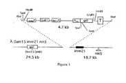
以下、実施例および図面により本発明をさらに詳しく説明する。図1は、ベクターλKM4のマップを表す。 Hereinafter, the present invention will be described in more detail with reference to examples and drawings. FIG. 1 represents a map of the vector λKM4.
ファージおよびプラスミド:
プラスミドpGEX−3X(Smith D.B.およびJohnson K.S.、Gene、67(1988)31−40)のBamHIおよびEcoRI部位において、合成オリゴヌクレオチドK108 5'-GATCCTTACTAGTTTTAGTAGCGGCCGCGGG-3'とK109 5'-AA-TTCCCGCGGCCGCTACTAAAACTAGTAAG-3のハイブリダイゼーションから誘導されたDNAフラグメントをクローン化することにより、プラスミドpGEX−SNを構築した。Phages and plasmids:
Synthetic oligonucleotides K108 5'-GATCCTTACTAGTTTTAGTAGCGGCCGCGGG-3 'and K109 5'-AA-TTCCCGCGGCCGCTACTAAAACTAGTAAG-3 at the BamHI and EcoRI sites of plasmid pGEX-3X (Smith DB and Johnson KS, Gene, 67 (1988) 31-40) Plasmid pGEX-SN was constructed by cloning DNA fragments derived from hybridization.
プラスミドpKM4のRsrIIおよびEcoRI部位において、合成オリゴヌクレオチドK106 5'-GACCGCGTTTGCCGGAACGGCAATCAGCATCGTTCACCACCAC-CACCACCACTAATAGG-3'とK107 5'-AATTCCTATTAGTGGTGGT-GGTGGTGGTGAACGATGCTGATTGCCGTTCCGGCAAACGCG-3'のハイブリダイゼーションから誘導されたDNAフラグメントをクローン化することにより、プラスミドpKM4−6Hを構築した。 In the RsrII and EcoRI sites of the plasmid pKM4, the synthetic oligonucleotide K106 5'-GACCGCGTTTGCCGGAACGGCAATCAGCATCGTTCACCACCAC-CACCACCACTAATAGG-3 'and K107 5'-AATTCCTATTAGTGGTGGTCG-GGTGGTGGTGAACGATGCTGATTGCCGTTCCGGCAAACGCG-3' pKM4-6H was constructed.
アフィニティーによる選択
ファルコンプレート(6cm、ファルコン1007)を、4℃で一晩、50mMのNaHCO3(pH9.6)中の1μg/mlのプロテインA(ピアス、#21184)3mlで被覆した。被覆溶液を廃棄後、プレートを10mlの遮断溶液(PBS×1、0.05%トウィーン20中、5%脱脂粉乳)と37℃で2時間インキュベーションした。10μlのヒト血清を、静かに攪拌しながら37℃で30分間10μlのBB4細菌抽出物、および1mlの遮断溶液中の10μlの1MのMgSO4、とプレインキュベーションした。ライブラリーの約1010ファージ粒子を血清溶液に加え、静かに攪拌しながら37℃でさらに1時間インキュベーションした。インキュベーション混合物を、プロテインAで被覆したプレートにおいて平板培養し、室温で30分間放置した。プレートを10mlの洗浄溶液(1×PBS、1%トリトン、10mMのMgSO4)で数回すすいだ。プレートへ直接加えられた(1プレートにつき600μl)BB4細胞の感染により、結合したファージを回収した。10mlの溶解NZY−トップ・アガー(48〜50℃)を感染細胞に加え、NZYプレート(15cm)へ直ちに注いだ。翌日、4℃で4時間プレートを15mlのSM緩衝液と攪拌しながらインキュベーションすることにより、ファージを集めた。ファージをPEGおよびNaCl沈澱により精製し、4℃で0.05%アジ化ナトリウムを含む初回量の10分の1のSM中で貯蔵した。Selection by affinity Falcon plates (6 cm, Falcon 1007) were coated with3 ml of 1 μg / ml protein A (Pierce, # 21184) in 50 mM NaHCO3 (pH 9.6) overnight at 4 ° C. After discarding the coating solution, the plate was incubated for 2 hours at 37 ° C. with 10 ml blocking solution (PBS × 1, 0.05% Tween 20, 5% nonfat dry milk). 10 μl of human serum was preincubated with 10 μl of BB4 bacterial extract for 30 minutes at 37 ° C. with gentle agitation, and 10 μl of 1M MgSO4 in 1 ml blocking solution. Plus approximately 1010 phage particles of the library serum solution was gently further 1 hour incubation with stirring at 37 ° C.. Incubation mixtures were plated on plates coated with protein A and left at room temperature for 30 minutes. Plates were rinsed several times with 10 ml wash solution (1 × PBS, 1% Triton, 10 mM MgSO4 ). Bound phage were recovered by infection of BB4 cells added directly to the plate (600 μl per plate). 10 ml of lysed NZY-top agar (48-50 ° C.) was added to the infected cells and immediately poured onto NZY plates (15 cm). The next day, phages were collected by incubating the plate with 15 ml SM buffer for 4 hours at 4 ° C. with agitation. The phage was purified by PEG and NaCl precipitation and stored at 4 ° C. in an initial volume of 1 / 10th SM containing 0.05% sodium azide.
免疫スクリーニング
細菌性培地のファージプラークを、4℃で1時間乾燥ニトロセルロースフィルター(シュライヒェル・アンド・シュエル)に移した。フィルターを室温で1時間遮断緩衝液(PBS×1、0.05%トウィーン20中、5%脱脂粉乳)中で遮断した。20μlのヒト血清を、20μlのBB4細菌抽出物、4mlの遮断緩衝液中の109/mlの野生型ラムダファージとプレインキュベーションした。遮断溶液を廃棄後、フィルターを室温で2時間攪拌しながら血清溶液とインキュベーションした。フィルターをPBS×1、0.05%トウィーン20で数回洗浄し、1:5000に希釈したアルカリ性ホスファターゼ(シグマA2064)とコンジュゲートしたヒト抗IgG二次抗体とインキュベーションした。次いで、フィルターを上記と同様洗浄し、基質緩衝液(100mMのトリス−HCl、pH9.6、100mMのNaCl、5mMのMgCl2)で簡単にすすいだ。各フィルターを、330mg/mlのニトロブルーテトラゾリウム、165mg/mlの5−ブロモ−4−クロロ−3−インドリルホスフェートを含む基質緩衝液10mlとインキュベーションした。水で洗浄することにより、反応を停止させた。Immunoscreening Phage plaques on bacterial media were transferred to dry nitrocellulose filters (Schleichel & Schuel) for 1 hour at 4 ° C. Filters were blocked in blocking buffer (PBS x 1, 0.05% Tween 20, 5% nonfat dry milk) for 1 hour at room temperature. 20 μl of human serum was preincubated with 20 μl of BB4 bacterial extract and 109 / ml wild type lambda phage in 4 ml blocking buffer. After discarding the blocking solution, the filter was incubated with the serum solution with stirring at room temperature for 2 hours. Filters were washed several times with PBS × 1, 0.05% Tween 20, and incubated with a human anti-IgG secondary antibody conjugated with alkaline phosphatase (Sigma A2064) diluted 1: 5000. The filter was then washed as above and rinsed briefly with substrate buffer (100 mM Tris-HCl, pH 9.6, 100 mM NaCl, 5 mM MgCl2 ). Each filter was incubated with 10 ml substrate buffer containing 330 mg / ml nitro blue tetrazolium, 165 mg / ml 5-bromo-4-chloro-3-indolyl phosphate. The reaction was stopped by washing with water.
ラムダファージの大規模製造(溶原細胞から)
BB4細胞を、攪拌しながら0.2%マルトース含有LB中でOD600=1.0まで増殖させ、遠心分離により回収し、OD600=2.0までSM緩衝液に再懸濁した。100μlの細胞を低多重度の感染でラムダにより感染させ、室温で20分間インキュベーションし、アンピシリンを含むLB寒天で平板培養し、32℃で18〜20時間インキュベーションした。翌日、単一コロニーを、アンピシリンを含む10mlのLB中、32℃で一晩攪拌しながらインキュベーションした。アンピシリンおよびMgSO4(10mM)を含む500mlの新鮮なLBに、大きなフラスコ中で5mlの一晩培養物を接種し、激しく攪拌しながらOD600=0.6まで32℃で増殖させた。フラスコを45℃の水浴中で15分間インキュベーションし、次いでさらに3時間振とう器中37℃でインキュベーションした。10mlのクロロホルムを培養物に加えて、細胞溶解を完了させ、混合物を振とう器中37℃でさらに15分間インキュベーションした。標準的手順(Sambrook,J.,Fritsch,E.F.およびManiatis,T.(1989)Molecular Cloning、コールドスプリングハーバー・ラボラトリー・プレス、コールドスプリングハーバー)にしたがってファージを溶解物の培養物から精製した。Large-scale production of lambda phage (from lysogen cells)
BB4 cells were grown in LB containing 0.2% maltose with agitation to OD600 = 1.0, collected by centrifugation, and resuspended in SM buffer until OD600 = 2.0. 100 μl of cells were infected with lambda at low multiplicity of infection, incubated for 20 minutes at room temperature, plated on LB agar with ampicillin and incubated at 32 ° C. for 18-20 hours. The next day, single colonies were incubated in 10 ml LB containing ampicillin at 32 ° C. with agitation overnight. 500 ml fresh LB containing ampicillin and MgSO4 (10 mM) was inoculated with 5 ml overnight culture in a large flask and grown at 32 ° C. to OD600 = 0.6 with vigorous stirring. The flask was incubated for 15 minutes in a 45 ° C. water bath and then incubated at 37 ° C. in a shaker for an additional 3 hours. 10 ml of chloroform was added to the culture to complete the cell lysis and the mixture was incubated for another 15 minutes at 37 ° C. in a shaker. Phages were purified from lysate cultures according to standard procedures (Sambrook, J., Fritsch, EF and Maniatis, T. (1989) Molecular Cloning, Cold Spring Harbor Laboratory Press, Cold Spring Harbor).
ELISA用のファージ溶解物を同様の手順により溶原細胞から製造したが、ただし、クロロホルムは加えなかった。NaClおよびPEGによる沈澱後、バクテリオファージペレットを、アジ化ナトリウム(0.05%)を含む出発量の10分の1のSM緩衝液に再懸濁し、4℃で貯蔵した。 A phage lysate for ELISA was prepared from lysogen cells by a similar procedure, except that chloroform was not added. After precipitation with NaCl and PEG, the bacteriophage pellet was resuspended in a starting volume of 1/10 SM buffer containing sodium azide (0.05%) and stored at 4 ° C.
ラムダELISA
マルチウェルプレート(イムノプレート・マキシソーブ、ヌンク)を、NaHCO3(50mM、pH9.6)中0.7μg/ml濃度で100μl/ウェルの抗ラムダポリクローナル抗体により4℃で一晩被覆した。被覆溶液を廃棄後、プレートを250μlの遮断溶液(PBS×1、0.05%トウィーン中、5%脱脂粉乳)とインキュベーションした。プレートを2回洗浄緩衝液(PBS×1、トウィーン20)で洗浄した。100μlの遮断緩衝液およびファージ溶解物の混合物(1:1)を、各ウェルに加え、37℃で1時間インキュベーションした。1mlのヒト血清を室温で30分間100μlの遮断緩衝液中109プラーク形成単位(pfu)のファージλKM4、1μlのウサギ血清、1μlのBB4抽出物、1μlのFBSとインキュベーションした。ファージ溶解物とインキュベーション後、プレートを洗浄し、37℃で60分間血清溶液とインキュベーションした。次いで、プレートを洗浄し、ヤギ抗ヒトHRPコンジュゲート抗体を、遮断緩衝液/二次抗体混合物(遮断溶液中1:40ウサギ血清)中、1:20000の希釈率で加えた(ジャクソン・イムノリサーチ・ラボラトリーズ)。30分インキュベーション後、プレートを洗浄し、100μlのTMB液体基質系(シグマ)によりペルオキシダーゼ活性を測定した。15分展開後、25μlのH2SO4(2M)により反応を停止させた。自動式ELISAプレート読取り装置でプレートを読み取り、結果をA=A450nm−A620nmとして表した。ELISAデータを、2独立検定の平均値として測定した。Lambda ELISA
Multiwell plates (Immunoplate Maxisorb, Nunc) were coated overnight at 4 ° C. with 100 μl / well anti-lambda polyclonal antibody at a concentration of 0.7 μg / ml in NaHCO3 (50 mM, pH 9.6). After discarding the coating solution, the plates were incubated with 250 μl of blocking solution (PBS × 1, 5% nonfat dry milk in 0.05% Tween). The plate was washed twice with wash buffer (PBS × 1, Tween 20). 100 μl of blocking buffer and phage lysate mixture (1: 1) was added to each well and incubated at 37 ° C. for 1 hour. 1 ml of human serum was incubated with 109 plaque forming units (pfu) of phage λKM4, 1 μl of rabbit serum, 1 μl of BB4 extract, 1 μl of FBS in 100 μl of blocking buffer for 30 minutes at room temperature. After incubation with phage lysate, the plate was washed and incubated with serum solution at 37 ° C. for 60 minutes. The plates were then washed and goat anti-human HRP conjugated antibody was added at a 1: 20000 dilution in blocking buffer / secondary antibody mixture (1:40 rabbit serum in blocking solution) (Jackson ImmunoResearch).・ Laboratories). After 30 minutes incubation, the plates were washed and peroxidase activity was measured with 100 μl of TMB liquid substrate system (Sigma). After 15 minutes of development, the reaction was stopped with 25 μl of H2 SO4 (2M). Plates were read on an automated ELISA plate reader and results were expressed as A = A450 nm- A620 nm . ELISA data was measured as the mean of two independent tests.
λKM4の構築
プラスミドpNS3785(Hoess、1995)を、後続のラムダファージでのクローニング用にオリゴヌクレオチド配列、部位XbaIおよびAvrII(下線部)を含むKT1 5'-TTTATCTAGACCCAGCCCTAG-GAAGCTTCTCCTGAGTAGGACAAATCC-3'およびXbaIを含むKT2 5'-GGGTCTAGATAAAACGAAAGGCCCA-GTCTTTC-3'による逆PCRにより増幅した。逆PCRにおいて、TaqポリメラーゼおよびPfu DNAポリメラーゼの混合物を用いて、DNA合成の忠実度を高めた。25増幅サイクルを遂行した(95℃・30秒、55℃・30秒、72℃・20分)。予めXbaIエンドヌクレアーゼで消化しておいたPCR産物の自己ライゲーションにより、プラスミドpKM3を得た。ラムダpD遺伝子を、制限部位NcoI、SpeI、NotI(下線部)を含むK51 5'-CCGCCTTCCATGGGTACTAGTTTTAAATGCGG-CCGCACGAGCAAAGAAACCTTTAC-3'およびK86 5'-CTCTCATCCGCCA-AAACAGCC-3'のプライマーを用いたプラスミドpNS3785からのPCRにより増幅した。PCR産物を精製し、NcoIおよびEcoRI制限エンドヌクレアーゼで消化し、pKM3のNcoIおよびEcoRI部位で再クローン化することにより、gpDの5'末端に制限部位SpeIおよびNotIのみをもつプラスミドpKM4を得た。このプラスミドをXbaI酵素で消化し、ラムダファージλDam15imm21nin5(Hoess、1995)のXbaI部位でクローン化した (図1)。Construction of λKM4 Plasmid pNS3785 (Hoess, 1995) was transformedinto KT1 5′-TTTATCTAGA CCCAGCCCTAG-G AAGCTTCTCCTGAGTAGGACAAATCC-3 ′ containing oligonucleotide sequences, sites XbaI and AvrII (underlined) for subsequent cloning in lambda phage. Amplification was carried out by inverse PCR with KT2 5′-GGGTCTAGA TAAAACGAAAGGCCCA-GTCTTTC-3 ′ containing XbaI. In inverse PCR, a mixture of Taq polymerase and Pfu DNA polymerase was used to increase the fidelity of DNA synthesis. 25 amplification cycles were performed (95 ° C. for 30 seconds, 55 ° C. for 30 seconds, 72 ° C. for 20 minutes). Plasmid pKM3 was obtained by self-ligation of the PCR product previously digested with XbaI endonuclease. Lambda pD gene was used with primers of K51 5'-CCGCCTTCCATGG GTACTAGT TTTAAATGCGG-CCGC ACGAGCAAAGAAACCTTTAC-3 'and K86 5'-CTCTCATCCGCCA-AAACAGCC-3' containing restriction sites NcoI, SpeI, NotI (underlined) Amplified by PCR from plasmid pNS3785. The PCR product was purified, digested with NcoI and EcoRI restriction endonucleases and recloned at the NcoI and EcoRI sites of pKM3, resulting in plasmid pKM4 with only the restriction sites SpeI and NotI at the 5 ′ end of gpD. This plasmid was digested with the XbaI enzyme and cloned at the XbaI site of lambda phage λDam15immm21nin5 (Hoess, 1995) (FIG. 1).
cDNAライブラリーの構築
クイックプレップ(QuickPrep)マイクロmRNA精製キット(アマーシャム・ファーマシア・バイオテック)を用い、製造業者の使用説明書にしたがって、107MCF−7細胞(T1ライブラリー)または0.1gの固体腫瘍試料(T4ライブラリー)からmRNAを単離した。タイムセーバー(TimeSaver)cDNA合成キット(アマーシャム・ファーマシア・バイオテック)を用いて、二本鎖cDNAを5μgのポリ(A)+RNAから合成した。以前に報告された方法(Santini、1986)でランダム標識プライミングを遂行した。500ngの二本鎖cDNAから、ランダム標識プライマー5'-GCGGCCGCTGG(N)9-3'を用いることによりcDNAコピーの第一鎖を合成し、プライマー5'-GGCGGCCAAC-(N)9-3'を用いることにより第二鎖cDNAコピーを合成した。3つの異なる読み枠を伴うSpeIおよびNotI部位を含むオリゴヌクレオチドを用いて最終cDNA産物を増幅することにより、λKM4ラムダベクターにおけるクローニングを容易にした(5'-GCACTAGTGGCCG-GCCAAC-3', 5'-GCACTAGTCGGCCGGCCAAC-3', 5'-GCACTAGTCG-GGCCGGCCAAC-3'および5'-GGAGGCTCGAGCGGCCGCTGG-3')。PCR産物をキアクイック(Quiaquick)カラム(キアゲン)で精製し、ミクロコン100(アミコン)で濾過することにより小DNAフラグメントを除去し、SpeI、NotI制限酵素で消化し、フェノールで抽出後、ミクロコン100で再濾過した。Construction of cDNA library Using a QuickPrep micro mRNA purification kit (Amersham Pharmacia Biotech), 107 MCF-7 cells (T1 library) or 0.1 g according to the manufacturer's instructions. MRNA was isolated from a solid tumor sample (T4 library). Double-stranded cDNA was synthesized from 5 μg of poly (A) + RNA using a TimeSaver cDNA synthesis kit (Amersham Pharmacia Biotech). Random labeling priming was performed as previously reported (Santini, 1986). From 500 ng double-stranded cDNA, the first strand of the cDNA copy was synthesized by using randomly labeled primer 5′-GCGGCCGCTGG (N)9 -3 ′, and primer 5′-GGCGGCCAAC- (N)9 -3 ′ was synthesized. A second strand cDNA copy was synthesized by use. Amplification of the final cDNA product using oligonucleotides containing SpeI and NotI sites with three different reading frames facilitated cloning in the λKM4 lambda vector (5'-GCACTAGTGGCCG-GCCAAC-3 ', 5'- GCACTAGTCGGCCGGCCAAC-3 ′, 5′-GCACTAGTCG-GGCCGGCCAAC-3 ′ and 5′-GGAGGCTCGAGCGGCCGCTGG-3 ′). The PCR product is purified with a Quiaquick column (Qiagen), the small DNA fragments are removed by filtration through Microcon 100 (Amicon), digested with SpeI, NotI restriction enzymes, extracted with phenol, and then Microcon 100 Re-filtered.
ベクターλKM4をSpeI/NotIで消化し、脱リン酸化し、8ライゲーション混合物を各ライブラリーについて、各々0.5mgのベクターおよび約3ngの挿入体を含むように調製した。一晩4℃でインキュベーション後、ラムダパッケージングキット(Ready−To-GoTMラムダ・パッケージング・キット、アマーシャム・ファーマシア・バイオテック)によりライゲーション混合物をインビトロでパッケージし、100(15cm)NZYプレート中トップアガーにおいて平板培養した。一晩インキュベーション後、ファージをSM緩衝液でプレートから溶離し、精製し、濃縮し、7%DMSO SM緩衝液中において−80℃で貯蔵した。 Vector λKM4 was digested with SpeI / NotI, dephosphorylated, and 8 ligation mixtures were prepared for each library containing 0.5 mg of vector and approximately 3 ng of insert each. After overnight incubation at 4 ° C., the ligation mixture is packaged in vitro with a lambda packaging kit (Ready-To-Go ™ Lambda Packaging Kit, Amersham Pharmacia Biotech) and topped in a 100 (15 cm) NZY plate. Plated in agar. After overnight incubation, the phage were eluted from the plate with SM buffer, purified, concentrated and stored at −80 ° C. in 7% DMSO SM buffer.
挿入体を伴う総独立クローンとして算出されたライブラリーの複雑度は、T1ライブラリーについては108およびT4ライブラリーについては3.6×10⁷であった。Library complexity calculated as total independent clones with inserts was 108 for the T1 library and 3.6 × 10⁷ for the T4 library.
アフィニティーによる選択
特異的腫瘍抗原の同定については、2つの異なるアフィニティー選択手順を使用した。第一の手順では、陽性血清(すなわち腫瘍病変を呈する患者から採取)による2サイクルのふるいわけ、次いで同血清により実施する免疫学的スクリーニング手順、または別法として選択されたファージの混合物から無作為に採取したクローンの分析を行った。第二の手順では、交差反応性抗原の選択効率を高める目的で、選択用、ふるいわけおよびスクリーニングの両方について異なる患者からの血清混合物を使用した。Selection by affinity Two different affinity selection procedures were used for the identification of specific tumor antigens. In the first procedure, two cycles of screening with a positive serum (ie taken from a patient presenting a tumor lesion) followed by an immunological screening procedure performed with the same serum, or alternatively from a mixture of phage selected randomly The clones collected were analyzed. In the second procedure, serum mixtures from different patients were used for both selection, sieving and screening in order to increase the selection efficiency of cross-reactive antigens.
T1ライブラリーを10陽性血清(B9、B11、B13、B14、B15、B16、B17、B18、B19およびB20)により選択し、単一選択ラウンド後、対応するプールp9I、p11I、p13I、p14I、p15I、p16I、p17I、p18I、p19I、およびp20Iを生成せしめた。次いで、上述の第一戦略にしたがって、各プールを同血清での第二のアフィニティー選択ラウンドにかけることにより、第二の一連のプール(p9II、p11II、p13II、p14II、p15II、p16II、p17II、p18II、p19II、およびp20IIと称す)を生成せしめた。ELISAで試験されたプールの中には、対応する血清と高い反応性を示すものもあったことから、ライブラリーおよびアフィニティー選択手順の効力を確認した。高い反応性を示すプール(p9II、p13II、p15II、p19II、p20II)からの個々のクローンを、選択用に使用した血清での免疫スクリーニングにより単離した。The T1 library was selected with 10 positive sera (B9, B11, B13, B14, B15, B16, B17, B18, B19 and B20) and after a single selection round, the corresponding pools p9I , p11I , p13I , p14I , p15I , p16I , p17I , p18I , p19I , and p20I were generated. Then, according to the first strategy described above, by applying each pool to a second affinity selection round of the same serum, a second set of pools(p9 II, p11 II, p13 II, p14 II, p15 II, p16II , p17II , p18II , p19II , and p20II ). Some of the pools tested by ELISA were highly reactive with the corresponding sera, confirming the efficacy of the library and affinity selection procedures. Individual clones from pools showing high reactivity(p9 II, p13 II, p15 II, p19 II, p20 II), was isolated by immunoscreening with serum used for selection.
上述の第二手順をp13IIプールに適用し、それをB13以外の血清混合物(B11、B14、B15、B16、B17、B18、B19およびB20)での第三選択ラウンドにかけることにより、交差反応性クローンを選択した。生成したプール(p13III)をふるいわけで使用したのと同じ血清混合物でのELISAにより検定した。プールからの個々のクローンを、混合ΔB13(B11、B14、B15、B16、B17、B18、B19およびB20)による免疫スクリーニングにより単離し、それによってさらなる陽性クローンの単離が可能となった。By applying the second procedure described above to the p13II pool and subjecting it to a third selection round with a serum mixture other than B13 (B11, B14, B15, B16, B17, B18, B19 and B20), cross-reactivity Sex clones were selected. The resulting pool (p13III ) was assayed by ELISA with the same serum mixture used for sieving. Individual clones from the pool were isolated by immunoscreening with mixed ΔB13 (B11, B14, B15, B16, B17, B18, B19 and B20), which allowed the isolation of further positive clones.
また、本明細書記載の方法にしたがって、アフィニティー選択実験をT4ライブラリーにより(同じく異なる血清を用いてT1ライブラリーにより)実施した。 Also, according to the method described herein, affinity selection experiments were performed with the T4 library (also with the T1 library using different sera).
多重免疫学的スクリーニング(ピック‐ブロット分析)
免疫学的スクリーニングで陽性であった個々のファージクローンを単離し、溶離したファージを整列させた順序で採取することにより15cmのプレートにおける細菌叢で増殖させた。プラークをニトロセルロース膜へ移し、異なる陽性および陰性血清での分析にかけた。この方法をさらにエラーに強くし、その再現性を高めるため、ジェネシス・テカン(Genesys Tekan)ロボットステーションを用いてプレート上のファージを選抜することにより、11×7.5cmの膜における最大396までの個々のクローンの分析が可能となり、または血清とのインキュベーション前に膜を小片に切断することにより低い数のクローンを同じプレートにおいて反復選抜した。Multiple immunological screening (Pick-Blot analysis)
Individual phage clones that were positive in the immunological screening were isolated and propagated in the bacterial flora in 15 cm plates by picking the eluted phage in an ordered order. Plaques were transferred to nitrocellulose membranes and subjected to analysis with different positive and negative sera. To make this method more error-resistant and reproducible, up to 396 in an 11 x 7.5 cm membrane can be selected by selecting phage on the plate using the Genesys Tekan robot station. Individual clones could be analyzed or a low number of clones were repeatedly selected on the same plate by cutting the membrane into small pieces prior to incubation with serum.
陽性クローンの特性確認
多重反応性、または健康なドナーの血清と比べ腫瘍患者の血清について大きな特異性を呈するクローンを、順次配列決定し、現時点で入手できる配列の種々のデータベースと比較した(Non−Redundant(重複が無い)Genbank CDS、Genbank Est DivisionのNon-Redundant Database、Non-Redundant Genbank+EMBL+DDBJ+PDB Sequences)。Characterization of positive clones Clones that exhibit multiple specificity or greater specificity for tumor patient sera compared to sera from healthy donors were sequentially sequenced and compared to various databases of currently available sequences (Non- Redundant Genbank CDS, Non-Redundant Database of Genbank Est Division, Non-Redundant Genbank + EMBL + DDBJ + PDB Sequences).
得られた配列は、6つのグループに分類し得る:
−既知乳癌抗原のエピトープをコードする配列、
−乳癌抗原以外の腫瘍抗原のエピトープをコードする既知配列、
−自己抗原をコードする配列、
−既知ではあるが、腫瘍または自己免疫疾患に関与することは知られていないタンパク質をコードする配列、
−未知蛋白質(例、EST)をコードする配列、
−データベースにまだ示されていない新規配列。The resulting sequences can be classified into six groups:
A sequence encoding an epitope of a known breast cancer antigen,
A known sequence encoding an epitope of a tumor antigen other than a breast cancer antigen,
A sequence encoding a self-antigen,
A sequence encoding a protein that is known but not known to be involved in tumors or autoimmune diseases,
A sequence encoding an unknown protein (eg EST),
-New sequences not yet shown in the database.
81の異なる配列をT1ライブラリーから同定し(T1−1〜T1−115と称す)、その13%は未知タンパク質であり、そのうち16%はデータベースに存在しなかった。21配列をT4ライブラリーから同定し(T4−1〜T4−38と称す)、その40%はデータベースからは見出されなかった。以下の表は、選択されたクローンの幾つかの配列を例として示している: 81 different sequences were identified from the T1 library (referred to as T1-1 to T1-115), 13% of which were unknown proteins, of which 16% were not present in the database. 21 sequences were identified from the T4 library (designated T4-1 to T4-38), 40% of which were not found in the database. The following table shows some sequences of selected clones as examples:

クローンT1−52は、結合タンパク質p53のフラグメントとして知られているが(Haluska P.et al.、NAR、1999、27巻、12号、2538−2544)、腫瘍抗原として同定されたことはない。該クローンは、配列VLVAGQRYQSRSGHDQKNHRKHHGKKRMKSKRSTSLSSPRNGTSGRを有し、腫瘍抗原としてのその使用は本発明の一部である。 Clone T1-52 is known as a fragment of the binding protein p53 (Haluska P. et al., NAR, 1999, 27, 12, 2538-2544) but has never been identified as a tumor antigen. The clone has the sequence VLVAGQRYQSRSGHDQKNHRKHHGKKRMKSKRSTSLSSPRNGTSGR and its use as a tumor antigen is part of the present invention.
クローンT1−17は、悪性中皮腫腫瘍抗原として同定されたDNAトポイソメラーゼIIベータのフラグメントとして知られている(Robinson C.,et al.Am.J.Respir.Cell.Mol.Biol.2000、22:550−56)。本発明は、それを乳癌腫瘍抗原として同定した。上記クローンは、配列MGTSRAGQLVEELDKVESQEREDVLAGMSGKSSFQRSEGDFLLRSLTSGRを有し、乳癌腫瘍抗原としてのその使用は本発明の一部である。 Clone T1-17 is known as a fragment of DNA topoisomerase II beta identified as a malignant mesothelioma tumor antigen (Robinson C., et al. Am. J. Respir. Cell. Mol. Biol. 2000, 22). : 550-56). The present invention has identified it as a breast cancer tumor antigen. Said clone has the sequence MGTSRAGQLVEELDKVESQEREDVLAGMSGKSSFQRSEGDFLLRSLTSGR and its use as a breast cancer tumor antigen is part of the present invention.
これまでは未知のものであったクローンT1−32は、以下の配列MGTSRAGQLHAFPLHSTTLYYTTPSGRを有する;それは腫瘍抗原であり、それ自体本発明の一部である。 The previously unknown clone T1-32 has the following sequence MGTSRAGQLHAFPLHSTTLYYTTPSGR; it is a tumor antigen and is itself part of the present invention.
これまでは未知のものであったクローンT1−74は、以下の配列MGTSRPANREAKQLHHQPHSIELIQSSGRを有する;それは腫瘍抗原であり、それ自体本発明の一部である。 Clone T1-74, previously unknown, has the following sequence MGTSRPANREAKQLHHQPHSIELIQSSGR; it is a tumor antigen and is itself part of the present invention.
これまでは未知のものであったクローンT4−2は、以下の配列MGTSRPANSEVYKPTLLYSSGRを有する;それは腫瘍抗原であり、それ自体本発明の一部である。 Clone T4-2, previously unknown, has the following sequence MGTSRPANSEVYKPTLLYSSGR; it is a tumor antigen and is itself part of the present invention.
これまでは未知のものであったクローンT4−11は、以下の配列MGTSGRPTVGFTLDFTVDPPSGRを有する;それは腫瘍抗原であり、それ自体本発明の一部である。 Clone T4-11, which was previously unknown, has the following sequence MGTSGRPTVGFTLDFTVDPPSGR; it is a tumor antigen and is itself part of the present invention.
これまでは未知のものであったクローンT4−19は、以下の配列MGTSRAGQLYRTTLTYTSGRを有する;それは腫瘍抗原であり、それ自体本発明の一部である。 Clone T4-19, previously unknown, has the following sequence MGTSRAGQLYRTTLTYTSGR; it is a tumor antigen and is itself part of the present invention.
これまでは未知のものであったクローンT1−12は、以下の配列MRYYTATKTYELMLDATTQTSGRを有する;それは腫瘍抗原であり、それ自体本発明の一部である。 The previously unknown clone T1-12 has the following sequence MRYYTATKTYELMLDATTQTSGR; it is a tumor antigen and as such is part of the present invention.
これまでは未知のものであったクローンT1−39は、以下の配列MRVIDRAQAFVDEIFGGGDDAHNLNQHNSSGRを有する;それは腫瘍抗原であり、それ自体本発明の一部である。 Clone T1-39, previously unknown, has the following sequence MRVIDRAQAFVDEIFGGGDDAHNLNQHNSSGR; it is a tumor antigen and is itself part of the present invention.
クローンT5−8は、AKAPタンパク質のフラグメントとして知られているが、腫瘍抗原として同定されたことはない。上記クローンは、配列MGTSRAGQQREQEKKRSPQDVEVLKTTTELFHSNEESGFFNELEALRAESVATKAELASYKEKAEKLQEELLVKETNMTSLQKDLSQVRDHQGRGを有し、腫瘍抗原としてのその使用は本発明の一部である。 Clone T5-8 is known as a fragment of the AKAP protein but has never been identified as a tumor antigen. Said clone has the sequence MGTSRAGQQREQEKKRSPQDVEVLKTTTELFHSNEESGFFNELEALRAESVATKAELASYKEKAEKLQEELLVKETNMTSLQKDLSQVRDHQGRG and its use as a tumor antigen is part of the present invention.
クローンT5−13は、SOS1タンパク質のフラグメントとして知られているが、腫瘍抗原として同定されたことはない。該クローンは、配列AGTSRAGQHAFEQIPSRQKKILEEAHELSEDHYKKYLAKLRSINPPCVPFFGIYLTNLLKTEEGNPEVLKRHGKELINFSKRRKVAEITGEIQQYQNQYCLRVESDIKRFFENLNPMGNSMEKEFTDYLFNKSLEIEPRKPSGRを有し、腫瘍抗原としてのその使用は本発明の一部である。 Clone T5-13 is known as a fragment of the SOS1 protein but has never been identified as a tumor antigen. The clone has the sequence AGTSRAGQHAFEQIPSRQKKILEEAHELSEDHYKKYLAKLRSINPPCVPFFGIYLTNLLKTEEGNPEVLKRHGKELINFSKRRKVAEITGEIQQYQNQYCLRVESDIKRFFENLNPMGNSMEKEFTDYLFNKSLEIEPRKPSGR as part of the present invention.
クローンT5−15は、ESTタンパク質KIAA1735のフラグメントとして知られているが、腫瘍抗原として同定されたことはない。該クローンは、配列MGTSRAGQQERSLALCEPGVNPEEQLIIIQSRLDQSLEENQDLKKELLKCKQEARNLQGIKDALQQRLTQQDTSVLQLKQELLRANMDKDELHNQNVDLQRKLDERTQRPを有し、腫瘍抗原としてのその使用は本発明の一部である。 Clone T5-15 is known as a fragment of EST protein KIAA1735, but has never been identified as a tumor antigen. The clone has the sequence MGTSRAGQQERSLALCEPGVNPEEQLIIIQSRLDQSLEENQDLKKELLKCKQEARNLQGIKDALQQRLTQQDTSVLQLKQELLRANMDKDELHNQNVDLQRKLDERTQRP, which is part of the present invention.
クローンT5−18は、mic オンコジーン(oncogen)、オルターナティブフレームのフラグメントとして知られているが、腫瘍抗原として同定されたことはない。該クローンは、配列MGTSRAGQPMSGHGSFQEVPRLHTSAQLRSASLHSEGLSCCQEGQVGQCQSPETDQQQPKMHQPSGRを有し、腫瘍抗原としてのその使用は本発明の一部である。 Clone T5-18 is known as a fragment of the mic oncogen, alternative frame, but has never been identified as a tumor antigen. The clone has the sequence MGTSRAGQPMSGHGSFQEVPRLHTSAQLRSASLHSEGLSCCQEGQVGQCQSPETDQQQPKMHQPSGR and its use as a tumor antigen is part of the present invention.
クローンT6−1は、皮膚T細胞リンパ腫腫瘍抗原として同定されたプロテインキナーゼC結合タンパク質のフラグメントとして知られている(Eichmuller S.,et al.PNAS、2001;98;629−34)。本発明は、それを乳癌腫瘍抗原として同定した。該クローンは、配列TSRAGQRYEKSDSSDSEYISDDEQKSKNEPEDTEDKEGCQMDKEPSAVKKKPKPTNPVEIKEELKSTPPAを有し、乳癌腫瘍抗原としてのその使用は本発明の一部である。 Clone T6-1 is known as a fragment of protein kinase C binding protein identified as a cutaneous T cell lymphoma tumor antigen (Eichmuller S., et al. PNAS, 2001; 98; 629-34). The present invention has identified it as a breast cancer tumor antigen. The clone has the sequence TSRAGQRYEKSDSSDSEYISDDEQKSKNEPEDTEDKEGCQMDKEPSAVKKKPKPTNPVEIKEELKSTPPA and its use as a breast cancer tumor antigen is part of the present invention.
これまでは未知のものであったクローンT6−2は、以下の配列TSRAGQLARIPSVTASEQGRTを有する;それは腫瘍抗原であり、それ自体本発明の一部である。 Clone T6-2, previously unknown, has the following sequence TSRAGQLARIPSVTASEQGRT; it is a tumor antigen and is itself part of the present invention.
クローンT6−6は、PI−3−キナーゼ関連キナーゼSMG−1との相同体のフラグメントとして知られているが、腫瘍抗原として同定されたことはない。該クローンは、配列TSGPANAAPPSADDNIKTPAERLRGPLPPSADDNLKTPSERQLTPLPPAAAKを有する。それは腫瘍抗原であり、それ自体が本発明の一部である。 Clone T6-6 is known as a homologous fragment to PI-3-kinase related kinase SMG-1, but has never been identified as a tumor antigen. The clone has the sequence TSGPANAAPPSADDNIKTPAERLRGPLPPSADDNLKTPSERQLTPLPPAAAK. It is a tumor antigen and is itself part of the present invention.
クローンT6−7は、フコシルトランスフェラーゼのフラグメントとして知られているが、腫瘍抗原として同定されたことはない。該クローンは、配列TSRAGQRELGRTGLYPSYKVREKIETVKYPTYPEAEKを有する。それは腫瘍抗原であり、それ自体が本発明の一部である。 Clone T6-7 is known as a fragment of fucosyltransferase but has never been identified as a tumor antigen. The clone has the sequence TSRAGQRELGRTGLYPSYKVREKIETVKYPTYPEAEK. It is a tumor antigen and is itself part of the present invention.
クローンT7−1は、ESTタンパク質KIAA1288のフラグメントとして知られているが、腫瘍抗原として同定されたことはない。該クローンは、配列TSVLEPTKVTFSVSPIEATEKCKKVEKGNRGLKNIPDSKEAPVNLCKPSLGKSTIKTNTPIGCKVRKTEIISYPSTSGRを有する。それは腫瘍抗原であり、それ自体が本発明の一部である。 Clone T7-1 is known as a fragment of EST protein KIAA1288, but has never been identified as a tumor antigen. The clone has the sequence TSVLEPTKVTFSVSPIEATEKCKKVEKGNRGLKNIPDSKEAPVNLCKPSLGKSTIKTNTPIGCKVRKTEIISYPSTSGR. It is a tumor antigen and is itself part of the present invention.
クローンT9−22は、逆転写酵素相同タンパク質に類似(50%の同一性)したもののフラグメントとして知られているが、腫瘍抗原として同定されたことはない。該クローンは、配列MDLTAVYRTFHPTIT-EYTFYLTVHGTFSKIDHMIGHKTSLNKSKKTEIISSTLSDHSGIKLESNSKRNPQIHASGRを有する。それは腫瘍抗原であり、それ自体が本発明の一部である。 Clone T9-22 is known as a fragment of a similar (50% identity) to reverse transcriptase homologous protein, but has never been identified as a tumor antigen. The clone has the sequence MDLTAVYRTFHPTIT-EYTFYLTVHGTFSKIDHMIGHKTSLNKSKKTEIISSTLSDHSGIKLESNSKRNPQIHASGR. It is a tumor antigen and is itself part of the present invention.
クローンT11−5は、無名の膜貫通理論的タンパク質のフラグメントとして知られているが、腫瘍抗原として同定されたことはない。該クローンは、配列MPIDVVYTWVNGTDLELLKELQQVREQMEEEQKAMREILGKNTTEPTKKRSYFVNFLAVSSGRを有する。それは腫瘍抗原であり、それ自体が本発明の一部である。 Clone T11-5 is known as an anonymous transmembrane theoretical protein fragment but has never been identified as a tumor antigen. The clone has the sequence MPIDVVYTWVNGTDLELLKELQQVREQMEEEQKAMREILGKNTTEPTKKRSYFVNFLAVSSGR. It is a tumor antigen and is itself part of the present invention.
クローンT11−6は、亜鉛フィンガータンパク質258のフラグメントとして知られているが、腫瘍抗原として同定されたことはない。該クローンは、配列TSGRPTYKVNISKAKTAVTELPSARTDTTPVITSVMSLAKIPATLSTGNTNSVLKGAVTKEAAKIIQDESTQEDAMKFPSSQSSQPSRLLKNKGISCKPVTHPSGRを有する。それは腫瘍抗原であり、それ自体が本発明の一部である。 Clone T11-6 is known as a fragment of zinc finger protein 258 but has never been identified as a tumor antigen. The clone has the sequence TSGRPTYKVNISKAKTAVTELPSARTDTTPVITSVMSLAKIPATLSTGNTNSVLKGAVTKEAAKIIQDESTQEDAMKFPSSQSSQPSRLLKNKGISCKPVTHPSGR. It is a tumor antigen and is itself part of the present invention.
クローンT11−9は、仮定的ヒトタンパク質のフラグメントとして知られているが、腫瘍抗原として同定されたことはない。該クローンは、配列TSRAGQLRFSDHAVLKSLSPVDPVEPISNSEPSMNSDMGKVSKNDTEEESNKSATTDNEISRTEYLCENSLEGKNKDNSSNEVFPQYASGRを有する。それは腫瘍抗原であり、それ自体が本発明の一部である。 Clone T11-9 is known as a hypothetical human protein fragment but has never been identified as a tumor antigen. The clone has the sequence TSRAGQLRFSDHAVLKSLSPVDPVEPISNSEPSMNSDMGKVSKNDTEEESNKSATTDNEISRTEYLCENSLEGKNKDNSSNEVFPQYASGR. It is a tumor antigen and is itself part of the present invention.
クローンT11−3は、ESTタンパク質KIAA0697のフラグメントとして知られているが、腫瘍抗原として同定されたことはない。該クローンは、配列TSRAGQRKQSFPNSDPLHQSDTSKAPGFRPPLQRPAPSPSGIVNMDSPYGSVTPSSTHLGNFASNISGGQMYGPGAPLGGAPTSGRを有する。それは腫瘍抗原であり、それ自体が本発明の一部である。 Clone T11-3 is known as a fragment of EST protein KIAA0697 but has never been identified as a tumor antigen. The clone has the sequence TSRAGQRKQSFPNSDPLHQSDTSKAPGFRPPLQRPAPSPSGIVNMDSPYGSVTPSSTHLGNFASNISGGQMYGPGAPLGGAPTSGR. It is a tumor antigen and is itself part of the present invention.
クローンT5−2は、ヒトゲノムDNAのフラグメントとして知られているが、腫瘍抗原として同定されたことはない。該クローンは、配列MGTSRAGQPTSENYLAVTTKTKHKHSLQPSNASISLLGIYPTPSGRを有する。それは腫瘍抗原であり、それ自体が本発明の一部である。 Clone T5-2 is known as a fragment of human genomic DNA but has never been identified as a tumor antigen. The clone has the sequence MGTSRAGQPTSENYLAVTTKTKHKHSLQPSNASISLLGIYPTPSGR. It is a tumor antigen and is itself part of the present invention.
クローンT5−19は、ESTタンパク質のフラグメントとして知られているが、腫瘍抗原として同定されたことはない。該クローンは、配列TSRAGQRDTQTHAHVSVCVHTPHHTYKYPTSGRを有する。それは腫瘍抗原であり、それ自体が本発明の一部である。 Clone T5-19 is known as a fragment of the EST protein but has never been identified as a tumor antigen. The clone has the sequence TSRAGQRDTQTHAHVSVCVHTPHHTYKYPTSGR. It is a tumor antigen and is itself part of the present invention.
本発明によると、既知タンパク質の一部ではあるが、腫瘍抗原としては知られていなかった配列も、腫瘍抗原としてのそれらの使用に関する限りでは本発明の対象であることがわかる。同様に、本発明の対象は、配列、または該配列をコードする遺伝子の産物の全体または一部の腫瘍抗原としての使用である。 According to the present invention, it can be seen that sequences which are part of a known protein but were not known as tumor antigens are also subject of the present invention as far as their use as tumor antigens are concerned. Similarly, the subject of the present invention is the use of a sequence, or the product of the gene encoding the sequence, in whole or in part as tumor antigen.
ピック‐ブロット分析手段により特性確認され、乳癌患者からの血清により比活性が立証されたファージクローンを、増幅し、次いで陽性および陰性血清の大型パネルにより分析した。このELISA試験後、特異的腫瘍抗原に対応するとみられるcDNAクローンを、それらの特異性および選択性をさらに正確に測定する目的で、種々の細菌発現系でクローン化した(タンパク質Dおよび/またはGST)。融合タンパク質を製造するため、以下のオリゴヌクレオチド:K84 5'-CGATTAAATAAGGAGGAATAAACC-3' および K86 5'-CTCTCATCCGCCAAAACAGCC-3'を用いたPCRにより、各クローンを単一プラークから増幅した。次いで、生成したフラグメントを、キアゲン精製キットを用いて精製し、制限酵素SpeIおよびNotIにより消化し、プラスミドpKM4−6Hでクローン化することにより、6−ヒスチジン尾部を有するDとの融合タンパク質、またはベクターpGEX−SNでクローン化することによりGSTとの融合タンパク質を製造した。次いで、標準プロトコール(Sambrook,J.、Fritsch,E.F.およびManiatis,T.(1989)Molecular Cloning、コールドスプリングハーバー・ラボラトリー・プレス、コールドスプリングハーバー)により対応する組換えタンパク質を製造および精製した。 Phage clones characterized by pick-blot analysis and demonstrated specific activity by sera from breast cancer patients were amplified and then analyzed by a large panel of positive and negative sera. After this ELISA test, cDNA clones that appeared to correspond to specific tumor antigens were cloned in various bacterial expression systems (protein D and / or GST) in order to more accurately determine their specificity and selectivity. ). To produce the fusion protein, each clone was amplified from a single plaque by PCR using the following oligonucleotides: K84 5′-CGATTAAATAAGGAGGAATAAACC-3 ′ and K86 5′-CTCTCATCCGCCAAAACAGCC-3 ′. The resulting fragment is then purified using a Qiagen purification kit, digested with restriction enzymes SpeI and NotI, and cloned with plasmid pKM4-6H, so that a fusion protein with D having a 6-histidine tail, or a vector A fusion protein with GST was produced by cloning with pGEX-SN. The corresponding recombinant protein was then produced and purified by standard protocols (Sambrook, J., Fritsch, E.F. and Maniatis, T. (1989) Molecular Cloning, Cold Spring Harbor Laboratory Press, Cold Spring Harbor).
以下の表は、例として、ファージまたは融合タンパク質調製物形態で検定された、若干の選択されたクローンの陰性および陽性血清との反応性を示す。
腫瘍細胞認識について選択された腫瘍抗原の効力を立証するため、すなわち病的異常を検出および診断する目的で、マウスを免疫化して、選択されたいくつかのクローンに対する抗体応答を誘導した。 In order to demonstrate the efficacy of selected tumor antigens for tumor cell recognition, ie to detect and diagnose pathological abnormalities, mice were immunized to induce antibody responses against several selected clones.
タンパク質DとクローンT1−52、T4−11およびT4−19の配列の融合体に対応する融合タンパク質D1−52、D4−11およびD4−19を免疫原として用いて、2ヶ月間にわたって抗原を7回投与することにより、マウスを免疫化した。毎回、1マウスに対し3タンパク質の各々について、CFA中20μg、IFA中20μg、PBS中10μgおよび4回PBS中5μgのタンパク質を注射(腹腔内または皮下)した。腫瘍抗原の配列に対する免疫の効力をチェックするため、異なる状況でクローン化された同じペプチド配列に対して免疫動物の血清を検定することにより、タンパク質Dとの反応性を排除した。 Using the fusion proteins D1-52, D4-11 and D4-19 corresponding to the fusion of protein D and the sequences of clones T1-52, T4-11 and T4-19 as an immunogen, Mice were immunized by multiple doses. Each time, one mouse was injected (intraperitoneally or subcutaneously) with 20 μg of protein in CFA, 20 μg in IFA, 10 μg in PBS, and 4 times 5 μg in PBS. To check the efficacy of immunization against the sequence of tumor antigens, reactivity with protein D was eliminated by assaying the sera of immunized animals against the same peptide sequences cloned in different situations.
D1−52の場合、免疫マウスの血清をGSTとの融合体(GST1−52)により検定し、D4−11およびD4−19の場合、対応するペプチド配列をベクターpC89(Felici et al.1991、J.Mol.Biol.222:301−310)でクローン化し、次いでpVIII(線維状バクテリオファージの主コートタンパク質)との融合体として試験した。免疫化動物の血清によるELISAの結果は、D1−52およびD4−11の場合には有効な免疫が達成されたことを示し、対応する血清を腫瘍細胞認識能力について検定した。このために、セルラインMCF7を使用し、FACSによる分析によると、両血清(抗D1−52および抗D4−11)に存在する抗体が、例えば卵巣腫瘍細胞ではなく、胸部腫瘍MCF7細胞特異的に認識することができ、この認識能力は同マウスからの免疫前血清に存在しない。
In the case of D1-52, sera of immunized mice were assayed with a fusion with GST (GST1-52), and in the case of D4-11 and D4-19, the corresponding peptide sequence was expressed in the vector pC89 (Felici et al. 1991, J Mol. Biol. 222: 301-310) and then tested as a fusion with pVIII (the main coat protein of filamentous bacteriophage). ELISA results with sera from immunized animals indicated that effective immunization was achieved in the case of D1-52 and D4-11, and the corresponding sera were assayed for tumor cell recognition ability. For this purpose, the cell line MCF7 was used and, according to the analysis by FACS, the antibodies present in both sera (anti-D1-52 and anti-D4-11) were not specifically ovarian tumor cells, but specifically breast cancer MCF7 cells. Can be recognized and this recognition ability is not present in preimmune serum from the same mice.
【配列表】
Claims (19)
Translated fromJapanese- MGTSRPANSEVYKPTLLYSSGR;
- MGTSGRPTVGFTLDFTVDPPSGR;
- MGTSRAGQLYRTTLTYTSGR;
- MGTSRAGQLHAFPLHSTTLYYTTPSGR;
- MRYYTATKTYELMLDATTQTSGR;
- MRVIDRAQAFVDEIFGGGDDAHNLNQHNSSGR
から成る群から選択される、請求項6記載の抗原。-MGTSRPANREAKQLHHQPHSIELIQSSGR;
-MGTSRPANSEVYKPTLLYSSGR;
-MGTSGRPTVGFTLDFTVDPPSGR;
-MGTSRAGQLYRTTLTYTSGR;
-MGTSRAGQLHAFPLHSTTLYYTTPSGR;
-MRYYTATKTYELMLDATTQTSGR;
-MRVIDRAQAFVDEIFGGGDDAHNLNQHNSSGR
7. The antigen of claim 6, wherein the antigen is selected from the group consisting of:
-MGTSRAGQQREQEKKRSPQDVEVLKTTTELFHSNEESGFFNELEALRAESVATKAELASYKEKAEKLQEELLVKETNMTSLQKDLSQVRDHQGRG;
-AGTSRAGQHAFEQIPSRQKKILEEAHELSEDHYKKYLAKLRSINPPCVPFFGIYLTNLLKTEEGNPEVLKRHGKELINFSKRRKVAEITGEIQQYQNQYCLRVESDIKRFFENLNPMGNSMEKEFTDYLFNKSLEIEPRKPSGR;
-MGTSRAGQQERSLALCEPGVNPEEQLIIIQSRLDQSLEENQDLKKELLKCKQEARNLQGIKDALQQRLTQQDTSVLQLKQELLRANMDKDELHNQNVDLQRKLDERTQRP;
-MGTSRAGQPMSGHGSFQEVPRLHTSAQLRSASLHSEGLSCCQEGQVGQCQSPETDQQQPKMHQPSGR;
-TSRAGQLARIPSVTASEQGRT;
-TSGPANAAPPSADDNIKTPAERLRGPLPPSADDNLKTPSERQLTPLPPAAAK;
-TSRAGQRELGRTGLYPSYKVREKIETVKYPTYPEAEK;
-TSVLEPTKVTFSVSPIEATEKCKKVEKGNRGLKNIPDSKEAPVNLCKPSLGKSTIKTNTPIGCKVRKTEIISYPSTSGR;
-MDLTAVYRTFHPTITEYTFYLTVHGTFSKIDHMIGHKTSLNKSKKTEIISSTLSDHSGIKLESNSKRNPQIHASGR;
-MPIDVVYTWVNGTDLELLKELQQVREQMEEEQKAMREILGKNTTEPTKKRSYFVNFLAVSSGR;
-TSGRPTYKVNISKAKTAVTELPSARTDTTPVITSVMSLAKIPATLSTGNTNSVLKGAVTKEAAKIIQDESTQEDAMKFPSSQSSQPSRLLKNKGISCKPVTHPSGR;
-TSRAGQLRFSDHAVLKSLSPVDPVEPISNSEPSMNSDMGKVSKNDTEEESNKSATTDNEISRTEYLCENSLEGKNKDNSSNEVFPQYASGR;
-TSRAGQRKQSFPNSDPLHQSDTSKAPGFRPPLQRPAPSPSGIVNMDSPYGSVTPSSTHLGNFASNISGGQMYGPGAPLGGAPTSGR;
-MGTSRAGQPTSENYLAVTTKTKHKHSLQPSNASISLLGIYPTPSGR;
-TSRAGQRDTQTHAHVSVCVHTPHHTYKYPTSGR
から成る群から選択される配列または該配列をコードする遺伝子の産物の全体または一部の腫瘍抗原としての使用。-VLVAGQRYQSRSGHDQKNHRKHHGKKRMKSKRSTSLSSPRNGTSGR;
-MGTSRAGQQREQEKKRSPQDVEVLKTTTELFHSNEESGFFNELEALRAESVATKAELASYKEKAEKLQEELLVKETNMTSLQKDLSQVRDHQGRG;
-AGTSRAGQHAFEQIPSRQKKILEEAHELSEDHYKKYLAKLRSINPPCVPFFGIYLTNLLKTEEGNPEVLKRHGKELINFSKRRKVAEITGEIQQYQNQYCLRVESDIKRFFENLNPMGNSMEKEFTDYLFNKSLEIEPRKPSGR;
-MGTSRAGQQERSLALCEPGVNPEEQLIIIQSRLDQSLEENQDLKKELLKCKQEARNLQGIKDALQQRLTQQDTSVLQLKQELLRANMDKDELHNQNVDLQRKLDERTQRP;
-MGTSRAGQPMSGHGSFQEVPRLHTSAQLRSASLHSEGLSCCQEGQVGQCQSPETDQQQPKMHQPSGR;
-TSRAGQLARIPSVTASEQGRT;
-TSGPANAAPPSADDNIKTPAERLRGPLPPSADDNLKTPSERQLTPLPPAAAK;
-TSRAGQRELGRTGLYPSYKVREKIETVKYPTYPEAEK;
-TSVLEPTKVTFSVSPIEATEKCKKVEKGNRGLKNIPDSKEAPVNLCKPSLGKSTIKTNTPIGCKVRKTEIISYPSTSGR;
-MDLTAVYRTFHPTITEYTFYLTVHGTFSKIDHMIGHKTSLNKSKKTEIISSTLSDHSGIKLESNSKRNPQIHASGR;
-MPIDVVYTWVNGTDLELLKELQQVREQMEEEQKAMREILGKNTTEPTKKRSYFVNFLAVSSGR;
-TSGRPTYKVNISKAKTAVTELPSARTDTTPVITSVMSLAKIPATLSTGNTNSVLKGAVTKEAAKIIQDESTQEDAMKFPSSQSSQPSRLLKNKGISCKPVTHPSGR;
-TSRAGQLRFSDHAVLKSLSPVDPVEPISNSEPSMNSDMGKVSKNDTEEESNKSATTDNEISRTEYLCENSLEGKNKDNSSNEVFPQYASGR;
-TSRAGQRKQSFPNSDPLHQSDTSKAPGFRPPLQRPAPSPSGIVNMDSPYGSVTPSSTHLGNFASNISGGQMYGPGAPLGGAPTSGR;
-MGTSRAGQPTSENYLAVTTKTKHKHSLQPSNASISLLGIYPTPSGR;
-TSRAGQRDTQTHAHVSVCVHTPHHTYKYPTSGR
Use as a tumor antigen of all or part of a sequence selected from the group consisting of or a product of a gene encoding said sequence.
-TSRAGQRYEKSDSSDSEYISDDEQKSKNEPEDTEDKEGCQMDKEPSAVKKKPKPTNPVEIKEELKSTPPA;
-MGTSRAGQLVEELDKVESQEREDVLAGMSGKSSFQRSEGDFLLRSLTSGR
から成る群から選択される抗原または該配列をコードする遺伝子の産物の全体または一部の使用。As a breast cancer tumor antigen
-TSRAGQRYEKSDSSDSEYISDDEQKSKNEPEDTEDKEGCQMDKEPSAVKKKPKPTNPVEIKEELKSTPPA;
-MGTSRAGQLVEELDKVESQEREDVLAGMSGKSSFQRSEGDFLLRSLTSGR
Use of all or part of an antigen selected from the group consisting of:
Use of the vector according to claim 18 in the method according to any one of claims 1-5.
Applications Claiming Priority (2)
| Application Number | Priority Date | Filing Date | Title |
|---|---|---|---|
| PCT/IT2001/000405WO2003010198A1 (en) | 2001-07-26 | 2001-07-26 | Identification of specific tumour antigens by selection of cdna libraries with sera and use of said antigens in diagnostic techniques |
| PCT/IT2002/000491WO2003010199A2 (en) | 2001-07-26 | 2002-07-25 | Identification of specific tumour antigens by means of the selection of cdna libraries with sera and the use of said antigens in diagnostic imaging techniques |
Publications (2)
| Publication Number | Publication Date |
|---|---|
| JP2005508616Atrue JP2005508616A (en) | 2005-04-07 |
| JP2005508616A5 JP2005508616A5 (en) | 2006-01-12 |
Family
ID=11133704
Family Applications (1)
| Application Number | Title | Priority Date | Filing Date |
|---|---|---|---|
| JP2003515558APendingJP2005508616A (en) | 2001-07-26 | 2002-07-25 | Identification of specific tumor antigens by selection of cDNA libraries in serum and use of these antigens in diagnostic imaging techniques |
Country Status (9)
| Country | Link |
|---|---|
| US (1) | US20050084857A1 (en) |
| EP (1) | EP1409537A2 (en) |
| JP (1) | JP2005508616A (en) |
| KR (1) | KR20040035687A (en) |
| CA (1) | CA2454784A1 (en) |
| HU (1) | HUP0400663A2 (en) |
| MX (1) | MXPA04000648A (en) |
| PL (1) | PL367619A1 (en) |
| WO (2) | WO2003010198A1 (en) |
Cited By (1)
| Publication number | Priority date | Publication date | Assignee | Title |
|---|---|---|---|---|
| JP2017165734A (en)* | 2011-01-04 | 2017-09-21 | シラジェン バイオセラピューティクス インコーポレイテッド | Generation of antibodies against tumor antigens and tumor-specific complement-dependent cytotoxicity by administration of oncolytic vaccinia virus |
Families Citing this family (9)
| Publication number | Priority date | Publication date | Assignee | Title |
|---|---|---|---|---|
| US8795332B2 (en) | 2002-09-30 | 2014-08-05 | Ethicon, Inc. | Barbed sutures |
| EP1551178A1 (en) | 2003-12-18 | 2005-07-06 | Koninklijke Philips Electronics N.V. | Supplementary visual display system |
| US7858323B2 (en) | 2004-06-09 | 2010-12-28 | The Regents Of The University Of Michigan | Phage microarray profiling of the humoral response to disease |
| JP2010517544A (en) | 2007-02-07 | 2010-05-27 | シグマ−タウ・インドゥストリエ・ファルマチェウチケ・リウニテ・ソシエタ・ペル・アチオニ | Recombinant antigen of human cytomegalovirus (HCMV) |
| US8915943B2 (en) | 2007-04-13 | 2014-12-23 | Ethicon, Inc. | Self-retaining systems for surgical procedures |
| US8216273B1 (en) | 2008-02-25 | 2012-07-10 | Ethicon, Inc. | Self-retainers with supporting structures on a suture |
| EP2108656A1 (en) | 2008-03-19 | 2009-10-14 | Beninati, Concetta | Antigenic protein fragments of streptococcus pneumoniae |
| US8663913B2 (en) | 2008-05-09 | 2014-03-04 | Sidney Hayes | Phage lambda display constructs |
| WO2011116209A2 (en) | 2010-03-17 | 2011-09-22 | The Regents Of The University Of Michigan | Using phage epitopes to profile the immune response |
Family Cites Families (5)
| Publication number | Priority date | Publication date | Assignee | Title |
|---|---|---|---|---|
| EP1169473B1 (en)* | 1999-04-14 | 2006-11-22 | THE GOVERNMENT OF THE UNITED STATES OF AMERICA, as represented by THE SECRETARY, DEPARTMENT OF HEALTH AND HUMAN SERVICES | High sensitivity phage display biomolecule detection |
| CN1307036A (en)* | 2000-01-26 | 2001-08-08 | 上海博道基因技术有限公司 | Polypeptide-human split compound 69 and polynucleotide for coding said polypeptide |
| FR2804690B1 (en)* | 2000-02-07 | 2004-11-05 | Centre Nat Rech Scient | NEW PROTEIN FAMILIES, NAMED ATIP, NUCLEIC SEQUENCES ENCODING SAID PROTEINS AND THEIR APPLICATIONS |
| US6436703B1 (en)* | 2000-03-31 | 2002-08-20 | Hyseq, Inc. | Nucleic acids and polypeptides |
| AU2001266787A1 (en)* | 2000-06-07 | 2002-01-08 | Human Genome Sciences, Inc. | Nucleic acids, proteins, and antibodies |
- 2001
- 2001-07-26WOPCT/IT2001/000405patent/WO2003010198A1/enactiveApplication Filing
- 2002
- 2002-07-25PLPL02367619Apatent/PL367619A1/enunknown
- 2002-07-25CACA002454784Apatent/CA2454784A1/ennot_activeAbandoned
- 2002-07-25WOPCT/IT2002/000491patent/WO2003010199A2/ennot_activeApplication Discontinuation
- 2002-07-25USUS10/484,917patent/US20050084857A1/ennot_activeAbandoned
- 2002-07-25JPJP2003515558Apatent/JP2005508616A/enactivePending
- 2002-07-25EPEP02790222Apatent/EP1409537A2/ennot_activeWithdrawn
- 2002-07-25MXMXPA04000648Apatent/MXPA04000648A/ennot_activeApplication Discontinuation
- 2002-07-25HUHU0400663Apatent/HUP0400663A2/enunknown
- 2002-07-25KRKR10-2004-7000953Apatent/KR20040035687A/ennot_activeWithdrawn
Cited By (2)
| Publication number | Priority date | Publication date | Assignee | Title |
|---|---|---|---|---|
| JP2017165734A (en)* | 2011-01-04 | 2017-09-21 | シラジェン バイオセラピューティクス インコーポレイテッド | Generation of antibodies against tumor antigens and tumor-specific complement-dependent cytotoxicity by administration of oncolytic vaccinia virus |
| US10434169B2 (en) | 2011-01-04 | 2019-10-08 | Sillajen, Inc | Generation of antibodies to tumor antigens and generation of tumor specific complement dependent cytotoxicity by administration of oncolytic vaccinia virus |
Also Published As
| Publication number | Publication date |
|---|---|
| MXPA04000648A (en) | 2004-03-19 |
| HUP0400663A2 (en) | 2005-01-28 |
| EP1409537A2 (en) | 2004-04-21 |
| CA2454784A1 (en) | 2003-02-06 |
| WO2003010199A3 (en) | 2003-10-02 |
| KR20040035687A (en) | 2004-04-29 |
| WO2003010199A2 (en) | 2003-02-06 |
| WO2003010198A1 (en) | 2003-02-06 |
| PL367619A1 (en) | 2005-03-07 |
| US20050084857A1 (en) | 2005-04-21 |
Similar Documents
| Publication | Publication Date | Title |
|---|---|---|
| US5698396A (en) | Method for identifying auto-immunoreactive substances from a subject | |
| Su et al. | Surface-epitope masking and expression cloning identifies the human prostate carcinoma tumor antigen gene PCTA-1 a member of the galectin gene family. | |
| EP0934526B1 (en) | Methods and means for selecting peptides and proteins having specific affinity for a target | |
| JP3135126B2 (en) | Method, assay, and therapeutic agent for obtaining diagnostic reagent based on clinical manifestation of disease | |
| CZ85597A3 (en) | Peptides | |
| US20040229289A1 (en) | Antigen binding peptides (abtides) from peptide libraries | |
| USRE45968E1 (en) | Vector for efficient selection and/or maturation of an antibody and uses thereof | |
| HUE030591T2 (en) | Anti-Abeta antibodies and their use | |
| US20040047880A1 (en) | Component for vaccine | |
| JP2005508616A (en) | Identification of specific tumor antigens by selection of cDNA libraries in serum and use of these antigens in diagnostic imaging techniques | |
| KR20100059977A (en) | Method for the preparation of new oligoclonal antibodies | |
| JP4564062B2 (en) | Methods for screening antibody libraries | |
| Xu et al. | Selection and identification of mimic epitopes for gastric cancer-associated antigen MG7 Ag | |
| JP4171844B2 (en) | Super repertoire artificial antibody library | |
| JP2005508309A (en) | Identification of specific tumor antigens by selection of cDNA libraries in serum and use of the antigens in tumor treatment | |
| JP2004512529A (en) | Method for in vivo identification of intracellular epitopes | |
| CN101625360B (en) | Kit for testing early lung cancer specific autoantibody enzyme linked immunity and preparation method thereof | |
| US7176286B2 (en) | Antigen fragments for the diagnosis of Toxoplasma gondii | |
| EP1108432B1 (en) | Method for identifying or isolating a molecule | |
| JP4729043B2 (en) | Methods for screening antibody libraries | |
| Robles et al. | Isolation of the Taenia crassiceps antigens from a phage display cDNA library and evaluation of their use for diagnosis of neurocysticercosis | |
| US20230131559A1 (en) | Recombinant peptides that bind to a tumour-specific antibody for breast cancer and use | |
| CN117986323A (en) | A peanut allergen polypeptide and its screening method and application | |
| WO1999015897A1 (en) | Subtractive protein screening for gene identification | |
| Palombo | 19 Biopanning |
Legal Events
| Date | Code | Title | Description |
|---|---|---|---|
| A521 | Request for written amendment filed | Free format text:JAPANESE INTERMEDIATE CODE: A523 Effective date:20050711 | |
| A621 | Written request for application examination | Free format text:JAPANESE INTERMEDIATE CODE: A621 Effective date:20050711 | |
| A521 | Request for written amendment filed | Free format text:JAPANESE INTERMEDIATE CODE: A523 Effective date:20050713 | |
| A131 | Notification of reasons for refusal | Free format text:JAPANESE INTERMEDIATE CODE: A131 Effective date:20080318 | |
| A02 | Decision of refusal | Free format text:JAPANESE INTERMEDIATE CODE: A02 Effective date:20080812 |